R - T - LARISSA VUITIKA.pdf
123
UNIVERSIDADE FEDERAL DO PARANÁ LARISSA VUITIKA MAPEAMENTO ESTRUTURAL DO SÍTIO ATIVO DE FOSFOLIPASES D PRESENTE NO VENENO DE ARANHA-MARROM (Loxosceles intermedia): CARACTERÍSTICAS BIOQUÍMICAS E BIOLÓGICAS CURITIBA 2016
-
Upload
khangminh22 -
Category
Documents
-
view
0 -
download
0
Transcript of R - T - LARISSA VUITIKA.pdf

UNIVERSIDADE FEDERAL DO PARANÁ
LARISSA VUITIKA
MAPEAMENTO ESTRUTURAL DO SÍTIO ATIVO DE FOSFOLIPASES D PRESENTE NO VENENO DE ARANHA-MARROM (Loxosceles intermedia):
CARACTERÍSTICAS BIOQUÍMICAS E BIOLÓGICAS
CURITIBA 2016

LARISSA VUITIKA
MAPEAMENTO ESTRUTURAL DO SÍTIO ATIVO DE FOSFOLIPASES D PRESENTE NO VENENO DE ARANHA-MARROM (Loxosceles intermedia):
CARACTERÍSTICAS BIOQUÍMICAS E BIOLÓGICAS Tese apresentada como requisito à obtenção do grau de doutor em Biologia Celular e Molecular, no curso de Pós-graduação em Biologia Celular e Molecular, Setor de Ciências Biológicas, Universidade Federal do Paraná Orientador: Prof.o Dr.o Silvio Sanches Veiga Co-orientador: Prof.a Dr.a Olga Meiri Chaim
CURITIBA 2016


Dedico essa tese primeiramente à minha Vó Josefina Vuitika (Dona Iuja) que desde sempre me incentivou a estudar. Dedico também aos meus pais Vilmar Vuitika e Cristina Vuitika, pelo imenso apoio em todos os momentos, pelo incentivo de nunca desistir dos sonhos e pelo exemplo de humildade e honestidade.

AGRADECIMENTOS
Aos meus pais, Vilmar e Cristina. Por me conceder a vida! Por todo
amor, dedicação e apoio nessa minha jornada científica. Obrigada pelos
valores e por me ensinar a nunca desistir. Vocês são exemplo de luta e
humildade. Amo muito vocês!
Ao meu irmão, “Ju”. Por ser mais que um irmão, um amigo para todas
as horas.
Ao meu companheiro, amigo e amor Ricardo Salmazo. Por estar ao
meu lado por todo o período do doutorado. Pela paciência, compreensão, pelo
ombro amigo e amparo em todas as horas difíceis. Também agradeço pelos
momentos de alegria e por compartilhar as vitórias e conquistas!
Agradeço ao meu orientador Prof.° Dr° Silvio Sanches Veiga, pela
oportunidade de fazer parte do seu grupo de pesquisa. Obrigada pelos
ensinamentos científicos e pelo grande exemplo de pesquisador. Minha
gratidão será eterna, pois o senhor me concedeu a realização de um sonho: a
de chegar perto de tornar-me cientista!
À minha co-orientadora Prof.a Dr.a Olga Meiri Chaim. Pela amizade,
paciência e pelos ensinamentos científicos. Obrigada pelas longas conversas e
conselhos sobre a vida de ser um pesquisador. Obrigada pela co-orientação!
À Prof.a Dr.a Andréia Senff Ribeiro pelo apoio na realização desse
trabalho, pelas contribuições na escrita do manuscrito e sugestões no trabalho.
À Pós doutoranda Dr.a Daniele Chaves Moreira pela ajuda nos
experimentos, escrita do manuscrito e demais contribuições científicas.

Ao amigo Dr.o Fernando Hitomi Matsubara (“Fer!!!”) por toda ajuda
fornecida nos experimentos, pelas discussões e contribuições científicas.
Obrigada pela amizade, apoio e pelos momentos de descontração no
laboratório.
Ao amigo e contemporâneo de pós-graduação MSc. Gabriel Otto Maissner (futuro doutor!). Obrigada pelas discussões científicas e pela
amizade.
Aos pós-graduandos e colegas de laboratório MSc. Adriano Morgon, MSc. Marianna Bóia, MSc. Aline Bednaski, MSc. Thiago Lopes de Mari, MSc. Tiago Beltrami, Msc. Vanessa Ribeiro, MSc. Alessandra Hamasaki, e
alunos de mestrado Antoniele Baldissera e Bruno Antunes. Por todo o
tempo de convivência, discussões científicas, pela ajuda em experimentos e
pelos momentos divertidos e descontraídos do laboratório!
Aos alunos de iniciação científica Verônica Cubas, Alana Basilio,
Maria Julia C. Mendes, Henrique Urbanski, Paula Deboni, em especial
Hanna Camara da Justa pela nossa amizade, risadas, choros, consolos e
festa; obrigada por toda ajuda nos meus experimentos.
Aos meus amigos sambistas: Jonas, Brunão, Téo, Carlinhos (“Baby
Face”), Jefferson (“Baby Balck”), Diego 7 cordas, Luis Ivanqui, Léo Fé, Ivo Queiroz, Otto, Luasses, Borel, Faísca, Cláudio Peba, Luis (“Pernoite”),
Felipão, Guilherme (“Pêlo), Faraj (Biólogo assim como eu!), Roberto
Guedes. E pastoras: Jana Queiroz, Lu Alves, Thais (“Tatá”), Etiene, Fer, Dani Reis, Dani Aliaga, Fabiane (“Mineira”), Neuzinha, Rafa, Helena. Obrigada pela amizade, convivência e ensinamentos da vida!
Agradeço aos meus amigos de longa data: George, Luiz Eduardo, Luis
Henrique, Matheus, Junior e Letícia. Pelos treze anos de amizade, nos quais
compartilharmos nossos sonhos. Acredito que cada um de nós está onde
gostaríamos de estar. Obrigada pelo apoio e torcida, mesmo estando longe uns
dos outros.

À coordenação do Curso de Pós-Graduação em Biologia Celular e
Molecular pelo empenho e dedicação ao curso.
À secretária do curso de Pós-Graduação Marlene B. de Camargo pela
disposição.
Agradeço a todos os professores do Departamento de Biologia Celular e
do Departamento de Patologia Básica da UFPR pela colaboração, materiais e
equipamento emprestados.
Aos amigos do Curso de Pós-Graduação em Biologia Celular e
Molecular da UFPR; aos amigos do Curso de Pós-Graduação em Patologia
Básica da UFPR.
Agradeço a todo o povo brasileiro, que mesmo sem saber, contribuiu
com o meu trabalho.
À CAPES, CNPq, SETI-PR e Fundação Araucária pelo apoio financeiro
aos projetos do laboratório.

Tinha eu 14 anos de idade Quando meu pai me chamou Perguntou me se eu queria Estudar filosofia Medicina ou engenharia Tinha eu que ser doutor Mas a minha aspiração Era ter um violão Para me tornar sambista Ele então me aconselhou Sambista não tem valor Nesta terra de doutor E seu doutor O meu pai tinha razão Vejo um samba ser vendido E o sambista esquecido, O seu verdadeiro autor Eu estou necessitado Mas meu samba encabulado Eu não vendo não senhor (14 anos- Paulinho da Viola e Élton Medeiros)

RESUMO
Os membros da família das fosfolipases D, também referidas como toxinas dermonecróticas, estão entre as moléculas mais bem estudadas presentes no veneno de aranhas-marrons do gênero Loxosceles. Estas toxinas são biologicamente ativas, uma vez que induzem dermonecrose, resposta inflamatória exacerbada, agregação plaquetária, hemólise e insuficiência renal aguda. Apesar da sua importância biomédica, o papel dos resíduos de aminoácidos abrangendo a interface catalítica na atividade das fosfolipases D e seus efeitos biológicos ainda não estão totalmente esclarecidos. Para abordar esta questão, foi realizada uma investigação mutacional e funcional utilizando a isoforma LiRecDT1 (primeira isoforma de fosfolipase D identificada de Loxosceles intermedia) como modelo. As formas mutadas H12A, H12A-H47A e E32A-D34A (sítio catalítico), C53A-C201A (ponte dissulfeto adicional), K93A, Y228A e W230A (ligação ao substrato lipídico) e G96A (controle) foram produzidos em modelos heterólogos e purificados. Todas as variantes mutantes foram reconhecidas pelos anticorpos policlonais anti-veneno total e anti-LiRecDT1, mostrando que mantiveram os epítopos imunogênicos conservados. Além disso, observamos que a conformação das variantes mutantes permaneceu semelhante a isoforma original, mostrada pelo dicroismo circular (CD) e calorimetria diferencial de varredura (DSC). As variantes H12A, H12A-H47A e E32A-D34A apresentaram atividade fosfolipásica, hemolítica, alteração da permeabilidade vascular e dermonecrótica diminuídas para valores residuais, reforçando a participação desses resíduos na atividade catalítica. O maior destaque foi para a mutação no resíduo Y228, que se mostrou igualmente importante para as atividades bioquímicas e biológicas, indicando um papel essencial no reconhecimento e ligação ao substrato. Por outro lado, a variante mutante C53A-C201A (mutação que suprime a ponte dissulfeto adicional nas fosfolipases D de classe II) não bloqueou a atividade fosfolipásica, hemolítica, permeabilidade vascular e dermonecrótica em comparação com a LiRecDT1. Estes resultados confirmam os determinantes estruturais envolvidos com as atividades biológicas das fosfolipases D do veneno de aranhas-marrons, abrindo portas para pesquisas de estrutura/função por método de cristalografia, estudos de vias sinalização celular e mediadores pró-inflamatórios, e também na prospecção de inibidores moleculares específicos para estas enzimas.
Palavras-chave: aranha-marrom, fosfolipase D, mutações sitio-dirigidas, atividade fosfolipásica.

ABSTRACT
Brown spider phospholipases D are among the most widely studied toxins in Loxosceles venom. These toxins are very active, as they induce dermonecrosis, exacerbated inflammatory responses, increased vascular permeability, hemolysis, and acute renal failure. Despite their biomedical importance, the role of residues at the catalytic interface of phospholipase D activity and other biological effects have not yet been determined. In the present study, a comprehensive mutational investigation was conducted using the Loxosceles intermedia phospholipase D (LiRecDT1) as model. All variants were identified using whole venom serum antibodies and a specific antibody to wild-type LiRecDT1 and showed native-like conformation according to circular dichroism (CD) and differential scanning calorimetry (DSC) analyses. The phospholipase activity of the variants H12A, H12A-H47A, and E32A-D34A, such as vascular permeability, dermonecrosis, and hemolytic effects were inhibited, suggesting the involvement of these residues in catalysis. The mutant Y228A was equally detrimental to biochemical and biological effects of phospholipase D, suggesting an essential role of this residue in substrate recognition and binding. On the other hand, the mutant C53A-C201A (which suppresses the hallmark disulfide bond of class II phospholipases D) did not block phospholipase activity, but compared to the wild-type LiRecDT1, the enzyme capacity to hydrolyze phospholipids and to promote dermonecrosis, hemolytic, and vascular effects was diminished. The results confirm the structural features of the biological activity of phospholipases D previously proposed by X-ray crystallographic studies, and leave open the possibility for the design of synthetic and specific inhibitors to Brown spider venom phospholipases D.
Keywords: brown spider, venom, phospholipase D, site-directed mutagenesis, activity modulation.

LISTA DE FIGURAS
FIGURA 1- FIGURA 1. PERFIL PROTEICO DO VENENO DE ARANHA-
MARROM............................................................................. 20
FIGURA 2- ESQUEMA DA TOPOLOGIA DE FOSFOLIPASES D DE L.
laeta (CLASSE I) e L. intermedia (CLASSE II)..................... 27
FIGURA 3- ALINHAMENTO ESTRUTURAL DA ISOFORMA LiRecDT1 DO
VENENO DE L. intermedia (CLASSE II) E PLD DE L. laeta
(CLASSE I)........................................................................... 28
FIGURA 4- MECANISMO CATALÍTICO DAS FOSFOLIPASES D DOS
VENENOS DE ARANHAS DO GÊNERO Loxosceles.......... 29
LISTA DE TABELAS
TABELA 1- VISÃO GERAL DAS CARACTERÍSTICAS BIOLÓGICAS DESCRITAS DAS PRINCIPAIS TOXINAS PRESENTES NOS VENENOS DE Loxosceles............................................................................ 22

LISTA DE ABREVEATURAS E SÍMBOLOS
A: Alanina
Ala: Alanina
Å: Ângstroms
Asn: Asparagina
Asp: Asparagina
APS: Persulfato de amônio
BOD: do Inglês, Biological Oxygen Demand
C: Cisteina
Cys: Cisteína
CD: do ingês, Circular Dichroism
CaCl2: Cloreto de Cálcio
C: Grau Celsius
C1P: Ceramida 1-Fosfato
D: Ácido Aspártico
D.O.: Densidade óptica
DIC: do inglês, Differential Interface Contrast
DNA: Ácido desoxirribonucléico
DSC: do inglês, Differential Scanning Calorimetry
E: Ácido Glutâmico
E. coli: Escherichia coli
Egg Lyso PC: Lisofosfotidilcolina de ovo
Egg SM: Esfingomielin de ovo
g: Grama
xg: Giro
h: Hora
H: Histidina
HPTLC: do inglês, High-Performance Thin-Layer Chromatography
Hys: Histidina
ICK: do inglês, Inhibitor Cystine Knot
IgG: Imunoglobulina do tipo G
IPTG : Isopropil-β-D-tiogalactopiranosídeo

IRA: Insuficiência renal aguda.
K: Lisina
KCl: Cloreto de potássio
kDa: quilodaltons (unidade de massa molecular equivalente a mil daltons)
kg: Kilogramas
L: Litro
L: Leucina LALP: do inglês, Loxosceles Astacin-like Proteases
LB: Luria-Bertani
LoxTox: do inglês, Loxosceles Toxins
LiRecDT: do inglês, Loxosceles intermedia Recombinat Dermonecrotic Toxin
LPA: Ácido Lisofosfatídico
Lys: Lisina
M: Mol por litro
mg: Miligrama
MgCl2: Cloreto de magnésio
MgSO4: Sulfato de Magnésio
Mg+2: forma iônica do Magnésio
mL: Mililitro
mM: Milimol por litro
mRNA: RNA mensageiro
NaCl: Cloreto de sódio
N: Asparagina
nm: Nanômetros
PBS: do inglês, Phosphate Buffered Saline
PCR: Reação em cadeia da polimerase
PLD: Fosfolipase D
pH: Potencial de hidrogeniônico
pI: Ponto isoelétrico
RNA: Ácido ribonucléico
rpm: Rotações por minuto
rP (1, 2, 3): do inglês, Recombinant Phospholipase D
SDS: do Inglês, Dodecilsulfato de Sódio
SDS-PAGE: do Inglês, Sodium dodecyl sulfate polyacrylamide gel

Electrophoresis
SMase: Esfingomielinase
SicTox: do inglês, Sicarridae Toxins
TCTP: do inglês, Translationally Controlled Tumor Protein
TEMED: N,N,N’N’-dimetilaminoetano
TM: temperatura de desnaturação
Trp: Triptofano
Tyr: Tirosina
UFPR: Universidade Federal do Paraná
UV: Ultravioleta
V: Volts
v/v: proporção volume/volume
W: Triptofano
Y: Tirosina
μg: Micrograma
μL: Microlitro
μM: Micromol por litro
α: Alfa
β: Beta

SUMÁRIO
1 INTRODUÇÃO........................................................................................... 17
2 REVISÃO DE LITERATURA..................................................................... 19
2.1 Veneno loxoscélico.......................................................................... 19
2.2 Fosfolipases D................................................................................. 23
2.3 Estrutura das fosfolipases D de Loxosceles sp............................... 26
3 OBJETIVOS.............................................................................................. 32
3.1 Objetivo Geral.................................................................................. 32
3.2 Objetivos Específicos...................................................................... 32
4 RESULTADOS......................................................................................... 33
Item 1: Mapeamento do sítio ativo de fosfolipases D de Loxosceles:
características bioquímicas e
biológicas............................................................................................... 34
5 DISCUSSÃO............................................................................................. 35
6 CONCLUSÃO........................................................................................... 52
7 REFERÊNCIAS BIBLIOGRÁFICAS........................................................ 53
8. ANEXOS.................................................................................................. 62
17
1 INTRODUÇÃO
Fosfolipases D (PLD) (fosfatidilcolina fosfatidohidrolases) pertencem a
uma superfamília de enzimas que catalisam a hidrólise de ligações fosfodiéster
de fosfolipídeos, gerando como produto colina e ceramida 1-fosfato (C1P)
(quando o substrato hidrolisado é esfingomielina); colina e ácido lisofosfatídico
(LPA) (quando o substrato hidrolisado é fosfatidilcolina) (SELVY, et al., 2011;
DAMNJANOVIC e IWASAKI, 2013; GOMEZ-CARBRONERO, 2014). Estas
enzimas foram primeiramente descritas em plantas e posteriormente muitas
outras foram descobertas em vírus, procariotos e organismos eucaróticos
(SELVY, et al., 2011; ABDLKAFI e ABOUSALHAM, 2011; DAMNJANOVIC e
IWASAKI, 2013).
As fosfolipases D identificas e descritas nos venenos de aranhas do
gênero Loxosceles são proteínas de massa molecular que varia entre 30-35
kDa (KALAPOTHAKIS et al., 2007) e que exibem grande representatividade
dentro do veneno. O estudo do transcriptoma da glândula produtora de veneno
de Loxosceles intermedia revelou que 20,2% dos transcritos codificadores de
toxinas estavam relacionados às fosfolipases-D (GREMSKI et al., 2010).
A notável proporção das fosfolipases D dentro do veneno total
impulsionou os estudos para a obtenção destas toxinas na sua forma
recombinante. Como resultado, muitas isoformas foram identificadas e
caracterizadas bioquimicamente e biologicamente, o que resultou no
agrupamento dessas moléculas em uma família de toxinas. Dentre as
atividades bioquímicas e biológicas já descritas, podemos destacar a hidrólise
de vários tipos de fosfolipídeos (LEE e LYNCH, 2006; CHAIM et al., 2011a),
lesão dermonecrótica, alterações na permeabilidade vascular, indução de
intensa resposta inflamatória, agregação plaquetária, citotoxicidade,
nefrotoxicidade, hemólise e letalidade em modelo animal (da SILVA et al.,
2004; APPEL et al., 2005; CHAIM et al., 2011b; GREMSKI et al.; 2014).
Estruturalmente, as fosfolipases D de Loxosceles possuem a forma
distorcida de um barril (α/β)8, formada por folhas β e α-hélices conectadas por 3
alças (loops), denominados de loop flexível, loop hidrofóbico e loop catalítico

18
(MURAKAMI et al., 2006; GIUSSEPE et al., 2011). O loop catalítico contém os
resíduos His12, Glu32, Asp34, Asp91, His47, Asp52, Trp230, Asp233 e
Asn252, os quais são conservados entre as fosfolipases D de aranhas-marrons
e, juntamente com a coordenação do íon Mg+2, formam o sítio catalítico capaz
de hidrolisar o substrato lipídico (MURAKAMI et al. 2005 e 2006; GUISEPPE et
al., 2011; DIAS-LOPES et al., 2013). Além dos resíduos que participam da
atividade catalítica da enzima e da coordenação do íon Mg+2, é proposto na
literatura que outros resíduos podem estar relacionados com o reconhecimento
e ligação do substrato lipídico, uma vez que foi evidenciado que as fosfolipases
D são toxinas capazes de se ligarem na membrana citoplasmática de células
em cultivo (KUSMA et al., 2008; CHAVES-MOREIRA, et al., 2009; PALUDO et
al., 2009; CHAIM et a.l, 2011a; WILLE et al., 2013). Além disso, as fosfolipases
D de aranhas-marrons podem ser agrupadas em duas classes: Classe I e
Classe II, baseado na sequência aminoacídica, na estrutura e atividade
catalítica. Estudos apontam que a sequência aminoacídica que compõem o
sítio catalítico das fosfolipases D é conservada. No entanto, é proposto que
qualquer substituição de resíduos-chave poderia modificar a sua capacidade
catalítica e/ou atividade biológica, além de alterar sua especificidade a
diferentes substratos (LEE e LYNCH, 2005; MURAKAMI et al., 2006,
GIUSEPPE et al., 2011; DIAS-LOPES et al., 2013).
A partir de dados estruturais de cristalografia das fosfolipases D de
Loxosceles, o presente estudo teve como objetivo identificar e mapear os
resíduos de aminoácidos que estão envolvidos na atividade catalítica, na
coordenação do íon metal Mg+2 e no reconhecimento do substrato lipídico. A
técnica de mutação sítio-dirigida foi utilizada para produzir isoformas mutadas,
tomando como molde a isoforma recombinante LiRecDT1 de L. intermedia. Os
resíduos de aminoácidos alvos do presente estudo foram: His12 e His47 (sítio
catalítico), Glu32 e Asp34 (coordenação do íon Mg+2), Lys93, Tyr228 e Trp230
(ligação ao substrato), Cys53 e Cys201 (ponte dissulfeto adicional) e Glu96
(mutação controle). A caracterização bioquímica e biológica destas isoformas
mutadas foi realizada com o propósito de aprofundar o conhecimento sobre a
relação estrutura/função destas toxinas.

19
2 REVISÃO DE LITERATURA 2.1 Veneno loxoscélico O veneno loxoscélico é um líquido cristalino e incolor, de natureza
essencialmente proteica, produzido por um par de glândulas situadas no
cefalotórax, ligadas a um par de quelíceras (Figura 1A) (dos SANTOS et al.,
2000). Os venenos de Loxosceles laeta, Loxosceles intermedia, Loxosceles
gaucho e Loxosceles reclusa possuem perfil eletroforético similar, revelando a
predominância de moléculas com massas variando de 5 a 10 kDa e 30 a 35
kDa (Figura 1C) (MOTA e BARBARO, 1995; BARBARO et al., 1996). Aranhas
adultas desse gênero inoculam um volume de apenas alguns microlitros na
vítima, contendo cerca de 20 a 200 µg de proteínas (BINFORD e WELLS,
2003; da SILVA et al., 2004; SENFF-RIBEIRO et al., 2008). Os venenos
loxoscélicos são compostos essencialmente por moléculas de natureza
protéica, com ação tóxica e/ou enzimática (CHAIM et al., 2011b; GREMSKI et
al., 2014)
A quantidade e o conteúdo de veneno produzido dependem de diversos
fatores tais como a espécie de Loxosceles, o tamanho, o sexo, a idade e
estado nutricional do animal. Por meio de análises de SDS-PAGE foi possível
visualizar diferenças no conteúdo de veneno entre L. intermedia e L. laeta (de
OLIVEIRA et al. 2005). Estas variações inter-específicas no conteúdo proteico
podem modular a atividade biológica do veneno. Foi observado
experimentalmente que a atividade dermonecrótica do veneno de L. laeta é
mais exacerbada que o veneno de L. intermedia. Além disso, o veneno das
fêmeas de aranhas-marrons provoca lesão dermonecrótica mais acentuada
quando comparado com as lesões provocadas pelos venenos dos machos (de
OLIVEIRA et al., 2005).
Muitos estudos focam na identificação dos constituintes dos venenos
loxoscélicos e algumas toxinas já foram descritas (da SILVA et al., 2004;
APPEL et al., 2005; CHAIM et al., 2011b; GREMSKI et al., 2014).
Aproximadamente 16,4% dos transcritos da glândula produtora de veneno de

20
L. laeta correspondem a sequências codificantes para toxinas já conhecidas
como metaloproteases, serino-proteases, fosfolipases D, lectinas do tipo C e
hialuronidases (FERNANDES-PEDROSA et al., 2008). Gremski e
colaboradores (2010) estudaram o perfil de transcritos da glândula produtora
de veneno de L. intermedia, no qual aproximadamente 43,5% dos transcritos
correspondem a toxinas (Figura 1B). Dentre eles, podemos observar a
presença de diversas isoformas de fosfolipases D, peptídeos potencialmente
inseticidas, neurotoxinas, serinoproteases, TCTP (do inglês, Translationally
Controlled Tumor Protein), alérgenos, hialuronidases, inibidor de
serinoproteases e metaloproteases.
FIGURA 1. PERFIL PROTEICO DO VENENO DE ARANHA-MARROM. A- Extração do veneno de um espécime de Loxosceles (a seta indica o líquido incolor correspondente ao veneno). B- Perfil dos transcritos da glândula produtora de veneno de L. intermedia. C- Perfil eletroforético do veneno de L. intermedia em condições não redutoras (1) e redutoras (2). Adaptado de CHAIM et al., 2011b; GREMSKI et al., 2014).
Até o momento, foram identificadas várias toxinas nos venenos de
aranhas-marrons, além de enzimas como fosfatase alcalina, 5’ ribonucleotídeo

21
fosfohidrolases (FUTRELL, 1992), peptídeos com provável atividade inseticida
(de CASTRO et al., 2004; GREMSKI et al., 2010; MATSUBARA et al., 2013),
hialuronidases (BARBARO et al., 2005; da SILVEIRA et al., 2007a; dos
SANTOS et al., 2009; YOUNG e PINCUS, 2001; FERRER et al., 2013),
fosfolipases D (de ANDRADE et al., 2005; CHAIM et al., 2006; da SILVEIRA et
al., 2006a, 2006b; KALAPOTHAKIS et al., 2007; APPEL et al., 2008; BINFORD
et al., 2009; MAGALHÃES et al., 2013; VUITIKA et al., 2013), serinoproteases
(VEIGA et al., 2000), alérgenos (FERNANDES-PEDROSA et al., 2008; dos
SANTOS et al., 2009) e metaloproteases (FEITOSA et al., 1998; da SILVEIRA
et al., 2007b; TREVISAN-SILVA et al., 2010). Muitos autores sugerem que a
toxicidade do veneno de aranha-marrom é decorrente de efeitos danosos
sinérgicos e/ou somatórios de todos os seus componentes (GEREN et al.,
1976; da SILVA et al., 2004; APPEL et al., 2005; KALAPOTHAKIS et al., 2007).
A Tabela 1 mostra resumidamente os principais efeitos biológicos descritos
para as principais toxinas presentes nos venenos de Loxosceles.

22
TABELA 1- VISÃO GERAL DAS CARACTERÍSTICAS BIOLÓGICAS DESCRITAS DAS
PRINCIPAIS TOXINAS PRESENTES NOS VENENOS DE Loxosceles.
TOXINA PESO
MOLECULAR (kDa)
CARACTERÍSTICAS BIOLÓGICAS DESCRITAS
Fosfolipases D 30-35 kDa
Várias isoformas com diferentes atividades: - Dermonecrose. - Hidrólise de fosfolípideos. - Hemólise. - Agregação plaquetária. - Atividade inflamatória. - Edema. - Distúrbio Renal. - Letalidade. - Citotoxicidade “in vitro”.
Peptídeos inseticidas 5-8 kDa
- Peptídeos relacionados à Magi 3. - LiTx: Letal para S. frugiperda (paralisia flácida). - LiTx3: atua sobre canais de Na+.
Metaloproteases 28-35 kDa
- Metaloproteases do tipo Astacinas (LALPs). - Presente no veneno de diferentes espécies de Loxosceles. - Atividade hidrolítica sobre gelatina, fibronectina, fibrinogênio e entactina.
Hialuronidases 41-43 kDa
- Classificadas como hidrolases do tipo endo-β- Nacetyl-d hexosaminidases. - Atividade degradadora sobre ácido hialurônico e condroitin-sulfato. - Presente no veneno de diferentes espécies de Loxosceles. - Fator de espalhamento gravitacional.
Serinoproteses 85-95 kDa - Atividade gelatinolítica. - Ativada in vitro pela tripsina. - Presente no veneno de L. intermedia e L. laeta.
Inibidor de Serinoproteases N.D.
- Pertencente à família das Serpinas. - Identificada em transcriptomas e proteomas de Loxosceles sp. - Relacionada aos processos de coagulação, fibrinólise e inflamação.
Adaptado de: Chaim et al. (2011b).

23
2.2 Fosfolipases D
A ação do veneno loxoscélico pode ser melhor compreendida quando
observada a ação da fosfolipase D, pois esta toxina obtida isoladamente foi
capaz de reproduzir experimentalmente grande maioria dos efeitos observados
em pacientes acometidos pela picada por aranha-marrom (Loxoscelismo).
Algumas atividades biológicas relacionadas às fosfolipases D presentes nos
venenos de aranhas-marrons já foram elucidadas, a exemplo de
dermonecrose, hemólise, agregação plaquetária, distúrbios renais,
citotoxicidade, nefrotoxicidade, alterações na permeabilidade vascular,
coagulação intravascular disseminada, edema e massiva resposta inflamatória
(FUTRELL, 1992; da SILVA et al., 2004; APPEL et al., 2005; SWANSON e
VETTER, 2006; TAMBOURGI et al, 2010; CHAIM et al., 2011b, GREMSKI et
al., 2014).
Inicialmente, as fosfolipases D encontradas no veneno de aranhas do
gênero Loxosceles foram denominadas esfingomielinases D (SMase D) devido
sua alta capacidade de hidrolisar esfingomielina. Atualmente, sabe-se que tais
enzimas são capazes de atuar sobre um amplo espectro de fosfolipídeos,
tornando o termo esfingomielinase limitado para descrevê-las (LEE e LYNCH,
2005; CHAIM et al., 2011a; LAJOIE et al., 2015a, 2015b). Outra denominação
encontrada na literatura para fosfolipase D de aranhas-marrons é de
fator/proteína dermonecrótica, devido sua principal atividade biológica de
induzir dermonecrose (APPEL et al., 2005). A partir de estudos filogenéticos,
Binford e colaboradores (2009) propuseram uma nomenclatura para esta
família de genes denominando-as de SicTox (Sicariidae Toxin). Estas proteínas
também foram agrupadas em seis grupos distintos, compondo uma família de
toxinas denominada Loxtox (Loxosceles toxin) (KALAPOTHAKIS, et al., 2007).
Vários trabalhos foram realizados para identificar e caracterizar
bioquimicamente e biologicamente as isoformas de fosfolipases D de
Loxosceles. Para L. reclusa, foram descritas 4 isoformas ativas, com
aproximadamente 32 kDa, as quais reproduzem experimentalmente a lesão
dermonecrótica, hemólise e agregação plaquetária (FUTRELL, 1992; da SILVA
et al., 2004; VETTER, 2011a, 2011b). Também foram descritas duas isoformas
24
de fosfolipases D no veneno de L. laeta, denominadas SMase I (32 kDa) e
SMase II (35 kDa), que experimentalmente, possuem atividade
esfingomielinásica, dermonecrótica e hemolítica (TAMBOURGI et al., 2000;
FERRARA et al., 2009; MURAKAMI et al., 2005). Catalán e colaboradores
(2011) descreveram duas novas isoformas de fosfolipases D de L. laeta, as
quais foram chamadas de rLIPLD1 e rLIPLD2, e demonstraram que a primeira
possui atividade esfingomielinásica e hemolítica, enquanto a segunda parece
ser inativa. Além disso, uma análise proteômica do veneno de L. gaucho,
identificou 11 isoformas da família das fosfolipases D, as quais foram
denominadas de Loxonecroginas (MACHADO et al., 2005). Recentemente,
Magalhães e colaboradores (2013) produziram uma isoforma recombinante de
L. gaucho (denominada LgRecDT1) com atividade esfingomielinásica,
dermonecrótica, hemolítica e agregação plaquetária.
Foram identificadas e caracterizadas bioquimicamente sete isoformas de
fosfolipases D de L. intermedia. Estas enzimas foram clonadas a partir de uma
biblioteca de cDNA da glândula produtora de veneno e foram denominadas de
Loxosceles intermedia Recombinante Dermonecrotic Toxins: LiRecDT1
(CHAIM et al., 2006), LiRecDT2 e LiRecDT3 (da SILVEIRA et al., 2006a),
LiRecDT4 e LiRecDT5 (da SILVEIRA et al., 2006b), LiRecDT6 (APPEL et al.,
2008) e LiRecDT7 (VUITIKA et al., 2013). Além disso, Gremski e colaboradores
(2010) demosntraram que além das isoformas caracterizadas, foram
identificados no transcriptoma de L. intermedia mais 4 novos clusters similares
a fosofolipases D, considerados como prováveis isoformas dessa enzima.
Recentemente, uma análise proteômica do veneno total de L. intermedia
identificou mais 25 “spots” que foram imunologicamente reconhecidos como
isoformas de fosfolipases D (WILLE et al., 2013). Os estudos utilizando a
biologia molecular e proteômica como ferramentas possibilitaram a
identificação e a caracterização de várias isoformas de fosfolipases D
presentes nas espécies de Loxosceles, sugerindo que estas isoformas
poderiam ser agrupadas em uma família de toxinas e que a atuação sinérgica
destas toxinas entre si e com outras moléculas está associado com o efeito
tóxico do veneno total (KALAPOTHAKIS et al., 2007).
Quanto à atividade enzimática, as fosfolipases D de aranhas-marrons
realizam a hidrólise da esfingomielina, com a formação da ceramida 1-fosfato

25
(C1P) e colina, subprodutos envolvidos em importantes fenômenos biológicos
(MARCHESINI e HANNUN, 2004). Lee e Lynch (2005) demonstram que uma
fosfolipase D de L. reclusa foi capaz de hidrolisar não somente esfingomielina,
mas também outros fosfolipídeos como lisofosfolipídeos, tais como
lisofosfatidilinositol e lisofosfatidilcolina, gerando ácido lisofosfatídico (LPA) e
ácido fosfatídico cíclico. Chaim e colaboradores (2011a), observaram a
atividade fosfolipásica da isoforma recombinate LiRecDT1, mostrando que esta
possui especificidade por diferentes substratos como esfingomielina e
lisofosfatidilcolina (maior atividade), fosfotidilcolina, lisofosfatidilinositol,
lisofosfatidiletanolamina, lisofosfatidilserina e 1-Ohexadecyl- 2-hydroxy-sn-
glycero-3-phosphocholine (lisoPAF) (menor intensidade). Além disso, foi
evidenciado a formação de produtos de fosfatos cíclicos a partir da hidrólise de
esfingomielina e lisofosfolipídeos, resultado de uma atividade intramolecular de
transfosfatidilação de fosfolipase D de L. arizonica (LAJOIE et al., 2013 e
2015a). Recentemente, Lajoie e colaboradores (2015b) propuseram que
fosfolipases D de aranhas-marrons possuem preferência por substratos
lipídicos carregados positivamente (colina), grupamento presente nos principais
substratos desta enzima (esfingomielina e lisofosfatidilcolina). No entanto, uma
isoforma de fosfolipase D de Sicarius terrosus (grupo irmão do gênero
Loxosceles) possui maior afinidade por fosfatidiletanolamina (também
carregado positivamente). O amplo espectro de substratos lipídicos
hidrolisados pelas fosfolipases D de Loxosceles pode explicar os diferentes
níveis de toxicidade dessas enzimas, devido às diferenças nas proporções de
fosfolipídeos que compõem as diversas membrana celulares, bem como
elucidar a diversidade de presas que essas aranhas possuem.
Como mencionado anteriormente, a maioria dos produtos da hidrólise de
fosfolipídeos são moléculas bioativas relacionadas com importantes funções
biológicas como sobrevivência, resposta inflamatória, divisão celular, apoptose,
proliferação celular, agregação plaquetária e hemólise (CHALFANT e
SPIEGEL, 2005; OHANIAN e OHANIAN, 2001; YANG et al., 2000). Ceramida
1-fosfato (C1P) e ácido lisofosfatídico (LPA) são metabólitos lipídicos
originados pela hidrólise de esfingomielina e lisofosfatidilcolina,
respectivamente, relacionados principalmente a processos inflamatórios
(CHALFANT e SPIEGEL, 2005; GOMEZ-MUÑOZ et al., 2015). Estudos
26
envolvendo veneno total e fosfolipases D recombinantes de Loxosceles
propõem que estes metabólitos lipídicos (C1P e LPA) agem como mensageiros
intracelulares capazes de estimular a produção e secreção de citocinas e
quimiocinas próinflamatórias, como: IL-6, IL-8, IL-1b, CCL5, CXCL1, CXCL2,
TNF-α, MPC1, KC (DRAGULEV et al., 2007; BARBARO, et al., 2010; HORTA
et al., 2013; RIVERA et al., 2015). Foi proposto que IL-6, IL-8 e TNF-α estão
envolvidos na histopatologia do Loxoscelismo por estimular a migração
massiva de neutrófilos, necrose, edema e proliferação de fibloblasto no local de
inoculação (DRAGULEV et al., 2007). Além disso, estes mediadores lipídicos
podem estar relacionados na ativação da degranulação de mastócitos,
migração de macrófagos e produção de mediadores inflamatórios
eucosanóides como prataglandinas (PGE2) e leucotrienos (CHALFANT e
SPIEGEL, 2005; PALUDO et al., 2009; RIVERA et al., 2015). As fosfolipases D
de Loxosceles mostram-se como potenciais bioferramentas para estudos de
metabolismo de membrana e formação de mediadores lipídicos envolvidos em
processos inflamatórios, contribuindo para a discussão da participação destas
moléculas na histopatologia do Loxoscelismo, bem como na elucidação de
novos alvos na intervenção terapêutica (DRAGULEV et al., 2007; BARBARO,
et al., 2010; HORTA et al., 2013).
2.3 Estrutura das fosfolipases D de Loxosceles sp
A sequência polipeptídica primária das fosfolipases D de Loxosceles é
composta em média por 284 ou 285 resíduos de aminoácidos que são bastante
conservados (55-99%) (KALAPOTHAKIS et al., 2007, FERRARA et al., 2009).
A cadeia polipeptídica é submetida a processos de dobramento (“folding”) para
formar uma estrutura em forma de barril distorcido, onde a face interna do barril
é composta por oito β-folhas dispostas paralelamente (A-H), que são
interligadas por oito alças (“loops”) flexíveis em α-hélice (1-8) que formam a
superfície da estrutura (α/β)8, também denominada de TIM-barril (Figuras 2 e
3A). Os loops de ligação são de caráter hidrofílico e hidrofóbico, na porção
superior e inferior, respectivamente. Observa-se a presença de uma pequena
β-folha (B`) entre a cadeia B e a hélice 2, e também duas pequenas hélices (3’e

27
4’) entre a hélice 3 e cadeia D, e hélice 4 e cadeia E. Devido à presença de α-
hélices, β-folhas e loops que variam de tamanho e de característica, a estrutura
em forma de barril é distorcida e possui uma área de 11,254 Å2 (MURAKAMI et
al., 2005). O interior do barril é composto por resíduos de aminoácidos
hidrofóbicos e por porções curtas das regiões N- e C- terminal. A porção C-
terminal é caracterizada por possuir uma pequena hélice (8`) e uma β-folha
(H`). As porções dos loops que estão mais próximos do barril possuem caráter
mais hidrofóbico, e uma cavidade estreita promove o acesso ao sítio catalítico,
que é caracterizado por um anel de resíduos de aminoácidos negativamente
carregados (MURAKAMI et al., 2005 e 2006; GUISEPPE et al., 2011; DIAS-
LOPES et al., 2013; GREMSKI et al., 2014).
FIGURA 2. ESQUEMA DA TOPOLOGIA DE FOSFOLIPASES D DE L. laeta (CLASSE I) e L. intermedia (CLASSE II). Β-folhas (flexas A-H), α-helices (cilindros 1-8) que formam a estrutura em forma de barril (α/β)8. Loop catalítico, loop variável, loop hidrofóbico, ponte dissulfeto (S-S entre Cys51-Cys57) e ponte dissulfeto adicional presente na enzima da classe II (S-S Cys53-Cys201). Letras C e N se referem às porções carboxi e amino-terminal das proteínas, respectivamente. (Adaptado de GREMSKI et al., 2014).
Estruturalmente, as fosfolipases D dos venenos de aranhas do gênero
Loxosceles podem ser agrupadas em duas classes, baseado na sua sequência
aminoacídica, na sua estrutura e atividade catalítica. As fosfolipases D da
classe I, que são representadas pela isoforma recombinate PLD I de L. laeta,
são caracterizadas pela presença de uma única ponte dissulfeto entre os
resíduos Cys51 e Cys57 e um extenso loop flexível. As fosfolipases D da
classe II, representadas pelas PLDs do veneno L. intermedia, contêm duas

28
pontes dissulfeto, entre os resíduos Cys51 e Cys57 e Cys53 e Cys201, que liga
o loop flexível e o loop catalítico (Figura 3A). Dependendo da sua capacidade
de hidrolisar esfingomielina, são divididas em subclasses IIa (contém as
isoformas mais ativas) e subclasse IIb (contém as isoformas menos ativas ou
inativas). É postulado que as diferenças eletrostáticas no sítio catalítico entre
as duas classes de fosfolipases D estão associadas às diferentes maneiras de
orientar o substrato na fenda catalítica, resultando em diferentes afinidades
pelo substrato (Figura 3B e C) (MURAKAMI et al., 2006; GIUSEPPE et al.,
2011).
FIGURA 3. ALINHAMENTO ESTRUTURAL DA ISOFORMA LiRecDT1 DO VENENO DE L. intermedia (CLASSE II) E PLD DE L. laeta (CLASSE I). A- Sobreposição de LiRecDT1 e PLD de L. laeta mostrando os resíduos de aminoácidos envolvidos na ligação do íon Mg+2 (esfera verde). Os loops catalítico, flexível e variável estão representados em azul, vermelho e verde, respectivamente. Os resíduos envolvidos na formação das pontes dissulfeto estão representados na cor laranja. A ponte dissulfeto adicional presente nas fosfolipases D da classe II (Cys53 e Cys201) está indicado na seta. B- Diferenças eletrostáticas entre as fosfolipaases D de classe I e de classe II (-2kv em vermelho e +2kv em azul). C- Representação do tamanho da cavidade catalítica entre as duas classes de fosfolipases D de aranhas-marrons. Adapatado de: GIUSEPPE et al. (2011).
O loop catalítico, loop variável e loop hidrofóbico estão localizados na
mesma face do barril (Figura 2). O loop catalítico (resíduos 46-60) contém os
resíduos de His12, Glu32, Asp34, Asp91, His47, Asp52, Trp230, Asp233 e
Asn252, os quais são conservados entre fosfolipases D de várias espécies, e
juntamente com a coordenação do íon Mg+2 formam o sítio catalítico capaz de
hidrolisar esfingomielina (MURAKAMI et al. 2005 e 2006; GUISEPPE et al.,

29
2011; DIAS-LOPES et al., 2013). O íon Mg+2 é essencial para a catálise e tem
coordenação octaédrica (com uma média de distância Mg2+-O de 1,98 Å) pelos
átomos de oxigênios dos ácidos carboxílicos das cadeias laterais dos resíduos
Glu32 e Asp34. Também existe a participação de duas moléculas de água que
estão fortemente ligadas ao átomo de oxigênio do ácido carboxílico do resíduo
Asp91, e por outra molécula de água ligada por uma ponte de hidrogênio ao
átomo do resíduo Glu32. Neste mesmo estudo, os autores sugerem um
mecanismo catalítico do tipo ácido-base para esta enzima (Figura 4). Neste
mecanismo proposto, a His47 inicia o processo da hidrólise, comportando-se
como um nucleófilo que ataca a ligação fosfodiéster do substrato, gerando uma
estrutura penta-coordenada. Já a His12 doa um próton (H+) para esta estrutura
penta-coordenada gerando a colina. A His12 retira um átomo de H+ da
molécula de água, iniciando um segundo ataque nucleófilo sobre o
intermediário da reação, que dessa vez, resulta na formação e liberação da
ceramida 1-fosfato (C1P). O íon Mg+2 é importante para o reconhecimento,
ligação e estabilização do estado intermediário do mecanismo catalítico.
FIGURA 4. MECANISMO CATALÍTICO DAS FOSFOLIPASES D DOS VENENOS DE ARANHAS DO GÊNERO Loxosceles. A atividade fosfolipásica do tipo D é uma hidrólise dependente de ataques nucleófilos das histidinas sobre a ligação fosfodiéster do substrato. Adaptado: GREMSKI et al. (2014).

30
Até o momento, a literatura aponta que os resíduos de aminoácidos que
compõem o sítio catalítico das fosfolipases D dos venenos de aranhas do
gênero Loxosceles são conservados. No entanto, é proposto que qualquer
substituição destes resíduos poderia modificar a sua capacidade catalítica e/ou
atividade biológica, além de alterar sua especificidade a diferentes substratos
(LEE e LYNCH, 2005; MURAKAMI et al., 2006, GIUSEPPE et al., 2011; DIAS-
LOPES et al., 2013).
Andrade et al. (2006) caracterizaram duas isoformas recombinates de
fosfolipase D de veneno de L. intermedia, denominadas de rP1 e rP2. Neste
estudo, foi observado que a isoforma rP1 hidrolisa menos esfingomielina que
rP2, tal fato que pode ser atribuído a substituição P203L e de outros resíduos
que compõem o canal hidrofóbico. Lee e Lynch (2005) produziram várias
isoformas recombinates de SMase D do veneno de L. reclusa com mutações
sítio-dirigidas e verificaram que isoformas com a mutação nos resíduos H37N e
His73N perderam a capacidade de hidrolisar esfingomielina e
lisofosfatidilcolina, além de não causar hemólise. Estudos realizados com uma
isoforma mutada de fosfolipase D do veneno de L. intermedia (LiRecDT1
H12A) demonstraram que esta molécula estimula resposta inflamatória, porém
possui uma diminuição significativa na atividade catalítica, dermonecrótica,
hemolítica e citotóxica (KUSMA et al., 2008, CHAVES-MOREIRA et al, 2009;
CHAVES-MOREIRA et al., 2011; CHAIM et al., 2011a; WILLE et al, 2013).
Embora a isoforma LiRecDT1 H12A não foi capaz de induzir o mesmo nível de
atividade catalítica e biológica que a isoforma original, foi observada que esta
foi capaz de se ligar na superfície celular ou a lipídeos mobilizados (KUSMA et
al., 2008; PALUDO et al., 2009; CHAIM et al., 2011a; WILLE et al., 2013). A
partir disso é proposto que outros domínios da proteína, além dos resíduos do
sítio catalítico, podem ser responsáveis pela interação da toxina com a
membrana celular ou com os substratos lipídicos.
Alguns resíduos de aminoácidos presentes nas estruturas de
fosfolipases D são propostos como sítios de reconhecimento e de ligação ao
substrato lipídico. Estudos por cristalografia de fosoflipases D de Loxosceles
sugerem que os resíduos de aminoácidos Lys93 e Trp228 estão relacionados
com a ligação e estabilização do substrato na fenda catalítica (MURAKAMI et
al., 2005; MURAKAMI et al. 2006; GUISEPPE et al. 2011). Recentemente,

31
Catalán et al. e colaboradores (2014) sugeriram por meio de mutagênese que
os resíduos Trp256 e Asp259 são importantes para a atividade de uma
isoforma recombinante de L. laeta e que esses resíduos podem estar
relacionados à ligação do substrato. Em glicerofosfodiéster fosfodiesterase
(GDPD) de Thermoanaerobacter tengcongensis foi proposto que o resíduo
Lys121 é importante para a ligação do substrato lipídico (SHI et al., 2008).
Estes estudos reforçam a ideia de que os resíduos de aminoácidos que
compõem o sítio ativo são importantes para a atividade catalítica das
fosfolipases D, uma vez que substituições desses resíduos poderiam alterar a
atividade catalítica, a acessibilidade e reconhecimento da enzima pelo seu
substrato. Além disso, outros domínios e outros resíduos podem estar
assumindo um papel estrutural e de reconhecimento e ligação ao substrato
lipídico que os tornam tão importantes quanto os resíduos relacionados com a
catálise. A partir destas evidências, houve grande interesse em mapear e
estudar mais profundamente a importância de alguns resíduos de aminoácidos
na atividade catalítica e avaliar biologicamente as propriedades destas
moléculas. Por isso, a produção e caracterização de proteínas mutantes é uma
essencial ferramenta para a caracterização estrutural, bem como para buscar o
esclarecimento funcional das proteínas.

32
3 OBJETIVOS 3.1 Objetivo Geral O objetivo principal do presente trabalho é o mapeamento estrutural do
sítio ativo das fosfolipases D de Loxosceles, a fim de contribuir para o melhor
entendimento sobre a relação entre a estrutura e a função destas toxinas. 3.2 Objetivos Específicos
Obter isoformas recombinantes de fosfolipase D (LiRecDT1) com as
seguintes mutações sítio-dirigidas: G96A, H12A, H12A-H47A, E32A-
D34A, K93A, Y228A, W230A e C53A-C201A.
A partir de experimentos de cunho bioquímico e biológico, determinar e
descrever a importância dos resíduos de aminoácidos no papel catalítico
das fosfolipases D, na coordenação do íon Mg+2 e na ligação do
substrato lipídico.
Analisar o impacto dos resíduos de aminoácidos mutados na interface
estrutura/atividade das fosfolipases D por modelagem molecular
utilizando ferramentas de bioinformática.
Utilizar as isoformas de fosfolipases D mutadas como bioferramentas
para futuros estudos estruturais (cristalografia por difração de raios-X),
bem como o estudo de inibidores para estas moléculas.

33
4 RESULTADOS
O presente trabalho de Tese descreve os resultados relativos à
caracterização funcional de toxinas derivadas da isoforma 1 (LiRecDT1) de
fosfolipase D de L. intermedia com mutações sítio-dirigidas. As alterações na
sequência de aminoácidos permitiram o mapeamento dos resíduos importantes
que compoêm o sítio ativo desta enzima. Item 1: manuscrito submetido em dezembro de 2015 (Vuitika et al.). O
manuscrito está na formatação exigida pela revista (inserido nos resultados).
Outros artigos publicados, produzidos durante o doutorado estão anexados
(Item 8 – Anexos).
ANEXO 1: Artigo publicado na revista Journal of Cellular Biochemistry em primeira autoria (Vuitika et al. 2013), Identification, cloning and functional characterization of a novel phospholipase D (dermonecrotic toxin) from brown spider (Loxosceles intermedia) venom containing a conservative mutation (D233E) in the catalytic site. ANEXO 2: Artigo como co-autora: Gremski et al., 2014, Recent advances in the understanding of brown spider venoms: From the biology of spiders to the molecular mechanisms of toxins, Toxicon.
ANEXO 3: Artigo como co-autora: Coronado et al., 2015, Structural Insights into Substrate Binding of Brown Spider Venom Class II Phospholipases D, Current Protein e Pepitide Science.
Os artigos foram incluídos nesta primeira versão de Tese na sua forma
original, com intuito de facilitar a apreciação dos membros da banca.

34
Resultados - Item 1: Active site mapping of Loxosceles phospholipases D: biochemical and biological features.
Vuitikaa, L.; Chaves-Moreiraa, D.; Carusob, I.; Limac, M. A.; Matsubaraa, F. H.;
Murakamid, M. T.; Takahashic, H. K.; Toledoc, M. S.; Coronadob, M. A.; Naderc,
H. B.; Senff-Ribeiro, Aa.; Chaima, O. M; Arnib, R. K.; Veigaa*, S. S.
aDepartment of Cell Biology, Federal University of Paraná (UFPR), Curitiba,
Paraná, Brazil. bMulti-User Biomolecular InnovatioHelio Kiyoshi Takahashin Center, Physics
Department, Paulista State University (UNESP), São José do Rio Preto, São
Paulo, Brazil. cDepartment of Biochemistry, Federal University of São Paulo (UNIFESP), São
Paulo, São Paulo, Brazil. dBrazilian Biosciences National Laboratory (LNBio), National Center for
Research in Energy and Materials (CNPEM), Campinas, São Paulo, Brazil.
*Corresponding author: Silvio S. Veiga
Department of Cell Biology, Federal University of Paraná, Jardim das Américas,
81531-990, Curitiba, Paraná, Brazil.
Fax: +55 41 3266 2042
E-mail: [email protected] Highlights
The key aminoacids residues for the catalytic activity of PLD from
Loxosceles sp.
Tyrosine 228 residue is essential for enzymatic activity and substrate
binding.
Biochemical and biological activities highlighted features of Brown spider
PLD.

Active site mapping of Loxosceles phospholipases D: Biochemical andbiological features
L. Vuitika a, D. Chaves-Moreira a, I. Caruso b, M.A. Lima c, F.H. Matsubara a, M.T. Murakami d, H.K. Takahashi c,M.S. Toledo c, M.A. Coronado b, H.B. Nader c, A. Senff-Ribeiro a, O.M. Chaim a, R.K. Arni b, S.S. Veiga a,⁎a Department of Cell Biology, Federal University of Paraná (UFPR), Curitiba, PR, Brazilb Multiuser Center for Biomolecular Innovation, Department of Physics, São Paulo State University (UNESP), São José do Rio Preto, SP, Brazilc Department of Biochemistry, Federal University of São Paulo (UNIFESP), São Paulo, SP, Brazild Brazilian Biosciences National Laboratory (LNBio), National Center for Research in Energy and Materials (CNPEM), Campinas, SP, Brazil
a b s t r a c ta r t i c l e i n f o
Article history:Received 8 December 2015Received in revised form 20 May 2016Accepted 23 May 2016Available online 24 May 2016
Brown spider phospholipases D from Loxosceles venoms are among themost widely studied toxins since they in-duce dermonecrosis, triggering inflammatory responses, increase vascular permeability, cause hemolysis, andrenal failure. The catalytic (H12 and H47) andmetal-ion binding (E32 and D34) residues in Loxosceles intermediaphospholipase D (LiRecDT1)weremutated to understand their roles in the observed activities. All mutants wereidentified using whole venom serum antibodies and a specific antibody to wild-type LiRecDT1, they were alsoanalyzed by circular dichroism (CD) and differential scanning calorimetry (DSC). The phospholipase D activitiesof H12A, H47A, H12A-H47A, E32, D34 and E32A-D34A, such as vascular permeability, dermonecrosis, and hemo-lytic effects were inhibited. The mutant Y228A was equally detrimental to biochemical and biological effectsof phospholipase D, suggesting an essential role of this residue in substrate recognition and binding. On theother hand, the mutant C53A-C201A reduced the enzyme's ability to hydrolyze phospholipids and promotedermonecrosis, hemolytic, and vascular effects. These results provide the basis understanding the importanceof specific residues in the observed activities and contribute to the design of synthetic and specific inhibitorsfor Brown spider venom phospholipases D.
© 2016 Elsevier B.V. All rights reserved.
Keywords:Brown spider venomPhospholipase DSite-directed mutagenesisActivity modulation
1. Introduction
Accidental bites from the genus Loxosceles (brown spiders, alsoknown as violin spiders) have been reported to induce several clinicalsigns, such as necrotic skin lesions with gravitational spreading (thehallmark of Loxosceles envenomation), hematological disturbances(thrombocytopenia and hemolytic anemia), and renal failure [1–4].Brown spiders have a cosmopolitan distribution and the variationof species is reflected by their geographical prevalence. Accidents in-volving brown spiders have been reported in America, Europe, Asia,Africa, and Oceania [1–4].
Molecular biological technologies such as, the construction of a cDNAlibrary from venom glands, cloning procedures, transcriptome analysis,heterologous recombinant toxin expression, and proteomics studieshave revealed the complex composition of brown spider venoms. Theycontain at least three major classes of molecules: (1) phospholipases D(also knownas Loxosceles intermediaRecombinantDermonecrotic Toxins
[LiRecDTs], based on their involvement in dermonecrosis followingaccidents); (2)metalloproteinases (which aremembers of the LoxoscelesAstacin-like Proteinase [LALP] family) [5,6]; and (3) lowmolecular massmolecules, putatively classified as insecticidal toxins (Cystine Knotpeptides [ICK]) [7,8]. The venom of Brown spiders also contains toxinswith low levels of expression, such as hyaluronidases, translationallycontrolled tumor protein (TCTP), serine proteinases, serine proteinaseinhibitors, and venom allergens [9–12].
Previous reports have proposed the existence of a family of phos-pholipase D toxins in Loxosceles spider venoms. Kalapothakis and col-leagues classified the phospholipases D from Loxosceles spider venomsas the LoxTox family, which was further extended to include the phos-pholipase D homologs identified in Sicariid spiders, which are referredto as SicTox [13,14]. Wood et al. compiled the ArachnoServer databaseof toxins from spider venoms, which includes several toxins character-ized as homologs of phospholipases D derived from Loxosceles species[15]. The existence of an intra-species family of antigenically and struc-turally related members has also been supported by experimental ap-proaches, including the immunodetection of multiple homologs ofphospholipase D in the venom of Loxosceles species [15,16].
Among the venoms of Brown spiders, the phospholipaseD family andits homologs are the most widely studied. Members of this family of
Biochimica et Biophysica Acta 1861 (2016) 970–979
⁎ Corresponding author at: Department of Cell Biology, Federal University of Paraná,Jardim das Américas, 81531-990, Curitiba, Paraná, Brazil.
E-mail address: [email protected] (S.S. Veiga).
http://dx.doi.org/10.1016/j.bbalip.2016.05.0091388-1981/© 2016 Elsevier B.V. All rights reserved.
Contents lists available at ScienceDirect
Biochimica et Biophysica Acta
j ourna l homepage: www.e lsev ie r .com/ locate /bba l ip

toxins hydrolyze sphingomyelin to ceramide 1-phosphate and choline,and also hydrolyze lysoglycerophospholipids, such as lysophospha-tidylcholine, lysophosphatidylinositol, lysophosphatidylethanolamine,lysophosphatidylserine, and lyso platelet activating factor (LPAF), to pro-duce the lipid mediator lysophosphatidic acid (LPA) [17–19]. With theaid of 31PNMRspectroscopy andmass spectrometry, the formationof cy-clic phosphate products from intramolecular transphosphatidylation,catalyzed by a recombinant phospholipase D from the venom ofLoxosceles arizonica has been reported recently and the authors postulat-ed that Brown spider phospholipases D catalyze transphosphatidylationrather than hydrolysis, to form cyclic phosphate products from sub-strates, such as sphingomyelin and lysophosphatidylcholine [20]. It ispresently accepted that members of this family of enzymes possessboth sphingomyelinase D and lysophospholipase D activity [17].
Brown spider venom recombinant phospholipases D have beenproduced and purified from various Loxosceles species, such as L. laeta[21,22]; L. reclusa and L. boneti [18,23]; L. intermedia [24–28]; L. gaucho[29]; and L. arizonica [30,31]. Under laboratory conditions, purifiedrecombinant brown spider venom phospholipase D isoforms fromL. intermedia can induce the major effects elicited by whole venom,such as dermonecrosis, deregulated inflammatory responses, hemoly-sis, platelet aggregation, increased vascular permeability, and acuterenal failure [31–39]. With the aid of X-ray crystallography and otherin silico structural analyses studies on recombinant phospholipases Dfrom L. laeta [40,41] and L. intermedia [42,43] venoms have demon-strated that these toxins consist of an (α/β)8-barrel catalytic domainwith conserved residues involved in catalysis (H12 and H47) andmetal-ion coordination (E32 and D34). In summary, these studies haveconfirmed the existence of an intra- and inter-specific family of phos-pholipases D and their biological importance in the life cycle of Brownspiders [40–43].
The phospholipase D from L. intermedia venom (LiRecDT1)was usedas a model [17,25,33,34,36] to perform a systematic mutational andfunctional investigation of the residues that have been proposed to beinvolved in catalysis (H12A, H47A and H12A-H47A), metal-ion binding(E32A, D34A and E32A-D34A), substrate recognition (K93A, Y228A,and W230A), and stabilization of the flexible loop (C53A-C201A). Inaddition, the mutant G96A was used as a control of the experimentalprocedures which recombinants toxins followed after site-directedmutagenesis.
2. Material and methods
2.1. Reagents
Whole venom from L. intermedia was extracted from spiders cap-tured in the wild with the authorization of the Brazilian GovernmentalAgency “Instituto Chico Mendes de Conservação da Biodiversidade”number 29801-1, in accordance with the methods outlined by Feitosaand colleagues [44]. Polyclonal antibodies against L. intermedia venomtoxins and phospholipase D (“dermonecrotic toxin” - LiRecDT1) wereproduced in rabbits as previously described [25]. Evans Blue dye waspurchased from Vetec (São Paulo, Brazil). NaCl, KCl, CaCl2, Na2HPO4,KH2PO4, Na2PO4, NaH2PO4, imidazole, and agar were purchased fromMerck (Darmstadt, Germany). Tryptone and yeast extract were obtainedfrom Himedia (Mumbai, India) and chloramphenicol and ampicillinwere purchased from USB Corporation (Cleveland, USA). CoomassieBlue, tris and sucrose were purchased from Bio-Rad (Hercules, USA)and Sigma–Aldrich (St. Louis, USA), respectively. Xylazine and ketaminewere purchased from Rhobifarma (São Paulo, Brazil).
2.2. Site-directed mutagenesis
The QuikChange® Site-Directed Mutagenesis Kit from Stratagene(Santa Clara, USA) was selected for in vitro site-directed mutation. Thewild-type toxin LiRecDT1 cloned in the pET-14b vector was used as a
template to construct and code mutated homologs. LiRecDT1 toxinis the most abundant in L. intermedia venom and following the site-directed mutations; it generated eleven mutants (H12A, H47A, H12A-H47A, E32A, D34A, E32A-D34A, K93A, Y228A, W230A, C53A-C201A,and G96A). Sense oligonucleotide and antisense mutagenic primerswere designed using the PrimerX tool (Table 1) and PCRwas performedusing the Pfu-Turbo polymerase High Fidelity kit as described by Scottand colleague [45]. PCR products were analyzed using 1.5% agarosegel electrophoresis, stained with ethidium bromide, and exposed to ul-traviolet (UV) light. The corresponding bands were cut and purifiedwith theWizard® SVGel and PCRClean-Up System (Promega,Madison,USA). Digestion of the non-mutated, methylated template strand wasperformed and themethylated fragment was identified by the endonu-clease DpnI enzyme kit (target sequence 5′-3′-Gm6ATC). XL1-Blue su-percompetent cells from a strain of Escherichia coli were transformedwith the mutated constructions. Positive clones were identified by asequencing reaction (BigDye Terminator v 3.1 Cycle Sequencing Kit,Applied Biosystems) using the DNA 3500 Genetic Analyzer automaticsequencer (Applied Biosystems, Warrington, UK).
2.3. Wild-type LiRecDT1 and mutants expression
Constructs in pET-14b vector were expressed as fusion proteinswith a 6xHis-Tag at the N-terminus and transformed into One ShotBL21(DE3)pLysS E. coli competent cells (Invitrogen, Waltham, USA)and plated on LB agar plates containing 100 μg/mL ampicillin and34 μg/mL chloramphenicol. A single colony was inoculated into 50 mLof LB broth (100 μg/mL ampicillin and 34 μg/mL chloramphenicol)and left overnight at 37 °C. From this overnight culture, 10mLwas inoc-ulated into 1 L of LB broth/ampicillin/chloramphenicol at 37 °C, untilthe optical density (O.D.) at 550 nm reached 0.5. Isopropyl β-D-thiogalactoside (IPTG) was added to a final concentration of 0.05 mMand the culture was incubated for an additional 3.5 h at 30 °C (with vig-orous shaking). Cellswere harvested by centrifugation (4000×g, 7min)and the resultant pellet was frozen at−20 °C overnight [25].
Table 1Sense and antisense mutagenic oligonucleotides were designed using the PrimerX tool(http://www.bioinformatics.org/primerx/cgi-bin/DNA_1.cgi).
Mutation Oligonucleotide sequence (5′–3′) Orientation
G96A GTGTTCGACTTAAAGACAGCCAGCCTCTACGATAATCAAG SenseG96A CTTGATTATCGTAGAGGCTGGCTGTCTTTAAGTCGAACAC AntisenseH12A ATTTACCATGGCCCCCATGATC SenseH12A GATCATGGGGGCCATGGTAAAT AntisenseH47A CAATCCTGAGTATACTTATGCCGGCATTCCATGTGATGCCG SenseH47A CGGCATCACATGGAATGCCGGCATAAGTATACTCAGGATTG AntisenseH12A-H47A CCAATCCTGAGTATACTTATGCCGGCATTCCATGTGATTGTGG SenseH12A-H47A CCACAATCACATGGAATGCCGGCATAAGTATACTCAGGATTGG AntisenseE32A CTTGGAGCAAACTCCATCGCCACAGACGTGTCTTTCGATG SenseE32A CATCGAAAGACACGTCTGTGGCGATGGAGTTTGCTCCAAG AntisenseD34A GCAAACTCCATCGAAACAGCCGTGTCTTTCGATGACAATG SenseD34A CATTGTCATCGAAAGACACGGCTGTTTCGATGGAGTTTGC AntisenseE23A-D34A CTTGGAGCAAACTCCATCGCCACAGCCGTGTCTTTCGATTG SenseE23A-D34A CAATCGAAAGACACGGCTGTGGCGATGGAGTTTGCTCCAAG AntisenseK93A CTGGTCTTAGTCGTGTTCGACTTAGCCACAGGTAGCCTCTACG SenseK93A CGTAGAGGCTACCTGTGGCTAAGTCGAACACGACTAAGA
CCAGAntisense
Y228A CGGATTCATTAACAAAGTGGCCTACTGGACAGTGGACAAGC SenseY288A GCTTGTCCACTGTCCAGTAGGCCACTTTGTTAATGAATCCG AntisenseW230A CAAAGTGTACTACGCCACAGTGGACAAGCGCTCAACGACC
AGAGSense
W230A CTCTGGTCGTTGAGCGCTTGTCCACTGTGGCGTAGTACACTTTG
Antisense
C53A CTTATCACGGCATTCCATGTGATGCCGGAAGGAATTGCAAGAAATATG
Sense
C53A CATATTTCTTGCAATTCCTTCCGGCATCACATGGAATGCCGTGATAAG
Antisense
C201A GAGCGATGGTATCACCAACGCCTTACCACGTGGCCTTAGTC SenseC201A GACTAAGGCCACGTGGTAAGGCGTTGGTGATACCATCGCTC Antisense
971L. Vuitika et al. / Biochimica et Biophysica Acta 1861 (2016) 970–979

2.4. Recombinant protein purification
The cells were thawed and disrupted by mechanical lysis and cen-trifuged (9000 ×g, for 30 min, at 4 °C), and the supernatants wereincubated with 1 mL of Ni2-NTA agarose beads for 1 h at 4 °C. Thebinding suspensions were loaded onto a column and the packed gelwas washed with a wash buffer (50 mM NaH2PO4/Na2HPO4 pH 8.0,500 mM NaCl and 20 mM Imidazol). The pure proteins were obtainedwith elution buffer (50 mM NaH2PO4/Na2HPO4 pH 8.0, 500 mM NaCland 250 mM Imidazole). The fractions were analyzed by SDS–PAGEunder reducing conditions using β-mercaptoethanol. The fractions werepooled and dialyzed against phosphate-buffered saline (PBS) [17,25].
2.5. Immunological cross-reactivity of LiRecDT1 and mutant toxins
Protein quantification was performed by the Coomassie Bluemethod according to the procedure outlined by Bradford [46]. Proteinprofiles of recombinant toxins (2.5 μg) were analyzed by 12.5% SDS–PAGE under reduced conditions. The toxinswere transferred onto nitro-cellulose membranes for immunoblotting and were immunostainedwith polyclonal antibodies raised against either phospholipase-Disoform 1 (LiRecDT1) (1:10,000) or L. intermedia whole venom(1:10,000) [26,32,39]. This was followed by detection using secondaryalkaline phosphatase-coupled anti-IgG (1:8000) (Sigma-Aldrich) andvisualization of immunoreactions through the BCIP/NBT substratereaction (Promega). Control of primary antibody specificity was per-formed using pre-immune serum (collected before immunization) forimmunodetection.
2.6. Circular dichroism spectroscopy (CD)
Recombinantwild-type LiRecDT1 andmutated toxinswere dialyzedat 4 °C against a phosphate buffer (20 mM NaH2PO4/Na2HPO4 pH 7.4and 150 mM NaCl) to a final concentration of 0.5 mg/mL. The spectrawere recorded in a Jasco J-815 spectropolarimeter (Jasco Corporation)using a 2 mm cuvette. The spectra of 0.5 nm intervals were the averageof eightmeasurements. Each test was performed at a rate of 50 nm/minusing a response time of 8 s and a bandwidth of 1 nm. The temperaturewas maintained constant at 20 °C [24–26]. Measurements were per-formed in triplicate. The secondary structures of toxins were estimatedfrom the spectra using the K2D3 web Server [47].
2.7. Differential scanning calorimetry (DSC)
The experimentswere performedusingN-DSC III (TA Instruments) inthe range of 20–80 °C at a heating and cooling scan rate of 1 °C/min [48].Samples were diluted in phosphate buffer (20 mM NaH2PO4/Na2HPO4
pH 7.4 and 150 mM NaCl) to a final concentration of 1.5 mg/mL. Bothcalorimeter cells were loaded with buffer solution, equilibrated at 20 °Cfor 10 min and scanned repeatedly. The sample cell was subsequentlyloaded with the recombinant toxin (LiRecDT1) and/or the mutatedform and scanned. The baseline was obtained by subtracting the bufferscan from the corresponding protein scan. Measurements were per-formed in triplicate.
2.8. Phospholipase activity
Phospholipase activitywasmeasured using the Amplex RedAssay Kit(Thermo Fisher Scientific,Waltham, USA). In this assay, phospholipase Dactivity was monitored using 10-acetyl-3,7-dihydroxyphenoxazine(Amplex Red reagent), a sensitive fluorogenic probe for H2O2 [24]. Re-combinant phospholipase D first hydrolyzed the lipid substrate to yieldceramide 1-phosphate and choline. Cholinewas then oxidized by cholineoxidase to betaine and H2O2. In the presence of horseradish peroxidase,H2O2 reacted with the Amplex Red reagent in a 1:1 stoichiometry togenerate the highly fluorescent product, resorufin. LiRecDT1 (positive
control) and mutated toxins (10 μg each, in five trials) were added tothe Amplex Red reagent mixture in reaction buffer (Tris-HCl 100 mMpH 7,4 containing 10 mM MgCl2). The negative control was obtainedby incubation of the Amplex Red reagent mixture in the absence oftoxins. The reaction tubes were incubated at 37 °C for 1 h, and the fluo-rescence was measured in a microplate fluorimeter (Tecan InfiniteM200, Männedorf, Switzerland) using excitation and emission detectionwavelengths of 540 nm and 570 nm, respectively. These procedureswere used to test the ability of phospholipase D wild-type and mutantsto hydrolyze solubilized Egg SM (Sphingomyelin Egg, Chicken) and/orsolubilized Egg LPC (L-α-lysophosphatidylcholine Egg, Chicken) in reac-tion buffer containing Triton X-100 all below CMC (Avanti Polar Lipids,Inc. Alabaster, USA).
2.9. High-performance thin-layer chromatography
To analyze the hydrolytic process, 50 μg of LiRecDT1 or each mutanttoxin was incubated for 2 h with 1 mg/mL of Sphingomyelin Egg,Chicken (SM) and lysophosphatidylcholine Egg, Chicken (LPC). Allsamples were recovered directly by partition with 2 mL of water-saturated 1-butanol and the butanol fraction was dried, resuspendedin chloroform, and analyzed byHPTLC. Analytical HPTLCwas performedon silica gel 60 plates (Merck) using 40% chloroform-methanol-methylamine (65:35:10 v/v/v) as the mobile phase. Lipid sampleswere dissolved in chloroform and 20 μL of each resultant solution wasapplied to respective plates using a micropipette. The samples werethen visualized under UV light after being sprayed with 0.01% primulinin 90% aqueous acetone [17]. Differences in lipid degradation followingenzyme treatments were quantified by densitometry of the digitalimages of HPTLC plates, acquired with the GeneSnap software for G:Box Chemi XL (Syngene, Cambridge, UK) and quantified by theQuantityOne software for Chemic Doc XRS (BioRad). For comparison, the per-centage of hydrolysis by LiRecDT1 was considered as 100% (The extentto which LiRecDT1 hydrolyzes SM and LPC under experimental condi-tions is 85% and 53%, respectively).
2.10. Determination of hemolytic activity and morphological alterations ofhuman erythrocytes
The hemolysis assay was performed as previously described [33,34].Washedhuman red blood cells (1×10⁸ cells)were added tomicrotubescontaining 25 μg/mL of LiRecDT1 or mutant toxin in Tris buffer sucrose(TBS: 250mM sucrose; 10mMTris/HCl, pH 7.4) containing 1mMCaCl2.Experiments were performed in pentaplicate at different intervals (0, 3,6, 12, and 24 h). Red blood cells in TBS with 1 mM CaCl2 served as thenegative control, and for the positive control, red blood cells were in-cubated in 0.1% v/v Triton X-100. Each incubation was performedwith gentle agitation, after which the controls and samples were cen-trifuged, using a refrigerated microfuge (Centrifuge 5804 R, Eppendorf,Hamburg, Germany), for 3 min at 350 ×g. Absorbance of the superna-tants was immediately measured at 550 nm (Meridian ELx 800, BioTekInstruments, Winooski, USA). Absorbance was converted to percentagehemolysis using the absorbance of the positive control (Triton X-100) as100% hemolysis.
Human erythrocytes were washed with Ringer's Solution (125 mMNaCl; 5 mM KCl; 1 mM MgSO4; 32 mM 4-[2-hydroxyethyl]-1-piperazineethanesulfonic acid [HEPES]; 5 mM glucose; and 1 mMCaCl2, pH 7.4) and incubated with 5 μg/mL of LiRecDT1 or mutatedtoxin for morphological observation. Cells were analyzed at variousintervals (0, 3, 6, 12, and 24 h) using the Zeiss Axio Observer Z1inverted microscope (Carl Zeiss, Oberkochen, Germany) at a magnifi-cation of 400× for differential interface contrast (DIC). Capturedimages were representative of each treatment. The negative controlcomprised red blood cells in the presence of Ringer buffer with1 mM CaCl2. Experiments were performed in triplicate. The AxioVision
972 L. Vuitika et al. / Biochimica et Biophysica Acta 1861 (2016) 970–979

LE software was used for snapshot processing in the Zeiss image for-mat (ZVI).
2.11. Animals
Adult Swiss mice (25–30 g) from the Central Animal House of theFederal University of Paraná and adult rabbits (~3 kg) from theProduction and Research Center of Immunobiology (CPPI) were ran-domly selected for in vivo experiments. All procedures involving ani-mals were performed in accordance with Brazilian Federal Laws, inaccordance with the Ethical Subcommittee on Research Animal CareAgreement number 743 of the Federal University of Paraná.
2.12. Vascular permeability
Detection of changes in capillary permeability was based on theleakage of plasma protein-bound dye into the extravascular compart-ment of the skin [28,37]. Evans Blue Dye diluted in PBS was adminis-tered to mice intravenously (30 mg/kg) 5 min prior to injections oftoxin samples. Recombinant LiRecDT1 and mutant homologs (10 μg)were administered intradermally to the dorsum of mice (n = 5 pertreatment). For the negative control, animals received PBS injectiononly. After 1 h, the animals were anesthetized using ketamine andxylazine, sacrificed, and the dorsal skin was resected for the visualiza-tion of dye extravasation. Mice were selected because this animalmodel does not develop dermonecrosis, which would prevent localhemorrhage following toxin exposure, and potentially confound the in-terpretation of vascular permeability.
2.13. Dermonecrosis in vivo
To evaluate the dermonecrotic effect, 10 μg of each toxin wasinjected intradermally into a shaven area of rabbit dorsum skin. Animalswere observed over the course of the evolution of the dermonecroticlesion. Macroscopic images were acquired after 0, 3, 6, and 24 h of therespective toxin applications using a Sony DSC-W55 camera (Tokyo,Japan). Experiments were performed in triplicate [24,26–28,37].
2.14. Statistical analysis
The data were analyzed by analysis of variance and Tukey's test foraverage comparisons in GraphPad InStat 3.0. Mean and SD values wereused to build the figures in GraphPad Prism 6.0. Results of p ≤ 0.001were considered significant.
3. Results
3.1. Expression, purification, immunological cross-reactivity, and biophysicalcharacterization of LiRecDT1 toxin and mutants
Eleven mutants of LiRecDT1 were obtained by site-directed mu-tagenesis. Mutations were in the histidine residues at positions 12and 47 (LiRecDT1 H12A, LiRecDT1 H47A and LiRecDT1 H12A-H47A); glutamic acid, 32 and aspartic acid 34 residues (LiRecDT1E32A, LiRecDT1 D34A and LiRecDT1 E32A-D34A); cysteine residues53 and 201 (LiRecDT1 C53A-C201A); lysine residue 93 (LiRecDT1K93A) tyrosine residue 228 (LiRecDT1 Y228A); tryptophan residue230 (LiRecDT1 W230A); and glycine residue 96 (LiRecDT1 G96A)(Fig. 1A). LiRecDT1 enzyme and its mutants, cloned into the pET-14b system, were expressed in E. coli cells and purified by affinitychromatography (Ni2+-NTA) (Fig. 1B). Recombinant toxins wereidentified by both phospholipase D specific [25] and whole venomantibodies [49] and thesemutants contained sequence/epitope iden-tity and antigenic cross-reactivity with LiRecDT1 and the nativevenom phospholipase D (Fig. 1C). In addition, to verify structuralsimilarity and the precise folding of the mutants, circular dichroism(CD) measurements were recorded. CD spectra revealed that all mu-tants were in the soluble form and adopted a native-like conforma-tion, containing alpha-helices and beta-sheets (Fig. 2A). Based onthe thermograms obtained by DSC, we observed that the stabilityof most mutations remained unaltered, except for the double substi-tution of the C53 and C201 residues that eliminates a disulfide bridgein the catalytic interface, thereby connecting the catalytic loop to theflex-ible loop (Fig. 2B).
Fig. 1. Molecular cloning, expression, purification, and immunological cross-reactivity of Brown spider venom phospholipase D (LiRecDT1) and its recombinant site-direct mutagenesisderivatives. (A) Cartoon representation of the LiRecDT1 structure (blue) highlighting the mutated residues H12, H47, E32, D34, C53, C201, K93, Y228, W230, and G96. These residuesare shown as sticks with carbon atoms in grey and the magnesium ion represented by a red sphere. In pink is represented the catalytic loop, in orange the flexible loop and in greenthe variable loop. (B) SDS-PAGE analysis of protein expression levels and purification of LiRecDT1 (DT1) and its mutants (12.5% gel under reducing conditions. Proteins were stainedby Coomassie blue dye. Lane 1 shows E. coli BL21(DE3)pLysS cells collected by centrifugation after induction for 3.5 h with 0.05 mM isopropyl β-D-1-thiogalactopyranoside (IPTG);lane 2 depicts purified toxins by Ni-NTA agarose bead affinity chromatography; supernatants of cell lysates were obtained by freeze-thawing and mechanical lysis in extraction buffer.(C) Purified toxins (2.5 μg) were separated SDS-PAGE under reducing conditions, transferred onto nitrocellulose membranes that were exposed to antibodies against whole venom ofL. intermedia (1: 10,000) or antibodies against LiRecDT1 (1: 10,000). Detection of immunoreactions used secondary alkaline phosphatase-coupled anti-IgG (1:8000) and the BCIP/NBTsubstrate reaction. The nitrocellulose membrane strips indicate no reactions in the presence of pre-immune serum (control for antibody specificity) (1:10,000).
973L. Vuitika et al. / Biochimica et Biophysica Acta 1861 (2016) 970–979

3.2. Phospholipase activity of LiRecDT1 toxin and mutants
Phospholipases D (PLDs), and specifically LiRecDT1, reportedlyexhibits sphingomyelinase D and lysophospholipase D hydrolyticactivity [17]. Purified LiRecDT1 and H12A, H47A, H12A-H47A, E32A,D34A, E32A-D34A, C53A-C201A, K93A, Y228A, W230A, and G96A mu-tants were tested for their activities on sphingomyelin and lysophos-phatidylcholine. A fluorometric method based on choline release fromphospholipid substrates was employed for enzymatic assays, usingsphingomyelin (Fig. 3A) or lysophosphatidylcholine (Fig. 3B). PurifiedLiRecDT1 was used as a positive control and the mutant G96A wasused as a control for the influence of site-directmutagenesis and exper-imental manipulation on the recombinant toxins. Phospholipase activi-ty was almost abolished in H12A, H47A, H12A-H47A, E32A, D34A andE32A-D34A on both phospholipid substrates. Phospholipase D activitywas also significantly reduced to residual values by the mutant Y228A.The mutants C53A-C201A, W230A, and K93A displayed reduced enzy-matic activity than LiRecDT1 did; however, phospholipid hydrolysiswas not completely eliminated.
In addition, to support the fluorometric assays, the hydrolysisof sphingomyelin (SM) (Fig. 3C) and lysophosphatidylcholine (LPC)(Fig. 3D) by the respective toxins was also determined using HPTLC.After incubation with LiRecDT1 (positive control), the intensity ofprimulin-positive bands corresponding to lipid substrates was reducedand the amount of metabolites generated after degradation increased.The enzymatic activity of the mutants H12A, H12A-H47A, and E32A-D34Awere abolished. Similarly, the catalytic activity of Y228Awas sup-pressed. Phospholipids were degraded by C53A-C201A, W230A, andK93A, suggesting partial retention of their catalytic activity. Degradationof SMwas 20% for K93A, 33% forW230A, and 30% for C53A-C201Awhencompared to LiRecDT1 and G96A (100%). H12A, H12A-H47A, Y228A,
and E32A-D34Amutants showed no hydrolytic/catalytic activity againstLPC (Fig. 3E). In comparison to LiRecDT1 and/or G96A (100%), othermutants degraded LPC to a lesser extent as follows: K93A, 23%; C53A-C201A, 34%; and W230A, 30%.
3.3. Hemolytic effects of LiRecDT1 toxin and mutants
Previous reports have described the direct hemolytic activity andmorphological alterations of human erythrocytes caused by brownspider venom phospholipases D [33–35]. To evaluate the alterations inthe biological effects of the enzyme, all eight mutants were tested fortheir hemolytic activity upon human erythrocytes while incubated inserum-free medium. As previously described, wild-type LiRecDT1 andtheG96Amutantwere used as controls. As illustrated in Fig. 4(A–B), dif-ferent levels of hemolysis andmorphological changes could be observedfollowing treatment of the erythrocytes with the respective mutants.The hemolytic and morphological activities of H12A, H12A-H47A, andE32A-D34A mutants were significantly reduced. Hemolytic effect ofthe Y228A mutant was strongly inhibited and no morphological alter-ations were observed. On the other hand, treatment of human erythro-cytes with purified K93A, C53A-C201A, or W230A induced hemolysisand morphological alterations, such as poikilocytosis and anisocytosis.Similar effects were observed on treatment with LiRecDT1 (wild-type)and G96A; however, the hemolytic effects of K93A, C53A-C201A, andW230A were to a comparatively lesser extent (50%, 38%, and 50%,respectively). Fig. 4B presents representative images of morphologicalalterations observed in all treated samples with controls and mutants.The present results strongly support the theory that these mutantsmildly alter the biochemical activity of phospholipase D or the hemo-lytic effects of LiRecDT1.
Fig. 2. Circular dichroism (CD) and differential scanning calorimetry (DSC) analyses of LiRecDT1 and its mutants. (A) Circular dichroism (CD) spectra of the purified LiRecDT1 (wild-type)and mutated toxins. Spectra were obtained using proteins in Phosphate buffer, pH 7.4, at 20 °C and deconvoluted. (B) Thermogram obtained in the range of 20–80 °C at a scan rate of1 °C/min in Phosphate buffer, pH 7.4, at 20 °C. Measurements were recorded in triplicate (SD ± 1).
974 L. Vuitika et al. / Biochimica et Biophysica Acta 1861 (2016) 970–979

3.4. Mouse vascular permeability and rabbit dermonecrosis of LiRecDT1toxin and mutants
Previous studies have reported that native and recombinant wild-type brown spider venom phospholipases D (LiRecDTs) are able to in-crease vascular permeability and trigger dermonecrotic lesions in vivo[24,26–28,37]. For this reason, we evaluated the effects of mutatedtoxins (10 μg) on vascular permeability (Miles assay) in mice. Asdepicted in Fig. 5A, LiRecDT1 and the G96A mutant caused diffuseleakage of dye from the blood vessels, whereas the mutants H12A,H12A-H47A, E32A-D34A, and Y228A induced no changes in vascularpermeability. The mutants C53A-C201A, K93A, and W230A stimulateda slight increase in vessel permeability.
Necrotic skin lesions induced by the LiRecDT1 toxin and respectivemutants were evaluated in rabbits (Fig. 5B). The macroscopic lesionsat the site of toxin (10 μg) inoculation were evaluated at 0, 3, 6, and24 h. Animals that received recombinant LiRecDT1 and the G96Amutant responded similarly in a time-dependent manner. Lesionsshowed an ischemic halo and local hemorrhage around the site of inoc-ulation. In addition, lesions were characterized by erythema, edema,and gravitational spreading of necrosis. Intradermal injections of H12A,H12A-H47A, E32A-D34A, andY228Amutants inducednodermonecroticfeatures. Signs of intermediate skin necrosis (edema, erythema,
hemorrhage, ischemia, and necrosis) were observed following inocula-tion with C53A-C201A, K93A, and W230A mutants, corroborating theaforementioned results of the present study.
4. Discussion
Previous studies have reported that enzymatic hydrolysis of mem-brane phospholipids is necessary for the biological activities triggeredby these PLD toxins. Phospholipase D homologs devoid of catalytic activ-ity are unable to induce dermonecrotic lesions in rabbits [17,24–28,37].Moreover, toxins with low or residual enzymatic activity on syntheticsphingomyelin exhibit reduced or residual hemolytic activity on humanerythrocytes and weaker or residual effects on platelet aggregation andvascular permeability [17,24–28,37].
The crystal structures of purified recombinant phospholipases Dfrom L. laeta and L. intermedia, were recently determined and dividedinto two classes [40–42,50]. The PLD class I enzymes, represented bythe L. laeta toxin, SMase I, possess a single disulfide bridge and an ex-tended hydrophobic loop [40,41]. Phospholipases D from L. intermediabelong to class II, which contains an additional intra-chain disulfidebridge that links the shortened flexible loop to the catalytic loop, herebyaltering the accessibility and conformation of the catalytic interface[42,50]. Based on the crystal structures andmultiple alignment analysis
Fig. 3. Phospholipase activity of LiRecDT1 and its mutants. (A) Sphingomyelinase D activity on 10 μg of LiRecDT1 (DT1), G96A, H12A, H47A, H12A-H47A, E32A, D34A, E32A-D34A, K93A,Y228A, W230A, and C53A-C201A variants. (B) Lysophospholipase D activity on 10 μg of LiRecDT1 (DT1), G96 A, H12A, H12A-H47A, E32A-D34A, K93A, Y228A, W230A, and C53A-C201Avariants were evaluated with the Amplex Red Assay Kit at 37 °C for 1 h. The product of the reaction was fluorimetrically measured at wavelengths of 540 nm (excitation) and 570 nm(emission) after substrates solubilization in reaction buffer containing Triton X-100. Negative control was obtained by incubation of Amplex Red reagent mixture in the absence oftoxins. The average ± standard errors are shown, with significance levels ***P ≤ 0.001 comparing activities of LiRecDT1 and its mutants. Alternatively, high performance thin layerchromatographic (HPTLC) analysis of hydrolysis of purified sphingomyelin (egg SM) (C) and purified lysophosphatidylcholine (egg LPC) (D) produced by these toxins wereperformed. Purified phospholipids were incubated with 50 μg of either LiRecDT1 (DT1) or its mutant homologs for 2 h (non-treated samples were used as the negative control).Recovered lipids post-butanol extractions were visualized by primuline reagent in HPTLC plates. Arrows show degradation products after toxin treatments. (E) Band intensities wereestimated densitometrically and were plotted as a percentage of the extent of degradation relative to LiRecDT1 samples (considered 100%).
975L. Vuitika et al. / Biochimica et Biophysica Acta 1861 (2016) 970–979

of the cDNA deduced amino acid sequences for Brown spider phospho-lipases D homologs, the catalytic site of these toxins was determinedand contains the residues His12, Glu32, Asp34, His47, Asp52, Asp91,Trp230, Asp233, and Asn252, which are highly conserved throughoutthe genus [30,40–42,50]. Moreover, it has been proposed that the cata-lytic mechanism of Brown spider venom phospholipases D involvesan acid-base reaction and an Mg2+ metal-binding site. In the proposedcatalytic mechanism both histidines (H12 and H47) can act as nucleo-phile. The interaction between the enzyme and the phospholipid is sta-bilized by the interaction of the Mg2+ ion with the phosphate group ofthe substrate. Mg2+ ions are coordinated by the residues Glu32, Asp34,and Asp91 and water molecules. According to this model, the catalyticprocess involves the key residues His12 andHis47, which are supportedby a network of hydrogen bonds between the residues Asp34, Asp52,Trp230, Asp233, and Asn252 [42,50].
PLD mutants studied herein were constructed based on the afore-mentioned structural and catalytic characteristics of Brown spidervenom phospholipases D [30,40–42,50] (Fig. 1A). The catalytic residues(H12 and H47) and the metal-binding residues (E32 and D34) weremutated to alanine to confirm their respective roles in the toxin's ac-tivities. In addition, the importance of the additional disulfide bridgeC53-C201 was analyzed, as these cysteine residues connect the flexibleloop to the catalytic loop in Class II PLD toxins. Residues in the substratebinding site were also mutated to yield K93A, Y228A, and W230Atoxins. The positive controls were LiRecDT1 and the mutant G96A.LiRecDT1 G96A was included in all assays to discard possible interfer-ences due to experimental procedures of site-directed mutagenesis andrecombinant toxin activity. We investigated the relationship amongthese mutants, their enzymatic activity, and biological effects. All mu-tants were successfully produced and purified in a native-like conforma-tion as evidenced by CD, DSC, and immunological studies (Figs. 1 and 2).
CD spectra confirm that all mutant toxins showed non-denatured andnon-aggregate forms, similar to the LiRecDT1 spectrum. DSC analysesshowed the stability of the mutants under experimental conditions(37 °C and below, temperatures in which biochemical and biological ex-periments were performed). Changes in thermostability were observedonly in much higher temperatures. C53A-C201A mutant melting tem-peraturewas altered, probably because of the loss of an intra-chain disul-fide bridge, which led to increased molecular flexibility. The Y228Amutant exhibited the opposite effect, probably because of a protein com-paction effect [51,52]. Additional DSC experiments showed that the ther-mal transitions of the wild-type L. intermedia PLD and its mutants areirreversible.
To verify the effectiveness of the proposed site-directed mutationson the biochemical properties of phospholipases D, the activity of themutants against synthetic sphingomyelin and lysophosphatidylcholinewas evaluated. Two methods (fluorimetric quantification of cholineandHPTLC detection of substrates)were performed for amore compre-hensive analysis. As depicted in Fig. 3, the absence of phospholipase Dactivity in the H12A, H12A-H47A, and E32A-D34A mutants suggeststhe critical role of these residues inmodulating phospholipid hydrolysisand supports previous reports [30,40–42,50]. Phospholipase D mem-bers of class I and II present a disulfide bond between C51 and C57residues; however, only class II PLDs have a second disulfide bridge atthe C53A-C201A residues [30,40–42,50]. Analysis of the C53A-C201Avariant, which abolishes the characteristic class II disulfide bondbetween those residues, revealed that this mutation decreases catalyticactivity of the toxin on phospholipid substrates by 30% when com-pared to LiRecDT1 (Fig. 3). On the other hand, class II PLDs hydrolyzesphingolipids more effectively. However, compared to LiRecDT1, theC53A-C201A mutant showed no change in its preference for SM.Based on the reduced hydrolytic activity and SM preference displayed
Fig. 4. Effect of LiRecDT1 and itsmutants upon erythrocytes: hemolysis andmorphology. (A)Human erythrocyteswere exposed to 10 μg of various purified toxins (LiRecDT1, G96A, H12A,H12A-H47A, E32A-D34A, K93A, Y228A,W230A, and C53A-C201A) for 0.5, 1, 3, 6, 12, and 24 h at 37 °C in Sucrose buffer. Evaluation of hemolysis wasmeasured spectrophotometrically at550 nm. As a negative control, erythrocyteswere incubated in the absence of toxins, under similar laboratory conditions; positive control consisted of erythrocytes in thepresence of TritonX-100. The results show an average of five experiments ±SEM. ***P b 0.001. (B) Human red blood cells suspended in Ringer's Solution were incubated with 2 μg of each toxin or without(control) for different time (3, 6 and 12 h). Morphological alterations of cells were observed with an inverted microscope in 400× magnification.
976 L. Vuitika et al. / Biochimica et Biophysica Acta 1861 (2016) 970–979

by the mutant, we can infer that structural changes in the moleculecould disrupt its activity and removal of the disulfide bond only is in-sufficient to mimic the catalytic interface and the substrate preferenceof a class I member. It is possible that other amino acid substitutionsand/or insertions in the loops forming the catalytic interface also con-tribute to substrate binding and catalysis of class II enzymes. The 100%activity for LiRecDT1 hydrolysis of substrate in the assays is for condi-tions where most of the substrate was hydrolyzed. Thus, mutants withreduced activities are likely to be comparatively more inactive thanpresented in this Fig. 3.
In relation to the substrate-binding site, the mutants K93A andW230A were effective in reducing hydrolytic activity by 20–30%, ascompared to LiRecDT1. These findings could be attributed to the lossof hydrophobic and cation-π interaction of W230 with choline in thesubstrate and loss of the hydrogen bond of K93 with the tail of the sub-strate. The substitution of a unique tyrosine residue in the substrate-binding site (Y228A) completely abolished enzymatic activity, indi-cating the participation of this residue in the catalytic cycle. Accordingto structural analysis followed by molecular dynamics simulations, theY228 residue may interact with the polar head of the substrate via hy-drogen bond and cation-π interaction.
The biological activities of LiRecDT1 and mutants were testedin vitro (hemolysis) and in vivo (vascular permeability anddermonecrosis). These biological activities are well established and
have been reported for phospholipase D homologs and Loxoscelesvenoms [17,24–28,37]. H12A, H12A-H47A, and E32A-D34A mutanttoxins did not trigger any cytotoxic activity against red blood cells(hemolytic assay, Fig. 4), thus corroborating their predicted involve-ment in catalysis and metal ion coordination seen in phospholipidhydrolysis experiments [40–42,50]. These results are in agreementwith previous reports of the importance of catalysis in hemolysis[33,34]. On the other hand, the C53A-C201A mutant induced hemo-lysis and morphological changes in red blood cells, albeit in a moder-ate manner (50%) compared to LiRecDT1 and G96A (100%) (Fig. 4).This finding is consistent with those of phospholipid hydrolysis andconfirms that removal of the C53-C201 disulfide bridge from a PLDclass II member does not block its cytotoxic effects. Likewise, themu-tations K93A and W230A could reduce the hemolytic activity ofLiRecDT1 more efficiently than the loss of a disulfide bridge. TheY228Amutant toxin did not cause erythrocyte hemolysis or morpho-logical changes, supporting reports of the involvement of this resi-due in phospholipid hydrolysis. Erythrocytes morphology followingtreatment with toxins revealed a time-dependent effect.Poikilocytosis, anisocytosis, and the appearance of spherocyteswere observed in LiRecDT1 and G96A treatments. When erythro-cytes were exposed to K93A, W230A, and C53A-C201A mutants,mild changes were observed mostly with long-term exposures.H12A, H12A-H47A, E32A-D34A, and Y228A mutants triggered no
Fig. 5. Effect of LiRecDT1 and its mutants on in vivo vascular permeability and dermonecrotic responses. (A) Vascular permeability of cutaneous blood vessels (Miles assay). Mice wereadministered intradermal injections of LiRecDT1 (10 μg) or each mutated toxin; 5 min after Evans Blue Dye diluted in PBS was administered intravenously (30 mg/kg). After 1 h, thedorsal skin was removed for visualization of dye extravasation. PBS was used as a control for baseline permeability levels. The photographs show the site of toxin injection. Positivecontrols (LiRecDT1 and G96 A) show increased dye leakage. (B) Dermonecrosis in rabbits. Macroscopic analysis of skin lesions following intradermal injection of 10 μg of purifiedrecombinant homologs of phospholipases D (LiRecDT1, G96A, H12A, H12A-H47A, E32A-D34A, K93A, Y228A, W230A, and C53A-C201A). Animal skin samples were observed andphotographed 0, 3, 6, and 24 h after injection of toxins.
977L. Vuitika et al. / Biochimica et Biophysica Acta 1861 (2016) 970–979

morphological changes in erythrocytes, which had a similar appear-ance to the normocytic negative control cells (biconcave disks flat-tened and depressed in the center).
The in vivo effects of these mutants were evaluated using vascularpermeability in mice and dermonecrosis in rabbits. As depicted inFig. 5, the mutations related to acid-base pairs and metal binding(H12A, H12A-H47A, and E32A-D34A) induced no changes to vascu-lar permeability in mice nor signs of skin injury in rabbits. These re-sults reinforce the importance of enzymatic activity in elicitingtoxicity. In vivo data proved the importance of H12, H47, E32, andD34 residues in PLD activity. When compared to the effects ofLiRecDT1, the mutation in the residues C53-C201, K93 andW230, re-sulted in reduced vascular permeability and dermonecrosis activity.Complete suppression of these in vivo effects was observed withthe Y228A mutation. In some instances, the Y228A mutationcompletely blocked the biochemical, cellular, and in vivo activitiesinduced by LiRecDT1. To our knowledge, this is the first time thatthe importance of the Y228 residue has been experimentally de-scribed for PLD activities. Several wild-type phospholipases D fromBrown spider L. intermedia (LiRecDT1 [25], LiRecDT6 [24], andLiRecDT7 [28]); L. laeta (SMase I [40,41]); and L. gaucho (LgRec1[29]) have two sequential tyrosine residues at positions 228 and229, present high phospholipid hydrolytic activity, and induce he-molysis and dermonecrosis. We should highlight that other wild-type recombinant Brown spider phospholipases D (LiRecDT3 [26]and LiRecDT5 [27]) that carry a mutation at position 229 with thesubstitution Y229T exhibit just residual or low phospholipid hydro-lytic activity and do not induce hemolysis or dermonecrosis. Thesubstitution Y229T is conservative, as an uncharged polar aminoacid usually occupies that position. This is probably the reason forthe maintenance of residual activity. We can infer that these two ty-rosine residues (228 and 229) of wild-type toxins are necessary hy-drophobic motifs for substrate binding, and thereby stabilize thenative-like conformation of the active-site pocket. Our results rein-force the importance of the role of these tyrosine residues, particu-larly the indispensable participation of residue 228 in interactionswith phospholipid substrates.
5. Conclusion
In conclusion, our mutational and functional approach to determinethe importance of specific residues in the biochemical and biological ac-tivities of phospholipases D from Brown spiders expands the possibili-ties to design and develop drugs that directly interact with the activesites and thereby inhibit the toxins. These inhibitors could be testedin vitro and/or in vivo and could be optimized to develop a specific treat-ment for Loxoscelism.
Transparency document
The Transparency Document associated with this article can befound, in online version.
Acknowledgments
The present study was supported by grants from CAPES, CNPq,FAPESP, FA-PR, and SETI-PR. We are also grateful to VeterinariansBruno Antunes, BVSc, and João Carlos Minozzo, PhD, from theProduction and Research Center of Immunobiology (CPPI) of the ParanáState Health Secretary (SESA-PR) for their help during dermonecrosisexperiments.
References
[1] P.H. da Silva, R.B. da Silveira, M.H. Appel, O.C. Mangili, W. Gremski, S.S. Veiga, Brownspiders and loxoscelism, Toxicon 44 (2004) 693–709.
[2] L.H. Gremski, D. Trevisan-Silva, V.P. Ferrer, F.H. Matsubara, G.O. Meissner, A.C. Wille,L. Vuitika, C. Dias-Lopes, A. Ullah, F.R. de Moraes, C. Chavez-Olortegui, K.C. Barbaro,M.T. Murakami, R.K. Arni, A. Senff-Ribeiro, O.M. Chaim, S.S. Veiga, Recent advancesin the understanding of brown spider venoms: from the biology of spiders to themolecular mechanisms of toxins, Toxicon 83 (2014) 91–120.
[3] C.J. Hogan, K.C. Barbaro, K. Winkel, Loxoscelism: old obstacles, new directions, Ann.Emerg. Med. 44 (2004) 608–624.
[4] D.L. Swanson, R.S. Vetter, Loxoscelism, Clin. Dermatol. 24 (2006) 213–221.[5] R.B. da Silveira, A.C. Wille, O.M. Chaim, M.H. Appel, D.T. Silva, C.R. Franco, L. Toma,
O.C. Mangili, W. Gremski, C.P. Dietrich, H.B. Nader, S.S. Veiga, Identification, cloning,expression and functional characterization of an astacin-like metalloprotease toxinfrom Loxosceles intermedia (brown spider) venom, Biochem. J. 406 (2007) 355–363.
[6] D. Trevisan-Silva, L.H. Gremski, O.M. Chaim, R.B. da Silveira, G.O. Meissner, O.C.Mangili, K.C. Barbaro, W. Gremski, S.S. Veiga, A. Senff-Ribeiro, Astacin-likemetalloproteases are a gene family of toxins present in the venom of different spe-cies of the brown spider (genus Loxosceles), Biochimie 92 (2009) 21–32.
[7] C.S. de Castro, F.G. Silvestre, S.C. Araujo, M.Y. Gabriel de, O.C. Mangili, I. Cruz, C.Chavez-Olortegui, E. Kalapothakis, Identification and molecular cloning of insecti-cidal toxins from the venom of the brown spider Loxosceles intermedia, Toxicon 44(2004) 273–280.
[8] F.H. Matsubara, L.H. Gremski, G.O. Meissner, E.S. Constantino Lopes, W. Gremski, A.Senff-Ribeiro, O.M. Chaim, S.S. Veiga, A novel ICK peptide from the Loxoscelesintermedia (brown spider) venom gland: cloning, heterologous expression and im-munological cross-reactivity approaches, Toxicon 71 (2013) 147–158.
[9] R.B. da Silveira, O.M. Chaim, O.C. Mangili, W. Gremski, C.P. Dietrich, H.B. Nader, S.S.Veiga, Hyaluronidases in Loxosceles intermedia (Brown spider) venom are endo-beta-N-acetyl-d-hexosaminidases hydrolases, Toxicon 49 (2007) 758–768.
[10] V.P. Ferrer, T.L. de Mari, L.H. Gremski, D. Trevisan Silva, R.B. da Silveira, W. Gremski,O.M. Chaim, A. Senff-Ribeiro, H.B. Nader, S.S. Veiga, A novel hyaluronidase frombrown spider (Loxosceles intermedia) venom (Dietrich's hyaluronidase): fromcloning to functional characterization, PLoS Negl. Trop. Dis. 7 (2013), e2206.
[11] L.H. Gremski, R.B. da Silveira, O.M. Chaim, C.M. Probst, V.P. Ferrer, J. Nowatzki, H.C.Weinschutz, H.M. Madeira, W. Gremski, H.B. Nader, A. Senff-Ribeiro, S.S. Veiga, Anovel expression profile of the Loxosceles intermedia spider venomous gland re-vealed by transcriptome analysis, Mol. BioSyst. (2010) 2403–2416.
[12] Y.B. Sade, M. Boia-Ferreira, L.H. Gremski, R.B. da Silveira, W. Gremski, A. Senff-Ribeiro, O.M. Chaim, S.S. Veiga, Molecular cloning, heterologous expression andfunctional characterization of a novel translationally-controlled tumor protein(TCTP) family member from Loxosceles intermedia (brown spider) venom, Int. J.Biochem. Cell Biol. 44 (2012) 170–177.
[13] G.J. Binford, M.R. Bodner, M.H. Cordes, K.L. Baldwin, M.R. Rynerson, S.N. Burns, P.A.Zobel-Thropp, Molecular evolution, functional variation, and proposed nomencla-ture of the gene family that includes sphingomyelinase D in sicariid spider venoms,Mol. Biol. Evol. 26 (2009) 547–566.
[14] E. Kalapothakis, M. Chatzaki, H. Goncalves-Dornelas, C.S. de Castro, F.G. Silvestre, F.V.Laborne, J.F. de Moura, S.S. Veiga, C. Chavez-Olortegui, C. Granier, K.C. Barbaro, TheLoxtox protein family in Loxosceles intermedia (Mello-Leitao) venom, Toxicon 50(2007) 938–946.
[15] D.L. Wood, T. Miljenovic, S. Cai, R.J. Raven, Q. Kaas, P. Escoubas, V. Herzig, D. Wilson,G.F. King, ArachnoServer: a database of protein toxins from spiders, BMC Genomics10 (2009) 375.
[16] C. Dias-Lopes, L. Felicori, L. Rubrecht, S. Cobo, L. Molina, C. Nguyen, P. Galea, C.Granier, F. Molina, C. Chavez-Olortegui, Generation and molecular characterizationof a monoclonal antibody reactive with conserved epitope in sphingomyelinasesD from Loxosceles spider venoms, Vaccine 32 (2014) 2086–2092.
[17] O.M. Chaim, R.B. da Silveira, D. Trevisan-Silva, V.P. Ferrer, Y.B. Sade, M. Boia-Ferreira,L.H. Gremski, W. Gremski, A. Senff-Ribeiro, H.K. Takahashi, M.S. Toledo, H.B. Nader,S.S. Veiga, Phospholipase-D activity and inflammatory response induced by brownspider dermonecrotic toxin: endothelial cell membrane phospholipids as targetsfor toxicity, Biochim. Biophys. Acta 1811 (2011) 84–96.
[18] S. Lee, K.R. Lynch, Brown recluse spider (Loxosceles reclusa) venom phospholipase D(PLD) generates lysophosphatidic acid (LPA), Biochem. J. 391 (2005) 317–323.
[19] L.A. van Meeteren, F. Frederiks, B.N. Giepmans, M.F. Pedrosa, S.J. Billington, B.H. Jost,D.V. Tambourgi, W.H. Moolenaar, Spider and bacterial sphingomyelinases D targetcellular lysophosphatidic acid receptors by hydrolyzing lysophosphatidylcholine,J. Biol. Chem. 279 (2004) 10833–10836.
[20] D.M. Lajoie, P.A. Zobel-Thropp, V.K. Kumirov, V. Bandarian, G.J. Binford, M.H. Cordes,Phospholipase D toxins of brown spider venom convert lysophosphatidylcholineand sphingomyelin to cyclic phosphates, PLoS One 8 (2013), e72372.
[21] A. Catalan,W. Cortes, H. Sagua, J. Gonzalez, J.E. Araya, Two new phospholipase D iso-forms of Loxosceles laeta: cloning, heterologous expression, functional characteriza-tion, and potential biotechnological application, J. Biochem. Mol. Toxicol. 25 (2011)393–403.
[22] F. Fernandes-Pedrosa, L. Junqueira de Azevedo Ide, R.M. Goncalves-de-Andrade,C.W. van den Berg, C.R. Ramos, P.L. Ho, D.V. Tambourgi, Molecular cloning andexpression of a functional dermonecrotic and haemolytic factor from Loxosceleslaeta venom, Biochem. Biophys. Res. Commun. 298 (2002) 638–645.
[23] B. Ramos-Cerrillo, A. Olvera, G.V. Odell, F. Zamudio, J. Paniagua-Solis, A. Alagon, R.P.Stock, Genetic and enzymatic characterization of sphingomyelinase D isoforms fromthe North American fiddleback spiders Loxosceles boneti and Loxosceles reclusa,Toxicon 44 (2004) 507–514.
[24] M.H. Appel, R.B. da Silveira, O.M. Chaim,K.S. Paludo,D.T. Silva, D.M. Chaves, P.H. da Silva,O.C. Mangili, A. Senff-Ribeiro, W. Gremski, H.B. Nader, S.S. Veiga, Identification, cloningand functional characterization of a novel dermonecrotic toxin (phospholipaseD) from brown spider (Loxosceles intermedia) venom, Biochim. Biophys. Acta 1780(2008) 167–178.
978 L. Vuitika et al. / Biochimica et Biophysica Acta 1861 (2016) 970–979

[25] O.M. Chaim, Y.B. Sade, R.B. da Silveira, L. Toma, E. Kalapothakis, C. Chavez-Olortegui,O.C. Mangili, W. Gremski, C.P. von Dietrich, H.B. Nader, S. Sanches Veiga, Brown spi-der dermonecrotic toxin directly induces nephrotoxicity, Toxicol. Appl. Pharmacol.211 (2006) 64–77.
[26] R.B. da Silveira, R.B. Pigozzo, O.M. Chaim, M.H. Appel, J.L. Dreyfuss, L. Toma, O.C.Mangili, W. Gremski, C.P. Dietrich, H.B. Nader, S.S. Veiga, Molecular cloning andfunctional characterization of two isoforms of dermonecrotic toxin from Loxoscelesintermedia (brown spider) venom gland, Biochimie 88 (2006) 1241–1253.
[27] R.B. da Silveira, R.B. Pigozzo, O.M. Chaim, M.H. Appel, D.T. Silva, J.L. Dreyfuss, L.Toma, C.P. Dietrich, H.B. Nader, S.S. Veiga, W. Gremski, Two novel dermonecrotictoxins LiRecDT4 and LiRecDT5 from brown spider (Loxosceles intermedia) venom:from cloning to functional characterization, Biochimie 89 (2007) 289–300.
[28] L. Vuitika, L.H. Gremski, M.R. Belisario-Ferrari, D. Chaves-Moreira, V.P. Ferrer, A.Senff-Ribeiro, O.M. Chaim, S.S. Veiga, Brown spider phospholipase-D containing aconservative mutation (D233E) in the catalytic site: identification and functionalcharacterization, J. Cell. Biochem. 114 (2013) 2479–2492.
[29] G.S. Magalhaes, M.C. Caporrino, M.S. Della-Casa, L.F. Kimura, J.P. Prezotto-Neto, D.A.Fukuda, J.A. Portes-Junior, A.G. Neves-Ferreira, M.L. Santoro, K.C. Barbaro, Cloning,expression and characterization of a phospholipase D from Loxosceles gauchovenom gland, Biochimie 95 (2013) 1773–1783.
[30] D.M. Lajoie, S.A. Roberts, P.A. Zobel-Thropp, J.L. Delahaye, V. Bandarian, G.J. Binford,M.H. Cordes, Variable substrate preference among phospholipase D toxins fromsicariid spiders, J. Biol. Chem. 290 (2015) 10994–11007.
[31] P.A. Zobel-Thropp, M.R. Bodner, G.J. Binford, Comparative analyses of venoms fromAmerican and African Sicarius spiders that differ in sphingomyelinase D activity,Toxicon 55 (2010) 1274–1282.
[32] D.R. Buch, F.N. Souza, G.O. Meissner, A.M. Morgon, L.H. Gremski, V.P. Ferrer, D.Trevisan-Silva, F.H. Matsubara, M. Boia-Ferreira, Y.B. Sade, D. Chaves-Moreira, W.Gremski, S.S. Veiga, O.M. Chaim, A. Senff-Ribeiro, Brown spider (Loxosceles genus)venom toxins: evaluation of biological conservation by immune cross-reactivity,Toxicon 108 (2015) 154–166.
[33] D. Chaves-Moreira, O.M. Chaim, Y.B. Sade, K.S. Paludo, L.H. Gremski, L. Donatti, J. deMoura, O.C. Mangili, W. Gremski, R.B. da Silveira, A. Senff-Ribeiro, S.S. Veiga,Identification of a direct hemolytic effect dependent on the catalytic activity inducedby phospholipase-D (dermonecrotic toxin) from brown spider venom, J. Cell.Biochem. 107 (2009) 655–666.
[34] D. Chaves-Moreira, F.N. Souza, R.T. Fogaca, O.C. Mangili, W. Gremski, A. Senff-Ribeiro, O.M. Chaim, S.S. Veiga, The relationship between calcium and the metabo-lism of plasma membrane phospholipids in hemolysis induced by brown spidervenom phospholipase-D toxin, J. Cell. Biochem. 112 (2011) 2529–2540.
[35] D. Chaves-Moreira, D. Trevisan-Silva, L.H. Gremski, S.S. Veiga, Brown Spider Venom:The Identification and Biotechnological Potential of Venom Toxins, VenomGenomics and Proteomics, 2014 1–22.
[36] J. Kusma, O.M. Chaim, A.C. Wille, V.P. Ferrer, Y.B. Sade, L. Donatti, W. Gremski, O.C.Mangili, S.S. Veiga, Nephrotoxicity caused by brown spider venom phospholipase-D(dermonecrotic toxin) depends on catalytic activity, Biochimie 90 (2008) 1722–1736.
[37] K.S. Paludo, S.M. Biscaia, O.M. Chaim, M.F. Otuki, K. Naliwaiko, P.A. Dombrowski, C.R.Franco, S.S. Veiga, Inflammatory events induced by brown spider venom and its re-combinant dermonecrotic toxin: a pharmacological investigation, Comp. Biochem.Physiol. C Toxicol. Pharmacol. 149 (2009) 323–333.
[38] K.S. Paludo, L.H. Gremski, S.S. Veiga, O.M. Chaim, W. Gremski, D. de Freitas Buchi,H.B. Nader, C.P. Dietrich, C.R. Franco, The effect of brown spider venom on endothe-lial cell morphology and adhesive structures, Toxicon 47 (2006) 844–853.
[39] R.O. Ribeiro, O.M. Chaim, R.B. da Silveira, L.H. Gremski, Y.B. Sade, K.S. Paludo, A.Senff-Ribeiro, J. de Moura, C. Chavez-Olortegui, W. Gremski, H.B. Nader, S.S. Veiga,Biological and structural comparison of recombinant phospholipase D toxins fromLoxosceles intermedia (brown spider) venom, Toxicon 50 (2007) 1162–1174.
[40] M.T. Murakami, M.F. Fernandes-Pedrosa, S.A. de Andrade, A. Gabdoulkhakov, C.Betzel, D.V. Tambourgi, R.K. Arni, Structural insights into the catalytic mechanismof sphingomyelinases D and evolutionary relationship to glycerophosphodiesterphosphodiesterases, Biochem. Biophys. Res. Commun. 342 (2006) 323–329.
[41] M.T. Murakami, M.F. Fernandes-Pedrosa, D.V. Tambourgi, R.K. Arni, Structural basisfor metal ion coordination and the catalytic mechanism of sphingomyelinases D,J. Biol. Chem. 280 (2005) 13658–13664.
[42] P.O. de Giuseppe, A. Ullah, D.T. Silva, L.H. Gremski, A.C. Wille, D. ChavesMoreira, A.S.Ribeiro, O.M. Chaim, M.T. Murakami, S.S. Veiga, R.K. Arni, Structure of a novel class IIphospholipase D: catalytic cleft is modified by a disulphide bridge, Biochem.Biophys. Res. Commun. 409 (2011) 622–627.
[43] A. Ullah, P.O. de Giuseppe, M.T. Murakami, D. Trevisan-Silva, A.C. Wille, D. Chaves-Moreira, L.H. Gremski, R.B. da Silveira, A. Sennf-Ribeiro, O.M. Chaim, S.S. Veiga,R.K. Arni, Crystallization and preliminary X-ray diffraction analysis of a class II phos-pholipase D from Loxosceles intermedia venom, Acta Crystallogr. Sect. F: Struct. Biol.Cryst. Commun. 67 (2011) 234–236.
[44] L. Feitosa, W. Gremski, S.S. Veiga, M.C. Elias, E. Graner, O.C. Mangili, R.R. Brentani,Detection and characterization of metalloproteinases with gelatinolytic,fibronectinolytic and fibrinogenolytic activities in brown spider (Loxoscelesintermedia) venom, Toxicon 36 (1998) 1039–1051.
[45] S.P. Scott, A. Teh, C. Peng, M.F. Lavin, One-step site-directed mutagenesis of ATMcDNA in large (20 kb) plasmid constructs, Hum. Mutat. 20 (2002) 323.
[46] M.M. Bradford, A rapid and sensitive method for the quantitation of microgramquantities of protein utilizing the principle of protein-dye binding, Anal. Biochem.72 (1976) 248–254.
[47] C. Louis-Jeune, M.A. Andrade-Navarro, C. Perez-Iratxeta, Prediction of proteinsecondary structure from circular dichroism using theoretically derived spectra,Proteins 80 (2011) 374–381.
[48] A. Lindae, R.J. Eberle, I.P. Caruso, M.A. Coronado, F.R. de Moraes, V. Azevedo, R.K.Arni, Expression, purification and characterization of cold shock protein A ofCorynebacterium pseudotuberculosis, Protein Expr. Purif. 112 (2015) 15–20.
[49] M.N. Luciano, P.H. da Silva, O.M. Chaim, V.L. dos Santos, C.R. Franco, M.F. Soares, S.M.Zanata, O.C. Mangili, W. Gremski, S.S. Veiga, Experimental evidence for a direct cyto-toxicity of Loxosceles intermedia (brown spider) venom in renal tissue, J. Histochem.Cytochem. 52 (2004) 455–467.
[50] M.A. Coronado, A. Ullah, L.S. da Silva, D. Chaves-Moreira, L. Vuitika, O.M. Chaim, S.S.Veiga, J. Chahine, M.T. Murakami, R.K. Arni, Structural insights into substrate bindingof brown spider venom class II phospholipases D, Curr. Protein Pept. Sci. (2015).
[51] D.H. Adamek, L. Guerrero, M. Blaber, D.L. Caspar, Structural and energetic conse-quences of mutations in a solvated hydrophobic cavity, J. Mol. Biol. 346 (2005)307–318.
[52] L. Serrano, J.L. Neira, J. Sancho, A.R. Fersht, Effect of alanine versus glycine in alpha-helices on protein stability, Nature 356 (1992) 453–455.
979L. Vuitika et al. / Biochimica et Biophysica Acta 1861 (2016) 970–979

35
5 DISCUSSÃO
Os papéis funcionais e estruturais de resíduos de aminoácidos
específicos de uma proteína de interesse podem ser estudados por
comparação entre uma proteína mutante e sua forma selvagem/original, por
meio da realização de alteração/troca dos resíduos de aminoácidos na proteína
original (LING e ROBINSON, 1997). A mutação sítio-dirigida é uma importante
técnica usada para estudar a relação entre a sequência do gene e a sua
função, mostrando-se uma das principais técnicas de elucidação da relação
estrutura-função de uma proteína (AN et al., 2005). As mutações sítio-dirigidas
realizadas neste presente estudo mostraram ser bastante eficientes. Ling e
Robinson (1997), a partir de um estudo geral sobre as várias estratégias de
mutagênese, constataram que a substituição dos resíduos de aminoácidos
geralmente ocorre com uma eficiência de 50-100%, corroborando com os
nossos resultados.
A ausência de resíduos de aminoácidos portadores de cargas em
regiões importantes da proteína podem comprometer suas funções catalíticas e
biológicas. No presente estudo, os resíduos de interesse foram substituídos
pelo resíduo do aminoácido alanina, o qual possui característica apolar com
uma estrutura bastante simples, permitindo avaliar a importância dos resíduos
originais para a atividade da fosfolipase D de aranha-marrom. Vale ressaltar
que em geral, a substituição por alanina não interfere no dobramento correto
das proteínas preservando a estrutural tridimensional (SAMBROOK e RUSSEL,
2001).
A escolha do modelo de expressão heteróloga, bem como da cepa de
expressão é muito importante para a produção de proteínas recombinantes
solúveis e ativas, e algumas questões devem ser levadas em consideração.
Cepas de expressão devem ser deficientes em proteases e manter a
expressão do plasmídeo estável (SORENSEN e MORTENSEN, 2005). As
cepas bacterianas são geralmente escolhidas para a expressão de proteínas
recombinantes devido a sua facilidade de manipulação, por possuírem
crescimento rápido em meios de cultura simples, por geralmente expressarem
grandes quantidades de proteína recombinante e por ser um método

36
relativamente barato. Devido ao fato das várias isoformas de fosfolipases D de
L. intermedia terem sido expressas de forma bem-sucedida na cepa E. coli
BL21(DE3)pLysS (LiRecDT1 - CHAIM et al., 2006; LiRecDT2 e LiRecDT3 - da
SILVEIRA et al., 2006a; LiRecDT4 e LiRecDT5 - da SILVEIRA et al., 2006b;
LiRecDT6 - APPEL et al., 2008; LiRecDT1 H12A - KUSMA et al., 2008),
optamos por utilizar essa mesma cepa para obtenção das isoformas com
mutação sítio-dirigida. Esta cepa possui um sistema bastante simples e
também possui o plasmídeo pLysS, o qual reprime a expressão basal antes da
indução da expressão por IPTG. Para todas as isoformas citadas acima, esta
cepa demonstrou ser bastante vantajosa por expressar grandes quantidades
das proteínas em condições solúveis e ativas bioquimica e biologicamente.
Normalmente, a purificação de proteínas é facilitada por adição de “tags”
de fusão, que permitem a rápida e eficiente purificação por cromatografia de
afinidade. Como já mencionado, usamos a isoforma LiRecDT1 e LiRecDT1
H12A (CHAIM, et al., 2006; KUSMA et al., 2008) como molde para a produção
das isoformas com mutação sítio-dirigidas, as quais foram subclonadas em
vetor de expressão pET-14b, a fim de inserir na porção N-terminal a etiqueta
6xHis-tag, possibilitando assim a purificação por cromatografia de afinidade
com resina Ni+2 -NTA agarose e .a imunodetecção da proteína expressa por
Western blot utilizando anticorpos anti-His tag.
A presença de contaminantes em preparados de proteínas
recombinantes não é incomum. Usualmente é necessário estabelecer quais
contaminantes não podem estar presentes, pois irão afetar os experimentos, e
qual deve ser proporção da proteína em relação aos contaminantes. O
processo de purificação utilizando IMAC (Immobilized Metal-Ion Affinity
Chromatography), como a cromatografia em resina Ni2+-NTA agarose, é uma
metodologia simples e rápida, que com frequência, resulta em produtos com
pureza que varia de 70 a 90%. A pureza da proteína recombinante produzida
depende de diversos fatores como o tipo de célula em que a proteína foi
expressa, condições cromatográficas como pH, concentração de sal e
tensoativos, presença de proteínas que interajam com a matriz ou com o metal,
ou mesmo uma baixa proporção da proteína recombinante na solução do lisado
bacteriano (CROWE et. al., 1995; GUPTA et. al., 2003).

37
A produção de grandes quantidades de proteína, da ordem de
miligramas, para estudos estruturais e funcionais, requer uma purificação
eficiente (DONNELLY et al., 2006). Algumas estratégias foram usadas para
tentar aumentar a pureza das proteínas recombinantes mutadas neste trabalho.
Primeiramente, foi padronizada a relação entre a quantidade de resina e de
proteína recombinante. Um excesso de resina resulta em sítios livres para uma
interação inespecífica com outras proteínas bacterianas. A melhor relação
observada, resultando em uma proteína de maior pureza e menor perda da
proteína no void, foi de 1 mL de resina Ni2+-NTA para cada litro de cultura, o
qual foi ressuspenso em 20 mL de tampão de ligação à resina. O pH dos
tampões da cromatografia pode ser um ponto chave na remoção de
contaminantes, uma vez que pHs alcalinos (acima de 8,0) facilitam a interação
de proteínas com o metal. Em pHs mais baixos, apenas proteínas que
interagem fortemente com o metal permanecem ligadas. Sabendo que os pI
das proteínas do presente estudo variam entre 7.72 – 8.5, todos os tampões
utilizados no procedimento de purificação para cada isoforma sempre foram
ajustados uma unidade acima do pI da proteína, com o objetivo de obter alta
pureza das amostras e também para evitar a precipitação das proteínas
recombinantes (SAMBROOK e RUSSEL, 2001; CUTLER, 2004). Por exemplo,
para a isoforma LiRecDT1 H12A-H47A, que possui pI 7.72, os pHs dos
tampões foram ajustados para 8.72. Ao final do processo de purificação,
obtivemos um rendimento bastante razoável para cada isoforma com mutação
sítio-dirigida, quantificado entre 15-30 mg por litro de cultura. Para a LiRecDT1,
a qual utilizamos como a proteína molde para as mutações sítio-dirigidas,
obteve-se após a purificação até 24 mg/L de cultura (CHAIM et al., 2006). Após expressar e purificar todas as isoformas recombinantes com
mutações sítio-dirigidas, a primeira etapa foi determinar a estrutura secundária
e verificar se as mutações alteraram o dobramento e a solubilidade dessas
isoformas. Sabemos que uma mutação em um único nucleotídeo pode
acarretar em mudanças na estrutura da proteína e, consequentemente,
interferir em sua função proteica. A partir da estrutura primária específica de
uma proteína, ocorrem interações entre os monômeros vizinhos e com as
moléculas de solvente circundantes. Estas interações entre monômeros
acabam por dar origem a padrões repetitivos de organização espacial,

38
denominados de estrutura secundária. A importância do conhecimento da
estrutura secundária de biomoléculas reside, principalmente, no fato de que
estes elementos se organizam de forma tridimensional no espeaço, dando
origem ao que chamamos de estrutura terciária. Em outras palavras, a
estrutura terciária de uma dada biomolécula corresponde à montagem dos
seus elementos de estrutura secundária. Por outro lado, é a estrutura terciária
(ou a quaternária em alguns casos) que irá exercer a função biológica da
biomolécula em questão. Os diversos elementos de estrutura secundária de
uma dada molécula se organizam em uma estrutura terciária através de um
fenômeno denominado enovelamento (também chamado em português de
dobramento, do termo em inglês folding). Neste processo, uma combinação de
forças converge para que a biomolécula adote uma conformação mais estável
no meio em que ela está imersa (VERLI, 2014).
A técnica espectroscópica denominada de dicroísmo circular (CD) é um
importante método na biologia estrutural que permite a determinação da
estrutura secundária de proteínas, detecção do dobramento folding/unfolding,
estabilidade proteica, formação de complexos macromoleculares e
caracterização de interações entre proteína-ligante (GREENFIELD, 2006;
RANJBAR e GILL, 2009, GADER et al., 2011).
O perfil de espectros gerados pelo CD sugere que todas as toxinas com
mutações sítio-dirigidas estão na forma solúvel e na sua conformação
secundária correta. Podemos observar uma predominância de α-hélices e
estrutura em β-folhas em todas as toxinas, tendo em vista que as estruturas
secundárias mais frequentemente observadas incluem três grupos de
elementos principais: as alças, as α-hélices e as folhas-β (VERLI, 2014). Até o
momento não foi possível obter os dados de deconvolução espectral de todas
as isoformas (dados em processamento com a colaboração com
pesquisadores da Escola Paulista de Medicina-UNIFESP). No entanto, dados
de deconvolução mostrados em trabalhos anteriores com a isoforma
recombinante LiRecDT1 mostraram que esta proteína possui 22% α-hélice,
22% β-folha, 13% β-turn e 40% outros (RIBEIRO, et al., 2007). Andrade e
colaboradores (2005) também obtiveram dados de deconvolução para as
isoforma de fosfolipases-D de L. intermedia (rP1, rP2, rP3) e L. laeta (SMase I),
nos quais também foram observadas a predominância de α-hélices e β-folhas.
39
Além disso, não foram observadas alterações importantes na estrutura, embora
espectros acima de 200 nm não foram acessíveis. Provavelmente isso se deve
à composição do tampão em que as toxinas estão solúveis (tampão fosfato-
PBS) e, assim, análises com outros tampões poderão ser testadas.
A técnica de calorimetria diferencial por varredura (DSC) é uma
ferramenta utilizada para estudar a termoestabilidade de uma amostra
submetida à diminuição ou aumento de temperatura. Em estudos bioquímicos
de biomoléculas, é comum utilizar essa técnica para avaliar a transição
conformacional e a estabilidade térmica de proteínas (GILL et al, 2010; SPINK,
2015). Até o momento não encontramos na literatura estudos envolvendo a
termoestabilidade das fosfolipases D de veneno de aranhas-marrons. Os
nossos resultados por DSC apontam que todas as toxinas estudadas, incluindo
as isoformas controles e as variantes mutadas, possuem alta estabilidade
térmica, pois podemos observar que a perda de conformação ocorre a partir de
60ºC. Podemos observar no termograma que as mutações realizadas na
isoforma original (LiRecDT1) não alteraram significantemente a
termoestabilidade das variantes mutantes na temperatura de 37ºC
(temperatura utilizada em nossos experimentos). Os resultados obtidos pelo
DSC complementam os resultados obtidos pelo CD, reforçando a ideia de que
as mutações realizadas não alteraram a conformação tridimensional das
variantes mutantes, e que todos os resultados obtidos pelos experimentos de
cunho bioquímico e biológico são decorrentes da participação dos resíduos de
aminoácidos que atuam na atividade catalítica dessa toxina.
Após verificar a estrutura secundária, o dobramento correto, a
solubilidade das toxinas e a termoestabilidade das toxinas com mutações sítio-
dirigidas, o próximo passo foi avaliar a atividade catalítica utilizando como
substrato lipídico esfingomielina (SM) e lisofosfatidilcolina (lisoPC). Avaliamos a
atividade catalítica por dois métodos distintos: Amplex Red Kit e Cromatografia
em Camada Delgada de Alta Performance (HPTLC). De modo geral, quando
comparamos todas as isofomas mutadas com a isoforma original LiRecDT1 e
com a isoforma controle mutada LiRecDT1 G96A, podemos observar que as
formas mutantes diminuíram significantemente a sua capacidade de hidrolisar
SM e lisoPC. Outro resultado muito importante é observado na comparação da
atividade da isoforma LiRecDT1 sobre SM e lisoPC. Observamos que a

40
atividade frente a lisoPC diminui aproximadamente pela metade do valor em
relação à atividade esfingomielinásica. Já é de conhecimento que as
fosfolipases D de L. intermedia possuem preferência hidrolítica por SM, mas já
foi evidenciado que esta enzima também é capaz de hidrolisar um amplo
espectro de fosfolipídeos (CHAVES-MOREIRA at al., 2011; CHAIM et al.,
2011a; WILLE et al., 2013; LAJOIE et al., 2015b). Como mencionado
anteriormente, foi demonstrado que a isoforma LiRecDT1 foi capaz de
hidrolisar fosfatildilglicerídeos, em especial a lisofosfatidilcolina e também
lipídeos obtidos da membrana plasmática de eritrócitos humanos e células em
cultivo celular (CHAIM et al., 2011a; CHAVES-MOREIRA et al., 2011, WILLE et
al., 2013).
Primeiramente, podemos observar que as isoformas variantes H12A,
H12A-H47A e E32A-D34A apresentaram uma redução bastante significativa na
sua atividade fosfolipásica. Estudos anteriores mostraram que a isoforma
LiRecDT1 H12A possui uma drástica diminuição na atividade fosfolipásica,
corroborando com nossos resultados (KUSMA et al., 2008; CHAVES-
MOREIRA et al., 2009; CHAVES-MOREIRA, et al., 2011; CHAIM et al., 2011b,
WILLE et al., 2013). Como já foi mencionado, encontram-se no loop catalítico
das fosfolipases D de Loxosceles os resíduos His12 e His47 e o íon Mg+2
(cofator) que é hexacoordenado pelos resíduos Glu32, Asp34 e Asp91. Todos
esses resíduos de aminoácidos são altamente conservados entre as espécies
de Loxosceles (MURAKAMI et al., 2005 e 2006; GUISEPPE et al., 2011; DIAS-
LOPES et al., 2013). Lee e Lynch (2005) também demonstraram que uma
fosfolipase D de L. reclusa com mutação sítio-dirigida nos seus resíduos de
histidina (H37N e H73N) não possuía atividade fosfolipásica sobre diversos
substratos lipídicos.
Além dos resíduos que estão envolvidos diretamente na hidrólise do
substrato, também é proposto que outros resíduos presentes na fenda
catalítica estão envolvidos no reconhecimento, estabilização e direcionamento
do substrato lipídico. Entre as isoformas estudadas com essas propriedades, a
qual mostrou menor atividade fosfolipásica foi a isoforma LiRecDT1 Y228A. As
outras isoformas LiRecDT1 K93A e LiRecDT1 W230A possuem uma
significante diminuição da atividade catalítica, mas não foram capazes de abolir
completamente a sua atividade. A partir desses resultados, podemos propor

41
que, provavelmente, esses resíduos de aminoácidos possuem importância no
reconhecimento, estabilização e direcionamento do substrato lipídico na fenda
catalítica das fosfolipases-D de L. intermedia. Foi proposto em trabalhos
anteriores que o resíduo Trp230, localizado muito próximo ao loop flexível, está
altamente conservado entre as SMasesD sugerindo que este também poderia
orientar o substrato na fenda catalítica permitindo o primeiro ataque nucleófilo
da His47, ou também, poderia estar participando estabilização do estado
intermediário da reação. Já o resíduo Lys93, localizado no bolsão catalítico da
enzima, também é altamente conservado e pode estar desempenhando um
papel crucial em equilibrar as cargas durante a catálise ou na orientação e/ou
acoplamento do substrato (MURAKAMI et al., 2005 e 2006, GUISEPPE et al.,
2011). Nestes mesmos estudos, também é proposto a participação do resíduo
Tyr228 no reconhecimento e direcionamento do substrato, mas a maneira pela
qual esse processo ocorre durante a hidrólise do substrato não foi elucidado
(GUISEPPE et al., 2011). Sabe-se que o resíduo de tirosina (Y) é um
aminoácido hidrofóbico que raramente participa no processo de catálise nas
enzimas, mas pode reconhecer e se ligar a substratos que também possuem
caráter hidrofóbico, tais como os lipídeos (BETTS e RUSSELL, 2003). Além
dos resíduos de aminoácidos discutidos acima, também é proposto que o íon
Mg+2 está posicionado entre duas moléculas de água e que ele poderia estar
participando na orientação do grupo fosfato do substrato a entrar na fenda
catalítica, ou também, agindo como estabilizador do composto intermediário
formado durante a hidrólise (MURAKAMI et al., 2005 e 2006, GUISEPPE et al.,
2011).
Também avaliamos o perfil da atividade catalítica da isoforma LiRecDT1
C53A-C201A que teve uma diminuição significativa na sua atividade
fosfolipásica comparado com a LiRecDT1. A isoforma LiRecDT1 está
classificada como PLD da classe IIa (ativas), que possuem na sua estrutura
duas pontes dissulfetos entre os resíduos Cys51-Cys57 e Cys53-Cys201. Esta
ponte dissulfeto adicional (Cys53-Cys201) favorece o deslocamento do loop
flexível em direção ao loop catalítico, ocluindo a fenda eletrostática e
diminuindo a cavidade do sítio catalítico (ANDRADE et al. 2006; GUISEPPE et
al., 2011). Por isso, propomos que a mutação nestes dois resíduos de cisteínas
(Cys53Ala e Cys201Ala) não possibilitou a formação da ponte dissulfeto

42
adicional durante a expressão heteróloga dessa proteína. Consequentemente,
não ocorreu o deslocamento do loop flexível em direção ao loop catalítico,
deixando a fenda eletrostática um pouco mais aberta, aumentando a cavidade
do sítio catalítico. Esta provável alteração proporcionada pelo deslocamento
entre os loops não foi evidenciada nos espectros de dicroísmo circular. Por fim,
podemos propor que a presença da ponte dissulfeto adicional entre os resíduos
Cys53 e Cys201 na isoforma LiRecDT1 é importante para a organização
estrutural e para o potencial eletrostático da fenda catalítica, e estas
propriedades são fundamentais para a atividade hidrolítica desta enzima.
Também avaliamos a atividade catalítica das isoformas com mutação
sítio-dirigidas frente a lisoPC. Podemos observar que todas as isoformas com
mutações sítio-dirigidas diminuíram significativamente a sua capacidade de
hidrolisar lisoPC em relação a isoforma controle LiRecDT1. Com base nestes
resultados podemos propor que as mutações sítio-dirigidas realizadas em
resíduos de aminoácidos associados ao sítio ativo da enzima, coordenação do
íon metal Mg+2, reconhecimento do substrato lipídico e envolvidos na parte
estrutural da proteína são muito importantes para a atividade enzimática desta
toxina. Além disso, e com base na literatura, também podemos propor que as
fosfolipases-D de classe IIa (ativas) possuem maior afinidade pela SM em
relação a lisoPC, sendo que o percentual de hidrólise desta última corresponde
a aproximadamente metade do que é observado para SM (CHAIM et al.,
2011b; CHAVES-MOREIRA et al., 2011, WILLE et al., 2013). Embora a maior
afinidade catalítica demonstrada seja para SM, também foi evidenciada
atividade fosfolipásica frente a lisoPC e também lipídeos obtidos da membrana
plasmática de eritrócitos humanos e células em cultivo celular (CHAIM et al.,
2011b; CHAVES-MOREIRA et al., 2011, WILLE et al., 2013). Isoformas de
fosfolipase D de outras espécies de Loxosceles, principalmente as fosfolipses
D de classe I, possuem maior afinidade por lisoPC. Uma isoforma de
fosfolipase D de L. reclusa foi capaz de hidrolisar lisofosfolipídios,
lisofosfatidilinositol, lisofosfatidilglicerol, ácido lisofosfatídico e ácido fosfatídico
cíclico (LEE e LYNCH, 2005).
Nesse mesmo estudo, uma isoforma com mutação sítio-dirigida nos
resíduos H37N e H73N não apresentou atividade sobre substratos lipídicos,
mostrando a importância desses resíduos na atividade catalítica. Uma SMase

43
D de L. laeta apresentou atividade sobre lisoPC, gerando ácido lisofosfatídico
(LPA) (van MEETEREN et al., 2004). Sabemos que as fosfolipases D de
Loxosceles da classe I possuem apenas uma ponte dissulfeto na sua estrutura,
a qual é estabelecida entre os resíduos Cys51 e Cys57 (ANDRADE et al. 2006;
MURAKAMI et al., 2005 e 2006; GUISEPPE et al., 2011) e que essas enzimas
possuem maior afinidade pelo substrato lipídico lisoPC do que pela
esfingomielina (LEE e LYNCH, 2005; van MEERETEN et al., 2004). No
entanto, há discussões propondo que essas diferenças observadas na
topologia e no potencial eletrostático entre as fosfolipases D das classes I e II
sugerem que estas enzimas possuem diferentes maneiras de orientar o
substrato na fenda catalítica (ANDRADE et al. 2006; GUISEPPE et al., 2011).
Nesses mesmos estudos foram propostos um mecanismo catalítico do tipo
ácido-base para estas enzimas, envolvendo principalmente os dois resíduos de
histidina (His12 e His47) combinado com a estabilização do íon metal (Mg+2)
pelos resíduos de ácido aspártico e ácido glutâmico (Glu32 e Asp34). Baseado
nesse mecanismo de catálise já proposto para outras enzimas análogas de
outros organismos, a exemplo da ribonuclease I (JONES et al., 1996), PLD e
SMase C de Bacilus cereus (STUCKEY et al., 1999; OBAMA et al., 2003;
LEIROS et al., 2004), propõem-se então que as fosfolipases D de L. laeta e L.
intermedia atuam por meio do mesmo mecanismo de catálise ácido-base (LEE
e LYNCH, 2005; ANDRADE et al. 2006, MURAKAMI et al. 2005 e 2006,
GUISEPPE et al., 2011). Embora o mecanismo catalítico do tipo ácido-base, a
participação dos resíduos na hidrólise, a coordenação do íon metal, o
reconhecimento, orientação e estabilização do substrato tenham sido bastante
discutidos, nenhum trabalho mostrou como esse mecanismo realmente ocorre.
Alguns estudos com proteínas análogas às fosfolipases D de Loxosceles
sugerem a participação de resíduos de aminoácidos específicos no mecanismo
catalítico dessas enzimas. Um dos métodos mais descritos na literatura que é
bastante utilizado para este propósito é a mutação sítio-dirigida. Estudos com
fosfolipases D de Bacillus cereus mostram que as mutações sítio-dirigidas nos
resíduos H151A, H296A e E195A, que são altamente conservados entre PLDs
de bactérias e mamíferos, aboliram completamente a atividade
esfingomielinásica e hemolítica dessas enzimas. Com base nos resultados de
atividade catalítica e analogia com enzimas da mesma família, foi proposto que

44
o mecanismo catalítico para esta enzima é do tipo ácido-base, com a
participação dos resíduos His195 e His296 no ataque nucleofílico ao substrato,
e o resíduo Asp195 coordenando o íon metal Mg+2 (OBAMA et al., 2004). Para
uma enzima GDPD (glicerofosfodiéster fosfodiesterase) de
Thermoanaerobacter tengcongensis, foi utilizada a mutação sítio-dirigida para
esclarecer quais resíduos formam o sítio-catalítico (His17 e His19), quais
resíduos estabilizam e coordenam o íon metal Ca+2 (Glu44, Asp46 e Glu119) e
esse estudo também identificou e sugeriu um possível resíduo que reconhece e
se liga ao substrato (Lys121). Assim, um mecanismo catalítico ácido-base
também foi proposto para esta enzima (SHI et al., 2008). Podemos observar
que os resíduos que foram propostos nos trabalhos discutidos acima são os
mesmos resíduos que são mencionados em estudos com as fosfolipases D de
Loxosceles (LEE e LYNCH, 2005; ANDRADE et al. 2006; MURAKAMI et al.
2005 e 2006, GUISEPPE et al., 2011; DIAS-LOPES et al., 2013), sugerindo
uma alta similaridade estrutural e catalítica entre diversas enzimas de
diferentes organismos.
Após avaliar a atividade fosfolipásica utilizando como substrato a SM e a
lisoPC, o próximo passo foi avaliar a atividade hemolítica tempo-dependente e
as alterações morfológicas dos eritrócitos tratados com as isoformas com
mutações sítio-dirigidas. Escolhemos o eritrócito para os testes por ser um
excelente modelo experimental devido à facilidade de obtenção e preservação
celular. Além disso, é de conhecimento que a hemólise causada pelo veneno
de L. intermedia é um efeito direto da atividade catalítica das fosfolipases D
(CHAVES-MOREIRA et al., 2009 e 2011; VUTIKA et al., 2013). Outros estudos
também mostraram que a hemólise pode ser indireta, ou seja, por um
mecanismo dependente da ativação do sistema complemento, tanto pela via
clássica, quanto pela via alternativa (TAMBOURGUI et al., 2010; TAMBOURGI
et al., 2007; TAMBOURGI et al., 2004). Analisando os nossos resultados,
podemos observar que as variantes H12A-H47A e E32A-D34A apresentaram
baixa atividade hemolítica. Estudos envolvendo a isoforma LiRecDT1 H12A
mostraram que esta não desencadeia hemólise, corroborando com os
resultados obtidos para a isoforma com dupla mutação LiRecDT1 H12A-H47A
(CHAVES-MOREIRA et al., 2009 e 2011). Os resultados sugerem que os
resíduos de His12 e His47 são fundamentais para a atividade catalítica e,

45
consequentemente, também estão envolvidos na atividade hemolítica dessas
enzimas. Também foi constatada uma baixa no percentual de hemólise
referente a uma isoforma de fosfolipase D de L. reclusa com mutação sítio-
dirigida nos resíduos H37N e H73N (LEE e LYNCH 2005). A baixa atividade
hemolítica da variante E32A-D34A pode ser atribuída devido a estes resíduos
mutados, uma vez que os resíduos originais participam na coordenação do íon
metal Mg+2, muito importante para a atividade catalítica das fosfolipases D
(MURAKAMI et al., 2005 e 2006; GUISEPPE et al., 2011; DIAS-LOPES et al.,
2013). Podemos propor que a ausência dos resíduos originais comprometeria a
coordenação do íon Mg+2 e, consequentemente, resultaria na diminuição da
atividade hemolítica dessa toxina. Estudos de Chaves-Moreira e colaboradores
(2009) mostraram uma diminuição significante da atividade hemolítica da
isoforma LiRecDT1 quando tratada com quelantes de metais divalentes como
EDTA, EGTA e 1-10, fenantrolina. A partir desses resultados, podemos reforçar
que a presença dos resíduos de aminoácidos que coordenam o íon metal Mg+2
é fundamental para a atividade catalítica destas enzimas.
Podemos observar uma atividade hemolítica diminuída, embora não
completamente abolida, para as células tratadas com as isoformas mutadas
LiRecDT1 K93A, LiRecDT1 W230A e LiRecDT1 C53A-C201A. Estes resultados
corroboram com a atividade fosfolipásica estudada, inferindo que estes
resíduos estão envolvidos no reconhecimento de substratos lipídicos
específicos, os quais também são encontrados na membrana do eritrócito.
Quando analisamos a atividade hemolítica da isoforma LiRecDT1 C53A-C201A
observamos um percentual de hemólise consideravelmente alto, semelhante ao
resultado observado para a LiRecDT1. No entanto, observamos anteriormente
que esta isoforma possui moderada atividade fosfolipásica. Para explicar esses
resultados, podemos propor que a alta atividade hemolítica observada para a
variante C53A-C201A se deve ao metabolismo de fosfolipídeos presentes nas
membranas celulares e suas respostas biológicas. É de conhecimento que a
atividade das fosfolipases D sobre SM gera colina e ceramida 1-fosfato (C1P) e
quando hidrolisa lisoPC gera colina e ácido lisofosfatídico (LPA) (FUTRELL,
1992). C1P e LPA são metabólitos lipídicos que exercem funções biológicas
essenciais na célula exemplo de: transdução de sinais, alteração no ciclo
celular, apoptose, viabilidade da membrana plasmática, diferenciação celular,

46
resposta inflamatória, agregação plaquetária, hemólise e outros (YANG et al.,
2000; OHANIAN e OHANIAN, 2001; ANLIKER e CHUN, 2004; CHALFANTAND
e SPIEGEL, 2005; MITSUTAKE e IGARASHI, 2007). Estudos mostraram que
quando eritrócitos humanos são incubados com os produtos de degradação de
SM e lisoPC gerados pela atividade catalítica da LiRecDT1, ou seja, C1P e
LPA, ocorre hemólise (CHAVES-MOREIRA et al., 2011). A partir disso,
podemos inferir que a toxina LiRecDT1 C53A-C201A também estimula a
produção de C1P e LPA a partir da hidrólise de SM e de lisoPC, e que estes
metabólitos bioativos podem desencadear hemólise em eritrócitos humanos
decorrente de eventos de sinalização intracelular. Além disso, foi relatado que
a atividade de LiRecDT1 acarreta um influxo de cálcio em eritrócitos humanos
através de um canal de cálcio do tipo L (CHAVES-MOREIRA et al., 2011).
Essas observações sugerem que o influxo de cálcio, os mediadores lipídicos
C1P e LPA podem ativar várias cascatas de sinalização intracelular que pode
também contribuir para a hemólise (CHAVES-MOREIRA et al., 2011; CHAIM et
al., 2011b).
Além disso, pudemos observar que a isoforma mutada LiRecDT1 Y228A
não apresentou atividade hemolítica, corroborando com os resultados obtidos
na avaliação da atividade fosfolipásica. Estudos de imunomarcação específica
para a isoforma original LiRecDT1 mostraram que esta toxina se liga na
membrana dos eritrócitos e altera a morfologia e estrutura dessas células
(CHAVES-MOREIRA, et al., 2009). Nesses mesmos estudos foi mostrado, por
meio de microscopia confocal, que a isoforma LiRecDT1 fusionada com a
proteína verde fluorescente (GFP) também se liga na membrana eritrocitária.
Também foi evidenciado por imunomarcação que a toxina mutada LiRecDT1
H12A se liga na membrana do eritrócito e não altera a morfologia da célula
devido a mutação no resíduo que faz parte do sítio catalítico (CHAVES-
MOREIRA, et al., 2009). Essas referências encontradas na literatura
corroboram com a nossa proposta de que existem sítios de ligação e
reconhecimento de fosfolipídeos sintéticos e fosfolipídeos presentes na
membrana eritrocitária. Além disso, podemos propor que o resíduo Lys228 é o
que melhor reconhece e direciona os substratos lipídicos na fenda catalítica
das fosfolipases D, e que os resíduos Tyr93 e o Trp230 também possuem

47
participação na ligação de substratos lipídicos, como discutido anteriormente
nos resultados de cinética enzimática.
Para reforçar os resultados da citotoxicidade sobre eritrócitos, avaliamos
também as alterações morfológicas dessas células tratadas com as toxinas
mutadas. Nesse ensaio verificamos alterações morfológicas nas células
tratadas com a toxina original e com a toxina utilizada como controle (LiRecDT1
e LiRecDT1 G96A, respectivamente), e também para as células tratadas com
as toxinas mutadas LiRecDT1 K93A, LiRecDT1 W230A e LiRecDT1 C53A-
C201A. Chaves-Moreira e colaboradores (2009) mostraram que a isoforma
LiRecDT1 altera a morfologia dos eritrócitos tratados, resultando em formas de
esferócitos, estomatócitos e knizócitos acompanhados pela diminuição do
volume celular no decorrer do tempo de exposição. Os eritrócitos tratados com
as toxinas mutadas LiRecDT1 H12A, LiRecDT1 H12A-H47A, LiRecDT1 E32A-
D34A e LiRecDT1 Y228A não apresentaram alterações na morfologia das
células, corroborando com os nossos resultados de hemólise tempo-
dependente. Esse resultado já era esperado, uma vez que essas variantes
mutadas não apresentam atividade catalítica sobre fosfolipídeos sintéticos e
fosfolipídeos de membrana eritrocitária devido à ausência dos resíduos
responsáveis pela hidrólise, coordenação do íon metal e ligação ao substrato
lipídico.
Já é bem descrito que as fosfolipases D de Loxosceles são capazes de
causar isoladamente dermonecrose e intensa resposta inflamatória em pele de
coelho (da SILVA et al., 2004; APPEL et al., 2005, CHAIM et al., 2011b).
Porém, havia a necessidade de esclarecer se este evento é dependente da sua
atividade catalítica e/ou apenas pelo fato das fosfolipases D se ligarem ao
substrato lipídico. O ensaio de permeabilidade vascular em camundongos
também foi realizado, correlacionando esses efeitos com a atividade catalítica
dessas enzimas, pois já foi mostrado que isoformas recombinantes e ativas de
fosfolipases D de L. intermedia podem desencadear essas respostas (APPEL
et al. 2008; VUITIKA et al., 2013).
A permeabilidade vascular é um processo essencial nos tecidos normais
e está envolvido no transporte de moléculas como gases, água e pequenas
moléculas que podem atravessar a barreira de células endoteliais em capilares
sanguíneos. Alterações na permeabilidade vascular são ocasionadas a partir

48
de processos inflamatórios agudos e patologias associadas com a angiogênese
e tumores (NAGY et al., 2008; NIDAVANI et al., 2014). O ensaio de Milers é
geralmente usado experimentalmente para avaliar o extravasamento vascular
de proteínas plasmáticas. Este método se baseia no uso do corante Azul de
Evans (EDB) injetado por via intravenosa e que se liga de forma não covalente
à albumina, formando o complexo albumina-corante que se acumula na pele do
animal (NAGY et al., 2008; NIDAVANI et al., 2014). Foram utilizados
camundongos porque este modelo animal não desenvolve dermonecrose e
hemorragia local após a exposição ao veneno total da aranha-marrom ou
fosfolipase D recombinante, um evento que poderia mascarar a interpretação
da permeabilidade vascular. Estudos anteriores mostraram que a
permeabilidade vascular aumentada é um evento biológico causado in vivo
pelo veneno total da aranha-marrom e toxina fosfolipase D nativa ou
recombinante (da SILVEIRA et al., 2006; APPEL et al., 2008; PALUDO et al.,
2009; VUITIKA et al., 2013). No presente estudo, observamos que as isoformas
mutadas LiRecDT1 H12A, LiRecDT1 H12A-H47A, LiRecDT1 E32A-D34A e
LiRecDT1 Y228A não acarretaram aumento na permeabilidade vascular. Como
discutido anteriormente, essas toxinas possuem mutações que aboliram sua
atividade catalítica e, consequentemente, essas enzimas não apresentam
funcionalidade biológica. Para as outras toxinas mutadas LiRecDT1 K93A,
LiRecDT1 W230A e LiRecDT1 C53A-C201A e controles LiRecDT1 e LiRecDT1
G96A, verificamos um aumento significativo na permeabilidade vascular.
Assim, nossos resultados reforçam a ideia de que o aumento da
permeabilidade capilar causada pelo veneno total e fosfolipases D
recombinantes de Loxosceles depende da atividade catalítica destas enzimas
(da SILVEIRA et al., 2006; APPEL et al., 2008; VUITIKA et al., 2013). Para a
isoforma mutada LiRecDT1 H12A verificamos uma atividade biológica residual,
verificada por uma discreta alteração da permeabilidade vascular, corroborando
com os resultados de Paludo e colaboradores (2009). Os subprodutos
originados a partir da hidrólise de fosfolipídios de membrana de células
endoteliais, como a ceramida 1-fosfato (C1P) e ácido lisofisfatídico (LPA)
possuem papel importante em processos inflamatórios. Além disso, os efeitos
citotóxicos diretos dessas toxinas em células endoteliais podem ser
responsáveis pelo desencadeamento intenso de infiltração de células

49
inflamatórias. Paludo e colaboradores (2009) propuseram que a capacidade
das fosfolipases D de evocar eventos inflamatórios, a exemplo do aumento da
permeabilidade vascular, é dependente da ativação de mastócitos e,
posteriormente, da liberação de histamina. De acordo com a literatura,
podemos sugerir uma classificação para o aumento da permeabilidade vascular
causada por fosfolipases D de L. intermedia como hiperpermeabilidade
vascular aguda (AVH). A AVH ocorre quando a microvasculatura é exposta de
forma aguda a qualquer tipo de fatores vasculares, ou seja, agentes de
hiperpermeabilidade que provocam degranulação de mastócitos, como por
exemplo VEGF-A, serotonina, histamina, PAF, alergia, picadas de insetos, etc.
O fluido que extravasa em casos de AVH é muito rico em proteínas do plasma,
tais como albumina, fibrinogênio (resultando em edema) e a coagulação do
sangue em cascata (NAGY et al., 2008).
Na literatura as fosfolipases D de aranha-marrom são também
conhecidas como "toxinas dermonecróticas" devido a sua capacidade
biológica, em condições experimentais, de causar dermonecrose in vivo
(APPEL et al., 2005; CHAIM et al., 2011; Gremski et al., 2014). Por este motivo,
foi avaliada a capacidade das toxinas com mutações sítio-dirigidas em causar
dermonecrose in vivo. Os resultados mostraram que a toxina original LiRecDT1
e toxina controle mutada LiRecDT1 G96A possuem alta capacidade de causar
dermonecrose. Para essas toxinas foi observada uma área de necrose no local
de aplicação das toxinas, eritema profundo, um edema difuso ao redor da lesão
e uma hemorragia notável. Estudos histopatológicos com o veneno total e
fosfolipases D recombinantes de espécies de Loxosceles mostraram atividade
dermonecrótica e massiva diapedese de células inflamatórias, áreas
hemorrágicas, degeneração dos vasos sanguíneos e formação de trombo,
desorganização das fibras de colágeno e deposição de fibrina (OSPEDAL et
al., 2002; CHAIM et al., 2006; da SILVEIRA et al., 2006; 2007; APPEL et al.,
2008; PEREIRA et al., 2012; VUITIKA et al., 2013). Nossos resultados também
mostraram que as toxinas com mutações LiRecDT1 K93A, LiRecDT1 W230A e
LiRecDT1 C53A-C201A foram capazes de causar dermonecrose em um nível
“intermediário” ao observado para os controles. Também podemos observar
que a isoforma LiRecDT1 Y228A não reproduziu nenhum efeito
dermonecrótico. Todos esses resultados observados na avaliação da atividade

50
dermonecrótica estão em consonância com os ensaios que demonstraram a
dependência de atividade catalítica das enzimas, sugerindo que a
dermonecrose além de estar relacionada com a atividade catalítica das
fosfolipases D, também está associada com o reconhecimento, ligação e
estabilização do substrato dentro do bolsão catalítico da enzima.
Alguns estudos propõem mecanismos para tentar explicar o
desencadeamento da lesão dermonecrótica e a grande resposta inflamatória.
Podemos destacar que a atividade catalítica das fosfolipases D provoca efeito
nocivo na pele de coelho, pois foi relatado em muitos trabalhos que essas
enzimas apresentam ação citotóxica in vitro em diferentes tipos celulares como
por exemplo eritrócitos (CHAVES-MOREIRA et al., 2009; 2011; VUTIKA et al.,
2013), célula endotelial de rim canino (linhagem MDCK) (CHAIM et al., 2006;
KUSMA et al., 2008), células endoteliais de aorta de coelho (linhagem RAEC)
(CHAIM et al., 2011b), melanoma murino (linhagem B16-F10) (WILLE et al.,
2013), fibroblasto humano (linhagem CRL-1635) (DRAGULEV et al., 2007) e
queratinócitos humano (linhagem HaCAT) (PAIXÃO-CAVALCANTE et al.,
2006). Podemos destacar outro mecanismo que envolve a expressão de
citocinas e quimiocinas pró-inflamatórias estimuladas pelos produtos de
hidrólise de fosfolipídeos presente na membrana plasmática de fibroblastos
(DRAGULEV et al., 2007). Estudos mais recentes (HORTA et al., 2013)
identificaram que o ácido lisofosfatídico (LPA) foi capaz de estimular a
produção e liberação de interleucinas (IL-6, IL-8, CXCL1 e CXCL2) via receptor
de LPA em linhagem de fibroblasto humano (Linhagem HFF-1).
Por último, foi proposto um modelo preliminar das isoformas mutantes, a fim de
ilustrar os possíveis resultados das substituições dos resíduos de aminoácidos
na interação com a esfingomileina (Figura 6 do item 4-Resultados). Exceto a
mutação G96A, que foi estrategicamente realizada em um local distante do
sítio ativo da enzima, todas as demais substituições alteraram a interação da
toxina com o substrato. De acordo com os resultados, podemos propor que na
ausência dos resíduos H12 e H47, resíduos importantes para o ataque
nucleofílico e para a estabilização do produto intermediário, ocorreu uma maior
distância entre a enzima e o substrato, que por sua vez, pode ter alterado a
configuração do sítio catalítico. Na isoforma mutante E32A-D34A, a ausência
dos resíduos E32 e D34 promoveu a perda de coordenação do íon magnésio

51
(co-factor), o que pode ter provocado a falta de estabilidade do substrato
durante a catálise (o resíduo de alanina é incapaz de coordenar o Mg+2). Além
disso, o mutante Y228A exibiu uma perda da interação entre o grupo amina
carregado positivamente da colina (esfingomielina) e o oxigênio carregado
negativamente do resíduo Y228, que podem ter alterado a configuração do
local de ligação ao substrato. A isoforma mutante W230A também pode ter
perdido a interação hidrofóbica com a cabeça polar da esfingomielina. Além
disso, o mutante K93A pareceu perder a interação com a cauda do substrato.
O mutante C53A-C201A perdeu uma ligação dissulfueto, o qual pode ter
facilitado a expansão da fenda catalítica e o deslocamento do resíduo catalítico
H47, não tormando-o acessível para o ataque nucleófilo.

52
6 CONCLUSÃO
A produção de isoformas mutantes mostrou ser uma ferramenta muito
importante para o estudo da relação estrutura/função das fosfolipases D de L.
intermedia. A partir dos resultados bioquímicos, biológicos e bioinformáticos,
podemos concluir que o conjunto de resíduos de aminoácidos que realizam o
ataque nucleofílico (His12 e His47), que estabilizam o íon metal Mg+2 (Asp32 e
Glu34) e que se ligam ao substrato lipídico (Lys93, Tyr228 e Trp230) são muito
importantes para o desempenho da atividade catalítica e função biológica
dessas enzimas. Além disso, os resíduos Cys53 e Cys201 mostraram ser
bastante importantes para a mesma relação estrutura/função da toxina, pois a
desestabilização da alça catalítica em relação à alça flexível pode alterar a
fenda eletrostática do sítio-catalítico.
Além disso, os resultados obtidos neste trabalho são muito relevantes no
sentido de abrir caminhos para novas pesquisas na área da biologia estrutural
das fosfolipases D por método de cristalografia, estudos de vias de sinalização
celular e mediadores pró-inflamatórios, e também na prospecção de inibidores
moleculares para fosfolipases D.

53
7 REFERÊNCIAS BIBLIOGRÁFICAS ABDLKAFi, S.; ABOUSALHAM, A. The substrate specificities of sunflower and soybean phospholipases D using transphosphatidylation reaction. Lipids Health Dis, v. 10, p. 2-7, 2011. AN, Y.; JI, J.; WU, W.; LV, A., HUANG, R.; WEI, Y. A rapid and efficient method for multiple-site mutagenesis with a modified overlap extension PCR. Appl Microbiol Biotechnol. v. 68, p. 774–778, 2005. ANLIKER, B. AND J. CHUN (2004). Lysophospholipid G protein-coupled receptors. J BiolChem, v. 20, p. 20555-20558, 2004. APPEL, M.H.; DA SILVEIRA, R.B.; GREMSKI, G.; VEIGA, S.S. Insights into brown spider and loxoscelism. Invert. Surv. Journ., v. 2, p. 52-158, 2005. APPEL, M.H.; da SILVEIRA R.B.; CHAIM, O.M.; PALUDO, K.S.; SILVA, D.T.; CHAVES, D.M.; da SILVA, P.H.; MANGILI, O.C.; SENFF-RIBEIRO, A.; GREMSKI, W.; NADER, H.B.; VEIGA, S.S. Identification, cloning and functional characterization of a novel dermonecrotictoxin (phospholipase-D) frombrownspider (Loxosceles intermedia) venom. Bioch. Bioph. Acta, v.1780, p.167-178, 2008. ANDRADE, S. A; MURAKAMI, M.T; CAVALCANTE D. P; ARNI, R. K; TAMBOURGI, D. V. Kinetic and mechanistic characterization of the Sphingomyelinases- D from Loxosceles intermedia spider venom. Toxicon, v.47, p. 380–386, 2006. BARBARO, K. C.; SOUSA, M. V.; EICKSTEDT, V. R.; MOTA, I. Compared chemecal properties of dermonecrotic and lethal toxins from spiders of the genus Loxosceles (Araneae). J. Protein Chem., v. 15, p. 337-343, 1996. BARBARO, K.C.; KNYSAK, I.; MARTINS, R.; HOGAN, C.; WINKEL, K. Enzymatic characterization, antigenic cross-reactivity and neutralization of dermonecrotic activity of five Loxosceles spider venoms of medical importance in the Americas. Toxicon, v.45, p.489-499, 2005. BARABARO, K. C.; LIRA, M. S.; ARAÚJO, C. A.; PAREJA-SANTOS, A.; TÁVORA, B. C. L. F.; PREZOTTO-NETO, J. P.; KIMURA, L. F.; LIMA, C.; LPOES-FERREIRA, M., SANTORO, M. L. Inflammatoru mediators generated at the site of inoculation of Loxosceles gaúcho spider venom. Toxicon, v. 56, p. 972-979, 2010. BETTS, M. J.; RUSSEL, R. B. Amino Acid Properties and Consequences of Substitutions. In: M.R. Barnes; I.C. Gray. Heidelberg: John Wiley e Sons, Ltd, 2003, p. 289, 316. BINFORD, G. J., WELLS, M. A. The phylogenetic distribution of sphingomyelinase D activity in venoms of Haplogyne spiders. Comp Biochem Physiol B., v. 135, p. 25-33, 2003. BINFORD, G. J.; BODNER, M. R.; CORDES, M. H. J.; BALDWIN, K. L.; RYNERSON, R.; BURNS, S.; ZOBEL-THROPP, P. A. Molecular evolution, functional variation, and proposed nomenclature nomenclature of the gene family that includes sphingomielinase-D in siicariid spider venoms. Mol. Biol. and Evol., v. 26, p. 547-566, 2009.

54
CATALÁN, A.; CORTES, W.; SAGUA, H.; GONZÁLEZ, J.; ARAYA, E. Two New Phospholipase D Isoforms of Loxosceles laeta: Cloning, Heterologous Expression, Functional Characterization, and Potential Biotechnological Application. J Biochem Mol Toxic, v. 00, p. 1-11, 2011. CATALÁN, A.; CORTES, W.; MUÑOZ, C.; ARAYA, E. Tryptophan and aspartic acid residues present in the glycerophosphoryl diester phosphodiesterase (GDPD) domain of the Loxosceles laeta phospholipase D are essential for substrate recognition. Toxicon, v. 81, p. 43-47, 2014. CHAIM, O.M.; SADE, Y.B.; da SILVEIRA, R.B.; TOMA, L.; KALAPOTHAKIS, E.; CHAVEZ-OLORTEGUI, C.; MANGILI, O.C.; GREMSKI, W.; DIETRICH, C.P.; NADER, H.B.; VEIGA, S.S.; Brown spider dermonecrotic toxin directly induces nephrotoxicity. Toxicol Appl Pharm, v. 221, p. 64- 77, 2006. CHAIM, O.M.; da SILVEIRA, R.B.; TREVISAN-SILVA, D.; FERRER, V.P.; SADE, Y.B.; BÓIA-FERREIRA, M.; GREMSKI, L.H.; GREMSKI, W.; SENFF-RIBEIRO, A.; TAKAHASHI, H.K.; TOLEDO, M.S.; NADER, H.B.; VEIGA, S.S. Phospholipase-D activity and inflammatory response induced by brown spider dermonecrotic toxin: Endothelial cell membrane phospholipids as targets for toxicity. BBA Mol. Cell Biol. Lipids, v. 1811, p. 84–96, 2011a. CHAIM, O. M.; TREVISAN-SILVA, D.; CHAVES-MOREIRA, D.; WILLE, A. C.M.; FERRER, V. P.; MATSUBARA, F. H.; MANGILI, O. C.; DA SILVEIRA, R. B.; GREMSKI, L. H.; GREMSKI, W.; SENFF-RIBEIRO, A.; VEIGA, S. S. Brown Spider (Loxosceles genus) Venom Toxins: Tools for Biological Purposes. Toxins, v. 3, p. 309-344, 2011b. CHAVES-MOREIRA, D.; CHAIM, O.M.; SADE, Y.B.; PALUDO, K.S.; GREMSKI, L.H.; DONATTI, L.; DE MOURA, J.; MANGILI, O.C.; GREMSKI, W.; da SILVEIRA, R.B.; SENFF-RIBEIRO, A.; VEIGA, S.S. Identification of a direct hemolytic effect dependent on the catalytic activity induced by phospholipase D (dermonecrotic toxin) from brown spider venom. J. Cell. Biochem.v. 107, p. 655–666, 2009. CHAVES-MOREIRA, D.; SOUZA, F. N.; FOGAÇA, R. T.H.; MANGILI, O. C.; GREMSKI, W.; SENFF-RIBEIRO, A.; CHAIM, O. M.; VEIGA, S.S. The Relationship Between Calcium and the Metabolism of Plasma Membrane Phospholipids in Hemolysis Induced by Brown Spider Venom Phospholipase-D Toxin. J. Cell. Biochem., v. 112, p. 2529-2540, 2011. CHALFANT, C. E.; SPIEGEL, S. Sphingosine 1-phosphate and ceramide 1-phosphate: expanding roles in cell signaling. J Cell Sci., v. 118, p. 4605-4612, 2005. CROWE, J.; MASONE, B. S.; RIBBE, J. One-Step Purification of Recombinant Proteins with the 6xHis Tag and Ni-NTA Resin. In: HARWOOD, A. J. Meth. Mol. Biol. p. 491- 510, 1995. CUTLER, P. Affinity Chromatography. In: CUTLER, P. Protein Purification Protocol. New Jersey: Humana Press, 2004, p. 139-150. da SILVA, P.H.; SILVEIRA, R.B.; APPEL, M.H.; MANGILI, O.C.; GREMSKI, W.; VEIGA, S.S. Brown spiders and loxoscelism. Toxicon, v. 44, p. 693-709, 2004. da SILVEIRA, R.B.; PIGOZZO, R.B.; CHAIM, O.M.; APPEL, M.H.; DREYFUSS, J.L.; TOMA, L.; MANGILI, O.C.; GREMSKI, W.; DIETRICH, C.P.; NADER, H.B.; VEIGA,

55
S.S. Molecular cloning and functional characterization of two isoforms of dermonecrotic toxin from Loxosceles intermedia (Brown spider) venom gland. Biochimie , v.88, p. 1241 – 1253, 2006a. da SILVEIRA, R.B.; PIGOZZO, R.B.; CHAIM, O.M.; APPEL, M.H.; SILVA, D.T.; DREYFUSS, J.L.; TOMA, L.; DIETRICH, C.P.; NADER, H.B.; VEIGA, S.S.; GREMSKI, W. Two novel dermonecrotic toxins from brown spider (Loxosceles intermedia) venom: from cloning to functional characterization. Biochimie , v. 89, p. 289 – 300, 2006b. da SILVEIRA, R.B.; CHAIM, O.M.; MANGILI, O.C.; GREMSKI, W.; DIETRICH, C.P.; NADER, H.B.; VEIGA, S.S. Hyauronidases in Loxosceles intermedia (Brown spider) venom are endo-b-N-acetyl-D-hexosaminidases hydrolases. Toxicon, v.49, p.758-768, 2007a. da SILVEIRA, R. B.; WILLE, A. C.; CHAIM, O. M.; APPEL, M. H.; SILVA, D. T.; FRANCO, C. R.; TOMA, L.; MANGILI, O. C.; GREMSKI, W.; DIETRICH, C. P.; NADER, H. B.; VEIGA, S. S. Identification, cloning, expression and functional characterization of an astacin-like metalloprotease toxin from Loxosceles intermedia (brown spider) venom. Biochem J, v. 406, p. 355-363, 2007b. de ANDRADE, S. A.; FERNANDES-PEDROSA, M.; ANDRADE, R. M. G.; OLIVA, M. L.V.; VAN DER BERG, C. W.; TAMBOURGI, D. V. Conformational changes of Loxosceles venom sphingomyelinases monitored by circular dichroism., Biochem Bioph Res Co, v. 327, p. 117-123, 2005. de CASTRO, S.C.; SILVESTRE, F.G.; ARAÚJO, S.C.; YAZBEC, G.M.; MANGILI, O. C.; CRUZ, I.; CHAVES-OLÓRTEGUI, C.; KALAPOTHAKIS, I. Identification and molecular cloning of insecticidal toxins from the venom of the brown spider Loxosceles intermedia, Toxicon, v.44, p.273-280, 2004. de OLIVEIRA, K. C., GONÇALVES-DE-ANDRADE, R. M., PIAZZA, R. M. F., FERREIRA JR, J. M. C., VAN DEN BERG, C. W., TAMBOURGI, D. V., 2005. Variations in Loxosceles spider venom composition and toxicity contribute to the severity of envenomation. Toxicon., v. 45, p. 421-429, 2005. DAMNJANOVIĆ, J.; IWASAKi, Y.; Phospholipase D as a catalyst: Application in phospholipid synthesis, molecular structure and protein engineering. J Biosci Bioeng., v. 116, p. 271-280, 2013. DIAS-LOPES, C. NESHICH, I. A. P.; NESHICH, G.; ORTEGA, J. M.; GRANIER, C.; CHÁVES-OLORTEGUI, C.; MOLINA, F.; FELICORI, L. Identification of New phingomyelinases D in Pathogenic Fungi and Other Pathogenic Organisms. PLoS ONE, v. 8, p. 1-12, 2013. DONNELLY, M. I; ZHOU, M.; MILLARD, C. S.; CLANCY, S.; STOLS, L.; ESCHENFELD, W. H.; COLLART, F. R.; JOACHIMIAK, A. An expression vector tailored for large-scale, high-througput purification of recombinant protein. Protein Express Purif, v. 47, p. 446-454, 2006. dos SANTOS, V. L.; FRANCO, C. R.; VIGGIANO, R. L.; da SILVEIRA, R. B.; CANTÃO, M. P.; MANGILI, P. C.; VEIGA, S. S.; GREMSKI, W. Structural and ultrastructural description of the venom gland of Loxosceles intermedia. Toxicon, v. 38, p. 265-285, 2000.

56
dos SANTOS, L. D.; DIAS, N. B.; JOSÉ ROBERTO, A. S. P.; PALMA, S. M.; Brown Recluse Spider Venom: Proteomic Analysis and Proposal of a Putative Mechanism of Action. Protein Peptide Lett, v. 16, p. 933-943, 2009. DRAGULEV, B.; de BAO, Y.; RAMOS-CIRILLO, B.; VASQUEZ, H.; OLVERA, A.; STOCK, R.; ALGARON, A.; FOX, J. W. Upregulation of IL-6, IL-8, CXCL1, and CXCL2 Dominates Gene Expression in Human Fibroblast Cells Exposed to Loxosceles reclusa Sphingomyelinase D: Insights into Spider Venom Dermonecrosis. J Invest Dermatol, v. 27, p. 1264–1266, 2007. FEITOSA, L.; GREMSKI, W.; VEIGA, S. S.; ELIAS, M. C.; GRANER, E., MANGILI, O. C.; BRENTANI, R. R. Detection and characterization of metalloproteinases with gelatinolytic, fibronectinolytic and fibrinogenolytic activities in brown spider (Loxosceles intermedia) venom. Toxicon, v. 36, p. 1039 1051, 1998. FERNANDES-PEDROSA, JUNQUEIRA-DE-ZEVEDO, I. L. M.; GONÇALVES-DE-ANDRADE, R. M.; KOBASHI, L. S.; ALMEIDA, D. D.; HO, P. l.; TAMBOURGI, D. V.; Transcriptome analysis of Loxosceles laeta (Araneae, Sicariidae) spider venomous gland using expressed sequence tags. Genomics, v. 9, p. 1-12, 2008. FERRARA, G. I. S.; FERNANDES-PEDROSA, M. F.; JUNQUEIRA-DE- AZEVEDO, I. L. M.; GONÇALVEZ-ANDRADE, R. M.; PORTARO, F. C. V.; MANZONI-DE-ALMEIDA, D.; MURAKAMI, M. T.; ARNI, R. K.; VAN DEN BERG, C. W.; HO, P. L.; TAMBOURGI, D. V. SMase II, a new sphingomyelinase D from Loxosceles laeta venom gland: Molecular cloning, expression, function and structural analysis. Toxicon, v. 53, p. 743-753, 2009. FERRER, V.P., DE MARI, T.L., GREMSKI, L.H., TREVISAN-SILVA, D., DA SILVEIRA, R.B., GREMSKI, W., CHAIM, O.M., SENFF-RIBEIRO, A., NADER, H.B., VEIGA, S.S. A Novel Hyaluronidase from Brown Spider (Loxosceles intermedia) Venom (Dietrich's Hyaluronidase): From Cloning to Functional Characterization. PLoS Negl Trop Dis., v. 7, p.1-12, 2013. FUTRELL, J. M. Loxoscelism. The American journal of the medical sciences, v. 304, p.261-267, 1992. GADER A, MILES AJ, WALLACE BA A reference dataset for the analyses of membrane protein secondary structures and transmembrane residues using circular dichroism spectroscopy. Bioinformatics. v. 12, p.1630-1636, 2011. GEREN, C.R.; CHAN, T.K.; HOWELL, D.E.; ODELL, G.V. Isolation and characterization of toxins from brown recluse spider venom (Loxosceles reclusa). Arch Biochem Biophys, v.174, p.90-99, 1976. GILL, P; MOGHADAM, T. T.; RANJBAR, B.; Differential Scanning Calorimetry Techniques: Applications in Biology and Nanoscience. J Biomol Tech., v. 21, p. 167–193, 2010. GREENFIELD, N. J. Using circular dichroism spectra to estimate protein secondary structure. Nature Protocols, v. 1, n. 6, p. 2876-2890, 2006. GIUSEPPE, P. O.; ULLAH, A.; TREVISAN-SILVA, D.; GREMSKI, L. H.; WILLW, A. C. M.; CHAVES-MOREIRA, D; SENFF-RIBEIRO, A.; CHAIM, O. M.; MURAKAMI, M. T.; VEIGA, S.S.; ARNI, R. K.; Structure of a novel class II phospholipase D: Catalytic cleft is modified by a disulphide bridge. Biochem Bioph Res Co, v. 409, p. 622-627, 2011.

57
GOMEZ-CAMBRONERO, et al. J. Phosphatidic acid, phospholipase D and tumorigenesis. Adv Biol Rgul., v. 54, 197-206, 2014. GOMEZ-MUÑOZ, A. PRESA, N.; GOMEZ-LARRAURI, A.; RIVERA, I. G.; TRUEBA, M.; ORDAÑEZ M. Controlo of inflammatory responses by ceramide, sphingosine 1-phosphate and ceramide 1-fosfate. Prog Lipid Res. v. 61, p. 51-62, 2015. GREMSKI, L. H.; DA SILVEIRA, R. B.; CHAIM, O.M.; PROBST, C. M.; FERRER, V. P.; NOWATZKI, J.; WEINSCHUTZ, H.C.; MADEIRA, H.M.F.; GREMSKI, W.; NADER, H.B.; SENFF-RIBEIRO, A.; VEIGA, S.S. A novel expression profile of the Loxosceles intermedia venomous gland revealed by transcriptome analysis. Molecular Biosystems, v. 12, p. 2403-2416, 2010. GREMSKI et al., Recent advances in the understanding of brown spider venoms: From the biology of spiders to the molecular mechanisms of toxins, Toxicon, v. 83,, p. 91-121, 2014. GUPTA,V.; ESHWARI, A. N. S.; PANDA, A. K.; AGARWAL, G. P. O ptimization of immobilized metal ion affinity chromatography for single-step purification of recombinant ovine growth hormone expressed in Escherichia coli. J. of Chromatography, v. 998, p. 93-101, 2003. HORTA, C. R.; OLIVEIRA-MENDES, B. B. R.; do CARMO, A. O.; SIQUEIRA, F. F.; BARROCA, T. M.; LACERDA, S. M. S. N.; CAMPOS JUNIOR, P. H. A.; FRANÇA, L. R.; FERREIRA, R. L.; KALAPOYHAKIS, E. Lysophosphatidic Acid Mediates the Release of Cytokines and Chemokines by Human Fibroblasts Treated with Loxosceles Spider Venom,J Invest Dermatol., v. 133, p. 1682-1685, 2013. JONES, S.; WORRALL, A. F.; CONNOLLY, B. A. Site-directed Mutagenesis of the Catalytic Residues of Bovine Pancreatic Deoxyribonuclease I. J. Mol. Biol., v. 264, p. 1154–1163, 1996. KALAPOTHAKIS, E., CHATZAKI, M., GONCALVES-DORNELAS, H., DE CASTRO, C. S., SILVESTRE, F. G., LABORNE, F. V., DE MOURA, J. F., VEIGA, S. S., CHAVEZ-OLORTEGUI, C., GRANIER, C. and BARBARO, K. C. The Loxtox protein family in Loxosceles intermedia (Mello-Leitao) venom. Toxicon, v. 50, p. 938-946, 2007. KUSMA, J.; CHAIM, O.M.; WILLE, A.C.M.; FERRER, V.; SADE, Y.B.; DONATTI, L.; GREMSKI, W.; MANGILI, O.C.; VEIGA, S.S. Nephrotoxicity caused by brown spider venom phospholipase-D (dermonecrotictoxin) depends on catalytic activity. Biochimie., v. 90, p. 1722- 1736, 2008. LAJOIE, D.M., et al., Phospholipase D toxins of brown spider venom convert lysophosphatidylcholine and sphingomyelin to cyclic phosphates. PLoS One, v.8, p.72372, 2013. LAJOIE, D. M. e CORDES, M. H. J. Spider, bacterial andfungal phospholipase D toxins make cyclic phosphate produts. Toxicon, v. 108, p. 176-180, 2015a. LAJOIE, D. M.; ROBERTS, S. A.; ZOBEL-THROPP, P. A.; DELAHAYE, J. L.; BANDARIAN, V.; BINFORD, G. J.; CORDES, M. H. J. Variable substrate preference among phospholipase D toxins from sicariid spiders. JBiol Chem, v. 24, p. 994-1007, 2015b.

58
LEE, S.; LYNCH, K.R. Brown recluse spider (Loxosceles reclusa) venom phospholipase D (PLD) generates lysophosphatidic acid (LPA). Biochem. J, v. 391, p. 317–323, 2005. LEIROS, I.; MCSWEENEY, S.; HOUGH, E. The Reaction Mechanism of Phospholipase D from Streptomyces sp. Strain PMF. Snapshots along the Reaction Pathway Reveal a Penta coordinate Reaction Intermediate and an Unexpected Final Product. J. Mol. Biol., v. 339, p. 805–820, 2004. LING, M. M.; ROBINSON, B. H. Approaches to DNA Mutagenesis: An Overview. Anal. Bioch., v. 254, p.157–178, 1997. MACHADO L.F., LAUGESEN S., BOTELHO E.D, RICART C.A, FONTES W., BARBARO K.C, ROEPSTORFF P., SOUSA M. V. Proteome analysis of brown spider venom: identification of loxnecrogin isoforms in Loxosceles gaucho venom.Proteomics, v. 4, p. 2167-2176, 2005. MAGALHÃES, G. S.; CAPORRINO, M. C; DELLA-CASA, M. S., KIMURA, L. F.; PREZOTTO-NETO, J. P; FUKUDA, D. A.; PORTES-JUNIOR, J. A.; NEVES-FERREIRA, A. G., SANTORO, M. L.; BARBARO, K. C. Cloning, expression and characterization of a phospholipase D from Loxosceles gaucho venom gland. Biochimie, v.9, p. 1773-1783, 2013. MARCHESINI, N.; HANNUN, Y.A. Acid and neutral sphingomyelinases: roles and mechanisms of regulation. Biochem. Cell Biol, v. 82, p. 27–44, 2004. MATSUBARA, F.H., GREMSKI, L.H., MEISSNER, G.O., SOARES, E.C.L., GREMSKI, W., CHAIM, O.M., VEIGA, S.S. A novel ICK peptide from the Loxosceles intermedia (brown spider) venom gland: cloning, heterologous expression and immunological cross-reactivity approaches. Toxicon. v. 71, p. 147-178, 2013. MITSUTAKE S, IGARASHI Y. Transbilayer movement of ceramide in the plasma membrane of live cells. Biochem Bioph Res Co. v. 3, p.622-627, 2007. MOTA, I.; BARBARO, K. C. Biological and Biochemical Properties of Venom from Medically Important Loxosceles (Araneae) species in Brazil.J. Toxicol., v. 14, p. 401-421, 1995. MURAKAMI, M.T.; FERNANDES-PEDROSA, M.F.; TAMBOURGI, D.V.; ARNI, R.K. Structural Basis for Metal-Ion Coordination and the Catalytic Mechanism of Sphingomyelinases D. J BiolChem, v. 280, p. 13658-13658, 2005. MURAKAMI, M.T.; FERNANDES-PEDROSA, M.F.; ANDRADE, S. A.; GABDOULKHAKOV, A.; BETZEL, C.; TAMBOURGI, D.V.; ARNI, R.K. Structural insights into the catalytic mechanism of sphingomyelinases D and evolutionary relationship to glycerophosphodiester phosphodiesterases. Biochem Bioph Res Co, v. 342, p. 323–329, 2006. NAGY, J. A.; BENJAMIN, L.; ZENG, H. DVORAK, A. M.; DVORAK, H. F. Vascular permeability, vascular hyperpermeability and angiogenesis. Angiogenesis, v. 11, p. 109-119, 2008. NIDAVANI, R. B.; MAHALAKSHMI, A. M.; SHALAWADI, M. Vascular permeability and Evans blue dye: a physiological and pharmacological approach. J App Pharm Sci, v. 4, 106-113, 2014.

59
OBAMA, T., FUJII, S., IKEZAWA, H.; IKEDA, K.; IMAGAWA, M.; TSUKAMOTO, K. His151 and His296 Are the Acid-Base Catalytic Residues of Bacillus cereus Sphingomyelinase in Sphingomyelin Hydrolysis. Biol. Pharm. Bull., v. 26, p. 920—926, 2003. OHANIAN, J; OHANIAN, V. Sphingolipids in mammalian cell signalling. Cell Mol Life Sci., v. 58, p.205320-68, 2001. OSPEDAL, K. Z., APPEL, M. H., FILLUS NETO, J., MANGILI, O. C., VEIGA, S. S.; GREMSKI, W. Histopathological findings in rabbits after experimental acute exposure to the Loxosceles intermedia (brown spider) venom. Int J Exp Pathol, v.83, p. 287-294, 2002. PAIXÃO-CAVALCANTI, D.; van den BERG, C. W.; FERNANDES-PEDROSA, M. F.; de ANDRADE, R. G.; TAMBOURGI, D. V. Role of Matrix Metalloproteinases in HaCaT Keratinocytes Apoptosis Induced by LoxoscelesVenom Sphingomyelinase D. J Invest Dermatol. v. 126, p.61-68, 2006. PALUDO, K. S.; BISCAIA, S. M.; CHAIM, O. M.; OTUKI, M. F.; NALIWAIKO, K.; DOMBROWSKI, P. A.; FRANCO, C. R.; VEIGA, S. S. Inflammatory events induced by brown spider venom and its recombinant dermonecrotic toxin: a pharmacological investigation. Comp Biochem Phys C, v. 149, p. 323-333, 2009. PEREIRA, N. B.; KALAPOTHAKIS, E.; VASCONCELOS, A. C.; CHATAZAKI, M.; CAMPOS, L. P.; VIEIRA, F. O.; VERÇOSA, B. L. A.; SILVA, S. S.; FERREIRA, W. M.; MORO, L. Histopathological characterization of experimentally induced cutaneous loxoscelism in rabbits inoculates with Loxosceles similis venom., J. Venom. Anim. Toxins incl. Trop. Dis, v. 18, p. 277-286, 2012. RANJBAR B.;GILL P. Circular dichroism techniques: biomolecular and nanostructural analyses- a review. Chem Biol Drug Des., v. 74, p. 101-120, 2009. RIBEIRO, R. O. S; CHAIM, O. M.; da SILVEIRA, R. B.; GREMSKI, L. H.; SADE, Y. S.; PALUDO, K. S.; SENFF-RIBEIRO, A.; MOURA, J.; CHÁVEZ-OLORTEGUI, C.; GREMSKI, W.; NADER, H. B.; VEIGA, S. S.; Biological and structural comparison of recombinant phospholipase D toxins from Loxosceles intermedia (brown spider) venom. Toxicon, v. 50, p. 1162-1174, 2007. RIVERA, I. G.; ORDOÑES, M.; PRESA, N.; GOMEZ-LARRAURI, A.; SIMÓN, J.; TRUEBA, M.; GOMES-MUÑOZ, A. Sphingomyelinase D/Ceramide 1-phosphate in cell survival and inflammation, Madame Curie Bioscience Database, v. 7, p. 1457-1466, 2015. SAMBROOK, J.; RUSSEL, D.W. Molecular cloning: a laboratory manual. 3th. Ed. New York; Cold Spring Harbour, 2001. SELVY, P. E.; LAVIERI, S. S.; LINDSLEY, C. W.; BROWN, H. A. Phospholipase D: enzymology, functionality, and chemical modulation. Chem Rev, v. 111, p. 6064-6119, 2011. SENFF-RIBEIRO, A.; HENRIQUE DA SILVA, P.; CHAIM, O. M.; GREMSKI, L. H.; PALUDO, K. S.; BERTONI DA SILVEIRA, R.; GREMSKI, W.; MANGILI, O. C.; VEIGA, S. S. Biotechnological applications of brown spider (Loxosceles genus) venom toxins. Biotechnol Adv, v.26, p.210-218, 2008. SHI, L.; LIU, J. F.; AN, X. M.; LIANG D.C. Crystal structure of glycerophosphodiester

60
phosphodiesterase (GDPD) from Thermoanaerobacter tengcongensis, a metal ion-dependent enzyme: insight into the catalytic mechanism, Proteins, v. 72, p. 280–288, 2008. SORENSEN, P. H.; MORTESEN, K. K.; Advanced genetic strategies for recombinant protein expression in Escherichia coli. J Biotechnol, v. 115, p. 113-128, 2005. SPINK, C. H.; The deconvolution of differential scanning calorimetry unfoldingtransitions. Methods, v. 76, p. 78-86, 2015. STUCKEY, J. A.; DIXON, J. E. Crystal structure of a phospholipase D family member. Nat. Struc. Biol., v. 6, p. 278-284, 1999. SWANSON, D.L.; VETTER, R.S. Loxoscelism. Clin Dermatol, v.24, p.213-221, 2006. TAMBOURGI, D. V., MORGAN, B. P., DE ANDRADE, R. M., MAGNOLI, F. C. e VAN DEN BERG, C. W. Loxosceles intermedia spider envenomation induces activation of an endogenous metalloproteinase, resulting in cleavage of glycophorins from the erythrocyte surface and facilitating complement-mediated lysis. Blood, v. 95, p. 683-691, 2000. TAMBOURGI D.V., FERNANDES-PEDROSA M.F., VAN DEN BERG C.W., GONCALVES DEANDRADE R.M., FERRACINI M., PAIXAO-CAVALCANTE D., MORGAN B.P., RUSHMERE N.K. Molecular cloning, expression, function and immunoreactivities of members of a gene family of sphingomyelinases from Loxosceles venom glands. Mol. Immunol. 41: 831-840, 2004. TAMBOURGI, D.V.; FERNANDES PEDROSA, M.F.; GONÇALVES DE ANDRADE, R.M.; BILLINGTON, S.J.; GRIFFITHS, M.; VAN DEN BERG, C.W. Sphingomyelinases-D induce direct association of C1q to the erythrocyte membrane causing complement mediated autologous haemolysis. Mol. Immunol., v. 44. p. 576–582, 2007. TAMBOURGI, D. V., GONCALVES-DE-ANDRADE, R. M. and VAN DEN BERG, C. W. Loxoscelism: From basic research to the proposal of new therapies. Toxicon, v. 56, p. 1113-1119, 2010. TREVISAN-SILVA, D.; GREMSKI, L. H.; CHAIM, O. M.; DA SILVEIRA, R. B.; MEISSNER, O. M.; MANGILI, O. C.; BARBARO, K. C.; GREMSKI, W; VEIGA, S. S.; SENFF-RIBEIRO, A. Astacin-like metalloproteases are a gene family of toxins present in the venom of different species of the brown spider (genus Loxosceles). Biochimie, v.92, p.21-32, 2010. VAN MEETEREN L.A., FREDERIKS F., GIEPMANS B.N., PEDROSA M.F., BILLINGTON S.J., JOST B.H., TAMBOURGI D.V., MOOLENAAR W.H. Spider and bacterial sphingomyelinases D target cellular lysophosphatidic acid receptors by hydrolyzing lysophosphatidylcholine.J Biol Chem. 2004, v.12, p.10833-10836, 2004. VEIGA, S.S.; da SILVEIRA, R.B.; DREYFUSS, J.L.; HAOACH, J.; PEREIRA, A.M.; MANGILI, O.C.; GREMSKI, W. Identification of high molecular weight serineproteases in Loxosceles intermedia (brown spider) venom. Toxicon, v.38, p.825-839, 2000. VERLI, H. Bioinformática da Biologia à flexibilidade molecular. Organização Hugo Verli, Porto Alegre, 282 p, 2014.

61
VETTER, R.S. Scavenging by spiders (Araneae) and its relationship to pest management of the brown recluse spider. J. Econ. Entomol. V.104, p. 986–989, 2011a. VETTER, R.S. Seasonality of brown recluse spiders, Loxosceles reclusa, submitted byt he general public: implications for physicians regarding loxoscelism diagnoses. Toxicon, v. 58, 623–625, 2011b. VUITIKA, L., GREMSKI, L. H., BELISÁRIO-FERRARI, M.R., CHAVES-MOREIRA, D., FERRER, V.P., SENFF-RIBEIRO, A., CHAIM, O. M., VEIGA, S.S. Brown spider phospholipase D containing a conservative mutation (D233E) in the catalytic site: identification and functional characterization. J Cell Biochem, v. 114, p. 2479-2492, 2013. WILLE, A. C. M., CHAVES-MOREIRA, D., SILVA, D.T., MAGNONI, M.G., FERREIRA, M.B., GREMSKI, L.H., GREMSKI, W., CHAIM, O.M., SENFF-RIBEIRO, A., VEIGA, S.S. Modulation of membrane phospholipids, the cytosolic calcium influx and cell proliferation following treatment of B16-F10 cells with recombinant phospholipase D from Loxosceles intermedia (brown spider) venom. Toxicon. v. 67, p. 17-30, 2013. YANG, L.; ANDREWS, D. A.; LOW, P. S. Lysophosphatidic acid opens a Ca (++) channel in human erythrocytes. Blood., v. 95, p. 2420-2425, 2000. YOUNG, A.R., PINCUS, S.J. Comparison of enzymatic activity from three species of necrotising arachnids in Australia: Loxosceles rufescens, Badumnainsignis and Lamponacylindrata. Toxicon., v. 39, p. 391–400, 2001.

62
8 ANEXOS
Demais trabalhos publicados durante o peíodo do doutorado:
ANEXO 1: Identification, cloning and functional characterization of a novel phospholipase D (dermonecrotic toxin) from brown spider (Loxosceles intermedia) venom containing a conservative mutation (D233E) in the catalytic site Autores: Larissa Vuitika, Luiza Helena Gremski, Matheus Regis Belisário,
Daniele Chaves-Moreira, Valéria Pereira Ferrer, Andrea Senff-Ribeiro, Olga
Meiri Chaim, Silvio Sanches Veiga. Revista: Journal of Cellular Biochemistry, 2013 (I.F.= 3.263).
ANEXO 2: Recent advances in the understanding of brown spider venoms: From the biology of spiders to the molecular mechanisms of toxins. Autores: Luiza Helena Gremski, Dilza Trevisan-Silva, Valéria Pereira Ferrer,
Fernando Hitomi Matsubara, Gabriel Otto Meissner, Ana Carolina Martins Wille,
Larissa Vuitika, Camila Dias-Lopes, Anwar Ullah, Fábio Rogério de Moraes,
Carlos Chávez-Olórtegui, Katia Cristina Barbaro, Mario Tyago Murakami,
Raghuvir Krishnaswamy Arni, Andrea Senff-Ribeiro, Olga Meiri Chaim, Silvio
Sanches Veiga. Revista: Toxicon, 2014 (I.F.=2.492).
ANEXO 3: Structural Insights into Substrate Binding of Brown Spider Venom Class II Phospholipases D. Autores: Monica Coronado, Anwar Ullah, Luciane Sussuchi Silva, Daniele
Chaves Moreira, Larissa Vuitika, Olga Meiri Chaim, Silvio Sanches Veiga, Jorge
Chahine, Mario Tyago Murakamy, Raghuvir Krishnaswamy Arni Revista: Current Protein e Pepitide Science, 2015 (I.F.=3.154).
Brown Spider Phospholipase‐D Containing a ConservativeMutation (D233E) in the Catalytic Site: Identification andFunctional CharacterizationLarissa Vuitika,1 Luiza Helena Gremski,1,2 Matheus Regis Belisário‐Ferrari,1
Daniele Chaves‐Moreira,1,3 Valéria Pereira Ferrer,1 Andrea Senff‐Ribeiro,1
Olga Meiri Chaim,1 and Silvio Sanches Veiga1*1Department of Cell Biology, Federal University of Paraná, Curitiba, Paraná, Brazil2Department of Clinical Pathology, Clinical Hospital, Federal University of Paraná, Curitiba, Paraná, Brazil3Department of Biophysics and Biophysical Chemistry School of Medicine, The Jonhs Hopkins University, Baltimore,Maryland
ABSTRACTBrown spider (Loxosceles genus) bites have been reported worldwide. The venom contains a complex composition of several toxins, includingphospholipases‐D. Native or recombinant phospholipase‐D toxins induce cutaneous and systemic loxoscelism, particularly necrotic lesions,inflammatory response, renal failure, and hematological disturbances. Herein, we describe the cloning, heterologous expression and purificationof a novel phospholipase‐D toxin, LiRecDT7 in reference to six other previously described in phospholipase‐D toxin family. The complete cDNAsequence of this novel brown spider phospholipase‐D isoformwas obtained and the calculated molecular mass of the predicted mature protein is34.4 kDa. Similarity analyses revealed that LiRecDT7 is homologous to the other dermonecrotic toxin family members particularly to LiRecDT6,sharing 71% sequence identity. LiRecDT7 possesses the conserved amino acid residues involved in catalysis except for a conservative mutation(D233E) in the catalytic site. Purified LiRecDT7 was detected as a soluble 36 kDa protein using anti‐whole venom and anti‐LiRecDT1 sera,indicating immunological cross‐reactivity and evidencing sequence‐epitopes identities similar to those of other phospholipase‐D familymembers. Also, LiRecDT7 exhibits sphingomyelinase activity in a concentration dependent‐manner and induces experimental skin lesions withswelling, erythema and dermonecrosis. In addition, LiRecDT7 induced a massive inflammatory response in rabbit skin dermis, which is ahallmark of brown spider venom phospholipase‐D toxins. Moreover, LiRecDT7 induced in vitro hemolysis in human erythrocytes and increasedblood vessel permeability. These features suggest that this novel member of the brown spider venom phospholipase‐D family, which naturallycontains a mutation (D233E) in the catalytic site, could be useful for future structural and functional studies concerning loxoscelism and lipidbiochemistry. Highlights: 1‐ Novel brown spider phospholipase‐D recombinant toxin contains a conservative mutation (D233E) on the catalyticsite. 2‐LiRecDT7 shares high identity level with isoforms of Loxosceles genus. 3‐LiRecDT7 is a recombinant protein immunodetected by specificantibodies to native and recombinant phospholipase‐D toxins. 4‐LiRecDT7 shows sphingomyelinase‐D activity in a concentration‐dependentmanner, but less intense than other isoforms. 5‐LiRecDT7 induces dermonecrosis and inflammatory response in rabbit skin. 6‐LiRecDT7increases vascular permeability in mice. 7‐LiRecDT7 triggers direct complement‐independent hemolysis in erythrocytes. J. Cell. Biochem. 114:2479–2492, 2013. � 2013 Wiley Periodicals, Inc.
KEY WORDS: BROWN SPIDER; VENOM; DERMONECROTIC TOXIN; PHOSPHOLIPASE‐D; CLONING; RECOMBINANT PROTEIN
2479
The authors declare no conflict of interest.GenBank data deposition information for L. intermedia cDNA clone: LiRecDT7, GenBank accession no. KC237286.
Grant sponsor: CAPES; Grant sponsor: CNPq; Grant sponsor: Fundação Araucária‐PR; Grant sponsor: Secretaria deEstado de Ciência, Tecnologia e Ensino Superior do Paraná (SETI‐PR), Brazil.
*Correspondence to: Silvio Sanches Veiga, Department of Cell Biology, Federal University of Paraná, Jardim dasAméricas, Curitiba, Paraná 81531‐990, Brazil. E‐mail: [email protected]
Manuscript Received: 12 March 2013; Manuscript Accepted: 7 May 2013
Accepted manuscript online in Wiley Online Library (wileyonlinelibrary.com): 4 June 2013
DOI 10.1002/jcb.24594 � � 2013 Wiley Periodicals, Inc.
Journal of CellularBiochemistry
ARTICLEJournal of Cellular Biochemistry 114:2479–2492 (2013)

Spiders of the Loxosceles genus are commonly known as brownspiders based on their body colors that range from fawn to dark
brown. At present, 103 species of spiders from the Loxosceles genushave been described and are distributed worldwide [Platnick, 2012].There are 12 species of Loxosceles genus spiders in Brazil [Gonçalves‐de‐Andrade et al., 2012]. Loxoscelism is the term used to describethe lesions caused from bites by Loxosceles genus spiders [da Silvaet al., 2004; Appel et al., 2005; Chaim et al., 2011a]. Traditionally,loxoscelism is divided in two clinical conditions: cutaneous andsystemic loxoscelism. Cutaneous loxoscelism refers to the typicalskin lesion with dermonecrosis, edema, erythema and gravitationalspreading that appears after most accidents. Systemic loxoscelism isless common than the cutaneous form, but is responsible forcomplications following accidents, which might include hematolog-ical disturbances, such as thrombocytopenia, disseminated intravas-cular coagulation, hemolytic anemia, and renal failure [Futrell, 1992;da Silva et al., 2004; Isbister and Fan, 2011].
The volume of venom inoculated by Loxosceles genus spiders istypically minute (some microliters) and enriched in proteins of lowmolecular mass (20–45 kDa). To date, a great number of brown spidervenom toxins have been identified in different Loxosceles species,including hyaluronidases [Young and Pincus, 2001; Barbaroet al., 2005; da Silveira et al., 2007a; dos Santos et al., 2009],metalloproteases from the astacin family [da Silveira et al., 2007b;Trevisan‐Silva et al., 2010], serine proteases [Veiga et al., 2000;Gremski et al., 2010], translationally controlled tumor protein (TCTP)[Sade et al., 2012], insecticidal peptides of the ICK family [Gremskiet al., 2010], serine protease inhibitors [Gremski et al., 2010], andmembers of the phospholipase‐D family, also known as dermone-crotic toxins, based on their biological activity [Kalapothakiset al., 2007; Appel et al., 2008; Binford et al., 2009].
Previous studies using native dermonecrotic toxins purified fromcrude venom have biochemically characterized this toxin as asphingomyelinase based on its ability to hydrolyze sphingomyelininto choline and ceramide‐1‐phosphate [Futrell, 1992]. Usingrecombinant isoforms of dermonecrotic toxins from differentLoxosceles species, it has recently been postulated that thesemolecules are phospholipase‐D enzymes based on their abilities tohydrolyze other synthetic phospholipids, such as lysophosphatidyl-choline, lysophosphatidylinositol, lysobisphosphatidic acid, cyclicphosphatidic acid, and lysoplatelet‐activating factor [Lee and Lynch,2005; Chaim et al., 2011b]. The final release of lipid metabolites suchas ceramide‐1‐phosphate (C1P) from sphingomyelin or lysophos-phatidic acid from a lysophospholipid was already described forLoxosceles phospholipases‐D [vanMeeteren et al., 2004; Chalfant andSpiegel, 2005; Lee and Lynch, 2005]. These metabolites are bioactivemediators that play a major role in complex signaling pathways thatcontrol several cellular processes and also in various pathophysio-logic processes [Hannun, 1994; Anliker and Chun, 2004; Moolenaaret al., 2004]. Thus, these lipid mediators could induce toxicity byactivating signaling pathways involved with a variety of pathophys-iologic changes [Chaim et al., 2011b].
The phospholipase‐D toxins in Loxosceles genus spider venomsrepresent the most biologically and biochemically characterizedbrown spider venom components and various isoforms of thesemolecules have previously been reported for different species [Chaim
et al., 2011a]. Four biochemically related isoforms of phospholipase‐D in a native purified dermonecrotic toxin have been reported inL. reclusa venom [Futrell, 1992]. The results from N‐terminalsequence studies of L. boneti venom have revealed three differentisoforms of phospholipase‐D in this species [Ramos‐Cerrilloet al., 2004]. Similarly, based on proteomic analyses such as two‐dimensional electrophoresis, N‐terminal amino acid sequencing andmass spectrometry, the presence of at least eleven isoforms ofphospholipase‐D have been reported for L. gaucho venom [Machadoet al., 2005]. Six isoforms of phospholipase‐D have been identified inL. intermedia venom using molecular biology techniques such ascloning, heterologous expression, amino acid alignment andphylogenetic studies [Chaim et al., 2006; da Silveira et al., 2006,2007c; Appel et al., 2008]. Additional studies have also describedmultiple members of the phospholipase‐D family in other Loxoscelesvenoms, and these isoforms differ in various features, such as theirbiological activity and substrate specificity [Lee and Lynch, 2005;Kalapothakis et al., 2007; Catalán et al., 2011; Chaim et al., 2011b].The transcriptome analysis of the venom gland of L. intermediarevealed that phospholipases‐D represent 20.2% of the total toxin‐encoding transcripts present in the gland [Gremski et al., 2010]. Aninter‐species family of similar phospholipase‐D toxins has beenpostulated, and the noxious effects of Loxosceles spp. whole venomare due to the family synergism of these related toxins [Kalapothakiset al., 2007]. Many studies indicate that the gene family of venomphospholipase‐D toxins has undergone frequent multiple duplica-tions and occasional functional evolution [Binford et al., 2009].
Here, we describe the cloning, heterologous expression, purifica-tion, and functional evaluation of a novel phospholipase‐D isoformfrom a cDNA library of L. intermedia venomgland. This novel isoformhas conservative the amino acid residues involved in the catalysis andmetal ion coordination that are important for phospholipase activity,as previously reported for the other members of the family [Murakamiet al., 2006; de Giuseppe et al., 2011], except for a conservativemutation (D233E) in the catalytic site. Taken together, these resultsstrengthen the concept of a gene family encoding differentphospholipase‐D toxins in the venom of L. intermedia. Here, wepresent evidence of a novel isoform and illustrate the features of adermonecrotic toxin with a natural mutation in an amino acid residuein the catalytic site.
METHODS
REAGENTSWhole venom from L. intermediawas extracted from spiders capturedin the wild with the authorization of the Brazilian GovernmentalAgency “Instituto Chico Mendes de Conservação da Biodiversidade”number 29801‐1, according to [Feitosa et al., 1998]. Polyclonalantibodies to L. intermedia crude venom toxins and phospholipase‐D“dermonecrotic toxin” (LiRecDT1) were produced in rabbits aspreviously described [Chaim et al., 2006]. Evans Blue dye waspurchased from Vetec (São Paulo, Brazil). NaCl, KCl, CaCl2, Na2HPO4,KH2PO4, Na2PO4, NaH2PO4, Imidazole, Lysozyme, and Ágar XLT4were purchased from Merck (Darmstadt, Germany). Tryptone Type I,Yeast Extract (Himedia, Mumbai, India). Chloramphenicol and
2480 NOVEL PHOSPHOLIPASE-D FROM BROWN SPIDER VENOM JOURNAL OF CELLULAR BIOCHEMISTRY

Ampicillin Trihydrate were purchased from USB Corporation(Cleveland, OH). Tris and Sucrose were purchased from Bio‐Rad(Hercules, CA) and Sigma–Aldrich (St. Louis, MO), respectively.Xylasine and Ketamine were purchased from Rhobifarma (Horto-lândia, São Paulo, Brazil).
cDNA CLONINGThe partial cDNA sequence for the dermonecrotic toxin isoform 7described herein was isolated from a previously constructed venomgland cDNA library [Gremski et al., 2010]. Briefly, venom glandmRNAs were purified from total RNA using amagnetic separation kit,PolyATtract mRNA Isolation System III (Promega Corporation,Madison, WI). A directional L. intermedia venom gland cDNA librarywas constructed using a Creator SMART cDNA Library ConstructionKit (BD Biosciences Clontech, Mountain View, CA). The first strand ofcDNA was synthesized from purified mRNA, and the second strandwas obtained through long distance PCR (LD‐PCR), according to themanufacturer0s instructions. The cDNA was size fractionated viachromatography to avoid contaminating the library with short lengthsequences. Competent Escherichia coli DH5a cells were transformedwith the cDNA library plasmids to amplify the cDNA. Thetransformants were selected on LB (Luria‐Bertani) agar platescontaining 34mg/ml chloramphenicol, and more than 20,000colonies were obtained.
AMPLIFICATION OF THE 50 END OF THE cDNATo obtain the complete 50 end of phospholipase‐D isoform 7 cDNA, a50RACE (Rapid Amplification of cDNA Ends) protocol was performedaccording to Sambrook and Russell [2001], with some modifications.Approximately 1mg of total RNA was obtained from L. intermediavenom glands. The first‐strand cDNA was synthesized using thegene‐specific reverse primer R1 (50‐CGAACACAACTAGGGT-CAGTTCTG‐30) and Improm‐II Reverse Transcriptase (Promega,Madison, WI) according to the manufacturer0s instructions. ThecDNA was recovered through ethanol precipitation in the presenceof ammonium acetate and was subsequently poly (A)‐tailed withterminal deoxynucleotidyl transferase (Fermentas, Hanover, MD)according to themanufacturer0s instructions. Themodified cDNAwasPCR amplified with the (dT) 17‐adaptor primer (50‐CGGTACCATG-GATCCTCGAGTTTTTTTTTTTTTTTTT‐30) and the nested gene‐specificreverse primer R2 (50‐CTTGTGACACCACCTTCTGCAATC‐30) usingPfu DNA polymerase (Fermentas). The PCR product was gel‐purifiedusing a PerfectPrep Gel Cleanup Kit (Eppendorf, Hamburg, Germany)according to the manufacturer0s instructions and was sequencedon both strands using an ABI Prism BigDye Terminator CycleSequencing Ready Reaction Kit on a DNA 3500 Genetic Analyzerautomatic sequencer (Applied Biosystems, Warrington, UK). Theputative protein product from the sequenced cDNAwas used to searchthe GenBank protein databases at NCBI.
RECOMBINANT PROTEIN EXPRESSIONThe cDNAs encoding putative mature phospholipase‐D isoform 7(LiRecDT7) were PCR amplified using primers designed to containNdeI (Fermentas) restriction sites at the 50 ends (forward primers) andBamHI (Fermentas) sites at the 30 ends (reverse primers). The PCRproducts were digested with NdeI and BamHI restriction enzymes and
subcloned into the pET‐14b plasmid (Novagen, Madison, USA)digested with the same enzymes. The recombinant construct wasexpressed as a fusion protein, with a 6� His‐Tag at the N‐terminusand a 13 amino acid linker, including a thrombin site between the6� His‐Tag and the mature protein. The expression construct wastransformed into E. coli SHuffle T7 Express lysY cells (New EnglandBiolabs, Ipswich, MA, UK) and plated on LB plates containing100mg/ml ampicillin. Single colonies of LiRecDT7 construct wereinoculated into LB broth (100mg/ml ampicillin) and grown overnightat 308C. This culture was diluted 1:50 into 2 L fresh LB broth/ampicillin and incubated at 308C until the OD600 nm¼ 0.5. Recombi-nant expression was induced with the addition of 0.05mM IPTG(isopropyl b‐D‐thiogalactoside; Fermentas) and cells were incubatedfor 4 h at 308C. The cells were harvested through centrifugation(4,000g, 7min, 48C), resuspended in 40ml of extraction buffer(50mM sodium phosphate pH 8.0, 500mM NaCl, 10mM imidazole,and 1mg/ml lysozyme) and frozen overnight at �208C.
PROTEIN PURIFICATIONThe cells were thawed and disrupted through mechanical lysis. Thelysed materials were centrifuged (9,000g, 30min, 48C), and thesupernatants were incubatedwith 500ml of Ni2þ‐NTA beads for 1 h at48C. The suspensions were loaded onto a column and the packed gelwas washed with a buffer containing 50mM sodium phosphate, pH8.0, 500mM NaCl and 30mM imidazole. The recombinant proteinwas eluted with 2ml of elution buffer (50mM sodium phosphate pH8.0, 500mM NaCl, and 250mM imidazole), and the fractions werecollected and analyzed using 12.5% SDS–PAGE under b‐mercap-toethanol reducing conditions. The fractions were pooled anddialyzed against phosphate‐buffered saline (PBS).
MOLECULAR MODELING OF THE LiRecDT7 AND STRUCTURALANALYSIS OF PROTEINS USING BIOINFORMATICS TOOLSWe used the SWISS‐MODEL [Kiefer et al., 2009] software (http://swissmodel.expasy.org/) to construct a prediction of the three‐dimensional structure of LiRecDT1 and LiRecDT7. The protein used asa model for analysis of the predictions was LiRecDT1 (PDB code:3RLH), as this toxin has been crystallized and its 3D structure has beensolved [de Giuseppe et al., 2011]. The production of the figurepresented in the results was produced with the software Open AstexViewer 3.0 (http://openastexviewer.net/web/), a program for molec-ular visualization.
SPHINGOMYELINASE‐D ACTIVITY ASSAYThe Sphingomyelinase‐D activity of purified LiRecDT7 was measuredusing an Amplex Red Assay Kit (Molecular Probes, Eugene, USA)through the analysis of the sphingomyelinase‐D activity ofrecombinant toxin. In this assay, sphingomyelinase‐D activity wasmonitored using 10‐acetyl‐3,7‐dihydroxyphenoxazine (Amplex Redreagent), a sensitive fluorogenic probe for H2O2 [Chaim et al., 2011b].First, sphingomyelinase‐D hydrolyzes sphingomyelin to yield C1Pand choline. Choline is then oxidized to betaine and H2O2 throughcoline oxidase. Finally, H2O2, in the presence of horseradishperoxidase, reacts with the Amplex reagent at 1:1 stoichiometry togenerate the highly fluorescence product, resorufin. LiRecDT1(positive control) or recombinant LiRecDT7 (5, 10, and 20mg, in
JOURNAL OF CELLULAR BIOCHEMISTRY NOVEL PHOSPHOLIPASE-D FROM BROWN SPIDER VENOM 2481
five trials) were added to the Amplex Red reagent mixture. Thereaction tubes were incubated at 378C for 30min, and thefluorescence was measured in a microplate fluorimeter (TecanInfinite® M200, Männedorf, Switzerland) using excitation at540 nm with emission detection at 570 nm.
DETERMINATION OF HEMOLYTIC ACTIVITYThe hemolysis assay was performed as previously described [Chaimet al., 2011b; Chaves‐Moreira et al., 2011]. Washed red blood cells(108 cells) were added to each Eppendorf tube containing theappropriate concentrations of LiRecDT7 (0.025, 0.25, 2.5, and25mg/ml) in Tris buffer sucrose (TBS; 250mM sucrose, 10mMTris/HCl, pH 7.4 and 280mOsm\kg H2O) containing 1mM CaCl2. Forthis assay, the experiments were performed in pentaplicate alongwithnegative (in presence of the appropriate amount of TBS with 1mMCaCl2) and positive (red blood cells in 0.1% v/v Triton X‐100)controls. After 24 h of incubation with gentle agitation, the controlsand samples were centrifuged (refrigerated Eppendorf microfuge) for3min at 1,600 rpm, and the absorbance values for the supernatantswere immediately measured at 550 nm (Meridian ELx 800, AutoReader Diagnostics, Inc., USA). The absorbance values were convertedto percent hemolysis using the absorbance values of the positivecontrol as 100% lysis. Blood collection from voluntary students wasauthorized through agreement of the ethical committee of the FederalUniversity of Paraná.
ANIMALSAdult Swiss mice (25–30 g) and adult rabbits weighting approxi-mately 3 kg from the Central Animal House of the Federal Universityof Parana were used for the in vivo experiments with whole venomand recombinant toxins. All procedures involving animals wereperformed in accordance with “Brazilian Federal Laws,” followingEthical Subcommittee on Research Animal Care Agreement number565 of the Federal University of Parana.
IN VIVO STUDIES ON RABBITSFor the evaluation of the dermonecrotic effect, 10mg of purifiedrecombinant LiRecDT7 diluted in PBS was injected intradermally intoa shaved area of rabbit skin. Dermonecrosis was assessed at 3, 6, and24 h after injection. For a negative control to assure that bacteriumconstituent contamination during purification did not influence theresults, we used purified recombinant green fluorescent protein (GFP)obtained under the same conditions for LiRecDT7. Rabbits were usedin experiments for dermonecrosis because this animal modelreproduces skin lesions consistent with those observed in accidentswith humans [da Silva et al., 2004]. The experiments were repeatedtwice for each sample.
HISTOLOGICAL METHODS FOR LIGHT MICROSCOPYRabbit skin was collected from animals anesthetized with ketamineand xylazine, and subsequently fixed in “ALFAC” (ethanol absolute85%, formaldehyde 10%, and glacial acetic acid 5%) for 16 h at roomtemperature. After fixation, the samples were dehydrated in a gradedseries of ethanol before paraffin embedding (for 2 h at 588C). Thin
sections (4mm) were processed for histology. The tissue sections werestained with hematoxylin and eosin (H&E) as previously described[Chaim et al., 2006; da Silveira et al., 2006]. The images were obtainedusing an Axio Imager Z2 microscope (Carl Zeiss, Jena, DE) equippedwith a motorized scanning module VSlider (MetaSystems, Altlus-sheim, DE).
MEASUREMENT OF VASCULAR PERMEABILITYThe changes in capillary permeability were based on the leakage ofplasma protein‐bound dye into the extravascular compartment of theskin [Appel et al., 2008]. Evans Blue dye diluted in saline wasadministered intravenously (30mg/kg of mice) 5min prior to sampleinjections. Whole venom and the recombinant toxins LiRecDT1 andLiRecDT7 (10mg) were injected intradermally into the dorsal skin ofmice (n¼ 5 per treatment). After 30min, the animals wereanesthetized using ketamine and acepromazine, sacrificed and thedorsal skin was removed for the visualization of dye extravasation.For the negative control, animals received only a saline injectionwithout venom toxins. Mice were used because this animal modeldoes not develop dermonecrosis and local hemorrhage followingbrown spider venom exposure, an event that could mask theinterpretation of vascular permeability.
GEL ELECTROPHORESIS AND IMMUNOBLOTTINGProtein content was determined using the Coomassie Blue method(BioRad, Hercules, CA). For protein analysis, 12.5% SDS–PAGE underreduced conditions. For immunoblotting, the proteins were trans-ferred to nitrocellulose filters and immunostained polyclonal withantibodies raised against phospholipase‐D isoform 1 (LiRecDT1) oragainst whole venom toxins. The molecular mass markers wereacquired from Sigma.
STATISTICAL ANALYSISThe statistical analyses were performed using analysis of variance(ANOVA) with a post hoc Tukey test using GraphPad InStat programversion 5.00 for Windows XP. Statistical significance was consideredwhen P� 0.05.
RESULTS
CLONING OF A NOVEL PHOSPHOLIPASE‐D ISOFORM FROML. intermedia VENOM GLANDA partial cDNA encoding for a novel phospholipase‐D isoform wasobtained through screening clones of a cDNA library of L. intermediavenomgland [Gremski et al., 2010]. The complete cDNA sequencewasobtained using RT‐PCR in a 50 RACE protocol. The putative proteinproduct from this cDNA was referred to as LiRecDT7 (fromL. intermedia recombinant dermonecrotic toxin). The other isoformspreviously described were the following: LiRecDT1 [Chaim et al.,2006], LiRecDT2 and LiRecDT3 [da Silveira et al., 2006], LiRecDT4 andLiRecDT5 [da Silveira et al., 2007c], and LiRecDT6 [Appel et al., 2008].The complete cDNA sequence of LiRecDT7 comprises 1,200 bp with asingle ORF coding for 300 amino acids with a hydrophobic andputative signal peptide of 18 amino acids (Fig. 1). The calculatedmolecular mass of the predicted mature protein for LiRecDT7 was34.4 kDa, with a pI of 5.94.
2482 NOVEL PHOSPHOLIPASE-D FROM BROWN SPIDER VENOM JOURNAL OF CELLULAR BIOCHEMISTRY

MULTIPLE ALIGNMENT ANALYSIS OF THE cDNA‐DEDUCEDAMINO ACID SEQUENCE AND THE SIMILARITY OF LiRecDT7WITH OTHER PHOSPHOLIPASE‐D TOXIN FAMILY MEMBERSThe BLAST GenBank database search revealed that LiRecDT7 hasstructural similarity to other LiRecDT family members. The overallidentity of LiRecDT7 is approximately 63% with LiRecDT1. From allL. intermedia phospholipases‐D described, LiRecDT7 is more similarto LiRecDT6, sharing 71% sequence identity. When compared withthe other L. intermedia dermonecrotic toxin isoforms, LiRecDT7shows 64% sequence identity to LiRecDT2, 45% identity to LiRecDT3,60% identity to LiRecDT4, and 46% identity to LiRecDT5 (Fig. 2A).LiRecDT7 also shares 58% identity with LoxTox i5 (EF535254), whosesequence has been previously described [Kalapothakis et al., 2007]. Asimilarity analysis (Fig. 2B) of these seven phospholipase‐D isoformtoxins and the cloned cDNAs of other brown spider dermonecrotictoxins revealed that LiRecDT7 was most identical to the L. hirsutatoxin (GenBank accession number ACN48948; 91%). These data areconsistent with the idea of an intra and inter‐species family of brownspider venom phospholipases‐D, now containing a novel member.
EXPRESSION, PURIFICATION, AND IMMUNOLOGICAL RELATIONSHIPOF LiRecDT7 TO OTHER BROWN SPIDER MEMBERSLiRecDT7 was expressed using the N‐terminal tag of six histidineresidues. Expression experiments were performed in E. coli SHuffle T7Express lysY cells. The expression of recombinant protein wasoptimal when induced for 4 h with 0.05mM of IPTG. Recombinantphospholipase‐D was purified using the soluble fraction of celllysates under native conditions using Ni2þ NTA agarose‐chelating
chromatography to obtain a 200mg/L sample of the purifiedrecombinant protein. The SDS–PAGE mobility of the purifiedrecombinant protein, reduced through b‐mercaptoethanol treatment,was 36 kDa (Fig. 3A). Differences between the deduced molecularmass and SDS‐electrophoretic mobility of LiRecDT7 resulted from the6� His‐tag fusion peptide. Immunoblot analysis using a LiRecDT1specific antibody and antibodies for whole venom toxins establishedan immunological relationship between purified recombinantLiRecDT7 and native venom phospholipases‐D (dermonecrotic toxins)and LiRecDT1 (Fig. 3B). Both antibodies reacted with purifiedLiRecDT7, demonstrating that whole venom contains proteins similarto this recombinant toxin, and LiRecDT7 contains similar sequence/epitopes and antigenic cross‐reactivity with LiRecDT1, the prototypeof this family of toxins.
MOLECULAR MODELING OF LiRecDT7To model the 3D structure of LiRecDT7, the isoform LiRecDT1 waschosen as a template because the crystallized protein in Protein DataBank (PDB) shows more identity with the novel isoform [de Giuseppeet al., 2011]. In addition, LiRecDT1 was used as a basis for comparisonin all assays performed in the present work. As the images displayedin Figure 4 demonstrate, the overall structure of the novel isoformclosely resembles the LiRecDT1 general outline, despite the differ-ences observed in the primary structure. The presence and location ofthe disulfide bridges (Cys51–Cys57 and Cys53–Cys201), the Mg2þ
binding site and the two catalytic histidine residues (His12 and His47)are conserved in LiRecDT7 model, which are components of theactive‐site pocket of spider venom PLDs [de Giuseppe et al., 2011].
Fig. 1. Molecular cloning of a novel phospholipase‐D toxin from L. intermedia venom gland cDNA library. Nucleotide and deduced amino acid sequences of cloned phospholipase‐D. In the protein sequence, the predicted signal peptide is underlined. The arrows show the annealing positions for the primers used for subcloning into the expression vector, and therestriction sites are highlighted in bold. The alanine in the box indicates the first amino acid of the mature protein with 282 amino acids. The nucleotide and amino acid residue forthe conservative substitution for LiRecDT7 (D233E) is highlighted in the bracket. The asterisk corresponds to the stop codon TAA.
JOURNAL OF CELLULAR BIOCHEMISTRY NOVEL PHOSPHOLIPASE-D FROM BROWN SPIDER VENOM 2483

Fig. 2. Multiple alignment analysis of the cDNA‐deduced amino acid sequence and the similarity relationship of described phospholipase‐D with other phospholipase‐D toxinfamily members from brown spider venoms. The sequences were aligned using the CLUSTAL W program (www.ebi.ac.uk/CLUSTAL). A: The black shaded regions show amino acididentity, the gray shaded regions show conservative substitutions, and the arrows point to amino acid residues of catalytic site of sphingomyelinases‐D. The asterisks show cysteineresidues. The conservative D223E substitution of LiRecDT7 is featured in the box. The line indicates the amino acids residues of the prominent loop. B: Similarity cladogram of thecloned phospholipase‐D toxin members based on sequence alignment from GenBank data. The tree was constructed using the CLUSTAL program as described above. LiRecDT7 ishighlighted in the box.
2484 NOVEL PHOSPHOLIPASE-D FROM BROWN SPIDER VENOM JOURNAL OF CELLULAR BIOCHEMISTRY

However, it is possible to observe a significant variation in thecontour of the loop located just behind the variable loop of bothmodels. In the LiRecDT7 model, this loop is more prominent (directedtoward the surface of the protein) compared with that in LiRecDT1,where this loop, although still prominent, is more positioned in theinterior of the protein. As presented in Figure 4, this condition can beeasily detected in LiRecDT7, as this loop follows the beginning of thenear helix, while in LiRecDT1 this loop follows a different path, awayfrom the near helix and toward the interior of the molecule.
In addition, in both models residue 233 is highlighted, illustratingthe difference in the structures of aspartic acid (Asp) and glutamicacid (Glu); it is clear that Glu (LiRecDT7) has a bulkier side chaincompared with to Asp (LiRecDT1), which protrudes towards theflexible loop.
SPHINGOMYELINASE AND HEMOLYTIC ACTIVITIES FOR LiRecDT7To further assess the recombinant molecule functionality, LiRecDT7was tested for its activity as a sphingomyelinase‐D because the nativebrown spider dermonecrotic toxin and other LiRecDT0s familymembers have sphingomyelinase‐D activity. The sphingomyelinaseactivity was measured using an Amplex‐R‐Red SphingomyelinaseAssay Kit (as described in material and methods). LiRecDT1 (10mg)was used as a positive control, and PBS was used as a negative
control. Samples of 5, 10, and 20mg of LiRecDT7 were tested in threesubjects. As shown in Figure 5A, the recombinant LiRecDT7 toxinexhibited sphingomyelinase‐D activity in a concentration‐dependentmanner. Nevertheless, as the graph depicts, LiRecDT7 exhibited lowersphingomyelinase‐D activity than LiRecDT1.
In addition, we reported the hemolytic activity for L. intermediarecombinant phospholipases‐D [Chaves‐Moreira et al., 2009, 2011].This hemolytic activity is attributed to the hydrolytic activity oftoxins on erythrocyte membrane phospholipids, as toxins inducedifferent degrees of hemolysis, which is proportional to sphingo-myelinase‐D activity. LiRecDT1H12A, a molecule without sphingo-myelinase‐D activity, also exhibits residual hemolytic activity[Chaves‐Moreira et al., 2011]. Herein, we provide additional datasuggesting the direct hemolysis activity induced though brown spidervenom phospholipase‐D. Figure 5B shows the hemolysis in humanred blood cells incubated in serum‐free medium containing differentamounts of LiRecDT7 at 378C. As observed, hemolysis occurred in adose‐dependent manner, indicating the direct hemolytic activity ofLiRecDT7 in human erythrocytes.
SKIN LESIONS AND INFLAMMATORY RESPONSE EVOKED BYRECOMBINANT LiRecDT7Because brown spider venom phospholipases‐D possess dermone-crotic and inflammatory activities [da Silva et al., 2004; Appelet al., 2005; Chaim et al., 2011b], we evaluated the functionality ofrecombinant LiRecDT7. For this purpose, LiRecDT7 (10mg) wasinjected intradermally into shaved rabbit skin. As a negative control,we used recombinant GFP (devoid of dermonecrotic activity), whichwas expressed and purified under the same conditions as those usedfor LiRecDT7. The macroscopic lesions were assessed 3, 6, and 24 hafter injection, and the tissue samples were collected and histologi-cally analyzed using a light microscope at 24 h after toxin exposure(Fig. 6). The animals that received recombinant toxin LiRecDT7showed lesions with a deep erythema, a diffuse edema surroundingthe lesion and a remarkable hemorrhage after 6 h. After 24 h, thesesigns were exacerbated, and an area of necrosis was observedsurrounding the injection site. Gravitational spreading was notobserved after the inoculation of LiRecDT7 compared with the clearspreading triggered by whole venom and LiRecDT1. The histopatho-logical findings of skin biopsies after recombinant toxin exposure(LiRecDT7) showed a massive inflammatory response (Fig. 7C), withinflammatory cells (neutrophil leukocytes; Fig. 7D) diffusely spreadwithin the dermis. Figure 7A,B shows panoramic views of sections ofnegative control and LiRecDT7, respectively, and clearly illustratethe edema triggered using the recombinant toxin, as evidenced bythe length of the skin structures. As observed, the panoramic viewof the tissue section after LiRecDT7 exposure is wider compared withthat of the control, indicating an event induced through tissue edema.In addition, in Figure 7E it is possible to observe the disorganization ofcollagen fibers in the dense connective tissue inside the dermis,which also indicates edema. The images also depict areas of necrosis,including the degeneration of blood vessel walls (Fig. 7F,G).We also detected thrombus formation into dermal blood vesselsafter LiRecDT7 inoculation (Fig. 7H). Thus, the recombinant toxinLiRecDT7 is functional and a member of the family of dermonecrotictoxins.
Fig. 3. Expression, purification, and immunological relationship of recombinantdermonecrotic toxin LiRecDT7 with other brown spider phospholipase‐Dmembers. A: The expression and purification of recombinant toxin was analyzedusing 12.5% SDS–PAGE under reducing conditions and Coomassie blue dyestaining. Lane 1 depicts E. coli SHuffle T7 Express lysY cells before 4 h ofinduction with 0.05mM IPTG. Lane 2 shows the proteins of cells after inductionwith 0.05mM IPTG. Lanes 3 and 4 depict the supernatant obtained throughfreeze, thawing and mechanical lysis in extraction buffer before and afteraffinity chromatography using a Ni2þ‐NTA column, respectively. Lane 5 showspurified recombinant protein LiRecDT7. The molecular protein mass standardsare shown on the left. B: Crude venom (lanes 1 and 2) and purified recombinanttoxin LiRecDT7 (lanes 3 and 4; 2.5mg) were separated using 12.5% SDS–PAGEunder reducing conditions, transferred onto nitrocellulose membranes that wereincubated with polyclonal antibodies against LiRecDT1 (lane 1) or polyclonalantibodies against whole venom toxins (lane 3). Lanes 2 and 4 indicate reactionsin the presence of pre‐immune serum (control for antibody specificity). Themolecular mass markers are shown on the left.
JOURNAL OF CELLULAR BIOCHEMISTRY NOVEL PHOSPHOLIPASE-D FROM BROWN SPIDER VENOM 2485

RECOMBINANT TOXIN LiRecDT7 INCREASED VASCULARPERMEABILITYThe data obtained from previous literature reported that brown spiderphospholipase‐D toxins increase vessel permeability [da Silveiraet al., 2006, 2007c]. To examine whether LiRecDT7 could changevessel integrity and permeability in vivo, purified recombinantLiRecDT7, along with the appropriate negative control (PBS) and apositive control (LiRecDT1), was injected into the skin of mice thathad been previously blue dye‐perfused (Miles assay). The LiRecDT7injection induced increased Evans blue extravasation compared withthe negative control. Nevertheless, LiRecDT7 showed a lower activitycompared with that of LiRecDT1 (Fig. 8).
DISCUSSION
Based on its ability to hydrolyze the phospholipid sphingomyelin intocholine and C1P, previous studies have characterized brown spidervenom phospholipase, also called “dermonecrotic toxin,” as asphingomyelinase‐D molecule [da Silva et al., 2004]. However, therecent results obtained from lipid biochemical research havesuggested that this toxin is a phospholipase‐D enzyme because itdegrades not only sphingophospholipids but also glycerophospho-lipids, generating C1P or lysophosphatidic acid (LPA) [Lee andLynch, 2005; Chaim et al., 2011b; Chaves‐Moreira et al., 2011]. It hasbeen postulated that by degrading phospholipids and generatingimportant lipid mediators, such as C1P or LPA, brown spider venomphospholipase‐D toxin activates signaling pathways in different cellscausing pathophysiological changes, such as inflammatory response,platelet aggregation, increased blood vessel permeability, hemolysis,
and nephrotoxicity [Chaim et al., 2011b; Chaves‐Moreira et al., 2011;Wille et al., 2013].
Currently, the idea of a family of similar molecules of phospholi-pase‐D toxins in brown spider venom species is evident. Thishypothesis was first suggested based on a biochemical characteriza-tion of L. reclusa venom, which contains four phospholipase‐Disoforms [Futrell, 1992]. Additional studies revealed antigenic cross‐reactivity for phospholipases‐D from different brown spider venoms,including L. gaucho, L. laeta, and L. intermedia [Barbaro et al., 1996].The biochemical and immunological analyses of L. deserta andL. reclusa venoms showed antigenic cross‐reactivity and biochemicalhomologies (amino acid composition) for phospholipase‐D toxins[Gomes et al., 2011]. Two phospholipase‐D‐like toxins were describedin L. gaucho [Cunha et al., 2003] and four toxins were described inL. boneti venom [Ramos‐Cerrillo et al., 2004]. Through proteomicanalysis, several phospholipase‐D isoforms were identified inL. gaucho venom [Machado et al., 2005], thereby confirming theidea that these molecules belong to a family of related toxins.Throughmolecular biology studies, the concept of an intra‐ and inter‐species family of brown spider venom phospholipase‐D was furtherconfirmed by the cloning and expression of phospholipase‐D toxinsfrom a variety of Loxosceles spiders. Binford et al. [2005] reportedthree cDNA sequences for phospholipase‐D toxins identified inL. arizonica. Chaim et al. [2006], da Silveira et al. [2006, 2007c], andAppel et al. [2008] cloned, expressed, and identified differentialfunctionality for six related phospholipase‐Dmolecules using a cDNAlibrary obtained from the venom gland of L. intermedia. Thetranscriptional profile of the L. intermedia venom gland obtainedthrough the construction of a wide cDNA library showed thatmembers of phospholipase‐D family represent 20.2% of the total
Fig. 4. Molecular modeling of LiRecDT1 and LiRecDT7. We used beta SWISS‐MODEL to build a prediction of the three‐dimensional structure of LiRecDT1 and LiRecDT7. Asp 233in LiRecDT1 and Glu233 in LiRecDT7 are indicated. The black arrow indicates the significant variation of the loops in LiRecDT7 and LiRecDT1. The catalytic (C), variable (V), andflexible (F) loops are indicated. The asterisks indicate the disulfide bridges. The main amino acid residues involved in the coordination of the Mg2þ (represented by a sphere) in thecatalytic site are highlighted (His12, Asp 32, Glu34, and His47).
2486 NOVEL PHOSPHOLIPASE-D FROM BROWN SPIDER VENOM JOURNAL OF CELLULAR BIOCHEMISTRY

toxin‐encoding transcripts [Gremski et al., 2010]. Brown spidervenom toxins that have phospholipase‐D activity are currentlygrouped into a family. It was postulated that the noxious effectsinduced by Loxosceles species crude venom might reflect thesynergism among these related phospholipase‐D toxins [Kalapothakiset al., 2007] and other components of venom, as the venom is acomplex mixture containing additional constituents, such asinsecticidal peptides, astacin‐like metalloproteases, neurotoxins,serine proteases, venom allergen, translationally controlled tumor
protein, hyaluronidases, serine protease inhibitors, and othercomponents [Gremski et al., 2010].
Herein, we described the cloning, heterologous expression, affinitypurification, and the functionality of a novel phospholipase‐D“dermonecrotic toxin” family member from brown spider venom,strengthening the proteomic, immunological, and molecular biologydata previously reported. The recombinant toxin identified wasdesignated LiRecDT7 (GenBank Accession no. KC237286). Thename is a reference to other previously identified brown spider(L. intermedia) venom phospholipase‐D isoforms (LiRecDT1,LiRecDT2, LiRecDT3, LiRecDT4, LiRecDT5, and LiRecDT6) [Chaimet al., 2006; da Silveira et al., 2006, 2007c; Appel et al., 2008].
Initially, our experimental strategy for cloning this novel memberof brown spider venom phospholipase‐D toxin cDNA was based onthe random sequence analysis obtained from a cDNA library ofL. intermedia venom gland [Gremski et al., 2010] using a BLASTsearch for similarities with previous cloned phospholipase‐D toxinsand 50 RACE amplification to obtain the complete cDNA sequence.
The complete sequence of this novel brown spider phospholipase‐Dincludes a signal peptide and a mature protein with high similarityto other previously reported brown spider venom phospholipase‐Dmolecules [Chaim et al., 2006; da Silveira et al., 2006, 2007c;Kalapothakis et al., 2007; Appel et al., 2008]. The deduced LiRecDT7protein displayed an amino acid sequence identity of 63% comparedwith LiRecDT1. The similarities between LiRecDT7 and the otherisoforms of L. intermedia varied showing the highest similarity withLiRecDT6 (71%) and the lowest similarity with LiRecDT3 (45%).Compared with phospholipase‐D toxins from other Loxoscelesspecies, the highest sequence similarities to LiRecDT7 were observedfor sphingomyelinase‐D alphaIV2 of L. hirsuta (ACN48948—91%amino acid identity). Binford et al. [2009] showed that recent geneduplications are apparent in groups of closely related species, which is
Fig. 6. Macroscopic and histological changes in rabbit skin exposed to wholevenom and the recombinant toxins LiRecDT1, LiRecDT7 and negative controlrecombinant—Green Fluorescent Protein (GFP). Macroscopic visualization ofdermonecrosis in rabbits intradermally injected with 10mg of whole venom andthe purified toxins LiRecDT1 and LiRecDT7. Lesions were observed at 6 and 24 hafter the injections. The arrowhead indicates a hemorrhagic area surroundingthe lesion. The white arrow indicates a necrotic area. The black arrows indicategravitational spreading of lesions.
Fig. 5. Sphingomyelinase‐D and hemolytic activities of LiRecDT7. A:Sphingomyelinase‐D activities of recombinant dermonecrotic toxins LiRecDT1and LiRecDT7 were evaluated using an Amplex Red Assay Kit at 378C for 1 h, andthe product of the reaction was determined at 550 nm. PBS was used as anegative control. Reactions used 5, 10, and 20mg of LiRecDT7 and 10mg ofLiRecDT1 (n¼ 5). The means� standard errors are shown, with significancelevels ��P� 0.01 and ���P� 0.001 comparing activities of LiRecDT1 andLiRecDT7. B: Human erythrocytes suspended in TBS were incubated withdifferent concentrations of LiRecDT7, or in the absence of toxin (negativecontrol), for 24 h at 378C. The absorbance values of the supernatants weremeasured at 550 nm, and the percentage of hemolysis was determined using theabsorbance values induced with 0.1% (v/v) Triton X‐100 as 100% hemolysis(positive control). The results represent the means of five experiments� SEM.�P� 0.05; ���P� 0.001.
JOURNAL OF CELLULAR BIOCHEMISTRY NOVEL PHOSPHOLIPASE-D FROM BROWN SPIDER VENOM 2487

the case for L. intermedia and L. hirsuta, members of the spadiceagroup. Thus, the high similarity observed between LiRecDT7 andsphingomyelinase‐D alphaVI2 certainly reflects the closeness amongthe species. Some authors argue that to mantain effectiveness againstpreys and predators, the genes encoding venom peptides and proteinshave undergone multiple duplication events. The duplicated genes,in turn, acquire related or even novel functions through adaptiveevolution [Ma et al., 2012].
Moreover, LiRecDT7 contains the conserved amino acid residuesinvolved in catalysis or metal ion coordination, which are important
for phospholipase‐D activity [Murakami et al., 2006], except fora punctual and conservative substitution at position 233 in thecatalytic site. While all L. intermedia phospholipase‐D isoformscontain a conserved an Asp residue in this position, LiRecDT7 has aGlu (D233E).
Structural analyses of LiRecDT1 show that the catalytic site of thisenzyme is formed by two histidines at positions 12 and 47 and anMg2þ ion. This catalytic site is hexacoordinated by Glu32, Asp34, andAsp91, a water molecule and two PEG4 oxygens [de Giuseppeet al., 2011]. This ion coordination is stabilized through hydrogenbonds formed byAsp34, Asp52, Trp230, Asp233, Asn252, andGly480[Murakami et al., 2005, 2006; de Andrade et al., 2006; de Giuseppeet al., 2011].
Asp is frequently involved in the formation of hydrogen bonds thatassist in stabilizing protein structures. This residue might also beinvolved in active or binding sites for interactions with other proteins.Glu is also a charged amino acid. However, Asp has a less bulky sidechain and is less flexible when located in the interior of a protein.These properties confer a slight preference for the inclusion of Asp inprotein active sites, as is observed with the Asp233 residue inLoxosceles phospholipases‐D, which is highly conserved. Thisphenomenon has also been observed in the classic example of theactive site of serine proteases, whosemechanism is based in the aminoacid triad Asp‐His‐Ser [Betts and Russell, 2003]. In this context, thesubstitution of Asp for Glu (Asp!Glu) is quite rare, although it ispossible that Glu in the position 233 might play a similar rolestabilizing the ion coordination structure of LiRecDT7, consistentwith the observed biochemical and biological properties of this toxin.
Several other structural and biochemical characteristics provideconfirmation of LiRecDT7 as a member of the brown spiderphospholipase‐D venom toxin family. First, the molecular mass ofLiRecDT7 (36 kDa) and the calculated isoelectric point from thededuced amino acid sequence of the mature protein (pI 5.94) aresimilar to those described for other phospholipase‐D members(ranging from 31 to 33 kDa and pI 5.3 to 8.7) [Kalapothakis et al.,2007]. Using immunological approaches, LiRecDT7 cross‐reactedwith antibodies against LiRecDT1, confirming antigenic homology
Fig. 8. Effect of LiRecDT7 on vascular permeability of skin vessels. The micewere administered with intradermal injections (10mg) of LiRecDT7, LiRecDT1,and PBS (control for baseline permeability level; n¼ 5 per treatment). Thephotographs show increased dye leakage after recombinant toxin LiRecDT1 andLiRecDT7 exposure compared with minimal vessel permeability due to thenegative control PBS.
Fig. 7. Histopathology features of rabbits0 skin 24 h following LiRecDT7exposure. Light microscopy analysis of sections of dermonecrotic lesions stainedwith H&E. A,B: Panoramic views of sections of negative control recombinant—green fluorescent protein (GFP)—and LiRecDT7, respectively, clearlydemonstrating the edema triggered by the recombinant toxin. At this stage,there was amassive inflammatory response (white arrows) (C), with the presenceof neutrophils in the dermis (D), the disorganization of collagen fibers (E), areasof necrosis (black arrows; F), including the degeneration of blood vessel walls(black arrowheads; G), and thrombus formation into dermal blood vessels(asterisk; H). Magnification of panoramic views: 15�; (C,E–H) 100�; (D) 630�.
2488 NOVEL PHOSPHOLIPASE-D FROM BROWN SPIDER VENOM JOURNAL OF CELLULAR BIOCHEMISTRY
with LiRecDT1. Moreover, antibodies for crude venom toxins alsocross‐reacted with recombinant LiRecDT7, demonstrating that thestructures of crude venom proteins are similar to LiRecDT7 and thatLiRecDT7 shows sequence/epitope similarities with native venomtoxins. Taken together, these results demonstrate that the phospholi-pase‐D epitopes are strong antigenic determinants.
Full length LiRecDT7 was heterogeneously expressed in E. coliSHuffle T7 Express lysY cells and purified using single step affinitychromatography. LiRecDT7 was eluted in a pure form as visualizedthrough SDS–PAGE. Using this approach, it was possible to obtainpurified material for biochemical and functional analyses.
The native brown spider venom phospholipase‐D toxins andpreviously described LiRecDT1 to 6 isoforms [Chaim et al., 2006; daSilveira et al., 2006, 2007c; Appel et al., 2008] exhibit sphingomye-linase‐D activity. As expected, the amino acid alignment andsimilarity analysis demonstrated the homology of LiRecDT7 withother brown spider phospholipase‐D isoforms, and LiRecDT7demonstrated sphingomyelinase‐D activity through the generationof choline in a concentration‐dependent manner. The substitution ofthe Asp residue with Glu at position 233 in the catalytic site didnot abolish the sphingomyelinase‐D activity of isoform LiRecDT7,although a reduction in the enzymatic activity was observedcompared with LiRecDT1. Nevertheless, these results confirm thebiochemical nature of LiRecDT7 as a sphingomyelinase‐D andstrongly suggest functionality for this LiRecDT isoform. However,with our results, it is not possible to infer whether this novel isoform isable to hydrolyze other phospholipids as observed for other membersof this family [Chaim et al., 2011b]. Further studies specificallyfocusing on these activities of LiRecDT7 are needed to identify suchaspects. As previously reported, all pathophysiological eventstriggered by brown spider venom recombinant isoforms, such asnephrotoxicity, dermonecrosis, inflammatory response, mice lethalityand hemolysis are dependent on phospholipase‐D catalysis [Kusmaet al., 2008; Paludo et al., 2009; Chaim et al., 2011a; Chaves‐Moreiraet al., 2011].
The data obtained from the literature have described brown spidervenom phospholipases‐D as remarkable inducers of necrotic skinlesions and inflammatory response. Due to these effects, brown spidervenom phospholipase‐D toxins are also referred to as “dermonecrotictoxins.” Indeed, the hallmark of accidents following brown spiderbites is dermonecrosis with gravitational spreading surrounding thebite site; thus, loxoscelism is also referred to as necrotic arachnidismor gangrenous arachnidism [da Silva et al., 2004; Senff‐Ribeiroet al., 2008]. Skin lesions triggered by brown spider phospholipase‐Dtoxins are the consequence of a massive inflammatory responseobserved in the epidermis and dermis. This event is histopathologi-cally referred to as “aseptic coagulative necrosis” and is aconsequence of dysregulated endothelial cell‐dependent neutrophilactivation [Ospedal et al., 2002]. As previously described for otherLiRecDT isoforms, LiRecDT7 demonstrated necrotic and inflammato-ry activities, providing further evidence of the activity of this toxin.LiRecDT7 induced noxious responses upon injection in rabbit skin(a macroscopic lesion), with swelling, erythema, hemorrhage,ischemia, and necrosis. Histological studies of samples collectedfrom macroscopic lesions at 24 h after toxin exposure providedadditional evidence for LiRecDT7 as an inducer of the inflammatory
response. The observed histopathological changes included diffusedermal edema, proteinaceous exudation and a massive and diffuseaggregation of leukocytes into the dermis. In addition, LiRecDT7induced tissue necrosis, including degeneration of blood vessel walls.The novel phospholipase‐D isoform also triggered thrombus forma-tion into dermal blood vessels. These events are consistent with theliterature data and resemble the observations reported after crudevenom exposure [Veiga et al., 2001; Ospedal et al., 2002; da Silvaet al., 2004].
Furthermore, the functionality of LiRecDT7 was also supportedthrough the observed increase in capillary permeability in mice aftertreatment with purified recombinant toxin. This event has beendescribed in L. intermedia whole venom, native brown spider venomphospholipase‐D toxins and other LiRecDT isoforms [da Silva et al.,2004; da Silveira et al., 2007c; Ribeiro et al., 2007; Appel et al., 2008].Notably, the increase in blood vessel permeability triggered byLiRecDT1 was more prominent than that induced by LiRecDT7. Asdescribed for other LiRecDTs, this ability is associated with the level ofenzymatic activity of the isoform, as observed for LiRecDT3 andLiRecDT5, which show lower sphingomyelinase‐D activity thanLiRecDT1 and also trigger a less prominent increase in blood vesselpermeability [da Silveira et al., 2006, 2007c]. Apparently, through theinduction of inflammatory response and leukocyte infiltration intothe dermis ofmice [Sunderkotter et al., 2001], phospholipase‐D toxinsincrease vessel permeability. In addition, a direct phospholipase‐Deffect upon endothelia, inducing endothelial cell activation andcytotoxicity, as previously reported for brown spider crude venom[Veiga et al., 2001; Zanetti et al., 2002; Paludo et al., 2009] increasesvessel permeability through a disruption of the blood vessel wall.
LiRecDT7 also induced the direct lysis of human erythrocytes. Thehemolytic effect of brown spider venom has been demonstratedthrough the clinical and laboratory features observed in accidentvictims. These features include hematuria, hemoglobinuria, elevatedcreatine kinase levels, proteinuria and shock [Kusma et al., 2008]. Wehave previously described the direct hemolytic effect of LiRecDT1upon erythrocytes of different animal sources. This activity is notdependent on the ABO or Rhesus systems, but rather is dependent onthe animal species, as human, sheep, and rabbit erythrocytes werelysed, but erythrocytes from horses were less severely damaged aftertoxin treatment, and these results depend upon the membranecomposition of the cells. This event is also dependent on the catalyticactivity of the phospholipase‐D, as a site‐directed mutation in thecatalytic site (H12A) completely abolished the hemolytic effect ofthe mutated isoform LiRecDT1H12A [Chaves‐Moreira et al., 2009].The results described herein using LiRecDT7, demonstrate the directhemolytic effect of this recombinant phospholipase‐D on humanerythrocytes. Toxin‐dependent hemolysis occurs in a concentration‐dependent manner, supporting the specificity of this effect.
Based on the crystallography results obtained using recombinantphospholipase‐D toxin isoforms from L. laeta and L. intermediavenom and the structural and sequence alignment comparison data[Murakami et al., 2006; de Giuseppe et al., 2011], a classificationsystem was proposed, which considers the phospholipase‐D activityof these toxins. According to the authors, brown spider venomdermonecrotic toxins can be divided into two categories: class Imolecules, containing a single intrachain disulfide bond and one
JOURNAL OF CELLULAR BIOCHEMISTRY NOVEL PHOSPHOLIPASE-D FROM BROWN SPIDER VENOM 2489

extended hydrophobic loop (L. laeta isoform) and class II molecules,containing an additional disulfide bond linking the catalytic loop to asecond flexible loop. The class II phospholipases can be subdividedinto classes IIa and IIb according to their ability, or lack thereof, tohydrolyze sphingomyelin, respectively. Based on this classification,LiRecDT7 belongs to class IIa, as this isoform possesses two putativedisulfide bonds (Cys51–Cys57 and Cys53–Cys201) and is able tohydrolyze sphingomyelin.
The molecular modeling data also support the classification ofLiRecDT7 as a class II sphingomyelinase‐D, as it revealed the presenceof two disulfide bridges in the putative structure of this novel isoform,as observed in LiRecDT1 model. The 3D modeling of LiRecDT7 alsodemonstrated that the structural basis for Mg2þ ion coordination andthe two catalytic histidine residues (His12 and His47), which play keyroles in the active‐site pocket of spider venom phospholipases‐D, ismaintained compared with LiRecDT1.
In LiRecDT1, these two catalytic histidine residues (His12 andHis47), are supported through a network of hydrogen bonds betweenAsp34, Asp52, Trp230, Asp233, and Asn252. Because LiRecDT7possesses a substitution of Asp233 to Glu233, this residue ishighlighted in the 3D modeling of both isoforms (LiRecDT1 andLiRecDT7) to illustrate this natural mutation. It is clear that the sidechain of Glu in LiRecDT7 is bulkier than the side chain of Asp inLiRecDT1. There is also an obvious variation in the positioning of theside chain of these residues, as Glu in LiRecDT7 protrudes toward theflexible loop. It is not possible to infer whether these differences affectthe network of hydrogen bonds between this residue and the twocatalytic histidine residues. Indeed, this natural mutation did notabolish the enzymatic and biological actions of this novel isoform.The molecular modeling of LiRecDT7 also revealed that one of theloops, located near the variable loop, is more prominent (directedtoward the surface of the molecule) compared with the same loop inLiRecDT1. When comparing the primary structure of this specificregion, we observed the presence of lysine (207), Asp (208), arginine(209) and Glu (210) residues in LiRecDT7. These amino acids generallyprefer to reside on the surface of the protein. Lysine and arginine arealso frequently involved in salt‐bridges where they pair with anegatively charged Asp or Glu to stabilize hydrogen bonds that areimportant for protein stability [Betts and Russell, 2003]. Thesefeatures might explain the prominent configuration of this loop inLiRecDT7. In addition, as shown in Figure 2, LiRecDT1 has only twoamino acid residues in this region. Thus, the presence of twoadditional residues in this region of LiRecDT7 might also explain thepresence of a longer loop in this isoform. Moreover, Figure 2 alsoshows that the only isoform containing the four conserved amino acidresidues in this region is LiRecDT6, although it is not possible to infera direct relation concerning their structures based on this observation.
The results presented herein, demonstrate that LiRecDT7 possessesan intermediate ability for sphingomyelin hydrolysis and biologicalactivities compared with LiRecDT1. A putative explanation forthe differences described could be inferred from a mutation in thecatalytic site (D233E) that, despite involving amino acids from thesame characteristics (negative charged residues), in some way coulddestabilize and disorganize the catalytic site, affecting enzyme/substrate interactions in the catalytic cleft as the Glu residue containsan additional CH2 group. However, it is not possible to infer that
D233E substitution alone is responsible for the observed differences,as some members of the phospholipase‐D family in L. intermediaexhibit lower sphingomyelinase‐D activity, even with all aminoacids of the catalytic site conserved (e.g., LiRecDT3, LiRecDT4 andLiRecDT5) [da Silveira et al., 2006, 2007c]. As observed for thesementioned isoforms, LiRecDT7 also possesses several substitutions inthe amino acid residues neighboring the catalytic site. As previouslyproposed [da Silveira et al., 2006] these residues might be involved inthe stabilization and organization of the catalytic site or even in thesynergistic domains of these toxins. Thus, in addition to the specificsubstitution present in LiRecDT7, these differences might also explainthe variations in functionality between these isoforms.
Venom toxin molecules have recently been used to investigatemolecular and cellular mechanisms, as models for the design of noveldrugs or even for diagnostic or therapeutic uses [Senff‐Ribeiroet al., 2008; Chaim et al., 2011a; Horta et al., 2013]. The developmentof a novel recombinant Loxosceles phospholipase‐D toxin canprovide an agonist molecule as an additional tool to study theinflammatory response or to design and identify antagonistmolecules using co‐crystallization techniques and X‐ray diffractionprocedures. Additionally, this novel phospholipase‐D can be used as atool in biochemical lipid research protocols or as a recombinantantigen for serum therapy applications.
In summary, we have identified a novel brown spider venomphospholipase‐D “dermonecrotic toxin” family member. This mole-cule, referred to as LiRecDT7, was cloned, heterogeneously expressedand purified. LiRecDT7 degraded sphingomyelin to generate cholinein a concentration‐dependent manner, induced dermonecrosis inrabbit skin and increased inflammation in the dermis of theseanimals. LiRecDT7 also increased vascular permeability in mice andinduced direct hemolysis in human erythrocytes. Together, theseresults provide new insights into loxoscelism, contribute to theunderstanding of venom phospholipase‐D and present thepossibility of applying venom toxins as biotechnological tools forlipid research.
REFERENCESAnliker B, Chun J. 2004. Cell surface receptors in lysophospholipid signaling.Semin Cell Dev Biol 15:457–465.
Appel MH, da Silveira RB, Gremski W, Veiga SS. 2005. Insights into brownspider and loxoscelism. Invert Surviv J 2:152–158.
Appel MH, da Silveira RB, Chaim OM, Paludo KS, Silva DT, Chaves DM, daSilva PH, Mangili OC, Senff‐Ribeiro A, Gremski W, Nader HB, Veiga SS. 2008.Identification, cloning and functional characterization of a novel dermone-crotic toxin (phospholipase D) from brown spider (Loxosceles intermedia)venom. Biochim Biophys Acta 1780:167–178.
Barbaro KC, Ferreira ML, Cardoso DF, Eickstedt VR, Mota I. 1996.Identification and neutralization of biological activities in the venoms ofLoxosceles spiders. Braz J Med Biol Res 29:1491–1497.
Barbaro KC, Knysak I, Martins R, Hogan C, Winkel K. 2005. Enzymaticcharacterization, antigenic cross‐reactivity and neutralization of dermone-crotic activity of five Loxosceles spider venoms of medical importance in theAmericas. Toxicon 45:489–499.
Betts MJ, Russell RB. 2003. Amino acid and consequences of substitutions.Bioinformatics for geneticists. John Wiley & Sons, Inc. New Jersey, USA.pp 289–316.
2490 NOVEL PHOSPHOLIPASE-D FROM BROWN SPIDER VENOM JOURNAL OF CELLULAR BIOCHEMISTRY

Binford GJ, Cordes MH,Wells MA. 2005. Sphingomyelinase D from venoms ofLoxosceles spiders: Evolutionary insights from cDNA sequences and genestructure. Toxicon 45:547–560.
Binford GJ, Bodner MR, Cordes MH, Baldwin KL, Rynerson MR, Burns SN,Zobel‐Thropp PA. 2009. Molecular evolution, functional variation, andproposed nomenclature of the gene family that includes sphingomyelinase Din sicariid spider venoms. Mol Biol Evol 26:547–566.
Catalán A, Cortes W, Sagua H, González J, Araya JE. 2011. Two newphospholipase D isoforms of Loxosceles laeta: Cloning, heterologousexpression, functional characterization, and potential biotechnologicalapplication. J Biochem Mol Toxicol 25:393–403.
ChaimOM, Sade YB, da Silveira RB, Toma L, Kalapothakis E, Chavez‐OlorteguiC, Mangili OC, Gremski W, von Dietrich CP, Nader HB, Sanches Veiga S. 2006.Brown spider dermonecrotic toxin directly induces nephrotoxicity. ToxicolAppl Pharmacol 211:64–77.
Chaim OM, Trevisan‐Silva D, Chaves‐Moreira D, Wille AC, Ferrer VP,Matsubara FH, Mangili OC, da Silveira RB, Gremski LH, Gremski W,Senff‐Ribeiro A, Veiga SS. 2011a. Brown spider (Loxosceles genus) venomtoxins: Tools for biological purposes. Toxins (Basel) 3:309–344.
Chaim OM, da Silveira RB, Trevisan‐Silva D, Ferrer VP, Sade YB, Boia‐FerreiraM, Gremski LH, Gremski W, Senff‐Ribeiro A, Takahashi HK, Toledo MS, NaderHB, Veiga SS. 2011b. Phospholipase‐D activity and inflammatory responseinduced by brown spider dermonecrotic toxin: Endothelial cell membranephospholipids as targets for toxicity. Biochim Biophys Acta 1811:84–96.
Chalfant CE, Spiegel S. 2005. Sphingosine 1‐phosphate and ceramide1‐phosphate: Expanding roles in cell signaling. J Cell Sci 118:4605–4612.
Chaves‐Moreira D, Chaim OM, Sade YB, Paludo KS, Gremski LH, Donatti L, deMoura J, Mangili OC, Gremski W, da Silveira RB, Senff‐Ribeiro A, Veiga SS.2009. Identification of a direct hemolytic effect dependent on the catalyticactivity induced by phospholipase‐D (dermonecrotic toxin) from brown spidervenom. J Cell Biochem 107:655–666.
Chaves‐Moreira D, Souza FN, Fogaca RT, Mangili OC, Gremski W,Senff‐Ribeiro A, Chaim OM, Veiga SS. 2011. The relationship betweencalcium and the metabolism of plasma membrane phospholipids in hemolysisinduced by brown spider venom phospholipase‐D toxin. J Cell Biochem112:2529–2540.
Cunha RB, Barbaro KC, Muramatsu D, Portaro FC, Fontes W, de Sousa MV.2003. Purification and characterization of loxnecrogin, a dermonecrotic toxinfrom Loxosceles gaucho brown spider venom. J Protein Chem 22:135–146.
da Silva PH, da Silveira RB, Appel MH, Mangili OC, Gremski W, Veiga SS.2004. Brown spiders and loxoscelism. Toxicon 44:693–709.
da Silveira RB, Pigozzo RB, Chaim OM, Appel MH, Dreyfuss JL, Toma L,Mangili OC, Gremski W, Dietrich CP, Nader HB, Veiga SS. 2006. Molecularcloning and functional characterization of two isoforms of dermonecrotictoxin from Loxosceles intermedia (brown spider) venom gland. Biochimie88:1241–1253.
da Silveira RB, Chaim OM,Mangili OC, GremskiW, Dietrich CP, Nader HB, VeigaSS. 2007a. Hyaluronidases in Loxosceles intermedia (Brown spider) venom areendo‐beta‐N‐acetyl‐D‐hexosaminidases hydrolases. Toxicon 49:758–768.
da Silveira RB, Wille AC, Chaim OM, Appel MH, Silva DT, Franco CR, Toma L,Mangili OC, Gremski W, Dietrich CP, Nader HB, Veiga SS. 2007b.Identification, cloning, expression and functional characterization of anastacin‐like metalloprotease toxin from Loxosceles intermedia (brown spider)venom. Biochem J 406:355–363.
da Silveira RB, Pigozzo RB, Chaim OM, Appel MH, Silva DT, Dreyfuss JL, TomaL, Dietrich CP, Nader HB, Veiga SS, Gremski W. 2007c. Two noveldermonecrotic toxins LiRecDT4 and LiRecDT5 from brown spider (Loxoscelesintermedia) venom: From cloning to functional characterization. Biochimie89:289–300.
de Andrade SA, Murakami MT, Cavalcante DP, Arni RK, Tambourgi DV. 2006.Kinetic and mechanistic characterization of the Sphingomyelinases D fromLoxosceles intermedia spider venom. Toxicon 47:380–386.
de Giuseppe PO, Ullah A, Silva DT, Gremski LH, Wille AC, Chaves Moreira D,Ribeiro AS, Chaim OM, Murakami MT, Veiga SS, Arni RK. 2011. Structure of anovel class II phospholipase D: Catalytic cleft is modified by a disulphidebridge. Biochem Biophys Res Commun 409:622–627.
dos Santos LD, Dias NB, Roberto J, Pinto AS, Palma MS. 2009. Brown reclusespider venom: Proteomic analysis and proposal of a putative mechanism ofaction. Protein Pept Lett 16:933–943.
Feitosa L, Gremski W, Veiga SS, Elias MC, Graner E, Mangili OC, Brentani RR.1998. Detection and characterization of metalloproteinases with gelatinolytic,fibronectinolytic and fibrinogenolytic activities in brown spider (Loxoscelesintermedia) venom. Toxicon 36:1039–51.
Futrell JM. 1992. Loxoscelism. Am J Med Sci 304:261–267.
Gomes MT, Guimaraes G, Frezard F, Kalapothakis E, Minozzo JC, Chaim OM,Veiga SS, Oliveira SC, Chavez‐Olortegui C. 2011. Determination ofsphingomyelinase‐D activity of Loxosceles venoms in sphingomyelin/cholesterol liposomes containing horseradish peroxidase. Toxicon 57:574–579.
Gonçalves‐de‐Andrade RM, Bertani R, Nagahama RH, Barbosa MF. 2012.Loxosceles niedeguidonae (Araneae, Sicariidae) a new species of brown spiderfrom Brazilian semi‐arid region. Zookeys 175:27–36.
Gremski LH, da Silveira RB, Chaim OM, Probst CM, Ferrer VP, Nowatzki J,Weinschutz HC, Madeira HM, Gremski W, Nader HB, Senff‐Ribeiro A, VeigaSS. 2010. A novel expression profile of the Loxosceles intermedia spidervenomous gland revealed by transcriptome analysis. Mol Biosyst 12:2403–2416.
Hannun YA. 1994. The sphingomyelin cycle and the second messengerfunction of ceramide. J Biol Chem 269:3125–3128.
Horta CC, Oliveira‐Mendes BB, do Carmo AO, Siqueira FF, Barroca TM, DosSantos Nassif Lacerda SM, de Almeida Campos PH, Jr., de Franca LR, FerreiraRL, Kalapothakis E. 2013. Lysophosphatidic acid mediates the release ofcytokines and chemokines by humanfibroblasts treated with Loxosceles spidervenom. J Invest Dermatol 133:1682–1685.
Isbister GK, Fan HW. 2011. Spider bite. Lancet 378:2039–2047.
Kalapothakis E, ChatzakiM, Goncalves‐Dornelas H, de Castro CS, Silvestre FG,Laborne FV, de Moura JF, Veiga SS, Chavez‐Olortegui C, Granier C, BarbaroKC. 2007. The Loxtox protein family in Loxosceles intermedia (Mello‐Leitao)venom. Toxicon 50:938–946.
Kiefer F, Arnold K, Kunzli M, Bordoli L, Schwede T. 2009. The SWISS‐MODELRepository and associated resources. Nucleic Acids Res 37:D387–D392.
Kusma J, Chaim OM, Wille AC, Ferrer VP, Sade YB, Donatti L, Gremski W,Mangili OC, Veiga SS. 2008. Nephrotoxicity caused by brown spider venomphospholipase‐D (dermonecrotic toxin) depends on catalytic activity.Biochimie 90:1722–1736.
Lee S, Lynch KR. 2005. Brown recluse spider (Loxosceles reclusa) venomphospholipase D (PLD) generates lysophosphatidic acid (LPA). Biochem J391:317–323.
Ma Y, He Y, Zhao R, Wu Y, Li W, Cao Z. 2012. Extreme diversity of scorpionvenom peptides and proteins revealed by transcriptomic analysis: Implicationfor proteome evolution of scorpion venom arsenal. J Proteomics 75:1563–1576.
Machado LF, Laugesen S, Botelho ED, Ricart CA, Fontes W, Barbaro KC,Roepstorff P, Sousa MV. 2005. Proteome analysis of brown spider venom:Identification of loxnecrogin isoforms in Loxosceles gaucho venom. Proteo-mics 5:2167–2176.
Moolenaar WH, van Meeteren LA, Giepmans BN. 2004. The ins and outs oflysophosphatidic acid signaling. BioEssays 26:870–881.
Murakami MT, Fernandes‐Pedrosa MF, Tambourgi DV, Arni RK. 2005.Structural basis for metal ion coordination and the catalytic mechanism ofsphingomyelinases D. J Biol Chem 280:13658–13664.
Murakami MT, Fernandes‐Pedrosa MF, de Andrade SA, Gabdoulkhakov A,Betzel C, Tambourgi DV, Arni RK. 2006. Structural insights into the catalytic
JOURNAL OF CELLULAR BIOCHEMISTRY NOVEL PHOSPHOLIPASE-D FROM BROWN SPIDER VENOM 2491

mechanism of sphingomyelinases D and evolutionary relationship toglycerophosphodiester phosphodiesterases. Biochem Biophys Res Commun342:323–329.
Ospedal KZ, AppelMH, Fillus Neto J, Mangili OC, Sanches Veiga S, GremskiW.2002. Histopathological findings in rabbits after experimental acute exposureto the Loxosceles intermedia (brown spider) venom. Int J Exp Pathol 83:287–294.
Paludo KS, Biscaia SM, Chaim OM, Otuki MF, Naliwaiko K, Dombrowski PA,Franco CR, Veiga SS. 2009. Inflammatory events induced by brown spidervenom and its recombinant dermonecrotic toxin: A pharmacologicalinvestigation. Comp Biochem Physiol C Toxicol Pharmacol 149:323–333.
Platnick NI. 2012. TheWorld Spider Catalog—Version 12.5. AmericanMuseumof Natural History.
Ramos‐Cerrillo B, Olvera A, Odell GV, Zamudio F, Paniagua‐Solis J, Alagon A,Stock RP. 2004. Genetic and enzymatic characterization of sphingomyelinaseD isoforms from the North American fiddleback spiders Loxosceles boneti andLoxosceles reclusa. Toxicon 44:507–514.
Ribeiro RO, ChaimOM, da Silveira RB, Gremski LH, Sade YB, Paludo KS, Senff‐Ribeiro A, de Moura J, Chavez‐Olortegui C, Gremski W, Nader HB, Veiga SS.2007. Biological and structural comparison of recombinant phospholipase Dtoxins from Loxosceles intermedia (brown spider) venom. Toxicon 50:1162–1174.
Sade YB, Boia‐Ferreira M, Gremski LH, da Silveira RB, Gremski W,Senff‐Ribeiro A, Chaim OM, Veiga SS. 2012. Molecular cloning, heterologousexpression and functional characterization of a novel translationally‐controlled tumor protein (TCTP) family member from Loxosceles intermedia(brown spider) venom. Int J Biochem Cell Biol 44:170–177.
Sambrook J, Russell DW. 2001. Molecular cloning: A laboratory manual. ColdSpring Harbor, NY: Cold Spring Harbor Laboratory Press.
Senff‐Ribeiro A, Henrique da Silva P, Chaim OM, Gremski LH, Paludo KS,Bertoni da Silveira R, Gremski W, Mangili OC, Veiga SS. 2008. Biotechnologi-cal applications of brown spider (Loxosceles genus) venom toxins. BiotechnolAdv 26:210–218.
Sunderkotter C, Seeliger S, Schonlau F, Roth J, Hallmann R, Luger TA, Sorg C,Kolde G. 2001. Different pathways leading to cutaneous leukocytoclasticvasculitis in mice. Exp Dermatol 10:391–404.
Trevisan‐Silva D, Gremski LH, Chaim OM, da Silveira RB, Meissner GO,Mangili OC, Barbaro KC, Gremski W, Veiga SS, Senff‐Ribeiro A. 2010.Astacin‐like metalloproteases are a gene family of toxins present in the venomof different species of the brown spider (genus Loxosceles). Biochimie 92:21–32.
van Meeteren LA, Frederiks F, Giepmans BN, Pedrosa MF, Billington SJ, JostBH, Tambourgi DV, Moolenaar WH. 2004. Spider and bacterial sphingomye-linases D target cellular lysophosphatidic acid receptors by hydrolyzinglysophosphatidylcholine. J Biol Chem 279:10833–10836.
Veiga SS, da Silveira RB, Dreyfus JL, Haoach J, Pereira AM, Mangili OC,Gremski W. 2000. Identification of high molecular weight serine‐proteases inLoxosceles intermedia (brown spider) venom. Toxicon 38:825–839.
Veiga SS, Zanetti VC, Franco CR, Trindade ES, Porcionatto MA, Mangili OC,Gremski W, Dietrich CP, Nader HB. 2001. In vivo and in vitro cytotoxicity ofbrown spider venom for blood vessel endothelial cells. Thromb Res 102:229–237.
Wille ACM, Chaves‐Moreira D, Trevisan‐Silva D, Magnoni MG, Boia‐FerreiraM, Gremski LH, Gremski W, Chaim OM, Senff‐Ribeiro A, Veiga SS. 2013.Modulation of membrane phospholipids, the cytosolic calcium influx and cellproliferation following treatment of B16‐F10 cells with recombinantphospholipase‐D from Loxosceles intermedia (brown spider) venom. Toxicon67:17–30.
Young AR, Pincus SJ. 2001. Comparison of enzymatic activity from threespecies of necrotising arachnids in Australia: Loxosceles rufescens, Badumnainsignis and Lampona cylindrata. Toxicon 39:391–400.
Zanetti VC, da Silveira RB, Dreyfuss JL, Haoach J, Mangili OC, Veiga SS,Gremski W. 2002. Morphological and biochemical evidence of blood vesseldamage and fibrinogenolysis triggered by brown spider venom. Blood CoagulFibrinolysis 13:135–148.
2492 NOVEL PHOSPHOLIPASE-D FROM BROWN SPIDER VENOM JOURNAL OF CELLULAR BIOCHEMISTRY

Review
Recent advances in the understanding of brown spidervenoms: From the biology of spiders to the molecularmechanisms of toxins
Luiza Helena Gremski a,b, Dilza Trevisan-Silva a, Valéria Pereira Ferrer a,Fernando Hitomi Matsubara a, Gabriel Otto Meissner a,Ana Carolina Martins Wille a,c, Larissa Vuitika a, Camila Dias-Lopes d,Anwar Ullah e, Fábio Rogério de Moraes e, Carlos Chávez-Olórtegui d,Katia Cristina Barbaro f, Mario Tyago Murakami g,Raghuvir Krishnaswamy Arni e, Andrea Senff-Ribeiro a, Olga Meiri Chaim a,Silvio Sanches Veiga a,*
aDepartamento de Biologia Celular, Universidade Federal do Paraná, Curitiba, Paraná, BrazilbDepartamento de Patologia Clínica, Hospital de Clínicas, Universidade Federal do Paraná, BrazilcDepartamento de Biologia Estrutural, Molecular e Genética, Universidade Estadual de Ponta Grossa, Ponta Grossa, Paraná, BrazildDepartamento de Bioquímica e Imunologia, ICB, Universidade Federal de Minas Gerais, Belo Horizonte, Minas Gerais, BrazileCentro Multiusuário de Inovação Biomolecular, Departamento de Física, Universidade Estadual Paulista (UNESP), São José do Rio Preto,São Paulo, Brazilf Laboratório de Imunopatologia, Instituto Butantan, São Paulo, Brazilg Laboratório Nacional de Biociências (LNBio), Centro Nacional de Pesquisa em Energia e Materiais, Campinas, São Paulo, Brazil
a r t i c l e i n f o
Article history:Received 6 August 2013Received in revised form 19 December 2013Accepted 27 February 2014Available online 11 March 2014
Keywords:Brown spiderLoxoscelesVenomToxinsLoxoscelismDermonecrosis
a b s t r a c t
The Loxosceles genus spiders (the brown spiders) are encountered in all the continents, andthe clinical manifestations following spider bites include skin necrosis with gravitationallesion spreading and occasional systemic manifestations, such as intravascular hemolysis,thrombocytopenia and acute renal failure. Brown spider venoms are complex mixtures oftoxins especially enriched in three molecular families: the phospholipases D, astacin-likemetalloproteases and Inhibitor Cystine Knot (ICK) peptides. Other toxins with low levelof expression also present in the venom include the serine proteases, serine protease in-hibitors, hyaluronidases, allergen factors and translationally controlled tumor protein(TCTP). The mechanisms by which the Loxosceles venoms act and exert their noxious ef-fects are not fully understood. Except for the brown spider venom phospholipase D, whichcauses dermonecrosis, hemolysis, thrombocytopenia and renal failure, the pathologicalactivities of the other venom toxins remain unclear. The objective of the present review isto provide insights into the brown spider venoms and loxoscelism based on recent results.These insights include the biology of brown spiders, the clinical features of loxoscelism andthe diagnosis and therapy of brown spider bites. Regarding the brown spider venom, thisreview includes a description of the novel toxins revealed by molecular biology andproteomics techniques, the data regarding three-dimensional toxin structures, and themechanism of action of these molecules. Finally, the biotechnological applications of the
* Corresponding author. Department of Cell Biology, Federal University of Paraná, Jardim das Américas, 81531-990 Curitiba, Paraná, Brazil. Fax: þ55 413266 2042.
E-mail address: [email protected] (S.S. Veiga).
Contents lists available at ScienceDirect
Toxicon
journal homepage: www.elsevier .com/locate/ toxicon
http://dx.doi.org/10.1016/j.toxicon.2014.02.0230041-0101/� 2014 Elsevier Ltd. All rights reserved.
Toxicon 83 (2014) 91–120

venom components, especially for those toxins reported as recombinant molecules, andthe challenges for future study are discussed.
� 2014 Elsevier Ltd. All rights reserved.
1. Introduction
Spider bites of the genus Loxosceles have been associ-ated with clinical manifestations characterized by dermo-necrosis with gravitational spreading near the lesion siteand, to a lesser extent, with systemic toxicity, such as thehematological disturbances of intravascular hemolysis,thrombocytopenia, disseminated intravascular coagulationand acute renal failure. The spiders of the genus Loxoscelesare encountered in all continents and different species havebeen reported in North America, Central America, SouthAmerica, Europe, Africa, the Middle East, Oceania and Asia.Five species (Loxosceles rufescens, Loxosceles laeta, Lox-osceles intermedia, Loxosceles gaucho and Loxosceles reclusa)are responsible for most cases of human envenomation bythe Loxosceles genus, and the pathology and clinical fea-tures of these spider bites are termed loxoscelism. Never-theless, sporadic accidents caused by others Loxoscelesspecies (Loxosceles deserta, Loxosceles arizonica, Loxoscelesanomala, Loxosceles similis, for instance) have beendescribed around the world (da Silva et al., 2004; Hoganet al., 2004; Swanson and Vetter, 2006; Bucaretchi et al.,2010; Isbister and Fan, 2011; Chatzaki et al., 2012).
The venom of the brown spider is a colorless and crys-talline liquid, formed by a complex mixture of toxinsenriched in proteins, glycoproteins and lowmolecular masspeptides with a predominance of toxins in the range of 5–40 kDa (Sams et al., 2001; da Silveira et al., 2002; da Silvaet al., 2004; Machado et al., 2005; Swanson and Vetter,2006). Previously published data have described highlyexpressed molecules, such as phospholipases D, astacin-like metalloproteases and low molecular mass insecticidalpeptides (characterized as ICK peptides) (da Silva et al.,2004; de Castro et al., 2004; da Silveira et al., 2007a;Gremski et al., 2010; Matsubara et al., 2013). Together,these three toxin classes comprise the majority of thetoxin-encoding transcripts in the venom gland of L. inter-media (approximately 95%) (Gremski et al., 2010). Othertoxins with low level of expression, such as hyaluronidase,serine proteases, serine protease inhibitors, venom aller-gens and a TCTP family member, have been identified in thevenom (de Castro et al., 2004; Barbaro et al., 2005; Gremskiet al., 2010; Sade et al., 2012; Ferrer et al., 2013).
Regarding the hemolymph of brown spiders, no currentdescription of its molecular composition, biological activ-ities or even physical properties exists. Nevertheless, thepotential of the hemolymph to contain natural inhibitors,antifungals or antibiotics is significant and is based on theenvironment in which the spiders live and the toxins thatthe brown spiders produce.
In recent years, knowledge of brown spider venoms hasadvanced significantly through the use of molecularbiology techniques. The transcriptomes of the L. laeta and L.intermedia venom glands were analyzed for the first time,
and this analysis confirmed the complexity of Loxoscelesvenoms (Fernandes-Pedrosa et al., 2008; Gremski et al.,2010). Additionally, by using recombinant DNA technol-ogy, heterologous toxins have been expressed and purified.These advances obtained with the recombinant Loxoscelesvenom toxins helped to overcome the obstacles to studyingspider toxins, such as the low venom volumes and thedifficulty in the purification of native toxins from crudevenom. Moreover, these recent advances have enabled re-searchers to utilize cell biology, biochemistry, immunology,pharmacology and crystallography to clarify the generalcharacteristics of Loxosceles toxins.
By using proteomics approaches, such as two-dimen-sional gel electrophoresis, N-terminal amino acidsequencing and mass spectrometry, the venoms of L.gaucho and L. intermedia have been investigated (Machadoet al., 2005; dos Santos et al., 2009).
Recent advances in protein purification techniques, theapplication of different models for the synthesis of re-combinant toxins, the modeling of domains, the knowledgeof the binding or catalytic sites of the toxins of interest and,finally, the availability of the varied cellular and animalmodels for assessing the products obtained have createdpossibilities for a broad putative biotechnological use ofbrown spider venom toxins as important tools (Senff-Ribeiro et al., 2008; Gremski et al., 2010; Chaim et al.,2011a; Wille et al., 2013).
This review focuses on the most recent literatureexamining brown spider venom and loxoscelism. It dis-cusses the molecular biology techniques used for thecharacterization of the molecules in brown spider venom,such as transcriptome projects, as well as the productionand evaluation of recombinant toxins. Furthermore, it alsodescribes the recent advances in the molecular complexityof venom toxins, and finally, it lists the putative biotech-nological applications of several brown spider venomcomponents.
2. Biology of brown spiders
The spiders of the Loxosceles genus belong to the Sicar-iidae family, sub-order Labidognatha, order Araneida, classArachnida, and phylum Arthropoda (Platnick, 2013) (Fig. 1).In North America, this genus is popularly referred as reclusespiders, brown recluse spiders and violin spiders (fiddleback), due to a characteristic violin shape on the dorsalsurface of the spider’s cephalothorax. In South America,they are called brown spiders (da Silva et al., 2004; Vetter,2008). The name Loxoscelesmeans “slanted legs” because ofthe way the spider positions its legs at rest (Vetter, 2008).Approximately 130 species of the Loxosceles genus havebeen described and are extensively distributed worldwide(Platnick, 2013). The majority of these spiders are presentin the Americas, West Indies and Africa, and some species
L.H. Gremski et al. / Toxicon 83 (2014) 91–12092

Fig. 1. Adult brown spiders. Loxosceles gaucho female (A) and male (E); Loxosceles intermedia female (B) and male (F); Loxosceles laeta female (C) and male (G). An adult brown spider and an ootheca (arrow) (D). The classicviolin pattern (arrow) appears on the dorsal surface of the cephalothorax from Loxosceles gaucho adult spider (H). Photos are courtesy of Denise Maria Candido from the Instituto Butantan, São Paulo, Brazil. The coloredfigure refers to the on-line image. (For interpretation of the references to colour in this figure legend, the reader is referred to the web version of this article.)
L.H.G
remski
etal./
Toxicon83
(2014)91
–12093

have been described in Mediterranean Europe and China(Binford et al., 2008). There is evidence that Loxosceles andSicarius originated from a common sicariid ancestor onWestern Gondwana, before the separation of the Africanand South American continents (Binford et al., 2008).Currently, in Brazil, 12 known species of spiders of thisgenus are present (Bertani et al., 2010; Gonçalves-de-Andrade et al., 2012).
They are small spiders, varying from 1 to 5 cm in length,including the legs. They exhibit sexual dimorphism, withfemales usually larger than males. The males have palpswithmodified tarsi with an additional structure specializedfor the transfer of sperm, the spermophore (Gilbert andRaunio, 1997). The spiders of the Loxosceles genus possesssix eyes arranged in non-touching pairs and a U-shapedpattern. This eye positioning has been described as the bestmeans to identify brown spiders (da Silva et al., 2004;Appel et al., 2005; Vetter, 2008; Chaim et al., 2011a).
Brown spiders can survive several months without foodor water and canwithstand temperatures ranging from 8 to43 �C (da Silva et al., 2004). The reported longevity for the L.intermedia is 1176� 478 days for females and 557� 87 daysfor males (Fischer and Vasconcellos-Neto, 2005a). Theyconstruct irregular webs that resemble cotton threads (daSilva et al., 2004). Other studies indicated the preferencefor dead prey (Sandidge, 2004; Fischer and Vasconcellos-Neto, 2005b), although this preference depends on thesize and freshness of the live or dead prey (Cramer, 2008;Vetter, 2011a).
The hemolymph of arachnids and insects has manyimportant functions. It participates in homeostatic pro-cesses (Ruppert et al., 2004) in the transport of hormones,enzymes and nutrients, as well as metabolic residues forexcretion; in the animal’s protection; and in the storage ofwater and lipids (Araújo, 2009).
Although spiders are arachnids and not insects, in-secticides are effective in reducing the brown spider pop-ulation. Many chemicals may not kill the spider but willdisrupt the nervous system and other bodily functions(Sandidge and Hopwood, 2005). Lindane and chlordane areinsecticides identified as effective, lethal substances toLoxosceles spiders, however, these products have carcino-genic effects and, therefore, are no longer used for spiderpopulation control (Navarro-Silva et al., 2010). The use ofpyrethroids for spider population control has also beenevaluated in the field and the laboratory by testing thesusceptibility of L. intermedia specimens to this class ofinsecticides. In laboratory tests, microencapsulatedlambda-cyhalothrin (ME lambda-cyhalothrin) demon-strated the highest toxicity. A field test confirmed theselaboratory results, and the authors concluded that MElambda-cyhalothrin would be useful in integrated pestmanagement programs for L. intermedia (Navarro-Silvaet al., 2010). Sandidge (2004) investigated the potential tobiologically control L. reclusa using the natural arachnidfauna found in most homes. Three common web-buildingcosmopolitan spiders, Achaearanea tepidariorum, Steatodatriangulosa and Pholcus phalangioides, readily feed onbrown recluse spiders and are deemed beneficial in thecontrol of these populations; most importantly, they arerelatively harmless to humans. Additionally, vacuum
cleaner use in the home has been considered as an effectivetool for the integrated management of L. intermedia andother spider populations (Ramires et al., 2007). Further-more, tolerance to the presence of geckos at home, whichare considered a natural predator of spiders, has also beenconsidered a promising tool (Ramires and Fraguas, 2004).
3. Clinical features of loxoscelism
The number of loxoscelism cases worldwide is likelyunderestimated because most cases are not reported (daSilva et al., 2004; Hogan et al., 2004; Dyachenko et al.,2006; Abdulkader et al., 2008; Makris et al., 2009; Pippirset al., 2009; Pernet et al., 2010; Bajin et al., 2011; Laneet al., 2011; Sanchez-Olivas et al., 2011; Huguet et al.,2012; Ribuffo et al., 2012). Notwithstanding their preva-lence as an underreported condition, Loxosceles spider bitesare considered a public health problem in countries such asBrazil, Chile and Peru because of their frequency andassociated morbidity (da Silva et al., 2004; Hogan et al.,2004; Zambrano et al., 2005; Swanson and Vetter, 2006;de Souza et al., 2008; Manríquez and Silva, 2009; Vetter,2009; Isbister and Fan, 2011; Malaque et al., 2011). Asshown in Table 1, since 2001, Brazil has experienced aprogressive increase in the number of reported loxoscelismcases. It is currently estimated that approximately 8000spider bites occur annually, and most of them are reportedin the southeastern and southern urban areas of Brazil. Theincrease in loxoscelism reports in recent years could be aconsequence of an ecological imbalance caused by thedeforestation and extinction of natural predators, climatechange, and pest management practices, which results inthe adaptation of spiders to the urban environment (daSilva et al., 2004; Hogan et al., 2004; Swanson and Vetter,2006; Isbister and Fan, 2011; Saupe et al., 2011; Vetter,2011a).
Spiders displaced from their natural environment endup inhabiting the breaches, fissures and orifices of humanhouses. They also seek shelter in storage boxes or in thecorners of cupboards and drawers. Accordingly, they can befound inside clothes, towels and bedclothes. As a result,
Table 1Notifications of Loxosceles accidents occurred in Brazil from 2001 to 2012.
Year Notifications Deaths % (deaths/notifications)
2012 7528 2 0.032011 8033 6 0.082010 7885 2 0.032009 8472 2 0.022008 7977 3 0.042007 9277 12 0.132006 7619 5 0.072005 7702 3 0.042004 8207 1 0.012003 7806 3 0.042002 6303 3 0.052001 5011 5 0.01
http://dtr2004.saude.gov.br/sinanweb/index.php?name¼Tnet.http://dtr2004.saude.gov.br/sinanweb/tabnet/dh?sinan/animaisp/bases/animaisbr.def.Source: Brazilian Ministry of Health - SINAN/SVS:
L.H. Gremski et al. / Toxicon 83 (2014) 91–12094

most spider bites occur when the victims press the spideragainst their body; for that reason, victims are mostfrequently bitten on the trunk, thigh and arm. The fangs ofthe Loxosceles spider are small, and the venom is likelyinjected by intradermal injection. The spider bites mainlyoccur during thewarmest seasons (spring and summer) (daSilva et al., 2004; Hogan et al., 2004; Isbister and Fan, 2011;Vetter, 2011b; Rader et al., 2012).
Loxosceles bites lead to amild stingingwith clinical signsand symptoms developing only several hours afterward;consequently, the bite is barely noticed, and the spider israrely captured (w10% of cases) at the time of the bite.Therefore, the diagnosis of loxoscelism is usually pre-sumptive and based on the clinical and epidemiologicalfeatures presented by the patient at the time of hospitaladmission, which usually occurs from 12 to 24 h after thebite, when the skin damage is more prominent (da Silvaet al., 2004; Hogan et al., 2004; Hubbard and James, 2011;Isbister and Fan, 2011). The classical clinical symptomscaused by Loxosceles spider bites are characterized by anintense inflammatory reaction at the bite site followed bylocal necrosis and can be classified as cutaneous loxoscel-ism (more than 70% of the cases). The cutaneous loxoscel-ism is characterized by initial local symptoms, such asburning-stinging, undefined pain, and edema. Most of thetime, the patients seek help only several hours after the bitewhen the signs and symptoms become more severe, suchas burning pain, edema, blister formation, erythema,ecchymosis, and paleness (called marble plaques) (Fig. 2 A–C). After several days, a necrotic area forms, which is fol-lowed by an ulcer of variable size that scabs over andfrequently leaves a sharply defined area surrounded by theraised edges of healthy skin (Fig. 2 D). These necroticwounds can take several weeks to heal. Other symptoms,such as a scarlatiniform or morbilliform rash, malaise,nausea, vomiting, a low-grade fever or headache, can alsooccur (da Silva et al., 2004; Hogan et al., 2004; Isbister andFan, 2011).
Apart from the venom, many other factors associatedwith the spider (intra- and inter specific variations, thedevelopmental stage and the amount of venom injected) orthe patient (the adipose tissue at the bite site, the amountof tissue sphingomyelin, the patient’s age and the individ-ual genetic variance) can influence the severity of the bite(da Silva et al., 2004; Hogan et al., 2004; de Oliveira et al.,2005; Tambourgi et al., 2010). For instance, terminal cir-culation areas or adipose tissue are more sensitive to thevenom’s action, developing necrosis and severe tissueinjury, which may require corrective plastic surgery.Despite the severity of the injury caused by Loxoscelesvenom at the bite site, the development of secondary in-fections is rare (Hogan et al., 2004; Abdulkader et al., 2008;Isbister and Fan, 2011; Malaque et al., 2011; Huguet et al.,2012; Ribuffo et al., 2012).
Severe cases can be classified as viscerocutaneous orsystemic loxoscelism and range from 0.7% to 27% varyinggeographically or by the Loxosceles species responsible forthe spider bite (Barbaro and Cardoso, 2003; Hogan et al.,2004; Abdulkader et al., 2008; Isbister and Fan, 2011). Forinstance, some data have demonstrated that viscerocuta-neous loxoscelism has a higher prevalence in several
countries, such as Chile (15.7%) and Peru (27.2%), as well asin Santa Catarina state/Brazil (13.1%), where L. laeta is found(da Silva et al., 2004; Hogan et al., 2004).
Systemic loxoscelism is characterized by hematuria,hemoglobinuria, jaundice, fever, nausea and disseminatedintravascular coagulation (da Silva et al., 2004; Hogan et al.,2004; Isbister and Fan, 2011). Recently, Malaque et al.(2011) found that mild hemolysis is frequent in patientsbitten by L. gaucho (present in one-third of the casesexamined, including those classified as cutaneous lox-oscelism). However, acute kidney injury occurred exclu-sively in patients with extensive hemolysis. Oliguria anddark urine, which can suggest extensive intravascular he-molysis or rhabdomyolysis, can result in acute renal failure,which is the primary cause of death associated with lox-oscelism. Nevertheless, the level of mortality (Table 1)caused by Loxosceles spider bites is low (França et al., 2002;da Silva et al., 2004; Hogan et al., 2004; Abdulkader et al.,2008; de Souza et al., 2008; Isbister and Fan, 2011;Malaque et al., 2011). Although large case studies reportsystemic loxoscelism across all age groups, most cases arereported in children (Schenone et al., 2001; Hostetler et al.,2003; da Silva et al., 2004; Hogan et al., 2004; Elbahlawanet al., 2005; Hubbard and James, 2011; Isbister and Fan,2011; Taskesen et al., 2011; Rosen et al., 2012).
4. Diagnosis and therapy
No consensus treatment for loxoscelism exists, anddifferent therapies are used in different parts of the world.In some countries, the therapy is based on dapsone, anti-histamines, analgesics and corticosteroids. However, othertreatments, such as the use of steroids, surgical excision,hyperbaric oxygen therapy, and negative pressure woundtherapy (vacuum-assisted closure), have been employed inan attempt to remedy the effects of envenomation (da Silvaet al., 2004; Hogan et al., 2004; Swanson and Vetter, 2005;Tutrone et al., 2005; Swanson and Vetter, 2006;Abdulkader et al., 2008; Vetter and Isbister, 2008; Wonget al., 2009; Tambourgi et al., 2010; Andersen et al., 2011;Hubbard and James, 2011; Isbister and Fan, 2011).Another treatment option is the administration of anti-venoms against the Loxosceles venoms, which are availablein Brazil, Argentina and Mexico [horse-derived F(ab0)2antivenoms] and Peru (whole IgG antivenom) (Isbisteret al., 2003; da Silva et al., 2004; Hogan et al., 2004; Pauliet al., 2009; Isbister and Fan, 2011). The antivenom isadministered intravenously, and the number of vials usedvaries according to the clinical severity of the envenom-ation (cutaneous loxoscelism) present at hospital admis-sion and is administered to all patients withviscerocutaneous loxoscelism (Brasil, 2001).
The delay in seeking medical care by patients (approx-imately 24 h after the spider bite) can further contribute tothe extension of local tissue damage at the bite site becausethe cutaneous necrosis and systemic clinical symptomsinduced by the venom are irreversible and begin a fewhours after envenomation (da Silva et al., 2004; Hoganet al., 2004). Accordingly, the type and effectiveness ofthe treatment are influenced by the amount of time be-tween the spider bite and the diagnosis.
L.H. Gremski et al. / Toxicon 83 (2014) 91–120 95
However, the effectiveness of the treatments describedin the international literature has beenwidely debated, anda definitive treatment has not yet been established (Isbisteret al., 2003; da Silva et al., 2004; Hogan et al., 2004; Pauliet al., 2009; Isbister and Fan, 2011). The bite severity,however, can be estimated by laboratory tests and clinicalcharacteristics, such as evidence of hemolysis (Malaqueet al., 2011) and the presence of creatine kinase in theserum, which indicates rhabdomyolysis (França et al.,2002), that can determine the presence of a viscerocuta-neous manifestation of envenomation. In these cases ofsystemic loxoscelism, vigorous hydration and urinaryalkalinization should be established early to avoidpigment-induced renal failure (Hogan et al., 2004).
Currently, no commercial test is available to confirmloxoscelism, and most patients never see or capture theoffending spider; consequently, the diagnosis is usuallybased on the clinical features presented by the patient.Moreover, the epidemiological information, such as thecircumstances of the bite (sleeping, dressing, etc.), the siteof the bite, and the timing of the injury progression(because the clinical signs and symptoms of Loxoscelesenvenomation occur slowly), can aid in the diagnosis ofloxoscelism. There are many other medical causes of focalskin necrosis (as described previously by literature data)
and laboratory tests can be helpful in determining thepresence of these other diseases. In addition, an enzyme-linked immunosorbent assay has been used to detect thevenom from patient lesions and sera or the circulatingantibodies to the venom, but it is not in widespread clinicaluse (Gomez et al., 2001, 2002; da Silva et al., 2004; Hoganet al., 2004; Stoecker et al., 2006; Akdeniz et al., 2007;Stoecker et al., 2009).
5. Brown spider venom
Loxosceles spider venom is a colorless and crystallineliquid produced from two bulbous glands situated in thecephalothorax of the spider and flows through an inocu-lator apparatus composed of a pair of chelicerae (dos Santoset al., 2000; da Silveira et al., 2002; da Silva et al., 2004).Histological findings have revealed that these glands aremade up of two adjacent layers of striated muscles fibers,one external and the other internal, in contact with anunderlying basement membrane that separates the musclecells from the secretory epithelium and use a holocrinesecretion mechanism (dos Santos et al., 2000).
The volume of venom produced by Loxosceles spiders isgenerally on the order of a few microliters, and it containsapproximately 20–200 mg of total protein (Binford and
Fig. 2. Cutaneous loxoscelism. A – A female patient bitten on the thigh. The lesion (2 days post-bite) is characterized by edema and erythema, paleness andhemorrhagic areas (marble plaque about 14 cm in diameter), and blistering with hemorrhagic content. B – Male patient bitten on the calf. Two days after spiderbite, the injury appeared with extensive bruising, serous blisters that progressed rapidly to hemorrhagic content and burning pain with the additional presence ofa cutaneous rash, myalgia and dizziness. Twenty days after bite, the patient presented with desquamation at the injury site without ulcer formation. C and D –
Male patient bitten on the inner left thigh while wearing clothes. Four days (C) after the bite, local damage is present and characterized by edema and erythemawith ecchymotic, paleness and hemorrhagic areas (marble plaque) and the presence of necrosis 20 days after the bite (D). The presumptive cutaneous loxoscelismbecause the patient did not bring the spider for formal identification. [Photos are courtesy of Dr. Marlene Entres, Centro de Controle de Envenenamentos deCuritiba, Secretaria de Estado da Saúde, Paraná, Brazil (A, B), and Dr. Ceila M. S. Malaque, Hospital Vital Brazil – Instituto Butantan, São Paulo, Brazil (C, D)]. Thecolored figure refers to the on-line image. (For interpretation of the references to colour in this figure legend, the reader is referred to the web version of thisarticle.)
L.H. Gremski et al. / Toxicon 83 (2014) 91–12096

Wells, 2003; da Silva et al., 2004; Senff-Ribeiro et al., 2008).The amount and the content of the venom produceddepend on several factors associated with the spiderspecimen, including species, size, sex, nutritional state andage. Using SDS-PAGE analysis, de Oliveira et al. (2005)showed that significant variations occurred between thecontent of the L. intermedia and L. laeta venoms. Thesevariations in the venom content can be enhanced by otherdifferences in their biological activities, such as the morepotent dermonecrotic activity (measured by the lesion size)of L. laeta venom compared with L. intermedia venomobserved in rabbits. In addition, the lesions caused byvenom from females were larger in area than those lesionscaused by venom from males (de Oliveira et al., 2005).Through 2D electrophoresis (IEF and SDS-PAGE), the venomof L. intermedia has been found to be enriched mainly intwo groups of proteins at 20–40 kDa and 2–5 kDa (Fig. 3).
In vivo experiments using a rabbit model have shownthat Loxosceles spp. venoms are associated with thedevelopment of a characteristic dermonecrotic lesion withgravitational spreading and ecchymosis. Analyses of rabbitskin exposed to Loxosceles venoms shown the following
characteristics: an initial edema under the dermis, anincreased vascular permeability, an intravascular fibrinnetwork deposition, the thrombosis of dermal blood ves-sels and the degeneration of the blood vessel walls as wellas the infiltration and aggregation of inflammatory cells. Atlonger exposure times, myonecrosis of the myofibrils andleukocyte infiltration in the skeletal muscle occur. Finally,the destruction of epidermis integrity, massive hemorrhageand the necrosis of surrounding collagen near theepidermis are observed (Ospedal et al., 2002; Tavares et al.,2004; Pretel et al., 2005; Silvestre et al., 2005; Chatzakiet al., 2012). The ability of the venom of Loxosceles spidersto be lethal to mice has also been described. Mota andBarbaro (1995) reported this lethality in mice injectedwith L. intermedia, L. gaucho and L. laeta venoms, and theLD50s determined were 0.48, 0.74 and 1.45 mg/kg, respec-tively. Appel et al. (2008) found 100% mortality of the micetested at the concentrations of 50 and 100 mg/kg of L.intermedia venom after 16 h post-injection. Silvestre et al.(2005) found an LD50 of 0.32 mg/kg for L. similis venom,and Pretel et al. (2005) indicated an LD50 of 0.696mg/kg forLoxosceles adelaida venom.
Fig. 3. Two-dimensional (2-DE) protein profile of Loxosceles intermedia venom. Samples (150 mg) of Loxosceles intermedia crude venom were separated by iso-electric focusing (IEF) on 13-cm immobilized pH gradient (IPG) gel strips with a pH linear range of 3–10 or a pH linear range of 6–11 (GE Healthcare, Piscataway,NJ, EUA) in the first dimension. The second dimension analysis was performed on a 20% polyacrylamide SDS-PAGE gel under reducing conditions. The 2-DE gelswere stained with colloidal Coomassie Brilliant Blue. (A) Then, the gels were maintained in a 1% acetic acid solution and scanned using an ImageScanner IIILabScan 6.0 (GE Healthcare). The detection of the gel spot and calculation of the isoelectric point (pI) and molecular mass (MM) were obtained using ImageMaster 2D Platinum software (GE Healthcare). (A) The protein profile of L. intermedia venom separated on a pH linear range of 3–10. (B) In total, 97 spots weredetected in the 20–40 kDa region, and the spots with pI values from 4.6 to 9.8 are highlighted in circles. (C) The protein profile of L. intermedia venom separatedon a pH linear range of 6–11. (D) 40 spots were detected in the 2–5 kDa region, and the spots with pI values from 6.1 to 10.4 are highlighted in circles.
L.H. Gremski et al. / Toxicon 83 (2014) 91–120 97

The full venom content of the Loxosceles spiders is stillunder investigation; however, many studies have shownthat its biochemical composition consists of a complexmixture of biologically active compounds, mainly proteinsand peptides with toxic and/or enzymatic action (Veigaet al., 2000a; Gremski et al., 2010). HPLC analysis ofwhole venom of L. intermedia showed the presence ofhistamine. According results in a sufficient concentration toinduce inflammatory responses (Paludo et al., 2009).Additionally, NMR spectroscopy and mass spectrometricanalyses of L. reclusa crude venom pointed for the presenceof sulfated guanosine derivatives as the major small-molecule components of the venom. Nevertheless, resultswere restricted to chemical analyses and data did notdescribe for a correlation with biological/pathological ac-tivities for these molecules (Schroeder et al., 2008).
In the following sections, the main identified andcharacterized molecules in the venom of Loxosceles spiderswill be presented as well as the relevance of these toxins tothe understanding of the envenomation process and po-tential biotechnological applications.
5.1. Proteomic analysis of brown spider venom
Proteomic analyses of brown spider venom are scarceand, in general, are focused on the phospholipase D familymembers. The first report using mass spectrometry for theidentification of the proteins in Loxosceles spider venomwas in 2003 (Binford and Wells, 2003). The aim of thisstudy was to identify the spider phylogenetic groups withsphingomyelinase and to identify the evolutionary origin ofthis toxin. Venoms from distinct spiders were analyzed,including L. laeta and 9 other Loxosceles species from Africaand North America. Sphingomyelinase D (SMD) activitywas identified in all surveyed Loxosceles species and in twoSicarius species (Loxosceles sister taxon), and the massspectrometry analyses found several molecules corre-sponding to the known SMD size range of 31–35 kDa. TheLoxosceles venom was first submitted to two-dimensionalelectrophoresis in 2004 by Luciano and colleagues. Theydemonstrated that L. intermedia venom is enriched incationic and low molecular mass proteins (20–35 kDa).Shortly thereafter, the venoms from L. adelaida and L.gaucho were subjected to 2D electrophoresis analysis(Pretel et al., 2005). Although the toxins of these venomsdisplayed a similar distribution with regard to the molec-ular mass of proteins, only 40% of the components exhibi-ted the same pI and molecular mass in the L. adelaida and L.gaucho venoms. L. similis venom was also analyzed by 2Dgel electrophoresis and exhibited protein bands rangingfrom 28 to 112 kDa, and the pI values were between 4.0 and7.0, which matched previous profiles of other Loxoscelesspecies (Silvestre et al., 2005).
In 2005, the protein contents of the L. gaucho, L. laetaand L. intermedia venoms were analyzed using 2D elec-trophoresis. The protein profiles of these three differentvenoms were similar, possessing the majority of proteinbands in the 30–35 kDa range. All Loxosceles species pre-sented protein bands of a highmolecular mass (45–94 kDa)and exhibited few proteins in the low molecular mass re-gion (14–25 kDa) (Machado et al., 2005).
To identify the dermonecrotic proteins in L. gauchovenom, several protein bands present in the 30–35 kDarange after separation by 2D electrophoresis were analyzedusing mass spectrometry de novo sequencing combinedwith N-terminal chemical sequencing. Only eight spotswere identified as sphingomyelinase D (Machado et al.,2005). The low abundance of all other protein bandsanalyzed did not enable their identification. In addition, byLC-MS analysis, 11 distinct proteins were detected in themolecular mass range of the dermonecrotic toxins, sug-gesting that more isoforms of sphingomyelinase D could bepresent in L. gaucho venom (Machado et al., 2005). Thedifficulties of protein identification in brown spidervenoms using MS approaches are due to the limitedamount of Loxosceles protein sequences available in onlinedata banks. Thus, sequencing the Loxosceles genome is stillan ongoing challenge in loxoscelism research that will helpguide future studies in this area. More recently, a proteomicanalysis was performed using the L. intermedia venom byMudPiT (Multidimensional protein identification technol-ogy). This approach allowed the identification of 39 pro-teins; 14 proteins were grouped as toxins generally foundin animal venoms and were considered responsible for thetissue damage observed in loxoscelism (dos Santos et al.,2009).
Thus, mass spectrometry and proteomic analysis areunderused in the investigation of brown spider toxins.These approaches could be applied in many studies, such asa complete analysis of the protein content of Loxoscelesvenoms to generate a Loxosceles protein data bank and theidentification of the post-translational modifications of theto toxins. In addition, these techniques could be helpful inanalyzing the in vivo effects of Loxosceles crude venom or aspecific recombinant toxin on certain tissues by examiningthe protein content after treatments.
5.2. Molecular biology approaches for studying brown spidervenom
The advent of molecular biology has allowed thedevelopment of numerous novel techniques and applica-tions that have enriched the knowledge in many scientificfields. Specifically, molecular biology has introduced newapproaches for studying venoms and insights into theirmechanisms of action.
One of the most successful tools applied in the study ofthe Loxosceles venoms is undoubtedly the cloning andheterologous expression of recombinant toxins, whichcircumvent the difficulties presented by the lowamounts ofvenom that can be collected from these spiders (Senff-Ribeiro et al., 2008; Catalan et al., 2011). Fernandes-Pedrosa et al. (2002) published the first report of thecDNA cloning and expression of a Loxosceles toxin, inwhicha functional phospholipase D (SMase I) obtained from acDNA library of L. laeta venom glands was expressed.Thereafter, several recombinant phospholipases D of otherLoxosceles species were produced and allowed for thecomplex biological and biochemical characterization ofthese toxins (Kalapothakis et al., 2002; Araújo et al., 2003;Lee and Lynch, 2005; Tambourgi et al., 2005; Chaim et al.,2006; da Silveira et al., 2006, 2007b; Olvera et al., 2006;
L.H. Gremski et al. / Toxicon 83 (2014) 91–12098

Ribeiro et al., 2007; Appel et al., 2008; Christoff et al., 2008;de Almeida et al., 2008; Kusma et al., 2008; Chaves-Moreiraet al., 2009; de Santi Ferrara et al., 2009; Paludo et al., 2009;Catalan et al., 2011; Chaim et al., 2011b; Chaves-Moreiraet al., 2011; Zobel-Thropp et al., 2012; Vuitika et al., 2013;Wille et al., 2013). Recently, two recombinant phospholi-pases D (recLiD1 and LiRecDT1) were developed as tools toassay the sphingomyelinase D activity in crude venoms orrecombinant enzymes (Gomes et al., 2011). Concurrently,these techniques also revealed that phospholipases Dcomprise a family of toxins in L. intermedia venom, asseveral isoforms were described (Chaim et al., 2006; daSilveira et al., 2006; da Silveira et al., 2007b; Appel et al.,2008; Vuitika et al., 2013; Wille et al., 2013).
Site-directed mutagenesis of recombinant Loxoscelesphospholipases D allowed for the production of recombi-nant molecules with drastically decreased enzymatic ac-tivity used as tools to elucidate the major role of thecatalytic activity of this enzyme’s toxicity (Lee and Lynch,2005; Kusma et al., 2008; Chaim et al., 2011b; Chaves-Moreira et al., 2009).
In addition, the recombinant toxins allowed for theresolution of the crystal structures of two Loxoscelesphospholipases D, SMase I from L. laeta (class I PLD) andLiRecDT1 from L. intermedia (class II PLD) (Murakami et al.,2005; de Giuseppe et al., 2011; Ullah et al., 2011). Thesestudies were fundamental in the understanding of thetoxins’ catalytic mechanisms.
Recombinant toxins could be used in animal immuni-zation, thus eliminating the need of spiders as a source ofvenom(Gutiérrez et al., 2011). Recombinant phospholipasesD have already been tested as antigens for the developmentof a polyvalent antivenom, which is effective in theneutralization of the crude venom and for understanding ofantigenicity of toxins (Alvarenga et al., 2003; Olvera et al.,2006; de Moura et al., 2011; Mendes et al., 2013).
Although the vast majority of studies focus on produc-ing phospholipases D as recombinant molecules, morerecent studies set out to produce other recombinant Lox-osceles toxins. For instance, a recombinant metalloproteasefrom L. intermedia venom has been produced and charac-terized (da Silveira et al., 2007a). Additionally, a recombi-nant hyaluronidase from L. intermedia venom was recentlyproduced and used to demonstrate the role of this toxin inthe venom (Ferrer et al., 2013). Moreover, a TCTP member-family toxin (Sade et al., 2012) and an Inhibitor CystineKnot peptide (Matsubara et al., 2013) have also been clonedand expressed and will enable functional and structuralstudies to further characterize these poorly studied brownspider venom toxins. As will be further discussed below,these recombinant toxins enabled additional insights intoloxoscelism and will also be putatively useful as tools for avariety of biotechnological applications.
The evolutionary aspects concerning both Loxoscelesspecimens and phospholipase D toxins have been thor-oughly investigated by employing molecular biology tech-niques, such as cDNA, rRNA and genomic sequencing,analyses of positive selection, structural modeling of aminoacid conservation and phylogenetic analyses (Binford et al.,2005, 2008; 2009; Cordes and Binford, 2006; Duncan et al.,2010).
Thus, these molecular biology techniques underlie therecent advances in the understanding of the Loxoscelestoxins that occurred in the last few decades. Novel tech-nologies not yet applied specifically to the study of Lox-osceles toxins are promising, such as quantitative PCR, RNAinterference and the expression of recombinant toxins ineukaryotic cells. Molecular biology approaches not onlyexpand the knowledge of spider biology and the patho-physiology of loxoscelism but also reveal novel moleculesfor biotechnological approaches.
6. Highly expressed toxin families
Over the last few years, several studies focusing on theexpression profiles of venomous glands of various organ-isms, such as snakes, scorpions and spiders, have beenconducted. As expected, most of the profiles showed theprevalence of the toxin families that have a direct role inthe main signs and symptoms observed in envenomationwith these animals. In addition, some profiles showed thatthe highly expressed toxins are mainly involved in themechanisms of prey capture for feeding purposes (Zhanget al., 2010; Rokyta et al., 2011; Ma et al., 2012).
In Loxosceles spider venom, the transcriptome analysisexpression profiles of the venomous glands of differentspecies showed different profiles of the highly expressedtoxin families. On one hand, Fernandes-Pedrosa et al.(2008) reported the prevalence of transcripts coding forphospholipase D toxins in the L. laeta venomous gland.Subsequently, Gremski et al. (2010) affirmed that in the L.intermedia venomous gland, transcripts coding for ICKpeptides were prevalent. This observation is consistentwith the fact that the primary role of brown spider venom,as in all arachnids, is to paralyze or kill envenomed prey.Hypotheses supporting the differences in the expressionlevels of these toxins have been discussed previously(Gremski et al., 2010).
Both the L. laeta and L. intermedia transcriptomes exhibithigh expression of the phospholipase D (PLDs) and metal-loprotease toxin families in the venomous glands. In fact,PLDs (referred to as sphingomyelinases D by Fernandes-Pedrosa et al., 2008) are able to reproduce the majorsymptoms of loxoscelism. Moreover, a recent studydemonstrated that PLDs also possess a potent insecticidalactivity (Zobel-Thropp et al., 2012). Thus, based on theknown activities of Loxosceles PLDs in vertebrates and ar-thropods, it is not surprising that this toxin family is highlyexpressed in brown spider venom glands.
Metalloproteases are also highly expressed toxins in L.laeta and L. intermedia venom glands (Fernandes-Pedrosaet al., 2008; Gremski et al., 2010). They comprise a familyof venom enzymes and may be involved in the initialdigestion of prey. These toxins may also have a role in thehemorrhaging observed in loxoscelism and in the systemicspreading of other toxins in victims (da Silveira et al.,2007a; Trevisan-Silva et al., 2010).
Analyses of venom gland expression profiles reveal aconsistent redundancy of transcripts coding for the toxinsthat are highly expressed (Cidade et al., 2006; Neiva et al.,2009). Functional redundancy in proteins is a rare phe-nomenon; venoms represent a rare case of this
L.H. Gremski et al. / Toxicon 83 (2014) 91–120 99

phenomenon (Morgenstern and King, 2013). Some authorsargue that to maintain effective toxins against prey andpredators, the genes encoding venom peptides and pro-teins underwent multiple duplication events. In turn, theduplicated genes acquired related or even novel functionsthrough adaptive evolution (Ma et al., 2012). In fact,because these highly expressed toxins are often related tothe venom’s main actions, the genes encoding these toxinscan be assumed to more likely undergo multiple duplica-tion events, generating redundancy.
6.1. Phospholipases D
The phospholipase D (PLD) family of toxins is the moststudied and well-characterized component in the Lox-osceles species venoms. These molecules have been re-ported to play an important role in the development ofclinical sign and symptoms in loxoscelism. Due to theirability to trigger dermonecrosis in vivo, the brown spiderPLDs are also known as dermonecrotic toxins (da Silvaet al., 2004; Appel et al., 2005; Swanson and Vetter, 2006).
Dermonecrotic toxins are soluble in water or physio-logical buffers and active enzymes are secreted by thebrown spider venom glands. These molecules catalyze thehydrolysis of phospholipids, such as sphingomyelin, at aterminal phosphodiester bond to release choline and pro-duce ceramide 1-phosphate (C1P) (da Silva et al., 2004;Chaim et al., 2011b; Wille et al., 2013). PLDs are also ableto hydrolyze lysophosphatydilcholine in a Mgþ2
dependent-manner (van Meeteren et al., 2004; Chaimet al., 2011b; Horta et al., 2013; Wille et al., 2013). Thesetoxins are proteins which vary in molecular mass from 30to 35 kDa, and include a signal peptide followed by a pro-peptide. The amino acid sequences of PLDs are highlyconserved (55–99%), especially in the residues around thecatalytic cleft. Based on phylogenetic studies, PLDs havebeen distributed in six different groups of the Loxtox family(Loxosceles toxin) (Chaim et al., 2006; Kalapothakis et al.,2007). In the same vein, Binford et al. (2009) have pro-posed a new nomenclature based on the evolution andphylogenetics of the PLD genes, termed the SicTox family(Sicariidae Toxin).
Gremski et al. (2010) revealed that 9% of the analyzedtranscripts from the L. intermedia venom gland corre-sponded to PLDs, comprising 20.2% of all the toxin-encoding ESTs (Expressed Sequence Tags), which is avery significant proportion of the toxins. For L. laeta, thecontent of the PLD-encoding transcripts was present athigher levels (16.3% of all ESTs present in database hits).However, the transcriptome analysis for L. laeta hadmethodological limitations due to using only female spec-imens, which are already known to produce a greaterquantity of venom when compared with male spiders(Fernandes-Pedrosa et al., 2008; Gremski et al., 2010).Moreover, the L. intermedia transcriptome analysis criteriafor bioinformatics screening provided new putative iso-forms of PLD (Vuitika et al., 2013), which can be included asnovel groups in the LoxTox family (Kalapothakis et al.,2007). These data corroborate the findings of Machadoet al. (2005), who identified at least 11 PLD isoforms inthe venom of L. gaucho, termed Loxnecrogin, or data
reported by Wille et al. (2013), which showed by 2D elec-trophoresis at least 25 spots immunologically related toPLD toxins in the L. intermedia crude venom.
Several PLD isoforms were also characterized in thevenom of other Loxosceles species. In L. reclusa venom, thenative PLDs were present at molecular mass of approxi-mately 32 kDa, and the four active isoforms were charac-terized as able to induce dermonecrotic lesions, hemolysis,and platelet aggregation (da Silva et al., 2004; Vetter, 2011a,2011b). Two PLD isoforms, SMase I (32 kDa) and SMase II(35 kDa), were also described in L. laeta venom, whichexperimentally demonstrated complement-dependenthemolysis, dermonecrosis and hydrolysis of sphingomye-lin (Fernandes-Pedrosa et al., 2002; de Santi Ferrara et al.,2009). Catalan et al. (2011) reported two new PLD iso-forms in L. laeta, rLIPLD1 was dermonecrotic and active onsphingomyelin while rLIPLD2 seemed to be inactive; butrLIPLD2 was cloned and expressed and was missing a largeportion of the PLD region, i.e., it did not include the initialamino acids of the catalytic site, such as His12. From L.intermedia venom, many PLD isoforms have beendescribed, and nine isoforms have already been expressedas recombinant proteins. It has been shown that recombi-nant isoforms of PLD are able to reproducemost of the toxiceffects observed in loxoscelism and antigenic properties ofthe venom (Kalapothakis et al., 2002; Fernandes-Pedrosaet al., 2002; Chaim et al., 2006; da Silveira et al., 2006; daSilveira et al., 2007b; Appel et al., 2008; Vuitika et al., 2013).Several isoforms of PLD were also very well characterizedand cloned from the venom of other Loxosceles species(Ramos-Cerrillo et al., 2004; Barbaro et al., 2005;Magalhães et al., 2013).
The PLDs are responsible for a large variety of distur-bances in loxoscelism. Both native and recombinant formsof PLDs have been reported to trigger dermonecrotic le-sions, an increase in vascular permeability, an intense in-flammatory response at the inoculation site and at asystemic level, platelet aggregation, hemolysis, nephro-toxicity, and even lethality in controlled experiments(Cunha et al., 2003; Appel et al., 2005; da Silveira et al.,2006, 2007b; Swanson and Vetter, 2006; Kusma et al.,2008; Senff-Ribeiro et al., 2008; Chaves-Moreira et al.,2009; Tambourgi et al., 2010; Chaim et al., 2011b).
Toxicity to a variety of cell types and structures is oftenenzyme dependent. At the beginning of the Loxtox proteinfamily characterization, it was thought that these toxinswere exclusively able to cleave the head-groups of sphin-gomyelin, the so-called sphingomyelinases. Further studieshave described other substrates to be included as suscep-tible to catalysis by the PLDs, such as glycerophospholipidsand lysophospholipids. Thus, the term phospholipase D forbrown spider dermonecrotic toxins is more suitable (Leeand Lynch, 2005; Chaim et al., 2011a; Chaves-Moreiraet al., 2011; Wille et al., 2013).
Studies comparing recombinant isoforms with distinctcapacities of degrading substrates have shown differencesin the intensity of their effects (Gomez et al., 2002; Chaimet al., 2011b; Stock et al., 2012). Several recombinant PLDisoforms from the Loxosceles genus were heterologouslyproduced in Escherichia coli. These recombinant PLDs areeasily obtained in their soluble and active enzyme forms in
L.H. Gremski et al. / Toxicon 83 (2014) 91–120100

large amounts, which provided interesting results con-cerning the structural and functional properties of thePLDs. For example, critical data examining the putativeenzyme mechanism and three-dimensional scaffold wereobtained by X-ray crystallography (more details, see Sec-tions 10 and 11). In summary, research into the catalytic siterevealed important insights into the enzymatic capabilitiesof each isoform (Murakami et al., 2005, 2006; de Giuseppeet al., 2011; Ullah et al., 2011). Recently, de Giuseppe et al.(2011) published the crystal structure of LiRecDT1 from L.intermedia, indicating that it contained an additional di-sulfide bond in the PLD structure catalytic loop comparedwith the previously described PLD from L. laeta. These de-tails of PLD molecules can explain the distinct enzymaticbehaviors of the venom from different species. PLDs withdifferent structures could have different substrate affinitiesor enzymatic activities; therefore, these differences couldexplain the clinical symptoms or severity observed at thelocal bite site or the systemic effects during envenomationby different species of the Loxosceles genus.
Furthermore, there are clear differences in the hydro-lytic ability of PLD isoforms within the Loxosceles genus(Gomez et al., 2002; Chaim et al., 2011a; Stock et al., 2012).All studies with the named LiRecDTs (isoforms 1–7)showed dermonecrosis at different levels in rabbit skin,consistent with the results of the spectrofluorimetricanalysis of sphingomyelin hydrolysis (Appel et al., 2008;Chaves-Moreira et al., 2011; Vuitika et al., 2013). Ribeiroet al. (2007) reported that LiRecDT1 and LiRecDT2 weresimilar in all functional tests, such as in vivo edema orcytotoxicity, while the LiRecDT3 effect was significantly lessintense. The amino acid alignment observed paralleledthese results: LiRecDT1 and LiRecDT2were very similar, butLiRecDT3 slightly was different. LiRecDT3 showed someimportant differences in hydrophobicity at the boundariesof the catalytic site, which can explain its differentialperformance.
Furthermore, site-directed mutagenesis of His12 of LiR-ecDT1, predicted to play a central role during catalysis, wasnot sufficient to completely abolish its catalytic activity.Moreover, the LiRecDT1H12A mutant isoform has a drasticreduction in its enzymatic activity, but with no change inthe secondary structure, compared with LiRecDT1. Inter-estingly, the mutant isoformwas unable to induce the samelevel of any activity examined, but the attachment to thecell surface or to mobilized lipids was unaltered (Kusmaet al., 2008; Paludo et al., 2009; Chaim et al., 2011b; Willeet al., 2013). Most likely, the other protein domains be-sides the catalytic cleft were preserved, as they might berelevant for the interaction of the toxin with the cellmembrane or lipid substrates. In general, the main value ofPLD catalysis can be related to the release of lipid metab-olites, which could modulate a wide range of biologicalevents, such as the cell cycle, cell proliferation, cell differ-entiation processes and cell death (Marchesini andHannun, 2004; Tani et al., 2007).
Studies have shown the upregulation of the expressionof proinflammatory cytokines/chemokines after the expo-sure of human fibroblasts to the L. reclusa PLD (Dragulevet al., 2007), which hydrolyzes the cell membrane sphin-gomyelin to ceramide 1-phosphate (C1P) and would lead to
a receptor-dependent inflammatory response. This ideachallenged the hypothesis that lysophosphatidic acid (LPA)was a preferential product and bioactivemetabolite insteadof C1P, due to the relative LPC abundance in the plasma as asubstrate (van Meeteren et al., 2004, 2007). Recently, Hortaet al. (2013) showed that cell death was induced by L. similiswhole venom (LsV) and especially with a recombinantisoform of L. intermedia PLD, recLiD1 (Kalapothakis et al.,2002; Felicori et al., 2006). LPA released by the PLD activ-ity of LsV and recLiD1 was unable to activate LPA receptorsin the presence of an LPA1/LPA3 antagonist. This effect wasindirectly observed by ELISA assays for IL-6, IL-8, CXCL1,and CXCL2. Moreover, the authors did not find that LPAplayed a role in the apoptosis induced by LsV or recLiD1 infibroblast and endothelial cells in vitro, which may berelated to other LPA-independent stimuli or to C1P actingon the cell membrane receptors, as has been previouslydescribed (Horta et al., 2013). The variety of molecularmechanisms triggered by Loxosceles PLDs and their lipidmetabolites remains open to further investigation as acomplex event dependent on the cell types involved, lipidsubstrate abundance and availability and intracellularsignaling cascades. PLDs can serve as biotools for the studyof cell–cell communication via cell membranes in thecontext of inflammation. PLD isoforms have been proposedas potential models for designer drugs or other biotech-nological applications (Senff-Ribeiro et al., 2008;Tambourgi et al., 2010; Chaim et al., 2011a). The produc-tion of more stable PLD isoforms with enhanced enzymaticactivity would greatly contribute to many areas of tox-inology and to the complete understanding of thebiochemical features of PLDs, their many biological impli-cations and their related molecular mechanisms.
6.1.1. Phospholipase D topology and structureThe amino acid sequence comparisons of spider venom
phospholipases D indicate that they contain either 284 or285 amino acids and display a significant degree of ho-mology (de Santi Ferrara et al., 2009). This single poly-peptide chain folds to form a distorted barrel where theinner barrel surface is lined with eight parallel b-strands(termed A–H) linked by short flexible loops to eight a-he-lices (termed helices 1–8) that form the outer surface of thebarrel (Murakami et al., 2005) (Figs. 4 and 5). This struc-tural motif was first observed in the structure of the triosephosphate isomerase (TIM) and is referred to as a TIMbarrel or as an (a/b)8 barrel. The topology diagram (Fig. 5)presents a structural schematic where the a-helices and b-strands are depicted as cylinders and arrows, respectively,and the central region forms the (a/b)8 barrel. The inter-connecting loops are primarily hydrophilic and hydropho-bic in the upper and lower sections, respectively (Fig. 5). Ashort b strand (B0) is inserted between strand B and helix 2,and two short helices (30 and 40) are inserted between helix3 and strand D, and helix 4 and strand E. The catalytic loopis stabilized by a disulfide bridge (Cys51 and Cys57) and asecond disulfide bridge (Cys53 and Cys201) is present only inthe class II enzymes (de Giuseppe et al., 2011), which linksthe catalytic loop to the flexible loop to significantly reducethe flexibility of the latter loop, as evidenced by the meantemperature factors (Figs. 4 and 6).
L.H. Gremski et al. / Toxicon 83 (2014) 91–120 101

Because the a-helices, b-strands and loops vary inlength and character, the barrel with a surface area of11,254 Å2 is significantly distorted (Fig. 4). The interior ofthe barrel is densely packedwith hydrophobic amino acids,and the short N-terminal section and the C-terminalextension, which contains a short helix (80), a b-strand (H0)and a random coiled region, serve to cap the torus of the farside of the barrel. The surface loops forming the near side ofthe barrel are mainly hydrophobic, and a narrow cavityprovides access to the catalytic site, which is characterizedby a ring of negatively charged amino acids (Murakamiet al., 2005; de Giuseppe et al., 2011). The catalytic, vari-able and flexible loops are located on the same face of thebarrel and are colored orange, magenta and blue, respec-tively (Fig. 4). The catalytic and Mg2þ binding sites arelocated in a shallow depression and contain His12, Glu32,Asp34, Asp91, His47, Asp52, Trp230, Asp233, and Asn252, whichare fully conserved in Loxosceles PLD isoforms (Figs. 4 and7) (Murakami et al., 2005; de Giuseppe et al., 2011).
Mutagenesis studies of PLDs (de Giuseppe et al., 2011;Ullah et al., 2011) and its crystal structure (Murakamiet al., 2005) indicate the involvement of two histidinesthat are in close proximity to the metal ion-binding site inthe acid-base catalytic mechanism. Based on the structuralresults, His12 and His47 of PLD have been identified as thekey residues for catalysis and are assisted by a hydrogenbond network that involves Asp52, Asn252, and Asp233. Themetal ion is coordinated by Glu32, Asp34, Asp91, and solventmolecules (Fig. 4B).
6.1.2. Involvement of the Mg2þ ion in the phospholipase Dcatalytic mechanism
The Mg2þ ion is essential for catalysis, and its bindingsite is completely conserved in all spider venom phos-pholipases D. Mg2þ ion is octahedrally coordinated (with amean Mg2þ–O distance of 1.98 �A) (Fig. 4) equatorially bythe carboxylate oxygens of the side chains of Glu32 andAsp34 and by two tightly bound water molecules and
Fig. 5. Structural topology of L. laeta phospholipase D I and L. intermedia phospholipase D II. The b-strands (arrows) and a-helices (cylinders) forming the (a/b)8barrel are labeled A–H and 1–8, respectively. The b-strands and a-helices not belonging to the core are designated with a prime. The positions of the catalytic loopB (orange), variable loop E (magenta), flexible loop F (blue), and the disulfide bridge (S–S) are indicated. The approximate relative positions of the amino acidsinvolved in the catalysis and Mg2þ ion binding are indicated. The dashed line represents the additional disulfide bond in phospholipase D II. Letters N and Crepresents the positions of amino- and carboxi-terminal domains of proteins. Figure color codes refer to the on-line images. (For interpretation of the referencesto colour in this figure legend, the reader is referred to the web version of this article.)
Fig. 4. Structural alignment between the class I and class II phospholipases D. (A) The residues involved in the metal–ion binding and catalysis are highlighted.The catalytic, flexible and variable loops are colored in orange, blue and magenta, respectively. The dark and light colors refer to phospholipase D II andphospholipase D I, respectively. The Mgþ2 ion is represented by a green sphere. The disulfide bridges are represented as yellow-colored sticks. (B) The coor-dination sphere of the Mg2þ ion. The figure color codes refer to the on-line images. (For interpretation of the references to colour in this figure legend, the readeris referred to the web version of this article.)
L.H. Gremski et al. / Toxicon 83 (2014) 91–120102

apically by the side-chain carboxylate oxygen atoms of theAsp91 and by a water molecule, which is also hydrogenbonded to the Glu32Oε1 atom. The enzyme structuredetermined in the presence of a bound sulfate ion(Murakami et al., 2005), which is considered to occupy the
position of the substrate phosphate moiety, is coordinatedby three solvent molecules, two of which also coordinatethe Mg2þ ion. The indole ring of Trp230 is partially disor-dered and likely plays a role in stabilizing the choline headgroup of the substrate.
Fig. 6. Protein flexibility by B factor analysis. (A) Ribbon representations of the phospholipase D structures colored to indicate the mean temperature factors asindicated by the bar graph. A light blue surface indicates the cavity volume of the active site. (B) The mean temperature factors (blue, main chain; red, side chain)as a function of the amino acid residues. The inset highlights the flexible region as discussed in the text. 1XX1 and 3RLH are the protein data bank codes for theClass I and Class II enzymes, respectively. Plot performed by MSSP module of BlueStar STING (Neshich et al., 2005). Figure color codes refer to the on-line images.(For interpretation of the references to colour in this figure legend, the reader is referred to the web version of this article.)
Fig. 7. The reaction mechanism of phospholipase D. The acid-base catalytic mechanism involves His12 and His47. R and R0 indicate ceramide 10-phosphate andcholine, respectively.
L.H. Gremski et al. / Toxicon 83 (2014) 91–120 103

Based on the crystal structures of phospholipase D, atwo-step catalytic mechanism has been suggested whereHis12 and His47 play important roles (Fig. 4). In the first stepof this mechanism, His47 plays a role as a nucleophile thatinitiates the attack on the substrate scissile phosphodies-terase bond, which is followed by the formation of a penta-coordinated intermediate that is subsequently destabilizedby the donation of a hydrogen atom byHis12, leading to theformation of a choline molecule. In the second reactionstep, His12 abstracts a proton from a solvent molecule thatthen initiates a nucleophilic attack on the stable covalenthistidine intermediate, resulting in the formation of thesecond product, ceramide 1-phosphate, and a return to theinitial state. The Mg2þ ion is important for the substraterecognition and binding and for further stabilization of theintermediate state in the two-step catalytic mechanism.
6.2. Astacins
The astacins are a family of proteases belonging to themetzincin super family, which are grouped with the zinc-dependent metalloproteases (Gomis-Rüth, 2003; Sterchiet al., 2008). Many metalloproteases are characterized bya conserved pentapeptide HEXXH in the active site, whichis essential for metal ion coordination and catalysis (Sterchiet al., 2008).
The first report of proteases in Loxosceles venom wasdescribed in L. reclusa venom, which showed protease ac-tivity against Heliothis virescens and Musca domesticalarvae, as observed by histochemical techniques (Eskafi andNorment, 1976). In addition, the L. reclusa venom proteaseactivity was assayed on L-aminoacyl-b-naphthylamide de-rivatives and was shown to act more efficiently on L-Leucyl-b-naphthylamide, although other derivatives were alsosusceptible (Jong et al., 1979).
In L. intermedia venom, two metalloproteases wereidentified, Loxolysin A, a 20–28 kDa protease that degradesthe Aa and Bb chains of fibronectin and fibrinogen, andLoxolysin B, a 30–32 kDa protease with gelatinolytic ac-tivity (Feitosa et al., 1998). Similarly, proteolytic enzymeswere identified in L. rufescens venom: a 23 kDa fibrinoge-nolytic protease and a 27.5 kDa gelatinolytic protease,which were both inhibited by 1,10-phenantroline (Youngand Pincus, 2001). The fibrinogenolytic activity was re-ported in both L. reclusa venom and L. laeta venom, whichshowed the same partial effects that were observed in L.intermedia venom (i.e., the degradation of the Aa and Bbfibrinogen chains) (Zanetti et al., 2002). L. deserta, L. gauchoand L. reclusa venoms were also shown to include metal-loproteases (Barbaro et al., 2005). Other extracellular ma-trix components were also demonstrated as targets for theLoxosceles metalloproteases, such as entactin and heparansulfate proteoglycans (Veiga et al., 2000b, 2001a). Althoughwith the identification of these Loxosceles proteases, theproof that proteases are venom components and notcontamination derived from gastric contents during venomextractionwas reported in two crucial studies of L. rufescensvenom and L. intermedia venom (Young and Pincus, 2001;da Silveira et al., 2002). The proteolytic effect of L. rufes-cens venom discussed above was observed in venom ob-tained by micro-dissection of the venom glands, a
procedure that ensures the absence of gastric contaminants(Young and Pincus, 2001). For L. intermedia venom, theprotein profile and proteolytic activity were very similarbetween the venom collected by electrostimulation(possible contamination) and macerated venom glands(free from gastric contaminants) (da Silveira et al., 2002).
A sequence encoding an astacin-like metalloproteasewas first identified in a cDNA library of L. intermedia venomglands (da Silveira et al., 2007a). Astacin-like proteases(Merops M12A family) have a consensus sequence of 18amino acids forming the catalytic domain–HEXXHXXGXXHEXXRXDR – in which the three histidinesare involved in zinc binding, which is necessary for thecatalytic activity. In addition, they have a conservedmethionine residue involved in a sequence turn, termed amet-turn (MXY) (Gomis-Rüth, 2003; Sterchi et al., 2008;Gomis-Rüth et al., 2012). The identified L. intermedia asta-cin sequence was named LALP (Loxosceles astacin-likeprotease) and possesses astacin family signatures (cata-lytic domain and met-turn). LALP was shown to be cyto-toxic upon rabbit subendothelial cells and able to hydrolyzefibrinogen and fibronectin (da Silveira et al., 2007a). Asta-cin family members have been described in prokaryotesand eukaryotes and possess diverse and distinct biologicalfunctions. In general, they are expressed in specific tissuesof mature organisms, and during embryo development,they are temporally and spatially regulated (Gomis-Rüth,2003; Mörhlen et al., 2003, 2006). The presence of astacinproteases in animal venoms is rare because LALP was thefirst report in the literature of an astacin molecule as aconstituent of animal venom (da Silveira et al., 2007a).
Recently, two new isoforms of astacin-like proteaseswere identified in L. intermedia venom (named LALP2 andLALP3) and in L. laeta venom (LALP4) and L. gaucho venom(LALP5). These findings demonstrate that astacin proteasesare a family of toxins present in Loxosceles venoms and thatthey are important components of these venoms (Trevisan-Silva et al., 2010, 2013). Corroborating the biologicalimportance of the Loxosceles astacin-like proteases, tran-scriptome analyses showed that astacins are highexpressed toxins in L. laeta and L. intermedia venoms(Fernandes-Pedrosa et al., 2008; Gremski et al., 2010). In L.laeta venom, astacin’s transcripts represents 8% of the totaltranscripts, and in L. intermedia venom, they represent 9.8%of the toxin transcripts, representing the second mostexpressed toxin in both species (Gremski et al., 2010).Studies concerning the biological activities of Loxoscelesastacins are essential to improve the knowledge of lox-oscelism and to apply these toxins in biotechnology.
6.3. Brown spider venom Inhibitor Cystine Knot (ICKs)
In early the 1990s, many venom peptides from spiders,scorpions, cone snails and sea anemones had their struc-tures solved using various techniques. A great number ofcysteine-rich peptides were described, including a com-mon structural motif called the “Inhibitor Cystine Knot”(ICK) (Daly and Craik, 2011). These peptides share a com-mon structure with three disulfide bonds and arecomposed of three antiparallel b-sheets (Craik et al., 2001;Zhu et al., 2003; Daly and Craik, 2011). Two disulfide bonds
L.H. Gremski et al. / Toxicon 83 (2014) 91–120104

form a ring that is crossed by the third bond, which formsthe structural motif ICK. This same structural motif is foundin two other families: the Cyclic Cystine Knot (CCK) orcyclotide family and Growth Factor Cystine Knot (GFCK)family (Vitt et al., 2001; Craik et al., 2010; Iyer and Acharya,2011). This molecular scaffold renders ICK peptides highlyresistant to protease action, making them good targets fordrug design (Daly and Craik, 2011; King, 2011; Moore et al.,2011). The homology between ICK peptides is usually low,but the distribution of cysteine residues is often conserved.Further studies showed that ICK peptides have an aminoacid consensus sequence of CX3–7CX3–6CX0–5CX1–4CX4–13C,where X can be any amino acid (Craik et al., 2001). ICKtoxins are quite diverse in their biological activity becausethey can act in voltage-gated sodium, potassium or calciumchannels; mechanosensitive channels; nicotinic acetyl-choline receptors or ryanodine receptors (Nicholson et al.,2004; Dutertre and Lewis, 2010).
Among spiders, many ICK toxins have been described.One of the most well-studied families is the d-Atracotoxins(d-ACTX) family isolated from the venom of Australianfunnel-web spiders. d-ACTX show a similar action to the seaanemone and scorpion ICK toxins, binding at site 3 of thesodium ion channel, which causes neurotransmitter releasein the nerve endings and results in the disturbance of theautonomic and somatic nervous systems (Nicholson et al.,2004).
From Loxosceles venom, three insecticide peptidesnamed LiTx 1-3 have already been purified (de Castro et al.,2004). These peptides were isolated from L. intermediavenomusing a combination of chromatography techniques,and their activities were assessed in Lepidoptera larvae,resulting in an LD50 of 0.90–1.92 mg/g insect. These authorsproposed that LiTx 3may act onNaV channels as with othertoxins and that LiTx 2 and 3 may act on NaV or CaV chan-nels. Furthermore, Fernandes-Pedrosa et al. (2008)analyzed the L. laeta transcriptome and found that 0.2% ofall toxin transcripts matched with the ICK neurotoxin Magi3 from Macrothele gigas (Corzo et al., 2003), which bind atsite 3 of NaV channels. The transcriptome analysis of the L.intermedia venom gland showed that 55.5% of all tran-scripts putatively encode for toxins that potentially repre-sent insecticide peptides and can be classified as ICKpeptides. From the 55.5% of transcripts, 2.3% representtranscripts similar to Magi 3, such as those transcriptsdescribed for L. laeta. Themost abundant venom transcriptsfoundwere transcripts similar to LiTx 3 (32%), LiTx 2 (11.4%)and LiTx 1 (6.2%). This transcriptome analysis alsodescribed transcripts encoding LiTx 4, another ICK peptidenot yet characterized, which represent 3.7% of the toxintranscripts. Recently, it was described the cloning, recom-binant peptide production, polyclonal antibody obtentionand evaluation of the cross-reactivity of a novel toxinwith agreat similarity to the ICK family of peptides from L. inter-media venom. This peptide was named U2-sicaritoxin-Li1b(U2- SCRTX-Li1b) according to the nomenclature proposedby King et al. (2008) (Matsubara et al., 2013).
The interest in ICK toxin class is due to their targeting ofion channels, which are transmembrane protein complexesregulating ion flux and membrane potential. This ability ofICK peptides to specifically bind to some ion channels
provides a great tool not only for electrophysiology and cellbiology studies but also for drug design. In addition, thehigh specificity of spider peptides for insect receptors leadsto the proposal of using these peptides for developing novelinsecticidal targets or for the development of new bio-pesticides (Estrada et al., 2007; Dutertre and Lewis, 2010;Klint et al., 2012). Currently, there is no evidence of theinvolvement of brown spider ICKs in the pathogenesis ofspider bites.
7. Low level of expression toxin families
Loxosceles venoms have demonstrated little variation inoverall toxin composition (Ramos-Cerrillo et al., 2004;Barbaro et al., 2005; Fernandes-Pedrosa et al., 2008;Gremski et al., 2010; Trevisan-Silva et al., 2010). The highdegree of intragenus toxin preservation is evidence of theevolutionary success of the venom formulation and issuggestive of the important functions of some types oftoxins (Trevisan-Silva et al., 2010; Corrêa-Netto et al., 2011).However, transcriptome analyses of L. intermedia and L.laeta venoms indicate some differences in the level of toxinexpression in this genus. In the analyses of the L. laetavenom gland expression profile, relatively low numbers oftranscripts of serine proteases, enzymatic inhibitors, C-typelectin, hyaluronidases, 50-nucleotidases, chitinases andvenom allergens were found (Fernandes-Pedrosa et al.,2008). On the other hand, Gremski and co-workersshowed low numbers of transcripts that coded for serineproteases, venom allergen, TCTP (TranslationallyControlled Tumor Protein), hyaluronidases and serineproteases inhibitors (Gremski et al., 2010). The hypothesesthat may explain these differences in the profile of toxinswith low level expression include the different approachesand methodologies employed in the analyses. Additionally,these differences apply to different species, which reinforcethe previous data that showed distinct behaviors amongthe venoms from distinct Loxosceles species.
Venom variations occur at all taxonomical levels andcan significantly impact the clinical manifestations andefficacy of anti-venom therapies following a spider bite.Cases of incomplete intrageneric antivenom efficacy havebeen documented, implying a high interspecies venomvariation (Casewell et al., 2009). Abundant differences canbe observed between the venom compositions of differentgenera, the venom compositions of different species withina genus and the venom compositions of different in-dividuals within a species (e.g., individuals from differentgeographical regions). Apparently, the venom compositionis subject to strong natural selection pressure as a result ofadaptation to specific diets because the primary role ofvenom is to aid in prey capture (Ruiming et al., 2010).Additionally, toxins with low level of expression do notnecessarily possess a low activity. On the contrary, sometypes of these toxins have been postulated to have a highactivity and high stability (Morey et al., 2006; Reitingeret al., 2008; Menaldo et al., 2012; Valeriano-Zapana et al.,2012), and therefore, these molecules would not be syn-thesized in large amounts. The below sub-items furtherdiscuss some of these low level of expression toxin family-members.
L.H. Gremski et al. / Toxicon 83 (2014) 91–120 105

7.1. TCTP
The L. intermedia venom gland transcriptome analysisdescribed the sequence of a protein identified as amember of the TCTP family and revealed that this TCTP ispresent at relatively low levels in the venom, only 0.4% ofthe transcripts that encoded toxins (Gremski et al., 2010).The name TCTP stands for Translationally ControlledTumor Protein, as this protein was described by scientistsstudying proteins that were regulated at the translationallevel. The tumor is derived from the first TCTP cDNAsequence described, which was obtained from a humanmammary tumor (Bommer, 2012). This protein was alsoshown to be a histamine-releasing factor (HRF)(McDonald et al., 1995) and a fortilin (Li et al., 2001). TheL. intermedia TCTP was cloned and expressed as a heter-ologous protein in an E. coli expression system. Thefunctional characterization of the recombinant protein,LiTCTP, showed that this toxin caused edema andenhanced vascular permeability (Sade et al., 2012). Thecutaneous symptoms of envenomation with Loxoscelesvenoms include erythema, itching and pain. In somecases, Loxosceles spider bites can cause hypersensitivityor even allergic reactions. These responses could beassociated with histaminergic events, such as an increasein the vascular permeability and vasodilatation. LiTCTPcould be related to these deleterious venom actions as itwas identified in L. intermedia venom (Sade et al., 2012).Another Loxosceles TCTP has been described in the venomgland of L. laeta by transcriptome analysis (Fernandes-Pedrosa et al., 2008). Recently, a transcriptome analysisrevealed a TCTP protein (named GTx-TCTP) in the venomgland and the pereopodal muscle of the tarantulaGrammostola rosea (Kimura et al., 2012).
Proteins of the TCTP super family have already beendescribed in the gland secretions of many arthropods,such as ixodid ticks, and in the venom gland of the wolfspider (Lycosa godeffroyi), where it was described as theprincipal pharmacological toxin (Mulenga and Azad,2005; Rattmann et al., 2008). TCTP family members aredescribed as extracellular HRFs and are associated withthe allergic reactions of parasites. Among species from thesame genus, the TCTPs are completely conserved(Bommer and Thiele, 2004). A LiTCTP phylogeny treedemonstrates the similarities with the TCTPs from ixodidticks, which were also characterized as HRFs (Mulengaand Azad, 2005; Sade et al., 2012). In the case of the Lox-osceles venom gland, the TCTP and other constituents ofthe whole venom are secreted via a holocrine secretion asdetermined by ultrastructural studies of the L. intermediavenom gland (dos Santos et al., 2000; Gremski et al.,2010). TCTP secretion from cells proceeds via an ER/Golgi-independent or non-classical pathway, most likelymediated by secreted vesicles called exosomes (Amzallaget al., 2004; Hinojosa-Moya et al., 2008). TCTP mRNAs donot encode a signal sequence, and no precursor proteinhas been described; however, a TCTP protein was found inthe biological fluid of asthmatic or parasitized patients, inthe saliva of ticks (Bommer and Thiele, 2004; Hinojosa-Moya et al., 2008) and in the crude venom of L. inter-media. TCTPs represent a large protein family that is highly
conserved and ubiquitous in eukaryotes, and they mem-bers are widely expressed in various tissues and cell types.TCTP protein levels are highly regulated in response to awide range of extracellular signals and cellular conditions,which points to an involvement in various participatingbiological functions at diverse biochemical and signalingpathways. In fact, a wide range of functions and differentbiochemical roles have already been examined in the TCTPfamily (Bommer and Thiele, 2004; Choi and Hsu, 2007;Bommer, 2012).
TCTP proteins have already been described as calcium-binding proteins (Graidist et al., 2007) and as proteinsthat interact with the cytoskeleton by binding to and sta-bilizing microtubules (Bazile et al., 2009). The involvementof TCTP in the mitotic spindle has also been shown, andTCTP is now considered a regulator of mitosis (Burgesset al., 2008). The crucial role of TCTP has also beendescribed in early development. The loss of TCTP expres-sion in mice leads to increased spontaneous apoptosisduring embryogenesis and causes lethality (Chen et al.,2007; Susini et al., 2008). TCTP can be described as amultifunctional protein due to the high number of proteinpartners and the several areas/pathways of cell metabolismwhere it is involved (Amson et al., 2013a).
The downregulation of TCTP has been implicated inbiological models of tumor reversion (Tuynder et al., 2002,2004), and the protein is the target of various anticancerdrugs (Efferth, 2005; Telerman and Amson, 2009; Amsonet al., 2013b).
Studying LiTCTPs can elucidate the biological aspects ofloxoscelism, especially those aspects related to the hista-minergic symptoms. Moreover, LiTCTP investigation canprovide new insights regarding the TCTP family and itsdifferent functions. LiTCTP is a promising subject for studyin toxinology and in immunological, allergenic and exper-imental oncology.
7.2. Hyaluronidases
Hyaluronidases are a group of enzymes that degradehyaluronic acid (HA) and, to a limited extent, chondroitin,chondroitin sulfate (CS) and dermatan sulfate (DS). HA is aubiquitous component of the vertebrate extracellular ma-trix where it fills the space between cells and acts as alubricant and a barrier to the penetration of foreign parti-cles (Markovic-Housley et al., 2000).
This type of matrix-degrading enzyme are found inmany animal venoms, such as lizards, scorpions, spiders,bees, wasps, snakes and stingrays (Girish and Kemparaju,2005; Kemparaju and Girish, 2006; Magalhães et al.,2008). These enzymes are always reported as “spreadingfactors” in the venoms due to their ability to degradeextracellular matrix components and to increase thediffusion of other toxins from the inoculation site(Kemparaju and Girish, 2006).
Wright et al. (1973) were the first to describe hyal-uronidase activity in the Loxosceles genus. This work wasperformed with L. reclusa venom, and the purified enzymeswere estimated to have molecular mass of 33 and 63 kDa(the 63 kDa protein is thought to be a 33 kDa-dimer) bySDS-PAGE. These enzymes exhibited activity against HA
L.H. Gremski et al. / Toxicon 83 (2014) 91–120106

and CS types A, B, and C. (Wright et al., 1973). AlthoughLoxosceles-derived hyaluronidases alone are not able toproduce necrosis, they are thought to be an importantfactor in the spread of these venoms. The detection by HA-substrate SDS-PAGE of a hyaluronidase of 32.5 kDa from L.rufescens venom has been reported (Young and Pincus,2001). In Loxosceles envenomation, the presence ofedema, erythema and necrosis is common, which indicatesextracellular matrix disturbances.
Barbaro et al. (2005) found hyaluronidase activity onhyaluronic acid in a 44 kDa protein in L. deserta, L. gaucho, L.intermedia, L. laeta and L. reclusa venoms. Shortly after, daSilveira et al. (2007c) showed that L. intermedia venomcontained at least two hyaluronidase isoforms. This venomdemonstrated lysis of both HA and CS substrates at 41 and43 kDa. These authors also showed, using biochemical as-says, that the hyaluronidases from this venom are pH-dependent endo-b-N-acetyl-D-hexosaminidase hydro-lases. L. intermedia venom was also able to degrade HA inrabbit skin (da Silveira et al., 2007c). A proteomic study alsocorroborated the presence of hyaluronidases in Loxoscelesvenoms (dos Santos et al., 2009).
Analyzing the transcriptome of L. laeta, Fernandes-Pedrosa et al. (2008) found 4 clones within 1 clusterwith similarity to the hyaluronidase from Bos taurus(gbjAAP55713.1), which represented 0.13% ofthe total transcriptome. In addition, Gremski et al.(2010) demonstrated a unique partial sequence in theL. intermedia transcriptome with similarity to hyalurono-glucosaminidase 1 from Rattus norvegicus(gbjEDL77243.1). Recently, the first recombinant hyal-uronidase from the Loxosceles venom was produced fromL. intermedia venom gland cDNA (Dietrich’s Hyaluroni-dase). The recombinant toxin was expressed in E. coli andhad a molecular mass of approximately 45 kDa. Hyal-uronidase activity of this recombinant toxin was detectedon HA and CS after refolding in vitro. An assessment ofdermonecrosis in vivo showed that Dietrich’s Hyaluroni-dase increased the macroscopic erythema, ecchymosisand dermonecrotic effect induced by the recombinantdermonecrotic toxin (LiRecDT1) a phospholipase D ho-mologue in rabbit skin. This work confirmed the hypoth-esis that hyaluronidase acts as a spreading factor inLoxosceles venoms (Ferrer et al., 2013).
HA levels are markedly increased during embryogen-esis, inflammation, malignant transformation, andwound healing and whenever fast tissue turnover andremodeling is required. The occurrence of various dis-eases related to HA metabolism suggest that the level ofHA must be tightly controlled (Markovic-Housley et al.,2000; Girish and Kemparaju, 2007). The process ofdegradation of glycosaminoglycans from connective tis-sues is related to bacterial pathogenesis, the spread oftoxins and venoms, fertilization processes, and cancerprogression (Hynes and Walton, 2000; Girish et al., 2004;Girish and Kemparaju, 2007; Lokeshwar and Selzer,2008). Therefore, the identification and characterizationof hyaluronidase inhibitors could be important in thedevelopment of new drugs and biotechnological tools tobe applied in the above-mentioned fields (Botzki et al.,2004; Barla et al., 2009).
7.3. Serine proteases
Serine proteases were first identified in Loxoscelesvenom as zymogens activated by trypsin (Veiga et al.,2000a). In Veiga et al. (2000a), using zymography assayswith venom previously incubated with exogenous pro-teases, trypsin was shown to activate two gelatinolyticmolecules of 85 and 95 kDa in L. intermedia venom. Amongthe various protease inhibitors assayed, only serine prote-ase inhibitors were able to inactivate these enzymes. Theactivity of the assayed L. intermedia serine proteases wereoptimal in a pH range of 7.0–8.0, and no enzymatic activitywas observed on hemoglobin, immunoglobulin, albumin,fibrinogen or laminin, suggesting the specificity of theirproteolytic actions.
At the time, no previous descriptions of proteases thatbehaved as zymogens had been described for spidervenoms. However, as this feature had already been re-ported for several snake venom proteases, the authorssuggested that trypsin treatment could specifically degradethe pro-peptide domains of the zymogen molecules andrelease the active proteases. As this activation was onlyobserved after treatment with trypsin, even though variousproteases were assayed, it was suggested that the hydro-lysis of zymogenmolecules of L. intermedia serine proteaseswas specific because trypsin hydrolyzes peptide bondsimmediately after a lysine or arginine (Veiga et al., 2000a).
Consistent with the results of the Veiga et al. (2000a)study, Machado et al. (2005) also found high molecularmass proteins at 85–95 kDa in 2-DE gels. These proteinspots were also detected in L. laeta and L. gaucho venoms.
The transcriptome analysis of the L. laeta venom glandrevealed twelve clusters that grouped fourteen ESTs puta-tively assigned as serine proteases coding sequences(Fernandes-Pedrosa et al., 2008). All clusters are similar toserine proteases described in arthropods, such as ticks,spiders and crabs. Shortly thereafter, a proteome study of L.intermedia venom described five peptide sequences similarto snake venom serine proteases (dos Santos et al., 2009).
A transcriptome analysis of the L. intermedia venomgland putatively assigned five transcripts as serine pro-teases (Gremski et al., 2010). The ESTs were grouped intotwo clusters with no sequence similarity with each other.One of the sequences significantly aligned with anarthropod serine protease that was most likely synthesizedas an inactive precursor (Nene et al., 2007). The othercluster was similar to a serine protease sequence of thespider Lycosa sigoriensis venom gland (Gremski et al., 2010;Zhang et al., 2010). As previously mentioned, L. intermediavenomdemonstrated serine proteolytic activity at two highmolecular mass proteins, suggesting that two or moremolecules in the venom exhibit these particular charac-teristics (Veiga et al., 2000a). Thus, it is not surprising thatboth transcriptome and proteome studies describeddistinct sequences coding for serine proteases (Fernandes-Pedrosa et al., 2008; dos Santos et al., 2009; Gremski et al.,2010). The latter Loxosceles venom proteome study notedthat the serine proteases in venoms have also been relatedto complement activation (dos Santos et al., 2009). In fact,various studies have related the involvement of comple-ment system factors in the pathological events triggered by
L.H. Gremski et al. / Toxicon 83 (2014) 91–120 107

Loxosceles venom, such as hemolysis and dermonecrosis(Lane and Youse, 2004; Tambourgi et al., 2005). However,this feature is currently associated specifically with venomphospholipases D.
Venom serine proteases, in addition to their contribu-tion to prey digestion, can play an important role in localtissue destruction and interfere in blood coagulation andfibrinolysis (Veiga et al., 2000a; Kini, 2005; Devaraja et al.,2010). In fact, venom serine proteases may possessthrombin-like, fibrinogenase and plasminogen-activatingactivities, and they are molecules with the potential to benovel diagnostic or anti-thrombotic agents (Muanpasitpornand Rojnuckarin, 2007). Snake venom serine proteases, inturn, have been used to determine fibrinogen levels in thepresence of heparin (Reptilase� time, Funk et al., 1971) andto remove fibrinogen in samples for thrombin-dependenttests (Mullin et al., 2000). In addition, recombinantAncrod�, a thrombin-like serine protease from the Agkis-trodon rhodostoma viper, improves the outcomes after ce-rebral stroke in humans (Liu et al., 2011), and Defibrase�,from the Bothrops spp., is clinically beneficial in ischemicstroke (Guo et al., 2006). Thus, further studies concerningLoxosceles serine proteases are imperative for the devel-opment of potential novel therapeutic agents.
7.4. Serine protease inhibitors
Proteinaceous inhibitors of proteolytic enzymescomprise the largest group of naturally occurring enzymeinhibitors. Their vast structural diversity is detailed in theMEROPS database of peptidase inhibitors (available inhttp://merops.sanger.ac.uk/inhibitors/). Recent workcomprehensively listed 91 families of protease inhibitorsgrouped based on their homology. Some families of theserine and cystein protease inhibitors stand out for theirhigh frequency, such as the Kazal and Kunitz-type inhibitorfamilies (e.g., I1 – I3 peptidase inhibitor families), serpins(e.g., I4 peptidase inhibitor family) and cystatins (e.g., I25peptidase inhibitor family) (Rawlings et al., 2012).
The first report of the presence of protease inhibitors inLoxosceles venom glands was made in 2008 by Fernandes-Pedrosa and colleagues, who performed a transcriptomeanalysis of venom glands of female L. laeta spiders. Thesequences that matched these molecules were described as“enzymatic inhibitors” and represented 0.6% of the totalnumber of sequences analyzed.
Some transcripts of the L. laeta cDNA library are relatedto serine (or cysteine) protease inhibitors of diverse species,which have been characterized and have been shown tohave different functions and activities, such as an in vitroanticlotting activity and in vivo antithrombotic and anti-coagulant activities related to the inhibition of Factor-Xa. Aproprotein-convertase (PC) inhibitor sequence of Bran-chiostoma lanceolatum (embjCAD68157.1) was also listed assimilar to some of the L. laeta ESTs (Bentele et al., 2006;Fernandes-Pedrosa et al., 2008). This serpin, termed Bl-Spn1, inhibits the proprotein processing proteases PC1/3and furin (Bentele et al., 2006). Analyses of some of the ESTsfrom the L. laeta cDNA library revealed a similarity with acystatin sequence from the tick Boophilus microplus(gbjABG36931.1) that was found to inhibit the human
cathepsin L and vitellin degrading cysteine endopeptidase(VTDCE). More recently, the venom of the brown spider L.intermediawas subjected to proteomic analysis through theMudPIT proteomic strategy, and approximately a dozenpeptides were found to be similar to protease inhibitors(dos Santos et al., 2009). Three sequences showed simi-larity with an inhibitor of Oryza sativa from the cystatinsuper family (P20907) (Kondo et al., 1990; dos Santos et al.,2009). On the other hand, other peptides sequenced in thisproteomic analysis are related to the Kunitz-type inhibitors(dos Santos et al., 2009). Finally, the L. intermedia proteomerevealed some peptides related to an inhibitor from theserpin super family of protease inhibitors (P07385) (dosSantos et al., 2009). Thus, it is possible that L. intermediavenom contains proteases inhibitors belonging to differentgroups (i.e., the serpins, Kunitz-type and cystatin superfamilies).
A transcriptome analysis of the venom glands of L.intermedia identified an EST similar to protease inhibitorsfrom the serpin family (Gremski et al., 2010). This ESTsequence is related to mammalian and arthropod serpins,such as the human neuroserpin and the Ambliommaamericanum tick and Tachypleus tridentatus horseshoe crabserpins (Gremski et al., 2010).
The function of protease inhibitors in L. intermediavenomhas been suggested to be related to the protection ofthe toxin integrity (dos Santos et al., 2009). Some authorsthat have described serine protease inhibitors in differentvenoms (snakes, spiders and scorpions) propose that one ofthe physiological roles of these molecules is to resist preyproteases to protect their venom protein toxins (Zupunskiet al., 2003; Yuan et al., 2008; Zhao et al., 2011). In addi-tion, these inhibitors may generate a synergistic effect withother neurotoxins, as suggested by other authors (Yuanet al., 2008; Zhao et al., 2011). Because the proteases areinvolved in several physiological processes, they representexcellent therapeutic targets. Thus, the protease inhibitorsarising from venoms are potential candidates to mediatecertain biological processes. The Kunitz-type protease in-hibitor isolated from Pseudonaja textilis venom, textili-nin-1, was submitted to a preclinical developmentalprogram and has been shown to be equally effective asaprotinin, an anti-fibrinolytic agent that reduces the bloodloss associated with cardiac surgery, but with an enhancedsafety profile (Flight et al., 2005).
Certain serpins are able to reduce the excess proteaseactivity and consequent damage associated with inflam-matory diseases. SERP1 from the myxoma poxvirus, forexample, inhibits human coagulation and fibrinolytic pro-teases and has been shown to have potent anti-inflammatory effects in the treatment of human inflam-matory diseases induced by vascular injuries. Therefore,Loxosceles protease inhibitors emerge as compounds withpotential therapeutic and biotechnological applications,which, in turn, depend on the further characterization oftheir biochemical and biological features.
7.5. Venom allergen
Hypersensitivity reactions from arthropod stingsinclude immediate reactions, such as local swelling,
L.H. Gremski et al. / Toxicon 83 (2014) 91–120108

generalized urticaria and anaphylaxis. The pathogenesis is,in many cases, most likely an IgE-mediated reaction.Delayed reactions are also possible, for example, localpapules or bullous, hemorrhagic reactions, disseminatedpapules, generalized papular urticaria and general systemicsymptoms, such as fever, myalgia and lymphadenopathy. Insome studies, deposits of complement components andimmunoglobulins have also been found (Arlian, 2002;Bircher, 2005).
With the exception of bee andwasp venom allergies, theimmediate type allergic reactions to arthropod stings andbites, such as mosquitoes, flies, ticks, moths, caterpillarsand spiders, are rare (Bircher, 2005). Indeed, allergic re-actions to the Loxosceles genus have been postulated in onlya few cases (Donepudi et al., 2005; Robb et al., 2007; Makriset al., 2009; Lane et al., 2011). In approximately 25% of thepublished loxoscelism cases, a fine macular or papulareruption develops over the entire body (Pippirs et al.,2009). Cases of AGEP (Acute Generalized ExanthematousPustulosis) following bites by L. reclusa and L. rufescens havealso been reported (Makris et al., 2009; Lane et al., 2011).
The pathogenesis of AGEP is not clear, but it is a rare andsevere cutaneous reaction usually triggered by drugs andviruses (Makris et al., 2009). Of note, several studies haveshown that Loxosceles venom stimulates the release of largeamounts of IL-8 and GM-CSF, in addition to other cytokines,such as the growth-related oncogene and protein-1(Gomez et al., 1999). This release of IL-8 and GM-CSFcould contribute to the development of AGEP followingLoxosceles envenomation (Lane et al., 2011). In addition, theability of this venom to evoke inflammatory events waspartially reduced in compound 48/80 –pretreated animals,suggesting that mast cells may be involved in these re-sponses. Pre-treating mice with receptor antagonists ofhistamine (prometazine and cetirizine) and of serotonin(methysergide) significantly attenuated the edema andvascular permeability induced by toxins (Paludo et al.,2009).
Corroborating the hypothesis that Loxosceles venommay cause allergic reactions, two transcriptome studies onLoxosceles venom glands found sequences similar toallergen-like toxins from other venoms. In the case of L.laeta, transcripts similar to venom allergen III(spjP35779jVA3_SOLRI) represented 0.6% of the total se-quences. The similarity of the putative amino acid sequenceof an allergen from L. laeta with known venom allergen IIIsequences includes the presence of conserved cysteineresidues (Fernandes-Pedrosa et al., 2008). Data from workwith the cDNAs of the L. intermedia venom gland showedthat some messages encode for venom allergens that arecysteine-rich molecules. These RNA messages are poorlyexpressed: two ESTs are grouped in one cluster repre-senting 0.2% of the toxin-encoding transcripts. Thesetranscripts putatively encode for allergens that show sig-nificant similarity to allergens from another spider genus(Lycosa sigoriensis), scorpion species (Opisthacanthus caya-porum) and some mite allergens (Ixodes scapularis andArgas monolakensis) (Gremski et al., 2010). In addition, an L.intermedia venom proteomic study also reported thepresence of a putative allergenic protein similar to a miteallergen (dos Santos et al., 2009).
Some of the allergens have been characterized, and afew of them have been synthesized through recombinanttechniques (Bircher, 2005). An isoform of a recombinantallergen from L. intermedia venom was cloned and had acalculated molecular mass of approximately 46.2 kDa and apredicted hydrophobic import signal (24 residues) to theendoplasmic reticulum (Ferrer, V.P. and de Mari, T.L. per-sonal communication, 2013). With the availability ofallergen sequences and purified recombinant allergens,allergen-specific cellular immune responses were investi-gated, and in vivo animal models based on defined andclinically relevant allergens were established (Valenta et al.,2011). In this context, the crystal structures from somerecombinant allergens derived from insect venoms (wasps,bees, and fire ants) have been important in the search forspecific or cross-reacted epitopes (Henriksen et al., 2001;Hoffman, 2008; Padavattan et al., 2008; Borer et al.,2012). Additionally, recombinant allergens were appliedfor in vivo provocation testing in allergic patients with theaim of comparing their biological activity to natural aller-gens and to explore their usefulness for in vivo diagnosis(Schmid-Grendelmeier and Crameri, 2001; van Hage-Hamsten and Pauli, 2004). These studies confirmed thebiological equivalence of most of the recombinant allergenpreparations with the corresponding natural allergens,indicating that the recombinant allergens can substitute fornatural allergen extracts for in vivo applications (Valentaet al., 2011). In this way, the allergen-like toxin from L.intermedia venommay be a useful tool for investigating theunderling mechanisms of allergic responses following spi-der bites involving this venom and might serve biomedicalpurposes in this area.
8. Modulation of cell and tissue structures by brownspider venom toxins
Brown spider venom toxins have been implicated in anumber of histological changes following spider bites orexperimental envenomation under laboratory conditions(Ospedal et al., 2002; da Silva et al., 2004; Hogan et al.,2004; Swanson and Vetter, 2006). The first and mostcharacteristic tissue/cellular changes observed after brownspider venom exposure is the massive infiltration of in-flammatory cells into the dermis and the generation ofinflammatorymediators near the bite site or toxin injection(Ospedal et al., 2002; Domingos et al., 2003; Barbaro et al.,2010). Recombinant brown spider phospholipases Dreproduce the above-mentioned histological changes(Chaim et al., 2006; da Silveira et al., 2006, 2007b; Ribeiroet al., 2007; Appel et al., 2008).
Although the modulation of leukocyte activity isdemonstrated by the massive infiltration of skin structures,Loxosceles venom apparently has no direct stimulatory ef-fects on leukocytes in culture, and leukocyte activationrepresents an indirect effect triggered by the endothelialcells of blood vessels exposed to the venom toxins. Thishypothesis is supported by data from cell culture assaysusing human umbilical vein endothelial cells (HUVEC)treated with L. reclusa crude venom. The results pointed toa potent endothelial cell agonist activity of the venom,which stimulated the endothelial cell expression of E-
L.H. Gremski et al. / Toxicon 83 (2014) 91–120 109

selectin and the secretion of the granulocyte macrophagecolony-stimulating factor and interleukin-8, which resultedin a dysregulated inflammatory response (Patel et al., 1994).The treatment of HUVEC with L. deserta crude venom leadsto the expression of a growth-related oncogene and to thesynthesis and secretion of the monocyte chemoattractantprotein-1 and interleukin-8 (Desai et al., 1999; Gomez et al.,1999). In addition, L. deserta venom evokes the expressionof vascular endothelial growth factor (VEGF) in humankeratinocytes (Desai et al., 2000), suggesting that VEGFmay contribute to the endothelial activation observed afterbrown spider envenomation. Additional data from histo-pathological findings revealed that L. intermedia venomacts in vivo (intradermally injected) on rabbit vesselendothelial cells, which causes an endothelial-leukocyteadhesion, a massive transmigration of leukocytes acrossthe endothelium, vessel instability, the degeneration ofblood vessels and vascular leakage (Veiga et al., 2001b;Ospedal et al., 2002; Zanetti et al., 2002). Exposingcultured rabbit aorta endothelial cells (RAEC) to L. inter-media crude venom evokes the disadhesion of the cells andthe degradation of heparan-sulfate proteoglycans, nidogen/entactin and fibronectin (Veiga et al., 2001a; Paludo et al.,2006). Moreover, the direct binding of the venom toxinson the endothelial cell surface has also been reported,which induces drastic morphological changes (Paludo et al.,2006). These data are supported by the internalization ofthe toxins following endothelial cell treatment with the L.intermedia crude venom, the involvement of endocyticvesicles and the final homing of toxins to lysosomes,culminating in cell death by anoikis (Nowatzki et al., 2010).
The direct binding of a recombinant L. intermediaphospholipase D on the surface of RAEC has also been re-ported, as well as the catalytic activity of this toxin todegrade RAEC membrane detergent-extracts, which gen-erates important bioactive lipids and cell morphologicalchanges (Chaim et al., 2011b). Additionally, by usingcultured human fibroblasts exposed to a recombinantphospholipase D isoform from the L. reclusa venom, anupregulation of the human cytokines genes IL-6, IL-8,CXCL1, CXCL2 that are important inflammatory activatorshas been demonstrated (Dragulev et al., 2007). The authorspostulated that together with the endothelia, the fibro-blasts in the dermis also mediate the dysregulated leuko-cyte activation involved in dermonecrosis and are anadditional cellular target for the venom toxins.
Other cells targeted by Loxosceles venom toxins areerythrocytes. The hemolytic activity evoked by Loxoscelesvenomwas first demonstrated using clinical and laboratoryobservations from spider bite victims, some of which had alethal outcome. These observations included elevated cre-atine kinase levels, hemoglobinuria, bilirubinuria, protein-uria, jaundice, acute hemolytic anemia, reticulocytosis, andshock (Lung and Mallory, 2000; França et al., 2002;Zambrano et al., 2005; de Souza et al., 2008; McDadeet al., 2010; Malaque et al., 2011). The hemolytic activityis a conserved event because it has also been reported forthe L. similis, L. gaucho, L. laeta, L. reclusa and L. intermediavenoms (da Silva et al., 2004; Silvestre et al., 2005;Zambrano et al., 2005; McDade et al., 2010; Malaqueet al., 2011). The lysis of erythrocytes is associated with
two distinct mechanisms, which are defined as direct(Chaves-Moreira et al., 2009, 2011) and complement-dependent hemolysis (Tambourgi et al., 2002, 2005,2007; Pretel et al., 2005). Various studies have already re-ported that spider envenomation induces the activation ofthe alternative complement pathway facilitatingcomplement-mediated hemolysis (da Silva et al., 2004;Swanson and Vetter, 2006). Tambourgi et al. (2002, 2005)evaluated the mechanism of hemolysis caused by L. inter-media venom and proposed the involvement of phospho-lipases D in the activation of an endogenousmetalloprotease, which then cleaves glycophorins andcauses erythrocyte susceptibility to lysis by human com-plement. In another study, the functional changes of rabbiterythrocytes were described following exposure to L.gaucho crude venom. The results pointed to an alteration ofred cell function using an osmotic fragility test and greaterdeformability after venom exposure (Barretto et al., 2007).
Chaves-Moreira et al. (2009) compared the hemolyticactivities of a recombinant active phospholipase D from L.intermedia venom (LiRecDT1) (Chaim et al., 2006) and itsmutated version (LiRecDT1H12A) (Kusma et al., 2008).They demonstrated the involvement of a direct molecularmechanism dependent on the catalytic activity of phos-pholipase D in hemolysis, strengthening previous data thatreported the participation of dermonecrotic toxins in redblood cell lysis. Furthermore, the metabolism of membranephospholipids, such as sphingomyelin and lysophosphati-dylcholine, and the influx of calciummediated by an L-typechannel in human erythrocytes have been shown to beinvolved in hemolysis (Chaves-Moreira et al., 2011).
Along with erythrocytes, platelets represent anothertarget of brown spider venom. Data indicating platelets as atarget were first described in biopsies of animal modelsexposed to crude venom, which reported the intravascularcoagulation and thrombosis inside the dermal blood ves-sels. The hypothesis of platelet involvement is alsostrengthened by the findings of the infiltration and aggre-gation of inflammatory cells in the generated thrombus. Inaddition, clinical laboratory analysis of envenomed victimsoften reveals thrombocytopenia (Ospedal et al., 2002; daSilva et al., 2004; Hogan et al., 2004; Swanson and Vetter,2006). Cellular changes in the bone marrow and periph-eral blood of rabbits caused by L. intermedia venom includethe marrow depression of megakaryocytes correlated withthe thrombocytopenia in the peripheral blood observed inthe early envenomation (da Silva et al., 2003). Similar re-sults were reported by using L. gaucho venom, where adecrease in the platelet count in rabbit peripheral bloodafter venom and/or purified phospholipase D exposure,without platelet aggregation and no signs of platelet lysis,was observed. The activation of platelets after venomexposure is supported by the increased expression ofligand-induced binding site 1 and P-selectin (Tavares et al.,2004, 2011).
By studying recombinant venom phospholipases D andusing human platelet-rich plasma, in vitro platelet aggre-gation activity has been reported (da Silveira et al., 2006,2007b; Appel et al., 2008), strengthening the hypothesisof venom activity on platelets. However, the molecularpathway by which the toxins cause platelet aggregation is
L.H. Gremski et al. / Toxicon 83 (2014) 91–120110

not fully understood. In the case of phospholipases D,catalysis involvement is expected, such that it wouldgenerate a broad range of bioactive lipids on the plateletmembrane and finally inducing aggregation.
Renal structures and kidney cells represent other tar-gets for brown spider venom toxins. Renal injury waspreviously described in clinical and laboratory data frominjured patients (da Silva et al., 2004; Hogan et al., 2004;Swanson and Vetter, 2006). Clinical case reports haveindicated a direct correlation of renal damage and he-molysis induced by different Loxosceles venoms(Zambrano et al., 2005; Abdulkader et al., 2008; de Souzaet al., 2008; Hubbard and James, 2011; Malaque et al.,2011). By studying experimental-induced kidney injuryin rats exposed to L. gaucho crude venom, Lucato et al.(2011) concluded that this acute injury was a conse-quence of the impaired renal blood flow associated withthe systemic rhabdomyolysis. Nevertheless, animalmodels or cultured MDCK epithelial cells exposed to L.intermedia crude venom or a recombinant phospholipaseD have also provided evidence for a direct renal cytotox-icity (Luciano et al., 2004; Chaim et al., 2006; Kusma et al.,2008). Using mice exposed to a recombinant phospholi-pase D and a mutated isoform exhibiting decreasedphospholipase activity, is has been shown that in vivonephrotoxicity and in vitro MDCK cytotoxicity depends onthe catalytic activity of the phospholipases (Kusma et al.,2008).
The activities of Loxosceles venom have also beendemonstrated in other tissues and organs, such as theheart and liver. Dias-Lopes et al. (2010) showed car-diotoxic effects in mice administered L. intermedia venom.These effects were observed through the increase of thelevels of markers associated with heart lesions, indicatingthat the venom antigens can reach the heart tissue and,thus, lead to cardiac dysfunction. Christoff et al. (2008)reported changes in histological and biochemical aspectsof the liver in rats injected with L. intermedia crudevenom. In the venom-treated group, the plasma levels ofenzymes, such as alanine aminotransferase, aspartateaminotransferase, gamma-glutamyl-transferase andlactate dehydrogenase, increased. Furthermore, histo-pathological changes indicating hepatic lesions were alsoobserved.
Finally, the extracellular matrix and associated constit-uents represent a key histological structure targeted byLoxosceles venom toxins, as observed by the histopatho-logical findings of hemorrhage into the dermis at the bitesite, fibrin deposition inside the blood vessels, defectivewound healing after bites, gravitational lesion spreadingand the systemic dissemination of toxins (da Silva et al.,2004; Swanson and Vetter, 2006). Previous work alsodescribed the proteolytic activities of L. intermedia venomon gelatin, fibronectin and fibrinogen (Feitosa et al., 1998;Veiga et al., 2001a; Zanetti et al., 2002; Paludo et al.,2006), the disruption of EHS basement structures, thedegradation of entactin/nidogen and the hydrolysis ofheparan sulfate proteoglycan from endothelial cells (Veigaet al., 2001a, b) and more recently the involvement of ahyaluronidase in the noxius activity of venom (Ferrer et al.,2013) (See topic 7.2).
9. Loxosceles spider toxin immunology andperspectives for development of a new generation ofantivenoms
Spiders and other venomous animals contain a com-plex mixture of biologically active substances developedto block the vital physiological and biochemical functionsof the victims. Antidotes prepared from animal anti-seraare effective against all species of Loxosceles spiders;these antivenoms are less complex than other spider orsnake antivenoms because the major toxic components ofthese spider venoms are proteins known as dermone-crotic toxins, sphingomyelinases D (SMases D), the phos-pholipase D family and Loxtox proteins (Kalapothakiset al., 2007; Binford et al., 2009; Wood et al., 2009).Although significant progress has been made in immu-nological studies of these groups of toxins, few medicaland university centers are dedicated to this subject ofresearch. Novel approaches based on epitopes and mim-otopes selected from microarray peptides (Spot-synthe-sis) or from phage-displayed random peptide librarieshave generated information sufficient to develop a newgeneration of antivenoms for therapeutic or vaccinespurposes. The immunological investigation of the phos-pholipase D of Loxosceles spiders was mainly performedwith LiD1, a dermonecrotic toxin from the venom of the L.intermedia, by Chávez-Olórtegui and his collaborators(Kalapothakis et al., 2002; Araújo et al., 2003; Felicoriet al., 2006, 2009; Dias-Lopes et al., 2010). The cDNAencoding this protein was shown to display a similaritywith the genes of the known Loxosceles phospholipase Dtoxins (Kalapothakis et al., 2007). The recombinant pro-tein rLiD1 was strongly recognized by anti-L. intermediacrude venom and was also able to generate reactive an-tibodies against the native dermonecrotic proteins andwhole L. intermedia venom. Using these antibodies andoverlapping synthetic peptides covering the whole (LiD1)sequence, regions with an epitope functionwere revealed.The N-terminal (residues 13–27), central (residues 31–45,58–72, 100–114, and 160–174) and C-terminal (residues247–261) parts of the protein have been shown to containcontinuous epitopes with neutralizing potential. The an-tibodies elicited by these epitopes were found to protectagainst the dermonecrotic-, hemorrhagic- and edema-forming activities induced by LiD1 and whole venom. Tovisualize the three-dimensional position of the experi-mentally determined epitopes, the LiD1 protein wasmodeled by homology using the solved structure ofphospholipase D from L. laeta as a template. The localiza-tion of the epitopes in the context of the three-dimensional structure of the dermonecrotic protein isshown in Fig. 8. As shown, most of epitope regionsdetermined were localized in the a-helix-loop regions.However, 3 of the 8 a-helix regions were not antigenic(Felicori et al., 2009).
Studies using monoclonal antibodies raised against thetoxins of the L. intermedia whole venom revealed that oneantibody (LimAb7) recognized several venom proteins,including LiD1 (Alvarenga et al., 2003). Because LimAb7reacts with rLiD1, the LiD1 epitope recognized by LimAb7was mapped. None of the overlapping peptides that
L.H. Gremski et al. / Toxicon 83 (2014) 91–120 111
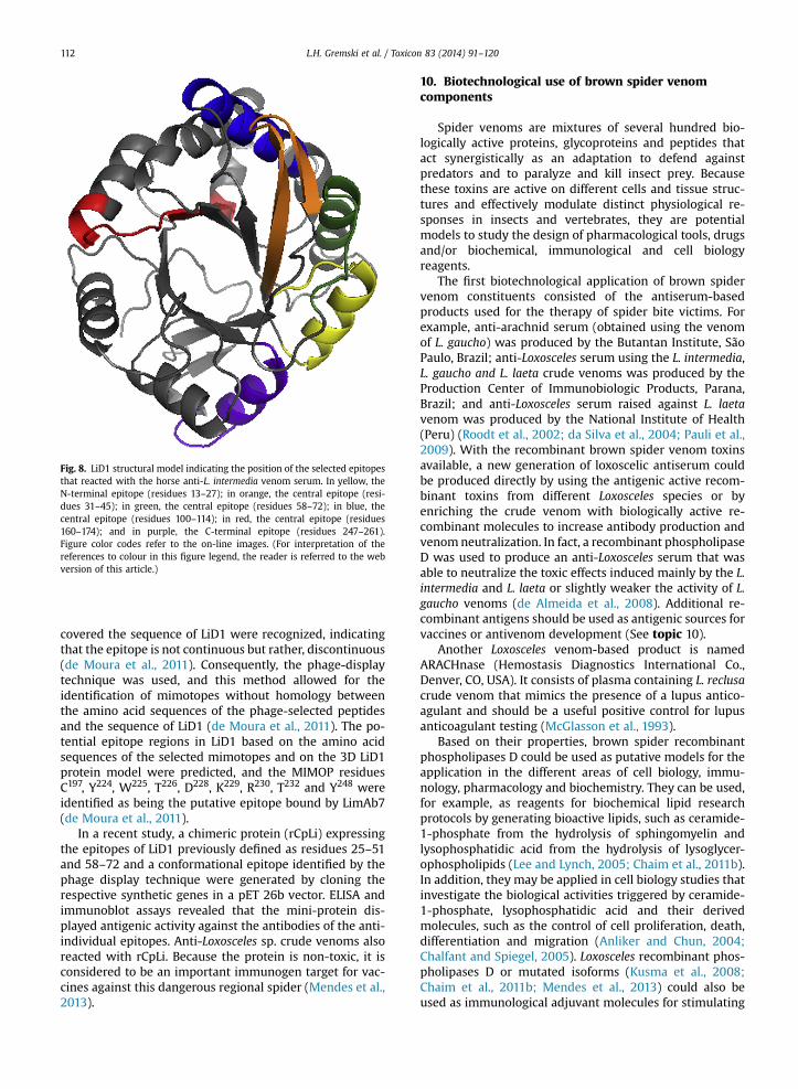
covered the sequence of LiD1 were recognized, indicatingthat the epitope is not continuous but rather, discontinuous(de Moura et al., 2011). Consequently, the phage-displaytechnique was used, and this method allowed for theidentification of mimotopes without homology betweenthe amino acid sequences of the phage-selected peptidesand the sequence of LiD1 (de Moura et al., 2011). The po-tential epitope regions in LiD1 based on the amino acidsequences of the selected mimotopes and on the 3D LiD1protein model were predicted, and the MIMOP residuesC197, Y224, W225, T226, D228, K229, R230, T232 and Y248 wereidentified as being the putative epitope bound by LimAb7(de Moura et al., 2011).
In a recent study, a chimeric protein (rCpLi) expressingthe epitopes of LiD1 previously defined as residues 25–51and 58–72 and a conformational epitope identified by thephage display technique were generated by cloning therespective synthetic genes in a pET 26b vector. ELISA andimmunoblot assays revealed that the mini-protein dis-played antigenic activity against the antibodies of the anti-individual epitopes. Anti-Loxosceles sp. crude venoms alsoreacted with rCpLi. Because the protein is non-toxic, it isconsidered to be an important immunogen target for vac-cines against this dangerous regional spider (Mendes et al.,2013).
10. Biotechnological use of brown spider venomcomponents
Spider venoms are mixtures of several hundred bio-logically active proteins, glycoproteins and peptides thatact synergistically as an adaptation to defend againstpredators and to paralyze and kill insect prey. Becausethese toxins are active on different cells and tissue struc-tures and effectively modulate distinct physiological re-sponses in insects and vertebrates, they are potentialmodels to study the design of pharmacological tools, drugsand/or biochemical, immunological and cell biologyreagents.
The first biotechnological application of brown spidervenom constituents consisted of the antiserum-basedproducts used for the therapy of spider bite victims. Forexample, anti-arachnid serum (obtained using the venomof L. gaucho) was produced by the Butantan Institute, SãoPaulo, Brazil; anti-Loxosceles serum using the L. intermedia,L. gaucho and L. laeta crude venoms was produced by theProduction Center of Immunobiologic Products, Parana,Brazil; and anti-Loxosceles serum raised against L. laetavenom was produced by the National Institute of Health(Peru) (Roodt et al., 2002; da Silva et al., 2004; Pauli et al.,2009). With the recombinant brown spider venom toxinsavailable, a new generation of loxoscelic antiserum couldbe produced directly by using the antigenic active recom-binant toxins from different Loxosceles species or byenriching the crude venom with biologically active re-combinant molecules to increase antibody production andvenomneutralization. In fact, a recombinant phospholipaseD was used to produce an anti-Loxosceles serum that wasable to neutralize the toxic effects induced mainly by the L.intermedia and L. laeta or slightly weaker the activity of L.gaucho venoms (de Almeida et al., 2008). Additional re-combinant antigens should be used as antigenic sources forvaccines or antivenom development (See topic 10).
Another Loxosceles venom-based product is namedARACHnase (Hemostasis Diagnostics International Co.,Denver, CO, USA). It consists of plasma containing L. reclusacrude venom that mimics the presence of a lupus antico-agulant and should be a useful positive control for lupusanticoagulant testing (McGlasson et al., 1993).
Based on their properties, brown spider recombinantphospholipases D could be used as putative models for theapplication in the different areas of cell biology, immu-nology, pharmacology and biochemistry. They can be used,for example, as reagents for biochemical lipid researchprotocols by generating bioactive lipids, such as ceramide-1-phosphate from the hydrolysis of sphingomyelin andlysophosphatidic acid from the hydrolysis of lysoglycer-ophospholipids (Lee and Lynch, 2005; Chaim et al., 2011b).In addition, they may be applied in cell biology studies thatinvestigate the biological activities triggered by ceramide-1-phosphate, lysophosphatidic acid and their derivedmolecules, such as the control of cell proliferation, death,differentiation and migration (Anliker and Chun, 2004;Chalfant and Spiegel, 2005). Loxosceles recombinant phos-pholipases D or mutated isoforms (Kusma et al., 2008;Chaim et al., 2011b; Mendes et al., 2013) could also beused as immunological adjuvant molecules for stimulating
Fig. 8. LiD1 structural model indicating the position of the selected epitopesthat reacted with the horse anti-L. intermedia venom serum. In yellow, theN-terminal epitope (residues 13–27); in orange, the central epitope (resi-dues 31–45); in green, the central epitope (residues 58–72); in blue, thecentral epitope (residues 100–114); in red, the central epitope (residues160–174); and in purple, the C-terminal epitope (residues 247–261).Figure color codes refer to the on-line images. (For interpretation of thereferences to colour in this figure legend, the reader is referred to the webversion of this article.)
L.H. Gremski et al. / Toxicon 83 (2014) 91–120112

immunogenicity because they can modulate inflammationand stimulate the production of cytokines in different cellmodels. Recombinant phospholipases D could be used asstandard laboratory reagents to investigate platelet aggre-gation, platelet receptor(s), and related molecular path-ways. In addition, these recombinant proteins could beused as reagents to induce hemolysis, possibly establishinga new model of hemolysis dependent on phospholipase Dand bioactive lipids. Furthermore, recombinant phospho-lipases D could be used as reagents applied in the diagnosisof loxoscelism because a clinical laboratory diagnosis iscurrently unavailable. Because brown spider venom phos-pholipases D are strong antigenic molecules and are highlyexpressed molecules in crude venom (Ribeiro et al., 2007;Gremski et al., 2010; Wille et al., 2013), recombinantphospholipases D could be used as antigens for generatingpolyclonal or monoclonal antibodies for the diagnosis ofLoxoscelism (de Moura et al., 2011).
ICK peptides have been studied as potential insecticidalbioactive toxin molecules, and recombinant brown spiderICK toxins (Matsubara et al., 2013) could be used as sub-stitutes for chemical defense products as well as in trans-genic agricultural models, if further studies show thatthey specifically act upon insect channels. In addition,brown spider ICK peptides could also be useful reagents toprobe ion channel structures and functions, as previouslydescribed for other similar molecules (Dutertre and Lewis,2010; Klint et al., 2012).
Recombinant Loxosceles hyaluronidase (Ferrer et al.,2013) could be used as a reagent in the biochemicalstudies of glycosaminoglycan hydrolases and as a tool todesign specific inhibitors to reduce the spread of venomand toxins retaining the activity of native hyaluroni-dases. Moreover, because hyaluronidases are involved inbacterial pathogenesis, fertilization, and cancer progres-sion, recombinant brown spider hyaluronidase could beutilized to generate hyaluronidase inhibitors that regulateseveral pathological events involving the balance betweenthe anabolism and catabolism of HA. Finally, it could beused as an adjuvant molecule to increase drug absorptionthrough increased tissue permeabilization (da Silveiraet al., 2007c; Ferrer et al., 2013).
Recombinant brown spider venom astacins (da Silveiraet al., 2007a; Trevisan-Silva et al., 2010) could be used astools in the study of extracellular matrix remodeling, forthe generation of proteolytic inhibitors and as directthrombolytic agents for the treatment of vasculardiseases.
Finally, other brown spider venom constituents alsohave putative biological applications. These include serineprotease inhibitors, which could be useful agents for theinvestigation of general proteolysis, and recombinant TCTP(Sade et al., 2012), which could be used to study tumor cellbehavior in experimental oncology, to study cell prolifera-tion mechanisms, in the screening of anticancer drugs andas a model for allergy screening. Recently, the N-terminalfragment of TCTP (MIIYRDLISH) was shown to function as aProtein Transduction Domain (PTD), which is cell-penetrating peptide. This new feature of TCTP is beingstudied in drug delivery systems development (Maenget al., 2013). Detailed data on the putative
biotechnological use of brown spider venom toxins havebeen provided in Senff-Ribeiro et al. (2008) and Chaim et al.(2011a).
11. Future directions
Althoughmany scientific studies have been published inrecent years examining the brown spider venom and lox-oscelism that have brought insights and improved theknowledge base regarding these topics, there are severalopened questions still to be answered, and the challengesand opportunities for researchers are enormous. The use ofcombined data from molecular biology techniques, bioin-formatics, proteomic studies, transcriptome analysis, andthe expression of recombinant toxins will open great pos-sibilities in this field.
The challenges concerning brown spiders and lox-oscelism can be divided into clinical and basic research. Thefirst clinical challenge is the production of a new generationof antisera using purified recombinant brown spiderbioactive and antigenic competent toxins individually or byenriching crude venoms with recombinant toxins. Suchantisera would be monospecific and may be used at lowerdoses, thereby decreasing the deleterious side effects ofserum therapy, but with the same or higher efficiency inneutralizing the noxious venom activities.
A second clinical challenge hinges on the improvementof treatment of the injured victims. Currently, loxoscelismtreatment is empirical and based on clinical signs, asdescribed above (topic 4). A molecular comprehension ofbrown spider venoms and the mechanism by which thetoxins trigger their effects, together by obtaining recom-binant toxins and toxin 3D structural/biological data, willopen to the possibility of a rational design of syntheticinhibitors directed at the specific venom toxins involvedin the local and systemic effects. In addition, inhibitors forthe receptors or cellular molecules involved in the meta-bolism of the bioactive lipid mediators generated byphospholipases D could also produce novel and powerfultools for the treatment of loxoscelism. Another clinicalhurdle is the development of a clinical, in vitro diagnosticfor loxoscelism with the sensitivity and specificity fordifferent brown spider species. This diagnostic would bebased on low-invasive molecular biology techniques andwould provide sensitivity and specificity for differentbrown spider species using blood or even urine takenfrom exposed victims.
Finally, because loxoscelism is a public health problemaround the world, the development of rational biologicalcontrol methods, which are currently not available, isnecessary to decrease the number of spiders and domicil-iary infestations in the endemic regions. Similar methodsare currently used for agricultural plagues, utilizing bacte-ria, fungus or other natural predators (Boyer et al., 2012;Lockett et al., 2012) to replace the pesticides currentlyused as a nonspecific biological control method and whichcause environmental and human hazards.
The future of basic research on brown spider venomsand/or loxoscelism represents a remarkable challenge.Although considerable growth in this field has occurred, agreat number of molecules in brown spider venoms remain
L.H. Gremski et al. / Toxicon 83 (2014) 91–120 113

unidentified or their biological effects and mechanismshave not been described, especially for toxins with lowlevel of expression and/or novel toxin isoforms of previ-ously described. In addition, a genomic project focusing onLoxosceles species is a rational future direction that willbring novel insights for brown spider biology and lox-oscelism and that will create access for several novelresearch tools.
Another current challenge for the brown spider venomtoxinologist is the access to purified recombinant toxins inmodels other than bacteria. Currently, all recombinantbrown spider venom toxins obtained have been producedin bacteria, an inexpensive expression model system that issimple to manipulate. However, because it does notgenerate co- and post-translational modifications, such asN-glycosylation and disulfide bonds, bacteria model sys-tems often produce recombinant molecules in theirunfolded form, with incorrect conformations, water insol-ubility, and with no biological function. The synthesis ofbrown spider venom recombinant toxins using alternativeexpression models with additional features that optimizeand refine this process, such as the yeast Pichia pastoris, theinsect Drosophila Schneider cells and mammalian systems,is an immediate challenge.
Another future direction is to obtain native brownspider venom toxins by developing primary cultures ofthe venom secretory cells. Similar approaches have beensuccessfully established for other venomous animals. Ex-amples include culturing the secretory cells from thevenom glands of snakes Crotalus durissus terrificus andBothrops jararaca (Duarte et al., 1999; Yamanouye et al.,2007) and from the Phoneutria nigriventer spider (Silvaet al., 2008). These cultures could produce and secretesufficient amounts of native toxins to be useful for bio-logical and biotechnological evaluation. The use of massspectrometry analysis and other proteomic protocols,such as 2-DE, N-terminal amino acid sequencing and highefficiency chromatography, provide great promise fordetailed studies of brown spider venoms and hemolymphproteins and peptides. To date, only two studies haveaddressed this topic: Machado et al. (2005) describedeleven isoforms of the phospholipase D toxin in L. gauchovenom, and dos Santos et al. (2009) identified 39 proteinsin L. intermedia venom. There are no reports of this type ofstudy for hemolymph.
Additionally, in the near future, novel data will provideinformation related to the tridimensional toxin structures,which will require the experimental co-crystallization ofputative ligands or substrates to recombinant toxins. Thetridimensional analysis of brown spider venom toxins willbe critical to elucidate the location of the catalytic sites andsites that interact with natural substrates or ligands, andespecially to show how the toxins interact with cell struc-tures. These analyses will allow for the development ofsynthetic ligands, analogs, or inhibitors.
Finally, an attractive and practically unknown model forstudying Loxosceles spiders is the analysis of hemolymphcontents and its relationship to venom toxins and lox-oscelism. From such analysis, natural inhibitors and/orother important molecules could be discovered, whichwould add great value to the field of toxinology.
Acknowledgments
This work was supported by grants from the CAPES,CNPq, FAPESP, FAPEMIG, INCTTOX, Fundação Araucária-Paraná and Secretaria de Tecnologia e Ensino Superior doParaná, SETI-PR, Brasil.
Conflict of interest statement
None.
References
Abdulkader, R.C., Barbaro, K.C., Barros, E.J., Burdmann, E.A., 2008. Neph-rotoxicity of insect and spider venoms in Latin America. Semin.Nephrol. 28, 373–382.
Akdeniz, S., Green, J.A., Stoecker, W.V., Gomez, H.F., Keklikci, S.U., 2007.diagnosis of loxoscelism in two turkish patients confirmed with anenzyme-linked immunosorbent assay (ELISA) and non-invasive tissuesampling. Dermatol. Online J. 13, 11.
Alvarenga, L.M., Martins, M.S., Moura, J.F., Kalapothakis, E., Oliveira, J.C.,Mangili, O.C., Granier, C., Chávez-Olórtegui, C., 2003. Production ofmonoclonal antibodies capable of neutralizing dermonecrotic activityof Loxosceles intermedia spider venom and their use in a specificimmunometric assay. Toxicon 42, 725–731.
Amson, R., Pece, S., Marine, J.C., di Fiore, P.P., Telerman, A., 2013a. TPT1/TCTP-regulated pathways in phenotypic reprogramming. Trends CellBiol. 23, 37–46.
Amson, R., Karp, J.E., Telerman, A., 2013b. Lessons from tumor reversionfor cancer treatment. Curr. Opin. Oncol. 25, 59–65.
Amzallag, N., Passer, B.J., Allanic, D., Segura, E., Thery, C., Goud, B.,Amson, R., Telerman, A., 2004. TSAP6 facilitates the secretion oftranslationally controlled tumor protein/histamine-releasing factorvia a nonclassical pathway. J. Biol. Chem. 279, 46104–46112.
Andersen, R.J., Campoli, J., Johar, S.K., Schumacher, K.A., Allison Jr., E.J.,2011. Suspected brown recluse envenomation: a case report and re-view of different treatment modalities. J. Emerg. Med. 41, 31–37.
Anliker, B., Chun, J., 2004. Lysophospholipid G protein-coupled receptors.J. Biol. Chem. 279, 20555–20558.
Appel, M.H., da Silveira, R.B., Gremski, W., Veiga, S.S., 2005. Insights intobrown spider and loxoscelism. Inverteb. Surv. J. 2, 152–158.
Appel, M.H., da Silveira, R.B., Chaim, O.M., Paludo, K.S., Trevisan-Silva, D.,Chaves-Moreira, D., da Silva, P.H., Mangili, O.C., Senff-Ribeiro, A.,Gremski, W., Nader, H.B., Veiga, S.S., 2008. Identification, cloning andfunctional characterization of a novel dermonecrotic toxin (phos-pholipase D) from brown spider (Loxosceles intermedia) venom. Bio-chim. Biophys. Acta 1780, 167–178.
Araújo, S.C., Castanheira, P., Alvarenga, L.M., Mangili, O.C., Kalapothakis, E.,Chávez-Olórtegui, C., 2003. Protection against dermonecrotic andlethal activities of Loxosceles intermedia spider venom by immuni-zation with a fused recombinant protein. Toxicon 41, 261–267.
Araújo, H.R.C., 2009. Ultra-estrutura dos hemócitos de Aedes aegypti(Linnaeus, 1762) (Diptera, Culicidae). Dissertação apresentada aoMestrado em Saúde Pública do Centro de Pesquisas Aggeu Magalhães,Fundação Oswaldo Cruz, para obtenção do grau de Mestre em Ciên-cias, p. 86.
Arlian, L.G., 2002. Arthropod allergens and human health. Annu. Rev.Entomol. 47, 395–433.
Bajin, M.S., Arikan, G., Parlak, M., Tuncok, Y., Yigit, N., Durak, I., Saatci, A.O.,2011. Necrotic arachnidism of the eyelid due to Loxosceles rufescensspider bite. Cutan. Ocul. Toxicol. 30, 302–305.
Barbaro, K.C., Cardoso, J.L.C., 2003. Mecanismo de ação do veneno deLoxosceles e aspectos clínicos do loxoscelismo. In: Cardoso, J.L.C.,França, F.O.S., Fan, H.W., Málaque, C.M.S., Haddad Jr., H. (Eds.), Ani-mais Peçonhentos no Brasil: Biologia, Clínica e Terapêutica dos acid-entes. Savier, São Paulo, Brasil, pp. 160–174.
Barbaro, K.C., Knysak, I., Martins, R., Hogan, C., Winkel, K., 2005. Enzy-matic characterization, antigenic cross-reactivity and neutralizationof dermonecrotic activity of five Loxosceles spider venoms of medicalimportance in the Americas. Toxicon 45, 489–499.
Barbaro, K.C., Lira, M.S., Araújo, C.A., Pareja-Santos, A., Távora, B.C.L.F.,Prezotto-Neto, J.P., Kimura, L.F., Lima, C., Lopes-Ferreira, M.,Santoro, M.L., 2010. Inflammatory mediators generated at the site ofinoculation of Loxosceles gaucho spider venom. Toxicon 56, 972–979.
Barla, F., Higashijima, H., Funai, S., Sugimoto, K., Harada, N., Yamaji, R.,Fujita, T., Nakano, Y., Inui, H., 2009. Inhibitive effects of alkyl gallates
L.H. Gremski et al. / Toxicon 83 (2014) 91–120114

on hyaluronidase and collagenase. Biosci. Biotechnol. Biochem 73,2335–2337.
Barretto, O.C., Prestes, K.S., Fonseca, L.K.F., Silveira, P.A.A., 2007. Functionalalterations of rabbit erythrocytes induced by Loxosceles gauchovenom. Hum. Exp.Toxicol. 26, 817–821.
Bazile, F., Pascal, A., Arnal, I., Le Clainche, C., Chesnel, F., Kubiak, J.Z., 2009.Complex relationshipbetween TCTP, microtubules and actin micro-filaments regulates cell shape in normal and cancer cells. Carcino-genesis 30, 555–565.
Brasil, 2001. Manual de diagnóstico e tratamento de acidentes por ani-mais peçonhentos. Ministério da Saúde, Fundação Nacional de Saúde,Brasília, p. 120.
Bentele, C., Krüger, O., Tödtmann, U., Oley, M., Ragg, H., 2006. A proproteinconvertase-inhibiting serpin with an endoplasmic reticulum target-ing signal from Branchiostoma lanceolatum, a close relative of verte-brates. Biochem. J. 395, 449–456.
Bertani, R., Fukushima, C.S., Nagahama, R.H., 2010. Loxosceles chapadensis(Araneae: Sicariidae): a new recluse spider species of the gauchogroup from Brazil. J. Arachnol. 38, 364–367.
Binford, G.J., Wells, M.A., 2003. The phylogenetic distribution of sphin-gomyelinase D activity in venoms of Haplogyne spiders. Comp. Bio-chem. Physiol. B. Biochem. Mol. Biol. 135, 25–33.
Binford, G.J., Cordes, M.H., Wells, M.A., 2005. Sphingomyelinase D fromvenoms of Loxosceles spiders: evolutionary insights from cDNA se-quences and gene structure. Toxicon 45, 547–560.
Binford, G.J., Callahan, M.S., Bodner, M.R., Rynerson, M.R., Núñes, P.B.,Ellison, C.E., Duncan, R.P., 2008. Phylogenetic relationships of Lox-osceles and Sicarius spiders are consistent with Western Gondwananvicariance. Mol. Phylogenet. Evol. 48, 538–553.
Binford, G.J., Bodner, M.R., Cordes, M.H., Baldwin, K.L., Rynerson, M.R.,Burns, S.N., Zobel-Thropp, P.A., 2009. Molecular evolution, functionalvariation, and proposed nomenclature of the gene family that in-cludes sphingomyelinase D in sicariid spider venoms. Mol. Biol. Evol.26, 547–566.
Bircher, A.J., 2005. Systemic immediate allergic reactions to arthropodstings and bites. Dermatology 210, 119–127.
Bommer, U.A., Thiele, B.J., 2004. The translationally controlled tumourprotein (TCTP). Int. J. Biochem. Cell Biol. 36, 379–385.
Bommer, U., 2012. Cellular function and regulation of the translationallycontrolled tumour protein TCTP. Open Allergy J. 5, 19–32.
Borer, A.S., Wassmann, P., Schmidt, M., Hoffman, D.R., Zhou, J.J., Wright, C.,Schirmer, T., Markovi�c-Housley, Z., 2012. Crystal structure of Sol I 2: amajor allergen from fire ant venom. J. Mol. Biol. 415, 635–648.
Botzki, A., Rigden, D.J., Braun, S., Nukui, M., Salmen, S., Hoechstetter, J.,Bernhardt, G., Dove, S., Jedrzejas, M.J., Buschauer, 2004. A. L-Ascorbicacid. 6-hexadecanoate, a potent hyaluronidase inhibitor. X-raystructure and molecular modeling of enzyme-inhibitor complexes. J.Biol. Chem. 279, 45990–45997.
Boyer, S., Paris, M., Jego, S., Lempérière, G., Ravanel, P., 2012. Influence ofinsecticide Bacillus thuringiensis subsp. israelensis treatments onresistance and enzyme activities in Aedes rusticus larvae (Diptera:Culicidae). Biol. Control 62, 75–81.
Bucaretchi, F., de Capitani, E.M., Hyslop, S., Sutti, R., Rocha-e-Silva, T.A.,Bertani, R., 2010. Cutaneous loxoscelism caused by Loxosceles anom-ala. Clin. Toxicol. 48, 764–765.
Burgess, A., Labbe, J.C., Vigneron, S., Bonneaud, N., Strub, J.M., vanDorsselaer, A., Lorca, T., Castro, A., 2008. Chfr interacts and colocalizeswith TCTP to the mitotic spindle. Oncogene 27, 5554–5566.
Casewell, N.R., Harrison, R.A., Wüster, W., Wagstaff, S.C., 2009. Compar-ative venom gland transcriptome surveys of the saw-scaled vipers(Viperidae:Echis) reveal substantial intra family gene diversity andnovel venom transcripts. BMC Genomics 10, 564.
Catalan, A., Cortes, W., Sagua, H., Gonzalez, J., Araya, J.E., 2011. Two newphospholipase D isoforms of Loxosceles laeta: cloning, heterologousexpression, functional characterization, and potential biotechnolog-ical application. J. Biochem. Mol. Toxicol. 25, 393–403.
Chaim, O.M., Sade, Y.B., da Silveira, R.B., Toma, L., Kalapothakis, E., Chavez-Olortegui, C., Mangili, O.C., Gremski, W., von Dietrich, C.P., Nader, H.B.,Sanches Veiga, S., 2006. Brown spider dermonecrotic toxin directlyinduces nephrotoxicity. Toxicol. Appl. Pharmacol. 211, 64–77.
Chaim, O.M., Trevisan-Silva, D., Chaves-Moreira, D., Wille, A.C.M.,Ferrer, V.P., Matsubara, F.H., Mangili, O.C., da Silveira, R.B.,Gremski, L.H., Gremski, W., Senff-Ribeiro, A., Veiga, S.S., 2011a. Brownspider (Loxosceles genus) venom toxins: tools for biological purposes.Toxins 3, 309–344.
Chaim, O.M., da Silveira, R.B., Trevisan-Silva, D., Ferrer, V.P., Sade, Y.B.,Boia-Ferreira, M., Gremski, L.H., Gremski, W., Senff-Ribeiro, A.,Takahashi, H.K., Toledo, M.S., Nader, H.B., Veiga, S.S., 2011b. Phos-pholipase-D activity and inflammatory response induced by brown
spider dermonecrotic toxin: endothelial cell membrane phospho-lipids as targets for toxicity. Biochim. Biophys. Acta 1811, 84–96.
Chalfant, C.E., Spiegel, S., 2005. Sphingosine 1-phosphate and ceramide 1-phosphate: expanding roles in cell signaling. J. Cell Sci. 118, 4605–4612.
Chatzaki, M., Horta, C.C., Almeida, M.O., Pereira, N.B., Mendes, T.M., Dias-Lopes, C., Guimarães, G., Moro, L., Chávez-Olórtegui, C., Horta, M.C.R.,Kalapothakis, E., 2012. Cutaneous loxoscelism caused by Loxoscelessimilis venom and neutralization capacity of its specific antivenom.Toxicon 60, 21–30.
Chaves-Moreira, D., Chaim, O.M., Sade, Y.B., Paludo, K.S., Gremski, L.H.,Donatti, L., de Moura, J., Mangili, O.C., Gremski, W., da Silveira, R.B.,Senff-Ribeiro, A., Veiga, S.S., 2009. Identification of a direct hemolyticeffect dependent on the catalytic activity induced by phospholipase-D (dermonecrotic toxin) from brown spider venom. J. Cell. Biochem.107, 655–666.
Chaves-Moreira, D., Souza, F.N., Fogaça, R.T.H., Mangili, O.C., Gremski, W.,Senff-Ribeiro, A., Chaim, O.M., Veiga, S.S., 2011. The relationship be-tween calcium and the metabolism of plasma membrane phospho-lipids in hemolysis induced by brown spider venom phospholipase-D.Toxin. J. Cell. Biochem. 112, 2529–2540.
Chen, S.H., Wu, P.S., Chou, C.H., Yan, Y.T., Liu, H., Weng, S.Y., Yang-Yen, H.F.,2007. A knockout mouse approach reveals that TCTP functions as anessential factor for cell proliferation and survival in a tissue- or celltype-specific manner. Mol. Biol. Cell 18, 2525–2532.
Choi, K.W., Hsu, Y.C., 2007. To cease or to proliferate: new insights intoTCTP function from a Drosophila study. Cell Adhes. Migr. 1, 129–130.
Christoff, A.O., de Oliveira, A., Chaim, O.M., Lugarini, D., Pereira, A.L.B.,Paludo, K.S., Telles, J.E.Q., Bracht, A., Veiga, S.S., Acco, A., 2008. Effect ofthe venom and the dermonecrotic toxin LiRecDT1 of Loxoscelesintermedia in the rat liver. Toxicon 52, 695–704.
Cidade, D.A., Simão, T.A., Dávila, A.M., Wagner, G., Junqueira-De-Azevedo, I.L., Ho, P.L., Bon, C., Zingali, R.B., Albano, R.M., 2006.Bothrops jararaca venom gland transcriptome: analysis of the geneexpression pattern. Toxicon 48, 437–461.
Cordes, M.H., Binford, G.J., 2006. Lateral gene transfer of a dermonecrotictoxin between spiders and bacteria. Bioinformatics 22, 264–268.
Corrêa-Netto, C., Junqueira-de-Azevedo, I.L.M., Silva, D.A., Ho, P.L., Leitão-de-Araújo, M., Alves, M.L., Sanz, L., Foguel, D., Zingali, R.B., Calvete, J.J.,2011. Snake venomics and venom gland transcriptomic analysis ofBrazilian coral snakes, Micrurus altirostris and M. corallinus. J. Prote-omics 74, 1795–1809.
Corzo, G., Gilles, N., Satake, H., Villegas, E., Dai, L., Nakajima, T., Haupt, J.,2003. Distinct primary structures of the major peptide toxins fromthe venom of the spider Macrothelegigas that bind to sites 3 and 4 inthe sodium channel. FEBS Lett. 547, 43–50.
Craik, D.J., Daly, N.L., Waine, C., 2001. The cystine knot motif in toxins andimplications for drug design. Toxicon 39, 43–60.
Craik, D., Mylne, J., Daly, N., 2010. Cyclotides: macrocyclic peptides withapplications in drug design and agriculture. Cell. Mol. Life Sci. 67, 9–16.
Cramer, K., 2008. Are brown recluse spiders, Loxosceles reclusa (Araneae,Sicariidae) scavengers? The influence of predator satiation, prey sizeand prey quality. J. Arachnol. 36, 140–144.
Cunha, R.B., Barbaro, K.C., Muramatsu, D., Portaro, F.C., Fontes, W., deSousa, M.V., 2003. Purification and characterization of loxnecrogin, adermonecrotic toxin from Loxosceles gaucho brown spider venom. J.Protein Chem. 22, 135–146.
Daly, N.L., Craik, D.J., 2011. Bioactive cystine knot proteins. Bioactivecystine knot proteins. Curr. Opin. Chem. Biol. 15, 362–368.
da Silva, P.H., Hashimoto, Y., Santos, F.A., Mangili, O.C., Gremski, W.,Veiga, S.S., 2003. Hematological cell findings in bone marrow andperipheral blood of rabbits after experimental acute exposure toLoxosceles intermedia (brown spider) venom. Toxicon 42, 155–161.
da Silva, P.H., da Silveira, R.B., Appel, M.H., Mangili, O.C., Gremski, W.,Veiga, S.S., 2004. Brown spiders and loxoscelism. Toxicon 44, 693–709.
da Silveira, R.B., Filho, J.F.S., Mangili, O.C., Veiga, S.S., Gremski, W.,Nader, H.B., 2002. Identification of proteases in the extract of venomglands from brown spider. Toxicon 40, 815–822.
da Silveira, R.B., Pigozzo, R.B., Chaim, O.M., Appel, M.H., Dreyfuss, J.L.,Toma, L., 2006. Molecular cloning and functional characterization oftwo isoforms of dermonecrotic toxin from Loxosceles intermedia(Brown spider) venom gland. Biochimie 88, 1241–1253.
da Silveira, R.B., Wille, A.C.M., Chaim, O.M., Appel, M.H., Silva, D.T.,Franco, C.R., 2007a. Identification, cloning, expression and func-tional characterization of an astacin-like metalloprotease toxinfrom Loxosceles intermedia (brown spider) venom. Biochem. J. 406,355–363.
L.H. Gremski et al. / Toxicon 83 (2014) 91–120 115
da Silveira, R.B., Pigozzo, R.B., Chaim, O.M., Appel, M.H., Trevisan- Silva, D.,Dreyfuss, J.L., 2007b. Two novel dermonecrotic toxins LiRecDT4 andLiRecDT5 from Brown spider (Loxosceles intermedia) venom: fromcloning to functional characterization. Biochimie 89, 289–300.
da Silveira, R.B., Chaim, O.M., Mangili, O.C., Gremski, W., Dietrich, C.P.,Nader, H.B., Veiga, S.S., 2007c. Hyaluronidases in Loxosceles intermedia(Brown spider) venom are endo-beta-N-acetyl-d-hexosaminidaseshydrolases. Toxicon 49, 758–768.
de Almeida, D.M., Fernandes-Pedrosa, F.M., de Andrade, R.M.,Marcelino, J.R., Gondo-Higashi, H., de Azevedo, I.L.M.J., Ho, P.L., vanden Berg, C., Tambourgi, D.V., 2008. A new anti-loxoscelic serumproduced against recombinant sphingomyelinase D: results of pre-clinical trials. Am. J. Trop. Med. Hyg. 79, 463–470.
de Castro, C.S., Silvestre, F.G., Araujo, S.C., de Gabriel, M.Y., Mangili, O.C.,Cruz, I., Chavez-Olortegui, C., Kalapothakis, E., 2004. Identificationand molecular cloning of insecticidal toxins from the venom of thebrown spider Loxosceles intermedia. Toxicon 44, 273–280.
de Giuseppe, P.O., Ullah, A., Trevisan-Silva, D., Gremski, L.H., Wille, A.C.M.,Chaves-Moreira, D., Senff-Ribeiro, A., Chaim, O.M., Murakami, M.T.,Veiga, S.S., Arni, R.K., 2011. Structure of a novel class II phospholipaseD: catalytic cleft is modified by a disulphide bridge. Biochem. Biophys.Res. Commun. 409, 622–627.
de Moura, J., Felicori, L., Moreau, V., Guimarães, G., Dias-Lopes, C.,Molina, L., Alvarenga, L.M., Fernandes, P., Frézard, F., Ribeiro, R.R.,Fleury, C., Nguyen, C., Molina, F., Granier, C., Chávez-Olórtegui, C.,2011. Protection against the toxic effects of Loxosceles intermediaspider venom elicited by mimotope peptides. Vaccine 29, 7992–8001.
de Oliveira, K.C., Gonçalves-de-Andrade, R.M., Piazza, R.M.F.,Ferreira Jr., J.M.C., van den Berg, C.W., Tambourgi, D.V., 2005. Varia-tions in Loxosceles spider venom composition and toxicity contributeto the severity of envenomation. Toxicon 45, 421–429.
Desai, A., Miller, M.J., Gomez, H.F., Warren, J.S., 1999. Loxosceles desertaspider venom induces NF-kB-dependent chemokine production byendothelial cells. J. Toxicol. Clin. Toxicol. 37, 447–456.
Desai, A., Lankford, H.A., Warren, J.S., 2000. Loxosceles deserta spidervenom induces the expression of vascular endothelial growth factor(VEGF) in keratinocytes. Inflammation 24, 1–9.
de Santi Ferrara, G.I., Fernandes-Pedrosa, M.F., Junqueira-de-Azevedo, I.L.,Gonçalves-de-Andrade, R.M., Portaro, F.C., Manzoni-de-Almeida, D.,Murakami, M.T., Arni, R.K., van den Berg, C.W., Ho, P.L.,Tambourgi, D.V., 2009. SMase II, a new sphingomyelinase D fromLoxosceles laeta venom gland: molecular cloning, expression, functionand structural analysis. Toxicon 53, 743–753.
de Souza, A.L., Malaque, C.M., Sztajnbok, J., Romano, C.C., Duarte, A.J.,Seguro, A.C., 2008. Loxosceles venom-induced cytokine activation,hemolysis, and acute kidney injury. Toxicon 51, 151–156.
Devaraja, S., Girish, K.S., Devaraja, V.R., Kemparaju, K., 2010. Factor Xa-likeand fibrin(ogen)olytic activities of a serine protease from Hippasaagelenoides spider venom gland extract. J. Thromb. Thrombolysis 29,119–126.
Dias-Lopes, C., Felicori, L., Guimarães, G., Gomes, E.R., Roman-Campos, D.,Duarte, H., Damasceno, D., Martins, M., Kalapothakis, E., Almeida, A.P.,Granier, C., Cruz, J.S., Guatimosim, S., Chávez-Olórtegui, C., 2010.Cardiotoxic effects of Loxosceles intermedia spider venom and therecombinant venom toxin rLiD1. Toxicon 56, 1426–1435.
Domingos, M.O., Barbaro, K.C., Tynan, W., Penny, J., Lewis, D.J., New, R.R.,2003. Influence of sphingomyelin and TNF-alpha release on lethalityand local inflammatory reaction induced by Loxosceles gaucho spidervenom in mice. Toxicon 42, 471–479.
Donepudi, S.K., Ahmed, K.A., Stocks, R.M., Nelson, D., Thompson, J.W.,2005. Aural involvement in loxoscelism: case report and literaturereview. Int. J. Pediatr. Otorhinolaryngol. 69, 1559–1561.
dos Santos, V.L., Franco, C.R.C., Viggiano, R.L., da Silveira, R.B., Cantão, M.P.,Mangili, P.C., Veiga, S.S., Gremski, W., 2000. Structural and ultra-structural description of the venom gland of Loxosceles intermedia.Toxicon 38, 265–285.
dos Santos, L.D., Dias, N.B., Pinto, J.R.A.S., Palma, M.S., 2009. Brown reclusespider venom: proteomic analysis and proposal of a putative mech-anism of action. Protein Pept. Lett. 16, 933–943.
Dragulev, B., Bao, Y., Ramos-Cerrillo, B., Vazquez, H., Olvera, A., Stock, R.,Algaron, A., Fox, J.W., 2007. Upregulation of IL-6, IL-8, CXCL1, andCXCL2 dominates gene expression in human fibroblast cells exposedto Loxosceles reclusa sphingomyelinase D: insights into spider venomdermonecrosis. J. Investig. Dermatol. 127, 1264–1266.
Duarte, M.M., Montes De Oca, H., Diniz, C.R., Fortes-Dias, C.L., 1999. Pri-mary culture of venom gland cells from the South American rattle-snake (Crotalus durissus terrificus). Toxicon 37, 1673–1682.
Duncan, R.P., Rynerson, M.R., Ribera, C., Binford, G.J., 2010. Diversity ofLoxosceles spiders in Northwestern Africa and molecular support for
cryptic species in the Loxosceles rufescenslineage. Mol. Phylogenet.Evol. 55, 234–248.
Dutertre, S., Lewis, R.J., 2010. Use of venom peptides to probe ion channelstructure and function. J. Biol. Chem. 285, 13315–13320.
Dyachenko, P., Ziv, M., Rozenman, D., 2006. Epidemiological and clinicalmanifestations of patients hospitalized with brown recluse spiderbite. J. Eur. Acad. Dermatol. Venereol. 20, 1121–1125.
Efferth, T., 2005. Mechanistic perspectives for 1,2,4-trioxanes in anti-cancer therapy. Drug Resist. Updat. 8, 85–97.
Elbahlawan, L.M., Stidham, G.L., Bugnitz, M.C., Storgion, S.A.,Quasney, M.W., 2005. Severe systemic reaction to Loxosceles reclusaspider bites in a pediatric population. Pediatr. Emerg. Care 21, 177–180.
Eskafi, F.M., Norment, B.R., 1976. Physiological action of Loxosceles reclusavenom on insect larvae. Toxicon 14, 7–12.
Estrada, G., Garcia, B.I., Schiavon, E., Ortiz, E., Cestele, S., Wanke, E.,Possani, L.D., Corzo, G., 2007. Four disulfide-bridged scorpion betaneurotoxin CssII: heterologous expression and proper folding in vitro.Biochim. Biophys. Acta 1770, 1161–1168.
Feitosa, L., Gremski, W., Veiga, S.S., Elias, M.C., Graner, E., Mangili, O.C.,Brentani, R.R., 1998. Detection and characterization of metal-loproteinases with gelatinolytic, fibronectinolytic and fibrinogeno-lytic activities in brown spider (Loxosceles intermedia) venom. Toxicon36, 1039–1051.
Felicori, L., Araujo, S.C., de Avila, R.A., Sanchez, E.F., Granier, C.,Kalapothakis, E., Chavez-Olortegui, C., 2006. Functional character-ization and epitope analysis of a recombinant dermonecrotic proteinfrom Loxosceles intermedia spider. Toxicon 48, 509–519.
Felicori, L., Fernandes, P.B., Giusta, M.S., Duarte, C.G., Kalapothakis, E.,Nguyen, C., Molina, F., Granier, C., Chávez-Olórtegui, C., 2009. Anin vivo protective response against toxic effects of the dermonecroticprotein from Loxosceles intermedia spider venom elicited by syntheticepitopes. Vaccine 27, 4201–4208.
Fernandes-Pedrosa, M.F., Junqueira-de-Azevedo, I.L.M., Gonçalves-de-Andrade, R.M., van den Berg, C.W., Ramos, C.R., Ho, P.L.,Tambourgi, D.V., 2002. Molecular cloning and expression of a func-tional dermonecrotic and haemolytic factor from Loxosceles laetavenom. Biochem. Biophys. Res. Commun. 298, 638–645.
Fernandes-Pedrosa, M.F., Junqueira-de-Azevedo, I.L.M., Gonçalves-de-Andrade, R.M., Kobashi, L.S., Almeida, D.D., Ho, P.L., Tambourgi, D.V.,2008. Transcriptome analysis of Loxosceles laeta (Araneae, Sicariidae)spider venomous gland using expressed sequence tags. BMC Geno-mics 9, 279.
Ferrer, V.P., de Mari, T.L., Gremski, L.H., Trevisan-Silva, D., da Silveira, R.B.,Gremski, W., Chaim, O.M., Senff-Ribeiro, A., Nader, H.B., Veiga, S.S.,2013. A novel hyaluronidase from brown spider (Loxosceles inter-media) venom (Dietrich’s Hyaluronidase): from cloning to functionalcharacterization. PLoS Negl. Trop. Dis. 7, e2206.
Fischer, M.L., Vasconcellos-Neto, J., 2005a. Development and life tables ofLoxosceles intermedia Mello-Leitão, 1934 (Araneae, Sicariidae). J.Arachnol. 33, 758–766.
Fischer, M.L., Vasconcellos-Neto, J., 2005b. Microhabitats occupied byLoxosceles intermedia and Loxosceles laeta (Araneae: Sicariidae) incuritiba, paraná, Brazil. J. Med. Entomol. 42, 756–765.
Flight, S.M., Johnson, L.A., Trabi, M., Gaffney, P., Lavin, M., de Jersey, J.,Masci, P., 2005. Comparison of textilinin-1 with aprotinin as serineprotease inhibitors and as antifibrinolytic agents. Pathophysiol. Hae-most. Thromb. 34, 188–193.
França, F.O.S., Barbaro, K.C., Abdulkader, C.R.M., 2002. Rhabdomyolisis inpresumed viscero-cutaneous loxoscelism: report of two cases. Trans.R. Soc. Med. Hyg. 96, 287–290.
Funk, C., Gmür, J., Herold, R., Straub, P.W., 1971. Reptilase-R–a new reagentin blood coagulation. Br. J. Haematol. 21, 43–52.
Gilbert, S.F., Raunio, A.M., 1997. Embryology: Constructing the Organism.Sinauer Associates, Incorporated, Sunderland, MA, USA.
Girish, K.S., Shashidharamurthy, R., Nagaraju, S., Gowda, T.V.,Kemparaju, K., 2004. Isolation and characterization of hyaluronidase a“spreading factor” from Indian cobra (Naja naja) venom. Biochimie86, 193–202.
Girish, K.S., Kemparaju, K.A., 2005. Low molecular weight isoform ofhyaluronidase: purification from Indian cobra (Naja naja) venom andpartial characterization. Biochemistry 70, 708–712.
Girish, K.S., Kemparaju, K., 2007. The magic glue hyaluronan and its eraserhyaluronidase: a biological overview. Life Sci. 80, 1921–1943.
Gomes, M.T., Guimarães, G., Frézard, F., Kalapothakis, E., Minozzo, J.C.,Chaim, O.M., Veiga, S.S., Oliveira, S.C., Chávez-Olórtegui, C., 2011.Determination of sphingomyelinase-D activity of Loxosceles venomsin sphingomyelin/cholesterol liposomes containing horseradishperoxidase. Toxicon 57, 574–579.
L.H. Gremski et al. / Toxicon 83 (2014) 91–120116

Gomez, H.F., Miller, M.J., Desai, A., Warren, J.S., 1999. Loxosceles spidervenom induces the production of alpha and beta chemokines: im-plications for the pathogenesis of dermonecrotic arachnidism.Inflammation 23, 207–215.
Gomez, H.F., Miller, M.J., Waggener, M.W., Lankford, H.A., Warren, J.S.,2001. Antigenic cross-reactivity of venoms from medically importantNorth American Loxosceles spider species. Toxicon 39, 817–824.
Gomez, H.F., Krywko, D.M., Stoecker, W.V., 2002. A new assay for thedetection of Loxosceles species (brown recluse) spider venom. Ann.Emerg. Med. 39, 469–474.
Gomis-Rüth, F.X., 2003. Structural aspects of the metzincin clan of met-alloendopeptidases. Mol. Biotechnol. 24, 157–202.
Gomis-Rüth, F.X., Trillo-Muyo, S., Stöcker, W., 2012. Functional andstructural insights into astacin metallopeptidases. Biol. Chem. 393,1027–1041.
Gonçalves-de-Andrade, R.M., Bertani, R., Nagahama, R.H.,Barbosa, M.F.R., 2012. Loxosceles niedeguidonae (Araneae, Sicariidae)a new species of brown spider from Brazilian semi-arid region.Zookeys 175, 27–36.
Graidist, P., Yazawa, M., Tonganunt, M., Nakatomi, A., Lin, C.C., Chang, J.Y.,Phongdara, A., Fujise, K., 2007. Fortilin binds Ca2þ and blocks Ca2þ-dependent apoptosis in vivo. Biochem. J. 408, 181–191.
Gremski, L.H., da Silveira, R.B., Chaim, O.M., Probst, C.M., Ferrer, V.P.,Nowatzki, J., Weinschutz, H.C., Madeira, H.M., Gremski, W.,Nader, H.B., Senff-Ribeiro, A., Veiga, S.S., 2010. A novel expressionprofile of the Loxosceles intermedia venomous gland revealed bytranscriptome analysis. Mol. Biosyst. 6, 2403–2416.
Guo, Y., Zuo, Y.F., Wang, Q.Z., Tang, B.S., Li, F.K., Sun, Y., 2006. Meta-analysis of defibrase in treatment of acute cerebral infarction. Chin.Med. J. (Engl.) 119, 662–668.
Gutiérrez, J.M., León, G., Burnouf, T., 2011. Antivenoms for the treatmentof snakebite envenomings: the road ahead. Biologicals 39, 129–142.
Henriksen, A., King, T.P., Mirza, O., Monsalve, R.I., Meno, K., Ipsen, H.,Larsen, J.N., Gajhede, M., Spangfort, M.D., 2001. Major venom allergenof yellow jackets, Ves v. 5: structural characterization of a patho-genesis related protein superfamily. Proteins 45, 438–448.
Hinojosa-Moya, J., Xoconostle-Cazares, B., Piedra-Ibarra, E., Mendez-Tenorio, A., Lucas, W.J., Ruiz-Medrano, R., 2008. Phylogenetic andstructural analysis of translationally controlled tumor proteins. J. Mol.Evol. 66, 472–483.
Hoffman, D.R., 2008. Structural biology of allergens from stinging andbiting insects. Curr. Opin. Allergy Clin. Immunol. 8, 338–342.
Hogan, C.J., Barbaro, K.C., Winkel, K., 2004. Loxoscelism: old obstacles,new directions. Ann. Emerg. Med. 44, 608–624.
Horta, C.C., Oliveira-Mendes, B.B., do Carmo, A.O., Siqueira, F.F.,Barroca, T.M., dos Santos, N.L.S.M., de Almeida Campos Jr., P.H., deFranca, L.R., Ferreira, R.L., Kalapothakis, E., 2013. Lysophosphatidicacid mediates the release of cytokines and chemokines by humanfibroblasts treated with Loxosceles spider venom. J. Invest. Dermatol.133, 1682–1685.
Hostetler, M.A., Dribben, W., Wilson, D.B., Grossman, W.J., 2003. Suddenunexplained hemolysis occurring in an infant due to presumed Lox-osceles envenomation. J. Emerg. Med. 25, 277–282.
Hubbard, J.J., James, L.P., 2011. Complications and outcomes of brownrecluse spider bites in children. Clin. Pediatr. 50, 252–258.
Huguet, J.L.B., Novo, J.A.S., Guzmán, A.N., 2012. Picadura por Loxoscelesrufescens (araña parda o del rincón). FMC 19, 517–518.
Hynes, W.L., Walton, S.L., 2000. Hyaluronidases of gram-positive bacteria.FEMS Microbiol. Lett. 183, 201–207.
Isbister, G.K., Graudins, A., White, J., Warrell, D., 2003. Antivenom treat-ment in arachnidism. J. Toxicol. Clin. Toxicol. 41, 291–300.
Isbister, G.K., Fan, H.W., 2011. Spider bite. Lancet 378, 2039–2047.Iyer, S., Acharya, K.R., 2011. Tying the knot: the cystine signature and
molecular-recognition processes of the vascular endothelial growthfactor family of angiogenic cytokines. FEBS J. 278, 4304–4322.
Jong, Y.S., Norment, B.R., Heitz, J.R., 1979. Separation and characterizationof venom components in Loxosceles reclusa–II. Protease enzyme ac-tivity. Toxicon 16, 529–537.
Kalapothakis, E., Araujo, S.C., de Castro, C.S., Mendes, T.N., Gomez, M.V.,Mangili, O.C., Gubert, I.C., Chavez-Olortegui, C., 2002. Molecularcloning, expression and immunological properties of LiD1, a proteinfrom the dermonecrotic family of Loxosceles intermedia spider venom.Toxicon 40, 1691–1699.
Kalapothakis, E., Chatzaki, M., Gonçalves-Dornelas, H., de Castro, C.S.,Silvestre, F.G., Laborne, F.V., de Moura, J.F., Veiga, S.S., Chávez-Olórtegui, C., Granier, C., Barbaro, K.C., 2007. The loxtox protein familyin Loxosceles intermedia (Mello-Leitão) venom. Toxicon 50, 938–946.
Kemparaju, K., Girish, K.S., 2006. Snake venom hyaluronidase: a thera-peutic target. Cell. Biochem. Funct. 24, 7–12.
Kimura, T., Ono, S., Kubo, T., 2012. Molecular cloning and sequenceanalysis of the cDNAs encoding toxin-Like peptides from thevenom glands of tarantula Grammostola rosea. Int. J. Pept. 2012.ID: 731293.
King, G.F., Gentz, M.C., Escoubas, P., Nicholson, G.M., 2008. A rationalnomenclature for naming peptide toxins from spiders and othervenomous animals. Toxicon 52, 264–276.
King, G.F., 2011. Venoms as a platform for human drugs: translating toxinsinto therapeutics. Expert. Opin. Biol. Ther. 11, 1469–1484.
Kini, R.M., 2005. Serine proteases affecting blood coagulation and fibri-nolysis from snake venoms. Pathophysiol. Haemost. Thromb. 34, 200–204.
Klint, J.K., Senff, S., Rupasinghe, D.B., Er, S.Y., Herzig, V., Nicholson, G.M.,King, G.F., 2012. Spider-venom peptides that target voltage-gatedsodium channels: pharmacological tools and potential therapeuticleads. Toxicon 60, 478–491.
Kondo, H., Abe, K., Nishimura, I., Watanabe, H., Emori, Y., Arai, S., 1990.Two distinct cystatin species in rice seeds with different specificitiesagainst cysteine proteinases. Molecular cloning, expression, andbiochemical studies on oryzacystatin-II. J. Biol. Chem. 265, 15832–15837.
Kusma, J., Chaim, O.M., Wille, A.C., Ferrer, V.P., Sade, Y.B., Donatti, L.,Gremski, W., Mangili, O.C., Veiga, S.S., 2008. Nephrotoxicity caused bybrown spider venom phospholipase-D (dermonecrotic toxin) de-pends on catalytic activity. Biochimie 90, 1722–1736.
Lane, D.R., Youse, J.S., 2004. Coombs-positive hemolytic anemia second-ary to brown recluse spider bite: a review of the literature and dis-cussion of treatment. Cutis 74, 341–347.
Lane, L., McCoppin, H.H., Dyer, J., 2011. Acute generalized exanthematouspustulosis and Coombs positive hemolytic anemia in a child followingLoxosceles reclusa envenomation. Pediatr. Dermatol. 28, 685–688.
Lee, S., Lynch, K.R., 2005. Brown recluse spider (Loxosceles reclusa) venomphospholipase D (PLD) generates lysophosphatidic acid (LPA). Bio-chem. J. 391, 317–323.
Li, F., Zhang, D., Fujise, K., 2001. Characterization of fortilin, a novelantiapoptotic protein. J. Biol. Chem. 276, 47542–47549.
Liu, S., Marder, V.J., Levy, D.E., Wang, S.J., Yang, F., Paganini-Hill, A.,Fisher, M.J., 2011. Ancrod and fibrin formation: perspectives onmechanisms of action. Stroke 42, 3277–3280.
Lockett, C.J., Dhileepan, K., Robinson, M., Pukallus, K.J., 2012. Impact of abiological control agent, Chiasmia assimilis, on prickly acacia (Acacianilotica ssp. indica) seedlings. Biol. Control 62, 183–188.
Lokeshwar, V.B., Selzer, M.G., 2008. Hyalurondiase: both a tumor pro-moter and suppressor. Semin. Cancer Biol. 18, 281–287.
Lucato Jr., R.V., Abdulkader, R.C., Barbaro, K.C., Mendes, G.E., Castro, I.,Baptista, M.A., Cury, P.M., Malheiros, D.M., Schor, N., Yu, L.,Burdmann, E.A., 2011. Loxosceles gaucho venom-induced acute kidneyinjury–in vivo and in vitro studies. PLoS Negl. Trop. Dis. 5, e1182.
Luciano, M.N., Silva, P.H., Chaim, O.M., Santos, V.P., Franco, C.R.C.,Soares, M.F.S., Zanata, S.M., Mangili, O., Gremski, O.C., Veiga, S.S.,2004. Experimental evidence for a direct cytotoxicity of Loxoscelesintermedia (brown spider) venom on renal tissue. J. Histochem.Cytochem 52, 455–467.
Lung, J.M., Mallory, S.B., 2000. A child with spider bite and glomerulo-nephritis: a diagnostic challenge. Int. J. Dermatol. 39, 287–289.
Ma, Y., He, Y., Zhao, R., Wu, Y., Li, W., Cao, Z., 2012. Extreme diversity ofscorpion venom peptides and proteins revealed by transcriptomicanalysis: implication for proteome evolution of scorpion venomarsenal. J. Proteomics 75, 1563–1576.
Machado, L.F., Laugesen, S., Botelho, E.D., Ricart, C.A., Fontes, W.,Barbaro, K.C., Roepstorff, P., Sousa, M.V., 2005. Proteome analysis ofbrown spider venom: identification of loxnecrogin isoforms in Lox-osceles gaucho venom. Proteomics 5, 2167–2176.
Maeng, J., Kim, H.Y., Shin, D.H., Lee, K., 2013. Transduction of transla-tionally controlled tumor protein employing TCTP-derived proteintransduction domain. Anal. Biochem 435, 47–53.
Magalhães, M.R., da Silva Jr., N.J., Ulhoa, C.J., 2008. A hyaluronidase fromPotamotrygon motoro (freshwater stingrays) venom: isolation andcharacterization. Toxicon 51, 1060–1067.
Magalhães, G.S., Caporrino, M.C., Della-Casa, M.S., Kimura, L.F., Prezotto-Neto, J.P., Fukuda, D.A., Portes-Junior, J.A., Neves-Ferreira, A.G.C.,Santoro, M.L., Barbaro, K.C., 2013. Cloning, expression and character-ization of a phospholipase D from Loxosceles gaucho venom gland.Biochimie 95, 1773–1783.
Makris, M., Spanoudaki, N., Giannoula, F., Chliva, C., Antoniadou, A.,Kalogeromitros, D., 2009. Acute generalized exanthematous pustu-losis (AGEP) triggered by a spider bite. Allergol. Int. 58, 301–303.
Malaque, C.M., Santoro, M.L., Cardoso, J.L., Conde, M.R., Novaes, C.T.,Risk, J.Y., França, F.O., de Medeiros, C.R., Fan, H.W., 2011. Clinical
L.H. Gremski et al. / Toxicon 83 (2014) 91–120 117

picture and laboratorial evaluation in human loxoscelism. Toxicon 58,664–671.
Manríquez, J.J., Silva, S., 2009. Cutaneous and visceral loxoscelism: asystematic review. Rev. Chil. Infectol. 26, 420–432.
Marchesini, N., Hannun, Y.A., 2004. Acid and neutral sphingomyelinases:roles and mechanisms of regulation. Biochem. Cell. Biol. 82, 27–44.
Markovic-Housley, Z., Miglierini, G., Soldatova, L., Rizkallah, P.J., Muller, U.,Schirmer, T., 2000. Crystal structure of hyaluronidase, a majorallergen of bee venom. Structure 8, 1025–1035.
Matsubara, F.H., Gremski, L.H., Meissner, G.O., Soares, E.C.L., Gremski, W.,Chaim, O.M., Veiga, S.S., 2013. A novel ICK peptide from the Loxoscelesintermedia (brown spider) venom gland: cloning, heterologousexpression and immunological cross-reactivity approaches. Toxicon71, 147–178.
McDade, J., Aygun, B., Ware, R.E., 2010. Brown recluse spider (Loxoscelesreclusa) envenomation leading to acute hemolytic anemia in six ad-olescents. J. Pediatr. 156, 155–157.
McDonald, S.M., Rafnar, T., Langdon, J., Lichtenstein, L.M., 1995. Molecularidentification of an IgE-dependent histamine-releasing factor. Science269, 688–690.
McGlasson, D.L., Babcock, J.L., Berg, L., Triplett, D.A., 1993. ARACHnase. Anevaluation of a positive control for platelet neutralization proceduretesting with seven commercial activated partial thromboplastin timereagents. Am. J. Pathol. 100, 576–578.
Menaldo, D.L., Bernardes, C.P., Santos-Filho, N.A., Moura, L.D., Fuly, A.L.,Arantes, E.C., Sampaio, S.V., 2012. Biochemical characterization andcomparative analysis of two distinct serine proteases from Bothropspirajai snake venom. Biochimie 94, 2545–2558.
Mendes, T.M., Oliveira, D., Figueiredo, L.F., Machado-de-Avila, R.A.,Duarte, C.G., Dias-Lopes, C., Guimarães, G., Felicori, L., Minozzo, J.C.,Chávez-Olortegui, C., 2013. Generation and characterization of a re-combinant chimeric protein (rCpLi) consisting of B-cell epitopes of adermonecrotic protein from Loxosceles intermedia spider venom.Vaccine 31, 2749–2755.
Mörhlen, F., Hutter, H., Zwilling, R., 2003. The astacin protein family inCaenorhabditis elegans. Eur. J. Biochem 270, 4909–4920.
Möhrlen, F., Maniura, M., Plickert, G., Frohme, M., Frank, U., 2006. Evo-lution of astacin-like metalloproteases in animals and their functionin development. Evol. Dev. 8, 223–231.
Moore, S.J., Leung, C.L., Cochran, J.R., 2011. Knottins: disulfide-bondedtherapeutic and diagnostic peptides. Drug. Discov. Today Technol. 9,e3–e11.
Morey, S.S., Kiran, K.M., Gadag, J.R., 2006. Purification and properties ofhyaluronidase from Palamneus gravimanus (Indian black scorpion)venom. Toxicon 47, 188–195.
Morgenstern, D., King, G.F., 2013. The venom optimization hypothesisrevisited. Toxicon 63, 120–128.
Mota, I., Barbaro, K.C., 1995. Biological and biochemical-properties ofvenoms from medically important Loxosceles (Araneae) species inBrazil. J. Toxicol. Tox. Rev. 14, 401–421.
Muanpasitporn, C., Rojnuckarin, P., 2007. Expression and characterizationof a recombinant fibrinogenolytic serine protease from green pit viper(Trimeresurus albolabris) venom. Toxicon 49, 1083–1089.
Mulenga, A., Azad, A.F., 2005. The molecular and biological analysis ofixodid ticks histamine release factors. Exp. Appl. Acarol. 37, 215–229.
Mullin, J.L., Gorkun, O.V., Binnie, C.G., Lord, S.T., 2000. Recombinantfibrinogen studies reveal that thrombin specificity dictates order offibrinopeptide release. J. Biol. Chem. 275, 25239–25246.
Murakami, M.T., Fernandes-Pedrosa, M.F., Tambourgi, D.V., Arni, R.K.,2005. Structural basis for metal ion coordination and the catalyticmechanism of sphingomyelinases D. J. Biol. Chem. 8, 13658–13664.
Murakami, M.T., Fernandes-Pedrosa, M.F., de Andrade, S.A.,Gabdoulkhakov, A., Betzel, C., Tambourgi, D.V., Arni, R.K., 2006.Structural insights into the catalytic mechanism of sphingomyeli-nases D and evolutionary relationship to glycerophosphodiesterphosphodiesterases. Biochem. Biophys. Res. Commun. 342, 323–329.
Navarro-Silva, M.A., Duque, J.E., Ramires, E.N., Andrade, C.F., Marques-da-Silva, E., Marques, F.A., Delay, C.E., Fontana, J.D., Silva, A.C.,Fraguas, G.M., 2010. Chemical control of Loxosceles intermedia (Ara-neae: Sicariidae) with pyrethroids: field and laboratory evaluation. J.Econ. Entomol. 103, 166–171.
Nene, V., Wortman, J.R., Lawson, D., Haas, B., Kodira, C., Tu, Z.J., Loftus, B.,Xi, Z., Megy, K., Grabherr, M., Ren, Q., Zdobnov, E.M., Lobo, N.F.,Campbell, K.S., Brown, S.E., Bonaldo, M.F., Zhu, J., Sinkins, S.P.,Hogenkamp, D.G., Amedeo, P., Arensburger, P., Atkinson, P.W.,Bidwell, S., Biedler, J., Birney, E., Bruggner, R.V., Costas, J., Coy, M.R.,Crabtree, J., Crawford, M., Debruyn, B., Decaprio, D., Eiglmeier, K.,Eisenstadt, E., El-Dorry, H., Gelbart, W.M., Gomes, S.L., Hammond, M.,Hannick, L.I., Hogan, J.R., Holmes, M.H., Jaffe, D., Johnston, J.S.,
Kennedy, R.C., Koo, H., Kravitz, S., Kriventseva, E.V., Kulp, D.,Labutti, K., Lee, E., Li, S., Lovin, D.D., Mao, C., Mauceli, E., Menck, C.F.,Miller, J.R., Montgomery, P., Mori, A., Nascimento, A.L., Naveira, H.F.,Nusbaum, C., O’leary, S., Orvis, J., Pertea, M., Quesneville, H.,Reidenbach, K.R., Rogers, Y.H., Roth, C.W., Schneider, J.R., Schatz, M.,Shumway, M., Stanke, M., Stinson, E.O., Tubio, J.M., Vanzee, J.P., Ver-jovski-Almeida, S., Werner, D., White, O., Wyder, S., Zeng, Q., Zhao, Q.,Zhao, Y., Hill, C.A., Raikhel, A.S., Soares, M.B., Knudson, D.L., Lee, N.H.,Galagan, J., Salzberg, S.L., Paulsen, I.T., Dimopoulos, G., Collins, F.H.,Birren, B., Fraser-Liggett, C.M., Severson, D.W., 2007. Genomesequence of Aedes aegypti, a major arbovirus vector. Science 316,1718–1723.
Neiva, M., Arraes, F.B., de Souza, J.V., Rádis-Baptista, G., Prieto-da-Silva, A.R., Walter, M.E., Brigido, M., de M., Yamane, T., Lopez-Lozano, J.L., Astolfi-Filho, S., 2009. Transcriptome analysis of theAmazonian viper Bothrops atrox venom gland using expressedsequence tags (ESTs). Toxicon 53, 427–436.
Neshich, G., Borro, L.C., Higa, R.H., Kuser, P.R., Yamagishi, M.E., Franco, E.H.,Krauchenco, J.N., Fileto, R., Ribeiro, A.A., Bezerra, G.B., Velludo, T.M.,Jimenez, T.S., Furukawa, N., Teshima, H., Kitajima, K., Bava, A., Sarai, A.,Togawa, R.C., Mancini, A.L., 2005. The Diamond STING server. NucleicAcids Res. 33, W29–W35.
Nicholson, G.M., Little, M.J., Liesl, C., Birinyi-Strachan, L.C., 2004. Structureand function of d-atracotoxins: lethal neurotoxins targeting thevoltage-gated sodium channel. Toxicon 43, 587–599.
Nowatzki, J., de Sene, R.V., Paludo, K.S., Veiga, S.S., Oliver, C., Jamur, M.C.,Nader, H.B., Trindade, E.S., Franco, C.R.C., 2010. Brown spider venomtoxins interact with cell surface glycoconjugates and are endocytosedby rabbit endothelial cells. Toxicon 56, 535–543.
Olvera, A., Ramos-Cerrillo, B., Estévez, J., Clement, H., de Roodt, A., Pan-iagua-Solís, J., Vázquez, H., Zavaleta, A., Arruz, M.S., Stock, R.P.,Alagón, A., 2006. North and South American Loxosceles spiders:development of a polyvalent antivenom with recombinant sphingo-myelinases D as antigens. Toxicon 48, 64–74.
Ospedal, K.Z., Appel, M.H., Neto, J.F., Mangili, O.C., Veiga, S.S., Gremski, W.,2002. Histopathological findings in rabbits after experimental acuteexposure to the Loxosceles intermedia (brown spider) venom. Int. J.Exp. Pathol. 84, 287–294.
Padavattan, S., Schmidt, M., Hoffman, D.R., Markovi�c-Housley, Z., 2008.Crystal structure of the major allergen from fire ant venom. J. Mol.Biol. 383, 178–185.
Paludo, K.S., Gremski, L.H., Veiga, S.S., Chaim, O.M., Gremski, W.,Buchi, D.F., 2006. The effect of brown spider venom on endo-thelial cell morphology and adhesive structures. Toxicon 47,844–853.
Paludo, K.S., Biscais, S.M.P., Chaim, O.M., Otuki, M.F., Naliwaiko, K.,Dombrowski, P.A., Franco, C.R.C., Veiga, S.S., 2009. Inflammatoryevents induced by brown spider venom and its recombinant der-monecrotic toxin: a pharmacological investigation. Comp. Biochem.Phisiol. Part C. 149, 323–333.
Patel, K.D., Modur, V., Zimmerman, G.A., 1994. The necrotic venom of thebrown recluse spider induces dysregulated endothelial cell-dependent neutrophil activation: differential induction of GM-CSF,IL-8, and E-selectin expression. J. Clin. Invest 94, 631–642.
Pauli, I., Minozzo, J.C., da Silva, P.H., Chaim, O.M., Veiga, S.S., 2009. Anal-ysis of therapeutic benefits of antivenin at different time intervalsafter experimental envenomation in rabbits by venom of the brownspider (Loxosceles intermedia). Toxicon 53, 660–671.
Pernet, C., Dandurand, M., Meunier, L., Stoebner, P.E., 2010. Necroticarachnidism in the south of France: two clinical cases of loxoscelism.Ann. Dermatol. Venereol. 137, 808–812.
Pippirs, U., Mehlhorn, H., Antal, A.S., Schulte, K.W., Homey, B., 2009. Acutegeneralized exanthematous pustulosis following a Loxosceles spiderbite in GreatBritain. Br. J. Dermatol 161, 208–209.
Platnick, N.I., 2013. The World Spider Catalog, Version 14.0. AmericanMuseum of Natural History. Online at: http://research.amnh.org/iz/spiders/catalog (Acessed, July 2013).
Pretel, F., Gonçalves-De-Andrade, R.M., Magnoli, F.C., da Silva, M.E.,Ferreira Jr., J.M.C., van den Berg, C.W., Tambourgi, D.V., 2005. Analysisof the toxic potential of venom from Loxosceles adelaida, a Brazilianbrown spider from karstic areas. Toxicon 45, 449–458.
Rader, R.K., Stoecker, W.V., Malters, J.M., Marr, M.T., Dyer, J.A., 2012. Sea-sonality of brown recluse populations is reflected by numbers ofbrown recluse envenomations. Toxicon 60, 1–3.
Ramires, E.N., Fraguas, G.M., 2004. Tropical house gecko (Hemidactylusmabouia) Predation on brown spiders (Loxosceles intermedia). J.Venom. Anim. Toxins Incl. Trop. Dis. 10, 185–190.
Ramires, E.N., Retzlaff, A.V.L., Deconto, L.R., Fontana, J.D., Marques, F.A.,Marques-da-Silva, E., 2007. Evaluation of the efficacy of vacuum
L.H. Gremski et al. / Toxicon 83 (2014) 91–120118

cleaners for the integrated control of brown spider Loxosceles inter-media. J. Venom. Anim. Toxins Incl. Trop. Dis. 13, 607–619.
Ramos-Cerrillo, B., Olvera, A., Odell, G.V., Zamudio, F., Paniagua-Solís, J.,Alagón, A., Stock, R.P., 2004. Genetic and enzymatic characterizationof sphingomyelinase D isoforms from the North American fiddlebackspiders Loxosceles boneti and Loxosceles reclusa. Toxicon 44, 507–514.
Rattmann, Y.D., Pereira, C.R., Cury, Y., Gremski, W., Marques, M.C., daSilva-Santos, J.E., 2008. Vascular permeability and vasodilationinduced by the Loxosceles intermedia venom in rats: involvement ofmast cell degranulation, histamine and 5-HT receptors. Toxicon 51,363–372.
Rawlings, N.D., Barrett, A.J., Bateman, A., 2012. MEROPS: the database ofproteolytic enzymes, their substrates and inhibitors. Nucleic. Acids.Res. 40 (Database issue), D343–D350.
Reitinger, S., Boroviak, T., Laschober, G.T., Fehrer, C., Müllegger, J.,Lindner, H., Lepperdinger, G., 2008. High-yield recombinant expres-sion of the extremophile enzyme, bee hyaluronidase in Pichia pastoris.Protein. Expr. Purif. 57, 226–233.
Ribeiro, R.O.S., Chaim, O.M., da Silveira, R.B., Gremski, L.H., Sade, Y.B.,Paludo, K.S., Senff-Ribeiro, A., de Moura, J., Chávez-Olórtegui, C.,Gremski, W., Nader, H.B., Veiga, S.S., 2007. Biological and structuralcomparison of recombinant phospholipase-D toxins from Loxoscelesintermedia (brown spider) venom. Toxicon 50, 1162–1174.
Ribuffo, D., Serratore, F., Famiglietti, M., Greco, M., Fois, F., Atzori, L.,Pau, M., Aste, N., 2012. Upper eyelid necrosis and reconstruction afterspider byte: case report and review of the literature. Eur. Rev. Med.Pharmacol. Sci. 16, 414–417.
Robb, C.W., Hayes, B.B., Boyd, A.S., 2007. Generalized vasculitic exanthemfollowing Loxosceles reclusa envenomation. J. Cutan. Pathol. 34, 513–514.
Rokyta, D.R., Wray, K.P., Lemmon, A.R., Lemmon, E.M., Caudle, S.B., 2011. Ahigh-throughput venom-gland transcriptome for the Eastern Dia-mondback Rattlesnake (Crotalus adamanteus) and evidence forpervasive positive selection across toxin classes. Toxicon 57, 657–671.
Roodt, A.R., Litwin, S., Dokmetjian, J.C., Vidal, J.C., 2002. A reduced im-munization scheme to obtain an experimental anti-Loxosceles laeta(violinist spider) venom. J. Nat. Toxins 11, 193–203.
Rosen, J.L., Dumitru, J.K., Langley, E.W., Meade Olivier, C.A., 2012. Emer-gency department death from systemic loxoscelism. Ann. Emerg.Med. 60, 439–441.
Ruiming, Z., Yibao, M., Yawen, H., Zhiyong, D., Yingliang, W., Zhijian, C.,Wenxin, L., 2010. Comparative venom gland transcriptome analysis ofthe scorpion Lychas mucronatusreveals intraspecific toxic gene di-versity and new venomous components. BMC Genomics 11, 452.
Ruppert, E.E., Fox, R.S., Barnes, R.D., 2004. Invertebrate Zoology, seventhed. Brooks Cole Thomson, UK.
Sade, Y.B., Bóia-Ferreira, M., Gremski, L.H., da Silveira, R.B., Gremski, W.,Senff-Ribeiro, A., Chaim, O.M., Veiga, S.S., 2012. Molecular cloning,heterologous expression and functional characterization of a noveltranslationally controlled tumor protein (TCTP) family member fromLoxosceles intermedia (Brown spider) venom. Int. J. Biochem. Cell Biol.44, 170–177.
Sams, H.H., Dunnick, C.A., Smith, M.L., King, L.E., 2001. Necrotic arach-nidism. J. Am. Acad. Dermatol. 44, 561–573.
Sanchez-Olivas, M.A., Valencia-Zavala, M.P., Sánchez-Olivas, J.A., Sepul-veda-Velázquez, G., Vega-Robledo, G., 2011. Cutaneous necrotic lox-oscelism. A case report. Rev. Alerg. Mex. 58, 171–176.
Sandidge, J., 2004. Predation by cosmopolitan spiders upon the medicallysignificant pest species Loxosceles reclusa (Araneae: Sicariidae):limited possibilities for biological control. J. Econ. Entomol. 97, 230–234.
Sandidge, J.S., Hopwood, J.L., 2005. Brown recluse spiders: a review ofbiology, life history and pest management. Kans. Acad. Sci. 108, 99–108.
Saupe, E.E., Papes, M., Selden, P.A., Vetter, R.S., 2011. Tracking a medicallyimportant spider: climate change, ecological niche modeling, and thebrown recluse (Loxosceles reclusa). PLoS One 6, e17731.
Schenone, F.H., Rubio, A.S., Saavedra, U.T., Rojas, S.A., 2001. Loxoscelismoen pediatría. Región Metropolitana, Chile. Rev. Chil. Pediatr. 72, 100–109.
Schmid-Grendelmeier, P., Crameri, R., 2001. Recombinant allergens forskin testing. Int. Arch. Allergy Immunol. 125, 96–111.
Schroeder, F.C., Taggi, A.E., Gronquist, M., Malik, R.U., Grant, J.B., Eisner, T.,Meinwald, J., 2008. NMR-spectroscopic screening of spider venomreveals sulfated nucleosides as major components for the brownrecluse and related species. Proc. Natl. Acad. Sci. U. S. A. 105, 14283–14287.
Senff-Ribeiro, A., da Silva, P.H., Chaim, O.M., Gremski, L.H., Paludo, K.S., daSilveira, R.B., Gremski, W., Mangili, O.C., Veiga, S.S., 2008.
Biotechnological applications of brown spider (Loxosceles genus)venom toxins. Biotechnol. Adv. 26, 210–218.
Silva, L.M., Lages, C.P., Venuto, T., Lima, R.M., Diniz, M.V., Valentim, C.L.L.,Baba, E.H., Pimenta, P.F.P., Fortes-Dias, C.L., 2008. Primary culture ofvenom glands from the Brazilian armed spider, Phoneutria nigriventer(Araneae, Ctenidae). Toxicon 51, 428–434.
Silvestre, F.G., de Castro, C.S., de Moura, J.F., Giusta, M.S., de Maria, M.,Alvares, E.S., Lobato, F.C., Assis, R.A., Gonçalves, L.A., Gubert, I.C.,Chávez-Olórtegui, C., Kalapothakis, E., 2005. Characterization of thevenom from the brazilian brown spider Loxosceles similis Moenkhaus,1898 (Araneae, Sicariidae). Toxicon 46, 927–936.
Sterchi, E.E., Stöcker, W., Bond, J.S., 2008. Meprins, membrane-bound andsecreted astacin metalloproteinases. Mol. Asp. Med. 29, 309–328.
Stock, R.P., Brewer, J., Wagner, K., Ramos-Cerrillo, B., Duelund, L.,Jernshoj, K.D., Olsen, L.F., Bagatolli, L.A., 2012. Sphingomyelinase Dactivity in model membranes: structural effects of in situ generationof ceramide-1-phosphate. PLoS One 7, e36003.
Stoecker, W.V., Green, J.A., Gomez, H.F., 2006. Diagnosis of loxoscelism ina child confirmed with an enzyme-linked immunosorbent assay andnoninvasive tissue sampling. J. Am. Acad. Dermatol. 55, 888–890.
Stoecker, W.V., Wasserman, G.S., Calcara, D.A., Green, J.A., Larkin, K., 2009.Systemic loxoscelism confirmation by bite-site skin surface: ELISA.Mo. Med. 106, 425–431.
Susini, L., Besse, S., Duflaut, D., Lespagnol, A., Beekman, C., Fiucci, G.,Atkinson, A.R., Busso, D., Poussin, P., Marine, J.C., Martinou, J.C.,Cavarelli, J., Moras, D., Amson, R., Telerman, A., 2008. TCTP protectsfrom apoptotic cell death by antagonizing bax function. Cell. DeathDiffer. 15, 1211–1220.
Swanson, D.L., Vetter, R.S., 2005. Bites of brown recluse spiders andsuspected necrotic arachnidism. N. Engl. J. Med. 352, 700–707.
Swanson, D.L., Vetter, R.S., 2006. Loxoscelism. Clin. Dermatol. 24, 213–221.Tambourgi, D.V., da Silva, M. de S., Billington, S.J., Gonçalves-de-
Andrade, R.M., Magnoli, F.C., Songer, J.G., van den Berg, C.W., 2002.Mechanism of induction of complement susceptibility of erythrocytesby spider and bacterial sphingomyelinases. Immunology 107, 93–101.
Tambourgi, D.V., Paixão-Cavalcante, D., Gonçalves-de-Andrade, R.M.,Fernandes-Pedrosa, M.F., Magnoli, F.C., Paul Morgan, B., van denBerg, C.W., 2005. Loxosceles sphingomyelinase induces complementdependent dermonecrosis, neutrophil infiltration, and endogenousgelatinase expression. J. Investig. Dermatol. 124, 725–731.
Tambourgi, D.V., Pedrosa, M.F., de Andrade, R.M., Billington, S.J.,Griffiths, M., van den Berg, C.W., 2007. Sphingomyelinases D inducedirect association of C1q to the erythrocyte membrane causingcomplement mediated autologous haemolysis. Mol. Immunol. 44,576–582.
Tambourgi, D.V., Gonçalves-de-Andrade, R.M., van den Berg, C.W., 2010.Loxoscelism: from basic research to the proposal of new therapies.Toxicon 56, 1113–1119.
Tani, M., Ito, M., Igarashi, Y., 2007. Ceramide/sphingosine/sphingosine 1-phosphate metabolism on the cell surface and in the extracellularspace. Cell Signal. 19, 229–237.
Taskesen, M., Akdeniz, S., Tas, T., Keklikci, U., Tas, M.A., 2011. A rare causeof severe periorbital edema and dermonecrotic ulcer of the eyelid in achild: brown recluse spider bite. Turk. J. Pediatr. 53, 87–90.
Tavares, F.L., Sousa-e-Silva, M.C., Santoro, M.L., Barbaro, K.C.,Rebecchi, I.M., Sano-Martins, I.S., 2004. Changes in hematological,hemostatic and biochemical parameters induced experimentally inrabbits by Loxosceles gaucho spider venom. Hum. Exp. Toxicol. 23,477–486.
Tavares, F.L., Peichoto, M.E., Rangel, D.M., Barbaro, K.C., Cirillo, M.C.,Santoro, M.L., Sano-Martins, I.S., 2011. Loxosceles gaucho spider venomand its sphingomyelinase fraction trigger the main functions ofhuman and rabbit platelets. Hum. Exp. Toxicol. 30, 1567–1574.
Telerman, A., Amson, R., 2009. The molecular programme of tumourreversion: the steps beyond malignant transformation. Nat. Rev.Cancer 9, 206–216.
Trevisan-Silva, D., Gremski, L.H., Chaim, O.M., da Silveira, R.B.,Meissner, G.O., Mangili, O.C., Barbaro, K.C., Gremski, W., Veiga, S.S.,Senff-Ribeiro, A., 2010. Astacin-like metalloproteases are a genefamily of toxins present in the venom of different species of thebrown spider (genus Loxosceles). Biochimie 92, 21–32.
Trevisan-Silva, D., Gremski, L.H., Chaim, O.M., Senff-Ribeiro, A., Veiga, S.S.,2013. Chapter 247-Loxosceles astacin-like proteases (LALPs), an asta-cin metalloprotease family from the brown spider venom. In:Rawlings, N.D., Salvesen, G.S. (Eds.), Handbook of Proteolytic En-zymes, third ed. Academic Press, Oxford, pp. 1081–1086.
Tutrone, W.D., Green, K.M., Norris, T., Weinberg, J.M., Clarke, D., 2005.Brown recluse spider envenomation: dermatologic application ofhyperbaric oxygen therapy. J. Drugs Dermatol. 4, 424–428.
L.H. Gremski et al. / Toxicon 83 (2014) 91–120 119

Tuynder, M., Susini, L., Prieur, S., Besse, S., Fiucci, G., Amson, R.,Telerman, A., 2002. Biological models and genes of tumor reversion:cellular reprogramming through tpt1/TCTP and SIAH-1. Proc. Natl.Acad. Sci. U. S. A. 99, 14976–14981.
Tuynder, M., Fiucci, G., Prieur, S., Lespagnol, A., Geant, A., Beaucourt, S.,Duflaut, D., Besse, S., Susini, L., Cavarelli, J., Moras, D., Amson, R.,Telerman, A., 2004. Translationally controlled tumor protein is atarget of tumor reversion. Proc. Natl. Acad. Sci. U. S. A. 101, 15364–15369.
Ullah, A., de Giuseppe, P.O., Murakami, M.T., Trevisan-Silva, D.,Wille, A.C.M., Chaves-Moreira, D., Gremski, L.H., da Silveira, R.B.,Senff-Ribeiro, A., Chaim, O.M., Veiga, S.S., Arni, R.K., 2011. Crystalli-zation and preliminary X-ray diffraction analysis of a class II phos-pholipase D from Loxosceles intermedia venom. Acta Crystallogr. Sect.F. Struct. Biol. Cryst. Commun. 67, 234–236.
Valenta, R., Linhart, B., Swoboda, I., Niederberger, V., 2011. Recombinantallergens for allergen specific immunotherapy: 10 years anniversaryof immunotherapy with recombinant allergens. Allergy 66, 775–783.
Valeriano-Zapana, J.A., Segovia-Cruz, F.S., Rojas-Hualpa, J.M., Martins-de-Souza, D., Ponce-Soto, L.A., Marangoni, S., 2012. Functional andstructural characterization of a new serine protease with thrombin-like activity TLBan from Bothrops andianus (Andean Lancehead)snake venom. Toxicon 59, 231–240.
van Hage-Hamsten, M., Pauli, G., 2004. Provocation testing with recom-binant allergens. Methods 32, 281–291.
van Meeteren, L.A., Frederiks, F., Giepmans, B.N., Pedrosa, M.F.,Billington, S.J., Jost, B.H., Tambourgi, D.V., Moolenaar, W.H., 2004.Spider and bacterial sphingomyelinases D target cellular lysophos-phatidic acid receptors by hydrolyzing lysophosphatidylcholine. J.Biol. Chem. 279, 10833–10836.
van Meeteren, L.A., Stortelers, C., Moolenaar, W.H., 2007. Upregulation ofcytokine expression in fibroblasts exposed to Loxosceles sphingo-myelinase D: what is the trigger? J. Invest. Dermatol. 127, 1266–1268.
Veiga, S.S., da Silveira, R.B., Dreyfus, J.L., Haoach, J., Pereira, A.M.,Mangili, O.C., Gremski, W., 2000a. Identification of high molecularweight serine-proteases in Loxosceles intermedia (brown spider)venom. Toxicon 38, 825–839.
Veiga, S.S., Feitosa, L., dos Santos, V.L., de Souza, G.A., Ribeiro, A.S.,Mangili, O.C., Porcionatto, M.A., Nader, H.B., Dietrich, C.P.,Brentani, R.R., Gremski, W., 2000b. Effect of brown spider venom onbasement membrane structures. Histochem. J. 32, 397–408.
Veiga, S.S., Zanetti, V.C., Franco, C.R.C., Trindade, E.S., Porcionatto, M.A.,Mangili, O.C., Gremski, W., Dietrich, C.P., Nader, H.B., 2001a. In vivoand in vitro cytotoxicity of brown spider venom for blood vesselendothelial cells. Throm. Res. 102, 229–237.
Veiga, S.S., Zanetti, V.C., Braz, A., Mangili, O.C., Gremski, W., 2001b.Extracellular matrix molecules as targets for brown spider venomtoxins. Braz. J. Med. Biol. Res. 34, 843–850.
Vetter, R.S., 2008. Spiders of the genus Loxosceles (Araneae, Sicariidae): areview of biological, medical and psychological aspects regardingenvenomations. J. Arachnol. 36, 150–163.
Vetter, R.S., Isbister, G.K., 2008. Medical aspects of spider bites. Annu. Rev.Entomol. 53, 409–429.
Vetter, R.S., 2009. The distribution of brown recluse spiders in thesoutheastern quadrant of the United States in relation to loxoscelismdiagnoses. South. Med. J. 102, 518–522.
Vetter, R.S., 2011a. Scavenging by spiders (Araneae) and its relationship topest management of the brown recluse spider. J. Econ. Entomol. 104,986–989.
Vetter, R.S., 2011b. Seasonality of brown recluse spiders, Loxoscelesreclusa, submitted by the general public: implications for physiciansregarding loxoscelism diagnoses. Toxicon 58, 623–625.
Vitt, U.A., Hsu, S.Y., Hsueh, A.J.W., 2001. Evolution and classification ofcystine knot-containing hormones and related extracellular signalingmolecules. Mol. Endocrinol. 15, 681–694.
Vuitika, L., Gremski, L.H., Belisário-Ferrari, M.R., Chaves-Moreira, D.,Ferrer, V.P., Senff-Ribeiro, A., Chaim, O.M., Veiga, S.S., 2013. Identifi-cation, cloning and functional characterization of a novelphospholipase-D (dermonecrotic toxin) from brown spider (Lox-osceles intermedia) venom containing a conservative mutation(D233E) in the catalytic site. J. Cell. Biochem 114, 2479–2492.
Wille, A.C.M., Chaves-Moreira, D., Silva, D.T., Magnoni, M.G., Ferreira, M.B.,Gremski, L.H., Gremski, W., Chaim, O.M., Senff-Ribeiro, A., Veiga, S.S.,2013. Modulation of membrane phospholipids, the cytosolic calciuminflux and cell proliferation following treatment of B16-F10 cells withrecombinant phospholipase-D from Loxosceles intermedia (brownspider) venom. Toxicon 67, 17–30.
Wong, S.L., Defranzo, A.J., Morykwas, M.J., Argenta, L.C., 2009. Loxoscelismand negative pressure wound therapy (vacuum-assisted closure): aclinical case series. Am. Surg. 75, 1128–1131.
Wood, D.L., Miljenovic, T., Cai, S., Raven, R.J., Kaas, Q., Escoubas, P.,Herzig, V., Wilson, D., King, G.F., 2009. ArachnoServer: a database ofprotein toxins from spiders. BMC Genomics 10, 375.
Wright, R.P., Elgert, K.D., Campbell, B.J., Barrett, J.T., 1973. Hyaluronidaseand esterase activities of the venom of the poisonous brown reclusespider. Arch. Biochem. Biophys. 159, 415–426.
Yamanouye, N., Kerchove, C.M., Moura-da-Silva, A.M., Carneiro, S.M.,Markus, R.P., 2007. Long-term primary culture of secretory cells ofBothrops jararaca venom gland for venom production in vitro. Nat.Protoc. 1, 2763–2766.
Young, A.R., Pincus, S.J., 2001. Comparison of enzymatic activity fromthree species of necrotising arachnids in Australia: Loxosceles rufes-cens, Badumna insignis and Lampona cylindrata. Toxicon 39, 391–400.
Yuan, C.H., He, Q.Y., Peng, K., Diao, J.B., Jiang, L.P., Tang, X., Liang, S.P., 2008.Discovery of a distinct superfamily of Kunitz-type toxin (KTT) fromtarantulas. PLoS One 3, e3414.
Zambrano, A., González, J., Callejas, G., 2005. Desenlace fatal por lox-oscelismo cutaneo visceral. Ver. Med. Chile 133, 219–223.
Zanetti, V.C., da Silveira, R.B., Dreyfuss, J.L., Haoach, J., Mangili, O.C.,Veiga, S.S., Gremski, W., 2002. Morphological and biochemical evi-dence of blood vessel damage and fibrinogenolysis triggered bybrown spider venom. Blood Coagul. Fibrinolysis 13, 135–148.
Zhang, Y., Chen, J., Tang, X., Wang, F., Jiang, L., Xiong, X., Wang, M.,Rong, M., Liu, Z., Liang, S., 2010. Transcriptome analysis of the venomglands of the Chinese wolf spider Lycosa singoriensis. Zoology 113, 10–18.
Zhao, R., Dai, H., Qiu, S., Li, T., He, Y., Ma, Y., Chen, Z., Wu, Y., Li, W., Cao, Z.,2011. SdPI, the first functionally characterized Kunitz-type trypsininhibitor from scorpion venom. PLoS One 6, e27548.
Zhu, S., Darbon, H., Dyason, K., Verdonck, F., Tytgat, J., 2003. Evolutionaryorigin of inhibitor cystine knot peptides. FASEB J. 17, 1765–1767.
Zobel-Thropp, P.A., Kerins, A.E., Binford, G.J., 2012. Sphingomyelinase D insicariid spider venom is a potent insecticidal toxin. Toxicon 60, 265–271.
Zupunski, V., Kordis, D., Gubensek, F., 2003. Adaptive evolution in thesnake venom Kunitz/BPTI protein family. FEBS Lett. 547, 131–136.
L.H. Gremski et al. / Toxicon 83 (2014) 91–120120

Send Orders for Reprints to [email protected]
Current Protein and Peptide Science, 2015, 16, 000-000 1
1389-2037/15 $58.00+.00 © 2015 Bentham Science Publishers
Structural Insights into Substrate Binding of Brown Spider Venom Class II Phospholipases D
M.A. Coronado1,#, A. Ullah1,#, L.S. da Silva1, D. Chaves-Moreira2, L. Vuitika2, O.M. Chaim2, S.S. Veiga2, J. Chahine1, M.T. Murakami3 and R.K. Arni1,*
1Centro Multiusuário de Inovação Biomolecular, Departamento de Física, Universidade Estadual Paulista (UNESP), São José do Rio Preto, 15054-000 SP, Brazil; 2Departamento de Biologia Celular, Universidade Federal do Paraná (UFPR), Curitiba, 80060-000 PR, Brazil; 3Laboratório Nacional de Biociências (LNBio), Centro Nacional de Pesquisa em Energia e Materiais, Campinas, 13083-970 SP, Brazil
Abstract: Phospholipases D (PLDs), the major dermonecrotic factors from brown spider venoms, trigger a range of biological reactions both in vitro and in vivo. Despite their clinical relevance in loxoscelism, structural data is restricted to the apo-form of these enzymes, which has been instrumen-tal in understanding the functional differences between the class I and II spider PLDs. The crystal structures of the native class II PLD from Loxosceles intermedia complexed with myo-inositol 1-phosphate and the inac-tive mutant H12A complexed with fatty acids indicate the existence of a strong ligand-dependent conformation change of the highly conserved aromatic residues, Tyr 223 and Trp225 indicating their roles in substrate binding. These results pro-vided insights into the structural determinants for substrate recognition and binding by class II PLDs.
Keywords: Loxoscele, myo-inositol-1-phosphate, phospholipase D, sphingomyelin.
INTRODUCTION
The crude venoms of spiders of the Loxosceles species contain approximately 20% (w/w) phospholipases D (PLD) [1, 2] which induce dermonecrosis in vivo and are hence referred to as dermonecrotic toxins since they play key roles in the development and manifestation of the observed clini-cal symptoms of loxoscelism [3, 4]. Most PLDs hydrolyze both cell membrane lipids such as sphingomyelin (SM) and plasma lysophosphatidylcholine (LPC) into ceramide-1-phosphate (C1P) and lysophosphatidic acids (LPAs), respec-tively, thereby generating a receptor-dependent inflamma-tory response and bioactive metabolites and are hence, also referred to as Sphingomyleinases D (SMase D) [5, 6]. More recently, recombinant PLDs from Loxosceles arizonica were shown to possess the ability to catalyze the transphosphati-dylation of SM to generate cyclic ceramide phosphate (CC(1,3)P) and to catalyze LPC to generate palmitoyl cyclic phosphatidic acid (CPA). The biological properties of cyclic phosphates are vastly different from their monoester coun-terparts, and these molecules might be relevant for the ob-served pathology of Brown spider envenomation [7]. In addi-tion, the PLD from L. reclusa was shown to upregulate the expression of pro-inflammatory cytokines after exposure to the venom or toxin. Collectively, these functional findings have emphasized the importance of brown spider PLDs in *Address correspondence to this author at the Centro Multiusuário de Ino-vação Biomolecular, Departamento de Física, Universidade Estadual Paulista (UNESP), São José do Rio Preto, 15054-000 SP, Brazil; Tel: 55-17-3321-2200; Fax: 55-17-3221-2247; E-mail, [email protected] #Both authors contributed equally to this manuscript
lipid [8] metabolism, cell signaling, meiosis, and vesicle trafficking [9], providing insights into novel pharmacological properties of these toxins.
Sequence alignment and structural superpositioning of PLDs indicate that the amino acids essential for metal-ion binding and catalysis are strictly conserved (Fig. 1) and that they can be grouped into two major structural classes [10]. The class I members, represented by the SMase I from L. laeta, possess a single disulphide bridge and contain an ex-tended hydrophobic loop [10], whereas class II members possess an additional disulphide bridge that links the cata-lytic loop to the flexible loop significantly modifying the shape and volume of the active-site cleft. A subgroup, class IIb PLD, where the amino acids forming the metal-ion bind-ing and catalytic sites are strictly conserved have been shown to be catalytically inactive against PLD substrates. Mura-kami et al. (2006) proposed that the microenvironment around the entrance to the active site becomes hydrophilic with the substitution of two residues, G95N/K in L. laeta and L. spinulosa, respectively, and P134E for L. laeta. This change in the polarity of the interface might affect the charge balance at the Mg2+-binding site and results in repulsive forces that are detrimental to substrate binding (Fig. 1). Cur-rently, structural data of spider PLDs complexed with sub-strates and substrate analogues, which would be useful to understand the structural determinants for substrate recogni-tion and binding, is not available.
We have solved the crystal structures of the recombinant wild-type and H12A mutant PLD from Loxosceles interme-dia referred to as the dermonecrotic toxin 1 (LiRecDT1)
wasim
Final

2 Current Protein and Peptide Science, 2015, Vol. 16, No. 8 Coronado et al.
Fig. (1). Alignment of the complete deduced amino acid sequences of PLD class I and class II from different Loxosceles. The N-terminus amino acid position of the mature toxins is located at position 1. The conserved catalytic histidines residues are marked with black arrows. Residues responsible for Mg2+ ion coordination are marked with black stars. The following sequences were used to build this align-ment and were obtained from Uniprot database: Loxosceles spinulosa (ID: C0JB44); Loxosceles laeta (ID: Q8I912); Loxosceles laeta (ID: Q8I914): Loxosceles variegata (ID: C0JAZ1); Loxosceles gaucho (ID: K9USW8); Loxosceles amazonica (ID: C0JAZ9); Loxosceles rufes-cens (ID: C0JB00); Loxosceles arizonica (ID: Q7Z1Y7); Loxosceles boneti (ID: Q5YD77); Loxosceles apachea (ID: C0JAV3); Loxosceles deserta (ID: C0JAW6); Loxosceles sabina (ID: C0JAY1); Loxosceles similis (ID: Q56JA9); Loxosceles intermedia (ID: P0CE81); Loxosceles hirsuta (ID: C0JAQ5); Loxosceles spadicea (ID: C0JAS7).

Structural Insights into Substrate Binding of Brown Spider Venom Current Protein and Peptide Science, 2015, Vol. 16, No. 8 3
complexed with myo-inositol and octanoic acid, respectively. LiRecDT1, a class II enzyme [11] demonstrates PLD head-group specificity by recognizing and cleaving common phospholipids such as lysophosphatidylcholine (LPC), sphingomyelin (SM) or lysoPAF, but is unable to efficiently hydrolyze phosphatidylcholine (PC) [3,11]. These complexes enable us to delineate residues relevant for substrate binding such as the conserved Trp225 (Trp230 in the numbering scheme of Murakami et al., [10]) and Tyr223, and demon-strate, for the first time, that a PLD can possibly hydrolyze phosphatidyl-inositol monophosphate (PiP). In the light of this finding, we suggest that this PLD from Loxosceles in-termedia could participate in other signaling pathways re-lated to inflammation and proliferation events by releasing PiP.
MATERIALS AND METHODS
Expression and Purification
Mature dermonecrotic toxin from L. intermedia (LiRecDT1) was cloned into a pET-14b vector (Novagen, madison, USA) and expressed in Escherichia coli BL21(DE3)pLysS cells (Invitrogen) as described previously [12]. Expression was induced by the addition of 0.5 mM IPTG (isopropyl β-D-thiogalactoside) during 3.5 h at 30 ºC after the cell culture had reached a value O.D550 nm of 0.5. The cell suspension was disrupted by six 10 s-cycles of soni-cation. Lysed material was centrifuged (20,000 x g, 20 min) and the supernatant was incubated with 1 ml Ni2+-NTA aga-rose beads for 1 h at 4 ºC. The suspensions were loaded onto a column and the packed gel was washed with 50 mM so-dium phosphate pH 8.0, 500 mM NaCl, 20 mM imidazole. The recombinant protein was eluted with 10 ml of the above buffer, which additionally contained 250 mM imidazole and 1 ml fractions were collected and analyzed by 12.5% SDS-PAGE. Fractions were pooled and dialyzed against phos-phate buffer saline (PBS).
Site-Direct Mutagenesis
The wild-type LiRecDT1 gene was used as a template for site-direct mutagenesis. The residue His12 was mutated to alanine (LiRecDT1-H12A) using the mega-primer strategy. His12 is one of the two catalytic residues and its truncation to alanine yielded an inactive enzyme as demonstrated pre-viously [13]. The H12A protein was produced and purified as described for the wild-type protein.
Crystallization
LiRecDT1 was crystallized by vapor diffusion in sitting drops using a Cartesian HoneyBee 963 system (Genomic solutions) at 18 ºC as described earlier [13]. The crystal used for data collection was obtained from a mixture of 2 µl of protein solution (17 mg ml-1) and 2 µl of the reservoir solu-tion equilibrated over 1 ml of reservoir solution (0.1 M Tris-HCl pH 7.5, 40% (v/v) PEG 200).
X-Ray Data Collection and Processing
LiRecDT1 crystals were directly flash-cooled in a 100 K nitrogen-gas stream. X-ray diffraction data were col-lected on the W01B-MX2 beamline at the Brazilian Syn-
chrotron Light laboratory (Campinas, Brazil). The data were indexed, integrated and scaled using DENZO and SCALEPACK programs from HKL-2000 package [14]. Data collection and refinement statistics are summarized in Table 1. Table 1. Data collection and refinement statistics.
Data Collection SMase Native SMase H12A
Radiation source W01B-MX2 (LNLS,
Brazil)
Wavelength (Å) 1.458 1.458
Detector MarMosaic 225
Space group P1211 P1211
Unit-cell parameters (Å) a=49.81, b=49.30,
c=56.30 a=49.76, b=49.34,
c=56.15
Resolution (Å) 30.0-1.72 (1.76-1.72) 24.4-1.6 (1.66-1.63)
Rmerge (%) 12.1 (49.4) 7.3 (37.0)
I/δ(I) 9.3 (2.4) 19.5 (2.9)
Completeness (%) 99.9 (99.8) 95.4 (74.1)
No. of measured reflections 28074
No. of unique reflections 25218 (4040) 47089 (3241)
Average B-factor (Å) 19
R factor (%) 18.3 17.7
Free R factor (%) 22.3 21.6
R.m.s. deviations
Bond lengths (Å) 0.008 0.019
Bond angles (º) 1.34 2.03
Ramachandran plot (%)
Most favored 99.3 99.3
Additionally allowed 0.7 0.7
Nº of atoms
Protein atoms 2241 2241
Water molecules 149 87
Magnesium ions 1 1
PEG 2 -
IPD 1 -
Glycerol 1 1
Fatty acid 7 3
† Rmerge = Ʃhkl Ʃi |Ii(hkl) – (I(hkl))|/Ʃhkl ƩiIi (hkl), where (I(hkl)) is the mean intensity of the observations Ii(hkl) of reflection hkl. Values in parentheses are for the highest resolution shell.

4 Current Protein and Peptide Science, 2015, Vol. 16, No. 8 Coronado et al.
Structure Determination and Refinement
The structures of LiRecDT1 and mutant LiRecDT1 H12A were determined by molecular replacement using the program MOLREP [15] and the atomic coordinates of the PLD from L. intermedia as the template (PDB code 3RLH; [11]). Refinement was carried out using cycles of RE-FMAC5 [16] interspersed with visual inspection of the elec-tron density maps and manual rebuilding with COOT [17]. One magnesium ion (Mg2+), one myo-inositol molecule in the active site, two polyethylene glycol (PEG), one glycerol (GOL), and seven fatty acids with different numbers of car-bon atoms were constructed in the difference Fourier map of LiRecDT1. One magnesium ion (Mg2+), three fatty acids, and one glycerol (GOL) molecule were modeled in the map of LiRecDT1 mutant. The refinements of both structures converged to crystallographic residuals of 18.3% and 17.7% (22.3% and 21.6% Rfree) for structures of the WT and mutant enzymes respectively. No Ramachandran plot outliers were present. Data collection and structure refinement statistics are summarized in Table 1.
Molecular Dynamics Simulation
The missing hydrogen atoms were added to the pho-phatidylinositol fragment from the crystal structure in order to optimize the ligand geometry by Conjugate Gradient Method by the Avogrado 1.0.3 software [18]. The gradient converged after 131 steps using the GAFF force field [19] and the convergence criteria of 10-4. The structure present in the last step of the optimization process was used to ini-tiate geometry optimization with Gaussian 09 [20]. The theory level HF/6-31G* was used to optimize the structure and to calculate the ESP. RESP charges and force field pa-rameters based on GAFF were obtained using antechamber and parmcheck suite implemented in AMBER 11 [21]. Mo-lecular dynamics simulations using the AMBER 11 pack-age were performed in three different protonation states for His12 and His47. In the first state, both ND1 and NE2 ni-trogens were protonated in His12 and His47, in the second state, both ND1 and NE2 were protonated in His12 and only ND1 in His47. For the third state, ND1 and NE2 were protonated in His47 and only ND1 in His12. The parame-ters previously obtained by antechamber were used for myo-inositol and AMBER 99SB parameters were applied to the protein. All systems were neutralized using Cl ions if necessary, and placed in a rectangular box of TIP3P [22] water extended 10 Å away from any protein atom. Re-strained minimizations runs were done with 10.000 steps of steepest descent followed by 10.000 steps of conjugate gra-dient applying a constant of 50 kcal/mol.A2 to protein-myo-inositol complex. More 10.000 steps of steepest descent followed by 10.000 steps of conjugate gradient were done allowing solvent and solute to relax. The systems were heated for 100ps by a Langevin thermostat [23] from 0 to 300 K maintaining a restrain constant to the solute of 50 kcal/mol.A2. After that, 300 ps of equilibration with con-stant pressure of 1 atm, without restraints, was performed by nearly 25 ns for production. Long range interactions were treated using the particle mesh Ewald method [24], van der Waals and short-range electrostatic interactions were calculated within a 12 Å cutoff. Bonds containing hydrogen atoms were constrained by using the SHAKE
algorithm [25]. The time step of 2 fs was used during heat-ing, equilibration and simulation.
RESULTS AND DISCUSSION
The spider venoms PLDs are phylogenetically related to the glycerophosphoryl diester phosphodiesterases [10] and possess a conserved TIM-barrel fold with a metal-ion bind-ing site (Fig. 2). The crystal structures of the wild-type LiRecDT1 and the H12A mutant, both in complex with ligand bound to the active site, were determined at 1.7 and 1.6 Å resolution, respectively.
LiRecDT1 in Complex with Myo-Inositol-1-Phosphate
The myo-inositol-1-phosphate molecule is located in the active-site cleft of LiRecDT1 interacting with the two cata-lytic His residues (His12 and His48) and with Asp92, Lys94 and Tyr223. The phosphoryl group is hydrogen bonded to the Tyr223 side chain, whereas the myo-inositol moiety is stabilized by interactions with His12, His48, Asp92 and Lys94 (Fig. 3a) and by a hydrophobic coplanar stacking interaction with the conserved aromatic residue Trp225. Lys94 is also highly conserved plays a crucial role in orienting the bound substrate and moderating the charge during catalysis. To evaluate the importance of these amino-acid residues in substrate binding, the mutants H12A, H48A, K94A and Y223A were constructed by mo-lecular modeling and the minimized models were analyzed Table S1 (Supplementary material). As expected, these mutations were detrimental for myo-inositol 1-phosphate binding with significant changes in the hydrogen bond pat-tern. Moreover, the ligand binding was evaluated by mo-lecular dynamics simulations varying the protonation states of the two catalytic residues (Fig. S1). According to the H++server [26], His12 is charged and His48 is neutral. This protonation state maintains the ligand in a more stable con-formation as indicated by MD simulations, corroborating previous studies in which the mechanism of action was proposed [10]. Interestingly, in the absence of the metal ion Mg, with charge +2, the protonation state of His12 changes from charged to neutral as shown by H++Server. This sup-ports the concept that the metal ion may play a crucial role in determining the proper pKa of the catalytic residues. The surprising finding of the complex LiRecDT1 with myo-inositol-1-phosphate not only permitted the identification of residues most likely to be involved in substrate binding of a class II PLD, but also suggested an important func-tional role of this toxin. Myo-inositol is a precursor for the phosphatidyl-inositol signaling pathway, which is impor-tant and not widely encountered in the plasma membrane. Furthermore, phosphatidyl-inositol phosphate (PiP) is a key regulator of diverse cellular processes in eukaryotic cells, expanding the possibilities of biological activities for spi-der class II PLDs, beyond SM and LPC degradation.
H12A Mutant in Complex with Octanoic Acid
The crystal structure of the H12A mutant was refined at resolution 1.63 Å (Table 1) and is very similar to the wild-type protein with a root-mean-square (r.m.s) deviation for all Cα atoms in the range of 0.3 to 0.4 Å. As expected no elec-tron density was observed for the imidazole side-chain of the

Structural Insights into Substrate Binding of Brown Spider Venom Current Protein and Peptide Science, 2015, Vol. 16, No. 8 5
Fig. (2). Stereo view of phospholipase D from Loxosceles intermedia model in ribbon view. The magnesium ion is highlighted. Figures were produced using the programs Chimera [29].
Fig. (3). Ribbon representation of the interactions of the ligands within the PLD binding pocket. (a) Representation of the WT PLD structure with the relative position of the myo-inositol 1 phosphate and residues mentioned in the text are shown. (b) Representation of the mutant PLD structure and the octanoic acid within the active site. Residues mentioned in the text are shown as sticks colored by atom type. Magnesium ions are colored in red. Figures were produced using the programs Chimera [29]. residue His12 and a fatty acid ligand was found in the active-site cleft. The octanoic acid, comprising an eight-carbon chain, was hydrogen bonded to the catalytic His48, interact-ing with the conserved Lys94 in a similar mode as observed for the myo-inositol-1-phosphate (Fig. 3b). This finding is in agreement with functional studies in which LiRecDT1 is able to cleave a variety of lipidic compounds encountered in cell membranes.
CONCLUSION
The two recombinant proteins, LiRecDT1 [27], and the mutated toxin LiRecDT1-H12A [13], from L. intermedia spider venom were crystallized and their structures were determined with myo-inositol-1-phosphate and octanoic acid bound at the active site, respectively. The interaction of the myo-inositol 1-phosphate with PLD from L. intermedia in the electron density map was very clear for the entire ligand
ring but was characterized by poor density for the fatty acid, suggesting the presence of multiple conformations and no solvent-mediated contacts between LiRecDT1 and the ligand were observed.
The molecular dynamic data obtained with L. interme-dia PLD (wild-type and mutated) in complex with different ligands, provided evidence for the binding of myo-inositol 1-phosphate and fatty acid and indicates that derivatives of myo-inositol 1-phosphate can be used as inhibitors for this class of enzyme. Our results suggest the probable existence of alternate signaling pathway for the observed pathogeny. The metabolite phosphatidic acid is a bioactive compound and has been implicated among others, in cell proliferation and differentiation, cell transformation, signaling and seems to regulate membrane trafficking events, and is also implicated in causing tissue damage during inflammation [28].

6 Current Protein and Peptide Science, 2015, Vol. 16, No. 8 Coronado et al.
LIST OF ABBREVIATIONS
A = Alanine Asp = Aspartic acid C1P = Ceramide-1-phosphate CC(1,3)P = Cyclic Ceramide Phosphate CPA = Cyclic Phosphatidic Acid E = Glutamic acid fs = Femtosecond G = Glycine GOL = Glycerol H/His = Histidine His/H = Histidine IPTG = Isopropyl β-D-thiogalactoside K/Lys = Lysine LPC = Lysophosphatidylcholine Mg2+ = Magnesium N = Asparagine P = Proline PBS = Phosphate Buffer Saline PC = Phosphatidylcholine PEG = Polyethylene Glycol PiP = Phosphatidyl-inositol Monophosphate PLD = Phospholipase D r.m.s = Root-mean-square SM = Sphingomyelin Trp = Tryptophan WT = Wild-Type Y/Tyr = Tyrosine
CONFLICT OF INTEREST
The authors confirm that this article content has no con-flict of interest.
ACKNOWLEDGEMENTS
This research was supported by funds from PROPe/UNESP, CAPES, CNPq and FAPESP. We are also grateful to the Center for Scientific Computing (NCC/GridUNESP) of the São Paulo State University (UNESP).
REFERENCES [1] Gremski, L.H.; da Silveira, R.B.; Chaim, O.M.; Probst, C.M.; Fer-
rer, V.P.; Nowatzki, J.; Weinschutz, H.C.; Madeira, H.M.; Grem-ski, W.; Nader, H.B.; Senff-Ribeiro, A.; Veiga, S.S. A novel ex-pression profile of the Loxosceles intermedia spider venomous gland revealed by transcriptome analysis. Mol Biosyst., 2010, 6(12), 2403-2416.
[2] Gremski, L.H.; Trevisan-Silva, D.; Ferrer, V.P.; Matsubara, F.H.; Meissner, G.O.; Wille, A.C.; Vuitika, L.; Dias-Lopes, C.; Ullah,
A.; de Moraes, F.R.; Chávez-Olórtegui, C.; Barbaro, K.C.; Mura-kami, M.T.; Arni, R.K.; Senff-Ribeiro, A.; Chaim, O.M.; Veiga, S.S. Recent advances in the understanding of brown spider ven-oms: From the biology of spiders to the molecular mechanisms of toxins. Toxicon, 2014, 83, 91-120.
[3] Chaim, O.M.; Trevisan-Silva, D.; Chaves-Moreira, D.; Wille, A.C.; Ferrer, V.P.; Matsubara, F.H.; Mangil,i O.C.; da Silveira, R.B.; Gremsk,i L.H.; Gremski, W.; Senff-Ribeiro, A.; Veiga, S.S. Brown spider (Loxosceles genus) venom toxins: tools for biological pur-poses. Toxins (Basel), 2011, 3(3), 309-344.
[4] Chaves-Moreira, D.; Souza, F.N.; Fogaça, R.T.; Mangili, O.C.; Gremski, W.; Senff-Ribeiro, A.; Chaim, O.M.; Veiga, S.S. The re-lationship between calcium and the metabolism of plasma mem-brane phospholipids in hemolysis induced by brown spider venom phospholipase-D toxin. J Cell Biochem., 2011, 112(9), 2529-2540.
[5] van Meeteren, L.A.; Frederiks, F.; Giepmans, B.N.; Pedrosa, M.F.; Billington, S.J.; Jost, B.H.; Tambourgi, D.V.; Moolenaar, W.H. Spider and bacterial sphingomyelinases D target cellular lysophos-phatidic acid receptors by hydrolyzing lysophosphatidylcholine. J. Biol. Chem., 2004, 279(12), 10833-10836.
[6] van Meeteren, D.M.; Zobel-Thropp, P.A.; Kumirov, V.K.; Bandarian, V.; Binford, G.J.; Cordes, M.H. Phospholipase D toxins of brown spider venom convert lysophosphatidylcholine and sphingomyelin to cyclic phosphates. PLoS One, 2013, 8(8), e72372.
[7] Lajoie, D.M.; Zobel-Thropp, P.A.; Kumirov, V.K.; Bandarian, V.; Binford, G.J.; Cordes, M.H. Phospholipase D toxins of brown spi-der venom convert lysophosphatidylcholine and sphingomyelin to cyclic phosphates. PLoS One, 2013, 8(8), e72372.
[8] Dragulev, B.; Bao, Y.; Ramos-Cerrillo, B.; Vazquez, H.; Olvera, A.; Stock, R.; Algaron, A.; Fox, J.W. Upregulation of IL-6, IL-8, CXCL1, and CXCL2 dominates gene expression in human fibro-blast cells exposed to Loxosceles reclusa sphingomyelinase D: in-sights into spider venom dermonecrosis. J. Invest. Dermatol., 2007, 127(5), 1264-1266.
[9] Brown, H.A.; Henage, L.G.; Preininger, A.M.; Xiang, Y.; Exton, J.H. Biochemical analysis of phospholipase D. Methods Enzymol., 2007, 434, 49-87.
[10] Murakami, M.T.; Fernandes-Pedrosa, M.F.; de Andrade, S.A.; Gabdoulkhakov, A.; Betzel, C.; Tambourgi, D.V.; Arni, R.K. Structural insights into catalytic mechanism of sphingomyelinase D and evolutionary relationship to glycerophosphodiester phosphodi-esterases. Biochem. Biophys. Res. Commun., 2006, 342(1), 323-329.
[11] Giuseppe, P.O.; Ullah, A.; Trevisan-Silva, D.; Gremski, L.H.; Wille, A.C.M.; Chaves-Moreira, D.; Ribeiro, A.S.; MeiriCHain, O.; Murakam,i M.T.; Veiga, S.S.; Arni, R.K. Structure of a novel class II phospholipase D: Catalytic cleftis modified by a disulphide bridge. Biochem. Biophys. Res. Commun., 2011, 409, 622-627.
[12] Chaves-Moreira, D.; Chaim, O.M.; Sade, Y.B.; Paludo, K.S.; Gremski, L.H.; Donatti, L.; de Moura, J.; Mangili, O.C.; Gremski, W.; da Silveira, R.B.; Senff-Ribeiro, A.; Veiga, S.S. Identification of a direct hemolytic effect dependent on the catalytic activity in-duced by phospholipase-D (dermonecrotic toxin) from brown spi-der venom. J. Cell Biochem., 2009, 107(4), 655-666.
[13] Ullah, A.; Giuseppe, P.O.; Murakami, M.T.; Trevisan-Silva, D.; Wille, A.C.M.; Chaves-Moreira, D.; Gremski, L.H.; da Silveira, R.B.; Sennf-Ribeiro, A.; Chaim, O.M.; Veiga, S.S.; Arni, R.K. Crystallization and preliminary X-ray diffraction analysis of a class II phospholipase D from Loxosceles intermedia venom. Acta Crys-tallogr. F, 2011, 67, 234-236.
[14] Otwinowskiand, Z.; Minor, W. Processing of X-ray Diffraction Data Collected in Oscillation Mode, Methods in Enzymology. Macromolecular Crystallography, part A. 276, 307-326, C.W. Car-ter, Jr. & R.M. Sweet, Eds. Academic Press. 1997.
[15] Vagin, A.; Teplyakov, A. MOLREP: An Automated Program for Molecular Replacement. J. Appl. Crystallogr., 1997, 30, 1022-1025.
[16] Murshudov, G.N.; Skubak, P.; Lebedev, A.A.; Pannu, N.S.; Steiner, R.A.; Nicholls, R.A.; Winn, M.D.; Long, F.; Vagin, A.A. REFMAC for the refinement of macromolecular crystal structure. Acta Cryst. D., 2011, 67, 355-367.
[17] Emsley. P.; Cowtan. K. Coot: model-building tools for molecular graphics. Acta Cryst. D-Bio Cryst,. 2004, 60, 2126-2132.
[18] Hanwell, M.D.; Curtis, D.E.; Lonie, D.C.; Vandermeersch, T.; Zurek, E.; Hutchison, G.R. Avogadro: An advanced semantic

Structural Insights into Substrate Binding of Brown Spider Venom Current Protein and Peptide Science, 2015, Vol. 16, No. 8 7
chemical editor, visualization, and analysis platform. J. Chemin-formatics, 2012, 4, 17.
[19] Wang, J.; Wolf, R.M.; Caldwell, J.W.; Kollamn, P.A.; Case, D.A. Development and testing of a general Amber force field. J. Com-put. Chem., 2004, 25, 1157-1174.
[20] Frisch, M.J.; Trucks, G.W.; Schlegel, H.B.; Scuseria, G.E.; Robb, M.A.; Cheeseman, J.R.; Scalmani G.; Barone, V.; Mennucci, B.; Petersson, G.A.; Nakatsuji, H.; Caricato, M.; Li, X.; Hratchian, H.P.; Izmaylov, A.F.; Bloino, J.; Zheng, G.; Sonnenberg, J.L.; Hada, M.; Ehara, M.; Toyota, K.; Fukuda, R.; Hasegawa, J.; Ishida, M.; Nakajima, T.; Honda, Y.; Kitao, O.; Nakai H.; Vreven, R.; Montgomery, J.A.; Peralta, J.E.; Ogliaro, F.; Bearpark, M.; Heyd, J.J.; Brothers, Kudin, K.N.; Staroverov, V.N.; Kobayashi, R.; Nor-mand J.; Raghavachari, K.; Rendell, A.; Burant, J.C.; Iyengar, S.S.; Tomasi, J.; Cossi, M.; Rega, N.; Millam, J.M.; Klene, M.; Knox, J.E.; Cross, J.B.; Bakken, V.; Adamo, C.; Jaramillo, J.; Gomperts, R.; Stratmann, R.E.; Yazyev, O.; Austin, A.J.; Cammi, R.; Pomelli, C.; Ochterski, J.W.; Martin, R.L.; Morokuma, K.; Zakrzewski, V.G.; Voth, G.A.; Salvador, P.; Dannenberg, J.J.; Dapprich, S.; Daniels, A.D.; Farkas, O.; Foresman, J.B.; Ortiz, J.V.; Cioslowski, J.; Fox, D.J. Gaussian, Inc., Wallingford CT, Gaussian 09, Revision A.02, 2009.
[21] Case, D.A.; Darden, T.A.; Cheatham, T.E.; Simmerling, C.L.; Wang, J.; Duke, R.E.; Luo, R.; Walker, R.C.; Zhang, W.; Merz, K.M.; Roberts, B.; Wang, B.; Hayik, S.; Roitberg, A.; Seabra, G.; Kolossváry, I.; Wong, K.F.; Paesani, F.; Vanicek, J.; Liu, J.; Wu, X.; Brozell, S.R.; Steinbrecher T.; Gohlke, H.; Cai, Q.; Ye, X.; Wang, J.; Hsieh, M.J.; Cui, G.; Roe, D.R.; Mathews, D.H.; Seetin, M.G.; Sagui, C.; Babin, V.; Luchko, T.; Gusarov, S.; Kovalenko, A.; Kollman, P.A. AMBER 11, San Francisco, 2010.
[22] Jorgensen, W.L.; Chandrasekhar, J.; Madura, J.; Klein, M.L. Com-parison of simple potential functions for simulating liquid water. J. Chem. Phys., 1983, 79, 926-935.
[23] Martyna, G.J.; Klein, M.L.; Tuckerman, M. Nosié-Hoover chains: The canonical ensemble via continuous dynamics. J. Chem. Phys., 1992, 97, 2635.
[24] Cerutti, D.S.; Case, D.A. Multi-Level Ewald: A Hybrid Multi-grid/Fast Fourier Transform Approach to the Electrostatic Particle-Mesh Problem. J. Chem. Theory Comput., 2010, 6, 443-458.
[25] Ryckaert, J.P.; Ciccotti, G.; Berendsen, H.J.C. Numerical integra-tion of the cartesian equations of motion of a system with con-straints: Molecular dynamics of n-alkanes. J. Comput. Phys., 1977, 23, 327-341.
[26] Anandakrishnan, R.; Aguilar, B.; Onufriev, A.V. H++ 3.0: auto-mating pK prediction and the preparation of biomolecular struc-tures for atomistic molecular modeling and simulation. Nucleic Ac-ids Res., 2012, 40, 537-541.
[27] Chaim, O.M.; Sade, Y.B.; da Silveira, R.B.; Toma, L.; Kalapo-thakis, E.; Chávez-Olórtegui, C.; Mangili, O.C.; Gremski, W.; von Dietrich, C.P.; Nader, H.B.; Veiga, S.S. Brown spider dermone-crotic toxin directly induces nephrotoxicity. Toxicol Appl Pharma-col., 2006, 211(1), 64-77.
[28] Bruntz, R.C.; Lindsley, C.W.; Brown, H.A. Phospholipase D Sig-naling Pathways and Phosphatidic Acid as Therapeutic Targets in Cancer. Pharmacol. Rev., 2014, 66, 1033-1079.
[29] Pettersen, E.F.; Goddard, T.D.; Huang, C.C.; Couch, G.S.; Green-blatt, D.M.; Meng, E.C.; Ferrin, T.E. UCSF Chimera – a visualiza-tion system for exploratory researchand analysis. J. Comput. Chem., 2004, 2(13), 1605-1612.
Received: ???????????????? Revised: ???????????????? Accepted: ????????????????