Quantitative and spatio-temporal features of protein aggregation in Escherichia coli and...
14
Quantitative and spatio-temporal features of protein aggregation in Escherichia coli and consequences on protein quality control and cellular ageing Juliane Winkler 1 , Anja Seybert 2 , Lars Ko ¨ nig 1,5 , Sabine Pruggnaller 2 , Uta Haselmann 2 , Victor Sourjik 1 , Matthias Weiss 3 , Achilleas S Frangakis 2,4 , Axel Mogk 1, * and Bernd Bukau 1, * 1 Zentrum fu ¨r Molekulare Biologie der Universita ¨t Heidelberg (ZMBH), DKFZ-ZMBH Alliance, Heidelberg, Germany, 2 EMBL Meyerhofstrasse 1, Heidelberg, Germany, 3 Cellular Biophysics Group, German Cancer Research Center, c/o BIOQUANT, Heidelberg, Germany and 4 Institute of Biophysics, Goethe University Frankfurt, Frankfurt am Main, Germany The aggregation of proteins as a result of intrinsic or environmental stress may be cytoprotective, but is also linked to pathophysiological states and cellular ageing. We analysed the principles of aggregate formation and the cellular strategies to cope with aggregates in Escherichia coli using fluorescence microscopy of thermolabile reporters, EM tomography and mathematical modelling. Misfolded proteins deposited at the cell poles lead to selective re-localization of the DnaK/DnaJ/ClpB disaggre- gating chaperones, but not of GroEL and Lon to these sites. Polar aggregation of cytosolic proteins is mainly driven by nucleoid occlusion and not by an active targeting mechan- ism. Accordingly, cytosolic aggregation can be efficiently re-targeted to alternative sites such as the inner membrane in the presence of site-specific aggregation seeds. Polar positioning of aggregates allows for asymmetric inheri- tance of damaged proteins, resulting in higher growth rates of damage-free daughter cells. In contrast, symmetric damage inheritance of randomly distributed aggregates at the inner membrane abrogates this rejuvenation process, indicating that asymmetric deposition of protein aggre- gates is important for increasing the fitness of bacterial cell populations. The EMBO Journal (2010) 29, 910–923. doi:10.1038/ emboj.2009.412; Published online 21 January 2010 Subject Categories: proteins; microbiology & pathogens Keywords: ageing; chaperones; disaggregation; nucleoid occlusion; protein aggregation Introduction Protein misfolding is an inevitable process in all cells that is enhanced by internal and environmental stress such as heat shock. In response to such problems, cells have evolved powerful protein quality control systems, composed of molecular chaperones and proteolytic machineries, to elimi- nate misfolded proteins and thereby maintain protein homeo- stasis. In the cytosol of Escherichia coli, the DnaK chaperone with its DnaJ and GrpE co-chaperones and the GroEL–GroES chaperonin have central functions in the refolding of mis- folded proteins (Hartl and Hayer-Hartl, 2002). Alternatively, misfolded proteins are degraded by overlapping activities of different ATP-dependent proteases that harbour AAA þ chaperone and peptidase components or domains (ClpAP, ClpXP, HslUV, Lon and FtsH). Despite its adaptability, this proteostasis network repre- sents a delicate equilibrium, which can be perturbed by severe stress, leading to the accumulation of misfolded con- formers, which form aggregates. Protein aggregates in E. coli were first identified as inclusion bodies resulting from the overproduction of heterologous proteins (Carrio et al, 1998) and are even detectable in wild-type cells cultivated at physiological temperatures in the absence of protein over- production (Lindner et al, 2008). Protein aggregation can be reverted in the cytosol of bacteria, yeast and plants by a powerful disaggregation machinery, composed of an Hsp70 chaperone system (DnaK in bacteria) and the AAA þ chaperone Hsp104 (ClpB in bacteria) (Glover and Lindquist, 1998). This bi-chaperone system solubilizes and refolds aggregated pro- teins, an activity that is essential for thermotolerance devel- opment (Sanchez and Lindquist, 1990; Weibezahn et al, 2004). However, when the refolding and degradation capacity of the cell is persistently limited, an initial strategy seems to involve the sequestration of aggregated proteins at specific sites, thereby protecting the cellular environment from potentially deleterious protein species (Wigley et al, 1999; Kaganovich et al, 2008; Kirstein et al, 2008). Such sequestra- tion may also allow for an asymmetric inheritance of damaged proteins if the aggregated proteins cannot be eliminated before cell division (Rujano et al, 2006; Kaganovich et al, 2008). In E. coli, protein aggregates are deposited at the polar sites and this spatial sequestration was suggested to allow for asymmetric damage inheritance (Lindner et al, 2008; Rokney et al, 2009). The underlying mechanisms for the sequestration of aggregated proteins and the resulting consequences for the protein quality control system, however, remain elusive. Furthermore, it is not evident whether asymmetric damage segregation is beneficial compared with symmetric inheritance, as it was so far not possible to manipulate the site of cellular protein aggregation. Received: 10 November 2009; accepted: 21 December 2009; published online: 21 January 2010 *Corresponding authors. A Mogk or B Bukau, Zentrum fu ¨r Molekulare Biologie der Universita ¨t Heidelberg (ZMBH), DKFZ-ZMBH Alliance, Ruprecht-Karls-Universitaet, Universitat Heidelberg, Im Neuenheimer Feld 282, Heidelberg 69120, Germany. Tel.: þ 49 6221 546863; Fax: þ 49 6221 545894; E-mail: [email protected] or Tel.: þ 49 6221 546795; Fax: þ 49 6221 545894; E-mail: [email protected] 5 Present address: Institute of Cellular and Molecular Immunology, Georg-August-University, Humboldtallee 34, Go ¨ttingen 37073, Germany The EMBO Journal (2010) 29, 910–923 | & 2010 European Molecular Biology Organization | All Rights Reserved 0261-4189/10 www.embojournal.org The EMBO Journal VOL 29 | NO 5 | 2010 & 2010 European Molecular Biology Organization EMBO THE EMBO JOURNAL THE EMBO JOURNAL 910
-
Upload
uni-heidelberg -
Category
Documents
-
view
0 -
download
0
Transcript of Quantitative and spatio-temporal features of protein aggregation in Escherichia coli and...
Quantitative and spatio-temporal featuresof protein aggregation in Escherichia coliand consequences on protein quality controland cellular ageing
Juliane Winkler1, Anja Seybert2,Lars Konig1,5, Sabine Pruggnaller2,Uta Haselmann2, Victor Sourjik1,Matthias Weiss3, Achilleas S Frangakis2,4,Axel Mogk1,* and Bernd Bukau1,*1Zentrum fur Molekulare Biologie der Universitat Heidelberg (ZMBH),DKFZ-ZMBH Alliance, Heidelberg, Germany, 2EMBL Meyerhofstrasse 1,Heidelberg, Germany, 3Cellular Biophysics Group, German CancerResearch Center, c/o BIOQUANT, Heidelberg, Germany and 4Institute ofBiophysics, Goethe University Frankfurt, Frankfurt am Main, Germany
The aggregation of proteins as a result of intrinsic or
environmental stress may be cytoprotective, but is also
linked to pathophysiological states and cellular ageing. We
analysed the principles of aggregate formation and the
cellular strategies to cope with aggregates in Escherichia
coli using fluorescence microscopy of thermolabile
reporters, EM tomography and mathematical modelling.
Misfolded proteins deposited at the cell poles lead to
selective re-localization of the DnaK/DnaJ/ClpB disaggre-
gating chaperones, but not of GroEL and Lon to these sites.
Polar aggregation of cytosolic proteins is mainly driven by
nucleoid occlusion and not by an active targeting mechan-
ism. Accordingly, cytosolic aggregation can be efficiently
re-targeted to alternative sites such as the inner membrane
in the presence of site-specific aggregation seeds. Polar
positioning of aggregates allows for asymmetric inheri-
tance of damaged proteins, resulting in higher growth
rates of damage-free daughter cells. In contrast, symmetric
damage inheritance of randomly distributed aggregates at
the inner membrane abrogates this rejuvenation process,
indicating that asymmetric deposition of protein aggre-
gates is important for increasing the fitness of bacterial
cell populations.
The EMBO Journal (2010) 29, 910–923. doi:10.1038/
emboj.2009.412; Published online 21 January 2010
Subject Categories: proteins; microbiology & pathogens
Keywords: ageing; chaperones; disaggregation; nucleoid
occlusion; protein aggregation
Introduction
Protein misfolding is an inevitable process in all cells that is
enhanced by internal and environmental stress such as heat
shock. In response to such problems, cells have evolved
powerful protein quality control systems, composed of
molecular chaperones and proteolytic machineries, to elimi-
nate misfolded proteins and thereby maintain protein homeo-
stasis. In the cytosol of Escherichia coli, the DnaK chaperone
with its DnaJ and GrpE co-chaperones and the GroEL–GroES
chaperonin have central functions in the refolding of mis-
folded proteins (Hartl and Hayer-Hartl, 2002). Alternatively,
misfolded proteins are degraded by overlapping activities of
different ATP-dependent proteases that harbour AAAþchaperone and peptidase components or domains (ClpAP,
ClpXP, HslUV, Lon and FtsH).
Despite its adaptability, this proteostasis network repre-
sents a delicate equilibrium, which can be perturbed by
severe stress, leading to the accumulation of misfolded con-
formers, which form aggregates. Protein aggregates in E. coli
were first identified as inclusion bodies resulting from the
overproduction of heterologous proteins (Carrio et al, 1998)
and are even detectable in wild-type cells cultivated at
physiological temperatures in the absence of protein over-
production (Lindner et al, 2008). Protein aggregation can be
reverted in the cytosol of bacteria, yeast and plants by a
powerful disaggregation machinery, composed of an Hsp70
chaperone system (DnaK in bacteria) and the AAAþ chaperone
Hsp104 (ClpB in bacteria) (Glover and Lindquist, 1998). This
bi-chaperone system solubilizes and refolds aggregated pro-
teins, an activity that is essential for thermotolerance devel-
opment (Sanchez and Lindquist, 1990; Weibezahn et al,
2004).
However, when the refolding and degradation capacity of
the cell is persistently limited, an initial strategy seems to
involve the sequestration of aggregated proteins at specific
sites, thereby protecting the cellular environment from
potentially deleterious protein species (Wigley et al, 1999;
Kaganovich et al, 2008; Kirstein et al, 2008). Such sequestra-
tion may also allow for an asymmetric inheritance of
damaged proteins if the aggregated proteins cannot be
eliminated before cell division (Rujano et al, 2006;
Kaganovich et al, 2008). In E. coli, protein aggregates are
deposited at the polar sites and this spatial sequestration was
suggested to allow for asymmetric damage inheritance
(Lindner et al, 2008; Rokney et al, 2009). The underlying
mechanisms for the sequestration of aggregated proteins and
the resulting consequences for the protein quality control
system, however, remain elusive. Furthermore, it is not
evident whether asymmetric damage segregation is beneficial
compared with symmetric inheritance, as it was so far not
possible to manipulate the site of cellular protein aggregation.Received: 10 November 2009; accepted: 21 December 2009; publishedonline: 21 January 2010
*Corresponding authors. A Mogk or B Bukau, Zentrum fur MolekulareBiologie der Universitat Heidelberg (ZMBH), DKFZ-ZMBH Alliance,Ruprecht-Karls-Universitaet, Universitat Heidelberg, Im NeuenheimerFeld 282, Heidelberg 69120, Germany. Tel.: þ 49 6221 546863;Fax: þ 49 6221 545894; E-mail: [email protected] orTel.: þ 49 6221 546795; Fax: þ 49 6221 545894;E-mail: [email protected] address: Institute of Cellular and Molecular Immunology,Georg-August-University, Humboldtallee 34, Gottingen 37073, Germany
The EMBO Journal (2010) 29, 910–923 | & 2010 European Molecular Biology Organization | All Rights Reserved 0261-4189/10
www.embojournal.org
The EMBO Journal VOL 29 | NO 5 | 2010 &2010 European Molecular Biology Organization
EMBO
THE
EMBOJOURNAL
THE
EMBOJOURNAL
910

Here, we present a rigorous analysis of the principles of
protein aggregation caused by physiological heat stress in
E. coli and the relationship between aggregate deposition and
bacterial ageing.
Results
Quantitative analysis of protein aggregation using
thermolabile reporters
Heat treatment of E. coli cells within their growth temperature
range can lead to protein aggregation, providing a means to
study the aggregation process at physiological stress condi-
tions. To monitor protein aggregation in vivo, we constructed
N- or C-terminal fusions between thermostable yellow or
cyan fluorescent proteins (YFP, CFP) and thermolabile
model proteins (Photinus pyralis Luciferase, E. coli MetA),
which aggregate in E. coli at temperatures above 441C (Gur
et al, 2002; Weibezahn et al, 2004). Immunoblot analysis of
cell extracts prior and after heat treatment revealed only
single protein bands corresponding to the sizes of the fusion
proteins, assuring that fluorescence microscopy images of
the fusion proteins reflect the cellular localization of the full-
length proteins. To exclude the possibility that protein aggre-
gation is the consequence of protein overproduction, we
produced YFP–Luciferase and MetA–YFP from IPTG-con-
trolled expression plasmids only to low/intermediate levels
(2800 and 1500 molecules/cell, respectively) that were even
below the endogenous levels in case of MetA (Supplementary
Figure 1; data not shown). Spectinomycin was added to the
cells before heat shock to block de novo protein synthesis,
ensuring that any differences in cellular localization reflect
the re-distribution of pre-existing fusion protein.
YFP–Luciferase and MetA–YFP showed a uniform cyto-
solic staining at 301C, whereas after temperature upshift
to 451C, both fusion proteins quantitatively re-localized to
form foci at the cell poles (475% of total YFP fluorescence;
Figure 1A and B). For YFP–Luciferase, we determined that
foci formation was accompanied by the complete loss of
Luciferase activity (data not shown). In contrast, heat-in-
duced polar localization was not observed when YFP alone
was produced in control experiments (data not shown),
together showing that the observed re-distribution is driven
by the thermolabile fusion partner. During a recovery period
at 301C, polar YFP–Luciferase and MetA–YFP foci were
reverted to a uniform cytosolic staining and polar foci were
no longer observed after 45–60 min. This process was directly
coupled to the regain of Luciferase activity, indicating that
the disappearance of fluorescent foci reflects ClpB-mediated
protein disaggregation. Accordingly, the disintegration of
heat-induced YFP–Luciferase foci was not detectable in
DclpB mutant cells (data not shown), thereby qualifying
YFP–Luciferase and MetA–YFP as valuable reporter to study
protein aggregation and disaggregation in vivo.
Using a heating device for the microscope, we directly
followed the kinetics of YFP–Luciferase and MetA–YFP
aggregation in individual cells. The first fluorescent foci of
YFP–Luciferase appeared after 2–5 min and aggregation
was completed after 10 min; MetA–YFP aggregated slower,
exhibiting the first visible aggregates after 4–8 min
(Supplementary Figure 2). Statistical evaluation revealed
that 62% of all heat-treated cells (n¼ 200) exhibited
two fluorescent foci at both poles, 22% contained only one
focus at one pole, 1% showed one focus in mid-cell position,
whereas p15% contained three fluorescent foci (Figure 1B).
In the latter case, two foci were localized to poles and the
third one located at mid-cell, probably representing a future
septation site. Aggregation was also analysed without addi-
tion of antibiotics and resulted in the same number and
localization of fluorescent foci after heat treatment, showing
that de novo protein synthesis is not required for the polar
deposition of aggregates.
EM tomography of protein aggregates
To elucidate the molecular features of the protein aggregates,
we used electron tomography (ET) of plastic embedded
wild-type and DclpB mutant cells, with the latter expressing
YFP–Luciferase, which allows to compare the extent of
protein aggregation in cells with and without disaggregating
chaperone activity. Aggregates were detected as electron
dense deposits close to the poles of heat-treated cells
(20 min at 451C), but not in cells kept at 301C. Using serial
sectioning (Supplementary Movies 1–3), we generated 3D
reconstructions of complete cells (Figure 1C). Number and
localization of the aggregates detected by EM tomography in
the reconstructed cells agreed well with the characteristics of
those detected by fluorescence microscopy in the living cells.
The 3D reconstructions revealed that protein aggregates have
amorphous structures and strongly varying sizes. They also
allowed to determine the volumes of the aggregates, which in
turn allowed to estimate the number of molecules trapped in
each aggregate. The average molecular weight of E. coli
proteins is approximately 35 kDa (Netzer and Hartl, 1998).
A typical globular protein of that size occupies a volume of
4.3�10�23 l (Harpaz et al, 1994). Under the assumptions that
the aggregates contain proteins with similar size average and
that the volume of these proteins is not dramatically altered
on heat denaturation, we estimate that individual aggregates
are composed of approximately 2400–16500 protein mole-
cules. The total number of heat-aggregated proteins within a
cell (wild-type or clpB-expressing YFP–Luciferase) was less
diverse between cells, ranging from 17500 to 33000 aggre-
gated proteins/cell, which corresponds to 1.5–3% of total
cytosolic proteins. We note that the presence of YFP–
Luciferase did not significantly alter the degree of aggregation
of endogenous thermolabile proteins, consistent with its
relatively low production level.
To ensure that the preparation of cells for electron micro-
scopy did not affect the existing protein aggregates, we
performed in addition cryo-ET of vitreous sections of cells
in a near-native state. Using this technique, we obtained
similar data with respect to number, size and localization of
heat-induced protein aggregates, showing that the recon-
struction of E. coli cells provides a valuable model for
analysing protein aggregation (Figure 1D).
Selective re-distribution of quality control components
to polar aggregates
We analysed the localization of the protein quality control
machinery prior and after heat treatment using C-terminal
fusions of DnaK, DnaJ, ClpB, ClpX, HslU, Lon and ClpP to
YFP or CFP. If not stated differently, all fusion proteins were
produced from IPTG-controlled expression plasmids in re-
spective knockout cells to approximately wild-type levels (at
301C) and their functional active state was verified
Protein aggregation and ageing in E. coliJ Winkler et al
&2010 European Molecular Biology Organization The EMBO Journal VOL 29 | NO 5 | 2010 911

(Supplementary Figure 3). Immunoblot analysis revealed
only single protein bands corresponding to the sizes of the
fusion proteins. As GroEL cannot be fused to YFP/CFP in its
functional active state, we monitored its cellular localization
(and that of DnaK for comparison) by immunofluorescence.
We first monitored the localization of YFP/CFP fusions to
DnaK, DnaJ and ClpB, which constitute the central bacterial
disaggregation machinery (Mogk et al, 1999). Although at
301C the fusion proteins exhibited diffuse cytosolic staining,
on heat shock to 451C, they extensively re-localize to the
poles (Figure 2A). Confirming these results, DnaK also ex-
hibited polar localization after heat treatment when analysed
by immunofluorescence. GroEL instead did not change its
localization on heat stress treatment and remained distribu-
ted throughout the cytosol (Figure 2B).
We next analysed the localization of the proteolytic
machineries. At 301C, the fusion proteins to ClpX, HslU and
ClpP all exhibited uniform cytosolic staining. The only
exception was Lon–YFP, which showed a more condensed
fluorescence coinciding with the DAPI staining of the chro-
mosome that is reminiscent of nucleoid-associated proteins
(data not shown). After temperature upshift to 451C, polar
foci were observed for all fusion proteins except Lon–YFP,
which did not change its localization (Figure 2C).
Numbers and positions of the fluorescent foci generated by
the different chaperone/protease fusion proteins on tempera-
ture upshift were comparable with those formed by YFP–
Luciferase and MetA–YFP, suggesting that the polar localiza-
tion of quality control components is directed by the
occurrence of protein aggregates (Supplementary Figure 4).
Indeed, co-expression of YFP–Luciferase and ClpB–CFP
showed complete co-localization of both fusion proteins at
polar foci after heat shock (Supplementary Figure 5).
DnaJ, DnaK and ClpB dynamically associate with
protein aggregates
The extent of polar localization differed significantly between
the individual fusion proteins and was most pronounced in
YFP–Luciferase MetA–YFP
30°C
45°C
A B
0
20
40
60
80
Luciferase MetA
% o
f cel
ls
C
D
Cell 2 ΔclpB YFP–LuciCell 1 wt Cell 3 ΔclpB YFP–Luci
Proteins/Aggregate
995068403290
123
20 080Total Total
123
14 700238016 400
33 480 Total
123
898042804210
17 470
Figure 1 Heat-induced protein aggregation in E. coli. (A) MC4100 cells expressing either YFP–Luciferase or MetA–YFP were grown at 301C andshifted to 451C for 20 min. The images show an overlay of YFP signal (false coloured in green) and membrane stain FM4-64 (red). Scale bar:1mm. (B) Localization of YFP–Luciferase and MetA–YFP aggregates after heat shock (n¼ 200). Positioning of protein aggregates is indicatedwith blue dots in the inset. (C) 3D electron tomographic reconstructions of heat-treated, serial-sectioned plastic embedded wild-type and DclpBmutant cells harbouring protein aggregates. DclpB mutant cells were expressing YFP–Luciferase. Cell surfaces are visualized in grey colour andaggregates in red. Scale bar: 100 nm. Quantifications of aggregate size and calculations of the copy numbers of aggregated protein species aregiven. (D) Obliquely sectioned 7.2 nm thick cryo-electron tomographic slices of vitreous sections of heat-shocked wild-type cells. Variouselectron dense complexes are visualized in the cytosol (presumably ribosomes), whereas the protein aggregates (indicated through the reddotted line) seem more electron lucent excluding these complexes. Scale bar: 100 nm.
Protein aggregation and ageing in E. coliJ Winkler et al
The EMBO Journal VOL 29 | NO 5 | 2010 &2010 European Molecular Biology Organization912

case of DnaK–YFP and ClpB–YFP (X75% of total fluores-
cence) (Figure 2D). These differences in the degree of heat-
induced polar fluorescence between refolding (DnaK/ClpB)
and degrading machineries (ClpX/ClpP/HslU) may imply
differences in their binding kinetics to aggregates. To
investigate this possibility, we determined the dynamics
of the relevant components at the cell poles using fluores-
cence recovery after photobleaching (FRAP) experiments
(Figure 3). The fluorescence of individual polar foci was
bleached followed by a monitoring of the time course of
fluorescence recovery. Such experiments were performed
using heat-treated cells harbouring fluorescent foci at both
poles, thereby allowing for fluorescence recovery through the
non-bleached focus and the cytosolic fluorescent fraction.
Before heat shock, spectinomycin was added to ensure that
the fluorescence recovery is solely resulting from the
re-localization of pre-existing fusion protein. The kinetics of
fluorescence recovery showed at least two phases. The first
phase represented an initial fast recovery because of the
re-equilibration of cytosolic fusion proteins within this
compartment. This phase represented a fraction of free
diffusing cytosolic proteins (Df) and was observed to a
similar degree (approximately 40% of regained fluorescence)
for all analysed fusion proteins. Second was a slower recov-
ery phase that correlated with a loss of fluorescence at the
non-bleached focus. This regain in fluorescence includes two
steps: the dissociation of the respective fusion protein from
the non-bleached focus and the re-association with the
bleached focus, thereby representing the mobile aggregate-
associated fraction (Maggregatef). We defined the Df and the
Maggregatef as the mobile fraction (Mf). The immobile fraction
(If) resulted from non-exchangeable aggregate-associated
proteins. We first monitored the recovery of polar fluores-
cence of YFP–Luciferase aggregates in heat-treated wild-type
cells on temperature downshift to 301C. YFP–Luciferase
fluorescence was only regained during the initial fast recov-
ery phase showing that the aggregated molecules are immo-
bile (40% Mf) (Figure 3). A similar result was obtained in
case of ClpX–YFP, indicating that the AAAþ component of the
ClpX/ClpP proteolytic machinery is irreversibly sequestered
A ClpB–YFP DnaK–YFP
ClpX–YFP ClpP–CFP HslU–YFPC
DnaJ–YFP
30°C
45°C
30°C
45°C
DnaKGroEL
Lon–YFP
30°C
45°C
D
% punctate fluorescence
% diffuse fluorescence
0
20
40
60
80
100
ClpB DnaK DnaJ ClpX ClpP HslU
Flu
ores
cenc
e di
strib
utio
n in
%
B
Figure 2 Re-localization of chaperones and proteases on heat treatment. (A) Indicated chaperone fusion proteins were produced in respectiveknockout cells, except DnaK–YFP that was produced in wild-type cells to approximately 1000 molecules. Cells were grown at 301C to mid-logarithmic growth phase and heat shocked to 451C for 20 min. The images show an overlay of the YFP signal (false coloured in green) and themembrane stain FM4-64 (red). Scale bar: 1mm. (B) Wild-type cells were incubated at 301C or heat shocked to 451C for 20 min. GroEL and DnaKlocalizations were determined by immunofluorescence using specific antibodies. Scale bar: 1mm. (C) Indicated AAAþ protein and peptidasefusions were produced in respective knockout cells that were grown at 301C to mid-logarithmic growth phase and shifted to 451C for 20 min.The images show an overlay of YFP signal (false coloured in green) and membrane stain FM4-64 (red). Scale bar: 1 mm. (D) Calculated ratio ofpolar and cytosolic fluorescence intensity for the indicated fusion proteins after heat shock. The fluorescence intensities of 20 individual cellsharbouring two polar aggregates of the respective fusion proteins were determined.
Protein aggregation and ageing in E. coliJ Winkler et al
&2010 European Molecular Biology Organization The EMBO Journal VOL 29 | NO 5 | 2010 913

within the aggregates (Supplementary Figure 6). In case of
HslU–YFP, we observed a minor Mf (59% Mf), whereas the
majority of HslU–YFP stayed immobile (Supplementary
Figure 6). In contrast, a pronounced polar fluorescence was
regained for DnaK–YFP, DnaJ–YFP and ClpB–YFP (100% Mf),
concomitant with a loss of fluorescence at the unbleached
pole, which declined with similar kinetics (Figure 3). This
finding shows that the disaggregating chaperones are rapidly
recruited from the opposite cell pole and exchange between
sites of protein damage within the entire cell. The kinetics of
the recovery (Mf) was fast for all three chaperone compo-
nents, with ClpB being slower than DnaK and DnaJ. Together,
these data illustrate that DnaK, DnaJ and ClpB associate
dynamically with polar-localized aggregated proteins,
whereas the occurrence of ClpX and HslU at the poles rather
indicates co-aggregation by binding to misfolded protein
species.
Nucleoid occlusion determines polar localization
of protein aggregates
As protein aggregates are specifically deposited at cell poles,
but are not randomly distributed throughout the cytosol, it
seemed an intriguing possibility that a cellular machinery is
actively engaged in aggregate depositioning, by either trans-
porting aggregates to the poles and/or providing a polar
retention system. To initially differentiate between these
possibilities, we microscopically followed protein aggregation
in real time using YFP–Luciferase and MetA–YFP as fluores-
cent monitors. In most cells subjected to a temperature
upshift to 451C (76%, n¼ 100), the fluorescent foci formed
directly at the poles without showing any movement over
longer distance. Still, in a minority of cells (24%), the sites of
initial and final positioning of foci differed, and occasionally
(10%) we observed the fusion of smaller foci to bigger
aggregates. Thus, a minority of aggregates does not form
directly at their final polar destination, but instead appears to
move within the cell (Supplementary Movie 4/5).
To detect a possible transport machinery that moves
aggregates within the cell, we used several approaches. We
first tested whether the cytoskeleton is involved in polar foci
formation by inactivating the bacterial actin homologue MreB
with the inhibitor A22 (Iwai et al, 2002). Treatment of cells
with A22 abrogated the helical structure of a YFP–MreB
fusion protein without affecting cell shape within the time
frame of the experiment. Addition of A22 to the cells before a
heat treatment neither changed the number nor the position-
ing of protein aggregates, largely excluding a function of the
cytoskeleton in the aggregation process (Supplementary
Figure 7A).
We then investigated whether the polar positioning of
aggregates is an active, energy-driven process by treating
cells with the uncouplers 2.4-dinitrophenol (DNP) and
CCCP or sodium azide, leading to strongly reduced ATP levels
(to 20–34% as compared with non-treated cells) before heat
shock. However, when monitoring YFP–Luciferase aggrega-
tion in such cells, we did not observe differences in the
aggregation process (Supplementary Figure 7B–E). In con-
trast, the solubilization of aggregated proteins by DnaK/ClpB
was abolished on ATP depletion by DNP, showing that the
disaggregation, but not the aggregation, process requires
normal ATP levels.
We also speculated that molecular chaperones are directly
involved in aggregate deposition by targeting misfolded pro-
tein species to polar sites. Here, we tested for an essential
function of the DnaK chaperone machinery in aggregate
deposition, as this chaperone system represents the central
YFP–Luciferase DnaJ–YFP
DnaK–YFP ClpB–YFP
0.30.40.50.60.70.80.91.01.11.2
0 25 50 75 100 125 150
Time (s)N
orm
aliz
ed fl
uore
scen
ce
0.30.40.50.60.70.80.91.01.11.2
0 50 100 150 200 250
Time (s)
Nor
mal
ized
fluo
resc
ence
0.30.40.50.60.70.80.9
11.11.2
0 25 50 75 100 125 150
Time (s)
Nor
mal
ized
fluo
resc
ence0 s
3 s
246 s
0.3 s
0 s
3 s
116 s
0.3 s
0 s
3 s
36 s
0 s
3 s
116 s
0.3 s
0.30.40.50.60.70.80.91.01.11.2
0 5 10 15 20 25 30 35
Time (s)
Nor
mal
ized
fluo
resc
ence
0.3 s
Figure 3 Disaggregating chaperones dynamically interact with aggregated proteins. FRAP measurements of Luciferase and chaperone (ClpB,DnaK, DnaJ) YFP fusion proteins were carried out in corresponding knockout strains that were heat shocked to 451C for 10 min. Analysis ofDnaK–YFP was performed in wild-type cells. De novo synthesis of fusion proteins was inhibited by addition of 100mg/ml spectinomycin beforeheat shock. One out of the two polar fluorescent foci was bleached when cells were shifted back to 301C for recovery. Representative pre- andpost-bleach images of the indicated fusion proteins were recorded at the indicated time periods and recovery curves were calculated based on10–20 cells.
Protein aggregation and ageing in E. coliJ Winkler et al
The EMBO Journal VOL 29 | NO 5 | 2010 &2010 European Molecular Biology Organization914

holder chaperone in E. coli cells at elevated temperatures
(Mogk et al, 1999). Polar aggregate formation of MetA–YFP
was largely unaffected in DdnaJ or DdnaK mutant cells,
as dominant polar fluorescent foci were still detectable
(Supplementary Figure 8). The existence of additional fluor-
escent foci in some of these cells is likely caused by the strong
increase in protein aggregation and cell size of the filamen-
tous mutant cells (data not shown).
Next, we analysed whether septum formation, which gen-
erates cell poles on division, has an impact on the positioning
of MetA–YFP aggregates. This was carried out by treating
cells with cephalexin, which inhibits FtsI, thereby causing the
formation of filamentous, multi-nucleated cells (Pogliano
et al, 1997). In cephalexin-treated cells, multiple MetA–YFP
foci were detectable at regular positions within the entire cell
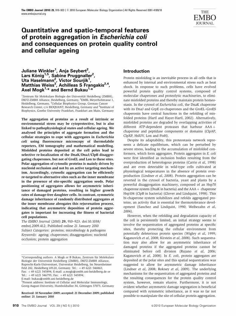
body in regions free of bacterial nucleoid (Figure 4A). This
finding indicates that the positioning of cytosolic protein
aggregates is not restricted to poles and thus not determined
by cell division. Instead, it shows that protein aggregates are
only formed in the nucleoid-free space.
To directly test whether nucleoid occlusion determines the
positioning of aggregates, we sought to monitor protein
aggregation in the absence of nucleoids by two strategies.
First, we used DmukB cells that frequently produce anucleoid
cells because of defects in chromosome segregation (Onogi
et al, 2000) (Figure 4B). Second, we disrupted the nucleoid
architecture by short-term expression (30 min) of the bacterio-
phage T4 nucleoid disruption protein (Ndd) (Figure 4C).
Ndd causes a re-organization of the nucleoid towards the
cell periphery, leaving a nucleoid-free cytosol before heat
shock (Bouet et al, 1996). When MetA–YFP aggregation was
monitored in DmukB and Ndd-induced cells, we observed
that the absence of nucleoid significantly affected the aggre-
gation process by decreasing the number of aggregates to one
per cell and increasing the number of aggregates in mid-cell
position (Figure 4B and C). These findings indicate that
nucleoid occlusion represents the primary parameter deter-
mining both number and final positioning of heat-induced
protein aggregates.
We challenged our findings by testing whether a simple
physico-chemical model, entirely based on the diffusion and
irreversible coagulation of misfolded proteins in combination
with DNA-induced occlusion, can sufficiently explain our
results. We developed a simulation model that takes into
account the diffusion of proteins in the cytoplasm, their
irreversible aggregation and the crowding in the bacterium’s
centre because of the DNA (see Materials and methods for
detail). The results of this model are in very good agreement
with the experimental data (Figure 4D). We observed almost
exclusively a single aggregation focus that dominantly loca-
lized to the cell centre when DNA crowding was neglected. In
the presence of DNA, the simulations revealed a dominant
phenotype with two foci at the bacterial poles. Diffusion and
aggregation, guided by the occlusion of DNA in the cell
centre, are, therefore, sufficient to explain the experimentally
observed phenotypes without the need for additional regula-
tory elements.
Protein aggregation at the chromosome
Our finding that a rather passive mechanism dictates the
deposition site of aggregates raises the question whether
alternative sites may also exist, perhaps triggered by aggrega-
tion ‘seeds’ at specific subcellular structures. We, therefore,
targeted thermolabile proteins to either the chromosome or
the inner membrane and monitored (i) the aggregation and
disaggregation properties of the respective reporter proteins
and (ii) the consequences on global protein aggregation.
We first investigated how the association of thermolabile
proteins with the chromosome affects their aggregation. CFP–
Luciferase was targeted to the DNA by fusing it to the DNA-
binding domain of the lac repressor (LacI) and after the
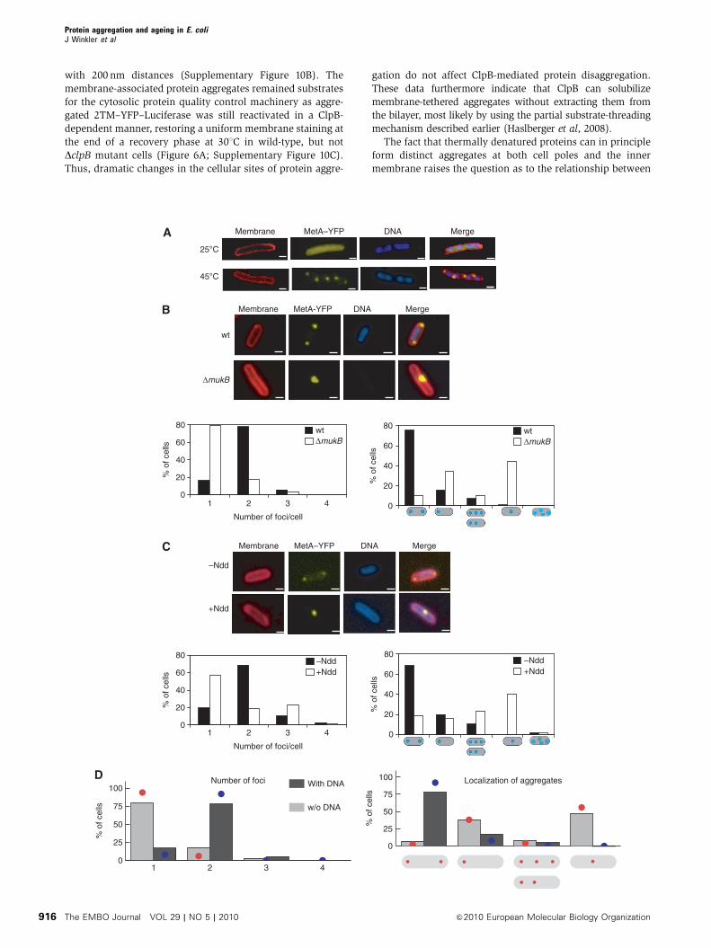
consequences on protein aggregation. LacI–CFP–Luciferase
was specifically directed to the terminus (ter) region of the
bacterial chromosome located at the mid-cell position (Lau
et al, 2003), by using a strain harbouring 240 copies of LacI-
binding cassettes integrated at the ter site. When such cells
were grown at 251C, one fluorescent focus of LacI–CFP–
Luciferase was detected at mid-cell position, whereas a uni-
form cytosolic distribution was observed in control cells
lacking LacI-binding sites in the chromosome (Figure 5A;
Supplementary Figure 9A). On temperature upshift, no
change in LacI–CFP–Luciferase localization was observed,
indicating that unfolded LacI–CFP–Luciferase remained asso-
ciated with the ter sites (Figure 5A). In contrast, polar-
localized aggregates of LacI–CFP–Luciferase were observed
in control cells lacking the LacI-binding sites on the chromo-
some. This shows that the fused LacI moiety does not alter
the aggregation properties of CFP–Luciferase and that the
targeting of LacI–CFP–Luciferase to the chromosome restricts
its aggregation, if occurring, to this particular site (Supple-
mentary Figure 9A).
Next, we tested the effects of DNA-associated LacI–CFP–
Luciferase on the heat-induced aggregation of co-produced
cytosolic MetA–YFP or YFP–Luciferase and localization of
ClpB–YFP. The localization of the foci of all three cytosolic
aggregation reporters was significantly affected by the
presence of DNA-bound LacI–CFP–Luciferase. In addition to
polar fluorescent foci of the respective fusion proteins, we
observed additional fluorescent spots at mid-cell, which
showed complete co-localization with LacI–CFP–Luciferase
(Figure 5B, C and D; Supplementary Figure 9B). Together,
these results illustrate that protein aggregation can also occur
at sites other than cell poles, with substantial effects on the
aggregation characteristics of other cytosolic proteins.
Protein aggregation at the inner membrane
In an alternative approach, we attached YFP–Luciferase to
the inner membrane by fusing it N-terminally with the first
and second transmembrane helices of MalF, yielding 2TM–
YFP–Luciferase. The 2TM–YFP–Luciferase was produced to
similar levels as compared with cytosolic YFP–Luciferase and
exhibited a homogeneous membrane staining before heat
shock (Figure 6A; Supplementary Figure 1). On temperature
upshift to 451C, multiple fluorescent foci were formed at the
membrane along the entire length of the cell without appar-
ent enrichment at polar regions. In contrast, a 2TM–YFP
fusion protein still exhibited a homogenous membrane
staining after heat shock (Supplementary Figure 10A). The
majority of cells expressing 2TM–YFP–Luciferase (80%,
n¼ 200) exhibited more than three aggregates after heat
shock, revealing significant differences between the aggrega-
tion of cytosolic and membrane-anchored YFP–Luciferase
(Figure 6A). The aggregates were randomly distributed
throughout the membrane as revealed by recording z-stacks
Protein aggregation and ageing in E. coliJ Winkler et al
&2010 European Molecular Biology Organization The EMBO Journal VOL 29 | NO 5 | 2010 915
with 200 nm distances (Supplementary Figure 10B). The
membrane-associated protein aggregates remained substrates
for the cytosolic protein quality control machinery as aggre-
gated 2TM–YFP–Luciferase was still reactivated in a ClpB-
dependent manner, restoring a uniform membrane staining at
the end of a recovery phase at 301C in wild-type, but not
DclpB mutant cells (Figure 6A; Supplementary Figure 10C).
Thus, dramatic changes in the cellular sites of protein aggre-
gation do not affect ClpB-mediated protein disaggregation.
These data furthermore indicate that ClpB can solubilize
membrane-tethered aggregates without extracting them from
the bilayer, most likely by using the partial substrate-threading
mechanism described earlier (Haslberger et al, 2008).
The fact that thermally denatured proteins can in principle
form distinct aggregates at both cell poles and the inner
membrane raises the question as to the relationship between
1 2 3 4
Number of foci100
50
25
75
0
100
50
25
75
0
With DNA
w/o DNA
Localization of aggregates
% o
f cel
ls
% o
f cel
ls
25°C
45°C
wt
ΔmukB
MembraneA
B
C
D
DNA Merge
MetA-YFP DNA Merge
Membrane DNA Merge
–Ndd
+Ndd
0
20
40
60
80
1 2 3 4
Number of foci/cell
% o
f cel
ls
0
20
40
60
80
1 2 3 4
Number of foci/cell
% o
f cel
ls
0
20
40
60
80
% o
f cel
ls
3 4
0
20
40
60
80
% o
f cel
ls
wtΔmukB
wtΔmukB
–Ndd+Ndd
–Ndd+Ndd
MetA–YFP
Membrane
MetA–YFP
Protein aggregation and ageing in E. coliJ Winkler et al
The EMBO Journal VOL 29 | NO 5 | 2010 &2010 European Molecular Biology Organization916
0
20
40
60
80
100LacI–CFP–Luciferase – MetA–YFP
LacI–CFP–Luciferase – ClpB–YFP
% o
f cel
ls
25°C
45°C
25°C
45°C
MergeMembrane
Membrane LacI–CFP–Luciferase
Merge
25°C
45°C
LacI–CFP–Luciferase
A
B
C
D
0
20
40
60
80
100
0
20
40
60
80
100
1 2 3 >3
Number of foci
% o
f cel
ls
% o
f cel
ls
25°C
45°C
25°C
45°C
MetA–YFPLacI–CFP–Luciferase
ClpB–YFP
Figure 5 DNA-anchored LacI–CFP–Luciferase aggregates capture misfolded cytosolic proteins. (A) LacI–CFP–Luciferase was produced inAB1157 cells harbouring multiple copies of lac operators at the terminus region (ter) of the bacterial chromosome. The localization of LacI–CFP–Luciferase was monitored at 251C and after heat stress (451C). The images show an overlay of the CFP signal (blue) and the membranestain FM4-64 (red). Numbers and positionings of LacI–CFP–Luciferase foci were determined at both temperatures (n¼ 100, right panels). Scalebar: 1 mm. (B) MetA–YFP and LacI–CFP–Luciferase were co-produced in AB1157 cells and images of the respective fusion protein were recordedat 301C and after heat shock (451C, 20 min). The overlay (merge) of both images reveals co-aggregation of both model proteins at the ter regionon heat stress. Membranes were stained with FM4-64. Scale bar: 1mm. (C) ClpB–YFP and LacI–CFP–Luciferase were co-produced in AB1157cells. Images of the respective fusion proteins were recorded prior (251C) and after heat shock (451C, 20 min), revealing re-localization ofClpB–YFP to the ter region of the chromosome (see merge). (D) The localizations of stress-induced MetA–YFP or ClpB–YFP foci, generated onco-production of LacI–CFP–Luciferase in AB1157 cells, were quantified (n¼ 100). The positioning of protein aggregates is indicated (white dot:co-localization; yellow dot: exclusive MetA–YFP or ClpB–YFP localization).
Figure 4 Nucleoid occlusion determines the positioning of protein aggregates. (A) Bacterial nucleoids and heat-induced MetA–YFP aggregatesdo not co-localize in filamentous cells. Filamentation of cells producing MetA–YFP was induced by addition of cephalexin. Cells were eitherkept at 301C or shifted to 451C for 20 min. Images were recorded and show membrane staining using FM4-64 (red), DNA staining using DAPI(blue) and the MetA–YFP signal. A merge of the individual images is given. Scale bar: 1mm. (B) Nucleoid-free DmukB cells predominantlycontain only one stress-induced MetA–YFP protein aggregate at either mid-cell or polar position. DmukB cells and isogenic YK1100 control cellsproducing MetA–YFP were grown at 371C and shifted to 451C for 20 min. Anucleoid cells were spontaneously formed in the DNA segregation-deficient DmukB strain. Images were recorded as described in (A). Scale bar: 1 mm. Numbers of fluorescent MetA–YFP foci per cell and theirrespective localizations were determined (n¼ 100, lower panel). The positioning of protein aggregates is indicated (blue dots). (C) Re-localization of the bacterial nucleoid to the inner membrane by the nucleoid disruption protein (Ndd) leads to the formation of one stress-induced MetA–YFP aggregate at the mid-cell or the polar region of cells. BL21 Rosetta cells producing MetA–YFP and harbouring a plasmidencoding for Ndd were grown at 301C to mid-logarithmic growth phase. The bacterial culture was splitted and Ndd production was induced inone population by addition of 1 mM IPTG for 30 min before heat shock (þNdd). Images were recorded as described in (A). Scale bar: 1mm.Numbers of fluorescent MetA–YFP foci per cell and their respective localizations were determined (n¼ 100, lower panel). The positioning ofprotein aggregates is indicated (blue dots). (D) The results of the biophysical model (coloured lines) on the number of protein aggregates andtheir cellular localization are in favourable agreement with the experimental data (grey bars). The average steady state after 104 proteins whereallowed to cluster in the cytoplasm over a time course of a few minutes in the presence (red) and absence (blue) of an occluding nucleoid(n¼ 50 independent simulation runs) yield a very good overlap with the experimentally observed phenotypes (shown below the plots).
Protein aggregation and ageing in E. coliJ Winkler et al
&2010 European Molecular Biology Organization The EMBO Journal VOL 29 | NO 5 | 2010 917

these two aggregate forms. To address this issue, we co-
produced membrane-anchored 2TM–CFP–Luciferase and cyto-
solic YFP–Luciferase in the same cells and analysed the
localization of the respective aggregates after heat treatment
(Figure 6B). Although the number and positioning of mem-
brane-anchored 2TM–CFP–Luciferase remained unchanged,
a complete re-targeting of cytosolic YFP–Luciferase aggrega-
tion was observed. YFP–Luciferase formed multiple fluores-
cent foci at the inner membrane, which entirely co-localized
with 2TM–CFP–Luciferase. The 2TM–CFP–Luciferase aggre-
gates, therefore, act as aggregation ‘seeds’, which attract
cytosolic misfolded YFP–Luciferase, thereby preventing ag-
gregate formation at the cell poles. Such re-targeting was also
observed when 2TM–CFP–Luciferase was co-produced with
cytosolic MetA–YFP (Supplementary Figure 10E), again
showing the high co-aggregation potential of structurally
unrelated proteins. The generality of this observation was
further substantiated by the finding that the aggregation of
denatured 2TM–YFP–Luciferase results in the re-targeting of
ClpB–CFP, which serves as a marker for the positioning of all
aggregated E. coli proteins, to the inner membrane and a
complete co-localization with fluorescent foci of 2TM–YFP–
Luciferase (Figure 6C).
To explain this phenomenon, we considered the possibility
that the intrinsic aggregation kinetics of the chosen model
protein (Luciferase), which is faster than that of MetA (see
above) and perhaps of other cellular proteins, is responsible
for this ‘seeding’ effect. We thus performed the same type of
experiments by attaching MetA instead of Luciferase to the
membrane. However, the co-production of 2TM–CFP–MetA
also completely changed the localization pattern of cytosolic
YFP–Luciferase and ClpB–YFP after heat stress (Supple-
mentary Figure 10F and G). It thus seems that the existence
of membrane-attached aggregation seeds is responsible for
the global re-targeting of aggregates of cytosolic proteins and
the quality control machinery.
Asymmetric but not symmetric damage inheritance
allows for rejuvenation of daughter cells
We finally investigated the relationship between the polar
aggregation of cytosolic proteins and the process of cell
division. We frequently observed that when two fluorescent
polar foci are present in a single cell, they exhibit differences
in brightness, implying different degrees of protein aggrega-
tion at the respective sites (Figure 7A). We wondered whether
an asymmetry of cell poles (old versus young pole) is the
basis for this observation, as it has been recently shown that
E. coli small heat shock proteins, used as an indirect marker
for protein aggregation, preferentially associate with the old-
cell pole in non-heat-shocked cells at 371C (Lindner et al,
2008). We, therefore, monitored protein aggregation immedi-
ately after cell division, enabling us to discriminate between
old- and new-cell poles. In these experiments, we applied a
temperature upshift to 421C instead of 451C. Such milder
stress conditions increased the number of cells containing
only a single polar aggregate, which facilitates to differentiate
between protein aggregation at old- or new-poles. An intrigu-
ingly high number of cells (88%, n¼ 100) contained fluor-
escent YFP–Luciferase or ClpB–YFP foci exclusively at old
poles, whereas the majority of the new poles remained free of
visible protein aggregates (Figure 7A).
This result raises the issue as to the relationship between
polar aggregate formation and bacterial ageing, which is
defined as a decline in bacterial growth rates with time
(Stewart et al, 2005). In starving E. coli cells, increased levels
of aggregated proteins correlate with increased cellular
senescence (Maisonneuve et al, 2008). Furthermore, the
existence of polar-localized aggregates in cells correlates
ΔclpB
wt
A
B C
0
20
40
60
80
100
1 2 3 >3
Number of aggregates
% o
f cel
ls
–Heat shock0 min
+ Heat shock 45°C10 min
After recovery90 min
2TM–YFP–Luciferase
30°C
45°C
Luciferase–YFP
2TM–CFP–Luciferase
Merge
30°C
45°C
ClpB–CFP Merge
Figure 6 Membrane-anchored 2TM–YFP–Luciferase forms multiple aggregates on heat shock and reprogrammes aggregation of cytosolicproteins. (A) 2TM–YFP–Luciferase was produced in wild-type and DclpB mutant cells. Images were recorded before heat shock (0 min), aftertemperature upshift to 451C (10 min) and at the end of a recovery period at 301C (90 min). Before heat shock, protein synthesis was stopped byaddition of rifampicin (50 mg/ml). The number of membrane-localized 2TM–YFP–Luciferase foci generated on heat stress was quantified(n¼ 200, right panel). Scale bar: 1 mm. (B) Luciferase–YFP and 2TM–CFP–Luciferase were co-produced in wild-type cells and images of therespective fusion protein were recorded at 301C and after heat shock (451C, 20 min). The overlay (merge) of both images reveals co-aggregationof both model proteins at high temperatures. Scale bar: 1 mm. (C) ClpB–CFP and 2TM–YFP–Luciferase were co-produced in DclpB cells. Imagesof the respective fusion proteins were recorded prior (301C) and after heat shock (451C, 20 min), revealing co-localization of fluorescent fociafter heat stress at the membrane (see merge). Scale bar: 1 mm.
Protein aggregation and ageing in E. coliJ Winkler et al
The EMBO Journal VOL 29 | NO 5 | 2010 &2010 European Molecular Biology Organization918

with a reduced growth rate at 371C, reflecting bacterial ageing
(Lindner et al, 2008). It is unclear whether the same correla-
tion also holds true for heat-induced protein aggregates.
To determine the consequences of an asymmetric deposition
of aggregated proteins after heat treatment on ageing, we
produced YFP–Luciferase in DclpB mutant cells, enabling us
to analyse by time-lapse microscopy the influence of protein
aggregates on bacterial growth during the recovery phase for
various generations (Supplementary Figure 11). As individual
colonies showed variations in their generation times, we
normalized the average growth rate, for better comparison,
within each generation to 1. When continuously grown at
301C, the lineage of DclpB cells, which inherited the old pole
exhibited a continuous decline of growth rates when followed
for four generations (Figure 7B). In contrast, cells that
inherited new poles (generated by septum formation during
cell division) regained higher growth rates (0.0246/min com-
pared with 0.0225/min, corresponding to generation times of
41 and 44 min) on cell division compared with the old pole
mother cell, thus exhibiting ‘rejuvenation’ in agreement with
earlier findings (Stewart et al, 2005). In the additional pre-
sence of protein aggregates at the old poles of heat-treated
mother cells, the difference in growth rates between old- and
new-pole cells became much more apparent (0.0211/min
compared with 0.0255/min) (Figure 7B and C). This finding
suggests, in agreement with earlier data, that the deposition
30°C , 40 min Cell division
10 min, 42°CA
B
C
ΔclpBClpB–YFP
wtYFP–Luciferase
0 min 40 min
0 min 40 min
YFP–Luciferase (30°C) YFP-Luciferase 45°C
2TM–YFP–Luciferase (30°C) 2TM–YFP–Luciferase 45°C
Growth rate (x10–2 min–1)/ standard deviation
0.70.80.91.01.11.21.3
1 2 3 4
Generation
Nor
mal
ized
gro
wth
rat
e
0.70.80.91.01.11.21.3
1 2 3 4
Generation
Nor
mal
ized
gro
wth
rat
eN
orm
aliz
ed g
row
th r
ate
Nor
mal
ized
gro
wth
rat
e
Old New
0.70.80.91.01.11.21.3
1 2 3 4Generation
0.70.80.91.01.11.21.3
1 2 3 4Generation
***
ΔclpB Mean Old pole New pole
YFP–Luciferase 30°C 2.40 /0.5 2.25 /0.4 2.46 /0.4YFP–Luciferase 45°C 2.31 /0.6 2.11 /0.5 2.55 /0.52TM–YFP–Luciferase 30°C 2.38 /0.4 2.23 /0.4 2.43 /0.42TM–YFP–Luciferase 45°C 2.17 /0.5 2.09 /0.5 2.25 /0.5
***
Old New
Old New Old New
0
20
40
60
80
100
Old pole New pole
YFP–Luciferase
ClpB–YFP
% o
f cel
ls
Figure 7 Asymmetric distribution of protein aggregates allows for the rejuvenation of damage-free new-pole daughter cells. (A) Stress-inducedYFP–Luciferase and ClpB–YFP foci preferentially localize to the old-cell poles of wild-type cells. Division of cells was monitored before heatstress (421C, 10 min), allowing for the identification of old- and new-cell poles. Old-cell poles are labelled with an arrow. The localization ofstress-induced YFP–Luciferase or ClpB–YFP foci at old and new poles were quantified (n¼ 100, right panel). Scale bar: 1 mm. (B) The presenceof polar-localized or membrane-anchored protein aggregates diminish the growth rate of E. coli cells. Comparison of the normalized growthrates for new- and old-pole cells expressing either YFP–Luciferase or 2TM-YFP–Luciferase. Growth rates of 30 colonies were determined for old-and new-pole cells that were continuously cultivated at 301C for four generations. Alternatively, the formation of protein aggregates wasinduced by heat shock (451C, 20 min) and growth rates were recorded on return to 301C, accordingly. The average growth rate within eachgeneration was normalized to 1. Old-pole cells harbouring polar YFP–Luciferase aggregates and derived aggregate-free new-pole cells differsignificantly in their growth rates (***Po0.01). In addition, new-pole cells expressing 2TM–YFP–Luciferase or YFP–Luciferase show significantdifferences in their growth rates (***Po0.01). (C) Determined absolute growth rates of old- and new-pole populations expressing the indicatedfusion proteins (non-heat shocked: 301C, transiently heat shocked: 451C). The mean growth rate of the respective entire bacterial populationwas calculated. Standard deviations are given.
Protein aggregation and ageing in E. coliJ Winkler et al
&2010 European Molecular Biology Organization The EMBO Journal VOL 29 | NO 5 | 2010 919

of heat-induced protein aggregates at the cell poles acceler-
ates bacterial ageing, but also allows for rejuvenation of
damage-free new-pole cells on cell division.
To provide direct evidence for a physiological relevance of
an asymmetric inheritance of protein aggregates, we tested
whether the bacterial ageing phenotype is solely depending
on the polar deposition of aggregates. To this end, we
expressed 2TM–YFP–Luciferase in DclpB mutant cells, caus-
ing re-targeting of heat-induced protein aggregates from polar
sites to random positions at the inner membrane (Figure 6A).
This experimental setup enabled us to follow the conse-
quences of a symmetric inheritance of protein aggregates
on bacterial ageing. The altered localization of aggregates
had a dramatic impact on bacterial growth rates, as the
differences in growth rates between old- and new-pole cells
were no longer increased (Figure 7B). The biggest differences
in growth rates were noticed for new-pole cells that were
derived from mother cells harbouring either polar or ran-
domly distributed aggregates. New-pole cells that lost polar
aggregates through asymmetric inheritance grew significantly
faster than new-pole cells that still retained damaged proteins
because of the symmetric inheritance of randomly distribu-
ted, membrane-associated protein aggregates (0.0255/min
compared with 0.0225/min) (Figure 7C). This finding for
the first time provides direct evidence that the deposition of
aggregated proteins at polar sites has unique beneficial con-
sequences for the ageing of a bacterial cell population.
Finally, we questioned whether the asymmetric deposition
of aggregated proteins at old-pole cells, which is key for
asymmetric damage inheritance, is also based on nucleoid
occlusion or relies on additional mechanisms that confer
specificity towards the old pole. A nucleoid occlusion scenario
suggests that the noticed asymmetry relies on a slightly
asymmetric distribution of the bacterial chromosome within
the cell, leaving more space for aggregate deposition at the
old pole. In consequence, an asymmetric localization of
protein aggregates at old poles should no longer be observa-
ble in anucleoid cells or in cells harbouring a nucleoid in a
more condensed state. We first monitored the polar deposi-
tion of MetA–mCherry aggregates in anucleoid DmukB cells
and analysed those cells that still contained polar aggregates.
In such experiments, old-pole cells were identified by co-
producing Tsr–YFP, which acts as a specific marker for the
old pole (Ping et al, 2008). In the absence of a bacterial
chromosome, an equal distribution of protein aggregates at
old- (53%) and new-pole cells (47%) was observed
(Supplementary Figure 12A and C; n¼ 200). We then mon-
itored protein aggregation in the presence of chlorampheni-
col, which leads to condensation of the bacterial nucleoid,
leaving more space at both poles (Sun and Margolin, 2004).
Such conditions again abrogated the preferential deposition
of aggregated proteins at the old pole (54%), compared with
the new pole (46%) (Supplementary Figure 12B and C;
n¼ 100), thereby providing further evidence that nucleoid
occlusion not only determines the polar localization of
protein aggregates, but also their asymmetric deposition at
old poles.
Discussion
Our study provides a detailed analysis of (i) the quantitative
and spatio-temporal features of protein aggregation in E. coli
cells subjected to heat shock, (ii) the protein quality control
machinery that responds to this challenge, (iii) the mechan-
isms driving aggregate formation at specific cellular sites and
(iv) the physiological relevance of aggregate deposition for
bacterial ageing. Together, this provides a framework for the
mechanistic understanding of the cellular events leading to
and strategies coping with protein aggregation.
Although the heat treatment at 451C used for our experi-
ments is within the growth temperature range of E. coli, it is
severe enough to generate microscopically visible protein
aggregates in wild-type cells, which typically form foci at
the cell poles, in agreement with earlier findings (Lindner
et al, 2008; Rokney et al, 2009). Further analysis by cryo-ET
revealed that these aggregates exhibit large heterogeneity in
size, whereas the total volume of all aggregates per cell, and
thus the total number of aggregated proteins, is more similar
between cells. On the basis of the determined aggregate
volumes, we estimate that 1.5–3% of total cytosolic E. coli
proteins, corresponding to approximately 17500–33000 mo-
lecules, aggregate on heat stress in wild-type cells. Although
this is a rather small fraction of total cytosolic protein, it
suffices to elicit a strong heat shock response, showing the
capacity of this stress regulon to detect even small alterations
in proteostasis. The aggregates appear amorphous, suggest-
ing that their formation is driven by non-specific, presumably
hydrophobic interactions between misfolded stretches of
proteins that may be structurally unrelated. Such formation
of mixed aggregates is supported by the observed, surpris-
ingly extensive co-aggregation of different thermolabile
aggregation reporters, even if they have different cellular
localizations (Figures 5 and 6).
How are misfolded cytosolic proteins targeted to the polar
dumping sites in which aggregates are deposited? We did not
obtain any evidence for an active, energy demanding trans-
port process, as ATP depletion did not affect protein aggrega-
tion. Furthermore, aggregate positioning was neither
dependent on MreB, a central component of the bacterial
cytoskeleton, nor on DnaJ, DnaK and ClpB, largely excluding
a function of the cytoskeleton and the protein quality control
machinery in targeting misfolded proteins to the poles. The
easiness by which cytosolic protein aggregation is re-targeted
by membrane or chromosome-attached misfolded proteins
provides a further argument against an active guidance of
protein aggregates to poles. Together, these findings strongly
argue against an active transport of misfolded or aggregated
proteins to the poles. This is in conflict to a recent study,
claiming that the polar deposition of aggregated proteins in
E. coli is mediated by an energy-dependent process involving
DnaK and MreB (Rokney et al, 2009). We cannot offer an easy
explanation for these discrepancies, as the experimental
setups used in both studies are rather similar and rely on
thermolabile fluorescent reporters. We note that the Rokney
study uses an unusual buffer (10 mM Tris–HCl, pH 8.0;
10 mM MgSO4) for resuspending the cells before performing
microscopy, and does not include time-lapse microscopy,
which is mandatory for following the destiny of individual
aggregates.
We instead provide evidences that nucleoid occlusion is
the main driving force, which determines number and posi-
tioning of protein aggregates in E. coli. First, both parameters
were significantly altered in cells lacking a nucleoid or
harbouring the chromosome in a different structural state.
Protein aggregation and ageing in E. coliJ Winkler et al
The EMBO Journal VOL 29 | NO 5 | 2010 &2010 European Molecular Biology Organization920

Second, filamentous cells develop multiple aggregates at
nucleoid-free non-polar sites within the entire cell body.
Nucleoid occlusion is thus not only necessary, but also
sufficient to determine the positioning and number of protein
aggregates in the cytosol. This function of the chromosome is
further supported by a mathematical model that exclusively
relies on a simple geometric occlusion mechanism. On this
basis, the model correctly predicts the formation of protein
aggregates at polar sites, in perfect quantitative and qualita-
tive agreement with our experimental findings.
The model also predicts that protein aggregation at the
membrane always outcompetes bulk protein aggregation in
the cytosol. Even under crowded conditions, the bulk diffusion
of unfolded proteins is fairly rapid leading to frequent colli-
sions of unfolded proteins with the cellular membrane.
If membrane-resident aggregation seeds reside in the mem-
brane patch at which such a collision occurs, the soluble
unfolded proteins are irreverisbly recruited to the membrane.
Even if a larger cluster of unfolded proteins forms in the
cytosol, it will fairly rapidly (i.e. within less than a minute)
get recruited to the membrane because of its diffusive motion.
Hence, rapidly more and more material is irreversibly seques-
tered to the membrane, thereby forming the dominant fraction
of large aggregates in agreement with our findings (Figure 6B
and C). We note that in wild-type cells that do not produce a
membrane-anchored aggregation-prone protein, the thermola-
bile proteins of the cytosol are not targeted to the inner
membrane, suggesting that only subcritical amounts of inner
membrane proteins are prone to aggregation under such
conditions.
The mathematical model can even account for the noticed
preferential deposition of misfolded proteins at the old pole
when an asymmetric positioning of the nucleoid, perhaps
resulting from the division process, is assumed. A more
spacious cytosolic region at one pole will increase the num-
ber of accumulating misfolded proteins, which may influence
the onset of aggregate formation. Indeed, an asymmetric
deposition of misfolded proteins was no longer observed
when the nucleoid was forced into a more condensed state,
for example by addition of chloramphenicol, leaving more
space at both poles.
E. coli thus uses a rather passive mechanism to deposit
aggregated proteins, in contrast to eukaryotic cells. The
localization of aggregates in S. cerevisiae (JUNQ/IPOD) or
mammalian cells (aggresomes) depends on the cytoskeleton
network (Garcia-Mata et al, 1999; Kaganovich et al, 2008)
and involves an active transport of aggregates to their final
destination (Kawaguchi et al, 2003). Thus, pro- and eukar-
yotes developed fundamentally different strategies to control
protein aggregation. These differences likely arose from the
less complex cellular architecture and the much smaller size
of prokaryotes, which thereby may not necessitate an active
process to ensure the deposition of damaged proteins at
specific sites.
What is the physiological relevance of the polar deposition
of protein aggregates? We show that the presence of
heat-induced, polar aggregates has a negative impact on
bacterial growth rates. Similar findings have been recently
reported by Taddei and co-workers, showing that non-
stressed E. coli cells harbouring an aggregation reporter
exhibit a reduced growth rate (Lindner et al, 2008). The
polar localization of aggregates might help cells to regain
faster growth rates, as it allows for the generation of damage-
free poles and cells by cell division. In the case of cells
harbouring a single aggregate at the old pole, it allows for
the generation of a heterogeneous cell population by division,
creating new-pole cells that can recover faster from stress
situations at the expense of old-pole cells, which retain the
main damage. Importantly, this prediction is confirmed by
our finding that symmetric damage distribution largely pre-
vents rejuvenation of new-pole cells (Figure 7B and C). This
finding provides for the first time direct experimental evi-
dence for the theoretical prediction that asymmetric distribu-
tion of damage is beneficial for a cell population by allowing
for faster recovery of new-pole cells from stress, which could
represent an important growth advantage in a competitive
microbial environment (Ackermann et al, 2007). Conversely,
there may be an evolutionary driving force to avoid non-polar
sites of protein aggregation, for example at the inner mem-
brane. It is tempting to speculate that membrane proteins
with domains exposed to the cytosol evolved to be more
resistant towards thermal unfolding compared with cytosolic
proteins, thereby preventing the generation of aggregation
seeds. The 2TM–Luciferase–YFP and 2TM–MetA–YFP model
constructs, which we used in this study, show that aggrega-
tion of membrane-associated proteins is, in principle, possi-
ble. The solubilization of these membrane-localized
aggregates strictly depended on ClpB, which gets targeted
to membrane-associated foci. Our observation that stress-
induced ClpB–YFP foci are not formed at the membrane of
wild-type cells shows that endogenous membrane proteins
do not aggregate on heat stress.
Many components of the protein quality control machinery
react to heat stress by re-localization to polar foci. A rapid
re-localization of the major cellular fraction of DnaK, DnaJ
and ClpB confirms their central function in protein disaggre-
gation. FRAP analysis revealed that these proteins remain
highly dynamic and thus are reversible associated with
aggregates. A more complicated picture emerged for the
proteolytic systems, which in part also re-localized to the
polar foci, as not all components seem to represent functional
interactions with aggregates. According to FRAP analysis,
ClpX and HslU become immobilized at foci and, therefore,
rather co-aggregate with misfolded proteins, consistent with
their inability to degrade aggregated proteins in vitro (jointly
with ClpP and HslV, respectively; data not shown). Thus, co-
localization or co-sedimentation of chaperones and proteases
with protein aggregates is an insufficient criterion for defining
a functional role in aggregate handling.
Interestingly, GroEL and Lon, representing important func-
tions in the folding and degradation of misfolded proteins
(Chung and Goldberg, 1981; Kerner et al, 2005; Chapman
et al, 2006), do not re-localize on heat stress and remain
distributed throughout the cytosol. For GroEL, this is ex-
plained by its mode of action, which relies on the encapsula-
tion of single non-native proteins, and hence represents a
‘downstream’ activity after the DnaK/DnaJ/ClpB-mediated
solubilization of aggregated proteins. Solubilized proteins
likely diffuse rapidly throughout the entire cytosol and thus
do not require an enrichment of GroEL at polar regions after
heat stress. GroEL thereby remains available for its house-
keeping function in the folding of newly synthesized proteins.
Similarly, the fast diffusion of misfolded species does not
necessitate for a polar localization of Lon, which can only act
Protein aggregation and ageing in E. coliJ Winkler et al
&2010 European Molecular Biology Organization The EMBO Journal VOL 29 | NO 5 | 2010 921

on soluble non-native proteins, but not on heat-aggregated
proteins in vitro (data not shown).
Summarized, we provide for the first time a rigorous,
quantitative analysis of protein aggregation in a single cell.
It will be an interesting future topic to compare the features of
the aggregation/disaggregation processes in E. coli with those
of the related processes in other organisms, which have
different sets of cytosolic chaperones including the lack of a
powerful disaggregation activity.
Materials and methods
Plasmids and strainsAll E. coli K12 strains and plasmids used in this study are listed inSupplementary Tables 1 and 2. Fluorescent fusion proteins wereconstructed by fusion PCR. Target genes and mcherry or yfp/cfpwere amplified using primers with complementary overlaps,encoding a 5x glycine linker. The fusion genes were inserted intothe listed expression plasmids (Supplementary Table 1).
Growth conditionsCell cultures were grown, if not indicated otherwise, at 301C in LBmedia to an OD600 of 0.6–0.8. For heat treatment, cells were shiftedto 451C for 10–20 min. When appropriate, antibiotics were added tostandard concentrations. Gene expression was induced on addition ofthe indicated concentrations of arabinose or IPTG (SupplementaryTable S2). All fluorescent chaperones/peptidase fusion proteins wereproduced at wild-type level. To stop protein translation, spectino-mycin (100mg/ml), rifampicine (50mg/ml) or tetracycline (25mg/ml)was added 10 min before starting the respective experiment.
ATP depletion experiments were performed in MOPS-basedminimal media with succinate as carbon source (Neidhardt et al,1974). A total of 10 mM DNP was added to the growth medium30 min before heat shock. For energy depletion experiments withCCCP (20 mM) or natriumazide (0.5–1% (w/v)), cells wereresuspended in PBS, incubated for 5 min with uncoupler andwashed with PBS before applying a heat shock. Intracellular ATPlevels were determined using a luciferin/luciferase-based assay asdescribed (Yang et al, 2002).
E. coli DmukB cells were grown in MCAT media (Onogi et al,2000) and A22 was added 20 min before heat shock.
Enzymatic assays and determination of protein copy numbersLuciferase activities were determined from 200 ml cell suspensionsin a luminometer (Lumat berthold LB9501) as described bySchroder et al (1993). The cellular levels of YFP fusion proteinswere determined by measuring the YFP fluorescence intensity fromtotal cell extracts in a luminescence spectrometer (Perkin Elmer). Astandard curve was generated from cell lysates spiked with differentquantities of purified YFP (details are included in the supplements).
Membrane and DNA stainingFor membrane staining, the dye FM4–64 (2mg/ml, Molecularprobes) was added to the culture 20 min before start of theexperiment. For DNA staining, cells were fixed for 5 min on ice with2.8% (v/v) formaldehyde and 0.04% (v/v) glutaraldehyde. Next,fixed cells were incubated with 0.3mg/ml DAPI for 10 min. Threewashing steps with 1 ml tethering buffer (TEB) were carried out toremove the fixatives and free DAPI.
Microscopy and cryo-ETFor snapshot imaging, cells pellets were resuspended in TEB (Blocket al, 1983) and immobilized on agarose pads. Agarose pads weresealed with Apiezon grease and covered with cover slips. For longtime-lapse imaging with growing cells, the cells were spotted ontoagarose pads that contained 15% (v/v) LB media and were sealedin a custom-made aluminium slide using cover slips on each side.For short time-lapse experiments, cells were collected, washed inTEB and placed on agarose pads without media. The slide waseither placed into a custom-made temperature-controlled holderthat was connected to a water bath (Lauda Ecoline Star edition, forFRAP experiments), a metal holder that was connected to a peltierelement (Oven industries, for studying protein aggregation) or aToka Hit temperature control chamber (to study the inheritance of
protein aggregates). For fluorescence microscopy, various micro-scopes and settings were used as specified in Supplementary data.
Methods for cryo-ET and for the generation of cell sections aredescribed in Supplementary data.
ImmunofluorescenceCells were fixed and stained as described (Teleman et al, 1998;Sourjik and Berg, 2000). Fixed cells were directly immobilized onpoly-L-lysine-coated slides. Incubation with affinity-purified anti-bodies (a-GroEL (1:200 dilution) or a-DnaK (1:1000 dilution)) andsecondary antibody (1:500 dilution Alexa 546 anti rabbit, MolecularProbes) was carried out for 1 h at room temperature in 2% (w/v)BSA containing PBS buffer with (GroEL) or without (DnaK) 0.05%(v/v) Tween. After washing with PBS, cells were embedded inmounting solution (1 mg/ml p-phenylenediamine, 90% glyceroland 5% PBS) before imaging. Incubation of MC4100 cells with thesecondary antibody alone did not result in any fluorescence signal.In addition, GroEL and DnaK antibodies did not show backgroundstaining in GroEL-depleted MGM100 or DdnaK52.
FRAP experimentsFor FRAP experiments, cells were grown at 301C in TB media to anOD600 of 0.5. Cells were immobilized on poly-L-lysine-coated slidesand incubated with TEB containing 100 mg/ml spectinomycin and10% (v/v) TB media. Cells were heat shocked for 10 min at 451Cand shifted back to 301C. After 10 min, FRAP measurements werestarted. For each chaperone–YFP protein fusion, 10–20 individualcells were measured. Fluorescence of polar protein aggregates werebleached with two 0.336 s scans at 50% laser intensity, pre- andpost-bleach images were acquired with 3–5% laser intensity usingbidirectional scanning. Post-bleach images consisted of 30 images,10 images with 0.336 s, 10 images with a time interval of 3 s,followed by 10 images with a time interval of 30 s. Measurementswere performed on a laser scanning confocal microscope (Leica TCSSP2) equipped with an FRAP software module.
Image processing, data analysis and spatio-temporal modelfor protein aggregate formationImage processing was carried out using Image J (W Rasband,http://rsb.info.nih.gov/ij/) software. Statistics and plotting of thedata were performed with Excel. Quantification of polar andcytosolic fluorescence intensities was calculated from 20 cells usingthe following formula: % polar fluorescence¼fluorescence of theregion of interest (ROI)�background ROI/fluorescence of the wholecell background � 100. For FRAP experiments, the fluorescenceintensity of the whole cell and the polar ROI was determined overtime. Relative fluorescence intensity of ROI was normalized to therelative ROI before bleaching. Mf and If were calculated from thecorresponding recovery curves. For time-lapse experiments thatinvolved cell growth, old- and new-pole cells were trackedmanually and growth rates were measured from 30 colonies/strainby determining the time between two cell divisions. The develop-ment of the spatio-temporal model for protein aggregate formationis described in detail in Supplementary data.
Supplementary dataSupplementary data are available at The EMBO Journal Online(http://www.embojournal.org).
Acknowledgements
We thank J Tyedmers for critical reading of the manuscript. We aregrateful to P Lund for providing E. coli MGM100, DJ Sherratfor E. coli AB1157, JY Bouet for pJYB41-Ndd, N Iwai for A22 andS Hiraga for mukB mutants. We thank the EMBL EM facility forproviding the equipment for the plastic section and the NikonImaging Center Heidelberg for the use of microscopes. This workwas supported by a grant of the Fonds der Chemischen Industrie toBB. JW was supported by the Network Aging Research Heidelbergand MW by the Institute for Modeling and Simulation in theBiosciences (BIOMS) in Heidelberg.
Conflict of interest
The authors declare that they have no conflict of interest.
Protein aggregation and ageing in E. coliJ Winkler et al
The EMBO Journal VOL 29 | NO 5 | 2010 &2010 European Molecular Biology Organization922
References
Ackermann M, Chao L, Bergstrom CT, Doebeli M (2007) On theevolutionary origin of aging. Aging Cell 6: 235–244
Block SM, Segall JE, Berg HC (1983) Adaptation kinetics in bacterialchemotaxis. J Bacteriol 154: 312–323
Bouet JY, Campo NJ, Krisch HM, Louarn JM (1996) The effects onEscherichia coli of expression of the cloned bacteriophage T4nucleoid disruption (ndd) gene. Mol Microbiol 20: 519–528
Carrio MM, Corchero JL, Villaverde A (1998) Dynamics of in vivoprotein aggregation: building inclusion bodies in recombinantbacteria. FEMS Microbiol Lett 169: 9–15
Chapman E, Farr GW, Usaite R, Furtak K, Fenton WA, ChaudhuriTK, Hondorp ER, Matthews RG, Wolf SG, Yates JR, Pypaert M,Horwich AL (2006) Global aggregation of newly translated pro-teins in an Escherichia coli strain deficient of the chaperoninGroEL. Proc Natl Acad Sci USA 103: 15800–15805
Chung CH, Goldberg AL (1981) The product of the lon (capR) genein Escherichia coli is the ATP-dependent protease, protease La.Proc Natl Acad Sci USA 78: 4931–4935
Garcia-Mata R, Bebok Z, Sorscher EJ, Sztul ES (1999)Characterization and dynamics of aggresome formation by acytosolic GFP-chimera. J Cell Biol 146: 1239–1254
Glover JR, Lindquist S (1998) Hsp104, Hsp70, and Hsp40: a novelchaperone system that rescues previously aggregated proteins.Cell 94: 73–82
Gur E, Biran D, Gazit E, Ron EZ (2002) In vivo aggregation of asingle enzyme limits growth of Escherichia coli at elevatedtemperatures. Mol Microbiol 46: 1391–1397
Harpaz Y, Gerstein M, Chothia C (1994) Volume changes on proteinfolding. Structure 2: 641–649
Hartl FU, Hayer-Hartl M (2002) Molecular chaperones in thecytosol: from nascent chain to folded protein. Science 295:1852–1858
Haslberger T, Zdanowicz A, Brand I, Kirstein J, Turgay K, Mogk A,Bukau B (2008) Protein disaggregation by the AAA+ chaperoneClpB involves partial threading of looped polypeptide segments.Nat Struct Mol Biol 15: 641–650
Iwai N, Nagai K, Wachi M (2002) Novel S-benzylisothioureacompound that induces spherical cells in Escherichia coliprobably by acting on a rod-shape-determining protein(s) otherthan penicillin-binding protein 2. Biosci Biotechnol Biochem 66:2658–2662
Kaganovich D, Kopito R, Frydman J (2008) Misfolded proteinspartition between two distinct quality control compartments.Nature 454: 1088–1095
Kawaguchi Y, Kovacs JJ, McLaurin A, Vance JM, Ito A, Yao TP(2003) The deacetylase HDAC6 regulates aggresome formationand cell viability in response to misfolded protein stress. Cell 115:727–738
Kerner MJ, Naylor DJ, Ishihama Y, Maier T, Chang HC, Stines AP,Georgopoulos C, Frishman D, Hayer-Hartl M, Mann M, Hartl FU(2005) Proteome-wide analysis of chaperonin-dependent proteinfolding in Escherichia coli. Cell 122: 209–220
Kirstein J, Strahl H, Moliere N, Hamoen LW, Turgay K (2008)Localization of general and regulatory proteolysis in Bacillussubtilis cells. Mol Microbiol 70: 682–694
Lau IF, Filipe SR, Soballe B, Okstad OA, Barre FX, Sherratt DJ (2003)Spatial and temporal organization of replicating Escherichia colichromosomes. Mol Microbiol 49: 731–743
Lindner AB, Madden R, Demarez A, Stewart EJ, Taddei F (2008)Asymmetric segregation of protein aggregates is associated with
cellular aging and rejuvenation. Proc Natl Acad Sci USA 105:3076–3081
Maisonneuve E, Ezraty B, Dukan S (2008) Protein aggregates: anaging factor involved in cell death. J Bacteriol 190: 6070–6075
Mogk A, Tomoyasu T, Goloubinoff P, Rudiger S, Roder D, Langen H,Bukau B (1999) Identification of thermolabile E. coli proteins:prevention and reversion of aggregation by DnaK and ClpB.EMBO J 18: 6934–6949
Neidhardt FC, Bloch PL, Smith DF (1974) Culture medium forenterobacteria. J Bacteriol 119: 736–747
Netzer WJ, Hartl FU (1998) Protein folding in the cytosol: chaper-onin-dependent and -independent mechanisms. TiBS 23: 68–73
Onogi T, Yamazoe M, Ichinose C, Niki H, Hiraga S (2000) Nullmutation of the dam or seqA gene suppresses temperature-sensitive lethality but not hypersensitivity to novobiocin of muknull mutants. J Bacteriol 182: 5898–5901
Ping L, Weiner B, Kleckner N (2008) Tsr-GFP accumulates linearlywith time at cell poles, and can be used to differentiate ‘old’versus ‘new’ poles, in Escherichia coli. Mol Microbiol 69:1427–1438
Pogliano J, Pogliano K, Weiss DS, Losick R, Beckwith J (1997)Inactivation of FtsI inhibits constriction of the FtsZ cytokineticring and delays the assembly of FtsZ rings at potential divisionsites. Proc Natl Acad Sci USA 94: 559–564
Rokney A, Shagan M, Kessel M, Smith Y, Rosenshine I, OppenheimAB (2009) E coli transports aggregated proteins to the poles by aspecific and energy-dependent process. J Mol Biol 392: 589–601
Rujano MA, Bosveld F, Salomons FA, Dijk F, van Waarde MA, vander Want JJ, de Vos RA, Brunt ER, Sibon OC, Kampinga HH(2006) Polarised asymmetric inheritance of accumulated proteindamage in higher eukaryotes. PLoS Biol 4: e417
Sanchez Y, Lindquist SL (1990) HSP104 required for inducedthermotolerance. Science 248: 1112–1115
Schroder H, Langer T, Hartl F-U, Bukau B (1993) DnaK, DnaJ, GrpEform a cellular chaperone machinery capable of repairing heat-induced protein damage. EMBO J 12: 4137–4144
Sourjik V, Berg HC (2000) Localization of components of thechemotaxis machinery of Escherichia coli using fluorescent pro-tein fusions. Mol Microbiol 37: 740–751
Stewart EJ, Madden R, Paul G, Taddei F (2005) Aging and death inan organism that reproduces by morphologically symmetric divi-sion. PLoS Biol 3: e45
Sun Q, Margolin W (2004) Effects of perturbing nucleoid structureon nucleoid occlusion-mediated toporegulation of FtsZ ring as-sembly. J Bacteriol 186: 3951–3959
Teleman AA, Graumann PL, Lin DC, Grossman AD, Losick R (1998)Chromosome arrangement within a bacterium. Curr Biol 8:1102–1109
Weibezahn J, Tessarz P, Schlieker C, Zahn R, Maglica Z, Lee S,Zentgraf H, Weber-Ban EU, Dougan DA, Tsai FT, Mogk A, BukauB (2004) Thermotolerance requires refolding of aggregated pro-teins by substrate translocation through the central Pore of ClpB.Cell 119: 653–665
Wigley WC, Fabunmi RP, Lee MG, Marino CR, Muallem S,DeMartino GN, Thomas PJ (1999) Dynamic association ofproteasomal machinery with the centrosome. J Cell Biol 145:481–490
Yang NC, Ho WM, Chen YH, Hu ML (2002) A convenient one-stepextraction of cellular ATP using boiling water for the luciferin-luciferase assay of ATP. Anal Biochem 306: 323–327
Protein aggregation and ageing in E. coliJ Winkler et al
&2010 European Molecular Biology Organization The EMBO Journal VOL 29 | NO 5 | 2010 923