Helicobacter pylori-Induced Chronic Gastritis and Assessing Risks for Gastric Cancer
Upload
independentCategory
view
3download
0
biopsies are sensitive diagnostic methods, they are of limited help for accurate quantification of the total bacterialload. The recent development of ‘4Cor @3C-ureabreathtest (UBT), based on the intense urease activity of H.pylon (10), could provide the missing quantitative toolsince the test integrates the response of the totality of H.pylon bacteria active in the stomach (11—13).
Inthe presentstudy,we firstevaluatedthe reliabilityofthe absolutelevels of ureaseactivitymeasuredwith the‘4C-ureabreath test. Thereafter, we have used the test toquantitate H. pylon infection in a large group of patientswithchronicgastritis,searchingfora clearpositivecorrelatiombetweenthe total ureaseactivityand the severityand activity ofgastritis. We also have investigated whetherthe presence of ulcer disease in H. pylon positive patientsis associated with a further increase of urease activity bycomparison with values obtained in absence of ulcer disease.
Gastric urease was studied isotopically in 230 patients withbiopsy-proven normal mucosa or chronic gastritis, including59 patientswith ulcerdisease.Carbon-i4-urea was givenin25 ml of water without substrateearneror nutrient-densemeal, and breath sampies were collected over a 60-mmperiod.The amountof 14C02excretedat 10 mmwas independentof the rateof gastricemptyingand was not quantitativelyinfluencedby the buccalureaseactivity.The 10-mm1'c02valuesdiscriminatedwellbetweenHeilcobacterpylonpositiveandnegativepatients(94%sensitivity,89%SpecifiCPity)and correlatedwith the numberof organismsassessedbyhistology.Thetestwasa goodpredictorofthronicgastrftis(95%sensitivityand96% specificity),anda quantitativerelationship was observed between 14C02valueSand the severityand activity of the gastritis. In H. pylon!positive patients,breath ‘4C02was found to be similar in patientswith andwithoutulcerdisease,suggestingthat thenumberof bacteriais not a determiningfactor for the onsetof ulceration.
J NucIMed 1991;32:1192—1198
everalreportshavedescribedan associationof He/i-cobacter pylon with gastric or duodenal ulcer, nonulcerdyspepsia, and gastritis (1-6). Direct evidence of a pathogenic role of H. pylon was obtained by self-inoculationexperiments or by inadvertent iatrogenic transmission followed by acute gastritisand, in some volunteers,thesubsequent development of chronic gastritis associatedwithpersistentH. pylon infection(7—9).Despitethe factthat the natural history of chronic gastritis remains unclear, there is mounting evidence that H. pylon is thecausal agent ofmost cases ofchromc gastritis (3,6). In thisrespect, a clear demonstration ofthe quantitative relationship betweenthe numberof organismspresentand theimportance of the gastritis would provide additional support for the etiologic role of H. pylon in chronic gastritis.Although histologic examination or bacterial culture of
ReCeived Jul. 5, 1990; revision accepted Nov. 19, 1990.For reprintscontact: Dr. S. Pauwels, Centre de MédecineNucléalre,UCL
54.30.avenueHippocrate54,B-1200Brussels.Belgium.
Patientsreferredfor uppergastrointestinalendoscopybetweenMarch 1988and March 1989wereconsideredfor entry into thestudy.Patientswithpreviousgastricsurgery,hematologicdisease,immunodeficiency,or endoscopicevidenceofgastriccancerwereexcluded. The 266 subjects included in the study underwent a‘4C-ureabreath test within lessthan sevendays after their endoscopicevaluation.Thirty-sixpatientsweresubsequentlytaken offthe study for the following reasons: intake of antibiotics orbismuth containing drugs (n = 9), no antral mucosai biopsies (n= 20), histologicevidenceof atrophic gastritis(n = 1), or acutegastritis (n = 6). Patients being treated with antacids (n = 25) orH2receptorantagonists(n = 42) werenotexcluded.
The 230 remaining subjectswere classifiedinto five groups,accordingto the endoscopicfindings:duodenal ulcer (n = 37),benigngastriculcer(n = 22),erosions(n = 42),gastritis(n = 77),and normal (n = 53). On the basis of the histologicresults, thesubjects were allotted to one of the followinggroups: normalmucosa (n = 80), chronic gastritis (n = 131), refiux gaStritiS (n =16), or lymphocytic gasintis (n = 3).
Forthe purposeof thisstudy,patientswereconsideredas H.pylon positive (HP@)or H. pylon negative (HP) on the basis ofthe presenceor absenceof the bacteriaon biopsy.
1192 TheJournalof NuclearMedicine•Vol.32 •No.6 •June1991
Quantffication of Helicobacter Pylon Infection inGastntis and Ulcer Disease Using a Simple andRapid Carbon-14-Urea Breath TestJ. C. Debongnie, S. Pauwels, A. Raat, Y. de Meeus, J. Haot, and P. Mainguet
Departments ofNuclear Medicine, Gastroenterology, and Pathology, University ofLouvain Medical School,Brussels, Belgium
MATERIALS AND METhODS
Endoscopy results of ‘4CO2activity were expressed as the percentage of theEsophagogastroduodenoscopy was performed using lignocain
throat anesthesia and intravenous administration of diazepam.The endoscope and biopsy forceps were disinfected in glutaraldehyde. At least two biopsies were obtained from the antrum.
HistologyBiopsyspecimens were fixed in Bouin medium and embedded
in paraffin. Six micron sections were stained with hematoxylinand eosin, for evaluation of gastritis,and with cresylviolet, fordetection of H. pylon. Diagnosis of chronic gastritis was basedon the presenceoflymphoplasmocytes.The severityand activityof the gastritisweregradedaccordingto the number of lymphoplasmocytes(gradedfrom 1 to 3) and neutrophils(gradedfrom0 to 3), respectively.Severity:I = only occasionallymphoplasmocytes;2 = intermediatebetweengrade 1and 3; and 3 = verydense infiltration. Activity: 0 = absence of neutrophils; 1 =occasional neutrophils; 2 = intermediate between grades 1 and3;and 3 = denseinfiltration.The presenceor absenceof intestinalmetaplasia was also noted. H. pylon was searched for at highmagnification on the gastric surface, the mucus and in the gastricpits. Accordingto the number of organisms,the severityof H.pylon infection was graded from 0 to 3; Grade 0 = no organisms,Grade 1 = only occasionalorganisms,Grade 2 = intermediatebetweenGrades 1and 3,Grade 3 = numerousorganisms.Refiuxgastritis was diagnosed in the presence of foveolar elongationtortuosity and hypercellularity together with oedema, vasodilatation, congestion,and a paucity of inflammatory cells in thelamina propria (14). The diagnosisof lymphocyticgastritiswasbased on the presence ofintraepithelial lymphocytes in a ratio of30 lymphocytes/lOOepithelial cells in the areas of maximallymphocyticinfiltration(15).
The reproducibilityof the histologicevaluation was assessedin 30 patients by taking two sets of two antral biopsies at thesame time. These 30 pairedbiopsiesweregiven a random numberfrom 1 to 60 prior to blind examination by the pathologist. Theagreementbetween the two seriesof biopsieswas evaluated bycomparingthe histologicscoresofgastritis and H. pylon infectionobtainedfor each patient in the two series.
Carbon-14-UreaBreathTestProcedure. After an overnight fast, the patients were given an
oraldoseof 185KBqof ‘4C-ureain 25 ml water.Beforeand afteradministration of the isotope, the patients brushed their teethand rinsed their mouth with water, without swallowing. Breathsamples were taken before the test and 5, 10, 15, 20, 30, 45, and60mmafteradministrationofthe labeledcompound.All breathsamples were collected in counting vials containing 4 ml 0.25 Mhyamine hydroxyde in ethanol, together with thymolphthalein(60 g/Iiter) as pH indicator. After addition of 11 ml Aquasol(NewEnglandNuclearCorp.,Boston,MA),‘4Cradioactivitywascountedin a liquidscintillationcounter.
CakulazionofResulis.The specificactivitywascalculatedforeach sample.The mean specificactivity(MSA)was assumedtobethearithmeticmeanbetweenthespecificactivityat thebeginning and at the end of a given period. The cumulative ‘4C02excretion was calculated by multiplying the MSA by the endogenousoutput ofCO2(9 mmol/kgJhr).Breathdata wereexpressedusingeither the cumulative ‘4CO2excretion(CE)or the specificactivity of a breath sample corrected for body weight (SA). All
administereddose.Validation Studies ofthe UBT. To assess the influence of the
ureaseproducingcommensalflora in the mouth, 22 patients (11HP, 11 HP) were studied twice the same day. The first testdiffered from the standard procedure in that the patients weregiven 14C-ureain 10 ml of water. After keeping the labeledcompound in the mouth for 3 mm without swallowing, they wereaskedto spititout.Elevenpatientsbrushedtheirteethandrinsedtheir mouths with water,whereasthe other elevendid not. Aftercompletion of the 1-hrbreath sampling, the 22 patients underwenta secondtest followingthe standard UBT procedure.
To evaluatethe influenceofthe rate ofemptying ofthe liquidmeal from the stomach on the UBT values, liquid gastricemptying was studied scintigraphicallyin combination with UBT in20 patients(8 HP@,12H@ patients).Followingingestionof 25ml of watercontaining22.2 MBqof @“Tc-suffurcoiledtogetherwith 185 KBq ‘4C-urea,the patients were positioned erect infront of a scintillation camera. Anteriorand posteriorviews wereobtained at 10-mmintervals for 1 hr. Regionsof interest weredrawn around the stomach for each view Gastric counts werecorrected forthe physical decay oftechnetium and for the changesin depth as the meal moves from the fundus to the antrum byuse of the geometricmean (16). Time-activitycurvesweregencrated and the value for 50% emptying time (t½)was obtainedby extrapolationfrom the curve.
Reproducibility ofUBT was studied in 12 patients (6 HF and6HP) byrepeatingthetestwithinthreedays.
Statistical Methods. All results were expressed as mean ±s.d.Datawereanalysedusingregressionanalysis,pairedand unpairedStudent's t-tests as appropriate. Intrasubject variabilityof UBTwasestimatedas followed:
. . . UBT1—UBT2vanation coefficient= x 100,
in which UBT1 and UBT2 corresponded to the UBT valuesobtained during the first and second study, respectively.Thepercent differencesobservedfor the 12 patients were expressedas mean.
RESULTSAssessment of the 1@C—UreaBreath Test
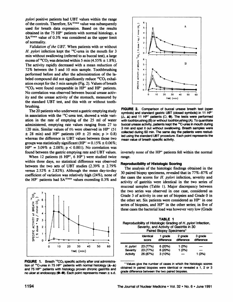
Expression ofBreath Data. In order to explore whetherthe sensitivity and specificity of the breath test variesdependingon the mode of expressionof breathdata,wecompared 75 controls (H. pylon negative on biopsy, normal mucosal histology, no ulcer at endoscopy) with 74non-ulcerpatientswithchronicgastritisandH. pylon onbiopsy. Excretion of “tO2in breath was found to besignificantly higher in these 74 Hi―patients compared tothe 75 controls, and this when resultswere expressedeitherbythespecificactivityorbythecumulativeexcretionovera periodvaryingfrom 5 to 60 mm (Fig. 1).None of the74 HF4 patients with chronic gastritis had a SA value ofthe 10 mm breath sample (SAlOmin)within the range of95% ofthe 75 controls (0. 14% ±0.07%). Similar discriminatiomwasobtainedusingvaluesof cumulativeradioactivity or specificactivityof breathsamplescollectedbetween the 15 and 60 mAn.At 5 mm however,threeH.
Carbon-i4-UreaBreathTest inGastntisand(JcerDisease•Debongnieet al 1193
II
TABLEIReproducibilityof HistologicGradingof H.pylon!Infection,Severity,
and Activity of Gastntis in30PairedBiopsySpecimens*Identical
1 grade 2 grade 3gradescoredifference difference difference
* Values give the number of cases in which the histologic scores
obtainedin pairedbiopsieswereidenticalor revealeda 1, 2 or 3gradedifferencebetweenthetwopairedbiopsies.
pylon positive patients had UBT values within the rangeofthe controls.Therefore,SAJOmInvaluewassubsequentlyused for breath data expression. Based on the resultsobtained in the 75 HP patientswith normal histology, aSA'°@'value of 0.3% was consideredas the upper limitof normality.
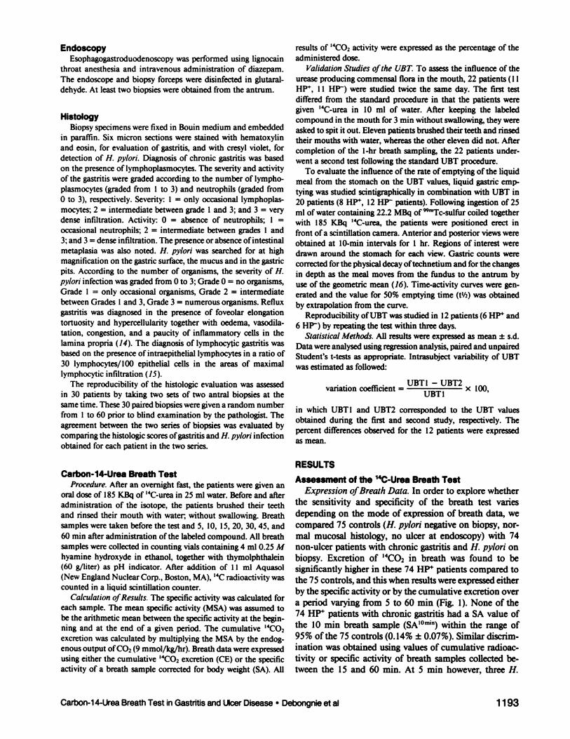
Validation ofthe UBT. When patients with or withoutH. pylon infection kept the 14C-ureain the mouth for 3mm withoutswallowing(referredto as buccaltest), a largeexcess of ‘4CO2was detected within 5 mm (4.55% ±1.8%).The activity rapidly decreasedwith a mean reduction of72% between the 5 and 10 mm sample. Toothbrushingperformed before and after the administration of the labeled compound did not significantly reduce ‘4CO2exhalation except for the 5 mm sample (Fig. 2). Values of breath14C02were found comparable in HP@and HP patients.No correlation was observed between buccal urease activity and the urease activity of the stomach, measured bythe standard UBT test, and this with or without toothbrushing.
The 20patientswhounderwenta gastricemptyingstudyin associationwith the @4C-ureatest, showeda wide vanation in the rate of emptying of the 25 ml of wateradministered, emptying rate values ranging from 27 to120mm. Similarvaluesof t½wereobservedin HP@(51±28 mm) and HP patients (49 ±25 mm; p > 0.8)whereas the difference in UBT values between those twogroups was statistically significant (HF = 0. 15% ±0.06%;HF = 3.09% ±2.08%; p < 0.001). No correlation wasfound betweenthe gastricemptyingrate and UBT values.
When 12 patients (6 HP@,6 HP) were studied twicewithin three days, no statistical difference was observedbetween the two sets of UBT studies (2.39% ±2.79%versus 2.52% ±2.82%). Although the mean day-to-daycoefficientof variation was relativelyhigh (24%),none ofthe HP patients had SAIOmInvalues exceeding 0.3% and
60U 5
@ 4
%.- 0
0U
- 6I
TIhIE (mm)
I'
FIGURE2. Comparisonof buccalureasebreathtest(opensymbols)and standardgastric UBT (closedsymbols) in 11 HP(& A) and 11 HP@patients(0, •).The tests wereperformedwithtoothbrushing(B)orwithouttoothbrushing(A).Toquantitatebuccalureaseactivity,patientskeptthe14@j@Inmouthduring3 mmandspatitoutwithoutswallowing.BreathsampleswerecOlleCtedduring60 mm.The sameday the patientswere restudledusingthestandardUBTprocedure.Eachpointrepresentsthemeanvalueof breathspecificactivity.
inversely none of the HP patients fell within the normalrange.
Reproducibmlftyof Histologic ScoringThe analysisof the histologicfindings obtained in the
30 paired biopsy specimens, revealed that in 77%—87%ofthe cases the scores for H. pylon infection, severityandactivity of gastritis were identical in the two series ofmucosal samples (Table 1). Major discrepancy betweenthe two series was observed in one case, considered asGrade 3 of activity in one set of biopsiesand Grade 0 inthe other set. Six patients were consideredas HP in oneseries of biopsies, and HP@in the other series; in five ofthese cases the bacterial load was however very low (Grade
(@‘ 70C) _
@ gN 6
@ 5
E4.@..,‘
@ 3
@ 2
@t 1U
a- _______________(i@ 0@ £ A
0 10 20 30 40 50 60
TIME(mm)H.pylon!23 (77%)6 (20%)1(3%)—Severity23
(77%)6 (20%)1(3%)—Activity26(87%)3 (10%)—1 (3%)
FIGURE1. Breath14C02specificactivityafteroraladministrationof 14C-ureain 75 HP patientswithnormalhistology(A—A)and75 HP@patientswithhistologyprovenchronicgastritisandnoulceratendoscopy(—). Eachpointrepresentsmean±s.d.
1194 The Journalof NuclearMedicine•Vol. 32 •No. 6 •June1991
BreathTest, Histology,andEndoscopyTABLResultsin ElevenFaE2
Isa Positiveand 8False-Negative Resultsof BreathTestsBreath
test
SA SAHistology
gradesPatient
5 mm i 0mmEndoscopyHistologySeverityActivityHP*%False
+veUBTI2.91 2.92gastntischronicg.21022.63 2.29gastric ulcerrefluxg.00031.25 1.53normalchronicg.12041.90 1.37duodenal ulcerchronicg.10050.75 1.25gastntisrefiuxg.00061.14 1.12gastric ulcerchronicg.20070.57 0.81gastntischronicg.10080.74 0.62gastntischronicg.10090.53 0.46duodenal ulcerrefluxg.0001
0 1.070.38erosionsnormal00011 0.560.31normalnormal000mean
1.011.19s.d.0.690.79False
—yeUBT10.060.04normalnormal00120.090.05gastntisnormal00130.110.07normalnormal00140.23 0.15gastriculcernormal00150.230.17gastntisnormal00160.23 0.18normalrefiuxg.00170.33 0.20erosionrefluxg.00180.54 0.28normalchronicg.111mean0.230.14s.d.0.150.08*
HP = Helicobacter Pylon
%mean0.25 2.64*[email protected] 2.252.282.06range0.02—2.920.04—8.380.66—9.040.98—8.16*
p < 0.001 compared to grade 0patientst
p < 0.001 compared to grade 1 patients
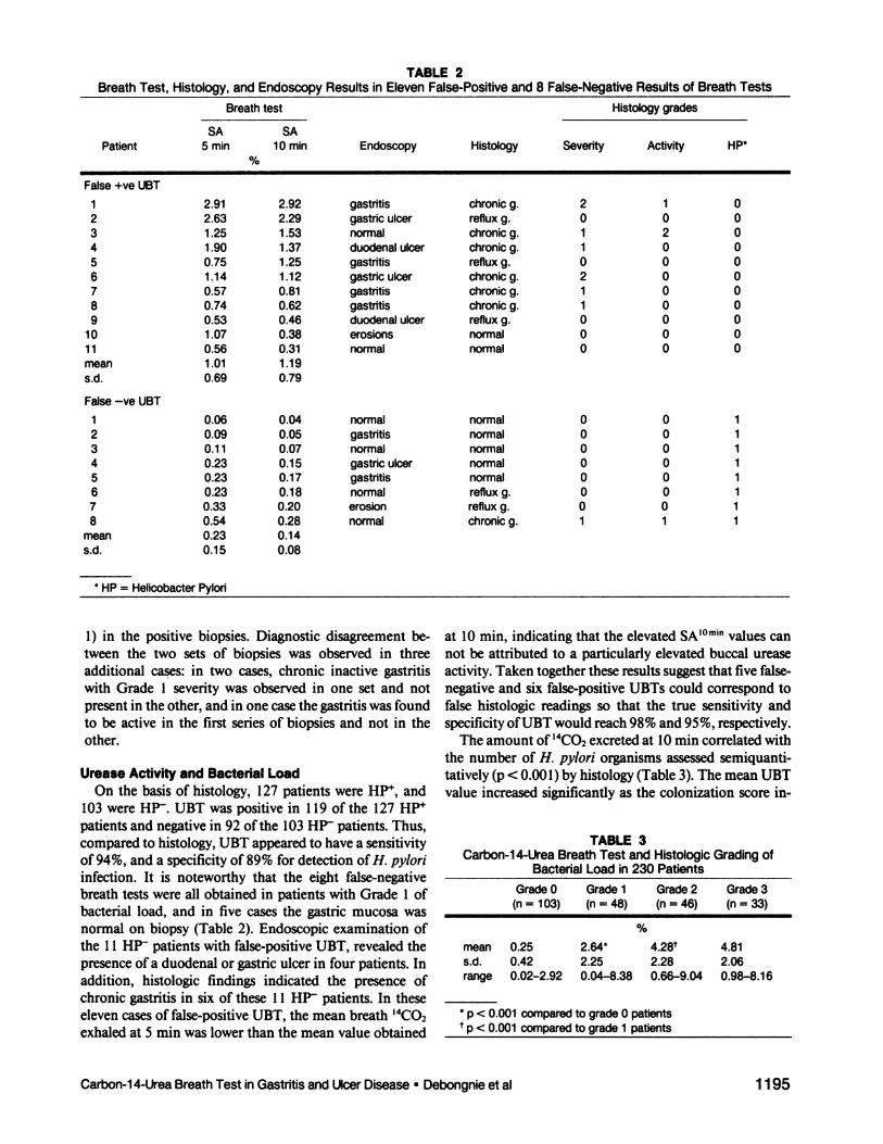
1) in the positive biopsies. Diagnostic disagreement between the two sets of biopsies was observed in threeadditional cases: in two cases, chronic inactive gastritiswith Grade 1 severity was observed in one set and notpresent in the other, and in one case the gastritis was foundto be active in the first series of biopsies and not in theother.
UreaseActivityand BacterialLoadOn the basis of histology, 127patients were HP@,and
103 were H@. UBT was positive in 119 of the 127 HP'patients and negative in 92 ofthe 103 HP patients. Thus,compared to histology, UBT appeared to have a sensitivityof94%, and a specificity of89% for detection ofH. pyloninfection. It is noteworthy that the eight false-negativebreath tests were all obtained in patients with Grade 1 ofbacterial load, and in five cases the gastric mucosa wasnormal on biopsy (Table 2). Endoscopic examination ofthe 11 HP- patients with false-positive UBT, revealed thepresence of a duodenal or gastric ulcer in four patients. Inaddition, histologic findings indicated the presence ofchronic gastritis in six of these 11 HP patients. In theseeleven cases of false-positive UBT, the mean breath ‘4C02exhaled at 5 mm was lower than the mean value obtained
at 10 mm, indicating that the elevated SAlOminvalues cannot be attributed to a particularlyelevated buccal ureaseactivity. Taken together these results suggest that five falsenegative and six false-positive UBTs could correspond tofalse histologic readings so that the true sensitivity andspecificity ofUBT would reach 98% and 95%, respectively.
The amount of ‘4CO2excretedat 10 mm correlatedwiththe number of H. pylon organisms assessed semiquantitatively (p < 0.00 1)by histology (Table 3). The mean UBTvalue increased significantly as the colonization score in
TABLE 3Carbon-14-UreaBreathTestandHistologicGradingof
BacterialLoadin 230 PatientsGrade0 Grade1 Grade2 Grade3(n= 103) (n= 48) (n= 46) (n= 33)
Carbon-i4-UreaBreathTest inGastntisand(JcerDisease•Debongnieet al 1195
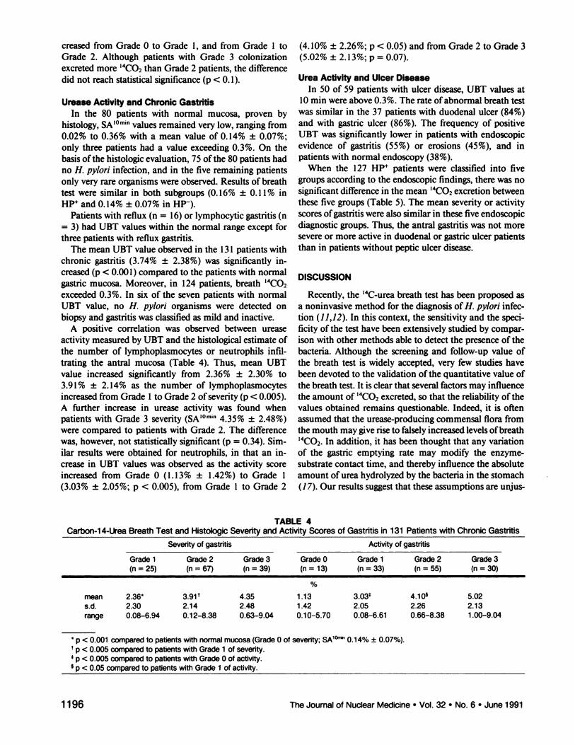
TABLE 4Carbon-i 4-Urea Breath Test and Histologic Severity and Activity Scores of Gastntis in 131 Patients withChronicGastntisSeventy
ofgastritis ActivityofgastritisGrade
1 Grade2 Grade3 Grade0 Grade1 Grade2Grade3(n= 25) (n = 67) (n= 39) (n= 13) (n= 33) (n= 55)(n =30)%mean
2.36* 3.9l@ 4.35 1.13 [email protected]@5.02s.d.2.30 2.14 2.48 1.42 2.052.262.13range0.08—6.94 0.12—8.38 0.63—9.04 0.10—5.70 0.08—6.610.66—8.381.00—9.04*
p < 0.001 compared to patients with normal mucosa (Grade 0 of seventy; SA1°―@' 0.14% ±0.07%).t
p < 0.005 compared to patients with Grade 1 ofseverity.S
p < 0.005 compared to patients with Grade 0 ofactivity..
p < 0.05 compared to patients with Grade 1 of activity.
creased from Grade 0 to Grade 1, and from Grade 1 toGrade 2. Although patients with Grade 3 colonizationexcreted more ‘4C02than Grade 2 patients, the differencedid not reach statistical significance (p < 0.1).
Urease Activity and Chronic GastritisIn the 80 patients with normal mucosa, proven by
histology, SAJOmInvalues remained very low, rangingfrom0.02% to 0.36% with a mean value of 0.14% ±0.07%;only three patients had a value exceeding 0.3%. On thebasis ofthe histologic evaluation, 75 ofthe 80 patients hadno H. pylon infection, and in the five remaining patientsonly very rare organisms were observed. Results of breathtest were similar in both subgroups (0. 16% ±0. 11% inHP@and 0.14% ±0.07% in HP-).
Patients with reflux (n = 16) or lymphocytic gastritis(n= 3) had UBT values within the normal range except forthree patients with reflux gastntis.
The mean UBT value observed in the 131 patients withchronic gastritis (3.74% ±2.38%) was significantly increased (p < 0.001) compared to the patients with normalgastric mucosa. Moreover, in 124 patients, breath ‘4CO2exceeded 0.3%. In six of the seven patients with normalUBT value, no H. pylon organisms were detected onbiopsy and gastritis was classified as mild and inactive.
A positive correlation was observed between ureaseactivity measured by UBT and the histological estimate ofthe number of lymphoplasmocytes or neutrophils infiltrating the antral mucosa (Table 4). Thus, mean UBTvalue increased significantly from 2.36% ±2.30% to3.91% ±2.14% as the number of lymphoplasmocytesincreased from Grade 1 to Grade 2 ofseverity (p < 0.005).A further increase in urease activity was found whenpatients with Grade 3 severity (SAJOm2@@4@35%±2.48%)were compared to patients with Grade 2. The differencewas, however, not statistically significant (p 0.34). Similar results were obtained for neutrophils, in that an increase in UBT values was observed as the activity scoreincreased from Grade 0 (1. 13% ± 1.42%) to Grade 1(3.03% ±2.05%; p < 0.005), from Grade 1 to Grade 2
(4.10% ±2.26%; p < 0.05) and from Grade 2 to Grade 3(5.02% ±2. 13%; p = 0.07).
Urea Activftyand UlcerDiseaseIn 50 of 59 patients with ulcer disease, UBT values at
10 mm were above 0.3%. The rate ofabnormal breath testwas similar in the 37 patients with duodenal ulcer (84%)and with gastric ulcer (86%). The frequency of positiveUBT was significantly lower in patients with endoscopicevidence of gastritis (55%) or erosions (45%), and inpatients with normal endoscopy (38%).
When the 127 HP patients were classified into fivegroups according to the endoscopic findings, there was nosignificant difference in the mean ‘4CO2excretion betweenthese five groups (Table 5). The mean severity or activityscores ofgastritis were also similar in these five endoscopicdiagnostic groups. Thus, the antral gastritis was not moresevere or more active in duodenal or gastric ulcer patientsthan in patients without peptic ulcer disease.
DISCUSSION
Recently, the ‘4C-ureabreath test has been proposed asa noninvasive method for the diagnosis of H. pylon infection (11,12). In this context, the sensitivity and the specificity of the test have been extensively studied by comparison with other methods able to detect the presence of thebacteria. Although the screening and follow-up value ofthe breath test is widely accepted, very few studies havebeen devoted to the validation ofthe quantitative value ofthe breathtest. It is clear that several factorsmay influencethe amount of ‘4C02excreted, so that the reliabilityof thevalues obtained remains questionable. Indeed, it is oftenassumed that the urease-producing commensal flora fromthe mouth may give rise to falsely increased levels of [email protected] addition, it has been thought that any variation
of the gastric emptying rate may modify the enzymesubstrate contact time, and thereby influence the absoluteamount of urea hydrolyzed by the bacteria in the stomach(17). Our results suggest that these assumptions are unjus
1196 TheJournalof NuclearMedicine•Vol.32 •No.6 •June1991
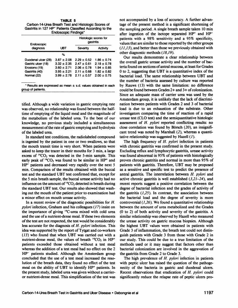
TABLE5Carbon-i4-Urea Breath Test and Histologic ScoresofGastntis
in 127 HP@PatientsClassifiedAccordingtotheEndoscopicFmndings*Histologic
scoresfor.gastntis
Endoscopicdiagnosis UBT SeverityActivity%
* Results are expressed as mean ± s.d. values obtained in each
groupofpatients.
not accompanied by a loss of accuracy. A further advantage of the present method is a significant shortening ofthe sampling period. A single breath sample taken 10 mmafter ingestion of the isotope separated H@ and HPpatients with a 98% sensitivity and a 95% specificity,values that are similar to those reported by the other groups(11,13), and better than those we previously obtained withother diagnostic methods (18,19).
Our results demonstrate a clear relationship betweenthe overall gastric urease activity and the number of bacteria found on sections ofantral mucosa, at least for Grades0 to 2, suggesting that UBT is a quantitative index of thebacterial load. The same relationship between UBT andthe number of bacteria assessed by culture was reportedby Rauws (13) with the same limitation: no differencecould be found between Grades 2+ and 3+ of colonization.Since an adequate mass of carrier urea was used by theAmsterdam group, it is unlikely that the lack of discrimination between patients with Grades 2 and 3 of bacterialload is due to an exhaustion of the substrate. Otherinvestigators comparing the time of reaction of a rapidurease test (CLO test) and the semiquantitative histologicassessment of H. pylon reported conflicting results: noclose correlation was found by BOrsch (20), an insignificant trend was noted by Marshall (2), whereas a quantitative relationship was suggested by Hazell (1).
The high frequency of H. pylon infection in patientswith chronic gastntis was confirmed in the present study.Excluding reflux and lymphocytic gastritis, urease activitywas found abnormal in 95% ofpatients with histologicallyproven chronic gastritis and normal in more than 95% ofpatients with gastntis. Therefore, UBT may be proposedas a sensitive and specific test to predict the presence ofantral gastritis. The interrelation between H. pylon andactive chronic gastntis is well established (3,21—24),andrecent reports suggest a positive correlation between thedegree of bacterial infection and the grades of activity ofthe gastritis (1,25). In contrast, the association betweenthe bacterial load and the degree of severity is morecontroversial (1,26). We found a quantitative relationshipbetween the amount of urea metabolized and the Grades(0 to 2) of both activity and severity of the gastritis. Asimilar relationship was observed by Hazel who measuredthe urease activity on gastric mucosa biopsies. Althoughthe highest UBT values were obtained in patients withGrade 3 of inflammation, the breath test could not distinguish patients with Grade 3 from those with Grade 2 inour study. This could be due to a true limitation of themethods used or it may suggest that factors other thanbacterial colonization are involved in the aggravation ofthe gastritis from Grade 2 to Grade 3.
The high prevalence of H. pylon infection in patientswith peptic ulcer has raised the question of the pathogenecity of the bacteria in gastric and duodenal ulcers.Recent observations that eradication of H. pylon couldsignificantly reduce the relapse rate of peptic ulcers pro
Duodenalulcer(28)3.87 ±2.082.29 ±0.521 .86±0.74Gastriculcer(18)3.32 ±2.352.47 ±0.612.18 ±0.78Erosions
(19)3.60 ±2.712.06 ±0.781 .94±0.85Gastritis(40)3.95 ±2.212.1 1±0.681 .82±0.82Normal
(22)3.99 ±2.782.1 1±0.572.00 ±0.75
tified. Although a wide variation in gastric emptying ratewas observed, no relationship was found between the halftime ofemptying ofthe liquid meal and the magnitude ofthe metabolism of the labeled urea. To the best of ourknowledge, no previous study included a simultaneousmeasurement ofthe rate ofgastric emptying and hydrolysisof the labeled urea.
In standardtest conditions, the radiolabeledcompoundis ingested by the patient in one or two swallows, so thatthe mouth transit time is very short. When patients wereasked to keep the tracer in their mouths for 3 mm, a largeexcess of “CO2was detected in the 5-mm sample. Thisearly peak of ‘4C02was found to be similar in HP@andHP patients and decreased very rapidly over the next 5mm. Comparison of the results obtained with the buccaltest and the standard UBT test confirmed that, except forthe 5 mm breath sample, the buccal urease activity had noinfluence on the amount of ‘4C02detected in breath duringthe standard UBT test. Our results also showed that washing out the mouth ofthe patient prior to examination hada minor effect on mouth urease activity.
In a recent review of the diagnostic possibilities for H.pylon infection, Graham and his colleagues (1 7) insist onthe importance of giving ‘4C-ureamixed with cold ureaand the use ofa nutrient-densemeal. Ifthese two elementsofthe test are not respected, the test would be considerablyless accurate for the diagnosis of H. pylon infection. Thisidea was supported by the report ofTytgat and co-workers(13) who found that when UBT was carried out with anutrient-dense meal, the values of breath “CO2in HP@patients exceeded those obtained without a test mealwhereas the addition of a test meal had no effect on the 2HP patients studied. Although the Amsterdam groupconcluded that the use of a test meal increased the resolution of the breath data, they found no effect of the testmeal on the ability of UBT to identify HP patients. Inthe present study, labeled urea was given without a carrieror nutrient-dense meal. This simplification of the test was
Carbon-i4-UreaBreathTest inGastritisand(JeerDisease•Debongnieet al 1197
pylobacterpyloridis associated chronic active antral gastritis. A prospectivestudy of its prevalence and the effects of antibacterial and antiulcer treatment.Gastroenserology1988;94:33—40.
4. Rollason TP, StoneJ, RhodesJM. Spiral organismsin endoscopicbiopsiesofthe human stomach. J Clin Pathol 1984;37:23—26.
5. Warren JR. Marshall B. Unidentified curved bacilli on gastric epitheliumin active chronic gastritis. Lancet 1983; i:1273.-1275.
6. Dooley CP, Cohen H. The clinicalsignificanceof Campylobacierpylon.Ann InternMed 1988;108:70—79.
7. Marshall BJ, Armstrong JA, McGechie DB, Glancy Ri. Attempt to fulfillKoch's postulates for Campylobacterpylonidis. MedJAusi l985;142:436—439.
8. MomsA, RafiqAM, BlaserM, Perez-PerezG. HumaningestionofCampylobacterpyloni—long-termfollow-up.In: MegraudF, LamouliatteH, eds. Gastnoduodenoi pathology & Campylobacten pyloni. Amsterdam:Excepta Medica; 1989:399-400.
9. Graham DY, Alpert LC, Smith L, Yoshimura HH. latrogenic Campylobactenpyloni infection is a cause ofepidemic achlorhydria. Am J Gastroentenol 1988;83:974—980.
10. Langeberg W, Tytgat GNJ, Schipper MEl, Rietra PiG, Zanen HC. Campylobacterlike organismsin the stomach of patientsand healthy individuals. Lances 1984;l:1348.
11. Graham DY, Klein PD, Evans DJ, et al. Campylobacten pylon detectednoninvasively by the ‘3C-ureabreath test. Lancet 1987;l:1174—1177.
12. Marshall Bi, Surveyor I. Carbon-14-urea breath test for the diagnosis ofCampylobaczenpyloni-associatedgastritis.J Nuci Med 1988;29:l1—16.
13. Rauws EAJ, Royen EV, LangeberbW, WoenselJV, Vrij AA, Tytgat GN.‘4C-ureabreath test in C. pylon gastritis. Gut 1989;30:789—803.
14. O'Connor HJ, Wyatt JJ, Dixon MF, Axon ATR. Campylobaczen-likeorganisms and reflux gastritis. I Clin Pathol 1986;39:531—534.
15. Haot J, Hamichi C, Wailer L, Mainguet P. Lymphocytic gastritis:a newlydescribed entity: a retrospective endoscopic and histological study. Gut1988;29:1258—1264.
16. Urbain JLC, Siegel JA, Debie NC, Pauwels S. Effect ofcisapride on gastricemptying in dyspeptic patients. Dig Dis Sci 1988;33:779—783.
17. Alpert LC, Graham DY, Evans DF, et al. Diagnostic possibilities forCampylobacter pylon infection. Eur J Gasinoentenol Hepazol 1989;1:17—26.
18. Debongnie JC, Legros 0, Wautelet M, Beyaert C, Mainguet P. Evaluationde la valeur de la cytologic perendoscopique dans l'identification duCampylobacten pylon sur la muqueuse gastrique. Gastroenterol Clin Biol1987;l 1:764—767.
19. Debongnie JC, Legros G, Beyaert C. La gastrite a Campylobacten pylon.Comparisonde plusieursméthodesdiagnostiques.Acta GasinoentenolBdg1987;154:125—132.
20. Borsch 0, Adamek R, Sandmann M, et al. Comparison of biopsy ureasetest and histologic examination for detection of Campylobacien pylon! induodenal, antral, and fundic biopsies.Hepatogasinoentenol1987;34:236—241.
21. Andersen LP, Holck S, Poulsen CO. Elsborg L, Justesen T. Campylobactenpylonidis in peptic ulcer disease. ScandJ Gastnoenienol 198722:219—224.
22. Blomberg B, Jarnerot 0, Kjellander J, Danielson D, Kraaz W. Prevalenceof Campylobactenpylon in an unselected Swedish population of patientsreferred for gastroscopy. Scandf Gastroentenol 198&,23:358—362.
23. ChodosJE, Dworkin BD, Smith F, Van Horn K, WeissL, RosenthalWS.Campylobacterpyloniandgastroduodenaldisease: a prospective endoscopicstudy and comparison of diagnostic tests. Am J Gastroenterol1988;83:1226—1230.
24. Marshall B. Unidentified curved bacilli on gastric epithelium in activechronicgastritis.Lancet1983;l:1273—1275.
25. Loffeld RJLF, PottersHVPJ, Arends JW, StobberinghE, FlendngJA, VanSpreeuwel JP. Campylobacten pylon-associated gastritis in patients withnon-ulcer dyspepsia. J Clin Pathol 1988;41:85—88.
26. Raskov H, Lazing C, Gaarslev K, Fisher-Hansen B, Hauch 0. Screeningfor Campylobacten pylonidis in patients with upper dyspepsia and therelation to inflammation of the human gastric antrum. Scand I Gastroentenol1987;22:568—572.
27.O'MorainC,TobinA, CoghlanJ.Themanagementofduodenalulcerrelapse.ScandlGastroentenol1989;245153:89—98.
28.QueirozDMM,BarbosaMA, MendesEN,etal.Distributionof Campylobacten pylon and gastritis in the stomach of patients with and withoutduodenal ulcer. Am J Gastroentenol 1988;83:1368—1370.
vided strong, but still indirect, evidence for a cause-effectrelationship between H. pylon colonization and ulcer disease (27). If most of the infected patients have gastritis,only a limited number develop ulcers, and the sequenceof events leading some of these patients after H. pyloninfection to ulceration is poorly understood. In this context, we have examined whether the occurrence of gastricor duodenal ulcer could be associated with a particularlyincreased number of H. pylon organisms. Our resultsshowed that most of ulcer patients had a positive UBT.However, the urease activity measured in the HP' ulcerpatients was similar to that found in HP nonulcer patients, indicating that the bacterial load was not increasedin ulcer compared to nonulcer patients. The present observations extend the previous study of Queiroz and coworkers who noted no difference in the intensity of bactenal growth, assessed on biopsies, between gastritis patients with and without duodenal ulcer (28).
Taken together, these findings suggest that the role ofH. pylon in the pathogenesis ofpeptic ulcers presents somesimilarity with the role played by acid. Indeed, both factorshave to be present for the onset of ulceration, and theirsuppression is associated with healing and prolonged remission ofthe ulcer. Although the presence ofacid constitutes a prerequisite, the amount ofacid, however, does nothave to be increased. Similarly, H. pylon organisms haveto be present, but their number appears not to be adeterminant factor for the onset ofthe ulceration.
The present results are of interest because they allow usto propose a simple and rapid noninvasive method foraccurate detection ofH. pylon infection. The quantitativerelationship found between the urease activity, measuredby UBT and both activity and severity grades of gastritisprovides furthersupport to the pathogenic role played bythe bacteria in chronic and active gastritis. The evidencethat in HP@patients, the presence of ulcer disease is notassociated with an increased urease activity compared tononulcer gastritissuggests that the importance of the bacterial load is not a determinant factor in the pathogenesisof ulcer disease.
ACKNOWLEDGMENTS
The authorsthankJean-LouisBavayand Marie-AgnesLeeroan for skilled technical assistanceand Sylvia Fernandez forpreparationofthe manuscript.
REFERENCES1. Hazell SL, Borody TJ, Gal A, Lee A. Campylobacterpyloridis gastritis. I.
Detection ofurease as a marker ofbacterial colonization and gastritis. AmJ Gastroenterol1987;82:292—296.
2. Marshall Bi, Warren JR. Francis GJ, Langton SR, Goodwin CS, BlincowE. Rapidureasetestin the managementof Campylobacierpyloridisassociated gastritis. Am J Gasiroenierol 1987;82:200—210.
3. Rauws EAJ, Langeberg W, Houthoff Hi, Zanen JC, Tytgat GMJ. Cam
1198 The Journalof NuclearMedicine•Vol. 32 •No. 6 •June 1991
Copyright © 2022 FDOKUMEN