
The mAKAPβ scaffold regulates cardiac myocyte hypertrophy via recruitment of activated calcineurin
Upload
independentCategory
view
1download
0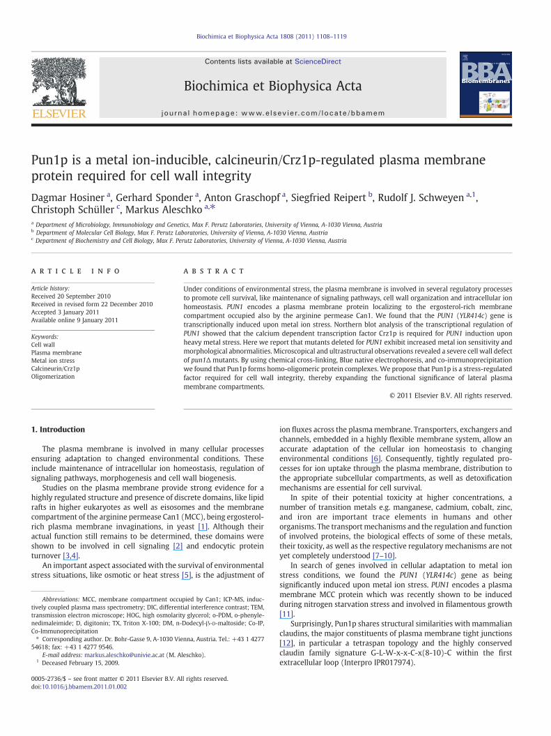
Biochimica et Biophysica Acta 1808 (2011) 1108–1119
Contents lists available at ScienceDirect
Biochimica et Biophysica Acta
j ourna l homepage: www.e lsev ie r.com/ locate /bbamem
Pun1p is a metal ion-inducible, calcineurin/Crz1p-regulated plasma membraneprotein required for cell wall integrity
Dagmar Hosiner a, Gerhard Sponder a, Anton Graschopf a, Siegfried Reipert b, Rudolf J. Schweyen a,1,Christoph Schüller c, Markus Aleschko a,⁎a Department of Microbiology, Immunobiology and Genetics, Max F. Perutz Laboratories, University of Vienna, A-1030 Vienna, Austriab Department of Molecular Cell Biology, Max F. Perutz Laboratories, University of Vienna, A-1030 Vienna, Austriac Department of Biochemistry and Cell Biology, Max F. Perutz Laboratories, University of Vienna, A-1030 Vienna, Austria
Abbreviations: MCC, membrane compartment occutively coupled plasma mass spectrometry; DIC, differenttransmission electron microscope; HOG, high osmolaritnedimaleimide; D, digitonin; TX, Triton X-100; DM, n-Co-Immunoprecipitation⁎ Corresponding author. Dr. Bohr-Gasse 9, A-1030 Vie
54618; fax: +43 1 4277 9546.E-mail address: [email protected] (M. A
1 Deceased February 15, 2009.
0005-2736/$ – see front matter © 2011 Elsevier B.V. Adoi:10.1016/j.bbamem.2011.01.002
a b s t r a c t
a r t i c l e i n f oArticle history:Received 20 September 2010Received in revised form 22 December 2010Accepted 3 January 2011Available online 9 January 2011
Keywords:Cell wallPlasma membraneMetal ion stressCalcineurin/Crz1pOligomerization
Under conditions of environmental stress, the plasma membrane is involved in several regulatory processesto promote cell survival, like maintenance of signaling pathways, cell wall organization and intracellular ionhomeostasis. PUN1 encodes a plasma membrane protein localizing to the ergosterol-rich membranecompartment occupied also by the arginine permease Can1. We found that the PUN1 (YLR414c) gene istranscriptionally induced upon metal ion stress. Northern blot analysis of the transcriptional regulation ofPUN1 showed that the calcium dependent transcription factor Crz1p is required for PUN1 induction uponheavy metal stress. Here we report that mutants deleted for PUN1 exhibit increased metal ion sensitivity andmorphological abnormalities. Microscopical and ultrastructural observations revealed a severe cell wall defectof pun1Δ mutants. By using chemical cross-linking, Blue native electrophoresis, and co-immunoprecipitationwe found that Pun1p forms homo-oligomeric protein complexes. We propose that Pun1p is a stress-regulatedfactor required for cell wall integrity, thereby expanding the functional significance of lateral plasmamembrane compartments.
pied by Can1; ICP-MS, induc-ial interference contrast; TEM,y glycerol; o-PDM, o-phenyle-Dodecyl-β-D-maltoside; Co-IP,
nna, Austria. Tel.: +43 1 4277
leschko).
ll rights reserved.
© 2011 Elsevier B.V. All rights reserved.
1. Introduction
The plasma membrane is involved in many cellular processesensuring adaptation to changed environmental conditions. Theseinclude maintenance of intracellular ion homeostasis, regulation ofsignaling pathways, morphogenesis and cell wall biogenesis.
Studies on the plasma membrane provide strong evidence for ahighly regulated structure and presence of discrete domains, like lipidrafts in higher eukaryotes as well as eisosomes and the membranecompartment of the arginine permease Can1 (MCC), being ergosterol-rich plasma membrane invaginations, in yeast [1]. Although theiractual function still remains to be determined, these domains wereshown to be involved in cell signaling [2] and endocytic proteinturnover [3,4].
An important aspect associated with the survival of environmentalstress situations, like osmotic or heat stress [5], is the adjustment of
ion fluxes across the plasmamembrane. Transporters, exchangers andchannels, embedded in a highly flexible membrane system, allow anaccurate adaptation of the cellular ion homeostasis to changingenvironmental conditions [6]. Consequently, tightly regulated pro-cesses for ion uptake through the plasma membrane, distribution tothe appropriate subcellular compartments, as well as detoxificationmechanisms are essential for cell survival.
In spite of their potential toxicity at higher concentrations, anumber of transition metals e.g. manganese, cadmium, cobalt, zinc,and iron are important trace elements in humans and otherorganisms. The transport mechanisms and the regulation and functionof involved proteins, the biological effects of some of these metals,their toxicity, as well as the respective regulatory mechanisms are notyet completely understood [7–10].
In search of genes involved in cellular adaptation to metal ionstress conditions, we found the PUN1 (YLR414c) gene as beingsignificantly induced upon metal ion stress. PUN1 encodes a plasmamembrane MCC protein which was recently shown to be inducedduring nitrogen starvation stress and involved in filamentous growth[11].
Surprisingly, Pun1p shares structural similarities with mammalianclaudins, the major constituents of plasma membrane tight junctions[12], in particular a tetraspan topology and the highly conservedclaudin family signature G-L-W-x-x-C-x(8-10)-C within the firstextracellular loop (Interpro IPR017974).
1109D. Hosiner et al. / Biochimica et Biophysica Acta 1808 (2011) 1108–1119
In addition to Can1p and Pun1p, seven other proteins are known tobe integral components of theMCC. Among them, the uracil permeaseFur4p, the tryptophan/tyrosin permease Tat2p, and the tetraspanproteins Sur7p, Nce102p, Fmp45p, Fhn1p, and Ynl194cp, all five ofunknown function [4,13]. Besides Fmp45p and Ynl194cp [14], Pun1pis a third S. cerevisiae paralog of Sur7p. Interestingly, all four proteinscontain a cysteine motif similar to the claudin signature in their firstextracellular loop, but only for Pun1p it is perfectly conserved [15].
Thus, the clustering of tetraspan proteins in MCC compartmentsraises the challenging question if these proteins and their commondomains share evolutionary conserved structural and functionalproperties.
2. Materials and methods
2.1. Yeast strains, media and culture conditions
Saccharomyces cerevisiae strains were grown at 28 °C in liquid YPDmedium(yeast extract/peptone/dextrose), on solid YPD in serial 10-folddilution steps or in standard SDmedium (0.67% yeast nitrogen base, 2%glucose, and amino acids as required). Escherichia coli strain DH10b(Invitrogen, Lofer, Austria) was cultivated at 37 °C in Luria–Bertani (LB)medium supplemented with 100 μg/ml ampicillin when appropriate.Mediawere supplementedwith variousmetal ion salts when indicated.Yeast strains BY4741 (acc. no. Y00000), W303 (acc. no. 20000A) andFY1679 (acc. no. 10000 M) provided by Euroscarf (Frankfurt, Germany)were used aswild-type.Mutant derivatives of BY4741 used in this studywere also provided by Euroscarf. These were BYhog1Δ, BYcrz1Δ,BYrlm1Δ, BYste12Δ, BYsko1Δ, and BYdig1Δ. Corresponding genes werecompletely deleted and replaced by the geneticin resistance moduleKanMX4. Strain W303msn2/4Δ was described previously [16]. StrainBY4741 bearing the chromosomal PUN1-GFP fusion was obtained fromthe yeast GFP-tagged collection [17]. The haploid deletion strainMApun1Δ was constructed from parental strain FY1679. According tothe one step replacement protocol [18] the PUN1 ORF was deleted byhomologous recombination with a HIS5 disruption cassette amplifiedfrom plasmid pSG634 with the primers PUN1-delfor: 5′-TTCGCTAG-GAGCACTTATATTAGCCATTGTTGCATGCGCAGGATCCGTACGCTG-CAGGTCGAC-3′ and PUN1-delrev: 5′-GACGGTGGGCCTCGATCTCACGCTACACCATCGAATGAAATTCAGTAGGCCACTAGTGGATCTG-3′. Verifica-tion of correct gene replacement was performed by analytical PCRusing primers PUN1-up: 5′-AAATCGGGCGTACTATCAGCCAAGC-3′,PUN1-in: 5′-AAACACGAACCTATACTGACTAATAGG-3′, and HIS-in:5′-TCTACAAAAGCCCTCCTACCCATG-3′.
2.2. Plasmid constructs
To express PUN1 from its endogenous promoter, the entire ORFand its flanking regions including 450 nt upstream of the ATG and 175nt downstream of the stop codon, were PCR-amplified from FY1679genomic DNA using the forward primer PUN1-fwd: 5′-TAGAGCT-CAACTGTCACAGCCTCCCACTTGACC-3′ and reverse primer PUN1-rev:5′-ATGTCGACAAGAAGATCATGCAACATCACC-3′. The PCR product wascloned via its introduced restriction sites SacI and SalI (underlined)into the centromeric vector YCplac22 [19], thereby creating thevector YCp-PUN1. To generate C-terminally triple HA-tagged Pun1pexpressed via the strong MET25 promoter, the PUN1 coding sequencewas PCR-amplified using the oligonucleotide primers PUN1-SpeI:5′-AAACACTAGTTCGAAGGACGCTATAAGCATGAGG-3′ and PUN1-SalI:5′-TTTCGTCGACAAATCAATGGTTTTTCCTCAATTGG-3′, and cloned viathe SpeI and SalI restriction sites (underlined) into the multicopyvector YEpM351HA [20]. For expression of C-terminally triple HA-tagged Pun1punder the control of its endogenous promoter, PUN1wasPCR-amplified from FY1679 genomic DNAusing the primer pair PUN1-fwd and PUN1-SalI. The PCR product was cloned via the SacI and SalIrestriction sites into plasmid YEp351HA [21] and then subcloned
together with the HA tag into plasmid YCplac33 [19], thereby creatingthe construct YCp-PUN1-HA. To generate the centromeric plasmidpUG35-PUN1-GFP expressing C-terminally GFP-tagged Pun1p underthe control of the MET25 promoter, the PUN1 coding sequence wasPCR-amplified using the oligonucleotide primers PUN1-SpeI (seeabove) and PUN1-EcoRI: 5′-TTTCAGAATTCAATCAATGGTTTTTCCT-CAATTGG-3′ and cloned into the SpeI- and EcoRI-digested vectorpUG35 [22].
2.3. Intracellular AsCl3 measurement
Strains FY1679 and MApun1Δ carrying the empty plasmidYEpM351HA and MApun1Δ overexpressing C-terminally HA-taggedPUN1 from plasmid YEpM-PUN1-HA (MApun1Δ(PUN1)n) were ad-justed to an OD600 = 0.5 and grown in SD medium and SD mediumsupplemented with 250 μMAsCl3 for 6 h at 28 °C. Cells were collectedand washed twice with Fluka highly pure deionized water (Sigma-Aldrich), OD600 and the dry weight were defined and the totalintracellular As3+ concentration of whole cells (μg As3+/g cells) wasmeasured by inductively coupled plasmamass spectrometry (ICP-MS,ARC Seibersdorf Research GmbH, Austria).
2.4. Fluorescence microscopy
Yeast cells used for analysis of GFP fusion proteins and for cell wallstaining were grown to early log phase. Fluorescence microscopy wasused to detect GFP and fluorescent dyes and differential interferencecontrast (DIC) optics were used to observe cell morphology. ForAniline Blue staining cells were washed with distilled water andincubated in 0.05% Aniline Blue for 5 min. For chitin staining washedcells were incubated in 1 mg/ml Calcofluor White for 5 min, washedtwice and then observed. Concanavalin A–FITC staining was carriedout by incubating washed cells in 0.1 mg/ml concanavalin A–FITC in10 mM sodium phosphate buffer, pH 7.2, 150 mMNaCl for 10 min. ForFilipin staining of plasma membrane ergosterol cells were washedwith 50 mMpotassium phosphate buffer, pH 5.5, stainedwith 5 μg/mlFilipin for 5 min and washed twice in the same buffer. Aniline Blue,Calcofluor White, concanavalin A–FITC, and Filipin were purchasedfrom Sigma-Aldrich (Schnelldorf, Germany).
Fluorescence was visualized in living cells without fixation. Imageswere captured with equivalent exposures using a Zeiss Axioplan 2fluorescence microscope with an AxioCam MRc5 CCD camera usingAxioVision 4.8.1 software (Carl Zeiss, Oberkochen, Germany). Grays-cale imageswere processedwith Photoshop CS3 (Adobe, San Jose, CA).
2.5. Electron microscopy
FY1679 wild-type and MApun1Δ cells bearing the empty plasmidYCplac22, as well as MApun1Δ cells expressing Pun1p from plasmidYCp-PUN1 were grown in YPD to log phase (OD600 = 1). Forcryofixation, cell pellets were introduced to flat sample holders ofan EMPACT high-pressure freezer (LEICA Microsystems, Austria). Flatspecimen holders were placed in a sample holder pod and tightlysealed. The samples were frozen as described previously for speci-mens ofmammalian tissues [23]. Following freezing, the flat specimenholders were transferred under liquid nitrogen to an automatic freezesubstitution unit (AFS; LEICA Microsystems). Freeze-substitution wasperformed as described elsewhere [24]. Thin sections were cut withan Ultracut S ultramicrotome (LEICA Microsystems), mounted oncopper grids with Formvar support film, counterstained with uranylacetate and lead citrate and examined at 80 kV in a JEOL JEM-1210electron microscope. Images were acquired using a digital camera(Morada) for the wide-angle port of the TEM and analySIS FIVEsoftware (Soft Image System).
1110 D. Hosiner et al. / Biochimica et Biophysica Acta 1808 (2011) 1108–1119
2.6. Zymolyase sensitivity assay
FY1679 wild-type and MApun1Δ cells were grown to early logphase in YPD at 28 °C. Cells were harvested, washed, and diluted to anequal OD600 value in 10 mM Tris/Cl pH 7.5 supplemented with 0, 30 or100 μg/ml zymolyase 20T (Seikagaku Corporation, Tokyo, Japan).Cells were incubated at 28 °C and the optical density was monitoredphotometrically at indicated time points by the use of a HITACHI U-2000photometer.
For immunoblottingexperiments, early log phase cellswerewashed,concentrated by centrifugation (OD600 = 10), resuspended in 10 mMTris/Cl pH 7.5 and treated for 30 min with identical zymolyaseconcentrations as described above. Equal amounts of supernatantswere subjected to SDS-PAGE and immunodetected with an antibodydirected against cytoplasmic hexokinase 1 (Hxk1), which was releaseddue to cell lysis.
2.7. Yeast cell extracts and immunoblotting
Cellswereharvestedandwashedwithdistilledwater. For lysis, equalcell amountswere incubated in 2 NNaOHand1.25%β-mercaptoethanolfor 10 min on ice. Proteins were precipitated with TCA (28% finalconcentration) for 15 min. Subsequentwashings of the precipitatewereperformedusing90% acetone. Protein extractsweredissolved by boilingin SDS-loading buffer, separated on a 12% SDS-polyacrylamide gel,transferred to a PVDF membrane, and immunodetected.
The antibodies used in this study were mouse anti-HA (laboratorystock), mouse anti-GFP (Roche, Vienna, Austria), rabbit anti-Hxk1p(Biotrend, Cologne, Germany), rabbit anti-Pdr5p (generous gift of KarlKuchler), mouse anti-Prc1p (Invitrogen), mouse anti-Por1p (Molec-ular Probes, Eugene, Oregon) and horseradish peroxidase-conjugatedgoat anti-mouse IgG and goat anti-rabbit IgG (Promega, Mannheim,Germany). Immunoblotting was performed in TBS-TWEEN plus 2.5%dry milk with the desired antibodies. The proteins were visualized byuse of the SuperSignal West Pico system (Pierce, Rockford, Illinois).
2.8. Northern blot analysis
Overnight cultures (BY4741 wild-type, BYhog1Δ, BYcrz1Δ,BYrlm1Δ, BYste12Δ, BYsko1Δ, BYdig1Δ, W303 wild-type, andW303msn2/4Δ) were grown in fresh YPD from OD600 = 0.15 toOD600 = 1.0 and then incubated with metal ions (MnCl2, AgNO3,HgCl2, CdCl2, AsCl3, NaCl; concentrations as indicated in the figures)for 30 min at 28 °C. Cells were harvested and total RNA was extractedas described previously [25]. Total RNA (20 μg) was separated on aformaldehyde gel; RNA was blotted onto a hybond membrane overnight and UV-crosslinked. Probes were generated by PCR fromchromosomal DNA using oligonucleotide primers PUN1-Nfwd:5′-GGAGCACTTATATTAGCCATTGTTGCATGCGCGTCGACAACC-3′ andPUN1-Nrev: 5′-GATGACGGTGGGCCTCGATCTCACGCTACACCCCAC-TAGTGGATC-3′, as well as ACT1-fwd: 5′-ACCAAGAGAGGTATCTTG-ACTTTACG-3′ and ACT1-rev: 5′-GACATCGACATCACACTTCATGATGG-3′. Radioactive labeling was performed with (α-32P) dATP by randomprimedDNA labeling in the course of PCR. Blots were analyzedwith anAmersham Biosciences Typhoon 8600 phosphor imaging system.Quantification of PUN1 levels relative to ACT1 was performed withImageQuant 5.1 software (Molecular Dynamics).
2.9. Membrane fractionation
Yeast strains FY1679 expressing C-terminally HA-tagged Pun1pfrom plasmid YEpM-PUN1-HA and BY4741 expressing chromosom-ally GFP-tagged Pun1p either bearing the empty plasmid YCplac33 orcoexpressing C-terminally HA-tagged Pun1p from plasmid YCp-PUN1-HA were grown in SD medium at 28 °C to mid-log phase. Thewashed cell pellet was resuspended in solution A (0.1 M Tris/SO4 pH
9.4; 10 mMDTT) and incubated for 10 min at 28 °C with shaking. Cellswere spheroplasted and homogenized as described [21]. Themembrane pellet was resuspended in TEDG buffer (10 mM Tris/ClpH 7.5; 0.2 mM EDTA; 0.2 mM DTT; 20% glycerol) and gently layeredon top of discontinuous sucrose gradients (25/30/35/40/45/50/60%and 30/43/53% sucrose in TED buffer (10 mM Tris/Cl pH 7.5; 0.2 mMEDTA; 0.2 mM DTT)). The gradients were centrifuged in a SW40Tirotor for 2 h at 100,000g. Interphase fractions were collected, dilutedfourfold with ice cold water, and again centrifuged at 30,000g for20 min. The pellet was resuspended in buffer T (10 mM Tris/Cl pH7.4), and the protein concentration was determined using Bio-Radprotein assay reagent according to the manufacturer's instructions.
2.10. Chemical cross-linking
Equal protein amounts (10 μg) of the 43% sucrose membranefraction were supplemented with 20 mM Hepes buffer (pH 7.5) andthe cysteine-specific cross-linking reagent ortho-phenyldimaleimide(o-PDM, Sigma-Aldrich) at increasing concentrations (0, 0.3, 3, 30,and 300 μM) in a total reaction volume of 50 μl. Probes wereincubated for 15 min on ice, and the reaction was quenched byadding 5 μl of 100 mM N-ethylmaleimic acid (10 min on ice). Sampleswere supplemented with SDS-loading buffer, heated (5 min, 65 °C)and loaded on a 12% SDS-polyacrylamide gel, followed by immuno-detection against HA-tagged Pun1p.
2.11. Blue native electrophoresis
Equal protein amounts (30 μg) of the same sucrose membranefraction used for chemical cross-linking were supplemented with50 μl extraction buffer (750 mM aminocaproic acid; 50 mMBis–tris/ClpH 7.0) and with digitonin (D), Triton X-100 (TX) or n-Dodecyl-β-D-maltoside (DM) at final concentrations indicated in the figure. Afterincubation on ice for 30 min, the samples were centrifuged at 45,000gfor 30 min, and the supernatant was supplemented with a 0.25volume of sample buffer (500 mM aminocaproic acid, 5% Servablue G). Solubilized proteins were analyzed by BN-PAGE on a 5–18%linear polyacrylamide gradient [26]. Proteinswere transferred to a PVDFmembrane followed by immunodetection against HA-tagged Pun1p.The calibration standards (Amersham) used in the BN-PAGE werebovine thyroglobulin (669 kDa), horse spleen apoferritin (440 kDa),bovine liver catalase (232 kDa), bovine heart lactate dehydrogenase(140 kDa) and bovine serum albumin monomer (67 kDa).
2.12. Co-immunoprecipitation
Membrane fractions (43% sucrose, 100 μg total protein) of strainBY4741 expressing chromosomally GFP-tagged Pun1p, either coex-pressing HA-tagged Pun1p from plasmid YCp-PUN1-HA or bearing theempty plasmid YCplac33, were resuspended in 10 mM Tris⁄Cl pH 7.4and membrane proteins were solubilized by addition of TX to a finalconcentration of 0.1% and incubation for 30 min on ice. Samples werecentrifuged at 43,000g for 30 min at 4 °C to remove nonsolubilizedmembrane debris. One hundred microliters of Protein A Dynabeads(Invitrogen) was washed with 10 mM Tris/Cl, pH 7.4 containing 0.1%TX. Coating of the beads was performed with a mouse anti-HA(laboratory stock) or mouse anti-GFP (Roche) antibody in the samebuffer for 1 h at 4 °C under rotation. Antibody coated beads werewashed twice and incubated with the clarified supernatant for 1 h at4 °C under gentle rotation. After the binding reaction, the supernatantwas removed, and the beads were washed three times with the samebuffer mentioned above. Proteins were eluted from the beads byheating for 5 min at 80 °C in SDS sample buffer. The supernatants andthe elution fractions were analyzed by SDS-PAGE and Westernblotting was performed as described above.
1111D. Hosiner et al. / Biochimica et Biophysica Acta 1808 (2011) 1108–1119
3. Results
3.1. PUN1 is induced under heavy metal treatment of yeast cells
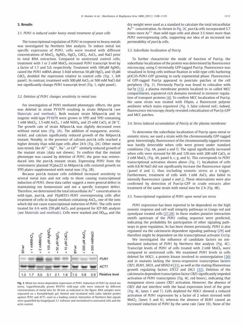
The transcriptional regulation of PUN1 in response to heavy metalswas investigated by Northern blot analysis. To induce metal ionspecific expression of PUN1, cells were treated with differentconcentrations of MnCl2, AgNO3, HgCl2, CdCl2, AsCl3, and NaCl priorto total RNA extraction. Compared to unstressed control cells,treatment with 1 or 2 mM MnCl2 increased PUN1 transcript level bya factor of 1.7 and 5.0, respectively. Treatment with 100 μM AgNO3
raised the PUN1 mRNA about 3-fold whereas 50 μM HgCl2 and 10 μMCdCl2 doubled the expression relative to control cells (Fig. 1, leftpanel). In contrast, treatment with 500 μM AsCl3 or 500 mM NaCl didnot significantly change PUN1 transcript level (Fig. 1, right panel).
3.2. Deletion of PUN1 changes sensitivity to metal ions
For investigation of PUN1 mediated phenotypic effects, the genewas deleted in strain FY1679 resulting in strain MApun1Δ (seeMaterials and methods). Both mutant strain MApun1Δ and itsisogenic wild-type FY1679 were grown in YPD and YPD containing1 mM MnCl2, 1.5 mM AsCl3, 1 mM NiSO4, and 25 mM CaCl2 at 28 °C.The growth rate of strain MApun1Δ was slightly decreased evenwithout metal ions (Fig. 2A). The addition of manganese, arsenic,nickel, and calcium significantly reduced growth of the MApun1Δmutant. Notably, in the presence of calcium pun1Δ cells reached ahigher density than wild-type cells after 24 h (Fig. 2A). Other metalions tested, like Al3+, Hg2+, Na+, or Cd2+ similarly reduced growth ofthe mutant strain (data not shown). To confirm that the mutantphenotype was caused by deletion of PUN1, the gene was reintro-duced into the pun1Δ mutant strain. Expressing PUN1 from thecentromeric plasmid YCplac22 in MApun1Δ cells restored growth onYPD plates supplemented with metal ions (Fig. 2B).
Because pun1Δ mutant cells exhibited increased sensitivity toseveral metal ions and not only to those causing transcriptionalinduction of PUN1, these data rather suggest a more general defect inmaintaining ion homeostasis and not a specific transport defect.Therefore, we determined the total intracellular As3+ concentration inwild-type, pun1Δ, and YEpM351-PUN1 overexpressing cells aftertreatment of cells in liquid medium containing AsCl3, one of the ionswhich did not cause transcriptional induction of PUN1. The cells weretreated for 6 h with 250 μM AsCl3 and prepared for ICP-MS analysis(see Materials and methods). Cells were washed and OD600 and the
Fig. 1. Metal ion stress-dependent expression of PUN1. Induction of PUN1 by metal ionstress. Logarithmically grown BY4741 wild-type cells were induced by differentconcentrations of metal ions for 30 min as indicated in the figure. RNA samples wereseparated on a formaldehyde gel, blotted and incubated with radio labeled probesagainst PUN1 and ACT1, used as a loading control. Intensities of Northern blot signalswere quantified by ImageQuant 5.1 software and normalized to untreated cells and theactin control.
dry weight were used as a standard to calculate the total intracellularAs3+ concentration. As shown in Fig. 2C, pun1Δ cells incorporated 5.7times more As3+ than wild-type cells and about 3.5 times more thanPUN1 overexpressing cells, supporting our idea of an increased ionpermeability of pun1Δ cells.
3.3. Subcellular localization of Pun1p
To further characterize the mode of function of Pun1p, thesubcellular localization of the proteinwas determined by fluorescencemicroscopy, using C-terminally GFP-tagged Pun1p. Fluorescence wasvisualized in living cells without fixation in wild-type cells harboringpUG35-PUN1-GFP growing in early exponential phase. Fluorescenceof GFP-tagged Pun1p appeared in punctate patches at the cellperiphery (Fig. 3). Previously Pun1p was found to colocalize withSur7p [15], a plasma membrane protein localized to so called MCCcompartments, ergosterol-rich domains involved in turnover regula-tion of transport proteins [4]. To confirm MCC localization of Pun1p,the same strain was treated with Filipin, a fluorescent polyeneantibiotic which stains ergosterol (Fig. 3, false colored red). Indeed,fluorescence microscopy clearly revealed colocalization of Pun1p-GFPand MCC patches.
3.4. Stress induced accumulation of Pun1p at the plasma membrane
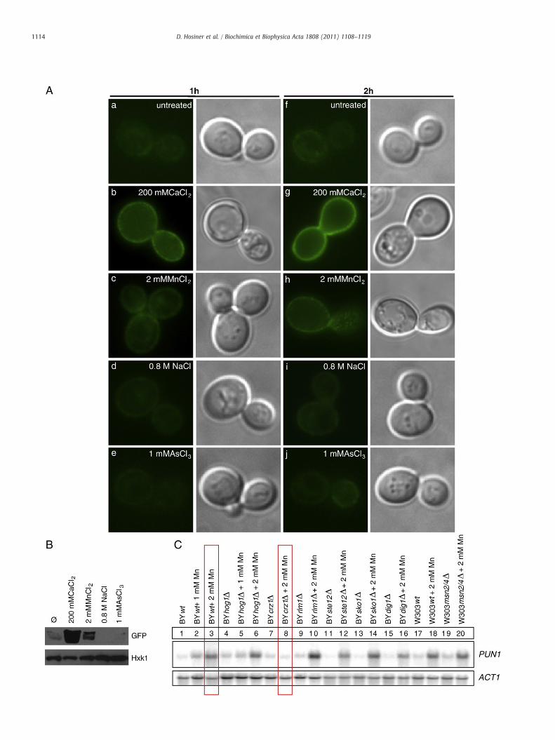
To determine the subcellular localization of Pun1p upon metal orosmotic stress, we used a strain with the chromosomally GFP-taggedgene. Interestingly, fluorescence of the chromosomally tagged versionwas hardly detectable when cells were grown under standardconditions (Fig. 4A, panel a and f). The signal significantly increasedwhen cells were stressed for 60 and 120 min with 200 mM CaCl2 or2 mM MnCl2 (Fig. 4A, panel b, c, g, and h). This corresponds to PUN1transcriptional activation shown above (Fig. 1). Incubation of cellswith 0.8 M NaCl did not significantly increase the fluorescence signal(panel d and i), thus excluding osmotic stress as a trigger.Furthermore, treatment of cells with 1 mM AsCl3 also failed tointensify fluorescence (panel e and j). These observations were alsoconfirmed by detection of Pun1p-GFP in crude extracts aftertreatment of the same strain with metal ions for 2 h (Fig. 4B).
3.5. Transcriptional regulation of PUN1 upon metal ion stress
PUN1 expression has been reported to be dependent on the highosmotic response and cell wall integrity pathways in Congo red andzymolyase treated cells [27,28]. In these studies putative interactionmotifs upstream of the PUN1 coding sequence were predicted,indicating the probability for participation of other signaling path-ways in gene regulation. As has been shown previously, PUN1 is alsoregulated via the calcineurin dependent signaling pathway [29] andtherefore might be dependent on the transcriptional activator Crz1p.
We investigated the influence of candidate factors on ion-mediated induction of PUN1 by Northern blot analysis (Fig. 4C).Transcript levels of PUN1 of cells treated with 2 mM MnCl2 werecompared to unstressed cells. We examined PUN1 levels in cellsdeleted for HOG1, a protein kinase involved in osmoregulation [30]and in mutants lacking the stress-responsive transcription factorsCRZ1, RLM1, SKO1, andMSN2/4 [31], as well as the mating/filamentousgrowth regulating factors STE12 and DIG1 [32]. Deletion of thecalcineurin dependent transcription factor CRZ1 significantly impededactivation of PUN1 transcription (Fig. 4C, red boxes), indicating thatmanganese stress causes CRZ1 activation. However, the absence ofCRZ1 did not interfere with the basal expression level of the gene(Fig. 4C, lanes 1 and 7). Cells depleted for HOG1 showed a reducedactivation of PUN1 expression by a factor of 2 when stressed withMnCl2 (lanes 5 and 6), whereas the absence of RLM1 caused anincreased induction of PUN1 by the same rate (lane 10). None of the
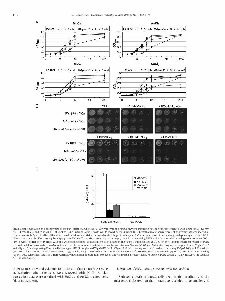
Fig. 2. Complementation and phenotyping of the pun1 deletion. A. Strains FY1679 wild-type and MApun1Δ were grown in YPD and YPD supplemented with 1 mM MnCl2, 1.5 mMAsCl3, 1 mM NiSO4, and 25 mM CaCl2 at 28 °C for 24 h under shaking. Growth was followed by measuring OD600. Growth curves shown represent an average of three individualmeasurements. MApun1Δ cells exhibited increased metal ion sensitivity compared to their isogenic wild-type. B. Complementation of the pun1Δ growth phenotype. Serial 10-folddilutions of strains FY1679, carrying the empty plasmid YCplac22 andMApun1Δ carrying the empty plasmid or expressing PUN1 under the control of its endogenous promoter (YCp-PUN1), were spotted on YPD plates with and without metal ions (concentrations as indicated in the figure), and incubated at 28 °C for 48 h. Plasmid-based expression of PUN1restored metal ion sensitivity of pun1Δ mutant cells. C. Measurement of intracellular AsCl3 concentration. Strains FY1679 and MApun1Δ carrying the empty plasmid YEpM351HAandMApun1Δ overexpressing C-terminally HA-tagged PUN1 from plasmid YEpM-PUN1-HA (MApun1Δ(PUN1)n) were grown in SDmedium containing 250 μMAsCl3 and SDmedium(w/o AsCl3) for 6 h at 28 °C. Cells were washed, OD600 and dry weight were defined and the total intracellular As3+ concentration of whole cells (μg As3+/g cells) was determined byICP-MS (ARC Seibersdorf research GmbH, Austria). Values shown represent an average of three individual measurements. Absence of PUN1 caused a highly increased intracellularAs3+ concentration.
1112 D. Hosiner et al. / Biochimica et Biophysica Acta 1808 (2011) 1108–1119
other factors provided evidence for a direct influence on PUN1 genetranscription when the cells were stressed with MnCl2. Similarexpression data were obtained with HgCl2 and AgNO3 treated cells(data not shown).
3.6. Deletion of PUN1 affects yeast cell wall composition
Reduced growth of pun1Δ cells even in rich medium and themicroscopic observation that mutant cells tended to be smaller and
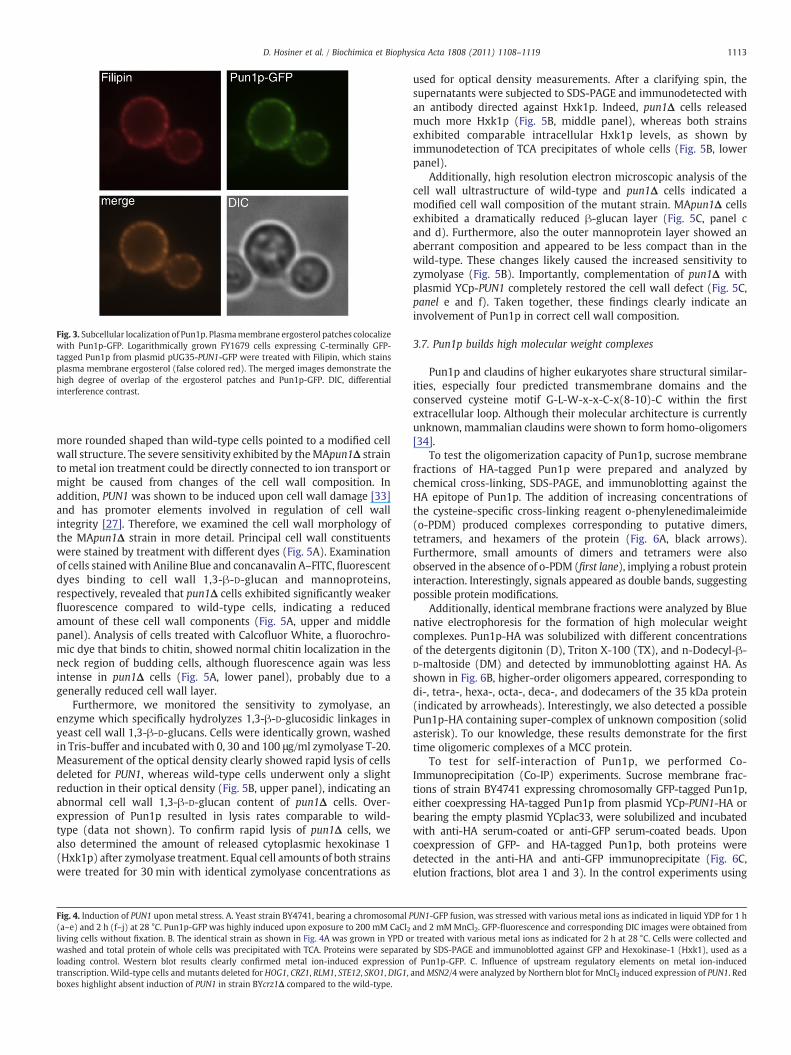
Fig. 3. Subcellular localization of Pun1p. Plasmamembrane ergosterol patches colocalizewith Pun1p-GFP. Logarithmically grown FY1679 cells expressing C-terminally GFP-tagged Pun1p from plasmid pUG35-PUN1-GFP were treated with Filipin, which stainsplasma membrane ergosterol (false colored red). The merged images demonstrate thehigh degree of overlap of the ergosterol patches and Pun1p-GFP. DIC, differentialinterference contrast.
1113D. Hosiner et al. / Biochimica et Biophysica Acta 1808 (2011) 1108–1119
more rounded shaped than wild-type cells pointed to a modified cellwall structure. The severe sensitivity exhibited by theMApun1Δ strainto metal ion treatment could be directly connected to ion transport ormight be caused from changes of the cell wall composition. Inaddition, PUN1 was shown to be induced upon cell wall damage [33]and has promoter elements involved in regulation of cell wallintegrity [27]. Therefore, we examined the cell wall morphology ofthe MApun1Δ strain in more detail. Principal cell wall constituentswere stained by treatment with different dyes (Fig. 5A). Examinationof cells stainedwith Aniline Blue and concanavalin A–FITC, fluorescentdyes binding to cell wall 1,3-β-D-glucan and mannoproteins,respectively, revealed that pun1Δ cells exhibited significantly weakerfluorescence compared to wild-type cells, indicating a reducedamount of these cell wall components (Fig. 5A, upper and middlepanel). Analysis of cells treated with Calcofluor White, a fluorochro-mic dye that binds to chitin, showed normal chitin localization in theneck region of budding cells, although fluorescence again was lessintense in pun1Δ cells (Fig. 5A, lower panel), probably due to agenerally reduced cell wall layer.
Furthermore, we monitored the sensitivity to zymolyase, anenzyme which specifically hydrolyzes 1,3-β-D-glucosidic linkages inyeast cell wall 1,3-β-D-glucans. Cells were identically grown, washedin Tris-buffer and incubatedwith 0, 30 and 100 μg/ml zymolyase T-20.Measurement of the optical density clearly showed rapid lysis of cellsdeleted for PUN1, whereas wild-type cells underwent only a slightreduction in their optical density (Fig. 5B, upper panel), indicating anabnormal cell wall 1,3-β-D-glucan content of pun1Δ cells. Over-expression of Pun1p resulted in lysis rates comparable to wild-type (data not shown). To confirm rapid lysis of pun1Δ cells, wealso determined the amount of released cytoplasmic hexokinase 1(Hxk1p) after zymolyase treatment. Equal cell amounts of both strainswere treated for 30 min with identical zymolyase concentrations as
Fig. 4. Induction of PUN1 upon metal stress. A. Yeast strain BY4741, bearing a chromosomal P(a–e) and 2 h (f–j) at 28 °C. Pun1p-GFP was highly induced upon exposure to 200 mM CaCl2living cells without fixation. B. The identical strain as shown in Fig. 4A was grown in YPD owashed and total protein of whole cells was precipitated with TCA. Proteins were separateloading control. Western blot results clearly confirmed metal ion-induced expression otranscription. Wild-type cells and mutants deleted for HOG1, CRZ1, RLM1, STE12, SKO1, DIG1,boxes highlight absent induction of PUN1 in strain BYcrz1Δ compared to the wild-type.
used for optical density measurements. After a clarifying spin, thesupernatants were subjected to SDS-PAGE and immunodetected withan antibody directed against Hxk1p. Indeed, pun1Δ cells releasedmuch more Hxk1p (Fig. 5B, middle panel), whereas both strainsexhibited comparable intracellular Hxk1p levels, as shown byimmunodetection of TCA precipitates of whole cells (Fig. 5B, lowerpanel).
Additionally, high resolution electron microscopic analysis of thecell wall ultrastructure of wild-type and pun1Δ cells indicated amodified cell wall composition of the mutant strain. MApun1Δ cellsexhibited a dramatically reduced β-glucan layer (Fig. 5C, panel cand d). Furthermore, also the outer mannoprotein layer showed anaberrant composition and appeared to be less compact than in thewild-type. These changes likely caused the increased sensitivity tozymolyase (Fig. 5B). Importantly, complementation of pun1Δ withplasmid YCp-PUN1 completely restored the cell wall defect (Fig. 5C,panel e and f). Taken together, these findings clearly indicate aninvolvement of Pun1p in correct cell wall composition.
3.7. Pun1p builds high molecular weight complexes
Pun1p and claudins of higher eukaryotes share structural similar-ities, especially four predicted transmembrane domains and theconserved cysteine motif G-L-W-x-x-C-x(8-10)-C within the firstextracellular loop. Although their molecular architecture is currentlyunknown, mammalian claudins were shown to form homo-oligomers[34].
To test the oligomerization capacity of Pun1p, sucrose membranefractions of HA-tagged Pun1p were prepared and analyzed bychemical cross-linking, SDS-PAGE, and immunoblotting against theHA epitope of Pun1p. The addition of increasing concentrations ofthe cysteine-specific cross-linking reagent o-phenylenedimaleimide(o-PDM) produced complexes corresponding to putative dimers,tetramers, and hexamers of the protein (Fig. 6A, black arrows).Furthermore, small amounts of dimers and tetramers were alsoobserved in the absence of o-PDM (first lane), implying a robust proteininteraction. Interestingly, signals appeared as double bands, suggestingpossible protein modifications.
Additionally, identical membrane fractions were analyzed by Bluenative electrophoresis for the formation of high molecular weightcomplexes. Pun1p-HA was solubilized with different concentrationsof the detergents digitonin (D), Triton X-100 (TX), and n-Dodecyl-β-D-maltoside (DM) and detected by immunoblotting against HA. Asshown in Fig. 6B, higher-order oligomers appeared, corresponding todi-, tetra-, hexa-, octa-, deca-, and dodecamers of the 35 kDa protein(indicated by arrowheads). Interestingly, we also detected a possiblePun1p-HA containing super-complex of unknown composition (solidasterisk). To our knowledge, these results demonstrate for the firsttime oligomeric complexes of a MCC protein.
To test for self-interaction of Pun1p, we performed Co-Immunoprecipitation (Co-IP) experiments. Sucrose membrane frac-tions of strain BY4741 expressing chromosomally GFP-tagged Pun1p,either coexpressing HA-tagged Pun1p from plasmid YCp-PUN1-HA orbearing the empty plasmid YCplac33, were solubilized and incubatedwith anti-HA serum-coated or anti-GFP serum-coated beads. Uponcoexpression of GFP- and HA-tagged Pun1p, both proteins weredetected in the anti-HA and anti-GFP immunoprecipitate (Fig. 6C,elution fractions, blot area 1 and 3). In the control experiments using
UN1-GFP fusion, was stressed with various metal ions as indicated in liquid YDP for 1 hand 2 mMMnCl2. GFP-fluorescence and corresponding DIC images were obtained fromr treated with various metal ions as indicated for 2 h at 28 °C. Cells were collected andd by SDS-PAGE and immunoblotted against GFP and Hexokinase-1 (Hxk1), used as af Pun1p-GFP. C. Influence of upstream regulatory elements on metal ion-inducedandMSN2/4 were analyzed by Northern blot for MnCl2 induced expression of PUN1. Red
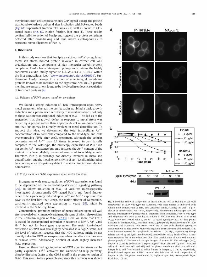
Fig. 5. Modified cell wall composition of pun1Δ mutant cells. A. Staining of cell wallcomponents. FY1679 wild-type and MApun1Δ cells were treated as indicated withAniline Blue, concanavalin A–FITC, and Calcofluor White, staining cell wall 1,3-β-D-glucan, mannoproteins, and chitin, respectively. Fluorescence microscopy revealedreduced fluorescence of pun1Δ cells. B. Treatment with zymolyase. FY1679 wild-typeand MApun1Δ cells were grown logarithmically in YPD medium, diluted to an equalOD600 value and treated with 0, 30, or 100 μg/ml zymolyase 20T. At time pointsindicated in the figure, OD600 was determined (upper panel). Equal amounts of FY1679wild-type and MApun1Δ cells were treated for 30 min with identical zymolyaseconcentrations as used before. After centrifugation, equal amounts of the supernatantwere immunodetected for cytoplasmic hexokinase 1 (Hxk1p), representing Hxk1prelease caused by cell lysis (middle panel). Intracellular Hxk1p levels of both strainswere compared by immunodetection after TCA precipitation of equal cell amounts(lower panel). C. Electron microscopic images of strains FY1679 wild-type (a-b),MApun1Δ (c and d), and MApun1Δ expressing PUN1 from plasmid YCp-PUN1. Principalcell wall constituents (GL and MP) and the plasma membrane (PM) are indicated.Images b, d, and f correspond to white frames in images a, c, and e, respectively.Plasmid-based expression of PUN1 restored the defective cell wall composition ofMApun1Δ cells. PM, plasma membrane; GL, β-glucan layer; MP, mannoprotein layer.Black bars, 100 nm.
1115D. Hosiner et al. / Biochimica et Biophysica Acta 1808 (2011) 1108–1119
membranes from cells expressing only GFP-tagged Pun1p, the proteinwas found exclusively unbound after incubation with HA-coated beads(Fig. 6C, supernatant fraction, blot area 2) as well as bound to GFP-coated beads (Fig. 6C, elution fraction, blot area 4). These resultsconfirm self-interaction of Pun1p and suggest the protein complexesdetected after cross-linking and Blue native electrophoresis torepresent homo-oligomers of Pun1p.
4. Discussion
In this study we show that Pun1p is a calcineurin/Crz1p-regulated,metal ion stress-induced protein involved in correct cell wallorganization, and a component of high molecular weight proteincomplexes. Pun1p has a tetraspan topology and contains the highlyconserved claudin family signature G-L-W-x-x-C-x(8-10)-C withinthe first extracellular loop (www.uniprot.org/uniprot/Q06991). Fur-thermore, Pun1p belongs to a group of nine integral membraneproteins known to be localized to the ergosterol-rich MCC, a plasmamembrane compartment found to be involved in endocytic regulationof transport proteins [4].
4.1. Deletion of PUN1 causes metal ion sensitivity
We found a strong induction of PUN1 transcription upon heavymetal treatment, whereas the pun1Δ strain exhibited a basic growthreduction and a pronounced sensitivity to several metal ions, not onlyto those causing transcriptional induction of PUN1. This led us to thesuggestion that the growth defect in response to metal stress wascaused by a general rather than a specific defect in ion homeostasisand that Pun1p may be directly involved in metal detoxification. Tosupport this idea, we determined the total intracellular As3+
concentration of mutant cells compared to the wild-type and cellsoverexpressing PUN1 after AsCl3 treatment. Although the cellularconcentration of As3+ was 5.7 times increased in pun1Δ cellscompared to the wild-type, the multicopy expression of PUN1 didnot confer As3+-resistance but only restored the As3+ content of themutant to a level slightly increased compared to wild-type cells.Therefore, Pun1p is probably not directly involved in metal iondetoxification and themetal ion sensitivity of pun1Δ cellsmight ratherbe a consequence of a primary defect in maintaining intracellular ionhomeostasis.
4.2. Crz1p mediates PUN1 expression upon metal ion stress
In a genome wide study, regulation of PUN1 expression was foundto be dependent on the calmodulin/calcineurin signaling pathway[29]. To follow induction of PUN1 in vivo, we microscopicallyinvestigated chromosomally GFP-tagged Pun1p and found fluores-cence to be significantly induced upon Ca2+ and Mn2+ exposure. Thisgave us the first hint that Crz1p, the major effector of calmodulin/calcineurin-regulated gene expression in yeast [29], might beinvolved in the PUN1 regulation.
Computational promoter analyses of genes induced upon cell wallstress revealed enrichment of certainmotifs someofwhich also residingin the upstream region of PUN1 [27,33]. Here we show that Crz1pis crucial for transcriptional activation in response to Mn2+, Ag3+, andHg2+ but not for basal gene expression. The metal ion-inducedexpression of PUN1 was also slightly decreased in a hog1Δ strain, butthe level of reduction suggests that the HOG-pathway might be notdirectly linked to PUN1 gene expression, when metal ions are the causeof cellular stress. Additionally, deletion of RLM1 slightly increasedPUN1 expression.
Based on these findings, induction of PUN1 upon ion stress can bepartly explained. Ca2+ activates the calcineurin/Crz1p pathway,thereby directing Crz1p to the CDRE motif in the promoter region ofPUN1. This seems to be a plausible step since this pathway was shown
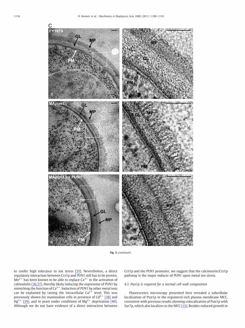
Fig. 5 (continued).
1116 D. Hosiner et al. / Biochimica et Biophysica Acta 1808 (2011) 1108–1119
to confer high tolerance to ion stress [35]. Nevertheless, a directregulatory interaction between Crz1p and PUN1 still has to be proven.Mn2+ has been known to be able to replace Ca2+ in the activation ofcalmodulin [36,37], thereby likely inducing the expression of PUN1 bymimicking the function of Ca2+. Induction of PUN1 by othermetal ionscan be explained by raising the intracellular Ca2+ level. This waspreviously shown for mammalian cells in presence of Cd2+ [38] andHg2+ [39], and in yeast under conditions of Mg2+ deprivation [40].Although we do not have evidence of a direct interaction between
Crz1p and the PUN1 promoter, we suggest that the calcineurin/Crz1ppathway is the major inducer of PUN1 upon metal ion stress.
4.3. Pun1p is required for a normal cell wall composition
Fluorescence microscopy presented here revealed a subcellularlocalization of Pun1p in the ergosterol-rich plasma membrane MCC,consistent with previous results showing colocalization of Pun1pwithSur7p, which also localizes to theMCC [15]. Besides reduced growth in
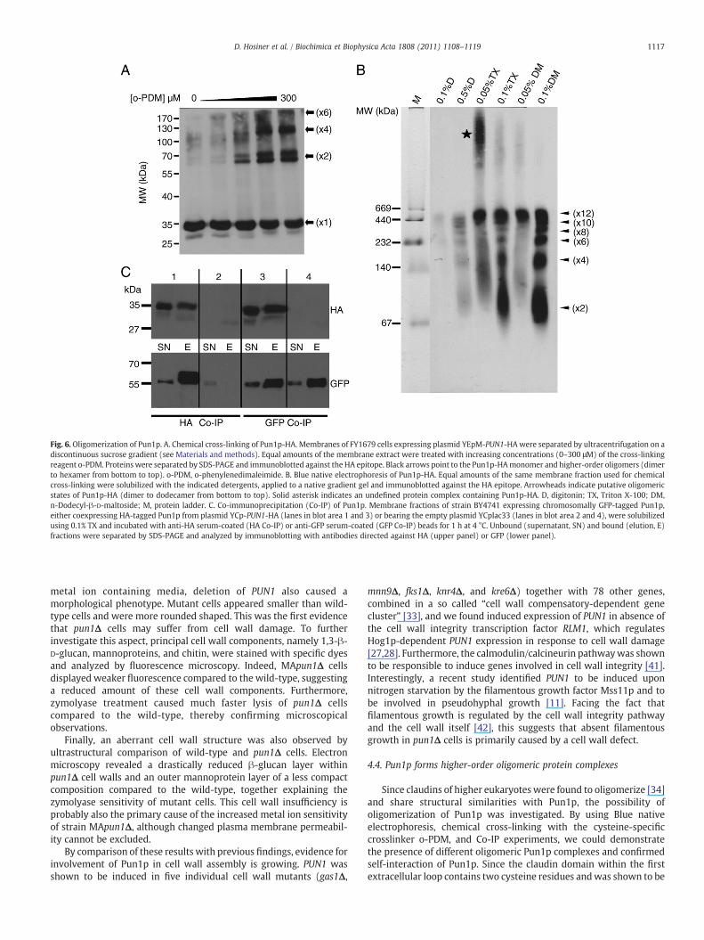
Fig. 6. Oligomerization of Pun1p. A. Chemical cross-linking of Pun1p-HA. Membranes of FY1679 cells expressing plasmid YEpM-PUN1-HAwere separated by ultracentrifugation on adiscontinuous sucrose gradient (see Materials and methods). Equal amounts of the membrane extract were treated with increasing concentrations (0–300 μM) of the cross-linkingreagent o-PDM. Proteins were separated by SDS-PAGE and immunoblotted against the HA epitope. Black arrows point to the Pun1p-HAmonomer and higher-order oligomers (dimerto hexamer from bottom to top). o-PDM, o-phenylenedimaleimide. B. Blue native electrophoresis of Pun1p-HA. Equal amounts of the same membrane fraction used for chemicalcross-linking were solubilized with the indicated detergents, applied to a native gradient gel and immunoblotted against the HA epitope. Arrowheads indicate putative oligomericstates of Pun1p-HA (dimer to dodecamer from bottom to top). Solid asterisk indicates an undefined protein complex containing Pun1p-HA. D, digitonin; TX, Triton X-100; DM,n-Dodecyl-β-D-maltoside; M, protein ladder. C. Co-immunoprecipitation (Co-IP) of Pun1p. Membrane fractions of strain BY4741 expressing chromosomally GFP-tagged Pun1p,either coexpressing HA-tagged Pun1p from plasmid YCp-PUN1-HA (lanes in blot area 1 and 3) or bearing the empty plasmid YCplac33 (lanes in blot area 2 and 4), were solubilizedusing 0.1% TX and incubated with anti-HA serum-coated (HA Co-IP) or anti-GFP serum-coated (GFP Co-IP) beads for 1 h at 4 °C. Unbound (supernatant, SN) and bound (elution, E)fractions were separated by SDS-PAGE and analyzed by immunoblotting with antibodies directed against HA (upper panel) or GFP (lower panel).
1117D. Hosiner et al. / Biochimica et Biophysica Acta 1808 (2011) 1108–1119
metal ion containing media, deletion of PUN1 also caused amorphological phenotype. Mutant cells appeared smaller than wild-type cells and were more rounded shaped. This was the first evidencethat pun1Δ cells may suffer from cell wall damage. To furtherinvestigate this aspect, principal cell wall components, namely 1,3-β-D-glucan, mannoproteins, and chitin, were stained with specific dyesand analyzed by fluorescence microscopy. Indeed, MApun1Δ cellsdisplayed weaker fluorescence compared to the wild-type, suggestinga reduced amount of these cell wall components. Furthermore,zymolyase treatment caused much faster lysis of pun1Δ cellscompared to the wild-type, thereby confirming microscopicalobservations.
Finally, an aberrant cell wall structure was also observed byultrastructural comparison of wild-type and pun1Δ cells. Electronmicroscopy revealed a drastically reduced β-glucan layer withinpun1Δ cell walls and an outer mannoprotein layer of a less compactcomposition compared to the wild-type, together explaining thezymolyase sensitivity of mutant cells. This cell wall insufficiency isprobably also the primary cause of the increased metal ion sensitivityof strain MApun1Δ, although changed plasma membrane permeabil-ity cannot be excluded.
By comparison of these results with previous findings, evidence forinvolvement of Pun1p in cell wall assembly is growing. PUN1 wasshown to be induced in five individual cell wall mutants (gas1Δ,
mnn9Δ, fks1Δ, knr4Δ, and kre6Δ) together with 78 other genes,combined in a so called “cell wall compensatory-dependent genecluster” [33], and we found induced expression of PUN1 in absence ofthe cell wall integrity transcription factor RLM1, which regulatesHog1p-dependent PUN1 expression in response to cell wall damage[27,28]. Furthermore, the calmodulin/calcineurin pathwaywas shownto be responsible to induce genes involved in cell wall integrity [41].Interestingly, a recent study identified PUN1 to be induced uponnitrogen starvation by the filamentous growth factor Mss11p and tobe involved in pseudohyphal growth [11]. Facing the fact thatfilamentous growth is regulated by the cell wall integrity pathwayand the cell wall itself [42], this suggests that absent filamentousgrowth in pun1Δ cells is primarily caused by a cell wall defect.
4.4. Pun1p forms higher-order oligomeric protein complexes
Since claudins of higher eukaryotes were found to oligomerize [34]and share structural similarities with Pun1p, the possibility ofoligomerization of Pun1p was investigated. By using Blue nativeelectrophoresis, chemical cross-linking with the cysteine-specificcrosslinker o-PDM, and Co-IP experiments, we could demonstratethe presence of different oligomeric Pun1p complexes and confirmedself-interaction of Pun1p. Since the claudin domain within the firstextracellular loop contains two cysteine residues andwas shown to be
PUN1
MCC
Mg2+
N
Rlm1
Mss11
Cd2+Hg2+
Ag3+
Crz1
Ca2+
Ca2+
Promoter
Pun1
Cell wall damage
Mn2+increased level
reduced level
direct binding
inducing trigger
supposed binding
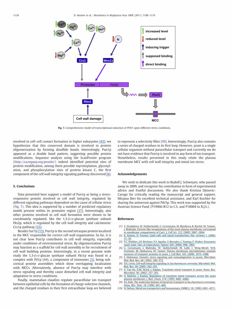
Fig. 7. Comprehensive model of transcriptional induction of PUN1 upon different stress conditions.
1118 D. Hosiner et al. / Biochimica et Biophysica Acta 1808 (2011) 1108–1119
involved in cell–cell contact formation in higher eukaryotes [43], wehypothesize that this conserved domain is involved in proteinoligomerization by forming disulfide bonds. Interestingly, Pun1pappeared as a double band pattern, suggesting possible proteinmodifications. Sequence analysis using the ScanProsite program(http://ca.expasy.org/prosite/) indeed identified potential sites ofprotein modification, among them possible myristoylation, glycosyl-ation, and phosphorylation sites of protein kinase C, the firstcomponent of the cell wall integrity signaling pathway discovered [6].
5. Conclusions
Data presented here support a model of Pun1p as being a stress-responsive protein involved in cell wall integrity, regulated bydifferent signaling pathways dependent on the cause of cellular stress(Fig. 7). This idea is supported by a number of predicted regulatorymotifs present within its promoter region [27]. Interestingly, alsoother proteins involved in cell wall formation were shown to becoordinately regulated, like the 1,3-β-D-glucan synthase subunitFks2p, which is regulated by the cell wall integrity and calcineurin/Crz1p pathway [33].
Besides Sur7p [15], Pun1p is the second tetraspan protein localizedin the MCC responsible for correct cell wall organization. So far, it isnot clear how Pun1p contributes to cell wall integrity, especiallyunder conditions of environmental stress. By oligomerization Pun1pmay function as a scaffold for cell wall assembly or for recruitment ofcell wall building proteins. Interestingly, in a recent genome widestudy the 1,3-β-D-glucan synthase subunit Fks1p was found in acomplex with Pil1p [44], a component of eisosomes [3], being sub-cortical protein assemblies which show overlapping localizationwith MCCs. Alternatively, absence of Pun1p may interfere withstress signaling and thereby cause disturbed cell wall integrity andadaptation to stress conditions.
Finally, mammalian claudins regulate paracellular ion transportbetween epithelial cells by the formation of charge-selective channels,and the charged residues in their first extracellular loop are believed
to represent a selectivity filter [45]. Interestingly, Pun1p also containsa series of charged residues in its first loop. However, yeast is a singlecellular organism without paracellular transport and currently we donot have evidence that Pun1p is involved in any form of ion transport.Nonetheless, results presented in this study relate the plasmamembrane MCC with cell wall integrity and metal ion stress.
Acknowledgements
We wish to dedicate this work to Rudolf J. Schweyen, who passedaway in 2009, and recognize his contribution in form of experimentaladvice and fruitful discussions. We also thank Kristina Djinovic-Carugo for critically reading the manuscript and general support,Mirjana Iliev for excellent technical assistance, and Karl Kuchler forsharing the antiserum against Pdr5p. This work was supported by theAustrian Science Fund (P19966-B12 to C.S. and P18004 to R.J.S.).
References
[1] V. Stradalova, W. Stahlschmidt, G. Grossmann, M. Blazikova, R. Rachel, W. Tanner,J. Malinsky, Furrow-like invaginations of the yeast plasma membrane correspondto membrane compartment of Can1, J. Cell Sci. 122 (2009) 2887–2894.
[2] K. Simons, D. Toomre, Lipid rafts and signal transduction, Nat. reviews 1 (2000)31–39.
[3] T.C.Walther, J.H. Brickner, P.S. Aguilar, S. Bernales, C. Pantoja, P. Walter, Eisosomesmark static sites of endocytosis, Nature 439 (2006) 998–1003.
[4] G. Grossmann, J. Malinsky, W. Stahlschmidt, M. Loibl, I. Weig-Meckl, W.B.Frommer, M. Opekarova, W. Tanner, Plasma membrane microdomains regulateturnover of transport proteins in yeast, J. Cell Biol. 183 (2008) 1075–1088.
[5] S. Hohmann, Osmotic stress signaling and osmoadaptation in yeasts, Microbiol.Mol. Biol. Rev. 66 (2002) 300–372.
[6] D.E. Levin, Cell wall integrity signaling in Saccharomyces cerevisiae, Microbiol. Mol.Biol. Rev. 69 (2005) 262–291.
[7] A. Van Ho, D.M. Ward, J. Kaplan, Transition metal transport in yeast, Annu. Rev.Microbiol. 56 (2002) 237–261.
[8] D. Radisky, J. Kaplan, Regulation of transition metal transport across the yeastplasma membrane, J. Biol. Chem. 274 (1999) 4481–4484.
[9] D.J. Eide, The molecular biology of metal ion transport in Saccharomyces cerevisiae,Annu. Rev. Nutr. 18 (1998) 441–469.
[10] N. Nelson, Metal ion transporters and homeostasis, EMBO J. 18 (1999) 4361–4371.
1119D. Hosiner et al. / Biochimica et Biophysica Acta 1808 (2011) 1108–1119
[11] T. Xu, C.A. Shively, R. Jin, M.J. Eckwahl, C.J. Dobry, Q. Song, A. Kumar, A profile ofdifferentially abundant proteins at the yeast cell periphery during pseudohyphalgrowth, The Journal of biological chemistry 285 15476–15488.
[12] K. Morita, M. Furuse, K. Fujimoto, S. Tsukita, Claudin multigene family encodingfour-transmembrane domain protein components of tight junction strands, Proc.Natl Acad. Sci. USA 96 (1999) 511–516.
[13] F.J. Alvarez, L.M. Douglas, J.B. Konopka, Sterol-rich plasma membrane domains infungi, Eukaryot. Cell 6 (2007) 755–763.
[14] P. Sivadon, M.F. Peypouquet, F. Doignon, M. Aigle, M. Crouzet, Cloning of themulticopy suppressor gene SUR7: evidence for a functional relationship betweenthe yeast actin-binding protein Rvs167 and a putative membranous protein, Yeast(Chichester, England) 13 (1997) 747–761.
[15] F.J. Alvarez, L.M. Douglas, A. Rosebrock, J.B. Konopka, The Sur7 protein regulatesplasma membrane organization and prevents intracellular cell wall growth inCandida albicans, Mol. Biol. Cell 19 (2008) 5214–5225.
[16] W. Gorner, E. Durchschlag, M.T. Martinez-Pastor, F. Estruch, G. Ammerer, B.Hamilton, H. Ruis, C. Schuller, Nuclear localization of the C2H2 zinc finger proteinMsn2p is regulated by stress and protein kinase A activity, Genes Dev. 12 (1998)586–597.
[17] W.K. Huh, J.V. Falvo, L.C. Gerke, A.S. Carroll, R.W. Howson, J.S. Weissman, E.K.O'Shea, Global analysis of protein localization in budding yeast, Nature 425 (2003)686–691.
[18] A. Wach, A. Brachat, R. Pohlmann, P. Philippsen, New heterologous modules forclassical or PCR-based gene disruptions in Saccharomyces cerevisiae, Yeast 10(1994) 1793–1808.
[19] R.D. Gietz, A. Sugino, New yeast-Escherichia coli shuttle vectors constructed within vitro mutagenized yeast genes lacking six-base pair restriction sites, Gene 74(1988) 527–534.
[20] M. Wachek, M.C. Aichinger, J.A. Stadler, R.J. Schweyen, A. Graschopf, Oligomer-ization of the Mg2+-transport proteins Alr1p and Alr2p in yeast plasmamembrane, FEBS J. 273 (2006) 4236–4249.
[21] A. Graschopf, J.A. Stadler, M.K. Hoellerer, S. Eder, M. Sieghardt, S.D. Kohlwein, R.J.Schweyen, The yeast plasma membrane protein Alr1 controls Mg2+ homeostasisand is subject to Mg2+-dependent control of its synthesis and degradation, J. Biol.Chem. 276 (2001) 16216–16222.
[22] R.K. Niedenthal, L. Riles, M. Johnston, J.H. Hegemann, Green fluorescent protein asa marker for gene expression and subcellular localization in budding yeast, Yeast12 (1996) 773–786.
[23] S. Reipert, I. Fischer, G. Wiche, High-pressure freezing of epithelial cells onsapphire coverslips, J. Microsc. 213 (2004) 81–85.
[24] S. Reipert, G. Wiche, High-pressure freezing and low-temperature fixation of cellmonolayers grown on sapphire coverslips, Meth. Cell Biol. 88 (2008) 165–180.
[25] M.A. Collart, S. Oliviero, Preparation of yeast RNA, Current protocols in molecularbiology / edited by Frederick M. Ausubel ... [et al Chapter 13 (2001) Unit13 12.
[26] H. Schagger, W.A. Cramer, G. von Jagow, Analysis of molecular masses andoligomeric states of protein complexes by blue native electrophoresis andisolation of membrane protein complexes by two-dimensional native electro-phoresis, Anal. Biochem. 217 (1994) 220–230.
[27] J.M. Rodriguez-Pena, R.M. Perez-Diaz, S. Alvarez, C. Bermejo, R. Garcia, C. Santiago, C.Nombela, J. Arroyo, The 'yeast cell wall chip'—a tool to analyze the regulation of cellwall biogenesis in Saccharomyces cerevisiae, Microbiology 151 (2005) 2241–2249.
[28] R. Garcia, J.M. Rodriguez-Pena, C. Bermejo, C. Nombela, J. Arroyo, The high osmoticresponse and cell wall integrity pathways cooperate to regulate transcriptionalresponses to zymolyase-induced cell wall stress in Saccharomyces cerevisiae, J. Biol.Chem. 284 (2009) 10901–10911.
[29] H. Yoshimoto, K. Saltsman, A.P. Gasch, H.X. Li, N. Ogawa, D. Botstein, P.O. Brown, M.S.Cyert, Genome-wide analysis of gene expression regulated by the calcineurin/Crz1psignaling pathway in Saccharomyces cerevisiae, J. Biol. Chem. 277 (2002) 31079–31088.
[30] J.L. Brewster, T. de Valoir, N.D. Dwyer, E. Winter, M.C. Gustin, An osmosensingsignal transduction pathway in yeast, Science 259 (1993) 1760–1763.
[31] M.T. Martinez-Pastor, G. Marchler, C. Schuller, A. Marchler-Bauer, H. Ruis, F.Estruch, The Saccharomyces cerevisiae zinc finger proteins Msn2p and Msn4p arerequired for transcriptional induction through the stress response element(STRE), EMBO J. 15 (1996) 2227–2235.
[32] S. Chou, S. Lane, H. Liu, Regulation of mating and filamentation genes by twodistinct Ste12 complexes in Saccharomyces cerevisiae, Mol. Cell. Biol. 26 (2006)4794–4805.
[33] A. Lagorce, N.C. Hauser, D. Labourdette, C. Rodriguez, H. Martin-Yken, J. Arroyo, J.D.Hoheisel, J. Francois, Genome-wide analysis of the response to cell wall mutations inthe yeast Saccharomyces cerevisiae, J. Biol. Chem. 278 (2003) 20345–20357.
[34] L.L. Mitic, V.M. Unger, J.M. Anderson, Expression, solubilization, and biochemicalcharacterization of the tight junction transmembrane protein claudin-4, ProteinSci. 12 (2003) 218–227.
[35] I. Mendoza, F.J. Quintero, R.A. Bressan, P.M. Hasegawa, J.M. Pardo, Activatedcalcineurin confers high tolerance to ion stress and alters the budding pattern andcell morphology of yeast cells, J. Biol. Chem. 271 (1996) 23061–23067.
[36] D.J. Wolff, P.G. Poirier, C.O. Brostrom, M.A. Brostrom, Divalent cation bindingproperties of bovine brain Ca2+-dependent regulator protein, J. Biol. Chem. 252(1977) 4108–4117.
[37] A. Mork, A. Geisler, Calmodulin-dependent adenylate cyclase activity in ratcerebral cortex: effects of divalent cations, forskolin and isoprenaline, Arch. Int.Physiol. Biochim. 97 (1989) 259–271.
[38] D. Beyersmann, S. Hechtenberg, Cadmium, gene regulation, and cellular signalingin mammalian cells, Toxicol. Appl. Pharmacol. 144 (1997) 247–261.
[39] X.X. Tan, C. Tang, A.F. Castoldi, L. Manzo, L.G. Costa, Effects of inorganic andorganic mercury on intracellular calcium levels in rat T lymphocytes, J. Toxicol.Environ. Health 38 (1993) 159–170.
[40] G.Wiesenberger, K. Steinleitner, R. Malli, W.F. Graier, J. Vormann, R.J. Schweyen, J.A.Stadler, Mg2+ deprivation elicits rapid Ca2+ uptake and activates Ca2+/calcineurinsignaling in Saccharomyces cerevisiae, Eukaryot. Cell 6 (2007) 592–599.
[41] P. Garrett-Engele, B. Moilanen, M.S. Cyert, Calcineurin, the Ca2+/calmodulin-dependent protein phosphatase, is essential in yeast mutants with cell integritydefects and in mutants that lack a functional vacuolar H(+)-ATPase, Mol. Cell.Biol. 15 (1995) 4103–4114.
[42] B. Birkaya, A. Maddi, J. Joshi, S.J. Free, P.J. Cullen, Role of the cell wall integrity andfilamentous growth mitogen-activated protein kinase pathways in cell wallremodeling during filamentous growth, Eukaryot. Cell 8 (2009) 1118–1133.
[43] L. Cukierman, L. Meertens, C. Bertaux, F. Kajumo, T. Dragic, Residues in a highlyconserved claudin-1 motif are required for hepatitis C virus entry andmediate theformation of cell–cell contacts, J. Virol. 83 (2009) 5477–5484.
[44] A.C. Gavin,M. Bosche, R. Krause, P. Grandi, M.Marzioch, A. Bauer, J. Schultz, J.M. Rick,A.M.Michon, C.M.Cruciat,M. Remor, C.Hofert,M. Schelder,M. Brajenovic, H. Ruffner,A. Merino, K. Klein, M. Hudak, D. Dickson, T. Rudi, V. Gnau, A. Bauch, S. Bastuck, B.Huhse, C. Leutwein,M.A.Heurtier, R.R. Copley, A. Edelmann, E. Querfurth, V. Rybin, G.Drewes, M. Raida, T. Bouwmeester, P. Bork, B. Seraphin, B. Kuster, G. Neubauer, G.Superti-Furga, Functional organization of the yeast proteome by systematic analysisof protein complexes, Nature 415 (2002) 141–147.
[45] O.R. Colegio, C.M. Van Itallie, H.J. McCrea, C. Rahner, J.M. Anderson, Claudins createcharge-selective channels in the paracellular pathway between epithelial cells,Am. J. Physiol. Cell Physiol. 283 (2002) C142–C147.
Copyright © 2022 FDOKUMEN