Propriedades físico-químicas e antimicrobiana de cimentos ...
71
UNIVERSIDADE FEDERAL DE PELOTAS Faculdade de Odontologia Programa de Pós-Graduação em Odontologia Dissertação de Mestrado Propriedades físico-químicas e antimicrobiana de cimentos endodônticos experimentais contendo metacrilatos metálicos Victoria Burmann da Silva Guimarães Pelotas, 2019
-
Upload
khangminh22 -
Category
Documents
-
view
1 -
download
0
Transcript of Propriedades físico-químicas e antimicrobiana de cimentos ...
UNIVERSIDADE FEDERAL DE PELOTAS
Faculdade de Odontologia
Programa de Pós-Graduação em Odontologia
Dissertação de Mestrado
Propriedades físico-químicas e antimicrobiana de cimentos endodônticos
experimentais contendo metacrilatos metálicos
Victoria Burmann da Silva Guimarães
Pelotas, 2019
Victoria Burmann da Silva Guimarães
Propriedades físico-químicas e antimicrobiana de cimentos endodônticos
experimentais contendo metacrilatos metálicos
Dissertação de Mestrado apresentado ao
Programa de Pós-Graduação em Odontologia
da Universidade Federal de Pelotas, como
requisito parcial à obtenção do título de Mestre
em Clínica Odontológica com ênfase em
Endodontia.
Orientador: Prof. Dr. Rafael Guerra Lund
Co-orientadora: Profa. Dra.Fernanda Geraldo Pappen
Pelotas, 2019
Universidade Federal de Pelotas / Sistema de Bibliotecas
Catalogação na Publicação
Elaborada por Fabiano Domingues Malheiro CRB: 10/1955
G963p Guimarães, Victoria Burmann da Silva
Propriedades físico-químicas e antimicrobiana de
cimentos endodônticos experimentais contendo
metacrilatos metálicos / Victoria Burmann da Silva
Guimarães ; Rafael Guerra Lund, orientador ; Fernanda
Geraldo Pappen, coorientadora. — Pelotas, 2019.
71 f. : il.
Dissertação (Mestrado) — Programa de Pós-Graduação
em Clínica Odontológica - ênfase em Endodontia,
Odontologia, Universidade Federal de Pelotas, 2019.
1. Endodontia. 2. Obturação do canal radicular. 3.
Metacrilatos. 4. Anti-infecciosos. 5. AH plus. I. Lund, Rafael
Guerra, orient. II. Pappen, Fernanda Geraldo, coorient. III.
Título.
Black : D24
Victoria Burmann da Silva Guimarães
Propriedades fisico-químicas e antimicrobiana de cimentos endodônticos
experimentais contendo metacrilatos metálicos
Dissertação de Mestrado apresentado ao Programa de Pós-Graduação em
Odontologia da Universidade Federal de Pelotas, como requisito parcial à obtenção
do título de Mestre em Clínica Odontológica com ênfase em Endodontia..
Data da Defesa: 27 de fevereiro de 2019.
Banca Examinadora:
Prof. Dr. Rafael Guerra Lund (Orientador), doutor em Odontologia (Área de
concentração Dentística) pela Universidade Federal de Pelotas.
Prof. Dr. Evandro Piva, doutor em Materiais Dentários pela Universidade Federal de
Campinas.
Prof. Dr. José Antonio Poli de Figueiredo, doutor em Endodontia pela Universidade
Federal de São Paulo.
Profa. Dra. Luciane Geanini Pena dos Santos (suplente), doutora em Endodontia
pela Universidade Federal de Santa Catarina.
Prof. Dra. Katerine Jahnecke Pilownic (suplente), doutora em Odontopediatria pela
Universidade Feresal de Pelotas.
Ao meu esposo, Igor, pessoa cоm quem аmо
compartilhar meus dias. Cоntigo tenho mе sentido
mais viva dе verdade. Obrigada pelo carinho, а
paciência е pоr sua capacidade dе me trazer pаz nа
correria dе cada dia. Aos meus pais, pоr sua
capacidade dе acreditar еm mіm е investir еm mim.
Agradecimentos
Agradecimento especial ao meu esposo Igor, por ser meu melhor amigo e
companheiro. Obrigada por tua dedicação, amor e alegria contagiante. Sem você
meus dias seriam tristes. Agradeço por me apoiares e acreditares em todos os
momentos em mim, principalmente quando eu mesma estava desistindo. Não
existem palavras para descrever o quanto te amo.
Quero agradecer em especial a meus pais Liane e Victor, que me
proporcionaram a melhor escola da vida: educação moral, espiritual e lições de vida.
Obrigada pelo apoio e ajuda para eu seguir estudando. Não tenho palavras para
dizer o quanto me orgulho de vocês. Por tudo o que vocês fizeram e fazem por mim,
muito obrigada. Obrigada, Fernanda, minha irmã querida, por ser tão companheira,
por me ajudar em momentos difíceis e até por contribuir neste trabalho.
Agradeço às amigas e colegas Andressa Barboza e Carolina Vieira. Agradeço
a todas pela amizade, pelos conselhos e por terem me ajudado tanto neste trabalho,
principalmente em momentos que eu não estava presente. Obrigada pela amizade,
pelos ensinamentos. Agradeço à bolsista de Iniciação Tecnológica, Larissa Nunes,
por toda a ajuda no percorrer deste trabalho.
Agradeço a PPGO pela oportunidade de realizar o mestrado em uma ótima
instituição com conceito 6. Agradeço a CAPES pela bolsa de estudo. Agradeço
também a todos os professores, colegas e demais colaboradores do PPGO pela
convivência e aprendizado e, principalmente aos amigos que conheci e convivi
nesses 2 anos.
Agradeço aos funcionários Lizangela, Josiane, Tatiana e Junior por tudo que
fizeram por mim, pela paciência, pela preocupação e pelas palavras carinhosas que
me motivavam em muitos dias. A equipe CDC-Bio e Laboratório de Microbiologia.
Agradeço a meu orientador Rafael Lund por me fazerem me sentir uma
pessoa de valor, pela confiança e pelas oportunidades. Agradeço pelas inúmeras
conversas e conselhos. Agradeço pela paciência, pelos ensinamentos. Vocês me
ensinaram muito mais do que teorias. Vocês são exemplos para mim de caráter,
honestidade, profissionalismo e de ser humano.
Agradeço à minha co-orientadora Fernanda pelo exemplo de professora e
profissional. Agradeço por todos os ensinamentos, oportunidades, disponibilidade e
atenção. Agradeço por todas às vezes que me chamavas a atenção, sempre
buscando o meu melhor. O teu incentivo, os teus conselhos, a tua amizade, a tua
parceria e a tua confiança em acreditar no meu potencial.
Agradeço aos professores Evandro Piva, Carlos Cuevas, Josué Martos,
Adriana Silva, Luciane Geanini, Nadia Ferreira, Anderson Ribeiro, Tiago Collares,
Thais Larré, Wellington Rosa e Simone Duarte, também a pós-doutoranda Meibel
Lisboa pela ajuda em neste trabalho e em trabalhos paralelos. Vocês também são
exemplos de educadores e pesquisadores, e serão sempre minha referência.
Por fim, agradeço аo meu Pai Celeste, pois sеm ele еυ nãо teria forças pаrа
essa longa jornada, pela vida, pela sabedoria e por todas vezes que pedi a Ele
tranquilidade e força para seguir em frente.

“Um dos maiores dons que Deus nos deu é a alegria de
tentar novemente, pois nenhum fracasso precisa ser o último.”
Thomas Spencer Monson
Resumo
GUIMARÃES, Victoria Burmann da Silva. Propriedades físico-químicas e antimicrobiana de cimentos endodônticos experimentais contendo metacrilatos metálicos. 2019. 71 p. Dissertação de Mestrado em Clínica Odontológica com Ênfase em Endodontia – Programa de Pós-Graduação em Odontologia, Universidade Federal de Pelotas, Pelotas. As propriedades dos cimentos endodônticos são importantes na qualidade do preenchimento dos canais radiculares e, posteriormente, no resultado endodôntico. No entanto, nenhum material possui todas as propriedades de um cimento endodôntico ideal, assim novos materiais estão sendo criados para melhorar suas características físico-químicas e biológicas. O objetivo deste estudo foi avaliar as propriedades físicas, químicas, atividade antimicrobiana, e shelf-life de um cimento experimental de fotoativação dual após a incorporação de metacrilato de dibutilestanho (Sn4+) (ETs) ou metacrilato de cálcio (Ca2+) (ECs), em concentração de 2%. O AH Plus foi usado como referência comercial. O pH e a liberação de íons foram medidos usando um pHmetro e um espectrômetro de emissão óptica de plasma induzido por microondas. Foi avaliado a estabilidade dimensional após 30 dias de acordo com a ISO 6876. A inibição do crescimento do biofilme foi avaliada por microscopia confocal de varredura a laser (CLSM). A análise da viabilidade do biofilme foi realizada com a técnica SYTO 9. O shelf-life foi avaliado através dos testes de grau de conversão e espessura de película nos tempos: imediato, 1 mês e 2 meses. Para a análise estatística, utilizou-se ANOVA e teste post hoc de Tukey, e um nível de significância de 5 %. Todos os cimentos testados apresentaram redução gradual de pH ao longo de 30 dias. O cimento ECs apresentou a maior liberação de cálcio e ETs apresentou a maior liberação de estanho em 30 dias. O cimento ETs demonstrou melhor estabilidade dimensional ao longo do tempo, comparado com outros cimentos endodônticos. ETs revelou melhor potencial antibiofilme após 15 dias em comparação com os controles. O grau de conversão foi reduzido após o final do shelf-life. Quanto à espessura de película, todos os materiais estavam de acordo com as especificações da norma ISO 6876, exceto ECs imediato (100 µm) e ETs no primeiro mês de envelhecimento (170 µm). Conclui-se que as propriedades físico-químicas do cimento endodôntico experimental contendo metacrilato de cálcio e dibutilestanho não foram drasticamente alteradas e melhoraram o efeito antibiofilme dos cimentos.
Palavras-chave: endodontia, obturação do canal radicular, metacrilatos, anti-infecciosos, AH plus.
Abstract
GUIMARÃES, Victoria Burmann da Silva. Novel endodontic sealer based in metals cross-linking methacrylate. 2019. 71 p. Master's Dissertation in Dental Clinic with Emphasis in Endodontics - Graduate Program in Dentistry, Federal University of Pelotas, Pelotas. The properties of endodontic sealers are important for the quality of filling of the root canals and, later, in the endodontic prognosis. However, no material has all the properties of an ideal endodontic sealer, so new materials are being created to improve its physicochemical and biological characteristics. The objective of this study was to evaluate the physical, chemical and antimicrobial properties, and shelf-life of a dual-reactive experimental sealer after the incorporation of dibutyltin methacrylate (Sn4+) (ETs) or calcium methacrylate (Ca2+) (ETs), in a concentration of 2%. AH Plus was used as a commercial reference. The pH and ion release were measured using a pH meter and a microwave induced plasma optical emission spectrometer. The dimensional stability was evaluated after 30 days in accordance with ISO 6876. Inhibition of biofilm growth was evaluated by laser scanning confocal microscopy (CLSM). The biofilm viability analysis was performed using the SYTO 9 technique. The shelf-life was evaluated through tests of degree of conversion and film thickness at the times: immediate, 1 month and 2 months. For statistical analysis, ANOVA and Tukey post hoc test were used, and a significance level of 5%. All sealers tested showed a gradual reduction of pH over 30 days. The ECs sealer showed the highest release of calcium and ETs presented the highest release of tin in 30 days. The ETs sealer showed better dimensional stability over time compared to other endodontic sealers. ETs revealed better antibiofilm potential after 15 days compared to controls. The degree of conversion was reduced after the shelf-life. Regarding film thickness, all materials were in accordance with ISO 6876 specifications, except ECs immediate (100 μm) and ETs in the first month of aging (170 μm). It was concluded that the physicochemical properties of the experimental endodontic sealers containing calcium and dibutyltin methacrylate were not drastically altered and the antibiofilm effect of the cements was improved. Key-words: endodontics, root canal obturation, methacrylates, antimicrobial agents, AH plus.
Lista de Abreviaturas e Siglas
ANOVA Análise de Variância de uma Via
ANSI/ADA American National Standards Institute/ American Dental Association
Ar Argônio
BHI Infusão coração-cérebro
BHT Hidroxibutil tolueno
Bis-EMA 30 Bisfenol A-glicil Dimetacrilato
C Carbono
Ca2+ Cálcio
CAPES Coordenação de Aperfeiçoamento de Pessoa de Nível Superior
CEEA Cômite de Ética em Experimentação Animal
CLSM Microscopia confocal de varredura a laser
CNPq Conselho Nacional de Desenvolvimento Científico e Tecnológico
DHEPT Dihidroxietil-para-toluidino
FAPERGS Fundação de Amparo à Pesquisa do Estado do Rio Grande do Sul
HCl Ácido clorídrico
IUPUI Indiana University – Purdue University Indianapolis
ISO International Organization for Standardization
L Comprimento inicial da amostra
L30 Comprimento após 30 dias
MTA Agregado trióxido mineral
MIP OES Espectrometria de Emissão Óptica com Plasma Induzido por Micro-
ondas
MSIS Nebulização multimodal
NaBH4 Borohidreto de Sódio
NaOH Hidróxido de sódio
PEG 400 Polietilenoglicol 400
Q10 Constante de coeficiente de reação
r Envelhecimento acelerado
RT Temperatura aumentada
Sn4+ Estanho
TA Temperatura ambiente
TEGDMA Trietileno glicol dimetacrilato
UFPel Universidade Federal de Pelotas
Sumário
1 Introdução ............................................................................................................. 15
2 Projeto de Pesquisa .............................................................................................. 18
2.1 Justificativa ..................................................................................................... 18
2.2 Objetivos ......................................................................................................... 19
2.2.1 Objetivo Geral ....................................................................................... 19
2.2.2 Objetivos Específicos ............................................................................ 19
2.3 Metodologia .................................................................................................... 19
2.3.1 Formulação e Grupos do Cimento Endodôntico Experimental .............. 19
2.3.2 Avaliação do pH, liberação de Cálcio e Estanho .................................. 20
2.3.3 Estabilidade Dimensional ...................................................................... 22
2.3.4 Ensaio de Inibição da Formação de Biofilme ......................................... 23
2.3.5 Processo de Envelhecimento Acelerado Dentro de Câmara Climática 24
2.3.5.1 Grau de Conversão ..................................................................... 25
2.3.5.2 Espessura de Película ................................................................. 25
2.3.6 Análise Estatística ................................................................................. 26
2.4 Hipótese ......................................................................................................... 26
2.5 Orçamento ..................................................................................................... 27
2.6 Cronograma ................................................................................................... 28
2.7 Referências .................................................................................................... 29
3 Relatório do Trabalho de Campo .......................................................................... 31
4 Artigo ..................................................................................................................... 33
5 Considerações Finais ............................................................................................ 62
6 Referências ........................................................................................................... 63
15
1. Introdução
Na terapia endodôntica, a obturação é responsável pelo selamento do
sistema de canais radiculares, proporcionando o preenchimento de irregularidades
anatômicas e o sepultamento de bactérias que tenham sobrevivido às demais
etapas do tratamento (ØRSTAVIK, 2005). Até o momento, no entanto, não foi
desenvolvido um material único que preencha todos os requisitos desejáveis para
obturação do espaço endodôntico. Idealmente, este material deveria ser radiopaco,
bactericida, promover adequado preenchimento e aderência às paredes dos canais
radiculares, ser facilmente removido quando necessário, biocompatível, possuir
suficiente tempo de trabalho e tempo de presa curto, além de não causar alteração
na coloração das estruturas dentárias (GROSSMAN, 1981).
Dentre os cimentos obturadores de canais radiculares mais utilizados na
prática clínica, podemos citar os cimentos obturadores à base de resina, cujo uso é
amplamente consolidado. Tais materiais apresentam excelentes propriedades como
estabilidade dimensional, solubilidade reduzida, selamento apical e potencial de
adesão à dentina intracanal (ØRSTAVIK, 2005; VERSIANI et al., 2006; RESENDE et
al., 2009; GARRIDO et al., 2010). No entanto, os materiais resinosos não são
bioativos e não apresentam propriedades indutoras (BORGES et al., 2012).
Recentemente, um cimento obturador à base de salicilato contendo agregado
trióxido mineral (MTA) foi introduzido no mercado e estudos relacionados às
propriedades deste material, tem demonstrado que este cimento apresentou
atividade antibacteriana (MORGENTAL et al., 2011), além de biocompatibilidade
aceitável e capacidade de estimular mineralização de tecidos duros (GOMES-FILHO
et al., 2012). No entanto, em outros estudos tal cimento demonstrou severos efeitos
citotóxicos (GUVEN et al., 2013), sendo sua capacidade de selamento e adesão às
paredes dentinárias bastante variável (GANDOLFI et al., 2010; OLIVEIRA et al.,
2011). Estas propriedades biológicas não tão satisfatórias provavelmente se devem
à adição de resinas na formulação do material (GUVEN et al., 2013; TASDEMIR et
al., 2014).
Ainda, atualmente tem se dado amplo destaque aos materiais biocerâmicos.
Na endodontia, cimentos endodônticos produzidos a partir de materiais biocerâmicos
estão sendo comercializados, incluindo o iRoot SP (Innovative Bioceramix Inc,
Vancouver, BC, Canadá) e o EndoSequence BC (Brasseler USA, Savannah, GA,
16
EUA). Os mesmos têm como base: silicato de cálcio, óxido de zircônia, fosfato de
cálcio monofásico, hidróxido de cálcio e agentes espessantes. Estes cimentos são
normalmente descritos como cimentos injetáveis, pré-misturados, prontos para uso,
livres de alumínio, radiopacos e insolúveis (ZHANG, LI, PENG 2009), também
apresentando atividade antibacteriana e citocompatibilidade (ZHANG et al., 2009;
ZHANG, LI, PENG 2010). Estudos têm demonstrado ainda, que os materiais
biocerâmicos apresentam boa capacidade de selamento (ZHANG, LI, PENG 2009) e
boa adesão às paredes de dentina do canal radicular (ERSAHAN, AYDIN 2000;
NAGAS et al., 2012).
A contínua investigação de novas formulações com o intuito de se obter um
cimento que reúna propriedades físico-químicas e biológicas adequadas a um
material obturador se faz necessária. Os acrilatos e metacrilatos são compostos
produzidos em grandes quantidades, em diversos tipos de indústrias, como as de
colas e adesivos, produtos medicinais e odontológicos (ATTRAMADAL & SVATUN,
1984; SVATUN, 1978; SVATUN & ATTRAMADAL,1978). Estudos preliminares
mostraram boa adesividade com a dentina e com a guta-percha, além de resultados
favoráveis quanto à citotoxidade (GRUBBS et al., 2000; HASCHKE, 2004; SHIPPER
et al., 2004; TAY et al, 2005). Os metacrilatos metálicos, quando adicionados a
elastômeros, aumentam suas propriedades mecânicas, se comportando como redes
poliméricas interpenetrantes (SAMUI et al., 2005). Os metacrilatos apresentam
diversas composições, dentre elas temos o metacrilato de cálcio e o metacrilato de
dibutilestanho.
Também partículas de metais nobres têm chamado à atenção devido às suas
propriedades físico-químicas, óticas e antibacterianas. Metacrilatos metálicos como
prata, zinco e estanho têm sido estudados. Estudos in vivo (ATTRAMADAL e
SVATUN, 1984) e in vitro (SVATUN, 1978; SVATUN e ATTRAMADAL, 1978)
mostram que soluções e dentifrícios contendo estanho inibem a formação da placa
dental e reduzem a produção de ácidos, provavelmente produzidos por
Streptococcus mutans. Os íons metálicos, em geral, têm como mecanismo de ação
perturbar o sistema respiratório e de transporte de elétrons em células bacterianas.
Além disso, são considerados seguros por que não serem absorvidos pelo corpo
(BARRY, TROGOLO e PASTECKL, 2001).
A incorporação de íons metálicos em polímeros não só afeta suas
características físicas, como resistência, mas também suas ligações químicas.
17
Cloreto de estanho e fluoreto de estanho são referidos como tendo efeito
bacteriostático sobre microrganismos orais (YOST, 1978). Já os íons cálcio são
necessários para a diferenciação e mineralização de células pulpares (TAKITA et al.,
2006).
É imperativo que os materiais endodônticos tenham boa biocompatibilidade
para mostrar potencial bioativo. O mecanismo de estimulação do reparo por
deposição de tecido mineralizado depende do pH do meio e da capacidade de
liberação de Ca2+. Os metacrilatos de cálcio podem liberar íons cálcio, os quais tem
papel importante no reparo, além de serem um polímero estável, desta forma
ajudando no reparo periapical somente pelo contato (REZENDE et al., 2016). Além
disto, o reparo endodôntico é melhorado quando se tem boa vedação biológica e a
redução ou eliminação dos microorganismos e seus produtos do sistema radicular
(ZHANG, LI & PENG, 2009; ALSHWAIMI et al., 2016).
Recentemente, Rossato et al. (2017), desenvolveram um cimento
endodôntico experimental com adição de metacrilato de cálcio e metacrilato de
estanho, constatando o aumento da atividade da ação antimicrobiana desse cimento
sobre uma cepa de E. faecalis, e citotoxicidade moderada quando comparada a um
controle comercial (Resilon/RealSeal® (RS – SybronEndo, CA, EUA).
Neste contexto, a partir dos resultados obtidos por Rossato et al. (2017), o
presente estudo avaliou o pH, a liberação de íons estanho e cálcio, e a ação
antimicrobiana, através de ensaios in vitro de biofilme de um cimento endodôntico
experimental, com a incorporação de diferentes concentrações de monômeros
metacrilatos metálicos de cálcio ou de dibutilestanho. Ainda, se procurou validar um
protocolo de envelhecimento acelerado com o intuito de determinar o shelf-life e o
prazo de validade destes cimentos, monitorando a estabilidade de suas
propriedades ao longo do tempo.

18
2 Projeto de Pesquisa
2.1 Justificativa
Estudos preliminares mostraram boa adesividade dos metacrilatos com a
dentina e com a guta-percha, além de resultados favoráveis quanto à citotoxidade
(TAY et al., 2005; HIRAISHI et al., 2006). Os metacrilatos metálicos, quando
adicionados a elastômeros, aumentam suas propriedades mecânicas, se
comportando como redes poliméricas interpenetrantes (SAMUI et al., 2006). Os
metacrilatos apresentam diversas composições, dentre elas o metacrilato de cálcio e
o metacrilato de dibutilestanho. A incorporação de íons metálicos em polímeros não
só afeta suas características físicas, como resistência, mas também suas ligações
químicas.
É imperativo que os materiais endodônticos tenham boa biocompatibilidade
para mostrar potencial bioativo. O mecanismo de estimulação do reparo por
deposição de tecido mineralizado depende do pH do meio e da capacidade de
liberação de Ca2+. Os metacrilatos de cálcio podem liberar íons cálcio, os quais tem
papel importante no reparo, além de serem um polímero estável, desta forma
ajudando no reparo periapical somente pelo contato (REZENDE et al., 2016).
Recentemente, Rossato et al. (2017) desenvolveram um cimento endodôntico
experimental com adição de metacrilato de cálcio e metacrilato de estanho,
constatando o aumento da atividade da ação antimicrobiana desse cimento sobre
uma cepa de E. faecalis, e citocompatibilidade moderada quando comparada a um
controle comercial (Resilon/RealSeal® (RS – SybronEndo, CA, EUA).
Neste contexto, a partir dos resultados obtidos por Rossato et al. (2017), o
presente estudo avaliou o pH, a liberação de íons estanho e cálcio, e a ação
antimicrobiana, através de ensaios in vitro de biofilme de um cimento endodôntico
experimental, com a incorporação de diferentes concentrações de monômeros
metacrilatos metálicos de cálcio ou de dibutilestanho. Ainda, se procurou validar um
protocolo de envelhecimento acelerado com o intuito de determinar o shelf-life e o
prazo de validade destes cimentos, monitorando a estabilidade de suas
propriedades ao longo do tempo.
19
2.2 Objetivos
2.2.1 Objetivo Geral
O objetivo deste estudo é avaliar a atividade antimicrobiana e a liberação
de íons de cimentos endodônticos experimentais contendo monômeros
metacrilatos metálicos (Ca2+ e Sn4+), e realizar um processo de envelhecimento
acelerado com fim de determinar o shelf-life e o prazo de validade destes
materiais, monitorando a estabilidade de diversas propriedades deles ao longo do
tempo.
2.2.2 Objetivos Específicos
1) Avaliar a liberação de íons cálcio e estanho e o pH do meio dos cimentos nas
horas iniciais e dias subsequentes;
2) Avaliar a distribuição de bactérias vivas e mortas através de ensaios in vitro
de inibição da formação de biofilme de cimentos experimentais contendo metacrilato
de cálcio e dibutilestanho;
3) Execução de protocolo de envelhecimento acelerado dentro de estufa com
temperatura controlada, com a realização de teste de grau de conversão e
espessura de película nos tempos experimentais (imediato, 1 mês e 2 meses);
4) Avaliação da alteração dimensional dos cimentos endodônticos após 30 dias
armazenados.
2.3 Metodologia
2.3.1 Formulação e delineamento dos grupos do cimento endodôntico
experimental
As composições das pastas base e catalisadora dos materiais experimentais
estão expressas na Tabela 1.
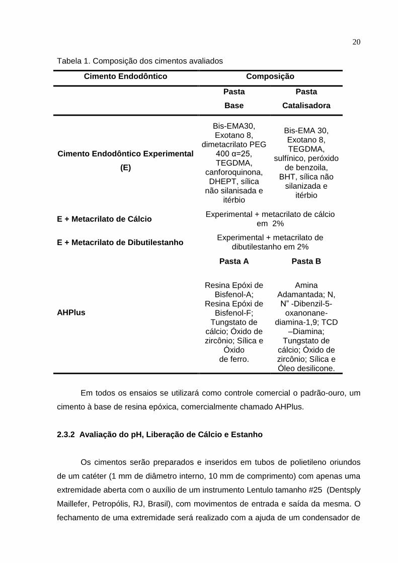
20
Tabela 1. Composição dos cimentos avaliados
Cimento Endodôntico Composição
Pasta
Base
Pasta
Catalisadora
Cimento Endodôntico Experimental
(E)
Bis-EMA30, Exotano 8,
dimetacrilato PEG 400 α=25, TEGDMA,
canforoquinona, DHEPT, sílica
não silanisada e itérbio
Bis-EMA 30, Exotano 8, TEGDMA,
sulfínico, peróxido de benzoila,
BHT, sílica não silanizada e
itérbio
E + Metacrilato de Cálcio Experimental + metacrilato de cálcio
em 2%
E + Metacrilato de Dibutilestanho Experimental + metacrilato de
dibutilestanho em 2%
AHPlus
Pasta A Pasta B
Resina Epóxi de Bisfenol-A;
Resina Epóxi de Bisfenol-F;
Tungstato de cálcio; Óxido de zircônio; Sílica e
Óxido de ferro.
Amina Adamantada; N, N” -Dibenzil-5-
oxanonane-diamina-1,9; TCD
–Diamina; Tungstato de
cálcio; Óxido de zircônio; Sílica e Óleo desilicone.
Em todos os ensaios se utilizará como controle comercial o padrão-ouro, um
cimento à base de resina epóxica, comercialmente chamado AHPlus.
2.3.2 Avaliação do pH, Liberação de Cálcio e Estanho
Os cimentos serão preparados e inseridos em tubos de polietileno oriundos
de um catéter (1 mm de diâmetro interno, 10 mm de comprimento) com apenas uma
extremidade aberta com o auxílio de um instrumento Lentulo tamanho #25 (Dentsply
Maillefer, Petropólis, RJ, Brasil), com movimentos de entrada e saída da mesma. O
fechamento de uma extremidade será realizado com a ajuda de um condensador de
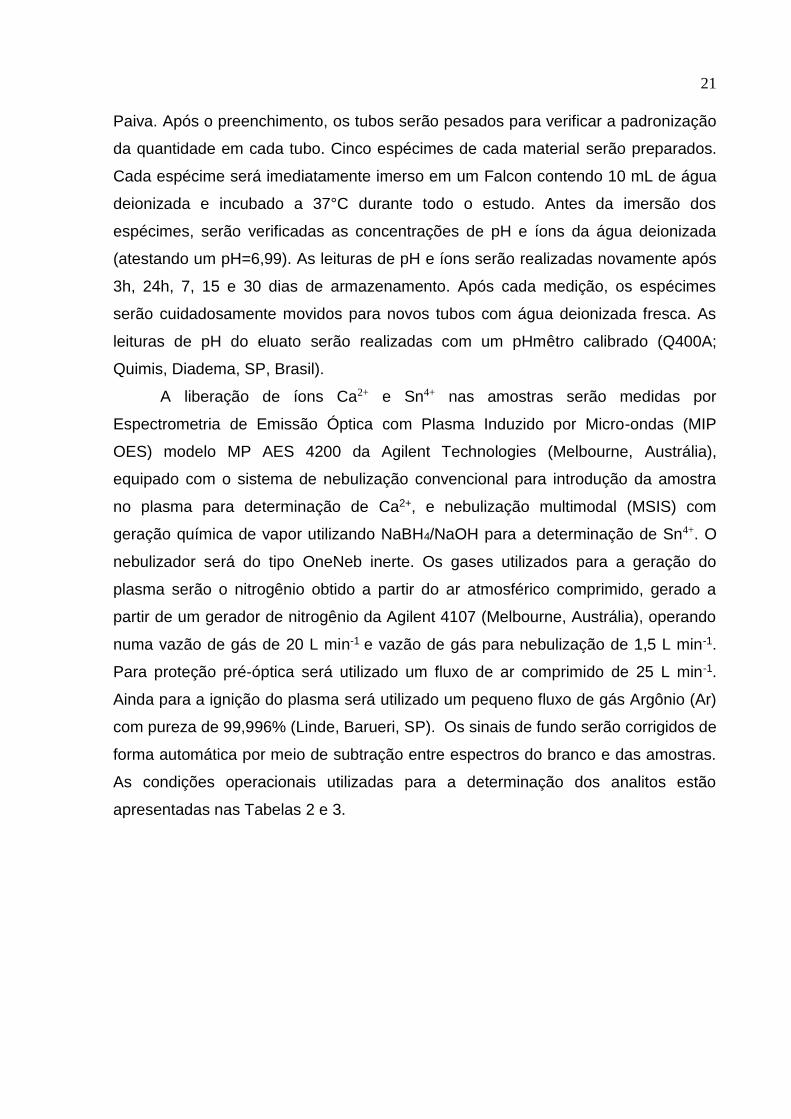
21
Paiva. Após o preenchimento, os tubos serão pesados para verificar a padronização
da quantidade em cada tubo. Cinco espécimes de cada material serão preparados.
Cada espécime será imediatamente imerso em um Falcon contendo 10 mL de água
deionizada e incubado a 37°C durante todo o estudo. Antes da imersão dos
espécimes, serão verificadas as concentrações de pH e íons da água deionizada
(atestando um pH=6,99). As leituras de pH e íons serão realizadas novamente após
3h, 24h, 7, 15 e 30 dias de armazenamento. Após cada medição, os espécimes
serão cuidadosamente movidos para novos tubos com água deionizada fresca. As
leituras de pH do eluato serão realizadas com um pHmêtro calibrado (Q400A;
Quimis, Diadema, SP, Brasil).
A liberação de íons Ca2+ e Sn4+ nas amostras serão medidas por
Espectrometria de Emissão Óptica com Plasma Induzido por Micro-ondas (MIP
OES) modelo MP AES 4200 da Agilent Technologies (Melbourne, Austrália),
equipado com o sistema de nebulização convencional para introdução da amostra
no plasma para determinação de Ca2+, e nebulização multimodal (MSIS) com
geração química de vapor utilizando NaBH4/NaOH para a determinação de Sn4+. O
nebulizador será do tipo OneNeb inerte. Os gases utilizados para a geração do
plasma serão o nitrogênio obtido a partir do ar atmosférico comprimido, gerado a
partir de um gerador de nitrogênio da Agilent 4107 (Melbourne, Austrália), operando
numa vazão de gás de 20 L min-1 e vazão de gás para nebulização de 1,5 L min-1.
Para proteção pré-óptica será utilizado um fluxo de ar comprimido de 25 L min-1.
Ainda para a ignição do plasma será utilizado um pequeno fluxo de gás Argônio (Ar)
com pureza de 99,996% (Linde, Barueri, SP). Os sinais de fundo serão corrigidos de
forma automática por meio de subtração entre espectros do branco e das amostras.
As condições operacionais utilizadas para a determinação dos analitos estão
apresentadas nas Tabelas 2 e 3.
22
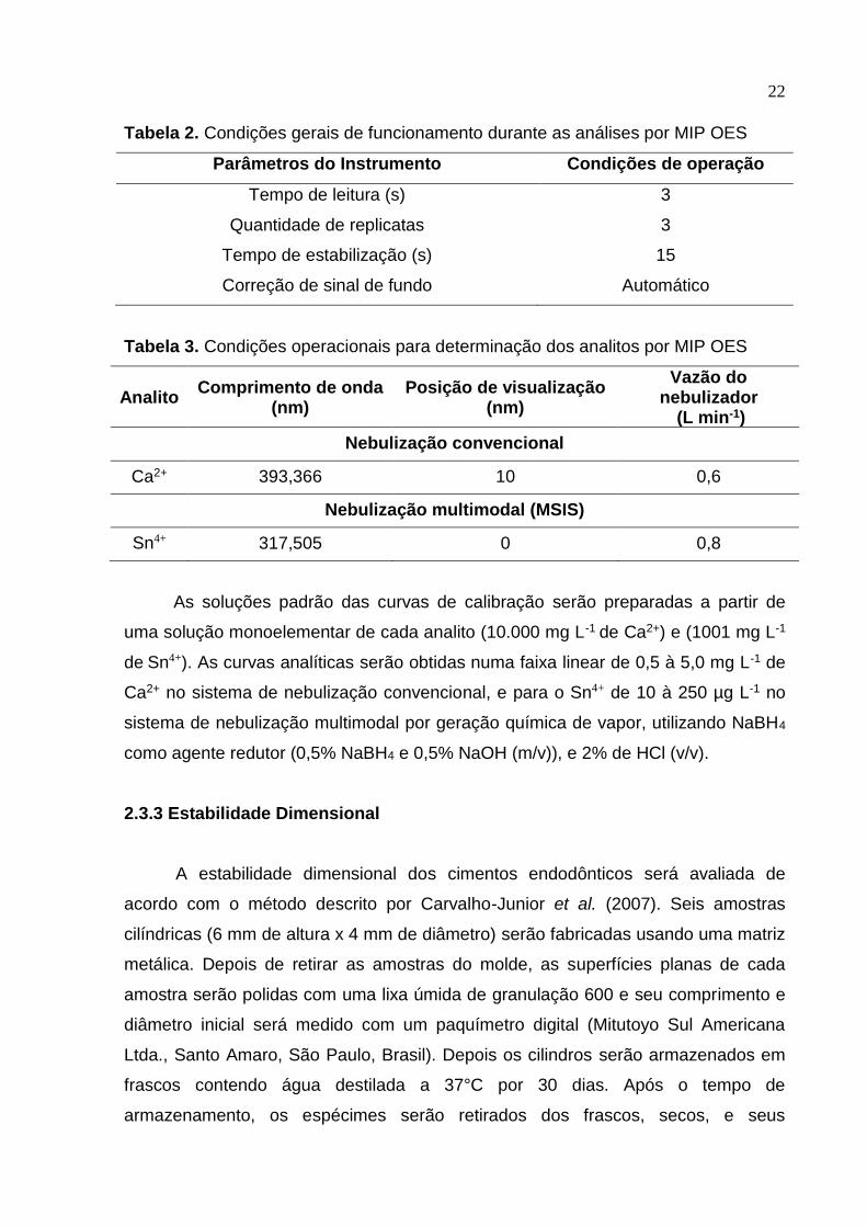
Tabela 2. Condições gerais de funcionamento durante as análises por MIP OES
Parâmetros do Instrumento Condições de operação
Tempo de leitura (s) 3
Quantidade de replicatas 3
Tempo de estabilização (s) 15
Correção de sinal de fundo Automático
Tabela 3. Condições operacionais para determinação dos analitos por MIP OES
Analito Comprimento de onda
(nm) Posição de visualização
(nm)
Vazão do nebulizador
(L min-1)
Nebulização convencional
Ca2+ 393,366 10 0,6
Nebulização multimodal (MSIS)
Sn4+ 317,505 0 0,8
As soluções padrão das curvas de calibração serão preparadas a partir de
uma solução monoelementar de cada analito (10.000 mg L-1 de Ca2+) e (1001 mg L-1
de Sn4+). As curvas analíticas serão obtidas numa faixa linear de 0,5 à 5,0 mg L-1 de
Ca2+ no sistema de nebulização convencional, e para o Sn4+ de 10 à 250 µg L-1 no
sistema de nebulização multimodal por geração química de vapor, utilizando NaBH4
como agente redutor (0,5% NaBH4 e 0,5% NaOH (m/v)), e 2% de HCl (v/v).
2.3.3 Estabilidade Dimensional
A estabilidade dimensional dos cimentos endodônticos será avaliada de
acordo com o método descrito por Carvalho-Junior et al. (2007). Seis amostras
cilíndricas (6 mm de altura x 4 mm de diâmetro) serão fabricadas usando uma matriz
metálica. Depois de retirar as amostras do molde, as superfícies planas de cada
amostra serão polidas com uma lixa úmida de granulação 600 e seu comprimento e
diâmetro inicial será medido com um paquímetro digital (Mitutoyo Sul Americana
Ltda., Santo Amaro, São Paulo, Brasil). Depois os cilindros serão armazenados em
frascos contendo água destilada a 37°C por 30 dias. Após o tempo de
armazenamento, os espécimes serão retirados dos frascos, secos, e seus
23
comprimentos e diâmetros finais serão medidos. A porcentagem de mudança
dimensional será calculada da seguinte forma:
[(L30 - L)/L] x 100
onde L é o comprimento inicial da amostra e L30, o comprimento após 30 dias.
2.3.4 Ensaio de inibição da Formação de Biofilme
Para o ensaio antimicrobiano, os cimentos serão manipulados e colocados
em moldes de plástico com 10 mm de diâmetro e 1,5 mm de espessura e os
espécimes armazenados à temperatura ambiente durante 48h. Os discos serão
esterilizados por luz ultravioleta em uma camada de fluxo laminar (Bio Seg 12,
Grupo Veco, Barão Geraldo Campinas, SP - Brasil) durante 30min em cada face dos
espécimes (GUERREIRO-TANOMARU et al., 2013). Serão utilizados discos de
hidroxiapatita estéril com as mesmas dimensões como substrato para crescimento
de biofilme no grupo de controle positivo. (SHEN et al. 2009).
Uma amostra de biofilme subgengival será colhida de um voluntário adulto
saudável e suspensa em caldo de infusão coração-cérebro (BHI, Becton Dickinson,
Sparks, MD, EUA). Os discos serão incubados em suspensões de placas de cultura
de BHI-24 poços sob condições anaeróbicas utilizando saco anaeróbico e indicador
anaeróbio (AnaeroGen, OXOID, Hampshire, UK) a 37°C. Cada poço deve conter 1,5
mL de caldo BHI estéril e 0,5 mL de inóculo, em que os espécimes serão mantidos
submersos. A densidade celular será ajustada em um espectrofotómetro a 405nm
(Sp22-325 a 1000nm, Bioespectro, Curitiba, PR, Brasil) até uma densidade de
aproximadamente 7,5x10⁷ unidades formadoras de colônias por mililitro em caldo
BHI. O meio BHI será substituído uma vez por semana sem adição de novos
microorganismos (SHEN et al. 2009). Após os períodos de crescimento de 3, 15 e 30
dias, serão analisados 45 discos (n=3 por grupo e por período de incubação) para
biofilme e proporção de bactérias vivas e mortas por coloração de viabilidade e
microscopia confocal de varredura a laser (CLSM).
A análise da viabilidade do biofilme será realizada utilizando a técnica SYTO 9
- iodeto de propídio (Live/Dead Bacligth Kit, Invitrogen, Eugene, OR, EUA). SYTO 9
é uma mancha fluorescente verde que rotula os microorganismos vivos e mortos. O
iodeto de propídio é uma mancha de ácido nucleico fluorescente vermelha que só
24
penetra células com membranas danificadas (células mortas). Primeiramente, as
amostras serão limpas com 2 mL de solução salina e, em seguida, 0,25 μL de
corante será colocado sobre o biofilme. Será utilizado um CLSM (Leica
Microsystems, Nussloch GmbH 2019, USA) para visualizar as amostras
(ORDINOLA-ZAPATA et al., 2012). Os respectivos comprimentos de onda de
absorção e emissão serão 494/518nm para SYTO 9 e 536/617nm para iodeto de
propídio. O biofilme será avaliado aleatoriamente em uma ampliação de 100x. Em
seguida, serão obtidas cinco pilhas confocais de diferentes áreas aleatórias de cada
amostra com lente de 10x e com formato de 512x512 pixels. Para cada grupo será
obtido um total de 25 pilhas (5 campos operacionais×5 espécimes por grupo). O
avaliador será cegado para os grupos experimentais. Todas as imagens serão
analisadas usando o software LAS X 3D Analysis (Leica Microsystems, Nussloch
GmbH 2019, USA) para o biovolume total (μm3), o número total de células vivas
(verde) e o percentual de células vivas (DE PAZ, 2009).
2.3.5 Processo de Envelhecimento Acelerado Dentro de Câmara Climática
Os materiais serão submetidos a um protocolo de armazenamento acelerado
para as metodologias de grau de conversão e cinética de polimerização e espessura
de película, de acordo com o modelo de Arrhenius (CLARK, 1991):
r = Q10^((RT-TA)/10)
Onde: r é o envelhecimento acelerado, TA é a temperatura ambiente, RT é a
temperatura aumentada, e Q10 é a constante de coeficiente de reação.
O modelo de envelhecimento será feito em uma estufa com temperatura
controlada a 40ºC por um período de 2 meses. O envelhecimento acelerado será
dentro de câmara climática (MA 835/UR, Marconi, Piracicaba, Brasil) com
temperatura controlada e umidade relativa de 75%. De acordo com o modelo de
Arrhenius, estas condições, quando mantidas por 6 meses, equivalem a um período
de armazenagem de 18 meses a temperatura ambiente.
Segundo este modelo, as propriedades químico-mecânicas do material, como
grau de conversão e espessura de película serão monitoradas em 3 tempos
diferentes: 0, 1, 2 meses, os quais serão equivalentes aos períodos previamente
determinados para a avaliação em tempo real. Os produtos não serão armazenados
25
em dessecador com a finalidade de avaliar a influência da umidade nas
propriedades químico-mecânicas do material.
2.3.5.1 Grau de Conversão
A cinética de polimerização será determinada utilizando espectroscopia
infravermelha com transformada de Fourier, com uma unidade de refletância total
atenuada acoplada ao sistema (RT-FTIR Shimadzu Prestige 21 Spectrometer,
Shimadzu, Japão). Quantidades padronizadas de cada material serão dispensadas
sobre o cristal e prévio à fotoativação, um espectro será obtido. Posteriormente, a
superfície do material será coberta com uma tira de matriz de poliéster através da
qual será feita a fotoativação com unidade LED de irradiância >1.000 mW/cm2 (Radii
® Curing Light, SDI, Bayswater, Victória, Australia). As amostras serão fotoativadas
por 40 s. A cinética de polimerização será determinada utilizando o software
IRSolution, utilizando a apodização de Happ-Genzel, em uma faixa espectral entre
1690 e 1575 cm-1. O grau de conversão, por segundo será determinado pela razão
da intensidade da absorbância observadas do C=C alifático (altura do pico em 1638
cm−1) contra a intensidade de absorbância da ligação carboxila (1710 cm-1) utilizada
como padrão interno. Cada teste será feito em triplicata.
2.3.5.2 Espessura de Película
Será necessário para realização do ensaio de espessura de película duas
placas de vidro quadradas, com espessura de 5 mm e área de contato de
aproximadamente (200±10) mm2; dispositivo de carga de (150±3) N e micrômetro
com acurácia de 1 μm. Serão feitas três vezes para cada material (n=3). O cimento
será manipulado e 5 mL serão dispensados no centro da placa de vidro. Após a
colocação do cimento sobre a placa, este será recoberto com outra placa com as
mesmas dimensões. Após (180±10)s do início da mistura do cimento, se aplicará
uma carga de (150±3)N sobre a placa superior.
Verificando-se que o cimento recobriu toda a placa, após 10 minutos do início
da manipulação, se medirá a espessura do conjunto formado pelas duas placas e o
filme de cimento por meio de um micrômetro com acurácea de 1 μm. A espessura de
cimento será definida pela diferença de espessura das placas com e sem o cimento.
26
Serão feitas três repetições. A ISO 6876 (2001) exige uma espessura de filme não
superior a 50μm.
2.3.6 Análise Estatística
Os dados estatísticos serão analisados estatisticamente por Análise de
Variância de uma via (ANOVA) e teste post hoc de Tukey utilizando SPSS software
22.0 (SPSS Inc, Chicago, IL) para os dados de biofilme.
A análise estatística do restante dos dados será realizada utilizando o software
Sigma Plot 12.0 Os dados serão analisados para verificar a distribuição normal e a
homogeneidade da variância. Análise ANOVA de duas vias seguida por um teste
complementar de Tukey será feita para avaliar cada uma das variáveis dependentes.
O nível de significância para todos os testes será p <0,05.
2.4 Hipótese
A incorporação de monômeros metacrilatos metálicos em um cimento
endodôntico experimental não conferirá propriedades de liberação de íons cálcio e
estanho, não conferirá atividade antimicrobiana contra biofilme de multiespécies, e
que o processo de envelhecimento irá alterar suas propriedades físico-químicas.
27
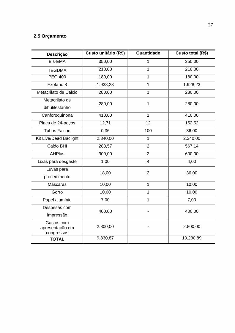
2.5 Orçamento
Descrição Custo unitário (R$) Quantidade Custo total (R$)
Bis-EMA 350,00 1 350,00
TEGDMA 210,00 1 210,00
PEG 400 180,00 1 180,00
Exotano 8 1.938,23 1 1.928,23
Metacrilato de Cálcio 280,00 1 280,00
Metacrilato de
dibutilestanho 280,00 1 280,00
Canforoquinona 410,00 1 410,00
Placa de 24-poços 12,71 12 152,52
Tubos Falcon 0,36 100 36,00
Kit Live/Dead Baclight 2.340,00 1 2.340,00
Caldo BHI 283,57 2 567,14
AHPlus 300,00 2 600,00
Lixas para desgaste 1,00 4 4,00
Luvas para
procedimento 18,00 2 36,00
Máscaras 10,00 1 10,00
Gorro 10,00 1 10,00
Papel alumínio 7,00 1 7,00
Despesas com
impressão 400,00 - 400,00
Gastos com apresentação em
congressos
2.800,00 - 2.800,00
TOTAL 9.830,87 10.230,89
28
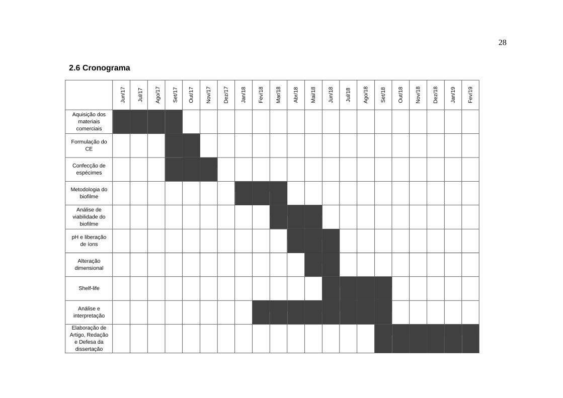
2.6 Cronograma
Jun/1
7
Jul/17
Ago/1
7
Set/
17
Out/
17
Nov/1
7
Dez/1
7
Jan/1
8
Fe
v/1
8
Ma
r/18
Abr/
18
Ma
i/18
Jun/1
8
Jul/18
Ago/1
8
Set/
18
Out/
18
Nov/1
8
Dez/1
8
Jan/1
9
Fe
v/1
9
Aquisição dos
materiais
comerciais
Formulação do
CE
Confecção de
espécimes
Metodologia do
biofilme
Análise de
viabilidade do
biofilme
pH e liberação
de íons
Alteração
dimensional
Shelf-life
Análise e
interpretação
Elaboração de
Artigo, Redação
e Defesa da
dissertação

29
2.7 Referências CARVALHO-JUNIOR, J.R., CORRER-SOBRINHO, L., CORRER, A.B., SINHORETI,
M.A., CONSANI, S.; SOUSA-NETO, M.D. Solubility and dimensional change after
setting of root canal sealers: a proposal for smaller dimensions of test samples.
Journal of Endodontics, v.33, n.9, p.1110-1116, 2007.
DE PAZ, L.E.C. Image analysis software based on color segmentation for
characterization of viability and physiological activity of biofilms. Applied and
Environmental Microbiology, v.75, n.6, p.1734-1739, 2009.
GRUBBS, R.B.; BROZ, M.E.; DEAN, J.M.; BATES, F.S. Selectively epoxidized
polyisoprene-polybutadiene block copolymers. Macromolecules. v.33, p.2308 –10,
2000.
GUERREIRO-TANOMARU, J.M., DE FARIA-JÚNIOR, N.B., DUARTE, M.A.H.,
ORDINOLA-ZAPATA, R., GRAEFF, M.S.Z. AND TANOMARU-FILHO, M.
Comparative analysis of Enterococcus faecalis biofilm formation on different
substrates. Journal of Endodontics, v.39, n.3, p.346-350, 2013.
HASCHKE, E. Adhesive endodontic cones and related methods. United States
Patent Application 20040202986. US Patent & Trademark Office, October 14, 2004.
HIRAISHI, N., LOUSHINE, R.J., VANO, M., CHIEFFI, N., WELLER, R.N., FERRARI,
M., PASHLEY, D.H. AND TAY, F.R. Is an oxygen inhibited layer required for bonding
of resin-coated gutta-percha to a methacrylate-based root canal sealer? Journal of
Endodontics, v.32, n.5, p.429-433, 2006.
OLIVEIRA, A.C.; TANOMARU, J.M.; FARIA-JUNIOR, N.; TANOMARU-FILHO, M.
Bacterial leakage in root canals filled with conventional and MTA-based sealers.
International Endodontic Journal, v. 44, n. 4, p. 370-375, 2011.
ORDINOLA‐ZAPATA, R., BRAMANTE, C.M., CAVENAGO, B., GRAEFF, M.S.Z., DE
MORAES, I.G., MARCIANO, M. AND DUARTE, M.A.H. Antimicrobial effect of
30
endodontic solutions used as final irrigants on a dentine biofilm model. International
Endodontic Journal, v.45, n.2, p.162-168, 2012.
REZENDE, G.C.; MASSUNARI, L.; QUEIROZ, I.O.; GOMES FILHO, J. E.; JACINTO,
R. C.; LODI, C. S., DEZAN JUNIOR, E. Antimicrobial action of calcium hydroxide-
based endodontic sealers after setting, against E. faecalis biofilm. Brazilian Oral
Research, v.30, p.e38, 2016.
ROSSATO, T.C.A.; GALLAS, J.A.; ROSA W.L.O.; DA SILVA, A.F.; PIVA, E;
PERALTA, S.L; LUND, R.G. Experimental Sealers Containing Metal Methacrylates:
Physical and Biological Properties. Journal of Endodontics, v.43, n.9, 2017.
SAMUI, A.B.; DALVI, V.G.; CHANDRASEKHAR, L.; PATRI, M.; CHAKRABORTY,
B.C. Interpenetrating polymer networks based on nitrile rubber and metal
methacrylates. Journal of Applied Polymer Science, v.99, n.5, p.2542-2548, 2006.
SHEN, Y.; ZHANG, H.; RUSE, N.D.; HAAPASALO, M. Antibacterial activity of
endodontic sealers by modified direct contact test against Enterococcus faecalis.
Journal of Endodontics, v.35, n.7, p.1051–1055, 2009.
SHIPPER, G.; ØRSTAVIK, D.; TEIXEIRA, F.B.; TROPE, M. An evaluation of
microbial leakage in roots filled with a thermoplastic synthetic polymer-based root
canal filling material (Resilon). Journal of Endodontics. v.30, n.5, p. 342–7, 2004.
TAY, F.R., LOUSHINE, R.J., MONTICELLI, F., WELLER, R.N., BRESCHI, L.,
FERRARI, M. AND PASHLEY, D.H. Effectiveness of resin-coated gutta-percha
cones and a dual-cured, hydrophilic methacrylate resin-based sealer in obturating
root canals. Journal of Endodontics, v.31, n.9, p.659-664, 2005.
TAY, F.R.; LOUSHINE, R.J.; WELLER, R.N.; KIMBROUGH, W.F.; PASHLEY, D.H.;
MAK, Y.F.; LAI, C.N.S.; RAINA, R.; WILLIAMS, M.C. Ultrastructural evaluation of the
quality of apical seal in roots filled with a polycaprolactone-based root canal filling
material. Journal of Endodontics. v.31, n.7, p.514 –9, 2005.
31
3 Relatório do Trabalho de Campo
Neste capítulo estão relatadas as complementações e as mudanças ocorridas no
planejamento e execução dos experimentos desta pesquisa.
3.1 O projeto de qualificação de dissertação apresentado à banca, se
intitulava: Atividade microbiana e resposta tecidual óssea de cimentos endodônticos
experimentais contendo metacrilatos metálicos. No entanto, a resposta tecidual em
animais não está sendo apresentada como parte desta dissertação devido à
necessidade de complementação de testes físico-químicos e antimicrobianos
anteriormente à realização do estudo in vivo. Para a realização de um estudo com
animais, deve-se buscar o menor dano possível ao mesmo, desta forma, optamos
por melhor conhecer as propriedades dos materiais estudados. Cabe salientar, no
entanto, que o projeto inicial, contendo a metodologia em animais, já foi submetido e
aprovado pelo Comitê de Ética em Pesquisa Animal (CEEA) através do processo no.
23110.005211/2017-43 e será executado durante o meu doutoramento.
3.2 No mês de Fevereiro de 2018 realizei uma visita científica na Indiana
Purdue University (IUPUI), sob a orientação da Dra. Simone Duarte. Esta visita
ocorreu para a realização da metodologia de biofilme através de o microscopia
confocal. Devido à falta de recursos humanos e tempo, somente foi possível realizar
o teste em parte das amostras. Era necessário que se participasse de muitas
capacitações antes de poder utilizar o laboratório de microbiologia, não havendo
tempo hábil para a confecção do biofilme de 30 dias. Adicionalmente, a pesquisa
original se baseava em uma coleta de microorganismos multiespécies, porém devido
ao tempo e a questões éticas da universidade, não foi possível realizar, sendo feito
somente o biofilme com cepas de Enterococcus faecalis. Os resultados não ficaram
condizentes com o esperado, sendo necessária a realização dos testes com biofilme
novamente em Pelotas, no Departamente de Biotecnologia da UFPel.
Em relação à metodologia de biofilme realizada na Universidade Federal de
Pelotas, houve perdas nas amostras do biofilme do período experimental de 30 dias.
Suspeita-se de que o meio de cultura utilizado para as trocas não estava em
condições de ser utilizado, ou houve alguma falha no gerador de anaerobiose. Este
32
fato gerou uma falha na apresentação dos resultados desta dissertação, pois não foi
possível contabilizar os resultados deste período de 30 dias. Estas amostras serão
repetidas a tempo de serem incluídas no artigo final, anteriormente à submissão.
Não foi possível repetirmos estas amostras para inclusão na dissertação devido ao
período de recesso da UFPel em Dezembro e Janeiro.
3.3 Os testes de liberação de íons e pH foram sugeridos pela banca de
defesa de qualificação da dissertação em Outubro de 2017. Foram então
incrementados no estudo para melhor avaliação do cimento endodôntico antes do
manuseio com animais. Os testes foram realizados logo após a chegada dos
materiais.
3.4 O teste de alteração dimensional foi realizado após a metodologia de
biofilme, porque se percebeu que os materiais eram muito porosos ao corante,
absorvendo a maior parte dele, dificultando a visualização no microscópio. Desta
forma, esta foi a última metodologia a ser feita.
3.5 Os ensaios de envelhecimento também foram incluídos após sugestão
da banca no momento de qualificação de mestrado em Outubro de 2017. Um estudo
em andamento no CDC-Bio, realizado pelo Prof Dr. Carlos Enrique Cuevas-Suárez e
Prof Dr. Evandro Piva, estava pesquisando o tempo de prateleira de materiais
comerciais. Houve o interesse de saber se os respectivos materiais durariam o
tempo de validade após situações adversas. Deste modo, o cimento endodôntico
experimental fez parte dos testes. A metodologia se baseia no fato de que a cada 1
mês de armazenamento, leva-se em conta o período de meia-vida do material.

33
4 Artigo
O artigo presente nesse trabalho está formatado segundo as normas do
periódico Dental Materials, disponível online em
https://www.elsevier.com/journals/dental-materials/01095641/guide-for-authors,
acesso em 11 de fevereiro de 2019.
Novel endodontic sealer based in metals cross-linking methacrylate
Victoria Burmann da Silva Guimarãesa, Andressa da Silva Barbozaa, Carlos Enrique
Cuevas-Suárezb,Tiago Collaresc, Thais Larré Oliveirac, Anderson Schwingel
Ribeirod, Meibel Teixeira Lisboad, Simone Duartee, Fernanda Geraldo Pappena,
Rafael Guerra Lunda*
aGraduate Program in Dentistry, Federal University of Pelotas, Pelotas, RS, Brazil
bDental Materials Laboratory, Academic Area of Dentistry, Autonomous University of Hidalgo State.
Circuito Ex Hacienda La Concepción S/N. San Agustín Tlaxiaca, Hgo. Mexico.
cCancer Biotechnology Laboratory, School of Biotechnology, Federal University of Pelotas, Brazil.
dGraduate Program in Chemistry, School of Chemistry, Chemical Metrology Laboratory (LabMeQui),
Federal University of Pelotas, Brazil.
eDepartment of Cariology, Operative Dentistry and Dental Public Health, School of Dentistry, Indiana
University, Indianapolis, USA.
*Corresponding author:
Rafael Guerra Lund, DDS, MSc, PhD
Post-Graduate Program in Dentistry, School of Dentistry, Federal University of
Pelotas
Rua Gonçalves Chaves, 457, Pelotas-RS 96015-560, Brazil
Phone: +55 53 3225-6741 / 134
E-mail: [email protected]
Declarations of interest: The authors deny any conflict of interest
Acknowledgements
34
VBS Guimarães thanks the Coordination for the Improvement of Higher Education
Personnel (CAPES) for a Graduate Fellowship.
Funding sources
This study was supported in part by the Brazilian agencies: Coordination for
the Improvement of Higher Education Personnel (CAPES), Brazilian National Council
for Scientific and Technological Development (CNPq), and Fundação de Amparo à
Pesquisa do Estado do Rio Grande do Sul (FAPERGS; PRONEX #16.0471-4).
Abstract
The properties of endodontic sealers are important for the quality of filling of the root canals and, later, in the endodontic prognosis. However, no material has all the properties of an ideal endodontic sealer, so new materials are being created to improve its physicochemical and biological characteristics. The objective of this study was to evaluate the physical, chemical and antimicrobial properties, and shelf-life of a dual-reactive experimental sealer after the incorporation of dibutyltin methacrylate (Sn4+) (ETs) or calcium methacrylate (Ca2+) (ETs), in a concentration of 2%. AH Plus was used as a commercial reference. The pH and ion release were measured using a pH meter and a microwave induced plasma optical emission spectrometer. The dimensional stability was evaluated after 30 days in accordance with ISO 6876. Inhibition of biofilm growth was evaluated by laser scanning confocal microscopy (CLSM). The biofilm viability analysis was performed using the SYTO 9 technique. The shelf-life was evaluated through tests of degree of conversion and film thickness at the times: immediate, 1 month and 2 months. For statistical analysis, ANOVA and Tukey post hoc test were used, and a significance level of 5%. All sealers tested showed a gradual reduction of pH over 30 days. The ECs sealer showed the highest release of calcium and ETs presented the highest release of tin in 30 days. The ETs sealer showed better dimensional stability over time compared to other endodontic sealers. ETs revealed better antibiofilm potential after 15 days compared to controls. The degree of conversion was reduced after the shelf-life. Regarding film thickness, all materials were in accordance with ISO 6876 specifications, except ECs immediate (100 μm) and ETs in the first month of aging (170 μm). It was concluded that the physicochemical properties of the experimental endodontic sealers containing calcium and dibutyltin methacrylate were not drastically altered and the antibiofilm effect of the cements was improved. Key-words: endodontics, root canal obturation, methacrylates, antimicrobial agents, AH plus.
35
Introduction
In the endodontic therapy, the obturation is responsible for the sealing of the
root canal system, providing the filling of anatomical irregularities and the burial of
bacteria that have survived the other stages of the treatment [1]. Sundqvist & Figdor
[3] assigned three primary functions to the root filling: sealing against ingrowth of
bacteria from the oral cavity; entombment of remaining microorganisms; and
complete obturation at a microscopic level to prevent stagnant fluid from
accumulating and serving as nutrients for bacteria from any source. Properties of root
canal sealers are important on the quality of root canal filling and subsequently on
the endodontic outcome [4]. Additionally, these materials should present adequate
physicochemical properties, such as setting time, radiopacity, flow, water sorption
and antimicrobial properties [2].
Many materials are currently used as root canal sealers, including zinc oxide–
eugenol, calcium hydroxide, glass-ionomer, silicone and epoxy resin [1,5]. However,
to date, no single established material meets all the desirable requirements for
endodontic space obturation. Thus, new materials are constantly being created in
order to improve its physicochemical and biological characteristics.
Recently, Rossato et al. [6], developed an experimental endodontic sealer with
addition of calcium methacrylate and tin methacrylate, evidencing an increase in the
antimicrobial action of this sealer on a strain of E. faecalis, and moderate cytotoxicity
when compared to a commercial control (Resilon/RealSeal® (RS-SybronEndo, CA,
USA). Calcium methacrylates may release calcium ions that play an important role in
the repair process [7]. Additionally, dibutyltin methacrylate presents tin, which has
been used for decades as a prophylactic agent in preventive dentistry; previous
findings revealed the independent action on inhibiting bacterial biofilm formation,
reducing the acid portion of this microbial polysaccharide matrix [8, 9].
In this context, from the results obtained by Rossato et al. [6], the present
study evaluated the pH, the release of tin and calcium ions, and the antimicrobial
action, through in vitro biofilm tests of experimental endodontic sealers, with the
incorporation of monomers of calcium hydroxide methacrylate (ECs) and with
dibutyltin methacrylates (ETs). Furthermore, it was attempted to validate an
accelerated aging protocol with the purpose of determining the shelf-life and
36
expiration date of these sealers by monitoring the stability of their properties (degree
of conversion and film thickness) over time.
The null hypothesis was as follows: the incorporation of metal methacrylate
monomers into an experimental endodontic sealer will not confer calcium and tin ion
release properties, confer antimicrobial activity against multispecies biofilms, and that
the aging process will alter its physicochemical properties.
Material and Methods
Formulation and delineation of groups of experimental endodontic sealers
The composition of ECs and ETs is shown in Table 1 as well as in the
commercial reference AH Plus. Two endodontic sealer pastes were mixed at the
same proportion and photoactivated with a light-emitting diode for 20 seconds (Radii
Curing Light; SDI, Bayswater, Victoria, Australia). AH Plus was mixed at the same
proportion, according to the manufacturer instructions.
pH Assessment and Release of Ca2+ and Sn4+
The sealers were prepared and inserted into polyethylene tubes from a
catheter (1 mm internal diameter, 10 mm long) with only one open end with the aid of
a Lentulo size #25 instrument (Dentsply Maillefer, Petropolis, RJ, Brazil) with
movements of entrance and exit of the same. The closing of one end will be
accomplished with the aid of a Paiva condenser. After filling, the tubes were weighed
to verify the standardization of the quantity in each tube. Three specimens of each
material were prepared. Each specimen was immediately immersed in a Falcon
containing 10 mL of deionized water and incubated at 37°C throughout the
experimental periods. Before the immersion of the specimens, the pH and ion
concentrations of the deionized water were verified (attesting to pH = 6.01). The pH
and ion readings were performed after 3h, 24h, 7, 15 and 30 days of storage. After
each measurement, the specimens were carefully moved to fresh tubes with fresh
deionized water. The pH readings of the eluates were performed with a calibrated pH
meter (Q400A; Quimis, Diadema, SP, Brazil).
37
The release of Ca2+ and Sn4+ ions in the samples were measured by Agilent
Technologies' MP AES 4200 Microwave Induced Spectrometer (MIP OES) model
(Melbourne, Australia), equipped with the conventional misting system for
introduction of the samples into the plasma for Ca2+ determination, and multimodal
misting (MSIS) with chemical vapor generation using NaBH4/NaOH for the
determination of Sn4+. The nebulizer was inert OneNeb type. The gases used to
generate the plasma were the nitrogen obtained from compressed atmospheric air,
generated from an Agilent 4107 nitrogen generator (Melbourne, Australia), operating
at a gas flow of 20 L min-1 and flow rate of for nebulization of 1.5 L min-1. For pre-
optic protection a compressed air flow of 25 L min-1 was used. Also, for the ignition of
the plasma was used a small gas flow Argon (Ar) with purity of 99.996% (Linde,
Barueri, SP). The background signals were automatically corrected by subtraction
between white and sample spectra. The operating conditions used for the
determination of the analytes are presented in Tables 2 and 3.
The standard solutions of the calibration curves were prepared from a
monoelement solution of each analyte (10,000 mg L-1 Ca2+) and (1001 mg L-1 Sn4+).
The analytical curves were obtained in a linear range from 0.5 to 5.0 mg L-1 of Ca2+ in
the conventional misting system, and for Sn4+ from 10 to 250 μg L-1 in the multimodal
misting system by chemical vapor generation, using NaBH4 as a reducing agent
(0.5% NaBH4 and 0.5% NaOH (m / v)), and 2% HCl (v / v).
Dimensional Stability
The dimensional stability of the endodontic sealers was evaluated according to
the method described by Carvalho-Junior et al. [10]. For each group, six cylindrical
specimens (6 mm high x 4 mm diameter) were made using a metal matrix. After
removing the samples from the mold, the flat surfaces of each sample were polished
with a wet granulation sandpaper 600 and their initial length and diameter was
measured with a digital caliper (Mitutoyo Sul Americana Ltda., Santo Amaro, Sao
Paulo, Brazil). The cylinders were then stored in vials containing distilled water at
37°C for 30 days. After the storage time, the specimens were removed from the
flasks, dried, and their final lengths and diameters were measured. The percentage
of dimensional change was calculated as follows:
38
[(L30 - L)/L] x 100
where L is the initial sample length and L30 is the length after 30 days.
Biofilm Formation Inhibition Assay
For the antimicrobial assay, the sealers were prepared and placed in plastic
molds 10 mm in diameter and 1.5 mm thick and the specimens stored at room
temperature for 48 h. The discs were sterilized by ultraviolet light in a laminar flow
layer (Bio Seg 12, Grupo Veco, Barão Geraldo Campinas, SP - Brasil) for 30 min per
side [11]. Sterile hydroxyapatite disks with the same dimensions as substrate for
biofilm growth were used in the positive control group [12].
A subgingival biofilm sample was collected from a healthy adult volunteer and
suspended in a heart-brain infusion broth (BHI, Becton Dickinson, Sparks, MD, USA).
The cell density was adjusted in a spectrophotometer at 405nm (Sp22-325 at
1000nm, Bioespectro, Curitiba, PR, Brazil) to a density of approximately 7.5x10⁷
colony forming units per milliliter in BHI broth.
The disks were incubated in this suspension in 24 well plates under anaerobic
conditions using anaerobac and anaerobic indicator (PROBAC DO BRASIL Produtos
Bacteriológicos Ltda. São Paulo – SP, Brazil) at 37°C. Each well contained 1.5 mL of
sterile BHI broth and 0.5 mL of inoculum, in which the specimens were kept
submerged. The BHI medium was replaced once a week without addition of new
microorganisms [12]. After the growth periods of 7 and 15 days, 30 discs (n = 3 per
group and per incubation period) were analyzed for biofilm and proportion of live and
dead bacteria by viability staining and laser scanning confocal microscopy (CLSM).
Biofilm viability analysis was performed using the SYTO 9 propidium iodide
(Live/dead Bacligth Kit, Invitrogen, Eugene, OR, USA) technique. SYTO 9 is a green
fluorescent stain that labels living and dead microorganisms. Propidium iodide is a
red fluorescent nucleic acid patch that penetrates only cells with damaged
membranes (dead cells). First, the samples were cleaned with 2 mL of saline solution
and then 0.25 μL of fluorescent stain was placed on the biofilm. A CLSM (Leica
Microsystems, Nussloch GmbH 2019, USA) was used to visualize the samples. The
biofilm was randomly evaluated at a 100x magnification. Then, five confocal piles of
different random areas of each sample were obtained using x10 lens and with the
format of 512x512 pixels. The evaluator was blinded to the experimental groups. All
39
images were analyzed using LAS X 3D Analysis (Leica Microsystems, Nussloch
GmbH 2019, USA) software for total biovolume (μm3), total number of live cells
(green) and percentage of live cells.
Accelerated Aging Process within Climate Chamber
The materials were subjected to an accelerated storage protocol for the
methodologies of degree of conversion and kinetics of polymerization and film
thickness, according to the Arrhenius model [13]:
r = Q10 ^ ((RT-TA) / 10)
where ‘r’ is the accelerated aging, ‘TA’ is the ambient temperature, ‘RT’ is the
increased temperature, and ‘Q10’ is the reaction coefficient constant.
The aging model was made in an oven with controlled temperature at 40ºC for
a period of 2 months. Accelerated aging occurred within a climatic chamber (MA 835
/ UR, Marconi, Piracicaba, Brazil) with controlled temperature and relative humidity of
75%. According to the Arrhenius model, these conditions, when maintained for 6
months, amount to a storage period of 18 months at room temperature.
According to this model, the chemical-mechanical properties of the material,
such as degree of conversion and film thickness were monitored at 3 different times:
0, 1, 2 months, which were equivalent to the periods previously determined for the
real-time evaluation. The products were not stored in a desiccator in order to
evaluate the influence of moisture on the chemical-mechanical properties of the
material.
Degree of Conversion
Polymerization kinetics was determined using Fourier transform infrared
spectroscopy, with an attenuated total reflectance unit coupled to the system (RT-
FTIR Shimadzu Prestige 21 Spectrometer, Shimadzu, Japan). Standard amounts of
each material were dispensed onto the crystal and prior to photoactivation, a
spectrum was obtained. Subsequently, the surface of the material was covered with a
polyester matrix strip through which the irradiation LED unit was irradiated >1000
mW / cm2 (Radii ® Curing Light, SDI, Bayswater, Victory, Australia). The samples
40
were photoactivated for up to 40 s. Polymerization kinetics were determined using
the IRSolution software, using the Happ-Genzel apodization, in a spectral range
between 1690 and 1575 cm-1. The degree of conversion per second was determined
by the absorbance intensity ratio observed for aliphatic C = C (peak height at 1638
cm-1) against the absorbance intensity of the carboxyl bond (1710 cm-1) used as the
internal standard. Each test was performed in triplicate.
Film Thickness
Two square glass plates with a thickness of 5 mm and a contact area of
approximately (200 ± 10) mm2 were required for the film thickness test; load device
(150 ± 3) N and micrometer with accuracy of 1 μm. The test was performed in
triplicate (n = 3). The sealers were prepared, and 5 ml of each was dispensed into
the center of the plate. After placing the material on the plate, it was covered with
another plate of the same dimensions. After (180 ± 10) s from the start of the mixture,
a load of (150 ± 3) N was applied to the top plate.
It was found that the sealer covered the whole plate, after 10 minutes of the
beginning of the manipulation, the thickness of the set formed by the two plates and
the cement film was measured by means of a micrometer with 1 μm accurate. The
sealer thickness was defined by the thickness difference of the plates with and
without the cement. ISO 6876 (2002) [14] requires a film thickness of not more than
50μm.
Statistical analyses
Statistical analyses of obtained data were performed using Sigma Plot 12.0
software. The data was analysed to verify the normal distribution and the
homogeneity of the variance. Two-way ANOVA analysis followed by a
complementary Tukey test were made to evaluate each of the dependent variables.
The level of significance for all the tests was set to p < 0.05.
Results
pH Assessment and Release of Ca2+ and Sn4+
41
The highest pH values were obtained for AH Plus over the 30 day-
period. In contrast, the other groups showed a decrease of pH values over the time. It
was observed a higher decrease of pH after 7 days in EXP and ETs groups, while the
ECs showed a lower pH variability (Figure 1).
The experimental endodontic sealers presented lower pH values in all times
(Figure 1). ECs pH values remained ±4.8 with no statistical difference between the
experimental sealer and AH Plus. ETs presented the higher variation of pH within the
experimental period, being statistically different from AH Plus (P<0.05).
The merit parameters obtained for Ca2+ and Sn4+ in aqueous standard are
described in Table 2. Figure 2 (A) shows the calcium release of the ECs, EXP and
AH Plus sealers by the conventional method and Figure 2 (B) shows the cumulative
effect of Ca2+ release in all times. It is possible to notice the decrease of Ca2+ release
of all sealers over the time. ECs showed the highest calcium release within the
experimental periods. The highest ion release occurred up to 3h, and it had
decreased along the time.
Figure 3 (A) shows the Sn4+ release of ETs, Experimental sealer and AH Plus
by multimodal system and Figure 3 (B) shows the cumulative effect of Sn4+ release in
all times. AH Plus did not release appreciable Sn4+ levels. At 3 hours period, the
experimental sealer showed the highest Sn4+ release, which had decreased along
the experimental period. ETs demonstrated the highest Sn4+ release from 24 hours to
30 days period, in comparison to the other groups.
The ECs cements did not obtain detectable levels of tin ions for the multimodal
method, whereas ETs cement did not find detectable levels of calcium in the
conventional method.
Dimensional Stability
The dimensional stability results are shown in Table 3. All the experimental
sealers showed positive values of dimensional stability, indicating expansion of the
material. Oppositely, AH Plus demonstrated negative values within 30 days storage
time (P<0.05).
Biofilm Formation Inhibition Assay
42
Total biovolume analysis revealed that biofilm formation occurred in all groups
at all incubation times. After 7 days, AH Plus and EXP demonstrated statistically
lower biofilm volume in comparison to control group (P<0.05). However, the total
biovolume growth were similar among the tested sealers (P>0.05) (Table 4).
After 15 days, in contrast, the ETs presented significantly lower biofilm
biovolume than ECs, EXP and control group (P<0.05), while ECs and EXP were
similar to control (P>0.05).
For viable bacteria biovolume (Table 4), all the evaluated sealers were similar
within the 7 days period (P>0.05). In relation to the 15 days period, the experimental
sealers (EXP, ETs and ECs) presented lower viable bacteria volume in comparison
to AH Plus and Control group (P<0.05). The positive effect of the methacrylates in
the antimicrobial properties of sealers was visible after 15 days.
Figure 4 shows examples of CSLM images of biofilm growth on the different
materials within the experimental periods tested. In the images, it is possible to notice
the higher biovolume of biofilm within the control group; and the lower viable
biovolume growth on the ETs specimens, in 15 days.
Accelerated Aging Process Within Climate Chamber
Degree of Conversion
The degree of conversion stability is shown in Table 5. The degree of
conversion values is shown in Table 6. The degree of conversion changed after the
end of shelf-life period of shelf-life simulation only for the specimens of ETs group
(P<0.05). Immediately, ETs showed the higher degree of conversion (P<0.05),
however, in the other tested periods, the sealers were similar (P>0.05).
Film Thickness
Table 5 and 6 show the values of film thickness and film stability respectively.
The differences in the mean values among the treatment groups are not great
enough to exclude the possibility that the difference is due to random sampling
variability; there is not a statistically significant difference among the groups (P =
43
0.378). All materials were in accordance with the specifications provided by ISO 6876
[14], except for ECs immediate (100 µm) and ETs (170 µm) in the first month of
aging, which were higher than the value indicated (50 µm).
Discussion
The hypothesis evaluated was partially accepted; 2% of calcium or dibutyltin
methacrylates incorporated into experimental endodontic sealers improved the
antimicrobial properties but affected physical and chemical properties.
The incorporation of calcium methacrylates particles to the experimental
sealer increased the levels of Ca2+ release, reaching higher levels compared with AH
Plus and the EXP sealer. Additionally, the incorporation of dibutyltin methacrylate
particles to the experimental sealer increased the levels of Sn4+ release, reaching
higher levels compared with AH Plus and the experimental without methacrylate
sealer. In the present study, it was not possible to determine the concentrations of
Ca+2 and Sn4+ by the same method. Sn4+ is a relatively scarce element, so its
determination is indicated by the multifocal method [15, 16]. On the other hand, Ca2+
is an element found in more abundance in materials and must be determined by the
conventional method [16]. Moreover, the difficulty of determining Ca2+ by the
multifocal method can be due to the fact that it binds with hydrochloric acid, a solution
used to acidify the eluate in the multifocal method [17, 18, 19].
Clinically, a sustained ion release is desirable for a long-term biofilm inhibition
effect as shown in Figures 2 and 3. Therefore, it becomes important to know the
kinetics of ion release over time. However, the available literature shows different
release profiles, depending on the particle characteristics and content, pH of the
immersion medium and hydrophilicity of the resin matrix [20, 21, 22].
Alkalinisation capacity (increase in pH) may be considered an important
chemical property, because it may induce repair by stimulating the mineralization
process [23]. Moreover, the release of ions from monomers, such as Ca2+ and Sn4+,
generally is resulted from the hydrolytic degradation that involves a chain scission
process during which polymer chains are cleaved to form oligomers and finally to
form monomers, which have different functional groups from polymers. This process
presents an interrelationship with erosion that designates the loss of material owing
to monomers and oligomers leaving the matrix. Basically, water enters the polymer
bulk, which might be accompanied by swelling. The intrusion of water triggers
44
chemical polymer degradation, leading to the creation of oligomers and monomers.
Progressive degradation changes the microstructure of the bulk through the
formation of pores, through which degradable components are released [24]. This
study showed that the AH Plus and the experimental sealers did not promote a
significant pH increase, in agreement with Vertuan et al. [25], in which the pH of the
sealers remained below 6. Our results also differed from Almeida et al. [26], where
the release of cationic ions (silver particles) alkalinized the pH of the eluate.
The clinical importance of the formation of calcium hydroxide during the setting
of endodontic materials has been emphasized repeatedly in the literature [27].
Calcium ions activate a series of signalling pathways associated with mineralization
[28,29] and hydroxide ions create an alkaline environment responsible for
antibacterial and anti-inflammatory activity [23,30]. Therefore, the cumulative effect of
Ca2+ of the EC over the 30 days can collaborate with the rising tendency of the pH of
the Ca2+. On the other hand, unfortunately, a dose-response relationship between
calcium release and mineral recovery is still to be established according to Braga
[31], Hence, the required calcium concentrations necessary to achieve better
remineralization of periapical tissues still need to be established to give this important
subject full consideration.
The low concentration of Ca2+ and Sn4+ released from the experimental
cements can be explained by the fact that they may copolymerize with the resin by
forming a covalent bond with the polymer network, suggesting these cationic ions
could be immobilized in the composite and not release over the time. The similarity of
this phenomenon is described with other monomers, such as quaternary ammonium
polymers, by Vaidyanathan et al. [32].
Dimensional change is another important property, and modifications in such
property, possibly leading to contraction, would likely have a negative impact on the
ability of the sealer [33]. According to ANSI/ADA standards [34], the mean linear
shrinkage of a sealer should not exceed 1%, or 0.1% of expansion. In the present
study, the dimensional change values of experimental sealers were not in
accordance with ANSI/ADA [34] requirements. The expansion observed upon setting
may be explained by the excessive absorption of water by the cements [35]. AH Plus
unlike other experimental cements, contracted, and may be due to its solubility
properties [25].
45
In this study, calcium and dibutyltin methacrylate were added in an
experimental endodontic sealer with the main objective of improving its antibacterial
effect. All the experimental sealers had anti-biofilm effect over time, reducing the
viable bacteria in 15 days in comparison to the control group and AH Plus sealer. The
antibacterial potential of calcium and dibutyltin methacrylates might be due to the fact
that the di-n-butyldimethacrylatetin belongs to an organotin group [36]. Some studies
have shown that organotin may inhibit DNA synthesis or induce DNA fragmentation
[37, 38]. For example, one study [39] showed that organotin had anti-biofilm effect
against some Gram-positive and Gram-negative bacteria.
Studies in root canals contaminated with planktonic bacteria and monospecies
model present limitations, showing results commonly overestimated, since bacteria,
in these cases, are not protected by the biofilm formation [40]. Differently from
Rossato [6], who evaluated the direct contact of the experimental sealers with strains
of Enterococcus faecalis, in the present study, the antimicrobial capacity of
methacrylates were tested in a microcosm biofilm model. Even though, the findings of
our study were comparable to those obtained by Rossato [6], showing that the
incorporation of methacrylates in root sealers increased the anti-biofilm potential,
indeed.
Moreover, the characterization of several adhesive systems according to a
protocol of accelerated aging, simulating different shelf-life periods of the materials,
was performed. According to the ANSI, the shelf life of a material is the length of time
(from the date of manufacture) where a material retains the physical and mechanical
properties necessary to accomplish its prescribed purpose. Dental materials are kept
for long periods between different usage on a shelf or in the refrigerator. The
degradation process of resin composites is complex and includes many aspects. Two
majors forms can be observed: intraoral degradation (mechanical, physical, or
chemical) and extraoral degradation due to the storage of the material and its shelf
life. Many materials used in dentistry are perishable and have specific storage
requirements to maintain optimum properties and maximize their shelf-life [41]. The
results obtained suggested that degree of conversion was affected after shelf-life
simulation only for ETs, and film thickness was not affected after simulation for none
of the tested materials. The degree of conversion is a feature that is largely
influenced by the type and concentration of the photoinitiator system. In this study,
the experimental sealers were based on the CQ/EDAB photoinitiation system. Some
46
studies have shown that, in acid environment, the effectiveness and stability of this
photoinitiator system was low [42, 43]. On the other hand, an acid-base reaction
occurs between the acid monomers and the amines, preventing the amine from
acting as a polymerization co-initiator [39]. On the other hand, the amine-acid
monomer interaction can neutralize the acid functional monomer, impairing its ability
to form stable bonds with the hydroxyapatite of the dentin substrate [44]. In fact, the
reduction in bond strength values after the lifetime simulation observed in this study
could also have occurred as a result of this neutralization process.
Film thickness is a test which provides information about the volume occupied
by the endodontic sealer in the root canal system after filling [45]. A thin film
thickness is required to ensure a correct wetting of the dental substrate, and thus
providing a better seal [46]. According to the immediate results, the addition of metal
methacrylates did not affect the film thickness when compared to the experimental
sealer and AH Plus. However, the obtained values of ECs and EXP exceed those
stablished in ISO 6876 (50µm) [14]. It is possible that another material characteristic,
such as viscosity and particle size distribution, had higher influences on this property
[47]. This is the first study in the literature to report shelf-life of film-thickness.
Through the months, it could be noticed that that the film-thickness decreased for
EXP, ECs and AH Plus, while it was increased for ETs, with a statistically significant
difference between groups at 1-month period, but no difference at the second month.
It is important to highlight, however, that all values of film thickness, after the storage
time, were in accordance with ISO 6876 (2002) [14].
To date, there are limited independent publications on the properties and
applications of modified endodontics sealers containing metal methacrylates. The
incorporation of metal methacrylates into endodontic sealers may be promising.
Nevertheless, further in vitro, ex vivo and in vivo investigations should be performed
to evaluate the performance of these new materials and confirm their beneficial use
in endodontic therapy.
Conclusions
The physico-chemical properties of the experimental sealer containing metal
methacrylate of calcium and dibutyltin were not drastically altered, while its antibiofilm
effect was enhanced. Furthermore, as the release of Ca2+ and Sn4+ had been
47
improved, a positive effect can be expected on the biological response of the
modified sealer.
References
[1] Ørstavik, D. Materials used for root canal obturation: technical, biological and
clinical testing. Endodontic Topics, 2005; 12:25-38.
[2] Grossman, Li; Endodontic Practice, 10 ed. Philadelphia: Henry Kimpton
Publishers; 1981.297.
[3] Sundqvist G, Figdor D. Endodontic treatment of apical periodontitis. In:
Ørstavik D, Pitt Ford TR, eds. Essential Endodontology. Prevention and Treatment of
Apical Periodontitis. Oxford: Blackwell, 1998.
[4] Parirokh M, Torabinejad M. Mineral trioxide aggregate: a comprehensive
literature review—part I: chemical, physical, and antibacterial properties. J Endod
2010; 36:16–27.
[5] Zhou HM, Shen Y, Zheng W, Li L, Zheng YF, Haapasalo M. Physical
properties of 5 root canal sealers. J Endod 2013;39:1281–6.
[6] Rossato TCA, Gallas JA, Rosa WLO, Da Silva AF, Piva E, Peralta SL, Lund
RG. Experimental Sealers Containing Metal Methacrylates: Physical and Biological
Properties. J Endod 2017; 43:10.
[7] Rezende GC, Massunari L, Queiroz IO, Gomes Filho JE, Jacinto RC, Lodi CS,
Dezan Junior E. Antimicrobial action of calcium hydroxide-based endodontic sealers
after setting, against E. faecalis biofilm. Braz Oral Res 2016; 30:e38.
[8] Addy M. Plaque control as a scientific basis for the prevention of dental caries.
J R Soc Med 1986; 79(Suppl 14):6–10.
[9] Eley BM. Antibacterial agents in the control of supragingival plaque–a review.
Br Dent J 1999; 186:286–96.
[10] Carvalho-Junior JR, Correr-Sobrinho L, Correr AB, Sinhoreti MAC, Consani S,
Sousa-Neto MD. Solubility and Dimensional Change after Setting of Root Canal
Sealers: A Proposal for Smaller Dimensions of Test Samples. J Endod 2007;
33:1110-6.
[11] Guerreiro-Tanomaru JM, De Faria-Júnior NB, Duarte MAH, Ordinola-Zapata
R, Graeff MSZ, Tanomaru-Filho M. Comparative analysis of Enterococcus faecalis
biofilm formation on different substrates. J Endod, 2013; 39:346-350.
48
[12] Shen Y, Zhang H, Ruse ND, Haapasalo M. Antibacterial activity of endodontic
sealers by modified direct contact test against Enterococcus faecalis. J Endod, 2009;
35:1051–1055.
[13] Clark G. Shelf Life of Medical Devices, Guidance Document. Division of Small
Manufacturers Assistance, Office of Training and Assistance, Center for Devices and
Radiological Health, Food and Drug Administration, 1991.
[14] ISO 6876:2012. Dentistry -- Root canal sealing materials. Int Organ Stand
2012.
[15] Carlin JF. Antimony recycling in the United States in 2000. US Department of
the Interior, US Geological Survey; 2006.
[16] Souza AO, Pereira CC, Heling AI, Oreste EQ, Cadore S, Ribeiro AS, Vieira
MA. Dermination of total concentration and bioaccessible fraction of metals in infant
cereals by MIP OES. J Food Compos Anal 2019; 77:60–65.
[17] Bramer, EA. Flue gas emission from FBC. Coal Sci Technol, 1995; 22:51.
[18] Xie W, Liu K, W P, Pan J, Riley T. Interaction between emissions of SO2 and
HCl in fluidized bed combustors. Fuel 1999; 78:1425-1436.
[19] Misra DN. Interaction of Citric or Hydrochloric Acid with Calcium Fluorapatite:
Precipitation of Calcium Fluoride. J Col Interf Sci. 1999;220:387-391.
[20] Marovic D, Tarle Z, Hiller KA, Muller R, Rosentritt M, Skrtic D, Schmalz G.
Reinforcement of experimental composite materials based on amorphous calcium
phosphate with inert fillers. Dent Mater, 2014, 30;9:1052-1060.
[21] Xu HH, Moreau JL, Sun L, Chow LC. Nanocomposite containing amorphous
calcium phosphate nanoparticles for caries inhibition Dent Mater, 2011, 27;8: 762-
769.
[22] Alania Y, Chiari MD, Rodrigues MC, Arana-Chavez VE, Bressiani AHA, Vichi
FM, Braga RR. Bioactive composites containing TEGDMA-functionalized calcium
phosphate particles: degree of conversion, fracture strength and ion release
evaluation. Dent Mater, 2016, 32;12:e374-e381.
[23] Holland R, de Souza V, Nery MJ, et al. Calcium salts deposition in rat
connective tissue after the implantation of calcium hydroxide-containing sealers. J
Endod 2002; 28:173–6.
[24] Gopferich, A. Mechanisms of polymer degradation and erosion
Biomaterials, 1996;17:103-114.
49
[25] Vertuan GC, Duarte MAH, Moraes IG, Piazza B, Vasconcelos BC, Alcalde MP
and Vivan RC. Physicochemical Properties of a New Root Canal Sealer J Endod.
2018; 44:501-505.
[26] Almeida LH, Moraes RR, Morgental RD, Cava SS, Rosa WL, Rodrigues P,
Ribeiro AS, Só M, Pappen FG. Synthesis of silver-containing calcium aluminate
particles and their effects on a MTA-based endodontic sealer. Dent Mater. 2018;
34:e214-23.
[27] Maeno S, Niki Y, Matsumoto H, Morioka H, Yatabe T, Funayama A, Toyama Y,
Taguchi T, Tanaka J. The effect of calcium ion concentration on osteoblast viability,
proliferation and differentiation in monolayer and 3d culture. Biomater, 2005,
26;23:4847–4855.
[28] Duarte MA, Midena RZ, Zeferino MA, Vivan RR, Weckwerth PH, Dos Santos F,
Guerreiro-Tanomaru JM, Tanomaru-Filho M. Evaluation of pH and calcium ion
release of calcium hydroxide pastes containing different substances. J. Endod.,
2009, 35;9:1274–1277.
[29] Peng W, Liu W, Zhai W, Jiang L, Li L, Chang J, Zhu Y. Effect of tricalcium
silicate on the proliferation and odontogenic differentiation of human dental pulp cells.
J. Endod., 2011, 37;9:1240–1246.
[30] Shen Q, Sun J, Wu J, Liu C, Chen F. An in vitro investigation of the
mechanical-chemical and biological properties of calcium phosphate/calcium
silicate/bismutite cement for dental pulp capping. J. Biomed. Mater. Res. B. Appl.
Biomater., 2010, 94;1:141–148.
[31] Braga RR. How far do calcium release measurements properly reflect its
multiple roles in dental tissue mineralization? Clin Oral Investig, 2019, 29;23:501.
[32] Vaidyanathan M, Sheehy EC, Gilbert SC, Beighton D. Antimicrobial properties
of dentine bonding agents determined using in vitro and ex vivo methods J. Dent.,
2009, 37;7:514-521.
[33] Wiltbank KB, Schwartz SA, Schindler WG. Effect of selected accelerants on
the physical properties of mineral trioxide aggregate and Portland cement. J Endod.
2007; 33:1235-8.
[34] (ANSI/ADA) ANSIADA. Specification 57: Endodontic Sealing Material. In:
Publishing A, editor. Chicago, IL, USA 2000.
[35] Camilleri J, Montesin FE, Curtis RV, Ford TR. Characterization of Portland
cement for use as a dental restorative material. Dent Mater. 2006; 22:569-75.
50
[36] Cocco AR, da Rosa WLO, Peralta SL, Maske TT, da Silva AF, Hartwig CA,
Mesko MF, Piva E, Lund RG. New adhesive system based in metals cross-linking
methacrylate. J Mech Behav Biomed Mat. 2018; 77:519–526.
[37] Ahmad, M.S., Hussain, M., Hanif, M., Ali, S., Mirza, B. Synthesis, chemical
characterization and biological screening for cytotoxicity and antitumor activity of
organotin (IV) derivatives of 3,4-methylenedioxy 6-nitrophenylpropenoic acid.
Molecules 2007; 12:2348–2363.
[38] Tabassum, S., Pettinaria, C. Chemical and biotechnological developments in
organotin cancer chemotherapy. J. Organomet. Chem. 2006; 691:1761–1766.
[39] Ahmad, M.S., Hussain, M., Hanif, M., Ali, S., Qayyum, M., Mirza, B. Di- and
triorganotin (IV) esters of 3,4-methylenedioxyphenylpropenoic acid: synthesis,
spectroscopic characterization and biological screening for antimicrobial, cytotoxic
and antitumor activities. Chem. Biol. Drug Des. 2008;71: 568–576.
[40] Cretella, G, Lajolo, C., Castagnola, R. Somma, F., Inchingolo, M., Marigo, L.
The Effect of Diode Laser on Planktonic Enterococcus faecalis in Infected Root
Canals in an Ex Vivo Model. Photomed laser surg, 2017; 35:190-194.
[41] Sabbagh J, Nabbout F, Jabbour E, Leloup G. The effect of expiration date on
mechanical properties of resin composites. J Int Soc Prevent Communit Dent
2018;8:99-103.
[42] Sodré CS, Albuquerque PPAC, Isolan CP, Moraes RR, Schneider LF. Relative
photon absorption determination and the influence of photoinitiator system and water
content on C=C conversion, water sorption/solubility of experimental selfetch
adhesives. Int J Adhes Adhes 2015; 63:152–7.
[43] Ikemura K, Endo T. A Review of our Development of Dental Adhesives-Effects
of Radical Polymerization Initiators and Adhesive Monomers on Adhesion. Dent
Mater J 2010; 29:109–21.
[44] Meereis CTW, Leal FB, Ogliari FA. Stability of initiation systems in acidic
photopolymerizable dental material. Dent Mater 2016; 32:889–98.
[45] Wu MK, Fan B, Wesselink PR. Leakage along apical root fillings in curved root
canals. Part I: Effects of apical transportation on seal of root fillings. J Endod, 2000,
26;4:210-216.
[46] Viapiana R, Flumignan DL, Guerreiro-Tanomaru JM, Camilleri J, Tanomaru-
Filho M. Physicochemical and mechanical properties of zirconium oxide and niobium
51
oxide modified Portland cement-based experimental endodontic sealers. Int Endod J
2014; 47 :437-48.
[47] Collares FM, Klein M, Santos PD, et al. Influence of radiopaque fillers on
physicochemical properties of a model epoxy resin-based root canal sealer. J Appl
Oral Sci 2013; 21:533-39.
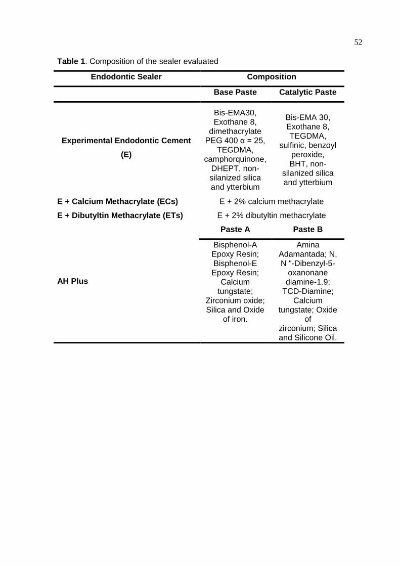
52
Table 1. Composition of the sealer evaluated
Endodontic Sealer Composition
Base Paste Catalytic Paste
Experimental Endodontic Cement
(E)
Bis-EMA30, Exothane 8,
dimethacrylate PEG 400 α = 25,
TEGDMA, camphorquinone,
DHEPT, non-silanized silica and ytterbium
Bis-EMA 30, Exothane 8, TEGDMA,
sulfinic, benzoyl peroxide, BHT, non-
silanized silica and ytterbium
E + Calcium Methacrylate (ECs) E + 2% calcium methacrylate
E + Dibutyltin Methacrylate (ETs) E + 2% dibutyltin methacrylate
AH Plus
Paste A Paste B
Bisphenol-A Epoxy Resin; Bisphenol-E Epoxy Resin;
Calcium tungstate;
Zirconium oxide; Silica and Oxide
of iron.
Amina Adamantada; N, N "-Dibenzyl-5-
oxanonane diamine-1.9;
TCD-Diamine; Calcium
tungstate; Oxide of
zirconium; Silica and Silicone Oil.
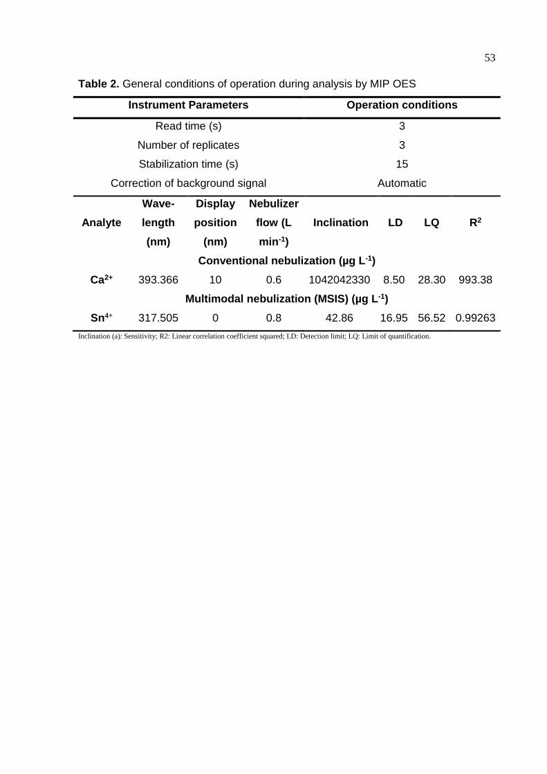
53
Table 2. General conditions of operation during analysis by MIP OES
Instrument Parameters Operation conditions
Read time (s) 3
Number of replicates 3
Stabilization time (s) 15
Correction of background signal Automatic
Analyte
Wave-
length
(nm)
Display
position
(nm)
Nebulizer
flow (L
min-1)
Inclination LD LQ R2
Conventional nebulization (µg L-1)
Ca2+ 393.366 10 0.6 1042042330 8.50 28.30 993.38
Multimodal nebulization (MSIS) (µg L-1)
Sn4+ 317.505 0 0.8 42.86 16.95 56.52 0.99263
Inclination (a): Sensitivity; R2: Linear correlation coefficient squared; LD: Detection limit; LQ: Limit of quantification.
54
Table 3. Dimensional stability of the Different Materials Tested *
Group Median Interquartile range Statistics
Exp 3,199 [2.603 - 4.068] A ECs 3,12 [1.853 - 4.209] A ETs 2,638 [1.904 - 3.56] A AH Plus -14,179 [-21.98 - -10.707] B
*Kruskal-Wallis / Dunn's Post hoc

55
Table 4. Median (25 – 75 percentile) for the total and viable biofilm biovolume (n=5)
Endodontic sealer Total biofilm biovolume, μm3 × 104 Viable biofilm biovolume, μm3 × 104
7 days 15 days 7 days 15 days
ECs 36.65 (5.88 – 274.9)A,ab 14.39 (2.02 – 29.32)A,bc 17.01 (2.10 – 63.56)A,a 0.41 (0.08 – 1.79)B,a
ETs 32.26 (20.62 – 185.65)A,ab 319.45 (0.35 –395.34)A,a 13.72 (9.57 – 45.83)A,a 0.08 (0.00 – 0.44)B,a
EXP 15.25 (4.99 – 58.79)A,a 226.92 (31.73 – 307.27)B,bc 4.91 (1.45 – 18.58)A,a 1.07 (0.01 – 5.13)B,a
AH Plus 4.34 (0.91 – 147.93)A,a 51.74 (20.64 – 130.69)A,ab 0.73 (0.14 – 78.04)A,a 31.01 (12.60 – 53.63)A,b
HA (control) 246.91 (73.29 – 421.83)A,b 462.57 (135.05 – 510.04)A,c 69.95 (11.87 – 96.86)A,a 38.65 (14.01 – 91.48)A,b
* Different upper-case letters indicate difference among the experimental times within the groups. Different lower-case letters indicate difference among the groups within the same experimental time.
56
Table 5. Degree of conversion and Film Thickness stability of the endodontic sealers evaluated after different periods of shelf-life
simulation [mean (SD)].
Degree of Conversion Film Thickness (µm)
Immediate 1 month 2 months Immediate 1 month 2 months
ECs 6.383 (1.266)Ba 2.29 (0.53)Aa 2.89 (2.11)Aa 100 (0.06) Aa 30 (0.008) ABa 6 (0.004) Aa
ETs 27.54 (2.771)Aa 10.26 (8.65)Ab 10.29 (5.03)Ab 5 (0.01) Aa 170 (0.15) Ba 50 (0.09) Aa
EXP 13.40 (6.78)Ba 6.65 (0.81)Aa 10.63 (4.76)Aa 80 (0.09) Aa 20 (0.05) Aa 6 (0.004) Aa
AH Plus - - - 3 (0.004) Aa 6 (0.004) Ba 0 (0.00) Aa
*One Way ANOVA / Tukey's Post hoc. Uppercase letter indicate analysis within columns. Lowercase letter indicate analysis within rows.
57
Table 6. Degree of conversion (DC)* and Film thickness (FT)# of the Different Materials Tested with Calcium Methacrylate or Dibutyltin Methacrylate (Mean – Standard Deviation)
DC (%) FT (µm)
Group Mean Standard deviation Statistics Mean Standard deviation Statistics
Exp 13.408 (6.782) b 0.0833 (0.119) a
EC 6.383 (1.266) b 0.107 (0.0737) a
ET 27.54 (2.771) a 0.0567 (0.0208) a
AHPlus - - - 0.00333 (0.00577) a
*One Way ANOVA / Tukey's Post hoc #One Way ANOVA

58
Fig 1. pH of the eluate of all endodontic sealers over the 30-day storage period. (One Way ANOVA / Tukey's Post hoc. Mean pH at each evaluation period for each material, 3 hours after manipulation. Different uppercase letters indicate significant statistical difference (P < 0.05) between different evaluation periods for the same material. Different lowercase letters indicate statistically significant difference (P < 0.05) between different materials within the same evaluation period.)
3h 24h 7 days 15 days 30 days
ETs 5.34 4.9 4.62 4.55 4.32
ECs 4.94 4.76 4.72 4.68 4.88
Exp 5.58 5.43 4.72 4.66 4.67
AH Plus 5.35 5.92 5.68 5.87 5.98
4
5
6
Ab
pH
Aa
Aa
Aa
Aa
Aa
Aa
Aa
Aa Aa
Ab
Ab Ab
Ab
Ab Ab
Ab Ab Aa
Aa
59
Fig 2. (A) Calcium ion release (µg/L) by conventional method over time for endodontic sealers tested. (B) Cumulative effect of Ca2+ released from endodontic sealers tested. (One Way ANOVA / Tukey's Post hoc. Uppercase letters indicate analysis within same sealer. Lowercase letters indicate analysis within time)
3 h 24 h 7 days 15 days 30 days
ECs 11100 5100 3480 3300 2350
Exp 150 510 1020 500 90
AH Plus 810 1180 1910 1560 500
ETs 0 0 0 0 0
0
2000
4000
6000
8000
10000
12000
0
5000
10000
15000
20000
25000
30000
3h 24h 7 days 15 days 30 days
Cal
ciu
m r
elea
se (
µg/
L)
ECs Exp AH Plus ETs
Ab
Ab Ab Ab
Aa
Aa
Aa
Aa
Aa
Aa
Aa Aa
Ca
lciu
m r
ele
ase
µg
/L
Aa
Aa
A B
Aa
Aa Aa Aa Aa Aa
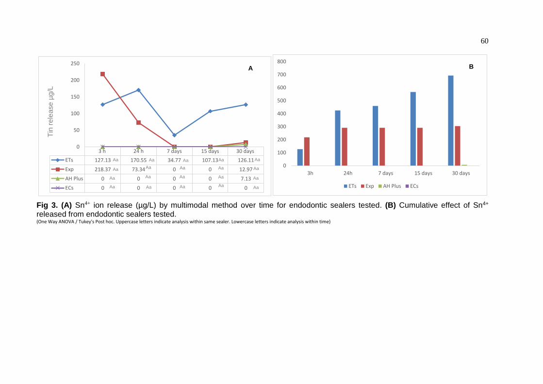
60
Fig 3. (A) Sn4+ ion release (µg/L) by multimodal method over time for endodontic sealers tested. (B) Cumulative effect of Sn4+ released from endodontic sealers tested. (One Way ANOVA / Tukey's Post hoc. Uppercase letters indicate analysis within same sealer. Lowercase letters indicate analysis within time)
3 h 24 h 7 days 15 days 30 days
ETs 127.13 170.55 34.77 107.13 126.11
Exp 218.37 73.34 0 0 12.97
AH Plus 0 0 0 0 7.13
ECs 0 0 0 0 0
0
50
100
150
200
250
0
100
200
300
400
500
600
700
800
3h 24h 7 days 15 days 30 days
ETs Exp AH Plus ECs
Tin
re
lease µ
g/L
Aa Aa
Aa
Aa
Aa
Aa
Aa
Aa Aa
Aa
Aa
Aa
Aa
Aa
Aa
A B
Aa Aa Aa Aa Aa
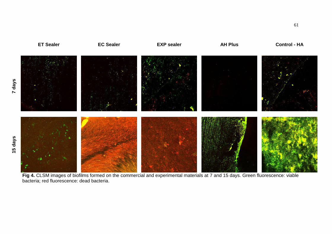
61
ET Sealer EC Sealer EXP sealer AH Plus Control - HA
7 d
ays
15
day
s
Fig 4. CLSM images of biofilms formed on the commercial and experimental materials at 7 and 15 days. Green fluorescence: viable bacteria; red fluorescence: dead bacteria.
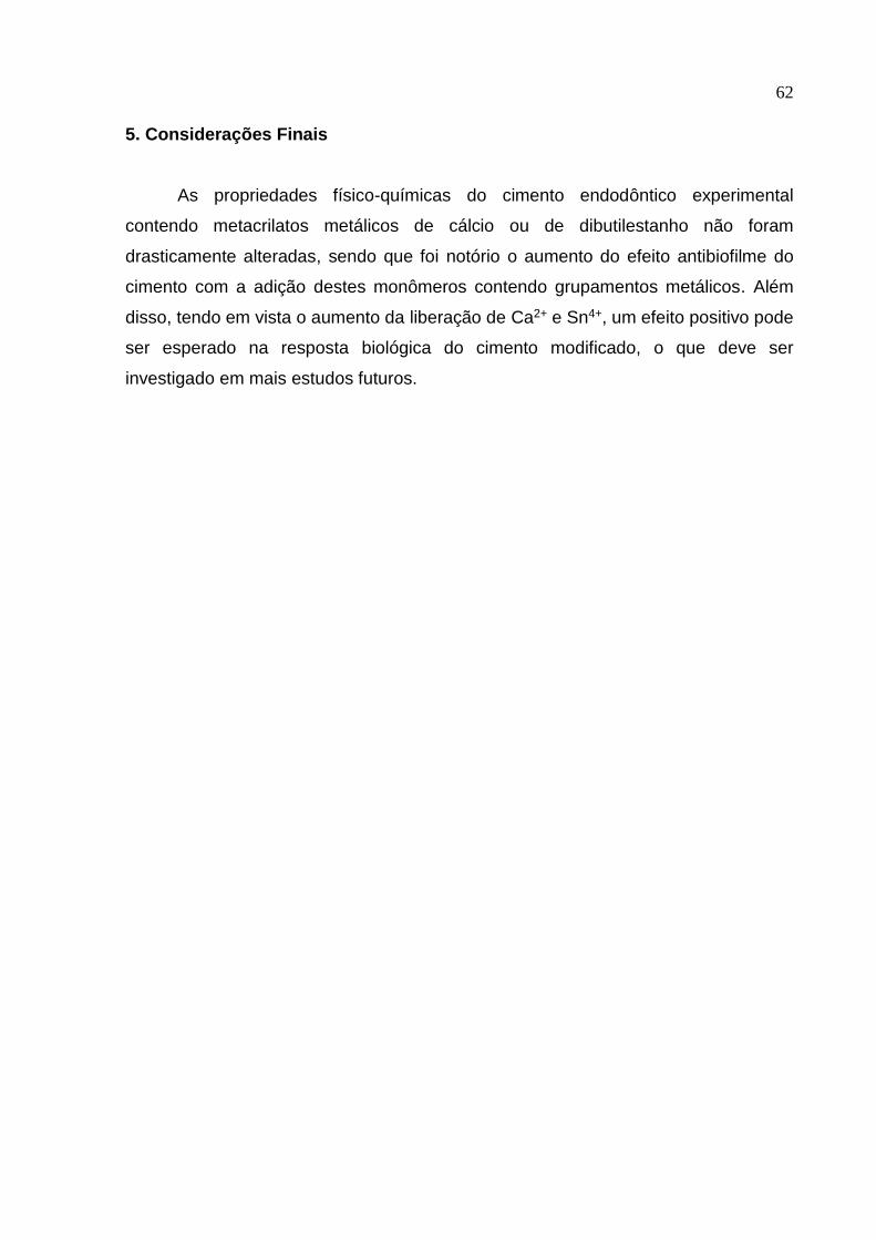
62
5. Considerações Finais
As propriedades físico-químicas do cimento endodôntico experimental
contendo metacrilatos metálicos de cálcio ou de dibutilestanho não foram
drasticamente alteradas, sendo que foi notório o aumento do efeito antibiofilme do
cimento com a adição destes monômeros contendo grupamentos metálicos. Além
disso, tendo em vista o aumento da liberação de Ca2+ e Sn4+, um efeito positivo pode
ser esperado na resposta biológica do cimento modificado, o que deve ser
investigado em mais estudos futuros.

63
6. Referências
ADDY, M. Plaque control as a scientific basis for the prevention of dental caries.
Journal of the Royal Society of Medicine, v.79, n.Suppl 14, p.6–10, 1986.
AHMAD, M.S.; HUSSAIN, M.; HANIF, M.; ALI, S.; QAYYUM, M.; MIRZA, B. Di- and
triorganotin (IV) esters of 3,4-methylenedioxyphenylpropenoic acid: synthesis,
spectroscopic characterization and biological screening for antimicrobial, cytotoxic
and antitumor activities. Chemical Biology & Drug Design. v.71, n.6, p.568–576,
2008.
ALANIA, Y.; CHIARI, M.D.; RODRIGUES, M.C.; ARANA-CHAVEZ, V.E.;
BRESSIANI, A.H.A.; VICHI, F.M.; BRAGA, R.R. Bioactive composites containing
TEGDMA-functionalized calcium phosphate particles: degree of conversion, fracture
strength and ion release evaluation. Dental Materials, v.32, n.12, p.e374-e381,
2016.
ALMEIDA, L.H.; MORAES, R.R.; MORGENTAL, R.D.; CAVA, S.S.; ROSA, W.L.;
RODRIGUES, P.; RIBEIRO, A.S.; SÓ, M.; PAPPEN, F.G. Synthesis of silver-
containing calcium aluminate particles and their effects on a MTA-based endodontic
sealer. Dental Materials. v.34, n.8, p.e214-223, 2018.
ALSHWAIMI, E.; BOGARI, D.; AJAJ, R.; AL-SHAHRANI, S.; ALMAS, K.; MAJEED,
A. In vitro antimicrobial effectiveness of root canal sealers against Enterococcus
faecalis: a systematic review. Journal of Endodontics, v.42, p.1588–1597, 2016.
(ANSI/ADA) ANSIADA. Specification 57: Endodontic Sealing Material. In:
Publishing A, editor. Chicago, IL, USA 2000.
ASSMANN, E.; BÖTTCHER, D.E.; HOPPE, C.B.; GRECCA, F.S.; KOPPER, P.M.
Evaluation of bone tissue response to a sealer containing mineral trioxide aggregate.
Journal of Endodontics, v.41, n.1, p.62-66, 2015.
64
ATTRAMADAL, A.; SVATUN, B. In vivo antibacterial effect of tin on the oral
microflora. Scandinavian Journal of Dental Research, v.92, n.2, p.161-164, 1984.
ATTRAMADAL, A.; NORDBO, H., ERIKSEN, H. M. Iron discoloration of acrylic resin
exposed to chlorhexidine or tannic acid: a model study. Journal of Prosthetic
Dentistry, v.49, n.1, p.126–129. 1983.
BARRY, JOHN E., JEFFREY A. TROGOLO, AND ELIZABETH A. PASTECKi.
"Antimicrobial dental products." U.S. Patent No. 6,267,590. 31 Jul. 2001.
BRAGA, R.R. How far do calcium release measurements properly reflect its multiple
roles in dental tissue mineralization? Clinical Oral Investigation, v.29, n.23, p.501,
2019.
BORGES, R.P.; SOUSA-NETO, M.D.; VERSIANI, M.A.; RACHED-JÚNIOR, F.A.;
DE-DEUS, G.; MIRANDA, C.E.; PÉCORA, J.D. Changes in the surface of four
calcium silicate-containing endodontic materials and an epoxy resin-based sealer
after a solubility test. International Endodontic Journal, v.45, n.5, p.419-428, 2012.
BRAMER, E.A. Flue gas emission from FBC. Coal Science Technology, v. 22, p.
51, 1995.
CAMILLERI J, MONTESIN FE, CURTIS RV, FORD TR. Characterization of Portland
cement for use as a dental restorative material. Dental Materials. v.22, n.6, p.569-
75, 2006.
CARLIN, JR., JAMES F. Minerals Yearbook 2006: Tin. United States Geological
Survey.
CARVALHO-JUNIOR, J.R.; CORRER-SOBRINHO, L.; CORRER, A.B.; SINHORETI,
M.A.C.; CONSANI, S; SOUSA-NETO, M.D. Solubility and Dimensional Change after
Setting of Root Canal Sealers: A Proposal for Smaller Dimensions of Test Samples.
Journal of Endodontics, v.33, n.9, p.1110-1116, 2007.
65
CLARK, G. Shelf Life of Medical Devices, Guidance Document. Division of Small
Manufacturers Assistance, Food and Drug Administration, 1991.
COCCO, A.R.; DA ROSA, W.L.O.; PERALTA, S.L.; MASKE, T.T.; DA SILVA, A.F.;
HARTWIG, C.A.; MESKO, M.F.; PIVA, E.; LUND, R.G. New adhesive system based
in metals cross-linking methacrylate. Journal of Mechanical Behavior of
Biomedical Materials v.77, n.1, p.519–526, 2018.
COLLARES, F.M. ; KLEIN, M. ; SANTOS, P.D, ; PORTELLA, F.F. ; OGLIARI, F. ;
LEITUNE, V.C.B. ; SAMUEL, S.M.W. Influence of radiopaque fillers on
physicochemical properties of a model epoxy resin-based root canal sealer. Journal
of Applied Oral Science, v.21, n.6, p.533-539,2013.
CRETELLA, G.; LAJOLO, C.; CASTAGNOLA, R.; SOMMA, F.; INCHINGOLO, M.;
MARIGO, L. The Effect of Diode Laser on Planktonic Enterococcus faecalis in
Infected Root Canals in an Ex Vivo Model. Photomedicine and Laser Surgery,
v.35, n.4, p.190-194, 2017.
DE PAZ, L.E.C. Image analysis software based on color segmentation for
characterization of viability and physiological activity of biofilms. Applied and
Environmental Microbiology, v.75, n.6, p.1734-1739, 2009.
DUARTE, M.A.; MIDENA, R.Z.; ZEFERINO, M.A.; VIVAN, R.R.; WECKWERTH,
P.H.; DOS SANTOS, F.; GUERREIRO-TANOMARU, J.M.; TANOMARU-FILHO, M.
Evaluation of pH and calcium ion release of calcium hydroxide pastes containing
different substances. Journal of. Endodontics, v.35, n.9, p.1274–1277, 2009.
ELEY, B.M. Antibacterial agents in the control of supragingival plaque–a review.
Brazilian Dental Journal, v.186, n.6, p.286–96, 1999.
ERSAHAN, S.; AYDIN, C. Dislocation resistance of iRoot SP, a calcium silicate-
based sealer, from radicular dentine. Journal of Endodontics, v.36, n.12, p.2000-
2002, 2010.
66
GANDOLFI, M.G.; PRATI, C. MTA and F-doped MTA cements used as sealers with
warm gutta-percha. Long-term study of sealing ability. International Endodontic
Journal, v.43, n.10, p.889-901, 2010
GARRIDO, A.D.; LIA, R.C.; FRANCA, S.C.; DA SILVA, J.F.; ASTOLFI-FILHO, S.;
SOUSA-NETO, M.D. Laboratory evaluation of the physicochemical properties of a
new root canal sealer based on Copaiferamultijuga oil-resin. International
Endodontic Journal, v.43, n.4, p.283–291, 2010.
GOMES-FILHO, J.E.; WATANABE, S.; LODI, C.S.; CINTRA, L.T.; NERY, M.J.;
FILHO, J.A.; DEZAN, E. JR.; BERNABÉ, P.F. Rat tissue reaction to MTA
FILLAPEX®. Dental Traumatology, v. 28, n. 6, p.452-456, 2012.
GROSSMAN, L. I. An improved root canal cement. The Journal American Dental
Association, v.56, n.3, p. 381-5, 1958.
GUERREIRO-TANOMARU, J.M.; DE FARIA-JÚNIOR, N.B.; DUARTE, M.A.H.;
ORDINOLA-ZAPATA, R.; GRAEFF, M.S.Z.; TANOMARU-FILHO, M. Comparative
analysis of Enterococcus faecalis biofilm formation on different substrates. Journal
of Endodontics, v.39, n.3, p.346-350, 2013.
GÜVEN, E.P.; YALVAÇ, M.E.; KAYAHAN, M.B.; SUNAY, H.; ŞAHIN, F.; BAYIRLI, G.
Human tooth germ stem cell response to calcium-silicate based endodontic cements.
Journal of Applied Oral Science, v.21, n.4, p.351-357, 2013.
GUVEN, E.P.; TASLI, P.N.; YALVAC, M.E.; SOFIEV, N.; KAYAHAN, M.B.; SAHIN, F.
In vitro comparison of induction capacity and biomineralization ability of mineral
trioxide aggregate and a bioceramic root canal sealer. International Endodontic
Journal, v.46, n.12, p.1173–1182, 2013.
HIRAISHI, N.; LOUSHINE, R.J.; VANO, M.; CHIEFFI, N.; WELLER, R.N.; FERRARI,
M.; PASHLEY, D.H., TAY, F.R. Is an oxygen inhibited layer required for bonding of
resin-coated gutta-percha to a methacrylate-based root canal sealer? Journal of
Endodontics, v.32, n.5, p.429-433, 2006.
67
HOLLAND, R.; DE SOUZA, V.; NERY, M.J.; BERNABÉ, P.F.E.; OTOBONI FILHO,
J.A.; JUNIOR, E.D.; MURATA, S.S. Calcium salts deposition in rat connective tissue
after the implantation of calcium hydroxide-containing sealers. Journal of
Endodontics, v.28, n.3, p.173–6, 2002.
IKEMURA, K.; ENDO, T. A Review of our Development of Dental Adhesives-Effects
of Radical Polymerization Initiators and Adhesive Monomers on Adhesion. Dental
Materials v. 29, n.2, p.109–21, 2010.
ISO 6876:2012. Dentistry -- Root canal sealing materials. International
Organizations Standarts, 2012.
MAENO, S.; NIKI, Y.; MATSUMOTO, H.; MORIOKA, H.; YATABE, T.; FUNAYAMA,
A.; TOYAMA, Y.; TAGUCHI, T.; TANAKA, J. The effect of calcium ion concentration
on osteoblast viability, proliferation and differentiation in monolayer and 3d culture.
Biomaterials, v.26, n.23, p.4847–4855, 2005.
MAROVIC, D.; TARLE, Z.; HILLER, K.A.; MULLER, R.; ROSENTRITT, M.; SKRTIC,
D.; SCHMALZ, G. Reinforcement of experimental composite materials based on
amorphous calcium phosphate with inert fillers. Dental Materials, v.30, n.9, p.1052-
1060, 2014.
MEEREIS, C.T.W.; LEAL, F.B.; OGLIARI, F.A. Stability of initiation systems in acidic
photopolymerizable dental material. Dental Materials v.32, n.7, p.889–98, 2016.
MISRA, D.N. Interaction of Citric or Hydrochloric Acid with Calcium Fluorapatite:
Precipitation of Calcium Fluoride. Journal of Colloid Interface Science. v.220, n.2,
p.387-391, 1999.
MORGENTAL, R.D.; VIER-PELISSER, F.V.; OLIVEIRA, S.D.; ANTUNES, F.C.;
COGO, D.M.; KOPPER, P.M. Antibacterial activity of two MTA-based root canal
sealers. International Endodontic Journal, v.44, n.12, p.1128-1133, 2011.
68
NAGAS, E.; UYANIK, M.O.; EYMIRLI, A.; CEHRELI, Z.C.; VALLITTU, P.K.;
LASSILA, L.V, DUMAZ, V. Dentin moisture conditions affect the adhesion of root
canal sealers. Journal of Endodontics, v.38, n.2, p.240-244, 2012.
OLIVEIRA, A.C.; TANOMARU, J.M.; FARIA-JUNIOR, N.; TANOMARU-FILHO, M.
Bacterial leakage in root canals filled with conventional and MTA-based sealers.
International Endodontic Journal, v. 44, n. 4, p. 370-375, 2011.
ORDINOLA‐ZAPATA, R.; BRAMANTE, C.M.; CAVENAGO, B.; GRAEFF, M.S.Z.; DE
MORAES, I.G.; MARCIANO, M.; DUARTE, M.A.H. Antimicrobial effect of endodontic
solutions used as final irrigants on a dentine biofilm model. International
Endodontic Journal, v.45, n.2, p.162-168, 2012.
ØRSTAVIK, D. Materials used for root canal obturation: technical, biological and
clinical testing. Endodontic Topics, v.12, n.1, p.25-38, 2005.
PARIROKH, M.; TORABINEJAD, M. Mineral trioxide aggregate: a comprehensive
literature review—part I: chemical, physical, and antibacterial properties. Journal of
Endodontics v.36, n.1, p.16–27, 2010.
PENG, W.; LIU, W.; ZHAI, W.; JIANG, L.; LI, L.; CHANG, J.; ZHU, Y. Effect of
tricalcium silicate on the proliferation and odontogenic differentiation of human dental
pulp cells. Journal of Endodontics, v.37, n.9, p.1240–1246, 2011.
RESENDE, L.M.; RACHED-JUNIOR, F.J.; VERSIANI, M.A.; SOUZA-GABRIEL, A.E.;
MIRANDA, C.E.; SILVA-SOUSA, Y.T.; SOUSA NETO, M.D. A comparative study of
physicochemical properties of AH Plus, Epiphany, and Epiphany SE root canal
sealers. International Endodontic Journal, v.42, n.9, p.785-793, 2009.
ROSSATO, T.C.A.; GALLAS, J.A.; ROSA W.L.O.; DA SILVA, A.F.; PIVA, E;
PERALTA, S.L; LUND, R.G. Experimental Sealers Containing Metal Methacrylates:
Physical and Biological Properties. Journal of Endodontics, v.43, n.10, p.1725-
1729, 2017.
69
SABBAGH, J.; NABBOUT, F.; JABBOUR, E.; LELOUP, G. The effect of expiration
date on mechanical properties of resin composites. Journal of International Society
of Preventive and Community Dentistry, v.8, n. 2, p.99-103, 2018.
SAMUI, A.B.; DALVI, V.G.; CHANDRASEKHAR, L.; PATRI, M.; CHAKRABORTY,
B.C. Interpenetrating polymer networks based on nitrile rubber and metal
methacrylates. Journal of Applied Polymer Science, v.99, n.5, p.2542-2548, 2006.
SHEN, Y.; ZHANG, H.; RUSE, N.D.; HAAPASALO, M. Antibacterial activity of
endodontic sealers by modified direct contact test against Enterococcus faecalis.
Journal of Endodontics, v.35, n.7, p.1051–1055, 2009.
SHEN, Q.; SUN, J.; WU, J.; LIU, C.; CHEN, F. An in vitro investigation of the
mechanical-chemical and biological properties of calcium phosphate/calcium
silicate/bismutite cement for dental pulp capping. Journal of Biomedical Materials
Research Part B: Applied Biomaterials, v.94, n.1, p.141–148, 2010.
SODRÉ, C.S.; ALBUQUERQUE, P.P.A.C.; ISOLAN, C.P.; MORAES, R.R.;
SCHNEIDER, L.F. Relative photon absorption determination and the influence of
photoinitiator system and water content on C=C conversion, water sorption/solubility
of experimental selfetch adhesives. International Journal of Adhesion and
Adhesive, v.63, p.152–7, 2015.
SOUZA, A.O.; PEREIRA, C.C.; HELING, A.I.; ORESTE, E.Q.; CADORE, S.;
RIBEIRO, A.S.; VIEIRA, M.A. Dermination of total concentration and bioaccessible
fraction of metals in infant cereals by MIP OES. Journal of Food Composition
Analytics, v.77, n.1, p 60–65, 2019.
SUNDQVIST, G.; FIGDOR, D. Endodontic treatment of apical periodontitis. In:
Ørstavik D, Pitt Ford TR, eds. Essential Endodontology. Prevention and Treatment of
Apical Periodontitis. Oxford: Blackwell, 1998.
SVATUN, B. Plaque-inhibiting effect of dentifrices containing stannous fluoride. Acta
Odontologica Scandinavica, v.36, n.4, p. 205-210, 1978.
70
SVATUN, B; ATTRAMADAL, A. The effect of stannous fluoride on human plaque
acidogenicity in situ (Stephan curve). Acta Odontologica Scandinavica, v.36, n.4,
p. 211-218, 1978.
TABASSUM, S., PETTINARIA, C. Chemical and biotechnological developments in
organotin cancer chemotherapy. Journal of Organometallic Chemistry. v.691, n.,
p.1761–1766, 2006.
TAŞDEMIR, T.; ER, K.; ÇELIK, D.; TAHAN, E.; SERPER, A.; CEYHANLI, K.T.;
YEŞILYURT, C. Bond strength of calcium silicate-based sealers to dentine dried with
different techniques. Medical Principles and Practice, v.23, n.4, p.373-376, 2014.
TAKITA, T.; HAYASHI, M.; TAKEICHI, O.; OGISO, B.; SUZUKI, N.; OTSUKA, K.;
ITO, K. Effect of mineral trioxide aggregate on proliferation of cultured human dental
pulp cells. International Endodontic Journal, v.39, n.5, p.415-22, 2006.
TAY, F.R.; LOUSHINE, R.J.; MONTICELLI, F.; WELLER, R.N.; BRESCHI, L.;
FERRARI, M.; PASHLEY, D.H. Effectiveness of resin-coated gutta-percha cones and
a dual-cured, hydrophilic methacrylate resin-based sealer in obturating root canals.
Journal of Endodontics, v.31, n.9, p.659-664, 2005.
VAIDYANATHAN, M.; SHEEHY, E.C.; GILBERT, S.C.; BEIGHTON, D. Antimicrobial
properties of dentine bonding agents determined using in vitro and ex vivo methods
Journal of Dentistry, v.37, n.7, p.514-521, 2009.
VERSIANI, M.A.; CARVALHO-JUNIOR, JR.; PADILHA, M.I.; LACEY, S.; PASCON,
E.A.; SOUSA-NETO, M.D. A comparative study of physicochemical properties of AH
Plus and Epiphany root canal sealants. International Endodontic Journal, v.39,
n.4, p.464-471, 2006.
VERTUAN, G.C.; DUARTE, M.A.H.; MORAES, I.G.; PIAZZA, B.; VASCONCELOS,
B.C.; ALCALDE, M.P.; VIVAN, R.C. Evaluation of physicochemical Properties of a
New Root Canal Sealer. Journal of Endodontics. v.44, n.3, p.501-505, 2018.
71
VIAPIANA, R.; FLUMIGNAN, D.L.; GUERREIRO-TANOMARU, J.M.; CAMILLERI, J.;
TANOMARU-FILHO, M. Physicochemical and mechanical properties of zirconium
oxide and niobium oxide modified Portland cement-based experimental endodontic
sealers. International Endodontic Journal, v.47, n.5, p.437-448. 2014.
YOST, K. G.; VANDEMARK, P. J. Growth inhibition of Streptococcus mutans and
Leuconostoc mesenteroides by sodium fluoride and ionic tin. Applied Environment
Microbiology, v.35, n.5, p.920-924, 1978.
XIE, W. ; LIU, K. ; W, P. ; PAN, J. ; RILEY, T. Interaction between emissions of SO2
and HCl in fluidized bed combustors. Fuel v.78, n.12, p.1425-1436, 1998.
XU, H.H.; MOREAU, J.L.; SUN, L.; CHOW, L.C. Nanocomposite containing
amorphous calcium phosphate nanoparticles for caries inhibition. Dental Materials,
v.27, n.8, p. 762-769, 2011.
WILTBANK, K.B.; SCHWARTZ, S.A.; SCHINDLER, W.G. Effect of selected
accelerants on the physical properties of mineral trioxide aggregate and Portland
cement. Journal of Endodontics, v.33, n.10, p.1235-8, 2007.
WU, M.K.; FAN, B.; WESSELINK, P.R. Leakage along apical root fillings in curved
root canals. Part I: Effects of apical transportation on seal of root fillings. Journal of
Endodontics, v.26, n.4, p.210-216, 2000.
ZHANG, W.; LI, Z.; PENG, B. Assessment of a new root canal sealer’s apical sealing
ability. Oral Surgery, Oral Medicine, Oral Pathology, Oral Radiology, and
Endodontology, v.107, n.6, p.79-82, 2009.
ZHANG, H.; SHEN, Y.; RUSE, N.D.; HAAPASALO, M. Antibacterial activity of
endodontic sealers by modified direct contact test against Enterococcus faecalis.
Journal of Endodontics, v.35, n.7, p.1051-1055, 2009.
ZHANG, W.; LI, Z.; PENG, B. Ex vivo cytotoxicity of a new calcium silicate-based
canal filling material. International Endodontic Journal, v.43, n.9, p.769-774, 2010.
72
ZHOU HM, SHEN Y, ZHENG W, LI L, ZHENG YF, HAAPASALO M. Physical
properties of 5 root canal sealers. Journal of Endodontics, v.39, n.10, p.1281–6,
2013.