Programme 2016 Version VIII Sept - AETE
251
32 nd Annual Meeting A.E.T.E. – Barcelona, Spain, 9 th – 10 th September 2016 i 32 ème COLLOQUE SCIENTIFIQUE 32 nd SCIENTIFIC MEETING * * * Dr. Henrik Callesen Special Celebration * * * Barcelona, Spain, 9 th and 10 th September 2016
-
Upload
khangminh22 -
Category
Documents
-
view
0 -
download
0
Transcript of Programme 2016 Version VIII Sept - AETE

32nd Annual Meeting A.E.T.E. – Barcelona, Spain, 9th – 10th September 2016 i
32ème
COLLOQUE SCIENTIFIQUE
32nd
SCIENTIFIC MEETING
*
* *
Dr. Henrik Callesen
Special Celebration
* *
*
Barcelona, Spain, 9th
and 10th
September 2016

32nd Annual Meeting A.E.T.E. – Barcelona, Spain, 9th – 10th September 2016 ii

32nd Annual Meeting A.E.T.E. – Barcelona, Spain, 9th – 10th September 2016 iii
Board of Governors
President Dimitrios Rizos, Spain Vice President Leroy Jo, Belgium
Treasurer Rainer Saner, Switzerland
Secretary Urban Besenfelder, Austria
Annual statistics Marja Mikkola, Finland
Newsletter Roger Sturmey, United Kingdom AETE website Peter Vos, The Netherlands
Representative of Daniel Le Bourhis, France French foundation
and of ET Industry
European legislation Ian Kippax, United Kingdom International Relations Jan Detterer, Germany

32nd Annual Meeting A.E.T.E. – Barcelona, Spain, 9th – 10th September 2016 iv
32nd Annual Meeting A.E.T.E. – Barcelona, Spain, 9th – 10th September 2016 v
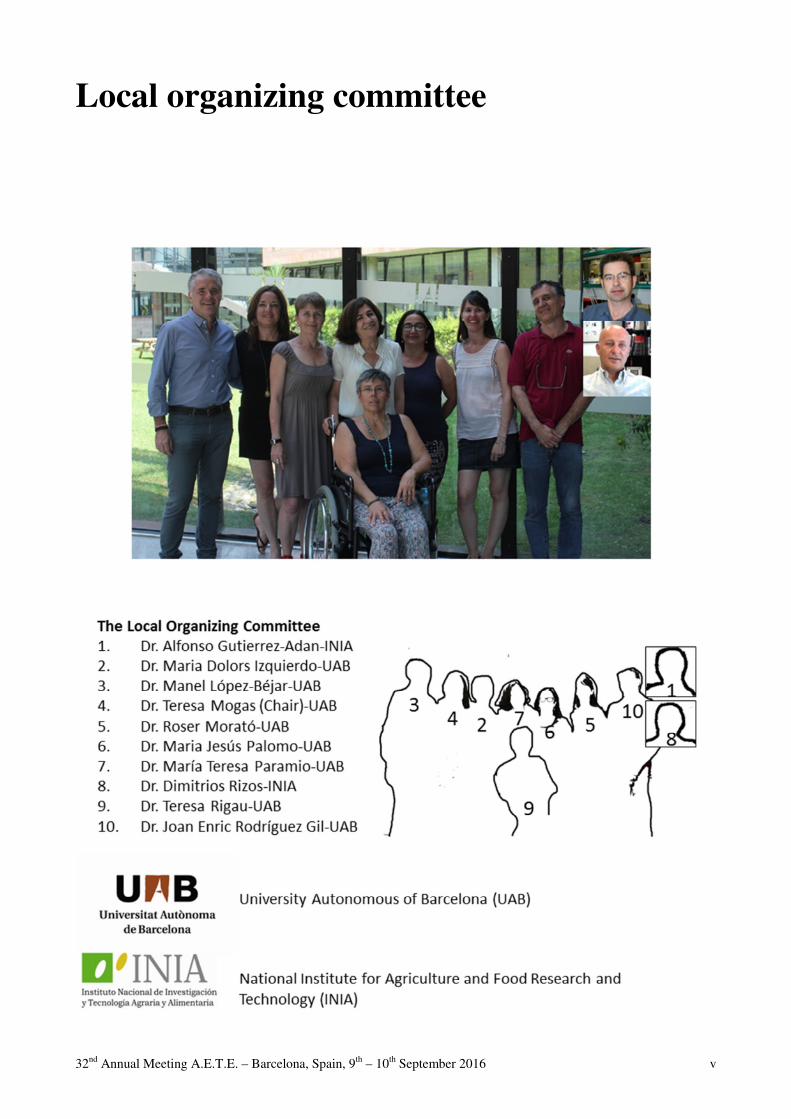
Local organizing committee

32nd Annual Meeting A.E.T.E. – Barcelona, Spain, 9th – 10th September 2016 vi

32nd Annual Meeting A.E.T.E. – Barcelona, Spain, 9th – 10th September 2016 vii

32nd Annual Meeting A.E.T.E. – Barcelona, Spain, 9th – 10th September 2016 viii
32nd Annual Meeting A.E.T.E. – Barcelona, Spain, 9th – 10th September 2016 ix

32nd Annual Meeting A.E.T.E. – Barcelona, Spain, 9th – 10th September 2016 x

32nd Annual Meeting A.E.T.E. – Barcelona, Spain, 9th – 10th September 2016 xi
C O N T E N T S
AETE pioneer award 2015: Dr. Henrik Callesen Greve T ................................................................................................................................................ 3 Commercial bovine embryo transfer activity in EUROPE 2015 Mikkola M.......................................................................................................................................... 19 INVITED LECTURES Circles around the farm animal embryo – a Danish perspective Callesen H ............................................................................................................................................ 7 Update and overview on assisted reproductive technologies (ARTs) in BRAZIL Sartori R, Prata AB, Figueiredo ACS, Sanches BV, Pontes GCS, Viana JHM, Pontes JH, Vasconcelos JLM, Pereira MHC, Dode MAN, Monteiro Jr. PLJ, Baruselli PS ................................ 29 The timing of puberty (oocyte quality and management) Duittoz AH, Tillet Y, Le Bourhis D, Schibler L ................................................................................ 51 Embryo maternal immune interactions in cattle Fair T. ................................................................................................................................................. 85 Practical applications of sperm selection techniques for improving reproductive efficiency Morrell JM ....................................................................................................................................... 101 SHORT COMMUNICATIONS
TAI / FTET / AI Cow and calf factors affect PAG values analysed in routine test milking R. Båge and M. Mörk....................................................................................................................... 116 A new deep-intrauterine artificial insemination device for cattle: XTREMIA.FIRST fertility results and a possible new instrument for embryo transfer. A. Decherf, P. Drevillon, J. Martin, and E, Mariani ........................................................................ 117 Comparative study of seasonal epigenomic effects on sperm cells from Spanish and Swedish bulls M. Sabés-Alsina, A. Johannisson, N. Lundeheim, M. López-Béjar, and J. Morrell ....................... 118

32nd Annual Meeting A.E.T.E. – Barcelona, Spain, 9th – 10th September 2016 xii
OPU - IVF and ET Pregnancy rates from different cattle breed embryos produced in vitro in a commercial program (part 1) C.J. Arreseigor, Y. Filipiak, A.R. Pereira, A.E. Arreseigor, K. Avelino, A.B. Ibarreche, A.C. Maraia, and J. Paredes...................................................................................................................... 120 Embryo mortality from different cattle breed embryos produced in vitro in a commercial program (part 2) C.J. Arreseigor, Y. Filipiak, A. Pereira, A.E. Arreseigor, K. Avelino, A.B. Ibarreche, A.C. Maraia, and J. Paredes ................................................................................................................................... 121 A comparison of Luprostiol and Dinoprost tromethamine for induction of oestrus in donkey embryo transfer M. Bottrel, E. Zarza, L. Fernandez, I. Ortiz, M. Hidalgo, and J. Dorado ........................................ 122 Effects of ghrelin on activation of Akt and Erk1/2 pathways during in vitro maturation of bovine oocyte T.M. Chouzouris, E. Dovolou, F. Krania, I. Pappas, K. Dafopoulos, G. Anifandis, and G.S. Amiridis ........................................................................................................................................... 123 Cryopreservation and intrauterine transfer of canine embryos L. Commin, S. Buff, E. Rosset, A. Charlot-Valdieu, T. Bonte, M. Guedes-teixeira, and T. Joly ... 124 Ovulation induction for embryo transfer in Andalusian donkeys: human Chorionic Gonadotrophin versus Deslorelin acetate J. Dorado, M. Bottrel, L. Fernandez, E. Zarza, I. Ortiz, and M. Hidalgo ........................................ 125 Cytological evaluation of PMN distribution in the genital tract of superovulated embryo donor cows J. Egberts, J. Detterer, A. Park, D. Töpfer, and S. Meinecke-Tillmann .......................................... 126 Route of Oxytocin administration and nonsurgical embryo recovery in Santa Inês ewes after induction synchronous estrus J.F. Fonseca, F.N. Zambrini, J.D. Guimarães, D.S. Chaves, V.S.A. Pereira, M.C.V. Miguel, S.N. Esteves, C.R. Leite, J.M.G. Souza-Fabjan, F.Z. Brandão, and R. Machado ................................... 127 Effect of oocyte transport between two European countries on the bovine blastocyst production G. Gamarra Lazo, D. Martinez Bello, G. Elcoso Usieto, J. Sabin Vilar, and S. Lacaze ................. 128 Effect of X-sorted sperm on development grade 1 in vitro-produced embryos derived from bovine ovum pick up oocytes under commercial conditions G. Gamarra Lazo, S. Lacaze, M. Mouneyres, and B. Marquant Le Guienne .................................. 129 Intrafollicular Oocyte Transfer (IFOT) of immature oocytes improves developmental rates and results in healthy calves M. Hoelker, E. Held, A. Kassens, F. Rings, H. Sieme, D. Tesfaye, and K. Schellander ................ 130

32nd Annual Meeting A.E.T.E. – Barcelona, Spain, 9th – 10th September 2016 xiii
In vino veritas? – How resveratrol attenuates oxidative stress in bovine oocytes of prepubertal and adult donors P. Kordowitzki, S. Klein, K.-G. Hadeler, P Aldag, A. Lucas-Hahn, and H. Niemann.................... 131 The application of bovine in vitro embryo production technology to the rescue of Valdostana Castana breed G. Lazzari, G. Crotti, P. Turini, M. Volget, and C. Galli ................................................................ 132 Melatonin accelerates the timing of in vitro porcine embryo development C.A. Martinez, A. Nohalez, I. Parrilla, J. Roca, C. Cuello, E.A. Martinez, and M.A. Gil .............. 133 Reproductive efficiency at a commercial farm comparing AI versus ET at first insemination D. Martinez, F. Sebastian, J. Sabin, M. Fernandez, R. Patron, I. Lopez-Helguera, J.L. Pesantez, O. Fargas, and S. Astiz.......................................................................................................................... 134 Genomic breeding value for number of OPU derived oocytes in bovine E. Mullaart, M. Cornelissen, H.A. Mulder, H. Flapper, and R. van der Linde ................................ 135 The ability of boar epididimal semen for in vitro fertilization K. Poniedzialek-Kempny, I. Rajska, L. Gajda, and B. Gajda .......................................................... 136
FOLLICULOGENESIS, OOGENESIS, and SUPEROVULATION The estrogen and progesterone receptors in porcine cumulus cells during real-time cell proliferation B. Kempisty, S. Ciesiolka, W. Kranc, A. Bryja, P. Antosik, D. Bukowska, K.P. Brüssow, M. Bruska, M. Nowicki, and M. Zabel.................................................................................................. 138 Inhibins expression in porcine oocytes isolated from follicles of different size B. Kempisty, S. Ciesiolka, W. Kranc, A. Bryja, P. Antosik, D. Bukowska, K.P. Brüssow, M. Bruska, M. Nowicki, and M. Zabel.................................................................................................. 139 Brilliant Cresyl Blue selection of cat cumulus-oocyte complexes does not improve nuclear maturation following IVM I. Lamas-Toranzo, J.M. Sánchez-Calabuig, R. Santamaría, D.A. Martínez-Corona, and P. Bermejo-Álvarez ............................................................................................................................................. 140 The influence of gonadotropin stimulation on breeding behavior in dairy cattle heifers V. Roettgen, P.C. Schoen, and F. Becker ....................................................................................... 141 Nuclear magnetic resonance (NMR) of goat follicular fluid shows different metabolic profiles among follicle size and female age S. Soto, M. Pérez, M.G. Catalá, M. Roura, D. Izquierdo, T. Parella, and M.T. Paramio ................ 142 Formation of cystic ovarian follicles after intrafollicular injection of indomethacin prior to ovulation in heifers A. Vernunft, T. Viergutz, V. Röttgen, and J.M. Weitzel ................................................................. 143

32nd Annual Meeting A.E.T.E. – Barcelona, Spain, 9th – 10th September 2016 xiv
PHYSIOLOGY of REPRODUCTION IN MALE and SEMEN TECHNOLOGY The in vitro impact of the herbicide Roundup® on human sperm motility and sperm mitochondrial functionality G Anifrandis, G.S. Amiridis, L Gavriil, V Gorgogetas, K Dafopoulos, A Daponte, E Dovolou, E Kachpani, Z Mamouris, C.I. Messini, and A.M. Psarra ................................................................... 145 Evaluation of the sperm maturation in swine and mouse by flow cytometry using aniline blue as fluorochrome A. Chacón De Castro, I. Ortega-Molina, E. Gómez-Fidalgo, R. Sánchez-Sánchez, A. Gutiérrez-Adán ................................................................................................................................................. 146 Effect of adding Rosa canina extract and Ascorbic Acid as natural and Synthetic antioxidants on freeze-thawing process of ram semen H. Daghigh Kia, S. Razavian, H. Vaseghi Dodran, and A. Najafi .................................................. 147 Could an extra long-term boar semen extender be successfully used during liquid storage of ram semen at 15°C? B. El Amiri, A. Ben Moula, A. Badi, K. El Khalil, L. Allai, E. Gergatz, E. Gyökér, and O. Szenci .......................................................................................................................................... 148 Effect of adding Cornus mas extract as a natural antioxidant and BHT on freezing/thawing process of ram semen D.K. Hossein, Z.G. Fatemeh, N. Abozar, and H. Vaseghi Dodran ................................................. 149 Morphological and functional characteristics of the epididymal sperm derived from the European bison (Bison bonasus) of the Altaic population B.S. Iolchiev, G.N. Singina, V.A. Bagirov, E.N. Shedova, A.B. Lopukhov, P.M. Klenovitskiy, S.S. Danch, and M.A. Zhilinskiy............................................................................................................. 150 Moxifloxacin effects on ram frozen-thawed sperm function F. Kountouris, A. Gómez, and J. Gadea .......................................................................................... 151 Phosphatidylserine translocation during sperm capacitation is modulated by eNOS in porcine R. López-Úbeda, R. Diego, and C. Matás........................................................................................ 152 Post-thaw changes in sperm membrane and ROS following cryopreservation of dairy bull semen using four different commercial extenders S. Miguel-Jiménez, T. Mogas, A.I. Peña, C. Tamargo, C.O. Hidalgo, R. Muiño, J.E. Rodríguez-Gil, and R. Morató .................................................................................................................................. 153 Analysis of sperm cell viability and chromatin integrity of ram semen held in a cryopreservation media for 24 hours at 5°C. U.I. Osuagwuh, P. Cots, and M.J. Palomo ....................................................................................... 154 Correlation between conventional sperm tests and chromatin integrity analysis M.J. Palomo, P. Cots, and U.I. Osuagwuh ....................................................................................... 155 Use of sexed semen for Holstein Friesian cattle breeding in Kazakhstan A.Y. Serikov, T.I. Kuzmina, V.P. Terletskiy, Y.S. Ussenbekov, A.A. Spanov, A.A. Turgumbekov, and M.S. Gaplaev ............................................................................................................................. 156
32nd Annual Meeting A.E.T.E. – Barcelona, Spain, 9th – 10th September 2016 xv
Periovulatory secretions modulate in vitro boar sperm capacitation decreasing tyrosine phosphorylation C. Soriano-Úbeda, F.A. García-Vázquez, L. Becerril, and C. Matás .............................................. 157 Heat shock proteins detection on heat stressed rabbit sperm cells M. Vendrell, L. Ferre-Dolcet, M. Sabes-Alsina, N. Arcarons, R. Morato, J. Miro, J.-E. Rodriguez-Gil, T. Mogas, and M. Lopez-Bejar ................................................................................................. 158 Follicular and oviductal fluid modulate the protein phosphorylation on serine and threonine residues during boar sperm capacitation H. Zapata, E. Paris-Oller, F. García-Vázquez, and C. Matás .......................................................... 159
EMBRYOLOGY, DEVELOPMENTAL BIOLOGY, and PHYSIOLOGY of REPRODUCTION Cumulus cells protect the bovine oocyte against lipotoxicity by converting saturated into unsaturated fatty acids using stearoyl-CoA-desaturase during in vitro maturation H. Aardema, H.T.A. Van Tol, J.F. Brouwers, B.M. Gadella, and B.A.J. Roelen ........................... 161 A comparative analysis of the protein composition of the oviductal and uterine fluids in cattle during the periovulatory phase by 2D fluorescence difference gel electrophoresis (DiGE). O.S. Acuña, S. Bauersachs, A. Torrecillas, M. Jiménez-Movilla, and M. Avilés ........................... 162 Proteomic characterization of oviductal extracellular vesicles along the estrous cycle in cattle C. Almiñana, A. Alcântara-Neto, G. Tsikis, V. Labas, L. Combes-Soia, and P. Mermillod .......... 163 Alpha-tocopherol affects gene expression patterns of rabbit cumulus-oocyte complexes and reduces apoptosis rate during in vitro maturation M. Arias-Alvarez, R.M. Garcia-Garcia, J. Lopez-Tello, K. Nieto, P.G. Rebollar, A. Gutierrez-Adan, and P.L. Lorenzo .............................................................................................................................. 164 Liquid preservation of bovine embryos as an alternative to cryopreservation N. Blad-Stahl, F. Kotarski, and C. Wrenzycki ................................................................................. 165 In vitro maturation of guinea pig oocytes supplemented with Epidermal Growth Factor and Insulin-Like Growth Factor I K. Cañón-Beltrán, Y.N. Cajas, R. Carrera, P.L. Lorenzo, P.G. Rebollar, R.M. Garcia-Garcia, and M. Arias-Alvarez ............................................................................................................................. 166 In vitro embryo production of lamb oocytes after IVF, ICSI and Parthenogenetic Activation in Autumn and Winter. M.G. Català, M. Roura, I. Contreras, D. Izquierdo, and T. Paramio ............................................... 167 A polarized oviduct epithelial cell culture model supports murine early embryo development without additional medium supply S. Chen, M. Langhammer, and J. Schoen ........................................................................................ 168 Progesterone and Estradiol concentrations of follicular fluid according to the follicular size from Prepuberal and Adult ewes I. Contreras-Solís, M. Catalá, S. Soto, A. Alessandroni, M. Roura, P. Maria-Teresa, and D. Izquierdo .......................................................................................................................................... 169

32nd Annual Meeting A.E.T.E. – Barcelona, Spain, 9th – 10th September 2016 xvi
The effects of hypo- and hyperglycemia during lipolysis-like conditions on bovine oocyte physiology J. De Bie, W.F. Marei, P.E.J. Bols, and J.L.M.R. Leroy ................................................................. 170 Reproductive performance and milk production of Damascus goats raised under the intensive system in southeastern Anatolia H. Demirezer and E. Emsen ............................................................................................................. 171 Effect of non-esterified fatty acids during sperm capacitation or IVF on developmental competence of bovine oocytes K.L.J. Desmet, W.F. Marei, E. Merckx, P.E.J. Bols, and J.L.M.R. Leroy ...................................... 172 Detection of Brucellosis in seropositive superovulated sheep embryo flushing media E. Emsen, M. Kutluca Korkmaz, and H. Demirezer ........................................................................ 173 Bovine embryo production is very sensitive to toxins released from 3-D printed acrylate chambers M.A.M.M Ferraz, H.H.W. Henning, P.L.A.M. Vos, T.A.E. Stout, and B.M. Gadella ................... 174
Response of bovine oviduct epithelial cells to early embryos in vitro J. Hamdi, S. Bagés, A. Gutiérrez-Adán, P. Lonergan, and D. Rizos ............................................... 175 Porcine sperm bind to beads conjugated to ZP2 protein under in vitro conditions J.G. Hamze, A. Canha, L. Zamorano, B. Algarra, M.C. Olivares, R. Romar, and M. Jimenez-Movilla ............................................................................................................................................. 176 The presence of L-carnitine during maturation improves bovine embryo production P. Hulinska, K. Hanzalova, D. Knitlova, M. Jeseta, and M. Machatkova ....................................... 177 In vitro monolayer barrier function of bovine oviduct epithelial cells is modified due to high concentrations of non-esterified fatty acids L. Jordaens, V. Van Hoeck, B. Vlaeminck, V. Fievez, S. Thys, I. Pintelon, P.E.J. Bols, and J.L.M.R. Leroy ................................................................................................................................. 178 The P4 and E2 treatment and protein expression of PGR and PGRMC1 in porcine endometrial cells B. Kempisty, S. Ciesiolka, W. Kranc, A. Bryja, P. Antosik, D. Bukowska, K.P. Brüssow, M. Bruska, M. Nowicki, and M. Zabel.................................................................................................. 179 Developmental competence of bovine oocytes that have not finished growth phase in vivo T.I. Kuzmina, A.V. Molchanov, T.I. Stanislavovich, and D.N. Tatarskaya .................................... 180 Progesterone is involved in anti-aging effects of prolactin on bovine cumulus-enclosed oocytes matured in vitro I.Y. Lebedeva, G.N. Singina, E.N. Shedova, T.E. Taradajnic, and N.A. Zinovieva ....................... 181 Developing a responsive mouse in vitro fertilization model with focus on sperm concentration Y. Liu, H.S. Pedersen, L. Foldager, H. Callesen, and M.T. Sørensen ............................................. 182 Effects of recombinant porcine OVGP1 protein on bovine embryo gene expression V. Maillo, B. Algarra, M. Avilés, A. Gutiérrez-Adán, D. Rizos, and M. Jiménez-Movilla ............ 183
32nd Annual Meeting A.E.T.E. – Barcelona, Spain, 9th – 10th September 2016 xvii
Dynamic changes of telomere length during bovine preimplantation development D.A. Martínez-Corona, I. Lamas-Toranzo, A. Gutiérrez-Adán, and P. Bermejo-Álvarez .............. 184 USP9Y is necessary for male development in early bovine embryo T. Mogas, N. Rho, K. Mahboubi, J. Caudle, E.J. St.John, L. Favetta, and W.A. King ................... 185 The effect of L-carnitine supplementation during IVM and/or IVC on sex ratio of transferable bovine embryos depends on the combination of the IVM/IVC media M. Mutikainen, T.-M. Hamama, N. Ghanem, M. Weldenegodguad, and J. Peippo ....................... 186 Royal jelly improves embryonic developmental competence and affects transcript levels of apoptosis-related genes in goat cumulus-oocyte complexes K. Nikdel, N. Ghanem, M. Khajenabi, R. Kamaledini, and A. Mohammadi-Sangcheshmeh ......... 187
A proteomic approach to monitor interactions between oviductal fluid and spermatozoa across the estrous cycle P. Nogues, J. Lamy, G. Tsikis, V. Labas, L. Combes, P. Mermillod, X. Druart, and M. Saint-Dizier................................................................................................................................. 188 Cotyledon Efficiency: A Novel Parameter to Assess Placental Efficacy in Small Ruminants S. Ocak and I.M. Mayorga Munoz................................................................................................... 189 Cell ultrastructure in bovine preimplantation embryos in relation to cow´s body condition score L. Olexikova, J. Pivko, A. Makarevich, E. Kubovicova, and P. Chrenek ....................................... 190 FFAR4 is involved in docosahexaenoic acid effects on oocyte developmental potential during in vitro maturation. M. Oseikria, S. Uzbekova, A. Vitorino Carvalho, V. Duranthon, and S. Elis ................................. 191 Reactive oxygen species level in pig embryos cultured in hypoxic conditions M. Romek, B. Gajda, I. Rajska, and Z. Smorag .............................................................................. 192 Prolactin supports the developmental competence and apoptosis resistance of aging bovine oocytes through the same signaling pathway G.N. Singina, I.Y. Lebedeva, T.E. Taradajnic, A.V. Lopukhov, and N.A. Zinovieva .................... 193 Dimethyl sulfoxide supplementation affects bovine in vitro embryo development J. Stöhr, H. Grothmann, and C. Wrenzycki ..................................................................................... 194 Effect of maternal genotype on embryo and foetal survival using rabbit as a model J. Valdes-Hernández, J.I. Cedano-Castro, X. García-Dominguez, J.S. Vicente, and F. Marco-Jiménez............................................................................................................................................. 195 Maternal impact of metabolic diseases: effect of nutrient-sensing pathways on developmental and differentiation programs in the bovine embryo. V. Van Hoeck, W. Marei, P.E.J. Bols, and J.L.M.R. Leroy ............................................................ 196 miR-21 expression in ovine oocytes: Implications for developmental competence A. Veshkini, M. Khajenabi, F. Kouhkan, R. Tavakoli, A.A. Khadem, M. Soleimani, and A. Mohammadi-Sangcheshmeh ............................................................................................................ 197

32nd Annual Meeting A.E.T.E. – Barcelona, Spain, 9th – 10th September 2016 xviii
Effects of BOEC and VERO co-culture systems on bovine blastocyst transcriptome A. Vitorino Carvalho, L. Jouneau, C. Archilla, E. Canon, L. Laffont, S. Ruffini, E. Corbin, P. Mermillod, and V. Duranthon .......................................................................................................... 198 A preliminary study focused on the comparison of meiotic maturation effectiveness between canine and porcine oocytes undergoing two-step in vitro culture under analogous biochemical and biophysical conditions E. Wojtylak-Jurkiewicz, M. Samiec, M. Skrzyszowska, and G. Ptak ............................................. 199
CLONING, TRANSGENESIS, and STEM CELLS Optimization of RNA concentration for genome editing by CRISPR in rabbit zygotes N. Fonseca Balvís, P.L. Lorenzo, A. Gutiérrez-Adán, P.G. Rebollar, M. Avilés, and P. Bermejo-Álvarez ............................................................................................................................................. 201
Superovulation rates and embryo recovery in 12 to 15-month-old genetically modified pigs for biomedical research B. Gajda, J. Jura, R. Slomski, and Z. Smorag .................................................................................. 202
Effect of the protein kinase inhibitor 6-Dimethylaminopurine on the parthenogenetic activation of mouse oocytes R. González-Martín, J. Santaló, M.T. Paramio, and E. Ibáñez ........................................................ 203
Trichostatin A-mediated epigenetic transformation of bone marrow-derived mesenchymal stem cells or blood-derived fibroblast-like cells brings about an abundance of nuclear-transferred pig embryos at the morula and blastocyst stages M. Samiec, J. Opiela, and M. Skrzyszowska ................................................................................... 204 Extracorporeal development of porcine somatic cell nuclear transfer (SCNT)-derived embryos is biased by scriptaid-induced epigenetic modulation of adult cutaneous fibroblast cells M. Samiec, M. Skrzyszowska, E. Mielczarek, and E. Wojtylak-Jurkiewicz ................................... 205 Adult peripheral blood-derived fibroblast-like cells provide a source of nuclear donor cells that is much less susceptible to promote the in vitro development of porcine cloned embryos than adult bone marrow-derived mesenchymal stem cells M. Samiec, M. Skrzyszowska, and J. Opiela ................................................................................... 206 Epigenomically transformed peripheral blood-derived fibroblast-like cells can be successfully utilised as a novel type of nuclear donor cells for generation of cloned pig embryos M. Samiec, M. Skrzyszowska, E. Wojtylak-Jurkiewicz, and E. Mielczarek ................................... 207 Generation of monogenetic twin embryos and progeny by modified bisection of zona-perforated pig blastocysts M. Skrzyszowska, M. Samiec, B. Gajda, Z. Smorag, and W. Królikowski .................................... 208
Culture optimization to obtain mouse embryonic stem cell lines from single blastomeres M. Vila-Cejudo, E. Ibáñez, and J. Santaló ....................................................................................... 209
32nd Annual Meeting A.E.T.E. – Barcelona, Spain, 9th – 10th September 2016 xix
Efficient derivation of embryonic stem cells from mouse B6CBAF1 blastocysts M. Vila-Cejudo, O. Massafret, E. Ibáñez, and J. Santaló ................................................................ 210
SUPPORT BIOTECHNOLOGIES: CRYOPRESERVATION and CRYOBIOLOGY, DIAGNOSIS THROUGH IMAGING, MOLECULAR BIOLOGY, and "OMICS" Cholesterol addition to immature and in vitro matured bovine oocytes before vitrification altered the expression of some genes important for early embryonic development N. Arcarons, R. Morató, T. Mogas, K. Rajapaksha, and M. Anzar ................................................. 212
MALDI-TOF mass spectrometry analysis of lipids in single bovine oocytes during IVM P. Bertevello, O. Ghazouani, C. Banliat, S. Elis, A.-P. Teixeira-Gomes, V. Maillard, V. Labas, and S. Uzbekova ..................................................................................................................................... 213 Sustainable regulation of metabolic performance of bovine embryos by L-Carnitine supplement and concurrent reduction of fatty acids E. Held, S.L. Klein, F. Rings, D. Tesfaye, K. Schellander, and M. Hoelker ................................... 214
Impact of enrichment of unsaturated fatty acids during in vitro maturation and culture of bovine embryos on blastocyst rates A.L. Hendrikx, B.M. Gadella, H.T. van Tol, and P.L. Vos ............................................................. 215 Viability of porcine vitrified morulae and blastocysts stored in a dry-shipper for 3 days A. Nohalez, C.A. Martinez, I. Parrilla, J. Roca, M.A. Gil, E.A. Martinez, and C. Cuello .............. 216 Comparison of the effects of slow freezing and vitrification on in vitro embryo quality in horse and donkey C.C. Perez-Marín, G. Vizuete Calero, J.J. Galisteo Martínez, E. Agüera Buendía, and R. Vazquez Martinez ........................................................................................................................................... 217 The influence of DNA stabilizing buffer on the results of genomic bovine embryo analysis M. Reichenbach, S. Jung, E. Wolf, E. Pimentel, R. Emmerling, K.-U. Götz, T. Grupp, J. Scherzer, R. Fries, and H.-D. Reichenbach ..................................................................................................... 218
JC-1 dye is a valuable indicator of embryo health in rabbits M. Teixeira, L. Commin, L. Gavin-Plagne, P. Bruyere, A. Philibert, T. Joly, and S. Buff ............. 219 Rabbit embryo vitrification without animal products M. Teixeira, L. Commin, L. Gavin-Plagne, P. Bruyere, A. Philibert, S. Buff, and T. Joly ............. 220 Ultrasound Monitoring of reproductive organs in Angora bucks. K. Tekin, C. Stelletta, D. Sahin, I. Bastan, O.O. Parildar, and A. Daskin ....................................... 221
AUTHORS INDEX ........................................................................................................................ 223


32nd Annual Meeting A.E.T.E. – Barcelona, Spain, 9th – 10th September 2016 1
Dr. Henrik Callesen
A.E.T.E. Medalist 2016

32nd Annual Meeting A.E.T.E. – Barcelona, Spain, 9th – 10th September 2016 2
32nd Annual Meeting A.E.T.E. – Barcelona, Spain, 9th – 10th September 2016 3
Dr. Henrik Callesen
A.E.T.E. Medalist 2016
Professor Henrik Callesen: Recipient of the AETE Pioneer Award 2016
Bibliography Henrik was born on July 31st 1956 in Christiansfeld, a village in Southern Denmark which was part
of Germany until 1920. His father was a dairy engineer and his mother a journalist and together
with his parents he spent part of childhood in Africa and Middle east and one may wonder whether
his later international engagement was founded by these early exposures to a globalized world?
Following High School, Henrik was enrolled as a veterinary student at the Royal Veterinary and
Agricultural University (KVL) and during his study he also worked as an animal caretaker in the
stables of Department of Reproduction where he assisted in taken blood samples from
superovulated animals and also participated in various surgical procedures. Maybe his interest for
animal reproduction and superovulation was founded during that period where he in fact also met
his later wife Kathe! Henrik married Kathe in March 1985 and in 1989 they moved to the town of
Bjerringbro where they still live and where Kathe runs a small animal clinic. Their three grown-up
children have all moved away from home and now live in Copenhagen.
Science and mentorship Having graduated as a veterinarian in 1982 with very high scores, Henrik and I had serious
discussions on whether he should start as a Ph.D.-student or maybe have more practical experience.
The final decision was that he became enrolled in KVLs graduate school and thus became my first
Ph.D.-student. That was a very good decision which I believe none of us have regretted. Henrik was
a truly dedicated student and worked extremely hard on issues that had not earlier been explored.
For many years it had been an enigma, why so many eggs from superovulated animals were
unfertilized and of poor quality. His overall working hypothesis was that poor fertilization rates and
inferior embryo quality often seen in conjunction with superovulation was caused by poor oocyte
quality brought about by both disturbed follicular endocrinology and peripheral endocrine profiles
(LH, P4 and E2). He examined the hypothesis by taking 4th hourly samples from donor animals,
subsequently ovariectomizing them and submitting the oocytes to light microscopic analysis and the
follicular fluid to endocrine analysis. The ovaries were removed by transvaginal ovariectomy using
an ecraseur designed by the Danish Veterinarian Richardt Møller and usually the procedure was
successful! However, one night a cow belonging to the Red Danish breed did not survive the
32nd Annual Meeting A.E.T.E. – Barcelona, Spain, 9th – 10th September 2016 4
procedure but died of blood loss and Henrik was truly sorry! The studies confirmed his hypothesis
and they were published in highly estimated international journals and thus frequently cited. Henrik
was - as mentioned - my first Ph.D.-student, and what a joy to work with him: friendly, timely,
consistent, open-minded to new ideas and loyal to the team. He never let you down and it was his
conviction that altruism is better than egoism. It was also during his period as a Ph.D.-student that
we became close friends and this unique friendship still exists. During his challenging work with
the oocyte done in collaboration with professor Poul Hyttel, he got exposed to the international
scene through participation in meetings in Europe as well as overseas where he met scientific
collaborators of which several later became his friends for life, again reflecting Henrik’s loyal and
trustworthy nature. I have the impression that he became merely addicted to globalization, although
addiction is not a positive word!
Henrik got a permanent position at the Danish Institute of Agricultural Sciences in Foulum where
he built a very strong frontline research team focused on MOET breeding plans (in Danish called
the FY-BI project), in-vitro embryo production, ultrasound guided transvaginal oocyte aspiration
(OPU), vitrification, time lapse imaging of embryo development, embryo metabolism and finally
cloning of pigs to produce models for human diseases. The projects were carried in collaboration
not only with Danish colleagues at the University of Copenhagen (previously KVL) and Aarhus
University but at least as much with scientists from Europe, China, Australasia and South America.
His laboratory became then and is still leading in embryo research in Denmark and has trained
many post-docs and students. He maintains an impressive network and has inspired and guided
many young scientists in this field of research both in Denmark and abroad. His numerous scientific
papers and proceeding abstracts witness an impressive scientific standing.
Ethics and Science The cloned sheep Dolly came as a shock for politicians and the general public in Denmark as well
as the rest of the world. Prior to Dolly, we have had an intense ethical debate on the implementation
of advanced reproductive technologies such as non-surgical embryo transfer, freezing and in vitro
production of embryos, and these were gradually accepted. Somatic cell cloning was in the public
perception too much and it generated a heated debate. Should it be allowed to clone animals? Could
it be accepted to eat meat or drink milk from cloned cows? All along, Henrik participated in this
debate at public meetings, in discussions with decision makers, gave presentations for school
classes and made his utmost to disseminate proper information on cloning and transgenesis. He did
so based on fact and not fiction which some of his opponents alluded to. His efforts were successful
and resulted in a legislation which allowed cloning to be used for specific purposes (medical), and
his laboratory is leading in this field.
International profile Science and Danish organisation of science (research committees etc.) has been a driving force in
Henrik’s carrier and his engagement in international boards and committees is also impressive!
Examples are the International Embryo Transfer Society (IETS) where he was board member,
treasurer and president, and in the context of being awarded the European Embryo Transfer
Association (AETE) Pioneer Award 2016, it should be stressed that he has been member of its

32nd Annual Meeting A.E.T.E. – Barcelona, Spain, 9th – 10th September 2016 5
Board for about 8 years and has been careful to attend the meetings for about 20 years. He has also
as a driving force to get the Danish Embryo Transfer practitioners to international meetings. I had
the privilege to accompany Henrik to many of these and other international meetings. Initially he
was a little shy but- as mentioned above - he ultimately really liked the international spirit. The last
AETE meeting we attended together was in Algerho, Sardinia, and he felt comfortable and liked the
social and scientific events. AETE was for him like coming home.
Henrik as a person As it is obvious from the above description and certainly from Henrik’s impressing CV, he is a true
and internationally well-respected scientist and mentor! Although his standing would allow it, he
never brags about his achievements. One might even argue that he sometimes is too modest but that
is one of his sympathetic characters. I have known Henrik for many years, as a student, as a
colleague, and he is trustworthy, reliable, listening, kind, empathic, well organized (sometimes too
much!) and never gossip nor speak evil of anybody. He rarely gets exaggerated or even angry, but
his body language and his look clearly signals when he disagrees. One may confide in him and be
certain that things remain confidential. And in discussions he plays the ball and not the man
although one could clearly sense when he wanted to play the person! That is commendable and is
worth for other discussants to follow. Any negative sides: hard to find but he may sometimes be less
flexible.
Conclusion One may argue that I - as one of his close colleagues and friends - am biased when writing this
promotion. I entirely agree but nevertheless: based on his scientific merits and in particular his
efforts to develop the field of practical embryo transfer and thus his devotion to working aspects
promoted by the AETE, Henrik Callesen is a worthy recipient of the AETE Pioneer Award 2016.
Copenhagen, March 2016
Torben Greve
DVM, DVSc, Dhc
Former professor of animal reproduction
University of Copenhagen

32nd Annual Meeting A.E.T.E. – Barcelona, Spain, 9th – 10th September 2016 6
32nd Annual Meeting A.E.T.E. – Barcelona, Spain, 9th – 10th September 2016 7
Circles around the farm animal embryo – a Danish perspective
Henrik Callesen
Department of Animal Science, Aarhus University, DK-8830 Tjele, Denmark
Abstract
With focus on the farm animal embryo, a short overview is given about my research
activities over the last 35 years. These activities have been described in five circles,
covering different key aspects of my work. The first circle included studies on the
basic biology related to oocyte maturation and follicular endocrinology in
superovulated dairy cows. Methods were developed to characterize the donor cows
with respect to their production of transferable embryos, and some were implemented
into a Danish MOET breeding plan. The second circle dealt with in-vitro embryo
production in cattle with development of a protocol to produce such embryos at high
and consistent levels. Several comparisons were made to reveal consequences of the
artificial in-vitro methods on oocytes and embryos, but also through studies of the
newborn calves. The third circle was related to development and implementation of a
number of technologies within this broad field; examples are mentioned for both
oocyte recovery from donor cows, different steps in the in-vitro embryo production
system, new ways to perform vitrification and nuclear transfer, and finally a new
system to determine oxygen consumption in single embryos. In the fourth circle is
described activities from the last years, where work was done with focus on the
pregnancy rates after insemination of the dairy cow in their post-partum period, and
where somatic cell nuclear transfer was developed both as a technology in itself as
well as a helping technique to produce transgenic pigs as models for important human
diseases. The fifth and final circle is addressing and thanking the many colleagues and
collaboration partners that I have been involved with during all the years to do this
work. Nothing could or would have been the same without them and their
participation.
Keywords:
Reproductive biology, embryo technology, cattle, pig, overview.
My first AETE meeting was in 1987 in Lyon where the 3rd meeting was held. Since I have been at
all but three of these annual highlights, and at every meeting I have been listening with interest to
the distinguished scientists receiving the AETE award. Today, it is my privilege and honor to be
that person, and since I was told this great news a year ago, I have gradually learned to use this
opportunity and reflect over my scientific career in ways that I usually do not.
Having worked with farm animal embryos for more than 30 years gives many possibilities
to do different things. My choice – to some extent also influenced by coincidences – has been to

32nd Annual Meeting A.E.T.E. – Barcelona, Spain, 9th – 10th September 2016 8
focus on the farm animal embryo in kind of circles around the days before, during and after
fertilization, and I have always had Denmark as the center for my activities. Within those frames,
most of my research interests have been divided between biology and technologies, from a basic to
an applied aspect, and then using collaborations very much.
So, it has been fairly limited circles, made in a rather small country, and always with the embryo
in focus. In the following, I will give a short overview of my scientific activities using some kind of
a chronological approach. It will not be a real literature review, but a self-centered presentation.
This is not often allowed, but on this special occasion I think it is. To prepare this has been
interesting for me, but of course I hope that it will also be that for at least some of you.
The superovulated donor cow From the beginning of the 1980ies, I started as Torben Greve’s first PhD-student at the
Royal Veterinary and Agricultural University in Copenhagen. He had been very much involved in
establishing the basics on superovulation and embryo transfer in cattle, also in practice, but time
had come to search for a better understanding of some of the reasons for the often varying and
rather unpredictable results. Together with another PhD-student, Poul Hyttel, my first years in
research were therefore focused on morphological and endocrine aspects of the preovulatory period
in superovulated dairy cattle. What were the actions and consequences of the exogenous FSH
treatment on the developing follicles and oocytes? Poul’s focus was ultrastructure, while mine was
the hormones and the more clinical and applied aspects.
Over several years we used more than 130 cows and heifers to study preovulatory oocyte
morphology and follicular endocrinology and to describe their overall reaction patterns, and to
relate these to the resulting oocyte and later embryo quality. Some of the main results were
presented at my first IETS conference held in Colorado Springs in 1986 (Callesen et al. 1986),
where characteristics of donors with good versus bad oocytes were presented. This was followed by
several also more practice-related studies (e.g. Callesen et al. 1995) also to identify the good or bad
donors. For this, donors were characterized through their patterns of progesterone or estradiol
concentrations in plasma and milk (Callesen et al. 1988, 1990) together with detailed estrus
observations (Callesen et al. 1993a), or by use of ultrasound examinations of follicular development
and ovulation (Purwantara et al. 1993, 1994). Also in these years, problems associated with the FSH
preparations used for superovulation (e.g. causing premature ovulations, Callesen et al. 1987a) were
addressed by attempts to control the superovulatory reaction by use of anti-PMSG (Callesen et al.
1992) or by controlling the LH-contents (Schmidt et al. 1988).
On the practical side, larger projects based on MOET breeding plans were started in Europe,
also in Denmark, where I got involved in such a project after having left Copenhagen and moved to
Foulum in the western part of Denmark. This resulted in work on e.g. donor selection (Greve &
Callesen 1989), but we also made several studies on the embryo recipients; some of this work was
presented at several AETE-meetings (Callesen et al. 1993b,c; 1994b,c). The contact to practice also
gave some challenges when the more theoretical and experimental work met the real practical world
(Liboriussen et al. 1995, Callesen et al. 1996), for example with the breeding potentials following
the realized outcome of superovulation (Callesen et al. 1994a).
32nd Annual Meeting A.E.T.E. – Barcelona, Spain, 9th – 10th September 2016 9
In-vitro production of bovine embryos During the 1980ies, the procedure for in-vitro production of bovine embryos was being
established based on work around the world with the first IVF calf reported from USA in 1983. In
Copenhagen, Kang Pu Xu - another of Torben Greve’s PhD-students - worked hard on this
technology (e.g. Xu et al. 1987), and a prominent result was achieved when the first IVP calf in
Europe was born here in 1987 (Greve et al. 1989).
In my Foulum-group we worked several years to optimize the bovine IVP-procedure. Peter
Holm went through the tedious work of testing close-to-everything in relation to media and
conditions for in-vitro production (e.g. Holm et al. 1994, 1995), before this resulted in a quite useful
modified SOF medium (Holm et al. 1999). Parallel to this work, we were still interested in
morphological differences between vivo versus vitro oocytes (e.g. Hyttel et al. 1989a), also
illustrating some of the implications for vivo and vitro fertilization (e.g. Hyttel et al. 1989b, 1991).
At the AETE meeting in Venice, some of this work was summarized (Holm & Callesen 1998).
With the gradual improvement of the IVP systems, we got a stronger interest in some of the
consequences of such artificial in-vitro conditions, stimulated by the disturbing reports about the so-
called Large or Abnormal Offspring Syndrome. Thus, we worked on differences between vivo
versus vitro embryos, both in cattle and pig (e.g. Hyttel et al. 2000), on chromosomal problems after
in-vitro production (Viuff et al. 1999), and finally also on the resulting calves (Jacobsen et al.
2000). As part of these studies, I spent some months in New Zealand and Australia in 2001 to study
pregnancies and calves after both IVP and cloning. In Europe, these concerns were also subject for
many discussions in the public with focus on animal ethics (e.g. Callesen et al. 1999), and also at
the AETE meetings, where questionnaires were the basis for workshop discussions in e.g. Santander
(den Daas & Callesen 2000).
The tools for working with reproductive technologies To work in such a biological area, technologies are required, and over the years I have been
involved in several such technical developments in quite different fields.
Ovum Pick-Up. Oocyte collection through the abdominal wall was developed in the human
field in the first part of the 1980ies, and together with a medical doctor we made some of the first
attempts in cattle. It was a para-lumbar approach (presented at the 1987 IETS meeting, Callesen et
al. 1987b), and several lessons were made – one was that it can result in recovery of oocytes, but
also that a cow patient will kick you when you prick her with a needle; a big surprise for the
medical doctor! Soon after, especially the group from Utrecht led the way into the much more
convenient vaginal approach that also became routinely used in human.
In-vitro embryo culture. We continued to work on in-vitro culture, driven by Gabor Vajta’s
urge for simplicity and reliability. One area was an incubator system based on having the culture
dish in a foil bag that was submerged into a water bath (Vajta et al. 1997a). This provided very
stable temperature conditions, required minimal use of gases, and each culture dish had its own
chamber. However, only few would - and will - accept a water bath in their culture lab. Another
area was related to the anticipated need for an embryo to establish a local in-vitro environment
during its development. This was obtained by hand-making small holes or impressions in the
bottom of standard plastic dishes (Vajta et al. 2000). The method was subject for a course also

32nd Annual Meeting A.E.T.E. – Barcelona, Spain, 9th – 10th September 2016 10
given at the AETE meeting in Lyon in 2004. A third interest was to see how far the embryo actually
could be cultured in-vitro (Brandao et al. 2004, Vajta et al. 2004). We learned that the trophoblastic
cells grew quite well to form up to an almost 2 cm long structure … but the very early embryo
proper did not.
Time-lapse systems. To study embryo morphology frequently during in-vitro culture, Peter
Holm built his own time-lapse system in the mid-1990ies (Holm et al. 1998). This was a fairly
simple and cheap system, but it provided what was needed from the oocyte’s and embryo’s point of
view, allowing complete, high and stable in-vitro development for up to 9 days together with taking
pictures every 20-30 minutes. The resulting films were rather boring during IVM, but there was
much more to see when monitoring the pre-implantation period in both cattle (e.g. Holm et al.
2002) and pig (e.g. Callesen & Holm 2016), also with cloned and parthenogenetic embryos (Holm
et al. 2003). Through this work I established an interesting collaboration with the Danish company
Unisense A/S that later developed the EmbryoScope®, an instrument that today is used in several
human fertility clinics around the world.
Vitrification. A challenge in cryopreservation was the more fragile types of eggs, such as
oocytes in general and embryos from certain species such as the pig; none of these structures really
tolerated traditional slow freezing. Parallel to a visit from Masashige Kuwayama (Kuwayama et al.
1997), the vitrification technology was taken further by Gabor Vajta, resulting in a thin-straw
system (Vajta et al. 1998). Now also early-stage embryos from both cattle and pig became possible
to cryopreserve (Vajta et al. 1997b,c). An impressing illustration of the potentials of this technology
was three calves born after having been vitrified/warmed two times before transfer: first as in-vitro
matured oocytes, second as blastocysts after in-vitro fertilization and in-vitro culture (Vajta et al.
1998).
Oxygen consumption. Working with embryos, it has for a long time been a wish to
complement the morphological evaluations of embryo development with functional measures.
Together with the company Unisense A/S, experts in microsensor technology, we established a
system for measuring oxygen consumption from single embryos, using the bovine as a model.
Together with a Portugese PhD-student, Ana Lopes, we used it for single-day measurements first on
in-vitro produced embryos (Lopes et al. 2005), but later also on flushed vivo embryos, that were
afterwards transferred, illustrating the relation between embryo “respiration” and viability (Lopes et
al. 2007a). In another approach, we installed the oxygen consumption system inside the previously
mentioned time-lapse system, so repeated oxygen measurements could be made on the same
embryos during seven days of in-vitro culture, resulting in very detailed oxygen consumption
curves (Gundersen et al. 2006, reviewed by Lopes et al. 2007b). The technology thus works, but its
technical complexity has so far not made it useful for other than special research purposes.
Using the basic circles in a wider context My focus on the embryo for several years, having interest in both biological and
technological issues, has taken me into a number of broader applications.
One was to question our traditional approach where we attempt to make the conditions for
the embryos as pleasant as possible during their stressful in-vitro period (Callesen et al. 2012) and
instead combine the various methods into a pro-active and challenging testing system to select the
32nd Annual Meeting A.E.T.E. – Barcelona, Spain, 9th – 10th September 2016 11
most robust embryos. This so far theoretical idea was subject for an IETS presentation in Argentina
(Callesen et al. 2010), but it still remains theoretical.
A second area was in the post-partum cow, in which period oocyte and embryo qualities are
key issues when it comes to establishment of a new pregnancy. The start of estrus cyclicity requires
the endocrine systems to be in positive balance with the follicular development in the ovaries, and a
successful outcome after insemination requires the whole reproductive system to be ready-for-use.
In three different studies, indirect measures for these internal events were studied with particular
reference to use in practice. In one, the vaginal discharge was characterized during the first period
after calving and related to the cow’s progesterone profiles (Gorzecka et al. 2011a,b,c). In another,
focus was on metritis in the same period, working on the bacterial population and its effect on
reproduction, as well as establishing a uterine scoring system (Elkjær et al. 2013a,b) as basis for
deciding when to perform the first insemination (Elkjær et al. 2013c). In the third study, estrus
cyclicity and reproductive performances were followed for a longer period after calving, namely in
a system with extended lactation (Gaillard et al. 2016).
The third area was somatic cell nuclear transfer of pigs (“cloning”). We have been working
on this complex technology over more than 20 years, first in cattle with birth of calves as a result
(Smith et al. 1994), since in cattle and pigs with a zona-free approach (Booth et al. 2001a,b), and
then with Gabor Vajta’s handmade-cloning system (HMC; Vajta et al. 2003, Kragh et al. 2004) that
resulted in the first piglets born in 2006 (review by Vajta & Callesen 2012). The HMC system was
going through a number of optimizations for example with different cytoplasmic volumes (Li et al.
2015) and with gilt versus sow oocytes (Li et al. 2014, Pedersen et al. 2015). Further, different pre-
treatments were tested with cells and oocytes being exposed to a frog extract (Liu et al. 2014) or
embryos to a high pressure treatment (Lin et al. 2014). Another very important side of our cloning
work was related to the recipient animal, both in their selection and pre-treatment, but also with the
transfer method used (Schmidt et al. 2010). Finally, the outcome was also being analyzed
thoroughly (Liu et al. 2015), both related to the period around birth and to the piglets born with their
reasons for not surviving this challenging procedure (Schmidt et al. 2011, 2014). Combining all
these aspects, we built up a system that over four years produced very satisfying results (Callesen et
al. 2014), and today we have a number of cloned piglets that are transgenic for different serious
human diseases (e.g. Luo et al. 2011, Staunstrup et al. 2012, al-Mashhadi et al. 2013, Jakobsen et al.
2014 - and more are coming). Over the next years, the medical doctors will reveal if these
transgenic piglets can serve as useful animal models for the different diseases.
To all my collaborators
The type of research that I have described does not work well if you are sitting alone on a
desert island with some paper and a pencil … no, we need each other. In the different activities, we
can have different roles, influenced by background education, experience, time etc. For all of these
activities described above, I believe to have had a significant role in making them happen, but all
have only been possible because we have been working as a group. We have never been a large
group and Denmark is not a large country. However, Torben Greve learned me the importance of
travelling around, meeting colleagues, presenting at scientific meetings. It may mean some fairly
big travel expenses, but it is worth it. I first saw that as a young PhD-student at my first
32nd Annual Meeting A.E.T.E. – Barcelona, Spain, 9th – 10th September 2016 12
international meeting in 1983 in Helsinki, and since I have been at many such meetings at IETS,
AETE, SBTE, ICAR and several others to meet you, discuss with you and visit you. From such an
approach, even the small Danish groups have been around for some years now, and surprisingly,
one of the Danes now stands here on this occasion. So, size does not always matter, if I may say so.
Final remarks Through the years I have been working around the farm animal embryo in the days before,
during and after fertilization, and this has been done in different species, in different contexts, with
different technologies, in different collaborations. From such a view, I may have become a
generalist in this field, but I still do consider the superovulated cow to have a special place in my
scientific heart.
Speaking about my heart: My almost 35 years in research - so far - have given me so many
contacts to colleagues, and today I am lucky and proud to consider quite many of you to have
become friends. Thanks to all I have met during this travel around the embryo, nothing like that
could or would have been done without you.
SELECTED AND SELF-CENTERED REFERENCES FOR THIS OVERVIEW
Al-Mashhadi RH, Sørensen CB, Kragh PM, Christoffersen C, Mortensen MB, Tolbod LP, Thim T,
Du Y, Li J, Liu Y, Moldt B, Schmidt M, Vajta G, Larsen T, Purup S, Bolund L, Nielsen LB,
Callesen H, Falk E, Mikkelsen JG, Bentzon JF (2013) Familial hypercholesterolemia and
atherosclerosis in cloned minipigs created by DNA transposition of a human PCSK9 gain-
of-function mutant. Science Translational Medicine 5:166ra1.
Booth PJ, Tan S, Reipurth R, Holm P, Callesen H (2001a) Simplification of bovine somatic cell
nuclear transfer by application of a zona-free manipulation technique. Cloning and Stem Cells
3:139-150.
Booth PJ, Tan SJ, Holm P, Callesen H (2001b) Application of the zona-free manipulation technique
to porcine somatic nuclear transfer. Cloning and Stem Cells 3:191-197.
Brandao DO, Maddox-Hyttel P, Løvendahl P, Rumpf R, Stringfellow D, Callesen H (2004) Post
Hatching Development (PHD): A novel system for extended in vitro culture of bovine
embryos. Biology of Reproduction 71:2048-2055.
Callesen H, Greve T, Hyttel P (1986) Preovulatory endocrinology and oocyte maturation in
superovulated cattle. Theriogenology 25:71-86.
Callesen H, Greve T, Hyttel P (1987a) Premature ovulations in superovulated cattle. Theriogenology
28:155-166.
Callesen H, Greve T, Christensen F (1987b) Ultrasonically guided aspiration of bovine follicular
oocytes. Theriogenology 27:217 (abst).
Callesen H, Greve T, Hyttel P (1988) Preovulatory evaluation of the superovulatory response in
donor cattle. Theriogenology 30:477-488.
32nd Annual Meeting A.E.T.E. – Barcelona, Spain, 9th – 10th September 2016 13
Callesen H, Greve T, Hyttel P, Bak A, Gotfredsen P, Holm P (1990) Preovulatory plasma estradiol-
17ß concentrations and ovulation rates in PMSG/anti-PMSG treated heifers. Theriogenology
34:251-258.
Callesen H, Bak A, Greve T (1992) Use of PMSG antiserum in superovulated cattle? Theriogenology
38:959-968.
Callesen H, Greve T, Hyttel P (1993a) Estrus characterization in superovulated cattle.
Theriogenology 40:1243-1250.
Callesen H, Bak A, Greve T (1993b) Spontaneous vs induced estrus in recipient cattle prior to non-
surgical transfer of fresh or frozen/thawed embryos. Proc. Assoc. Européenne de Transfert
Embryonnaire, Lyon France, 176 (abst).
Callesen H, Bak A, Greve T (1993c) Transfer of embryos to recipient cows: Influence of side of
previous pregnancy? Proc. Assoc. Européenne de Transfert Embryonnaire, Lyon France, 178
(abst).
Callesen H, Liboriussen T, Bak A, Greve T (1994a) Realized reproductive efficiency in MOET
nucleus breeding cattle herds. Proc. 1st Integrated Europ. Conf. on Progress in Emb Tech and
Genetic Engineering in Cattle and Sheep Breeding, Krakow Poland, 97-102.
Callesen H, Bak A, Greve T (1994b) Pregnancy failure following transfer of bovine embryos:
Inherent low fertility of recipients? Theriogenology 41:172 (abst).
Callesen H, Bak A, Greve T (1994c) Embryo recipients: Dairy cows or heifers? Proc. Assoc.
Européenne de Transfert Embryonnaire, Lyon France, 125-135.
Callesen H, Løvendahl P, Bak A, Greve T (1995) Factors affecting the developmental stage of
embryos recovered on day-7 from superovulated dairy cattle. Journal of Animal Science
73:1539-1543.
Callesen H, Liboriussen T, Greve T (1996) Practical aspects of Multiple Ovulation-Embryo Transfer
in cattle. Animal Reproduction Science 42:215-226.
Callesen H, Holm P, Greve T, Sandøe P (1999) The role of biotechnology in farm animal breeding.
In: Use of biotechnology in animal husbandry. Antology about bioethics (in Danish), eds.
Karsten Klint Jensen and Svend Andersen, Rosinante Publishing, 180-187.
Callesen H (2010) Challenge testing of gametes to enhance their fertility. Reproduction, Fertility
and Development 22:40-46.
Callesen H (2012) Challenges in work with bovine gametes and embryos. Animal Reproduction
9:341-344.
Callesen H, Liu Y, Pedersen HS, Li R, Schmidt M (2014) Increasing efficiency in production of
cloned piglets. Cellular Reprogramming 16:407-410.
Callesen H, Holm P (2016) Developmental characteristics of later-stage porcine embryos produced
in vivo or in vitro. Reproduction, Fertility and Development 28:158 (abst 57).
den Daas N, Callesen H (2000) Minutes of the workshop on ’Animal Health and Welfare’. AETE
Newsletter no. 11, January 2000, 14-16.
Elkjær K, Ancker M-L, Gustafsson H, Friggens NC, Waldmann A, Mølbak L, Callesen H (2013a)
Uterine bacterial flora in postpartum Danish Holstein dairy cows determined using DNA-
based fingerprinting: Correlation to uterine condition and calving management. Animal
Reproduction Science 138:39-48.

32nd Annual Meeting A.E.T.E. – Barcelona, Spain, 9th – 10th September 2016 14
Elkjær K, Labouriau R, Ancker M-L, Gustafsson H, Callesen H (2013b) Large-scale study on effects
of metritis on reproduction in Danish Holstein cows. Journal of Dairy Science 96:372-377.
Elkjær K, Labouriau R, Ancker M-L, Gustafsson H, Callesen H (2013c) Practical use of a uterine
score system for predicting effects on interval from calving to first insemination and non-
return rate 56 in Danish dairy herds. The Veterinary Journal 198:644-648.
Gaillard C, Barbu H, Sørensen MT, Sehested J, Callesen H, Vestergaard M (2016) Milk yield and
estrus behavior during eight consecutive estruses in Holstein cows fed standardized or high
energy diets and grouped according to live weight changes in early lactation. Journal of
Dairy Science, in press. DOI 10.3168/jds.2015-10023.
Gorzecka J, Friggens NC, Ridder C, Callesen H (2011a) A universal index of uterine discharge
symptoms from calving to 6 weeks postpartum. Reproduction in Domestic Animals 46:100-
107.
Gorzecka J, Codrea MC, Friggens NC, Callesen H (2011b) Progesterone profiles around the time
of insemination do not show clear differences between of pregnant and not pregnant dairy
cows. Animal Reproduction Science 123:14-22.
Gorzecka J, Callesen H, Pedersen KM, Friggens NC (2011c) The relationship between postpartum
vaginal discharge symptoms and progesterone profile characteristics in lactating dairy cows
in Denmark. Theriogenology 75:1016-1028.
Greve T, Callesen H (1989) Selection and management of donor cattle: Improvement of embryo
yield. Proc. Assoc. Européenne de Transfert Embryonnaire, Lyon France, 85-103.
Greve T, Xu KP, Callesen H, Hyttel P (1989) Calves resulting from in-vitro fertilization of oocytes.
Zuchthygiene 24:79-83.
Gundersen, JK, Ramsing NB, Callesen H (2006) Embryo quality assessment by respiration rate
measurements and image analysis of time-lapse images during embryo development. Proc.
17th Nordic Fertility Society Conference, 02-05 Aug 2006, Espoo Finland (abst). Acta
Obstetricia et Gynecologica Scandinavica 86 (1):119 (abst).
Holm P, Smith S, Callesen H (1994) Post-thaw in-vitro viability of bovine blastocysts produced in-
vitro in different media and gas atmospheres. Proc. Assoc. Européenne de Transfert
Embryonnaire, Lyon France, 182 (abst).
Holm P, Vajta G, Greve T, Callesen H (1995) Effect of different protein sources on in vitro
development of bovine in vitro zygotes. Proc. Assoc. Européenne de Transfert Embryonnaire,
Hannover Germany, 190 (abst).
Holm P, Callesen H (1998) In vivo versus in vitro produced bovine ova: Similarities and differences
relevant for practical application. Proc. Assoc. Européenne de Transfert Embryonnaire, Venice
Italy, 65-79.
Holm P, Shukri NN, Vajta G, Booth P, Bendixen C, Callesen H (1998) Developmental kinetics of the
first cell cycles of bovine in vitro produced embryos in relation to their in vitro viability and
sex. Theriogenology 50:1285-1299.
Holm P, Booth PJ, Schmidt MH, Greve T, Callesen H (1999) High bovine blastocyst development in
a static in vitro production system using SOFaa medium supplemented with sodium citrate and
myo-inositol with or without serum-proteins. Theriogenology 52:683-700.
32nd Annual Meeting A.E.T.E. – Barcelona, Spain, 9th – 10th September 2016 15
Holm P, Booth PJ, Callesen H (2002) Kinetics of early in vitro development of bovine in vivo and in
vitro zygotes produced and/or cultured in chemically defined or serum containing media.
Reproduction 123:553-565.
Holm P, Booth PJ, Callesen H (2003) Developmental kinetics of bovine nuclear transfer and
parthenogenetic embryos. Cloning and Stem Cells 5:133-142.
Hyttel P, Greve T, Callesen H (1989a) Ultrastructural aspects of oocyte maturation and fertilization
in cattle. Journal of Reproduction and Fertility, Suppl. 38:35-47.
Hyttel P, Callesen H, Greve T (1989b) A comparative ultrastructural study of in-vivo versus in-vitro
fertilization of bovine oocytes. Anatomy and Embryology 179:435-442.
Hyttel P, Callesen H, Greve T, Schmidt M (1991) Oocyte maturation and sperm transport in
superovulated cattle. Theriogenology 35:91-108.
Hyttel P, Viuff D, Laurincik J, Schmidt M, Thomsen PD, Avery B, Callesen H, Rath D, Niemann H,
Rosenkrantz C, Schellander K, Ochs RL, Greve T (2000) Risks of in-vitro production of cattle
and swine embryos: aberrations in chromosome numbers, ribosomal RNA gene activation and
perinatal physiology. Human Reproduction 15, Suppl. 1:87-97.
Jacobsen H, Schmidt M, Holm P, Sangild PT, Vajta G, Greve T, Callesen H (2000) Body
dimensions, birth and organ weights of calves derived from in vitro produced embryos
cultured with or without serum and oviduct epithelium cells. Theriogenology 53:1761-1769.
Jakobsen, J, Østergaard T, Lund S, Brorsbøl M, Dagnæs-Hansen F, Liu Y, Li R, Schmidt M,
Callesen H, Høyer S, Mapendano C, Ørntoft T, Dyrskjøt L, Callesen M (2014) Generating a
porcine bladder cancer model. Transgenic Research 23:889 (abst 156).
Kragh PM, Vajta G, Corydon TJ, Purup S, Bolund L, Callesen H (2004) Production of transgenic
porcine blastocysts by hand-made cloning. Reproduction, Fertility and Development 16:315-
318.
Kuwayama M, Holm P, Jacobsen H, Greve T, Callesen H (1997) Successful cryopreservation of
porcine embryos by vitrification. Veterinary Record 141:365.
Liboriussen T, Makulska J, Callesen H (1995) Genetic responsiveness of dairy cattle to
superovulatory treatment. Acta Agricultura Scandinavica, Section A, Animal Science 45:99-
105.
Li J, Skovsgaard Pedersen H, Li R, Adamsen J, Liu Y, Schmidt M, Purup S, Callesen H (2014)
Developmental potential of pig embryos reconstructed by use of sow versus pre-pubertal gilt
oocytes after somatic cell nuclear transfer. Zygote 22:356-365.
Li J, Li R, Villemoes K, Liu Y, Purup S, Callesen H (2015) Developmental potential and kinetics
of pig embryos with different cytoplasmic volume. Zygote 23:277-287.
Lin L, Luo Y, Sørensen P, Prætorius H, Vajta G, Callesen H, Pribenszky C, Bolund L, Kristensen
TN (2014) Effects of high hydrostatic pressure on genomic expression profiling of porcine
parthenogenetic activated and cloned embryos. Reproduction, Fertility and Development
26:469-484.
Liu Y, Østrup O, Li R, Li J, Vajta G, Kragh PM, Schmidt M, Purup S, Hyttel P, Klærke D,
Callesen H (2014) Long-term effect on in-vitro cloning efficiency after treatment of somatic
cells with Xenopus egg extract in the pig. Reproduction, Fertility and Development
26:1017-1031.
32nd Annual Meeting A.E.T.E. – Barcelona, Spain, 9th – 10th September 2016 16
Liu Y, Li J, Løvendahl P, Schmidt M, Larsen K, Callesen H (2015) In-vitro manipulation
techniques of porcine embryos: a meta-analysis related to transfers, pregnancies and piglets.
Reproduction, Fertility and Development 27:429-439.
Lopes AS, Larsen LH, Ramsing N, Løvendahl P, Räty M, Peippo J, Greve T, Callesen H (2005)
Respiration rates of individual bovine in vitro-produced embryos measured with a novel,
non-invasive and highly sensitive microsensor system. Reproduction 130:669-679.
Lopes AS, Madsen SE, Ramsing NB, Løvendahl P, Greve T, Callesen H (2007a) Investigation of
respiration of individual bovine embryos produced in vivo and in vitro and correlation with
viability following transfer. Human Reproduction 22:558-566.
Lopes AS, Greve T, Callesen H (2007b) Quantification of embryo quality by respirometry.
Theriogenology 67:21-31.
Luo Y, Li J, Liu Y, Lin L, Du Y, Li S, Yang H, Vajta G, Callesen H, Bolund L, Sørensen CB
(2011) High efficiency of BRCA1 knockout using rAAV-mediated gene targeting:
developing a pig model for breast cancer. Transgenic Research 20:975-988.
Pedersen HS, Liu Y, Li R, Purup S, Løvendahl P, Holm P, Hyttel P, Callesen H (2015) Selection of
pre- versus postpubertal pig oocytes for parthenogenetic activation and somatic cell nuclear
transfer. Reproduction, Fertility and Development 27:544-550.
Purwantara B, Schmidt M, Greve T, Callesen H (1993) Follicular dynamics prior to and during
superovulation in heifers. Theriogenology 40:913-921.
Purwantara B, Callesen H, Greve T (1994) Characterization of ovulation in superovulated cattle.
Animal Reproduction Science 37:1-5.
Schmidt M, Greve T, Callesen H (1988) Superovulation of cattle using FSH containing standardized
LH amounts. 11th Int. Congr. on Anim. Reprod. and AI, Dublin Ireland, 191.
Schmidt M, Kragh PM, Li J, Du Y, Lin L, Liu Y, Vajta G, Callesen H (2010) Pregnancies in and
piglets from Large White sow recipients after two transfer methods of cloned embryos of
different pig breeds. Theriogenology 74:1233-1240.
Schmidt M, Winther KD, Dantzer V, Li J, Kragh PM, Du Y, Lin L, Liu Y, Vajta G, Sangild PT,
Callesen H, Agerholm JS (2011) Maternal endometrial oedema may increase perinatal
mortality of cloned and transgenic piglets. Reproduction, Fertility and Development 23:645-
653.
Schmidt M, Winther KD, Secher JO, Callesen H (2015) Post-mortem findings in cloned and
transgenic piglets dead before weaning. Theriogenology 84:1014-1023.
Smith S, Holm P, Callesen H (1994) Transfer of nuclei into activated cytoplasts. Proc. Assoc.
Europienne de Transfert Embryonaire, Lyon France, 248 (abst).
Staunstrup NH, Madsen J, Primo MN, Li J, Liu Y, Kragh PM, Li R, Schmidt M, Purup S, Dagnæs-
Hansen F, Svensson L, Petersen TK, Callesen H, Bolund L, Mikkelsen JG (2012)
Development of transgenic cloned pig models of skin inflammation by DNA transposon-
directed ectopic expression of human β1 and α2 integrin. PLoS One 7:e36658.
Vajta G, Holm P, Greve T, Callesen H (1997a) The Submarine Incubation System, a new tool for in
vitro embryo culture. A technique report. Theriogenology 48:1379-1385.

32nd Annual Meeting A.E.T.E. – Barcelona, Spain, 9th – 10th September 2016 17
Vajta G, Booth PJ, Holm P, Greve T, Callesen H (1997b) Successful vitrification of early stage
bovine in vitro produced embryos with the Open Pulled Straw (OPS) method. Cryo-letters
18:191-195.
Vajta G, Holm P, Greve T, Callesen H (1997c) Vitrification of porcine embryos using the Open
Pulled Straw (OPS) method. Acta veterinaria Scandinavica 38:349-352.
Vajta G, Holm P, Kuwayama M, Booth PJ, Jacobsen H, Greve T, Callesen H (1998) The Open
Pulled Straw (OPS) vitrification: a new way to reduce cryoinjuries of bovine ova and
embryos. Molecular Reproduction and Development 51:53-58.
Vajta G, Peura TT, Holm P, Paldi A, Greve T, Trounson AO, Callesen H (2000) New method for
culture of zona-included or zona-free embryos: The Well of the Well (WOW) system.
Molecular Reproduction and Development 55:256-264.
Vajta G, Lewis IM, Trounson AO, Purup S, Maddox-Hyttel P, Schmidt M, Pedersen HG, Greve T,
Callesen H (2003) Hand-made somatic cell cloning in cattle: analysis of factors contributing
to the high efficiency in vitro. Biology of Reproduction 68:571-578.
Vajta G, Alexopoulos NI, Callesen H (2004) Rapid growth and elongation of bovine blastocysts in
vitro in a three-dimensional gel system. Theriogenology 62:1253-1263.
Vajta G, Callesen H (2012) Establishment of an efficient somatic cell nuclear transfer system for
production of transgenic pigs. Theriogenology 77:1263-1274.
Viuff D, Richords L, Offenberg H, Hyttel P, Avery B, Greve T, Olsaker I, Williams J, Callesen H,
Thomsen PD (1999) A high proportion of bovine blastocysts produced in vitro are mixoploid.
Biology of Reproduction 60:1273-1278.
Xu KP, Greve T, Callesen H, Hyttel P (1987) Pregnancy resulting from cattle oocytes matured and
fertilized in-vitro. Journal of Reproduction and Fertility 81:501-504.

32nd Annual Meeting A.E.T.E. – Barcelona, Spain, 9th – 10th September 2016 18
32nd Annual Meeting A.E.T.E. – Barcelona, Spain, 9th – 10th September 2016 19
Commercial
Embryo Transfer Activity
in Europe 2015

32nd Annual Meeting A.E.T.E. – Barcelona, Spain, 9th – 10th September 2016 20

32nd Annual Meeting A.E.T.E. – Barcelona, Spain, 9th – 10th September 2016 21
Commercial Bovine Embryo Transfer Activity 2015 General information
Country Collector N°°°° of approved ET teams
N°°°° of ET teams providing data
N°°°° approved IVP teams providing data
Austria Friedrich Führer 5 N/A 0
Belgium Peter Vercauteren & Isabelle Donnay 9 8 0
Bosnia Herzegovina Teodor Markovic 1 1 0
Croatia Mario Matkovic N/A N/A 0
Czech Republic Pavel Bucek N/A 1 0
Denmark Henrik Callesen 13 13 0
Estonia Jevgeni Kurykin 1 1 1
Finland Seija Vahtiala 4 3 1
France Serge Lacaze 19 15 3
Germany Hubert Cramer 39 22 2
Greece Foteini Samarzi 2 2 3
Hungary Ferenc Flink 5 3 0
Ireland Patrick Lonergan 6 3 0
Italy Giovanna Lazzari N/A N/A 1
Latvia Vita Antane 1 1 0
Lithuania Giedrius Palubinskas, Vytuolis Žilaitis, Jonas Kutra
1 1 0
Luxembourg Marianne Vaessen 2 2 0
Macedonia Toni Dovenski 1 1 0
The Netherlands Helga Flapper 10 6 3
Norway Eiliv Kummen 2 2 0
Poland Jędrzej M. Jaśkowski 8 8 0
Portugal João Nestor Chagas e Silva 10 5 0
Russian Federation Denis Knurow 6 6 1
Slovakia Dalibor Polak 1 1 1
Slovenia Janko Mrkun 1 1 1
Spain Santiago Fuentes Ibanez 21 10 2
Sweden Renee Båge 14 7 0
Switzerland Rainer Saner 7 5 1
Turkey Ebru Emsen 2 1 0
Ukraine Victor Madison 1 1 0
Total 180 124 (69%) 20
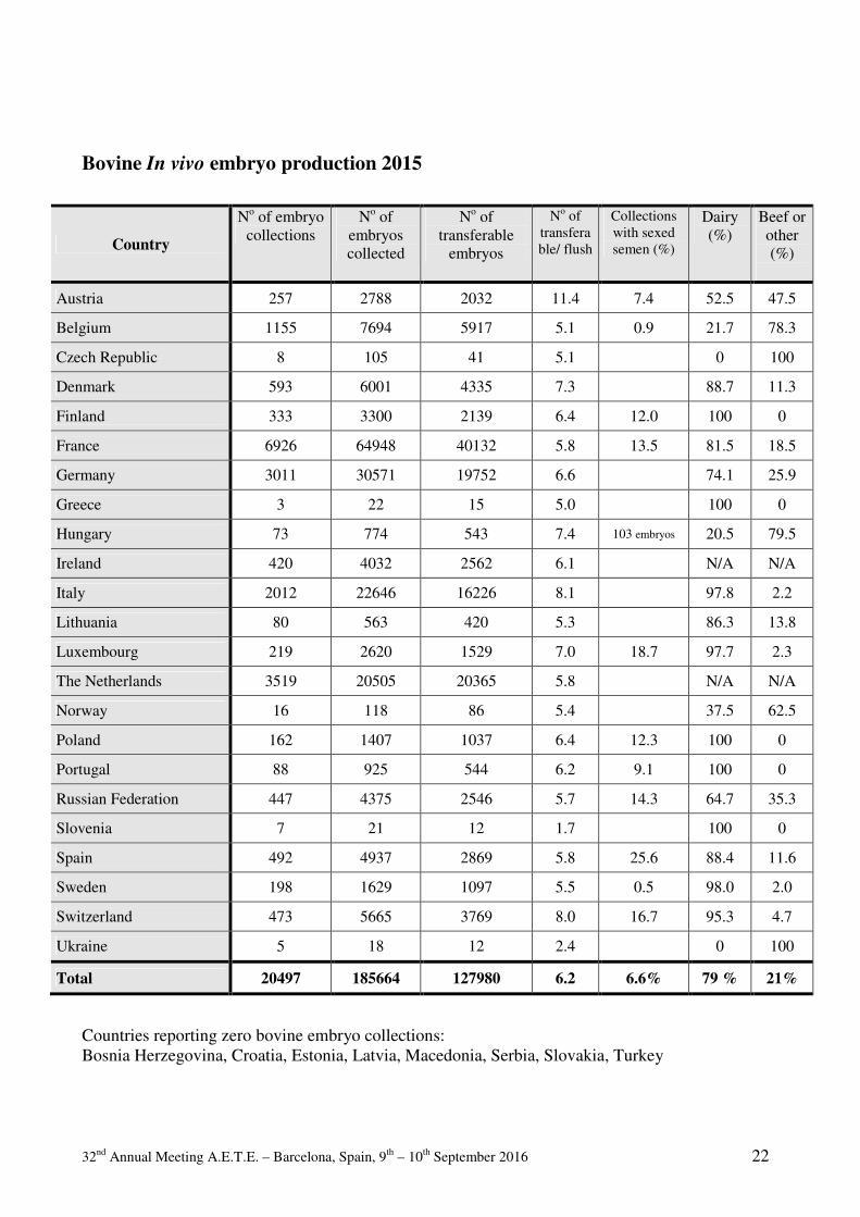
32nd Annual Meeting A.E.T.E. – Barcelona, Spain, 9th – 10th September 2016 22
Bovine In vivo embryo production 2015
Country
No of embryo collections
No of embryos collected
No of transferable
embryos
No of transferable/ flush
Collections with sexed semen (%)
Dairy (%)
Beef or other (%)
Austria 257 2788 2032 11.4 7.4 52.5 47.5
Belgium 1155 7694 5917 5.1 0.9 21.7 78.3
Czech Republic 8 105 41 5.1 0 100
Denmark 593 6001 4335 7.3 88.7 11.3
Finland 333 3300 2139 6.4 12.0 100 0
France 6926 64948 40132 5.8 13.5 81.5 18.5
Germany 3011 30571 19752 6.6 74.1 25.9
Greece 3 22 15 5.0 100 0
Hungary 73 774 543 7.4 103 embryos 20.5 79.5
Ireland 420 4032 2562 6.1 N/A N/A
Italy 2012 22646 16226 8.1 97.8 2.2
Lithuania 80 563 420 5.3 86.3 13.8
Luxembourg 219 2620 1529 7.0 18.7 97.7 2.3
The Netherlands 3519 20505 20365 5.8 N/A N/A
Norway 16 118 86 5.4 37.5 62.5
Poland 162 1407 1037 6.4 12.3 100 0
Portugal 88 925 544 6.2 9.1 100 0
Russian Federation 447 4375 2546 5.7 14.3 64.7 35.3
Slovenia 7 21 12 1.7 100 0
Spain 492 4937 2869 5.8 25.6 88.4 11.6
Sweden 198 1629 1097 5.5 0.5 98.0 2.0
Switzerland 473 5665 3769 8.0 16.7 95.3 4.7
Ukraine 5 18 12 2.4 0 100
Total 20497 185664 127980 6.2 6.6% 79 % 21%
Countries reporting zero bovine embryo collections: Bosnia Herzegovina, Croatia, Estonia, Latvia, Macedonia, Serbia, Slovakia, Turkey

32nd Annual Meeting A.E.T.E. – Barcelona, Spain, 9th – 10th September 2016 23
Bovine In vitro embryo production, OPU Total Stimulated Non-stimulated
Country N°°°° OPU sessions
N°°°° oocytes
N°°°° of embryos
N°°°° OPU sessions
N°°°° oocytes
N°°°° of embryos
N°°°° OPU sessions
N°°°° oocytes
N°°°° of embryos
Austria 8 80 17 8 80 17 0 0 0
Finland 148 1084 235 0 0 0 148 1084 235
France 256 2441 778 212 2203 716 44 238 62
Germany 1631 13663 2244 N/A N/A N/A N/A N/A N/A
Italy 730 9258 1481 0 0 0 730 9258 1481
The Netherlands 5818 45395 8727 N/A N/A N/A N/A N/A N/A
Spain 55 668 103 54 614 83 1 54 20
Russian Federation 446 1808 195 0 0 0 446 1808 195
Total 9092 74397 13780 274 2897 816 4543 12442 4237
Bovine In vitro embryo production, “slaughtered donor” Country N°°°° of oocyte
donors N°°°° of oocytes
collected N°°°° of transferable
embryos
Germany 24 889 165 Italy 10 428 108 Lithuania 3 9 0 The Netherlands 28 1146 161
Total 65 2472 434
Bovine Embryo Technologies Country N°°°° of sexed embryos (via
biopsy) N°°°° of genotyped embryos
In vivo In vitro In vivo In vitro
France 984 634 Germany 865 154 371 38 Netherlands 90 Hungary 24
Total 1873 154 1095 38

32nd Annual Meeting A.E.T.E. – Barcelona, Spain, 9th – 10th September 2016 24
Bovine Embryo Transfer
Country
N°°°° of transferred embryos
N°° °°o
f ex
port
ed
yos
In vivo In vitro Total
Fresh Frozen domestic
Frozen foreign
Fresh Frozen domestic
Frozen foreign
Austria 456 759 28 12 5 0 1260 0
Belgium 1275 3869 1007 0 0 0 6151 146
Czech Republic 0 5 0 0 0 0 5 0
Denmark 2238 1007 0 0 0 3245 91
Finland 762 1544 496 66 43 0 2911 4
France 18837 17200 1042 251 297 0 37627 220
Germany 8017 12546 0 1684 358 0 22605 0
Greece 5 0 25 0 0 0 30 0
Hungary 289 212 106 0 0 0 607 10
Italy 6003 N/A N/A 328 1337 0 7668 N/A
Ireland 720 750 0 0 0 0 1470 0
Lithuania 54 36 0 0 0 0 90 0
Luxembourg 736 791 0 0 45 0 1572 50
The Netherlands 4072 16406 0 7287 899 0 28664 1115
Norway 22 45 55 0 0 0 122 0
Poland 608 588 0 0 0 0 1196 0
Portugal 124 450 35 0 0 0 609 16
Russian Federation 104 1510 482 0 5 1630 3731 0
Slovenia 10 2 2 0 0 0 14 0
Spain 869 1130 138 171 76 8 2392 56
Sweden 768 329 263 0 0 0 1360 0
Switzerland 950 1969 523 0 0 0 3442 72
Ukraine 12 0 25 0 0 0 37 0
Total 46919 61148 4202 9799 3065 1638 126808 1780
Countries reporting zero bovine embryo transfers: Bosnia Herzegovina, Croatia, Estonia, Latvia, Macedonia, Serbia, Slovakia, Turkey
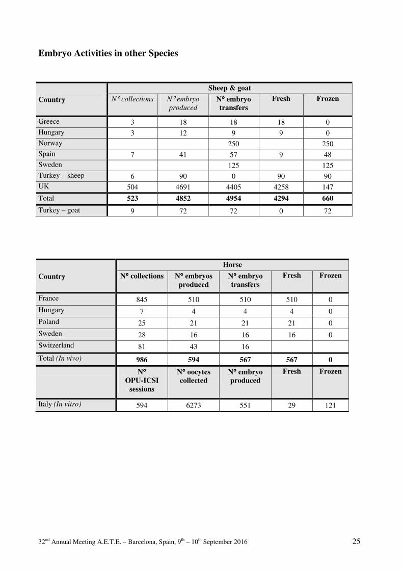
32nd Annual Meeting A.E.T.E. – Barcelona, Spain, 9th – 10th September 2016 25
Embryo Activities in other Species
Country
Sheep & goat
N° collections N° embryo
produced
N°°°° embryo transfers
Fresh Frozen
Greece 3 18 18 18 0 Hungary 3 12 9 9 0 Norway 250 250 Spain 7 41 57 9 48 Sweden 125 125 Turkey – sheep 6 90 0 90 90 UK 504 4691 4405 4258 147
Total 523 4852 4954 4294 660
Turkey – goat 9 72 72 0 72
Country
Horse
N°°°° collections N°°°° embryos produced
N°°°° embryo transfers
Fresh Frozen
France 845 510 510 510 0 Hungary 7 4 4 4 0 Poland 25 21 21 21 0 Sweden 28 16 16 16 0 Switzerland 81 43 16
Total (In vivo) 986 594 567 567 0
N°°°° OPU-ICSI
sessions
N°°°° oocytes collected
N°°°° embryo produced
Fresh Frozen
Italy (In vitro) 594 6273 551 29 121

32nd Annual Meeting A.E.T.E. – Barcelona, Spain, 9th – 10th September 2016 26

32nd Annual Meeting A.E.T.E. – Barcelona, Spain, 9th – 10th September 2016 27
INVITED LECTURES

32nd Annual Meeting A.E.T.E. – Barcelona, Spain, 9th – 10th September 2016 28

32nd Annual Meeting A.E.T.E. – Barcelona, Spain, 9th – 10th September 2016 29
Update and overview on assisted reproductive technologies (ARTs) in Brazil
Roberto Sartori1*, Alexandre Barbieri Prata1, Ana Cristina Silva Figueiredo2, Bruno Valente
Sanches3, Guilherme Corrêa de Souza Pontes4, João Henrique Moreira Viana5, José Henrique
Pontes3, José Luiz Moraes Vasconcelos6, Marcos Henrique Colombo Pereira6, Margot Alves
Nunes Dode5, Pedro Leopoldo Jeronimo Monteiro Jr.1, Pietro Sampaio Baruselli7
1Department of Animal Science, ESALQ, University of São Paulo (USP), Piracicaba, SP13418-900,
Brazil 2University of Jose do Rosario Vellano, Alfenas, MG 37130-000, Brazil
3InVitro Brasil, Mogi Mirim, SP 13800-970, Brazil 4Rehagro, Belo Horizonte, MG 30310-300, Brazil
5Embrapa Genetic Resources and Biotechnology, Brasília, DF 70770-917, Brazil 6Department of Animal Production, School of Veterinary Medicine and Animal Science, São Paulo
State University (UNESP), Botucatu, SP 18168-000, Brazil 7Department of Animal Reproduction, School of Veterinary Medicine and Animal Science,
University of São Paulo (USP), São Paulo, SP 05508-270, Brazil
Corresponding author: [email protected]
Abstract The impressive increase in the use of assisted reproductive technologies
(ARTs), especially in cattle, during the last few years in Brazil is well known
worldwide. In 2015, there were over 13.7 million artificial inseminations (AI), of
which, about 77% were carried out using fixed-time AI (FTAI). This technology has
helped to substantially improve reproductive efficiency in beef and dairy cattle. In
relation to embryo transfer, production of in vivo derived (IVD) embryos remained
relatively stable, with average production of 30-40,000 embryos per year, whereas in
vitro production (IVP) of embryos had a substantial increase, from about 12,500 IVP
embryos in 2000 to more than 300,000 IVP embryos after 2010. The increasing
availability and use of sex-sorted sperm was one of the factors responsible for a recent
shift from the predominance of IVP embryos from beef breeds to dairy breeds in
Brazil. Moreover, there was also an increase from 13% in 2014 to 29% in 2015 in the
percentage of vitrified/frozen embryos. Moreover, the successful use of protocols for
fixed-time ET (FTET) due to their high efficiency and ease of implementation, has
facilitated the dissemination of ET programs all over Brazil. However, there is room
for improvement, since there are several reports of high pregnancy loss and high
peripartum loss, when IVP embryos are used. The production of healthy cattle by
somatic cell nuclear transfer has also increased in the last few years in Brazil, but
despite substantial progress in reducing postnatal losses, no drastic increase in cloning
efficiency up to parturition has occurred.
Keywords: artificial insemination, bovine, embryo, in vitro production, superovulation.
32nd Annual Meeting A.E.T.E. – Barcelona, Spain, 9th – 10th September 2016 30
Introduction
Currently, Brazilian cattle industry has one of the largest commercial herds in the world,
about 208.3 million head (ABIEC, 2014). Brazil produced 10.7 million tons of beef in 2014
(ABIEC, 2014), being second place in the world ranking of meat production. Moreover, the dairy
herd in Brazil ranks in the fifth position worldwide (FAO, 2012). Despite the magnitude of the herd,
the annual Brazilian production of milk in 2014 was 24.741 billion liters, with a productivity of
only 1,380 L of milk/cow/yr (IBGE, 2014). This is obviously very low production if compared, for
example, with data from the USA herd (10,096 L of milk/cow/yr), currently the largest producer of
milk in the world (USDA, 2014). However, both Brazilian beef and dairy productivity is increasing,
which is directly related to technological advances in animal breeding, such as greater use of
artificial insemination (AI) and embryo transfer (ET).
To have an idea on the evolution of these biotechnologies, in 2002 only 5-6% of heifers and
cows were artificially inseminated in Brazil, about 7 million AIs, with only 1% of inseminations
being through fixed-time artificial insemination (FTAI). In contrast, in 2015, about 13 million AIs
were performed corresponding to 10-12% of females of reproductive age and 77% of these
inseminations were performed by FTAI (Pietro Baruselli, unpublished).
In relation to embryo production in cattle, there are two different scenarios. While
production of in vivo derived (IVD) embryos remained relatively stable over the last 15 yr, with
average production of 30-40,000 embryos per year, the in vitro production (IVP) of embryos had a
substantial increase from about 12,500 IVP embryos in 2000, to over 348,000 IVP embryos in
2014, representing almost 60% of the world embryo production.
Sex-sorted sperm has been widely and increasingly used in Brazil, especially for AI or IVP.
Unfortunately, epidemiological data on the use of sex-sorted sperm in Brazil are not available.
Regarding IVP, data from the last 3 yr from one of the main labs in Brazil confirm other data from
the literature that there is a reduction in embryo production per cultured oocyte if sex-sorted sperm
is used for in vitro fertilization when compared with conventional unsorted sperm (23.6%
[311,788/1,323,541] vs. 28.5% [242,259/848,939]; P < 0.01).
For cloning, data from the Brazilian Association of Zebu Breeders (ABCZ) show a gradual
increase in registered Bos indicus calves (predominantly of Nelore and Gir breeds) produced by
somatic cell nuclear transfer (SCNT) during the years 2010 (n = 5), 2011 (n = 23), 2012 (n = 22),
and 2013 (n = 41). Unofficial data indicate a continuous increase in number of healthy calves
produced by SCNT from 2014 to 2016.
This manuscript aims to present an update and overview of the assisted reproduction
technologies (ARTs) in Brazil focused on AI and ET in cattle and to describe reports on how these
technologies have positively influenced the reproductive efficiency of dairy and beef herds.
Artificial insemination
As mentioned above, Brazil has one of the largest cattle herds in the world, however the use
of AI is still low. In 2015, there were over 13.7 million inseminations, which correspond to 10-12%
of cows and heifers of reproductive age (Figure 1). Out of this total AIs, about 4.7 million were
performed in dairy cows, with a decrease of 12.4% compared with the previous year. In beef cattle,

32nd Annual Meeting A.E.T.E. – Barcelona, Spain, 9th – 10th September 2016 31
9 million inseminations were performed, with an increase of 16.2% in relation to 2014. In 2015,
more than 10.5 million FTAIs were performed, with an increase of 11.2% compared to 2014, and
FTAI now represents ~77% of all AIs carried out in Brazil (Figure 1). These data demonstrate that
FTAI is increasing the use of AI across Brazil with a doubling in the overall use of AI during the
last decade, but over a 10-fold increase in the use of FTAI from ~1 million protocols in 2005 (11%
of all AIs) to 10.5 million protocols in 2015 (77% of all AIs).
Figure 1. Data of artificial insemination (AI) based on cows and heifers bred to estrus or
to fixed-time AI (FTAI) systems in Brazil during the period of 2002 to 2015. Numbers of
FTAI were estimated based on hormones/products sold for each FTAI protocol.
Use of FTAI in dairy cattle
Although most dairy cows and heifers are bred by bulls in Brazil, AI is the preferred ART for most
progressive dairy farms. When AI is employed, the question practitioners and producers ask is
whether they should breed cows to estrus or FTAI. In fact, this doubt is understandable because
studies that properly compared insemination to estrus vs. insemination to a FTAI protocol have
described lower (Strickland et al., 2010; Carvalho and Fricke, unpublished), similar (Rabiee et al.,
2005; Nascimento et al., 2013b), or greater (Nascimento et al., 2013a) pregnancies per AI (P/AI)
when cows are bred to estrus. However, suboptimal estrus detection rates in cycling cows (Lopez et
al., 2004; Fricke et al., 2014) and a substantial percentage (~24%) of cows that are not cycling
(Wiltbank et al., 2002; Santos et al., 2009), produces the problem of low service rates (SR) and, in
general, lower 21-d pregnancy rates (21-d PR = P/AI x SR, every 21 d after the voluntary waiting
period; VWP) for cows bred to estrus than cows bred to FTAI (Nascimento et al., 2013a; Wiltbank
and Pursley, 2014).
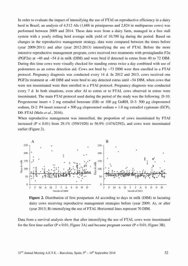
32nd Annual Meeting A.E.T.E. – Barcelona, Spain, 9th – 10th September 2016 32
In order to evaluate the impact of intensifying the use of FTAI on reproductive efficiency in a dairy
herd in Brazil, an analysis of 4,512 AIs (1,688 in primiparous and 2,824 in multiparous cows) was
performed between 2009 and 2014. These data were from a dairy farm, managed in a free stall
system with a yearly rolling herd average milk yield of 10,700 kg during the period. Based on
changes in the reproductive management strategy, data were compared between the times before
(year 2009-2011) and after (year 2012-2013) intensifying the use of FTAI. Before the more
intensive reproductive management program, cows received two treatments with prostaglandin F2α
(PGF2α) at ~40 and ~54 d in milk (DIM) and were bred if detected in estrus from 40 to 72 DIM.
During this time cows were visually checked for standing estrus twice a day combined with use of
pedometers as an estrus detection aid. Cows not bred by ~73 DIM were then enrolled in a FTAI
protocol. Pregnancy diagnosis was conducted every 14 d. In 2012 and 2013, cows received one
PGF2α treatment at ~40 DIM and were bred to any detected estrus until ~54 DIM, when cows that
were not inseminated were then enrolled in a FTAI protocol. Pregnancy diagnosis was conducted
every 7 d. In both situations, even after AI to estrus or to FTAI, cows observed in estrus were
inseminated. The main FTAI protocol used during the period of the study was the following. D-10:
Progesterone insert + 2 mg estradiol benzoate (EB) or 100 µg GnRH, D-3: 500 µg cloprostenol
sodium, D-2: P4 insert removal + 500 µg cloprostenol sodium + 1.0 mg estradiol cypionate (ECP),
D0: FTAI (Melo et al., 2016).
When reproductive management was intensified, the proportion of cows inseminated by FTAI
increased (P < 0.01) from 29.1% (559/1920) to 56.9% (1474/2592), and cows were inseminated
earlier (Figure 2).
Figure 2. Distribution of first postpartum AI according to days in milk (DIM) in lactating
dairy cows receiving reproductive management strategies before (year 2009; A), or after
(year 2013; B) intensifying the use of FTAI. Horizontal lines represent 70 DIM.
Data from a survival analysis show that after intensifying the use of FTAI, cows were inseminated
for the first time earlier (P < 0.01; Figure 3A) and became pregnant sooner (P < 0.01; Figure 3B).
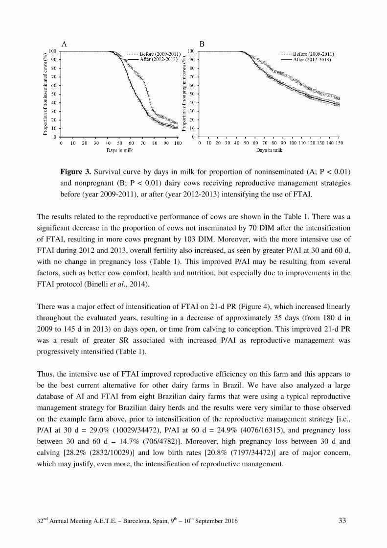
32nd Annual Meeting A.E.T.E. – Barcelona, Spain, 9th – 10th September 2016 33
Figure 3. Survival curve by days in milk for proportion of noninseminated (A; P < 0.01)
and nonpregnant (B; P < 0.01) dairy cows receiving reproductive management strategies
before (year 2009-2011), or after (year 2012-2013) intensifying the use of FTAI.
The results related to the reproductive performance of cows are shown in the Table 1. There was a
significant decrease in the proportion of cows not inseminated by 70 DIM after the intensification
of FTAI, resulting in more cows pregnant by 103 DIM. Moreover, with the more intensive use of
FTAI during 2012 and 2013, overall fertility also increased, as seen by greater P/AI at 30 and 60 d,
with no change in pregnancy loss (Table 1). This improved P/AI may be resulting from several
factors, such as better cow comfort, health and nutrition, but especially due to improvements in the
FTAI protocol (Binelli et al., 2014).
There was a major effect of intensification of FTAI on 21-d PR (Figure 4), which increased linearly
throughout the evaluated years, resulting in a decrease of approximately 35 days (from 180 d in
2009 to 145 d in 2013) on days open, or time from calving to conception. This improved 21-d PR
was a result of greater SR associated with increased P/AI as reproductive management was
progressively intensified (Table 1).
Thus, the intensive use of FTAI improved reproductive efficiency on this farm and this appears to
be the best current alternative for other dairy farms in Brazil. We have also analyzed a large
database of AI and FTAI from eight Brazilian dairy farms that were using a typical reproductive
management strategy for Brazilian dairy herds and the results were very similar to those observed
on the example farm above, prior to intensification of the reproductive management strategy [i.e.,
P/AI at 30 d = 29.0% (10029/34472), P/AI at 60 d = 24.9% (4076/16315), and pregnancy loss
between 30 and 60 d = 14.7% (706/4782)]. Moreover, high pregnancy loss between 30 d and
calving [28.2% (2832/10029)] and low birth rates [20.8% (7197/34472)] are of major concern,
which may justify, even more, the intensification of reproductive management.
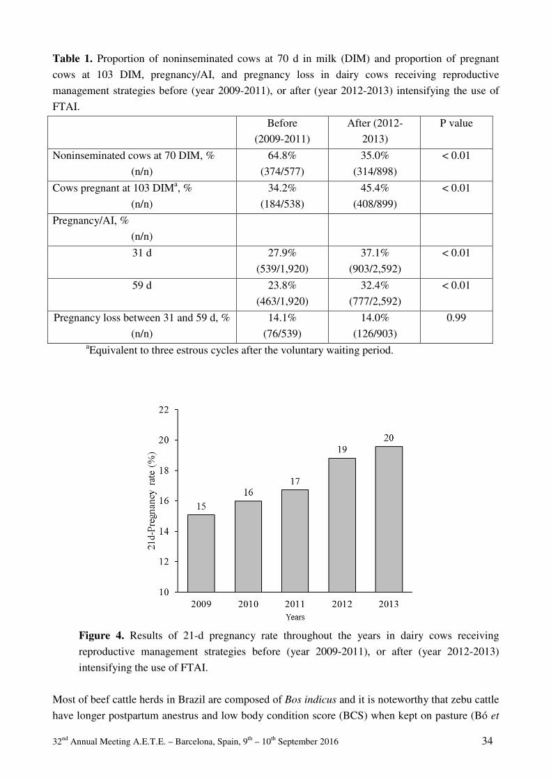
32nd Annual Meeting A.E.T.E. – Barcelona, Spain, 9th – 10th September 2016 34
Table 1. Proportion of noninseminated cows at 70 d in milk (DIM) and proportion of pregnant
cows at 103 DIM, pregnancy/AI, and pregnancy loss in dairy cows receiving reproductive
management strategies before (year 2009-2011), or after (year 2012-2013) intensifying the use of
FTAI.
Before
(2009-2011)
After (2012-
2013)
P value
Noninseminated cows at 70 DIM, %
(n/n)
64.8%
(374/577)
35.0%
(314/898)
< 0.01
Cows pregnant at 103 DIMa, %
(n/n)
34.2%
(184/538)
45.4%
(408/899)
< 0.01
Pregnancy/AI, %
(n/n)
31 d 27.9%
(539/1,920)
37.1%
(903/2,592)
< 0.01
59 d 23.8%
(463/1,920)
32.4%
(777/2,592)
< 0.01
Pregnancy loss between 31 and 59 d, %
(n/n)
14.1%
(76/539)
14.0%
(126/903)
0.99
aEquivalent to three estrous cycles after the voluntary waiting period.
Figure 4. Results of 21-d pregnancy rate throughout the years in dairy cows receiving
reproductive management strategies before (year 2009-2011), or after (year 2012-2013)
intensifying the use of FTAI.
Most of beef cattle herds in Brazil are composed of Bos indicus and it is noteworthy that zebu cattle
have longer postpartum anestrus and low body condition score (BCS) when kept on pasture (Bó et
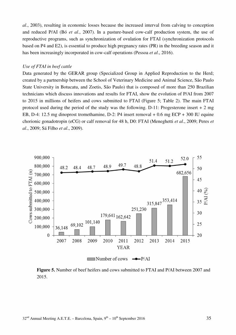
32nd Annual Meeting A.E.T.E. – Barcelona, Spain, 9th – 10th September 2016 35
al., 2003), resulting in economic losses because the increased interval from calving to conception
and reduced P/AI (Bó et al., 2007). In a pasture-based cow-calf production system, the use of
reproductive programs, such as synchronization of ovulation for FTAI (synchronization protocols
based on P4 and E2), is essential to produce high pregnancy rates (PR) in the breeding season and it
has been increasingly incorporated in cow-calf operations (Pessoa et al., 2016).
Use of FTAI in beef cattle
Data generated by the GERAR group (Specialized Group in Applied Reproduction to the Herd;
created by a partnership between the School of Veterinary Medicine and Animal Science, São Paulo
State University in Botucatu, and Zoetis, São Paulo) that is composed of more than 250 Brazilian
technicians which discuss innovations and results for FTAI, show the evolution of P/AI from 2007
to 2015 in millions of heifers and cows submitted to FTAI (Figure 5; Table 2). The main FTAI
protocol used during the period of the study was the following. D-11: Progesterone insert + 2 mg
EB, D-4: 12.5 mg dinoprost tromethamine, D-2: P4 insert removal + 0.6 mg ECP + 300 IU equine
chorionic gonadotropin (eCG) or calf removal for 48 h, D0: FTAI (Meneghetti et al., 2009; Peres et
al., 2009; Sá Filho et al., 2009).
Figure 5. Number of beef heifers and cows submitted to FTAI and P/AI between 2007 and
2015.

32nd Annual Meeting A.E.T.E. – Barcelona, Spain, 9th – 10th September 2016 36
Table 2. Pregnancy per AI of heifers, primiparous, multiparous and non-lactating beef cows
submitted to FTAI between 2007 and 2015.
Year Heifers %, (n) Primiparous%, (n) Multiparous %, (n) Non-lactating, % (n) 2007 39.6% (3,037) 44.5% (5,249) 49.7% (22,519) 45.1% (1,510) 2008 44.8% (4,944) 42.6% (9,763) 50.9% (44,628) 45.6% (5,354) 2009 50.5% (8,347) 43.4% (15,476) 49.9% (70,308) 46.5% (5,526) 2010 39.7% (24,372) 48.5% (18,819) 50.7% (123,380) 49.4% (9,566) 2011 49.3% (21,810) 41.6% (22,453) 51.2% (105,440) 52.0% (11,076) 2012 47.1% (42,030) 44.1% (32,345) 50.2% (130,236) 52.1% (10,252) 2013 49.0% (58,032) 47.8% (42,467) 53.1% (189,726) 50.1% (24,432) 2014 46.8% (56,026) 48.0% (47,882) 53.0% (200,082) 50.8% (26,091) 2015 48.5% (124,687) 47.1% (80,690) 54.1% (392,511) 51.5% (69,734)
As shown in Table 2, the fertility in all types of beef cattle has been relatively constant
(~50%) during the last 3 yr. Nevertheless, there is likely to be room for improvement in many of the
herds since some herds (~24%) had average P/AI greater or equal to 60% (Figure 6). These herds
are likely to have more intensive reproductive programs, better nutrition with fewer cows with low
BCS (Figure 7), and may use cattle with better fertility traits, such as Nelore X Angus crossbreds
(Figure 8).
Figure 6. Distribution of farms according to P/AI of beef cows submitted to FTAI in 2015.
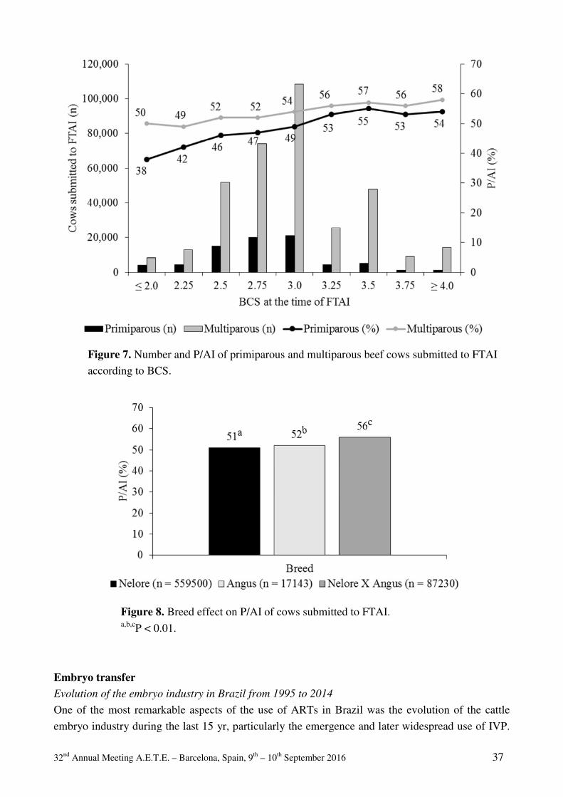
32nd Annual Meeting A.E.T.E. – Barcelona, Spain, 9th – 10th September 2016 37
Figure 7. Number and P/AI of primiparous and multiparous beef cows submitted to FTAI
according to BCS.
Figure 8. Breed effect on P/AI of cows submitted to FTAI. a,b,cP < 0.01.
Embryo transfer Evolution of the embryo industry in Brazil from 1995 to 2014
One of the most remarkable aspects of the use of ARTs in Brazil was the evolution of the cattle
embryo industry during the last 15 yr, particularly the emergence and later widespread use of IVP.

32nd Annual Meeting A.E.T.E. – Barcelona, Spain, 9th – 10th September 2016 38
In the early 90’s, the Brazilian embryo industry was already substantial, and the country was the
largest embryo producer, outside Europe and North America. However, the adoption of IVP after
the year 2000 boosted the embryo industry, and since 2005, Brazil accounts for more than 20% of
the world embryo production. In 2014, Brazil produced 348,468 embryos in vitro, which
corresponds to 59.0% of the total world IVP (Perry, 2015).
The success of IVP in Brazil was due to a complex interplay of technical and economic factors that
likely explain why it initially diverged from the trends elsewhere (Faber et al., 2003). Initially, in
the period from the emergence of the first commercial IVP companies in 1999 to 2003, there was a
relatively high cost and low efficiency of IVP (Hasler, 2000), but this was balanced by the high
commercial value of the donors used. Thus, during this initial growth phase (first phase) IVP
expanded mainly within the market of high genetic merit cows and the number of both IVD and
IVP embryos increased similarly (Figure 9).
Figure 9. Production of bovine embryos in Brazil, according to the technique employed,
during the period of 1995 to 2014. IVD: embryos produced by superovulation (in vivo);
IVP: embryos produced in vitro.
A second phase of growth in the use of IVP embryos occurred between 2003 and 2010, driven
largely by production of embryos from high genetic merit animals used to produce replacement
bulls. Prior to this second phase, the large size of beef cattle population in Brazil and the relatively
low use of AI at that time (~6%, Baruselli et al., 2012) resulted in a reduced demand for such
animals (bulls), especially in the Nelore breed. However at the peak of this second growth phase, in
2005, embryo production in Nelore (214,500) accounted for 82.7% of all embryos produced in the
country, and for 90.0% of the embryos from beef breeds (Viana et al., 2012). Meanwhile, embryo
prices began to decrease in Brazil due to many factors including: increasing efficiency of embryo
production protocols, increased recovery of cumulus-oocyte complexes (COC) and greater
blastocyst rates obtained in Bos indicus breeds (Pontes et al., 2009, Viana et al., 2012), and an

32nd Annual Meeting A.E.T.E. – Barcelona, Spain, 9th – 10th September 2016 39
increase in the scale of embryo production in commercial embryo production companies in Brazil.
The IVP industry became more competitive, and eventually replaced multiple ovulation and embryo
transfer (MOET) as the technique of choice for embryo production. Total embryo production
increased rapidly, reaching numbers over 250,000 for the years after 2005.
We are currently in the midst of the third growth phase with increasing use of sex-sorted sperm in
IVP, which occurred mainly in dairy breeds. In dairy breeds, production of a high percentage of
female calves has many economic advantages and use of in IVP allows the production of
approximately 90% of the embryos with the desired sex (Morotti et al., 2014). Thus, this third
growth phase of the Brazilian embryo industry after 2010 has been marked by a clear shift from the
predominance of beef breeds to dairy breeds. For example, in 2014, embryo production in dairy
breeds increased by 46.5% and the total numbers of embryos produced from dairy breeds exceeded,
for the first time, the number of embryos from beef breeds, (270,367 of 391,805, or 69.0% of total
embryos). The expansion in the dairy sector also highlighted a new trend in the Brazilian embryo
industry, the use of large-scale IVP to produce crossbred calves (Pontes et al., 2010). Producers and
veterinarians explored the possibilities of obtaining the gains due to heterosis while maintaining
herds with specific crossbred values (F1, ¾, etc.). For example, 79.3% of embryos produced in
dairy breeds in 2014 were from Gir x Holstein crosses.
The inherent characteristics of dairy production, such as smaller herds and lack of a set breeding
season, limits the availability of recipients, and thus required the development and use of
cryopreservation alternatives. In 2015, the three main commercial laboratories in Brazil produced
more than 276,000 embryos from ~50,000 donors with a blastocyst rate ~30% (more than 1 million
oocytes used for IVF). Of those embryos, 111,000 were conventional embryos and 165,000 were
produced using sex-sorted sperm and 29% (80,000) of these embryos were vitrified or frozen. In
addition, these laboratories reported an increase in embryo production of more than 30% compared
to 2014 (211,000 IVP embryos) and the percentage of vitrified/frozen embryos in these laboratories
increased from 13% in 2014 to 29% in 2015. The continuing development and use of the direct
transfer technique (over 9,000 embryos in 2015) is likely to lead to further increases in the use of
cryopreserved IVP embryos. Moreover, the successful use of protocols for fixed-time ET (FTET),
due to their high efficiency and ease of implementation, has facilitated the dissemination of ET
programs across Brazil.
Use of IVP embryos for reproductive management in dairy cattle
As seen above, the use of IVP embryos in dairy herds has increased in recent years. In 2015, the
two largest laboratories that produce embryos from dairy breeds transferred more than 27,000
embryos, obtaining reasonable pregnancies per ET (P/ET), and acceptable pregnancy losses (Table
3), especially when beef cows, crossbreds, or heifers are used as embryo recipients. Moreover, the
best IVP embryos are usually selected for vitrification, which may explain the observation of
similar pregnancy losses for fresh and vitrified embryos, as presented in Table 3.
However, results can vary from farm to farm, and rigorous evaluation and monitoring are necessary
for this technology to be used on a large scale as a substitute for AI or FTAI. The following data
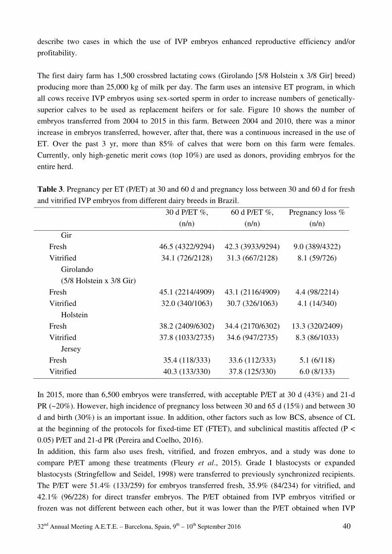
32nd Annual Meeting A.E.T.E. – Barcelona, Spain, 9th – 10th September 2016 40
describe two cases in which the use of IVP embryos enhanced reproductive efficiency and/or
profitability.
The first dairy farm has 1,500 crossbred lactating cows (Girolando [5/8 Holstein x 3/8 Gir] breed)
producing more than 25,000 kg of milk per day. The farm uses an intensive ET program, in which
all cows receive IVP embryos using sex-sorted sperm in order to increase numbers of genetically-
superior calves to be used as replacement heifers or for sale. Figure 10 shows the number of
embryos transferred from 2004 to 2015 in this farm. Between 2004 and 2010, there was a minor
increase in embryos transferred, however, after that, there was a continuous increased in the use of
ET. Over the past 3 yr, more than 85% of calves that were born on this farm were females.
Currently, only high-genetic merit cows (top 10%) are used as donors, providing embryos for the
entire herd.
Table 3. Pregnancy per ET (P/ET) at 30 and 60 d and pregnancy loss between 30 and 60 d for fresh
and vitrified IVP embryos from different dairy breeds in Brazil.
30 d P/ET %,
(n/n)
60 d P/ET %,
(n/n)
Pregnancy loss %
(n/n)
Gir
Fresh 46.5 (4322/9294) 42.3 (3933/9294) 9.0 (389/4322)
Vitrified 34.1 (726/2128) 31.3 (667/2128) 8.1 (59/726)
Girolando
(5/8 Holstein x 3/8 Gir)
Fresh 45.1 (2214/4909) 43.1 (2116/4909) 4.4 (98/2214)
Vitrified 32.0 (340/1063) 30.7 (326/1063) 4.1 (14/340)
Holstein
Fresh 38.2 (2409/6302) 34.4 (2170/6302) 13.3 (320/2409)
Vitrified 37.8 (1033/2735) 34.6 (947/2735) 8.3 (86/1033)
Jersey
Fresh 35.4 (118/333) 33.6 (112/333) 5.1 (6/118)
Vitrified 40.3 (133/330) 37.8 (125/330) 6.0 (8/133)
In 2015, more than 6,500 embryos were transferred, with acceptable P/ET at 30 d (43%) and 21-d
PR (~20%). However, high incidence of pregnancy loss between 30 and 65 d (15%) and between 30
d and birth (30%) is an important issue. In addition, other factors such as low BCS, absence of CL
at the beginning of the protocols for fixed-time ET (FTET), and subclinical mastitis affected (P <
0.05) P/ET and 21-d PR (Pereira and Coelho, 2016).
In addition, this farm also uses fresh, vitrified, and frozen embryos, and a study was done to
compare P/ET among these treatments (Fleury et al., 2015). Grade I blastocysts or expanded
blastocysts (Stringfellow and Seidel, 1998) were transferred to previously synchronized recipients.
The P/ET were 51.4% (133/259) for embryos transferred fresh, 35.9% (84/234) for vitrified, and
42.1% (96/228) for direct transfer embryos. The P/ET obtained from IVP embryos vitrified or
frozen was not different between each other, but it was lower than the P/ET obtained when IVP

32nd Annual Meeting A.E.T.E. – Barcelona, Spain, 9th – 10th September 2016 41
embryos were transferred fresh (P < 0.05). Therefore, these results highlighted the positive aspects
of cryopreservation of IVP embryos with the convenience of direct transfer as compared with
vitrification.
Figure 10. Number of embryos transferred from 2004 to 2015 in one dairy farm (From
Pereira and Coelho, 2016).
The second farm has 1,100 lactating cows (Holstein and Girolando breeds) with average milk
production of 30 kg/d. The reproductive management consists of use of AI or transfer of IVP
embryos. As shown in Table 4, despite having greater pregnancy losses, the IVP technique was
chosen as a better reproductive management strategy for this dairy farm, as compared to AI, due to
greater P/ET vs. P/AI, and greater birth rates for ET vs. AI. In addition, the use of sex-sorted sperm
for IVF allowed an increased number of heifers born with IVF and greater genetic improvement.
Pregnancy losses for the farms described above are much greater than those shown in Table 3,
probably due to the use of different embryo recipients, as well as quality of IVP embryos selected
for transfer. For the farms described above (Table 4), lactating cows were primarily used as
recipients, whereas data presented in Table 3 are mainly from non-lactating embryo recipients. In
fact, data of other dairy farms (n = 7) in which IVP embryos were transferred to lactating cows
show acceptable P/ET at 30 d [42.9% (7204/16771)], however, pregnancy loss between 30 and 60 d
[15.9% (820/5147)], and pregnancy loss between 30 d and calving [33.4% (2323/6956)] are high,
resulting in low birth rates [28.8% (4663/16170)]. Greater pregnancy loss in lactating dairy cows as
compared to heifers or non-lactating cows has been well-described elsewhere (Santos et al., 2004;
Sartori, 2004).
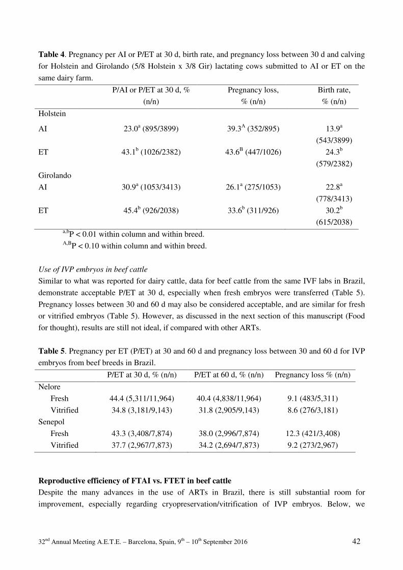
32nd Annual Meeting A.E.T.E. – Barcelona, Spain, 9th – 10th September 2016 42
Table 4. Pregnancy per AI or P/ET at 30 d, birth rate, and pregnancy loss between 30 d and calving
for Holstein and Girolando (5/8 Holstein x 3/8 Gir) lactating cows submitted to AI or ET on the
same dairy farm.
P/AI or P/ET at 30 d, %
(n/n)
Pregnancy loss,
% (n/n)
Birth rate,
% (n/n)
Holstein
AI 23.0a (895/3899) 39.3A (352/895) 13.9a
(543/3899)
ET 43.1b (1026/2382) 43.6B (447/1026) 24.3b
(579/2382)
Girolando
AI 30.9a (1053/3413) 26.1a (275/1053) 22.8a
(778/3413)
ET 45.4b (926/2038) 33.6b (311/926) 30.2b
(615/2038) a,bP < 0.01 within column and within breed. A,BP < 0.10 within column and within breed.
Use of IVP embryos in beef cattle
Similar to what was reported for dairy cattle, data for beef cattle from the same IVF labs in Brazil,
demonstrate acceptable P/ET at 30 d, especially when fresh embryos were transferred (Table 5).
Pregnancy losses between 30 and 60 d may also be considered acceptable, and are similar for fresh
or vitrified embryos (Table 5). However, as discussed in the next section of this manuscript (Food
for thought), results are still not ideal, if compared with other ARTs.
Table 5. Pregnancy per ET (P/ET) at 30 and 60 d and pregnancy loss between 30 and 60 d for IVP
embryos from beef breeds in Brazil.
P/ET at 30 d, % (n/n) P/ET at 60 d, % (n/n) Pregnancy loss % (n/n)
Nelore
Fresh 44.4 (5,311/11,964) 40.4 (4,838/11,964) 9.1 (483/5,311)
Vitrified 34.8 (3,181/9,143) 31.8 (2,905/9,143) 8.6 (276/3,181)
Senepol
Fresh 43.3 (3,408/7,874) 38.0 (2,996/7,874) 12.3 (421/3,408)
Vitrified 37.7 (2,967/7,873) 34.2 (2,694/7,873) 9.2 (273/2,967)
Reproductive efficiency of FTAI vs. FTET in beef cattle Despite the many advances in the use of ARTs in Brazil, there is still substantial room for
improvement, especially regarding cryopreservation/vitrification of IVP embryos. Below, we
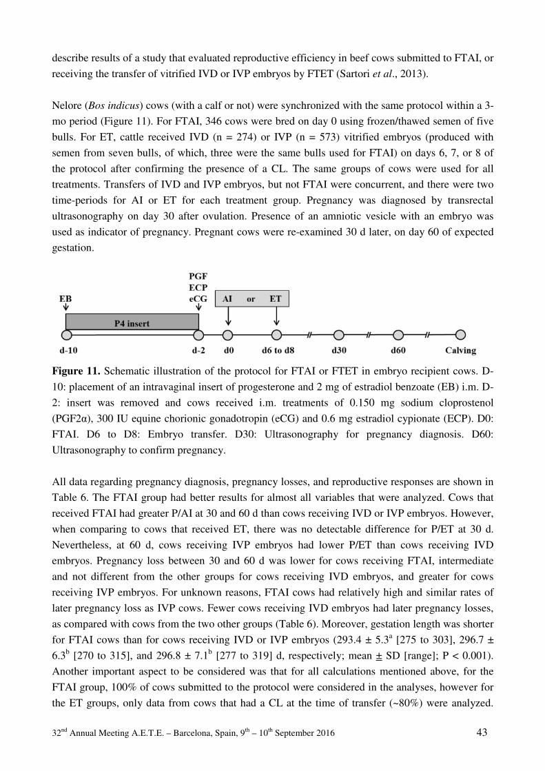
32nd Annual Meeting A.E.T.E. – Barcelona, Spain, 9th – 10th September 2016 43
describe results of a study that evaluated reproductive efficiency in beef cows submitted to FTAI, or
receiving the transfer of vitrified IVD or IVP embryos by FTET (Sartori et al., 2013).
Nelore (Bos indicus) cows (with a calf or not) were synchronized with the same protocol within a 3-
mo period (Figure 11). For FTAI, 346 cows were bred on day 0 using frozen/thawed semen of five
bulls. For ET, cattle received IVD (n = 274) or IVP (n = 573) vitrified embryos (produced with
semen from seven bulls, of which, three were the same bulls used for FTAI) on days 6, 7, or 8 of
the protocol after confirming the presence of a CL. The same groups of cows were used for all
treatments. Transfers of IVD and IVP embryos, but not FTAI were concurrent, and there were two
time-periods for AI or ET for each treatment group. Pregnancy was diagnosed by transrectal
ultrasonography on day 30 after ovulation. Presence of an amniotic vesicle with an embryo was
used as indicator of pregnancy. Pregnant cows were re-examined 30 d later, on day 60 of expected
gestation.
Figure 11. Schematic illustration of the protocol for FTAI or FTET in embryo recipient cows. D-
10: placement of an intravaginal insert of progesterone and 2 mg of estradiol benzoate (EB) i.m. D-
2: insert was removed and cows received i.m. treatments of 0.150 mg sodium cloprostenol
(PGF2α), 300 IU equine chorionic gonadotropin (eCG) and 0.6 mg estradiol cypionate (ECP). D0:
FTAI. D6 to D8: Embryo transfer. D30: Ultrasonography for pregnancy diagnosis. D60:
Ultrasonography to confirm pregnancy.
All data regarding pregnancy diagnosis, pregnancy losses, and reproductive responses are shown in
Table 6. The FTAI group had better results for almost all variables that were analyzed. Cows that
received FTAI had greater P/AI at 30 and 60 d than cows receiving IVD or IVP embryos. However,
when comparing to cows that received ET, there was no detectable difference for P/ET at 30 d.
Nevertheless, at 60 d, cows receiving IVP embryos had lower P/ET than cows receiving IVD
embryos. Pregnancy loss between 30 and 60 d was lower for cows receiving FTAI, intermediate
and not different from the other groups for cows receiving IVD embryos, and greater for cows
receiving IVP embryos. For unknown reasons, FTAI cows had relatively high and similar rates of
later pregnancy loss as IVP cows. Fewer cows receiving IVD embryos had later pregnancy losses,
as compared with cows from the two other groups (Table 6). Moreover, gestation length was shorter
for FTAI cows than for cows receiving IVD or IVP embryos (293.4 ± 5.3a [275 to 303], 296.7 ±
6.3b [270 to 315], and 296.8 ± 7.1b [277 to 319] d, respectively; mean ± SD [range]; P < 0.001).
Another important aspect to be considered was that for all calculations mentioned above, for the
FTAI group, 100% of cows submitted to the protocol were considered in the analyses, however for
the ET groups, only data from cows that had a CL at the time of transfer (~80%) were analyzed.
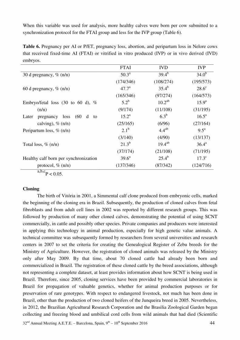
32nd Annual Meeting A.E.T.E. – Barcelona, Spain, 9th – 10th September 2016 44
When this variable was used for analysis, more healthy calves were born per cow submitted to a
synchronization protocol for the FTAI group and less for the IVP group (Table 6).
Table 6. Pregnancy per AI or P/ET, pregnancy loss, abortion, and peripartum loss in Nelore cows
that received fixed-time AI (FTAI) or vitrified in vitro produced (IVP) or in vivo derived (IVD)
embryos.
FTAI IVD IVP
30 d pregnancy, % (n/n) 50.3a
(174/346)
39.4b
(108/274)
34.0b
(195/573)
60 d pregnancy, % (n/n) 47.7a
(165/346)
35.4b
(97/274)
28.6c
(164/573)
Embryo/fetal loss (30 to 60 d), %
(n/n)
5.2b
(9/174)
10.2ab
(11/108)
15.9a
(31/195)
Later pregnancy loss (60 d to
calving), % (n/n)
15.2a
(25/165)
6.3b
(6/96)
16.5a
(27/164)
Peripartum loss, % (n/n) 2.1b
(3/140)
4.4ab
(4/90)
9.5a
(13/137)
Total loss, % (n/n) 21.3b
(37/174)
19.4ab
(21/108)
36.4a
(71/195)
Healthy calf born per synchronization
protocol, % (n/n)
39.6a
(137/346)
25.4b
(87/342)
17.3c
(124/716) a,b,c
P < 0.05.
Cloning The birth of Vitória in 2001, a Simmental calf clone produced from embryonic cells, marked
the beginning of the cloning era in Brazil. Subsequently, the production of cloned calves from fetal
fibroblasts and from adult cell lines in 2002 was reported by different research groups. This was
followed by production of many other cloned calves, demonstrating the potential of using SCNT
commercially, in cattle and possibly other species. Private companies and producers were interested
in applying this technology in animal production, especially for high genetic value animals. A
technical committee was subsequently formed by researchers from several universities and research
centers in 2007 to set the criteria for creating the Genealogical Register of Zebu breeds for the
Ministry of Agriculture. However, the registration of cloned animals was released by the Ministry
only after May 2009. By that time, about 70 cloned cattle had already been born and
commercialized in Brazil. The registration of these cloned cattle by the breed associations, although
not representing a complete dataset, at least provides information about how SCNT is being used in
Brazil. Therefore, since 2005, cloning services have been provided by commercial laboratories in
Brazil for propagation of valuable genetics, whether for animal production purposes or for
preservation of rare genotypes. With respect to endangered livestock, not much has been done in
Brazil, other than the production of two cloned heifers of the Junqueira breed in 2005. Nevertheless,
in 2012, the Brazilian Agricultural Research Corporation and the Brasilia Zoological Garden began
collecting and freezing blood and umbilical cord cells from wild animals that had died (Scientific
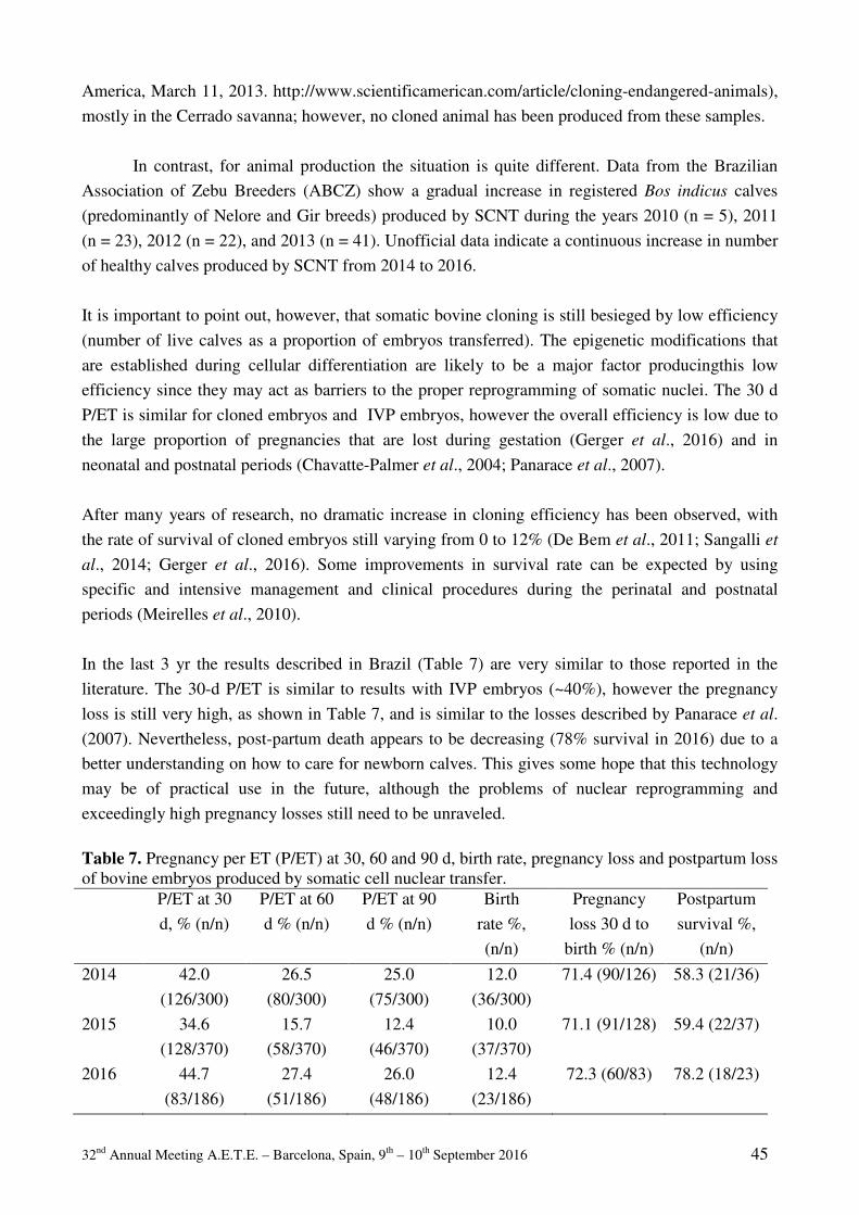
32nd Annual Meeting A.E.T.E. – Barcelona, Spain, 9th – 10th September 2016 45
America, March 11, 2013. http://www.scientificamerican.com/article/cloning-endangered-animals),
mostly in the Cerrado savanna; however, no cloned animal has been produced from these samples.
In contrast, for animal production the situation is quite different. Data from the Brazilian
Association of Zebu Breeders (ABCZ) show a gradual increase in registered Bos indicus calves
(predominantly of Nelore and Gir breeds) produced by SCNT during the years 2010 (n = 5), 2011
(n = 23), 2012 (n = 22), and 2013 (n = 41). Unofficial data indicate a continuous increase in number
of healthy calves produced by SCNT from 2014 to 2016.
It is important to point out, however, that somatic bovine cloning is still besieged by low efficiency
(number of live calves as a proportion of embryos transferred). The epigenetic modifications that
are established during cellular differentiation are likely to be a major factor producingthis low
efficiency since they may act as barriers to the proper reprogramming of somatic nuclei. The 30 d
P/ET is similar for cloned embryos and IVP embryos, however the overall efficiency is low due to
the large proportion of pregnancies that are lost during gestation (Gerger et al., 2016) and in
neonatal and postnatal periods (Chavatte-Palmer et al., 2004; Panarace et al., 2007).
After many years of research, no dramatic increase in cloning efficiency has been observed, with
the rate of survival of cloned embryos still varying from 0 to 12% (De Bem et al., 2011; Sangalli et
al., 2014; Gerger et al., 2016). Some improvements in survival rate can be expected by using
specific and intensive management and clinical procedures during the perinatal and postnatal
periods (Meirelles et al., 2010).
In the last 3 yr the results described in Brazil (Table 7) are very similar to those reported in the
literature. The 30-d P/ET is similar to results with IVP embryos (~40%), however the pregnancy
loss is still very high, as shown in Table 7, and is similar to the losses described by Panarace et al.
(2007). Nevertheless, post-partum death appears to be decreasing (78% survival in 2016) due to a
better understanding on how to care for newborn calves. This gives some hope that this technology
may be of practical use in the future, although the problems of nuclear reprogramming and
exceedingly high pregnancy losses still need to be unraveled.
Table 7. Pregnancy per ET (P/ET) at 30, 60 and 90 d, birth rate, pregnancy loss and postpartum loss of bovine embryos produced by somatic cell nuclear transfer. P/ET at 30
d, % (n/n)
P/ET at 60
d % (n/n)
P/ET at 90
d % (n/n)
Birth
rate %,
(n/n)
Pregnancy
loss 30 d to
birth % (n/n)
Postpartum
survival %,
(n/n)
2014 42.0
(126/300)
26.5
(80/300)
25.0
(75/300)
12.0
(36/300)
71.4 (90/126) 58.3 (21/36)
2015 34.6
(128/370)
15.7
(58/370)
12.4
(46/370)
10.0
(37/370)
71.1 (91/128) 59.4 (22/37)
2016 44.7
(83/186)
27.4
(51/186)
26.0
(48/186)
12.4
(23/186)
72.3 (60/83) 78.2 (18/23)
32nd Annual Meeting A.E.T.E. – Barcelona, Spain, 9th – 10th September 2016 46
Acknowledgments The authors would like to acknowledge the support from SBTE, FAPESP, CNPq and
CAPES of Brazil. We also would like to thank São Jorge Farm (São Pedro, SP), Santa Luzia Farm
(Passos, MG), São João True Type Farm (Inhaúma, MG), and Experimental Station Hildegard
Georgina Von Pritzelwitz (Londrina, PR). We also thank Dr. Milo Wiltbank for his collaboration
with the manuscript.
References Associação Brasileira das Indústrias Exportadoras de Carne (ABIEC). 2014. Estatísticas:
Balanço da Pecuária. São Paulo, BR, p.19.
Baruselli PS, Sales JNS, Sala RV, Vieira LM, Sa Filho MF. 2012. History evolution and
perspectives of timed artificial insemination programs in Brazil. Anim Reprod, 9:139-152.
Binelli M, Sartori R, Vasconcelos JLM, Pereira MHC, Monteiro Jr PLJ, Ramos RS. 2014.
Evolution in fixed-time: from synchronization of ovulation to improved fertility. p.493-506
in Reproduction in Domestic Ruminants VIII. Vol. 1. 1st ed. J. L. Juengel, A. Miyamoto, C.
Price, L. P. Reynolds, M. F. Smith, and R. Webb, ed. Context, Ashby de la Zouch, UK.
Bó GA, Baruselli PS, Martinez MF. 2003. Pattern and manipulation of follicular development in
Bos indicus cattle. Anim Reprod Sci, 78:307-326.
Bó GA, Cutaia L, Peres LC, Pincinato D, Maraña D, Baruselli PS. 2007. Technologies for
fixed-time artificial insemination and their influence on reproductive performance of Bos
indicus cattle. Soc Reprod Fertil Suppl, 64:223-236.
Chavatte-Palmer P, Remy D, Cordonnier N, Richard C, Issenman H, Laigre P, Heyman Y, Mialot JP. 2004. Health status of cloned cattle at different ages. Cloning Stem Cells,
6(2):94-100.
De Bem TH, Chiaratti MR, Rochetti R, Bressan FF, Sangalli JR, Miranda MS, Pires PR,
Schwartz KR, Sampaio RV, Fantinato-Neto P, Pimentel JR, Perecin F, Smith LC, Meirelles FV, Adona PR, Leal CL. 2011. Viable calves produced by somatic cell nuclear
transfer using meiotic-blocked oocytes. Cell Reprogram, 13(5):419-429.
Faber DC, Molina JA, Ohlrichs CL, Vander Zwaag DF, Ferré LB. 2003. Commercialization of
animal biotechnology. Theriogenology, 59:125-38.
Fleury PDC, Sanches BV, Cardoso BL, Basso AC, Arnold DR, Tannura JH, Pereira MHC, Gaitkoski D, Seneda MM. 2015. Comparison of pregnancy rates after fresh, vitrified or
cryopreserved in vitro produced embryos for direct transfer. Anim Reprod, 12:828.
(abstract).
Food Agriculture Organization of United Nations (FAO). 2012. Food outlook global market
analysis. Rome, ITA, p.143.
Fricke PM, Carvalho PD, Giordano JO, Valenza A, Lopes G Jr, Amundson MC. 2014.
Expression and detection of estrus in dairy cows: the role of new technologies. Animal,
8:134-143.

32nd Annual Meeting A.E.T.E. – Barcelona, Spain, 9th – 10th September 2016 47
Gerger RP, Zago FC, Ribeiro ES, Gaudencio Neto S, Martins LT, Aguiar LH, Rodrigues VH,
Furlan FH, Ortigari I, Sainz RD, Ferrell CL, Miglino MA, Ambrósio CE, Rodrigues JL, Rossetto R, Forell F, Bertolini LR, Bertolini M. 2016. Morphometric developmental
pattern of bovine handmade cloned concepti in late pregnancy. Reprod Fertil Dev, doi:
10.1071/RD15215. [Epub ahead of print].
Hasler JF. 2000. In vitro production of cattle embryos: problems with pregnancies and parturition.
Hum Reprod, 15(suppl. 5):47-58.
Instituto Brasileiro de Geografia e Estatística (IBGE). 2014. Produção da pecuária municipal.
Brasília, BR, p.80.
Lopez H, Satter LD, Wiltbank MC. 2004. Relationship between level of milk production and
estrous behavior of lactating dairy cows. Anim Reprod Sci, 81:209-223.
Meirelles FV, Birgel EH, Perecin F, Bertolini M, Traldi AS, Pimentel JR, Komninou ER,
Sangalli JR, Neto PF, Nunes MT, Pogliani FC, Meirelles FD, Kubrusly FS, Vannucchi CI, Silva LC. 2010. Delivery of cloned offspring: experience in Zebu cattle (Bos indicus).
Reprod Fertil Dev. 22(1):88-97.
Meneghetti M, Sá Filho OG, Peres RF, Lamb GC, Vasconcelos JL. 2009. Fixed-time artificial
insemination with estradiol and progesterone for Bos indicus cows I: basis for development
of protocols. Theriogenology, 72(2):179-189.
Morotti F, Sanches BV, Pontes JH, Basso AC, Siqueira ER, Lisboa LA, Seneda MM. 2014.
Pregnancy rate and birth rate of calves from a large-scale IVF program using reverse-sorted
semen in Bos indicus, Bos indicus-taurus, and Bos taurus cattle. Theriogenology, 81:696-
701.
Nascimento AB, Monteiro Jr PLJ, Fernandes GO, Pontes GCS, Prata AB, Melo LF, Bortoli R, Medeiros D, Surjus RS, Wiltbank MC, Sartori R. 2013a. Fertility of lactating dairy
cows submitted to TAI protocols with GnRH or estradiol compared to a pre-synchronization
or AI after estrus detection. Anim Reprod, 10:435. (abstract).
Nascimento AB, Souza AH, Pontes G, Wiltbank MC, Sartori R. 2013b. Assessment of
systematic breeding programs: A comparison between AI after estrus detection and timed AI
in lactating dairy cows. J Dairy Sci, 96(E-Suppl. 1):594-595. (abstract).
Panarace M, Agüero JI, Garrote M, Jauregui G, Segovia A, Cané L, Gutiérrez J, Marfil M,
Rigali F, Pugliese M, Young S, Lagioia J, Garnil C, Forte Pontes JE, Ereno Junio JC, Mower S, Medina M. 2007. How healthy are clones and their progeny: 5 years of field
experience. Theriogenology, 67:142-151.
Pereira MHC, Coelho MS.. 2016. Importância dos índices zootécnicos reprodutivos na Fazenda
Santa Luzia. O Embrião. In press.
Peres RF, Claro I Jr, Sá Filho OG, Nogueira GP, Vasconcelos JL. 2009. Strategies to improve
fertility in Bos indicus postpubertal heifers and nonlactating cows submitted to fixed-time
artificial insemination. Theriogenology, 72:681-689.
Perry G. 2014. Statistics of embryo collection and transfer in domestic farm animals. 2015.
Embryo Transfer Newsletter, December: 9-18.

32nd Annual Meeting A.E.T.E. – Barcelona, Spain, 9th – 10th September 2016 48
Pessoa GA, Martini AP, Carloto GW, Rodrigues MCC, Claro Junior I, Baruselli PS, Brauner CC, Rubin MIB, Côrrea MN, Leivas FG, Sá Filho MF. 2016. Different doses of equine
chorionic gonadotropin on ovarian follicular growth and pregnancy rate of suckled Bos
taurus beef cows subjected to timed artificial insemination protocol. Theriogenology,
85:792-799.
Pontes JHF, Nonato Jr I, Sanches BV, Ereno Jr J, Uvo S, Barreiros T, Oliveira J, Hasler J, Seneda MM. 2009. Comparison of embryo yield and pregnancy rate between in vivo and in
vitro methods in the same Nelore (Bos indicus) donor cows. Theriogenology, 71:690-697.
Pontes JH, Silva KC, Basso AC, Rigo AG, Ferreira CR, Santos GM, Sanches BV, Porcionato JP, Vieira PH, Faifer FS, Sterza FA, Schenk JL, Seneda MM. 2010. Large-scale in vitro
embryo production and pregnancy rates from Bos taurus, Bos indicus, and indicus-taurus
dairy cows using sexed sperm. Theriogenology, 74:1349-1355.
Rabiee AR, Lean IJ, Stevenson MA. 2005. Efficacy of Ovsynch program on reproductive
performance in dairy cattle: a meta-analysis. J Dairy Sci, 88:2754-2770.
Sá Filho OG, Meneghetti M, Peres RF, Lamb GC, Vasconcelos JL. 2009. Fixed-time artificial
insemination with estradiol and progesterone for Bos indicus cows II: strategies and factors
affecting fertility. Theriogenology, 72:210-218.
Sangalli JR, Chiaratti MR, De Bem TH, de Araújo RR, Bressan FF, Sampaio RV, Perecin F, Smith LC, King WA, Meirelles FV. 2014. Development to term of cloned cattle derived
from donor cells treated with valproic acid. Plos one, 9(6):e101022. doi:
10.1371/journal.pone.0101022. eCollection 2014.
Santos JEP, Rutigliano HM, Sa Filho MF. 2009. Risk factors for resumption of postpartum estrous cycles
and embryonic survival in lactating dairy cows. Anim Reprod Sci, 110: 207-221.
Santos JEP, Thatcher WW, Chebel RC, Cerri RL, Galvão KN. 2004. The effect of embryonic
death rates in cattle on the efficacy of estrus synchronization programs. Anim Reprod Sci,
82-83:513-535.
Sartori R. 2004. Fertilization and embryonic mortality in cattle. Acta Sci Vet, 32(suppl ):35-50.
Sartori R, Prata AB, Surjus RS, Mattos MCC, Basso AC, Pontes JHF, Goncalves JRS, Lima LG, Aguiar TS. 2013. Reproductive outcomes of timed AI or transfer of in vivo- or in
vitro-produced vitrified embryos in beef cattle. J Anim Sci, 91:595-595. (abstract).
Strickland JM, Martins JPN, Neuder LM, Pursley JR. 2010. Effect of 14/11 Presynch/Ovsynch
on 1st service conception rates of lactating dairy cows compared to AI following a detected
estrus. In: ‘2010 American Association of Bovine Practitioners Meeting’, 2010,
Albuquerque, New Mexico, USA.
Stringfellow DA, Seidel SM. 1998. Manual of the International Embryo Transfer Society. 3rd
Edition. (IETS: Savoy, IL).
United States Department of Agriculture (USDA). 2015. Milk cows and production by state and
region (Annual). Washington, DC, USA, p.2.
Viana JHM, Siqueira LGB, Palhao MP, Camargo LSA. 2012. Features and perspectives of the
Brazilian in vitro embryo industry. Anim Reprod, 9:12-18.

32nd Annual Meeting A.E.T.E. – Barcelona, Spain, 9th – 10th September 2016 49
Wiltbank MC, Gumen A, Sartori R. 2002. Physiological classification of anovulatory conditions
in cattle. Theriogenology, 57:21-52.
Wiltbank MC, Pursley JR. 2014. The cow as an induced ovulator: Timed AI after synchronization
of ovulation. Theriogenology, 81:170-185.

32nd Annual Meeting A.E.T.E. – Barcelona, Spain, 9th – 10th September 2016 50
32nd Annual Meeting A.E.T.E. – Barcelona, Spain, 9th – 10th September 2016 51
The timing of Puberty (oocyte quality and management)
Anne H Duittoz1, Yves Tillet1, Daniel Le Bourhis2, Laurent Schibler3
1 PRC, INRA, CNRS, IFCE, Université de Tours, 37380 Nouzilly, France
2 Allice, Department R&D, 37380 Monnaie, France 3Allice, Département R&D, F-75595 Paris, France
Abstract
This review aims at giving an overview on the physiological events leading to puberty
onset in mammals and more specifically in cattle. Puberty is an important
developmental milestone in mammals involving numerous changes in various
physiological regulations and behaviors. It is a physiological unique event integrating
several important central regulations at the crossroad of adaptation to environment:
reproductive axis, feeding behavior and nutritional controls, growth, seasonal rhythm
and stress. Puberty onset is also an important economic parameter in replacement heifer
program and in genomic selection (genomic bulls). The quest for advanced puberty
onset should be carefully balanced by its impact on physiological parameters of the
animal and its offspring. Thus one has to carefully consider each step leading to puberty
onset and set up a strategy that will lead to early puberty without being detrimental in
the long term. In this review, major contributions in the understanding of puberty
process obtained in rodents, primates and farm animals such as sheep and cattle are
discussed. In the first part we will detail the endocrine events leading to puberty onset
with a special focus on the regulation of GnRH secretion. In the second part we will
describe the neural mechanisms involved in silencing and reactivating the GnRH
neuronal network. These central mechanisms are at the crossroad of the integration of
environmental factors such as the nutritional status, the stress and the photoperiod that
will be discussed in the third part. In the fourth part, we will discuss the genetic
determinants of puberty onset and more particularly in humans, where several
pathologies are associated with puberty delay or advance and in cattle where several
groups have now identified genomic regions or gene networks associated with puberty
traits. Last but not least, in the last part we will focus on the embryologist point of view,
how to get good oocytes for in vitro fertilization and embryo development from younger
animals.

32nd Annual Meeting A.E.T.E. – Barcelona, Spain, 9th – 10th September 2016 52
1 Introduction
Puberty is an important developmental milestone in mammals involving numerous changes in
various physiological regulations and behaviors. It is a physiological unique event integrating
several important central regulations at the crossroad of adaptation to environment: reproductive
axis, feeding behavior and nutritional controls, growth, seasonal rhythm and stress.
Puberty, puberty onset, peri-pubertal, reproductive maturity what's the difference.
Puberty onset results from a complex and integrated sequence of biological events leading to
progressive maturation of sexual characteristics that ultimately lead to attainment of full
reproductive capacity. This sequence is referred as the timing of puberty. Puberty timing in
mammals is the result of evolution allowing females to attain ideal pelvic anatomy and size,
complete growth and maximize skeletal mineralization, prior to the demands of pregnancy, lactation
and offspring rearing.
Puberty is defined as the moment of the first emission of gametes, ie the first ovulation in females
and the first spermatozoa entering the epididymis in males. Therefore puberty is expressed as a date
or as an age. From this definition, it is obvious that puberty can be easily detected in females by
detecting the first ovulation. However in males there is no non-invasive method to assess the
presence of epididymal spermatozoa and it is usually defined according various physical and
behavioral changes. Therefore puberty is very often studied through the modifications observed
before and immediately following the first emission of gametes. In that case it is better to speak of
peri-pubertal period. For example in females, breeders usually monitor the exterior signs of
receptivity (age at first estrus). However one has to keep in mind that estrus behavior can exist
without a proper ovulation and the reciprocal is also true: ovulation can occur without any sign of
estrus behavior. For males, breeders look at the sexual behavior too: mounting behavior and
erection. Here again, this behavior does not mean that is there any spermatozoa in the ejaculate.
The strict definition of puberty onset as the first emission of gametes does not mean that the animals
are able to breed yet. They can produce and release gametes but reproduction is more than that.
Females usually need a period of time after puberty onset to have regular ovarian cycles and to get
their uterus capable of supporting a pregnancy. For males, the concentration of spermatozoa in the
ejaculate should reach a certain threshold to give an adequate fertility; here again this can take some
time after the puberty onset. Reproductive maturity is another phenomenon and the mechanisms
leading to puberty onset are different from those leading to reproductive maturity.
2 Endocrine basis of puberty
2.1 Brief overview of the endocrine events across the estrus cycle Post-pubertal females present estrus cycles, which is the reflection of the ovarian cyclicity. During
the late follicular phase, the preovulatory follicles release high estradiol levels in the blood stream.
The starting point of all endocrine events leading to ovarian cyclicity is the secretion of a
neurohormone: the gonadotropin releasing hormone (GnRH). GnRH acts on the gonadotrope cells
located in the anterior pituitary and promote the synthesis and release of both gonadotropins
luteinizing hormone (LH) and follicle stimulating hormone (FSH). LH and FSH will act at the
32nd Annual Meeting A.E.T.E. – Barcelona, Spain, 9th – 10th September 2016 53
ovary level to promote the release of gonadal steroid estrogens and progestagens and to promote
follicular growth. Granulosa cells and thecal cells collaborate to synthesize and release estrogens,
among which 17-β-estradiol (E2) is the most prevalent estrogen in most species. The amount of
estrogen released is dependent on the number of granulosa and thecal cells. Considering the growth
of a sphere, the amount of estrogen that can be synthetized is proportional to the cubic of the follicle
radius. Therefore the final growth of the dominant follicle is accompanied by a huge increase in E2
production. High E2 levels are responsible for the expression of estrus behavior. In most species
studied, high E2 levels will also exert a positive feedback on GnRH secretion leading a large
amount of GnRH release that causes a large amount of LH release: the pre-ovulatory GnRH and LH
surges (see Figure 1). LH surge occurs just before the ovulation and last several hours after,
contributing to the luteinization of granulosa and thecal cells of the ovulated follicle. De facto, E2
levels drop and the positive feedback disappears, stopping its repressive action on GnRH secretion.
In parallel progesterone (P4) secretion increases and exerts a negative feedback on GnRH secretion.
High P4 levels have a positive action on E2 receptors (ERs) expression. Without P4-priming during
the previous cycle, ERs expression is low and despite high E2 levels during the preovulatory phase,
the estrus behavior, which is strongly dependent on ERα signaling, is poorly expressed. Once
luteolysis occurs, P4 levels drop, the negative feedback is suppressed and GnRH secretion increases
again leading to LH and FSH release and a new follicular phase starting.
2.2 Evolution of gonadotropin secretion in the pre-pubertal period The key decisive event required for puberty to occur is an increase in pulsatile gonadotropin
releasing hormone (GnRH) release from GnRH neurons leading to gonadotropins LH and FSH
secretion. In mature adult, the GnRH is released in the portal veins in a pulsatile manner. GnRH
secretion is difficult to assess. As a matter of fact, GnRH is released in capillaries within the ME
that form portal veins along the pituitary stalk. From these portal veins, pituitary capillaries emerge
and the GnRH is released in the intercellular space and reaches anterior pituitary cells. GnRH
concentration in the portal veins varies between 4-100pg/ml (Caraty et al., 1982; Clarke and
Cummins, 1982; Levine et al., 1982; Irvine and Alexander, 1987; Gazal et al., 1998), which gives a
very small amount of GnRH for the small blood volume considered. Thus, the amount of GnRH
that passes in the general circulation is very small; the concentration is well below the detection
threshold of known hormonal assays. Moreover GnRH half-life is very short, a few minutes. Due to
its small peptidic structure, circulating endopeptidases degrades rapidly the GnRH. Therefore to
assess the GnRH secretion, blood should be punctured from pituitary portal vessels or from canulae
inserted in the third ventricle (Gazal et al., 1998) and this can only be performed in large animals
and requires invasive surgical procedures (Clarke and Cummins, 1982; Levine et al., 1982). An
alternative is to follow LH secretion since it has been clearly demonstrated that a GnRH pulse
precedes every LH pulse (Clarke and Cummins, 1982; Caraty et al., 1989).
In the female Rhesus monkey the early prepubertal period is characterized by an increase in
pulsatile release with a concomitant increase in pulse frequency and pulse amplitude. In the
midpubertal phase, only an increase in GnRH pulse amplitude is noticed and the global GnRH
secretion is increased during the night (Watanabe and Terasawa, 1989). This is in contrast to ewes
and heifers where the midpubertal period is characterized by an increase in LH pulse frequency
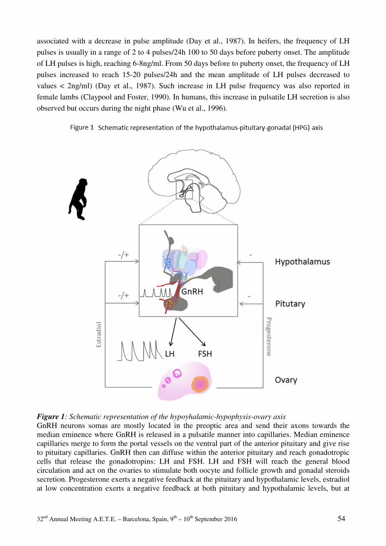
32nd Annual Meeting A.E.T.E. – Barcelona, Spain, 9th – 10th September 2016 54
associated with a decrease in pulse amplitude (Day et al., 1987). In heifers, the frequency of LH
pulses is usually in a range of 2 to 4 pulses/24h 100 to 50 days before puberty onset. The amplitude
of LH pulses is high, reaching 6-8ng/ml. From 50 days before to puberty onset, the frequency of LH
pulses increased to reach 15-20 pulses/24h and the mean amplitude of LH pulses decreased to
values < 2ng/ml) (Day et al., 1987). Such increase in LH pulse frequency was also reported in
female lambs (Claypool and Foster, 1990). In humans, this increase in pulsatile LH secretion is also
observed but occurs during the night phase (Wu et al., 1996).
Figure 1: Schematic representation of the hypoyhalamic-hypophysis-ovary axis
GnRH neurons somas are mostly located in the preoptic area and send their axons towards the median eminence where GnRH is released in a pulsatile manner into capillaries. Median eminence capillaries merge to form the portal vessels on the ventral part of the anterior pituitary and give rise to pituitary capillaries. GnRH then can diffuse within the anterior pituitary and reach gonadotropic cells that release the gonadotropins: LH and FSH. LH and FSH will reach the general blood circulation and act on the ovaries to stimulate both oocyte and follicle growth and gonadal steroids secretion. Progesterone exerts a negative feedback at the pituitary and hypothalamic levels, estradiol at low concentration exerts a negative feedback at both pituitary and hypothalamic levels, but at
32nd Annual Meeting A.E.T.E. – Barcelona, Spain, 9th – 10th September 2016 55
high concentration (during estrus) it will have a positive feedback at both pituitary and hypothalamic levels.
In spite of numerous physiological studies in model animals, little is known about the key events
leading to GnRH neurons progressive activation at puberty onset. The scientific community admits
that puberty onset is preceded by gradual changes in trans-synaptic and glial inputs to the GnRH
neuronal network. The trans-synaptic changes consist of a coordinated increase in excitatory inputs
and/or a reduction in inhibitory influences. Glial cells could also participate in regulating
extracellular glutamate concentration, and in releasing growth factors and small diffusible
molecules that directly or indirectly stimulate GnRH secretion. In addition to the classical excitatory
glutamatergic neurons, kisspeptin signaling through GPR54 was discovered in 2005 as a powerful
stimulator of GnRH release (Messager et al., 2005). Nevertheless, how these key events are
triggered through environmental and nutritional factors is far from being understood.
2.3 GnRH control
2.3.1 The two modes of secretion
GnRH secretion is characterized by two modes of secretion: pulsatile and continuous (the surge).
These two modes have been described in the pioneering work of Ernst Knobil in the rhesus monkey
where he described a tonic and a phasic mode of LH secretion controlled by two different areas
within the hypothalamus Preoptic area (POA) and mediobasal hypothalamus (MBH), respectively
(Nakai et al., 1978). The pulsatile pattern of GnRH secretion was confirmed in the 80's when a
trans-nasal surgical approach allowed the collection of blood from the portal vessels between the
hypothalamus and the pituitary (Clarke and Cummins, 1982; Levine et al., 1982).
GnRH/LH secretion is pulsatile during the follicular and the luteal phases, the surge mode occurs
during the pre-ovulatory period. In most species where GnRH and LH secretions have been
monitored simultaneously, the LH secretion profile is a good estimate of the GnRH pulsatile
secretion: a GnRH pulse always precedes one LH pulse. The frequency of pulsatile secretion varies
across the estrus cycle. For example in the ewe, the follicular phase is characterized by a high
frequency ie 1 pulse per hour, and low amplitude of LH pulses, whereas the luteal phase is
characterized by a low frequency ie 1 pulse per 6 hours but high amplitude of LH pulses (Moenter
et al., 1991). The GnRH pulse frequency is decoded by the GnRH receptor (GnRH-R) expressed by
gonadotropic cells: high frequency favors the expression of the β-LH subunit whereas low
frequency favors the expression of the β-FSH subunit (Bédécarrats and Kaiser, 2003; Thompson
and Kaiser, 2014).
2.3.2 Anatomy of the GnRH neuronal network
The GnRH is a small peptide (10 amino-acids) issued from the processing of pre-pro-GnRH
encoded by the Gnrh1 gene. The pre-pro-GnRH is processed in GnRH neurons to give the GnRH
and the GnRH-associated peptide (GAP), Both are packed in large dense core vesicles (LDCV) for
further release (Clarke et al., 1987). The GnRH is synthetized and secreted by a specialized
population of neurons: the GnRH neurons. In most mammals the GnRH neurons' somas are located
in the POA with a few cell bodies located in the MBH and the axons project towards the median
32nd Annual Meeting A.E.T.E. – Barcelona, Spain, 9th – 10th September 2016 56
eminence at the bottom of the MBH. However in primates, the repartition is different with the
majority of GnRH neurons' somas located in the MBH and just a few in the POA. Axonal
projections are projected to the median eminence where GnRH is released in blood capillaries and
transported in portal vessels to the capillaries network of the anterior pituitary where it will
stimulate the expression and release of the gonadotropins FSH and LH.
2.3.3 Extracerebral embryonic origin of GnRH neurons
During embryogenesis, the GnRH neurons originate from the medial part of the nasal embryonic
placode at early embryonic age 30 (E30) in sheep (Caldani et al., 1987; Caldani et al., 1995), E11.5
in mouse (Schwanzel-Fukuda and Pfaff, 1989; Wray et al., 1989), 6-7weeks of pregnancy in
humans (Schwanzel-Fukuda et al., 1996). They then migrate along olfactory-, vomero-nasal- and
terminal nerves to finally enter the forebrain through the cribiform plate. In the sheep, this phase of
nasal migration is completed at E45, in the mouse at E13.5. Once in the brain they turn ventro-
caudally to reach their final location in the POA/MBH. The phase of intra-cerebral migration is
completed at E60 in the sheep and E16.5 in the mouse. Once settled, they grow their axonal
projections toward the ME. This phase of axonal growth is terminated at E70 in sheep and E18.5 in
the mouse. Once connected to the ME, it is believed that GnRH secretion occurs since it is
correlated with the first observation of β-LH expression in the pituitary cells (Messaoud-Toumi et
al., 1993). Primary cultures derived from embryonic nasal explants from E26 sheep embryos, E35
rhesus monkey embryos or E11.5 mouse embryos allow the development of functional secreting
GnRH neurons that form a network in vitro. The GnRH secretion is pulsatile and the frequency is
correlated to what it is observed in vivo according to each species considered (Duittoz and Batailler,
2000; Constantin et al., 2009; Shingleton, 2015). These in vitro approaches suggest that the
pulsatility of secretion is an endogenous property of the GnRH network and that this property
develops during the fetal life.
2.3.4 Functionality of the fetal GnRH neuronal network
Whether the pulsatile secretion develops in utero and plays a role in development has been studied
particularly in the sheep species. Several groups have carried on a series of experiments on sheep
fetuses. In chronically catheterized ovine fetuses, both LH and FSH exhibit a similar trend of peak
values in midgestation (70-100 days) with a progressive decrease in plasma concentrations towards
term (145 days) (Sklar et al., 1981). Measurements from 55-60 days of gestation embryos gave low
values of plasma LH and FSH concentrations. This pattern is similar to the one described in human
fetuses with high concentration values of LH between 15-29 weeks of gestation (Kaplan and
Grumbach, 1976; Clements et al., 2009). The ovine fetal pituitary gland has the capacity to respond
to exogenous GnRH as early as 60 days of gestation with a maximal amplitude occurring during
mid-gestation (mueller et al., 1981). The pulsatile nature of LH fetal secretion was clearly assessed
by serial blood sampling during a 4 hours period in ovine fetuses at mid-gestation (Clark et al.,
1984) (Figure 2). If we put in parallel the physiological maturation profiles of LH secretion and the
development of the GnRH neuronal network we can clearly see the correlation between those
events. Thus, once GnRH neuronal migration and axonal growth toward the median eminence is
completed, the GnRH secretion can take place and induce the expression of gonadotropin subunits

32nd Annual Meeting A.E.T.E. – Barcelona, Spain, 9th – 10th September 2016 57
(Messaoud-Toumi et al., 1993). LH and FSH will act on the fetal gonad to stimulate gonadal steroid
synthesis, and this is particular evident in male ovine fetuses where a spurt in testosterone secretion
is detected at mid-gestation. This increase in testosterone in male fetuses or new born has been
demonstrated in numerous mammals (Foster and Hileman, 2015; Plant et al., 2015; Prevot, 2015).
In precocious mammals such as ovine and bovine species, the spurt occurs during the last third of
gestation and is terminated at birth, whereas in altricial species such as rodents, the spurt occurs
during the last days of pregnancy and during the first week post-natal (mice) (Sisk and Foster,
2004). After this "mini puberty", the frequency of GnRH/LH secretion dramatically decreases and
steroid levels drop; the infancy period is starting.
3 Puberty: endocrine or brain revolution?
The pubertal transition involves both gonadal and behavioral maturation. The increase in the
frequency of GnRH release and gonadotropins secretion progressively leads to the onset of gonadal
functions: gametogenesis and steroid production. These steroids act in turn onto the brain to
remodel neural circuits particularly those involved in sexual behaviors, but not only (Forger et al.,
2015). In humans, several neurological or psychiatric diseases appear or are exacerbated at puberty
(autism, schizophrenia, epilepsy, anorexia nervosa…).
Several decades of research have tempted to answer the question of the timing of the reactivation of
GnRH secretion and the onset of puberty. As mentioned earlier, the hypothalamic-pituitary gonadal
axis is functional during fetal/perinatal period, leading to the sexualization of external genitalia and
specific regions of the nervous system. This activation is limited in time but offers a window of
sensitivity to external factors such as endocrine disruptors (Parent et al., 2015; Hines et al., 2016).
3.1 Inhibitory mechanisms
3.1.1 Steroid-dependent mechanism
Early studies highlighted the role of the steroid negative feedback, the so-called "gonadostat"
hypothesis (Frisch and Revelle, 1970). The "gonadostat" theory implies a higher sensitivity of
GnRH neuronal network to the negative feedback of steroids: a steroid-dependent mechanism. In
the prepubertal period, GnRH secretion is less sensitive to the negative feedback of gonadal
steroids, the GnRH pulse frequency increases leading to gonadotropin secretion and gonadal
activation. In the sheep species, early post-natal gonadectomy leads to immediately increased levels
of gonadotropins as in the postpubertal period. Replacing steroid gonadal hormones causes
gonadotropins levels to go back to initial prepubertal values (Foster and Hileman, 2015). Similar
findings were found in other mammals: hamster, ferret (Sisk and Foster, 2004). In heifers the
negative feedback of estradiol declined as puberty approached (Day et al., 1987). However, in rat
and rhesus monkey, the gonadostat theory is not sufficient to account for the low gonadotropins
levels during infancy (Sisk and Foster, 2004). Interestingly, a steroid dependent mechanism exists at
the end of the juvenile period of female rhesus monkey (Rapisarda et al., 1983).
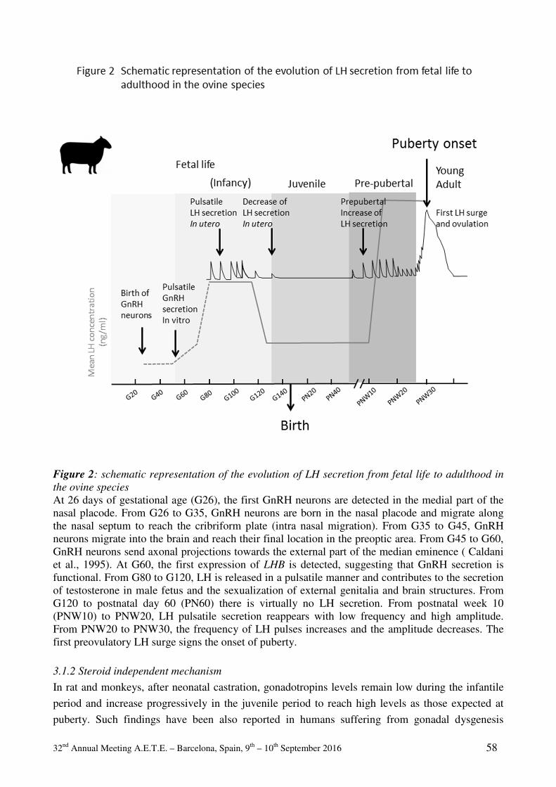
32nd Annual Meeting A.E.T.E. – Barcelona, Spain, 9th – 10th September 2016 58
Figure 2: schematic representation of the evolution of LH secretion from fetal life to adulthood in
the ovine species
At 26 days of gestational age (G26), the first GnRH neurons are detected in the medial part of the nasal placode. From G26 to G35, GnRH neurons are born in the nasal placode and migrate along the nasal septum to reach the cribriform plate (intra nasal migration). From G35 to G45, GnRH neurons migrate into the brain and reach their final location in the preoptic area. From G45 to G60, GnRH neurons send axonal projections towards the external part of the median eminence ( Caldani et al., 1995). At G60, the first expression of LHB is detected, suggesting that GnRH secretion is functional. From G80 to G120, LH is released in a pulsatile manner and contributes to the secretion of testosterone in male fetus and the sexualization of external genitalia and brain structures. From G120 to postnatal day 60 (PN60) there is virtually no LH secretion. From postnatal week 10 (PNW10) to PNW20, LH pulsatile secretion reappears with low frequency and high amplitude. From PNW20 to PNW30, the frequency of LH pulses increases and the amplitude decreases. The first preovulatory LH surge signs the onset of puberty.
3.1.2 Steroid independent mechanism
In rat and monkeys, after neonatal castration, gonadotropins levels remain low during the infantile
period and increase progressively in the juvenile period to reach high levels as those expected at
puberty. Such findings have been also reported in humans suffering from gonadal dysgenesis
32nd Annual Meeting A.E.T.E. – Barcelona, Spain, 9th – 10th September 2016 59
(Winter and Faiman, 2009). Although the precise neuronal target of gonadal steroid feedback is not
clearly known, POA and the ArcN are involved in sensing estradiol negative feedback in
gonadectomized prepubertal rats (Uenoyama et al., 2015). GnRH neurons, although located in the
POA, are not considered as the primary target since they do not express the estrogen receptor α
(ERα) albeit they do express ERβ (Hrabovszky et al., 2000; Herbison and Pape, 2001) but this later
isoform does not seem to be involved in puberty onset. To account for this steroid-independent
system, one assumption is that during infancy, inhibitory brain circuits block GnRH secretion; this
break is released at puberty concomitantly with the onset of stimulatory brain circuits. GABA (γ-
aminobutyiric acid) neurons are involved in the inhibition of GnRH neurons during juvenile period
in several species. In the rhesus monkey, Terasawa's group showed the existence of a GABAergic
break on gonadotropin secretion during the juvenile period. GABA level in the pituitary stalk (PS)
and ME of juvenile monkeys is high but decreases during the peripubertal period (Mitsushima et al.,
1994; Terasawa, 2005). The local infusion of GABA-A receptor antagonist bicuculline in the PS-
ME of juvenile female rhesus monkey induces a rise in gonadotropins levels and the onset of
ovarian cyclicity (Keen et al., 2011). The infusion of anti-sens mRNA encoding GAD67 (glutamic
acid decarboxylase 67), a key enzyme for the synthesis of GABA, in juvenile rhesus monkey
females triggered puberty onset with estrus cyclicity and ovulation (Kasuya et al., 1999) (Figure 3).
Both mechanisms co-exist to a different degree according to the species considered and also to the
sex. One theoretical hypothesis would be that the steroid-independent mechanism provides a coarse
regulation and will program the year (month) of puberty onset and the steroid-dependent
mechanism will program the week/day when the first ovulation occurs.
3.2 Excitatory mechanisms The pubertal reduction in GABAergic inhibition is accompanied by an increase in glutamate levels
in the PS-ME, as well as an increase in the levels of the stimulatory neurotransmitters such as
noradrenaline and Neuropeptide Y (NPY) (Gore and Terasawa, 1991).
3.2.1 Neuropeptide Y
NPY is an appetite-stimulating neuropeptide and a neuromodulator of neuroendocrine functions.
The interactions between NPY and neuroendocrine networks are complex and depend upon the sex
and steroid environments. For example NPY is a potent stimulator of LH secretion in sex-steroid
primed rats (Allen et al., 1985), whereas its intra-cerebroventricular (ICV) administration in
gonadectomized rats inhibits LH release (McDonald et al., 1989). In the male Rhesus monkey, NPY
exerts a negative effect on the GnRH pulse generator in prepubertal animals (Majdoubi et al., 2000).
However in the female Rhesus monkey NPY release in the ME increases and is responsible for the
observed increase in LH secretion at puberty onset (Gore et al., 1993). Two populations of NPY
containing neurons have been described in the ArcN and the authors suggest that these two
populations have distinct roles during the prepubertal period and at puberty onset (Majdoubi et al.,
2000) (Figure 3). In the prepubertal ewe, NPY stimulates the expression of Lhb (β-LH subunit) in
gonadotrope cells (Wańkowska and Polkowska, 2009). Neuroanatomical studies in prepubertal
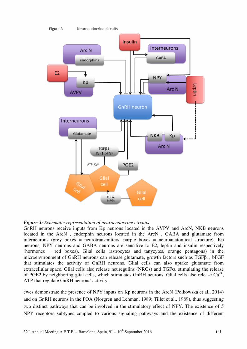
32nd Annual Meeting A.E.T.E. – Barcelona, Spain, 9th – 10th September 2016 60
Figure 3: Schematic representation of neuroendocrine circuits
GnRH neurons receive inputs from Kp neurons located in the AVPV and ArcN, NKB neurons located in the ArcN , endorphin neurons located in the ArcN , GABA and glutamate from interneurons (grey boxes = neurotransmitters, purple boxes = neuroanatomical structure). Kp neurons, NPY neurons and GABA neurons are sensitive to E2, leptin and insulin respectively (hormones = red boxes). Glial cells (astrocytes and tanycytes, orange pentagons) in the microenvironment of GnRH neurons can release glutamate, growth factors such as TGFβ1, bFGF that stimulates the activity of GnRH neurons. Glial cells can also uptake glutamate from extracellular space. Glial cells also release neuregulins (NRGs) and TGFα, stimulating the release of PGE2 by neighboring glial cells, which stimulates GnRH neurons. Glial cells also release Ca2+, ATP that regulate GnRH neurons' activity. ewes demonstrate the presence of NPY inputs on Kp neurons in the ArcN (Polkowska et al., 2014)
and on GnRH neurons in the POA (Norgren and Lehman, 1989; Tillet et al., 1989), thus suggesting
two distinct pathways that can be involved in the stimulatory effect of NPY. The existence of 5
NPY receptors subtypes coupled to various signaling pathways and the existence of different
32nd Annual Meeting A.E.T.E. – Barcelona, Spain, 9th – 10th September 2016 61
hypothalamic and pituitary targets, can account for such opposite effects observed according to the
sex, the steroid environment and the physiological state (Pralong, 2010).
3.2.2 Glutamate/NMDA
The excitatory amino acid glutamate and especially its NMDA subtype receptor are important
components of the neural system that regulates sexual maturation. Multiple daily injections of
NMDA agonists to immature rats (Smyth and Wilkinson, 1994) and monkeys (Urbanski and Ojeda,
1990) induce precocious puberty. On the contrary, administration of the non-competitive NMDA
antagonist, MK801, delays puberty onset (Veneroni et al., 1990). GnRH neurons receive direct
glutamatergic inputs and express NMDA and kainite receptors (Figure 3). Hypothalamic glutamate
contents increase during the prepubertal period and reach maximal values at puberty onset.
Glutamate receptors are ubiquitous in the CNS and they play important roles in many processes
involving excitatory mechanisms, whether this increase in glutamatergic signaling is specific to
puberty onset or whether it's a more general developmental process is not known (Parent et al.,
2005) .
3.2.3 Kisspetin/GPR54
In 2004, a new key component was discovered, originally named metastin due to its anti-mitotic
properties and now named Kisspeptin (Kp) (Matsui et al., 2004; Seminara, 2005). Kiss1 encodes a
54 amino-acids peptide Kp-54 (Kisspeptin-54) that cleaves into several shorter forms (Kp14, Kp13
and Kp10) forming the Kp family. Kp neurons strongly regulate the activity of GnRH neurons. Kp
acts through a G-protein coupled receptor (GPCR): GPR54. Kp neurons are found in two distinct
populations: ArcN and anteroventral periventricular nucleus (AVPV) (Figure 3). GnRH neurons
express GPR54 and Kp fibers contact GnRH terminals in the ME. In humans, mutations in the
GPR54 gene lead to an hyponadotropic hypogonadism (HH) characterized by a deficiency in
pituitary secretion of gonadotropins which results in the impairment of pubertal maturation and of
reproductive function (de Roux et al., 2003). Genetic models in rodents highlighted the central role
of Kp/GPR54 system in the onset of puberty (Colledge and de Tassigny, 2009) (Figure 3). The
Kp/GPR54 system is strongly regulated by metabolic factors and environmental factors, and could
represent the central hub for decoding metabolic and environmental cues.
3.2.4 Glial regulation
When speaking of neuroendocrine regulations, most scientists focus on the roles played by neuronal
circuits, neurotransmitters and neuromodulators and their cognate receptors. During the prepubertal
period, although neuronal networks synaptically-connected to GnRH neurons govern the increase in
GnRH secretion; glial cells contribute to the processes engaged through several mechanisms. Glial
is a generic adjective to characterize several cell populations that are associated with GnRH
neurons: astrocytes, tanycytes and olfactory unsheathing cells. For the sake of simplicity, we use the
generic term, bearing in mind that different phenotypic cell types support it. One mechanism
involves the production of growth factors acting on serine/threonine kinase receptors. Growth
factors such as Transforming Growth Factor α (TGFα) and neuregulins acting on erbB receptors
play a major role in glia-GnRH neurons communication. Activation of erbB receptors in glial cells

32nd Annual Meeting A.E.T.E. – Barcelona, Spain, 9th – 10th September 2016 62
associated with GnRH neurons, leads to the release of prostaglandin E2 (PGE2), which stimulates
the electrical activity of GnRH neurons and the GnRH release (Prevot et al., 2003a; Prevot et al.,
2003b; Prevot et al., 2005; Ojeda et al., 2008). Other growth factors such as TGFβ, IGF1, bFGF are
secreted by glial cells and regulate directly the activity of GnRH neurons (Ojeda et al., 2010)
(Figure 3). Besides the secretion of growth factors, glial cells release small molecules such as
calcium, glutamate and ATP that affect the GnRH neuronal activity. Glial cells can also uptake K+
ions and glutamate that accumulate in the extracellular space during neuronal activity through glial
specific dedicated transporters. These mechanisms are of major importance in regulating neuronal
electrical activity and excitability. These mechanisms of regulation are tightly dependent upon the
distance between the membrane of the glial and the synaptic cleft (Giaume et al., 2010).
Another mechanism that can affect glia-GnRH neurons interactions is the modulation of
adhesiveness of glial cells onto GnRH neurons. Glial cells interact with GnRH neurons via
hemophilic interactions involving Neural Cell Adhesion Molecule (NCAM) and synaptic cell
adhesion molecule (SynCAM1). In contrast, the poly-syalilated form of NCAM, PSA-NCAM,
prevents hemophilic interactions between adjacent glial and GnRH neuronal cells. Heterophilic
interactions also exist via the neuronal membrane protein contactin and the glial receptor like
protein tyrosine phosphatase-b (Parent et al., 2007). These cell-to-cell interactions can trigger
intracellular signaling cascades that can affect both glial and neuronal activities (Viguie et al., 2001;
Parkash and Kaur, 2007; Sharif et al., 2013). Altering cell-to-cell communication through glial gap
junctions or hemichannels decreases dramatically GnRH neuronal activity and GnRH secretion in
vitro (Pinet-Charvet et al., 2015). Gap-junctions have previously been reported in the
hypothalamus, particularly in the ArcN of female rats, where they are regulated by estrogen ( Perez
et al., 1990). Hypothalamic tanycytes, particularly the β-type which is closely associated with
GnRH nerve terminals in the ME, express functional connexin-43 (Cx-43) hemichannels encoded
by Gja1, which play a role in a glucose-sensing mechanism by releasing ATP (Orellana et al.,
2012). The Gja1 (Cx-43) promoting region contains AP1 and AP2 sites and a series of half
palindromic estrogen response elements suggesting that Cx-43 (Gja1) expression can be directly
regulated by estrogen levels (Yu et al., 1994). Taken altogether, these studies suggest that glial cells
might exert a control of GnRH neuronal network as important as the classical transynaptic model.
Therefore, several layers of neuronal and glial components are involved in controlling the onset of
puberty, increasing the complexity of the system. The most important question remains: what
determines the timing of the inhibitory break removal and/or the timing on excitatory inputs onset?
4 Puberty: environmental cues
The timing of puberty is maybe the best example of the interaction between genotype and
environment. Puberty is a physiological event integrating several important central regulations at
the crossroad of adaptation to environment: reproductive axis, feeding behaviour and nutritional
controls, growth, seasonal rhythm, corticotropic axis and stress.
32nd Annual Meeting A.E.T.E. – Barcelona, Spain, 9th – 10th September 2016 63
4.1 Nutrition and metabolism
Nutritional factors have been considered for a long time as the key factor in puberty onset. In
humans, until the mid-20th century, a gradual decline in age at menarche (first menstruation) has
been reported in most industrialized populations. It is generally admitted that this trend was due to
gradual improvements in nutrition and healthcare (Sørensen et al., 2012) giving birth in the 70ies to
the critical fat mass hypothesis according which, for a given species a critical fat mass is necessary
for puberty onset. The link between nutrition and puberty onset was confirmed in numerous studies
on laboratory animals, and also in farm animals. Adequate growth and adiposity are critical for the
onset of puberty in mammals. Food restriction (Foster and Olster, 1984; Suttie et al., 1991) and
excessive exercise during the juvenile period delay the onset of puberty (Manning and Bronson,
1989; Manning and Bronson, 1991). The mechanism involved is the maintenance of the juvenile
high sensitivity to the negative feedback sensitivity to gonadal steroids. In contrast, increased
adiposity advance the onset of puberty (Kaplowitz et al., 2001; Rosales Nieto et al., 2014). This
occurrence is associated with attenuation of estradiol negative feedback and increased pulsatile
release of LH (Gasser, 2006). Therefore, nutritional cues interact with gonadal steroid feedback to
time the onset of puberty in females. These findings led to the concept of nutritional programming
of puberty in cattle. Age at puberty in cattle is indeed influenced by food intake, food composition
and body weight (BW). It is usually admit that puberty occurs at 55-65% of adult BW, depending
on the breed considered (Freetly et al., 2011). However, the cost of supplemental feeding to reach
this target BW earlier is not always compensated by a sufficient improve in reproduction and calf
production (Davis Rincker et al., 2011). The permissive nutritional signals for puberty onset are
metabolic cues such as glucose, insulin and leptin for the most studied factors. These metabolic
markers signal the brain that the somatic growth and energy stores are sufficient to sustain
pregnancy and lactation without threating the mother and foetus' health. Interestingly, these factors
are also important for males, although the mechanisms involved may differ. Since the discovery of
the fat-signalling hormone leptin (Zhang et al., 1994), whose blood level is proportional to the
amount of adipose tissue (Frederich et al., 1995), a great amount of research work has tried to
demonstrate that leptin is a hormonal messenger signalling the metabolic state for initiating puberty
and also for fertility. Studies performed in rodents suggested that leptin administration could
advance the onset of female puberty (Ahima et al., 1997). Humans with leptin deficiency due to
mutations in the leptin gene or in the leptin receptor, and mouse models with inactivated leptin gene
or leptin receptor gene, are obese and do not undergo puberty (Chehab et al., 1996). However leptin
administration in healthy juveniles does not advance puberty onset. In ewes (Henry et al., 2011) and
cows (Amstalden et al., 2002) leptin administration does not affect the secretion of LH but leptin
prevents fasting-induced reduction in LH pulsatility in prepuberal heifers (Maciel, 2004). In
addition to leptin, other hormones such as insulin, or nutrients such as glucose, fatty acids and
amino-acids have been shown to regulate GnRH neuronal activity in a direct manner or via a
complex glial/neuronal network. Among the critical neuronal pathways, hypothalamic NPY/agouti-
related protein (AgRP) and proopiomelanocortin (POMC) neurons located in the ArcN are
considered as the two major pathways mediating nutritional cues. Theses neurons express the leptin
receptor and target GnRH neurons, setting the physical pathway for the control of puberty onset. A
small subpopulation of Kp neurons in the ArcN express LepR (Louis et al., 2011) and may
32nd Annual Meeting A.E.T.E. – Barcelona, Spain, 9th – 10th September 2016 64
constitute another target for nutritional regulation see (Sánchez-Garrido and Tena-Sempere, 2013)
for a review. However selective ablation of LepR in Kiss1 expressing neurons does not alter
puberty onset and fertility (Donato et al., 2011).
Taken altogether, these studies support a permissive role of leptin in the metabolic gating of
pubertal maturation (Barash et al., 1996; Cheung et al., 1997).
4.2 Photoperiod
In photoperiodic species, puberty onset will depend on the timing of the birth. For example in the
ovine species lambs born at the end of the winter or during spring time reach puberty at the next
breeding season in autumn, a younger age that those born during autumn, reaching puberty at the
following breeding season 10-12 months later. This delay in puberty in autumn-born ewe lambs is
due to a prolonged hypersensitivity to the negative steroid feedback (Foster and Hileman, 2015).
Similar findings were observed for photoperiodic short-lived animals such as Siberian hamsters
where spring born individuals mature rapidly and breed during the summer whereas young born in
lid to late summer have a delayed puberty the next spring (Butler et al., 2007). Exposing Holstein
heifers to long day photoperiod enhance BW gain and hasten the onset of puberty (Rius et al.,
2005), a result that has been observed also for the seasonal Murrah buffalo species (Roy et al.,
2016). In photoperiodic species, the variation in food intake and metabolism is an adaptive
physiological mechanism allowing the storage of energy resources in anticipation of the harsh days
of winter. The immune response is also sensitive to photoperiod, short days photoperiod enhance
immunological defenses. This seasonal plasticity of the immune system is highly conserved and is
in opposite phase with the breeding season, one explanation would be that the energy cost of both
activating reproduction and maintaining the immune function at its higher level is too high (Walton
et al., 2011). In dairy cows, short days photoperiod improve mammary gland capacity, prolactin
secretion and immune function (Dahl, 2008).
4.3 Stress and corticotropic axis
Prolonged or chronic stress results in the suppression of gonadotropin secretion and the inhibition of
reproduction. Acute stress has variable effects (Tilbrook et al., 2000). Studies on adaptive response
processes highlighted a positive link between childhood adversities with accelerated female
reproductive development. Longer-term health costs are traded off for increased probability of
reproducing before dying via a process of accelerated reproductive maturation. Early adversity,
early sexual maturation form the core component linking stress physiology with poor health later in
life (Hochberg and Belsky, 2013).
5 Puberty: genetic determinants
While the timing of pubertal onset varies within and between different populations, it is a highly
heritable trait, suggesting strong genetic determinants. Previous epidemiological studies estimate
that 60–80% of the variation in pubertal onset is under genetic regulation (Parent et al., 2003;
Gajdos et al., 2010). Abnormal pubertal timing affects up to 5% of adolescents and is associated
32nd Annual Meeting A.E.T.E. – Barcelona, Spain, 9th – 10th September 2016 65
with adverse health and psychosocial outcomes.
5.1 Genetic factors associated with delay of puberty in Humans
Idiopathic hypogonadotropic hypogonadism (IHH) is defined by absent or delayed sexual
development, with puberty being either absent or incomplete by the age of 18 years. Deleterious
mutations in genes coding for factors necessary for the migration of GnRH neurons lead to
hypogonadotropic hypogonadism (IHH), which is the absence of puberty associated with low levels
of gonadotropins and gonadal steroids. IHH is frequently accompanied by non-reproductive
abnormalities such as anosmia (Kallmann's syndrome). In the Kallmann's syndrome, which
associates IHH and anosmia, mutated genes encode for proteins involved in the development of
GnRH neurons (Hardelin et al., 1992; Franco et al., 1992). The disruption of the migration of GnRH
neurons causes them to stay into the nasal region or at the level of the cribriform plate, and they do
not reach their final location in the hypothalamus. The Kallmann's syndrome is associated to
mutations in KAL1 , FGFR1 (Dodé et al., 2003), NELF (Miura et al., 2004; Xu et al., 2011),
PROKR2 (Dodé et al., 2006), FGF8 (Hardelin and Dodé, 2008), CHD7 (Kim et al., 2008), and
WDR11 (Kim and Layman, 2011) genes encoding for anosmin, FGF receptor 1 (FGF-R1), NMDA
receptor synaptonuclear signaling and neuronal migration factor (alias Nasal Embryonic Factor),
prokinecitin receptor 2, FGF-8, chromodomain helicase binding protein 7, WD repeat domain 11,
respectively (Figure 4). In normosmic IHH (nIHH), the development of GnRH neurons is not
affected but the functionality of the GnRH secretion is altered. n-IHH cases are associated with
mutations in GNRH (Chevrier et al., 2011), KISS1 (de Roux et al., 2003; Bianco et al., 2011), DAX1
(Habiby et al., 1996; Merke et al., 1999), GNRH1 (Bouligand et al., 2009; Chan et al., 2011),
LEPR/LEP (Clement et al., 1998), PCSK1 (Jackson et al., 2003), PROKR2/ PROK2 (Dodé et al.,
2006), SEMA3A/ SEMA7A (Hanchate et al., 2012; Young et al., 2012), TACR3/ TAC3 ( Topaloglu
et al., 2009, Topaloglu, 2010), DMLX2 (Tata et al., 2014) genes encoding GnRH-R, GPR54,
nuclear receptor 0B1, GnRH, Leptin-R, leptin, protein convertase subtilisin/kexin type 1,
prokinecitin receptor 2, prokinecitin, neurokinin-B receptor, semaphorins-3a and -7a, neurokinin-B
and Rab-connectin-3, respectively (Figure 4). Most cases of IHH are sporadic, consistent with the
affected individuals being infertile, but familial transmission has also been well described. Kindred
analysis suggests that IHH is a wider spectrum of disease with individuals and relatives sharing an
apparent common genotype but displaying a variety of reproductive or non-reproductive
phenotypes. Oligogenicity could be one explanation for this phenotypic variation (Mitchell et al.,
2011).
Oligogenic and complex genetic environmental interactions have now been identified, with
physiological and environmental factors interacting in genetically susceptible individuals to alter
their reproductive capacities.
5.2 Genetic factors associated with precocious puberty in Humans
Human precocious puberty is defined as the development of secondary sexual characteristics and
elevated sexual hormones before 8 years of age in girls and 9 years of age in boys. There are two
major forms of premature sexual maturation: inappropriate early activation of HPG axis that
induces central precocious puberty (CPP) and peripheral precocious puberty (PPP) due to the
32nd Annual Meeting A.E.T.E. – Barcelona, Spain, 9th – 10th September 2016 66
increase of sex steroids with no activation of the HPG axis. Precocious puberty is highly deleterious
since it will cause short stature, psychosocial problems and increase the risk of adulthood diseases.
Mutations in the LHCGR gene coding the LH receptor (LH-R) and leading to constitutive activation
of the LH-R without ligand were the first mutations characterized in various family cases of
peripheral precocious puberty limited to the male (Layman, 1999). These mutations affected only
the male offspring and were without effect on the females. Recently cases of central precocious
puberty have been associated with genetic variants affecting Kp signalling: mutation in the KISS1
gene encoding Kp (Silveira et al., 2010), (Mazaheri et al., 2015) or activating mutation of the
KISS1R gene encoding GPR54 the Kp receptor (Teles et al., 2009; Silveira et al., 2010) (Figure 4).
One of these mutations was present at heterozygous state in patient's mother and grandmother
suggesting incomplete sex-dependent penetrance. Another possibility is that other genes could be
involved in this phenotype evoking the oligogenicity concept in central precocious puberty as was
well described for IHH (Mitchell et al., 2011).
Other cases of central precocious puberty are associated with mutations in the imprinted MKRN3
gene encoding the makorin ring finger protein 3, a gene located in the imprinted Prader Willi
syndrome region (Settas et al., 2014; Simon et al., 2015) (Figure 4). Data from Human cases and
animal models suggests that MKRN3 plays an inhibitory role in the reproductive axis and may
represent a new pathway in pubertal regulation (Ong et al., 2009; Simon et al., 2015). MKNR3 is
expressed ubiquitously.
Before 2000, clinical studies were individual case studies but now with the improvement of the
methods of sequencing of the genome, the increase of the capacities of calculation and the
improvement of the algorithms, the studies of association of genomic data allow to find genetic
variants associated to the age in the puberty. With this process, more than 100 loci involved in the
susceptibility to precocious puberty have been discovered. Among them the LIN28B locus is one of
the most significant (Ong et al., 2009; Elks et al., 2010) (Figure 4). LIN28B is a human homolog of
lin28 of Caenorhabtidis elegans, which was originally identified as a heterochronic regulator of
developmental timing (Ambros and Horvitz, 1984) Deleterious mutations in lin28 resulted in
precocious larval to adult development and a partial transformation in sexual phenotype (Ambros,
2011). The Lin28 proteins are potent and specific post-transcriptional repressors of the biogenesis
of let-7 miRNAs, which are time-specific expressed miRNAs that control developmental timing
(Zhu et al., 2010).
A recent meta-analysis suggests that the variant allele carriers, especially people with heterozygote
genotype for ESR1 XbaI polymorphism and the wild allele for ESR1 PvuII polymorphism, are
associated with precocious puberty susceptibility (Luo et al., 2015) (Figure 4).
5.3 Genetic factors associated with age at puberty in cattle
Age at first calving usually varied between 24 and 36 months, according to cattle breeds and is
considered a key factor in terms of profitability and efficiency in both dairy and beef cattle.
Likewise, bull puberty also shows significant differences within and among breeds. In dairy cattle,
age at first has continually decreased during the last decades. Improvement in nutrition and health
have certainly contributed to an improve BW gain, but genetic selection for improved breeding and
economic efficiency may also have indirectly impacted the onset of puberty (precocity) (Mourits et
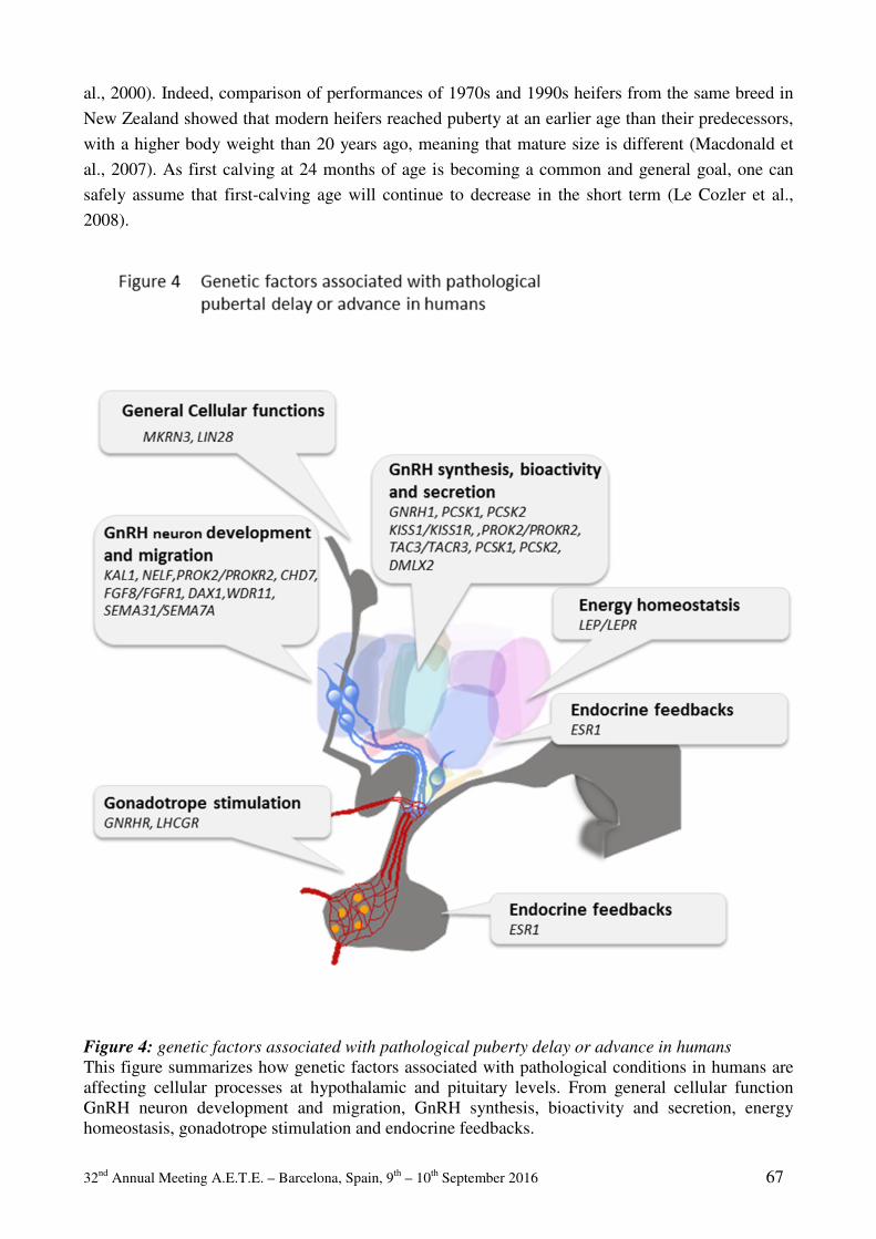
32nd Annual Meeting A.E.T.E. – Barcelona, Spain, 9th – 10th September 2016 67
al., 2000). Indeed, comparison of performances of 1970s and 1990s heifers from the same breed in
New Zealand showed that modern heifers reached puberty at an earlier age than their predecessors,
with a higher body weight than 20 years ago, meaning that mature size is different (Macdonald et
al., 2007). As first calving at 24 months of age is becoming a common and general goal, one can
safely assume that first-calving age will continue to decrease in the short term (Le Cozler et al.,
2008).
Figure 4: genetic factors associated with pathological puberty delay or advance in humans
This figure summarizes how genetic factors associated with pathological conditions in humans are affecting cellular processes at hypothalamic and pituitary levels. From general cellular function GnRH neuron development and migration, GnRH synthesis, bioactivity and secretion, energy homeostasis, gonadotrope stimulation and endocrine feedbacks.

32nd Annual Meeting A.E.T.E. – Barcelona, Spain, 9th – 10th September 2016 68
Despite its economic importance, only a few studies have been conducted to identify genes and
mutations associated with onset of puberty in either bulls or heifers. Most of these studies were
done in beef cattle (mainly Angus), tropical breeds such as Brahman and Nelore cattle (Bos indicus
cattle) and crosses which are reportedly older at puberty when compared with most Bos taurus
breeds (Lunstra and Cundiff, 2003). Several parameters have been measured as a phenotype to
study heifer puberty, from simple traits such as age at first service, age at first calving and age at
first oestrus to more expensive and difficult to measure ones such as age at first corpus luteum
(ultrasonography) or plasma progesterone concentration. For males, scrotal circumference, sperm
quality (concentration, motility and morphology) as well as LH or IGF-1 circulating blood
concentration have been monitored. One has to be aware that the nature of the quantitative puberty
traits thus differs between studies. Moreover, their physiological meaning might be different than
the strictly defined puberty onset. For example age at first oestrus does not mean age at puberty
onset since oestrus behaviour is usually not present before the third oestrus cycle. Age at first
calving is not age at first oestrus since the genital tract need several oestrus cycles to be fully
developed in order to insure a full-length pregnancy.
Moderate to high heritability has been computed for heifer’s age at puberty (0.2 to 0.48) and scrotal
circumference (0.22 to 0.42) (Vargas et al., 1998), suggesting that timing of puberty is likely to be a
multigenic trait. Genetic correlations have also been observed between scrotal circumference or
male IGF-1 blood concentration and heifer’s age at puberty, suggesting that some common
pathways may be involved in the two genders (Martinez-Velazquez and Gregory, 2003; Morris et
al., 2010; Johnston et al., 2013).
Despite the multigenic nature of puberty onset, some major key player genes have been identified in
humans, stimulating association studies in cattle, focused on some candidate genes. Polymorphisms
in GNRHR, LHR and IGF were search for association with age of puberty in Angus male cattle
(Lirón et al., 2012), showing significant association with one SNP located in IGF1. Likewise,
polymorphisms in the LHR, FSHR and GNRHR were analysed in the Nellore breed, showing only
association between FSHR and early puberty phenotype (Milazzotto et al. 2008). Furthermore,
seven genes from the IGF1 pathway (IGF1R, IGFBP2, IGFBP4, EIF2AK3, PIK3R1, GSK3B and
IRS1) were shown to be associated with heifer puberty in both Tropical Composite or Brahman
breeds (Fortes et al., 2013). These findings support the hypothesis that IGF1 regulates arrival to
puberty in male calves and also impact heifer puberty. In contrast to human and mouse, there are no
evidences that genetic variation within GNRH, LH and its receptors could impact the regulation of
pubertal timing in cattle. Based on their known effect on sexual precocity in mammals, 57 candidate
genes related to lipid metabolism were also studied on a large panel of 1689 precocious and non-
precocious Nellore heifers. Statistical analysis revealed that SNPs located within the FABP4 and
PPP3CA gene had a significant effect on sexual precocity (Dias et al., 2015).
Genome-wide association studies (GWAS) using microsatellites or SNPs have also been set up to
identify QTL regions and highlight to new candidate genes. A search for markers associated with
heifer’s age at puberty and age at first calving in the Animal QTLdb (Hu et al., 2016) retrieves
about 350 markers located within roughly 200 QTL regions, irrespective to breeds. Likewise, 10650
makers within 60 regions have been associated with male puberty, mainly on the X chromosome.
Several candidate genes have been proposed starting from these regions and regulatory networks
32nd Annual Meeting A.E.T.E. – Barcelona, Spain, 9th – 10th September 2016 69
have been constructed (Fortes et al., 2010a; Fortes et al., 2010b; Fortes et al., 2011; Fortes et al.,
2016). These findings suggest an enrichment of genes involved in axon guidance, cell adhesion,
ErbB signaling, and glutamate activity, pathways that are known to affect pulsatile release of
GnRH, which is necessary for the onset of puberty. In addition several TF were proposed as
regulator of heifer’s puberty, including ESRRG, PPARG, HIVEP3, TOX, EYA1, NCOA2, and
ZFHX4. Combining GWAS and expression analysis in a multi-tissue omics also identified several
key transcriptional regulators such as PITX2, FOXA1, DACH2, PROP1, SIX6… (Canovas et al.
2014). U6 spliceosomal RNA was also proposed as a positional candidate gene associated with age
at first calving (Nascimento et al., 2016).
Interestingly, only a few common genes can be identified between genes located within QTL
associated with either heifer’s or bull puberty and genes already known in human to be involved in
puberty onset: HDAC8 and NR0B1 may play a role in male puberty, whereas CHST8, GABRA1,
LEP and PROP1 may influence female puberty (Figure 5). This finding suggests that cattle could
provide new insight into the genetic basis of puberty in mammals. Consistent with the hypothesis of
common pathways between genders, 16 common genes can be identified within heifer and bull
QTL regions: ARL2, CAPN1, CDC42EP2 , DPF2 , FRMD8 , MRPL49 , PARPBP , POLA2 ,
SAC3D1 , Slc22a20 , SNX15 , SPDYC , TIGD3 , TM7SF2 , VPS51 , ZFPL1.
6 How to get good oocytes at younger age?
The overall goal of a replacement heifer program is to rear heifers to reach a desired age and body
weight early so that they initiate puberty, establish pregnancy, and calve easily at a minimal cost. In
addition to the investment needed to raise heifers from birth to calving, heifers that calve earlier
spend a greater proportion of their life producing milk, and therefore returning profit to a dairy,
whereas heifers that calve later spend more time in a non-productive period before initiation of
lactation. The development of replacement heifers is a major economic investment for all beef and
dairy operations. The costs associated with heifer development cannot be recovered if heifers do not
conceive and remain productive in the herd; therefore, heifers need to conceive early in the breeding
season or risk being culled. Breeders can use various levers to meet these objectives.
6.1 Advancing puberty
Feeding and photoperiod (ovine species) were the two main levers used by farmers to advance
puberty. Young juvenile heifers fed with high-concentrate diet have a better weight gain and an
advanced puberty onset compared to control heifers. The timing of this nutritional support is
important, there is a developmental window during the early juvenile period (between 4-6.5
months) during which, high-concentrate diet will be effective on the timing of puberty onset. Feed
restriction after this point will have little effect on the timing of puberty (Cardoso et al., 2015). One
could imagine that the qualitative nutritional value and the timing of nutritional programming are of
importance and should benefit from a research effort in this field.
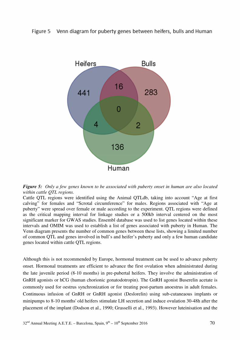
32nd Annual Meeting A.E.T.E. – Barcelona, Spain, 9th – 10th September 2016 70
Figure 5: Only a few genes known to be associated with puberty onset in human are also located
within cattle QTL regions.
Cattle QTL regions were identified using the Animal QTLdb, taking into account “Age at first calving” for females and “Scrotal circumference” for males. Regions associated with “Age at puberty” were spread over female or male according to the experiment. QTL regions were defined as the critical mapping interval for linkage studies or a 500kb interval centered on the most significant marker for GWAS studies. Ensembl database was used to list genes located within these intervals and OMIM was used to establish a list of genes associated with puberty in Human. The Venn diagram presents the number of common genes between these lists, showing a limited number of common QTL and genes involved in bull’s and heifer’s puberty and only a few human candidate genes located within cattle QTL regions. Although this is not recommended by Europe, hormonal treatment can be used to advance puberty
onset. Hormonal treatments are efficient to advance the first ovulation when administrated during
the late juvenile period (8-10 months) in pre-pubertal heifers. They involve the administration of
GnRH agonists or hCG (human chorionic gonatodotropin). The GnRH agonist Buserelin acetate is
commonly used for oestrus synchronization or for treating post-partum anoestrus in adult females.
Continuous infusion of GnRH or GnRH agonist (Deslorelin) using sub-cutaneaous implants or
minipumps to 8-10 months' old heifers stimulate LH secretion and induce ovulation 30-48h after the
placement of the implant (Dodson et al., 1990; Grasselli et al., 1993). However luteinisation and the
32nd Annual Meeting A.E.T.E. – Barcelona, Spain, 9th – 10th September 2016 71
production of progesterone are not consistently observed and this may cause short luteal phases.
The continuous exposure to GnRH or GnRH agonists induces the desensitization of the GnRH-R
signalling. After GnRH agonist implants removal, the animals do not respond to exogenous GnRH
treatment for 12 days (Bergfeld et al., 1996). For these reasons, hCG is usually preferred. hCG will
mimic the effect of endogenous LH surge and stimulate the ovulation of the dominant follicle. The
luteotropic effect of hCG guarantees the formation of a functional corpus luteum and will have a
beneficial effect on the initiation of pregnancy. Its major side effect is that hCG is a human
hormone and as such its repeated administration causes the development of an acquired immunity
that impedes future treatments to be efficient (De et al., 2010; Dahlen et al., 2011). Both GnRH
agonists- and hCG-based treatments rely on peptidic or proteic substances that are not an
environmental issue. In contrast to oestradiol- and progesterone- based hormonal treatments that
have been used in the past in Europe or are still in use on the American, Asian and Australian
continents. It would be interesting to test for Kp long life agonists that have been developed for the
ovine species to see whether they could offer a more physiological activation of the central GnRH
controlling system and thus avoiding the desensitization of GnRH-R signalling (Beltramo et al.,
2015).
6.2 Collecting prepubertal oocytes
Another strategy is to overcome these problems by using in vitro production techniques and oocytes
collection by Ovum Pick-Up (OPU) techniques. Despite the fact that large follicles are present
before puberty, that good quality oocytes evaluated by the presence of compact cumulus can be
collected by OPU, that the proportion of cleavages up to 8 cells after in vitro fertilization is correct,
the rate of blastocysts obtained is low and their ability to produce successful pregnancy after
embryo transfer is poor in comparison to data obtained from adult oocytes (Armstrong et al., 1992;
Levesque and Sirard, 1994; Majerus et al., 1999; Landry et al., 2016). Ovarian stimulation using
FSH can improve the rate of blastocyst formation, underlining the importance of hormonal
environment to insure the oocyte competency to sustain development (Khatir et al., 1996). Different
factors have been studied and sustain the cytoplasmic immaturity of prepubertal oocytes (Gandolfi
et al., 1998; Oropeza et al., 2004; Bernal-Ulloa et al., 2016). Gene expression in blastocyst embryos
relies mostly on post-transcriptional control of maternal transcripts accumulated during oocyte
maturation. In calf oocytes, the expression of maternal transcripts differs from that of adult oocytes.
Transcripts of PRDX2 and PRDX1 genes are in less quantities in oocytes collected from
prepubertal animals in comparison to adult animals (Romar et al., 2011).
7 Conclusions
With the introduction of genomic selection 15 years ago, international agricultural politics have
started to modify selection strategies, which now include puberty traits in order to advance puberty
onset with the objective of reducing generation intervals. The selective pressure on onset of puberty
will undoubtedly increase in a near future. Indeed, advances in molecular genetics have now made
it possible to predict the total genetic value of animals by using genome-wide dense marker maps

32nd Annual Meeting A.E.T.E. – Barcelona, Spain, 9th – 10th September 2016 72
leading to the forthcoming of Genomic Selection (GS) (Humblot et al., 2010). GS is of particular
interest in cattle since the generation interval is long, artificial insemination bulls should be tested
on their progeny before dissemination and some important traits such as fertility have a low
heritability, due probably to a great sensitivity to environmental factors. Yearling bulls that have
genomic breeding values information but lack phenotypic data on their daughters are often referred
to as “genomic bulls”. There has been an immense shift among the AI companies toward the use of
genomic bulls in the past 3 years. Some AI companies use almost all genomic bulls as sires of sons,
whereas other companies use a combination of genomic bulls and progeny-tested bulls (Schefers
and Weigel, 2012). Instead of waiting a minimum of 4.5 years to use progeny-tested bulls as sires of
sons, AI companies could now use the best DNA-tested young bulls by roughly 1 year of age. Due
to economical constrains, AI companies are now looking for animals having an advancement of
their puberty.
It’s therefore of major importance to understand the link between these phenotypic changes, genetic
determinants and environment. Indeed, GS de facto reduces the interval of generation and will
speed up the selection process. This could be a great opportunity but may also increase the risk of
disseminating unsuitable traits by lack of knowledge of their related pathways. Therefore, before
implementing GS for QTL associated with puberty traits, it’s crucial to evaluate whether or not this
selection process may affect other reproductive characteristics or reduce the robustness and increase
vulnerability to environmental changes. There is clearly a need for basic research on factors that
control puberty in order to improve heifer development and fertility (Perry, 2016) and address the
question of robustness.
Bibliography
Ahima RS, Dushay J, Flier SN, Prabakaran D and Flier JS. 1997. Leptin accelerates the onset
of puberty in normal female mice. J. Clin. Invest. 99:391–395. Allen L G, Kalra P S, Crowley WR, Kalra S P. 1985. Comparison of the effects of neuropeptide
Y and adrenergic transmitters on LH release and food intake in male rats. Life Sci 37:617–623.
Ambros V. 2011. MicroRNAs and developmental timing. Curr. Opin. Genet. Dev. 21:511–517. Ambros V, Horvitz H R. 1984. Heterochronic mutants of the nematode Caenorhabditis elegans.
Science 226:409–416. Amstalden M, Garcia MR, Stanko RL, Nizielski SE, Morrison CD, Keisler DH, Williams G.
2002. Central infusion of recombinant ovine leptin normalizes plasma insulin and stimulates a novel hypersecretion of luteinizing hormone after short-term fasting in mature beef cows. Biol Reprod 66:1555–1561.
Armstrong DT, Holm P, Irvine B, Petersen BA, Stubbings RB, Mclean D, Stevens G, Seamark RF. 1992. Pregnancies and Live Birth From Invitro Fertilization of Calf Oocytes Collected by Laparoscopic Follicular Aspiration. Theriogenology 38:667–678.
Barash IA, Cheung CC, Weigle DS, Ren HP, Kabigting EB, Kuijper JL, Clifton DK, Steiner RA. 1996. Leptin is a metabolic signal to the reproductive system. Endocrinology 137:3144–3147.
32nd Annual Meeting A.E.T.E. – Barcelona, Spain, 9th – 10th September 2016 73
Beltramo M, Robert V, Galibert M, Madinier J-B, Marceau P, Dardente H, Decourt C, de Roux N, Lomet D, Delmas AF, Caraty A, Aucagne V. 2015. Rational Design of Triazololipopeptides Analogs of Kisspeptin Inducing a Long-Lasting Increase of Gonadotropins. J. Med. Chem. 58:3459–3470.
Bergfeld EG, D'Occhio MJ, Kinder JE. 1996. Pituitary function, ovarian follicular growth, and plasma concentrations of 17 beta-estradiol and progesterone in prepubertal heifers during and after treatment with the luteinizing hormone-releasing hormone agonist deslorelin. Biology of Reproduction 54:776–782.
Bernal-Ulloa SM, Heinzmann J, Herrmann D, Hadeler K-G, Aldag P, Winkler S, Pache D, Baulain U, Lucas-Hahn A, Niemann H. 2016. Cyclic AMP Affects Oocyte Maturation and Embryo Development in Prepubertal and Adult Cattle. PLoS ONE 11:e0150264.
Bédécarrats GY, Kaiser UB. 2003. Differential regulation of gonadotropin subunit gene promoter activity by pulsatile gonadotropin-releasing hormone (GnRH) in perifused L beta T2 cells: role of GnRH receptor concentration. Endocrinology 144:1802–1811.
Bianco SDC, Vandepas L, Correa-Medina M, Gereben B, Mukherjee A, Kuohung W, Carroll R, Teles MG,. Latronico AC, Kaiser UB. 2011. KISS1R Intracellular Trafficking and Degradation: Effect of the Arg386Pro Disease-Associated Mutation. Endocrinology 152:1616–1626.
Bouligand J, Ghervan C, Tello JA, Brailly-Tabard S, Salenave S, Chanson P, Lombes M, Millar RP, Guiochon-Mantel A, Young J. 2009. Isolated Familial Hypogonadotropic Hypogonadism and a GNRH1 Mutation. New England Journal of Medicine 360:2742–2748.
Butler MP, Trumbull JJ, Turner KW, Zucker I. 2007. Timing of puberty and synchronization of seasonal rhythms by simulated natural photoperiods in female Siberian hamsters. AJP: Regulatory, Integrative and Comparative Physiology 293:R413–R420.
Caldani M, Antoine M., Batailler M, Duittoz AH. 1995. Ontogeny of GnRH systems. Journal of Reproduction and Fertility Supplements 49:147–162.
Caldani M, Batailler M, Jourdan F. 1987. The sheep terminal nerve: coexistence of LHRH- and AChE-containing neurons. Neurosciences Letters 83:221–226.
Cánovas A, Reverter A, DeAtley KL, Ashley RL, Colgrave ML, Fortes MRS, Islas-Trejo A, Lehnert S, Porto-Neto L, Rincón G, Silver GA, Snelling WM, Medrano JF, Thomas MG. 2014. Multi-tissue omics analyses reveal molecular regulatory networks for puberty in composite beef cattle. PLoS ONE 9, e102551.
Caraty A, Locatelli A,. Martin GB. 1989. Biphasic response in the secretion of gonadotrophin-releasing hormone in ovariectomized ewes injected with oestradiol. Journal of Endocrinology 123:375–382.
Caraty A, Orgeur P, Thiery JC. 1982. Demonstration of the pulsatile secretion of LH-RH into hypophysial portal blood of ewes using an original technic for multiple samples. Comptes Rendus des Seances de l'Académie des Sciences III 295:103–106.
Cardoso RC, Alves BRC, Sharpton SM, Williams GL, Amstalden M. 2015. Nutritional Programming of Accelerated Puberty in Heifers: Involvement of Pro-Opiomelanocortin Neurones in the Arcuate Nucleus. Journal of Neuroendocrinology 27:647–657.
Chan Y-M, Broder-Fingert S, Paraschos S, Lapatto R, Au M, Hughes V, Bianco SDC, Min L, Plummer L, Cerrato F, De Guillebon A, Wu I-H, Wahab F, Dwyer A, Kirsch S, Quinton R, Cheetham T, Ozata M, Ten S,. Chanoine J-P, Pitteloud N, Martin KA, Schiffmann R, Van der Kamp HJ, Nader S, Hall JE, Kaiser UB, Seminara SB. 2011. GnRH-deficient phenotypes in humans and mice with heterozygous variants in KISS1/Kiss1. Journal of Clinical Endocrinology and Metabolism 96:E1771–E1781.
Chehab FF, Lim ME, Lu R. 1996. Correction of the sterility defect in homozygous obese female mice by treatment with the human recombinant leptin. Nat. Genet. 12:318–320.
Cheung CC, Thornton JE, Kuijper JL, Weigle DS, Clifton DK, Steiner RA. 1997. Leptin is a metabolic gate for the onset of puberty in the female rat. Endocrinology 138:855–858.
32nd Annual Meeting A.E.T.E. – Barcelona, Spain, 9th – 10th September 2016 74
Chevrier L, Guimiot F, de Roux N. 2011. GnRH receptor mutations in isolated gonadotropic deficiency. Molecular and Cellular Endocrinology 346:21–28.
Clark SJ, Ellis N, Styne DM, Gluckman PD, Kaplan SL, Grumbach MM. 1984. Hormone ontogeny in the ovine fetus. XVII. Demonstration of pulsatile luteinizing hormone secretion by the fetal pituitary gland. Endocrinology 115:1774–1779.
Clarke IJ, Cummins JT. 1982. The temporal relationship between gonadotropin releasing hormone (GnRH) and luteinizing hormone (LH) secretion in ovariectomized ewes. Endocrinology 111:1737–1739.
Clarke I.J, Cummins JT, Karsch FJ, Seeburg PH, Nikolics K. 1987. GnRH-associated peptide (GAP) is cosecreted with GnRH into the hypophyseal portal blood of ovariectomized sheep. Biochem Biophys Res Commun 143:665–671.
Claypool LE, Foster DL. 1990. Sexual differentiation of the mechanism controlling pulsatile secretion of luteinizing hormone contributes to sexual differences in the timing of puberty in sheep. Endocrinology 126:1206–1215.
Clement K, Vaisse C, Lahlou N, Cabrol S, Pelloux V, Cassuto D, Gourmelen M, Dina C, Chambaz J, Lacorte JM, Basdevant A, Bougneres P, Lebouc Y, Froguel P, Guy-Grand B. 1998. A mutation in the human leptin receptor gene causes obesity and pituitary dysfunction. Nature 392:398–401.
Clements JA, Reyes FI, Winter JSD, Faiman C. 2009. Studies on Human Sexual Development. III. Fetal Pituitary and Serum, and Amniotic Fluid Concentrations of LH, CG, and FSH. Journal of Clinical Endocrinology and Metabolism 42:9–19.
Colledge WH, de Tassigny XD. 2009. Kisspeptin Signaling in Rodents: insights from Knock-Out Mice. Biology of Reproduction 81:75–76.
Constantin S, Caraty A, Wray S, Duittoz AH. 2009. Development of gonadotropin-releasing hormone-1 secretion in mouse nasal explants. Endocrinology 150:3221–3227.
Dahl GE. 2008. Effects of short day photoperiod on prolactin signaling in dry cows: A common mechanism among tissues and environments? Journal of Animal Sciences. 86:10–14.
Dahlen CR, Marquezini GHL, Larson JE, Lamb GC. 2011. Human chorionic gonadotropin influences ovarian function and concentrations of progesterone in prepubertal Angus heifers. Journal of Animal Sciences. 89:2739–2749.
Davis Rincker LE, Vandehaar MJ, Wolf CA, Liesman JS, Chapin LT, Weber Nielsen MS. 2011. Effect of intensified feeding of heifer calves on growth, pubertal age, calving age, milk yield, and economics. Journal of Dairy Science. 94:3554–3567.
Day ML, Imakawa K, WolfPL, Kittok RJ, Kinder JE. 1987. Endocrine mechanisms of puberty in heifers. Role of hypothalamo-pituitary estradiol receptors in the negative feedback of estradiol on luteinizing hormone secretion. Biology of Reproduction 37:1054–1065.
de Roux N, Genin E, Carel J-C, Matsuda F, Chaussain J-L., Milgrom E. 2003. Hypogonadotropic hypogonadism due to loss of function of the KiSS1-derived peptide receptor GPR54. Proceedings of the National Academy of Sciences USA 100:10972–10976.
De RF, López-Gatius F, García-Ispierto I, Techakumpu M. 2010. Clinical use of human chorionic gonadotropin in dairy cows: An update. Theriogenology 73:1001–1008.
Dias M, Souza M, Takada L, Feitosa FLB, Costa RB, Diaz IDPS, Cardoso DF, Tonussi R L, Baldi F, Albuquerque LG, Oliveira HN. 2015. Study of lipid metabolism-related genes as candidate genes of sexual precocity in Nellore cattle. Genetic Molecular Research. 14:234–243.
Dodé C, Levilliers J, Dupont J-M, De Paepe A, Le Dû N, Soussi-Yanicostas N, Coimbra RS, Delmaghani S, Compain-Nouaille S, Baverel F, Pêcheux C, Le Tessier D, Cruaud C, Delpech M, Speleman F, Vermeulen S, Amalfitano A, Bachelot Y, Bouchard P, Cabrol S, Carel JC, Delemarre-van de Waal H, Goulet-Salmon B, Kottler M-L, Richard O, Sanchez-Franco F, Saura R, Young J, Petit C,. Hardelin J-P. 2003. Loss-of-function mutations in FGFR1 cause autosomal dominant Kallmann syndrome. Nature Genetics. 33:463–465.
32nd Annual Meeting A.E.T.E. – Barcelona, Spain, 9th – 10th September 2016 75
Dodé C, Teixeira L, Levilliers J, Fouveaut C, Bouchard P, Kottler M-L, Lespinasse J, Lienhardt-Roussie A, Mathieu M, Moerman A, Morgan G, Murat A, Toublanc J-E, Wolczynski S, Delpech M, Petit C, Young J, Hardelin J-P. 2006. Kallmann syndrome: Mutations in the genes encoding prokineticin-2 and prokineticin receptor-2. PLoS Genetics 2:1648–1652.
Dodson SE, McLeod BJ,. Lamming GE, Peters AR. 1990. Ovulatory responses to continuous administration of GnRH in nine-month-old prepubertal beef heifers. Animal Reproduction Sciences 22:271–280.
Donato J, Cravo RM, Frazão R, Gautron L, Scott MM, Lachey J, Castro IA, Margatho LO, Lee S, Lee C, Richardson JA, Friedman J, Chua S, Coppari R, Zigman JM, Elmquist, JK, Elias CF. 2011. Leptin’s effect on puberty in mice is relayed by the ventral premammillary nucleus and does not require signaling in Kiss1 neurons. Journal of Clinical Investigations. 121:355–368.
Duittoz AH, Batailler M. 2000. Pulsatile GnRH secretion from primary cultures of sheep olfactory placode explants. Journal of Reproduction and Fertility 120:391–396.
Elks CE, Perry JRB, Sulem P, Chasman DI, Franceschini N, He C, Lunetta KL, Visser JA, Byrne EM, Cousminer DL, Gudbjartsson DF, Esko T, Feenstra B, Hottenga J-J, Koller DL, Kutalik Z, Lin P, Mangino M, Marongiu M, McArdle PF, Smith AV, Stolk L, van Wingerden SH, Zhao JH, Albrecht E, Corre T, Ingelsson E, Hayward C, Magnusson PKE, Smith EN, Ulivi S, Warrington NM, Zgaga L, Alavere H, Amin N, Aspelund T. Bandinelli S, Barroso I, Berenson GS, Bergmann S, Blackburn H, Boerwinkle E., Buring JE, Busonero F, Campbell H, Chanock SJ, Chen W, Cornelis MC, Couper D, Coviello AD, d'Adamo P, de Faire U, de Geus EJC, Deloukas P, Döring A, Smith GD, Easton DF, Eiriksdottir G, Emilsson V, Eriksson J, Ferrucci L, Folsom AR, Foroud T, Garcia M, Gasparini P, Geller F, Gieger C, GIANT Consortium, Gudnason V, Hall P, Hankinson S.E, Ferreli L, Heath AC, Hernandez DG, Hofman A, Hu FB, Illig T, Järvelin M-R, Johnson AD, Karasik D,. Khaw K-T, Kiel DP, Kilpeläinen TO, Kolcic I, Kraft P, Launer LJ, Laven JSE, Li S, Liu, J, Levy D, Martin NG, McArdle WL, Melbye M, Mooser V, Murray JC, Murray SS, Nalls MA, Navarro P, Nelis M, Ness AR, Northstone K, Oostra BA, Peacock M, Palmer LJ, Palotie A, Paré G, Parker AN, Pedersen NL, Peltonen L, Pennell CE, Pharoah P, Polasek O, Plump AS, Pouta A, Porcu E, Rafnar T, Rice JP, Ring SM, Rivadeneira F, Rudan I, Sala C, Salomaa V, Sanna S, Schlessinger D, Schork NJ, Scuteri A, Segrè AV, Shuldiner AR, Soranzo N, Sovio U, Srinivasan SR, Strachan DP, Tammesoo ML, Tikkanen E, Toniolo D, Tsui K, Tryggvadottir L, Tyrer J, Uda M, van Dam RM, van Meurs JB, Vollenweider P, Waeber G, Wareham NJ, Waterworth DM, Weedon MN, Wichmann HE, Willemsen G, Wilson JF, Wright AF, Young L, Zhai G, Zhuang WV, Bierut LJ, Boomsma DI, Boyd HA, Crisponi L, Demerath EW, van Duijn CM, Econs MJ, Harris TB, Hunter DJ, Loos RJ, Metspalu A, Montgomery GW, Ridker PM, Spector TD, Streeten EA, Stefansson K, Thorsteinsdottir U, Uitterlinden AG, Widen E, Murabito JM, Ong KK, Murray A. 2010. Thirty new loci for age at menarche identified by a meta-analysis of genome-wide association studies. Nature Genetics 42:1077–1085.
Forger NG, de Vries GJ, Breedlove SM. 2015. Chapter 47 Sexual Differentiation of Brain and Behavior. In: Knobil and Neill's Physiology of Reproduction. Fourth Edition. pp. 2109–2155.
Fortes MRS, Reverter A, Nagaraj SH, Zhang Y, Jonsson NN, Barris W, Lehnert S, Boe-Hansen GB, Hawken RJ. 2011. A single nucleotide polymorphism-derived regulatory gene network underlying puberty in 2 tropical breeds of beef cattle. Journal of Animal Sciences. 89:1669–1683.
Fortes MRS, Reverter A, Zhang Y, Collis E, Nagaraj SH, Jonsson NN, Prayaga KC, Barris W, Hawken RJ. 2010a. Association weight matrix for the genetic dissection of puberty in beef cattle. Proceedings of the National Academy of Sciences USA 107:13642–13647.

32nd Annual Meeting A.E.T.E. – Barcelona, Spain, 9th – 10th September 2016 76
Fortes MRS, Nguyen LT, Porto Neto LR, Reverter A, Moore SS, Lehnert SA, Thomas M. 2016. Polymorphisms and genes associated with puberty in heifers. Theriogenology 86: 333-339.
Fortes MRS, Li Y, Collis E, Zhang Y, Hawken RJ. 2013. The IGF1 pathway genes and their association with age of puberty in cattle. Animal Genetics. 44:91–95.
Fortes M, Reverter A, Zhang Y, Collis E, Nagaraj Y, Johnoson NN, Barris W, Hawken R. 2010b. Association weight matrix for the genetic dissection of puberty in beef cattle. Proceedings of the National Acadmey of Sciences USA 107: 13642-13647.
Foster DL, Olster DH. 1984. Effect of Restricted Nutrition on Puberty in the Lamb: Patterns of Tonic Luteinizing Hormone (LH) Secretion and Competency of the LH Surge System. Endocrinology 116:375–381.
Franco B, Guioli S, Pragliola A, Incerti B, Bardoni B, Tonlorenzi R, Carrozzo R, Maestrini E, Pieretti M, Taillonmiller P, Brown CJ, Willard HF, Lawrence C, Persico MG, Camerino G, Ballabio A. 1991. A Gene Deleted in Kallmanns Syndrome Shares Homology with Neural Cell-Adhesion and Axonal Path-Finding Molecules. Nature 353, 529–536.
Foster DL, Hileman SM. 2015. Chapter 31 Puberty in the Sheep. In: Knobil and Neill's Physiology of Reproduction. Fourth Edition. pp. 1441–1485.
Frederich RC, Haman A, Anderson S, Lollmann B, Lowell BB, Flier JS. 1995. Leptin Levels Reflect Body Lipid-Content in Mice - Evidence for Diet-Induced Resistance to Leptin Action. Nature Medicine 1:1311–1314.
Freetly HC, Kuehn LA, Cundiff LV. 2011. Growth curves of crossbred cows sired by Hereford, Angus, Belgian Blue, Brahman, Boran, and Tuli bulls, and the fraction of mature body weight and height at puberty. Journal of Animal Sciences. 89:2373–2379.
Frisch RE, Revelle R. 1970. Height and weight at menarche and a hypothesis of critical body weights and adolescent events. Science 169:397–399.
Gajdos ZKZ, Henderson KD, Hirschhorn JN, M. R. Palmert. 2010. Genetic determinants of pubertal timing in the general population. Mol Cell Endocrinol 324:21–29.
Gandolfi F, Milanesi E, Pocar P, Luciano AM, Brevini T, Acocella F, Lauria A, Armstrong DT. 1998. Comparative analysis of calf and cow oocytes during in vitro maturation. Molecular Reproduction and Development 49:168–175.
Gasser CL. 2006. Induction of precocious puberty in heifers III: Hastened reduction of estradiol negative feedback on secretion of luteinizing hormone. Journal of Animal Sciences. 84:2050–2056.
Gazal OS, Leshin LS, Stanko R, Thomas MG, Keisler DH, Anderson LL, Williams GL. 1998. Gonadotropin-releasing hormone secretion into third-ventricle cerebrospinal fluid of cattle: correspondence with the tonic and surge release of luteinizing hormone and its tonic inhibition by suckling and neuropeptide Y. Biology of Reproduction 59:676–683.
Giaume C, Koulakoff A, Roux L, Holcman D, Rouach N. 2010. Astroglial networks: a step further in neuroglial and gliovascular interactions. Nature Review in Neurosciences 11:87–99.
Gore AC, Terasawa E. 1991. A Role for Norepinephrine in the Control of Puberty in the Female Rhesus-Monkey, Macaca-Mulatta. Endocrinology 129:3009–3017.
Gore AC, Mitsushima D, Terasawa E. 1993. A Possible Role of Neuropeptide-Y in the Control of the Onset of Puberty in Female Rhesus-Monkeys. Neuroendocrinology 58:23–34.
Grasselli F, Baratta M, Tamanini C. 1993. Effects of a GnRH analogue (buserelin) infused via osmotic minipumps on pituitary and ovarian activity of prepubertal heifers. Anim Reprod Sci 32:153–161.
Habiby RL, Boepple P, Nachtigall L, Sluss PM, Crowley Jr WF, Jameson. JL. 1996. Adrenal hypoplasia congenita with hypogonadotropic hypogonadism: evidence that DAX-1 mutations lead to combined hypothalmic and pituitary defects in gonadotropin production. Journal of Clinical Investigations. 98:1055–1062.
32nd Annual Meeting A.E.T.E. – Barcelona, Spain, 9th – 10th September 2016 77
Hanchate NK, Giacobini P, Lhuillier P, Parkash J, Espy C, Fouveaut C, Leroy C, Baron S, Campagne C, Vanacker C, Collier F, Cruaud C, Meyer V, García-Piñero A, Dewailly D, Cortet-Rudelli C, Gersak K, Metz C, Chabrier G, Pugeat M, Young J, Hardelin J-P, Prevot V, Dodé C. 2012. SEMA3A, a gene involved in axonal pathfinding, is mutated in patients with Kallmann syndrome. PLoS Genetics 8:e1002896.
Hardelin JP, Dodé C. 2008. The complex genetics of Kallmann syndrome: KAL1, FGFR1, FGF8, PROKR2, PROK2, et al. Sexual Development 2:181–193.
Hardelin JP, Levilliers J, Delcastillo I, Cohen-Salmon M, Legouis R, Blanchard S, Compain S, Bouloux P, Kirk J, Moraine C, Chaussain JL, Weissenbach J, Petit C. 1992. X-Chromosome-Linked Kallmann Syndrome - Stop Mutations Validate the Candidate Gene. Proceedings of the National Academy of Sciences USA 89:8190–8194.
Henry BA, Goding JW, Alexander WS, Tilbrook AJ, Canny BJ, Dunshea F, Rao A, Mansell A, Clarke IJ. 2011. Central Administration of Leptin to Ovariectomized Ewes Inhibits Food Intake without Affecting the Secretion of Hormones from the Pituitary Gland: Evidence for a Dissociation of Effects on Appetite and Neuroendocrine Function1. Endocrinology 140:1175–1182.
Herbison A.E, Pape JR. 2001. New evidence for estrogen receptors in gonadotropin-releasing hormone neurons. Frontiers in Neuroendocrinolology 22:292–308.
Hines M, Spencer D, Kung KT, Browne WV, Constantinescu M., Noorderhaven RM. 2016. The early postnatal period, mini-puberty, provides a window on the role of testosterone in human neurobehavioural development. Current Opinion in Neurobiology 38:69–73.
Hochberg Z, Belsky J. 2013. Evo-devo of human adolescence: beyond disease models of early puberty. BMC Medicine 11: 113.
Hrabovszky E, Shughrue PJ, Merchenthaler I, Hajszán T, Carpenter CD, Liposits Z, Petersen SL. 2000. Detection of estrogen receptor-beta messenger ribonucleic acid and 125I-estrogen binding sites in luteinizing hormone-releasing hormone neurons of the rat brain. Endocrinology 141:3506–3509.
Hu Z-L, Park CA, Reecy JM. 2016. Developmental progress and current status of the Animal QTLdb. Nucl. Acids Res. 44:D827–D833.
Humblot P. Le Bourhis D, Fritz S, Colleau JJ, Gonzalez C, Guyader Joly C, Malafosse A, Heyman Y, Amigues Y, Tissier M, Ponsart C. 2010. Reproductive Technologies and Genomic Selection in Cattle. Veterinary Medicine International 2010:1–8.
Irvine CH, Alexander SL. 1987. A novel technique for measuring hypothalamic and pituitary hormone secretion rates from collection of pituitary venous effluent in the normal horse. Journal of Endocrinology 113:183–192.
Jackson RS, Creemers JWM, Farooqi IS, Raffin-Sanson M-L, Varro A, Dockray GJ, Holst JJ, Brubaker PL, Corvol P, Polonsky KS, Ostrega D, Becker KL, Bertagna X, Hutton JC, White A, Dattani MT, Hussain K, Middleton SJ, Nicole TM, Milla PJ, Lindley KJ, O'Rahilly S. 2003. Small-intestinal dysfunction accompanies the complex endocrinopathy of human proprotein convertase 1 deficiency. Journal of Clinical Investigations. 112:1550–1560.
Johnston DJ, Corbet NJ, Barwick SA, Wolcott ML, Holroyd. RG. 2013. Genetic correlations of young bull reproductive traits and heifer puberty traits with female reproductive performance in two tropical beef genotypes in northern Australian Animal Production Sciences. 54:74–84.
Kaplan SL, Grumbach MM. 1976. The Ontogenesis of Human Foetal Hormones. II. Luteinizing Hormone (LH) and follicle stimulating hormone (FSH). European Journal of Endocrinology 81:808–829.
Kaplowitz PB, Slora EJ, Wasserman RC, Pedlow SE, Herman-Giddens ME. 2001. Earlier onset of puberty in girls: Relation to increased body mass index and race. Pediatrics 108:347–353.

32nd Annual Meeting A.E.T.E. – Barcelona, Spain, 9th – 10th September 2016 78
Kasuya E, Nyberg CL, Mogi K, Terasawa E. 1999. A role of gamma-amino butyric acid (GABA) and glutamate in control of puberty in female rhesus monkeys: Effect of an antisense oligodeoxynucleotide for GAD67 messenger ribonucleic acid and MK801 on luteinizing hormone-releasing hormone release. Endocrinology 140:705–712.
Keen KL, Burich AJ, Mitsushima D, Kasuya E, Terasawa E. 2011. Effects of Pulsatile Infusion of the GABAA Receptor Blocker Bicuculline on the Onset of Puberty in Female Rhesus Monkeys1. Endocrinology 140:5257–5266.
Khatir H, Lonergan P, Carolan C, Mermillod P. 1996. Prepubertal bovine oocyte: A negative model for studying oocyte developmental competence. Molecular Reproduction and Development 45:231–239.
Kim H-G, Layman LC. 2011. The role of CHD7 and the newly identified WDR11 gene in patients with idiopathic hypogonadotropic hypogonadism and Kallmann syndrome. Molecular and Cellular Endocrinology 346:74–83.
Kim H-G, Kurth I, Lan F, Meliciani I, Wenzel W, Eom SH, Kang GB, Rosenberger G, Tekin M, Ozata M, Bick DP, Sherins RJ, Walker SL, Shi Y, Gusella JF, Layman LC. 2008. Mutations in CHD7, Encoding a Chromatin-Remodeling Protein, Cause Idiopathic Hypogonadotropic Hypogonadism and Kallmann Syndrome. The American Journal of Human Genetics 83:511–519.
Landry DA, Bellefleur A-M, Labrecque R, Grand F-X, Vigneault C, Blondin P, Sirard M-A. 2016. Effect of cow age on the in vitro developmental competence of oocytes obtained after FSH stimulation and coasting treatments. Theriogenology 86:1–7.
Layman LC. 1999. Mutations in human gonadotropin genes and their physiologic significance in puberty and reproduction. Fertility and Sterility 71:201–218.
Le Cozler Y, Lollivier V, Lacasse P, Disenhaus C. 2008. Rearing strategy and optimizing first-calving targets in dairy heifers: a review. Animal 2:1393–1404.
Levesque JT, Sirard MA. 1994. Proteins in Oocytes From Calves and Adult Cows Before Maturation - Relationship with Their Development Capacity. Reproduction Nutrition and Development. 34:133–139.
Levine JE, Pau KY, Ramirez VD, Jackson GL. 1982. Simultaneous measurement of luteinizing hormone-releasing hormone and luteinizing hormone release in unanesthetized, ovariectomized sheep. Endocrinology 111:1449–1455.
Lirón JP, Prando AJ, Fernández ME, Ripoli MV, Rogberg-Muñoz A, Goszczynski DE, Posik, DM, Peral-García P, Baldo A, Giovambattista G. 2012. Association between GNRHR, LHR and IGF1 polymorphisms and timing of puberty in male Angus cattle. BMC Genetics 13:26.
Louis GW, Greenwald-Yarnell M, Phillips R, Coolen LM, Lehman MN, Myers MG. 2011. Molecular Mapping of the Neural Pathways Linking Leptin to the Neuroendocrine Reproductive Axis. Endocrinology 152:2302–2310.
Lunstra DD, Cundiff LV. 2003. Growth and pubertal development in Brahman-, Boran-, Tuli-, Belgian Blue-, Hereford- and Angus-sired F1 bulls. Journal of. Animal. Sciences. 81:1414–1426.
Luo Y, Liu Q, Lei X, Wen Y, Yang Y-L., Zhang R, Hu M-Y. 2015. Association of estrogen receptor gene polymorphisms with human precocious puberty: a systematic review and meta-analysis. Gynecology and Endocrinology 31:516–521.
Macdonald KA, McNaughton LR, Verkerk GA, Penno JW, Burton LJ, Berry DP, Gore PJS, Lancaster JAS, Holmes CW. 2007. A Comparison of Three Strains of Holstein-Friesian Cows Grazed on Pasture: Growth, Development, and Puberty. Journal of Dairy Science. 90:3993–4003.
Maciel M. 2004. Chronic administration of recombinant ovine leptin in growing beef heifers: Effects on secretion of LH, metabolic hormones, and timing of puberty. Journal of Animal Sciences. 82:2930–2936.

32nd Annual Meeting A.E.T.E. – Barcelona, Spain, 9th – 10th September 2016 79
Majdoubi El M, Sahu A, Ramaswamy S, Plant TM. 2000. Neuropeptide Y: A hypothalamic brake restraining the onset of puberty in primates. Proceedings of the National Academy of Sciences USA 97:6179–6184.
Majerus V, De Roover R, Etienne D, Kaidi S, Massip A, Dessy F, Donnay. I. 1999. Embryo production by ovum pick up in unstimulated calves before and after puberty. Theriogenology 52:1169–1179.
Manning JM, Bronson FH. 1989. Effects of prolonged exercise on puberty and luteinizing hormone secretion in female rats. American Journal of Physiology. 257:R1359–64.
Manning JM, Bronson FH. 1991. Suppression of puberty in rats by exercise: effects on hormone levels and reversal with GnRH infusion. American Journal of Physiology. 260:R717–23.
Martinez-Velazquez G, Gregory KE. 2003. Genetic relationships between scrotal circumference and female reproductive traits. Journal of Animal Sciences 81:395-401
Matsui H, Takatsu Y, Kumano S, Matsumoto H, Ohtaki T. 2004. Peripheral administration of metastin induces marked gonadotropin release and ovulation in the rat. Biochemical and Biophysical Research Communications 320:383–388.
Mazaheri A, Hashemipour M. Salehi M, Behnam M, Hovsepian S, Hassanzadeh A. 2015. Mutation of kisspeptin 1 gene in children with precocious puberty in isfahan city. International Journal of Preventive Medicine 6:41.
McDonald JK, Lumpkin MD, DePaolo LV. 1989. Neuropeptide-Y suppresses pulsatile secretion of luteinizing hormone in ovariectomized rats: possible site of action. Endocrinology 125:186–191.
Merke DP, Tajima T, Baron J, Cutler GB. 1999. Hypogonadotropic hypogonadism in a female caused by an X-linked recessive mutation in the DAX1 gene. New England Journal of Medicine 340:1248–1252.
Messager S, Chatzidaki EE, Ma D, Hendrick AG, Zahn D, Dixon J, Thresher RR, Malinge I, Lomet D, Carlton MBL, Colledge WH, Caraty A, Aparicio SAJR. 2005. Kisspeptin directly stimulates gonadotropin-releasing hormone release via G protein-coupled receptor 54. Proceedings of the National Academy of Sciences USA 102:1761–1766.
Messaoud-Toumi LH, Taragnat C, Durand P. 1993. Heterogeneity in the storage of gonadotropins in the ovine fetus and evidence for luteinizing hormone-follicle-stimulating hormone cells in the fetal pituitary. Biology of Reproduction 48:1239–1245.
Milazzotto MP, Rahal P, Nichi M, Miranda-Neto T, Teixeira LA, Ferraz JBS, Eler JP, Campagnari F, Garcia JF. 2008. New molecular variants of hypothalamus-pituitary-gonad axis genes and their association with early puberty phenotype in Bos taurus indicus (Nellore). Livestock Science 114, 274–279.
Mitchell A., Dwyer A, Pitteloud N, Quinton R. 2011. Genetic basis and variable phenotypic expression of Kallmann syndrome: towards a unifying theory. Trends in Endocrinology and Metabolism 22:249–258.
Mitsushima D, Hei DL, Terasawa E. 1994. Gamma-Aminobutyric-Acid Is an Inhibitory Neurotransmitter Restricting the Release of Luteinizing-Hormone-Releasing Hormone Before the Onset of Puberty. Proceedings of the National Academy of Sciences USA 91:395–399.
Miura K, Acierno JS, Seminara SB. 2004. Characterization of the human nasal embryonic LHRH factor gene, NELF, and a mutation screening among 65 patients with idiopathic hypogonadotropic hypogonadism (IHH). Journal of Human Genetics. 49:265–268.
Moenter SM, Caraty A, Locatelli A, Karsch FJ. 1991. Pattern of gonadotropin-releasing hormone (GnRH) secretion leading up to ovulation in the ewe: existence of a preovulatory GnRH surge. Endocrinology 129:1175–1182.
Morris CA, Wilson JA, Bennett GL, Cullen NG, Hickey SM, Hunter JC. 2010. Genetic parameters for growth, puberty, and beef cow reproductive traits in a puberty selection experiment. New Zealand Journal of Agricultural Research 43:83–91.
32nd Annual Meeting A.E.T.E. – Barcelona, Spain, 9th – 10th September 2016 80
Mourits MC, Galligan DT, Dijkhuizen AA, Huirne RB. 2000. Optimization of dairy heifer management decisions based on production conditions of Pennsylvania. Journal of Dairy Science. 83:1989–1997.
Mueller PL, Sklar CA, Gluckman PD, Kaplan SL, Grumbach MM. 1981. Hormone ontogeny in the ovine fetus. IX. Luteinizing hormone and follicle-stimulating hormone response to luteinizing hormone-releasing factor in mid- and late gestation and in the neonate. Endocrinology 108:881–886.
Nakai Y, Plant TM, Hess DL, Keogh E J, Knobil E. 1978. On the sites of the negative and positive feedback actions of estradiol in the control of gonadotropin secretion in the rhesus monkey. Endocrinology 102:1008–1014.
Nascimento AV, Matos MC, Seno LO, Romero ARS, Garcia JF, Grisolia AB. 2016. Genome wide association study on early puberty in Bos indicus. Genetic and Molecular Research. 15 (1): gmr.15017548
Norgren RB, Lehman MN. 1989. A double-label pre-embedding immunoperoxidase technique for electron microscopy using diaminobenzidine and tetramethylbenzidine as markers. Journal of Histochemistry and Cytochemistry 37:1283–1289.
Ojeda SR, Lomniczi A, Sandau US. 2008. Glial-gonadotrophin hormone (GnRH) neurone interactions in the median eminence and the control of GnRH secretion. Journal of Neuroendocrinology 20:732–742.
Ojeda SR, Lomniczi A, Sandau U. 2010. Contribution of glial-neuronal interactions to the neuroendocrine control of female puberty. European Journal of Neurosciences 32:2003–2010.
Ong KK, Elks CE, Li S,. Zhao JH, Luan J, Andersen LB, Bingham SA, Brage S, Smith GD, Ekelund U, Gillson CJ, Glaser B, Golding J, Hardy R, Khaw K-T, Kuh D, Luben R, Marcus M, McGeehin M A, Ness AR, Northstone K, Ring SM, Rubin C, Sims MA, Song K, Strachan DP, Vollenweider P, Waeber G, Waterworth DM, Wong A, Deloukas P, Barroso I, Mooser V, Loos RJ, Wareham NJ. 2009. Genetic variation in LIN28B is associated with the timing of puberty. Nature Genetics. 41:729–733.
Orellana JA, Sáez PJ, Cortés Campos C, Elizondo RJ, Shoji KF, Contreras Duarte S, Figueroa V. Velarde V, Jiang JX, Nualart F, Sáez JC, García MA. 2012. Glucose increases intracellular free Ca2+ in tanycytes via ATP released through connexin 43 hemichannels. Glia 60:53–68.
Oropeza A, Wrenzycki C, Herrmann D, Hadeler K-G, Niemann H. 2004. Improvement of the developmental capacity of oocytes from prepubertal cattle by intraovarian insulin-like growth factor-I application. Biology of Reproduction 70:1634–1643.
Parent A-S, Mungenast AE, Lomniczi A, Sandau US, Peles E, Bosch MA, Rønnekleiv OK, Ojeda SR. 2007. A contactin-receptor-like protein tyrosine phosphatase beta complex mediates adhesive communication between astroglial cells and gonadotrophin-releasing hormone neurones. Journal of Neuroendocrinology 19:847–859.
Parent A-S, Franssen D, Fudvoye J, Gérard A, Bourguignon J-P. 2015. Developmental variations in environmental influences including endocrine disruptors on pubertal timing and neuroendocrine control: Revision of human observations and mechanistic insight from rodents. Frontiers in Neuroendocrinol 38:12–36.
Parent A-S, Teilmann G, Juul A, Skakkebaek NE, Toppari J, Bourguignon JP. 2003. The timing of normal puberty and the age limits of sexual precocity: Variations around the world, secular trends, and changes after migration. Endocrine Reviews 24:668–693.
Parent A-S, Matagne V, Bourguignon JP. 2005. Control of puberty by excitatory amino acid neurotransmitters and its clinical implications. Endocrine 28:281–285.
Parkash J, Kaur G. 2007. Potential of PSA-NCAM in neuron-glial plasticity in the adult hypothalamus: role of noradrenergic and GABAergic neurotransmitters. Brain Research Bulletin 74:317–328.

32nd Annual Meeting A.E.T.E. – Barcelona, Spain, 9th – 10th September 2016 81
Perry G A. 2016. Factors affecting puberty in replacement beef heifers. Theriogenology. 86:373-378
Pinet-Charvet C, Geller S, Desroziers E, Ottogalli M, Lomet D, Georgelin C, Tillet Y, Franceschini I, Vaudin P, Duittoz A. 2015. GnRH episodic secretion is altered by pharmacological blockade of gap junctions: possible involvement of Glial cells. Endocrinology 157:304-322.
Plant TM, Terasawa E, Witchel SF. 2015. Chapter 32 Puberty in Non-human Primates and Man. In: Knobil and Neill's Physiology of Reproduction. Fourth Edition. pp. 1487–1536.
Polkowska J, Picard S, Wankowska M, Cieslak M, Caraty A, Tillet Y. 2014. Localization of kisspeptin neurons in the hypothalamus of peripubertal female lambs; possible connection with gonadotrophin releasing hormone and neuropeptide Y neurons. Journal of Animal and Feed Sciences 23:139–148.
Pralong FP. 2010. Insulin and NPY pathways and the control of GnRH function and puberty onset. Molecular and Cellular Endocrinology 324:82–86.
Prevot V. 2015. Chapter 30 Puberty in Mice and Rats. In: Knobil and Neill's Physiology of Reproduction. Fourth Edition. Elsevier. pp. 1395–1439.
Prevot V, Cornea A, Mungenast A, Smiley G, Ojeda SR. 2003a. Activation of erbB-1 signaling in tanycytes of the median eminence stimulates transforming growth factor beta1 release via prostaglandin E2 production and induces cell plasticity. Journal of Neuroscience 23:10622–10632.
Prevot V, Lomniczi A, Corfas G, Ojeda SR. 2005. erbB-1 and erbB-4 receptors act in concert to facilitate female sexual development and mature reproductive function. Endocrinology 146:1465–1472.
Prevot V, Rio C, Cho GJ, Lomniczi A, Heger S, Neville CM, Rosenthal NA, Ojeda SR, Corfas G. 2003b. Normal female sexual development requires neuregulin-erbB receptor signaling in hypothalamic astrocytes. Journal of Neuroscience 23:230–239.
Rapisarda JJ, Bergman KS, Steiner RA, Foster DL. 1983. Response to Estradiol Inhibition of Tonic Luteinizing-Hormone Secretion Decreases During the Final Stage of Puberty in the Rhesus-Monkey. Endocrinology 112:1172–1179.
Rius AG, Connor EE, Capuco AV,. Kendall PE, Auchtung-Montgomery TL, Dahl GE. 2005. Long-Day Photoperiod that Enhances Puberty Does Not Limit Body Growth in Holstein Heifers. Journal of. Dairy Sciences. 88:4356–4365.
Romar R, De Santis T, Papillier P, Perreau C, Thelie A, Dell'Aquila ME, Mermillod P, Dalbies-Tran R. 2011. Expression of Maternal Transcripts During Bovine Oocyte In Vitro Maturation is Affected by Donor Age. Reproduction in Domestic Animals 46:e23–e30.
Rosales Nieto CA, Thompson AN, Macleay CA, Briegel JR, Hedger MP, Ferguson MB, Martin GB. 2014. Relationships among body composition, circulating concentrations of leptin and follistatin, and the onset of puberty and fertility in young female sheep. Animal Reproduction Science 151:148–156.
Roy AK, Singh M, Kumar P, Kumar BSB. 2016. Effect of extended photoperiod during winter on growth and onset of puberty in Murrah buffalo heifers. Veterinary World 9:216–221.
Sánchez-Garrido MA, Tena-Sempere M. 2013. Metabolic control of puberty: Roles of leptin and kisspeptins. Hormones and Behaviours 64:187–194.
Schefers JM, Weigel KA. 2012. Genomic selection in dairy cattle: Integration of DNA testing into breeding programs. Animal Frontiers 2:4–9.
Schwanzel-Fukuda M, Pfaff DW. 1989. Origin of luteinizing hormone-releasing hormone neurons. Nature 338:161–164.
Schwanzel-Fukuda M, Crossin KL, Pfaff DW, Bouloux, PM, Hardelin JP, Petit C. 1996. Migration of luteinizing hormone-releasing hormone (LHRH) neurons in early human embryos. Journal of Comparative Neurology 366:547–557.
Seminara SB. 2005. Metastin and its G protein-coupled receptor, GPR54: Critical pathway modulating GnRH secretion. Frontiers in Neuroendocrinology 26:131–138.
32nd Annual Meeting A.E.T.E. – Barcelona, Spain, 9th – 10th September 2016 82
Settas N, Dacou-Voutetakis C, Karantza M, Kanaka-Gantenbein C, Chrousos GP, Voutetakis A. 2014. Central precocious puberty in a girl and early puberty in her brother caused by a novel mutation in the MKRN3 gene. Journal of Clinical Endocrinology and Metabolism 99:E647–51.
Sharif A, Baroncini M, Prevot V. 2013. Role of glia in the regulation of gonadotropin-releasing hormone neuronal activity and secretion. Neuroendocrinology 98:1–15.
Silveira LFG, Trarbach EB, Latronico AC. 2010. Genetics basis for GnRH-dependent pubertal disorders in humans. Molecular and Cellular Endocrinology 324:30–38.
Simon D, Ba I, Mekhail N, Ecosse E, Paulsen A, Zenaty D, Houang M, Jesuran-Perelroizan M, De Filippo G, Salerno M, Simonin G, Reynaud R, Carel JC, Léger J, de Roux N. 2015. Mutations in the Maternally Imprinted Gene MKRN3 Are Common in Familial Central Precocious Puberty. European Journal of Endocrinology 174:EJE–15–0488–8.
Sisk CL, Foster DL. 2004. The neural basis of puberty and adolescence. Nature Neuroscience 7:1040–1047.
Sklar CA, Mueller PL, Gluckman PD, Kaplan SL, Rudolph AM, Grumbach MM. 1981. Hormone ontogeny in the ovine fetus. VII. Circulating luteinizing hormone and follicle-stimulating hormone in mid- and late gestation. Endocrinology 108:874–880.
Smyth C, Wilkinson M. 1994. A Critical Period for Glutamate Receptor-Mediated Induction of Precocious Puberty in Female Rats. Journal of Neuroendocrinology 6:275–284.
Suttie JM, Foster DL, Veenvliet BA, Manley TR, Corson ID. 1991. Influence of Food-Intake but Independence of Body-Weight on Puberty in Female Sheep. Journal of Reproduction and Fertility 92:33–39.
Sørensen K, Mouritsen A, Aksglaede L, Hagen CP, Mogensen SS, Juul A. 2012. Recent secular trends in pubertal timing: implications for evaluation and diagnosis of precocious puberty. Hormone Research in Paediatrics 77:137–145.
Tata B, Huijbregts L, Jacquier S, Csaba Z, Genin E, Meyer V, Leka S, Dupont J, Charles P, Chevenne D, Carel JC, Léger J, de Roux N. 2014. Haploinsufficiency of Dmxl2, encoding a synaptic protein, causes infertility associated with a loss of GnRH neurons in mouse. PLoS Biology. 12:e1001952.
Teles MG, Bianco SDC, Brito VN, Trarbach EB, Kuohung W, Xu S, Seminara SB, Mendonca BB, Kaiser UB, Latronico AC. 2008. A GPR54-Activating Mutation in a Patient with Central Precocious Puberty. New England Journal of Medicine 358: 709-715
Terasawa E. 2005. Role of GABA in the mechanism of the onset of puberty in non-human primates. International Review in Neurobiology 71:113–129.
Thompson IR, Kaiser UB. 2014. GnRH pulse frequency-dependent differential regulation of LH and FSH gene expression. Molecular and Cellular Endocrinology 385:28–35.
Tilbrook AJ, Turner AI, Clarke IJ. 2000. Effects of stress on reproduction in non-rodent mammals: the role of glucocorticoids and sex differences. Reproduction 5:105–113.
Tillet Y, Caldani M, Batailler M. 1989. Anatomical Relationships of Monoaminergic and Neuropeptide Y-Containing Fibers with Luteinizing-Hormone-Releasing Hormone Systems in the Preoptic Area of the Sheep Brain - Immunohistochemical Studies. Journal of Chemical Neuroanatomy 2:319–326.
Topaloglu AK, Reimann F, Guclu M, Yalin AS, Kotan LD, Porter KM, Serin A, Mungan NO, Cook JR, Ozbek MN, Imamoglu S, Akalin NS, Yuksel B, O'Rahilly S, Semple RK. 2009. TAC3 and TACR3 mutations in familial hypogonadotropic hypogonadism reveal a key role for Neurokinin B in the central control of reproduction. Nat. Genet. 41, 354–358.
Topaloglu AK. 2010. Neurokinin B signaling in puberty: Human and animal studies. Molecular and Cellular Endocrinology 324:64–69.
Uenoyama Y, Tanaka A, Takase K, Yamada S, Pheng V, Inoue N, Maeda K-I., Tsukamura H. 2015. Central estrogen action sites involved in prepubertal restraint of pulsatile luteinizing hormone release in female rats. Journal of Reproduction and Development. 61:351–359.

32nd Annual Meeting A.E.T.E. – Barcelona, Spain, 9th – 10th September 2016 83
Urbanski HF, Ojeda SR. 1990. A role for n-methyl-d-aspartate (nmda) receptors in the control of lh secretion and initiation of female puberty. Endocrinology 126:1774–1776.
Vargas CA, Elzo MA, Chase CC, Chenoweth PJ, Olson TA. 1998. Estimation of genetic parameters for scrotal circumference, age at puberty in heifers, and hip height in Brahman cattle. Journal of Animal Sciences. 76:2536–2541.
Veneroni O, Cocilovo L, Müller EE, Cocchi D. 1990. Delay of puberty and impairment of growth in female rats given a non competitive antagonist of NMDA receptors. Life Sciences 47:1253–1260.
Viguie C, Jansen HT, Glass JD, Watanabe M, Billings HJ, Coolen L, Lehman MN, Karsch FJ. 2001. Potential for polysialylated form of neural cell adhesion molecule-mediated neuroplasticity within the gonadotropin-releasing hormone neurosecretory system of the ewe. Endocrinology 142:1317–1324.
Walton JC,. Weil ZM, Nelson RJ. 2011. Influence of photoperiod on hormones, behavior, and immune function. Frontiers in Neuroendocrinology 32:303–319.
Wańkowska M, Polkowska J. 2009. Gonadotrophin-releasing hormone and GnRH-associated peptide neurobiology from the rearing period until puberty in the female sheep. Journal of Chemical Neuroanatomy 38:9–19.
Watanabe G, Terasawa E. 1989. In vivo release of luteinizing hormone releasing hormone increases with puberty in the female rhesus monkey. Endocrinology 125:92–99.
Winte JSD, Faiman C. 2009. Serum Gonadotropin Concentrations in Agonadal Children and Adults. Journal of Clinical Endocrinology and Metabolism 35:561–564.
Wray S, Grant P, Gainer H. 1989. Evidence that cells expressing luteinizing hormone-releasing hormone mRNA in the mouse are derived from progenitor cells in the olfactory placode. Proceedings of the National Academy of Sciences USA 86:8132–8136.
Wu FC, Butler GE, Kelnar CJ, Huhtaniemi I, Veldhuis JD. 1996. Ontogeny of pulsatile gonadotropin releasing hormone secretion from midchildhood, through puberty, to adulthood in the human male: a study using deconvolution analysis and an ultrasensitive immunofluorometric assay. Journal of Clinical Endocrinology and Metabolism 81:1798–1805.
Xu N, Kim H-G, Bhagavath B, Cho S-G, Lee JH, Ha K, Meliciani I, Wenzel W, Podolsky RH,. Chorich LP, Stackhouse KA, Grove AMH, Odom LN, Ozata M, Bick DP, Sherins RJ, Kim S-H, Cameron RS, Layman LC. 2011. Nasal embryonic LHRH factor (NELF) mutations in patients with normosmic hypogonadotropic hypogonadism and Kallmann syndrome. Fertility and. Sterility. 95:1613–20.e1–7.
Young J, Metay C, Bouligand J, Tou B, Francou B, Maione L. Tosca L, Sarfati J, Brioude F, Esteva B, Briand-Suleau A, Brisset S, Goossens M, Tachdjian G, Guiochon-Mantel A. 2012. SEMA3A deletion in a family with Kallmann syndrome validates the role of semaphorin 3A in human puberty and olfactory system development. Human Reproduction. 27:1460–1465.
Yu W, Dahl G, Werner R. 1994. The connexin43 gene is responsive to oestrogen. Proceedings of the Royal Society B: Biological Sciences 255:125–132.
Zhang Y, Proenca R, Maffei M, Barone M, Leopold L, Friedman JM. 1994. Positional cloning of the mouse obese gene and its human homologue. Nature 372:425–432.
Zhu H, Shah S, Shyh-Chang N, Shinoda G, Einhorn WS, Viswanathan SR, Takeuchi A, Grasemann C, Rinn JL, Lopez MF, Hirschhorn JN, Palmert MR, Daley GQ. 2010. Lin28a transgenic mice manifest size and puberty phenotypes identified in human genetic association studies. Nature Genetics 42:626–630.
Perez J, Tranque PA, Naftolin F, Garcia-Segura LM. 1990. Gap junctions in the hypothalamic arcuate neurons of ovariectomized and estradiol-treated rats. Neuroscience Letters108:17–21.

32nd Annual Meeting A.E.T.E. – Barcelona, Spain, 9th – 10th September 2016 84
32nd Annual Meeting A.E.T.E. – Barcelona, Spain, 9th – 10th September 2016 85
Embryo Maternal Immune Interactions in Cattle
Trudee Fair*
School of Agriculture and Food Sciences, University College Dublin, Dublin 4, Ireland
Abstract Mammalian embryo implantation requires the priming of the maternal immune
system, but, not the provocation. There are many examples of conditions where
a disturbed or aberrant immune profile during embryo implantation leads to
pregnancy loss. However, these studies are primarily associated with human and
mouse species; data is generally limited for cattle and livestock. Most available
information centres on the endometrial response to interferon tau (IFNT), a type
I antiviral cytokine, which is the maternal recognition factor for cattle and sheep.
Interferon tau secretion by the embryo and detection by the dam is critical to
corpus luteum (CL) maintenance and pregnancy retention. However, the large
volume of bovine endometrial and conceptual transcriptomic data highlights a
broader more integral role of the maternal immune system in the establishment
of pregnancy in cattle. Which when taken together with available
immunohistochemistry and flow cytometry data from livestock, mouse, and
human, presents a profile of immune cell involvement from ovulation to
conception and placentation. The key events of pregnancy establishment in cattle
and the involvement of the maternal immune system will be discussed.
*Correspondence:
e-mail: [email protected]
32nd Annual Meeting A.E.T.E. – Barcelona, Spain, 9th – 10th September 2016 86
Introduction The maternal immune system plays a critical role in mammalian embryo implantation. Successful
establishment of pregnancy requires the activation of a controlled immune response that is
simultaneously responsive and tolerant towards paternal antigens and the semi-allogenic embryo.
The discipline of Reproductive Immunology has received considerable attention from a human
clinical point of view and much data has been gathered from patients and generated from various
mouse and in vitro model systems. In contrast, information from cattle, mostly revolves around the
endometrial response to the maternal recognition factor for cattle and sheep, the type I antiviral
cytokine, interferon tau (IFNT), detection of which by the dam is critical to corpus luteum (CL)
maintenance and the establishment of pregnancy. The greatest source of information has come from
the large volume of bovine endometrial and conceptal transcriptomic data that has been generated in
the past decade. The emerging knowledge clearly indicates that regardless of specificities
inplacentation physiology, an appropriate maternal immune response is just as critical to the
establishment of pregnancy in cattle as it is in human and rodents.
In cattle, the first three to four cell cycle divisions post fertilization occur in the oviduct, such that
the embryo enters the uterus on approximately day 4 post fertilization. There it undergoes a number
of cell divisions to form the morula which, after further differentiation, forms a blastocyst
consisting of the inner cells mass (which will eventually give rise to the embryo/foetus) and an
outer cell mass consisting of trophectoderm cells which ultimately give rise to the placenta. Up to
this stage, the embryo is encased in the glycoprotein shell, the zona pellucida. Therefore the
endometrial lining is not exposed to paternal antigens again until hatching, which occurs from day 8
to 9 post fertilization. Transcriptomic analysis of the bovine endometrium during this early stage of
pregnancy indicates little or no change in the gene expression in response to the zona-enclosed
blastocyst stage embryo (Forde and Lonergan, 2012, Forde et al., 2011). Once hatched, the
blastocyst forms an ovoid-shaped conceptus between days 12–14 and the elongation process begins,
whereby rapid proliferation of the conceptus trophectoderm cells occurs, reaching 3-4 mm or more
in length by day 14 (Randi et al., 2015), and 25 cm or more in length by day 17. As the embryo
elongates, the trophectoderm and endometrial luminal epithelium (LE) become closely apposed, see
Spencer et al., (2007), for review. During this period the conceptus relies on maternal secretions,
collectively termed histotroph, for survival (Bazer, 1975). In contrast to mouse and human species,
implantation in cattle is non-invasive. It is characterized by a superficial attachment and adhesion of
the trophectoderm to caruncular and intercaruncular areas, commencing about day 19 (see Brooks et
al. (Brooks et al., 2014), for review. During implantation, bovine trophectoderm cells form
binucleate cells (BNCs) as well as trinucleate cells (TNCs), TNCs are products of fusion between
binucleate cells and uterine epithelial cells (Wooding and Beckers, 1987) and are only located in the
endometrium (Wooding, 1992). These multinuclear cells may play a role in implantation,
contributing to the adhesion between conceptus and uterine endometrium at the placentomes. In
cattle, several integrin family members (ITGs) have been characterized at the uteroplacental
interface during trophectoderm attachment (MacIntyre et al., 2002, Pfarrer et al., 2003) and
placentation (Pfarrer, 2006) periods and are believed to play constitutive roles. Similarly, the
transmembrane glycoprotein, vascular cell adhesion -molecule (Osborn et al., 1989), is also
regarded as a cell adhesion mediator during the process of lymphocyte homing (May et al., 1993),
32nd Annual Meeting A.E.T.E. – Barcelona, Spain, 9th – 10th September 2016 87
angiogenesis (Ding et al., 2003) and allantoic membrane fusion to the chorion (Gurtner et al., 1995).
The key events and interactions between the embryo and the dam are presented and reviewed.
The role of the embryo in the maternal immune response Response to insemination: During transmission of seminal plasma (SP) at coitus, cells of the
maternal immune system recognize various signaling constituents of semen, including IL-8, TGFb,
and IFNG. In addition, sperm antigens are recognized as foreign (Robertson, 2005). The recognition
of non-self initiates activation of a maternal immune response, which may ultimately confer
immunological tolerance to paternal antigens that will be expressed by the embryo that develops
after fertilization (Moldenhauer et al., 2009). The first stage of the maternal immune response is
characterized by an influx of eosinophils and neutrophils to the uterine lumen. Data from mice show
that chemoattractants released by these cells, such as granulocyte macrophage colony-stimulating
factor (GM-CSF) and interleukin (IL) -6, attract both monocytes and dendritic cells, potentially
creating an environment that regulates the inflammatory status of responding macrophages and
increases expression of factors which promote early embryo development (Bromfield et al., 2014,
Robertson et al., 1996, Robertson, 2007, Robertson et al., 2000). Data from mice indicate that the
absence of these changes in the reproductive tract caused by SP can alter the developmental
program of the developing conceptus (Bromfield, 2014). This cell-free, fluid fraction of the
ejaculate is significantly diluted during semen preparation for use in AI programs, thus cows bred in
this manner are only exposed to trace amount of SP. However, the relatively high pregnancy rates
achieved in cattle following artificial insemination (AI) or embryo transfer (ET) suggests that
maternal exposure to SP is not a critical component of the maternal immune response in cattle
(Odhiambo et al., 2009, Lima et al., 2009).
Molecular response of maternal endometrium to the embryo: The presence of a rapidly
elongating embryo is certainly registered by the maternal endometrium, as there is a dramatic
change in global gene expression at this time (Forde et al., 2011). The type 1 interferon, interferon
tau (IFNT), is the main signaling factor in maternal detection/recognition of pregnancy in cattle and
sheep (Hansen et al., 1999, Choi et al., 2003). IFNT is secreted by the elongating conceptus,
specifically the trophectoderm (Robinson et al., 2006). It is believed that the luminal epithelium of
the uterine endometrium is the primary target for IFNT (Roberts et al., 1992, Imakawa et al., 2002);
IFNT binds to a common receptor complex with two polypeptide subunits (IFNAR1 and IFNAR2)
(Rosenfeld et al., 2002). There is evidence to suggest that IFNT can reach the stroma, the uterine
myometrium (Ott et al., 1998, Johnson et al., 1999, Hicks et al., 2003) and most likely, the
circulating immune cells and the ovaries as well (Shirasuna et al., 2012). IFNT acts on the
endometrium to stimulate the expression of genes that promote conceptus growth and development
and induce uterine receptivity (Mansouri-Attia et al., 2012, Hansen et al., 1997, Johnson et al.,
2000, Song et al., 2007). Candidate and global gene expression analysis revealed that a classical
Type I IFN response during the peri-implantation period is induced by the conceptus/IFNT
(Mansouri-Attia et al., 2012, Spencer et al., 2008, Li and Roberts, 1994). Induced endometrial
classical Type I IFN stimulated genes (ISGs) include, 2’,5’-oligoadenylate synthetase 1, OAS1 or
ISG15, MCP1 Chemokine (C-X-C motif) ligand 5; CXCL5, (for review, see Forde and Lonergan
32nd Annual Meeting A.E.T.E. – Barcelona, Spain, 9th – 10th September 2016 88
(2012)). The expression of several chemokines is induced in endometrial tissues including
chemokine ligands 10 (CXCL10) and 9 (CXCL9) (Nagaoka et al., 2003b, Imakawa et al., 2006).
Endometrial CXCL10 attracts immune cells to the caruncular regions of the endometrium (Nagaoka
et al., 2003a), and by acting through the CXCL10 receptor, CXCR3, this chemokine regulates TE
cell migration and integrin expression (Imakawa et al., 2006).
Conceptus–maternal communication is vital for the successful establishment and maintenance of
pregnancy, Sub-optimal communication, resulting from impairment of conceptus development
and/or from abnormal uterine receptivity, contributes to a high incidence of embryonic mortality
(see Lonergan and Forde for review, (Lonergan and Forde, 2014). Using RNA sequencing, Mamo
et al. (2011) described the temporal changes in transcriptional profiles of the bovine conceptus from
a spherical blastocyst on Day 7 through Days 10, 13, 16 and 19, corresponding to the formation of
an ovoid conceptus, initiation of elongation, maternal recognition of pregnancy to a filamentous
conceptus at the initiation of implantation on Day 19. Generally, genes encoding trophoblast kunitz
domain proteins, pregnancy-associated glycoproteins, cytoskeletal transcripts, heat shock proteins
and calcium-binding proteins had highest expression levels at each of these stages of development
(Lonergan and Forde, 2014, Mamo et al., 2011). Bauersachs et al. (2012) carried out a gene set
enrichment analysis of several global transcriptomic datasets from Days 15, 16, 17, 18 and 20 of the
oestrus cycle or pregnancy and identified a conceptus-induced signature in the endometrium during
the process of pregnancy recognition. Together with progesterone (P4), IFNT regulates endometrial
gene expression necessary for the establishment of the proper uterine environment during the
implantation period (Klein et al., 2006). A panel of approximately 30 genes was identified as
expressed on Day 16 as part of the early endometrial response to the conceptus and may represent
early endometrial markers of a viable pre-implantation conceptus (Bauersachs et al., 2006,
Mansouri-Attia et al., 2009a), Reviewed by Lonergan and Forde (Forde and Lonergan, 2012a)).
Although most data demonstrates that the main molecule affecting the endometrium is IFNT,
additional conceptus secreted molecules, including prostaglandins (PG) (Dorniak et al., 2012,
Spencer et al., 2013) and cortisol (Dorniak et al., 2013b), also act on the endometrium. An
additional, but critically important action of IFNT, is the attenuation of endometrial PGF2a
secretion, to maintain luteal production of P4. IFNT binds to IFNARs on the endometrial luminal
epithelium and superficial glandular epithelium to inhibit transcription of the ESR1 gene through a
signalling pathway involving IFN regulatory factor (IRF) 2. These antiluteolytic actions of IFNT on
the ESR1 gene prevent ESR1 expression and, therefore, the ability of oestrogen to induce expression
of OXTR required for pulsatile release of luteolytic PGF (Spencer et al., 2007).
There has been much interest in determining the differences in global transcriptome profiles in
embryos derived following natural mating or artificial insemination compared to those produced
using assisted reproductive technologies (ART), such as in vitro embryo production or cloning. It is
now widely accepted that ART derived embryos have significantly altered gene expression patterns
compared to their in vivo derived counterparts (Clemente et al., 2011, Gad et al., 2012) What is
most striking, is that these embryos elicit diverging responses from their recipient maternal
endometrium, even though IFNT production levels was found to be similar in these pregnancies
(reviewed by Sandra et al., (2015)), suggesting that other pathways than IFNT-mediated, are
32nd Annual Meeting A.E.T.E. – Barcelona, Spain, 9th – 10th September 2016 89
involved in recognition of pregnancy. Comparing endometrial transcriptomes of cows that were
recipients of in vivo, IVF-derived or SCNT -embryos revealed distinct patterns of gene expression
among the three groups (Mansouri-Attia et al., 2009b, Bauersachs et al., 2009). Moreover, studies
show that the supply of numerous amino acids and derivatives was significantly lower in the
endometrium of SCNT conceptus-carrying females (Dorniak et al., 2013a, Groebner et al., 2011b).
It is likely that the class I major histocompatibility complex (MHC-I) also plays a role in embryo
maternal interaction and modulation of the maternal immune response. The MHC, termed the
bovine leukocyte antigen (BoLA) in cattle and the human leukocyte antigen (Davies et al., 2006) in
humans, encodes a collection of immune and non-immune related molecules (see Kelley et al.
(2005) for review). The class I region of the MHC includes the classical, or class Ia genes, the non-
classical (NC), class Ib, genes and a number of pseudogenes. Although not directly homologous,
classical class I genes have common characteristics across all species, such as high levels of
polymorphism and high expression levels; their main function is to discriminate between self and
non-self by presenting antigenic peptides to cytotoxic T lymphocytes, thus eliciting an immune
response. Non-classical class I genes are generally non-polymorphic, have lower expression levels
and varied functions (Ellis, 2004, Hughes et al.). Currently there are circa 90 full length class I
cDNA sequences validated and listed in the bovine MHC database
(http://www.ebi.ac.uk/ipd/mhc/bola). There are six or more classical BoLA class I genes, expressed
in a number of different combinations, such that no more than three are expressed on a haplotype
(Ellis et al., 1999, Birch et al., 2008). The existence and genomic location on chromosome 23, of
five bovine MHC-Ib genes (named NC1-NC5), is recorded on the database. Their expression has
been demonstrated in early cleavage stage bovine embryos (Doyle et al.) binucleate cells
(Bainbridge et al., 2001) and in first and second trimester and term trophoblast tissues (Davies et al.,
2006). In general, the classical class I genes are found to be down-regulated or modified in the
trophoblast cell populations in many mammalian species (for review, see Ellis et al. (Ellis, 2004)).
MHC-I mRNA expression by bovine embryos is both transcript and embryo stage-specific (Doyle
et al.) and can be regulated by a number of cytokines including IFNG, IL-4, and LIF (Al Naib et al.,
O'Gorman et al., 2010). Humans express three classical class I genes (HLA-A, -B and -C), and a
number of non-classical genes, including HLA-G. HLA-G is expressed by human trophoblast (Ellis
et al., 1999), which exists in both membrane-bound and soluble (secreted) alternatively spliced
forms. A literature survey on the role of soluble HLA-G (sHLA-G) reported that sHLA-G secreted
by early embryos may be necessary for implantation and could represent a good non-invasive
marker for pregnancy rate following IVF (Rizzo et al., 2007, Fuzzi et al., 2002). However, the
association between sHLA-G concentration and implantation success is not robust (Tabiasco et al.,
2009).
We have investigated the mRNA expression profiles of bovine embryos as different stages of pre
and peri -implantation development. Embryos were recovered from slaughtered pregnant beef-cross
heifers at days 16, 17, 20, 24 and 34 post AI. The relative abundance of trophectodermal NC-MHC-
I (BoLA- NC1, NC2, NC3 & NC4) mRNA expression was analysed using quantitative real time
PCR. mRNA expression of NC BoLA sequences was detected at all stages investigated, with
expression increasing linearly with embryo development (Reddy et al., 2011). In human, successful

32nd Annual Meeting A.E.T.E. – Barcelona, Spain, 9th – 10th September 2016 90
trophoblast invasion appears to depend upon the appropriate combination of fetal HLA-C
expression by trophoblast and killer cell immunoglobulin-like receptors (Rouas-Freiss et al.) by
maternal uterine natural killer (NK) cells, moreover, inappropriate combinations could lead to poor
placentation as seen in pre-eclampsia (Hiby et al., 2004). It appears that extra-villous human fetal
trophoblast cell HLA-G expression may also potentiate maternal immuno-tolerance through
modulation of CD4+ T, CD8+ T and NK -cell activity (Bainbridge et al., 2000, Fournel et al., 2000,
Rouas-Freiss et al., 1997, Mansouri-Attia et al., 2012, Tilburgs et al., 2015). In general, MHC class
I or class I-like ligands bind to KIR and Ly-49 multigene family members. The KIRs are expressed
by NK cells and subsets of T cells (Vilches and Parham, 2002); whereas leukocyte
immunoglobulin-like receptors (LILR) are expressed by several types of leukocytes (Long, 1999).
Binding of MHC-I ligands either inhibits or activates their effector functions. In cattle, inhibitory
(KIR2DL or KIR3DL) and activating (KIR2DS and KIR3DS) members have been identified
(Storset et al., 2003). Non-classical BoLA are produced in both nonsoluble and soluble forms, so it
has been speculated that the soluble BoLA also bind LILR receptors on leukocytes in cows, which
could inhibit the leukocyte (Rapacz-Leonard et al., 2014). However, to date, their interaction with
trophoblast MHC-I ligands has not been detailed.
Maternal immune cell response to pregnancy Although studies on the involvement of the maternal immune system in the establishment of
pregnancy in cattle are few in number, particularly, for early pregnancy, monocyte (Mo),
macrophages MØ and dendritic cells (DCs) appear to be the key actors during the implantation
period (Fair, 2015). Macrophage recruitment to the pregnant endometrium has been described for a
wide range of mammalian species, including the mouse (Fest et al., 2007), human (Mincheva-
Nilsson et al., 1994, McIntire et al., 2008), sheep and cattle ((Tekin and Hansen, 2004, Mansouri-
Attia et al., 2012, Oliveira et al., 2010, Oliveira and Hansen, 2009). Some of the roles of associated
with macrophages at this time include clearing cellular debris and regulating apoptosis
(Straszewski-Chavez et al., 2005), and regulation of placental lactogen concentrations at the fetal–
maternal interface (Kzhyshkowska et al., 2008). However, these roles may be more important for
mouse and human, where implantation is quite invasive. An additional role, which may be more
relevant to ruminant species, is regulating the activation of anti-conceptus immune responses
(Oliveira et al., 2010) in response to IFNT stimulation and antigenicity of the conceptus due to
paternal antigen and classical MHC protein expression (Doyle et al.). In cattle, using
immunofluorescent labeling of immune cell markers, we observed an initial expansion of Mo, MØs
(CD14+-cells), and DC (CD172a–CD11c+) populations in the endometrial stroma on day 13 of
pregnancy (Mansouri-Attia et al., 2012). At the same time there was a decline in the number of
CD11b positive cells cells; the loss of CD11b expression is characteristic of monocytes acquiring a
stationary phenotype (Mansouri-Attia et al., 2012). Which supports their accumulation in the
endometrial stroma in response to the embryo. Similarly, a human and mouse -specific role of
Dendritic cells is involvement in decidua formation (Plaks et al., 2008, Blois et al., 2007).
Immunofluorescent labeling of CD172a and CD11c in bovine endometrium sections, revealed a
large population of immature cells within the endometrial DC population during early pregnancy
(Mansouri-Attia et al., 2012). Immature DC’s have been associated with the initiation and
32nd Annual Meeting A.E.T.E. – Barcelona, Spain, 9th – 10th September 2016 91
maintenance of peripheral tolerance (Dietl et al.) and their presence has been positively associated
with the establishment of healthy pregnancies in women (Tirado-Gonzalez et al., 2010). The
expansion of these populations in the maternal endometrium is likely to be induced by IFNT.
The maternal immune response to pregnancy in humans, has long been described as a Th1/Th2
dichotomy with an imbalance toward a Th2 type immune response (Wegmann, 1988, Raghupathy).
However, with more in depth transcriptomic and proteomic profiling, this view has been expanded,
to take in to account the reported endometrial expression of Th1-type cytokines during implantation
and proposed associated requirements for inflammatory signaling during the establishment of
pregnancy (Lin et al., 1993, Chaouat, 2007). Using fluorescent labeling of lymphocyte subset
markers on endometrial sections, we identified CD4+, CD8+, gamma delta T and FoxP3+
lymphocyte populations in both pregnant and cyclic cattle from day 5 to 16 of pregnancy/oestrous
cycle. The population sizes did not appear to be temporally regulated during the oestrous cycle, or
by the presence of an embryo (Oliveira et al., 2013). Although the population size did not alter, the
gene expression profile of these cells was temporally modified; inflammatory/Th1 immune factors
IFNA, LIF, IL1B, IL8, and IL12A were down regulated during the luteal phase of the oestrus cycle,
while Th2 factors LIF and IL10 were upregulated. Our findings suggested that the inflammatory
status of T-lymphocytes is modulated during the oestrous cycle, taken together with the similar
transcriptome profiles of cyclic and pregnant endometrial tissue, it would appear that the default
mechanism in the uterus is to prepare for, and expect, pregnancy (Forde et al., 2011). In contrast to
our findings, Correia-Álvarez et al. (Correia-Álvarez et al.), reported reduced numbers of CD45-
positive leukocytes in the endometrium three days after transfer of in vitro produced bovine Day 8
blastocysts to the uterus of heifers, compared to those with sham transfers. Similarly, Groebner
et al. (Groebner et al., 2011a) reported fewer CD45-positive leukocytes in the zona basalis of
pregnant animals on Day 18 of pregnancy, simultaneous with an increase in transcripts and elevated
enzymatic activity of the tryptophan (Trp) -degrading enzyme indoleamine 2, 3 dioxygenase 1
(IDO). The Authors proposed that the elevated enzyme activity resulted in local Trp ablation, which
lead to the reduced the number of leucocytes in the zona basalis of pregnant animals on Day 18.
However, neither group identified the specific leukocyte subset that was regulated in their study.
Endometrial TGFb2 expression is also down regulated during the ovine and bovine implantation
period, but appears to increase specifically in the placentome at this time (Mansouri-Attia et al.,
2012). Several roles have been proposed for TGFb2 during placentation: ((ET2020).)
chemoattractant for Mo recruitment to the placentation foci; (2) regulator of trophoblast invasion
and (3) regulation of Mo inflammatory status (Wahl et al., 1987, Graham and Lala, 1991).
The final lymphocyte to address is the NK cell, which is an essential player in the establishment of
pregnancy in mouse and human. Using immunofluorsecent labeling of CD335+ cells, we found
these cells to be surprisingly elusive in bovine endometrial tissue, in cyclic animals and particularly
during the early stages of pregnancy (Oliveira et al., 2013). There is evidence from an vitro study
suggesting that uterine NK cell expansion could be restricted by IFNT (Skopets et al., 1992). Given
that in mouse and human, uterine NK cells are critically involved in local vascular remodeling and
regulation of trophoblast invasion during implantation (Mor et al., 2011), the restricted NK

32nd Annual Meeting A.E.T.E. – Barcelona, Spain, 9th – 10th September 2016 92
expansion might be a contributory mechanism by which non-invasive implantation develops in
cattle, see review by Bazer et al. (2009).
Peripheral response of the maternal immune system to early pregnancy In addition to the local uterine immune response, extra-uterine tissues, including peripheral blood
cells (PBL) and the corpus luteum, respond to conceptus secretions (Sandra et al., 2015). The
systemic effect of the conceptus has also been investigated with regard to IFNT and the expression
of ISG in peripheral blood leucocytes (PBL) (Oliveira and Hansen, 2008) and (Ott and Gifford,
2010). As observed in the endometrium, gene expression of ISGs (MX1, MX2, OAS1, ISG15) is
induced in bovine PBLs (Pugliesi et al., 2014; Green et al., 2010) by day 18. These suggest that
PBL ISG expression could be evaluated to determine cow pregnancy status (Forde and Lonergan,
2012), or to characterize the post insemination PBL profile of cows that maintain their pregnancies
or those that ultimately re-cycle.
Summary The role of embryo derived IFNT and the importance of maternal macrophage and dendritic cells in
the establishment of pregnancy in cattle is widely understood. Further support for basic research in
the area of bovine reproductive immunology is essential to generate new knowledge of the
mechanisms involved in maternal – embryo immunological cross-talk. This information will lead to
a better understanding of the optimal maternal immunophenotype to support early embryo
development and implantation and facilitate the optimization of transition and post-partum -cow
management to ensure this phenotype is achieved prior to breeding.
Bibliography Al Naib, A., Mamo, S., O’gorman, G. M., Lonergan, P., Swales, A., Fair, T. 2011. Regulation
Of Non-Classical Major Histocompatability Complex Class I Mrna Expression In Bovine Embryos. Journal Of Reproductive Immunology, 91, 31-40.
Bainbridge, D., Sargent, I., Ellis, S. 2001. Increased Expression Of Major Histocompatibility Complex (MHC) Class I Transplantation Antigens In Bovine Trophoblast Cells Before Fusion With Maternal Cells. Reproduction, 122, 907-913.
Bainbridge, D. R., Ellis, S. A., Sargent, I. L. 2000. Hla-G Suppresses Proliferation Of Cd4(+) T-Lymphocytes. J Reprod Immunol, 48, 17-26.
Bauersachs, S., Ulbrich, S. E., Gross, K., Schmidt, S. E., Meyer, H. H., Wenigerkind, H., Vermehren, M., Sinowatz, F., Blum, H., Wolf, E. 2006. Embryo-Induced Transcriptome Changes In Bovine Endometrium Reveal Species-Specific And Common Molecular Markers Of Uterine Receptivity. Reproduction, 132.
Bauersachs, S., Ulbrich, S. E., Reichenbach, H. D., Reichenbach, M., Buttner, M., Meyer, H. H., Spencer, T. E., Minten, M., Sax, G., Winter, G., Wolf, E. 2012. Comparison Of The Effects Of Early Pregnancy With Human Interferon, Alpha 2 (IFNA2), On Gene Expression In Bovine Endometrium. Biol Reprod, 86.

32nd Annual Meeting A.E.T.E. – Barcelona, Spain, 9th – 10th September 2016 93
Bauersachs, S., Ulbrich, S. E., Zakhartchenko, V., Minten, M., Reichenbach, M., Reichenbach, H.-D., Blum, H., Spencer, T. E., Wolf, E. 2009. The Endometrium Responds Differently To Cloned Versus Fertilized Embryos. Proceedings Of The National
Academy Of Sciences Of The United States Of America, 106, 5681-5686.
Bazer, F. W. 1975. Uterine Protein Secretions: Relationship To Development Of The Conceptus. J
Anim Sci, 41.
Bazer, F. W., Spencer, T. E., Johnson, G. A. 2009. Interferons And Uterine Receptivity. Seminars
Reproductive Medicine, 27.
Birch, J., Codner, G., Guzman, E., Ellis, S. A. 2008. Genomic Location And Characterisation Of Nonclassical Mhc Class I Genes In Cattle. Immunogenetics, 60, 267-73.
Blois, S. M., Kammerer, U., Alba Soto, C., Tometten, M. C., Shaikly, V., Barrientos, G., Jurd, R., Rukavina, D., Thomson, A. W., Klapp, B. F., Fernandez, N., Arck, P. C. 2007. Dendritic Cells: Key To Fetal Tolerance? Biol Reprod, 77, 590-8.
Bromfield, J. J. 2014. Seminal Fluid And Reproduction: Much More Than Previously Thought. J
Assist Reprod Genet, 31, 627-36.
Bromfield, J. J., Schjenken, J. E., Chin, P. Y., Care, A. S., Jasper, M. J., Robertson, S. A. 2014. Maternal Tract Factors Contribute To Paternal Seminal Fluid Impact On Metabolic Phenotype In Offspring. Proc Natl Acad Sci U S A, 111, 2200-5.
Brooks, K., Burns, G., Spencer, T. E. 2014. Conceptus Elongation In Ruminants: Roles Of Progesterone, Prostaglandin, Interferon Tau And Cortisol. Journal Of Animal Science And
Biotechnology, 5, 1-12.
Chaouat, G. 2007. The Th1/Th2 Paradigm: Still Important In Pregnancy? Semin Immunopathol, 29, 95-113.
Choi, Y., Johnson, G. A., Spencer, T. E., Bazer, F. W. 2003. Pregnancy And Interferon Tau Regulate Major Histocompatibility Complex Class I And Beta2-Microglobulin Expression In The Ovine Uterus. Biol Reprod, 68, 1703-10.
Clemente, M., Lopez-Vidriero, I., O'gaora, P., Mehta, J. P., Forde, N., Gutierrez-Adan, A., Lonergan, P., Rizos, D. 2011. Transcriptome Changes At The Initiation Of Elongation In The Bovine Conceptus. Biology Of Reproduction, 85, 285-295.
Correia-Álvarez, E., Gómez, E., Martín, D., Carrocera, S., Pérez, S., Peynot, N., Giraud-Delville, C., Caamaño, J. N., Balseiro, A., Sandra, O., Duranthon, V., Muñoz, M. 2015. Early Embryonic And Endometrial Regulation Of Tumor Necrosis Factor And Tumor Necrosis Factor Receptor 2 In The Cattle Uterus. Theriogenology, 83, 1028-1037.
Davies, C. J., Eldridge, J. A., Fisher, P. J., Schlafer, D. H. 2006. Evidence For Expression Of Both Classical And Non-Classical Major Histocompatibility Complex Class I Genes In Bovine Trophoblast Cells. American Journal Of Reproductive Immunology, 55, 188-200.
Dietl, J., Hönig, A., Kämmerer, U., Rieger, L. 2006. Natural Killer Cells And Dendritic Cells At The Human Feto-Maternal Interface: An Effective Cooperation? Placenta, 27, 341-347.
Ding, Y. B., Chen, G. Y., Xia, J. G., Zang, X. W., Yang, H. Y., Yang, L. 2003. Association Of Vcam-1 Overexpression With Oncogenesis, Tumor Angiogenesis And Metastasis Of Gastric Carcinoma. World J Gastroenterol, 9, 1409-14.
32nd Annual Meeting A.E.T.E. – Barcelona, Spain, 9th – 10th September 2016 94
Dorniak, P., Bazer, F. W., Spencer, T. E. 2013a. Physiology And Endocrinology Symposium: Biological Role Of Interferon Tau In Endometrial Function And Conceptus Elongation. J
Anim Sci, 91. 1627-1638.
Dorniak, P., Bazer, F. W., Wu, G., Spencer, T. E. 2012. Conceptus-Derived Prostaglandins Regulate Endometrial Function In Sheep. Biol Reprod, 87(1):9 1-7.
Dorniak, P., Welsh, T. H., Bazer, F. W., Spencer, T. E. 2013b. Cortisol And Interferon Tau Regulation Of Endometrial Function And Conceptus Development In Female Sheep. Endocrinology, 154(2):931-941.
Doyle, J., Ellis, S. A., O’gorman, G. M., Aparicio Donoso, I. M., Lonergan, P., Fair, T. 2009. Classical And Non-Classical Major Histocompatibility Complex Class I Gene Expression In In Vitro Derived Bovine Embryos. Journal Of Reproductive Immunology, 82, 48-56.
Ellis, S. A. 2004. Review; Immune Status: Normal Expression Of MHC Class I In The Placenta And What Is Expected In Clones. Cloning And Stem Cells, 6, 121-125.
Ellis, S. A., Holmes, E. C., Staines, K. A., Smith, K. B., Stear, M. J., Mckeever, D. J., Machugh, N. D., Morrison, W. I. 1999. Variation In The Number Of Expressed Mhc Genes In Different Cattle Class I Haplotypes. Immunogenetics, 50, 319-28.
Fair, T. 2015. The Contribution Of The Maternal Immune System To The Establishment Of Pregnancy In Cattle. Frontiers In Immunology, 6, 7.
Fest, S., Aldo, P. B., Abrahams, V. M., Visintin, I., Alvero, A., Chen, R., Chavez, S. L., Romero, R., Mor, G. 2007. Trophoblast-Macrophage Interactions: A Regulatory Network For The Protection Of Pregnancy. Am J Reprod Immunol, 57, 55-66.
Forde, N., Carter, F., Spencer, T. E., Bazer, F. W., Sandra, O., Mansouri-Attia, N., Okumu, L. A., Mcgettigan, P. A., Mehta, J. P., Mcbride, R., O'gaora, P., Roche, J. F., Lonergan, P. 2011a. Conceptus-Induced Changes In The Endometrial Transcriptome: How Soon Does The Cow Know She Is Pregnant? Biol Reprod, 85, 144-56.
Forde, N., Lonergan, P. 2012. Transcriptomic Analysis Of The Bovine Endometrium: What Is Required To Establish Uterine Receptivity To Implantation In Cattle? J Reprod Dev, 58.
Fournel, S., Aguerre-Girr, M., Huc, X., Lenfant, F., Alam, A., Toubert, A., Bensussan, A., Le Bouteiller, P. 2000. Cutting Edge: Soluble Hla-G1 Triggers Cd95/Cd95 Ligand-Mediated Apoptosis In Activated Cd8+ Cells By Interacting With Cd8. J Immunol, 164, 6100-4.
Fuzzi, B., Rizzo, R., Criscuoli, L., Noci, I., Melchiorri, L., Scarselli, B., Bencini, E., Menicucci, A., Baricordi, O. R. 2002. Hla-G Expression In Early Embryos Is A Fundamental Prerequisite For The Obtainment Of Pregnancy. Eur J Immunol, 32, 311-5.
Gad, A., Hoelker, M., Besenfelder, U., Havlicek, V., Cinar, U., Rings, F., Held, E., Dufort, I., Sirard, M.-A., Schellander, K., Tesfaye, D. 2012. Molecular Mechanisms And Pathways Involved In Bovine Embryonic Genome Activation And Their Regulation By Alternative In Vivo And In Vitro Culture Conditions. Biology Of Reproduction, 87, 100, 1-13.
Graham, C. H., Lala, P. K. 1991. Mechanism Of Control Of Trophoblast Invasion In Situ. J Cell
Physiol, 148, 228-34.
Green JC., Okamura CS., Poock SE., Lucy MC. 2010. Measurement of interferon-tau (IFN-tau) stimulated gene expression in blood leukocytes for pregnancy diagnosis within 18-20d after insemination in dairy cattle. Animal Reproduction Science.121(1):24-33.

32nd Annual Meeting A.E.T.E. – Barcelona, Spain, 9th – 10th September 2016 95
Groebner, A. E., Schulke, K., Schefold, J. C., Fusch, G., Sinowatz, F., Reichenbach, H. D., Wolf, E., Meyer, H. H. D., Ulbrich, S. E. 2011a. Immunological Mechanisms To Establish Embryo Tolerance In Early Bovine Pregnancy. Reproduction, Fertility And Development, 23, 619-632.
Groebner, A. E., Zakhartchenko, V., Bauersachs, S., Rubio-Aliaga, I., Daniel, H., Buttner, M., Reichenbach, H. D., Meyer, H. H., Wolf, E., Ulbrich, S. E. 2011b. Reduced Amino Acids In The Bovine Uterine Lumen Of Cloned Versus In Vitro Fertilized Pregnancies Prior To Implantation. Cell Reprogram, 13, 403-10.
Gurtner, G. C., Davis, V., Li, H., Mccoy, M. J., Sharpe, A., Cybulsky, M. I. 1995. Targeted Disruption Of The Murine Vcam1 Gene: Essential Role Of Vcam-1 In Chorioallantoic Fusion And Placentation. Genes Dev, 9, 1-14.
Hansen, T. R., Austin, K. J., Johnson, G. A. 1997. Transient Ubiquitin Cross-Reactive Protein Gene Expression In The Bovine Endometrium. Endocrinology, 138, 5079-82.
Hansen, T. R., Austin, K. J., Perry, D. J., Pru, J. K., Teixeira, M. G., Johnson, G. A. 1999. Mechanism Of Action Of Interferon-Tau In The Uterus During Early Pregnancy. J Reprod
Fertil, 54.
Hiby, S. E., Walker, J. J., O'shaughnessy, K. M., Redman, C. W. G., Carrington, M., Trowsdale, J., Moffett, A. 2004. Combinations Of Maternal Kir And Fetal HLA-C Genes Influence The Risk Of Preeclampsia And Reproductive Success. The Journal Of
Experimental Medicine, 200, 957-965.
Hicks, B. A., Etter, S. J., Carnahan, K. G., Joyce, M. M., Assiri, A. A., Carling, S. J., Kodali, K., Johnson, G. A., Hansen, T. R., Mirando, M. A., Woods, G. L., Vanderwall, D. K., Ott, T. L. 2003. Expression Of The Uterine Mx Protein In Cyclic And Pregnant Cows, Gilts, And Mares. J Anim Sci, 81, 1552-61.
Hughes, A. L., Yeager, M., Elshof, A. E. T., Chorney, M. J. A.1999. New Taxonomy Of Mammalian Mhc Class I Molecules. Immunology Today, 20, 22-26.
Imakawa, K., Imai, M., Sakai, A., Suzuki, M., Nagaoka, K., Sakai, S., Lee, S. R., Chang, K. T., Echternkamp, S. E., Christenson, R. K. 2006. Regulation Of Conceptus Adhesion By Endometrial Cxc Chemokines During The Implantation Period In Sheep. Mol Reprod Dev, 73, 850-8.
Imakawa, K., Tamura, K., Lee, R. S., Ji, Y., Kogo, H., Sakai, S., Christenson, R. K. 2002. Temporal Expression Of Type I Interferon Receptor In The Peri-Implantation Ovine Extra-Embryonic Membranes: Demonstration That Human Ifnalpha Can Bind To This Receptor. Endocr J, 49, 195-205.
Johnson, G. A., Spencer, T. E., Burghardt, R. C., Joyce, M. M., Bazer, F. W. 2000. Interferon-Tau And Progesterone Regulate Ubiquitin Cross-Reactive Protein Expression In The Ovine Uterus. Biol Reprod, 62.
Johnson, G. A., Spencer, T. E., Hansen, T. R., Austin, K. J., Burghardt, R. C., Bazer, F. W. 1999. Expression Of The Interferon Tau Inducible Ubiquitin Cross-Reactive Protein In The Ovine Uterus. Biol Reprod, 61.
Kelley, J., Walter, L., Trowsdale, J. 2005. Comparative Genomics Of Major Histocompatibility Complexes. Immunogenetics, 56, 683-695.
32nd Annual Meeting A.E.T.E. – Barcelona, Spain, 9th – 10th September 2016 96
Klein, C., Bauersachs, S., Ulbrich, S. E., Einspanier, R., Meyer, H. H., Schmidt, S. E., Reichenbach, H. D., Vermehren, M., Sinowatz, F., Blum, H., Wolf, E. 2006. Monozygotic Twin Model Reveals Novel Embryo-Induced Transcriptome Changes Of Bovine Endometrium In The Preattachment Period. Biol Reprod, 74, 253-64.
Kzhyshkowska, J., Gratchev, A., Schmuttermaier, C., Brundiers, H., Krusell, L., Mamidi, S., Zhang, J., Workman, G., Sage, E. H., Anderle, C., Sedlmayr, P., Goerdt, S. 2008. Alternatively Activated Macrophages Regulate Extracellular Levels Of The Hormone Placental Lactogen Via Receptor-Mediated Uptake And Transcytosis. J Immunol, 180, 3028-37.
Li, J., Roberts, R. M. 1994. Interferon-Tau And Interferon-Alpha Interact With The Same Receptors In Bovine Endometrium. Use Of A Readily Iodinatable Form Of Recombinant Interferon-Tau For Binding Studies. J Biol Chem, 269, 13544-50.
Lima, F. S., Risco, C. A., Thatcher, M. J., Benzaquen, M. E., Archbald, L. F., Santos, J. E., Thatcher, W. W. 2009. Comparison Of Reproductive Performance In Lactating Dairy Cows Bred By Natural Service Or Timed Artificial Insemination. J Dairy Sci, 92, 5456-66.
Lin, H., Mosmann, T. R., Guilbert, L., Tuntipopipat, S., Wegmann, T. G. 1993. Synthesis Of T Helper 2-Type Cytokines At The Maternal-Fetal Interface. J Immunol, 151, 4562-73.
Lonergan, P., Forde, N. 2014. Maternal-Embryo Interaction Leading Up To The Initiation Of Implantation Of Pregnancy In Cattle. Animal, 8, 64-69.
Long, E. O. 1999. Regulation Of Immune Responses Through Inhibitory Receptors. Annual Review
Of Immunology, 17, 875-904.
Macintyre, D. M., Lim, H. C., Ryan, K., Kimmins, S., Small, J. A., Maclaren, L. A. 2002. Implantation-Associated Changes In Bovine Uterine Expression Of Integrins And Extracellular Matrix. Biol Reprod, 66, 1430-6.
Mamo, S., Mehta, J. P., Mcgettigan, P., Fair, T., Spencer, T. E., Bazer, F. W., Lonergan, P. 2011. RNA Sequencing Reveals Novel Gene Clusters In Bovine Conceptuses Associated With Maternal Recognition Of Pregnancy And Implantation. Biol Reprod, 85, 1143-51.
Mansouri-Attia, N., Aubert, J., Reinaud, P., Giraud-Delville, C., Taghouti, G., Galio, L., Everts, R. E., Degrelle, S., Richard, C., Hue, I., Yang, X., Tian, X. C., Lewin, H. A., Renard, J. P., Sandra, O. 2009a. Gene Expression Profiles Of Bovine Caruncular And Intercaruncular Endometrium At Implantation. Physiol Genomics, 39, 14-27.
Mansouri-Attia, N., Oliveira, L. J., Forde, N., Fahey, A. G., Browne, J. A., Roche, J. F., Sandra, O., Reinaud, P., Lonergan, P., Fair, T. 2012. Pivotal Role For Monocytes/Macrophages And Dendritic Cells In Maternal Immune Response To The Developing Embryo In Cattle. Biol Reprod, 87, 123.
Mansouri-Attia, N., Sandra, O., Aubert, J., Degrelle, S., Everts, R. E., Giraud-Delville, C., Heyman, Y., Galio, L., Hue, I., Yang, X., Tian, X. C., Lewin, H. A., Renard, J.-P. 2009b. Endometrium As An Early Sensor Of In Vitro Embryo Manipulation Technologies. Proceedings Of The National Academy Of Sciences Of The United States Of America, 106, 5687-5692.
May, M. J., Entwistle, G., Humphries, M. J., Ager, A. 1993. Vcam-1 Is A Cs1 Peptide-Inhibitable Adhesion Molecule Expressed By Lymph Node High Endothelium. J Cell Sci, 106 ( Pt 1), 109-19.
32nd Annual Meeting A.E.T.E. – Barcelona, Spain, 9th – 10th September 2016 97
McIntire, R. H., Ganacias, K. G., Hunt, J. S. 2008. Programming Of Human Monocytes By The Uteroplacental Environment. Reprod Sci, 15, 437-447.
Mincheva-Nilsson, L., Baranov, V., Yeung, M. M., Hammarstrom, S., Hammarstrom, M. L. 1994. Immunomorphologic Studies Of Human Decidua-Associated Lymphoid Cells In Normal Early Pregnancy. J Immunol, 152, 2020-32.
Moldenhauer, L. M., Diener, K. R., Thring, D. M., Brown, M. P., Hayball, J. D., Robertson, S. A. 2009. Cross-Presentation Of Male Seminal Fluid Antigens Elicits T Cell Activation To Initiate The Female Immune Response To Pregnancy. J Immunol, 182, 8080-93.
Mor, G., Cardenas, I., Abrahams, V., Guller, S. 2011. Inflammation And Pregnancy: The Role Of The Immune System At The Implantation Site. Ann N Y Acad Sci, 1221, 80-7.
Nagaoka, K., Nojima, H., Watanabe, F., Chang, K. T., Christenson, R. K., Sakai, S., Imakawa, K. 2003a. Regulation Of Blastocyst Migration, Apposition, And Initial Adhesion By A Chemokine, Interferon Gamma-Inducible Protein 10 Kda (IP-10), During Early Gestation. J
Biol Chem, 278, 29048-56.
Nagaoka, K., Sakai, A., Nojima, H., Suda, Y., Yokomizo, Y., Imakawa, K., Sakai, S., Christenson, R. K. 2003b. A Chemokine, Interferon (Ifn)-Gamma-Inducible Protein 10 Kda, Is Stimulated By IFN-Tau And Recruits Immune Cells In The Ovine Endometrium. Biol Reprod, 68.
O'Gorman, G. M., Naib, A. A., Ellis, S. A., Mamo, S., O'doherty, A. M., Lonergan, P., Fair, T. 2010. Regulation Of A Bovine Nonclassical Major Histocompatibility Complex Class I Gene Promoter. Biology Of Reproduction, 83, 296-306.
Odhiambo, J. F., Poole, D. H., Hughes, L., Dejarnette, J. M., Inskeep, E. K., Dailey, R. A. 2009. Pregnancy Outcome In Dairy And Beef Cattle After Artificial Insemination And Treatment With Seminal Plasma Or Transforming Growth Factor Beta-1. Theriogenology, 72, 566-71.
Oliveira, L. J., Hansen, P. J. 2008. Deviations In Populations Of Peripheral Blood Mononuclear Cells And Endometrial Macrophages In The Cow During Pregnancy. Reproduction, 136, 481-90.
Oliveira, L. J., Hansen, P. J. 2009. Phenotypic Characterization Of Macrophages In The Endometrium Of The Pregnant Cow. Am J Reprod Immunol, 62, 418-26.
Oliveira, L. J., Mansouri-Attia, N., Fahey, A. G., Browne, J., Forde, N., Roche, J. F., Lonergan, P., Fair, T. 2013. Characterization Of The Th Profile Of The Bovine Endometrium During The Oestrous Cycle And Early Pregnancy. Plos One, 8, E75571.
Oliveira, L. J., Mcclellan, S., Hansen, P. J. 2010. Differentiation Of The Endometrial Macrophage During Pregnancy In The Cow. Plos One, 5, E13213.
Osborn, L., Hession, C., Tizard, R., Vassallo, C., Luhowskyj, S., Chi-Rosso, G., Lobb, R. 1989. Direct Expression Cloning Of Vascular Cell Adhesion Molecule 1, A Cytokine-Induced Endothelial Protein That Binds To Lymphocytes. Cell, 59, 1203-11.
Ott, T. L., Gifford, C. A. 2010. Effects Of Early Conceptus Signals On Circulating Immune Cells: Lessons From Domestic Ruminants. Am J Reproductive Immunol, 64.
32nd Annual Meeting A.E.T.E. – Barcelona, Spain, 9th – 10th September 2016 98
Ott, T. L., Yin, J., Wiley, A. A., Kim, H. T., Gerami-Naini, B., Spencer, T. E., Bartol, F. F., Burghardt, R. C., Bazer, F. W. 1998. Effects Of The Estrous Cycle And Early Pregnancy On Uterine Expression Of Mx Protein In Sheep (Ovis Aries). Biol Reprod, 59, 784-94.
Pfarrer, C., Hirsch, P., Guillomot, M., Leiser, R. 2003. Interaction Of Integrin Receptors With Extracellular Matrix Is Involved In Trophoblast Giant Cell Migration In Bovine Placentomes. Placenta, 24, 588-97.
Pfarrer, C. D. 2006. Characterization Of The Bovine Placenta By Cytoskeleton, Integrin Receptors, And Extracellular Matrix. Methods Mol Med, 121, 323-35.
Plaks, V., Birnberg, T., Berkutzki, T., Sela, S., Benyashar, A., Kalchenko, V., Mor, G., Keshet, E., Dekel, N., Neeman, M., Jung, S. 2008. Uterine Dcs Are Crucial For Decidua Formation During Embryo Implantation In Mice. J Clin Invest, 118, 3954-65.
Pugliesi, G., Miagawa, B. T., Paiva, Y. N., França, M. R., Silva, L. A., Binelli, M. 2014. Conceptus-Induced Changes In The Gene Expression Of Blood Immune Cells And The Ultrasound-Accessed Luteal Function In Beef Cattle: How Early Can We Detect Pregnancy? Biology Of Reproduction, 91, 95, 1-12.
Raghupathy, R. 1997. Th 1-Type Immunity Is Incompatible With Successful Pregnancy. Immunology Today, 18, 478-482.
Randi, F., Fernandez-Fuertes, B., Mcdonald, M., Forde, N., Kelly, A. K., Bastos Amorin, H., Muniz De Lima, E., Morotti, F., Marcondes Seneda, M., Lonergan, P. 2015. Asynchronous Embryo Transfer As A Tool To Understand Embryo–Uterine Interaction In Cattle: Is A Large Conceptus A Good Thing? Reproduction, Fertility And Development.
Rapacz-Leonard, A., Dąbrowska, M., Janowski, T. 2014. Major Histocompatibility Complex I Mediates Immunological Tolerance Of The Trophoblast During Pregnancy And May Mediate Rejection During Parturition. Mediators Of Inflammation, 2014, 579279.
Reddy, E., Mansouri-Attia, N., Hue, N., Coady, M., Fitzpatrick, E., Fahey, A., Lonergan, P., Fair, T. 2011. Characterisation Of Non-Classical MHC-I Gene Expression In Bovine Peri-Implantation Conceptii. Journal Of Reproductive Immunology, 90, 173.
Rizzo, R., Melchiorri, L., Stignani, M., Baricordi, O. R. 2007. Hla-G Expression Is A Fundamental Prerequisite To Pregnancy. Hum Immunol, 68, 244-50.
Roberts, R. M., Cross, J. C., Leaman, D. W. 1992. Interferons As Hormones Of Pregnancy. Endocrine Reviews, 13, 432-452.
Robertson, S. A. 2005. Seminal Plasma And Male Factor Signalling In The Female Reproductive Tract. Cell Tissue Res, 322, 43-52.
Robertson, S. A. 2007. Seminal Fluid Signaling In The Female Reproductive Tract: Lessons From Rodents And Pigs. J Anim Sci, 85, E36-44.
Robertson, S. A., Mau, V. J., Tremellen, K. P., Seamark, R. F. 1996. Role Of High Molecular Weight Seminal Vesicle Proteins In Eliciting The Uterine Inflammatory Response To Semen In Mice. J Reprod Fertil, 107, 265-77.
Robertson, S. A., O'connell, A. C., Hudson, S. N., Seamark, R. F. 2000. Granulocyte-Macrophage Colony-Stimulating Factor (Gm-Csf) Targets Myeloid Leukocytes In The Uterus During The Post-Mating Inflammatory Response In Mice. J Reprod Immunol, 46, 131-154.
32nd Annual Meeting A.E.T.E. – Barcelona, Spain, 9th – 10th September 2016 99
Robinson, R. S., Fray, M. D., Wathes, D. C., Lamming, G. E., Mann, G. E. 2006. In Vivo Expression Of Interferon Tau Mrna By The Embryonic Trophoblast And Uterine Concentrations Of Interferon Tau Protein During Early Pregnancy In The Cow. Mol Reprod
Dev, 73,470-474.
Rosenfeld, C. S., Han, C. S., Alexenko, A. P., Spencer, T. E., Roberts, R. M. 2002. Expression Of Interferon Receptor Subunits, IFNAR1 and IFNAR2, In The Ovine Uterus. Biol Reprod, 67, 847-853.
Rouas-Freiss, N., Marchal, R. E., Kirszenbaum, M., Dausset, J., Carosella, E. D. 1997. The Alpha1 Domain Of Hla-G1 And HLA-G2 Inhibits Cytotoxicity Induced By Natural Killer Cells: Is HLA-G The Public Ligand For Natural Killer Cell Inhibitory Receptors? Proc Natl
Acad Sci U S A, 94, 5249-5254.
Sandra, O., Constant, F., Vitorino Carvalho, A., Eozenou, C., Valour, D., Mauffre, V., Hue, I., Charpigny, G. 2015. Maternal Organism And Embryo Biosensoring: Insights From Ruminants. J Reprod Immunol, 108, 105-113.
Shirasuna, K., Nitta, A., Sineenard, J., Shimizu, T., Bollwein, H., Miyamoto, A. 2012. Vascular And Immune Regulation Of Corpus Luteum Development, Maintenance, And Regression In The Cow. Domest Anim Endocrinol, 43, 198-211.
Skopets, B., Li, J., Thatcher, W. W., Roberts, R. M., Hansen, P. J. 1992. Inhibition Of Lymphocyte Proliferation By Bovine Trophoblast Protein-1 (Type I Trophoblast Interferon) And Bovine Interferon-Alpha I1. Vet Immunol Immunopathol, 34, 81-96.
Song, G., Bazer, F. W., Spencer, T. E. 2007. Pregnancy And Interferon Tau Regulate RSAD2 And IFIH1 Expression In The Ovine Uterus. Reproduction, 133, 285-295.
Spencer, T. E., Johnson, G. A., Bazer, F. W., Burghardt, R. C., Palmarini, M. 2007a. Pregnancy Recognition And Conceptus Implantation In Domestic Ruminants: Roles Of Progesterone, Interferons And Endogenous Retroviruses. Reprod Fertil Dev, 19,65-78.
Spencer, T. E., Sandra, O., Wolf, E. 2008. Genes Involved In Conceptus-Endometrial Interactions In Ruminants: Insights From Reductionism And Thoughts On Holistic Approaches. Reproduction, 135,165-179.
Spencer, T. E., Forde, N., Dorniak, P., Hansen, T. R., Romero, J. J., Lonergan, P. 2013. Conceptus-Derived Prostaglandins Regulate Gene Expression In The Endometrium Prior To Pregnancy Recognition In Ruminants. Reproduction (Cambridge, England), 146, 377-387.
Storset, A. K., Slettedal, I. Ö., Williams, J. L., Law, A., Dissen, E. 2003. Natural Killer Cell Receptors In Cattle: A Bovine Killer Cell Immunoglobulin-Like Receptor Multigene Family Contains Members With Divergent Signaling Motifs. European Journal Of Immunology, 33, 980-990.
Straszewski-Chavez, S. L., Abrahams, V. M., Mor, G. 2005. The Role Of Apoptosis In The Regulation Of Trophoblast Survival And Differentiation During Pregnancy. Endocr Rev, 26, 877-897.
Tabiasco, J., D'hauterive, S. P., Thonon, F., Parinaud, J., Leandri, R., Foidart, J. M., Chaouat, G., Munaut, C., Lombroso, R., Selva, J., Bergere, M., Hammoud, I., Kozma, N., Aguerre-Girr, M., Swales, A. K., Sargent, I. L., Le Bouteiller, P., Ledee, N. 2009. Soluble HLA-G In IVF/ICSI Embryo Culture Supernatants Does Not Always Predict Implantation Success: A Multicentre Study. Reprod Biomed Online, 18, 374-381.
32nd Annual Meeting A.E.T.E. – Barcelona, Spain, 9th – 10th September 2016 100
Tekin, S., Hansen, P. J. 2004. Regulation Of Numbers Of Macrophages In The Endometrium Of The Sheep By Systemic Effects Of Pregnancy, Local Presence Of The Conceptus, And Progesterone. Am J Reprod Immunol, 51, 56-62.
Tilburgs, T., Evans, J. H., Crespo, Â. C., Strominger, J. L. 2015. The Hla-G Cycle Provides For Both NK Tolerance And Immunity At The Maternal–Fetal Interface. Proceedings Of The
National Academy Of Sciences Of The United States Of America, 112, 13312-13317.
Tirado-Gonzalez, I., Munoz-Fernandez, R., Blanco, O., Leno-Duran, E., Abadia-Molina, A. C., Olivares, E. G. 2010. Reduced Proportion Of Decidual Dc-Sign+ Cells In Human Spontaneous Abortion. Placenta, 31, 1019-22.
Vilches, C., Parham, P. 2002. Kir: Diverse, Rapidly Evolving Receptors Of Innate And Adaptive Immunity. Annual Review Of Immunology, 20, 217-251.
Wahl, S. M., Hunt, D. A., Wakefield, L. M., Mccartney-Francis, N., Wahl, L. M., Roberts, A. B., Sporn, M. B. 1987. Transforming Growth Factor Type Beta Induces Monocyte Chemotaxis And Growth Factor Production. Proc Natl Acad Sci U S A, 84, 5788-5792.
Wegmann, T. G. 1988. Maternal T Cells Promote Placental Growth And Prevent Spontaneous Abortion. Immunol Lett, 17, 297-302.
Wooding, F. B. 1992. Current Topic: The Synepitheliochorial Placenta Of Ruminants: Binucleate Cell Fusions And Hormone Production. Placenta, 13, 101-113.
Wooding, F. B., Beckers, J. F. 1987. Trinucleate Cells And The Ultrastructural Localisation Of Bovine Placental Lactogen. Cell Tissue Res, 247, 667-673.

32nd Annual Meeting A.E.T.E. – Barcelona, Spain, 9th – 10th September 2016 101
Practical applications of sperm selection techniques for improving reproductive efficiency
J.M. Morrell 1, M. Sabes Alsina, M.C. Abraham, Y. Sjunnesson.
Division of Reproduction, Clinical Sciences, Swedish University of Agricultural Sciences, Uppsala,
Sweden.
Abstract
Several selection techniques are available for processing spermatozoa. Apart from sperm washing
to remove seminal plasma, only “swim-up” and colloid centrifugation have been used to any extent
to prepare spermatozoa for in vitro fertilization, and only colloid centrifugation has been used to
prepare sperm samples for artificial insemination. Single-layer centrifugation (SLC) through a
species-specific colloid has been shown to be effective in selecting spermatozoa with good motility,
normal morphology and intact chromatin in a range of species. This method is less time-consuming
than swim-up, and has been scaled-up to allow whole ejaculates to be processed in a practical
manner. The applications of SLC are as follows: to improve sperm quality in insemination doses or
in samples for in vitro fertilization, to increase the shelf life of normal sperm doses, to remove
pathogens (viruses, bacteria), to improve cryosurvival by removing dead and dying spermatozoa
before freezing or after thawing, to select spermatozoa for intracytoplasmic sperm injection, and to
aid conservation breeding.
Keyword: migration, filtration, chromatin integrity, artificial insemination, in vitro fertilization,
Introduction The purpose of this review is to compare and contrast the sperm selection techniques that are
available for selecting spermatozoa for use in various Assisted Reproduction Technologies (ART),
with particular reference to those techniques used in preparing sperm samples for artificial
insemination (AI) and in vitro fertilization (IVF) or intracytoplasmic sperm injection (ICSI). The
reasons for needing to select sperm will be described first, followed by a summary of the techniques
available. The main part of the review will focus on colloid centrifugation, particularly Single Layer
Centrifugation as an emerging (and promising) technology for sperm selection.
Why are sperm selection techniques needed? It is difficult to produce good quality blastocysts from poor quality gametes. Semen samples contain
a heterogeneous population of spermatozoa, some of which may possess the attributes necessary for
successful fertilization. The “desirable” spermatozoa are thought to be selected by various
mechanisms within the female reproductive tract, with the result that the small number of
spermatozoa found in the vicinity of the oocyte are typically those best able to penetrate the zona
pellucida and fertilize the oocyte. When using ART, however, these natural selection mechanisms
32nd Annual Meeting A.E.T.E. – Barcelona, Spain, 9th – 10th September 2016 102
are circumvented, allowing most spermatozoa to be found near the oocyte. The main reason for
using a sperm selection technique in assisted reproduction is to select good quality (hopefully,
functional) spermatozoa and to separate them from the rest of the sample, including any seminal
plasma or extender that may be present (Morrell and Rodriguez-Martinez, 2009). Since seminal
plasma contains decapacitation factors, spermatozoa must be removed from seminal plasma before
they can be used in IVF. In the female reproductive tract, separation of spermatozoa from seminal
plasma and selection of normal spermatozoa occurs at various sites, ranging from mechanical
separation in the cervix (in animals that have vaginal deposition of semen during mating) to
selective binding to oviductal epithelial cells (Suarez, 2007). The separation techniques that are
performed in the laboratory mimic these selection mechanisms and are therefore biomimetic
techniques.
Spermatozoa face many challenges in the female reproductive tract before they can reach the site of
fertilization and penetrate the oocyte. There are fewer challenges to overcome in the in vitro
situation, for example, spermatozoa do not have to navigate their way through the reproductive tract
to reach the oocyte, and therefore the parameters of sperm quality linked to fertilizing ability, and
the timing of events such as capacitation, may be different in vitro and in vivo. It is not only
penetration and activation of the oocyte that is important but also the ability of the zygote to
continue to develop; sperm quality; chromatin integrity particularly appears to be important in this
process.
Which sperm selection techniques are available and which are used in practice? In previous reviews, sperm preparation techniques have been divided into those that separate
spermatozoa from seminal plasma e.g. sperm washing and simple filtration, and those that also
select a sub-population of spermatozoa, e.g. migration and colloid centrifugation. These techniques
have been described in detail previously (Morrell and Rodriguez-Martinez, 2009, 2010; Morrell,
2012) and are summarized in Table 1. The methods that have been used consistently in practice are
sperm migration (in the form of “swim-up”) and colloid centrifugation. Both of these techniques
separate the spermatozoa from seminal plasma and extender but whereas “swim-up” separates
motile spermatozoa from immotile ones, colloid centrifugation allows the separation of
morphologically normal, motile spermatozoa with intact chromatin from the rest of the sample
(Morrell et al., 2009), and also removes seminal plasma proteins that are coating the surface of the
spermatozoa (Kruse et al., 2011). Swim-up has been used to prepare spermatozoa for IVF, whereas
colloid centrifugation has been used for both IVF and AI. The disadvantages of swim-up are that it
takes approximately 45-60 min to do and only 10-20% of the spermatozoa in the sample are
recovered. For colloid centrifugation, only 25 min preparation time is needed (including the
centrifugation) and a recovery rate of >50% is commonly achieved (Thys et al., 2009), although
this does depend on the sperm quality of the original sample.
The remainder of this review will focus on colloid centrifugation for preparation of sperm samples
for ART.

32nd Annual Meeting A.E.T.E. – Barcelona, Spain, 9th – 10th September 2016 103
Table 1. Sperm separation and selection methods. Method Seminal
plasma removed
Sperm quality enhanced Removal of potential
pathogens
Fertility improved
Fractionation during collection
Mostly Survival during storage increased
May be decreased bacterial
contamination
Possibly
Sperm washing; simple filtration
yes Kinematics may change; survival during storage
increased
No No
Sperm migration Yes Selection for motility Viruses removed, bacteria may be
present
Possibly
Filtration eg through glass wool, Sephadex
Partial removal
Selection for morphological normality, acrosome integrity
No Possibly
Colloid centrifugation
Yes Selection for motile, morphologically normal, intact
membrane and acrosome, intact, chromatin.
Mostly Yes
Hyaluronic acid binding
(spermatozoa are washed
first)
Selection for binding to HA droplets or hyaluronan
Not tested Yes
Sexing by flow cytometry
Yes No, selection is for X or Y chromosome
Not reported No
Modified from Morrell and Rodriguez-Martinez (2009). Background to colloid centrifugation Heterogeneous cell populations can be separated into sub-populations according to density by
centrifugation through colloids, a technique commonly known as density gradient centrifugation.
During centrifugation on a gradient, cells move to a point corresponding to their own density,
known as the isopycnic point (Pertoft, 2000). The apparent densities of spermatozoa from several
species were identified by Oshio (1988). However, when colloids are used to prepare spermatozoa
for ART, the density of the bottom layer of colloid is chosen to be less than that of mature
spermatozoa, with the result that the spermatozoa pass through the colloid and collect in a pellet at
the bottom of the tube. Immotile, morphologically abnormal, acrosome reacted spermatozoa or
those with damaged chromatin are mostly retained at the interface between the semen and the
colloid (Morrell et al., 2009).
The first colloid to be used for sperm preparation consisted of polyvinylpyrrolidone (PVP)-coated
silica particles (Percoll, GE, Uppsala, Sweden) e.g. Serafini et al. (1990). Different densities of
Percoll were produced by mixing the Percoll with various salt solutions, for example, TALP
(Parrish et al., 1995) concentrated Tyrodes salts (Matás et al., 2010). However, approximately 20
years ago there was some controversy about whether the PVP-coating could be toxic to
spermatozoa (Avery and Greve, 1995). Although Motoshi et al. (1996) observed no detrimental
effects to bull spermatozoa after their exposure to PVP when capturing them for ICSI, there were
reports of toxicity to mouse spermatozoa similarly exposed (Mizuno et al., 2002). This issue has not
been satisfactorily resolved, partly because of species differences but also differences in PVP from
various sources (Balaban et al., 2003). Subsequently it became apparent that some batches of
Percoll had high endotoxin levels that were detrimental to sperm survival, which necessitated
testing each batch of Percoll for sperm toxicity prior to use (Mortimer et al., 2000). The subsequent
32nd Annual Meeting A.E.T.E. – Barcelona, Spain, 9th – 10th September 2016 104
availability of silane-coated silica colloids provided an alternative to Percoll and resolved both the
toxicity and the endotoxin issues.
Other issues with Percoll were related to loss of the acrosome during passage through the colloid.
Cesari et al. (2006) reported a significant proportion of lost acrosomes from bull spermatozoa after
preparation by Percoll density centrifugation compared to swim-up. This observation was in
contrast to Somfai et al. (2002) who observed that preparation by Percoll density gradient resulted
in a higher proportion of spermatozoa with intact acrosomes than swim-up. However, the
centrifugation force used was greater in the study by Cesari et al. (700g compared to 300g) which
may have contributed to the lost acrosomes.
A variant of colloid centrifugation using only one layer of colloid (in which case there is no
gradient) has been developed. This method, known as Single Layer Centrifugation (SLC), has
gained popularity when preparing spermatozoa for AI since larger volumes of semen can be
processed in this manner than by density gradient. It is too time-consuming and impractical to
attempt to layer several colloids of different densities in large tubes to create density gradients for
large volumes of semen. Initially the SLC technique was developed in 15 ml centrifuge tubes using
4 ml colloid but is was subsequently scaled-up to use 15 ml colloid in a 50 ml tube (Morrell et al.,
2009), then to 20 ml colloid in a 100 ml tube and eventually to 150 ml colloid in a 500 ml tube
(Morrell et al., 2011). Subsequently a small volume of colloid (1 ml) was used to prepare thawed
red deer semen in an eppendorf tube (Anel-López et al., 2015). Recently a comparison was made
between the original 4 ml colloid (Small SLC) and 1 ml of colloid, either in a 15 ml centrifuge tube
(Mini-SLC) or in an eppendorf tube (Mini-EP) when preparing bull spermatozoa for IVF; although
the sperm quality was good in all three variants, the sperm yield was highest in the Mini-SLC.
Fortyfour blastocysts were produced from the Mini-SLC sperm preparation compared to 36 from
the control (swim-up) preparation (Abraham et al., 2016,. Although the difference was not
statistically significant for this particular bull, the spermatozoa from this bull are known to work
well in IVF (Y Sjunnesson; 2016; Division of Reproduction, Clinical Sciences, Swedish University
of Agricultural Sciences, Uppsala, Sweden; personal communication). It would be interesting to try
the Mini-SLC preparation technique for semen that does not normally work well in IVF.
Properties of SLC-selected sperm samples The SLC technique has been used in a variety of species to prepare sperm samples for assisted
reproduction. Most of the work on SLC has been carried out with stallion semen, mainly to prepare
spermatozoa for AI but also for cryopreservation and for ICSI (Colleoni et al., 2011). The SLC-
selected sperm samples tend to have better sperm quality than unselected controls, in terms of
sperm motility (kinematics measured by computer assisted sperm analysis), membrane integrity,
chromatin integrity, and normal morphology. Selected stallion sperm samples survive longer than
unselected samples (Johannisson et al., 2009; Morrell et al., 2010) and retain their fertilizing
capacity during cold storage for at least 96h (Lindahl et al., 2012). They show improved pregnancy
rates compared to controls, even for stallions of “normal” fertility (Morrell et al., 2014b).
Spermatozoa can be separated from most of the bacteria contaminating a stallion ejaculate and also
from equine arteritis virus in the semen of “shedding” stallions (Morrell et al., 2103). Selected
spermatozoa show improved cryosurvival compared to unselected controls (Hoogewijs et al., 2011),
32nd Annual Meeting A.E.T.E. – Barcelona, Spain, 9th – 10th September 2016 105
and survive longer post-thawing (Hoogewijs et al., 2012). An additional interesting observation is
that the yield of stallion spermatozoa after SLC was highly correlated with the fertility of the male
after insemination of unselected samples (Morrell et al., 2014d), implying that SLC could be used
as a diagnostic tool to indicate the potential fertility of a breeding stallion.
Boar sperm samples may have very good quality initially, as assessed by commonly used laboratory
assays, which makes it difficult to see an improvement after selection. However, some improvement
in sperm quality after SLC has been reported (Morrell et al., 2009). The SLC-selected samples
showed enhanced ability to fertilize oocytes in IVF, necessitating a reduction in sperm dose to avoid
polyspermy (Sjunnesson et al., 2013). The selected samples survive cryopreservation better than
unselected samples (Martinez-Alborcia et al., 2012). Boar spermatozoa can be separated from
bacteria contaminating the ejaculate during semen collection (Morrell and Wallgren 2011) and from
porcine circovirus added to the ejaculate (Blomquist et al., 2011).
Most of the studies with bull semen have used SLC to prepare spermatozoa for IVF e.g. Thys et al.
(2009), but there are some reports of its use with fresh semen e.g. Goodla et al. (2014). One report
indicated that preparing fresh bull semen by SLC resulted in improved sperm quality in the thawed
samples in terms of the proportion of sperm with high mitochondrial membrane potential and with
high superoxide production, indicating high metabolic activity (Johannisson et al., 2016). Recent
studies with bull spermatozoa in our laboratory have been to develop the reduced volume SLC
mentioned earlier (MC Abraham et al.; 2016; Division of Reproduction, Clinical Sciences, Swedish
University of Agricultural Sciences, Uppsala, Sweden; unpublished data), and also to use a low
density colloid formulation to compare actual fertility of sperm samples in IVF (M Sabés Alsina,
JM Morrell, Y Sjunnesson; 2016; Division of Reproduction, Clinical Sciences, Swedish University
of Agricultural Sciences, Uppsala, Sweden; unpublished data). One of the problems with using IVF
to detect differences between sperm treatments is that the method of preparing the sperm sample
can influence the result. Using either swim-up or the normal colloid centrifugation will select motile
or good quality spermatozoa, respectively, thus negating the effect of the quality of the original
sample. In order to evaluate differences in fertility between treatments a low density colloid can be
used just to separate the spermatozoa from the seminal plasma.
Conservation breeding Isolated studies of the use of colloids to prepare spermatozoa have been reported in rare or
endangered breeds of sheep (Jiménez-Rabadán et al., 2012), donkeys (Ortiz et al., 2015), and bears
(Alvarez-Rodriguez et al., 2016), as well as in cats (Chatdarong et al., 2010), dogs (Dorado et al.,
2103), red deer (Anel-Lopez et al., 2015), llamas (Trassoras et al., 2012) and camels (Malo et al.,
2016). In these cases, either the sperm samples were prepared by SLC prior to freezing, to improve
cryosurvival, or the most motile spermatozoa were selected from thawed sperm samples to improve
their usability. The latter method is very useful when preparing spermatozoa for IVF or ICSI, but
may not be so useful for AI particularly if large numbers of .spermatozoa are needed for an
insemination dose.
Another article described using a low density colloid to remove somatic cells from an epididymal
suspension of spermatozoa of the gray wolf, to enable DNA to be extracted (Muñoz-Fuentes et al.,
2014). In this case it is important to recover all the sperm cells while separating them from non-
32nd Annual Meeting A.E.T.E. – Barcelona, Spain, 9th – 10th September 2016 106
sperm cells in the sample. In other cases, epididymal sperm samples may be needed for gamete
cryopreservation, in which case it is important to select mature spermatozoa and separate them from
cellular debris in the sample. This aspect is particularly relevant for conservation breeding purposes,
when it is important to rescue genetic material from rare individuals, which may mean extracting
epididymal spermatozoa after the death of the animal. Testicular fragments may also be a source of
germplasm for conservation purposes; recent studies with cat testicular tissue showed that SLC
could be used to enrich testicular sperm cells from cell suspensions (Chatdarong et al., 2016). The
latter authors conclude that SLC could be a useful selection tool for recovering testicular sperm
cells from wild cats post mortem for conservation purposes.
One of the challenges with creating gene banks for species conservation is to cryopreserve material
in such a manner that it will be usable at a future time. Few checks are carried out to see if frozen
spermatozoa retain fertilizing capacity when thawed, partly because of the lack of suitable females
or the opportunity to carry out a controlled fertility trial. Many sperm samples are frozen in media
containing egg yolk or similar material of animal origin, with the result that it may not be
appropriate to use it in the future from the point of view of disease transmission. Although breeding
males of domestic species are tested for various virus diseases before freezing their semen, such a
luxury is not practical for wild animals. In addition, it is only possible to test for known viruses;
there is no means of knowing whether “emerging” diseases have been present in the wild
population for some time, with the result that it may be possible to infect or re-infect animal
populations by using untested frozen sperm samples.
Removal of pathogens As mentioned previously, SLC has been used to separate spermatozoa from bacteria contaminating
the ejaculate during semen collection (reviewed by Morrell and Wallgren, 2014). Almost all semen
samples are contaminated by bacteria during semen collection and these bacteria tend to multiply
due to the ready availability of nutrients supplied by the semen extender. Apart from competing
with spermatozoa for nutrients in semen extenders, bacteria may produce metabolic byproducts that
are detrimental to spermatozoa, and Gram-negative bacteria produce lipopolysaccharide from their
cell walls that is toxic to spermatozoa. High bacterial loads in semen doses can cause a decrease in
sperm motility, viability and agglutination, and induce the acrosome reaction. Females inseminated
with such contaminated semen may return to oestrus after insemination, or there may be high
embryonic mortality, endometritis, systemic infection and/or disease, or a reduced litter size in
polytocous species.
Therefore, antibiotics are added to semen extenders to control bacterial growth in semen doses for
international trade. The antibiotics to be used and the doses are specified in various regulations e.g.
European Council Directive 90/429/EEC, Annex C2 (European Union, 2012). However, such a
non-therapeutic use of antibiotics is problematic in view of current attempts to reduce antibiotic use.
It is now known that antibiotic resistance can develop very quickly and spread to other bacterial
species within the same host, or even in different hosts. Therefore, SLC offers an alternative to the
use of antibiotics since it can separate spermatozoa from a large proportion of the contaminating
bacteria in a sperm sample. Some bacteria are more difficult to remove than others, e.g. those that
tend to aggregate or form biofilms, presumably because the density of the “unit” formed is then

32nd Annual Meeting A.E.T.E. – Barcelona, Spain, 9th – 10th September 2016 107
similar to that of the spermatozoa, or because some bacteria can hook on to spermatozoa and are
thus carried through the colloid.
Economics of Single Layer Centrifugation The disadvantage of using colloid centrifugation to prepare sperm samples for IVF or ICSI is the
extra cost involved. It is very difficult to determine the “value” of embryos produced from
particular sires, which makes it impossible to generalise about the economics of using SLC to
prepare sperm samples. However, since the sperm samples have to be separated from seminal
plasma and/or cryoextender, SLC takes less time than swim-up and has the advantage of selecting
spermatozoa with good chromatin integrity. In addition, semen from a male of superior genetic
merit is usually used, resulting in embryos of considerable “value”. The added advantage of being
able to process the semen on only 1 ml of colloid instead of the 4 ml used previously, adds
considerably to the merits of SLC as a selection technique. Thus the extra cost involved in
purchasing colloid will be more than compensated by the production of more embryos or their
enhanced survival and implantation rate. Other factors such as the biosecurity of the semen, reduced
antibiotic usage, and the requirements of embryo production for export, must also be considered.
Thus, any processing steps that can improve the quality of the semen, and potentially the number of
good quality embryos produced, will add value to embryo production.
Concluding remarks Sperm selection techniques are needed to prepare spermatozoa for assisted reproduction. Colloid
centrifugation, especially Single Layer Centrifugation, can be particularly beneficial since it not
only separates the best quality spermatozoa but also separates them from bacteria and viruses that
may be present in seminal plasma. The fertility trials that have been carried out to date in a limited
number of species indicate that the selected spermatozoa may have enhanced fertilizing capacity
compared to unselected controls. If these observations also hold for other species, especially rare
breeds and endangered species, the technique will be particularly relevant for conservation
breeding. Recent developments in reducing the volume of colloid needed to prepare bull
spermatozoa for IVF may be particularly advantageous, especially when deciding which sperm
preparation technique to adopt.
References Abraham MC, A Johannisson, M Lopez-Alsina, A Ruete, JM Morrell 2016. Comparison of
three variants of colloid centrifugation for sperm selection: sperm quality and blastocyst
development in vitro. Anim Reprod Sci 169: 96.
Alvarez-Rodriguez M, Alvarez M, Anel-Lopez L, Lopez-Ursena E, Manrique P, Borregan S, Morrell JM, de Paz P, Anel L. 2016. Effect of colloid (Androcoll-Bear, Percoll and
PureSperm) selection on the freezability of brown bear (Ursus arctos sperm).
Theriogenology, 85:1097-105.
Anel-López L, Martínez-Rodríguez C, Soler AJ, Fernández-Santos MR, Garde JJ, Morrell JM. 2015. Use of Androcoll-S after thawing improves the quality of electroejaculated and
epididymal sperm samples from red deer. Anim Reprod Sci, 158:68-74.
32nd Annual Meeting A.E.T.E. – Barcelona, Spain, 9th – 10th September 2016 108
Avery B, Greve T. 1995 Impact of Percoll on bovine spermatozoa used for in vitro insemination.
Theriogenology, 44:871-878.
Balaban B, Lundin K, Morrell JM, Tjellström H, Urman B, Holmes PV. 2003. An alternative
to PVP for slowing sperm prior to ICSI. Hum Reprod, 18:1-3.
Blomqvist G, Persson M, Wallgren M, Wallgren P, Morrell JM. 2011. Removal of virus from
boar semen spiked with porcine circovirus type 2. Anim Reprod Sci, 126:108-114.
Cesari, A., Kaiser, G.G., Mucci, N., Mutto, A., Vincenti, A., Fornes, M.W., Alberio, R.H.
(2006) Integrated morphophysiological assessment of two methods for sperm selection in
bovine embryo production in vitro. Theriogenology 66, 1185-1193
Colleoni S, Lagutina I, Rodriguez-Martinez H, Lazzari G, Galli C, Morrell JM. 2011. New
techniques for selecting spermatozoa for equine assisted reproduction. J Equine Vet Sci,
31:536-541.
Chatdarong K, Thuwanut P, Morrell JM. 2010. Single-layer centrifugation through colloid
selects improved quality of epididymal cat sperm. Theriogenology. 73:1284-1292.
Chatdarong K, Thuwanut P, Morrell JM. 2016. The development of cat testicular sperm
cryopreservation protocols: effects of tissue fragments or sperm cell suspensions.
Theriogenology, 85:200-206.
Dorado J, Gálvez MJ, Morrell JM, Alcaráz L, Ortiz I, Acha D, Hidalgo M. 2013. Use of single
layer centrifugation with Androcoll-C to enhance sperm quality in frozen-thawed dog
semen. Theriogenology, 80:955-962.
European Union. Commission Implementing Regulation (EU) No 176/2012 of 1 March 2012.
Amending Annexes B, C and D to Council Directive 90/429/EEC. Available on:
http://faolex.fao.org/docs/pdf/eur110397.pdf.
Goodla L, Morrell JM, Yusnizar Y, Stålhammar H, Johannisson A. 2014. Quality of bull
spermatozoa after preparation by single layer centrifugation. J Dairy Sci, 97:2204-2212.
Hoogewijs M, Morrell JM, Van Soom A, Govaere J, Johannisson A, Piepers P, De Schauwer C, de Kruif A, De Vliegher S. 2011. Sperm selection using single layer centrifugation prior
to cryopreservation can increase post thaw sperm quality in stallions. Equine Vet J,
43(suppl. 40):35-41.
Hoogewijs M, S Piepers, J Govaere, C De Schauwer, A de Kruif, JM Morrell 2012 Sperm
longevity following pre-freeze sperm selection. JEVS, 32:489.
Jiménez-Rabadán P, Morrell JM, Johannisson A, Ramón M, García-Álvarez O, Maroto-
Morales A, Alvaro-García PJ, Pérez-Guzmán MD, Fernández-Santos MR, Garde JJ, Soler AJ. 2012. Single layer centrifugation (SLC) improves sperm quality of cryopreserved
Blanca-Celtibérica buck semen. Anim Reprod Sci, 136:47-54.
Johannisson A, Morrell JM, Thorén J, Jonsson M, Dalin A-M, Rodriguez-Martinez H. 2009.
Colloidal centrifugation with Androcoll-E™ prolongs stallion sperm motility, viability and
chromatin integrity. Anim Reprod Sci, 116:119-128.
Johannisson A, Thanapol Nongbua T, Anders Edman A, Jane M Morrell JM 2016. Effects of
Single Layer Centrifugation (SLC) on bull spermatozoa prior to freezing on post-thaw
semen characteristics. Theriogenology 86:140.
32nd Annual Meeting A.E.T.E. – Barcelona, Spain, 9th – 10th September 2016 109
Kruse, R, Dutta PC, Morrell JM. 2011. Colloid centrifugation removes seminal plasma and
cholesterol from boar spermatozoa. Reprod Fertil Dev, 23:858-865.
Lindahl J, Dalin A-M, Stuhtmann G, Morrell JM. 2012. Stallion spermatozoa selected by single
layer centrifugation are capable of fertilization after storage for up to 96h at 6˚C prior to
artificial insemination. Acta Vet Scand, 54:40-45.
Malo C, J.M. Morrell, E.G. Crichton, B.S. Pukazhenthi, J.A. Skidmore 2016. Use of colloid
single layer centrifugation for dromedary camel semen: Effect of initial dilution and
comparison of two colloids on sperm quality parametersUse of colloid single layer
centrifugation for dromedary camel semen: Effect of initial dilution and comparison of two
colloids on sperm quality parameters Anim Reprod Sci 169, 123
Martinez-Alborcia MJ, Morrell JM, Parrilla I, Barranco I, Vázquez JM, Martinez EA, Roca J. 2012. Improvement of boar sperm cryosurvival by using Single-layer colloid
centrifugation prior to freezing. Theriogenology, 78:1117-1125.
Matás C, Sansegundo M, Ruiz S, García-Vázquez FA, Gadea J, Romar R, Coy P. 2010. Sperm
treatment affects capacitation parameters and penetration ability of ejaculated and
epididymal boar spermatozoa. Theriogenology, 74:1327-1340.
Mizuno K, Hoshi K, Huang T. 2002. Fertilization and embryo development in a mouse ICSI
model using human and mouse sperm after immobilisation in polyvinlypyrrolidone. Hum
Reprod, 17:2350-2355.
Morrell JM, Johannisson A, Dalin A-M, Rodriguez-Martinez, H. 2009. Single layer
centrifugation with Androcoll™-E can be scaled-up to allow large volumes of stallion
ejaculate to be processed easily. Theriogenology, 72:879-884.
Morrell JM, Klein C, Lundeheim N, Erol E, Troedsson MHT. 2014a. Removal of bacteria from
stallion semen by colloid centrifugation. Anim Reprod Sci, 145:47-53.
Morrell JM, Richter J, Martinsson G, Stuhtmann G, Hoogewijs M, Roels K, Dalin A-M.
2014b. Pregnancy rates are higher after artificial insemination with cooled stallion
spermatozoa selected by single layer centrifugation than with control semen doses.
Theriogenology, 82:1102-1105.
Morrell JM, Rodriguez-Martinez H, Andersson M. 2014c. Colloid centrifugation selects normal
spermatozoa from polymorphic bull ejaculates: a case study. Reprod Domest Anim, 49:281-
284.
Morrell JM, Rodriguez-Martinez H., Johannisson A. 2010. Single Layer Centrifugation of
stallion spermatozoa consistently selects the most robust spermatozoa from the rest of the
ejaculate in a large sample size: data from 3 breeding seasons. Equine Veterinary Journal 42,
579-585.
Morrell JM, Rodriguez-Martinez H. 2009. Biomimetic techniques for improving sperm quality in
animal breeding: a review. Open Androl J, 1:1-9.
Morrell JM, Rodriguez-Martinez H. 2010. Practical applications of sperm selection technqiues as
a tool for improving reproductive efficiency. Vet Med Int. doi: 10-4061/2011/2984767.
Morrell JM, Stuhtmann G, Meurling S, Lundgren A, Winblad C, Macias Garcia B, Johannisson A. 2014d. Sperm yield after single layer centrifugation with Androcoll-E is
related to the potential fertility of the original ejaculate Theriogenology, 81:1005-1011.
32nd Annual Meeting A.E.T.E. – Barcelona, Spain, 9th – 10th September 2016 110
Morrell JM, Timoney P, Klein C, Shuck K, Campos J, Troedsson M. 2013. Single layer
centrifugation reduces equine arteritis virus titer in the semen of shedding stallions. Reprod
Domest Anim, 48:604-612.
Morrell JM, van Wienen M, Wallgren, M. 2011. Single layer centrifugation with Androcoll™-P
can be scaled-up to process larger volumes of boar semen. ISRN Vet Sci.
doi:10.5402/2011/183412
Morrell JM, Wallgren M. 2011. Removal of bacteria from boar ejaculates by single layer
centrifugation can reduce the use of antibiotics in semen extenders. Anim Reprod Sci, 123:
64-69.
Morrell JM, Wallgren M. 2014. Alternatives to antibiotics in semen extenders: a review.
Pathogens, 3:934-946.
Morrell JM. 2012. Stallion sperm selection: past, present and future trends. Journal of Equine
Veterinary Science 32, 436 – 440.
Mortimer D. 2000. Sperm preparation methods. J Androl, 21:357-366.
Motoishi M, Goto K, Tomita K, Ookutsu S, Nakanushi Y. 1996. Examination of the safety of
intracytoplasmic injection procedures by using bovine oocytes. Hum Reprod, 11:618-620.
Muñoz-Fuentes V, Linde Forsberg C, Vilà C, Morrell JM. 2014. Single layer centrifugation
separates spermatozoa from diploid cells in epididymal samples from grey wolves, Canis
lupus (L.) Theriogenology, 82:773-776.
Ortiz I, Dorado J, Morrell JM, Crespo F, Gosalvez J, Gálvez MJ, Acha D, Hidalgo M. 2015.
Effect of single layer centrifugation or washing on frozen-thawed donkey semen quality: do
they have the same effect regardless of the quality of the sample? Theriogenology, 84:294-
300.
Oshio S. 1988. Apparent densities of spermatozoa of various mammalian species. Gamete Res,
20:159-164 .
Parrish JJ, Krogenaes A, Susko-Parrish JL. 1995. Effect of bovine sperm separation by either
swim-up or Percoll method on success of in vitro fertilization and early embryonic
development. Theriogenology, 44:859-69
Pertoft H. 2000. Fractionation of cells and subcellular particles with Percoll. J Biochem
Biophys Methods, 44:1-30. Serafini P, Blank W, Tran C, Mansourian M, Tan T, Batzofin J. 1990. Enhanced penetration of
zona-free hamster ova by sperm prepared by Nycodenz and Percoll gradient centrifugation.
Fertil Steril, 53:551-555.
Sjunnesson YC, Morrell JM, González R. 2013. Single layer centrifugation-selected boar
spermatozoa are capable of fertilization in vitro. Acta Vet Scand, 55:20-26.
Somfai T, Bodo S, Nagy S, Papp AB, Ivancsics J, Baranyai B, Gocza E, Kovacs K. 2002. Effect
of swim-up and Percoll treatment on viability and acrosome integrity of frozen-thawed bull
spermatozoa. Reprod Domest Anim, 37:285-290.
Suarez S. 2007. Interactions of spermatozoa with the female reproductive tract: inspiration for
assisted reproduction. Reprod Fertil Dev, 19:103-110.
Thys M, Vanadele L, Morrell JM, Mestach J, Van Soom A, Hoogewijs M, Rodriguez-Martinez H. 2009. In vitro fertilising capacity of frozen-thawed bull spermatozoa separated

32nd Annual Meeting A.E.T.E. – Barcelona, Spain, 9th – 10th September 2016 111
by colloidal centrifugation through single-layer or gradients. Reprod Domest Anim, 44:390-
394.
Trasorras V, Giuliano S, Chaves G, Neild D, Agüero A, Carretero M, Pinto M, Baca Castex C, Alonso A, Rodríguez D, Morrell J, Miragaya M. 2012. In vitro embryo production in llamas (Lama glama) from in vivo matured oocytes with raw semen processed with Androcoll-E using defined embryo culture media. Reprod Domest Anim, 47: 562-567.

32nd Annual Meeting A.E.T.E. – Barcelona, Spain, 9th – 10th September 2016 112

32nd Annual Meeting A.E.T.E. – Barcelona, Spain, 9th – 10th September 2016 113
SHORT COMMUNICATIONS

32nd Annual Meeting A.E.T.E. – Barcelona, Spain, 9th – 10th September 2016 114

32nd Annual Meeting A.E.T.E. – Barcelona, Spain, 9th – 10th September 2016 115
TAI/FTET/AI

32nd Annual Meeting A.E.T.E. – Barcelona, Spain, 9th – 10th September 2016 116
Cow and calf factors affect PAG values analysed in routine test milking
R. Båge*1,2 and M. Mörk2
1Department of Clinical Sciences, Swedish University of Agricultural Sciences, Uppsala, Sweden, 2Växa Sverige, Development and Services for Farmers, Stockholm, Sweden.
Keywords: Pregnancy diagnosis, artificial insemination, milk recording Detection of empty dairy cows early post-AI ensures rapid rebreeding and improved reproduction efficiency. Analysis of pregnancy-associated glycoproteins, PAG (IDEXX Laboratories, Portland, USA), is since September 2014 a service in the Swedish monthly milk recording. Herds enrolled in the official milk recording scheme (SOMRS) can analyse PAG at the earliest d 28 post-AI with a follow up analysis from d 100. Our aim was to evaluate cow (breed, lactation number, milk yield) and calf (gender, twins) effects on PAG values using results from the first year of service, until October 2015, and compare fertility statistics for cows with PAG analysis with cows diagnosed by rectal palpation. We also wanted to investigate if the d 100 follow-up could be analysed earlier to minimize days open. The data set contained 33070 lactations, (32578 cows, mainly Swedish Red and Holstein breeds) with PAG analyses (n=46214). PAG values <0.1 were recorded as non-pregnancy, 0.1-0.25 as insecure diagnosis and >0.25 as pregnancy. Effects of cow and calf factors were tested using linear regression with PAG as dependent and cow ID as random variable, including 21436 calvings. Fertility data, reported calvings, intervals from calving to last AI (CLI) and calving intervals (CI), were collected from SOMRS and analysed with linear regression with CLI and CI as dependent variables, including 86515 calvings. Median sample day was d 54. Swedish Red cows (36.0% of lactations) had higher PAG values than Holstein (49.1% of calvings, P=0.012). Values decreased with increasing lactation number (P<0.001) and milk yield (P<0.001). Pregnancies with a male calf (50.7% of the calves) and twins (4.1% of calvings) had higher PAG values (P<0.001) than female calf and singleton pregnancy. Cows with insecure diagnosis (n=2723) at analysis after d 28 were to 95% non-pregnant, only 5% gave birth. Calving to last AI was shorter in PAG herds than in herds using rectal palpation (p<0.001), and CI was shorter in PAG herds during 2014 (P=0.019) but not during 2015. Comparing pregnancy outcome after PAG analysis from d 80 (13863 analyses, median day post-AI 117) and d 100 post-AI (11763 analyses, median day post-AI 121), the proportion non-pregnant not giving birth was 98.5% and 98.2%, respectively, and the proportion pregnant cows eventually giving birth was 80.9% and 81.3. We conclude that using the time-saving PAG analysis for pregnancy diagnosis will improve or maintain the herd fertility statistics compared to rectal palpation. PAG levels in milk vary significantly depending on cow and calf factors, which may have to be taken into account when investigating possible causes for false positive or negative analyses. Cows with insecure diagnosis should in most cases be considered to be empty and subjected to examination in order to be rebred. The follow-up sample at d 100 can be analysed at d 80 in order to decrease days open.

32nd Annual Meeting A.E.T.E. – Barcelona, Spain, 9th – 10th September 2016 117
A new deep-intrauterine artificial insemination device for cattle: XtremiA.First fertility results and a possible new instrument for embryo transfer.
A. Decherf*1, P. Drevillon1, J. Martin2, and E, Mariani2
1Elexinn, Migennes, FRANCE, 2XR Repro, Coubon, FRANCE.
Keywords: Deep Intra Uterine Insemination - Embryo Transfer One of the factors that can affect Non Return Rates (NRRs) is the insemination technique. Most commercial AI in cattle are performed into the uterine body by means of the Cassou device. Nevertheless, in horses and pigs semen deposition closer to the site of fertilization has been shown to improve pregnancy rates (PRs) while using lower insemination doses and poorly fertile semen (Verberckmoes, 2004). A new AI concept called XtremiA was developed by a French start-up, Elexinn. The principle is to associate an ultrasonographic peri-ovulation check-up to verify the female’s ability to be inseminated with a new device that can adapt to the horn’s shape for semen deposition near the utero-tubal junction (UTJ). A 5-months trial was set-up (July to November 2014) in cooperation with 3 French AI societies. 9 experienced AI technicians have tested the concept following a daily alternation: classic AI without peri-ovulatory check-up vs. the XtremiA method. 2377 females (229 heifers, 2148 cows) were inseminated using the new method vs. 5674 females (689 heifers, 4985 cows) for the control group (CG). Each AI technician performed between 123 and 447 XtremiAs. The main breeds in the sample were Montbéliarde (55.57% for XtremiA – 50.79% for CG) and Holstein. Most cows were inseminated for the 1st or 2nd time (59.66% and 23.52% for XtremiA – 61.02% and 23.44% for CG). 662 AIs were performed with sexed semen in the CG and 314 using the XtremiA method. PRs were obtained by ultrasounds between D35 and D90 and were significantly higher (p<0.05) for the sexed AI for the group with the XtremiA method (48.73%) than for the CG (42.30%) which means an improvement of 15.2% of the results (Linear effects mixed model – R). A non-significant improvement of PRs was obtained in conventional semen with XtremiA vs CG. Differences were obtained for heifers (51.97% with XtremiA and 46.01% for CG), animals in 1st lactation (45.06% vs 42.16%) and animals in 3rd lactation (41.78% vs 38.79%), but non-significant. PRs were also analysed regarding the AI technicians performances. Whilst variability between the lowest results and the others were between 4.40% and 10.86% for the control group, it increased from 9.86% to 20.23% with the XtremiA method. One of the best technicians increased his results from 41.18% of gestations for the CG to 53.65% for the tested group. One of the most interesting advantages of the device is the capacity of its flexible catheter to follow the horn’s shape. This property highlights an interest for another technical act: the embryo transfer. It could be an alternative to the conventional transfer technique in order to avoid spinal anaesthetic and to be more homogenous in the embryo deposition since each of them will be dropped up to 25cm inside of the chosen horn. The first samples of the catheter have been sent to Serge Lacaze and his team in order to undertake a field trial and get results of PRs to this new method of ET by fall 2016.
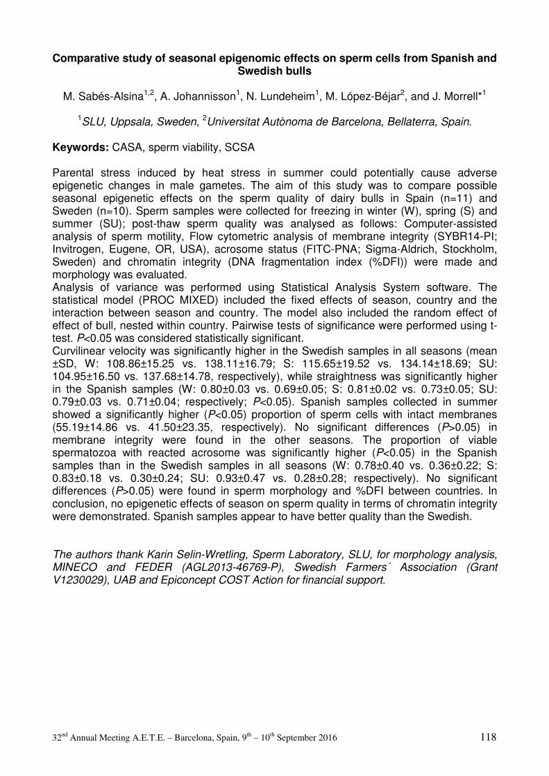
32nd Annual Meeting A.E.T.E. – Barcelona, Spain, 9th – 10th September 2016 118
Comparative study of seasonal epigenomic effects on sperm cells from Spanish and Swedish bulls
M. Sabés-Alsina1,2, A. Johannisson1, N. Lundeheim1, M. López-Béjar2, and J. Morrell*1
1SLU, Uppsala, Sweden, 2Universitat Autònoma de Barcelona, Bellaterra, Spain.
Keywords: CASA, sperm viability, SCSA Parental stress induced by heat stress in summer could potentially cause adverse epigenetic changes in male gametes. The aim of this study was to compare possible seasonal epigenetic effects on the sperm quality of dairy bulls in Spain (n=11) and Sweden (n=10). Sperm samples were collected for freezing in winter (W), spring (S) and summer (SU); post-thaw sperm quality was analysed as follows: Computer-assisted analysis of sperm motility, Flow cytometric analysis of membrane integrity (SYBR14-PI; Invitrogen, Eugene, OR, USA), acrosome status (FITC-PNA; Sigma-Aldrich, Stockholm, Sweden) and chromatin integrity (DNA fragmentation index (%DFI)) were made and morphology was evaluated. Analysis of variance was performed using Statistical Analysis System software. The statistical model (PROC MIXED) included the fixed effects of season, country and the interaction between season and country. The model also included the random effect of effect of bull, nested within country. Pairwise tests of significance were performed using t-test. P<0.05 was considered statistically significant. Curvilinear velocity was significantly higher in the Swedish samples in all seasons (mean ±SD, W: 108.86±15.25 vs. 138.11±16.79; S: 115.65±19.52 vs. 134.14±18.69; SU: 104.95±16.50 vs. 137.68±14.78, respectively), while straightness was significantly higher in the Spanish samples (W: 0.80±0.03 vs. 0.69±0.05; S: 0.81±0.02 vs. 0.73±0.05; SU: 0.79±0.03 vs. 0.71±0.04; respectively; P<0.05). Spanish samples collected in summer showed a significantly higher (P<0.05) proportion of sperm cells with intact membranes (55.19±14.86 vs. 41.50±23.35, respectively). No significant differences (P>0.05) in membrane integrity were found in the other seasons. The proportion of viable spermatozoa with reacted acrosome was significantly higher (P<0.05) in the Spanish samples than in the Swedish samples in all seasons (W: 0.78±0.40 vs. 0.36±0.22; S: 0.83±0.18 vs. 0.30±0.24; SU: 0.93±0.47 vs. 0.28±0.28; respectively). No significant differences (P>0.05) were found in sperm morphology and %DFI between countries. In conclusion, no epigenetic effects of season on sperm quality in terms of chromatin integrity were demonstrated. Spanish samples appear to have better quality than the Swedish. The authors thank Karin Selin-Wretling, Sperm Laboratory, SLU, for morphology analysis, MINECO and FEDER (AGL2013-46769-P), Swedish Farmers´ Association (Grant V1230029), UAB and Epiconcept COST Action for financial support.

32nd Annual Meeting A.E.T.E. – Barcelona, Spain, 9th – 10th September 2016 119
OPU – IVF and ET

32nd Annual Meeting A.E.T.E. – Barcelona, Spain, 9th – 10th September 2016 120
Pregnancy rates from different cattle breed embryos produced in vitro in a commercial program (part 1).
C.J. Arreseigor*1,2, Y. Filipiak4, A.R. Pereira2, A.E. Arreseigor1,2, K. Avelino3, A.B.
Ibarreche1, A.C. Maraia3, and J. Paredes2
1Arreseigor & Asociados, Corrientes, Argentina, 2Bionorte SA, Asunción, Paraguay, 3Embryo Rio Preto Ltda, San Jose do Rio Preto, Brasil, 4Facultad de Veterinaria,
Universidad de la República, Montevideo, Uruguay. Keywords: breed, embryo, pregnancy rate The objective of this study was to determine the pregnancy rate after transferring embryos from different cattle breeds in a commercial program of in vitro embryo production (IVP). Donors of different breeds, Angus, Nelore, Brangus, Holstein, Gyr and Girolando, were subjected to ovum pick up (OPU).Cumulus oocyte complexes were in vitro matured (22h, 38°C, 5% CO2 in humidified air) and then, fertilized in vitro with frozen-thawed semen, previously centrifuged for capacitation (18h, 38°C, 5% CO2 in humidified air).. Embryos were cultured under the same conditions and procedures during 7 days (38°C, 5% CO2 in humidified air), when they were classified according to IETS criteria. Only grade 1 embryos were considered for transfer. Thus, 19195 fresh morula and blastocyst stage embryos (Angus=2576; Nelore=9715; Brangus=652; Hostein=647; Gyr=4231 and Girolando=1374) were transported up to 12 h in portable incubator and transferred to crossbred heifers. From day 28 to 32 after fertilization, the pregnancy status of recipients was determined through transrectal ultrasonography by visualization of the embryonic vesicle. For statistical studies, comparisons of frequency and analysis by Chi-square were performed, considering a significance level p<0.05. Pregnancy rates according to the different breeds were: 36.8% for Brangus (240), 36.7% for Nelore (3569), 32.9% for Angus (848), 32.2% for Girolando (443), 29.7% for Gyr (1256) and 22.7% for Holstein (147). Significant differences (p<0.05) were observed between all breeds except Angus vs. Girolando (p=0.66), Nelore vs. Brangus (p=0.97), Angus vs. Brangus (trend toward significance, p=0.06) and Gyr vs. Girolando (trend, p=0.07). In conclusion, the breed of the embryo significantly influences pregnancy rate in crossbred recipients, which affects the production results in commercial OPU/IVP programs, and furthermore demonstrating an improved performance of beef breed embryos. Embryo Rio Preto and Bionorte SA

32nd Annual Meeting A.E.T.E. – Barcelona, Spain, 9th – 10th September 2016 121
Embryo mortality from different cattle breed embryos produced in vitro in a commercial program (part 2).
C.J. Arreseigor*1,2, Y. Filipiak4, A. Pereira2, A.E. Arreseigor1,2, K. Avelino3, A.B. Ibarreche1,
A.C. Maraia3, and J. Paredes2
1Arreseigor & Asociados, Corrientes, Argentina, 2Bionorte SA, Asuncion, Paraguay, 3Embryo Rio Preto Ltda, San Jose do Rio Preto, Brasil, 4Facultad de veterinaria,
Universidad de la Republica, Montevideo, Uruguay.
Keywords: Pregnancy, Embryo Mortality, Breeds Embryo mortality (EM) in cattle is considered the main cause of reproductive failure, increasing the calving interval and implicating a greater economic loss in programs of in vitro embryo production (IVP) from high genetic merit animals. The objective of this study was to determine the EM rate after transferring embryos from different cattle breeds in a commercial program of IVP. Donors of different breeds, Angus, Nelore, Brangus, Holstein, Gyr and Girolando, were subjected to ovum pick up (OPU). Cumulus oocyte complexes (COC) were in vitro matured (22h, 38°C, 5%CO2 in humidified air) and then, fertilized in vitro with frozen-thawed semen, previously centrifuged for capacitation (18h, 38°C, 5%CO2 in humidified air). Embryos were cultured under the same conditions and procedures during 7 days (38°C, CO2 in humidified air) and classified according to IETS criteria. Only grade 1 embryos were considered for transfer. Thus, 19195 fresh morula and blastocyst stage embryos were transported up to 12h in portable incubator and transferred to crossbred heifers. From day 28 to 32 after fertilization, the pregnancy status of recipients was determined through transrectal ultrasonography by visualization of the embryonic vesicle. A total of 6503 pregnancies were diagnosed, 240 for Brangus (36.8%), 3569 for Nelore (36.7%), 848 for Angus (32.9%), 443 for Girolando (32.2%), 1256 for Gyr (29.7%) and 147 for Holstein (22.7%). Pregnant recipients were reevaluated by ultrasound 35 to 40 days later to confirm embryo survival by finding the heartbeat and/or spontaneous movements. For statistical studies comparisons of frequency and analysis by Chi-square were performed, considering a significance level p<0.05. EM rate according to breeds were: 0.0% for Brangus, 4.9% for Gyr (61), 5.1% for Nelore (181), 6.3% for Girolando (28), 6.5% for Angus (55) and 12.2% for Holstein (18). Significant differences (p<0.05) were observed between all breeds except Angus vs. Girolando, Angus vs. Nelore, Nelore vs.Gyr, Nelore vs. Girolando and Gyr vs. Girolando.In conclusion, the breed of the embryo significantly affects EM, demonstrating a better performance of embryos originated from Bos indicus and their crossbreeds than those obtained from pure Bos taurus breeds. Embryo Rio Preto Ltda,Bionorte SA

32nd Annual Meeting A.E.T.E. – Barcelona, Spain, 9th – 10th September 2016 122
A comparison of Luprostiol and Dinoprost tromethamine for induction of oestrus in donkey embryo transfer
M. Bottrel, E. Zarza, L. Fernandez, I. Ortiz, M. Hidalgo, and J. Dorado*
Veterinary Reproduction Group, Department of Animal Medicine and Surgery, University of
Cordoba, Cordoba, Spain.
Keywords: Luprostiol, Dinoprost acetate, oestrus Prostaglandin F2alpha (PGF2a) and its analogues have been routinely used after the embryo flush to induce the onset of oestrus in female donors (mares and jennies) and thus to obtain more embryos in a given season. The aim of this preliminary study was to compare the efficacy of Luprostiol (Prosolvin, Virbac, Spain) and Dinoprost tromethamine (Lutalyse, Pfizer Animal Health, Brazil) for induction of oestrus in jennies. Fourteen Andalusian jennies, 3-12 years of age, were used as embryo donors. Ovarian activity was evaluated daily during oestrus by transrectal ultrasound. When a ≥ 35 mm follicle was detected, donor jennies were naturally mated every other day until ovulation (Day 0). Uterine flushing was done on Days 6 to 9. Each recovered embryo was measured and evaluated for morphology. In a total of 38 collections, donors were randomly treated with either 5.25 mg Luprostiol i.m. (n = 27) or 3.5 mg Dinoprost tromethamine i.m. (n = 11) immediately after the embryo recovery. Data were assessed by the chi-square test and the Kruskal-Wallis ANOVA. All donors responded well to prostaglandins (Luprostiol and Dinoprost tromethamine) given right after embryo collection. There were no significant (P > 0.05) differences between treatments for the interval from prostaglandin to ovulation (9.85 ± 0.72 days vs. 10.18 ± 0.84 days). In addition, the interovulatory interval was similar (16.56 ± 0.63 days vs. 17.27 ± 1.23 days; P > 0.05) between prostaglandins. Furthermore,
there were no significant (P > 0.05) differences between these treatments for the embryo recovery rate (average, 74.3%) and embryo quality. Eighteen embryos were recovered from 27 flushes in Prosolvin group (66.7%), of which 14 were classified as Grade 1 (77.8%), 3 as Grade 2 (16.7%), and 1 as Grade 3 (5.6%). Meanwhile, nine embryos were recovered from 11 flushes in Lutalyse group (81.8%), of which 7 were classified as Grade 1 (77.8%), 1 as Grade 2 (11.1%), and 1 as Grade 3 (11.1%). Based in our results, we can conclude that both drugs used in the present study are adequate to consistently induce the onset of oestrus in jennies, with no deleterious effects on embryo recovery rate or embryo quality. Supported by Grant AGL2013-42726-R

32nd Annual Meeting A.E.T.E. – Barcelona, Spain, 9th – 10th September 2016 123
Effects of ghrelin on activation of Akt and Erk1/2 pathways during in vitro maturation of bovine oocyte
T.M. Chouzouris*1, E. Dovolou1, F. Krania1, I. Pappas2, K. Dafopoulos3, G. Anifandis3, and
G.S. Amiridis1
1Department Obstetrics and Reproduction, Veterinary Faculty, School of Health Sciences, Karditsa, Greece, 2Department of Pharmacology, Veterinary Faculty, School of Health Sciences, Karditsa, Karditsa, Greece, 3Department of Obstetrics and Gynecology, ART Unit, University of Thessaly, School of Health Sciences, Faculty of Medicine, Larissa,
Greece.
Keywords: ghrelin, oocyte maturation, Akt Ghrelin –a stomach derived peptide- acts, in general, as a negative feedback signal for reproductive functions affecting gonadotropin secretion, oocyte maturation and early embryo development. In a previous study we have shown that ghrelin at 800pg/ml accelerates in vitro maturation of bovine oocytes, reducing embryo yield (Dovolou et al 2014; RDA 49,665-72). Here we studied the role and possible mode of action of ghrelin on in vitro maturation of bovine oocytes. In exp. 1, in 3 replicates, COCs (n=632) collected from abattoir material were matured in vitro for 18 or 24 hours in the presence of 0 or 800pg/ml of acylated bovine ghrelin (groups G18, G24, C18, C24). Matured oocytes were co-cultured for 24h with thawed bull semen, and presumptive zygotes were cultured for 9 days at 390C, 5%CO2, and 5% O2 . Cleavage and blastocyst formation rate were assessed on days 2 and 7 to 9, respectively and statistical analysis was carried out by one-way repeated measures ANOVA, with arcsine transformation. Exp. 1 served as internal control for the ensuing study on proteins. In exp. 2, after 0,6,10,18 and 24h in maturation, subsets of maturing COCs (each n=10 in three replicates, totally n=270) were mechanically denuded from the surrounding cells and cumulus cells and oocytes were separately stored in liquid N2 for the assessment of activation of Akt and Erk1/2 pathways. Whole-cell extracts were prepared by lysis on ice using a buffer containing Tris-HCl NaCl, EDTA, Triton X-100, sodium deoxycholate containing protease inhibitors cocktail and sodium orthovanadate and assays were performed as recommended by the manufacturer (EMD Millipore, Darmstadt, Germany). Phospho-Akt1/PKBalpha (Ser473) and phospho-Erk/MAP Kinase 1/2 (Thr185/Tyr187) beadmates were used together. Protein expressions were measured on a Luminex 100 instrument (Luminex Corporation). The data output gives the mean fluorescence intensity (mFI) as a measure of protein abundance. We calculated the ratio of phosphoprotein mFI to number of treated or un-treated oocytes and cumulus cells and results were analyzed by ttest. In exp. 1 no difference existed in cleavage rate between groups. G24 yielded significantly less (p<0.03) day 7 blastocyst in comparison to those of C18, C24 and G18 (16.3% , 29.3%, 26.9% and 30.0%, respectively). In exp. 2, it was found that at 18 and 24 hours ghrelin caused significant increased phosphorylation of Akt in cumulus cells in comparison to the respective controls (pAkt/total Akt : 18h, 1.12±0.1 and 0.79±0.15; 24h, 0.99± 0.04 and 0.72± 0.08, for ghrelin and control groups). Oocytes matured in the presence of ghrelin at 10 h expressed lower Akt phosphorylation rate compared to controls (0.77± 0.13 and 1.21± 0.09). At 6 hours Erk1/2 phosphorylation was increased in ghrelin treated oocytes compared to the respective controls (11.48± 3.9 vs 6.08± 1.6). We infer that ghrelin exerts antiapoptotic and antioxidant actions through the cumulus cells, and accelerates maturation via early phosphorylation of Erk1/2 that possibly brings about early activation of the maturation promoting factor.

32nd Annual Meeting A.E.T.E. – Barcelona, Spain, 9th – 10th September 2016 124
Cryopreservation and intrauterine transfer of canine embryos
L. Commin*, S. Buff, E. Rosset, A. Charlot-Valdieu, T. Bonte, M. Guedes-Teixeira, and T. Joly
UPSP 2011-03-101 ICE ‘Interactions Cellules Environnement’ & Centre de Ressources
Biologiques – Infrastructure CRB-ANIM (ANR11-INBS-0003), VetAgro Sup, Marcy l'Etoile, Rhone France.
Keywords: embryo transfer, canine, cryopreservation The objective of the present study was to obtain live birth after transfer of cryopreserved canine embryos. To this end, seven bitches (Labrador retriever, 6.2 ± 1.8 years old) were used as donors according to ethical committee agreement (Vetagro Sup ethical committee number 1070) and ovulation timing was confirmed using blood sample monitoring (progesterone and LH assays). All donors were mated with fertile males 48 hours post ovulation and repeated 24-48 hours later to ensure fertilization. Females underwent ovariohysterectomies 9.7 ± 0.6 days post ovulation. Then, the embryos were collected ex vivo as previously described (Commin. L, Reproduction of Domestic Animals; Suppl 6:144-6; 2012), and classified under stereomicroscope. Among the 43 embryos collected (2 cells to blastocyst stage), 35 were slow freezed in a Vigro ® solution (Bioniche, Belleville Canada) containing ethylene glycol (EG, 1,5M) supplemented with BSA (0.4% v/v) and stored in 0.3 mL straws in liquid nitrogen until transfer to recipients. Six beagle bitches (16.5 ± 6.6 months old) were used as recipients and monitored by blood progesterone analysis and ultrasound examination to determine the time of ovulation. After thawing, embryos were observed under stereomicroscope to check for normal embryo morphology. Recipients were transferred at 9.6 ± 0.8 days post ovulation to allow less than 1 day of interval between the recipients and donors estrus cycles. Thawed embryos were surgically transferred to the recipients under gas anesthesia. The transfer were performed bilaterally on the top of the uterine horn, next to the oviduct junction, or close to the uterine bifurcation. Three to nine thawed embryos (undamaged, good quality) were transferred per recipient. After the surgery, the females were allowed to recover during at least 15 days before performing abdominal ultrasound for pregnancy diagnosis. None of the 6 bitches that were transferred became pregnant and no luteal deficiency was observed after blood progesterone analysis, suggesting more the inefficiency of the uterine transfer technique than the negative impact of the pregnancy maintenance. Even if the morphological assessment of thawed embryos did not allow to identify any significant damage after such a cryopreservation protocol, we were unable to confirm the viability of the embryos before transfer. Further in vitro studies (ie. in vitro culture or embryo differential staining) should be performed to confirm the intrinsic quality of slow frozen/thawed embryos. Also, improvement of the transfer technique (for example, using a transcervical endoscopic approach) should be evaluated with respect of a reduction the traumatic effects of the surgical technique (ie. capillary blood contamination). This work was supported by a grant from the French Federation of Guide Dogs Associations (FFAC)

32nd Annual Meeting A.E.T.E. – Barcelona, Spain, 9th – 10th September 2016 125
Ovulation induction for embryo transfer in Andalusian donkeys: human Chorionic Gonadotrophin versus Deslorelin acetate
J. Dorado*, M. Bottrel, L. Fernandez, E. Zarza, I. Ortiz, and M. Hidalgo
Veterinary Reproduction Group, Department of Animal Medicine and Surgery, University of
Cordoba, Cordoba, Spain.
Keywords: hCG, Deslorelin acetate, ovulation The use of ovulation-inducing agents can improve the effectiveness of embryo transfer by synchronizing ovulation and artificial insemination (AI) or mating. Moreover, the duration of oestrus and the number of AIs or matings can be reduced with these treatments. This preliminary study was conducted to compare the efficacy of two drugs, human Chorionic Gonadotrophin (hCG; Veterin Corion, Divasa Farmavic, Spain) and Deslorelin acetate (Sincrorrelin, Ourofino Saúde Animal, Brazil), for induction of ovulation in jennies. Data on embryo recovery rates were recorded from 16 Andalusian jennies aged 3-12 years old. A total of 50 oestrus cycles were used and randomly divided into three groups: (i) no treated as control (n = 26), (ii) treated with 1500 IU hCG i.m. (n = 11), and (iii) treated with 0.75 mg Deslorelin acetate i.m. (n = 13). Donors were treated when they had a follicle larger than 35 mm of diameter and uterine oedema. Ovulation was diagnosed by daily ultrasound observations. Six to nine days after ovulation, donors were flushed three times for embryo recovery and each recovered embryo was measured and evaluated for morphology. Data were assessed by the chi-square test and the Kruskal-Wallis ANOVA. All donors (n = 24) responded well to the administration of both ovulation inducing agents. No differences (P > 0.05) were observed between treatments for the time to ovulation from treatment (hCG: 52.00 ± 4.00 h; Deslorelin acetate: 49.60 ± 4.95 h). The range of response was also similar (P > 0.05) for hCG and Deslorelin acetate (24 h: 9.1% vs. 15.4%; 48 h: 72.7% vs. 46.2%; 72 h: 18.2% vs. 38.5%). None of the variables studied (embryo recovery and mean diameter and age of embryos) were affected (P > 0.05) either by the ovulation treatment or the time to ovulation from treatment, but embryo quality (P = 0.019 for hCG vs. Deslorelin acetate). Nineteen embryos were recovered from 26 flushes (73.08%) in control jennies, of which 17 (89.5%) were classified as Grade 1 and 2 (10.5%) as Grade 2, whereas 8 Grade 1 embryos were collected from 11 flushes (72.73%) in jennies treated with hCG. Nine embryos were recovered from 13 flushes (69.23%) in jennies treated with Deslorelin acetate, of which 4 were classified as Grade 1 (44.4%), 3 as Grade 2 (33.3%), and 2 as Grade 3 (22.2%), showing a significantly lower percentage of Grade 1 embryos (P < 0.05) compared to hCG group. In conclusion, our results suggest that both drugs used in this study are adequate to consistently induce ovulation in jennies, but we noted that embryos recovered in jennies treated with hCG had better quality than those of the Deslorelin acetate ones. Supported by Grant AGL2013-42726-R

32nd Annual Meeting A.E.T.E. – Barcelona, Spain, 9th – 10th September 2016 126
Cytological evaluation of PMN distribution in the genital tract of superovulated embryo donor cows
J. Egberts*1,2, J. Detterer1, A. Park1, D. Töpfer2, and S. Meinecke-Tillmann2
1AI- and ET-Center Georgsheil, Georgsheil, Germany, 2Institute for Reproductive Biology, University of veterinary medicine Hannover, Hannover, Germany.
Keywords: PMN, estrus cycle, donor cows Polymorphonuclear neutrophils (PMN) aid in the protection of the mucosae of the reproductive tract and comprise the first line of defense against infections. Since the mucosae of donor cows are challenged during inseminations (AI) and embryo collection (EC), the distribution of PMN in the genital tract of superovulated cows [n=26; 1st–10th lactation; average milk yield: 33.2±8 kg; 222±192 days in milk (range: 72-1018d); healthy and gynecologically sound at pre-examination] has been examined. Superovulation followed the Georgsheil-Pluset protocol [PlusetÒ, 4 d, twice daily with decreasing dosage (in total 10 or 12.5 ml); PGF2α (PG) twice (am: PG analogue; pm: PG) on day 4 of FSH treatment]. Twelve to 16 h prior to EC animals were treated with a PG to induce corpora lutea regression and enhance embryo collection. Cytological samples were examined on 5 occasions: D9±1.6 of the cycle before EC (E1); at 1st (E2) and second AI (E3) after superovulation; at EC (E4; D7 of the cycle); 16±3 d after the last PG treatment (E5). Samples were taken with a modified cytobrush from vagina and cervix (E1, E2, E3, E4, E5) as well as corpus uteri and both uterine horns (E1, E4, E5), and from the tip of the insemination gun after AI into the uterine body (E2, E3). Following diff-quick® staining 300 cells/slide were differentiated into epithelial cells and PMN. The mean percentage of PMN (PMN%) at all locations of the genital tract varied during the investigation period (p<0.05 to p<0.001; Dunn’s method), except intrauterine samples E2 vs. E3. Values in the vagina at E1 (4.1±5.0%) increased to a maximum of 17.7±12.1% at E3. Thereafter, a decrease below E1-values was observed (E4: 3.7±5.2%), whereas a second rise was obvious at E5 (10.4±10.5%). The PMN-profile in the cervical canal ran parallel to that in the vagina, reaching a lower maximum level. Intrauterine cytobrush samples never exceeded 2.7±1.8 PMN%, although cyclical variations were obvious (E1: 2.5±1.8%; E4: 1.1±0.9%; E5: 1.7±2.7%). Samples taken from the tip of the insemination gun varied largely in relation to PMN% (E2: 13.1±19.0%; E3: 15.6±16.2%) which might be due to some cell contamination from the cervix, although a disposable plastic cover had been used for AI. This study is the first to outline the PMN-distribution in different areas of the genital tract of superovulated embryo donor cows throughout the estrus cycle. The results show that PMN%-profiles in the different parts of the genital tract of superovulated embryo donor cows are subject to cyclic changes. This is most obvious in vagina and cervix which might be linked to the type of natural ejaculate deposition in bovines. The uterine PMN% (cytobrush samples) indicate that superovulation might have a positive effect on the endometrial health status, since values at EC are significantly lower than in the preceding interestrus. Samples from the tip of the insemination gun are not directly comparable to endometrial cytobrush samples. Yet they fit the PMN% fluctuations of the other localizations.
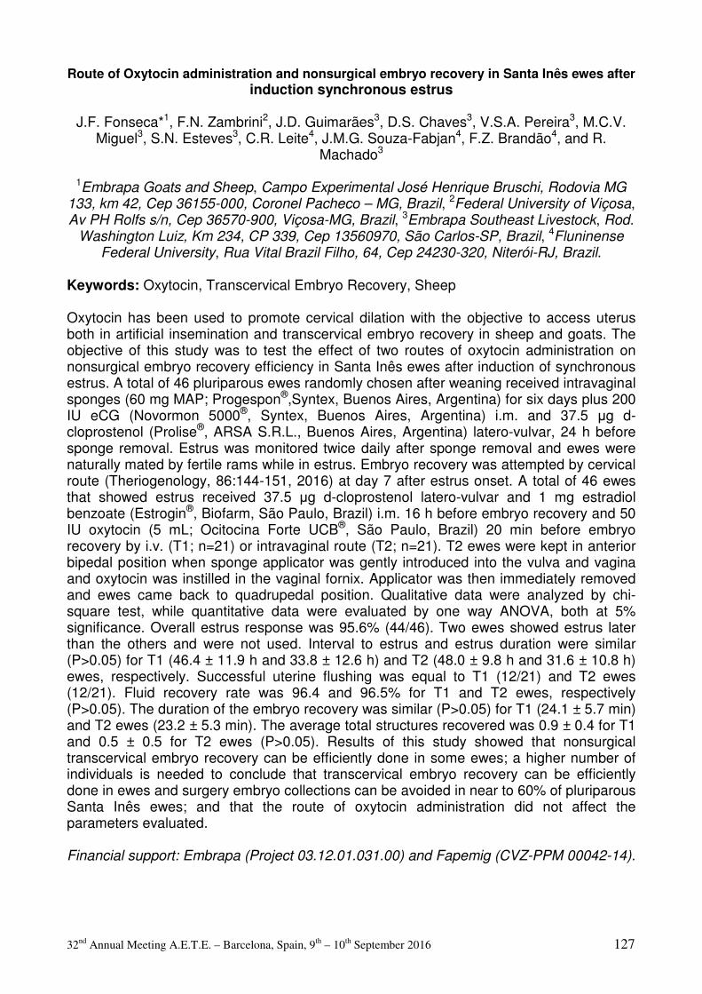
32nd Annual Meeting A.E.T.E. – Barcelona, Spain, 9th – 10th September 2016 127
Route of Oxytocin administration and nonsurgical embryo recovery in Santa Inês ewes after
induction synchronous estrus
J.F. Fonseca*1, F.N. Zambrini2, J.D. Guimarães3, D.S. Chaves3, V.S.A. Pereira3, M.C.V. Miguel3, S.N. Esteves3, C.R. Leite4, J.M.G. Souza-Fabjan4, F.Z. Brandão4, and R.
Machado3
1Embrapa Goats and Sheep, Campo Experimental José Henrique Bruschi, Rodovia MG 133, km 42, Cep 36155-000, Coronel Pacheco – MG, Brazil, 2Federal University of Viçosa, Av PH Rolfs s/n, Cep 36570-900, Viçosa-MG, Brazil, 3Embrapa Southeast Livestock, Rod.
Washington Luiz, Km 234, CP 339, Cep 13560970, São Carlos-SP, Brazil, 4Fluninense Federal University, Rua Vital Brazil Filho, 64, Cep 24230-320, Niterói-RJ, Brazil.
Keywords: Oxytocin, Transcervical Embryo Recovery, Sheep Oxytocin has been used to promote cervical dilation with the objective to access uterus both in artificial insemination and transcervical embryo recovery in sheep and goats. The objective of this study was to test the effect of two routes of oxytocin administration on nonsurgical embryo recovery efficiency in Santa Inês ewes after induction of synchronous estrus. A total of 46 pluriparous ewes randomly chosen after weaning received intravaginal sponges (60 mg MAP; Progespon®,Syntex, Buenos Aires, Argentina) for six days plus 200 IU eCG (Novormon 5000®, Syntex, Buenos Aires, Argentina) i.m. and 37.5 µg d-cloprostenol (Prolise®, ARSA S.R.L., Buenos Aires, Argentina) latero-vulvar, 24 h before sponge removal. Estrus was monitored twice daily after sponge removal and ewes were naturally mated by fertile rams while in estrus. Embryo recovery was attempted by cervical route (Theriogenology, 86:144-151, 2016) at day 7 after estrus onset. A total of 46 ewes that showed estrus received 37.5 µg d-cloprostenol latero-vulvar and 1 mg estradiol benzoate (Estrogin®, Biofarm, São Paulo, Brazil) i.m. 16 h before embryo recovery and 50 IU oxytocin (5 mL; Ocitocina Forte UCB®, São Paulo, Brazil) 20 min before embryo recovery by i.v. (T1; n=21) or intravaginal route (T2; n=21). T2 ewes were kept in anterior bipedal position when sponge applicator was gently introduced into the vulva and vagina and oxytocin was instilled in the vaginal fornix. Applicator was then immediately removed and ewes came back to quadrupedal position. Qualitative data were analyzed by chi-square test, while quantitative data were evaluated by one way ANOVA, both at 5% significance. Overall estrus response was 95.6% (44/46). Two ewes showed estrus later than the others and were not used. Interval to estrus and estrus duration were similar (P>0.05) for T1 (46.4 ± 11.9 h and 33.8 ± 12.6 h) and T2 (48.0 ± 9.8 h and 31.6 ± 10.8 h) ewes, respectively. Successful uterine flushing was equal to T1 (12/21) and T2 ewes (12/21). Fluid recovery rate was 96.4 and 96.5% for T1 and T2 ewes, respectively (P>0.05). The duration of the embryo recovery was similar (P>0.05) for T1 (24.1 ± 5.7 min) and T2 ewes (23.2 ± 5.3 min). The average total structures recovered was 0.9 ± 0.4 for T1 and 0.5 ± 0.5 for T2 ewes (P>0.05). Results of this study showed that nonsurgical transcervical embryo recovery can be efficiently done in some ewes; a higher number of individuals is needed to conclude that transcervical embryo recovery can be efficiently done in ewes and surgery embryo collections can be avoided in near to 60% of pluriparous Santa Inês ewes; and that the route of oxytocin administration did not affect the parameters evaluated. Financial support: Embrapa (Project 03.12.01.031.00) and Fapemig (CVZ-PPM 00042-14).

32nd Annual Meeting A.E.T.E. – Barcelona, Spain, 9th – 10th September 2016 128
Effect of oocyte transport between two European countries on the bovine blastocyst production
G. Gamarra Lazo1, D. Martinez Bello*2,3, G. Elcoso Usieto3, J. Sabin Vilar2, and S. Lacaze1
1AURIVA, Domaine de Sensacq, 64230 Denguin, France, 2Embriovet, Rois, 18K 15165, Bergondo, Spain, 3Ponderosa Holsteins, C/Unica sn, Hostalets de Tost, 25795 Lleida.
Keywords: oocyte transportation, bovine blastocyst production, European countries Commercial OPU is often performed on sites that are quite distant from the IVP labs (Ward et al., Theriogenology 54, 433-446, 2000). Maturing oocytes during shipment is a practical solution to overcome the problem of distance and help to apply the technique in many regions without IVF facilities. Despite free circulation agreements for trade and people through European countries, specific limitations are set for biological specimens and specifically for bovine gametes. We hypothesized that trans-border cooperation for IVP provided that sanitary status of donor females is in agreement with EEC requirements. The aim of this study was to evaluate the effect of oocyte transport during 6 hours between Spain and France on the bovine blastocyst production. In the transport group, Holstein Cows (n=6) from Ponderosa Holstein farm in Lleida-Spain were stimulated with decreasing doses pFSH (Folltropin, Vetoquinol, France) twice daily during 3 days (total dose: 350 µg). Sanitary tests were performed on donors one week prior to FSH treatment investigating for Tuberculosis, Brucellosis, Leucosis, infectious bovine rhinotracheitis, Bovine viral diarrhea, and Paratuberculosis. COCs were collected by OPU after 36 hours coasting period. COCs were recovered, evaluated immediately after OPU and placed into 2.0 mL tubes (Corning, USA) in 500 µL maturation medium. Gas mix (5% CO2 in air) was injected into each tube tightly sealed and placed at 38.0°C in portable incubator (Minitub, Tiefenbach, Germany) during 6 hours. On arrival in Auriva IVP lab, tubes were opened and placed into the incubator with 5% CO2 at 38.5°C with maximum humidity to complete the 24 hours maturation period. They were fertilized with frozen–thawed X-sorted sperm in TALP medium. Presumptive zygotes were cultured in SOF medium (Minitub) up to day 7 at 38,5 °C in 5% CO2, 5% O2 and 90% N2 atmosphere with maximum humidity. Grade 1 blastocysts and expanded blastocysts according to IETS classification were recorded on days 6.5 and 7. The same procedure was used in control Holstein cows (n=8) from Auriva IVP Laboratory in Denguin France, in the same period, except COCs were placed directly into CO2 incubator, after retrieval and inseminated with X-sorted semen of different bulls. Oocyte recovery and embryo production were analyzed by student T-test and blastocyst yield by Chi-Square. The variation among X-sorted semen on the blastocyst rate in the control group was analyzed by ANOVA. In the transport group and the control group, respectively 22.2 ± 10.0 COCs (6 OPU sessions) and 8.1 ± 3.7 COCs (21 OPU sessions) were collected (p<0.05) and were processed for in vitro maturation. 7.5 ± 5.4 and 4.7 ± 2.0 G1 embryos were produced (p>0.05). The mean embryo development rate (grade 1 embryos / number of oocytes entering maturation process) was 33.8% in the transport group and 57.7% in the control group (p<0.05). No difference was found between bulls. Although it is still necessary to increase embryo development rate, these results demonstrate that successful inter-border cooperation for bovine IVF/IVP is possible in neighbour areas.

32nd Annual Meeting A.E.T.E. – Barcelona, Spain, 9th – 10th September 2016 129
Effect of X-sorted sperm on development grade 1 in vitro-produced embryos derived from bovine ovum pick up oocytes under commercial conditions
G. Gamarra Lazo*1, S. Lacaze1, M. Mouneyres1, and B. Marquant Le Guienne2
1AURIVA, 64230 Denguin, France, 2ALLICE, 78352 Jouy en Josas, France.
Keywords: embryo developmental kinetic, OPU, FIV. Sex-sorted sperm has been used for IVF to produce in vitro sexed bovine embryos. Following IVF, developmental kinetics, specifically the timing of blastocyst formation, has been related to embryo developmental competence (Lonergan et al, Journal of Reproduction and Fertility 117, 159-167, 1999). The objective of this work was to analyze developmental kinetics of OPU-IVP bovine embryos produced with unsorted and X-sorted sperm under commercial conditions. The work was performed at the Biotechnology AURIVA Station located in Denguin, South West, France. Four to fifteen years old Holstein cows (n=31) and 16-22 months old heifers (n=18) were used in an OPU-IVP program. Donor animals were stimulated with decreasing pFSH doses (Stimufol; Reprobiol, Liège, Belgium) twice daily during 3 days, (total dose: 350 µg for cows and 250 µg for heifers). Cumulus oocyte complexes (COCs) were collected by OPU 12 to 24 h after the last FSH injection and matured in vitro using a standard IVM protocol. Oocytes were fertilized with frozen–thawed unsorted or X-sorted sperm in modified Tyrode’s bicarbonate buffered medium using different non pre-tested bulls (n=58). Presumptive zygotes were cultured in SOF medium (Minitub, Tiefenbach, Germany) up to Day 8 at 38.5 °C under an atmosphere of 5% CO2, 5% O2 and 90% N2 with maximum humidity. OPU/IVP was repeated one to 16 times (2.3 ± 3.0) for each donor cow or heifer. Grade 1 (G1) expanded blastocysts, according to IETS classification, were recorded on days 6.5, 7.0, 7.5 and 8. Oocyte recovery and embryo production were analyzed by Student T-test and the rate of expanded blastocyst by Chi-Square. Sixty one OPU sessions were performed with unsorted semen and 66 with X-sorted semen, with a mean number of oocytes processed for in vitro maturation 10.9 ± 6.4 and 9.1 ± 4.8 (p>0.05) and a mean number of G1 produced embryos 7.4 ± 4.5 and 4.7 ± 2.9 (p<0.05) per session, respectively. The mean embryo developmental rate (G1 embryos/number of oocytes entering maturation process) was 67.8% (unsorted group) and 52.1% (X-sorted group; p<0.05). On day 6.5, 52.8% of total blastocysts had already expanded after IVF with unsorted sperm, compared with 37.0% with X-sorted sperm (p<0.05); while 41.3% were developed with unsorted and 53.1% with X-sorted sperm (p<0.05) on day 7.0. Expansion was observed in 4.6% of embryos produced with unsorted and 9.7% with X-sorted sperm (p<0.05) on day 7.5. No difference was observed on day 8 in proportion of expanded blastocysts produced with unsorted and X-sorted sperm. Unlike previous studies in which sexed embryos develop on days 8 and 9, in our study, although the number of expanded blastocysts was high with unsorted semen on day 6.5, the majority of expanded blastocysts developed on day 7 with unsorted (94%) and X-sorted sperm (90%).

32nd Annual Meeting A.E.T.E. – Barcelona, Spain, 9th – 10th September 2016 130
Intrafollicular Oocyte Transfer (IFOT) of immature oocytes improves developmental rates and results in healthy calves
M. Hoelker*1,2, E. Held1,2, A. Kassens3, F. Rings1,2, H. Sieme3, D. Tesfaye1, and K.
Schellander1
1Institute of Animal Science, Animal Breeding and Husbandry Group, University of Bonn, Bonn, Germany, 2Researchstation Frankenforst, Faculty of Agriculture, University of Bonn,
Königswinter, Germany, 3Reproductive Medicine Unit, University of Veterinary Medicine Hannover, Hannover, Germany.
Keywords: IVP, IFOT Although the in vitro production (IVP) of bovine embryos is a well-established technique, there are still major differences of IVP-derived blastocysts compared to their in vivo derived counterparts. To circumvent the negative impacts of in vitro culture on bovine embryos, we have recently established the so called intrafollicular oocyte transfer (IFOT). This technique allows injection of oocytes into the dominant follicle without breaking it, so they move to the oviduct and regular IA can be performed and blastocysts can be recovered from the uterus on day 7. However, it remained an open question whether IFOT could be applied to immature slaughterhouse derived bovine oocytes allowing in vivo fertilization and development, too. Therefore, recipient heifers were synchronized by two injections of PGF2α (2 ml Estrumate ®) 11 days apart followed by a final injection of GnRH (2.5 ml Receptal ®) 42 hours after the second PGF2α administration. Groups of immature oocytes (n = 50) derived from slaughterhouse ovaries were injected into the presumed dominant follicle of synchronized Simmental heifers 37-42 hours after the second PGF2α injection using a modified Ovum-Pick-Up (OPU) equipment. All in all, a total of 800 immature bovine oocytes were transferred to 16 heifers. Subsequently, 307 embryos (38.6 ± 27.2 %) were recovered after flushing the uteri at day 7. Among all recipients, 13 of 16 delivered extra embryos above the recipient’s native one presumed to be derived from the dominant follicle’s own oocyte. Of these recollected embryos, 83.2 ± 11.5 % had cleaved and 48.2 ± 11.2 % had developed to the blastocyst stage. However, when excluding one blastocyst per recipient (considered to be derived from its native oocyte) IFOT embryos still reached a blastocyst rate of 45.5 % being significantly higher (45.5 % vs. 29.3 %, ANOVA, p < 0.05) compared to fully in vitro produced embryos (SOFaa + 0,4 % BSA in 5 % O2 & 5 % CO2) although derived from the same charge of slaughterhouse ovaries. In contrast, cleavage rates did not differ (83.2 % vs. 83.7 %). Hence, our results indicate that the maturational environment profoundly affects the ability of bovine oocytes to develop to the blastocyst stage. Finally, transfer of 13 cryopreserved IFOT-derived blastocysts resulted in 2 pregnancies (15.4 %) and birth of 2 healthy calves. To the best of our knowledge, for the first time our study reports full in vivo development of immature slaughterhouse derived oocytes to the blastocyst stage and these are the first calves reported after transfer of immature slaughterhouse derived oocytes into preovulatory follicles. Thus, our study proofed that IFOT offers a new method to circumvent harmful effects of in vitro production conditions for immature bovine oocytes derived from slaughterhouse ovaries also enabling attractive and innovative new experimental setups.

32nd Annual Meeting A.E.T.E. – Barcelona, Spain, 9th – 10th September 2016 131
In vino veritas? – How resveratrol attenuates oxidative stress in bovine oocytes of prepubertal and adult donors.
P. Kordowitzki*, S. Klein, K.-G. Hadeler, P Aldag, A. Lucas-Hahn, and H. Niemann
Institute of Farm Animal Genetics, Department for Biotechnology, Friedrich-Loeffler-
Institut, Neustadt, Germany. Keywords: reproductive aging, cattle, OPU The oocyte aging phenotype has been well characterized, but the adaptive response machinery to oxidative stress has been poorly investigated. Recently, a growing body of evidence supports the hypothesis that the aging process is regulated by a continuous crosstalk between reactive oxygen species (ROS) and SIRT1. The SIRT1 protein belongs to the family of NAD+-dependent deacetylases, and has been shown to regulate several key processes, including fertility and ageing. Resveratrol (3,4’,5-trihydroxystilbene) is an antioxidant identified in various plant species and red wine which stimulates SIRT1 activity. Based on these observations, the goal of the present study was to examine, if resveratrol protects bovine oocytes and blastocysts from oxidative stress.We hypothesize that SIRT1 signalling activated by resveratrol could play a role in reproductive aging. Cumulus-oocyte-complexes (COCs) of prepubertal (5-6 months old) and adult (2.-6. lactation) cows were collected by Ovum Pick-up. The medium for in vitro maturation and in vitro fertilization was supplemented with 2µM Resveratrol® (Sigma-Aldrich, Buchs, Switzerland). Our standard lab protocol was used as control. Cleavage rates at day 4 and blastocyst formation at day 7 were evaluated. ROS levels in oocytes and embryos were assessed by using BODIPY C11 (Thermo Fisher, Bonn, Germany). Oocytes were incubated for 30min at 37°C in TCM culture supplemented with 10mM BODIPY, blastocysts respectively were incubated in SOF. As positive control, oocytes and blastocysts were treated with 10mM H2O2 for 10min. Finally, oocytes and blastocysts were analyzed under a fluorescent confocal microscope (LSM510, Zeiss, Germany) and relative fluorescent intensity was calculated. For each treatment group 12 independent replicates were performed.One-way ANOVA from JMP software was implemented to evaluate differences of fluorescence intensity units (FIU). Data were expressed in mean ±SEM. In total 456 COCs of cows and 482 of claves were used for the IVP. The cleavage rates of adult and prepubertal donors did not differ significantly among the treatments (standard protocol: 56.5±5.4% for cows and 53.0±4.7% for calves, resveratrol supplemented protocol: 62.1±4.3% for cows and 63.6±3.9%). The blastocyst rates were slightly enhanced in the resveratrol supplemented groups (cows: 34.2±3.8% and calves: 33.1±4.2%) compared to those of standard protocol (cows: 27.5±4.8% and calves: 26.4±3.3%). The relative fluorescence levels of ROS were significantly lower (121±34 FIU) in the resveratrol treated samples than in that of the control group (865±45 FIU, p≤0.05). Additionally, the ROS levels in the untreated groups were significantly higher in MII-oocytes (1255±56 FIU) and blastocysts (984±26 FIU) derived from cows compared to their younger counterparts (442±37 FIU and 310±23 FIU, respectively, p≤0.05). In conclusion, these preliminary results indicate that resveratrol is able to influence the cellular redox status in oocytes and blastocysts of donors in different age.

32nd Annual Meeting A.E.T.E. – Barcelona, Spain, 9th – 10th September 2016 132
The application of bovine in vitro embryo production technology to the rescue of Valdostana Castana breed
G. Lazzari*1,2, G. Crotti1, P. Turini1, M. Volget3, and C. Galli1,4
1Avantea, Laboratory of Reproductive Technologies, Cremona, Italy, 2Avantea Foundation, Cremona, Italy, 3Veterinarian bovine practitioner, Aosta, Italy, 4Dept. of Veterinary Medical
Sciences, University of Bologna, Bologna, Italy. Keywords: genetic rescue, bovine, embryo The Aosta Valley breed brown (Valdostana Castana) is a native rustic breed of alpine zone with a good attitude to the production of milk and meat. Excellent grazer, easily exploits even the high-altitude pastures, a characteristic that has led her to develop a defensive behaviour of its territory and an instinct for pugnacity. This propensity has been selected over the centuries and has turned the breed into "sporting animals” to be presented in local competitions and increasing the interest toward the most performing individuals. Moreover the Valdostana Castana counts a limited number of about 5000 animals giving further incentive to apply modern biotechnologies to preserve the breed. Sporadic attempts to apply superovulation have met with inconsistent results most likely due to the lack of a reliable stimulation protocol for the breed. Therefore over the last three years the ovaries of 15 donor cows have been referred to our laboratory for embryo production by in vitro technologies. The oocytes were aspirated from ovarian follicles larger than 2mm and those with a non atretic cumulus cells were matured in medium TCM199 supplemented with 10% FCS, ITS (insulin, transferrin, sodium selenite, Sigma), FSH and LH (1:1, Menopur, Ferring) for 24h. Frozen semen was used for IVF following separation on a Redigrad gradient composed of two fractions: 45% and 90%. The motile fraction was resuspended in medium SOF-heparin (1µg/ml) at a concentration ranging from 0.3 to 1 million sperm/ml depending on the bull. The day after IVF (day 0) the presumptive zygotes were transferred in 500 microl of medium SOF1 plus aminoacids. On Day 4 and Day 6 half of the medium was changed with SOF2 plus aminoacids. On day 7 and 8 of culture the G1 embryos (IETS grading) that had reached the full/expanded blastocyst stage were frozen in 1.5M ethylene glycol. The freezing curve was seeding at -6°C and cooling at 0.5°C/min down to -32°C and plunging in liquid nitrogen. The total number of oocytes recovered was 694 and the average number per donor was 46.27, ranging from 15 to 88. In total 8 different bulls were used for IVF and cleavage rate was between 25% and 89% with an average of 66.28%. The number of transferrable embryos was 141 ranging from 0 (1 cow) to 30 with an average of 9.4 per cow. The number of freezable embryos was 112 ranging from 0 (1 cow) to 26 with an average of 7.47 per cow. The percentage of transferable and freezable embryos was 30.65% and 24.35% of cleaved respectively. All the grade 1 embryos (112) were frozen in ethylene glycol. None of the non freezable embryos (29) was transferred as fresh. Sixty-three frozen-thawed embryos were transferred into recipient heifers obtaining 21 pregnancies (33% pregnancy rate). At present 14 calves have been born from 17 pregnancies gone to term and 3 pregnancies were lost. These results confirm that the in vitro embryo production technology can contribute to the preservation of special cattle breeds by generating valuable calves from donors destined to the abattoir. This work was supported by project Fecund, EU FP7 n°312097.

32nd Annual Meeting A.E.T.E. – Barcelona, Spain, 9th – 10th September 2016 133
Melatonin accelerates the timing of in vitro porcine embryo development
C.A. Martinez*, A. Nohalez, I. Parrilla, J. Roca, C. Cuello, E.A. Martinez, and M.A. Gil
Dpt. of Animal Medicine and Surgery, University of Murcia, Murcia, Spain. Keywords: Melatonin, embryo culture, porcine Melatonin (MEL) has been used to promote in vitro oocyte maturation and embryo development in different species, including the pig, due to its potential as antioxidant, anti-apoptotic and free radical scavenger. However, the effects of MEL on the timing of embryo development are still unknown. This study aimed to determine the influence of MEL on the developmental kinetics of in vitro produced porcine embryos. Immature oocytes (N=868) collected from abattoir-derived ovaries of prepubertal gilts were cultured in 500 µL drops of maturation medium supplemented with 10 IU/mL eCG and 10 IU/mL hCG for 22 h and then for an additional 22 h in maturation medium without hormonal supplements. Mature oocytes were then inseminated with thawed sperm (1000 spermatozoa per oocyte) in 100 µL drops of fertilization medium for 5 h. Presumed zygotes were cultured in 500 µL drops of glucose-free embryo culture medium that was supplemented with pyruvate and lactate for 2 days and then changed to fresh embryo culture medium containing glucose for an additional 5 days. MEL (1 nM) was added to both the maturation and culture media (IVM+IVC group), or only to the culture medium (IVC group). Media without MEL supplement were used as control. Embryo developmental stages were evaluated after 24, 48, 120 and 144 h of culture to assess the timing of embryonic development. Data are presented as means ± SEM of 4 replicates, and differences among groups were analyzed by ANOVA. Our results show that MEL increased (P<0.001) the rate of cleavage (59.1±2.0% and 61.6±3.5% for IVM+IVC and IVC groups, respectively) at 48 h of culture compared to controls (44.0±5.6%). In addition, a higher (P<0.002) proportion of embryos that cleaved within 48 h of fertilization reached the 3-4-cell stage at 24 h of culture in IVM+IVC (16.9±1.9%) and IVC (17.8±1.5%) groups compared to the control (10.1±3.4%). MEL also increased (P<0.04) the blastocyst formation rates at 120 and 144 h of culture (30.5±2.5% and 40.2±2.7%, and 29.9±2.3% and 40.6±2.2%, for IVM+IVC and IVC groups, respectively) in comparison with the controls (19.5±1.4% and 29.3±3.3%, respectively). At 120 h of culture, blastocysts formed in the IVM+IVC and IVC groups consisted of a population with less (P<0.002) early blastocysts (47.2±3.9% and 45.1±2.6%, respectively) and more (P<0.002) full-expanded blastocysts (52.8±3.9% and 54.9±2.6%, respectively) compared with the controls (68.3±3.9% and 31.7±3.9%, respectively). Similarly, from the total number of blastocysts formed at 144 h of culture, the percentages of expanded-hatching-hatched blastocysts were higher (P<0.05) in the IVM+IVC (58.7±5.7%) and IVC (59.7±3.8%) groups than in the controls (38.7±8.8%). In conclusion, the addition of 1 nM MEL to embryo culture medium accelerated the timing of embryo development regardless of whether MEL was present or not in the maturation medium. Supported by Séneca (19892/GERM/15).

32nd Annual Meeting A.E.T.E. – Barcelona, Spain, 9th – 10th September 2016 134
Reproductive efficiency at a commercial farm comparing AI versus ET at first insemination
D. Martinez*1, F. Sebastian2, J. Sabin1, M. Fernandez2, R. Patron3, I. Lopez-Helguera4,
J.L. Pesantez5, O. Fargas6, and S. Astiz5
1EMBRYOVET SL, A Coruña, Spain, 2Granja SAT MORE, Bétera, Valencia, Spain, 3TRIALVET SL, Cabanillas de la Sierra, Madrid, Spain, 4Ciencia Animal, Universitat Lleida,
Lleida, Spain, 5Dpto. Reproducción Animal, INIA, Madrid, Spain, 6VAPL, SL, Tona, Barcelona, Spain.
Keywords: Reproductive efficiency, ET, first AI To evaluate reproductive efficiency using ET versus AI at first breeding post-calving procedures were performed on a commercial dairy farm in Spain. Eleven heifers were superovulated (Bo et al. Reprod Fertil Dev 2010 22:106-12). The same commercial frozen bull semen (Gerard, ABS) was used. Flushings were performed by the same ET-team (Nr. ES11ET05B). Quality 1 embryos were frozen in Ethylen-glycol and used. 170 primiparous (DIM=158±17.9), were selected to be first-AI with the same semen (Group AI; n=82; DIM=159±16.3) or, ET with collected embryos (Group ET; n=88; DIM=157±19.4); DIM did not differ between groups (P=0.402). Estrus of recipient/inseminated cows was detected (Westfalia Rescounter II pedometers). ET-cows were scanned (7.5-MHz linear, Ibex® Pro) and subjected to ET if at least one CL≥18 mm diameter present. Luteal Surface (LS) was calculated. Early pregnancy diagnosis was performed at 28-34d (ED) and pregnant cows confirmed at 56-63d (CD). Data were analyzed with Chi squared test and logistic regression. Quality-1-embryos average/donor was 5.36; 32.9% of recipient cows were rejected. LS was 460±223.5 mm2 and did not affect pregnancy. Two donors produced more fertile transferred embryos [88.9% (8/9) and =75% (6/8)], and two gave less fertile embryos [0% (0/4) and 14.3% (1/7) P=0.009]. These individual differences would not introduce bias into the study; the distribution of transferred embryos was 28% (17/59) of high fertility embryos, 19% (11/59) low fertility embryos and 52% (31/59) of average fertility embryos. Pregnancy loss did not differ among donors (P=0.114). Fertility was higher for ET-cows [45.8% (27/59 P=0.011)] than for AI-cows [24.4%; (20/82); OR of ED-pregnancy=2.616; P=0.009]. DIM did not affect fertility (P=0.7) with similar DIM (158.8± vs. 157.5±18.1) for non-pregnant and pregnant cows. If rejected cows were included as non-pregnant, fertility did not differ [24.4% (20/82) and 30.7% (27/88) for AI and ET-groups; P=0.39]. This highlights the relevance of the non-physiological ovarian activity resumption. Discarding recipients means to reject those with altered ovarian functionality. Therefore, one mean factor affecting fertility that can be overcome with ET-programs is to diagnose altered ovarian cycle-cows. 75.6% of non-pregnant cows after AI followed pregnancy diagnosis at day 28, with this date being the earliest date to treat. Rejected recipients (32.9%) could be treated at day 7. This accelerated reproductive rhythm has a positive impact on the farm pregnancy rate, and implies benefits of this first-AI ET-program. Early fetal loss did not differ among groups [5% (1/20) vs. 11% (3/27); P=0.63], according with previous works showing similar pregnancy loss after day 42 of pregnancy. In conclusion, the ET-program resulted in a higher farm reproductive efficiency not through a higher fertility of the transferred embryos but through a better-expected global pregnancy rate. Economic implications should be further evaluated.

32nd Annual Meeting A.E.T.E. – Barcelona, Spain, 9th – 10th September 2016 135
Genomic breeding value for number of OPU derived oocytes in bovine
E. Mullaart*1, M. Cornelissen2, H.A. Mulder2, H. Flapper1, and R. van der Linde1
1CRV BV, Arnhem The Netherlands, 2Animal Breeding and Genomics Centre, Wageningen University and Research, Wageningen, The Netherlands.
Keywords: OPU-IVP, breeding value, genomics The success of embryo production by ovum pick-up (OPU) in vitro production (IVP), is determined to a large extent by the initial number of oocytes that can be collected per donor cow. This number of oocytes varies per cow and has a heritability of 0.3 (Merton et al 2009, Theriogenology. 72:885–893). A genomic (based on DNA markers) breeding value for the number of oocytes could be used to select female animals (at young age) that give a high number of oocytes. Since OPU-IVP within CRV is already carried out for more than 20 years and almost all animals are genotyped, the development of such genomic breeding values for OPU should be possible and more accurate than a pedigree-based breeding value. The aim of this study is to determine the reliability of genomic breeding values for OPU. The total dataset contained 2,543 female Holstein Friesian animals with in total 40,734 OPU sessions. From these animals, 890 were genotyped with the Illumina 50K SNP chip (reference population). Classical (pedigree based) and genomic (pedigree + DNA marker based) breeding values were estimated using the singe-step BLUP method. Reliability was determined as the correlation between the pedigree-based or genomic breeding value before OPU records were obtained with the realized number of oocytes after OPU using cross-validation. Based on this dataset the reliability was 0.21 for the pedigree-based breeding values and the reliability increased to 0.29 when genomic information was added. The reliability of genomic breeding values for OPU oocytes was significantly lower than reliabilities obtained for milk production traits (which are around 0.65), but in that case the reference population is much larger, i.e. ~40,000 animals. When selecting the best or the worst 10% based on the genomic breeding value for oocytes, the best animals give two times more oocytes than the worst, 5.0 and 10.8 oocytes respectively. This shows that there is substantial genetic variation in OPU oocytes. This study shows that it is feasible to estimate genomic breeding values for the number of oocytes derived after OPU before OPU records are obtained (no phenotypic data yet). This breeding value has a reliability that is high enough to be of practical use. The breeding value can be used to select donor female animals for optimal use in an OPU-IVP embryo production program. Since all animals in the CRV OPU program are genotyped, the reference population will grow over the coming years resulting in more reliable genomic breeding values.
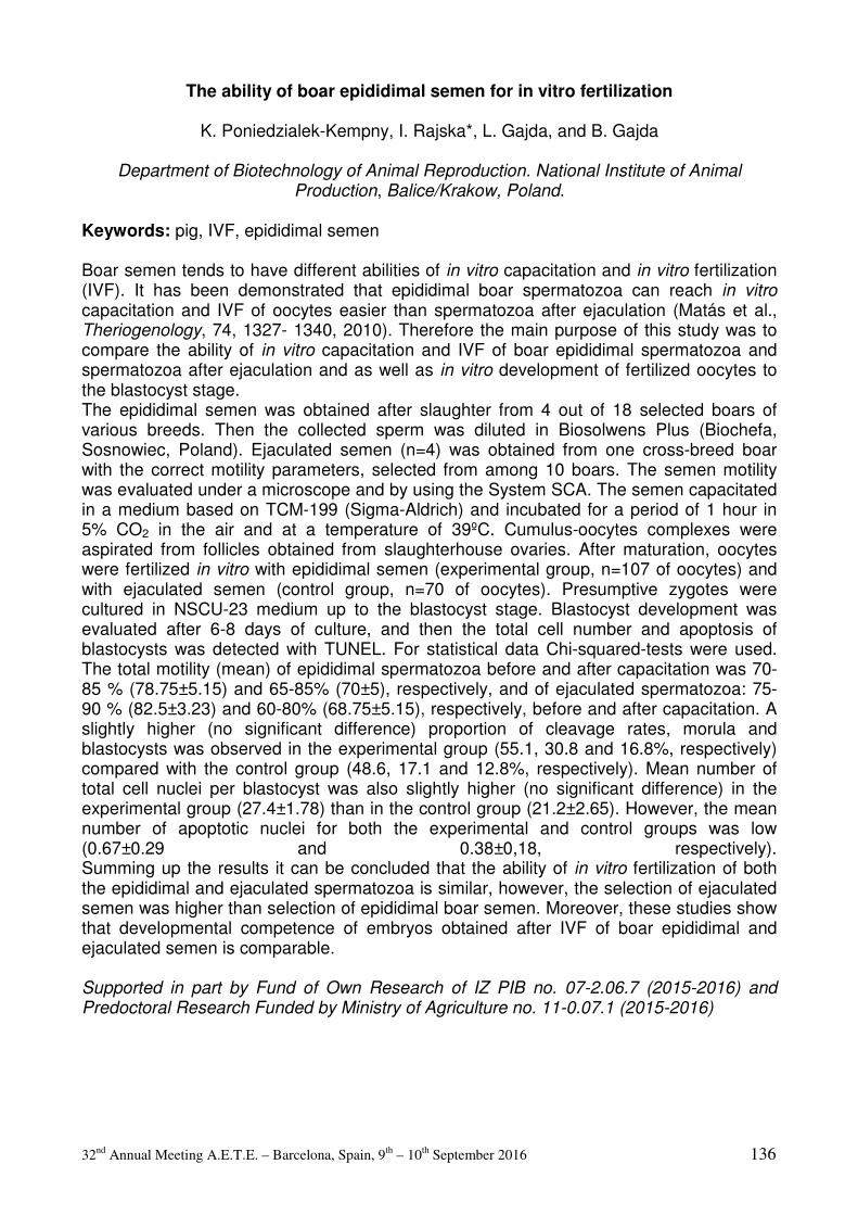
32nd Annual Meeting A.E.T.E. – Barcelona, Spain, 9th – 10th September 2016 136
The ability of boar epididimal semen for in vitro fertilization
K. Poniedzialek-Kempny, I. Rajska*, L. Gajda, and B. Gajda
Department of Biotechnology of Animal Reproduction. National Institute of Animal Production, Balice/Krakow, Poland.
Keywords: pig, IVF, epididimal semen Boar semen tends to have different abilities of in vitro capacitation and in vitro fertilization (IVF). It has been demonstrated that epididimal boar spermatozoa can reach in vitro capacitation and IVF of oocytes easier than spermatozoa after ejaculation (Matás et al., Theriogenology, 74, 1327- 1340, 2010). Therefore the main purpose of this study was to compare the ability of in vitro capacitation and IVF of boar epididimal spermatozoa and spermatozoa after ejaculation and as well as in vitro development of fertilized oocytes to the blastocyst stage. The epididimal semen was obtained after slaughter from 4 out of 18 selected boars of various breeds. Then the collected sperm was diluted in Biosolwens Plus (Biochefa, Sosnowiec, Poland). Ejaculated semen (n=4) was obtained from one cross-breed boar with the correct motility parameters, selected from among 10 boars. The semen motility was evaluated under a microscope and by using the System SCA. The semen capacitated in a medium based on TCM-199 (Sigma-Aldrich) and incubated for a period of 1 hour in 5% CO2 in the air and at a temperature of 39ºC. Cumulus-oocytes complexes were aspirated from follicles obtained from slaughterhouse ovaries. After maturation, oocytes were fertilized in vitro with epididimal semen (experimental group, n=107 of oocytes) and with ejaculated semen (control group, n=70 of oocytes). Presumptive zygotes were cultured in NSCU-23 medium up to the blastocyst stage. Blastocyst development was evaluated after 6-8 days of culture, and then the total cell number and apoptosis of blastocysts was detected with TUNEL. For statistical data Chi-squared-tests were used. The total motility (mean) of epididimal spermatozoa before and after capacitation was 70-85 % (78.75±5.15) and 65-85% (70±5), respectively, and of ejaculated spermatozoa: 75-90 % (82.5±3.23) and 60-80% (68.75±5.15), respectively, before and after capacitation. A slightly higher (no significant difference) proportion of cleavage rates, morula and blastocysts was observed in the experimental group (55.1, 30.8 and 16.8%, respectively) compared with the control group (48.6, 17.1 and 12.8%, respectively). Mean number of total cell nuclei per blastocyst was also slightly higher (no significant difference) in the experimental group (27.4±1.78) than in the control group (21.2±2.65). However, the mean number of apoptotic nuclei for both the experimental and control groups was low (0.67±0.29 and 0.38±0,18, respectively). Summing up the results it can be concluded that the ability of in vitro fertilization of both the epididimal and ejaculated spermatozoa is similar, however, the selection of ejaculated semen was higher than selection of epididimal boar semen. Moreover, these studies show that developmental competence of embryos obtained after IVF of boar epididimal and ejaculated semen is comparable. Supported in part by Fund of Own Research of IZ PIB no. 07-2.06.7 (2015-2016) and Predoctoral Research Funded by Ministry of Agriculture no. 11-0.07.1 (2015-2016)

32nd Annual Meeting A.E.T.E. – Barcelona, Spain, 9th – 10th September 2016 137
Folliculogenesis, Oogenesis and Superovulation
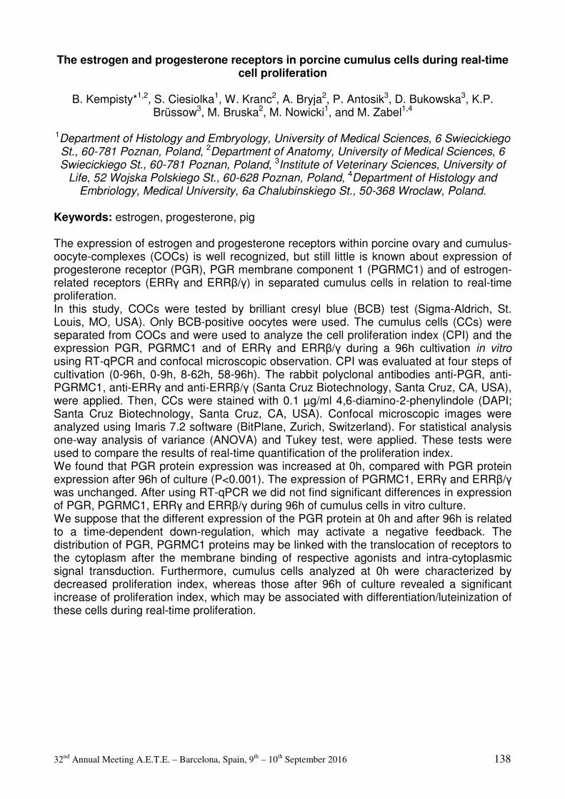
32nd Annual Meeting A.E.T.E. – Barcelona, Spain, 9th – 10th September 2016 138
The estrogen and progesterone receptors in porcine cumulus cells during real-time cell proliferation
B. Kempisty*1,2, S. Ciesiolka1, W. Kranc2, A. Bryja2, P. Antosik3, D. Bukowska3, K.P.
Brüssow3, M. Bruska2, M. Nowicki1, and M. Zabel1,4
1Department of Histology and Embryology, University of Medical Sciences, 6 Swiecickiego St., 60-781 Poznan, Poland, 2Department of Anatomy, University of Medical Sciences, 6 Swiecickiego St., 60-781 Poznan, Poland, 3Institute of Veterinary Sciences, University of
Life, 52 Wojska Polskiego St., 60-628 Poznan, Poland, 4Department of Histology and Embriology, Medical University, 6a Chalubinskiego St., 50-368 Wroclaw, Poland.
Keywords: estrogen, progesterone, pig The expression of estrogen and progesterone receptors within porcine ovary and cumulus-oocyte-complexes (COCs) is well recognized, but still little is known about expression of progesterone receptor (PGR), PGR membrane component 1 (PGRMC1) and of estrogen-related receptors (ERRγ and ERRβ/γ) in separated cumulus cells in relation to real-time proliferation. In this study, COCs were tested by brilliant cresyl blue (BCB) test (Sigma-Aldrich, St. Louis, MO, USA). Only BCB-positive oocytes were used. The cumulus cells (CCs) were separated from COCs and were used to analyze the cell proliferation index (CPI) and the expression PGR, PGRMC1 and of ERRγ and ERRβ/γ during a 96h cultivation in vitro using RT-qPCR and confocal microscopic observation. CPI was evaluated at four steps of cultivation (0-96h, 0-9h, 8-62h, 58-96h). The rabbit polyclonal antibodies anti-PGR, anti-PGRMC1, anti-ERRγ and anti-ERRβ/γ (Santa Cruz Biotechnology, Santa Cruz, CA, USA), were applied. Then, CCs were stained with 0.1 µg/ml 4,6-diamino-2-phenylindole (DAPI; Santa Cruz Biotechnology, Santa Cruz, CA, USA). Confocal microscopic images were analyzed using Imaris 7.2 software (BitPlane, Zurich, Switzerland). For statistical analysis one-way analysis of variance (ANOVA) and Tukey test, were applied. These tests were used to compare the results of real-time quantification of the proliferation index. We found that PGR protein expression was increased at 0h, compared with PGR protein expression after 96h of culture (P<0.001). The expression of PGRMC1, ERRγ and ERRβ/γ was unchanged. After using RT-qPCR we did not find significant differences in expression of PGR, PGRMC1, ERRγ and ERRβ/γ during 96h of cumulus cells in vitro culture. We suppose that the different expression of the PGR protein at 0h and after 96h is related to a time-dependent down-regulation, which may activate a negative feedback. The distribution of PGR, PGRMC1 proteins may be linked with the translocation of receptors to the cytoplasm after the membrane binding of respective agonists and intra-cytoplasmic signal transduction. Furthermore, cumulus cells analyzed at 0h were characterized by decreased proliferation index, whereas those after 96h of culture revealed a significant increase of proliferation index, which may be associated with differentiation/luteinization of these cells during real-time proliferation.
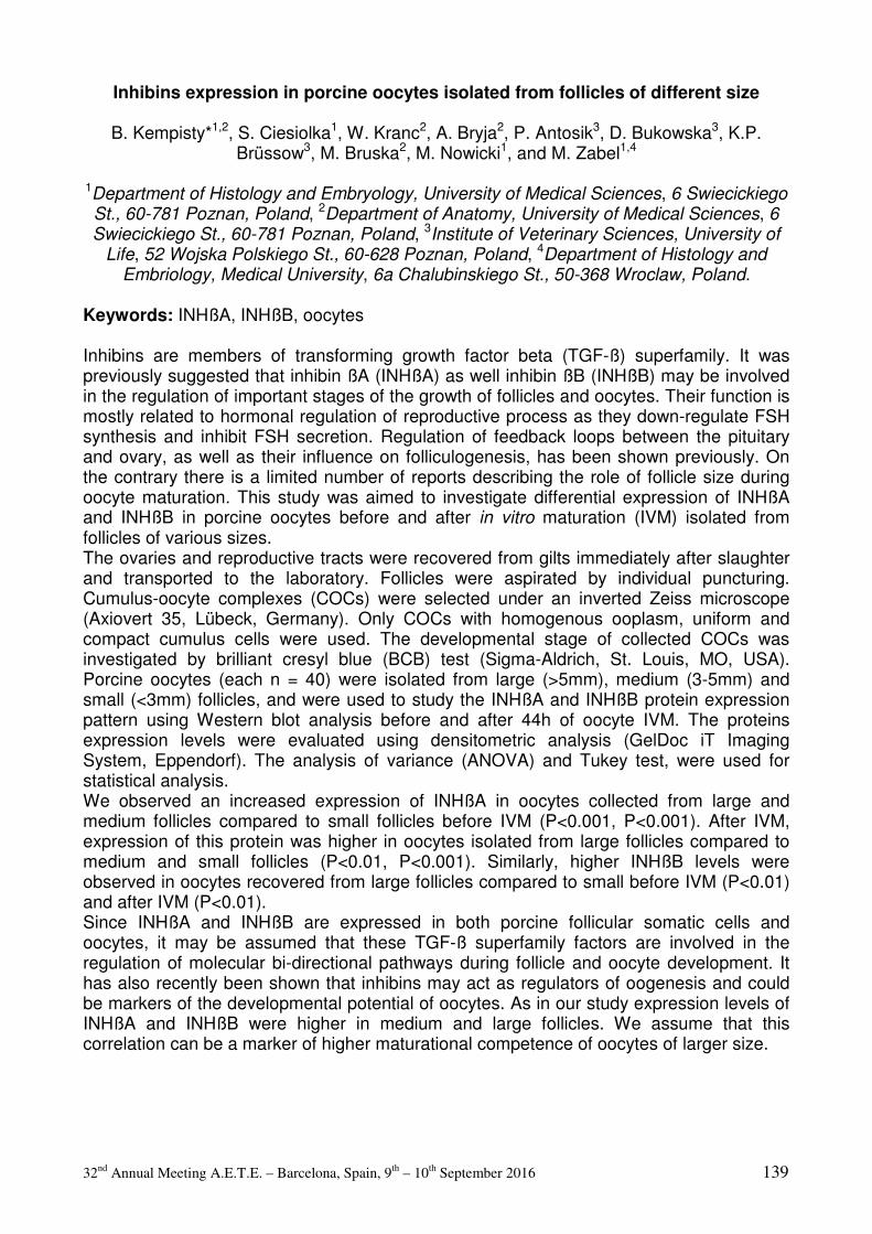
32nd Annual Meeting A.E.T.E. – Barcelona, Spain, 9th – 10th September 2016 139
Inhibins expression in porcine oocytes isolated from follicles of different size
B. Kempisty*1,2, S. Ciesiolka1, W. Kranc2, A. Bryja2, P. Antosik3, D. Bukowska3, K.P. Brüssow3, M. Bruska2, M. Nowicki1, and M. Zabel1,4
1Department of Histology and Embryology, University of Medical Sciences, 6 Swiecickiego St., 60-781 Poznan, Poland, 2Department of Anatomy, University of Medical Sciences, 6 Swiecickiego St., 60-781 Poznan, Poland, 3Institute of Veterinary Sciences, University of
Life, 52 Wojska Polskiego St., 60-628 Poznan, Poland, 4Department of Histology and Embriology, Medical University, 6a Chalubinskiego St., 50-368 Wroclaw, Poland.
Keywords: INHßA, INHßB, oocytes Inhibins are members of transforming growth factor beta (TGF-ß) superfamily. It was previously suggested that inhibin ßA (INHßA) as well inhibin ßB (INHßB) may be involved in the regulation of important stages of the growth of follicles and oocytes. Their function is mostly related to hormonal regulation of reproductive process as they down-regulate FSH synthesis and inhibit FSH secretion. Regulation of feedback loops between the pituitary and ovary, as well as their influence on folliculogenesis, has been shown previously. On the contrary there is a limited number of reports describing the role of follicle size during oocyte maturation. This study was aimed to investigate differential expression of INHßA and INHßB in porcine oocytes before and after in vitro maturation (IVM) isolated from follicles of various sizes. The ovaries and reproductive tracts were recovered from gilts immediately after slaughter and transported to the laboratory. Follicles were aspirated by individual puncturing. Cumulus-oocyte complexes (COCs) were selected under an inverted Zeiss microscope (Axiovert 35, Lübeck, Germany). Only COCs with homogenous ooplasm, uniform and compact cumulus cells were used. The developmental stage of collected COCs was investigated by brilliant cresyl blue (BCB) test (Sigma-Aldrich, St. Louis, MO, USA). Porcine oocytes (each n = 40) were isolated from large (>5mm), medium (3-5mm) and small (<3mm) follicles, and were used to study the INHßA and INHßB protein expression pattern using Western blot analysis before and after 44h of oocyte IVM. The proteins expression levels were evaluated using densitometric analysis (GelDoc iT Imaging System, Eppendorf). The analysis of variance (ANOVA) and Tukey test, were used for statistical analysis. We observed an increased expression of INHßA in oocytes collected from large and medium follicles compared to small follicles before IVM (P<0.001, P<0.001). After IVM, expression of this protein was higher in oocytes isolated from large follicles compared to medium and small follicles (P<0.01, P<0.001). Similarly, higher INHßB levels were observed in oocytes recovered from large follicles compared to small before IVM (P<0.01) and after IVM (P<0.01). Since INHßA and INHßB are expressed in both porcine follicular somatic cells and oocytes, it may be assumed that these TGF-ß superfamily factors are involved in the regulation of molecular bi-directional pathways during follicle and oocyte development. It has also recently been shown that inhibins may act as regulators of oogenesis and could be markers of the developmental potential of oocytes. As in our study expression levels of INHßA and INHßB were higher in medium and large follicles. We assume that this correlation can be a marker of higher maturational competence of oocytes of larger size.
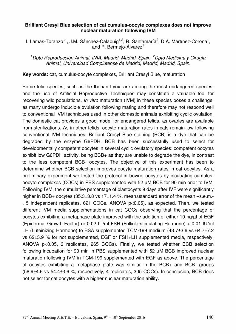
32nd Annual Meeting A.E.T.E. – Barcelona, Spain, 9th – 10th September 2016 140
Brilliant Cresyl Blue selection of cat cumulus-oocyte complexes does not improve nuclear maturation following IVM
I. Lamas-Toranzo*1, J.M. Sánchez-Calabuig1,2, R. Santamaría2, D.A. Martínez-Corona1,
and P. Bermejo-Álvarez1
1Dpto Reproducción Animal, INIA, Madrid, Madrid, Spain, 2Dpto Medicina y Cirugía Animal, Universidad Complutense de Madrid, Madrid, Madrid, Spain.
Key words: cat, cumulus-oocyte complexes, Brilliant Cresyl Blue, maturation Some felid species, such as the Iberian Lynx, are among the most endangered species, and the use of Artificial Reproductive Techniques may constitute a valuable tool for recovering wild populations. In vitro maturation (IVM) in these species poses a challenge, as many undergo inducible ovulation following mating and therefore may not respond well to conventional IVM techniques used in other domestic animals exhibiting cyclic ovulation. The domestic cat provides a good model for endangered felids, as ovaries are available from sterilizations. As in other felids, oocyte maturation rates in cats remain low following conventional IVM techniques. Brilliant Cresyl Blue staining (BCB) is a dye that can be degraded by the enzyme G6PDH. BCB has been successfully used to select for developmentally competent oocytes in several cyclic ovulatory species: competent oocytes exhibit low G6PDH activity, being BCB+ as they are unable to degrade the dye, in contrast to the less competent BCB- oocytes. The objective of this experiment has been to determine whether BCB selection improves oocyte maturation rates in cat oocytes. As a preliminary experiment we tested the protocol in bovine oocytes by incubating cumulus-oocyte complexes (COCs) in PBS supplemented with 52 µM BCB for 90 min prior to IVM. Following IVM, the cumulative percentage of blastocysts 9 days after IVF were significantly higher in BCB+ oocytes (35.3±3.8 vs 17±1.4 %, mean±standard error of the mean –s.e.m.-, 5 independent replicates, 621 COCs, ANOVA p<0.05), as expected. Then, we tested different IVM media supplementations in cat COCs observing that the percentage of oocytes exhibiting a metaphase plate improved with the addition of either 10 ng/µl of EGF (Epidermal Growth Factor) or 0.02 IU/ml FSH (Follicle-stimulating Hormone) + 0.01 IU/ml LH (Luteinizing Hormone) to BSA supplemented TCM-199 medium (43.7±3.6 vs 64.7±7.2 vs 62±5.9 % for not supplemented, EGF or FSH+LH supplemented media, respectively, ANOVA p<0.05, 3 replicates, 265 COCs). Finally, we tested whether BCB selection following incubation for 90 min in PBS supplemented with 52 µM BCB improved nuclear maturation following IVM in TCM-199 supplemented with EGF as above. The percentage of oocytes exhibiting a metaphase plate was similar in the BCB+ and BCB- groups (58.9±4.6 vs 54.4±3.6 %, respectively, 4 replicates, 305 COCs). In conclusion, BCB does not select for cat oocytes with a higher nuclear maturation ability.
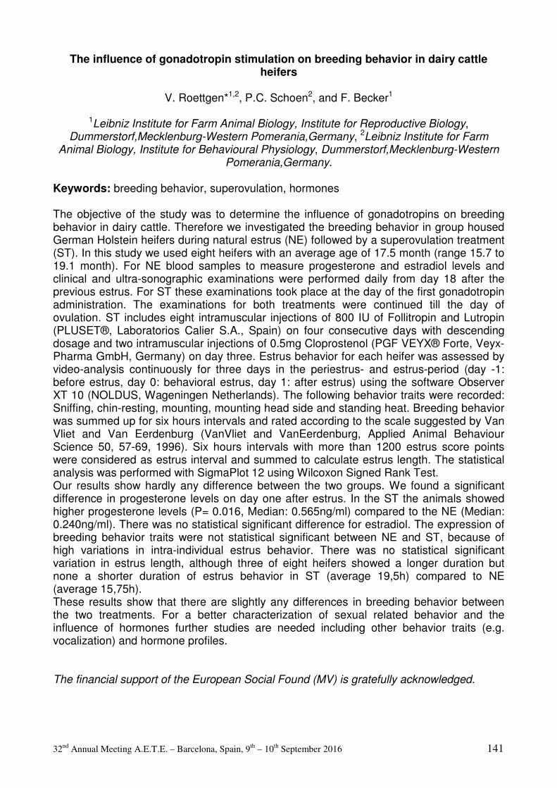
32nd Annual Meeting A.E.T.E. – Barcelona, Spain, 9th – 10th September 2016 141
The influence of gonadotropin stimulation on breeding behavior in dairy cattle heifers
V. Roettgen*1,2, P.C. Schoen2, and F. Becker1
1Leibniz Institute for Farm Animal Biology, Institute for Reproductive Biology, Dummerstorf,Mecklenburg-Western Pomerania,Germany, 2Leibniz Institute for Farm
Animal Biology, Institute for Behavioural Physiology, Dummerstorf,Mecklenburg-Western Pomerania,Germany.
Keywords: breeding behavior, superovulation, hormones The objective of the study was to determine the influence of gonadotropins on breeding behavior in dairy cattle. Therefore we investigated the breeding behavior in group housed German Holstein heifers during natural estrus (NE) followed by a superovulation treatment (ST). In this study we used eight heifers with an average age of 17.5 month (range 15.7 to 19.1 month). For NE blood samples to measure progesterone and estradiol levels and clinical and ultra-sonographic examinations were performed daily from day 18 after the previous estrus. For ST these examinations took place at the day of the first gonadotropin administration. The examinations for both treatments were continued till the day of ovulation. ST includes eight intramuscular injections of 800 IU of Follitropin and Lutropin (PLUSET®, Laboratorios Calier S.A., Spain) on four consecutive days with descending dosage and two intramuscular injections of 0.5mg Cloprostenol (PGF VEYX® Forte, Veyx-Pharma GmbH, Germany) on day three. Estrus behavior for each heifer was assessed by video-analysis continuously for three days in the periestrus- and estrus-period (day -1: before estrus, day 0: behavioral estrus, day 1: after estrus) using the software Observer XT 10 (NOLDUS, Wageningen Netherlands). The following behavior traits were recorded: Sniffing, chin-resting, mounting, mounting head side and standing heat. Breeding behavior was summed up for six hours intervals and rated according to the scale suggested by Van Vliet and Van Eerdenburg (VanVliet and VanEerdenburg, Applied Animal Behaviour Science 50, 57-69, 1996). Six hours intervals with more than 1200 estrus score points were considered as estrus interval and summed to calculate estrus length. The statistical analysis was performed with SigmaPlot 12 using Wilcoxon Signed Rank Test. Our results show hardly any difference between the two groups. We found a significant difference in progesterone levels on day one after estrus. In the ST the animals showed higher progesterone levels (P= 0.016, Median: 0.565ng/ml) compared to the NE (Median: 0.240ng/ml). There was no statistical significant difference for estradiol. The expression of breeding behavior traits were not statistical significant between NE and ST, because of high variations in intra-individual estrus behavior. There was no statistical significant variation in estrus length, although three of eight heifers showed a longer duration but none a shorter duration of estrus behavior in ST (average 19,5h) compared to NE (average 15,75h). These results show that there are slightly any differences in breeding behavior between the two treatments. For a better characterization of sexual related behavior and the influence of hormones further studies are needed including other behavior traits (e.g. vocalization) and hormone profiles. The financial support of the European Social Found (MV) is gratefully acknowledged.
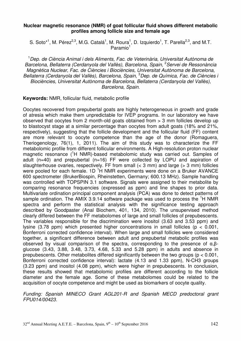
32nd Annual Meeting A.E.T.E. – Barcelona, Spain, 9th – 10th September 2016 142
Nuclear magnetic resonance (NMR) of goat follicular fluid shows different metabolic profiles among follicle size and female age
S. Soto*1, M. Pérez2,3, M.G. Catalá1, M. Roura1, D. Izquierdo1, T. Parella2,3, and M.T.
Paramio1
1Dep. de Ciència Animal i dels Aliments, Fac. de Veterinària, Universitat Autònoma de Barcelona, Bellaterra (Cerdanyola del Vallès), Barcelona, Spain, 2Servei de Ressonància Magnètica Nuclear, Fac. de Ciències i Biociències, Universitat Autònoma de Barcelona,
Bellaterra (Cerdanyola del Vallès), Barcelona, Spain, 3Dep. de Química, Fac. de Ciències i Biociències, Universitat Autònoma de Barcelona, Bellaterra (Cerdanyola del Vallès),
Barcelona, Spain.
Keywords: NMR, follicular fluid, metabolic profile Oocytes recovered from prepubertal goats are highly heterogeneous in growth and grade of atresia which make them unpredictable for IVEP programs. In our laboratory we have observed that oocytes from 2 month-old goats obtained from > 3 mm follicles develop up to blastocyst stage at a similar percentage than oocytes from adult goats (18% and 21%, respectively), suggesting that the follicle development and the follicular fluid (FF) content are more relevant to oocyte competence than the age of the donor (Romaguera, Theriogenology, 76(1), 1, 2011). The aim of this study was to characterize the FF metabolomic profile from different follicular environments. A High-resolution proton nuclear magnetic resonance (1H NMR)-based metabolomic study was carried out. Samples of adult (n=40) and prepubertal (n=16) FF were collected by LOPU and aspiration of slaughterhouse ovaries, respectively. FF from small (< 3 mm) and large (> 3 mm) follicles were pooled for each female. 1D 1H NMR experiments were done on a Bruker AVANCE 600 spectrometer (BrukerBiospin, Rheinstetten, Germany; 600.13 MHz). Sample handling was controlled with TOPSPIN 3.1 software. Signals were assigned to their metabolite by comparing resonance frequencies (expressed as ppm) and line shapes to prior data. Multivariate ordination principal component analysis (PCA) was done to detect patterns of sample ordination. The AMIX 3.9.14 software package was used to process the 1H NMR spectra and perform the statistical analysis with the significance testing approach described by Goodpaster (Anal Biochem, 401, 134, 2010). The unsupervised method clearly differed between the FF metabolomes of large and small follicles of prepubescents. The variables responsible for the discrimination were inositol (3.63 and 3.53 ppm) and lysine (3.78 ppm) which presented higher concentrations in small follicles (p < 0.001, Bonferroni corrected confidence interval). When large and small follicles were considered together, a significant difference between adult and prepubertal metabolic profiles was observed by visual comparison of the spectra, corresponding to the presence of α,β-glucose (3.43, 3.88, 3.48, 3.73, 4.68, 5.33 and 5.28 ppm) in adults and absence in prepubescents. Other metabolites differed significantly between the two groups (p < 0.001, Bonferroni corrected confidence interval): lactate (4.13 and 1.33 ppm), N-CH3 groups (3.23 ppm) and inositol (4.08 ppm), which were higher in prepubescents. In conclusion, these results showed that metabolomic profiles are different according to the follicle diameter and the female age. Some of these metabolomes could be related to the acquisition of oocyte competence and might be used as biomarkers of oocyte quality. Funding: Spanish MINECO Grant AGL201-R and Spanish MECD predoctoral grant FPU014/00423.
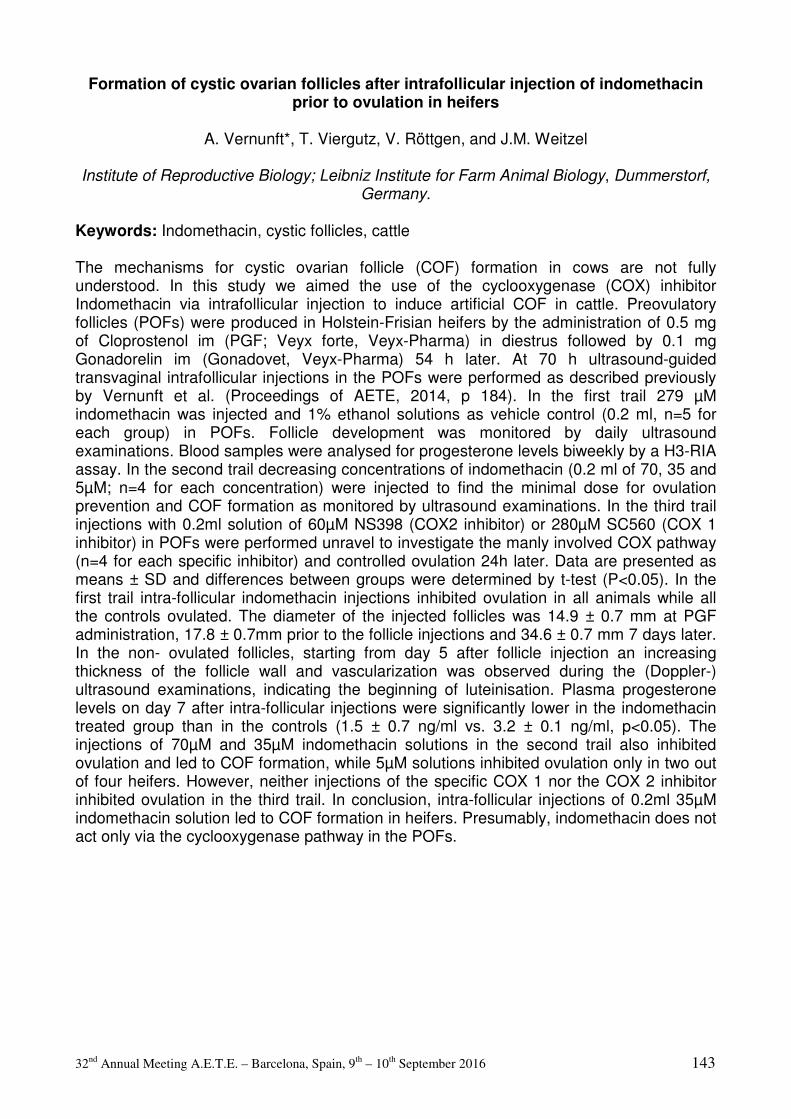
32nd Annual Meeting A.E.T.E. – Barcelona, Spain, 9th – 10th September 2016 143
Formation of cystic ovarian follicles after intrafollicular injection of indomethacin prior to ovulation in heifers
A. Vernunft*, T. Viergutz, V. Röttgen, and J.M. Weitzel
Institute of Reproductive Biology; Leibniz Institute for Farm Animal Biology, Dummerstorf,
Germany.
Keywords: Indomethacin, cystic follicles, cattle The mechanisms for cystic ovarian follicle (COF) formation in cows are not fully understood. In this study we aimed the use of the cyclooxygenase (COX) inhibitor Indomethacin via intrafollicular injection to induce artificial COF in cattle. Preovulatory follicles (POFs) were produced in Holstein-Frisian heifers by the administration of 0.5 mg of Cloprostenol im (PGF; Veyx forte, Veyx-Pharma) in diestrus followed by 0.1 mg Gonadorelin im (Gonadovet, Veyx-Pharma) 54 h later. At 70 h ultrasound-guided transvaginal intrafollicular injections in the POFs were performed as described previously by Vernunft et al. (Proceedings of AETE, 2014, p 184). In the first trail 279 µM indomethacin was injected and 1% ethanol solutions as vehicle control (0.2 ml, n=5 for each group) in POFs. Follicle development was monitored by daily ultrasound examinations. Blood samples were analysed for progesterone levels biweekly by a H3-RIA assay. In the second trail decreasing concentrations of indomethacin (0.2 ml of 70, 35 and 5µM; n=4 for each concentration) were injected to find the minimal dose for ovulation prevention and COF formation as monitored by ultrasound examinations. In the third trail injections with 0.2ml solution of 60µM NS398 (COX2 inhibitor) or 280µM SC560 (COX 1 inhibitor) in POFs were performed unravel to investigate the manly involved COX pathway (n=4 for each specific inhibitor) and controlled ovulation 24h later. Data are presented as means ± SD and differences between groups were determined by t-test (P<0.05). In the first trail intra-follicular indomethacin injections inhibited ovulation in all animals while all the controls ovulated. The diameter of the injected follicles was 14.9 ± 0.7 mm at PGF administration, 17.8 ± 0.7mm prior to the follicle injections and 34.6 ± 0.7 mm 7 days later. In the non- ovulated follicles, starting from day 5 after follicle injection an increasing thickness of the follicle wall and vascularization was observed during the (Doppler-) ultrasound examinations, indicating the beginning of luteinisation. Plasma progesterone levels on day 7 after intra-follicular injections were significantly lower in the indomethacin treated group than in the controls (1.5 ± 0.7 ng/ml vs. 3.2 ± 0.1 ng/ml, p<0.05). The injections of 70µM and 35µM indomethacin solutions in the second trail also inhibited ovulation and led to COF formation, while 5µM solutions inhibited ovulation only in two out of four heifers. However, neither injections of the specific COX 1 nor the COX 2 inhibitor inhibited ovulation in the third trail. In conclusion, intra-follicular injections of 0.2ml 35µM indomethacin solution led to COF formation in heifers. Presumably, indomethacin does not act only via the cyclooxygenase pathway in the POFs.

32nd Annual Meeting A.E.T.E. – Barcelona, Spain, 9th – 10th September 2016 144
Physiology of Reproduction in Male and Semen
Technology
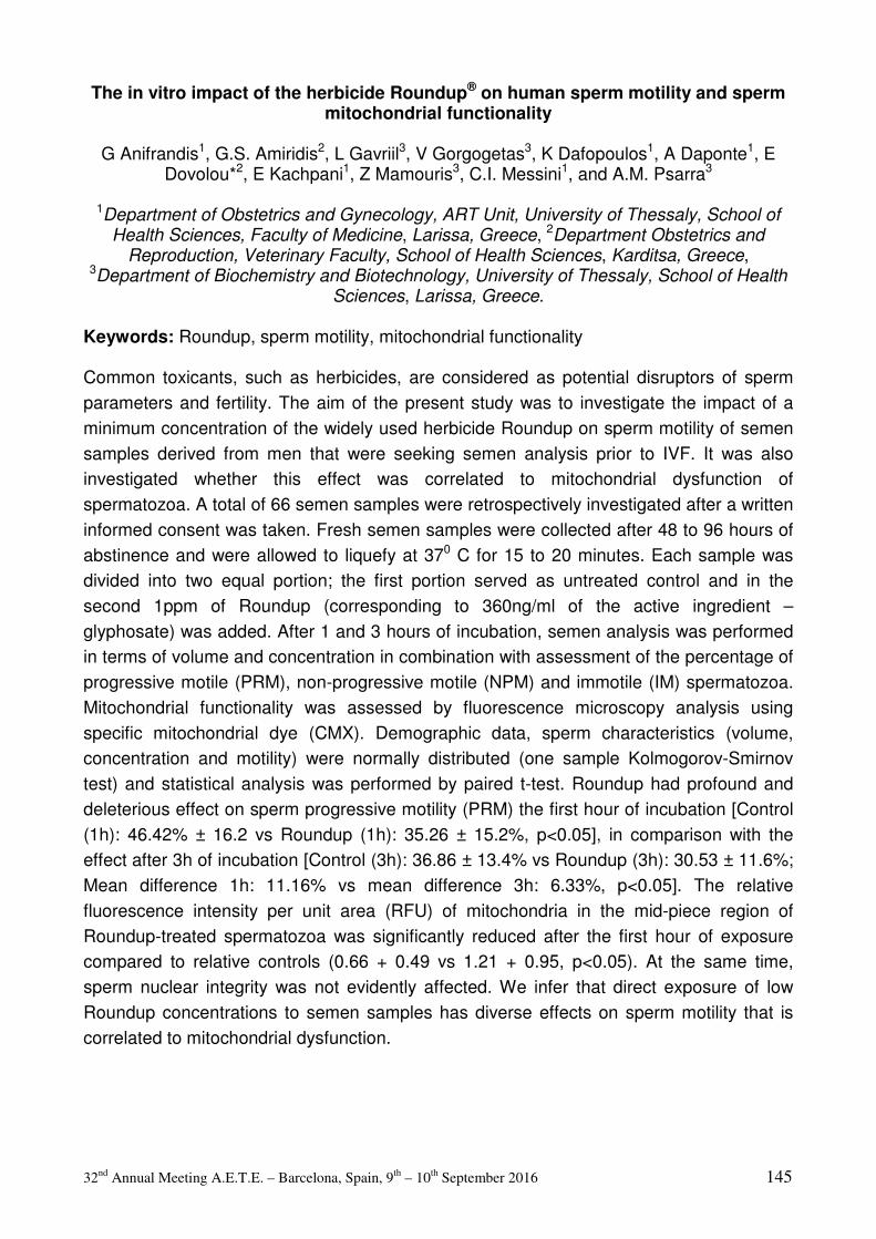
32nd Annual Meeting A.E.T.E. – Barcelona, Spain, 9th – 10th September 2016 145
The in vitro impact of the herbicide Roundup® on human sperm motility and sperm mitochondrial functionality
G Anifrandis1, G.S. Amiridis2, L Gavriil3, V Gorgogetas3, K Dafopoulos1, A Daponte1, E
Dovolou*2, E Kachpani1, Z Mamouris3, C.I. Messini1, and A.M. Psarra3
1Department of Obstetrics and Gynecology, ART Unit, University of Thessaly, School of Health Sciences, Faculty of Medicine, Larissa, Greece, 2Department Obstetrics and
Reproduction, Veterinary Faculty, School of Health Sciences, Karditsa, Greece, 3Department of Biochemistry and Biotechnology, University of Thessaly, School of Health
Sciences, Larissa, Greece.
Keywords: Roundup, sperm motility, mitochondrial functionality Common toxicants, such as herbicides, are considered as potential disruptors of sperm parameters and fertility. The aim of the present study was to investigate the impact of a minimum concentration of the widely used herbicide Roundup on sperm motility of semen samples derived from men that were seeking semen analysis prior to IVF. It was also investigated whether this effect was correlated to mitochondrial dysfunction of spermatozoa. A total of 66 semen samples were retrospectively investigated after a written informed consent was taken. Fresh semen samples were collected after 48 to 96 hours of abstinence and were allowed to liquefy at 370 C for 15 to 20 minutes. Each sample was divided into two equal portion; the first portion served as untreated control and in the second 1ppm of Roundup (corresponding to 360ng/ml of the active ingredient –glyphosate) was added. After 1 and 3 hours of incubation, semen analysis was performed in terms of volume and concentration in combination with assessment of the percentage of progressive motile (PRM), non-progressive motile (NPM) and immotile (IM) spermatozoa. Mitochondrial functionality was assessed by fluorescence microscopy analysis using specific mitochondrial dye (CMX). Demographic data, sperm characteristics (volume, concentration and motility) were normally distributed (one sample Kolmogorov-Smirnov test) and statistical analysis was performed by paired t-test. Roundup had profound and deleterious effect on sperm progressive motility (PRM) the first hour of incubation [Control (1h): 46.42% ± 16.2 vs Roundup (1h): 35.26 ± 15.2%, p<0.05], in comparison with the effect after 3h of incubation [Control (3h): 36.86 ± 13.4% vs Roundup (3h): 30.53 ± 11.6%; Mean difference 1h: 11.16% vs mean difference 3h: 6.33%, p<0.05]. The relative fluorescence intensity per unit area (RFU) of mitochondria in the mid-piece region of Roundup-treated spermatozoa was significantly reduced after the first hour of exposure compared to relative controls (0.66 + 0.49 vs 1.21 + 0.95, p<0.05). At the same time, sperm nuclear integrity was not evidently affected. We infer that direct exposure of low Roundup concentrations to semen samples has diverse effects on sperm motility that is correlated to mitochondrial dysfunction.
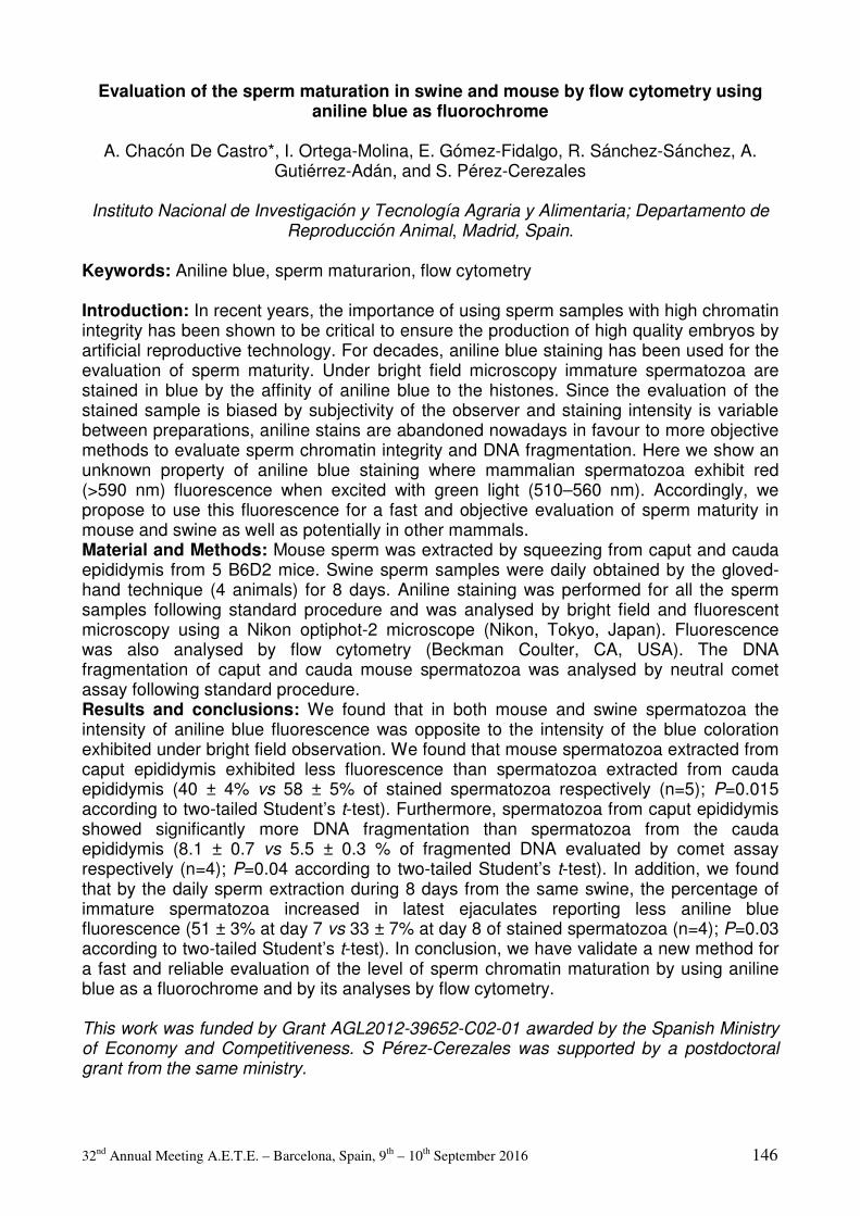
32nd Annual Meeting A.E.T.E. – Barcelona, Spain, 9th – 10th September 2016 146
Evaluation of the sperm maturation in swine and mouse by flow cytometry using aniline blue as fluorochrome
A. Chacón De Castro*, I. Ortega-Molina, E. Gómez-Fidalgo, R. Sánchez-Sánchez, A.
Gutiérrez-Adán, and S. Pérez-Cerezales
Instituto Nacional de Investigación y Tecnología Agraria y Alimentaria; Departamento de Reproducción Animal, Madrid, Spain.
Keywords: Aniline blue, sperm maturarion, flow cytometry Introduction: In recent years, the importance of using sperm samples with high chromatin integrity has been shown to be critical to ensure the production of high quality embryos by artificial reproductive technology. For decades, aniline blue staining has been used for the evaluation of sperm maturity. Under bright field microscopy immature spermatozoa are stained in blue by the affinity of aniline blue to the histones. Since the evaluation of the stained sample is biased by subjectivity of the observer and staining intensity is variable between preparations, aniline stains are abandoned nowadays in favour to more objective methods to evaluate sperm chromatin integrity and DNA fragmentation. Here we show an unknown property of aniline blue staining where mammalian spermatozoa exhibit red (>590 nm) fluorescence when excited with green light (510–560 nm). Accordingly, we propose to use this fluorescence for a fast and objective evaluation of sperm maturity in mouse and swine as well as potentially in other mammals. Material and Methods: Mouse sperm was extracted by squeezing from caput and cauda epididymis from 5 B6D2 mice. Swine sperm samples were daily obtained by the gloved-hand technique (4 animals) for 8 days. Aniline staining was performed for all the sperm samples following standard procedure and was analysed by bright field and fluorescent microscopy using a Nikon optiphot-2 microscope (Nikon, Tokyo, Japan). Fluorescence was also analysed by flow cytometry (Beckman Coulter, CA, USA). The DNA fragmentation of caput and cauda mouse spermatozoa was analysed by neutral comet assay following standard procedure. Results and conclusions: We found that in both mouse and swine spermatozoa the intensity of aniline blue fluorescence was opposite to the intensity of the blue coloration exhibited under bright field observation. We found that mouse spermatozoa extracted from caput epididymis exhibited less fluorescence than spermatozoa extracted from cauda epididymis (40 ± 4% vs 58 ± 5% of stained spermatozoa respectively (n=5); P=0.015 according to two-tailed Student’s t-test). Furthermore, spermatozoa from caput epididymis showed significantly more DNA fragmentation than spermatozoa from the cauda epididymis (8.1 ± 0.7 vs 5.5 ± 0.3 % of fragmented DNA evaluated by comet assay respectively (n=4); P=0.04 according to two-tailed Student’s t-test). In addition, we found that by the daily sperm extraction during 8 days from the same swine, the percentage of immature spermatozoa increased in latest ejaculates reporting less aniline blue fluorescence (51 ± 3% at day 7 vs 33 ± 7% at day 8 of stained spermatozoa (n=4); P=0.03 according to two-tailed Student’s t-test). In conclusion, we have validate a new method for a fast and reliable evaluation of the level of sperm chromatin maturation by using aniline blue as a fluorochrome and by its analyses by flow cytometry. This work was funded by Grant AGL2012-39652-C02-01 awarded by the Spanish Ministry of Economy and Competitiveness. S Pérez-Cerezales was supported by a postdoctoral grant from the same ministry.
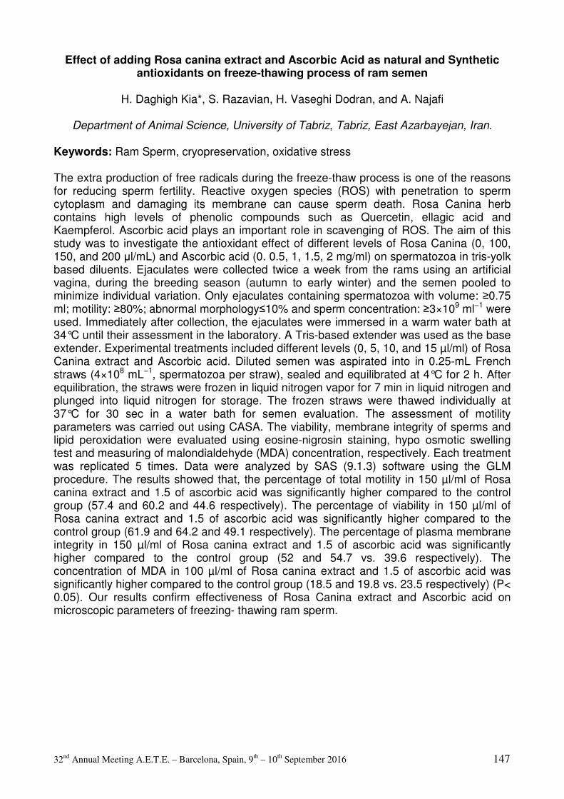
32nd Annual Meeting A.E.T.E. – Barcelona, Spain, 9th – 10th September 2016 147
Effect of adding Rosa canina extract and Ascorbic Acid as natural and Synthetic antioxidants on freeze-thawing process of ram semen
H. Daghigh Kia*, S. Razavian, H. Vaseghi Dodran, and A. Najafi
Department of Animal Science, University of Tabriz, Tabriz, East Azarbayejan, Iran.
Keywords: Ram Sperm, cryopreservation, oxidative stress The extra production of free radicals during the freeze-thaw process is one of the reasons for reducing sperm fertility. Reactive oxygen species (ROS) with penetration to sperm cytoplasm and damaging its membrane can cause sperm death. Rosa Canina herb contains high levels of phenolic compounds such as Quercetin, ellagic acid and Kaempferol. Ascorbic acid plays an important role in scavenging of ROS. The aim of this study was to investigate the antioxidant effect of different levels of Rosa Canina (0, 100, 150, and 200 µl/mL) and Ascorbic acid (0. 0.5, 1, 1.5, 2 mg/ml) on spermatozoa in tris-yolk based diluents. Ejaculates were collected twice a week from the rams using an artificial vagina, during the breeding season (autumn to early winter) and the semen pooled to minimize individual variation. Only ejaculates containing spermatozoa with volume: ≥0.75 ml; motility: ≥80%; abnormal morphology≤10% and sperm concentration: ≥3×10⁹ ml−1 were used. Immediately after collection, the ejaculates were immersed in a warm water bath at 34°C until their assessment in the laboratory. A Tris-based extender was used as the base extender. Experimental treatments included different levels (0, 5, 10, and 15 µl/ml) of Rosa Canina extract and Ascorbic acid. Diluted semen was aspirated into in 0.25-mL French straws (4×10⁸ mL−1, spermatozoa per straw), sealed and equilibrated at 4°C for 2 h. After equilibration, the straws were frozen in liquid nitrogen vapor for 7 min in liquid nitrogen and plunged into liquid nitrogen for storage. The frozen straws were thawed individually at 37°C for 30 sec in a water bath for semen evaluation. The assessment of motility parameters was carried out using CASA. The viability, membrane integrity of sperms and lipid peroxidation were evaluated using eosine-nigrosin staining, hypo osmotic swelling test and measuring of malondialdehyde (MDA) concentration, respectively. Each treatment was replicated 5 times. Data were analyzed by SAS (9.1.3) software using the GLM procedure. The results showed that, the percentage of total motility in 150 µl/ml of Rosa canina extract and 1.5 of ascorbic acid was significantly higher compared to the control group (57.4 and 60.2 and 44.6 respectively). The percentage of viability in 150 µl/ml of Rosa canina extract and 1.5 of ascorbic acid was significantly higher compared to the control group (61.9 and 64.2 and 49.1 respectively). The percentage of plasma membrane integrity in 150 µl/ml of Rosa canina extract and 1.5 of ascorbic acid was significantly higher compared to the control group (52 and 54.7 vs. 39.6 respectively). The concentration of MDA in 100 µl/ml of Rosa canina extract and 1.5 of ascorbic acid was significantly higher compared to the control group (18.5 and 19.8 vs. 23.5 respectively) (P< 0.05). Our results confirm effectiveness of Rosa Canina extract and Ascorbic acid on microscopic parameters of freezing- thawing ram sperm.

32nd Annual Meeting A.E.T.E. – Barcelona, Spain, 9th – 10th September 2016 148
Could an extra long-term boar semen extender be successfully used during liquid storage of ram semen at 15°C?
B. El Amiri*1, A. Ben Moula1, A. Badi1, K. El Khalil1, L. Allai1, E. Gergatz2, E. Gyökér2, and
O. Szenci3
11INRA, Centre Régional de la Recherche Agronomique de Settat, Settat, BP= 589, Morocco, 22Biotechnical Research Station, Pharmagene-Farm Ltd., Mosonmagyaróvár,
Hungary, 33SZIU Large Animal Clinical Research Group,, Üllo, Hungary.
Keywords: Duragen, Skim milk, Tris egg yolk The aim of the present study, was to verify, whether, a commercial diluent (Duragen: an extra long-term boar semen extender) could be a good alternative to skim milk (SM) and Tris-egg yolk (TEY) to store ram semen at 15°C. Ejaculates were collected once a week during 6 weeks from two different local Moroccan breeds (Boujaad a non-prolific and INRA180 a prolific sheep) using an artificial vagina. The ejaculates containing spermatozoa with >80 % total motility and concentrations higher than 3 x 10⁹ spermatozoa/ml were pooled. The pool was divided to three parts and each part was extended in TEY, SM or Duragen (Duragen; Magapor S.L.; Zaragoza, Spain) to reach a final concentration of 0.8 × 10⁹ spermatozoa/ml. The Sperm motility (proportion of total and progressive motile sperm) was assessed by means of a computer-assisted semen analysis (CASA) (ISAS, version 1.0.17, Proiser, Valencia, Spain) at different periods (0, 8, 24h). The sperm viability (VIA), the morphology of abnormal spermatozoa (ABN); the hypoosmotic swelling test (HOST); the spontaneous lipid peroxidation (thiobarbituric acid reactive substance: TBARS) and DNA fragmentation tests are in progress to complete the evaluation of semen quality. All data were analyzed using the Statistical Analysis System software JMP (SAS version 10) and expressed as the mean ± SEM. Differences with values of P < 0.05 were considered to be statistically significant. The results of progressive motility (PM) showed that at 0h, there was no different between SM (66.12±0.89%) and Duragen (66.52±0.85%) while the TEY showed the lowest value (54.06±1.71%). At 8h of storage, the best PM was recorded with SM (63.57±0.93%), while Duragen showed 58.90±1.19% and TEY 35.69±1.63%. After 24h of storage, the same tendency was recorded. The SM showed 59.53±1.63% of PM while Duragen gave 48.89±1.51% and TEY showed 34.06±2.05%. Similarly, even at 24h of storage, the SM and TEY gave the best results on terms of total motility (TM) (78.48±1.48%, 76.97±1.44% respectively) while Duragen showed the lowest value (68.41±1.47%). In conclusion, Duragen could be a good alternative to TEY and SM regarding the PM. However, it will not be a good one regarding the TM for both extenders (SM and TEY). Additional tests such as VIA, ABN, HOST, TBARS and DNA fragmentation are in progress to complete the evaluation of semen quality. The authors owe to thank Magapor company (Parque Científico Tecnológico Agroalimentario Valdeferrín-AulaDei, Calle 5: 50600 Ejea de los Caballeros (Zaragoza) Spain) for providing the diluent. Dr. Maria Sancho Navarro and Pr. Carles Soler for their help. CNRST of Morocco for supporting a part of this work.
32nd Annual Meeting A.E.T.E. – Barcelona, Spain, 9th – 10th September 2016 149
Effect of adding Cornus mas extract as a natural antioxidant and BHT on freezing/thawing process of ram semen
D.K. Hossein*, Z.G. Fatemeh, N. Abozar, and H. Vaseghi Dodran
Department of Animal Science, University of Tabriz, Tabriz, East Azarbayejan, Iran.
Keywords: Sperm, Free Radicals, Cornus mas During the freeze-thaw process sperm quality, viability and fertility are reduced because of physiological and chemical stresses on the sperm cell membrane. Cornus mas herb contains high levels of phenolic compounds such as Gallic acid, and polyphenols such as Rosmarinic acid and Quercetin. Butylated hydroxytoluene (BHT) is a synthetic analogue of vitamin E that inhibits the auto-oxidation reaction by converting peroxy radicals to hydroperoxides. BHT has been tested successfully to preserve liquid semen and minimize cold shock damage. The purpose of this study was to investigate the effect of Cornus mas extract as a natural antioxidant and BHT on frozen-thawed semen quality of ram semen. In this study, five ram were used for semen collection twice a week using an artificial vagina, during the breeding season (August to October) and the semen pooled to minimize individual variation. Only ejaculates containing spermatozoa with volume: ≥0.75 ml; motility: ≥80%; abnormal morphology≤10% and sperm concentration: ≥3×10⁹ ml−1 were used. Different levels of extract of Cornus mas (100, 150 and 200 µl/ml) and BHT (1, 1.5, 2 and 3 mM) were added to Tris-yolk based diluents. Following cooling and freezing of semen samples, they were stored in liquid nitrogen until evaluation. After freezing-thawing, the dynamic parameters were evaluated using CASA system, the viability of sperms using eosin-nigrosin stain, membrane integrity using hypo osmotic swelling test and lipid peroxidation by measuring of malondialdehyde concentration. Each treatment was replicated 5 times. Data were analyzed by SAS (9.1.3) software using the GLM procedure. The results showed that, the percentage of total motility in 150, 200 µl/ml Cornus mas extracts was significantly higher compared to the control group (59.2, 52.4 and 44.6 respectively). The percentage of viability in 150, 200 µl/ml Cornus mas extracts was significantly higher compared to the control group (63.1, 56.6 and 50.5 respectively). The percentage of plasma membrane integrity in 150, 200 µl/ml Cornus mas extracts was significantly higher compared to the control group (54.8, 48.4 and 40.4 respectively) (P< 0.05). The percentage of total motility in 1.5, 2mM BHT was significantly higher compared to the control group (56.2, 65.7 and 44.6 respectively). The percentage of viability in 1.5, 2mM BHT was significantly higher compared to the control group (60.5, 69.9 and 50.3 respectively). The percentage of plasma membrane integrity in 1.5, 2 mM BHT was significantly higher compared to the control group (52.5, 61.5 and 40.3 respectively) (P< 0.05). Our results confirm the effectiveness of Cornus mas extract and BHT on microscopic parameters of freezing-thawing ram sperm.
32nd Annual Meeting A.E.T.E. – Barcelona, Spain, 9th – 10th September 2016 150
Morphological and functional characteristics of the epididymal sperm derived from the European bison (Bison bonasus) of the Altaic population
B.S. Iolchiev, G.N. Singina*, V.A. Bagirov, E.N. Shedova, A.B. Lopukhov, P.M.
Klenovitskiy, S.S. Danch, and M.A. Zhilinskiy
L.K. Ernst Institute of Animal Husbandry, Podolsk, Russia.
Keywords: epididymal sperm, European bison, in vitro test The aim of the present research was to study the morphology, motility, viability and fertility characteristics of epididymal sperm of the European bison. The epididymes were obtained following a forced slaughter (as a result of severe injury) of two bulls from the Altaic population aged 5-7 years. The sperm was collected by scraping the inner surface of the epididymes and assessed for motility and concentration. All procedures were performed within 1 h. Thereafter, the sperm was diluted with the lactose-yolk-glycerol medium to 40x106 spermatozoa/ml and equilibrated for 4 h at 4oC. Sperm aliquots (0.2 ml) were frozen in liquid nitrogen vapor for 5 min and then plunged into liquid nitrogen for storage. Prior to analysis, frozen semen was thawed in pre-warmed medium for 1 min at 37°C. The post-thawed sperm morphometry was performed with help of NIS Elements BR software (Nicon, Amsterdam, Netherlands), the motility was evaluated using computer-assisted semen analysis (Videotest, St. Petersburg, Russia). The sperm viability was assessed with Sperm VitalStain (Nidocon, Mölndal, Sweden) and the fertilizing capacity was determined using a heterologous IVF system. Each analysis was conducted by a single investigator. For in vitro test, slaughterhouse-derived cattle oocytes were subjected to IVM procedure described previously (Singina et al., Reprod Fert Dev, 26:154, 2014). The sperm was prepared by the swim-up method. Matured oocytes (n=234, 35-40 oocytes per group) were co-incubated for 18 h with sperm (1 × 10⁶ spermatozoa/ml) in 500 µl of TALP containing 10 µg mL-1 heparin, 20 µM penicillamine, 10 µM hypotaurine, and 1 µM epinephrine at 38.5°C and 5% CO2 in humidified air. At the end of co-incubation, a part of oocytes was examined for the penetration rate (the number of oocytes having enlarged sperm head(s) or male pronucleus(ei)) by cytological analysis. At Days 2 and 7 after insemination, the sperm fertilizing capacity was determined using morphological and cytological evaluation of cleavage stages and blastocysts formation. Data expressed as means±SEM were processed by SigmaStat software. The morphometric assay (750 spermatozoa per bull, magnification 400x) demonstrated that the average length, head length, head width, head perimeter, head area and tail length of the European bison epididymal spermatozoa were 68.7±12.5 µm, 8.5±0.7 µm, 4.7±0.4 µm, 24.0±1.6 µm, 35.4±5.7 µm2, and 57.3±9.0 µm, respectively. The post-thawed motility reached 40.0±3.3 % (n=3), whereas the viability was 34.4±4.1 % (n=3). After heterologous fertilization of bovine oocytes, the penetration, cleavage and blastocyst rates were high and reached 93.3±1.7, 76.6±1.9, and 27.9±2.7 % (n=5), respectively. The findings of the present research demonstrate that the post-mortem collected epididymal sperm of European bison may be used for creating a bank of genetic resources when breeding this endangered animal species. The research was supported by Program of Presidium of the Russian Academy of Science, project no. IV.13.3.

32nd Annual Meeting A.E.T.E. – Barcelona, Spain, 9th – 10th September 2016 151
Moxifloxacin effects on ram frozen-thawed sperm function F. Kountouris1, A. Gómez1, and J. Gadea*1,2
1Faculty of Veterinary Science. Excellence Campus for Higher Education and Research
“Campus Mare Nostrum”. University of Murcia, Spain, 2Institute for Biomedical Research of Murcia (IMIB-Arrixaca), Spain.
Keywords: antibiotics, bacterial contamination, spermatozoa The control of bacteria present in ram seminal samples is important in order to avoid deleterious effect on sperm functionality during storage and transmission of venereal diseases. Previous studies have shown the presence of Mycoplasma agalactie (Ma) in goat and ram seminal samples and its ability to survive in diluted semen (de la Fe et al. Theriogenology 72: 1278, 2009; Gómez-Martín et al. Theriogenology 83: 911, 2015; Prats-van der Ham et al. Theriogenology 2016 doi: 10.1016/j.theriogenology.2016.02.033). Conventional antibiotics used in semen extenders are not effective against Ma or have a detrimental effect on sperm cells. Preliminary results from our lab show that moxifloxacin has antibacterial effect on mycoplasma when used at 0.3 µg/mL (Martínez-Fresneda, unpublished data). However, there is no information about the use of moxifloxacin in semen extenders and its likely effect on sperm cells. The aim of this study was to analyze the effect of moxifloxacin on ram sperm functionality measured in terms of motility, viability and acrosome status. Commercial frozen semen straws from 4 Assaf rams were thawed at 37ºC and incubated for 2 h in Sperm-Talp media supplemented with 0 µg/mL (MOXI-0), 0.3 µg/mL (MOXI-3) and 0.9 µg/mL (MOXI-9) of moxifloxacin. All groups had gentamicine (50 µg/mL) according to Sperm-Talp formulation. Motility and motion parameters were measured by a CASA system, and simultaneously viability and acrosome status by flow cytometry after propidium iodide and PSA-FITC staining (6 replicates). Data were analysed by two-way ANOVA (treatment and incubation time) and when ANOVA revealed a significant effect (p<0.05), values were compared by Tukey test. Results showed that after incubation the percentage of live spermatozoa with altered acrosome, including those with spontaneous acrosome reaction, was higher in MOXI-9 group than in MOXI-0 group (2.7±0.2 vs. 1.8±0.1, p<0.01). Besides a different pattern movement was observed with an increase in the sperm velocities (µm/s) in MOXI-9 group compared to MOXI-0 group (curvilinear-VCL 131.3±2.7 vs. 117.0±3.1; Straight line-VSL 91.8±2.7 vs. 76.7±2.2; Average path-VAP 105.7±2.9 vs. 87.9±2.7, p<0.01). No differences were found neither for viable sperm with intact acrosome (mean value 46.3±2.0%), nor for percentage of motility (mean value 29.2±1.1) nor for other motion parameters. A further study of sperm subpopulations was done by using cluster analysis (Abaigar et al. Biol. Reprod. 60: 32, 1999) grouping spermatozoa in three clusters: slow, medium and fast spermatozoa. Results showed a higher proportion of fast spermatozoa in MOXI-9 group than in MOXI-0 (48.8 vs. 24.3%), and a lower proportion of medium (27.9 vs. 43.9%) and slow spermatozoa (23.2 vs. 31.7%). It can be concluded that under our experimental conditions, moxifloxacin has no effect on sperm functionality when used at low concentration (0.3 µg/mL), but it modifies the pattern of sperm motility and spontaneous acrosome reaction at higher ones (0.9 µg/mL). Supported by MINECO-FEDER AGL2015-66341-R. Frozen semen samples generously provided by Ovigen (Zamora. Spain).
32nd Annual Meeting A.E.T.E. – Barcelona, Spain, 9th – 10th September 2016 152
PHOSPHATIDYLSERINE TRANSLOCATION DURING SPERM CAPACITATION IS MODULATED BY eNOS IN PORCINE
R. López-Úbeda, R. Diego, and C. Matás*
Physiology of Reproduction Group. Veterinary School, University of Murcia, Campus Mare
Nostrum, Murcia, Spain.
Keywords: nitric oxide, sperm capacitation, phosphatidylserine translocation Introduction: Reactive oxygen species (ROS) play a key role in the process of sperm capacitation and/or apoptosis [Aitken, J Androl; 13:36-42, 2011]. Nitric oxide (NO) synthesis is mediated by nitric oxide synthase (NOS), responsible for the conversion of L-arginine to L-citrulline [Funahashi, Reproduction; 124: 857-864, 2002]. NOS exists in three isoforms (eNOS, iNOS and nNOS) that have been identified in porcine oocyte and sperm, but many of its functions remain unknown. Objective:The aim of this study was to analyse the effect of NO on the externalization of phosphatidylserine (PS), a process that occurs during sperm capacitation due to destabilization of the membrane [Flesch, Journal of cell science; 114: 3543-3555, 2001]. Materials and methods: The role of NO was studied using a NO donor, S-nitroso-glutathione (GSNO; 50µM) or by inhibiting its synthesis employing two different NOS inhibitors: 10mM L-NAME (eNOS inhibitor) and 10mM aminoguanidine (AG) (eNOS and iNOS inhibitor). Sperm cells washed by discontinuous gradient of Percoll, were incubated with the different treatments for 1 hour. Washed sperm but untreated were employed as a control group. Translocation of PS residues to the outer leaflet of the plasma membrane was detected by Annexin V-Cy3TM Apoptosis Detection Kit (Sigma, Madrid, Spain). For this assay, 1µl Annexin V in 450 µl of binding buffer (commercial kit) was mixed with 50 µl of each sperm sample. After 10 minutes of incubation in the dark, at room temperature, samples were fixed with 10 µl formaldehyde (10% in PBS). Sperm with PS exposed (Annexin +) were visualized in red fluorescence (rhodamine filter) [Marti, Anim Reprod Sci; 106: 113-32, 2008]. The data were analyzed by ANOVA (p<0.05). Results: Our results showed that CONTROL (37.7%), GSNO (38.0%) and AG (36.3%) groups were not statistically significant for staining with Annexin V (p<0.05). However, the sperm incubated with NOS inhibitor (L-NAME) had a lower translocation of PS (29.8%, p<0.05). Conclusions: These data reveal the possible involvement of NO in the PS translocation during sperm capacitation and that eNOS isoform plays a modulation role. Supported by Spanish Ministry of Economy and Competitiveness (MINECO) Grant AGL2015-66341-R.
32nd Annual Meeting A.E.T.E. – Barcelona, Spain, 9th – 10th September 2016 153
Post-thaw changes in sperm membrane and ROS following cryopreservation of dairy bull semen using four different commercial extenders
S. Miguel-Jiménez*1, T. Mogas1, A.I. Peña2, C. Tamargo3, C.O. Hidalgo3, R. Muiño2, J.E.
Rodríguez-Gil1, and R. Morató1
1Departamento de Medicina y Cirugía Animales, Universidad Autónoma de Barcelona, Cerdanyola del Valles, Barcelona, Spain, 2Departamento de Patología Animal,
Universidad de Santiago de Compostela, Santiago de Compostela, Lugo, Spain, 3Área de Selección y Reproducción Animal, Servicio Regional de Investigación y Desarrollo
Agroalimentario, Gijon, Spain.
Keywords: sperm viability, acrosome integrity, flow cytometry Semen diluents containing egg yolk as a cryoprotectant may pose hygienic risks and are difficult to standardize. Therefore as an alternative to replace the component of animal origin, egg yolk-free extenders have been developed. This study was designed to compare the effect of three yolk-free extenders (Andromed®, Bioxcell® and Optixcell®) and an egg yolk based diluent (Triladyl®) on post-thaw quality of bull spermatozoa. A total of five ejaculates were collected from six healthy Holstein Frisian bulls (2 to 4 years old) of proven fertility with the aid of an artificial vagina. Each ejaculate was divided into four aliquots and diluted at room temperature with one of the four cryopreservation extenders, allow to equilibrate at 4°C for 4 h and packaged in 0.25 ml straws. Finally, semen was cryopreserved in liquid nitrogen following a standard procedure applied to a computerized freezing processor. For semen analysis, 30 straws from each treatment were thawed for 40 sec at 37.5°C in a water bath as previously described (Muiño et al. Anim Reprod Sci. 109:27-39. 2008). Flow cytometry analyzed parameters were plasma membrane integrity, through the SYBR-14 and propidium iodide (PI) stain, acrosome membrane status by fluorescein isothiocyanate-peanut agglutinin (PNA) and PI stain, ·O2 intracellular levels through the hydroethidine (HE) and YoPro-1 stain and H2O2 levels by the 2,7-dichlorodihydrofluorescein diacetate and PI stain. Differences among groups were performed through an ANOVA, followed by the Tukey’s post-hoc test. Significance was set at two-tailed P<0.05. Semen samples frozen with Optixcell® triggered significantly higher sperm viability (66.6±1.3%) than those frozen with Triladyl® (58.5±1.6%) and Bioxcell® (57.2±2.2%). Acrosome damage of sperm samples frozen using Triladyl® (24.6±1.3%) was significantly higher than when Bioxcell® (17.7±1.7%) and Optixcell® (18.3±1.6%) were used. No significant differences in terms of sperm viability or acrosome integrity were observed after cryopreservation with Andromed® (61.5±1.7% and 19.3±1.7%, respectively) compared to the other three extenders. Regarding ROS generation, Triladyl® showed a better protection against superoxides production (19.98±1.9% HE+/YoPro-1-) compared to Optixcell® (27.8±2.8%), Andromed® (33.4±2.5%) and Bioxcell® (43.3±3.4%). Optixcell® (6.7±1%), Andromed® (3.7±2%) and Triladyl® (5.3±1%) triggered similar results when the percentages of viable sperm with a high intracellular H2O2 were analized, while this percentage was significantly decreased when Bioxcell® was used as an extender (1.4±0.4%) when compared to Optixcell®. Our results suggest that Optixcell® could be chosen as an egg yolk-free extender for bull sperm cryopreservation due to its better cryoprotective properties. This study was supported by the Spanish Ministry of Science and Innovation (Project AGL2013–46769).
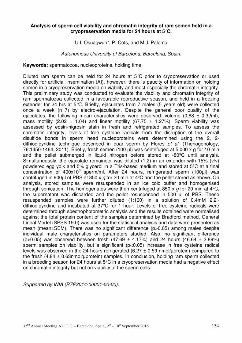
32nd Annual Meeting A.E.T.E. – Barcelona, Spain, 9th – 10th September 2016 154
Analysis of sperm cell viability and chromatin integrity of ram semen held in a cryopreservation media for 24 hours at 5°C.
U.I. Osuagwuh*, P. Cots, and M.J. Palomo
Autonomous University of Barcelona, Barcelona, Spain.
Keywords: spermatozoa, nucleoproteins, holding time Diluted ram sperm can be held for 24 hours at 5°C prior to cryopreservation or used directly for artificial insemination (AI), however, there is paucity of information on holding semen in a cryopreservation media on viability and most especially the chromatin integrity. This preliminary study was conducted to evaluate the viability and chromatin integrity of ram spermatozoa collected in a favourable reproductive season, and held in a freezing extender for 24 hrs at 5°C. Briefly, ejaculates from 7 males (5 years old) were collected once a week (n=7) by electro-ejaculation. Despite the general poor quality of the ejaculates, the following mean characteristics were observed: volume (0.68 ± 0.32ml), mass motility (2.02 ± 1.04) and linear motility (67.75 ± 1.27%). Sperm viability was assessed by eosin-nigrosin stain in fresh and refrigerated samples. To assess the chromatin integrity, levels of free cysteine radicals from the disruption of the overall disulfide bonds in sperm head nucleoproteins were determined using the 2, 2-dithiodipyridine technique described in boar sperm by Flores et al. (Theriogenology, 76:1450-1464, 2011). Briefly, fresh semen (100 µl) was centrifuged at 5,000 x g for 10 min and the pellet submerged in liquid nitrogen before stored at -80ºC until analysis. Simultaneously, the ejaculate remainder was diluted (1:2) in an extender with 15% (v/v) powdered egg yolk and 5% glycerol in a Tris-based medium and stored at 5ºC at a final concentration of 400x106 sperm/ml. After 24 hours, refrigerated sperm (100µl) was centrifuged in 900µl of PBS at 850 x g for 20 min at 4ºC and the pellet stored as above. On analysis, stored samples were resuspended in an ice cold buffer and homogenised through sonication. The homogenates were then centrifuged at 850 x g for 20 min at 4ºC, the supernatant was discarded and the pellet resuspended in 500 µl of PBS. These resuspended samples were further diluted (1:100) in a solution of 0.4mM 2,2’-dithiodipyridine and incubated at 37ºC for 1 hour. Levels of free cysteine radicals were determined through spectrophotometric analysis and the results obtained were normalised against the total protein content of the samples determined by Bradford method. General Lineal Model (SPSS 19.0) was used for the statistical analysis and data were presented as mean (mean±SEM). There was no significant difference (p>0.05) among males despite individual male characteristics on parameters studied. Also, no significant difference (p>0.05) was observed between fresh (47.69 ± 4.17%) and 24 hours (46.64 ± 3.89%) sperm samples on viability, but a significant (p<0.05) increase in free cysteine radical levels was observed in the 24 hours refrigerated (6.27 ± 0.59 nmol/µprotein) compared to the fresh (4.84 ± 0.63nmol/µprotein) samples. In conclusion, holding ram sperm collected in a breeding season for 24 hours at 5ºC in a cryopreservation media had a negative effect on chromatin integrity but not on viability of the sperm cells. Supported by INIA (RZP2014-00001-00-00).
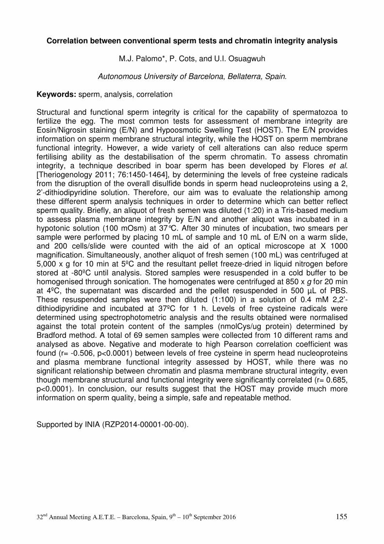
32nd Annual Meeting A.E.T.E. – Barcelona, Spain, 9th – 10th September 2016 155
Correlation between conventional sperm tests and chromatin integrity analysis
M.J. Palomo*, P. Cots, and U.I. Osuagwuh
Autonomous University of Barcelona, Bellaterra, Spain. Keywords: sperm, analysis, correlation Structural and functional sperm integrity is critical for the capability of spermatozoa to fertilize the egg. The most common tests for assessment of membrane integrity are Eosin/Nigrosin staining (E/N) and Hypoosmotic Swelling Test (HOST). The E/N provides information on sperm membrane structural integrity, while the HOST on sperm membrane functional integrity. However, a wide variety of cell alterations can also reduce sperm fertilising ability as the destabilisation of the sperm chromatin. To assess chromatin integrity, a technique described in boar sperm has been developed by Flores et al. [Theriogenology 2011; 76:1450-1464], by determining the levels of free cysteine radicals from the disruption of the overall disulfide bonds in sperm head nucleoproteins using a 2, 2’-dithiodipyridine solution. Therefore, our aim was to evaluate the relationship among these different sperm analysis techniques in order to determine which can better reflect sperm quality. Briefly, an aliquot of fresh semen was diluted (1:20) in a Tris-based medium to assess plasma membrane integrity by E/N and another aliquot was incubated in a hypotonic solution (100 mOsm) at 37°C. After 30 minutes of incubation, two smears per sample were performed by placing 10 mL of sample and 10 mL of E/N on a warm slide, and 200 cells/slide were counted with the aid of an optical microscope at X 1000 magnification. Simultaneously, another aliquot of fresh semen (100 mL) was centrifuged at 5,000 x g for 10 min at 5ºC and the resultant pellet freeze-dried in liquid nitrogen before stored at -80ºC until analysis. Stored samples were resuspended in a cold buffer to be homogenised through sonication. The homogenates were centrifuged at 850 x g for 20 min at 4ºC, the supernatant was discarded and the pellet resuspended in 500 µL of PBS. These resuspended samples were then diluted (1:100) in a solution of 0.4 mM 2,2’-dithiodipyridine and incubated at 37ºC for 1 h. Levels of free cysteine radicals were determined using spectrophotometric analysis and the results obtained were normalised against the total protein content of the samples (nmolCys/ug protein) determined by Bradford method. A total of 69 semen samples were collected from 10 different rams and analysed as above. Negative and moderate to high Pearson correlation coefficient was found (r= -0.506, p<0.0001) between levels of free cysteine in sperm head nucleoproteins and plasma membrane functional integrity assessed by HOST, while there was no significant relationship between chromatin and plasma membrane structural integrity, even though membrane structural and functional integrity were significantly correlated (r= 0.685, p<0.0001). In conclusion, our results suggest that the HOST may provide much more information on sperm quality, being a simple, safe and repeatable method. Supported by INIA (RZP2014-00001-00-00).
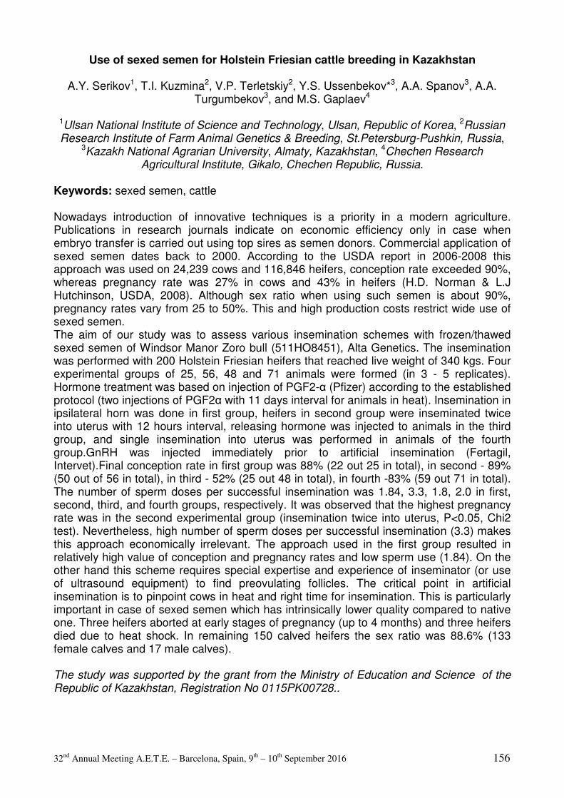
32nd Annual Meeting A.E.T.E. – Barcelona, Spain, 9th – 10th September 2016 156
Use of sexed semen for Holstein Friesian cattle breeding in Kazakhstan
A.Y. Serikov1, T.I. Kuzmina2, V.P. Terletskiy2, Y.S. Ussenbekov*3, A.A. Spanov3, A.A. Turgumbekov3, and M.S. Gaplaev4
1Ulsan National Institute of Science and Technology, Ulsan, Republic of Korea, 2Russian Research Institute of Farm Animal Genetics & Breeding, St.Petersburg-Pushkin, Russia,
3Kazakh National Agrarian University, Almaty, Kazakhstan, 4Chechen Research Agricultural Institute, Gikalo, Chechen Republic, Russia.
Keywords: sexed semen, cattle Nowadays introduction of innovative techniques is a priority in a modern agriculture. Publications in research journals indicate on economic efficiency only in case when embryo transfer is carried out using top sires as semen donors. Commercial application of sexed semen dates back to 2000. According to the USDA report in 2006-2008 this approach was used on 24,239 cows and 116,846 heifers, conception rate exceeded 90%, whereas pregnancy rate was 27% in cows and 43% in heifers (H.D. Norman & L.J Hutchinson, USDA, 2008). Although sex ratio when using such semen is about 90%, pregnancy rates vary from 25 to 50%. This and high production costs restrict wide use of sexed semen. The aim of our study was to assess various insemination schemes with frozen/thawed sexed semen of Windsor Manor Zoro bull (511HO8451), Alta Genetics. The insemination was performed with 200 Holstein Friesian heifers that reached live weight of 340 kgs. Four experimental groups of 25, 56, 48 and 71 animals were formed (in 3 - 5 replicates). Hormone treatment was based on injection of PGF2-α (Pfizer) according to the established protocol (two injections of PGF2α with 11 days interval for animals in heat). Insemination in ipsilateral horn was done in first group, heifers in second group were inseminated twice into uterus with 12 hours interval, releasing hormone was injected to animals in the third group, and single insemination into uterus was performed in animals of the fourth group.GnRH was injected immediately prior to artificial insemination (Fertagil, Intervet).Final conception rate in first group was 88% (22 out 25 in total), in second - 89% (50 out of 56 in total), in third - 52% (25 out 48 in total), in fourth -83% (59 out 71 in total). The number of sperm doses per successful insemination was 1.84, 3.3, 1.8, 2.0 in first, second, third, and fourth groups, respectively. It was observed that the highest pregnancy rate was in the second experimental group (insemination twice into uterus, P<0.05, Chi2 test). Nevertheless, high number of sperm doses per successful insemination (3.3) makes this approach economically irrelevant. The approach used in the first group resulted in relatively high value of conception and pregnancy rates and low sperm use (1.84). On the other hand this scheme requires special expertise and experience of inseminator (or use of ultrasound equipment) to find preovulating follicles. The critical point in artificial insemination is to pinpoint cows in heat and right time for insemination. This is particularly important in case of sexed semen which has intrinsically lower quality compared to native one. Three heifers aborted at early stages of pregnancy (up to 4 months) and three heifers died due to heat shock. In remaining 150 calved heifers the sex ratio was 88.6% (133 female calves and 17 male calves). The study was supported by the grant from the Ministry of Education and Science of the Republic of Kazakhstan, Registration No 0115PK00728..
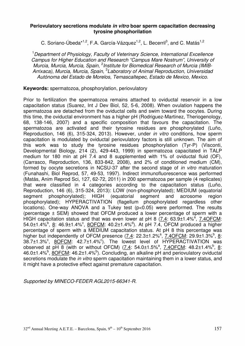
32nd Annual Meeting A.E.T.E. – Barcelona, Spain, 9th – 10th September 2016 157
Periovulatory secretions modulate in vitro boar sperm capacitation decreasing tyrosine phosphorilation
C. Soriano-Úbeda*1,2, F.A. García-Vázquez1,2, L. Becerril3, and C. Matás1,2
1Department of Physiology, Faculty of Veterinary Science, International Excellence Campus for Higher Education and Research “Campus Mare Nostrum”, University of Murcia, Murcia, Murcia, Spain, 2Institute for Biomedical Research of Murcia (IMIB-Arrixaca), Murcia, Murcia, Spain, 3Laboratory of Animal Reproduction, Universidad
Autónoma del Estado de Morelos, Temascaltepec, Estado de Mexico, Mexico.
Keywords: spermatozoa, phosphorylation, periovulatory Prior to fertilization the spermatozoa remains attached to oviductal reservoir in a low capacitation status (Suarez, Int J Dev Biol, 52, 5-6, 2008). When ovulation happens the spermatozoa are detached from the oviductal cells and swim toward the oocytes. During this time, the oviductal environment has a higher pH (Rodriguez-Martinez, Theriogenology, 68, 138-146, 2007) and a specific composition that favours the capacitation. The spermatozoa are activated and their tyrosine residues are phosphorylated (Luño, Reproduction, 146 (6), 315-324, 2013). However, under in vitro conditions, how sperm capacitation is modulated by oviductal periovulatory factors is still unknown. The aim of this work was to study the tyrosine residues phosphorylation (Tyr-P) (Visconti, Developmental Biology, 214 (2), 429-443, 1999) in spermatozoa capacitated in TALP medium for 180 min at pH 7.4 and 8 supplemented with 1% of oviductal fluid (OF), (Carrasco, Reproduction, 136, 833-842, 2008), and 2% of conditioned medium (CM), formed by oocyte secretions in NCSU-37 after the second stage of in vitro maturation (Funahashi, Biol Reprod, 57, 49-53, 1997). Indirect immunofluorescence was performed (Matás, Anim Reprod Sci, 127, 62-72, 2011) in 200 spermatozoa per sample (4 replicates) that were classified in 4 categories according to the capacitation status (Luño, Reproduction, 146 (6), 315-324, 2013): LOW (non-phosphorylated); MEDIUM (equatorial segment phosphorylated); HIGH (equatorial segment and acrosome region phosphorylated); HYPERACTIVATION (flagellum phosphorylated regardless other locations). One-way ANOVA and a Tukey test (p<0.05) were performed. The results (percentage ± SEM) showed that OFCM produced a lower percentage of sperm with a HIGH capacitation status and that was even lower at pH 8 (7.4: 63.9±1.4%a, 7.4OFCM: 54.0±1.4%b, 8: 46.9±1.4%c, 8OFCM: 40.2±1.4%d). At pH 7.4, OFCM produced a higher percentage of sperm with a MEDIUM capacitation status. At pH 8 this percentage was higher but independently of OFCM presence (7.4: 22.3±1.2%a, 7.4OFCM: 29.9±1.3%b, 8: 38.7±1.3%c, 8OFCM: 42.7±1.4%c). The lowest level of HYPERACTIVATION was observed at pH 8 (with or without OFCM) (7.4: 54.0±1.5%a, 7.4OFCM: 48.2±1.4%b, 8: 46.0±1.4%b, 8OFCM: 46.2±1.4%b). Concluding, an alkaline pH and periovulatory oviductal secretions modulate the in vitro sperm capacitation maintaining them in a lower status, and it might have a protective effect against premature capacitation. Supported by MINECO-FEDER AGL2015-66341-R.
32nd Annual Meeting A.E.T.E. – Barcelona, Spain, 9th – 10th September 2016 158
Heat shock proteins detection on heat stressed rabbit sperm cells
M. Vendrell*1,2, L. Ferre-Dolcet2, M. Sabes-Alsina1, N. Arcarons2, R. Morato2, J. Miro2, J.-E. Rodriguez-Gil2, T. Mogas2, and M. Lopez-Bejar1
1Dpt of Animal Health and Anatomy, Faculty of Veterinary Medicine, Autonomous
University of Barcelona, Spain, 2Dpt of Animal Medicine and Surgery, Faculty of Veterinary Medicine, Autonomous University of Barcelona, Spain.
Keywords: Spermatozoa, Hsp, western blot Heat Shock Proteins (HSP) are highly conserved chaperones found in all cell types that have been identified as a defense mechanism for cell survival under severe environmental conditions. Most HSP are constitutively expressed at low levels but some can be up-regulated in response to cellular stresses (physiological, chemical, nutritional or environmental) so to protect cellular proteins against aggregation and denaturation. This study is aimed at determining the presence of HSP60, HSP70 and HSP90 in rabbit spermatozoa exposed to heat stress conditions and to detect if there is any difference between physiological and high temperatures treatments. An up-regulation or an activation of HSP with high temperatures could be a key to induce cryotolerance if heat stress is produced before cryopreservation. Fresh semen samples (6 to 8 New-Zealand White rabbits) were diluted in a semen extender and sperm was incubated for 3h at 4 different temperatures: 32 (scrotal), 37 (body), 42 (heat stress) and 60ºC (killing high temperature). Sperm cells were frozen and kept until the day of processing. Frozen samples were homogenized in 1 mL of protein extraction buffer. Protein concentration of the supernatants was determined by Bradford technique by utilizing a commercial kit. Afterwards, proteins were separated by SDS-PAGE electrophoresis in 10% (w:v) acrylamide gels and transferred to nitrocellulose membranes. Detection of HSP60, HSP70 and HSP90 was performed by using mouse monoclonal anti-HSP60 (1:4000), mouse monoclonal anti-HSP70 (1:1000), mouse monoclonal anti-HSP90 (1:1000), respectively. Membranes were exposed to radiograph films to reveal the HSPs bands (60-90 KDa) after 5 minutes incubation with Luminol Reagent. An goat anti-mouse IgG-HRP was used as secondary antibody at a dilution of 1:2000. Stripping buffer was used to remove the specific HSP marking and a specific anti-mouse a-tubulin antibody for re-test. ImageJ software was used to analyze the adjusted relative densities. Presence of HSP60 and HSP90 was observed in each of the temperatures evaluated. Although an HSP70 band was detectable at 32ºC, 37ºC and 42ºC, no signal for this protein was present at 60ºC. Differences among groups were performed through an ANOVA, followed by the Sidak's post-hoc test fot the HSP analysis. The level of sigificance was set at p<0.05. No significant differences were found between the results. In conclusion, the present study confirms the presence of HSP in rabbit sperm cells as a basal defense system. Further studies about specific location changes and variations in HSP phosphorylation levels could be done so to elucidate if there are changes in protein activity related to high temperatures. This study was supported by the Spanish Ministry of Science and Innovation (Project AGL2013–46769).

32nd Annual Meeting A.E.T.E. – Barcelona, Spain, 9th – 10th September 2016 159
FOLLICULAR AND OVIDUCTAL FLUID MODULATE THE PROTEIN PHOSPHORYLATION ON SERINE AND THREONINE RESIDUES DURING BOAR
SPERM CAPACITATION
H. Zapata1,2, E. Paris-Oller2, F. García-Vázquez2,3, and C. Matás*2,3
1Laboratory of Biology of Reproduction, Department of Biomedicine, Faculty of Health Sciences, University of Antofagasta, Antofagasta, Chile, 2Department of Physiology,
Faculty of Veterinary Science, International Excellence Campus for Higher Education and Research “Campus Mare Nostrum”, University of Murcia, Murcia, Spain, 3Institute for
Biomedical Research of Murcia (IMIB-Arrixaca), Murcia, Spain.
Keywords: Oviduict, follicle, fluid, sperm capacitation, boar
In boar spermatozoa, protein kinases A have a role in the capacitation and various compounds present in the oviduct could have an important regulatory role during this process. Thus, in this work we evaluated if follicular and oviductal fluids have any influence in PKA activity during sperm capacitation. For this purpose, ejaculated sperm (n=7) was adjusted to 2 x 10⁶ sperm/ml in all experimental groups. Each group was incubated for 3 hours in TALP (Rath, J AnimSci, 77,3346–52, 1999) either with or without 1% follicular fluid (FF), 1% periovulatory oviductal fluid (POF) and 2% cumulus cells secretion media (MC). The fluids were collected by aspiration with an automatic pipette as described (Carrasco, Reproduction, 136, 833-842, 2008). The MC was obtained from dishes where groups of 50 COCs had completed the second phase of IVM (with FF but without dbAMPc, PMSG and hCG). The NCSU-37 IVM medium with the COCs was collected and COCs were pipetted to mix COCs secretion with the surrounding media. Centrifugation was the following procedure and the pellet with cellular debris was discarded. After treatment, sperm samples were resuspended in Laemmli buffer and separated on SDS-PAGE. The pattern of protein phosphorylation on Ser and Thr residues was evaluated by western blot. The antibodies used in this study were: phospho-(Ser/Thr) PKA substrate (9624, Cell Signaling Technology, Beverly, USA, 1:2,000) and peroxidase secondary antibody (sc-2004 goat anti-rabbit IgG HRP, Santa Cruz Biotechnology, USA, 1:10,000). Western blot bands were quantified by densitometry (ImageJ software).The results are expressed as densitometric arbitrary units. The results indicated that sperm incubated with FF and MC have a similar serine and threonine phosphorylation pattern of sperm when incubated in a capacitation media. POF decreases the phosphorylation of these residues during capacitation, as also seen when incubating MC + POF. Concluding, the results suggest that POF regulate the phosphorylation pattern of Ser and Thr residues during sperm capacitation. Supported by MINECO-FEDER AGL2015-66341-R and CONICYT 21120844

32nd Annual Meeting A.E.T.E. – Barcelona, Spain, 9th – 10th September 2016 160
Embryology, Developmental Biology, and
Physiology of Reproduction

32nd Annual Meeting A.E.T.E. – Barcelona, Spain, 9th – 10th September 2016 161
Cumulus cells protect the bovine oocyte against lipotoxicity by converting saturated into
unsaturated fatty acids using stearoyl-CoA-desaturase during in vitro maturation
H. Aardema*1, H.T.A. Van Tol1, J.F. Brouwers2, B.M. Gadella1,2, and B.A.J. Roelen1
1Department of Farm Animal Health, Faculty of Veterinary Medicine, Utrecht University,
The Netherlands, 2Department of Biochemistry and Cell Biology. Keywords: cumulus, oocyte, free fatty acid Elevated levels of free fatty acid (FFA) in blood are a metabolic indication for either obesity or a sign for a negative energy balance and adversely influence cell functioning, including that of oocytes. In particular, saturated palmitic (C16:0) and stearic (C18:0) acid can induce lipotoxic events in cumulus-oocyte-complexes (COCs) and result in reduced developmental competence of the oocyte (Leroy et al., Reproduction; 130: 485-495, 2005). In contrast, elevation in mono-unsaturated oleic acid (C18:1) levels prevents the loss in oocyte developmental competence even in presence of relatively high levels of saturated FFA (Aardema et al., Biol Reprod; 85: 62-69, 2011). We have recently reported on the role of cumulus cells in protecting the oocyte against lipotoxic effects associated to elevated levels of FFA (Aardema et al., Biol Reprod; 88: 164, 2013; Lolicato et al., Biol Reprod; 92: 16, 2015). Here we extend that study and investigated whether stearoyl-CoA-desaturase (SCD-1 which converts C18:0 into C18:1) is functional in cumulus cells as a protecting enzyme. COCs were retrieved from bovine slaughterhouse ovaries, matured for 23h, were fertilized and the presumed zygotes were cultured until day 8 according to our standard protocol. The SCD-1 gene- and protein expression in cumulus cells were detected by quantitative RT-PCR and immunoblotting respectively. Results are presented as means ± SD. Statistical analyses was performed in SPSS version 22.0 by the use of an univariate general linear model for blastocyst rates and an univariate analysis of variance on the log transformed lipid data to achieve normally distributed data. P<0.05 was considered significant. Inhibition of SCD-1 activity (1µM, Biovision) in the presence of C18:0 (250 µM) during maturation of COCs resulted in a reduction in the blastocyst rate when compared to the not inhibited control group in the presence of C18:0 (10 ± 4.7% and 25 ± 7.5%, respectively; P<0.001, n=~280 in 3 runs per group). C18:0 and C18:1 levels were determined by HPLC mass spectrometry after total lipid extraction and hydrolysis. A decrease in the C18:1/C18:0 ratio (0.5 ± 0.03) was identified in cumulus cells after inhibition of SCD activity in the presence of 250µM C18:0 versus the group without SCD inhibition (1.3 ± 0.46; P<0.01), suggesting active conversion of C18:0 into C18:1 by SCD. Combined, the data indicate that in cumulus cells SCD-1 converts the potentially toxic saturated C18:0 into less harmful mono-unsaturated C18:1. These data unravel the mechanism of how cumulus cells are competent to protect maturing oocytes against saturated FFA.
32nd Annual Meeting A.E.T.E. – Barcelona, Spain, 9th – 10th September 2016 162
A comparative analysis of the protein composition of the oviductal and uterine fluids in
cattle during the periovulatory phase by 2D fluorescence difference gel electrophoresis (DiGE).
O.S. Acuña1,3, S. Bauersachs2, A. Torrecillas3, M. Jiménez-Movilla3, and M. Avilés*3
1Autonomous University of Sinaloa, Faculty of Veterinary Medicine and Zootechnics, 80246, Culiacan, Mexico, 2ETH Zurich, Institute of Agricultural Sciences, 8092, Zurich, Switzerland, 3University of Murcia, Cell Biology and Histology and IMIB 30100, Murcia,
Spain.
Keywords: oviductal fluid, uterine fluid, protein composition During the periovulatory phase of the estrous cycle processes important for fertilization are going on in the female genital tract. These processes include, e.g., the transport and maturation of gametes. The oviduct and uterus lumen contain complex fluids, which interact with gametes; however, the composition is not entirely known. The oviductal fluid (OF) and uterine fluid (UF) consist of secreted components derived from epithelial tissue as well as transudate from blood serum. The identification of the components of these fluids involved in transport and interaction of gametes during fertilization can be very useful for improvement of culture media used in IVF. The aim of this study was to identify, through a comparative study, differences in the protein composition of OF and UF. Six Simmental heifers were synchronized using gonadotropin-releasing hormone 500 µg IM (Receptal®, MSD) in conjunction with two intramuscular injections of PGF2alpha (10 µg , Estrumate®, MSD). A rectal examination was performed confirming the presence of corpora lutea and the heifers were slaughtered three days after the second PGF injection. OF and UF from slaughterhoused animals classified according to the ovarian morphology (ovarian follicle of approximately 15mm diameter) were pooled. The fluids (150 µg of OF and 150 µg of UF) were labeled with different cyanine fluorescent probes (Amersham) and separated according to the isoelectric point using immobilized pH gradient strips (3-10 pH, 17 cm, Protean® IEF cell system, Bio Rad). The second dimension was performed in a polyacrylamide gel (12%) in the presence of SDS using a Protean II XL system (Bio Rad). The images were obtained with a Typhoon 9410 scanner (Amersham). Image analysis was performed with the Progenesis SameSpots software v 4.0. Approximatively 1000 spots were identified in both, oviductal and uterine fluids. The image analysis showed that 23 spots were different between the two samples (p <0.01 and fold difference >5). Thus, a number of 16 spots were more abundant in UF and 7 more abundant in OF. Future studies will identify these different protein spots and this information would provide more detailed information about their roles during the fertilization. This work was supported by Spanish grant AGL2012-40180-C03-02, the European Commission (FEDER), and a fellowship "Short Term Scientific Missions" (STSM) for the author Acuña OS within the COST action Epiconcept (reference FA1201-010513-028404).

32nd Annual Meeting A.E.T.E. – Barcelona, Spain, 9th – 10th September 2016 163
Proteomic characterization of oviductal extracellular vesicles along the estrous cycle in cattle
C. Almiñana*1, A. Alcântara-Neto1, G. Tsikis1, V. Labas2, L. Combes-Soia2, and P.
Mermillod1
1INRA, UMR7247, Physiologie de la Reproduction et des Comportements, Nouzilly, France, 2INRA, Plateforme d'Analyse Intégrative des Biomolécules, Nouzilly, France.
Keywords: Extracellular vesicles, oviduct, estrous cycle. Exosomes along with microvesicles, named globally as extracellular vesicles (EVs) (Raposo and Stoorvogel, 2013) have been suggested as mediators of the interactions between gametes and embryos and the maternal tract. EVs contain proteins, lipids, and nucleic acids that can be transferred to recipient cells as a new way of cell-to-cell communication. The delivery of the molecular cargo from the maternal tract to sperm and embryos may have a functional impact, promoting sperm capacitation and fertilization ability or supporting early embryo development. Since the reproductive tract undergoes many physiological and hormonal changes during the estrus cycle regulating the environment surrounding gamete and embryos, these changes may also modulate EVs cargo. Thus, the objective of our study was to determine the influence of the stage of the oestrous cycle on the oviductal EVs content at protein level. To perform this study, a bovine in vivo model was used. Pairs of oviducts with their attached ovaries were collected from cyclic (corpus luteum (CL) present), non-pregnant bovine reproductive tracts at the slaughterhouse. To estimate the stage of the oestrous cycle, the CL morphology and follicle populations were assessed according to Ireland et al. (1980): Stage 1 (d 1-4) recently ovulated follicle; Stage 2: (d 5-11) early CL development; Stage III: (d 11-17) yellow or orange CL and Stage 4: (d 18-20) regressing CL with little vasculature and a large preovulatory follicle present. Subsequently, oviducts were flushed to collect their fluid. EVs were isolated by ultracentrifugation. Protein composition from EVs samples was analyzed by high resolution tandem mass spectrometry using GeLC-MS/MS strategy combined to label free quantitative method (samples from 5 replicates were pooled for each stage, n=20 samples). Our first preliminary quantitative proteomic results based on spectral counting showed differential protein abundance of EVs along the bovine oestrous cycle. Among the 336 clusters of proteins identified, 170 were differential among different stages of oestrous cycle (p-value<0.05, ratio <0.5 or ratio>2). From the 498 individual proteins identified from all stages, only a small number of proteins were specific of each stages (2 proteins at Stage 1, 1 at Stage 2, 1 at Stage 3 and 2 at Stage 4), while 454 proteins were common to all stages but with significant differences in abundance between stages. Among the common proteins, we found significant differences in proteins involved in gamete interaction and embryo development such as OVGP: P= 0.00010 S1<S2; HSPA8: P= 0.00010 S1<S2; Myosin-9 (P=0.0028) S2<S1; HSP90: P= 0.00010 S2<S1). The characterization of oviduct-derived EVs under different regulation by oestrogen and progesterone will extend our understanding of gamete/embryo-maternal communication with potential impacts on infertility. Supported by EU-AgreenSkills267196, INRA-PHASE seeding funds and EU-FECUND312097.
32nd Annual Meeting A.E.T.E. – Barcelona, Spain, 9th – 10th September 2016 164
Alpha-tocopherol affects gene expression patterns of rabbit cumulus-oocyte complexes and reduces apoptosis rate during in vitro maturation
M. Arias-Alvarez*1, R.M. Garcia-Garcia1, J. Lopez-Tello1, K. Nieto1, P.G. Rebollar2, A.
Gutierrez-Adan3, and P.L. Lorenzo1
1Complutense University of Madrid, Madrid, Spain, 2Polytechnic University of Madrid, Madrid, Spain, 3INIA, Madrid, Spain.
Keywords: a-tocopherol, in vitro oocyte maturation, rabbit Oxidative stress compromises oocyte developmental competence during in vitro maturation (IVM). Antioxidants such as vitamin E may avoid this imbalance. The aim of this study was to investigate the effect of a-Tocopherol (α-TocOH) on the relative mRNA abundance of genes involved in cumulus expansion (GJA1, PTGS2), cell cycle and viability (AKT1), cell cycle regulation and apoptosis (Tp53, CASP3) and antioxidant response (SOD2, GPX1, CAT) in rabbit cumulus oocyte complexes (COCs) in vitro matured. The apoptosis index in cumulus cells (CCs) and the hydrogen peroxide (H2O2) released by the COCs in maturation media were also assessed. For these purposes, COCs from follicles ≥1mm were recovered, selected and in vitro-matured for 16h (38ºC, 5% CO2) in a medium containing TCM-199 (Sigma, Madrid, Spain) with 0.3% bovine serum albumin (Sigma, Madrid, Spain) and 10 ng/mL Epidermal Growth Factor (EGF) (Sigma, Madrid, Spain) supplemented with 0, 100, 200 or 400 µM α-TocOH (Sigma, Madrid, Spain), named as 0E, 100E, 200E and 400E groups, respectively. After IVM, maturation media without cells was collected and stored at -32ºC and H2O2 concentrations were measured by the Amplex Red Hydrogen Peroxide/Peroxidase Assay Kit (Life Techonologies, NY, USA). The mRNA transcripts were quantified in 203 oocytes and their respective CCs (n=51, n=50, n=50, n=52 for 0E, 100E, 200E and 400E groups, respectively) by qRT-PCR to contrast relative levels of histone H2AZ and genes described above. Apoptotic index was studied in 43 COCs by TUNEL technique (Roche Diagnostics, SL, Barcelona, Spain) (n=10, n=10, n=10, n=12 for 0E, 100E, 200E and 400E groups, respectively). Data were analysed using one way ANOVA and Bonferroni test to compare means. In oocytes, SOD2, CAT and Tp53 poly (A) mRNA contents were down regulated with 100 µM α-TocOH supplementation compared to the control group without this antioxidant (P<0.05). In CCs, CASP3 mRNA transcripts were lower in groups with intermediate concentrations of antioxidants (100E and 200E) compared to 0E and 400E groups (P<0.005), in spite of the apoptosis rate was significantly reduced in all groups supplemented with α-TocOH (100E: 9.12±1.81%, 200E: 10.26±2.75%, 400E: 8.50±2.63% vs 0E: 22.50±3.40%, P<0.05). However, the amount of H2O2 released by the COCs to the maturation media was similar in all the experimental groups (7.62±0.60, 10.93±1.23, 7.76±0.00 and 7.75±0.45 µM in 0E, 100E, 200E and 400E groups, respectively). This study has demonstrated that supplementation of α-TocOH in IVM medium induced significant changes in the molecular machinery of oocytes. Thus, α-TocOH reduced the apoptosis rate in CCs despite non-differences in H2O2 concentrations were found among groups. We acknowledge UCM, CM and MICINN for funding. We acknowledge UCM, CM and MICINN for funding.

32nd Annual Meeting A.E.T.E. – Barcelona, Spain, 9th – 10th September 2016 165
Liquid preservation of bovine embryos as an alternative to cryopreservation N. Blad-Stahl*, F. Kotarski, and C. Wrenzycki
Clinic for Veterinary Obstetrics, Gynecology and Andrology, Chair for Molecular
Reproductive Medicine, Justus-Liebig-University, Giessen, Germany.
Keywords: biopsy of bovine embryos, liquid preservation, hypothermic storage One of the most promising challenges in modern cattle breeding since the introduction of genomic breeding value estimation (GBVE) employing SNP genotyping is the GBVE of early embryos. To determine the genomic breeding value a biopsy of the embryo needs to be taken. In total it lasts approximately seven days until the result is announced. During this time span the embryo has to be preserved. At present, the possibilities to preserve biopsied embryos are either fresh transfer into recipients or cryopreservation. Due to lower pregnancy rates after transfer of biopsied and cryopreserved embryos especially when they are generated via in vitro production (IVP), an alternative preservation method needs to be developed. Recently published data show similar pregnancy rates of non-biopsied in vivo generated embryos which were stored for seven days at temperatures around 4°C in comparison to their fresh-transferred embryos (75% vs 77%; Ideta et al. Scientific Reports, 2013). The aim of this study is to develop an efficient medium that can be used to preserve the viability of biopsied, in vitro produced bovine embryos for up to seven days under hypothermic conditions. Therefore, day 6 embryos were generated employing a standard IVP protocol and assessed according to the IETS standard. Only morulae were used. With the aspiration technique the zona pellucida was perforated and some blastomeres were aspirated. After 24 hours of culture the embryos were examined a second time. Then the embryos were preserved in TCM supplemented with either FBS (25% and 50%) or BSA (1 mg/ml and 10 mg/ml) for seven days at 0-4°C. A control group of non-biopsied embryos was handled and stored under the same conditions. After liquid preservation, embryo quality was determined at the morphological level. Furthermore, live-dead staining was performed. First results show that embryos stemming from all groups have similar total cell numbers (25% FBS non-biopsied and biopsied: 118.4±28.5 vs 106.6±23.4; 50% FBS non-biopsied and biopsied: 98.8±29.5 vs 103.8±19.3; 1mg/ml BSA non-biopsied and biopsied: 100.0±29.0 vs 103.4±27.0; 10 mg/ml BSA non-biopsied and biopsied: 110.5±30.5 vs 111.9±28.5). The live/dead ratio was affected by the protein source, irrespective of the concentration (25% FBS non-biopsied and biopsied: 14.8±14.4 vs 15.9±8.6; 50% FBS non-biopsied and biopsied: 10.2±6.1 vs 14.3±11.9; 1 mg/ml BSA non-biopsied and biopsied: 9.0±6.1 vs 8.1±4.8; 10 mg/ml BSA non-biopsied and biopsied: 7.8±3.9 vs 6.5±3.2) suggesting that the biopsy procedure itself does not harm further embryo quality. These results indicate that liquid preservation employing FBS might be an alternative preservation method for early embryos. Further analyses employing antifreeze protein (AFP Type III) as preservation protein are underway. The financial support of the Förderverein Bioökonomieforschung e.V. (FBF) is gratefully acknowledged.
32nd Annual Meeting A.E.T.E. – Barcelona, Spain, 9th – 10th September 2016 166
In vitro maturation of guinea pig oocytes supplemented with Epidermal Growth Factor and Insulin-Like Growth Factor I
K. Cañón-Beltrán2,1, Y.N. Cajas2, R. Carrera2, P.L. Lorenzo1, P.G. Rebollar3, R.M. Garcia-
Garcia1, and M. Arias-Alvarez*1
1Complutense University of Madrid, Madrid, Spain, 2Universidad Técnica Particular de Loja, Loja, Ecuador, 3Polytechnic University of Madrid, Madrid, Spain.
Keywords: Oocyte maturation, growth factors, Guinea pig Insights in oocyte maturation process in guinea pigs are essential for the development of in vitro culture systems in this species, since it represents an interesting animal model in reproduction field (Suzuki et al. Mol Reprod Dev 2003; 64, 219–25). The goal of this study was to elucidate the influence of both Epidermal Growth Factor (EGF) and Insulin-like Growth Factor I (IGF-I) on in vitro oocyte maturation (IVM) medium of guinea pig. We assessed meiotic and cytoplasmic oocyte maturation, in terms of cortical granules (CG) and mitochondrial distribution, apoptotic rate and steroidogenic response of cumulus-oocyte-complexes (COCs) after IVM. A pool of 500 COCs from adult guinea pigs were cultured in groups of 40 COCs in four replicates in TCM-199 with 2 mM/mL glutamine, 0.1 mg/mL sodium pyruvate and 0.003% BSA for 17h (38ºC, 5%CO2) (Sigma Chemical Company). Oocytes were distributed in different combination doses of growth factors as follows: group 0 (without growth factors); group EI (50 ng/mL EGF + 100 ng/mL IGF-I); group EI-FCS [50 ng/mL EGF + 100 ng/mL IGF-I + 10% Fetal Calf Serum (FCS)] and group FCS (10% FCS). After IVM, 456 oocytes were randomly selected, fixed and stained with 10 µg/mL Hoechst 33342 to assess nuclear configuration [Metaphase II (MII)]. Among them, a total of 152 oocytes were denuded and stained with 100 µg/mL FITC-LCA for CG visualization (n=93) or with 180 nm MitoTrackerRedCMXRos (Molecular Probes Inc) (n=59) for mitochondria assessment. CG and mitochondria patterns were analyzed with laser- scanning confocal microscopy (Leica). Estradiol (E2) and Progesterone (P4) production by COCs was measured by ELISA assay (DEMEDITEC Diagnostics GmbH) in the maturation medium. In the rest of COCs (n=44) apoptosis rate was visualized with TUNEL technique (Roche Diagnostics, SL) and analyzed with Image J software. Chi-square test and one-way ANOVA with Duncan post-hoc test were used. MII rate significantly increased in oocytes from EI and EI-FCS groups compared to 0 and FCS groups (78.3 and 83.7% vs 38.4 and 55.8%, respectively; P<0.05). EI-FCS group showed higher rate of oocytes with peripheral migration of CG (76.9%) (compatible with cytoplasmic maturation) compared with 0 group (23.8%) (P<0.05) whereas EI and FCS groups showed intermediate results (59.1 and 50.0%, respectively). There were no significant differences between groups in the mitochondrial distributions EI-FCS COCs´ showed the lowest apoptosis rate (6.6±0.7%) and the highest E2 (0.3±0.01ng/mL) and P4
(1.9±0.05 pg/mL) production compared to the remaining experimental groups (P<0.05). In our conditions, combination of 50 ng/mL EGF, 100 ng/mL IGF-I and 10% FCS seems to be a suitable medium for IVM system in guinea pig oocytes since it offers superior results of oocyte maturation and quality of CCs compared to the other groups studied included when FCS was added alone. Future studies using these oocytes for IVF and IVC are needed to assess the potential of such COCs. Funded by UTPL and UCM. We acknowledge UTPL and UCM for funding.
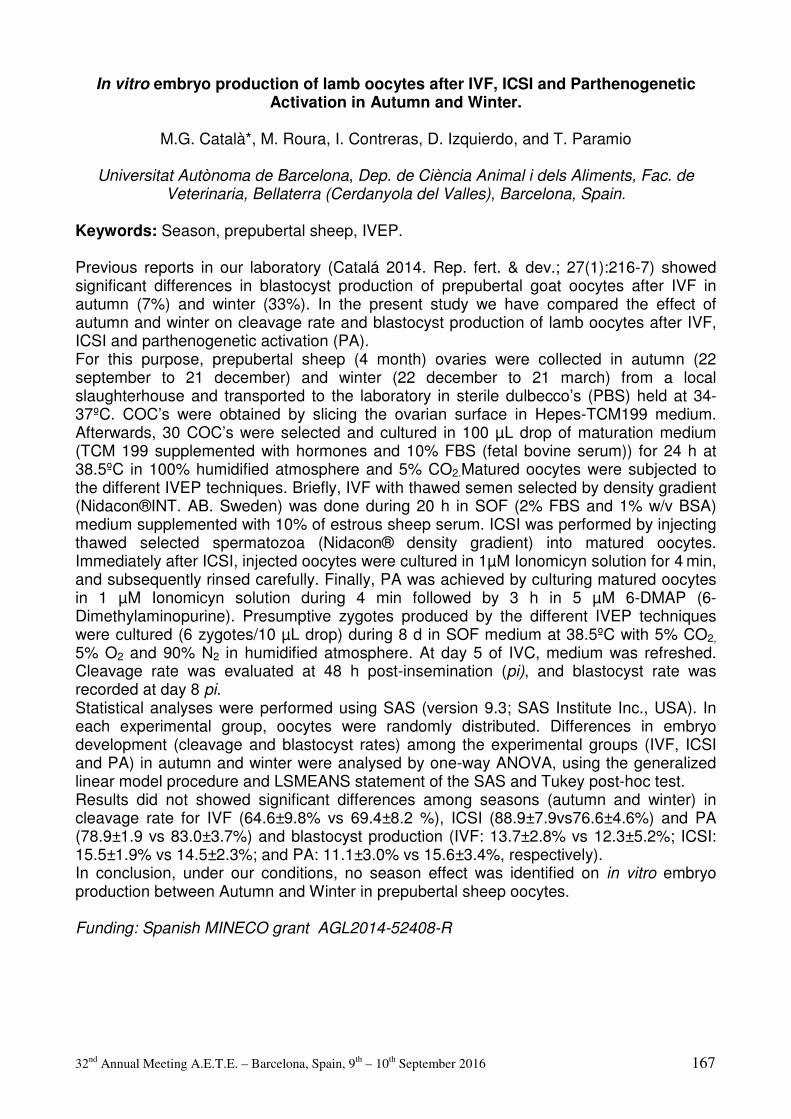
32nd Annual Meeting A.E.T.E. – Barcelona, Spain, 9th – 10th September 2016 167
In vitro embryo production of lamb oocytes after IVF, ICSI and Parthenogenetic Activation in Autumn and Winter.
M.G. Català*, M. Roura, I. Contreras, D. Izquierdo, and T. Paramio
Universitat Autònoma de Barcelona, Dep. de Ciència Animal i dels Aliments, Fac. de
Veterinaria, Bellaterra (Cerdanyola del Valles), Barcelona, Spain.
Keywords: Season, prepubertal sheep, IVEP. Previous reports in our laboratory (Catalá 2014. Rep. fert. & dev.; 27(1):216-7) showed significant differences in blastocyst production of prepubertal goat oocytes after IVF in autumn (7%) and winter (33%). In the present study we have compared the effect of autumn and winter on cleavage rate and blastocyst production of lamb oocytes after IVF, ICSI and parthenogenetic activation (PA). For this purpose, prepubertal sheep (4 month) ovaries were collected in autumn (22 september to 21 december) and winter (22 december to 21 march) from a local slaughterhouse and transported to the laboratory in sterile dulbecco’s (PBS) held at 34-37ºC. COC’s were obtained by slicing the ovarian surface in Hepes-TCM199 medium. Afterwards, 30 COC’s were selected and cultured in 100 µL drop of maturation medium (TCM 199 supplemented with hormones and 10% FBS (fetal bovine serum)) for 24 h at 38.5ºC in 100% humidified atmosphere and 5% CO2.Matured oocytes were subjected to the different IVEP techniques. Briefly, IVF with thawed semen selected by density gradient (Nidacon®INT. AB. Sweden) was done during 20 h in SOF (2% FBS and 1% w/v BSA) medium supplemented with 10% of estrous sheep serum. ICSI was performed by injecting thawed selected spermatozoa (Nidacon® density gradient) into matured oocytes. Immediately after ICSI, injected oocytes were cultured in 1µM Ionomicyn solution for 4 min, and subsequently rinsed carefully. Finally, PA was achieved by culturing matured oocytes in 1 µM Ionomicyn solution during 4 min followed by 3 h in 5 µM 6-DMAP (6-Dimethylaminopurine). Presumptive zygotes produced by the different IVEP techniques were cultured (6 zygotes/10 µL drop) during 8 d in SOF medium at 38.5ºC with 5% CO2,
5% O2 and 90% N2 in humidified atmosphere. At day 5 of IVC, medium was refreshed. Cleavage rate was evaluated at 48 h post-insemination (pi), and blastocyst rate was recorded at day 8 pi. Statistical analyses were performed using SAS (version 9.3; SAS Institute Inc., USA). In each experimental group, oocytes were randomly distributed. Differences in embryo development (cleavage and blastocyst rates) among the experimental groups (IVF, ICSI and PA) in autumn and winter were analysed by one-way ANOVA, using the generalized linear model procedure and LSMEANS statement of the SAS and Tukey post-hoc test. Results did not showed significant differences among seasons (autumn and winter) in cleavage rate for IVF (64.6±9.8% vs 69.4±8.2 %), ICSI (88.9±7.9vs76.6±4.6%) and PA (78.9±1.9 vs 83.0±3.7%) and blastocyst production (IVF: 13.7±2.8% vs 12.3±5.2%; ICSI: 15.5±1.9% vs 14.5±2.3%; and PA: 11.1±3.0% vs 15.6±3.4%, respectively). In conclusion, under our conditions, no season effect was identified on in vitro embryo production between Autumn and Winter in prepubertal sheep oocytes. Funding: Spanish MINECO grant AGL2014-52408-R
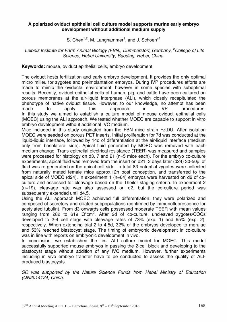
32nd Annual Meeting A.E.T.E. – Barcelona, Spain, 9th – 10th September 2016 168
A polarized oviduct epithelial cell culture model supports murine early embryo development without additional medium supply
S. Chen1,2, M. Langhammer1, and J. Schoen*1
1Leibniz Institute for Farm Animal Biology (FBN), Dummerstorf, Germany, 2College of Life Science, Hebei University, Baoding, Hebei, China.
Keywords: mouse, oviduct epithelial cells, embryo development The oviduct hosts fertilization and early embryo development. It provides the only optimal micro milieu for zygotes and preimplantation embryos. During IVP procedures efforts are made to mimic the oviductal environment, however in some species with suboptimal results. Recently, oviduct epithelial cells of human, pig, and cattle have been cultured on porous membranes at the air-liquid interphase (ALI), which closely recapitulated the phenotype of native oviduct tissue. However, to our knowledge, no attempt has been made to apply this approach in IVP procedures. In this study we aimed to establish a culture model of mouse oviduct epithelial cells (MOEC) using the ALI approach. We tested whether MOEC are capable to support in vitro embryo development without additional IVC medium. Mice included in this study originated from the FBN mice strain FztDU. After isolation MOEC were seeded on porous PET inserts. Initial proliferation for 7d was conducted at the liquid-liquid interface, followed by 14d of differentiation at the air-liquid interface (medium only from basolateral side). Apical fluid generated by MOEC was removed with each medium change. Trans-epithelial electrical resistance (TEER) was measured and samples were processed for histology on d3, 7 and 21 (n=5 mice each). For the embryo co-culture experiments, apical fluid was removed from the insert on d21. 3 days later (d24) 30-50µl of fluid was re-generated on the apical cell side. In total 83 potential zygotes were collected from naturally mated female mice approx.12h post conception, and transferred to the apical side of MOEC (d24). In experiment 1 (n=64) embryos were harvested on d2 of co-culture and assessed for cleavage based on the Theiler staging criteria. In experiment 2 (n=19), cleavage rate was also assessed on d2, but the co-culture period was subsequently extended until d4.5. Using the ALI approach MOEC achieved full differentiation: they were polarized and composed of secretory and ciliated subpopulations (confirmed by immunofluorescence for acetylated tubulin). From d3 onwards cells possessed moderate TEER with mean values ranging from 282 to 619 Ω*cm2. After 2d of co-culture, uncleaved zygotes/COCs developed to 2-4 cell stage with cleavage rates of 73% (exp. 1) and 95% (exp. 2), respectively. When extending trial 2 to 4.5d, 32% of the embryos developed to morulae and 53% reached blastocyst stage. The timing of embryonic development in co-culture was in line with reports on embryonic development in vivo. In conclusion, we established the first ALI culture model for MOEC. This model successfully supported mouse embryos in passing the 2-cell block and developing to the blastocyst stage without addition of any IVC medium. However, further experiments including in vivo embryo transfer have to be conducted to assess the quality of ALI-produced blastocysts. SC was supported by the Nature Science Funds from Hebei Ministry of Education (QN2014124) China.

32nd Annual Meeting A.E.T.E. – Barcelona, Spain, 9th – 10th September 2016 169
Progesterone and Estradiol concentrations of follicular fluid according to the follicular size from Prepuberal and Adult ewes
I. Contreras-Solís*, M. Catalá, S. Soto, A. Alessandroni, M. Roura, P. Maria-Teresa, and
D. Izquierdo
Universitat Autònoma de Barcelona,, Ciència Animal i dels Aliments, 08193, Bellaterra (Barcelona), Spain.
Keywords: Follicle size, Follicular fluid, Prepuberal and Adults ewes. The aim of this study was compare the hormonal status (Estradiol and Progesterone) from follicles of different sizes (diameter) from prepuberal and adult ewes. Eleven ovaries from adults (n =7) and prepuberal ewes (n=4; from two to three months old), were obtained from abattoir and transported to the laboratory in PBS at 34 to 37oC. These follicles were dissected and grouped as: 1) smaller (S: from 2.0 to 2.9 mm) and; 2) large (L: ≥ 3mm) for prepuberal (P) and adults (A) ewes/group. The dissected follicles were carefully cut with microscissor and follicular fluid was recovered into vial of 200 mL and centrifugated at 10000 x g for 15 minutes. The supernatant was stored and analysed by radioimmunoassay to detect Estradiol (E2) and Progesterone (P4) concentrations. Steroids were analysed by radioimmunoassay using ImmuChem Double Antibody P4 and E2 125I RIA kit (MP Biomedicals™, Santa Ana, California, USA). The intra-assay and inter-assay coefficient of variation and the limit of sensitivity were 7.0 and 7.1%, 14.2 and 7.9% and 0.2 and 1.7 ng/mL, for E2 and P4, respectively. Outlier and samples without both steroids analyzed were excluded from analyses. Data of E2 and P4 were transformed to logarithms and analysed using Two-Way ANOVA where size, age of the ewe and their interactions were included in the model. Comparisons were performed using Tukey’s test. Estradiol concentrations for S-P (n=20), L-P (n=15), S-A (n=19) and L-A (n=23) follicles were 1.8 ± 0.6, 18.7 ± 5.9, 3.1 ± 1.4 and 21.2 ± 5.8 ng/mL, respectively. Progesterone concentrations for S-P, L-P, S-A and L-A groups were 5.1 ± 1.9, 8.1 ± 1.3, 66.4 ± 28.0 and 73.8 ± 17.4 ng/mL, respectively. Follicular fluid from S-P and S-A follicles showed less E2 than L-P and L-A (P<0.01). Progesterone concentration was minor in follicular fluid from prepuberal than adult ewes (P<0.05). In conclusion, E2 concentration is related to the size of the follicle while P4 is related to age of the donor ewe.

32nd Annual Meeting A.E.T.E. – Barcelona, Spain, 9th – 10th September 2016 170
The effects of hypo- and hyperglycemia during lipolysis-like conditions on bovine oocyte physiology
J. De Bie*, W.F. Marei, P.E.J. Bols, and J.L.M.R. Leroy
Gamete Research Centre, Wilrijk, Belgium.
Keywords: NEFA, glucose, oocyte Next to elevated non-esterified fatty acid (NEFA) concentrations, lipolytic metabolic conditions can be associated with hypo- and hyperglycemia. Previous research has shown that in the presence of high NEFAs, hypoglycemia (LO GLUC) during IVM hampers embryo development to a greater extend as compared to hyper- (HI GLUC) and normoglycemic conditions. Blastocyst metabolism and carbohydrate- and oxidative stress related gene expression were not affected, but blastocysts from LO- and HI GLUC exposed oocytes showed a higher degree of apoptosis. As a consequence we aimed to study the effects of hypo- and hyperglycemia in presence of elevated NEFAs on oocyte metabolism, apoptosis and reactive oxygen species generation (ROS). Hereto bovine cumulus oocyte complexes (COCs) were routinely matured (1 COC/10µL medium) during 24h under different NEFA and GLUC levels: 1) physiological NEFA (72µM palmitic, stearic, oleic acid) and routine IVM GLUC (5.5mM) (=CONT), 2) pathophysiological NEFA (420µM) and routine GLUC (=HI NEFA), 3) HI NEFA+HI GLUC (10mM) and 4) HI NEFA+LO GLUC (2.8mM). Initial and conditioned medium was sampled (4 repeats) and analyzed for glucose and lactate concentrations. After IVM, all COCs were fixed and stained with caspase-3 and HOECHST to determine apoptosis (n=182, 3 repeats) or denuded and stained for intracellular ROS during 30min using H2DCFDA (n=79, 3 repeats). H2DCFDA fluorescence intensity was quantified using ImageJ and COC apoptosis was classified as: <25%, 25-75% and >75% cumulus cell apoptosis. All data were compared between 4 treatments using a mixed model ANOVA and Bonferroni post-hoc (IBM SPSS Statistics 20). Means ± SEM are presented. COCs exposed to HI NEFA+HI GLUC consume significantly less glucose (485 ± 63 pmol/COC/h) compared with the other treatments (mean of 894 ± 35 pmol/COC/h). HI NEFA+HI GLUC and HI NEFA+LO GLUC exposed COCs produced significantly less lactate (1738 ± 192 and 1848 ± 51 pmol/COC/h, respectively) than CONT and HI NEFA exposed COCs (3573 ± 212 and 3494 ± 289 pmol/COC/h). In addition, LACT/GLUC ratio was significantly lower in all treated COCs compared with CONT, with the lowest LACT/GLUC ratio in HI NEFA+LO GLUC exposed COCs indicating a shift of glucose towards pathways other than glycolysis. No differences were observed in COC apoptosis between treatments. Oocyte ROS was significantly higher in HI NEFA+HI GLUC exposed oocytes (15.85 ± 1.87) compared with HI NEFA+LO GLUC (13.06 ± 1.28) and HI NEFA oocytes (12.06 ± 1.11). In conclusion, lipolytic conditions with or without glycemic perturbations influence the oocyte’s glucose and lactate metabolism. Whereas hypoglycemia in the presence of elevated NEFAs hampers embryo development the most, high GLUC exposed oocytes suffer from increased intracellular ROS. This could not be substantiated by increased cumulus cell apoptosis.

32nd Annual Meeting A.E.T.E. – Barcelona, Spain, 9th – 10th September 2016 171
Reproductive performance and milk production of Damascus goats raised under the intensive system in southeastern Anatolia
H. Demirezer*1 and E. Emsen2
1Mustafa Kemal University, Department of Reproduction and Artificial Insemination, Hatay, Turkey, 2Ataturk University, Department of Animal Science, Erzurum, Turkey.
Keywords: Damascus, intensive system, milk yield The Damascus goat, also known as the Shami, is a native breed of Syria and other Near
East countries. It has been practiced in a commercial goat enterprise (Pan Hayvancilik)
since 2011 with 4500 heads under intensive system. Pregnancy and kidding rates in
different months of mating; lactation periods and milk yield of in does of different parity
were reported.
A total 1427 lactating goats were used in this study. Estrus of lactating does was
synchronized with CIDR (EAZI-BREED™ CIDR®) for 14d in three different months such
as August, September, October. Does were placed with sexually experienced Shami
bucks in the ratio 1 buck to 10 does following CIDR removal. Thirty days after mating,
pregnancy status was determined by trans-abdominal ultrasonography using a Real-time
B-mode.
Pregnancy rates of does were found 88%, 94% and 86% (P<0.05) in August, September
and October mating groups, respectively. Kidding rates of does mated in different months
were found similar (99%). Lactation period and milk yield of first, second, third and fourth
parities of does were found 231 d and 419 l, 293 d and 617 l, 283 d and 637 l, 299d and
680 l, respectively.
It was concluded that Damascus goat under intensive system in southeastern Anatolia
performed well in terms of reproductive performance and lactation yield. We found that
Damascus goat subjected to selection in a commercial farms showed higher reproductive
performance and milk yield than those reported by various researchers (Khazaal, 2009;
Monem et al., 2005; Khoury, 1996).

32nd Annual Meeting A.E.T.E. – Barcelona, Spain, 9th – 10th September 2016 172
Effect of non-esterified fatty acids during sperm capacitation or IVF on developmental competence of bovine oocytes
K.L.J. Desmet*, W.F. Marei, E. Merckx, P.E.J. Bols, and J.L.M.R. Leroy
Gamete Research Centre, University of Antwerp, Wilrijk, Belgium.
Keywords: bovine embryo, IVF, development Deviating metabolic conditions, present in dairy cows suffering negative energy balance (NEB), are reflected in the follicular and oviductal fluid (Leroy et al. (2015), RFD 27: 693-703). Elevated non-esterified fatty acid (NEFA) concentrations, associated with NEB, during in vitro maturation and culture have significant carry over effects on embryo quality and physiology (Van Hoeck et al. (2014), ARS 149: 19-29). Moreover, the oviduct plays an important role in sperm storage and selection, regulation of sperm motility and capacitation (Holt et al. (2010), MRD 77:934-943). This implicates that fertilization can be influenced by alterations in oviductal fluid composition. Therefore, we hypothesized that exposure of sperm cells to elevated (NEB-like) NEFA concentrations shortly before and during IVF can affect fertilization and further embryonic development. To differentiate between possible effects on both spermatozoa and oocytes, two experiments were conducted. Bovine oocytes were matured following standard procedures. In experiment 1, mature oocytes were fertilized under 4 conditions: 1) standard lab conditions (CONT), 2) solvent control (SOLV), 3) physiological NEFA conditions (mixture of 23 µM palmitic acid (PA), 28 µM stearic acid (SA) and 21 µM oleic acid (OA)) (BAS-NEFAs) or 4) lipolytic NEFA conditions (mixture of 230 µM PA, 280 µM SA and 210 µM OA) (HIGH-NEFAs). In experiment 2, spermatozoa were pre-exposed for 4h under conditions CONT, SOLV, BAS-NEFAs or HIGH-NEFAs, then washed and used for IVF of mature oocytes in FA-free media. After 24h, presumptive zygotes were cultivated in serum-free medium until day 8 and developmental competence was assessed. Development was analyzed using binary logistic regression. In experiment 1, cleavage rate was not significantly different between all treatment groups. A significantly higher proportion of HIGH-NEFAs zygotes showed 2-cell block (24.8%) compared to CONT (6.9%; P=0.001), SOLV (11.5%; P=0.016) and BAS-NEFAs (13.1%; P=0.057) zygotes. Blastocyst rate was significantly decreased in the BAS-NEFAs (36.7%; P=0.007) and HIGH-NEFAs (36.6%; P=0.024) compared to the CONT group (54.3%). In experiment 2, no differences in developmental competence were observed among treatments. In conclusion, exposure to elevated NEFA concentrations during IVF has no obvious effect on the fertilization process itself since cleavage rate is not significantly affected. However, further embryonic development is hampered due to NEFA exposure during fertilization. NEFAs have no influence on the fertilizing capacity of pre-exposed sperm suggesting that NEFA-induced reduction in developmental competence is through alterations in oocyte quality but not through affecting sperm quality. More research is ongoing to investigate underlying mechanisms. This research is funded by the FWO (grant nr. 11L8716N).
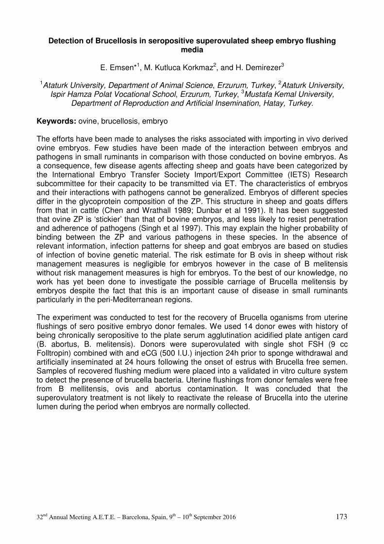
32nd Annual Meeting A.E.T.E. – Barcelona, Spain, 9th – 10th September 2016 173
Detection of Brucellosis in seropositive superovulated sheep embryo flushing media
E. Emsen*1, M. Kutluca Korkmaz2, and H. Demirezer3
1Ataturk University, Department of Animal Science, Erzurum, Turkey, 2Ataturk University, Ispir Hamza Polat Vocational School, Erzurum, Turkey, 3Mustafa Kemal University,
Department of Reproduction and Artificial Insemination, Hatay, Turkey.
Keywords: ovine, brucellosis, embryo The efforts have been made to analyses the risks associated with importing in vivo derived ovine embryos. Few studies have been made of the interaction between embryos and pathogens in small ruminants in comparison with those conducted on bovine embryos. As a consequence, few disease agents affecting sheep and goats have been categorized by the International Embryo Transfer Society Import/Export Committee (IETS) Research subcommittee for their capacity to be transmitted via ET. The characteristics of embryos and their interactions with pathogens cannot be generalized. Embryos of different species differ in the glycoprotein composition of the ZP. This structure in sheep and goats differs from that in cattle (Chen and Wrathall 1989; Dunbar et al 1991). It has been suggested that ovine ZP is ‘stickier’ than that of bovine embryos, and less likely to resist penetration and adherence of pathogens (Singh et al 1997). This may explain the higher probability of binding between the ZP and various pathogens in these species. In the absence of relevant information, infection patterns for sheep and goat embryos are based on studies of infection of bovine genetic material. The risk estimate for B ovis in sheep without risk management measures is negligible for embryos however in the case of B melitensis without risk management measures is high for embryos. To the best of our knowledge, no work has yet been done to investigate the possible carriage of Brucella melitensis by embryos despite the fact that this is an important cause of disease in small ruminants particularly in the peri-Mediterranean regions. The experiment was conducted to test for the recovery of Brucella oganisms from uterine flushings of sero positive embryo donor females. We used 14 donor ewes with history of being chronically seropositive to the plate serum agglutination acidified plate antigen card (B. abortus, B. melitensis). Donors were superovulated with single shot FSH (9 cc Folltropin) combined with and eCG (500 I.U.) injection 24h prior to sponge withdrawal and artificially inseminated at 24 hours following the onset of estrus with Brucella free semen. Samples of recovered flushing medium were placed into a validated in vitro culture system to detect the presence of brucella bacteria. Uterine flushings from donor females were free from B mellitensis, ovis and abortus contamination. It was concluded that the superovulatory treatment is not likely to reactivate the release of Brucella into the uterine lumen during the period when embryos are normally collected.

32nd Annual Meeting A.E.T.E. – Barcelona, Spain, 9th – 10th September 2016 174
Bovine embryo production is very sensitive to toxins released from 3-D printed acrylate chambers
M.A.M.M Ferraz*1, H.H.W. Henning2, P.L.A.M. Vos1, T.A.E. Stout2, and B.M. Gadella1,3
1Department of Farm Animal Health, Faculty of Veterinary Sciences, Utrecht University, Utrecht, The Netherlands, 2Department of Equine Sciences, Faculty of Veterinary
Sciences, Utrecht University, Utrecht, The Netherlands, 3Department of Biochemistry and cell Biology, Faculty of Veterinary Sciences, Utrecht University, Utrecht, The Netherlands.
Keywords: oviduct, embryo, 3-D printing The oviduct provides an ideal microenvironment for gamete interaction and early embryo development. However, in conventional monolayer culture bovine oviduct epithelial cells (BOEC) undergo rapid loss of typical differentiated BOEC properties (e.g. cilia and secretory activity). Recently, we developed a BOEC culture chamber using 3D-printing with acrylate-based resins (3-D BOEC). The printed chambers were successfully detoxified to allow colonization with BOEC monolayers that formed cilia, secreted OVGP1 and supported sperm activation and fertilization of oocytes. However, embryo development was blocked at the 4-cell stage. In fact, when the chambers were used without BOECs no fertilization was observed at all, whereas in the absence of the 3-D chamber the same medium allowed >80% fertilization. We therefore tested whether the BOEC monolayer protects the apical compartment from toxic components leaking out of the 3-D chambers. Primary BOECs (originating from 4 different cows) were seeded onto Transwell® inserts (Polycarbonate, 0.4 µm pore), grown to confluence and cultured at an air-liquid interface for up to 58 days (Transwell® culture). In vitro fertilization (IVF) and culture (IVC) media were conditioned for 24 h with routinely detoxified 3-D chambers, and compared to identical media that had not been exposed to 3-D chambers. Transwell® culture supported the formation of differentiated cuboidal to columnar BOECs, and assessment of trans-epithelial electrical resistance at day 28 indicated that they established a functional barrier (mean value: 578 ohm/ cm2). Transwells with or without BOECs were used for IVF and IVC using a routine IVP protocol. A mixed model ANOVA was used to evaluate statistically significant differences (p<0.05) between groups and a total of 4 replicates, using 100 oocytes per experimental group each (400 oocytes per group) were performed. Using non-conditioned media, cleavage rates were similar in Transwells with or without BOEC (77.45±3.54% and 79±0.92% respectively p>0.05), and considerably better (p<0.05) than when 3-D chamber-conditioned media were used in the Transwells (56.07±12.4% and 11.21±4.18%; with or without BOEC, respectively). Notably, the presence of BOEC significantly reduced the toxic effect (p<0.05). Likewise, in non-conditioned media blastocyst rates were similar (29.5±1.3% and 27±1.8% of matured oocytes; with and without BOEC), whereas they were lower (p<0.05) or completely blocked (p<0.05) when conditioned media were used (19±1.8% with BOEC and 0±0% without BOEC). Again the BOEC had a protective effect (p<0.05). Clearly, the acrylate-based 3-D chambers release a toxic component to which fertilized oocytes, but not BOECs, are extremely sensitive. Moreover, a functional BOEC barrier partially protects the early embryo from this toxic effect. Future studies should identify the component(s) responsible for embryo toxicity, while the data emphasize that care is needed in selecting and testing materials for 3-D printing technologies before applying them to in vitro embryo production.
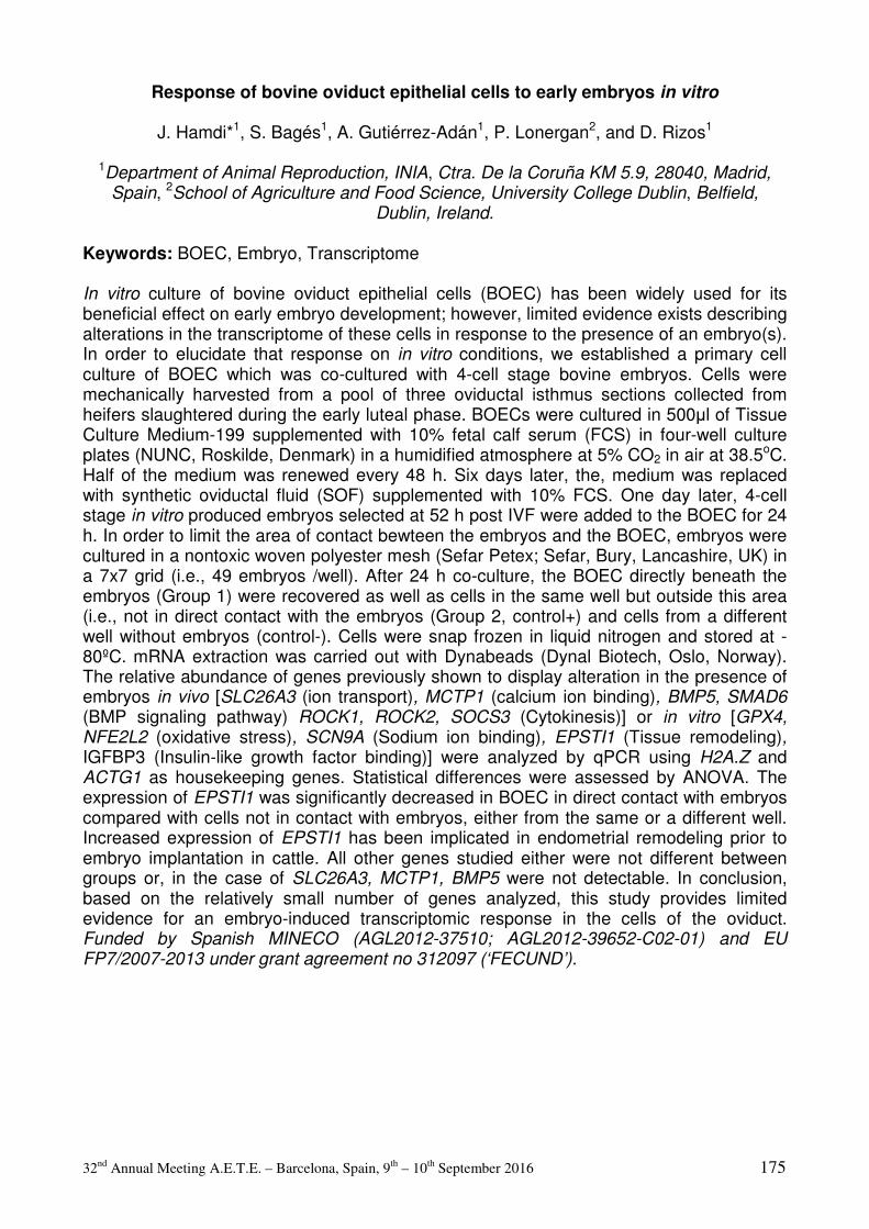
32nd Annual Meeting A.E.T.E. – Barcelona, Spain, 9th – 10th September 2016 175
Response of bovine oviduct epithelial cells to early embryos in vitro
J. Hamdi*1, S. Bagés1, A. Gutiérrez-Adán1, P. Lonergan2, and D. Rizos1
1Department of Animal Reproduction, INIA, Ctra. De la Coruña KM 5.9, 28040, Madrid, Spain, 2School of Agriculture and Food Science, University College Dublin, Belfield,
Dublin, Ireland.
Keywords: BOEC, Embryo, Transcriptome In vitro culture of bovine oviduct epithelial cells (BOEC) has been widely used for its beneficial effect on early embryo development; however, limited evidence exists describing alterations in the transcriptome of these cells in response to the presence of an embryo(s). In order to elucidate that response on in vitro conditions, we established a primary cell culture of BOEC which was co-cultured with 4-cell stage bovine embryos. Cells were mechanically harvested from a pool of three oviductal isthmus sections collected from heifers slaughtered during the early luteal phase. BOECs were cultured in 500µl of Tissue Culture Medium-199 supplemented with 10% fetal calf serum (FCS) in four-well culture plates (NUNC, Roskilde, Denmark) in a humidified atmosphere at 5% CO2 in air at 38.5oC. Half of the medium was renewed every 48 h. Six days later, the, medium was replaced with synthetic oviductal fluid (SOF) supplemented with 10% FCS. One day later, 4-cell stage in vitro produced embryos selected at 52 h post IVF were added to the BOEC for 24 h. In order to limit the area of contact bewteen the embryos and the BOEC, embryos were cultured in a nontoxic woven polyester mesh (Sefar Petex; Sefar, Bury, Lancashire, UK) in a 7x7 grid (i.e., 49 embryos /well). After 24 h co-culture, the BOEC directly beneath the embryos (Group 1) were recovered as well as cells in the same well but outside this area (i.e., not in direct contact with the embryos (Group 2, control+) and cells from a different well without embryos (control-). Cells were snap frozen in liquid nitrogen and stored at -80ºC. mRNA extraction was carried out with Dynabeads (Dynal Biotech, Oslo, Norway). The relative abundance of genes previously shown to display alteration in the presence of embryos in vivo [SLC26A3 (ion transport), MCTP1 (calcium ion binding), BMP5, SMAD6 (BMP signaling pathway) ROCK1, ROCK2, SOCS3 (Cytokinesis)] or in vitro [GPX4, NFE2L2 (oxidative stress), SCN9A (Sodium ion binding), EPSTI1 (Tissue remodeling), IGFBP3 (Insulin-like growth factor binding)] were analyzed by qPCR using H2A.Z and ACTG1 as housekeeping genes. Statistical differences were assessed by ANOVA. The expression of EPSTI1 was significantly decreased in BOEC in direct contact with embryos compared with cells not in contact with embryos, either from the same or a different well. Increased expression of EPSTI1 has been implicated in endometrial remodeling prior to embryo implantation in cattle. All other genes studied either were not different between groups or, in the case of SLC26A3, MCTP1, BMP5 were not detectable. In conclusion, based on the relatively small number of genes analyzed, this study provides limited evidence for an embryo-induced transcriptomic response in the cells of the oviduct. Funded by Spanish MINECO (AGL2012-37510; AGL2012-39652-C02-01) and EU FP7/2007-2013 under grant agreement no 312097 (‘FECUND’).
32nd Annual Meeting A.E.T.E. – Barcelona, Spain, 9th – 10th September 2016 176
Porcine sperm bind to beads conjugated to ZP2 protein under in vitro conditions
J.G. Hamze1, A. Canha2, L. Zamorano1, B. Algarra1, M.C. Olivares3, R. Romar2, and M.
Jimenez-Movilla*1
1Departamento de Biología Celular e Histología, Facultad de Medicina, Universidad de Murcia. IMIB, Murcia, España, 2Departamento de Fisiología, Facultad de Veterinaria, 30071 Campus Espinardo,, Murcia, España, 3Departamento de Bioquímica y Biología
Molecular, Facultad de Medicina, Universidad de Murcia. IMIB., Murcia, España.
Keywords: ZP proteins, beads, sperm-binding The oocyte is encapsulated by a filamentous structure composed of several glycoproteins termed zona pellucida (ZP),that acts as a matrix mediating interaction with sperm. It has been shown that processing of ZP2 at N terminal position (LADEN) mediates the recognition between gametes (Avella M, J Cell Biol. 205(6):801-9, 2014). The study of these molecular mechanisms is very limited due to the ethical problems, the difficulty of obtaining mature oocytes in many mammalian species, the high cost of genetically modified mice and the inability to transfer this knowledge to other species such as swine. Therefore, we propose an in vitro model that mimics the oocyte shape, allowing the research on the interaction of gametes, identification and characterization of the ZP proteins activity and the conditions for sperm recognition. In this work, the proposed model is developed by combining magnetic beads (His Mag Sepharose™ Excel) conjugated with porcine ZP2 and ZP4 recombinant proteins. This novel model besides increasing our knowledge of the oocyte-sperm interaction, could also be industry-wide implemented as an evaluator of mammalian sperm quality. For this study ZP2 and ZP4 recombinant proteins were marked with a Flag and V5 tag recognition site, respectively, and with a histidine tag for easy identification and adhesion to the beads. The proteins were expressed in mammalian cells (CHO) and once secreted, identified by electrophoresis and western blot. Then, the secreted recombinant proteins were conjugated with the magnetic beads. Groups of 40-45 ZP proteins conjugated-beads and beads raised with growth CHO-cell medium (Control group) were coincubated for 2hr with boar spermatozoa (heterospermic dose) in TALP medium at a final concentration of 200.000 spermatozoa/ml. After 2h coincubation, the beads were washed twice in PBS, fixed and stained with Hoechst. Bound sperm in each bead was scored by fluorescence microscopy and the obtained results analysed by one-way ANOVA. Three replicates in a blind analysis were done and P-value <0.05 was taken to denote statistical significance. Secreted proteins ZP2 and ZP4 were identified by electrophoresis and western blot with anti-Flag and and Anti-V5 antibodies, respectively. The ZP2 showed a molecular weight of 100 kDa and ZP4 a molecular weight of 65 kDa. Adhesion of secreted proteins to the beads was confirmed by western blot. Finally, number of sperms bound to ZP2-beads (8.56 ± 0.64, n=230) was significantly higher (P<0.001) than sperm bound to ZP4-beads (3.00 ± 0.27, n=233) and control (4.00 ± 0.36, n=207). In conclusion, a novel in vitro model combining magnetic beads with ZP proteins (ZP2 and ZP4) was developed in order to study the role of ZP2 protein on sperm-oocyte interaction. Future studies could increase our knowledge of the oocyte-sperm interaction and could be also implemented as an in vitroselection and quality evaluation technique of potentially fertile mammalian spermatozoa. This work was funded by Fundación Seneca "Jóvenes Lideres en Investigación 2013" (18931/JLI/13)

32nd Annual Meeting A.E.T.E. – Barcelona, Spain, 9th – 10th September 2016 177
The presence of L-carnitine during maturation improves bovine embryo production
P. Hulinska*, K. Hanzalova, D. Knitlova, M. Jeseta, and M. Machatkova
Veterinary Research Institute, Brno, Czech Republic.
Keywords: bovine oocytes, L-carnitine, IVF outcome Supplementation of IVM media with mitochondrial stimulators can improve oocyte cytoplasmic maturation and embryo production in farm animals. Although the positive effect of L-carnitine on oocytes and embryos was described (Takahashi et al., Rep Fert Dev 25, 589–599), there is no information about the specific impact of L-carnitine on oocytes with different developmental competence. This study was designed to define the effect of L-carnitine during maturation on bovine oocytes with different developmental competence in terms of IVF outcomes. Ovaries from the growth to stagnation phase were used for oocyte collection. Meiotically more competent (MMC) and meiotically less competent (MLC) oocytes were isolated regarding the follicles size (medium or small). The oocytes maturing with or without 2.5 mM L-carnitine (Sigma-Aldrich Co., Prague, Czech Republic) were fertilized and cultured into blastocysts, using standard protocols (Machatkova et al., Zygote 16: 203–209, 2008). The effect of L-carnitine treatment on mitochondrial cluster formation, lipid consumption, fertilization, cleavage and blastocyst differentiation was assessed. The results were statistically analysed by the ANOVA procedure using Chi-square and Fisher’s least difference tests, significant level was set at P<0.05. No significant differences were found in the proportions of MII oocytes among MMC and MLC oocytes matured with or without L-carnitine. However, a significantly higher proportion of MII oocytes with mitochondrial clusters was observed in MLC oocytes matured with L-carnitine than with those matured without L-carnitine (67.2% vs 49.2%). A significantly lower mean lipid content was also detected in MLC oocytes matured with L-carnitine in comparison with those matured without L-carnitine. A significantly higher fertilization (91.0% vs 85.9%) and syngamy rates (55.0% vs 46.7%) in MLC oocytes but similar fertilization and significantly lower syngamy rates (36.4% vs 52.9%) were found in MMC oocytes when they matured with L-carnitine compared with those matured without L-carnitine. Although no significant difference in cleavage rates was found among oocytes matured with or without L-carnitine, significantly more MLC oocytes matured with L-carnitine developed into D7 early blastocysts and D8 expanded blastocysts compared with the controls (31.7% vs 23.1% and 33.3% vs 25.8%, respectively). On the other hand, a significantly higher proportion of D8 expanded blastocysts was obtained in MMC oocytes matured with L-carnitine compared with the controls (72.7% vs 59.3%). It can be concluded that L-carnitine treatment during maturation enhances production of bovine embryos from meiotically less competent oocytes and accelerates differentiation of blastocysts developed from more competent oocytes. The study was supported by Grant COST CZ-LD14104 of the Ministry of Education, Youth and Sports and Grant QJ1510138 of the Ministry of Agriculture of the Czech Republic.
32nd Annual Meeting A.E.T.E. – Barcelona, Spain, 9th – 10th September 2016 178
In vitro monolayer barrier function of bovine oviduct epithelial cells is modified due to high concentrations of non-esterified fatty acids
L. Jordaens*1, V. Van Hoeck1, B. Vlaeminck2, V. Fievez2, S. Thys3, I. Pintelon3, P.E.J.
Bols1, and J.L.M.R. Leroy1
1Gamete Research Centre, University of Antwerp, Wilrijk, Belgium, 2Laboratory for Animal Nutrition and Animal Product Quality, Ghent University, Melle, Belgium, 3Core Facility for
Biomedical Microscopic Imaging, University of Antwerp, Wilrijk, Belgium.
Keywords: Bovine oviduct, Maternal metabolism Early post-partum negative energy balance in high yielding dairy cows has considerable repercussions on reproductive ability and economic merit of these animals. Typically, lipolysis is upregulated and the associated rise of non-esterified fatty acids (NEFAs) has been proposed as a key factor in the decline of oocyte and embryo quality. However, the effects on the oviductal micro-environment remain largely unknown. In this study, we hypothesized that elevated NEFAs may modify in vitro bovine oviduct epithelial cell (BOEC)-physiology by altering the BOEC-barrier function, and thus may potentially affect overall fertility. Hereto, fatty acid (FA)-transfer was evaluated, monolayer permeability was linked to transepithelial electric resistance (TER), and BOEC TJP1-expression and lipid droplet (LD) formation were analyzed. In 4 repeats, early luteal BOECs were seeded in a polarized cell culture (PCC)-system. After reaching 100% confluency (D9), monolayers were NEFA-exposed (PA+SA+OA) for 24h in 4 groups: 1) CONTROL (0µM NEFA + 0%EtOH), 2) SOLVENT CONTROL (0µM NEFA + 0.45%EtOH), 3) BASAL NEFA (720µM NEFA + 0.45%EtOH in the basal compartment), 4) APICAL NEFA (720µM NEFA + 0.45%EtOH in the apical compartment). Next, spent medium was photometrically assessed for total FA-concentration and subjected to gas chromatography for FA-profiling. Also, a 3h permeability assay using FITC-albumin was performed, and related to pre- and postexposure TER-measurements. BOEC-mRNA was retrieved for qRT-PCR of TJP1 to assess expression levels of tight junction protein 1. LD-formation was studied using Bodipy® 493/503 and confocal imaging. All data were analyzed with one way ANOVA. Spent medium analyses showed a 19.5% NEFA-decrease in the supplemented compartment of BASAL NEFA, with paracellular passage to the non-supplemented, apical compartment of PA (56.0%↑), SA (60.0%↑), OA (33.5%↑) as free FAs. However, in APICAL NEFA 53.4% of FA-decrease was observed in the supplemented compartment, while no FA-increase was apparent at the non-supplemented side, suggesting intracellular FA-uptake, which was positive for LD-formation in APICAL NEFA. FITC-albumin flux increased significantly (27.59%) in APICAL NEFA, associated with a reduced relative TER-increase (46.85%) during the NEFA-exposure. TJP1-expression was not affected by the treatments. In conclusion, elevated NEFAs in the apical, ‘oviductal lumen’ compartment may decrease the tightness of cell-cell interactions. BOEC-barrier function was thereby compromised in APICAL NEFA. Also, the PCC allows to observe FA-transfer across BOEC-monolayers and the resulting response strongly depends on cell polarity. These data substantiate the concept of the oviduct as a possible gatekeeper that shields its micro-environment from detrimental metabolites, such as high NEFAs.
32nd Annual Meeting A.E.T.E. – Barcelona, Spain, 9th – 10th September 2016 179
The P4 and E2 treatment and protein expression of PGR and PGRMC1 in porcine endometrial cells
B. Kempisty*1,2, S. Ciesiolka1, W. Kranc2, A. Bryja2, P. Antosik3, D. Bukowska3, K.P.
Brüssow3, M. Bruska2, M. Nowicki1, and M. Zabel1,4
1Department of Histology and Embryology, University of Medical Sciences, 6 Swiecickiego St., 60-781 Poznan, Poland, 2Department of Anatomy, University of Medical Sciences, 6 Swiecickiego St., 60-781 Poznan, Poland, 3Institute of Veterinary Sciences, University of
Life, 52 Wojska Polskiego St., 60-628 Poznan, Poland, 4Department of Histology and Embriology, Medical University, 6a Chalubinskiego St., 50-368 Wroclaw, Poland.
Keywords: progesterone receptors, luminal epithelial cells, pig The endometrium consists of a layer of columnar epithelium and connective tissue. In the endometrium can be distinguish two zones based on their involvement in the changes during the oestrus cycle. The functional layer contain the luminal part of the endometrium and it is the site of cyclic changes in the endometrium. The proper functionality of endometrial tissue is regulated by paracrine and endocrine pathways that activate several mediators or metabolic pathways and gene cascades. This study was aimed to investigate the influence of estradiol 17-beta (E2) and progesterone (P4) on progesterone receptor (PGR) and progesterone receptor membrane component 1 (PGRMC1) protein expression in porcine luminal epithelial cells and their influence on the proliferation of these cells in real-time. Surface uterine luminal epithelial cells were removed using sterile surgical blades from uterine horns of ten crossbred anestrus gilts. Following treatment with collagenase I, cells were separated and transferred into 48-well E-Plates for use in a real-time cell analyzer (RTCA, Roche-Applied Science, GmbH, Penzberg, Germany). The luminal epithelial cells were cultured in vitro (IVC) in standard Dulbecco's Modified Eagle's Medium (DMEM; Sigma Aldrich, Madison, USA) and incubated with E2 (10 pg/ml, 40 pg/ml, 500 pg/ml) and P4 (10 ng/ml, 40 ng/ml, 500 ng/ml). The cell proliferation index was analyzed after 0-240h, 0-120h, 120-240h. The analysis was carried out using RTCA and confocal microscopic observations. Results were performed using analysis of variance (ANOVA) and Tukey test, and by using Imaris 7.2 software (BitPlane, Zurich, Switzerland). Using RTCA analysis we found an increased proliferation of luminal epithelial cells after treatment of low doses of P4 (10 and 40 ng/ml), (P<0.001). Higher doses of P4 leads to decrease of proliferation (P<0.001). Conversely, higher doses of E2 (500 pg/ml) increased proliferation index as compared to low doses (10 pg/ml) and control (P<0.001). Confocal microscopic observations revealed that higher concentrations of E2 up-regulate the expression of both PGR and PGRMC1. Additionally, P4 used in lower concentrations stimulated the expression of these receptors. Our study demonstrated that E2 and P4 treatment significantly regulated the expression of PGR and PGRMC1, which is accompanied by real-time proliferation of porcine luminal epithelial cells. The relationship between PGR or PGRMC1 expression and the proliferation of luminal epithelial cells may be influenced by E2 or P4 in a steroid type- and dose-dependent manner.

32nd Annual Meeting A.E.T.E. – Barcelona, Spain, 9th – 10th September 2016 180
Developmental competence of bovine oocytes that have not finished growth phase in vivo
T.I. Kuzmina*1, A.V. Molchanov2, T.I. Stanislavovich1, and D.N. Tatarskaya3
1Russian Research Institute of Farm Animal Genetics & Breeding, St.Petersburg-Pushkin, Russia, 2Saratov State Agrarian University named after NI Vavilov, Saratov, Russia,
3International Centre for Reproductive Medicine, St. Petersburg, Russia.
Keywords: oocyte, prolactin, IVF In our previous studies we demonstrated that the addition of bovine prolactin (PRL) to culture media enhanced the proportion of blastocysts obtained from bovine oocytes selected by brilliant cresyl blue (BCB, Heleil B. et al., J. Reprod. & Infertility 1 (1):01-07, 2010). Process of nuclear maturation of BCB- oocytes (oocytes that have not finished growth phase in vivo) are slowly compared to the BCB+ oocytes (oocytes that have finished growth phase in vivo). Most of the BCB +oocytes (81%) reached the metaphase-II after 24 hours and only 52% BCB- oocytes completed nuclear maturation (Kuzmina T. et al. Tsitologiya. 55 (9): 664-665, 2014). The aim of the present study was to evaluate the developmental competence of BCB- oocytes matured in medium supplemented by PRL at prolongation of the time of cultivation to 30 h. Ovaries were collected at slaughterhouse. Compact cumulus oocyte complexes (COC) were aspirated from follicles 3-8 mm diameter. Before IVM COC were incubated in 26 µM BCB (B-5388, Sigma) solution for 90 minutes. Oocytes were divided into BCB- (colorless cytoplasm) and BCB+ (colored cytoplasm). COC were cultured 15 h in TCM 199 + 10% (v/v) FCS + 50 ng/ml PRL with 106 /ml granulosa cells. Then medium were supplemented by 10 IU/ml hCG. The time of cultivation for BCB+ and BCB- oocytes were 24 h in control and 30 h in experimental groups. After IVM oocytes were fertilized and embryos were cultured by standard protocols up to Day 8. All chemicals used in this study were purchased from Sigma - Aldrich (Moscow, Russia). Data were analyzed by chi-square. Chromatin of 193 BCB + and 176 BCB – oocytes (total 369 oocytes, in 5 replicates, 17-20 oocytes/group) was evaluated after 24 and 30 hours of cultivation. 81 % (77/95) and 85 % (83/98) of BCB + oocytes reached metaphase II after 24 and 30 h of cultivation, respectively. 52% (46/89) of BCB- oocytes reached metaphase II after 24 h, but after 30 h of cultivation percentage of oocytes significantly increased [71%(62/87), P<0.01]. These results suggest the possibility of BCB- to complete the nuclear maturation in vitro with the prolongation of culture time. In the control group the cleavage rates were significantly higher in the BCB+ oocytes in comparison to the BCB- oocytes [87% (107/123) vs 63% (87/139), P<0.01]. Prolongation of the time of cultivation significantly increased percentage of cleavage after IVF of BCB – oocytes [63% (87/139) vs 78% (118/151), P<0.05]. We did not find significant differences between the percentage of late morulae and blastocysts that developed from BCB+ oocytes independently of prolongation of maturation time [41% (50/123) and 38% (56/147)]. The BCB- oocytes yielded a higher proportion of late morulae and blastocysts by the prolongation of the time of cultivation to 30 h [12 % (17/139) vs 30 % (45/151), P<0.01].The prolongation of maturation time to 30 h improved the developmental competence of BCB- oocytes. Further investigation is needed to evaluate the potential of obtained embryos to development (including evaluation level of apoptosis, gene expression, ET).
32nd Annual Meeting A.E.T.E. – Barcelona, Spain, 9th – 10th September 2016 181
Progesterone is involved in anti-aging effects of prolactin on bovine cumulus-enclosed oocytes matured in vitro
I.Y. Lebedeva*, G.N. Singina, E.N. Shedova, T.E. Taradajnic, and N.A. Zinovieva
L.K. Ernst Institute of Animal Husbandry, Podolsk, Russia.
Keywords: prolactin, progesterone, oocyte aging In matured oocytes, aging processes are accelerated that reduces the ovum fitness. However, little is known about physiological factors responsible for the oocyte protection from precocious senescence. We have found previously that prolactin (PRL) can maintain the developmental capacity and apoptosis resistance of in vitro matured bovine oocytes aging in vitro (Singina et al., Reprod Fert Dev, 27:204, 2015; Singina et al., Reprod Domest Anim, 50(S3):77, 2015). The goal of this study was to test a hypothesis that these anti-aging effects of PRL might be achieved through stimulation of progesterone production by cumulus cells (CCs). Bovine cumulus-enclosed oocytes (CEOs) were cultured for 20 h in the IVM medium (TCM 199 supplemented with 10% fetal calf serum, 10 µg/ml FSH, and 10 µg/ml LH). Unless otherwise stated, all chemicals were purchased from Sigma-Aldrich (St. Louis, MO, USA). A part of in vitro matured oocytes was set free from their CCs. Then CEOs and denuded oocytes (DOs) were cultured for additional 12 or 24 h in the aging medium (TCM 199 containing 10% fetal calf serum) without (Control group) or with bovine PRL (50 ng/ml; Research Center for Endocrinology, Moscow, Russia) and/or trilostane (an inhibitor of progesterone synthesis). Apoptosis was detected in oocytes aged for 24 h using the In Situ Cell Death Detection Kit (Fluorescein, Roche, Indianapolis, USA). Oocytes aged for 12 h were subjected to the IVF procedure described previously (Singina et al., Reprod Fert Dev, 26:154, 2014). The embryo development was evaluated at Days 2 and 8 for cleavage and blastocyst formation. The data for apoptosis (n=4, 80-90 oocytes per treatment) and IVF/IVC (n=5-6, 135-175 oocytes per treatment) were analyzed by ANOVA. After 24 h aging, the rate of apoptotic CEOs in the PRL-treated group was reduced as compared to the Control group (8.2±3.3 vs. 24.5±3.3%, P<0.001), but did not differ from that prior to aging (3.5±1.8%). Meanwhile, PRL did not affect this rate in the medium containing 1 or 10 µM trilostane (1 µM: 23.8±3.4 vs. 29.0±1.6%; 10 µM: 30.7±3.4 vs. 36.2±2.0%). Furthermore, the effect of PRL on oocyte apoptosis disappeared when removing CCs. After IVM for 20 h, the blastocyst rate in our IVF/IVP system was 25.9±3.0%. Following the prolonged culture of CEOs for 12 h, the blastocyst yield in the Control group decreased to 9.9±0.9% and was lower than in the PRL group (18.5±2.8, P<0.01). The addition of 1 µM trilostane to the aging medium containing PRL (but not to the Control medium) caused the blastocyst yield to decline up to 11.7±2.4% (P<0.05). At the same time the developmental capacity of cultured DOs was unaffected by PRL. Thus, the supporting effects of PRL on the developmental capacity and apoptosis resistance of aging oocytes are mediated through CCs and related to stimulation of progesterone synthesis. Therefore, the anti-aging effects of PRL may be achieved, at least in part, due to a pro-survival action of progesterone on mature oocytes. The study was supported by the Russian Science Foundation (project No. 16-16-10069).

32nd Annual Meeting A.E.T.E. – Barcelona, Spain, 9th – 10th September 2016 182
Developing a responsive mouse in vitro fertilization model with focus on sperm concentration
Y. Liu, H.S. Pedersen*, L. Foldager, H. Callesen, and M.T. Sørensen
Department of Animal Science, Aarhus University, Tjele, 8830, Denmark.
Keywords: in vitro fertilization, mouse, sperm concentration
Pesticides and other xenobiotics are assessed for reprotoxicity prior to approval. For this, a model based on rodents is often used, with natural mating and litter size as the end-point. The use of an in vitro embryo production (IVP) system would provide us with more details about the potential effects of the chemicals, as this includes the fertilization process (IVF) itself followed by the embryo development (PLoS One, 2013:e70112). When assessing sperm fertilizing capacity, however, the concentration used for IVF is important. The fraction of fertilized oocytes in an IVF system will increase with increasing sperm concentration until reaching a maximum attainable plateau – we call this “the responsive range” of sperm concentrations. Adding an overload of sperm, i.e. more sperm than necessary to reach the maximum attainable plateau, will not increase the level of this plateau. Furthermore, an overload of sperm in an IVF system could mask compromised sperm fertilizing capacity, dependent on the type of sperm defect, resulting in a compromised assessment of sperm fertilizing capacity. In mouse IVF, the commonly used sperm concentration in relation to toxicity tests of pesticides is 1x10⁶ sperm/ml, which could result in an overload of sperm. Therefore, we studied the effect of using different sperm concentrations for IVF in a complete mouse IVP system on fertilization rates and subsequent embryo development, to develop a sensitive assessment test for sperm fertilizing capacity. Sperm from 20 NMRI males (9-15 weeks old) and 3,416 oocytes from 195 C57BL/6J females (3-4 weeks old) were used for IVF (Theriogenology 65:1716). A total of nine sperm concentrations were used in the range of 1x10⁴ to 2x10⁶/ml, using three to four concentrations from each male. To get representative sperm samples, the sperm was mixed in capacitation drops before use for IVF. Fertilization capacity was expressed as percentage of oocytes developing to the 2-cell stage (2-cell rate). The mean values of 2-cell rates at concentration of 1x10⁴, 2.5x10⁴, 4x10⁴, 5x10⁴, 1x10⁵, 5x10⁵, 1x10⁶, 1.5x10⁶ and 2x10⁶ sperm/ml were 23%, 42%, 47%, 55%, 56%, 45%, 36%, 50% and 41%, respectively. The data of 2-cell rates were used for a breakpoint analysis based on segmented negative binomial regressions with R software. The breakpoint analysis was made to achieve a model of the evolvement over the whole range of sperm concentrations rather than focusing on the specific sperm concentrations. The breakpoint analysis revealed a maximum 2-cell rate (51%, 95% CI: 38-69%) at 35,892 sperm/ml (95% CI: 20,999 – 61,348). Based on these results, we will use a sperm concentration of 2.5x10⁴ sperm/ml, i.e. somewhat lower than the estimated breakpoint, in order to be within the “the responsive range” in our ongoing work with evaluating pesticide reprotoxicity. We conclude that a relatively low sperm concentration is a precondition in a mouse IVF system in order to detect reprotoxic effect on sperm cell quality.

32nd Annual Meeting A.E.T.E. – Barcelona, Spain, 9th – 10th September 2016 183
Effects of recombinant porcine OVGP1 protein on bovine embryo gene expression
V. Maillo1, B. Algarra2, M. Avilés2, A. Gutiérrez-Adán1, D. Rizos2, and M. Jiménez-Movilla*2
1INIA, Department of Animal Reproduction, Madrid, Spain, 2Department of Cell Biology and Histology, University of Murcia, Campus Mare Nostrum IMIB, Murcia, Spain.
Keywords: Oviductin, early development embryo, recombinant protein Oviductin is the most abundant non-serum protein present in the oviductal fluid. It has been detected bound to the zona pellucida (ZP) of gametes and embryos indicating a potential biological role during fertilization and development. We have detected porcine oviductin (pOVGP1) bound to the ZP and inside of multivesicular bodies of in vitro matured pig oocytes with a positive effect on in vitro fertilization (IVF). Previously, we tested the effect of two concentrations of pOVGP1 (10 and 50µg/mL) in a heterologous system using bovine cumulus-oocyte complexes in vitro. Whereas the supplementation during fertilization or culture (IVC) did not show any statistical effect on the cleavage rate nor the blastocyst yield, there was a positive tendency in the blastocyst rate when 50µg/mL of pOVGP1 was used during IVC. Therefore, the aim of this study was to evaluate if pOVGP1 had any effect on bovine embryo gene expression. Purified recombinant pOVGP1 tagged with histidine tail was obtained from HEK 293T cells. Embryos were produced by in vitro maturation and fertilization of oocytes derived from ovaries from slaughtered heifers. The effect of 10 or 50µg/mL of pOVGP1 was tested during IVF (day 0-1), IVC (day 1-3.5) or both IVF+IVC (day 0-3.5). Media used for IVC until day 3.5 was SOF supplemented with pOVGP1 or not (control). Then all groups were changed into SOF supplemented with 3mg/ml BSA. At day 8 a representative number of embryos from each group were fixed and the presence of pOVGP1 was analysed using anti-his antibody by confocal microscopy. Gene expression was analysed in three pools of 10 expanded blastocysts recovered from day 7 and 8 for each experimental group using RT-qPCR. Relative mRNA abundance was analyzed using one-way ANOVA. Day 8 embryos cultured in 50µg/mL of pOVGP1 during IVC were the only ones which showed fluorescent signal bound to ZP. Supplementation with 50µg/mL of pOVGP1 during IVF increased the expression of ATF4, gene related with endoplasmic reticulum homeostasis, and both concentrations of pOVGP1 up-regulated the expression of aquaporin 3 (AQP3), a gene positively correlated with survival after vitrification (P<0.05). In addition to those genes, supplementation with 10 and 50µg/mL of pOVGP1 during IVC also increased the expression of desmocollin 2 (DSC2), a gene involved in cell to cell communication (P<0.05). Finally, supplementation during IVF+IVC up-regulated the same genes in both concentrations (ATF4, AQP3 and DSC2) and only when 50µg/mL concentration was used DNMT3A, gene related with epigenetics, was up-regulated (P<0.05). In conclusion, pOVGP1supplementation during in vitro fertilization and culture has a positive effect on developmental related genes, indicating an improvement on bovine embryo quality. This work was supported by Spanish grant AGL2012-40180-C03-02, the European Commission (FEDER).
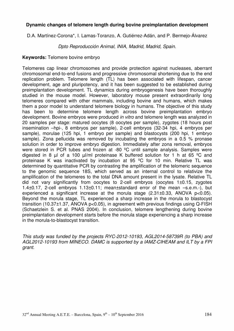
32nd Annual Meeting A.E.T.E. – Barcelona, Spain, 9th – 10th September 2016 184
Dynamic changes of telomere length during bovine preimplantation development
D.A. Martínez-Corona*, I. Lamas-Toranzo, A. Gutiérrez-Adán, and P. Bermejo-Álvarez
Dpto Reproducción Animal, INIA, Madrid, Madrid, Spain.
Keywords: Telomere bovine embryo Telomeres cap linear chromosomes and provide protection against nucleases, aberrant chromosomal end-to-end fusions and progressive chromosomal shortening due to the end replication problem. Telomere length (TL) has been associated with lifespan, cancer development, age and pluripotency, and it has been suggested to be established during preimplantation development. TL dynamics during embryogenesis have been thoroughly studied in the mouse model. However, laboratory mouse present extraordinarily long telomeres compared with other mammals, including bovine and humans, which makes them a poor model to understand telomere biology in humans. The objective of this study has been to determine telomere length across bovine preimplantation embryo development. Bovine embryos were produced in vitro and telomere length was analyzed in 20 samples per stage: matured oocytes (8 oocytes per sample), zygotes (18 hours post insemination –hpi-, 8 embryos per sample), 2-cell embryos (32-34 hpi, 4 embryos per sample), morulae (125 hpi, 1 embryo per sample) and blastocysts (200 hpi, 1 embryo sample). Zona pellucida was removed by incubating the embryos in a 0.5 % pronase solution in order to improve embryo digestion. Immediately after zona removal, embryos were stored in PCR tubes and frozen at -80 ºC until sample analysis. Samples were digested in 8 µl of a 100 µl/ml proteinase K buffered solution for 1 h at 65 ºC and proteinase K was inactivated by incubation at 95 ºC for 10 min. Relative TL was determined by quantitative PCR by contrasting the amplification of the telomeric sequence to the genomic sequence 18S, which served as an internal control to relativize the amplification of the telomeres to the total DNA amount present in the lysate. Relative TL did not vary significantly from oocytes to 2-cell embryos (oocytes 1±0.15, zygotes 1.4±0.17, 2-cell embryos 1.13±0.11; mean±standard error of the mean –s.e.m.-), but experienced a significant increase at the morula stage (2.31±0.33, ANOVA p<0.05). Beyond the morula stage, TL experienced a sharp increase in the morula to blastocyst transition (10.37±1.37, ANOVA p<0.05), in agreement with previous findings using Q-FISH (Schaetzlein S. et al. PNAS 2004). In conclusion, telomere lengthening during bovine preimplantation development starts before the morula stage experiencing a sharp increase in the morula-to-blastocyst transition. This study was funded by the projects RYC-2012-10193, AGL2014-58739R (to PBA) and AGL2012-10193 from MINECO. DAMC is supported by a IAMZ-CIHEAM and ILT by a FPI grant.
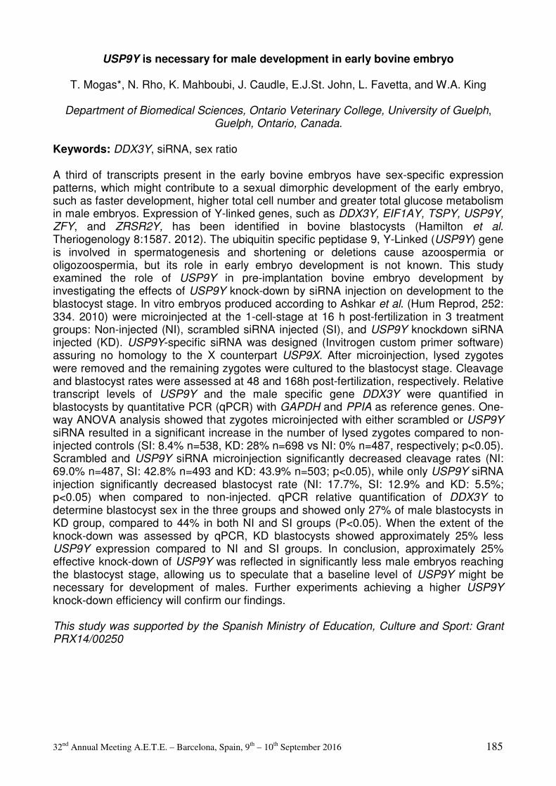
32nd Annual Meeting A.E.T.E. – Barcelona, Spain, 9th – 10th September 2016 185
USP9Y is necessary for male development in early bovine embryo
T. Mogas*, N. Rho, K. Mahboubi, J. Caudle, E.J.St. John, L. Favetta, and W.A. King
Department of Biomedical Sciences, Ontario Veterinary College, University of Guelph, Guelph, Ontario, Canada.
Keywords: DDX3Y, siRNA, sex ratio A third of transcripts present in the early bovine embryos have sex-specific expression patterns, which might contribute to a sexual dimorphic development of the early embryo, such as faster development, higher total cell number and greater total glucose metabolism in male embryos. Expression of Y-linked genes, such as DDX3Y, EIF1AY, TSPY, USP9Y, ZFY, and ZRSR2Y, has been identified in bovine blastocysts (Hamilton et al. Theriogenology 8:1587. 2012). The ubiquitin specific peptidase 9, Y-Linked (USP9Y) gene is involved in spermatogenesis and shortening or deletions cause azoospermia or oligozoospermia, but its role in early embryo development is not known. This study examined the role of USP9Y in pre-implantation bovine embryo development by investigating the effects of USP9Y knock-down by siRNA injection on development to the blastocyst stage. In vitro embryos produced according to Ashkar et al. (Hum Reprod, 252: 334. 2010) were microinjected at the 1-cell-stage at 16 h post-fertilization in 3 treatment groups: Non-injected (NI), scrambled siRNA injected (SI), and USP9Y knockdown siRNA injected (KD). USP9Y-specific siRNA was designed (Invitrogen custom primer software) assuring no homology to the X counterpart USP9X. After microinjection, lysed zygotes were removed and the remaining zygotes were cultured to the blastocyst stage. Cleavage and blastocyst rates were assessed at 48 and 168h post-fertilization, respectively. Relative transcript levels of USP9Y and the male specific gene DDX3Y were quantified in blastocysts by quantitative PCR (qPCR) with GAPDH and PPIA as reference genes. One-way ANOVA analysis showed that zygotes microinjected with either scrambled or USP9Y siRNA resulted in a significant increase in the number of lysed zygotes compared to non-injected controls (SI: 8.4% n=538, KD: 28% n=698 vs NI: 0% n=487, respectively; p<0.05). Scrambled and USP9Y siRNA microinjection significantly decreased cleavage rates (NI: 69.0% n=487, SI: 42.8% n=493 and KD: 43.9% n=503; p<0.05), while only USP9Y siRNA injection significantly decreased blastocyst rate (NI: 17.7%, SI: 12.9% and KD: 5.5%; p<0.05) when compared to non-injected. qPCR relative quantification of DDX3Y to determine blastocyst sex in the three groups and showed only 27% of male blastocysts in KD group, compared to 44% in both NI and SI groups (P<0.05). When the extent of the knock-down was assessed by qPCR, KD blastocysts showed approximately 25% less USP9Y expression compared to NI and SI groups. In conclusion, approximately 25% effective knock-down of USP9Y was reflected in significantly less male embryos reaching the blastocyst stage, allowing us to speculate that a baseline level of USP9Y might be necessary for development of males. Further experiments achieving a higher USP9Y knock-down efficiency will confirm our findings. This study was supported by the Spanish Ministry of Education, Culture and Sport: Grant PRX14/00250
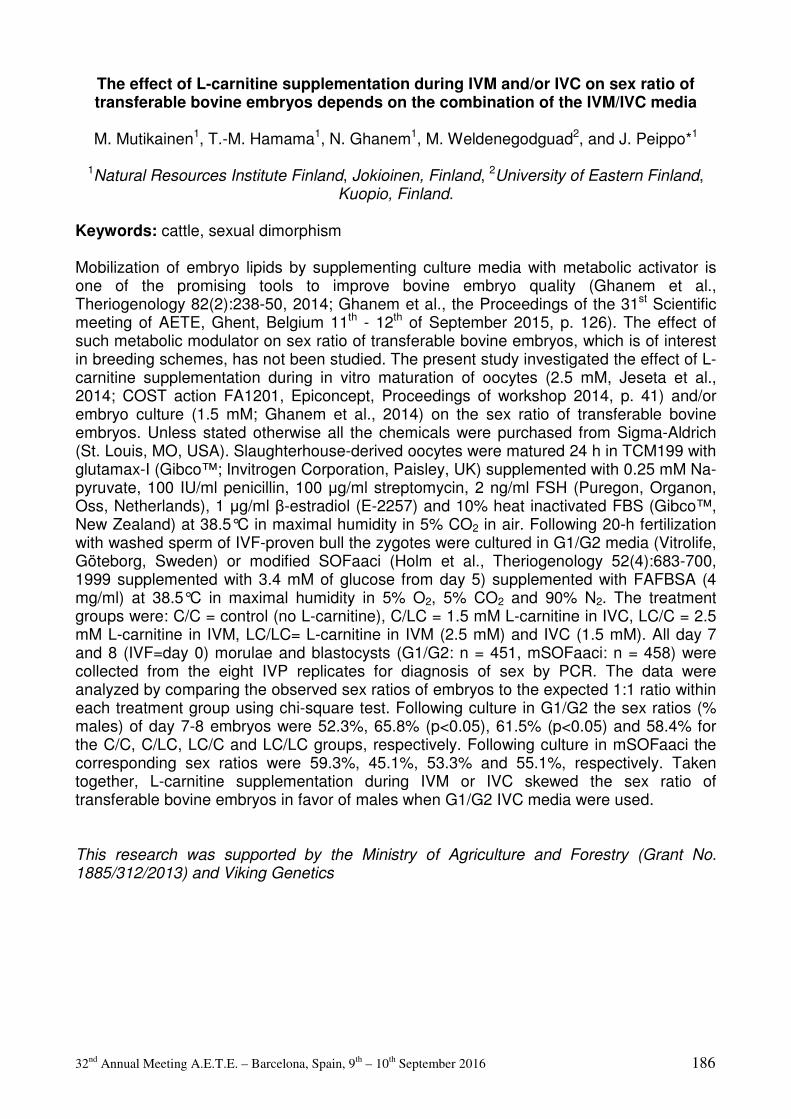
32nd Annual Meeting A.E.T.E. – Barcelona, Spain, 9th – 10th September 2016 186
The effect of L-carnitine supplementation during IVM and/or IVC on sex ratio of transferable bovine embryos depends on the combination of the IVM/IVC media
M. Mutikainen1, T.-M. Hamama1, N. Ghanem1, M. Weldenegodguad2, and J. Peippo*1
1Natural Resources Institute Finland, Jokioinen, Finland, 2University of Eastern Finland, Kuopio, Finland.
Keywords: cattle, sexual dimorphism Mobilization of embryo lipids by supplementing culture media with metabolic activator is one of the promising tools to improve bovine embryo quality (Ghanem et al., Theriogenology 82(2):238-50, 2014; Ghanem et al., the Proceedings of the 31st Scientific meeting of AETE, Ghent, Belgium 11th - 12th of September 2015, p. 126). The effect of such metabolic modulator on sex ratio of transferable bovine embryos, which is of interest in breeding schemes, has not been studied. The present study investigated the effect of L-carnitine supplementation during in vitro maturation of oocytes (2.5 mM, Jeseta et al., 2014; COST action FA1201, Epiconcept, Proceedings of workshop 2014, p. 41) and/or embryo culture (1.5 mM; Ghanem et al., 2014) on the sex ratio of transferable bovine embryos. Unless stated otherwise all the chemicals were purchased from Sigma-Aldrich (St. Louis, MO, USA). Slaughterhouse-derived oocytes were matured 24 h in TCM199 with glutamax-I (Gibco™; Invitrogen Corporation, Paisley, UK) supplemented with 0.25 mM Na-pyruvate, 100 IU/ml penicillin, 100 µg/ml streptomycin, 2 ng/ml FSH (Puregon, Organon, Oss, Netherlands), 1 µg/ml β-estradiol (E-2257) and 10% heat inactivated FBS (Gibco™, New Zealand) at 38.5°C in maximal humidity in 5% CO2 in air. Following 20-h fertilization with washed sperm of IVF-proven bull the zygotes were cultured in G1/G2 media (Vitrolife, Göteborg, Sweden) or modified SOFaaci (Holm et al., Theriogenology 52(4):683-700, 1999 supplemented with 3.4 mM of glucose from day 5) supplemented with FAFBSA (4 mg/ml) at 38.5°C in maximal humidity in 5% O2, 5% CO2 and 90% N2. The treatment groups were: C/C = control (no L-carnitine), C/LC = 1.5 mM L-carnitine in IVC, LC/C = 2.5 mM L-carnitine in IVM, LC/LC= L-carnitine in IVM (2.5 mM) and IVC (1.5 mM). All day 7 and 8 (IVF=day 0) morulae and blastocysts (G1/G2: n = 451, mSOFaaci: n = 458) were collected from the eight IVP replicates for diagnosis of sex by PCR. The data were analyzed by comparing the observed sex ratios of embryos to the expected 1:1 ratio within each treatment group using chi-square test. Following culture in G1/G2 the sex ratios (% males) of day 7-8 embryos were 52.3%, 65.8% (p<0.05), 61.5% (p<0.05) and 58.4% for the C/C, C/LC, LC/C and LC/LC groups, respectively. Following culture in mSOFaaci the corresponding sex ratios were 59.3%, 45.1%, 53.3% and 55.1%, respectively. Taken together, L-carnitine supplementation during IVM or IVC skewed the sex ratio of transferable bovine embryos in favor of males when G1/G2 IVC media were used. This research was supported by the Ministry of Agriculture and Forestry (Grant No. 1885/312/2013) and Viking Genetics

32nd Annual Meeting A.E.T.E. – Barcelona, Spain, 9th – 10th September 2016 187
Royal jelly improves embryonic developmental competence and affects transcript levels of apoptosis-related genes in goat cumulus-oocyte complexes
K. Nikdel*1,2, N. Ghanem3, M. Khajenabi2,5, R. Kamaledini2, and A. Mohammadi-
Sangcheshmeh4,5
1Nahadehaye Dami Jahed (NDJ) Company, Tehran, Iran, 2Department of Animal Science, Science and Research Branch, Islamic Azad University, Tehran, Iran, 3Animal Production Department, Faculty of Agriculture, Cairo University, Giza, Egypt, 4Department of Animal and Poultry Science, College of Aburaihan, University of Tehran, Pakdasht, Tehran, Iran,
5Department of Transgenic Animal Science, Stem Cell Technology Research Center, Tehran, Iran.
Keywords: Royal jelly, goat oocyte, development Objectives: Royal jelly (RJ) has been described with its vital biological property an antioxidant. This antioxidative capacity of RJ has been linked with protecting female/male gametes and improving post-thaw sperm motility. Considering such beneficial effects, here we have performed experiments to evaluate whether addition of RJ to in vitro maturation medium can affect oocye developmental competence and transcription of apoptosis-related Bax, Bcl-2, and p53 genes in goat. Materials and methods: Good quality cumulus-oocyte complexes were recovered from slaughterhouse ovaries. Capsulated pure RJ (Natural Life; Brookvale, NSW, Australia) was supplemented in maturation medium during the 24 h of in vitro maturation at three different concentrations (2.5, 5 and 10 mg/mL). A cohort of oocytes without any RJ treatment was assigned as control group. Embryo cleavage as well as blastocyst rate were recorded at days 3 and 8 postinsemination, respectively for all groups. Gene expression of apoptosis related transcripts (Bax, Bcl-2 and p53) was profiled using Real-time PCR. Differences in mean values were tested using ANOVA followed by a multiple pair wise comparison using t-test. The relative expression data were analyzed using the General Linear Model of SAS. A P-value of less than 0.05 was considered significant. Results: The percentage of cleaved embryos and Day 8 blastocysts was higher (P<0.05) in the RJ-treated groups at concentration of 5 (70.2 ± 3.2 and 33.1 ± 2.2) and 10 mg/mL (69.8 ± 2.1 and 26.4 ± 3.5) than in 2.5 mg/mL (59.2 ± 3.3 and 21.2 ± 4.1%) and the control groups (54.5 ± 3.6 and 22.3 ± 3.7%). The expression profile of apoptotic induced (Bax and p53) was down-regulated (P<0.05) in oocytes treated was RJ at 5 and 10 mg/mL compared with control counterparts. On the other hand, the transcript abundance of antiapoptotatic gene (Bcl-2) was up-regulated (P<0.05) in all oocytes treated with RJ at concentration of 5 and 10 mg/mL while at 2.5 mg/mL expressed similar (P≥0.05) profile of other all groups. Conclusions: Overall, the addition of RJ at concentrations of 5 mg/mL has enhanced embryo development through reducing expression of genes inducing apoptosis. The authors thank the members of their own laboratories for their helpful discussions.

32nd Annual Meeting A.E.T.E. – Barcelona, Spain, 9th – 10th September 2016 188
A proteomic approach to monitor interactions between oviductal fluid and spermatozoa across the estrous cycle
P. Nogues1, J. Lamy*1, G. Tsikis1, V. Labas2, L. Combes2, P. Mermillod1, X. Druart1, and
M. Saint-Dizier1,3
1INRA UMR PRC, Nouzilly, France, 2INRA PAIB, Nouzilly, France, 3Université de Tours, Tours, France.
Keywords: spermatozoa, oviduct, tubal fluid In the bovine during estrus (pre-ovulatory period), both oviducts are able to maintain sperm viability in the so called “sperm reservoir” with the assumed aim of preserving sperm viability up to the time of ovulation. However, very few molecules interacting with bull spermatozoa (spz) and playing a role in the maintenance of bull sperm viability in vivo were identified. Furthermore, the effect of the stage of the estrous cycle on sperm-oviduct interactions is barely known. The aim of this study was to identify proteins present in the oviductal fluid (OF) that interact with spz and to determine how stage of cycle affects these interactions. Bovine oviductal fluids (OF) were collected at a local slaughterhouse at 3 stages of the estrous cycle based on ovarian morphology: before ovulation (Pre-ov; preovulatory follicle, corpus albicans), after ovulation (Post-ov; corpus hemorragicum) and during the luteal phase (Lut; corpus luteum), and stored at −80°C until used. Frozen-thawed spermatozoa (spz) from one bull were washed then incubated at a final concentration of 330.106 spz/ml in phosphate-buffered saline supplemented with 14% OF (7 mg proteins/ml, based on a positive effect of OF at this concentration on sperm viability) at one of the three stages or a protein-free medium (Synthetic oviductal fluid or SOF, control group) for 1h at 37°C, then washed again. Total proteins from spz were migrated on a 10% SDS-PAGE and each lane was divided in 3 bands for in-gel digestion before proteomic analysis by nano LC-MS/MS. Proteins were considered to originate from the OF and to interact with spz when detected in at least one treatment group but not in the control group. Normalized spectral counts of interacting proteins were compared between stages with T-tests and considered differential when p<0.05 and ratios >2 or <0.5. A total of 270 protein clusters were identified, among which 53 oviductal clusters (that included 55 proteins) interacted with spz, including several myosins (MYH), heat shock proteins (HSP), annexins (ANX) and protein disulfide isomerases (PDI). The proportion of proteins interacting with spz increased from Pre-ov (60%, 33/55) to Post-ov (78%, 43/55) and was maximal at Lut stage (98%, 54/55). Furthermore, interacting proteins shared between Lut and Pre-ov or Post-ov were always more abundant at Lut than at periovulatory stages. When comparing Post-ov and Pre-ov, 20 interacting proteins (including MYH9, HSPB1 and ANXA2) were found more abundant at Post-ov and 4 (OVGP1, GRP78, PDIA3, PDIA6) at Pre-ov. In conclusion, a new approach was proposed to identify and quantify proteins interacting with spz in the oviductal fluid. These results need to be confirmed with more animals. Further studies are also required to decipher the roles played around the time of ovulation by interacting proteins on sperm function.
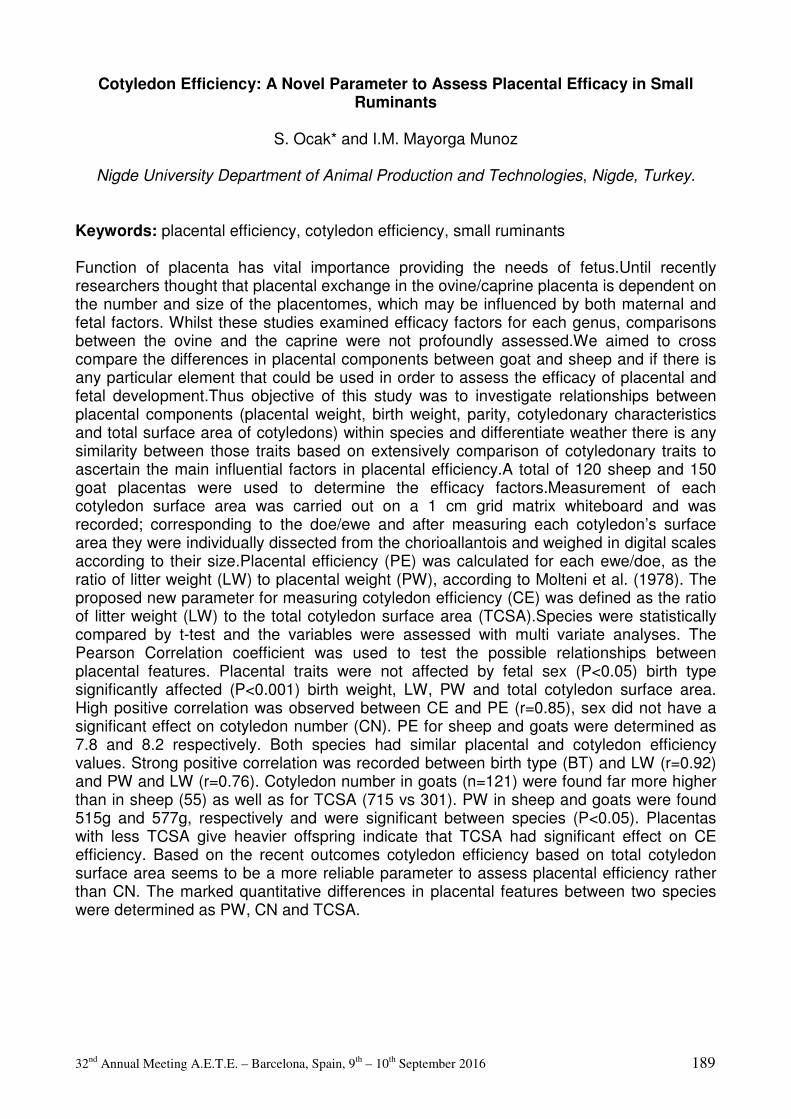
32nd Annual Meeting A.E.T.E. – Barcelona, Spain, 9th – 10th September 2016 189
Cotyledon Efficiency: A Novel Parameter to Assess Placental Efficacy in Small Ruminants
S. Ocak* and I.M. Mayorga Munoz
Nigde University Department of Animal Production and Technologies, Nigde, Turkey.
Keywords: placental efficiency, cotyledon efficiency, small ruminants Function of placenta has vital importance providing the needs of fetus.Until recently researchers thought that placental exchange in the ovine/caprine placenta is dependent on the number and size of the placentomes, which may be influenced by both maternal and fetal factors. Whilst these studies examined efficacy factors for each genus, comparisons between the ovine and the caprine were not profoundly assessed.We aimed to cross compare the differences in placental components between goat and sheep and if there is any particular element that could be used in order to assess the efficacy of placental and fetal development.Thus objective of this study was to investigate relationships between placental components (placental weight, birth weight, parity, cotyledonary characteristics and total surface area of cotyledons) within species and differentiate weather there is any similarity between those traits based on extensively comparison of cotyledonary traits to ascertain the main influential factors in placental efficiency.A total of 120 sheep and 150 goat placentas were used to determine the efficacy factors.Measurement of each cotyledon surface area was carried out on a 1 cm grid matrix whiteboard and was recorded; corresponding to the doe/ewe and after measuring each cotyledon’s surface area they were individually dissected from the chorioallantois and weighed in digital scales according to their size.Placental efficiency (PE) was calculated for each ewe/doe, as the ratio of litter weight (LW) to placental weight (PW), according to Molteni et al. (1978). The proposed new parameter for measuring cotyledon efficiency (CE) was defined as the ratio of litter weight (LW) to the total cotyledon surface area (TCSA).Species were statistically compared by t-test and the variables were assessed with multi variate analyses. The Pearson Correlation coefficient was used to test the possible relationships between placental features. Placental traits were not affected by fetal sex (P<0.05) birth type significantly affected (P<0.001) birth weight, LW, PW and total cotyledon surface area. High positive correlation was observed between CE and PE (r=0.85), sex did not have a significant effect on cotyledon number (CN). PE for sheep and goats were determined as 7.8 and 8.2 respectively. Both species had similar placental and cotyledon efficiency values. Strong positive correlation was recorded between birth type (BT) and LW (r=0.92) and PW and LW (r=0.76). Cotyledon number in goats (n=121) were found far more higher than in sheep (55) as well as for TCSA (715 vs 301). PW in sheep and goats were found 515g and 577g, respectively and were significant between species (P<0.05). Placentas with less TCSA give heavier offspring indicate that TCSA had significant effect on CE efficiency. Based on the recent outcomes cotyledon efficiency based on total cotyledon surface area seems to be a more reliable parameter to assess placental efficiency rather than CN. The marked quantitative differences in placental features between two species were determined as PW, CN and TCSA.

32nd Annual Meeting A.E.T.E. – Barcelona, Spain, 9th – 10th September 2016 190
Cell ultrastructure in bovine preimplantation embryos in relation to cow´s body condition
score
L. Olexikova1, J. Pivko1, A. Makarevich*1, E. Kubovicova1, and P. Chrenek1,2
1Research Institute for Animal Production Nitra, National Agricultural and Food Centre, Hlohovecka 2, 95141 Lužianky-near-Nitra, Slovak Republic, 2Slovak University of
Agriculture, Tr.A.Hlinku 1, 94901 Nitra, Slovak Republic.
Keywords: embryo, BCS, ultrastructure The aim of the study was to examine ultrastructure of several embryo cell organelles from cows with different body condition score (BCS). Embryos (first numeral in brackets) were recovered on 7th day after the insemination by a standard non-surgical flushing of the uterine horns from superovulated Holstein-Friesian cows (second numeral in brackets) with BCS2 (71/17), BCS3 (141/31), BCS4 (48/11) and BCS5 (9/4). Thereafter, the good quality embryos (only those at blastocyst stage) were processed for transmission electron microscopy, and electronograms were evaluated by stereological analysis determining relative volumes of important organelles (mitochondria, lipid droplets, vacuoles, inclusion bodies and apoptotic bodies). Statistical analysis of cellular components was done using one-way ANOVA and Mann-Whitney U test. No differences were observed in the embryo recovery per cow among BCS2 (4.2±0.8), BCS3 (4.6±0.9) and BCS4 (4.4±1.3) groups, whilst BCS5 group showed lesser embryo recovery rate (2.3±0.6). Blastocysts from BCS3 cow with relative volumes (%) of 4.9±0.32, 5.5±0.5, 4.6±0.5, 6.9±1.05 and 1.6±0.5 for mitochondria, lipid droplets, vacuoles, inclusion bodies and apoptotic bodies, respectively were served as a control. Ultrastructure of blastocyst cells in the BCS2 group was similar to those in the BCS3 group: relative volumes of lipid droplets was 7.9%, vacuoles and inclusion bodies - 9.2%. The relative volume of lipid droplets in BCS4 and BCS5 embryos increased significantly (18.5 and 22.6%) when compared to BCS3 embryos. The volume of apoptotic bodies did not significantly differ among the embryos of BCS2-BCS5 cows. In the embryos from the BCS4 or BCS5 cows we observed different morphological patterns of mitochondria: oval, round-shaped or mitochondria with vacuoles inside. Cell nuclei from BCS4 and BCS5 embryos showed the signs typical for low transcription activity (none or very few reticular nucleoli). In conclusion, differences in the ultrastructural morphology of embryos between over-conditioned (BCS4 and BCS5) and average-conditioned (BCS3) cows, in particular the higher lipid content in the cytoplasm, may indicate their low quality, and this fact can be a contributing factor to subfertility in over-conditioned cows. This study was supported by the Agency for the Support of Science and Technology in the Slovak Republic: grants APVV-0137-10 and APVV-14-00-43, as well as from the European Community under project no. 26220220180: Building Research Centre "AgroBiotech".
32nd Annual Meeting A.E.T.E. – Barcelona, Spain, 9th – 10th September 2016 191
FFAR4 is involved in docosahexaenoic acid effects on oocyte developmental potential during in vitro maturation.
M. Oseikria1, S. Uzbekova1, A. Vitorino Carvalho2, V. Duranthon2, and S. Elis*
1UMR PRC, INRA, CNRS, IFCE, Université de Tours, 37380 Nouzilly, France, 2UMR BDR, ENVA, INRA, Université Paris-Saclay, 78350, Jouy-en-Josas, France.
Keywords: PUFA, oocyte maturation Besides affecting uterine environment, a direct effect of n-3 poly-unsaturated fatty acids (PUFA) on the oocyte could enhance fertility. We previously showed that docosahexaenoic acid (DHA, C22:6 n-3, Sigma), when provided during in vitro maturation (IVM), improved oocyte developmental competence through possible effects on cytoplasm but not nuclear maturation and without affecting lipid metabolism gene expression in cumulus cells (CC) (Oseikria et al Theriogenology 85:1625-1634. 2016). DHA could act through several mechanisms of action: i.e. via surface fatty acid receptors (free fatty acid receptor 1 or 4, FFAR1 and 4) or sensors involving PPAR or NFkB pathways; via changes in composition of cell membrane phospholipids; via production of eicosanoids… The aim of the present work was to investigate whether the FFAR4 was involved in the DHA effects previously reported on oocyte quality. We therefore investigated the effect of a specific agonist of the FFAR4, TUG-891, on embryo development after IVF. The response of surrounding CC to DHA or TUG treatment was also studied by gene expression analyses. Oocyte cumulus complexes were collected from slaughtered cows. The protein FFAR4 was first localized by immunohistochemistry, by using a customized antibody produced specifically against the bovine protein. FFAR4 is expressed in CC and localized close to the cellular membrane, as expected. After 22h IVM with or without DHA 1 µM or TUG 1 and 5 µM oocytes were subjected to in vitro fertilization (IVF) and in vitro development in modified synthetic oviduct fluid supplemented with 10% fetal calf serum for 7 days. At day 7, both blastocyst and expanded blastocyst rates were significantly increased with either DHA 1µM or TUG 1 or 5 µM (logistic regression, p < 0.05). In order to decipher the DHA mechanisms linked to oocyte developmental competence, we then investigated the common pathways of DHA and TUG actions. Microarray hybridization of CC after 4h IVM in the presence or absence of 1µM DHA was performed (n=4 samples per condition). A customized 60K bovine microarray (Agilent technology) including 97.4% of Ensembl Bos taurus transcripts was used (GEO accession: GPL21724). Only 14 differentially expressed genes varied more than two-fold and were enriched in gene ontologies related to regulation of translation, RNA splicing and spliceosome formation, oxidation/reduction, actin cytoskeleton organization and vesicle-mediated transport. The kinetic of expression of these genes is currently characterized by qRT-PCR analysis on CC samples at 0, 4, 10 and 24h IVM with or without DHA 1 µM, TUG 1 or 5 µM. Altogether the IVF data suggest that DHA exert its effect partly through FFAR4 on oocyte developmental competence. Also, we are studying the common transcriptomic modulation between DHA and TUG to provide insights on its detailed mechanism of action. Funding: Region Centre Val de Loire Bovomega3 project and European Fecund project
32nd Annual Meeting A.E.T.E. – Barcelona, Spain, 9th – 10th September 2016 192
Reactive oxygen species level in pig embryos cultured in hypoxic conditions
M. Romek1, B. Gajda*2, I. Rajska2, and Z. Smorag2
1Department of Cell Biology and Imaging, Institute of Zoology, Jagiellonian University, Krakow, Poland, 2Department of Biotechnology of Animal Reproduction, National
Research Institute of Animal Production, Balice/Krakow, Poland.
Keywords: pig embryo, hypoxic conditions, ROS It has been previously shown that low oxygen tension (5%) during culture of porcine embryos improves the development of zygotes to the blastocyst stage. Such produced blastocysts possess lower apoptotic index and higher total cell number in comparison with those cultured in ambient oxygen tension (21%). Alternatively reactive oxygen species (ROS) generated during mitochondrial electron transport are detrimental to embryo development. Therefore, considering the positive effect of hypoxic condition on in vitro embryo development we examined whether oxygen tension may alter ROS level in pig embryos during culture. Pig zygotes were collected surgically after flushing oviducts of superovulated and inseminated gilts and then were cultured up to the blastocyst stage in NCSU-23 (North Carolina State University-23) medium, at 39°C in an atmosphere containing 5% CO2 and 5% O2 for experimental group (A) as well as 5% CO2 and 21% O2 for control group (B). To estimate ROS level embryos at the zygote, 2- to 4-cell, 8- to 16 cell, morula and expanded blastocyst were labelled with 5 mM CM-H2DCFDA dye (Molecular Probes Inc., OR, USA) for 30 min. at 39°C and then examined under a Nikon Eclipse fluorescence microscope with a CCD camera. We measured ROS level in embryos as the amount of fluorescence emitted from each labelled embryo in arbitrary unit. The data was analyzed using one-way analysis of variance and post-hoc Tukey test. ROS level (mean±standard deviation) in group (A) was:1.32±0.31 (n=15, zygote), 1.85±0.53 (n=18, 2to 4 cell), 1.67±0.38 (n=20, 8-16 cell), 1.51±0.44 (n=22, morula) and 4.61±1.23 (n=27, blastocyst), whereas in group (B) ROS level was: 1.27±0.30 (n=18, 2 to 4 cell), 1.84±0.35 (n=19, 8 to 16 cell), 2.34±0.49 (n=21, morula) and 5.62±1.41 (n=18, blastocyst). ROS levels remained low and unchanged up to the 8 to 16 cell and morula stage in (A) and (B) group, respectively (p<0.01). Then this parameter significantly increased at the morula and blastocyst stages for (B) group and at the blastocyst stage for (A) group (p<0.01). Additionally, ROS level in morula and blastocysts cultured in atmosphere containing 5% O2 were significantly lower (p<0.01) then those cultured in ambient oxygen tension (21% O2). Simultaneously, we observed similar developmental timing of pig embryos cultured in 5% O2 and 21% O2. In conclusion, oxygen tension during embryo culture reduces ROS level in porcine embryos, especially during morula compaction and blastocoel formation, i.e. when embryo aerobic metabolism significantly increases. Our findings suggest that the beneficial effect of hypoxia condition on pig embryo development and blastocyst quality may be related to ROS level decrease. The project was funded by the National Science Centre based on decision number DEC-2012/07/B/NZ9/01326 and project number INNOMED/I/17/NCBR/2014.
32nd Annual Meeting A.E.T.E. – Barcelona, Spain, 9th – 10th September 2016 193
Prolactin supports the developmental competence and apoptosis resistance of aging bovine oocytes through the same signaling pathway
G.N. Singina*, I.Y. Lebedeva, T.E. Taradajnic, A.V. Lopukhov, and N.A. Zinovieva
L.K. Ernst Institute of Animal Husbandry, Podolsk, Russia.
Keywords: prolactin, oocyte aging, developmental capacity The time-dependent senescence of mammalian oocytes attained the M-II stage results in a decline of their quality in vivo and in vitro. We have previously shown that the decelerating effect of prolactin (PRL) on age-associated alterations in M-II chromosomes in bovine cumulus-enclosed oocytes is related to activation of Src-family tyrosine kinases, Akt, and protein kinase C (Lebedeva et al., Front Genet, 6:274, 2015). The aim of the present research was to study mechanisms of PRL actions on the developmental competence and apoptosis resistance of bovine oocytes aging in vitro. Bovine cumulus-oocyte complexes (COCs) were cultured for 20 h in TCM 199 containing 10% fetal calf serum, 10 µg/ml porcine FSH, and 10 µg/ml ovine LH at 38.5°C and 5% CO2. Unless otherwise stated, all chemicals were purchased from Sigma-Aldrich (St. Louis, MO, USA). After IVM, COCs were transferred to the aging medium (TCM 199 supplemented with 10% fetal calf serum) and cultured for 12 or 24 h in the absence (Control) or in the presence of 50 ng/ml bovine PRL (Research Center for Endocrinology, Moscow, Russia) and/or protein kinase inhibitors. The following inhibitors were applied: (1) PP2 (an inhibitor of Src-family tyrosine kinases), (2) triciribine (an inhibitor of Akt kinase), and (3) calphostin C (a protein kinase C inhibitor; Calbiochem, Darmstadt, Germany). After the prolonged culture for 12 h, oocytes underwent IVF and IVC as described previously (Singina et al., Reprod Fert Dev, 26:154, 2014). The cleavage and blastocyst rates were assessed at Days 2 and 8, respectively. Apoptosis was detected in oocytes following 24 h aging using the TUNEL kit (Roche, Indianapolis, USA). The data for apoptosis (52-57 oocytes per treatment) and IVF/IVC (177-212 oocytes per treatment) were analyzed by ANOVA. For oocytes fertilized just after IVM, the cleavage and blastocyst rates were 67.9±4.2% and 22.1±1.6%, respectively. After 12 h aging, the blastocyst yield declined to 7.7±1.2% (Control), whereas PRL raised the yield to 14.9±2.6% (P<0.01). Calphostin C (0.5 µM) eliminated (P<0.01) this effect of PRL on aging oocytes, although it did not affect the blastocyst rate in the control medium. Triciribine (25 µM) reduced the yield of blastocysts both in the control and PRL-treated groups (to 3.3±1.2 and 7.9±2.8%, respectively, P<0.05), whereas PP2 (10 µM) did not. Furthermore, PRL decreased the apoptosis frequency in aging oocytes from 23.3±3.4% (Control) to 9.0±2.4% (P<0.01), while this frequency was 4.1±2.3% before aging. The hormonal action on apoptosis was abolished by calphostin C (1 µM) but not by triciribine (50 µM) or PP2 (20 µM). Our findings indicate that PRL can maintain the developmental competence and apoptosis resistance of bovine cumulus-enclosed oocytes aging in vitro by activating protein kinase C. Thus, the supporting action of PRL on the oocyte developmental capacity is likely to be related to its pro-survival action. This research was supported by the Federal Agency for Scientific Organizations and RFBR (project No. 14-48-03681).
32nd Annual Meeting A.E.T.E. – Barcelona, Spain, 9th – 10th September 2016 194
Dimethyl sulfoxide supplementation affects bovine in vitro embryo development
J. Stöhr*, H. Grothmann, and C. Wrenzycki
Clinic for Veterinary Obstetrics, Gynecology and Andrology, Molecular Reproductive Medicine, Justus-Liebig-University, Giessen, Germany.
Keywords: IVP, bovine Embryo Many techniques for IVP of embryos make use of dimethyl sulfoxide (DMSO) as solvent or cryoprotectant. Based on its high glass forming characteristics it is essential for vitrification. It is known that high concentrations could be embryo-toxic. Only little attention has been paid to small concentrations present in culture media when used as vehicle. Earlier studies deemed concentrations up to 0.4% in in vitro maturation (Avery and Greve 2000, MolReprod Dev 55 (4):438-445) and 0.1% in in vitro culture (IVC) as safe with regards to morphological criteria (Stinshoff et al. 2013, ReprodFertil Dev 26 (4):502-510). Recently it has been shown that the live/dead ratio, the apoptotic index and fat accumulation were affected in bovine IVP blastocysts employing DMSO concentrations of 0.10% (0.20%) and 0.15% (Stöhr et al. 2016, ReprodFertil Dev 28 (1-2):157-158). In the present study, cumulus-oocyte-complexes (COC) isolated from abattoir derived ovaries were matured and fertilized in vitro. Thirty zygotes were randomly allocated per group and cultured in vitro without oil-overlay in SOFaa + BSA supplemented with the following DMSO concentrations during IVC: 0% (control), 0.05%, 0.10%, 0.15%, 0.20% and 0.25%. The chemicals which were used for IVM, IVF and IVC were purchased by Sigma Aldrich (Steinheim, Germany). Blastocysts at day 8 were analyzed via RT-qPCR to support these morphological results at the molecular level. Data were analyzed for normal distribution using a Kolmogorov-Smirnov test followed by an ANOVA and a Tukey test employing SigmaStat 3.5 software (Systat Software GmbH, Germany). The level of significance was set at P ≤ 0.05. The following gene transcripts were assessed: ACAA1, FASN, CPT2, SCD1,SCL2A3, G6PD, BCL2L1, BAX,HSP1A1. Transcripts related to fat metabolism show an increased relative abundance (RA) of ACAA1 and FASN in blastocysts of the 0.10% DMSO group in comparison to the embryos out of other groups (p≤0.05). The RA of CPT2 was significantly lower in embryos of groups with 0.05%, 0.15% and 0.25% DMSO than in embryos out of control group (p≤0.05). Transcripts related to apoptosis demonstrated that the relative amount of BCL2L1 was significantly lower in embryos out of group supplemented with 0.15% DMSO than in the ones out of control group and 0.10% DMSO (p≤0.05). The RA of BAX was significantly decreased in embryos out of the DMSO groups in comparison to control ones with exception of embryos out of group with 0.10% DMSO supplementation(p≤0.05). The RA of G6PD was significantly higher in embryos of groups with 0.05%, 0.15% and 0.25% DMSO than in embryos out of control group (p≤0.05). In conclusion, it seems possible that the supplementation of 0.10% DMSO is suitable for working with bovine embryos in vitro. The financial support of the Förderverein Bioökonomieforschung e.V. (FBF) is gratefully acknowledged.

32nd Annual Meeting A.E.T.E. – Barcelona, Spain, 9th – 10th September 2016 195
Effect of maternal genotype on embryo and foetal survival using rabbit as a model
J. Valdes-Hernández, J.I. Cedano-Castro, X. García-Dominguez, J.S. Vicente, and F. Marco-Jiménez*
Instituto de Ciencia y Tecnología Animal, Universitad Politècnica de València, VALENCIA,
SPAIN.
Keywords: Maternal effect; Implantation; Offspring Actually, the influence of maternal and embryonic genotype in prenatal survival continues being unclear. However, it is know that maternal genotype partially determines the uterine environment, leading to the possibility of induced helpful or detrimental long term effects in the developing fetus. Whilst several studies have linked prenatal survival with maternal genotype, others suggest that embryonic genotype could modify uterine secretions, so that prenatal survival no longer depended exclusively on maternal factors. Specifically, in rabbit has been demostrated that both embryonic and maternal genotype can affected embryo survival. In the current study, we aimed to evaluate the maternal effect on embryo and foetal survival using superovulation treatment and embryo transfer technology to discard the embryonic effect. Maternal effect was determined by comparison among the three different genotype-phenotype: two lines selected for reproductive traits but with different genetic selection processes, named line A and line V and one line selected by individual selection on daily gain from weaning to slaughter age (28 and 63 days), named line R. Nulliparous donors from line R were superovulation with one subcutaneous injection of corifollitropin alfa (3 µg, Elonva®). Sixty hour after, does were inseminated (AI) and the embryos were collected 72 h after AI. Embryos from each donor were distributed equally (7-10 embryos) among maternal genotypes to discard the embryonic genotype influence, and transferred into multiparous does using laparoscopic technique. A total of 453 embryos from 13 donors were transferred (151 in each line). The embryo survival rates were assessed by laparoscopy at day 14 and at birthday noting implantation rates and birth rates, respectively. A GLM was used (SPSS 21.0 software package). The error was designated as having a binomial distribution using probit link function. A P value of less than 0.05 was considered to indicate a statistically significant difference. The data presented as least square mean ± standard error mean. Our results indicate a clearly influence of maternal genotype on embryo and foetal survival. Specifically, lines selected for reproductive traits showed a significantly higher implantation rate and development to term (63±4.0% and 36±4.0% and 59±4.0% and 32±4.0%, for line A and V, respectively) than that line selected by daily gain (47±4.3% and 24±3.6%, line R). Foetal losses were similar between groups (21±4.8%, 20±4.8% and 18±4.8%, for line A, V and R respectively). Results showed that after transplantation one peak of loss occurs before implantation, but after implantation till the end of gestation all maternal genotypes followed similar paths. In conclusion, our observation appears to be explained by the importance of the intrauterine environmental differences. This work was supported by the Spanish Research Projects (AGL2014-53405-C2-1-P)
32nd Annual Meeting A.E.T.E. – Barcelona, Spain, 9th – 10th September 2016 196
Maternal impact of metabolic diseases: effect of nutrient-sensing pathways on developmental and differentiation programs in the bovine embryo.
V. Van Hoeck*, W. Marei, P.E.J. Bols, and J.L.M.R. Leroy
Gamete Research Centre, University of Antwerp, Wilrijk, Belgium.
Keywords: nutrient-sensing, embryo, differentiation Maternal metabolic disorders affect reproductive physiology, leading to a disappointing fertility. A correct proliferation and differentiation process of the embryo inner cell mass (ICM) versus trophectoderm (TE) cells is a prerequisite for successful embryo-endometrial cell interactions upon arrival in the uterus. Recent cancer cell studies revealed that regulation of cell differentiation occurs via nutrient-sensing mechanisms. In this context, glucose and amino acids are upstream factors regulating the mTOR driven nutrient-sensing pathways, coupling metabolite availability to cell growth and differentiation. Here, the earliest preimplantation stages of bovine in vitro embryo development were studied as ‘window’ for nutrient-sensitive manipulations. Embryos were cultured during 4 days under distinct nutrient conditions: [C1] CONTROL based on the SOF medium; [C2] HIGH GLUCOSE using 3.5mM glucose; [C3] LOW AMINO ACID containing only 10% of amino acid concentrations as presented in C1. At morula stage, i.e. 120h post insemination, embryos were transferred to a routine IVC medium (SOF with 5% serum; without oil overlay; 5 morulae per well). In D8 blastocysts from 4 replicates, cell differentiation (ICM/TE ratio) and apoptotic cell index (ACI) were evaluated using CDX2, Casp3 and Hoechst immuno-staining techniques. Embryo development was analyzed using binary logistic regression and other parameters with mixed model ANOVA. Cleavage and blastocyst rates were similar for all groups (P>0.05). However, the capacity of cleaved zygotes to reach blastocyst stage tended to drop after embryo culture till morula stage under C2 and C3 conditions (29.2% and 30.8%, respectively) compared to the C1 group (37.2%) (P<0.1). No differences in total cell numbers were observed when comparing treatment groups. Nevertheless, a significant shift in cell lineage commitment was noticed; C2 and C3 (0.41±0.02 and 0.49±0.02, respectively) displayed dropped ICM/TE ratios compared to C1 (0.65±0.04) blastocysts (P<0.02). Furthermore, the overall ACI was twice as high in blastocysts from C2 (0.30±0.04) and C3 (0.35±0.03) compared to C1 (0.15±0.02) (P<0.001). More specifically, the ACI of the ICM fraction was drastically increased in C2 and C3 (0.58±0.09 and 0.55±0.06; respectively) compared to C1 (0.11±0.01) blastocysts (P<0.001). The latter can contribute to the observed drop in ICM/TE ratios in C2 and C3 blastocysts. However, also an increased TE ACI was noticed in C2 (0.14±0.02) and C3 (0.23±0.03) compared to C1 (0.10±0.01) blastocysts (P<0.05). In conclusion, a bovine preimplantation embryo responds to nutrient availability in its microenvironment, resulting in changed blastomere cell fates. The latter might jeopardize first maternal-embryonic interactions and thereby establishment of pregnancy can be threatened in females suffering metabolic disorders.

32nd Annual Meeting A.E.T.E. – Barcelona, Spain, 9th – 10th September 2016 197
miR-21 expression in ovine oocytes: Implications for developmental competence
A. Veshkini1,2, M. Khajenabi3, F. Kouhkan4, R. Tavakoli4, A.A. Khadem2, M. Soleimani5, and A. Mohammadi-Sangcheshmeh*2
1Department of Transgenic Animal Science, Stem Cell Technology Research Center, Tehran, Iran, 2Department of Animal and Poultry Science, College of Aburaihan, University of Tehran, Pakdasht, Tehran, Iran, 3Department of Animal Science, Science and Research
Branch, Islamic Azad University, Tehran, Iran, 4Department of Molecular biology and genetic engineering, stem cell technology research center, Tehran, Iran, 5Department of
Hematology, Faculty of Medical Science, Tarbiat Modares University, Tehran, Iran.
Keywords: MicroRNA-21, oocyte, developmental competence It is known that microRNAs (miRNAs) have important roles in diverse cellular processes, but not much is known about their identity and functions during oocyte growth and development. One of these, miR-21, has been shown to have proproliferative and antiapoptotic cellular activity, which directly regulate Bcl-2/Bax expression. Taking into account that apoptosis could directly affect oocytes quality, we aimed to evaluate the expression of miR-21 and its downstream genes in oocytes with different developmental competencies. Visualization of cytoplasmic lipid content was performed for selecting the oocytes with high and low competence for the expression study. To re-confirm our oocyte quality selection criteria, a preliminary test was made to evaluate the developmental potantial of oocytes with high , middle, and low cytoplasmic lipid content (HCLC, MCLC, and LCLC, respectively). Visible follicles were aspirated from abattoir-derived ewes' ovaries to obtain cumulus–oocyte complexes (COCs). COCs were then classified as HCLC, MCLC, and LCLC under stereomicroscope. For maturation, COCs were cultured for 24 h at 39°C and 5% CO2 in a humidified atmosphere. After maturation, a cohort of oocytes from HCLC, MCLC, and LCLC groups were evaluated for their nuclear maturation rates. Second cohort of oocytes from aforementioned groups was then activated parthenogenetically for assessing subsequent embryonic developmental potential followed by in vitro embryo culture. Third cohort of oocytes from HCLC and LCLC groups were analyzed for relative expression of miR-21 and its downstream Bcl-2/Bax genes using Real-time PCR. Differences in mean values were tested using ANOVA followed by a multiple pair wise comparison using t-test. The relative expression data of each gene were analyzed using the General Linear Model of SAS. A P-value of less than 0.05 was considered significant. As previously demonstrated by our group HCLC oocytes (80.75±6.09) revealed higher (P<0.05) maturation rate than LCLC oocytes (67.25±6.62) while the differences between MCLC oocytes (75.89±6.29) and these two groups were not significant (P>0.05). HCLC oocytes also showed a higher (P<0.05) cleavage (89.85±4.37) and blastosyst (39.4±6.73) rates as compared to the LCLC oocytes (74.9±5.80 and 27.18±3.21, respectively). However, cleavage (82.49±5.60) and blastocyst (35.61±7.39) rates of MCLC oocytes showed no differences (P>0.05) with the other two groups. The transcript abundance of miR-21 and its downstream Bcl-2 gene decreased (P<0.05) in HCLC oocytes as compared to LCLC oocytes. However, Bax gene expression did not showed any change between the groups upon application of this screening methodology. It is postulated that miR-21 and its downstream target gene (Bcl-2) has lower transcript abundance in competent (HCLC) vs incompetent (LCLC) oocytes. More studies are needed to fully delineate the antiapoptotic role of miR-21 in oocyte growth and development in ovine. The authors thank the members of their own laboratories for their helpful discussions.

32nd Annual Meeting A.E.T.E. – Barcelona, Spain, 9th – 10th September 2016 198
Effects of BOEC and VERO co-culture systems on bovine blastocyst transcriptome
A. Vitorino Carvalho1,2, L. Jouneau1, C. Archilla1, E. Canon1, L. Laffont1, S. Ruffini1, E. Corbin2, P. Mermillod2, and V. Duranthon*1
1UMR BDR, INRA, ENVA, Université Paris Saclay, Jouy en Josas, France, 2UMR PRC,
INRA, CNRS, IFCE, Université de Tours, Nouzilly, France.
Keywords: Bovine Blastocyst, BOEC cells, VERO cells Early embryo development is known to be impacted by its environment and especially by oviductal secretions in vivo. In cattle, embryo co-culture with bovine oviduct epithelial cells (BOEC) has thus been developed to mimic the in vivo oviduct/embryo crosstalk. Nevertheless, to the best of our knowledge, whether BOEC had a specific impact on embryo transcriptome hasn’t been investigated yet. To answer this question, we compared bovine blastocysts obtained by co-culture with BOEC to blastocysts obtained with another co-culture system: VERO cells (an epithelial cell line derived from monkey kidney). Control blastocysts were obtained in standard conditions, i.e. at 5% O2 in SOF medium (Minitüb, Tiefenbach, Germany) + 5% Fetal Calf Serum (FCS). Because co-culture systems require 20% O2 to maintain feeder cells alive, embryos cultured at 20% O2 in SOF + 5% FCS were included as an additional control. Cleavage rates and timing of blastocyst appearance were similar in the four culture conditions. A significant decrease in blastocyst rate was observed at 20% O2 without feeder cells. Day 8 blastocysts transcriptome was analyzed on a new customized bovine microarray including more than 26 700 transcripts and 250 retroviral ESTs (GEO platform GPL21734). Hierarchical clustering of the samples revealed very weak differences between culture conditions but a clear clustering of samples depending on the presence or absence of feeder cells. Considering an adjusted P value <0.05 and a fold change >2 (Limma test), 36 transcripts were found differentially expressed between blastocysts obtained in SOF medium in 5% or 20% O2. Comparing the two co-culture conditions revealed only 10 differentially expressed transcripts suggesting almost no difference induced by the origin of cells used in co-culture systems on bovine blastocyst transcriptome. Nevertheless, the presence of BOEC or VERO cells induced differential expression of 192 and 229 transcripts respectively when compared to 5% O2 and 542 and 881 transcripts respectively when compared to 20% O2. A large proportion of the transcripts affected by co-culture with BOEC were also impacted by VERO cells. Several biofunctions relative to cell cycle regulation, free radical scavenging and glucose and lipid metabolism were impacted by both cell types when compared to culture in SOF without feeder cells. Collectively, co-culture systems, using BOEC or VERO cells, do not improve cleavage and blastocyst rates and induce weak and closely related modifications of blastocyst transcriptome when compared to 5% O2 culture condition in SOF medium. This work was supported by the EU FP7 KBBE FECUND project
32nd Annual Meeting A.E.T.E. – Barcelona, Spain, 9th – 10th September 2016 199
A preliminary study focused on the comparison of meiotic maturation effectiveness between canine and porcine oocytes undergoing two-step in vitro culture under
analogous biochemical and biophysical conditions
E. Wojtylak-Jurkiewicz, M. Samiec*, M. Skrzyszowska, and G. Ptak
National Research Institute of Animal Production, Department of Biotechnology of Animal Reproduction, Balice n. Kraków, Poland.
Keywords: canine, porcine, in vitro oocyte meiotic maturation The efficiency of in vitro oocyte maturation (IVM) in domestic dog (Canis lupus familiaris) persists at the disappointingly low levels that range from 0% to approximately 25%. For this reason, either development of the effective strategies used for IVM in this member of the Canidae family or optimisation of the procedures adapted from IVM in other mammalian species seem to be inevitable to successfully generate canine embryos by applying such assisted reproductive technologies (ARTs) as standard in vitro fertilization (IVF), microsurgical IVF by intracytoplasmic sperm injection (ICSI) and intra- or inter-species somatic cell nuclear transfer (SCNT). The aim of the present study was to compare the frequencies of canine (Group I) and porcine (Group II) oocytes reaching the metaphase II (MII) stage under similar biochemical and biophysical conditions of sequential (two-step) IVM. The medium intended for the first step of IVM was comprised of TCM 199 and enriched with 10% foetal bovine serum, 10% porcine follicular fluid, 5 ng/mL recombinant human basic fibroblast growth factor, 10 ng/mL recombinant human epidermal growth factor, 1.2 mM L-cysteine, 0.1 IU/mL human menopausal gonadotropin (hMG), 5 mIU/mL porcine follicle-stimulating hormone (pFSH) and 1 mM dibutyryl cyclic adenosine monophosphate (db-cAMP). In the second step of IVM, cumulus-oocyte complexes (COCs) were cultured in the medium depleted of hMG, pFSH and db-cAMP. In Group I, canine COCs were matured in vitro for 22 h in the hMG-, pFSH-, and db-cAMP-supplemented medium. They were subsequently incubated for a further 50 h in the medium deprived of hMG, pFSH and db-cAMP. In Group II, porcine COCs that had been selected for IVM were cultured for 22 h in the hMG-, pFSH-, and db-cAMP-enriched medium, followed by 22-h incubation in the medium lacking hMG, pFSH, and db-cAMP. The meiotic (nuclear) maturity status of canine and porcine cumulus-denuded oocytes was determined on the basis of morphological evaluation (the presence of fully extruded the first polar bodies) and Hoechst 33342-mediated fluorocytochemical analysis. Sequential IVM resulted in achieving the complete meiotic maturity at the MII stage by 0/177 (0%)A bitch oocytes as compared to 163/189 (86.2%)B gilt/sow oocytes (A,B P<0.001; χ2 test). Moreover, after 22 h and 72 h or 44 h of sequential in vitro culture, the degeneration rates of canine or porcine oocytes were maintained at the levels of 75.1% (133/177)C and 100% (177/177)E or 2.6% (5/189)D and 4.2% (8/189)F, respectively (C,D; E,F P<0.001; χ2 test). Summing up, canine oocytes failed to acquire the meiotic competence and to attain the nuclear maturity status under analogous conditions of two-step IVM that were used to culture porcine oocytes. Further investigations are indispensable to develop and adapt efficient and more cytobiochemically or cytophysiologically relevant approaches to extracorporeal meiotic maturation of oocytes derived from domestic dog bitches.
32nd Annual Meeting A.E.T.E. – Barcelona, Spain, 9th – 10th September 2016 200
Cloning, Transgenesis, and Stem Cells

32nd Annual Meeting A.E.T.E. – Barcelona, Spain, 9th – 10th September 2016 201
Optimization of RNA concentration for genome editing by CRISPR in rabbit zygotes
N. Fonseca Balvís*1, P.L. Lorenzo2, A. Gutiérrez-Adán1, P.G. Rebollar3, M. Avilés4, and P.
Bermejo-Álvarez1
1Dpto Reproducción Animal, INIA, Madrid, Madrid, Spain, 2Dpto Fisiología Animal, Universidad Complutense de Madrid, Madrid, Madrid, Spain, 3Dpto Producción Animal,
Universidad Politécnica de Madrid, Madrid, Madrid, Spain, 4Dpto Biología Celular e Histología, Universidad de Murcia, Murcia, Murcia, Spain.
Keywords: CRISPR, Rabbit, Zona pellucida Site-specific genetic modification aiming to delete (knock-out) a gene provides an unequivocal answer to elucidate the function of such particular gene in the whole organism. Site-specific genetic modification has been achieved by homologous recombination, generally in embryonic stem cells, which has made the mouse the most widely used mammalian model. However, the mouse model is not appropriate to study some biological functions or to recapitulate some human diseases. As an example, ZP4, one of the four proteins of the zona pellucida in humans and rabbits, is not present in mice, so its function remains elusive due to the lack of a knock-out model. The use of the newly developed site-specific endonucleases, such as CRISPR, allows site-specific genetic modification in zygotes, being a suitable technique for genetic modification in domestic mammalian species. The aim of this experiment has been to determine the optimal concentration of the two components of the CRISPR system (Cas9 mRNA and gdRNA) for genome editing following microinjection of rabbit zygotes. Capped polyadenylated Cas9 mRNA was produced by in vitro transcription from BstBI digested pMJ920 plasmid. A gdRNA was designed against the first exon of rabbit ZP4 gene, cloned into the plasmid px330, amplified by PCR adding T7 promoter and in vitro transcribed. Rabbit zygotes were obtained from the oviduct 14 hours after mating. Immediately after collection, zygotes were microinjected into the ooplasm with approximately 10 picoliters of three different combinations of Cas9 capped polyadenylated mRNA and gdRNA: 1) 300 ng/µl Cas9 and 150 ng/µl of gdRNA (300:150), 2) 150 ng/µl Cas9 and 50 ng/µl gdRNA (150:50) and 3) 100 ng/µl Cas9 and 25 ng/µl gdRNA (100:25). Following microinjection, embryos were cultured in TCM199 supplemented with 5 % FCS at 38.5 ºC in a 5 % CO2, 5 % O2 and 90 % N2 water saturated atmosphere. CRISPR components did not affect preimplantation development, as all embryos surviving microinjection (~90 %) developed to the blastocyst stage. At the blastocyst stage, the zona pellucida was removed and blastocysts were individually stored at -20 ºC. Blastocysts were digested in 8 µl of a 100 µg/ml proteinase K buffered solution and 2 µl of the lysate were used to amplify the genomic sequence including the CRISPR target site. PCR products were purified and sequenced to determine genome edition around the target site. All combinations were similarly effective in generating insertion/deletions around the target site: in the groups 300:150 and 150:50 all blastocysts analysed (6/6 in both groups) were edited, whereas in the group 100:25 only one blastocyst out of six was not edited. In conclusion, CRISPR system constitutes an effective means for genome editing in rabbit zygotes and the ooplasm microinjection of 100 ng/µl capped polyadenylated Cas9 mRNA and 25 ng/µl gdRNA achieves high genome editing efficiencies. This study is supported by the projects AGL2014-58739-R and RYC-2012-10193 (to PBA), AGL2012-40180-C03-02 (to MA), AGL2011-23822 (to PGR and PL) and AGL2012-39652-C02-01 (to AGA). NFB is supported by a FPI grant.
32nd Annual Meeting A.E.T.E. – Barcelona, Spain, 9th – 10th September 2016 202
Superovulation rates and embryo recovery in 12 to 15-month-old genetically modified pigs for biomedical research
B. Gajda*1, J. Jura1, R. Slomski2,3, and Z. Smorag1
1Department of Biotechnology of Animal Reproduction, National Research Institute of Animal Production, Balice/Krakow, Poland, 2Department of Biochemistry and
Biotechnology, Poznan University of Life Sciences, Poznan, Poland, 3Institute of Human Genetics, Polish Academy of Sciences, Poznan, Poland.
Keywords: transgenesis, pig, superovulation The aim of this study was to evaluate the superovulatory response, fertilization rate and quality of embryos recovered from superovulated genetically modified donor pigs. Twenty nine transgenic pigs (Polish Landrace and crossbreed) 12 to 15-month-old with body weight ranging from 120 – 200 kg were used as embryo donors. Pigs with expression one gene CMV-FUT II (n=3) or GAL (n=11) (1TG), and two genes CMV-FUT II x GAL (n=16) (2TG) made generation F2 to F8 of transgenic animals. The pigs were superovulated by intramuscular injection of 1500 IU of PMSG (Folligon, Intervet, Holland), followed 96 hours later by i.m. 1000 IU of hCG (Chorulon, Intervet, Holland). At the onset of oestrus (24 h after hCG administration) the donors were artificially inseminated twice at 12 h intervals with the standard dose of semen of transgenic boar. Presumptive zygotes were collected surgically under general anaesthesia on Day 1 after insemination. Superowulatory response was measured by counting the number of ovulations in each ovary. Each oviduct was flushed with 10 mL of PBS (Sigma Chemical Company, USA) supplemented with 20% fetal calf serum (Sigma Chemical Company, USA) at 30oC. Recovered embryos were morphologically evaluated under stereomicroscope and classified according to IETS standard code. Then zygotes were used for further modifications. The results were analyzed statistically with Chi-square test. There was no significant difference in the proportion of pigs with 1 gene (70%) and 2-genes (81%) that responded to superovulation treatment. There was also no significant difference in the ovary reaction (mean of number of ovulations) of 1TG (9.2) and 2TG (9.3) pigs. On the other hand, there was a significantly higher (P<0.01) embryo yields in 1 TG (65%) than 2TG (48%). There was no significant difference in the number and quality recovered embryos between 1TG (8.8±2.3; code 1, 8.6±2.2) and 2TG pigs (5.4±1.8; code 1, 5.2±1.8). However, 2TG pigs had higher proportion of fertilized ova than 1TG pigs (97.4% and 91.8%, respectively). It was concluded that in our experiment, genetically modified pigs with expression one (CMV-FUT II or GAL) or two genes (CMV-FUTxGAL) had a similar ovarian response and a number of embryos “good” and “excellent” quality. However, superovulatory response of both 1TG and 2TG pigs was low. Supported by project number INNOMED/INCBR/2014.
32nd Annual Meeting A.E.T.E. – Barcelona, Spain, 9th – 10th September 2016 203
Effect of the protein kinase inhibitor 6-Dimethylaminopurine on the parthenogenetic activation of mouse oocytes
R. González-Martín*1, J. Santaló1, M.T. Paramio2, and E. Ibáñez1
1Departament de Biologia Cel·lular, Fisiologia i Immunologia, Facultat de Biociències, Universitat Autònoma de Barcelona, 08193 Bellaterra, Barcelona, Spain, 2Departament de
Ciència Animal i dels Aliments, Facultat de Veterinària, Universitat Autònoma de Barcelona, 08193 Bellaterra, Barcelona, Spain.
Keywords: Cytochalasin B, Latrunculin A, SrCl2
Parthenogenetic activation is a key step in nuclear transfer procedures, allowing the reconstructed oocytes to initiate embryonic development in the absence of fertilization. To maintain diploidy, extrusion of the second polar body (PB2) must be prevented. In mice, this is typically achieved by adding an inhibitor of actin polymerization, as cytochalasin B (CytoB) or latrunculin A (LatA), to the activation medium, which also contains SrCl2 to induce increases of cytosolic Ca2+. In other mammalian species, the protein kinase inhibitor 6-dimethylaminopurine (6-DMAP) is often used to inhibit PB2 extrusion and to further enhance the activation stimulus. The aim of this study was to evaluate the efficacy of 6- DMAP for the activation of mouse oocytes in the presence of SrCl2. Mature oocytes were collected from B6CBAF1 females at 14 h post-hCG and cultured in KSOM medium for 6 h, to simulate the timing at which nuclear transfer oocytes are usually activated. Activation was performed by 6 h culture in Ca2+-free CZB medium containing 10 mM SrCl2 (Sigma-Aldrich, Madrid, Spain) and either 5 µg/ml CytoB (Sigma-Aldrich, Madrid, Spain), 5 µg/ml LatA (Santa Cruz Biotechnology, Heidelberg, Germany) or 2 mM 6-DMAP (Sigma-Aldrich, Madrid, Spain). In one group the 6-DMAP treatment was reduced to the first 4 h of the SrCl2-induced activation. Activated oocytes were cultured in KSOM medium until the blastocyst stage, and then fixed and stained for Oct4 (Santa Cruz Biotechnology, Heidelberg, Germany) to detect inner cell mass (ICM) cells, and DNA (Hoechst; Fisher Scientific, Madrid, Spain). Data on in vitro embryonic development and blastocyst cell numbers were analyzed by Fisher’s exact test and Kruskall–Wallis test (GraphPad Prism 5), respectively. A probability value of p < 0.05 was considered statistically significant. Activation rates were similarly high for all treatments (91.8-97%), but the percentage of activated oocytes that extruded PB2 was higher in the 6-DMAP groups (25% for 6 h and 26.7% for 4 h treatments) than in the CytoB (5.2%) and the LatA (6.5%) groups. Accordingly, in vitro development to the blastocyst stage was higher in these last two groups (88.7% CytoB and 93.5% LatA) than in the two 6-DMAP groups (62% for 6 h and 66.7% for 4 h treatments). Blastocyst total cell numbers were higher in the 6 h (76.9 ± 14.9) and 4 h (82.7 ± 12.6) treatments with 6-DMAP than in the treatment with CytoB (64.4 ± 11.9), but the number of ICM cells was similar among all the groups tested. In conclusion, our results show that 6- DMAP is not a good substitute for inhibitors of actin polymerization in mouse oocyte activation protocols. This work has been supported by Ministerio de Economía y Competitividad AGL2014-52408-R and Generalitat de Catalunya 2014 SGR-52. RGM is beneficiary of a PIF-UAB fellowship.
32nd Annual Meeting A.E.T.E. – Barcelona, Spain, 9th – 10th September 2016 204
Trichostatin A-mediated epigenetic transformation of bone marrow-derived mesenchymal stem cells or blood-derived fibroblast-like cells brings about an
abundance of nuclear-transferred pig embryos at the morula and blastocyst stages
M. Samiec*, J. Opiela, and M. Skrzyszowska
National Research Institute of Animal Production, Department of Biotechnology of Animal Reproduction, Balice n. Kraków, Poland.
Keywords: TSA-mediated epigenomic modulation, nuclear donor cell, porcine cloned embryo The current research was aimed at a comparative exploration of the influence of either epigenomically modulated adult bone marrow-retrieved mesenchymal stem cells (ABM-MSCs) or adult peripheral blood-derived fibroblast-like cells (APB-FLCs) that provided a source of nuclear donor cells (NDCs) on the extracorporeal development of porcine cloned embryos. Before use for somatic cell cloning (SCC), clonal ABM-MSC lines (derived from bone marrow aspirates recovered from the iliac crests of a postnatal female piglet) or clonal APB-FLC lines (derived from blood samples collected from external jugular vein of a postnatal female piglet) were epigenetically transformed by exposure to 50 nM trichostatin A (TSA) during 24-h contact inhibition. Gilt/sow oocytes that had acquired meiotic maturity status under in vitro culture conditions were utilised as nuclear recipient cells for SCC. Cumulus-oocyte complexes (COCs) were matured ex vivo for 20 to 22 h in TC 199 medium supplemented with 10% FBS, 10% porcine follicular fluid, 5 ng/mL recombinant human basic fibroblast growth factor, 10 ng/mL recombinant human epidermal growth factor, 0.6 mM L-cysteine, 1 mM dibutyryl cyclic adenosine monophosphate (bucladesine), 0.1 IU/mL human menopausal gonadotropin (hMG) and 5 mIU/mL porcine follicle-stimulating hormone (pFSH). The COCs were subsequently cultured for an additional 22 to 24 h in the maturation medium lacking bucladesine, hMG and pFSH. The oocytes that had been enucleated underwent insertion of TSA-treated ABM-MSCs (Group I) or APB-FLCs (Group II) into their perivitelline spaces. The ooplasts were then electrofused with NDCs and simultaneously activated by applying two consecutive DC pulses of 1.2 kV/cm for 60 µs. The electroactivated nuclear-ooplasmic hybrids were exposed to 5 µg/mL cytochalasin B for 2 h, followed by in vitro culture to morula/blastocyst stages in BSA- and FBS-enriched NCSU-23 medium for 6 to 7 days. In Groups I and II, 156/168 (92.9%)a and 164/185 (88.6%)a oocytes were successfully fused/activated and intended to be cultured, respectively (a,a P≥0.05; χ2 test). Groups I and II yielded proportions of 149/156 (95.5%)A and 135/164 (82.3%)B for cleaved embryos, respectively (A,B P<0.001; χ2 test). The rates of cloned embryos that reached the morula and blastocyst stages were 134/156 (85.9%)A and 97/156 (62.2%)A or 117/164 (71.3%)B and 65/164 (39.6%)B in Groups I or II, respectively (A,B P<0.001; χ2 test). Cumulatively, the competences of the cell nuclei that had been inherited from TSA-exposed ABM-MSCs to support both cleavage divisions and ex vivo development of nuclear-transferred pig embryos to morula and blastocyst stages were remarkably higher than the competences of those that had been inherited from TSA-exposed APB-FLCs. This study was conducted as a part of statutory activity No. 02-011.1, which is financed by the Polish Ministry of Science and Higher Education.

32nd Annual Meeting A.E.T.E. – Barcelona, Spain, 9th – 10th September 2016 205
Extracorporeal development of porcine somatic cell nuclear transfer (SCNT)-derived embryos is biased by scriptaid-induced epigenetic modulation of adult cutaneous
fibroblast cells
M. Samiec*, M. Skrzyszowska, E. Mielczarek, and E. Wojtylak-Jurkiewicz
National Research Institute of Animal Production, Department of Biotechnology of Animal Reproduction, Balice n. Kraków, Poland.
Keywords: adult cutaneous fibroblast cell, scriptaid, cloned pig embryo The current study was conducted to examine the ex vivo developmental competences of cloned pig embryos reconstructed with oocytes receiving the cell nuclei of adult dermal fibroblast cells that had been epigenetically transformed by treatment with new-generation non-specific inhibitor of histone deacetylases, known as scriptaid (6-(1,3-dioxo-1H,3H-benzo[de]isoquinolin-2-yl)-hexanoic acid hydroxyamide). Cumulus-oocyte complexes (COCs) were matured in vitro for 20 h in Tissue Culture Medium 199 (TCM 199). The maturation medium was enriched with 1 mM dibutyryl cyclic adenosine monophosphate (db-cAMP), 10 IU/mL equine chorionic gonadotropin (eCG), 10 IU/mL human chorionic gonadotropin (hCG), 10% foetal bovine serum (FBS), 10% porcine follicular fluid, 10 ng/mL recombinant human epidermal growth factor, 5 ng/mL recombinant human basic fibroblast growth factor and 1.2 mM L-cysteine. Afterwards, the COCs were cultured for an additional 22 to 24 h in the db-cAMP- and eCG+hCG-deprived TC 199 medium. Prior to use for SCNT, the permanent fibroblast cell lines (between passages 1 and 3) that had been established from the primary cultures derived from ear skin biopsies of a prepubertal boar were exposed to 350 nM scriptaid during 24-h contact inhibition. Reconstruction of enucleated in vitro-matured oocytes was accomplished by their electrofusion with epigenetically modulated fibroblast cells. Simultaneous fusion and electrical activation of reconstituted oocytes were triggered using two consecutive DC pulses of 1.2 kV/cm for 60 µs. Immediately after fusion/activation, nuclear-transferred oocytes (clonal cybrids) were incubated in North Carolina State University-23 (NCSU-23) medium supplemented with 5 µg/mL cytochalasin B for 2 h, followed by in vitro culture to morula and blastocyst stages in NCSU-23/BSA/FBS medium for 144 to 168 h. The rates of dividing embryos (187/252; 74.2%A), morulae (145/252; 57.5%A) and blastocysts (76/252; 30.2%C) that originated from nuclear-transferred oocytes reconstituted with adult cutaneous fibroblast cells undergoing scriptaid treatment were significantly higher than in the scriptaid-unexposed group (133/229; 58.1%B, 101/229; 44.1%B and 42/229; 18.3%D, respectively) [A,B P<0.001; C,D P<0.01; χ2 test]. Altogether, the improvements in not only cleavage activity of porcine cloned embryos, but also their morula/blastocyst yields seem to arise from enhanced abilities for promotion of faithful and complete epigenetic reprogramming of scriptaid-treated adult dermal fibroblast cell nuclei in a cytoplasm of reconstituted oocytes. This work was financially supported by the National Centre for Research and Development (grant number INNOMED/I/17/NCBR/2014).
32nd Annual Meeting A.E.T.E. – Barcelona, Spain, 9th – 10th September 2016 206
Adult peripheral blood-derived fibroblast-like cells provide a source of nuclear donor cells that is much less susceptible to promote the in vitro development of
porcine cloned embryos than adult bone marrow-derived mesenchymal stem cells
M. Samiec*, M. Skrzyszowska, and J. Opiela
National Research Institute of Animal Production, Department of Biotechnology of Animal Reproduction, Balice n. Kraków, Poland.
Keywords: adult peripheral blood-derived fibroblast-like cell, adult bone marrow-derived mesenchymal stem cell, porcine nuclear-transferred embryo The objective of the current study was to ascertain the impact of not only adult peripheral blood-derived fibroblast-like cells (APB-FLCs), but also adult bone marrow-derived mesenchymal stem cells (ABM-MSCs) on the ex vivo developmental capabilities of nuclear-transferred pig embryos generated using either type of genomic DNA donor cell. Oocytes that had attained the meiotic maturity status under extracorporeal conditions were utilised as a source of genomic DNA recipient cells for the purposes of somatic cell nuclear transfer (SCNT). Cumulus-oocyte complexes (COCs) were matured in vitro for 20 to 22 h in Tissue Culture Medium 199 that was enriched with 10% foetal bovine serum (FBS), 10% porcine follicular fluid, 5 ng/mL recombinant human basic fibroblast growth factor, 10 ng/mL recombinant human epidermal growth factor, 0.6 mM L-cysteine, 1 mM dibutyryl cyclic adenosine monophosphate (db-cAMP), 0.1 IU/mL human menopausal gonadotropin (hMG) and 5 mIU/mL porcine follicle-stimulating hormone (pFSH). The COCs were subsequently cultured for a further 22 to 24 h in the fresh maturation medium depleted of db-cAMP, hMG and pFSH. To form the ooplast-nuclear donor cell complexes, the previously enucleated oocytes were subjected to microinjection of contact-inhibited/trypsinised APB-FLCs (Group I) or ABM-MSCs (Group II) under their zonae pellucidae. The ooplasts then underwent simultaneous fusion and electrical activation. The electroactivated nuclear-ooplasmic hybrids (clonal cybrids) were treated with 5 µg/mL cytochalasin B for 2 h, followed by in vitro culture to morula and blastocyst stages in 0.4% bovine serum albumin- and 10% FBS-supplemented North Carolina State University-23 medium for 6 to 7 days. A total of 293 and 234 enucleated oocytes that were electrically fused with either APB-FLCs or ABM-MSCs were simultaneously activated in Groups I and II, respectively. In Groups I and II, 172/196 (87.8%)a and 161/174 (92.5%)a oocytes were efficiently electrofused/electroactivated and classified for in vitro culture, respectively (a,a P≥0.05; χ2 test). Out of 172 and 161 cultured SCNT-derived embryos assigned into Groups I and II, 106 (61.6%)A and 147 (91.3%)B exhibited cleavage activities, respectively (A,B P<0.001; χ2 test). The percentages of embryos that completed their development to the morula and blastocyst stages were 85/172 (49.4%)A and 41/172 (23.8%)A or 126/161 (78.3%)B and 68/161 (42.2%)B in Groups I or II, respectively (A,B P<0.001; χ2 test). In conclusion, porcine SCNT-derived embryos reconstructed with APB-FLCs were characterized by significantly lower competences to undergo the cleavage divisions and to reach the morula/blastocyst stages as compared to those reconstructed with ABM-MSCs. This study was supported by the Polish Ministry of Science and Higher Education as the statutory activity No. 02-011.1.

32nd Annual Meeting A.E.T.E. – Barcelona, Spain, 9th – 10th September 2016 207
Epigenomically transformed peripheral blood-derived fibroblast-like cells can be
successfully utilised as a novel type of nuclear donor cells for generation of cloned pig embryos
M. Samiec*, M. Skrzyszowska, E. Wojtylak-Jurkiewicz, and E. Mielczarek
National Research Institute of Animal Production, Department of Biotechnology of Animal
Reproduction, Balice n. Kraków, Poland.
Keywords: pig, epigenomically transformed APB-FLC, NT embryo The present study was undertaken to assess the in vitro developmental outcome of porcine nuclear-transferred (NT) embryos created using adult peripheral blood-derived fibroblast-like cells (APB-FLCs) that had been epigenetically modified by exposure to non-selective inhibitor of histone deacetylases (HDACs), designated as trichostatin A (TSA; [R-(E,E)]-7-[4-(dimethylamino)phenyl]-N-hydroxy-4,6-dimethyl-7-oxo-2,4-heptadienamide). Cumulus-oocyte complexes (COCs) were matured in vitro for 20 h in TC 199 medium enriched with 1 mM dibutyryl cyclic adenosine monophosphate (db-cAMP), 10 IU/mL equine chorionic gonadotropin (eCG), 10 IU/mL human chorionic gonadotropin (hCG), 10% porcine follicular fluid, 10 ng/mL recombinant human epidermal growth factor, 5 ng/mL recombinant human basic fibroblast growth factor and 0.6 mM L-cysteine. Afterwards, the COCs were cultured for 22 to 24 h in the db-cAMP- and eCG+hCG-free medium. Before their use for somatic cell cloning, the adherent fibroblast-like cell lines (between passages 1 and 5) that had been established from the primary cultures originating from blood samples, collected with the aid of peripheral venipuncture and intravascular cannulation via external jugular venous catheterisation of postnatal female piglet, were treated with 50 nM TSA during 24-h serum starvation. Reconstruction of enucleated metaphase II-stage oocytes was achieved by their electrofusion with epigenomically transformed APB-FLCs that was evoked by two successive DC pulses of 1.2 kV/cm for 60 µs. The same DC pulses that triggered the fusion of ooplast-nuclear donor cell couplets were simultaneously applied to induce activation of reconstituted oocytes (clonal cybrids). These latter were subsequently incubated in NCSU-23 medium supplemented with 5 µg/mL cytochalasin B for 1.5 to 2 h, followed by in vitro culture to morula and blastocyst stages in NCSU-23/BSA/FBS medium for 6 to 7 days. The percentages of cleaved embryos (208/257; 80.9%A), morulae (175/257; 68.1%A) and blastocysts (96/257; 37.4%C) developing from NT embryos that were reconstructed with APB-FLCs undergoing TSA treatment were significantly higher as compared to the TSA-unexposed group (159/243; 65.4%B, 126/243; 51.9%B and 62/243; 25.5%D, respectively) [A,B P<0.001; C,D P<0.01; χ2 test]. Collectively, the enhancements in both cleavage rate of porcine cloned embryos and their morula/blastocyst formation rates appear to result from increased functional abilities for proper onset and progression of epigenetic remodelling and reprogramming of TSA-treated APB-FLC nuclei in a cytoplasm of clonal cybrids. Presented work was financially supported by the National Centre for Research and Development (grant number INNOMED/I/17/NCBR/2014) from the Innovative Economy Operational Programme founds, in the framework of the European Regional Development Fund.
32nd Annual Meeting A.E.T.E. – Barcelona, Spain, 9th – 10th September 2016 208
Generation of monogenetic twin embryos and progeny by modified bisection of zona-perforated pig blastocysts
M. Skrzyszowska*1, M. Samiec1, B. Gajda1, Z. Smorag1, and W. Królikowski2
1National Research Institute of Animal Production, Department of Biotechnology of Animal Reproduction, Balice n. Kraków, Poland, 2Jagiellonian University Medical College,
Intensive Care Unit, 2nd Department of Internal Medicine, Kraków, Poland.
Keywords: porcine hatching blastocyst, modified bisection, monogenetic twin offspring Microsurgical splitting of post-compaction morulae and blastocysts is the standard method of experimental embryo duplication and subsequent production of genetically identical twin offspring in several species of mammals, with the exception of pig. In this method, embryos are incised vertically through the meridional or equatorial median plane into two equivalent embryo halves. Disadvantage of the conventional embryo bisection technique that is related to high incidence of cell loses among manually micro-dissected embryos was eliminated by development of the modified approach to bisection of bovine and rabbit blastocysts (Skrzyszowska et al., 1997; Theriogenology). This was achieved by the accomplishment of one-point drilling in their zonae pellucidae and assisted induction of their specific hatching process with a figure-of-eight pattern through zona perforation. At last, two parts of the hatching blastocyst (the first inside and the second one outside the zona pellucida) were split into demi-embryos via performing the vertical midline incision downstream of the zona perforation and across a thin cellular cross-bridge connecting both embryonic compartments. The purpose of the study was to use this modified method of embryo microdissection for generation of monozygotic twin piglets. A total of 541 embryos at the expanding/expanded blastocyst stages that had been recovered from uterine horns of 15 hormonally stimulated donor sows were subjected to the microsurgical puncturing of zonae pellucidae and selected to be extracorporeally incubated for 20 to 22 h. After the ex utero incubation had passed, 208 zona-punctured blastocysts that progressed to hatch according to a specific figure-8 pattern were manually bisected with the aid of a glass needle. As a result, 416 blastocyst halves were obtained. All the half-embryos (both zona-free and those remaining inside their zonae pellucidae) were together intended to be surgically transferred into uterine horns of 38 recipient sows. Additionally, 333 zona-perforated isogenic blastocysts that did not undergo the hatching process were transferred into reproductive tracts of the same recipients. The number of transferred demi-blastocysts and non-hatching zona-drilled blastocysts per recipient ranged from 8 to 14 and from 7 to 11, respectively. Twenty three to 28 days after embryo transfer, ultrasound examinations of recipient females were performed in order to confirm pregnancy. Pregnancies were detected in 7 foster mothers, from among which 5 sows farrowed, delivering the litters that, in total, included 24 piglets (22 live and 2 stillborn). The genomic DNA isolated from blood samples that had been collected from 20 specimens out of 22 piglets (2 piglets died within 3 weeks after parturition) was analysed to detect and profile the consanguinity or inbreeding extent. In summary, the molecular genetic analysis has confirmed the production of 2 pairs of monogenetic twin piglets (brothers and sisters) that have been selected to undertake and apply preclinical biomedical research.

32nd Annual Meeting A.E.T.E. – Barcelona, Spain, 9th – 10th September 2016 209
Culture optimization to obtain mouse embryonic stem cell lines from single blastomeres
M. Vila-Cejudo*, E. Ibáñez, and J. Santaló
Departament de Biologia Cel·lular, Fisiologia i Immunologia, Unitat de Biologia Cel·lular,
Facultat de Biociències, Universitat Autònoma de Barcelona, Bellaterra, Spain.
Keywords: KSR, N2B27, 2i The derivation efficiency of mouse embryonic stem cell (mESC) lines is determined by several factors such as the genetic background of the embryos, the culture medium or the presence of molecules that modify the activity of signalling pathways (Czechanski et al., Nat Protoc, 9: 559-74, 2014). The aim of this study was to determine the role of these factors in the derivation of mESC from single blastomeres. Blastomeres were isolated from 8-cell embryos from 129S2xC57BL and B6CBAF1 permissive strains and from the non-permissive CBA strain, seeded in microdrops with a monolayer of feeder cells and cultured at 37ºC and 5% CO2. Two DMEM-based derivation media were used, a serum-free medium supplemented with N2B27 (Gibco, Madrid, Spain) and a defined medium containing KnockOut Serum Replacement (Life Technologies, Barcelona, Spain), both supplemented with 0.1 mg/ml adrenocorticotropic hormone (ACTH; ProspecBio, East Brunswick, USA). For each strain and derivation medium, a group of blastomeres was cultured with 2i (Ying et al., Nature, 453: 519-523, 2008) a combination of inhibitors consisting of 1 µM of the MAPK inhibitor PD0325901 and 3 µM of the GSK3β inhibitor CHIR 99021 (Axon Medchem, Groningen, Netherlands). After a week, outgrowths were subcultured and maintained for 5 more weeks in 4-well plates in the same culture conditions except for the absence of ACTH. To verify the stemness of the putative mESC lines an immunofluorescence analysis was performed to detect pluripotency markers Oct4 (Santa Cruz, Heidelberg, Germany) and Sox2 (Merck Millipore, Madrid, Spain) and, after culturing the cells under differentiation conditions for 10 days, the differentiation potential was assessed using markers Tuj1 (BioLegend, San Diego, USA), αSMA (Sigma, Madrid, Spain) and AFP (R&D Systems, Minneapolis, USA). Results were statistically analyzed with a Fisher exact test. A minimum of 145 blastomeres were analysed per group with at least 3 replicates. mESC derivation rates from single blastomeres were low in both media for all strains (0-5.9%). Addition of 2i significantly increased mESC derivation efficiencies from blastomeres of 129S2xC57BL and B6CBAF1 strains, but only when defined medium was used (23.9% and 22.9%, respectively). In serum-free medium, derivation rates remained low despite the addition of 2i (0.7%-1.6%). Although results for the CBA strain are still preliminary, derivation efficiency did not improve in this strain by the addition of 2i to defined medium (2.2%). In conclusion, serum-free medium does not allow an efficient mESC derivation from single blastomeres. The only combination to efficiently obtain mESC is to culture blastomeres from permissive strains in defined medium supplemented with 2i. This work has been supported by Ministerio de Economia y Competitividad AGL2014-52408-R and Generalitat de Catalunya 2014 SGR-524.

32nd Annual Meeting A.E.T.E. – Barcelona, Spain, 9th – 10th September 2016 210
Efficient derivation of embryonic stem cells from mouse B6CBAF1 blastocysts
M. Vila-Cejudo*, O. Massafret, E. Ibáñez, and J. Santaló
Departament de Biologia Cel·lular, Fisiologia i Immunologia, Unitat de Biologia Cel·lular, Facultat de Biociències, Universitat Autònoma de Barcelona, Bellaterra, Spain.
Keywords: KSR, N2B27, 2i Genetic background is one of the variables influencing the derivation efficiency of mouse embryonic stem cells (mESC). Accordingly, mouse strains can be classified as permissive or non-permissive for mESC derivation (Ohtsuka et al., Development, 142: 431-437, 2015). This study aimed to determine the behaviour of B6CBAF1 blastocysts for mESC derivation, using 129S2xC57BL and CBA embryos as controls of permissive and non-permissive strains, respectively. Embryos were collected at the 2-cell stage and cultured in KSOM medium at 37ºC and 5% CO2 until the blastocyst stage. Blastocysts were denuded using acidic Tyrode’s solution, seeded on a monolayer of human foreskin fibroblasts feeder cells and cultured at 37ºC and 5% CO2 with two DMEM-based derivation media: a serum-free medium supplemented with N2B27 (Gibco, Madrid, Spain) or a defined medium containing KnockOut Serum Replacement (Life Technologies, Barcelona, Spain). For each strain and medium, one group was cultured with 2i, consisting of 1 µM of the MEK inhibitor PD0325901 and 3 µM of the GSK3B inhibitor CHIR99021 (Axon Medchem, Groningen, Netherlands). Putative mESC were weekly subcultured and the medium was changed every other day. After 6 passages, the stemness of mESC lines was proved by immunofluorescence with the pluripotency markers Oct4 (Santa Cruz, Heidelberg, Germany) and Sox2 (Merck Millipore, Madrid, Spain) and, after culturing the cells under differentiation conditions for 10 days, the differentiation potential was assessed with the markers Tuj1 (BioLegend, San Diego, USA), αSMA (Sigma, Madrid, Spain) and AFP (R&D Systems, Minneapolis, USA). Results were statistically analysed with a Fisher exact test. A minimum of 30 embryos were analysed per group with at least 3 replicates. The defined medium allowed a high derivation rate from 129SvxC57BL (74.3%) and B6CBAF1 (77.4%) embryos, but a statistically significant lower rate (46.7%) from CBA embryos. The addition of 2i significantly improved the derivation from CBA embryos (87.1%), resulting in equivalent derivation rates for all strains tested (75.9%-87.1%). In the serum-free medium, derivation rates were low for the three strains (3.1%-9.4%), although the addition of 2i treatment increased the derivation rates for all strains tested (82.4%-96.9%). Our results confirm that the 2i treatment compensate for the differences in derivation rates due to the genetic background (Czechanski et al., Nat Protoc, 9: 559-74, 2014), allowing an efficient derivation of mESC lines in both serum-free and defined media. Moreover, our results indicate that B6CBAF1 embryos behave as 129S2xC57BL/6 when deriving mESC from whole blastocysts, and differ from CBA, demonstrating that B6CBAF1 should be considered a permissive strain. This work has been supported by Ministerio de Economia y Competitividad AGL2014-52408-R and Generalitat de Catalunya 2014 SGR-524.

32nd Annual Meeting A.E.T.E. – Barcelona, Spain, 9th – 10th September 2016 211
Support Biotechnologies: Cryopreservation and
Cryobiology, Diagnosis through Imaging, Molecular Biology, and "OMICS"
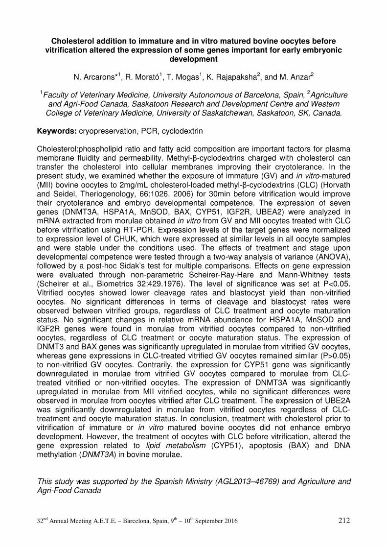
32nd Annual Meeting A.E.T.E. – Barcelona, Spain, 9th – 10th September 2016 212
Cholesterol addition to immature and in vitro matured bovine oocytes before vitrification altered the expression of some genes important for early embryonic
development
N. Arcarons*1, R. Morató1, T. Mogas1, K. Rajapaksha2, and M. Anzar2
1Faculty of Veterinary Medicine, University Autonomous of Barcelona, Spain, 2Agriculture and Agri-Food Canada, Saskatoon Research and Development Centre and Western
College of Veterinary Medicine, University of Saskatchewan, Saskatoon, SK, Canada.
Keywords: cryopreservation, PCR, cyclodextrin Cholesterol:phospholipid ratio and fatty acid composition are important factors for plasma membrane fluidity and permeability. Methyl-β-cyclodextrins charged with cholesterol can transfer the cholesterol into cellular membranes improving their cryotolerance. In the present study, we examined whether the exposure of immature (GV) and in vitro-matured (MII) bovine oocytes to 2mg/mL cholesterol-loaded methyl-β-cyclodextrins (CLC) (Horvath and Seidel, Theriogenology, 66:1026. 2006) for 30min before vitrification would improve their cryotolerance and embryo developmental competence. The expression of seven genes (DNMT3A, HSPA1A, MnSOD, BAX, CYP51, IGF2R, UBEA2) were analyzed in mRNA extracted from morulae obtained in vitro from GV and MII oocytes treated with CLC before vitrification using RT-PCR. Expression levels of the target genes were normalized to expression level of CHUK, which were expressed at similar levels in all oocyte samples and were stable under the conditions used. The effects of treatment and stage upon developmental competence were tested through a two-way analysis of variance (ANOVA), followed by a post-hoc Sidak’s test for multiple comparisons. Effects on gene expression were evaluated through non-parametric Scheirer-Ray-Hare and Mann-Whitney tests (Scheirer et al., Biometrics 32:429.1976). The level of significance was set at P<0.05. Vitrified oocytes showed lower cleavage rates and blastocyst yield than non-vitrified oocytes. No significant differences in terms of cleavage and blastocyst rates were observed between vitrified groups, regardless of CLC treatment and oocyte maturation status. No significant changes in relative mRNA abundance for HSPA1A, MnSOD and IGF2R genes were found in morulae from vitrified oocytes compared to non-vitrified oocytes, regardless of CLC treatment or oocyte maturation status. The expression of DNMT3 and BAX genes was significantly upregulated in morulae from vitrified GV oocytes, whereas gene expressions in CLC-treated vitrified GV oocytes remained similar (P>0.05) to non-vitrified GV oocytes. Contrarily, the expression for CYP51 gene was significantly downregulated in morulae from vitrified GV oocytes compared to morulae from CLC-treated vitrified or non-vitrified oocytes. The expression of DNMT3A was significantly upregulated in morulae from MII vitrified oocytes, while no significant differences were observed in morulae from oocytes vitrified after CLC treatment. The expression of UBE2A was significantly downregulated in morulae from vitrified oocytes regardless of CLC-treatment and oocyte maturation status. In conclusion, treatment with cholesterol prior to vitrification of immature or in vitro matured bovine oocytes did not enhance embryo development. However, the treatment of oocytes with CLC before vitrification, altered the gene expression related to lipid metabolism (CYP51), apoptosis (BAX) and DNA methylation (DNMT3A) in bovine morulae. This study was supported by the Spanish Ministry (AGL2013–46769) and Agriculture and Agri-Food Canada
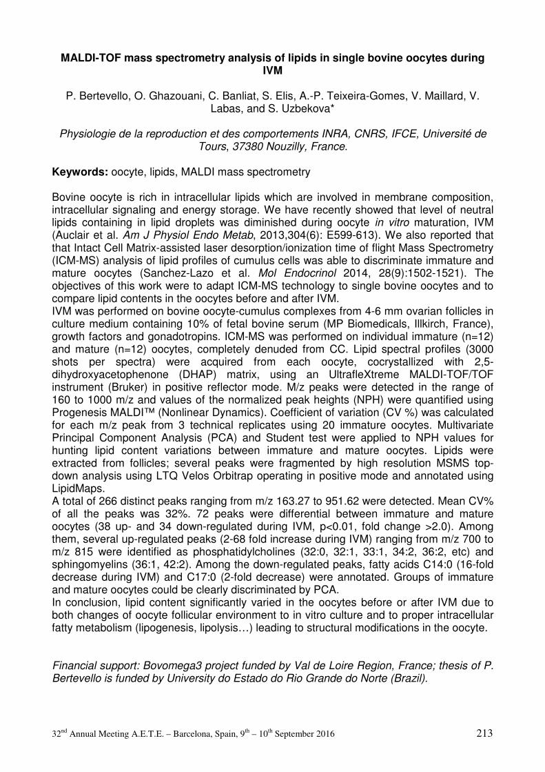
32nd Annual Meeting A.E.T.E. – Barcelona, Spain, 9th – 10th September 2016 213
MALDI-TOF mass spectrometry analysis of lipids in single bovine oocytes during IVM
P. Bertevello, O. Ghazouani, C. Banliat, S. Elis, A.-P. Teixeira-Gomes, V. Maillard, V.
Labas, and S. Uzbekova*
Physiologie de la reproduction et des comportements INRA, CNRS, IFCE, Université de Tours, 37380 Nouzilly, France.
Keywords: oocyte, lipids, MALDI mass spectrometry Bovine oocyte is rich in intracellular lipids which are involved in membrane composition, intracellular signaling and energy storage. We have recently showed that level of neutral lipids containing in lipid droplets was diminished during oocyte in vitro maturation, IVM (Auclair et al. Am J Physiol Endo Metab, 2013,304(6): E599-613). We also reported that that Intact Cell Matrix-assisted laser desorption/ionization time of flight Mass Spectrometry (ICM-MS) analysis of lipid profiles of cumulus cells was able to discriminate immature and mature oocytes (Sanchez-Lazo et al. Mol Endocrinol 2014, 28(9):1502-1521). The objectives of this work were to adapt ICM-MS technology to single bovine oocytes and to compare lipid contents in the oocytes before and after IVM. IVM was performed on bovine oocyte-cumulus complexes from 4-6 mm ovarian follicles in culture medium containing 10% of fetal bovine serum (MP Biomedicals, Illkirch, France), growth factors and gonadotropins. ICM-MS was performed on individual immature (n=12) and mature (n=12) oocytes, completely denuded from CC. Lipid spectral profiles (3000 shots per spectra) were acquired from each oocyte, cocrystallized with 2,5-dihydroxyacetophenone (DHAP) matrix, using an UltrafleXtreme MALDI-TOF/TOF instrument (Bruker) in positive reflector mode. M/z peaks were detected in the range of 160 to 1000 m/z and values of the normalized peak heights (NPH) were quantified using Progenesis MALDI™ (Nonlinear Dynamics). Coefficient of variation (CV %) was calculated for each m/z peak from 3 technical replicates using 20 immature oocytes. Multivariate Principal Component Analysis (PCA) and Student test were applied to NPH values for hunting lipid content variations between immature and mature oocytes. Lipids were extracted from follicles; several peaks were fragmented by high resolution MSMS top-down analysis using LTQ Velos Orbitrap operating in positive mode and annotated using LipidMaps. A total of 266 distinct peaks ranging from m/z 163.27 to 951.62 were detected. Mean CV% of all the peaks was 32%. 72 peaks were differential between immature and mature oocytes (38 up- and 34 down-regulated during IVM, p<0.01, fold change >2.0). Among them, several up-regulated peaks (2-68 fold increase during IVM) ranging from m/z 700 to m/z 815 were identified as phosphatidylcholines (32:0, 32:1, 33:1, 34:2, 36:2, etc) and sphingomyelins (36:1, 42:2). Among the down-regulated peaks, fatty acids C14:0 (16-fold decrease during IVM) and C17:0 (2-fold decrease) were annotated. Groups of immature and mature oocytes could be clearly discriminated by PCA. In conclusion, lipid content significantly varied in the oocytes before or after IVM due to both changes of oocyte follicular environment to in vitro culture and to proper intracellular fatty metabolism (lipogenesis, lipolysis…) leading to structural modifications in the oocyte. Financial support: Bovomega3 project funded by Val de Loire Region, France; thesis of P. Bertevello is funded by University do Estado do Rio Grande do Norte (Brazil).

32nd Annual Meeting A.E.T.E. – Barcelona, Spain, 9th – 10th September 2016 214
Sustainable regulation of metabolic performance of bovine embryos by L-Carnitine supplement and concurrent reduction of fatty acids
E. Held*1,2, S.L. Klein1, F. Rings1,2, D. Tesfaye1, K. Schellander1, and M. Hoelker1,2
1Institute of Animal Science, Animal Breeding and Husbandry Group, University of Bonn, Bonn, Germany, 2Researchstation Frankenforst, Faculty of Agriculture, University of Bonn,
Königswinter, Germany.
Keywords: bovine embryos, L-Carnitine, fatty acids, IVP The increasing importance of in vitro produced (IVP) embryos in commercial cattle breeding programs demands improvement of embryo viability after cryopreservation. Positive effects of L-Carnitine (LC) supplement on lipid accumulation of IVP embryos, which is suggested to play a key role in viability after cryopreservation, has been shown previously. In the present study we aimed to reveal the impact of LC supplementation and the concurrent reduction of fatty acids (FA) during in vitro culture on metabolic features, implicating the morphological as well as the molecular level. In the control group presumptive zygotes were cultured in SOFaa media supplemented with BSA. In the three experimental groups either fatty acids were removed using BSA fatty acid free (FAF) or 2.5 mM L-Carnitine were added (BSA+LC) or removing of fatty acids and supplement of L-Carnitine were combined (FAF+LC). All embryos were cultured in groups of 50 in 400µl medium covered with mineral oil in 5% O2 and 5% CO2.
Concerning developmental rates, no significant differences were observed between the groups, therefore, the impact of either FA reduction or LC supplement could be excluded. In contrast, survival rates after slow-freezing (EG) and thawing of day 7 blastocysts show that LC positively affects re-expansion speed of the embryos. Significantly higher hatching rates were detected for embryos cultured in FAF+LC. Furthermore, lipid accumulation was quantitatively measured, by uptake and elution of Oil Red in day 7 blastocysts using a Microplate reader. Significantly lower amounts of lipid were detected in FAF and BSA+LC, implicating that either removal of FA or supplementation of LC reduce lipid content effectively. To understand molecular mechanisms affected by different culture conditions, a set of genes related to oxidative stress response (KEAP1 and SOD1) and lipid metabolism (AMPK, ACC and PGC1α) were determined using realtime PCR. Regarding KEAP1, playing multiple roles in the cascade of oxidative stress response, and SOD1, known to act as ROS scavenger, we detected significantly higher abundance in embryos cultured with FAF and FAF+LC compared to their counterparts cultured with BSA and BSA+LC. We suggest that not LC but the presence or absence of fatty acids influence oxidative stress response. A similar pattern was observed for AMPK being significantly higher expressed in embryos which were cultured in the absence of fatty acids. In contrast, the expression of ACC, known to play key a role in storage and synthesis of long chain fatty acids, and PGC1α, being responsible for mitochondrial activity, are affected by the presence of FA but also by supplement of LC in combination with removal of FA. In summary, we could unravel, the positive effect of LC and the concurrent removal of FA on cryosurvival, metabolism and gene expression.
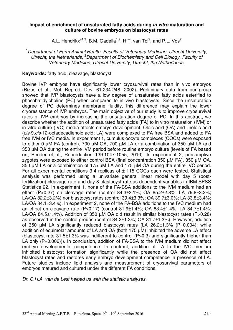
32nd Annual Meeting A.E.T.E. – Barcelona, Spain, 9th – 10th September 2016 215
Impact of enrichment of unsaturated fatty acids during in vitro maturation and culture of bovine embryos on blastocyst rates
A.L. Hendrikx*1,2, B.M. Gadella1,2, H.T. van Tol2, and P.L. Vos2
1Department of Farm Animal Health, Faculty of Veterinary Medicine, Utrecht University, Utrecht, the Netherlands, 2Department of Biochemistry and Cell Biology, Faculty of
Veterinary Medicine, Utrecht University, Utrecht, the Netherlands.
Keywords: fatty acid, cleavage, blastocyst Bovine IVP embryos have significantly lower cryosurvival rates than in vivo embryos (Rizos et al., Mol. Reprod. Dev. 61:234-248, 2002). Preliminary data from our group showed that IVP blastocysts have a low degree of unsaturated fatty acids esterified to phosphatidylcholine (PC) when compared to in vivo blastocysts. Since the unsaturation degree of PC determines membrane fluidity, this difference may explain the lower cryoresistance of IVP embryos. The main objective of our study is to improve cryosurvival rates of IVP embryos by increasing the unsaturation degree of PC. In this abstract, we describe whether the addition of unsaturated fatty acids (FA) to in vitro maturation (IVM) or in vitro culture (IVC) media affects embryo development. Oleic acid (OA) and linoleic acid (cis-9,cis-12-octadecadienoic acid; LA) were complexed to FA free BSA and added to FA free IVM or IVC media. In experiment 1, cumulus oocyte complexes (COCs) were exposed to either 0 µM FA (control), 700 µM OA, 700 µM LA or a combination of 350 µM LA and 350 µM OA during the entire IVM period before routine embryo culture (levels of FA based on; Bender et al., Reproduction 139;1047-1055, 2010). In experiment 2, presumptive zygotes were exposed to either control BSA (final concentration 350 µM FA), 350 µM OA, 350 µM LA or a combination of 175 µM LA and 175 µM OA during the entire IVC period. For all experimental conditions 3-4 replicas of ± 115 COCs each were tested. Statistical analysis was performed using a univariate general linear model with day 5 (post-fertilization) cleavage rate and day 8 blastocyst rate as dependent variables in IBM SPSS Statistics 22. In experiment 1, none of the FA-BSA additions to the IVM medium had an effect (P>0.27) on cleavage rates (control 84.3±3.1%; OA 85.2±2.8%; LA 79.8±3.2%; LA/OA 82.2±3.2%) nor blastocyst rates (control 39.4±3.3%; OA 39.7±3.0%; LA 33.8±3.4%; LA/OA 34.1±3.4%). In experiment 2, none of the FA-BSA additions to the IVC medium had an effect on cleavage rate (P>0.17) (control 81.9±1.4%; OA 83.4±1.4%; LA 84.7±1.4%; LA/OA 84.5±1.4%). Addition of 350 µM OA did result in similar blastocyst rates (P>0.28) as observed in the control groups (control 34.2±1.3%; OA 31.7±1.3%). However, addition of 350 µM LA significantly reduced blastocyst rates (LA 26.2±1.3% (P=0.004); while addition of equimolar amounts of LA and OA (both 175 µM) inhibited the adverse LA effect (blastocyst rate 31.5±1.3% was indifferent to control (P>0.3) and significantly higher than LA only (P=0.006))). In conclusion, addition of FA-BSA to the IVM medium did not affect embryo developmental competence. In contrast, addition of LA to the IVC medium inhibited blastocyst formation significantly while the presence of OA did not affect blastocyst rates and restores early embryo development competence in presence of LA. Future studies include lipid analysis and measurement of cryosurvival parameters of embryos matured and cultured under the different FA conditions. Dr. C.H.A. van de Lest helped us with the statistic analyses.

32nd Annual Meeting A.E.T.E. – Barcelona, Spain, 9th – 10th September 2016 216
Viability of porcine vitrified morulae and blastocysts stored in a dry-shipper for 3 days
A. Nohalez*, C.A. Martinez, I. Parrilla, J. Roca, M.A. Gil, E.A. Martinez, and C. Cuello
Dpt. of Animal Medicine and Surgery, University of Murcia, Murcia, Spain.
Keywords: Vitrification, dry shipper, porcine embryos In the last decade, vitrification has emerged as an efficient tool for porcine embryo cryopreservation. Vitrified samples are highly sensitive to temperature oscillations, because embryos are vitrified in very small volumes that could suffer devitrification. To date, the only efficient way to transport vitrified embryos was the liquid nitrogen (LN) tanks. However, at present, the air movement of devices containing LN is subjected to a strict regulation since LN is considered as hazardous material. Recently, dry vapour shipper (DS) containers have been developed for the safe transport of biological samples at cryogenic temperatures (-150º). These devices guarantee a stable temperature for several days, which may allow the long-distance transport of vitrified embryos. The aims of this study was to assess the efficacy of a DS (ST reproductive technologies LLC, Navasota, USA) to maintain the viability of vitrified porcine embryos for a 3 days storage period compared to the routine storage in LN. For that, donor sows were subjected to a laparotomy six days after estrus. Embryos were collected by flushing the tip of each uterine horn with Tyrode’s lactate (TL)-HEPES-PVA (TL-HEPES) medium. Only embryos at the morula and blastocyst stages showing good or excellent morphology according to the criteria determined by the IETS were used in the experiment. Vitrification and warming were performed with the superfine open pulled straw method using TL-HEPES as basic medium and ethylene-glycol and dimethyl sulfoxide as cryoprotectants. After vitrification, SOPS straws containing the embryos were stored in a LN tank for one month. Then, the straws were transferred from the LN tank to a DS (DS group) or to other LN tank (control group) for additional three days. After warming, DS (N=47) and control (N=46) embryos were cultured for 24 h to assess embryo survival (ratio of viable blastocysts at the end of culture to the total number of embryos cultured) and hatching rates. The results were analysed using Fisher’s exact test. In both groups, the embryos vitrified at the morula stage (N=48) displayed the same survival rates (95.8%) and similar embryo development stage at the end of the culture (34.6% and 65.4% of DS embryos and 43.4% and 56.6% of control embryos were early-full and expanded-hatching blastocysts, respectively). When the embryos were vitrified at the blastocyst stage (N=45), there were also no differences between groups in the survival rates (95.6 vs. 100%) and embryo development (100% of the embryos were at the perihatching or hatching stage at the end of de culture in both groups). The present study shows the efficacy of the DS for the storage of vitrified porcine morulae and blastocysts for a minimum of 3 days, which allows the international air transport of porcine embryos safely. Supported by MINECO-FEDER (AGL201569735-R) and Séneca (19892/GERM/15).
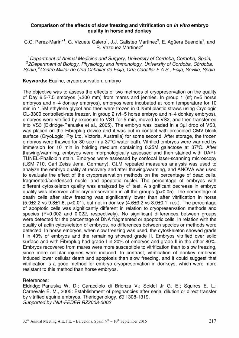
32nd Annual Meeting A.E.T.E. – Barcelona, Spain, 9th – 10th September 2016 217
Comparison of the effects of slow freezing and vitrification on in vitro embryo quality in horse and donkey
C.C. Perez-Marín*1, G. Vizuete Calero1, J.J. Galisteo Martínez3, E. Agüera Buendía2, and
R. Vazquez Martinez2
1Department of Animal Medicine and Surgery, University of Cordoba, Cordoba, Spain, 22Department of Biology, Physiology and Immunology, University of Cordoba, Córdoba,
Spain, 3Centro Militar de Cría Caballar de Ecija, Cría Caballar F.A.S., Ecija, Seville, Spain.
Keywords: Equine, cryopreservation, embryo The objective was to assess the effects of two methods of cryopreservation on the quality of Day 6.5-7.5 embryos (<300 mm) from mares and jennies. In group 1 (sf; n=5 horse embryos and n=4 donkey embryos), embryos were incubated at room temperature for 10 min in 1.5M ethylene glycol and then were frozen in 0.25ml plastic straws using Cryologic CL-3300 controlled-rate freezer. In group 2 (vf=5 horse embryo and n=4 donkey embryos), embryos were vitrified by exposure to VS1 for 5 min, moved to VS2, and then transferred into VS3 (Eldridge-Panuska et al., 2005). The embryo was loaded in a 3µl drop of VS3, was placed on the Fibreplug device and it was put in contact with precooled CMV block surface (CryoLogic, Pty Ltd, Victoria, Australia) for some second. After storage, the frozen embryos were thawed for 30 sec in a 37ºC water bath. Vitrified embryos were warmed by immersion for 10 min in holding medium containing 0.25M galactose at 37ºC. After thawing/warming, embryos were morphologically assessed and then stained with DAPI-TUNEL-Phalloidin stain. Embryos were assessed by confocal laser-scanning microscopy (LSM 710, Carl Zeiss Jena, Germany). GLM repeated measures analysis was used to analyze the embryo quality at recovery and after thawing/warming, and ANOVA was used to evaluate the effect of the cryopreservation methods on the percentage of dead cells, fragmented/condensed nuclei and apoptotic nuclei. The percentage of embryos with different cytoskeleton quality was analyzed by c2 test. A significant decrease in embryo quality was observed after cryopreservation in all the groups (p<0.05). The percentage of death cells after slow freezing was significantly lower than after vitrification in horse (5.0±2.2 vs 9.8±1.6, p<0.01), but not in donkey (4.6±3.2 vs 3.0±0.1; n.s.). The percentage of apoptotic cells was significantly different in relation to cryopreservation methods and species (P=0.002 and 0.022, respectively). No significant differences between groups were detected for the percentage of DNA fragmented or apoptotic cells. In relation with the quality of actin cytoskeleton of embryos, no differences between species or methods were detected. In horse embryos, when slow freezing was used, the cytoskeleton showed grade I in 40% of embryos and the remaining showed grade II. Embryos vitrified over solid surface and with Fibreplug had grade I in 20% of embryos and grade II in the other 80%. Embryos recovered from mares were more susceptible to vitrification than to slow freezing, since more cellular injuries were induced. In contrast, vitrification of donkey embryos induced lower cellular death and apoptosis than slow freezing, and it could suggest that vitrification is a good method for embryo cryopreservation in donkeys, which were more resistant to this method than horse embryos. References: Eldridge-Panuska W. D.; Caracciolo di Brienza V.; Seidel Jr G. E.; Squires E. L.; Carnevale E. M., 2005: Establishment of pregnancies after serial dilution or direct transfer by vitrified equine embryos. Theriogenology, 63 1308-1319. Supported by INIA-FEDER RZ2008-0002
32nd Annual Meeting A.E.T.E. – Barcelona, Spain, 9th – 10th September 2016 218
The influence of DNA stabilizing buffer on the results of genomic bovine embryo analysis
M. Reichenbach*1, S. Jung1, E. Wolf2, E. Pimentel3, R. Emmerling3, K.-U. Götz3, T.
Grupp1, J. Scherzer1, R. Fries4, and H.-D. Reichenbach3
1Bayern-Genetik GmbH, Grub, Germany, 2Chair for Molecular Animal Breeding and Biotechnology, Ludwig-Maximilians-University, Munich, Germany, 3Bavarian State
Research Center for Agriculture, Institute for Animal Breeding, Grub, Germany, 4Chair of Animal Breeding, Technische Universität München, Freising, Germany.
Keywords: embryo biopsy, DNA conservation, TE buffer Nowadays breeding programs involve the biopsy of preimplantation bovine embryos to determine sex (S), polled status (PS), hereditary defects (HD) and early genetic selection. Those techniques are more cost effective the higher the success rate of analyzed samples. To minimize the loss of information due to DNA degradation the obtained cells must be successfully stored in tubes prior to whole genome amplification. The objective of this study was to analyze the use of a DNA stabilizing buffer on the successful outcome of S, PS, HD determination and on the call rate after chip SNP analysis. Embryos were obtained on day 7 after insemination by superovulation of German Simmental animals with a standard protocol. Immediately after recovery, embryos were biopsied by a single operator under a mobile stereo microscope (Olympus, Japan) at 50x magnification and a steel blade mounted on a blade holder (Bausch & Lomb, Germany) attached to a micromanipulator (Eppendorf, Germany). Two different biopsy media (BM) were used, (1: PBS from IMV, France or 2: 0.9% NaCl solution with 1.5% PVP). Removed cells were brought to reaction tubes by pipetting them with 0.5 µl medium in either the empty tube or in 2.5 µl TE buffer (TE buffer, 10 mM Tris, 1 mM EDTA, pH 8.0). Biopsied cells, approximately 10, were immediately used after biopsy for whole genome amplification (Repli-g Mini Kit, Qiagen, Germany) followed by PCR analysis of S and PS. HD were analyzed using a 5’-exonuclease assay. Embryos were genotyped with the Illumina Bovine 54k BeadChip. Call rates were recorded. Differences in success (%) and call rates were analyzed by proc GLM, SAS (fixed factors: BM (1+2), TE (yes/no), developmental stage of embryo, morulae (4), early blastocyst (5), blastocyst (6), and expanded blastocyst (7)). In total 503 embryos were analyzed for S, PS and HD. From the total number of embryos 101 Embryos were SNP analyzed and call rates obtained. The BM had no influence on the success rate of positive analyzed S (90.7% vs. 86.0%), PS (89.4% vs. 86.9%) or HD (90.0% vs. 82.8%), p>0.10. The stage of embryo had also no influence on the success rate of S (4=90.1%, 5=85.7%, 6=87.9%, 7=85.0%, p>0.10), PS (4=89.8%, 5=85.2%, 6=86.5, 7=95.0%; p>0.10), HD (4=89.8%, 5=85.2%, 6=86.5%, 7=86.7%, p>0.10) analysis and call rate (4=0.8939, 5=0.8893, 6=0.9211, 7=0.9224, p>0.10). Highly significant differences showed the use of TE buffer vs. no TE buffer for call rate (0.9137 vs. 0.8396, p<0.0001), S (91.4% vs. 78.0%, p<0.0001) and PS (90.9% vs. 79.7%, p<0.0001) and HD (89.3% vs. 74.3%, p<0.0001). The biopsy medium and the stage of embryo had no effect on the success rate for the analysis of S, PS and HD. The use of a DNA stabilizing TE buffer improved results by 10-15%. Therefore, we advise the use of TE buffer for storing small embryo cell samples to optimize analysis. Funded by the Bavarian Research Foundation (AZ-1031-12).

32nd Annual Meeting A.E.T.E. – Barcelona, Spain, 9th – 10th September 2016 219
JC-1 dye is a valuable indicator of embryo health in rabbits
M. Teixeira*, L. Commin, L. Gavin-Plagne, P. Bruyere, A. Philibert, T. Joly, and S. Buff
UPSP 2011-03-101 Biological Resources Center –CRB-ANIM Infrastructure, Université de Lyon (Vetagro sup), Marcy L Etoile, Lyon, France.
Keywords: Mitochondrial metabolism, Embryo cryopreservation One of the main challenges in embryo biotechnology research is to develop in vitro evaluation methods of embryo quality to detect early alterations correlated with embryo development competence. JC-1 (5,5′,6,6′-tetrachloro-1,1′,3,3′-tetraethylbenzimidazolylcarbocyanine iodide) is a lipophilic cationic dye, which accumulates within mitochondria according to its mitochondrial membrane potential (MMP) emitting different fluorescent properties. High MMP mitochondria accumulate more cationic dye (J aggregates) and exhibits red fluorescence, while low MMP mitochondria accumulate J monomers, showing green fluorescence. Disruption of MMP has been associated with metabolic stress and early cellular apoptosis. Our aim was to adapt a JC-1 staining method to fresh and cryopreserved rabbit embryos in order to evaluate their quality. To this end, embryos (n = 64) were collected at the morula stage, and were randomly divided into two groups: fresh (n = 39) and slow frozen (n = 25; DMSO, 1.5 M) embryos. All embryos were analyzed at the blastocyst stage, according to morphological quality and classified as normal (inner cell mass and trophectoderm quality considered as good or fair) or damaged embryos (delayed, fragmented (> 20%), poor quality inner cell mass and trophectoderm, and smaller blastocysts were considered as damaged). Embryos were pretreated with pronase (Roche, Meylan, France), stained with JC-1 (1.5 µM; Invitrogen, Life Technologies, Eugen, Oregon, USA) for 75 min (38.5°C, 5 % CO2) and observed under an epifluorescence microscope. CCCP (Sigma-Aldrich, Saint Quentin Fallavier, France), a MMP disruptor, was used as a control to confirm the JC-1 sensitivity to changes in MMP. The staining intensity (pixel) was determined in two randomly defined areas on each embryo. In order to dismiss the potential effects of the cryopreservation process on stain uptake, only the red/green ratio was analyzed using ImageJ software. Significant differences were found between fresh (R = 3.55 ± 0.94) and normal cryopreserved embryos (R = 2.55 ± 0.78; p<0.01), as well as between normal and damaged (R = 1.03 ± 0.50; p<0.05) cryopreserved embryos. We conclude embryo’ morphological defects are associated with MMP disruption, and cryopreservation seems to impair the mitochondrial metabolism even in absence of identifiable alterations of the embryo. This study is the first to describe a JC-1 staining protocol for rabbit embryo evaluation, showing it can be used as a valuable indicator of embryo’ health and functionality for this species.
32nd Annual Meeting A.E.T.E. – Barcelona, Spain, 9th – 10th September 2016 220
Rabbit embryo vitrification without animal products
M. Teixeira*, L. Commin, L. Gavin-Plagne, P. Bruyere, A. Philibert, S. Buff, and T. Joly
UPSP 2011-03-101 Biological Resources Center –CRB-ANIM Infrastructure, Université de Lyon (Vetagro sup), Marcy L Etoile, Lyon, France.
Keywords: Animal derived products, Cryopreservation methods, Bovine serum albumin Embryo cryopreservation often requires the use of animal derived products (such as bovine serum albumin (BSA) and fetal calf serum (FCS) ), which represent a sanitary risk, and contain undefined material that can considerably vary between batches, altering its thermodynamic properties. CRYO3 (Stem-Alpha, Saint-Genis-l'Argentière France) is a chemically defined substitute without animal products, created for mononuclear cell cryopreservation. Recently, our research team successfully slow freezed rabbit embryos with a buffer medium composed of D-PBS (Dutscher, Brumath, France) supplemented with 20% of CRYO3 and 1.5M DMSO (Bruyère P et al. Plos One 8(8): e71547, 2013). Our objective was to compare three rabbit embryo vitrification buffer media: IMV holding medium (IMV, L’ Aigle, France): a commercial medium containing BSA (G1); D-PBS supplemented with 20% of CRYO3 (G2); and a CRYO3 medium (G3). All the media contained the same cryoprotectant composition. Rabbit New Zealand does (n = 12) were submitted to a superovulation treatment and collected embryos (n = 231) were randomly divided into three groups. After equilibration, embryos were exposed for 30 sec to a vitrification solution of G1 / G2 / G3 medium, containing 20 % Me2SO and 20 % EG, before being loaded to the hook at the end of a custom designed fibre called a Fibreplug™ (CVM Kit, Cryologic, Victoria, Australia) and vitrified by solid surface vitrification. Thawing was performed by immersing the end of the Fibreplug™ directly into a thawing solution (0.5 M sucrose, respectively), for 5 min, followed by three successive dilution baths. Embryos were cultured to the hatching stage in M199 medium (G1: 52, G2: 37 and G3: 71 embryos), supplemented with 10 % FCS (38.5°C, 5 % CO2). The survival rate after thawing (embryos with < 20 % or no fragmentation, per vitrified embryos), the blastocyst formation rate, and the hatching rate (per survived), were evaluated, and analyzed with Chi-square test; Delayed, fragmented ( > 20%) and smaller blastocysts were considered as damaged. The survival rate (G1: 87 %, G2: 81 % and G3: 96 % )) was significantly superior in the CRYO3 group (p < 0.05). No significant difference was observed regarding the blastocyst formation rate at 48 h (G1: 77 %, G2: 65 % and G3: 66 %) or the hatching rate (G1: 35 %, G2: 19 % and G3: 24 %). In conclusion, CRYO3 can be used as a chemically defined substitute for animal-based products in rabbit embryo vitrification solutions, reducing the solutions’ variability and the sanitary risk inherent to animal derived products.

32nd Annual Meeting A.E.T.E. – Barcelona, Spain, 9th – 10th September 2016 221
Ultrasound Monitoring of reproductive organs in Angora bucks
K. Tekin1, C. Stelletta2, D. Sahin1, I. Bastan1, O.O. Parildar1, and A. Daskin*1
1Department of Reproduction and Artificial Insemination, University of Ankara,, Ankara, Turkey, 2Department of Animal Medicine Production and Health University of Padova,,
Padova, Italy.
Keywords: Accessory glands, Doppler ultrasound, transrectal massage Objectives: The current study was design to monitor and create an evaluation method of reproductive organs (testis, epididymis and accessory glands) in clinically healthy Angora buck through ultrasound (US). Material and Methods: Sixteen clinically healthy adult Angora bucks (ranging 1-4 year of age) were used. Classical breeding soundness examination was performed for each buck. Body condition score (BCS), testicular dimensions (Length, width and thickness) and scrotal circumference were measured with caliper, and as well as testis, epididymis and spermatic cord ultrasound (US) monitoring were assessed. Monitoring were performed longitudinal and transversal sections to evaluate the echo texture of the testicular parenchyma (Echo-TTP) including the mediastinum, tail epididymis (Echo-TEP) and colour doppler of vascular cone (EchoDop-VC) of each animal. The US of accessory glands was done in standing position using endocavitary linear probe (MyLabVetTM One, ESAOTE S.p.A., Genova, 10 MhZ probe frequency). Vesicular gland (Echo-VG) and bulbourethral gland (Echo-BG) were monitored with the landmark of urinary bladder. Testicular parenchyma was scored (0-3) according to echogenicity of the testis tissue. The tail of epididymis was evaluated for US appearance in relation with testicular parenchyma. Vascular cone monitoring was performed according to blood flow rate with using colour doppler US. First and foremost, sperm collection was carried out with transrectal ampullar massage. Spermatological parameters of volume, colour, viscosity, mass activity, motility and concentration were evaluated. Results: High correlations were found among sperm volume and EchoDop-VC (R2=0,6), sperm concentration with Echo-VG (R2=0,9) and Echo-BG (R2=0,8). Sperm motility and viscosity were also highly correlated according to Echo-VG (R2=0,7). Echogenicity was increased progressively with age and with low body condition score presenting the testicular parenchyma granular none-homogeneous echogenic pattern. The echogenicity of the epididymis was although homogeneous and visual isoechoic with testis parenchyma. Conclusion: Doppler US and echo-texture of external reproductive organs and accessory glands gives us valuable information on sperm production and related parameters. In addition, according to these results early detection of reproductive pathological conditions can be optimized with Doppler US in Angora goat. Project was supported by the Scientific and Technological Research Council of Turkey (TUBITAK) and Ankara University (Project No:213O034).

32nd Annual Meeting A.E.T.E. – Barcelona, Spain, 9th – 10th September 2016 222
Notes ________________________________________________________________________

32nd Annual Meeting A.E.T.E. – Barcelona, Spain, 9th – 10th September 2016 223
AUTHORS INDEX

32nd Annual Meeting A.E.T.E. – Barcelona, Spain, 9th – 10th September 2016 224
32nd Annual Meeting A.E.T.E. – Barcelona, Spain, 9th – 10th September 2016 225
AUTHORS INDEX
A Aardema, H. ................................................ 161 Abozar, N. ................................................... 149 Abraham MC............................................... 101 Acuña, O.S. ................................................. 162 Agüera Buendía, E. ..................................... 217 Alcântara-Neto , A. ..................................... 163 Aldag, P ....................................................... 131 Alessandroni, A. .......................................... 169 Algarra, B. ............................................ 176,183 Allai, L. ....................................................... 148 Almiñana, C. ............................................... 163 Amiridis, G.S. ...................................... 123,145 Anifandis, G. ........................................ 123,145 Antosik, P. ..................................... 138,139,179 Anzar, M. .................................................... 212 Arcarons, N. ........................................ 158,212 Archilla, C. .................................................. 198 Arias-Alvarez, M. ................................ 164,166 Arreseigor, A.E. ................................... 120,121 Arreseigor, C.J. .................................... 120,121 Astiz, S. ....................................................... 134 Avelino, K. ........................................... 120,121 Avilés, M. ...................................... 162,183,201 B Badi, A. ....................................................... 148 Båge, R. ....................................................... 116 Bagés, S. ...................................................... 175 Bagirov, V.A. .............................................. 150 Banliat, C. .................................................. 213 Baruselli, P.S. ................................................ 29 Bastan, I. .................................................... 221 Bauersachs, S. ............................................. 162 Becerril, L. .................................................. 157 Becker, F. .................................................... 141
Ben Moula, A. ............................................. 148 Bermejo-Álvarez, P. ..................... 140,184,201 Bertevello, P................................................ 213 Blad-Stahl, N. ............................................. 165 Bols, P.E.J. ............................. 170,172,178,196 Bonte, T. .................................................... 124 Bottrel, M. ............................................ 122,125 Brandão, F.Z. .............................................. 127 Brouwers, J.F. ............................................. 161 Bruska, M. .................................... 138,139,179 Brüssow, K.P. ............................... 138,139,179 Bruyere, P. .......................................... 219,220 Bryja, A. ........................................ 138,139,179 Buff, S. .......................................... 124,219,220 Bukowska, D. ................................ 138,139,179 C Cajas, Y.N. .................................................. 166 Callesen, H. .............................................. 7,182 Canha, A. .................................................... 176 Canon, E...................................................... 198 Cañón-Beltrán, K. ...................................... 166 Carrera, R. ................................................... 166 Català, M.G. .................................. 142,167,169 Caudle, J...................................................... 185 Cedano-Castro, J.I....................................... 195 Chacón De Castro, A. ................................. 146 Charlot-Valdieu, A...................................... 124 Chaves, D.S................................................. 127 Chen, Shuai ................................................. 168 Chouzouris, T.M. ........................................ 123 Chrenek, P. .................................................. 190 Ciesiolka, S. .................................. 138,139,179 Combes, L. .................................................. 188 Combes-Soia, L. ......................................... 163 Commin, L. .................................. 124,219,220 Contreras, I.................................................. 167 Contreras-Solís, I. ....................................... 169
32nd Annual Meeting A.E.T.E. – Barcelona, Spain, 9th – 10th September 2016 226
Corbin, E. .................................................... 198 Cornelissen, M. ........................................... 135 Cots, P. ................................................. 154,155 Crotti, G. ..................................................... 132 Cuello, C. ............................................. 133,216 D Dafopoulos, K ..................................... 123,145 Daghigh Kia, H. .......................................... 147 Danch, S.S. .................................................. 150 Daponte, A .................................................. 145 Daskin, A. ................................................... 221 De Bie, J. ..................................................... 170 Decherf, A. .................................................. 117 Demirezer, H. ....................................... 171,173 Desmet, K.L.J.............................................. 172 Detterer, J. ................................................... 126 Diego, R. ..................................................... 152 Dode, M.A.N. ................................................ 29 Dorado, J. ............................................. 122,125 Dovolou, E. .......................................... 123,145 Drevillon, P. ................................................ 117 Druart, X. .................................................... 188 Duittoz, A.H. ................................................ 51 Duranthon, V. ....................................... 191,198 E Egberts, J. .................................................... 126 EL Amiri, B................................................. 148 El Khalil, K. ................................................ 148 Elcoso Usieto, G. ........................................ 128 Elis, S. ................................................. 191,213 Emmerling, R. ............................................. 218 Emsen, E. ............................................. 171,173 Esteves, S.N. ............................................... 127 F Fair, T. ........................................................... 85 Fargas, O. .................................................... 134 Fatemeh, Z.G............................................... 149 Favetta, L. ................................................... 185 Fernandez, L......................................... 122,125 Fernandez, M. ............................................. 134 Ferraz, M.A.M.M ........................................ 174 Ferre-Dolcet, L. ........................................... 158 Fievez, V. .................................................... 178
Figueiredo, A.C.S. ........................................ 29 Filipiak, Y. .......................................... 120,121 Flapper, H. .................................................. 135 Foldager, L. ................................................. 182 Fonseca Balvís, N. ...................................... 201 Fonseca, J.F................................................. 127 Fries, R. ....................................................... 218 G Gadea, J. ...................................................... 151 Gadella, B.M. ................................ 161,174,215 Gajda, B. ................................ 136,192,202,208 Gajda, L. .................................................... 136 Galisteo Martínez, J.J.................................. 217 Galli, C. ....................................................... 132 Gamarra Lazo, G.................................. 128,129 Gaplaev, M.S. ............................................. 156 García-Dominguez, X. ................................ 195 Garcia-Garcia, R.M.............................. 164,166 García-Vázquez, F.A. .......................... 157,159 Gavin-Plagne, L. ................................. 219,220 Gavriil, L ..................................................... 145 Gergatz, E. .................................................. 148 Ghanem, N. .......................................... 186,187 Ghazouani, O. ............................................ 213 Gil, M.A. .............................................. 133,216 Gómez, A. ................................................... 151 Gómez-Fidalgo, E. ...................................... 146 González-Martín, R. .................................. 203 Gorgogetas, V. ............................................ 145 Götz, K.-U................................................... 218 Greve, T. ......................................................... 3 Grothmann, H. ............................................ 194 Grupp, T. ..................................................... 218 Guedes-Teixeira, M. ................................... 124 Guimarães, J.D. ........................................... 127 Gutiérrez-Adán, A. . 146,164,175,183,184,201 Gyökér, E. ................................................... 148 H Hadeler, K.-G. ............................................. 131 Hamama, T.-M. ........................................... 186 Hamdi , J. .................................................... 175 Hamze, J.G. ................................................. 176 Hanzalova, K. ............................................. 177 Held, E. ................................................ 130,214 Hendrikx, A.L. ............................................ 215 Henning, H.H.W. ........................................ 174

32nd Annual Meeting A.E.T.E. – Barcelona, Spain, 9th – 10th September 2016 227
Hidalgo, C.O. .............................................. 153 Hidalgo, M. .......................................... 122,125 Hoelker, M. .......................................... 130,214 Hossein, D.K. .............................................. 149 Hulinska, P. ................................................. 177 I Ibáñez, E. ...................................... 203,209,210 Ibarreche, A.B. ..................................... 120,121 Iolchiev, B.S. ............................................... 150 Izquierdo, D. ................................. 142,167,169 J Jeseta, M...................................................... 177 Jiménez-Movilla, M. ..................... 162,176,183 Johannisson, A. ........................................... 118 John, E.J.St. ................................................. 185 Joly, T. .......................................... 124,219,220 Jordaens, L. ................................................. 178 Jouneau, L. .................................................. 198 Jung, S. ........................................................ 218 Jura, J. ......................................................... 202 K Kachpani, E ................................................. 145 Kamaledini, R. ............................................ 187 Kassens, A. .................................................. 130 Kempisty, B. ................................. 138,139,179 Khadem, A.A. ............................................. 197 Khajenabi, M. ....................................... 187,197 King, W.A. .................................................. 185 Klein, S. ...................................................... 131 Klein, S.L. .................................................. 214 Klenovitskiy, P.M. ...................................... 150 Knitlova, D. ................................................ 177 Kordowitzki, P. ........................................... 131 Kotarski, F. .................................................. 165 Kouhkan, F. ................................................. 197 Kountouris, F. ............................................. 151 Kranc, W. ...................................... 138,139,179 Krania, F...................................................... 123 Królikowski, W. .......................................... 208 Kubovicova, E ............................................. 190 Kutluca Korkmaz, M................................... 173 Kuzmina, T.I. ....................................... 156,180
L Labas, V. ....................................... 163,188,213 Lacaze, S. ............................................. 128,129 Laffont, L. ................................................... 198 Lamas-Toranzo, I. ................................ 140,184 Lamy, J........................................................ 188 Langhammer, M.......................................... 168 Lazzari, G.................................................... 132 Le Bourhis, D. .............................................. 51 Lebedeva, I.Y. ...................................... 181,193 Leite, C.R. ................................................... 127 Leroy, J.L.M.R. ...................... 170,172,178,196 Liu, Y. ......................................................... 182 Lonergan, P. ................................................ 175 López-Béjar, M. ................................... 118,158 Lopez-Helguera, I. ...................................... 134 Lopez-Tello, J. ............................................ 164 López-Úbeda, R. ......................................... 152 Lopukhov, A.B. .......................................... 150 Lopukhov, A.V. .......................................... 193 Lorenzo, P.L. ................................ 164,166,201 Lucas-Hahn, A. ........................................... 131 Lundeheim, N. ............................................ 118 M Machado, R. ................................................ 127 Machatkova, M. .......................................... 177 Mahboubi, K. .............................................. 185 Maillard, V. ................................................. 213 Maillo, V. .................................................... 183 Makarevich, A. ........................................... 190 Mamouris, Z................................................ 145 Maraia, A.C.......................................... 120,121 Marco-Jiménez, F. ...................................... 195 Marei, W.F. ................................... 170,172,196 Mariani, E, .................................................. 117 Maria-Teresa, P. ......................................... 169 Marquant Le Guienne, B. ........................... 129 Martin, J. ..................................................... 117 Martinez Bello, D. ...................................... 128 Martinez, C.A. ..................................... 133,216 Martinez, D. ................................................ 134 Martinez, E.A. ...................................... 133,216 Martínez-Corona, D.A. ........................ 140,184 Massafret, O. .............................................. 210 Matás, C. ....................................... 152,157,159 Mayorga Munoz, I.M. ................................. 189
32nd Annual Meeting A.E.T.E. – Barcelona, Spain, 9th – 10th September 2016 228
Meinecke-Tillmann, S. ................................ 126 Merckx, E. ................................................... 172 Mermillod, P. ................................ 163,188,198 Messini, C.I. ................................................ 145 Mielczarek, E. ..................................... 205,207 Miguel, M.C.V. ........................................... 127 Miguel-Jiménez, S. ..................................... 153 Mikkola, M.................................................... 19 Miro, J. ........................................................ 158 Mogas, T. ............................... 153,158,185,212 Mohammadi-Sangcheshmeh, A. .......... 187,197 Molchanov, A.V. ......................................... 180 Monteiro, Jr. P.L.J. ........................................ 29 Morató, R. .................................... 153,158,212 Mörk, M. ..................................................... 116 Morrell, J. ............................................. 101,118 Mouneyres, M. ............................................ 129 Muiño, R. .................................................... 153 Mulder, H.A. ............................................... 135 Mullaart, E. ................................................. 135 Mutikainen, M. ............................................ 186 N Najafi, A. ..................................................... 147 Niemann, H. ................................................ 131 Nieto, K. ...................................................... 164 Nikdel, K. .................................................... 187 Nogues, P. ................................................... 188 Nohalez, A. .......................................... 133,216 Nowicki, M. ................................. 138,139,179 O Ocak, S. ....................................................... 189 Olexikova, L................................................ 190 Olivares, M.C. ............................................. 176 Opiela, J. ............................................. 204,206 Ortega-Molina, I.......................................... 146 Ortiz, I. ................................................. 122,125 Oseikria, M.................................................. 191 Osuagwuh, U.I. .................................... 154,155 P Palomo, M.J. ........................................ 154,155 Pappas, I. .................................................... 123 Paramio, M.T. ............................... 142,167,203 Paredes, J. ............................................. 120,121
Parella, T. .................................................... 142 Parildar, O.O. ............................................. 221 Paris-Oller, E. ............................................. 159 Park, A. ....................................................... 126 Parrilla, I. ............................................. 133,216 Patron, R. .................................................... 134 Pedersen, H.S. ............................................. 182 Peippo, J. ..................................................... 186 Peña, A.I. .................................................... 153 Pereira, A.R.......................................... 120,121 Pereira, M.H.C. ............................................. 29 Pereira, V.S.A. ............................................ 127 Pérez, M. ..................................................... 142 Pérez-Cerezales S. ...................................... 146 Perez-Marín, C.C. ....................................... 217 Pesantez, J.L. ............................................. 134 Philibert, A. ......................................... 219,220 Pimentel, E. ................................................. 218 Pintelon, I. ................................................... 178 Pivko, J........................................................ 190 Poniedzialek-Kempny, K. .......................... 136 Pontes, G.C.S. ............................................... 29 Pontes, J.H. ................................................... 29 Prata, A.B. ..................................................... 29 Psarra, A.M. ............................................... 145 Ptak, G. ....................................................... 199 R Rajapaksha, K. ............................................ 212 Rajska, I. .............................................. 136,192 Razavian, S. ................................................ 147 Rebollar, P.G................................. 164,166,201 Reichenbach, H.-D. ..................................... 218 Reichenbach, M. ......................................... 218 Rho, N. ........................................................ 185 Rings, F. ............................................... 130,214 Rizos, D. .............................................. 175,183 Roca, J. ................................................. 133,216 Rodriguez-Gil, J.-E. ............................. 153,158 Roelen, B.A.J. ............................................. 161 Roettgen, V. ......................................... 141,143 Romar, R. .................................................... 176 Romek, M. .................................................. 192 Rosset, E. .................................................... 124 Roura, M. ...................................... 142,167,169 Ruffini, S. .................................................... 198

32nd Annual Meeting A.E.T.E. – Barcelona, Spain, 9th – 10th September 2016 229
S Sabés-Alsina, M. ........................... 101,118,158 Sabin Vilar , J. ............................................ 128 Sabin, J. ....................................................... 134 Sahin, D. ...................................................... 221 Saint-Dizier, M. .......................................... 188 Samiec, M. .............. 199,204,205,206,207,208 Sanches, B.V. ................................................ 29 Sánchez-Calabuig, J.M. .............................. 140 Sánchez-Sánchez, R. ................................... 146 Santaló, J. ...................................... 203,209,210 Santamaría, R. ............................................. 140 Sartori, R. ...................................................... 29 Schellander, K. ..................................... 130,214 Scherzer, J. .................................................. 218 Schibler, L. .................................................... 51 Schoen, J. .................................................... 168 Schoen, P.C. ................................................ 141 Sebastian, F. ................................................ 134 Serikov, A.Y. .............................................. 156 Shedova, E.N........................................ 150,181 Sieme, H. ..................................................... 130 Singina, G.N. ................................. 150,181,193 Sjunnesson Y............................................... 101 Skrzyszowska, M. .... 199,204,205,206,207,208 Slomski, R. .................................................. 202 Smorag, Z. ..................................... 192,202,208 Soleimani, M. .............................................. 197 Sørensen, M.T. ............................................ 182 Soriano-Úbeda, C. ....................................... 157 Soto, S. ................................................. 142,169 Souza-Fabjan, J.M.G. .................................. 127 Spanov, A.A. ............................................... 156 Stanislavovich, T.I. ..................................... 180 Stelletta, C. .................................................. 221 Stöhr, J. ....................................................... 194 Stout, T.A.E. ............................................... 174 Szenci, O. .................................................... 148 T Tamargo, C.................................................. 153 Taradajnic, T.E. .................................... 181,193 Tatarskaya, D.N. ......................................... 180 Tavakoli, R. ................................................. 197 Teixeira, M. .......................................... 219,220 Teixeira-Gomes, A.-P. ................................ 213 Tekin, K. ..................................................... 221 Terletskiy, V.P. ........................................... 156
Tesfaye, D. ........................................... 130,214 Thys, S. ....................................................... 178 Tillet, Y. ....................................................... 51 Töpfer, D. .................................................... 126 Torrecillas, A. ............................................. 162 Tsikis, G. .............................................. 163,188 Turgumbekov, A.A. .................................... 156 Turini, P. ..................................................... 132 U Ussenbekov, Y.S. ........................................ 156 Uzbekova, S. ....................................... 191,213 V Valdes-Hernández, J. .................................. 195 van der Linde, R.......................................... 135 Van Hoeck, V. ..................................... 178,196 Van Tol, H.T.A. ................................... 161,215 Vasconcelos, J.L.M. ...................................... 29 Vaseghi Dodran, H. ............................. 147,149 Vazquez Martinez, R. ................................ 217 Vendrell, M. ................................................ 158 Vernunft, A. ................................................ 143 Veshkini, A. ................................................ 197 Viana, .J.H.M................................................. 29 Vicente, J.S. ................................................ 195 Viergutz, T. ................................................. 143 Vila-Cejudo, M. .................................. 209,210 Vitorino Carvalho, A. .......................... 191,198 Vizuete Calero, G. ...................................... 217 Vlaeminck, B. ............................................. 178 Volget, M. .................................................. 132 Vos, P.L.A.M. ...................................... 174,215 W Weitzel, J.M. ............................................... 143 Weldenegodguad, M. .................................. 186 Wojtylak-Jurkiewicz, E. .............. 199,205,207 Wolf, E. ....................................................... 218 Wrenzycki, C. ..................................... 165,194 Z Zabel, M. ....................................... 138,139,179 Zambrini, F.N. ............................................ 127

32nd Annual Meeting A.E.T.E. – Barcelona, Spain, 9th – 10th September 2016 230
Zamorano, L. .............................................. 176 Zapata, H. .................................................... 159 Zarza, E. ............................................... 122,125 Zhilinskiy, M.A. .......................................... 150 Zinovieva, N.A. .................................... 181,193

32nd Annual Meeting A.E.T.E. – Barcelona, Spain, 9th – 10th September 2016