PROGRAM BOOK AND ABSTRACTS - SOSORT
196
PROGRAM BOOK AND ABSTRACTS 14 TH INTERNATIONAL SOSORT MEETING 2019 APRIL 25-27th, 2019 SAN FRANCISCO | Paper & Poster Section | SOSORT 2019
-
Upload
khangminh22 -
Category
Documents
-
view
3 -
download
0
Transcript of PROGRAM BOOK AND ABSTRACTS - SOSORT

PROGRAM BOOK AND ABSTRACTS
14TH INTERNATIONALSOSORT MEETING 2019
A P R I L 2 5 - 2 7 t h , 2 0 1 9
SAN FRANCISCO
| Paper & Poster Section |
SOSORT
2019


PROGRAM BOOK AND ABSTRACTS
A P R I L 2 5 - 2 7 t h , 2 0 1 9SAN FRANCISCO
| Paper & Poster Section |
SOSORT
2019

4 | 14TH INTERNATIONAL SOSORT MEETING
2019 SOSORT | Sponsors
Maria van Vuuren
Gold Partner
Platinum Partner
Silver Partner
Bronze Partner
Special Thanks to
Nancy Sherratt, Scoliosis Rehab, Inc. Elizabeth Janssen Chintan Pancholi

SAN FRANCISCO, APRIL 25-27th, 2019 | 5
San Francisco is known for its scenic beauty, cultural attractions, diverse communities, and world-class cuisine. Measuring 49 square miles,
this walk-able city includes landmarks like the Gold-en Gate Bridge, cable cars, Alcatraz and the largest Chinatown in the United States. A stroll of the city’s streets can lead from Union Square to North Beach to Fisherman’s Wharf, Japantown and the Mission District, with intriguing neighborhoods to explore at every turn.Views of the Pacific Ocean and San Francisco Bay are often laced with fog, creating a romantic mood in this most European of American cities. The city has a col-orful past, growing from a small village to a major city nearly overnight as a result of the 1849 Gold Rush. The writers of the “beat” generation, the hippies of the Summer of Love in the late 1960’s and the large gay/lesbian population have all contributed to making San Francisco the fascinating place it is today. The city is home to world-class theatre, opera, sym-phony and ballet companies and often boasts pre-mieres of Broadway-bound plays and culture-chang-
ing performing arts. San Francisco is one of America’s greatest dining cities. The diverse cultural influences, proximity of the freshest ingredients and competitive creativity of the chefs result in unforgettable dining experiences throughout the city.
San Francisco has well over 32,000 hotel rooms, from first-class hotels and ultra-chic boutique hotels to fa-miliar names in lodging and budget friendly inns. San Francisco International Airport (SFO) offers non-stop flights to more than 46 international cities on 39 inter-national carriers. The Bay Area’s largest airport con-nects non-stop with 79 cities in the U.S. on 13 domes-tic airlines.
San Francisco is known for its scenic beauty, cultural attractions, diverse communities, and world-class cuisine
San Francisco

6 | 14TH INTERNATIONAL SOSORT MEETING
Dear Colleagues, Friends, Guests, and members of SOSORT,
The time has come! It’s my pleasure to welcome you to SOSORT’s annual meeting. And for those visiting California for the very first time – Welcome to San Fran!
For the past three years, the scientific, communication and education committees of SOSORT have been hard at work to make this conference happen. Together, we’ve crea-ted a program that will ignite the passion of everyone looking to advance their knowled-ge of non-operative treatment of scoliosis and spinal deformities.
And of course, what better place to learn and share our expertise then San Francisco! The culture, architecture, and innovation happening here is truly unprecedented.
Research, ideas, and the best minds from around the world are here in one place, with you, and for you.
I’m excited to meet you and to share your passion for patient care in our great professi-on. Welcome to SOSORT!
Grant Wood, MS, CPO (UK), CO (US)Organizing Committee Chair
2019 SOSORT | Welcome

SAN FRANCISCO, APRIL 25-27th, 2019 | 7
President’s MessageWelcome to San Francisco SOSORT 2019! We are delighted you ha-ve joined us, and hope you are looking forward to several wonderful days with friends and colleagues focused on the science and practice of non-operative treatment of scoliosis and spinal disorders.
Since its inception, SOSORT has published several important consensus papers and guidelines for treatment. We are committed to the team concept which encourages collaboration between physicians, orthotists, therapists, scientists, researchers and other health care providers. This “team spirit” is also evident in all the behind the sce-nes planning and preparations necessary to make this annual meeting successful. The-re are many volunteers who have invested countless hours on our behalf. I especially want to acknowledge and thank our local host, Grant Wood, CPO, his capable staff and members of the Organizing Committee; Sanja Schreiber,Ph.D and Hagit Berdishevsky, DPT, co-chairs of the Education Committee; Lori Dolan, Ph.D., chair of the Scientific Committee; Patrick Knott, Ph.D., advisor and presider; Sabrina Donzelli, MD, chair of the Membership Committee; and all the members of these committees and the Bo-ard of Directors.
We are raising the bar! From our first official meeting in Poznan, Poland in 2006, the quality of research reflected in conference presentations continues to reach new levels. The number and diversity of international participants is increasing every year as well as our membership. We have truly matured into an established, well-respected global society. On the scoliosis continuum of care, we are fulfilling our mission of providing scientific research, knowledge and education in our unique bandwidth of non-opera-tive treatment. However, we need new co-laborers who share our passion and vision. At every level, SOSORT depends on your involvement to advance the mission, share the weight of responsibility and revel in the success of collective effort.
It has been a great honor and privilege serving you this year. I am grateful for the wi-se counsel from the Advisory Board of past presidents and, most of all, the contribu-tions and dedication of our Board members who have been constant in their support and faithful in their service. I extend my deepest appreciation and admiration to my friends Eric Parent, Andrea Lebel, Angelo Aulisa, Michele Romano, Lori Dolan, Jim Wynne, Suncica Bulat Würsching, and Larry Cohen. As my service comes to an end, I am confident that our next President, Eric Parent, will inspire and lead us success-fully in the year ahead.
Warmest regards and best wishes for a great meeting!
Luke StikeleatherSOSORT President 2018-2019


SAN FRANCISCO, APRIL 25-27th, 2019 | 9
2019 SOSORT | Board 2018-2019
SecretaryAndrea Lebel [email protected]
PresidentLuke [email protected]
Member at large 4Larry [email protected]
Member at large 3Suncica Bulat Wü[email protected]
Member at large 2James [email protected]
Member at large 1Lori [email protected]
Past PresidentMichele [email protected]
TreasurerAngelo [email protected]
President ElectEric [email protected]

10 | 14TH INTERNATIONAL SOSORT MEETING
2019 SOSORT | Speakers
Dr. Peter NewtonIs chief of the division of Orthopedics & Scoliosis at Rady Children’s Hospi-tal-San Diego and a clinical professor at UC San Diego School of Medicine.After earning his degree in bioengineering, Dr. Newton attended the Univer-sity of Texas, Southwestern Medical School. He completed his residency at UC San Diego, followed by a fellowship in pediatric orthopedics and scoliosis at the Texas Scottish Rite Hospital in Dallas.Dr. Newton primarily treats scoliosis and other spinal conditions, such as vertebral fractures. On the research side, he has been involved in more than 200 publications; among his areas of focus are spine surgery, scoliosis with-out spinal fusion, and orthopedic biomechanics. He has also authored nu-merous books and book chapters.His professional affiliations include membership in numerous pediatric or-thopedic organizations, including the American Academy of Orthopaedic Surgeons and the Pediatric Orthopaedic Society of North America, where he was a former president. He is the current president of the Scoliosis Re-search Society.
Dr. Stuart L. Weinstein Dr. Weinstein, Ignacio V. Ponseti Chair and Professor of Orthopaedic Surgery and Professor of Pediatrics, has been treating pediatric orthopaedic patients for over 40 years. He received his medical education and residency training at the University of Iowa, and has on the faculty there since 1976. Dr. Weinstein’s research has focused on the natural history and long-term outcomes of spinal deformity and hip disease, and has been instrumental in establishing the re-search base for the treatment of children around the world. He is the principal investigator of the BrAIST program which provided the first Level 1 evidence in support of the use of bracing to treat adolescent idiopathic scoliosis. Dr. Weinstein has published over 250 papers in journals such as the New En-gland Journal of Medicine, the Journal of the American Medical Association and the Lancet. He has received multiple awards for his outstanding research work, including two Kappa Delta/OREF awards for Outstanding Orthopedic Clinical Research and three Russell A. Hibbs awards from the Scoliosis Re-search Society for research on scoliosis, the Nicolas Andry Award from the Association of Bone and Joint Surgeons for his body of work in developmen-tal hip dysplasia. His contributions have also been recognized via the Distinguished Achieve-ment Award from the Pediatric Orthopaedic Society of North America, the William Tipton Leadership Award from the American Academy of Orthopaedic Surgeons and the Alfred R. Shands, Jr. award for significant contributions and devotion to furthering knowledge in the field of musculoskeletal disease. Dr. Weinstein is past president of the American Academy of Orthopedic Surgeons, the American Orthopedic Association, the American Board of Orthopedic Sur-gery, and the Pediatric Orthopedic Society of North America.

SAN FRANCISCO, APRIL 25-27th, 2019 | 11
Dr. Manuel Rigo Is the chair and Medical Director of the ‘Institut Rigo Quera Salva’, Barcelona. Having qualified with a Bachelor of Medicine and Surgery at the UAB, Barcelona in 1981, Dr. Rigo qualified as Doctor of Medicine and Surgery from University of Córdoba in 2001. With over 30 years experience working in the field of Scoliosis and Spinal De-formities, Dr. Rigo is highly respected for his role in the growth of conservative management of scoliosis. Through his own clinical experience and collabora-tion with clinicians, he has continued to develop this care with the introduction of the Rigo-Cheneau Brace and Classification Systems as well as launching the Barcelona Scoliosis Physical Therapy School in 2009.He was the first president of SOSORT 2006-2008 and an ever present influencer in the Society’s growth and direction.
2019 SOSORT | Speakers
Dr. Scott HaldemanDr. Haldeman is a Doctor of Chiropractic, a doctor of Medicine, and a doc-tor of Philosophy in neurophysiology. He currently holds the positions of Clinical Professor in the Department of Neurology, University of California, Irvine, Adjunct Professor, Department of Epidemiology, School of Public Health, University of California, Los Angeles, Adjunct Professor, Southern California University of Health Sciences and Adjunct Professor in the Faculty of Health Sciences University of Ontario Institute of Technology. He is the founder and currently serves as President of World Spine Care, a non-profit organization endorsed by the Decade of the Bone and Joint, an initiative of the WHO, whose goal is to help people in underserved regions of the world who suffer from spinal disorders. He currently chairs the Global Spine Care Initiative to develop evidence-informed, practical, and sustain-able, spine health care models for communities around the world. Dr. Haldeman has published over 220 articles or book chapters, and has been active in translating research and experience into practice via his member-ship on the US Department of Health AHCPR Clinical Guidelines Committee on Acute Low Back Problems in Adults and the Scientific Advisory Council of the National Center for Complimentary and Integrative Health. He has been multiply recognized for his contributions, including the David Selby Award by the North American Spine Society, the Patenge Distinguished Lecturer Award from Michigan State University, the Bone and Joint Decade Global Alliance Distinguished Service Award and the Dr. Lou Sportelli Life-time of Leadership Award. He is Past President of the North American Spine Society, the American Back Society, the North American Academy of Manipulative Therapy, and the Orange County Neurological Society.

12 | 14TH INTERNATIONAL SOSORT MEETING
2019 SOSORT | Speakers
Prof. René Castelein, MD, PhDTrained as a general Orthopaedic surgeon, he has always had a broad field of interest within the specialty of Orthopaedics. Throughout his professional life, however, spine surgery and paediatric orthopaedics have been foremost. During his training years, he worked as a fellow in paediatric and spine sur-gery at the Alfred I du Pont Institute of the Nemours Foundation (Head: Dean MacEwen), in Wilmington, Delaware. Also, during training, he did research at the department of Anatomy in Leiden, the Netherlands, on the subject of idiopathic scoliosis. At the same time, a PhD thesis on ultrasound screening in developmental hip dysplasia was completed. After a number of very enjoyable years as an Orthopaedic surgeon and direc-tor of the orthopaedic training programme at the Isala Clinics in Zwolle, the Netherlands, he became Professor of Paediatric Orthopaedics in 2002 at the University Medical Centre in Utrecht, and soon after Professor of Orthopaedic Surgery and Chairman of the department of Orthopaedic Surgery. In 2007, he was appointed by the Board of Directors as Chairman of the Surgi-cal Division, at the end of 2011 he gave up this responsibility to remain close to his core business as Head of the Department of Orthopaedic Surgery. Main fields of interest are etio-pathogenesis of idiopathic scoliosis, spinal trauma and oncology, orthopaedic imaging, orthopaedic infections and paediatric orthopaedics.
Dr. Elisabetta d’AgataIs an Italian psychologist and psychotherapist who has been studying and treating patients with Idiopathic Scoliosis for more than ten years. After a PhD in psychology researching the Quality of life of patients with scoliosis, she has been working at Unit of Spine of a Public Hospital (Barcelona) for 7 years. Her own history of scoliosis and treatment pushed her to enter this area with the intention of understanding deeply, helping patients and sharing her experience. She considers that a patient with scoliosis is more than his/her Cobb degrees. Her commitment with scientific community moved her to participate active-ly to Sosort Congress since 2010 and to publish research papers

SAN FRANCISCO, APRIL 25-27th, 2019 | 13
Dr. Virginie Lafage Has completed her mechanical engineering degree and her Ph.D. in Biomechan-ics in one of the most prestigious engineering schools in France, the “Labora-toire de Biomécanique”, Ecole Nationale Supérieure d’Arts et Métiers (ENSAM) Paris, France. After completing her fellowship in 2005, she redirected her efforts to-ward clinical research, in the setting of adult spinal deformity. In just a few years she was able to build a productive research team focused on outcomes based research, obtaining society, institutional, and indus-try grants. Most recently Dr. Lafage became the Senior Director of Spine Research of the Spine Service at Hospital for Special Surgery, where she leads a multidisciplinary team of researchers, clinicians and engineers. During the early stages of her career Dr. Lafage developed patient-specific-models using finite elements modeling of surgical outcomes. With her strong collaboration with Dr. Frank Schwab, Dr. Lafage and her team have now developed predictive models of outcomes, based upon clinical data. In addition to her research activities, Dr. Lafage and Dr. Schwab founded a company (Nemaris Inc) for the development of a clinical and research software platform to bridge the direct transition between outcomes based patient care, multicenter predictive models and databases.
Dr. Stefan Parent, MD, PhDIs a Pediatric Orthopedic Surgeon and Full-Professor of Surgery at Univer-sity of Montreal. After completing his residency and his PhD in Biomedical Sciences simultaneously at University of Montreal, he completed two clin-ical pediatric orthopedic fellowships (San Diego and Paris) and completed a post-doctoral fellowship at ENSAM in Paris. He is Head of Pediatric Ortho-pedic Surgery at CHU Ste-Justine. He is a Principal Investigator and a Clinician-Scientist with a focus on spi-nal deformities. He holds several grants either as principal investigator or co-investigator in the fields of scoliosis progression, growth modulation of the spine and chest cage and in the development of various other animal models. He recently received a Canada Foundation for Innovation Leaders Opportunity Fund to establish a Pediatric Orthopedic Experimental Surgery Laboratory at the CHU Sainte-Justine. This laboratory was specifically created to develop animal models of growth modulation of the spine. He also holds several patents in the field of fusion-less surgery and has made several contributions in the field of bracing for scoliosis.
2019 SOSORT | Speakers

14 | 14TH INTERNATIONAL SOSORT MEETING
2019 SOSORT | Speakers
Prof. Stefano Negrini, MD Is Professor in Physical and Rehabilitation Medicine at the Clinical Experi-mental Sciences Department of the University of Brescia (Italy). He is Reha-bilitation Research Coordinator of the Rovato Centre of the Care & Research Institute (IRCCS) Fondazione Don Carlo Gnocchi of Milan, and Scientific Di-rector of ISICO (Italian Scientific Spine Institute) in Milan. He is also Direc-tor of Cochrane Rehabilitation and Chief-Editor of the “European Journal of Physical and Rehabilitation Medicine”.The main research interests of Prof. Negrini include scoliosis and other spi-nal deformities conservative (rehabilitation) treatment, Evidence Based Medicine in Rehabilitation, spine movement analysis, low back pain and other spinal disorders. His main clinical interest is scoliosis and other spinal deformities during growth.Prof. Negrini is listed among Top Italian Scientist and is academician of the European Academy of Rehabilitation Medicine (EARM). He has been nominated the Italian delegate of the Physical and Rehabilitation Medicine Section of the European Union Medical Specialists (UEMS-PRM Section), an invited member of the Executive Committee of the European Society of Physical and Rehabilitation Medicine (ESPRM) and in the Advisory Board of the international Society On Spinal Orthopaedic and Rehabilitation Treat-ment (SOSORT). He is Associate Editor of 3 indexed journals, in the Editorial Board of 3 other international journals and has been the coordinator of the Editors of the 3rd Edition of the White Book on Physical and Rehabilitation Medicine in EuropeProf. Negrini has been promoter of Cochrane Rehabilitation and founder, President and General Secretary of SOSORT. He has been President or Sci-entific Secretary of 17 National and International Meetings, promoter and Chair of the International on-line Master on Spinal Deformities conservative treatment (since 2016), promoter and Chair of the Italian Master on Spinal Deformities conservative treatment (since 2007).Prof. Negrini has been awarded the DeLisa Lecture by the American Associ-ation of Academic Physiatrists (AAP), and has won 12 scientific Awards. He has published 302 PubMed/Medline papers.
SAN FRANCISCO, APRIL 25-27th, 2019 | 15
Dr. Sabrina DonzelliShe achieved the M.D. degree in Medicine and Surgery in 2004 at the Uni-versity of Milan and Specialized in Physical and Rehabilitation Medicine in 2008 at Turin University. Expert clinician in scoliosis conservative care in ISICO since 2010, she is Re-search Coordinator at ISICO since 2018. Course coordinator in Principle and Practice in Scoliosis Conservative Treatment, since 2015, Senior Teaching As-sistant at Principle and Practice in Clinical Research provide by the Harvard TH CHAN public Health Department, since 2014. She is currently a Master- Degree student in Medical Statistics at Oxford University.Authors and co-authors of various publication in scoliosis field including paper and books chapters, she won the SOSORT Award for the best paper in 2010 and 2014. She is Associate editor for BMC since 2016. She is SOSORT Active member since 2011 and SRS member and part of the SRS Educational and the Non-Operative Committee). She is currently the Chair of the SOSORT Membership Committee.
Dr. Fabio Zaina Since 2006 Medical Doctor in ISICO (Italian Scientific Spine Institute) (www.isico.it) in Milan, working in the field of spinal deformities and low back pain treatment.Since 2006 Member of the Scientific Secretary Board of the Italian Study Group on Scoliosis and spinal diseases (GSS) (www.gss.it).Since 2010 member of ISSLS. Since 2017 Member of Eurospine.Past President of SOSORT (Society on Scoliosis Orthopaedic and Rehabili-tation Treatment).Author and co-author of 107 scientific papers about spinal rehabilitation, back pain and scoliosis. Author of 3 Cochrane reviews.
2019 SOSORT | Speakers

16 | 14TH INTERNATIONAL SOSORT MEETING
2019 SOSORT | Speakers
Dr. Jean Claude de MauroyIs chief of the Orthopaedic Medicine Department of the Clinique du Parc – Lyon, and retired lecturer at the University Claude Bernard Lyon 1. In 1973, at the “Centre des Massues”, world famous for scoliosis treatment. he met Dr. Stagnara who entrusted him his thesis on infantile scoliosis. He was head of a department for Paediatric Orthopaedics and became specialist in: Physical and Rehabilitation Medicine, Biomechanics of the musculoskeletal system, Elec-tromyography, Osteopathy and Manual Medicine, Sports Medicine.When Dr. Stagnara retiring, he created with his successor Dr Picault, the Eu-ropean Spine Center at the Clinique du Parc. Dr de Mauroy is President of the SIRER (International Society of Spine Research) and was President of the SO-SORT in 2013.He is the author of numerous publications concerning the Lyon method of physiotherapy and bracing and is co-inventor of the new Lyon ARTbrace (2013). (EP 2 878 284 A1)
Dr. Angelo Gabriele Aulisa He achieved the M.D. degree in Medicine and Surgery in 2001 at the Catholic University of the Sacred Heart, Rome, Italy. In 2006, he completed the Special-ization in Orthopaedics and Traumatology In 2013, he obtain the habilitation to II band Professor in Sector 06/F4 (Orthopaedics and Traumatology).From 2007 through 2008, he has served as medical with free agent contract into U.O.C. of Orthopaedics and Traumatology at the Bambino Gesù Children’s Hospital, Rome, italy. Since 2008, he has served as Managing Doctor Level I into U.O.C. of Orthopaedics and Traumatology at the Bambino Gesù Children’s Hospital, Rome, italy. Owner of private contract for the performance of supple-mentary activities related to the teaching of the Locomotive Diseases from the year 2007/08 to 2016/17.Adjunct Professor from 2017/18 to date for the course on Locomotive Diseases in the course’s degree in Sports Science and Master of Science in Science and Techniques of Preventive and Adaptive Motor Activities at the Faculty of Kine-siology, University of Cassino. In the context of Orthopaedics and Traumatol-ogy, Dr. Aulisa has specifically focused on spinal diseases, biomechanics and Pediatric Orthopaedic, both clinically and experimentally.He authored several peer-reviewed papers on the scoliosis focused on the con-servative treatment. Through his experimental studies, Dr. Aulisa has tested the collagen matrix for cartilage repair and a new technique to reconstruction of patellofemoral and patellotibial medial ligament in patellar dislocation in skeletally immature patients.Most notably, Dr. Aulisa collected the data of the P.A.S.B. (Progressive Action Short Brace), a custom-made brace based on an original biomechanical approach to the treatment of lumbar and thoraco-lumbar scoliosis. Results obtained through its use have been published in several high-ranked scientific journals.
SAN FRANCISCO, APRIL 25-27th, 2019 | 17
Grant WoodIs a co-founder of Align Clinic of San Mateo. He specializes in scoliosis bracing with the Chêneau brace and the Rigo-Chêneau braces and has over 30 years of experience in the areas of prosthetic and orthotic patient care, management, product development, and scientific research. He is an active presenter, re-searcher, and winner of awards in prosthetics and orthotics. Grant’s professional career as an orthotist and prosthetist spans work in the USA, England and Spain, where he worked for 8 years at Group IDEO in Sevilla and Malaga. He holds both a Bachelor of Science and Master of Science degree from the University of Salford, School of Prosthetics and Orthotics in England. His post-graduate thesis was on idiopathic scoliosis treatment using the Chêneau brace at the Research Institute for Health, University of Salford. As well as being British certified in prosthetics and orthotics, he also has an American Board Certification to be a Certified Orthotist (CO) and is US Board Certified in Prosthetics.
2019 SOSORT | Speakers

18 | 14TH INTERNATIONAL SOSORT MEETING
Privacy and Property RightsThank you for your cooperation
Photography Policy ¼ SOSORT will be taking photographs throughout the Pre-course, Annu-
al Meeting, and Gala dinner. These photos may be used in various so-cial media formats, written and electronic publications, the SOSORT website, and to produce products for public release. Individuals will not receive compensation for the use of these photographs and it will be assumed that consent has been given for their use. If you are oppo-sed to being photographed, please immediately notify the photograp-her or staff at the meeting registration desk IF your photo is taken.
Video Recording Prohibited ¼ SOSORT does not allow personal video recording of the presentations.
SOSORT holds the right to confiscate any and all recordings taken of the presentations. All sessions will be recorded by SOSORT and avai-lable to participants after the meeting on the SOSORT website.
Intellectual Property ¼ The presentations (podium and posters) are the intellectual property
of the authors. Please do not reproduce photos or recordings of the presentations without permission from the author. This includes po-sting of photos/recordings of presentations on websites or social me-dia, and reproduction of slides/posters for use in your clinics, teaching or presentations. Please contact the author and ask if you can repro-duce their work for your purposes.
Meeting and Pre-Course Etiquette
PhotographyYou will be allowed to take photos during the presentations. How-ever, please be considerate of your fellow participants. Holding up a camera, cell phone or tablet to take pictures can be very distracting for the people around you, especially those seated behind you. If you plan on taking multiple photos during the presentations, please sit in the back of the room or on the side.
Discussion PeriodThe presenters welcome your questions at the end of each session. Most discussion sessions will last for 6 minutes (2 minutes per paper).
Please be respectful by following the following guidelines:
1. Go to the microphone and wait to be called upon by the moderator
2. Introduce yourself by name and city and/or country
3. State your question and who you want to answer it. Keep your question to one or two sentences. Remember, questions end with a question mark!
4. Please limit yourself to one question if others are waiting to par-ticipate.
5. If you have personal experiences or opinions to share with the presenter, please save them for the break or email the presenter directly.

SAN FRANCISCO, APRIL 25-27th, 2019 | 19
Since its beginning in 2005, the International Society on Sco-liosis Orthopedic and Rehabilitation Treatment (SOSORT) has been dedicated to the advancement of the non-operative ma-nagement of idiopathic scoliosis and other spinal deformi-ties. SOSORT is a multi-disciplinary platform that brings to-gether clinicians and researchers from all over the World to exchange the knowledge and newest trends in conservative treatment of spinal deformities. The work of this organization is grounded in scientific evidence, clinical expertise, and professional consensus. Its mission is to advance founda-tional knowledge, document effective historical interventions, and devel-op and refine guidelines to promote effective and innovative treatments for this population. The Education Committee (EC) is an important part of SOSORT, consisting of chairs and a sub-committee. Our main goal is to promote and deliver spe-cific education to professionals involved in the care of patients with spinal deformities and to the public. The EC is dedicated to creating up-to-date and well-grounded educational material, and as such, it is our mission to organize meaningful and effective educational courses that are presented as a prelude to the annual SOSORT meeting. This year, the topic of the Pre-Conference Educational Day is Biomechan-ics of Growth and Maturation. Our speakers will discuss the theories and principles of growth and maturation, the points in the development that might be critical in initiation of the spinal deformity, and how specific treat-ments might address the specific biomechanics of scoliosis and kyphosis. We have also planned a panel discussion that will bring patients and clini-cians to the same table.We cordially invite you to the Pre-Conference Educational Day to learn from some of the most prominent and well accomplished researchers in the world of spinal deformities. We are looking forward to welcoming you all in San Francisco!
2019 SOSORT | Education CommitteeChairs of Education Committee
Sanja Schreiber, Ph.D
Dr. Hagit Berdishevsky, PT, DPT

20 | 14TH INTERNATIONAL SOSORT MEETING
Time Topic Speaker
8:00-8:05 Opening remarks
1 8:05-8:25 The Natural History of Adolescent Idiopathic Scoliosis Stuart Weinstein
2 8:25-8:45 Understanding 3D normal vertebral growth and predicting scoliosis progression in AIS
Stefan Parent
8:45-9:05 Discussion
3 9:05-9:25 Patho-Biomechanics of growth and development in Idiopathic scoliosis
Sabrina Donzelli
4 9:25-9:45 Idiopathic scoliosis: The sagittal plane drives the deformity Rene Castelein
9:45-10:05 Discussion
10:05-10:35 Coffee Break
5 10:35-10:55 Lessons learned from 3D analysis of scoliosis Peter Newton
6 10:55-11:15 Scoliosis as a transversal plane deformity - Dubousset's concept. Implications for non-surgical treatment
Manuel Rigo
7 11:15-11:35 The role of the muscular system in the regulation of sagittal alignment
Virginie Lafage
11:35-12:00 Discussion
12:00-1:00 Lunch break
8 1:00-1:20 Biomechanics of scoliosis on a life span. Implications for clinical practice
Jean Claude DeMauroy
9 1:20-1:40 Adult deformity and implications for treatment Fabio Zaina
10 1:40-2:00 The biomechanics of symmetrical, semi symmetrical and asymmetrical scoliosis bracing for IS
Grant Wood
2:00-2:25 Discussion
SOSORT 2019 | Pre-conference Day
WEDNESDAY, APRIL 25, 2019
GROWTH AND DEVELOPMENT IN SCOLIOSIS - Biomechanical, Physiological, and Psychological Intricacies

SAN FRANCISCO, APRIL 25-27th, 2019 | 21
11 2:25-2:45 The Evidence of PSSE Stefano Negrini
12 2:45-3:05 The Evidence of Braces Angelo Aulisa
3:05-3:25 Discussion
13 3:25-3:45 Construction of Body Image from childhood into adulthood and its implication for scoliosis treatment
Elisabetta d’Agata
3:45-3:55 Discussion
3:55-4:25 Coffee Break
4:25-4:55 *Panel discussion: adolescent borderline (45°, 16 y/o, no pain)
4:55-5:25 *Panel discussion: Symptomatic vs non-symptomatic adult de novo
5:25-5:30 Closing remarks
Time Topic Speaker
WEDNESDAY, APRIL 24, 2019

22 | 14TH INTERNATIONAL SOSORT MEETING
SAN FRANCISCO, APRIL 25-27th, 2019 | 23
Pre-conference Day | Abstracts

24 | 14TH INTERNATIONAL SOSORT MEETING
 Wedn 4/24/19 | Pre-conference 1
Natural History of Adolescent Idiopathic ScoliosisDr. Stuart Weinstein
Adolescent idiopathic Scoliosis(AIS) affects 2-3% of the population of which only 0.3% to 0.5% will have a cur-vature of > 20°, the size at which treatment is generally recommended. For AIS the current natural history data is limited and most of the information comes from a small body of literature from the University of Iowa. The Iowa natural history studies began as retrospective re-views but as will be discussed in this paper, the cohort was subsequently followed prospectively. Outcomes assessed in this group of patients included; mortality, pulmonary function, pregnancy-(effect of pregnancy on scoliosis and the effect of scoliosis on pregnancy),
radiographic, curve progression, and osteoarthritis. In addition validated questionnaires were used to evalu-ate back pain, pulmonary symptoms, general function, depression, and body image. The summary findings of this unique lifetime natural history of AIS patients pro-vides patients and parents a solid evidence base upon which to make informed decisions and will be discussed in this paper.
Key words: Adolescent Idiopathic Scoliosis, AIS, Natural History, Pulmonary Function, Back Pain, General Func-tion, Outcomes, Curve Progression, Pregnancy.

SAN FRANCISCO, APRIL 25-27th, 2019 | 25
 Wedn 4/24/19 | Pre-conference 2
Understanding 3D normal vertebral growth and predicting scoliosis progression in AISDr. Stefan Parent
The presentation will review recent advances in 3D normal vertebral growth in children prior to their growth spurt. These vertebral growth charts were derived from normal chil-dren during the growing years. The data was derived from 3D reconstructions of standing radiographs and combined for either multiple visits (98 patients) and single visits (over 600 patients with one-time radiographic exams). Precise ref-erence values were derived for spinal dimensions in healthy children. Spinal dimension charts showed that the 3D To-tal Spine Length (TSL) changed relatively constantly across the age groups denoting a fairly constant growth rate during that period that closely resembles total body growth charts.
Prediction of curve progression remains challenging in adolescent idiopathic scoliosis (AIS) at the first visit. Pre-diction of progression is based on curve type, curve mag-nitude and skeletal or chronological age. A predictive model was obtained with a determination coefficient (R2) of 0,702. Included predictors were a 3 stages skel-etal maturity system and type of curvature. The initial frontal Cobb angle was also included as well as the angle of the plane of maximal curvature. We will discuss the benefits of using such a predictive model for identifying patients that could be offered earlier brace and/or sur-gical treatment.

26 | 14TH INTERNATIONAL SOSORT MEETING
 Wedn 4/24/19 | Pre-conference 3
Patho-Biomechanics of growth and development in Idiopathic scoliosisDr. Sabrina Donzelli
To understand the etiology of scoliosis, is required for earlier intervention and therefore, prevention of scolio-sis progression and finally reduction of surgeries. Scoliosis screening is still requiring a good an accurate index test able to detect scoliosis subjects before they develop the deformity(1). The understanding of etiology and natural history of scoliosis would help for effective, tailored and justified treatment (2). The etiopathogenesis of scoliosis has not been elucidated. The causes of scoliosis are be-ing sought in congenital or acquired disorders of vertebral structure(3,4). The multifactorial nature of the etiology of scoliosis largely justify the variety of evidences in this field. The main available theories can be categorized in two main group:• a primary disturbance in the development of the spine,
spine biomechanics and growth;• a primary cause coming from the factors outside the
spine. Studies on natural history would be needed to understand what happens in scoliosis subjects, but due to the currently
available evidences on the effectiveness of treatment, to leave patients without treatment is not ethical anymore. A review of currently available studies attempt to study the natural history of previously published papers, but the large variety of data provided make it challenging to make significant comparisons of data(5–7). Considering this lim-itation further studies are needed in order to understand the effect of treatment and the role of risks factors and how risk factors interact with different treatment (8–10). Are there specific signs characterizing patients at higher risk for progression? Another huge gap in the currently available literature, is adult scoliosis, several questions remain unanswered: which factors are involved in adult deformities progression? There are forms which develops in adulthood, like “de novo” scoliosis, which are the fac-tors involved in these forms’ onset? Which role is played by sagittal parameters? The presentation will give an overview of the currently available evidences and possible ideas for future studies.
Bibliography:1. Altaf F, Drinkwater J, Phan K, Cree AK. Systematic Review of School
Scoliosis Screening. Spine Deform. settembre 2017;5(5):303–9. 2. Negrini S, Donzelli S, Aulisa AG, Czaprowski D, Schreiber S, de Mau-
roy JC, et al. 2016 SOSORT guidelines: orthopaedic and rehabilitation treatment of idiopathic scoliosis during growth. Scoliosis Spinal Dis-ord. 2018;13:3.
3. Bagnall KM. Using a synthesis of the research literature related to the aetiology of adolescent idiopathic scoliosis to provide ideas on future directions for success. Scoliosis. 2008;3:5.
4. Burwell RG. Aetiology of idiopathic scoliosis: current concepts. Pediatr Rehabil. dicembre 2003;6(3–4):137–70.
5. Agabegi SS, Kazemi N, Sturm PF, Mehlman CT. Natural History of Ad-olescent Idiopathic Scoliosis in Skeletally Mature Patients: A Critical Review. J Am Acad Orthop Surg. dicembre 2015;23(12):714–23.
6. Asher MA, Burton DC. Adolescent idiopathic scoliosis: natural history and long term treatment effects. Scoliosis. 31 marzo 2006;1:2.
7. Di Felice F, Zaina F, Donzelli S, Negrini S. The natural history of idio-pathic scoliosis during growth: a meta-analysis. Am J Phys Med Rehabil [Internet]. 23 gennaio 2018 [citato 25 gennaio 2018];Publish Ahead of Print. Available at: https://journals.lww.com/ajpmr/Abstract/publisha-head/The_natural_history_of_idiopathic_scoliosis_during.98593.aspx
8. Dolan LA, Wright JG, Weinstein SL. Effects of bracing in adolescents with idiopathic scoliosis. N Engl J Med. 13 febbraio 2014;370(7):681.
9. Negrini S, Donzelli S, Negrini A, Parzini S, Romano M, Zaina F. Specif-ic exercises reduce the need for bracing in adolescents with idiopathic scoliosis: a practical clinical trial. Ann Phys Rehabil Med. 23 agosto 2018;
10. Schreiber S, Parent EC, Hill DL, Hedden DM, Moreau MJ, Southon SC. Schroth physiotherapeutic scoliosis-specific exercises for adolescent idiopathic scoliosis: how many patients require treatment to prevent one deterioration? - results from a randomized controlled trial - «SO-SORT 2017 Award Winner». Scoliosis Spinal Disord. 14 novembre 2017;12:1–8.
SAN FRANCISCO, APRIL 25-27th, 2019 | 27
 Wedn 4/24/19 | Pre-conference 4
Idiopathic scoliosis: The sagittal plane drives the deformityDr. Rene M Castelein
Idiopathic scoliosis is a frequently occurring (2-8% of the growing population) deformity of not only the spine, but also the trunk, and indeed the whole person. In the more severe cases, it has a significant impact on the wellbeing of the patient, with serious implications at least for self-im-age during the vulnerable years of puberty, and the occur-rence of back pain, during the development of the curve, but also later in life. In early onset cases, also the devel-opment of the lungs is at stake. Patients with scoliosis are at a higher risk to develop malignancies later in life, so all attempts to reduce ionizing radiation, such as the use of the EOS system, spine ultrasound and bone MRI, are very important. The most common form of idiopathic scoliosis occurs around the period of rapid pubertal growth, mainly affects previously healthy and well-functioning girls and is called adolescent idiopathic scoliosis (AIS). Much research has been performed to elucidate the etio-pathogenesis of this classic orthopedic disorder. This has led to an increased knowledge of the genetics, the unique biomechanics that act on the human upright spine which is loaded in an es-sentially different manner than any other spine in nature, the role of bone metabolism, and functioning of the central nervous system. In this lecture, the importance of the bio-
mechanical loading of the human spine, in relation to the development of the sagittal profile, will be discussed. All spines in nature are loaded in an axial as well as an anteri-or direction, the human, fully upright spine undergoes, as only species, also posteriorly directed loading (dorsal shear loads). Loading in this direction has been shown to lead to a significant reduction of the rotatory stability of the in-volved segments. The areas on which these unique loads act are relatively ill-defined because too little is known of the normal and abnormal growth patterns of the imma-ture spine. It will be demonstrated that the sagittal pro-file varies during different phases of maturation, that the female spine during the pubertal growth spurt has – in a rotatory sense - a less stable profile than the male spine, and that lumbar scoliosis starts on a different sagittal pro-file than thoracic scoliosis. Also, attention will be given to the well-known phenomenon of relative anterior spinal overgrowth (RASO). Although this has been suggested to play a role in the pathogenesis of idiopathic scoliosis, it will be shown that this is not an active growth mechanism, and that it occurs in other types of scoliosis as well. Rather than an initiator of idiopathic scoliosis, it appears to be a more generalizable consequence of any scoliotic mechanism.
28 | 14TH INTERNATIONAL SOSORT MEETING
 Wedn 4/24/19 | Pre-conference 5
Lessons learned from 3D analysis of scoliosisDr. Peter O. Newton, MD
Adolescent idiopathic scoliosis is a common condition in teenage females. 3D studies of spinal shape reveal pat-terns not obvious by 2D plain xray assessment. EOS bi-planar imaging with 3D reconstruction has allowed us to create models in which to study various curve patterns. In addition to measuring the deformity globally, we have developed methods to analyze the position of each verte-bra relative to the adjacent vertebra in a reference system that is orthogonal to the vertebrae rather than the body.
Segmental analysis in the local reference planes demon-strates coronal plane angulation, axial plane rotation and commonly a relative loss of flexion (loss of kyphosis). The loss of kyphosis increases as the overall curve magnitude increases suggesting this loss of kyphosis develops in con-junction with deformity in the other 2 planes. This sagittal plane deformity is not well seen nor measured in 2D sagit-tal xrays, but is important to recognize in order to design treatment strategies.
SAN FRANCISCO, APRIL 25-27th, 2019 | 29
 Wedn 4/24/19 | Pre-conference 6
Scoliosis as a transversal plane deformity - Dubousset’s concept. Implications for non-surgical treatmentDr. Manuel Rigo
Idiopathic Scoliosis is 3D deformity of the trunk and the spine with an unknown cause. Non-surgical treatment is mainly based on rigid bracing in combination or not with specific physiotherapy (PSSE). Success of treatment is related to patient, brace and phys-iotherapy quality. Although the specific principles of cor-rection might be different in surgical and non-surgical treatment, a general principle of correction applicable to both surgical and non-surgical treatment would be de-sirable. One of the most enlightening explanations about the 3D nature of idiopathic scoliosis and its implications for sur-gical and non-surgical treatment was given by Jean Du-bousset in 1992. According to Dubousset concept, scoliosis can be considered a ‘Transversal Plane Deformity’, best observed and understood from a top view. The author,
also defined a general principle of correction: ‘reaching the best sagittal and frontal plane alignment from detor-sional forces’. We have been trying to assimilate this very general prin-ciple to both physiotherapy and bracing since it was out-lined, and have adapted some ‘Specific Principles of Cor-rection’, which need to be applied in an individual way. However, we needed a long time to understand that ‘best correction’ does not mean ‘maximum correction at any cost’ but ‘best possible correction not creating harmful compensatory effects or imbalance’. In accordance with these principles, some pre-defined strategies of correc-tion-activation or facilitation have been also developed. Scientific approach is necessary and essential for standard-ization, however, we cannot predict when ‘the Art of Correc-tion’ from the clinical experience will cease to be relevant.
30 | 14TH INTERNATIONAL SOSORT MEETING
 Wedn 4/24/19 | Pre-conference 7
The role of the muscular system in the regulation of sagittal alignment in the setting of adult spinal deformityDr. Virginie Lafage
Adult spinal deformity (ASD) is an increasingly appreciat-ed debilitating pathology with growing incidence of up to 32% in adults and 60% in elderly. Sagittal malalignment, a key presentation of ASD, has been extensively correlated to decreased patient reported outcomes.However, the axial skeleton is not necessarily the exclu-sive driver in the development of sagittal malalignment. Muscles have been an under-appreciated component that cannot be overlooked in the investigation of ASD. Prior studies on lumbar degenerative kyphosis reveal that ky-photic patients have significantly smaller lumbar muscle and higher proportion of fat deposits in multifidus and erector spinae muscles. Furthermore, increase in fat infil-tration was also found to be associated with high-inten-sity pain and disability, highlighting the potential impact that derangements in the muscular system can have on patients with ASD. Indeed, muscle degeneration requires more attention due to the close relationship with sagittal malalignment. In sagittal malalignment scenario, in order to maintain an erect posture, compensatory mechanisms are recruited through the involvement of muscles and the corresponding radiographic presentations such as pelvic retroversion and knee flexion are the consequences. With the help of novel 3D MRI reconstruction technique, it is now possible to quantitatively evaluate the volume and fat infiltration of muscles and bring more accurate descrip-
tion of how the muscle impacts or correlates with sagit-tal malalignment. Our internal investigation based on this technique demonstrated that fat infiltration within muscle groups ranged from 8.7% and 31.9% on average-the least affected being the knee extensors. This suggests that mus-cular degeneration, evaluated with fat infiltration, does not impact the groups of muscles to the same extent. The lumbar spine extensors had the greatest percentage of fat component (31.9%). This is particularly interesting regarding the loss of lumbar lordosis present in most ASD patients. The investigation of the association between spino-pelvic parameters and mus-cular system demonstrated that pelvic retroversion was associated with a decrease in the contractile component of the spine erector, and a decrease of the fat component of the spine flexor. The global alignment was correlated with the percentage of contractile component within the lower extremities (Hip extensor r=-0.466, Hip Flexor r=-0.521, Knee extensor r=-0.522, and Knee flexor r=-0.533), as well as the fat infiltration in the spine erectors (r=0.501).A better understanding of the role of muscles in sagittal malalignment and compensation permits a more thorough understanding of the underlying pathoanatomical cascade that leads to ASD and thus represent a potential therapeu-tic target upstream of bony pathology that may only rep-resent the symptoms of the underlying etiology.
SAN FRANCISCO, APRIL 25-27th, 2019 | 31
 Wedn 4/24/19 | Pre-conference 8
Biomechanics of scoliosis on a life span. Implications for clinical practiceDr. Jean Claude DeMauroy
The biomechanics of scoliosis in adulthood differs from that of the growth period because there is no more growth cartilage for rapid remodelling of the vertebral body. How-ever, there is an evolution of scoliosis in adulthood depend-ing on multiple anatomical factors.1 BoneThe peak bone mass is reached only around 22 years, the fragility is maximum at the end of vertebral growth. This is why we insist on axial impact sports such as jogging and running which create a piezoelectric current along the spine favouring the fixation of calcium on the bone. Even after 22 years, the pursuit of such activity may limit the loss of trabecular bone in women and their thinning in men.2. Intervertebral discsThe herniated disc is exceptional, but the stress of the discs is greater under scoliosis. An adequate sitting position will reduce stress. Rotations in anterior flexion of the trunk are contraindicated and lumbar lordosis must be maintained during this anterior flexion.3. Soft tissues along the spineThe reduction in the height of the discs leads to a progres-sive loss of tensegrity gradually limiting high-level sports activity. Throughout life, rigid and corrective braces can re-harmonize tensions along the spinal axis and restore the spine stability, completing the action of physiotherapy.
The reinforcement of spiral chains that cross at the thora-co-lumbar level are very useful in scoliosis. Walking with poles combines axial impact and dissociation of belts.After age 65, muscle strength decreases by 2% (sarcopenia) each year, while endurance is little changed. Adapted mus-cle reinforcement will limit muscle loss.4. Degenerative phenomenaThey are located at the thoraco-lumbar level and affect the mobile segment of Junghans. In 1/3 of the cases, a spondy-lolisthesis is associated. They associate a reduction of the height of the disc and a posterior osteoarthritic blockage. The loss of lordosis is usual and justifies a lordosing physio-therapy. The most classic form is the rotational dislocation of scoliosis de novo at age 50.Neurological aging affects most of the elements of postur-al control. There is a decrease in vibration sensitivity and nerve conduction velocities. We will insist on the work of static and dynamic equilibrium.In an extra-pyramidal neurological context, the peri-ver-tebral muscles can undergo fatty degeneration leading to camptocormia and Pisa syndrome. A specific high rigid brace may be an alternative to the folding walker.Biomechanics provides a better understanding of scoliosis pro-gression in adulthood and a better physiotherapy adapted to the age of the patient and the disturbed anatomical element.
32 | 14TH INTERNATIONAL SOSORT MEETING
 Wedn 4/24/19 | Pre-conference 9
Adult deformity and implications for treatmentDr. Fabio Zaina
Scoliosis has been estimated to affect up to 68% of the pop-ulation over 60, and it’s frequently associated with a re-duced quality of life. The impact of scoliosis during adult-hood is correlated with two main parameters: the frontal curve and the sagittal profile. The Cobb angle, measured in the frontal plane, is associated with the risk of progression of the deformity, this being negligible for curves below 30° Cobb, and very high for curves over 50° Cobb. The Cobb angle is also predictive for respiratory restrictive syndrome when it exceeds 70°. The impairment of the sagittal profile is predictive of back pain and disability, and evaluation of this impairment has become more relevant for both the surgical and conserva-
tive approach. For scoliosis patients with chronic low back pain, the main approach according to current practice is the surgical one, aimed at both preventing progression and improving pain and quality of life. Unfortunately surgery has quite frequent complications, and is not appropriate for patients with certain comorbidities; moreover some patients simply do not want to undergo surgery. Despite these issues there is scant literature about conser-vative treatments for adult patients with scoliosis so far. Recently some papers appeared reporting about bracing and exercises reporting the chance to effectively reduce pain, increase quality of life and prevent or stop progres-sion.

SAN FRANCISCO, APRIL 25-27th, 2019 | 33
 Wedn 4/24/19 | Pre-conference 10
Relative efficacy of symmetrical, semi-symmetrical and asymmetrical scoliosis braces for cobb angle and 3d correction of idiopathic scoliosisGrant Wood
Introduction: Scoliosis brace designs have been various-ly described by orthotists, patients, parents, and other medical professionals in terms of their relative symme-try, and potential for addressing 3D abnormalities, yet without a common understanding of the precise implica-tions of these on specific shape and design characteristics. Objective: To describe the inherent trade-offs of so-called symmetrical, semi-symmetrical and asymmetrical scoli-osis brace types in terms of their relative efficacy in im-proving the major Cobb angle versus 3D aspects of a pa-tient’s scoliosis, as well as the respective challenges posed regarding brace modification, fitting, and follow-up as well as orthotists’ skills. Methods: The shapes of different handmade and CAD-mod-ified brace types for the same curve pattern were studied in CAD software as well as in brace clinical photos. The characteristics and effects of each brace type are explained and presented.Results and discussion: We found that symmetrical brace designs were less complex designs that required minimal time, effort and clinician’s skills to modify, fit
and adjust on follow-up. They focus on Cobb angle cor-rection in the frontal plane, while neglecting the 3D as-pects of scoliosis. By comparison, the semi-symmetrical designs were a bit more complex, required slightly more time and effort for modification, fitting and follow-up, and achieved slightly more 3D correction. The asymmetrical designs were the most complex of the three, with pressure and expansion areas that required the greatest amount of time for modi-fication, fitting and follow-up, as well as clinician’s skills. They also had the highest potential for adequate 3D cor-rection of the torso and the spine, but the greatest risk for mistakes due to their complexity. Conclusions and significance: Scoliosis brace designs have the capability of addressing both the principal Cobb angle as well as 3D abnormalities, but with important differences in their relative efficacy on these two aspects and on ease of use and modification. Increased awareness of these in-herent trade-offs should help both orthotists and their pa-tients ensure that they choose a brace design that properly weighs their concerns for 3D and Cobb angle correction, as well as their other priorities.
34 | 14TH INTERNATIONAL SOSORT MEETING
 Wedn 4/24/19 | Pre-conference 11
The Evidence of PSSEDr. Stefano Negrini
For years there has been the dogma “no evidence” about bracing for idiopathic scoliosis (IS), now recently overcome. Another dogma exists since decades: “exercises have no role in the treatment of IS”. One reason can be the disconnection perceived between neuro-muscular action and a real bone deformity, another a very old paper published in the US lit-erature down in 1978, the only one existing for some years.1 Recently, this attitude is slowly changing, but the main point is: is there any evidence against this dogma?When we speak of exercises, we should differentiate between those specific for scoliosis (defined as “physiotherapic scoli-osis specific exercises” – PSSE – by the SOSORT Guidelines)2 and general exercises. This distinction is based on expert con-sensus, as defined down in 2005, when SOSORT was founded in Milan: PSSEs require the self-correction performed by the patient, with specific stabilization exercises, plus education, counselling and other cognitive-behavioural approaches.3 A first Cochrane Review on existing RCTs was produced in 2012,4 co-published in Spine in 2013,5 where it was possible to include only two studies. Like in previous systematic re-views, that included different study designs,1,6,7 evidence was in favour of exercises, even if not strong. After that date, oth-er RCTs have been published, with a sudden increase in the last few years.8 Unfortunately, many of these papers suffer
some flaws like short term observation (from few weeks to few months), and inappropriate application according to the current Guidelines2 in terms of exercises techniques (aspe-cific exercises) and scoliosis ranges (above 25°-30° as stand-alone therapy). While this could be advisable in front of a consolidated knowledge and evidence, at this moment in time it seems only to increase the existing confusion about evidence in the field. In the meantime, some interesting RCTs (short-term) have been published comparing PSSE to bracing and/or as an adjunctive to bracing.9
While waiting for the update of the Cochrane Review, that is underway and should be published by the end of the year, re-cently another systematic review with metanalysis has been published.7 This review included9 RCTs, and confirmed the previous result of efficacy of PSSE, with an increase of the quality of evidence from very low to low. At this moment in time there is only one end of growth RCT, and it shows the efficacy of PSSE.10,11 A subsequent pragmatic prospective trial confirmed this result in everyday clinical practice.12
Finally, there is currently evidence in favour of specific tech-niques (schools) with the following strength: SEAS (Scientific Exercises Approach to Scoliosis); end-of-growth RCT; Schroth: short-term RCT; Lyon: retrospective controlled; Dobomed: un-controlled prospective; side-shift and FITS: SOSORT abstract.
1. Negrini S et al. Exercises reduce the progression rate of adoles-cent idiopathic scoliosis: results of a comprehensive systemat-ic review of the literature. Disabil Rehabil. 2008;30(10):772-85. doi:10.1080/09638280801889568.
2. Negrini S et al. 2016 SOSORT guidelines: orthopaedic and rehabilitation treatment of idiopathic scoliosis during growth. Scoliosis Spinal Disord. 2018 Jan 10;13:3. doi:10.1186/s13013-017-0145-8.
3. Weiss HR et al. Physical exercises in the treatment of idiopathic scoliosis at risk of brace treatment -- SOSORT consensus paper 2005. Scoliosis. 2006 May 11;1:6. doi: 10.1186/1748-7161-1-6.
4. Romano M et al. Exercises for adolescent idiopathic scoliosis. Cochrane Database Syst Rev. 2012 Aug 15;(8):CD007837. doi:10.1002/14651858.CD007837.pub2.
5. Romano M et al. Exercises for adolescent idiopathic scoliosis: a Cochrane systematic review. Spine (Phila Pa 1976). 2013 Jun 15;38(14):E883-93. doi:10.1097/BRS.0b013e31829459f8.
6. Fusco C et al. Physical exercises in the treatment of adolescent idiopathic scoliosis: an updated systematic review. Physiother Theory Pract. 2011 Jan;27(1):80-114. doi:10.3109/09593985.2010.533342.
7. Negrini S et al. Physical exercises as a treatment for adolescent id-iopathic scoliosis. A systematic review. Pediatr Rehabil. 2003 Jul-Dec;6(3-4):227-35.
References:8. Thompson JY et al. Effectiveness of scoliosis-specific exercises for adoles-
cent idiopathic scoliosis compared with other non-surgical interventions: a systematic review and meta-analysis. Physiotherapy ePub 2018 Oct 27 doi:10.1016/j.physio.2018.10.004.
9. Zheng Y et al. Whether Orthotic Management and Exercise are Equally Effective to the Patients With Adolescent Idiopathic Scoliosis in Mainland China?: A Randomized Controlled Trial Study. Spine (Phila Pa 1976). 2018 May 1;43(9):E494-E503. doi:10.1097/BRS.0000000000002412.
10. Monticone M et al. Active self-correction and task-oriented exercises reduce spinal deformity and improve quality of life in subjects with mild adolescent idiopathic scoliosis. Results of a randomised controlled trial. Eur Spine J. 2014 Jun;23(6):1204-14. doi:10.1007/s00586-014-3241-y.
11. Negrini S et al. Letter to the editor concerning: “active self-correction and task-oriented exercises reduce spinal deformity and improve quality of life in subjects with mild adolescent idiopathic scoliosis. Results of a randomized controlled trial” by Monticone M, Ambrosini E, Cazzaniga D, Rocca B, Ferrante S (2014). Eur Spine J. 2014 Oct;23(10):2218-20. doi: 10.1007/s00586-014-3464-y.
12. Negrini S et al. Specific exercises reduce the need for bracing in adoles-cents with idiopathic scoliosis: A practical clinical trial. Ann Phys Rehabil Med. 2019 Mar;62(2):69-76. doi:10.1016/j.rehab.2018.07.010. Epub 2018 Aug 24.

SAN FRANCISCO, APRIL 25-27th, 2019 | 35
 Wedn 4/24/19 | Pre-conference 12
The Evidence of BracesDr. Angelo Aulisa
Idiopathic scoliosis is a three-dimensional deformity of the spine characterized by a lateral curvature of the spine with a Cobb angle of more than 10 degrees and vertebral rotation. The most common form is diagnosed in adoles-cence and it can worse during growth. Bracing is currently the primary method for treating mod-erate idiopathic scoliosis during growth. There are many different brace designs, but with all of them, the objec-tive is to restore the normal contours and alignment of the spine by means of external forces and, in some designs, the stimulation of active correction as the patient moves the spine away from pressures within the brace. In the old literature, several studies have suggested that bracing decreases the risk of curve progression. However, the role of bracing was questioned because many stud-ies were observational and the prospective study enrolled only patients who underwent bracing. But in last year’s trial study have provided a high level of evidence and may have convinced most of those who were doubtful. In particular there is a RCT (BrAIST) comparing bracing with observation which reports 72% of treatment success after bracing, as compared with 48% after observation.
Moreover in the last years literature showed positive re-sults for bracing of patients with idiopathic scoliosis above 40° who refused surgery and the results will be better par-ticularly if the rotation is lower than 20° and Risser is be-tween 0-2.Another important paper about the bracing is the Co-chrane on “Braces for idiopathic scoliosis in adolescents”. It showed that bracing prevented curve progression, but the Cochrane underlined that further research is very like-ly to have an impact on our confidence in the estimate of effect, due to the strength of evidence (from low to very low quality). To overcome these limits both The Scoliosis Research Society (SRS) and the SOSORT established crite-ria to allow for improved AIS research and to guarantee a minimum level of expertise for MDs and CPOs involved in brace treatment.In conclusion, considering all the issues above, there might be no doubt that we can conclude brace is an alternative effective treatment for AIS.
− Weinstein SL, Dolan LA, Wright JG, Dobbs MB. Effects of brac-ing in adolescents with idiopathic scoliosis. N Engl J Med. 2013 Oct 17;369(16):1512-21.
− Lusini M, Donzelli S, Minnella S, Zaina F, Negrini S. Brace treatment is effective in idiopathic scoliosis over 45°: an observational prospec-tive cohort controlled study. Spine J. 2014 Sep 1;14(9):1951-6.
− Aulisa AG, Guzzanti V, Falciglia F, Giordano M, Galli M, Aulisa L. Brace treatment of idiopathic scoliosis is effective for a curve over 40 degrees, but is the evaluation of Cobb angle the only parameter for the indication of treatment? Eur J Phys Rehabil Med. 2018 Mar 7.
References: − Negrini S, Minozzi S, Bettany-Saltikov J, Chockalingam N, Grivas TB,
Kotwicki T, Maruyama T, Romano M, Zaina F. Braces for Idiopathic Scoliosis in Adolescents. Spine (Phila Pa 1976). 2016 Dec 1;41(23):1813-1825.

36 | 14TH INTERNATIONAL SOSORT MEETING
 Wedn 4/24/19 | Pre-conference 13
Construction of Body Image from childhood into adulthood and its implication for scoliosis treatmentDr. Elisabetta d’Agata
Although Body Image lacks of a clear consensus in liter-ature about its inner dimensions, it exceeds the simple perception of an individual’s appearance and it is a multi-faceted construct (Cash, 2004). Body Image is defined as “a multidimensional psychological experience of embod-iment that comprises evaluative thoughts, beliefs, feel-ings, behaviors” (Badoud & Tsakiris, 2017). The construct seems to be strongly related to self-esteem and quality of life (Sarwer & Polonsky, 2016). Throughout life, body image is in permanent construc-tion. In fact, body concerns, body-related beliefs and be-haviors directed at improving physical appearance may start during childhood (Neves et al., 2017). Adolescence is a period marked by important physical, cognitive and social changes. Body dissatisfaction starts to be relevant at 12-15 years of age (Senín-Calderón et al., 2017). Stress related to body image is correlated significantly with peer pressure and school attendance (Murray, Byrne & Rieger,
2011). Disturbance in Body Image can have deep psycho-logical consequences as depression, low self-esteem and it may be a key predictor of eating disorders. Reduced in-vestment of the older women in their appearance may not be translated into a body dissatisfaction (Halliwell, 2015). Body Image is a common construct assessed in patients with Idiopathic Scoliosis both in research and in clinic. Although there is a controversial impact of scoliosis and its treatment on Body Image perception, a special atten-tion has to be focused on adolescents as they are in a delicate phase of development. Body image stress can have important consequences while Positive Body Image (Piran, 2015) as a state of body-self integration, functions as a protective factor. Psychopathological dimensions of Body Image have to be recognized in patients with IS to address patients to the right treatment. Psychological interventions can alleviate stress and enhance positive body image in our patients.
− American Psychiatric Association (2014). Manuale Diagnostico e Statistico dei Disturbi Mentali DSM 5. Milano: Cortina, 2014.
− Badoud D, Tsakiris M. From the body’s viscera to the body’s image: Is there a link between interoception and body image concerns? Neurosci Biobehav Rev. 2017 Jun;77:237-246.
− Cash T. Body image: Past, present and future. Body Image 2014, 1(1),1-5
− Chan SL, Cheung KM, Luk KD, et al. A correlation study between in-brace correction, compliance to spinal orthosis and health-related quality of life of patients with adolescent idiopathic scoliosis. Scolio-sis. 2014;9:1.
− Danielsson AJ, Hasserius R, Ohlin A, et al. Health-related quality of life in untreated versus brace-treated patients with adolescent idio-pathic scoliosis: a long-term follow-up. Spine 2010;35:199–205.
− Danielsson AJ, Hasserius R, Ohlin A, Nachemson AL. Body appear-ance and quality of life in adult patients with adolescent idiopathic scoliosis treated with a brace or under observation alone during ado-lescence. Spine (Phila Pa 1976). 2012 Apr 20;37(9):755-62.
− Donnelly MJ, Dolan LA, Grande L, et al. Patient and parent perspec-tives on treatment for adolescent idiopathic scoliosis. The Iowa Or-thopaedic Journal. 2004;24:76–83.
− Halliwell E. Future directions for positive body image research. Body Image. 2015 Jun;14:177-89.
− Hasler CC, Wietlisbach S, Buchler P. Objective compliance of adoles-cent girls with idiopathic scoliosis in a dynamic SpineCor brace. J of Children’s Orthop. 2010;4:211–218.
− Kenny, T., Knott, L. and Cox, J. (2012 ). Patient.co.uk. Retrieved Feb-ruary 28, 2014, from http://www.patient.co.uk/health/body-dysmor-phic-disorder
− Merenda L, Costello K, Santangelo AM, et al. Perceptions of self-image and physical appearance: Conversations with typically developing youth and youth with idiopathic scoliosis. Orthop Nurs. 2011;30:383–390.
− Murray KM, Byrne DG, Rieger E. Investigating adolescent stress and body image. J Adolesc. 2011 Apr;34(2):269-78.
− Neves CM, Cipriani FM, Meireles JFF, Morgado FFDR, Ferreira MEC. BODY IMAGE IN CHILDHOOD: AN INTEGRATIVE LITERATURE RE-VIEW. Rev Paul Pediatr. 2017 Jul-Sep;35(3):331-339.
− Rivett L, Rothberg A, Stewart A, et al. The relationship between quality of life and compliance to a brace protocol in adolescents with idiopathic scoliosis: a comparative study. BMC Musculoskeletal Disor-ders. 2009;10:5.
− Sarwer DB, Polonsky HM. Body Image and Body Contouring Proce-dures. Aesthet Surg J. 2016 Oct;36(9):1039-47.
− Schwieger T, Campo S, Weinstein SL, et al. Body Image and Quali-ty-of-Life in Untreated Versus Brace-Treated Females with Adolscent Idiopathic Scoliosis. Spine (Phila Pa 1976). 2016;41.
− Senín-Calderón C, Rodríguez-Testal JF, Perona-Garcelán S, Perpiñá C. Body image and adolescence: A behavioral impairment model. Psy-chiatry Res. 2017 Feb;248:121-126.
− Simony A, Hansen EJ, Carreon LY, Christensen SB, Andersen MO. Health-related quality-of-life in adolescent idiopathic scoliosis pa-tients 25 years after treatment. Scoliosis. 2015 Jul 16;10:22.
Reference:

SAN FRANCISCO, APRIL 25-27th, 2019 | 37
Scientific Program

38 | 14TH INTERNATIONAL SOSORT MEETING
Elisabetta d’Agata, Spain
Angelo Gabriel Aulisa, Italy
Larry Cohen, Australia
Dariusz Czaprowski, Poland
Jean Claude de Mauroy, France
Sabrina Donzelli, Italy
Carole Fortin, Canada
Theodoros Grivas, Greece
I would like to acknowledge and thank the members of the 2018-2019 Scientific Committee. SOSORT was founded by a multinational, multidisciplinary group of clinicians devoted to the con-servative treatment of patients with spinal deformity. The composition of the Scientific Committee reflects this diversity and commitment. It has been my pleasure to work with and learn from each member as we reviewed and discussed the abstracts submitted for this year’s program.
Chair, Lori Dolan, USA
Nikos Karavidas, Greece
Patrick Knott, USA
Tomasz Kotwicki, Poland
Jeb McAviney, Australia
Michael Mendelow, USA
Stefano Negrini, Italy
Eric Parent, Canada
Nigel Price, USA
Michele Romano, Italy
Judith Sanchez Raya, Spain
Sanja Schreiber, Canada
Luke Stikeleather, USA, SOSORT President
Lukasz Stolinski, Poland
Fabio Zaina, Italy
James Wynne, USA
2019 SOSORT | Scientific Committee
SAN FRANCISCO, APRIL 25-27th, 2019 | 39
THURSDAY, APRIL 25, 2019
8:00 - 8:05 Welcome & AnnouncementsGrant Wood, Local Host and Organizing Committiee Chair Patrick Knott, Scientific Session Presider
8:06 - 8:16 Tribute to Dr. Geoffrey BurwellTheodoros Grivas
8:17 - 9:42 Paper # Session #1: Patient-Centered Care | Pulmonary Issues Moderators: Eric Parent and Elisabetta d’Agata
8:17 - 8:23 1 Scolios-us: A Web-Based Platform To Address The Psychological Complexity Of Scoliosis BracingMegan E. Glahn, Joshua B. Utay, Brad A. Culotta, John R. Faust, William A. Phillips
8:24 - 8:30 2 Support Needs of Adolescents With Spinal Deformities: A Qualitative StudyKathleen Shearer, Sarah Southon, Laura Rogers, Nicole Gingrich, Leanne Willson
8:31 - 8:37 3 Needs of Parents of Children Diagnosed With a Back Condition: A Qualitative Needs Assessment Kathleen Shearer, Sarah Southon, Laura Rogers, Jeremy Kieftenbeld, Leanne Willson
8:38 - 8:44 4 To Brace Or Not To Brace: Development of an AIS Patient Decision AidLori Dolan, Matthew Halsey, Noelle Larson, Charles Mehlman, Leigh Davis, Stuart Weinstein
8:45 - 8:53 Discussion
8:54 - 9:00 5 Quality of Life Measured by 463 Brace Questionnaires During the Treatment for Adolescent Idiopathic Scoliosis (AIS)Jean-Claude Bernard, Camille Castan, Nicolas Lebedel, Gautier de Chelle, Frederic Barral
9:01 - 9:07 6 Sagittal Plane Misalignment Affects Less Than Expected HrQOL In Non-Operated Scoliosis PatientsManuel Rigo, Gloria Quera-Salvá, Elisabetta D’Agata
9:08 - 9:14 7 How to Assess Quality of Life in Adults with Scoliosis: Comparison of Two QuestionnairesFabio Zaina, Francesca Di Felice, Sabrina Donzelli, Antonio Caronni, Stefano Negrini
9:15 - 9:21 Discussion
9:22 - 9:28 8 The Association Between Aerobic Capacity and Spinal Deformity in Patients with Adolescent Idiopathic Scoliosis – A Systematic ReviewArnold Wong, Francesca Di Felice, Matthew Chung, Henry Pang, Stefano Negrini, Jason Cheung, Sabrina Donzelli, Fabio Zaina, Dino Samartzis
9:29 - 9:35 9 Relationship Between Thoracic Curve Magnitude, Number of Involved Vertebrae and Thoracic Kyphosis Angle Versus Preoperative Pulmonary Parameters in Children with Idiopathic ScoliosisKatarzyna Politarczyk, Łukasz Stępniak, Piotr Janusz, Mateusz Kozinoga, Tomasz Kotwicki
9:36 - 9:42 Discussion

40 | 14TH INTERNATIONAL SOSORT MEETING
THURSDAY, APRIL 25, 2019
9:43 - 10:13 Invited Lecture: “Physiotherapy Scoliosis Specific Exercises - PSSE: Evolution or Involution, A Historical Perspective”Dr. Manuel Rigo
10:13 - 10:53 Break and Poster Session #1
10:53 - 12:07 Paper # Session #2: Physiotherapeutic Scoliosis-Specific Exercises | ScreeningModerators: Theodoros Grivas and Sanja Schreiber
10:53 - 10:59 10 The Impact of Schroth Therapy on Adolescent Idiopathic Scoliosis with a High Risk of Curve ProgressionRebecca Garvin, Laura Dobrich, Li-An Lai
11:00 - 11:06 11 Effect of Mono- Versus Multidisciplinary Schroth Based BSPTS Rehabilitation Therapy on Quality of Life of Adolescent Idiopathic Scoliosis Patients Evaluated by Scoliosis Research Society-22 Questionnaire (SRS-22)Mariette Zoer-Kosse, Marjan de Jonge, Anneke Struijk-Kramer, Saba Pasha
11:07 - 11:13 12 Effectiveness of Physiotherapeutic Scoliosis Specific Exercises (PSSE) on Adolescent Without Brace Treatment – 24 Months Follow-UpBorislav Chongov, Evgenia Dimitrova
11:14 - 11:20 Discussion
11:21 - 11:27 13 Physiotherapeutic Scoliosis Specific Exercises (PSSE) Can Reduce the Risk of Progression in Adolescent Idiopathic Scoliosis During the Peak of Growth: A Prospective Study With a Control GroupNikos Karavidas
11:28 - 11:34 14 Pilot Study for a Randomized Controlled Clinical Trial on the Effect of Global Postural Re-Education in the Treatment of Idiopathic Scoliosis: A Feasibility StudyCarole Fortin, Jean-François Aubin-Fournier, Jérôme Gauvin-Lepage, Stefan Parent
11:35 - 11:39 Discussion
11:40 - 11:46 15 Bone Health in Swedish Adolescents with Idiopathic Scoliosis: A Comparison with Age- and Sex-Matched Controls Elias Diarbakerli, Panayiotis Savvides, Axel Wihlborg, Ingrid Bergström, Allan Abbott, Paul Gerdhem
11:47 - 11:53 16 A Five Year Investigation of a Two-Phase School Screening Program for Adolescent Idiopathic ScoliosisKaren Turner-Bare, Sheri Dawson, Wendy Novick, Peter Gabos, Alicia McCarthy, Kenneth Rogers, Suken A. Shah
11:54 - 12:00 17 School Screening for Prevention of Postural Impairments and Spinal DeformitiesEvgeniya Dimitrova, Lubomira Sazdova, Diana Popova-Dobreva, Penka Mincheva, Nadejda Popova, Gergana Markovska, Mariana Chavdarova
12:01 - 12:07 Discussion
SAN FRANCISCO, APRIL 25-27th, 2019 | 41
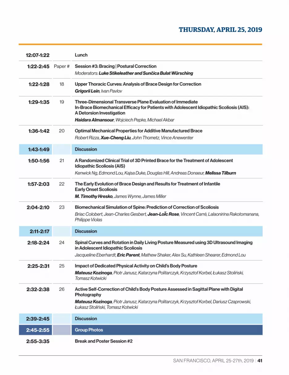
12:07-1:22 Lunch
1:22-2:45 Paper # Session #3: Bracing | Postural CorrectionModerators: Luke Stikeleather and Sunčica Bulat Würsching
1:22-1:28 18 Upper Thoracic Curves: Analysis of Brace Design for CorrectionGrigorii Lein, Ivan Pavlov
1:29-1:35 19 Three-Dimensional Transverse Plane Evaluation of Immediate In-Brace Biomechanical Efficacy for Patients with Adolescent Idiopathic Scoliosis (AIS): A Detorsion InvestigationHaidara Almansour, Wojciech Pepke, Michael Akbar
1:36-1:42 20 Optimal Mechanical Properties for Additive Manufactured BraceRobert Rizza, Xue-Cheng Liu, John Thometz, Vince Anewenter
1:43-1:49 Discussion
1:50-1:56 21 A Randomized Clinical Trial of 3D Printed Brace for the Treatment of Adolescent Idiopathic Scoliosis (AIS)Kenwick Ng, Edmond Lou, Kajsa Duke, Douglas Hill, Andreas Donaeur, Melissa Tilburn
1:57-2:03 22 The Early Evolution of Brace Design and Results for Treatment of Infantile Early Onset ScoliosisM. Timothy Hresko, James Wynne, James Miller
2:04-2:10 23 Biomechanical Simulation of Spine: Prediction of Correction of ScoliosisBriac Colobert, Jean-Charles Gesbert, Jean-LoÏc Rose, Vincent Carré, Lalaonirina Rakotomanana, Philippe Violas
2:11-2:17 Discussion
2:18-2:24 24 Spinal Curves and Rotation in Daily Living Posture Measured using 3D Ultrasound Imaging in Adolescent Idiopathic ScoliosisJacqueline Eberhardt, Eric Parent, Mathew Shaker, Alex Su, Kathleen Shearer, Edmond Lou
2:25-2:31 25 Impact of Dedicated Physical Activity on Child’s Body PostureMateusz Kozinoga, Piotr Janusz, Katarzyna Politarczyk, Krzysztof Korbel, Łukasz Stoliński, Tomasz Kotwicki
2:32-2:38 26 Active Self-Correction of Child’s Body Posture Assessed in Sagittal Plane with Digital PhotographyMateusz Kozinoga, Piotr Janusz, Katarzyna Politarczyk, Krzysztof Korbel, Dariusz Czaprowski, Łukasz Stoliński, Tomasz Kotwicki
2:39-2:45 Discussion
2:45-2:55 Group Photos
2:55-3:35 Break and Poster Session #2
THURSDAY, APRIL 25, 2019
42 | 14TH INTERNATIONAL SOSORT MEETING
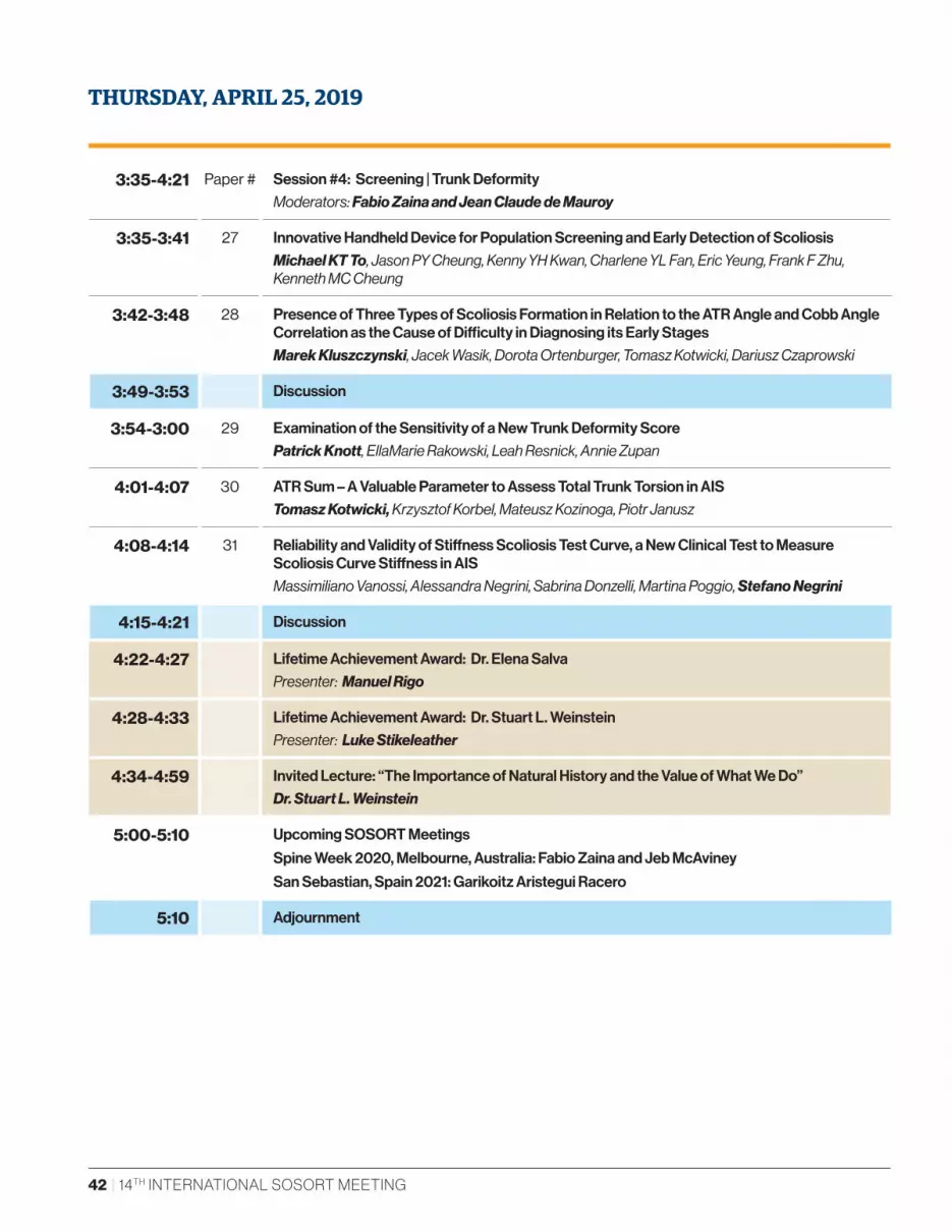
3:35-4:21 Paper # Session #4: Screening | Trunk DeformityModerators: Fabio Zaina and Jean Claude de Mauroy
3:35-3:41 27 Innovative Handheld Device for Population Screening and Early Detection of ScoliosisMichael KT To, Jason PY Cheung, Kenny YH Kwan, Charlene YL Fan, Eric Yeung, Frank F Zhu, Kenneth MC Cheung
3:42-3:48 28 Presence of Three Types of Scoliosis Formation in Relation to the ATR Angle and Cobb Angle Correlation as the Cause of Difficulty in Diagnosing its Early StagesMarek Kluszczynski, Jacek Wasik, Dorota Ortenburger, Tomasz Kotwicki, Dariusz Czaprowski
3:49-3:53 Discussion
3:54-3:00 29 Examination of the Sensitivity of a New Trunk Deformity ScorePatrick Knott, EllaMarie Rakowski, Leah Resnick, Annie Zupan
4:01-4:07 30 ATR Sum – A Valuable Parameter to Assess Total Trunk Torsion in AISTomasz Kotwicki, Krzysztof Korbel, Mateusz Kozinoga, Piotr Janusz
4:08-4:14 31 Reliability and Validity of Stiffness Scoliosis Test Curve, a New Clinical Test to Measure Scoliosis Curve Stiffness in AISMassimiliano Vanossi, Alessandra Negrini, Sabrina Donzelli, Martina Poggio, Stefano Negrini
4:15-4:21 Discussion
4:22-4:27 Lifetime Achievement Award: Dr. Elena SalvaPresenter: Manuel Rigo
4:28-4:33 Lifetime Achievement Award: Dr. Stuart L. WeinsteinPresenter: Luke Stikeleather
4:34-4:59 Invited Lecture: “The Importance of Natural History and the Value of What We Do”Dr. Stuart L. Weinstein
5:00-5:10 Upcoming SOSORT Meetings
Spine Week 2020, Melbourne, Australia: Fabio Zaina and Jeb McAviney
San Sebastian, Spain 2021: Garikoitz Aristegui Racero
5:10 Adjournment
THURSDAY, APRIL 25, 2019

SAN FRANCISCO, APRIL 25-27th, 2019 | 43
FRIDAY, APRIL 26, 2019
8:00 - 8:05 Welcome & Announcements
8:06-9:52 Paper # Session #5: SOSORT Paper Award NomineesModerators: Lori Dolan and Nigel Price
8:06-8:14 32 Alternately Repetitive Cast/Brace (ARCB) Treatment for a Larger Magnitude of Early-Onset ScoliosisNoriaki Kawakami, Toshiki Saito, Ryoji Tauchi, Kazuki Kawakami, Tetsuya Ohara
8:15-8:23 33 Are Lung Functions Related to Spinal Deformities in Patients With Adolescent Idiopathic Scoliosis? A Systematic Review and Meta-AnalysisArnold Wong, Henry Pang, Francesca Di Felice, Stefano Negrini, Matthew Chung, Sabrina Donzelli, Fabio Zaina, Jason Cheung, Dino Samartzis
8:24-8:32 34 The Effects of Inspiratory Muscle Training on Respiratory Muscle Strength, Respiratory Function and Functional Capacity in Adolescents with Idiopathic Scoliosis: A Randomized Controlled StudyGozde Basbug, Nuh Mehmet Elmadag, Hulya Nilgun Gurses
8:33-8:39 Discussion
8:40-8:48 35 Long-Term Results after Brace Treatment with the PASB in Adolescent Idiopathic ScoliosisAngelo Gabriele Aulisa, Renato Maria Toniolo, Francesco Falciglia, Marco Giordano, Lorenzo Aulisa
8:49-8:57 36 Very Long Term Results of Bracing Management with Initial Plaster Cast in Juvenile and Adolescent Idiopathic Scoliosis: Comparative Results 20 and 30 Years after WeaningJean Claude de Mauroy, Fabio Gagliano, Frédéric Barral
8:58-9:06 37 Final Result of Asymmetric Polycarbonate Detorsion Brace: Case Series of 110 Adolescent Idiopathic Scoliosis According to the Sosort-SRS RecommendationsJean Claude de Mauroy, Fabio Gagliano, Frédéric Barral, Sophie Pourret
9:07-9:15 38 Final Results of Bracing Management with Initial Plaster Cast or Total Time Polycarbonate Bracing in Adolescent Idiopathic Scoliosis at Late Risser (3-5)Jean Claude de Mauroy, Fabio Gagliano, Frédéric Barral
9:16-9:24 Discussion
9:25-9:33 39 Impact of Sports Activity on Full-Time Braced Patients: An Observational Study of 785 Risser 0–2 Adolescents with Idiopathic ScoliosisAlessandra Negrini, Sabrina Donzelli, Massimiliano Vanossi, Martina Poggio, Fabio Zaina, Michele Romano, Stefano Negrini
9:34-9:42 40 Predictors of Results in a 1051 Cohort of Adolescents with Idiopathic Scoliosis at High Risk of ProgressionSabrina Donzelli, Fabio Zaina, Francesca Di Felice, Stefano Negrini
9:43-9:51 41 Initial In-Brace Correction: Can Evaluation of the Cobb Angle Be the Only Parameter Predictive of the Outcome of Brace Treatment in Patients with Adolescent Idiopathic Scoliosis?Angelo Gabriele Aulisa, Marco Galli, Renato Maria Toniolo, Marco Giordano, Lorenzo Aulisa

44 | 14TH INTERNATIONAL SOSORT MEETING
FRIDAY, APRIL 26, 2019
9:52-9:58 Discussion
9:59-10:14 2019 SOSORT Presidential AddressLuke Stikeleather
10:14-10:54 Break and Poster Session #3
10:54-11:49 Paper # Session #6: Adult DeformityModerators: Joseph O’Brien and Stefano Negrini
10:54-11:00 42 The Prevalence of Adult de Novo Scoliosis: A Systematic Review and Meta-AnalysisJeb McAviney, Carrie Roberts, Bryony Sullivan, Alex Alevras, Petra L Graham, Benjamin T Brown
11:01-11:07 43 A Systematic Literature Review of Bracing Treatment for Adults with ScoliosisJeb McAviney, Johanna Mee, Azharuddin Fazalbhoy, Juan Du Plessis, Benjamin Brown
11:08-11:14 44 Safe Exercises for Adults with Scoliosis - A Scoping Review with a Proposed AlgorithmLise Stolze, Hagit Berdishevsky, Sanja Schreiber, Jean Claude De Mauroy
11:15-11:21 Discussion
11:22-11:28 45 Mobilisation of the Nervous System in Patients with Adult Scoliosis and PainEvgeniya Dimitrova
11:29-11:35 46 Exploring the Cost Effectiveness of an Intensive Physiotherapeutic Scoliosis Specific Exercise (PSSE) Programme in a UK Adult PopulationD. A. Jason Black, Erika Maude, David Glynn, Abbie Turland
11:36-11:42 47 Cheneau Bracing is Effective in Older Patients with Larger Curves – Results of Retrospective Chart AnalysisGrant Wood, Sanja Schreiber
11:43-11:49 Discussion
11:50-12:20 Invited Speaker: “World Spine Care and the Global Spine Care initiative: A Model of Care for the Management of Spinal Disorders in Under-Served Communities”Dr. Scott Haldeman
12:20-1:50 Lunch and Symposia (12:50-1:35)
Option 1: Toward a Standardized Minimum Dataset for Clinical Practice and Research (Cyril Magnin Ballroom, Level Four)
Option 2: Building a Multidisciplinary Collaborative Practice (Market Street, Level Three)
1:50-2:26 Paper # Session #7: Surface TopographyModerators: Patrick Knott and Angelo Gabriele Aulisa
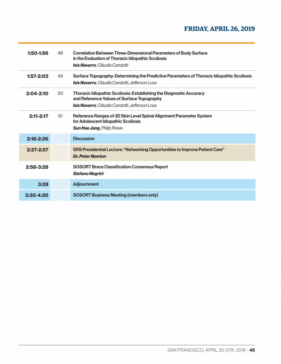
SAN FRANCISCO, APRIL 25-27th, 2019 | 45
1:50-1:56 48 Correlation Between Three-Dimensional Parameters of Body Surface in the Evaluation of Thoracic Idiopathic ScoliosisIsis Navarro, Cláudia Candotti
1:57-2:03 49 Surface Topography: Determining the Predictive Parameters of Thoracic Idiopathic ScoliosisIsis Navarro, Cláudia Candotti, Jefferson Loss
2:04-2:10 50 Thoracic Idiopathic Scoliosis: Establishing the Diagnostic Accuracy and Reference Values of Surface TopographyIsis Navarro, Cláudia Candotti, Jefferson Loss
2:11-2:17 51 Reference Ranges of 3D Skin Level Spinal Alignment Parameter System for Adolescent Idiopathic ScoliosisSun Hae Jang, Philip Rowe
2:18-2:26 Discussion
2:27-2:57 SRS Presidential Lecture: “Networking Opportunities to Improve Patient Care”Dr. Peter Newton
2:58-3:28 SOSORT Brace Classification Consensus ReportStefano Negrini
3:28 Adjournment
3:30-4:30 SOSORT Business Meeting (members only)
FRIDAY, APRIL 26, 2019

46 | 14TH INTERNATIONAL SOSORT MEETING
SATURDAY, APRIL 27, 2019
8:00-9:00 Breakfast
9:00-9:03 Welcome & Announcements
9:04-9:09 “The Power of Support”Guest Speaker: Rachel Mulvaney, Curvy Girls
9:10-10:42 Paper # Session #8: Bracing | Treatment AdherenceModerators: James Wynne and Sabrina Donzelli
9:10-9:16 52 Back Pain Related to Providence Night-Time Bracing in Adolescent Idiopathic Scoliosis PatientsAne Simony, Helle Munk, Lena Quisth, Inge Beuschau, Mikkel Osterheden Andersen
9:17-9:23 53 Providence Night-Time Bracing is Effective in Treatment of Adolescent Idiopathic Scoliosis, Even in Curves Larger than 35°Ane Simony, Lena Quisth, Inge Beuschau, Stig Mindedahl Jespersen, Mikkel Osterheden Andersen
9:24-9:30 54 Indication for 12h Nighttime Bracing on Late-Onset Idiopathic ScoliosisGautier de Chelle, Imad Bentellis, Federico Solla, Olivier Rosello, Jean-Luc Clement, Virgine Rocher-Rampal
9:31-9:37 Discussion
9:38-9:44 55 The 13 Hour Wear Time Myth in Conservative Scoliosis Management with a TLSODavid Speers
9:45-9:51 56 Influence of Wearing Time on Rotation in Corset Care of Patients with Idiopathic ScoliosisChristian Grasl, Renata Pospischill, Karin Riedl, Sebastian Farr, Tamara Serth
9:52-9:58 57 Translation and Subsequent Implementation of SOSORT Consensus Guidelines into Internal Clinical Practice Guidelines on Patient Management and Biomechanical Design Considerations for a National Provider of Scoliosis BracingPhil M. Stevens, Brian L. Kerl, Megan Chamis
9:59-10:05 Discussion
10:06-10:12 58 Effect of an Informative Booklet in Scoliosis on Compliance to Brace, Self-Rehabilitation Program, and Health-Related Quality of Life of Patients with Adolescent Idiopathic Scoliosis: A Prospective Comparative StudyMathieu de Seze, Hèlène Chung
10:13-10:19 59 A Retrospective Review of Patients Attending Schroth Method Physical Therapy: Factors Affecting AdherenceKristen Venuti, Peiting Lien, Allison Greaney, Michelle Kohler, Paul Sponseller, Majd Marrache
10:20-10:26 60 A Self-Administered Questionnaire Designed to Determine Causes of Poor Adherence to Brace Treatment in Adolescent Idiopathic ScoliosisSamuel Sassine, Marie Beauséjour, Julie Joncas, Soraya Barchi, Nikita Cobetto, Sylvie Le May, Omar Elsemin, Carl-Éric Aubin, Stefan Parent, Hubert Labelle
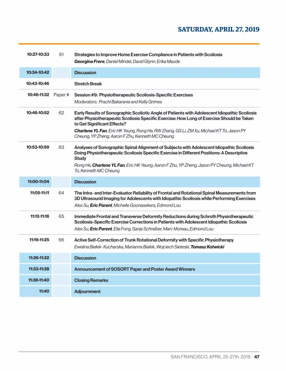
SAN FRANCISCO, APRIL 25-27th, 2019 | 47
10:27-10:33 61 Strategies to Improve Home Exercise Compliance in Patients with ScoliosisGeorgina Frere, Daniel Mindel, David Glynn, Erika Maude
10:34-10:42 Discussion
10:43-10:46 Stretch Break
10:46-11:32 Paper # Session #9: Physiotherapeutic Scoliosis-Specific ExercisesModerators: Prachi Bakarania and Kelly Grimes
10:46-10:52 62 Early Results of Sonographic Scoliotic Angle of Patients with Adolescent Idiopathic Scoliosis after Physiotherapeutic Scoliosis Specific Exercise: How Long of Exercise Should be Taken to Get Significant Effects?Charlene YL Fan, Eric HK Yeung, Rong He, RW Zhang, GS Li, ZM Xu, Michael KT To, Jason PY Cheung, YP Zheng, Aaron F Zhu, Kenneth MC Cheung
10:53-10:59 63 Analyses of Sonographic Spinal Alignment of Subjects with Adolescent Idiopathic Scoliosis Doing Physiotherapeutic Scoliosis Specific Exercise in Different Positions: A Descriptive StudyRong He, Charlene YL Fan, Eric HK Yeung, Aaron F Zhu, YP Zheng, Jason PY Cheung, Michael KT To, Kenneth MC Cheung
11:00-11:04 Discussion
11:05-11:11 64 The Intra- and Inter-Evaluator Reliability of Frontal and Rotational Spinal Measurements from 3D Ultrasound Imaging for Adolescents with Idiopathic Scoliosis while Performing ExercisesAlex Su, Eric Parent, Michelle Goonasekera, Edmond Lou
11:12-11:18 65 Immediate Frontal and Transverse Deformity Reductions during Schroth Physiotherapeutic Scoliosis-Specific Exercise Corrections in Patients with Adolescent Idiopathic ScoliosisAlex Su, Eric Parent, Elia Fong, Sanja Schreiber, Marc Moreau, Edmond Lou
11:19-11:25 66 Active Self-Correction of Trunk Rotational Deformity with Specific PhysiotherapyEwelina Białek- Kucharska, Marianna Białek, Wojciech Sieteski, Tomasz Kotwicki
11:26-11:32 Discussion
11:33-11:38 Announcement of SOSORT Paper and Poster Award Winners
11:38-11:40 Closing Remarks
11:40 Adjournment
SATURDAY, APRIL 27, 2019

SAN FRANCISCO, APRIL 25-27th, 2019 | 49
Scientific Abstracts

50 | 14TH INTERNATIONAL SOSORT MEETING
 Thurs 4/25/19 | Paper 1
Scolios-us: A Web-Based Platform to Address the Psychological Complexity of Scoliosis BracingMegan E. Glahn, Joshua B. Utay, Brad A. Culotta, John R. Faust, William A. Phillips Baylor College of Medicine, Baton Rouge Orthopedic Clinic, Texas Children’s Hospital
Introduction: Bracing for adolescent idiopathic scolio-sis (AIS) is an effective way to control curve progression. Because success heavily relies on wear time, adherence is problematic. While decreased wear time compromis-es the effectiveness of bracing, increased psychological stress jeopardizes the mental wellbeing of braced patients. Although the emotional effects of bracing are well-docu-mented, clinicians are not initiating conversations to al-leviate these effects. Scolios-us was created to help clini-cians address the psychological side of bracing. Scolios-us is a web-based platform intended to empower scoliosis patients with the tools and resources they need to be suc-cessful brace-wearers.Objectives: 1. Develop the site to meet the needs of AIS patients. 2. Collect feedback to optimize Scolios-us.Methods: The study consisted of two surveys. Results were analyzed with descriptive statistics and a paired t-test.Clinician Survey: Clinicians were recruited from the Spinal Orthotics Society site and O&P Listserv. English-speaking orthotists/orthotic residents were eligible. Subjects eval-uated website design, contents, video, and feasibility of using Scolios-us as a clinical tool in a 16-question survey.Patient Survey: AIS subjects were recruited at the Baton Rouge Orthopedic Clinic. The inclusion criteria consist-ed of the following: AIS diagnosis, 10-17 years old, En-glish-speaking, assenting, and receive parental consent. Subjects evaluated the video in a 10-question survey.
Results and Discussion: Clinician Survey: The sur-vey produced an average of 29.5 responses on multi-ple-choice questions and 14.2 responses on free-response questions. Over 80% of subjects treat scoliosis patients frequently or very frequently. They perceived the site as engaging, easy to navigate, and helpful for scoliosis pa-tients. The video was perceived as the most helpful tool, and subjects found it to be empowering. When asked for improvements, two themes emerged: shorten the video and increase participant diversity. The majority (60%) of subjects are extremely likely to recommend Scolios-us to patients. Patient Survey: Eleven female subjects with a mean age of 13.1 ± 2.0 years participated. Subjects’ feelings about bracing improved significantly after watching the video (P=0.011, Fig. 1). When asked for improvements, the theme of wanting more brace information emerged.Results show the potential for Scolios-us to satisfy pa-tients’ need for emotional support. Clinicians who par-ticipated are likely to recommend Scolios-us; in doing so, they touch on the intangible side of bracing. Results from the patient survey also support Scolios-us. After watching the video, subjects’ feelings improved significantly, reaf-firming the need for psychological support and validating the video as an effective approach.Conclusion and Significance: Although Scolios-us is still in its infancy, preliminary data suggest it is encouraging a more holistic approach to scoliosis treatment.

SAN FRANCISCO, APRIL 25-27th, 2019 | 51
 Thurs 4/25/19 | Paper 2
Support Needs of Adolescents with Spinal Deformities: A Qualitative StudyKathleen Shearer, Sarah Southon, Laura Rogers, Nicole Gingrich, Leanne Willson Stollery Children’s Hospital, University of Alberta, The King’s University, Edmonton, Canada
Introduction: Within orthopedics, social support is known to shorten post-surgical stay and promote patient well-be-ing. Children and adolescents with spinal deformities, such as scoliosis, who are facing spinal surgery or other treat-ments, are under significant stress as they and their care-givers make health decisions. Understanding the specific social and informational needs of patients at various stag-es of diagnosis and treatment is necessary to facilitate the types of supports that would be most beneficial to these patients.Objectives: A needs assessment was conducted in part-nership with our scoliosis clinic to determine the support services required for patients with scoliosis. The aim of the study was to identify what forms of social and infor-mational supports would be most useful and accessible for children and adolescents diagnosed with a spinal deformi-ty from the perspective of the patients.Methods: This descriptive qualitative study included 12 patients (9 female, 3 male) with spinal deformity aged 10-16 years (mean= 13.1 years). We conducted 12 semi struc-tured, individual face-to-face interviews with the patient participants, asking questions pertaining to clinic-based supports, online supports, and whether personal and in-formational needs were met during out-patient treatment and surgery (if applicable). Interviews were transcribed and analyzed using thematic analysis.Results and Discussion: Participants reported challenges and sources of support from the initial stage of diagno-
sis, through the course of treatment including bracing and surgery. Reported challenges included the experience of pain, participation in school and extracurricular activities, the need to address feelings following diagnosis and lack of understanding of scoliosis by the patients, caregivers and community members, including teachers. Participants also wanted more information about surgery well ahead of their surgery dates. Identified supports included social and professional support, and written material; patients were mostly unaware of online resources. Participants identified a need for more reliable online information for themselves and family members, and written materials for teachers. Conclusions and Significance: This study explored the in-formational and social support needs of adolescents with spinal deformities. Adolescents expressed the need to talk about their feelings, especially at initial diagnosis, but felt isolated due to perceived lack of understanding of their condition by those around them. Informational supports identified as potentially helpful to adolescents included access to social media, on-line and written information about scoliosis for themselves, family members and teach-ers. Results of this study have been used to inform devel-opment of educational resources and on-line supports by the local scoliosis clinic and community support group for scoliosis.

52 | 14TH INTERNATIONAL SOSORT MEETING
 Thurs 4/25/19 | Paper 3
Needs of Parents of Children Diagnosed With a Back Condition: A Qualitative Needs AssessmentKathleen Shearer, Sarah Southon, Laura Rogers, Jeremy Kieftenbeld, Leanne Willson Stollery Children’s Hospital, University of Alberta, The King’s University, Edmonton, Canada
Introduction: Research has reflected parents’ desire to be partners in their child’s treatment and care. Parents of children diagnosed with spinal deformities have both so-cial and informational needs. Studies indicate that parents experience emotional burden, whether their children are being braced or having surgery for scoliosis. Parents also do not have a clear understanding of the post-operative needs of their children following surgery for scoliosis, and research indicates that a clearer understanding of what their child will face during treatment might reduce antic-ipatory anxiety among parents.Objectives: The purpose of this Community Engaged Re-search study was to conduct a needs assessment of parents of children attending the scoliosis clinic, to understand the ways in which the clinic and the related community net-work could best provide the necessary supports for parents of children with spinal deformities.Methods: This qualitative descriptive study involved in-person interviews with mothers or fathers of children with spinal deformities, with or without other comorbid-ities, recruited through the scoliosis clinic. Sixteen par-ents (12 mothers, 2 fathers, 2 step-fathers) of 15 children aged 11-16 years were interviewed. Interviews were tran-scribed and analyzed using thematic and content analysis and managed through NVivo 11.
Results and Discussion: At the various stages including initial diagnosis, bracing, pre-surgery, and post-surgery, parents sought connection with other parents who under-stood their journey, education materials for themselves and others and reliable online medical and rehabilitation information as well as information about complementa-ry and alternative treatment options. They sought reli-able information about implications the diagnosis had on their child’s future. When considering surgery, parents expressed uncertainty about their child’s future abilities in activities of daily living, mobility, school return, and participation in community activities. Parents wanted support in how to explain the diagnosis and treatment options to their child.Conclusions and Significance: Parents of children with spi-nal deformities undergo many stressors, and this study provides a model for clinicians to use to address parent needs. Asking parents about their needs can inform clinic, school and community supports in relevant social, emo-tional, and medical- rehabilitative areas. The study results are being used to guide the development of educational re-sources and develop an on-line support group for parents of children with spinal deformities.

SAN FRANCISCO, APRIL 25-27th, 2019 | 53
 Thurs 4/25/19 | Paper 4
To Brace or Not To Brace: Development of an AIS Patient Decision AidLori Dolan, Matthew Halsey, Noelle Larson, Charles Mehlman, Leigh Davis, Stuart Weinstein University of Iowa, Oregon Health & Science University, Mayo Clinic, Cincinnati Children’s Hospital, Children’s Healthcare of Atlanta
Introduction: The treatment of adolescent idiopathic sco-liosis (AIS) may include a number of different modalities including bracing. BrAIST demonstrated the efficacy of bracing to prevent progression to surgery but the deci-sion to initiate and maintain bracing is still difficult for patients and families. Whilst there is a plethora of infor-mation regarding AIS bracing, there is no specific patient decision aid (PtDA) available to help in the shared deci-sion-making process.Objective: The purpose of this study was to develop a prototype PtDA with respect to bracing for AIS utilizing a community participatory research design.Methods: 104 patients were assessed at five separate US sites by means of in-person interview, focus group or on-line survey. The interviews were structured, using the Ot-tawa Decision Support Framework, to elicit decision sup-port needs. Patient and family inputs were transcribed and reviewed centrally to identify common needs, con-cerns and desires. Specifically, we elicited comments on the decision-making process, goals, general scolio-sis knowledge and treatment information needs, brac-ing-specific information needs and how best to depict risks and benefits of AIS bracing.
Results and Discussion: Common threads were identified in each domain: 1. Parents did not feel they had a choice about treatment and requested more options and compari-sons. Specifically, parents and families noted that a risk cal-culator, based on BrAIST data, would have helped to make the decision regarding bracing. 2. Every family’s goal was to avoid surgery and to prevent progression. 3. Many families felt they lacked adequate knowledge of AIS natural histo-ry, the impact of AIS on other health issues (e.g. back pain) and the risks of frequent x-ray exposure. 4. Families wanted more specific information with respect to all aspects of brace treatment (e.g. duration, brace types) and surgical treatment (e.g. decreased flexibility, cost). Furthermore, they desired information about other options including yoga, scoliosis-specific exercises, vertebral body tethering, and chiropractic care.Conclusion and Significance: Based upon these results, and using the information developed from the BrAIST study with respect to risk factors and dose-response relationship, a us-er-centered designed web-based PtDA will be developed. Once created, the PtDA will be presented to a different cohort of families to evaluate its usability and its impact on patients’ knowledge, treatment decisions and decisional conflict.

54 | 14TH INTERNATIONAL SOSORT MEETING
 Thurs 4/25/19 | Paper 5
Quality of Life Measured by 463 Brace Questionnaires during the Treatment for Adolescent Idiopathic Scoliosis (AIS)Jean-Claude Bernard, Camille Castan, Nicolas Lebedel, Gautier de Chelle, Frederic Barral Croix-Rouge Française. Centre Médico-Chirurgical de Réadaptation des Massues. Proteor-Lecante Society, France
Introduction: Quality of Life (QL) scales have to be intro-duced in the treatment evaluation of our patients with ad-olescent idiopathic scoliosis. Vasiliadis and al. created the Brace Questionnaire (BrQ), which is specific for brace-treat-ed adolescents. This tool was validated in French in 2017 (Deceuninck J et al.). The higher the percentage is found, the better the quality of life is.Objective: The main objective was to measure the QL by BrQ in a sample of French patients wearing a brace for AIS. Secondary objective was to evaluate observance during brace-treatment.Methods: Since its validation in French, the BrQ is pro-posed to every patient treated by brace after 3 months, 6 months, one year and then 1 time a year. The BrQ contains 34 items out of 8 domains. BrQ was developed and adapt-ed so that children can complete it themselves between 9 and 18 years old. 463 BrQ were collected along the treat-ment corresponding to 329 patients. The average age was 14,1 years old (9-18) and 87% were girls. The patients were treated by different braces: underarm braces like X1 (x1 Brace) for low Lenke 1,2,3,4 curves (43% BrQ) and X2 brace for severe Lenke 1,2,3,4 curves (24% BrQ), the X3 (X3Brace) for Lenke 5 curves (20% BrQ) and other braces (13% BrQ).Results and Discussion: On overall population, BrQ score were 76.7% at 3 months, 78.7% at 1 year and 79.2% at 2
years of brace-wear. Vitality domain was significantly improved between 3 months and 2 years of brace-wear (63.5% vs 75.7%, p<0.01). Better results of BrQ were found for X3 brace compared to underarm braces like X2 brace (80.6% vs 75%, p<0.01) or X1 brace (80.6%vs77.1%, p<0.02). We found that the Therapeutic Educational Pro-gram (ETP) which is focused on scoliosis has a low influ-ence. The observance, corresponding to the ratio of pre-scribed brace-wear on effective brace-wear, was 91 % with variations between: underarm braces = X1 89% and X2 Brace 91%, and short braces = X3 95%.Taking care of pa-tient’s quality of life using BrQ at different time along the treatment is a tool to respond to observance difficulties and patient well-being. On this cohort, scores are superior to 75% at the beginning and during the treatment which shows that bracing has low impact on quality of life. ETP was proposed to patients with treatment difficulties after Educative Diagnosis and didn’t permit to enhance BrQ score as expected.Conclusion and Significance: Quality of Life during full-time brace-treatment is stable contrary to popular belief. It depends on brace-type with better results for short brac-es than underarm braces. In the future, in addition of the global score, it is necessary to focus on difficulties inside the domains concerned by the BrQ and give individualized medical advice.

SAN FRANCISCO, APRIL 25-27th, 2019 | 55
 Thurs 4/25/19 | Paper 6
Sagittal Plane Misalignment Affects Less Than Expected HrQOL in Non-Operated Scoliosis PatientsManuel Rigo, Gloria Quera-Salvá, Elisabetta D’Agata Institute E. Salva, Barcelona, Spain
Introduction: In scoliosis population, the relationship be-tween radiological parameters and HRQoL is still a contro-versial issue. GAP score is a new Pelvic Incidence-based proportional method of analyzing the individualized sagit-tal plane, which better predicts mechanical complications and correlates with HRQoL in operated patients. Objective: To study the relationship between GAP score and HRQoL in a mixed scoliosis population formed mainly by non-operated patients.Methods: Retrospective series of 45 consecutive scoliosis patients with good enough quality radiographs (AP and latero-lateral projections). A single experienced observer measured all the studied radiological parameters: Thorac-ic Cobb angle, Lumbar or thoracolumbar Cobb angle, GAP score related parameters (Relative Pelvic Version, Relative Lumbar Lordosis, Lordosis Distribution Index and Relative Spinopelvic alignment). Age, Thoracic/Lumbar/ThL Cobb Angles and GAP score were correlated with SRS-22 Pain, Self-Image and Sub-Total (SPSS).Results and Discussion: Sample was formed by 45 patients with Idiopathic Scoliosis, aged 10 to 68 years (mean 28.8 + 17). Twenty main thoracic/non or minor lumbar, 8 single lumbar or ThL and 14 double major or Lumbar-ThL ma-jor/Thoracic minor. Three out of 45 had an instrumented arthrodesis. Thoracic mean Cobb angle was 31° +/- 18 and Lumbar or thoracolumbar 31° +/- 16. Mean GAP score was 2.7 (0 to 10). PI 48.7° (24-70). SS 37.2° (15-56). L1S1 49.5°
(23-80). L4S1 35.2° (15-53). SRS-22 Pain 4 +/- 0.8, SRS-22 SI 3.6 +/- 0.7 and Sub Total 19.8 +/- 3.2. Age correlated with SRS-22 Pain (- .5 p<.01), with SRS-22 SI (- .36 p<.05) and with SRS-22 Sub-Total (- .42 p<.01). Cobb thoracic did not correlated with SRS-22. Lumbar Cobb correlated with SRS-22 Pain (- .35 P<.01), SI (- .52 p<.01) and Sub-Total (- .39 p<.01). GAP Score did not show correlation with SRS-22. Age also correlated with Lumbar Cobb (.4<.01) and with GAP Score (.39 p<.01). Lumbar Cobb correlated with GAP Score (.39 p<.01). Pelvic Indexes (PI and SS) showed similar values and correlations between them and with Lumbar Lordosis than in previous studies. GAP score correlated the best with Relative Lumbar Lordosis (.8 p<.01). SRS-22 Pain showed a good correlation with SRS-SI (.7 p<.01) and Sub-Total (.87 p<.01).Conclusion and Significance: Age and Lumbar Cobb but not GAP score correlate with SRS-22 Pain, Self-Image and Sub-Total. HRQoL in a mixed (Adolescent and Adult) id-iopathic scoliosis population, mainly non-operated, is af-fected by age and magnitude of the Lumbar or thoraco-lumbar curve in the frontal plane (Cobb angle). We did not find a significant correlation between HRQoL and individ-ualized sagittal plane alignment. This study suggests that the influence of sagittal plane misalignment on HRQoL in non-operated scoliosis patients, at least in a relatively young population, might be overestimated and advises against using routine latero-lateral radiographs during follow-up.

56 | 14TH INTERNATIONAL SOSORT MEETING
 Thurs 4/25/19 | Paper 7
How to Assess Quality of Life in Adults with Scoliosis: Comparison of Two QuestionnairesFabio Zaina, Francesca Di Felice, Sabrina Donzelli, Antonio Caronni, Stefano Negrini ISICO, Don Gnocchi Foundation
Introduction: Scoliosis is a very frequent problem during adulthood. Surgery is a very common option for these pa-tients, but there are relevant risks and side effects and not all patients are willing for such treatment. For these rea-sons many patients seek for a conservative treatment in re-habilitation centers. For the assessment of quality of life in patients with scoliosis. The SRS-22 questionnaire was first developed, and more recently the ISYQOL questionnaire, with the latter tested only in youngers.Objective: The aim of the present study is to test the prop-erties of the ISYQOL in a group of adults with scoliosis and compare its properties to the SRS-22.Methods: We retrospectively review the record of all the adult patients included in our prospective database run-ning between 2003 and 2017. The inclusion criteria were: diagnosis of idiopathic scoliosis with a curve of 30° Cobb or more, no surgical treatment, availability of the SRS-22 and ISYQOL. The SRS-22 includes 5 subscales (Function, Psychological Wellbeing, Pain, Aesthetics and Treatment Satisfaction), with scores ranging from 5 (no impairment) to 0 (high impairment). The ISYQOL is a Rasch consistent
questionnaire based on 13 questions that gives a contin-uous value of quality of life ranging from 0 to 100. The Cronbach alpha was used to check the internal validity, and a Rasch analysis was run to explore the features of the different tools.Results and Discussion: 100 patients (29 males) met the inclusion criteria. The mean Cobb angle was 46±14°, age 42±15. The Cronbach alpha value was above 0.70 for both questionnaires. This means that their internal consistency is good, and allows their application in a clinical setting. The Rasch analysis of the values of both questionnaires would allow a more precise comparison.Conclusion and Significance: This is the first study report-ing the general characteristics of patients affected by scoli-osis attending a specialized rehabilitation center based on the SRS-22 and ISYQOL questionnaires. Both questionnaire can describe the population of adult with scoliosis, and are able to assess the quality of life. Adult scoliosis patients need specific tools of evaluation mainly for quality of life and pain. The SRS-22 and the ISYQOL seem to be able to accomplish this task.
SAN FRANCISCO, APRIL 25-27th, 2019 | 57
 Thurs 4/25/19 | Paper 8
The Association Between Aerobic Capacity and Spinal Deformity in Patients with Adolescent Idiopathic Scoliosis – A Systematic ReviewArnold Wong, Francesca Di Felice, Matthew Chung, Henry Pang, Stefano Negrini, Jason Cheung, Sabrina Donzelli, Fabio Zaina, Dino Samartzis Hong Kong Polytechnic University, Prince of Wales Hospital, Italian Scientific Spine Institute, University of Brescia, University of Hong Kong, Rush University
Introduction: While patients with severe adolescent id-iopathic scoliosis (AIS) are known to have poor aerobic capacity, it remains unclear if some components of spinal deformities are related to aerobic capacity in patients with various severity of AIS. However, no systematic review has summarized the relations between these components and aerobic capacity in AIS patients, which may inform clinical practice. Objective: To summarize the relations between various structural parameters and aerobic capacity of AIS patients during exercise tolerance tests. Methods: Potential citations were searched from 8 elec-tronic databases, from inception to November 2016. Two reviewers independently screened the titles, abstracts, and full-text of potential articles based on the preset cri-teria. Two other independent reviewers extracted data and appraised the methodological quality of the included studies using established evaluation tools. Associations between spinal parameters and aerobic capacity were summarized qualitatively. Results and Discussion: Eight of 1,045 identified citations (377 patients) were included. The overall methodologi-cal quality was low to moderate. Most included studies did not justify sample sizes nor adjust for confounders. Treadmill exercise tolerance tests (treadmill tests) and cycloergometer exercise tolerance tests (bike tests) esti-mated diverse aerobic capacity of AIS patients (expressed as maximum oxygen intake (VO2max)). Using treadmill
tests, 3 studies noted that the mean body weight nor-malized VO2max in patients with mild to moderate AIS (Cobb angles between 20° and 45°) was significantly low-er than the normative values. Likewise, 2 studies found that patients with mild curves had significantly poorer ventilatory efficiency (i.e. higher ventilation volume per VO2max) than healthy controls during a treadmill test. A study found that increased thoracic Cobb angles were as-sociated with decreased VO2max (r = -0.71; p < 0.01). An-other study showed that patients with mild to moderate curves had significantly poorer exercise tolerance, lower anaerobic thresholds and a higher breathing frequency than healthy controls during treadmill tests. Converse-ly, using bike tests, 3 studies found that the curve angle or number of scoliotic vertebrae was unrelated to body weight normalized VO2max or maximum tidal volume/inspiratory capacity ratio. Interestingly, a study found that patients with Cobb angles >40° showed slightly de-creased VO2max and mild hyperventilation during a bike test. Conclusion and Significance: While our results showed that patients with mild thoracic curves have suboptimal aerobic capacity, these findings might be confounded by other factors (e.g. muscularity). Future cohort stud-ies should examine if suboptimal aerobic capacity in AIS patients is related to poor fitness level or spinal deformi-ties. However, given the benefits of aerobic exercises on aerobic capacity, regular aerobic training is recommended for AIS patients.

58 | 14TH INTERNATIONAL SOSORT MEETING
 Thurs 4/25/19 | Paper 9
Relationship Between Thoracic Curve Magnitude, Number of Involved Vertebrae and Thoracic Kyphosis Angle Versus Preoperative Pulmonary Parameters in Children with Idiopathic ScoliosisKatarzyna Politarczyk, Łukasz Stępniak, Piotr Janusz, Mateusz Kozinoga, Tomasz Kotwicki University of Medical Sciences, Rehasport Clinic, Poznań, Poland
Introduction: Scoliosis is a very frequent problem during adulthood. Surgery is a very common option for these pa-tients, but there are relevant risks and side effects and not all patients are willing for such treatment. For these rea-sons many patients seek for a conservative treatment in re-habilitation centers. For the assessment of quality of life in patients with scoliosis. The SRS-22 questionnaire was first developed, and more recently the ISYQOL questionnaire, with the latter tested only in youngers.Objective: The aim of the present study is to test the prop-erties of the ISYQOL in a group of adults with scoliosis and compare its properties to the SRS-22.Methods: We retrospectively review the record of all the adult patients included in our prospective database run-ning between 2003 and 2017. The inclusion criteria were: diagnosis of idiopathic scoliosis with a curve of 30° Cobb or more, no surgical treatment, availability of the SRS-22 and ISYQOL. The SRS-22 includes 5 subscales (Function, Psychological Wellbeing, Pain, Aesthetics and Treatment Satisfaction), with scores ranging from 5 (no impairment) to 0 (high impairment). The ISYQOL is a Rasch consistent
questionnaire based on 13 questions that gives a contin-uous value of quality of life ranging from 0 to 100. The Cronbach alpha was used to check the internal validity, and a Rasch analysis was run to explore the features of the different tools.Results and Discussion: 100 patients (29 males) met the inclusion criteria. The mean Cobb angle was 46±14°, age 42±15. The Cronbach alpha value was above 0.70 for both questionnaires. This means that their internal consistency is good, and allows their application in a clinical setting. The Rasch analysis of the values of both questionnaires would allow a more precise comparison.Conclusion and Significance: This is the first study report-ing the general characteristics of patients affected by scoli-osis attending a specialized rehabilitation center based on the SRS-22 and ISYQOL questionnaires. Both questionnaire can describe the population of adult with scoliosis, and are able to assess the quality of life. Adult scoliosis patients need specific tools of evaluation mainly for quality of life and pain. The SRS-22 and the ISYQOL seem to be able to accomplish this task.

SAN FRANCISCO, APRIL 25-27th, 2019 | 59
 Thurs 4/25/19 | Paper 10
The Impact of Schroth Therapy on Adolescent Idiopathic Scoliosis with a High Risk of Curve ProgressionRebecca Garvin, Laura Dobrich, Li-An Lai UPMC’s Children’s Hospital of Pittsburgh, University of Pittsburgh
Introduction: Idiopathic scoliosis (IS) occurs in 5% of the adolescent population. The standard of care (SOC) in North America is based on Cobb angle (Cobb) and skele-tal maturity. Smaller Cobb are treated with observation, curves 25-40° are treated with thoracolumbosacral or-thosis (TLSO), while curves ≥45° are treated with spi-nal fusion. The SOSORT recommendation and European SOC includes referral for scoliosis-specific exercise (SSE), such as the Schroth method and/or bracing. The efficacy of Schroth has been shown in several studies through im-proved measures such as Cobb, vertebral rotation, asym-metry, self-image and pain. Currently, there is a dearth of information collectively incorporating patient- and clin-ic-specific aspects of Schroth therapy such as patient de-mographics, time to treat, Physical Therapy (PT) frequen-cy or duration and their effect on successful outcomes such as treatment frequency or duration, or pain.Objectives: To evaluate the success (no progression) of clinical-based PT on outpatients undergoing Schroth ther-apy in relation to various participant demographics and treatment aspects.Methods: A retrospective chart review was conducted (2014-17) on pediatric IS patients 8-18 yrs with skeletal immaturity completing a Schroth therapy episode of care in outpatient PT. Data collected included demographics, skeletal maturity, initial diagnosis Cobb, initial Cobb PT start, curve progression risk factor, curve classification,
TLSO prescription, time TLSO worn, TLSO type, time PT waiting list, PT frequency, PT duration, and amount of breath chest expansion. Stable curves considered <6° in-crease in 6 months.Results and Discussion: (n=72) included all Schroth clas-sifications with age (avg (std): 12(1.7))yrs, Risser 0.78 (1.4) and Cobb 30 (10–57)° at PT start. At PT end, 53/72 (74%) of patients had stable curves, while 1 year after PT 48/72 (67%) (p=0.4) of patients had stable curves and 6/72 (8%) had spinal fusions. There was no significant difference in demographics between no-progression v progression (p>0.05 for all). The largest Cobb at diagnosis tended to be smaller in no-progression (26 (11)°) when compared to progression (31 (12)°, p=0.08), while Risser tended to be smaller in progression (p=0.085). There was no statistical difference between diag Cobb<30° or ≥30° between the no-progression v progression (p=0.4). The average risk of curve Progression (RCP, 20°<diag Cobb<29°) 86%, was not significantly different between the no-progression (84(20)%) v progression (90(16)%, p=0.5).Conclusion and Significance: Although, as evidenced from the high RCP, our cohort would be considered at high risk of progression the majority of the participants did not progress even 1 year after PT with few spinal fusions. Additionally, we found no difference for progression with diagnosis Cobb≥30° suggesting that Schroth therapy may be a mitigating factor.
60 | 14TH INTERNATIONAL SOSORT MEETING
 Thurs 4/25/19 | Paper 11
Effect of Mono- Versus Multidisciplinary Schroth Based BSPTS Rehabilitation Therapy on Quality of Life of Adolescent Idiopathic Scoliosis Patients Evaluated by Scoliosis Research Society-22 Questionnaire (SRS-22)Mariette Zoer-Kosse, Marjan de Jonge, Anneke Struijk-Kramer, Saba Pasha Scoliosis Care Clinic, Zwolle, The Netherlands; The Children’s Hospital of Philadelphia, Philadelphia, PA
Introduction: Spinal deformity in adolescent idiopathic scoliosis (AIS) impacts patients’ self-image which adverse-ly impacts the patients’ psychological and mental health. Schroth based therapy offers conservative treatment to control the curve progression yet, the impact of long hour in person BSPTS rehabilitation therapy on the patients’ self-image and quality of life (QoL) has not been investi-gated. It is not known whether a multidisciplinary team of professionals including psychologist, occupational thera-pist, social worker and rehabilitation doctor or physician assistant in conjunction with the BSPTS therapist (multi-disciplinary) can improve the patients QoL compared to when only the BSPTS therapist (mono-disciplinary) inter-act with the patients. Objective: The objective is to study the impact of the mono- versus multidisciplinary BSPTS rehabilitation ther-apy on short and long-term patients’ QoL.Methods: 220 patients with AIS undergoing BSPTS ther-apy were included. All patients had completed a SRS-22 questionnaire before the start of treatment and at the most recent follow-up. The participants were divided into four groups. Group 1 (n=67) (mean age: 22,40 ±14,62) monodisciplinary therapy for six months, group 2 (n=42) (mean age: 21,43±12,83) multidisciplinary therapy for six months, group 3 (n=92) (mean age: 22,74±16,49) monodis-
ciplinary therapy for one year and group 4 (n=19) (mean age: 22,84±15,09) multidisciplinary therapy for one year. Effect of therapy is measured by means of the SRS-22 ques-tionnaire in all four domains: pain, mental health, self-im-age and function and the overall score.Results and Discussion: Mean score in pain, self-image, function and overall score improved significantly in group 1 and group 2 (p<0.050). The change in mental health mean score was nog significant. Group 2 scores significantly better on pain than group 1 (p=0.048). The mean score in pain, self-image and overall score improved significantly in group 3 and group 4 (p<0.050). No significant change in mental health in group 3 (p=0.491) or group 4 (p=0.132) was observed. The mean score in function improved signifi-cantly in group 3 (p=0.009) but nog in group 4 (p=0.130). Group 4 scores significantly better on pain (p=0.011) and overall score (p=0.046) than group 3.Conclusion and Significance: The QoL in patients with AIS improved after following a mono- or multidisci-plinary BSPTS therapy, but the multidisciplinary group scored higher in SRS-22 pain after short term therapy and in pain and overall score after long-term therapy in com-parison to the monodisciplinary treatment. The multidis-ciplinary and long-term therapy regimen yielded in higher QoL among all the 4 patient groups.

SAN FRANCISCO, APRIL 25-27th, 2019 | 61
 Thurs 4/25/19 | Paper 12
Effectiveness of Physiotherapeutic Scoliosis Specific Exercises (PSSE) on Adolescent without Brace Treatment – 24 Months Follow-UpBorislav Chongov, Evgenia Dimitrova National Sports Academy, Sofia, Bulgaria
Introduction: Treatment of adolescent idiopathic scolio-sis (AIS) is multidisciplinary. Depending of guidelines on SOSORT therapy is determined according to the severity of the curvature and skeletal maturity. Our interest is the effectiveness of treatment and teaching with physiother-apeutic scoliosis specific exercises (PSSE).Objective: To follow the effects of PSSE based of Schroth principles according to Barcelona scoliosis physical ther-apy school (BSPTS) on the clinical signs of AIS patients with respect to Cobb angle, angle of trunk rotation (ATR) and trunk symmetry.Methods: For a period of 4 years and 8 mounts (April 2014 – November 2018) we accessed 421 children’s with AIS. We included 26 (24 females and 2 mails) ranged in age between 10 and 15 years old with a mean age of 12.9±2.8 years. Risser 0 in 10 (38.5%), Risser 1 in 6 (23.1%), Risser 2 in 6 (23.1%) and Risser 3 in 4 (15.4%). They were dis-tributed as follows: single thoracic 4 (15.3%), single tho-racolumbar 8 (30.8%), single lumbar 6 (23.1%) and dou-ble major (thoracic and lumbar) 8 (30.8%). We excluded children treated with brace and awaiting surgery. All pa-tients we taught to make exercises for correction accord-
ing to Schroth method (BSPTS) after the initial evaluation. Teaching the patients included 5 consecutive days of 120 minutes in small groups of 2 children. Follow of weekly one hour (group 3-6 persons) supervised Schroth PSSE sessions and a daily home program.Results and Discussion: The average beginning prima-ry Cobb angle was 20.2° ± 6° with ATR mean 5.2° ± 3.3°, and POTSI 26.96 ± 9.1. Looking at the Cobb angle mea-surements, a mean 2.7° ± 4° of reduction was observed across all curvature groups. It improved more than 5° in 7 (26.9%) of children after 12 months and further improved at 24-month follow-up. Stabilization of AIS were in 18 (69.2%) of children, and worsening in 1 (3.8%). We have a statistically significant reduction of ATR after treatment from 5.19 ± 1.7 to the end value of 3.5 ± 2.82 (p<0.05) and also statistically significant improvement in all patients as for POTSI from initial 25.97 ± 9.07 to the end value of 18.23 ± 6.08 (ρ<0.05).Conclusion and Significance: Schroth method BSPTS had positive effect on scoliosis correction after 12 months of therapy and further improved at 24 months follow-up.

62 | 14TH INTERNATIONAL SOSORT MEETING
 Thurs 4/25/19 | Paper 13
Physiotherapeutic Scoliosis Specific Exercises (PSSE) Can Reduce the Risk of Progression in Adolescent Idiopathic Scoliosis during the Peak of Growth: A Prospective Study With a Control GroupNikos Karavidas Schroth Scoliosis & Spine Clinic, Athens, Greece
Introduction: During the last 5 years, many high-quality randomized control trials (RCT) have been published, pro-viding scientific evidence for the effectiveness of Physio-therapeutic Scoliosis Specific Exercises (PSSE) in scoliosis treatment. The main goal of treatment in mild scoliosis is to prevent progression and avoid bracing.Objective: The objective of this study is to evaluate the ef-ficacy of PSSE, as an exclusive treatment, during the peak of growth.Methods: 19 patients (16 girls – 3 boys, mean age 12.1 years, Risser sign 0.8, Thoracic (Th) Cobb angle 25.7° and Lum-bar/Thoracolumbar (L/TL) Cobb angle 22.7°) performed Schroth exercises for scoliosis treatment. They attended regular supervised sessions with a Physiotherapist and fol-lowed a home-program 5 times per week. Inclusion crite-ria were defined as Cobb > 15°, Risser 0-2 and Angle Trunk Rotation (ATR) > 5°, measured by a scoliometer. The out-come parameters were the Cobb angle before and after the intervention (improvement or progression were defined as angle difference more than 5°) and the number of patients that finally needed a brace. Average follow up time was 20.1
months. Control group consisted of 22 patients (21 girls – 1 boy, mean age 11.1 years, Risser sign 0-3, Cobb Th 19.3°, Cobb L/TL 18.9°). They were retrospectively analyzed and performed generic or no exercises.Results and Discussion: For PSSE group, 13 patients (68.4%) remained stable, 3 (15.8%) improved and 3 (15.8%) worsened, while for Control group, 5 (22.7%) were stable and 17 (77.3%) worsened. 4 patients (21.1%) finally needed a brace for the PSSE group and 10 (45.5%) in Control group. The results seem to be significantly in favor of PSSE group, despite the fact that initial Cobb angle was higher than the Control group.Conclusions and Significance: Schroth exercises (PSSE) reduced the risk of progression in adolescent idiopathic scoliosis (AIS) patients, during the riskiest period of growth spurt. PSSE proved to be superior to general or no exercis-es. Our results are in accordance with the recently pub-lished literature, showing the effectiveness of PSSE, which should be the first step of scoliosis treatment, in order to avoid progression and bracing.
SAN FRANCISCO, APRIL 25-27th, 2019 | 63
 Thurs 4/25/19 | Paper 14
Pilot Study for a Randomized Controlled Clinical Trial on the Effect of Global Postural Re-Education in the Treatment of Idiopathic Scoliosis: A Feasibility StudyCarole Fortin, Jean-François Aubin-Fournier, Jérôme Gauvin-Lepage, Stefan Parent Université de Montréal, CHU Sainte-Justine, Montreal, Canada
Introduction: In North America, children and adolescents with idiopathic scoliosis (IS) are rarely referred for a reha-bilitation program whereas the European guidelines rec-ommend scoliosis specific physiotherapy exercises at an early stage. Global postural re-education (GPR) showed fa-vorable effects on posture, respiratory function and back pain in patients with musculoskeletal disorders but no study has been reported in IS. Objectives: The objectives of this pilot study in to assess feasibility of conducting a larger randomized control trial (RCT) on the effect of GPR on scoliosis progression (Cobb angle), posture, back pain and participation and to assess the equivalence of two groups of GPR inter-ventions.Methods: A convenience sample of 60 adolescents with IS (age between 8-16 years old, Cobb angle 15°-50°, Risser sign ≤3) will be recruited at a scoliosis specialized Center. Participants will be randomly allocated to GPR-A (individ-ual sessions once a week) or GPR-B (individual sessions once per two weeks alternately with class exercises once per two weeks) for 6 months. After 6 months, groups will be interchanged for another 6 months. Feasibility out-comes will be recruitment rate, consent rate, completion rate and adherence to treatment at 12 months. The pri-mary outcome of the effect of GPR will be the Cobb an-gle. Secondary outcomes will be: posture, back pain and participation at 6 and 12 months. Statistical analyses: For feasibility, percentage of eligible patients recruited, per-
centage of recruited patients who completed the trial and adherence to treatment will be calculated. For the prelim-inary effects of GPR, linear mixed- models will be used to assess differences in groups’ changes from baseline, to 6 and 12-month while adjusting for covariates (age, Riss-er, adherence). Separate analyses will be conducted for each outcome.Results and Discussion: After 6-months GPR treatment, preliminary results displayed a mean reduction of 6° (±5°) of the Cobb angle and improvement of some pos-ture indices in eight compliant participants while the three non-compliant participants showed deterioration of posture with stable or increase Cobb angle. Conclusion and Significance: The present study will help answer a crucial question about the effectiveness of GPR in IS, the standard-of-care at our center. Several families refuse to participate in an ongoing RCT study comparing this approach to the Schroth approach and to the stan-dard-of-care in North America (observation without ex-ercises) because they don’t want to be in the observation group. Moreover, our physiotherapy service has difficul-ty to offer the recommended once a week frequency of treatment because of the number of new referrals. Being able to show equivalent benefits of both groups of GPR interventions will allow us to offer more cost-effective physiotherapy interventions such as a mixed GPR model (GPR-B) to increase the number of children and adoles-cents receiving physiotherapy interventions.

64 | 14TH INTERNATIONAL SOSORT MEETING
 Thurs 4/25/19 | Paper 15
Bone Health in Swedish Adolescents with Idiopathic Scoliosis: A Comparison with Age- and Sex-Matched ControlsElias Diarbakerli, Panayiotis Savvides, Axel Wihlborg, Ingrid Bergström, Allan Abbott, Paul Gerdhem Karolinska Institute, Linköping University, Sweden
Introduction: Idiopathic scoliosis (IS) is the most common spinal deformity in adolescents and children. The etiology of the disease remains unknown. Previous studies have shown a higher prevalence of osteopenia in individuals with IS which may contribute to the etiology. Objective: The aim of the present study was to compare bone health in adolescents with IS to controls.Methods: We included 78 adolescents with IS (57 females) at a mean (SD) age of 13.7 (2.0) years and 52 age- and sex matched healthy controls (39 females) aged 13.8 (2.1) years. Bone age, as estimated according to Tanner-Whitehouse 3 (TW3), was 13.4 (2.2) years for the IS individuals and 13.1 (2.5) for the controls. Mean Cobb angle for the IS individu-als was 29 (11) degrees. All individuals were scanned with dual energy x-ray absorptiometry (DXA) and peripheral Quantitative Computed Tomography (pQCT) of the left radius and tibia to assess bone health. Statistical analyses were performed with the Mann-Whitney U-test and the Chi2 test.
Results and Discussion: There were no differences in chronologic age, bone age, sex or body mass index (BMI) between the groups (all p >0.05). Compared to controls, adolescents with IS had lower DXA values in the total spine (0.91 vs 0.85 g/cm2), total body (1.04 vs 0.98 g/cm2), left femoral neck (1.00 vs 0.94 g/cm2), left total hip (1.01 vs 0.94 g/cm2), L1-L4 (1.06 vs 0.99 g/cm2) and distal radius (0.39 vs 0.35 g/cm2) (all p ≤0.040) but not in the mid radius (0.74 vs 0.72, p=0.07). Compared to controls, adolescents with IS had lower pQCT values in the distal radius (305 vs 285 mg/cm3, p=0.038), but not in other parts of the radius, or the tibia (p>0.06).Conclusion and Significance: In the present study, IS patients seemed to have markedly lower bone mineral density at central skeletal sites and less evident differ-ences in the peripheral skeletal sites when compared to controls.

SAN FRANCISCO, APRIL 25-27th, 2019 | 65
 Thurs 4/25/19 | Paper 16
A Five Year Investigation of a Two-Phase School Screening Program for Adolescent Idiopathic ScoliosisKaren Turner-Bare, Sheri Dawson, Wendy Novick, Peter Gabos, Alicia McCarthy, Kenneth Rogers, Suken A. Shah Nemours Foundation, Alfred I. duPont Hospital for Children, Wilmington, DE
Introduction: Early detection of adolescent idiopathic scoliosis (AIS) may allow for earlier intervention and more effective treatment. Controversy exists regarding the util-ity of school screening programs for AIS. Objective: This five year study provides empirical support for the effectiveness of a school-based scoliosis screening program in one state. Methods: From 2010-2014, approximately 150 public, pri-vate and parochial schools conducted posture and gait screenings for approximately 46,600 students in 5th-9th grades during each school year. Phase I screenings were completed by school nurses and identified students were referred to Phase II. Physical therapists (PTs), associated with a pediatric hospital and trained by pediatric ortho-paedic spine providers, completed the Phase II screenings utilizing a Scoliometer® and clinical visual inspection. Results and Discussion: An average of 2,278 students were referred to Phase II each year from 2010-2014. Of
the phase II students assessed by PT over the five years an average of 52% needed no further school based follow up, 39% required a recheck by PT in the school setting in one year and 9% were referred to their primary care physi-cian (PCP). Of those referred to their PCP, an average 24% received an evaluation by a pediatric orthopaedic spine provider at our institution and 75% of those students seen were recommended for ongoing orthopaedic follow up. Of those requiring orthopaedic follow up, 16% were pre-scribed a thoracolumbosacral orthosis (TLSO) and 13% received a spinal fusion.Conclusion and Significance: This state’s school scoliosis screening program was effective in selectively identifying students who require physician evaluation for scoliosis. It is clear that the targeted efforts of screenings by school nurses followed-up by specially trained PTs were able to successfully identify a large proportion of children who may not have received early detection and treatment for idiopathic scoliosis.

66 | 14TH INTERNATIONAL SOSORT MEETING
 Thurs 4/25/19 | Paper 17
School Screening for Prevention of Postural Impairments and Spinal DeformitiesEvgeniya Dimitrova, Lubomira Sazdova, Diana Popova-Dobreva, Penka Mincheva, Nadejda Popova, Gergana Markovska, Mariana Chavdarova National Sports Academy, Sofia Regional Health Inspectorate, Sofia, Bulgaria
Introduction: Although there are no systematic obser-vations in our country, the results from regular medical checks up of preschool and school-age children show that postural impairments and spinal deformities are the most common condition in the morbidity structure of this age group. Many factors like sedentary way of live, maintenance of bad posture or wrong sitting, overburden with heavy physical work, traumatic injuries, inflamma-tory processes, etc., can lead to temporary and subse-quently to irreversible spinal curvatures, like scoliosis, kyphosis and lordosis. School screening programmes are useful for the early diagnosis of postural impairments and spinal deformities. Objective: This study aims at investigating the incidence rate of postural impairments and spinal deformities among the school children between 5-10 years.Methods: The study included 8 kindergarten and 12 schools. The postural impairments and spinal deformi-ties screening covered 2451 children with a mean age of 6.31 (4.8-10.4) years. Screening was done based on the examination of alignment in standing, palpation, tests for active postural correction, Adam’s forward bend-ing test, and measurements by a Bunnell scoliometer. The value of the angle of trunk rotation ≥ 5° was used to determine the prevalence of scoliosis in the examined school children.Results and Discussion: 902 of the students were diag-nosed with postural impairments (36.8%), 88 of the stu-dents were diagnosed with scoliosis (3.59%), 40 of the
students were diagnosed with kyphosis (1.63%), 17 of the students were diagnosed with lordosis (0.69%), 4 of the students were diagnosed with flat-back posture (0.37%), and 10 of the students were diagnosed with chest defor-mities (0.4%). 64 (72.7%) of the scoliotic patients were girls. A scoliosis diagnosis was confirmed when the Cobb angle ≥10° together with the presence of vertebral rota-tion. 27 (67.5%) of the kyphotic patients were boys. Re-sults showed a high frequency of postural impairments. Data obtained by us is comparable with those described by other authors. The effectiveness of postural impair-ments and spinal deformities screening programs is still controversial. Some controversial issues include no symp-toms observed in majority of the patients detected by the screening, high rate of false positive results and measure-ment variations introduced by different factors. Improve-ment of the screening programs will be useful for reducing the amount of false positive results.Conclusion and Significance: The results of school screening programs provide valuable data regarding the prevalence of postural impairments or spinal deformi-ties. By screening the deformity can be detected early and treated to avoid progression. The benefits of postural impairments and spinal deformities screening include increased public awareness of and knowledge about ep-idemiology and natural history of postural impairments and spinal deformities.

SAN FRANCISCO, APRIL 25-27th, 2019 | 67
 Thurs 4/25/19 | Paper 18
Upper Thoracic Curves: Analysis of Brace Design for CorrectionGrigorii Lein, Ivan Pavlov Scoliologic.ru, Ltd., St. Petersburg, Russia
Introduction: Today corrective brace is an evidence based method of preventing the progression of idiopathic scoli-osis. A large number of works shows the high efficiency of braces for deformation correction. However, it is known that the upper thoracic curves are the worst to be cor-rected. At the moment the standard of production of the correcting braces is not developed and despite the gener-al name Cheneau brace - design of a corset depends only on experience of the master of the manufacturer. There are also no works with a high degree of evidence on the subject of »design of corrective brace”.Methods: We analyzed 525 patients who applied to our clinic primarily with the diagnosis of idiopathic scoliosis. In 194 patients, the upper thoracic curve of more than 10 degrees was revealed. In 99 patients, this curve had a compensatory character, and in 95 structural, exceeded 20 degrees and had pronounced clinical manifestations like shoulder lift on the side of the curve convex and ro-tation of the chest segment at the level of 1-4 ribs. All 95 patients were provided with corrective brace (Cheneau type) made in our clinic. Only patients with full-fledged X-rays (in frontal projection, standing, from C7 to S1) were accepted into the work.X-rays were measured according to the method of Cobb with the use of software. The rotation was estimated by the method of Raimondi. All patients were determined by the type of deformation and, accordingly, the basic type of brace classification Rigo: type-67 patients, B-16, C-12. All patients had type strain D-modifier. All braces were man-ufactured using CAD/CAM technology. All models were
processed by doctors with experience of at least 3 years of work with plaster positives and 2 years with the soft-ware (at least 5 years of work with braces) individually in the software. In 20 patients d modifier was not taken into account in the manufacture and braces had no additional specific elements. 10 was modeled right subclavian pad. The 65 was modeled subclavian pad elevation of the right shoulder, while in 30 patients were removed, the left sub-clavian pad. Results and Discussion: The results of in brace correction on the X-ray for a period of 1 to 3 months were analyzed. In 52 patients, the correction of the upper thoracic curve was noted. In 9 patients in the brace there was an increase in the upper thoracic curve with a correction of at least 30% in the remaining curves. In 34 patients, the curve value has not changed.Conclusions and Significance: Given that the positive re-sults amounted to more than 50%, we consider it nec-essary to use additional modifications in the design of corsets aimed at the correction of upper thoracic curves. It is also necessary to assess the fixation of the results achieved at the X-ray without a brace for a period of 12 months or more. We also consider it necessary to create a single global database for further research in the field of assessing the effectiveness of braces of different designs and the effectiveness of other methods of conservative treatment.

68 | 14TH INTERNATIONAL SOSORT MEETING
 Thurs 4/25/19 | Paper 19
Three-Dimensional Transverse Plane Evaluation of Immediate In-Brace Biomechanical Efficacy for Patients with Adolescent Idiopathic Scoliosis (AIS): A Detorsion InvestigationHaidara Almansour, Wojciech Pepke, Michael Akbar University of Heidelberg, Germany
Introduction: Transverse plane pattern comprising apical axial rotation (AVR), the intervertebral axial rotation (IAR) and the torsion index (Ti) are being increasingly studied due to their significance at predicting the progression of curves in the setting of mild AIS. Objective: To quantify the immediate in-brace correction of transverse plane parameters.Methods: Spines of 45 patients with AIS undergoing brace treatment were reconstructed using biplanar stereoradiog-raphy in a standing position. AVR, IAR at junctions and Ti (the mean of the two sum of IAR from lower junction to the apex, then from the apex to the upper junction) were calculated. Subsequently, a detorsion index was computed to quantify the immediate in-brace correction on scoliot-ic curves. Mean values of each parameter were compared between thoracic, thoracolumbar and lumbar curves. Sha-piro-Wilk test was used for normality testing. Paired t-test was utilized to delineate the pre- to in-brace changes. Results and Discussion: Descriptively, thoracic (n=19), thoracolumbar (n=10), and lumbar (n=14) curves were an-alyzed. As expected, a significant correction of Cobb an-gle for all curves was observed (all p< 0.01) with a pre- to in-brace correction of 14° for the thoracic, 15° for thoraco-lumbar, and 10° for the lumbar curves. Without the brace, mean Ti for the thoracic, thoracolumbar and lumbar curves
was respectively 15° ± 12, 13° ± 7, and 8°± 5. Mean AVR was 8° ± 5, 13° ± 9, and 9°± 5. With the brace, mean Ti for the aforementioned curves was 14° ± 8, 8°± 6 and 9° ± 4. Mean AVR was 7° ± 4, 9° ± 6, and 10°± 5. A significant correction of AVR was noted in thoracolumbar curves (p=0.04). Sim-ilarly, a significant correction of IAR (»detorsion index« = pre brace – In brace/pre brace) was found at 40% in thora-columbar curves (p=0.03). No significant differences were found for the correction (in terms of AVR, IAR and detor-sion) in thoracic or lumbar curves. Conclusions and Significance: Coronal correction of AIS curves is insufficient to describe the efficacy of bracing. In this retrospective 3D analysis of transverse plane param-eters, we were able to demonstrate that despite bracing success in coronal correction, it differed in its immediate transverse correction between thoracic, lumbar and tho-racolumbar curves. Our results elucidate the efficacy of 3D corrective forces on thoracolumbar curves and variability of its corrective forces on thoracic and lumbar curves. To our knowledge, this is the first study that addresses this issue in a population of AIS patients undergoing bracing. Despite the small sample size, our results should enhance the brace-maker’s understanding of bracing correction and pave the way for prospective studies with a larger sample size and a follow-up until skeletal maturity to predict curve progression under bracing treatment.
1. Steib JP et al. Surgical correction of scoliosis by in situ contouring: a detorsion analysis. Spine (Phila Pa 1976). 2004

SAN FRANCISCO, APRIL 25-27th, 2019 | 69
 Thurs 4/25/19 | Paper 20
Optimal Mechanical Properties for Additive Manufactured BraceRobert Rizza, Xue-Cheng Liu, John Thometz, Vince Anewenter Milwaukee School of Engineering, Children’s Hospital of Wisconsin, Medical College of Wisconsin, Milwaukee, WI
Introduction: There is much interest in using Additive Manufacturing (AM), a 3D printing method, as this ap-proach is a minimal hands-on very accurate method that may generate a brace with a very short technician in-volvement time (as little as 0.5-2.5 hours). Traditional-ly manufactured braces are made of Polyethylene (PE). Many original AM materials have poorer mechanical properties (less endurance, are more brittle, and have less flexibility and rigidity) than PE. Although there are many new AM materials which compare favorably to PE, com-puterized models must be implemented to predict the functional behavior of the brace a priori with the proper constraint forces.Objectives: This study was motivated by the goal to inves-tigate new AM materials with optimal mechanical prop-erties that generate the same constraint forces as a tradi-tional brace made of polyethylene.Methods: Data of a patient’s torso and spinal geome-try was used in a validated spine Finite Element Anal-ysis (FEA) model to predict the mechanical behavior of a brace. Computer Aided Design (CAD) was employed to create a computer model of the brace design. The design was optimized with cut-outs to remove unnecessary ma-terial and further proper patient breathing and derotation of the spine. Various AM materials were investigated and
mechanically tested to determine the one that provides the optimal mechanical properties. As the mechanical properties are influenced by the 3D printing process. The candidate materials where investigated with respect to the printing process. A brace made of the optimal AM ma-terial) was constructed (Fortis 900®, Stratsys, Inc.) and tested in the laboratory to validate the FEA and determine the brace response. Results and Discussions: A total of 10 AM materials were investigated. Of these only PC-Iso (Stratsys, Inc) were found to have properties better than the Polyeth-ylene (Young’s Modulus and Strength). For example, the Young’s Modulus for PE is 1.50 MPa and 2.12 MPa for PC-Iso. For ultimate strength, we found 41.4 MPa for PE ver-sus 45.4MPa for PC-Iso. With these improved mechanical properties, the study yielded a PC-Iso brace design that weighed 56% less than the traditional brace (made of PE). It also deformed 62.5% less than its PE counterpart. Conclusion and Significance: Both AM and traditional de-signs will generate the proper constraint forces to main-tain spinal correction. But an AM design made of PC-Iso will be lighter and deform less under these applied con-straint forces. So, it is a more optimal design. It was con-structed with a short technician time which indicates that it may available for final patient fitting faster.

70 | 14TH INTERNATIONAL SOSORT MEETING
 Thurs 4/25/19 | Paper 21
A Randomized Clinical Trial of 3D Printed Brace for the Treatment of Adolescent Idiopathic Scoliosis (AIS)Kenwick Ng, Edmond Lou, Kajsa Duke, Douglas Hill, Andreas Donaeur, Melissa Tilburn University of Alberta, Glenrose Rehabilitation Hospital, Edmonton, Canada
Introduction: Brace treatment is the only proven non-sur-gical treatment for AIS. Using 3D ultrasound (3DUS) during brace design has demonstrated considerable benefits. As 3D printing technology becomes more advanced, applying 3D printing technology to manufacture braces has become a promising research topic. This randomized clinical trial (RCT) evaluates the effectiveness, the structure and man-ufacturing time of 3DUS plus 3D printed braces compared to the plaster casting technique for AIS. Methods: This is an ongoing longitudinal RCT study. Fe-males who were 1) age 10 - 16 years, 2) diagnosis of AIS, 3) prescribed full-time braces, and had 4) largest Cobb an-gle between 20° - 40°, 5) Risser sign <3 and 6) premenar-chal or less than 1 year postmenarchal were recruited. All participants were randomly assigned (50% probability) to either the standard brace (control) or 3D printed brace (in-tervention) group. During the brace design clinic, standing and bending 3DUS images were acquired and measured to determine the spinal flexibility and to set the target for in-brace correction. Orthotists used a custom brace de-sign frame which simulates wearing a brace by applying corrective forces via directional arms at proposed brace pad locations. 3DUS was used to determine the correction and the force locations were altered to determine the optimum location and direction of pressure pads. The control group had their brace designed and manufactured with the plaster wrap method, while the intervention group used the Vorum
Spectra scanner to acquire the 3D trunk shape and Canfit software to tune and personalize the file for 3D printing. Nylon12 material with ~3 mm thickness was used for the 3D printed braces for the intervention group. Results and Discussion: Four subjects, 2 in the control and 2 in the intervention group, with mean age 13±1 years old with a total of 7 curves have participated. Both subjects in the intervention group had double curves (RT/LL). Their pre-brace Cobb angles were 29°/30° and 28°/20° and their in-brace Cobb angles were 13°/13° and 17°/10°; average cor-rection of all curves was 50% (range 39-57%). For the con-trol group, one had a RT (pre-brace: 30°) and the second subject had LUT/RT (pre-brace: 28°/20°). Their in-brace Cobbs were 3° and 18°/11°, respectively; average correc-tion of all curves was 57% (range 36-90%). The 3D printed braces were 30% thinner and 26% lighter compared with the traditional brace on the same body size. The 3D printed brace took 4.5 times less labour time than the traditional braces. Conclusion and Significance: All subjects had good in-brace correction of the treated curves. The 3DUS plus the 3D printing technology provided a good brace design and similar effectiveness as the traditional brace. The 3D print-ed brace was lighter, thinner and lower cost compared to the traditional brace design and manufacturing process. A lighter and thinner brace is less obtrusive and uncom-fortable to wear and thus may lead to better wear time compliance.

SAN FRANCISCO, APRIL 25-27th, 2019 | 71
 Thurs 4/25/19 | Paper 22
The Early Evolution of Brace Design and Results for Treatment of Infantile Early Onset ScoliosisM. Timothy Hresko, James Wynne, James Miller Boston Children’s Hospital, Boston Orthotics and Prosthetics, Boston, MA
Introduction: The use of the Mehta casting technique applied under anesthesia has become the standard treat-ment for infantile scoliosis. However, concern has been raised about frequent general anesthesia in children less than three years. The development of customized TLSO could avoid the anesthesia risk of Mehta casting. Objectives: To develop a bracing technique for infantile scoliosis that achieves patient compliance and scoliosis correction. Methods: A consecutive series of 7 patients with infantile idiopathic scoliosis were offered treatment with a custom fabricated TLSO as an alternative to Mehta casting. One patient declined due to insurance issue. No anesthesia was required for measurement or fitting of the TLSO. A temperature sensitive monitor recorded wear time. Brace success was determined by radiographic in brace correc-tion and adherence to prescription of greater than 20 hours per day.
Results and Discussion: The mean age of the 6 patients was 22 months (range 13-44). All had curve magnitude of >35 degree with rib vertebral angle of greater than 20 degree. In brace correction was to less than 15 degrees in 5 of 6 pa-tients. Compliance monitor recorded 20 hours or greater in all except one who achieved 16-18 hrs. All patients are under activity treatment. Brace design has evolved in re-sponse to patient/feedback. Posterior opening was used in order to maximize relief area of the abdomen. A lateral shift away from the apex in tho-racolumbar region with ipsilateral relief was powered into the brace design. A foam liner was added to prevent skin irritation through the relief opening in some patients. Conclusion and Significance: A customized TLSO can achieve in brace correction comparable to Mehta casting with compliance greater than 20 hour per day without the need for general anesthesia while allowing bathing and skin care.

72 | 14TH INTERNATIONAL SOSORT MEETING
 Thurs 4/25/19 | Paper 23
Biomechanical Simulation of Spine: Prediction of Correction of ScoliosisBriac Colobert, Jean-Charles Gesbert, Jean-Löic Rose, Vincent Carré, Lalaonirina Rakotomanana, Philippe Violas Proteor R&D Center, University of Rennes, University Hospital of Rennes, France
Introduction: Adolescent idiopathic scoliosis (AIS) in-volves complex 3D deformities of the spine and of the ribcage. It is usually diagnosed using bi-planar X-rays and quantified by Cobb angles. For mild scoliosis (Cobb angle ranging between 20° and 40°), it is usually admitted that the use of spinal brace is effective if we limit the objective to stabilize progressive curve deformities up to the end of the period of growth so that the patient does not un-dergo surgery. Several braces are used for AIS treatment, their design are mainly empirical using X-rays and mea-surements of the patient (e.g. CTM, 3D brace, Providence brace). Therefore, a great variability has been reported be-tween braces designs and objectives. Currently there is no consensus on the orthotic system that should be used for a given patient. Today, the manufacture of a type of brace re-quires specific training, all orthopedic centers do not have the experience required for each type of brace. Therefore, the rationalization of the manufacturing process in order to optimize the brace design is a major improvement track of the scoliosis treatment.Objective: The objective of this work is to evaluate the ac-curacy of a FEM solver that can be used to predict brace effect.Method: We propose a model whose objective is to carry out real time spine simulation. The model that we pro-pose has 829 elements and 682 knots in order to describe
the spine structure (S1-T1) and the first ten pairs of ribs. To validate our finite element solver, we compared it to the results obtained by a commercial FEM solver for 600 different simulations.Results and Discussion: For the same boundary condi-tions, forces exerted on the costal grill, the sum of the displacement differences of all knot never exceeds 4 mm. The solution is computed in less than 10 seconds which is compatible with clinical use. Computer Aided Design and Manufacturing systems (CAD/CAM) linked to a numerically controlled modeler have proven to be as effective for man-ufacturing brace as conventional methods (Wong, 2011). Results and predictions of our FEM model need to be in-terpreted with care, however it seems possible to predict the initial correction exerted by a brace.Conclusion and Significance: The system that we propose allows a modeling of the spine behavior with a very low computational time. Results seems to be of the same order of precision than results obtained by a commercial FEM solver. Computational times and ease of use are import-ant limits to the use of these model in clinical routine. This work is an important step forward in providing a good pre-diction of brace effect.
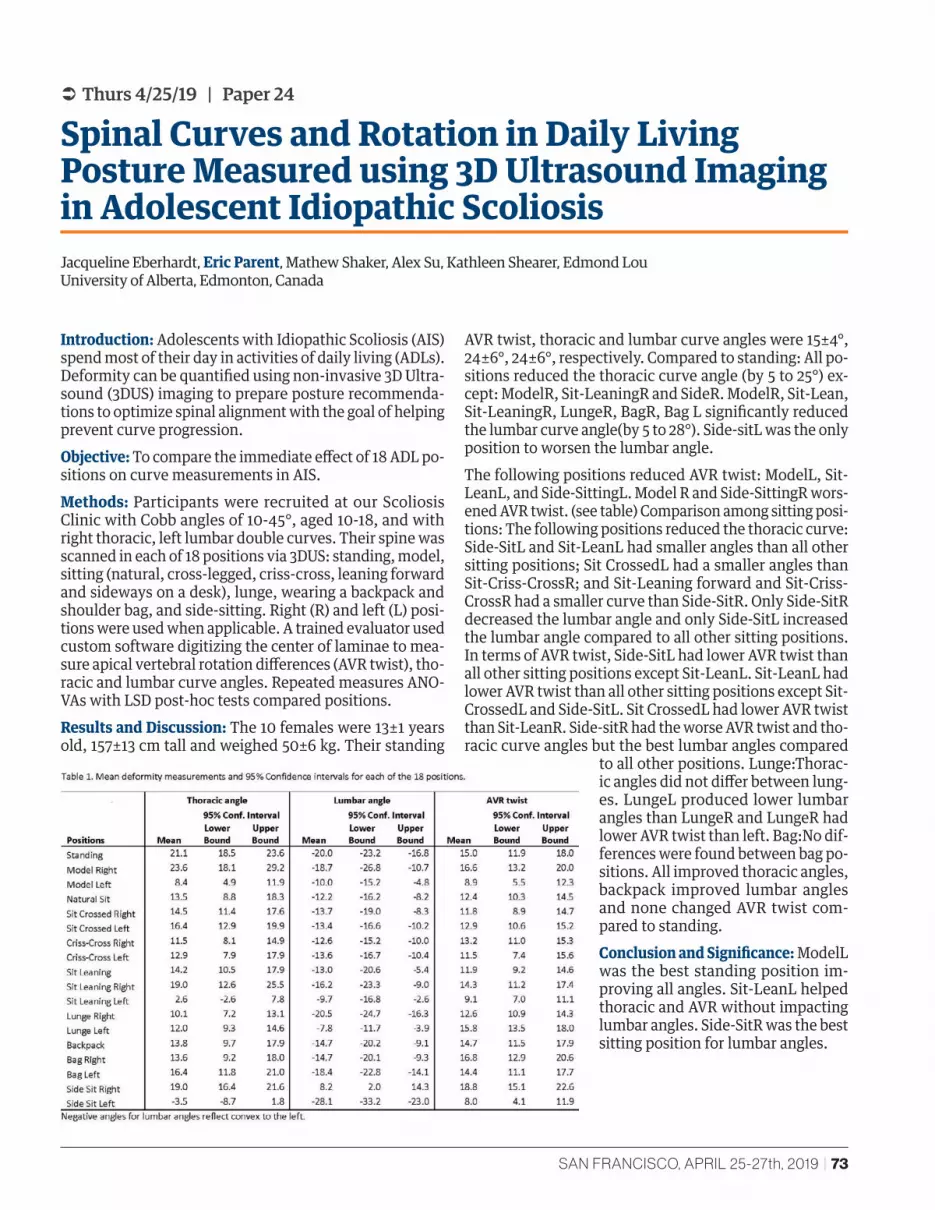
SAN FRANCISCO, APRIL 25-27th, 2019 | 73
 Thurs 4/25/19 | Paper 24
Spinal Curves and Rotation in Daily Living Posture Measured using 3D Ultrasound Imaging in Adolescent Idiopathic ScoliosisJacqueline Eberhardt, Eric Parent, Mathew Shaker, Alex Su, Kathleen Shearer, Edmond Lou University of Alberta, Edmonton, Canada
Introduction: Adolescents with Idiopathic Scoliosis (AIS) spend most of their day in activities of daily living (ADLs). Deformity can be quantified using non-invasive 3D Ultra-sound (3DUS) imaging to prepare posture recommenda-tions to optimize spinal alignment with the goal of helping prevent curve progression.Objective: To compare the immediate effect of 18 ADL po-sitions on curve measurements in AIS.Methods: Participants were recruited at our Scoliosis Clinic with Cobb angles of 10-45°, aged 10-18, and with right thoracic, left lumbar double curves. Their spine was scanned in each of 18 positions via 3DUS: standing, model, sitting (natural, cross-legged, criss-cross, leaning forward and sideways on a desk), lunge, wearing a backpack and shoulder bag, and side-sitting. Right (R) and left (L) posi-tions were used when applicable. A trained evaluator used custom software digitizing the center of laminae to mea-sure apical vertebral rotation differences (AVR twist), tho-racic and lumbar curve angles. Repeated measures ANO-VAs with LSD post-hoc tests compared positions.Results and Discussion: The 10 females were 13±1 years old, 157±13 cm tall and weighed 50±6 kg. Their standing
AVR twist, thoracic and lumbar curve angles were 15±4°, 24±6°, 24±6°, respectively. Compared to standing: All po-sitions reduced the thoracic curve angle (by 5 to 25°) ex-cept: ModelR, Sit-LeaningR and SideR. ModelR, Sit-Lean, Sit-LeaningR, LungeR, BagR, Bag L significantly reduced the lumbar curve angle(by 5 to 28°). Side-sitL was the only position to worsen the lumbar angle. The following positions reduced AVR twist: ModelL, Sit-LeanL, and Side-SittingL. Model R and Side-SittingR wors-ened AVR twist. (see table) Comparison among sitting posi-tions: The following positions reduced the thoracic curve: Side-SitL and Sit-LeanL had smaller angles than all other sitting positions; Sit CrossedL had a smaller angles than Sit-Criss-CrossR; and Sit-Leaning forward and Sit-Criss-CrossR had a smaller curve than Side-SitR. Only Side-SitR decreased the lumbar angle and only Side-SitL increased the lumbar angle compared to all other sitting positions. In terms of AVR twist, Side-SitL had lower AVR twist than all other sitting positions except Sit-LeanL. Sit-LeanL had lower AVR twist than all other sitting positions except Sit-CrossedL and Side-SitL. Sit CrossedL had lower AVR twist than Sit-LeanR. Side-sitR had the worse AVR twist and tho-racic curve angles but the best lumbar angles compared
to all other positions. Lunge:Thorac-ic angles did not differ between lung-es. LungeL produced lower lumbar angles than LungeR and LungeR had lower AVR twist than left. Bag:No dif-ferences were found between bag po-sitions. All improved thoracic angles, backpack improved lumbar angles and none changed AVR twist com-pared to standing.Conclusion and Significance: ModelL was the best standing position im-proving all angles. Sit-LeanL helped thoracic and AVR without impacting lumbar angles. Side-SitR was the best sitting position for lumbar angles.
74 | 14TH INTERNATIONAL SOSORT MEETING
 Thurs 4/25/19 | Paper 25
Impact of Dedicated Physical Activity on Child’s Body PostureMateusz Kozinoga, Piotr Janusz, Katarzyna Politarczyk, Krzysztof Korbel, Łukasz Stoliński, Tomasz Kotwicki University of Medical Sciences, Poznan, Rehasport Clinic, Poznan, Poland, Spine Disorders Center, Skierniewice, Poland
Introduction: The shape of spine curvatures in the correct body posture is characterized by harmonious arches in the sagittal plane which provides both stability and mobility of the spine. Body posture can be assessed with photographic methods. Corrective exercises programs are prescribed as activity to improve faulty posture among school children.Objective: The aim of the study was to compare the sag-ittal spinal curvatures and the head position adopted by healthy children in habitual posture, before and after par-ticipating in a one-year corrective program, by comparing the exercising children versus the non-exercising group. Methods: Material consists of digital photographs of body posture (whole body silhouette) of 400 healthy children (200 girls and 200 boys) age 7.0 to 10.0 years (mean 8.5±1.1): body height (mean 135cm±12.7); body weight (mean 33 kg ±9.7). Exercising group consisted of 167 children (both sexes), non-exercising of 237 children (both sexes). Po-sition of the subjects and the camera were standardized. Sagittal profile of the trunk was assess in standing habit-ual spontaneous position at T0 and after 1 year (T1). The following body posture parameters were measure using a semiautomatic software: Sacral slope (SS), lumbar lor-dosis (LL), thoracic kyphosis (TK), chest inclination (CI),
and head protraction (HP). Increase of the angle value was interpreted as posture deterioration. Corrective activities program consisted of corrective exercises in groups, swim-ming and/or individual corrective sessions. Wilcoxon or paired t-Student test were used. Results and Discussion: Postural parameters [mean (sd)] measured in habitual position at T0 vs. T1 revealed as fol-lows: For non-exercising group: SS 29.94 (6.79) VS. 30.92 (6.69), P=0.0618; LL 42.44 (10.87) VS. 43.7 (10.3), P=0.0152; TK 38.62 (9.86) VS. 40.01 (9.26); P=0.0165; CI 20.1 (9.43) VS. 21.65 (9.79), P=0.0013; HP 31.43 (5.25) VS. 32.87 (5.93), P=0.0009. For exercising group: SS 28.63 (7.6) VS. 27.23 (6.68), P=0.0009; LL 46.08 (9.78) VS. 44.5 (9.34), P=0.0616; TK 42.23 (9.11) VS. 42.79 (9.98); P=0.4044; CI 16.1 (5.64) VS. 16.37 (5.95), P=0.6807; HP 31.96 (6.44) VS. 32.8 (6.07), P=0.1018. In the non-exercising group, the values of all the parameters increased which can be interpreted as pos-ture deterioration. In the exercising group, no significant change was observed. Conclusion and Significance: Physical activities applied as a dedicated program prevented deterioration of pos-tural parameters which was observed as natural history in non-exercising children.
SAN FRANCISCO, APRIL 25-27th, 2019 | 75
 Thurs 4/25/19 | Paper 26
Active Self-Correction of Child’s Body Posture Assessed in Sagittal Plane with Digital PhotographyMateusz Kozinoga, Piotr Janusz, Katarzyna Politarczyk, Krzysztof Korbel, Dariusz Czaprowski, Łukasz Stoliński, Tomasz Kotwicki University of Medical Sciences, Poznan, Poland, Rehasport Clinic, Poznan, Poland, Józef Rusiecki University College, Olsztyn, Poland, Spine Disorders Center, Skierniewice, Poland
Introduction: Body posture is described as spontaneous spatial position of the body parts in standing. Digital photography was reported to be a reliable method for trunk sagittal profile assessment. Active self-correction (ASC) comprises an active movement performed by the child presumably aiming to achieve the best possible position of the body. “Straighten your back” reveals a common command used by the parents to their children in daily living.Objective: The aim of the study was to detect differences between the spontaneous and the actively corrected body posture after “straighten your back” command in a group of previously non-instructed children.Methods: Material consists of digital photographs of body posture (whole body silhouette) of 400 healthy children (200 girls and 200 boys), aged 7.0 to 10.0 years (mean 8.5±1.1) body height mean 135 cm (±12.7); body weight mean 33 kg (±9.7). Children were divided in to the groups: 7-8, 9-10-years old girls, 7-8, 9-10 years old boys, 100 pupils in each. Position of the subjects and the camera were standardized. Sagittal profile of the trunk was assessed twice in standing position: in habitu-al spontaneous position, and position adopted after the “straighten your back” command. The children were not instructed previously about the corrective movement. Body posture parameters (in degrees) were measured using a semiautomatic dedicated program: sacral slope (SS), lumbar lordosis (LL), thoracic kyphosis (TK), chest inclination (CI), and head protraction (HP). Wilcoxon or paired t-Student test were used.
Results and Discussion: Postural parameters in mean (±sd) measured in habitual position vs. position after the “straighten your back” command were as follows: for girls 7-8 yo SS 30.2(±7.51) vs.32.14 (±7.67), p<0.0001; LL 47.99(±9.39) vs.46.88 (±10.45), p=0.154; TK 40.66(±9.14) vs. 32.14(±12.62); p<0.0001. CI 15.76(±6.34) vs.10.81 (±7.87), p<0.0001; HP 32.56(±6.2) vs.28.22 (±6.88), p<0.0001. For boys 7-8 yo SS 28.44 (±7.89) vs.30.4 (±7.84), p<0.0001; LL 46.71 (±9.94) vs.45.56 (±10.33), p=0.1343; TK 42.9 (±9.04) vs.34.57 (±10.95); p<0.0001.CI 15.09 (±5.98) vs.10.14 (±6.6), p<0.0001; HP 30.35 (±6.24) vs.25.97 (±6.5), p<0.0001. For girls 9-10 yo SS 28.37 (±6.94) vs.29.71 (±6.74), p=0.002; LL 45.43 (±8.44) vs.43.64 (±10.92), p=0.0477; TK 41.26 (±9.75) vs.33.58 (±10.91); p<0.0001. CI 16.77 (±6.54) vs.12.28 (±6.98), p<0.0001; HP 32.58 (±5.02) vs.29.62 (±5.3), p<0.0001. For boys 9-10 yo SS 29.1(±7.41) vs. 30.57(±7.15), p=0.0006; LL 45.62 (±9.99) vs.46.02 (±9.552), p=0.538; TK 43.1 (±9.12) vs.35.27 (±11.10); p<0.0001. CI 16.44 (±6.03) vs.10.82 (±7.09), p<0.0001; HP 31.81 (±5.97) vs.27.16 (±7.62); p<0.0001.Conclusion and Significance: Children reacted to the “straighten your back” command by changing their body posture. Active self-correction resulted in the ex-tension of the thoracic spine, reducing chest inclina-tion and head protraction in previously non-instruct-ed healthy children. There were no differences in ASC among children from 7 to 10 yo nor between boys and girls.
76 | 14TH INTERNATIONAL SOSORT MEETING
 Thurs 4/25/19 | Paper 27
Innovative Handheld Device for Population Screening and Early Detection of ScoliosisMichael KT To, Jason PY Cheung, Kenny YH Kwan, Charlene YL Fan, Eric Yeung, Frank F Zhu, Kenneth MC Cheung The University of Hong Kong
Introduction: Scoliosis is a 3-dimensional spinal deformi-ty. Traditionally, screening is performed using the scoli-ometer to measure the back asymmetry of the subjects in forward bend position. However, the location of the hump is inaccurate, and sensitivity and specificity is low with intra and inter-observer errors. The authors devel-oped a novel handheld device that utilizes a gyroscope to assess back asymmetry in real-time 3D. It can also pro-vide more precise identification of the site of deformity and its severity. Objective: The objective of this study is to validate its util-ity in assessing back asymmetry and compare it to the sco-liometer.Methods: Scoliosis patients between 8-20 years old were consecutively recruited. The patients were assessed by scoliometer, our handheld device and X-rays. The mea-surements of apical trunk rotation (ATR) by scoliometer and the tilt angle (SSA) by our device were performed with patients in forward bend position. Intra- and inter-observ-
er reliabilities were assessed by Cronbach’s alpha. Correla-tion of ATR and SSA parameters at their equivalent sites was performed.Results and Discussion: A total of 577 patients were re-cruited for the study. The intra-observer reliability was determined to be 0.9 for the SSA. The inter-observer reli-ability for the spine length, SSA for the maximum +/- tilt, SSA for upper thoracic and lower thoracic were 0.84, 0.67, 0.87, 0.7 and 0.82, respectively. When comparing the cor-relation between ATR and SSA, there was a significant cor-relation between Cobb angle measurement of the thoracic (r=0.77) and lumbar (r=0.66) curvaturesConclusion and Significance: This is the first validation study demonstrating that our novel handheld device has high intra- and inter-observer reliabilities when assessing the spinal profile of patients in forward bend position. Our device provides equal utility as the scoliometer in evalu-ating back asymmetry with the advantages of real-time feedback and 3D assessment.

SAN FRANCISCO, APRIL 25-27th, 2019 | 77
 Thurs 4/25/19 | Paper 28
Presence of Three Types of Scoliosis Formation in Relation to the ATR Angle and Cobb Angle Correlation as the Cause of Difficulty in Diagnosing its Early StagesMarek Kluszczynski, Jacek Wasik, Dorota Ortenburger, Tomasz Kotwicki, Dariusz Czaprowski Jan Dlugosz University in Czestochowa, University of Medical Sciences in Poznan, Poland
Introduction: A lack of correlation between the ATR an-gle and the Cobb angle value in the X-ray picture in chil-dren’s clinical examination is often noticed especially in the early phase of scoliosis.Methods: A comparative analysis of ATR clinical param-eters and the Cobb angle in the X-ray picture was carried out in a group of 117 children. The results of the ATR and Cobb angles were compiled for each child and statistical-ly developed.Results and Discussion: A statistical analysis revealed that the sensitivity range, specificity and AUC in the reliabili-ty assessment of the ATR for the 7˚ amounted to 79.17%,
40.68% and 0.60, respectively. The positive predictive value was 52.05 % and the negative predictive value was 70.59 %. A child with ATR ≥ 7˚ can be sure in 52.05% to have the Cobb angle value ≥ 10˚, while a child with ATR < 7˚ can be 70.59% sure that their Cobb angle is < 10˚.Conclusion and Significance: 1. Consideration should be given to lowering the 7° ATR cut-off threshold to avoid undetected scoliosis at its early stage. 2. The result in-dicates the need to distinguish three types of scoliosis developed – hypo-rotational, normo-rotational and hy-per-rotational scoliosis.

78 | 14TH INTERNATIONAL SOSORT MEETING
 Thurs 4/25/19 | Paper 29
Examination of the Sensitivity of a New Trunk Deformity ScorePatrick Knott, EllaMarie Rakowski, Leah Resnick, Annie Zupan Rosalind Franklin University of Medicine and Science, North Chicago, IL
Introduction: Scoliosis has traditionally been assessed and quantified using standing radiographs and measuring Cobb Angles. The benefit of this method is that it can re-liably quantify the magnitude of the curve in the coronal plane. However, a better way to describe and quantify the true deformity of the trunk caused by scoliosis would be to capture many different measurements and to com-bine them into a score that represented a more global, 3-D approach. Fifteen surface topography parameters were selected to quantify the asymmetry seen in the coronal, sagittal and axial planes. Each measurement was com-pared to a range of possible measurements and then con-verted to a score based on where they fall in that range. Five scores in each plane were combined to arrive at a total deformity score.Objectives: To test the sensitivity of this new deformity score to small changes in trunk shape.Methods: Fifteen healthy adult volunteers were measured using surface topography scanning. The new deformity score was applied to their measurement results on repeat-ed scans to assess the variance in scores. Then three dif-ferent maneuvers were used to change the shape of the
trunk: a heel lift to raise one side of the pelvis in the cor-onal plane; a forward lean to bend the trunk forward in the sagittal plane; and a twist of the pelvis relative to the shoulders to change the trunk in the axial plane. Addi-tional scans assessed the change in the deformity score during these maneuvers.Results and Discussion: Repeated scans showed a vari-ance of +/- 3.06 points in the total deformity score among these subjects in neutral standing. A coronal plane change in trunk shape resulted in a 5.6 point change in the de-formity score. A sagittal plane change in trunk shape re-sulted in a 3.5 point change in the deformity score. And an axial plane change in trunk shape resulted in a 22.9 point change in the deformity score. An analysis of the sub-scores for each plane showed that the large majority of the total score change came from the sub-score in the expected plane.Conclusions and Significance: This new trunk deformi-ty score is sensitive to small changes in the shape of the trunk, and can be used as an adjunct to the radiographic Cobb Angle in describing and quantifying deformity in scoliosis.

SAN FRANCISCO, APRIL 25-27th, 2019 | 79
 Thurs 4/25/19 | Paper 30
ATR Sum – A Valuable Parameter to Assess Total Trunk Torsion in AISTomasz Kotwicki, Krzysztof Korbel, Mateusz Kozinoga, Piotr Janusz University of Medical Sciences, Poznan, Poland
Introduction: Spine deformity in adolescents with idio-pathic scoliosis (AIS) can occur according to a few pat-terns: single primary structural spine curvature (Lenke type 1 or type 5), two primary curvatures (Lenke type 2, type 3 or type 6) or three primary curvatures (Lenke type 4). Natural history of AIS comprises development of secondary, non-structural, spinal curvatures due to the balancing function of the posture control system. Measurement of the Angle of Trunk Rotation (ATR) is used to assess external trunk deformity. During pro-longed non-surgical scoliosis treatment, there is a need to quantify and monitor the global torsional deformity of the trunk. Objective: To introduce a parameter ATR Sum in order to better assess the total trunk torsion. ATR Sum is defined as an algebraic sum of the three ATRs (absolute values) measured at three trunk levels. ATR Sum covers all pri-mary curves and all secondary curves.Methods: (1) Thirty AIS patients were measured with Bunnell scoliometer. ATR was measured at 3 levels of the back: proximal thoracic (pTh), main thoracic (mTh) and thoracolumbar/lumbar (Th/L), as proposed by Len-ke classification. Two authors checked for intra-observer agreement and three authors checked for inter-observer agreement using ICC calculation. ATR Sum was calculated for all patients and compared between authors. Bland-Al-tman plots were analyzed and limits of agreement were estimated. (2) Forty-five AIS girls treated with rigid TLSO were longitudinally measured (mean follow-up time was
4.0±1.3 years) for the ATR at 3 levels and the ATR Sum was calculated. The patients’ ATRs were checked for wheth-er the ATR distribution among the three levels changed over time. Results and Discussion: (1) Reliability of measurements of ATR and ATR Sum were as follows: Intra-rater ICC: pTh 0.84, mTh 0.91, Th/L 0.98, ATR Sum 0.96. Inter-rat-er ICC: pTh 0.81, mTh 0.93, Th/L 0.95, ATR Sum 0.92. In-tra-rater Bland-Altman limits: pTh (1.6 to -2.0), mTh (2.6 to -3.2), Th/L (2.3 to -3.3), ATR Sum (3.6 to -4.8). Inter-rater Bland-Altman limits: pTh (5.7 to -3.4), mTh (4.4 to -4.8), Th/L (2.8 to -4.0), ATR Sum (6.9 to -3.7).(2) Longitudinally calculated ATR Sum revealed stabilization of the torsion-al trunk deformity in 40 patients (including decrease in 16 patients) versus increase in 5 patients, corresponding with Cobb angle increase. Moreover, within the 40 ATR-Sum-stable patients a phenomenon of ATR distribution shift was observed in 14 patients. The ATR Sum present-ing stable value, a shift of trunk rotational deformity from one level to another was observed, most often from the thoracic to proximal thoracic level or from lumbar to tho-racic level. Conclusion and Significance: ATR Sum revealed easy to measure and reliable. This parameter considers the whole thoracic and lumbar spine, covering both the primary and the secondary spinal curvatures. Calculating ATR Sum provided additional information about the AIS brace treat-ment course because the shift of trunk rotation could be detected.
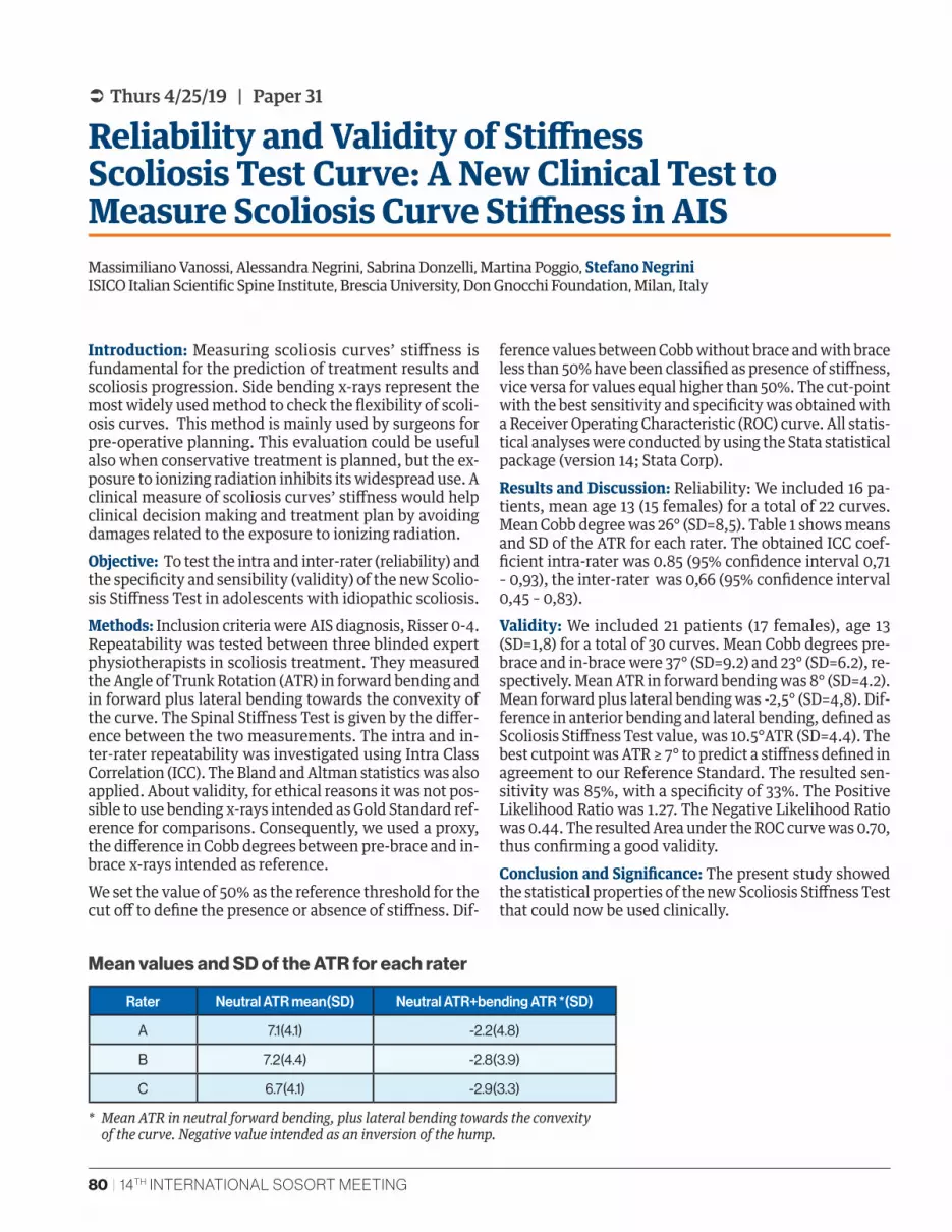
80 | 14TH INTERNATIONAL SOSORT MEETING
 Thurs 4/25/19 | Paper 31
Reliability and Validity of Stiffness Scoliosis Test Curve: A New Clinical Test to Measure Scoliosis Curve Stiffness in AISMassimiliano Vanossi, Alessandra Negrini, Sabrina Donzelli, Martina Poggio, Stefano Negrini ISICO Italian Scientific Spine Institute, Brescia University, Don Gnocchi Foundation, Milan, Italy
Introduction: Measuring scoliosis curves’ stiffness is fundamental for the prediction of treatment results and scoliosis progression. Side bending x-rays represent the most widely used method to check the flexibility of scoli-osis curves. This method is mainly used by surgeons for pre-operative planning. This evaluation could be useful also when conservative treatment is planned, but the ex-posure to ionizing radiation inhibits its widespread use. A clinical measure of scoliosis curves’ stiffness would help clinical decision making and treatment plan by avoiding damages related to the exposure to ionizing radiation. Objective: To test the intra and inter-rater (reliability) and the specificity and sensibility (validity) of the new Scolio-sis Stiffness Test in adolescents with idiopathic scoliosis.Methods: Inclusion criteria were AIS diagnosis, Risser 0-4. Repeatability was tested between three blinded expert physiotherapists in scoliosis treatment. They measured the Angle of Trunk Rotation (ATR) in forward bending and in forward plus lateral bending towards the convexity of the curve. The Spinal Stiffness Test is given by the differ-ence between the two measurements. The intra and in-ter-rater repeatability was investigated using Intra Class Correlation (ICC). The Bland and Altman statistics was also applied. About validity, for ethical reasons it was not pos-sible to use bending x-rays intended as Gold Standard ref-erence for comparisons. Consequently, we used a proxy, the difference in Cobb degrees between pre-brace and in-brace x-rays intended as reference. We set the value of 50% as the reference threshold for the cut off to define the presence or absence of stiffness. Dif-
ference values between Cobb without brace and with brace less than 50% have been classified as presence of stiffness, vice versa for values equal higher than 50%. The cut-point with the best sensitivity and specificity was obtained with a Receiver Operating Characteristic (ROC) curve. All statis-tical analyses were conducted by using the Stata statistical package (version 14; Stata Corp).Results and Discussion: Reliability: We included 16 pa-tients, mean age 13 (15 females) for a total of 22 curves. Mean Cobb degree was 26° (SD=8,5). Table 1 shows means and SD of the ATR for each rater. The obtained ICC coef-ficient intra-rater was 0.85 (95% confidence interval 0,71 – 0,93), the inter-rater was 0,66 (95% confidence interval 0,45 – 0,83). Validity: We included 21 patients (17 females), age 13 (SD=1,8) for a total of 30 curves. Mean Cobb degrees pre-brace and in-brace were 37° (SD=9.2) and 23° (SD=6.2), re-spectively. Mean ATR in forward bending was 8° (SD=4.2). Mean forward plus lateral bending was -2,5° (SD=4,8). Dif-ference in anterior bending and lateral bending, defined as Scoliosis Stiffness Test value, was 10.5°ATR (SD=4.4). The best cutpoint was ATR ≥ 7° to predict a stiffness defined in agreement to our Reference Standard. The resulted sen-sitivity was 85%, with a specificity of 33%. The Positive Likelihood Ratio was 1.27. The Negative Likelihood Ratio was 0.44. The resulted Area under the ROC curve was 0.70, thus confirming a good validity.Conclusion and Significance: The present study showed the statistical properties of the new Scoliosis Stiffness Test that could now be used clinically.
Mean values and SD of the ATR for each rater
Rater Neutral ATR mean(SD) Neutral ATR+bending ATR *(SD)
A 7.1(4.1) -2.2(4.8)
B 7.2(4.4) -2.8(3.9)
C 6.7(4.1) -2.9(3.3)
* Mean ATR in neutral forward bending, plus lateral bending towards the convexity of the curve. Negative value intended as an inversion of the hump.

SAN FRANCISCO, APRIL 25-27th, 2019 | 81
 Fri 4/26/19 - SOSORT Paper Award Nominee | Paper 32
Alternately Repetitive Cast/Brace (ARCB) Treatment for a Larger Magnitude of Early-Onset ScoliosisNoriaki Kawakami, Toshiki Saito, Ryoji Tauchi, Kazuki Kawakami, Tetsuya Ohara Meijo Hospital, Nagoya, Japan
Introduction: Alternately repetitive cast/brace treatment (ARCB-T) is performed to decrease scoliosis and/or delay the need for surgery for mild-to-moderate early-onset sco-liosis (EOS). However, limited studies have investigated the clinical outcomes of patients with larger-magnitude scoliosis.Objectives: This study aims to investigate the potential of ARCB-T as a delayed tactic even for larger-magnitude scoliosis.Methods: In this retrospective cohort study, the inclusion criteria were as follows: (1) EOS; 2) ≤4 years; and 3) scoliosis ≥50° at the first casting. Overall, 78 patients (24 males, 54 females; mean age: 2.9 years at the first casting) fulfilled the criteria. Etiologies were as follows: congenital/structur-al defects (CS/ST; n = 30); infantile idiopathic scoliosis (IIS; n = 23); syndromic scoliosis (SS; n = 23); and neuromuscu-lar scoliosis (n = 2). ARCB-T was switched to surgical in-tervention in 52 patients (age: 7.1 years) primarily because of progression. The endpoints were the progression rate during ARCB-T and the magnitude of scoliosis at the end of ARCB-T. We compared 78 patients (Cast Larger-Group, CL-G) with 42 patients (Cast Mild-Group, CM-G) with sco-liosis <50° at the first cast placement.
Results and Discussion: We observed a reduction of sco-liosis <30° in 11.5% of the CL-G and 28.6% of the CM-G; patients with scoliosis >80° did not display improvement <30°. Early initiation of ARCB-T significantly correlated with a lower progression rate (P = 0.0384). Patients with larger-magnitude scoliosis at the first casting exhibited sig-nificantly larger-magnitude scoliosis at the end of ARCB-T (P < 0.0001). Better correction of scoliosis by initial casting decreased the progression rate (P = 0.0113) in 120 patients (CL-G + CM-G). Although the correction of scoliosis by initial casting did not correlate with the progression rate in the CL-G (P = 0.1153), the progression rate during ARCB-T significantly correlated with the correction by casting in patients with ≥70° or 80° of scoliosis (P = 0.0016). The diagnoses cor-related with the efficacy of ARCB-T; IIS exhibited a sig-nificantly better progression rate (−3.0°/year) than CS/ST or SS (P = 0.0007).Conclusions and Significance: Despite being limited in the suppression of the progression of larger-magnitude scolio-sis, ARCB-T works less efficiently as a delayed tactic to sur-gery and could be an option for larger-magnitude scoliosis if it displays better correction at the first cast placement.
82 | 14TH INTERNATIONAL SOSORT MEETING
 Fri 4/26/19 - SOSORT Paper Award Nominee | Paper 33
Are Lung Functions Related to Spinal Deformities in Patients With Adolescent Idiopathic Scoliosis? A Systematic Review and Meta-AnalysisArnold Wong, Henry Pang, Francesca Di Felice, Stefano Negrini, Matthew Chung, Sabrina Donzelli, Fabio Zaina, Jason Cheung, Dino Samartzis The Hong Kong Polytechnic University, Prince of Wales Hospital, Italian Scientific Spine Institute, University of Brescia, The University of Hong Kong, Rush University
Introduction: Some studies have shown that teens with adolescent idiopathic scoliosis (AIS) display pulmonary impairments although conflicting findings have been re-ported. To better identify and manage AIS patients with pulmonary impairments, a meta-analysis is warranted to summarize the correlations between various lung func-tion parameters and structural features of AIS patients.Objective: To summarize the associations between var-ious lung function parameters and radiographic pheno-types of AIS patients. Methods: Candidate publications were identified from 8 electronic databases, from inception to November 2016. Two independent reviewers screened the titles, abstracts, and full-text of potential articles based on the selection criteria. Another 2 independent reviewers ex-tracted relevant data and appraised the methodological quality of the included studies using established assess-ment tools. The relations between various pulmonary parameters and spinal parameters were summarized qualitatively. Meta-analyses were conducted using ran-dom-effects models, if possible. Results and Discussion: Of 1,045 identified citations, 18 studies (3,075 participants) met the inclusion criteria. Fourteen included studies (2,766 patients) reported sig-nificant low-to-moderate associations between diverse spinal parameters (e.g., axial vertebral rotation) and lung functions (e.g., percent predicted functional vital
capacity (%FVC), percent predicted forced expiratory volume in one second (%FEV1), and percent predicted total lung capacity (%TLC)) in AIS patients. Three in-cluded studies found that patients with impaired lung functions (<65 %FEV1 or %FVC) had significantly larg-er thoracic Cobb angles and less thoracic kyphosis than healthy controls. Homogenous data from 10 studies were pooled for meta-analyses. The results showed that larg-er thoracic Cobb angles were related to lower %FVC (r = -0.34; 95%CI: -0.39 to -0.29), lower %FEV1(r = - 0.33; 95%CI: -0.39 to -0.27), smaller %TLC (r = -0.19; 95%CI: -0.25 to -0.13), and smaller percent predicted vital capac-ity (r = -0.28; 95%CI: -0.34 to -0.22). Conversely, thoracic kyphosis angles were positively associated with %FVC (r = 0.11; 95%CI: 0.07 to 0.15), %FEV1 (r = 0.23; 95%CI: 0.18 to 0.27), and %TLC (r = 0.19; 95%CI: 0.15 to 0.24) (Figure 1). Many included studies had suboptimal methodologi-cal quality. Notably, they did not justify the sample siz-es, and did not report response rates, information about ethics approval, and confounders. Conclusion and Significance: Severe spinal deformities were related to clinically significant pulmonary impair-ments in AIS patients. However, these findings were not adjust for confounders (e.g. physical activity levels). Fu-ture studies should consider the influences of other fac-tors (e.g., physical activity levels) in restoring the lung functions of these patients by non-surgical and surgical means.

SAN FRANCISCO, APRIL 25-27th, 2019 | 83
 Fri 4/26/19 - SOSORT Paper Award Nominee | Paper 33
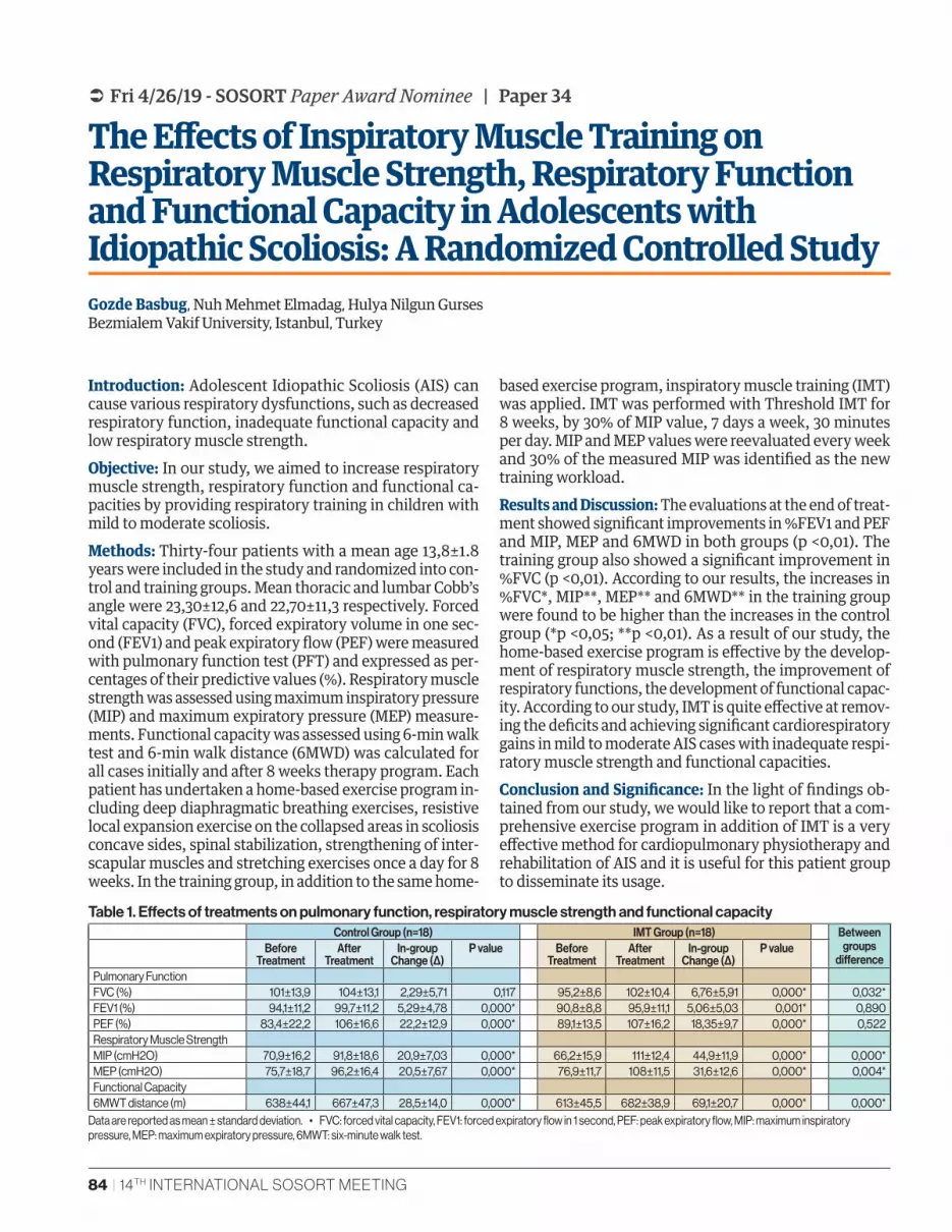
84 | 14TH INTERNATIONAL SOSORT MEETING
 Fri 4/26/19 - SOSORT Paper Award Nominee | Paper 34
The Effects of Inspiratory Muscle Training on Respiratory Muscle Strength, Respiratory Function and Functional Capacity in Adolescents with Idiopathic Scoliosis: A Randomized Controlled StudyGozde Basbug, Nuh Mehmet Elmadag, Hulya Nilgun Gurses Bezmialem Vakif University, Istanbul, Turkey
Introduction: Adolescent Idiopathic Scoliosis (AIS) can cause various respiratory dysfunctions, such as decreased respiratory function, inadequate functional capacity and low respiratory muscle strength.Objective: In our study, we aimed to increase respiratory muscle strength, respiratory function and functional ca-pacities by providing respiratory training in children with mild to moderate scoliosis. Methods: Thirty-four patients with a mean age 13,8±1.8 years were included in the study and randomized into con-trol and training groups. Mean thoracic and lumbar Cobb’s angle were 23,30±12,6 and 22,70±11,3 respectively. Forced vital capacity (FVC), forced expiratory volume in one sec-ond (FEV1) and peak expiratory flow (PEF) were measured with pulmonary function test (PFT) and expressed as per-centages of their predictive values (%). Respiratory muscle strength was assessed using maximum inspiratory pressure (MIP) and maximum expiratory pressure (MEP) measure-ments. Functional capacity was assessed using 6-min walk test and 6-min walk distance (6MWD) was calculated for all cases initially and after 8 weeks therapy program. Each patient has undertaken a home-based exercise program in-cluding deep diaphragmatic breathing exercises, resistive local expansion exercise on the collapsed areas in scoliosis concave sides, spinal stabilization, strengthening of inter-scapular muscles and stretching exercises once a day for 8 weeks. In the training group, in addition to the same home-
based exercise program, inspiratory muscle training (IMT) was applied. IMT was performed with Threshold IMT for 8 weeks, by 30% of MIP value, 7 days a week, 30 minutes per day. MIP and MEP values were reevaluated every week and 30% of the measured MIP was identified as the new training workload.Results and Discussion: The evaluations at the end of treat-ment showed significant improvements in %FEV1 and PEF and MIP, MEP and 6MWD in both groups (p <0,01). The training group also showed a significant improvement in %FVC (p <0,01). According to our results, the increases in %FVC*, MIP**, MEP** and 6MWD** in the training group were found to be higher than the increases in the control group (*p <0,05; **p <0,01). As a result of our study, the home-based exercise program is effective by the develop-ment of respiratory muscle strength, the improvement of respiratory functions, the development of functional capac-ity. According to our study, IMT is quite effective at remov-ing the deficits and achieving significant cardiorespiratory gains in mild to moderate AIS cases with inadequate respi-ratory muscle strength and functional capacities.Conclusion and Significance: In the light of findings ob-tained from our study, we would like to report that a com-prehensive exercise program in addition of IMT is a very effective method for cardiopulmonary physiotherapy and rehabilitation of AIS and it is useful for this patient group to disseminate its usage.
Control Group (n=18) IMT Group (n=18) Between groups
differenceBefore
TreatmentAfter
TreatmentIn-group
Change (Δ)P value Before
TreatmentAfter
TreatmentIn-group
Change (Δ)P value
Pulmonary FunctionFVC (%) 101±13,9 104±13,1 2,29±5,71 0,117 95,2±8,6 102±10,4 6,76±5,91 0,000* 0,032*FEV1 (%) 94,1±11,2 99,7±11,2 5,29±4,78 0,000* 90,8±8,8 95,9±11,1 5,06±5,03 0,001* 0,890PEF (%) 83,4±22,2 106±16,6 22,2±12,9 0,000* 89,1±13,5 107±16,2 18,35±9,7 0,000* 0,522Respiratory Muscle StrengthMIP (cmH2O) 70,9±16,2 91,8±18,6 20,9±7,03 0,000* 66,2±15,9 111±12,4 44,9±11,9 0,000* 0,000*MEP (cmH2O) 75,7±18,7 96,2±16,4 20,5±7,67 0,000* 76,9±11,7 108±11,5 31,6±12,6 0,000* 0,004*Functional Capacity6MWT distance (m) 638±44,1 667±47,3 28,5±14,0 0,000* 613±45,5 682±38,9 69,1±20,7 0,000* 0,000*
Table 1. Effects of treatments on pulmonary function, respiratory muscle strength and functional capacity
Data are reported as mean ± standard deviation. • FVC: forced vital capacity, FEV1: forced expiratory flow in 1 second, PEF: peak expiratory flow, MIP: maximum inspiratory pressure, MEP: maximum expiratory pressure, 6MWT: six-minute walk test.
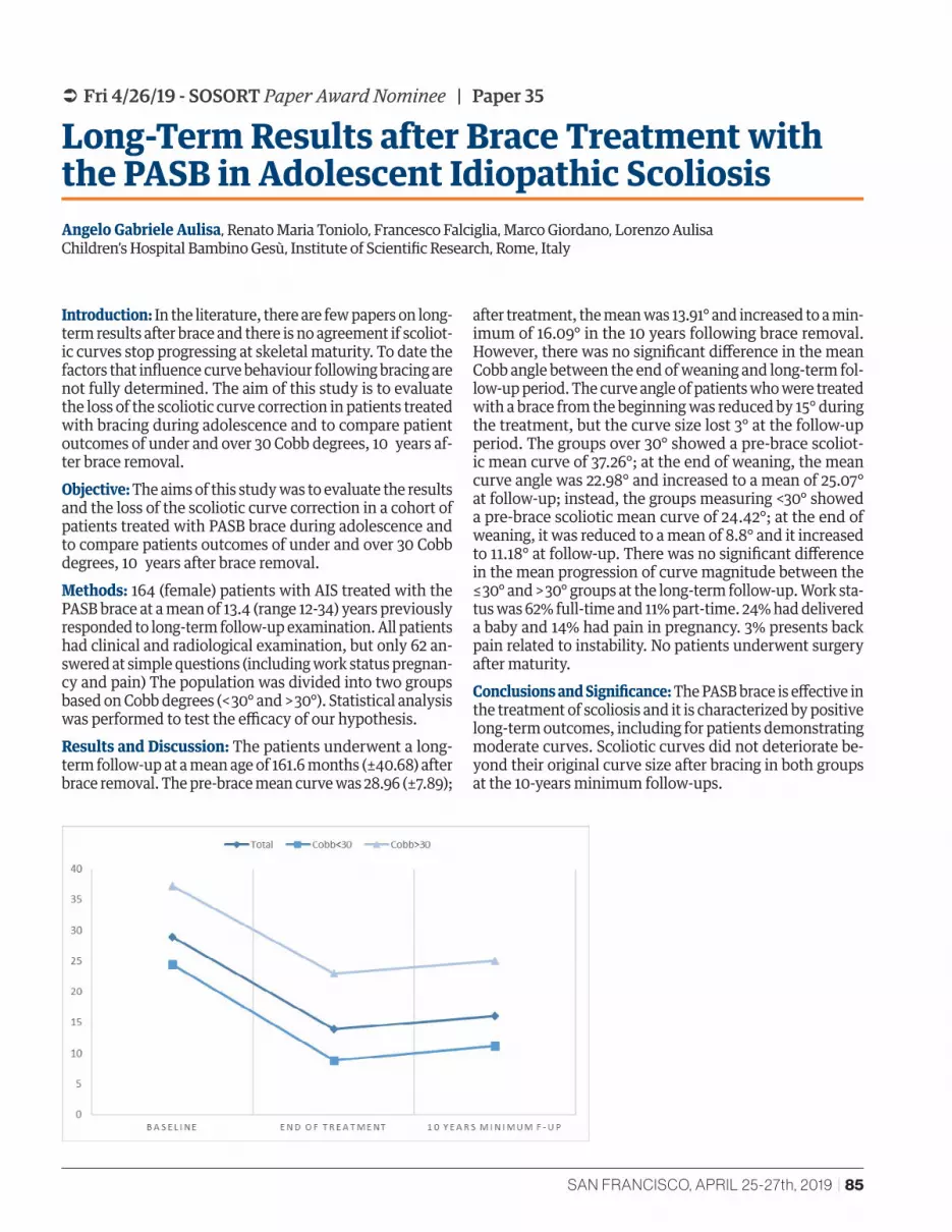
SAN FRANCISCO, APRIL 25-27th, 2019 | 85
 Fri 4/26/19 - SOSORT Paper Award Nominee | Paper 35
Long-Term Results after Brace Treatment with the PASB in Adolescent Idiopathic ScoliosisAngelo Gabriele Aulisa, Renato Maria Toniolo, Francesco Falciglia, Marco Giordano, Lorenzo Aulisa Children’s Hospital Bambino Gesù, Institute of Scientific Research, Rome, Italy
Introduction: In the literature, there are few papers on long-term results after brace and there is no agreement if scoliot-ic curves stop progressing at skeletal maturity. To date the factors that influence curve behaviour following bracing are not fully determined. The aim of this study is to evaluate the loss of the scoliotic curve correction in patients treated with bracing during adolescence and to compare patient outcomes of under and over 30 Cobb degrees, 10 years af-ter brace removal.Objective: The aims of this study was to evaluate the results and the loss of the scoliotic curve correction in a cohort of patients treated with PASB brace during adolescence and to compare patients outcomes of under and over 30 Cobb degrees, 10 years after brace removal.Methods: 164 (female) patients with AIS treated with the PASB brace at a mean of 13.4 (range 12-34) years previously responded to long-term follow-up examination. All patients had clinical and radiological examination, but only 62 an-swered at simple questions (including work status pregnan-cy and pain) The population was divided into two groups based on Cobb degrees (< 30° and > 30°). Statistical analysis was performed to test the efficacy of our hypothesis.Results and Discussion: The patients underwent a long-term follow-up at a mean age of 161.6 months (±40.68) after brace removal. The pre-brace mean curve was 28.96 (±7.89);
after treatment, the mean was 13.91° and increased to a min-imum of 16.09° in the 10 years following brace removal. However, there was no significant difference in the mean Cobb angle between the end of weaning and long-term fol-low-up period. The curve angle of patients who were treated with a brace from the beginning was reduced by 15° during the treatment, but the curve size lost 3° at the follow-up period. The groups over 30° showed a pre-brace scoliot-ic mean curve of 37.26°; at the end of weaning, the mean curve angle was 22.98° and increased to a mean of 25.07° at follow-up; instead, the groups measuring <30° showed a pre-brace scoliotic mean curve of 24.42°; at the end of weaning, it was reduced to a mean of 8.8° and it increased to 11.18° at follow-up. There was no significant difference in the mean progression of curve magnitude between the ≤ 30° and > 30° groups at the long-term follow-up. Work sta-tus was 62% full-time and 11% part-time. 24% had delivered a baby and 14% had pain in pregnancy. 3% presents back pain related to instability. No patients underwent surgery after maturity.Conclusions and Significance: The PASB brace is effective in the treatment of scoliosis and it is characterized by positive long-term outcomes, including for patients demonstrating moderate curves. Scoliotic curves did not deteriorate be-yond their original curve size after bracing in both groups at the 10-years minimum follow-ups.

86 | 14TH INTERNATIONAL SOSORT MEETING
 Fri 4/26/19 - SOSORT Paper Award Nominee | Paper 36
Very Long Term Results of Bracing Management with Initial Plaster Cast in Juvenile and Adolescent Idiopathic Scoliosis: Comparative Results 20 and 30 Years after WeaningJean Claude de Mauroy, Fabio Gagliano, Frédéric Barral Clinique du Parc - Lyon, ROGA - Enna, Lecante Research Group, France
Introduction: The publications concerning the very long-term results of scoliosis bracing are exceptional. Without a brace, the average progression in curvatures less than 40 ° after 20 years is 0.5 ° / year. Management with initial plaster cast or “total time” polycarbonate are currently rare. It is important to confirm the previous studies of the Italian teams from Rome and Milan.Objective: The aim of the study is to check if the scolio-sis remains stable during the period of pregnancies and menopause, 20 and 30 years after brace weaning, unlike natural history.Methods: Retrospective study of all 105 consecutive pa-tients (92 women and 13 men) who were reviewed at least 20 years after brace weaning, extracted from our prospec-tive database starting in 1998. All patients wore a plaster cast before fitting the brace, usually a Lyon, but also Mil-waukee, Charleston, TLSO, Chêneau. All patients had clin-ical and radiological examination according to the same protocol which provides for a control of scoliosis, every 5 years. The population was divided into two groups based on the follow up (20 and 30 years). Statistical analysis was performed on radiological data with SPSS v23.Results and Discussion: For the group 20, the pre-brace scoliotic mean curve was 34,46° (± 11,37) (range 20°-76°). The final result 2 years after weaning was 25,37° (±9,80) with a final correction of 26%. At weaning + 20 the mean
curve was 29,17° (± 13,83), with an angular evolution of 0.21° / year. Two patients had surgery. For the group 30 (n=59), the significant result concerns the evolution be-tween 20 years and 25 years after weaning. The corre-sponding values were 32.72° at W+20 and 34.98° at W+25, with an annual evolution of 0.45° / year. The average final correction percentage is 26%, the average correction 20 years after brace weaning is still 13%.We did not note any significant difference by gender. There is no significant cor-relation between initial angulation and outcome 20 years after ablation. A Pearson product-moment correlation co-efficient was computed to assess the relationship between final result and Cobb angulation 20 years after weaning There was a strong positive correlation between the two variables, r = .912, n = 33, p<0.001 for thoracic curves and r=.902, n=30, p<0.001 for lumbar curves.Conclusion and Significance: To our knowledge, these long-term results have the largest follow-up ever pub-lished on a consecutive series of more than 100 patients. The conservative treatment with initial plaster cast allows an overall correction of 26% 2 years after weaning. The evolution at 20 years after weaning (period of pregnancy) is 0.21° / year lower than in natural history. There is an in-crease in this evolution at the time of menopause. Part of this stability is probably related to the initial plastic defor-mation or creep, of the concave soft tissue.
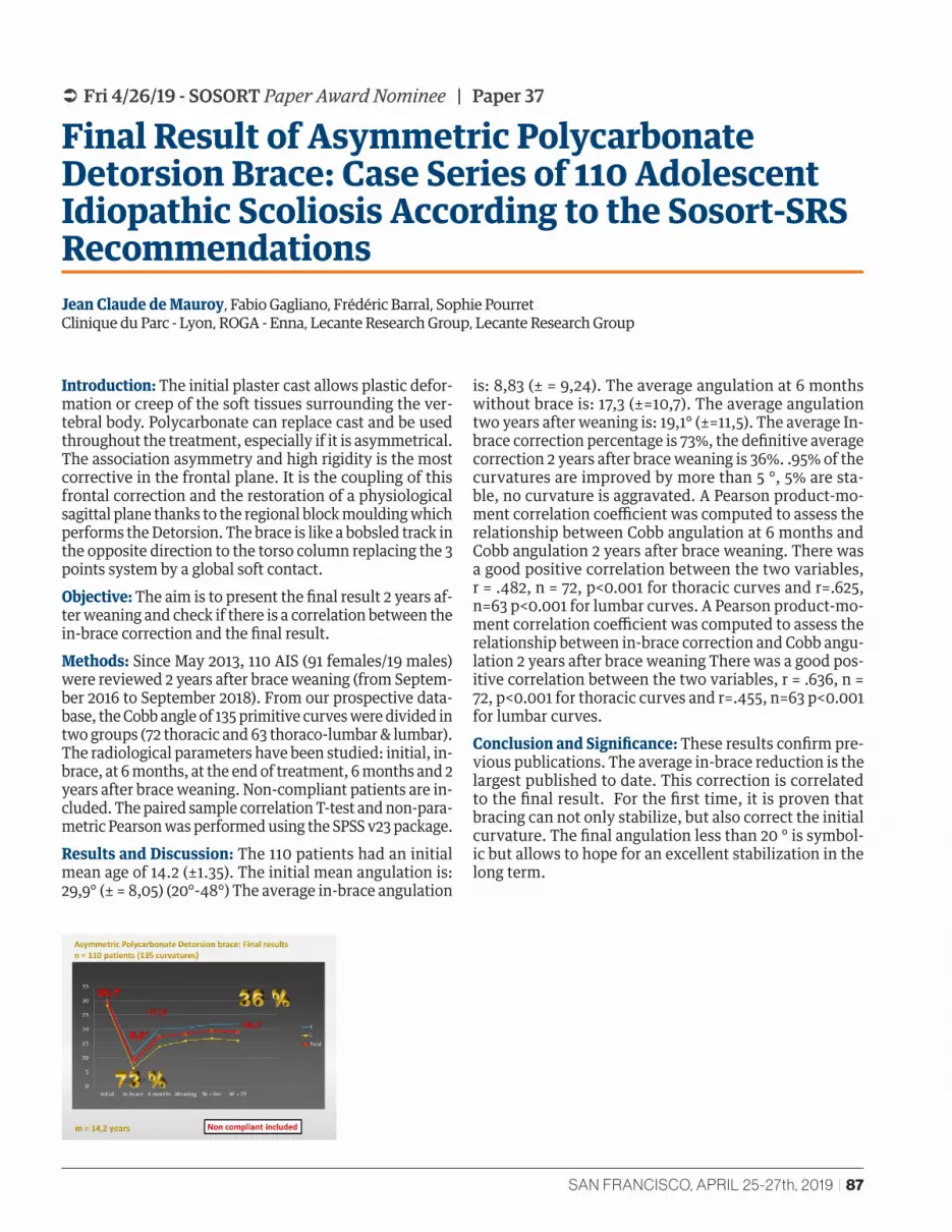
SAN FRANCISCO, APRIL 25-27th, 2019 | 87
 Fri 4/26/19 - SOSORT Paper Award Nominee | Paper 37
Final Result of Asymmetric Polycarbonate Detorsion Brace: Case Series of 110 Adolescent Idiopathic Scoliosis According to the Sosort-SRS RecommendationsJean Claude de Mauroy, Fabio Gagliano, Frédéric Barral, Sophie Pourret Clinique du Parc - Lyon, ROGA - Enna, Lecante Research Group, Lecante Research Group
Introduction: The initial plaster cast allows plastic defor-mation or creep of the soft tissues surrounding the ver-tebral body. Polycarbonate can replace cast and be used throughout the treatment, especially if it is asymmetrical. The association asymmetry and high rigidity is the most corrective in the frontal plane. It is the coupling of this frontal correction and the restoration of a physiological sagittal plane thanks to the regional block moulding which performs the Detorsion. The brace is like a bobsled track in the opposite direction to the torso column replacing the 3 points system by a global soft contact.Objective: The aim is to present the final result 2 years af-ter weaning and check if there is a correlation between the in-brace correction and the final result.Methods: Since May 2013, 110 AIS (91 females/19 males) were reviewed 2 years after brace weaning (from Septem-ber 2016 to September 2018). From our prospective data-base, the Cobb angle of 135 primitive curves were divided in two groups (72 thoracic and 63 thoraco-lumbar & lumbar). The radiological parameters have been studied: initial, in-brace, at 6 months, at the end of treatment, 6 months and 2 years after brace weaning. Non-compliant patients are in-cluded. The paired sample correlation T-test and non-para-metric Pearson was performed using the SPSS v23 package. Results and Discussion: The 110 patients had an initial mean age of 14.2 (±1.35). The initial mean angulation is: 29,9° (± = 8,05) (20°-48°) The average in-brace angulation
is: 8,83 (± = 9,24). The average angulation at 6 months without brace is: 17,3 (±=10,7). The average angulation two years after weaning is: 19,1° (±=11,5). The average In-brace correction percentage is 73%, the definitive average correction 2 years after brace weaning is 36%. .95% of the curvatures are improved by more than 5 °, 5% are sta-ble, no curvature is aggravated. A Pearson product-mo-ment correlation coefficient was computed to assess the relationship between Cobb angulation at 6 months and Cobb angulation 2 years after brace weaning. There was a good positive correlation between the two variables, r = .482, n = 72, p<0.001 for thoracic curves and r=.625, n=63 p<0.001 for lumbar curves. A Pearson product-mo-ment correlation coefficient was computed to assess the relationship between in-brace correction and Cobb angu-lation 2 years after brace weaning There was a good pos-itive correlation between the two variables, r = .636, n = 72, p<0.001 for thoracic curves and r=.455, n=63 p<0.001 for lumbar curves.Conclusion and Significance: These results confirm pre-vious publications. The average in-brace reduction is the largest published to date. This correction is correlated to the final result. For the first time, it is proven that bracing can not only stabilize, but also correct the initial curvature. The final angulation less than 20 ° is symbol-ic but allows to hope for an excellent stabilization in the long term.
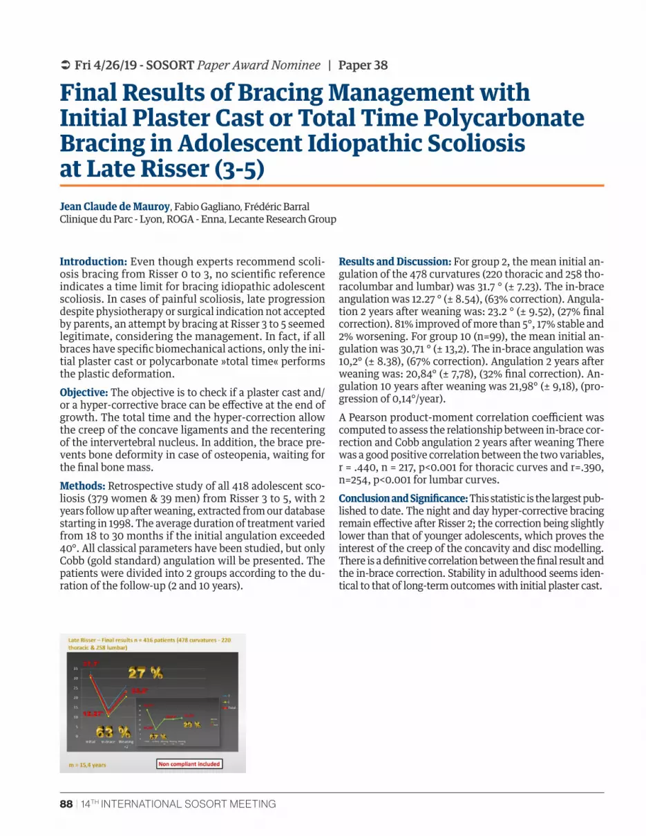
88 | 14TH INTERNATIONAL SOSORT MEETING
 Fri 4/26/19 - SOSORT Paper Award Nominee | Paper 38
Final Results of Bracing Management with Initial Plaster Cast or Total Time Polycarbonate Bracing in Adolescent Idiopathic Scoliosis at Late Risser (3-5)Jean Claude de Mauroy, Fabio Gagliano, Frédéric Barral Clinique du Parc - Lyon, ROGA - Enna, Lecante Research Group
Introduction: Even though experts recommend scoli-osis bracing from Risser 0 to 3, no scientific reference indicates a time limit for bracing idiopathic adolescent scoliosis. In cases of painful scoliosis, late progression despite physiotherapy or surgical indication not accepted by parents, an attempt by bracing at Risser 3 to 5 seemed legitimate, considering the management. In fact, if all braces have specific biomechanical actions, only the ini-tial plaster cast or polycarbonate »total time« performs the plastic deformation.Objective: The objective is to check if a plaster cast and/or a hyper-corrective brace can be effective at the end of growth. The total time and the hyper-correction allow the creep of the concave ligaments and the recentering of the intervertebral nucleus. In addition, the brace pre-vents bone deformity in case of osteopenia, waiting for the final bone mass.Methods: Retrospective study of all 418 adolescent sco-liosis (379 women & 39 men) from Risser 3 to 5, with 2 years follow up after weaning, extracted from our database starting in 1998. The average duration of treatment varied from 18 to 30 months if the initial angulation exceeded 40°. All classical parameters have been studied, but only Cobb (gold standard) angulation will be presented. The patients were divided into 2 groups according to the du-ration of the follow-up (2 and 10 years).
Results and Discussion: For group 2, the mean initial an-gulation of the 478 curvatures (220 thoracic and 258 tho-racolumbar and lumbar) was 31.7 ° (± 7.23). The in-brace angulation was 12.27 ° (± 8.54), (63% correction). Angula-tion 2 years after weaning was: 23.2 ° (± 9.52), (27% final correction). 81% improved of more than 5°, 17% stable and 2% worsening. For group 10 (n=99), the mean initial an-gulation was 30,71 ° (± 13,2). The in-brace angulation was 10,2° (± 8.38), (67% correction). Angulation 2 years after weaning was: 20,84° (± 7,78), (32% final correction). An-gulation 10 years after weaning was 21,98° (± 9,18), (pro-gression of 0,14°/year). A Pearson product-moment correlation coefficient was computed to assess the relationship between in-brace cor-rection and Cobb angulation 2 years after weaning There was a good positive correlation between the two variables, r = .440, n = 217, p<0.001 for thoracic curves and r=.390, n=254, p<0.001 for lumbar curves.Conclusion and Significance: This statistic is the largest pub-lished to date. The night and day hyper-corrective bracing remain effective after Risser 2; the correction being slightly lower than that of younger adolescents, which proves the interest of the creep of the concavity and disc modelling. There is a definitive correlation between the final result and the in-brace correction. Stability in adulthood seems iden-tical to that of long-term outcomes with initial plaster cast.

SAN FRANCISCO, APRIL 25-27th, 2019 | 89
 Fri 4/26/19 - SOSORT Paper Award Nominee | Paper 39
Impact of Sports Activity on Full-Time Braced Patients: An Observational Study of 785 Risser 0–2 Adolescents with Idiopathic ScoliosisAlessandra Negrini, Sabrina Donzelli, Massimiliano Vanossi, Martina Poggio, Fabio Zaina, Michele Romano, Stefano Negrini ISICO, Milan, Italy
Introduction: No information is available to determine if sports activity modifies short- and long-term results of adolescents with idiopathic scoliosis (IS) wearing a brace full-time.Objectives: To assess the effect of sports activities and their frequency in a large population of adolescents with IS, wearing full-time a thoraco-lumbar-sacral (TLSO) brace.Methods: Design. Retrospective observational multi-centric cohort study embedded in a prospective clini-cal database started in 2003. Date of extraction: Novem-ber 1st, 2018. Participants. All the consecutive patients in a multicenter clinical database of a tertiary referral institute age ≥ 10, with juvenile or adolescent IS diag-nosis, Risser 0–2, TLSO brace prescription and self-re-ported adherence ≥ 20 hours per day, follow-up out-of-brace X-rays 6 and 18 months after brace prescription. Outcome measures: A reduction ≥ 5° Cobb at 6-months and 18-months follow-up X-rays was considered an im-provement. Statistics: Odds ratio (OR) was calculated to compare the outcome of subjects performing sport and those not performing sports. A logistic regression with
covariate adjustment was run to assess if frequency had an effect on the outcome measures.Results and Discussion: 785 subjects (mean age 12.7 ± 1.3, 693 females, Cobb degrees 39.8 ± 11.4, range 16-90) were included. At 6-months and 18-months follow-up subjects performing sports showed higher odds of improvement (OR = 1.62, 95%CI = 1.19-2.20, p = 0.0011, and OR = 1.59, 95%CI = 1.17-2.16, p = 0.0018, respectively). After adjust-ments for covariates, at 6-months follow-up only sports ac-tivity resulted significant, whereas at 18-months follow-up ATR, sports activity and sports frequency resulted signifi-cant (p = 0.009, p = 0.009, p = 0.001, respectively). As the sport’s frequency increased, the odds of improvement in-creased.Conclusion and Significance: This study shows that sports activities increase the odds of improvement at 6- and 18-months follow-up in adolescents with IS treated with a full-time brace. In the long term follow-up, the odds of improvement increase with the increase of sports frequen-cy on a weekly basis. Future studies with different designs should look at single sports.
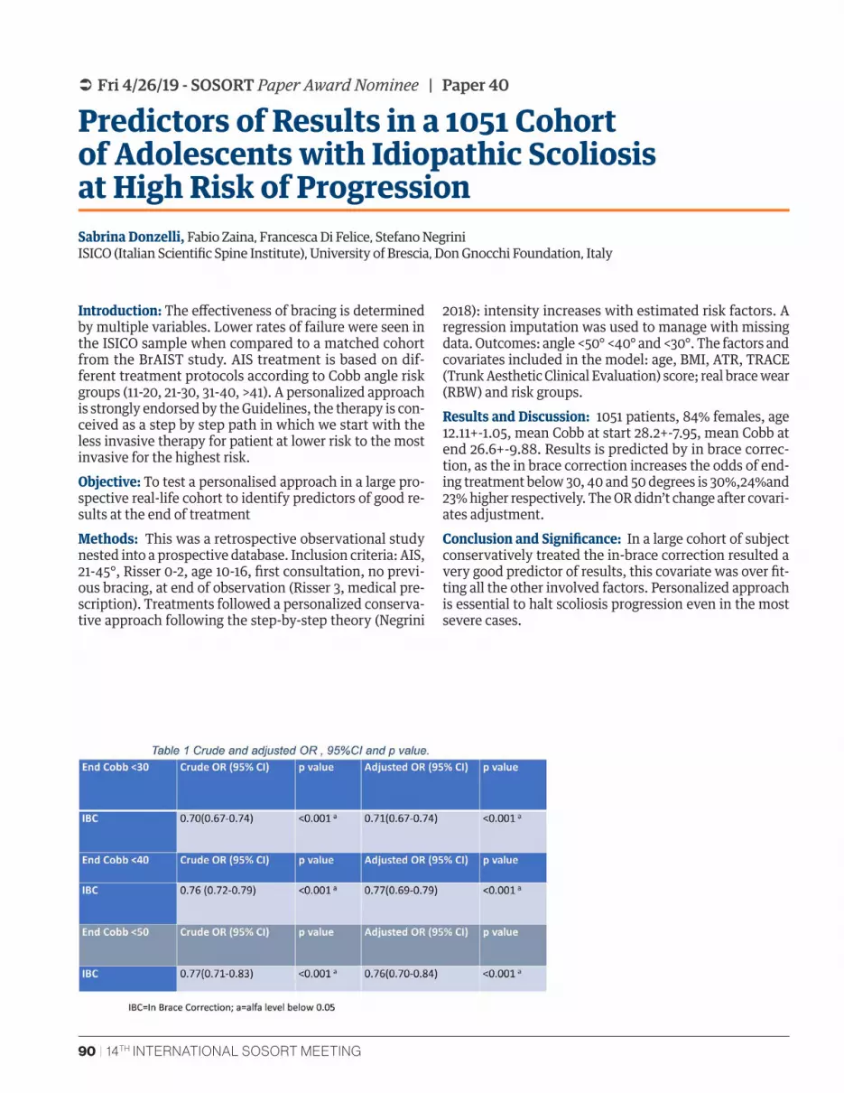
90 | 14TH INTERNATIONAL SOSORT MEETING
 Fri 4/26/19 - SOSORT Paper Award Nominee | Paper 40
Predictors of Results in a 1051 Cohort of Adolescents with Idiopathic Scoliosis at High Risk of ProgressionSabrina Donzelli, Fabio Zaina, Francesca Di Felice, Stefano Negrini ISICO (Italian Scientific Spine Institute), University of Brescia, Don Gnocchi Foundation, Italy
Introduction: The effectiveness of bracing is determined by multiple variables. Lower rates of failure were seen in the ISICO sample when compared to a matched cohort from the BrAIST study. AIS treatment is based on dif-ferent treatment protocols according to Cobb angle risk groups (11-20, 21-30, 31-40, >41). A personalized approach is strongly endorsed by the Guidelines, the therapy is con-ceived as a step by step path in which we start with the less invasive therapy for patient at lower risk to the most invasive for the highest risk.Objective: To test a personalised approach in a large pro-spective real-life cohort to identify predictors of good re-sults at the end of treatment Methods: This was a retrospective observational study nested into a prospective database. Inclusion criteria: AIS, 21-45°, Risser 0-2, age 10-16, first consultation, no previ-ous bracing, at end of observation (Risser 3, medical pre-scription). Treatments followed a personalized conserva-tive approach following the step-by-step theory (Negrini
2018): intensity increases with estimated risk factors. A regression imputation was used to manage with missing data. Outcomes: angle <50° <40° and <30°. The factors and covariates included in the model: age, BMI, ATR, TRACE (Trunk Aesthetic Clinical Evaluation) score; real brace wear (RBW) and risk groups.Results and Discussion: 1051 patients, 84% females, age 12.11+-1.05, mean Cobb at start 28.2+-7.95, mean Cobb at end 26.6+-9.88. Results is predicted by in brace correc-tion, as the in brace correction increases the odds of end-ing treatment below 30, 40 and 50 degrees is 30%,24%and 23% higher respectively. The OR didn’t change after covari-ates adjustment. Conclusion and Significance: In a large cohort of subject conservatively treated the in-brace correction resulted a very good predictor of results, this covariate was over fit-ting all the other involved factors. Personalized approach is essential to halt scoliosis progression even in the most severe cases.
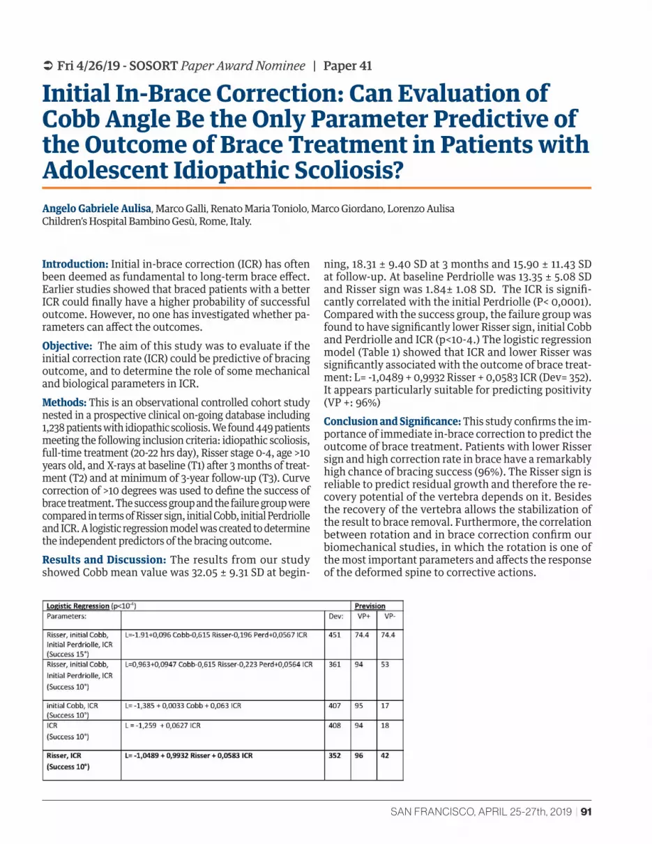
SAN FRANCISCO, APRIL 25-27th, 2019 | 91
 Fri 4/26/19 - SOSORT Paper Award Nominee | Paper 41
Initial In-Brace Correction: Can Evaluation of Cobb Angle Be the Only Parameter Predictive of the Outcome of Brace Treatment in Patients with Adolescent Idiopathic Scoliosis?Angelo Gabriele Aulisa, Marco Galli, Renato Maria Toniolo, Marco Giordano, Lorenzo Aulisa Children’s Hospital Bambino Gesù, Rome, Italy.
Introduction: Initial in-brace correction (ICR) has often been deemed as fundamental to long-term brace effect. Earlier studies showed that braced patients with a better ICR could finally have a higher probability of successful outcome. However, no one has investigated whether pa-rameters can affect the outcomes.Objective: The aim of this study was to evaluate if the initial correction rate (ICR) could be predictive of bracing outcome, and to determine the role of some mechanical and biological parameters in ICR.Methods: This is an observational controlled cohort study nested in a prospective clinical on-going database including 1,238 patients with idiopathic scoliosis. We found 449 patients meeting the following inclusion criteria: idiopathic scoliosis, full-time treatment (20-22 hrs day), Risser stage 0-4, age >10 years old, and X-rays at baseline (T1) after 3 months of treat-ment (T2) and at minimum of 3-year follow-up (T3). Curve correction of >10 degrees was used to define the success of brace treatment. The success group and the failure group were compared in terms of Risser sign, initial Cobb, initial Perdriolle and ICR. A logistic regression model was created to determine the independent predictors of the bracing outcome. Results and Discussion: The results from our study showed Cobb mean value was 32.05 ± 9.31 SD at begin-
ning, 18.31 ± 9.40 SD at 3 months and 15.90 ± 11.43 SD at follow-up. At baseline Perdriolle was 13.35 ± 5.08 SD and Risser sign was 1.84± 1.08 SD. The ICR is signifi-cantly correlated with the initial Perdriolle (P< 0,0001).Compared with the success group, the failure group was found to have significantly lower Risser sign, initial Cobb and Perdriolle and ICR (p<10-4.) The logistic regression model (Table 1) showed that ICR and lower Risser was significantly associated with the outcome of brace treat-ment: L= -1,0489 + 0,9932 Risser + 0,0583 ICR (Dev= 352). It appears particularly suitable for predicting positivity (VP +: 96%)Conclusion and Significance: This study confirms the im-portance of immediate in-brace correction to predict the outcome of brace treatment. Patients with lower Risser sign and high correction rate in brace have a remarkably high chance of bracing success (96%). The Risser sign is reliable to predict residual growth and therefore the re-covery potential of the vertebra depends on it. Besides the recovery of the vertebra allows the stabilization of the result to brace removal. Furthermore, the correlation between rotation and in brace correction confirm our biomechanical studies, in which the rotation is one of the most important parameters and affects the response of the deformed spine to corrective actions.

92 | 14TH INTERNATIONAL SOSORT MEETING
 Fri 4/26/19 | Paper 42
The Prevalence of Adult de Novo Scoliosis: A Systematic Review and Meta-AnalysisJeb McAviney, Carrie Roberts, Bryony Sullivan, Alex Alevras, Petra L Graham, Benjamin T Brown Scolicare. Kogarah NSW, Australia and Macquarie University, North Ryde NSW, Australia
Introduction: The reported prevalence estimates for de-generative scoliosis are quite varied ranging from 2-68%. Objectives: Researchers sought to determine the prev-alence of the subtype of degenerative scoliosis, termed adult de novo scoliosis, based on reports from the exist-ing literature.Methods: We searched MEDLINE, Embase, CINAHL, Web of Science and PubMed from database inception to 28th March, 2018. Studies that assessed adults from the general population for scoliosis using medical imaging techniques were included. Importantly, studies were included only if the authors had tried to exclude participants with previ-ously diagnosed scoliosis or related spinal disorders. Stud-ies in English, German, Dutch and Greek were included. The risk of bias and quality of reporting were assessed for each study. We used mixed-effects logistic-regression to determine an overall prevalence estimate with 95% con-fidence intervals and to examine the effect of age and gen-der on prevalence. Results and Discussion: Five studies (4 cross-sectional studies and 1 cohort study) involving 4069 participants
aged between 41 and 94 years were eligible for final inclu-sion in the review. The highest reported prevalence fig-ure reported was 68% (51/75) and the lowest prevalence (13% [318/2395]) was reported in the cohort study. The risk of bias assessment revealed that sample size calculations were not conducted in any of the included studies, and that 3/5 studies did not adequately discuss the limitations asso-ciated with their study. These same deficiencies were also noted in the assessment of reporting quality. The pooled prevalence estimate from the mixed-effects logistic regres-sion analysis was 37.6% (95% CI 18.7-61.8). Females were more likely to suffer from scoliosis compared with males (P<0.001), with prevalence figures of 41.2% (95% CI 20.7-65.8) versus 27.5% (95% CI 12.2-51.1) respectively. Individ-uals aged under 60 years had a prevalence of 13% (95% CI 5 to 30) whereas the prevalence estimates were substan-tially higher in the 60+ age groups (36% [95% CI 17 to 61]). Conclusion and Significance: Adult de novo scoliosis is a highly prevalent condition, especially in females. Further research, specifically targeting adult de novo scoliosis, is required to obtain more precise estimates of the preva-lence of the condition and the influence of age and gender.

SAN FRANCISCO, APRIL 25-27th, 2019 | 93
 Fri 4/26/19 | Paper 43
A Systematic Literature Review of Bracing Treatment for Adults with ScoliosisJeb McAviney, Johanna Mee, Azharuddin Fazalbhoy, Juan Du Plessis, Benjamin Brown Sydney Scoliosis Clinic, Melbourne Scoliosis Clinic, Australia
Introduction: Scoliosis is common in adults with the prev-alence reported to be as high as 68%. There is currently a paucity of literature regarding the conservative manage-ment of this subgroup of the population. Objective: To review and summarise the existing literature on the efficacy of bracing for adults with scoliosis. Methods: CINAHL, Embase, CENTRAL, PubMed and PE-Dro were searched. The authors sought to retrieve all studies investigating the clinical effects of brace/ortho-sis treatment in adults with scoliosis. A combination of MeSH terms and keywords pertaining to three core con-cepts (adult, scoliosis, and braces/orthoses) were used. Studies were included if A) clinical outcomes were col-lected from B) participants ≥18 years of age C) who were receiving soft or rigid brace treatment for D) degenerative de novo scoliosis (DDS) or progressive adolescent scoliosis in the adult (ASA) and E) had been published in full-text in English. Data were extracted, and a risk of bias assessment was performed using the Newcastle-Ottawa scale (NOS). The data were then collated and summarized. Results and Discussion: After applying the inclusion cri-teria there were 9 studies (4 case reports and 5 cohort studies [1 retrospective and 4 prospective]) detailing the clinical outcomes of soft (2/9) or rigid (7/9) bracing, used as a standalone therapy (6/9) or in combination with physiotherapy/rehabilitation (3/9), in 306 partic-ipants with various types of idiopathic or degenerative
scoliosis of varying severity. Most (7/9) studies included only female participants. The most commonly reported outcomes were pain, function and coronal Cobb angle measurement, with follow-up times ranging from 2 days to 17 years. Brace wear prescriptions ranged from 2-20 hours per day and there was mixed brace-compliance reported among studies. Most studies highlighted a sig-nificant reduction in pain and improvement in function at follow-up assessments. Overall there were mixed re-sults with regards to the sagittal and coronal plane ra-diographic parameters. However, one of the strongest longitudinal studies demonstrated a statistically sig-nificant change in long-term established progression of both DDS and ASA via the introduction of bracing. There were no direct harms associated with bracing. Each of the five cohort studies were of low quality based on the NOS instrument. Conclusion and Significance: Bracing appears to have a positive influence on pain and functional outcomes in adults with progressive ASA and DDS. From a clinical per-spective the evidence would suggest that some patients may benefit in the short- to medium-term from bracing. However, even though most of the studies published in this area support the use of braces for adult scoliosis, the current evidence is scant and of low quality due to reliance on samples drawn from clinical databases. Properly con-structed prospective trials are required to truly understand the efficacy of bracing for adults with scoliosis.
94 | 14TH INTERNATIONAL SOSORT MEETING
 Fri 4/26/19 | Paper 44
Safe Exercises for Adults with Scoliosis - A Scoping Review with a Proposed AlgorithmLise Stolze, Hagit Berdishevsky, Sanja Schreiber, Jean Claude De Mauroy Columbia University, New York, University of Alberta, Edmonton, Canada, Clinique du Parc – Lyon, France
Introduction: Adults with scoliosis are at risk for curve pro-gression, pain and disability, especially in the presence of scoliosis adult modifiers. SOSORT guidelines recommend that all people with scoliosis remain active. The sparse evi-dence on exercises for adults with scoliosis has focused on Physiotherapy Scoliosis Specific Exercise (PSSE), but not general exercise for fitness. Since adults with scoliosis of-ten join fitness programs such as Yoga or Pilates to maintain strength and flexibility for overall quality of life, it is im-portant for this population to understand how to exercise safely and minimize the risk of future pain and disability. Objectives: We undertook a scoping review to map the ev-idence on general exercises or presence of guidelines for adults with asymptomatic scoliosis, and to develop an al-gorithm based on current evidence and clinical experience to help adults with scoliosis make safe exercise choices. Method: MEDLINE database was searched from inception to Nov 12, 2018. To augment the search, we traced the ref-erences of key studies and hand searched the key journals in the area. Inclusion criteria: cohort studies with control group, controlled clinical trials (randomized and quasi randomized) and guidelines on exercises for symptomatic and asymptomatic adults with scoliosis. Exclusion criteria: studies and guidelines investigating the effect of PSSE. We applied screening in duplicate. The evidence was synthe-sized narratively. PRISMA Extension for Scoping Reviews (PRISMA-ScR) Checklist was consulted.
Results and Discussion: The search yielded 396 references. We found no recommendations regarding general exercis-es for adults with asymptomatic scoliosis. Consequent-ly, an algorithm has been developed based on current un-derstanding of scoliosis spine mechanics and research de-scribing the impact of adult spine modifiers on adults with scoliosis. We propose the following guidelines: 1) adults with symp-tomatic scoliosis should consult a scoliosis specialist pri-or to beginning a general exercise program; 2) adults with asymptomatic scoliosis and with adult modifiers verified clinically or radiographically should perform exercises in a neutral spine position; 3) adults with asymptomatic scolio-sis, with no radiographic or clinical modifiers may practice general exercises in non-neutral spinal positions if they can minimize forces that increase spinal collapse (Condi-tional Program); 4) adults with asymptomatic scoliosis and no clinical modifiers who lack a radiograph may practice a Conditional Program if the primary curve is not lumbar or thoracolumbar. Conclusions and Significance: Adults with scoliosis should participate in exercise programs adapted in the presence of pain and adult spinal modifiers. A scoping review iden-tified a lack of guidelines on safe exercising for adults with scoliosis. A theoretical framework and algorithm have been proposed to help guide safe exercise choices for this pop-ulation.

SAN FRANCISCO, APRIL 25-27th, 2019 | 95
 Fri 4/26/19 | Paper 44
96 | 14TH INTERNATIONAL SOSORT MEETING
 Fri 4/26/19 | Paper 45
Mobilisation of the Nervous System in Patients with Adult Scoliosis and PainEvgeniya Dimitrova National Sports Academy, Sofia, Bulgaria
Introduction: There are very few papers concerning phys-iotherapy for adult scoliosis. Objectives: This study aims to determine the effect of a twelve-month neuro-mobilization intervention added to standard physiotherapy method (Experimental group) on the pain and deformity compared to standard physiother-apy method alone (Control group) in patients with adult idiopathic scoliosis and pain. Methods: Thirty six patients with adolescent-onset adult thoracolumbar idiopathic scoliosis aged 22-35 years, with curves of 30°-40° and Risser grade 5 were recruited from a single orthopedic clinic and randomized to the Experimen-tal (EG, n=18) or Control group (CG, n=18). The standard physiotherapy (PT) method, which includes auto-correc-tion in 3D, integration in daily life, stabilizing the corrected posture, and patient education consisted of a 30 minute daily home program and weekly supervised sessions by a physiotherapist. The neuro-mobilization exercise was performed from supine position with straight leg raised repetitive active ankle dorsiflexion (20 repetitions, 2 times daily). Patient outcomes included visual analogue scale (VAS) for pain, the angle of trunk rotation (ATR), and Cobb angles measured by radiological images at baseline, and at 12-months follow-up. The collected data were statistically treated by using SPSS software, version 18.0, and the fol-lowing values were found: minimum, maximum, mean,
S.D., and one sample paired t-test to compare between pre- and post-treatment in results in the group. The unpaired t-test was used to compare pre- and post-treatment results between two groups, at a confidence level of p≤0.05. Results and Discussion: The statistical processing of the data shows a significant Cobb angles and ATR improvement in both groups of patients. In EG Cobb angle decreased from 34.7±3.9° to 27.6±4.1°, ∆X=7.1, p<0.05, ATR from 10.7±2.5° to 5.6±1.3°, p<0.05, and pain score from 3.7±1.1 to 0.6±0.9, p<0.05. A change of 5° Cobb angle was consid-ered clinically significant. In CG Cobb angle decreased from 33.6±3.4° to 30.6±4.7°, ∆X=2.9, p<0.05, ATR from 10.6±2.2° to 7.9±1.2°, p<0.05, and pain score from 3.8±1.3 to 2.9±1.2, p<0.05. The comparative analysis of the final results be-tween the two groups showed better pain outcomes in the EG patients compared to CG patients (p<0.05). These find-ings indicate a connection between the mobility of the spi-nal cord and the pain. Therefore, we propose to perform neuro-mobilization exercise in conjunction with scoliosis specific exercises in the treatment of patients with adult idiopathic scoliosis and chronic back pain. Conclusions and Significance: The standard PT method improved deformity. Neuro-mobilization added to the standard PT method was superior compared to standard PT method alone for reducing the pain and curve severity in patients with adult idiopathic scoliosis.

SAN FRANCISCO, APRIL 25-27th, 2019 | 97
 Fri 4/26/19 | Paper 46
Exploring the Cost Effectiveness of an Intensive Physiotherapeutic Scoliosis Specific Exercise (PSSE) Programme in a UK Adult PopulationD. A. Jason Black, Erika Maude, David Glynn, Abbie Turland Scoliosis SOS, London, England
Introduction: A key objective of treatment for scoliosis in adulthood should aim to improve quality of life (QoL) whilst maintaining cost-effectiveness. Cost effectiveness analysis (CEA) is a recognised approach to estimate the cost of treatment to health systems and is used to inform fund-ing decisions. Generic QoL measures such as the EQ5D5L are required in CEA to compare treatments across disease areas. The dimensions of the EQ5D5L are weighted to re-flect the relationship of quality of life and life expectancy and are used to calculate quality adjusted life years (QA-LYs). CEA takes both costs and benefits of treatment into account to provide estimates of cost-effectiveness. NICE stipulates that the incremental cost effectiveness ratio (ICER) should be below £30,000 per QALY to be consid-ered cost effective.Objectives: The aim of this study was to explore the cost effectiveness of intensive physiotherapy scoliosis specific exercises (PSSEs) on adults with idiopathic scoliosis (IS) in the United Kingdom (UK). Methods: 183 consecutive UK-based adults with IS who were initially completing an intensive course of PSSEs were enrolled into the study over a 3 year period. The partici-pants completed the EQ5D5L questionnaire before, im-mediately after treatment and at 6-, 12- and 18-months post-treatment. The EQ5D5L results were converted into QALYs using assumptions about the duration of treatment effect.
Results and Discussion: Of the 183 participants who reached their respective timeframes, the EQ5D5L ques-tionnaire was answered by 97% pre-treatment, 80% pre- and post-treatment, 86% pre- and 6-months post-treat-ment, 93% pre- and 12-months post-treatment and 72% pre- and 18-months post-treatment. Pre-treatment, the mean EQ5D5L score was 0.773. The mean change of the score was 0.11 (P<0.05) immediately post-treatment and 0.09 (P<0.05) at 6-, 12- and 18-months post-treatment. There was no significant decline in benefit between post-treatment and 6-, 12- and 18-months post-treatment (p=0.1).The remaining life expectancy of the average pa-tient in this cohort was 43.8 years. Assuming that the effect of treatment would persist for this length of time and that the expected costs of treatment were £4,024, additional QALYs ranged from 3.899 (lifetime benefit) to 0.045 (ben-efit only until post-treatment), which meant that the ICER ranged from £1,000 to £90,000 per QALY respectively. To meet the requirements of NICE, the effects of treatment needed to persist for 1.5 years.Conclusion and Significance: Intensive PSSEs were pro-posed to be effective at £30,000 per QALY if the observed treatment effect persisted for at least 18 months. Further research is required to determine the long-term effects.

98 | 14TH INTERNATIONAL SOSORT MEETING
 Fri 4/26/19 | Paper 47
Cheneau Bracing is Effective in Older Patients with Larger Curves – Results of Retrospective Chart AnalysisGrant Wood, Sanja Schreiber Align Clinic, LLC, San Mateo, CA, University of Alberta, Edmonton, Canada
Introduction: In North America, bracing is a standard of care for growing adolescents with idiopathic scolio-sis (AIS) and curves 20°-40°. Cheneau type braces are designed to achieve the best possible correction in all 3 planes as compared to most commonly used TLSO type with a tubular design that are most commonly used. Research suggests that TLSO bracing prevents scoliosis curves progression to the threshold for surgery defined as 50°. Conversely, the evidence of the effectiveness of Cheneau type brace is lacking. Moreover, skepticism around bracing patients who are close to the end of growing (i.e. Risser >3) and with larg-er curves exists.Objective: To determine the effect of at least 1-year inter-vention with Cheneau brace on the Cobb angle change in patients with AIS considering their baseline maturity level as defined by Risser.Methods: This is a preliminary retrospective chart anal-ysis of a cohort of 38 adolescents with AIS recruited in a single centre from March 2013 to November 2018. The cohort included patients aged 10 to 16, Risser score 0 to 4 and curves between 20° and 60°. The primary outcome was change in the Largest Curve, and secondary out-comes were change in the Thoracic and Lumbar Curves. The treatment consisted of an evaluation and 3D scan-ning of patients using Rodin Neo Software followed by a computer-aided design/computer-aided manufacturing
(CAD/CAM) 3D modelling of a brace positive, and pro-duction of a hand-modified hyper-corrected mold. Each brace was custom fitted, and the in-brace correction was checked. Patients were followed-up between 12 and 15 months. Out-of-brace radiographs were taken at base-line, and follow-up with a patient being out of a brace for at least 24 hours. The patients were instructed to wear a brace full time (23h/day). The repeated measures ANCO-VA was used to determine the effect of the treatment on change in primary and secondary outcomes using base-line Risser score as a covariate. We used Tukey adjust-ment for post hoc comparisons. Results and Discussion: The mean age, Risser score, Largest Curve, Thoracic Curve and Lumbar Curve were 12.3±1.4, 0.8±1.2, 35.1°± 10.1°, 31.9°±12.7° and 23.9°±13.2°, respectively. Of 38 patients, 32 were girls. The mean follow-up was 13.5 months. On average, the Largest Curve and Thoracic curve improved by 5.58° (SE=1.78°, p=0.005) and 4.94° (SE=1.94°, p=0.017), respectively. Lumbar curve also improved by 4.5° but did not reach statistical significance (SE=2.79°, p=0.119). Risser sign was predictive of success, such that adolescents with Risser 4 had a largest improvement of on average 8.0° (SE=8.98°), 12.0° (SE=9.95°) and 9.0° (SE=11.1°) for Larg-est, Thoracic and Lumbar Curves, respectively. Patients with Risser 4 had largest curves at the initiation of the treatment, with Largest, Thoracic and Lumbar Curves measuring 52.3° (SE=8.93°), 51.4° (SE=9.95°) and 41.0° (SE=11.1°), respectively.Conclusion and Significance: The main goal of the brace treatment is to stop the curves from progression. In this study, we found that Cheneau braces improved the Larg-est, Thoracic and Lumbar curves over a mean follow up of 13.5 months in patients with AIS. Moreover, we found that older patients also benefitted from the treatment despite starting with curves beyond the threshold for bracing, suggesting that bracing could be effective even when curves are larger than suggested 40°. Long-term effect of the 3D Cheneau type brace concept is urgently needed.

SAN FRANCISCO, APRIL 25-27th, 2019 | 99
 Fri 4/26/19 | Paper 48
Correlation between Three-Dimensional Parameters of Body Surface in the Evaluation of Thoracic Idiopathic ScoliosisIsis Navarro, Cláudia Candotti Universidade Federal do Rio Grande do Sul - Grupo Biomec, Brazil
Introduction: The early detection of idiopathic scolio-sis is recommended by the most important worldwide organizations of conservative and surgical treatment of scoliosis. The Scoliometer is considered the reference among early detection procedures which seek to avoid the X-ray examination. Also, photogrammetry is a reli-able tool which allows to evaluate the posture and inves-tigate the presence of scoliosis. Besides, it can be easily used as an early detection practice.Objectives: The aim of this study was to evaluate the correlation and agreement between the trunk rotation, the apex of the curve and the thoracic kyphosis angle measured with a surface topography scanner and: (1) the angle of trunk rotation (ATR) measured with the Scolio-meter; (2) the scoliotic arrow and (3) thoracic kyphosis angle measured with a photogrammetric protocol. Methods: This cross-sectional study was conducted at the Universidade Federal do Rio Grande do Sul - Brazil, between June 2017 and April 2018. All study procedures were approved by the Research Ethics Committee of the University. Fifty-two participants of both genders, aged between 7 and 18 years old, were intentionally included in this study. The exclusion criteria were spinal surgical treatment, neurological disease, lower limb discrepancy greater than 2 cm and body mass index above 29 kg/m2. Each participant was evaluated by means of a surface topography scanner (which was developed and built in Brazil), photogrammetry (using the protocol of the Dig-
ital Image-based Postural Assessment – DIPA) and a sco-liometer. The sequence of the three consecutive proce-dures of evaluation was randomly-chosen. For statistical purpose, the Spearman’s Rho and Pearson’s correlation test, the root mean square (RMS) error and the Bland-Al-tman graphic analysis were conducted (p<.05). Results and Discussion: For the thoracic region, the cor-relation with surface topography parameters was strong and significant for both Scoliometer (trunk rotation Rho=.72; RMS error = 6.4°; n=52) and photogrammetry (scoliotic arrow Rho=.79; RMS error = 1 cm; n=52 and tho-racic kyphosis angle r=.76; RMS error 8.4°; n=51) which presented 0.8°, -0.2 cm and 0.4° of mean difference in the Bland-Altman analysis, respectively. The high cor-relation between the surface parameters observed in this study, associated with the reliability of the tools taken as reference, support the possibility of using the surface topography scanner to evaluate patients under investi-gation of scoliosis. Also, the use of the topographic eval-uation can be helpful in the early detection of scoliosis.Conclusion and Significance: The findings of this study showed that there is correlation and agreement between the surface topography parameters with the scoliometer and photogrammetry. Based on our results, we suggest the use of surface topography for the evaluation and fol-low-up of patients with idiopathic scoliosis. The exam provides relevant three-dimensional information and can be quick and easily used.

100 | 14TH INTERNATIONAL SOSORT MEETING
 Fri 4/26/19 | Paper 49
Surface Topography: Determining the Predictive Parameters of Thoracic Idiopathic ScoliosisIsis Navarro, Cláudia Candotti, Jefferson Loss Universidade Federal do Rio Grande do Sul - Grupo Biomec, Brazil
Introduction: Surface topography is a radiation-free ex-amination that provides three-dimensional information for the evaluation of patients with Idiopathic Scoliosis (IS). However, its usage is not standardized, which re-stricts its applicability. Objective: The aim of this study was to identify which parameters extracted from surface topography predict thoracic IS. Methods: This is a cross-sectional study. All the proce-dures were approved by the Research Ethics Committee of the Universidade Federal do Rio Grande do Sul - Brazil, where the data were collected between June 2017 and April 2018. Seventy-seven participants of both genders, aged between 7 and 18 years old, were intentionally included in this study. The exclusion criteria were spinal surgical treatment, neurological disease, a lower limb discrepan-cy greater than 2 cm and body mass index above 29 kg/m2. Each participant was evaluated using three random-ly-chosen consecutive procedures, by means of surface topography scanner (developed and built in Brazil), pho-togrammetry (using the protocol of Digital Image-based Postural Assessment – DIPA) and a scoliometer®. The latter two were taken as references in this study. For statistical purposes, a multiple linear regression analysis using the “backward” math method was conducted. The scoliotic arrow (photogrammetry) and angle of trunk rotation (sco-liometer) were used as dependent variables, while the 13 surface topography parameters recommended by SOSORT were used as independent variables, with α=0.05.
Results and Discussion: The analysis resulted in two sta-tistically significant models (p<0.001). The model us-ing the scoliometer as reference showed the highest R2 (.73) and included eight surface topography parameters: three in the frontal plane (distance C7-S1 β=-.192, left β=-.004 and right β=.338 apex of the curve), three in the axial plane (Posterior Superior Iliac Spine (PSIS) rotation β=.347, left β= -.084 and right β=.562 trunk rotation) and two in the sagittal plane (thoracic kyphosis β=-.097 and displacement C7-S1 β= -.078). The model using the photogrammetry as reference showed a R2 (.71) and in-cluded nine surface topography parameters: four in the frontal plane (trunk length β=-.164, pelvic tilt β=.104, left β=.055 and right β=.440 apex of the curve), three in the axial plane (posterior superior iliac spine (PSIS) rota-tion β=.136, left β= -.176 and right β=.358 trunk rotation) and two in the sagittal plane (thoracic kyphosis β=-.066 and displacement C7-S1 β= -.110). Our results corroborate with the literature, which emphasizes the three-dimen-sional approach. The use of this exam can provide rele-vant information in the evaluation of scoliosis. Conclusion and Significance: Surface topography is able to identify thoracic IS based on a three-dimensional ap-proach using eight predictive parameters. Standardiz-ing the use of this tool would allow the evaluation and follow-up of patients with IS avoiding exposure to ion-izing radiation.

SAN FRANCISCO, APRIL 25-27th, 2019 | 101
 Fri 4/26/19 | Paper 50
Thoracic Idiopathic Scoliosis: Establishing the Diagnostic Accuracy and Reference Values of Surface TopographyIsis Navarro, Cláudia Candotti, Jefferson Loss Universidade Federal do Rio Grande do Sul - Grupo Biomec, Brazil
Introduction: Surface topography is a very useful exam-ination for the evaluation of the spine and the trunk. The exam provides relevant three-dimensional information that can help in the diagnosis and follow-up of patients with id-iopathic scoliosis. However, there is no agreement among health professionals regarding the appropriate procedures and reference values for the use and interpretation of sur-face topography. Objective: The aim of this study was to establish the diag-nostic accuracy and cut-off points of the surface topography parameters. Methods: This cross-sectional study was conducted in the Universidade Federal do Rio Grande do Sul - Brazil, between June 2017 and April 2018. All the study proce-dures were approved by the Research Ethics Committee of the university. Seventy-seven participants of both gen-ders, aged between 7 and 18 years old, were intentionally included in this study. The exclusion criteria were spinal surgical treatment, neurological disease, a lower limb dis-crepancy greater than 2 cm and body mass index above 29 kg/m2. Each participant was evaluated using two ran-domly-chosen consecutive procedures, by means of a sur-face topography scanner (developed and built in Brazil) and a Scoliometer. In this study, the angle of trunk rota-tion (ATR) determined using the Scoliometer was taken as reference. For statistical purposes, a multiple linear re-gression analysis was made to establish which surface to-
pography parameters have the highest standardized beta coefficients (β). Based on the β values, two topographic parameters were chosen (apex of the curve and trunk ro-tation) to compose the Receiver Operating Characteristic (ROC) analysis. Results and Discussion: The sample was composed of 60% females (n= 46) and 40% males (n=31), with an average age of 13.0±3.3, weight 46±15 kg and height 157±15 cm. The cut-off points for the topographic parameters were established as ATR ≤5° for subjects without scoliosis and >8° for severe sco-liosis. The ROC curve analysis (see Table 1) for the apex of the curve was significant (p<0.001) with an area under the curve (AUC) ranging between 76% [cut-off point 4.4 mm] for the subjects without scoliosis and 84% [cut-off point 9.4 mm] for the subjects with severe scoliosis. For the trunk rotation parameter, the AUC was also significant, ranging between 68% [cut-off point 1.5°, p=0.023] for subjects without scoli-osis, and 73% [cut-off point 4.8°, p=0.018] for the subjects with severe scoliosis. The evidence in this field is scarce, and the lack of standardization hinders the comparison among studies. Our results suggest an approach to standardize the use of surface topography. Conclusion and Significance: Surface topography provides adequate accuracy and can be used to evaluate the presence of the thoracic idiopathic scoliosis. Our study established the cut-off points for the apex of the curve and trunk rotation as parameters for surface topography.
ATR Surface topography Cut-off point Sensitivity Specificity AUC
5°
Apex of the curve
4.4 72% 67% 0.76**
6° 4.4 82% 69% 0.81**
7° 6.6 76% 77% 0.84**
>8° 9.4 71% 85% 0.84**
5°
Trunk rotation
1.5 64% 69% 0.68*
6° 1.5 68% 69% 0.71*
7° 4.8 65% 93% 0.73*
ART: angle of trunk rotation; AUC: area under the curve; *p≤0.05; **p≤0.01
Data from the building of the receiver operating characteristics (ROC) curve
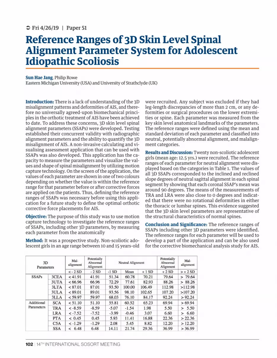
102 | 14TH INTERNATIONAL SOSORT MEETING
 Fri 4/26/19 | Paper 51
Reference Ranges of 3D Skin Level Spinal Alignment Parameter System for Adolescent Idiopathic ScoliosisSun Hae Jang, Philip Rowe Eastern Michigan University (USA) and University of Strathclyde (UK)
Introduction: There is a lack of understanding of the 3D misalignment patterns and deformities of AIS, and there-fore no universally agreed-upon biomechanical princi-ples in the orthotic treatment of AIS have been achieved to date. To address these concerns, 3D skin level spinal alignment parameters (SSAPs) were developed. Testing established their concurrent validity with radiographic alignment parameters and the ability to quantify the 3D misalignment of AIS. A non-invasive calculating and vi-sualising assessment application that can be used with SSAPs was also developed. This application has the ca-pacity to measure the parameters and visualize the val-ues and shape of spinal misalignment by utilizing motion capture technology. On the screen of the application, the values of each parameter are shown in one of two colours depending on whether the value is within the reference range for that parameter before or after corrective forces are applied on the patients. Thus, defining the reference ranges of SSAPs was necessary before using this appli-cation for a future study to define the optimal orthotic corrective force placements for AIS. Objective: The purpose of this study was to use motion capture technology to investigate the reference ranges of SSAPs, including other 3D parameters, by measuring each parameter from the anatomically Method: It was a prospective study. Non-scoliotic ado-lescent girls in an age range between 10 and 15 years-old
were recruited. Any subject was excluded if they had leg-length discrepancies of more than 2 cm, or any de-formities or surgical procedures on the lower extremi-ties or spine. Each parameter was measured from the key skin level anatomical landmarks of the parameters. The reference ranges were defined using the mean and standard deviation of each parameter and classified into neutral, potentially abnormal alignment, and malalign-ment categories. Results and Discussion: Twenty non-scoliotic adolescent girls (mean age: 12.5 yrs.) were recruited. The reference ranges of each parameter for neutral alignment were dis-played based on the categories in Table 1. The values of all 3D SSAPs corresponded to the inclined and reclined slope degrees of neutral sagittal alignment in each spinal segment by showing that each coronal SSAP’s mean was around 90 degrees. The means of the measurements of TRA and LRA were also close to 0 degrees and indicat-ed that there were no rotational deformities in either the thoracic or lumbar spines. This evidence suggested that the 3D skin level parameters are representative of the structural characteristics of normal spines.Conclusion and Significance: The reference ranges of SSAPs including other 3D parameters were identified. The reference ranges for each parameter will be used to develop a part of the application and can be also used for the corrective biomechanical analysis study for AIS.
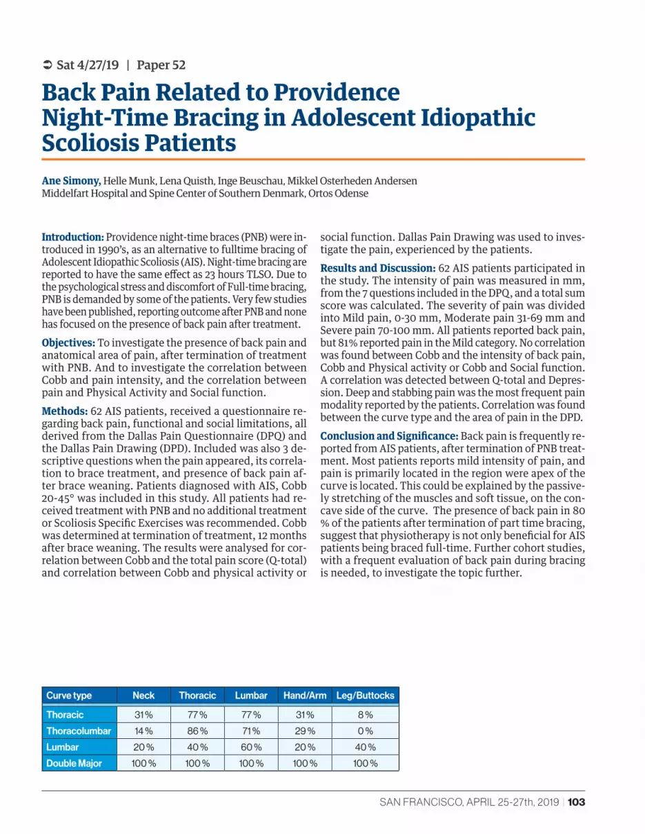
SAN FRANCISCO, APRIL 25-27th, 2019 | 103
 Sat 4/27/19 | Paper 52
Back Pain Related to Providence Night-Time Bracing in Adolescent Idiopathic Scoliosis PatientsAne Simony, Helle Munk, Lena Quisth, Inge Beuschau, Mikkel Osterheden Andersen Middelfart Hospital and Spine Center of Southern Denmark, Ortos Odense
Introduction: Providence night-time braces (PNB) were in-troduced in 1990’s, as an alternative to fulltime bracing of Adolescent Idiopathic Scoliosis (AIS). Night-time bracing are reported to have the same effect as 23 hours TLSO. Due to the psychological stress and discomfort of Full-time bracing, PNB is demanded by some of the patients. Very few studies have been published, reporting outcome after PNB and none has focused on the presence of back pain after treatment. Objectives: To investigate the presence of back pain and anatomical area of pain, after termination of treatment with PNB. And to investigate the correlation between Cobb and pain intensity, and the correlation between pain and Physical Activity and Social function.Methods: 62 AIS patients, received a questionnaire re-garding back pain, functional and social limitations, all derived from the Dallas Pain Questionnaire (DPQ) and the Dallas Pain Drawing (DPD). Included was also 3 de-scriptive questions when the pain appeared, its correla-tion to brace treatment, and presence of back pain af-ter brace weaning. Patients diagnosed with AIS, Cobb 20-45° was included in this study. All patients had re-ceived treatment with PNB and no additional treatment or Scoliosis Specific Exercises was recommended. Cobb was determined at termination of treatment, 12 months after brace weaning. The results were analysed for cor-relation between Cobb and the total pain score (Q-total) and correlation between Cobb and physical activity or
social function. Dallas Pain Drawing was used to inves-tigate the pain, experienced by the patients. Results and Discussion: 62 AIS patients participated in the study. The intensity of pain was measured in mm, from the 7 questions included in the DPQ, and a total sum score was calculated. The severity of pain was divided into Mild pain, 0-30 mm, Moderate pain 31-69 mm and Severe pain 70-100 mm. All patients reported back pain, but 81 % reported pain in the Mild category. No correlation was found between Cobb and the intensity of back pain, Cobb and Physical activity or Cobb and Social function. A correlation was detected between Q-total and Depres-sion. Deep and stabbing pain was the most frequent pain modality reported by the patients. Correlation was found between the curve type and the area of pain in the DPD.Conclusion and Significance: Back pain is frequently re-ported from AIS patients, after termination of PNB treat-ment. Most patients reports mild intensity of pain, and pain is primarily located in the region were apex of the curve is located. This could be explained by the passive-ly stretching of the muscles and soft tissue, on the con-cave side of the curve. The presence of back pain in 80 % of the patients after termination of part time bracing, suggest that physiotherapy is not only beneficial for AIS patients being braced full-time. Further cohort studies, with a frequent evaluation of back pain during bracing is needed, to investigate the topic further.
Curve type Neck Thoracic Lumbar Hand/Arm Leg/Buttocks
Thoracic 31 % 77 % 77 % 31 % 8 %
Thoracolumbar 14 % 86 % 71 % 29 % 0 %
Lumbar 20 % 40 % 60 % 20 % 40 %
Double Major 100 % 100 % 100 % 100 % 100 %
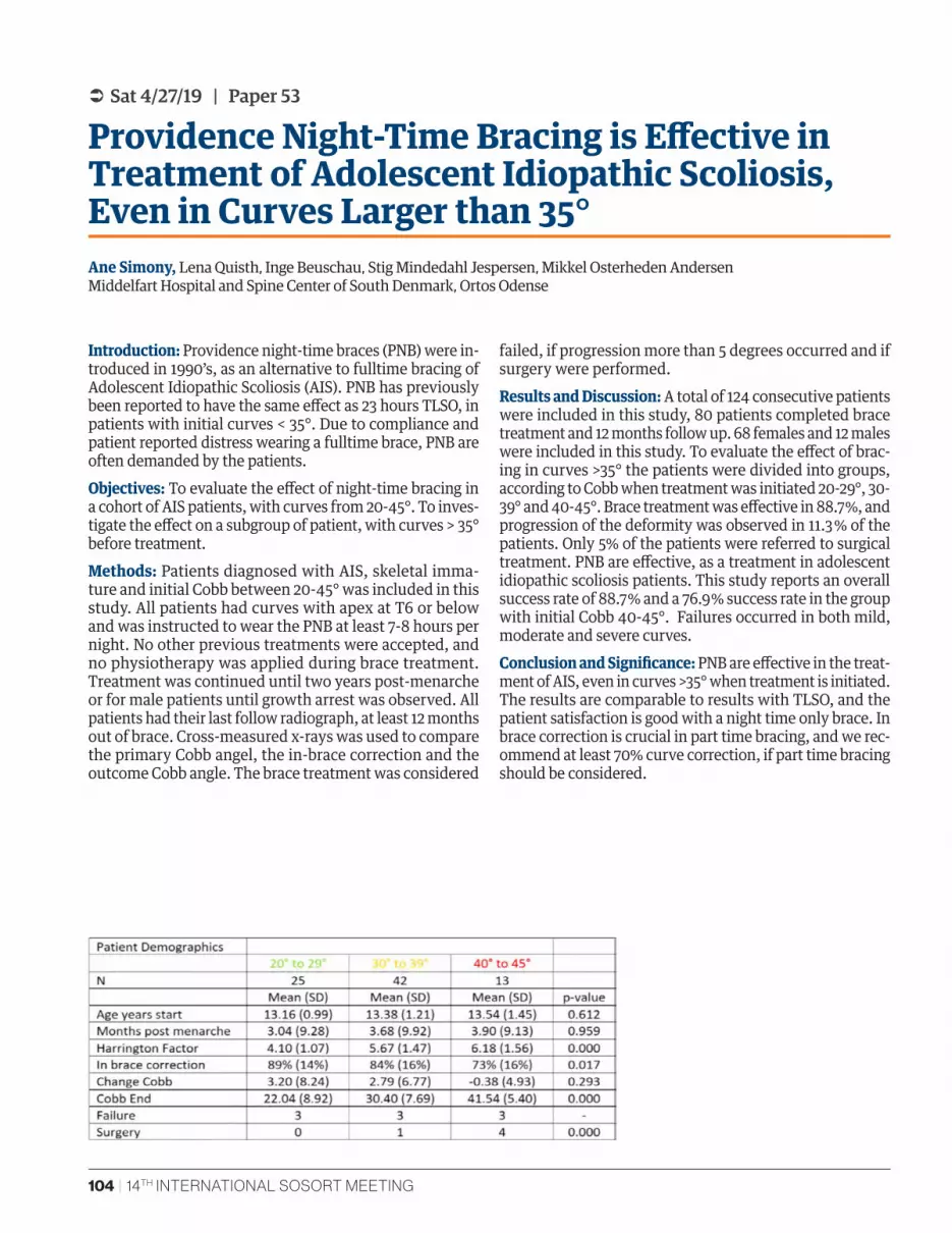
104 | 14TH INTERNATIONAL SOSORT MEETING
 Sat 4/27/19 | Paper 53
Providence Night-Time Bracing is Effective in Treatment of Adolescent Idiopathic Scoliosis, Even in Curves Larger than 35°Ane Simony, Lena Quisth, Inge Beuschau, Stig Mindedahl Jespersen, Mikkel Osterheden Andersen Middelfart Hospital and Spine Center of South Denmark, Ortos Odense
Introduction: Providence night-time braces (PNB) were in-troduced in 1990’s, as an alternative to fulltime bracing of Adolescent Idiopathic Scoliosis (AIS). PNB has previously been reported to have the same effect as 23 hours TLSO, in patients with initial curves < 35°. Due to compliance and patient reported distress wearing a fulltime brace, PNB are often demanded by the patients. Objectives: To evaluate the effect of night-time bracing in a cohort of AIS patients, with curves from 20-45°. To inves-tigate the effect on a subgroup of patient, with curves > 35° before treatment.Methods: Patients diagnosed with AIS, skeletal imma-ture and initial Cobb between 20-45° was included in this study. All patients had curves with apex at T6 or below and was instructed to wear the PNB at least 7-8 hours per night. No other previous treatments were accepted, and no physiotherapy was applied during brace treatment. Treatment was continued until two years post-menarche or for male patients until growth arrest was observed. All patients had their last follow radiograph, at least 12 months out of brace. Cross-measured x-rays was used to compare the primary Cobb angel, the in-brace correction and the outcome Cobb angle. The brace treatment was considered
failed, if progression more than 5 degrees occurred and if surgery were performed.Results and Discussion: A total of 124 consecutive patients were included in this study, 80 patients completed brace treatment and 12 months follow up. 68 females and 12 males were included in this study. To evaluate the effect of brac-ing in curves >35° the patients were divided into groups, according to Cobb when treatment was initiated 20-29°, 30-39° and 40-45°. Brace treatment was effective in 88.7 %, and progression of the deformity was observed in 11.3 % of the patients. Only 5% of the patients were referred to surgical treatment. PNB are effective, as a treatment in adolescent idiopathic scoliosis patients. This study reports an overall success rate of 88.7 % and a 76.9 % success rate in the group with initial Cobb 40-45°. Failures occurred in both mild, moderate and severe curves. Conclusion and Significance: PNB are effective in the treat-ment of AIS, even in curves >35° when treatment is initiated. The results are comparable to results with TLSO, and the patient satisfaction is good with a night time only brace. In brace correction is crucial in part time bracing, and we rec-ommend at least 70% curve correction, if part time bracing should be considered.

SAN FRANCISCO, APRIL 25-27th, 2019 | 105
 Sat 4/27/19 | Paper 54
Indication for 12h Nighttime Bracing on Late-Onset Idiopathic ScoliosisGautier de Chelle, Imad Bentellis, Federico Solla, Olivier Rosello, Jean-Luc Clement, Virgine Rocher-Rampal Department of Pediatric Orthopaedic Surgery and Scoliosis Surgery, Nice-CHU Lenval, France
Introduction: The results of 12h nighttime Cheneau-Tou-louse-Munster (CTM) brace wear on late onset idiopathic scoliosis have not been described.Objectives: Main objective was to analyze its efficiency for Juvenile and Adolescent Idiopathic Scoliosis during puberty growth. Secondary objectives were to find influ-encing factors for brace success in order to progress in its indication.Methods: Pool of 150 consecutive patients treated between 2006 and 2017 was enrolled. Inclusion criteria were evolu-tive scoliosis defined by Cobb Angle Progression (CAP) > 5° between two successive roentgenograms or Cobb angle >30°, 12h nighttime CTM brace-wear during puberty growth, Riss-er stages 0, 1 or 2 at the time of the prescription, complete radiological and clinical control 1 year after weaning. Suc-cess was defined by no surgery, no CAP>5° for main and sec-ondary curves. Subgroups analyses were performed using mLenke classification and Risser test. Lumbar and thora-columbar scoliosis were grouped together in Main Lumbar curves. Most of the clinical and radiological predictive fac-tors known were analyzed.Results and Discussion: Overall success was 58.7%, with 48% for Main Thoracic Curves and 74% for Main Lumbar Curves (p=0.002). At Risser stage 0, efficacy was 46.8%
whereas it was 69% and 74.2% for Risser stage 1 and 2 (p=0.009). Sagittal plane was less impacted by 12h night-time brace wear in comparison to fulltime. Overall mean Angular loss was respectively 6.5 % for Thoracic Kyphosis and 2% for Lumbar Lordosis. Success was associated with the increase of age, Height, Gibbosity, Compliance and Cobb Angle reduction in-brace. Conversely the increase of Frontal and Sagittal C7 Tilt, Cobb Angle, and Frontal Wedging were correlated to failure. An initial main curve Cobb Angle of 27° was the limit for its indication. After lo-gistic regression, Thoracic Gibbosity and Standing Height were the only independent predictive factors with respec-tively OR=0.88 (IC95%:0.70-0.96 p=0.00864) and OR=1.11 (IC95%:1.02-1.22 p=0.01752).Conclusions and Significance: The 12h Nighttime Chene-au-Toulouse-Munster brace-wear finds its best indication for Main Lumbar curves. For this curve pattern, the best results were observed from Risser Stage 1 when frontal C7 Tilt <1cm with 70% of Cobb Angle reduction In-brace. For MT curves the indication should be more restricted. Good results were found from Risser Stage 2, with Cobb Angle < 24° having 50% of Cobb Angle reduction In-brace.
106 | 14TH INTERNATIONAL SOSORT MEETING
 Sat 4/27/19 | Paper 55
The 13 Hour Wear Time Myth in Conservative Scoliosis Management with a TLSODavid Speers Scheck and Siress, Chicago, IL
Introduction: Increased compliance in TLSO wear has been shown to be a major factor in successful outcomes in the conservative management of scoliosis. The BRAIST study (2013) is one study that verifies this and also has provided much useful data as far as compliance data in relation to bracing and scoliosis management. One piece of data stated that 90 - 93% of patients had a successful outcome if wear-ing the brace from 12.9-17.6 hrs, which is a wide range. We have noticed, in our practice, that some physicians have rec-ommended the lower end of this range at 13 hours of daily brace wear to treat scoliosis, while other physicians have recommended 18-20 hours and subjectively these patients have done much better in preventing curve progression.Objective: To determine if the recommended wear time giv-en by the prescribing physician was followed by the patient, and if the full time group had better final results than the part time group.Methods: We conducted a retrospective chart review from 2013-2018 on patients that were recommended 13 hours and 20 hours of daily wear respectively and checked their initial and final out of brace (oob) x-rays for progression (>6 degrees Cobb angle increase). Inclusion criteria were patients with
AIS, Risser 0-2, prescribed a TLSO for scoliosis management and had an I-button (Maxim) compliance monitor installed in the TLSO to track wear time. 8 subjects were in the part time (recommended 13 hrs) group and 8 subjects were in the full time (recommended 18-20 hr) group. All types of TLSO were included in the chart review except for night time only TLSO designs which were excluded.Results and Discussion: The part time group wore the brace on average for 9.01 hours daily and 5 of the 8 (62.5%) had documented progression of their curve(s). The full time group wore the brace on average for 18.25 hours daily and 1 of the 8 (12.5%) had documented progression of the curve(s). This patient progressed to surgery.Conclusion and Significance: The prescribed wear time has a significant effect on the actual hours the brace is worn by the patients. Higher prescribed wear times were directly re-lated to actual higher wear times where lower prescribed wear times were directly related to lower actual wear times. Patients did tend to follow the guidelines of the prescribed wear time by the treating physician. It was also seen, as has been documented many times, that increased wear times equated to more successful outcomes.
SAN FRANCISCO, APRIL 25-27th, 2019 | 107
 Sat 4/27/19 | Paper 56
Influence of Wearing Time on Rotation in Corset Care of Patients with Idiopathic ScoliosisChristian Grasl, Renata Pospischill, Karin Riedl, Sebastian Farr, Tamara Serth Orthomanufaktr Grasl GmbH, Othopädisches Spital Speising, Vienna, Austria
Introduction: For a long time, the primary focus was on correcting scoliosis in the corset, especially among ortho-pedic technicians, reducing the COBB angle - the rotation-al component was considered a secondary correction tar-get, if at all. However, the current Rigo classification and design guidelines and the ever closer cooperation between orthopaedic technicians and physiotherapists have led to an increasing focus on the rotational component in the treat-ment, and the correction of the COBB angle as the primary treatment objective has even partially replaced it. Current studies show that the primary correction has only a very limited causal relationship with the wearing time, this has so far remained largely unanswered with regard to derotation. In the context of the study presented here, this is to be answered in the best possible way on the basis of approximately 210 patients.Method: About 210 patients with idiopathic scoliosis were treated with an RSC® derotation corset in the pe-riod 2017/2018. Using a 3D scan, all patients of the tho-rax were recorded, exact measurements were taken, a standardized photo documentation was created and the Thomastest was carried out. Classification and model se-lection were carried out by Prof. Rigo, according to stan-dardized guidelines. In the course of the adaptation, a thermosensor was installed in all test persons for wearing time monitoring. The wearing time specifications varied
between 10h (night time bracing) for mild scolioses and 23h for severe scolioses. In all patients, follow-up controls were performed after 1, 3, 8 weeks and subsequently ev-ery 3 months after the corset fitting; the control X-ray was performed after 6 weeks. A Thomastest was performed at each control appointment and the rotation was measured with a scoliometer.Results and Discussion: At the beginning, the rotation in the thoracic segment was on average 7.28°, in the lumbar segment 3.87°, all groups combined. After 3 months of wear-ing, the initial values were compared with the actual values. With a wearing time of less than 8 hours per day, these val-ues could only be improved by 2.9% thoracically and 15.5% lumbarly. With a wearing time of 12 hours per day, the im-provement was already 26.5% and 29.8% respectively. With a wearing time of more than 16 hours per day, the thoracic derotation was 39.8%, lumbar 33.7%. With wearing times over 18 hours, the improvement flattened out considerably, which is however due to a significantly higher initial cur-vature. Conclusion and Significance: In contrast to the primary correction of the COBB angle in the corset, there is a direct correlation between an improvement in the rotational com-ponent and the wearing time. A wearing time of about 16 hours per day shows a significant improvement in rotation compared to shorter wearing times.
108 | 14TH INTERNATIONAL SOSORT MEETING
 Sat 4/27/19 | Paper 57
Translation and Subsequent Implementation of SOSORT Consensus Guidelines into Internal Clinical Practice Guidelines on Patient Management and Biomechanical Design Considerations for a National Provider of Scoliosis BracingPhil M. Stevens, Brian L. Kerl, Megan Chamis Hanger Clinic
Introduction: Clinical Practice Guidelines (CPGs) are be-coming increasingly common in modern healthcare as a means of ensuring comprehensive, evidence-informed care. These guidelines should be based on the best avail-able evidence. The international, multi-disciplinary na-ture of SOSORT Guidelines constitute this standard in ar-eas where clinical trials are ill-adept at informing detailed practice standards.Objectives: Translate existing SOSORT Consensus Guide-lines into an internal CPG on patient management princi-ples and biomechanical design considerations for a large national provider of scoliosis TLSOs. Methods: Existing SOSORT Consensus guidelines were screened for their relevance to the clinical provision of scoliosis orthoses. Relevant SOSORT guidelines were iden-tified and reviewed by a small panel of experienced clini-cal Orthotists. Relevant evidence statements were identi-fied and synthesized into concise recommendations with additional explanatory narrative, comprising an internal CPG on the orthotic management of Adolescent Idiopathic Scoliosis (AIS). Results and Discussion: Two relevant SOSORT Consensus Guidelines were identified, encompassing the constructs of patient management standards and TLSO biomechan-ics. Extracted evidence statements were synthesized into 4 concise recommendations on patient management prin-
ciples and 7 biomechanical design considerations. Pa-tient management recommendations included minimum experience standards, the longitudinal, multidisciplinary care pathway of conservative management and the im-portance of monitored compliance. A fifth recommen-dation on emotional stress was later identified through systematic review and added. Biomechanic consider-ations included the application of three-point pressure systems, shoulder correction, optimal vertical heights for corrective forces, principles of derotation, consideration of the sagittal profile and areas where a lack current con-sensus suggests patient-specific design elements. Upon completion, the CPG was integrated into all relevant na-tional educational sessions, posted on the organization’s intranet and integrated into the organization’s national compliance training. Conclusions and Significance: There is tremendous vari-ation in knowledge and experience relative to scoliosis bracing. A concise CPG begins to ensure minimal stan-dards of understanding and practice management. These CPGs should be based on the highest levels of evidence for a given clinical construct. While most SOSORT guide-lines are not immediately directed at clinical Orthotists, a number of relevant evidence statements were identified in within existing SOSORT guidelines. Synthesizing these constructs into a single CPG directed at practicing Ortho-tists facilitates evidence-based medicine.

SAN FRANCISCO, APRIL 25-27th, 2019 | 109
 Sat 4/27/19 | Paper 58
Effect of an Informative Booklet in Scoliosis on Compliance to Brace, Self-Rehabilitation Program, and Health-Related Quality of Life of Patients with Adolescent Idiopathic Scoliosis: A Prospective Comparative StudyMathieu de Seze, Hèlène Chung University Hospital of Bordeaux, France
Introduction: The relation between therapeutic compliance and scoliosis evolution is now certainly established. Objective: Our aim was to measure the impact of an educa-tional booklet on the compliance to the conservative treat-ment (bracing and self-rehabilitation exercises) of children and adolescents with idiopathic scoliosis. Methods: Two sequential groups of children and adoles-cents from 9 to 18 years old with idiopathic scoliosis, fol-lowed at the scoliosis consultation in Bordeaux’s University hospital, have been compared in an observational analytical study. The Control group included, from March to December 2015, 73 subjects who did not receive the booklet. The Book-let group included, from January to October 2016, 71 subjects who benefited from the booklet in their therapeutic care. The patients were examined by the same physician during two consecutive consultations with an interval of 3 months. The compliance of bracing and self-rehabilitation exercis-es was evaluated by the filling of a calendar by the patients during a period of 3 months, and its ratification by the phy-sician during the follow-up consultation. Two health-related quality of life questionnaires (SRS-22 and Brace Question-naire) were completed by all subjects at each consultation.
Results and Discussion: The general and scoliosis charac-teristics of subjects in both groups were comparable. This study did not reveal a statistically significant difference in the bracing time in the Control versus Booklet groups (69% vs 75%, IC 95% [-0.1605; 0.039], p=0,23). It showed a sta-tistically significant positive effect of the booklet on two secondary outcomes: the evolution of the bracing compli-ance in 3 months and the self-rehabilitation compliance. The dropout rate of brace wearing in 3 months was significantly reduced in the Booklet group compared to the Control group (-0,37% vs -11,51%, IC 95% [-0.1943 ; -0.0285], p=0,0088), and the self-rehabilitation exercises compliance doubled in the Booklet group (20% vs 11%, IC 95% [-0.18 ; -0.0027], p=0,04). Finally, health-related quality of life scores did not vary across groups. Conclusion and Significance: This study shows that the educational booklet tool is associated with a lower drop-out rate of brace wearing and an increase in the number of self-rehabilitation exercises. But these results being secondary, the impact of this booklet must be deepened by a study of greater magnitude in order to gaggle in sta-tistical power.

110 | 14TH INTERNATIONAL SOSORT MEETING
 Sat 4/27/19 | Paper 59
A Retrospective Review of Patients Attending Schroth Method Physical Therapy: Factors Affecting AdherenceKristen Venuti, Peiting Lien, Allison Greaney, Michelle Kohler, Paul Sponseller, Majd Marrache Johns Hopkins University, Baltimore, MD
Introduction: The aim of non-operative scoliosis treat-ment is to prevent curve increase and maximize physical function. Schroth Method physical therapy is relatively new to the US as a treatment option for those with AIS. It is often prescribed to children with pre-braceable curves (preventively), and to children wearing a brace as an ad-junct. Schroth Method physical therapy is an intense, individualized exercise program. It requires a high level of commitment from the patient (child) and the parent. Objective: We aim to retrospectively describe the popula-tion of patients attending Schroth therapy and note vari-ables contributing to treatment adherence. Methods: We performed a retrospective review of patients presenting to our institution from 1/2016 – 10/2018 seeking Schroth therapy. We identified 107 patients; we included those with idiopathic scoliosis (no age exclusions) who had completed a minimum of 2 visits with the physical therapist with radiographic data available. We excluded those who had undergone spinal fusion surgery prior to the first PT visit. Results and Discussion: Sixty-nine patients with idiopathic scoliosis completed > 2 visits with a Schroth certified phys-ical therapist, of which 59 were female. The change in pri-mary Cobb angle from start of treatment to last visit was
-0.45o (range, -27 o to +24 o). Female patients on average attended more PT sessions (mean 11.2) compared to males (mean 9.5 sessions). Patients receiving PT as an adjunct to bracing were likely to attend a greater number of PT ses-sions compared to those not braced (11.5 vs 8.9). Pre-teens attended more visits than teens (13.7 vs 9.7).A total of 24 patients (35%) abandoned Schroth PT prior to formal dis-charge. Miles from home, family history of scoliosis, gen-der, and age did not have a statistically significant predic-tive value of adherence to the PT program. We did find that wearing a brace trended toward significance in predicting adherence to Schroth PT visits (p = 0.079). A statistically significant relationship between increasing Cobb angle at the initial visit and decreased likelihood of adhering to the Schroth treatment program was present (p = 0.019). Conclusion and Significance: Schroth physical therapy is a demanding intervention for idiopathic scoliosis. Females, preteens, and braced patients completed more visits. Pa-tients with larger Cobb angles at the start of treatment were less likely to adhere to a full course of PT treatment. Our retrospective review highlights the time commitment to treatment and patient specific variables that may have an effect on adherence. Further research is needed to un-derstand variables that may predict adherence to treat-ment and outcomes.

SAN FRANCISCO, APRIL 25-27th, 2019 | 111
 Sat 4/27/19 | Paper 60
A Self-Administered Questionnaire Designed to Determine Causes of Poor Adherence to Brace Treatment in Adolescent Idiopathic ScoliosisSamuel Sassine, Marie Beauséjour, Julie Joncas, Soraya Barchi, Nikita Cobetto, Sylvie Le May, Omar Elsemin, Carl-Éric Aubin, Stefan Parent, Hubert Labelle University of Montreal; CHU Sainte-Justine, Montreal, Canada
Introduction: There is a significant positive association be-tween hours of brace wear and rate of treatment success in the conservative management of adolescent idiopathic scoliosis (AIS). Unfortunately, the BrAIST study has clearly documented a serious adherence problem. Objectives: To document the perceptions of AIS patients with respect to brace wear, and to use this knowledge to elaborate a new instrument for assessing the factors asso-ciated with a poor bracing adherence.Methods: First, a literature review was conducted to de-termine causes of poor adherence to treatment. Based on the World Health Organization (WHO) conceptual frame-work, factors explaining poor adherence were grouped in 2 categories and 7 dimensions: quality of life (physical, emotional, functional and social) and barriers to enroll-ment (related to the patient, the treatment or the health system).Second, 9 health sciences experts were recruited at a pediatric university hospital to review the generated items from the literature review. Relevance and clarity of the items were rated in a Delphi process. Third, a focus group of 8 AIS brace patients (11-17 y.o.) was conducted by an orthopedic nurse. Participants shared their opinion on open questions related to their health-related quality of life and perceived barriers to brace adherence. Discussion material was transcribed and submitted to the-matic analysis by 2 independent coders who agreed on a thematic tree of key concepts.
Results and Discussion: A list of candidate items (N=58) was extracted from the literature under the 7 dimensions. Four-round Delphi was conducted with the experts to generate a consensual preliminary list of items. Content extraction and analysis of the focus group identified 180 themes/sub-themes. Agreement between the coders was excellent (95%). Most recurring themes were impact on sport and social activities, concerns about perceptions from peers, difficulty to put the brace on by themselves, complaints about discomfort. The instrument under de-velopment takes the form of a self-administered question-naire with scores on a 5-point Likert scale. Triangulation of data sources allowed to formulate items to include in the final instrument while supporting content validity through cross-verification. The final list of 34 consensual items has created the MOBI questionnaire (My Orthopedic Brace In-ventory) with items under the following categories: physi-cal, emotional, functional, and social well-being of patients wearing braces in addition to system, treatment and pa-tient-related barriers to adherence. Conclusion and Significance: We have developed a self-ad-ministrated instrument, with good content validity that documents factors associated to the poor adherence to brace treatment in AIS. Based on the WHO’s conceptu-al framework and the experience of patients and experts, the MOBI questionnaire will now be used to explain poor adherence and allow personalized adherence support in-terventions.

112 | 14TH INTERNATIONAL SOSORT MEETING
 Sat 4/27/19 | Paper 61
Strategies to Improve Home Exercise Compliance in Patients with ScoliosisGeorgina Frere, Daniel Mindel, David Glynn, Erika Maude Scoliosis SOS Clinic, London, UK
Introduction: Compliance is the degree to which a patient acts in accordance with the advice of their therapist. Pa-tients with scoliosis completing physiotherapeutic scoli-osis specific exercises (PSSEs) are recommended to com-plete their exercises at home. It has been shown that pa-tients with scoliosis are non-compliant with completing their home exercise programmes. Objectives: The aim of this randomised control trial was to determine whether the use of a paper diary helped to im-prove home exercise compliance in patients with scoliosis. Methods: 82 patients who had completed an intensive course of PSSEs were randomly allocated into a control group (using a personalised exercise schedule) and a diary group (using a personalised exercise schedule and a paper diary) and enrolled onto the study. Data was collected from children three months post-treatment and from adults six months post-treatment. Participants in both groups were asked ‘on average, over the last three/six months, how many hours do you spend on your scoliosis-specific ex-ercises per week?’ Results and Discussion: There were 43 patients recruit-ed in to the control group and 39 patients in to the diary group. The average age of the participants was 27 years old (range from 10 to 70). 36 patients (84%) provided data
from the control group and 20 patients (51%) provided data from the diary group. On average the home PSSEs were verbally reported to be completed for 3.25 hours per week in the control group, which was 74% of the recommended time, and recorded on the diary to be 1.96 hours per week for the diary group, which was 45% of the recommended time. The control group was associated with a higher per-centage of recommended hours completed (p<0.05). Dif-ferently, the patients in the diary group verbally reported to have completed their home PSSEs 2.98 hours per week which was 68% of the recommended time. The discrep-ancy between the number of hours of exercise recorded and reported in the diary group was statistically signifi-cant (p<0.05), however the difference between the report-ed time spent on exercises was statistically insignificant across the two groups (p=0.64). Conclusion and Significance: This study showed that pa-tients in this dataset reported more hours of exercise than recorded for the diary group. This suggested that patients were either over-estimating the amount of time they be-lieved they were spending on their home PSSEs or not us-ing the diary correctly. The use of a diary did not help to improve home exercise compliance in this cohort. Further research is necessary to determine strategies to improve home PSSE compliance in patients with scoliosis.

SAN FRANCISCO, APRIL 25-27th, 2019 | 113
 Sat 4/27/19 | Paper 62
Early Results of Sonographic Scoliotic Angle of Patients with Adolescent Idiopathic Scoliosis after Physiotherapeutic Scoliosis Specific Exercise: How Long of Exercise Should be Taken to Get Significant EffectsCharlene YL Fan, Eric HK Yeung, Rong He, RW Zhang, GS Li, ZM Xu, Michael KT To, Jason PY Cheung, YP Zheng, Aaron F Zhu, Kenneth MC Cheung The University of Hong Kong - Shenzhen Hospital
Introduction: Clinical evidence suggested that physio-therapeutic scoliosis specific exercises (PSSE) showed promising effects of scoliosis regression. However, the duration of exercise that should be implemented is un-known due to X-ray assessments only performed every 6-12 months. Objective: The objective of this study is to investigate how long of PSSE is necessary to achieve scoliosis regression ac-cording to sonographic spinal image.Methods: 78 idiopathic scoliosis patients aged between 10 and 16 were retrospectively reviewed. All subjects attend-ed PSSE supervised by Schroth trained physiotherapists. Frequency and intensity of treatment was set as follows: ≥30min/time; ≥2times/week. Exercises taught were: 50 semi-hangings, self-correction exercise in sitting/standing (50 repetitions) and lying (50 repetitions) positions. Each subject underwent spinal topographical examination using
3D ultrasound. The sonographic scoliotic angle (SSA) was measured every month after commencing of exercise pro-gramme till 6 months post treatment. Results and Discussion: After excluding 11 patients due to poor compliance to treatment, 67 patients’ data remained for analysis. Multiple Paired-T test was performed. Signifi-cant reduction of SSA was observed between baseline and the 2 months value (23.0+/-6.28 vs 19.4+/-2.53, p =0.037), baseline with the 3 months value (23.0+/-6.28 vs 17.7+/-4.47, p =0.02), and baseline with 6months value (23.0+/-6.28 vs 16.2+/-5.13, p<0.01). There was no significant difference between 2 and 6 months data (19.4+/-2.53 vs 16.2+/-5.13, p>0.05).Conclusion and Significance: Significant improvement of scoliosis was observed as early as 2 months post PSSE treat-ment. Further study beyond 6 months is required to deter-mine whether continued improvement is observed.
114 | 14TH INTERNATIONAL SOSORT MEETING
 Sat 4/27/19 | Paper 63
Analyses of Sonographic Spinal Alignment of Subjects with Adolescent Idiopathic Scoliosis Doing Physiotherapeutic Scoliosis Specific Exercise in Different Positions: A Descriptive StudyRong He, Charlene YL Fan, Eric HK Yeung, Aaron F Zhu, YP Zheng, Jason PY Cheung, Michael KT To, Kenneth MC Cheung The University of Hong Kong - Shenzhen Hospital
Introduction: Clinical evidence suggested that physiother-apeutic scoliosis specific exercises (PSSE) showed promis-ing effects of scoliosis regression. However, it is unknown that the differences of spinal alignment of patients when they doing PSSE in different positions. Objective: The objective of this study is to analyze how does it change of sonographic spinal alignment (SSA) of subjects with different scoliotic patterns when they doing PSSE in different positions.Methods: 18 idiopathic scoliosis patients aged between 10 and 16 with Risser sign ≤2 were participated in this study (12 lumbar major and 6 thoracic major). All subjects at-tended intensive and supervised PSSE for one week to op-timally adopt exercise programme before alignment anal-ysis. Exercises taught were: correction exercise in sitting/standing, side lying and prone on knee positions. Each subject underwent spinal topographical examination us-ing 3D ultrasound. After one-week intensive exercise pro-gramme, the real time sonographic scoliotic angle (SSA) was measured in neutral standing as baseline data, then compared with SSA in each exercise position. Results and Discussion: Prior to the commencement of the main study, assessment on 5 subjects in pilot study were conducted to assess the reliability and repeatability
of the measurement. The results showed intraclass cor-relation coefficient (ICC) with satisfactory reliability of so-nographic scoliotic angle measurement in indifferent po-sitions (neutral standing: ICC3,3=0.98; standing exercise: ICC3,3=0.97; sitting exercise: ICC3,3=0.98; side lying exer-cise: ICC3,3=0.92 and prone on knee: ICC3,3=0.96). There was no significant difference between SSA of baseline with sitting/standing positions (baseline vs sitting: 15.0+/-3.21 vs 16.3+/-2.87, p >0.05; baseline vs standing: 15.0+/-3.21 vs 18.2 +/-4.82; p >0.05), but significant reduction of SSA was observed between baseline with side lying and prone on knee (baseline vs side lying: 15.0+/-3.21 vs 10.05+/-3.87, p=0.37; baseline vs standing: 15.0+/-3.21 vs 9.2 +/-4.01, P= 0.26). Additionally, SSA (2.6 to 5.01deg) was noticed in upper thoracic region when subjects performing muscle cylinder in standing. Conclusion and Significance: Significant real time spinal alignment variation was observed during subjects doing exercise in different lying positions. Further study with bigger sample size is required to validate exercise effects for adolescent idiopathic scoliosis population.

SAN FRANCISCO, APRIL 25-27th, 2019 | 115
 Sat 4/27/19 | Paper 64
The Intra- and Inter-Evaluator Reliability of Frontal and Rotational Spinal Measurements from 3D Ultrasound Imaging for Adolescents with Idiopathic Scoliosis while Performing ExercisesAlex Su, Eric Parent, Michelle Goonasekera, Edmond Lou University of Alberta, Edmonton, Canada
Introduction: Reliable measurements are needed to study physiotherapeutic scoliosis-specific exercises. Validated 3D ultrasound (US) imaging can assess spinal alignment with good curve angle and rotation measurement reli-ability in standing but has not been tested in PSSE posi-tions. Inter-apical distance measurements have yet to be studied using 3DUS.Objectives: To determine the intra- and inter-evaluator reliability of curve angle, axial vertebral rotation (AVR), and inter-apical distance measurements from 3DUS im-ages of adolescents with idiopathic scoliosis (AIS) in 16 Schroth-related positions.Methods: In single sessions, 35 participants with AIS and over three months of Schroth training were imaged in 16 positions, including side-bending, habitual positions (prone, side-lying, sitting, and standing) and their pas-sively-corrected, corrected without leg activation, and fully-corrected Schroth variations. 3DUS images were measured using the center of lamina method. Differences in the thoracic and lumbar AVR and apical vertebral trans-lations represent the AVR twist and inter-apical distance, respectively. The thoracic and lumbar curve end-verte-brae in habitual standing were used for curve extraction in all other positions. Intra-evaluator reliability (ICC3,1) was determined from 13 of the 35 participants measured twice by a single evaluator. Two evaluators measured 35 scans each for inter-evaluator reliability (ICC2,1). Stan-dard error of measurements (SEM) were also calculated.
Results and Discussion: The mean age was 15±3 years old with mean thoracic and lumbar curve angles of 16±8° and 18±9°, respectively; an AVR twist of 14±7°, and inter-api-cal distance of 16±8mm. For intra-evaluator reliability, only 8/62 ICC values were less than 0.70 including 3 tho-racic curve angles, 4 lumbar curve angles, and 1 AVR twist (Table 1). For inter-evaluator reliability, 8/62 ICC values were less than 0.70 including 3 thoracic and 5 lumbar curve angles. Otherwise, the intra and inter-evaluator reliability of the thoracic and lumbar curve angles, AVR twists, and inter-apical distances were all adequate for research use (ICC>0.70). Respective intra- and inter-eval-uator median SEM values were within accepted thresh-olds (Curve angle <5° and rotation <3°) for the thoracic (3.3, 3.2°) and lumbar curve angles (3.2, 3.9°), AVR twist (2.4, 2.6°), and inter-apical distance (2.4, 5.1mm). Inter-apical distances were among the most reliable hav-ing intra- and/or inter-ICC with values above 0.90 (good for individual clinical use), in all positions except habit-ual sitting.Conclusion and Significance: 3DUS imaging produces re-liable frontal and rotational deformity measurements for research in many positions. Curve angle and AVR reliabil-ity was lower than previous standing or lying studies, but SEMs were still within accepted thresholds. Differences compared to previous studies may be from our extract-ing measurements using levels identified in standing, or lower evaluator training, sample severity and variance.

116 | 14TH INTERNATIONAL SOSORT MEETING
 Sat 4/27/19 | Paper 65
Immediate Frontal and Transverse Deformity Reductions during Schroth Physiotherapeutic Scoliosis-Specific Exercise Corrections in Patients with Adolescent Idiopathic ScoliosisAlex Su, Eric Parent, Elia Fong, Sanja Schreiber, Marc Moreau, Edmond Lou University of Alberta, Edmonton, Canada
Introduction: Stakeholders wonder which exercises pro-duce the best in-exercise corrections for adolescents with idiopathic scoliosis (AIS) and if corrections can occur with-out worsening elsewhere.Objectives: To quantify the immediate effect of Schroth postural corrections on the thoracic and lumbar curve angle, axial vertebral rotation (AVR), and apical vertebral translation (AVT) using 3D ultrasound (US) imaging.Methods: In single sessions, 36 participants with AIS, dou-ble curves, and three or more months of Schroth training were imaged using 3DUS in 14 positions. These positions contained four habitual positions (prone, side-lying, sit-ting, and standing) and their passively corrected, corrected without leg activation, and fully-corrected Schroth varia-tions. A final repeat scan in habitual standing was used to detect carryover effects. Measurements were performed with the center of lamina method. Differences between the thoracic and lumbar AVR and AVT correspond to the AVR twist and inter-apical distance, respectively. Groups of po-sitions of interest were compared using repeated measures ANOVAs with Sidak pairwise comparisons.Results and Discussion: The mean age was 15±3 years old with thoracic and lumbar curve angles of 16±8° and 18±9°, respectively. Schroth curve types were: 22 4CP, 5 4C, 7 3CP, and 2 3C. Thoracic and lumbar curve angles, and AVR twist were largest in habitual standing. Inter-apical dis-tance was largest in habitual sitting. Thoracic curve angles
were reduced in fully-corrected prone (6±1°), sitting (5±2°), and standing positions (7±2°) compared to their habitual positions (p<0.05). Lumbar curve angles were reduced in fully-corrected prone (-3±1°) and side-lying (-2±2°) com-pared to their respective habitual and passive positions (p<.05). Corrected without leg activation prone, and ful-ly-corrected sitting and standing, resulted in reduction in AVR twist compared to their habitual position while ful-ly-corrected side-lying was significantly less rotated than in passively supported side-lying (p<.05). Active correc-tion in prone, sitting, and standing significantly reduced the inter-apical distance compared to their habitual posi-tion. Fully-corrected sitting had the lowest thoracic and lumbar curve angles (5±2°, 1±2°) and inter-apical distance (1±3mm), achieving lumbar overcorrection. Fully-correct-ed prone had the lowest AVR twist (7±1°). Lumbar curve angles in the repeated habitual standing position (-12±2°) were reduced compared to the original habitual standing (p<0.05), potentially indicating residual effects from min-imal exercise repetitions during testing.Conclusion and Significance: Schroth exercises produced the greatest corrections, regardless of habitual position, without worsening secondary curves. The largest incre-mental reductions were from active participant correc-tions. Prone Schroth exercises may maximize derotation. Lumbar curve angle corrections may occur from minimal repetitions of Schroth exercise but with unknown dura-tion.

SAN FRANCISCO, APRIL 25-27th, 2019 | 117
 Sat 4/27/19 | Paper 66
Active Self-Correction of Trunk Rotational Deformity with Specific PhysiotherapyEwelina Białek- Kucharska, Marianna Białek, Wojciech Sieteski, Tomasz Kotwicki FITS Education Center, Poland, Vigo-Ortho, Poland, University of Medical Sciences, Poznan, Poland
Introduction: Active auto-correction reveals the basic physiotherapeutic approach in conservative scoliosis treatment. Reduction of the rotational component of the deformity – rib prominence and lumbar prominence –presents the major challenge for physiotherapy. Objective: The aim of the study was to compare the ca-pability to correct trunk rotational deformity before ver-sus after 10 days of scoliosis in-patient physiotherapeu-tic stay. Methods: Forty-three adolescent girls, aged 13.3 ± 1.6 years (range 10.0 – 16.0 years), 19 pre-menarchial and 24 post-menarchial, were assessed. The thoracic Cobb angle was 27.6° ± 10.8 (range 12.0-51.0); the lumbar Cobb an-gle was 26.3° ± 10.4 (range 10.0-55.0). Risser sign 0 was in 18,6% of the patients. The adolescents received the same protocol of PSE (Physiotherapeutic Specific Exer-cises) treatment during 10 days in-patient stay. The trunk rotational deformity was assessed at the beginning and at
the end of the stay at the thoracic and lumbar level using Rodin4D software which is used for designing orthosis. The patient’s position was standardized using a dedicated stabilizer so the conditions were provided the same for both measurements. Results and Discussion: The ATR in thoracic spine was 7,8° ± 3,9 (range 0-15°) , in lumbar 5,1° ± 4,2 (range 0-19°) at the beginning of therapy. After therapy was 6,4° ± 3,8 in thoracic spine and 4° ± 3,4 in lumbar spine. The tho-racic rotational trunk deformity was 10.1 (± 6.1) at the be-ginning versus 9.1 (± 6.6) at the end of stay, p=0.0461. The lumbar rotational trunk deformity was 6.3 (± 4.3) at the beginning versus 5.2 (± 3.9) at the end of stay, p=0.0593. Conclusion and Significance: After 10-days of in-patient physiotherapy, the adolescent girls performed the au-to-corrective movement in a better way and the corrected position revealed significantly less trunk rotational de-formity comparing to the assessment before treatment.

118 | 14TH INTERNATIONAL SOSORT MEETING

SAN FRANCISCO, APRIL 25-27th, 2019 | 119
Poster presentations
120 | 14TH INTERNATIONAL SOSORT MEETING
THURSDAY, APRIL 25, 2019
Poster #
3 The Effectiveness of Scoliosis Specific Exercises in a Painful Adult with Congenital ScoliosisRebecca Hurst, Emery Maloney
4 Impact of Bracing and Scoliosis Specific Exercises in a Child with Collagen VI Myopathy and Scoliosis – A Case ReportRebecca Hurst, Emery Maloney
10 TLSO: The Effect of Spinal Translation on Curve Correction, Trunk Shift, and Coronal Balance in ScoliosisKierra Falbo, Ammanath Peethambaran
11 A Modified Physiotherapeutic Scoliosis Specific Exercise Program (Schroth Method) and Night Bracing Stabilized Scoliotic Curves and Improved Pulmonary Function in Young Boy with Spinal Muscular Atrophy Type 3 - A Case Report – A Four Year Follow UpChintan Pancholi-Parekh, Jennifer Rothman
12 A Modified Physiotherapeutic Scoliosis Specific Exercise Program (Schroth Method) and Traditional Physical Therapy Improved Posture, Balance and Pulmonary Parameters in a 62-Year Old Adult Male with Complex Medical History – A Case ReportChintan Pancholi-Parekh, Jennifer Rothman
15 Configuration Standards of Scoliosis Orthoses for Chinese Children and AdolescentsJing Tao, Zhiqiang Zhang
25 The Tweezers as a Final Solution for the Assessment of Angle Trunk Rotation. Reliability Study Compared with the Gold Standard ToolMichele Romano, Alessandra Negrini
29 Proprioceptive Neuromuscular Facilitation in the Treatment of Idiopathic ScoliosisPamela Schweitzer, Matheus Branco, Patrícia Lencina, Paola Moura
34 Clinical Case: Functional Changes After Schroth Method in a Young Girl with Adolescent Idiopathic Scoliosis (AIS) Using a Motion Capture System During Gait and StanceDoug W. Dendy, C. Roger James, BM Dewan
37 Compliance and Persistence at Kinesitherapy in Adolescents with Idiopathic ScoliosisSime Mijic, Grgur Bulovic, Stipo Dajakovic
39 Outcomes of 6 Months Intensive Physiotherapy Scoliosis Specific Exercise (PSSE) and Full Time Cheneau-Gensingen Brace Treatment for Adolescent Idiopathic Scoliosis: 3 Case SeriesTevfik Acar, Gozde Basbug, Ferda Firdin
42 Short Term Results Reviewing the Initial In-Brace Correction and Hours of Brace Wear For Patients Utilizing a Custom Fabricated from Scan and Modified in CAD TLSO Following Boston Brace PrinciplesJames Wynne, M. Timothy Hresko
POSTER SESSION #1

SAN FRANCISCO, APRIL 25-27th, 2019 | 121
THURSDAY, APRIL 25, 2019
45 A Case Study of the Difference Between a Correctly-Designed Rigo-Chêneau Type Brace and an Incorrectly-Designed Rigo-Chêneau Type Brace in a Patient with ScoliosisRaúl Ferrera Hernández
46 A Case Study of an Adolescent Patient with Idiopathic Scoliosis Treated with Schroth Therapy According to BSPTS and Rigo-Cheneau Type Scoliosis BraceRaúl Ferrera Hernández
47 A One Year Review Examining the Effectiveness of a Collaborative Approach When Treating a 14 Y/O Female with Adolescent Idiopathic Scoliosis Using Scoliosis Specific Exercises and a Boston 3d Orthosis: A Case ReportMichelle Dwyer, James Miller, James Wynne
51 Non-Fusion Scoliosis Treatment with ApiFix System: Physiotherapeutic Scoliosis Specific Exercises (PSSE) With Schroth Method Can Significantly Improve the Final ResultNikos Karavidas
52 Physiotherapeutic Scoliosis Specific Exercises (PSSE) Can Reduce the Risk of Progression in Adolescent Idiopathic Scoliosis During the Peak of Growth: A Prospective Study With a Control GroupNikos Karavidas
55 Schroth-Based Physical Therapy to Treat Severe Adult Scoliosis of Adolescent Onset with Concomitant Multilevel Degenerative Changes in the Spine: A Case ReportBeatriz Rodriguez
58 Leptin in Adolescent Idiopathic Scoliosis - Meta-AnalysisJung Sub Lee, Tae Sik Goh, Seung Min Son, Sung Hoon Choi
59 The Prevalence of Scoliosis Within an Adult Myelomeningocele Population in Belgium. Retrospective Case SeriesArne Heyns, Stefano Negrini, Katrien Jansen, Pierre Moens, Sebastiaan Schelfaut, Koen Peers, Carlotte Kiekens
49 Management of Symptoms Related to Post-Operative Spinal Fusion “Adding-On” Phenomena in Idiopathic Scoliosis Utilizing Schroth Scoliosis-Specifc Exercises: A Case ReportNicola Khalaf, Nikola Jevtic, Lauren Katz
POSTER SESSION #1

122 | 14TH INTERNATIONAL SOSORT MEETING
THURSDAY, APRIL 25, 2019
Poster #
9 Global Posture Evaluation for Patients Treated with Scoliosis OrthosisJean-LoÏc Rose, Lydie Journoud, Adèle Sigal, Carole Fortin, Jean-Claude Bernard
13 Evaluation of Pressure on Mobile Thoracic Pad with Prestressed Carbon Blade System, In Treatment with CMCR BraceJean-Loïc Rose, Lydie Journoud, Grégory Notin, Adèle Sigal, Nuria Rodriguez Martinez , Jean-Claude Bernard
16 CAD Versus Custom TLSO Bracing: Initial Curve Correction For Idiopathic ScoliosisChristopher Vara
17 Cobb Angle Reduction after an Intensive Course of Physiotherapy Scoliosis Specific Therapy: A Case SeriesGeorgina Frere, Erika Maude, Mollie-Rose Turkentine, Jack Whiteside, Abbie Turland, Luke McKendrick
19 Retrospective Chart Review of WCR Orthosis Outcomes in Patients with Idiopathic ScoliosisMarlies Beerli Cabell, Julie McCulley, Brian Kaluf
20 Case Study: Treatment of Scoliosis with WCR Orthosis in 10mo Patient with Ehlers Danlos Syndrome Ocular Scoliosis Type Fkbp-14Marlies Beerli Cabell
21 Case Study: Treatment of Scoliosis with WCR Orthosis in an 82 Year Old with Osteogenesis Imperfecta Type 1Marlies Beerli Cabell
22 Case Presentation for BMI >99% in an Adolescent Idiopathic Scoliosis Correction of the Curves in Her Out of Brace Correction At 1 YearMarlies Beerli Cabell
23 Systematic Review of Chiropractic Biophysics® (CBP®) Methods Employed in the Rehabilitation of Cervical LordosisDeed Harrison, Ibrahim Moustafa, Paul Oakley
24 Systematic Review of CBP® Methods Employed in the Rehabilitation of Lumbar LordosisDeed Harrison, Ibrahim Moustafa, Paul Oakley
27 Correction of Cervical Pseudo-Scoliosis and Alleviation of Dystonia Symptoms Using Chiropractic Biophysics® (CBP®) Technique: A Case ReportJason Haas, Paul Oakley, Deed Harrison
31 Improvement in Thoracolumbar Neuromuscular Levoscoliosis in a 14-Year-Old Male with a History of Stage 3 Medulloblastoma and Post-Operative Cerebellar Mutism Syndrome Using Chiropractic Biophysics Technique and ScoliBrace Thoracolumbosacral Orthotic Bracing: A Case Study and 30-Month Follow-UpDouglas F. Lightstone, Curtis Fedorchuk, Julia Guo, Jeb McAviney
POSTER SESSION #2
SAN FRANCISCO, APRIL 25-27th, 2019 | 123
32 A New Method to Determine Appropriate Ischial Lift Orthotic Height for Seated Support in Populations with an Unlevel Sacral Base Using Radiographic Imaging and Lines of MeasurementDouglas F. Lightstone, Curtis Fedorchuk, Jeb McAviney
33 A New Method to Measure the Lateral Translation of Vertebrae in Populations with Laterolisthesis using Radiographic Imaging and Lines of MeasurementDouglas F. Lightstone, Curtis Fedorchuk
36 Comparison of SRS-22 Scores of Adults After Versus the Adolescents During Specific Physiotherapy for ScoliosisEwelina Białek-Kucharska, Marianna Białek, Tomasz Kotwicki
41 The Effect of 5th Pelvic Correction on Postural Alignment during International Schroth Three Dimensional Scoliosis Therapy (ISST) Exercises In Adolescent Idiopathic ScoliosisNikola Jevtic, Nicola Khalaf, Danka Ljubojevic, Milan Pantovic
44 Successful Multimodal Treatment of Severe Adolescent Idiopathic Scoliosis after Failing Standard TLSO BracingAnthony Nalda, Ben Brown, Jeb McAviney
50 Reduction of a Severe Scoliosis Using an Over-Corrective Rigid Orthosis in an Adolescent FemaleJeb McAviney, Benjamin T. Brown
54 An Elongation Bending Derotation Brace: An Alternative to Serial Casting for Early Onset ScoliosisJohn Thometz, XueCheng Liu
61 Assessment of Scoliosis in the Future in FinlandMaarit Keskinen
62 Case Study: Fascia Manipulation Treatment in ScoliosisMaarit Keskinen
66 Medical Radiologists Consistently Misrepresent the Cervical Lordosis in Radiology Reports: A Comparison of Subjective Qualitative Assessment Versus Objective Quantitative Measurement in 100 Consecutive PatientsPaul Oakley, Leesa Sanchez, Deed Harrison
THURSDAY, APRIL 25, 2019POSTER SESSION #2

124 | 14TH INTERNATIONAL SOSORT MEETING
FRIDAY, APRIL 26, 2019
Poster #
5 Progressive Anterior Vertebral Fusion or Spontaneous Anterior Fusion in Scheuermann’s Disease? A Case ReportMonica Lusini, Sabrina Donzelli, Fabio Zaina, Stefano Negrini
6 Actigraphy-Based Sleep Parameters and Rest-Activity Circadian Rhythm in a Young Scoliotic Patient Treated with Rigid Bracing: A Case StudyJacopo A.Vitale, Francesco Negrini, Giulia Rebagliati, Sabrina Donzelli, Giuseppe Banfi
7 Kinematic Motion Analysis of Four Individuals with Scoliosis in the Squat Motion: Implications For Therapeutic Conservative Clinical UseSuzanne Clements Martin
8 The Scoliosis Specific Physical Orthopedic Test - Conservative Care for Youth with Idiopathic Scoliosis Using Pilates Method Exercise as Part of a Multi-Disciplinary TeamSuzanne Clements Martin, Lorna Roza, Kris Shevlin, Chithrah Cherian
14 One and a Half Years Follow-Up of Adolescent Idiopathic Scoliosis Patients with Mild Curvature Doing Physiotherapeutic Scoliosis Specific Exercise: A Retrospective StudyCharlotte YL Fan, Eric HK Yeung, Rong He, RW Zhang, GS Li, ZM Xu, Michael KT To, Aaron F Zhu, Jason PY Cheung, YP Zheng, Kenneth MC Cheung
18 Restoration of Excessive Posterior T12-S1 Sagittal Balance and Reduction in a Thoraco-Lumbar Junctional Kyphosis in an Adolescent with Back Pain: A Chiropractic Biophysics® Case ReportChris Gubbels, Paul Oakley, Joshua Werner, Deed Harrison
26 Potential for Comprehensive Evaluation of Scoliosis Using Formetric 4DJonathan Horng, Xue-Cheng Liu, MD, John Thometz, Channing Tassone, Benjamin Escott
30 Using the Alexander Technique to Improve Awareness of AIS (Adolescent Idiopathic Scoliosis) – the Influence of It in the Aspect of Quality of LifeNanyoung Ji, Sunyoung Ji, Soo Yeon Han, Ji Soo Kim
35 Treatment of Degenerative Adult Scoliosis Using a Unique 3D Over-Corrective Brace: A Case ReportMichael Underhill, Ben Brown, Jeb McAviney
38 How Inexperience and Trim Lines Can Affect Your Expected OutcomeEmery Maloney
40 Case Study Presentation: The Correctional Derotation from the Wood Cheneau Rigo Scoliosis TLSO Brings Failed Wilmington Scoliosis TLSO Wearers Into ComplianceJulie McCulley
43 Correction of Scoliosis with a Postural Exercise MethodCarme Carré-Llopis, Juan Carlos Rodríguez Olaverri
48 Effect of Bracing in a Surgery Case (Over 45°) of a Girl with Juvenile Idiopathic Scoliosis: A Case StudyMariette Zoer-Kosse, Anneke Struijk-Kramer, Marjan de Jonge
POSTER SESSION #3

SAN FRANCISCO, APRIL 25-27th, 2019 | 125
65 The Assessment of Postural Instability in Patients with Idiopathic ScoliosisChin-Yen Tseng, Chung-Liang Lai, Chun-Hou Wang, Meng-Chuan Tasi, Shiuan-Yu Tseng
53 Toward Trace 2: Expert Opinions about Items to Evaluate Trunk AestheticsSara Rossi Raccagni, Alessandra Negrini, Giulia Rebagliati, Stefano Negrini
56 The Effectiveness of Barcelona Scoliosis Physical Therapy School (BSPTS) Schroth Based Exercises on a Patient with ScoliosisBeth Runzo
60 Physiotherapy Scoliosis Specific Exercises (PSSE) In Prehab and Rehab to Anterior Vertebral Tethering (AVT) Surgery in AISNancy Sherratt
63 Surface Topography Scans Used to Monitor Progression of Scoliotic Curves in a Pregnant Adult with Scoliosis While Undergoing a Physiotherapeutic Scoliosis Specific Exercise Program (Schroth Method): A Case ReportRebecca Pacailler, Christine Gaul
67 A Case Study of a Patient with Early Onset Scoliosis Using Mehta Casting and Rigo-Chêneau Type Scoliosis BraceGrant Wood
68 A Four-Year Case Study of an Adult Patient with Scoliosis Using Schroth Therapy and an Adult WCR Scoliosis BraceGrant Wood
FRIDAY, APRIL 26, 2019POSTER SESSION #3
126 | 14TH INTERNATIONAL SOSORT MEETING
 Poster 03
The Effectiveness of Scoliosis Specific Exercises in a Painful Adult with Congenital ScoliosisRebecca Hurst, Emery Maloney Scoliosis Rehab of TX, Bryan, TX, Align Clinic, Green Bay, WI
Introduction: Scoliosis is a common congenital dis-order which can be due to failure of formation, seg-mentation or their combination. While Physiothera-py Scoliosis Specific Exercises (PSSE) are a significant modality in the conservative, non-surgical treatment of scoliosis, there has been limited research on the ben-efit of PSSE in the management of painful adults with congenital scoliosis. Objectives: To determine the efficacy of Barcelona Sco-liosis Physiotherapy School (BSPTS) Schroth based PSSE in the treatment of a painful adult with congenital sco-liosisMethods: This is a case study of a 25 y.o. with congen-ital scoliosis who underwent fusion in situ of T6-9 at age 10 and has since experienced further curve progres-sion. She was referred for Schroth based physical ther-apy by her orthopedic surgeon due to progressive pain and dysfunction despite repeated trials of traditional physical therapy. The patient was seen for 5 days of in-tensive BSPTS PSSE, a 2 day follow up intensive session 3 months later and was given a written home program to follow 30 min per day 5 days per week. Patient was also seen for trial of bracing and fabrication of new shoe lift. Clinical measurements such as ATR, rib cage mo-bility, forced expiratory volume, trunk imbalance in the coronal plane and standing height were documented and the patient was asked to complete the Oswestry Disability Index (ODI), the SRS-22 and the visual analog
pain scale upon evaluation and at the end of treatment. Radiology reports and x-ray images were also reviewed for elements commonly associated with pain.Results and Discussion: Patient reports completing home program 30 min per day 6 days per week. The following changes were noted from the evaluation: ATR in seated for-ward flexion decreased from 18 degrees to 10 degrees, forced expiratory volume increased from 2850cc to 3000cc, trunk imbalance in the coronal plane decreased from 5.0cm to 2.6 cm and standing height increased from 161.5cm to 163.2cm. The Oswestry Back Disability Index decreased from 66% to 34%. The SRS-22 overall score before treatment = 2.27 as compared to 3.5 after treatment. The domains of the SRS-22 were also analyzed before and after treatment and the re-sults were as follows: Function = 2.0 before treatment and 3.0 after treatment, Pain = 1.4 before treatment and 3.4 after treatment, Self Image = 2.2 before treatment and 3.4 after treatment, Mental Health = 3.0 before treatment and 3.6 af-ter treatment and Satisfaction with Management = 2.5 be-fore treatment and 5.0 after treatment. The patient also re-ported decreased intensity of pain on the 0-10 VAS with back pain upon evaluation =9/10 and at the end of treatment = 1/10. Patient was also able to decrease narcotic pain medica-tion from 10 mg Percocet three times daily to 5mg once daily.Conclusion and Significance: These findings suggest that intensive Schroth based PSSE according to the BSPTS may be beneficial for painful adults with congenital scoliosis who are seeking conservative treatment.

SAN FRANCISCO, APRIL 25-27th, 2019 | 127
 Poster 04
Impact of Bracing and Scoliosis Specific Exercises in a Child with Collagen VI Myopathy and Scoliosis – A Case ReportRebecca Hurst, Emery Maloney Scoliosis Rehab of TX, Bryan, TX, Align Clinic, Green Bay, WI
Introduction: Collagen VI Myopathy is a rare group of dis-orders affecting skeletal muscles and connective tissue. Severity ranges from mild to severe and symptoms can include: progressive muscle weakness, joint contractures, progressive difficulty with ambulation and in some cases respiratory dysfunction. There is little information about scoliosis treatment in this patient population.Objective: To determine the impact of Barcelona Scoliosis Physiotherapy School (BSPTS) Schroth based physical ther-apy and Wood Cheneau Rigo (WCR) bracing in the treat-ment of a juvenile with Collagen VI Myopathy and ScoliosisMethods: This is a case study of a 9 y.o. patient with Col-lagen VI Myopathy and a recent diagnosis of juvenile sco-liosis. X-rays revealed 26 degree left thoracolumbar curve and patient was fit with a custom TLSO 3 months prior to referral for Schroth based Physical Therapy. Patient was seen for 5 consecutive days of intensive BSPTS PSSE (4 hours per day) and she was given a written home program to follow 30 minutes per day 5 days per week. She was also seen by an orthotist and fit with a WCR brace. Clinical mea-surements such as ATR, rib cage mobility, trunk imbalance in the coronal plane, seated and standing height were doc-umented. Forced expiratory volumes with the Spiropet as well as balance and fall assessments were completed in the TLSO, out of brace and in the WCR brace. Results and Discussion: The patient follows a modified home program of PSSE with the help of her mother and is
compliant with full time brace wear. The following chang-es were noted four months after intensive treatment at the clinic. Forced expiratory volume as measured with a Spiropet over 5 seconds was documented in her TLSO, out of brace and in the WCR brace. Volume in TLSO = 500cc upon evaluation, out of brace volume increased from 800cc to 1000cc, volume in WCR brace increased from 800cc at initial fitting to 1050cc after 4 months. Trunk imbalance in the coronal plane upon evaluation: out of brace = 2 cm, in TLSO = 5cm and in WCR = 0cm. After 4 months trunk imbalance in the coronal plane out of brace decreased to 1cm. ATR in seated forward flexion decreased from 10 degrees to 5 degrees in the thoraco-lumbar region. Rib cage mobility as measured as the dif-ference between inhalation and exhalation at the axilla, xyphoid and waist respectively were: 1.5cm, 1.0 cm and -1.2cm as compared to 3.0cm, 2.4cm and 1.8cm. Patient required a walker or minimal assist to rise from a chair and minimal assist for household level gait upon evalu-ation. Patient is now modified independent with sit to stand and household level gait in WCR brace. Timed up and go-in children (TUG-IC) in TLSO =24.25 seconds and in WCR = 15.11secondsConclusion and Significance: Schroth based PSSE when used in conjunction with a WCR brace may have a posi-tive impact on patients with juvenile scoliosis who have co-morbidities such as respiratory dysfunction, muscle weakness, balance deficits and functional impairments.

128 | 14TH INTERNATIONAL SOSORT MEETING
 Poster 10
TLSO: The Effect of Spinal Translation on Curve Correction, Trunk Shift, and Coronal Balance in ScoliosisKierra Falbo, Ammanath Peethambaran University of Michigan, Ann Arbor, MI
Introduction: Adolescent idiopathic scoliosis (AIS) curves of magnitude 25-40° are often treated non-operatively with thoracolumbar sacral orthoses (TLSO). The goal of these TLSOs is to improve the scoliotic curve during wear in order to prevent further curve progression. There are multiple styles of TLSOs used for treatment of AIS. Typi-cally, corrective forces are applied to the patient via pad-ding added to the TLSO. Thickness of the padding used to achieve Cobb angle correction is somewhat arbitrarily chosen by the treating orthotist. If the correction is not enough, adjustments are made, and additional padding is often added to the orthosis after the in-orthosis x-ray. With the integration of CAD in orthotics, the ML shift method (translation) has been used to apply corrective forces to the torso. However, there are no available reported data to substantiate the amount of translation and its effect on Cobb angle correction. Objectives: The objective of this study is to investigate the relationship between amount of applied translation and its effect on Cobb angle correction, apical vertebra trans-lation (AVT), coronal balance (CB), and trunk shift (TS) in patients with AIS treated with TLSOs. Methods: Female subjects with idiopathic scoliosis who were prescribed a TLSO were included in this study. Data was retrospectively reviewed to collect information re-garding curve flexibility, hours of brace wear prescribed, and amount of medial-lateral shifting used during the CAD
modification process. Differences in the primary scoliotic curve were then assessed based on initial x-rays and in-or-thosis x-rays to calculate change in Cobb angle, AVT, CB, and TS.Results and Discussion: 16 subjects have been included in the study to date. The primary scoliotic curve was thoracic in 10 subjects, thoracolumbar in 2 subjects, and lumbar in 4 subjects. Average measured curve flexibility was 59%, and applied shifting to the CAD model ranged from 6 to 15 millimeters. Average Cobb angle improvement from the initial x-ray to the first in-orthosis x-ray was 25%. Aver-age improvement of AVT was 27% and of TS was 58%. CB showed an average negative change of 37%. Conclusion and Significance: These preliminary results suggest that greater applied shifting during the modifica-tion process leads to greater in-orthosis curve correction. With this limited amount of subjects, it is difficult to draw meaningful conclusions about direct relationships at this time. Subjects will continue to be recruited for this study for a larger sample size. This data will be used to deter-mine any correlations between CAD modifications, curve flexibility, and hours of brace wear with curve correction parameters. The information will guide clinicians in ap-plying the optimal amount of shifting for each patient to achieve the desired in-orthosis curve improvement at the initial fitting. This will limit the need for later adjustments and will improve overall care.

SAN FRANCISCO, APRIL 25-27th, 2019 | 129
 Poster 11
A Modified Physiotherapeutic Scoliosis Specific Exercise Program (Schroth Method) and Night Bracing Stabilized Scoliotic Curves and Improved Pulmonary Function in Young Boy with Spinal Muscular Atrophy Type 3 - A Case Report – A Four Year Follow UpChintan Pancholi-Parekh, Jennifer Rothman Scoliosis Speciality Center, Totowa, NJ
Introduction: Spinal Muscular Atrophy (SMA) is a neuro-muscular disease characterized by the progressive loss of lower motor neurons. Scoliosis is a common condition that impacts patients with SMA. Management of scoliosis in these patients consists of observation, possibly bracing or corsets and eventually surgery. While physiotherapeu-tic scoliosis specific exercise (PSSE) programs have shown effectiveness in halting progression of Cobb angles in pa-tients with idiopathic scoliosis, there is no literature that reports on the effect of PSSE for patients with SMA.Objective: To report a case of a now 12-year old boy with SMA type 3 who underwent a modified PSSE program (Schroth method) while wearing a night brace. This is a continuation of a case report that was presented at SO-SORT in 2016.Methods: Patient underwent 36 PSSE sessions over a 39-month period. Mom’s goal was to avoid spinal fusion so that her son would be eligible to receive a new drug treatment. This drug treats people with SMA and slows the progression of the disease. He was originally treat-ed 1x/week and after 2 months of treatment he continued with PSSE with faded follow ups ranging from 1x/month to 1x/4 months. He also performed his home exercise pro-gram (HEP) 3x/week which consisted of core strengthening in supine with assist as needed, Schroth concepts of au-to-elongation and expansion, kyphosis specific exercises, postural corrections during activities of daily living and breathing strategies. With disease progression he lost the
ability to stand and ambulate, thus his program was mod-ified so that he could exercise in his adaptive equipment and wheelchair. During the program he wore a nighttime brace; he wore a Providence brace for 11 months and then wore a regular Rigo System Cheneau brace. At 36-month follow up surgery was not required and he was able to be-gin the drug treatments. He continued with RSC bracing and with HEP. Results and Discussion: When comparing 0-month to 39-month follow up, lumbar Cobb angle decreased from 30° to 29°, thoracic Cobb angle increased from 20° to 22° and kyphosis decreased from 60° to 35°. In patients with SMA, Cobb angles typically increase 5°-15° per year. In this case, even with a 26.52cm increase in height, lumbar and thoracic Cobb angles stabilized and thoracic kyphosis improved. Pulmonary function in patients with SMA type 3 typically remain stable or decline with loss of functional mobility. In this case pulmonary function was improved even though he lost the ability to stand; he was originally diagnosed with early obstructive pulmonary impairment and at 39-month follow up he was diagnosed with normal respiratory values. Conclusions and Significance: PSSE with night bracing may help to stabilize or decrease the scoliotic curves in patients with SMA type 3. PSSE may also help to improve pulmonary function. In patients with SMA, future stud-ies should examine the effectiveness of PSSE and the use of RSC bracing.
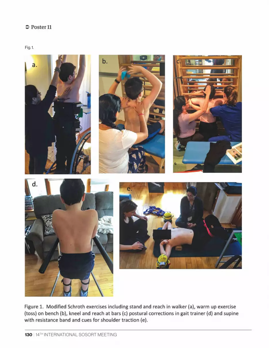
130 | 14TH INTERNATIONAL SOSORT MEETING
 Poster 11
Fig. 1.

SAN FRANCISCO, APRIL 25-27th, 2019 | 131
 Poster 12
A Modified Physiotherapeutic Scoliosis Specific Exercise Program (Schroth Method) and Traditional Physical Therapy Improved Posture, Balance and Pulmonary Parameters in a 62-Year Old Adult Male with Complex Medical History – A Case ReportChintan Pancholi-Parekh, Jennifer Rothman Scoliosis Speciality Center, Totowa, NJ
Introduction: In older patients, kyphoscoliosis causes deleterious effects on health, physical functioning, bal-ance and quality of life. Currently there is no standard intervention to reduce kyphoscoliosis in aging patients. Physiotherapeutic scoliosis specific exercise (PSSE) pro-grams incorporate a kyphosis-specific component when appropriate. Currently there is limited literature that re-ports on the effect of PSSE for aging adults with kypho-scoliosis.Objective: To report a case of a 62-year old male with a complex medical history and kyphoscoliosis who was treated with PSSE (Schroth method) and traditional phys-ical therapy.Methods: Patient has a history of multiple myeloma treat-ed with chemotherapy and steroids. He developed severe osteoporosis which led to multiple rib and vertebral com-pression fractures. He underwent kyphoplasty at T6-T9 and T11 in August 2017. Patient reports taking high doses of medications to aide with chronic pain. He underwent 30 PSSE sessions over a 6-month period. He was treated 1-2x/week for 45-60 minutes. Initiated treatment with manu-al soft tissue work paired with simple concepts of align-ment and equal weight bearing in functional positions. Balance exercises including tandem stance on even and uneven surfaces, marching with upper extremity support and single leg stance activities were incorporated early in the program. Modified Schroth exercises including sit and reach and stand and reach with breathing strategies were
added. Slow introduction of full activations and breathing were performed in modified supine, standing and sitting with 2 poles.Results and Discussion: Patient’s height at initial eval-uation (IE) was 153.4cm and increased to 158cm after 6 months of treatment. Pulmonary function was monitored with spirometry. Average spirometry reading at IE was 0.26 liters (L) and increased to 1.5 L after 6 months of treat-ment. This indicates improvement in pulmonary func-tion. Inclinometer measurements taken at IE measured 40° lumbar lordosis and 108° thoracic kyphosis. After 6 months treatment, inclinometer revealed that lumbar lor-dosis decreased to 25° and thoracic kyphosis decreased to 90°. While still in an atypical range, this demonstrates an improvement in thoracic sagittal profile. Single limb stance test (SLST) with eyes open was 2-3s on left leg and 4s on right leg. Normative data suggests that patients un-able to stand <5s on one limb classify the patient as high fall risk. By 6 months SLST times increased to 30s on left leg and 21s on right leg which then classified patient as low fall risk. Furthermore, pain at worst decreased from 10/10 on Numerical Pain Rating Scale (NPRS) to 4/10 on NPRS.Conclusion and Significance: PSSE, with traditional phys-ical therapy, helped a medically fragile older patient im-prove respiratory parameters and sagittal posture while decreasing risk of falls. More research should be conduct-ed to examine the effects of PSSE on adults with kypho-scoliosis.

132 | 14TH INTERNATIONAL SOSORT MEETING
 Poster 15
Configuration Standards of Scoliosis Orthoses for Chinese Children and AdolescentsJing Tao, Zhiqiang Zhang Chinese National Research Center for Rehabilitation Technical Aids, Beijing, China
Introduction: Due to changes in living habits such as learn-ing and looking down on electronic products, a large num-ber of unexplained scoliosis occurred in children and ad-olescents in China. For children and adolescents with a scoliosis Cobb angle of less than 45 degrees, Chinese doc-tors usually choose to wear a scoliosis orthosis for treat-ment. However, it still lacks standards for the configuration of spinal orthoses in China, with only relevant terms and screening criteria. Objectives: In order to end the above situation, we dis-cuss the configuration process and general requirements of Chinese children and adolescent scoliosis orthosis, and propose the configuration standards of Chinese children and adolescent spine orthoses.Methods: We conducted a survey and statistics on the configuration of the scoliosis orthosis for spinal ortho-paedic medical institutions in different regions of Chi-na. Then, combining of Chinese national standards for prosthetics and orthotics, the configuration require-ments and procedures for the scoliosis of children and
adolescents in this paper were discussed and deter-mined.Results and Discussion: The criteria presented in this pa-per are applicable to orthotic configuration services for scoliosis patients aged 6-18 years. First, we present gen-eral requirements and comfort testing Methods for the configuration of adolescent scoliosis orthosis, including terminology and definitions, general principles, systems and management, delivery, and follow-up. Secondly, we summarized the hospitality inquiries, clinical examina-tions, prescribing, signing agreements, orthosis making, orthotic fit testing and comfort evaluation of the scoliosis orthosis. Finally, we also developed a follow-up procedure for the scoliosis orthosis.Conclusions and Significance: The proposed procedure standard for the Chinese children and adolescent spine or-thosis proposed in this paper can guide the configuration process of the Chinese children and adolescent scoliosis orthosis, and will regulate the various scoliosis orthopedic medical institutions in China.

SAN FRANCISCO, APRIL 25-27th, 2019 | 133
 Poster 25
The Tweezers as a Final Solution for the Assessment of Angle Trunk Rotation. Reliability Study Compared with the Gold Standard ToolMichele Romano, Alessandra Negrini ISICO (Italian Scientific Spine Institute)
Introduction: The measurement of the prominences of the trunk (angle trunk rotation) is one of the essential as-sessments for the treatment of scoliosis patients and for adolescent group screening. The prominence is classically measured using a specific and quite expansive tool called scoliometer. In recent years, many digital smartphone ap-plications that replicate the scoliometer’s measurement have been put on the market. The functionality of these applications was compared to the gold standard (scolio-meter) and it showed an excellent reliability. To use the smartphone like a scoliometer, it is necessary to modify the shape of the case touching the patient’s back to avoid a contact between the spinous apophysis and the conse-quent instability. Some plastic supports have been devel-oped but the shape of these tools is not always adequate to the size of all smartphones.Objectives: To assess the correlation of a new, simple and very cheap tool applicable directly to any smartphone compared with the classic scoliometer. To assess the in-tra operator reliability of ATR measurements, performed with the two different tools.Methods: Two simple tweezers have been applied on the lateral aspect of the smartphone. They allow to create a symmetrical support that avoids a contact between the smartphone and the spinous apophyses. No patients were involved in the study because enough scientific papers
have already been published, verifying the reliability of the scoliometer and the digital applications of smart-phones. The subjects involved were fellow physiothera-pists who participated in a training course. 25 blind mea-surements were performed by two physiotherapists. The purpose was to verify if the tweezers associated with the smartphone can guarantee the same measurement as the gold standard tool. Statistical analysis was performed by a third operator, using Pearson’s correlation index and Bland-Altman plot.Results and Discussion: The results showed excellent con-sistency of measurements using the two tools. The Pear-son correlation index between a standard scoliometer and the adapted scoliometer was 0,805504. The intra-operator reliability was 0,86532. Using tweezers to adapt a smart-phone and making it suitable to measure the ATR of a sco-liotic trunk is a procedure having two specific advantages: The tweezers are very cheap and can be applied to any type of smartphone and overcome the problem of the size and the design.Conclusions and Significance: If a physician or a physio-therapist does not have a scoliometer because he rarely treats scoliotic patients, the adaptation of the smartphone with the tweezers can be an extremely cheap and easy solution to have an equally reliable tool for assessments and screenings.

134 | 14TH INTERNATIONAL SOSORT MEETING
 Poster 29
Proprioceptive Neuromuscular Facilitation in the Treatment of Idiopathic ScoliosisPamela Schweitzer, Matheus Branco, Patrícia Lencina, Paola Moura Centro Universitário Unifacvest, Lages, Brazil
Introduction: Idiopathic Scoliosis is considered a complex deformity because it presents concomitant three-dimen-sional changes in the frontal, sagittal and transverse planes of the spine in the orthostatic position. In addition, it is the most common pathology of the spine, and its etiology is not completely understood. Clinically, idiopathic scoli-osis continues to be a problem that is difficult for health professionals to solve, because of its unknown cause, its confused, fragmented and incomplete pathogenesis. The theory of Veldhuizen, Wever and Webb 2000 states that the most likely pathogenesis of idiopathic scoliosis is at the neuromuscular level, with asymmetry of interspinal and rotational muscles, causing a difference of forces in the region of the deformity. The study by Wong, 2015, reinforces this thesis, stating that differences in paravertebral muscles morphology, electro-myographic and behavioral responses during exercises, indicate that muscle imbalance is responsible for the pro-gression or regression of idiopathic scoliosis.Objective: To investigate if the integration of manual tech-niques of inhibition of tonus, associated to dynamic and static exercises of muscular strengthening, promote the reduction of scoliotic curves. We presented a case study, illustrating the technique and outcome.
Methods: A 22 years old man, presented with highly rotat-ed right thoracic and left thoracolumbar scoliosis curves. Proprioceptive neuromuscular facilitation techniques, PNF, were used through the neurophysiological princi-ples of successive induction, innervation, reciprocal inhi-bition, and the phenomenon of radiation. The technique of inhibitor pressure was used, by the application of deep manual pressure in the tonic muscles of convex side of the curves, aiming to inhibit muscle hypertonia, reducing muscle tone, during the active exercises of muscle activa-tion on the concave side. Associated with the inhibitory pressure, assisted rotation active movements were used, dynamically and isometrically, in the opposite direction of the rotation of the scoliosis. A radiographic analysis was performed by the Cobb method, De Carvalho, 2007.Results and Discussion: The high integration of the pa-tient´s somatosensory system, provided by FNP, promot-ed the reduction of scoliosis curves of the spine, bringing it closer to the physiological curves, Figure 1.Conclusions and Significance: It was concluded that PNF techniques associated with patient education and aware-ness of postural defects allowed the reduction of scoliosis curves, improved neuromuscular control of the spine and trunk stabilization.

SAN FRANCISCO, APRIL 25-27th, 2019 | 135
 Poster 34
Clinical Case: Functional Changes After Schroth Method in a Young Girl with Adolescent Idiopathic Scoliosis (AIS) Using a Motion Capture System During Gait and StanceDoug W. Dendy, C. Roger James, BM Dewan Texas Tech University Health Sciences Center, Lubbuck, TX
Introduction: The amount of evidence related to phys-iotherapeutic scoliosis specific exercises (PSSE) has been increasing. Randomized controlled trials (RCT) report significant results favoring PSSE. At this time no research has been done to evaluate how PSSE changes functional measures. Objective: The purpose of this clinical case study was to see whether the patient could hold PSSE corrected postures during functional skills. Method: An 11 y.o. female patient with AIS was seen for physical therapy utilizing a three-dimensional treat-ment of scoliosis according to the principles of C.L. Schroth. The patient had a 3C curve pattern with a 47° right thoracic curve and 28° left lumbar curve. There were eight individual 1.5 hours sessions with the PT held once every other week and a home program 4-5 times a week. Laboratory measurements with gait and static stance were assessed in a gait lab before the first PSSE session and at the end of PSSE 12 weeks later.
Frontal plane angular measurements of a marker tri-ad located at midspine (T9-T10) were recorded using motion capture in uncorrected and corrected postures during standing and gait. Results and Discussion: The average uncorrected pre-test standing posture was 3.4°of side bend (SB) to the left, which decreased to 1.4° of SB to the left after cor-rection. At post-test the average uncorrected standing posture was 2.3° SB to the left, which changed to 0.08° SB to the right after correction. Although the absolute angle changes in standing posture with correction were small, these changes at pre-test and post-test demon-strate a pattern of improved posture due to decreased left SB. Midspine posture during right stance of gait also revealed changes with correction both within session and from pre-test to post-test. At pre-test, uncorrected gait had a three trial average of 4.4±0.5°of SB to the left, which increased to 5.0±0.9° SB to the left when cor-rected (Figure 1). At post-test SB changed from 1.5±1.2° to the right when uncorrected to 3.2±1.0° to the right when corrected. The increase in left SB with correction at pre-test was a small change and likely did not indi-cate a clinically worsening effect. The midspine angle change from pre-test to post-test suggests a SB correc-tion away from her curve pattern, which could be clin-ically relevant. The SB change after correction during the post-test also suggests improvement away from the initial SB pattern observed at pre-test. Conclusions and Significance: Clinically it is import-ant to see if a patient can hold a correction functional-ly and not just during a static X-Ray. Through the use of motion capture, positive functional changes were observed in gait and stance. In addition to functional changes, the patient has improved her major thoracic curve from 47° (Measured 2-14-17) to 42° (Measured 10-10-18). More research is needed regarding functional skills and X-rays.

136 | 14TH INTERNATIONAL SOSORT MEETING
 Poster 37
Compliance and Persistence at Kinesitherapy in Adolescents with Idiopathic ScoliosisSime Mijic, Grgur Bulovic, Stipo Dajakovic Polyclinic Faktor Zdravlje, Zagreb, Croatia
Introduction: Paper analyzes the compliance and per-sistence of conservative treatment of idiopathic adoles-cent scoliosis during kinesitherapy. The poor response to compliance and persistence results in worsening of scolio-sis and general fitness, cardiopulmonary function and the psycho-social component of the patient.Objectives: The aim of this paper was to monitor how pa-tients with idiopathic scoliosis accept therapy, cooperate with expert staff, primarily physicians and physiothera-pists. The goal was to determine whether and to what ex-tent, acceptance of recommendations and expert advice, compliance and persistence during exercise patients.Methods: The data is accumulated from the medical doc-umentation of our center. Compliance and persistence in the conduct of the therapy as well as adherence to exer-cise instruction have been observed. Thirty adolescent pa-tients were analyzed, 13 being boys and 17 girls, all of them included in the research based on radiological tests and clinical examination.
Results and Discussion: Data analysis has proved that 43.33% did not continuously implement kinesitherapy whereas 93.33% of the patients did not even appear for the control check-ups, i.e. the final evaluation. The results obtained indicate that, despite meeting the inclusion cri-teria for idiopathic adolescent scoliosis, there is a high de-gree of non-cooperativeness in the implementation of the kinesitherapy program and monitoring of the patient. The above results show that continuous exercise and monitor-ing could provide a better insight into the impact of kine-sitherapy on idiopathic adolescent scoliosis.Conclusion and Significance: Compliance and persistence on therapy is critical in order to single out the patients at increased risk for the aggravation of idiopathic scoliosis and possibly other comorbidities, especially in those re-quiring orthotic therapy or surgical treatment.

SAN FRANCISCO, APRIL 25-27th, 2019 | 137
 Poster 39
Outcomes of 6 Months Intensive Physiotherapy Scoliosis Specific Exercise (PSSE) and Full Time Cheneau-Gensingen Brace Treatment for Adolescent Idiopathic Scoliosis: 3 Case SeriesTevfik Acar, Gozde Basbug, Ferda Firdin Bezmialem Vakif University, Ferda Firdin Clinic, Istanbul, Turkey
Introduction: Physiotherapy Scoliosis Specific Exercise (PSSE) and Cheneau-Gensingen brace has been recognized as an effective non-operative treatment combination for patients with adolescent idiopathic scoliosis (AIS). The Walter Reed Visual Assessment Scale (WRVAS) was de-signed to allow idiopathic scoliosis patients to describe their perception of their deformity.Methods: 3 patients (P1, P2, P3) was assessed by Cobb an-gle, Angle of Trunk Rotation (ATR) and WRVAS. X-ray were taken before and after treatment as ordered by the ortho-pedist. ATR measured on Bunnell scoliometer by phys-iotherapist. Each patient filled out WRVAS. All measure-ments were done before and after 6 months treatment. The patients were instructed to wear a brace full time (22h/day). After an initial assessment, patients performed an out-patient program of Schroth method. Each patient has undertaken home based program everyday 90-120 minutes in length. One of exercise sessions was supervised by ISST certified Schroth physiotherapist each week.
Results and Discussion: Mean age was 13 years. Cobb angle of the major curve decreased from P1: 32.3°, P2: 31°, P3: 33° to P1: 18.1°, P2: 23.1°, P3:20° respectively (37% improve-ment). ATR angle decreased from P1: 15°, P2: 12°, P3: 13° to P1: 4°, P2: 5°, P3: 6° respectively (37.5% improvement). WRVAS score decreased from P1: 22, P2: 21, P3: 19 to P1: 13, P2: 14, P3: 14 respectively (33.66% improvement).Conclusion and Significance: Cobb angle, ATR and WR-VAS score was significantly corrected with PSSE and Che-neau-Gensingen brace in AIS patients who received it for 6 months. This case series study suggested that PSSE and Cheneau-Gensingen brace treatment is effective interven-tion for AIS patients.
138 | 14TH INTERNATIONAL SOSORT MEETING
 Poster 42
Short Term Results Reviewing the Initial In-Brace Correction and Hours of Brace Wear For Patients Utilizing a Custom Fabricated from Scan and Modified in CAD TLSO Following Boston Brace PrinciplesJames Wynne, M. Timothy Hresko Boston Orthotics & Prosthetics, Boston Children’s Hospital
Introduction: Use of a thoracolumbosacral orthosis (TLSO), has proven to be effective in the non-operative manage-ment of AIS. There are several styles of TLSO’s. Each sys-tem is based on specific principles and fabrication tech-niques. Three-dimensional (3D) scanning devices coupled with computer aided design (CAD), computer aided man-ufacturing (CAM) software along with thermal sensors are now utilized in the orthotic management of this patient population. One TLSO style is based on the brace fabrica-tion over a symmetrical custom model. Specific pads/trim lines and relief areas are added to create an asymmetri-cal design. Technological advancements have allowed for there to be intrinsic pushes and reliefs incorporated into the custom model; thereby creating an asymmetrical mod-el to be used in the fabrication process. Specific pads/trim lines and relief areas are still added, enhancing the asym-metry. The patient’s ability to wear the brace has proven to be vital to treatment success. Thermal sensors are installed into a TLSO if the parents and patients agree to utilize this technology. This study will report the initial in brace per-cent reduction and the objective hours of brace wear. Objective: To determine the initial in brace reduction of the patient’s curve and to objectively report the patient’s ability to wear the brace the recommended hours of wear. Methods: First time brace wearers fit between July, 2017 and June, 2018 with a diagnosis of AIS will be included. The percent in brace reduction will be calculated. Not all
patients opted to have a thermal sensor installed in their orthosis. The thermal sensor report is downloaded via an excel program. Average number of hours worn during the initial four to six weeks of wear will be reviewed.Results and Discussion: 182 patients, treated in two area had an in -brace x-ray. Initial thermal sensor data is ex-pected to be below the recommended hours of wear due to patients break in schedule. Initial Cobb angle percent reduction for single thoracic curves was 62 percent, and 78 percent for single thoracolumbar curves. In double curves, the primary curves showed a 51 percent reduction, and 53 percent reduction for the secondary curves. 132 patients agreed to have a thermal sensor installed into their ortho-sis. During their break in period, they averaged 10 hours per day, 60 patients on their second read averaged 13.2 hours. Conclusion and Significance: This custom fabricated from scan, modified in CAD, according to enhanced well accept-ed bracing principles, TLSO, is tolerated by patients and is effective in initially reducing the curve in brace. These preliminary results are positive; however, a larger cohort of patients need to be followed for a longer period of time. This systematic approach of standard scanning and fabri-cation methods will continue to evolve. 3Dscanning, CAD-CAM and thermal sensor technologies should be embraced and studied to improve the quality of care and potential long-term outcomes of AIS treatment.

SAN FRANCISCO, APRIL 25-27th, 2019 | 139
 Poster 45
A Case Study of the Difference Between a Correctly-Designed Rigo-Chêneau Type Brace and an Incorrectly-Designed Rigo-Chêneau Type Brace in a Patient with ScoliosisRaúl Ferrera Hernández Align Clinic LLC
Introduction: This case study reviewed the effect of an in-appropriately-classified Rigo-Chêneau type brace and an appropriately-classified Rigo-Chêneau type brace for an adolescent girl with adolescent idiopathic scoliosis. The patient wore the Rigo-Chêneau type brace from 12 to 13 years of age. Objective: The Objective of this case study was to show the importance of an appropriately-classified Rigo- Chêneau type brace and made by an orthotist trained according to the Rigo principles. Methods: In October 2017, at age 12, the patient was pre-scribed a Rigo-Chêneau type brace by her doctor (MD). The patient had no prior bracing to treat her scoliosis. The first brace was made only considering the thoracolumbar curve (E type according to Rigo). The patient could not tolerate the brace and the physical therapist and parents disagreed with the brace design. Therefore, the patient visited an-other orthotist, who was certified by Dr. Rigo, to receive a second opinion. The second orthotist classified the curve as a B2 according to Rigo. Results and Discussion: The thoracic curve decreased from 38.6° Cobb angle to 25.5° Cobb angle in brace. The lumbar curve decreased from 50.2° Cobb angle to 13.4°
Cobb angle in brace. The first Rigo-Chêneau type brace was classified like an E type. But the patient’s scoliosis curve was a B2 type according to Rigo. The biomechan-ical principles of the Rigo-Chêneau type brace was not applied in this first brace, for example: it had a symmet-rical design at the pelvis and did not allow for transla-tion or correction, there was no ventral expansion in the anterior thoracic region and anterior abdominal region, the pad in the right lumbar region most like-ly increased the curve, the pressure on the shoulders created a hyperextended thoracic region and flat back, and caused the patient to be imbalanced in the sagit-tal plane. Therefore, the brace was not tolerated by the patient. The second Rigo-Chêneau type brace that was made by a certified orthotist, trained in Rigo-Chêneau type brace. It was tolerated by the patient, improved the posture and balance, and significantly decreased the Cobb angle in brace. Conclusion and Significance: A correctly classified and ap-propriately designed brace plays an important role in cor-rection of the scoliosis and allows for control of the scoli-osis during puberty. An incorrectly classified and designed brace will be ineffective in correcting the scoliosis and may further progress the patient’s curve.
140 | 14TH INTERNATIONAL SOSORT MEETING
 Poster 46
A Case Study of an Adolescent Patient with Idiopathic Scoliosis Treated with Schroth Therapy According to BSPTS and Rigo-Cheneau Type Scoliosis BraceRaúl Ferrera Hernández Align Clinic, LLC
Introduction: The purpose of this case study is to pres-ent a description and timeline of Schroth Therapy and Ri-go-Chêneau type brace according to the Rigo principles for the conservative treatment of a girl with Idiopathic Scoli-osis from the age of 13 to 15 years.Objective: The Objective of this case study is to review the conservative treatment of a patient with idiopathic scolio-sis, which was effective in avoiding surgery. Methods: In 2016 at age 13 the patient was fit in her first Rigo-Chêneau type brace. The brace was a B1 type design according to the Rigo classification. Concurrently, the pa-tient began physical therapy with a Schroth BSPTS-certi-fied physical therapist. Results and Discussion: The main thoracic curve went from 50.2° Cobb angle pre-treatment in 2016 to 46.7° Cobb angle out of brace in 2018. The lumbar curve went from 42.9° Cobb angle pre-treatment in 2016 to 40° Cobb angle
out of brace in 2018. The scoliosis went from Risser 1 in 2016 to Risser 4 in 2018. Although the scoliosis was at risk for progression, because the patient was a Risser 1 and had a 50.2° and 42.9° Cobb angle, the combination of Schroth therapy and the Rigo-Chêneau type brace was efficient for avoiding the progression and surgery. This combination also resulted in improving the clinical presentation and balance of the patient. Conclusion and Significance: The combination of Schroth therapy and the Rigo-Chêneau type brace was efficient to avoid the progression of the idiopathic scoliosis curve and reduce the risk of surgery.

SAN FRANCISCO, APRIL 25-27th, 2019 | 141
 Poster 47
A One Year Review Examining the Effectiveness of a Collaborative Approach When Treating a 14 Y/O Female with Adolescent Idiopathic Scoliosis Using Scoliosis Specific Exercises and a Boston 3D Orthosis: A Case ReportMichelle Dwyer, James Miller, James Wynne Michelle Dwyer Physical Therapy and Consulting, Boston O&P
Introduction: The purpose of this study is to examine the effectiveness of a combined treatment approach using sco-liosis specific exercises and a Boston 3D Orthosis when treating children with AIS. Case Description: A 14 y/o female was initially diagnosed with AIS in October of 2017, with a curve of 49 degrees and ATR of 16 degrees and a Risser score of 0. Two different initial opinions recommended immediate surgery as the only option. The patient and parents sought a third opin-ion at Boston Children’s Hospital which involved bracing combined with physical therapy consisting of scoliosis spe-cific exercises. Method: The patient was seen prior to bracing for three in-tensive sessions for specifically Schroth (BSPT) and SEAS exercises. In December of 2017, the patient was fitted for a Boston 3D orthosis that had a thermal sensor to objective-ly monitor the patient’s wear time. Recommended brace wearing time was 18-21 hrs a day. Patient was seen two times a week for physical therapy for 8 weeks, then de-creased to one time a week for six weeks and bi-monthly follow ups thereafter. Patient performed a home exercise
program for 20-30 minutes a day average of 5 days week, combined with in-brace breathing exercises throughout the day. The patient and parents followed up with physi-cian and orthotist in January, March, August and Novem-ber of 2018. The parents, physical therapist, orthotist and physician corresponded by phone and email in regards to brace modifications and progress.Results and Discussion: Initial in-brace x ray at one month of intensive physical therapy and brace compliance of 16 hours a day revealed a reduction in Cobb Angle from 49 to 12 degrees, ATR from 16 to 10 degrees after four months of continued physical therapy and brace compliance of 18 hours a day, dramatic decrease in Cobb angle from 49 degrees to 21 degrees out of brace, ATR 10 degrees, Risser 1-2. After eight months of treatment, the Cobb angle was further reduced to 16 degrees out of brace, ATR 6 degrees.Conclusion and Significance: These early results demon-strate the effectiveness of using a collaborative combined treatment approach of scoliosis specific exercises along with a Boston 3D Orthosis when treating children with AIS when surgery is otherwise recommended.

142 | 14TH INTERNATIONAL SOSORT MEETING
 Poster 51
Non-Fusion Scoliosis Treatment with ApiFix System: Physiotherapeutic Scoliosis Specific Exercises (PSSE) With Schroth Method Can Significantly Improve the Final ResultNikos Karavidas Schroth Scoliosis & Spine Clinic, Athens, Greece
Introduction: The purpose of this case study is to pres-ent a description and timeline of Schroth Therapy and Ri-go-Chêneau type brace according to the Rigo principles for the conservative treatment of a girl with Idiopathic Scoli-osis from the age of 13 to 15 years.Objective: The Objective of this case study is to review the conservative treatment of a patient with idiopathic scolio-sis, which was effective in avoiding surgery. Methods: In 2016 at age 13 the patient was fit in her first Rigo-Chêneau type brace. The brace was a B1 type design according to the Rigo classification. Concurrently, the pa-tient began physical therapy with a Schroth BSPTS-certi-fied physical therapist.
Results and Discussion: The main thoracic curve went from 50.2° Cobb angle pre-treatment in 2016 to 46.7° Cobb angle out of brace in 2018. The lumbar curve went from 42.9° Cobb angle pre-treatment in 2016 to 40° Cobb angle out of brace in 2018. The scoliosis went from Risser 1 in 2016 to Risser 4 in 2018. Although the scoliosis was at risk for progression, because the patient was a Risser 1 and had a 50.2° and 42.9° Cobb angle, the combination of Schroth therapy and the Ri-go-Chêneau type brace was efficient for avoiding the pro-gression and surgery. This combination also resulted in im-proving the clinical presentation and balance of the patient. Conclusion and Significance: The combination of Schroth therapy and the Rigo-Chêneau type brace was efficient to avoid the progression of the idiopathic scoliosis curve and reduce the risk of surgery.
SAN FRANCISCO, APRIL 25-27th, 2019 | 143
 Poster 52
Physiotherapeutic Scoliosis Specific Exercises (PSSE) Can Reduce the Risk of Progression in Adolescent Idiopathic Scoliosis During the Peak of Growth: A Prospective Study With a Control GroupNikos Karavidas Schroth Scoliosis & Spine Clinic, Athens, Greece
Introduction: During the last 5 years, many high-quality randomized control trials (RCT) have been published, pro-viding scientific evidence for the effectiveness of Physio-therapeutic Scoliosis Specific Exercises (PSSE) in scoliosis treatment. The main goal of treatment in mild scoliosis is to prevent progression and avoid bracing.Objective: The objective of this study is to evaluate the ef-ficacy of PSSE, as an exclusive treatment, during the peak of growth.Methods: 19 patients (16 girls – 3 boys, mean age 12.1 years, Risser sign 0.8, Thoracic (Th) Cobb angle 25.7° and Lumbar/Thoracolumbar (L/TL) Cobb angle 22.7°) per-formed Schroth exercises for scoliosis treatment. They attended regular supervised sessions with a Physiother-apist and followed a home-program 5 times per week. Inclusion criteria were defined as Cobb > 15°, Risser 0-2 and Angle Trunk Rotation (ATR) > 5°, measured by a sco-liometer. The outcome parameters were the Cobb angle before and after the intervention (improvement or pro-gression were defined as angle difference more than 5°) and the number of patients that finally needed a brace.
Average follow up time was 20.1 months. Control group consisted of 22 patients (21 girls – 1 boy, mean age 11.1 years, Risser sign 0-3, Cobb Th 19.3°, Cobb L/TL 18.9°). They were retrospectively analyzed and performed ge-neric or no exercises.Results and Discussion: For PSSE group, 13 patients (68.4%) remained stable, 3 (15.8%) improved and 3 (15.8%) worsened, while for Control group, 5 (22.7%) were stable and 17 (77.3%) worsened. 4 patients (21.1%) finally needed a brace for the PSSE group and 10 (45.5%) in Control group. The results seem to be significantly in favor of PSSE group, despite the fact that initial Cobb angle was higher than the Control group.Conclusions and Significance: Schroth exercises (PSSE) reduced the risk of progression in adolescent idiopathic scoliosis (AIS) patients, during the riskiest period of growth spurt. PSSE proved to be superior to general or no exercis-es. Our results are in accordance with the recently pub-lished literature, showing the effectiveness of PSSE, which should be the first step of scoliosis treatment, in order to avoid progression and bracing.
144 | 14TH INTERNATIONAL SOSORT MEETING
 Poster 55
Schroth-Based Physical Therapy to Treat Severe Adult Scoliosis of Adolescent Onset with Concomitant Multilevel Degenerative Changes in the Spine: A Case ReportBeatriz Rodriguez CorpoSchema Pilates & Rehab, Inc, Coral Gables, FL
Introduction: Some studies associate severe adult scoliosis of a Cobb angle > 50 with pain and limited activity levels. Severe curves in adults may worsen by an average of 1 per year. Most adults with spinal deformity can be categorized as having adult scoliosis of adolescent onset (ASAO) or de novo degenerative scoliosis. A combination of both types can lead to increased pain and diminished quality of life. Conservative pain management may consist of medica-tions and injections, which can cause unwanted long term side effects. Schroth-based therapy from the Barcelona Scoliosis Physical Therapy School (BSPTS) can be part of the conservative care in these cases, but there is a lack of strong evidence to date. This case report demonstrates im-proved quality of life scores, reduced pain, and improved posture in a female with severe ASAO and arthritic changes in the spine after 10 Schroth therapy sessions. Objectives: The aim of this study is to report the positive outcome of Schroth-based therapy when treating an adult with painful ASAO and degenerative changes in the spine.Methods: A 52 year-old Argentinian female was diagnosed at age 17 with idiopathic scoliosis, which was left untreated for decades in her country. A 3-year plan of epidural and facet joint injections received in the United States could not manage her lower back pain and left leg paresthesia. In August 2018, she initiated weekly, 1-hour long, Schroth physical therapy, provided by a BSPTS certified physical
therapist. X-rays confirmed a right thoracic curve of 65, a left proximal thoracic curve of 30, and a left lumbar curve of 36. Pelvic parameters measurements revealed a mis-match of 24 between lumbar lordosis and pelvic incidence. The following data was collected at initial evaluation (IE) and at a reassessment (RA) after 10 sessions: angle of trunk rotation (ATR), vital capacity, seated trunk height, ribcage circumference expansion, timed single limb balance, the pain numeric rating scale (NRS) value, the SRS-22 score, and the Oswestry Disability Index (ODI) value. Postural photographs were taken at IE and RA.Results and Discussion: Photographs of the patient showed improvement in posture. The following param-eters changed: ATR at the main thoracic apical level de-creased by 3, seated trunk height increased by 0.8 cm, vi-tal capacity increased by 500 cc, ribcage circumference expansion at the xiphoid level increased by 1.2 cm, single limb stance increased by 24 seconds on the left and by 27 seconds on the right, pain level decreased from 8/10 to 2/10 SPL, the left leg radicular symptoms disappeared. The SRS-22 score improved from 2.0 to 3.7. Additionally, the ODI value changed from 74% to 25% disability. Conclusion and Significance: This case report shows a fa-vorable outcome after 10 sessions of Schroth-based thera-py when treating an adult with severe ASAO and multilevel degenerative changes in the spine.
SAN FRANCISCO, APRIL 25-27th, 2019 | 145
 Poster 58
Leptin in Adolescent Idiopathic Scoliosis - Meta-AnalysisJung Sub Lee, Tae Sik Goh, Seung Min Son, Sung Hoon Choi Pusan National University Hospital and Pusan National University Yangsan Hospital, Korea
Introduction: Previous case-control studies have doc-umented disorders in leptin levels in adolescent idio-pathic scoliosis (AIS) patients, while there were some case-control studies that obtained inconsistent conclu-sions.Objectives: To compare the serum levels of leptin and sol-uble leptin receptor (sOB-R) with AIS girls and controls through meta-analysis. Methods: The MEDLINE and EMBASE database, from the earliest available date of indexing through June 10, 2018, were searched for comparative studies evaluating serum levels of leptin and sOB-R in AIS girls. Two authors per-formed the data extraction independently. Any discrep-ancies were resolved by a consensus.
Results and Discussion: Six comparative studies were identi-fied. There was no statistically significant difference in terms of leptin between AIS girls and control [p=0.19, WMD=-2.06 (-5.14, 1.03) ng/mL]. However, the sOB-R level was signifi-cantly higher [p<0.00001, WMD=2.85 (1.81, 3.88) ng/mL] and the free leptin index was significantly lower [p=0.0006, WMD=-0.12 (-0.19, -0.05)] in AIS girls than those of healthy control girls. The body mass index was significantly lower in AIS girls [p=0.03, WMD=-1.53 (-2.95, -0.12) kg/m2]. Conclusion and Significance: The current meta-analysis showed that the level of sOB-R is higher in AIS patients than controls, while the concentration of leptin remains unchanged in AIS patients. Further well-designed studies would be necessary to substantiate our results.

146 | 14TH INTERNATIONAL SOSORT MEETING
 Poster 59
The Prevalence of Scoliosis within an Adult Myelomeningocele Population in Belgium. Retrospective Case SeriesArne Heyns, Stefano Negrini, Katrien Jansen, Pierre Moens, Sebastiaan Schelfaut, Koen Peers, Carlotte Kiekens University Hospitals Leuven, Belgium, University of Brescia / Don Gnocchi, Milano, Italy
Introduction: Scoliosis is described as the most common orthopaedic problem in patients with myelomeningocele. Despite numerous studies examined the prevalence of sco-liosis in myelomeningocele the results are quite divergent.Objective: To define the prevalence of scoliosis in my-elomeningocele patients of the University Hospitals Leu-ven, Belgium and compare curve characteristics with idiopathic scoliosis.Methods: All the medical files concerning patients in-cluded in the spina bifida convention at our University Hospital were examined, using the following inclusion criteria: diagnosis of myelomeningocele, age≥15 year and availability of useful radiographs (full spine or combi-nation of others) or radiology report from which the Cobb-angle could be derived. The following data were collected: age of the patient, sex, radiograph type, age at time of radiograph, posi-tion during radiograph, fusion, age at time of fusion, am-bulatory status (wheelchair dependent, walking with Kaye-walker, walking with crutches, walking with walk-ing sticks, walking without aid on level surfaces, walk-ing without aid on all surfaces), main Cobb angle, curve convexity and curve location. The data were compared to a group of patients with idiopathic scoliosis.
Results and Discussion: The 116 patients who met the cri-teria had a prevalence of scoliosis of 78.4% for a 10°, 60.3% for 20° and 52.6% for 30° cut-off value. There was a signifi-cant difference in the distribution of two ambulatory status subgroups (walking on all surfaces versus wheelchair bound) between the different scoliotic subgroups. Of course myelo-meningocele is associated with other problems (e.g. tethered cord, syringomyelia, Chiari malformation) which could have an impact on the prevalence and/or magnitude of scoliosis as well as on the ambulatory status. Compared to idiopathic scoliosis, there were more thoracolumbar curves (59.3% for myelomeningocele and 31.8% for idiopathic scoliosis), but less thoracic curves (22% for myelomeningocele and 44.8% for idiopathic scoliosis). Probably the explanation lies in the different causes of idiopathic scoliosis and scoliosis caused by neuromuscular disorders as with myelomeningocele.Conclusion and Significance: We can conclude that fur-ther, more powered, research is needed to study the prev-alence of scoliosis in myelomeningocele, as well as to in-vestigate the possible associations with ambulatory status. Ideally, we would suggest a prospective study to determine the prevalence of scoliosis within a spina bifida population where the data gathered in this study are examined with addition of the presence of hydrocephalus, Chiari malfor-mation, tethered cord and the neurological level.
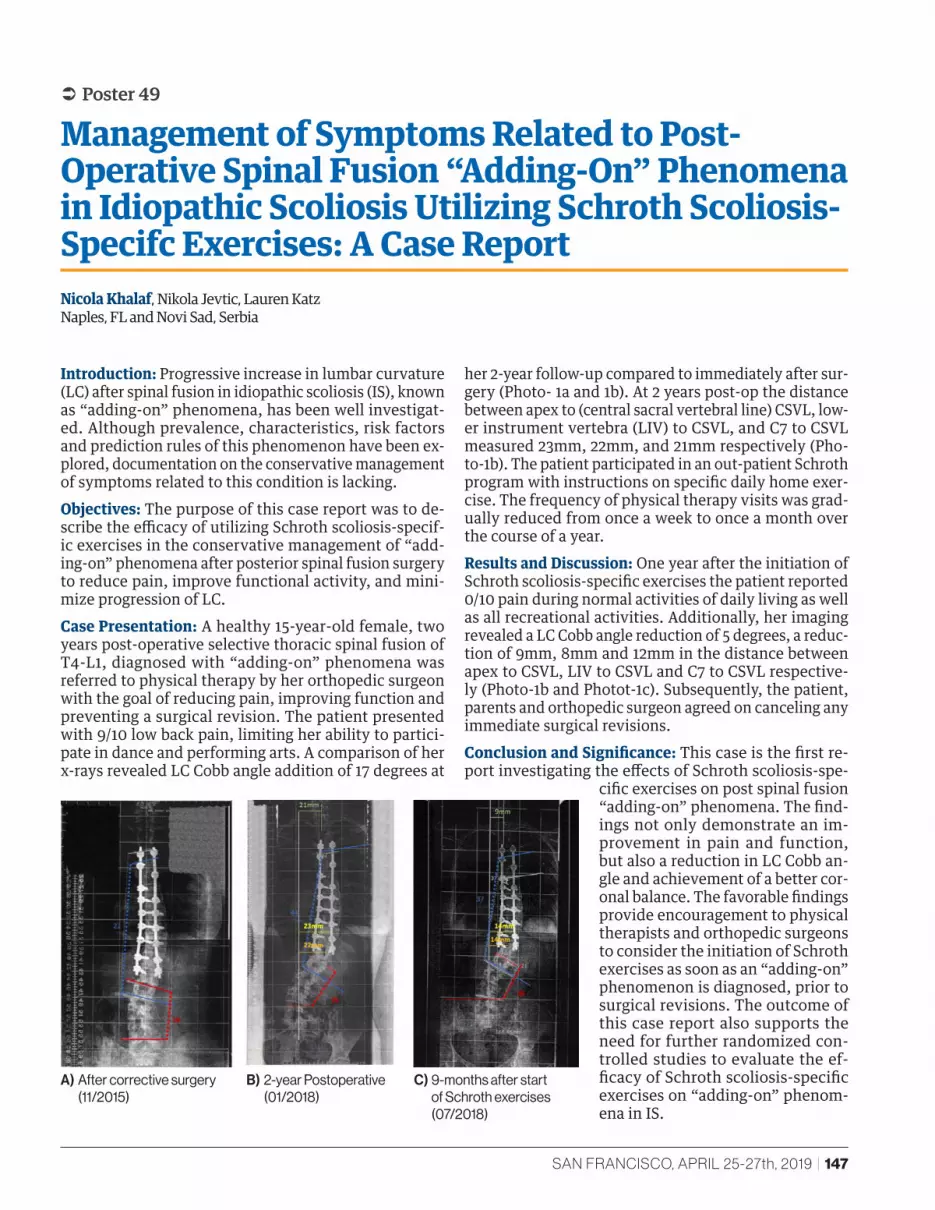
SAN FRANCISCO, APRIL 25-27th, 2019 | 147
 Poster 49
Management of Symptoms Related to Post-Operative Spinal Fusion “Adding-On” Phenomena in Idiopathic Scoliosis Utilizing Schroth Scoliosis-Specifc Exercises: A Case ReportNicola Khalaf, Nikola Jevtic, Lauren Katz Naples, FL and Novi Sad, Serbia
Introduction: Progressive increase in lumbar curvature (LC) after spinal fusion in idiopathic scoliosis (IS), known as “adding-on” phenomena, has been well investigat-ed. Although prevalence, characteristics, risk factors and prediction rules of this phenomenon have been ex-plored, documentation on the conservative management of symptoms related to this condition is lacking.Objectives: The purpose of this case report was to de-scribe the efficacy of utilizing Schroth scoliosis-specif-ic exercises in the conservative management of “add-ing-on” phenomena after posterior spinal fusion surgery to reduce pain, improve functional activity, and mini-mize progression of LC. Case Presentation: A healthy 15-year-old female, two years post-operative selective thoracic spinal fusion of T4-L1, diagnosed with “adding-on” phenomena was referred to physical therapy by her orthopedic surgeon with the goal of reducing pain, improving function and preventing a surgical revision. The patient presented with 9/10 low back pain, limiting her ability to partici-pate in dance and performing arts. A comparison of her x-rays revealed LC Cobb angle addition of 17 degrees at
her 2-year follow-up compared to immediately after sur-gery (Photo- 1a and 1b). At 2 years post-op the distance between apex to (central sacral vertebral line) CSVL, low-er instrument vertebra (LIV) to CSVL, and C7 to CSVL measured 23mm, 22mm, and 21mm respectively (Pho-to-1b). The patient participated in an out-patient Schroth program with instructions on specific daily home exer-cise. The frequency of physical therapy visits was grad-ually reduced from once a week to once a month over the course of a year.Results and Discussion: One year after the initiation of Schroth scoliosis-specific exercises the patient reported 0/10 pain during normal activities of daily living as well as all recreational activities. Additionally, her imaging revealed a LC Cobb angle reduction of 5 degrees, a reduc-tion of 9mm, 8mm and 12mm in the distance between apex to CSVL, LIV to CSVL and C7 to CSVL respective-ly (Photo-1b and Photot-1c). Subsequently, the patient, parents and orthopedic surgeon agreed on canceling any immediate surgical revisions. Conclusion and Significance: This case is the first re-port investigating the effects of Schroth scoliosis-spe-
cific exercises on post spinal fusion “adding-on” phenomena. The find-ings not only demonstrate an im-provement in pain and function, but also a reduction in LC Cobb an-gle and achievement of a better cor-onal balance. The favorable findings provide encouragement to physical therapists and orthopedic surgeons to consider the initiation of Schroth exercises as soon as an “adding-on” phenomenon is diagnosed, prior to surgical revisions. The outcome of this case report also supports the need for further randomized con-trolled studies to evaluate the ef-ficacy of Schroth scoliosis-specific exercises on “adding-on” phenom-ena in IS.
A) After corrective surgery (11/2015)
B) 2-year Postoperative (01/2018)
C) 9-months after start of Schroth exercises (07/2018)
148 | 14TH INTERNATIONAL SOSORT MEETING
 Poster 09
Global Posture Evaluation for Patients Treated with Scoliosis OrthosisJean-LoÏc Rose, Lydie Journoud, Adèle Sigal, Carole Fortin, Jean-Claude Bernard Orten, Lecante group Protéor, Centre Médico-Chirurgical et de Réadaptation des Massues, France
Introduction: Adolescent idiopathic scoliosis (AIS) is a three-dimensional (3D) developmental deformity of the spine and trunk that appears during pubertal growth. Recent works assessed postural indices obtained from 2D photographic interventions in the frontal and sagit-tal planes. Correction of posture is an important goal of physiotherapy and orthotist interventions.Objective: This study aimed to evaluate global posture effect for patient treated with scoliosis orthesis.Methods: Measurements are carried out with the Orten-Body [ONE] system. It consists in a mobile column with optical data acquisition system with turntable to acquire the whole surface of the patient. Acquisition time comes to 25 s. It uses color markers, which are attached to fea-tured anatomical points. For this case study, one clinic patient (female, 14 years) with confirmed adolescent id-iopathic scoliosis who had not undergone spinal fusion surgery was scanned and X-rayed, with and without flex brace. Efficiency of brace was validated with x-rays.
Results and Discussion: Posture correction was evaluat-ed in sagittal, frontal and transversal plane. Sagittal pos-tural correction was evaluated with the malleolus-tro-chanter-tragus anatomical point’s alignment. Sagittal balance of patient was equal to 1.59 degrees (with brace) and 4.02 degrees (without brace). In frontal plane, pos-ture analysis shows torso correction with a diminution of left frontal deport equal to 2.09 degrees (3.39 degrees with brace and 1.29 degrees without brace). In transver-sal plane, shoulder rotation was reduced from 9.3 de-grees to 3.4 degrees with brace.Conclusion and Significance: This case study demon-strates that the proposed approach is a valid non-inva-sive method to evaluate and estimate the postural cor-rection of brace in patients with idiopathic scoliosis. This validation shows difference and quantifies posture correction of patients with brace. We can make an inten-sive clinical validation using a large database of scoliotic patients.

SAN FRANCISCO, APRIL 25-27th, 2019 | 149
 Poster 13
Evaluation of Pressure on Mobile Thoracic Pad with Prestressed Carbon Blade System, In Treatment with CMCR BraceJean-Loïc Rose, Lydie Journoud, Grégory Notin, Adèle Sigal, Nuria Rodriguez Martinez , Jean-Claude Bernard Orten, Lecante group Protéor, Centre Médico-Chirurgical et de Réadaptation des Massues, France
Introduction: The CMCR brace (monoshell carbon brace respecting breathing) is an idiopathic scoliosis (IS) brace that uses pre-stressed carbon blade on a mobile pad. This technology preserves the thorax mobility and the max-imal vital capacity. It can be used at the very young age without disrupting the chest growth because the support move with the rib cage. Objective: The case-study aim is to assess the effective-ness of mobile pad, despite the movement during respi-ratory cycle.Methods: Measurements are carried out with pressure map system (Embrace). It’s an undershirt with 1024 pres-sure sensors. Each sensor records pressure during a nor-mal respiratory cycle, following to a maximal one. The pressure undershirt is worn under CMCR brace designed for the patient and validate by X-ray. For this case study, one consenting clinic patient (female, 14 years) with con-firmed IS who had not undergone spinal fusion surgery
Results and Discussion: We have evaluated pressure under pad with carbon blade. The mean pressure un-der fixing is 8.82 N/cm2 with standard deviation equal to 0.594. In other part of the brace, mean pressure is close to 1.6 with a large deviation. The minimum and maxi-mum pressure values obtained with forced respiratory cycle are respectively 8.07 N/cm2 (expiratory) and 12.01 N/cm2 (inspiratory). The mean pressure under brace pad is 7.41 N/cm2 with 0.66 for standard deviation. The maximum pressure resulting of maximum inspira-tion exerts a mean pressure of 10.05 N/cm2 and standard deviation equal to 0.92.Conclusion and Significance: The results show that carbon blade system allows a uniformly distributed and constant pressure during normal respiratory cycle. The movement of the support during the respiration doesn’t decrease the pressures of correction, but rather, provides a good cor-rection with less pressure.

150 | 14TH INTERNATIONAL SOSORT MEETING
 Poster 16
CAD Versus Custom TLSO Bracing: Initial Curve Correction For Idiopathic ScoliosisChristopher Vara Shriners Hospitals for Children - Twin Cities, MN
Introduction: We treated AIS/JIS with a custom TLSO brace fabricated at our institution until July 1, 2015, when we adopted computer-aided design (CAD) to fabricate scoliosis braces. Objectives: No significant difference in mean percent change in curve correction between CAD and CUSTOM bracing.Methods: Retrospective cohort study of female single or double-curve scoliosis patients aged 7-14 yrs. treated with CAD or CUSTOM bracing; all had digital PA full-spine x-rays. We matched CAD (July 2015-Dec. 2016) with CUS-TOM patients (April 2009-June 2015) on diagnosis (AIS or JIS), Risser stage (all 0-2), skeletal maturity (all < 2 yrs. post-menarchal) and primary curve magnitudes (25-45 degrees). Initial Cobb angles of standing in-brace curve correction were compared to most recent pre-brace full spine x-ray. T-tests compared the mean pre-post percent correction for each curve by group.
Results and Discussion: 13 CAD-treated females met cri-teria and were matched with 13 CUSTOM patients. Mean age was similar between groups (12+0 yrs. CAD; 11+6 yrs. CUSTOM). Most patients had AIS (70% CAD; 70% CUS-TOM) and Lenke I curves (85% CAD; 77% CUSTOM). Tho-racic and lumbar curves were similar at brace initiation (Table). There was no significant difference in the mean percent change in curve correction for thoracic (41% CAD vs. 36% CUSTOM, n=24) or lumbar (42% CAD vs. 45% CUSTOM, n=5) curves. Conclusion and Significance: This study found that ini-tial thoracolumbar curve correction via CAD vs. custom TLSO (CUSTOM) bracing was similar in females with ad-olescent (AIS) or juvenile (JIS) idiopathic scoliosis treat-ed at one institution. A larger study is needed to confirm these early results.

SAN FRANCISCO, APRIL 25-27th, 2019 | 151
 Poster 17
Cobb Angle Reduction after an Intensive Course of Physiotherapy Scoliosis Specific Therapy: A Case SeriesGeorgina Frere, Erika Maude, Mollie-Rose Turkentine, Jack Whiteside, Abbie Turland, Luke McKendrick Scoliosis SOS Clinic, London, UK
Introduction: Treatment for scoliosis in the United King-dom (UK) is determined using the Cobb angle. Physiother-apy scoliosis specific exercises (PSSE’s) are not recognised by the National Health Service (NHS) in the UK for treat-ment for scoliosis. Objectives: 1. To determine the significance of Cobb angle reduction in patients with scoliosis after completing an intensive course of PSSE’s. 2. To compare Cobb angle re-duction between adults and children. 3. To compare Cobb angle reduction between size of curvatures.Methods: 23 patients with scoliosis that were treated solely with an intensive course of PSSE’s and who had obtained a reduction in their Cobb angle were included in the case series. X-Rays of the patients’ spines, which were taken independently before and after treatment, were obtained and measured by three blind assessors within a 5 degree error margin. The averages were analysed using descrip-tive statistics. Results and Discussion: 19 of the patients were female (83%) and 18 were children (78%). The average age of the participants was 17.4 years old. 11 thoracic- and 8-lum-
bar curves were measured. On average, there was a Cobb angle reduction of 11.9 degrees of the thoracic curve (SD=3.9) and 10.8 degrees of the lumbar curve (SD=4.9). On average, the children had a 10.4 degree reduction of the thoracic curve (SD=3.9) and 9 degree reduction of the lumbar curve (SD=5.2) whereas adults had a 14.4 degree reduction of the thoracic curve (SD=1.3) and 13.2 degree reduction of the lumbar curve (SD=1.6). There was an average of 9.6 degrees reduction in moderate thoracic curves (Cobb angle >23.9 and <40) (SD=4.0) and 14.8 de-gree reduction in moderate lumbar curves (SD=8.7). In the severe curves (Cobb angle >39.9 degrees), on aver-age, there was an 11.5 degree reduction of the thoracic curves (SD=3.9) and a 9.1 degree reduction of the lumbar curves (SD=3.4).Conclusion and Significance: A reduction of 5 degrees is clinically significant. This case series showed that PSSE’s can achieve clinically significant Cobb angle reductions of thoracic and lumbar curves in adults and children with mild, moderate and severe curves. Further research is rec-ommended to determine the effects of PSSE’s on Cobb an-gle reduction.
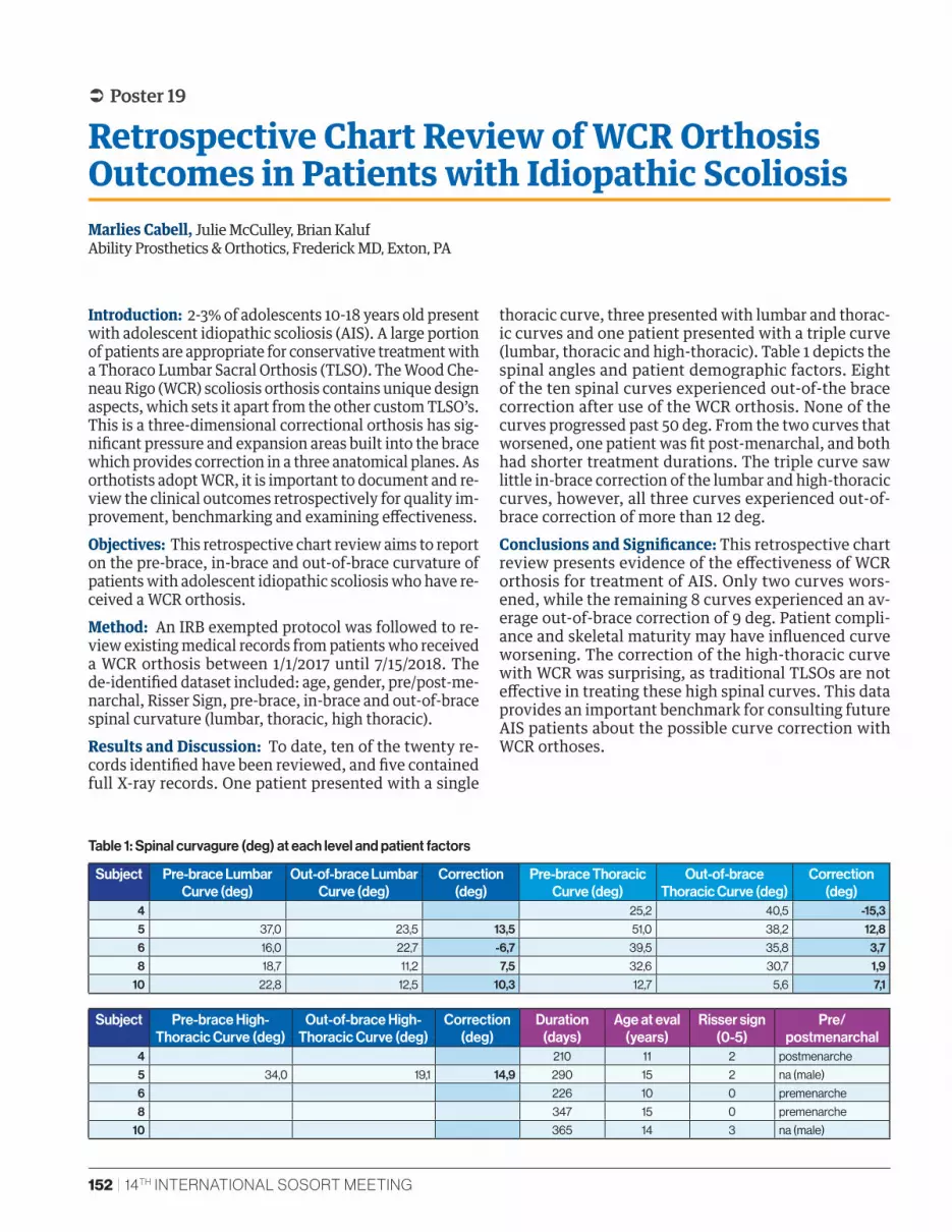
152 | 14TH INTERNATIONAL SOSORT MEETING
 Poster 19
Retrospective Chart Review of WCR Orthosis Outcomes in Patients with Idiopathic ScoliosisMarlies Cabell, Julie McCulley, Brian Kaluf Ability Prosthetics & Orthotics, Frederick MD, Exton, PA
Introduction: 2-3% of adolescents 10-18 years old present with adolescent idiopathic scoliosis (AIS). A large portion of patients are appropriate for conservative treatment with a Thoraco Lumbar Sacral Orthosis (TLSO). The Wood Che-neau Rigo (WCR) scoliosis orthosis contains unique design aspects, which sets it apart from the other custom TLSO’s. This is a three-dimensional correctional orthosis has sig-nificant pressure and expansion areas built into the brace which provides correction in a three anatomical planes. As orthotists adopt WCR, it is important to document and re-view the clinical outcomes retrospectively for quality im-provement, benchmarking and examining effectiveness. Objectives: This retrospective chart review aims to report on the pre-brace, in-brace and out-of-brace curvature of patients with adolescent idiopathic scoliosis who have re-ceived a WCR orthosis.Method: An IRB exempted protocol was followed to re-view existing medical records from patients who received a WCR orthosis between 1/1/2017 until 7/15/2018. The de-identified dataset included: age, gender, pre/post-me-narchal, Risser Sign, pre-brace, in-brace and out-of-brace spinal curvature (lumbar, thoracic, high thoracic).Results and Discussion: To date, ten of the twenty re-cords identified have been reviewed, and five contained full X-ray records. One patient presented with a single
thoracic curve, three presented with lumbar and thorac-ic curves and one patient presented with a triple curve (lumbar, thoracic and high-thoracic). Table 1 depicts the spinal angles and patient demographic factors. Eight of the ten spinal curves experienced out-of-the brace correction after use of the WCR orthosis. None of the curves progressed past 50 deg. From the two curves that worsened, one patient was fit post-menarchal, and both had shorter treatment durations. The triple curve saw little in-brace correction of the lumbar and high-thoracic curves, however, all three curves experienced out-of-brace correction of more than 12 deg.Conclusions and Significance: This retrospective chart review presents evidence of the effectiveness of WCR orthosis for treatment of AIS. Only two curves wors-ened, while the remaining 8 curves experienced an av-erage out-of-brace correction of 9 deg. Patient compli-ance and skeletal maturity may have influenced curve worsening. The correction of the high-thoracic curve with WCR was surprising, as traditional TLSOs are not effective in treating these high spinal curves. This data provides an important benchmark for consulting future AIS patients about the possible curve correction with WCR orthoses.
Subject Pre-brace Lumbar Curve (deg)
Out-of-brace Lumbar Curve (deg)
Correction (deg)
Pre-brace Thoracic Curve (deg)
Out-of-brace Thoracic Curve (deg)
Correction (deg)
4 25,2 40,5 -15,35 37,0 23,5 13,5 51,0 38,2 12,86 16,0 22,7 -6,7 39,5 35,8 3,78 18,7 11,2 7,5 32,6 30,7 1,9
10 22,8 12,5 10,3 12,7 5,6 7,1
Subject Pre-brace High-Thoracic Curve (deg)
Out-of-brace High-Thoracic Curve (deg)
Correction (deg)
Duration (days)
Age at eval (years)
Risser sign (0-5)
Pre/postmenarchal
4 210 11 2 postmenarche
5 34,0 19,1 14,9 290 15 2 na (male)
6 226 10 0 premenarche
8 347 15 0 premenarche
10 365 14 3 na (male)
Table 1: Spinal curvagure (deg) at each level and patient factors

SAN FRANCISCO, APRIL 25-27th, 2019 | 153
 Poster 20
Case Study: Treatment of Scoliosis with WCR Orthosis in 10mo Patient with Ehlers Danlos Syndrome Ocular Scoliosis Type Fkbp-14Marlies Beerli Cabell Ability Prosthetics and Orthotics, Inc., Frederick, MD
Introduction: Ehlers Danlos Syndrome (EDS) Ocular Scoli-osis Type FKBP-14 is a rare genetic condition affecting con-nective tissues and skin integrity. Treatment of infantile scoliosis with Mehta casting or bracing is considered when the spinal deformity is flexible. The presence of this EDS complicates treatment, as the corrective forces may cause skin ulceration. This 10mo female patient with EDS Ocular Scoliosis Type FKBP-14 presented with a spinal curve of 38 deg. She was referred for orthotic management with the WCR orthosis instead of Mehta casting, due to skin integ-rity concerns and parents’ desire to avoid anesthesia. The WCR orthosis provides three-dimensional correction and has significant pressure and expansion areas built into the brace for spinal elongation and derotation.Objective: Treatment goals include 1. Correct spinal cur-vature and rotation. 2. Avoid skin breakdown. 3. Allow parents to perform daily skin checks. Methods: The patient was evaluated by a certified ortho-tist trained in the Wood Cheneau Rigo (WCR) orthosis. The flexibility of the thoracic curvature was confirmed. The spine was elongated for the impression by the moth-er suspending the patient underneath the arms. This achieved an impression in the flexible corrected posi-tion for Wood Cheneau Rigo (WCR) orthosis fabrication. The patient was 69.2cm long and weighed 17lbs when the Wood Cheneau Rigo (WCR) orthosis was fabricated by Align Clinic. Initially, two foam-lined orthoses with different closure designs were provided.
Results and Discussion: Parents reported the anterior opening WCR design was easiest to don. A superior fit was confirmed by inspecting the derotation of the patient’s spine and filling of the voided spaces in the WCR design. X-rays confirmed in-brace correction to 42 deg. The patient was seen for follow-up every month. With the scoliosis stabilized using the WCR orthosis, the patient was able to roll-over, crawl, push her own wheelchair and participate in social activities with an upright posture. As the patient grew, she received two more WCR orthoses (every 8 weeks) before reaching the age of 17 mo, height of 80.9 cm and weight of 22 lbs. The patient was then referred for Mehta casting because the out-of-brace X-ray curvature wors-ened. Only one Mehta casting was tolerated until it was discontinued due to skin ulceration. The patient’s out-of-brace curvature worsened to 72 degrees, and the patient was finally referred for surgical correction.Conclusion and Significance: Patient’s scoliosis is expect-ed to worsen until she reaches skeletal maturity, due to the EDS Ocular Scoliosis Type FKBP-14 diagnosis. Her skin integrity made conservative scoliosis treatment difficult. The Wood Cheneau Rigo (WCR) orthosis provided a ben-efit over Mehta casting, because it was removable for skin checks and may reduce the risk of skin ulceration in pa-tients with this form of EDS. If conservative treatment of infantile scoliosis with WCR orthoses can be tolerated better than Mehta casting, sur-gical interventions can be prolonged until later in life.
154 | 14TH INTERNATIONAL SOSORT MEETING
 Poster 21
Case Study: Treatment of Scoliosis with WCR Orthosis in an 82 Year Old with Osteogenesis Imperfecta Type 1Marlies Beerli Cabell Ability Prothetics and Orthotics, Frederick, MD
Introduction: Osteogenesis imperfecta (OI) type 1 known as “brittle bone disease”, and scoliosis is one manifesta-tion of the condition. This 82 yo female OI patient presents with 46.5 degree thoracic and 50 degree lumbar curves. Her scoliosis results in pain from ribs contacting her pel-vis, difficulty breathing and limits her ambulation. The patient has received Schroth therapy for 10 years, which teaches scoliosis-specific exercises. The effort to main-tain the frontal plane balance and sagittal plane alignment was so exhausting that she could not maintain long term. Pre-fabricated spinal orthoses were ineffective in treat-ing her scoliosis. The Wood Cheneau Rigo (WCR) orthosis provides three-dimensional correction and has significant pressure and expansion areas built into the brace for spi-nal elongation and derotation. Although typically used to treat idiopathic adolescent scoliosis (IAS), the SSS ortho-sis was prescribed for this 82 year old OI patient to reduce complications related to her scoliosis.Objective: Patient goals are to 1. Remove the pain from her ribs rubbing on her pelvis. 2. Allow her to breathe while walking with her walker 3. Support a corrected and elon-gated spinal position.Methods: The patient was evaluated by a certified ortho-tist trained in the WCR. A 3D scan was captured while the patient performed her Schroth therapy exercises to main-tain a corrected spinal position in standing. She to develop endurance to hold the desired position for the duration of
the scan. The WCR orthosis was custom fabricated through Align Clinic. The patient was instructed to don the WCR orthosis while holding the corrected Schroth position. The patient tolerated wearing the WCR orthosis for 4 hours each day and able to perform her Schroth exercises while wearing the WCR orthosis.Results and Discussion: After 8 months of WCR orthosis use, an out-of-brace X-ray showed correction of 40 de-gree thoracic and 36 degree lumbar curves and a “Stable S-shaped Curve”. When wearing the WCR orthosis, the patient reported lower rib pain, easier breathing and in-creased mobility on stairs and carrying objects. The pa-tient reports improvement in regular bowel movements and being more active and independent than in the past 10 years. She has suffered no falls or bone fractures.Conclusion and Significance: Although typically consid-ered for treating AIS, this case study highlights the poten-tial to apply Schroth therapy and WCR orthosis to treat other scoliosis presentations. This 82 yo female OI patient tolerated the WCR orthosis well, and she experienced clin-ically meaningful improvement in her rib pain, breathing and mobility. The role of Schroth therapy in conjunction with the WCR orthosis was significant in the outcome of this case. The treatment principles of Schroth therapy and the WCR orthosis may be considered when presented with unique scoliosis cases, such as geriatric patients with os-teogenesis imperfecta type 1.
SAN FRANCISCO, APRIL 25-27th, 2019 | 155
 Poster 22
Case Presentation for BMI >99% in an Adolescent Idiopathic Scoliosis Correction of the Curves in Her Out of Brace Correction At 1 YearMarlies Beerli Cabell Ability Prothetics and Orthotics, Frederick, MD
Introduction: Patient presented with a Double Major sco-liosis curve at BMI >99% for her age and gender. At initial referral her curve magnitudes were apex T8 with 18 degree and Apex L3 at 28 degree, Risser 3. Objective: At the evaluation the forward bending test was completed but the amount of rotation was not significant. The main understanding of the rotation component to her scoliosis is the medical necessity for the Wood Cheneau Rigo Scoliosis TLSO. Methods: She was able to complete all the clinical require-ments for the fabrication of the Wood Cheneau Rigo Sco-liosis TLSO. The patient was then delivered the WCR with the instruction to always lie down for the anterior overlap to continue to move. At the three month follow-ups the an-terior overlap was assessed. The amount of closure became so tight at the rivets for the Velcro straps had to be drilled wider so that the desired correction continued. The straps
were also replaced. The patient was wearing the WCR 20 hours or more every day. Results and Discussion: The one year clinical report with out of brace X-ray reveals that the apex T8 is at 15 degrees and the Apex at L3 is 22.1degrees. The patient is now at a Risser 4 and SMS 7. This means that she is in the final stages of bony maturity. She has not grown much and so the WCR fits her corrective forces still. The out of brace correction is evident through her in brace photos between delivery and the 1year use. Conclusion and Significance: This patient base with a BMI >99% must be given the WCR as an opportunity to correct the scoliosis curve. The patient’s compliance will bring cor-rection to their curves as they induce more pull for closure and thus the built-in corrective padding becomes more in-fluential with time. Their adipose tissue migrates outside the WCR trim lines.

156 | 14TH INTERNATIONAL SOSORT MEETING
 Poster 23
Systematic Review of Chiropractic Biophysics® (CBP®) Methods Employed in the Rehabilitation of Cervical LordosisDeed Harrison, Ibrahim Moustafa, Paul Oakley CBP NonProfit, Inc., Eagle, ID, University of Sharjah, UAE, Newmarket, ON, Canada
Introduction: Biomechanically the cervical lordosis is es-sential for preserving physiologic slack in cord/nerve roots, normal spinal coupling, and ideal muscle length-tension relationships. Evidence for maintenance/restoration of cer-vical lordosis is well established in the surgical literature and is accumulating in spinal rehabilitation.Objective: To systematically review the evidence for Chi-ropractic BioPhysics® (CBP®) non-surgical methods used to increase cervical lordosis.Methods: Literature was searched across several databas-es using search terms including combinations of ‘cervical,’ ‘spine,’ ‘lordosis,’ ‘kyphosis,’ ‘hypolordosis,’ ‘rehabilitation,’ ‘restoration,’ ‘improvement,’ ‘treatment,’ ‘sagittal,’ ‘align-ment,’ ‘neck,’ ‘headache,’ ‘pain,’ and ‘CBP’ or ‘Chiroprac-tic BioPhysics.’ Only peer-reviewed papers were included. Two independent reviewers located studies and extracted data.Results and Discussion: There were 44 papers located: 9 RCTs, 3 NRCTs, 2 case series, 30 case reports. All reported radiographic lordosis improvements associated with pain reduction; many documented disability and quality of life improvements. Cases varied in lordosis correction (2-47º), forward head translation reduction (3-55mm), treatment number (11-86), and treatment duration (0.75-9.5months). All RCTs and many cases included follow-up (3months-13years) showing continued patient wellbeing and rela-
tive stability of lordosis improvement. 8 RCTs definitively show CBP extension traction leads to increased lordosis (7-14º over 30 sessions) not accomplished by other treat-ments. CBP treated traction groups remained well up to 2 years, whereas, comparison groups showed symptom re-gression after treatment. There is substantial evidence that increasing cervical lordosis by extension traction as part of multimodal rehabilitation programs leads to improved outcomes that remain stable long-term, versus ‘standard’ programs (not increasing lordosis) showing symptom re-gression after cessation of care. Valid pre-post cervical lordosis improvements must be greater than slight head nodding (6º) and error of posterior tangent measurement (2-3º). Based on this, approximately 11 trials and 30 indi-vidual cases, representing >400 patients, substantiate sig-nificant lordosis improvement (7-17º) in 3-6 months. The evidence also shows diverse cranio-cervical ailments and radiographic spinal misalignments improved coinciding with improvements in diverse physiologic parameters in-cluding central somatosensory conduction time, cervico-cephalic kinesthetic sensibility, pressure pain thresholds, dermatomal somatosensory evoked potentials, flexion-ex-tension kinematics, pain and disability.Conclusion and Significance: CBP technique has substan-tial evidence demonstrating consistency in increasing cer-vical lordosis in diverse symptomatic cohorts and case pre-sentations.
SAN FRANCISCO, APRIL 25-27th, 2019 | 157
 Poster 24
Systematic Review of CBP® Methods Employed in the Rehabilitation of Lumbar LordosisDeed Harrison, Ibrahim Moustafa, Paul Oakley CBP NonProfit, Inc., Eagle, ID, University of Sharjah, UAE, Newmarket, ON, Canada
Introduction: The lumbar lordosis has proven essential for the wellbeing of patients in many recent systematic reviews. Its maintenance has also proven superior for long-term successful surgical outcomes.Objective: To systematically review the evidence on lum-bar extension traction methods used to non-surgically in-crease lumbar lordosis in the treatment of back pain and related disorders by CBP® technique methods.Methods: Literature was searched using multiple data-bases. Search terms included ‘lumbar,’ ‘spine,’ ‘lordosis,’ ‘rehabilitation,’ ‘restoration,’ ‘improvement,’ ‘correction,’ ‘treatment,’ ‘increase,’ ‘traction,’ ‘sagittal,’ ‘alignment,’ ‘low back,’ ‘pain,’ and CBP® technique. Two independent re-viewers located studies and extracted data.Results and Discussion: 12 articles were located, 3 RCTs, 1 NRCT, 2 case series, 6 case reports. All studies demon-strated increased radiographic lumbar lordosis after ex-tension traction as part of multimodal rehabilitation pro-grams. All cases demonstrated concomitant decreases in pain; many showed reduced disability. There were large variations in lordosis improvement (3-50 degrees), treat-ment number (30-100) and treatment duration (10weeks-7months). All clinical trials included follow-up showing good maintenance of both lordosis correction and pain relief.The clinical trials showed increases in radiographic mea-sured lordosis of 7-11 degrees. Comparison groups had no
increase in lordosis. In the RCTs, comparison groups had no increase in lordosis, but had only temporary symptom-atic relief during treatment which regressed to baseline pain levels at follow-up while traction groups remained well up to 15-months.Preliminarily, these studies indicate that increasing the lumbar lordosis via extension traction as part of multimodal rehabilitation programs are associat-ed with superior outcomes over ‘standard-care’ treatment programs that do not improve hypo-lordosis. The strength of the RCT designs was accomplished by having random-ized comparison groups receive identical treatments less the traction. Since the comparison groups had regression of symptoms towards baseline following care, this sub-stantiates that the increase in lordosis was achieved by the extension traction, and this led to better and stable outcomes following care. The evidence points to the fact that ‘conventional’ low back treatments do not structurally improve the lordosis and therefore, may leave patients in predisposition for fu-ture LBP after treatment cessation. It is essential for future research to include post-treatment follow-up in low back pain trials to assess for regression of symptoms after ces-sation of care.Conclusion and Significance: There is a moderate but con-sistent evidence substantiating CBP® technique as a reli-able approach to increase lumbar lordosis in symptomatic patients having hypo-lordosis. This is significant as low back pain treatment remains controversial.

158 | 14TH INTERNATIONAL SOSORT MEETING
 Poster 27
Correction of Cervical Pseudo-Scoliosis and Alleviation of Dystonia Symptoms Using Chiropractic Biophysics® (CBP®) Technique: A Case ReportJason Haas, Paul Oakley, Deed Harrison Windsor, CO, Newmarket, ON, Canada; CBP NonProfit, Inc., Eagle, ID
Introduction: Biomechanically the cervical lordosis is es-sential for preserving physiologic slack in cord/nerve roots, normal spinal coupling, and ideal muscle length-tension relationships. Evidence for maintenance/restoration of cer-vical lordosis is well established in the surgical literature and is accumulating in spinal rehabilitation.Objective: To systematically review the evidence for Chi-ropractic BioPhysics® (CBP®) non-surgical methods used to increase cervical lordosis.Methods: Literature was searched across several databas-es using search terms including combinations of ‘cervical,’ ‘spine,’ ‘lordosis,’ ‘kyphosis,’ ‘hypolordosis,’ ‘rehabilitation,’ ‘restoration,’ ‘improvement,’ ‘treatment,’ ‘sagittal,’ ‘align-ment,’ ‘neck,’ ‘headache,’ ‘pain,’ and ‘CBP’ or ‘Chiroprac-tic BioPhysics.’ Only peer-reviewed papers were included. Two independent reviewers located studies and extracted data.Results and Discussion: There were 44 papers located: 9 RCTs, 3 NRCTs, 2 case series, 30 case reports. All reported radiographic lordosis improvements associated with pain reduction; many documented disability and quality of life improvements. Cases varied in lordosis correction (2-47º), forward head translation reduction (3-55mm), treatment number (11-86), and treatment duration (0.75-9.5months). All RCTs and many cases included follow-up (3months-13years) showing continued patient wellbeing and rela-
tive stability of lordosis improvement. 8 RCTs definitively show CBP extension traction leads to increased lordosis (7-14º over 30 sessions) not accomplished by other treat-ments. CBP treated traction groups remained well up to 2 years, whereas, comparison groups showed symptom re-gression after treatment. There is substantial evidence that increasing cervical lordosis by extension traction as part of multimodal rehabilitation programs leads to improved outcomes that remain stable long-term, versus ‘standard’ programs (not increasing lordosis) showing symptom re-gression after cessation of care. Valid pre-post cervical lordosis improvements must be greater than slight head nodding (6º) and error of posterior tangent measurement (2-3º). Based on this, approximately 11 trials and 30 indi-vidual cases, representing >400 patients, substantiate sig-nificant lordosis improvement (7-17º) in 3-6 months. The evidence also shows diverse cranio-cervical ailments and radiographic spinal misalignments improved coinciding with improvements in diverse physiologic parameters in-cluding central somatosensory conduction time, cervico-cephalic kinesthetic sensibility, pressure pain thresholds, dermatomal somatosensory evoked potentials, flexion-ex-tension kinematics, pain and disability.Conclusion and Significance: CBP technique has substan-tial evidence demonstrating consistency in increasing cer-vical lordosis in diverse symptomatic cohorts and case pre-sentations.

SAN FRANCISCO, APRIL 25-27th, 2019 | 159
 Poster 31
Improvement in Thoracolumbar Neuromuscular Levoscoliosis in a 14-Year-Old Male with a History of Stage 3 Medulloblastoma and Post-Operative Cerebellar Mutism Syndrome Using Chiropractic Biophysics Technique and ScoliBrace Thoracolumbosacral Orthotic Bracing: A Case Study and 30-Month Follow-UpDouglas F. Lightstone, Curtis Fedorchuk, Julia Guo, Jeb McAviney Institute for Spinal Health and Performance, Institute for Spinal Health and Performance, ScoliCare
Introduction: A 14-year-old male presented to a chiro-practic clinic with complaints of abnormal posture and a diagnosis of neuromuscular scoliosis with a left lateral thoracolumbar curvature from T6-L1 with the apex at T11 measuring 29.3˚ using Cobb method. The patient had a history of stage 3 medulloblastoma at 5 years old. He un-derwent a craniotomy to remove the tumor in his cere-bellum. A postoperative cerebellar mutism rendered him unable to walk, talk, swallow and use his right arm. The patient also sustained high-frequency hearing loss, a pro-cessing disorder affecting his speed of processing infor-mation, and physical coordination disabilities as side ef-fects from chemotherapy and radiation therapy. The pa-tient presented with abnormal posture. The physical exam portion revealed dysmetria and dysdiadochokinesia. The patient had difficulty with balance and proprioception. Upon static and motion palpation, it was noted that there was decreased range of motion in all directions and in-creased rigidity for cervical, thoracic, and lumbar regions. Objectives: To report the clinical results with long-term follow-up of using Chiropractic BioPhysics (CBP) Technique combined with ScoliBrace thoracolumbo-sacral orthotic (TLSO) bracing for thoracolumbar neu-romuscular levoscoliosis in a 14-year-old male at Riss-er stage 2 with a history of stage 3 medulloblasto-ma and post-operative cerebellar mutism syndrome. Methods: The patient was seen 14 times over 13 months due to the distance between the patient’s
home and the treating chiropractor. CBP® chiro-practic care consisted of Mirror Image exercis-es, adjustments, and tractions. ScoliBrace was pre-scribed to the patient to improve the scoliosis. Results and Discussion: A 13-month follow-up radio-graphic analysis of the patient out of the brace for over 24 hours showed an improvement in T6-L1 Cobb angle from 29.3˚ to 7.9˚. Follow-up postural analysis showed improvement in coronal posture toward cen-ter of gravity. The patient continues to wear the brace for structural support. A 30-month follow-up radio-graphic analysis showed skeletal maturity to a Risser stage 4 and an increase in T6-L1 Cobb angle from 7.9˚ to 15.8˚. However, the T6-L1 Cobb angle showed an over-all maintained improvement from the original 29.3˚. Conclusion and Significance: Neuromuscular scolio-sis is caused by disorders of the brain, spinal cord, and muscular system resulting in an inability to maintain appropriate postural and spinal balance and alignment. Neuromuscular curves are often associated with pelvic obliquity and thoracic hyperkyphosis. Neuromuscular scoliosis curves are very likely to progress continuing into adulthood. The clinical progress documented in this case study reveals that chiropractic care using CBP pro-tocol combined with ScoliBrace TLSO bracing may prove effective in correcting Cobb angle in patients with neu-romuscular scoliosis and a history of cerebellar pathol-ogy. Further research on the effectiveness is warranted.
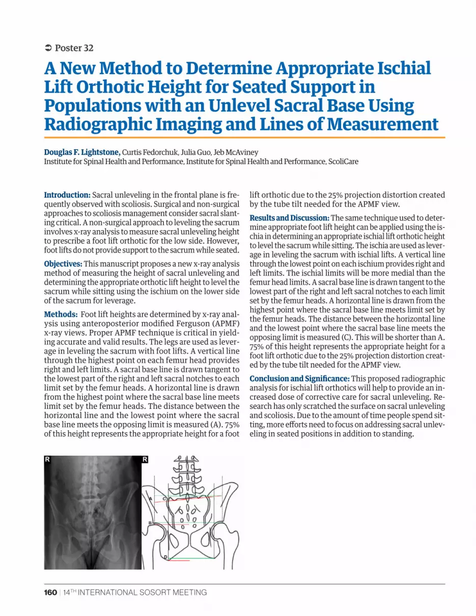
160 | 14TH INTERNATIONAL SOSORT MEETING
 Poster 32
A New Method to Determine Appropriate Ischial Lift Orthotic Height for Seated Support in Populations with an Unlevel Sacral Base Using Radiographic Imaging and Lines of MeasurementDouglas F. Lightstone, Curtis Fedorchuk, Julia Guo, Jeb McAviney Institute for Spinal Health and Performance, Institute for Spinal Health and Performance, ScoliCare
Introduction: Sacral unleveling in the frontal plane is fre-quently observed with scoliosis. Surgical and non-surgical approaches to scoliosis management consider sacral slant-ing critical. A non-surgical approach to leveling the sacrum involves x-ray analysis to measure sacral unleveling height to prescribe a foot lift orthotic for the low side. However, foot lifts do not provide support to the sacrum while seated.Objectives: This manuscript proposes a new x-ray analysis method of measuring the height of sacral unleveling and determining the appropriate orthotic lift height to level the sacrum while sitting using the ischium on the lower side of the sacrum for leverage.Methods: Foot lift heights are determined by x-ray anal-ysis using anteroposterior modified Ferguson (APMF) x-ray views. Proper APMF technique is critical in yield-ing accurate and valid results. The legs are used as lever-age in leveling the sacrum with foot lifts. A vertical line through the highest point on each femur head provides right and left limits. A sacral base line is drawn tangent to the lowest part of the right and left sacral notches to each limit set by the femur heads. A horizontal line is drawn from the highest point where the sacral base line meets limit set by the femur heads. The distance between the horizontal line and the lowest point where the sacral base line meets the opposing limit is measured (A). 75% of this height represents the appropriate height for a foot
lift orthotic due to the 25% projection distortion created by the tube tilt needed for the APMF view.Results and Discussion: The same technique used to deter-mine appropriate foot lift height can be applied using the is-chia in determining an appropriate ischial lift orthotic height to level the sacrum while sitting. The ischia are used as lever-age in leveling the sacrum with ischial lifts. A vertical line through the lowest point on each ischium provides right and left limits. The ischial limits will be more medial than the femur head limits. A sacral base line is drawn tangent to the lowest part of the right and left sacral notches to each limit set by the femur heads. A horizontal line is drawn from the highest point where the sacral base line meets limit set by the femur heads. The distance between the horizontal line and the lowest point where the sacral base line meets the opposing limit is measured (C). This will be shorter than A. 75% of this height represents the appropriate height for a foot lift orthotic due to the 25% projection distortion creat-ed by the tube tilt needed for the APMF view.Conclusion and Significance: This proposed radiographic analysis for ischial lift orthotics will help to provide an in-creased dose of corrective care for sacral unleveling. Re-search has only scratched the surface on sacral unleveling and scoliosis. Due to the amount of time people spend sit-ting, more efforts need to focus on addressing sacral unlev-eling in seated positions in addition to standing.

SAN FRANCISCO, APRIL 25-27th, 2019 | 161
 Poster 33
A New Method to Measure the Lateral Translation of Vertebrae in Populations with Laterolisthesis using Radiographic Imaging and Lines of MeasurementDouglas F. Lightstone, Curtis Fedorchuk Institute for Spinal Health and Performance
Introduction: Lateral translation of one vertebra on an-other in the frontal plane is frequently observed with scoliosis. Surgical and non-surgical approaches to scolio-sis management consider laterolisthesis critical. Current methods to measure the severity of laterolisthesis use the overhanging measurement of one vertebra on another with the lateral aspects of the vertebrae as landmarks. However, when degeneration and arthritis progress, bony deformation takes place in the form of osteophytes and compression. This can make it difficult to determine an accurate, reliable, and valid measurement of laterolisthe-sis severity. In scoliosis management, accurate, reliable, and valid measurements are essential in determining the rate of progression as well as efficacy of care.Objectives: This manuscript proposes a new x-ray analy-sis method of measuring laterolisthesis severity.Methods: On anteroposterior (AP) and posteroanterior (PA) spinal views, the centroid for each vertebra can be assessed by connecting the midpoint of the superior and inferior endplates and the midpoints of the sides of the
vertebra. This method is more accurate than connecting the superior left and inferior right points and the supe-rior right and inferior left points. Where a laterolisthesis exists, a line perpendicular to the superior endplate of the inferior vertebra at the midpoint of the superior end plate is drawn until is reaches the centroid of the supe-rior vertebra. Results and Discussion: The distance between these lines will provide the lateral displacement of the centroids be-tween vertebrae where a laterolisthesis exists. Center of gravity is more important than is overhang which may prove to be exaggerated by bony deformation supported by Wolff’s law.Conclusion and Significance: This proposed radiographic analysis to measure laterolisthesis severity will help to properly monitor this skeletal condition which contrib-utes to spinal canal stenosis. Research has only scratched the surface on laterolisthesis and scoliosis and spinal ca-nal stenosis can be very detrimental to neuromusculo-skeletal and visceral function and health.
162 | 14TH INTERNATIONAL SOSORT MEETING
 Poster 36
Comparison of SRS-22 Scores of Adults After Versus Adolescents During Specific Physiotherapy for ScoliosisEwelina Białek-Kucharska, Marianna Białek, Tomasz Kotwicki FITS Education Center, University of Medical Sciences, Poznan, Poland
Introduction: The health related quality of life during non-operative scoliosis treatment has been extensively studied however, there is less data comparing the quality of life reported by the adults previously treated versus the adolescents being under treatment.Objective: The aim of the study was to compare the results of the SRS-22 questionnaire applied to the adults patients who had undergone the FITS treatment (Functional Indi-vidual Therapy of Scoliosis) in the past and had complet-ed it at least 2 years before versus the adolescent patients who have just started the FITS treatment.Methods: Forty-one adults, aged 18-30, mean 22.7, SD 3.2, 5 men and 36 women, filled the questionnaire versus 28 adolescents girls, aged 10-16, mean 13.6, SD 1.9. The Cobb angle in the adults group was 34.4° ± 12.9 (range 11°-61°) within the primary curve and 28.7° ± 9.4 (range 11°-51°) within the secondary curve while in the adolescents group it was 34.6° ± 11.0 (range 18°-55°) and 24.2° ± 9.5 (range 12°-44°), respectively.The adult patients underwent FITS treatment and com-pleted the treatment on the average 6 years before (range:
2-13 years). The treatment consisted of [specific physio-therapy name] standard protocol while 27 patients addi-tionally had the TLSO rigid brace. The adolescents received the same protocol of [specific physiotherapy name] treat-ment while 18 adolescents additionally had the TLSO brace (the same brace type as the first group).All patients filled the SRS-22 questionnaire using the cul-turally adapted and validated Polish version. The scores for each domain were compared between the two groups. Results and Discussion: The SRS-22 scores revealed as fol-lows (adult patients vs. adolescent patients): Function: 4.5 (± 0.4) vs. 4.6 (± 0.4), p>0.05; Pain: 4.2 (± 0.6) vs. 4.6 (± 0.6), p=0.0018; Self-image: 3.9 (± 0.6) vs. 3.5 (± 0.8), p>0.05; Mental health: 3.6 (± 0.6) vs. 3.7 (± 0.7), p>0.05; Satisfaction: 4.4 (± 0.5) vs. 3.8 (± 0.9), p=0026; Total 4.1 (± 0.3) vs. 4.1 (± 0.4), p>0.05. Conclusion and Significance: Adult patients previously treated with specific physiotherapy revealed higher SRS-22 scores for Satisfaction and lower SRS-22 scores for Pain, comparing to adolescent patients being under treatment because of similar spinal deformity.

SAN FRANCISCO, APRIL 25-27th, 2019 | 163
 Poster 41
The Effect of 5th Pelvic Correction on Postural Alignment during International Schroth Three Dimensional Scoliosis Therapy (ISST) Exercises In Adolescent Idiopathic ScoliosisNikola Jevtic, Nicola Khalaf, Danka Ljubojevic, Milan Pantovic Scolio Center, Degrees Scoliosis Center, Scolio Center, University of Nevada
Introduction: The efficacy of using Schroth scoliosis-spe-cific exercises in the management of adolescent idiopath-ic scoliosis (AIS) has shown promising results. Pelvic cor-rections (PC) are a key component of and play an import-ant role in this method of the pelvic corrections the 5th PC, unique to the ISST method, adjusts the pelvis in the frontal plane and forces the lumbar spine into a more midline position. There are no current studies demon-strating radiographic changes in postural alignment while performing the 5th PC during Schroth exercises.Objectives: The purpose of this case report is to demon-strate the radiographic postural changes achieved while performing the 5th pelvic correction during Schroth ex-ercises.Methods: A 14-year-old girl with a Risser grade 4, diagnosed with AIS presented with lumbar curve (LC) Cobb angle mea-suring at 39 ˚, and thoracic curve (TC) Cobb angle of 20 ˚ according to the radiologist. The lumbar and thoracic angle of trunk rotation (ATR) measured by a scoliometer in stand-ing position was 16 ˚ and 2 ˚, respectively. She was given a Schroth classification of Lri Tle (L-type with major lumbar right and secondary thoracic left curves). In the clinic, x-ray imaging was obtained with the patient in a neutral standing position (Photo-1a), followed by an image while performing a 5th pelvic correction and auto-elongation (Photo-1b). In addition, patient was photographed in sitting and stand-
ing positions to establish a clear clinical picture of above mentioned stances for future comparisons. Testing includ-ed gathering results relative to changes in the following fea-tures: Cobb angles, coronal balance, trunk deviation, dis-tance from lumbar apex to central sacral line (CSL), angle of pelvic tilt in frontal plane, and angle of L4-L5 tilts. Results and Discussion: Analysis of the radiographic im-aging features revealed differences in measurements be-tween the neutral position as compared to the 5th pelvic correction position. Cobb angle reduced from 38.7˚ to 27.8˚ in LC and from 20.3˚ to 17.9˚ in TC. Coronal balance moved from 34.89mm to 26.41mm, while trunk deviation changed from 24.65mm to 15.20mm and the apex lumbar distance shifted from 46.89mm to 36.81mm. In the fron-tal plane the angle of pelvic tilt and L4-L5 tilt changed from 1.1˚ to 4˚ and from 0.5˚ to 2.6˚, respectively, with-out producing a counter tilt in the L4-L5 vertebrae.Conclusion and Significance: This case is the first of its kind demonstrating the effectiveness of ISST’s 5th PC on postural alignment in AIS. Additionally, these favorable results were achieved without producing a counter tilt in the L4-L5 vertebral segment. Based on these findings, the 5th PC should be utilized in L-type scoliosis patients during the entire exercise period. However, prospective studies with a higher subject number is needed to eval-uate if these effects are reproducible.

164 | 14TH INTERNATIONAL SOSORT MEETING
 Poster 44
Successful Multimodal Treatment of Severe Adolescent Idiopathic Scoliosis after Failing Standard TLSO BracingAnthony Nalda, Ben Brown, Jeb McAviney Celebration Family Chiropractic, ScoliCare, Australia
Introduction: Bracing has the potential to stop adolescent idiopathic scoliosis (AIS) progressing to surgery. Howev-er, results vary and some braces may be more successful than others due to their design. This case study describes the treatment of a patient treated with a standard Boston style TLSO which ultimately failed and led to a recom-mendation of surgery. The patient refused surgery and opted for a different bracing and rehabilitation approach, achieving significant correction of the curve, major cos-metic improvement, and avoiding surgery. Methods: The patient was initially diagnosed at age 11 with AIS, Risser stage 0. Orthopaedic and neurological tests were normal, there was no history of trauma or relevant medi-cal conditions. MRI showed no abnormalities. She had a primary right thoracic curve of 37°. She was fitted with a Boston style TLSO brace made at the O&P department in a Canadian hospital. She was under orthopaedic care and was recommended general physical therapy. From age 11 to 13 her curve continued to progress despite good report-
ed brace compliance. After 3 years of treatment her curve had progressed to 50°, she was Risser 4 and she was recom-mended surgery. The patient and the parents refused sur-gery and switched her treatment to a different brace and a chiropractic rehabilitation system. Examination showed that on Adams forward bending her Scoliometer measurement was 19° at T8. She had a TRACE score of 12. On right lateral bending x-ray her primary thoracic curve reduced to 26°. She completed a 2-week intensive in-patient scoliosis rehabilita-tion program using the CLEAR method and was fitted with a ScoliBrace TLSO. The patient was then given home exercis-es based on the SEAS principle, combined with a ScoliRoll. Results and Discussion: At the end of the 2-week inten-sive program an in-brace x-ray was taken, and her curve measured 9°. At one-month follow-up a 4-hour out of brace x-ray showed the curve had reduced to 33°. At 4 months’ follow-up, the patient’s curve has reduced to just 18° out-of-the-brace, a reduction of 32°. Her TRACE score had im-proved to 2 and her Scoliometer reading was 7°. She report-ed good compliance with the brace and exercise regime. De-spite the fact this patient has not yet finished treatment, the result so far is important as it demonstrates the difference between two approaches in their ability to effectively treat scoliosis. The ScoliBrace brace is a 3D over-corrective brace using mirror image principles of correction. The CLEAR re-habilitation method focused on increasing the flexibility of the curve during the 2-week intensive period, and chi-ropractic rehabilitation had a stabilizing focus. Conclusion and Significance: This case demonstrates that using a brace and rehabilitation approach that are designed to try an achieve correction rather than stabilization of the curve can give superior in-treatment results compared to standard treatment.
Figure 1. Shows the patient after the failure of standard TLOS treatment, on the left. On the right, the impove-ment after 4 months of treatment with ScoliBrace and combined CLEAR and SEAS rehabilitation.
SAN FRANCISCO, APRIL 25-27th, 2019 | 165
 Poster 50
Reduction of a Severe Scoliosis Using an Over-Corrective Rigid Orthosis in an Adolescent FemaleJeb McAviney, Benjamin T. Brown Scolicare, Kogarah, NSW, Australia
Introduction: Adolescent idiopathic scoliosis (AIS) is a common condition characterized by a three-dimensional spinal curvature that exceeds 10° (Cobb angle) in the cor-onal plane and includes vertebral rotation. While there are several treatments available for AIS patients, surgery is recommended for curves ≥45°. This is due to the high risk of progression and subsequent decline in health-re-lated outcomes in patients with curves of this magnitude. Conservative management is not generally considered as a valid/effective treatment option for patients with severe scoliosis. Objective: The authors present a case involving the suc-cessful management of an adolescent patient with severe idiopathic scoliosis using a customised over-corrective thoracolumbosacral orthosis.Methods: The 13-year-old, pre-menarchal, female pa-tient presented to the clinic with a recent diagnosis of scoliosis. The patient was receiving treatment for dental and temporomandibular joint issues but was otherwise healthy. The physical examination revealed significant postural change, specifically curvature in the middle and lower sections of the patient’s spine, uneven heights of the shoulders and iliac crests, waist curve asymmetry and scapula mal-positioning. Thoracic trunk rotation (8 Bun-nell degrees on the left) was observed during the Adams forward bend test. Full spine x-rays highlighted a tho-racic and a thoracolumbar curve measuring 43° and 41°
respectively. The patient was graded as Risser 1. Given the magnitude of the curves the patient was referred for an orthopaedic consultation. Surgery was advised, howev-er the patient and her parents refused surgery and chose to trial conservative care. The patient was subsequently prescribed an over-corrective rigid thoracolumbosacral orthosis and scoliosis specific rehabilitation. Results and Discussion: The patient was compliant with the prescribed treatment program and wore the brace full-time (23 hours) for 25 months and was then grad-ually weaned from the brace after that point. After 33 months of treatment the patient’s posture was improved and the curves had been significantly reduced. The final out-of-brace x-rays demonstrated a reduction of 14° of the primary thoracic curve, and a reduction of 26° in the secondary thoracolumbar curve. Conclusion and Significance: This case demonstrates the reduction of a severe scoliosis in an AIS patient using a customised rigid thoracolumbosacral orthosis and sco-liosis specific rehabilitation. While surgery was indicat-ed in this case, the patient and her parents chose to trial conservative care. The risk of curve progression has been dramatically reduced, and the potential risks associated with scoliosis surgery have been avoided in this patient. This case highlights that conservative treatment may be effective in the management of some patients with curves that have reached the surgical threshold.
166 | 14TH INTERNATIONAL SOSORT MEETING
 Poster 54
An Elongation Bending Derotation Brace: An Alternative to Serial Casting for Early Onset ScoliosisJohn Thometz, XueCheng Liu Children’s Hospital of Wisconsin, Medical College of Wisconsin
Introduction: Casting has been the standard for early on-set curves at risk for progression. Concerns have been raised about the long term negative neuro developmen-tal effects of repeated anesthesia on young children during casting. In December 2016 the FDA issued a “Drug Safety Communication” that repeated use of general anesthetic in children less than 3 years of age “may affect the devel-opment of child’s brains”.1
Objectives: The purpose of the study is to describe a sim-ple and quick technique for the creation of an elongation bending derotation brace (EBDB), which achieves similar results and avoids the drawbacks of casting.Methods and Materials: CAD/CAM technology was used to create EBDB after the child was placed in traction and a de-rotation strap applied. A laser scanner is used to obtain the CAD information. This created a brace which fits the child’s torso in the corrected position with no excess force or compression. Technique of positioning patient on Spica table and obtaining the EBDB brace are demonstrated with illustrations. Compliance is monitored with a heat sensor.
Results and Discussion: Nine patients with mean age of 3.4 years had a follow up over 2 years.2 Mean age at the treatment was 11 months, ranging from 4 to 24 months. Two casts (a minimum of 0 and a maximum of 5) were ap-plied prior to the use of the EBDB.2 On average, patients in the series had a curvature of 45 degrees (± 9°). The 1st in brace (IB) Cobb angle was 21±7°, and the out of brace (OOB) Cobb angle was 19 ± 13° at the end of the treatment. Four patients were fully corrected with serial bracing alone or preliminary cast (a curve ≤ 10 degrees), showing a 81% reduction of Cobb angle. Five patients with larger rigid curves showed improve-ment from a mean of 47° (30°-60°) to a mean of 28° (14°-38°), having a 40% reduction of Cobb angle.Conclusion and Significance: This study demonstrates a new technique for using sequential CAD/CAM bracing to achieve similar results and avoid the drawbacks of cast-ing. It provides a better fit and correction as an alternative to serial casting.
References:
1.
2.
1. Andropoulos DB, Greene MF. Anesthesia and developing brains-implications of the FDA warning. New England Journal of Medicine. 376(10):905-907, 2017 Mar 09.
2. Thometz J, and Liu XC. Serial CAD/CAM Bracing: An Alternative to Serial Casting for Early Onset Scoliosis. In printing J.Ped.Orthop; 2019.
References:

SAN FRANCISCO, APRIL 25-27th, 2019 | 167
 Poster 61
Assessment of Scoliosis in the Future in FinlandMaarit Keskinen Tampere University of Applied Sciences, Finland
Introduction: The scoliosis of children and adolescents is classified as a back pain. Scoliosis can be divided into func-tional and structural scoliosis. Idiopathic scoliosis in ju-venile life is the most common form of scoliosis. It occurs in puberty and is much more common among girls than boys. The etiology of idiopathic scoliosis has different the-ories, but the cause is still unknown. Generally, the idio-pathic scoliosis of adolescence is determined by the Cobb method. The method estimates the lateral curve, which is 10 degrees or greater. Physiotherapy is often mobility both muscles exercises and corset treatment. The studies have found that there is a connection between the quali-ty of life experienced and the scoliosis. The body image is defined as a belief of your own body at that moment, the perception is influenced by the human bio-psycho-social environment since childhood. The greater the spine curve measured by the Cobb method, the faulty the quality of life experienced by the patient.Objectives: 1) How should physiotherapists assess scolio-sis patients in Finland?
2) How should physiotherapists analyse the body image of scoliosis patients?Method: An interview was conducted with physiother-apists asking for assessment of the scoliosis patient, if physiotherapist use a structured form and body image assessment. Results and Discussion: The study involved 5 physiother-apists from each university hospital. Physiotherapists did-n`t use a structured form. The study showed that physio-therapists were assess a posture, a posture of spine, spine movement with scoliometer, muscles strength and mus-cles stiffness. The scoring of the body image of the scoli-osis patient wasn`t evaluated. Conclusion and Significance: In Finland, the assessment of scoliosis patients should be harmonized, taking into account body image assessment. Education should be de-veloped both in physiotherapy education and in postgrad-uate education, including assessment of body image and quality of life.
References:
1.
2.

168 | 14TH INTERNATIONAL SOSORT MEETING
 Poster 62
Case Study: Fascia Manipulation Treatment in ScoliosisMaarit Keskinen Tampere University of Applied Sciences, Finland
Introduction: There are many types of treatment for sco-liosis patients. Fascia is important tissue for the func-tioning of the muscles, tendons and nerves. Fascia is three- dimensional structure which is covering different tissues. Fascia is densely innervated by mechanorecep-tors which are responsive to manual pressure. Scoliosis in spine change affects the muscles and ligaments as well as the connective tissues surrounding them asymmetri-cally. The deep muscular fasciae are seen as coordinat-ing elements for motor units, uniting elements between unidirectional myofascial units and connecting elements between body joints through myofascial expansions and retinacula. Le Bauer et al., 2008 has made a little research regarding the possible role of muscular fascia in idiopathic scoliosis or postural hyperkyphosis. The Fascial Manipulation pro-tocol proceeded with a comparative palpatory assessment of specific areas of the deep fascia called Centres of Coor-dination (CC). During palpatory assessment, the therapist noted relative values of pain, lack of sliding in the tissues
and the presence of any referred pain. A combination of al-tered CCs was then chosen in each individual case and the treatment involved a deep manual friction of these CCs.Objective: The effect of Fascia Manipulation treatment on scoliosis in the patient’s mobility.Method: The research is case study. The patient is a 23-year-old woman who has been diagnosed with scolio-sis at the age of 14. The patient has used corset for 4 years. A Cobb angle of 35 degrees on the thoracic spine. Before the treatment, the mobility of the back was assess, spine side bending (middle finger distance difference), back for-ward bending with scoliometer, thoracic spine rotation (Pavelka test). Treatment in compliance with the Fascia Manipulation protocol. The patient was given three Fascia Manipulation treatments.Results and Discussion: Fascia Manipulation treatment changed the patient’s mobility, especially in side bending and rotation. Treatment has been found to be effective even in the long run.
Measurements Before first treatment/After first treatment (centimeter)
Before second treatment/ After second treatment
Before third treatment/ After third treatment
Side bending left 7, right 5 / left 7, right 6 left 7,right 5 / left 8, right 6 left 8, right 7 / left 9, right 8
Scoliometer 25 degree / 25 degree 25 degree / 25 degree 30 degree / 32 degree
Rotation left 56, right 50 / left 58, right 52 left 57, right 50 / left 59, right 52 left 58, right 52 / left 60, right 54
SAN FRANCISCO, APRIL 25-27th, 2019 | 169
 Poster 66
Medical Radiologists Consistently Misrepresent the Cervical Lordosis in Radiology Reports: A Comparison of Subjective Qualitative Assessment Versus Objective Quantitative Measurement in 100 Consecutive PatientsPaul Oakley, Leesa Sanchez, Deed Harrison Newmarket, ON, Canada; Aurora, ON, Canada; CBP NonProfit, Inc., Eagle, ID
Introduction: Cranio-cervical symptoms are often relat-ed to the loss of cervical lordosis. Radiological imaging of the spine necessitates a corresponding radiology report that has a standard structure, the so-called ‘ABCs,’ corre-sponding to ‘alignment,’ ‘Bone,’ ‘Cartilage,’ and ‘Soft tis-sues.’ Thus, alignment is one of the anchors of the radiol-ogy report. The authors note that information reported by medical radiologists relating to cervical lordosis is often missing or not consistent with a comprehensive biome-chanical alignment analysis from the actual image.Objective: The purpose of this study is to compare Medical radiologists’ subjective qualitative commentary on sagittal cervical spine alignment to the images’ actual quantitative and objective mensuration. Methods: 108 consecutive patients receiving lateral cervi-cal plain film x-rays were reviewed for commentary on the spine alignment from the medical radiologists correspond-ing radiology reports. These images were collected from one of the author’s clinic that refers out for imaging, span-ning the years 2010-2012. Image digitization and analysis quantified magnitude of forward head excursion (mm), global lordosis angle by the posterior tangent method (C2-C7), and any kyphotic functional spinal units (FSU) were also noted. To compare quantitative measurements to sub-jective commentary, we devised definitive clear-cut end-point classifications corresponding to different lordosis measurements based on previous work. Commentary re-
sults were organized into 3 groups: 1. “Normal” alignment; 2. “No comment” on alignment; 3. “Abnormal” alignment. Results and Discussion: Of the 100 images included for evaluation (8 thrown out), 55 were commented as ‘nor-mal,’ 20 had ‘no comment,’ and 25 mentioned an ‘abnor-mal’ alignment. Within the ‘normal’ group, only 16% were actually within contemporary limits of normal; 0% of the ‘abnormal’ group were. Only 30% of the ‘no comment’ group had a lordosis within a normal range. In 100% of the cases, regardless of actual magnitude, no radiology re-port mentioned the presence of forward head position de-spite 73% of the sample had >15mm and 32% had >25mm, and many patients had a substantial magnitude. In 100% of the cases, regardless of actual magnitude, no radiology report mentioned the presence of a segmental kyphosis despite 80% of our total sample had at least one FSU in a kyphotic orientation. Approximately 50% of the time radiologists include explanations for the loss of lordosis including muscle spasm, patient positioning and/or de-generative changes. Much evidence refutes these caus-ative explanations.Conclusion and Significance: Medical radiologists make generalized, non-specific statements regarding the cervi-cal lordosis and very often are not accurate in their quali-tative statements; as well, they often omit this information altogether. This downplays the importance of the cervical spine alignment as being involved in a patient’s complaint.
170 | 14TH INTERNATIONAL SOSORT MEETING
 Poster 05
Progressive Anterior Vertebral Fusion or Spontaneous Anterior Fusion in Scheuermann’s Disease? A Case ReportMonica Lusini, Sabrina Donzelli, Fabio Zaina, Stefano Negrini ISICO, University of Brescia, Don Gnocchi Foundation, Italy
Introduction: Scheuermann’s disease (SD) is the most common cause of progressive, structural thoracic hyper-kyphosis or junctional kyphosis in adolescents, it con-sists of a juvenile spinal osteochondrosis occurring on a cartilaginous vertebral endplate. SD can give pain in the thoracic region during adolescence, but also in adult-hood; the sagittal kyphotic deformity normally progress-es during growth. Neurologic deficits related to SD have been very rarely reported in the literature. Methods: In this paper we present a case report of one adolescent patient (12 years at the start of therapy) with SD at the levels T9-T10-T11-T12, with MRI confirmation and no pain at start. Results and Discussion: He has been treated by part-time bracing (21 h/day at start, reduced progressively 2 hours every 6 months) and specific exercises. During the 3 years of treatment, a temperature sensor showed a compliance of about 100%±11 (initially he wore the brace a little more than prescribed; a little less in last year). At the end of treatment, kyphosis angle changed from 52°to 27° Cobb on the radiography and from 64° to 38° on the rasterstereography; the curve in the Scheuermann area changed from 30° to 22°. The last x-ray showed surpris-
ingly a synostosis T10-T11, confirmed by MRI: in this area the curve had remained stable during treatment. All previous x-rays (5/2012 and 3/2015) and MRIs (6/2012; 8/2012; 4/2013; 1/2015) have been checked again and the synostosis was never present before. We could exclude congenital malformation, pain (apart some discomfort for a couple of days after a fall 1 year before starting treat-ment), infection, serious direct or indirect injury, other pathology.Conclusion and Significance: In the literature a rare complication of SD with vertebral fusion has been re-ported once in 1949 by Knutsson and Kharrat, but also a rare non-infective progressive anterior vertebral fusion (PAVF) (almost 80 cases) has been described. We would like to:• show that even with a careful treatment providing
good results fusion occurred anyhow• check if other colleagues found other similar cases to
compare and eventually add to a case series• question if this and the other case in the literature are
due to PAVF as an adjunct to SD
SAN FRANCISCO, APRIL 25-27th, 2019 | 171
 Poster 06
Actigraphy-Based Sleep Parameters and Rest-Activity Circadian Rhythm in a Young Scoliotic Patient Treated with Rigid Bracing: A Case StudyJacopo A.Vitale, Francesco Negrini, Giulia Rebagliati, Sabrina Donzelli, Giuseppe Banfi IRCCS Istituto Ortopedico Galeazzi, ISICO (Italian Scientific Spine Institute) and Vita-Salute San Raffele University, Milan, Italy
Introduction: Brace is a cornerstone of conservative treat-ment of adolescent idiopathic scoliosis (AIS). However, it is an invasive treatment that can lead to both physical discomfort and psychological issues. A crucial factor for physiological well-being is a correct expression of circadi-an rhythmicity, together with a good sleep behavior, that can lead to correct body homeostasis. To date, we cannot find any study that investigated circadian rhythmicity in scoliotic patients.Objective: Investigate the existence of Rest-activity circadi-an rhythms (RARs) and to study actigraphy-based sleep be-havior in a 14 years-old male patient affected by severe AIS and treated with a rigid brace (Sforzesco) 23 hours per day.Method: The present study is a case study with a prospec-tive observational design. RAR and sleep parameters were studied through actigraphy for seven consecutive days. The subject’s rest-activity data were recorded for one con-secutive week, at home, through an actigraph. The ana-lyzed period was the first seven days of Sforzesco brace wearing.
Results and Discussion: The mean cosinor analysis re-vealed the presence of a significant RAR (p<0.001), spe-cifically: the percentage of rhythm was 23.4%, the mean Midline Estimating Statistic Of Rhythm was 84.6 activity counts (A.C.s), the amplitude registered a mean value of 74.4 A.Cs and the acrophase occurred at 17:56 h. The subject reached a good sleep quantity: 507.9 ± 30.2 minutes of Time in Bed with a mean Total Sleep Time of 450.7 ± 20.1 minutes; Similarly, Sleep Efficiency was equal to 83.3 ± 7.2% and the Fragmentation Index was 27.3 ± 12.8%. We observed that RAR and sleep behavior were not negatively affected using a rigid brace for the treatment of severe AIS in a 14 years-old patient.Conclusion and Significance: It is well known that brace is a demanding treatment for the patients; still great compli-ance is needed for an effective treatment. The case study proposed showed that the very rigid Sforzesco brace is well tolerated during night time and this could lead to better compliance. However, further studies with bigger sample size are needed to confirm this finding.

172 | 14TH INTERNATIONAL SOSORT MEETING
 Poster 07
Kinematic Motion Analysis of Four Individuals with Scoliosis in the Squat Motion: Implications for Therapeutic Conservative Clinical UseSuzanne Clements Martin Pilates Therapeutics, Alameda, CA
Introduction: Conservative practitioners observe and monitor axial rotation measures to help lead treatment. Research suggests that both vestibular function and foot loading are altered in those with scoliosis and both may respond favorably to treatment. Few data exist on the relationship between these alterations. There is lack of kinematic motion imaging for this population to provide quantitative and qualitative measures of pelvic and leg imbalances providing insight into the impact of alteration of the pelvic center of gravity (PCOG) upon leg loading and the central vertical spinal line plumb (CVSLP).Objectives: We assembled a convenience sample to test whether kinematic motion analysis is a feasible measure for clinical and research use with the adult scoliosis pop-ulation. We sought to better understand individual move-ment strategies in relation to head and PCOG on leg load-ing in a variety of scoliosis types and ages. Methods: We conducted an observational study using a well sample of four adult female individuals with vary-ing scoliosis profiles ranging between the ages of 31 and 65 years performing the single and double leg squat. We used a research grade 9 sensor inertial measurement unit (IMU) system, a 60x90 force plate, and HD cameras, plac-ing sensors at the thorax and lower extremities, calibrat-ing each trial in upright neutral posture. We used a nor-mal CSVLP to evaluate variance of CSVLP from each leg
and to observe variance of the head with PCOG in relation to the CSVLP.Results and Discussion: All four subjects displayed varia-tions in strategy and smoothness of form. Subjects 1 and 3 presented a gravitational head preference in opposition to the PCOG whereas Subjects 2 and 4 presented with same side preferences, right and left respectively. We de-termined imbalances in leg loading with Subjects 2 and 4 displaying frank Trendelenburg patterns, Subject 3 dis-playing bilateral pelvic instability. Subject 2’s observa-tion was slight but quantitative measures verified hip ro-tation differences. Qualitatively, Subject 3 displayed the most non-coordinated motion with frequent hesitations, Subject 2 displayed the smoothest regularity of timing of movement. We were able to obtain data on patterns of individual strategy for both real time and delay-time evaluation as well as determine variations of the CVSLP between the legs and analyze strategies for compensa-tion. Qualitatively we determined the smoothness and coordinated timing of movements.Conclusion and Significance: Adult scoliosis impacts functional movements such as gait and balance for fall prevention. Our prediction that kinematic analyses are useful for therapists seeking to improve functional strat-egies in individuals with scoliosis shows promise in the conservative care of adults with scoliosis.
SAN FRANCISCO, APRIL 25-27th, 2019 | 173
 Poster 08
The Scoliosis Specific Physical Orthopedic Test - Conservative Care for Youth with Idiopathic Scoliosis Using Pilates Method Exercise as Part of a Multi-Disciplinary TeamSuzanne Clements Martin, Lorna Roza, Kris Shevlin, Chithrah Cherian Pilates Therapeutics, Rehab Pilates Physical Therapy, Kris Shevlin Physical Therapy, FairHaven Medical Centre, California
Introduction: Physical therapy is controversial in youth with scoliosis for both early onset and adolescent groups. Research has shown youth respond well to postural exer-cise and self-correction, even more than specific 3D cor-rections, and that the Pilates Method exercise program is beneficial for building self- esteem in this age group. We created a structured program of exercise adapted from the Pilates Method Mat Series combining postural correction with individualized 3D correctives as part of a multi-dis-ciplinary scoliosis conservative care program. The pro-gram consists of 15 mat exercise progressions to be intro-duced sequentially throughout the duration of treatment to serve as evaluative measures and treatment for whole-body strength, function and flexibility, and as well as a home program. Objective: Our overall goal is to provide both musculoskel-etal and neuromuscular intervention through function-al dynamic spinal stability, bone-building and laterality movement education.Methods: We conducted a retrospective case study includ-ing 12 patients. They varied in scoliosis profiles ranging from 10-17 years (2-10 y/o; 4-14 y/o; 2-15 y/o; 4-16 y/o) at onset of treatment with initial Cobb angle measures from 10 degrees to 60 degrees. (4 at 10 deg; 3 at 25-35 deg; 5 at 60 deg). Treatment duration varied between two to five years. We reviewed use and outcomes of the instrument named
the SSPOT-2™ (Scoliosis Specific Physical and Observation-al Test), developed over a seven year period (2011-2017). Satisfactory outcome was achieved by a score of good for ten of fifteen factors. We also reviewed adherence to home program usage, as well as indications of potential contin-uance of the program after discharge. Results and Discussion: Ten of the 12 subjects demon-strated improvements in their exercise skills from initial evaluation to discharge with scores changing from “fair” or “poor” at baseline to “good” , achieving a satisfac-tory outcome. They maintained stable curves with no progression and reported desire for continuance of the program. Of the 2 other subjects not receiving satisfac-tory scores, 1 (treated for 7 months) had been indicated for surgery at the start of treatment. Another subject re-quired spinal fusion after 9 months of treatment due to early- onset scoliosis progression. All 12 subjects reported home program use.Conclusion and Significance: Retrospective investigation suggests we developed a viable, effective and safe pro-gram of exercise progressions for youth at risk for contin-ued curve progression. We achieved satisfactory skill out-comes and positive participatory indications in 10 out of 12 subjects suggesting the SSPOT-2 could be standardized for intervention in the young idiopathic scoliosis population. More research is warranted to validate its use.

174 | 14TH INTERNATIONAL SOSORT MEETING
 Poster 14
One and a Half Years Follow-Up of Adolescent Idiopathic Scoliosis Patients with Mild Curvature Doing Physiotherapeutic Scoliosis Specific Exercise: A Retrospective StudyCharlotte YL Fan, EHK Yeung, R He, RW Zhang, GS Li, ZM Xu, MKT To, F Zhu, J Cheung, YP Zheng, KMC Cheung The University of Hong Kong - Shenzhen Hospital
Introduction: The observation was mostly advised by doc-tors for adolescent idiopathic scoliosis patient who has only mild curvature. Nowadays more and more clinical evidence suggested that promising effects of physiother-apeutic scoliosis specific exercises (PSSE) on scoliosis re-gression. However, it is unclear that effects of PSSE on curve regression for patients with mild scoliosis in men-arche. Objective: The objective of this study is to investigate whether the regular PSSE programme effectively impact on scoliosis progression for patients only have mild scoli-osis during menarche.Methods: Data of 72 idiopathic scoliosis patients aged be-tween 10 and 16, with Risser sign ≤2 and had less than 25 degree Cobb angle were retrospectively reviewed in this study. All subjects were recommended to take out-patient supervised PSSE service at least once per two months, ≥3 times self-exercise at home and undertook X-ray each year for scoliosis progression monitoring. Exercises programme was set including exercise in sitting/standing, lying po-sitions based on their curvature type according to Rigo
scoliosis classification. Cobb angles were measured and compared with before commencing PSSE programme. Results and Discussion: There were only 24 patients (33%) met compliance criteria and showed better consistency of follow up evaluations, others showed less good compli-ance according to their treatment records and there were 6 patients’ data missed out. There was no significant dif-ference between whom with good or less good compliance on Cobb progression after 1.5 years (20.2+/-2.82 vs 20.0+/-3.23, p >0.05). The scoliosis was deteriorated similarly af-ter 1.5years (17.0+/-4.26 vs 20.0+/-3.23, p >0.05; 15.0+/-3.21 vs 20.2 +/-2.82; p >0.05). However, less deterioration was noticed among good compliance patients even through no significant statistical difference had been found (3.0+/-1.24 vs 5.2+/-1.19, p >0.05). Conclusion and Significance: Regular PSSE programme did not effectively impact on scoliosis progression for pa-tients only have mild scoliosis during menarche. Further study with bigger sample size is required to validate exer-cise effects on retarding of scoliosis deterioration for mild curvature cases.
SAN FRANCISCO, APRIL 25-27th, 2019 | 175
 Poster 18
Restoration of Excessive Posterior T12-S1 Sagittal Balance and Reduction in a Thoraco-Lumbar Junctional Kyphosis in an Adolescent with Back Pain: A Chiropractic Biophysics® Case ReportChris Gubbels, Paul Oakley, Joshua Werner, Deed Harrison Fort Collins, CO, Newmarket, ON, Canada, and CBP NonProfit, Inc., Eagle, ID
Introduction: Sagittal balance is an important postural pa-rameter associated with back pain. Research has demon-strated that poor posture is relatively common in adoles-cence and that this may translate into back pain in adult-hood. Since back pain is a leading culprit of adult disability, the correction of poor posture in adolescents should be a priority. Objective: To present the structural improvement in ex-cessive posterior thoraco-lumbar (TL) T12-S1 sagittal bal-ance, the reduction of excessive TL kyphosis, and increase in lumbar lordosis in a 16-year old reporting with chronic mid and low back pain treated by Chiropractic Biophysics® (CBP®) technique.Methods: A full-spine posture and radiographic assess-ment revealed the patient had pronounced posterior T12-S1 sagittal balance (61mm). There was also a prominent TL ky-phosis (T10-L3 posterior tangent flexion angle =29.8º) with lumbar hypolordosis (L1-L5 posterior tangent absolute ro-tation extension angle = 24.3º). The patient rated her LBP as a 7/10 (0=no pain; 10=worst pain ever). The patient was treated using Chiropractic BioPhysics technique including spinal manipulation as well as mirror image® exercises and spinal traction. The exercises and traction were very spe-cifically performed to anteriorly translate the thoracic cage as well as to hyperextend the TL region. Traction was per-formed continuously for 15 minutes. Exercises were done for 10 minutes per day and included holding the mirror
image position while performing functional movements such as leaning back and forth in a chair and progressing to performing the sit-to-stand.Results and Discussion: After 24 treatments over 8 weeks a re-assessment confirmed significant improvement in pos-terior T12-S1 sagittal balance (13mm vs. 61mm), a decrease in TL kyphosis (7.9º vs. 29.8º), and an increase in lumbar lordosis (35.5º vs. 24.3º). The back pains were reported to be minimal following treatment (1/10 vs. 7/10).The use of the mirror image concept by CBP methods led to significant postural improvements in this patient. TL kyphosis has been shown to result from a posterior thoracic translation posture by Harrison group; in fact, the spinal coupling in this postur-al position creates an upper TL kyphosis and a lower lumbar hyperextension and reduced sacral base angle. Thus, the mirror image approach of anteriorly translating the thoracic cage, creates the opposite spinal coupling; that is, the upper TL spine extends and the lower lumbar spine flexes and in-creases the sacral base angle. CBPs mirror image approach may prove to be an effective method to correct poor sagittal balance but more research is necessary to verify its efficacy for postural improvements in the adolescent population.Conclusions and Significance: This case demonstrates the simultaneous reduction in posterior sagittal balance and TL kyphosis in an adolescent. Use of the mirror image con-cept by CBP technique may prove effective to treat poor posture in adolescents.
176 | 14TH INTERNATIONAL SOSORT MEETING
 Poster 26
Potential for Comprehensive Evaluation of Scoliosis Using Formetric 4DJonathan Horng, Xue-Cheng Liu, MD, John Thometz, Channing Tassone, Benjamin Escott Children’s Hospital of Wisconsin, Medical College of Wisconsin, Milwaukee, WI
Introduction: Formetric 4D dynamic system (DIERS Med-ical Systems, Schlengenbad Germany) is a radiation-free imaging system that can be used to detect static and dy-namic back contour in scoliosis patients without using a VICON gait lab, which is more expensive and requires more space and experienced personnel to operate. While the current literature does not have any studies on mea-surements of dynamic back contour for scoliosis patients, the reproducibility and accuracy of Formetric 4D in mea-suring the back contour of healthy patients suggests that it can be effectively used to evaluate scoliosis patients as well. However, few clinicians understand the potential of the Formetric 4D system as the literature currently lacks consolidated information about how the DIERS system can be utilized in the clinical environment. Objective: The aim of this paper was to provide a brief introduction to the DIERS system and how its capabilities compare to other systems. Additionally, the authors also review studies that demonstrate the correlation of spinal measurements taken by Formetric 4D with those taken by radiographs as well as the reproducibility of the Formet-ric 4D. Finally, we present future clinical uses and suggest potential research studies utilizing the Formetric 4D as a screening tool for scoliosis and to evaluate posture and gait in scoliosis patients. Methods: MEDLINE (PubMed) and ScienceDIRECT was queried for recent studies of Formetric 4D as well as other
studies on Adolescent Idiopathic Scoliosis (AIS) patients using surface topography, gait, and foot pressure analy-sis of scoliosis patients to develop a review of the current literature reporting use of Formetric 4D to analyze static and dynamic back contour.Results and Discussion: Across multiple studies, correla-tions between scoliosis angle as measured by Formetric 4D and Cobb angle as measured by radiographs is from 0.7 to 0.872 in the thoracic spine and from 0.5 to 0.758 in the lumbar spine. The correlation between vertebral ro-tation as measured by radiographs with vertebral rotation as measured by Formetric 4D is 0.52. The intraday reliabil-ity of Formetric 4D measurements by the same observer ranged from 0.65 to 0.996 across multiple studies. When utilizing Formetric 4D as a screening tool in evaluating scoliosis using various spinal measurements, researchers have found that an algorithm using multiple spinal param-eters has 92% sensitivity, 74% specificity, 26% false-posi-tive rate, and 8% false-negative rate. When evaluating the differences in detection of markers using Formetric 4D and gold-standard dynamic analysis VICON, the mean differ-ences between the two systems in evaluating the distance between 4 markers on the back was 0.24 mm.Conclusion and Significance: Formetric 4D has great po-tential to be used in the screening and management of children with scoliosis because it is radiation-free, has ex-cellent reliability, and can measure dynamic back contour.

SAN FRANCISCO, APRIL 25-27th, 2019 | 177
 Poster 30
Using the Alexander Technique to Improve Awareness of AIS (Adolescent Idiopathic Scoliosis) – the Influence of It in the Aspect of Quality of LifeNanyeong Ji, Sunyoung Ji, Soo Yeon Han, Ji Soo Kim THINK BODY (Physical Training & Exercise for Scoliosis Clinic Studio), Korea
Introduction: The Alexander Technique is known as a self-help method for recognizing your own body’s problems and physically getting rid of bad postures. For scoliosis pa-tients, concentration and image training are the two most important things when it comes to 3D specific exercising and the AT is one of the most appropriate methods to ac-celerate the process. It is a popular method in the medical field and we would like to prove that it is also effective for scoliosis patients, especially for the AIS patients in psy-chologically sensitive age.Objective: The aim of this study is to find out how effec-tive the Alexander Technique is for the AIS patients and to improve the efficiency of the treatment.Method: We did an experiment on 12 teenage scoliosis pa-tients (6 boys, 6 girls) whom we treated in our studio. The mean age was 15 (13-17) and none of them ever had any surgery or braces. They all had been exercising with a spe-cific method for 2 hours a day, 3 times a week, for 4 weeks before we started the experiment. During the experiment, they added the AT in their exercise program under the same condition. To physically see the effectiveness of the AT, we measured the FVC (Force Vital Capacity). In order to assess the self-awareness and the effectiveness of the technique, we asked the patients to do a survey and write an exercise journal every day to keep track of their improvements.
Results and Discussion: The average FVC of the patients (N12) has increased after we applied the AT. The median went from 3.8 to 4.1. So, as a result, the FVC has slightly increased. But, it didn’t make a huge difference in terms of statistics. As their FVC and their physical condition were improving, they started to participate more during the pro-gram. As they participated more, they were able to exercise more efficiently and as a result, they became more satisfied with the AT. Even after the experiment, they could still ap-ply the technique in their daily lives. Therefore, the QOL has increased overall compared to before the experiment (increased from average 68% to 82%. We took pictures of the patients every day during the experiment and anyone can see the clear difference between the before and the after photos, which proves that the AT was effective.Conclusion and Significance: Through this experiment, we found out that the AT is effective for the AIS patients. Of course, it depends on each patient’s physical condi-tions and how serious their scoliosis is. However, when we compared the patients’ mental and physical conditions before and after the experiment, there were definite im-provements and that’s the main point of this whole exper-iment. We need to optimize the questionnaires to follow changes to get practical information that can be used in adjusting the long term treatment.

178 | 14TH INTERNATIONAL SOSORT MEETING
 Poster 35
Treatment of Degenerative Adult Scoliosis Using a Unique 3D Over-Corrective Brace: A Case ReportMichael Underhill, Ben Brown, Jeb McAviney 180 Degrees Chiropractic, Beaverton, OR, ScoliCare, Australia
Introduction: Adult scoliosis is an under-recognised con-dition with an increasing prevalence that poses significant challenges for future health care. High-level evidence for non-surgical treatment of adult degenerative scoliosis is lacking. With this gap in evidence, case studies offer in-sight into potential treatments. This is a case of a 66-year-old female with chronic lower back pain (CLBP) of 2 years duration. She presented with tingling of the right foot and increasing back pain. She described pain that was worse in the morning, and that forward bending was a trigger for acute lower back pain. She described frequent weekly episodes of lower back pain averaging 6/10 on the NRS, with neck pain rated also as frequent NRS 5/10. She described her spinal pain as hav-ing a significant impact on her life. She had no previous history of a scoliosis and a history of one significant a car accident at 13yrs old suffering broken ribs and a concus-sion. She was an active person with a lifelong history of dance and exercise. Methods: Initial x-rays demonstrated a right lumbar adult degenerative scoliosis with a Cobb angle of 43°. The lum-bar lordosis and sagittal balance were normal and there was a latero-listhesis at L3/4 of 8mm. The patient was 3D scanned for a unique 3D over-corrective brace, which dif-fers from traditional braces in that it actively uses postur-al re-positioning to harness spinal coupling to improve alignment rather than using 3-point pressure. The patient
was also prescribed home mirror image exercises, includ-ing a ScoliRoll, and returned for multi-modal chiropractic treatment. Results and Discussion: At one-month re-evaluation the patient’s CLBP had reduced from 6/10 to 2/10. Her lower extremity symptoms improved in terms of frequency, but not intensity. In-brace follow up x-rays showed improve-ment from 43° to 35° and brace wearing was reported as 6 hours per day. At 4-month re-evaluation she reported oc-casional CLBP of 2/10 and occasional neck pain 1/10. Lower extremity symptoms resolved while wearing the brace and up to 4 hours after wearing. The patient wore the brace 18-23 hours per day. Follow up x-rays 6 hours out of the brace demonstrated a lumbar scoliosis of 36°. She reported poor compliance with the home exercises. Conclusion and Significance: Degenerative adult scoliosis is a progressive disease, strongly associated with chronic lower back pain and disability. Surgical intervention is as-sociated with a high rate of co-morbidities and mortality whereas risks from orthotic management are low. In this case, a specific type of adult scoliosis orthosis was used. The most notable result was the improvement in the pa-tient’s chronic pain which had been previously unrespon-sive to conservative treatment, most likely as a result of the demonstrated change in spinal alignment both in and out of the brace. Further studies are required to establish if this approach is suitable for other types of adult scoliosis.

SAN FRANCISCO, APRIL 25-27th, 2019 | 179
 Poster 38
How Inexperience and Trim Lines Can Affect Your Expected OutcomeEmery Maloney Align Clinic, Green Bay, WI
Introduction: Earning the title Certified Orthotist (CO) by itself, offer the specific qualifications and expertise essen-tial to effectively crafting a Cheneau style brace. This post-er paper was developed to provide insight into the unique experience and training necessary to affect your desired outcome when treating AIS patients.Objective: To show that years of additional training is nec-essary to fit the Cheneau style brace for AIS patients.Method: Evaluation of a case study, which clearly shows the consequences of orthotist inexperience and common errors of design that limit brace effectiveness.Results and Discussion: A 16-year-old female AIS patient was evaluated by a Certified Orthotist for a Cheneau style TLSO at a local O & P facility. She presented with a Right 4-Curve (R4C) using the Rigo Principles of Classification with Cobb angles of 26˚ right thoracic and 26˚ left lum-bar. She was digitally scanned & measured per manufac-turer guidelines for fabrication of a scoliosis TLSO. The Cheneau style TLSO was fabricated by a national spinal fabrication company and returned to the orthotist for fit-
ting. The patient was fitted by the experienced orthotist, retained the TLSO and was given proper wearing proto-cols. She was scheduled for a follow-up in 3 weeks for an in-brace x-ray. The family returned to the orthotist with the x-ray results which showed an increase in her thoracic and lumbar curves (thoracic curve increased 5.2˚ while the lumbar curve decreased by 1.4 ˚). The family was very concerned with these results and despite the best efforts of the orthotist the brace could not be modified to improve the results as the proximal trim lines of the left axilla and right thoracic extensions were trimmed too distally. The only course of action at this point was start again with a new orthosis.Conclusion and Significance: A Certified Orthotist (CO) can treat a great number of patients for a wide variety of conditions under the basis of their initial training. How-ever, when it comes to treating AIS patients with the com-plexity of a Cheneau style brace, years of additional train-ing are needed evaluate, design, fit and problem-solve after fitting to ensure the patient has the most effective style brace for their specific curve pattern.
180 | 14TH INTERNATIONAL SOSORT MEETING
 Poster 40
Case Study Presentation: The Correctional Derotation from the Wood Cheneau Rigo Scoliosis TLSO Brings Failed Wilmington Scoliosis TLSO Wearers Into ComplianceJulie McCulley Ability Prosthetics & Orthotics, Exton, PA
Introduction: Two patients with adolescent idiopathic sco-liosis (AIS) were previously provided the three-dimension-al Wilmington Scoliosis thoraco-lumbar sacral orthosis (TLSO). Both patients reported indigestion and vomiting when wearing the Wilmington and both could not toler-ate nighttime wear. The previous orthotist exhausted all modifications with the Wilmington. When their curves began to worsen due to non-compliance, their physician referred them for a Wood Cheneau Rigo (WCR) TLSO. The WCR differs from the Wilmington as it provides three-di-mensional correction and has significant pressure and ex-pansion areas built into the brace for spinal elongation and derotation.Objective: The medical necessity was established by the progression of their curve magnitudes while wearing the Wilmington three dimensional orthosis for as much time as was tolerated.Methods: by a certified orthotist trained in WCR. The WCR orthoses were fabricated by Align Clinic and delivered to the patients. The patients quickly reported increased com-fort or indigestion, and this lead to increased tolerance for the WCR orthosis. The patient’s sensor involved in a
compliance study was transferred over so that the doctor could demonstrate increased compliance with wear time in the better correction TLSO.Results and Discussion: The in-brace X-rays revealed the correction amount that ensures even thickness between vertebrae for growth plate height increased. Compliance was found to be increased with the WCR, which allowed the WCR to maintain spinal curvature holding forces for a longer duration than the wear time tolerated with the Wilmington. The WCR has allowed this patient commu-nity the chance to be compliant with a fully supported Scoliosis TLSO.Conclusion and Significance: Patient access to the Wood Cheneau Rigo Scoliosis TLSO is currently limited to a small number of certified providers across the US. This case se-ries provides examples where WCR was more effective in treatment of AIS than traditional TLSO designs due to in-creased compliance. The in-brace X-rays also showed im-provements in curvature correction compared to the Wilm-ington. As the WCR orthosis becomes increasingly available in the US, patients may experience more effective scoliosis treatment due to the increased tolerance and compliance.

SAN FRANCISCO, APRIL 25-27th, 2019 | 181
 Poster 43
Correction of Scoliosis with a Postural Exercise MethodCarme Carré-Llopis, Juan Carlos Rodríguez Olaverri Instituto Pilar Domínguez, Clínica Corachan, Barcelona, Spain
Introduction: Scoliosis-specific exercises can positively in-fluence the spinal curvatures in growing adolescents. PDM is an individualized dance postural fitness method intend-ed to promote scoliosis correction and postural awareness in a conservative, non-invasive approach. Objective: To evaluate the effects of the PDM method in the improvement of the structured children and adolescent idiopathic scoliosis (AIS).Methods: An interventional, retrospective study was de-signed. PDM, a personalized dance based exercise method specific for children or adolescents with scoliosis was ap-plied. Exercises are performed by subjects with their own strength with the supervision of a trained technician, in 90 minutes sessions, once a week for two years. Prelimi-nary results from scoliometer measures were registered at 3 months of follow-up. Statistical bilateral tests were un-derwent with a significance level of 0.05. Safety measures: All side effects, both those reported spontaneously by the
patients and those observed by the therapist, were record-ed. 25 children and adolescents, 20 (80%) females, from 10 to 19 years old (mean 14,9; SD:±1,8) with structural idio-pathic scoliosis who assisted at IPD centers were included. Results and Discussion: Children and adolescents pre-sented a mild-moderate scoliosis at baseline from 5° to 20° (mean of 9.4; SD ±4,6). At 3 months of follow-up, measures were from 5° to 14° (mean of 8,8; SD ±4,9). We observed a correction of the alignment as soon as at 3 months, with a mean reduction of 1,0° (SD±2,4; (p=0,27). No side effects were reported.Conclusion and Significance: PDM could be effective in improving spine alignment and reducing mild-moderate structural idiopathic scoliosis in children and adoles-cents. This method improves body movement in daily activity, school work and sports performance, promoting the prevention of musculoskeletal problems and better quality of life.

182 | 14TH INTERNATIONAL SOSORT MEETING
 Poster 48
Effect of Bracing in a Surgery Case (Over 45°) of a Girl with Juvenile Idiopathic Scoliosis: A Case StudyMariette Zoer-Kosse, Anneke Struijk-Kramer, Marjan de Jonge Scoliosis Care Clinic, Zwolle, The Netherlands.
Introduction: Brace therapy is often used in progressive curvatures with Cobb angles between 25 and 40 degrees. If a Cobb angle is above 45 degrees, it is often recommend-ed to undergo surgery to correct the scoliosis. Sometimes, however, there may be reasons why an operation by a pa-tient is avoided. Is brace therapy also an effective treat-ment for this group of patients?The patient was a six year old girl, recently diagnosed with juvenile idiopathic scoliosis. X-ray shows a Cobb angle of 62 degrees thoracic and 44 degrees lumbar. A2 type scoli-osis in Rigo Classification. There was an indication for sur-gery but the parents refused. The parents wanted Schroth based BSPTS therapy, but the daughter was too young to start this therapy and the cobb-angle was too large to start the brace protocol. Finally parents, CPO and therapist de-cided to start brace therapy. The aim was to delay the need for surgery and bridging the period until Schroth based BSPTS therapy would be possible.
Objective: Is it possible to treat a juvenile scoliosis above 45 degrees with a brace and avoid surgery?Methods: The patient used and is still using an RSC Brace (rigid). She wears the brace 23 hours a day. There is a check-up every three to six months. In this check-up the DIERS Formetric scan will be used for measurements. X-rays in brace are made 6-8 weeks after brace fitting and out brace X-rays are made every year. Pain is measured by the Visual analogue scale (VAS).Results and Discussion: The Cobb angle of the main curve at start was 62 degrees thoracic and 44 degrees lumbar. Af-ter six years of brace therapy the Cobb angle decreased to 29 degrees (-53%) thoracic and 23 degrees (-48%) lumbar. Type of the scoliosis changed from unbalanced (A-type) to balanced (C-type). No pain and no surgery needed.Conclusion and Significance: Until this moment bracing is effective in this case of juvenile idiopathic scoliosis.
SAN FRANCISCO, APRIL 25-27th, 2019 | 183
 Poster 65
The Assessment of Postural Instability in Patients with Idiopathic ScoliosisChin-Yen Tseng, Chung-Liang Lai, Chun-Hou Wang, Meng-Chuan Tasi, Shiuan-Yu Tseng UprightCome Scoliosis Clinic, Taichung Hospital-Ministry of Health and Welfare, Chung Shan Medical University, Da-Yeh University
Introduction: Idiopathic scoliosis (IS) is a multifactorial, three-dimensional (3D) deformity of the spine and trunk. It causes an asymmetry of postural alignment and may be related to decreased postural control of the body. Objective: This purpose is to study any mechanical force difference between patients with scoliosis and normal sub-jects by the acceleration measurement in the head, hip and plantar during vibration platform test.Methods: 17 subjects participated in this study to analyze the acceleration of head, hip and plantar, including 7 IS pa-tients (female, age: 24.6 ± 3.2 years, height: 161.9 ± 5.7 cm, weight: 49.6 ± 6.3 kg, cobb angle range: 15°~39°) without history of surgical treatment or rehabilitation and 10 nor-mal subjects (female, age: 23.7 ± 6.9 years, height: 159.4 ± 6.3 cm, weight: 52.3 ± 6.8 kg) without musculoskeletal diseases. The vibration platform testing for 5 min based on a peak-to-peak amplitude of 4 mm at 20 Hz, and resulting in a peak acceleration of 1.5 g. During the test, the subjects stood on the vibration platform with the knees slightly flexed (about 20°). Tri-axial accelerations of each Vibration
platform and each subject were measured using miniature tri-axial accelerometers sensor. Accelerometers were pow-ered on 1hr before commencing data collection, to ensure a constant accelerometer temperature during testing. Sig-nals were sampled at 1500 Hz synchronously.Results and Discussion: The RMS acceleration mea-sured from IS patients and normal subjects averaged were 0.85±0.32g vs 0.44±0.23g in the head, 0.93±0.32g vs 0.88±0.29g in the hip and 1.43g±0.13g vs 1.43±0.17g in plantar, respectively. The results show more accel-eration difference from hip to head in IS patients. This means the spine in IS patients would suffer from more mechanical force than normal subjects in vibration en-vironment.Conclusions and Significance: The stabilization of the corrected posture has been emphasized more and more in Physiotherapy Scoliosis Specific Exercise (PSSE). The capability to reduce spine mechanical force caused from the vibration environment in daily life could be taken in clinical considerations in advance.
184 | 14TH INTERNATIONAL SOSORT MEETING
 Poster 53
Toward Trace 2: Expert Opinions about Items to Evaluate Trunk AestheticsSara Rossi Raccagni, Alessandra Negrini, Giulia Rebagliati, Stefano Negrini ISICO, University of Brescia, Don Gnocchi Foundation, ISICO, Milan, Italy
Introduction: TRACE (Trunk Aesthetic Clinical Evaluation) is a Rasch-consistent clinical tool developed to evaluate aesthetics in scoliosis patients. It is obtained rating asym-metries of shoulders (0-3), scapulae (0-2), hemi-thorax (0-2) and waist (0-4): the final results are converted through a table in a continuous measurement between 0 and 100 (worst aesthetics). While TRACE showed to be simple and Rasch-consistent, its reliability is low, probably because of its small number of items. Objective: To develop an Experts Consensus to improve the evaluation of trunk aesthetic adding new items to the current TRACE scale, in order to test and validate TRACE2 in a Rasch environment. Methods: All expert physicians and physiotherapists working in a tertiary referral outpatient institute special-ized in scoliosis conservative treatment were invited to participate in a Delphi study. In the first round ideas have been collected; the summary has been sent for a second round to evaluate appropriateness, utility and assessibil-ity of each item on a 5 point Likert scale. Items were se-lected based on the mean of answers to each evaluation
(appropriateness, utility, assessibility) with a cut-off of 4 points. We checked results according to professional cat-egory and working age.Results and Discussion: 5 physicians and 16 physiothera-pists (14 females, working age 7.5±6.5 years), answered to the first round and 30 items were collected and grouped in 9 categories (head, shoulders, waist, thorax, thoraco-lum-bar, lumbosacral, skin, trunk balance, sagittal plane). Ten physicians and 26 physiotherapists (21 females, working age 5±5.8 years) answered to the second round leading to 18 final items and 7 rearranged categories (shoulders, scap-ulae, prominences, waist, trunk balance, sagittal balance, skin). Out of 90 total answers, only 2 were significantly different for professional category or working age. Conclusion and Significance: A new list of 18 items is now available: some items increase the already existing cate-gories, others create new ones. Next steps will include: feasibility assessment, first Rasch analysis validation and reduction of the items, in vivo application on 300 patients with different clinical features, final Rasch analysis vali-dation to develop final TRACE2.

SAN FRANCISCO, APRIL 25-27th, 2019 | 185
 Poster 56
The Effectiveness of Barcelona Scoliosis Physical Therapy School (BSPTS) Schroth Based Exercises on a Patient with ScoliosisBeth Runzo Children’s Hospital of the King’s Daughters, Norfolk, VA
Introduction: Scoliosis can be idiopathic, congenital, or neuromuscular. Idiopathic scoliosis comprises about 80 percent of all cases. Adolescent scoliosis is diagnosed during puberty and consists of a lateral spinal curvature and rotation of the vertebrae. The diagnosis is made from a standing x-ray of the spine. Many studies document the effectiveness of PSSE and bracing. However there are few that report on the use of PSSE and curve progression after brace weaning.Objective: To examine the effectiveness of the BSPTS Schroth based exercises on a patient with adolescent id-iopathic scoliosis after brace weaning.Methods: The patient was diagnosed with scoliosis at the age of 15 with a right thoracic curve of 28 degrees. He was placed in a Boston brace x 16-18 hours per day. At one year follow up he was determined to be a Sanders 6 and brace wear was stopped. At his next 6 month follow up his curve had progressed to 42 degrees. He was re-ferred to physical therapy for treatment using the Schroth method. The patient completed the Modified Oswestry form at evaluation. His sagittal and frontal plane align-
ments as well as rib cage mobility were evaluated using an inclinometer. Forced vital capacity was assessed us-ing a Spiropet. Strength and flexibility were evaluated. The plan of care consisted of BSTPS Schroth exercises for scoliosis. The patient received physical therapy 1x/week for 45 minutes per session for 40 weeks.Results and Discussion: He performed his home exercis-es 3-4x/week. Modified Oswestry score of 16% at initial evaluation improved to 0% following treatment. Forced vital capacity remained unchanged. Scoliometer mea-surements remained unchanged. Patient reported pain of 0/10 – improved from 3/10 prior to treatment. X-rays at first follow up 4 months after starting physical therapy with Schroth exercises revealed improved right thoracic curve to 33 degrees. He continued with Schroth exercises at home and in the clinic. At second 4 month follow up curve had improved to 30 degrees.Conclusions and Significance: This patient responded positively after maturity with BSPTS Schroth exercises for improved alignment, quality of life and spinal curve decrease without the use of a brace.

186 | 14TH INTERNATIONAL SOSORT MEETING
 Poster 60
Physiotherapy Scoliosis Specific Exercises (PSSE) In Prehab and Rehab to Anterior Vertebral Tethering (AVT) Surgery in AISNancy Sherratt Scoliosis Rehab Physical Therapy, San Jose, CA
Introduction: As parents and patients are seeking treat-ments for adolescent idiopathic scoliosis (AIS) before skel-etal maturity, some are opting for anterior vertebral tether-ing. PSSE before vertebral tethering and after can benefit the patient with an improved outcome. Hypothesis: Are PSSE’s with a Home Exercise Program beneficial to people with AIS, who choose to undergo AVT?Methods: According to the teaching from the BSPTS, curve pattern classification was determined and physiotherapy scoliosis specific exercises were instructed. Patient par-ticipated in 4 sessions, 2 hours each with a home exercise program (HEP) prehab to include: 3D postural corrections from a stable base, expansion technique to assist in open-ing concavity, muscle activation while maintaining pos-tural corrections and integration of corrected posture in ADL’s, along with core and back strengthening exercises. Patient followed HEP 4-5 times a week for 30 min. for 6 weeks. Patient had restricted movements for 12 weeks following tethering surgery. Patient returned to the clin-ic for PSSE post tether at 13 weeks post op and follow up at 24 weeks. Results and Discussion: Prehab: Patient had improvement in postural alignment and bilateral scapular positioning in
standing-photo comparison with plumb line, spirometer measures increased 11% (pre PSSE on initial exam com-pared to 6 weeks of PSSE), increase in standing trunk elon-gation with height comparison increased 2 cm pre and post PSSE and a decrease in anterior pelvic tilt. Following ver-tebral tethering-Rehab- patient improved thoracic angle of trunk rotation during PSSE session by 25%, and scapu-la positioning on convex side of main thoracic following HEP as seen on photos. Spirometer reading at return to therapy was below initial evaluation 8% (6 weeks prior to surgery. After resuming PSSE, there was a 2% improve-ment 9 weeks later.Conclusions and Significance: PSSE is beneficial to pa-tients in preparation for vertebral tethering for manage-ment of AIS and as rehab following surgery to promote the best patient outcome. Even though the long term use of anterior vertebral teth-ering is unknown, PSSE shows benefits in correcting trunk shift in 3 planes of motion with integration into activi-ties, sagittal plane alignment, promoting back and core strengthening in corrected alignment, improving pulmo-nary function-measuring with spirometer and scapular stabilization.

SAN FRANCISCO, APRIL 25-27th, 2019 | 187
 Poster 63
Surface Topography Scans Used to Monitor Progression of Scoliotic Curves in a Pregnant Adult with Scoliosis While Undergoing a Physiotherapeutic Scoliosis Specific Exercise Program (Schroth Method): A Case ReportRebecca Pacailler, Christine Gaul Scoliosis Specialty Center, Totowa, NJ
Introduction: A systematic literature review by Dewan et al in 2017 concluded a minor, but statistically significant, curve progression during and after pregnancy in patients with idiopathic scoliosis (IS). Surface topography scans could be a radiation-free alternative to monitor progres-sion, as radiographs are not advised during pregnancy. Physiotherapeutic scoliosis specific exercise (PSSE) has been shown to halt progression of Cobb angles in adoles-cent IS, but there is no literature on PSSE effectiveness in pregnant patients.Objectives: To monitor frontal, sagittal and transverse planes utilizing a surface topography scanner on a 29 year-old healthy pregnant female who underwent a PSSE program (Schroth method).Methods: Patient presented to physical therapy at seven weeks pregnant with complaints of upper back pain rat-ed 10/10 on Numerical Rating Scale (NRS). Surgery was recommended and postponed secondary to pregnancy. Patient was treated as a Right 3C with d-modifier with 17 hours of PSSE consisting of scoliosis education and motor training through therapeutic exercise and neuromuscu-lar re-education in functional positions and activities of daily living (ADLs). Treatment frequency tapered, receiv-ing treatment 1-2 times per week during first trimester, faded to 1-2 times per month in second trimester and one time per month in third trimester. Therapist respected pregnancy precautions including limit of supine after 12 weeks, limit of prone positions and increased focus on
ADLs and functional postures in second and third trimes-ters. Monthly surface topography scans monitored sco-liosis angle (SA) and sagittal profile while providing per-tinent feedback for motor learning and control. Patient was compliant with home exercise program, practicing five days per week.Results and Discussion: Baseline topography scan mea-sured 45° SA at T4-L1 with a decrease to 33° post-partum. Maximum SA correction occurred after four months of treatment, when patient was five months pregnant, with a 16° angle at T4-T11. Patient improved sagittal alignment in thoracic and lumbar regions to within normal limits during first and second trimester with hypokyphosis and hypolordosis during third trimester. Vertebral rotation reached a minimum of 6° after three months of treatment compared to initial 10°. Post-partum radiograph mea-sured stable Cobb angles at 61°, 67°, and 29° compared to initial angles of 62°, 67° and 30° in the upper thorac-ic, thoracic, and lumbar prominences respectively. Pain decreased to 0/10 on NRS after one month of treatment and remained at 0/10 throughout pregnancy.Conclusion and Significance: PSSE throughout preg-nancy improved frontal, sagittal and transverse plane curvatures and stabilized curvatures post-partum while significantly decreasing pain. Surface topography could be a safe option to monitor scoliotic curves of pregnant patients with IS. Future studies are warranted to further investigate the effects of PSSE on this population.

188 | 14TH INTERNATIONAL SOSORT MEETING
 Poster 67
A Case Study of a Patient with Early Onset Scoliosis Using Mehta Casting and Rigo-Chêneau Type Scoliosis BraceGrant Wood Align Clinic LLC, San Mateo, CA
Introduction: The purpose of this case study is to present a description and timeline of Mehta casting and Chêneau type brace using the Rigo principles for the conservative treatment of a boy with infantile idiopathic scoliosis (IIS) from the age of 18 months to 42 months. Objective: To review the quality of treatment for a patient with IIS using different conservative methods.Methods: At age 1 the patient was fit in a Boston–type TLSO which had little or no in brace correction. The patient was then in a series of three Mehta casts lasting 3 to 4 months each until age 2. After the third Mehta cast, the patient was fit with an A1 type brace design at age 2 in 2017 and an A2 type brace design in 2018.Results and Discussion: The right main thoracic curve went from 13° and 32° Cobb angle pre-WCR brace in 2017 to 7° and 25° Cobb angle respectively out of brace in 2018. The thoracic rotation went from 15° pre-treatment to 6°
after 3 months of bracing to 5° after 6 months of bracing. The design of the WCR brace focused on supporting curves and deformities, specifically to address the scoliosis curve pattern of the patient. The cervicothoracic curve was a concern for the first brace. In the second brace, the growth of the patient’s trunk was an area of focus. The change in pressures were important for the patient’s future ability to control the scoliosis during growth and puberty. That is, pushing too much might cause the ribs to lose integrity and thus lose effectiveness in the transmission of forces from the brace to the spine. Conclusion and Significance: The brace design plays an important role in not only current correction of the sco-liosis, but also future ability to control the scoliosis and prevent progression during rapid growth spurts. Specif-ically, the location, orientation and magnitude of pres-sures, and size or shape of the expansion areas affect the efficacy of the brace.

SAN FRANCISCO, APRIL 25-27th, 2019 | 189
 Poster 68
A Four-Year Case Study of an Adult Patient with Scoliosis Using Schroth Therapy and an Adult WCR Scoliosis BraceGrant Wood Align Clinic LLC, San Mateo, CA
Introduction: The purpose of this case study is to present a description and timeline of Schroth therapy and WCR type brace using the Rigo principles for the conservative treatment of a 70-year-old female with scoliosis. Patient was diagnosed with idiopathic scoliosis at age 12 and has worn several braces prior to a WCR brace. Patient was also diagnosed with intervertebral disc degeneration at the thoracic region and facet arthropathy. Traditionally, adult scoliosis braces have been design to accommodate the body and trunk shape of a patient to provide support of the scoliosis. Objective: To review the brace design of an adult WCR brace.Methods: Patient has been working with a Schroth-cer-tified physical therapist and doing Schroth therapy reg-ularly at home since 2015. Results and Discussion: Patient had a curve of 45° pre-treatment, thoracic rotation of 14° and lumbar rota-tion of 4° prior to the WCR brace. The Rigo Classification was Non 3, Non 4, like 4. The design of the first brace in 2015 focused on providing an optimal correction of the clinical presentation of the patient. The design of the sec-ond brace in late 2016 focused on joint stabilization and
increasing ventral space to support curves. The design of the third brace in 2017 focused on thoracic and lum-bar support and rotational improvement to reduce pain, correct deformity, and stabilize joints. It also increased pressure to minimize scapular winging. The patient re-quested that each new brace, pushed more compared with the previous brace. This gave the patient a feeling of an improved, support, clinical presentation, alignment, balance and a sensation of well-being.Conclusion and Significance: The brace design plays an important role in not only current support of the scolio-sis, but also an improved state of physical and psycho-logical well-being. The design of the adult WCR brace played an important role in maximal postural correc-tion. Through Schroth therapy, the patient has been able to significantly improve her comfort level, alignment, and feeling of well-being while wearing the adult WCR brace. Traditionally, adult scoliosis braces have been designed to accommodate the body and trunk shape of a patient to provide support of the scoliosis. However, a better understanding of adult scoliosis and postur-al correction, as well as how to use large pressure and expansion rooms have led to improved adult patient comfort and care.

190 | 14TH INTERNATIONAL SOSORT MEETING

SOSORT INTERNATIONALCONGRESS 2021
SAN SEBASTIAN IS WAITING FOR YOU......YOU LL NEVER FORGET IT
sosort2021.com
SPAINSAN SEBASTIANDONOSTIA
28-29-30 APRIL / 1 MAY
With the San Sebastian Turist Office collaboration
NEUROSCIENCE DEPARTMENTMEDICINE FACULTYBASQUE COUNTRY UNIVERSITY

14TH INTERNATIONALSOSORT MEETING 2019
A P R I L 2 5 - 2 7 t h , 2 0 1 9SAN FRANCISCO

14TH INTERNATIONALSOSORT MEETING 2019
A P R I L 2 5 - 2 7 t h , 2 0 1 9SAN FRANCISCO
SOSORT
2019