Prof. G. NAGARAJAPERUMAL, M.Pharm., (Ph.D)., Professor ...
81
SCIENTIFIC VALIDATION OF ANTIDIABETIC ACTIVITY OF ETHANOLIC EXTRACT OF TECOMA STANS (L) JUSS.LEAF A Dissertation submitted to THE TAMIL NADU Dr. M.G.R. MEDICAL UNIVERSITY, CHENNAI– 600 032 In partial fulfillment of the requirements for the award of the Degree of MASTER OF PHARMACY IN BRANCH –VI - PHARMACOLOGY Submitted by Mr. BHAVAN KUMAR REGISTRATION No.261526158 Under the guidance of Prof. G. NAGARAJAPERUMAL, M.Pharm., (Ph.D)., Professor & Head Department of Pharmacology DEPARTMENT OF PHARMACOLOGY KARPAGAM COLLEGE OF PHARMACY COIMBATORE-641 032 MAY - 2017
-
Upload
khangminh22 -
Category
Documents
-
view
0 -
download
0
Transcript of Prof. G. NAGARAJAPERUMAL, M.Pharm., (Ph.D)., Professor ...

SCIENTIFIC VALIDATION OF ANTIDIABETIC ACTIVITY OF ETHANOLIC EXTRACT OF TECOMA STANS (L) JUSS.LEAF
A Dissertation submitted to THE TAMIL NADU Dr. M.G.R. MEDICAL UNIVERSITY,
CHENNAI– 600 032
In partial fulfillment of the requirements for the award of the Degree of
MASTER OF PHARMACY IN
BRANCH –VI - PHARMACOLOGY
Submitted by
Mr. BHAVAN KUMAR REGISTRATION No.261526158
Under the guidance of
Prof. G. NAGARAJAPERUMAL, M.Pharm., (Ph.D)., Professor & Head
Department of Pharmacology
DEPARTMENT OF PHARMACOLOGY
KARPAGAM COLLEGE OF PHARMACY COIMBATORE-641 032
MAY - 2017

CERTIFICATE
This is to certify that the dissertation entitled
“Scientific Validation of Antidiabetic Activity of Ethanolic Extract of
Tecoma Stans (L) Juss.Leaf” being submitted to The Tamil Nadu
Dr. M.G.R Medical University, Chennai was carried out by
Mr. Bhavan Kumar.A to The Tamil Nadu Dr. M.G.R Medical University,
Chennai in partial fulfillment for the degree of Master of Pharmacy in
Pharmacology is a bonafied work carried out by candidate under my
guidance and supervision in the Department of Pharmacology, Karpagam
College of Pharmacy Coimbatore – 32.
I have fully satisfied with his performance and work. I have
forwarded this dissertation work for evaluation.
Station: G.NAGARAJA PERUMAL M Pharm.,(Ph.D).,
Date : Professor & Head
Department of Pharmacology

CERTIFICATE
This is to certify that the dissertation entitled
“Scientific Validation of Antidiabetic Activity of Ethanolic Extract of
Tecoma Stans (L) Juss.Leaf” being submitted to The Tamil Nadu
Dr. M.G.R Medical University, Chennai was carried out by
Mr. Bhavan Kumar. A to The Tamil Nadu Dr. M.G.R Medical University ,
Chennai in partial fulfillment for the degree of Master of Pharmacy in
Pharmacology is a bonafied work carried out by candidate
under the guidance of Prof. G. Nagarajaperumal, M.Pharm., (Ph.D)., in
the Department of Pharmacology, Karpagam College of Pharmacy,
Coimbatore – 32.
I have fully satisfied with her performance and work. I have
forwarded this dissertation work for evaluation.
Station: Dr. S. MOHAN, M.Pharm, Ph.D.,
Date :
Principal

DECLARATION
I hereby declare that this dissertation “Scientific Validation of
Antidiabetic Activity of Ethanolic Extract of Tecoma Stans (L)
Juss.Leaf”submitted by me , in partial fulfillment of requirements for the
degree of Master of Pharmacy in Pharmacology to The Tamil Nadu
Dr.M.G.R Medical University, Chennai is the result of my original and
independent research work carried out under the guidance of
Prof .G.Nagarajaperumal.,M.Pharm., Professor & Head Department of
Pharmacology, Karpagam College of Pharmacy, Coimbatore -32,&
Co-guide Dr. Hashim K.M., U Win Life Science, during the academic
year 2016-2017.
Station : BHAVAN KUMAR.A
Date : Reg . No: 261526158

EVALUATION CERTIFICATE
This is to certify that disseration work entitled “Scientific Validation of
Antidiabetic Activity of Ethanolic of Tecoma Stans (L) Juss.Leaf” submitted by Mr.BhavanKumar.A, bearing Reg. No : 261526158 to The
Tamil Nadu Dr. M.G.R Medical University, Chennai in the partial fulfillment
for the degree of Master of Pharmacy in Pharmacology is a bonafied
work carried out during the academic year 2016-2017 by the candidate at
Department of Pharmacology, Karpagam College of Pharmacy,
Coimbatore and evaluated by us.
Examination centre:
Date:
Internal Examiner Convenor of Examination
External examiner

ACKNOWLEDGEMENT
First of all I would like to thank God for his blessings to do this research
work successfully. With immense pleasure and pride I would like to take his
opportunity in expressing my deep sense of gratitude to my beloved guide
Prof. G. Nagarajaperumal, M.Pharm., (Ph.D)., Professor and Head,
Department of Pharmacology, Karpagam College of Pharmacy under whose
active guidance, innovative ideas , Constant inspiration and encouragement of the
work entitled “Scientific Validation of Antidiabetic Activity of Ethanolic Extract of
Tecoma Stans (L) Juss. Leaf”carried out.
I wish to express my deep sense of gratitude to Dr.R.Vasanthakumar,
Chairman of Karpagam Group of Institutions for the facilities provided me in this
institution.
My sincere thanks to our respected and beloved
,Dr.S.Mohan, M Pharm ,Ph.D, Principal, Karpagam College of Pharmacy for his
encouragement and also providing all facilities in this institutions to the fullest
possible extent enabling me to complete this work successfully.
My sincere thnanks to Mr. Muthukumar, M.Pharm., Assistant
Professor, a Department of Pharmacology and Ms. Mary Priya,M.Pharm.,
Assistant Professor, Department of Pharmacy Practice for theirindispensable
support which enable me to complete this work successfully.
I am also conveying my thanks to Dr. M.Karpagavalli, M. Pharm.,
Ph.D., Associate Professor, Department of Pharmaceutical Chemistry, for
encouragement and valuable suggestion during this work.
I take this opportunity with pride and immense pleasure expressing my
deep sense of gratitude to my co guide Dr.Hashim,K.M, Director of U WIN
LIFE SCIENCES, whose innovative ideas,guidance, inspiration, tremendous
encouragement, help and continuous supervision has made the dissertation a grand
and glaring success to complete.

My glorious acknowledgement to Mr.N. Shafi and Mujeeb Lab Assistant
of U WIN LIFE SCIENCES for encouraging us in a kind and generous manner to
complete his work.
I express my sincere thanks to Mr. K. Simon, Lab Assistant, Department
of Pharmaceutical Chemistry for his kind support.
I convey my gratitude to Mr. S. Antony Das, Lab Assistant, Department
of Pharmaceutics for his kind support.
I am duly bound to all my Non Teaching Staffs of Karpagam College of
Pharmacy for their valuable advices and co-operation.
Above all, I am remain indebted to my seniors and class mates (Anoopa,
Amirtha, Shanavas, Mohammed Shanavas, Habeeb, Sijad, Ubaid), to my
beloved parents who inspired and guided me and also for being that back bone for
all my successful endeavors in my life.
BHAVAN KUMAR
(261526158)
Abbreviations
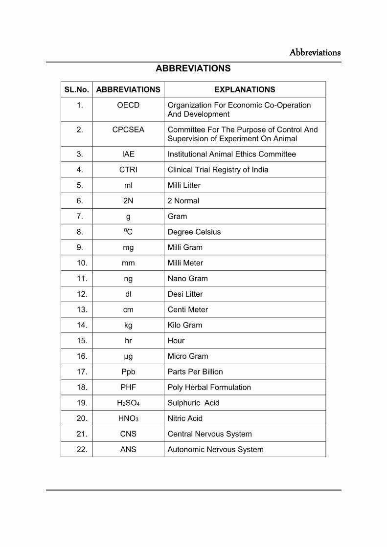
ABBREVIATIONS
SL.No. ABBREVIATIONS EXPLANATIONS
1. OECD Organization For Economic Co-Operation And Development
2. CPCSEA Committee For The Purpose of Control And Supervision of Experiment On Animal
3. IAE Institutional Animal Ethics Committee
4. CTRI Clinical Trial Registry of India
5. ml Milli Litter
6. 2N 2 Normal
7. g Gram
8. 0C Degree Celsius
9. mg Milli Gram
10. mm Milli Meter
11. ng Nano Gram
12. dl Desi Litter
13. cm Centi Meter
14. kg Kilo Gram
15. hr Hour
16. µg Micro Gram
17. Ppb Parts Per Billion
18. PHF Poly Herbal Formulation
19. H2SO4 Sulphuric Acid
20. HNO3 Nitric Acid
21. CNS Central Nervous System
22. ANS Autonomic Nervous System

CONTENTS
Chapter
No Title
Page
No
1 INTRODUCTION 1
2 LITERATURE REVIEW 21
3 AIM AND OBJECTIVE 25
4 PLAN OF WORK 26
5 PLANT PROFILE 27
6 MATERIALS AND METHODS 33
7 RESULTS AND DISCUSSION 44
8 SUMMARY AND CONCLUSION 57
BIBLIOGRAPHY

LIST OF TABLES
Table No. Title Page
No
1
2
3
4
5
6
7

LIST FIGURES
Table No. Title Page
No
1
2
3
4
5
6
7

Introduction
1
1. INTRODUCTION
1.1. Diabetes Mellitus (DM)
Diabetes mellitus is one of the most common and challenging
disease conditions of 21st centuary. It is a chronic complex progressive and
multisystemic disorder with life threatening micro and macrovacular
complications1. WHO defined Diabetes mellitus as a metabolic disorder of
multiple etiologies characterized by chronic hyperglycemia with
disturbances in carbohydrate, fat and protein metabolism resulting from
defects in insulin secretion, insulin action, or both2. It is a major cause of
morbidity and mortality. Prevalence of DM are about more than 150
million diabetics across the world and more than one fifth of them are
Indians. International Diabetes Federation, India has been declared India
as "Diabetic Capital of the World" at the recent Conference in Paris3.
Diabetes mellitus consists of a group of syndrome characterized by
hyperglycemia, altered metabolism of lipids, carbohydrates, and proteins;
resulting from defects in insulin secretion, its action, or both4.
DM is a complex, heterogeneous and polygenic metabolic disease
where there will be an absolute lack of insulin, decreased sensitivity to
insulin or both and which results in abnormal glucose homeostasis and
subsequent hyperglycemia. Mutual interaction between genetic and
environmental factors plays an important role in the pathogenesis of
diabetes mellitus5. DM Has been characterized by a variety of causes such
asobesity, abdominal adiposity, genetic, ethnicity etc.
Obesity and increased BMI have a great impact on diabetogenesis.
The association between increased BMI and weight gain and risk of
Diabetes mellitus is significant among Asians. Waist circumference (WC)
cut point for Indians for any cardio- metabolic risk factors is 87cm for men
and 82cm for women whereas that of BMI is 23 kg/m2 in both sexes5. It is

Introduction
2
found that the developing countries adopt the western life styles like
decreased physical activity and over consumption of cheap, energy dense
food for past 20 years and as a result the rate of obesity has tripled in
developing countries. Such changes have a direct influence on the child
health of the country; the prevalence of obesity among them ranges from 2
to 10% and the prevalence of overweight ranges from 10 to 25%6.
DM has a strong genomic association, genome-wide association
studies has catalogued a number of gene that have an influence on DM
(with modest odds ratio ranges between 1.2 to 1.5) and they include
TCF7L2, HHEX, CDKAL1, SLC30A8etc5.
In India nearly 75% of the Type DM has first degree family history
of diabetes indicating a strong familial aggregation. Prevalence of insulin
resistance is found to be high in Asian Indians and they need higher
amount of insulin to maintain normoglycemia. Comparison of Asian
Indians, Europeans and other ethnic groups have shown that the former
have higher insulin response than others, at fasting and in response to
glucose. Asian Indians also have some factors which decreases the insulin
sensitivity such as central obesity and high percentage of body fat in
comparison to many other populations.
Diabetes mellitus (DM) currently is a major health problem of the
world and due to chronic metabolic syndrome resulting from a variable
interaction of hereditary and environmental factors and is characterized by
abnormal insulin secretion or insulin receptor or post-receptor events,
affecting metabolism involving carbohydrates, proteins and fats in addition
to damaging liver, kidney and beta-cell of pancreas7.

Introduction
3
1.2. Ayurveda treatment for DM
Ayurveda which means ‘Science of life’ has derived from the
Sanskrit words ‘Ayur’ meaning life and ‘Veda’ meaning
knowledge.Ayurveda is based on the concept that everything in the
universe has composed of five basic elements such as space, air, energy,
liquid and solid and they are called The PanchaMahabhuta, or “five great
elements”. Ayurveda believes in the theory of tridoshas, namely vata
(ether and air), pitha (fire) and kapha (earth and water). Ayurveda aims to
keep the structural and physiological entities in a state of equilibrium,
which signifies good health. Any imbalance in tridosha may leads to
disease.
Diabetes mellitus and its clinical features were known to ancient
Indian physicians and they reported an elaborate description of Diabetes
mellitus and its management. Ayurvedic practitioners treat diabetes with a
multi-pronged approach, using diet modification, Panchkarma to cleanse
the system, herbal preparations, yoga and breathing exercises.
The common herbs which can be used against diabetes include
turmeric, neem, coccineaindica, amalaki, triphala, bitter gourd, rose apple,
leaves of bilva, cinnamon, gymnema, fenugreek, bay leaf and aloe vera.
The Ayurvedic preparations ‘Vasanta Kusumakar Ras’ and
‘Chandraprabhavati’ are used to treat diabetes mellitus. Proprietary
Ayurvedic medications are also used to treat diabetes8.
In ayurveda, diabetes mellitus is known as ‘madhumeha’ (madhu
means “honey” and meha means “urine”).
Madhumeha is characterized by Deterioration of the body with
impairment of vata (vatajaprameha). Since it deteriorates maximum of
dhatus (body tissues) all the vital organs will be affected. Impaired

Introduction
4
digestion leads to accumulation of certain specific digestive impurities in
pancreas and thereby abnormal insulin production. Madhumeha is a
Maharoga (major disease) lead to several complications like retinopathy,
neuropathy, nephropathy, joint pain, impotency, sexual and urologic
problems9.
1.3. Prevalence of DM in India10
India is currently experiencing an epidemic of diabetes mellitus. To
study the consequences of diabetes and the importance of diabetic care in
India we need a thorough study on Epidemiology of Diabetes in different
regions of India. Epidemiology of diabetes in India has an extensive
history. Long back a national study reported that prevalence of DM in
urban areas was 2.1 % and that in rural area was 1.5%. The available
studies show that there is a sharp rise in the prevalence of DM in both
urban and rural areas, among these southern India having the sharpest
increase.
Although in rural India the prevalence of diabetes is much lower
than in the urban population, even here the prevalence of diabetes is
rapidly rising. Diabetes is fast becoming the epidemic of the 21st century.
Type diabetes, which is more prevalent (more than 90% of all DM
cases) and the main driver of the diabetes epidemic, now affects 5.9% of
the world's adult population with almost 80% of the total in developing
countries. World Health Organization (WHO) reported that 32 million
Indians had diabetes in the year 2000. The International Diabetes
Federation (IDF) estimates the total number of diabetic subjects to be
around 40.9 million in India and it will rise to 69.9 million by the year 2025.
The earliest documented diabetes prevalence study of India was done in
Calcutta (now Kolkata) in the year 1938. They found that only 1% of
subjects have DM with glycosuria. In Bombay (now Mumbai) reports on
test for detection of diabetes in large number of subjects were first
available in 1959. Another population based study - National Urban

Introduction
5
Diabetes Survey (NUDS) was conducted in six large cities from different
regions of India in 2001. This study was done on 11,216 subjects aged
over 20 years from all socio-economic strata.
The WHO criterion was used for diagnosis diabetes after an Oral
Glucose Tolerance Test using capillary blood. The study showed that the
age standardized prevalence of type diabetes was 12.1%. The
prevalence was the highest in Hyderabad (16.6%), followed by Chennai
(13.5%), Bengaluru (12.4%), Kolkata (11.7%), New Delhi (11.6%) and
Mumbai (9.3%). The prevalence of IGT was 16.8% in Chennai, 14.9% in
Bengaluru (formerly Bangalore), 29.8% in Hyderabad, 10% in Kolkata,
10.8% in Mumbai and 8.6% in New Delhi. The Prevalence of Diabetes in
India Study (PODIS) was carried out in 108 centers
(49 urban and 59 rural) in different parts of India to look at the urban-rural
differences in type 2 diabetes and glucose intolerance.
The prevalence of DM was found to be more in urban areas of
northern region. Urban area of Chandigarh was the first site to study the
prevalence of DM in this region. The study conducted in rural areas
reported that the prevalence of diabetes in a rural locality near Delhi was
1.5 % in the year 1991. The prevalence has been reported to vary
between 1.5% in Delhi (1991) and 3.7% in Nagpur (2007) in rural areas.
Regarding urban prevalence, an increasing trend is observed in the
northern part of India since late 1960's, which has escalated from 2.9% in
Chandigarh (1966) to 20.1% in Jaipur (2007).
The first study done in South India was a hospital based study at
Vellore in 1964, which showed a prevalence of 2.5%. The study conducted
in Hyderabad showed a high prevalence of 4.1%. However, the studies in
rural areas were conducted since 1972. In 1984, house to house surveys
were conducted in Tenali, a small town in Andhra Pradesh (urban) and
rural population of Pondicherry (now Puducherry), which reported a
prevalence of 4.7% and 1.8% respectively. The prevalence in south India
has been reported to vary between 0.7% in Pondicherry to 19.5% in Kochi
in urban areas, while the prevalence in rural areas range from 1.3% in

Introduction
6
Trivandrum to 13.2% in Godavari. The Chennai Urban Rural Epidemiology
Study (CURES) investigators had a unique opportunity to compare
prevalence rates of diabetes in Chennai city with three earlier
epidemiological studies carried out in the same city using similar methods.
It has been shown that Indians have a younger age of onset of diabetes
compared to other ethnic groups.
The Chennai Urban Population Study (CUPS) and CURES state
that prevalence of coronary artery disease was 21.4 per cent among
diabetic subjects compared to 9.1 per cent in subjects with normal glucose
tolerance. The Impaired Glucose Tolerance subjects showed 14.9 per cent
of prevalence of CAD. It was also seen that there is a rising prevalence of
atherosclerosis with an increased intimal medial thickness.
The prevalence of diabetes in urban areas has increased from 2.3%
in 1975 to 11.7% in the year 2000. The prevalence of diabetes in peri-
urban population was found to be 4.0%. Mumbai and Ahmedabad are the
main site f study. Urban prevalence has an escalating trend from 1.5%
(1963) to 9.3% in Mumbai (2001). A similar trend is observed in the rural
areas - an increase from 3.9% in 1991 to 9.3% in 2006.
1.4. Mechanism of diabetes mellitus induction2
1. β- cell destruction (Type 1 diabetes - IDDM)
(a) Immune mediated
(b) Idiopathy
2. Insulin resistance (Type 2 diabetes - NIDDM)
3. Genetic defects of β- cell function
(a) Glucokinase
(b) Hepatocyte nuclear transcription factor – 4 α
(c) Insulin promoter factor
(d) Mitochondrial DNA
(e) Proinsulin or insulin conversion
4. Genetic defects in insulin processing or insulin actions defects in

Introduction
7
(a) Proinsulin conversion.
(b) Insulin gene mutation
(c) Insulin receptor mutation
5. Exocrine pancreatic defects
6. Endocrinopathy
(a) Acromegaly
(b) Cushing syndrome
(c) Hyperthyroidism
(d) Pheochrmocytoma
(e) Glucocanonama
7. Infections
(a) Cytomegalovirus
(b) Coxhacivirus
8. Genetic syndrome associated with diabetes
(a) Down‘s syndroo generate reactive oxygen species, which also
contribute to DNA fragmentation. The formation of superoxide
anions results from both STZ action on mitochondria and increased
activity of xanthine oxidase.
STZ induced DNA damage activates poly ADP ribosylation leading
to the depletion of cellular NAD+ and ATP content and thereby inhibition of
insulin biosynthesis and secretion. Calcium, which may also induce
necrosis, does not seen to play a significant roleme
(b) Kleinfelter‘s syndrome
(c) Turner‘s syndrome
9. Drugs
(a) Glucocorticoid
(b) Thyroid hormone
(c) Thiazides
(d) Phenytoins
1.5. Types of Diabetes

Introduction
8
There are three main types of diabetes
1.5.1 Type Diabetes
Insulin-dependent diabetes (IDDM; Type I diabetes) is one of the
most common metabolic disorders characterized by pancreatic beta cell
destruction, it may be due to autoimmune attack. Genetic and
environmental factors play a part and HLA- DR3 and HLA-DR4 confer
susceptibility to Type 1 Diabetes Mellitus.
1.5.2 Type Diabetes:
Non-insulin-dependent diabetes mellitus (NIDDM) or adult-onset
diabetes. Resulting from the combination of resistance to insulin action,
inadequate insulin secretion and excessive or inappropriate glucagon
secretion.
1.5.3 Gestational Diabetes:
Gestational diabetes mellitus (GDM) affects ∼ 7% of all pregnancies
and it may also be defined as carbohydrate intolerance during gestation.
The condition can be associated with several maternal and fetal
complications, such as macrosomia, birth trauma, cesarean section and
hypocalcaemia, hypoglycemia and hyperbilirubinemia in newborns.
1.6. Etiology/Contributing Factors
Insulin resistance in the hepatic and skeletal muscle, increased
hepatic glucose synthesis, over production of free fatty acids and
relative insulin deficiency.
Beta cells failure.
Contributing factors:
Obesity
Racial/ ethnic background.

Introduction
9
Age (onset of puberty is associated with increased insulin
resistance).
Sedentary lifestyle.
Genetic predisposition.
Conditions associated with insulin resistance, (e.g., polycystic ovary
syndrome)
1.6.1 Risk Factors of Diabetes
The risk factors for diabetes may be categorized as modifiable risk
factors and non-modifiable risk factors.
1.6.2 Modifiable Risk Factors
Obesity (via BMI and WHR) : A meta-analysis demonstrated the
pooled relative risks for incident diabetes of 1.87 (95% confidence
interval (CI): 1.67-2.10), 1.87 (95% CI: 1.58-2.20) and 1.88 (95%
CI: 1.61-2.19) per standard deviation of body mass index, waist
circumference and waist/hip ratio and these three results in obesity
and may leads to diabetes.
Physical Inactivity: The protective effect of physical activity in
subjects with an excessive BMI and elevated glucose levels;
Diabetes can be prevented by physical activity and weight control in
peoples with both normal and impaired glycemic control.
Plasma Lipids and Lipoproteins Level: There are reports like the
blood level of LDL, VLDL, TGL are high and that of HDL is low in
Diabetic people. According to American Diabetes Association LDL
Cholesterol should be <100 mg/dl; HDL Cholesterol: >60 mg/dL;
and Triglycerides: <150 mg/dl.
Hypertension: It has been reported that the incident of diabetes for
hypertensive patient is 2.21 greater.
Dietary Habits: It is suggested that whole grains are rich resources
of dietary fiber, fat, vitamin, antioxidant nutrients, minerals, lignans,
Introduction
10
starch, and phenolic compounds that have been linked to the
reduced risk of insulin resistance, dyslipidemia, obesity, T2DM,
heart diseases and dietary fructose specifically increases de novo
lipogenesis, promotes dyslipidemia, decreases insulin sensitivity,
and increases visceral adiposity in overweight/obese adults.
1.6.2 Non-modifiable Risk Factors
Family History: First degree family history of diabetes was found in
an approximate of 75% of the T2DM patients.
Genetic factors: Genome-wide association studies show a strong
association between genetic factor and Diabetes.
Low/High Birth Weight: Both Low and High Birth Weight are risk
factors of Diabetes.
1.7 Pathophysiology of non-insulin dependent DM (Type-II)
Type-2 diabetes mainly occurs due to insulin resistance and/or
insulin deficiency. Insulin resistance result in the impaired insulin mediated
glucose uptake in the peripheral tissues like muscle and fat, fail to
suppress hepatic glucose production and TGL reuptake by fat cells. To
overcome the insulin resistance, beta islet cells try to increase the amount
of insulin secreted. Beta cell destruction reduces insulin production.
Therefore DM-2 has to occur, two defects are necessary: insulin
resistance and insulin deficiency relative to the resistance.
Fig. No: 01: Pathophysiology of Type-II Diabetes mellitus

Introduction
11
1.7.1 Clinical Manifestations of Type 2 Diabetes Mellitus:11
Polydipsia – increased thirst.
Polyphagia – increased hunger.
Polyuria – increased urine.
Slow healing infections.
Blurred vision.
Impotence in men
1.7.2. Criteria
Overweight (BMI ≥ 85th percentile for age and gender, weight for
height ≥ 85th percentile or weight ≥ 120% of ideal for height).
Plus any two of the following risk factors:
Family history of type 2 diabetes in first- or second degree relative
Race/ethnicity (American Indian, African American, Hispanic,
Asian/Pacific Islander)
Signs of insulin resistance or conditions associated with insulin
resistance (acanthosisnigricans, hypertension, dyslipidemia,
polycystic ovary syndrome)

Introduction
12
1.7.3 Age of initiation:
Age 10 years or at onset of puberty if puberty occurs at a younger
age.
1.7.4. Frequency:
Every 2 years.
1.7.5. Test:
Fasting plasma glucose is the preferred method for screening.
* Clinical judgment should be used to test for diabetes in high-risk
patients who do not meet these criteria.
1.8. Type 2 dm management in India
The patient should receive appropriate medical care along with self-
management to keep the diabetes under control. The main treatment
goals are
1. To achieve optimal glycemic level.
2. To reduce other cardiovascular risk factors, including hypertension,
hyperlipidemia, and overweight and obesity; and
3. To diminish micro and macro vascular complications.
Diabetics are treated with oral hypoglycemic agents with
appropriate diet and exercise. Some patients may require insulin therapy
with or without oral hypoglycemic agents.
1.9 Treatment12
Fig. No : 02. Image showing the effect of different anti diabetic drugs

Introduction
13
Table 01: Classification of Oral Hypoglycaemic Drugs
ORAL HYPOGLYCAEMIC DRUGS
Si No
CLASS DRUGS
1 Sulfonylureas
First generation;
Tolbutamide, Chlorpropamide.
Second generation;
Glibenclamide, Glipizide
2 Biguanide Metformin, Gliclazide, Glimepiride
3 Meglitinide/Phenylalanine analogues
Repaglinide, Nateglinide
4 Thiazolidinediones Rosiglitazone, Pioglitazone
5 Glucosidase inhibitors Acarbose, Miglitol
6 Dipeptidyl peptidase-4 (DPP-4) inhibitor
SitagliptinVildagliptin
1.9.1 Sulfonylureas

Introduction
14
Drugs: Tolbutamide, chlorpropamide these are the drugs in the first
generation. In second generation drugs like glibenclimide, glipizide,
gliclazide and glimepiride.
1.9.2 Side Effects
• Low blood sugar,
• An upset stomach,
• Skin rash or itching,
• Weight gain.
1.9.3 Biguanides
Drugs: Metformin
1.9.4 Side Effects
• Abdominal pain
• Nausea
• Metallic taste
• Mild diarrhea
• Anorexia
1.9.5 Alpha-glycosidase inhibitors
Drugs: Acarbose and miglitol
1.9.6 SideEffects
May cause stomach problems such as gas, bloating and
diarrhea
1.9.7 Thiazolidinediones Drugs
Drugs:Pioglitazone and troglitazone
1.9.8 Side Effects
• Plasma volume expansion

Introduction
15
• Edema
• Weight gain Headache
• Myalgia
• Mild anemia
1.9.9 Meglitinides
Drugs: Repaglinide and nateglinide
1.9.10 Side Effect
• Weight gain
• Low blood sugar
1.10 Medical nutrition therapy 11
A meal plan with regular meals and snacks and carbohydrate goals
that are moderately less than their usual intake will often help lower the
elevated blood.Since most of the teens diagnosed with type diabetes are
obese, they have to identify carbohydrate-containing foods and monitor
carbohydrate intake thereby cessation of weight gainmay occur.
1.10.1 General Guidelines for Food Intake
Eat 3 meals and 1 snack on a regular schedule.
Try not to skip meals.
Follow carbohydrate goals for meal planning from the dietitian.
Try to eat about the same amount of carbohydrate at the same time
each day.
Decrease saturated fat intake.
Work towards a healthy weight.
Eat smaller portions at meals.
1.10.2 Ways to Limit Carbohydrate Intake
Drink calorie-free beverages (e.g., water, tea, diet soda).

Introduction
16
Limit fruit juice to 1 cup/day.
Limit carbohydrate servings to 3-4/meal. If necessary decrease to
1-2 at breakfast.
Check blood glucose level 2 hours after eating. (If >180 mg/dl, you
ate more carbohydrate than your body could handle).
1.10.3 Benefits of Exercise
Helps you feel better and increases your energy
Reduces HbA1c
Improves insulin sensitivity
Helps in reaching a healthy weight
Increases strength and flexibility
Decreases risk factors for heart disease
Educes body fat and increases muscle mass1
The aim of treatment is to maintain glycemic level with in normal
range. Additional considerations are:
• Maintain healthy weight.
• Minimize hyperglycemia and hypoglycemia.
• Achieve normal lipid levels.
• Prevent and delay complications.
• Promote optimal health and well-being.
Treatment regimen depends on the type of diabetes, medical
nutrition therapy, frequent blood glucose monitoring to identify and
evaluate blood glucose patterns, and comprehensive education in
diabetes, self-management and decision-making skills at diagnosis and
follow-up visits. Target blood glucose goals for teens are listed in Table 2.

Introduction
17
Table 02 : Blood Glucose Goals for Adolescents
Biochemical Index Normal Goal Action Indicated
Average premeal BG (mg/dl)
<110 80-120 <80 or >140
Average 2 hour postmeal BG
for rapid-acting insulin only
<120 150-180 >180
Average bedtime BG (mg/dl)
<120 100-140 <100 or >160
Average 3:00 am BG (mg/dl)
<110 80-100 <80 or >120
HbA1c (%) <6 <7 >8
Adapted from: Orr, DP. Contemporary management of adolescents with diabetes mellitus. Part 1: Type 1 diabetes. Adolescent Health Update 2000;12(2), Table1, p 2.
1.11 Medicinal Plants
Nature always stands as a golden ark to exemplify the
outstanding phenomenon of symbiosis. The biotic and abiotic elements of
the nature are all independent. The plants were indispensible to man, for
his life. A nest of other useful products are supplied to him by the plant
kingdom. Nature has provides a complete range of remedies to came an
ailments of mankind. The knowledge of drugs has accumulated thoughts
of years of a result of meaning inquisitive nature so that today we possess
many affective of causing health care.Archaeological evidence indicates
that the use of medical plants data of least the paleotic, approximately
60,000 years age.In India, medicinal plants are widely used in traditional
systems of medicine like Ayurvedic, Unani, Siddha and Homeopathy. India
with it's valuable resources of natural flora has always been one of the
richest sources of medicinal plants in the world.

Introduction
18
1.11.1 Importance of herbal drugs
Antidiabetic allopathic drugs have their own side effect & adverse
events like hypoglycaemia, nausea, vomiting, hyponatremia, flatulence,
diarrhoea or constipation, alcohol flush, headache, weight gain, lactic
acidosis, pernicious anaemia, dyspepsia, dizziness, joint pain. So instead
of allopathic drugs, herbal drugs are a great choice which is having more
or less no side effect & adverse effects. Around 800 Indian herbs possess
ant diabetic activity. Though complementary & alternative medicine (CAM)
treatments are popular, scientific evidence support their application to
diabetes care is scare. Instead of focusing on single modalities CAM
practitioners prescribe complex, multi dietary intervention. Ayurvedic
interventions may benefits patients with higher base line HbA1c value,
warranting further research.14
Natural origin and fewer side effectspromote the use of herbal
drugs in bothdeveloping and developed countries. In the last few years
there has been an exponential growth in the use of herbal drugs. Many
traditional medicines in use are derived from medicinal plants, minerals
and organic matter.According to World Health Organization (WHO) there
are about 21,000 plants, which are used for medicinal purposes around
the world. Among these 2500 species are found in India. Indiais called as
botanical garden of the world because of the rich herbal medicine
resources. Very recently, two exhaustive reviews have been published
based on global literature survey on 150 plants and 343 plants from
different parts of the world. some plants like Allium cepa (Onion, piyaj),
Allium sativum (garlic, lasun), Syzygiumcumini (Syn. Eugenia jambolana;
(black plum; jamun), Momordicacharantia (bitter gourd; karela)
Gymemasylvestre (Gurmar), Pterocarpusmarsupium (Vijay-) sar) etc. are
well noticed by scientists as well as laymen, in recent years 15.

Introduction
19
Biological actions of the plant products used against diabetes are
related to their phytochenistry. Herbal products or plant products are rich
in phenolic compounds, flavonoids, terpenoids, coumarins, and other
constituents which reduces the blood glucose levels16.
Our Vedic literatures like CharakSamhita already report the use of
herbs and herbal derivatives for treatment of diabetes mellitus. According
to CharakSamhitsa more than 400 plants are used in 700 recipes which
are used to treat diabetes mellitus in almost two thirds of the world
population. A large number of in vivo studies have been conducted on
animals to test the claimed activity have demonstrated the hypoglycemic
property of many plants, already reported in various literatures. The plant
families, including the species most studied for their confirmed
hypoglycemic effects include, Leguminoseae, Lamiaceae, Liliaceae,
Cucurbitaceae, Asteraceae, Moraceae, Rosaceae, Euphorbiaceae and
Araliaceae17.
1.12 Animal Models 13
Streptozotocin or streptozocin or Izostazin or zanosar (STZ) is a
synthetic glucopyranose derivative isolated by the fermentations of
Streptomyces achromogenes which possess anti-tumor antibiotic activity.
It can be used to induce both type 1 and type 2 diabetes. Chemically it is
(2-deoxy-2-(3-methyl-3-nitrosoureido)-D-glucopyranose).The frequently
used single i.v dose in adult rats to induce IDDM by immune system
activation was found to be in between 40 and 60mg/kg. NIDDM can also
be induced in rats by intravenous or intraperitoneal treatment with
100mg/kg b.w.STZ on the day of birth.
STZ decrease insulin biosynthesis and secretion by impairing
glucose oxidation. STZ at first abolished the B cell response to glucose.
Temporary return of responsiveness than appears which is followed by its
permanent loss and cells are damaged. STZ is taken up by pancreatic B
cells via glucose transporter mainly GLUT-2. Intracellular action of STZ

Introduction
20
causes in changes of DNA I pancreatic B cells compromising its
fragmentation. Alkylation of DNA is the main reason for the STZ induced B
cells death.
STZ inhibits the Krebs cycle and decreases oxygen consumption by
mitochondria and strongly limit mitochondrial ATP production and cause
depletion of this nucleotide in B cells. Augmented ATP dephosphorylation
increases the supply of substrate for xanthine and enhances for uric the
final product of ATP degradation.
STZ is a Nitric oxide (NO) donor and NO was found to bring about
the destruction of pancreatic islet cells. Hence it produces DNA damage.
STZ is however not a spontaneous nitric oxide donor. STZ was found to
generate reactive oxygen species, which also contribute to DNA
fragmentation. The formation of superoxide anions results from both STZ
action on mitochondria and increased activity of xanthine oxidase.
STZ induced DNA damage activates poly ADP ribosylation leading
to the depletion of cellular NAD+ and ATP content and thereby inhibition of
insulin biosynthesis and secretion. Calcium, which may also induce
necrosis, does not seen to play a significant role.

Literature Review
21
2. LITERATURE REVIEW
Mohammed Salem et al (2013).,14 studied the antioxidant and
antibacterial activity of Tecoma stans leaves and branches against the
growth of some human bacterial strains using the disc diffusion and
minimum inhibitory concentration (MIC) methods and antioxidant activity
using 2,2-dimethyl-1-picrylhydrazyl (DPPH) method. Very significant
activities were exhibited by the samples. These findings provide scientific
evidence to support traditional medicines uses of Tecoma stans and
indicate a promising potential for the development of an antibacterial and
antioxidant agent from T.stans
Kameswaran et al (2013).,15 studied Hepato protective activity of
Tecoma stans extract against the liver injury induced by paracetamol,
carbon tetrachloride and thioacetamide. Results revealed that the extracts
significantly reduce the elevated serum levels of Asparate amino
transferase, alanine amino transferase, alkaline phosphate and bilirubin.
The ethanolic extract at the dose of 500 mg/ kg more effective than 250
mg/kg but his excluded in paracetamol induced liver damage. In chronic
liver injury induced by CCl4, ethanolic extracts at the dose of 500 mg/kg.
P.O. was found to be more effective than the extract of the dose of 250
mg/kg. Histological examination of the liver tissues supported the hepato
protective activity of the extracts.
Kameswaran Sugavanan et al (2012).,16 studied the CVS
depressant potential of different extracts of Tecoma stans flowers. Namely
chloroform, methanol and water on Albino mice of both sexes. The study
conform that the different extracts of Tecoma stans flowers exhibit CNS
depressant activity.

Literature Review
22
Amad Ali-Azzawi et al (2012).,17 studied the genotoxic potential of
Tecoma stans by in vivo and in vitro system. This study examined the
genotoxic activity of aqueous and ethanolic extracts on bone marrow cells
from BAL B/C mice through evalution of milotic index and chromosome
aberration and cytotoxic effect of two extracts on mouse embryo fibroplast
(MFF) cell line. No alteration in the total no. of chromosomal aberration
were observed and percentage of miotic index at the concentrations tested
remained unchanged. The higher concentrations of the plant extracts had
a cytotoxic effect on MFF cell line. Both extracts had no significant
elastogenic effect in vivo,but showed cytotoxic effects on mouse embryo.
Thirumal et al (2012).,18 studied the crude leaf extract of Tecoma
stans. These were examined for their anticancer activity. To determine in
vitro Anticancer activity, different concentration of crude extract were
tested in MCD - of cancer cell line by 3- (4,5- dimethylthiazole-2-yl)-2,5-
dimethyl tetrazolium bromide (TT) assay. Tecoma stans leaf extract
showed significant anti-proliferative activity and a dose dependant effect
was observed. Minimum inhibition of 14.6% was shown by extract at
concentration 7.5 g/ ml and maximum inhibition (95.9%) was observed at
1000 g/ml.
Govindappa et al (2012).,19 studied the antibacterial and
antioxidant activity of ethanol, methanol and aques extract of Tecoma
stans. These three solvent fractions possessed strong radical scavenging
activity when analyzed using FRAP and DPPH. It was ranged from
1443.79 to 3841.17 g/ml. The results indicate that the plant is a potential
candidate to be used as an antimicrobial and antioxidant.
Kameswaran et al (2012).,20 evaluated the anticancer activity of
methanolic flower extract of Tecoma standin vitro and in vivo methods.
Extract was subjected to preliminary qualitative phytochemical

Literature Review
23
investigation by using standard procedures and In vitro antitumor activities
were evaluated by the MTT Assay method using vero and HFP - 2 cell
lines. The extract was subjected to in vivo anticancer activity using enrich
as cities carcinoma (EAC) tumor model. The activity was assessed
increase in life span, average increase body weight, changes in food
intake, Tumor volume, tumor weight, variable cell count, non-variable cell
count, PCV, total cell count and hematological studies. The potency of
extract was compared with standard 5-flurouracil (20 mg/kg). In vitro
anticancer activity was exhibited and significant cytotoxic activity against
both cell lines, at different concentration. Oral administration of the
extracts at the doses of 200 and 400 mg/kg, significantly (P<0.001)
increase the survival time. Non-viable cell count, decreased the average
body weight and food intake, viable cell count of the tumor bearing mice.
After 14 days of inoculation, METS was able to release the changes in the
haemotological parameters, protein and P<V consequent to tumor
inoculation.
Raju et al (2011).,21 received on the ethno-pharmacological,
phytochemical and therapeutic potential of Tecoma stans. An exhaustive
survey of literature revealed that alkaloids, flavanoids, saponins, phenols,
steroids, anthraquinone tannis, terpenes and glycosides constitute the
major classes of phytoconstituentspresent in the plant. Pharmacological
report revealed that is having antidiabetic, anticancer, antioxidant,
antispasmodic, antimicrobial, antifungal properties and extensively used in
the treatment of diabetes.
Krishna et al (2009).,22 studied the phytochemical screening
analysis of n-hexane, ethyl acetate, ethanol and aqueous extracts of
Tecoma stans, which indicates, the presence of alkaloids, saponins and
tannins, In the n-hexane extract, the constituents were absent.

Literature Review
24
Lin et al (2008).,23 found that, the flavanoids from the plant
extractspossesssubstantial antimicrobial and antioxidant activities.
Socolwsji et al (2008).,24 studied the effects of temperature and
light on the control of seed germination in Tecoma stands. The influence of
constant temperature from 10-40°C, with 5°C intervals under the white
light and darkness were tested. The optimum temperature for the seed
germination were found to be between 25 and 30° C for both the light and
dark treatment. The maximal germination was reached in the range of
19-38°C under the light and 20-40°C during the darkness. The seeds
showed highest synchronization of the germination near the optimal
temperatures. The germination in the field was tested under the two light
conditions. The highest percent of germination occurred under the Canopy
(69%). However under the Canopy, the seedling presented 1.9% of the
recruitment, while under the direct sunlight, 96.9% results showed that
T. stans seeds germinated well in open areas with the occurrence of high
seedling recruitment indicating the invasion potential of the speciesunder
such light conditions.
Gharib Naseri . et al (2007).,25 have reported antispasmodic effect
of Tecoma stans hydro alcoholic leaf extract on rat ileum.

Aim & Objective
25
3. AIM AND OBJECTIVE
The diabetes mellitus prevalence was increased day by day, due to
metabolic disorder, life style changes, improper food intake and less
physical activity .symptoms of high blood sugar, left untreated, diabetes
mellitus can cause many complications . Synthetic drugs have many side
effects and harmful to the health. Over the centuries, they are traditionally
practicing medicinal plants used to the treatment for various diseases but
no scientific validation. Several literatures are indicated that the herbal
drugs have lesser side effects when compared to synthetic medicines. The
TecomaStans(L)juss is not scientifically validated and which was
traditionally practicing herb .The work provides scientific validation for use
of leaf against diabetes mellitus.
The current study is help to develop a plant based diabetic drug
which will be evaluated by using invivo streptozocin induced diabetes in
rats

Plan of Work
26
4. PLAN OF WORK
Plant selection
Authentication of leaf
Phytochemical studies
Acute Toxicity Study
Pharmacognostical studies
Results and discussion
Test for carbohydrate
Test for proteins
Test of amino acids
Test for steroids
Test for cardiac glycosides
Test for flavonoids
Test for Alkaloids
Antidiabetic activity
Summary & Conclusion

Plant Profile
27
5. PLANT PROFILE
5.1 Tecomastans (l.)Juss.exkunth
Fig No:03. Palnt of TecomaStans(L.)
Kingdom : Plantae
Sub kingdom : Tracheobionta
Super division: Spermatophyta
Division : Magnoliophyta
Class : Magnoliopsida
Subclass : Asteridae
Order : Scrophulariales
Family : Bignoniaceae
Genus : Tecoma Juss.
Species : Tecoma stans (L.) Juss.exKunth
5.2 Vernacular names

Plant Profile
28
Tamil : Manjarali, Naagasmbagam, Soonnapati,Sornapati
English : Trumpet Flower, Yellow Elder, Yellow Trumpet Bush.
Spanish : Lluvia De Oro, Trompeta, Tronafrente, Tronadora
French : Tecomajaune
Portuguese : Amarelinho, Ipê-Mírím
Germany : Aufrechtetrompetenwinde
Italy : tecomagiallo
Pacific Islands :piti
Tecomastans is a species of flowering perennial large shrub or
small, much-branched, tree usually growing 1.5 to 5 m tall, but
occasionally reaching up to 10 m in height in the trumpet
vine family, Bignoniaceae, that is native to the southern USA, Mexico, the
Caribbean, Peru and Ecuador. Tecomastans is the official flower of
the United States Virgin Islands and the floral emblem of the Bahamas.
Yellow trumpet bush is an attractive plant that is cultivated as
an ornamental.
The plant is desirable fodder when it grows in fields grazed
by livestock. Yellow trumpetbush is a ruderal species, readily colonizing
disturbed, rocky, sandy, and cleared land and occasionally becoming
an invasive weed. It is used as firewood and charcoal, in the construction
of buildings and the leaf infusion can be taken orally for diabetes and
stomach pains. A strong leaf and root decoction is taken orally as a
diuretic, to treat syphilis or for intestinal worms. It is a strong shading plant
and can be planted as a live hedge.

Plant Profile
29
5.3 General description
5.3.1 Stem
Full and cylindrical. The younger stems are smooth, glabrous and
greenish in colour. They are slightly quadrangular and turn pale brown or
reddish-brown in colour as they age. The bark on the main stem is light
brown to pale grey in colour, furrowed, and relatively rough in texture,
covered in light greyish to brown barks.
5.3.2 Leaf
Fig No:04: Tecomastans(L.) Leaf
Borne on petioles 3-5 cm long. Compound, opposite, 10-25 cm
long, with 3-13 leaflets, but usually 3-7 leaflets.
Leaflets lanceolate to elliptic, 2-10 cm long and 1-4 cm wide, apexlong-
acuminate, base cuneate. Margins irregularly and finely toothed. Both
sides of the leaf blade are smooth and mostly glabrous, though a few hairs
may be present on the undersides near the midrib.

Plant Profile
30
Inflorescence: Erect or inclined several-flowered clusters (5-15 cm
long), produced terminally (at the ends of the branches), and then later, in
the leaf axils near the tips of the branches.
5.3.3 Flowers
Showy bright yellow, tubular (trumpet-shaped), borne on short
pedicels somewhat curved or twisted. Corolla tube 3-5 cm long with five
rounded lobes, 8-30 mm long. Presence of several faint reddish lines in
the throat of the flower, which is slightly ridged and hairy.
Fig. 5 : Flower of Tecomastans(L.)
5.3.4. Fruits
The fruits are large, linear capsules, somewhat flattened, 10-20 cm
long and 0.5-2 cm wide, brown at maturity, they split open to
release numerous seeds, 3-5 mm each.

Plant Profile
31
Fig No: 03 Fruit of Tecomastans(L.)
5.3.5. Seed
Numerous. The seeds are very flat, oblong in shape (7-8 mm long
and about 4 mm wide), and have a transparent wing at each end (the size
of entire seed including the wings is about 20 x 6 mm)
Fig No: 03 Seed of Tecomastans(L.)

Plant Profile
32
5.3.5 Trunk
Tecomastans has a tendency to grow with several trunks. As an
ornamental it can be trained to grow with a single trunks. The bark on the
main trunk is light brown, hard, lose grained and become corky with age.
5.4 Chemical Constituents
The plant contains triterpenes, hydrocarbons, resins and volatile
oil.
1. Leaves contain flavanoids, chryseriol, Luteolin, Hyperoside,
Indoleoxygenase. Alakloid like Tecomanine, Tecostamine, 4-
noroctidine, N- hormethyl-skytarthine and S- skytanthine.
2. Flowers contain p- carotene and Zeaxanthin. Methanolic extract of
the flowers showed the presence of flavonone , 7, 8 dihydroxy-5,6-
dimethoxy flavones and Kaemperferol .
3. Seeds contain fatty oil and the compositionis plamitic acid, stearic
acid octadecenoic acid, octadienoic acid, octadecatrienoic acid and
octadeccatetranoic acid.
4. Ethanolic extract of fruit contains monoterpenic alkaloids, 7-
hydroxyskytenthine, 5-hydroxytecomanine, 5-hydroxyskytenthine .
5.5 Medicinal Uses
Aerial parts used in the treatment of stomach problems, gastritis,
diarrhoea
Roots are used as diuretic, vermifuge, tonic, beer making, a remedy
in snakes bits, scorpion sting and in the treatment of syphilis .
Flowers possess narcotic and analgesic activity.

Materials& Method
33
6. MATERIALS AND METHOD
6.1 Plant Material
The plant leaves was collected locally from herbal store and
botanical garden of the garden of the botany central council for Research
Ayurvedicand Sidha Govt. of India .The plant was identified and
authenticated by comparison with herbarium specimens.
The leaf of Tecomastans (L.)juss ex kunth were authenticated by
comparison with herbarium specimens and authentification No. BSI/SRC
5/23/2016/Tec/1993.
The weighed coarse powder was used for the extraction by
successive solvent extraction by Soxhlet apparatus using various solvents.
6.2 Animals
Wistar rats (150 – 250 g) used for the study were obtained from the
animal house of the Department of Pharmacology, Karpagam College of
Pharmacy, Coimbatore, Tamil Nadu. The animals are randomly selected,
marked to permit individual identification, and kept in their cages for at
least 5 days prior to dosing to allow for acclimatisation to the laboratory
conditions. The animals were housed three per cage in a polypropylene
cage and maintained in standard laboratory conditions with free access to
food and water ad libitum18. All animal experiments were conducted in
compliance with (Organization for Economic Cooperation and
Development) OECD Guideline and approved by the Institutional Animal
Ethics Committee, Karpagam College of Pharmacy
6.3 Chemicals, Drugs and Instruments
Streptozotocin, citric acid, sodium citrate were collected from a
private chemical store Coimbatore (Ponmani and co). Other important
chemical used in phytochemical analysis like alcohol, hydrochloric acid, ∞-
napthol, Sulphuric acid, Fehling A&B, Benedict reagent, sodium hydroxide,

Materials& Method
34
nitric acid, ammonia, lead acetate, ninhydrin, sudan red III reagent,
glycerin, picric acid, chloroform, acetic anhydride, ferric chloride, zinc,
dragendroff's reagent, Wagner’s reagent, Mayer’s reagent, sodium
chloride and bromin water were collected from the store of Karpagam
College of Pharmacy. All the chemicals used in the study are of analytical
grade.
6.4 Extraction Procedure26
The leaves of plant, dried under shade are carefully removed and
grinded using a blender. The coarse power so obtained was used for the
extraction by successive solvent extraction by Soxhlet apparatus using
various solvents. The assembly of Soxhlet apparatus is as shown in the
figure.
Image No. 01: Extraction using soxhlet apparatus assembly

Materials& Method
35
A. Alcoholic extract
Marc obtained from the above extract was dried and extracted with
2.5litres of ethanol (90%) in soxhlet apparatus for 36 hours .Then the
extract obtained were collected and concentrated by vaccum distillation
.The concentrated extract were then dried by in a vaccum desciccator.
6.5 Phytochemical Analysis27
Phytochemicals are biologically active, naturally occurring chemical
compounds found in plants, which provide health benefits for humans
further than those attributed to macronutrients and micronutrients. Phyto-
constituents are the contributors of pharmacological activities of a plant.
The individual extracts are subjected to qualitative tests for identification of
various plant constituents.
6.5.1 Test for Carbohydrates
I. Molisch Test: To the aqueous extract, 1ml of ∝- napthol solution
was added and Conc. Sulphuric acid were added along the sides
of the test tube.
II. Fehling Test: To the aqueous extract, equal quantities of Fehling
A & B were added .Upon heating gently.
III. Benedict’s test: To 5ml of Benedict reagent, 8 drops of solution
under test was added to the test solution mixed well. Then it was
boiled vigorously for 2 minutes and cooled.
6.5.2 Test for Proteins
I. Biuret Test: To the aqueous extract, 1ml of 40% NaOH and 2
drops of 1% copper sulphate solution was added.
II. Xanthophoretic Test: To the aqueous extract, 1ml of conc. Nitric
acid was added. When a white precipitate was formed, it is boiled
and cooled. Then 20% of NaOH or ammonia was added.

Materials& Method
36
III. Lead acetate Test: To the aqueous extract, 1ml of lead acetate
solution was added.
6.5.3 Test for Amino acids
Ninhydrin Test: 2drops of freshly prepared 0.2% ninhydrin reagent
was added to the aqueous extract and heated.
6.5.4 Test for Fats and Oils
Place a thick section of drug on glass slide. Add a drop of Sudan
Red III reagent. After two minutes, wash with 50 % alcohol. Mount in
glycerin. Observe under microscope.
6.5.5 Test for Steroids
I. Liebermann Burchard Test: The aqueous extract was dissolved in
2ml chloroform in dry test tube. 10 drops of acetic anhydride and 2
drops of conc. sulphuric acid were added.
II. Salkowaski Test: The aqueous extract was dissolved in chloroform
and equal volume of sulphuric acid was added to it.
6.5.5 Test for Cardiac glycosides
Keller-killiani Test: Test sample was dissolved in acetic acid
containing traces of ferric chloride and transferred to the surface of
conc. Sulphuric acid.
6.5.7 Test for Saponins
Foam Test: About 1ml of aqueous extract is diluted separately with
distilled water to 20ml and shaken in a graduated cylinder for 15 minutes.
6.5.8 Test for Flavonoids

Materials& Method
37
I. Sulphuric Acid Test: On addition of sulphuric acid (66% or 80%)
flavons and flavonols dissolves into it and give a deep yellow
solution.
II. Heat the test solution with Zinc and HCl, pink to red colour
observation shows the presence of flavonoids.
6.5.9 Test for Alkaloids
I. Dragendroff’s Test: To the aqueous extract, add 1ml of
Dragendroff’s reagent.
II. Wagner’s Test: To the aqueous extract, add1 ml of Wagner’s
reagent.
III. Mayer’s Test: To the aqueous extract, add 1ml of Mayer’s reagent.
6.5.10 Test for Phenolic compounds and Tannins
Small quantities of alcoholic and aqueous extracts in water were
tested for the presence of phenolic compounds and tannins with dilute
ferric chloride solution (5%), 1% solution of gelatin containing 10% sodium
chloride, 10% lead acetate and bromine solutions.
Materials& Method
38
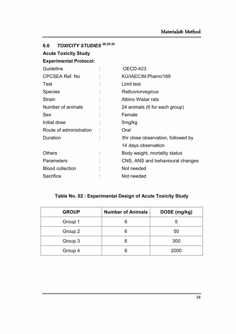
6.6 TOXICITY STUDIES 28,29,30
Acute Toxicity Study
Experimental Protocol:
Guideline : OECD-423
CPCSEA Ref. No : KU/IAEC/M.Pharm/169
Test : Limit test
Species : Rattusnorvegicus
Strain : Albino Wistar rats
Number of animals : 24 animals (6 for each group)
Sex : Female
Initial dose : 5mg/kg
Route of administration : Oral
Duration : 3hr close observation, followed by
14 days observation
Others : Body weight, mortality status
Parameters : CNS, ANS and behavioural changes
Blood collection : Not needed
Sacrifice : Not needed
Table No. 02 : Experimental Design of Acute Toxicity Study
GROUP Number of Animals DOSE (mg/kg)
Group 1 6 5
Group 2 6 50
Group 3 6 300
Group 4 6 2000

Materials& Method
39
6.7 Selection of Test animals.
Female adult wistar rats of 8-12 weeks are selected. Nulliparous
and non-pregnant animals were obtained from the centralized animal
house of Karpagam College of pharmacy, Coimbatore and they are
acclimatized for holding 1 week prior to dosing.
6.8 Housing and feeding conditions for Experimental Animals.
Temperature - As per OECD guideline-420 the temperature of
animal house were maintained at 23°C±5°C.
Humidity - The relative humidity of animal room maintained at 50-
60% preferably not exceeds 70% (OECD guidelines-423, 2001).
Otherwise there may be chances of developing lesions such as ring tail
and food consumption may be increased.
Light – The sequence of light used was 12 hrs light and 12 hrs
dark.
Caging – Polypropylene cages with solid bottom and walls. The lids
are made up of stainless steel grill which is capable to hold both feed and
water.
Feeding condition and feed – Sterile laboratory feed (ad libitum)
and water daily. The feed used were brown coloured chow diet.
6.9 Drug administration
Animals are fasted prior to dosing (food but not water should be
withheld for overnight).After that animals are weighed and the test
substance administered. The healthy rats has been taken and divided into
4 different groups. The test substance was administered in a single dose
by oral gavages, using a curved and ball tipped stainless steel feeding
needle.

Materials& Method
40
6.10 Experimental Design
In this study, 4 groups of 6 rats each were given with 5, 50 and 300
and 2000 mg/kg of the extract (p.o.). After drug administration the food is
withheld for 3 hours. The animals are observed continuously for the first 2
hours, then occasionally up to 6 hours and then daily up to 14 days, post
treatment to observe for any symptoms of toxicity and mortality. Daily
observations on the changes in skin and fur, eyes and mucus membrane
(nasal), autonomic effects (salivation, lacrimation, gauntness
andpiloerection) and central nervous system (gait, tremors and convulsion)
were carried out and changes were noted (OECD, 2001).
6.11 Clinical observation
All animals were monitored continuously with special attention for 4
hrs after dosing for signs of toxicity. Additional observations are also done
for the next 14 days for any other behavioural or clinical signs of toxicity.
Weight changes are calculated. At the end of the test animals are
weighed. LD50 values are established using the formula.
6.12 Dose Calculation Equation
LD50= higher dose ─ Σ (a x b)/n
Where, a = dose difference b = animal died
n = No. of animals in each group
ED50=LD50
10 6.13 Pharmacological Studies31
6.13.1 Selection of Test animals.
Male wistar rats weighing 150-200g were used for the present work.
The animals used for the experiment were maintained under standard
laboratory conditions in an animal house of Karpagam College of

Materials& Method
41
Pharmacy approved by the committee for the purpose of control and
supervision on experiments on animals (IAECNO.KU/IAEC/M.Pharm/169)
under 12 h dark/light cycle and controlled temperature 24±2°C. They had
free access to food and water ad libitum. The animals were acclimatized to
the laboratory for a period of 7 days, before the commencement of
experiment.
6.13.2 Induction of Diabetes in Experimental Animals
Experimental diabetes was induced by single intra-peritonial
injection of
25 mg/kg of streptozotocin (STZ), freshly dissolved in cold citrate buffer
(pH 4.5) after 15 min of intra-peritonialinjection of nicotinamide (110
mg/kg) prepared in normal saline. Rats with marked glycosuria (fasting
blood glucose level greater than 200 mg/dL) after one week of
administration of STZwere used for the study.
6.14 Assessment of diabetes
Diabetes was confirmed after 48 hr of streptozotocin injection, the
blood samples were collected through tail vein and plasma glucose levels
were estimated by glucose oxidase method (accu check active
glucometer). The rats having fasting plasma glucose levels more than
200mg/dL were selected and used for the present study.
6.15 Glucose Tolerance Test
The Oral Glucose Tolerance test (OGTT) measures the body’s
ability to use glucose, which is the body’s main source of energy. Oral
glucose tolerance test was performed in overnight fasted (18 hours)
normal rats.

Materials& Method
42
6.16 Experimental Design
Normal rats were divided into four groups, each consisting of six
rats. Group I was normal control (distilled water). Group II and III animals
received different concentrations of extract viz., 200 mg/kg and 300 mg/kg
respectively. Group IV animals are standard receiving Glibenclamide (GL)
10 mg/kg body weight. Groups II and III animals were treated orally with a
single dose of extract at a dose of 200mg/kg and 300 mg/kg p.o.
respectively. Glucose (2 g/kg) was fed orally through oro-gastric tubes 30
min after the administration of the drug. Control animals were administered
with equal volume of water. Blood was withdrawn from the tail vein at 0, 1,
2, 3 and 4 hr of glucose administration. The percentage induced glycaemia
(%IG) following oral glucose load at different time intervals was calculated
for the control and treated groups as follows.
%IG = (Gx-Go)/Go × 10
Where Go is the initial glycemia (mg/dL) and Gx the glycemia (mg/dL) at
different time intervals after the oral glucose load.
6.17 Hypoglycaemic Activity
On the basis of the OGTT studies in normal and diabetic rats,
dose was selected for STZ-induced diabetic rat model studies.
6.18 Experimental Design: All hyperglycaemic rats were randomly
divided into four groups of six rats in each groups, 24rats (18 diabetic rats
and 6 normal rats).
Group I – Normal control (Distilled Water)
Group II – Diabetic control (Distilled Water)
Group III – Streptozotocin + Glibenclamide (10 mg/kg p.o)
Group IV – Streptozotocin + Ethanolic extract (300 mg/kg p.o.)

Materials& Method
43
The test drug was administered orally using an oral feeding needle
once daily for 28 days. The body weight, food and water intake behaviour
of the animals were measured at the onset of the study and at the regular
intervals of every week up to 28 days.
Group I animals (normal rats) were administered orally with
distilled water whereas group II animals (diabetic) received distilled water,
group III animals (diabetic) received glibenclamide (10 mg/kg p.o) and
group IV animals (diabetic) received extract 300 mg/kg body weight for
28 consecutive days.
The blood samples collected from the tail vein of rats on 0, 7, 14,
21 and 28 days after administration of formulation. The blood glucose
levels were determined by the glucose oxidase method using glucometer
(Accucheck active).
6.19 Statistical Analysis
All values are expressed as mean ± SEM. Statistical analysis was
performed by One-way Anova, analysis of variance (ANOVA) followed by
Dunnet’s t-test. A ‘p’ value less than 0.05 was considered significant.

Results & Discussion
44
7. RESULTS AND DISCUSSION
7.1. EXTRACTION
The dried powdered course blend of leaf form TecomaStansare
undergone successive solvent extraction using alcohol and water as
solvents. A comparatively greater extractive value was obtained in
alcoholic extract of the leaf.
Table No. 01 : SOXHLET EXTRACTION OF TECOMASTANS (L.)
juss. EX KUNTH
Plant Part used
Method of Extraction
Solvents Average value of extractive(%W/V)
TecomaStans(L.) juss.exkunth
Dried Leafs
Continuous Hot
percolation by Soxhlet apparatus
Ethanol (50%)
33.2%
7.2. PHYTOCHEMICAL EVALUATION
Phytochemicals are bioactive substances of plants that have been
associated in the protection of human health against chronic degenerative
diseases39. Phytochemical analysis of ethanol extract shows alkaloids,
carbohydrates, saponins, proteins, amino acids, flavonoids and tannins.
The combination of above mentioned phytochemicals may be the reason
behind the ant diabetic properties of the plant.
7.2.1. Test for Carbohydrates
I. Molisch Test:
Purple or reddish violet colour at the junction between the two
liquids indicates the presence of carbohydrates.

Results & Discussion
45
II. Fehling Test:
A brick red precipitate indicates the presence of carbohydrates.
III. Benedict’s test:
Red precipitate indicates the presence of carbohydrates.
7.2.2. Test for Proteins
I. Biuret Test:
A violet colour indicates the presence of proteins.
II. Xanthophoretic Test:
Orange colour indicates the presence of aromatic acids.
III. Lead acetate Test:
A white precipitate indicates the presence of proteins.
7.2.3 Test for Amino acids
I. Ninhydrin Test:
A blue colour indicates the presence of proteins, peptides or amino
acids.
7.2. 4. Test for Fats and Oils
I. Red globules in the section when viewed under the microscope
show the presence of fats or oils.
7.2.5. Test for Steroids
I. Layer assumes marked green fluorescence indicates the presence
of steroids.
7.2.6. Test for Cardiac glycosides

Results & Discussion
46
II. Keller-killiani Test:
At the junction, reddish brown colour was formed, which gradually
becomes blue indicates the presence of cardiac glycosides.
7.2.7. Test for Saponins
I. Foam Test:
A1cm layer of foam indicates the presence of saponins..
7.2.8. Test for Alkaloids
I. Dragendroff’s Test:
An orange red coloured precipitate indicates the presence of
alkaloids.
II. Wagner’s Test:
Reddish brown coloured precipitate indicates the presence of
alkaloids.
III. Mayer’s Test:
A dull white coloured precipitate indicates the presence of
alkaloids.
7.2.9. Test for Phenolic compounds and Tannins
I. The respective observations may be deep blue black colour, white
precipitate, white precipitate, decolouration of bromine water
showing the presence of tannins and phenolic compounds.
Table No.02: preliminary phytochemical evaluation of Tecomastans (l.) juss. exKunthleaf extracts

Results & Discussion
47
S.No Phytoconstituents Ethanol
1 Alkaloids +
2 Carbohydrates & Glycosides +
3 Phytosterols _
4 Fixed oils -
5 Saponins +
6 Tannins and Phenols +
7 Proteins and Amino acids +
8 Gums and Mucilage’s -
9 Flavonoids +
10 Tannins’s +
(+) – Presence, (-) – Absence

Results & Discussion
48
7.3. ACUTE TOXICITY STUDY
There were no mortality or signs of toxicity up to the limit dose of
2000 mg/kg in treated rats. All 24 rats were normal throughout the study
and survived until the end of the 14-day experiment period. Animal
wellness parameters were observed continuously for the first 2 hours, then
occasionally up to 6 hours and then daily up to 14 days as per paragraph
24 and 25 of OECD Guideline 423. Experimental observations are
recorded systematically for each group. The parameters considered are
changes in skin and fur, eyes and mucous membrane and also respiratory
and circulatory, autonomic and central nervous system, somatomotor
activity and behavioral pattern. Special attention is given for the
observations of tremor, convulsion, salivation, diarrhoea, lethargy, sleep
and coma.
Table No: 03-Changes in wellness parameters observed for ethanolic extracttreated wistar rats.
Sl no
Response Group1(5mg/kg)
Group 2 (50mg/kg)
Group 3 (300mg/kg)
Group 4 (2000mg/kg)
Before After Before After Before After Before After
1 Alertness Normal Normal Normal Normal Normal Normal Normal Normal
2 Grooming Absent Absent Absent Absent Absent Absent Absent Absent
3 Anxiety Absent Absent Absent Absent Absent Absent Absent Absent
4 Roaming Normal Normal Normal Normal Normal Normal Normal Normal
5 Tremor Absent Absent Absent Absent Absent Absent Absent Absent
6 Convulsion Absent Absent Absent Absent Absent Absent Absent Absent
7 Depression Normal Normal Normal Normal Normal Normal Normal Normal
8 Gripping strength
Normal Normal Normal Normal Normal Normal Normal Normal
9 Scratching Present Present Present Present Present Present Present Present
10 Defecation Normal Normal Normal Normal Normal Normal Normal Normal

Results & Discussion
49
Sl no
Response Group1(5mg/kg)
Group 2 (50mg/kg)
Group 3 (300mg/kg)
Group 4 (2000mg/kg)
Before After Before After Before After Before After
11 Writhing Absent Absent Absent Absent Absent Absent Absent Absent
12 Pupils Normal Normal Normal Normal Normal Normal Normal Normal
13 Urination Normal Normal Normal Normal Normal Normal Normal Normal
14 Salivation Normal Normal Normal Normal Normal Normal Normal Normal
15 Skin and fur Normal Normal Normal Normal Normal Normal Normal Normal
16 Lacrimation Normal Normal Normal Normal Normal Normal Normal Normal
17 Pilo erection Absent Absent Absent Absent Absent Absent Absent Absent
18 Nail status Normal Normal Normal Normal Normal Normal Normal Normal
19 Gauntness Normal Normal Normal Normal Normal Normal Normal Normal
20 Gait Normal Normal Normal Normal Normal Normal Normal Normal
21 Diarrhoea Absent Absent Absent Absent Absent Absent Absent Absent
22 Sleep Normal Normal Normal Normal Normal Normal Normal Normal
23 Coma Absent Absent Absent Absent Absent Absent Absent Absent
24 Lethargy Normal Normal Normal Normal Normal Normal Normal Normal
25 Mucous
membrane Normal Normal Normal Normal Normal Normal Normal Normal
(+) – Presence, (-) – Absence

Results & Discussion
50
7.4. PHARMAACOLOGICAL STUDIES
7.4.1. Effect of ethanolic extract on Glucose-Loaded Rat (OGTT Model)
Vehicle treated group and GL (10 mg/kg body wt) treated group
showed significantly rise in serum glucose level (SGL) after one hour of
glucose administration, whereas groups II and III showed significantly
increase in SGL respectively. From the study, it is found that both 200
mg/kg and 400 mg/kg of ethanolic extract possess significant
hypoglycemic activity in normal rats. It is found that 200 mg/kg of ethanolic
extract showed a significant reduction in blood glucose at second hour and
400 mg/kg of ethanolic extract shows more significant reduction at the
same time interval compared to control group and GL group respectively,
shown in Table No 4. Hence, ethanolic extract 400 mg/kg dose was
selected for further study in STZ-induced diabetic rat model. However, all
groups of animals almost normalized the SGLs within three hours
indicating that the pancreas of animals was healthy to clear out the
glucose load from the body.

Results & Discussion
51
Table No.4: - Effect of ethanolic extract on serum glucose levels in
OGTT model in normal rats
Values are represented as mean ± SEM (n=6 rats). Values are statistically significant at *P < 0.05,** P < 0.01. GL = Glibenclamide.
Graph 1 - Effect of ethanolic extract on serum glucose levels in OGTT
model in normal rats
0
0.5
1
1.5
2
2.5
3
3.5
4
4.5
5
Body Weight 0 hour 1 hour 2 hour 3 hour 4 hour
Blo
od
glu
cose
lev
el (
mg/d
L)
Drug /Control
Group I Control (Distilled water) Group II Etract (200 mg/kg)
Group III extract (300 mg/kg) Group IV GL (10 mg/kg body Wt)
S.No Drug/Control
Body weight
Blood glucose level (mg/dL)
0 hour 1 hour 2 hour 3 hour 4 hour
1 Group-1
control(distilled water)
180.0±2.0
92.0± 2.5
132.0±3.5
117.0±0
119.0±1.0
100.5±1.5
2 Group-2 extract
(200mg/kg)
164.1±2.7
102.0±1.0
**
123±0 **
107.0±2.0 **
101.0±3.0 *
98.0± 2.0 *
3 Group-3 exotract
(400mg/kg)
152.6±3.4
99.0±1.5 **
120.0±1.5
**
100.0±2.5 **
96.0± 3.0 *
88.5± 1.5 *
4 Group-4 GL (10 mg/kg body wt)
151.3 ±2.3
111.0±4.5
**
121.0±3.1
**
117.0±3.6 **
114.0±2.6 *
112.5±1.2 *
4

Results & Discussion
52
7.4.2. Effect of ethanolic extract on serum glucose level of diabetic
rats
Diabetic control rats showed consistent and gradual rise in SGL
during the study. GL (10 mg/kg body wt) and ethanolic extract 400 mg/kg
treated rats showed a significant reduction 7th, 14th, 21st, and 28th day
of the study and the results were found to be statistically significant
(P<001) as compared to diabetic control which is shown in Table 5. The
effect was found to be time dependent up to 28th day of the study.
Decrease in SGL was more significant (P<0.001) on 28th day when
compared with standard drug.
Table No. 5: - Effect of 27 days treatment of ethanolic extract on
serum glucose levels of STZ-induced diabetic rats
S.No
Treatment Initial 7th day 14th day
21stday 28th day
1 Normal control 89.3±3.8 91.0± 1.5
95.0±1.0
92.8± 2.1
89.0± 1.7
2 Diabetic control 221.5±3.
2 267.3±
3.5 310.3±
2.2 383.0±
2.8 405.3±
3.2
3 Diabetic+Gliben
clamide (10mg/kg)
281.0±1.9
***
261.0± 3.6 **
153±3.8
***
140.1± 3.1 ***
129.5± 2.7 ***
4 Diabetic+
extract (400 mg/kg)
240.1±2.2
***
210.6± 3.3 ***
160.3± 3.7 ***
121.3± 1.4 ***
96.8± 1.7 ***
Values are represented as Mean ± SEM (n=6 rats). Values are statistically significant at ** P < 0.01, *** P < 0.001. Diabetic + ethanolic extract compared with diabetic + glibenclamide and normal control rats.

Results & Discussion
53
Graph: 2 - Effect of 27 days treatment of ethanolic extract on serum
glucose levels of STZ-induced diabetic rats
7.4.3. Effect of ethanolic extract treatment on body weight
There was also a significant reduction in body weight in diabetic
animals, however, the animals treated with 400 mg of ethanolic extract
and GL showed significant (P<0.001) check on the loss of body weight on
days 21 and 28 in comparison to the day of onset of the study. This effect
may be attributed to increased insulin secretion and food consumption.
These results implied that the developed ethanolic extract can reduce the
complications of body weight and associated cardiovascular risk factors
during diabetes.
0
0.5
1
1.5
2
2.5
3
3.5
4
Initial 7th day 14th day 21st day 28th day
Blo
od
glu
cose
lev
el (
mg/d
L)
Treatment
Normal control Diabetic control
Diabetic + Glibenclamide (10mg/kg) Diabetic + extract (300 mg/kg)4

Results & Discussion
54
Table No.6: - Effect of ethanolic extract treatment on body weight in
STZ-induced diabetic rats on 21st day and 28th day
S.No Drug/Control Body weight(g)
Baseline 21st day 28th day
1 Normal control 180.0±2.0 180.9±3.2 182.2±3.1
2 Diabetic control 164.1±7.1 155.0±7.0 123±10.2
**
3
Diabetic+ Glibenclamide
(10mg/kg)
152.6±8.4 153.8±9.5
**
155.1±6.7
***
4 Diabetic + extract
(400mg/kg) 151.3 ±7.3
152.0±5.1
**
156.0±7.3
***
Values are represented as Mean ± SEM (n=6 rats). Values are statistically significant at ** P < 0.01, *** P < 0.001. Diabetic + ethanolic extract compared with diabetic + glibenclamide and normal control rats.
Graph 3 :- Effect of ethanolic extract treatment on body weight in
STZ-induced diabetic rats on 21st day and 28th day
0
1
2
3
4
5
6
7
8
9
10
Baseline 21st day 28th day
Bo
dy w
eight
(g))
Drug /Control
Norma Control Diabetic control
Diabetic + Glibenclamide (10mg/kg) Diabetic + extract (300 mg/kg)4

Results & Discussion
55
The body’s ability to maintain the glycemic level may be measured
by OGTT in normal rats. The method is usually used to test DM, insulin
resistance, beta cell function40 and sometime reactive hypoglycaemia,
acromegaly or rarer disorders of carbohydrate metabolism. Glucose
tolerance was first described in 1923 by Jerome etal41. In the present
study the blood samples were collected at a time interval of 0, 1, 2, 3 & 4
hours. The glycemic level of extract treated groups at different doses are
compared with control groups. Vehicle treated group and Glibenclamide
(10 mg/kg body weight) treated group showed 43.4% and 9.0% rise in
serum glucose level (SGL) after one hour of glucose administration
whereas groups II and III showed 20.5% increase and 21% increase in
SGL respectively. From the study, it was found out that both 200 mg/kg
and 400 mg/kg of extract possess significant hypoglycemic activity in
normal rats. It is found that 200 mg/kg of extract showed a 13% reduction
in blood glucose at second hour and 400 mg/kg of ethanolic extract shows
16.5 % reduction at the same time interval compared to 11.3% decrease
and 3.4% decrease in control group and GL group respectively. Hence,
ethanolic extract of 400 mg/kg dose was selected for further study in STZ-
induced diabetic rat model. However, all groups of animals almost
normalized the SGLs within three hours indicating that the pancreas of
animals was healthy to clear out the glucose load from the body.
After OGTT the anti-hyperglycemic effect of ethanolic extract was
checked in streptozotocin induced diabetic Wistar rats after an 18 hours
fasting. Glibenclamide 10 mg/kg is used as a standard. The diabetic rats
were subjected for 28 days study ad libitum. Diabetic control rats showed
consistent and gradual rise in SGL during the study. GL (10 mg/kg body
weight) and extract 400 mg/kg treated rats showed a reduction in SGL.
Diabetic control rats showed consistent and gradual rise in SGL during the
study. GL (10 mg/kg body wt) and extract 400 mg/kg treated rats showed
Results & Discussion
56
a reduction in SGL by 7.1%, 45.5%, 50.1, 53.9%; and 12.3%, 33.3%,
49.5%, 59.7% on 7th, 14th, 21st, and 28th day of the study and the results
were found to be statistically significant (P<001) as compared to diabetic
control. The effect was found to be time dependent up to 28th day of the
study. Decrease in SGL was more significant (P<0.001) on 28th day when
compared with standard drug.

Summary & Conclusion
57
8. SUMMARY AND CONCLUSION
The current anti-diabetic drug research is facing complex
challenges. As times go on it demands an integrated approach towards
the health care system.There has been a growing interest in natural
medicinal plantrelated research37. They are many difference in their
philosophical and epistemological foundation concerted frame work and
practical outlook. In case of diabetes both the system of medicine have
different type of treatment approaches based on the severity of the
diseases. By using medicines reduces the signs and symptoms of the
disease. Once diabetes mellitus is diagnosed, the patient should take
medication lifelong. In modern medical system long duration treatment of
diabetes is risky, because the side effects of the drugs are severe. But in
the case of ayurvedic medical system the side effects of drugs are less
compared to modern medical system, because they are natural in origin.
Phytochemicals are bioactive substances of plants that have been
associated in the protection of human health against chronic degenerative
diseases27. Phytochemical analysis of ethanol extract shows alkaloids,
carbohydrates, saponins, proteins, amino acids, flavonoids and tannins.
The combination of above mentioned phytochemicals may be structural
similarity of compound of the pant.
In the toxicity studies ethanolic extract did not show any signs or
symptoms of toxicity in rats at doses up to 2000 mg/kg p.o., indicating that
it has no toxicity at the maximal doses tested in this work. Although herbal
medicinal products are widely considered to be of lower risk compared
with synthetic drugs, they are not completely free from the possibility of
toxicity or other adverse effects40. Thus, toxicological evaluation of plants
derived products, including extracts forms an essential part of scientific
validation of medicinal plants. Although, poisonous plants are ubiquitous41,

Summary & Conclusion
58
herbal medicine is used by up to 80% of the population in the developing
countries. The safety of herbal medicine use has recently been questioned
due to reports of illness and fatalities like nephrotoxicity and
hepatotoxicity42-43.
The acute toxicity study indicated that ethanolic extract at a dose
2000 mg/kg caused neither visible signs of toxicity nor mortality. The LD50
and ED50 of the drug were estimated as 2000 mg/kg and 200 mg/kg
respectively. If LD50 is 2000 mg/kg, it could be generally regarded as safe
(GRAS). This finding is in agreement with Clarke and Clarke45, who
reported that any compound or drug with oral LD50 estimates greater than
1000 mg/kg body weight could be considered to be of low toxicity and
safe. However, it is suggested that variables such as animal species,
strain, age, gender, diet, bedding, ambient temperature, caging conditions,
and time of the day can all affect the LD50 values obtained and as such are
considerable uncertainties in extrapolating the LD50 obtained for species to
other species. This finding is suggestive that LD50 may not be considered
as a biological constant 46.
Oral administration of ethanolic extract at doses of 200, 500, or
1000 mg/kg body weight daily for 28 day did not produce any signs of
toxicity or mortality. The animals did not show any changes in general
behavior or other physiological activities and were found normal
throughout the study. 28 day study provides information on the effects of
repeated oral exposure and can indicate the need for further longer term
studies. It can also provide information on the selection of concentrations
for longer term studies. All animals are observed for morbidity and
mortality twice daily. Little or no change was observed in body weight, food
consumption, and water intake in ethanolic extract (200, 500 and
1000 mg/kg)-treated groups compared with control group after 28 days of
study period in rats. All animals are weighed before starting the

Summary & Conclusion
59
experiment and once in a week. Measurements of food and water
consumption are also made once weekly. Ethanolic extract caused a
statistically significant (P <0.01) rise in body weight among group III
animals. It is necessary to measure the water consumption at least
weekly. No signs and symptoms of toxicity, changes in behavior or other
physical and physiological abnormalities were observed during the
experimental period.
Streptozotocin is probably the most widely used agents producing
insulin-dependent diabetes mellitus and non-insulin dependent diabetes
mellitus in experimental animals. It is a glucosamine nitrosourea
compound54 causes beta cells of islets of Langerhans of rats to clearly
degenerate. In three days, Streptozotocin makes pancreas swell and at
last causes degeneration in beta cells of islets of Langerhans and induces
experimental diabetes. It also changes normal metabolism in diabetic rats
in comparison with normal rats. Prolonged administration of STZ might
have reduced the beta calls of islets of Langerhans to produce insulin. The
observed blood glucose lowering effect of the decoction in STZ induced
diabetic rats could also possibly due to increase peripheral glucose
utilization. A number of other plant have also been shown to exert
hypoglycemic activity through stimulation of insulin release.55,56
Consumption of water and food, volume of urine, serum glucose
increases in diabetic animals in comparison with normal rats, but the
levels of serum insulin, C-peptide and body weight decreases.55 The
characteristic loss of bodyweight is due to increased muscle wasting in
diabetes. 56When diabetic rats were treated with extract, the weight loss
was put on check and reversed.

Summary & Conclusion
60
The different extracts (alcoholic and aqueous) of TecomaStanswere
subjected to physicochemical analysis. Tests for carbohydrates, phenols,
tannins, alkaloids, flavonoids, fats, glycosides, steroids, amino acids,
proteins carbohydrates, proteins, amino acids, flavonoids, saponins,
phenol and tannins which may probably responsible for their expected
pharmacologic action. The extract with maximum number of phyto-
constituents and extractive value identified (ethanolic) is used in the further
evaluations. Toxicity study shows the safety nature of the extract and also
acute and sub-acute toxicity study do not produce any toxic symptoms
upto 500 mg/kg.
The extract was pre-clinically evaluated against STZ induced
diabetic rats models for its antidiabetic activity. The extract showed insulin
mimetic activity and control of blood sugar level which are comparable to
the reference drug glibenclamide at a dose of 10mg/kg. as the invivo
results indication has been concluded 50% ethanolic extract of Techoma
Stanus (L), which may be containing structurally insulin resembled
compounds. In conclusion the extract is safe and can be used to treat
diabetic condition without any harmful effects. Further studies are required
to confirm the exact mechanism behind the antidiabetic activity of the
extract.


Bibliography
BIBLIOGRAPHY
1. HewageeganaHGSP, ArawalaLADM.2013, Phytochemical AndAnti
Oxidant Activity Of Traditional Decoction Used For Type 2 Diabetes
Mellitus. Universal Journal Of Pharmacy.2(2):134-137.
2. Maninder Kaur, VandanaValecha: 2014 Diabetes and Antidiabetic
Herbal Formulations:2 An Alternative to Allopathy. European
Journal of Medicine;6(4):226-240.
3. M.Murugan, C.UmaMaheshwaraReddy : 2009, Hypoglycemic and
hypolipidemic activity of leaves of Mucunapruriens in alloxan
induced diabetic rats. Journal of Pharmaceutical Science and
Technology.1(2):69-73.
4. KeshriUmashankerPd, Satish Chandra, Janardan Sharma: 2014
Research Article Antidiabetic Efficacy Of Ethanolic Extract Of
HolarrhenaAntidysenterica Seeds In Streptozotocin – Induced
Diabetic Rats And Its Influence On Certain Biochemical
Parameters. Journal Of Drug Delivery & Therapeutics.2(4):159-162.
5. Vipin Gupta: Type 2 Diabetes Mellitus In India. South Asia Network
For Chronic Disease, New Delhi.1-28.
6. Lt Gen SR Mehta VSM, Col AS Kashyap, 2009 Lt Col S Das:
Diabetes Mellitus in India: The Modern Scourge-review article.
MJAFI ; 65 : 50-54.
7. E.D. Eze, A. Mohammed, K.Y. Musa ,Y. Tanko : 2012 Evaluation of
Effect of Ethanolic Leaf Extract of Mucunapruriens on Blood
Glucose Levels in Alloxan-Induced Diabetic Wistar Rats. Asian
Journal of Medical Science.; 4(1): 23-28.
8. Kalpana Sridharan, Roshni Mohan, Sridharan, Ramaratnam,
Deepak Panneerselvam: 2011 Ayurvedic Treatments For Diabetes
Mellitus.Cochrane Database Syst Rev. ; 7 (12).

Bibliography
9. Dr. Amritha Rajeev Bams : Life Style Disorder Diabetes -
Management Through Ayurveda And Yoga Pg Diploma In One
Health. Assignment For The Course Oh 001: Concepts Of One
Health :1-19.
10. Dr V. Mohan, Dr R. Pradeepa: 2009 Epidemiology Of Diabetes In
Different Regions Of India. Health Administrator ; Vol: Xxii Number
1& 2: 1- 18.
11. Emily Loghmani: 2005 Diabetes Mellitis: Type 1 And Type 2, Stang
J, Story (eds) Guidelines for Adolescent Nutrition Services.:167-
182.
12. Murugesh Shivashankar,Dhandayuthapani Mani: 2011 A Brief
Overview Of Diabetes .International Journal Of Pharmacy And
Pharmaceutical Sciences; 3( 4) :22-27.
13. Szkudelski .T. 2001 the mechanism of alloxan and streptozotocin
action in B cells of the rat pancreas. Physiological research.;50:
p.536-546.
14. Mohammed Z.M.salem .Yousry.M., 2013 African University of
microbiology research 7(1):39
15. Kameswaran.S,kothariar, Jotimanivannan. M and Senthilkumar.R,
Pharmacologia 2013,4(5):236-246
16. Kameswaran Sugavanam, Suresh Velayutam, Arunachalam
Ganeshan 2012,Asian journal of Traditionl medicine 7(1):39
17. Al-Azzawi,Amad,2012.journal of Biological;15(2):92
18. Thirumal M,kishore G,prathika R ,Sampadass and Nithya 2012
chemical and biological sviences 2(4):488-493
19. Govindappa M,Sadanada TS, Channabasavar and Vinay
B.Ragevendra ,2012 International Journal of Pharma and Bio
Sciences .275-285
20. Kameswaran S,Suresh v ,Mohan raj M,2012 Pharmatutor-
art,antiacancer potential of Tecomastanus extract.1(2)1275

Bibliography
21. Raju S, Kavimani S,Uma Maheswra Rao,Sreeramlu reDdy k,2011
Journal of Pharamaceutical and bio medical Sciences,8(7):1-5
22. Krishna A V L N, vineela P A S and ravikmar, 2009 phytochemical
screening analysis of Tecoma Stanus extracts .int j chem.
Si(2):1439-1438
23. Linn j ,Gross D,Paul M,2008 antimicrobial and antioxidant studies of
plant extracts, International Journal Of Medical Sciences 146-149
24. Socolwsji , Mongaddam M A,dhadorams. 2008 anti spasmodic
effect of Tecomastanus (L)jugs leaf extract on rat ileum123-12
25. Gharib Naseri , Mognaddam M A, Dhadroms 2007. anti spasmodic
effect of Tecoma Stans.Leaf extract 123-8
26. Rakhi Mishra, MohdShuaib, Shravan, PremShanker Mishra: 2011 A
review on herbal antidiabetic drugs. Journal of Applied
Pharmaceutical Science ;01 (06): 235-237.
27. Manish Gunjan, Ravindran M, Goutam K. Jana: 2011 A Review on
Some Potential Traditional Phytomedicine with Antidiabetic
Properties. International Journal of Phytomedicine; 3: 448-458 .
28. M.Upendra Rao, M.Sreenivasulu, B.Chengaiah, K.Jaganmohan
Reddy, C.Madhusudhana Chett:2010 Herbal Medicines for
Diabetes Mellitus: A .Review. International Journal of Pharm Tech
Research CODEN (USA).2010;2(3): 1883-1892.
29. Chauhan, P. K. Sharma, P. Srivastava, N. Kumar, R. Dudhe: Plants
Having Potential Antidiabetic Activity: A Review . Der Pharmacia
Lettre,;2(3): 369-387.
30. KeshriUmashankerPD, SatishChandra, Janardan Sharma. 2012
Antidiabetic efficacy of ethanolic extract of Holarrhena antidysenterica
seeds in streptozotocin–induced diabetic rats and its influence on
certain biochemical parameters. J. Drug Delivery Ther;2:159-62.
31. Kokate CK., Purohit AP., Gokhale SB.,Pharmacognosy,
NiraliPrakashan, Pune, 2006, 35, p 106-107.

Bibliography
32. Madhulika Singhand EktaSharma. 2013 Preliminary Phytochemical
Investigation Of BerberisAristata, Acacia Catechu And
FicusBenghalensis- Important Medicinal Plants For
Photoprotection. International Journal of Biological &
Pharmaceutical Research; 4(9):
614-617
33. Organization for Economic Cooperation and Development (OECD).
2001. Guideline For Testing Of Chemicals 423: Acute Oral Toxicity
– Acute Toxic Class Method.
34. Organization for Economic Cooperation and Development (OECD).
2002. Guidelines for the Testing of Chemicals /Section 4, Health
Effects Test No. 423, Acute Oral toxicity - Acute Toxic Class
Method
35. Organization for Economic Cooperation and Development (OECD).
2008. Guidelines For The Testing Of Chemicals 407: Repeated
Dose 28-Day Oral Toxicity Study in Rodents.
36. Shawn Tomy, Sandra Celine, UjwalaTk, Sam Johnson
UdayaChander J, CS Kandasamy, P Arulraj, R Venkatanarayanan.
2015 Anti-Diabetic Effect of Polyherbal Formulation in Ogtt and
Streptozotocin-Induced Diabetic Rat Model. IJPPS; 7(10)
37. Aaron R.Hansen,Donna M.Graham,GregoryR.PondandLillian L.SiuI.
2014 Phase 1Trial Design:Is 3 + 3 the Best?. Cancer Control; 21(3):
200-208.
38. MSViswanathan,PMUnnikrishnan,KalsukoKomalsu,HiroloshiFushim
i,Pur usotam Basnet: 2003 A Brief Introduction Of Ayurvedic
System Of Medicine & Some Of Its Problems.International Journel
Of Traditional Knowledge;2(2):159-169.
39. UshaKuamari J., Navas M, Mathew Dan, Rajasekharan S: 2009
Pharmacognostic studies on Acrotremaarnottianum Wight – A
promising ethnomedicinal plant. Indian Journal of Traditional
Knowledge;8(3): 334-337

Bibliography
40. Sauvaire Y ,et al.,: 1998 4-hydroxyisoleucine:a novel amino acid
potentiator of insulin secretion. Diabetes; 47(2): 206-210.
41. Jordan S.A, Cunningham D.G, MarlesR.J, 2010: Assessment of
herbal medicinal products: challenges, and opportunities to
increase the knowledge base for safety assessment. Toxicology
and Applied Pharmacology ; 243, 198–216.
42. John M Kingsbury ,prentice-hall, Englewood cliffs 1964: Poisonous
Plants of the United States and Canada.NJ, USA;145(3639):1425-
1426.
43. Stewart M J, Moar J J, Steenkamp P, Kokot M, 1999: Findings in
fatal cases of poisoning at- tributed to traditional remedies in South
Af- rica. Forensic Sci Int;101:177–83.
44. Cosyns J P, 2003: Aristolochic acid and ‘Chinese herbs
nephropathy,A review of the evi- dence to date. Drug Saf.;26:
33–48.
45. Saad B, Azaizeh H, Abu-Hijleh G, Said O, 2006: Safety of
traditional Arab herbal. Evid Based Complement Alternat Med;3:
433–9.
46. Clarke M L, Clarke E G C, 1967: Garner’s Veterinary toxi-cology.
London: Bailliere Tindall:1-154.
47. Zbinden G, Roversi F, 1981 : Significance of the LD50 test for the
toxicological evaluation of chemical substances. Arch Toxicol; 47:
77–99.
48. Ashafa et al., 2009 : Effects of the extract from the leaves of
Chrysocomaciliata L. on some biochemical parameters of Wistar
rats. African Journal of Biotechnology; 8: 1425-1430.
49. Ode et al., 2011: The Biochemical Changes in Rats Following
Chronic Toxicity with Cassia Singueana Leaf Extract. Journal of
Pharmaceutical and Biomedical Sciences.;8:1-4.

Bibliography
50. Yakubu et al., 2002: Comparative effect of administration of
halofantrine hydrochloride and dihydroartemisinin on some
enzymes of selected rat tissues. NISEB Journal;2:175-180.
51. VasudevanD.M, Sreekumari S ,2007: Texbook of Biochemistry for
Medical students, fifth ed. JaypeeBrothers Medical Publishers Ltd,
New Delhi.:1-552.
52. Stanley P, Prince M, Menon VP, 2000: Hypoglycemic and other
related action of Tinosporacardiifilo in alloxan induced diabetic rats.
J. Ethnopharmacol.;70:9-15.
53. DeFronzo RA, Abdul-Ghani M, 2011 : Assessment and treatment of
cardiovascular risk in prediabetes: impaired glucose tolerance and
impaired fasting glucose. American Journal of Cardiology;108
54. Teschke R, Wolff A, Frenzel C ,Schulze J, 2014 : Review article:
herbal hepato- toxicity-an update on traditional Chinese medicine
preparations. Alimentary Pharmacology & Therapeutics;40: 32–50.
55. Kamala Kannan N, Prince PSM, 2006 :Atihyperglycemic and
antioxidant effect of rutin, a polyherbal flavonoid in streptozotocin
induced diabetic wistar rats. Basic ClinPharmacolToxicol;98:97-103.
56. Pari L, Uma Maheswari J, 200: Antihyperglycemic activity of
Amausasapientum flowers: Effect on lipid peroxidation on alloxan
diabetic rats. PhytotherapyRes;14(2):136-138.
57. TeerapornKatisart, 2011 :Transient Receptor Potential Function In
Bladder From Control and StreptozotocinTreated Rats.Ph.DThesis,
Faculty of Health and Human Sciences, University of
Hertfordshire,UK:1-233.
58. A Akbarzadeh,et al., 2007 : Induction Of Diabetes By Streptozotocin
In Rats. Indian Journal of Clinical Biochemistry; 22 (2):60-64.

Bibliography
59. Ogbe et al. 2012: Aqueous Ethanolic Extract of Mangiferaindica
Stem Bark Effect on the Biochemical and Haematological
Parameters of Albino Rats. Archives of Applied Science
Research;4:1618-1622.
60. Teschke R, Frenzel C, Glass X ,Schulze J, Eickhoff A, 2013: Herbal
hepatotoxi- city: a critical review. British Journal of Clinical
Pharmacology ;75: 630–636.