Enabling and inhibiting factors of productive organisational energy

Productive Replication and Evolution of HIV-1 in Ferret Cells
Hind J. Fadel,a,c Dyana T. Saenz,a,b Rebekah Guevara,a Veronika von Messling,d Mary Peretz,a and Eric M. Poeschlaa,b,c
Department of Molecular Medicine,a Department of Immunology,b and Division of Infectious Diseases,c Mayo Clinic College of Medicine, Rochester, Minnesota, USA, andINRS-Institut Armand-Frappier, University of Quebec, Laval, Quebec, Canadad
A rodent or other small animal model for HIV-1 has not been forthcoming, with the principal obstacles being species-specificrestriction mechanisms and deficits in HIV-1 dependency factors. Some Carnivorans may harbor comparatively fewer impedi-ments. For example, in contrast to mice, the domestic cat genome encodes essential nonreceptor HIV-1 dependency factors. AllFeliformia species and at least one Caniformia species also lack a major lentiviral restriction mechanism (TRIM5�/TRIMCypproteins). Here we investigated cells from two species in another carnivore family, the Mustelidae, for permissiveness to theHIV-1 life cycle. Mustela putorius furo (domesticated ferret) primary cells and cell lines did not restrict HIV-1, feline immuno-deficiency virus (FIV), equine infectious anemia virus (EIAV), or N-tropic murine leukemia virus (MLV) postentry and sup-ported late HIV-1 life cycle steps comparably to human cells. The ferret TRIM5� gene exon 8, which encodes the B30.2 domain,was found to be pseudogenized. Strikingly, ferret (but not mink) cells engineered to express human HIV-1 entry receptors sup-ported productive spreading replication, amplification, and serial passage of wild-type HIV-1. Nevertheless, produced virionshad relatively reduced infectivity and the virus accrued G¡A hypermutations, consistent with APOBEC3 protein pressure. Fer-ret cell-passaged HIV-1 also evolved amino acid changes in the capsid cyclophilin A binding loop. We conclude that the genomeof this carnivore can provide essential nonreceptor HIV-1 dependency factors and that ferret APOBEC3 proteins with activityagainst HIV-1 are likely. Even so, unlike in cat cells, HIV-1 can replicate in ferret cells without vif substitution. The virus evolvesin this novel nonprimate cell adaptive landscape. We suggest that further characterization of HIV-1 adaptation in ferret cells anddelineation of Mustelidae restriction factor repertoires are warranted, with a view to the potential for an HIV-1 animal model.
Exogenous lentiviruses infect species in four mammalian or-ders: Primates, Perissodactyla, Artiodactyla, and Carnivora.
Endogenous and now apparently extinct lentiviruses have beenidentified in several Lagomorpha and lemur genomes (22, 33, 34).Extant lentiviruses exhibit narrow tropisms with no cross-orderand highly limited cross-species infection. HIV-1, for example,cannot replicate in a sustained fashion or cause disease in anyspecies besides Homo sapiens (3). These impediments have beencentral considerations for animal model development, and theyreflect two complementary issues: viral requirements for specificcellular cofactors and the antiviral activities of species-specific re-striction factors such as APOBEC3 proteins, TRIM5 proteins, andtetherin (52, 59, 60, 63). Lentiviruses have evolved counterde-fenses to restriction. Impressively, it is now believed that the pri-mate lentiviral accessory genes (vif, vpu, vpr, vpx, and nef) arelargely devoted to this role (43). Central plus-strand initiationprovides an additional defense against APOBEC3G editing of theunduplexed minus strand (28).
Recently, HIV-1 clones that contain only SIVmac vif or vif andcapsid sequences were shown to evade macaque TRIM5-alpha andAPOBEC3 restrictions (24, 26, 29, 32), and a vif-only chimerareplicated for up to 6 months in pig-tailed macaques (24).Chronic replication and disease have not yet been observed, butthis approach is promising for achieving an HIV-1 animal model,and it highlights the centrality of the known restrictions. In con-trast, progress toward transgenic rodent and other common smalllaboratory animal models for HIV-1 has been confounded notonly by multiple specific restrictions but also by complex viral lifecycle blocks, particularly to proper particle assembly (5, 9, 17, 45,64). Such a model would be valuable and informative whether ornot macaque HIV-1 models become more fully realized, becauseof practical limitations intrinsic to research on these nonhuman
primates and because of insights that could be gained from ob-serving how the host responds and how HIV-1 evolves as it tran-sitions into a different mammalian order.
Carnivorans comprise over 260 species of placental mammals.They group phylogenetically into two suborders, the Feliformia(Felidae, Hyaenidae, Herpestidae, and others) and the Caniformia(Canidae, Ursidae, Pinnepedia, Mustelidae, etc.). Variants of fe-line immunodeficiency virus (FIV) currently infect approximatelyhalf of Felidae, and FIV-ancestral lentiviruses have been endemicin the Panthera (lion) lineage since at least the late Pleistocene andperhaps earlier (4, 53, 68). AIDS very similar to the human syn-drome results in one feline species, the domestic cat, in which thevirus is pandemic and acquisition occurred relatively recently.Differences in the respective host-lentiviral equilibria of Primatesand Carnivora are also informative and potentially exploitable.For example, considering restriction factors, Feliformia lack func-tioning antiviral Trim5� or TRIMCyp genes (48), as does at leastone Caniformia species, the dog (57). The domestic cat does havean effective APOBEC3 repertoire that restricts HIV-1 (19, 49, 50,62). However, when FIV Vif was stably expressed in trans in afeline cell line (CrFK) that also expressed HIV-1 entry receptors,productive spreading replication was enabled (62). vif-chimericHIV-1 clones that encode FIV Vif in cis replicated in such cells, too(62, 73). The most important implication of these results is that,
Received 16 August 2011 Accepted 25 November 2011
Published ahead of print 14 December 2011
Address correspondence to Eric M. Poeschla, [email protected].
Copyright © 2012, American Society for Microbiology. All Rights Reserved.
doi:10.1128/JVI.06035-11
2312 jvi.asm.org 0022-538X/12/$12.00 Journal of Virology p. 2312–2322

except for entry receptors, the domestic cat genome can supply thedependency factors needed for HIV replication, which is a funda-mental difference from the mouse (9). SIVmac Vif was also effec-tive in mediating feline APOBEC3 evasion, showing for the firsttime that a Vif could function effectively in a different mammalianorder (62). Since corroborated for SIVmac Vif and extended tovisna virus Vif as well (38), this is an exception to the generaltheme of narrow species specificity in evolved retroviral evasions.
Based on these results, we here examined cells of a differentcarnivore family, Mustelidae (suborder Caniformia). There aregood precedents for effective Mustelidae models of human viraldiseases. One species, the domesticated ferret, is a favored exper-imental host for studies of important human RNA virus patho-gens (influenza virus, severe acute respiratory syndrome [SARS]coronavirus, and Nipah virus). No Mustelidae antiretroviral re-striction factors have been cloned or characterized.
MATERIALS AND METHODSHIV-1 entry receptor-expressing stable Mustelidae cell lines. Adherentcell lines and T cell lines were maintained in Dulbecco modified Eaglemedium (DMEM) and RPMI 1640 medium, respectively, with 10% heat-inactivated fetal calf serum (FCS), penicillin-streptomycin, andL-glutamine. Mpf, a ferret (Mustela putorius furo) brain-derived cell line,and Mv.1.Lu, an American mink (Neovison vison, formerly Mustela vison)fetal lung-derived cell line, were obtained from ATCC. The M. putoriusfuro lung cell line, FtAEpC, was recently derived as described previously(37). Mpf.CD4.X41 cells were derived in two selection steps, with FIV-based lentiviral vectors (55) used consecutively as described previously(62). One vector encoded hCD4 plus neo (G418 resistance), and the sec-ond encoded hCXCR4 plus pac (puromycin resistance), with each recep-tor and resistance gene linked by an intervening internal ribosome entrysite (IRES). Cells were selected and maintained in 1 mg/ml G418 and 1�g/ml puromycin. To establish Mpf.CD4.X42, FtAEpC.CD4.X4, andMv.1.Lu.CD4.X4 cell lines, a single HIV-1-based lentiviral vector derivedfrom TSINcherry (40) was used; the transfer vector has the followingelements in series: hCD4-porcine teschovirus 2A (P2A) peptide-hCXCR4-IRES-pac. Cell surface expression was verified by flow cytom-etry using mouse anti-hCXCR4 (RD Systems) and anti-hCD4 (BD Bio-sciences Pharmingen; phycoerythrin and fluorescein isothiocyanate[FITC] conjugated, respectively). Competence for gp120-mediated entrywas assayed by infecting with an HIV-1 LAI luciferase reporter viruskindly provided by M. Emerman (54). Luciferase activities were deter-mined by lysing cells with cold phosphate-buffered saline (PBS; 1%) andTween 20 followed by assay with SteadyGlo or BrightGlo (Promega) in aTopCount NXT microplate scintillation and luminescence counter(Perkin-Elmer). Activities were normalized for total protein (Bio-Rad) orfor cell number counted at the time of cell lysate collection. Mean lucifer-ase activity � standard deviation (SD) from duplicate measurements wascalculated.
Primary mononuclear cells. Spleen, bone marrow, and lymph nodesfrom 3 different ferret donors were used. Organs were finely minced inPBS supplemented with 2% FCS to allow extravasation of cells. Afterpassage through a strainer, mononuclear cells were purified by Ficoll cen-trifugation and maintained in RPMI supplemented with 10% FCS, 0.05mM �-mercaptoethanol, phytohemagglutinin E (PHA-E; 2 �g/ml), andhuman interleukin-2 (IL-2) (in conditioned medium from murine L2.23feeder cells, a gift of T. Miyazawa). PHA-E was discontinued 48 h afterisolation. Human peripheral blood mononuclear cells (PBMC) were pu-rified by Ficoll centrifugation of cells eluted from Mayo Clinic Blood Bankapheresis machine leukoreduction system chambers. Transduction withchallenge vector HIV-1luc� was performed with six serial 1:3 dilutions ina 24-well plate (200,000 cells/well). At day 5 after transduction, cells werecounted and lysed to assay for luciferase activity as described above. Fivemillion cells were then plated in a 10-cm tissue dish and transduced with
equal amounts of HIV-1luc�. At day 5, cells were collected, centrifuged,and lysed for immunoblotting as described below. Supernatants were col-lected and concentrated by ultracentrifugation over a sucrose cushion inan SW32Ti swinging bucket rotor at 25,000 rpm for 2 h and then resus-pended in 300 �l of PBS, a portion of which was set aside for p24 mea-surements and the rest of which was directly lysed in Laemmli buffer with�-mercaptoethanol and then boiled for 10 min before being loaded intopolyacrylamide gels for immunoblotting. Control supernatants from un-infected cells were collected and processed the same way.
Vectors and viruses. HIV-1luc� and HIV-1luc�, the vesicular stoma-titis virus G protein (VSV-G)-pseudotyped NL4-3R�E��426 and NL4-3R�E��426 luciferase reporter viruses, have been described previously(40). Replication-competent NL4-3 clones that express the Vif proteins ofHIV-1 NL4-3 or SIVmac239 (HIV-1VH and HIV-1VS) from a vif frameengineered to not overlap integrase are those of Stern et al. (62). TRIP-lucwas constructed by exchanging firefly luciferase for gfp in TRIP-greenfluorescent protein (GFP), a gift of Pierre Charneau. Replication-competent viruses were produced by transfection of 293T cells with 10 �gplasmid DNA in 75-cm2 flasks. Particle normalization utilized reversetranscriptase (RT) activity or p24 antigen. RT activity was determinedusing a 32P-based RT assay as described previously (40). p24 antigen wasmeasured using the Zeptometrix enzyme-linked immunosorbent assay(ELISA) kit. Mean p24 � SD from duplicate measurements for each sam-ple was calculated. Infectivity per ng of p24 was determined by titration onGHOST cells according to the NIH AIDS Research and Reference ReagentProgram protocol. For infections with p24-normalized viruses, 3 � 105
entry receptor-complemented cells were infected in six-well plates. Thecells were washed 24 to 36 h later with DMEM five times to remove inputvirus, and a time zero p24 sample was collected. Cultures were maintainedby splitting them 1:5 or 1:10 when confluent, and supernatants were sam-pled every 2 to 4 days for p24 measurements. Supernatants were filtered(0.45 �m) before passage to uninfected cells.
Hypermutation analysis. Virus particles were pelleted by ultracentrif-ugation over a sucrose cushion for 2 h at 25,000 rpm. Viral RNA wasisolated (RNeasy; Qiagen), and reverse transcribed with a Transcriptorfirst-strand cDNA synthesis kit (Roche). Genomic segments spanninggag-vpr and the 5= and 3= long terminal repeat (LTR) and leader wereamplified with Phusion Hot Start DNA polymerase. Products were gelpurified and cloned (StrataClone Ultra Blunt PCR cloning kit; Strat-agene). Eight to 10 independent clones for each virus were sequenced.
Cloning of an Mpf cell cyclophilin A (CypA) cDNA and ferretTRIM5� exon 8 sequences. Degenerate primers were designed from thecanine and feline sequences. The forward primer, which contained a hem-agglutinin (HA) epitope tag, was FelHuFerCypA (5=-ATATGGATCCACCATGTACCCATACGACGTCCCAGACTACGCTATGGTCAACCCCAYCRTGTT-3=), and the reverse primer was KpnIFelFerCypA (5=-ATATGGTACCTTAGATYTGTCCACAGTCAGCAATGG-3=). Mpf, FtAEpC,and Mv.1.Lu TRIM5� exon 8 sequences were isolated using the primersdescribed by McEwan et al. (47, 48), i.e., gex8 feT5f, ATCCCTYTYACAGKGTCACA, and gex8 feT5r, MATGAARAGAAYKTATAGATGAGAAACC, where M � A/C, K � G/T, R � G/A, and Y � C/T.
Capsid mutants H87Q and A92T. The H87Q and A92T capsid mu-tants were constructed in the HIV-1–GFP or NL4-3 backbone by site-directed mutagenesis using the QuikChange Lightning site-directed mu-tagenesis kit (Agilent). For virus-like particle (VLP) saturation assays, afixed dose of GFP-encoding vector was coinfected with 4-fold serial dilu-tions of VSV-G-pseudotyped HIV-1 vector encoding pac. Cyclosporine(CsA; Paddock Laboratories) was obtained from the Mayo Clinic phar-macy and used at 5 �M. GFP-positive cells were counted by fluorescence-activated cell sorting (FACS) 48 after transduction.
Immunoblotting. Cells were lysed in RIPA buffer (150 mM NaCl,0.5% deoxycholate, 0.1% sodium dodecyl sulfate, 1% NP-40, 150 mMTris-HCl, pH 8.0) with protease inhibitors (Complete Mini; Boehringer).Protein was quantified with the Bradford assay. Twenty micrograms oflysate was boiled in Laemmli buffer with �-mercaptoethanol for 10 min
HIV-1 Replication in Ferret Cells
February 2012 Volume 86 Number 4 jvi.asm.org 2313
and then electrophoresed in 12% Tris-HCl gels (Bio-Rad) and transferredover 1 h to Immobilon P membranes (Millipore). The blocked mem-branes were incubated for 2 h with the primary antibody (Ab) anti-CypA(Santa Cruz rabbit polyclonal 133494) at 1:250 and then washed withTris-buffered saline–Tween 20 (TBST) three times for 7 min each. After-ward, membranes were incubated for 1 h at room temperature with thesecondary Ab, goat anti-rabbit– horseradish peroxidase (HRP) (Calbi-ochem), at 1:4,000. After being washed with TBST 3 times for 10 min each,membranes were incubated in SuperSignal West Pico chemiluminescentsubstrate (Pierce) for 1 to 2 min and exposed to film. Human and ferretprimary mononuclear cell lysates and supernatants were electrophoresedin 10% Tris-HCl gels (Bio-Rad) and transferred over 1 h to Immobilon Pmembranes (Millipore). Blocked membranes were incubated overnightwith primary anti-p24 (mouse monoclonal, Abcam 9071) at 1:2,000 andthen washed with TBST three times for 7 min each. Afterward, mem-
branes were incubated for 2 h at room temperature with the secondary Ab,goat anti-mouse–HRP (Calbiochem). After being washed with TBST 3times for 10 min each, membranes were incubated in Lumi-lightplus West-ern blot substrate (Roche) for 1 to 2 min and exposed to film.
Nucleotide sequence accession number. The sequence of exon 8 ofthe ferret TRIM5� gene from ferret (Mpf and FtAEpC) cells was depositedin GenBank under accession no. JQ048543.
RESULTS
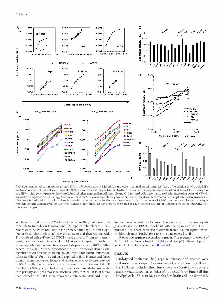
Pseudotyped luciferase (luc) reporter viruses and vectors wereused initially to compare human, rodent, and carnivore cell lines(Fig. 1). These included three lines from two Mustelidae species: arecently established ferret (Mustela putorius furo) lung cell line(FtAEpC cells [37]), an M. putorius furo brain cell line (Mpf cells
FIG 1 Assessment of gammaretroviral and HIV-1 life cycle stages in Mustelidae and other mammalian cell lines. (A) Lack of restriction to N-tropic MLVluciferase vectors in Mustelidae cell lines. HT1080 cells were used as the positive-control line. The same viral preparation was used for all lines. (B to E) Early andlate HIV-1 viral gene expression in Mustelidae and other mammalian cell lines. (B and C) Indicated cells were transduced with increasing doses of VSV-G-pseudotyped reporter virus HIV-1luc. Curves for the three Mustelidae are colored gray. Error bars represent standard deviations of duplicate measurements. (D)Cells were transduced with an HIV-1 vector in which transfer vector luciferase expression is driven by an internal CMV promoter. Cell lysates from equalnumbers of cells were assayed for luciferase activity 5 days later. (E) p24 antigen, measured at day 5 postransduction, in supernatants of the respective cellstransduced in panel C.
Fadel et al.
2314 jvi.asm.org Journal of Virology

[67]), and an American mink (Neovison vison, formerly Mustelavison) fetal lung cell line (Mv.1.Lu cells [27]). In agreement with aprevious study that included the latter two (65), we found that allthree Mustelidae cell lines as well as dog and cat cells supportedequivalent N- and NB-murine leukemia virus (MLV) infection,whereas N-MLV-restricting human HT1080 cells were much lessefficiently infected with N-MLV (Fig. 1A). The Mustelidae andtwo other carnivore lines were also readily susceptible to HIV-1luc
reporter virus infection, yielding luciferase activities that equaledor exceeded those of human cells infected with the same inputs(Fig. 1B and C). Since luc is expressed from the nef open readingframe in HIV-1luc (40), this virus demonstrates competence forthe following postentry stages: reverse transcription, integration,and Tat/U3-promoted early (Rev-independent) viral gene expres-sion (40). Similar results were observed when primary humanPBMC and primary ferret mononuclear cells obtained fromspleen, bone marrow, and lymph nodes were compared (Fig. 2A).Furthermore, similarly equivalent transduction was observed inferret cell lines with a genome-minimized trans-packaged HIV-1vector in which an internal human cytomegalovirus (CMV) pro-
moter drives expression (Fig. 1D) and with analogously organizedsingle-cycle FIV and equine infectious anemia virus (EIAV) vec-tors (Fig. 3A and B).
Using primers validated by McEwan et al. to amplify TRIM5�exon 8 from mink, dog, and various feline species genomic DNA(47, 48), we amplified and sequenced exon 8 of the ferret TRIM5�gene from ferret (Mpf and FtAEpC) cells (see above) and foundthat the Feliformia-specific premature stop codon (48) is lacking,as was previously reported for the dog and mink exons (48, 57).However, multiple other stop codons were present in all readingframes, indicating pseudogenization as in the dog (57). No ferretTRIMCyp transcript could be identified by PCR using primersanchored in the ferret CypA sequence (determined in the presentstudy; see below) and sets of degenerate primers homologous toCarnivora and human exon 2 (data not shown). Late HIV-1 lifecycle events were assessed initially by measuring HIV-1 p24 pro-duction. Mouse (3T3) cells displayed the previously well-established (9, 17, 46, 64) assembly block to HIV-1, whereas Mus-telidae cell lines and primary cells yielded robust HIV-1 p24production similar to that of human cells (Fig. 1E and Fig. 2B and
FIG 2 Early and late HIV-1 viral gene expression in primary human and ferret cells. Primary mononuclear cells from human peripheral blood and ferret spleen,bone marrow, and lymph nodes were transduced with increasing doses of VSV-G pseudotyped reporter virus HIV-1luc�. (A) Luciferase activity was measuredat 5 days postransduction in cell lysates. (B) Immunoblotting for HIV-1 capsid protein. (C) p24 antigen measured at day 5 postransduction, in supernatants ofthe respective cells transduced in panel B.
HIV-1 Replication in Ferret Cells
February 2012 Volume 86 Number 4 jvi.asm.org 2315

C). Taken together, these data indicate that major pre- and postin-tegration portions of the HIV-1 life cycle are grossly unimpairedin each of the three Mustelidae cell lines tested as well as in primaryferret mononuclear cells derived from spleen, bone marrow, andlymph nodes. There is an absence of TRIM5�/TRIMCyp/Fv1-type postentry blocks to lentiviral and gammaretroviral life cycles,substantial Tat transactivation function, and substantial Rev-mediated protein production, assembly, and particle release inthese cells.
Based on these experiments, we proceeded to test directlywhether mink or ferret cells could support productive, spreadingreplication of HIV-1. We derived stable cell lines that express hu-man CD4 and CXCR4 and verified cell surface expression of theseproteins by flow cytometry (Fig. 4A). Entry receptor competencewas verified by infecting the receptor-complemented cells withHIV-1 LAI-luc (69) (Fig. 4B). The Mustelidae cells were then chal-lenged with HIV-1 NL4-3 (2) and two variants of NL4-3, HIV-1VS
and HIV-1VH (62). HIV-1VS utilizes a separation of the normallyoverlapping integrase and vif reading frames to encode the Vifprotein of SIVmac (62), and HIV-1VH is a matched control virusthat encodes HIV-1 Vif in the same manner. HIV-1VS but notHIV-1VH or wild type HIV-1 NL4-3 replicates in felineCrFK.CD4.X4 cells (62).
In the present experiments in Mustelidae cells, the pattern wasdifferent. Productive, spreading replication of wild-type HIV-1NL4-3, HIV-1VH, and HIV-1VS was observed in ferret but notmink cells (Fig. 5A to C). This was the case in the FtAEpC.CD4.X4cell line and in two independently derived stable Mpf.CD4.X4 celllines (Mpf.CD4.X41 and Mpf.CD4.X42, which were made withdifferent receptor-transducing systems). The viruses could bemultiply passaged and amplified (Fig. 5D and E). In contrast, thereceptor-complemented mink cell line Mv.1.Lu.CD4.X4, thoughclearly enabled for efficient viral entry (Fig. 4B), did not supportproductive HIV-1 replication with any of the above viruses, in-cluding HIV-1 first passaged through the ferret cell lines (data notshown).
These results showed that wild-type HIV-1 will replicate pro-ductively and spread in ferret cells if entry receptors are provided.Nevertheless, we found that virions produced in these cells were
still less infectious per unit of p24 antigen than were virions pro-duced in human cell lines. Figure 4F shows GHOST cell titrationsof third-passage viruses from the ferret cell line Mpf.CD4.X41,which we refer to as HIV-1VH(mpfP3) and HIV-1VS(mpfP3). Compar-ison is made to virus produced in maximally permissive human293T cells. This producer cell-dependent loss of infectivity in fer-ret cells suggested that APOBEC3 proteins may be targetingHIV-1. Therefore, we amplified and sequenced long terminal re-peat (LTR) and gag/pol-vpr segments of HIV-1VH(mpfP3) and HIV-1VS(mpfP3) genomes as well as from later FtAEpC cell passages (Fig.6A). A signature of APOBEC3 protein activity, G¡A hypermuta-tion, was observed. This genome editing is nevertheless manifestlynot lethal since HIV-1 was still amplified exponentially and pas-saged robustly in ferret cell lines. Another finding was a prematurestop codon in vpr, a frequent occurrence when HIV-1 is passagedin any cultured cells (51). In HIV-1VH(mpfP3), a vif reading frame-preserving closure of the artificial integrase-vif separation arosethrough deletion of a 62-nucleotide (nt) segment (Fig. 6B and itslegend). This change, effectively a reversion to wild type, wasfound at the 5= end of vif in 8/10 clones and was likely consequentto the duplication of 5=-GGAAAACAG-3= (56, 62) at the 5= end ofvif in the input virus, which facilitated recombination during re-verse transcription (72) to produce the virus illustrated in Fig. 6B.While G-to-A changes predominated, other kinds of mutationswere also seen (Fig. 6A). For example, as previously observed tohappen with rat and rabbit APOBEC1 (10, 30), a significant num-ber of plus-strand C-to-T mutations were observed (45, versus161 G-to-A changes). This outcome might reflect RNA deamina-tion as well (10, 30). The dinucleotide contexts observed for cyt-idine deamination can vary substantially with different APOBECproteins (6). Here in ferret cells, it was different from the patterntypically seen with human A3G, where CC and TC (edited minus-strand cytidine underlined) contexts predominate (23, 44). In-stead, a broader dinucleotide context pattern was observed in the161 G-to-A mutations that occurred with passage in ferret cells:CC, 46 (29%); TC, 32 (20%); GC, 27 (17%); and AC, 56 (35%).This is reminiscent of more diverse dinucleotide patterns ob-served previously for different feline A3 proteins (50).
Vif did not accrue consistent amino acid changes in any of the
FIG 3 FIV and EIAV infection. The indicated cells were transduced with increasing doses of luciferase-encoding single-cycle vectors derived from FIV (A) orEIAV (B). Cell lysates from equal numbers of cells were assayed for activity 72 h later. Error bars represent standard deviations of duplicate measurements.
Fadel et al.
2316 jvi.asm.org Journal of Virology

viruses. However, capsid did. Two coding changes arose in thecyclophilin A (CypA) binding loop, which is complexly involvedin the viral life cycle, including in TRIM5 protein restriction (41).For HIV-1VH(mpfP3), a T¡A change at nt 261 in capsid and, forHIV-1VS(mpfP3), a G¡A mutation at nt 274 produced, respec-tively, H87Q and A92T mutations (Fig. 6C). As the selection oftwo different mutations in this functionally significant region ofcapsid after passage in ferret cells was intriguing, we introducedthem prospectively, alone and in combination, into HIV-1 NL4-3reporter viruses and full-length clones. Examined in this geneti-cally defined context, the HIV-1 capsid mutants were found tohave moderately increased infectivity in ferret cells compared towild type (WT) (Fig. 7A).
In the case of replicating virus, capsid mutants replicated tohigher peak levels than did wild-type virus in ferret cells (Fig. 7B).We then tested effects of cyclosporine (Fig. 8A through C). Inthese experiments, the increase in infectivity conferred by theCypA binding loop mutations was again observed in ferret cellsand also in owl monkey kidney (OMK) cells (Fig. 8A through C).CsA, which disrupts TRIMCyp restriction (59), produced thewell-known dramatic augmenting effect in OMK cells for wild-type NL4-3 and each of the mutants (Fig. 8A). The moderatelyincreased infectivity of H87Q in the absence of CsA in these ex-
periments (Fig. 8A) is consistent with that observed previously inOMK cells (31). In clear contrast, CsA did not boost infectivity inferret cells for any of the viruses (Fig. 8B and C); rather, a slightinhibitory effect was discernible, particularly for A92T. Since lev-els of CypA have been reported to play a role in determiningHIV-1 infectivity in certain contexts (70), we performed immu-noblotting for this protein. CypA was clearly present in the ferretcells, and its levels were also similar to those in human cells (Fig.8D). A ferret CypA cDNA was isolated by reverse transcriptasePCR (RT-PCR) (Fig. 8E); sequencing and determination of thepredicted amino acid sequence did not reveal significant differ-ences in the known HIV-1 capsid-interacting regions (13, 20, 71).
To complete the analysis with respect to the capsid mutants,VLP saturation experiments were performed. A dose-dependent,clear VLP saturation effect was observed in rhesus FrHK4 cells asanticipated (8, 16), but no such effects occurred in ferret cells (Fig.9A to D).
DISCUSSION
The results of this study show that, unlike the mouse genome (9),the ferret genome can supply the nonreceptor dependency factorsneeded for productive, spreading HIV-1 replication. Moreover, incontrast to cells of another carnivore that share this property (62,
FIG 4 Mustelidae cell lines with functional HIV-1 entry receptors. FIV vectors that coencode the neo or pac resistance markers (62) were used along with G418and puromycin selection, respectively, to introduce the two receptors serially into Mpf cells, yielding the Mpf.CD4.X41 cell line. An HIV-1 TSIN series (40)lentiviral vector was also constructed to encode the receptor and coreceptor transgenes linked by a porcine teschovirus 2A (P2A) peptide, with pac coencoded bya downstream IRES. Using this vector, a second Mpf line (Mpf.CD4.X42) as well as FtAEpC.CD4.X4 and Mv.1.Lu.CD4.X4 cell lines was derived and maintainedwith puromycin selection. (A) Flow cytometry was performed to determine the expression of human CD4 and CxCR4 on the surface of the derived cells. All cellswere labeled with the antibodies except for the SupT1 cells in the top left panel. The parental Mustelidae species cells not complemented with receptors were usedas negative controls (left, bottom three panels), and SupT1 was used as a positive control (top right panel). (B) gp120-mediated entry. Cells with or withoutreceptors were infected with increasing doses of native enveloped HIV-1 LAI-luc reporter virus (54). Cell lysates from equal numbers of cells were assayed forluciferase activity 72 h later.
HIV-1 Replication in Ferret Cells
February 2012 Volume 86 Number 4 jvi.asm.org 2317

73), vif gene substitution was not needed and wild-type HIV-1 wascapable of replication and serial passage. As in feline cells, G¡Ahypermutation and producer cell-dependent infectivity reduc-tions were observed, but in both ferret cell lines they did not pre-vent productive viral replication. The selection of capsid muta-tions is also strong corroborative evidence that continuous viralreplication occurred. Whether the HIV-1 or SIVmac Vif proteinproduces partial APOBEC3 mitigation in ferret cells, or might beevolved by repeated passage to acquire it, deserves further specificanalysis. The absence of postentry capsid-targeting defensesagainst N-MLV and lentiviruses is consistent with the apparentlack of an intact Trim5� or TRIMCyp gene in this species. Weobserved similarly robust completion of early and late events in
primary ferret cells (Fig. 2). We were not able to complementprimary ferret mononuclear cells with HIV-1 receptors, andlymphoid-lineage cell lines are not yet available. Therefore, theextent to which specific relevant primary cell types (CD4� T cells)in ferrets in vivo express all needed nonreceptor dependency fac-tors and/or might manifest additional restrictions will be a worthysubject for further study.
The capability to repeatedly passage a primate lentivirusthrough the novel adaptive environment of a nonprimate cell al-lows experimental selection for continued viral evolution. So far,after three passages, we have observed selection of two capsidCypA binding loop mutations, H87Q and A92T. We found thatthese confer moderately increased infectivity in ferret cells and
FIG 5 Productive HIV-1 replication in ferret cells. (A and B) Mpf.CD4.X41 cells were infected with HIV-1VH and HIV-1VS, and virus produced was seriallypassaged 5 additional times. Input inocula were 1 ng p24. (C) Mpf.CD4.X42 cells were infected with 10 ng p24 of HIV-1 NL4-3, and virus produced was seriallypassaged 5 additional times. (D and E) Third-passage HIV-1 HIV-1VH(mpfP3) and HIV-1VS(mpfP3) viruses from Mpf.CD4.X41 replication experiments wereserially passaged 4 times on FtAEpC.CD4.X4 cells. The passage numbers above the curves reflect the total passages on both ferret lines, while the numbers in thesymbol keys refer to the number of passages on FtAEpC.CD4.X4 cells. (F) Infectivity determined by titration on GHOST cells.
FIG 6 Sequencing of HIV-1VH(mpfP3) and HIV-1VS(mpfP3) viruses isolated from passage 3 of HIV-1VH and HIV-1VS on the Mpf.CD4.X41 cell line. (A) Sequencingof 290,607 nt (8 to 10 clones per segment) was performed, revealing 161 G¡A changes. (B) Recombinant HIV-1VH found in 8/10 clones. Capital letters indicateoriginal mutations used to separate the integrase and vif frames. (C) Capsid mutations selected in the unstructured CypA binding loop of HIV-1 CA. H87Q andA92T were present in 10 of 10 and 9 of 9 sequenced clones, respectively.
Fadel et al.
2318 jvi.asm.org Journal of Virology

that disruption of CypA interaction with CsA only slightly affectsthis (Fig. 7 and 8). The reasons that these mutations arose are notclear because of the ambiguities that persist about the roles thatCypA plays in retroviral life cycles, but they may represent optimalfitness of the CypA binding loop in the presence of CypA but in theabsence of any functionally antiviral TRIM5 protein. CypA, whichis highly conserved between mammals, is a peptidyl-prolylisomerase that binds lentiviral capsids. It catalyzes the cis/transisomerization of the G89-P90 peptide bond in the HIV-1 capsidprotein (11). CypA has distinctive and at times opposite context-dependent effects (18, 20, 42). The protein promotes infectivity inhuman cells (25, 61, 66) but is in contrast necessary for Trim5�restriction in rhesus macaque and African green monkey cells (7).While multiple possibilities have been proposed for the role ofCypA in the viral life cycle, a unifying mechanistic explanationremains elusive. It has been hypothesized that this peptidyl-prolylisomerase protects HIV-1 from human cell restriction by compet-ing with Trim5� binding (31) or that it shields HIV-1 from anunknown antiviral factor in human cells since the stimulatory
effect of CypA on HIV-1 infectivity is independent of humanTrim5� (58, 59). Some CypA binding loop mutations alleviaterestriction in Old and New World monkey cells; similar effects areseen with CsA (15, 31). Of interest, H87Q decreases the bindingaffinity of HIV-1 CA for CypA by 4.8-fold (71) and was reportedto confer a replication advantage to HIV-1 in CypA-rich humancells (21). The mutation is present in 19.7% of natural isolates inthe Los Alamos database (15), and in human cells it can conferHIV-1 resistance to the effects of CsA (i.e., CypA independence)(15, 31). H87Q has also been reported to mediate escape fromcytotoxic T lymphocyte responses, emerging in the late phase ofinfection in 13.7% of patients infected with HIV-1 (36, 39).
A92T has not been previously reported, but a charge-addingmutation at this residue, A92E, is known to arise when HIV-1 ispassaged in HeLa cells in the presence of CsA, thus conferring CsAresistance and in some cell lines actually conferring CsA depen-dence on the virus (1, 12); this phenomenon was later shown toreflect high levels of CypA in HeLa cells (70). Our experimentsmake it clear that CypA is present at substantial levels in the Mus-
FIG 7 Infection of ferret cells with WT, H87Q, A92T, and H87Q/A92T NL4-3 clones. (A) HIV-1GFP reporter viruses. The number of GFP-positive cells wasdetermined by FACS at 48 h. The upper and lower graphs show the results of two independent experiments with different vector preparations. Student’s 2-tailedt test was used to determine P values for comparisons of the titers of WT virus with each of the capsid mutants. All calculated P values were �0.05 except for thecomparison of WT and H87Q/A92T mutants in FtAEpC cells (asterisk, upper right plot; P � 0.07). (B) Full-length WT and mutant viral clone challenges ofMpf.CD4.X41 cells.
HIV-1 Replication in Ferret Cells
February 2012 Volume 86 Number 4 jvi.asm.org 2319

telidae cell lines that we used (Fig. 8D) and also that its amino acidsequence is conserved versus human CypA in the hydrophobicregions known to form the retroviral capsid-interacting domain(13) (Fig. 8E). Thus, these mutations that developed in ferret cells
may represent HIV-1 adaptation to a situation where CypA ispresent but there is no Trim5 protein pressure.
In contrast to ferret cells, we did not observe spreading HIV-1replication in the mink (Mv.1.Lu.CD4.X4) cell line. This result,
FIG 8 Cyclosporine experiments in ferret cells and CypA alignments. (A to C) Infectious titers of HIV-1 GFP vector were determined in the presence (blackcolumns) and absence (gray columns) of 5 �M CsA for owl monkey kidney (A), Mpf (B), and FtAEpC (C) cells. (D) Immunoblotting for CypA in human andMustelidae cell lines. Jurkat ppia�/� cells (14) were used as a negative control. (E) Alignment of the ferret and human CypA amino acid sequences reveals highconservation overall and complete conservation in the central hydrophobic pocket involved in HIV-1 capsid binding. The amino acids involved in HIV-1 capsidbinding, which are known from several mutational and high-resolution structural studies (13, 20, 71), are highlighted with boxes. Comparison is made betweenthe human protein reference sequence (top line, accession no. NP_066953) and the predicted amino acid sequence determined from the ferret CypA cDNAisolated from Mpf cell mRNA in the present study (middle line) and a sequence predicted from an M. putorius furo whole-genome shotgun (WGS) sequencecontig (bottom line, accession no. AEYP01032892). The dashed arrows at each end indicate the span of the degenerate primers used to obtain the Mpf cell cDNAsequence. Stringent selection pressure for amino acid level conservation is apparent. For example, at the nucleotide sequence level (not shown), there were 49differences between Mpf cell CypA and human CypA in the coding region of the mRNA (10.1% nonidentity), but virtually all were synonymous. Only threeamino acid differences were present, and these were located at both termini (arrowheads indicating the fifth and the final two C-terminal amino acids); the T5Idifference (black arrowhead) is uncertain since both threonine and isoleucine were encoded by the degenerate primer, the domestic cat sequence has anisoleucine at this position, and the ferret WGS contig predicts a threonine. Polymporphism is evident in the two ferret sequences; they differed at 10 nt (notshown), but besides T5I there were only two other predicted amino acid differences, A26S and R37H.
FIG 9 VLP saturation experiments in ferret cells. FRhK4, Mpf, and FtAEpC cells were infected with a fixed dose of wild-type HIV-1 GFP (A) and capsid mutantsH87Q (B), A92T (C), and H87Q/A92T (D), in the presence of increasing amounts of HIV-1 VLPs. While the VLPs clearly released Lv1 restriction in the rhesusmacaque cells as anticipated, they had no effect in ferret cells.
Fadel et al.
2320 jvi.asm.org Journal of Virology
which was verified in repeated experiments, is at variance with aprior report (35). As was discussed previously (62), it may bepossible to reconcile these differences by considering that a singleround of provirus generation and p24 production occurred in theexperiments of Koito et al. (35).
Our results add to emerging evidence that cells of carnivorespecies appear in general to harbor relatively few restrictions toHIV-1 replication, and prominent among these are APOBEC3-mediated restrictions. Reference 19 provides a recent review ofcarnivore cell restrictions. We also conclude that, like the domes-tic cat and unlike the mouse, the ferret genome encodes the majornonreceptor dependency factors for this primate lentivirus. Thereare robust precedents for modeling human RNA pathogens in theferret. A ferret genome sequencing project is nearing comple-tion (http://www.broadinstitute.org/scientific-community/science/projects/mammals-models/ferret-genome-project). We suggestthat further characterization of HIV-1 adaptation in ferret cellsand delineation of Mustelidae restriction factor gene repertoiresare warranted, with a view to several possible benefits. These in-clude potentials for developing an eventual HIV-1 animal model,for gaining basic insights into mechanisms of species-specific ret-roviral restriction, and for exploring the extent to which HIV-1will evolve when confronted with the novel adaptive landscape ofa nonprimate cell.
ACKNOWLEDGMENTS
We thank M. Stern for assistance with initial experiments in Mpf cells, J.Luban for Jurkat CypA-knockout cells, and M. Emerman, J. Olsen, and P.Charneau for plasmids.
We are grateful for funding from NIH AI47536 and AI77344 and theTietze Foundation.
REFERENCES1. Aberham C, Weber S, Phares W. 1996. Spontaneous mutations in the
human immunodeficiency virus type 1 gag gene that affect viral replica-tion in the presence of cyclosporins. J. Virol. 70:3536 –3544.
2. Adachi A, et al. 1986. Production of acquired immunodeficiencysyndrome-associated retrovirus in human and nonhuman cells trans-fected with an infectious molecular clone. J. Virol. 59:284 –291.
3. Ambrose Z, KewalRamani VN, Bieniasz PD, Hatziioannou T. 2007.HIV/AIDS: in search of an animal model. Trends Biotechnol. 25:333–337.
4. Antunes A, et al. 2008. The evolutionary dynamics of the lion Pantheraleo revealed by host and viral population genomics. PLoS Genet.4:e1000251.
5. Baumann JG, et al. 2004. Murine T cells potently restrict human immu-nodeficiency virus infection. J. Virol. 78:12537–12547.
6. Beale RC, et al. 2004. Comparison of the differential context-dependenceof DNA deamination by APOBEC enzymes: correlation with mutationspectra in vivo. J. Mol. Biol. 337:585–596.
7. Berthoux L, Sebastian S, Sokolskaja E, Luban J. 2005. Cyclophilin A isrequired for TRIM5alpha-mediated resistance to HIV-1 in Old Worldmonkey cells. Proc. Natl. Acad. Sci. U. S. A. 102:14849 –14853.
8. Besnier C, Takeuchi Y, Towers G. 2002. Restriction of lentivirus inmonkeys. Proc. Natl. Acad. Sci. U. S. A. 99:11920 –11925.
9. Bieniasz PD, Cullen BR. 2000. Multiple blocks to human immunodefi-ciency virus type 1 replication in rodent cells. J. Virol. 74:9868 –9877.
10. Bishop KN, Holmes RK, Sheehy AM, Malim MH. 2004. APOBEC-mediated editing of viral RNA. Science 305:645.
11. Bosco DA, Eisenmesser EZ, Pochapsky S, Sundquist WI, Kern D. 2002.Catalysis of cis/trans isomerization in native HIV-1 capsid by human cy-clophilin A. Proc. Natl. Acad. Sci. U. S. A. 99:5247–5252.
12. Braaten D, et al. 1996. Cyclosporine A-resistant human immunodefi-ciency virus type 1 mutants demonstrate that Gag encodes the functionaltarget of cyclophilin A. J. Virol. 70:5170 –5176.
13. Braaten D, Ansari H, Luban J. 1997. The hydrophobic pocket of cyclo-philin is the binding site for the human immunodeficiency virus type 1Gag polyprotein. J. Virol. 71:2107–2113.
14. Braaten D, Luban J. 2001. Cyclophilin A regulates HIV-1 infectivity, asdemonstrated by gene targeting in human T cells. EMBO J. 20:1300 –1309.
15. Chatterji U, et al. 2005. Naturally occurring capsid substitutions renderHIV-1 cyclophilin A independent in human cells and TRIM-cyclophilin-resistant in owl monkey cells. J. Biol. Chem. 280:40293– 40300.
16. Cowan S, et al. 2002. Cellular inhibitors with Fv1-like activity restricthuman and simian immunodeficiency virus tropism. Proc. Natl. Acad.Sci. U. S. A. 99:11914 –11919.
17. Diaz-Griffero F, Taube R, Muehlbauer SM, Brojatsch J. 2008. Efficientproduction of HIV-1 viral-like particles in mouse cells. Biochem. Biophys.Res. Commun. 368:463– 469.
18. Dorfman T, Weimann A, Borsetti A, Walsh CT, Gottlinger HG. 1997.Active-site residues of cyclophilin A are crucial for its incorporation intohuman immunodeficiency virus type 1 virions. J. Virol. 71:7110 –7113.
19. Fadel H, Poeschla E. 2011. Retroviral restriction and dependency factorsin primates and carnivores. Vet. Immunol. Immunopathol. 143:179 –189.
20. Gamble TR, et al. 1996. Crystal structure of human cyclophilin A boundto the amino-terminal domain of HIV-1 capsid. Cell 87:1285–1294.
21. Gatanaga H, et al. 2006. Altered HIV-1 Gag protein interactions withcyclophilin A (CypA) on the acquisition of H219Q and H219P substitu-tions in the CypA binding loop. J. Biol. Chem. 281:1241–1250.
22. Gifford RJ, et al. 2008. A transitional endogenous lentivirus from thegenome of a basal primate and implications for lentivirus evolution. Proc.Natl. Acad. Sci. U. S. A. 105:20362–20367.
23. Harris RS, et al. 2003. DNA deamination mediates innate immunity toretroviral infection. Cell 113:803– 809.
24. Hatziioannou T, et al. 2009. A macaque model of HIV-1 infection. Proc.Natl. Acad. Sci. U. S. A. 106:4425– 4429.
25. Hatziioannou T, Perez-Caballero D, Cowan S, Bieniasz PD. 2005.Cyclophilin interactions with incoming human immunodeficiency virustype 1 capsids with opposing effects on infectivity in human cells. J. Virol.79:176 –183.
26. Hatziioannou T, et al. 2006. Generation of simian-tropic HIV-1 by re-striction factor evasion. Science 314:95.
27. Henderson IC, Lieber MM, Todaro GJ. 1974. Mink cell line Mv 1 Lu(CCL 64). Focus formation and the generation of “nonproducer” trans-formed cell lines with murine and feline sarcoma viruses. Virology 60:282–287.
28. Hu C, et al. 2010. The HIV-1 central polypurine tract functions as asecond line of defense against APOBEC3G/F. J. Virol. 84:11981–11993.
29. Igarashi T, et al. 2007. Human immunodeficiency virus type 1 derivativewith 7% simian immunodeficiency virus genetic content is able to estab-lish infections in pig-tailed macaques. J. Virol. 81:11549 –11552.
30. Ikeda T, et al. 2008. The antiretroviral potency of APOBEC1 deaminasefrom small animal species. Nucleic Acids Res. 36:6859 – 6871.
31. Ikeda Y, Ylinen LM, Kahar-Bador M, Towers GJ. 2004. Influence of gagon human immunodeficiency virus type 1 species-specific tropism. J. Vi-rol. 78:11816 –11822.
32. Kamada K, et al. 2006. Generation of HIV-1 derivatives that productivelyinfect macaque monkey lymphoid cells. Proc. Natl. Acad. Sci. U. S. A.103:16959 –16964.
33. Katzourakis A, Tristem M, Pybus OG, Gifford RJ. 2007. Discovery andanalysis of the first endogenous lentivirus. Proc. Natl. Acad. Sci. U. S. A.104:6261– 6265.
34. Keckesova Z, Ylinen LM, Towers GJ, Gifford RJ, Katzourakis A. 2009.Identification of a RELIK orthologue in the European hare (Lepus euro-paeus) reveals a minimum age of 12 million years for the lagomorphlentiviruses. Virology 384:7–11.
35. Koito A, Kameyama Y, Cheng-Mayer C, Matsushita S. 2003. Suscepti-bility of mink (Mustera vision)-derived cells to replication by human im-munodeficiency virus type 1. J. Virol. 77:5109 –5117.
36. Kootstra NA, Navis M, Beugeling C, van Dort KA, Schuitemaker H.2007. The presence of the Trim5alpha escape mutation H87Q in the cap-sid of late stage HIV-1 variants is preceded by a prolonged asymptomaticinfection phase. AIDS 21:2015–2023.
37. Kugel D, et al. 2009. Intranasal administration of alpha interferon re-duces seasonal influenza A virus morbidity in ferrets. J. Virol. 83:3843–3851.
38. LaRue RS, Lengyel J, Jonsson SR, Andresdottir V, Harris RS. 2010.Lentiviral Vif degrades the APOBEC3Z3/APOBEC3H protein of its mam-malian host and is capable of cross-species activity. J. Virol. 84:8193– 8201.
39. Leslie AJ, et al. 2004. HIV evolution: CTL escape mutation and reversionafter transmission. Nat. Med. 10:282–289.
HIV-1 Replication in Ferret Cells
February 2012 Volume 86 Number 4 jvi.asm.org 2321

40. Llano M, et al. 2006. An essential role for LEDGF/p75 in HIV integration.Science 314:461– 464.
41. Luban J. 2007. Cyclophilin A, TRIM5, and resistance to human immu-nodeficiency virus type 1 infection. J. Virol. 81:1054 –1061.
42. Luban J, Bossolt KL, Franke EK, Kalpana GV, Goff SP. 1993. Humanimmunodeficiency virus type 1 Gag protein binds to cyclophilins A and B.Cell 73:1067–1078.
43. Malim MH, Emerman M. 2008. HIV-1 accessory proteins– ensuring viralsurvival in a hostile environment. Cell Host Microbe 3:388 –398.
44. Mangeat B, et al. 2003. Broad antiretroviral defence by humanAPOBEC3G through lethal editing of nascent reverse transcripts. Nature424:99 –103.
45. Mariani R, et al. 2001. Mouse-human heterokaryons support efficienthuman immunodeficiency virus type 1 assembly. J. Virol. 75:3141–3151.
46. Mariani R, et al. 2000. A block to human immunodeficiency virus type 1assembly in murine cells. J. Virol. 74:3859 –3870.
47. McEwan WA. 2010. Factors affecting replication and cross-species trans-mission of feline immunodeficiency virus. Ph.D. thesis. University ofGlasgow, Glasgow, United Kingdom. http://theses.gla.ac.uk/1388/.
48. McEwan WA, et al. 2009. Truncation of TRIM5 in Feliformia explains theabsence of retroviral restriction in cells of the domestic cat. J. Virol. 16:8270 – 8275.
49. Münk C, et al. 2008. Functions, structure, and read-through alternativesplicing of feline APOBEC3 genes. Genome Biol. 9:R48.
50. Münk C, et al. 2007. Multiple restrictions of human immunodeficiencyvirus type 1 in feline cells. J. Virol. 81:7048 –7060.
51. Nakaya T, et al. 1996. Serial passage of human immunodeficiency virustype 1 generates misalignment deletions in non-essential accessory genes.Virus Res. 46:139 –147.
52. Neil SJ, Zang T, Bieniasz PD. 2008. Tetherin inhibits retrovirus releaseand is antagonized by HIV-1 Vpu. Nature 451:425– 430.
53. Pecon-Slattery J, Troyer JL, Johnson WE, O’Brien SJ. 2008. Evolution offeline immunodeficiency virus in Felidae: implications for human healthand wildlife ecology. Vet. Immunol. Immunopathol. 123:32– 44.
54. Peden K, Emerman M, Montagnier L. 1991. Changes in growthproperties on passage in tissue culture of viruses derived from infec-tious molecular clones of HIV-1LAI, HIV-1MAL, and HIV-1ELI. Vi-rology 185:661– 672.
55. Poeschla E, Wong-Staal F, Looney D. 1998. Efficient transduction ofnondividing cells by feline immunodeficiency virus lentiviral vectors. Nat.Med. 4:354 –357.
56. Sakurai A, et al. 2004. Functional analysis of HIV-1 vif genes derived fromJapanese long-term nonprogressors and progressors for AIDS. MicrobesInfect. 6:799 – 805.
57. Sawyer SL, Emerman M, Malik HS. 2007. Discordant evolution of theadjacent antiretroviral genes TRIM22 and TRIM5 in mammals. PLoS Pat-hog. 3:e197.
58. Sayah DM, Luban J. 2004. Selection for loss of Ref1 activity in humancells releases human immunodeficiency virus type 1 from cyclophilin Adependence during infection. J. Virol. 78:12066 –12070.
59. Sayah DM, Sokolskaja E, Berthoux L, Luban J. 2004. Cyclophilin Aretrotransposition into TRIM5 explains owl monkey resistance to HIV-1.Nature 430:569 –573.
60. Sheehy AM, Gaddis NC, Malim MH. 2003. The antiretroviral enzymeAPOBEC3G is degraded by the proteasome in response to HIV-1 Vif. Nat.Med. 9:1404 –1407.
61. Sokolskaja E, Sayah DM, Luban J. 2004. Target cell cyclophilin A mod-ulates human immunodeficiency virus type 1 infectivity. J. Virol. 78:12800 –12808.
62. Stern MA, et al. 2010. Productive replication of Vif-chimeric HIV-1 infeline cells. J. Virol. 84:7378 –7395.
63. Stremlau M, et al. 2004. The cytoplasmic body component TRIM5alpharestricts HIV-1 infection in Old World monkeys. Nature 427:848 – 853.
64. Swanson CM, Puffer BA, Ahmad KM, Doms RW, Malim MH. 2004.Retroviral mRNA nuclear export elements regulate protein function andvirion assembly. EMBO J. 23:2632–2640.
65. Towers G, et al. 2000. A conserved mechanism of retrovirus restriction inmammals. Proc. Natl. Acad. Sci. U. S. A. 97:12295–12299.
66. Towers GJ, et al. 2003. Cyclophilin A modulates the sensitivity of HIV-1to host restriction factors. Nat. Med. 9:1138 –1143.
67. Trowbridge RS, Lehmann J, Brophy P. 1982. Establishment and char-acterization of ferret cells in culture. In Vitro 18:952–960.
68. Troyer JL, et al. 2008. FIV cross-species transmission: an evolutionaryprospective. Vet. Immunol. Immunopathol. 123:159 –166.
69. Yamashita M, Emerman M. 2004. Capsid is a dominant determinant ofretrovirus infectivity in nondividing cells. J. Virol. 78:5670 –5678.
70. Ylinen LM, et al. 2009. Cyclophilin A levels dictate infection efficiency ofhuman immunodeficiency virus type 1 capsid escape mutants A92E andG94D. J. Virol. 83:2044 –2047.
71. Yoo S, et al. 1997. Molecular recognition in the HIV-1 capsid/cyclophilinA complex. J. Mol. Biol. 269:780 –795.
72. Zhang J, Temin HM. 1994. Retrovirus recombination depends on thelength of sequence identity and is not error prone. J. Virol. 68:2409 –2414.
73. Zielonka J, et al. 2010. Vif of feline immunodeficiency virus from domes-tic cats protects against APOBEC3 restriction factors from many felids. J.Virol. 84:7312–7324.
Fadel et al.
2322 jvi.asm.org Journal of Virology
Copyright © 2022 FDOKUMEN