Preliminary evaluation of particle systems visualization on the skin surface by scanning electron...
10
Preliminary evaluation of particle systems visualization on the skin surface by scanning electron microscopy and transparency profilometry Paola Perugini 1 , M. Vettor 1 , M. Bleve 1 , G. Bruni 2 , A. Mondelli 3 , G. F. Secchi 3 and F. Pavanetto 1 1 Department of Pharmaceutical Chemistry, University of Pavia, Pavia, Italy, 2 Department of Physical Chemistry, University of Pavia, Pavia, Italy, and 3 Kelisema srl, Tavernerio, Como, Italy Background: There is a rising debate concerning the pos- sible side effects arising from the use of particles at nano- size since the production of nanomaterials is increasing worldwide. Nanoparticles are able to enter the body through the skin, lungs or intestinal tract, depositing in several organs, and the risk associated with exposure to them, the routes of entry and the molecular mechanisms of any cytotoxicity need to be well understood.The aim of this work was to evaluate the suitability of skin replica as a method to study the colloidal systems visualization and distribution on skin surface. Methods: Solid lipid nanoparticles (SLN) were used as carrier systems. Skin replicas on healthy volunteers, before and after SLN application, were prepared and visualized using profilometry and scanning electron microscopy (SEM). Results: The results obtained in our study show that skin replica represents a suitable method to study the colloidal systems and their interaction with the skin surface. Conclusion: Profilometry enabled us to observe the sys- tems distribution on a cutaneous texture. In addition, SEM, thanks to its high magnifications and field depth, allowed us to evaluate particles’ distribution on the skin texture and the interaction between particles of different compositions and replica silicone. Key words: nanoparticles – scanning electron microscopy – profilometry – skin replica & 2011 John Wiley & Sons A/S Accepted for publication 16 February 2011 H UMANS HAVE been exposed to nanoparticles throughout their evolutionary phase; how- ever, this exposure has increased to a huge extent in the past century because of the industrial revolution. Nanoparticles constitute a part of particulate matter. Epidemiological studies have shown that urban pollution with airborne parti- culate matter deriving from combustion sources such as motor vehicle and industrial emission contributes to respiratory and cardiovascular morbidity and mortality. A typical ambient par- ticulate matter is a highly complex mix of parti- cles with a diameter size distribution ranging from some nanometre to 100 mm. Only a fraction of these particles with a mass median diameter of 2.5 mm or less is capable of depositing deep in the lung (1). The growing use of nanotechnology in high- tech industries is likely to become another means for humans to be exposed to intentionally gener- ated engineered nanoparticles. However, the same properties (small size, chemical composi- tion, structure, large surface area and shape), which make nanoparticles so attractive in medi- cine, may contribute to the toxicological profile of nanoparticles in biological systems (1, 2). Because of the increased use of nanotechnol- ogy, the risk associated with exposure to nano- particles, the routes of entry and the molecular mechanisms of any cytotoxicity need to be well understood. In fact, these tiny particles (o1000 nm in size) are able to enter the body through the skin, lungs or intestinal tract, depositing in several organs, and may cause adverse biological reactions by modifying the physiochemical properties of liv- ing matter at the nanolevel (1, 3). With regard to the skin, it has been hypothesized that dermal exposure might be the most significant route of exposure; however, only a few literature papers referring to the absorption and the effects of nanoparticles in the skin are available. 45 Skin Research and Technology 2012; 18: 45–54 Printed in Singapore All rights reserved doi: 10.1111/j.1600-0846.2011.00529.x © 2011 John Wiley & Sons A / S Skin Research and Technology
-
Upload
independent -
Category
Documents
-
view
0 -
download
0
Transcript of Preliminary evaluation of particle systems visualization on the skin surface by scanning electron...

Preliminary evaluation of particle systems visualization on
the skin surface by scanning electron microscopy and
transparency profilometry
Paola Perugini1, M. Vettor1, M. Bleve1, G. Bruni2, A. Mondelli3, G. F. Secchi3 and F. Pavanetto1
1Department of Pharmaceutical Chemistry, University of Pavia, Pavia, Italy, 2Department of Physical Chemistry, University of Pavia, Pavia, Italy, and3Kelisema srl, Tavernerio, Como, Italy
Background: There is a rising debate concerning the pos-
sible side effects arising from the use of particles at nano-
size since the production of nanomaterials is increasing
worldwide. Nanoparticles are able to enter the body through
the skin, lungs or intestinal tract, depositing in several
organs, and the risk associated with exposure to them, the
routes of entry and the molecular mechanisms of any
cytotoxicity need to be well understood.The aim of this
work was to evaluate the suitability of skin replica as a
method to study the colloidal systems visualization and
distribution on skin surface.
Methods: Solid lipid nanoparticles (SLN) were used as
carrier systems. Skin replicas on healthy volunteers, before
and after SLN application, were prepared and visualized
using profilometry and scanning electron microscopy
(SEM).
Results: The results obtained in our study show that skin
replica represents a suitable method to study the colloidal
systems and their interaction with the skin surface.
Conclusion: Profilometry enabled us to observe the sys-
tems distribution on a cutaneous texture. In addition, SEM,
thanks to its high magnifications and field depth, allowed us
to evaluate particles’ distribution on the skin texture and the
interaction between particles of different compositions and
replica silicone.
Key words: nanoparticles – scanning electron microscopy
– profilometry – skin replica
& 2011 John Wiley & Sons A/SAccepted for publication 16 February 2011
HUMANS HAVE been exposed to nanoparticlesthroughout their evolutionary phase; how-
ever, this exposure has increased to a huge extentin the past century because of the industrialrevolution. Nanoparticles constitute a part ofparticulate matter. Epidemiological studies haveshown that urban pollution with airborne parti-culate matter deriving from combustion sourcessuch as motor vehicle and industrial emissioncontributes to respiratory and cardiovascularmorbidity and mortality. A typical ambient par-ticulate matter is a highly complex mix of parti-cles with a diameter size distribution rangingfrom some nanometre to 100 mm. Only a fractionof these particles with a mass median diameter of2.5mm or less is capable of depositing deep in thelung (1).
The growing use of nanotechnology in high-tech industries is likely to become another meansfor humans to be exposed to intentionally gener-ated engineered nanoparticles. However, the
same properties (small size, chemical composi-tion, structure, large surface area and shape),which make nanoparticles so attractive in medi-cine, may contribute to the toxicological profile ofnanoparticles in biological systems (1, 2).
Because of the increased use of nanotechnol-ogy, the risk associated with exposure to nano-particles, the routes of entry and the molecularmechanisms of any cytotoxicity need to be wellunderstood.
In fact, these tiny particles (o1000 nm in size)are able to enter the body through the skin, lungsor intestinal tract, depositing in several organs,and may cause adverse biological reactions bymodifying the physiochemical properties of liv-ing matter at the nanolevel (1, 3).
With regard to the skin, it has been hypothesizedthat dermal exposure might be the most significantroute of exposure; however, only a few literaturepapers referring to the absorption and the effects ofnanoparticles in the skin are available.
45
Skin Research and Technology 2012; 18: 45–54Printed in Singapore � All rights reserveddoi: 10.1111/j.1600-0846.2011.00529.x
© 2011 John Wiley & Sons A/SSkin Research and Technology

Contact with nanoparticles through the skincan occur due to working exposure during themanufacture of pesticides or pharmaceuticals.
Skin exposure to nanoparticles can also occurduring non-working situations from the use ofcosmetics and with the intentional application oftopical creams and other drug treatments.
Initial studies of nanoparticle absorptionthrough the skin are inconclusive; some demon-strate little penetration into the epidermis, whileothers, using more complex flexing protocols,show deep absorption. To better understanddermal absorption of nanoparticles, more re-search is necessary (2).
The most superficial cutaneous layer, the stratumcorneum, constitutes the major physical barrier ofthe skin. The stratum corneum is a heterogeneoustwo-compartment system composed of keratin-filled corneocytes, embedded in a lipid-enrichedintercellular matrix. The permeability barrier islocated within the lipid lamellae in the intercellularspaces of the stratum corneum (4–6).
Actually, only a few studies elucidate theinteraction between colloidal particles and skin.
Van den Bergh and colleagues studied theinteraction between elastic liquid-state vesiclesformulated with varying compositions and thehuman skin. Using different techniques (trans-mission electron microscopy, freeze-fracture elec-tron microscopy, X-ray diffraction and electronspin resonance), they described the possible me-chanism by which elastic liquid-state vesiclesmay interact with human skin and hence con-tribute to transdermal drug delivery (7).
Visualization of colloidal systems is essential toevaluate their safety and health risk assessment (3).
The aim of this work was to evaluate thesuitability of a skin replica as a method to studythe colloidal systems’ distribution on the skinsurface.
Solid lipid nanoparticles (SLN) were used ascolloidal carrier systems because nanoparticlesbased on solid lipids have actually been pro-posed as an alternative colloidal drug-deliverysystem to polymer nanoparticles, emulsion andliposomes (6, 8).
Skin replica is a technique used for the indirecttopographic measurement of the skin. The pro-cedure consists in placing a thin layer of silicone-based resin on the test skin surface in order toobtain an imprint (9, 10). Systems visualizationwas carried out using profilometry and scanningelectron microscopy (SEM).
The lipospheres, evaluated in the presentstudy, were produced using different lipid matrixcompositions and by varying the experimentalconditions of preparation (11).
The parameters assessed during the visualiza-tion were the composition of the carrier systems,with particular attention to the presence of theamphiphilic lipid lecithin in the lipid phase andthe effect of the particle size on the systemsdistribution on the skin surface.
Furthermore, the behaviour of lipid systemsapplied on the skin of volunteers of different ageswas evaluated.
Materials and Methods
MaterialsCetyl palmitate was purchased from GattefosseItalia S.r.l. (Milano, Italy), phosphatydilcholine(Lecithin, 85% purity) extracted from soybeanswas obtained from AVG S.r.l. (GarbagnateMilanese, Italy), PEG-25 hydrogenated castoroil was a gift from Seppic Italia (Milano, Italy)and myristyl myristate and cetearyl glucosidewere obtained from Goldschmidt Italia (Milano,Italy).
All chemicals were used without any furtherpurification treatment. Distilled water was used.
Solid lipid particles preparationSLN were used as particulate systems. The par-ticles (P) were prepared using the microemulsiontechnique. This preparation method involvedmelting the lipidic phase and heating the aqu-eous phase at 80 1C. At this temperature, thehydrophilic phase was dispersed into the lipo-philic one and high-speed homogenization al-lowed to obtain an O/W microemulsion. Themicroemulsion was placed in a cooling bath for1 h to obtain SLN aqueous suspensions.
The composition of the formulations andhomogenization rates are reported in the Table 1.Formulation TE contains a non-ionic emulsifier inthe lipid matrix and lecithin in the aqueousphase, while formulation BR contains lecithin inthe lipophilic phase and a non-ionic surfactant inthe hydrophilic one.
Skin replicaA cutaneous replica was used as a technique toevaluate systems distribution on the skin surface.
Skin replica is a technique used for the indirecttopographic measurement of the skin. The
46
Perugini et al.

procedure consists in placing a thin layer ofsilicone-based resin on the test skin surface inorder to obtain an imprint (9, 10).
Ten healthy volunteers of both sexes, agedbetween 8 and 71 years, participated in the studyafter signing an informed consent. None of thevolunteers had a known history of skin diseases.The subjects remained at rest for at least 10–20 min, in order to bring their blood circulationto a normal level. Emotional stress may also leadto increased transpiration. Moreover, the sub-ject’s skin was cleaned with water and no pro-duct was applied on it. The inner forearm skinwas found to be a representative test area. Skin’stension should be constant; therefore, the forearmwas placed on a plane surface at a 901 angle.Replicas should only be made on skin areaswithout hair. Moreover, areas with scars andtattoos should be excluded. A small circularzone (8 mm) on the forearm was identified andwas delimited by a 0.5 mm thick adhesive filmthat was used as a frame.
Silicone replicas were produced by hardeningof a silicone gel under controlled conditions(25 1C temperature and 60% relative humidity).The gel was prepared by mixing a basis and acatalyst (1:1 w/w) with a vacuum pump to avoidbubble formation. The basis and catalyst werekept in the ice bath before mixing.
At the centre of the delimited area a drop ofcolloidal system suspension was deposed, andafter 10 or 60 min, one drop of silicone gel wasapplied on the same zone. After hardening, thereplica was ready. A control replica was pro-duced by application of the gel on the skin with-out particle deposition.
Analytical techniquesLaser diffractionLaser diffraction was used to determine theparticle size distribution of systems. The mea-surements were performed using a Malvern
Mastersizer 2000 (Malvern Ltd, Malvern, UK);this instrument works on laser diffraction optics.The size range is from 0.02 to 2000mm.
A nanoparticle suspension was diluted into adispersion unit and analysed under stirring at2500 rpm at room temperature. At least threerepetitions of each measurement were performedfor each sample.
Zeta potential measurementsThe electrophoretic mobility was determinedusing laser doppler anemometry in a microelec-trophoresis cell AZ4 of a Nicomp Zetasizer 380(Santa Barbara, CA, USA). The instrument wasequipped with a 15 mW laser diode and a PMTdetector with two optical fibres: one set at 901 forparticle sizing and the other set at 191 for zetapotential. A sample was introduced with plasticcuvettes. The standard electrodes are made ofplatinum. Before measurement, the SLN werediluted with filtered water. Three analyses wereperformed for each sample.
SEMDifferent from an optical microscope, which isbased on the interaction between light and mat-ter, the electronic microscope is based on theinteraction between electrons and matter, havinga higher resolution and greater field depth. InSEM, an accelerated beam of mono-energeticelectrons is focused on the surface of the sampleand is scanned over a small area in a rosterpattern, from where secondary electrons andother signals are generated, which produce asynchronous image on a cathode ray tube(CRT). The brightness and contrast of any pointon the image displayed on the CRT depend onthe amplitude of the signal current from thedetector, which in turn again depends on themorphological variation of the scanned area. Assecondary electrons have escaped depth of about100 A, SEM represents a good technique of prob-ing exclusively the surface of the samples at the
TABLE 1. Lipidic particle composition
TE1 TE2 BR1 BR2
Cetyl palmitate 4.8% 4.8%
Cetearyl glucoside 1.2% 1.2%
Myristyl myristate 3.6% 3.6%
PEG-25 hydrogenated castor oil 3.6% 3.6%
Soybeans phosphatydilcholine 1.2% 1.2% 1.2% 1.2%
Distilled water 94% 94% 90.4% 90.4%
Homogenization rate 13,500 rpm 9500 rpm 13,500 rpm 9500 rpm
47
Preliminary evaluation of particle systems visualization on skin

sub-micron level (12). SEM micrographs werecollected using Cambridge Stereoscan 200 (Cam-bridge, UK) on gold-sputtered samples of boththe nanoparticle suspension and the skin replica.In particular, in order to observe the morphologyof the lipid particles, a drop of each aqueoussuspension was placed, before the sputtering, ona slide and allowed to dry at room temperature.
ProfilometryThe measurement principle of profilometry (SkinVisiometer SV600-Courage&Khazaka, Cologne,Germany) is based on light transmission througha thin replica comprising blue-dyed siliconewhose light absorption is known. Direct lightcoming from a neon lamp penetrates the replicaand is absorbed depending on the thickness ofthe silicone material (13–15). A video sensorcharge couple device camera with 752� 852pixels measures the amount of locally trans-mitted light, and the intensity of the light is
calculated according to Lambert and Beer’s lawof absorption. Heights and depths of the pictureare displayed using a 256 grey-level scale(0 5 black, 256 5 white) and are calculated bythe software program in micrometres (13). Theadvantage of this method is the short processingtime and the direct visual control of the skinreplica on the computer display. The analysis ofgrey levels thus created enables us to obtain a setof standardized parameters characterizing thecutaneous relief: Rt (depth of roughness),Rz (mean depth of roughness), Rm (maximumroughness) and Ra (mean roughness) (16).
Results and Discussion
Preparation and characterization of solid lipidparticlesTwo formulations of each composition, namelyTE e BR with increasing homogenization rate,were prepared successfully.
The dispersions obtained were homogeneoussystems of milky colour when observed macro-scopically.
In Table 2, size distribution results are re-ported. Particles obtained using lecithin in thelipophilic phase had the most homogeneous sizedistribution regardless of the experimental con-ditions (uniformity 1.27).
TABLE 2. Size distribution results
Batch d10% (mm) d50% (mm) d90% (mm) Span Uniformity
BR1 0.077 0.177 0.797 4.062 1.27
BR2 0.080 0.202 1.028 5.694 1.45
TE1 0.070 0.148 0.773 4.769 7.92
TE2 2.236 15.659 90.660 5.658 1.85
Fig. 1. Scanning electron micrographs of solid lipid nanoparticles BR: (a) � 465, (b) 57.7 Kx.
Fig. 2. Scanning electron micrographs of solid lipid nanoparticles TE: (a) � 222; (b) 1.04 Kx
48
Perugini et al.
The zeta potential of SLN varied depending onthe emulsifier present in the lipid matrix. Accord-ing to the results obtained by Schubert and
Muller-Goymann (17), SLN containing lecithinin the lipid matrix (BR) revealed a mean zetapotential of � 12.22 mV.
Particles (batches TE) prepared using the non-ionic emulsifier (cetearyl glucoside) in the lipidmatrix showed a higher mean zeta potential(� 20.36 mV). The presence of the phosphatidyl-choline in the hydrophilic phase was, certainly,very important for the stability of the suspension.The lecithin could be distributed to the particles’interface, improving the separation.
Electrophoretic mobility results are in agree-ment with those obtained from the morphologi-cal analysis carried out by SEM.
As shown in the Fig. 1a, the BR nanoparticlesseem to be aggregated, forming branched shapes.A higher magnification of the lipidic systemsshows the round shape of the particles that arepositioned very closely between them.
The TE formulation seems to be better dis-persed as shown in Fig. 2a. Also in this case,the spherical shape of the particles is noticeable(Fig. 2b).
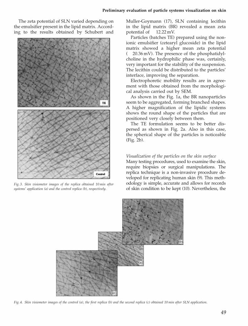
Visualization of the particles on the skin surfaceMany testing procedures, used to examine the skin,require biopsies or surgical manipulations. Thereplica technique is a non-invasive procedure de-veloped for replicating human skin (9). This meth-odology is simple, accurate and allows for recordsof skin condition to be kept (10). Nevertheless, the
Fig. 3. Skin visiometer images of the replica obtained 10 min after
systems’ application (a) and the control replica (b), respectively.
Fig. 4. Skin visiometer images of the control (a), the first replica (b) and the second replica (c) obtained 10 min after SLN application.
49
Preliminary evaluation of particle systems visualization on skin

use of the replica technique requires systematicmethodology development and rigorous adherenceto the experimental protocol, as artefacts can be
readily introduced (9). Therefore, the quality ofreplicas is a limiting factor for the accuracy of thevisualization.
Fig. 5. Skin visiometer images of control (a), the first replica (b) and the second replica (c) obtained 60 min after SLN application.
Fig. 6. Scanning electron micrographs of replicas obtained 1 h after solid lipid nanoparticles (batches BR1 and TE1) application (on the right) and the
corresponding control replicas (on the left).
Perugini et al.
50

If the silicone is spilled over the borders of thering or if the ring’s thickness varies or if airbubbles are present in the gel, the measurementresults are heavily influenced. For these reasons,we have chosen a fluid material enough topenetrate the furrows, but not excessively fluidas to avoid overflowing outside the interestedarea. Furthermore, we exercised a constant pres-sure on the gel applied on the skin in order toobtain replicas with reproducible thickness. Theformation of air bubbles during mixing of the twosilicone components can be avoided by using avacuum pump. In this way, the natural skinprofile can be replicated (12).
Before and after the application of each for-mulation, a replica of the skin was produced inorder to evaluate the interactions between lipidsystems and human skin. The replica was thenanalysed using profilometry and SEM.
The profilometry allows us to have first infor-mation about the distribution of the particles onthe skin texture. Figure 3a and b show the replicaobtained 10 min after systems’ application andthe control replica, respectively. In the first image,
particles seem to be lined up into cutaneousfurrows.
The main barrier of the skin is located withinits uppermost layer, the stratum corneum. Hu-man stratum corneum consists of corneocytes,embedded in a highly organized matrix of lipidlamellae.
Several approaches have been used in order toweaken this skin barrier and in order to improvetransdermal drug delivery (18).
The colloidal systems interacted both withsilicone gel and with skin. To evaluate the SLNpartition between the two compartments, thecontact time between particles and skin wasconsidered. In particular, we obtained replicas10 min (Fig. 4) and 60 min after systems’ applica-tion (Fig. 5). In both cases, on the chosen area, wecarried out two replicas in succession without anew SLN application.
The results highlighted that: (a) if the analysesare carried out 10 min after the system applica-tion, the presence of the TE nanoparticles isclearly evident in the skin visiometer image ofthe first replica (Fig. 4b), while the colloidal
Fig. 7. Scanning electron micrographs of replicas obtained 1 h after solid lipid nanoparticle (batches BR1 and TE1) application (on the right) and the
corresponding control replicas (on the left).
Preliminary evaluation of particle systems visualization on skin
51

particles are less visible in the second one (Fig.4c); b) the opposite is true for the replicasobtained 60 min after the system application(Fig. 5).
This behaviour could be due to the contact timebetween SLN and skin. The stratum corneum isconsidered to be a dense homogeneous film inwhich ceramides, cholesterol and fatty acidscompose lipid bilayers (18). It is likely that afew minutes are not enough for a significantinteraction to take place between the lipid parti-cles and the stratum corneum components. Thus,in our opinion, the first replica, obtained 10 minafter application, could be responsible for colloi-dal systems’ removal and this could explain thepoor presence of SLN in the second replica (Fig.4). On the contrary, a longer time could enable thepenetration of the SLN through the stratumcorneum components (Fig. 5). As it is impossibleto obtain high magnifications using the visi-ometer, to gain more information about the col-loidal system application, we analysed siliconeskin replicas by SEM.
During this study, colloidal systems’ composi-tion and volunteers’ age are different. In order toevaluate the effect of the systems’ composition, weapplied particles made up of different lipids on thesame subject. Particles displayed the same size.
In the Fig. 6, SEM images of replicas obtained 1 hafter the lipid systems’ application are reported,together with the respective control replica.
This magnification (� 50) does not show hugedifferences between the two formulations but byobserving a higher magnification (� 200) a dis-similar interaction with the replica material isnoticeable.
As highlighted in Fig. 7, TE1 nanoparticlesseem to be incorporated into the replica material,while BR1particles rest on the surface. This be-haviour could be due to the system’s composi-tion. The lipophilic phase of the BR formulationcontains phosphatidylcholine, while the TE for-mulation contains cetearyl glucoside, a non-ionicsurfactant. The second formulation is, therefore,more apolar and could be more similar to thesilicone material of the replica.
Fig. 8. Scanning electron micrographs of replicas obtained after application of particle (TE batches) with different sizes (on the right) and the
corresponding control replicas (on the left).
Perugini et al.
52

In order to evaluate the influence of particlesize, systems with the same composition (TEbatches) but different size distributions wereapplied. In this case, the visualization of thereplicas using SEM did not show differencesand the behaviour of the two formulations seemsto be the same, as revealed in Fig. 8.
The behaviour of lipid systems applied on theskin of volunteers of different ages was alsoevaluated. In particular, we applied particles onthe volar forearm of an 8-year-old child and a 38-year-old woman. Systems studied in this casepresented the same composition and micrometricsize (TE2).
In Fig. 9, skin replicas after system applicationand the corresponding controls are shown.
In the case of the child’s skin, the particlesseem to be distributed mainly on the reliefs of thesilicone resina that represent primary and sec-ondary lines of the skin (19, 20).
The behaviour changed when we observed thereplica of the 38-year-old woman under the samemagnification. In this replica, the particle distri-bution seems more homogeneous and particles
are present both in the relief and in the furrows.To explain these results, it is useful to rememberthat skin relief reflects the organization of thedeeper layers of the skin and changes at the levelof the dermis will affect skin relief. The skin of achild and the skin of a woman are very different.The number of primary and secondary linesdecreases progressively with age and the poly-gonal surface delimited by these lines becomesless homogeneous and defined (13). Thesechanges could be the reason for the differentsystem distributions on the skin surface.
Conclusion
The SLN visualization on the skin surfacethrough non-invasive techniques is the mostremarkable innovation of the present work. Skinreplica proved to be a suitable method to studythe morphology of skin surface and colloidalsystems’ distribution.
Profilometry and mainly SEM, thanks to itshigh magnification and field depth, allowed us toevaluate particle distribution on the skin texture
Fig. 9. Scanning electron micrographs of replicas obtained after the application of TE2 systems on the skin of an 8-year-old child and a 38-year-old
woman.
Preliminary evaluation of particle systems visualization on skin
53

and the interaction between particles of differentcompositions and replica silicone.
These results are a starting point for skinpenetration studies. In fact, by observing skinreplicas obtained at scheduled times after sys-tems’ application, it is possible to evaluate theirpenetration through the skin.
Furthermore, the profilometry will be surelyessential for the evaluation of the efficacy of anti-wrinkle actives included in SNL, through an invivo clinical trial, by studying roughness para-meters (maximum roughness, mean depth ofroughness) before, during and after 12 weeks ofdaily systems’ application.
References
1. Medina C, Santos-Martinez MJ, Ra-domski A, Corrigan OI, RadomskiMW. Nanoparticles: pharmacologi-cal and toxicological significance. BrJ Pharmacol 2007; 150: 552–558.
2. Vega-Villa KR, Takemoto JK, YanezJA, Remsberg CM. Clinical toxicitiesof nanocarrier systems. Adv DrugDeliv Rev 2008; 60: 929–938.
3. SCCP. Opinion on safety nanoma-terials in cosmetic products, publicconsultation on the 14th plenary of18 December 2007, European Com-mission 2007: 1–63
4. Bouwstra JA, Ponec M. The skinbarrier in healthy and diseasedstate. Biochim Biophys Acta 2006;1758: 2080–2095.
5. Proksch E, Brandner JM, Jensen JM.The skin: an indispensable barrier.Exp Dermatol 2008; 17: 1063–1072.
6. Walters KA, Roberts MS. The struc-ture and function of skin. In: WaltersKA, ed. Drugs and the pharmaceu-tical Sciences. New York: MarcelDekker, 2002: 1–40.
7. Van den Bergh BAI, Vroom J, Ger-ritsen H, Junginger HE, BouwstraJA. Interactions of elastic and rigidvesicles with human skin in vitro:electron microscopy and two-photon excitation microscopy. Bio-chim Biophys Acta 1999; 1461: 155–173.
8. Muller RH, Radtke M, Wissing SA.Solid lipid nanoparticles (SLN) andnanostructured lipid carriers (NLC)in cosmetic and dermatological pre-
parations. Adv Drug Deliv Rev2002; 1: 131–155.
9. Ryan RL, Hing SAO, Theiler RF. Areplica technique for the evaluationof human skin by scanning electronmicroscopy. J Cutan Pathol 1983; 10:262–276.
10. Hatzis J. The wrinkle and its mea-surement - A skin surface Profilo-metric method. Micron 2004; 35:201–219.
11. Perugini P, Tomasi C, Vettor M,Dazio V, Conti B, Genta I, PavanettoF. Influence of SLN matrix modifica-tion on ‘‘in vitro’’ and ‘‘in vivo’’nanoparticle performances. Int JPharm Pharm Sci 2010; 2: 37–42.
12. Bogner A, Jouneau PH, Thollet G,Basset D, Gauthier C. A history ofscanning electron microscopy de-velopments: towards ‘‘wet-STEM’’imaging. Micron 2007; 38: 390–401.
13. De Paepe K, Lagarde JM, Gall Y,Roseeuw D, Rogiers V. Microreliefof the skin using a light transmis-sion method. Arch Dermatol Res2000; 292: 500–510.
14. Teglia A, Mondelli A. Influence ofcosmetic treatments on the intercor-relations of skin elasticity, hydrationand microrelief. Proceeding of 19thIFSCC Congress, Sydney, 1996
15. Articus K, Khazaka G, Wilhelm KP.The skin visiometer-a photometricdevice for the measurement of skinroughness. In: Fluhr J, Elsner P,Berardesca E, Marbach HI, eds.Bioengineering of the skin: skin sur-face imaging and analysis. Boca Ra-ton: CRC Press, 1997: 59–72.
16. Lee MH, Cha JG, Hong HS, Lee JS,Park SJ, Paik SH, Lee HK. Com-parison of high-resolution ultraso-nography and computed tomogra-phy in the diagnosis of nasalfractures. J Ultrasound Med 2009;8: 717–723.
17. Schubert MA, Muller-Goymann CC.Characterisation of surface-modi-fied solid lipid nanoparticles(SLN): influence of lecithin and non-ionic emulsifier. Eur J Pharm Bio-pharm 2005; 1: 77–86.
18. Honeywell-Nguyen PL, de GraaffAM, Groenink HW, Bouwstra JA.The in vivo and in vitro interactionsof elastic and rigid vesicles withhuman skin. Biochim Biophys Acta2002; 573: 130–140.
19. Martin A. Drug product design. In:Martin A, ed. Physical pharmacy,4th edn. Philadelphia: Lea&Febiger,1993: 538–542.
20. Hashimoto K. New methods for sur-face ultrastructure: comparativestudies of scanning electron micro-scopy, transmission electron micro-scopy and replica method. Int JDermatol 1974; 3: 357–381.
Address:Dr Paola PeruginiDepartment of Pharmaceutical ChemistryUniversity of PaviaVia Taramelli 12, 27100 PaviaItalyTel: 139 03 8298 7363Fax: 139 03 8242 2975e-mail: [email protected]
Perugini et al.
54