melissopalynological investigations of natural honey samples ...
Upload
independentCategory
view
2download
0
Zaid et al. BMC Complementary and Alternative Medicine 2014, 14:509http://www.biomedcentral.com/1472-6882/14/509
RESEARCH ARTICLE Open Access
Potential protective effect of Tualang honey onBPA-induced ovarian toxicity in prepubertal ratSiti Sarah Mohamad Zaid1,3, Shatrah Othman2 and Normadiah M Kassim1*
Abstract
Background: To investigate the potential protective effects of Tualang honey against the toxicity effects inducedby Bisphenol A (BPA) on pubertal development of ovaries.
Methods: This study was conducted on pre-pubertal female Sprague Dawley rats. Animals were divided into fourgroups (n = 8 in each group). Group I was administered with vehicle 0.2 ml of corn oil (Sigma-Aldrich, USA) usingoral gavage daily for six weeks; these animals served as negative control (CO group), Group II was administeredwith BPA suspended in corn oil at 10 mg/kg body weight and served as positive control (PC group), Group IIIwas administered with 200 mg/kg body weight of Tualang honey 30 min before the administration of BPA at10 mg/kg (TH group) while Group IV was administered with 200 mg/kg body weight of Tualang honey 30 minbefore the administration of corn oil (THC group). Body weight of all animals were monitored weekly.
Results: The BPA-exposed animals exhibited disruption of their estrus cycle, while those animals treated with BPAtogether with Tualang honey, exhibited an improvement in percentage of normal estrous cycle. Their ovaries hadlower numbers of atretic follicles compared to the PC group but higher than the CO group.
Conclusions: Tualang honey has a potential role in reducing BPA-induced ovarian toxicity by reducing themorphological abnormalities of the ovarian follicles and improving the normal estrous cycle.
Keywords: Tualang honey, Bisphenol-A, Ovary, Toxicity, Antioxidant
BackgroundIn recent years, environmental toxicants have become aserious health concern. It has caused a rise in concernwhen exposure of endocrine-disrupting chemicals (EDCs)on human and wildlife has effects on the reproduction de-velopment and function [1,2]. One of the EDCs is bisphe-nol A that is widely used in industries as plasticizer forthe production of polycarbonate plastics and epoxyresins [3]. In daily life, bisphenol A is widely used innumerous products including digital media (CDs andDVDs), electronic equipment, automobiles, constructionglazing, sports safety equipment, medical devices, table-ware, reusable bottles, toys, water pipes and food con-tainers [4,5]. The dramatic increase of bisphenol Aexposure in humans were detected in serum, follicularfluid and amniotic fluid [6], fetal serum [7], milk ofnursing mother [8] and urine [9,10]. These findings have
* Correspondence: [email protected] of Anatomy, Faculty of Medicine, University of Malaya, 50603Kuala Lumpur, MalaysiaFull list of author information is available at the end of the article
© 2014 Zaid et al.; licensee BioMed Central. ThCommons Attribution License (http://creativecreproduction in any medium, provided the orDedication waiver (http://creativecommons.orunless otherwise stated.
resulted in both scientific and public interests in assessingbisphenol A as a potential EDCs to health risk.Several studies have reported that bisphenol A could
induce morphological and functional alterations of thefemale genital system, especially in the ovaries at lowpresumably environmentally relevant doses [11]. In vitrostudies claimed that bisphenol A negatively affects gran-ulosa cell steroidogenesis by altering the steroidogenicenzymes and stimulatory effects on vascular endothelialgrowth factor (VEGF) that cause uncontrolled neovascu-larization [5,12]. Subsequently, these findings suggestthat these cells are highly sensitive to bisphenol A. Otherin vivo studies also indicated that neonatal exposure tobisphenol A reduced the pool of primordial follicles inrats [13] while in mice, prenatal exposure to bisphenol Acaused increased antral follicles but reduced corpora luteapercentages [14]. Moreover, development of polycysticovaries (PCOS) [15] and decreased luteinizing hor-mone (LH) were observed [16]. Bisphenol A exposurehas been claimed to promote oxidative stress (OS) and
is is an Open Access article distributed under the terms of the Creativeommons.org/licenses/by/4.0), which permits unrestricted use, distribution, andiginal work is properly credited. The Creative Commons Public Domaing/publicdomain/zero/1.0/) applies to the data made available in this article,

Zaid et al. BMC Complementary and Alternative Medicine 2014, 14:509 Page 2 of 12http://www.biomedcentral.com/1472-6882/14/509
inflammation in women [17]. Treatment with bisphenol Ainduced OS in various tissues of rodent [18] by decreasingantioxidant enzymes and increasing hydrogen peroxideand lipid peroxidation [19]. Several compounds with anti-oxidant properties have been studied extensively as amethod to counter disease-associated OS [20]. With theseconcerns in mind, many studies have been focusing on thepossible therapeutic and preventive measures to counterthe effects of deleterious effects of bisphenol A on thereproductive system.Tualang honey, a wild Malaysian honey, contains high
antioxidant properties [21-24]. It has been shown toameliorate OS in renal and pancreas of streptozotocin-induced diabetic rat [25,26]. It was also reported to pre-vent uterine and vaginal atrophy [27] as well as preventosteoporosis of bones [28], protects rat testis against dam-age and OS induced by cigarette smoke [29]. It was alsoreported to have antiproliferative effects on oral squamouscell carcinomas (OSCC), human osteosarcoma (HOS) [30]and keloid fibroblasts [31]. The beneficial effects in thosepositive findings were claimed to be due to the antioxidantproperties of Tualang honey.The aim of the present study was to investigate the
effects of bisphenol A administered during prepubertalperiod on ovarian follicular development, estrous cyclicityand hormonal profile. Consequently, the protective effectsof Tualang honey against the deleterious effects of bisphe-nol A toxicity on ovarian follicular development, estrouscyclicity and hormonal profile were investigated.
MethodsTualang honey (Agromas, Malaysia)Tualang honey, a wild multifloral honey was supplied byFederal Agricultural Marketing Authority (FAMA), underMinistry of Agriculture and Agro-Based Industry, Malaysia.It was collected from Apis dorsata’s beehive built on a gianttree (tall and big), Koompassia excels (locally known asTualang tree) that grows in the rain forest of Kedah,Malaysia. The honey was filtered to remove solid parti-cles, concentrated in an oven at 40°C and subjected to γirradiation at 25 kGy at Sterilgamma (M) Sdn. Bhd.(Selangor, Malaysia). The water concentration of thehoney was standardized by FAMA at 18%.
Animal and experimental designThirty-two prepubertal female Sprague Dawley rats aged21 (P21) days were obtained from the Animal Husbandry,Faculty of Medicine, University of Malaya. The experi-mental design and procedures were conducted under pro-tocols in compliance with EU Directive 2010/63/EU thatapproved by the Animal Care and Committee (ACUC) ofUniversity of Malaya. The animals were maintained underthe standard laboratory conditions (temperature 25 ± 2°C,50 ± 15% relative humidity and normal photoperiod of
12 h dark and 12 h light) with free access to rat commer-cial pellet diet (Gold Coin Feedmills Pte. Ltd, Malaysia)and water ad libitum. To minimize additional exposuresto EDCs, water was supplied in glass bottles with rubberstoppers surrounded by a steel ring. They were acclima-tized to the laboratory environment for a week prior tothe commencement of the experiments. At P28, the an-imals were weighed and randomly divided into fourgroups (n = 8 in each group). Group I was administeredwith vehicle 0.2 ml of corn oil (Sigma-Aldrich, USA)and served as negative control (CO group). Group IIwas administered with BPA (Sigma Aldrich, USA) sus-pended in corn oil at 10 mg/kg body weight and servedas positive control (PC group). Group III was adminis-tered with 200 mg/kg body weight of Tualang honey30 min before the administration of BPA at 10 mg/kg(TH group). Group IV was administered with 200 mg/kg body weight of Tualang honey 30 min before the ad-ministration of corn oil (THC group). The treatment wasperformed in the morning (between 09:00 and 10:00 AM)once daily by oral gavage (to mimic the most likely routeof human exposure) for six consecutive weeks.Tualang honey was freshly prepared every morning (to
avoid oxidation of the antioxidants) by dissolving in de-ionized water. Justification for dose selection of BPA wasbased on previous studies (influenced morphological andbiochemical parameters in reproductive system) [32-35].The dose of Tualang honey used was based on previousstudy which showed positive biological effects on femalereproductive organs and the dose used was equivalent toroutine/normal dose (one table spoon) in adult human[27]. Throughout the administration period, daily bodyweight was recorded while vaginal smear was taken to de-termine the estrous phase. After the last treatment, theanimals were sacrificed. Venous blood samples werecollected by direct heart puncture under deeply ketamineanaesthesia (Troy Laboratories, Australia). Serum fromblood samples were store at −80°C until analysis. Theovaries were weighed and immediately fixed in 10% buff-ered formalin for histopathological analysis.
Histopathological analysisThe harvested ovaries were fixed by immersion in 10%buffered formalin for 24 hours. Subsequently, the ovarieswere hydrated in a graded series of ethanol, clearance byxylene, embedded in paraffin and sectioned. Serial sec-tions (5 μm thickness) at were obtained and mountedonto glass slide, deparaffinized in xylene, stained withhematoxylin and eosin (Sigma Aldrich, USA) and dehy-drated in a graded series of ethanol, cleared in xylene andmounted with Canada Balsam (Sigma-Aldrich). Histo-pathological changes and morphometric analysis in eachovarian section was performed on 62 total fields areasthat were measured with grid lines using NIS-Elements

Zaid et al. BMC Complementary and Alternative Medicine 2014, 14:509 Page 3 of 12http://www.biomedcentral.com/1472-6882/14/509
software. All sections were observed under a light micro-scope (Olympus CH-B145-2) attached to image analyzer(NIS-Elements Advanced Research, Nikon, Japan).
Classification and quantification of ovarian folliclesOvarian follicles were classified and counted accordingto the criteria described by Zhuang et al. [36] as follows:
i) Primary follicles: an oocyte surrounded by a singlelayer of cuboidal granulose cells in part or in entirety.
ii) Secondary follicles: surrounded by more than one layerof cuboidal granulose cells with no visible antrum.
iii)Antral follicles: identified by the presence of anantral space and cumulus granulose cell layer.
iv) Corpus luteum: formed only after ovulation andfilled with lutein cells.
v) Atretic follicles: had abnormal structures such asinspissated follicular fluid, degenerated egg,disorganized and thickened granulosa layers or filledwith organizing fibrinous material in the antrum.
Assessment of estrous cyclesEstrous cycles of rats were determined by daily observa-tion of vaginal smears (between 09:00 and 10:00 AM).Vaginal secretion was collected using a plastic pipettefilled with approximately 0.2 ml of normal saline (NaCl0.9%). The tip of the pipette was inserted into the vaginato a depth of 2–5 mm. Then, the normal saline wasflushed into the vagina and returned into the pipette bygently squeezing and releasing the bulb of the pipette.These steps were repeated for three times before thesample was collected. Subsequently, a drop of the cellsuspension was smeared onto a labelled glass slide. Un-stained smear was observed under a light microscope,without the use of the condenser lens, with 10 x and 40 xobjective lenses. The cytological appearance of vaginalsmears determined the phase of the estrous cycle asfollows:The proestrous phase (twelve to fourteen hours) was
defined by the predominance of nucleated epithelial cells;an estrous phase (twenty-five to twenty-seven hours) pri-marily consisted of anucleated cornified cells; a metestrousphase (six to eight hours) consisted of the same proportionamong leucocytes, cornified and nucleated epithelial cells;and the diestrous phase (fifty-five to fifty-seven hours) pri-marily consisted of a predominance of leucocyctes.The criteria implemented for determining cycle patterns:
1) Regular cycle (RC): denoted a 4 to 5-day estrouscycle in which the estrous phase was observed atleast twice during the sampling period.
2) Persistent diestrous: or prolonged diestrous havefour or more days of diestrous phase during mostof the cycles.
Assay of serum estradiol, FSH, LH and progesteroneAfter two hours at room temperature, clotted bloodsamples were centrifuged at 1,000 x g for 15 minutes,extracted serum were stored at −80°C until subsequentanalysis. ELISA (Cusabio, USA) was used for measure-ment of the circulating levels of 17β-estradiol (E2), folliclestimulating hormones (FSH), luteinizing hormones (LH)and progesterone (P4). Each sample was run in duplicate.In brief, 50 μl each of the standards, control and serumsamples were added to respective wells coated with estra-diol (E2), follicle stimulating hormones (FSH), luteinizinghormones (LH) or progesterone (P4) antibody and incu-bated with 50 μl of enzyme conjugate for two hours at37°C in oven (Echo Therm, USA). Subsequently, thewells were rinsed three times with distilled water and50 μl of the substrate was added and incubated for15 minutes at 37°C. Reactions were terminated using50 μl of stop solution. The optical density (O.D) wasmeasured at 450 nm using a microplate reader (BioTek,USA). For determination of each hormonal level, a standardcurve was constructed by plotting a graph of the absorb-ance of each reference standard against its correspondinglevels. The inter-assay and intra-assay variations were foundto be less than 15%.
Statistical analysisAll statistical evaluations were performed with StatisticalPackage for Social Sciences (SPSS Inc. Chicago, Illinois,USA version 18.0 for windows). Parametric variables (bodyweight, ovary weight, hormonal assay and follicular count-ing) were analyzed using one-way analysis of variance(ANOVA) followed by Bonferroni test for multiple com-parisons to identify significant different between groups.Estrous cycle phase was analyzed with Fisher’s exact prob-ability test. Values are reported as mean ± S.E.M. P < 0.05was considered significant.
ResultsBody weight and ovary weightAnalysis of body weight and the weight of selected organsin toxicological studies are sensitive indicators for adverseeffects of treatments. Analysis of relative organ weight(normalized to absolute organ weight to body weight) isan important and accurate analytical endpoint for identifi-cation of harmful effects of chemicals on the organ weights[37]. The value of body weight gain, changes in bodyweight, ovary wet weight and ovary relative weight inexperimental groups are shown in Table 1. The meanbody weight gain in each group was obtained from thedifference in values between the final body weight (at P71)and the initial body weight (at P28). This body weight gainwas divided by the final body weight (at P71) to obtainthe percentage to obtain the percentage of body weightchange. The changes in body weight in PC group and

Table 1 Body weight and ovary weight in experimental groups
Group Body weight gain (g) % changes in body weight Ovary wet weight (g) Ovary relative weight (mg/g body weight)
CO 78.88 ± 14.61 48.15 ± 6.60 36.88 ± 1.88 0.24 ± 0.01b
PC 99.25 ± 9.90 56.52 ± 3.49 49.38 ± 1.13 0.29 ± 0.12a
TH 92.50 ± 4.62 56.12 ± 1.94 40.00 ± 1.64 0.24 ± 0.02b
THC 89.5 ± 10.64 52.39 ± 3.47 38.75 ± 0.82 0.23 ± 0.01bb
The data were presented as mean ± S.E.M. Ovary relative weight with different superscripts are significantly different. aP < 0.05 versus negative control group (CO)and bP < 0.05, bbP < 0.01 versus positive control group (PC). There were no significant changes in body weight between all groups.CO = Negative control group administered with vehicle (corn oil).PC = Positive control group administered with BPA at 10 mg/kg body weight.TH = Tualang honey group administered with Tualang honey at 200 mg/kg body weight + BPA at 10 mg/kg body weight.THC = Tualang honey control group administered with Tualang honey at 200 mg/kg body weight.
Zaid et al. BMC Complementary and Alternative Medicine 2014, 14:509 Page 4 of 12http://www.biomedcentral.com/1472-6882/14/509
TH group were slightly higher than CO group. However,the difference was not significant. It was also noted thattreatment with Tualang honey in BPA-exposed rats cannotprevent increase of body weight. The changes in bodyweight in THC group were comparable to CO group. Theovary relative weight in PC group was significantly higherthan CO group. Interestingly, treatment with Tualanghoney in BPA-exposed rat (TH group) showed significantreduction in body weight compared to the PC group.
Estrous cycleTable 2 shows the estrous cycle patterns in all experimen-tal groups. In CO group and THC groups, all rats main-tained in normal estrous cycle. In PC group, only 37.5%presented normal estrous cycles and a higher percentageshowed persistent diestrous (62.5%). These estrous cyclespattern were significantly different with both CO groupand THC group. However, treatment with Tualang honeyin BPA-exposed rats showed higher percentage in normalestrous cycle (62.5%) and lower in persistent diestrous(37.5%) compared to PC group but not statisticallydifferent.
Follicle stimulating hormone (FSH) and luteinizinghormone (LH)Serum FSH and LH levels were significantly reduced in PCgroup and TH group compared to CO group and THCgroup. It was also noted that Tualang honey treatment on
Table 2 Pattern of estrous cycle of rats from day 41 to day-70
Group Number of rats with normal c
CO 8 (100%)b
PC 3 (37.5%)a
TH 5 (62.5%)
THC 8 (100%)b
The data were presented as number (percentage). Numbers with different superscrbP < 0.05 versus positive control group (PC).CO = Negative control group administered with vehicle (corn oil).PC = Positive control group administered with BPA at 10 mg/kg body weight.TH = Tualang honey group administered with Tualang honey at 200 mg/kg body wTHC = Tualang honey control group administered with Tualang honey at 200 mg/kg
BPA-exposed rats cannot prevent the reduction of bothhormones levels (Figures 1 and 2).
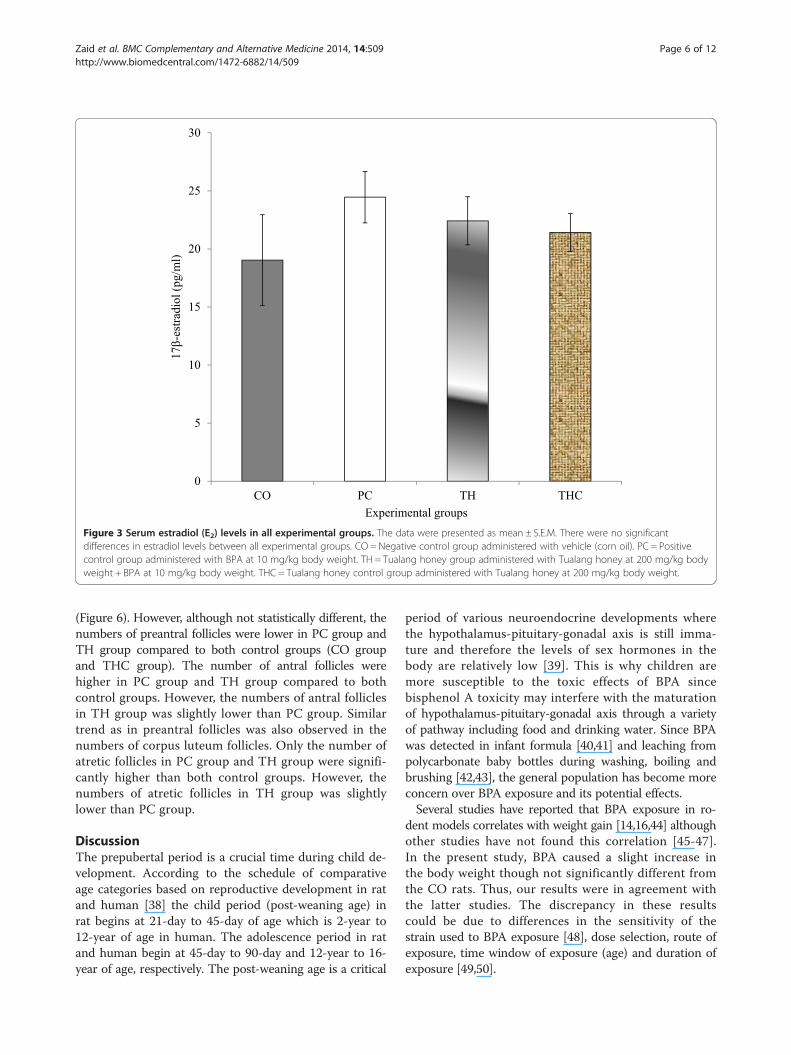
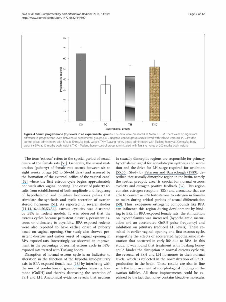
17β-Estradiol (E2) and progesterone (P4)There were no significant change with regards to both E2and P4 levels in all experimental groups. However, the E2level in PC group and TH group were slightly increasedcompared to the CO group (vehicle-treated). The reduc-tion in the P4 level as seen in the PC group (BPA-treated)was slightly prevented with Tualang honey treatment(Figures 3 and 4).
Ovarian follicular developmentOvarian morphology analysis was conducted by qualitativeand quantitative methods. Generally, ovaries from allgroups displayed all stages of follicular development.Qualitative histological examination revealed that THCgroup exhibited healthy ovaries comparable to the ovariesof the negative control group (Figure 5). On the otherhand, both the PC group and TH group showed someabnormalities of the ovaries with large antral-like folliclesthat did not arrive at ovulation and the presence of atreticcystic-like follicles. There was less number of corpus luteaobserved. However, the degree of abnormalities was moreapparent in the ovaries of the PC group compared to theTH group. Ovarian follicles at different stages were foundin the ovaries of experimental groups. No significant dif-ference was observed in the number of preantral, antralfollicles and corpora lutea among all experimental groups
in all experimental groups
ycles (%) Persistent diestrous (%)
0b
5 (62.5%)a
3 (37.5%)
0b
ipts are significantly different. aP < 0.05 versus negative control group (CO) and
eight + BPA at 10 mg/kg body weight.body weight.

bbb
aaa aaa
bbb
0
10
20
30
40
50
60
70
80
90
CO PC TH THC
Fo
llic
le s
tim
ula
tin
g h
orm
on
e (m
IU/m
l)
Experimental groups
Figure 1 Serum follicle stimulating hormone (FSH) in all experimental groups. The data were presented as Mean ± S.E.M. Mean withdifferent superscripts are significantly different. aP < 0.001 versus negative control group (CO) and bP < 0.001 versus positive control group (PC).CO = Negative control group administered with vehicle (corn oil). PC = Positive control group administered with BPA at 10 mg/kg body weight.TH = Tualang honey group administered with Tualang honey at 200 mg/kg body weight + BPA at 10 mg/kg body weight. THC = Tualang honeycontrol group administered with Tualang honey at 200 mg/kg body weight.
bb
aa aa
bb
0
2
4
6
8
10
12
14
CO PC TH THC
Lu
tein
izin
g h
orm
on
e (m
lU/m
l)
Experimental groups
Figure 2 Serum luteinizing hormone (LH) levels in all experimental groups. The data were presented as Mean ± S.E.M. Mean with differentsuperscripts are significantly different aP < 0.01 versus negative control group (CO) and bP < 0.01 versus positive control group (PC). CO = Negativecontrol group administered with vehicle (corn oil). PC = Positive control group administered with BPA at 10 mg/kg body weight. TH = Tualanghoney group administered with Tualang honey at 200 mg/kg body weight + BPA at 10 mg/kg body weight. THC = Tualang honey control groupadministered with Tualang honey at 200 mg/kg body weight.
Zaid et al. BMC Complementary and Alternative Medicine 2014, 14:509 Page 5 of 12http://www.biomedcentral.com/1472-6882/14/509
0
5
10
15
20
25
30
CO PC TH THC
17
β-e
stra
dio
l (p
g/m
l)
Experimental groups
Figure 3 Serum estradiol (E2) levels in all experimental groups. The data were presented as mean ± S.E.M. There were no significantdifferences in estradiol levels between all experimental groups. CO = Negative control group administered with vehicle (corn oil). PC = Positivecontrol group administered with BPA at 10 mg/kg body weight. TH = Tualang honey group administered with Tualang honey at 200 mg/kg bodyweight + BPA at 10 mg/kg body weight. THC = Tualang honey control group administered with Tualang honey at 200 mg/kg body weight.
Zaid et al. BMC Complementary and Alternative Medicine 2014, 14:509 Page 6 of 12http://www.biomedcentral.com/1472-6882/14/509
(Figure 6). However, although not statistically different, thenumbers of preantral follicles were lower in PC group andTH group compared to both control groups (CO groupand THC group). The number of antral follicles werehigher in PC group and TH group compared to bothcontrol groups. However, the numbers of antral folliclesin TH group was slightly lower than PC group. Similartrend as in preantral follicles was also observed in thenumbers of corpus luteum follicles. Only the number ofatretic follicles in PC group and TH group were signifi-cantly higher than both control groups. However, thenumbers of atretic follicles in TH group was slightlylower than PC group.
DiscussionThe prepubertal period is a crucial time during child de-velopment. According to the schedule of comparativeage categories based on reproductive development in ratand human [38] the child period (post-weaning age) inrat begins at 21-day to 45-day of age which is 2-year to12-year of age in human. The adolescence period in ratand human begin at 45-day to 90-day and 12-year to 16-year of age, respectively. The post-weaning age is a critical
period of various neuroendocrine developments wherethe hypothalamus-pituitary-gonadal axis is still imma-ture and therefore the levels of sex hormones in thebody are relatively low [39]. This is why children aremore susceptible to the toxic effects of BPA sincebisphenol A toxicity may interfere with the maturationof hypothalamus-pituitary-gonadal axis through a varietyof pathway including food and drinking water. Since BPAwas detected in infant formula [40,41] and leaching frompolycarbonate baby bottles during washing, boiling andbrushing [42,43], the general population has become moreconcern over BPA exposure and its potential effects.Several studies have reported that BPA exposure in ro-
dent models correlates with weight gain [14,16,44] althoughother studies have not found this correlation [45-47].In the present study, BPA caused a slight increase inthe body weight though not significantly different fromthe CO rats. Thus, our results were in agreement withthe latter studies. The discrepancy in these resultscould be due to differences in the sensitivity of thestrain used to BPA exposure [48], dose selection, route ofexposure, time window of exposure (age) and duration ofexposure [49,50].
0
10
20
30
40
50
60
70
80
CO PC TH THC
Pro
ges
tero
ne
(ng/m
l)
Experimental groups
Figure 4 Serum progesterone (P4) levels in all experimental groups. The data were presented as Mean ± S.E.M. There were no significantdifference in progesterone levels between all experimental groups. CO= Negative control group administered with vehicle (corn oil). PC = Positivecontrol group administered with BPA at 10 mg/kg body weight. TH = Tualang honey group administered with Tualang honey at 200 mg/kg bodyweight + BPA at 10 mg/kg body weight. THC = Tualang honey control group administered with Tualang honey at 200 mg/kg body weight.
Zaid et al. BMC Complementary and Alternative Medicine 2014, 14:509 Page 7 of 12http://www.biomedcentral.com/1472-6882/14/509
The term ‘estrous’ refers to the special period of sexualdesire of the female rats [51]. Generally, the sexual mat-uration (puberty) of female rats occurs between six toeight weeks of age (42 to 56-old days) and assessed bythe formation of the external orifice of the vaginal canal[52] where the first estrous cycle begins approximatelyone week after vaginal opening. The onset of puberty re-sults from establishment of both amplitude and frequencyof hypothalamic and pituitary hormones pulses thatstimulate the synthesis and cyclic secretion of ovariansteroid hormone [51]. As reported in several studies[11,14,16,44,50,53,54], estrous cyclicity was disruptedby BPA in rodent models. It was observed that theestrous cycles became persistent diestrus, persistent es-trous or ultimately to acyclicity. BPA-exposed rodentswere also reported to have earlier onset of pubertybased on vaginal opening. Our study also showed per-sistent diestrus and earlier onset of vaginal opening inBPA-exposed rats. Interestingly, we observed an improve-ment in the percentage of normal estrous cycle in BPA-exposed rats treated with Tualang honey.Disruption of normal estrous cycle is an indicator to
alteration in the function of the hypothalamic-pituitaryaxis in BPA-exposed female rats [16] by interfering withthe normal production of gonadotrophin releasing hor-mone (GnRH) and thereby decreasing the secretion ofFSH and LH. Anatomical evidence reveals that neurons
in sexually dimorphic regions are responsible for primaryhypothalamic signal for gonadotropin synthesis and secre-tion and the drive for LH surge required for ovulation[55,56]. Study by Petersen and Barraclough (1989), de-scribed that sexually dimorphic region in the brain, namelythe rostral preoptic area, is crucial for normal estrouscyclicity and estrogen positive feedback [57]. This regioncontains estrogen receptors (ERs) and aromatase that areable to convert in situ testosterone to estrogen in femalesor males during critical periods of sexual differentiation[58]. Thus, exogenous estrogenic compounds like BPAcan influence this region during development by bind-ing to ERs. In BPA-exposed female rats, the stimulationon hypothalamus was increased (hypothalamic matur-ation and an accelerated GnRH pulse frequency) andinhibition on pituitary (reduced LH levels). These re-sulted in earlier vaginal opening and first estrous cycle,suggesting the effects of accelerated hypothalamic mat-uration that occurred in early life due to BPA. In thisstudy, it was found that treatment with Tualang honeycould hinder the disruption in normal estrous cycle viathe reversal of FSH and LH hormones to their normallevels, which is reflected in the normalization of GnRHproduction in the brain. These results are also in linewith the improvement of morphological findings in theovarian follicles. All these improvements could be ex-plained by the fact that honey contains bioactive molecules

A B
C D
AT
CL
A
PA
AT
CL
A
PA
AT
A
PA
CL
PO
CL
AT
PA
Figure 5 Photomicrographs of representative ovarian section in experimental groups. A) CO group B) PC group C) TH group D) THCgroup. Staining with H&E. PA: Preantral; A: Antral; At: Atretic; CL: Corpus luteum; PO: Preovulatory. Scale bar = 100 μm. CO = Negative controlgroup administered with vehicle (corn oil). PC = Positive control group administered with BPA at 10 mg/kg body weight. TH = Tualang honeygroup administered with Tualang honey at 200 mg/kg body weight + BPA at 10 mg/kg body weight. THC = Tualang honey control groupadministered with Tualang honey at 200 mg/kg body weight.
Zaid et al. BMC Complementary and Alternative Medicine 2014, 14:509 Page 8 of 12http://www.biomedcentral.com/1472-6882/14/509
that exert protective effects via their estrogenic properties,namely the flavonols. Flavonols are phytochemicals andoriginate from a subfamily of flavonoids with many bio-logical activities. Quercetin and kaempferol are the twomain naturally-occuring flavonols which share structuralsimilarities with 17β-estradiol, and therefore have potentialestrogenic effects [59]. Thus, their beneficial effect in theimprovement of the function of hypothalamic-pituitary axisis possibly due to their ability to bind to ERs in rostral pre-optic region of brain, competing with other xenoestrogeniclike BPA. This hypothesis of the protective effects of flavo-nols via their estrogenic mechanism is also supported byprevious studies [60,61]. Indeed, it is possible that both fla-vonols in Tualang honey are accountable as the modulators
of xenoestrogenic effects of BPA in the female reproductivefunctions.In this study, decreased levels of serum LH were ob-
served in BPA-treated rats compared to control group.The decrease in serum LH level may result in the forma-tion of cystic follicle (anovulation follicle) and consequentlylack of corpora lutea, reflecting reduction in the progester-one levels [12]. Cystic follicle is formed from anovulatoryfollicle surrounded by thin layers of granulose cells withnon-detectable theca cell layers [62]. Others claimed thatlarge antral-like follicles do not support ovulation processin the ovary [11]. Our results is in agreement with theprevious study that the total numbers of follicles inBPA-exposed rats have a positive correlation with the

bb
aa
a
bbb
0
2
4
6
8
10
12
Pre-antral Antral Corpus luteum Atretic
Nu
mber
of
fo
llic
les/
un
it a
rea
Follicle
CO
PC
TH
THC
Figure 6 Number of different types of follicles in all experimental groups. The data were presented as Mean ± S.E.M. Means with differentsuperscripts are significantly different. aP < 0.05, aaP < 0.01 versus negative control group (CO) and bbP < 0.01, bbbP < 0.001 versus positive controlgroup (PC). There were no significant differences in pre-antral, antral and corpus luteum between all groups. CO = Negative control group administeredwith vehicle (corn oil). PC = Positive control group administered with BPA at 10 mg/kg body weight. TH = Tualang honey group administeredwith Tualang honey at 200 mg/kg body weight + BPA at 10 mg/kg body weight. THC = Tualang honey control group administered with Tualanghoney at 200 mg/kg body weight.
Zaid et al. BMC Complementary and Alternative Medicine 2014, 14:509 Page 9 of 12http://www.biomedcentral.com/1472-6882/14/509
ovarian weight [63]. The number of preantral folliclesin BPA-exposed rat was reduced or less compared tocontrol rat, indicating that exposure to BPA interferedwith the normal development of growing follicles in theovary. This result is similar to previous study that usedprepubertal female rat as an animal model [34]. Evenwith the disruption in normal ovulation process and inpersistent diestrous phase, some follicles still progressedbut without ovulation, followed by atresia, as reflected bythe increase in the number of atretic follicles in BPA-exposed rats compared to the control rat. Indeed, our re-sults showed a trend towards an increase in the number ofantral follicle as well as a decrease in the number ofcorpora lutea in BPA-treated rats compared to controlgroup. These results are in agreement with previousfinding that also showed reduction in the number of ovu-lated oocytes [14]. As for the BPA-exposed rats treatedwith Tualang honey, the morphological abnormalities ofthe ovary were reduced and the number of atretic follicleswas also slightly lower.Lipid peroxidation and generation of free radicals are
the two major contributors for the toxicity effects of BPA[35]. Studies have also reported that BPA was shown to in-duce OS in different tissues rodents [19] and could pro-mote OS and inflammation in women [17]. In particular,OS plays an important role in the pathologies of female
infertility problems that influence the entire reproductivelife span by affecting oocyte maturation, ovarian steroide-genesis, ovulation, luteolysis and luteal maintenance inpregnancy [64]. The metabolite groups of BPA such as qui-nones (phenolic precursors), metal complexes (complexors),aromatic nitro compounds (reduced hydroxylamine andnitroso derivatives) and conjugated imines (iminium spe-cies) can incorporate with electron transfer (ET) to induceROS [65]. Small quantities of oxidized metabolite can actas catalyst in a redox manner with generation of largequantities of ROS that result in OS. Thus, ET-ROS-OSframework provide reasonable and convenience evidencethat BPA is a toxic agent [65]. OS is an important exacer-bating factor for diverse pathological processes whichcan lead to deoxyribonucleic acid (DNA) damage, mu-tations, cellular injury, oncogenesis and aging process[66]. The genotoxicity effects of BPA have been widelytested in vitro and in vivo studies [67,68]. The cometassays shows that BPA caused DNA fragmentation inblood lympocytes and structural chromosome aberrationsin bone-marrow cells [69]. Other studies claimed thatBPA induce lipid peroxidation, DNA fragmention inspermatozoa and up-regulate clusterin expression in atro-phic prostate epithelial cells [70], DNA damage in germcells [71] and MCF-7 cells [72]. Recently, Tualang honeywas reported for its anticancer activity in breast cancer cell

Zaid et al. BMC Complementary and Alternative Medicine 2014, 14:509 Page 10 of 12http://www.biomedcentral.com/1472-6882/14/509
lines via the upregulation of double strand DNA repairenzymes that preserve cellular DNA integrity [73] or bypromoting apoptotic cell death [74]. In ultraviolet (UV) Bradiation study, treatment with Tualang honey to PAM212cells can result in the reduction of the number of cyclo-butane pyrimidine dimers and 8-oxo-dG-positive cells(biomarkers of DNA damage) due to improvement inDNA repair [75]. In agreement with these findings, studieson Buckwheat honey [76] and honey from arid regions[77] also demonstrated their protective effects on DNAdamage. In general, the reducing power, DPPH radicalscavenging activity, metal chelating activity and hydroxylradical scavenging activities are all attributed to the anti-oxidants mechanisms in honey in preventing DNA damage.Interestingly, Tualang honey has also been claimed as
an ideal antioxidant with both aqueous and lipophilicproperties that enable it to easily penetrate biologicalmembranes and consequently act at different cellularlevels of the cells [22]. The combination of endogenousantioxidants and the antioxidants from natural productsplays an important role in the protective antioxidantmechanism in the body system against OS. When BPA-exposed rats are treated with Tualang honey, there was amarked improvement in the morphological abnormal-ities in ovarian follicules. In addition to the reducednumber of atretic follicles, there was also improvementin the percentage of animals with normal estrous cyclecompared to BPA-exposed rats without Tualang honeytreatment. These results illustrated the ability of Tua-lang honey as a potential antigenotoxic mediator that isable to reduce ovarian toxicity, hence demonstrating itscontribution to the protective mechanism against thegenotoxic effects induced by BPA.
ConclusionsIn conclusion, Tualang honey has a potential role inreducing BPA-induced ovarian toxicity by reducing themorphological abnormalities of the ovarian follicles andimproving the normal estrous cycle.
AbbreviationsEDCs: Endocrine-disrupting chemicals; BPA: Bisphenol-A; VEGF: Vascularendothelial growth factor; PCOS: Polycystic ovaries; LH: Luteinizing hormone;FSH: Follicle stimulating hormone; E2: 17β-Estradiol; P4: Progesterone;OS: Oxidative stress; DNA: Deoxyribonucleic acid; DPPH: 1,1-diphenyl-2-pic-rylhydrazyl; OSCC: Oral squamous cell carcinoma; HOS: Human osteosarcoma;CO: Negative control group; PC: Positive control group; TH: Tualang honeygroup; THC: Tualang honey control group.
Competing interestsThe authors declare that they have no competing interests.
Authors’ contributionsSMZ (1st author) carried out the lab work, analysis the data and drafted themanuscript. NMK (3rd author) designed and led the whole study, main-supervisorto SMZ, principal investigator of grant, provided guidance in histological lab workand data analysis and finalized the revised manuscript. SO (2nd author) read andrevised the manuscript. All authors read and approved the final manuscript.
Authors’ informationNMK (PhD) is a Professor in Deparment of Anatomy, Faculty of Medicine,University of Malaya. Her areas of expertise including developmentalanatomy, cytology (histology, electron microscopy, immunocytochemistry)and toxicology (endorine disrupting chemicals).SO (PhD) is a senior lecturer in Department of Molecular Medicine, Faculty ofMedicine, University of Malaya. Her areas of expertise including cellularimmunology and drug discovery.SMZ (Masters) is a postgraduate student in Department of Anatomy, Facultyof Medicine, University of Malaya. Her research project focus on preventingor ameliorating effects of natural products on BPA-induced reproductivetoxicity in rats.
AcknowledgementsThe authors would like to acknowledge the University of Malaya for providing apostgraduate research grant (No: PG087-2012B), Universiti Putra Malaysia andMinistry of Higher Education for the financial assistance to the first author SitiSarah Mohamad Zaid. Sincere thanks to all staff of the Department of Anatomy,Faculty of Medicine, University of Malaya.
Author details1Department of Anatomy, Faculty of Medicine, University of Malaya, 50603Kuala Lumpur, Malaysia. 2Department of Molecular Medicine, Faculty ofMedicine, University of Malaya, 50603 Kuala Lumpur, Malaysia. 3Departmentof Environmental Sciences, Faculty of Environmental Studies, 43400, UPMSerdang, Selangor, Malaysia.
Received: 23 May 2014 Accepted: 9 December 2014Published: 17 December 2014
References1. Borgeest C, Greenfeld C, Tomic D, Flaws JA: The effects of endocrine
disrupting chemicals on the ovary. Front Biosci 2002, 7:1941–1948.2. Richter CA, Birnbaum LS, Farabollini F, Newbold RR, Rubin BS, Talsness CE,
Vandenbergh JG, Walser-Kuntz DR, vom Saal FS: In vivo effects of bisphenol Ain laboratory rodent studies. Reprod Toxicol 2007, 24(2):199–224.
3. Staples CA, Dorn PB, Klecka GM, O’Block ST, Harris LR: A review of theenvironmental fate, effects, and exposures of bisphenol A. Chemosphere1998, 36(10):2149–2173.
4. Braun JM, Hauser R: Bisphenol A and children’s health. Curr Opin Pediatr2011, 23(2):233–239.
5. Grasselli F, Baratta L, Baioni L, Bussolati S, Ramoni R, Grolli S, Basini G:Bisphenol A disrupts granulosa cell function. Domest Anim Endocrinol2009, 39(1):34–39.
6. Ikezuki Y, Tsutsumi O, Takai Y, Kamei Y, Taketani Y: Determination ofbisphenol A concentrations in human biological fluids reveals significantearly prenatal exposure. Hum Reprod 2002, 17(11):2839–2841.
7. Kuroda N, Kinoshita Y, Sun Y, Wada M, Kishikawa N, Nakashima K, Makino T,Nakazawa H: Measurement of bisphenol A levels in human blood serumand ascitic fluid by HPLC using a fluorescent labeling reagent. J PharmBiomed Anal 2003, 30(6):1743–1749.
8. Sun Y, Irie M, Kishikawa N, Wada M, Kuroda N, Nakashima K: Determinationof bisphenol A in human breast milk by HPLC with column-switchingand fluorescence detection. Biomed Chromatogr 2004, 18(8):501–507.
9. Yang M, Kim SY, Chang SS, Lee IS, Kawamoto T: Urinary concentrations ofbisphenol A in relation to biomarkers of sensitivity and effect andendocrine-related health effects. Environ Mol Mutagen 2006, 47(8):571–578.
10. Calafat AM, Kuklenyik Z, Reidy JA, Caudill SP, Ekong J, Needham LL: Urinaryconcentrations of bisphenol A and 4-nonylphenol in a human referencepopulation. Environ Health Perspect 2005, 113(4):391–395.
11. Adewale HB, Jefferson WN, Newbold RR, Patisaul HB: Neonatal bisphenol-aexposure alters rat reproductive development and ovarian morphologywithout impairing activation of gonadotropin-releasing hormone neurons.Biol Reprod 2009, 81(4):690–699.
12. Zhou W, Liu J, Liao L, Han S: Effect of bisphenol A on steroid hormoneproduction in rat ovarian theca-interstitial and granulosa cells. Mol CellEndocrinol 2008, 283(1–2):12–18.
13. Rodriguez HA, Santambrosio N, Santamaria CG, Munoz-de-Toro M, Luque EH:Neonatal exposure to bisphenol A reduces the pool of primordial folliclesin the rat ovary. Reprod Toxicol 2010, 30(4):550–557.

Zaid et al. BMC Complementary and Alternative Medicine 2014, 14:509 Page 11 of 12http://www.biomedcentral.com/1472-6882/14/509
14. Markey CM, Coombs MA, Sonnenschein C, Soto AM: Mammalian developmentin a changing environment: exposure to endocrine disruptors reveals thedevelopmental plasticity of steroid-hormone target organs. Evol Dev2003, 5(1):67–75.
15. Kato H, Ota T, Furuhashi T, Ohta Y, Iguchi T: Changes in reproductiveorgans of female rats treated with bisphenol A during the neonatalperiod. Reprod Toxicol 2003, 17(3):283–288.
16. Rubin BS, Murray MK, Damassa DA, King JC, Soto AM: Perinatal exposure tolow doses of bisphenol A affects body weight, patterns of estrous cyclicity,and plasma LH levels. Environ Health Perspect 2001, 109(7):675–680.
17. Yang YJ, Hong YC, Oh SY, Park MS, Kim H, Leem JH, Ha EH: Bisphenol Aexposure is associated with oxidative stress and inflammation inpostmenopausal women. Environ Res 2009, 109(6):797–801.
18. Kabuto H, Hasuike S, Minagawa N, Shishibori T: Effects of bisphenol A onthe metabolisms of active oxygen species in mouse tissues. Environ Res2003, 93(1):31–35.
19. Bindhumol V, Chitra KC, Mathur PP: Bisphenol A induces reactive oxygenspecies generation in the liver of male rats. Toxicology 2003, 188(2–3):117–124.
20. Poeggeler B, Reiter RJ, Tan DX, Chen LD, Manchester LC: Melatonin,hydroxyl radical-mediated oxidative damage, and aging: a hypothesis.J Pineal Res 1993, 14(4):151–168.
21. Al-Mamary M, Al-Meeri A, Al-Habori M: Antioxidant activities and totalphenolics of different types of honey. Nutr Res 2002, 22(9):1041–1047.
22. Aljadi AM, Kamaruddin MY: Evaluation of the phenolic contents andantioxidant capacities of two Malaysian floral honeys. Food Chem 2004,84(4):513–518.
23. Mahaneem M, Sirajudeen KNS, Swamy M, Nik SY, Siti AS: Studies onantioxidant properties of Tualang honey. Afr J Tradit Complement AlternMed 2010, 7(1):59–63.
24. Khalil MI, Alam N, Moniruzzaman M, Sulaiman SA, Gan SH: Phenolic acidcomposition and antioxidant properties of Malaysian honeys. J Food Sci2011, 76(6):C921–C928.
25. Erejuwa OO, Sulaiman SA, Ab Wahab MS, Sirajudeen KN, Salleh S, Gurtu S:Honey supplementation in spontaneously hypertensive rats elicitsantihypertensive effect via amelioration of renal oxidative stress.Oxid Med Cell Longev 2012, 2012:374037.
26. Erejuwa OO, Sulaiman SA, Wahab MS, Salam SK, Salleh MS, Gurtu S:Antioxidant protective effect of glibenclamide and metformin incombination with honey in pancreas of streptozotocin-induced diabeticrats. Int J Mol Sci 2010, 11(5):2056–2066.
27. Zaid SS, Sulaiman SA, Sirajudeen KN, Othman NH: The effects of Tualanghoney on female reproductive organs, tibia bone and hormonal profilein ovariectomised rats–animal model for menopause. BMC ComplementAltern Med 2010, 10:82.
28. Zaid SS, Sulaiman SA, Othman NH, Soelaiman IN, Shuid AN, Mohamad N,Muhamad N: Protective effects of Tualang honey on bone structure inexperimental postmenopausal rats. Clinics (Sao Paulo) 2012, 67(7):779–784.
29. Mahaneem M, Siti AS, Hasnan J, Kuttulebbai NMS: Antioxidant protectiveeffect of honey in cigaratte smoke-induced testicular damage in rats. IntJ Mol Med 2011, 12:5508–5521.
30. Abdulmlik AG, Nor HO, Mohammed NK, Noorliza, Rajan S: Antiproliferativeeffect of Tualang honey on oral squamous cell carcinoma andosteosarcoma cell lines. BMC Complement Altern Med 2010, 10(49):1–7.
31. Mohamad SNS, Ahmad SH, Siew HG, Shaharum S: Antiproliferative effectof methanolic extraction of Tualang honey on human keloid fibroblasts.BMC Complement Altern Med 2011, 11(82):1–8.
32. Suzuki A, Sugihara A, Uchida K, Sato T, Ohta Y, Katsu Y, Watanabe H, Iguchi T:Developmental effects of perinatal exposure to bisphenol-A anddiethylstilbestrol on reproductive organs in female mice. ReprodToxicol 2002, 16(2):107–116.
33. Okuda K, Takiguchi M, Yoshihara S: In vivo estrogenic potential of4-methyl-2,4-bis(4-hydroxyphenyl)pent-1-ene, an active metabolite ofbisphenol A, in uterus of ovariectomized rat. Toxicol Lett 2010, 197(1):7–11.
34. Li Y, Zhang W, Liu J, Wang W, Li H, Zhu J, Weng S, Xiao S, Wu T:Prepubertal bisphenol A exposure interferes with ovarian follicledevelopment and its relevant gene expression. Reprod Toxicol 2014,44:33–40.
35. Anjum S, Rahman S, Kaur M, Ahmad F, Rashid H, Ansari RA, Raisuddin S:Melatonin ameliorates bisphenol A-induced biochemical toxicity intesticular mitochondria of mouse. Food Chem Toxicol 2011,49(11):2849–2854.
36. Zhuang XL, Fu YC, Xu JJ, Kong XX, Chen ZG, Luo LL: Effects of genisteinon ovarian follicular development and ovarian life span in rats.Fitoterapia 2010, 81(8):998–1002.
37. Michael B, Yano B, Sellers RS, Perry R, Morton D, Roome N, Johnson JK,Schafer K, Pitsch S: Evaluation of organ weights for rodent and non-rodenttoxicity studies: a review of regulatory guidelines and a survey of currentpractices. Toxicol Pathol 2007, 35(5):742–750.
38. Hood RD: Developmental and Reproductive Toxicology: A Practical Approach.United States of America: CRC Press; 2006.
39. Li Y, Zhang W, Liu J, Wang W, Li H, Zhu J, Weng S, Xiao S, Wu T: Prepubertalbisphenol A exposure interferes with ovarian follicle development and itsrelevant gene expression. Reprod Toxicol 2013, 44:33–40.
40. Biles JE, McNeal TP, Begley TH: Determination of bisphenol A migratingfrom epoxy can coatings to infant formula liquid concentrates.J Agric Food Chem 1997, 45:4691–4700.
41. Kuo HW, Ding WH: Trace determination of bisphenol A and phytoestrogensin infant formula powders by gas chromatography–mass spectrometry.J Chromatogr A 2004, 1027(1–2):67–74.
42. Sun Y, Wada M, Al-Dirbashi O, Kuroda N, Nakazawa H, Nakashima K:High-performance liquid chromatography with peroxyoxalatechemiluminescence detection of bisphenol A migrated frompolycarbonate baby bottles using 4-(4,5-diphenyl-1H-imidazol-2-yl)benzoyl chloride as a label. J Chromatogr B Biomed Sci Appl 2000,749(1):49–56.
43. Brede C, Fjeldal P, Skjevrak I, Herikstad H: Increased migration levels ofbisphenol A from polycarbonate baby bottles after dishwashing, boilingand brushing. Food Addit Contam 2003, 20(7):684–689.
44. Howdeshell KL, Hotchkiss AK, Thayer KA, Vandenbergh JG, vom Saal FS,Vandenbergh JG, Vom Saal FS: Exposure to bisphenol A advancespuberty. Nature 1999, 401(6755):763–764.
45. Kwon S, Stedman DB, Elswick BA, Cattley RC, Welsch F: Pubertaldevelopment and reproductive functions of Crl:CD BR Sprague–Dawleyrats exposed to bisphenol A during prenatal and postnataldevelopment. Toxicol Sci 2000, 55(2):399–406.
46. Tinwell H, Haseman J, Lefevre PA, Wallis N, Ashby J: Normal sexualdevelopment of two strains of rat exposed in utero to low doses ofbisphenol A. Toxicol Sci 2002, 68(2):339–348.
47. Tan BL, Kassim NM, Mohd MA: Assessment of pubertal development injuvenile male rats after sub-acute exposure to bisphenol A andnonylphenol. Toxicol Lett 2003, 143(3):261–270.
48. Steinmetz R, Mitchner NA, Grant A, Allen DL, Bigsby RM, Ben-Jonathan N:The xenoestrogen bisphenol A induces growth, differentiation, and c-fosgene expression in the female reproductive tract. Endocrinology 1998,139(6):2741–2747.
49. Allard P, Colaiacovo MP: Bisphenol A. In Reproductive and DevelopmentalToxicology. Edited by Gupta RC. USA: Elsevier; 2011:673–686.
50. Mendoza-Rodriguez CA, Garcia-Guzman M, Baranda-Avila N, Morimoto S,Perrot-Applanat M, Cerbon M: Administration of bisphenol A to dams duringperinatal period modifies molecular and morphological reproductiveparameters of the offspring. Reprod Toxicol 2011, 31(2):177–183.
51. Westwood FR: The female rat reproductive cycle: a practical histologicalguide to staging. Toxicol Pathol 2008, 36(3):375–384.
52. Li S, Davis B: Evaluating rodent vaginal and uterine histology in toxicitystudies. Birth Defects Res B Dev Reprod Toxicol 2007, 80(3):246–252.
53. Fernandez M, Bianchi M, Lux-Lantos V, Libertun C: Neonatal exposure tobisphenol a alters reproductive parameters and gonadotropin releasinghormone signaling in female rats. Environ Health Perspect 2009, 117(5):757–762.
54. Nikaido Y, Yoshizawa K, Danbara N, Tsujita-Kyutoku M, Yuri T, Uehara N, Tsubura A:Effects of maternal xenoestrogen exposure on development of thereproductive tract and mammary gland in female CD-1 mouse offspring.Reprod Toxicol 2004, 18(6):803–811.
55. Petersen SL, Ottem EN, Carpenter CD: Direct and indirect regulation ofgonadotropin-releasing hormone neurons by estradiol. Biol Reprod 2003,69(6):1771–1778.
56. Simonian SX, Spratt DP, Herbison AE: Identification and characterization ofestrogen receptor alpha-containing neurons projecting to the vicinity ofthe gonadotropin-releasing hormone perikarya in the rostral preopticarea of the rat. J Comp Neurol 1999, 411(2):346–358.
57. Petersen SL, Barraclough CA: Suppression of spontaneous LH surges inestrogen-treated ovariectomized rats by microimplants of antiestrogensinto the preoptic brain. Brain Res 1989, 484(1–2):279–289.

Zaid et al. BMC Complementary and Alternative Medicine 2014, 14:509 Page 12 of 12http://www.biomedcentral.com/1472-6882/14/509
58. McEwen BS, Alves SE: Estrogen actions in the central nervous system.Endocr Rev 1999, 20(3):279–307.
59. Wattel A, Kamel S, Mentaverri R, Lorget F, Prouillet C, Petit JP, Fardelonne P,Brazier M: Potent inhibitory effect of naturally occurring flavonoidsquercetin and kaempferol on in vitro osteoclastic bone resorption.Biochem Pharmacol 2003, 65(1):35–42.
60. Hertog MG, Hollman PC, Katan MB, Kromhout D: Intake of potentiallyanticarcinogenic flavonoids and their determinants in adults inThe Netherlands. Nutr Cancer 1993, 20(1):21–29.
61. Hertog MGL, Hollman PCH, Katan MBJ: Content of potentiallyanticarcinogenic flavonoids of 28 vegetables and fruits commonlyconsumed in The Netherlands. Agr Food Chem 1992, 1992(40):2379–2383.
62. Fernandez M, Bourguignon N, Lux-Lantos V, Libertun C: Neonatal exposureto bisphenol a and reproductive and endocrine alterations resemblingthe polycystic ovarian syndrome in adult rats. Environ Health Perspect2010, 118(9):1217–1222.
63. Murasawa M, Takahashi T, Nishimoto H, Yamamoto S, Hamano S, Tetsuka M:Relationship between ovarian weight and follicular population in heifers.J Reprod Dev 2005, 51(5):689–693.
64. Yi B, Kasai H, Lee HS, Kang Y, Park JY, Yang M: Inhibition by wheat sprout(Triticum aestivum) juice of bisphenol A-induced oxidative stress inyoung women. Mutat Res 2011, 724(1–2):64–68.
65. Kovacic P: How safe is bisphenol A? Fundamentals of toxicity: metabolism,electron transfer and oxidative stress. Med Hypotheses 2010, 75(1):1–4.
66. Zahin M, Ahmad I, Aqil F: Antioxidant and antimutagenic activity ofCarum copticum fruit extracts. Toxicol In Vitro 2010, 24(4):1243–1249.
67. Hilliard CA, Armstrong MJ, Bradt CI, Hill RB, Greenwood SK, Galloway SM:Chromosome aberrations in vitro related to cytotoxicity ofnonmutagenic chemicals and metabolic poisons. Environ Mol Mutagen1998, 31(4):316–326.
68. Naik P, Vijayalaxmi KK: Cytogenetic evaluation for genotoxicity ofbisphenol-A in bone marrow cells of Swiss albino mice. Mutat Res 2012,676(1–2):106–112.
69. Tiwari D, Kamble J, Chilgunde S, Patil P, Maru G, Kawle D, Bhartiya U, Joseph L,Vanage G: Clastogenic and mutagenic effects of bisphenol A: an endocrinedisruptor. Mutat Res 2012, 743(1–2):83–90.
70. De Flora S, Micale RT, La Maestra S, Izzotti A, D’Agostini F, Camoirano A,Davoli SA, Troglio MG, Rizzi F, Davalli P, Bettuzzi Y: Upregulation of clusterin inprostate and DNA damage in spermatozoa from bisphenol A-treated ratsand formation of DNA adducts in cultured human prostatic cells. Toxicol Sci2011, 122(1):45–51.
71. Wu HJ, Liu C, Duan WX, Xu SC, He MD, Chen CH, Wang Y, Zhou Z, Yu ZP,Zhang L, Chen Y: Melatonin ameliorates bisphenol A-induced DNA damagein the germ cells of adult male rats. Mutat Res 2012, 752(1–2):57–67.
72. Iso T, Watanabe T, Iwamoto T, Shimamoto A, Furuichi Y: DNA damagecaused by bisphenol A and estradiol through estrogenic activity.Biol Pharm Bull 2006, 29(2):206–210.
73. Yaacob NS, Ismail NF: Comparison of cytotoxicity and genotoxicity of4-hydroxytamoxifen in combination with Tualang honey in MCF-7 andMCF-10A cells. BMC Complement Altern Med 2014, 14:106.
74. Yaacob NS, Nengsih A, Norazmi MN: Tualang honey promotes apoptoticcell death induced by tamoxifen in breast cancer cell lines. Evid BasedComplement Alternat Med 2013. http://www.hindawi.com/journals/ecam/2013/989841/.
75. Ahmad I, Jimenez H, Yaacob NS, Yusuf N: Tualang honey protectskeratinocytes from ultraviolet radiation-induced inflammation and DNAdamage. Photochem Photobiol 2012, 88(5):1198–1204.
76. Zhou J, Li P, Cheng N, Gao H, Wang B, Wei Y, Cao W: Protective effectsof buckwheat honey on DNA damage induced by hydroxyl radicals.Food Chem Toxicol 2012, 50(8):2766–2773.
77. Habib HM, Al Meqbali FT, Kamal H, Souka UD, Ibrahim WH: Bioactivecomponents, antioxidant and DNA damage inhibitory activities ofhoneys from arid regions. Food Chem 2014, 153:28–34.
doi:10.1186/1472-6882-14-509Cite this article as: Zaid et al.: Potential protective effect of Tualanghoney on BPA-induced ovarian toxicity in prepubertal rat. BMCComplementary and Alternative Medicine 2014 14:509.
Submit your next manuscript to BioMed Centraland take full advantage of:
• Convenient online submission
• Thorough peer review
• No space constraints or color figure charges
• Immediate publication on acceptance
• Inclusion in PubMed, CAS, Scopus and Google Scholar
• Research which is freely available for redistribution
Submit your manuscript at www.biomedcentral.com/submit
Copyright © 2022 FDOKUMEN