Potential for Mercury Reduction by Microbes in the High Arctic
11
Published Ahead of Print 9 February 2007. 10.1128/AEM.02701-06. 2007, 73(7):2230. DOI: Appl. Environ. Microbiol. Gerben J. Zylstra and Tamar Barkay Ariya, Marc Amyot, Edenise Garcia, Peter G. C. Campbell, Alexandre J. Poulain, Sinéad M. Ní Chadhain, Parisa A. Microbes in the High Arctic Potential for Mercury Reduction by http://aem.asm.org/content/73/7/2230 Updated information and services can be found at: These include: REFERENCES http://aem.asm.org/content/73/7/2230#ref-list-1 at: This article cites 65 articles, 15 of which can be accessed free CONTENT ALERTS more» articles cite this article), Receive: RSS Feeds, eTOCs, free email alerts (when new CORRECTIONS here this page, please click An erratum has been published regarding this article. To view http://journals.asm.org/site/misc/reprints.xhtml Information about commercial reprint orders: http://journals.asm.org/site/subscriptions/ To subscribe to to another ASM Journal go to: on September 18, 2014 by guest http://aem.asm.org/ Downloaded from on September 18, 2014 by guest http://aem.asm.org/ Downloaded from on September 18, 2014 by guest http://aem.asm.org/ Downloaded from
Transcript of Potential for Mercury Reduction by Microbes in the High Arctic

Published Ahead of Print 9 February 2007. 10.1128/AEM.02701-06.
2007, 73(7):2230. DOI:Appl. Environ. Microbiol. Gerben J. Zylstra and Tamar BarkayAriya, Marc Amyot, Edenise Garcia, Peter G. C. Campbell, Alexandre J. Poulain, Sinéad M. Ní Chadhain, Parisa A. Microbes in the High ArcticPotential for Mercury Reduction by
http://aem.asm.org/content/73/7/2230Updated information and services can be found at:
These include:
REFERENCEShttp://aem.asm.org/content/73/7/2230#ref-list-1at:
This article cites 65 articles, 15 of which can be accessed free
CONTENT ALERTS more»articles cite this article),
Receive: RSS Feeds, eTOCs, free email alerts (when new
CORRECTIONS herethis page, please click
An erratum has been published regarding this article. To view
http://journals.asm.org/site/misc/reprints.xhtmlInformation about commercial reprint orders: http://journals.asm.org/site/subscriptions/To subscribe to to another ASM Journal go to:
on Septem
ber 18, 2014 by guesthttp://aem
.asm.org/
Dow
nloaded from
on Septem
ber 18, 2014 by guesthttp://aem
.asm.org/
Dow
nloaded from
on Septem
ber 18, 2014 by guesthttp://aem
.asm.org/
Dow
nloaded from

APPLIED AND ENVIRONMENTAL MICROBIOLOGY, Apr. 2007, p. 2230–2238 Vol. 73, No. 70099-2240/07/$08.00�0 doi:10.1128/AEM.02701-06Copyright © 2007, American Society for Microbiology. All Rights Reserved.
Potential for Mercury Reduction by Microbes in the High Arctic�
Alexandre J. Poulain,1 Sinead M. Nı Chadhain,2 Parisa A. Ariya,3 Marc Amyot,1 Edenise Garcia,1Peter G. C. Campbell,4 Gerben J. Zylstra,2,5 and Tamar Barkay5*
Groupe de Recherche Inter-universitaire en Limnologie (GRIL), Departement des Sciences Biologiques, Universite de Montreal,C. P. 6128, Succursale Centre-Ville, Pavillon Marie-Victorin, Montreal, Quebec, Canada H3C 3J71; Biotechnology Center for
Agriculture and the Environment, Cook College, Rutgers University, Foran Hall, 59 Dudley Road, New Brunswick,New Jersey 08901-85202; Department of Chemistry and Atmospheric and Oceanic Sciences, McGill University,
801 Sherbrooke Street West, Montreal, Quebec, Canada H3A 2K63; Universite du Quebec, INRS-Eau,Terre et Environnement, 490 de la Couronne, Quebec, Canada G1K 9A94; and Department of
Biochemistry and Microbiology, Rutgers University, 76 Lipman Drive,New Brunswick, New Jersey 089015
Received 19 November 2006/Accepted 5 February 2007
The contamination of polar regions due to the global distribution of anthropogenic pollutants is of greatconcern because it leads to the bioaccumulation of toxic substances, methylmercury among them, in Arctic foodchains. Here we present the first evidence that microbes in the high Arctic possess and express diverse merAgenes, which specify the reduction of ionic mercury [Hg(II)] to the volatile elemental form [Hg(0)]. Thesampled microbial biomass, collected from microbial mats in a coastal lagoon and from the surface of marinemacroalgae, was comprised of bacteria that were most closely related to psychrophiles that had previously beendescribed in polar environments. We used a kinetic redox model, taking into consideration photoredoxreactions as well as mer-mediated reduction, to assess if the potential for Hg(II) reduction by Arctic microbescan affect the toxicity and environmental mobility of mercury in the high Arctic. Results suggested thatmer-mediated Hg(II) reduction could account for most of the Hg(0) that is produced in high Arctic waters. Atthe surface, with only 5% metabolically active cells, up to 68% of the mercury pool was resolved by the modelas biogenic Hg(0). At a greater depth, because of incident light attenuation, the significance of photoredoxtransformations declined and merA-mediated activity could account for up to 90% of Hg(0) production. Thesefindings highlight the importance of microbial redox transformations in the biogeochemical cycling, and thusthe toxicity and mobility, of mercury in polar regions.
The contamination of polar regions by pollutants that areformed at lower latitudes and are subject to global transport isan important issue. In temperate zones, microbial activitiesimpact the toxicity and mobility of these environmental con-taminants (8, 37), but their role in the transformations ofpollutants at high latitudes remains largely unexplored. Re-search to date has focused on the biodegradation of petroleumproducts (20, 45), with little attention to the importance ofpolar microbes in the degradation of other types of organiccontaminants (39, 62) or the transformations of metals. Thislack of information may become critical since high-latitudeecosystems are currently undergoing major alteration due toenvironmental changes, which in turn may greatly affect thecycling of contaminants (38).
A growing body of literature documents increases in mer-cury contamination of the Arctic food chain (42) and increasedlevels of mercury exposure in indigenous populations (59). Theaccumulation of mercury in Arctic biota, as in temperatezones, is mostly in the form of the potent neurotoxic substancemethylmercury (16). Because mercury enters the Arctic bio-sphere in its inorganic form, bioaccumulation depends on insitu synthesis of methylmercury and its subsequent uptake by
the microbes and phytoplankton that occupy the base of theArctic food web. Hence, processes that either directly or indi-rectly affect methylmercury production modulate the impact ofmercury contamination in the Arctic. The source of this mer-cury is atmospheric deposition. Modeling studies have esti-mated that 325 tons of mercury are deposited throughout theArctic over a 1-year cycle (3), with much of it occurring duringpolar sunrise (24, 36, 55), leading in some cases to 1,000-foldincreases in mercury levels relative to that of background con-centration (23). Mercury deposition is thought to be due to theoxidation of atmospheric elemental mercury, Hg(0), the formin which mercury is globally distributed, by reactive halogenradicals from sea salt aerosols (4, 36). Thus, marine and coastalenvironments in polar regions are more susceptible to mercurydeposition than inland areas, and there is a need to betterunderstand the postdepositional fate of mercury in these en-vironments.
Along with photochemical processes (2, 56), the bacterialmercuric reductase enzyme (MerA) affects mercury mobilityand bioavailability by converting water-soluble inorganic mer-cury and methylmercury to the volatile elemental form. This isa detoxification process as evidenced by the resumption ofmicrobial growth after the removal of the gaseous form ofHg(0) (8). Mercury resistance is widespread among microor-ganisms from diverse environments (44), including extremeenvironments such as hydrothermal vents (60) and permafrost(41). In temperate regions, microbial reduction of mercuryaffects the production of dissolved gaseous mercury (DGM) in
* Corresponding author. Mailing address: Dept. of Biochemistryand Microbiology, 76 Lipman Dr., New Brunswick, NJ 08901. Phone:(732) 932-9763. Fax: (732) 932-8965. E-mail: [email protected].
� Published ahead of print on 9 February 2007.
2230
on Septem
ber 18, 2014 by guesthttp://aem
.asm.org/
Dow
nloaded from

pristine and in contaminated aquatic systems (6, 7, 9, 57) aswell as the degradation of methylmercury in contaminatedaquatic systems (52). To date, the role of mercury-resistantmicrobes in redox cycling of mercury in polar regions has notbeen examined. Here, we report that merA genes were presentand expressed by microbes from remote polar areas. Further-more, we used a kinetic redox model to assess the importanceof microbial mercury reduction in the production of DGM and,thus, its role in mercury biogeochemistry in Arctic coastalwaters.
MATERIALS AND METHODS
Sampling sites and sample collection. Samples were taken from the westernshoreline of Cornwallis Island, Nunavut, Canada (75°N, 95°W), during the sum-mer of 2005. Two different types of samples were collected: (i) water andmacroalgae, identified as Fucus sp. and Desmarestia sp., present in seawater ingaps between melting sea ice; and (ii) thick photosynthetic microbial mats fromcoastal lagoons on the seashore, which are fed daily by tides. In both cases, solid(biomass) and liquid (water) samples were collected and immediately frozenuntil further processing of the biomass. All containers used for sampling werecleaned and rinsed following trace metal protocols and were sterile. Nonpow-dered gloves were worn at all times during sampling and further handling of thesamples.
Mercury analysis. Total mercury (THg) concentrations in water were quanti-fied using U.S. EPA method 1631 and an automated mercury fluorescencedetector (model 2600; Tekran). DGM in water was measured following theprotocol described in reference 48. THg concentration in the sampled biomasswas measured by thermal decomposition at 750°C using a direct mercury ana-lyzer (DMA 80; Milestone, MLS). Briefly, prior to analyses, samples were freeze-dried. Prior to combustion, 0.05-g samples were further dried in an oxygenstream passing through a quartz tube located inside a controlled heating coil.The combustion gases were further decomposed on a catalytic column at 750°C.Mercury vapor was collected on a gold amalgamation trap and subsequentlydesorbed for quantification by atomic absorption spectrometry at 254 nm. Theworking detection limit of this method was previously calculated to be 0.01 ng ofmercury or three times the standard deviation of 10 procedural blanks.
Modeling of mercury redox transformations in coastal Arctic waters. Therelative importance of Hg(0) production by photochemical or biological reac-tions as well as its destruction by photochemical reactions was modeled usingACUCHEM modeling software (12) and a custom-designed kinetic code. Modelparameters are presented in Table 1. We performed sensitivity studies at theinitial concentration ranges of THg and DGM as measured in the field in theCornwallis Island area. We also performed sensitivity studies of the numbers ofmetabolically active bacteria capable of mercury reduction. The model runs wereperformed over a period of 10 days to ensure that the concentrations of mercuryspecies, i.e., Hg(II), Hg(0) from the photoreduction of Hg(II), and Hg(0) fromthe bioreduction of Hg(II), became independent of the initial concentrationsused. Additional, shorter-run models were performed and they did not alter thegeneral conclusions.
(i) Biological reduction. Biological mercury reduction in Arctic seawater wasestimated using a merA-mediated reduction rate of 10 nmol Hg(II) min�1 mgprotein�1 (�10%) (46), corrected for the protein content of bacterial cells (65)and calculated at the surface and at depths of 5 and 10 m. To take into consid-eration the harsh arctic conditions, several scenarios were tested. Based onobservations indicating that 0.5 to 5% of cells in melted sea ice respire (30), weconsidered that only 1 and 5% of the cells were active at the surface and 10% at5- and 10-m depths. Since the reported Hg(II) reduction rate was obtained at anoptimal growth temperature (46), we reduced this rate by 99% to account for theeffect of temperature on microbial activities as observed for many reactions incold environments (49). Finally, not all cells in a given environment carry meroperon genes. Using direct cell counts, Liebert and Barkay (35) found thatbetween 1 and 10% of all cells in temperate coastal or marine environments weremercury resistant; to be conservative, we estimated that only 1% of the viablecells expressed mercury resistance in our system.
(ii) Photochemical reactions. Photochemical rates of reduction and oxidationwere based on previously published data for temperate coastal marine systemsand ranged from 0.1 h�1 to 1.4 h�1 for photooxidation (33, 34, 63) and from 0.4h�1 to 1.58 h�1 for photoreduction (1, 63). UV energy attenuation was calcu-lated using an empirical model based on dissolved organic carbon concentrationsused for Arctic ocean waters (25). Dissolved organic carbon concentrations for
Arctic water ranged from 43 to 225 �mol liter�1, as observed for the centralArctic Ocean (10).
DNA extraction and PCR amplification of merA sequences. Total DNA wasextracted from biomass samples and from the concentrated water wash of thesebiomass samples, using a PowerSoil DNA isolation kit (MoBio, Carlsbad, CA)according to the manufacturer’s guidelines. The water used for the wash wascollected with the solid samples at the sampling sites. Biomass in the washedsuspensions was concentrated by centrifugation (10,000 rpm for 10 min at 4°C)prior to DNA extraction as described above. The presence of merA genes inDNA extracts was detected using a nested PCR approach using primers specificfor merA genes, among all gram-negative bacteria (43). First, a 1,250-bp frag-ment was amplified using the forward primer A2-n.F (5�-CCA TCG GCG GCWCYT GCG TSA A-3�) and the reverse primer A5-n.R (5�-ACC ATC GTC AGRTAR GGR AAVA-3�) and the following reaction conditions: final volume of 15to 50 �l containing 1� buffer, 1.5 mM MgCl2, 0.2 mM of each deoxynucleosidetriphosphate (dNTP), 0.4 �M of each forward and reverse primer, and 1 U Taqpolymerase (Fisher). Amplification conditions were as follows: 45 cycles of 10 sat 94°C, 30 to 60 s at 54°C, and 30 s at 72°C. The products of these reactions wereseparated by electrophoresis on a 1% agarose gel. Numerous bands were ob-served for most Arctic biomass samples. Amplicons of the predicted size (1,250bp) were excised from the gel and purified using a QIAGEN DNA gel extractionkit (QIAGEN, Valencia, CA) and used as templates for a second PCR, whichamplified a 291-bp fragment internal to the 1,250-bp template. PCR conditionswere as described above, with the following changes: a concentration of 0.8 �Mwas used for both forward primer A7s-n.F (5�-CGA TCC GCA AGT GGC IACBGT-3�) and reverse primer A5-n.R (5�-ACC ATC GTC AGR TAR GGRAAVA-3�). Attempts to directly amplify the 291-bp fragment from Arctic bio-mass DNA extracts were unsuccessful.
In order to assess the diversity of microbes present, the 16S rRNA gene wasamplified using the forward primer 27F (5�-AGAGTTTGATCMTGGCTCAG-3�) and the reverse primer 907R (5�-CCGTCAATTCATTTGAG-3�) in 25-�lreaction mixtures containing 1� buffer, 1.5 mM MgCl2, 0.2 mM of each dNTP,0.4 �M of each primer, and 1 U Taq polymerase (Fisher). Amplification condi-tions were as follows: 5 min at 95°C, followed by 35 cycles of 10 s at 94°C, 30 sat 55°C, and 90 s at 72°C, followed by a final extension of 12 min at 72°C.
RNA extraction and cDNA synthesis. Total RNA was extracted from microbialbiomass using an UltraClean microbial RNA kit (MoBio, Carlsbad, CA), fol-lowing the manufacturer’s guidelines. To eliminate contaminating DNA, extractswere treated with RQ1 RNase-free DNase (Promega, Madison, WI), accordingto the manufacturer’s instructions. Reverse transcription was carried out using
TABLE 1. Parameters used in simulations of mercury redox statein high Arctic seawater
Model parameters Valuea Reference(s)
Bacterial cell count (cellsml�1) at a depth ofa:
Surface 9.49 � 105 � 1.75 � 105 This study5 m 5.53 � 105 � 1.15 � 105
10 m 4.93 � 105 � 1.7 � 104
MerA-mediated reductionrates [nmol Hg(II) min�1
mg protein�1]
10 46
Protein content of bacteria (mgprotein · cell�1)
24 65
Correction factor for cellscontaining merA in a givenenvironment
1% 35
Correction factor for the effectof cold temperatures onbacterial activity
1% 49
Photoreduction rate (h�1) 0.43–1.58b This study; 1Photooxidation rate (h�1) 0.1–1.4b 34, 63UVA attenuation (m�1) 0.24–4.02c 10, 25UVB attenuation (m�1) 0.05–2.7c
Total mercury (ng · liter�1) 0.5 This studyElemental mercury
(ng · liter�1)0.03c This study; 1,
19
a Sensitivity studies were performed on 100%, 50%, 10%, 5%, and 1% cellviability.
b The values of 0.5 and 0.6 h�1 were used in the base run. Additional sensitivitystudies were performed for lower and upper estimations.
c Mean values were employed in the base run.
VOL. 73, 2007 merA GENE PRESENCE AND EXPRESSION IN THE ARCTIC 2231
on Septem
ber 18, 2014 by guesthttp://aem
.asm.org/
Dow
nloaded from

SuperScript III reverse transcriptase (Invitrogen, Carlsbad, CA), according tothe manufacturer’s instructions, and random hexamers as primers. cDNA wasthen used as the template in PCR to detect merA transcripts as described above.
Cloning and sequencing of merA and 16S rRNA gene PCR products. PCRproducts used for cloning were separated by gel electrophoresis (2% agarose).The 291-bp merA amplicon was extracted and purified from the gel, as describedabove, and cloned into the vector pCR4-TOPO, using a TOPO-TA cloning kit(Invitrogen, Carlsbad, CA) according to the manufacturer’s instructions. Fourlibraries were created: (i) sea ice merA genes (SId), (ii) sea ice merA transcripts(SIr), (iii) lagoon merA genes (LGd), and (iv) lagoon merA transcripts (LGr).Plasmid DNA was isolated from 96 clones from each library using a PureLink96plasmid kit (Invitrogen) and the plasmids screened for inserts of the correct sizeby restriction digestion with PvuII or by amplification of the cloned regionfollowing the PCR protocol described above. Clones containing the 291-bp insertwere sequenced using the M13r primer and ABI dye terminator chemistry(BigDye, v.3.1; Applied Biosystems, Foster City, CA) on an ABI 3100 geneticanalyzer. The same protocol was used to clone 16S rRNA gene PCR productsfrom sea ice and lagoon DNA.
ARDRA. For amplified ribosomal DNA restriction analysis (ARDRA), 50randomly picked colonies per 16S rRNA gene library were each grown overnightat 37°C in LB containing antibiotics. Plasmid DNA extraction was performedusing a PureLink 96-well kit (Invitrogen) and the insert amplified using primers27F and 907R. Ten microliters of each amplicon was digested for 3 h at 37°C with2.5 U of HaeIII and 2.5 U of EcoRI in 20-�l reaction mixtures. Restrictionpatterns were separated on 3% NuSieve agarose gels and stained with SYBRgreen I DNA stain. Each different restriction pattern was defined as an opera-tional taxonomic unit (OTU). The distribution of OTUs in each library wasdetermined. Two representatives of each OTU were sequenced using primersM13f and M13r and compared to those in the GenBank database, using BlastN.The efficiency of our sampling effort was assessed using EstimateS (version 7.5;http://purl.oclc.org/estimates). Sample-based rarefaction curves and richnessestimations and the coverage percentage of clone libraries were computed.
merA clone library diversity analysis. A distance matrix of aligned merAsequences was generated with model F84 for nucleotide substitution using theDNADIST program in PHYLIP (version 3.6; Department of Genomic Sciences,University of Washington, Seattle, WA; http://evolution.genetics.washington.edu/phylip.html). This distance matrix was then used to calculate sample coverageand richness estimates with DOTUR (54). Phylotypes were assigned using thefurthest-neighbor-clustering algorithms. merA phylotypes were assigned based ona nucleotide sequence identity of 97%. Several tests were then applied to themerA clone libraries to estimate sampling efficiency by rarefaction analysis (29)and to calculate nonparametric richness estimates with abundance-based cover-age estimator (ACE) (18) and Chao1 (17) as a function of sampling effort fromrarefaction curves following 1,000 randomizations.
Phylogenetic analysis. The merA nucleotide sequences were trimmed andassembled using SeqMan (DNAStar, Madison, WI). DNA sequences were thentranslated into amino acids and aligned with reference MerA sequences using theClustalW function in MegAlign (DNAStar). The alignment was exported toMEGA 3.1, and a neighbor-joining tree was built with 1,000 bootstrap replicates.16S rRNA nucleotide sequences were trimmed and assembled in a similarmanner and the nearest relatives to each OTU determined using BlastN.
Nucleotide sequence accession numbers. The nucleotide sequences reportedin this study were deposited in the GenBank database with the following acces-sion numbers: DQ408728 to DQ408744 and EF379214 to EF379240.
RESULTS
Amplification and cloning of merA from Arctic DNA andRNA samples. Previously described primers for merA (43)were used to amplify the merA gene from DNA and RNAextracts of microbial biomass samples collected from a benthicbiofilm in a coastal lagoon and from a biofilm associated withmacroalgae that were retrieved from seawater between blocksof coastal sea ice (Fig. 1). Four merA clone libraries wereconstructed using the PCR products of the lagoon and sea icecommunities. Seventeen unique MerA phylotypes were ob-tained which encompassed the known diversity of MerAamong bacteria (Fig. 2). Over 29% of these phylotypes, allfrom the lagoon gene library, were related to a putative locusin Sphingopyxis alaskensis, a psychrophilic bacterium that had
been isolated from Alaskan waters. These MerA phylotypesformed a distinct cluster most closely related to the MerAphylotypes from gram-positive bacteria (Fig. 2). A further 18%of the phylotypes observed clustered with a putative MerAfrom the genome sequence of Polaromonas sp. strain JS666, anorganism that is closely related to the Antarctic bacteriumPolaromonas vacuolata. The remaining merA phylotypes, whichincluded the majority of the clones, were closely related to thegenetically and biochemically characterized MerA phylotypesfrom Tn21, Tn501, and pDU1358 (Fig. 2).
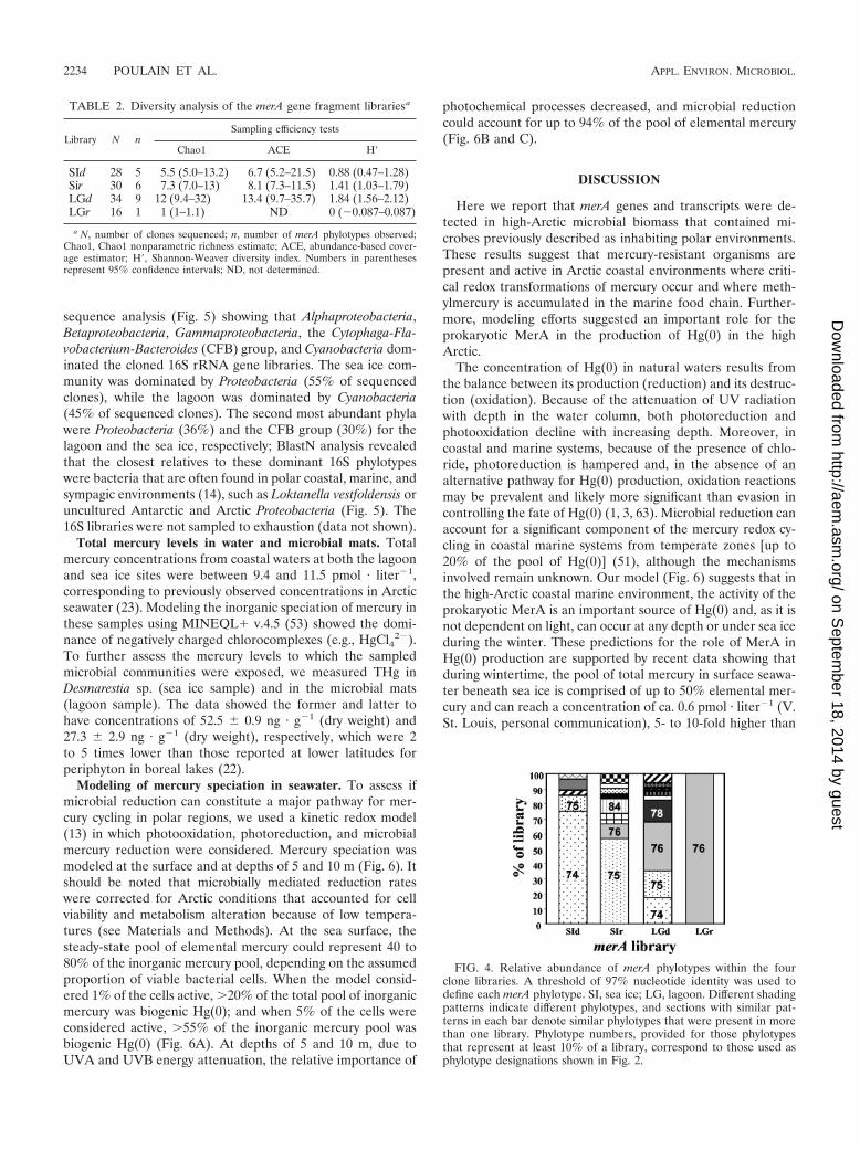
Diversity analysis of merA. Rarefaction curves were con-structed for each merA library to assess our sampling effort(Fig. 3). Both sea ice merA libraries were sampled adequatelyas the rarefaction curves approached a plateau. Furthermore,ACE and Chao1 estimates indicated that in both libraries, wecaptured approximately 80% of the predicted phylotypes (Ta-ble 2). While the distribution of phylotypes in the sea ice RNAlibrary was more even than that in the sea ice DNA library(Fig. 3), the richness (Chao1 and ACE) and diversity (H�)estimates were the same within the sampling error (Table 2).The sea ice DNA library was dominated by Arct74, which wasmost closely related to the MerA of Tn21 (Fig. 2 and 4). Arct74was not detected in the sea ice RNA library. This library wasdominated by the second most abundant clone from the DNAlibrary, Arct75 (Fig. 4). With the exception of Arct75, therewas no overlap between the sea ice DNA and RNA merAlibraries.
The lagoon DNA library was the most diverse, with an H� of1.84, while the lagoon RNA library was the least diverse, with
FIG. 1. merA genes and transcripts in microbial biomass. (Top) Gelshows 291-bp merA PCR products obtained with sea ice (SI) andlagoon (LG) DNA extracts as templates. SI1 and LG, DNA obtainedby extracting biomass with its associated microbes; SI2, extract of afraction obtained by washing alga with site water; (�) pPB20, positivecontrol consisting of merA from Pseudomonas stutzeri OX (50); PCRblank lane (�) and DNA size markers (in bp). (Bottom) Gel showsmerA PCR products following reverse transcriptase reactions (�RT)of RNA extracted from sea ice and lagoon biomass. (�RT), no RTcontrols; (�) PCR blank lane; a 291-bp PCR fragment is shown.
2232 POULAIN ET AL. APPL. ENVIRON. MICROBIOL.
on Septem
ber 18, 2014 by guesthttp://aem
.asm.org/
Dow
nloaded from

an H� of 0, corresponding to the presence of a single phylotype,Arct76 (Table 2). ACE- and Chao1-based coverage estimatesindicated that our sequencing effort captured 75% of the pre-dicted phylotypes from the lagoon DNA library. The singlephylotype in the lagoon RNA library was most closely relatedto MerA of Tn21. Arct76 comprised about one-third of thelagoon DNA library (Fig. 4). A Tn21-like MerA locus waspreviously described in a bacterium obtained from 8,000-year-old permafrost (31), and thus, this MerA phylotype may becommon in cold environments.
Bacterial community analysis. We constructed and analyzed16S rRNA gene clone libraries to examine whether microbesthat are commonly found in polar regions were present in thesampled lagoon and sea ice biomass. This was confirmed by
FIG. 3. merA phylotype accumulation curves showing the numberof clones sampled versus the number of merA phylotypes observed.
FIG. 2. Phylogenetic distribution of merA phylotypes. The dendrogram was constructed from a ClustalW alignment of the trimmed amino acidsequences by neighbor-joining analysis using MEGA 3.1. Bootstrap values greater than 50 are indicated. The number of clones in a particular phylotypeis indicated in parentheses. Phylotypes representing �25% of the clones in a library are highlighted in bold type. Phylotype designations that contain theletters LG originated in the lagoon biomass and those that contain SI in the macroalgae; d and r indicate that the phylotype originated in DNA and RNAclone libraries, respectively. Reference sequences from GenBank include the accession numbers.
VOL. 73, 2007 merA GENE PRESENCE AND EXPRESSION IN THE ARCTIC 2233
on Septem
ber 18, 2014 by guesthttp://aem
.asm.org/
Dow
nloaded from
sequence analysis (Fig. 5) showing that Alphaproteobacteria,Betaproteobacteria, Gammaproteobacteria, the Cytophaga-Fla-vobacterium-Bacteroides (CFB) group, and Cyanobacteria dom-inated the cloned 16S rRNA gene libraries. The sea ice com-munity was dominated by Proteobacteria (55% of sequencedclones), while the lagoon was dominated by Cyanobacteria(45% of sequenced clones). The second most abundant phylawere Proteobacteria (36%) and the CFB group (30%) for thelagoon and the sea ice, respectively; BlastN analysis revealedthat the closest relatives to these dominant 16S phylotypeswere bacteria that are often found in polar coastal, marine, andsympagic environments (14), such as Loktanella vestfoldensis oruncultured Antarctic and Arctic Proteobacteria (Fig. 5). The16S libraries were not sampled to exhaustion (data not shown).
Total mercury levels in water and microbial mats. Totalmercury concentrations from coastal waters at both the lagoonand sea ice sites were between 9.4 and 11.5 pmol · liter�1,corresponding to previously observed concentrations in Arcticseawater (23). Modeling the inorganic speciation of mercury inthese samples using MINEQL� v.4.5 (53) showed the domi-nance of negatively charged chlorocomplexes (e.g., HgCl4
2�).To further assess the mercury levels to which the sampledmicrobial communities were exposed, we measured THg inDesmarestia sp. (sea ice sample) and in the microbial mats(lagoon sample). The data showed the former and latter tohave concentrations of 52.5 � 0.9 ng · g�1 (dry weight) and27.3 � 2.9 ng · g�1 (dry weight), respectively, which were 2to 5 times lower than those reported at lower latitudes forperiphyton in boreal lakes (22).
Modeling of mercury speciation in seawater. To assess ifmicrobial reduction can constitute a major pathway for mer-cury cycling in polar regions, we used a kinetic redox model(13) in which photooxidation, photoreduction, and microbialmercury reduction were considered. Mercury speciation wasmodeled at the surface and at depths of 5 and 10 m (Fig. 6). Itshould be noted that microbially mediated reduction rateswere corrected for Arctic conditions that accounted for cellviability and metabolism alteration because of low tempera-tures (see Materials and Methods). At the sea surface, thesteady-state pool of elemental mercury could represent 40 to80% of the inorganic mercury pool, depending on the assumedproportion of viable bacterial cells. When the model consid-ered 1% of the cells active, �20% of the total pool of inorganicmercury was biogenic Hg(0); and when 5% of the cells wereconsidered active, �55% of the inorganic mercury pool wasbiogenic Hg(0) (Fig. 6A). At depths of 5 and 10 m, due toUVA and UVB energy attenuation, the relative importance of
photochemical processes decreased, and microbial reductioncould account for up to 94% of the pool of elemental mercury(Fig. 6B and C).
DISCUSSION
Here we report that merA genes and transcripts were de-tected in high-Arctic microbial biomass that contained mi-crobes previously described as inhabiting polar environments.These results suggest that mercury-resistant organisms arepresent and active in Arctic coastal environments where criti-cal redox transformations of mercury occur and where meth-ylmercury is accumulated in the marine food chain. Further-more, modeling efforts suggested an important role for theprokaryotic MerA in the production of Hg(0) in the highArctic.
The concentration of Hg(0) in natural waters results fromthe balance between its production (reduction) and its destruc-tion (oxidation). Because of the attenuation of UV radiationwith depth in the water column, both photoreduction andphotooxidation decline with increasing depth. Moreover, incoastal and marine systems, because of the presence of chlo-ride, photoreduction is hampered and, in the absence of analternative pathway for Hg(0) production, oxidation reactionsmay be prevalent and likely more significant than evasion incontrolling the fate of Hg(0) (1, 3, 63). Microbial reduction canaccount for a significant component of the mercury redox cy-cling in coastal marine systems from temperate zones [up to20% of the pool of Hg(0)] (51), although the mechanismsinvolved remain unknown. Our model (Fig. 6) suggests that inthe high-Arctic coastal marine environment, the activity of theprokaryotic MerA is an important source of Hg(0) and, as it isnot dependent on light, can occur at any depth or under sea iceduring the winter. These predictions for the role of MerA inHg(0) production are supported by recent data showing thatduring wintertime, the pool of total mercury in surface seawa-ter beneath sea ice is comprised of up to 50% elemental mer-cury and can reach a concentration of ca. 0.6 pmol · liter�1 (V.St. Louis, personal communication), 5- to 10-fold higher than
FIG. 4. Relative abundance of merA phylotypes within the fourclone libraries. A threshold of 97% nucleotide identity was used todefine each merA phylotype. SI, sea ice; LG, lagoon. Different shadingpatterns indicate different phylotypes, and sections with similar pat-terns in each bar denote similar phylotypes that were present in morethan one library. Phylotype numbers, provided for those phylotypesthat represent at least 10% of a library, correspond to those used asphylotype designations shown in Fig. 2.
TABLE 2. Diversity analysis of the merA gene fragment librariesa
Library N nSampling efficiency tests
Chao1 ACE H�
SId 28 5 5.5 (5.0–13.2) 6.7 (5.2–21.5) 0.88 (0.47–1.28)Sir 30 6 7.3 (7.0–13) 8.1 (7.3–11.5) 1.41 (1.03–1.79)LGd 34 9 12 (9.4–32) 13.4 (9.7–35.7) 1.84 (1.56–2.12)LGr 16 1 1 (1–1.1) ND 0 (�0.087–0.087)
a N, number of clones sequenced; n, number of merA phylotypes observed;Chao1, Chao1 nonparametric richness estimate; ACE, abundance-based cover-age estimator; H�, Shannon-Weaver diversity index. Numbers in parenthesesrepresent 95% confidence intervals; ND, not determined.
2234 POULAIN ET AL. APPL. ENVIRON. MICROBIOL.
on Septem
ber 18, 2014 by guesthttp://aem
.asm.org/
Dow
nloaded from

that observed in coastal seawater during the ice-free season.Although very few data exist as to the metabolic activity ofmicrobes under sea ice during polar winter, it is possible thatthey further increase the pool of reduced mercury at a time ofthe year when photooxidation processes are greatly reduced.Moreover, because mer operon functions also degrade MeHg,bacteria may decrease the pool of methylmercury available foruptake by Arctic food webs. In this regard, it is important tonote that macroalgae such as Desmarestia sp. were reported toproduce both monomethylmercury and dimethylmercury inpolar seawater (47). The importance of mercury-resistant mi-crobes that are associated with this macroalgae-to-mercurycycling is critical to the understanding of contamination path-ways in the Arctic.
The discovery of merA transcripts in Arctic microbial bio-mass was unexpected. The expression of merA is tightly con-trolled by intracellular concentrations of divalent mercury (15),and merA induction requires exposure concentrations in thenmol · liter�1 range in both pure cultures (64) and aquaticmicrobial communities (52). The mercury concentrations wepresent here and those reported for late spring and summer inmeltwater, lakes, and seawater are in the pmol · liter�1 range(36, 58), typical of pristine environments. Furthermore, thismercury was present mostly as negatively charged chlorocom-plexes, which are poorly bioavailable to microbes (5, 27).Therefore, not only was mercury concentration low in thecoastal environments sampled here, but its bioavailability waslikely also limited by high concentrations of Cl�. The induction
FIG. 5. 16S rRNA-based phylogeny of Arctic microbial biomass. The dendrogram was constructed from a ClustalW alignment of the trimmednucleotide sequences by neighbor-joining analysis using MEgA 3.1. Bootstrap values greater than 50 are indicated. }, phylotypes represented inthe sea ice library; F, lagoon library.
VOL. 73, 2007 merA GENE PRESENCE AND EXPRESSION IN THE ARCTIC 2235
on Septem
ber 18, 2014 by guesthttp://aem
.asm.org/
Dow
nloaded from

of merA observed in our study may reflect (i) the existence ofenvironmental conditions that enhance mercury bioavailabilityor (ii) the presence of microniches with high mercury concen-trations within the sampled biomass. Our analysis of mercuryconcentrations involved homogenization steps and would misssuch niches.
To understand why merA was expressed in the high Arctic,an understanding of both the processes that control mercurybioavailability and how these processes are impacted by theuniqueness of the microbial habitats in high latitudes isneeded. These are not well understood beyond the passivediffusion of Hg-sulfide complexes (11) and the facilitated trans-port of inorganic and organic mercury complexes (26). Nicheswith high concentrations of mercury can occur in the highArctic, possibly in association with the complex and heteroge-neous nature of sea ice (21). One of the biomass samplesdescribed here (SI) was obtained from waters with sea ice, andconsidering the slow degradation of RNA in frozen environ-ments (61), a sea ice origin for the merA transcripts detected inthis sample may be plausible. This proposition is supported bythe dominance of bacteria endemic to sympagic environmentsin the sampled communities (Fig. 5). The conditions whichcontrol the uptake of mercury are still unclear and may well beas diverse as the environments studied. The occurrence of
merA expression in Arctic coastal and marine aquatic environ-ments exhibiting low mercury levels underscores the gap in ourcurrent knowledge of Hg-microbe interactions, and furtherinvestigation of the biogeochemical cycling of mercury in polarenvironments should elucidate some key aspects of the Arcticmercury cycle.
Many of the MerA phylotypes reported here from Arcticbiomass are related to those from microbes that thrive at highlatitudes. Forty-eight percent were most closely related to pu-tative mer loci from the complete genomes of Sphingopyxisalaskensis and Polaromonas vacuolata, two microbes first iso-lated from polar environments. This finding attests to the au-thenticity of the MerA loci that were isolated here as repre-sentative of those loci that are present and expressed inmicrobial communities from polar regions. The remainingMerA phylotypes, which constitute the majority of the clonessequenced, were closely related to MerA phylotypes fromTn21, Tn501, and pDU1358. A Tn21-related mercury resis-tance transposon, Tn5060, has previously been described in an8,000- to 10,000-year-old Siberian permafrost isolate ofPseudomonas sp. (31).
Phylotypes related to Tn21 and Tn501 merA phylotypesdominated in an anaerobic enrichment of mercury-contami-nated sediment (43) as well as in the microbial biomass of an
FIG. 6. Results of modeling of the relative importance of photochemical reactions versus biologically mediated reduction in mercury redoxcycling in seawater in Arctic near-coastal environments. (A) Relative distribution of inorganic mercury species and proportion of Hg(0) formedby photochemical and microbial processes at the sea surface. In this example, photoreduction and photooxidation rates were considered to be 0.5and 0.6 h�1, respectively, and the proportion of active bacterial cells was assumed to be 1 or 5%. (B) Relative importance of photochemical versusmicrobial contributions to the pool of Hg(0) at the surface and at depths of 5 and 10 m. (C) A proposed model for redox cycling of mercury inthe water column of Arctic near-coastal marine environments. Arrow width correlates with the relative quantitative contribution of the depictedprocess to the total activity at each depth. Black, white, and dashed-line arrows represent microbially mediated reduction, photoreduction triggeredmostly by UVB, and photooxidation triggered mostly by UVA, respectively. The gray inverted triangle depicts the gradient of light penetration withdepth.
2236 POULAIN ET AL. APPL. ENVIRON. MICROBIOL.
on Septem
ber 18, 2014 by guesthttp://aem
.asm.org/
Dow
nloaded from

acidic and sulfidic mercury-rich spring in Yellowstone NationalPark (T. Barkay, unpublished data). These data suggest thatthis merA locus, which represents the most recently evolvedMerA lineage (Fig. 2), is broadly distributed in many environ-ments. The total number of merA phylotypes observed here(Fig. 2) was low compared to that in the anaerobic enrichmentwith its 39 merA phylotypes. Others have shown that selectionfor the target genes may increase phylotype diversity (28),while enhanced activities in environmental incubations wereassociated with a reduced phylotype diversity (40). A morerigorous examination of the relationship of merA diversity tomercury resistance and reduction in environmental samples isneeded for a clear understanding of how phylotype diversitiesis related to the potential for mercury transformations in var-ious environments.
Two contrasting patterns were observed when the diversitiesof the DNA and RNA clone libraries that were obtained fromthe same community were examined: (i) a high diversity ofmerA genes and a low diversity of transcripts in the lagoon and(ii) a high diversity of both genes and transcripts in the sea icesamples. At this point, we can only speculate on why the lagoonand the sea ice communities showed such different patterns.One possibility is that the high diversity of merA transcripts inthe sea ice reflects the heterogeneous nature of microbialniches within sea ice (see above). Alternatively, it may berelated to the in situ temperature at the time of sampling.While Arctic coastal seawater temperatures are usually slightlybelow 0°C, the temperatures in coastal lagoons may reach amesophilic range (58), and the temperature at the lagoon atsampling time was 7°C. Higher temperatures in the lagoon mayhave accelerated the turnover rate of transcripts, while colderconditions in open waters may have slowed mRNA degrada-tion to the point that we detected a greater diversity of ex-pressed merA genes in sea ice.
The diversity of expressed genes, not that of the genes thatare present in a community, is at the heart of what determinesecosystem function as related to environmental conditions.While experimental approaches to studying the expression ofbiogeochemical functions have been widely used in the lastdecade, few studies have compared the diversities of functionalgenes and mRNA transcripts in microbial biomass. Knauth etal. (32) used terminal restriction fragment length polymor-phism analysis of both DNA and RNA extracted from micro-bial biomass of the rice rhizosphere to examine how nitrogenavailability and plant host species affected the diversity of thenitrogenase gene nifH. As in our study of merA, their resultsshowed significant differences between nifH DNA and mRNAprofiles, underscoring the importance of mRNA-based ap-proaches to understanding functional gene diversity. The ob-servation of different merA gene and transcript profiles pre-sented here highlights the need to further develop anunderstanding of how patterns of microbial gene expressionare affected by environmental parameters in heterogeneousenvironments such as the Arctic.
Our discovery of merA expression and the results of themodel calculations that simulated redox transformations sug-gest an active role for microbial activities in the cycling ofmercury in Arctic coastal and marine environments. Clearly,microbial redox transformations have the potential to drivemercury dynamics in cold environments, and Arctic microbes
that possess and express merA genes stand to be key players indetermining the environmental mobility and toxicity of mer-cury in high-latitude polar regions.
ACKNOWLEDGMENTS
We thank H. Vandermeulen for his help with alga identification,Paul del Giorgio for access to the flow cytometer, J. Brenchley and V.Miteva for stimulating discussions, and B. E. Keatley for comments onthe manuscript.
Support by the U.S. National Science Foundation (CHE-0221978and ATM-0322022), the U.S. National Institute of EnvironmentalHealth and Safety (P42 ES004911), and the Fond Quebecois de laRecherche sur la Nature et les Technologies for funding is acknowl-edged.
REFERENCES
1. Amyot, M., G. A. Gill, and F. M. M. Morel. 1997. Production and loss ofdissolved gaseous mercury in coastal seawater. Environ. Sci. Technol. 31:3606–3611.
2. Amyot, M., G. Mierle, D. R. S. Lean, and D. J. McQueen. 1994. Sunlight-induced formation of dissolved gaseous mercury in lake waters. Environ. Sci.Technol. 28:2366–2371.
3. Ariya, P. A., A. P. Dastoor, M. Amyot, W. H. Schroeder, L. Barrie, K. Anlauf,F. Raofie, A. Ryzhkov, D. Davignon, J. Lalonde, and A. Steffen. 2004. TheArctic: a sink for mercury. Tellus. Ser. B 56:397–403.
4. Ariya, P. A., A. Khalizov, and A. Gidas. 2002. Reactions of gaseous mercurywith atomic and molecular halogens: kinetics, product studies, and atmo-spheric implications. J. Phys. Chem. A 106:7310–7320.
5. Barkay, T., M. Gillman, and R. R. Turner. 1997. Effects of dissolved organiccarbon and salinity on bioavailability of mercury. Appl. Environ. Microbiol.63:4267–4271.
6. Barkay, T., C. Liebert, and M. Gillman. 1989. Environmental significance ofthe potential for mer(Tn21)-mediated reduction of Hg2� to Hg0 in naturalwaters. Appl. Environ. Microbiol. 55:1196–1202.
7. Barkay, T., C. Liebert, and M. Gillman. 1989. Hybridization of DNA probeswith whole-community genome for detection of genes that encode microbialresponses to pollutants: mer genes and Hg2� resistance. Appl. Environ.Microbiol. 55:1574–1577.
8. Barkay, T., S. M. Miller, and A. O. Summers. 2003. Bacterial mercuryresistance from atoms to ecosystems. FEMS Microbiol. Rev. 27:355–384.
9. Barkay, T., R. R. Turner, A. Vandenbrook, and C. Liebert. 1991. The rela-tionships of Hg(II) volatilization from a fresh-water pond to the abundanceof mer-genes in the gene pool of the indigenous microbial community.Microb. Ecol. 21:151–161.
10. Benner, R., P. Louchouarn, and R. M. W. Amon. 2005. Terrigenous dissolvedorganic matter in the Arctic Ocean and its transport to surface and deepwaters of the North Atlantic. Global Biogeochem. Cycles 19:GB2025.
11. Benoit, J. M., C. C. Gilmour, R. P. Mason, and A. Heyes. 1999. Sulfidecontrols on mercury speciation and bioavailability to methylating bacteria insediment pore waters. Environ. Sci. Technol. 33:951–957.
12. Braun, W., J. T. Herron, and D. K. Kahaner. 1988. ACUCHEM: a com-puter-program for modeling complex chemical-reaction systems. Int.J. Chem. Kinet. 20:51–62.
13. Braune, B., D. Muir, B. DeMarch, M. Gamberg, K. Poole, R. Currie, M.Dodd, W. Duschenko, J. Eamer, B. Elkin, M. Evans, S. Grundy, C. Hebert,R. Johnstone, K. Kidd, B. Koenig, L. Lockhart, H. Marshall, K. Reimer, J.Sanderson, and L. Shutt. 1999. Spatial and temporal trends of contaminantsin Canadian Arctic freshwater and terrestrial ecosystems: a review. Sci. TotalEnviron. 230:145–207.
14. Brinkmeyer, R., K. Knittel, J. Jurgens, H. Weyland, R. Amann, and E.Helmke. 2003. Diversity and structure of bacterial communities in Arcticversus Antarctic pack ice. Appl. Environ. Microbiol. 69:6610–6619.
15. Brown, N. L., J. V. Stoyanov, S. P. Kidd, and J. L. Hobman. 2003. The MerRfamily of transcriptional regulators. FEMS Microbiol. Rev. 27:145–163.
16. Campbell, L. M., R. J. Norstrom, K. A. Hobson, D. C. G. Muir, S. Backus,and A. T. Fisk. 2005. Mercury and other trace elements in a pelagic Arcticmarine food web (Northwater Polynya, Baffin Bay). Sci. Total Environ.351:247–263.
17. Chao, A. 1984. Non-parametric estimation of the number of classes in apopulation. Scand. J. Stat. 11:265–270.
18. Chao, A., and S. M. Lee. 1992. Estimating the number of classes via samplecoverage. J. Am. Stat. Assoc. 87:210–217.
19. Costa, M., and P. S. Liss. 1999. Photoreduction of mercury in sea water andits possible implications for Hg(0) air-sea fluxes. Mar. Chem. 68:87–95.
20. De Domenico, M., A. Lo Giudice, L. Michaud, M. Saitta, and V. Bruni. 2004.Diesel oil and PCB-degrading psychrotrophic bacteria isolated from antarc-tic seawaters (Terra Nova Bay, Ross Sea). Polar Res. 23:141–146.
21. Deming, J. W. 2002. Psychrophiles and polar regions. Curr. Opin. Microbiol.5:301–309.
VOL. 73, 2007 merA GENE PRESENCE AND EXPRESSION IN THE ARCTIC 2237
on Septem
ber 18, 2014 by guesthttp://aem
.asm.org/
Dow
nloaded from

22. Desrosiers, M., D. Planas, and A. Mucci. 2006. Mercury methylation in theepilithon of boreal shield aquatic ecosystems. Environ. Sci. Technol. 40:1540–1546.
23. Douglas, T. A., M. Sturm, W. R. Simpson, S. Brooks, S. E. Lindberg, andD. K. Perovich. 2005. Elevated mercury measured in snow and frost flowersnear Arctic sea ice leads. Geophys. Res. Lett. 32:L04502.
24. Ebinghaus, R., H. H. Kock, C. Temme, J. W. Einax, A. G. Lowe, A. Richter,J. P. Burrows, and W. H. Schroeder. 2002. Antarctic springtime depletion ofatmospheric mercury. Environ. Sci. Technol. 36:1238–1244.
25. Gibson, J. A. E., W. F. Vincent, B. Nieke, and R. Pienitz. 2000. Control ofbiological exposure to UV radiation in the Arctic Ocean: comparison of theroles of ozone and riverine dissolved organic matter. Arctic 53:372–382.
26. Golding, G. R., C. A. Kelly, R. Sparling, P. C. Loewen, J. W. M. Rudd, andT. Barkay. 2002. Evidence for facilitated uptake of Hg(II) by Vibrio anguil-larum and Escherichia coli under anaerobic and aerobic conditions. Limnol.Oceanogr. 47:967–975.
27. Gutknecht, J. 1981. Inorganic mercury (Hg2�) transport through lipid bi-layer membranes. J. Membr. Biol. 61:61–66.
28. Hobel, C. F. V., V. T. Marteinsson, G. O. Hreggvidsson, and J. K.Kristjansson. 2005. Investigation of the microbial ecology of intertidal hotsprings by using diversity analysis of 16S rRNA and chitinase genes. Appl.Environ. Microbiol. 71:2771–2776.
29. Hughes, J. B., J. J. Hellmann, T. H. Ricketts, and B. J. M. Bohannan. 2001.Counting the uncountable: statistical approaches to estimating microbialdiversity. Appl. Environ. Microbiol. 67:4399–4406.
30. Junge, K., H. Eicken, and J. W. Deming. 2004. Bacterial activity at �2 to�20°C in Arctic wintertime sea ice. Appl. Environ. Microbiol. 70:550–557.
31. Kholodii, G., S. Mindlin, M. Petrova, and S. Minakhina. 2003. Tn5060 fromthe Siberian permafrost is most closely related to the ancestor of Tn21 priorto integron acquisition. FEMS Microbiol. Lett. 226:251–255.
32. Knauth, S., T. Hurek, D. Brar, and B. Reinhold-Hurek. 2005. Influence ofdifferent Oryza cultivars on expression of nifH gene pools in roots of rice.Environ. Microbiol. 7:1725–1733.
33. Lalonde, J. D., M. Amyot, A. M. L. Kraepiel, and F. M. M. Morel. 2001.Photooxidation of Hg(0) in artificial and natural waters. Environ. Sci. Tech-nol. 35:1367–1372.
34. Lalonde, J. D., M. Amyot, J. Orvoine, F. M. M. Morel, J. C. Auclair, andP. A. Ariya. 2004. Photoinduced oxidation of Hg0(aq) in the waters from theSt. Lawrence estuary. Environ. Sci. Technol. 38:508–514.
35. Liebert, C., and T. Barkay. 1988. A direct viable counting method for mea-suring tolerance of aquatic microbial communities to Hg2�. Can. J. Micro-biol. 34:1090–1095.
36. Lindberg, S. E., S. Brooks, C. J. Lin, K. J. Scott, M. S. Landis, R. K. Stevens,M. Goodsite, and A. Richter. 2002. Dynamic oxidation of gaseous mercury inthe Arctic troposphere at polar sunrise. Environ. Sci. Technol. 36:1245–1256.
37. Lovley, D. R. 2003. Cleaning up with genomics: applying molecular biology tobioremediation. Nat. Rev. Microbiol. 1:35–44.
38. Macdonald, R. W., T. Harner, and J. Fyfe. 2005. Recent climate change inthe Arctic and its impact on contaminant pathways and interpretation oftemporal trend data. Sci. Total Environ. 342:5–86.
39. Master, E. R., and W. W. Mohn. 1998. Psychrotolerant bacteria isolated fromArctic soil that degrade polychlorinated biphenyls at low temperatures.Appl. Environ. Microbiol. 64:4823–4829.
40. Metcalfe, A. C., M. Krsek, G. W. Gooday, J. I. Prosser, and E. M. H.Wellington. 2002. Molecular analysis of a bacterial chitinolytic community inan upland pasture. Appl. Environ. Microbiol. 68:5042–5050.
41. Mindlin, S., L. Minakhin, M. Petrova, G. Kholodii, S. Minakhina, Z.Gorlenko, and V. Nikiforov. 2005. Present-day mercury resistance trans-posons are common in bacteria preserved in permafrost grounds since theUpper Pleistocene. Res. Microbiol. 156:994–1004.
42. Muir, D., B. Braune, B. DeMarch, R. Norstrom, R. Wagemann, L. Lockhart,B. Hargrave, D. Bright, R. Addison, J. Payne, and K. Reimer. 1999. Spatialand temporal trends and effects of contaminants in the Canadian Arcticmarine ecosystem: a review. Sci. Total Environ. 230:83–144.
43. Nı Chadhain, S. M., J. K. Schaefer, S. Crane, G. J. Zylstra, and T. Barkay.2006. Analysis of mercuric reductase (merA) gene diversity in an anaerobicmercury-contaminated sediment enrichment. Environ. Microbiol. 8:1746–1752.
44. Osborn, A. M., K. D. Bruce, P. Strike, and D. A. Ritchie. 1997. Distribution,diversity and evolution of the bacterial mercury resistance (mer) operon.FEMS Microbiol. Rev. 19:239–262.
45. Pepi, M., A. Cesaro, G. Liut, and F. Baldi. 2005. An antarctic psychrotrophicbacterium Halomonas sp. ANT-3b, growing on n-hexadecane, produces anew emulsifying glycolipid. FEMS Microbiol. Ecol. 53:157–166.
46. Philippidis, G. P., L. H. Malmber, W. S. Hu, and J. L. Schottel. 1991. Effectof gene amplification on mercuric ion reduction activity of Escherichia coli.Appl. Environ. Microbiol. 57:3558–3564.
47. Pongratz, R., and K. G. Heumann. 1998. Production of methylated mercuryand lead by polar macroalgae: a significant natural source for atmosphericheavy metals in clean room compartments. Chemosphere 36:1935–1946.
48. Poulain, A. J., M. Amyot, D. Findlay, S. Telor, T. Barkay, and H. Hintel-mann. 2004. Biological and photochemical production of dissolved gaseousmercury in a boreal lake. Limnol. Oceanogr. 49:2265–2275.
49. Price, P. B., and T. Sowers. 2004. Temperature dependence of metabolicrates for microbial growth, maintenance, and survival. Proc. Natl. Acad. Sci.USA 101:4631–4636.
50. Reniero, D., E. Mozzon, E. Galli, and P. Barbieri. 1998. Two aberrantmercury resistance transposons in the Pseudomonas stutzeri plasmid pPB.Gene 208:37–42.
51. Rolfhus, K. R., and W. F. Fitzgerald. 2004. Mechanisms and temporal vari-ability of dissolved gaseous mercury production in coastal seawater. Mar.Chem. 90:125–136.
52. Schaefer, J. K., J. Yagi, J. R. Reinfelder, T. Cardona, K. M. Ellickson, S.Tel-Or, and T. Barkay. 2004. Role of the bacterial organomercury lyase(MerB) in controlling methylmercury accumulation in mercury-contami-nated natural waters. Environ. Sci. Technol. 38:4304–4311.
53. Schecher, W. D., and D. C. McAvoy. 1992. Mineql�: a software environmentfor chemical-equilibrium modeling. Comput. Environ. Urban Syst. 16:65–76.
54. Schloss, P. D., and J. Handelsman. 2005. Introducing DOTUR, a computerprogram for defining operational taxonomic units and estimating speciesrichness. Appl. Environ. Microbiol. 71:1501–1506.
55. Schroeder, W. H., K. G. Anlauf, L. A. Barrie, J. Y. Lu, A. Steffen, D. R.Schneeberger, and T. Berg. 1998. Arctic springtime depletion of mercury.Nature 394:331–332.
56. Sellers, P., C. A. Kelly, J. W. M. Rudd, and A. R. MacHutchon. 1996.Photodegradation of methylmercury in lakes. Nature 380:694–697.
57. Siciliano, S. D., N. J. O’Driscoll, and D. R. Lean. 2002. Microbial reductionand oxidation of mercury in freshwater lakes. Environ. Sci. Technol. 36:3064–3068.
58. St. Louis, V. L., M. J. Sharp, A. Steffen, A. May, J. Barker, J. L. Kirk, D. J.Kelly, S. E. Arnott, B. Keatley, and J. P. Smol. 2005. Some sources and sinksof monomethyl and inorganic mercury on Ellesmere Island in the Canadianhigh Arctic. Environ. Sci. Technol. 39:2686–2701.
59. Van Oostdam, J., S. G. Donaldson, M. Feeley, D. Arnold, P. Ayotte, G.Bondy, L. Chan, E. Dewaily, C. M. Furgal, H. Kuhnlein, E. Loring, G.Muckle, E. Myles, O. Receveur, B. Tracy, U. Gill, and S. Kalhok. 2005.Human health implications of environmental contaminants in ArcticCanada: a review. Sci. Total Environ. 351–352:165–246.
60. Vetriani, C., Y. S. Chew, S. M. Miller, J. Yagi, J. Coombs, R. A. Lutz, and T.Barkay. 2005. Mercury adaptation among bacteria from a deep-sea hydro-thermal vent. Appl. Environ. Microbiol. 71:220–226.
61. Vlassov, A. V., S. A. Kazakov, B. H. Johnston, and L. F. Landweber. 2005.The RNA world on ice: a new scenario for the emergence of RNA infor-mation. J. Mol. Evol. 61:264–273.
62. Welander, U. 2005. Microbial degradation of organic pollutants in soil in acold climate. Soil Sediment Contam. 14:281–291.
63. Whalin, L. M., and R. P. Mason. 2006. A new method for the investigationof mercury redox chemistry in natural waters utilizing deflatable Teflon bagsand additions of isotopically labeled mercury. Anal. Chim. Acta 558:211–221.
64. Wright, J. G., M. J. Natan, F. M. Macdonnell, D. M. Ralston, and T. V.Ohalloran. 1990. Mercury(II) thiolate chemistry and the mechanism of theheavy-metal biosensor MerR. Prog. Inorg. Chem. 38:323–412.
65. Zubkov, M. V., B. M. Fuchs, H. Eilers, P. H. Burkill, and R. Amann. 1999.Determination of total protein content of bacterial cells by SYPRO stainingand flow cytometry. Appl. Environ. Microbiol. 65:3251–3257.
2238 POULAIN ET AL. APPL. ENVIRON. MICROBIOL.
on Septem
ber 18, 2014 by guesthttp://aem
.asm.org/
Dow
nloaded from

APPLIED AND ENVIRONMENTAL MICROBIOLOGY, June 2007, p. 3769 Vol. 73, No. 110099-2240/07/$08.00�0 doi:10.1128/AEM.00819-07
ERRATUM
Potential for Mercury Reduction by Microbes in the High ArcticAlexandre J. Poulain, Sinead M. Ni Chadhain, Parisa A. Ariya, Marc Amyot, Edenise Garcia,
Peter G. C. Campbell, Gerben J. Zylstra, and Tamar BarkayGroupe de Recherche Inter-universitaire en Limnologie (GRIL), Departement des Sciences Biologiques, Universite de Montreal,
C. P. 6128, Succursale Centre-Ville, Pavillon Marie-Victorin, Montreal, Quebec, Canada H3C 3J7; Biotechnology Center forAgriculture and the Environment, Cook College, Rutgers University, Foran Hall, 59 Dudley Road, New Brunswick,
New Jersey 08901-8520; Department of Chemistry and Atmospheric and Oceanic Sciences, McGill University,801 Sherbrooke Street West, Montreal, Quebec, Canada H3A 2K6; Universite du Quebec, INRS-Eau,
Terre et Environnement, 490 de la Couronne, Quebec, Canada G1K 9A9; and Department ofBiochemistry and Microbiology, Rutgers University, 76 Lipman Drive,
New Brunswick, New Jersey 08901
Volume 73, no. 7, p. 2230–2238, 2007. Page 2231, Table 1: The attenuation coefficients for UVA and UVB should be as follows:
UVA attenuation (m�1), 0.05–2.7UVB attenuation (m�1), 0.24–4.02
3769