Routing of straddle carriers at a container terminal with the special aspect of internal moves
Upload
independentCategory
view
3download
0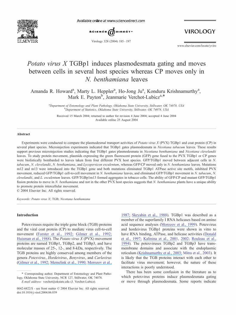
www.elsevier.com/locate/yviro
Virology 328 (20
Potato virus X TGBp1 induces plasmodesmata gating and moves
between cells in several host species whereas CP moves only in
N. benthamiana leaves
Amanda R. Howarda, Marty L. Hepplera, Ho-Jong Jua, Konduru Krishnamurthya,
Mark E. Paytonb, Jeanmarie Verchot-Lubicza,*
aDepartment of Entomology and Plant Pathology, Oklahoma State University, Stillwater, OK 74078, USAbDepartment of Statistics, Oklahoma State University, Stillwater, OK 74078, USA
Received 15 March 2004; returned to author for revision 4 June 2004; accepted 4 June 2004
Available online 25 August 2004
Abstract
Experiments were conducted to compare the plasmodesmal transport activities of Potato virus X (PVX) TGBp1 and coat protein (CP) in
several plant species. Microinjection experiments indicated that TGBp1 gates plasmodesmata in Nicotiana tabacum leaves. These results
support previous microinjection studies indicating that TGBp1 gates plasmodesmata in Nicotiana benthamiana and Nicotiana clevelandii
leaves. To study protein movement, plasmids expressing the green fluorescent protein (GFP) gene fused to the PVX TGBp1 or CP genes
were biolistically bombarded to leaves taken from four different PVX host species. GFP/TGBp1 moved between adjacent cells in N.
tabacum, N. clevelandii, N. benthamiana, and Lycopersicon esculentum, whereas GFP/CP moved only in N. benthamiana leaves. Mutations
m12 and m13 were introduced into the TGBp1 gene and both mutations eliminated TGBp1 ATPase active site motifs, inhibited PVX
movement, reduced GFP/TGBp1 cell-to-cell movement in N. benthamiana leaves, and eliminated GFP/TGBp1 movement in N. tabacum, N.
clevelandii, and L. esculentum leaves. GFP/TGBp1m13 formed aggregates in tobacco cells. The ability of GFP/CP and mutant GFP/TGBp1
fusion proteins to move in N. benthamiana and not in the other PVX host species suggests that N. benthamiana plants have a unique ability
to promote protein intercellular movement.
D 2004 Elsevier Inc. All rights reserved.
Keywords: Potato virus X; TGB; Nicotiana benthamiana
Introduction
Potexviruses require the triple gene block (TGB) proteins
and the viral coat protein (CP) to mediate virus cell-to-cell
movement (Forster et al., 1992; Gilmer et al., 1992;
Huisman et al., 1988). The Potato virus X (PVX) movement
proteins are named TGBp1, TGBp2, and TGBp3, and have
molecular masses of 25-, 12-, and 8-kDa, respectively. The
TGB proteins are highly conserved among members of the
genera Potexvirus, Hordeivirus, Benyvirus, and Carlavirus
(Gilmer et al., 1992; Memelink et al., 1990; Morozov et al.,
0042-6822/$ - see front matter D 2004 Elsevier Inc. All rights reserved.
doi:10.1016/j.virol.2004.06.039
* Corresponding author. Department of Entomology and Plant Patho-
logy, Oklahoma State University, NCR 127, Stillwater, OK 74078.
E-mail address: [email protected] (J. Verchot-Lubicz).
1987; Skryabin et al., 1988). TGBp1 was described as a
member of the superfamily I RNA helicases based on amino
acid sequence analyses (Morozov et al., 1999). Potexvirus
and hordeivirus TGBp1 proteins were shown in vitro to
have RNA binding, ATPase, and helicase activities (Donald
et al., 1997; Kalinina et al., 2001, 2002; Rouleau et al.,
1994). The potexviruses TGBp2 and TGBp3 have trans-
membrane domains and associate with the endoplasmic
reticulum (Krishnamurthy et al., 2003; Mitra et al., 2003). It
is likely that the TGB proteins interact with each other to
facilitate virus movement; however, the nature of these
interactions is poorly understood.
There has been some confusion in the literature as to
which potexvirus proteins induce plasmodesmata gating
or move through plasmodesmata. Some reports indicate
04) 185–197
A.R. Howard et al. / Virology 328 (2004) 185–197186
that TGBp1 is the primary viral factor that gates and
moves through plasmodesmata while other reports sug-
gest that TGBp2 is primarily responsible for plasmodes-
mata gating. In two microinjection studies, the potexvirus
TGBp1 was identified to be the viral factor responsible
for increasing the plasmodesmal size exclusion limit
(SEL) for virus cell-to-cell movement (Angell et al.,
1996; Lough et al., 1998). In the first study, F-dextrans
of various sizes were injected into Nicotiana clevelandii
trichome cells along with either wild-type PVX virions or
virions of a mutant PVX which had a large segment of
TGBp1 deleted (Angell et al., 1996). Wild-type PVX, but
not the mutant PVX, increased the plasmodesmal SEL to
allow movement of large dextrans between adjacent cells
(Angell et al., 1996). In the second study, F-dextrans of
various sizes were injected into mesophyll cells of
transgenic Nicotiana benthamiana expressing the TGB
proteins of White clover mosaic virus (WClMV).
WClMV TGBp1, but not TGBp2 or TGBp3, increased
the plasmodesmal SEL for transport of large dextrans
(Lough et al., 1998). In a third microinjection study using
PVX CP-expressing transgenic Nicotiana tabacum leaves,
it was reported that the PVX CP did not increase the
plasmodesmal SEL to allow movement of large F-
dextrans (Oparka et al., 1996).
It was recently reported that TGBp2 was responsible for
increasing the plasmodesmal SEL (Fridborg et al., 2003;
Tamai and Meshi, 2001). Plasmids expressing the green
fluorescent protein gene (GFP) fused to the TGBp2 gene
were biolistically delivered to N. benthamiana leaves and
protein cell-to-cell movement was observed. Because these
results seem to conflict with previous findings relating to
TGBp1, we have undertaken a series of experiments to
determine which of the PVX proteins move from cell to
cell. Plasmids expressing GFP/TGBp2 or GFP/TGBp3
were biolistically delivered to six different PVX host
species to compare protein movement in a broader range
of PVX host plants (Krishnamurthy et al., 2003). GFP/
TGBp2 and GFP/TGBp3 each moved from cell to cell in
N. benthamiana leaves but were restricted primarily to
single cells in N. tabacum, N. clevelandii, Lycopersicon
esculentum, Chenopodia quinoa, and Gomphrena glo-
bossa (Krishnamurthy et al., 2003).
The goal of this study is to determine if PVX TGBp1
and CP increase the plasmodesmal SEL and move from
cell to cell in several PVX host species. TGBp1-expressing
transgenic N. tabacum leaves (Verchot et al., 1998) were
used in microinjection studies to determine if TGBp1 can
induce changes in the plasmodesmal SEL in N. tabacum.
These microinjection studies were conducted to comple-
ment previous studies in N. benthamiana and N. cleve-
landii, and to provide more information about PVX
TGBp1 activities in various host plants. In addition, the
ability of PVX TGBp1 and CP to move from cell to cell
was studied in N. tabacum, N. benthamiana, N. cleve-
landii, and L. esculentum leaves. Mutations were intro-
duced into the TGBp1 NTPase active site and the effects
of these mutations on protein movement were host
dependent. However, when the same mutations were
introduced into the PVX genome, virus movement was
eliminated in all host species tested.
Results
TGBp1 increases the plasmodesmal SEL in N. tabacum
leaves
It was determined using microinjection techniques that
the typical plasmodesmal SEL is below 1 kDa in tobacco
leaves (Robards and Lucas, 1990). The low molecular
weight (457 Da) Lucifer Yellow CH dilithium salt (LYCH)
is a fluorescent dye that can pass through plasmodesmata,
whereas F-dextrans of 4.4- or 9.5-kDa are often restricted to
single cells in tobacco leaves (Ding et al., 1992; Wolf et al.,
1989). TGBp1 was reported to increase the plasmodesmal
SEL in N. clevelandii and N. benthamiana leaves, allowing
movement of the large F-dextrans between adjacent cells
(Angell et al., 1996; Lough et al., 1998).
TGB100 transgenic N. tabacum plants express the PVX
TGBp1 and were used to determine if TGBp1 increases
the plasmodesmal SEL in N. tabacum plants as in other
Nicotiana species. The TGB100 plants were selected for
these experiments because they are able to complement
cell-to-cell movement of TGBp1-deficient PVX viruses
(Verchot et al., 1998). The 4.4- or 9.5-kDa F-dextrans were
injected into mesophyll cells of nontransgenic and
TGB100 transgenic N. tabacum source leaves, and
intercellular movement of the fluorescent-labeled mole-
cules was recorded. LYCH was injected into nontransgenic
and TGB100 transgenic mesophyll cells as a control to
ensure plasmodesmata were not closed as a result of tissue
preparation.
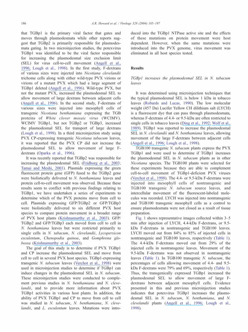
Fig. 1 shows representative images collected within 3–5
min after injection of LYCH, 4.4-kDa F-dextrans, or 9.5-
kDa F-dextrans in nontransgenic and TGB100 leaves.
LYCH moved out from 84% to 85% of injected cells in
nontransgenic and TGB100 leaves, respectively (Table 1).
The 4.4-kDa F-dextrans moved out from 29% of the
injected cells in nontransgenic leaves. Movement of the
9.5-kDa F-dextrans was not observed in nontransgenic
leaves (Table 1). In TGB100 transgenic N. tabacum, the
percentages of cells allowing movement of 4.4- and 9.5-
kDa F-dextrans were 79% and 69%, respectively (Table 1).
Thus, the transgenically expressed TGBp1 increased the
plasmodesmal SEL to allow movement of large F-
dextrans between adjacent mesophyll cells. Evidence
presented in this and previous microinjection studies
indicates that potexvirus TGBp1 increases the plasmo-
desmal SEL in N. tabacum, N. benthamiana, and N.
clevelandii plants (Angell et al., 1996; Lough et al.,
1998).
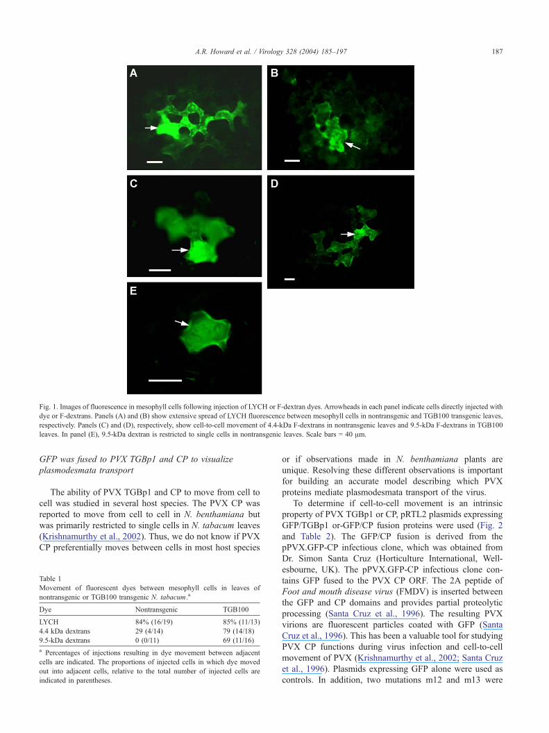
Fig. 1. Images of fluorescence in mesophyll cells following injection of LYCH or F-dextran dyes. Arrowheads in each panel indicate cells directly injected with
dye or F-dextrans. Panels (A) and (B) show extensive spread of LYCH fluorescence between mesophyll cells in nontransgenic and TGB100 transgenic leaves,
respectively. Panels (C) and (D), respectively, show cell-to-cell movement of 4.4-kDa F-dextrans in nontransgenic leaves and 9.5-kDa F-dextrans in TGB100
leaves. In panel (E), 9.5-kDa dextran is restricted to single cells in nontransgenic leaves. Scale bars = 40 Am.
A.R. Howard et al. / Virology 328 (2004) 185–197 187
GFP was fused to PVX TGBp1 and CP to visualize
plasmodesmata transport
The ability of PVX TGBp1 and CP to move from cell to
cell was studied in several host species. The PVX CP was
reported to move from cell to cell in N. benthamiana but
was primarily restricted to single cells in N. tabacum leaves
(Krishnamurthy et al., 2002). Thus, we do not know if PVX
CP preferentially moves between cells in most host species
Table 1
Movement of fluorescent dyes between mesophyll cells in leaves of
nontransgenic or TGB100 transgenic N. tabacum.a
Dye Nontransgenic TGB100
LYCH 84% (16/19) 85% (11/13)
4.4 kDa dextrans 29 (4/14) 79 (14/18)
9.5-kDa dextrans 0 (0/11) 69 (11/16)
a Percentages of injections resulting in dye movement between adjacent
cells are indicated. The proportions of injected cells in which dye moved
out into adjacent cells, relative to the total number of injected cells are
indicated in parentheses.
or if observations made in N. benthamiana plants are
unique. Resolving these different observations is important
for building an accurate model describing which PVX
proteins mediate plasmodesmata transport of the virus.
To determine if cell-to-cell movement is an intrinsic
property of PVX TGBp1 or CP, pRTL2 plasmids expressing
GFP/TGBp1 or-GFP/CP fusion proteins were used (Fig. 2
and Table 2). The GFP/CP fusion is derived from the
pPVX.GFP-CP infectious clone, which was obtained from
Dr. Simon Santa Cruz (Horticulture International, Well-
esbourne, UK). The pPVX.GFP-CP infectious clone con-
tains GFP fused to the PVX CP ORF. The 2A peptide of
Foot and mouth disease virus (FMDV) is inserted between
the GFP and CP domains and provides partial proteolytic
processing (Santa Cruz et al., 1996). The resulting PVX
virions are fluorescent particles coated with GFP (Santa
Cruz et al., 1996). This has been a valuable tool for studying
PVX CP functions during virus infection and cell-to-cell
movement of PVX (Krishnamurthy et al., 2002; Santa Cruz
et al., 1996). Plasmids expressing GFP alone were used as
controls. In addition, two mutations m12 and m13 were
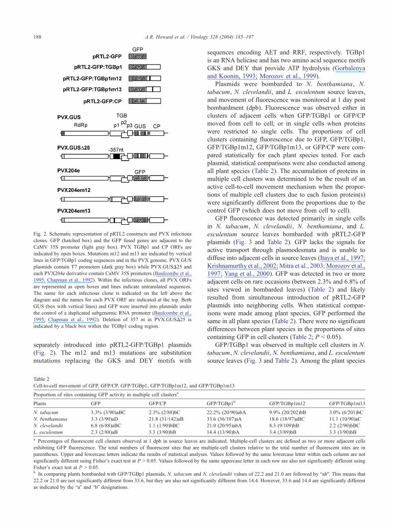
Fig. 2. Schematic representation of pRTL2 constructs and PVX infectious
clones. GFP (hatched box) and the GFP fused genes are adjacent to the
CaMV 35S promoter (light gray box). PVX TGBp1 and CP ORFs are
indicated by open boxes. Mutations m12 and m13 are indicated by vertical
lines in GFP/TGBp1 coding sequences and in the PVX genome. PVX.GUS
plasmids contain T7 promoters (dark gray box) while PVX.GUSD25 and
each PVX204e derivative contain CaMV 35S promoters (Baulcombe et al.,
1995; Chapman et al., 1992). Within the infectious clones, all PVX ORFs
are represented as open boxes and lines indicate untranslated sequences.
The name for each infectious clone is indicated on the left above the
diagram and the names for each PVX ORF are indicated at the top. Both
GUS (box with vertical lines) and GFP were inserted into plasmids under
the control of a duplicated subgenomic RNA promoter (Baulcombe et al.,
1995; Chapman et al., 1992). Deletion of 357 nt in PVX.GUSD25 is
indicated by a black box within the TGBp1 coding region.
A.R. Howard et al. / Virology 328 (2004) 185–197188
separately introduced into pRTL2-GFP/TGBp1 plasmids
(Fig. 2). The m12 and m13 mutations are substitution
mutations replacing the GKS and DEY motifs with
Table 2
Cell-to-cell movement of GFP, GFP/CP, GFP/TGBp1, GFP/TGBp1m12, and GFP
Proportion of sites containing GFP activity in multiple cell clustersa
Plants GFP GFP/CP
N. tabacum 3.3% (3/90)aBC 2.3% (2/88)bC
N. benthamiana 3.3 (3/90)aD 21.8 (31/142)aB
N. clevelandii 6.8 (6/88)aBC 1.1 (1/90)bBC
L. esculentum 2.3 (2/88)aB 3.3 (3/90)bB
a Percentages of fluorescent cell clusters observed at 1 dpb in source leaves are
exhibiting GFP fluorescence. The total numbers of fluorescent sites that are mu
parentheses. Upper and lowercase letters indicate the results of statistical analyses
significantly different using Fisher’s exact test at P N 0.05. Values followed by the
Fisher’s exact test at P N 0.05.b In comparing plants bombarded with GFP/TGBp1 plasmids, N. tabacum and N
22.2 or 21.0 are not significantly different from 33.6, but they are also not signific
as indicated by the baQ and bbQ designations.
sequences encoding AET and RRF, respectively. TGBp1
is an RNA helicase and has two amino acid sequence motifs
GKS and DEY that provide ATP hydrolysis (Gorbalenya
and Koonin, 1993; Morozov et al., 1999).
Plasmids were bombarded to N. benthamiana, N.
tabacum, N. clevelandii, and L. esculentum source leaves,
and movement of fluorescence was monitored at 1 day post
bombardment (dpb). Fluorescence was observed either in
clusters of adjacent cells when GFP/TGBp1 or GFP/CP
moved from cell to cell, or in single cells when proteins
were restricted to single cells. The proportions of cell
clusters containing fluorescence due to GFP, GFP/TGBp1,
GFP/TGBp1m12, GFP/TGBp1m13, or GFP/CP were com-
pared statistically for each plant species tested. For each
plasmid, statistical comparisons were also conducted among
all plant species (Table 2). The accumulation of proteins in
multiple cell clusters was determined to be the result of an
active cell-to-cell movement mechanism when the propor-
tions of multiple cell clusters due to each fusion protein(s)
were significantly different from the proportions due to the
control GFP (which does not move from cell to cell).
GFP fluorescence was detected primarily in single cells
in N. tabacum, N. clevelandii, N. benthamiana, and L.
esculentum source leaves bombarded with pRTL2-GFP
plasmids (Fig. 3 and Table 2). GFP lacks the signals for
active transport through plasmodesmata and is unable to
diffuse into adjacent cells in source leaves (Itaya et al., 1997;
Krishnamurthy et al., 2002;Mitra et al., 2003;Morozov et al.,
1997; Yang et al., 2000). GFP was detected in two or more
adjacent cells on rare occasions (between 2.3% and 6.8% of
sites viewed in bombarded leaves) (Table 2) and likely
resulted from simultaneous introduction of pRTL2-GFP
plasmids into neighboring cells. When statistical compar-
isons were made among plant species, GFP performed the
same in all plant species (Table 2). There were no significant
differences between plant species in the proportions of sites
containing GFP in cell clusters (Table 2; P b 0.05).
GFP/TGBp1 was observed in multiple cell clusters in N.
tabacum, N. clevelandii, N. benthamiana, and L. esculentum
source leaves (Fig. 3 and Table 2). Among the plant species
/TGBp1m13
GFP/TGBp1b GFP/TGBp1m12 GFP/TGBp1m13
22.2% (20/90)abA 9.9% (20/202)bB 3.0% (6/201)bC
33.6 (36/107)aA 18.6 (18/97)aBC 11.1 (10/90)aC
21.0 (20/95)abA 8.3 (9/109)bB 2.2 (2/90)bBC
14.4 (13/90)bA 3.4 (3/89)bB 3.3 (3/90)bB
indicated. Multiple-cell clusters are defined as two or more adjacent cells
ltiple-cell clusters relative to the total number of fluorescent sites are in
. Values followed by the same lowercase letter within each column are not
same uppercase letter in each row are also not significantly different using
. clevelandii values of 22.2 and 21.0 are followed by babQ. This means that
antly different from 14.4. However, 33.6 and 14.4 are significantly different
Fig. 3. Representative images of leaf epidermal cells containing fluorescence due to GFP, GFP/TGBp1, or GFP/CP. Plasmids expressing GFP, GFP/TGBp1, or
GFP/CP were biolistically delivered to N. benthamiana, N. tabacum, N. clevelandii, or L. esculentum leaves. Plasmids used in each bombardment are indicated
on the left and plant species for each column of images are indicated on top. Images were taken using a 20� objective lens. Scale bars = 20 Am.
A.R. Howard et al. / Virology 328 (2004) 185–197 189
tested, the proportions of sites containing GFP/TGBp1 in
multiple-cell clusters were significantly greater than the
proportions of sites that were cell clusters containing GFP
(P b 0.05). In Nicotiana spp., between 21% and 34% of sites
viewed were clusters of two to five adjacent cells (Table 2),
and in L. esculentum leaves, approximately 14% of sites
were multiple-cell clusters (two to five adjacent cells). GFP/
TGBp1 movement in tomato was less frequent than in
tobacco species (Table 2). Statistical comparisons made
among plant species determined that the proportions of cell
clusters in N. benthamiana and L. esculentum represented
extreme values that were significantly different (Table 2; P b
0.05). The proportions of cell clusters in N. tabacum and N.
clevelandii were between the two extreme values and were
not significantly different from either L. esculentum or N.
benthamiana (Table 2; P N 0.05).
The proportions of multiple cell clusters containing GFP/
TGBp1m12 and GFP/TGBpm13 were significantly different
from GFP/TGBp1 in all hosts tested, indicating that these
mutations were inhibitory (Table 2; P b 0.05). GFP/
TGBp1m12 and GFP/TGBp1m13 were detected in multiple
cell clusters in 18.6% and 11.1% of sites viewed in N.
benthamiana leaves (Fig. 3 and Table 2). In N. tabacum, N.
clevelandii, or L. esculentum leaves, between 2% and 10%
of sites contained these proteins in adjacent cells (Fig. 3 and
Table 2). The proportions of sites containing GFP, GFP/
TGBp1m12, and GFP/TGBp1m13 were not significantly
different in N. tabacum, N. clevelandii, and L. esculentum
leaves (Table 2; P N 0.05). Only in N. benthamiana leaves
were the proportions of sites containing GFP/TGBp1m12 or
GFP/TGBp1m13 in multiple cell clusters significantly
greater than those containing GFP in multiple cell clusters
(Table 2; P b 0.05).
GFP/CP accumulated in multiple cell clusters in N.
benthamiana leaves and primarily in single cells in N.
tabacum, N. clevelandii, and L. esculentum leaves (Fig. 3
and Table 2). Only in N. benthamiana leaves were the
proportion of sites containing GFP/CP in multiple-cell
A.R. Howard et al. / Virology 328 (2004) 185–197190
clusters significantly greater than the proportion of sites
containing GFP in multiple cell clusters (Table 2; P b 0.05).
In N. benthamiana leaves, 22% of the fluorescent sites were
multiple cell clusters whereas in N. tabacum, N. clevelandii,
or L. esculentum leaves, between 1% and 3% of sites
contained GFP/CP in adjacent cells (Table 2). The differ-
ences between GFP and GFP/CP accumulation in cell
clusters in N. tabacum, N. clevelandii, and L. esculentum
were not significantly different (P N 0.05). These data in
total suggest that PVX TGBp1, but not PVX CP, possesses
the ability to move between adjacent cells in N. tabacum, N.
clevelandii, and L. esculentum.
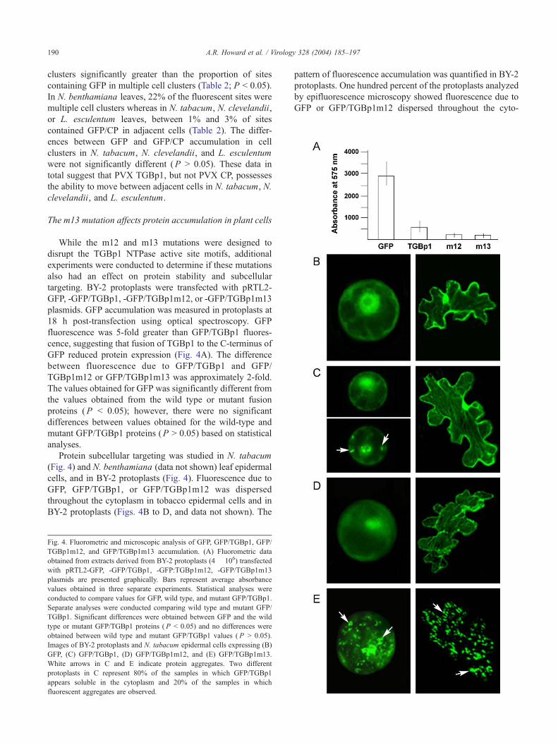
The m13 mutation affects protein accumulation in plant cells
While the m12 and m13 mutations were designed to
disrupt the TGBp1 NTPase active site motifs, additional
experiments were conducted to determine if these mutations
also had an effect on protein stability and subcellular
targeting. BY-2 protoplasts were transfected with pRTL2-
GFP, -GFP/TGBp1, -GFP/TGBp1m12, or -GFP/TGBp1m13
plasmids. GFP accumulation was measured in protoplasts at
18 h post-transfection using optical spectroscopy. GFP
fluorescence was 5-fold greater than GFP/TGBp1 fluores-
cence, suggesting that fusion of TGBp1 to the C-terminus of
GFP reduced protein expression (Fig. 4A). The difference
between fluorescence due to GFP/TGBp1 and GFP/
TGBp1m12 or GFP/TGBp1m13 was approximately 2-fold.
The values obtained for GFP was significantly different from
the values obtained from the wild type or mutant fusion
proteins (P b 0.05); however, there were no significant
differences between values obtained for the wild-type and
mutant GFP/TGBp1 proteins (P N 0.05) based on statistical
analyses.
Protein subcellular targeting was studied in N. tabacum
(Fig. 4) and N. benthamiana (data not shown) leaf epidermal
cells, and in BY-2 protoplasts (Fig. 4). Fluorescence due to
GFP, GFP/TGBp1, or GFP/TGBp1m12 was dispersed
throughout the cytoplasm in tobacco epidermal cells and in
BY-2 protoplasts (Figs. 4B to D, and data not shown). The
Fig. 4. Fluorometric and microscopic analysis of GFP, GFP/TGBp1, GFP/
TGBp1m12, and GFP/TGBp1m13 accumulation. (A) Fluorometric data
obtained from extracts derived from BY-2 protoplasts (4 � 106) transfected
with pRTL2-GFP, -GFP/TGBp1, -GFP:TGBp1m12, -GFP/TGBp1m13
plasmids are presented graphically. Bars represent average absorbance
values obtained in three separate experiments. Statistical analyses were
conducted to compare values for GFP, wild type, and mutant GFP/TGBp1.
Separate analyses were conducted comparing wild type and mutant GFP/
TGBp1. Significant differences were obtained between GFP and the wild
type or mutant GFP/TGBp1 proteins ( P b 0.05) and no differences were
obtained between wild type and mutant GFP/TGBp1 values ( P N 0.05).
Images of BY-2 protoplasts and N. tabacum epidermal cells expressing (B)
GFP, (C) GFP/TGBp1, (D) GFP/TGBp1m12, and (E) GFP/TGBp1m13.
White arrows in C and E indicate protein aggregates. Two different
protoplasts in C represent 80% of the samples in which GFP/TGBp1
appears soluble in the cytoplasm and 20% of the samples in which
fluorescent aggregates are observed.
pattern of fluorescence accumulation was quantified in BY-2
protoplasts. One hundred percent of the protoplasts analyzed
by epifluorescence microscopy showed fluorescence due to
GFP or GFP/TGBp1m12 dispersed throughout the cyto-
A.R. Howard et al. / Virology 328 (2004) 185–197 191
plasm (Figs. 4B and D). Eighty percent of protoplasts
expressing GFP/TGBp1 had fluorescence dispersed through-
out the cytoplasm while 20% of protoplasts showed a few
fluorescent aggregates (Fig. 4C). Because TGBp1 accumu-
lates in inclusion bodies during virus infection (Davies et al.,
1993), we assume that these aggregates are GFP/TGBp1-
containing inclusion bodies.
Numerous fluorescent aggregates were observed in N.
tabacum and N. benthamiana (data not shown) leaf
epidermal cells, and in BY-2 protoplasts expressing GFP/
TGBp1m13 (Fig. 4E). Ninety-three percent of protoplasts
showed GFP/TGBp1m13 primarily in aggregates (Fig. 4E)
and only 7% showed fluorescence throughout the cyto-
plasm. The abundance of these GFP/TGBp1m13-containing
aggregates suggests that the m13 mutation altered protein
subcellular targeting.
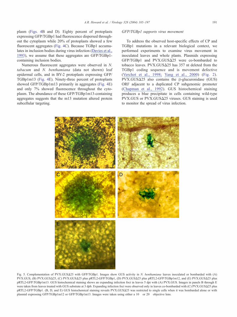
Fig. 5. Complementation of PVX.GUSD25 with GFP/TGBp1. Images show GU
PVX.GUS, (B) PVX.GUSD25, (C) PVX.GUSD25 plus pRTL2-GFP/TGBp1, (D)
pRTL2-GFP:TGBp1m13. GUS histochemical staining shows an expanding infect
were taken from leaves treated with GUS substrate at 3 dpb. Expanding infection fo
pRTL2-GFP/TGBp1. (B, D, and E) GUS histochemical staining reveals PVX.GU
plasmid expressing GFP/TGBp1m12 or GFP/TGBp1m13. Images were taken usi
GFP/TGBp1 supports virus movement
To address the observed host-specific effects of CP and
TGBp1 mutations in a relevant biological context, we
performed experiments to examine virus movement in
inoculated leaves and whole plants. Plasmids expressing
GFP/TGBp1 and PVX.GUSD25 were co-bombarded to
tobacco leaves. PVX.GUSD25 has 357 nt deleted from the
TGBp1 coding sequence and is movement defective
(Verchot et al., 1998; Yang et al., 2000) (Fig. 2).
PVX.GUSD25 also contains the h-glucuronidase (GUS)
ORF adjacent to a duplicated CP subgenomic promoter
(Chapman et al., 1992). GUS histochemical staining
produces a blue precipitate in cells containing wild-type
PVX.GUS or PVX.GUSD25 viruses. GUS staining is used
to monitor the spread of virus infection.
S activity in N. benthamiana leaves inoculated or bombarded with (A)
PVX.GUSD25 plus pRTL2-GFP/TGBp1m12, and (E) PVX.GUSD25 plus
ion foci in leaves 5 dpi with (A) PVX.GUS. Images in panels B through E
ci were observed only in leaves co-bombarded with (C) PVX.GUSD25 plus
SD25 was restricted to single cells when it was bombarded alone or with
ng either a 10� or 20� objective lens.
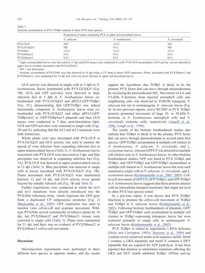
Table 3
Systemic accumulation of PVX TGBp1 mutants in three PVX hosts species
Proportions of plants containing PVX in upper non-inoculated leaves
N. tabacum N. benthamiana N. clevelandii
PVX.GUSa NDb 12/12 ND
PVX.GUSD25a ND 0/12 ND
PVX204ec 12/12 12/12 12/12
PVX204em12 0/12 0/12 0/12
PVX204em13 0/12 0/12 0/12
a Upper noninoculated leaves were harvested at 12 dpi and GUS assays were conducted to verify PVX.GUS accumulation. GUS activity was not detected in
upper leaves of plants inoculated with PVX.GUSD25.b ND = not determined.c Systemic accumulation of PVX204e was first observed at 10 dpi using a UV lamp to detect GFP expression. Plants, inoculated with PVX204em12 and
PVX204em13, were maintained for 14 dpi and virus was never detected in upper non-inoculated leaves.
A.R. Howard et al. / Virology 328 (2004) 185–197192
GUS activity was detected in single cells at 3 dpb in N.
benthamiana leaves bombarded with PVX.GUSD25 (Fig.
5B). GUS and GFP activities were detected in large
infection foci at 3 dpb in N. benthamiana leaves co-
bombarded with PVX.GUSD25 and pRTL2-GFP/TGBp1
(Fig. 5C), demonstrating that GFP/TGBp1 was indeed
functionally competent. N. benthamiana leaves were co-
bombarded with PVX.GUSD25 and either pRTL2-GFP/
TGBp1m12 or -GFP/TGBp1m13 plasmids and then GUS
assays were conducted at 3 days post-inoculation (dpi).
GUS and GFP activities were restricted to single cells (Figs.
5D and E), indicating that the m12 and m13 mutations were
both deleterious.
Whole plants were also inoculated with PVX.GUS or
PVX.GUSD25 and GUS activity was used to monitor the
spread of virus infection from expanding infection foci to
upper noninoculated leaves (Table 3). N. benthamiana leaves
inoculated with PVX.GUS were harvested at 3 dpi, and blue
precipitate was observed in expanding infection foci (Fig.
5A). PVX.GUS was detected in upper noninoculated leaves
at 12 dpi (Table 3). Blue precipitate was detected in single
cells in leaves inoculated with PVX.GUSD25 (Fig. 5B).
Plants inoculated with PVX.GUSD25 were maintained
between 12 and 14 dpi, and GUS activity never spread
beyond the initially infected cell (Fig. 5B and Table 3).
Further experiments were conducted in which the m12
and m13 mutations were directly introduced into the
PVX204e infectious clone, which expresses the GFP gene
from a duplicated CP subgenomic promoter (Fig. 2)
(Baulcombe et al., 1995). GFP expression was used to
monitor virus cell-to-cell and systemic movement. Wild-
type PVX204e moved systemically in tobacco plants by 10
dpi, but PVX204em12 and PVX204em13 viruses were
restricted to single cells (Table 3). Plants were maintained
for 21 dpi, and there was no evidence of PVX204em12 or
PVX204em13 cell-to-cell movement.
Discussion
Microinjection experiments were performed in three
different host species in separate studies, and the results
support the hypothesis that TGBp1 is likely to be the
primary PVX factor that can move through plasmodesmata
by increasing the plasmodesmal SEL. Movement of 4.4- and
9.5-kDa F-dextrans from injected mesophyll cells into
neighboring cells was observed in TGB100 transgenic N.
tabacum but not in nontransgenic N. tabacum leaves (Fig.
4). In two previous reports, active WClMVor PVX TGBp1
proteins promoted movement of large 10- or 20-kDa F-
dextrans in N. benthamiana mesophyll cells and N.
clevelandii trichome cells, respectively (Angell et al.,
1996; Lough et al., 1998).
The results of the biolistic bombardment studies also
indicate that TGBp1 is likely to be the primary PVX factor
that can move through plasmodesmata in several plant host
species. GFP/TGBp1 accumulated in multiple cell clusters in
N. benthamiana, N. tabacum , N. clevelandii, and L.
esculentum leaves, whereas GFP/CPwas detected inmultiple
cell clusters only in N. benthamiana leaves. In other biolistic
bombardment studies, GFP was fused to PVX TGBp2 and
TGBp3, and GFP/TGBp2 and GFP/TGBp3 accumulated in
multiple cell clusters in N. benthamiana leaves but were each
restricted to single cells in N. tabacum, N. clevelandii, and L.
esculentum leaves (Krishnamurthy et al., 2002, 2003). Cell-
to-cell movement of GFP/CP, GFP/TGBp2, and GFP/TGBp3
inN. benthamiana leaves suggests that these proteins interact
with an intercellular transport machinery that might not exist
in other PVX host species tested.
In a previous report, it was shown that PVX TGBp1
functions to promote the cell-to-cell movement of TGBp2
and TGBp3 in N. tabacum leaves (Krishnamurthy et al.,
2002). Following biolistic bombardment of plasmids, GFP/
TGBp2 and GFP/TGBp3 each accumulated in multiple cell
clusters in TGBp1-expressing transgenic leaves but were
restricted primarily to single cells in nontransgenic N.
tabacum leaves (Krishnamurthy et al., 2002).
PVX TGBp1 is related to superfamily I RNA helicases
(Dolja and Carrington, 1992a; Morozov et al., 1999) and
contains seven conserved amino acid sequence motifs. Motif
I contains a GKS tripeptide and motif II contains a DEY
tripeptide that are required for ATP hydrolysis. It has been
reported that deletion or substitution mutations affecting the
GKS and DEY motifs inhibited TGBp1 ATPase activity
A.R. Howard et al. / Virology 328 (2004) 185–197 193
(Donald et al., 1997; Morozov et al., 1999). The m12
mutation replaced sequences encoding the GKS tripeptide
with sequences encoding an AET tripeptide. A, E, and T are
similar to G, K, and S, and the substitution mutation would
likely disable ATPase activity without disturbing the tertiary
structure of the protein. The m13 mutation replaced
sequences encoding the DEY tripeptide with sequences
encoding RRF. While the m13 mutation was designed to
eliminate ATPase activity by replacing D and E, which are
negatively charged residues with two positively charged R
residues. However, this mutation may have had an effect on
the tertiary structure of the protein, as evidenced by the
aggregation of GFP/TGBp1m13 seen in bombarded leaves
and in protoplasts. Thus, the effects of the m13 mutation on
protein subcellular accumulation were more severe than the
m12 mutation. Based on these observations, we cannot
conclude whether ATPase activity or protein subcellular
targeting is important for TGBp1 and virus movement.
The m13 mutation is interesting because it has a general
effect on protein accumulation in plant cells, which
correlates with an inhibition of virus movement in N.
benthamiana and N. tabacum plants. The effects of m12
and m13 mutations on protein movement in N. benthami-
ana were not as extreme as in N. tabacum, N. clevelandii,
and L. esculentum leaves. Cell-to-cell movement of the
GFP/TGBp1m12 and GFP/TG Bp1m13 is severely
inhibited in N. tabacum, N. clevelandii, and L. esculentum,
but is moderately hampered in N. benthamiana leaves. The
results of mutational analyses provide further evidence that
protein movement in N. benthamiana leaves might not
reflect inherent properties of the proteins, but might reflect
unique properties of N. benthamiana plants. It is possible
that N. benthamiana plants differ from the other three PVX
host species in how plasmodesmata gating is triggered, the
extent to which the plasmodesmal SEL may be altered, or
the cellular factors contributing to plasmodesmal transport.
N. benthamiana leaves might either contain a positive
factor that promotes protein movement through plasmo-
desmata (that is not present in the other host species tested)
or lack a repressor that occurs in other plant species to limit
protein trafficking through plasmodesmata. It is possible
that there are cellular chaperones mediating protein
plasmodesmal transport in N. benthamiana that are absent
from N. tabacum leaves. A class of Hsp70 chaperones that
contain a structural motif necessary for protein cell-to-cell
transport were recently isolated from Cucurbita maxima
plants (Aoki et al., 2002). There are also DNA-J-like
chaperones that interact with some viral movement proteins
(Oparka, 2004; Soellick et al., 2000). However, we do not
know if these Hsp70 or DNAJ-like chaperones interact
with PVX TGB proteins or CP for plasmodesmal transport.
TIP is a factor isolated from N. tabacum leaves that
interacts with TGBp2 and whose function is unknown
(Fridborg et al., 2003). It is possible that TIP is a factor in
N. tabacum that downregulates TGBp2 movement in this
host. Because TIP has not been isolated from N.
benthamiana, we do not know if the identical factor is
present or absent from N. benthamiana.
The least likely explanation is that the architecture of
plasmodesmata in N. benthamiana is different than in other
host species and that there is a greater opportunity for
nonspecific movement of proteins between cells in N.
benthamiana leaves. If, for example, the SEL of plasmo-
desmata in N. benthamiana is slightly larger than in other
Nicotiana species, then the restrictions on protein cell-to-
cell trafficking may be less. This explanation is not likely
because GFP does not move from cell to cell in N.
benthamiana under experimental conditions that promote
cell-to-cell movement of the GFP fusion proteins, suggest-
ing that the mechanism for protein plasmodesmata transport
in N. benthamiana is selective.
Data obtained from studies using N. benthamiana have
been used to build models explaining the role of the
potexvirus TGB proteins in plasmodesmata transport.
TGBp2, TGBp3, and CP move through plasmodesmata in
N. benthamiana leaves, and the current model suggests that
these proteins travel with virus or viral RNA through
plasmodesmata (Lough et al., 1998, 2000). Because experi-
ments conducted in other hosts fail to reproduce data
obtained in N. benthamiana leaves, the current model likely
reflects unique properties of N. benthamiana plants and
might not explain viral protein functions in a broader context.
Materials and methods
Microinjection of mesophyll cells
Microinjection experiments were performed using pre-
viously described protocols (Ding et al., 1995; Wolf et al.,
1989). Nontransgenic and TGBp1-expressing source
tobacco leaves were detached and floated on 0.1 M mannitol
in a petri plate. The epidermis was peeled from the abaxial
side of the leaf to expose the underlying mesophyll cells. The
leaf was placed on wet filter paper under the microscope with
the abaxial side up. A 1-mM solution of LYCH and a 5-mM
solution of either 4.4- or 9.4-kDa F-dextrans (Sigma, St.
Louis, MO) were each prepared in 5 mM KHCO3 (pH 8.0),
filtered through a 0.5 Am-pore syringe filter, and stored at 4
8C (Ding et al., 1992; Fujiwara et al., 1993; Wolf et al.,
1989). Mesophyll cells were injected with LYCH or FITC-
dextrans using a PV820 Pneumatic Pico Pump (World
Precision Instruments, Sarasota, FL) and an epifluorescence
Nikon E600 microscope. Injections were carried out at an
eject pressure of 5–15 psi. The PV820 has a regulated hold
pressure that maintains a constant low pressure following
each injection to help maintain cell turgor. The needle was
withdrawn slowly to allow the injection wound to seal. The
spread of fluorescence from injected cells to adjacent cells
was monitored following injection and images were taken
with a SPOT digital camera (Diagnostic Instruments Inc.,
Sterling Heights, MI) attached to the microscope.
A.R. Howard et al. / Virology 328 (2004) 185–197194
Bacterial strains and plasmids
All plasmids were used to transform Escherichia coli
strain JM109. Plasmids pRTL2-GFP, -GFP/TGBp1, and
-GFP/CP were described previously (Krishnamurthy et al.,
2002; Yang et al., 2000) (Fig. 1). Two substitution mutations
named m12 and m13 were introduced into pRTL2-
GFP:TGBp1 and pPVX204e plasmids, respectively, replac-
ing 9 nts encoding the GKS (GGT AAG GTC) and DEY
(GAT GAG TAT) motifs with 9 nts encoding AET (GCG
GAA ACC) and RRF (AGG AGA TTC) (Kalinina et al.,
2002; Morozov et al., 1999). These mutations were located at
positions 4585–4593 and 4729–4737 within the PVX
genome. The QuikChange Site-Directed Mutagenesis kit
(Stratagene, La Jolla, CA) requires two mutagenesis
oligonucleotides in the forward and reverse orientation. To
introduce the m12 mutation into pRTL2-GFP/TGBp1
plasmids, the forward oligonucleotides, GTA GCC GGA
GCC GCG GAA ACC ACA GCC CTA AGG, and the
reverse oligonucleotides, CCT TAG GGC TGT GGT TTC
GCG GCT CCG GCTAC, were used. To introduce the m13
mutation into plasmids, the forward oligonucleotides, TTC
GCA ATC CTC AGG AGATTC ACT TTG GAC AAC, and
the reverse oligonucleotides, GTT GTC CAA AGT GAA
TCT CCT GAG GAT TGC GAA, were used. Nucleotides
encoding the substitution mutations are underlined.
The plasmids pPVX.GUS, pPVX.GUSD25K,
pPVX204e, pPVX204em12, and pPVX204em13 are infec-
tious clones of PVX with either the GUS or GFP genes
inserted into the viral genome adjacent to a duplicated
subgenomic promoter (Fig. 1). GUS and GFP reporters are
used as a visual marker to study the spread PVX infection
(Baulcombe et al., 1995; Chapman et al., 1992). The
pPVX.GUS, pPVX.GUSD25K, and pPVX204e plasmids
were prepared previously (Baulcombe et al., 1995; Chapman
et al., 1992; Krishnamurthy et al., 2003; Verchot et al., 1998)
( F i g . 2 ) . Th e pPVX .GUSD25K , pPVX204 e ,
pPVX204em12, and pPVX204em13 plasmids contain
cDNA of the PVX genome adjacent to the CaMV 35S
promoter (Fig. 2). The pPVX.GUS plasmids contain the
PVX genomic cDNA adjacent to bacteriophage T7 pro-
moters (Fig. 2). The pPVXGUS.D25K plasmids have 357 nt
deleted from the TGBp1 gene, and this mutation inhibits
cell-to-cell movement of PVX (Verchot et al., 1998) (Fig. 2).
To introduce m12 and m13 mutations into the PVX
genome, a fragment of pPVX204e was PCR amplified using
a forward primer that anneals to the 3Vend of the replicase
gene (GCC AAA CAC CAC TGC ATA CCA GAG GAA
ATC), as well as a reverse primer (GGC GGT CGA CAT
TTA CTT GTA CAG CTC GTC CAT) that overlaps the GFP
gene and contains a SalI (underlined) site. The PCR products
were ligated into the pGEM-T Easy vector (Promega,
Madison, WI). Mutations were separately introduced into
the pGEM-T clones using QuikChange Site-Directed Muta-
genesis kit and the forward and reverse primers indicated
above. Then the pPVX204e and the pGEM-T plasmids,
containing a fragment of the PVX genome encoding the m12
or m13 mutations, were digested with AvrII and ApaI.
Digested pPVX204e and PVX genomic fragments contain-
ing m12 or m13 mutations were ligated.
Plant material
N. benthamiana, N. tabacum (cvs. Petit Havana or
Samsun nn), N. clevelandii, and L. esculentum (cv. Trust)
leaves were used. TGB100 transgenic N. tabacum plants
express PVX TGBp1, are susceptible to PVX infection, and
complement cell-to-cell movement of TGBp1-defective
PVX viruses, as previously described (Verchot and Car-
rington, 1995).
Leaves were identified as source or sink using the
carboxyfluorescein (CF) dye test to ensure that the leaves
used in biolistic bombardment experiments were physio-
logically similar. The CF dye test was developed as a
means to standardize the developmental stage of the leaves
being assayed (Oparka et al., 1994; Roberts et al., 1997).
CF dye (Sigma) is applied to the petiole of the most
mature leaf, moves through the phloem in a source-to-sink
direction, and unloads in sink but not source leaves (data
not shown) (Krishnamurthy et al., 2002; Oparka et al.,
1994; Roberts et al., 1997; Yang et al., 2000). Source
leaves were taken from similar, untreated plants for
biolistic bombardment experiments.
In vitro transcription and plant inoculation
Infectious transcripts derived from pPVX.GUS plasmids
were inoculated to N. benthamiana plants. Plasmids were
linearized with SpeI, and in vitro transcription reactions
were carried out by mixing the following: 5.0 Ag of
linearized DNA, 5 Al of 10� T7 transcription buffer, 2.0
Al SUPERased In ribonuclease inhibitor (20 U/Al) (Ambion,
Austin, TX), a 10� A/C/U/G mixture containing 20 mM
ATP, CTP, and UTP, and 2 mM GTP (Pharmacia-Pfizer,
Mississauga, Ontario, Canada), 5.0 Al m7(5V)ppp(5V)G Cap
Analog (Ambion), 4.0 Al of T7 polymerase (Ambion), and
nuclease-free water to a final volume of 50 Al. The reactionswere incubated at 37 8C for 15 min before the addition of
5.0 Al 20 mM GTP (Pharmacia-Pfizer) and 1 h at 37 8Cfollowing the addition of GTP. Transcripts (10 Al) were
mechanically inoculated to each leaf of an N. benthamiana
plant and two leaves per plant were inoculated in each
experiment. Experiments were repeated four times and a
total of 12 plants were inoculated per construct.
Biolistic bombardment
Source leaves were detached from plants of similar age
and bombarded with plasmids using the PDS 1000/He
System (Bio-Rad Laboratories, Hercules, CA), as
described previously (Krishnamurthy et al., 2002, 2003;
Yang et al., 2000). Leaves were bombarded with 20 Ag

A.R. Howard et al. / Virology 328 (2004) 185–197 195
pPVX.GUSD25K alone or 20 Ag pPVX.GUSD25K com-
bined with 20 Ag of either pRTL2-GFP/TGBp1, -GFP/
TGBp1m12, and -GFP/TGBp1 m13 mixed with 1 mg of 1
Am gold particles. Bombarded leaves were then stored in
the dark on Phytagar (Gibco BRL, Life Technologies, Inc.
Grand Island, NY) for 72 h before GUS histochemical
staining (see below). In other experiments, leaves were
bombarded with 10–20 Ag of the pRTL2 constructs mixed
with 1 mg of 1 Am gold particles. Ten microliters of a
DNA/gold mixture was loaded on a carrier disk and
bombarded to detached leaves as described previously
(Yang et al., 2000). GFP expression was monitored in
leaves bombarded with pRTL2 constructs using epifluor-
escence microscopy at 1 dpb.
Assays for GUS and GFP expression
PVX.GUS and PVX.GUSD25 inoculated leaves were
vacuum infiltrated as described previously with a working
solution of 10 mM EDTA, 0.5 mM K3(FeCN)6, 0.5 mM
K4(FeCN)6, and 1.2 mM 5-bromo-4-chloro-3-indoxlyl-h-d-glucuronide cyclohexylammonium salt (X-Gluc) (BD Bio-
sciences Clontech, Palo Alto, CA), and incubated overnight
at 37 8C (Dolja et al., 1992b; Verchot et al., 1998). The
infiltrated leaves were preserved with 95% ethanol, and
images were captured using the Optronics Magnafire
camera (Intelligent Imaging Innovations, Inc., Denver,
CO) attached to the Nikon E600 microscope (Krishnamur-
thy et al., 2002).
Confocal microscopy was conducted to collect images of
GFP, GFP/TGBp1, GFP/TGBp1m12, and GFP/TGBp1m13
in bombarded leaves. The Leica TCS SP2 system was
attached to a Leica DMRE microscope (Leica Micro-
systems, Bannockburn, IL). GFP expression was also
monitored in bombarded leaves and protoplasts using a
Nikon E600 (Nikon Corp., Tokyo, Japan) epifluorescence
microscope with a Nikon B2A filter cube (containing a 470-
to 490-nm excitation filter, a DM505 dichroic mirror, and a
BA520 barrier filter). Images were captured using the
Optronics Magnafire camera (Intelligent Imaging Innova-
tions, Inc.) attached to the Nikon E600 microscope
(Krishnamurthy et al., 2002).
Plants inoculated with PVX204e, PVX204em12, or
PVX204em13 viruses were monitored at 7, 14, and 21 dpi
using a hand-held model B-100 BLAK-RAY long wave
ultraviolet (UV) lamp (Ultraviolet Products, Upland, CA).
The data were recorded with a Sony Digital Still Camera
model DSC-F717 (Sony Corporation of America, New
York, NY). All images taken with the Optronics or Sony
cameras were edited using Adobe Photoshop version 4.0
software (Adobe Systems Inc., San Jose, CA).
BY-2 protoplast preparation and electroporation
Suspension cells of tobacco BY-2 (Nagata et al., 1992)
were cultured and maintained as described in Qi and Ding
(2002) with little modification. BY-2 cells were propagated
in the BY-2 culture media [Murashiege and Skoog medium
(MS salts; Sigma) supplemented with 30 g/l sucrose, 256
mg/l KH2 PO4, 100 mg/l myo-inositol, 1 mg/l thiamine, and
0.2 mg/l 2,4-D, pH 5.5] on a rotary shaker at 120 rpm at 28
8C in the dark and weekly subcultured by transferring 10 ml
BY-2 cells to 50 ml fresh media.
Protoplasts were isolated from the suspension cells as
previously described with slight modifications (Gaire et al.,
1999; Qi and Ding, 2002). Three-day-old cells in
suspension culture media were collected by centrifugation
at 200g for 5 min and resuspended in 1.5% cellulose
bOnozuka RSQ (Yakult Pharmaceutical Ind. Co., Ltd.,
Tokyo, Japan) and 0.2% macerase (Calbiochem-Novabio-
chem Corp. La Jolla, CA) in solution 1 [0.5 M mannitol,
3.6 mM 2-(N-morpholino) ethanesulfonic acid, pH 5.5]
and incubated for 3–5 h at 308C in water bath with rotary
shaker. The protoplasts collected by filtration through 41-
Am nylon mesh (Spectrum Laboratories, Inc., Rancho
Dominguez, CA) were centrifuged and washed twice with
solution 1 at 100g for 5 min. Finally, protoplasts were
resuspended in solution 2 (solution 1 plus 0.1 mM CaCl2)
to a density about 2 � 106 protoplasts/ml and incubated on
ice for 1 h.
Protoplasts were electroporated as described by Gaire et
al. (1999) with a few modifications. Protoplasts (1 � 106 in
0.5 ml) were mixed with 70–80 Ag of plasmid DNA and 40
Ag of sonicated salmon sperm DNA, as carrier. The
protoplast DNA mixture was placed in a 0.4-cm gap cuvette
(Bio-Rad Laboratories) on ice and then electroporated using
a Gene Pulser (Bio-Rad Laboratories) at 0.18 KV, 100 V,
and 125 AF with three pulses. Protoplasts were transferred
after electroporation into a new tube containing 1 ml of
solution 2, incubated on ice for 30 min, and then collected
by centrifugation at 59g for 5 min. Protoplasts were
resuspended in 1.5 ml of solution 3 (BY-2 culture media
plus 0.45 M mannitol) and added to 6-well cell culture
plates (Corning Inc., Corning, NY) containing solution 3
plus 1.0% agarose (pH 5.7). Protoplasts were cultured at 27
8C for 18 h and were then collected by centrifugation at 39g.
GFP expression was assayed either by epifluorescence
microscopy or fluorometric analyses (see below).
Fluorometric measurement of GFP expression in
protoplasts
To measure accumulation of GFP or GFP fusion proteins
in protoplasts, total protein was extracted from protoplasts
(about 4 � 106) by vortexing and sonicating in 100 Al ofgrinding buffer (10 mM Tris-HCl pH 7.5, 100 mM NaCl, 1
mM MgCl2, and 10 mM DDT) (Pang et al., 1996). Samples
were centrifuged at 3000g for 10 min and the supernatants
were removed for fluorometric analyses. GFP expression
was measured spectrally with excitation at 485 nm and
emission at 535 nm in VICTOR2 D fluorometer (Perkin-
Elmer, Boston, MA).
A.R. Howard et al. / Virology 328 (2004) 185–197196
Statistical analyses
All statistical analyses were conducted using PC SAS
Version 8.2 (SAS Institute, Cary, NC) and a 0.05 signifi-
cance level was used to compare all proportions. Fisher’s
exact tests with PROC FREQ in SAS were used to compare
proportions reported in Table 1. Fisher’s exact tests were
used because many of the proportions are small, which will
violate a sample size requirement needed to perform chi
square tests. Comparisons of plant species for a given
plasmid were made as well as comparisons of plasmid for a
given plant species. Pair wise comparisons of the levels
were performed for overall comparisons that proved
significant. Results of the multiple comparisons in Table 1
are presented with lower case letters (comparing plant
species at a given plasmid) and upper case letters (compar-
ing plasmid types at a given plant species) (Krishnamurthy
et al., 2002, 2003).
Fluorometric measurements of GFP expression (Fig. 4)
were compared using analysis of variance (SAS Institute).
Values were assigned to a completely randomized model
with three replications and different plasmids as the treat-
ment variable. Treatment means were separated using a
Fisher’s protected least significant difference (LSD).
Acknowledgments
We thank Barbara A. Driskel and Dr. G. Schoenknecht
for technical support. Dr. Biao Ding provided vital training
and consultation on the microinjection technique. We also
appreciate the support of Dr. Arron Guenzi and the
Oklahoma State University Wheat Transformation Center
for use of the PDS1000/He DNA delivery system. The OSU
Electron Microscopy Facility houses a Leica confocal
microscope, which was obtained through NSF Major
Research Instrumentation Award 0079329. We also wish
to thank Dr. U. Melcher and Dr. K. Scheets for reviewing
the manuscript. This work was approved for publication by
the director of the Oklahoma Agriculture Experiment
Station and supported in part under the project H-2371.
The project was funded by the National Science Foundation
Integrative Plant Biology Program Award IBM-9982552.
References
Angell, S.M., Davies, C., Baulcombe, D.C., 1996. Cell-to-cell movement of
Potato Virus X is associated with a change in the size-exclusion limit of
plasmodesmata in trichome cells of Nicotiana clevelandii. Virology
216, 197–201.
Aoki, K., Kragler, F., Xoconostle-Cazares, B., Lucas, W.J., 2002. A
subclass of plant heat shock cognate 70 chaperones carries a motif that
facilitates trafficking through plasmodesmata. Proc. Natl. Acad. Sci.
U.S.A. 99, 16342–163467.
Baulcombe, D.C., Chapman, S., Santa Cruz, S., 1995. Jellyfish green fluo-
rescent protein as a reporter for virus infections. Plant J. 7, 1045–1053.
Chapman, S., Kavanagh, T., Baulcombe, D.C., 1992. Potato Virus X as a
vector for gene expression in plants. Plant J. 2, 549–557.
Davies, C., Hills, G., Baulcombe, D.C., 1993. Sub-cellular localization of
the 25-kDa protein encoded in the triple gene block of Potato virus X.
Virology 197, 166–175.
Ding, B., Haudenshield, J.S., Hull, R.J., Wolf, S., Beachy, R.N., Lucas,
W.J., 1992. Secondary plasmodesmata are specific sites of localization
of the Tobacco mosaic virus movement protein in transgenic tobacco
plants. Plant Cell 4, 915–928.
Ding, B., Li, Q., Nguyen, L., Palukaitis, P., Lucas, W.J., 1995. Cucumber
mosaic virus 3a protein potentiates cell-to-cell trafficking of CMV RNA
in tobacco plants. Virology 207, 345–353.
Dolja, V.V., Carrington, J.C., 1992. Evolution of positive-strand RNA
viruses. Semin. Virol. 3, 315–326.
Dolja, V.V., McBride, H.J., Carrington, J.C., 1992. Tagging of plant
potyvirus replication and movement by insertion of h-glucuronidaseinto the viral polyprotein. Proc. Natl. Acad. Sci. U.S.A. 89,
10208–10212.
Donald, R.G.K., Lawrence, D.M., Jackson, A.O., 1997. The Barley stripe
mosaic virus 58-Kilodalton hb protein is a multifunctional RNA
binding protein. J. Virol. 71, 1538–1546.
Forster, R.L.S., Beck, D.L., Guilford, P.J., Voot, D.M., Van Dolleweerd,
C.J., Andersen, M.T., 1992. The coat protein of White clover mosaic
potexvirus has a role in facilitating cell-to-cell transport in plants.
Virology 191, 480–484.
Fridborg, I., Grainger, J., Page, A., Coleman, M., Findlay, K., Angell, S.,
2003. TIP, a novel host factor linking callose degradation with the cell-
to-cell movement of Potato virus X. Mol. Plant-Microb. Interact. 16,
132–140.
Fujiwara, T., Giesman-Cookmeyer, D., Ding, B., Lommel, S., Lucas, W.J.,
1993. Cell-to-cell trafficking of macromolecules through plasmodes-
mata potentiated by the Red clover necrotic mosaic virus movement
protein. Plant Cell 5, 1783–1794.
Gaire, F., Schmitt, C., Stusssi-Garaud, C., Pinck, L., Ritzenthaler, C.,
1999. Protein 2A of Grapevine fanleaf nepovirus is implicated in
RNA2 replication and colocalizes to the replication site. Virology 264,
25–36.
Gilmer, D., Bouzoubaa, S., Hehn, A., Guilley, H., Richards, K., Jonard, G.,
1992. Efficient cell-to-cell movement of Beet necrotic yellow vein virus
requires 3Vproximal genes located on RNA 2. Virology 189, 40–47.
Gorbalenya, A.E., Koonin, E.V., 1993. Helicases: amino acid sequence
comparisons and structure–function relationships. Curr. Opin. Cell Biol.
3, 419–429.
Huisman, M.J., Linthorst, H.J.M., Bol, J.F., Cornelissen, B.J.C., 1988. The
complete nucleotide sequence of Potato virus X and its homologies at
the amino acid level with various plus-stranded RNA viruses. J. Gen.
Virol. 69, 1789–1798.
Itaya, A., Hickman, H., Bao, Y., Nelson, R., Ding, B., 1997. Cell-to-cell
trafficking of Cucumber mosaic virus movement protein: green
fluorescent protein fusion produced by biolistic gene bombardment in
tobacco. Plant J. 12, 1223–1230.
Kalinina, N.O., Rakitina, D.A., Yelina, N.E., Zamyatnin Jr., A.A.,
Stroganova, T.A., Klinov, D.V., Prokhorov, V.V., Ustinova, S.V.,
Chernov, B.K., Schiemann, J., Solovyev, A.G., Morozov, S.Y.,
2001. RNA-binding properties of the 63-kDa protein encoded by
the triple gene block of Poa semilatent hordeivirus. J. Gen. Virol.
82, 2569–2578.
Kalinina, N., Rakitina, D.A., Solovyev, A.G., Schiemann, J., Morozov,
S.Y., 2002. RNA helicase activity of the plant virus movement
proteins encoded by the first gene of the triple gene block. Virology
296, 321–329.
Krishnamurthy, K., Mitra, R., Payton, M.E., Verchot-Lubicz, J., 2002. Cell-
to-cell movement of the PVX 12K, 8K, or coat proteins may depend on
the host, leaf developmental stage, and the PVX 25K protein. Virology
300, 269–281.
Krishnamurthy, K., Heppler, M., Mitra, R., Blancaflor, E., Payton, M.E.,
Nelson, R.S., Verchot-Lubicz, J., 2003. The Potato virus X TGBp3
A.R. Howard et al. / Virology 328 (2004) 185–197 197
protein associates with the ER network for virus cell-to-cell movement.
Virology 309, 135–151.
Lough, T.J., Shash, K., Xoconostle-Cazares, B., Hofstra, K.R., Beck, D.L.,
Balmori, E., Forster, R.L.S., Lucas, W.J., 1998. Molecular dissection of
the mechanism by which potexvirus triple gene block proteins mediate
cell-to-cell transport of infectious RNA. Mol. Plant-Microb. Interact.
11, 801–814.
Lough, T.J., Netzler, N.E., Emerson, S.J., Sutherland, P., Carr, F., Beck,
D.L., Lucas, W.J., Forster, R.L.S., 2000. Cell-to-cell movement of
potexviruses: evidence for a ribonucleoprotein complex involving the
coat protein and first triple gene block protein. Mol. Plant-Microb.
Interact. 13, 962–974.
Memelink, J., van der Vlugt, C.I.M., Linthorst, H.J.M., Derk, A.F.L.M.,
Asjes, C.J., Bol, J.F., 1990. Homologies between the genomes of a
carlavirus (Lily symptomless virus) and a potexvirus (Lily virus X)
from lily plants. J. Gen. Virol. 71, 917–924.
Mitra, R., Krishnamurthy, K., Blancaflor, E., Payton, M., Nelson, R.S.,
2003. The Potato virus X TGBp2 protein associates with the ER
and Golgi network for virus cell-to-cell movement. Virology 312,
35–48.
Morozov, S.Y., Lukasheva, L.I., Chernov, B.K., Skryabin, K.G., Atabekov,
J.G., 1987. Nucleotide sequence of the open reading frames adjacent to
the coat protein cistron in Potato virus X genome. FEBS Lett. 213,
438–442.
Morozov, S.Y., Fedorkin, O.N., Juttner, G., Schiemann, J., Baulcombe,
D.C., Atabekov, J.G., 1997. Complementation of a Potato virus X
mutant mediated by bombardment of plant tissues with cloned viral
movement protein genes. J. Gen. Virol. 78, 2077–2083.
Morozov, S.Y., Solovyev, A.G., Kalinina, N.O., Fedorkin, O.N., Samui-
lova, O.V., Schiemann, J., Atabekov, J.G., 1999. Evidence for two
nonoverlapping functional domains in the Potato virus X 25K move-
ment protein. Virology 260, 55–63.
Nagata, T., Nemoto, Y., Hasezawa, S., 1992. Tobacco BY-2 cell line as the
bHeLaQ cell in the cell biology of higher plants. Int. Rev. Cyt. 132, 1–30.Oparka, K.J., 2004. Getting the message across: how do plant cells
exchange macromolecular complexes? Trends Plant Sci. 9, 33–41.
Oparka, K.J., Duckett, C.M., Prior, D.A.M., Fisher, D.B., 1994. Real-time
imaging of phloem unloading in the root tip of Arabidopsis. Plant J. 6,
759–766.
Oparka, K.J., Roberts, A.G., Roberts, I.M., Prior, D.A.M., Santa Cruz, S.,
1996. Viral coat protein is targeted to, but does not gate plasmodesmata
during cell-to-cell movement of Potato virus X. Plant J. 10, 805–813.
Pang, S.-Z., DeBore, D.L., Wan, Y., Ye, G., Layton, J.G., Neher, M.K.,
Armstrong, C.L., Fry, J.E., Hinchee, M.A.W., Fromm, M.E., 1996. An
improved green fluorescent protein gene as a vital marker in plants.
Plant Physiol. 112, 893–900.
Qi, Y., Ding, B., 2002. Replication of potato spindle tuber viroid in cultured
cells of tobacco and Nicotiana benthamiana: the role of specific
nucleotides in determining replication levels for host adaptation.
Virology 302, 445–456.
Robards, A.W., Lucas, W.J., 1990. Plasmodesmata. Annu. Rev. Plant
Physiol. Plant Mol. Biol. 41, 369–419.
Roberts, A.G., Santa Cruz, S., Roberts, I.M., Prior, D.A.M., Turgeon, R.,
Oparka, K.J., 1997. Phloem unloading in sink leaves of Nicotiana
benthamiana: comparison of a fluorescent solute with a fluorescent
virus. Plant Cell 9, 1381–1396.
Rouleau, M., Smith, R.J., Bancroft, J.B., Mackie, G.A., 1994. Purification,
properties, and subcellular localization of Foxtail mosaic potexvirus 26-
kDa protein. Virology 204, 254–265.
Santa Cruz, S., Chapman, S., Roberts, A.G., Roberts, I.M., Prior, D.A.M.,
Oparka, J.K., 1996. Assembly and movement of a plant virus carrying a
green fluorescent protein overcoat. Proc. Natl. Acad. Sci. U.S.A. 93,
6286–6290.
Skryabin, K.G., Morozov, S.Y., Kraev, A.S., Rozanov, M.N., Chernov,
B.K., Lukasheva, L.I., Atabekov, J.G., 1988. Conserved and variable
elements in RNA genomes of potexviruses. FEBS 240, 33–40.
Soellick, T.-R., Uhrig, J.F., Bucher, G.L., Kellman, J.-W., Schreier, P.H.,
2000. The movement protein of NSm of Tomato spotted wilt tospovirus
(TSWV): RNA binding, interaction with the TSWV N protein, and
identification of interacting plant proteins. Proc. Natl. Acad. Sci. U.S.A.
97, 2373–2378.
Tamai, A., Meshi, T., 2001. Cell-to-cell movement of Potato virus X: the
role of p12 and p8 encoded by the second and third open reading frames
of the triple gene block. Mol. Plant-Microb. Interact. 14, 1158–1167.
Verchot, J., Carrington, J.C., 1995. Evidence that the Potyvirus P1
proteinase functions in trans as an accessory factor for genome
amplification. J. Virol. 69, 3668–3674.
Verchot, J., Angell, S.M., Baulcombe, D.C., 1998. In vivo translation of the
triple gene block of Potato virus X (PVX) requires two mRNAs.
J. Virol. 72, 8316–8320.
Wolf, S., Deom, C.M., Beachy, R.N., Lucas, W.J., 1989. Movement protein
of Tobacco mosaic virus modifies plasmodesmatal size exclusion limit.
Science 246, 339–377.
Yang, Y., Ding, B., Baulcombe, D.C., Verchot, J., 2000. Cell-to-cell
movement of the 25K protein of Potato virus X is regulated by three
other viral proteins. Mol. Plant-Microb. Interact. 13, 599–605.
Copyright © 2022 FDOKUMEN