Poster display IV experimental and instrumentation
27
SPECT/CT using Iodine-123 in patients with differentiated thyroid cancer - additional value over whole body planar imaging and SPECT Tara Barwick 1,4 , Iain Murray 1 , Hakim Megadmi 1 , William M. Drake 2 , P. Nick Plowman 3 , Scott A. Akker 2 , Shern l. Chew 2 , Ashley B. Grossman 2 , and Norbert Avril 1 Departments of Nuclear Medicine 1 , Endocrinology 2 and Clinical Oncology 3 , Barts and The London School of Medicine and NHS Trust, London, UK 4 current address: Department of Nuclear Medicine, Imperial College Healthcare NHS Trust, London, UK Corresponding Author: Norbert Avril, M.D., Department of Nuclear Medicine, Barts and The London, Queen Mary, University of London, West Smithfield (QE II), London EC1A 7BE, United Kingdom Tel. #44(20)7601-7144, Fax. #44(20)7601-7149, Email: [email protected] Key words: differentiated thyroid cancer; SPECT/CT, radioiodine imaging, Iodine-123 Running title: I-123 SPECT/CT in thyroid cancer Acknowledgements: We would like to thank Mr. Tri Tat from the Department of Statistics, Imperial College, London, UK for statistical support. Word Count: 3570 Page 1 of 27 Accepted Preprint first posted on 8 March 2010 as Manuscript EJE-09-1023 Copyright © 2010 European Society of Endocrinology.
-
Upload
independent -
Category
Documents
-
view
1 -
download
0
Transcript of Poster display IV experimental and instrumentation

SPECT/CT using Iodine-123 in patients with differentiated thyroid cancer - additional
value over whole body planar imaging and SPECT
Tara Barwick1,4, Iain Murray1, Hakim Megadmi1, William M. Drake2, P. Nick Plowman3, Scott
A. Akker2, Shern l. Chew2, Ashley B. Grossman2, and Norbert Avril1
Departments of Nuclear Medicine1, Endocrinology2 and Clinical Oncology3, Barts and The
London School of Medicine and NHS Trust, London, UK
4 current address: Department of Nuclear Medicine, Imperial College Healthcare NHS Trust,
London, UK
Corresponding Author:
Norbert Avril, M.D., Department of Nuclear Medicine, Barts and The London, Queen Mary,
University of London, West Smithfield (QE II), London EC1A 7BE, United Kingdom
Tel. #44(20)7601-7144, Fax. #44(20)7601-7149, Email: [email protected]
Key words: differentiated thyroid cancer; SPECT/CT, radioiodine imaging, Iodine-123
Running title: I-123 SPECT/CT in thyroid cancer
Acknowledgements:
We would like to thank Mr. Tri Tat from the Department of Statistics, Imperial College,
London, UK for statistical support.
Word Count: 3570
Page 1 of 27 Accepted Preprint first posted on 8 March 2010 as Manuscript EJE-09-1023
Copyright © 2010 European Society of Endocrinology.

2
Abstract
The aim of the study was to assess the diagnostic performance of co-registered SPECT/CT
compared to Iodine-123 whole body gamma camera imaging (WBGC) and to SPECT alone
in patients with differentiated thyroid cancer.
Methods: WBGC and SPECT/CT (n=85) of the neck and thorax was performed in 79
consecutive patients. Three experienced observers reviewed: i) WBGC images followed by
ii) SPECT alone and iii) co-registered SPECT/CT. Foci of increased radioiodine uptake were
classified on a five point scale. Biopsy, other imaging modalities, and clinical follow-up
served as the reference standard.
Results: Twenty-two patients had local recurrence or metastatic thyroid cancer (11 were
radio-iodine negative), 9 had remnant thyroid tissue and 54 had no evidence of disease.
When classifying equivocal, probably and definitely malignant findings as positive for
malignancy, the sensitivity, specificity, positive-predictive value and negative-predictive value
were as follows: 41%, 68%, 31%, and 77% for WBGC; 45%, 89%, 59%, and 82% for WBGC
plus SPECT; and 50%, 100%, 100% and 85% for WBGC plus SPECT/CT, respectively. The
specificity was improved by addition of SPECT (p=0.0002) and SPECT/CT (p<0.0001) to
WBGC. SPECT/CT was also more specific than WBGC plus SPECT (p=0.016). In a study-
based analysis, SPECT/CT provided additional diagnostic information in 42% (36/85) of
cases. SPECT/CT provided further characterization in 70% (63/90) of foci and improved the
diagnostic confidence of all three observers.
Conclusion: The addition of SPECT/CT significantly improved the diagnostic information
over Iodine-123 whole body gamma camera imaging (WBGC) and WBGC plus SPECT
alone.
Page 2 of 27

3
INTRODUCTION
Patients with differentiated thyroid cancer have an overall good prognosis; however, lifelong
follow up is required for many cases, since potentially curable local recurrences and distant
metastases may occur even decades later 1-4. Radioiodine imaging plays a major role in the
follow up after initial surgery and ablation of residual thyroid tissue using Iodine-131 therapy,
although the need for such extensive therapy for microcarcinomas has been questioned.
Iodine-123 has replaced Iodine-131 at many institutions as a radionuclide for gamma camera
imaging due to its more suitable physical characteristics 5.
However, the lack of anatomical details on planar gamma camera imaging and
superimposition of areas presenting with increased radioiodine uptake can make accurate
diagnosis and localization of radioiodine-avid metastatic disease challenging. This is
particularly important in the neck and thorax where differentiation between physiological
radioiodine accumulation, remnant thyroid tissue and residual or recurrent thyroid cancer
may be difficult. Furthermore, accurate characterization and localization of thoracic
radioiodine uptake to lung, bone or lymph nodes can be equally challenging. In addition,
altered anatomy, particularly in the neck and sites of physiological radioiodine accumulation
such as in the oesophagus, may be misinterpreted as metastatic disease 6, 7. For example, a
retrospective review of 500 radioiodine whole body scans revealed diagnostic pitfalls leading
to additional imaging or diagnostic procedures in 59% of studies 8.
Single photon emission computed tomography (SPECT) provides a 3-D set of gamma
camera images which improve the localization of increased tracer uptake within the body.
This allows one to analyse the radioiodine distribution within the body in any plane; e.g. axial,
coronal and sagittal. However, precise anatomical localization is frequently still difficult due to
a lack of appropriate landmarks on SPECT images. Integrated SPECT/CT enables co-
registration of structural and functional data and provides patient-specific data for the
correction of photon attenuation 9, 10. This often permits improved spatial localization and
characterization of increased tracer uptake which should ultimately lead to improved
diagnostic performance in radioiodine imaging as well.
Most studies of SPECT/CT in thyroid cancer have used Iodine-131 to further evaluate
equivocal findings on planar imaging. In our study, Iodine-123 SPECT/CT of the neck and
thorax was performed in consecutive patients regardless of the findings on planar imaging.
At our institution, thyroid cancer patients generally undergo thyroidectomy and radioiodine
ablation, followed by endogenous TSH-stimulated Iodine-123 whole body planar gamma
camera imaging and serum thyroglobulin measurement in high-risk patients six months later.
Iodine-123 gamma camera imaging is also performed in patients with rising thyroglobulin
Page 3 of 27

4
levels. The routine protocol includes whole body planar imaging and SPECT/CT of the neck
and thorax at 24 hours after radioiodine injection.
This study represents a detailed analysis of the incremental value of the addition of Iodine-
123 SPECT/CT compared with whole body planar imaging and SPECT alone. In addition, we
assessed potential changes in patient management.
Page 4 of 27
5
MATERIALS AND METHODS
Patients
Consecutive patients with differentiated thyroid cancer, who underwent Iodine-123 imaging
from March 2006 to February 2009, were eligible for the study. Iodine-123 gamma camera
imaging was performed in patients with rising thyroglobulin levels as well as in high risk
patients 6 months following thyroidectomy and radioiodine ablation. Measurement of urinary
iodine excretion was not performed prior to Iodine-123 administration. All patients were
imaged following endogenous TSH stimulation.
This retrospective analysis of patient data was performed as part of an audit and institutional
audit committee approval was obtained. The demographical and follow up data were
collected through a combination of review of clinic/ correspondence letters/ multidisciplinary
meeting summaries and biochemical/histopathology databases on the electronic patient
record, review of patient notes and direct communication with the physician in charge of the
patient’s care. The information for each patient was recorded on a patient case record form.
Gamma Camera and SPECT/CT Imaging
Whole body planar gamma camera (WBGC) images were acquired in the anterior and
posterior projections approximately 24 hours post-radiotracer injection (350-400 MBq Iodine-
123 sodium iodide) on a dual-headed gamma camera (Millennium VG Hawkeye, GE
Healthcare, Amersham, UK) with low energy general purpose collimators (LEGP). The bed
speed was 5 cm/min and the data were acquired into a 256x1024 matrix, with a pixel size of
2.2mm. SPECT imaging of the neck and thorax was performed following WBGC with 60
seconds frame time, step and shoot mode 360/60 frames (6º angle step), 1.28 zoom. One
patient also underwent an additional SPECT/CT of the abdomen and pelvis. SPECT data
were reconstructed using an OSEM iterative reconstruction algorithm (Hermes, Nuclear
Diagnostics Medical Systems, Sweden) into a 128x128 matrix, with a pixel size of 3.45mm
that incorporated the attenuation map information described below.
The CT acquisition was carried out using a fixed protocol (140 kV, 2.5 mA) resulting in forty
10mm slices that were subsequently rebinned to produce 128 3.45mm slices with a
transverse pixel size of 1.7mm. The CT data was also rescaled to produce an attenuation
map related to attenuation coefficient values at 159 keV matching the dimensions of the
reconstructed SPECT slices.
Image Analysis
Page 5 of 27

6
Gamma camera images were reviewed by three experienced observers (TB, HM, NA) in a
consensus reading without knowledge of clinical data on a dedicated workstation (Hermes
Medical Solutions, Nuclear Diagnostics, Sweden) using a linear grey scale. For analysis of
SPECT/CT images, both linear grey and colour scale displays were used. The three
clinicians reviewed the scans first separately followed by a consensus reading. In all cases a
consensus was reached.
The whole body planar images were reviewed initially followed by the SPECT and then the
SPECT/CT. For Iodine-123 whole body planar imaging symmetrical uptake in the salivary
glands, linear uptake in the region of the oesophagus, uptake in the stomach, gastrointestinal
tract and bladder was considered normal. Any focal increased uptake outside these areas
was considered abnormal. Radioiodine uptake in the neck which was localized in the area of
the thyroid bed was considered residual thyroid tissue following previous radioiodine
ablation.
Sites of increased tracer uptake were noted along with the likelihood of the presence of
disease according to the following five point grading scale for whole body planar images,
SPECT alone and SPECT/CT:
1. definitely normal
2. probably normal (more likely to be physiological)
3. equivocal (equally likely to be pathological or physiological)
4. probably abnormal (more likely to be pathological)
5. definitely abnormal
For SPECT and SPECT/CT the anatomical localisation of increased radioiodine uptake was
taken into account when reviewing images. Although we used a low-dose CT for anatomical
correlation, the lung and bone windows were also reviewed for lung nodules and bone
metastases. In addition, the overall diagnostic confidence score for each patient was
allocated using the same five point scale. For subsequent analysis, definitely abnormal,
probably abnormal and equivocal findings were considered malignant. Probably normal and
definitely normal findings were considered benign. Thyroglobulin levels or findings from neck
ultrasound or any other clinical information were not made available to the image reviewers.
Statistical Analysis
The sensitivity, specificity, positive predictive value (PPV) and negative predictive value
(NPV) were calculated for whole body planar gamma camera imaging, SPECT and
SPECT/CT on a study basis. For the purpose of these calculations, equivocal findings were
considered malignant. Comparison of the detection of thyroid cancer metastases by each
method was performed using an exact Mcnemar test with a two-tailed p value <0.05 being
Page 6 of 27

7
statistically significant 11. Following the exclusion of 11 studies found on follow-up to have
non-radioiodine-avid disease, receiver-operating characteristic (ROC) curve analysis was
performed by recalculating sensitivity and specificity for each technique along the five point
grading scale. In addition, the area under the curve was calculated.
Page 7 of 27

8
RESULTS
Ninety-three patients underwent Iodine-123 imaging for follow up of thyroid cancer from
March 2006 to February 2009; however, in 12 cases SPECT/CT was not performed due to
logistical and/or technical reasons. In one case, neck movement resulted in substantial
image misregistration and one patient with normal planar and SPECT/CT imaging was lost to
follow up. Therefore, the study population consisted of 79 patients in whom a total of 85
Iodine-123 imaging studies were performed. Patient characteristics are shown in Table 1.
Of these cases, 64 were studied as routine follow-up of high risk patients 6 months after
radioiodine ablation of residual thyroid tissue; 5 were studied 6 months after radioiodine
therapy for completion of ablation (n=3) or for metastatic disease. Twelve studies were
performed in patients with rising or elevated thyroglobulin. The remaining three cases were
to exclude recurrence - one in a patient with severe shoulder pain who was found to have an
osteolytic lesion and in two patients with lung nodules, one of whom had a history of breast
cancer. One patient underwent Iodine-123 imaging prior to radioiodine ablation following
surgery.
Following endogenous TSH stimulation after withdrawal of L-thyroxine therapy, all patients
had elevated TSH levels (TSH>30 mIU/L) before intravenous administration of Iodine-123.
SPECT/CT of the neck and thorax was performed in all patients as per our imaging protocol
and a further SPECT/CT of the abdomen/pelvis was performed in one patient, which
revealed no abnormal radioiodine uptake. The reference standard was established by clinical
follow up including thyroglobulin levels in addition to histopathology (n=5), neck ultrasound
(n=18), computed tomography (n=17), gamma camera imaging of subsequent radioiodine
therapy (n=10), bone scintigraphy (n=2), MR imaging of the neck (n=2), MR imaging of the
spine (n=2) and [F-18]FDG-PET/CT (n=2). Mean follow up was 13 months.
Diagnostic performance
Based on the reference standard, there was evidence for residual or recurrent thyroid cancer
in 22 out of 85 Iodine-123 imaging studies, 11 of which were non-radioiodine avid. In
addition, 9 studies had radioiodine-avid remnant thyroid tissue only; 54 studies had no
evidence of disease. The diagnostic performance of whole body planar gamma camera
(WBGC) imaging, SPECT and SPECT/CT are presented in Tables 2 and 3. In a study-based
analysis, SPECT/CT provided additional diagnostic information in 42% (36/85) of cases.
The sensitivity was 41% for WBGC, 45% with the addition of SPECT and 50% with the
addition of SPECT/CT. However, although we observed an increase in sensitivity this did not
reach statistical significance for either WBGC versus SPECT (p=1.0) or WBGC versus
Page 8 of 27

9
SPECT/CT (p=0.5). When 11 cases found on follow-up to have non-radioiodine-avid disease
were excluded, the sensitivity increased from 82% for WBGC to 91% with the addition of
SPECT and to 100% with the addition of SPECT/CT.
Nevertheless, there was a significant improvement in specificity between WBGC imaging,
and the addition of SPECT from 68% to 89% (p=0.0002). The addition of SPECT/CT further
increased specificity to 100% (p<0.0001). The increase in specificity between SPECT and
SPECT/CT also was statistically significant (p=0.016).
A major factor in the increased specificity of SPECT and/or SPECT/CT was the re-
classification of initially equivocal findings. There were 12 WBGC scans classed as
equivocal, three remained so on SPECT but none on SPECT/CT. Classing equivocal
findings as positive for disease, WBGC was false-positive in 20 cases (9 thyroid remnants; 4
oesophageal tracer retention; 4 asymmetric salivary glands; 3 surface contamination) and
false negative in 13 cases (11 non-radioiodine avid metastases, 2 cervical nodal
metastases).
The addition of SPECT alone correctly classified 13 false-positive findings as physiological
activity or remnant and identified metastatic neck lymph nodes in one study and additional
bone metastases in another study. Therefore, the combination of WBGC and SPECT
produced 7 false-positive findings and 12 false-negative findings (Figures 1 and 2).
SPECT/CT correctly assigned all false-positive findings and identified a metastatic cervical
lymph node misinterpreted as submandibular gland on WBGC and SPECT alone. Several
additional small bone metastases were identified on the CT component of the study in a
patient already noted to have multiple bone metastases on WBGC and SPECT. SPECT/CT
was false-negative for 11 cases with non-radioiodine avid disease; 5 of these cases had
metastatic neck lymph nodes, one with additional miliary lung metastases. One case had a
2.5 cm metastatic skin lesion on the neck which was subsequently excised and three cases
had small lung metastases on diagnostic CT which were not visible on the low-dose CT
component of the SPECT/CT. In two cases the sites of disease remain occult but were
classed as radioiodine negative on the basis of a persistently elevated thyroglobulin.
ROC analysis
Following the exclusion of 11 cases found on follow up to have non-radioiodine-avid disease,
receiver-operating characteristic (ROC) curve analysis revealed an area under the curve
(AUC) for WBGC of 0.84, for WBGC plus SPECT of 0.95 and for WBGC plus SPECT/CT 1.0
(Figure 3).
Diagnostic confidence
Page 9 of 27

10
On a per study basis, overall diagnostic confidence increased with the addition of SPECT
and SPECT/CT to whole body gamma camera imaging. The percentage of studies classed
as ‘definitely normal’ or ‘definitely abnormal’ increased from 46% (39/85) with WBGC to 66%
(56/85) with the addition of SPECT to 89% (76/85) with the addition of SPECT/CT.
Change in diagnostic findings
The addition of SPECT/CT provided further characterization in 63 (70%) out of 90 foci
identified on 51 WBGC studies (Table 4). A total of 41 foci initially thought to be either
equivocal or pathological (false-positive) were re-classified by the addition of SPECT/CT as
being thyroid remnant (n=20) or other sites of physiological radioiodine uptake (n=21). Five
metastatic foci thought to be physiological (false-negative) were lymph node (n=3) or bone
metastases (n=2). For 17 foci the site of metastases was accurately localized.
Change in patient management
The addition of SPECT/CT to WBGC led to a change in management in 9 (11%) out of 85
cases. In two patients identification of cervical lymph node metastases that were not clearly
identified on WBGC imaging led to surgical lymph node dissection rather than radioiodine
therapy. In one patient thought to have both cervical and mediastinal lymph node
metastases, SPECT/CT localized the mediastinal activity to the oesophagus. Therefore, the
patient underwent a cervical lymph node dissection. In one patient thought to have localized
cervical nodal disease equivocal mediastinal uptake was shown to be a mediastinal lymph
node and the patient had radioiodine therapy rather than surgery. In 5 patients with elevated
thyroglobulin levels WBGC was thought to be abnormal; however, SPECT/CT showed the
increased Iodine-123 uptake to be physiological. These patients underwent diagnostic CT,
neck ultrasound and FDG-PET/CT scans and surgery or radiotherapy rather than radioiodine
therapy.
In addition, in 11 cases thought to have abnormal Iodine-123 uptake on WBGC, SPECT/CT
did not confirm disease obviating the need for further investigations or avoiding ineffective
radioiodine therapy. However, none of these patients had raised serum thyroglobulin levels
and most likely a ‘watch-and-wait’ policy would have been adopted. Thus, although
SPECT/CT provided additional information in these cases, we did not anticipate a change in
management.
Page 10 of 27

11
DISCUSSION
SPECT/CT improved the diagnostic performance of Iodine-123 imaging for differentiated
thyroid cancer. The specificity increased from 68% to 89% with the addition of SPECT
(p=0.0002), and to 100% with the addition of SPECT/CT (p<0.0001). The overall accuracy
improved from 61% for planar gamma camera imaging to 78% with the addition of SPECT
and to 87% with the addition of SPECT/CT. Furthermore, the diagnostic confidence of the
observers improved such that we found no equivocal findings using SPECT/CT.
The accurate localization and characterization of focally increased Iodine-123 uptake is often
not possible on planar gamma camera imaging, particularly in the neck and upper thorax.
Although the addition of SPECT improved the diagnostic accuracy, it frequently remained
difficult to differentiate between malignant and physiological radioiodine uptake. For example,
we found it difficult to resolve between residual thyroid tissue and cervical lymph node
metastases on whole body imaging with the addition of SPECT alone. Radioiodine retention
in the oesophagus was often recognized on planar imaging and SPECT by its typical linear
configuration. However, focally increased activity is better characterized by SPECT/CT. In 13
studies, SPECT/CT enabled accurate localization of physiological radioiodine retention in the
oesophagus thought to be possibly pathological on planar gamma camera imaging and
SPECT. This is an important finding as in such cases the patient is spared further imaging
procedures or even unnecessary radioiodine therapy 8.
The sensitivity of radioiodine whole body imaging depends on various factors including
technical parameters, the activity administered and the reference standard used 12. Although
we injected 350-400 MBq Iodine-123, the sensitivity was only 41% (9 out of 22), which is
lower than 70-89% sensitivity figures reported by others 13-15. However, previous studies
used for example post-radioiodine therapy imaging as the reference standard; thus, all
patients had radioiodine avid disease. In our study, follow-up revealed 11 cases with non-
radioiodine avid disease, which when excluded resulted in a sensitivity of 82% for planar
imaging, which compares well with these reports 13-15.
To date there have been several studies addressing the use of radioiodine SPECT/CT in
differentiated thyroid cancer 16-20. Fused SPECT/CT in 25 patients with inconclusive findings
on post-radioiodine ablation imaging improved anatomical localization in 17 (44%) out of 39
foci compared to SPECT alone 18. Furthermore, image interpretation changed in 15 (38%)
foci. In another study, SPECT/CT provided additional diagnostic information in 41 (57%) out
of 71 patients who underwent Iodine-131 imaging post radioiodine therapy (n=54) or as a
diagnostic procedure (n=17) 19. SPECT/CT particularly improved the characterization and
localization of increased Iodine-131 uptake in lymph node metastases, remnant thyroid
Page 11 of 27

12
tissue, lung and mediastinal as well as bone metastases. Similar results were found by
others 16, 17, 20. The additional diagnostic information of SPECT/CT over planar imaging
ranged from 57-74% of patients 16, 17, 19. When assessing the impact of SPECT/CT on patient
management we found a change in 11% of the patients. Previous reports found 24% 17, 25%
18, and 41% 19. However, these studies were limited to patients with indeterminate findings on
planar Iodine-131 imaging.
To the best of our knowledge, this is the first consecutive series using Iodine-123 SPECT/CT
in the follow up of thyroid cancer regardless of the findings on whole body imaging.
However, previous studies have analyzed consecutive series of cases using Iodine-131
imaging. Spanu and co-workers prospectively performed 117 SPECT/CT of the neck and
thorax demonstrating 116 foci on planar imaging 21. SPECT/CT had incremental value over
planar in 67.8% of positive cases (40/59) and modified management in (21/59) 35.6%
positive cases. This translated to an incremental value in 40/117 (34%) studies and change
in management in 21/117 (21%) studies. Of note, patients with non-radioiodine avid disease
were excluded from their analysis. Our findings compare well with additional diagnostic
information of SPECT/CT in 42% of studies. Spanu and co-workers found SPECT/CT to be
particularly useful in revealing nodal metastases adjacent to salivary glands occult on planar
imaging (4 cases in 3 patients) 21. Similarly, we found 3 cases of cervical nodal metastases in
2 patients. These would be potentially missed in other studies which only perform SPECT/CT
in equivocal findings on planar imaging. The specific advantage of SPECT/CT for nodal
assessment of the neck is also emphasized by a recent study which found a gain in
information on nodal stage in 20 (35%) out of 57 patients leading to a change in risk
stratification in 14 (25%) patients 22.
We found a reduction in the number of equivocal/ indeterminate findings with the addition of
SPECT/CT, which is supported by a recent study where positive SPECT/CT predicted
persistent/ recurrent disease in 9 (16%) out of 55 patients 23. Three out of nine patients (33%)
had non-radioiodine avid disease and accounted for false negative SPECT/CT as reported
for our study 23.
In common with the majority of previous studies discussed above the CT performed as part
of SPECT/CT in our study did not contribute independent diagnostic information. We used a
low resolution Hawkeye-CT, which was not of diagnostic image quality although the CT
images provided valuable anatomical information regarding the precise localization of
abnormal radioiodine uptake. More recently, SPECT/CT systems are incorporating
diagnostic multislice CT, capable of low-dose as well as diagnostic CT. Depending on the
protocol used these may permit even better lesion characterization and, as such, further
reduce the need for subsequent diagnostic studies 24. It is also important to consider the
Page 12 of 27

13
additional radiation exposure from the CT portion of SPECT/CT, which was in our cases
approximately 1 mSv in addition to approximately 5 mSv from I-123 imaging.
There are some limitations of this study which need to be considered. The diagnostic
performance was patient-based. This may underestimate the fraction of “false-negative”
findings. For example, two patients had lung metastases which were negative on SPECT/CT
but the patients were considered “true positive” for the purpose of a patient-based analysis
as cervical metastases were correctly identified. This is a general limitation as a detailed
focus based analysis is challenging because histopathologic confirmation would be difficult
as it is not feasible to sample all potential sites of disease.
The number of patients with radioiodine avid disease was limited. Patients subsequently
shown to have non-radioiodine avid disease were retained in this analysis of consecutive
patients. This resulted in lower sensitivities compared to previous reports. However, the
primary purpose of the study was to assess the incremental value of adding SPECT and
SPECT/CT to conventional planar gamma camera imaging. This analysis is not biased by
the study population. For the same reason, thyroglobulin levels or findings from neck
ultrasound or any other clinical information were not made available to the image reviewers.
In conclusion, our study has shown that the addition of SPECT/CT improved the diagnostic
performance of Iodine-123 whole body planar imaging and SPECT alone. With the
increasing availability of latest generation SPECT/CT systems, implementation into routine
clinical radioiodine imaging should be considered. Future studies should assess the optimum
protocol, impact on patient outcome and cost effectiveness.
Declaration of interest: There is no conflict of interest that could be perceived as
prejudicing the impartiality of the research reported.
Funding: This research did not receive any specific grant from any funding agency in the
public, commercial or not-for-profit sector. Only internal departmental resources were used.
Page 13 of 27

14
REFERENCES:
1. DeGroot LJ, Kaplan EL, McCormick M & Straus FH. Natural history, treatment, and
course of papillary thyroid carcinoma. J Clin Endocrinol Metab 1990 71 414-424.
2. Hay ID, Bergstralh EJ, Goellner JR, Ebersold JR & Grant CS. Predicting outcome in
papillary thyroid carcinoma: development of a reliable prognostic scoring system in a
cohort of 1779 patients surgically treated at one institution during 1940 through 1989.
Surgery 1993 114 1050-1057.
3. Hundahl SA, Fleming ID, Fremgen AM & Menck HR. A National Cancer Data Base
report on 53,856 cases of thyroid carcinoma treated in the U.S., 1985-1995. Cancer
1998 83 2638-2648.
4. Mazzaferri EL & Jhiang SM. Long-term impact of initial surgical and medical therapy
on papillary and follicular thyroid cancer. Am J Med 1994 97 418-428.
5. Siddiqi A, Foley RR, Britton KE, Sibtain A, Plowman PN, Grossman AB, Monson JP &
Besser GM. The role of 123I-diagnostic imaging in the follow-up of patients with
differentiated thyroid carcinoma as compared to 131I-scanning: avoidance of
negative therapeutic uptake due to stunning. Clin Endocrinol (Oxf) 2001 55 515-521.
6. Carlisle MR, Lu C & McDougall IR. The interpretation of 131I scans in the evaluation
of thyroid cancer, with an emphasis on false positive findings. Nucl Med Commun
2003 24 715-735.
7. Mitchell G, Pratt BE, Vini L, McCready VR & Harmer CL. False positive 131I whole
body scans in thyroid cancer. Br J Radiol 2000 73 627-635.
8. Leitha T & Staudenherz A. Frequency of diagnostic dilemmas in 131I whole body
scanning. Nuklearmedizin 2003 42 55-62.
9. Seo Y, Mari C & Hasegawa BH. Technological development and advances in single-
photon emission computed tomography/computed tomography. Semin Nucl Med
2008 38 177-198.
10. Townsend DW & Cherry SR. Combining anatomy and function: the path to true image
fusion. Eur Radiol 2001 11 1968-1974.
11. Hawass NE. Comparing the sensitivities and specificities of two diagnostic
procedures performed on the same group of patients. Br J Radiol 1997 70 360-366.
12. Lubin E, Mechlis-Frish S, Zatz S, Shimoni A, Segal K, Avraham A, Levy R &
Feinmesser R. Serum thyroglobulin and iodine-131 whole-body scan in the diagnosis
Page 14 of 27

15
and assessment of treatment for metastatic differentiated thyroid carcinoma. J Nucl
Med 1994 35 257-262.
13. de Geus-Oei LF, Oei HY, Hennemann G & Krenning EP. Sensitivity of 123I whole-
body scan and thyroglobulin in the detection of metastases or recurrent differentiated
thyroid cancer. Eur J Nucl Med Mol Imaging 2002 29 768-774.
14. Gerard SK & Cavalieri RR. I-123 diagnostic thyroid tumor whole-body scanning with
imaging at 6, 24, and 48 hours. Clin Nucl Med 2002 27 1-8.
15. Park HM, Park YH & Zhou XH. Detection of thyroid remnant/metastasis without
stunning: an ongoing dilemma. Thyroid 1997 7 277-280.
16. Chen L, Luo Q, Shen Y, Yu Y, Yuan Z, Lu H & Zhu R. Incremental value of 131I
SPECT/CT in the management of patients with differentiated thyroid carcinoma. J
Nucl Med 2008 49 1952-1957.
17. Kohlfuerst S, Igerc I, Lobnig M, Gallowitsch HJ, Gomez-Segovia I, Matschnig S, Mayr
J, Mikosch P, Beheshti M & Lind P. Posttherapeutic (131)I SPECT-CT offers high
diagnostic accuracy when the findings on conventional planar imaging are
inconclusive and allows a tailored patient treatment regimen. Eur J Nucl Med Mol
Imaging 2009 36 886-893.
18. Ruf J, Lehmkuhl L, Bertram H, Sandrock D, Amthauer H, Humplik B, Ludwig Munz D
& Felix R. Impact of SPECT and integrated low-dose CT after radioiodine therapy on
the management of patients with thyroid carcinoma. Nucl Med Commun 2004 25
1177-1182.
19. Tharp K, Israel O, Hausmann J, Bettman L, Martin WH, Daitzchman M, Sandler MP &
Delbeke D. Impact of 131I-SPECT/CT images obtained with an integrated system in
the follow-up of patients with thyroid carcinoma. Eur J Nucl Med Mol Imaging 2004 31
1435-1442.
20. Wong KK, Zarzhevsky N, Cahill JM, Frey KA & Avram AM. Incremental value of
diagnostic 131I SPECT/CT fusion imaging in the evaluation of differentiated thyroid
carcinoma. AJR Am J Roentgenol 2008 191 1785-1794.
21. Spanu A, Solinas ME, Chessa F, Sanna D, Nuvoli S & Madeddu G. 131I SPECT/CT
in the follow-up of differentiated thyroid carcinoma: incremental value versus planar
imaging. J Nucl Med 2009 50 184-190.
22. Schmidt D, Szikszai A, Linke R, Bautz W & Kuwert T. Impact of 131I SPECT/spiral
CT on nodal staging of differentiated thyroid carcinoma at the first radioablation. J
Nucl Med 2009 50 18-23.
Page 15 of 27

16
23. Aide N, Heutte N, Rame JP, Rousseau E, Loiseau C, Henry-Amar M & Bardet S.
Clinical relevance of single-photon emission computed tomography/computed
tomography of the neck and thorax in postablation (131)I scintigraphy for thyroid
cancer. J Clin Endocrinol Metab 2009 94 2075-2084.
24. Buck AK, Nekolla S, Ziegler S, Beer A, Krause BJ, Herrmann K, Scheidhauer K,
Wester HJ, Rummeny EJ, Schwaiger M & Drzezga A. SPECT/CT. J Nucl Med 2008
49 1305-1319.
Page 16 of 27

17
FIGURE LEGENDS
Figure 1a
Planar whole body Iodine-123 gamma camera imaging in anterior and posterior views shows
mildly increased linear activity in projection of the oesophagus but more focal activity
projected over the mediastinum. This was interpreted as equivocal for either radioiodine
retention in the oesophagus or a mediastinal lymph node metastasis.
Figure 1b The finding remained equivocal on SPECT but clearly localises to the oesophagus
on SPECT/CT
Figure 2a Whole body planar Iodine-123 gamma camera imaging in anterior view shows
minor asymmetric uptake in the region of the submandibular glands thought to be probably
normal.
Figure 2b SPECT/CT axial, sagittal and coronal images: On SPECT there is discrete focal
uptake bilaterally. On SPECT/CT this localised to several small bilateral cervical lymph
nodes.
Figure 2c Axial diagnostic CT thorax (lung windows) demonstrates several small pulmonary
nodules consistent with metastases, which were not apparent on SPECT
Figure 3
Receiver Operating Characteristic (ROC) Curves of Whole Body Gamma Camera Imaging
(WBGC) versus WBGC + SPECT versus WBGC + SPECT/CT. (Patients with non-iodine avid
disease excluded)
AUC: Area under the curve
Page 17 of 27

Figure 1a
Page 18 of 27

Figure 1b
Page 19 of 27

Figure 2a
Page 20 of 27

Figure 2b
Page 21 of 27

Figure 2c
Page 22 of 27

Figure 3
0
0.1
0.2
0.3
0.4
0.5
0.6
0.7
0.8
0.9
1
0 0.2 0.4 0.6 0.8 1
1-Specificity
Sen
sit
ivit
y
WBGC
SPECT
SPECT/CT
AUC = 0.84
AUC = 0.95
AUC = 1.0
Page 23 of 27

Table 1
Patient Characteristics
Characteristics Number (N=79)
Participant Age, years
Mean 49
Range 16-85
Sex N (%)
Female 60 (76%)
Male 19 (24%)
Histopathology N (%)
Papillary Thyroid Cancer 61 (77%)
Follicular Thyroid Cancer 17 (22%)
Unknown 1 (1%)
Stage at Diagnosis
I 36
II 7
III 7
IV 16
Unknown 13
Page 24 of 27
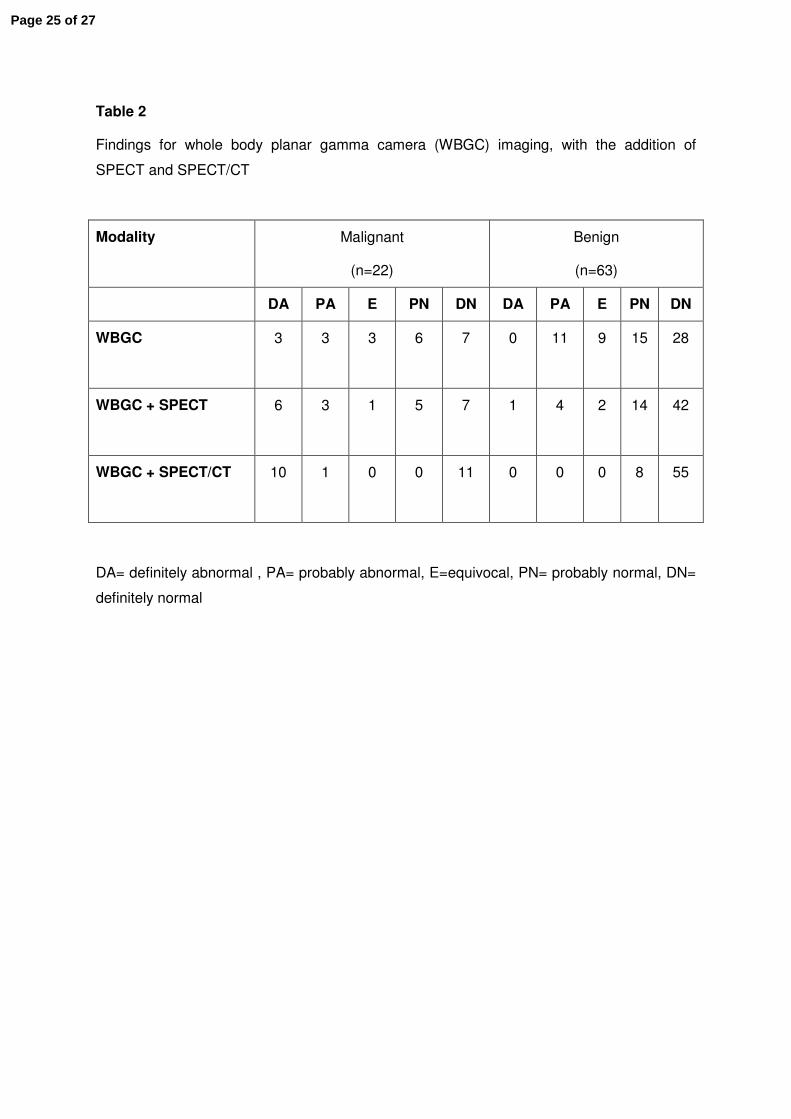
Table 2
Findings for whole body planar gamma camera (WBGC) imaging, with the addition of
SPECT and SPECT/CT
Modality Malignant
(n=22)
Benign
(n=63)
DA PA E PN DN DA PA E PN DN
WBGC
3 3 3 6 7 0 11 9 15 28
WBGC + SPECT
6 3 1 5 7 1 4 2 14 42
WBGC + SPECT/CT
10 1 0 0 11 0 0 0 8 55
DA= definitely abnormal , PA= probably abnormal, E=equivocal, PN= probably normal, DN=
definitely normal
Page 25 of 27

Table 3
Diagnostic performance of whole body planar gamma camera (WBGC) imaging, with the
addition of SPECT and SPECT/CT
WBGC WBGC + SPECT
WBGC + SPECT/CT
True-positive (n) 9 10 11
False-positive (n) 20 7 0
True-negative (n) 43 56 63
False-negative (n) 13 (2) 12 (1) 11 (0)
Sensitivity (%) 41 (82) 45 (91) 50 (100)
Specificity (%) 68 89 100
PPV (%) 31 59 100
NPV (%) 77 (96) 82 (98) 85 (100)
Accuracy (%) 61 (70) 78 (89) 87 (100)
In the analysis equivocal interpretation was considered malignant.
Number in brackets refer to analysis of patients with radioiodine avid disease only
Page 26 of 27
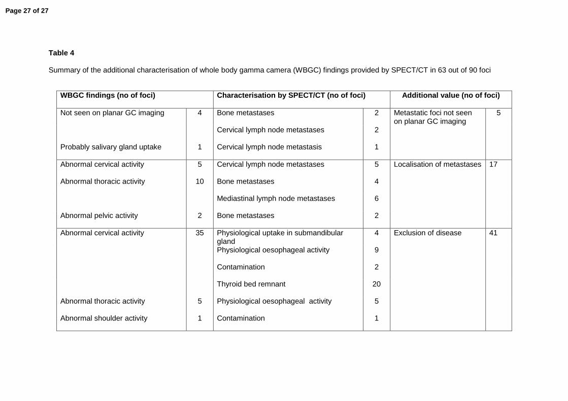
Table 4 Summary of the additional characterisation of whole body gamma camera (WBGC) findings provided by SPECT/CT in 63 out of 90 foci
WBGC findings (no of foci)
Characterisation by SPECT/CT (no of foci)
Additional value (no of foci)
Not seen on planar GC imaging 4 Bone metastases 2 Metastatic foci not seen on planar GC imaging
5
Cervical lymph node metastases 2
Probably salivary gland uptake
1 Cervical lymph node metastasis 1
Abnormal cervical activity
5 Cervical lymph node metastases 5 Localisation of metastases 17
Abnormal thoracic activity
10 Bone metastases 4
Mediastinal lymph node metastases 6
Abnormal pelvic activity
2 Bone metastases 2
Abnormal cervical activity 35 Physiological uptake in submandibular gland
4 Exclusion of disease 41
Physiological oesophageal activity 9
Contamination 2
Thyroid bed remnant 20
Abnormal thoracic activity
5 Physiological oesophageal activity 5
Abnormal shoulder activity
1 Contamination 1
Page 27 of 27