Pollenkitt – its composition, forms and functions
17
Flora 200 (2005) 399–415 Pollenkitt – its composition, forms and functions Ettore Pacini a , Michael Hesse b, a Dipartimento di Scienze Ambientali, Universita´ di Siena, Via P.A. Mattioli 4, I-53100 Sienna, Italy b Department of Ultrastructure Research and Palynology, Institute of Botany, University of Vienna, Rennweg 14, A-1030 Vienna, Austria Received 9 December 2004; accepted 27 February 2005 Abstract Two types of sticky pollen coat material exist in angiosperms, both produced by the anther tapetum. Pollenkitt is the most common adhesive material present around pollen grains of almost all angiosperms pollinated by animals, whereas tryphine seems to be restricted only to Brassicaceae. Tapetal cell protoplasts have different patterns of development according to the products formed during their development and degeneration. If tryphine is formed, the tapetal cell protoplasts lose their individuality at the microspore stage. If pollenkitt is formed, their contents degenerate at later stages. Cell content is totally reabsorbed, when ripe pollen is not surrounded by any gluing material. Current knowledge of pollenkitt formation, deposition on pollen grains and chemical composition are reviewed and discussed. Methods for detecting this viscous fluid are also presented. The many functions of pollenkitt, deduced from personal observations and the literature, act in the period between anther opening and pollen hydration on the stigma; they are: (1) to hold pollen in the anther until dispersal; (2) to enable secondary pollen presentation; (3) to facilitate pollen dispersal; (4) to protect pollen from water loss; (5) to protect pollen from ultra-violet radiation; (6) to maintain sporophytic proteins responsible for pollen–stigma recognition inside exine cavities; (7) to protect pollen protoplasts from fungi and bacteria; (8) to keep together pollen grains during transport; (9) to protect pollen from hydrolysis and exocellular enzymes; (10) to render pollen attractive to animals; (11) to render pollen visible to animal eyes; (12) to hide pollen from animal eyes; (13) to avoid predation of pollen through smell; (14) to enable adhesion to insect bodies; (15) to enable pollen packaging by bees and to form corbicules; (16) to provide a digestible reward for pollinators; (17) to enable pollen clumps to reach the stigma; (18) to allow self-pollination; (19) to facilitate adhesion to the stigma; (20) to facilitate pollen rehydration. Depending on the developmental program of the species, these functions may act during pollen presentation, in relation to pollinators, during pollen dispersal and when pollen reaches the stigma. r 2005 Elsevier GmbH. All rights reserved. Keywords: Pollenkitt; Tryphine; Tapetum; Orbicules; Pollen water content; Carbohydrates; Pollination; Stigma Introduction Female reproductive organs of angiosperms, unlike those of gymnosperms, contain from one to tens of thousands ovules per ovary. It is probably because of the variable number of ovules per ovary that different ARTICLE IN PRESS www.elsevier.de/flora 0367-2530/$ - see front matter r 2005 Elsevier GmbH. All rights reserved. doi:10.1016/j.flora.2005.02.006 Corresponding author. E-mail addresses: [email protected] (E. Pacini), [email protected] (M. Hesse).
-
Upload
independent -
Category
Documents
-
view
1 -
download
0
Transcript of Pollenkitt – its composition, forms and functions

ARTICLE IN PRESS
0367-2530/$ - se
doi:10.1016/j.flo
�CorrespondE-mail addr
michael.hesse@
Flora 200 (2005) 399–415
www.elsevier.de/flora
Pollenkitt – its composition, forms and functions
Ettore Pacinia, Michael Hesseb,�
aDipartimento di Scienze Ambientali, Universita di Siena, Via P.A. Mattioli 4, I-53100 Sienna, ItalybDepartment of Ultrastructure Research and Palynology, Institute of Botany, University of Vienna, Rennweg 14, A-1030 Vienna,
Austria
Received 9 December 2004; accepted 27 February 2005
Abstract
Two types of sticky pollen coat material exist in angiosperms, both produced by the anther tapetum. Pollenkitt is themost common adhesive material present around pollen grains of almost all angiosperms pollinated by animals,whereas tryphine seems to be restricted only to Brassicaceae. Tapetal cell protoplasts have different patterns ofdevelopment according to the products formed during their development and degeneration. If tryphine is formed, thetapetal cell protoplasts lose their individuality at the microspore stage. If pollenkitt is formed, their contents degenerateat later stages. Cell content is totally reabsorbed, when ripe pollen is not surrounded by any gluing material. Currentknowledge of pollenkitt formation, deposition on pollen grains and chemical composition are reviewed and discussed.Methods for detecting this viscous fluid are also presented. The many functions of pollenkitt, deduced from personalobservations and the literature, act in the period between anther opening and pollen hydration on the stigma; they are:(1) to hold pollen in the anther until dispersal; (2) to enable secondary pollen presentation; (3) to facilitate pollendispersal; (4) to protect pollen from water loss; (5) to protect pollen from ultra-violet radiation; (6) to maintainsporophytic proteins responsible for pollen–stigma recognition inside exine cavities; (7) to protect pollen protoplastsfrom fungi and bacteria; (8) to keep together pollen grains during transport; (9) to protect pollen from hydrolysis andexocellular enzymes; (10) to render pollen attractive to animals; (11) to render pollen visible to animal eyes; (12) to hidepollen from animal eyes; (13) to avoid predation of pollen through smell; (14) to enable adhesion to insect bodies; (15)to enable pollen packaging by bees and to form corbicules; (16) to provide a digestible reward for pollinators; (17) toenable pollen clumps to reach the stigma; (18) to allow self-pollination; (19) to facilitate adhesion to the stigma; (20) tofacilitate pollen rehydration. Depending on the developmental program of the species, these functions may act duringpollen presentation, in relation to pollinators, during pollen dispersal and when pollen reaches the stigma.r 2005 Elsevier GmbH. All rights reserved.
Keywords: Pollenkitt; Tryphine; Tapetum; Orbicules; Pollen water content; Carbohydrates; Pollination; Stigma
e front matter r 2005 Elsevier GmbH. All rights reserved.
ra.2005.02.006
ing author.
esses: [email protected] (E. Pacini),
univie.ac.at (M. Hesse).
Introduction
Female reproductive organs of angiosperms, unlikethose of gymnosperms, contain from one to tens ofthousands ovules per ovary. It is probably because ofthe variable number of ovules per ovary that different

ARTICLE IN PRESSE. Pacini, M. Hesse / Flora 200 (2005) 399–415400
types of pollen dispersing units are present in angios-perms (Pacini and Franchi, 1999); pollen dispersal unitsmay be composed of one to about 100,000 grains(Pacini, 2000). There is a positive correlation betweenthe number of ovules per ovary and the number ofgrains per pollen dispersal unit (Pacini and Franchi,1998). An extreme case is represented by massulateorchids (Pacini and Hesse, 2002, also for review).Pollen of anemophilous species, whether gymno- or
angiosperm, is dispersed as single grains with fewexceptions, and angiosperms with this pollinationsyndrome generally have only one ovule per ovary(e.g., Poaceae, Moraceae). On the contrary, the zoophi-lous species examined thus far generally have more thanone ovule/ovary and pollen grains surrounded bydifferent kinds of gluing material i.e. different devicesto enable transport ‘‘en masse’’ (Hesse, 1981b; Paciniand Franchi, 1998). The only exception is the case ofmassulate orchids (Pacini and Hesse, 2002). There arefour types of anther derived pollen-gluing or pollenconnecting materials: (a) pollenkitt; (b) tryphine; (c)elastoviscin, a fluid with high viscosity in orchids andAsclepiadaceae, but interpreted as a pollenkitt analogue(Dannenbaum and Schill, 1991; Schill and Wolter,1986); and d. thread-like structures connecting indivi-dual pollen grains or pollen tetrads, namely sporopolle-nin viscin threads, thin ropes continuous with exine orintermingled with grains produced by the tapetum, oranther mucilage derived for example from connectivecells and lacking sporopollenin (Hesse et al., 2000;Pacini, 1997). All these agents differ in composition,origin, development, and in part also in function.Besides common walls, exine and intine may also enabletransport dispersing unit en mass of pollen, as in allspecies having tetrads as pollen dispersing units, ormultiples of tetrads such as massulae, polyads anddifferent kinds of pollinia (Pacini and Franchi, 1999;Pacini and Hesse, 2002). We focus on pollenkitt andmention tryphine when necessary, but do not deal withother agents.Pollenkitt was recently discovered very well preserved
in fossil flower buds on and between fossil pollen grains.Amorphous, highly electron-opaque substances (pollen-kitt) and orbicules both very similar to those of recentTilia, were observed on and between pollen grains offossil flower buds of Craigia bronnii (Tilioideae, Mal-vaceae). Staining and sectioning of this amorphoussubstance demonstrated that it was indeed pollenkitt(Zetter et al., 2002, also for review).The systematic occurrence and meaning of pollenkitt in
plant reproduction was studied by Dobson (1989, 1994),who also proposed a method to collect and analysepollenkitt. She also determined its chemical compositionin certain angiosperm species (Dobson, 1988).The aim of this paper is to define differences between
pollenkitt and tryphine, to describe the methods used for
its detection, to summarize recent knowledge onpollenkitt and to suggest directions for further research.The authors have already written on this subject,especially from a cytological point of view, but herethe topic is considered in a broad sense. Data withoutcited references are from personal observations.
Pollenkitt and tryphine
Role and fate of the tapetum
Tapetal cells have nutritive functions, degenerating atdifferent stages of development with different results.Their content is reabsorbed or gives rise to differentkinds of substances (Table 1) (Pacini, 1997). Tapetal cellplastids always differentiate from proplastids to elaio-plasts during microspore stage, but subsequently differ-entiation varies according to tapetum fate and thesubstances produced (Clement and Pacini, 2001).If tapetal plasmamembranes disappear during micro-
spore stage, as occurs in Brassicaceae (Piffanelli et al.,1998), the cytoplasmic contents merge around the grainsand degenerate into tryphine. This is extra situm
degeneration because tapetal degeneration only occursafter loss of tapetal cell individuality. Pollenkitt isformed when tapetal cell plasmamembranes ruptureafter complete degeneration of tapetal protoplasts. Thisis in situ degeneration (Pacini and Franchi, 1992). Thetapetal cytoplasm may disappear without giving rise toany fatty substance and is therefore reabsorbed by thepollen grains, as for example in Lolium (Pacini, 1997;Pacini et al., 1992). Table 1 summarizes all the steps oftapetum development, considering modes of degenera-tion, resorption and the presence or absence of orbicules(see also diagrams in Piffanelli et al., 1998).
Who discovered and named pollenkitt? A short
history
It was Knoll (1930), who termed pollenkitt ‘‘alle jeneSubstanzen, welche eine Verbindung der Pollenkornerzu Klumpen bewirken y. Ohne Rucksicht darauf, obsolche Stoffe bei der Anthese flussig bleiben oder balderstarren, oder ob sie beim Austreten aus dem Anther-enfach bereits fest sind’’ (all substances which connectsingle pollen grains to pollen clumps .... irrespectively ofwhether the substances remain fluid during anthesis orbecome hard, or are non-fluids from the beginning ofanthesis onwards). Knoll evidently included not onlyoily/fatty fluids, but also the viscous pulps of Cypripe-
dium, and even the viscin threads of Oenothera. He wasnot aware that his various ‘‘Pollenkitt’’ substancesdiffered in ontogenesis, chemistry, structure, form andfunction.

ARTICLE IN PRESS
Table 1. Differences between pollenkitt and tryphine and the case of absence of fluids around pollen
TRYPHINE
(only Brassicaceae) Entomophily
Plastids of tapetal cells become elaioplasts, spherosomes are evident
POLLENKITT
(Monocots and dicots with amoeboid or parietal tapetum)
Entomophily > Anemophily
plasmamembrane of tapetal cells breaks and cell components aredispersed in the loculus
RESORPTION (mono- and dicots)
anemophily and hydrophily
tapetal cells may release other substances before tapetal degeneration is complete
components degenerate and merge on the exine surface
elaioplasts, spherosomes and other cell components aretotally reabsorbed by developing pollen; which is later dispersed asmonads
Orbicules present or absent
elaioplast globules merge among themselves and withspherosomes
tryphine may adapt during pollen dehydration and rehydration (harmomegathic effect)
the degeneration products of a tapetal cell form a single mass
Orbicules never present
these masses are deposited on the pollen grains when contiguous loculi fuse, in any case before anther opening
pollenkitt may adapt during pollen dehydration and rehydration (harmomegathic effect)
Orbicules present or absent
The first step is the same irrespective of subsequent development.
E. Pacini, M. Hesse / Flora 200 (2005) 399–415 401

ARTICLE IN PRESSE. Pacini, M. Hesse / Flora 200 (2005) 399–415402
However, Knoll had some forerunners in discoveringand naming pollen coatings. Troll (1928) had alreadycoined the term ‘‘Pollenklebstoff’’ (‘‘pollen adhesive’’)for sticky pollen surface substances, but the term wasnot successful. Pohl (1930) described pollen-’’Kleb-stoffe’’ as being of variable colour and viscosity in anastonishingly ‘‘modern’’ manner, but without knowl-edge of Knoll. He called the lipid substances on thepollen surface of anemophilous plants ‘‘Kittstoffreste’’(remnants of glues), and this term was in part acceptedby the scientific community. However, ‘‘Pollenkitt’’ wasand is the generally accepted terminus technicus.1
To our knowledge the term tryphine was first used byRowley and Erdtman (1967, p. 564, figure legend 3) andsubsequently by Echlin and Godwin (1968, p. 167), inboth cases without explanation. The etymology is theGreek tryphos, that means fragment, and Greektrypheros, that means soft (Hesse, 1978a). Tryphineformation was first described by Dickinson and Lewis(1973) in Raphanus. They also coined this term todistinguish the specific Brassicaceae features frompollenkitt known since Knoll’s papers. It has only beenstudied and described for Brassicaceae (a precisedefinition is given by Dickinson et al., 2000), but isprobably also present in other groups (Pacini, 1997).Pollenkitt and tryphine, irrespective of their mode of
formation, composition and so on, are similar in manyrespects because both are formed by fusion of tapetalelaiosomes and spherosomes (Platt et al., 1998). Never-theless, pollenkitt and tryphine differ in the followingaspects (see also Dickinson et al., 2000): (a) time ofdeposition on pollen surface (Pacini, 1997; Pacini andFranchi, 1993); (b) pollenkitt may coexist with orbicules,but tryphine has never been observed together withorbicles. Orbicules are also present in species whosetapetum is totally reabsorbed by developing pollen andwhose pollen is devoid of any glue enabling en massetransport (Table 1).Pollenkitt is hydrophobic because it is mainly
composed by lipids, while tryphine is a mixture ofhydrophilic and hydrophobic substances (Dickinsonand Lewis, 1973). Tapetal cells produce pollenkitt,irrespective of tapetum type (secretory or amoeboid)and maintain their individuality until degeneration; vice
1A curiosity should not overseen: Long before Knoll already Kerner
had observed the pollenkitt, but he interpreted it first as a slimy
polysaccaridic substance and called it ‘‘Bassorin’’ (Kerner von
Marilaun, 1873). In his famous "Pflanzenleben" (several editions) he
referred to oily coatings, neither using the term ‘‘Bassorin’’ any more
nor using ‘‘pollenkitt’’ or similar terms. Botanical histochemistry was
not yet developed. Bassorin, also called Bassora-Gummi, is a residual
of gums or slimes (polysaccharides!) mainly from stems of Astragalus
sp., for technical use as sticky, highly viscous liquid, and surely no
lipid, hence no real pollenkitt precursor term. Kerner misinterpreted
this Bassorin as deriving from the cell walls (!) of pollen mother cells,
or as plasmatic residual from microspore division. He does not
mention tapetal cells, for any reason.
versa tapetal cells producing tryphine are always of thesecretory type (Pacini, 1997).In the case of tryphine the tapetal plasmamebrane
ruptures at late microspore stage when organelles canstill be recognized. In the case of pollenkitt, this onlyoccurs when all the tapetal components have degener-ated and intermingled, namely during the final stage ofpollen maturation (Pacini and Franchi, 1993). Tryphineripens after the tapetal plasmamembrane has disap-peared; pollenkitt ripens in situ and the plasmamem-brane only disappears when pollenkitt has alreadyformed and before its deposition on the grains. Tryphineis certainly responsible for pollen adhesion to the stigmaby virtue of a species specific interaction mediated bylipophilic molecules in the pollen exine (Zinkl et al.,1999). In pollenkitt, this kind of molecule has not beendescribed but the function exists.At least up to a certain stage the pattern of tapetum
development is essentially the same whether pollenkittor tryphine forms. Elaioplasts and spherosomes formfirst but later the pattern of development depends onwhether pollenkitt or tryphine is produced, or if tapetalcontent is totally reabsorbed as in strictly anemophilousplants (Table 1). Reabsorption has been described inLolium perenne (Pacini et al., 1992). Tryphine and thefunctions of pollen coats in general are extensivelyreviewed by Dickinson et al. (2000).Despite these differences, pollenkitt and tryphine
share some functions and this may be why some authorsdo not distinguish them, or call pollenkitt tryphine andvice versa, or use the generic term pollencoat (Hernan-dez-Pinzon et al., 1999; Piffanelli et al., 1998). Theseauthors published a survey on tryphine biogenesis andits physico-chemical characterization.
Presence or absence of pollenkitt
Pollenkitt is produced as a tapetum derivative whichhas been found in almost all zoophilous angiospermsstudied so far i.e. with entomophilous and relatedpollination syndromes, but not in gymnosperms (fordetails and reviews see Hesse 1978a–d, 1979a–e,1980a–c, 1981a, b, 1984). Pollenkitt as pollen bindingagent is typical of almost all entomophilous plants withmonads and tetrads (Hesse, 1993); however, thedistribution of pollenkitt on pollen surfaces is highlyvariable. The characters of the so-called Pollenverkit-tung (for this perfectly apt German word, a roughtranslation is ‘‘the variable degree of pollen stickiness’’),namely amount, consistency and distribution of pollen-kitt in relation to exine ultrastructure has beeninvestigated in closely related genera with anemo- andentomophilous species, for example, Plantago, Fraxinus,
Acer, Carex, Sanguisorba, and Artemisia (Hesse, 1979c).
ARTICLE IN PRESSE. Pacini, M. Hesse / Flora 200 (2005) 399–415 403
It is absent from the pollen surface of strictly andprimary anemophilous taxa in the Fagaceae (exceptCastanea), Urticaceae, Chenopodiaceae, (most) Poa-ceae, but not in Cyperaceae (see the strictly anemophi-lous Carex acutifolia and C. vulpina versus theentomophilous Carex baldensis with a lot of sticky,active pollenkitt, Hesse, 1980b). Vice versa in somesecondary anemophilous members of otherwise ento-mophilous families such as the Ranunculaceae (Thalic-
trum), Oleaceae (Olea, Fraxinus), Euphorbiaceae(Mercurialis and Ricinus) and Asteraceae (Artemisia
and Ambrosia), pollenkitt is still present, at least inpatches on the pollen surface (Pacini and Franchi, 1998,for details see also Hesse, 1979a, b, and for conspectus1981a, b). An excellent example for transformationwithin a single genus, with several more or lessentomophilous and anemophilous species, respectively,is Acer, where the respective flower syndrome becomesless entomophilous and more anemophilous, andsimultaneously pollenkitt amount, density and homo-geneity decreases (Hesse, 1979d).There is always the same strategy: sticky pollen is
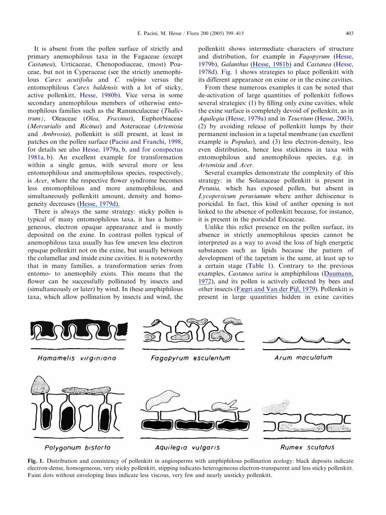
typical of many entomophilous taxa, it has a homo-geneous, electron opaque appearance and is mostlydeposited on the exine. In contrast pollen typical ofanemophilous taxa usually has few uneven less electronopaque pollenkitt not on the exine, but usually betweenthe columellae and inside exine cavities. It is noteworthythat in many families, a transformation series fromentomo- to anemophily exists. This means that theflower can be successfully pollinated by insects and(simultaneously or later) by wind. In these amphiphiloustaxa, which allow pollination by insects and wind, the
Fig. 1. Distribution and consistency of pollenkitt in angiosperms w
electron-dense, homogeneous, very sticky pollenkitt, stipping indicate
Faint dots without enveloping lines indicate less viscous, very few a
pollenkitt shows intermediate characters of structureand distribution, for example in Fagopyrum (Hesse,1979b), Galanthus (Hesse, 1981b) and Castanea (Hesse,1978d). Fig. 1 shows strategies to place pollenkitt withits different appearance on exine or in the exine cavities.From these numerous examples it can be noted that
de-activation of large quantities of pollenkitt followsseveral strategies: (1) by filling only exine cavities, whilethe exine surface is completely devoid of pollenkitt, as inAquilegia (Hesse, 1979a) and in Teucrium (Hesse, 2003),(2) by avoiding release of pollenkitt lumps by theirpermanent inclusion in a tapetal membrane (an excellentexample is Populus), and (3) less electron-density, lesseven distribution, hence less stickiness in taxa withentomophilous and anemophilous species, e.g. inArtemisia and Acer.
Several examples demonstrate the complexity of thisstrategy: in the Solanaceae pollenkitt is present inPetunia, which has exposed pollen, but absent inLycopersicum peruvianum where anther dehiscence isporicidal. In fact, this kind of anther opening is notlinked to the absence of pollenkitt because, for instance,it is present in the poricidal Ericaceae.Unlike this relict presence on the pollen surface, its
absence in strictly anemophilous species cannot beinterpreted as a way to avoid the loss of high energeticsubstances such as lipids because the pattern ofdevelopment of the tapetum is the same, at least up toa certain stage (Table 1). Contrary to the previousexamples, Castanea sativa is amphiphilous (Daumann,1972), and its pollen is actively collected by bees andother insects (Fægri and Van der Pijl, 1979). Pollenkitt ispresent in large quantities hidden in exine cavities
ith amphiphilous pollination ecology: black deposits indicate
s heterogeneous electron-transparent and less sticky pollenkitt.
nd nearly unsticky pollenkitt.

ARTICLE IN PRESSE. Pacini, M. Hesse / Flora 200 (2005) 399–415404
(Hesse, 1978d). The ecological meaning of the absenceof pollenkitt or other sticky materials is that pollengrains may leave the anther as soon as it opens (Paciniand Hesse, 2004).In the epi- or hypo-hydrophilous marine monocoty-
ledons (the so-called seagrasses with surface or sub-marine pollination, Ackerman, 2000, also for review)pollenkitt is not absent, as demonstrated optically andcytochemically for the spherical pollen of Thalassia
(Pettitt, 1976, 1980), while in other genera with filiformpollen TEM has shown a slimy or gelatinous coatconsisting mostly of polysaccharides plus some proteinsand lipids (Pettitt, 1976, 1980; Pettitt et al., 1984).Hence, a mucilaginous fluid composed of polysacchar-ides, proteins and lipids is present around dispersingpollen grains in at least some underwater-pollinatedtaxa. With the stepwise disappearance of pollenkitt, theexine is also progressively reduced and may be absent asdemonstrated by Cooper et al. (2000) in some repre-sentative members of the Callitrichaceae, which tendtowards submergence (Martinsson, 1993); the sameoccurs in submerged monocots, the seagrasses withtheir filiform pollen, being the most extreme adaptation(Blackmore et al., 1987; Pettitt et al., 1984).
Ontogenesis of pollenkitt
The starting point of pollenkitt formation varies.Pollenkitt is formed from prophase 1 onwards, at theend of meiosis, after meiosis, during tetrad stage, shortlybefore break-up of tetrads, or after tetrad stage (Hesse,1978b). Judging from studies using different TEMstaining methods, it seems that pollenkitt is mainly ablend of two main components: elaiosomes and sphero-somes. The former are the product of a distinct type ofplastid with lipid reserves, the elaioplasts (Clement andPacini, 2001; Pacini and Casadoro, 1981; Reznickovaand Dickinson, 1982; Santos et al., 2003); the latter arethe result of accumulation of smooth ER lipids in cisterndilations (Santos et al., 2003; Ubera-Jimenez et al.,1996). Other parts of the tapetum cytoplasm may also beinvolved in pollenkitt formation: during tapetal cyto-plasm disappearance other cell components may con-tribute to spherosome-borne material.At least during microspore stage the tapetum is
engaged in exocytotic processes, and other substancesmay be released into the loculus and absorbed by pollengrains or deposited on them (Pacini and Juniper, 1979):the tapetum shows a different secretory nutritionalactivity according to the stage of pollen development.Finally, pollenkitt is deposited on the pollen as ahomogeneous coating or as a non-homogeneous layerof variable electron density, especially in not strictlyentomophilous taxa.
In species in which development of the tapetum hasbeen studied ultrastructurally, plastids, initially at thestage of proplastid divide and accumulate plastoglobulesto become elaioplasts (Ciampolini et al., 1993; Pacini etal., 1992). Later smooth ER gives rise to spherosomes.In Ilex paraguaiensis the opposite occurs, elaioplastswith unsaturated lipids form first and later plastoglo-bules (Santos et al., 2003). In some cases there is atransitional phase of chromoplasts (Clement and Pacini,2001) or amyloplasts (Ubera-Jimenez et al., 1996);however, it is noteworthy that in Rosmarinus, plastidsdo not contribute to pollenkitt formation, only sphero-somes (Ubera-Jimenez et al., 1996).Few papers give a detailed description of the final
dissolution of tapetal cells, i.e. the merging of elaioplastsinto spherosomes. In some cases, this only seems tooccur when the tapetal plasmamembranes are lost(Lesniewska and Charzynska, 2000).Tapetum consists of fragile cells, presumably because
they totally or partially lose their inner tangential andradial walls for quick exocytosis (Pacini, 1994). Tapetalintegrity is easily damaged during dissection beforefixation, especially if long large anthers are divided intosmall pieces. The pollen is suspended in the locular fluidand if the anther is sectioned or damaged or com-pressed, the tapetal plasmamembrane may be ruptured,releasing its contents. Even the molarity of the fixativecan cause artefacts in tapetum structure. Artefactsarising from damages may be interpreted as degenera-tion. This difficulty also arises from the fact that theplasmamembrane is not always easily discernible and itis sometimes difficult to state whether or not it ispresent.Since tapetal cells die to nourish the developing pollen
they have features typical of programmed cell death, i.e.apoptosis (Papini et al., 1999; Wu and Cheung, 2000).
Mode and time of deposition on the pollen
surface
The question of the wholesale transfer and regulardeposition of coatings on pollen walls, and in somespecies in pollen walls, is complex (Dickinson et al.,2000). However, some points have been established.Deposition mode has been described for some generaand species. Weber (1996) showed that Geranium
robertianum tapetum first releases coating vesicles andlater real pollenkitt, which completely envelops thegrains. In Smyrnium (Weber, 1991) and Alisma (Hesse,1980b) small droplets act as a ‘‘transport form’’,whereas large lumps (e.g. in Tilia) are released by thedegenerating tapetum. Deposition timing has only beendescribed for a few species (Hesse, 1978b), but it seemsto be stimulated by the sudden increase in volume of the

ARTICLE IN PRESSE. Pacini, M. Hesse / Flora 200 (2005) 399–415 405
cavity containing pollen by fusion of contiguous loculi(Keijzer, 1987a, b; Nepi and Pacini, 1993; Pacini, 2000).Clement et al. (1988) described pollenkitt formation in
Lilium in detail. Some droplets are extruded directly byplastid stroma through the plastid envelope whileothers, unsaturated lipid globules, are presumablyderived from the interaction of smooth ER and plastids.When any type of tapetum has completely degener-
ated, i.e. when cytoplasmic components are no longerevident, TEM may reveal different aspects: a homo-geneous mass as in olive, cherry, Cichorium (Pacini andCasadoro, 1981; Pacini and Keijzer, 1989; Pacini et al.,1986), or non-homogeneous, with electron opaque andelectron translucent areas, as in G. robertianum (Weber,1996). When deposited on the grains, it appears ashomogeneous masses inside the pollen ornamentation.It may be a continuous homogeneous electron opaquemass as in Rosmarinus officinalis (Ubera-Jimenez et al.,1996), or it may consist of electron opaque and electrontranslucent masses or foamy bubbles, as in G. robertia-
num (Weber, 1996).An underrated example (Hesse, 2003) of the exine
reservoir function may be the presence of various lipids,proteins and other compounds, plus unstable substanceson the surface and inside the exine. Only in optimallypreserved pollen of the apparently strictly entomophi-lous Teucrium montanum (Lamiaceae), may it be seenthat the remarkable amount of pollenkitt (the electron-opaque substance) is almost completely absorbed inexine cavities. The real pollen surface (tectum) lackspollenkitt entirely. Hence, the pollen of this species isnot really sticky, which is perhaps related to itspollination biology (thought to be an obligate self-pollinated species).The different modes observed in the final stages of
tapetum degeneration and pollen coating may also belinked to the following processes: the moment whentapetal plasmamembranes break or disappear, fusion ofcontiguous loculi, locular fluid resorption, pollendehydration and anther opening. All these phases,especially the last three, seem very specific, differingfrom species to species, and deeply influenced byenvironmental parameters such as: temperature, relativehumidity and light intensity (Bianchini and Pacini, 1996;Lisci et al., 1994; Pacini, 1996, 2000).
Appearance under LM, SEM and TEM
The appearance of pollenkitt in ripe pollen by LMdepends on the miscibility of the mounting medium withwater. If miscible, pollenkitt may form spheres on thepollen surface, because it is hydrophobic and probablybecause the increase in volume at rehydration causingpollenkitt to extrude from the exine cavities. During
rehydration pollenkitt droplets even emerge from exinecavities in the complex double tectate-columellateektexine of Asteraceae. Pollen rehydration modifiespollen shape and increases its volume, causing rearran-gement of pollenkitt on the pollen surface.If the mounting medium is immersion oil or another
apolar fluid, the pollen does not rehydrate andpollenkitt dissolves. This is particularly evident whenpollenkitt is dark in colour: deep red, violet or brown asin some lilies, tulips and Petunia, or blue-black as insome poppies (Papaver). Under the light microscope onecan see that pollenkitt has been removed and dissolvedby currents in the mounting medium.If pollen is observed by LM without any mounting
medium, i.e. directly on the slide, and without acoverslip, pollenkitt is visible inside the exine cavitiesor coating the pollen surface totally in a thin layer.Small patches are only seen where pollen grains adheretogether.SEM visualization of pollenkitt likewise depends on
the method used. After alcohol fixation, pollenkitt tendsto form strands, as in the entomophilous Bryonia dioica
(Hesse, 1981b). After aldehyde fixation it is mostlyevident. In Cucurbita pepo and Lavatera arborea where itis abundant, it obscures some minor exine ornamenta-tions that become evident only after its removal withlipid solvents (Nepi and Pacini, 1993, 1999). An effectivemethod to clean pollen surfaces of pollenkitt is the so-called DMP-CPD-technique (Halbritter, 1998); how-ever, removal may fail if pollenkitt is extremely viscous.TEM visualization depends largely on sample pre-
paration. Because of its lipidic composition, pollenkittcan only be preserved if the material is fixed in osmiumtetroxide or other fixatives that stabilize lipids. Pollen-kitt is often dissolved during fixation and embedding.Classical palynologists used to clean pollen before the
observation by the acetolysis technique, which removesall structures not resistant to this strong solvent, exceptexine (Hesse and Waha, 1989). Using non-aqueousfixation methods, the pollen coating appears as anevenly thin coating seen by TEM, especially inBrassicaceae (Dickinson et al., 2000, also for review ofchemical composition and function).
Methods for pollenkitt identification
Here we report some methods to reveal pollenkitt onpollen collected from anthers. The pollen is deposited ona slide and a droplet of water and a coverslip are added.The pollen quickly rehydrates and pollenkitt appears asbirefringent droplets on the exine surface. These dropseventually merge and grow in size. In this way it is alsopossible to determine the real colour, because in theanther the colour is the result of interference with that of

ARTICLE IN PRESSE. Pacini, M. Hesse / Flora 200 (2005) 399–415406
pollen. A similar method enables slow rehydration,because of glycerine in the mounting medium andinvolves mixing pollen grains with Carlberla fluid(Ogden et al., 1974). After 6–24 h the exine turns pinkand pollenkitt drops appear on its surface. Theadvantage of this method is that slides can be observedseveral hours later.By virtue of its lipid composition, pollenkitt stains
with all Sudan dyes and all other dyes for lipids. Theproblem is that it is necessary to observe the slideimmediately after the dye is added, i.e. within 2–10min,depending on the amount of pollenkitt and size of thepollen, because exine stains too, but later thanpollenkitt. If the slide is observed too late it is difficultto distinguish the two, especially if the quantity ofpollenkitt covering the pollen is small. On the otherhand, if the quantity is large and emerging from theexine cavities in drops, observation can wait 15–30min.Heslop-Harrison and Heslop-Harrison (1985) stainedEucalyptus pollenkitt with Scarlet R in ethanol withgood results.Another good method when pollenkitt is abundant is
to dust pollen on a slide just wiped with chloroform. Thepollen is then removed by gently blowing and isobserved directly or after staining with dyes for lipids;pollenkitt droplets of different size adhere to the slide.A method to check for pollenkitt in small quantities is
to prepare two aliquots of pollen, one of which iswashed with a mixture of chloroform/carbon disulphide(1:1) for a few seconds and allowed to dry. The other isuntreated. The treated and untreated pollen is thenobserved by SEM (Pacini and Vosa, 1979). If the imagesare similar it means that pollenkitt is absent; if theydiffer and washed pollen shows better ornamentation, itmeans that pollenkitt is present.The acetolysis method destroys all component of
pollen except of the exine (Hesse and Waha, 1989),whereas treatment of pollen with 2% osmium tetroxideat 401 for 6 days destroys the exine, leaving thepollenkitt at least in Trachymene pilosa (Audran, 1981).A task for further research is to check the number of
pollen grains per clump adhering to insect bodies. Theinsect must be prepared without the use of substancessuch as chloroform and carbon disulphide, whichremove lipids. The whole insect or some dissected partscan be viewed directly with a high power stereomicro-scope, after sputtering by SEM or in water by LM.
Chemical composition
It seems unlikely that pollenkitt composition is thesame in all species, because of its different consistency,colour, etc. Little research has been done on this topic.
Dobson (1988) developed a method to separatepollenkitt by double extraction, first extracting pollenwith benzene for 20 s and then chloroform: methanol(2:1) for 23–28 h. The rationale was that the first quickextraction removed the external lipids, namely pollenkittand the second internal and any external lipids notcompletely removed by the first extraction. Inner andouter lipid components can then be analysed by thin layerchromatography (TLC) or other analytical methods.Comparison of the results of both extractions providesinformation about internal (cytoplasmic) and external(pollenkitt) lipid composition (Piffanelli et al., 1998).Pollenkitt is a hydrophobic mixture of materials,
composed mainly of saturated and unsaturated lipids(Dobson, 1988), carotenoids (Hesse, 1993; Lunau,1995), flavonoids (Wiermann and Gubatz, 1992),proteins and carbohydrates (Parkinson and Pacini,1995; Santos et al., 2003; Weber, 1996, also for review).Differences in pollenkitt composition have been re-ported in the heterantherous species Lagerstroemia
indica which has two pollen forms produced bypollinating and feeding anthers (Nepi et al., 2003). Onlythe pollinating pollen contains neutral lipids, a peculiarpartition factor.According to an isolated observation by Pohl (1932),
the pollenkitt of Philodendron is crumbly and somewhatslimy, not lipidic or oily like all other known pollenkitt.The physico-chemical properties of pollenkitt depend
on the substances in the mixture and their proportions.Substances responsible for hiding pollen from animaleyes are understandably absent on pollen activelycollected by animals. Molecules responsible for pollenflavour and smell, are probably species specific.We do not know if pollenkitt composition is stable or
whether it changes during pollen exposure and pollentransport by animals, as occurs for carbohydratereserves (Vesprini et al., 2002). Changes in colour andconsistency have been noticed, e.g. in Cyclamen (Pohl,1930), and not only in entomophilous species, where thischange could be to discourage animals from visitingwilted flowers when anthesis is already over. Thischange is known in Mercurialis annua (anemophilous)where pollen is only exposed for 1 day and by afternoon,the initially yellow pollen is blue green (Lisci et al., 1994and our unpublished data). On the other hand, in Erica
arborea, which is first entomophilous and then anemo-philous, a change in viscosity occurs when the nectaryceases to produce nectar and pollen is dispersed by wind(Franchi and Pacini, 1996).
The many functions of pollenkitt
The pollenkitt starts to play its role as soon as antheropens, continues during pollen exposure and dispersal,

ARTICLE IN PRESSE. Pacini, M. Hesse / Flora 200 (2005) 399–415 407
and presumably ceases when pollen rehydrate on thestigma (Table 2). Twenty proposed functions in theliterature are listed and discussed below:
1.
Tab
Poll
1.
2.
3.
4.
5.
6.
7.
8.
9.
10.
11.
12.
13.
14.
15.
16.
17.
18.
19.
20.
Som
To hold pollen in the anther until dispersal: Thisoccurs mainly in entomophilous flowers, wherepollen remains firmly attached to the exposed anthereven when the air is turbulent, and in upside downanthers such as Passiflora and vertical ones such asAcanthus mollis. In some Liliaceae pollen may belost when wind speed is high probably because it isnot firmly attached to the anther. This enablessimultaneous dispersal of pollen by insect and wind(Dafni, 1986). In some species stamens move whentouched by insects, the result of this movement,sometimes sudden, is intended as a way to avoid selfpollination (Proctor et al., 1996) and brushes pollenon the body of the dispersing animal. In someOpuntia species, this movement reveals the nectary,previously hidden by clustered stamens (Schlindweinand Wittmann, 1997). Despite these movements,pollenkitt prevents loss of pollen from anthers nottouched by animals.
2.
To enable secondary pollen presentation: Somefamilies or members of angiosperm families do notpresent pollen for dispersal on the anther but inother flower parts. This means that as soon as thele 2. Pollenkitt functions and the stage when they act
enkitt functions During p
presentat
To hold pollen in the anther until dispersal X
To enable secondary pollen presentation X
To facilitate pollen dispersal X
To protect pollen from the waterloss X
To protect pollen from the UV radiation X
To conserve sporophytic proteins inside exine
cavities
X
To protect the pollen protoplasts from fungi and
bacteria especially during presentation
X
To keep pollen grains together during transport
To protect pollen from hydrolysis and exocellular
enzymes
X
To render pollen attractive to animals X
To render pollen visible to animal eyes X
To hide pollen from animal eyes X
To avoid predation of pollen through smell X
To enable adhesion to insects bodies
To enable pollen packaging by bees and form
corbicules
To provide a digestible reward for pollinators
To enable pollen clumps to reach the stigma
To allow self-fertilization X
To facilitate adhesion to the stigma
To facilitate pollen rehydration
e only act at a certain stage, others at different stages.
anther opens, pollen is stuck to other floral partsmainly by virtue of pollenkitt or other devices(Erbar and Leins, 1995; Pacini and Hesse, 2004),and is later removed by pollinating agents. Pre-sentation outside the anther has different apparentreasons. In the Asteraceae and related families, theflowers are tiny, with long corollas and denselypacked in the inflorescence. Pollen is stuck to thestyle in Asteraceae (Pacini, 1996) and to the stylarhead in Petromarula sp. (Campanulaceae) (Iger-sheim, 1993). In Acrotriche (Epacridaceae) theflowers are conspicuous, the corolla is full of nectarand pollen is presented on the petals (McConchie etal., 1986). In many Araceae, such as Zantedeschia
and Anthurium, male and female flowers are tightlypacked but separated in different parts of theinflorescence (Mayo et al., 1997). Their pollen,mixed with a highly viscous pollenkitt, is extrudedfrom the anther by a piston mechanism (Yeo, 1993)in coiled threads or noodles, up to 3–4mm long,subsequently loaded by pollinators.
3.
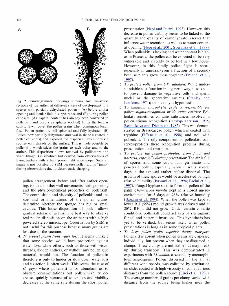
To facilitate pollen dispersal: Pollen grains arepacked inside the developing anther, but in manycases when it opens and during presentation theyform a kind of sponge, i.e. there is much spacebetween grains and the external part is fringy withgrains forming threads (Fig. 2). This change inollen
ion
During pollen
dispersal
During pollinator
interactions
On stigma
X
X X
X
X X X
X X
X
X
X
X
X
X X
X
X
ARTICLE IN PRESS
Fig. 2. Semidiagramatic drawings showing two transverse
sections of the anther at different stages of development in a
species with partially dehydrated pollen – (A) before anther
opening and locular fluid disappearance and (B) during pollen
exposure: (A) Tapetal content has already been converted to
pollenkitt and occurs as spheres (dotted) lining the locular
cavity. It will cover the pollen grains when contiguous loculi
fuse. Pollen grains are still spherical and fully hydrated. (B)
Pollen, now partially dehydrated and oval in shape is coated in
pollenkitt (dots) and exposed for dispersal. Pollen forms a
sponge with threads on the surface. This is made possible by
pollenkitt, which sticks the grains to each other and to the
anther. This disposition allows removal by pollinators and
wind. Image B is idealised but derived from observations of
living anthers with a high power light microscope. Such an
image is not possible by SEM because pollen grains ‘‘jump’’
during observations due to electrostatic charging.
E. Pacini, M. Hesse / Flora 200 (2005) 399–415408
pollen arrangement, before and after anther open-ing, is due to anther wall movements during openingand the physico-chemical properties of pollenkitt.The composition and viscosity of pollenkitt and thesize and ornamentations of the pollen grains,determine whether the sponge has big or smallcavities. This loose disposition of pollen allowsgradual release of grains. The best way to observereal pollen disposition on the anther is with a highpowered stereo microscope. Observation by SEM isnot useful for this purpose because many grains arelost due to the vacuum.
4.
To protect pollen from water loss: It seems unlikelythat some species would have protection againstwater loss, while others, such as those with viscinthreads, hidden anthers, or without any pollen gluematerial, would not. The function of pollenkitttherefore is only to hinder or slow down water lossand its action is often weak. This seems the case ofC. pepo where pollenkitt is so abundant as toobscure ornamentations but pollen viability de-creases quickly because of water loss and viabilitydecreases at the same rate during the short pollenpresentation (Nepi and Pacini, 1993). However, thisdecrease in pollen viability seems to be linked to thequantity and quality of carbohydrate reserves thatinfluence water retention, as well as to water contentat opening (Nepi et al., 2001; Speranza et al., 1997).When pollenkitt is lacking and water content is high,as in Poaceae, the pollen can be expected to be veryvulnerable and viability to be lost in a few hours.However, in this family pollen flight is short,especially in annuals (even a fraction of a second)because plants grow close together (Franchi et al.,1997).
5.
To protect pollen from UV radiation: While under-standable as a function in a general way, it was saidto prevent damage to vegetative cells and spermnuclei or the generative nucleus (Stanley andLinskens, 1974); this is only a hypothesis.6.
To maintain sporophytic proteins responsible forpollen stigma-recognition inside exine cavities: Pol-lenkitt sometimes contains substances involved inpollen–stigma recognition (Heslop-Harrison, 1975;Reznickova and Dickinson, 1982). This was demon-strated in Brassicaceae pollen which is coated withtryphine (Piffanelli et al., 1998) and not withpollenkitt. The oily component of pollenkitt con-serves/protects these recognition proteins duringpresentation and transport.
7.
To protect the pollen protoplast from fungi andbacteria, especially during presentation: The air is fullof spores and some could fall, germinate andpenetrate pollen, especially when it waits severaldays in the exposed anther before dispersal. Thegrowth of these spores would be accelerated by highrelative humidity (Bassani et al., 1994; Pacini et al.,1997). Fungal hyphae start to form on pollen of thepalm Chamaerops humilis kept in a closed micro-environment for 5 days at 98% relative humidity(Bassani et al. 1994). When the pollen was kept atlower RH (55%) mould growth was delayed and at20% RH it did not grow. Under certain climaticconditions, pollenkitt could act as a barrier againstfungal and bacterial invasions. This hypothesis hasyet to be verified, but seems likely when pollenpresentations is long as in some tropical plants.
8.
To keep pollen grains together during transport:Pollenkitt is absent when pollen grains are dispersedindividually, but present when they are dispersed inclumps. These clumps are not stable but may breakup during transport. This was demonstrated inexperiments with M. annua, a secondary anemophi-lous angiosperm. Pollen dispersed in the air atdifferent wind speeds, was collected by gravitationon slides coated with high viscosity silicon at variousdistances from the pollen source (Lisci et al., 1996).The average number of grains per clump varied withdistance from the source being higher near the
ARTICLE IN PRESSE. Pacini, M. Hesse / Flora 200 (2005) 399–415 409
source and lower far from it. We do not know if it ispossible to generalize this observation to otheranemophilous species with pollenkitt or if there iseven a loss of pollen from the bodies of flyinginsects.
9.
To protect pollen from hydrolysis and extracellularenzymes: Parallel to protect glycocalyx componentsby accumulated sporopollenin from hydrolysis andextracellular enzymes (Rowley et al., 1999) pollen-kitt may function like sporopollenin sealing most ofthe pollen surface, at least in entomophilous taxa.
10.
To render pollen attractive to consuming animals:Pollenkitt may have different perfumes odors andcertain different substances needed by pollinators(Dobson and Bergstrom, 2000). Pollen of Rosarugosa has a perfumed pollenkitt (Dobson, 1994,also for review).
11.
To render pollen visible to animal eyes: Lunau (1995)published a detailed and exhaustive study on thespectrum of pollen colours according to visibility forhumans and pollinating animals. Pollen colourdepends on pollenkitt and if pollenkitt is removedpollen is invariably yellow (Nepi et al., 2003).12.
To hide pollen from animal eyes: In many plants,flower visitors collect nectar but not pollen, evenwhen the anthers are evident and exposed outsidethe flower. The reason for this may be that pollen isnot evident to animal eyes due to some compoundsin pollenkitt. This seems the case in L. indica, whichhas flowers with two sets of anthers, one inside andan outer encircling set. Both produce viable pollen,but the inner set of anthers produces pollen withlittle pollenkitt. The pollen of the inner anthers isfeed pollen and that of the outer set is fertile pollen.The grains of the two sets differ in colour, but bothare yellow when pollenkitt is removed (Pacini andBellani, 1986). This hiding mechanism may evenoccur at the end of the blooming period; if the pollenis no longer visible pollinators are presumablydiscouraged from visiting the flower.13.
To avoid predation of pollen through smell: Dobsonand Bergstrom (2000) found a-methyl alcohols andketones in the pollenkitt of some anemophilous speciesand Filipendula and hypothesized that this was toavoid predation of pollen by discouraging the insectthrough smell. These compounds also seem toxic forsome insects (Dobson and Bergstrom, 2000).14.
To enable adhesion to insect bodies: Pollenkitt wasnecessary in early angiosperms to stick pollen topollinator bodies. The first insects involved in thismutual activity were Diptera and Coleoptera (Thien,1980 and references therein); the latter have little orno body hairs to trap pollen.Researches have shown that pollen is electrostati-cally attracted by the insect even if it does not touchthe anther. The pollen adheres to the body byelectrostatic forces between insect and the flower(Vaknin et al., 2000). We do not know whether or towhat extent pollenkitt is involved in this electrostaticmechanism.
15.
To enable pollen packaging by bees and to formcorbicules: Pollen of species visited by Apoideawhich collect pollen to store as food always haspollenkitt (Pacini, 2000). When other pollen sourcesare not available these insects also collect pollen ofZea mays and other grasses or anemophilous speciesdevoid of pollenkitt (Wille and Wille, 1983). Toform corbicules they must however use pollen ofspecies with pollenkitt to pack pollen devoid of it.An alternative mechanism is to regurgitate a stickyfluid composed mainly of sugars thus using apollenkitt substitute (Klungness and Peng, 1983).Pollenkitt fills the spaces between grains so that themass becomes more coherent and compact. For agiven viscosity, the larger the diameter of pollen, themore pollenkitt is necessary to stick it together. Thisis probably why large pollen grains have a thicklayer of pollenkitt, sometimes even covering orna-mentations, and adhere to pollinators as individualgrains (Nepi and Pacini, 1993). Not only thephysicochemical properties of the pollen grains butalso their geometrical shape influence the possibilityof forming compact corbiculae. Indeed if pollen istetrahedral, as in many Myrtaceae, it can be closelypacked with little space between grains, in respect toovoid or spherical pollen grains.
16.
To provide a digestible reward for pollinators: Unlikethe other substances in pollen grains protected byrelatively impermeable walls, pollenkitt is immedi-ately dissolved by digestive enzymes after ingestion(Franchi et al., 1997). Pollenkitt could therefore beone of the first substances absorbed by the digestivetract of pollen consumers (Nepi et al., 2000).17.
To enable pollen clumps to reach the stigma:Pollenkitt is responsible for the formation of pollenclumps before transport. When pollen of M. annuareaches the stigma surface the clumps are alreadybroken up, probably due to impact with the stigma(Lisci et al., 1996). Break up of clumps also reducescompetition among grains because they are scatteredover the stigma. This does not seem to be the casefor entomophilous pollination where pollen grainsadhere to the stigma in clumps (Nepi and Pacini,1999).
18.
To allow self-pollination: Wang et al. (2004) de-scribed the programmed, non-incidental, transportof pollen in a film of pollenkitt from the anther tothe concave stigma in Caulokaempferia coenobialis(Zingiberaceae); this seems to be a case of obligedself-fertilization.
19.
To facilitate adhesion to the stigma: Angiospermpollen may or may not have pollenkitt, and likewise
ARTICLE IN PRESS
Table 3. Some examples of the presence and/or absence of pollenkitt and stigma exudate in angiosperms
Pollenkitt
Present Absent
Stigma exudate
Present Olea europaea Lycopersicum peruvianum
(wet stigma) Prunus sp. All massulate orchids
Petunia sp.
Lilium sp.
Absent Helleborus sp. Chenopodiaceaea
(dry stigma) Cucurbita pepoa Corylaceaea
Mercurialis annua Some Plantaginaceaea
Ricinus communis Humulus lupulusa
Asteraceae Parietaria sp.a
Malvaceaea Most Fagaceae
Spinacia sp.a,b
Most Poaceaea
Data on stigma exudates from Heslop-Harrison and Shivanna (1977) and personal observations.aThese species emit pollen tubes a few minutes after pollen adhesion to the stigma because they are partially hydrated at shedding. In the other
species listed pollen tube formation lasts for more than 1 h because the pollen is partially dehydrated (Franchi et al., 2002).bOur unpublished observations.
E. Pacini, M. Hesse / Flora 200 (2005) 399–415410
stigma exudates may or may not be present(Table 3). Stigma exudates also have a main lipidcomponent (Lush et al., 1998). Table 3 givesexamples of presence/absence of coating substanceson stigma and pollen.
20.
To facilitate pollen rehydration: Despite its lipidcomposition, pollenkitt does not totally seal thepollen surface because pollen still rehydrates, even ifdead (Pacini, 1990) or when some grains of a clumpdo not touch the stigma papillae. This was demon-strated elegantly in some Solanaceae by in vitropollination of dry and wet stigma exudates anddifferent lipids, with and without water (Lush et al.,2000). Pollen only germinated if water was inter-mingled with stigma exudates or lipids. Thissuggests a role of pollenkitt so far overlooked; ifthe ektexine is greatly reduced, its role is taken overby other wall layers, namely endexine and intine.The role of thick endexine or intine in manyinaperturate pollen may be taken over by abundantpollenkitt. Even common tricolpate pollen in drystate may behave like inaperturate pollen and doesnot fold. A great amount of pollenkitt preventsfolding of pollen with thin walls, as in somePassifloraceae (Adenia, Halbritter, 2000a, and Passi-flora racemosa, Halbritter, 2000b). Because lipidcoatings are impermeable like waxes, pollenkitt is inthis respect like leaf cuticle. For the complexdiversity of harmomegathic folding and the role ofexine strata or other components see Halbritter andHesse (2004, and references therein).
The functions listed above are not always present in agiven species and may not act simultaneously (Table 2).
In pollen of anemophilous species such as Ricinus,
pollen is released ballistically as soon as the antheropens (Bianchini and Pacini, 1996) and at least in thiscase, a major widespread function, namely to keeppollen on the anther until dispersal, is absent. Never-theless pollenkitt is absent in the anemophilous Par-
ietaria which launches its pollen by a sudden movementof the filament (Pacini and Hesse, 2004). Other functionsas that of number 19 (to facilitate adhesion to the stigmasurface) become predominant when the stigma is devoidof stigma exudates (see examples in Table 3).Some of the properties and functions of pollenkitt are
due to its sticky nature (1, 2, 3, 8, 14, 15, 17, 18, 19),others depend on its components with their physico-chemical properties.The presence and functions of pollenkitt can be correlat-
ed with pollen presentation and exposure, as well as pollenwater content. If pollen is dispersed with a high watercontent, as in many grasses, and without pollenkitt, beingreleased as soon as the anther opens, its flight is commonlyshort (a few seconds) because a stigma of an individual ofthe same species is in the immediate surroundings (a fewcentimetres or decimetres). In the case of palms, which areanemophilous, and have pollen with a low water content,and are often dioecious, pollenkitt is present andabundant, probably to protect pollen during its longexposure and transport (Pacini, 2000).
Further studies
Little research has been done on pollen clumping andthe number of grains per clump (Lisci et al., 1996; Nepi

ARTICLE IN PRESSE. Pacini, M. Hesse / Flora 200 (2005) 399–415 411
and Pacini, 1993), in entomophilous, secondary anemo-philous (for example Olea europaea) or hypo-hydro-philous species. It would be interesting to know if thisnumber is constant, and if so, why. There are very fewrecords of the number of pollen grains per clumpdeposited on the animal bodies. In the case of C. pepo,wich has large pollen, up to 200 mm in diameter,clumping is rare and single grains are mostly observed.Has all the pollen of an anther the same amount of
pollenkitt? If so, is there a recognition substance inpollenkitt and on the pollen surface? Does the pollenkittnaturally cover the whole pollen surface or is it absentfrom some sites, for instance, the pores, when theyprotrude as in some partially hydrated grains (Franchiet al., 2002; Nepi et al., 2001)? Investigations by SEMsuggest that it is regularly distributed in some cases as inC. pepo (Nepi and Pacini, 1993, Fig. 4a) where it coversprotruding pores. However, in many examples, it doesnot seem to be regularly distributed, especially whenpollen is ovoid with furrows (being partially dehydrated)and reticulate exine. Pollenkitt is a viscous fluid and canmove/flow during pollen presentation or handling(Wang et al., 2004). Is there an efficient mechanism
Table 4. Flow diagram showing a possible path of evolution of s
(PDU) and pollination syndrome in angiosperms
Chenopodiaceae,
Fagaceae, Poaceae
Primary anemophily
Pollenkitt
disappears
Basal (rece
entomoph
angiosperm
(e. g., rece
or fossil Cr
submerged monocots
and dicots, the exine
is sometimes reduced
or even absent
Hydrophily
POLLENKI
increase ac
the differen
syndromes
Mercurialis annua
(Euphorbiaceae), Olea
europaea (Oleaceae),
Artemisia sp.
(Compositae), palms
Secondary
anemophily
Pollenkitt
persists
"Higher" en
angiosperm
pollenkitt
Pollenkitt is the most common way to disperse pollen clumps.
governing a uniform distribution of pollenkitt? Thisquestion arises especially for large anthers crowded withgrains. If pollenkitt is not regularly distributed thedestiny of pollen may be affected through malecompetition. Only pollen grains with sufficient pollen-kitt will succeed in sticking firmly to the pollinator bodyand later to the stigma. Does a remnant of pollenkittremains in the anther? Is all pollenkitt deposited onpollen or does some persist in the anther?Differences in pollenkitt colour and composition in
heterantherous species have only been demonstrated inL. indica, a species having flowers with two types ofpollen: feed pollen and real pollen (Nepi et al., 2003).Differences in composition can also be expected inheterantheric and tristylous species where pollen grainshave different colours, such as Lythrum, and probablyalso in the heterostylous species Primula sp. andEichhornia crassipes. Differences in spectral reflectionof pollen have been reported in heterantherous andheterostylous species such as Lythrum salicaria (Lunau,1995 and references therein).Are composition and viscosity the same or similar in
species of the same family such as Anthurium and Arum
ubstances responsible for formation of pollen dispersing units
nt and fossil)
ilous
s
nt Hamamelis
aigia)
Common walls are responsible
for different pollen dispersal unit
types: tetrads, polyads,
massulae and pollinia
Entomophily > Anemophily
TT functions
cording to
t pollination
Other sticky devices are present
on pollen grains: tryphine,
elastoviscin, pollen connecting
threads (viscin threads or others
in the anther): Entomophily
tomophilous
s with

ARTICLE IN PRESSE. Pacini, M. Hesse / Flora 200 (2005) 399–415412
(Araceae), or different because of different pollenpresentation mechanisms? In the first case pollenretained by pollenkitt is extruded from the anther (seeabove), but in the second it falls to the base of the spathawhence it is collected by trapped pollinators: sapromyo-philous syndrome (Fægri and Van der Pijl, 1979). Is thecomposition the same or very similar in anemophilousspecies belonging to a mainly entomophilous family?Is there a recognition system between pollenkitt
deposited from the outside and substances present inexine cavities or even in the cytoplasm? Since pollenkittis a viscous fluid, it could be interesting to know whetherviscosity varies widely or little from species to speciesand whether this physical parameter is affected bychanges in temperature and relative humidity aroundthe flower. We could also expect to find differentpollenkitt viscosity according to pollen grain size andpackaging mode.If pollenkitt and/or stigma exudates enable the
passage of water for pollen rehydration, as demon-strated by Lush et al. (1998), how does rehydrationoccur if both of these lipid structures are absent, as ingrasses? Grasses and other species without pollenkittand stigma exudates (Table 3) are partially hydrated atdispersal and germinate quickly.
Conclusions
Pollenkitt is certainly the commonest device respon-sible for dispersal of pollen in clumps. Early angios-perms are widely considered to have beenentomophilous (Friis and Pedersen, 1996). Table 4illustrates a hypothetical pathway of pollenkitt evolu-tion in angiosperms.The main functions of pollenkitt have to do with
pollen adhesion: pollen to pollen, pollen to anther,pollen to animal bodies/hairs, pollen to stigma and tofacilitate pollen dispersal irrespective of syndrome. Inbiological systems, sticky fluids are not normallycomposed of lipids but rather proteins or carbohydrates.Why is this high-energy fluid used as glue? A reason maybe because pollen is often dispersed in an arresteddevelopmental state, usually by means of partialdehydration (Franchi et al., 2002; Fotitt and Cohn,2001). Coating pollen with a semipermeable fluid is away to reduce water loss and uptake, especially duringlong presentation and dispersal. The presence of lipidsoutside the pollen protoplast could also serve as reservesduring pollen rehydration and activation on the stigma.This hypothesis seems to be corroborated by the factthat pollenkitt is absent in the case of underwaterpollination (Blackmore et al., 1987) and certain viscouscarbohydrates have taken over some of the functions ofpollenkitt (Ackerman, 1995, 2000; Pettitt, 1980).
Acknowledgements
The authors are very grateful to Mrs. A. Frosch-Radivo and to Mrs. U. Schachner for help in technicalmatters.
References
Ackerman, J.D., 1995. Convergence of filiform pollen
morphologies in seagrasses: functional mechanisms. Evol.
Ecol. 9, 139–153.
Ackerman, J.D., 2000. Abiotic pollen and pollination:
ecological, functional, and evolutionary perspectives. Plant
Syst. Evol. 222, 167–186.
Audran, J.C., 1981. Degeneration of Trachymene pilosa exine
by osmium tetroxide used in impregnation technique.
Planta 152, 282–284.
Bassani, M.E., Pacini, E., Franchi, G.G., 1994. Humidity
stress responses in pollen of anemophilous and entomo-
philous species. Grana 33, 145–150.
Bianchini, M., Pacini, E., 1996. Anther explosive dehiscence in
Ricinus communis involves cell wall modifications and
relative humidity. Int. J. Plant Sci. 157, 739–745.
Blackmore, S., McConchie, C.A., Knox, R.B., 1987. Phyloge-
netic analysis of the male ontogenetic program in aquatic
and terrestrial monocotyledons. Cladistics 3, 333–347.
Ciampolini, F., Nepi, M., Pacini, E., 1993. Tapetum develop-
ment in Cucurbita pepo (Cucurbitaceae). Plant Syst. Evol. 7
(Suppl.), 13–22.
Clement, C., Pacini, E., 2001. Anther plastids in Angiosperms.
Bot. Rev. 67, 54–73.
Clement, C., Laporte, P., Audran, J.C., 1988. The loculus
content and tapetum during pollen development in Lilium.
Sex. Plant Reprod. 11, 94–106.
Cooper, R.L., Osborn, J.M., Philbrick, C.T., 2000. Compara-
tive pollen morphology and ultrastructure of the Calli-
trichaceae. Am. J. Bot. 87, 161–175.
Dafni, A., 1986. Insect and wind pollination in Urginea
maritima (Liliaceae). Plant Syst. Evol. 154, 1–10.
Dannenbaum, C., Schill, R., 1991. Die Entwicklung der
Pollentetraden und Pollinien bei Asclepiadaceae. Bibl.
Bot. 141, 1–138 (Schweizerbart, Stuttgart).
Daumann, E., 1972. Tier-, Wind- und Wasserblutigkeit in der
tschechoslowakischen Flora. II. Dicotyledonen. III.
Angiospermen zusammenfassend. Preslia 44, 28–36.
Dickinson, H.G., Lewis, F.R.S., 1973. The formation of the
tryphine coating the pollen grains of Raphanus, and its
properties relating to the self-incompatibility system. Proc.
R. Soc. Lond. B 184, 149–165.
Dickinson, H.G., Elleman, C.J., Doughty, J., 2000. Pollen
coatings – chimaeric genetics and new functions. Sex. Plant
Reprod. 12, 302–309.
Dobson, H.E.M., 1988. Survey of pollen and pollenkitt lipids –
chemical cues to flower visitors? Am. J. Bot. 75, 170–182.
Dobson, H.E.M., 1989. Pollenkitt in plant reproduction. In:
Bock, J.H., Linhart, Y.B. (Eds.), The Evolutionary Ecology
of Plants. Westview Press, Boulder, CO, pp. 227–246.

ARTICLE IN PRESSE. Pacini, M. Hesse / Flora 200 (2005) 399–415 413
Dobson, H.E.M., 1994. Floral volatiles in insect biology. In:
Bernays, E.A. (Ed.), Insect–Plant Interactions. CRC Press,
Boca Raton, pp. 47–81.
Dobson, H.E.M., Bergstrom, G., 2000. The ecology of the
evolution of pollen odors. Plant Syst. Evol. 222, 63–87.
Echlin, P., Godwin, H., 1968. The ultrastructure and
ontogenesis of pollen in Helleborus foetidus. I. The
development of the tapetum and Ubisch bodies. J. Cell
Sci. 3, 161–174.
Erbar, C., Leins, P., 1995. Portioned pollen release and the
syndromes of secondary pollen presentation in the Campa-
nulales–Asterales -complex. Flora 190, 323–338.
Fægri, K.L., Van der Pijl, L., 1979. The Principles of
Pollination Ecology, third ed. Pergamon Press, Oxford.
Fotitt, S., Cohn, M.A., 2001. Developmental arrest: from sea
urchins to seeds. Seed Sci. Res. 11, 3–16.
Franchi, G.G., Pacini, E., 1996. Types of pollination and seed
dispersal in mediterranean plants. Giorn. Bot. Italy 130,
579–585.
Franchi, G.G., Franchi, G., Corti, P., Pompella, A., 1997.
Microspectrophotometric evaluation of digestivity of pol-
len grains. Pl. Foods Hum. Nutr. 50, 115–126.
Franchi, G.G., Nepi, M., Pacini, E., 2002. Partially hydrated
pollen: taxonomic distribution, ecological and evolutionary
significance. Plant Syst. Evol. 234, 211–227.
Friis, E.M., Pedersen, K.R., 1996. Angiosperm pollen in situ in
Cretaceous reproductive organs. In: Jansonius, J., McGre-
gor, D.C. (Eds.), Palynology: Principles and Applications,
vol. 1. AASP Foundation, A&M University, College
Station, TX, pp. 409–426.
Halbritter, H., 1998. Preparing living pollen material for
scanning electron microscopy using 2,2-dimethoxypropane
(DMP) and critical-point drying. Biotech. Histochem. 73,
137–143.
Halbritter, H., 2000a. Adenia fruticosa. In: Buchner, R.,
Weber, M. (Eds.), PalDat – A Palynological Database:
Descriptions, Illustrations, Identification, and Information
Retrieval http://paldat.botanik.univie.ac.at/.
Halbritter, H., 2000b. Passiflora racemosa. In: Buchner, R.,
Weber, M. (Eds.), PalDat – A Palynological Database:
Descriptions, Illustrations, Identification, and Information
Retrieval http://paldat.botanik.univie.ac.at/.
Halbritter, H., Hesse, M., 2004. Principal modes of infoldings
in tricolp(or)ate Angiosperm pollen. Grana 41, 1–14.
Hernandez-Pinzon, I., Ross, J.H.E., Barnes, K.A., Damant,
A.P., Murphy, D.J., 1999. Composition and role of tapetal
lipid bodies in the biogenesis of the pollen coat of Brassica
napus. Planta 208, 588–598.
Heslop-Harrison, J., 1975. The physiology of the pollen grain
surface. Proc. R. Soc. Lond. B 190, 275–299.
Heslop-Harrison, J., Heslop-Harrison, Y., 1985. Germination
of stress tolerant Eucalyptus pollen. J. Cell Sci. 73, 135–157.
Heslop-Harrison, Y., Shivanna, K.R., 1977. The receptive
surface of the angiosperm stigma. Ann. Bot. 41, 1233–1258.
Hesse, M., 1978a. Der Feinbau der Pollenklebstoffe. Prapara-
tive Probleme bei der Strukturerhaltung, Grundfragen zur
Nomenklatur und zur Begriffsabgrenzung. Linzer Biol.
Beitrage 9, 181–201.
Hesse, M., 1978b. Vergleichende Untersuchungen zur En-
twicklungsgeschichte und Ultrastruktur von Pollenkleb-
stoffen verschiedener Angiospermen. Linzer Biol. Beitrage
9, 237–258.
Hesse, M., 1978c. Entwicklungsgeschichte und Ultrastruktur
des Pollenkitts bei Tilia (Tiliaceae). Plant Syst. Evol. 129,
13–30.
Hesse, M., 1978d. Entwicklungsgeschichte und Ultrastruktur
von Pollenkitt und Exine bei nahe verwandten entomophi-
len und anemophilen Sippen: Ranunculaceae, Hamameli-
daceae, Platanaceae und Fagaceae. Plant Syst. Evol. 130,
13–42.
Hesse, M., 1979a. Entwicklungsgeschichte und Ultrastruktur
von Pollenkitt und Exine bei nahe verwandten entomophi-
len und anemophilen Sippen der Salicaceae, Tiliaceae und
Ericaceae. Flora 168, 540–557.
Hesse, M., 1979b. Entwicklungsgeschichte und Ultrastruktur
von Pollenkitt und Exine bei nahe verwandten entomophi-
len und anemophilen Angiospermen: Polygonaceae. Flora
168, 558–577.
Hesse, M., 1979c. Entwicklungsgeschichte und Ultrastruktur
von Pollenkitt und Exine bei nahe verwandten entomophi-
len und anemophilen Sippen der Oleaceae, Scrophularia-
ceae, Plantaginaceae und Asteraceae. Plant Syst. Evol. 132,
107–139.
Hesse, M., 1979d. Ultrastruktur und Verteilung des Pollenkitts
in der insekten- und windblutigen Gattung Acer (Acer-
aceae). Plant Syst. Evol. 131, 277–289.
Hesse, M., 1979e. Entstehung und Auswirkungen der un-
terschiedlichen Pollenklebrigkeit von Sanguisorba officinalis
und S. minor. Pollen Spores 21, 399–414.
Hesse, M., 1980a. Ultrastruktur und Entwicklungsgeschichte
des Pollenkitts von Euphorbia cyparissias, E. palustris und
Mercurialis perennis (Euphorbiaceae). Plant Syst. Evol. 135,
253–263.
Hesse, M., 1980b. Entwicklungsgeschichte und Ultrastruktur
von Pollenkitt und Exine bei nahe verwandten entomophi-
len und anemophilen Angiospermensippen der Alismata-
ceae, Liliaceae, Juncaceae, Cyperaceae, Poaceae und
Araceae. Plant Syst. Evol. 134, 229–267.
Hesse, M., 1980c. Pollenkitt is lacking in Gnetum gnemon
(Gnetaceae). Plant Syst. Evol. 136, 41–46.
Hesse, M., 1981a. The fine structure of the exine in relation to
the stickiness of angiosperm pollen. Rev. Palaeobot.
Palynol. 35, 81–92.
Hesse, M., 1981b. Pollenkitt and viscin threads: their role in
cementing pollen grains. Grana 20, 145–152.
Hesse, M., 1984. Pollenkitt is lacking also in Ephedra and
Welwitschia (Gnetaceae): further proofs for its restriction to
the angiosperms. Plant Syst. Evol. 144, 9–16.
Hesse, M., 1993. Pollenkitt development and composition in
Tilia platyphyllos (Tiliaceae) analysis by conventional and
energy filtering TEM. Plant Syst. Evol. 7 (Suppl.), 39–52.
Hesse, M., 2003. Towards a deeper understanding of
sporoderm structure and function. In: Stuessy, T.F.,
Mayer, V., Horandl, E. (Eds.), Deep Morphology. Toward
a Renaissance of Morphology in Plant Systematics.
Regnum Vegetabile, vol. 141. IAPT (Institute of Botany,
University of Vienna). A. R. G. Gantner Verlag, Ruggell,
Liechtenstein, pp. 207–220.
Hesse, M., Waha, M., 1989. A new look on the acetolysis
method. Plant Syst. Evol. 163, 147–152.

ARTICLE IN PRESSE. Pacini, M. Hesse / Flora 200 (2005) 399–415414
Hesse, M., Vogel, S., Halbritter, H., 2000. Thread-forming
structures in angiosperm anthers: their diverse role in
pollination ecology. Plant Syst. Evol. 222, 281–292.
Igersheim, A., 1993. Floral development and secondary pollen
presentation in Petromarula Vent. Ex Hedwig f. (Campa-
nulaceae). Bot. Jahrb. Syst. 115, 301–313.
Keijzer, C.J., 1987a. The process of anther dehiscence and
pollen dispersal. 1. The opening mechanism of longitudin-
ally dehiscing anthers. New Phytol. 105, 487–498.
Keijzer, C.J., 1987b. The process of anther dehiscence and
pollen dispersal. 2. The formation and transfer mechanism
of pollenkitt, cell wall development in the locule tissue and
a function of the orbicules in pollen dispersal. New Phytol.
105, 499–507.
Kerner von Marilaun, A., 1873. Die Schutzmittel des Pollens.
Wagner’sche Universitats-Buchhandlung, Innsbruck.
Klungness, L.M., Peng, Y.-S., 1983. A scanning electron
microscopic study of pollen loads collected and stored by
honeybees. J. Apic. Res. 22, 264–271.
Knoll, F., 1930. Uber Pollenkitt und Bestaubungsart. Z. Bot.
23, 610–675.
Lesniewska, J., Charzynska, M., 2000. Tapetal plastids in
Ornithogalum virens: from meristematic stage to pollen
coat. Acta Biol. Cracov. (Ser. Bot.) 42, 141–149.
Lisci, M., Tanda, C., Pacini, E., 1994. Pollination ecophysiol-
ogy of Mercurialis annua L. (Euphorbiaceae), an anemo-
philous species flowering all year round. Ann. Bot. 74,
125–135.
Lisci, M., Cardinali, G., Pacini, E., 1996. Pollen dispersal and
role of pollenkitt in Mercurialis annua L. (Euphorbiaceae).
Flora 191, 385–391.
Lunau, K., 1995. Notes on the color of pollen. Plant Syst.
Evol. 198, 235–252.
Lush, W.M., Grieser, F., Wolters-Art, M., 1998. Directional
guidance of Nicotiana alata pollen tubes in vitro and on the
stigma. Plant Physiol. 118, 733–741.
Lush, W.M., Grieser, F., Spurck, T., 2000. Does water direct
the initial growth of pollen tubes towards the stigma of
Solanaceous plants. In: Harley, M.M., Morton, C.M.,
Blackmore, S. (Eds.), Pollen and Spores: Morphology and
Biology. Royal Botanic Gardens, Kew, pp. 99–108.
Martinsson, K., 1993. The pollen of Swedish Callitriche
(Callitrichaceae) – trends towards submergence. Grana
32, 198–209.
Mayo, S.J., Bogner, J., Boyce, P.C., 1997. The Genera of
Araceae. Royal Botanic Gardens, Kew.
McConchie, C.A., Hough, T., Singh, M.B., Knox, R.B., 1986.
Pollen presentation on petal combs in the geoflorous
heath Acrotriche serratula (Epacridaceae). Ann. Bot. 57,
155–164.
Nepi, M., Pacini, E., 1993. Pollination, pollen viability and
pistil receptivity in Cucurbita pepo. Ann. Bot. 72,
527–536.
Nepi, M., Pacini, E., 1999. What may be the significance of
polysiphony in Lavatera arborea? In: Clement, C., Pacini,
E., Audran, J.-C. (Eds.), Anther and Pollen: From Biology
to Biotechnology. Springer, Berlin, pp. 13–20.
Nepi, M., Vesprini, J.L., Guarnieri, M., Pacini, E., 2000.
Pollen content and bee preference: facts and hypothesis.
Apiacta 35, 171–176.
Nepi, M., Franchi, G.G., Pacini, E., 2001. Pollen hydration
status at dispersal: cytophysiological features and status.
Protoplasma 216, 171–180.
Nepi, M., Guarnieri, M., Pacini, E., 2003. ‘‘Real’’ and feed
pollen of Lagerstroemia indica: ecophysiological differ-
ences. Plant Biol. 5, 311–314.
Ogden, E.C., Rayner, G.S., Hayes, J.V., Lewis, D.M., Hayes,
J.M., 1974. Manual of Sampling Airborne Pollen. Hafner
Press, New York.
Pacini, E., 1990. Harmomegathic characters of Pteridophyta
spores and Spermatophyta pollen. Plant Syst. Evol. 5
(Suppl.), 53–69.
Pacini, E., 1994. Cell biology of anther and pollen develop-
ment. In: Williams, E.G., Clarke, A.E., Knox, R.B. (Eds.),
Genetic Control of Self-Incompatibility and Reproductive
Development in Flowering Plants. Kluwer, Dordrecht, pp.
289–308.
Pacini, E., 1996. Tapetum types in the Compositae: form and
function. In: Hind, D.J.H., Beentije, G.V., Pope, G.V.
(Eds.), Proceedings of the International Compositae Con-
ference, vol. 1, pp. 21–28.
Pacini, E., 1997. Tapetum character states: analytical keys for
tapetum types and activities. Can. J. Bot. 75, 1448–1459.
Pacini, E., 2000. From anther and pollen ripening to pollen
presentation. Plant Syst. Evol. 222, 19–43.
Pacini, E., Bellani, L.M., 1986. Lagerstroemia indica L. pollen:
form and function. In: Blackmore, S., Ferguson, I. (Eds.),
Pollen and Spores, Form and Function. Academic Press,
London, pp. 344–347.
Pacini, E., Casadoro, G., 1981. Tapetum plastids of Olea
europea. Protoplasma 101, 289–296.
Pacini, E., Franchi, G.G., 1992. Diversification and evolution
of the tapetum. In: Blackmore, S., Barnes, S. (Eds.), Pollen
and Spores: Patterns of Diversifications. Oxford University
Press, Oxford, pp. 301–316.
Pacini, E., Franchi, G.G., 1993. Role of the tapetum in pollen
and spore dispersal. Plant Syst. Evol. 7 (Suppl.), 1–11.
Pacini, E., Franchi, G.G., 1998. Pollen dispersal units,
gynoecium and pollination. In: Owens, S.J., Rudall, P.J.
(Eds.), Reproductive Biology. Royal Botanic Gardens,
Kew, pp. 183–195.
Pacini, E., Franchi, G.G., 1999. Pollen grain sporoderm and
types of dispersal units. Acta Soc. Bot. Pol. 68, 362–366.
Pacini, E., Hesse, M., 2002. Types of pollen dispersal units in
orchids, and their consequences for germination and
fertilization. Ann. Bot. 89, 653–664.
Pacini, E., Hesse, M., 2004. Cytophysiology of pollen
presentation and dispersal. Flora 199, 273–285.
Pacini, E., Juniper, B.E., 1979. The ultrastructure of pollen-
grain development in the olive (Olea europaea). 1. Proteins
in the pore. New Phytol. 83, 157–163.
Pacini, E., Keijzer, C.J., 1989. Ontogeny of intruding non-
periplasmodial tapetum in the wild chicory (Cichorium
intybus L. Compositae). Plant Syst. Evol. 167, 149–164.
Pacini, E., Vosa, C.G., 1979. Scanning electron microscopy
analysis of exine pattern in cultivars of olive (Olea europaea
L.). Ann. Bot. 44, 745–748.
Pacini, E., Bellani, L.M., Lozzi, R., 1986. Pollen, tapetum and
anther development in two cultivars of sweet cherry (Prunus
avium). Phytomorphology 36, 197–210.

ARTICLE IN PRESSE. Pacini, M. Hesse / Flora 200 (2005) 399–415 415
Pacini, E., Taylor, P.E., Singh, M.B., Knox, R.B., 1992.
Development of plastids in pollen and tapetum of rye-grass,
Lolium perenne L. Ann. Bot. 70, 179–188.
Pacini, E., Franchi, G.G., Lisci, M., Nepi, M., 1997. Pollen
viability related to type of pollination in six angiosperm
species. Ann. Bot. 80, 83–87.
Papini, A., Mosti, S., Brighigna, L., 1999. Programmed cell
death events during tapetum development of angiosperms.
Protoplasma 207, 213–221.
Parkinson, B., Pacini, E., 1995. A comparison of tapetal
structure and function in pteridophytes and angiosperms.
Plant Syst. Evol. 149, 155–185.
Pettitt, J.M., 1976. Pollen wall and stigma surface in the
marine angiosperms Thalassia and Thalassodendron. Mi-
cron 7, 21–32.
Pettitt, J.M., 1980. Reproduction in seagrasses: nature of the
pollen and receptive surface of the stigma in the Hydro-
charitaceae. Ann. Bot. 45, 257–271.
Pettitt, J.M., McConchie, C.A., Ducker, S.C., Knox, R.B.,
1984. Reproduction in seagrasses: pollen wall morphogen-
esis in Amphibolis antarctica and wall structure in filiform
grains. Nordic J. Bot. 4, 199–216.
Piffanelli, P., Ross, J.H.E., Murphy, D.J., 1998. Biogenesis
and function of the lipidic structures of pollen grains. Sex
Plant Reprod. 11, 65–80.
Platt, K.A., Huang, A.H., Thompson, W.W., 1998. Ultra-
structural study of lipid accumulation in tapetal cells of
Brassica napus L. cv. Westar during microsporogenesis. Int.
J. Plant Sci. 159, 724–737.
Pohl, F., 1930. Kittstoffreste auf der Pollenoberflache wind-
blutiger Pflanzen. Untersuchungen zur Morphologie und
Biologie des Pollens II. Beih. Bot. Cbl. 46, 286–305.
Pohl, F., 1932. Anatomische und okologische Untersuchungen
an Blutenstanden von Philodendron selloum Schott,
mit besonderer Berucksichtigung der Harzkanale und
der Beschaffenheit der Pollenkittstoffe. Planta 15,
506–529.
Proctor, M., Yeo, P., Lack, A., 1996. The Natural History of
Pollination. Harper Collins, London.
Reznickova, S.A., Dickinson, H.G., 1982. Ultrastructural
aspects of storage lipid mobilization in the tapetum of
Lilium hybrida var. enchantment. Planta 155, 400–408.
Rowley, J.R., Erdtman, G., 1967. Sporoderm in Populus and
Salix. Grana Palynol. 7, 517–567.
Rowley, J.R., Claugher, D., Skvarla, J.J., 1999. Structure of
the exine in Artemisia vulgaris (Asteraceae): a review.
Taiwania 44, 1–21.
Santos, R.P., Mariath, J.E.A., Hesse, M., 2003. Pollenkitt
formation in Ilex paraguariensis A. St. Hill. (Aquifolia-
ceae). Plant Syst. Evol. 217, 185–198.
Schill, R., Wolter, M., 1986. On the presence of elastoviscin in
all subfamilies of the Orchidaceae and the homology to
pollenkitt. Nordic J. Bot. 6, 321–324.
Schlindwein, C., Wittmann, D., 1997. Stamen movements in
flowers of Opuntia (Cactaceae) favour oligolectic pollina-
tors. Plant Syst. Evol. 204, 179–193.
Speranza, A., Calzoni, G.L., Pacini, E., 1997. Occurrence of
mono- or disaccharides and polysaccharide reserves in
mature pollen grains. Sex Plant Reprod. 10, 110–115.
Stanley, R.G., Linskens, H.F., 1974. Pollen: Biology Biochem-
istry Management. Springer, Berlin.
Thien, L.B., 1980. Patterns of pollination in the primitive
angiosperms. Biotropica 12, 1–13.
Troll, W., 1928. Uber Antherenbau, pollen und pollination
von Galanthus nivalis L. Flora 123, 321–343.
Ubera-Jimenez, J.L., Hidalgo Fernandez, P., Schlag, M.G.,
Hesse, M., 1996. Pollen and tapetum development in male
fertile Rosmarinus officinalis L. (Lamiaceae). Grana 34,
305–316.
Vaknin, Y., Gan-Mor, S., Bechar, A., Ronen, B., Eisikowitch,
D., 2000. The role of electrostatic forces in pollination.
Plant Syst. Evol. 222, 133–142.
Vesprini, J.L., Nepi, M., Cresti, L., Guarnieri, M., Pacini, E.,
2002. Changes in cytoplasmic carbohydrate content during
Helleborus pollen presentation. Grana 41, 16–20.
Wang, Y., Zhang, D., Renner, S.S., Chen, Z., 2004. A new self-
pollination mechanism. Nature 431, 39–40.
Weber, M., 1991. The transfer of pollenkitt in Smyrnium
perfoliatum (Apiaceae). Ann. Bot. 68, 63–68.
Weber, M., 1996. The existence of a special exine coating in
Geranium robertianum pollen. Int. J. Plant Sci. 157,
195–202.
Wiermann, R., Gubatz, S., 1992. Pollen wall and sporopolle-
nin. Int. Rev. Cytol. 140, 35–72.
Wille, M., Wille, H., 1983. Vergleichende pollenanalytische
Untersuchungen des Ruckbehaltes in Pollenfallen. Ermit-
tlungsjahre 1981 und 1982. Schweiz. Bienenztg. 107,
353–362.
Wu, H., Cheung, A.Y., 2000. Programmed cell death in plant
reproduction. Plant Mol. Biol. 44, 267–281.
Yeo, P., 1993. Secondary pollen presentation: form, function
and evolution. Plant Syst. Evol. 6 (Suppl.), 1–268.
Zetter, R., Weber, M., Hesse, M., Pingen, M., 2002. Pollen,
pollenkitt and orbicules in Craigia bronnii flower buds
(Tilioideae, Malvaceae) from the Miocene of Hambach,
Germany. Int. J. Plant Sci. 163, 1067–1071.
Zinkl, G.M., Zwiebel, B.I., Grier, D.G., Preuss, D., 1999.
Pollen-adhesion in Arabidopsis: a specific interaction
mediated by lipophilic molecules in the pollen exine.
Development 126, 5431–5440.