Analyzing Mosquito (Diptera: Culicidae) Diversity in Pakistan by DNA Barcoding
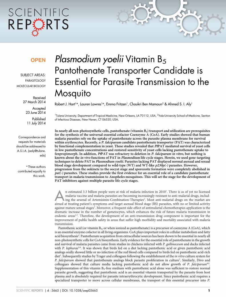
Plasmodium yoelii Vitamin B5
Pantothenate Transporter Candidate isEssential for Parasite Transmission to theMosquitoRobert J. Hart1*, Lauren Lawres1*, Emma Fritzen1, Choukri Ben Mamoun2 & Ahmed S. I. Aly1
1Tulane University, Department of Tropical Medicine, New Orleans, LA 70112, USA, 2Yale University School of Medicine, Sectionof Infectious Diseases, New Haven, CT 06520, USA.
In nearly all non-photosynthetic cells, pantothenate (vitamin B5) transport and utilization are prerequisitesfor the synthesis of the universal essential cofactor Coenzyme A (CoA). Early studies showed that humanmalaria parasites rely on the uptake of pantothenate across the parasite plasma membrane for survivalwithin erythrocytes. Recently, a P. falciparum candidate pantothenate transporter (PAT) was characterizedby functional complementation in yeast. These studies revealed that PfPAT mediated survival of yeast cellsin low pantothenate concentrations and restored sensitivity of yeast cells lacking pantothenate uptake tofenpropimorph. In addition, PfPAT was refractory to deletion in P. falciparum in vitro, but nothing isknown about the in vivo functions of PAT in Plasmodium life cycle stages. Herein, we used gene-targetingtechniques to delete PAT in Plasmodium yoelii. Parasites lacking PAT displayed normal asexual and sexualblood stage development compared to wild-type (WT) and WT-like p230p(-) parasites. However,progression from the ookinete to the oocyst stage and sporozoite formation were completely abolished inpat(-) parasites. These studies provide the first evidence for an essential role of a candidate pantothenatetransport in malaria transmission to Anopheles mosquitoes. This will set the stage for the development ofPAT inhibitors against multiple parasite life cycle stages.
An estimated 3.3 billion people were at risk of malaria infection in 20101. There is as of yet no licensedmalaria vaccine and malaria parasites are becoming increasingly resistant to anti-malarial drugs, includ-ing the arsenal of Artemisinin-Combination-Therapies2. Most anti-malarial drugs on the market are
aimed at treating patient’s symptoms and target asexual blood stage (BS) parasites, with no or limited activityagainst mature sexual stages3. Moreover, a frequent side-effect of antimalarial chemotherapies application is thedramatic increase in the number of gametocytes, which enhances the risk of future malaria transmission inendemic areas1,3. Therefore, the development of an anti-transmission drug component is important for theimprovement of public health safety in areas that suffer high morbidity and mortality associated with malariatransmission.
Pantothenic acid (or vitamin B5, or when ionized as pantothenate) is a precursor of coenzyme A (CoA), whichis an essential enzyme cofactor in all living organisms. CoA plays important roles in cellular metabolism and fattyacid biosynthesis4. Pantothenate acquisition from extracellular sources has been shown to be essential in nearly allnon-photosynthetic cells for CoA biosynthesis. Early evidence for the essential role of pantothenate in the growthand survival of malaria parasites came from studies in chickens infected with P. gallinaceum and ducks infectedwith P. lophurae5,6. It was shown that birds fed on a diet lacking pantothenic acid or given pantothenic acidanalogs orally showed little or no infection of the red blood cells compared to birds fed on pantothenic acid-richdiet5. Subsequently studies by Trager and colleagues following the establishment of the in vitro culture system forP. falciparum showed that pantothenate analogs block parasite proliferation in culture7. Similarly, Divo andcolleagues showed that culture media lacking pantothenic acid do not allow growth of P. falciparum8,9.Supplementation of this vitamin B5-free medium with pantothenic acid alone was sufficient to restore normalparasite growth, suggesting that pantothenic acid is an essential vitamin transported by the parasite from hostplasma and is absolutely required for parasite intraerythrocytic development. Since pantothenic acid requires aspecialized transporter to move across cellular membranes, the transport of this essential precursor into P.
OPEN
SUBJECT AREAS:PARASITOLOGY
MOLECULAR BIOLOGY
Received27 March 2014
Accepted23 June 2014
Published11 July 2014
Correspondence andrequests for materials
should be addressed toA.S.I.A. (aaly@tulane.
edu)
* These authorscontributed equally to
this work.
SCIENTIFIC REPORTS | 4 : 5665 | DOI: 10.1038/srep05665 1
falciparum-infected erythrocytes and its subsequent metabolism intoCoA are therefore essential for this parasite’s survival10. All CoAbiosynthesis genes have been identified in the Plasmodium genomesexcept the pantothenate transporter4.
In a marked contrast with the findings in P. falciparum, no activityof CoA biosynthesis enzymes could be detected in Plasmodiumlophurae isolated from duck red blood cells6,11,12. It has been sug-gested that in these cells pantothenate is taken up from host plasmavia the endogenous vitamin transporter and transported across thered blood cell membrane into the host erythrocyte where it is con-verted by host enzymes into CoA7,13,14. This preformed CoA is sub-sequently transported into the intra erythrocytic parasite6,11,12.However, the mechanism by which CoA is transported across theparasite plasma membrane remains unknown.
In a recent study, a conserved putative transporter gene with mul-tiple transmembrane domains was functionally characterized as acandidate pantothenate transporter (PAT) in Plasmodium falci-parum15. PfPAT localized to the parasite plasma membrane of P.falciparum and its expression in a fen2D yeast mutant (lacking theonly pantothenate transporter Fen2p) restored growth on low pan-tothenate concentrations and restored sensitivity to the antifungaldrug, fenpropimorph, which is primarily transported into yeast cellsvia the Fen2 permease16. Moreover, genetic studies in P. falciparumindicated that PfPAT gene is not amenable to targeted deletion byclassical genetic methods and down-regulation of its expressionusing morpholino-based oligonucleotides resulted in parasitedeath15. Together these data indicated that PfPAT might functionas the primary pantothenate transporter of P. falciparum and itstransport activity could be essential for BS development. Becauseof this phenotype the role of PAT in sexual stages developmentand parasite transmission could not be investigated.
In order to further characterize the role pantothenate transportplays during different life cycle stages of the malaria parasite, we haveused the murine malaria parasite Plasmodium yoelii as a model sys-tem. Using reverse genetics approaches, we found that pantothenatetransporter ortholog PyPAT is not essential for P. yoelii developmentin erythrocytes, thus making it possible to investigate the role pan-tothenate uptake and utilization play in sexual differentiation andparasite transmission to the mosquito. Our studies provide the firstevidence that a candidate pantothenate transporter is required foroocyst development and sporozoite formation, and thus is essentialfor parasite transmission to the mosquito.
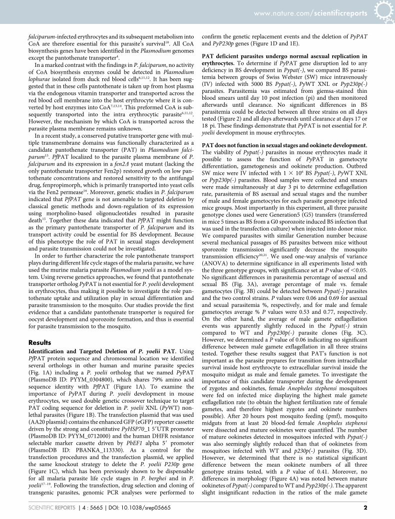
ResultsIdentification and Targeted Deletion of P. yoelii PAT. UsingPfPAT protein sequence and chromosomal location we identifiedseveral orthologs in other human and murine parasite species(Fig. 1A) including a P. yoelii ortholog that we named PyPAT(PlasmoDB ID: PYYM_0304800), which shares 79% amino acidsequence identity with PfPAT (Figure 1A). To examine theimportance of PyPAT during P. yoelii development in mouseerythrocytes, we used double genetic crossover technique to targetPAT coding sequence for deletion in P. yoelii XNL (PyWT) non-lethal parasites (Figure 1B). The transfection plasmid that was used(AA20 plasmid) contains the enhanced GFP (eGFP) reporter cassettedriven by the strong and constitutive PyHSP70_1 59UTR promoter(PlasmoDB ID: PYYM_0712000) and the human DHFR resistanceselectable marker cassette driven by PbEF1 alpha 59 promoter(PlasmoDB ID: PBANKA_113330). As a control for thetransfection procedures and the transfection plasmid, we appliedthe same knockout strategy to delete the P. yoelii P230p gene(Figure 1C), which has been previously shown to be dispensablefor all malaria parasite life cycle stages in P. berghei and in P.yoelii17–19. Following the transfection, drug selection and cloning oftransgenic parasites, genomic PCR analyses were performed to
confirm the genetic replacement events and the deletion of PyPATand PyP230p genes (Figure 1D and 1E).
PAT deficient parasites undergo normal asexual replication inerythrocytes. To determine if PyPAT gene disruption led to anydeficiency in BS development in Pypat(-), we compared BS parasi-temia between groups of Swiss Webster (SW) mice intravenously(IV) infected with 5000 BS Pypat(-), PyWT XNL or Pyp230p(-)parasites. Parasitemia was estimated from giemsa-stained thinblood smears until day 10 post infection (pi) and then monitoredafterwards until clearance. No significant differences in BSparasitemia could be detected between all three strains on all daystested (Figure 2) and all days afterwards until clearance at days 17 or18 pi. These findings demonstrate that PyPAT is not essential for P.yoelii development in mouse erythrocytes.
PAT does not function in sexual stages and ookinete development.The viability of Pypat(-) parasites in mouse erythrocytes made itpossible to assess the function of PyPAT in gametocytedifferentiation, gametogenesis and ookinete production. OutbredSW mice were IV infected with 1 3 106 BS Pypat(-), PyWT XNLor Pyp230p(-) parasites. Blood samples were collected and smearswere made simultaneously at day 3 pi to determine exflagellationrate, parasitemia of BS asexual and sexual stages and the numberof male and female gametocytes for each parasite genotype infectedmice groups. Most importantly in this experiment, all three parasitegenotype clones used were Generation5 (G5) transfers (transferredin mice 5 times as BS from a G0 sporozoite induced BS infection thatwas used in the transfection culture) when injected into donor mice.We compared parasites with similar Generation number becauseseveral mechanical passages of BS parasites between mice withoutsporozoite transmission significantly decrease the mosquitotransmission efficiency20,21. We used one-way analysis of variance(ANOVA) to determine significance in all experiments listed withthe three genotype groups, with significance set at P value of ,0.05.No significant differences in parasitemia percentage of asexual andsexual BS (Fig. 3A), average percentage of male vs. femalegametocytes (Fig. 3B) could be detected between Pypat(-) parasitesand the two control strains. P values were 0.06 and 0.69 for asexualand sexual parasitemia %, respectively, and for male and femalegametocytes average % P values were 0.53 and 0.77, respectively.On the other hand, the average of male gamete exflagellationevents was apparently slightly reduced in the Pypat(-) straincompared to WT and Pyp230p(-) parasite clones (Fig. 3C).However, we determined a P value of 0.06 indicating no significantdifference between male gamete exflagellation in all three strainstested. Together these results suggest that PAT’s function is notimportant as the parasite prepares for transition from intracellularsurvival inside host erythrocyte to extracellular survival inside themosquito midgut as male and female gametes. To investigate theimportance of this candidate transporter during the developmentof zygotes and ookinetes, female Anopheles stephensi mosquitoeswere fed on infected mice displaying the highest male gameteexflagellation rate (to obtain the highest fertilization rate of femalegametes, and therefore highest zygotes and ookinete numberspossible). After 20 hours post mosquito feeding (pmf), mosquitomidguts from at least 20 blood-fed female Anopheles stephensiwere dissected and mature ookinetes were quantified. The numberof mature ookinetes detected in mosquitoes infected with Pypat(-)was also seemingly slightly reduced than that of ookinetes frommosquitoes infected with WT and p230p(-) parasites (Fig. 3D).However, we determined that there is no statistical significantdifference between the mean ookinete numbers of all threegenotype strains tested, with a P value of 0.41. Moreover, nodifferences in morphology (Figure 4A) was noted between matureookinetes of Pypat(-) compared to WT and Pyp230p(-). The apparentslight insignificant reduction in the ratios of the male gamete
www.nature.com/scientificreports
SCIENTIFIC REPORTS | 4 : 5665 | DOI: 10.1038/srep05665 2
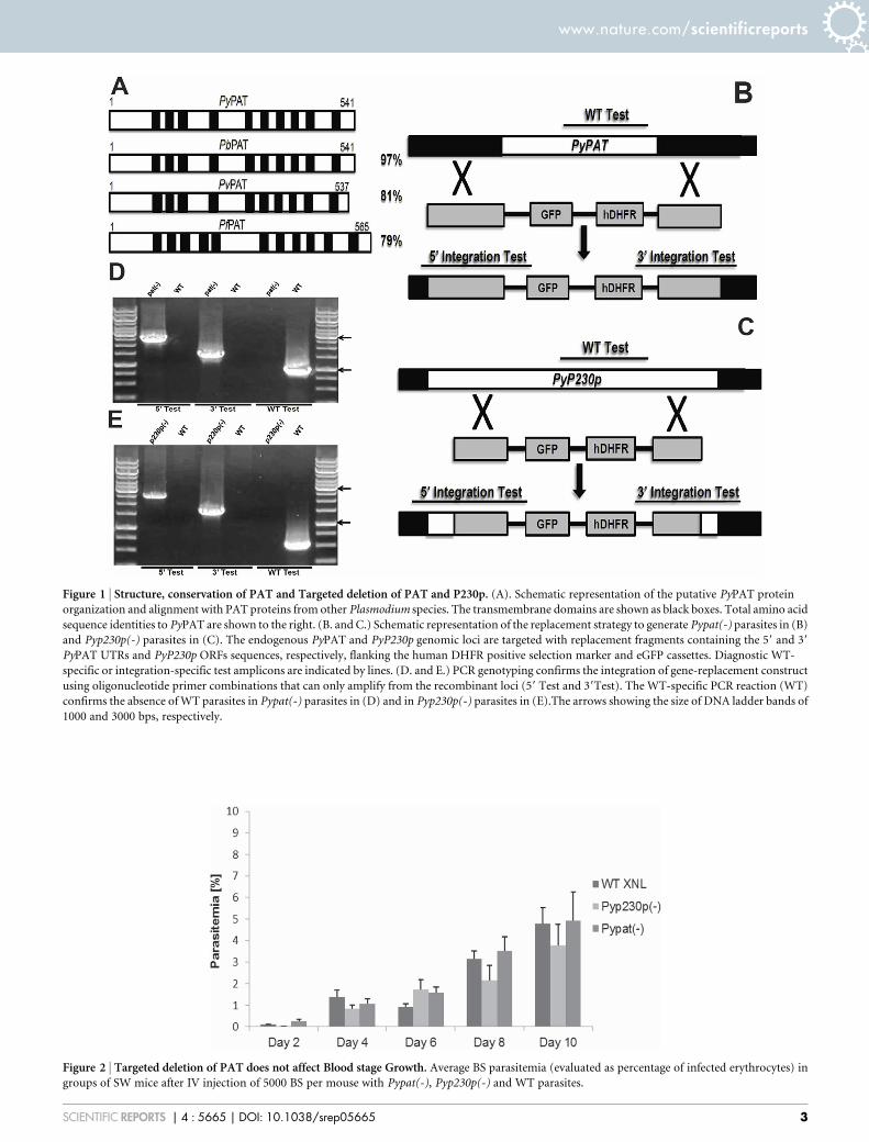
Figure 1 | Structure, conservation of PAT and Targeted deletion of PAT and P230p. (A). Schematic representation of the putative PyPAT protein
organization and alignment with PAT proteins from other Plasmodium species. The transmembrane domains are shown as black boxes. Total amino acid
sequence identities to PyPAT are shown to the right. (B. and C.) Schematic representation of the replacement strategy to generate Pypat(-) parasites in (B)
and Pyp230p(-) parasites in (C). The endogenous PyPAT and PyP230p genomic loci are targeted with replacement fragments containing the 59 and 39
PyPAT UTRs and PyP230p ORFs sequences, respectively, flanking the human DHFR positive selection marker and eGFP cassettes. Diagnostic WT-
specific or integration-specific test amplicons are indicated by lines. (D. and E.) PCR genotyping confirms the integration of gene-replacement construct
using oligonucleotide primer combinations that can only amplify from the recombinant loci (59 Test and 39Test). The WT-specific PCR reaction (WT)
confirms the absence of WT parasites in Pypat(-) parasites in (D) and in Pyp230p(-) parasites in (E).The arrows showing the size of DNA ladder bands of
1000 and 3000 bps, respectively.
Figure 2 | Targeted deletion of PAT does not affect Blood stage Growth. Average BS parasitemia (evaluated as percentage of infected erythrocytes) in
groups of SW mice after IV injection of 5000 BS per mouse with Pypat(-), Pyp230p(-) and WT parasites.
www.nature.com/scientificreports
SCIENTIFIC REPORTS | 4 : 5665 | DOI: 10.1038/srep05665 3
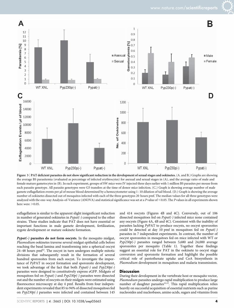
exflagellation is similar to the apparent slight insignificant reductionin number of generated ookinetes in Pypat(-) compared to the otherstrains. These studies indicate that PAT does not have essential orimportant functions in male gamete development, fertilization,zygote development or mature ookinete formation.
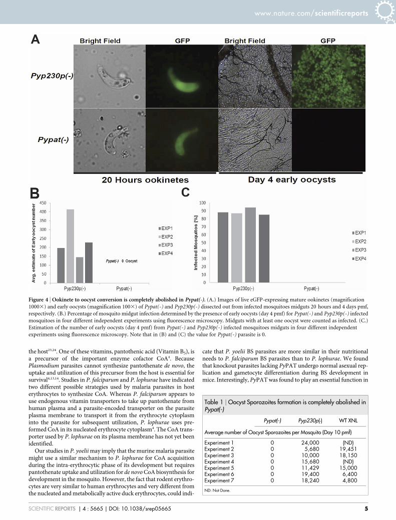
Pypat(-) parasites do not form oocysts. In the mosquito midgut,Plasmodium ookinetes traverse several midgut epithelial cells beforereaching the basal lamina and transforming into a spherical oocyst24–48 hours pmf22. The oocyst in turn undergoes multiple mitoticdivisions that subsequently result in the formation of severalhundred sporozoites from each oocyst. To investigate the impor-tance of PyPAT in oocyst formation and sporozoite development,we took advantage of the fact that both Pypat(-) and Pyp230p(-)parasites were designed to constitutively express eGFP. Midguts ofmosquitoes fed on Pypat(-) and Pyp230p(-) parasites were dissectedout and the number of oocysts on their midguts were estimated usingfluorescence microscopy at day 4 pmf. Results from four indepen-dent experiments revealed that 85 to 94% of dissected mosquitoes fedon Pyp230p(-) parasites were infected and contained between 145
and 414 oocysts (Figures 4B and 4C). Conversely, out of 106dissected mosquitoes fed on Pypat(-) infected mice none containedany oocysts (Figure 4A, 4B and 4C). Consistent with the inability ofparasites lacking PyPAT to produce oocysts, no oocyst sporozoitescould be detected at day 10 pmf in mosquitoes fed on Pypat(-)parasites in 7 independent experiments. In contrast, the number ofoocyst sporozoites in mosquitoes fed on mice infected with WT orPyp230p(-) parasites ranged between 5,680 and 24,000 averagesporozoites per mosquito (Table 1). Together these findingssuggest an essential role for PAT in the ookinete to oocyst stageconversion and sporozoite formation and highlight the possiblecritical role of pantothenate uptake and CoA biosynthesis inPlasmodium development in mosquitoes and malaria transmission.
DiscussionDuring their development in the vertebrate host or mosquito vector,Plasmodium parasites undergo rapid multiplication to produce largenumber of daughter parasites22,23. This rapid multiplication reliesheavily on successful acquisition of essential nutrients such as purinenucleosides and nucleobases, amino acids, sugars and vitamins from
Figure 3 | PAT deficient parasites do not show significant reduction in the development of sexual stages and ookinetes. (A. and B.) Graphs are showing
the average BS parasitemia (evaluated as percentage of infected erythrocytes) for asexual and sexual stages in (A), and the average ratio of male and
female mature gametocytes in (B). In each experiment, groups of SW mice were IV injected three days earlier with 1 million BS parasites per mouse from
each parasite genotype. All parasite genotypes were G5 transfers at the time of donor mice infection. (C.) Graph is showing average number of male
gamete exflagellation events per ml of mouse blood determined by a hemocytometer using 1510 dilution of tail blood. (D.) Graph is showing the average
number of ookinetes dissected out of mosquitos infected with each of the three genotypes 20 hours pmf. The median values for all three genotypes were
analyzed with the one-way Analysis-of-Variance (ANOVA) and statistical significance was set at a P value of ,0.05. The P values in all experiments shown
here were .0.05.
www.nature.com/scientificreports
SCIENTIFIC REPORTS | 4 : 5665 | DOI: 10.1038/srep05665 4
the host23,24. One of these vitamins, pantothenic acid (Vitamin B5), isa precursor of the important enzyme cofactor CoA4. BecausePlasmodium parasites cannot synthesize pantothenate de novo, theuptake and utilization of this precursor from the host is essential forsurvival4,13,14. Studies in P. falciparum and P. lophurae have indicatedtwo different possible strategies used by malaria parasites in hosterythrocytes to synthesize CoA. Whereas P. falciparum appears touse endogenous vitamin transporters to take up pantothenate fromhuman plasma and a parasite-encoded transporter on the parasiteplasma membrane to transport it from the erythrocyte cytoplasminto the parasite for subsequent utilization, P. lophurae uses pre-formed CoA in its nucleated erythrocyte cytoplasm4. The CoA trans-porter used by P. lophurae on its plasma membrane has not yet beenidentified.
Our studies in P. yoelii may imply that the murine malaria parasitemight use a similar mechanism to P. lophurae for CoA acquisitionduring the intra-erythrocytic phase of its development but requirespantothenate uptake and utilization for de novo CoA biosynthesis fordevelopment in the mosquito. However, the fact that rodent erythro-cytes are very similar to human erythrocytes and very different fromthe nucleated and metabolically active duck erythrocytes, could indi-
cate that P. yoelii BS parasites are more similar in their nutritionalneeds to P. falciparum BS parasites than to P. lophurae. We foundthat knockout parasites lacking PyPAT undergo normal asexual rep-lication and gametocyte differentiation during BS development inmice. Interestingly, PyPAT was found to play an essential function in
Table 1 | Oocyst Sporozoites formation is completely abolished inPypat(-)
Pypat(-) Pyp230p(-) WT XNL
Average number of Oocyst Sporozoites per Mosquito (Day 10 pmf)
Experiment 1 0 24,000 (ND)Experiment 2 0 5,680 19,451Experiment 3 0 10,000 18,150Experiment 4 0 15,680 (ND)Experiment 5 0 11,429 15,000Experiment 6 0 19,400 6,400Experiment 7 0 18,240 4,800
ND: Not Done.
Figure 4 | Ookinete to oocyst conversion is completely abolished in Pypat(-). (A.) Images of live eGFP-expressing mature ookinetes (magnification
10003) and early oocysts (magnification 1003) of Pypat(-) and Pyp230p(-) dissected out from infected mosquitoes midguts 20 hours and 4 days pmf,
respectively. (B.) Percentage of mosquito midgut infection determined by the presence of early oocysts (day 4 pmf) for Pypat(-) and Pyp230p(-) infected
mosquitoes in four different independent experiments using fluorescence microscopy. Midguts with at least one oocyst were counted as infected. (C.)
Estimation of the number of early oocysts (day 4 pmf) from Pypat(-) and Pyp230p(-) infected mosquitoes midguts in four different independent
experiments using fluorescence microscopy. Note that in (B) and (C) the value for Pypat(-) parasite is 0.
www.nature.com/scientificreports
SCIENTIFIC REPORTS | 4 : 5665 | DOI: 10.1038/srep05665 5
oocyst progression and sporozoite formation but was not critical forgametogenesis, fertilization and ookinete development. Male game-tocytes, in less than ten minutes, undergo up to three rounds ofmitotic divisions and cytokinesis to produce up to eight highly motilegametes in the mosquito midgut22. This indicates that all the nutri-tional needs for mitotic division, cytokinesis and fertilization must bepresent in male gametocytes before transmission to the mosquito.Likewise, female gametocytes provide maternally the main cell struc-ture, all cellular organelles and all nutritional needs required forfemale gametogenesis, fertilization and the conversion of the spher-ical zygote into pointed elongated invasive ookinete22. However, fol-lowing maturation of ookinetes and traversal of midgut epithelialcells, ookinetes stop migrating and convert to oocysts, whichundergo several mitotic divisions that result in the production ofhundreds of sporozoites from each oocyst22. Because malaria para-sites cannot synthesize the precursors of several essential macromo-lecules and co-factors, including CoA, their rapid development andactive replication inside the mosquito vector is heavily dependent onthe acquisition of these precursors from the host environment.Whereas major efforts have been made over the years to understandthese processes during the parasite intraerythrocytic life cycle, little isknown about parasite nutrient requirement, uptake and utilization inan extracellular environment in the mosquito vector. The inability ofPypat(-) knockout parasite to progress from ookinetes to oocysts orto form sporozoites suggest that this gene is required for these stagesof parasite development. Apparently, active CoA biosynthesis fromits primary precursor pantothenate at this specific stage of the lifecycle, unlike BS parasites, seems essential for survival. However, weshould not undermine the possibility of the presence of an alternativeprimary or a secondary pantothenate or pantetheine transporter thatrescue the lack of PAT in BS, albeit this does not happen in mosquitostages.
Our study provides the first evidence for an essential role of acandidate pantothenate transporter in the arthropod phase of thelife cycle of any protozoan parasite. The availability of Pypat(-)knockout parasites will make it possible to investigate the direct roleof this molecule in the uptake of exogenous pantothenate. Due of therapid and magnitude of multiplication of the parasite in hepatocytes(in which tens of thousands of merozoites develop from one liverstage), we predict that PAT function might also be essential forparasite pre-erythrocytic development.
Experimental ProceduresExperimental Animals, Parasites and Mosquitoes. Female SwissWebster (SW) mice (6 to 8 weeks old) were purchased fromHarlan Laboratories (Indianapolis, Indiana). Animal handling wasconducted according to Institutional-Animal-Care-and-Use-Com-mittee (IACUC) approved protocols. Wild-type (WT) P. yoeliiXNL (non-lethal strain), Pyp230p(-) clone A5 and Pypat(-) cloneE5 parasites were used to infect SW mice and Anopheles stephensimosquitoes. We always used the same G-number (Generationnumber, which refers to the number of serial mouse blood trans-fers from sporozoite induced blood stage infection counted as G0)while comparing phenotypes of different clonal parasite populations.Mosquitoes were allowed to feed for at least 15 minutes onanesthetized infected mice on day 3 post intravenous injection(IV) of 1 million parasites in each parasite clone. Infected mosqui-toes were maintained on 10% Sucrose solution 10.05% Para-AminoBenzoic Acid (PABA) wetted cotton pads at 24uC, 70% humidity and16/8 hours light/dark cycles. Midguts were extracted from infectedmosquitoes on days 1, 4 and 10 or 11 post mosquito infection (pmf)for counting ookinetes, early oocysts and oocyst sporozoites,respectively. Pyp230p(-) clone A5 and Pypat(-) clone E5 earlyoocyst detection was by counting green fluorescent oocysts viafluorescence microscopy. Sporozoites were freed from oocysts in
RPMI medium by grinding the midguts with a pestle, sporozoiteswere counted using a hemocytometer.
Generation of transgenic parasites. Targeted deletions of PyPATand PyP230p genes were done by double genetic crossover strategydriven by homologous recombination. P. yoelii XNL genomic DNA(gDNA) was used as a template to amplify fragments of the 59UTR ofPyPAT and of the 59 end of PyP230p ORF (Open Reading Frame)using primers 31–32 and primers 41–42, respectively. The amplifiedfragments were inserted into the transfection plasmid AA20 betweenSacII and BamHI restriction enzyme sites. Fragments from the39UTR sequence of PyPAT and of the 39 end of PyP230p ORFwere amplified using primers 33–34 and primers 43–44,respectively, from P. yoelii XNL gDNA as a template, and insertedbetween the KpnI and HindIII restriction sites in the respectiveintermediate transfection plasmids. The fragments cloned forPyPAT targeted deletion were designed not to interfere with thecoding sequences of any upstream or downstream neighboringgenes and the makeup of those fragments was checked bysequencing to ensure that any unknown 59 or 39 promoter signalsfor the neighboring genes were not altered. The final plasmids werelinearized with SacII and KpnI prior to the transfection. Transfectionof P. yoelii XNL parasites with Lonza Nucleofector II device wasperformed on a parasite cultures that originated from donor ratsthat were infected with P. yoelii WT XNL sporozoites. Thetransfection procedures, resistant parasites selection and transgenicrecombinant parasite cloning by serial dilutions were all conductedas described elsewhere17,25. To confirm the targeted deletion and thenew genetic recombination, gDNAs were extracted from Pyp230p(-)clone A5 and Pypat(-) clone E5 and 59 and 39 integration-specific andWT specific PCR amplifications were conducted for 35 cycles (PCRcycle conditions: 94u for 30 seconds, 55u for 30 seconds and 60u for 3minutes). 59 Integration Tests were performed with primers 35–16and primers 45–16, 39 Integration Tests with primers 17–36 andprimers 17–46 and WT coding sequence Tests with primers 37–38and primers 47–48 for Pypat(-) and Pyp230p(-) gDNA, respectively.
Blood Stage Growth, Male gamete Exflagellation and GametocytesRatio Estimations. Donor mice were infected with the threegenotypes with the same generation number by Intraperitonealinfection (IP) of frozen stocks. At day 2 pi, number of asexual BSper ml of donor mouse blood was determined by giemsa stained thinblood smears (to determine parasitemia %) and hemocytometercounts of donor mouse erythrocytes. Limited dilution with RPMImedium of donor mouse blood, when the parasitemia was between0.1% and 1%, was used to aliquot the specific number of parasites tobe injected into each mouse into separate micro-centrifuge tubes. Forestimation of BS growth, recipient groups of 3 SW received IVinjection of 5000 parasites from each genotype, and parasitemia %were counted daily from giemsa stained thin blood smears from day 2until day 10 pi. At least 50 microscopic fields (magnification 10003)were used for the parasitemia quantification on Nikon eclipse 50imicroscope. Afterwards, giemsa stained thin blood smears werechecked on days 12, 14, 16, 17 and 18 pi for clearance, whichoccurred at days 17 or 18 pi for all three genotypes. For theestimation of male gamete exflagellation and mosquito infections,recipient naı̈ve mice were treated with phenylhydrazine (50 mg/kg inPBS) three days before the IV injection with 1 million BS parasites.Male gamete exflagellation was quantified three days after the IVinfection by adding 5 mL blood from a tail puncture with aheparinized needle to complete ookinete medium in 1510 dilutionand counting the exflagellation events in a hemocytometer after 10minutes incubation at room temperature (22–24uC). At the sametime, a blood droplet was taken from the leftover blood at the siteof tail puncture and was smeared and giemsa stained as a thin bloodsmear. From these smears BS sexual and asexual parasitemia % and
www.nature.com/scientificreports
SCIENTIFIC REPORTS | 4 : 5665 | DOI: 10.1038/srep05665 6
average percentage of male and female gametocytes were estimatedat the same recording time.
In vivo Ookinete and Early Oocysts Quantifications. The mice withthe highest exflagellation rate from each genotype were fed tomosquitoes cages. 18–20 hours pmf, the midguts of at least 20blood fed mosquitoes from each genotype were dissected out,ground with a pestle and diluted before loading onto a hemocyto-meter for counting of mature ookinetes. On day 4 pmf, mosquitomidguts were extracted and placed on a microscope slide. We thencounted the number of early oocysts and estimated the number ofoocysts per infected mosquito.
Statistical Analysis. Statistical significant differences in all experi-ments was determined by the GraphPad InStat software between themedian values for all three genotypes using one-way Analysis-of-Variance (ANOVA). P values of ,0.05 were considered statis-tically significant.
Primers sequences (59-39). 31: GGCCGCGGTCATGTAATGTTA-TAGTGTCATGCAACTAAAAAT;
32: TCCGGATCCCGCTTACCTGTTTTGGGTATAGGAATC-GGCATTCAT;
33: TGCCAAGCTTTCAAAATAGGATAATAATAGATACTT-TTAGAAT;
34: TCCGGTACCTATATAGATATACATATTACTAAAAAT-GCAGTC;
35: ACCTTTTACTTACTATTTTTAATTAGCTATTTAA;36: ATATCAAAAAAAAATATATAACCATATTTAACTA;37: GGCCGCGGGATGAAATATTTTGGAACAATTCGAGG-
TTGGAG;38: TCCGAATTCTGACATAACTTTATCTGCCATAGTCTC-
GTCATC;41: GGCCGCGGATGATAAACCATATTTTGAAGAAATTAT-
TAGTGA;42: TCCGGATCCGCTTATTCTGGATTTTCAAATGAATAG-
TTAATAA;43: GCCCAAGCTTCTTTAAGAAATTATGAACAATTCAAG-
CAATCCAA;44: TCCGGTACCTTCCTAATATATATGATTTATAAATTA-
ATTTGAT;45: GAAGATTTTATCATTATTCGAGTCAGAGTACATCA;46: ACACGAACCCAAAGAAATCGGCAAGCTAAAACTA;47: ATTATATGGATGTGATTTTTCAGGTAATTCTAA;48: CATACTCAAATATCTTTTTAGTAGTTCCTCAATA;16: ATGTCCATTAACATCACCATCTAATTCAACAAG;17: GTGTTCTTTCTGATGTTCAAGAAGAAAAAGGTA.
1. Alonso, P. L. & Tanner, M. Public health challenges and prospects for malariacontrol and elimination. Nat Med 19, 150–155, doi:10.1038/nm.3077 (2013).
2. Beshir, K. B. et al. Residual Plasmodium falciparum parasitemia in Kenyanchildren after artemisinin-combination therapy is associated with increasedtransmission to mosquitoes and parasite recurrence. J Infect Dis 208, 2017–2024,doi:10.1093/infdis/jit431 (2013).
3. mal, E. R. A. C. G. o. D. A research agenda for malaria eradication: drugs. PLoSMed 8, e1000402, doi:10.1371/journal.pmed.1000402 (2011).
4. Spry, C., Kirk, K. & Saliba, K. J. Coenzyme A biosynthesis: an antimicrobial drugtarget. FEMS microbiology reviews 32, 56–106, doi:10.1111/j.1574-6976.2007.00093.x (2008).
5. Brackett, S., Waletzky, E. & Baker, M. The relation between pantothenic acid andPlasmodium gallinaceum infections in the chicken and the antimalarial activity ofanalogues of pantothenic acid. J Parasitol 32, 453–462 (1946).
6. Trager, W. Further Studies on the Survival and Development in Vitro of a MalarialParasite. The Journal of experimental medicine 77, 411–420 (1943).
7. Trager, W. Cofactors and vitamins in the metabolism of malarial parasites. Factorsother than folates. Bulletin of the World Health Organization 55, 285–289 (1977).
8. Divo, A. A., Geary, T. G., Davis, N. L. & Jensen, J. B. Nutritional requirements ofPlasmodium falciparum in culture. I. Exogenously supplied dialyzablecomponents necessary for continuous growth. J Protozool 32, 59–64 (1985).
9. Saliba, K. J., Ferru, I. & Kirk, K. Provitamin B5 (pantothenol) inhibits growth ofthe intraerythrocytic malaria parasite. Antimicrob Agents Chemother 49, 632–637(2005).
10. Saliba, K. J. & Kirk, K. H1-coupled pantothenate transport in the intracellularmalaria parasite. J Biol Chem 276, 18115–18121, doi:10.1074/jbc.M010942200(2001).
11. Trager, W. Coenzyme A and the antimalarial action in vitro of antipantothenateagainst Plasmodium lophurae, P. coatneyi and P. falciparum. Trans N Y Acad Sci28, 1094–1108 (1966).
12. Trager, W. Studies on the extracellular cultivation of an intracellular parasite(avian malaria). II. The effects of malate and of coenzyme A concentrates. TheJournal of experimental medicine 96, 465–476 (1952).
13. Spry, C. & Saliba, K. J. The human malaria parasite Plasmodium falciparum is notdependent on host coenzyme A biosynthesis. J Biol Chem 284, 24904–24913,doi:10.1074/jbc.M109.025312 (2009).
14. Spry, C., van Schalkwyk, D. A., Strauss, E. & Saliba, K. J. Pantothenate utilizationby Plasmodium as a target for antimalarial chemotherapy. Infectious disordersdrug targets 10, 200–216 (2010).
15. Augagneur, Y. et al. Identification and functional analysis of the primarypantothenate transporter, PfPAT, of the human malaria parasite Plasmodiumfalciparum. J Biol Chem 288, 20558–20567, doi:10.1074/jbc.M113.482992 (2013).
16. Stolz, J., Caspari, T., Carr, A. M. & Sauer, N. Cell division defects ofSchizosaccharomyces pombe liz1- mutants are caused by defects in pantothenateuptake. Eukaryotic cell 3, 406–412 (2004).
17. Janse, C. J. et al. High efficiency transfection of Plasmodium berghei facilitatesnovel selection procedures. Mol Biochem Parasitol 145, 60–70, doi:10.1016/j.molbiopara.2005.09.007 (2006).
18. Lin, J. W. et al. A novel ‘gene insertion/marker out’ (GIMO) method for transgeneexpression and gene complementation in rodent malaria parasites. PLoS One 6,e29289, doi:10.1371/journal.pone.0029289 (2011).
19. van Dijk, M. R. et al. Three members of the 6-cys protein family of Plasmodiumplay a role in gamete fertility. PLoS pathogens 6, e1000853, doi:10.1371/journal.ppat.1000853 (2010).
20. Sinha, A. et al. A cascade of DNA-binding proteins for sexual commitment anddevelopment in Plasmodium. Nature doi:10.1038/nature12970 (2014).
21. Janse, C. J., Ponzi, M., Sinden, R. E. & Waters, A. P. Chromosomes and sexualdevelopment of rodent malaria parasites. Memorias do Instituto Oswaldo Cruz 89Suppl 2, 43–46 (1994).
22. Aly, A. S., Vaughan, A. M. & Kappe, S. H. Malaria parasite development in themosquito and infection of the mammalian host. Annu Rev Microbiol 63, 195–221,doi:10.1146/annurev.micro.091208.073403 (2009).
23. Saliba, K. J. & Kirk, K. Nutrient acquisition by intracellular apicomplexanparasites: staying in for dinner. Int J Parasitol 31, 1321–1330 (2001).
24. Kirk, K. & Saliba, K. J. Targeting nutrient uptake mechanisms in Plasmodium.Current drug targets 8, 75–88 (2007).
25. Philip, N., Orr, R. & Waters, A. P. Transfection of rodent malaria parasites.Methods Mol Biol 923, 99–125, doi:10.1007/978-1-62703-026-7_7 (2013).
AcknowledgmentsWe like to thank Emily Molina and Fleur Porter for technical assistance to some of theexperiments and Ashley Vaughn for critical reading of the manuscript. This work wassupported by funds provided by Tulane University School of Public Health and TropicalMedicine.
Author contributionsA.A. and C.B.M. initiated the research, A.A. designed the experiments, A.A., R.H., L.L. andE.F. conducted the experiments, A.A. and R.H. analyzed the data and prepared the figures,A.A. and C.B.M. wrote the manuscript.
Additional informationCompeting financial interests: The authors declare no competing financial interests.
How to cite this article: Hart, R.J., Lawres, L., Fritzen, E., Mamoun, C.B. & Aly, A.S.I.Plasmodium yoelii Vitamin B5 Pantothenate Transporter Candidate is Essential for ParasiteTransmission to the Mosquito. Sci. Rep. 4, 5665; DOI:10.1038/srep05665 (2014).
This work is licensed under a Creative Commons Attribution-NonCommercial-NoDerivs 4.0 International License. The images or other third party material inthis article are included in the article’s Creative Commons license, unless indicatedotherwise in the credit line; if the material is not included under the CreativeCommons license, users will need to obtain permission from the license holderin order to reproduce the material. To view a copy of this license, visit http://creativecommons.org/licenses/by-nc-nd/4.0/
www.nature.com/scientificreports
SCIENTIFIC REPORTS | 4 : 5665 | DOI: 10.1038/srep05665 7
Copyright © 2022 FDOKUMEN