Phoenix Is Required for Mechanosensory Hair Cell Regeneration in the Zebrafish Lateral Line
14
Phoenix Is Required for Mechanosensory Hair Cell Regeneration in the Zebrafish Lateral Line Martine Behra 1 , John Bradsher 2 , Rachid Sougrat 3 , Viviana Gallardo 4 , Miguel L. Allende 4 , Shawn M. Burgess 1 * 1 National Human Genome Research Institute, National Institutes of Health, Bethesda, Maryland, United States of America, 2 National Cancer Institute, Bethesda, Maryland, United States of America, 3 National Institute of Child Health and Human Development, Bethesda, Maryland, United States of America, 4 Center for Genomics of the Cell, Facultad de Ciencias, Universidad de Chile, Santiago, Chile Abstract In humans, the absence or irreversible loss of hair cells, the sensory mechanoreceptors in the cochlea, accounts for a large majority of acquired and congenital hearing disorders. In the auditory and vestibular neuroepithelia of the inner ear, hair cells are accompanied by another cell type called supporting cells. This second cell population has been described as having stem cell-like properties, allowing efficient hair cell replacement during embryonic and larval/fetal development of all vertebrates. However, mammals lose their regenerative capacity in most inner ear neuroepithelia in postnatal life. Remarkably, reptiles, birds, amphibians, and fish are different in that they can regenerate hair cells throughout their lifespan. The lateral line in amphibians and in fish is an additional sensory organ, which is used to detect water movements and is comprised of neuroepithelial patches, called neuromasts. These are similar in ultra-structure to the inner ear’s neuroepithelia and they share the expression of various molecular markers. We examined the regeneration process in hair cells of the lateral line of zebrafish larvae carrying a retroviral integration in a previously uncharacterized gene, phoenix (pho). Phoenix mutant larvae develop normally and display a morphologically intact lateral line. However, after ablation of hair cells with copper or neomycin, their regeneration in pho mutants is severely impaired. We show that proliferation in the supporting cells is strongly decreased after damage to hair cells and correlates with the reduction of newly formed hair cells in the regenerating phoenix mutant neuromasts. The retroviral integration linked to the phenotype is in a novel gene with no known homologs showing high expression in neuromast supporting cells. Whereas its role during early development of the lateral line remains to be addressed, in later larval stages phoenix defines a new class of proteins implicated in hair cell regeneration. Citation: Behra M, Bradsher J, Sougrat R, Gallardo V, Allende ML, et al. (2009) Phoenix Is Required for Mechanosensory Hair Cell Regeneration in the Zebrafish Lateral Line. PLoS Genet 5(4): e1000455. doi:10.1371/journal.pgen.1000455 Editor: Mary C. Mullins, University of Pennsylvania School of Medicine, United States of America Received September 3, 2008; Accepted March 16, 2009; Published April 17, 2009 This is an open-access article distributed under the terms of the Creative Commons Public Domain declaration which stipulates that, once placed in the public domain, this work may be freely reproduced, distributed, transmitted, modified, built upon, or otherwise used by anyone for any lawful purpose. Funding: This research was supported by the Intramural Research Program of the National Human Genome Research Institute, National Institutes of Health (SB). MLA was supported by FONDECYT (1070867) and ICM (P06-037F). VG received a CONICYT Fellowship and a travel grant from the Vicerrectorı ´a de Asuntos Acade ´ micos, Departamento de Postgrado y Postı ´tulo. Part of this work was supported by the Intramural Program of Eunice Kennedy Shriver National Institute of Child Health and Human Development. The funders had no role in study design, data collection and analysis, decision to publish, or preparation of the manuscript. Competing Interests: The authors have declared that no competing interests exist. * E-mail: [email protected] Introduction During development of the vertebrate inner ear, a subset of neuroepithelial cells specialize to give rise to hair cells and supporting cells [1–3]. These two cell populations assume distinct and complementary functions. The hair cell is a highly differentiated mechanoreceptor cell, transducing sound waves in the cochlea or acceleration and head movements in the vestibular organ, into electrical signals [4,5]. The supporting cells provide cohesive support [6,7] and have secretory functions [8,9]. More importantly, they have been clearly implicated in the addition of new hair cells, both during normal growth and during restoration of the sensory epithelium after damage in various animal models [10–14]. Thus, among supporting cells, there exists a tissue- specific population of progenitor cells. However, in mammals, this ‘‘stem cell like’’ property is lost shortly after birth in most neuroepithelia of the inner ear [15]. With the exception of some limited regeneration in a sub-region of the vestibular organ [16,17], post natal hair cell loss is permanent and irreversible. Consequently, a large majority of deafness cases in humans are linked to absent or damaged hair cells. To restore the regenerative capacity of supporting cells is an obvious therapeutic aim, but our understanding of the regenerative process is incomplete. Because birds, amphibians, reptiles, and fish have retained the ability to regenerate hair cells [14,18–25] they provide opportunities to find genes involved in the regeneration process and its maintenance. Fish and amphibians have an additional organ related to the inner ear called the lateral line, which is used to detect water currents [26–28]. It is a superficial organ running along each side of the body which consists of stereotypically distributed patches of neuroepithelium, called neuromasts. These are discrete organs made of hair cells that project into the aqueous environment and supporting cells that surround them. In the zebrafish embryo, the lateral line neuromasts first appear in the head approximately 2 days post fertilization (dpf) and in parallel begin to develop along the entire length of the trunk and tail [29]. Hair cells are fully PLoS Genetics | www.plosgenetics.org 1 April 2009 | Volume 5 | Issue 4 | e1000455
Transcript of Phoenix Is Required for Mechanosensory Hair Cell Regeneration in the Zebrafish Lateral Line

Phoenix Is Required for Mechanosensory Hair CellRegeneration in the Zebrafish Lateral LineMartine Behra1, John Bradsher2, Rachid Sougrat3, Viviana Gallardo4, Miguel L. Allende4, Shawn M.
Burgess1*
1 National Human Genome Research Institute, National Institutes of Health, Bethesda, Maryland, United States of America, 2 National Cancer Institute, Bethesda, Maryland,
United States of America, 3 National Institute of Child Health and Human Development, Bethesda, Maryland, United States of America, 4 Center for Genomics of the Cell,
Facultad de Ciencias, Universidad de Chile, Santiago, Chile
Abstract
In humans, the absence or irreversible loss of hair cells, the sensory mechanoreceptors in the cochlea, accounts for a largemajority of acquired and congenital hearing disorders. In the auditory and vestibular neuroepithelia of the inner ear, haircells are accompanied by another cell type called supporting cells. This second cell population has been described as havingstem cell-like properties, allowing efficient hair cell replacement during embryonic and larval/fetal development of allvertebrates. However, mammals lose their regenerative capacity in most inner ear neuroepithelia in postnatal life.Remarkably, reptiles, birds, amphibians, and fish are different in that they can regenerate hair cells throughout their lifespan.The lateral line in amphibians and in fish is an additional sensory organ, which is used to detect water movements and iscomprised of neuroepithelial patches, called neuromasts. These are similar in ultra-structure to the inner ear’s neuroepitheliaand they share the expression of various molecular markers. We examined the regeneration process in hair cells of thelateral line of zebrafish larvae carrying a retroviral integration in a previously uncharacterized gene, phoenix (pho). Phoenixmutant larvae develop normally and display a morphologically intact lateral line. However, after ablation of hair cells withcopper or neomycin, their regeneration in pho mutants is severely impaired. We show that proliferation in the supportingcells is strongly decreased after damage to hair cells and correlates with the reduction of newly formed hair cells in theregenerating phoenix mutant neuromasts. The retroviral integration linked to the phenotype is in a novel gene with noknown homologs showing high expression in neuromast supporting cells. Whereas its role during early development of thelateral line remains to be addressed, in later larval stages phoenix defines a new class of proteins implicated in hair cellregeneration.
Citation: Behra M, Bradsher J, Sougrat R, Gallardo V, Allende ML, et al. (2009) Phoenix Is Required for Mechanosensory Hair Cell Regeneration in the ZebrafishLateral Line. PLoS Genet 5(4): e1000455. doi:10.1371/journal.pgen.1000455
Editor: Mary C. Mullins, University of Pennsylvania School of Medicine, United States of America
Received September 3, 2008; Accepted March 16, 2009; Published April 17, 2009
This is an open-access article distributed under the terms of the Creative Commons Public Domain declaration which stipulates that, once placed in the publicdomain, this work may be freely reproduced, distributed, transmitted, modified, built upon, or otherwise used by anyone for any lawful purpose.
Funding: This research was supported by the Intramural Research Program of the National Human Genome Research Institute, National Institutes of Health (SB).MLA was supported by FONDECYT (1070867) and ICM (P06-037F). VG received a CONICYT Fellowship and a travel grant from the Vicerrectorıa de AsuntosAcademicos, Departamento de Postgrado y Postıtulo. Part of this work was supported by the Intramural Program of Eunice Kennedy Shriver National Institute ofChild Health and Human Development. The funders had no role in study design, data collection and analysis, decision to publish, or preparation of themanuscript.
Competing Interests: The authors have declared that no competing interests exist.
* E-mail: [email protected]
Introduction
During development of the vertebrate inner ear, a subset of
neuroepithelial cells specialize to give rise to hair cells and
supporting cells [1–3]. These two cell populations assume distinct
and complementary functions. The hair cell is a highly
differentiated mechanoreceptor cell, transducing sound waves in
the cochlea or acceleration and head movements in the vestibular
organ, into electrical signals [4,5]. The supporting cells provide
cohesive support [6,7] and have secretory functions [8,9]. More
importantly, they have been clearly implicated in the addition of
new hair cells, both during normal growth and during restoration
of the sensory epithelium after damage in various animal models
[10–14]. Thus, among supporting cells, there exists a tissue-
specific population of progenitor cells. However, in mammals, this
‘‘stem cell like’’ property is lost shortly after birth in most
neuroepithelia of the inner ear [15]. With the exception of some
limited regeneration in a sub-region of the vestibular organ
[16,17], post natal hair cell loss is permanent and irreversible.
Consequently, a large majority of deafness cases in humans are
linked to absent or damaged hair cells. To restore the regenerative
capacity of supporting cells is an obvious therapeutic aim, but our
understanding of the regenerative process is incomplete. Because
birds, amphibians, reptiles, and fish have retained the ability to
regenerate hair cells [14,18–25] they provide opportunities to find
genes involved in the regeneration process and its maintenance.
Fish and amphibians have an additional organ related to the
inner ear called the lateral line, which is used to detect water
currents [26–28]. It is a superficial organ running along each side
of the body which consists of stereotypically distributed patches of
neuroepithelium, called neuromasts. These are discrete organs
made of hair cells that project into the aqueous environment and
supporting cells that surround them. In the zebrafish embryo, the
lateral line neuromasts first appear in the head approximately
2 days post fertilization (dpf) and in parallel begin to develop along
the entire length of the trunk and tail [29]. Hair cells are fully
PLoS Genetics | www.plosgenetics.org 1 April 2009 | Volume 5 | Issue 4 | e1000455

functional by 4dpf [30] and as the larva grows into an adult,
additional neuromasts are continually added to the embryonic
pattern [31,32]. In adult fish, newly formed neuromasts are
thought to originate from preexisting ones, with supporting cells
budding off and migrating to their new locations [33].
Additionally, this sensory organ is known to continuously replace
hair cells in larvae and adults [34]. The neuromasts are also able
to replace hair cells after all existing ones have been have been
destroyed [35,36]. Several waterborne agents have been used to
eliminate hair cells from neuromasts, including aminoglycosides,
platinum-based drugs, and metal ions. Copper is a potent
ototoxic agent, killing hair cells in the lateral line, as early as
3dpf [37]. After 5dpf, the hair cells also become sensitive to
antibiotics of the aminoglycoside family, in particular to
neomycin, presumably coinciding with their functional maturity
[38,39]. At all time points, regeneration of hair cells has been
documented, mainly resulting from the division of
supporting cells and subsequent differentiation into new hair
cells [14,40–44].
We utilized an in vivo assay using both copper and neomycin to
follow regeneration of hair cells in neuromasts of 6dpf to 8dpf
larvae. Using this assay, we characterized two allelic mutant lines
generated by retroviral insertion [45], which we have named
phoenix (pho). Homozygous mutants did not display obvious
morphological or behavioral phenotypes and developed a
functional lateral line as ascertained by FM1-43 incorporation.
However, when phoenix larvae were treated with either copper or
neomycin, they showed a strongly reduced regeneration of the hair
cells. In parallel, we demonstrated that another form of
regeneration, the growth of the tail after amputation, was not
affected in the phoenix mutants, arguing that the regeneration
defect was specific to hair cells.
We show that, the number of supporting cells was not
significantly different before or after copper and neomycin
treatments in neuromasts of wild-type versus phoenix mutant
larvae. Furthermore, we monitored cell death over the time course
of our assay in treated wild-type and mutant neuromasts and did
not find significant differences in supporting cell death. Strikingly,
we found that the proliferation rate in this progenitor cell
population was strongly reduced in mutants. Thus, impaired
proliferation is tightly correlated to the observed deficit of
regenerated hair cells in the phoenix mutant.
We characterized the gene carrying the retroviral integration
linked to the phenotype, and find that it is a novel gene, with no
previously described homologs. Expression at 3-4dpf is upregu-
lated in the supporting cells of the neuromasts. Thus, Phoenix is the
first documented member of a novel gene family, which has an
important role in the regeneration of hair cells in the lateral line.
Results
Monitoring hair cell regeneration in the lateral line ofzebrafish mutant lines
We have utilized an assay to monitor hair cell regeneration in
the lateral line of 5dpf zebrafish larvae, using transient exposure to
copper sulphate (10mM) or neomycin (200mM) dissolved in the
water. Our intention was to test for defective hair cell regeneration
from a collection of mutant lines previously identified in a
retroviral integration screen [45]. The major advantage of using
retroviral constructs, over chemicals like ENU, as a mutagenic
agent is the rapid identification of the mutated gene. This allows
the spatio-temporal expression of the mutated genes to be
compared to the observed phenotypes, facilitating selection of
the mutants of interest. We reasoned that mutant larvae which
develop a normal and functional lateral line and that exhibit
expression of the mutated gene in neuromasts would be good
candidates to test for defects in the regeneration of the hair cells.
We found such a mutant line, which we called phoenix.
Homozygotes displayed no behavioral defects (response to sound
or mechanical stimulation was normal, data not shown) or visible
morphological abnormalities until 5dpf, when the swim bladder
failed to inflate. Later, around 7 to 8 dpf, mutants display necrosis
in the liver, and death ensues at approximately 14 dpf. Because the
gene was expressed in the lateral line neuromasts (see below), we
performed a detailed assessment of this organ in 2 to 12 dpf larvae
(Figure 1). Semi-thin sections through neuromasts (Figure 1A) of
phoenix mutants (lower panels, 5dpf left panel and 9dpf right panel)
were indistinguishable from wild-type larvae (upper panels). Camera
lucida drawings (Figure 1B) of each section show the cilia (green) of
hair cells and their nuclei (red) and the nuclei of the supporting
cells (blue). Likewise, ultra-thin sections observed by electron
microscopy (EM) (10dpf larvae shown in Figure 1C) did not
present obvious differences between wild-type (top panel) and
mutant neuromasts (lower panel). Camera lucida drawings
(Figure 1D) show, like in Figure 1C, the cilia (green) of hair cells
and their nuclei (red) and the nuclei of the supporting cells (blue).
The cytoplasm of hair cells is outlined in dark red and an apoptotic
body is shown in yellow in the lower panel. Note that apoptotic
bodies, as the one present in the mutant, were found at the same
rate in untreated wild-type and mutant sections, being probably
products of the regular turnover of the supporting cells, which has
been described previously [34]. We further observed wild-type and
mutant neuromasts in three different transgenic backgrounds
(Figure S1). As shown in live images, these lines express GFP in all
the cells of the neuromast in the cldnB::GFP line [46] (Figure S1A),
in the hair cells in the pou4f3::GFP line [47] (Figure S1B) or in a
subset of supporting cells in the ET20::GFP line [48] (Figure S1C).
Camera lucida drawings were added to each panel, for better
illustration of the live images. To test the functionality of the hair
cells in wild-type and phoenix mutant larvae, we used the well-
described technique of monitoring the absorption of the vital dye
FM1-43 [49]. We imaged live wild-type and mutant larvae in the
cldnB::GFP line background (Figure S1A, GFP in green in first and
third columns, FM1-43 in red in second and third columns. We
added camera lucida drawings of the merged images in the fourth
column). Again, we did not find a significant difference between
Author Summary
By screening for regeneration deficient zebrafish muta-tions, we identified a zebrafish mutant line deficient in ahighly specific regeneration process, the renewal of haircells in the lateral line. Although this organ is specific tofish and amphibians, it contains essentially the samemechanosensory cells (the hair cells) that function in theear for sound and balance detection in all vertebrates.Mammals are unusual vertebrates in that they have lostthe ability to regenerate functional hair cells after damageby sound or chemical exposure. All other vertebrates retaintheir ability to regenerate their hair cells after damage, butthis process is not well understood at the molecular level.The retroviral insertion linked to the phoenix mutation is ina new gene family class that is specifically required for thesupporting cells to enter into mitosis after hair celldamage. What is particularly unusual about this mutationis that it appears not to affect the normal developmentand differentiation pathways, but only seems to affect thecells’ post-differentiation regeneration.
Phoenix Is Required for Hair Cell Regeneration
PLoS Genetics | www.plosgenetics.org 2 April 2009 | Volume 5 | Issue 4 | e1000455

the absorption of the dye in wild-type (top) and mutant (bottom)
hair cells, nor did we see any significant observable structural
differences. Therefore, although the gene is expressed in the lateral
line, its initial development appears unaffected in mutant larvae.
Mutations in the phoenix gene are linked to theregeneration phenotype
The first allele (hi43) of the phoenix mutant line was generated by
retroviral mutagenesis as previously reported [45]. The genomic
location of the retroviral integration was isolated and used to
genotype the offspring to maintain the mutant line through
numerous generations [45]. We identified a BAC spanning the
genomic locus from a zebrafish BAC library and sequenced it in its
entirety. Using GENSCAN [50], we predicted the genomic
structure of phoenix (Figure 2A). The gene spanned ,16.5 kb with
seven exons, the final predicted exon being unusually long as it was
comprised of 7758bp with the entire exon consisting of an open
reading frame. The size of the exons and introns is indicated in
Figure 1. The development of the neuromasts in the lateral line is normal in the phoenix mutant larvae. (A) Semi-thin sections showingwild-type (top panels) and mutant neuromasts (bottom panels) in 5dpf (left panels) and 9dpf (right panels) larvae. (B) Camera lucida drawing for eachsection, highlighting the hair cells nuclei (red) and their cilia (green) and the supporting cells nuclei (blue). (C) Ultra-thin sections viewed by ElectronMicroscopy (EM) of a wild-type (top panel) and a mutant (lower panel) neuromast in 10dpf larvae. The hair cells stain darker than the supporting cells.(D) Camera lucida drawings of the EM sections, highlighting the hair cell nuclei (red), cell bodies (dark red), and cilia (green). The nuclei of thesupporting cells are highlighted (blue). One apoptotic body was visible in (yellow). – 5 microns in (A) and 1 micron in (C).doi:10.1371/journal.pgen.1000455.g001
Phoenix Is Required for Hair Cell Regeneration
PLoS Genetics | www.plosgenetics.org 3 April 2009 | Volume 5 | Issue 4 | e1000455

Figure 2. Phoenix is a gene without obvious homologs expressed in the supporting cells of the lateral line. (A) The coding region of thephoenix gene spans approximately 16.5kb with seven spliced exons. Exon 7 is an unusually long ORF of 7758bp. The retroviral integration in the zmallele (green triangle) is in the beginning of the first exon, probably leading to a null allele. In the hi43 allele, the retroviral integration (black triangle)is adjacent to the first splice donor site inducing an aberrant spliced mRNA (see [B] and [C]). The size of exons and introns are indicated. (B) RT-PCR ontotal RNA extracts of the zebrafish cell line PAC2 (lanes 1 and 3) and 7dpf wild-type (lane 2) and hi43 mutant allele (lane 4) larvae. The expected bandof 835bp (left -.) was detected in the PAC2 cell line (lane 1 and 3) and in the wild-type larvae (lane 2). However, in the hi43 mutant lane there is anew 368 bp band representing an aberrant splice event from exon 1 to exon 7 (right -.). (C) We identified five splicing variants from wild-type larvaeand the PAC2 cell line and one in the hi43 mutant allele larvae. In the mutant mRNA, the ab ATPase domain (plusses) is disrupted by the aberrantsplice (diamonds). A putative transmembrane domain is found in exon 7 (zig-zag bar). The location of the conserved domain described in (D) is
Phoenix Is Required for Hair Cell Regeneration
PLoS Genetics | www.plosgenetics.org 4 April 2009 | Volume 5 | Issue 4 | e1000455

Figure 2A. The retroviral integration in hi43 (black triangle) landed
in the first exon and was adjacent to the first splice donor site.
To confirm that the retroviral insertion disrupted the surround-
ing gene, we designed primers to perform RT-PCR on total RNA
extracts of 7 dpf wild-type and mutant larvae and on the zebrafish
tissue culture cell line Pac2, which also expressed the gene [51].
The expected 835 bp RT-PCR product was seen (Figure 2B, left
arrow), in wild-type larvae (lane 2), and in the Pac2 cell line (lane 1
and 3), but was absent in the mutant larvae (lane 4). However a
shorter 366 bp product was generated in the mutant larvae
exclusively (Figure 2B, right arrow). We cloned and sequenced all
of the observed PCR fragments. In pho mutants, aberrant splicing
occurred, fusing exon 1 to exon 7 and causing a deletion and a
frame-shift truncation of the presumptive protein product
(Figure 2C). Additionally, the aberrant splicing event resulted in
an arginine replacing the last serine in a predicted abATPase
signature, therefore potentially abrogating the putative enzymatic
activity (Figure 2C).
To further prove that the deficient regeneration of the hair cells
in the lateral line was caused by a mutation in the phoenix gene, we
acquired a second commercially available allele (Znomics, Inc.
line: ZM_00003486). This mutant line carried a retroviral
insertion in the first exon (75bp downstream of the ATG) of the
phoenix gene (green triangle Figure 2A). The insertion of the
provirus in this position most probably led to a null mutation. All
of the phenotypes observed in the original hi43 allele were present
in the second recovered mutation and all subsequent experiments
were performed in both allelic mutant lines.
We did not attempt to phenocopy the regeneration phenotype
using morpholino injection, as our observations started at 6dpf.
This time-point is beyond the time-window of efficacy of
morpholinos, which get diluted over time after the numerous cell
divisions occurring in the growing embryo/larva.
Taken together, our data strongly indicate that we have
correctly identified the genetic defect responsible for the observed
phenotype.
Phoenix is a novel gene encoding a large protein withlow sequence complexity and which is stronglyexpressed in neuromast supporting cells
Using RT-PCR, we identified several alternatively spliced
variants of the pho gene (Figure 2C). We compared the different
isoforms against various RefSeq databases using BLAST [52]. The
best predicted homologs in other species for the pho gene were of
low quality and were all predicted proteins, therefore providing
little information. However, using genomic homology comparisons
from the UCSC Genome Browser (http://genome.ucsc.edu/) we
were able to identify the syntenic region for four other fish species:
Takifugu rubripes (fugu), Tetraodon nigroviridis (tetraodon), Oryzias
latipes (medaka), and Gasterosteus aculeatus (stickleback). The genes
flanking phoenix in zebrafish are aspa and c11orf54 homologs. In all
fish species examined, this synteny was maintained, but a synteny
break occurred at this location in other vertebrate genomes.
Located between aspa and c11orf54 in all four fish species was a
large predicted ORF. The corresponding predicted genes
(GENSCAN00000016645 in fugu, chr14.906.1 in medaka,
chrVII.1390.1 in stickleback and chr7.288.1 in tetraodon) had
very weak but noticeable identity with the zebrafish phoenix gene.
The overall identity of the fish homologues was still not significant
(the best match was fugu to tetraodon at 35% identity in a limited
region, with no other pair-wise comparison showing higher than
24% identity). The main feature of all proteins was a low
complexity sequence with a large number of proline and lysine
residues. Using ClustalW2 (http://www.ebi.ac.uk/Tools/clustalw2/
index.html), we showed that the only significant identity in these
predicted transcripts was in the amino-terminal end of the
proteins (Fig .2D red box and 2C yellow rectangle). One small
region was highly conserved in all transcripts (SDS-X(2-3)-SLF-
[ILV]-TQ, red box in Figure 2D), which may represent a
functional motif for this class of proteins. The high rate of
divergence across the different fish species suggested that, the
function of the protein can tolerate significant alterations in the
primary sequence. Because these fish homologs were not
identified through the usual BLAST comparisons, if homologs
in other vertebrates exist, they will be difficult to find using
traditional approaches. It is likely they will have to be identified
through functional similarities instead of primary sequence
homology.
To gather more information on the phoenix gene, we analyzed
the putative phoenix product, looking for protein motifs and
domains, using protein structure databases such as PFAM and
CDD. We identified one putative functional domain using Motif
Search (http://motif.genome.jp). It contained an abATPase
signature, PSVHSPPSDS (P-[SAP]-[LIV]-[DNH]-X(3)-SXS), en-
coded in the first exon with the last serine overlapping the splicing
site (Figure 2C). It is important to note, that evidence for the
ATPase signature was not found in the other fish species, but those
transcripts were computationally predicted and it is possible that
there were missing exons or were incorrectly sequenced. In the
largest phoenix splice form, there was a strongly predicted single
membrane-spanning domain (Bioweb, Pasteur Paris, Figure 2C).
Therefore, the majority of the protein appeared to be a very long,
poorly structured, tail with a single potential anchorage point to a
membrane. All the shorter gene products were predicted to be
cytoplasmic. No secretion signal peptide was found (CBS Website,
University of Denmark).
We performed in situ hybridization (ISH) on embryos from
8 hours post fertilization (hpf) through 5 dpf. The expression of the
phoenix gene was first detected in the anterior and posterior lateral
line system (Figure 2E) beginning at 2 dpf, and was uniformly
expressed in all neuromasts and still maintained in this organ at
5dpf. Phoenix mRNA was found in a ‘‘ring-like’’ structure
(Figure 2F, left panel), clearly staining the supporting cells, while
it was absent from the center of the neuromasts, where hair cells
were located (Figure 2F, right). Additionally, we found expression
in discrete areas of the inner ear, which could correspond to the
neuroepithelial patches (Figure S2B). The pou4f3::GFP line allowed
us to observe in live larvae the hair cells in the inner ear which
seemed unaffected by the mutation in wild-type (left) and mutant
(right) cristae (Figure S2A)
Taken together, these observations clearly show that, we have
identified a new gene with an upregulated expression in the
shown (yellow rectangle). (D) Alignment that shows the region of homology for putative phoenix genes in four other fish species. One particularstretch seems to be strongly conserved in all fish species, the short SDS- X(3)-SLF-[ILV]-TQ sequence (red box). The remaining sequence for allpredicted proteins shows little to no significant identity, beyond enrichment for proline and lysine residues. (E) In situ hybridization with an antisenseprobe directed against the phoenix gene. At 2dpf the expression was found in all the neuromasts of the anterior and posterior lateral line. (F) Highermagnification of two trunk neuromasts (left panel) and a single neuromast (right panel), showing the typical ring-like staining restricted to thesupporting cells. – 100 microns in E, 50 microns in (F, left panel) and 10 microns in (F, right panel).doi:10.1371/journal.pgen.1000455.g002
Phoenix Is Required for Hair Cell Regeneration
PLoS Genetics | www.plosgenetics.org 5 April 2009 | Volume 5 | Issue 4 | e1000455

supporting cells of the lateral line in zebrafish. Based on the
various predictions, we speculate that Phoenix is most likely a
structural protein, potentially carrying an enzymatic ATPase
activity at its N-terminus.
Impaired regeneration of hair cells in the lateral line ofphoenix mutant larvae
Previous reports have demonstrated that copper [37] and
neomycin, an antibiotic of the aminoglycoside family, effectively
kill hair cells in the lateral line [38,39,41]. We treated 5dpf larvae
with copper sulphate (10mM) for 2 hours or with neomycin
(200mM) for 1 hour. After rinsing the larvae, we stained with Yo-
Pro1, a vital dye that specifically accumulates in hair cells and
binds irreversibly to DNA, allowing easy visualization of the hair
cells in live larvae [38]. Untreated control wild-type (Figure 3A,
left top panels) and phoenix mutant (Figure 3A, left lower panels)
larvae at 5dpf showed a bright uniform staining of the hair cells,
which adopt the shape of a rosette. Most hair cells were ablated
immediately after the copper treatment (+0h) in all observed wild-
type (top panel) and phoenix mutant (lower panel) larvae. At most,
one remaining hair cell could be seen, but its irregular shape
indicating that it was probably a dying cell (Figure 3A, second
column). We concluded that we could efficiently destroy the lateral
line hair cells with this treatment. Likewise, 4h after exposure to
neomycin, we found that most hair cells were absent (data not
shown). We next monitored, over the following three days, the
reappearance of hair cells in wild-type and mutant larvae after
both treatments (copper shown in Figure 3A, neomycin not
shown). At one day post treatment (+24h, third column), we found
an average of 6 stained hair cells in the wild-type (top panel), but at
most one in the mutant (bottom panel) neuromasts. At two days
post treatment (+48h, fourth column), we found on average 8
stained hair cells in the wild-type (top panel), but at most two in
the mutant (bottom panel) neuromasts. Three days post treatment
(+72h, fifth column), we found averages of 12 stained hair cells in
the wild-type (top panel) vs. at most three in the mutant (bottom
panel) neuromasts. Therefore we conclude that the regeneration
process is severely impaired in phoenix mutant larvae.
We further quantified the treatments, counting Yo-Pro1 positive
hair cells in 10 head and all trunk and tail neuromasts in wild-type
and mutant larvae, after both copper and neomycin treatments
and at all three time points during the recovery period. To factor
in subtle developmental delays that we might have missed in the
mutant, we counted untreated larvae at comparable stages.
Numbers are presented as a percentage, 100% being the total
number of hair cells present in untreated wild-type and mutant
larvae at comparable stages. After copper treatment (Figure 3B) at
all three time points, we found close to a five-fold difference in the
number of Yo-Pro1 positive cells in wild-type when compared to
phoenix neuromasts. After neomycin treatment (Figure 3C) we
found nearly a two-fold difference at all stages. We conclude that
in the absence of the phoenix product, neuromasts are not able to
efficiently regenerate the destroyed hair cells.
The regeneration defect in phoenix is specific to thelateral line
To determine if phoenix’s role in regeneration was specific to the
lateral line, or represented a general inability to regenerate
damaged tissues, we tested larval tail regeneration. We transected
tails of 3dpf untreated wild-type (top panel) and mutant (lower
panel) larvae and followed them to 9dpf (Figure 3D left panels). As
described previously, the tails of wild-type larvae first closed the
injured tips of the notochord and neural tube, followed by fin fold
outgrowth [53]. Likewise, in mutant larvae, we observed a
complete regeneration of the tail (Figure 3D, left lower panel).
Furthermore, to exclude the fact that the treatments with the
ototoxic agents could be interfering with the tail regeneration, we
sectioned tails, immediately after copper (middle panels) or
neomycin treatment at 5dpf (right panels). We monitored tail
regeneration at 8dpf for copper and 9dpf for neomycin. While tail
regrowth did not reach completion in wild-type or in mutant
larvae, we saw no differences in the growth rate between wild-type
and mutant larvae with either treatment. To further exclude more
subtle differences, we quantified the tail growth after copper
exposure (Figure 3E top graph) in 8dpf wild-type (green bar) and
mutant (yellow bar) larvae and did not find a significant difference.
Similarly, after neomycin treatment (Figure 3E, bottom panel), the
growth of the tail in 9dpf wild-type (blue bar) and phoenix (pink bar)
larvae was identical. Thus, we conclude that the phoenix mutation
affects hair cell but not tail regeneration, indicating a defect
specific to the lateral line.
Taken together, these observations strongly suggest that phoenix
has an important role in the regeneration process of hair cells in
the lateral line.
The number of supporting cells is not affected by copperand neomycin treatments in mutant neuromasts
The differences in regeneration capacity of hair cells in phoenix
versus wild-type larvae could be due to a difference in
susceptibility of the progenitor cells to the ototoxic agents used
in our assays. There is substantial evidence pointing to supporting
cells as the source of new hair cells in neuromast regeneration. The
new hair cells are likely to arise from a population of dividing
progenitor cells [42–44], however it is not clear if all or only a
subset of the supporting cells represent cells capable of forming
new hair cells [42–44]. In order to visualize all the supporting cells
(including all the cells capable of initiating regeneration), we took
advantage of a transgenic zebrafish line generated by an enhancer
trap event, which expresses GFP in all supporting cells of the
neuromast (MB/SB unpublished). We named this transgenic line
SCM1 (Supporting Cell Marker 1). It was crossed into the phoenix
mutant background in order to obtain mutant transgenic
individuals. We evaluated the effect of either copper or neomycin
treatment on the number of supporting cells in wild-type and
mutant treated larvae after copper or neomycin exposure. We
confirmed that there were no significant differences in the
appearance of the supporting cells in the mutant transgenic larvae
by performing the regeneration assay, followed by immuno-
staining against GFP to detect supporting cells and anti-Myosin VI
antibody to detect hair cells. In figure 4A, we present a schematic
drawing, of the stainings in a transverse view (top) and a dorsal
view (bottom) of a neuromast, as seen in both untreated wild-type
and mutant neuromasts. We found centrally located hair cells
(red), surrounded by the supporting cells (green) that form
cytoplasmic furrows around hair cells, as described previously
[54,55]. In the control untreated wild-type (Figure 4B, fist column)
and phoenix mutant (second column) neuromasts, hair cells were
present and centrally located as visualized by anti-myosin VI
antibody staining (red, middle and lower panels). The supporting
cells, as expected, surrounded them as visualized with an anti-GFP
antibody staining (green, top and lower panels). After copper
(shown in Figure 4B) and neomycin (not shown) treatments, we
could see a clear difference between wild-type and mutant
neuromasts in the number of hair cells (red, middle and lower
panels) at all stages of recovery, +24h (columns 3 and 4), +48h
(columns 5 and 6) and +72h (not shown). Importantly, we did not
see an obvious difference at any of the observed stages post-
Phoenix Is Required for Hair Cell Regeneration
PLoS Genetics | www.plosgenetics.org 6 April 2009 | Volume 5 | Issue 4 | e1000455

Figure 3. Lateral line hair cell regeneration is severely reduced in phoenix mutants but regeneration in other tissues is not affected.(A) Neuromasts of the lateral line are stained with YoPro 1, which exclusively stains hair cells. We treated wild-type (top panels) and pho/pho larvae(bottom panels) at 5dpf with copper (10mM). We monitored regeneration of hair cells over the following three days, at +24, +48, and +72h. As areference, 5dpf wild-type and mutant neuromasts are shown just prior to treatment and just after the treatment (untreated and +0h, respectively, inthe first and second columns from the left). (B) and (C) The number of hair cells was counted in untreated wild-type and mutant larvae at 5dpf (n = 39/53), 6dpf (n = 37/37), 7dpf (n = 23/22), and 8 dpf (n = 20/15). In each larva, we counted ten head neuromasts and all the tail neuromasts. We countedhair cells in healthy surviving larvae after 10 mM copper (B) or 200 mM neomycin (C) treatments. In (B), we counted wild-type (green line) and mutant(yellow line) larvae at +24h, (n = 40/36), +48h (n = 23/32), and +72h (n = 25/19), respectively, after copper treatment. In (C), we counted wild-type (blueline) and mutant (pink line) larvae at +24h, (n = 40/36), +48h (n = 23/32), and +72h (n = 25/19), respectively, after neomycin treatment. The numbersare presented as a percentage of regenerated hair cells in treated larvae, 100% being the respective number of hair cells at equivalent untreated wild-type and mutant stages. (D) We monitored the regeneration of tails in wild-type (top panels) and mutant (bottom panels) larvae. In untreated larvae(left panels), we amputated the tail at 3dpf and monitored the regeneration until 9dpf in wild-type (n = 8) and mutant (n = 20). We monitored thepartial regeneration of tails amputated at 5dpf after copper (middle panels) and neomycin treatment (right panels). (E) Quantification of the tailregeneration after amputation at 5dpf after the copper treatment (upper graph), as measured in 8dpf wild-type (green, n = 11) and pho/pho (yellow,n = 10) larvae and after neomycin treatment (lower graph), as measured in 9dpf wild-type (dark blue, n = 6) and mutant (pink, n = 7) larvae. – 10microns in (A) and 50 microns in (D).doi:10.1371/journal.pgen.1000455.g003
Phoenix Is Required for Hair Cell Regeneration
PLoS Genetics | www.plosgenetics.org 7 April 2009 | Volume 5 | Issue 4 | e1000455

Figure 4. Supporting cells are present and do not die at a higher rate because of the treatments in the phoenix mutant neuromasts.(A) Schematic drawings of a side view (top panel) and a dorsal view (bottom panel) of a neuromast, illustrating the distribution of the two cell types.The hair cells (red) are centrally located and engulfed in a thin cytoplasmic furrow projected apically by the underlying supporting cells (green). (B)Immuno-labeled neuromasts with an antibody against GFP (top panels) staining the supporting cells and an antibody against Myosin VI (middlepanels) staining the hair cells. Merged images are presented in the bottom panels. The SCM1 transgenic line, which we crossed into the phoenixmutant background, is expressing GFP exclusively in the supporting cells of the lateral line. We imaged transgenic wild-type (from the left, columns 1,3, and 5) and transgenic phoenix mutant (columns 2, 4, and 6) larvae. Columns 1 and 2 are untreated 5dpf larvae. Columns 3 and 4 are larvae 24h postcopper treatment. Columns 5 and 6 are larvae 48h post copper treatment. (C) Quantification of the number of GFP positive supporting cellsrespectively, in untreated (n = 18/21) and in +0h (n = 12/9), +4h (n = 15/17), +24h (n = 15/12), and +48h (n = 15/18) post copper treated transgenicwild-type (green bars) and transgenic phoenix mutant (yellow bars) head neuromasts. (D) Trunk neuromasts in wild-type and phoenix mutanttransgenic larvae have been counted at the same stages, respectively untreated (n = 3/4), +0h (n = 4/3), +4h (n = 1/4), +24h (n = 2/5), and +48h (n = 2/3). (E) Semi-thin sections through wild-type (left panels) and mutant (right panels) neuromasts in untreated (top panels) and + 4h post neomycintreated (lower panel) 5dpf larvae. We used between 13 and 20 successive sections to reconstruct each wild-type and mutant neuromast (n = 1/2untreated, n = 6/6 at +4h, respectively). We show one representative wild-type and mutant section at each stage. Under each section, a camera lucidasketch is showing the limits of the neuromast and the outlines of the nuclei (red for hair cells, blue for supporting cells). (F) Live images of two wild-type (left panels) and two mutant (right panels) neuromasts after AO stainings in larvae, 24h post copper treatment. (G) Scoring of the number ofacridine orange (AO) positive cells in the lateral line/larvae, 24, 48, and 72h post copper (Cu++) or neomycin (neo) treatments. For the coppertreatment, we monitored wild-type (green line) and mutant (yellow line, n = 22/13 at +24h, n = 14/12 at +48h, and n = 18/6 at +72h, respectively)larvae. For the neomycin treatment, we monitored wild-type (blue line) and mutant (pink line, n = 12/10 at +24h, n = 11/14 at +48h, and n = 10/11 at+72h, respectively) larvae. – 10 microns in (B) and (F) and 5 microns in (E).doi:10.1371/journal.pgen.1000455.g004
Phoenix Is Required for Hair Cell Regeneration
PLoS Genetics | www.plosgenetics.org 8 April 2009 | Volume 5 | Issue 4 | e1000455

treatment, in the number or appearance of supporting cells (green,
top and lower panels) in phoenix mutants compared to wild-type
larvae. This finding suggests that, while hair cell regeneration is
greatly decreased in phoenix mutant neuromasts, the number of
supporting cells appear unaffected.
To further support this finding, we used confocal imaging to
count supporting cells in the immuno-stained larvae (Figure 4C
and 4D) in wild-type (green bars) and phoenix mutants (yellow bars).
We counted the supporting cells in 4 different neuromasts in the
head (Figure 4C) and in two different trunk neuromasts (Figure 4D)
in each larva. Cell numbers were determined for untreated or
copper treated larvae at +0h, +4h, +24h and +48h after the
treatment. We did not find significant differences in any of the
observed stages between wild-type and mutant larvae.
Another possible explanation for the observed phenotype is that
mutant progenitor cells are dividing but that the newly formed
precursor cells do not survive because they are hyper-sensitive or
unstable after the chemical exposure. To detect dying cells in the
supporting cell layer in the mutant neuromasts, we analyzed semi-
thin sections (n = 2/2 wild-type and mutant larvae, respectively in
Figure 4E). We reconstructed each neuromast using between 12
and 20 successive sections. We show representative examples of a
wild-type (left panels) and a mutant (right panels) neuromast,
untreated (Figure 4E, top panels) and 4 hours after neomycin
treatment (lower panels). Before and after treatment, wild-type and
mutant neuromasts were indistinguishable. As outlined in the
camera lucida drawings under each section, the wild-type neuromast
had three hair cells (nuclei outlined in red) and ten supporting cells
visible (nuclei outlined in blue). In the mutant neuromast, three
hair cells and ten supporting cells were visible. Four hours after
neomycin treatment hair cells were completely destroyed in the
wild-type and in the mutant neuromast (Figure 4E, lower panels).
The supporting cells appeared unaffected in both wild-type and
phoenix mutant neuromasts. Ten nuclei in the wild-type and ten in
the mutant neuromast were visible (as outlined in the camera lucida
in blue). Thus, at all observed stages, we did not notice a
significantly higher cell death rate among the supporting cell
population in phoenix mutant larvae.
Next, we examined cell death in live larvae using acridine orange
[37]. We stained larvae at +24h, +48h and +72h post treatment.
Dying cells displayed strong staining as shown in two wild-type
(Figure 4G left panels) and two mutant (Figure 4G right panels)
neuromasts at +24h post copper treatment. We counted the acridine
orange positive cells in ten head and all trunk and tail neuromasts
(Figure 4F) after copper treatment in the wild-type (green line) and
mutant (yellow line), or after neomycin treatment (wild-type: blue,
mutant: pink). At all stages observed we did not find a significant
difference. Additionally, we looked at cell death in the neuromasts,
using TUNEL stainings and did not find any obvious difference in
the rate of dying cells at all stages observed (data not shown).
We therefore conclude that the copper and neomycin
treatments do not significantly affect the number of supporting
cells in either wild-type or mutant larvae. Moreover, we rule out
the possibility that progenitor cells may be dying in phoenix
neuromasts as we did not observe an increase in cell death rates in
mutant larvae compared to wild types. Taken together these
results indicate that the regenerative capacity rather than the
survival of supporting cells is affected by the phoenix mutation.
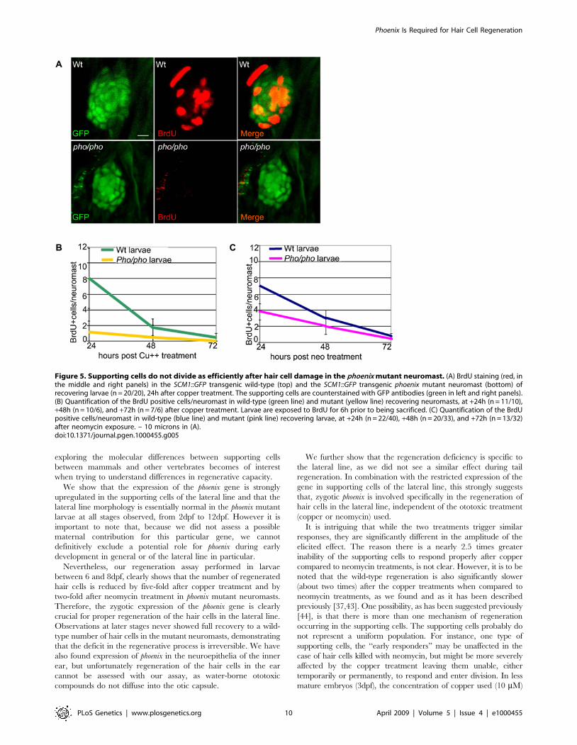
Supporting cell proliferation is impaired duringregeneration in phoenix mutant neuromasts
Since the number of supporting cells in mutant neuromasts is
not unlike that of wild type neuromasts, we turned our attention to
differences in cell division to explain the regeneration phenotype
observed in phoenix larvae. To test the hypothesis that the phoenix
mutation is affecting proliferation of the progenitor cell popula-
tion, we exposed SCM1 transgenic wild-type and mutant larvae,
treated with copper or neomycin, to BrdU for 6h prior to fixing
them, at +24h, +48h and +72h after chemical treatment. We
subsequently double-stained the larvae with an anti-BrdU
antibody (red in Figure 5A, middle and right panels) and with
an anti-GFP antibody (green in left and right panels). At +24h
after copper (Figure 5A) and neomycin (not shown) treatments, the
BrdU-labeled transgenic wild-type (Figure 5A, top panels) and the
transgenic phoenix mutant (lower panels) larvae showed a striking
difference in the number of BrdU positive cells. On average, we
found eight BrdU positive cells in the transgenic wild-type
neuromasts, in mutant neuromasts we typically found no positive
cells while at most we found two. Thus the number of BrdU
positive cells in the progenitor cells population in the mutant
neuromasts is drastically reduced, most likely explaining the
decrease in the number of newly regenerated hair cells in phoenix
mutant embryos.
To obtain quantitative data to support the above conclusion and
to follow the dynamics of BrdU incorporation over time, we
counted BrdU positive cells after copper treatment (Figure 5B) in
at least ten wild-type (green line) and ten phoenix mutant (yellow
line) neuromasts per larva. BrdU was added to the medium 6h
before fixation allowing us to determine rates of DNA synthesis in
equivalent time-windows during the 3 days regeneration period.
Since recovery to a normal number of hair cells is reached at
around 72 hours after treatment, this time point represents the
rate of BrdU incorporation in undamaged neuromasts, or the
BrdU baseline. At +24h of recovery, we had an eight-fold
difference in the number of BrdU positive cells between wild-type
and mutant neuromasts. This difference was still two-fold at +48h
of recovery and both returned to the base line frequency at +72h.
We performed similar tests after neomycin treatment (Figure 5C,
wild-type: blue line and mutant: pink line). At +24h, we found
close to a two-fold difference of BrdU labeled cells, and at +48h a
33% decrease between wild-type and mutant neuromasts.
Taken together, these results show that the lack of the Phoenix
protein has a significant negative impact on proliferation in the
regenerating neuromast, which is not due to a reduction in the
number of supporting cells. Thus, phoenix has an important role in
ensuring proper proliferation of the supporting cells during hair
cell regeneration in neuromasts.
Discussion
We have isolated a mutation in a previously uncharacterized
gene in zebrafish that we have named phoenix because of an
observed deficiency in the regeneration of the hair cells of the
lateral line. Identical phenotypes were observed in two different
alleles, caused by independent retroviral integrations in the phoenix
gene in different genetic backgrounds. This unambiguously links
the phenotype to this particular gene, which encodes a rapidly
evolving protein with no obvious homologs in the current genomic
databases. The predicted Phoenix protein is a long peptide of low
complexity with a predicted ATPase domain reminiscent of many
cytoskeletal proteins. This would argue in favor of a structural
protein, possibly interacting with the cytoskeleton and subcellular
membranes. How it is linked to the ability of the supporting cells to
regenerate will require further investigation. It also remains to be
discovered whether mammalian cells have a similar molecule; we
were unable to detect a related sequence in the mammalian
genomes. Interestingly, inner ear hair cells in mammals are unable
to regenerate from supporting cells postnatally and, therefore,
Phoenix Is Required for Hair Cell Regeneration
PLoS Genetics | www.plosgenetics.org 9 April 2009 | Volume 5 | Issue 4 | e1000455
exploring the molecular differences between supporting cells
between mammals and other vertebrates becomes of interest
when trying to understand differences in regenerative capacity.
We show that the expression of the phoenix gene is strongly
upregulated in the supporting cells of the lateral line and that the
lateral line morphology is essentially normal in the phoenix mutant
larvae at all stages observed, from 2dpf to 12dpf. However it is
important to note that, because we did not assess a possible
maternal contribution for this particular gene, we cannot
definitively exclude a potential role for phoenix during early
development in general or of the lateral line in particular.
Nevertheless, our regeneration assay performed in larvae
between 6 and 8dpf, clearly shows that the number of regenerated
hair cells is reduced by five-fold after copper treatment and by
two-fold after neomycin treatment in phoenix mutant neuromasts.
Therefore, the zygotic expression of the phoenix gene is clearly
crucial for proper regeneration of the hair cells in the lateral line.
Observations at later stages never showed full recovery to a wild-
type number of hair cells in the mutant neuromasts, demonstrating
that the deficit in the regenerative process is irreversible. We have
also found expression of phoenix in the neuroepithelia of the inner
ear, but unfortunately regeneration of the hair cells in the ear
cannot be assessed with our assay, as water-borne ototoxic
compounds do not diffuse into the otic capsule.
We further show that the regeneration deficiency is specific to
the lateral line, as we did not see a similar effect during tail
regeneration. In combination with the restricted expression of the
gene in supporting cells of the lateral line, this strongly suggests
that, zygotic phoenix is involved specifically in the regeneration of
hair cells in the lateral line, independent of the ototoxic treatment
(copper or neomycin) used.
It is intriguing that while the two treatments trigger similar
responses, they are significantly different in the amplitude of the
elicited effect. The reason there is a nearly 2.5 times greater
inability of the supporting cells to respond properly after copper
compared to neomycin treatments, is not clear. However, it is to be
noted that the wild-type regeneration is also significantly slower
(about two times) after the copper treatments when compared to
neomycin treatments, as we found and as it has been described
previously [37,43]. One possibility, as has been suggested previously
[44], is that there is more than one mechanism of regeneration
occurring in the supporting cells. The supporting cells probably do
not represent a uniform population. For instance, one type of
supporting cells, the ‘‘early responders’’ may be unaffected in the
case of hair cells killed with neomycin, but might be more severely
affected by the copper treatment leaving them unable, either
temporarily or permanently, to respond and enter division. In less
mature embryos (3dpf), the concentration of copper used (10 mM)
Figure 5. Supporting cells do not divide as efficiently after hair cell damage in the phoenix mutant neuromast. (A) BrdU staining (red, inthe middle and right panels) in the SCM1::GFP transgenic wild-type (top) and the SCM1::GFP transgenic phoenix mutant neuromast (bottom) ofrecovering larvae (n = 20/20), 24h after copper treatment. The supporting cells are counterstained with GFP antibodies (green in left and right panels).(B) Quantification of the BrdU positive cells/neuromast in wild-type (green line) and mutant (yellow line) recovering neuromasts, at +24h (n = 11/10),+48h (n = 10/6), and +72h (n = 7/6) after copper treatment. Larvae are exposed to BrdU for 6h prior to being sacrificed. (C) Quantification of the BrdUpositive cells/neuromast in wild-type (blue line) and mutant (pink line) recovering larvae, at +24h (n = 22/40), +48h (n = 20/33), and +72h (n = 13/32)after neomycin exposure. – 10 microns in (A).doi:10.1371/journal.pgen.1000455.g005
Phoenix Is Required for Hair Cell Regeneration
PLoS Genetics | www.plosgenetics.org 10 April 2009 | Volume 5 | Issue 4 | e1000455

has been shown to eliminate post-mitotic precursors, thus requiring
cell division and longer times for regeneration when compared to
lower doses of the metal [37].
Additionally, it was recently shown in a screen to identify genes
that modify hair cell resistance in the lateral line, that different
ototoxic agents can elicit different pathways for protection and
survival of hair cells in treated neuromasts [56]. It is therefore also
possible that the regenerative response in supporting cells depends
partially on the type of ototoxic agent applied. Namely, the trigger or
the response to the trigger of regeneration in supporting cells could
rely on different pathways, partially overlapping or complementing
each other depending on the ototoxic chemical. Further investigation
will allow us to distinguish between these possibilities. It appears that
phoenix is upstream of the differential response between neomycin and
copper, as the differences in the rates of response in mutant larvae
remain similar to the differences seen in wild-type larvae.
We showed that the death rate of supporting cells was unaltered
in mutant neuromasts as observed by three different approaches
(histology, acridine orange and TUNEL staining). Furthermore,
making use of the MSC1 transgenic line that expresses GFP
exclusively in the supporting cells, we demonstrated no loss in the
number of those cells in mutant neuromasts. It is to be noted that
the counting of cells in the neuromast is technically challenging.
Although we did not see obvious differences, we cannot exclude a
possible subtle reduction in the number of supporting cells. If a key
subset of cells critical to the initiation of regeneration is missing,
even a subtle difference in number might have a drastic effect on
regeneration, which we cannot exclude at our level of analysis.
However, it was previously shown that the regeneration process is
happening in many supporting cells simultaneously, encompassing
most of the neuromast [44] making this possibility unlikely. We
clearly show that a significant number of supporting cells are
present and that the phoenix mRNA is expressed in all supporting
cells, therefore the most likely explanation is that the regenerative
potential of all supporting cells is severely impaired.
To assess proliferation of the progenitor cells, we follow the
incorporation of BrdU during the S-phase of the cell cycle in
neuromasts. The incorporation of BrdU is detected in cells that
have exited the cell cycle and have differentiated. Therefore, it
provides no information on the post-mitotic state of the BrdU
positive cells. However, as the number of regenerated hair cells
correlates closely to the number of BrdU labeled cells and as we do
not see additional cell death in either wild-type or mutant
neuromasts, this would suggest that the progenitor cells that
actually enter S-phase proceed through the entire cell cycle and
into differentiation. In the phoenix mutant larvae, this regenerative
process is happening in a significantly reduced number of
supporting cells. Further work is required to address the timing
of the blockage occurring in the supporting cells in mutant
neuromasts, but it is clearly occurring before DNA synthesis
initiates. This invaluable information will help to elucidate the
mechanism of action of the novel phoenix gene in the regeneration
process of hair cells in the lateral line.
Materials and Methods
All animals were handled in strict accordance with good animal
practice as defined by the relevant national and/or local animal
welfare bodies, and all animal work was approved by the
appropriate committee (NHGRI protocol # G-01-3).
Fish care and husbandryFish care and husbandry were performed according to [57] in
compliance with NIH guideline for animal care. The hi43 allele of
phoenix was recovered in a retroviral screen performed at MIT
[58]. The genomic locus of the retroviral integration and the
putative cDNA were determined as described in [45]. Genotyping
was done with primers GGAGATCGACAGCGCCCTGAAG
and AAACTGCTGAGGGCTGCTGGACCGCATC. The zm
allele (ZM_00003486) was purchased (Znomics, Inc) and carriers
were genotyped using the primers CGAGACCCCGCCGCCT-
GATGTT and GACGCAGGCGCATAAAATCAGTC. The
MSC1 transgenic line was a gift from B. Weinstein and carriers
were identified by detecting specific expression of GFP in the
lateral line. The cldnB::GFP line [46] was obtained from M.
Allende, the pou4f3::GFP line [47] from H. Baier and the
ET20::GFP line [48] from Vladimir Korzh.
Staining with vital dyes (YoPro-1, Acridine Orange, andFM1-43) and treatment with copper or neomycin of livelarvae
All active agents were added to system water at 28uC. During
the staining/treatment, larvae were kept in cell strainers (BD
Falcon) in 6 well plates (Costar, Corning, Inc)) at a maximum of 35
larvae/well. This allowed rapid transfer of the larvae during the
post-treatment rinsing. All larvae were rinsed 663 minutes with
system water and then transferred into large Petri dishes with
abundant fresh system water. After the various treatments, only
healthy looking larvae displaying no additional morphological or
behavioral defects were kept for subsequent analysis over the
following 72 hours. The number of hair cells and the AO positive
cells were counted in 10 head and all the trunk and tail
neuromasts, in each larva.
We added YoPro-1 (Molecular probes) for 15 minutes at
2mM, as described in [38], FM1-43 (Molecular probes) for
1minute at 2mM, as described [49] and Acridine orange (AO)
hemi (zinc chloride, Sigma) for 5 minutes at 2mg/ml, as described
[37]. Copper (Copper(II) sulfate, Sigma) was added for 2h at
10mM as described in [37] and Neomycin (sulfate, Calbiochem)
for 1h at 200mM as described in [38,39]. Monitoring, counting
and imaging of the lateral line in live larvae, after using the
different life dyes, was done on an inverted Zeiss AXIO-
VERT200M equipped with an Apotome Grid Confocal. Larvae
were anesthetized with MS222 (0.005%) and mounted on a cover
slip in 2% Methylcellulose (Sigma).
Tail transectionsFive day old larvae were allowed to recover for an hour after
copper or neomycin treatment. Subsequently, larvae were
anesthetized with 0.005% MS-222 (Sigma) and placed on a clean
glass slide and a portion of their tail was amputated with a scalpel.
We cut not just the fin, but also a portion of the actual tail
including the neural tube, the notochord, and the somites, to
evaluate the regeneration of other tissues in addition to the fin
itself. Three day old untreated larvae were similarly processed. To
follow and image the tail growth over 3 to 6 days post amputation,
we mounted anesthetized larvae on cover slips in 2% methylcel-
lulose. Measurements were made on pictures taken from live
larvae on the inverted Zeiss AXIOVERT200M, using the
Axiovision software from Zeiss. The region of amputation was
usually easily identifiable, as it presented healing scars, allowing a
reasonably accurate measurement of the newly grown tail.
BrdU treatmentBrdU (Sigma) was diluted to 10mM in system water and larvae
were exposed for 6h before fixation at the chosen time points (24,
48 and 72 hours post treatment) o/n with 4% formaldehyde
Phoenix Is Required for Hair Cell Regeneration
PLoS Genetics | www.plosgenetics.org 11 April 2009 | Volume 5 | Issue 4 | e1000455

(Electron Microscopy Sciences) in 16 PBS (Quality Biological,
Inc.). Double staining was done as described [40,44], with a
fluorochrome labeled mouse monoclonal antibody against BrdU
(Molecular probes) and a fluorochrome labeled rabbit polyclonal
antibody against GFP (Abcam). Larvae were mounted on slides in
Aquapolymount (Polyscience, Inc) and at least 10 neuromasts/
larva were counted. The imaging was done on an upright confocal
microscope (Zeiss AXIOVERT)
Immunofluorescence on larvaeUntreated and copper treated larvae were fixed o/n with 4%
formaldehyde (Electron Microscopy Sciences) in 16PBS (Quality
Biological, Inc.), at various stages of interest and subsequently
stored in 100% methanol. After progressive rehydration (25%,
50% 75% and 100% PTW (PBS16, 0.001% Tween and 0.001%
DMSO)), larvae were treated with acetone for 7mn at 220uC.
Subsequently, we rehydrated and rinsed them 365mn in PTW.
Next we digested them with 1mg/ml collagenase (Sigma) in PTB
(PTW + 10% goat serum +10% BSA) for 35mn. After 565mn
rinses with PBT, we pre-incubated the larvae 4 hours in PBT.
Larvae were incubated o/n with the polyclonal rabbit primary
antibody (1/200) against Myosin VI (Proteus Biosciences, Inc) and
a fluorescently labeled monoclonal mouse primary antibody (1/
200) against GFP (Abcam). The next day we rinsed the larvae
6610 mn in PTW and preincubated them again for 4 hours in
PBT. The fluorescently labeled secondary anti-rabbit antibody (1/
500) was added o/n. The next day 6610 mn rinses were
performed. Larvae were mounted on slides in Aquapolymount
(Polyscience, Inc) for imaging on an upright confocal microscope
(Zeiss AXIOVERT).
Semi-thin and ultra thin sections for TEM on zebrafishlarvae
Larvae were fixed overnight at in 2.5% glutaraldehyde (Sigma)
and 4% paraformaldehyde prepared from paraformaldehyde
(Sigma) in 0.1M sodium cacodylate buffer (Sigma). Larvae were
then rinsed and post-fixed 1h at room temperature in reduced
osmium (1:1 mixture of 2% aqueous potassium ferrocyanide) as
described previously [59]. After post-fixation the cells were
dehydrated in ethanol and processed for Epon (Sigma) embedding.
Semi-thin sections (300 nm) were cut and collected on a glass slide,
and subsequently stained using toluidine blue (Sigma). The
analysis and imaging were done on an inverted Zeiss Axio-
vert200M. Ultra thin sections (80 nm) were cut on a Reichter-E
ultramicrotome, collected on copper grids and stained with lead
citrate (Sigma) for 2 min. Sections were then examined with a CM
10 Philips electron microscope at 80kV.
Whole mount in situ hybridizationPerformed as described previously [60]. We designed an
antisense probe, using the following primers TCAACTGATG-
TATTTCCTGGGC and GTTTTGCTCCACTATCT-
GACCTTT. The probe was hybridized at 62uC. Larvae were
mounted on slides in Aquapolymount (Polyscience, Inc) for
imaging on an upright confocal microscope (Zeiss AXIOVERT).
Isolation of the BAC containing the phoenix gene andbioinformatic analysis
We screened the zebrafish BAC library (Chori 211, BacPac
consortium) with a probe generated from total RNA (5 dpf
larva), with the primers AGATCTTGAGATTGCCGAATGT
and CATCTCTCTCACCTTCTTCAGTGAC. The BAC was
sequenced, by shotgun assembly and brought to finished quality
(#1 error per 50kb) at the National Intramural Sequencing
Center (NISC). Sequences were aligned to the zebrafish genome
(UCSC Genome Browser http://genome.ucsc.edu/) and syn-
tenic regions identified using genomic chain comparisons in
Takifugu rubripes (fugu), Tetraodon nigroviridis (tetraodon), Oryzias
latipes (medaka), and Gasterosteus aculeatus (stickleback). Genescan
or N-SCAN [50] predicted transcripts between the two flanking
genes were identified and compared using ClustalW [61]
(http://www.ebi.ac.uk/Tools/clustalw2/index.html). The BLO-
SUM protein matrix [62] was used with a window of 7 and gap
penalty of 2.
RT-PCR, sequencing, and cDNA identificationWe prepared total RNA extracts from embryos and Pac2 cells
(which is an embryonic zebrafish cell line described in [51] in
Trizol (Invitrogen). For the RT-PCR, we retro-transcribed specific
cDNA using superscript II Reverse transcriptase (Invitrogen),
following the manufacturer’s protocol. For the cloning of all
transcripts shown in Figure 2B and 2C, we used the same set of 2
nested forward primers CCATATCAAAACAGAGCTGTGC-
TAC and GTCAAGGCAGAGTAAGCAAGTGACACTG, both
located in the 59UTR, respectively starting 142bp and 121bp
upstream of the start codon. For the cloning of transcripts 2, 3 and
6, they were respectively coupled with the reverse primers
GTTCTGTTTTCTTTTCCTTGTCAACGCC and
CCTTCTCTCCCTGATAAAGTCTGGCAACC both found in
the 59end of exon 7. For the cloning of the transcript 1, we coupled
them respectively with the reverse primers CATTCATTCATT-
CATTCATTTTCCT and TTTCCTTTTGCTTAGTCCCT-
TATTT both found in the 39end of exon 2. For the cloning of
the transcript 4 we coupled them respectively with the reverse
primers TTTTACATCTACAGAATCGTGAAAAA and
AATTGTGATTCTCATTTTAGCCAGA, both found in the 39
end of exon 6. For the cloning of the transcript 5 we coupled them
respectively with the reverse primers TTTTTCTTTCATAGC-
GAACACAAAG and TTTAAACTGTCAGCTCTTGATGC,
both found in 39end of exon 5. To verify the quality of the Total
RNA from different stages, which we used for the RT-PCR, we
amplified a 159bp fragment of b-actin using the following primers,
GACCCAGACATCAGGGAGTGATGG and AGGTGT-
GATGCCAGATCTTCTCCAGT. All sub-cloning for subse-
quent sequencing were done using the TOPO TA cloning Kit
(Invitrogen), following the manufacturer protocol. Sequencing was
performed by ACGT, Inc. The analysis of the sequences was done
using the Sequencher (Gene Codes Corp.) program, with the
genomic sequence of the phoenix gene (as previously isolated in the
BAC) as a reference.
Statistical analysisAt all time points and in all graphs, the p values for wild-type
versus mutant larvae were calculated using a Student’s T test (two
tailed) with two samples of equal variance and considered
significant when p was ,0.05. Error bars in the different graphs
represent the standard deviation, or standard error, depending on
the number of samples (n) analyzed.
Image reconstructionStacks of images collected on the inverted microscope were
treated with the Axiovision software from Zeiss. Stacks of
images collected on the confocal microscope were treated with
the LSM software from Zeiss. All images or stack of images were
exported as tiff files, which were subsequently processed with
Photoshop.
Phoenix Is Required for Hair Cell Regeneration
PLoS Genetics | www.plosgenetics.org 12 April 2009 | Volume 5 | Issue 4 | e1000455

Supporting Information
Figure S1 Live imaging of transgenic lines crossed into the
phoenix background show no differences from wild-type larvae. A.
We crossed the phoenix mutant line into the cldnB::GFP transgenic
background, which expressed GFP (green in first and third panels)
in all cells of the neuromast. We stained untreated 5 dpf larvae
with FM1-43 (red in second and third a panel). Shown are images
of live wild-type (top panels) and phoenix (bottom panels)
neuromasts. A camera lucida drawing of each merged picture is
highlighting the GFP positive cells contours (green) and the FM1-
43 positive cells (red) in the fourth panel. B. We crossed the phoenix
mutant line into the pou4f3::GFP transgenic background, which
expressed GFP in all of the neuromast’s hair cells. Dorsal views
(left panels) and lateral views (right panels) of wild-type (top panels)
and pho/pho (bottom panels) are shown. A camera lucida drawing of
each panel is outlining the GFP positive hair cells of each
respective 41 panel. C. We crossed the phoenix mutant into the
ET20::GFP transgenic background, which expressed GFP in a
subset of supporting cells known as the mantle cells. Two
representative examples of wild-type (top panels) and of phoenix
mutant (bottom panels) neuromasts are shown. A camera lucida
drawing of each panel is outlining the GFP + cells of each
respective panel. - 10 microns in all panels.
Found at: doi:10.1371/journal.pgen.1000455.s001 (6.66 MB TIF)
Figure S2 Hair cells seem unaffected in the phoenix mutant ear.
Wild-type expression of the phoenix mRNA and protein. A. Live
images in pou4f3;;GFP transgenic animals of hair cells in a crista of
a wild-type (left panel) and a phoenix mutant (right panel) ear. B.
In situ hybridization with an antisense probe against phoenix in the
inner ear of a 4dpf old embryo (top panel). A camera lucida drawing
(lower panel) shows the positions of the two otoliths (red), of the
cristae’s (light green) and the maculae’s (dark green) hair cells. The
AP/DV ventral orientation is indicated. C. Immunofluorescence
on cultured zebrafish cells (Pac2) using EX1 (green in the top and
bottom left panels) or EX7 (green in the top and bottom right
panels) antibodies. To highlight the ER, we co-stained with an
antibody against PDI (ER-associated protein disulfide isomerase,
red in middle and bottom left and right panels). DAPI was used to
counterstain nuclei (blue in the two bottom left and right panels).
Merged images (bottom, left and right panels) showed a
cytoplasmic staining of phoenix, which was more concentrated
in the perinuclear region. It appeared excluded from the ER, as
there was no co-localization with DPI. Note how phoenix protein
was upregulated in cells preparing to enter division (white arrow in
the top right panel). D. Western blot on PAC2 cell extracts with
two rabbit polyclonal antibodies, raised against an epitope
encoded by exon 1 (EX1) or by exon 7 (EX7). A common band
in the range of 95KDa was found with both antibodies (black
arrow). - 10 microns in A, 50 microns in B and 3 microns in C.
Found at: doi:10.1371/journal.pgen.1000455.s002 (3.51 MB TIF)
Acknowledgments
We thank the National Intramural Sequencing Center (NISC) for the
sequencing of the BAC, Dr. J. Yan for help with the BAC library, Drs. K.
Shaw and B. Weinstein for the gift of the SCM1 transgenic line, M.
Sheppard and A. Davis for fish husbandry, Dr. B. Feldman and his lab for
the use of the confocal microscope, Dr. U. Straehle, Dr. M. Kelley, Dr. P.
Liu and Dr. W. Chen for critical reading of the manuscript.
Author Contributions
Conceived and designed the experiments: MB SMB. Performed the
experiments: MB JB RS VG. Analyzed the data: MB JB RS VG MLA
SMB. Contributed reagents/materials/analysis tools: MB JB RS VG MLA
SMB. Wrote the paper: MB MLA SMB.
References
1. Fritzsch B, Pauley S, Beisel KW (2006) Cells, molecules and morphogenesis: the
making of the vertebrate ear. Brain Res 1091: 151–171.
2. Kelley MW (2006) Regulation of cell fate in the sensory epithelia of the innerear. Nature Neurosci. Rev 7: 837–849.
3. Kelley MW (2006) Hair cell development: commitment through differentiation.
Brain Res 1091: 172–185.
4. Fettiplace R, Hackney CM (2006) The sensory and motor roles of auditory hair
cells. Nature Rev Neurosci 7: 19–29.
5. Kung C (2005) A possible unifying principle for mechanosensation. Nature 436:647–654.
6. Warchol ME (2007) Characterization of supporting cell phenotype in the avian
inner ear: implications for sensory regeneration. Hear Res 227: 11–18.
7. Cotanche DA, Henson MM, Henson OW Jr. (1992) Contractile proteins in thehyaline cells of the chicken cochlea. J Comp Neurol 324: 353–364.
8. Cotanche DA (1987) Regeneration of the tectorial membrane in the chick
cochlea following severe acoustic trauma. Hear Res 30: 197–206.
9. Epstein JE, Cotanche DA (1995) Secretion of a new basal layer of
tectorial membrane following gentamicin-induced hair cell loss. Hear Res 90:31–43.
10. Raphael Y (1992) Evidence for supporting cells mitosis in the chick cochlea.
J Neurocytol 31: 663–671.
11. Balak KJ, Corwin JT, Jones JE (1990) Regenerated hair cells can originate fromsupporting cell progeny: evidence from phototoxicity and laser ablation
experiments in the lateral line system. J Neurosci 10: 2502–2512.
12. Stone JS, Cotanche DA (1994) Identification of the timing of S phase and thepatterns of cell proliferation during hair cell regeneration in the chick cochlea.
J Comp Neurol 341: 50–67.
13. Bermingham-McDonogh O, Rubel EW (2003) Hair cell regeneration: wingingour way towards a sound future. Curr Opin Neurobiol 13: 119–126.
14. Jones JE, Corwin JT (1996) Regeneration of sensory cells after laser ablation in
the lateral line system: hair cell lineage and macrophage behavior revealed bytime-lapse video microscopy. J Neurosci 16: 649–662.
15. Ruben RT (1967) Development of the inner ear of the mouse: a
radioautographic study of terminal mitoses. Acta Otolaryngol 220: 4–44.
16. Kuntz AL, Oesterle EC (1998) Transforming growth factor alpha with insulin
stimulates cell proliferation in vivo in adult rat vestibular sensory epithelium.J Comp Neurol 399: 413–423.
17. Rubel EW, Dew LA, Roberson DW (1995) Mammalian vestibular hair cell
regeneration. Science 267: 701–707.
18. Corwin JT (1981) Postembryonic production and aging in inner ear hair cells insharks. J Comp Neurol 201: 541–553.
19. Corwin JT (1983) Postembryonic growth of the macula neglecta auditory
detector in the ray, Raja clavata: continual increases in hair cell number, neuralconvergence, and physiological sensitivity. J Comp Neurol 217: 345–356.
20. Corwin JT (1985) Perpetual production of hair cells and maturational changes in
hair cell ultrastructure accompany postembryonic growth in an amphibian ear.Proc Natl Acad Sci USA 82: 3911–3915.
21. Corwin JT, Oberholtzer J (1997) Fish n’ chicks: model recipes for hair cell
regeneration. Neuron 19: 951–954.
22. Corwin JT, Cotanche DA (1988) Regeneration of sensory hair cells after acoustic
trauma. Science 240: 1772–1774.
23. Ryals BM, Rubel EW (1988) Hair cell regeneration after acoustic trauma inadult Coturnix quail. Science 240: 1774–1776.
24. Presson JC, Lanford PJ, Popper AN (1996) Hair cell precursors are
ultrastructurally indistinguishable from mature support cells in the ear of apostembryonic fish. Hear Res 100: 10–20.
25. Avallone B, Fascio U, Balsamo G, Marmo F (2008) Gentamicin ototoxicity in
the saccule of the lizard Podarcis Sicula induces hair cell recovery andregeneration. Hear Res 235: 15–22.
26. Dijkgraaf S (1964) The supposed use of the lateral line as an organ of hearing in
fish. Experientia 20: 586–587.
27. Elepfandt A (1988) Processing of wave patterns in the lateral line system parallels
to auditory processing. Act Biol Hung 39: 251–265.
28. Shelton PM (1971) The structure and function of the lateral line system in larvalXenopus laevis. J Exp Zool 178: 211–231.
29. Metcalfe WF, Kimmel CB, Schabtach E (1985) Anatomy of the posterior lateral
line system in young larvae of the zebrafish. J Comp Neurol 233: 377–389.
30. Raible DW, Kruse GJ (2000) Organization of the lateral line system inembryonic zebrafish. J Comp Neurol 421: 189–198.
31. Laguerre L, Soubiran F, Ghysen A, Konig N, Dambly-Chaudiere C (2005) Cell
proliferation in the developing lateral line system of zebrafish embryos. Dev Dyn233: 466–472.
32. Gompel N, Cubedo N, Thisse C, Thisse B, Dambly-Chaudiere C, et al. (2001)Pattern formation in the lateral line of the zebrafish. Mech Dev 105: 69–77.
Phoenix Is Required for Hair Cell Regeneration
PLoS Genetics | www.plosgenetics.org 13 April 2009 | Volume 5 | Issue 4 | e1000455

33. Ghysen A, Dambly-Chaudiere C (2007) The lateral line microcosmos. Genes
Dev 21: 2118–2130.34. Jorgensen JM (1991) Regeneration of lateral line and inner ear vestibular cells.
Ciba Found Symp 160: 151–163.
35. Chambers MH (1922) Degeneration and regeneration of the lateral-line organsin Amiurus Nebulosus. Proc Natl Acad Sci USA 8: 186–187.
36. Song J, Yan HY, Popper AN (1995) Damage and recovery of hair cells in fishcanal (but not superficial) neuromasts after gentamicin exposure. Hear Res 91:
63–71.
37. Hernandez PP, Moreno V, Olivari FA, Allende ML (2006) Sub-lethalconcentrations of waterborne copper are toxic to lateral line neuromasts in
zebrafish (Danio rerio). Hear Res 213: 1–10.38. Santos F, MacDonald G, Rubel EW, Raible DW (2006) Lateral line hair cell
maturation is a determinant of aminoglycoside susceptibility in zebrafish (Daniorerio). Hear Res 213: 25–33.
39. Murakami SL, Cunningham LL, Werner LA, Bauer E, Pujol R, et al. (2003)
Developmental differences in suceptibility to neomycin-induced hair cell deathin the lateral line neuromasts of zebrafish (Danio rerio). Hear Res 186: 47–56.
40. Williams JA, Holder N (2000) Cell turnover in neuromasts of zebrafish larvae.Hear Res 143: 171–181.
41. Harris JA, Cheng AG, Cunningham LL, MacDonald G, Raible DW, et al.
(2003) Neomycin-induced hair cell death and rapid regeneration in the lateralline of zebrafish (Danio rerio). JARO 4: 219–234.
42. Lopez-Schier H, Huspeth AJ (2006) A two-step mechanism underlies the planarpolarization of regenerating sensory hair cells. Proc Natl Acad Sci 103:
18615–18620.43. Hernandez PP, Olivari FA, Sarrazin AF, Sandoval PC, Allende ML (2007)
Regeneration in zebrafish lateral line neuromasts: expression of the neural
progenitor cell marker sox2 and proliferation-dependent and-independentmechanisms of hair cell renewal. Dev Neurobiol 67: 637–654.
44. Ma EY, Rubel EW, Raible DW (2008) Notch signaling regulates the extent ofhair cell regeneration in the zebrafish lateral line. J Neurosci 28: 2261–2273.
45. Golling G, Amsterdam A, Sun Z, Antonelli M, Maldonado E, et al. (2002)
Insertional mutagenesis in zebrafish rapidly identifies genes essential for earlyvertebrate development. Nat Genet 31: 135–140.
46. Haas P, Gilmour D (2006) Chemokine signaling mediates self-organizing tissuemigration in the zebrafish lateral line. Dev Cell 10: 673–680.
47. Xiao T, Roeser T, Staub W, Baier H (2005) A GFP-based genetic screen revealsmutations that disrupt the architecture of the zebrafish retinotectal projection.
Dev 132: 2955–2967.
48. Parinov S, Kondrichin I, Korzh V, Emelyanov A (2004) Tol2 transposon-
mediated enhancer trap to identify developmentally regulated zebrafish genes in
vivo. Dev Dyn 231: 449–459.
49. Seiler C, Nicolson T (1999) Defective calmodulin-dependent rapid apical
endocytosis in zebrafish sensory hair cell mutants. J Neurobiol 41: 424–434.
50. Burge C, Karlin S (1997) Prediction of complete gene structures in human
genomic DNA. J Mol Biol 268: 78–94.
51. Lin S, Gaiano N, Culp P, Burns JC, Friedmann T, et al. (1994) Integration and
germ-line transmission of a pseudotyped retroviral vector in zebrafish. Science
265: 666–669.
52. Altschul SF, Gish W, Miller W, Myers EW, Lipman DJ (1990) Basic local
alignment search tool. J Mol Biol 215: 403–410.
53. Yang CT, Hindes AE, Hultman Ka, Johnson SL (2007) Mutations in gfpt1 and
skiv2l2 cause distinct stage-specific defects in larval melanocyte regeneration in
zebrafish. PLoS Genet 3(6): e88. doi:10.1371/journal.pgen.0030088.
54. Stone L (1933) The development of lateral line sense organs in amphibians
observed in living and vital-stained preparations. J Comp Neurol 57: 507–540.
55. Stone L (1937) Further experimental studies of the development of lateral line
sense organs in the amphibians observed in living preparations. J Comp Neurol
68: 83–115.
56. Owens KN, Santos F, Roberts B, Linbo T, Coffin AB, et al. (2008) Identification
of genetic and chemical modulators of zebrafish mechanosensory hair cell death.
PLoS Genet 4(2): e1000020. doi:10.1371/journal.pgen.1000020.
57. Westerfield M (2000) The zebrafish book: a guide to the laboratory use of the
zebrafish ( Danio Rerio). 4th edition. Eugene: University of Oregon Press.
58. Amsterdam A, Nissen RM, Sun Z, Swindell EC, Farrington S, et al. (2004)
Identification of 315 genes essential for early zebrafish development. Proc Natl
Acad Sci 101: 12792–12797.
59. Karnovsky MJ (1961) Simple methods for ‘‘staining with lead’’ at high pH in
electron microscopy. J Biophys Biochem Cytol 11: 729–739.
60. Oxtoby E, Jowett T (1993) Cloning of the zebrafish krox-20 gene (krx-20) and its
expression during hindbrain development. Nucleic Acids Res 21: 1087–1095.
61. Thompson JD, Higgins DG, Gibson TJ (1994) CLUSTAL W: improving the
sensitivity of progressive multiple sequence alignment through sequence
weighting, position-specific gap penalties and weight matrix choice. Nucelic
Acids Res 22: 4673–4680.
62. Henikoff S, Henikoff JG (1993) Performance evaluation of amino acid
substitution matrices. Proteins 17: 49–61.
Phoenix Is Required for Hair Cell Regeneration
PLoS Genetics | www.plosgenetics.org 14 April 2009 | Volume 5 | Issue 4 | e1000455