pharmaceuticals-13-00167-v2 (1) - Åbo Akademi University ...
31
This is an electronic reprint of the original article. This reprint may differ from the original in pagination and typographic detail. Evolution of Nanotechnology in Delivering Drugs to Eyes, Skin and Wounds via Topical Route Koppa Raghu, Pratheeksha; Bansal, Kuldeep K; Thakor, Pradip; Bhavana, Valamla; Madan, Jitender; Rosenholm, Jessica M; Mehra, Neelesh Kumar Published in: Pharmaceuticals DOI: 10.3390/ph13080167 Published: 01/08/2020 Document Version Publisher's PDF, also known as Version of record Document License CC BY Link to publication Please cite the original version: Koppa Raghu, P., Bansal, K. K., Thakor, P., Bhavana, V., Madan, J., Rosenholm, J. M., & Mehra, N. K. (2020). Evolution of Nanotechnology in Delivering Drugs to Eyes, Skin and Wounds via Topical Route. Pharmaceuticals, 13(8), 1-30. [167]. https://doi.org/10.3390/ph13080167 General rights Copyright and moral rights for the publications made accessible in the public portal are retained by the authors and/or other copyright owners and it is a condition of accessing publications that users recognise and abide by the legal requirements associated with these rights. Take down policy If you believe that this document breaches copyright please contact us providing details, and we will remove access to the work immediately and investigate your claim. This document is downloaded from the Research Information Portal of ÅAU: 15. Mar. 2022
-
Upload
khangminh22 -
Category
Documents
-
view
1 -
download
0
Transcript of pharmaceuticals-13-00167-v2 (1) - Åbo Akademi University ...
This is an electronic reprint of the original article. This reprint may differ from the original in pagination and typographic detail.
Evolution of Nanotechnology in Delivering Drugs to Eyes, Skin and Wounds viaTopical RouteKoppa Raghu, Pratheeksha; Bansal, Kuldeep K; Thakor, Pradip; Bhavana, Valamla; Madan,Jitender; Rosenholm, Jessica M; Mehra, Neelesh KumarPublished in:Pharmaceuticals
DOI:10.3390/ph13080167
Published: 01/08/2020
Document VersionPublisher's PDF, also known as Version of record
Document LicenseCC BY
Link to publication
Please cite the original version:Koppa Raghu, P., Bansal, K. K., Thakor, P., Bhavana, V., Madan, J., Rosenholm, J. M., & Mehra, N. K. (2020).Evolution of Nanotechnology in Delivering Drugs to Eyes, Skin and Wounds via Topical Route. Pharmaceuticals,13(8), 1-30. [167]. https://doi.org/10.3390/ph13080167
General rightsCopyright and moral rights for the publications made accessible in the public portal are retained by the authors and/or other copyright ownersand it is a condition of accessing publications that users recognise and abide by the legal requirements associated with these rights.
Take down policyIf you believe that this document breaches copyright please contact us providing details, and we will remove access to the work immediatelyand investigate your claim.
This document is downloaded from the Research Information Portal of ÅAU: 15. Mar. 2022
pharmaceuticals
Review
Evolution of Nanotechnology in Delivering Drugs toEyes, Skin and Wounds via Topical Route
Pratheeksha Koppa Raghu 1, Kuldeep K. Bansal 2,* , Pradip Thakor 1, Valamla Bhavana 1,Jitender Madan 1, Jessica M. Rosenholm 2 and Neelesh Kumar Mehra 1,*
1 Pharmaceutical Nanotechnology Research Laboratory, Department of Pharmaceutics,National Institute of Pharmaceutical Education and Research, Hyderabad 500037, Telangana, India;[email protected] (P.K.R.); [email protected] (P.T.); [email protected] (V.B.);[email protected] (J.M.)
2 Pharmaceutical Sciences Laboratory, Faculty of Science and Engineering, Åbo Akademi University,20520 Turku, Finland; [email protected]
* Correspondence: [email protected] (K.K.B.); [email protected] (N.K.M.)
Received: 30 June 2020; Accepted: 21 July 2020; Published: 27 July 2020�����������������
Abstract: The topical route is the most preferred one for administering drugs to eyes, skin and woundsfor reaching enhanced efficacy and to improve patient compliance. Topical administration of drugsvia conventional dosage forms such as solutions, creams and so forth to the eyes is associated withvery low bioavailability (less than 5%) and hence, we cannot rely on these for delivering drugs to eyesmore efficiently. An intravitreal injection is another popular drug delivery regime but is associatedwith complications like intravitreal hemorrhage, retinal detachment, endophthalmitis, and cataracts.The skin has a complex structure that serves as numerous physiological barriers to the entry ofexogenous substances. Drug localization is an important aspect of some dermal diseases and requiresdirected delivery of the active substance to the diseased cells, which is challenging with currentapproaches. Existing therapies used for wound healing are costly, and they involve long-lastingtreatments with 70% chance of recurrence of ulcers. Nanotechnology is a novel and highly potentialtechnology for designing formulations that would improve the efficiency of delivering drugs via thetopical route. This review involves a discussion about how nanotechnology-driven drug deliverysystems have evolved, and their potential in overcoming the natural barriers for delivering drugs toeyes, skin and wounds.
Keywords: nanomedicine; topical route; eyes; skin; nanotechnology; drug delivery
1. Introduction
The oral route is the most preferred route of drug delivery for management of several diseases.When the oral route is used for treating topical infections of skin and eyes, drugs delivered systemicallymight cause adverse effects due to non-specific drug exposure [1]. In contrast, application ofconventional dosage forms locally is not able to provide sustained drug release, and may not beefficient in overcoming the anatomical barrier effect at the diseased site. However, the emergenceof nanotechnology in the field of drug delivery has shown to be a promising tool in overcomingthese difficulties. The advancement in nanotechnology has paved the way for topical drug deliveryby providing possibilities for sustained drug release, enhanced localized effects by overcoming thenatural barriers and reduction in toxicity [2]. Researchers have been continuously working on variousnanotechnology-driven drug delivery systems such as dendrimers, micelles, solid lipid nanoparticles(SLNs), liposomes, nanostructured lipid carriers (NLC), nanoemulsions and nano-crystals. However,
Pharmaceuticals 2020, 13, 167; doi:10.3390/ph13080167 www.mdpi.com/journal/pharmaceuticals
Pharmaceuticals 2020, 13, 167 2 of 30
low loading capacity, toxicity, stability scale-up, batch-to-batch reproducibility and clinical performanceare the major challenges in the development of nanotechnology-driven products.
The human eye is an anatomically complex structure, due to which delivering drugs to eyesremains a challenge [3]. The drugs applied onto the eye have to be either retained at the cornea and/orconjunctiva, or have to cross these barriers to reach the targeted ocular site. For treating diseasesassociated with the back of the eye, drugs have to diffuse through a gel-like, very dense matrix calledthe vitreous humor [4]. The topical route is the most preferred route for delivering drugs to eyes dueto its non-invasive nature with minimal side effects. However, the conventional topical drug deliverysystems have shown an ocular bioavailability of less than 5% due to blinking, rapid tear turnover anddrainage in the eyes [5].
Skin is the first line of defense of the human body against the entry of exogenous chemicals andmicroorganisms with stratum corneum as a major barricade [6,7]. There could be two diffusional routesfor a drug molecule to penetrate through the skin surface, i.e., transappendageal and transepidermalroute. The transappendageal route includes transport through the sweat glands and hair follicles.The transepidermal route involves two main pathways: (i) transcellular—involving passage of drugmolecules across the corneocytes and lipid matrix, (ii) intercellular—involving movement of drugmolecules in between the corneocytes [8]. Several methods have been employed to improve thepenetration of drugs through the skin, such as penetration enhancers (chemicals that interact with theskin components to promote drug flux) [9], prodrugs [10], iontophoresis [11], magnetophoresis [12],and nanotechnology-based drug delivery systems. Nevertheless, due to the unique characteristicsand small size of nanotechnology-based drug delivery systems, these especially may offer efficient,site specific and controlled delivery of drugs via the topical route.
Age, obesity, smoking and chronic diseases such as diabetes, arterial and venous insufficiency arefactors known to delay wound healing and enhance the risk of developing chronic wounds. Thesechronic wounds are a challenging clinical problem worldwide. It has been reported that the elderlypopulation is more prone to chronic wounds. In the United States, 3% of the population who areabove 65 years of age have open wounds. It is estimated that in 2020, the population of elderlyAmerican people would reach over 55 million, which indicates that chronic wounds would remain anincreasingly persistent problem in this age group [13]. The wound healing process involves phases likeinflammatory, proliferative and remodeling that occur sequentially. Chronic wounds fail to go throughthese sequential phases, resulting in delayed wound healing. Current therapies for wound healingsuch as tissue engineered skin substitutes are expensive, and a repetitive administration regime isrequired for the recovery of patients as well as there being high chances of ulcer recurrence [13]. Hence,there is a need to develop novel drug delivery systems for sustained drug release at the wound site,which are also capable of reducing the recurrence of ulcers. Statistics shows that the market for topicalmedicines was worth about USD 92.40 billion in 2016 and is expected to reach approximately USD125.88 billion by 2021 [14]. Therefore, considering the advantages of nanotechnology, with this reviewwe attempt to compile the recent advancements on topical nanoformulations for skin and eye diseasesand wound healing. We first narrate the barriers involved in topical drug delivery and then shed lighton the superiority of nano-based delivery systems in overcoming these barriers.
2. Nanotechnology in Ocular Drug Delivery
2.1. Anatomical Structure of Eye
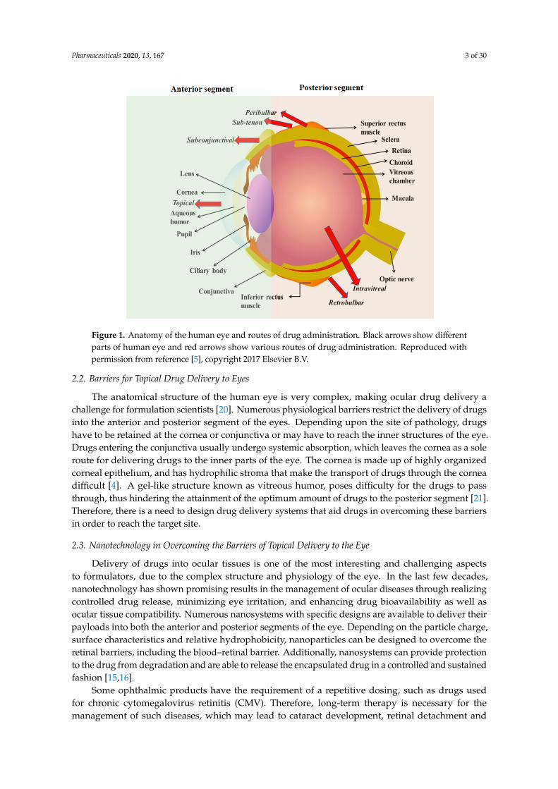
The detailed anatomical structure of the eye and its routes of administration are presented inFigure 1. The anatomical structure of the eye is composed of two parts, i.e., anterior segment andposterior segment [15–19]. Paracellular and transcellular are the two routes for penetration of drugs intothe corneal epithelium. The penetration of lipophilic molecules predominantly involves transcellularmechanisms, however, hydrophilic molecules and ions involve the paracellular route [19].
Pharmaceuticals 2020, 13, 167 3 of 30Pharmaceuticals 2020, 13, 167 3 of 30
Figure 1. Anatomy of the human eye and routes of drug administration. Black arrows show different parts of human eye and red arrows show various routes of drug administration. Reproduced with permission from reference [5], copyright 2017 Elsevier B.V.
2.2. Barriers for Topical Drug Delivery to Eyes
The anatomical structure of the human eye is very complex, making ocular drug delivery a challenge for formulation scientists [20]. Numerous physiological barriers restrict the delivery of drugs into the anterior and posterior segment of the eyes. Depending upon the site of pathology, drugs have to be retained at the cornea or conjunctiva or may have to reach the inner structures of the eye. Drugs entering the conjunctiva usually undergo systemic absorption, which leaves the cornea as a sole route for delivering drugs to the inner parts of the eye. The cornea is made up of highly organized corneal epithelium, and has hydrophilic stroma that make the transport of drugs through the cornea difficult [4]. A gel-like structure known as vitreous humor, poses difficulty for the drugs to pass through, thus hindering the attainment of the optimum amount of drugs to the posterior segment [21]. Therefore, there is a need to design drug delivery systems that aid drugs in overcoming these barriers in order to reach the target site.
2.3. Nanotechnology in Overcoming the Barriers of Topical Delivery to the Eye
Delivery of drugs into ocular tissues is one of the most interesting and challenging aspects to formulators, due to the complex structure and physiology of the eye. In the last few decades, nanotechnology has shown promising results in the management of ocular diseases through realizing controlled drug release, minimizing eye irritation, and enhancing drug bioavailability as well as
ocular tissue compatibility. Numerous nanosystems with specific designs are available to deliver their payloads into both the anterior and posterior segments of the eye. Depending on the particle charge, surface characteristics and relative hydrophobicity, nanoparticles can be designed to overcome the retinal barriers, including the blood–retinal barrier. Additionally, nanosystems can provide protection to the drug from degradation and are able to release the encapsulated drug in a controlled and sustained fashion [15,16].
Some ophthalmic products have the requirement of a repetitive dosing, such as drugs used for chronic cytomegalovirus retinitis (CMV). Therefore, long-term therapy is necessary for the management of such diseases, which may lead to cataract development, retinal detachment and
endophthalmitis [5]. However, nanoparticles prepared using natural polymers, e.g., albumin,
Figure 1. Anatomy of the human eye and routes of drug administration. Black arrows show differentparts of human eye and red arrows show various routes of drug administration. Reproduced withpermission from reference [5], copyright 2017 Elsevier B.V.
2.2. Barriers for Topical Drug Delivery to Eyes
The anatomical structure of the human eye is very complex, making ocular drug delivery achallenge for formulation scientists [20]. Numerous physiological barriers restrict the delivery of drugsinto the anterior and posterior segment of the eyes. Depending upon the site of pathology, drugshave to be retained at the cornea or conjunctiva or may have to reach the inner structures of the eye.Drugs entering the conjunctiva usually undergo systemic absorption, which leaves the cornea as a soleroute for delivering drugs to the inner parts of the eye. The cornea is made up of highly organizedcorneal epithelium, and has hydrophilic stroma that make the transport of drugs through the corneadifficult [4]. A gel-like structure known as vitreous humor, poses difficulty for the drugs to passthrough, thus hindering the attainment of the optimum amount of drugs to the posterior segment [21].Therefore, there is a need to design drug delivery systems that aid drugs in overcoming these barriersin order to reach the target site.
2.3. Nanotechnology in Overcoming the Barriers of Topical Delivery to the Eye
Delivery of drugs into ocular tissues is one of the most interesting and challenging aspectsto formulators, due to the complex structure and physiology of the eye. In the last few decades,nanotechnology has shown promising results in the management of ocular diseases through realizingcontrolled drug release, minimizing eye irritation, and enhancing drug bioavailability as well asocular tissue compatibility. Numerous nanosystems with specific designs are available to deliver theirpayloads into both the anterior and posterior segments of the eye. Depending on the particle charge,surface characteristics and relative hydrophobicity, nanoparticles can be designed to overcome theretinal barriers, including the blood–retinal barrier. Additionally, nanosystems can provide protectionto the drug from degradation and are able to release the encapsulated drug in a controlled and sustainedfashion [15,16].
Some ophthalmic products have the requirement of a repetitive dosing, such as drugs usedfor chronic cytomegalovirus retinitis (CMV). Therefore, long-term therapy is necessary for themanagement of such diseases, which may lead to cataract development, retinal detachment and
Pharmaceuticals 2020, 13, 167 4 of 30
endophthalmitis [5]. However, nanoparticles prepared using natural polymers, e.g., albumin,demonstrate promising therapeutic outcomes for such disease conditions [22]. Upon eye instillation,they did not induce any inflammatory reactions in the retinal tissue nor disturb the surroundingocular tissues. Nanotechnology-mediated ophthalmic products are spurring enormous interest inretinal drug delivery, because retinal neovascularization and choroidal neovascularization have asimilar microenvironment as solid tumors, where enhanced permeation and retention (EPR) effectis also playing a role for choroidal neovascularization and drug targeting using nanoparticles [23].The EPR effect is a postulation by which molecules of certain sizes (typically liposomes, nanoparticles,and macromolecular drugs) tend to accumulate in tumor tissues much more than they do in normaltissues [24]. Different nanosized drug delivery systems applied to various ocular diseases arediscussed below.
2.4. Nanotechnology-Driven Drug Carriers for Topical Delivery to Eyes
Various nanotechnology-driven drug carriers are summarized in Table 1 and are discussed inthe following.
2.4.1. Liposomes
Liposomes are made up of natural biocompatible phospholipids that delivers drugs in asustained-release manner to the eyes [20]. The structure of liposomal vesicles allows them toencapsulate hydrophilic drugs in the aqueous core, while hydrophobic or amphiphilic drugs can beembedded in the lipid layers [20,25]. The liposomal vesicles vary in size from 10 to 1000 nm [26].The drug in the liposomes is delivered to the eyes via four mechanisms:
(1) Adsorption: Adsorption of liposomes onto the cell membrane is the first step in delivering thedrugs from the liposomes. In the presence of cell surface proteins, liposomes become leaky andrelease their contents near the cell membrane. This results in a high drug concentration in thevicinity of the cell membrane, and promotes the cellular uptake of drugs by passive diffusion [26].
(2) Endocytosis: After adsorption and cellular uptake, the liposome reaches into the endosomes, andis then transported to the lysosomes through endosomes. Later, the enzymes in the lysosomesdegrade the lipids and the entrapped drug will be released into the cytoplasm [26].
(3) Fusion: Fusion of the lipid bilayer of liposomes with lipoidal cell membrane by intermixing andlateral diffusion of lipids results in direct delivery of liposomal contents into the cytoplasm [26].
(4) Lipid exchange: Due to the similarity in the lipids present in the liposomal membrane and thephospholipids present in the cell membrane, lipid transfer proteins in the cell membrane recognizeliposomes and therefore cause lipid exchange. As a result of this, the liposomal membrane getsdestabilized and the drug gets released [26].
Liposomes are said to enhance the permeability of hydrophilic, lipophilic and amphiphilic drugsthrough the cornea. There are many factors influencing drug delivery efficiency through liposomes.The surface charge of liposomes is one, and cationic liposomes have been shown to have a prolongedresidence time in the cornea since they strongly interact with the cornea due to its negative charge [26].Multi-lamellar vesicles are shown to be retained for a longer time than the small unilamellar vesicles,which may be attributed to the restricted drainage from the inner canthal region [26].
Liposomes have evolved from conventional to the new generation, in order to enhance safetyand efficacy of this carrier system. Conventional liposomes generally have an outer lipid layer and aninner aqueous core, whereas the new generation liposomes—also known as stealth liposomes—arecoated with polyethylene glycol (PEG), and protect them from engulfment by phagocytic cells [27].Another class of liposomes are immunoliposomes, which have been developed for targeted deliveryby conjugating antibodies at the surface that recognizes specific proteins on the target cells [28–30].Cationic liposomes consist of positively charged lipids that interact efficiently with the negativelycharged DNA and thereby condense the DNA into a more compact structure; hence, they are mostly
Pharmaceuticals 2020, 13, 167 5 of 30
used for delivering genes into eyes into various diseases such as glaucoma, epithelial retinal diseases,cytomegalovirus retinitis, etc. These lipid complexes provide protection to the entrapped geneticmaterial and enhance its intracellular delivery. In some cases, the cationic liposome also contains helperlipids such as di-oleoyl phosphatidylethanolamine, which stabilize the liposome complex. Amongthe new generation liposomes, cationic liposomal formulations are the youngest members and hold apromising future, particularly for delivering genes to the eyes [31,32]. The zeta potential of cationicliposomes must be between 10 to 30 mV [33].
Kawakami and co-workers prepared cationic liposomes with plasmid DNA and investigatedthe in vivo transfection efficiency in rabbits after intravitreal injection (100 µL). Plasmid DNA alonedoes not show significant transfection efficiency from 40 to 85 mg, however, pDNA complexedwith N-[1-(2,3-dioleyloxy)propyl]-N,N,N-trimethylammonium chloride (DOTMA)/Cholesterol (chol)liposomes significantly increased the pDNA ranging from 40 to 85 µg (p < 0.05). It was difficultto prepare the complex of pDNA with the DOTMA/Chol liposomal formulation having more than60 µg pDNA. These liposomes (pDNA; 85 mg/DOTMA/Chol) showed high transfection efficiency andluciferase activity in the cornea, aqueous humor, iris-ciliary body, lens, vitreous body and retina for3 days, but at the same time showed high cytotoxicity and aggregation behavior with the retina, whichdecreases visual activity and causes blindness. Therefore, an anionic coating of natural polysaccharidesor synthetic polymer is applied to reduce cytotoxicity as well as aggregation behaviors [34].
Liu and coworkers formulated PEGylated liposome-protamine-hyaluronic acid nanoparticles(PEG-LPH-NP) loaded with siRNA (PEG-LPH-NP-S) for the treatment of choroidal neovascularization(CNV) on a laser-induced rat model. siRNA directed against vascular endothelial growth factor(VEGF) or VEGF receptors in CNV animal models have shown promising results. The PEG-LPH-NP-Swere 132 nm in size with 20 mV cationic zeta potential, and depicted >95% encapsulation efficiency.It was found that PEG-LPH-NP could efficiently protect the siRNA load, facilitate the intracellulardelivery of siRNA and the expression inhibition of VEGFR1. Apoptosis was detected by the terminaldeoxynucleotidyl transferase-mediated dUTP-biotin nick ending-labeling method (TUNEL). The TUNELtesting and morphologic observation showed low toxicity on the rat retina, and thus, these lioposomeswere considered as a safe nanoformulation in the treatment of choroidal neovascularization [35].
In another study, the delivery efficiency of diclofenac-loaded liposomes to the retina was comparedwith conventional diclofenac ophthalmic solution on rabbit eyes. It was reported that the liposomesmodified with PVA or polyvinyl alcohol derivatives bearing a hydrophobic anchor (C16H33 S) at theterminal of the molecule (PVA-R) improved the physical stability of diclofenac-loaded liposomes. Afteradministration to eye, PVA-R-liposomes took the non-corneal pathway to deliver diclofenac efficientlyto the retina. These liposomes also displayed better physical stability and less aggregation [36].
It has been suggested that bioadhesive and biodegradable polymers can be used to prolongthe residence time of liposomal formulations. Mehanna et al. demonstrated that chitosan-coatedliposomes encapsulated with antibiotic ciprofloxacin HCl improved the ocular permeation of the drug.This formulation inhibited the growth of P. aeruginosa in rabbit eyes for 24 h when compared to themarketed preparation [37]. Fernandez de Sá et al. designed voriconazole-loaded liposomes composedof soy phosphatidylcholine for fungal keratitis, which demonstrated the delivery of reasonable amountsof drug to the cornea [38].
2.4.2. Niosomes
Niosomes are vesicles similar to liposomes, but unlike liposomes they are composed of non-ionicsurfactants. Similarly to liposomes, they can encapsulate both hydrophilic and hydrophobic as wellamphiphilic compounds. Niosomes were investigated to deliver the drug timolol maleate, widelyprescribed for glaucoma. It was found that 80% of the drug was lost either because of rapid blinking ofeyes or due to drainage into the nasolacrimal duct while using as a conventional eye drop. In addition,drug drainage into the nasolacrimal duct results in systemic side effects such as heart disease or asthma.In contrast, niosomes loaded with timolol maleate demonstrate better retention and penetration of the
Pharmaceuticals 2020, 13, 167 6 of 30
drug through the cornea and, at the same time, reduced side effects. The size of prepared niosomalformulation was found be in the range of 2 to 3 µm with entrapment efficiency (EE) of 24.3% w/v.The prepared niosomal formulation provided prolonged and controlled release as compared to themarketed solution (Timolet® GFS, Sun Pharmaceuticals India Limited, Vadodara, India. A higherconcentration of the drug was found across the corneal barrier of the eye in niosomal formulation.These results indicated that the niosomal formulation has a higher permeation capacity as compared tomarketed niosomal formulation. The prepared niosomal formulation used less than 50% of timololmeleate which produce fewer side effects [39].
Abdelbary and co-workers prepared an ophthalmic niosomal formulation for controlled deliveryof gentamycin. Niosomal formulations were prepared using various surfactants (Tween 60, Tween 80or Brij 35), in the presence of cholesterol and a negative charge inducer dicetyl phosphate (DCP) indifferent molar ratios using a thin film hydration technique. An in vitro study suggested that niosomalformulation exhibited high drug retention in vesicles and showed sustained drug release as comparedto the free drug solution. Prepared niosomal formulations were tested for ocular irritation and it wasfound that the prepared niosomal formulation did not produce irritation in albino rats [40].
2.4.3. Nanoparticles
The first prototype of polymeric nanoparticles used for topical ocular delivery was made frompoly-(alkyl cyanoacrylates) (PACA). Polymers that are commonly used for preparing nanoparticles arepoly-ε-caprolactone (PCL), chitosan, poly-(lactic-co-glycolic acid) (PLGA), carbopol, gelatin, Eudragit®
(RS100 and RL100), hyaluronic acid and so forth [4]. Gold, silver, silica and cerium oxide nanoparticlesare also used as drug carriers for ocular delivery [15]. Nanocapsules and nanospheres are the two maintypes of nanoparticles with distinct structures. As stated in the introduction, nanoparticles have manyadvantages over the conventional drug delivery systems that can be exploited in ocular drug deliveryincluding better penetration through biological barriers, potential for targeted delivery and controlleddrug release.
Similarly to cationic liposomes, nanoparticles with positive surface charge demonstrate bettercorneal retention and permeability [41]. Vasconcelos et al. employed a PLGA-b-PEG copolymerto conjugate a cell penetrating positively charged peptide for improving ocular penetration of theprepared nanoparticles loaded with flurbiprofen. Nanoparticles fabricated using the abovementionedconjugate demonstrated sustained release (for up to 8 h) and high payload of drug as well as depictinglow ocular toxicity [42].
Chitosan-based nanoparticles loaded with ketorolac tromethamine were studied by Fathalla et al.for their enhanced delivery to the eyes. The size of the NPs was affected bychitosan/tripolyphosphate(CS/TPP) ratio where the diameter of the NPs ranged from 108.0 ± 2.4 nm to 257.2 ± 18.6 nm. The zetapotential was found to be in the range of 22.9 to 27.8 mV. The positive charge of NPs of differentformulation was obtained due to the cationic nature of chitosan. The in vitro release study demonstrateda sustained release of the drug over a period of 6 h from the nanoparticles compared to a conventionalsolution, which was released within 3 h. The ex vivo studies conducted using porcine eye ballsconfirmed the ability of the prepared nanoparticles to retain the drug on the corneal surface for a longertime compared to the ketorolac tromethamine solution [43].
Naringenin is being used for treating age-related macular degeneration (AMD), however, itsuse is limited due to the poor aqueous solubility. Hence, nanoparticular carrier systems have beenexamined for its ocular drug delivery. Naringenin (Nag)-loaded sulfobutyl ether-β-cyclodextrin/chitosannanoparticles (Nag CD/CS-NPs) were formulated using an ionic gelation method of chitosan with butylether-β-cyclodextrin (SBE-β-CD). The Nag CD/CS-NPs with positive zeta potential (~22 mV) and sizearound 450 nm released the drug in a sustained manner. In vitro release studies were evaluated insimulated tear fluid (STF) using a dialysis tube with a molecular weight cutoff of 8000–14,000. After 5 h,Nag-CD gave 57% drug release whereas Nag CD/CS-NPs gave only 38% drug release. These resultsindicated that the coated nanoparticle complex provided a sustained release of drug as compared to the
Pharmaceuticals 2020, 13, 167 7 of 30
only cyclodextrin complex. The in vivo study was carried out on New Zealand white rabbits. The in vivostudy showed that Nag CD/CS-NPs had a much higher ophthalmic bioavailability and prolongedresidence time, which significantly increased Nag bioavailability in the aqueous humor compared tothe suspension formulation and, consequently, reduced the frequency of administration [44].
2.4.4. Polymeric Micelles
Polymeric micelles are also investigated for delivering drugs to both the anterior and posteriorsegments of eye, due to their ability to increase the solubility of hydrophobic drugs and to enhance drugabsorption across biological barriers including the ones in the eye. Their ability to bind with cellularbarriers and improve penetration of drugs through the cornea may be attributed to their similarity in sizerange to the membrane proteins, scleral pores (cornea), and other biomolecules. Polymeric micelles are apotential carrier to deliver hydrophobic drugs and proteins through biological membranes, to increasethe chemical stability of unstable compounds, and to control the release of drugs [15,45].
Luschmann et al. prepared an in-situ forming nanosuspension made of liquid poly(ethyleneglycols) (PEG) and a simple micellar solution containing nonionic surfactants that increase cyclosporineA solubility. The in vivo experiments suggested that the micellar solution is suitable for deliveringdrugs to the anterior segment of eye. The in-situ nanosuspension and the micellar solution deliverhigh levels of drug to eye, i.e., 1683 ± 430 ng CsA/g cornea and 826 ± 163 ng CsA/g cornea, respectively.Both formulations exceeded the drug tissue levels reported for Restasis® (350 ng CsA/g cornea) andcationic emulsions (750 ngCsA/g cornea). Despite the neutral charge of micelles, concentrations ofdrug at tissues levels were slightly higher as compared to cationic emulsion [46].
In another similar study, cyclosporine A micelles were prepared using polyvinylcaprolactam-polyvinyl acetate-polyethylene glycol graft copolymer (PVCL-PVA-PEG), also known asSoluplus®. The Soluplus micelles were able to deliver an adequate amount of Cyclosporine A intothe cornea for the treatment of immune-mediated corneal disease. The developed formulation alsodisplayed a good ocular tolerance in rabbit eyes [47].
Antioxidantα-lipoic acid (ALA) is used for treating diabetic keratopathy and retinopathy. Anterioreye segment structures are usually damaged in corneal neuropathy and keratopathy. Micelles preparedusing Soluplus were investigated for their ability to solubilize ALA and the prepared formulationwas compared with a marketed eye drop that was obtained using conventional micelles of sodiumdioctylsulfo succinate surfactant. Prepared Soluplus-based nanoformulation was of around 70–80 nmin size and increased the solubility of ALA by around 10 times compared to the commercial eye drop.These micelles enhanced the flux of α-lipoic acid through cornea and were found to be less irritant [48].
As an example of targeted delivery to the eyes, Boddu and co-workers developed doxorubicinentrapped local in-situ gel utilizing folic acid-PEG-PLGA copolymer micelles for ligand-mediatedtargeted delivery to retinoblastoma cells via intravitreal administration. After intravitrealadministration, DOX micelles in PLGA-PEG-PLGA gel gave sustained release for up to two weeks.The uptake of DOX in micellar solution was four times greater than plain DOX solution [49].
2.4.5. Dendrimers
Dendrimers are “tree-like” nanostructured polymers that have been interesting in terms of oculardrug delivery. They are attractive systems for drug delivery due to their nanosize range, ability todisplay multiple surface groups that allows for targeting, and easy preparation and functionalization.Dendrimers demonstrate promising properties in topical delivery of drugs to the eyes. Holden et al.encapsulated the two anti-glaucoma drugs brimonidine and timolol maleate into dendrimer hydrogel(DH) made up of polyamidoamine (PAMAM) dendrimers tethered with multiple PEG chains andphotoreactive acrylate groups. They investigated the intracellular uptake of these two encapsulateddrugs on human corneal epithelial (HCE) cells and the transport across the bovine corneal endothelium.The prepared DH formulation showed mucoadhesive properties towards mucin particles and were
Pharmaceuticals 2020, 13, 167 8 of 30
non-toxic to human corneal epithelial cells, and depicted higher HCE uptake and significantly increasedbovine corneal transport for both anti-glaucoma drugs, as compared to the conventional eye drop [50].
Yavuz et al. investigated anionic and cationic PAMAM dendrimer complexes for deliveringdexamethasone to the posterior segment of the eye (retina) for the treatment of diabetic retinopathy.An ex vivo transport study using rabbit cornea and sclera-choroid retina pigment epithelium (SCRPE)tissues demonstrated that anionic dendrimers delivered higher drug concentrations at the corneal andscleral tissues compared to the free drug solution and cationic dendrimers. From this finding, it wasconcluded that dexamethasone-PAMAM complex formulations enhanced ocular bioavailability ofdexamethasone via topical route [51].
Lancina et al. designed fast dissolving dendrimer-based nanofibers (DNF) as a topical deliveryvehicle for brimonidine tartrate for glaucoma, where DNF showed better efficacy than pristinebrimonidine tartrate solution [52]. The water-soluble anionic, cationic carbosilane dendrimers(generation 1–3) was explored in eye drop solution as muco-adhesive polymer containing acetazolamide(ACZ) in normotensive rabbits for improved hypotensive effect of ACZ. Authors investigated boththe cationic and anionic modified carbosilane dendrimers using human ocular epithelial cell linesand rabbit eye for in vitro and in vivo tolerance studies, respectively. This study indicates thatboth carbosilane dendrimers are well-tolerated in concentration between 5 and 10 µM. An eye dropformulation comprising G3 carbosilane dendrimers (5 µM) and ACZ (0.07%) induced a rapid (onsettime 1 h) and extended (up to 7 h) hypotensive effect which led to a significant increment in thetherapeutic efficacy with area under the plasma drug concentration (AUC0) (8 h), along with themaximal intraocular pressure reduction. These research finding suggest that the dendrimer-mediatedtopical eye formulation is safe and can be commercialized for treatment of eye diseases [53].
2.4.6. Nano-Implants
Implantable technology is not new and has been in use for many decades; however, theincorporation of micro- and nanotechnologies in implants enables the development of safe andmore efficient drug delivery devices. Advancement in implants creates a huge impact on patientcomfort and compliance, while treating chronic conditions by delivering drugs in a sustained mannerto the posterior segment of eyes (posterior segment eye disease). These technologies are scalabletechnologies and offer localized drug delivery for a prolonged duration.
Some nanocontrolled release systems that are implanted into the eyes to achieve drug deliveryare already in clinical practices (e.g., Vitrasert®, Retrisert®, Surodex™ and Ozurdex®). Biodegradablepolymers including both synthetic (most commonly used for ocular implants), e.g., polylactic acid (PLA),polyglycolic acid (PGA), PLGA, PCL, polyanhydrides (PA) and polyortho-esters (POE), and naturalpolymers such as collagen, gelatin and chitosan have been utilized to fabricate nano-implants [54].
Vitrasert® is an example of intravitreal implant technology useful for cytomegalovirus retinitis andHIV infections comprising of ganciclovir formulated using semipermeable poly vinyl alcohol (PVA),which allows for drug release and an impermeable ethyl vinyl acetate polymer (EVA). The implant tabletcontains 4.5 mg of drug and 0.015 mg magnesium stearate, which release from all sides of the tabletexcept the discontinuous EVA layer. The discontinuity in the EVA coating creates a diffusion port forthe diffusion of ganciclovir from the implant into the eye. This technology releases active medicamentsfor 5–8 months and is devoid of systemic toxicity and thus cuts the costs of treatment. In the clinicalstudies, median time to disease progression of 6–8 months was observed in patients treated with aVitrasert implant. The shelf life of the implant is 24 months and it is stored at a temperature between15 and 25 ◦C [55,56]. Retisert® is a United State of Food & Drug Administration (USFDA)-approvedsteroid implant manufactured by Bausch & Lomb, Rochester, NY, USA by incorporation of 0.59 mg offluocinolone acetonide (FA) for controlled delivery into vitreous cavity (approximately 0.6 µg/day).It can release fluocinolone slowly for up to 3 years. In the long term, drug retention after release fromthe Retisert® effectively suppresses the inflammation, reduces the recurrence of uveitis and improvesvisual acuity in 80% of patients [56,57].
Pharmaceuticals 2020, 13, 167 9 of 30
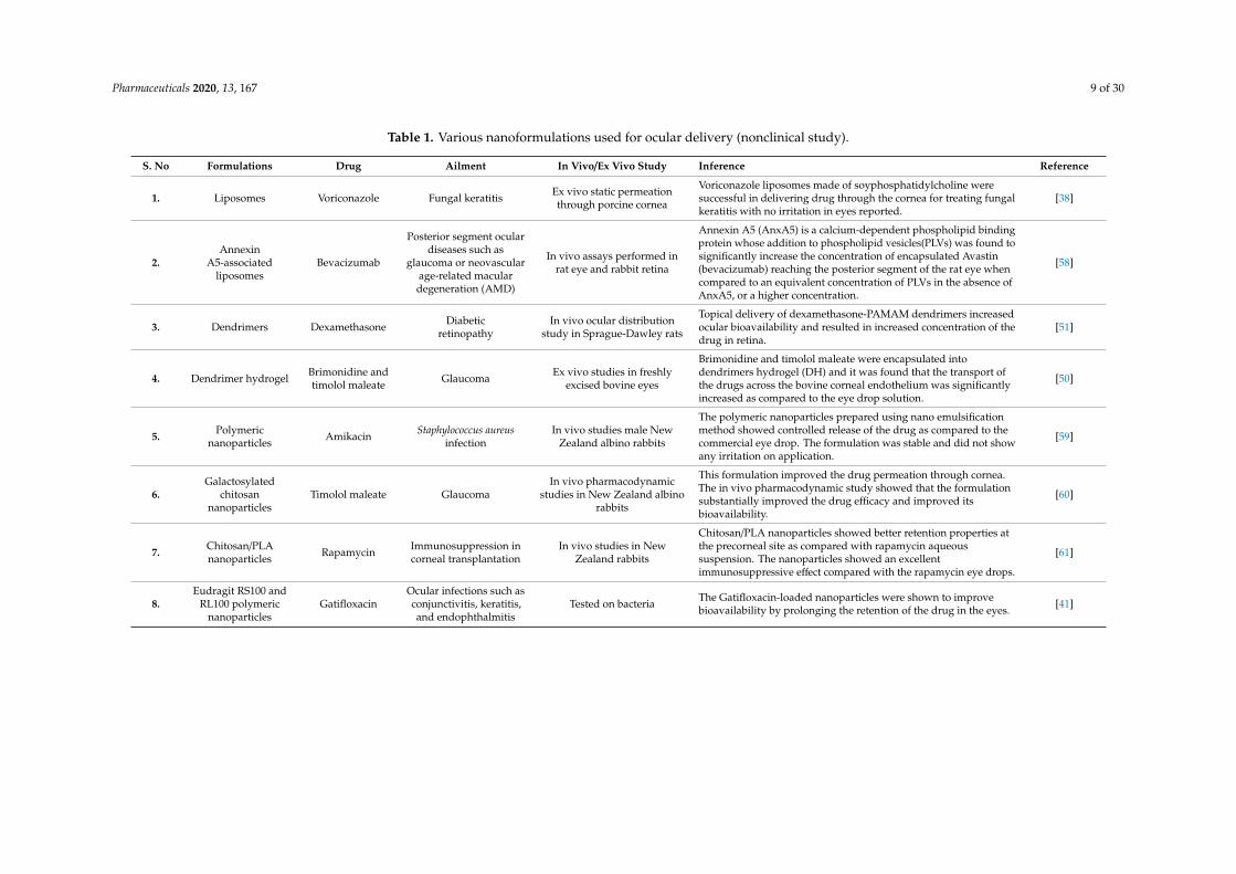
Table 1. Various nanoformulations used for ocular delivery (nonclinical study).
S. No Formulations Drug Ailment In Vivo/Ex Vivo Study Inference Reference
1. Liposomes Voriconazole Fungal keratitis Ex vivo static permeationthrough porcine cornea
Voriconazole liposomes made of soyphosphatidylcholine weresuccessful in delivering drug through the cornea for treating fungalkeratitis with no irritation in eyes reported.
[38]
2.Annexin
A5-associatedliposomes
Bevacizumab
Posterior segment oculardiseases such as
glaucoma or neovascularage-related maculardegeneration (AMD)
In vivo assays performed inrat eye and rabbit retina
Annexin A5 (AnxA5) is a calcium-dependent phospholipid bindingprotein whose addition to phospholipid vesicles(PLVs) was found tosignificantly increase the concentration of encapsulated Avastin(bevacizumab) reaching the posterior segment of the rat eye whencompared to an equivalent concentration of PLVs in the absence ofAnxA5, or a higher concentration.
[58]
3. Dendrimers Dexamethasone Diabeticretinopathy
In vivo ocular distributionstudy in Sprague-Dawley rats
Topical delivery of dexamethasone-PAMAM dendrimers increasedocular bioavailability and resulted in increased concentration of thedrug in retina.
[51]
4. Dendrimer hydrogel Brimonidine andtimolol maleate Glaucoma Ex vivo studies in freshly
excised bovine eyes
Brimonidine and timolol maleate were encapsulated intodendrimers hydrogel (DH) and it was found that the transport ofthe drugs across the bovine corneal endothelium was significantlyincreased as compared to the eye drop solution.
[50]
5. Polymericnanoparticles Amikacin Staphylococcus aureus
infectionIn vivo studies male New
Zealand albino rabbits
The polymeric nanoparticles prepared using nano emulsificationmethod showed controlled release of the drug as compared to thecommercial eye drop. The formulation was stable and did not showany irritation on application.
[59]
6.Galactosylated
chitosannanoparticles
Timolol maleate GlaucomaIn vivo pharmacodynamic
studies in New Zealand albinorabbits
This formulation improved the drug permeation through cornea.The in vivo pharmacodynamic study showed that the formulationsubstantially improved the drug efficacy and improved itsbioavailability.
[60]
7. Chitosan/PLAnanoparticles Rapamycin Immunosuppression in
corneal transplantationIn vivo studies in New
Zealand rabbits
Chitosan/PLA nanoparticles showed better retention properties atthe precorneal site as compared with rapamycin aqueoussuspension. The nanoparticles showed an excellentimmunosuppressive effect compared with the rapamycin eye drops.
[61]
8.Eudragit RS100 and
RL100 polymericnanoparticles
GatifloxacinOcular infections such asconjunctivitis, keratitis,and endophthalmitis
Tested on bacteria The Gatifloxacin-loaded nanoparticles were shown to improvebioavailability by prolonging the retention of the drug in the eyes. [41]
Pharmaceuticals 2020, 13, 167 10 of 30
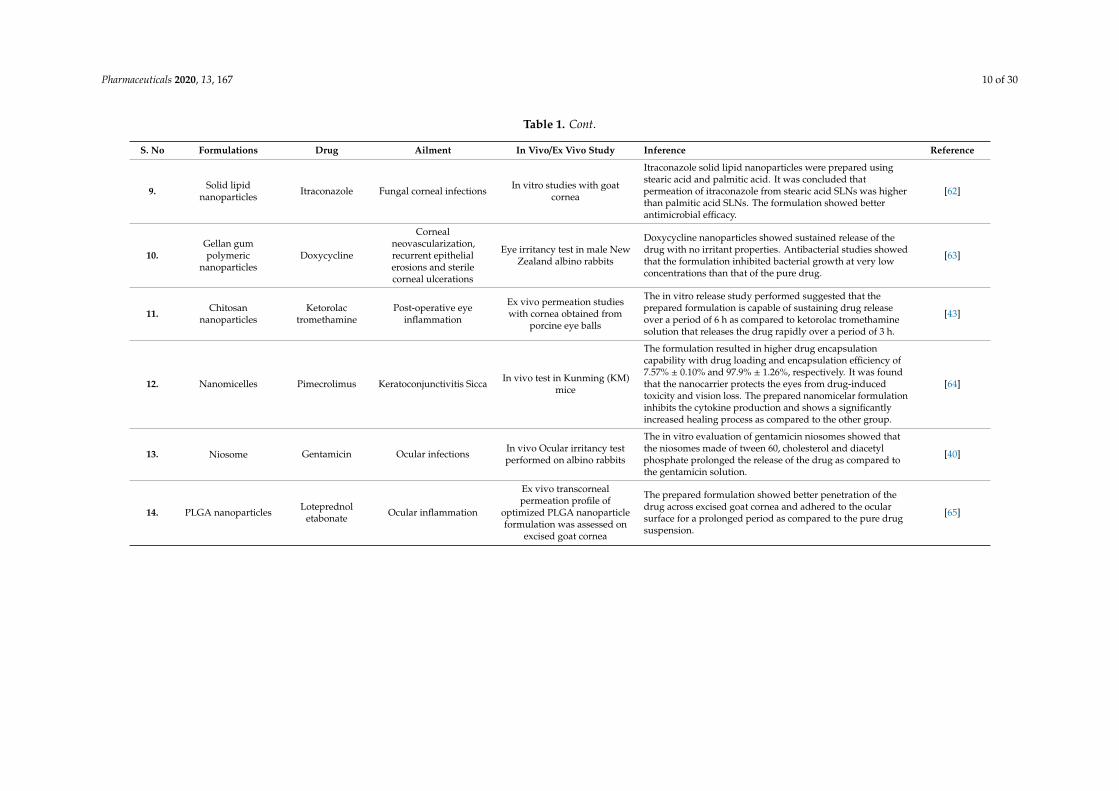
Table 1. Cont.
S. No Formulations Drug Ailment In Vivo/Ex Vivo Study Inference Reference
9. Solid lipidnanoparticles Itraconazole Fungal corneal infections In vitro studies with goat
cornea
Itraconazole solid lipid nanoparticles were prepared usingstearic acid and palmitic acid. It was concluded thatpermeation of itraconazole from stearic acid SLNs was higherthan palmitic acid SLNs. The formulation showed betterantimicrobial efficacy.
[62]
10.Gellan gumpolymeric
nanoparticlesDoxycycline
Cornealneovascularization,recurrent epithelialerosions and sterilecorneal ulcerations
Eye irritancy test in male NewZealand albino rabbits
Doxycycline nanoparticles showed sustained release of thedrug with no irritant properties. Antibacterial studies showedthat the formulation inhibited bacterial growth at very lowconcentrations than that of the pure drug.
[63]
11. Chitosannanoparticles
Ketorolactromethamine
Post-operative eyeinflammation
Ex vivo permeation studieswith cornea obtained from
porcine eye balls
The in vitro release study performed suggested that theprepared formulation is capable of sustaining drug releaseover a period of 6 h as compared to ketorolac tromethaminesolution that releases the drug rapidly over a period of 3 h.
[43]
12. Nanomicelles Pimecrolimus Keratoconjunctivitis Sicca In vivo test in Kunming (KM)mice
The formulation resulted in higher drug encapsulationcapability with drug loading and encapsulation efficiency of7.57% ± 0.10% and 97.9% ± 1.26%, respectively. It was foundthat the nanocarrier protects the eyes from drug-inducedtoxicity and vision loss. The prepared nanomicelar formulationinhibits the cytokine production and shows a significantlyincreased healing process as compared to the other group.
[64]
13. Niosome Gentamicin Ocular infections In vivo Ocular irritancy testperformed on albino rabbits
The in vitro evaluation of gentamicin niosomes showed thatthe niosomes made of tween 60, cholesterol and diacetylphosphate prolonged the release of the drug as compared tothe gentamicin solution.
[40]
14. PLGA nanoparticles Loteprednoletabonate Ocular inflammation
Ex vivo transcornealpermeation profile of
optimized PLGA nanoparticleformulation was assessed on
excised goat cornea
The prepared formulation showed better penetration of thedrug across excised goat cornea and adhered to the ocularsurface for a prolonged period as compared to the pure drugsuspension.
[65]
Pharmaceuticals 2020, 13, 167 11 of 30
Surodex™ and Ozurdex® are other examples of biodegradable implants that are widely usedin clinical practice for intraocular delivery and sustained release of dexamethasone. Surodex™ isimplanted into the atria to treat the post-operative inflammation of cataract patients and comprises60 µg of dexamethasone into the PLGA hydroxypropyl methylcellulose polymer matrix. Ozurdex® isan intravitreal implant containing 0.7 mg of dexamethasone in PLGA polymer, which is capable ofreleasing the drug at a slow rate for up to 6 months. It is preloaded into a single-use drug deliverysystem (DDS) device for direct injection into the intravitreal cavity, administrated with the assistanceof a healthcare expert. Clinical trial data suggest its potency in reducing vision loss. Data also suggestthe improvement in vision acuity in eyes with macular edema associated with branch retinal veinocclusion (BRVO) or central retinal vein occlusion [54,56].
3. Topical Drug Delivery to Skin
3.1. Anatomy of Skin
Skin has a complex structure that serves as a physical barrier to the entry of exogenous substances.Its detailed anatomical structure is presented in Figure 2. Skin is composed of three distinct layers,i.e., epidermis, dermis and hypodermis [66]. The presence of numerous physiological barriers to thedifferent layers of skin poses challenges to the pharmaceutical researchers for effective drug transportthrough the skin.
Pharmaceuticals 2020, 13, 167 11 of 30
Surodex™ and Ozurdex® are other examples of biodegradable implants that are widely used in clinical practice for intraocular delivery and sustained release of dexamethasone. Surodex™ is implanted into the atria to treat the post-operative inflammation of cataract patients and comprises 60 µg of dexamethasone into the PLGA hydroxypropyl methylcellulose polymer matrix. Ozurdex® is an intravitreal implant containing 0.7 mg of dexamethasone in PLGA polymer, which is capable of releasing the drug at a slow rate for up to 6 months. It is preloaded into a single-use drug delivery system (DDS) device for direct injection into the intravitreal cavity, administrated with the assistance of a healthcare expert. Clinical trial data suggest its potency in reducing vision loss. Data also suggest the improvement in vision acuity in eyes with macular edema associated with branch retinal vein occlusion (BRVO) or central retinal vein occlusion [54,56].
3. Topical Drug Delivery to Skin
3.1. Anatomy of Skin
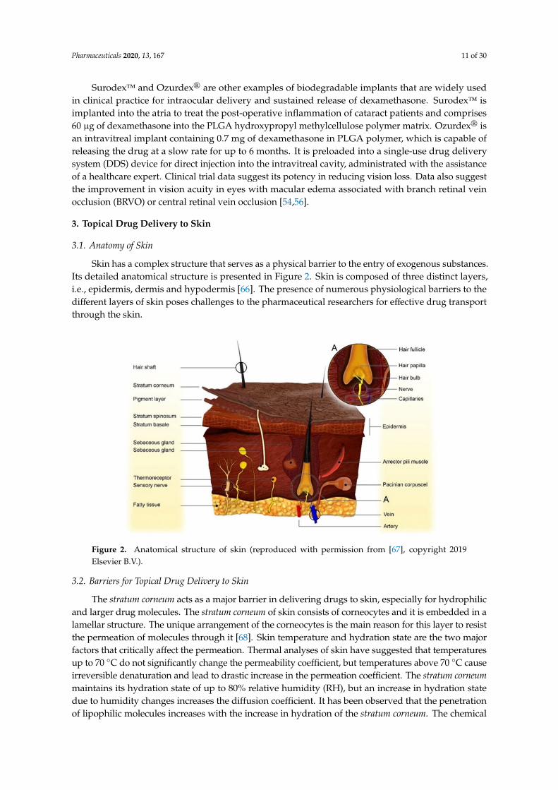
Skin has a complex structure that serves as a physical barrier to the entry of exogenous substances. Its detailed anatomical structure is presented in Figure 2. Skin is composed of three distinct layers, i.e., epidermis, dermis and hypodermis [66]. The presence of numerous physiological barriers to the different layers of skin poses challenges to the pharmaceutical researchers for effective drug transport through the skin.
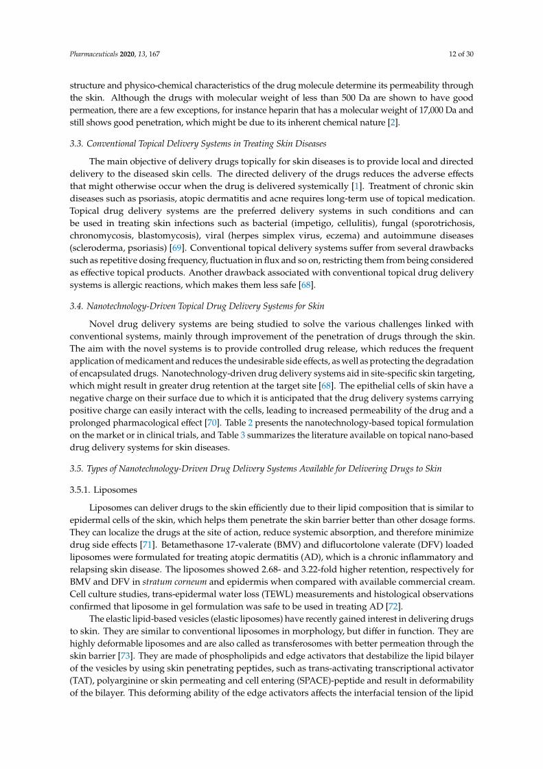
Figure 2. Anatomical structure of skin (reproduced with permission from [67], copyright 2019 Elsevier B.V.)
3.2. Barriers for Topical Drug Delivery to Skin
The stratum corneum acts as a major barrier in delivering drugs to skin, especially for hydrophilic and larger drug molecules. The stratum corneum of skin consists of corneocytes and it is embedded in a lamellar structure. The unique arrangement of the corneocytes is the main reason for this layer to resist the permeation of molecules through it [68]. Skin temperature and hydration state are the two major factors that critically affect the permeation. Thermal analyses of skin have suggested that temperatures up to 70 °C do not significantly change the permeability coefficient, but temperatures above 70 °C cause irreversible denaturation and lead to drastic increase in the permeation coefficient. The stratum corneum maintains its hydration state of up to 80% relative humidity (RH), but an increase in hydration state due to humidity changes increases the diffusion coefficient. It has been observed that the penetration of lipophilic molecules increases with the increase in hydration of the stratum corneum. The chemical structure and physico-chemical characteristics of the drug molecule determine its permeability through the skin. Although the drugs
Figure 2. Anatomical structure of skin (reproduced with permission from [67], copyright 2019Elsevier B.V.).
3.2. Barriers for Topical Drug Delivery to Skin
The stratum corneum acts as a major barrier in delivering drugs to skin, especially for hydrophilicand larger drug molecules. The stratum corneum of skin consists of corneocytes and it is embedded in alamellar structure. The unique arrangement of the corneocytes is the main reason for this layer to resistthe permeation of molecules through it [68]. Skin temperature and hydration state are the two majorfactors that critically affect the permeation. Thermal analyses of skin have suggested that temperaturesup to 70 ◦C do not significantly change the permeability coefficient, but temperatures above 70 ◦C causeirreversible denaturation and lead to drastic increase in the permeation coefficient. The stratum corneummaintains its hydration state of up to 80% relative humidity (RH), but an increase in hydration statedue to humidity changes increases the diffusion coefficient. It has been observed that the penetrationof lipophilic molecules increases with the increase in hydration of the stratum corneum. The chemical
Pharmaceuticals 2020, 13, 167 12 of 30
structure and physico-chemical characteristics of the drug molecule determine its permeability throughthe skin. Although the drugs with molecular weight of less than 500 Da are shown to have goodpermeation, there are a few exceptions, for instance heparin that has a molecular weight of 17,000 Da andstill shows good penetration, which might be due to its inherent chemical nature [2].
3.3. Conventional Topical Delivery Systems in Treating Skin Diseases
The main objective of delivery drugs topically for skin diseases is to provide local and directeddelivery to the diseased skin cells. The directed delivery of the drugs reduces the adverse effectsthat might otherwise occur when the drug is delivered systemically [1]. Treatment of chronic skindiseases such as psoriasis, atopic dermatitis and acne requires long-term use of topical medication.Topical drug delivery systems are the preferred delivery systems in such conditions and canbe used in treating skin infections such as bacterial (impetigo, cellulitis), fungal (sporotrichosis,chronomycosis, blastomycosis), viral (herpes simplex virus, eczema) and autoimmune diseases(scleroderma, psoriasis) [69]. Conventional topical delivery systems suffer from several drawbackssuch as repetitive dosing frequency, fluctuation in flux and so on, restricting them from being consideredas effective topical products. Another drawback associated with conventional topical drug deliverysystems is allergic reactions, which makes them less safe [68].
3.4. Nanotechnology-Driven Topical Drug Delivery Systems for Skin
Novel drug delivery systems are being studied to solve the various challenges linked withconventional systems, mainly through improvement of the penetration of drugs through the skin.The aim with the novel systems is to provide controlled drug release, which reduces the frequentapplication of medicament and reduces the undesirable side effects, as well as protecting the degradationof encapsulated drugs. Nanotechnology-driven drug delivery systems aid in site-specific skin targeting,which might result in greater drug retention at the target site [68]. The epithelial cells of skin have anegative charge on their surface due to which it is anticipated that the drug delivery systems carryingpositive charge can easily interact with the cells, leading to increased permeability of the drug and aprolonged pharmacological effect [70]. Table 2 presents the nanotechnology-based topical formulationon the market or in clinical trials, and Table 3 summarizes the literature available on topical nano-baseddrug delivery systems for skin diseases.
3.5. Types of Nanotechnology-Driven Drug Delivery Systems Available for Delivering Drugs to Skin
3.5.1. Liposomes
Liposomes can deliver drugs to the skin efficiently due to their lipid composition that is similar toepidermal cells of the skin, which helps them penetrate the skin barrier better than other dosage forms.They can localize the drugs at the site of action, reduce systemic absorption, and therefore minimizedrug side effects [71]. Betamethasone 17-valerate (BMV) and diflucortolone valerate (DFV) loadedliposomes were formulated for treating atopic dermatitis (AD), which is a chronic inflammatory andrelapsing skin disease. The liposomes showed 2.68- and 3.22-fold higher retention, respectively forBMV and DFV in stratum corneum and epidermis when compared with available commercial cream.Cell culture studies, trans-epidermal water loss (TEWL) measurements and histological observationsconfirmed that liposome in gel formulation was safe to be used in treating AD [72].
The elastic lipid-based vesicles (elastic liposomes) have recently gained interest in delivering drugsto skin. They are similar to conventional liposomes in morphology, but differ in function. They arehighly deformable liposomes and are also called as transferosomes with better permeation through theskin barrier [73]. They are made of phospholipids and edge activators that destabilize the lipid bilayerof the vesicles by using skin penetrating peptides, such as trans-activating transcriptional activator(TAT), polyarginine or skin permeating and cell entering (SPACE)-peptide and result in deformabilityof the bilayer. This deforming ability of the edge activators affects the interfacial tension of the lipid
Pharmaceuticals 2020, 13, 167 13 of 30
bilayer [74]. Eline Desmet et al. designed an elastic liposome formulation for RNA interference (RNAi)to treat psoriasis. Cultured primary skin cells and psoriasis tissue model in vitro were used to provethe effectiveness of elastic liposomal carriers. Cationic liposomes deliver functional RNAi molecules tothe different epidermal layers of normal or impaired skin, without crossing the dermal compartment.The psoriasis model was checked by down-regulation of hBD-2 and it was found that hBD-2 mRNAexpression decreased to ~70% using cationic liposomes. From the finding it was suggested that theseelastic liposomes are better carriers for RNAi delivery [73].
3.5.2. Solid Lipid Nanoparticles
Solid lipid nanoparticles (SLN) are novel carriers that are composed of a solid lipid core coatedwith surfactant. The lipids used are non-irritating and non-toxic to the skin, and they are alsoconsidered safe to be applied on damaged skin. The small size of the lipid particles is shown tohave a close interaction with stratum corneum and thereby the system increases the penetration,occlusion and accumulation of drug in the dermis region, which makes them the appropriate systemfor topical-targeted drug delivery. Pradhan et al. prepared fluocinolone acetonide SLN for treatingpsoriasis by emulsification-ultrasonication method. The authors prepared a gel-like formulation, whichshowed enhanced solubility of fluocinolone acetonide and helped the drug get deposited onto theepidermis, the location where psoriasis develops [75].
Irritant contact dermatitis (ICD), is one of the skin diseases manifested by edema, erythema,epidermal thickening, itching and scaling [76]. Presently, anti-histaminic, hydroquinones and topicalcorticosteroids are used for treating ICD, but long-term use of these drugs may lead to undesirableeffects such as dry mouth, cataracts, high blood pressure, constipation, obesity, etc. Polyphenols arevaluable compounds possessing scavenging properties towards radical oxygen species and complexingproperties towards proteins. These abilities make polyphenols interesting for the treatment of variousdermatological problems. Curcumin (CUR) is a naturally occurring polyphenol found in rhizomes ofCurcuma longa Linn. CUR also exhibits antioxidant activity, which is beneficial for several skin-relatedproblems such as eczema, dermatitis, pigmentation, acne, psoriasis lesions and all the exfoliative skindiseases. Due to the presence of high amounts of epidermal lipids within the stratum corneum, SLNsseem to be a promising delivery system as compared to other nanoparticulate carriers. Consideringthis, Shrotriya et al. designed curcumin-loaded SLN (CUR-SLNs) incorporated into carbopol gel.Occlusive test, irritation test on skin, tyrosine enzyme inhibition activity and antioxidant activity usingcommercial gel and SLN-loaded gel was performed on skin model. It was demonstrated that CUR-SLNgel showed efficient occlusion properties, and enhanced skin deposition, inhibition of tyrosinaseenzyme activity and improved antioxidant activity compared to that of a conventional CUR-plain gel.ICD was developed using dinitrochlorobenzene (DNCB) on the dorsal surface of ears of mice. In vivostudy suggested that SLN-loaded gels are superior in suppression of ear swelling and reduction inskin water content in ICD disease compared to commercial gel [76].
3.5.3. Niosomes
Meng et al. formulated celastrol-loaded niosomal hydrogel to treat psoriasis, which increased thepermeation of celastrol in the skin and thus increased its anti-psoriasis activity in a mice model [77].Benzoyl peroxide (BPO) is the topical medication mostly used for the treatment of acne against thebacteria Propionibacterium acnes. BPO has poor aqueous solubility, forms cluster and it gets crystallizedin aqueous environment. These clusters are not able to penetrate through the follicles which iswhy conventional formulations like cream, lotion and gel have higher concentration of the drug(2.5–10%). Goyal et al. encapsulated BPO in niosome gel and found that the formulation resultedin better permeation of the drug through the skin and also reduced skin irritation. It was alsosuggested that BPO-loaded niosomal gel causes better reduction in colony forming unit (CFU) count ofPropionibacterium acnes bacteria in mice ear than plain BPO [78].
Pharmaceuticals 2020, 13, 167 14 of 30
Topical application of 5-aminolevulinic acid (ALA) has been employed in photodynamic therapy(PDT) treatment of superficial skin carcinoma. According to the PDT protocol, a concentrated aqueoussolution (20% w/w) of ALA is applied on the lesion, left for 3–6 h for drug absorption, and removedbefore the irradiation of light at the lesion. The main limitation of this therapy was the hydrophilicityof ALA due to which permeation of ALA through stratum corneum was hindered. ALA-loadednoisome gel was prepared to improve the permeation of the drug across the stratum corneum. Ex vivoskin permeation studies performed on excised human skin showed that the ALA-loaded niosomalformulation showed better penetration across the skin when compared to the aqueous solution ofALA [79]. El-Say et al. formulated a diacerein-loaded niosomal gel formulation that exhibited betteranti-inflammatory activity compared to the commercial gel formulation. White albino rats were chosenas the animal model to evaluate anti-inflammatory activity and inflammation was developed by usingthe carrageenan raw paw method with slight modifications [80].
3.5.4. Nanoparticles
Nanoparticles exhibit improved localized targeting of drug and thus are helpful in reducingsystemic side effects. Mao et al. prepared CUR loaded polymeric nanoparticles to treat inflammationprevailing in psoriasis disease. The polymer used here was RRR-α-tocopherol succinate-graftedpoly-lysine conjugate. Imiquimod (IMQ) induced psoriasis-like mouse model was used to test theprepared formulation. Tumor necrosis factor (TNF) is one of the most potent inflammation activatorsthat play a major role in signaling the classical nuclear factor kappa B (NF-B). The contribution of NF-Bmay be significant in IMQ-induced psoriasis-like inflammation. The results showed that TNF-α, NF-B,IL-6 expression was increased in IMQ-treated skin, but after the treatment with CUR-NPs-gel/clobetasoltheir expression was significantly decreased. Angiogenesis inhibition study was performed using theprepared nanoformulation gel by quantifying cluster of differentiation 31 (CD31) expression visuallyafter skin staining with immunofluorescent dye. CD31 are biomarkers that are highly expressed duringnew blood vessel formation. Large amounts of new blood vessel formation were found in the controlgroup, based on the large numbers of immunoflouroscent CD31 that were seen visually in the controlledgroup. It was found that a much smaller quantity of CD was shown in CUR-NPs-gel. It was concludedthat curcumin-loaded nanoparticle gel inhibited the inflammatory cascade triggered by TNF-α andthe inflammation associated angiogenesis stimulated in psoriasis, improving antioxidant activity.Curcumin nanoparticles were formed using a novel cationic amphiphilic polymer, an RRR-α-tocopherylsuccinate-grafted ε-polylysine conjugate (VES-g-ε-PLL). These cationic nanoparticles were incorporatedinto 8% silk fibroin through electrostatic interaction. The increase in skin permeation of curcumin wasresponsible for an improved response in psoriatic plaque-like model, suggesting a promising potentialof this drug delivery system to treat inflammatory skin disorders like psoriasis [81].
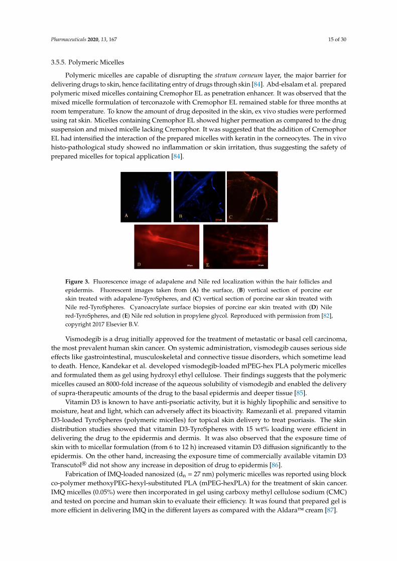
Ramezanli et al. investigated adapalene (drug for the topical treatment of acne) loadedtyrosine-derived polymeric nanoparticles (also known as TyroSpheres). The formulation was evaluated exvivo using human cadaver and porcine ear skin and compared with the marketed adapalene formulation(Differin®). It was reported that the crystallinity of adapalene was decreased and the TyroSpheres gotaccumulated in the hair follicles, due to which the topical delivery of adapalene was improved as comparedto the commercial product. Drug distribution in the hair follicles was visualized using adapalene inherentfluorescence and Nile red loaded TyroSpheres with the aid of a fluorescence microscope (Figure 3).Improved follicular delivery of the TyroSpheres was attributed to the small particle size [82].
Balzus et al. formulated dexamethasone-loaded Eudragit RS and ethyl cellulose nanoparticles,which adhere well to the skin and penetrate through the hair follicles. The nanoparticles released thedexamethasone drug in a controlled fashion compared to the commercially available cream in ex vivostudies. [83].
Pharmaceuticals 2020, 13, 167 15 of 30
3.5.5. Polymeric Micelles
Polymeric micelles are capable of disrupting the stratum corneum layer, the major barrier fordelivering drugs to skin, hence facilitating entry of drugs through skin [84]. Abd-elsalam et al. preparedpolymeric mixed micelles containing Cremophor EL as penetration enhancer. It was observed that themixed micelle formulation of terconazole with Cremophor EL remained stable for three months atroom temperature. To know the amount of drug deposited in the skin, ex vivo studies were performedusing rat skin. Micelles containing Cremophor EL showed higher permeation as compared to the drugsuspension and mixed micelle lacking Cremophor. It was suggested that the addition of CremophorEL had intensified the interaction of the prepared micelles with keratin in the corneocytes. The in vivohisto-pathological study showed no inflammation or skin irritation, thus suggesting the safety ofprepared micelles for topical application [84].
Pharmaceuticals 2020, 13, 167 15 of 30
observed that the mixed micelle formulation of terconazole with Cremophor EL remained stable for three months at room temperature. To know the amount of drug deposited in the skin, ex vivo studies were performed using rat skin. Micelles containing Cremophor EL showed higher permeation as compared to the drug suspension and mixed micelle lacking Cremophor. It was suggested that the addition of Cremophor EL had intensified the interaction of the prepared micelles with keratin in the corneocytes. The in vivo histo-pathological study showed no inflammation or skin irritation, thus suggesting the safety of prepared micelles for topical application [84].
Figure 3. Fluorescence image of adapalene and Nile red localization within the hair follicles and epidermis. Fluorescent images taken from (A) the surface, (B) vertical section of porcine ear skin treated with adapalene-TyroSpheres, and (C) vertical section of porcine ear skin treated with Nile red-TyroSpheres. Cyanoacrylate surface biopsies of porcine ear skin treated with (D) Nile red-TyroSpheres, and (E) Nile red solution in propylene glycol. Reproduced with permission from [82], copyright 2017 Elsevier B.V.)
Vismodegib is a drug initially approved for the treatment of metastatic or basal cell carcinoma, the most prevalent human skin cancer. On systemic administration, vismodegib causes serious side effects like gastrointestinal, musculoskeletal and connective tissue disorders, which sometime lead to death. Hence, Kandekar et al. developed vismodegib-loaded mPEG-hex PLA polymeric micelles and formulated them as gel using hydroxyl ethyl cellulose. Their findings suggests that the polymeric micelles caused an 8000-fold increase of the aqueous solubility of vismodegib and enabled the delivery of supra-therapeutic amounts of the drug to the basal epidermis and deeper tissue [85].
Vitamin D3 is known to have anti-psoriatic activity, but it is highly lipophilic and sensitive to moisture, heat and light, which can adversely affect its bioactivity. Ramezanli et al. prepared vitamin D3-loaded TyroSpheres (polymeric micelles) for topical skin delivery to treat psoriasis. The skin distribution studies showed that vitamin D3-TyroSpheres with 15 wt% loading were efficient in delivering the drug to the epidermis and dermis. It was also observed that the exposure time of skin with to micellar formulation (from 6 to 12 h) increased vitamin D3 diffusion significantly to the epidermis. On the other hand, increasing the exposure time of commercially available vitamin D3 Transcutol® did not show any increase in deposition of drug to epidermis [86].
Fabrication of IMQ-loaded nanosized (dn =27nm) polymeric micelles was reported using block co-polymer methoxyPEG-hexyl-substituted PLA (mPEG-hexPLA) for the treatment of skin cancer. IMQ micelles (0.05%) were then incorporated in gel using carboxy methyl cellulose sodium (CMC) and tested on porcine and human skin to evaluate their efficiency. It was found that prepared gel is more efficient in delivering IMQ in the different layers as compared with the Aldara™ cream [87].
Figure 3. Fluorescence image of adapalene and Nile red localization within the hair follicles andepidermis. Fluorescent images taken from (A) the surface, (B) vertical section of porcine earskin treated with adapalene-TyroSpheres, and (C) vertical section of porcine ear skin treated withNile red-TyroSpheres. Cyanoacrylate surface biopsies of porcine ear skin treated with (D) Nilered-TyroSpheres, and (E) Nile red solution in propylene glycol. Reproduced with permission from [82],copyright 2017 Elsevier B.V.
Vismodegib is a drug initially approved for the treatment of metastatic or basal cell carcinoma,the most prevalent human skin cancer. On systemic administration, vismodegib causes serious sideeffects like gastrointestinal, musculoskeletal and connective tissue disorders, which sometime leadto death. Hence, Kandekar et al. developed vismodegib-loaded mPEG-hex PLA polymeric micellesand formulated them as gel using hydroxyl ethyl cellulose. Their findings suggests that the polymericmicelles caused an 8000-fold increase of the aqueous solubility of vismodegib and enabled the deliveryof supra-therapeutic amounts of the drug to the basal epidermis and deeper tissue [85].
Vitamin D3 is known to have anti-psoriatic activity, but it is highly lipophilic and sensitive tomoisture, heat and light, which can adversely affect its bioactivity. Ramezanli et al. prepared vitaminD3-loaded TyroSpheres (polymeric micelles) for topical skin delivery to treat psoriasis. The skindistribution studies showed that vitamin D3-TyroSpheres with 15 wt% loading were efficient indelivering the drug to the epidermis and dermis. It was also observed that the exposure time ofskin with to micellar formulation (from 6 to 12 h) increased vitamin D3 diffusion significantly to theepidermis. On the other hand, increasing the exposure time of commercially available vitamin D3Transcutol® did not show any increase in deposition of drug to epidermis [86].
Fabrication of IMQ-loaded nanosized (dn = 27 nm) polymeric micelles was reported using blockco-polymer methoxyPEG-hexyl-substituted PLA (mPEG-hexPLA) for the treatment of skin cancer.IMQ micelles (0.05%) were then incorporated in gel using carboxy methyl cellulose sodium (CMC)and tested on porcine and human skin to evaluate their efficiency. It was found that prepared gel ismore efficient in delivering IMQ in the different layers as compared with the Aldara™ cream [87].
Pharmaceuticals 2020, 13, 167 16 of 30
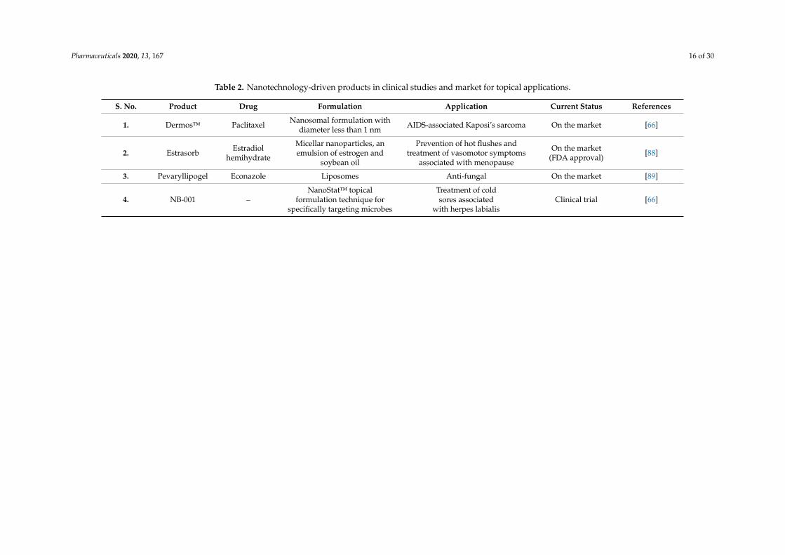
Table 2. Nanotechnology-driven products in clinical studies and market for topical applications.
S. No. Product Drug Formulation Application Current Status References
1. Dermos™ Paclitaxel Nanosomal formulation withdiameter less than 1 nm AIDS-associated Kaposi’s sarcoma On the market [66]
2. Estrasorb Estradiolhemihydrate
Micellar nanoparticles, anemulsion of estrogen and
soybean oil
Prevention of hot flushes andtreatment of vasomotor symptoms
associated with menopause
On the market(FDA approval) [88]
3. Pevaryllipogel Econazole Liposomes Anti-fungal On the market [89]
4. NB-001 –NanoStat™ topical
formulation technique forspecifically targeting microbes
Treatment of coldsores associated
with herpes labialisClinical trial [66]
Pharmaceuticals 2020, 13, 167 17 of 30
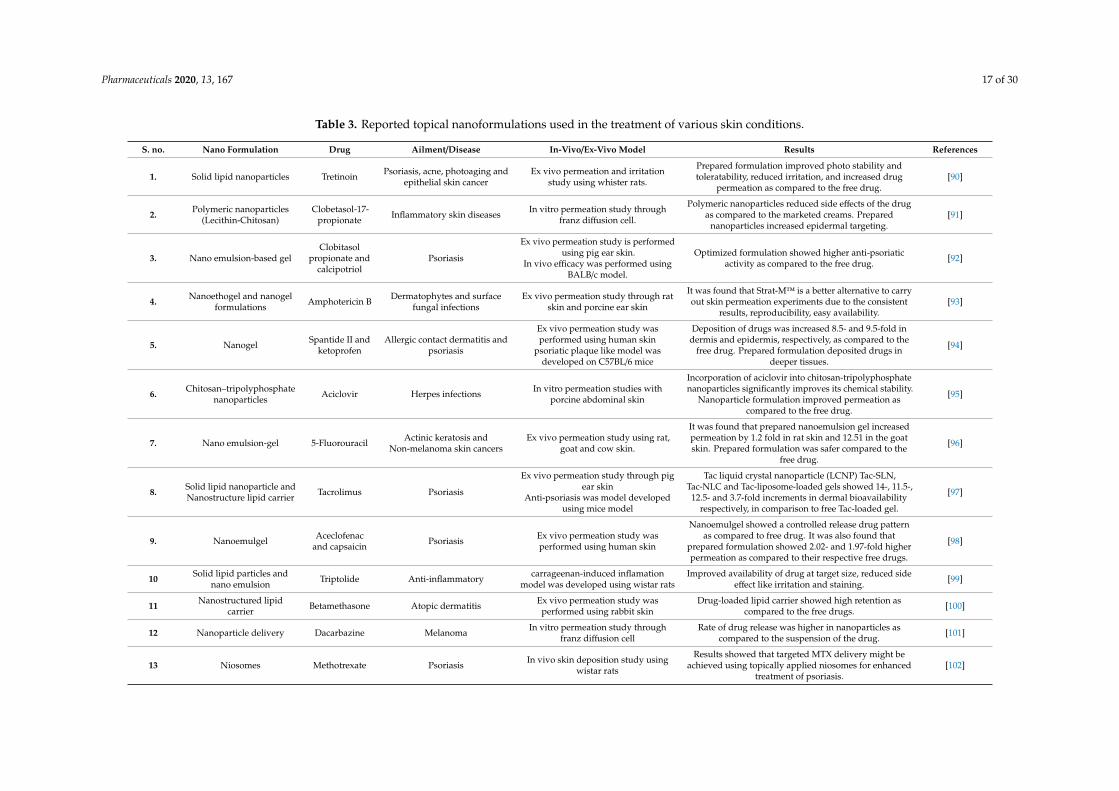
Table 3. Reported topical nanoformulations used in the treatment of various skin conditions.
S. no. Nano Formulation Drug Ailment/Disease In-Vivo/Ex-Vivo Model Results References
1. Solid lipid nanoparticles Tretinoin Psoriasis, acne, photoaging andepithelial skin cancer
Ex vivo permeation and irritationstudy using whister rats.
Prepared formulation improved photo stability andtoleratability, reduced irritation, and increased drug
permeation as compared to the free drug.[90]
2. Polymeric nanoparticles(Lecithin-Chitosan)
Clobetasol-17-propionate Inflammatory skin diseases In vitro permeation study through
franz diffusion cell.
Polymeric nanoparticles reduced side effects of the drugas compared to the marketed creams. Prepared
nanoparticles increased epidermal targeting.[91]
3. Nano emulsion-based gelClobitasol
propionate andcalcipotriol
Psoriasis
Ex vivo permeation study is performedusing pig ear skin.
In vivo efficacy was performed usingBALB/c model.
Optimized formulation showed higher anti-psoriaticactivity as compared to the free drug. [92]
4. Nanoethogel and nanogelformulations Amphotericin B Dermatophytes and surface
fungal infectionsEx vivo permeation study through rat
skin and porcine ear skin
It was found that Strat-M™ is a better alternative to carryout skin permeation experiments due to the consistent
results, reproducibility, easy availability.[93]
5. Nanogel Spantide II andketoprofen
Allergic contact dermatitis andpsoriasis
Ex vivo permeation study wasperformed using human skin
psoriatic plaque like model wasdeveloped on C57BL/6 mice
Deposition of drugs was increased 8.5- and 9.5-fold indermis and epidermis, respectively, as compared to the
free drug. Prepared formulation deposited drugs indeeper tissues.
[94]
6. Chitosan–tripolyphosphatenanoparticles Aciclovir Herpes infections In vitro permeation studies with
porcine abdominal skin
Incorporation of aciclovir into chitosan-tripolyphosphatenanoparticles significantly improves its chemical stability.
Nanoparticle formulation improved permeation ascompared to the free drug.
[95]
7. Nano emulsion-gel 5-Fluorouracil Actinic keratosis andNon-melanoma skin cancers
Ex vivo permeation study using rat,goat and cow skin.
It was found that prepared nanoemulsion gel increasedpermeation by 1.2 fold in rat skin and 12.51 in the goatskin. Prepared formulation was safer compared to the
free drug.
[96]
8. Solid lipid nanoparticle andNanostructure lipid carrier Tacrolimus Psoriasis
Ex vivo permeation study through pigear skin
Anti-psoriasis was model developedusing mice model
Tac liquid crystal nanoparticle (LCNP) Tac-SLN,Tac-NLC and Tac-liposome-loaded gels showed 14-, 11.5-,
12.5- and 3.7-fold increments in dermal bioavailabilityrespectively, in comparison to free Tac-loaded gel.
[97]
9. Nanoemulgel Aceclofenacand capsaicin Psoriasis Ex vivo permeation study was
performed using human skin
Nanoemulgel showed a controlled release drug patternas compared to free drug. It was also found that
prepared formulation showed 2.02- and 1.97-fold higherpermeation as compared to their respective free drugs.
[98]
10 Solid lipid particles andnano emulsion Triptolide Anti-inflammatory carrageenan-induced inflamation
model was developed using wistar ratsImproved availability of drug at target size, reduced side
effect like irritation and staining. [99]
11 Nanostructured lipidcarrier Betamethasone Atopic dermatitis Ex vivo permeation study was
performed using rabbit skinDrug-loaded lipid carrier showed high retention as
compared to the free drugs. [100]
12 Nanoparticle delivery Dacarbazine Melanoma In vitro permeation study throughfranz diffusion cell
Rate of drug release was higher in nanoparticles ascompared to the suspension of the drug. [101]
13 Niosomes Methotrexate Psoriasis In vivo skin deposition study usingwistar rats
Results showed that targeted MTX delivery might beachieved using topically applied niosomes for enhanced
treatment of psoriasis.[102]
Pharmaceuticals 2020, 13, 167 18 of 30
4. Wounds and the Barriers for Topical Drug Delivery to Wounds
Wounds can be classified based on the nature of healing, thickness and microbial load. Acuteor chronic wounds are based on the nature of healing. In acute wounds, the wound healing time is8–12 weeks. In chronic wounds, anatomical and functional integrity of the skin is not restored for aperiod of over three months. Chronic wounds include pressure ulcers, vascular ulcers and diabeticfoot ulcers [103]. Based on the thickness of the wound, they can be classified as superficial, partialand full thickness wounds and can be further classified as clean, clean-contaminated, contaminatedand infected wounds based on their microbial load. Nussbaum et al. conducted a retrospectiveanalysis of the Medicare 5% Limited Data Set to determine the cost of chronic wound care for Medicarebeneficiaries. The study concluded that the Medicare cost for surgical wounds and infected diabeticfoot ulcers was 38.3 billion and 18.7 billion US dollars respectively [104].
Endogenous factors are used for wound healing clinically, but their clinical use is limited due totheir breakdown by the proteolytic enzymes that are present at the wound site. The wound environmenthas various pro-inflammatory cytokines that can deactivate drugs. Topical use of growth factors forwound healing is limited because of the very short half-life of the proteins due to the close control andinactivation by protease inhibitors. Blood flow is poor at the site of wound healing. Therefore, systemicdrug delivery is also not of significant importance in wound healing. The dysfunctional wound bedvasculature reduces the bioavailability of compounds that are administered orally or intravenously.Hence, the wound environment requires complex drug delivery systems that can deliver the drugsand growth factors to the appropriate site [105].
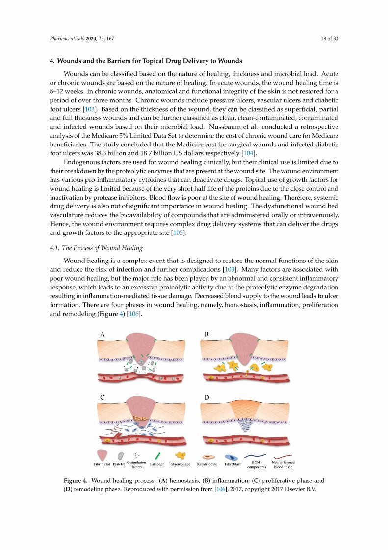
4.1. The Process of Wound Healing
Wound healing is a complex event that is designed to restore the normal functions of the skinand reduce the risk of infection and further complications [103]. Many factors are associated withpoor wound healing, but the major role has been played by an abnormal and consistent inflammatoryresponse, which leads to an excessive proteolytic activity due to the proteolytic enzyme degradationresulting in inflammation-mediated tissue damage. Decreased blood supply to the wound leads to ulcerformation. There are four phases in wound healing, namely, hemostasis, inflammation, proliferationand remodeling (Figure 4) [106].Pharmaceuticals 2020, 13, 167 19 of 30
Figure 4. Wound healing process: (A) hemostasis, (B) inflammation, (C) proliferative phase and (D) remodeling phase. Reproduced with permission from [106], 2017, copyright 2017 Elsevier B.V.
4.2.1. Nanoparticles
It has been reported that deficiency of epidermal growth factor (EGF) is one of the causes of diabetic foot ulcer (DFU). The effective concentration of exogenous recombinant human EGF (rhEGF) in wound for the treatment of DFU could not be achieved after local administration due to the short biological half-life of rhEGF, rapid dilution by tissue fluid, leakage from the wound surface, and degradation by enzymes. In order to overcome these challenges, Chu et al. designed rhEGF nanoparticles using PLGA polymer. These nanoparticles showed a controlled release of rhEGF for up to 24 h. These nanoparticles possessed better wound healing effects than those of pristine rhEGF [107]. LL37, an anti-microbial peptide/host defense peptide, which is part of the innate immune system, is known to promote wound healing. Chereddy et al. prepared LL37 antimicrobial peptide-loaded PLGA nanospheres and found that compared to LL37 alone, PLGA-LL37 nanoparticles significantly improved wound healing activity. The healing effect of PLGA-LL37 NP included higher re-epithelialization, granulation, tissue formation and immunomodulation [108].
Inorganic nanoparticles, especially metal oxide nanoparticles (MONPs), are also studied for the speedy recovery of wounds. MONPs like zinc oxide (ZnO), titanium dioxide (TiO2) and silicon dioxide (SiO2) play important roles in the production of reactive oxygen species, which stimulate the proliferation of fibroblasts and wound healing [109].
In a study, Origanum vulgare (as aqueous leaf extract) engineered with titanium dioxide nanoparticles was applied topically on the wound for improving the therapeutic efficacy of the extract. The results suggested significant wound healing activity in Albino rats, established by measuring wound closure, histopathology and protein expression profiling [110]. Bacterial infection is often a major complication associated with burn wounds. Curcumin is a naturally derived substance with innate antimicrobial and wound healing properties, but since it has poor aqueous solubility and rapid degradation, encapsulating it within a protective carrier is necessary. Krausz et al. encapsulated curcumin in chitosan nanoparticles (size—222 ± 14 nm) using sol-gel-based formulation for wound healing purposes in the setting of skin infection. This novel topical gel formulation showed significantly reduced bacterial load and improved wound healing in murine burn model compared to other groups (Figure 5). Histological analysis showed increased granulation tissue formation, improved collagen proliferation and enhanced neovascularization [111].
4.2.2. Solid Lipid Nanoparticles and Nanostructured Lipid Carriers
Solid lipid nanoparticles (SLN) and nanostructured lipid carriers (NLC) are efficient and non-toxic drug carriers, which can provide high drug concentration at the site of wound. SLNs consist of
Figure 4. Wound healing process: (A) hemostasis, (B) inflammation, (C) proliferative phase and(D) remodeling phase. Reproduced with permission from [106], 2017, copyright 2017 Elsevier B.V.
Pharmaceuticals 2020, 13, 167 19 of 30
Homeostasis involves platelet aggregation, immunity activation, blood clotting and complementssystem induction. Inflammation includes the recruitment of neutrophils and macrophages withextra cellular matrix (ECM) molecule production by resident cells in the skin. Proliferation involvesre-epithelialization, angiogenesis and granulation. The expansion of keratinocytes epithelial cells, stemcells and fibroblasts is known as re-epithelialization. Activation of endothelial cells and the creation ofa site in which these cells can proliferate in the wound is called angiogenesis. Granulation is defined asthe promotion of tissue granulation by fibroblasts, granulocytes and macrophages. Finally, remodelinginvolves the production of collagen- and elastin-including fibroblasts leading to myofibroblasts in thepresence of T cells and macrophages [106]. Monocyte chemoattractant protein 1, cytokines (interlukin-1and interlukin-6), growth factors such as fibroblast growth factor (FGF), epidermal growth factor (EGF),transforming growth factor-β (TGF-β), platelet-derived growth factor (PDGF), bone morphogenicproteins (BMPs), connective tissue growth factor (CTGF), granulocyte macrophage-colony stimulatingfactor (GM-CSF) and vascular endothelial growth factor (VEGF) participate in wound healing.
4.2. Role of Nanotechnology in Wound Healing
Numerous strategies based on nanotechnology are available for wound healing and aresummarized in Table 4.
4.2.1. Nanoparticles
It has been reported that deficiency of epidermal growth factor (EGF) is one of the causes of diabeticfoot ulcer (DFU). The effective concentration of exogenous recombinant human EGF (rhEGF) in woundfor the treatment of DFU could not be achieved after local administration due to the short biologicalhalf-life of rhEGF, rapid dilution by tissue fluid, leakage from the wound surface, and degradation byenzymes. In order to overcome these challenges, Chu et al. designed rhEGF nanoparticles using PLGApolymer. These nanoparticles showed a controlled release of rhEGF for up to 24 h. These nanoparticlespossessed better wound healing effects than those of pristine rhEGF [107]. LL37, an anti-microbialpeptide/host defense peptide, which is part of the innate immune system, is known to promote woundhealing. Chereddy et al. prepared LL37 antimicrobial peptide-loaded PLGA nanospheres and foundthat compared to LL37 alone, PLGA-LL37 nanoparticles significantly improved wound healing activity.The healing effect of PLGA-LL37 NP included higher re-epithelialization, granulation, tissue formationand immunomodulation [108].
Inorganic nanoparticles, especially metal oxide nanoparticles (MONPs), are also studied forthe speedy recovery of wounds. MONPs like zinc oxide (ZnO), titanium dioxide (TiO2) and silicondioxide (SiO2) play important roles in the production of reactive oxygen species, which stimulate theproliferation of fibroblasts and wound healing [109].
In a study, Origanum vulgare (as aqueous leaf extract) engineered with titanium dioxidenanoparticles was applied topically on the wound for improving the therapeutic efficacy of theextract. The results suggested significant wound healing activity in Albino rats, established bymeasuring wound closure, histopathology and protein expression profiling [110]. Bacterial infection isoften a major complication associated with burn wounds. Curcumin is a naturally derived substancewith innate antimicrobial and wound healing properties, but since it has poor aqueous solubility andrapid degradation, encapsulating it within a protective carrier is necessary. Krausz et al. encapsulatedcurcumin in chitosan nanoparticles (size—222 ± 14 nm) using sol-gel-based formulation for woundhealing purposes in the setting of skin infection. This novel topical gel formulation showed significantlyreduced bacterial load and improved wound healing in murine burn model compared to other groups(Figure 5). Histological analysis showed increased granulation tissue formation, improved collagenproliferation and enhanced neovascularization [111].
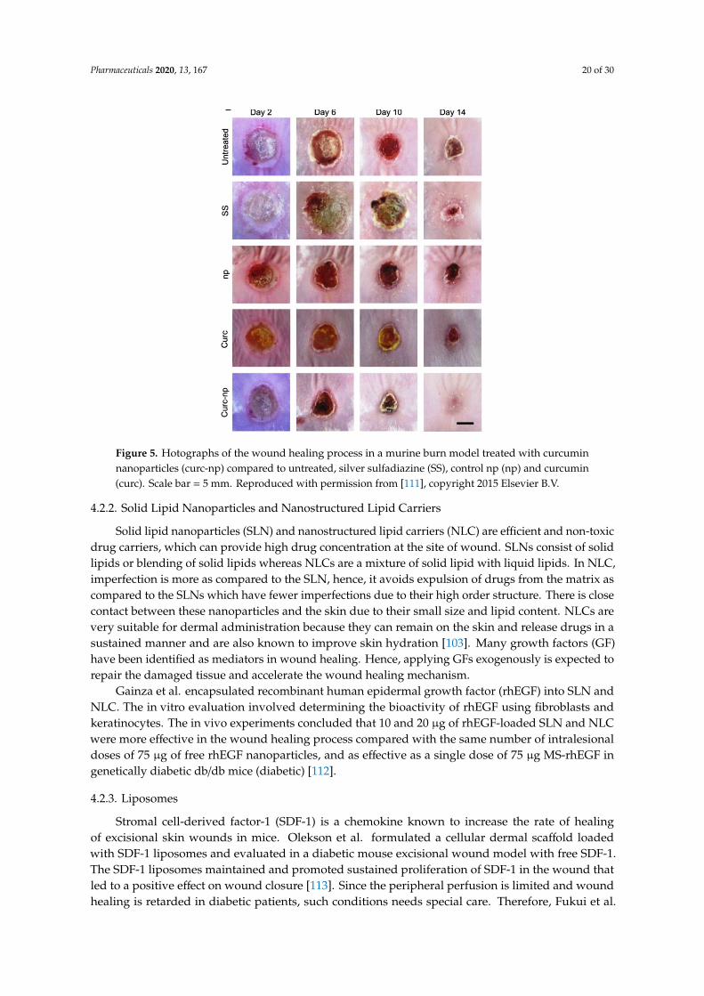
Pharmaceuticals 2020, 13, 167 20 of 30
Pharmaceuticals 2020, 13, 167 20 of 30
solid lipids or blending of solid lipids whereas NLCs are a mixture of solid lipid with liquid lipids. In NLC, imperfection is more as compared to the SLN, hence, it avoids expulsion of drugs from the matrix as compared to the SLNs which have fewer imperfections due to their high order structure. There is close contact between these nanoparticles and the skin due to their small size and lipid content. NLCs are very suitable for dermal administration because they can remain on the skin and release drugs in a sustained manner and are also known to improve skin hydration [103]. Many growth factors (GF) have been identified as mediators in wound healing. Hence, applying GFs exogenously is expected to repair the damaged tissue and accelerate the wound healing mechanism.
Gainza et al. encapsulated recombinant human epidermal growth factor (rhEGF) into SLN and NLC. The in vitro evaluation involved determining the bioactivity of rhEGF using fibroblasts and keratinocytes. The in vivo experiments concluded that 10 and 20 µg of rhEGF-loaded SLN and NLC were more effective in the wound healing process compared with the same number of intralesional doses of 75 µg of free rhEGF nanoparticles, and as effective as a single dose of 75 µg MS-rhEGF in genetically diabetic db/db mice (diabetic) [112].
Figure 5. Hotographs of the wound healing process in a murine burn model treated with curcumin nanoparticles (curc-np) compared to untreated, silver sulfadiazine (SS), control np (np) and curcumin (curc). Scale bar = 5 mm. Reproduced with permission from [111], copyright 2015 Elsevier B.V.).
4.2.3. Liposomes
Stromal cell-derived factor-1 (SDF-1) is a chemokine known to increase the rate of healing of excisional skin wounds in mice. Olekson et al. formulated a cellular dermal scaffold loaded with SDF-1 liposomes and evaluated in a diabetic mouse excisional wound model with free SDF-1. The SDF-1 liposomes maintained and promoted sustained proliferation of SDF-1 in the wound that led to a positive effect on wound closure [113]. Since the peripheral perfusion is limited and wound healing is retarded in diabetic patients, such conditions needs special care. Therefore, Fukui et al. prepared liposome-encapsulated hemoglobin with high O2 affinity to increase tissue perfusion, suppress inflammatory cytokines and promote faster skin wound healing. The results suggest that the above-mentioned parameters were significantly improved in diabetic dB/dB mice and are comparable to normal mice when treated with liposomal formulation [114].
Figure 5. Hotographs of the wound healing process in a murine burn model treated with curcuminnanoparticles (curc-np) compared to untreated, silver sulfadiazine (SS), control np (np) and curcumin(curc). Scale bar = 5 mm. Reproduced with permission from [111], copyright 2015 Elsevier B.V.
4.2.2. Solid Lipid Nanoparticles and Nanostructured Lipid Carriers
Solid lipid nanoparticles (SLN) and nanostructured lipid carriers (NLC) are efficient and non-toxicdrug carriers, which can provide high drug concentration at the site of wound. SLNs consist of solidlipids or blending of solid lipids whereas NLCs are a mixture of solid lipid with liquid lipids. In NLC,imperfection is more as compared to the SLN, hence, it avoids expulsion of drugs from the matrix ascompared to the SLNs which have fewer imperfections due to their high order structure. There is closecontact between these nanoparticles and the skin due to their small size and lipid content. NLCs arevery suitable for dermal administration because they can remain on the skin and release drugs in asustained manner and are also known to improve skin hydration [103]. Many growth factors (GF)have been identified as mediators in wound healing. Hence, applying GFs exogenously is expected torepair the damaged tissue and accelerate the wound healing mechanism.
Gainza et al. encapsulated recombinant human epidermal growth factor (rhEGF) into SLN andNLC. The in vitro evaluation involved determining the bioactivity of rhEGF using fibroblasts andkeratinocytes. The in vivo experiments concluded that 10 and 20 µg of rhEGF-loaded SLN and NLCwere more effective in the wound healing process compared with the same number of intralesionaldoses of 75 µg of free rhEGF nanoparticles, and as effective as a single dose of 75 µg MS-rhEGF ingenetically diabetic db/db mice (diabetic) [112].
4.2.3. Liposomes
Stromal cell-derived factor-1 (SDF-1) is a chemokine known to increase the rate of healingof excisional skin wounds in mice. Olekson et al. formulated a cellular dermal scaffold loadedwith SDF-1 liposomes and evaluated in a diabetic mouse excisional wound model with free SDF-1.The SDF-1 liposomes maintained and promoted sustained proliferation of SDF-1 in the wound thatled to a positive effect on wound closure [113]. Since the peripheral perfusion is limited and woundhealing is retarded in diabetic patients, such conditions needs special care. Therefore, Fukui et al.
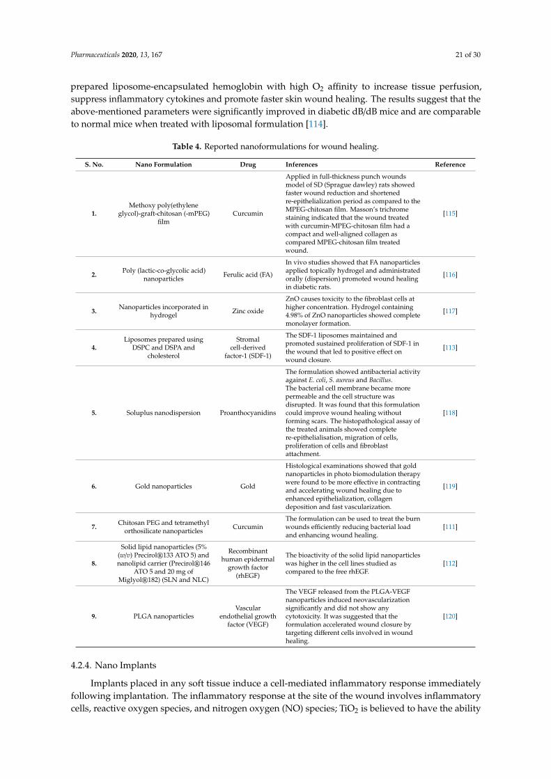
Pharmaceuticals 2020, 13, 167 21 of 30
prepared liposome-encapsulated hemoglobin with high O2 affinity to increase tissue perfusion,suppress inflammatory cytokines and promote faster skin wound healing. The results suggest that theabove-mentioned parameters were significantly improved in diabetic dB/dB mice and are comparableto normal mice when treated with liposomal formulation [114].
Table 4. Reported nanoformulations for wound healing.
S. No. Nano Formulation Drug Inferences Reference
1.Methoxy poly(ethylene
glycol)-graft-chitosan (-mPEG)film
Curcumin
Applied in full-thickness punch woundsmodel of SD (Sprague dawley) rats showedfaster wound reduction and shortenedre-epithelialization period as compared to theMPEG-chitosan film. Masson’s trichromestaining indicated that the wound treatedwith curcumin-MPEG-chitosan film had acompact and well-aligned collagen ascompared MPEG-chitosan film treatedwound.
[115]
2. Poly (lactic-co-glycolic acid)nanoparticles Ferulic acid (FA)
In vivo studies showed that FA nanoparticlesapplied topically hydrogel and administratedorally (dispersion) promoted wound healingin diabetic rats.
[116]
3. Nanoparticles incorporated inhydrogel Zinc oxide
ZnO causes toxicity to the fibroblast cells athigher concentration. Hydrogel containing4.98% of ZnO nanoparticles showed completemonolayer formation.
[117]
4.Liposomes prepared using
DSPC and DSPA andcholesterol
Stromalcell-derived
factor-1 (SDF-1)
The SDF-1 liposomes maintained andpromoted sustained proliferation of SDF-1 inthe wound that led to positive effect onwound closure.
[113]
5. Soluplus nanodispersion Proanthocyanidins
The formulation showed antibacterial activityagainst E. coli, S. aureus and Bacillus.The bacterial cell membrane became morepermeable and the cell structure wasdisrupted. It was found that this formulationcould improve wound healing withoutforming scars. The histopathological assay ofthe treated animals showed completere-epithelialisation, migration of cells,proliferation of cells and fibroblastattachment.
[118]
6. Gold nanoparticles Gold
Histological examinations showed that goldnanoparticles in photo biomodulation therapywere found to be more effective in contractingand accelerating wound healing due toenhanced epithelialization, collagendeposition and fast vascularization.
[119]
7. Chitosan PEG and tetramethylorthosilicate nanoparticles Curcumin
The formulation can be used to treat the burnwounds efficiently reducing bacterial loadand enhancing wound healing.
[111]
8.
Solid lipid nanoparticles (5%(w/v) Precirol®133 ATO 5) andnanolipid carrier (Precirol®146
ATO 5 and 20 mg ofMiglyol®182) (SLN and NLC)
Recombinanthuman epidermal
growth factor(rhEGF)
The bioactivity of the solid lipid nanoparticleswas higher in the cell lines studied ascompared to the free rhEGF.
[112]
9. PLGA nanoparticlesVascular
endothelial growthfactor (VEGF)
The VEGF released from the PLGA-VEGFnanoparticles induced neovascularizationsignificantly and did not show anycytotoxicity. It was suggested that theformulation accelerated wound closure bytargeting different cells involved in woundhealing.
[120]
4.2.4. Nano Implants
Implants placed in any soft tissue induce a cell-mediated inflammatory response immediatelyfollowing implantation. The inflammatory response at the site of the wound involves inflammatorycells, reactive oxygen species, and nitrogen oxygen (NO) species; TiO2 is believed to have the ability
Pharmaceuticals 2020, 13, 167 22 of 30
to inhibit these reactive oxygen species. Smith et al. modified a titanium implant surface via TiO2
nanotubes to increase the TiO2 surface area and eventually the catalytic degradation of NO species.It was observed that the TiO2 nanotubes reduced the levels of the pro-inflammatory signaling moleculeNO compared with an implant having a planar TiO2 surface [121]. Similarly, a coating of sandblastedand acid-etched surface dental implants with hydroxyapatite nanoparticles to increase the healingprocess [122]. Nanosurface-modified implants significantly increase the bone/implant contact and thusimprove the osseointegration and neovascularization process and ultimately accelerate the healingprocess [123].
5. Toxicological Aspects of Topically Applied Nanoformulations
Toxicity is a prime concern for any topical formulation and the establishment of a toxicityprofile is a mandatory requirement for regulatory approval. Nanoparticles have the potential topenetrate the cells and can produce free radicals, which leads to oxidative stress and collagen depletion.This depletion induces keratinization, atrophy of the dermis and skin wrinkling [124,125]. In oneresearch finding it was found that keratinocyte proliferation was significantly inhibited after exposureof nanocrystals-coated dressing [126].
The various critical quality attributes including the polymer concentration, drug characteristicand critical process parameters of the topical formulation can lead to remarkable differences in termsof stability, safety, efficacy and toxicity. In a study, the authors investigated the signs of toxicity afterexposure of o/w formulation comprising 5% hydroquinone (HQ) and a different base. The majorchallenge associated with HQ is its poor permeability and physicochemical stability, which leads to theuse of p-benzoquinone (pBQ) in the formulation which is known as carcinogenic [127–129].
The high HQ concentration was found in skin from formulation 1 containing Beeler’s based plusantioxidants, followed by formulation 2, which contains Beeler’s base and dimethyl isosorbide assolubilizer. This interaction might be between the cationic proteins of the skin and the anionic natureof Beeler’s base (sodium lauryl sulfate). Some signs of toxicity were also observed on the skin withall topical semisolid o/w formulation with 5% HQ after 6 h of exposure [130].Wu and co-workersreported the dermal exposure of titanium dioxide (TiO2) nanoparticles in vitro and in vivo usingisolated porcine skin. The results suggest that the pathological lesions are likely to occur probably dueto oxidative stress induced by the deposition of TiO2 nanoparticles. They also reported that with aprolonged exposure time, this topically applied nano-TiO2 can induce skin aging and may pose healthrisk issues to humans after long dermal exposure [131].
Trop and co-workers prepared silver nanoparticles for the treatment of wound healing. Silvercoated dressing acticoat caused liver enzymes and argyria-like symptoms in acute burn patients (30%burns). The silver levels in urine and plasma were monitored regularly to avoid the toxic effect of silverin burn patients. After 1 week of local treatment, hepatotoxicity and argyria-like symptoms and grayishdiscoloration of the patient’s face was also reported [132]. Numerous reports are available mentioningthe advantages of nanotechnology-based dermal formulations but their toxicity assessments are scarce,albeit its notable significance in clinical translation.
6. Conclusions
Conventional drug delivery systems for topical delivery possess several challenges, includingrepetitive dosing administration and rapid release, which impact product performance in healthcaresectors. Nowadays, nanotechnology-driven topical complex formulations such as polymeric micelles,liposomes, niosomes, nanoparticles, and implantable devices are gaining significant attention in thepharmaceutical sector for topical applications.
Nanotechnology has provided minimally invasive ocular drug delivery systems. The invasivenature of intravitreal and periocular injection has spurred researchers to work on topical deliverysystems that offer higher patient compliance. The nanotechnology-driven topical drug delivery systemsto eyes possess properties such as enhanced residence time in the pre-corneal area, better permeation
Pharmaceuticals 2020, 13, 167 23 of 30
ability through the ocular barriers, sustained delivery of drugs at targeted sites, ultimately improvingtherapeutic efficacy. Although the in vivo studies have shown that the nanotechnology-based drugdelivery systems did not produce any irritation and inflammatory response, their long-term effect mustbe properly analyzed. The existing topical formulations to treat skin diseases face challenges includinga smaller amount of drug penetrating the skin barrier, systemic absorption leading to unwanted sideeffects and, if administered orally, adverse effects and poorly targeted therapy. Psoriasis, skin cancer,fungal infections and bacterial infections are a few of the most common skin diseases that requiretopical treatment. Most of the in vivo studies performed have shown better skin permeability of thenanoformulations, their ability to reach the diseased site, being free from irritation and inflammatoryresponses. However, extensive investigation has to be carried out to translate the novel formulationsfor clinical use. Nano-based systems certainly exemplify an improvement in safety and efficacy fortopical drug delivery and hold great potential for efficient treatment leading to improved quality of thepatient’s life.
7. Future Prospects
Nanotechnology is still a promising and emerging field, presently focusing on encapsulatingdrugs which require a low dose for treatment. Several products encompass technical challenges, e.g.,(i) nanoparticles have a low loading capacity, (ii) dendrimers show toxicity problems, and (iii) liposomeshave stability challenges. Scale-up, batch-to-batch reproducibility, stability and clinical performance canbe regarded as the major challenges in the clinical translation from industry perspectives. The numberof nanotechnology-based products going into the market is smaller compared to the efforts that havebeen put at the research level. We need to focus on the technology transfer, scale-up and validation ofthese products in R&D laboratories for clinical translation. Amalgamation of nanotechnology withother delivery systems such as implants, nanofibers, hydrogels, etc., may be further investigated forimproved results in drug delivery by topical route. The main challenges that researchers need tofocus on for clinical translation are retention time, vision interferences, nano-bio interactions, drugpermeation and accumulation.
Author Contributions: Conceptualization, N.K.M.; Writing—original draft preparation, P.K.R., P.T., V.B., J.M.;Writing—review and editing, N.K.M., K.K.B., J.M.R. All authors have read and agreed to the published version ofthe manuscript.
Funding: No funding received for this article.
Acknowledgments: The authors would like to acknowledge the Department of Pharmaceutics, National Institute ofPharmaceutical Education & Research (NIPER), Hyderabad for providing the facilities for writing this manuscript.
Conflicts of Interest: No conflict of interest was reported by the authors of this article.
References
1. Jensen, L.; Petersson, K.; Nielsen, H.M. In vitro penetration properties of solid lipid nanoparticles in intactand barrier-impaired skin. Eur. J. Pharm. Biopharm. 2011, 79, 68–75. [CrossRef] [PubMed]
2. Patel, V.; Sharma, O.P.; Mehta, T. Nanocrystal: A novel approach to overcome skin barriers for improvedtopical drug delivery. Expert Opin. Drug Deliv. 2018, 15, 351–368. [CrossRef] [PubMed]
3. Sánchez-López, E.; Espina, M.; Doktorovova, S.; Souto, E.; García, M. Lipid nanoparticles (SLN, NLC):Overcoming the anatomical and physiological barriers of the eye–Part I–Barriers and determining factors inocular delivery. Eur. J. Pharm. Biopharm. 2017, 110, 70–75. [CrossRef] [PubMed]
4. Reimondez-Troitiño, S.; Csaba, N.; Alonso, M.J.; De La Fuente, M. Nanotherapies for the treatment of oculardiseases. Eur. J. Pharm. Biopharm. 2015, 95, 279–293. [CrossRef]
5. Weng, Y.; Liu, J.; Jin, S.; Guo, W.; Liang, X.; Hu, Z. Nanotechnology-based strategies for treatment of oculardisease. Acta Pharm. Sin. B 2017, 7, 281–291. [CrossRef]
6. Goyal, R.; Macri, L.K.; Kaplan, H.M.; Kohn, J. Nanoparticles and nanofibers for topical drug delivery.J. Control. Release 2015, 240, 77–92. [CrossRef]
7. Barry, B.W. Breaching the skin’s barrier to drugs. Nat. Biotechnol. 2004, 22, 165–167. [CrossRef]
Pharmaceuticals 2020, 13, 167 24 of 30
8. Montenegro, L.; Lai, F.; Offerta, A.; Sarpietro, M.G.; Micicchè, L.; Maccioni, A.M.; Valenti, D.; Fadda, A.M.From nanoemulsions to nanostructured lipid carriers: A relevant development in dermal delivery of drugsand cosmetics. J. Drug Deliv. Sci. Technol. 2016, 32, 100–112. [CrossRef]
9. Williams, A.C.; Barry, B.W. Penetration enhancers. Adv. Drug Deliv. Rev. 2012, 64, 128–137. [CrossRef]10. Ita, K.B. Prodrugs for transdermal drug delivery-trends and challenges. J. Drug Target. 2016, 24, 671–678.
[CrossRef]11. Charoenputtakun, P.; Li, S.K.; Ngawhirunpat, T. Iontophoretic delivery of lipophilic and hydrophilic drugs
from lipid nanoparticles across human skin. Int. J. Pharm. 2015, 495, 318–328. [CrossRef] [PubMed]12. Benson, H.A.E.; Mcildowie, M.; Prow, T.W. Magnetophoresis: Skin Penetration Enhancement by a
Magnetic Field. In Percutaneous Penetration Enhancers Physical Methods in Penetration Enhancement; Springer:Berlin/Heidelberg, Germany, 2017; pp. 195–206.
13. Sen, C.K. Human Wounds and Its Burden: An Updated Compendium of Estimates; Mary Ann Liebert, Inc.:New Rochelle, NY, USA, 2019.
14. Radhakrishnan, A.; Kuppusamy, G.; Karri, V.V.S.R. Spray bandage strategy in topical drug delivery. J. DrugDeliv. Sci. Technol. 2018, 43, 113–121. [CrossRef]
15. Ingle, A.P.; Paralikar, P.; Grupenmacher, A.; Padovani, F.H.; Ferrer, M.T.; Rai, M.K.; Alves, M.Nanotechnological Interventions for Drug Delivery in Eye Diseases. In Nanotechnology Applied ToPharmaceutical Technology; Springer: Berlin/Heidelberg, Germany, 2017; pp. 279–306.
16. Jiang, S.; Franco, Y.L.; Zhou, Y.; Chen, J. Nanotechnology in retinal drug delivery. Int. J. Ophthalmol. 2018, 11,1038–1044. [CrossRef] [PubMed]
17. Sharma, O.P.; Patel, V.; Mehta, T. Nanocrystal for ocular drug delivery: Hope or hype. Drug Deliv. Transl.Res. 2016, 6, 399–413. [CrossRef] [PubMed]
18. Liang, F.-Q.; Godley, B.F. Oxidative stress-induced mitochondrial DNA damage in human retinal pigmentepithelial cells: A possible mechanism for RPE aging and age-related macular degeneration. Exp. Eye Res.2003, 76, 397–403. [CrossRef]
19. Mehra, N.K.; Cai, D.; Kuo, L.; Hein, T.; Palakurthi, S. Safety and toxicity of nanomaterials for ocular drugdelivery applications. Nanotoxicology 2016, 10, 397–403. [CrossRef]
20. Liu, S.; Jones, L.; Gu, F. Nanomaterials for Ocular Drug Delivery. Macromol. Biosci. 2012, 12, 608–620.[CrossRef]
21. Kompella, U.B.; Amrite, A.C.; Ravi, R.P.; Durazo, S.A. Nanomedicines for back of the eye drug delivery, genedelivery, and imaging. Prog. Retin. Eye Res. 2013, 36, 172–198. [CrossRef]
22. Patra, J.K.; Das, G.; Fraceto, L.F.; Campos, E.; Rodriguez-Torres, M.D.P.; Acosta-Torres, L.S.; Diaz-Torres, L.A.;Grillo, R.; Swamy, M.K.; Sharma, S.; et al. Nano based drug delivery systems: Recent developments andfuture prospects. J. Nanobiotechnol. 2018, 16, 71. [CrossRef]
23. Wang, Y.; Liu, C.-H.; Ji, T.; Mehta, M.; Wang, W.; Marino, E.; Chen, J.; Kohane, D.S. Intravenous treatment ofchoroidal neovascularization by photo-targeted nanoparticles. Nat. Commun. 2019, 10, 804. [CrossRef]
24. Liu, Y.; Sun, D.; Fan, Q.; Ma, Q.; Dong, Z.; Tao, W.; Tao, H.; Liu, Z.; Wang, C. The enhanced permeability andretention effect based nanomedicine at the site of injury. Nano Res. 2020, 13, 564–569. [CrossRef]
25. Mody, N.; Tekade, R.K.; Mehra, N.K.; Chopdey, P.; Jain, N.K. Dendrimer, Liposomes, Carbon Nanotubes andPLGA Nanoparticles: One Platform Assessment of Drug Delivery Potential. AAPS PharmSciTech 2014, 15,388–399. [CrossRef] [PubMed]
26. Agarwal, R.; Iezhitsa, I.; Agarwal, P.; Nasir, N.A.A.; Razali, N.; Alyautdin, R.; Ismail, N.M. Liposomes intopical ophthalmic drug delivery: An update. Drug Deliv. 2014, 23, 1075–1091. [CrossRef] [PubMed]
27. Immordino, M.L.; Dosio, F.; Cattel, L. Stealth liposomes: Review of the basic science, rationale, and clinicalapplications, existing and potential. Int. J. Nanomed. 2006, 1, 297–315.
28. Eloy, J.O.; Petrilli, R.; Trevizan, L.N.F.; Chorilli, M. Immunoliposomes: A review on functionalizationstrategies and targets for drug delivery. Colloids Surf. B Biointerfaces 2017, 159, 454–467. [CrossRef] [PubMed]
29. Schuster, L.; Seifert, O.; Vollmer, S.; Kontermann, R.E.; Schlosshauer, B.; Hartmann, H. Immunoliposomes forTargeted Delivery of an Antifibrotic Drug. Mol. Pharm. 2015, 12, 3146–3157. [CrossRef]
30. Sandeep, D.; AlSawaftah, N.M.; Husseini, G.A. Immunoliposomes: Synthesis, Structure, and Their Potentialas Drug Delivery Carriers. Curr. Cancer Ther. Rev. 2020, 16, 1–14. [CrossRef]
31. Rodríguez, A.; Del, A.; Angeles, M. Non-Viral Delivery Systems in Gene Therapy. In Gene Therapy-Tools andPotential Applications; IntechOpen: London, UK, 2013.
Pharmaceuticals 2020, 13, 167 25 of 30
32. Zuidam, N.J.; Barenholz, Y. Electrostatic and structural properties of complexes involving plasmid DNAand cationic lipids commonly used for gene delivery. Biochim. Biophys. Acta Biomembr. 1998, 1368, 115–128.[CrossRef]
33. Smith, M.C.; Crist, R.M.; Clogston, J.D.; McNeil, S.E. Zeta potential: A case study of cationic, anionic, andneutral liposomes. Anal. Bioanal. Chem. 2017, 409, 5779–5787. [CrossRef] [PubMed]
34. Kawakami, S.; Harada, A.; Sakanaka, K.; Nishida, K.; Nakamura, J.; Sakaeda, T.; Ichikawa, N.; Nakashima, M.;Sasaki, H. In vivo gene transfection via intravitreal injection of cationic liposome/plasmid DNA complexesin rabbits. Int. J. Pharm. 2004, 278, 255–262. [CrossRef]
35. Liu, H.-A.; Liu, Y.-L.; Ma, Z.-Z.; Wang, J.-C.; Zhang, Q. A Lipid Nanoparticle System Improves siRNAEfficacy in RPE Cells and a Laser-Induced Murine CNV Model. Investig. Opthalmol. Vis. Sci. 2011, 52,4789–4794. [CrossRef] [PubMed]
36. Fujisawa, T.; Miyai, H.; Hironaka, K.; Tsukamoto, T.; Tahara, K.; Tozuka, Y.; Ito, M.; Takeuchi, H. Liposomaldiclofenac eye drop formulations targeting the retina: Formulation stability improvement using surfacemodification of liposomes. Int. J. Pharm. 2012, 436, 564–567. [CrossRef] [PubMed]
37. Mehanna, M.M.; Elmaradny, H.A.; Samaha, M.W. Mucoadhesive liposomes as ocular delivery system:Physical, microbiological, and in vivo assessment. Drug Dev. Ind. Pharm. 2010, 36, 108–118. [CrossRef][PubMed]
38. De Sá, F.; Taveira, S.F.; Gelfuso, G.M.; Lima, E.M.; Gratieri, T. Liposomal voriconazole (VOR) formulation forimproved ocular delivery. Colloids Surf. B Biointerfaces 2015, 133, 331–338. [CrossRef]
39. Kaur, I.P.; Aggarwal, D.; Singh, H.; Kakkar, S. Improved ocular absorption kinetics of timolol maleate loadedinto a bioadhesive niosomal delivery system. Graefe’s Arch. Clin. Exp. Ophthalmol. 2010, 248, 1467–1472.[CrossRef]
40. Abdelbary, G.; El-Gendy, N. Niosome-Encapsulated Gentamicin for Ophthalmic Controlled Delivery. AAPSPharmSciTech 2008, 9, 740–747. [CrossRef]
41. Duxfield, L.; Sultana, R.; Wang, R.; Englebretsen, V.; Deo, S.; Swift, S.; Rupenthal, I.D.; Al-Kassas, R.Development of gatifloxacin-loaded cationic polymeric nanoparticles for ocular drug delivery. Pharm. Dev.Technol. 2016, 21, 172–179. [CrossRef]
42. García, M.L.; Pérez, Y.; Gómara, M.J.; Vasconcelos, A.; Vega, E.; Haro, I. Conjugation of cell-penetratingpeptides with poly(lactic-co-glycolic acid)-polyethylene glycol nanoparticles improves ocular drug delivery.Int. J. Nanomed. 2015, 10, 609–631. [CrossRef]
43. Fathalla, Z.M.A.; Khaled, K.A.; Hussein, A.K.; Alany, R.G.; Vangala, A. Formulation and corneal permeationof ketorolac tromethamine-loaded chitosan nanoparticles. Drug Dev. Ind. Pharm. 2015, 42, 514–524.[CrossRef]
44. Zhang, P.; Liu, X.; Hu, W.; Bai, Y.; Zhang, L. Preparation and evaluation of naringenin-loadedsulfobutylether-β-cyclodextrin/chitosan nanoparticles for ocular drug delivery. Carbohydr. Polym. 2016, 149,224–230. [CrossRef]
45. Özsoy, Y.; Güngör, S.; Kahraman, E.; Durgun, M.E. Polymeric micelles as a novel carrier for ocular drugdelivery. In Nanoarchitectonics in Biomedicine; Elsevier: Amsterdam, The Netherlands, 2019; pp. 85–117.
46. Luschmann, C.; Herrmann, W.; Strauß, O.; Luschmann, K.; Goepferich, A.M. Ocular delivery systems forpoorly soluble drugs: An in-vivo evaluation. Int. J. Pharm. 2013, 455, 331–337. [CrossRef] [PubMed]
47. Guo, C.; Zhang, Y.; Yang, Z.; Li, M.; Li, F.; Cui, F.; Liu, T.; Shi, W.; Wu, X. Nanomicelle formulation for topicaldelivery of cyclosporine A into the cornea: In vitro mechanism and in vivo permeation evaluation. Sci. Rep.2015, 5, 12968. [CrossRef]
48. Alvarez-Rivera, F.; Fernández-Villanueva, D.; Concheiro, A.; Diaz-Rodriguez, P. α-Lipoic Acid in Soluplus®
Polymeric Nanomicelles for Ocular Treatment of Diabetes-Associated Corneal Diseases. J. Pharm. Sci. 2016,105, 2855–2863. [CrossRef] [PubMed]
49. Boddu, S.H.S.; Jwala, J.; Chowdhury, M.R.; Mitra, A.K. In Vitro Evaluation of a Targeted and SustainedRelease System for Retinoblastoma Cells Using Doxorubicin as a Model Drug. J. Ocul. Pharmacol. Ther. 2010,26, 459–468. [CrossRef] [PubMed]
50. Holden, C.A.; Tyagi, P.; Thakur, A.; Kadam, R.; Jadhav, G.; Kompella, U.B.; Yang, H. Polyamidoaminedendrimer hydrogel for enhanced delivery of antiglaucoma drugs. Nanomed. Nanotechnol. Biol. Med. 2012, 8,776–783. [CrossRef]

Pharmaceuticals 2020, 13, 167 26 of 30
51. Yavuz, B.; Pehlivan, S.B.; Vural, I.; Ünlü, N. In Vitro/In Vivo Evaluation of Dexamethasone—PAMAMDendrimer Complexes for Retinal Drug Delivery. J. Pharm. Sci. 2015, 104, 3814–3823. [CrossRef]
52. Lancina, M.G.; Singh, S.; Kompella, U.B.; Husain, S.; Yang, H. Fast Dissolving Dendrimer Nanofiber Mats asAlternative to Eye Drops for More Efficient Antiglaucoma Drug Delivery. ACS Biomater. Sci. Eng. 2017, 3,1861–1868. [CrossRef]
53. Bravo-Osuna, I.; Vicario-De-La-Torre, M.; Andrés-Guerrero, V.; Sánchez-Nieves, J.; Guzman-Navarro, M.; DeLa Mata, F.J.; Gómez, R.; Heras, B.D.L.; Argüeso, P.; Ponchel, G.; et al. Novel Water-Soluble MucoadhesiveCarbosilane Dendrimers for Ocular Administration. Mol. Pharm. 2016, 13, 2966–2976. [CrossRef]
54. Sepahvandi, A.; Eskandari, M.; Moztarzadeh, F. Drug Delivery Systems to the Posterior Segment of the Eye:Implants and Nanoparticles. BioNanoScience 2016, 6, 276–283. [CrossRef]
55. Falavarjani, K.G. Implantable Posterior Segment Drug Delivery Devices; Novel Alternatives to CurrentlyAvailable Treatments. J. Ophthalmic Vis. Res. 2009, 4, 191–193.
56. Wang, X.; Wang, S.; Zhang, Y. Advance of the application of nano-controlled release system in ophthalmicdrug delivery. Drug Deliv. 2016, 23, 2897–2901. [CrossRef] [PubMed]
57. Jervis, L. A Summary of Recent Advances in Ocular Inserts and Implants. J. Bioequiv. Bioavailab. 2017, 9,320–323. [CrossRef]
58. Davis, B.; Normando, E.M.; Guo, L.; Turner, L.A.; Nizari, S.; O’Shea, P.; Moss, S.E.; Somavarapu, S.;Cordeiro, M.F. Topical Delivery of Avastin to the Posterior Segment of the Eye In Vivo Using AnnexinA5-associated Liposomes. Small 2014, 10, 1575–1584. [CrossRef] [PubMed]
59. Sharma, U.K.; Verma, A.; Prajapati, S.K.; Pandey, H.; Pandey, A.C. In vitro, in vivo and pharmacokineticassessment of amikacin sulphate laden polymeric nanoparticles meant for controlled ocular drug delivery.Appl. Nanosci. 2014, 5, 143–155. [CrossRef]
60. Zhao, R.; Li, J.; Wang, J.; Yin, Z.; Zhu, Y.; Liu, W. Development of Timolol-Loaded Galactosylated ChitosanNanoparticles and Evaluation of Their Potential for Ocular Drug Delivery. AAPS PharmSciTech 2017, 18,997–1008. [CrossRef]
61. Yuan, X.; Yuan, Y.-B.; Jiang, W.; Liu, J.; Tian, E.-J.; Shun, H.-M.; Huang, D.; Yuan, X.-Y.; Li, H.;Sheng, J. Preparation of rapamycin-loaded chitosan/PLA nanoparticles for immunosuppression in cornealtransplantation. Int. J. Pharm. 2008, 349, 241–248. [CrossRef]
62. Mohanty, B.; Majumdar, D.K.; Mishra, S.K.; Panda, A.K.; Patnaik, S. Development and characterization ofitraconazole-loaded solid lipid nanoparticles for ocular delivery. Pharm. Dev. Technol. 2014, 20, 458–464.[CrossRef]
63. Pokharkar, V.; Patil, V.; Mandpe, L. Engineering of polymer–surfactant nanoparticles of doxycyclinehydrochloride for ocular drug delivery. Drug Deliv. 2015, 22, 955–968. [CrossRef]
64. Yingfang, F.; Zhuang, B.; Wang, C.; Xu, X.; Xu, W.; Lv, Z.-H. Pimecrolimus micelle exhibits excellenttherapeutic effect for Keratoconjunctivitis Sicca. Colloids Surf. B Biointerfaces 2016, 140, 1–10. [CrossRef]
65. Sah, A.K.; Suresh, P.K.; Verma, V.K. PLGA nanoparticles for ocular delivery of loteprednol etabonate: Acorneal penetration study. Artif. Cells Nanomed. Biotechnol. 2017, 45, 1156–1164. [CrossRef]
66. Abla, M.J.; Singh, N.D.; Banga, A.K. Role of Nanotechnology in Skin Delivery of Drugs. In PercutaneousPenetration Enhancers Chemical Methods in Penetration Enhancement; Springer: Berlin/Heidelberg, Germany,2016; pp. 1–13.
67. Jahromi, M.A.M.; Zangabad, P.S.; Basri, S.M.M.; Zangabad, K.S.; Ghamarypour, A.; Aref, A.R.; Karimi, M.;Hamblin, M.R. Nanomedicine and advanced technologies for burns: Preventing infection and facilitatingwound healing. Adv. Drug Deliv. Rev. 2018, 123, 33–64. [CrossRef] [PubMed]
68. Gupta, M.; Agrawal, U.; Vyas, S.P. Nanocarrier-based topical drug delivery for the treatment of skin diseases.Expert Opin. Drug Deliv. 2012, 9, 783–804. [CrossRef] [PubMed]
69. Malik, D.S.; Mital, N.; Kaur, G.; Singh, D. Topical drug delivery systems: A patent review. Expert Opin. Ther.Pat. 2016, 26, 213–228. [CrossRef] [PubMed]
70. Piemi, M.P.Y.; Korner, D.; Benita, S.; Marty, J.-P. Positively and negatively charged submicron emulsions forenhanced topical delivery of antifungal drugs. J. Control. Release 1999, 58, 177–187. [CrossRef]
71. Firooz, A.; Nafisi, S.; Maibach, H.I. Novel drug delivery strategies for improving econazole antifungal action.Int. J. Pharm. 2015, 495, 599–607. [CrossRef] [PubMed]
Pharmaceuticals 2020, 13, 167 27 of 30
72. Eroglu, I.; Azizoglu, E.; Ozyazici, M.; Nenni, M.; Gurer-Orhan, H.; Ozbal, S.; Tekmen, I.; Ertam, I.; Unal, I.;Ozer, O. Effective topical delivery systems for corticosteroids: Dermatological and histological evaluations.Drug Deliv. 2016, 23, 1502–1513. [CrossRef]
73. Desmet, E.; Bracke, S.; Forier, K.; Taevernier, L.; Stuart, M.C.A.; De Spiegeleer, B.; Raemdonck, K.;Van Gele, M.; Lambert, J. An elastic liposomal formulation for RNAi-based topical treatment of skindisorders: Proof-of-concept in the treatment of psoriasis. Int. J. Pharm. 2016, 500, 268–274. [CrossRef]
74. Cosco, D.; Paolino, D.; Maiuolo, J.; Di Marzio, L.; Carafa, M.; Ventura, C.A.; Fresta, M. Ultradeformableliposomes as multidrug carrier of resveratrol and 5-fluorouracil for their topical delivery. Int. J. Pharm. 2015,489, 1–10. [CrossRef]
75. Pradhan, M.; Singh, D.; Singh, M.R. Development characterization and skin permeating potential of lipidbased novel delivery system for topical treatment of psoriasis. Chem. Phys. Lipids 2015, 186, 9–16. [CrossRef]
76. Shrotriya, S.N.; Ranpise, N.; Satpute, P.; Vidhate, B. Skin targeting of curcumin solid lipidnanoparticles-engrossed topical gel for the treatment of pigmentation and irritant contact dermatitis.Artif. Cells Nanomed. Biotechnol. 2017, 46, 1471–1482. [CrossRef]
77. Meng, S.; Sun, L.; Wang, L.; Lin, Z.; Liu, Z.; Xi, L.; Wang, Z.; Zheng, Y. Loading of water-insoluble celastrol intoniosome hydrogels for improved topical permeation and anti-psoriasis activity. Colloids Surf. B Biointerfaces2019, 182, 110352. [CrossRef] [PubMed]
78. Goyal, G.; Garg, T.; Malik, B.; Chauhan, G.; Rath, G.; Goyal, A.K. Development and characterization ofniosomal gel for topical delivery of benzoyl peroxide. Drug Deliv. 2015, 22, 1027–1042. [CrossRef] [PubMed]
79. Bragagni, M.; Scozzafava, A.; Mastrolorenzo, A.; Supuran, C.T.; Mura, P. Development and ex vivo evaluationof 5-aminolevulinic acid-loaded niosomal formulations for topical photodynamic therapy. Int. J. Pharm.2015, 494, 258–263. [CrossRef] [PubMed]
80. El-Say, K.M.; Abd-Allah, F.I.; Lila, A.E.; Hassan, A.E.-S.A.; Kassem, A.E.A. Diacerein niosomal gel fortopical delivery: Development, in vitro and in vivo assessment. J. Liposome Res. 2015, 26, 57–68. [CrossRef][PubMed]
81. Mao, K.-L.; Fan, Z.-L.; Yuan, J.-D.; Chen, P.-P.; Yang, J.-J.; Xu, J.; Zhuge, D.-L.; Jin, B.-H.; Zhu, Q.-Y.; Shen, B.-X.;et al. Skin-penetrating polymeric nanoparticles incorporated in silk fibroin hydrogel for topical delivery ofcurcumin to improve its therapeutic effect on psoriasis mouse model. Colloids Surf. B Biointerfaces 2017, 160,704–714. [CrossRef]
82. Ramezanli, T.; Zhang, Z.; Michniak-Kohn, B.B. Development and characterization of polymericnanoparticle-based formulation of adapalene for topical acne therapy. Nanomed. Nanotechnol. Biol.Med. 2017, 13, 143–152. [CrossRef]
83. Balzus, B.; Sahle, F.F.; Hönzke, S.; Gerecke, C.; Schumacher, F.; Hedtrich, S.; Kleuser, B.; Bodmeier, R.Formulation and ex vivo evaluation of polymeric nanoparticles for controlled delivery of corticosteroids tothe skin and the corneal epithelium. Eur. J. Pharm. Biopharm. 2017, 115, 122–130. [CrossRef]
84. Abd-Elsalam, W.H.; El-Zahaby, S.A.; Al-Mahallawi, A.M. Formulation and in vivo assessment ofterconazole-loaded polymeric mixed micelles enriched with Cremophor EL as dual functioning mediator foraugmenting physical stability and skin delivery. Drug Deliv. 2018, 25, 484–492. [CrossRef]
85. Kandekar, S.G.; Singhal, M.; Sonaje, K.B.; Kalia, Y.N. Polymeric micelle nanocarriers for targeted epidermaldelivery of the hedgehog pathway inhibitor vismodegib: Formulation development and cutaneousbiodistribution in human skin. Expert Opin. Drug Deliv. 2019, 16, 667–674. [CrossRef]
86. Ramezanli, T.; Kilfoyle, B.E.; Zhang, Z.; Michniak-Kohn, B.B. Polymeric nanospheres for topical delivery ofvitamin D3. Int. J. Pharm. 2017, 516, 196–203. [CrossRef]
87. Lapteva, M.; Mignot, M.; Mondon, K.; Möller, M.; Gurny, R.; Kalia, Y.N. Self-assembled mPEG-hexPLApolymeric nanocarriers for the targeted cutaneous delivery of imiquimod. Eur. J. Pharm. Biopharm. 2019, 142,553–562. [CrossRef] [PubMed]
88. Beh, C.C.; Mammucari, R.; Foster, N.R. Lipids-based drug carrier systems by dense gas technology: A review.Chem. Eng. J. 2012, 188, 1–14. [CrossRef]
89. Verma, D.; Verma, S.; Blume, G.; Fahr, A. Liposomes increase skin penetration of entrapped and non-entrappedhydrophilic substances into human skin: A skin penetration and confocal laser scanning microscopy study.Eur. J. Pharm. Biopharm. 2003, 55, 271–277. [CrossRef]
90. Shah, K.A.; Date, A.; Joshi, M.; Patravale, V.B. Solid lipid nanoparticles (SLN) of tretinoin: Potential in topicaldelivery. Int. J. Pharm. 2007, 345, 163–171. [CrossRef] [PubMed]
Pharmaceuticals 2020, 13, 167 28 of 30
91. Senyigit, T.; Sonvico, F.; Barbieri, S.; Ozer, O.; Santi, P.; Colombo, P. Lecithin/chitosan nanoparticles ofclobetasol-17-propionate capable of accumulation in pig skin. J. Control. Release 2010, 142, 368–373. [CrossRef][PubMed]
92. Kaur, A.; Katiyar, S.S.; Jain, S.; Jain, S. Nanoemulsion loaded gel for topical co-delivery of clobitasol propionateand calcipotriol in psoriasis. Nanomed. Nanotechnol. Biol. Med. 2017, 13, 1473–1482. [CrossRef]
93. Kaur, L.; Singh, K.; Paul, S.; Singh, S.; Singh, S.; Jain, S.K. A Mechanistic Study to Determine the StructuralSimilarities between Artificial Membrane Strat-M™ and Biological Membranes and Its Application to CarryOut Skin Permeation Study of Amphotericin B Nanoformulations. AAPS PharmSciTech 2018, 19, 1606–1624.[CrossRef]
94. Shah, P.P.; Desai, P.R.; Patel, A.R.; Singh, M. Skin permeating nanogel for the cutaneous co-delivery of twoanti-inflammatory drugs. Biomaterials 2012, 33, 1607–1617. [CrossRef]
95. Hasanovic, A.; Zehl, M.; Reznicek, G.; Valenta, C. Chitosan-tripolyphosphate nanoparticles as a possibleskin drug delivery system for aciclovir with enhanced stability. J. Pharm. Pharmacol. 2009, 61, 1609–1616.[CrossRef]
96. Ahmad, N.; Ahmad, R.; Buheazaha, T.M.; Al-Homoud, H.S.; Al-Nasif, H.A.; Sarafroz; Mohammed, T.A comparative ex vivo permeation evaluation of a novel 5-Fluorocuracil nanoemulsion-gel by topicallyapplied in the different excised rat, goat, and cow skin. Saudi J. Biol. Sci. 2020, 27, 1024–1040. [CrossRef]
97. Jain, S.; Addan, R.; Jain, S.; Harde, H.; Mahajan, R.R. Comparative assessment of efficacy and safety potentialof multifarious lipid based Tacrolimus loaded nanoformulations. Int. J. Pharm. 2019, 562, 96–104. [CrossRef][PubMed]
98. Somagoni, J.; Boakye, C.H.A.; Godugu, C.; Patel, A.R.; Faria, H.A.M.; Zucolotto, V.; Singh, M.Nanomiemgel—A Novel Drug Delivery System for Topical Application—In Vitro and In Vivo Evaluation.PLoS ONE 2014, 9, e115952. [CrossRef] [PubMed]
99. Mei, Z. Solid lipid nanoparticle and microemulsion for topical delivery of triptolide. Eur. J. Pharm. Biopharm.2003, 56, 189–196. [CrossRef]
100. Kong, X.; Zhao, Y.; Quan, P.; Fang, L. Development of a topical ointment of betamethasone dipropionateloaded nanostructured lipid carrier. Asian J. Pharm. Sci. 2016, 11, 248–254. [CrossRef]
101. Hafeez, A.; Kazmi, I. Dacarbazine nanoparticle topical delivery system for the treatment of melanoma.Sci. Rep. 2017, 7, 16517. [CrossRef]
102. Abdelbary, A.A.; AbouGhaly, M.H. Design and optimization of topical methotrexate loaded niosomes forenhanced management of psoriasis: Application of Box–Behnken design, in-vitro evaluation and in-vivoskin deposition study. Int. J. Pharm. 2015, 485, 235–243. [CrossRef]
103. Gainza, G.; Villullas, S.; Pedraz, J.L.; Hernandez, R.; Igartua, M. Advances in drug delivery systems (DDSs)to release growth factors for wound healing and skin regeneration. Nanomed. Nanotechnol. Biol. Med. 2015,11, 1551–1573. [CrossRef] [PubMed]
104. Nussbaum, S.R.; Carter, M.J.; Fife, C.E.; DaVanzo, J.; Haught, R.; Nusgart, M.; Cartwright, D. An EconomicEvaluation of the Impact, Cost, and Medicare Policy Implications of Chronic Nonhealing Wounds. ValueHealth 2018, 21, 27–32. [CrossRef]
105. Saghazadeh, S.; Rinoldi, C.; Schot, M.; Kashaf, S.S.; Sharifi, F.; Jalilian, E.; Nuutila, K.; Giatsidis, G.;Mostafalu, P.; Derakhshandeh, H.; et al. Drug delivery systems and materials for wound healing applications.Adv. Drug Deliv. Rev. 2018, 127, 138–166. [CrossRef]
106. Garcia-Orue, I.; Gainza, G.; Villullas, S.; Pedraz, J.L.; Hernandez, R.; Igartua, M. Nanotechnology approachesfor skin wound regeneration using drug-delivery systems. In Nanobiomaterials in Soft Tissue Engineering;Elsevier: Amsterdam, The Netherlands, 2016; pp. 31–55.
107. Chu, Y.; Yu, D.; Wang, P.; Xu, J.; Li, D.; Ding, M. Nanotechnology promotes the full-thickness diabetic woundhealing effect of recombinant human epidermal growth factor in diabetic rats. Wound Repair Regen. 2010, 18,499–505. [CrossRef]
108. Chereddy, K.K.; Her, C.-H.; Comune, M.; Moia, C.; Lopes, A.; Porporato, P.E.; Vanacker, J.; Lam, M.C.;Steinstraesser, L.; Sonveaux, P.; et al. PLGA nanoparticles loaded with host defense peptide LL37 promotewound healing. J. Control. Release 2014, 194, 138–147. [CrossRef] [PubMed]
109. Augustine, R.; Mathew, A.P.; Sosnik, A. Metal Oxide Nanoparticles as Versatile Therapeutic AgentsModulating Cell Signaling Pathways: Linking Nanotechnology with Molecular Medicine. Appl. Mater. Today2017, 7, 91–103. [CrossRef]
Pharmaceuticals 2020, 13, 167 29 of 30
110. Sankar, R.; Dhivya, R.; Shivashangari, K.S.; Ravikumar, V. Wound healing activity of Origanum vulgareengineered titanium dioxide nanoparticles in Wistar Albino rats. J. Mater. Sci. Mater. Electron. 2014, 25,1701–1708. [CrossRef] [PubMed]
111. Krausz, A.E.; Adler, B.L.; Cabral, V.; Navati, M.; Doerner, J.; Charafeddine, R.; Chandra, D.; Liang, H.;Gunther, L.; Clendaniel, A.; et al. Curcumin-encapsulated nanoparticles as innovative antimicrobial andwound healing agent. Nanomed. Nanotechnol. Biol. Med. 2014, 11, 195–206. [CrossRef]
112. Gainza, G.; Pastor, M.; Aguirre, J.J.; Villullas, S.; Pedraz, J.L.; Hernández, R.M.; Igartua, M. A novel strategyfor the treatment of chronic wounds based on the topical administration of rhEGF-loaded lipid nanoparticles:In vitro bioactivity and in vivo effectiveness in healing-impaired db/db mice. J. Control. Release 2014, 185,51–61. [CrossRef]
113. Olekson, M.; Faulknor, R.; Bandekar, A.; Sempkowski, M.; Hsia, H.; Berthiaume, F. SDF-1 liposomes promotesustained cell proliferation in mouse diabetic wounds. Wound Repair Regen. 2015, 23, 711–723. [CrossRef]
114. Fukui, T.; Kawaguchi, A.T.; Takekoshi, S.; Miyasaka, M.; Sumiyoshi, H.; Tanaka, R. Liposome-EncapsulatedHemoglobin Accelerates Skin Wound Healing in Diabetic dB/dB Mice. Artif. Organs 2017, 33, 146–326.[CrossRef]
115. Li, X.; Nan, K.; Li, L.; Zhang, Z.; Chen, H. In vivo evaluation of curcumin nanoformulation loaded methoxypoly(ethylene glycol)-graft-chitosan composite film for wound healing application. Carbohydr. Polym. 2012,88, 84–90. [CrossRef]
116. Bairagi, U.; Mittal, P.; Singh, J.; Mishra, B. Preparation, characterization, and in vivo evaluation of nanoformulations of ferulic acid in diabetic wound healing. Drug Dev. Ind. Pharm. 2018, 44, 1783–1796. [CrossRef]
117. Raguvaran, R.; Manuja, B.K.; Chopra, M.; Thakur, R.; Anand, T.; Kalia, A.; Manuja, A. Sodium alginateand gum acacia hydrogels of ZnO nanoparticles show wound healing effect on fibroblast cells. Int. J. Biol.Macromol. 2017, 96, 185–191. [CrossRef] [PubMed]
118. Rajendran, R.; Volova, T.; Oluwafemi, O.S.; Rajeshkumar, S.; Thomas, S.; Kalarikkal, N. Nano formulatedproanthocyanidins as an effective wound healing component. Mater. Sci. Eng. C 2020, 106, 110056. [CrossRef]
119. Lau, P.; Bidin, N.; Islam, S.; Shukri, W.N.B.W.M.; Zakaria, N.; Musa, N.; Krishnan, G. Influence of goldnanoparticles on wound healing treatment in rat model: Photobiomodulation therapy. Lasers Surg. Med.2016, 49, 380–386. [CrossRef] [PubMed]
120. Chereddy, K.K.; Lopes, A.; Koussoroplis, S.; Payen, V.; Moia, C.; Zhu, H.; Sonveaux, P.; Carmeliet, P.;Rieux, A.D.; Vandermeulen, G.; et al. Combined effects of PLGA and vascular endothelial growth factorpromote the healing of non-diabetic and diabetic wounds. Nanomed. Nanotechnol. Biol. Med. 2015, 11,1975–1984. [CrossRef] [PubMed]
121. Smith, G.C.; Chamberlain, L.; Faxius, L.; Johnston, G.W.; Jin, S.; Bjursten, L.M. Soft tissue response to titaniumdioxide nanotube modified implants. Acta Biomater. 2011, 7, 3209–3215. [CrossRef]
122. Bryington, M.S.; Hayashi, M.; Kozai, Y.; Van DeWeghe, S.; Andersson, M.; Wennerberg, A.; Jimbo, R.The influence of nano hydroxyapatite coating on osseointegration after extended healing periods. Dent.Mater. 2013, 29, 514–520. [CrossRef]
123. Khosravi, N.; Maeda, A.; Dacosta, R.S.; Davies, J.E. Nanosurfaces modulate the mechanism of peri-implantendosseous healing by regulating neovascular morphogenesis. Commun. Biol. 2018, 1, 72. [CrossRef]
124. Hashempour, S.; Ghanbarzadeh, S.; Maibach, H.I.; Ghorbani, M.; Hamishehkar, H. Skin toxicity of topicallyapplied nanoparticles. Ther. Deliv. 2019, 10, 383–396. [CrossRef]
125. Samberg, M.E.; Oldenburg, S.J.; Monteiro-Riviere, N.A. Evaluation of Silver Nanoparticle Toxicity in Skin inVivo and Keratinocytes in Vitro. Environ. Health Perspect. 2009, 118, 407–413. [CrossRef]
126. Paddle-Ledinek, J.E.; Nasa, Z.; Cleland, H.J. Effect of Different Wound Dressings on Cell Viability andProliferation. Plast. Reconstr. Surg. 2006, 117, 110S–118S. [CrossRef]
127. Program, N.T. NTP Toxicology and Carcinogenesis Studies of Hydroquinone (CAS No. 123-31-9) in F344/NRats and B6C3F1 Mice (Gavage Studies). Natl. Toxicol. Program Tech. Rep. Ser. 1989, 366, 1.
128. Doktorovová, S.; Kovacevic, A.B.; Garcia, M.L.; Souto, E.B. Preclinical safety of solid lipid nanoparticles andnanostructured lipid carriers: Current evidence from in vitro and in vivo evaluation. Eur. J. Pharm. Biopharm.2016, 108, 235–252. [CrossRef] [PubMed]
Pharmaceuticals 2020, 13, 167 30 of 30
129. Tehranchinia, Z.; Saghi, B.; Rahimi, H. Evaluation of Therapeutic Efficacy and Safety of Tranexamic Acid LocalInfiltration in Combination with Topical 4% Hydroquinone Cream Compared to Topical 4% HydroquinoneCream Alone in Patients with Melasma: A Split-Face Study. Dermatol. Res. Pract. 2018, 2018, 8350317.[CrossRef] [PubMed]
130. Serrano, D.R.; Gordo, M.J.; Matji, A.; González, S.; Lalatsa, A.; Torrado, J.J. Tuning the Transdermal Delivery ofHydroquinone upon Formulation with Novel Permeation Enhancers. Pharmaceutics 2019, 11, 167. [CrossRef][PubMed]
131. Wu, J.; Liu, W.; Xue, C.; Zhou, S.; Lan, F.; Bi, L.; Xu, H.; Yang, X.; Zeng, F.-D. Toxicity and penetration of TiO2
nanoparticles in hairless mice and porcine skin after subchronic dermal exposure. Toxicol. Lett. 2009, 191,1–8. [CrossRef]
132. Trop, M.; Novak, M.; Rödl, S.; Hellbom, B.; Kroell, W.; Goessler, W. Silver-Coated Dressing Acticoat CausedRaised Liver Enzymes and Argyria-like Symptoms in Burn Patient. J. Trauma Acute Care Surg. 2006, 60,648–652. [CrossRef]
© 2020 by the authors. Licensee MDPI, Basel, Switzerland. This article is an open accessarticle distributed under the terms and conditions of the Creative Commons Attribution(CC BY) license (http://creativecommons.org/licenses/by/4.0/).