
pharmaceutical chemistry - RODERIC
382
1. Introduction. 2. Drug targets. 3. Basic concepts in the drug action. 4. Drug metabolism. 5. Design and development of new drugs. 6. Quantitative structure-biological activity relationships(QSAR). PHARMACEUTICAL CHEMISTRY DRUG DISCOVERY AND DESIGN 34066
-
Upload
khangminh22 -
Category
Documents
-
view
1 -
download
0
Transcript of pharmaceutical chemistry - RODERIC
1. Introduction.
2. Drug targets.
3. Basic concepts in the drug action.
4. Drug metabolism.
5. Design and development of new drugs.
6. Quantitative structure-biological
activity relationships(QSAR).
PHARMACEUTICAL CHEMISTRY
DRUG DISCOVERY AND DESIGN
34066
REFERENCES:
• G. L. Patrick. An Introduction to Medicinal Chemistry, 5th Ed.,
Oxford Univ. Press 2013, (6th Ed., 2016)
• R. B. Silverman, M. H. Holladay. 2nd Ed. The Organic Chemistry of Drug
Design and Drug Action, 3rd Ed., Elsevier/Academic Press, 2014.
• T. L. Lemke, D. A. Williams, V. F. Roche, S. W. Zito, Foye’s Principles of
Medicinal Chemistry, 7th Ed., Wolters Kluwer, Lippincott, Williams &
Wilkins, 2013.
• C. G. Wermuth, D. Aldous, P. Raboisson, D. Rognan, The Practice of
Medicinal Chemistry, 4th Ed., Academic Press, Elsevier 2015.
• A. Delgado, C. Minguillon, J. Joglar. Introducción a la Química
Terapéutica, 2ª Ed, Díaz de Santos,. 2004.
• C. Avendaño. Introducción a la Química Farmacéutica. 2ª Ed., Ed.
Interamericana - McGraw-Hill, 2001.
PHARMACEUTICAL CHEMISTRY
DRUG DISCOVERY AND DESIGN
34066
2

1.1. Definitions: Pharmaceutical Chemistry, Drug,
Medicine, Active Principle.
1.2. Relationships between Pharmaceutical
Chemistry and other Sciences.
1.3. Classification of drugs.
1.4. Nomenclature of drugs.
1.5. The Pharmaceutical Industry.
PHARMACEUTICAL CHEMISTRY
UNIT 1. INTRODUCTION
34066
3

PHARMACEUTICAL (MEDICINAL) CHEMISTRY:
DEALS WITH THE STUDY OF DRUGS FROM A CHEMICAL POINT OF VIEW,
INCLUDING THEIR DESIGN, SYNTHESIS AND STRUCTURAL ANALYSIS.
(As we shall see, this is a simple definition.)
OBJECTIVES:
TO FIND, DEVELOP AND IMPROVE DRUGS THAT PREVENT, CURE
OR ALLEVIATE DISEASES IN HUMANS AND ANIMALS.
DRUGS are chemicals that interact with a biological system to produce a
biological effect.
This effect may be beneficial (pharmaceutical) or harmful (toxic),
depending on the drug used and the dose administered.
In the above definition, drugs are chemicals used for medicinal purposes.
1.1. DEFINITIONS 34066
4

A DRUG SUBSTANCE, ACTIVE PRINCIPLE or ACTIVE
PHARMACEUTICAL INGREDIENT (API):
Is a PURE compound that shows biological activity and can be used
based on its therapeutic effects.
(In Spanish: Fármaco)
A MEDICINE results from the development of a drug. It may contain one
or more active principles and excipients, solvents, stabilizers and/or
preservatives may be also present.
Its commercialization must have been authorized after it has passed all
analytical, pharmacological and toxicological controls.
(In Spanish: Medicamento)
1.1. DEFINITIONS 34066
5
In the European Union, the regulatory agency for drugs is the European
Medicines Agency (EMA) www.ema.europa.eu
In Spain, the regulatory agency for drugs is the AEMPS (Agencia Española de
Medicamentos y Productos Sanitarios) https://www.aemps.gob.es
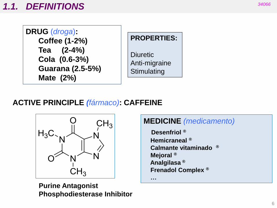
DRUG (droga):
Coffee (1-2%)
Tea (2-4%)
Cola (0.6-3%)
Guarana (2.5-5%)
Mate (2%)
ACTIVE PRINCIPLE (fármaco): CAFFEINE
MEDICINE (medicamento)
Desenfriol ®
Hemicraneal ®
Calmante vitaminado ®
Mejoral ®
Analgilasa ®
Frenadol Complex ®
…
PROPERTIES:
Diuretic
Anti-migraine
Stimulating
Purine Antagonist
Phosphodiesterase Inhibitor
1.1. DEFINITIONS 34066
6

“Medicinal chemistry concerns the discovery, the
development, the identification and the interpretation of the
mode of action of biologically active compounds at a
molecular level. Emphasis is put on drugs, but the interests
of the medicinal chemist are not restricted to drugs, but
include bioactive compounds in general. Medicinal
chemistry is also concerned with the study, identification,
and synthesis of the metabolic products of these drugs and
related compounds”.
DISCOVERY/RATIONAL DESIGN
STRUCTURE-ACTIVITY RELATIONSHIPS (SAR)
SYNTHESIS and DEVELOPMENT
1.1. DEFINITIONS 34066
7
Definition of Medicinal Chemistry provided by a specialized commission of the
IUPAC (IUPAC= International Union of Pure and Applied Chemistry):
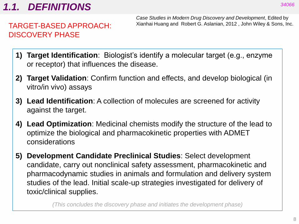
1) Target Identification: Biologist‟s identify a molecular target (e.g., enzyme
or receptor) that influences the disease.
2) Target Validation: Confirm function and effects, and develop biological (in
vitro/in vivo) assays
3) Lead Identification: A collection of molecules are screened for activity
against the target.
4) Lead Optimization: Medicinal chemists modify the structure of the lead to
optimize the biological and pharmacokinetic properties with ADMET
considerations
5) Development Candidate Preclinical Studies: Select development
candidate, carry out nonclinical safety assessment, pharmacokinetic and
pharmacodynamic studies in animals and formulation and delivery system
studies of the lead. Initial scale-up strategies investigated for delivery of
toxic/clinical supplies.
(This concludes the discovery phase and initiates the development phase)
1.1. DEFINITIONS 34066
TARGET-BASED APPROACH:
DISCOVERY PHASE
Case Studies in Modern Drug Discovery and Development, Edited by
Xianhai Huang and Robert G. Aslanian, 2012 , John Wiley & Sons, Inc.
8

DEVELOPMENT
1.1. DEFINITIONS 34066
1) Process and manufacturing chemists work on large scale synthesis, commercial route
synthesis and long-term route synthesis to support toxicological studies. Phase I-IV
clinical trials. Good Manufacturing Practice (GMP) is required in all of these processes.
Patent filing.
2) Investigational New Drugs (IND) applications filed before beginning Phase I clinical trial.
All clinical studies need to follow Good Clinical Practice (GCP) and work with regulatory
authorities.
3) Phase I Clinical Trial: Assess human pharmacokinetic profile, safety, and tolerability in
healthy human beings.
4) Phase II Clinical Trial: Further assess drug safety, dose range, and efficacy studies in a
small number of patients.
5) Phase III Clinical Trial: Further assess drug safety, dose range, and efficacy studies in
large number of patients with multiple trials.
6) New Drug Application (NDA) filed.
7) Drug Approval.
8) Phase IV Clinical Studies (if necessary): Post-marketing event, very large scale clinical
studies to assess long term effect of the drug. Post-approval studies designed to assess
the drug versus competitors, effectiveness, and qualify of life considerations.
9) Drug Life Cycle Management.
Case Studies in Modern Drug Discovery and Development, Edited by
Xianhai Huang and Robert G. Aslanian, 2012 , John Wiley & Sons, Inc.
9

TARGET-BASED DRUG DESIGN AND DEVELOPMENT
a) Identifying the target disease b) Identifying the drug target c) Establishing testing procedures: finding a “hit” compound d) Finding a lead compound e) Establishing Structure-Activity Relationships (SAR) f) Identifying a pharmacophore g) Design: optimising target interactions and pharmaco-kinetic properties i) Toxicological and safety tests j) Chemical development and production of a candidate k) Patenting and regulatory affairs l) Clinical trials
(Medicinal chemists usually work in the stages highlighted in blue)
1.1. DEFINITIONS 34066
G. L. Patrick. An Introduction to Medicinal Chemistry. Oxford
Univ. Press., 5ª Ed. 2013
10

There are three distinct phases in the path of a drug through the body:
The chemical structure affects all these
processes that influence biological activity!
1.1. DEFINITIONS 34066
11
- The Pharmaceutical Phase:
from administration (oral or parenteral) to liberation of the active
principle
(medicinal chemists and pharmaceutical technologists)
- The Pharmacokinetic Phase:
Absorption, Distribution, Metabolism, and Excretion (ADME)
“What the body does to the drug”
(medicinal chemists and biopharmacists)
- The Pharmacodynamic Phase:
Interaction of the drug with the target that generates a biological
response
(medicinal chemists with biochemists and pharmacologists)
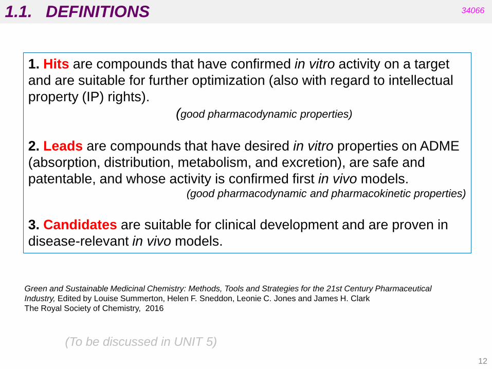
1. Hits are compounds that have confirmed in vitro activity on a target
and are suitable for further optimization (also with regard to intellectual
property (IP) rights).
(good pharmacodynamic properties)
2. Leads are compounds that have desired in vitro properties on ADME
(absorption, distribution, metabolism, and excretion), are safe and
patentable, and whose activity is confirmed first in vivo models. (good pharmacodynamic and pharmacokinetic properties)
3. Candidates are suitable for clinical development and are proven in
disease-relevant in vivo models.
Green and Sustainable Medicinal Chemistry: Methods, Tools and Strategies for the 21st Century Pharmaceutical
Industry, Edited by Louise Summerton, Helen F. Sneddon, Leonie C. Jones and James H. Clark
The Royal Society of Chemistry, 2016
(To be discussed in UNIT 5)
1.1. DEFINITIONS 34066
12
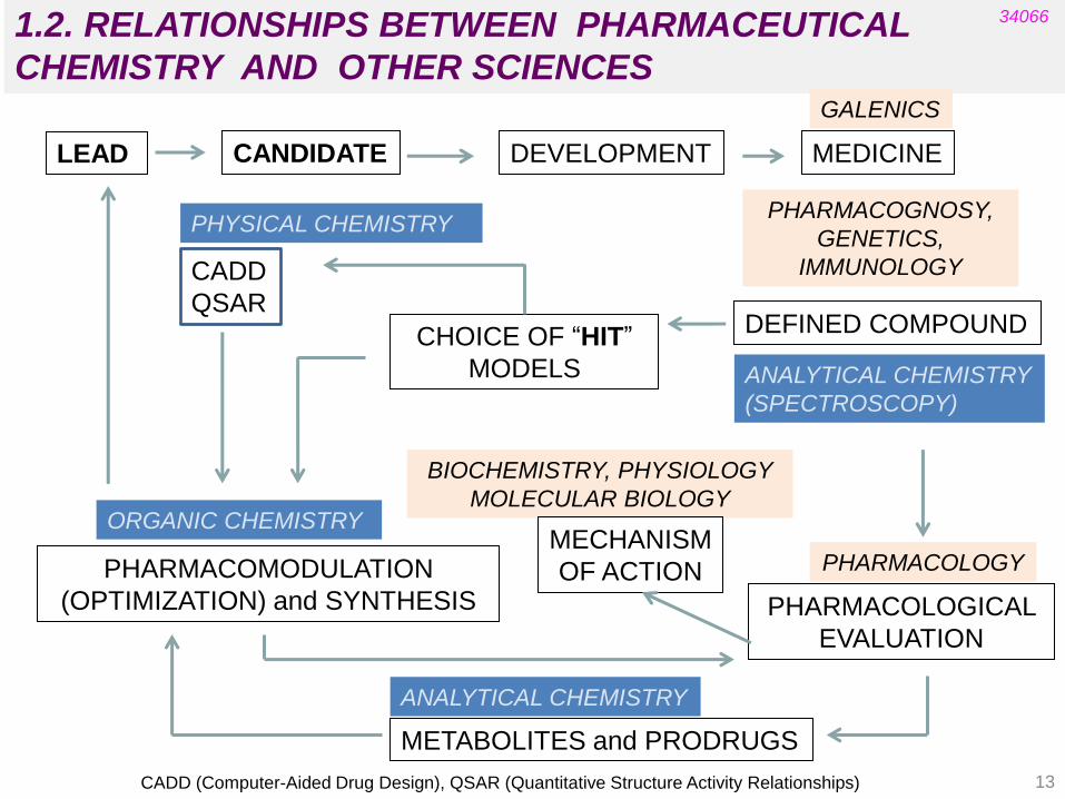
1.2. RELATIONSHIPS BETWEEN PHARMACEUTICAL
CHEMISTRY AND OTHER SCIENCES
LEAD DEVELOPMENT MEDICINE
GALENICS
CADD
QSAR
PHYSICAL CHEMISTRY
CHOICE OF “HIT”
MODELS
DEFINED COMPOUND
PHARMACOGNOSY,
GENETICS,
IMMUNOLOGY
ORGANIC CHEMISTRY
PHARMACOMODULATION
(OPTIMIZATION) and SYNTHESIS
BIOCHEMISTRY, PHYSIOLOGY
MOLECULAR BIOLOGY
MECHANISM
OF ACTION PHARMACOLOGY
PHARMACOLOGICAL
EVALUATION
ANALYTICAL CHEMISTRY
METABOLITES and PRODRUGS
CADD (Computer-Aided Drug Design), QSAR (Quantitative Structure Activity Relationships)
34066
CANDIDATE
ANALYTICAL CHEMISTRY
(SPECTROSCOPY)
13

1.3. CLASSIFICATION OF DRUGS
a. By chemical structure
Drugs with a common skeleton usually show the same biological action and
mechanism of action, e.g. penicillins and cephalosporins, barbiturates and
opioids.
These are called structurally specific drugs.
Sometimes compounds with similar chemical structures have very different effects
in the body (e.g. sulfonamides and steroids). There may also be very different
structures for the same effect (e.g. anaesthetics).
These are structurally nonspecific drugs.
34066
(Patrick‟s) 14
b. By pharmacological effect
Pharmacodynamic agents (e.g. analgesics, antipsychotics, anti-hypertensives
and anti-asthmatics) alter certain biological process in our cellules or systems.
Chemotherapy agents (e.g. antibacterials and antivirals) are used against cancer
or infectious diseases.
This is a useful criterion for determining the full scope of drugs available for a
certain ailment but the drugs included are numerous and highly varied in structure.

1.3. CLASSIFICATION OF DRUGS
c. By target system
Drugs can be classified according to whether they affect a certain system in the
body (e.g. the Central Nervous System).
A system usually has several targets with which drugs could interact (e.g. the
brain, synapses, etc.).
Drugs in each group are likely to be varied in structure due to the different
mechanisms of action involved (anti-cholinergic and anti-adrenergic drugs).
34066
15
d. By target molecule
Some drugs are classified according to the molecular target with which they
interact (anticholinesterases).
This is a more specific classification since it identifies the precise target upon
which the drug acts.
We can expect a structural similarity between the agents involved as well as a
common mechanism of action, though this is not an unbreakable assumption.
(Patrick‟s)

Tema 1: Introducción
a. penicillin
c. antibacterial d. enzyme inhibitor
a. specific
b. chemotherapy
ampicillin
1.3. CLASSIFICATION OF DRUGS 34066
16
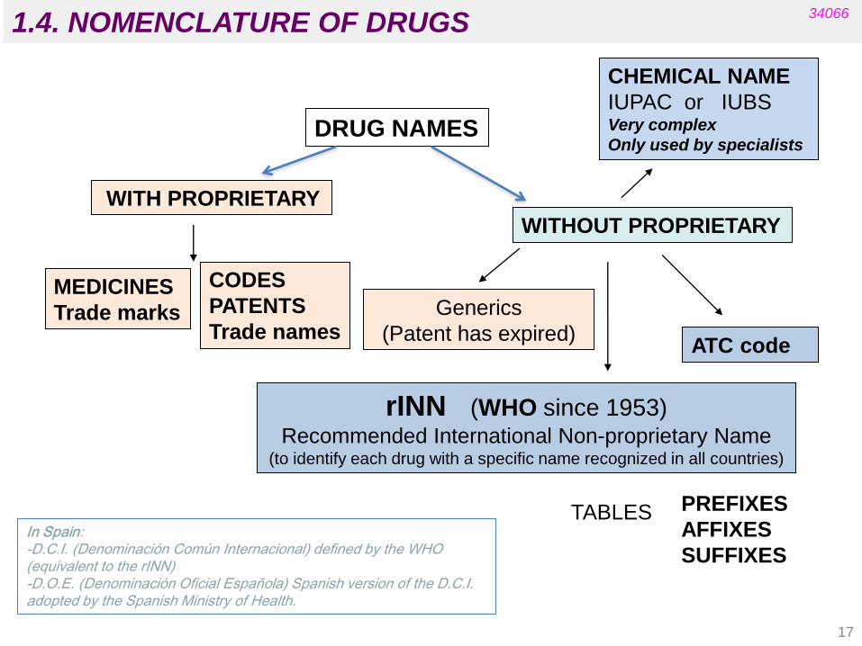
1.4. NOMENCLATURE OF DRUGS
DRUG NAMES
MEDICINES
Trade marks
CHEMICAL NAME
IUPAC or IUBS Very complex
Only used by specialists
WITH PROPRIETARY
WITHOUT PROPRIETARY
Generics
(Patent has expired)
rINN (WHO since 1953) Recommended International Non-proprietary Name
(to identify each drug with a specific name recognized in all countries)
PREFIXES
AFFIXES
SUFFIXES
TABLES
CODES
PATENTS
Trade names
34066
In Spain: -D.C.I. (Denominación Común Internacional) defined by the WHO (equivalent to the rINN) -D.O.E. (Denominación Oficial Española) Spanish version of the D.C.I. adopted by the Spanish Ministry of Health.
ATC code
17

1.4. NOMENCLATURE OF DRUGS
Diazepam (rINN): active principle name
Trade name (medicine): Valium®
34066
IUPAC name:
ATC code:
N05BA01
PAY ATTENTION TO THE CHEMICAL NAME!
It is important to see whether the name and the chemical structure match
18

1.4. NOMENCLATURE OF DRUGS
http://www.who.int/medicines/publications/druginformation/innlists/en/
34066
19
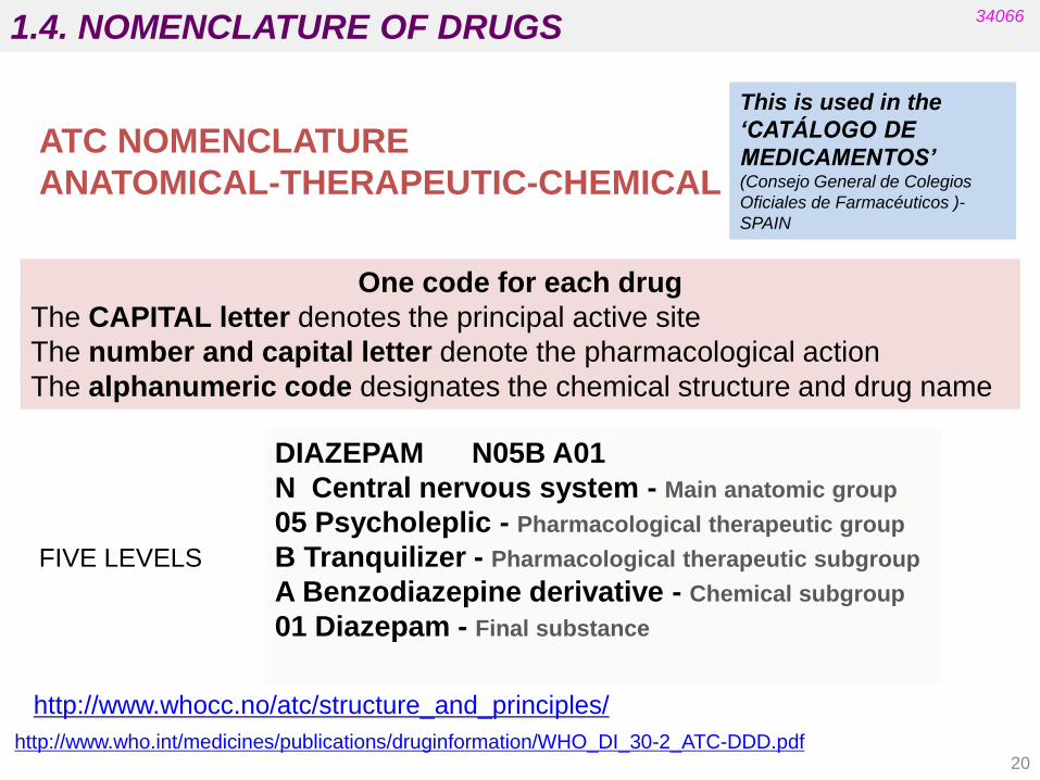
1.4. NOMENCLATURE OF DRUGS
ATC NOMENCLATURE
ANATOMICAL-THERAPEUTIC-CHEMICAL
One code for each drug
The CAPITAL letter denotes the principal active site
The number and capital letter denote the pharmacological action
The alphanumeric code designates the chemical structure and drug name
DIAZEPAM N05B A01
N Central nervous system - Main anatomic group
05 Psycholeplic - Pharmacological therapeutic group
B Tranquilizer - Pharmacological therapeutic subgroup
A Benzodiazepine derivative - Chemical subgroup
01 Diazepam - Final substance
This is used in the
‘CATÁLOGO DE
MEDICAMENTOS’ (Consejo General de Colegios
Oficiales de Farmacéuticos )-
SPAIN
34066
http://www.who.int/medicines/publications/druginformation/WHO_DI_30-2_ATC-DDD.pdf
http://www.whocc.no/atc/structure_and_principles/
FIVE LEVELS
20

The 1st Level (anatomical): indicates the anatomical main group. It consists of a capital
letter denoting the organ or system on which the drug acts. There are 14 main groups.
A DIGESTIVE AND METHABOLIC SYSTEM
B BLOD AND HEMATOPOIETIC ORGANS
C CARDIOVASCULAR SYSTEM
D DERMATOLOGICAL MEDICINES
G SEX HORMONES AND GENITOURINARY
H SYSTEMIC HORMONAL PREPARATIONS, EXCL. SEX HORMONES
J ANTIINFECTIOUS IN GENERAL FOR SYSTEMIC USE
L ANTINEOPLASM AGENTS AND IMUNOMODULATORS
M SKELETAL MUSCLE SYSTEM
N NERVOUS SYSTEM
P ANTIPARASITARY, INSECTICIDE AND REPELLENT PRODUCTS
R RESPIRATORY SYSTEM
S ORGANS OF THE SENSES
V VARIOUS
Tema 1: Introducción 1.4. NOMENCLATURE OF DRUGS
https://en.wikipedia.org/wiki/Anatomical_Therapeutic_Chemical_Classification_System
DIAZEPAM N05B A01
34066
21

1.4. NOMENCLATURE OF DRUGS
The 3rd Level indicates the therapeutic or pharmacologic subgroup and consists of
one capital letter.
N05B Anxiolytics
The 4th Level indicates the chemical subgroup and consists of one capital letter.
N05BA Benzodiazepine
The 5th Level indicates the chemical substance or pharmacological association and
consists of two digits.
The 2nd Level indicates the therapeutic group and consists of two digits.
N05 Psycholeptics
N05BA01 Diazepam
N05BA02
N05BA03
N05BA04
N05BA05
etc……….
34066
DIAZEPAM N05B A01
22
1.5. THE PHARMACEUTICAL INDUSTRY
The Pharmaceutical Industry in Figures (Key Data):
http://www.efpia.eu/uploads/Figures_2015_Final.pdf
https://www.efpia.eu/media/219735/efpia-pharmafigures2017_statisticbroch_v04-
final.pdf
(EFPIA: European Federation of Pharmaceutical Industries and Associations)
34066
Electronic book (UV):
Green and Sustainable Medicinal Chemistry: Methods, Tools and Strategies for the
21st Century Pharmaceutical Industry Edited by Louise Summerton, Helen F. Sneddon, Leonie C. Jones and James H. Clark. The Royal Society of Chemistry
2016
CHAPTER 9 Medicinal Chemistry: How „„Green‟‟ is Our Synthetic Tool Box?
GOOD INTRODUCTION
REFERENCES:
Farmaindustria (Asociación Nacional Empresarial de la Industria Farmacéutica
establecida en España)
http://www.farmaindustria.es/Farma_Public/Farmaindustria/Asociados/index.htm
http://www.pmfarma.es/colaboradores/mercado-mundial/1664-mercado-
farmaceutico-mundial-a-agosto-2014.html
23

1.5. THE PHARMACEUTICAL INDUSTRY 34066
Some Features:
24
It is one of Europe‟s top performing high-technology sectors:
• it directly employs more than 700,000 people (and generates three to
four times more employment indirectly)
• it has the highest added-value per person employed (significantly
higher than the average value for high-tech and manufacturing
industries)
• it is the sector with the highest ratio of R&D investment to net sales.
The pharmaceutical industry aims to turn fundamental research into
innovative treatments that are widely available and accessible to
patients.
It contributes to medical progress by researching, developing and
bringing new medicines that improve health and quality of life for
patients around the world.
The majority of drug discovery approaches in pharmaceutical companies
still focus on target-based research activities.

1.5. THE PHARMACEUTICAL INDUSTRY 34066
• Costly research and development (R&D) is conducted by pharmaceutical
companies to introduce new medicines into the market.
• On average, 12-13 years will elapse between the first synthesis of the new
active substance and its commercialisation.
• In 2012 the cost of researching and developing a new chemical or biological
entity was estimated at € 1,172 million ($ 1,506 million in 2011 dollars).
• On average, only one to two of every 10,000 substances tested in
laboratories will successfully pass all the stages of development required to
become a marketable medicine.
Source: EFPIA
The number of new drugs approved (NDA) had decreased in recent decades:
• In the 1980s roughly 60 new drugs were approved annually.
• On average, FDA has approved 20-30 new drugs per year in the past two
decades.
• Annual approvals in the past five years have been in the range of 40-50 new
drugs (except in 2016). In 2018 a record number has been reached (59).
Expenditures are increasing, thus making the cost per approved drug much
higher.
In recent decades the industry has shifted toward the biological as a drug class.
25
1.5. THE PHARMACEUTICAL INDUSTRY 34066
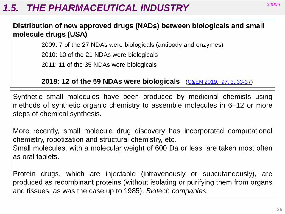
Distribution of new approved drugs (NADs) between biologicals and small
molecule drugs (USA)
2009: 7 of the 27 NDAs were biologicals (antibody and enzymes)
2010: 10 of the 21 NDAs were biologicals
2011: 11 of the 35 NDAs were biologicals
2018: 12 of the 59 NDAs were biologicals (C&EN 2019, 97, 3, 33-37)
Synthetic small molecules have been produced by medicinal chemists using
methods of synthetic organic chemistry to assemble molecules in 6–12 or more
steps of chemical synthesis.
More recently, small molecule drug discovery has incorporated computational
chemistry, robotization and structural chemistry, etc.
Small molecules, with a molecular weight of 600 Da or less, are taken most often
as oral tablets.
Protein drugs, which are injectable (intravenously or subcutaneously), are
produced as recombinant proteins (without isolating or purifying them from organs
and tissues, as was the case up to 1985). Biotech companies.
26

1.5. THE PHARMACEUTICAL INDUSTRY 34066
GENERICS
When intellectual property protection rights have expired, a manufacturer who is
not the inventor of the original product is allowed to produce and market similar
medicines. These are called „generics‟.
An. Quím. 2006, 102 (3), 13-22
A Generic Medicine is developed to be the same as a medicine that has
already been authorised (the „reference medicine‟). It contains the same
active substance as the originator medicine, and it is used at the same
dose to treat the same disease as the reference medicine. However, the
name of the medicine, its appearance (such as colour or shape) and its
packaging can be different from those of the reference medicine.
http://www.medicinesforeurope.com/
European Generics Medicines Association (EGA)
27
1.5. THE PHARMACEUTICAL INDUSTRY 34066
A generic medicine “is any medicine that has the same qualitative
and quantitative composition, active substance and
pharmaceutical form as the originator product and whose
bioequivalence with that product has been demonstrated with
appropriate bioequivalence studies”
Spanish Law of July 2006 on the Guarantees and Rational Use of
Medicines and Sanitary Products (LGURMPS) .
http://www.aeseg.es/en/ Spanish Generic Medicines Association
28
A generic medicine contains the same active medicinal substance as
an originator pharmaceutical product. Because it acts in the same way
in the human body, it is interchangeable with the originator product.
Generic medicines are launched when the originator product‟s patent
has expired.

1.5. THE PHARMACEUTICAL INDUSTRY 34066
http://www.aeseg.es/en/ Spanish Generic Medicines Association
Generic medicines contribute to obtaining considerable savings, and effectively
reduce the cost of medicines from 40 to 60%.
In Spain, the generic medicines market during the last year only represented
20% of the total pharmaceutical market in value and 40% in volume.
Generic medicines in Spain are still a long way from the European average,
which is around 55% in volume.
In general, the market share of generics is significantly higher in newer EU
Member States with historically low levels of intellectual property protection.
http://www.aeseg.es/documentos/
Bioequivalence demonstrates the interchangeability between a
generic medicine and the originator medicine in terms of quality,
security and efficacy. Bioequivalence studies are done to prove that
the generic medicines are equivalent and interchangeable with the
originator product in terms of therapeutic efficacy.
These studies are much less expensive than clinical trials required for
the originator product.
29
2.1. Concept of drug target.
2.2. Drug-target interactions. (Patrick‟s, 5th ed. chapter 1)
2.3. Chemical nature of the targets (proteins,
lipids, nucleic acids and carbohydrates). (Patrick‟s, 5th ed. chapters 2,3, 5 and 6)
2.4. Examples of drugs that interact with the
targets. (Patrick‟s, 5th ed. chapters 4, 7-10)
PHARMACEUTICAL CHEMISTRY
UNIT 2. DRUG TARGETS
34066
(see also Biochemistry I and II and Pharmacology I and II) 30

2.1. CONCEPT OF DRUG TARGET 34066
Specificity is one of a drug‟s most important properties. We want the drug to
perform a specific action (therapeutic, preventive or diagnostic). This is only
possible if the active principle is able to recognise in the body those biomolecules
that are related to the disease and trigger a response. These biomolecules are
known as drug targets.
excretion
DRUGS
Cross membranes
Are deposited in tissues
Bind to the target
pharmacological response
drug metabolism
pharmacokinetic pharmacodynamic
transport
lifetime
31

2.1. DRUG-TARGET INTERACTIONS
The AFFINITY of a drug (D) for a target (T) is a measure of how
strongly the drug binds to the target. It is proportional to the
equilibrium constant of complex formation.
EFFICACY (a) is a measure of the maximum biological effect that a
drug can produce as a result of target binding.
Arïens and Stephenson
Biological response = a[DT]
34066
complex
32
2.1. CONCEPT OF DRUG TARGET 34066
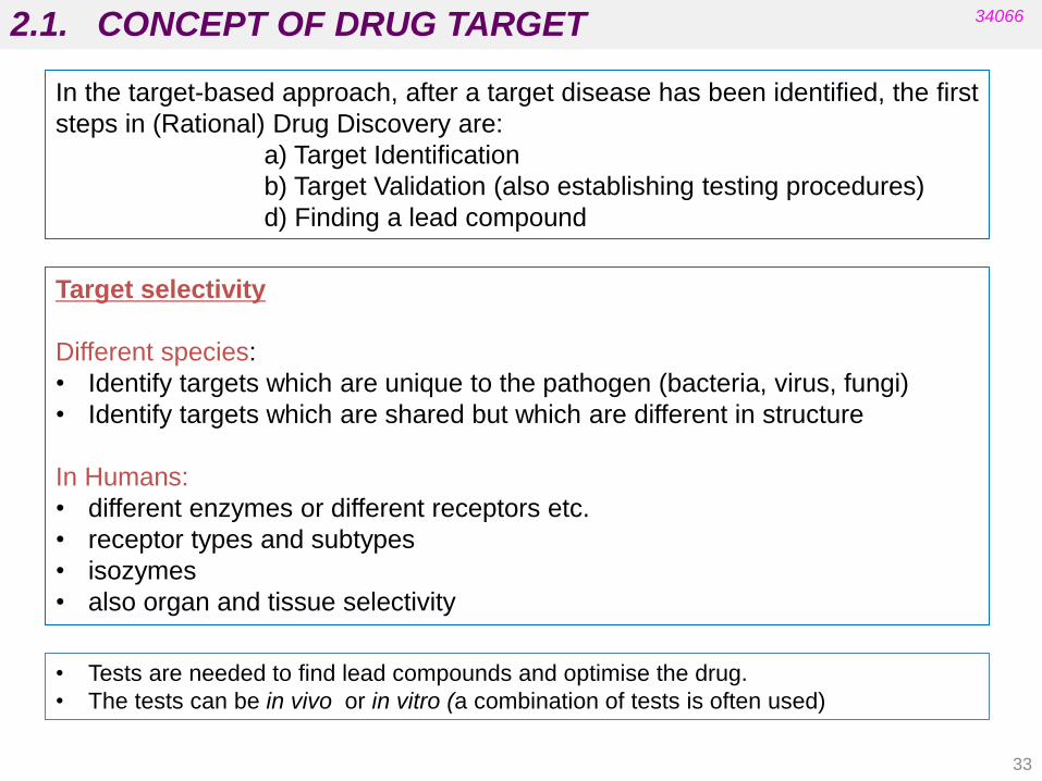
Target selectivity
Different species:
• Identify targets which are unique to the pathogen (bacteria, virus, fungi)
• Identify targets which are shared but which are different in structure
In Humans:
• different enzymes or different receptors etc.
• receptor types and subtypes
• isozymes
• also organ and tissue selectivity
In the target-based approach, after a target disease has been identified, the first
steps in (Rational) Drug Discovery are:
a) Target Identification
b) Target Validation (also establishing testing procedures)
d) Finding a lead compound
• Tests are needed to find lead compounds and optimise the drug.
• The tests can be in vivo or in vitro (a combination of tests is often used)
33

2.1. CONCEPT OF DRUG TARGET 34066
• Are not carried out on animals/humans
Target molecules
Cells Tissues Organs Micro-organisms
• Involve fewer factors and are more suitable for routine testing (easier to
rationalise).
• Measure the interaction of a drug with the target but not the ability of the drug to
reach the target (do not demonstrate a physiological or clinical effect and do not
allow the identification of possible side effects).
In vitro TESTS
• Are carried out on live animals or humans.
• Measure an observed physiological effect (also a drug‟s ability to interact with
its target and its ability to reach that target).
• Possible side effects can be identified.
• Rationalisation could be difficult (many factors involved).
In vivo TESTS
34

2.2. DRUG-TARGET INTERACTIONS 34066
• Drug targets are generally molecules that are much bigger than those of drugs.
• Drugs interact with their targets by binding to binding sites.
• Binding sites are located on the surface of macromolecules and consist typically
of hydrophobic hollows that are recognised by the drug.
• Binding interactions involve intermolecular bonds between drug and target (or
between targets and endogenous molecules or organic compounds in general).
• In the body, most drugs are in equilibrium (bound and unbound to their target).
• The functional groups on the drug that are involved in binding interactions are
called binding groups.
• Specific regions within the binding site that are involved in binding interactions
are called binding regions.
Drug targets are molecular structures that undergo a specific interaction with a
drug (drug is a compound administered to treat, prevent or diagnose a disease). A
change in the behaviour or properties of these targets can be detected as a
consequence of this interaction.
(Patrick‟s 5th ed., chapter 1)
35

2.2. DRUG-TARGET INTERACTIONS 34066
D G = D H - TD S (Remember Organic Chemistry)
Maximum energy available at room temperature 80 KJ/mol
Energy required to produce a conformational change 10 KJ/mol
Bond energies Bond type Energy (KJ/mol) Covalent 150-400 Ionic 20-25 Hydrogen bond 5-30 Ion-dipole 5-30 Dipole-dipole 5-30 Van der Waals 2-4 Hydrophobic 4 Charge transfer 5-30
COMMON INTERMOLECULAR FORCES
36

2.2. DRUG-TARGET INTERACTIONS 34066
Covalent bonds
• Are the strongest bonds (150-400 kJ mol-1).
• Are considered “irreversible” interactions (the target is modified by the drug).
• Are not very common (they are found in some drugs used in chemotherapy).
• Are often related to toxicological effects.
• Usually involve a nucleophilic centre in proteins or nucleic acids that react with
an electrophilic centre present in the drug.
Nucleophilic centres in proteins:
• Thiol group of cysteine
• S-atoms of methionine
• Primary amino-groups (e.g.: lysine, arginine)
• N-atom in the imidazol ring of histidine
• O-atom in serine
Nucleophilic centres in nucleic acids:
• Primary amino-groups of purine bases (e.g. adenine, guanine)
• N-atoms in rings of purine and pyrimidine bases (e.g. N7 of guanine)
• O-atoms of purine and pyrimidine bases (e.g. O6 of guanine)
• Phosphate O-atom (P=O)
Measurement and Estimation of Electrophilic Reactivity for Predictive Toxicology
J. A. H. Schwobel, Y. K. Koleva, S. J. Enoch, F. Bajot, M. Hewitt, J. C. Madden, D. W. Roberts, T. W. Schultz, and M. T. D.
Cronin, Chem. Rev. 2011, 111, 2562–2596 37

2.2. DRUG-TARGET INTERACTIONS 34066
Common reaction mechanisms in the formation of covalent bonds
Measurement and Estimation of Electrophilic Reactivity for Predictive Toxicology
J. A. H. Schwobel, Y. K. Koleva, S. J. Enoch, F. Bajot, M. Hewitt, J. C. Madden, D. W. Roberts, T. W. Schultz, and M. T. D.
Cronin*, Chem. Rev. 2011, 111, 2562–2596
• SN1 (alkyl –X, X = leaving group) Protein-Nu-R (alkyl)
• SN2 (alkyl –X, X = leaving group) Protein-Nu-R (alkyl)
• Acylation (R-COX, X = leaving group) Protein-Nu-COR (acyl)
• Imine (Schiff Base)
Formation (RCHO) Protein-N=CH-R
• Michael (Conjugate) (C=C-X) Protein-Nu-C –C- X
Addition (X = electron withdrawing group -CHO, -COR)
• SNAr (Ar –X, X = leaving group) Protein-Nu - Ar
(Ar: only if electron withdrawing groups are present NO2, CN,…)
Functional group present in drugs Modified protein
38
2.2. DRUG-TARGET INTERACTIONS
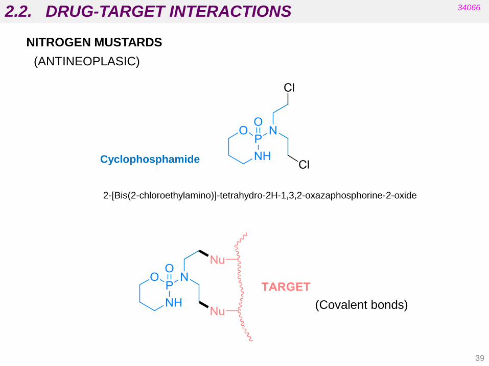
NITROGEN MUSTARDS
(ANTINEOPLASIC)
34066
Cyclophosphamide
2-[Bis(2-chloroethylamino)]-tetrahydro-2H-1,3,2-oxazaphosphorine-2-oxide
(Covalent bonds)
39
2.2. DRUG-TARGET INTERACTIONS 34066
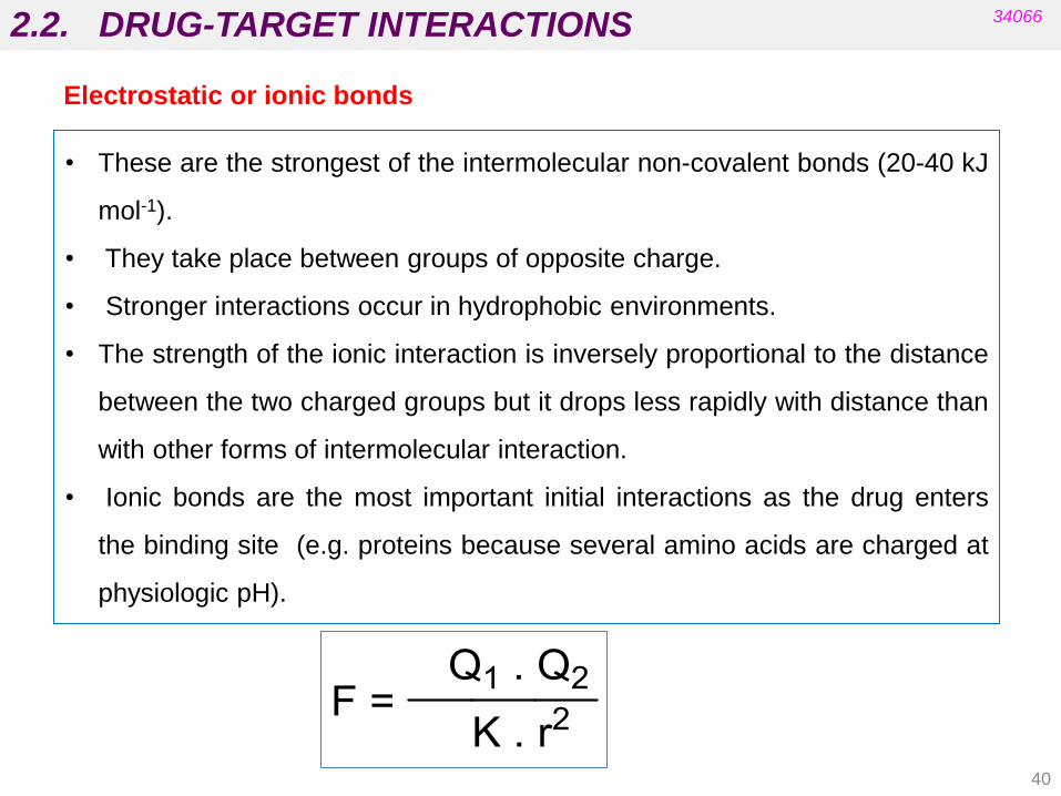
• These are the strongest of the intermolecular non-covalent bonds (20-40 kJ
mol-1).
• They take place between groups of opposite charge.
• Stronger interactions occur in hydrophobic environments.
• The strength of the ionic interaction is inversely proportional to the distance
between the two charged groups but it drops less rapidly with distance than
with other forms of intermolecular interaction.
• Ionic bonds are the most important initial interactions as the drug enters
the binding site (e.g. proteins because several amino acids are charged at
physiologic pH).
40
Electrostatic or ionic bonds

2.2. DRUG-TARGET INTERACTIONS 34066
Electrostatic or ionic bonds
Charged Residues
Arginine
(Arg)
Glutamic Acid
(Glu)
Lysine
(Lys)
Aspartic Acid
(Asp)
41
2.2. DRUG-TARGET INTERACTIONS 34066
Hydrogen bonds
• These bonds vary in strength.
• They are weaker than electrostatic interactions but stronger than van der Waals
interactions.
• A hydrogen bond takes place between an electron-deficient hydrogen (usually
attached to a heteroatom (O or N) and an electron-rich heteroatom (N or O).
•The electron-deficient hydrogen is called a hydrogen bond donor (HBD).
• The electron-rich heteroatom is called a hydrogen bond acceptor (HBA).
• The interaction is directional and involves orbitals.
• Optimum orientation is where the X-H bond points directly to the lone pair on Y
such that the angle between X, H and Y is 180o.
42

2.2. DRUG-TARGET INTERACTIONS 34066
• Examples of strong hydrogen bond acceptors:
- COO-, PO4-, -NR3
• Examples of moderate hydrogen bond acceptors:
- Oxygen atom in carbonyl groups:
carboxylic acids, amides, ketones, esters
- Oxygen atom in ethers, alcohols (phenols)
• Examples of poor hydrogen bond acceptors:
- S, F, Cl, aromatic ring, nitrogen in amides, aromatic amines
• Example of good hydrogen bond donors:
- ammonium ions when a H atom is present (HNR3+)
See Appendix 8, Patrick’s 5th Ed: Hydrogen Bonding Interactions
Hydrogen bonds
43
2.2. DRUG-TARGET INTERACTIONS 34066
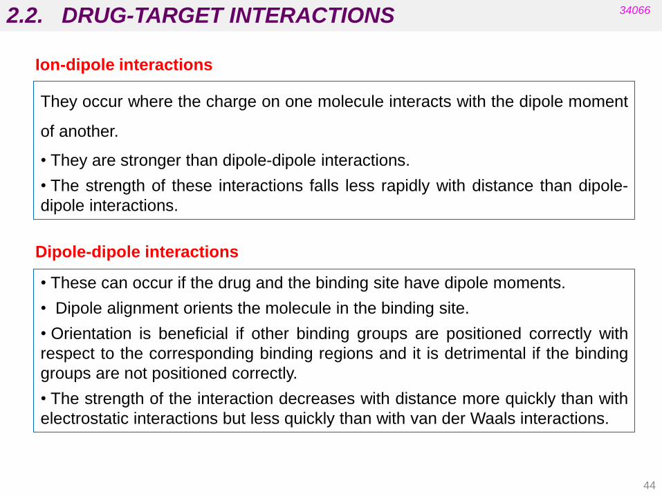
They occur where the charge on one molecule interacts with the dipole moment
of another.
• They are stronger than dipole-dipole interactions.
• The strength of these interactions falls less rapidly with distance than dipole-
dipole interactions.
Dipole-dipole interactions
• These can occur if the drug and the binding site have dipole moments.
• Dipole alignment orients the molecule in the binding site.
• Orientation is beneficial if other binding groups are positioned correctly with
respect to the corresponding binding regions and it is detrimental if the binding
groups are not positioned correctly.
• The strength of the interaction decreases with distance more quickly than with
electrostatic interactions but less quickly than with van der Waals interactions.
44
Ion-dipole interactions

2.2. DRUG-TARGET INTERACTIONS 34066
They occur when the charge on one molecule induces a dipole on another (e.g.
between a quaternary ammonium ion and an aromatic ring).
Van der Waals interactions
• These are very weak interactions (2-4 kJ mol-1) but the overall contribution of
van der Waals interactions can be crucial to binding.
• They occur between hydrophobic regions of the drug and the target because
transient areas of high and low electron densities cause temporary dipoles.
• Interactions drop off rapidly with distance (the drug must be close to the binding
region for interactions to occur).
45
Ion-induced dipole interactions
2.2. DRUG-TARGET INTERACTIONS 34066
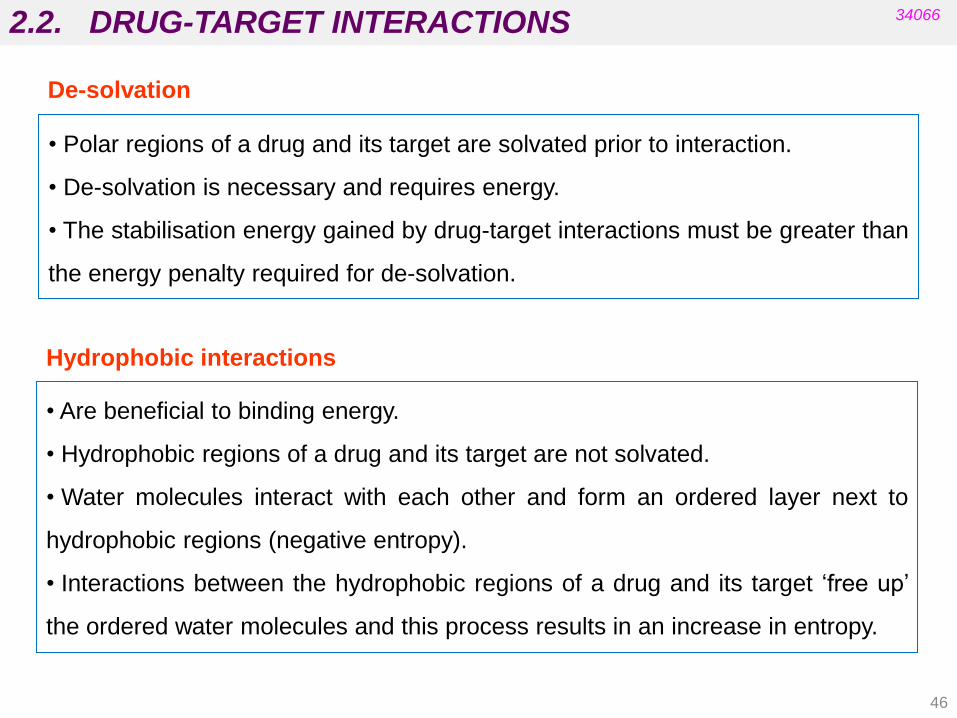
De-solvation
• Polar regions of a drug and its target are solvated prior to interaction.
• De-solvation is necessary and requires energy.
• The stabilisation energy gained by drug-target interactions must be greater than
the energy penalty required for de-solvation.
Hydrophobic interactions
• Are beneficial to binding energy.
• Hydrophobic regions of a drug and its target are not solvated.
• Water molecules interact with each other and form an ordered layer next to
hydrophobic regions (negative entropy).
• Interactions between the hydrophobic regions of a drug and its target „free up‟
the ordered water molecules and this process results in an increase in entropy.
46

2.2. DRUG-TARGET INTERACTIONS 34066
HOMO-LUMO interaction
Ligand metal
• Electrostatic interaction by π orbital overlapping between two molecules
(donor and acceptor).
• Most common examples: electron-rich aromatic ring (methoxybenzene) and
electron-poor aromatic ring (nitrobenzene). E.g. in nature: noradrenaline and
ATP.
• Interaction between a metallic cation and a ligand
atom or functional group acting as electron-donor
(e.g. active site in carbonic anhydrase).
47
Charge Transfer
Coordination

A) Proteins
Structural proteins
Carrier (transport) proteins
Enzymes
Receptors (Patrick’s, 5th ed. chapters 2, 3, 5 and 6)
B) Nucleic acids: DNA and RNA
C) Lipids Membrane lipids
D) Carbohydrates in cell surface
Antigens and recognition molecules
2.3. CHEMICAL NATURE OF DRUG TARGETS 34066
These targets have different locations in cells. Many of them are located in membranes.
48

2.3. CHEMICAL NATURE OF DRUG TARGETS 34066
Cell Membrane
• The cell membrane is made up of a phospholipid bilayer.
• The polar head groups interact with water at the inner and outer surfaces of the
membrane.
•The hydrophobic tails are hidden from the aqueous media (they interact with
each other by van der Waals interactions).
•The cell membrane provides a hydrophobic barrier around the cell, preventing
the passage of water and polar molecules.
• Proteins (ion channels, receptors, enzymes and transport proteins) are
embedded in the cell membrane.
Remember:
49

2.3. CHEMICAL NATURE OF DRUG TARGETS 34066
• Amino acids are the building blocks for proteins.
• Each amino acid has an identical head group.
• Amino acids are chiral molecules (except Gly, R=H).
• Only L-amino acids are present in human biochemistry.
• The L-amino acids are S-enantiomers (except Cys; R = CH2SH).
Remember:
Proteins
50
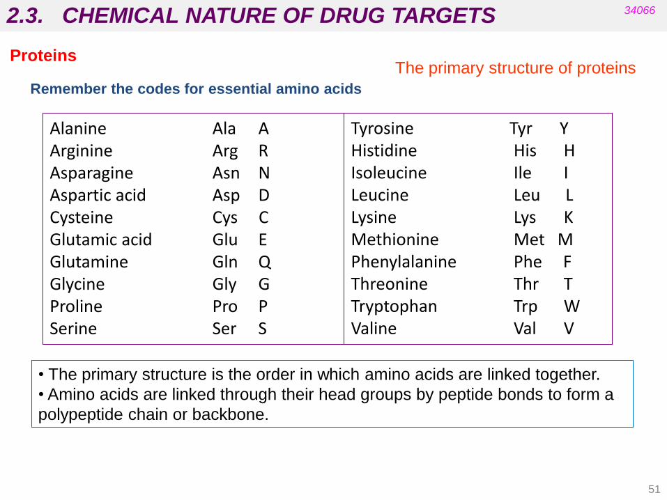
Alanine Ala A Arginine Arg R Asparagine Asn N Aspartic acid Asp D Cysteine Cys C Glutamic acid Glu E Glutamine Gln Q Glycine Gly G Proline Pro P Serine Ser S
Tyrosine Tyr Y Histidine His H Isoleucine Ile I Leucine Leu L Lysine Lys K Methionine Met M Phenylalanine Phe F Threonine Thr T Tryptophan Trp W Valine Val V
Remember the codes for essential amino acids
2.3. CHEMICAL NATURE OF DRUG TARGETS 34066
The primary structure of proteins
• The primary structure is the order in which amino acids are linked together.
• Amino acids are linked through their head groups by peptide bonds to form a
polypeptide chain or backbone.
Proteins
51
2.3. CHEMICAL NATURE OF DRUG TARGETS 34066
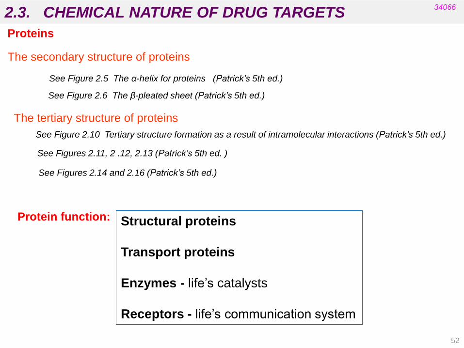
The secondary structure of proteins
See Figure 2.5 The α-helix for proteins (Patrick’s 5th ed.)
See Figure 2.6 The β-pleated sheet (Patrick’s 5th ed.)
Proteins
The tertiary structure of proteins
See Figure 2.10 Tertiary structure formation as a result of intramolecular interactions (Patrick’s 5th ed.)
See Figures 2.11, 2 .12, 2.13 (Patrick’s 5th ed. )
See Figures 2.14 and 2.16 (Patrick’s 5th ed.)
52
Protein function: Structural proteins
Transport proteins
Enzymes - life‟s catalysts
Receptors - life‟s communication system

2.3. CHEMICAL NATURE OF DRUG TARGETS 34066
They are globular proteins acting as the body‟s catalysts:
• Lower the activation energy of a reaction (but ΔG remains the same).
• Speed up time for reaction to reach equilibrium.
• Provide a reaction surface (the active site) and a suitable environment
(hydrophobic).
• Bring reactants together and position them correctly for reaction.
• Stabilise the transition state with intermolecular bonds.
• Weaken bonds in the reactants and provide acid/base catalysis or nucleophilic
groups
Protein
Enzymes
Remember:
53

2.3. CHEMICAL NATURE OF DRUG TARGETS 34066
• Histidine
Acid/base catalysis
Nucleophilic residues
Non-ionised.
Acts as a basic catalyst
Ionised (stabilised by resonance).
Acts as an acid catalyst
Protein
Enzymes
L- Serine L- Cysteine
54

2.3. CHEMICAL NATURE OF DRUG TARGETS 34066
The Active Site
• Accepts reactants (substrates and cofactors).
• Is a hydrophobic hollow or cleft on the enzyme surface.
• Contains amino acids which bind reactants and participate in the
enzyme-catalysed reaction.
Induced fit:
The active site changes shape to maximise intermolecular bonding
Protein
Enzymes
Overall Process of Enzyme Catalysis
Binding interactions must be strong enough to hold the substrate long
enough for the reaction to occur, but they must be weak enough to
allow the product to depart.
55

2.3. CHEMICAL NATURE OF DRUG TARGETS 34066
Protein
Enzymes
Enzymes with allosteric sites are often at the start of a biosynthetic pathway.
• The enzyme is controlled by the final product of the pathway.
• The final product binds to the allosteric site and switches off the enzyme.
Regulation of Enzymes
• Designing molecules with stronger binding interactions can result in enzyme
inhibitors which block the active site (competitive inhibitor).
• Some compounds are able to modify the activity of an enzyme by binding to an
allosteric site. These are allosteric inhibitors (not competitive).
56

2.3. CHEMICAL NATURE OF DRUG TARGETS 34066
Protein
Receptors Structure and function
• Are globular proteins located mainly in the cell membrane.
• Receive messages from chemical messengers from other cells and
transmit a message into the cell leading to a cellular effect.
There are specific receptors for different chemical messengers and each cell
has a range of receptors in the cell membrane that make it responsive to
different chemical messengers.
Chemical messengers are able to „switch on‟ receptors without undergoing a
reaction:
• Neurotransmitters are released from the end of a neuron to bind with a
receptor on a target cell, such as a muscle cell or another neuron. They are
usually short-lived and are responsible for messages between individual
cells.
• Hormones are released from a cell or a gland which travels some distance
throughout the body to bind with receptors on target cells.
57

2.3. CHEMICAL NATURE OF DRUG TARGETS 34066
Mechanism
• Receptors contain a binding site that is recognised by the chemical messenger.
• The binding of the messenger involves intermolecular bonds and it results in an
induced fit of the receptor protein.
• This change in the receptor shape results in a „domino‟ effect known as Signal
Transduction and leads to a chemical signal being received inside the cell.
• The chemical messenger does not enter the cell and is not permanently bound.
• It departs the receptor unchanged.
Protein
Receptors
58
2.3. CHEMICAL NATURE OF DRUG TARGETS 34066
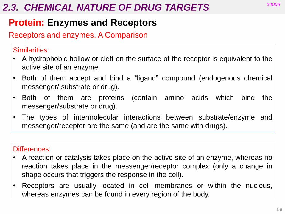
Protein: Enzymes and Receptors
Receptors and enzymes. A Comparison
Similarities:
• A hydrophobic hollow or cleft on the surface of the receptor is equivalent to the
active site of an enzyme.
• Both of them accept and bind a “ligand” compound (endogenous chemical
messenger/ substrate or drug).
• Both of them are proteins (contain amino acids which bind the
messenger/substrate or drug).
• The types of intermolecular interactions between substrate/enzyme and
messenger/receptor are the same (and are the same with drugs).
Differences:
• A reaction or catalysis takes place on the active site of an enzyme, whereas no
reaction takes place in the messenger/receptor complex (only a change in
shape occurs that triggers the response in the cell).
• Receptors are usually located in cell membranes or within the nucleus,
whereas enzymes can be found in every region of the body.
59
2.3. CHEMICAL NATURE OF DRUG TARGETS 34066
Receptor Superfamilies
• ION CHANNEL RECEPTORS
• G-PROTEIN COUPLED RECEPTORS
• KINASE LINKED RECEPTOR
• INTRACELLULAR RECEPTORS
(to be developed in Pharmacology I)
• Binding interactions in receptors must be strong enough to hold the
messenger and long enough for signal transduction to take place.
• Interactions must be weak enough to allow the messenger to depart.
• Designing molecules with stronger binding interactions can result in drugs that
block the binding site. These are called competitive antagonists (drugs which
bind to a receptor without activating it) and are the opposite of agonists
(drugs which produce the same response on a receptor as the natural
messenger does).
These concepts will be developed later (see Unit 5)
Protein
Receptors
60

2.3. CHEMICAL NATURE OF DRUG TARGETS 34066
Nucleic Acids /DNA
Deoxyadenosine
phosphate
Deoxythymidine
phosphate
Deoxyguanosine
phosphate Deoxycytidine
phosphate
Building blocks (nucleotide): Phosphate + sugar + nucleic acid (purine or pyrimine base)
Primary structure
nucleoside
61

2.3. CHEMICAL NATURE OF DRUG TARGETS
• The sugar phosphate backbone is ionised and faces outward because of
favourable interactions with water.
• Nucleic acid bases point inward and pair up.
• Chains are complementary.
• Purine pairs with pyrimidine (constant diameter to helix) A-T or G-C.
• Base pairs are stacked (Van der Waals interactions between pairs).
34066
Nucleic Acids /DNA
Secondary structure
See Fig 6.4. The secondary structure of DNA (Patrick’s 5th ed. )
62

2.3. CHEMICAL NATURE OF DRUG TARGETS 34066
Tertiary structure
• The double helix coils into a 3D shape.
• The double helix has to unravel during replication and unravelling leads
to strain.
• The strain is relieved by enzyme-catalysed cutting and repair of the
DNA chain (quinolone antibacterial agents inhibit this enzyme).
• Topoisomerase II relieves the strain in the DNA helix by temporarily
cleaving the DNA chain and crossing an intact strand through the broken
strand.
•Tyrosine residues in the enzyme form covalent bonds to DNA.
Topoisomerase II
Nucleic Acids /DNA
63
2.3. CHEMICAL NATURE OF DRUG TARGETS 34066
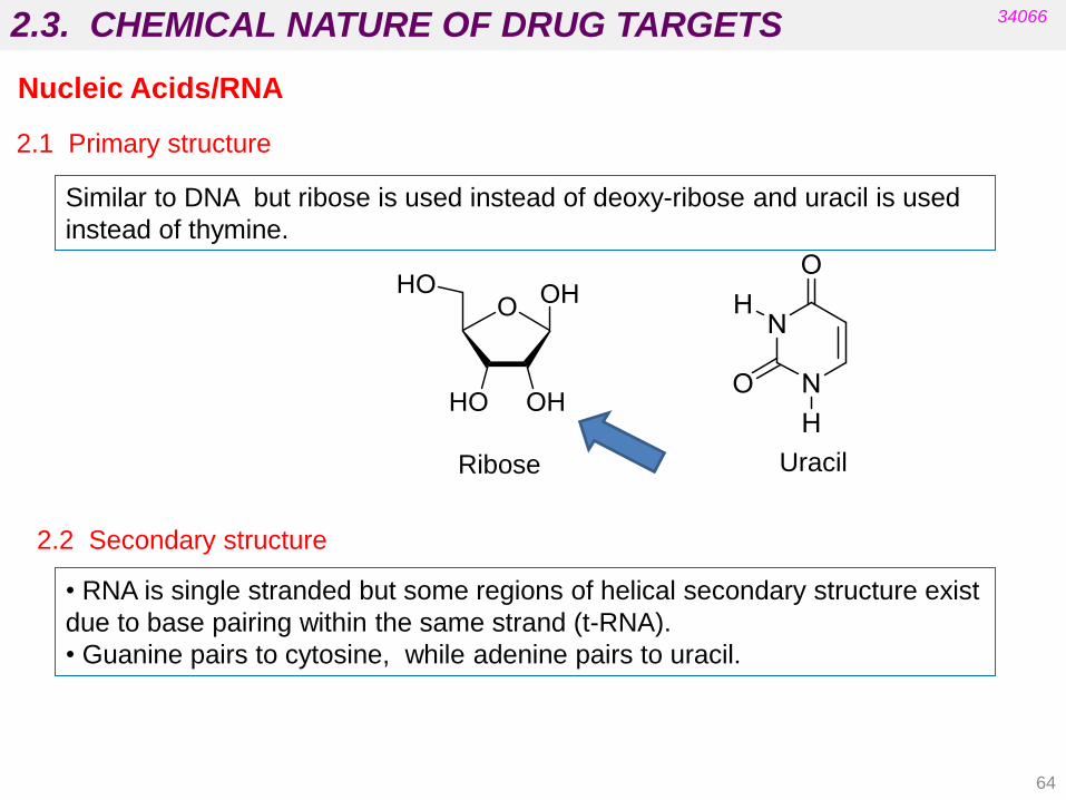
2.1 Primary structure
Similar to DNA but ribose is used instead of deoxy-ribose and uracil is used
instead of thymine.
Uracil Ribose
2.2 Secondary structure
• RNA is single stranded but some regions of helical secondary structure exist
due to base pairing within the same strand (t-RNA).
• Guanine pairs to cytosine, while adenine pairs to uracil.
Nucleic Acids/RNA
64

2.3. CHEMICAL NATURE OF THE TARGETS 34066
Lipids in membranes
Polar head groups and fatty acids can be varied (ethanolamine, inositol, serine
instead of choline, etc.).
Polar heads
Fatty acids
Phosphoglycerides
65

2.3. CHEMICAL NATURE OF THE TARGETS 34066
Lipids in membranes
sphingosine
ceramide
sphingomyelin
glycolipids
Sphingolipids
66

2.3. CHEMICAL NATURE OF THE TARGETS 34066
Carbohydrates
See also Figure 10.2.2. Cellulose, where glucosyl units are linked β-1,4. (Patrick’s 5th ed).
• Carbohydrates play an important role in cell recognition, regulation and growth.
• They are potential targets for the treatment of bacterial and viral infections,
cancer and autoimmune diseases (they act as antigens).
67

2.4. EXAMPLES OF DRUGS THAT INTERACT WITH THESE TARGETS
A) Proteins as drug targets
Structural proteins (tubulin) (Patrick’s 5th Ed. Chapter 10)
Carrier (transport) proteins (Patrick’s 5th Ed. Chapter 10)
Enzymes (Patrick’s 5th Ed. Chapter 7)
Receptors (Patrick’s 5th Ed. Chapter 8)
(More enzyme inhibitors are included in Pharmaceutical Chemistry II)
(Examples of agonists and antagonists are also included in Pharmaceutical Chemistry II)
B) Nucleic acids as drug targets (Patrick’s 5th Ed. Chapter 9)
C) Lipids (Patrick’s 5th Ed. Chapter 10)
D) Carbohydrates (Patrick’s 5th Ed. Chapter 10)
See UNIT 5
34066
68

2.4. EXAMPLES OF DRUGS THAT INTERACT WITH THESE TARGETS 34066
PROTEINS:
Agents blocking transport proteins
• Prevent the re-uptake of neurotransmitters (e.g. dopamine, serotonin
and noradrenaline).
• Result in increased levels of affected neurotransmitters.
• Causes euphoric effects (reuptake inhibitor for
dopamine in CNS) and suppresses hunger
(reuptake inhibitor of noradrenaline in the
peripheral system).
cocaine
Selective serotonin reuptake inhibitor (SSRI)
used as an antidepressant.
fluoxetine
(Prozac®)
(Patrick’s 5th Ed. Chapter 10)
69
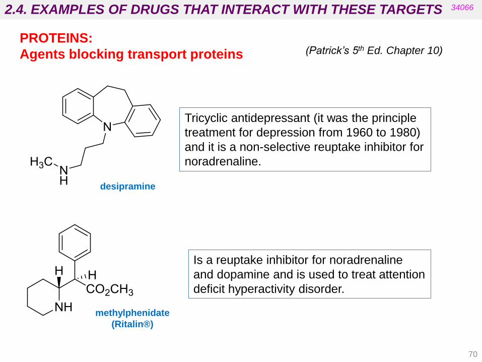
2.4. EXAMPLES OF DRUGS THAT INTERACT WITH THESE TARGETS 34066
Tricyclic antidepressant (it was the principle
treatment for depression from 1960 to 1980)
and it is a non-selective reuptake inhibitor for
noradrenaline.
Is a reuptake inhibitor for noradrenaline
and dopamine and is used to treat attention
deficit hyperactivity disorder.
methylphenidate
(Ritalin®)
PROTEINS:
Agents blocking transport proteins
desipramine
(Patrick’s 5th Ed. Chapter 10)
70

2.4. EXAMPLES OF DRUGS THAT INTERACT WITH THESE TARGETS
71
34066
Drugs acting on nucleic acids
1. Intercalating agents
2. Topoisomerase poisons (non-intercalating)
3. Alkylating agents
4. Metallating agents
5. Chain cutters and chain terminators
Classification based on the mechanism of action:
(Patrick’s 5th Ed. Chapter 9)

2.4. EXAMPLES OF DRUGS THAT INTERACT WITH THESE TARGETS 34066
Drugs acting on DNA
Intercalating agents
Mechanism of action
• Intercalating agents contain planar aromatic or heteroaromatic ring systems
that slip between the layers of nucleic acid pairs and disrupt the shape of the
helix.
• Intercalation prevents replication and transcription and can inhibit
topoisomerases.
•Preference is often shown for the minor or major groove.
(Patrick’s 5th Ed. Chapter 9)
72
2.4. EXAMPLES OF DRUGS THAT INTERACT WITH THESE TARGETS
73
34066
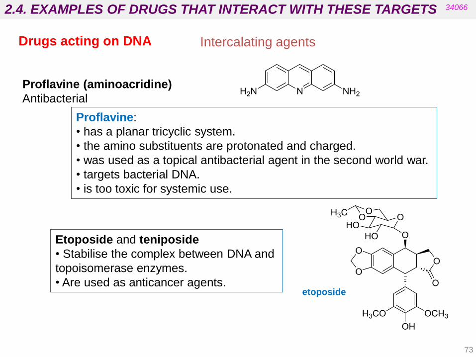
Drugs acting on DNA
Proflavine:
• has a planar tricyclic system.
• the amino substituents are protonated and charged.
• was used as a topical antibacterial agent in the second world war.
• targets bacterial DNA.
• is too toxic for systemic use.
Proflavine (aminoacridine)
Antibacterial
Intercalating agents
Etoposide and teniposide
• Stabilise the complex between DNA and
topoisomerase enzymes.
• Are used as anticancer agents. etoposide

2.4. EXAMPLES OF DRUGS THAT INTERACT WITH THESE TARGETS
74
34066
Topoisomerase poisons (non-intercalating)
Quinolones and fluoroquinolones
• Four drug molecules are stacked in the bound complex.
• They are bound to DNA and enzyme by hydrogen and ionic bonds.
See also Fig 9.6. Complex formed between DNA, the topoisomerase enzyme and fluoroquinolones. (Patrick’s 5th ed.)
ciprofloxacin
Drugs acting on DNA

2.4. EXAMPLES OF DRUGS THAT INTERACT WITH THESE TARGETS
75
34066
Alkylating and metallating agents
• Contain highly electrophilic groups and form covalent bonds to
nucleophilic groups in DNA (7-N of guanine).
• Prevent replication and transcription and are useful anticancer agents.
• Have toxic side effects (e.g. alkylation of proteins).
• Can cause inter-strand and intra-strand cross-linking (in double helix) if
two electrophilic groups are present
• The alkylation of nucleic acid bases can result in miscoding.
Examples:
Nitrogen mustards
Nitrosoureas
Methanesulfonates
Cisplatin
(Patrick’s 5th Ed. Chapter 9) Drugs acting on DNA
2.4. EXAMPLES OF DRUGS THAT INTERACT WITH THESE TARGETS
76
34066
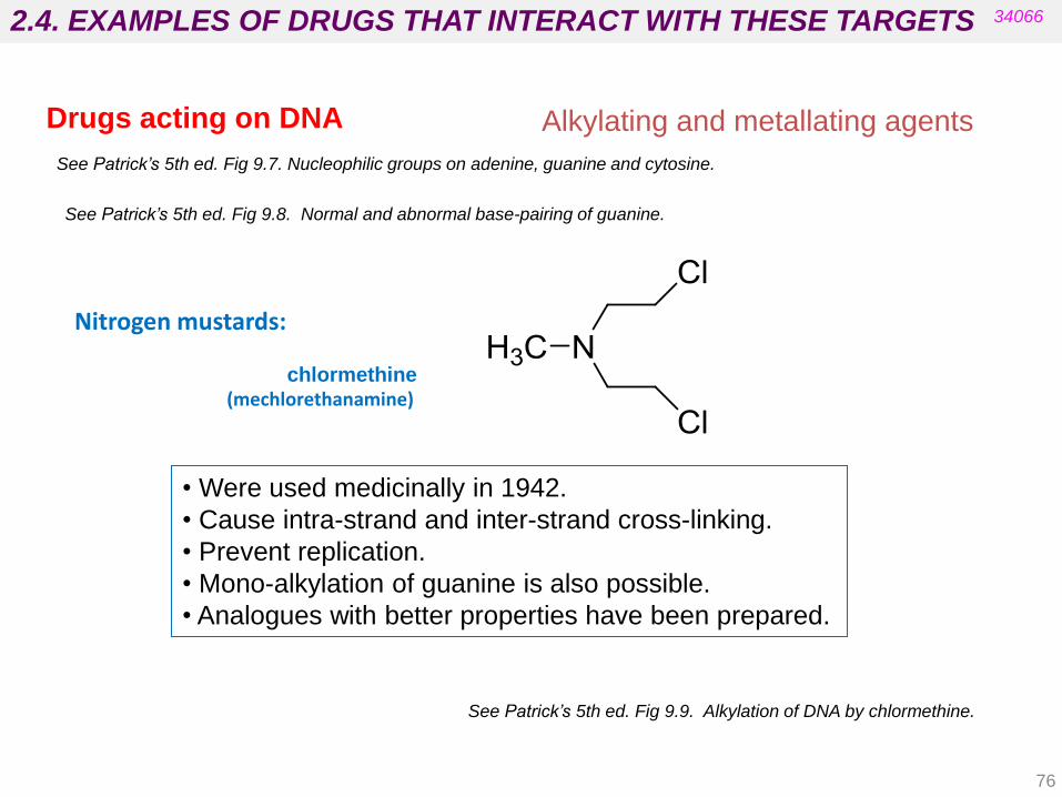
Alkylating and metallating agents
See Patrick’s 5th ed. Fig 9.7. Nucleophilic groups on adenine, guanine and cytosine.
See Patrick’s 5th ed. Fig 9.8. Normal and abnormal base-pairing of guanine.
Nitrogen mustards:
chlormethine (mechlorethanamine)
• Were used medicinally in 1942.
• Cause intra-strand and inter-strand cross-linking.
• Prevent replication.
• Mono-alkylation of guanine is also possible.
• Analogues with better properties have been prepared.
See Patrick’s 5th ed. Fig 9.9. Alkylation of DNA by chlormethine.
Drugs acting on DNA
2.4. EXAMPLES OF DRUGS THAT INTERACT WITH THESE TARGETS
77
34066
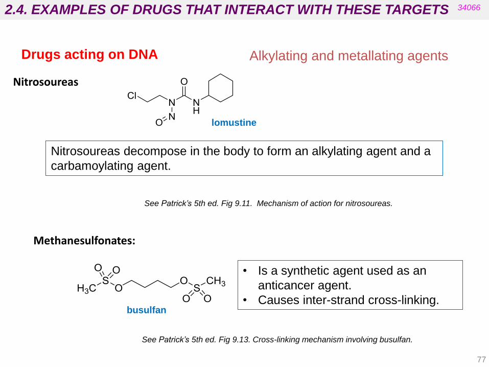
Nitrosoureas
Nitrosoureas decompose in the body to form an alkylating agent and a
carbamoylating agent.
See Patrick’s 5th ed. Fig 9.11. Mechanism of action for nitrosoureas.
lomustine
• Is a synthetic agent used as an
anticancer agent.
• Causes inter-strand cross-linking.
Methanesulfonates:
See Patrick’s 5th ed. Fig 9.13. Cross-linking mechanism involving busulfan.
busulfan
Drugs acting on DNA Alkylating and metallating agents

2.4. EXAMPLES OF DRUGS THAT INTERACT WITH THESE TARGETS 34066
• Is a neutral inactive molecule that acts as a “prodrug”.
• Platinum is covalently linked to chloro substituents and ammonia molecules
act as ligands.
• Is activated in cells with low chloride ion concentration where chloro
substituents are replaced with neutral water ligands.
• Produces positively charged species.
• Binds to DNA in regions rich in guanine units.
• Links intrastrand rather than interstrand.
• Localised unwinding of DNA double helix.
• Inhibits transcription.
Cisplatin
See Patrick’s 5th ed. Fig 9.14. Activation of cisplatin and intrastrand cross-linking of DNA.
Drugs acting on DNA Alkylating and metallating agents
78
2.4. EXAMPLES OF DRUGS THAT INTERACT WITH THESE TARGETS
79
34066
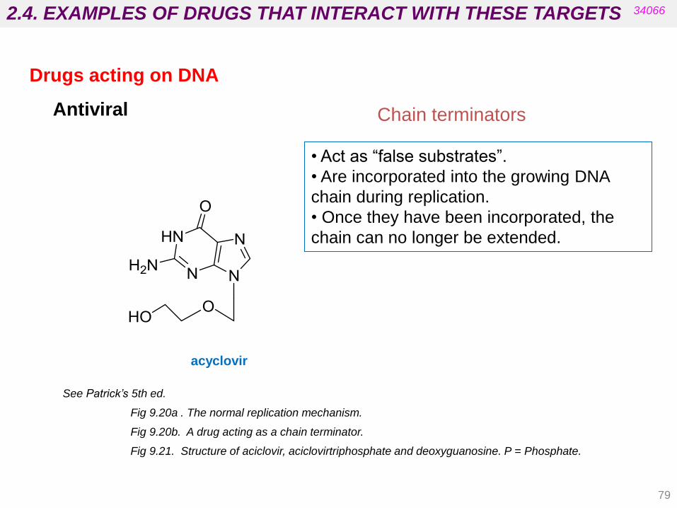
Chain terminators
• Act as “false substrates”.
• Are incorporated into the growing DNA
chain during replication.
• Once they have been incorporated, the
chain can no longer be extended.
Antiviral
See Patrick’s 5th ed.
Fig 9.20a . The normal replication mechanism.
Fig 9.20b. A drug acting as a chain terminator.
Fig 9.21. Structure of aciclovir, aciclovirtriphosphate and deoxyguanosine. P = Phosphate.
acyclovir
Drugs acting on DNA

2.4. EXAMPLES OF DRUGS THAT INTERACT WITH THESE TARGETS
Drugs acting on lipids
34066
•The number of drugs that interact with lipids is relatively small (anaesthetics
and antibiotics).
• In general, all act in the same way by disrupting the lipid structure of cell
membranes.
Amphotericin B
“Tunnelling molecule”: Amphotericin B (antifungal agent) builds tunnels
through the membrane and drains the cell.
See Patrick’s 5th ed. Fig 10.12 (a) Ion channel pore through the cell membrane formed by amphotericine;
(b) Interaction between amphotericin and ergosterol in the ion pore channel.
(Patrick’s 5th Ed. Chapter 10)
80

2.4. EXAMPLES OF DRUGS THAT INTERACT WITH THESE TARGETS
81
Gramicidin (Antibiotic)
Gramicidin is a peptide antibiotic that is thought to form a helix in the cell
membrane.
• Two helices aligned end to end form an „escape tunnel‟.
• The hydrophobic exterior interacts with cell membrane lipids.
• The hydrophilic interior allows the uncontrolled passage of ions.
Drugs acting on lipids
34066
See Patrick’s 5th ed. Fig 10.14 . Gramicidin helices aligned end-to-end to traverse the cell membrane.
Tunnelling molecules
Val-Gly-Ala-Leu-Ala-Val-Val-Val-Trp-Leu-Trp-Leu-Trp-Leu-Trp-NH-CH2-CH2-OH

2.4. EXAMPLES OF DRUGS THAT INTERACT WITH THESE TARGETS
82
Valinomycin has a cyclical structure with alternating ester and amide links and
it allows uncontrolled escape of potassium ions from cell.
•Hydrophobic residues are placed on the exterior.
• Polar carbonyl groups are placed in the interior.
Drugs acting on lipids
34066
Ion carriers
Valinomycin acts as an «inverted detergent»
Potassium ion in the hydrophilic centre of
valinomycin: carbonyl groups interact with
potassium ion.

3.1. Membranes. Physicochemical models that
explain the transport across membranes.
3.2. Physicochemical properties and pharmacological
activity: water solubility, degree of ionization, lipid
solubility and partition coefficient.
3.3. Molecular topology and biological activity.
Concept of structure, constitution, configuration and
conformation: implications for pharmacological
activity.
3.4. Stereoselectivity in drug activity and pharmaco-
kinetics.
PHARMACEUTICAL CHEMISTRY
UNIT 3. BASIC CONCEPTS IN DRUG ACTION
34066
Patrick‟s 5th ed. chapters 5, 11, 13 , 14 and 18 Delgado‟s 2nd ed. chapter 3
Foye’s 7th ed. chapter 2 Silverman‟s, chapters 2 and 3
83

3.1. BASIC CONCEPTS IN DRUG ACTION
84
34066
• The most potent drug at its target site may be useless clinically.
• Active drugs in vitro may be inactive in vivo if a drug cannot reach its target
site.
• Drug design should consider both binding interactions and pharmacokinetics.
Bioavailability refers to how quickly and how much of a particular drug reaches
the blood supply once all the problems associated with absorption, distribution,
metabolism and excretion have been taken into account.
Oral bioavailability is the fraction of the ingested dose that survives to reach the
blood supply. This property should be considered alongside the
pharmacodynamics of the drugs.
Factors to be considered (ADME)
• Drug Absorption
• Drug Distribution
• Drug Metabolism
• Drug Excretion
PHARMACOKINETICS (see
Biopharmacy and
Pharmacokinetics)
3.1. BASIC CONCEPTS IN DRUG ACTION
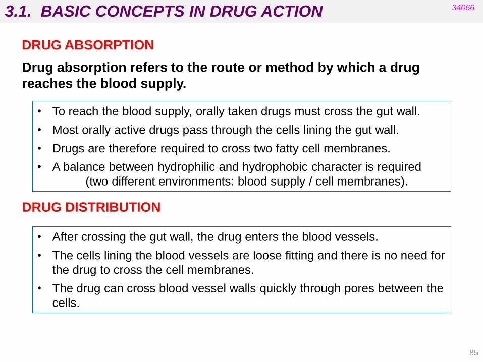
85
DRUG ABSORPTION
• To reach the blood supply, orally taken drugs must cross the gut wall.
• Most orally active drugs pass through the cells lining the gut wall.
• Drugs are therefore required to cross two fatty cell membranes.
• A balance between hydrophilic and hydrophobic character is required
(two different environments: blood supply / cell membranes).
Drug absorption refers to the route or method by which a drug
reaches the blood supply.
34066
• After crossing the gut wall, the drug enters the blood vessels.
• The cells lining the blood vessels are loose fitting and there is no need for
the drug to cross the cell membranes.
• The drug can cross blood vessel walls quickly through pores between the
cells.
DRUG DISTRIBUTION

3.1. BASIC CONCEPTS IN DRUG ACTION
86
• Drugs absorbed orally are first taken to the liver.
• Chemical modification of the drug is possible by enzymes in the liver (drug
metabolism).
• Drug metabolism in the liver deactivates a certain percentage of the absorbed
drug before distribution occurs around the body (first pass effect).
34066
DRUG DISTRIBUTION
• Distribution around the body is uneven due to uneven blood supply.
• Distribution from blood vessels to tissues and organs is rapid.
• If the target is within the cell the drug has to enter a cell.
• Blood concentration drops rapidly after absorption due to distribution,
macromolecular binding and storage in fat tissue (e.g. barbiturates) or bone.
• The blood brain barrier hinders polar drugs from entering the brain:
The polarity of peripherally acting drugs can be
increased to reduce CNS side effects.

3.1. BASIC CONCEPTS IN DRUG ACTION
•Drugs must be sufficiently polar to be soluble in aqueous conditions.
•Drugs must be sufficiently polar/fatty to interact with binding sites.
•Drugs must be sufficiently lipophilic to cross cell membranes.
•Drugs must be sufficiently lipophilic to avoid rapid excretion.
•Drugs must have both hydrophilic and lipophilic characteristics.
34066
For the drug to reach the site of action it must be able to interact with two
environments:
• a lipophilic environment (e.g. membranes).
• an aqueous environment (e.g. cytoplasm inside the cell, extracellular
fluid outside the cell).
87
• Very polar drugs can be administered by injection.
• Very polar drugs can be useful in gut infections.

3.1. TRANSPORT ACROSS MEMBRANES
88
34066
Transport Molecules moved Uses energy? Example
transporter
Simple diffusion Small, nonpolar No -
Facilitated
diffusion
Polar molecules,
larger ions No GLUT4
Primary active
transport
Molecules moving
against their
gradient coupled
to the hydrolysis of
ATP
Yes
Sodium-potassium
pump
Proton pump
Secondary active
transport
Molecules going
with + molecules
going against
gradient
Yes Sodium-calcium
exchanger
https://www.khanacademy.org/test-prep/mcat/cells/transport-across-a-cell-membrane
https://www.youtube.com/watch?v=RPAZvs4hvGA (Remember: Physiology I)

3.1. TRANSPORT ACROSS MEMBRANES
89
34066
FICK‟S LAW
- dC / dt = diffusion velocity through membrane
K = diffusion constant
A = diffusion area
C1 and C2 = concentrations on either side of the membrane
d = membrane thickness
Relates the diffusive flux to the concentration under the assumption of
steady state.
Passive Diffussion

3.1. TRANSPORT ACROSS MEMBRANES
90
or Polar surface area < 140 Angstroms
Number of rotatable bonds ≤ 10
Veber‟s parameters
• Molecular flexibility and the polar surface of the molecule are important to drug
absorption.
• Too many rotatable bonds is bad for absorption.
(Molecular weight is not a factor)
Total number of HBDs and HBAs ≤ 12
Number of rotatable bonds ≤ 10
34066
Lipinski‟s Rule of Five
Orally active drugs generally show a balance between hydrophilic and
hydrophobic properties and obey at least three of the following rules:
This rule is not foolproof – there are
several exceptions (active transport,
pinocytosis,…).
Structural requirements for drugs taken orally: “Drug-like properties”
• MW < 500
• No more than 5 HBD groups
• No more than 10 HBA groups
• log P < +5 See: J. Med. Chem. 2011, 54, 6405–6416
dx.doi.org/10.1021/jm200504p

3.1. TRANSPORT ACROSS MEMBRANES
91
lisinopril
34066
• Polar molecules bearing a structural resemblance to some building blocks
such as amino acids or nucleic acid bases are carried across the membrane by
transport proteins (e.g. lisinopril).
Some highly polar drugs are absorbed from the digestive system.
• Pinocytosis - a process allowing the passage of large polar drugs into a cell
without actually crossing the cell membrane.
• Small polar molecules (MW <200) cross the gut wall through small pores
between cells (do not cross membranes).

3.2. PHYSICOCHEMICAL PROPERTIES
92
34066
• Water Solubility
• Degree of Ionization
• Lipid Solubility (Lipophilicity) and Partition Coefficient
• The degree of ionization has an important
influence on water solubility and lipophilicity.
• The ionization state of a drug depends on the
pKa values of the ionizable groups and the pH of
the medium with which it has to interact.
Foye’s, Silverman’s and Delgado’s (Spanish)

3.2. PHYSICOCHEMICAL PROPERTIES
93
34066
Water Solubility
Depends mainly on two factors:
- Hydrogen bonds with water molecules.
- Ionization (ion-dipole interactions with water molecules).
• Is based on the carbon-solubilizing potential of organic functional groups.
• If the potential of the functional groups exceeds the total number of C atoms,
the compound is considered water-soluble.
Predicting the drug water solubility by an empiric approach (Lemke):
Functional group Monofunctional group Polyfunctional group
Alcohol 5-6 carbon atoms 3-4 carbon atoms
Phenol 6-7 carbon atoms 3-4 carbon atoms
Ether 4-5 carbon atoms 2 carbon atoms
Aldehyde 4-5 carbon atoms 2 carbon atoms
Ketone 5-6 carbon atoms 2 carbon atoms
Amine 6-7 carbon atoms 3 carbon atoms
Carboxylic acid 5-6 carbon atoms 3 carbon atoms
Ester 6 carbon atoms 3 carbon atoms
Amide 6 carbon atoms 2-3 carbon atoms
Urea, Carbonate, Carbamate 2 carbon atoms
Charge (cationic and anionic) 20-30 carbon atoms
3.2. PHYSICOCHEMICAL PROPERTIES
94
34066
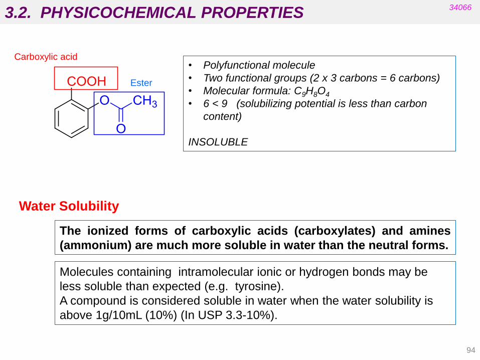
The ionized forms of carboxylic acids (carboxylates) and amines
(ammonium) are much more soluble in water than the neutral forms.
Molecules containing intramolecular ionic or hydrogen bonds may be
less soluble than expected (e.g. tyrosine).
A compound is considered soluble in water when the water solubility is
above 1g/10mL (10%) (In USP 3.3-10%).
Water Solubility
• Polyfunctional molecule
• Two functional groups (2 x 3 carbons = 6 carbons)
• Molecular formula: C9H8O4
• 6 < 9 (solubilizing potential is less than carbon
content)
INSOLUBLE
Carboxylic acid
Ester

3.2. PHYSICOCHEMICAL PROPERTIES
95
34066
Water Solubility
Definition of approximate drug solubility by US pharmacopoeia
Term Parts of the solvent required for one part of
solute
Very soluble Less than 1 part (1 : 1)
Freely soluble 1 to 10 part (1 : 1-10)
Soluble 10 to 30 part (1 : 10-30)
Sparingly soluble 30 to 100 part (1: 30-100)
Slightly soluble 100 to 1000 part (1 : 100-1000)
Very slightly soluble 1000 to 10000 part (1 : 1000-10000)
Practically insoluble More than 10000 part (1:>10000)

3.2. PHYSICOCHEMICAL PROPERTIES
96
34066
morphine
(C17H19NO3)
ouabain
(C29H44O12) poisonous glycoside
Are morphine and ouabain water-soluble drugs?
Is the protonated form of morphine a water-soluble drug?
Water Solubility
(see Delgado’s book)

3.2. PHYSICOCHEMICAL PROPERTIES
97
34066
Water Solubility
(from Foye’s)
Water solubility in salts depends on the chemical structure of both
components (cation and anion).

3.2. PHYSICOCHEMICAL PROPERTIES
98
34066
• Ionization can have a profound effect not only on a drug‟s interaction with a
target but also on its lipophilicity.
• The ionization of the drug can favour binding to the receptor but can also make
crossing membranes difficult prior to reaching the target.
• An equilibrium can be established between the neutral form and the ionized
form.
• E.g. many drugs contain an amine functional group. Amines are weak bases
and form ammonium cations in water (pKa = 9-10 for aliphatic amines).
Degree of ionization
For carboxylic acids (pKa = 4-5), an equilibrium with the corresponding
carboxylate anion can be established at physiological pH.
R-NH2 + H2O RNH3+ + OH-
R-COOH + H2O RCOO- + H3O+

3.2. PHYSICOCHEMICAL PROPERTIES
99
34066
Degree of ionization
REMEMBER: when we refer to the pKa of a
basic substance we are referring to the pKa of
its conjugate acid (RNH3+ for a primary amine).
See Figure 11.1. Equilibrium between the ionized and non-ionized form of an
amine (Patrick’s 5th Ed.).
Only non-ionized forms are able to cross membranes by diffusion.
Once in the blood, the two forms are also in equilibrium.

3.2. PHYSICOCHEMICAL PROPERTIES
100
Acidic drug
Basic drug
34066
Degree of ionization
Henderson-Hasselbach equation
• For an ionizable substance with a given pKa, the extent of ionization depends
on the pH.
• At a particular pH the degree of ionization can be determined by the
Henderson-Hasselbach equation, which is derivable from the acid
dissociation constant equation:
pH = pKa + log [A-] / [AH]
pKa = pH + log [AH] / [A-]
pKa = pH + log [BH+] / [B]
pH = pKa + log [B] / [BH+]
or
or
3.2. PHYSICOCHEMICAL PROPERTIES 34066
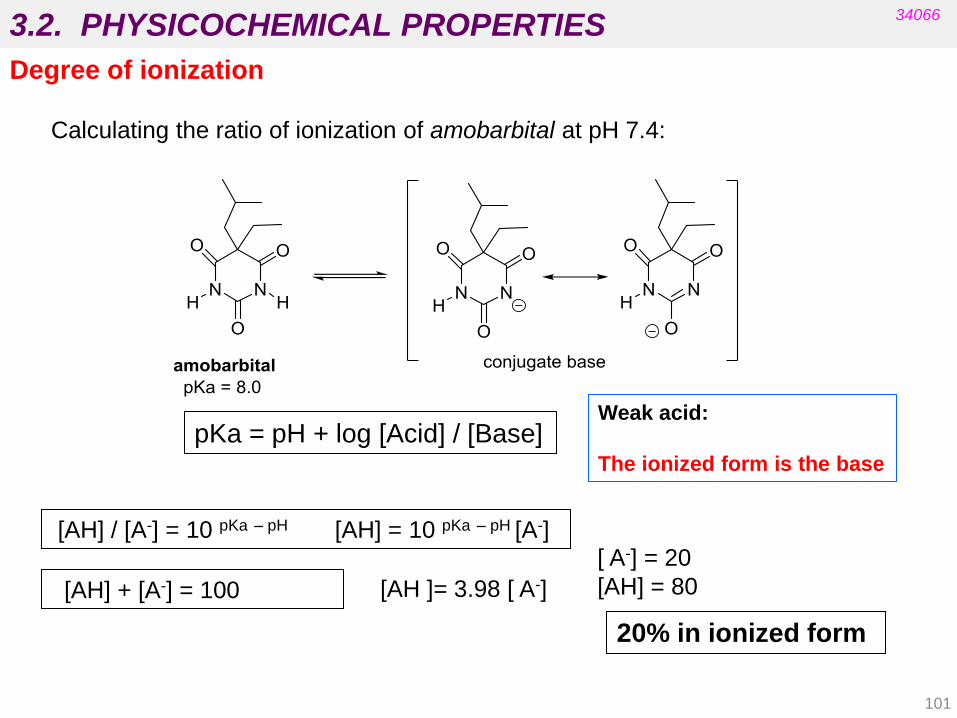
Degree of ionization
pKa = pH + log [Acid] / [Base]
[AH ]= 3.98 [ A-]
[ A-] = 20
[AH] = 80
Weak acid:
The ionized form is the base
Calculating the ratio of ionization of amobarbital at pH 7.4:
20% in ionized form
[AH] / [A-] = 10 pKa – pH [AH] = 10 pKa – pH [A-]
[AH] + [A-] = 100
101

3.2. PHYSICOCHEMICAL PROPERTIES
102
34066
Degree of ionization
Phenobarbital. Degree of ionization at different pHs.
pKa = 7.3
pH
Stomach 1-3
Small intestine 6-8
Large intestine 5-7
Fallingborg J. Intraluminal pH of the human gastrointestinal tract.
Dan Med Bull. 1999;46(3):183-96.
3.2. PHYSICOCHEMICAL PROPERTIES
103
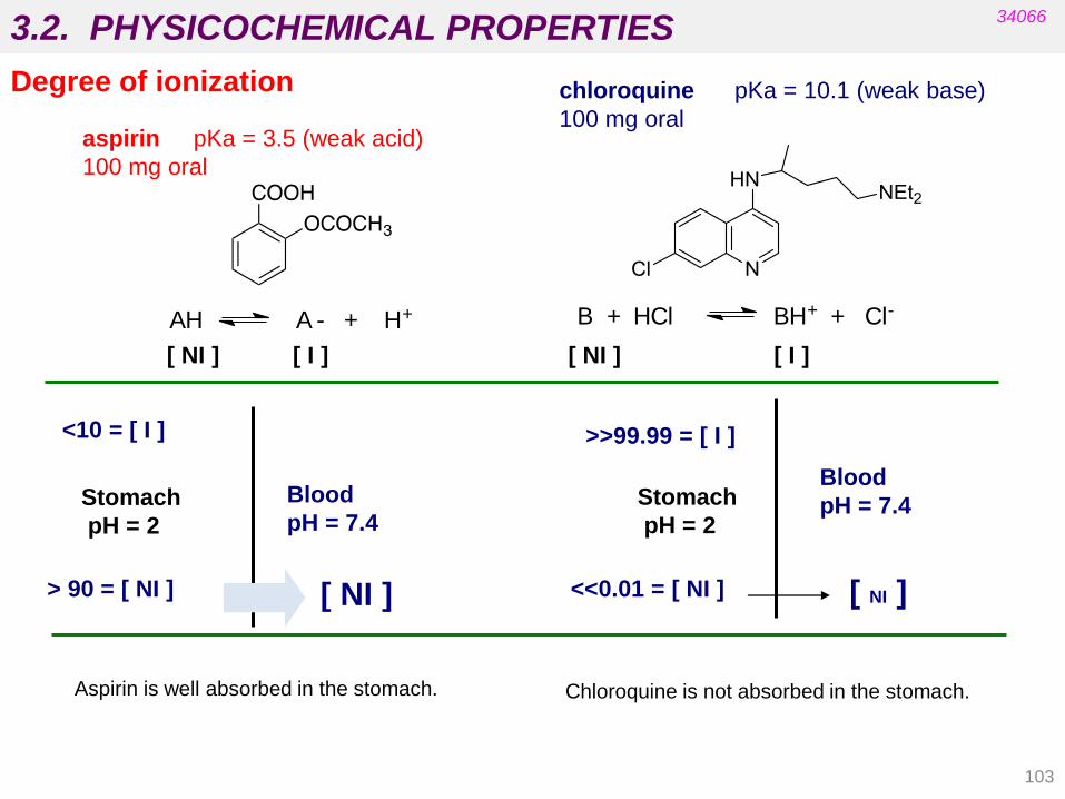
[ NI ] [ I ] [ NI ] [ I ]
aspirin pKa = 3.5 (weak acid)
100 mg oral
> 90 = [ NI ] [ NI ]
Stomach
pH = 2
Blood
pH = 7.4
<10 = [ I ]
Aspirin is well absorbed in the stomach.
<<0.01 = [ NI ] [ NI ]
Blood
pH = 7.4
>>99.99 = [ I ]
chloroquine pKa = 10.1 (weak base)
100 mg oral
Stomach
pH = 2
Chloroquine is not absorbed in the stomach.
AH A - + H+ B + HCl BH+ + Cl-
34066
Degree of ionization

3.2. PHYSICOCHEMICAL PROPERTIES
104
Weak ACIDS
pH = pKa ~ 50% ionized
pH = pKa + 1 ~ 90% ionized pH = pKa - 1 ~ 90% non-ionized
pH = pKa + 2 ~ 99% ionized pH = pKa - 2 ~ 99% non-ionized
pH = pKa + 3 ~ 99.9% ionized pH = pKa - 3 ~ 99.9% non-ionized
pH = pKa + 4 ~ 99.99% ionized pH = pKa - 4 ~ 99.99% non-ionized
Example: pKa of ASPIRIN is 3.5
Physiological pH = 7.4 pH = pKa + 4 % ionization = 99.99% ionized
Stomach pH = 2 pH = pKa – 1.5 % ionization = 1-10% ionized
Weak BASES
pH = pKa ~ 50% ionized
pH = pKa - 1 ~ 90% ionized pH = pKa + 1 ~ 90% non-ionized
pH = pKa - 2 ~ 99% ionized pH = pKa + 2 ~ 99% non-ionized
pH = pKa - 3 ~ 99.9% ionized pH = pKa + 3 ~ 99.9% non-ionized
pH = pKa - 4 ~ 99.99% ionized pH = pKa + 4 ~ 99.99% non-ionized
Example: pKa of CHLOROQUINE is 10.1
Physiological pH = 7.4 pH = pKa - 3 % ionization = ~ 99.9% ionized
Stomach pH = 2 pH = pKa – 8 % ionization = >> 99.99% ionized
(Rule of Thumb)
34066
Degree of ionization

3.2. PHYSICOCHEMICAL PROPERTIES
105
https://www.drugbank.ca/drugs
34066
Degree of ionization
3.2. PHYSICOCHEMICAL PROPERTIES
106
34066
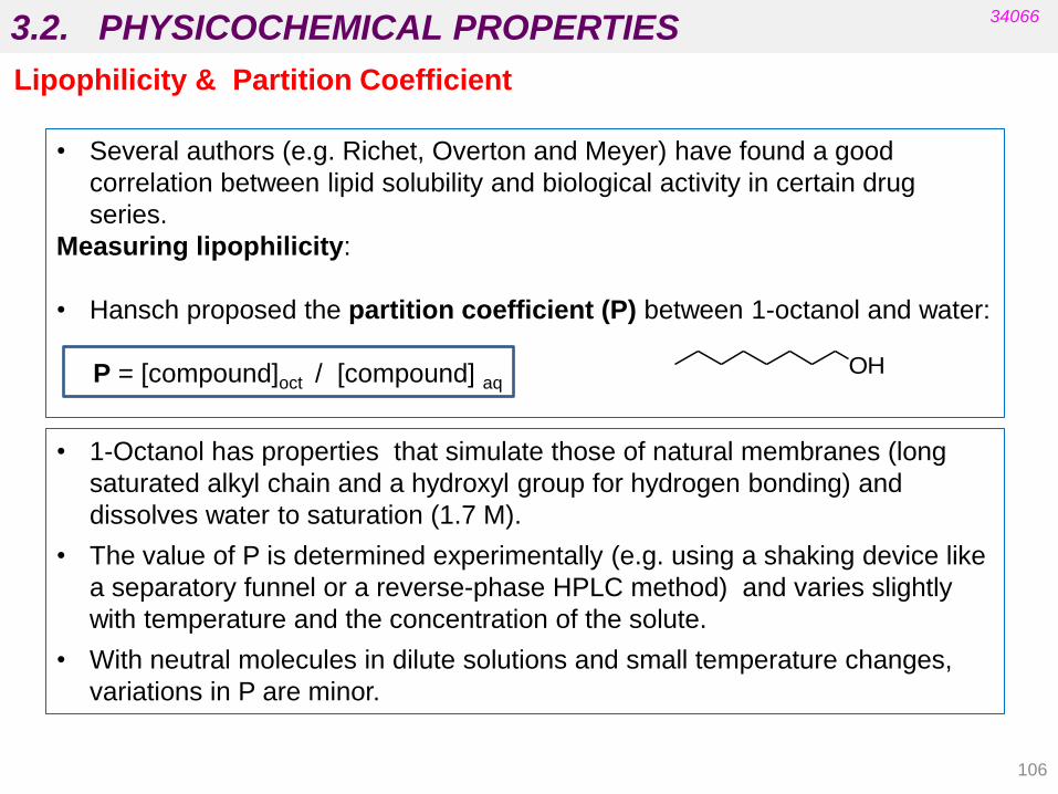
Lipophilicity & Partition Coefficient
• Several authors (e.g. Richet, Overton and Meyer) have found a good
correlation between lipid solubility and biological activity in certain drug
series.
Measuring lipophilicity:
• Hansch proposed the partition coefficient (P) between 1-octanol and water:
P = [compound]oct / [compound] aq
• 1-Octanol has properties that simulate those of natural membranes (long
saturated alkyl chain and a hydroxyl group for hydrogen bonding) and
dissolves water to saturation (1.7 M).
• The value of P is determined experimentally (e.g. using a shaking device like
a separatory funnel or a reverse-phase HPLC method) and varies slightly
with temperature and the concentration of the solute.
• With neutral molecules in dilute solutions and small temperature changes,
variations in P are minor.
OH

3.2. PHYSICOCHEMICAL PROPERTIES
107
34066
Lipophilicity & Partition Coefficient
P > 1 ( log P > 0 ) lipophilic
P < 1 ( log P < 0 ) hydrophilic P = [compound]oct / [compound] aq
• The more positive the log P, the more lipophilic the compound.
• The larger the value of P, the more the drug will interact with the lipid phase
(e.g. membranes).
• With very high values of P, the drug will be unable to cross the aqueous phase
and will localize in the first lipophilic phase with which it comes into contact.
• As P approaches zero, the drug will be so water-soluble that it will not be
capable of crossing the lipid phase and will localize in the aqueous phase
(remember there are exceptions for the absorption of some very polar drugs).
• A parabolic relationship between potency (log 1/C) and log P is often found for
a series of structurally related drugs:
log 1/C = - k (log P)2 + k‟ (log P) + k‟‟
• In these cases there is a value of log P which corresponds to the maximum:
log Po (E.g. in a series of nonspecific hypnotics, Hansch found that all active
compounds had a similar log P, approximately 2).
3.2. PHYSICOCHEMICAL PROPERTIES
108
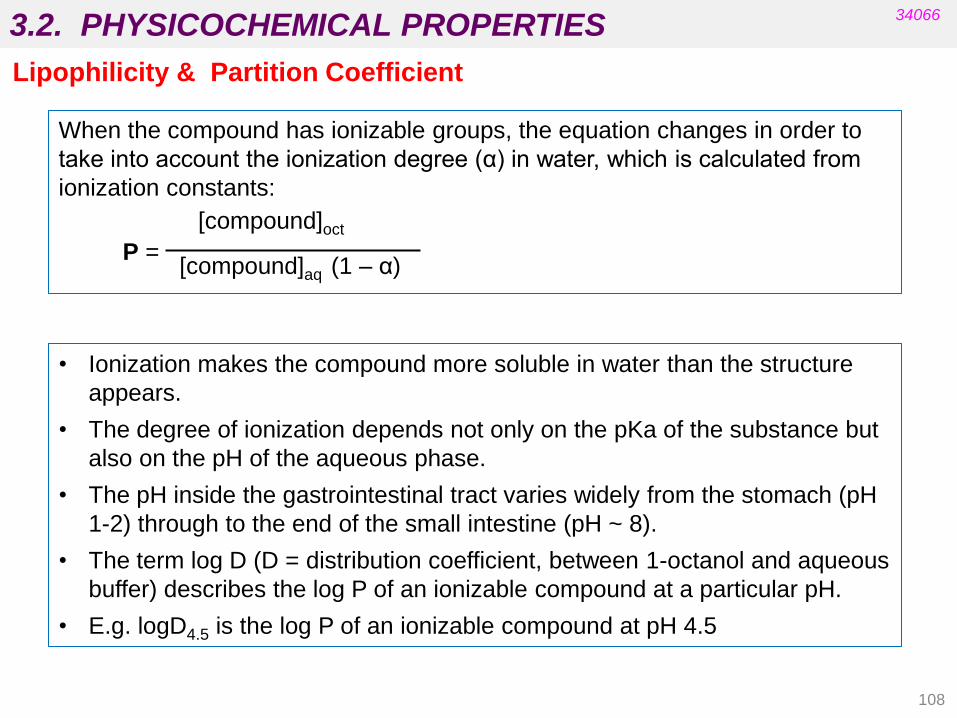
When the compound has ionizable groups, the equation changes in order to
take into account the ionization degree (α) in water, which is calculated from
ionization constants:
[compound]oct
P = [compound]aq (1 – α)
• Ionization makes the compound more soluble in water than the structure
appears.
• The degree of ionization depends not only on the pKa of the substance but
also on the pH of the aqueous phase.
• The pH inside the gastrointestinal tract varies widely from the stomach (pH
1-2) through to the end of the small intestine (pH ~ 8).
• The term log D (D = distribution coefficient, between 1-octanol and aqueous
buffer) describes the log P of an ionizable compound at a particular pH.
• E.g. logD4.5 is the log P of an ionizable compound at pH 4.5
34066
Lipophilicity & Partition Coefficient

3.2. PHYSICOCHEMICAL PROPERTIES
109
34066
Lipophilicity & Partition Coefficient
Predicting the drug lipophilicity from the structure:
• This is possible if we know the lipophilicities of substituents and atoms.
• Hansch and coworkers derived substituent constants for the contribution of
individual atoms and groups to the partition coefficient.
• The lipophilicity constant for an atom or group X, πX, is defined by:
πX = log PX – log PH = log ( PX / PH)
• PX is the partition coefficient for the compound with substituent X.
• PH is the partition coefficient for the parent molecule.
• Log P calculated from the lipophilic constants for substituents is called Clog P:
Clog P = ∑ π

3.2. PHYSICOCHEMICAL PROPERTIES
110
34066
Lipophilicity & Partition Coefficient
diphenhydramine
(antihistamine)
• pX depends on the characteristics of the group/atom: inductive, resonance and
steric effects are important (a positive value means that the substituent increases
lipophilicity and a negative value means the opposite).
• The values of p for the most common substituents are tabulated .
Numerous software packages are now commercially available (ChemDraw, etc.) but the
results can differ widely and also differ from the experimental value.
Calculated log P = 2pPh + pCH + pOCH2 + pCH2 + pNMe2 - 0.2 =
= 2 (2.13) + 0.50 – 0.73 + 0.50 - 0.95 – 0.2 = 3.38
(see Chapter 6)
3.2. PHYSICOCHEMICAL PROPERTIES
111
34066
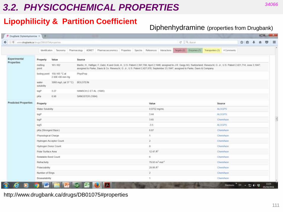
Lipophilicity & Partition Coefficient
http://www.drugbank.ca/drugs/DB01075#properties
Diphenhydramine (properties from Drugbank)

3.3. STEREOCHEMISTRY AND BIOLOGICAL ACTIVITY
112
34066
• Targets are chiral biomolecules (e.g. carbohydrates, amino acids,
cholesterol, etc.).
• Carrier proteins in membranes are chiral (absorption).
• Proteins in blood supply are chiral (distribution).
• Enzymes are chiral (metabolism).
• Most drugs currently on the market are some type of isomeric mixture.
For many of these drugs the biological activity resides in one isomer.
• If critical functional groups in the drug molecule do not occupy the proper
spatial region, the desired pharmacological activity cannot be achieved
because productive interactions with the biological target will not be
possible.
Stereochemistry involves the study of the relative spatial arrangement of
atoms that form the structure of molecules.

3.3. STEREOCHEMISTRY AND BIOLOGICAL ACTIVITY
113
Molecular topology is a part of mathematical chemistry dealing with the
algebraic description of chemical compounds so allowing a unique and
easy characterization of them. The totality of information about the
mutual connectedness of all pairs of atoms (chemically bonded and not
chemically bonded) in a molecule determines the topology of the
respective molecule.
(Journal of Chemical Education , 1995,72, 12, 1059)
The three-dimensional (3D) nature of the functional groups that
contribute to the pharmacological activity of the drug is important in
predicting drug potency and potential side effects.
These important binding groups that are required for activity and their
relative positions in space with respect to each other are called
PHARMACOPHORE.
34066

3.3. STEREOCHEMISTRY AND BIOLOGICAL ACTIVITY
114
34066
Different constitution and connectivity = different properties
Same constitution and connectivity but different configuration:
Double bonds or rings: diastereomers
Chiral compounds: enantiomers and diastereomers
Same constitution, connectivity and configuration:
Flexible compounds (single bonds): different conformations
There are different levels to define the structure of organic molecules:
• Constitution (atoms present)
• Connectivity (order in which the atoms are connected) isomers
• Configuration (spatial distribution of atoms and groups) stereoisomers
changes in configuration require bond cleavage and bond formation
(different compounds)
• Conformation (flexibility/rotatable bonds) conformers (stereoisomers)
changes in conformation do not require bond cleavage (same compound)

3.4. STEREOCHEMISTRY AND BIOLOGICAL ACTIVITY
115
34066
• Molecules are non-superimposable, non-mirror images.
• Are possible in molecules with two or more asymmetric centres and/or double
bonds and/or rings.
• The physicochemical properties are different (water solubility, partition
coefficient, pKa, etc.)
• The distances and angles between non-connected atoms are different.
• Differences in biological activity are usually observed.
α- and β-adrenergic agonists
Used to treat various diseases
Diastereomers:

3.4. STEREOCHEMISTRY AND BIOLOGICAL ACTIVITY
116
34066
• Molecules that are non-superimposable, mirror images.
• They are possible when the molecule is chiral (asymmetrical centres, etc.).
• The physical properties are identical (water solubility, partition coefficient,
pKa, etc.) except for interaction with the polarized light.
• There are no differences in distances or angles between non-connected
atoms.
• Different biological activity (enantioselectivity) may be observed because of
the different interaction with the target (asymmetrical nature).
• Each enantiomer may also experience selective absorption, metabolism,
distribution and/or excretion determining a different biological activity or
potency.
• Such processes may also contribute to the adverse effects of a particular
enantiomer (e.g. if a different protein is involved in side effects or toxicity).
Enantiomers:

3.4. STEREOCHEMISTRY AND BIOLOGICAL ACTIVITY
117
DRUG
SELECTIVITY
MEMBRANE
SELECTIVITY
METABOLISM
SELECTIVITY
RECEPTOR
THERAPEUTIC
EFFECT
SELECTIVITY
NONSPECIFIC RECEPTORS/
DISTRIBUTION
34066
Enantioselectivity
The more active enantiomer is
called the eutomer.
The less active enantiomer is
called the distomer.
Enantiomers: Complexes of both enantiomers of a
chiral drug with the target or other chiral
macromolecules are diastereomeric, so
the pharmacological activity may be
qualitatively and quantitatively different.

3.4. STEREOCHEMISTRY AND BIOLOGICAL ACTIVITY
118
thalidomide
(R) – (+) sedative
(S) – (-) teratogen
ketamine
(S) – (+) anaesthetic
(R) – (-) hallucinogenic
penicillamine
(S) – (-) chelating agent ( in metal poisoning)
(R) – (+) toxic
tetramisol
(S) – (-) anthelmintic
R) – (+) side effects
34066
Enantiomers: Distomer showing toxicity or side effects.
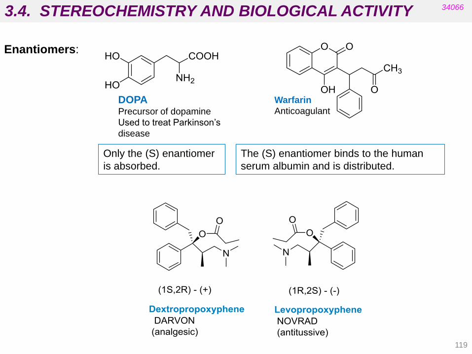
3.4. STEREOCHEMISTRY AND BIOLOGICAL ACTIVITY
119
34066
Enantiomers:
Only the (S) enantiomer
is absorbed.
The (S) enantiomer binds to the human
serum albumin and is distributed.
Warfarin
Anticoagulant
DOPA Precursor of dopamine
Used to treat Parkinson‟s
disease

3.4. STEREOCHEMISTRY AND BIOLOGICAL ACTIVITY
120
34066
THE EASSON-STEDMAN HYPOTHESIS
In order to show enantioselectivity, the more potent enantiomer must be
involved in at least three intermolecular interactions with the binding site
on the target (three binding regions) and the less potent enantiomer must
only interact with one or two regions.
EUTOMER
(MORE ACTIVE)
DISTOMER
(enantiomer)
Enantiomers:
Eudismic Ratio
E.R. =
activity of eutomer
activity of distomer
Affinity rather than activity is very often used in this equation.

3.4. STEREOCHEMISTRY AND BIOLOGICAL ACTIVITY
121
34066
Differences in bronchodilator activity between some catecholamines:
R = H noradrenaline
R = Me adrenaline
R = i Pr isoprenaline
Bronchodilator activity relative to
(R)-(-) noradrenaline
(R)-(-)-noradrenaline 100
(S)-(+)-noradrenaline 1.4
(R)-(-)-adrenaline 5800
(S)-(+)-adrenaline 130
(R)-(-)-isoprenaline 27000
(S)-(+)-isoprenaline 33
R/S = 71
R/S = 45
R/S = 818
Eudismic ratio
Enantiomers:

3.4. STEREOCHEMISTRY AND BIOLOGICAL ACTIVITY
122
34066
Flat area
HYDROGEN
BONDS
HYDROGEN BOND
IONIC
INTERACTION
Enantiomers:
Flat area
HYDROGEN
BONDS
IONIC
INTERACTION
NO HYDROGEN BOND
Schematic representation of the
interactions of R and S adrenaline
with the receptor.

3.4. STEREOCHEMISTRY AND BIOLOGICAL ACTIVITY
123
34066
Benefits of using optically pure drugs:
- Required dose is smaller.
- Fewer side effects are observed.
- Pharmacological activity is improved.
Isomers in a mixture that do not contribute to
the desired action are called ISOMERIC
BALLAST.
• Most chiral drugs contain two or more
asymmetric centres.
• The racemate of a chiral drug may be less
potent than the eutomer and may have more
side effects.

3.4. STEREOCHEMISTRY AND BIOLOGICAL ACTIVITY
First tests are carried out with racemic mixtures
If activity is observed, a decision has to be made to develop the racemic mixture or one
enantiomer.
One single enantiomer must be developed when:
The pharmacological activity depends on one enantiomer (eutomer).
The less active enantiomer (distomer) is toxic.
The therapeutic index (TI) is small.
The racemic mixture can be marketed when:
Both enantiomers show activity (synergic or additive).
The distomer has no toxicity.
The therapeutic index is high.
Chiral synthetic compounds as drug candidates:
34066
It is important to know the margin of safety that exists between the dose needed for the
desired effect and the dose that produces unwanted and possibly dangerous side effects.
This relationship, known as the therapeutic index, is defined as the ratio LD50/ED50. In
general, the narrower this margin, the more likely it is that the drug will produce unwanted
effects.
124
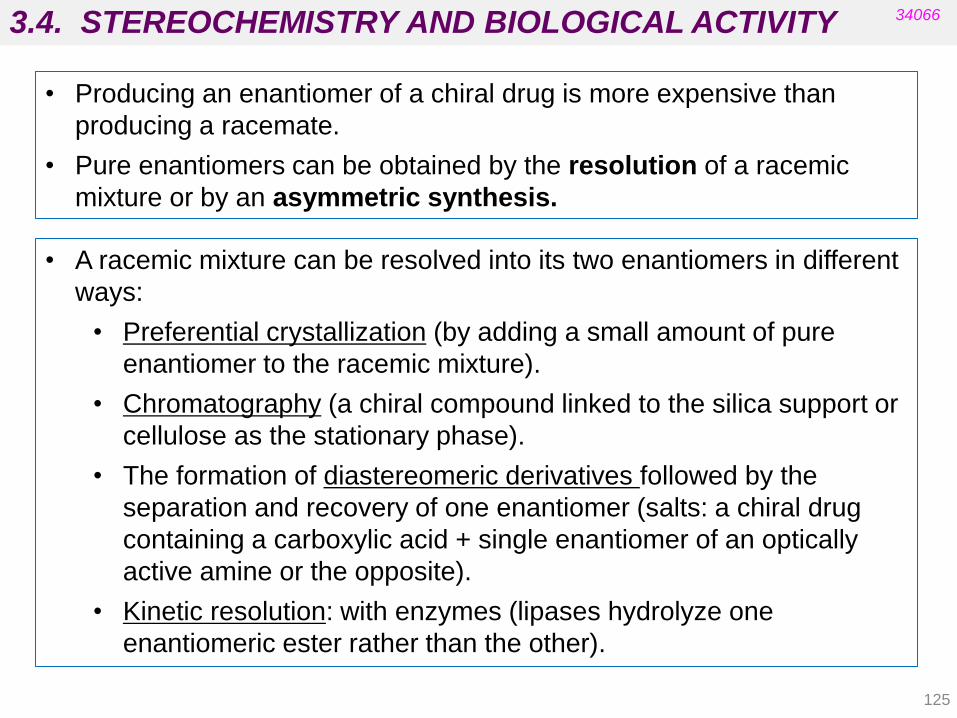
3.4. STEREOCHEMISTRY AND BIOLOGICAL ACTIVITY
125
34066
• A racemic mixture can be resolved into its two enantiomers in different
ways:
• Preferential crystallization (by adding a small amount of pure
enantiomer to the racemic mixture).
• Chromatography (a chiral compound linked to the silica support or
cellulose as the stationary phase).
• The formation of diastereomeric derivatives followed by the
separation and recovery of one enantiomer (salts: a chiral drug
containing a carboxylic acid + single enantiomer of an optically
active amine or the opposite).
• Kinetic resolution: with enzymes (lipases hydrolyze one
enantiomeric ester rather than the other).
• Producing an enantiomer of a chiral drug is more expensive than
producing a racemate.
• Pure enantiomers can be obtained by the resolution of a racemic
mixture or by an asymmetric synthesis.

3.4. STEREOCHEMISTRY AND BIOLOGICAL ACTIVITY
126
34066
Enantiomers: Resolution Synthesis of (R)-adrenaline
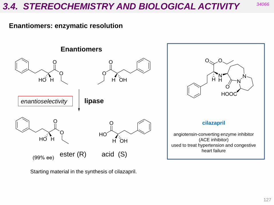
127
Enantiomers
lipase
ester (R) acid (S)
Starting material in the synthesis of cilazapril.
cilazapril
angiotensin-converting enzyme inhibitor
(ACE inhibitor)
used to treat hypertension and congestive
heart failure
Enantiomers: enzymatic resolution
3.4. STEREOCHEMISTRY AND BIOLOGICAL ACTIVITY 34066
enantioselectivity

128
3.4. STEREOCHEMISTRY AND BIOLOGICAL ACTIVITY 34066
• An asymmetric synthesis can be achieved by starting with an optically pure
starting material that already contains the required asymmetric centres or
by including an asymmetric reaction.
• An asymmetric reaction produces a new asymmetric centre with
selectivity for one stereoisomer over the other.
omeprazole (racemic)
It was the first proton pump inhibitor
to reach the market (1988). In
Europe the patents expired in 1999.
Enantiomers
S-enantiomer (esomeprazole)
It was launched in 2000 on the basis that
it has therapeutic advantages over
omeprazole (superior in terms of
pharmacokinetic profile).
It is an example of chiral switching, whereby a racemic
drug is replaced on the market with a single enantiomer.

Conformation and biological activity
Tema 3: Conceptos Básicos del Mecanismo de Acción de los Fármacos
• Many endogenous compounds are very simple and flexible (e.g.
adrenaline, acetylcholine).
• They interact with different targets because they are able to adopt
different conformations (e.g. different types and subtypes of adrenergic
and cholinergic receptors).
Remember:
Conformations in butane: staggered, partially eclipsed,
gauche, fully eclipsed
These conformations have different energy depending on the
steric and electrostatic interactions (see 2,3-butanediol)
3.4. STEREOCHEMISTRY AND BIOLOGICAL ACTIVITY 34066
129
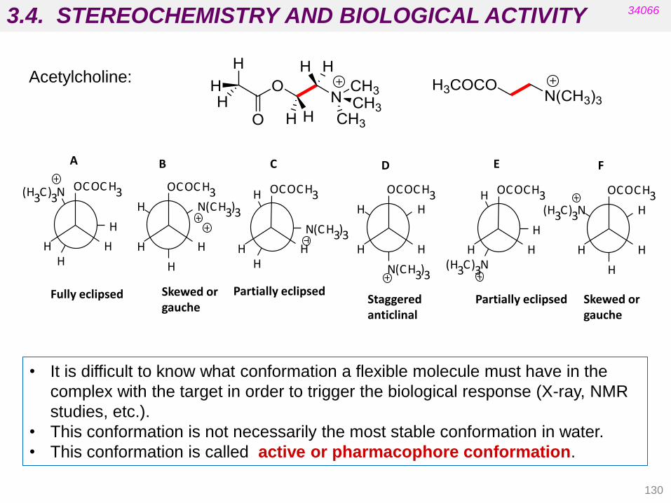
130
A B C D E F
Fully eclipsed Skewed or gauche
Skewed or gauche
Partially eclipsed Partially eclipsed Staggered
anticlinal
H
H N ( C H 3 ) 3
O C O C H 3
H H H
( H 3 C ) 3 N
H
O C O C H 3
H H
N ( C H 3 ) 3
H H
O C O C H 3
H H H
H
N ( C H 3 ) 3
O C O C H 3
H H
H
( H 3 C ) 3 N H
O C O C H 3
H H ( H 3 C ) 3 N
H
H
O C O C H 3
H H
Acetylcholine:
3.4. STEREOCHEMISTRY AND BIOLOGICAL ACTIVITY 34066
• It is difficult to know what conformation a flexible molecule must have in the
complex with the target in order to trigger the biological response (X-ray, NMR
studies, etc.).
• This conformation is not necessarily the most stable conformation in water.
• This conformation is called active or pharmacophore conformation.

131
3.4. STEREOCHEMISTRY AND BIOLOGICAL ACTIVITY 34066
Pharmacophore
•Defines the important groups involved in binding and tthe relative
positions of the binding groups.
•Needs to know the active conformation.
•Is important for drug design and for drug discovery
2D Pharmacophore
Defines the minimum skeleton that connects important binding groups (from structure-activity relationships (SAR) studies in analogous compounds).
3D Pharmacophore:
Defines relative positions in space of important binding groups (computational techniques and SAR studies).

132
3.4. STEREOCHEMISTRY AND BIOLOGICAL ACTIVITY 34066
2D pharmacophore morphine:
Important groups for activity
O
N CH3
HO
HO
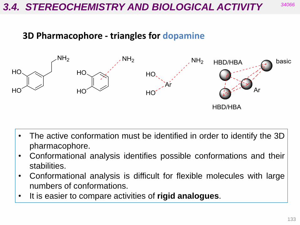
133
3.4. STEREOCHEMISTRY AND BIOLOGICAL ACTIVITY 34066
3D Pharmacophore - triangles for dopamine
• The active conformation must be identified in order to identify the 3D
pharmacophore.
• Conformational analysis identifies possible conformations and their
stabilities.
• Conformational analysis is difficult for flexible molecules with large
numbers of conformations.
• It is easier to compare activities of rigid analogues.

134
Rigidification: introducing rings
Tema 3: Conceptos Básicos del Mecanismo de Acción de los Fármacos 3.4. STEREOCHEMISTRY AND BIOLOGICAL ACTIVITY 34066
The “cisoid” conformation of
acetylcholine
The rigid trans-decalin analogue The rigid cyclopropyl analogue
The “transoid” conformation of
acetylcholine
The rigid trans-decalin analogue The rigid cyclopropyl analogue
Rigid analogues of acetylcholine

135
Combretastatin (anticancer agent) Rotatable
bond
3.4. STEREOCHEMISTRY AND BIOLOGICAL ACTIVITY 34066
Rigidification: introducing double bonds

136
Rigidification: introducing steric blocks
3.4. STEREOCHEMISTRY AND BIOLOGICAL ACTIVITY 34066

4.1. Phase I: Transformation reactions.
4.2. Phase II: Conjugation reactions.
4.3. Bioreversible derivatives: prodrugs and
bioprecursors.
4.4. “Soft” drugs and “hard” drugs.
PHARMACEUTICAL CHEMISTRY
UNIT 4. DRUG METABOLISM
34066
Patrick‟s 5th ed. chapter 13 and Delgado‟s chapter 5
Prodrugs – from Serendipity to Rational Design
K.M. Huttunen, H. Raunio, and J. Rautio, Pharmacol. Rev. 63:750 –771, 2011
- Patrick‟s, 5th ed. chapter 14 (14.6)
- Silverman‟s, 3rd ed. chapters 8 and 9
- Avendaño‟s (chapter 8 - Spanish)
137
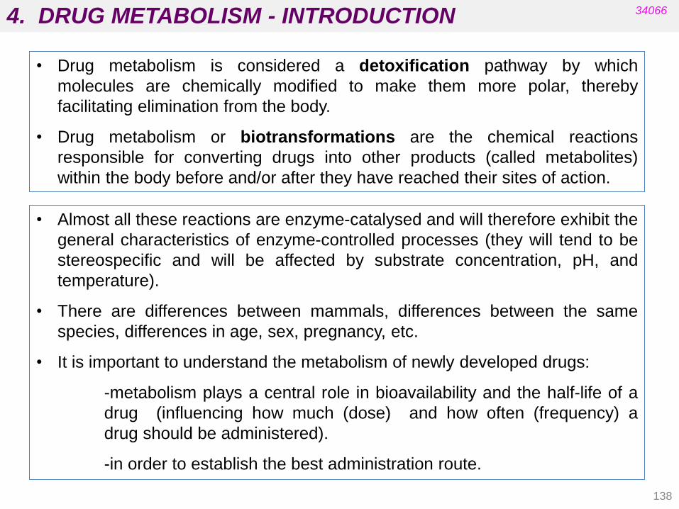
4. DRUG METABOLISM - INTRODUCTION
138
• Drug metabolism is considered a detoxification pathway by which
molecules are chemically modified to make them more polar, thereby
facilitating elimination from the body.
• Drug metabolism or biotransformations are the chemical reactions
responsible for converting drugs into other products (called metabolites)
within the body before and/or after they have reached their sites of action.
34066
• Almost all these reactions are enzyme-catalysed and will therefore exhibit the
general characteristics of enzyme-controlled processes (they will tend to be
stereospecific and will be affected by substrate concentration, pH, and
temperature).
• There are differences between mammals, differences between the same
species, differences in age, sex, pregnancy, etc.
• It is important to understand the metabolism of newly developed drugs:
-metabolism plays a central role in bioavailability and the half-life of a
drug (influencing how much (dose) and how often (frequency) a
drug should be administered).
-in order to establish the best administration route.
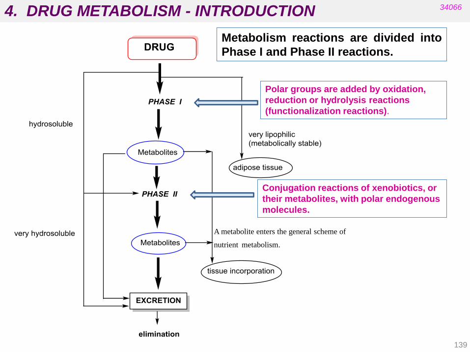
4. DRUG METABOLISM - INTRODUCTION 34066
Polar groups are added by oxidation,
reduction or hydrolysis reactions
(functionalization reactions).
Conjugation reactions of xenobiotics, or
their metabolites, with polar endogenous
molecules.
A metabolite enters the general scheme of
nutrient metabolism.
Metabolism reactions are divided into
Phase I and Phase II reactions.
139

4. DRUG METABOLISM - INTRODUCTION
140
34066
Phase I Enzymes:
Cytochrome P450 enzymes (microsomes)
Flavin monooxygenases (microsomes)
Xanthine oxidase and aldehyde oxidase (cytosol)
Monoamine oxidases (MAO-A and MAO-B) (mitochondria)
Dihydrodiol dehydrogenase (cytosol)
Alcohol and aldehyde dehydrogenases (cytosol)
Esterases/amidases (microsomes, cytosol, plasma)
Epoxide hydrolase (microsomes and cytosol)
Sulfur reductase (cytosol)
Quinone reductase (microsomes and cytosol)
Azo/nitro reductase (microsomes, cytosol and gut)
Drug Discovery: Practices, Processes and Perspectives
John Wiley and Sons, p 126
Phase II Enzymes:
Glucuronosyl transferases (microsomes)
Sulfo-transferases (cytosol)
Methyl transferases (microsomes and cytosol)
Acetyl transferases (cytosol)
Glutathione transferases (microsomes and cytosol)
Amino acid transferases (microsomes)

4. DRUG METABOLISM - INTRODUCTION
141
34066
• Most reactions add a polar „handle‟ to the molecule.
• Increasing the polarity of a compound increases the rate of drug excretion.
• Most enzyme-catalysed reactions occur in the liver (detoxification).
• Metabolic reactions also occur in blood, the gut wall and other organs.
• Drug metabolites are usually less active or inactive (with the exception of
prodrugs).
• Modification of a structure may interfere or prevent binding interactions with a
target (pharmacodynamics).
Example:

4.1. PHASE I TRANSFORMATIONS
142
34066
4.1.1. Microsomal oxidations Carbon sp3 oxidations Allylic and benzylic oxidations Oxidative dealkylations. Amines, ethers and thioethers.
Oxidative dehydrohalogenation
Oxidative N-dealkylation and deamination
Alkene and alkyne oxidations
Aromatic hydroxylation
Amine and sulphur derivative oxidations
4.1.2. Non-microsomal oxidations
Alcohol and aldehyde oxidations catalyzed by dehydrogenases
Oxidative deamination catalyzed by monoamine oxidase
Purine oxidation
4.1.3. Reductions
Azo reductions
Ketone reductions
Nitro-derivative reductions
Alkene reductions
Sulfoxide reductions
4.1.4. Hydrolytic reactions
Hydrolysis of nitriles, esters, and amides Hydration of epoxides

4.1. PHASE I TRANSFORMATIONS
143
34066
Cytochrome P-450
The heme residue is protoporphyrin IX containing a coordinated Fe(III).
• It is located in the liver.
• There are at least 12 families in human biochemistry.
• Individuals differ in the types of Cyt P-450 enzymes present.
• Patient variability in drug metabolism complicates dose levels and leads
to different susceptibilities to drugs.
These are catalyzed by mixed-function oxidases in the liver: cytochrome P-450
and flavin-containing monooxygenase (FMO).
Cytochrome enzymes use an oxygenated heme (or haem) prosthetic group,
while the FMO family uses FAD to oxidize its substrates.
Microsomal oxidations: monooxygenases
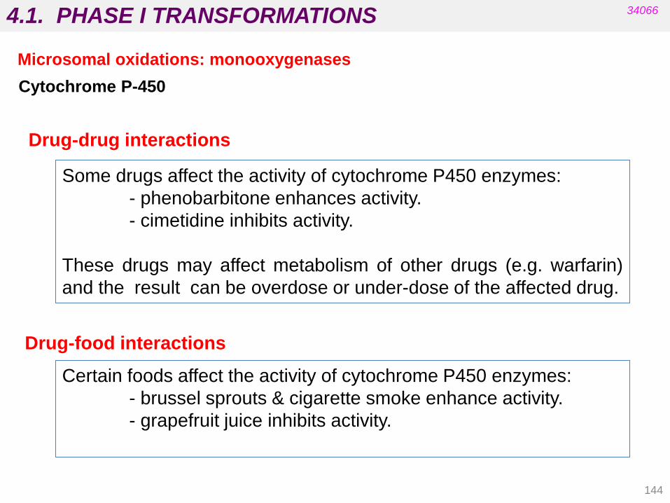
4.1. PHASE I TRANSFORMATIONS
144
Some drugs affect the activity of cytochrome P450 enzymes:
- phenobarbitone enhances activity.
- cimetidine inhibits activity.
These drugs may affect metabolism of other drugs (e.g. warfarin)
and the result can be overdose or under-dose of the affected drug.
Drug-drug interactions
Certain foods affect the activity of cytochrome P450 enzymes:
- brussel sprouts & cigarette smoke enhance activity.
- grapefruit juice inhibits activity.
Drug-food interactions
34066
Microsomal oxidations: monooxygenases
Cytochrome P-450
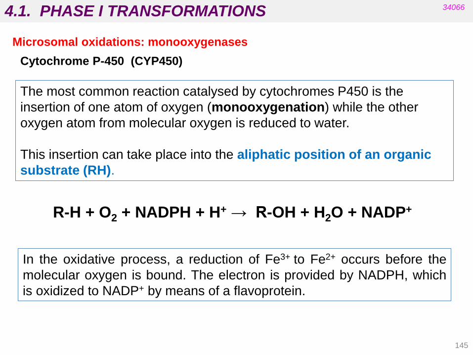
4.1. PHASE I TRANSFORMATIONS
145
34066
The most common reaction catalysed by cytochromes P450 is the
insertion of one atom of oxygen (monooxygenation) while the other
oxygen atom from molecular oxygen is reduced to water.
This insertion can take place into the aliphatic position of an organic
substrate (RH).
R-H + O2 + NADPH + H+ → R-OH + H2O + NADP+
In the oxidative process, a reduction of Fe3+ to Fe2+ occurs before the
molecular oxygen is bound. The electron is provided by NADPH, which
is oxidized to NADP+ by means of a flavoprotein.
Microsomal oxidations: monooxygenases
Cytochrome P-450 (CYP450)
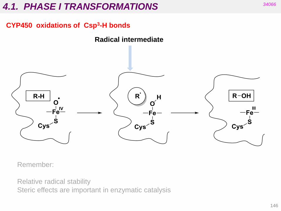
4.1. PHASE I TRANSFORMATIONS
146
34066
Radical intermediate
Remember:
Relative radical stability
Steric effects are important in enzymatic catalysis
CYP450 oxidations of Csp3-H bonds
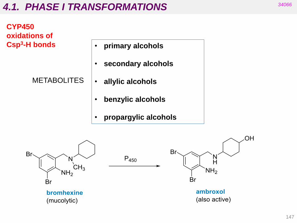
4.1. PHASE I TRANSFORMATIONS
147
34066
CYP450
oxidations of
Csp3-H bonds • primary alcohols
• secondary alcohols
• allylic alcohols
• benzylic alcohols
• propargylic alcohols
METABOLITES

4.1. PHASE I TRANSFORMATIONS
148
CYP450 oxidations of Csp3-H bonds: allylic and benzylic positions
34066
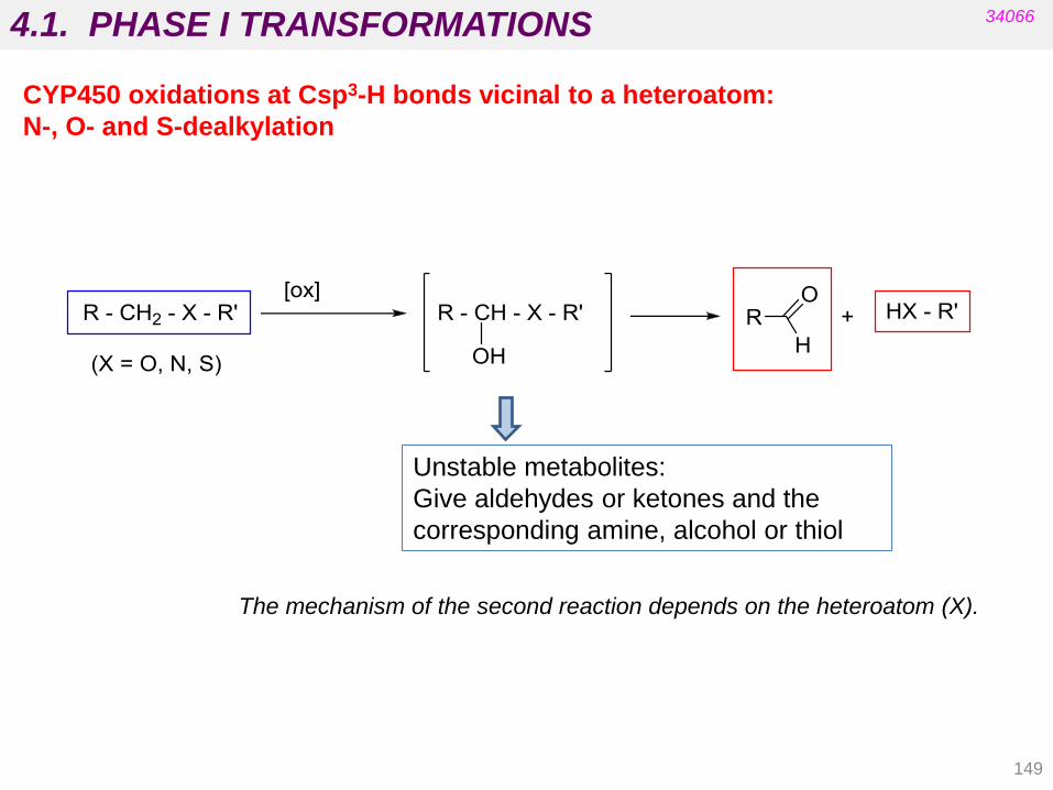
4.1. PHASE I TRANSFORMATIONS
149
34066
CYP450 oxidations at Csp3-H bonds vicinal to a heteroatom:
N-, O- and S-dealkylation
Unstable metabolites:
Give aldehydes or ketones and the
corresponding amine, alcohol or thiol
The mechanism of the second reaction depends on the heteroatom (X).

4.1. PHASE I TRANSFORMATIONS
150
34066
CYP450 oxidations at Csp3-H bonds vicinal to a heteroatom:
N-, O- and S-dealkylation

4.1. PHASE I TRANSFORMATIONS
151
imipramine desipramine
(principal)
(minor products)
34066
CYP450 oxidations at Csp3-H bonds vicinal to a heteroatom:
N-, O- and S-dealkylation

4.1. PHASE I TRANSFORMATIONS
152
34066
CYP450 oxidations at Csp3-H bonds vicinal to a halogen atom
chloramphenicol
(antibiotic)
RR‟CHX RR‟C(OH)X RCOR‟ + HX
Dehalogenation of alkyl chlorides

4.1. PHASE I TRANSFORMATIONS
153
34066
Double bonds, aromatic rings and heteroatoms can also be oxidized by CYP450.
More polar
derivatives
More polar
derivatives
Drug + O2 + NADPH + H+ → Oxidized drug + H2O + NADP+
Epoxides are
electrophilic:
Nucleophilic
addition from
amino, thiol or
hydroxyl groups
from proteins or
nucleic acids can
take place
(toxicity).

4.1. PHASE I TRANSFORMATIONS
irreversible
alkylation
(toxicity)
ketene
34066
CYP450 oxidations of double and triple bonds
154
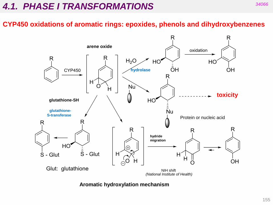
4.1. PHASE I TRANSFORMATIONS
155
Aromatic hydroxylation mechanism
NIH shift (National Institute of Health)
hydrolase
arene oxide
toxicity glutathione-SH
glutathione-
S-transferase
hydride
migration
34066
CYP450 oxidations of aromatic rings: epoxides, phenols and dihydroxybenzenes
Protein or nucleic acid
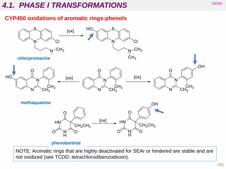
4.1. PHASE I TRANSFORMATIONS
156
34066
CYP450 oxidations of aromatic rings:phenols
NOTE: Aromatic rings that are highly deactivated for SEAr or hindered are stable and are
not oxidized (see TCDD: tetrachlorodibenzodioxin).
phenobarbital
methaqualone
chlorpromazine
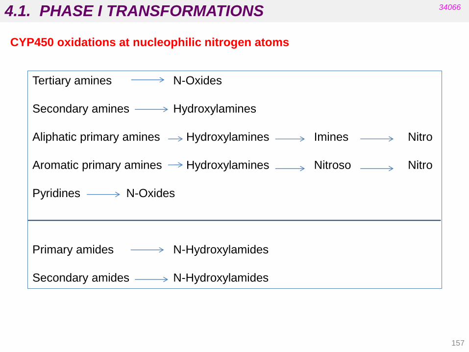
4.1. PHASE I TRANSFORMATIONS
157
34066
CYP450 oxidations at nucleophilic nitrogen atoms
Tertiary amines N-Oxides
Secondary amines Hydroxylamines
Aliphatic primary amines Hydroxylamines Imines Nitro
Aromatic primary amines Hydroxylamines Nitroso Nitro
Pyridines N-Oxides
Primary amides N-Hydroxylamides
Secondary amides N-Hydroxylamides

4.1. PHASE I TRANSFORMATIONS
158
34066
CYP450 oxidations at nucleophilic sulphur atoms
metaqualone (hypnotic)
cimetidine (treatment of peptic ulcers)
Thiol (x 2) Disulphide
Thiol Sulfoxide Sulphone
Thioketone Ketone (desulphurization)

4.1. PHASE I TRANSFORMATIONS 34066
Phase I reactions catalysed by flavin-containing monooxigenases
Many of these reactions are also
catalysed by cytochrome P450
enzymes.
R1 NMe2 R1 NMe2
O
R1 NHR R1 NHR
OH
R2 N NHR1 R2 N NHR1
O
2 RSH RS - SR
159

4.1. PHASE I TRANSFORMATIONS
160
34066
4.1.1. Microsomal oxidations Carbon sp3 oxidations Allylic and benzylic oxidations Oxidative dealkylations. Amines, ethers and thioethers.
Oxidative dehydrohalogenation
Alkene and alkyne oxidations
Aromatic hydroxylation
Amine and sulphur derivative oxidations
4.1.2. Non-microsomal oxidations
Alcohol and aldehyde oxidations catalyzed by dehydrogenases
Oxidative deamination catalyzed by monoamine oxidase
Purine oxidation
4.1.3. Reductions
Azo reductions
Ketone reductions
Nitro-derivative reductions
Alkene reductions
Sulfoxide reductions
4.1.4. Hydrolytic reactions
Hydrolysis of nitriles, esters, and amides Hydration of epoxides
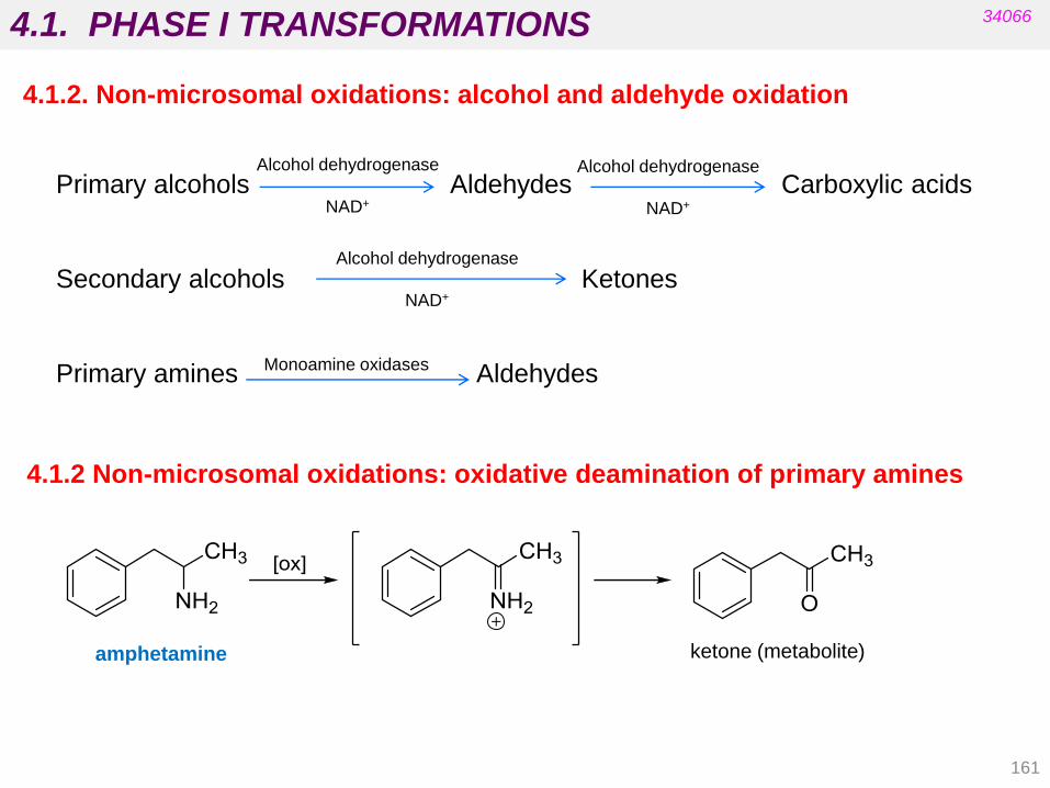
4.1. PHASE I TRANSFORMATIONS
161
34066
4.1.2. Non-microsomal oxidations: alcohol and aldehyde oxidation
Primary alcohols Aldehydes Carboxylic acids
Secondary alcohols Ketones
Primary amines Aldehydes
Alcohol dehydrogenase
NAD+
Monoamine oxidases
Alcohol dehydrogenase
NAD+
Alcohol dehydrogenase
NAD+
4.1.2 Non-microsomal oxidations: oxidative deamination of primary amines
amphetamine ketone (metabolite)

4.1. PHASE I TRANSFORMATIONS
162
34066
Non-microsomal oxidations: oxidative deamination of primary amines
(MAO)
Compare with oxidative N-dealkylation
Flavin adenine-dinucleotide
(cofactor)
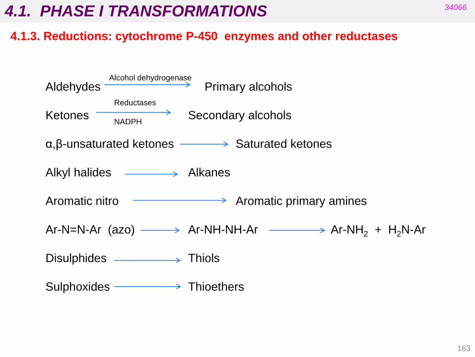
4.1. PHASE I TRANSFORMATIONS
163
34066
4.1.3. Reductions: cytochrome P-450 enzymes and other reductases
Aldehydes Primary alcohols
Ketones Secondary alcohols
α,β-unsaturated ketones Saturated ketones
Alkyl halides Alkanes
Aromatic nitro Aromatic primary amines
Ar-N=N-Ar (azo) Ar-NH-NH-Ar Ar-NH2 + H2N-Ar
Disulphides Thiols
Sulphoxides Thioethers
Alcohol dehydrogenase
Reductases
NADPH

4.1. PHASE I TRANSFORMATIONS
164
34066
4.1.3. Reductions: cytochrome P-450 enzymes and other reductases
clonazepam

4.1. PHASE I TRANSFORMATIONS
165
4.1.4. Hydrolysis
34066
Clofibrate (hypolipidemic)
Iproniazid
(antidepressant)
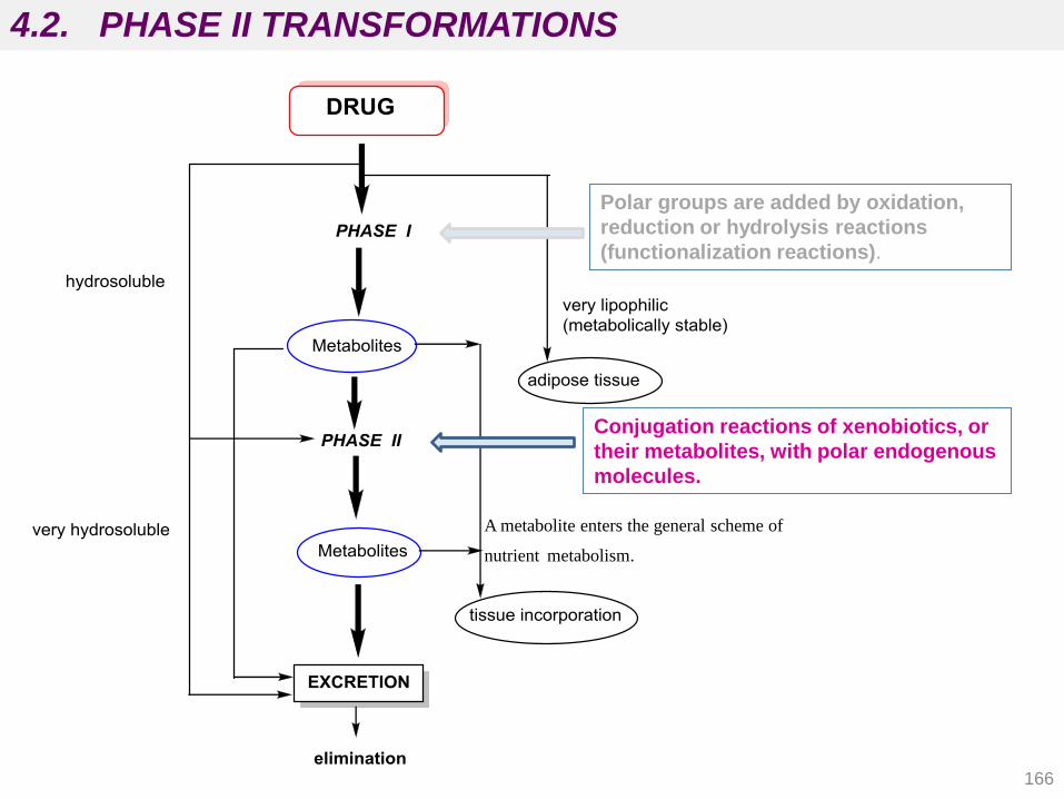
166
4.2. PHASE II TRANSFORMATIONS
Polar groups are added by oxidation,
reduction or hydrolysis reactions
(functionalization reactions).
Conjugation reactions of xenobiotics, or
their metabolites, with polar endogenous
molecules.
A metabolite enters the general scheme of
nutrient metabolism.

4.2. PHASE II TRANSFORMATIONS
167
4.2.1. Conjugation with glucuronic acid
4.2.2. Conjugation with sulphate
4.2.3. Conjugation with glutathione
4.2.4. Conjugation with amino acids
4.2.5. Acylation reactions
4.2.6. Methylation reactions
34066
Enzymes in phase II reactions:
1. Uridine diphosphate glucuronyl transferase (UDP-glucuronyl transferase)
2. 3’-Phosphoadenosine-5’- phosphosulphate transferase
3. Glutathione transferase
4. Amino acid N-acyl transferase
5. N-Acyl transferase
6. Methyl transferase

4.2. PHASE II TRANSFORMATIONS
168
34066
4.2.1. Glucuronides (nucleophilic atoms in drugs)
(uridine diphosphate
glucuronyl transferase)
R-X-H:
• Alcohols and amines
• Hydroxylamines R2N-O-H
• Thiols R-S-H
• Phenols Ar-O-H
• Carboxylic acids RCO-O-H
• Sulphonamides R-SO2-NR‟-H

4.2. PHASE II TRANSFORMATIONS
169
34066
4.2.1. Glucuronides (nucleophilic atoms in drugs)

4.2. PHASE II TRANSFORMATIONS
170
3‟-phosphoadenosine-5‟- phosphosulphate
34066
4.2.2. Sulphates (nucleophilic atoms in drugs)

4.2. PHASE II TRANSFORMATIONS
171
34066
4.2.2. Sulphates (nucleophilic atoms in drugs)
Ar – OH
Phenols
R – OH
Alcohols
R2N – H
Amines
Ar – O – SO3 -
sulphate conjugate
R – O – SO3 -
sulphate conjugate
R2N – SO3 -
sulphate
Sulphotransferase
PAPS
Sulphotransferase
PAPS
Sulphotransferase
PAPS
Ionized at physiological pH salbutamol
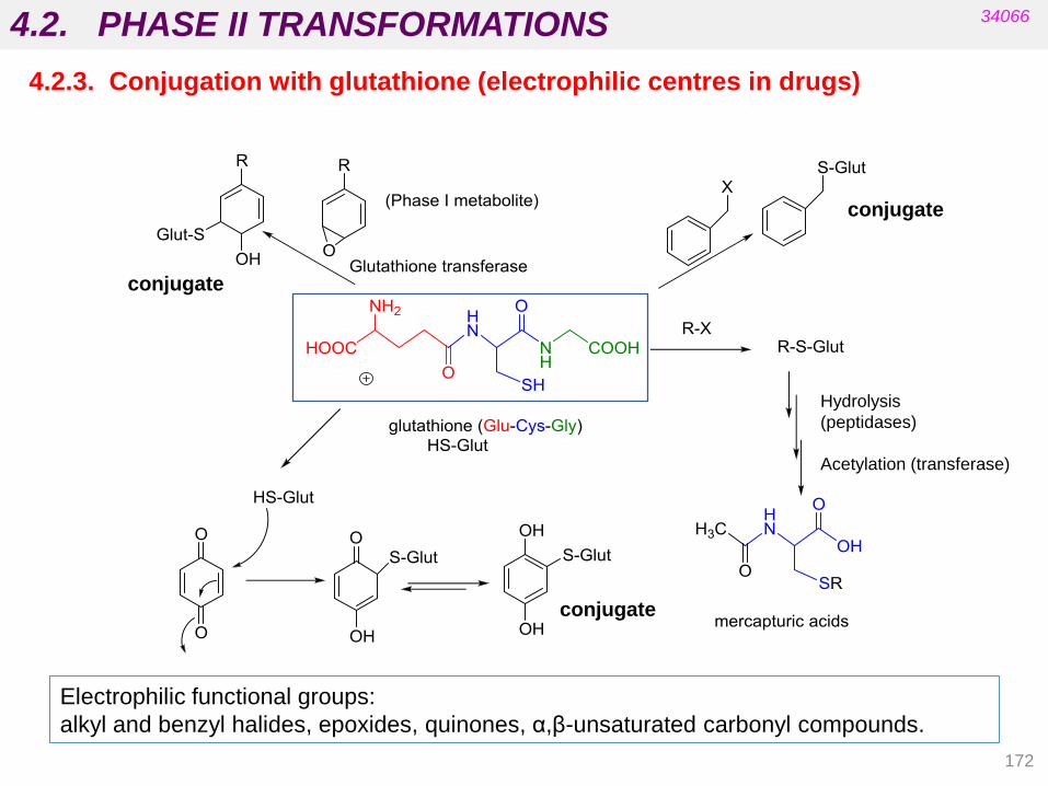
4.2. PHASE II TRANSFORMATIONS
172
34066
4.2.3. Conjugation with glutathione (electrophilic centres in drugs)
conjugate
conjugate
conjugate
Electrophilic functional groups:
alkyl and benzyl halides, epoxides, quinones, α,β-unsaturated carbonyl compounds.
Hydrolysis
(peptidases)
Acetylation (transferase)

4.2. PHASE II TRANSFORMATIONS
173
34066
4.2.3. Conjugation with glutathione (electrophilic centres in drugs)
Propose the structure of the products that result from the reaction of Glut-SH with the
following substrates:
HS-Glut
HS-Glut
HS-Glut
HS-Glut
(conjugate addition)

4.2. PHASE II TRANSFORMATIONS
174
34066
4.2.4. Conjugation with amino acids (aromatic and arylacetic acids)
Adenosine triphosphate
Adenosine monophosphate
Active acyl-coenzyme A
intermediate N-acyl transferase
Drug-COOH
ACTIVATION
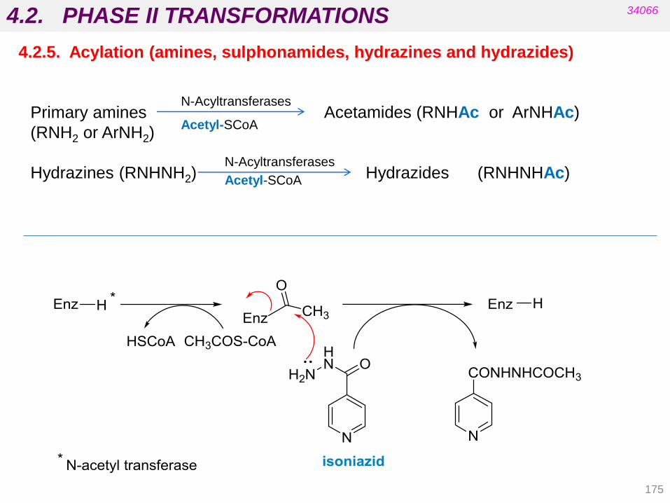
4.2. PHASE II TRANSFORMATIONS
175
34066
4.2.5. Acylation (amines, sulphonamides, hydrazines and hydrazides)
Primary amines
(RNH2 or ArNH2)
Hydrazines (RNHNH2) Hydrazides (RNHNHAc)
N-Acyltransferases
Acetyl-SCoA
N-Acyltransferases
Acetyl-SCoA
Acetamides (RNHAc or ArNHAc)

4.2. PHASE II TRANSFORMATIONS
176
4.2.5. Methylation (amines, thiols, pyridines and phenol groups)
34066
Methylated the hydroxyl group at
meta position to the alkyl substituent
Ar – OH
Phenols
R – SH
Thiols
R2N – H
Amines
Ar – O - Me
R – S - Me
R2N – Me
Methyltransferase
S-Adenosylmethionine
Methyltransferase
S-Adenosylmethionine
Methyltransferase
S-Adenosylmethionine

4.2. DRUG METABOLISM 34066
Consequences of drug metabolism
- Deactivation
- Phenobarbital (aromatic hydroxylation)
- Procaine (ester hydrolysis)
- Bioactivation (prodrugs)
- Phenacetin (paracetamol)
- Prontosil (sulfanilmide)
- Imipramine (desipramine)
- Activity change (iproniazid)
N-dealkylation hydrolysis
isoniazid
(tuberculostatic)
Iproniazid
(antidepressant)
Isopropylhydrazine
177
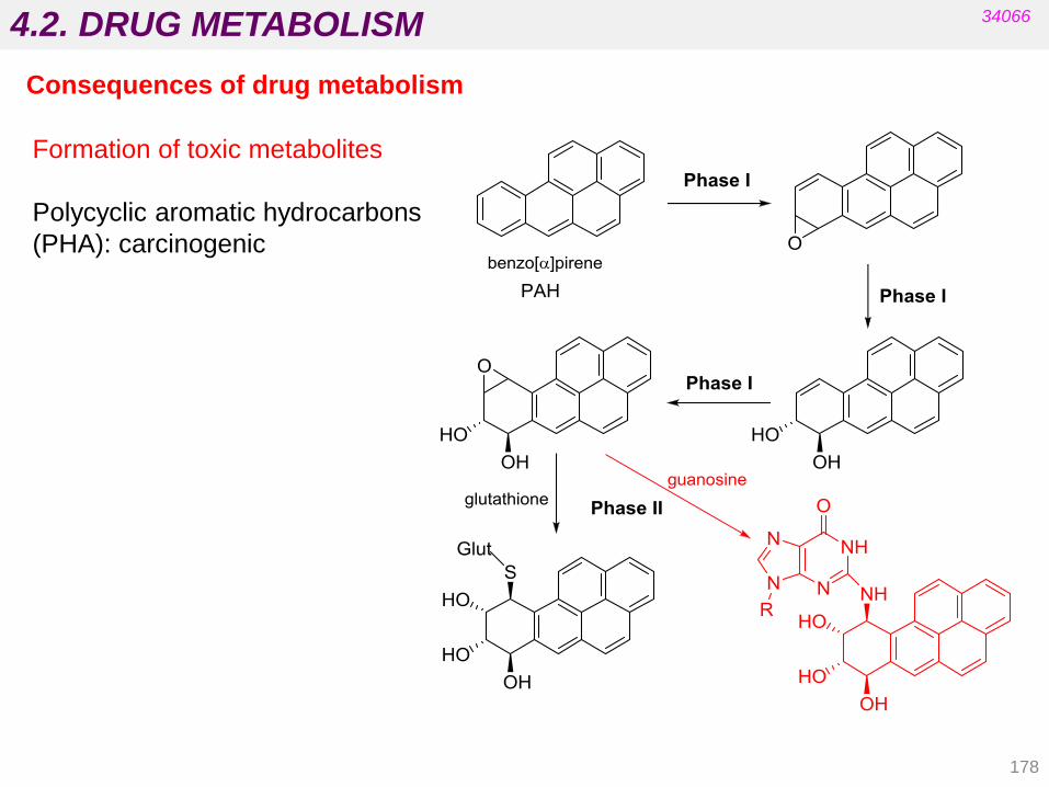
4.2. DRUG METABOLISM
178
34066
Consequences of drug metabolism
Formation of toxic metabolites
Polycyclic aromatic hydrocarbons
(PHA): carcinogenic
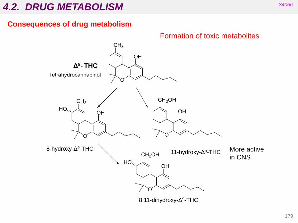
4.2. DRUG METABOLISM
179
34066
8-hydroxy-Δ9-THC 11-hydroxy-Δ9-THC
Δ9- THC
8,11-dihydroxy-Δ9-THC
More active
in CNS
Formation of toxic metabolites
Consequences of drug metabolism

4.2. DRUG METABOLISM
180
34066
Consequences of drug metabolism
Formation of toxic metabolites
Paracetamol:
conjugates (sulphate and glucuronide, excretion, detoxification)
oxidation in liver: toxicity
See next slide
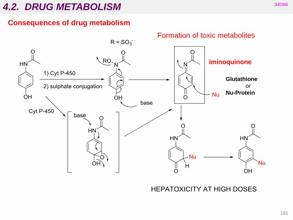
4.2. DRUG METABOLISM 34066
Consequences of drug metabolism
Glutathione
or
Nu-Protein
181
iminoquinone
Formation of toxic metabolites

4.3. BIOREVERSIBLE DERIVATIVES
182
34066
SEVERAL PROBLEMS CAN LIMIT THE EFFECTIVENESS OF A POTENTIAL DRUG
pharmacodynamics / pharmacokinetics / chemistry / galenics
DRUG
poor absorption
fast absorption
low specificity
chemical instability
formulation
problems
difficulty of patient
acceptance
insolubility
in water toxicity
does (not) cross
the blood-brain
barrier
biotransformation before arriving to the site of action

4.3. BIOREVERSIBLE DERIVATIVES
183
34066
BIOREVERSIBLE DERIVATIVES:
Structural changes are intended to obtain INACTIVE derivatives which will be
activated in our body (bioactivation) by means of a metabolic process.
Bioreversible derivative is synonymous with prodrug or latent drug.
Prodrug “An inactive compound which is converted into an
active compound in the body” (Albert 1951 )
Prodrugs are masked forms of active drugs that are designed to become
activated after an enzymatic or chemical reaction after they have been
administered in the body. There are two types of prodrugs:
• carrier-linked prodrugs
• bioprecursor prodrugs
SOLUTIONS TWO APPROACHES :
PHARMACOMODULATION: DESIGNING IMPROVED ACTIVE ANALOGUES:
Structural changes are incorporated into active compounds in order to maintain
activity and eliminate or decrease undesirable properties.
(TO BE DEVELOPED IN UNIT 5)

4.3. BIOREVERSIBLE DERIVATIVES
184
34066
A carrier-linked prodrug is a prodrug that contains a temporary linkage of a
given active substance with a transient carrier group that can be easily removed
in vivo, usually by a hydrolytic reaction.
A bioprecursor prodrug is a prodrug that results from a molecular modification
of the active principle generating a new compound that is able to be transformed
metabolically or chemically. The resulting compound is the active principle.
Modification does not involve linkage to a carrier group.
CARRIER-LINKED
PRODRUGS
BIOPRECURSOR
PRODRUGS
D - C
D’
D-C D Hydrolysis
- C
D’ D No hydrolytic
reactions
D = active drug
D - C = carrier-linked prodrug, inactive
D‟ = inactive derivative

4.3. BIOREVERSIBLE DERIVATIVES
185
34066
DRUG AND CARRIER
1) Union by covalent bond (non-ionic).
2) The prodrug must be inactive or
much less active than the drug.
3) The bond must be broken in vivo.
4) The prodrug and the carrier must be
non-toxic.
5) The regeneration of the drug in the
target must be fast so as to avoid
alternative metabolism.
DRUG
DRUG
DRUG
CARRIER
CARRIER
CARRIER
In vivo
regeneration
Synthesis
PRODRUG
Carrier-linked prodrugs
When, because of steric hindrance or functional properties, the desired prodrug carrier
cannot be attached directly to the parent active molecule, conventional carrier-linked
prodrugs can have a synthetic handle (a spacer or linker) between the active drug and the
carrier.
Bioactivation

4.3. BIOREVERSIBLE DERIVATIVES
CARRIER-LINKED PRODRUGS
- A labile additional fragment is temporarily attached to the drug.
- Prodrugs facilitate drug localization/distribution.
- Alteration of lipophilicity in relation to the active drug. - Metabolic activation is done by (chemical or enzymatic) hydrolysis.
BIOPRECURSOR PRODRUGS
- There is no additional labile fragment.
- There are no major differences in lipophilicity between the
bioprecursor prodrug and the active drug.
- Activation occurs primarily by redox processes (phase I metabolic reactions).
34066
186
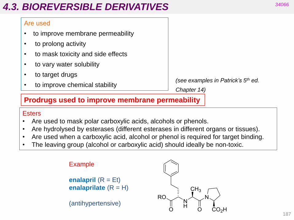
4.3. BIOREVERSIBLE DERIVATIVES
187
34066
Are used
• to improve membrane permeability
• to prolong activity
• to mask toxicity and side effects
• to vary water solubility
• to target drugs
• to improve chemical stability
Prodrugs used to improve membrane permeability
Esters
• Are used to mask polar carboxylic acids, alcohols or phenols.
• Are hydrolysed by esterases (different esterases in different organs or tissues).
• Are used when a carboxylic acid, alcohol or phenol is required for target binding.
• The leaving group (alcohol or carboxylic acid) should ideally be non-toxic.
Example
enalapril (R = Et)
enalaprilate (R = H)
(antihypertensive)
(see examples in Patrick’s 5th ed.
Chapter 14)

4.3. BIOREVERSIBLE DERIVATIVES
188
34066
These para groups withdraw electron
density from the nitrogen atom.
the active compound is the aziridinium salt INACTIVE
Anticancer
β-haloalkylamines
Prodrugs used to target drugs

4.3. BIOREVERSIBLE DERIVATIVES
189
34066
Precursor that
can cross the brain
membrane Active on the CNS
(central nervous
system)
oxidation oxidation
baclofen
Prodrugs used to target drugs
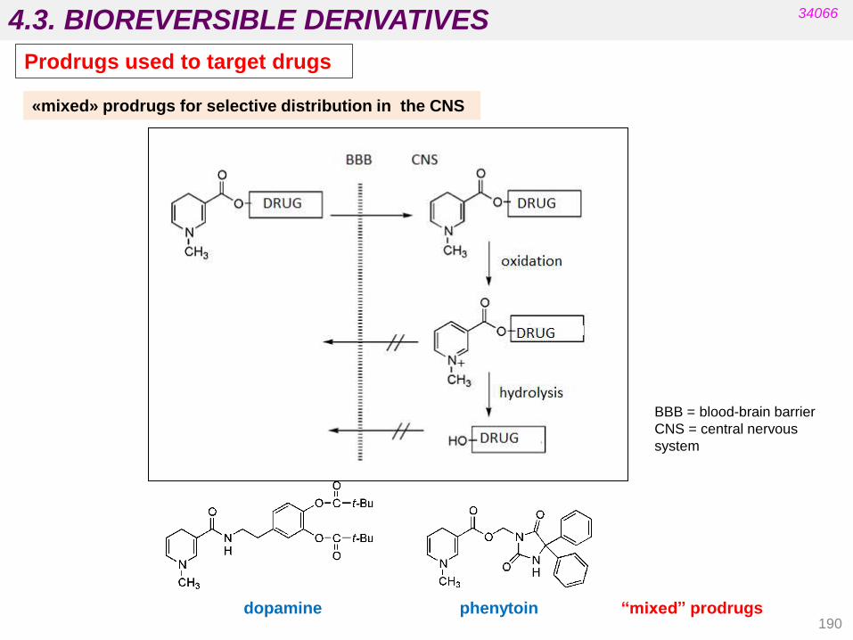
4.3. BIOREVERSIBLE DERIVATIVES
190
34066
dopamine phenytoin “mixed” prodrugs
BBB = blood-brain barrier
CNS = central nervous
system
«mixed» prodrugs for selective distribution in the CNS
Prodrugs used to target drugs
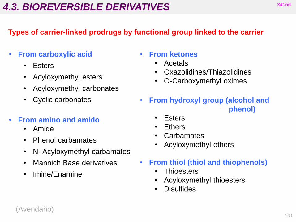
4.3. BIOREVERSIBLE DERIVATIVES
191
34066
Types of carrier-linked prodrugs by functional group linked to the carrier
• From carboxylic acid
• Esters
• Acyloxymethyl esters
• Acyloxymethyl carbonates
• Cyclic carbonates
• From amino and amido
• Amide
• Phenol carbamates
• N- Acyloxymethyl carbamates
• Mannich Base derivatives
• Imine/Enamine
• From ketones
• Acetals
• Oxazolidines/Thiazolidines
• O-Carboxymethyl oximes
• From hydroxyl group (alcohol and
phenol)
• Esters
• Ethers
• Carbamates
• Acyloxymethyl ethers
• From thiol (thiol and thiophenols)
• Thioesters
• Acyloxymethyl thioesters
• Disulfides
(Avendaño)
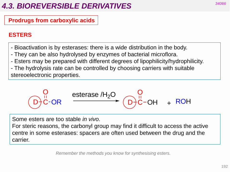
4.3. BIOREVERSIBLE DERIVATIVES
192
34066
Prodrugs from carboxylic acids
Some esters are too stable in vivo.
For steric reasons, the carbonyl group may find it difficult to access the active
centre in some esterases: spacers are often used between the drug and the
carrier.
- Bioactivation is by esterases: there is a wide distribution in the body.
- They can be also hydrolysed by enzymes of bacterial microflora.
- Esters may be prepared with different degrees of lipophilicity/hydrophilicity.
- The hydrolysis rate can be controlled by choosing carriers with suitable
stereoelectronic properties.
ESTERS
D C
O
OResterase /H2O
D C
O
OH + ROH
Remember the methods you know for synthesising esters.

D C
OH
O-CH2-O CR
Oesterase
H2O
D C
O
O-CH2-O-H + R-C-OH
O
D C
O
OH +
Hydroxymethyl ester
HCHO
4.3. BIOREVERSIBLE DERIVATIVES
193
ACYLOXYMETHYL ESTERS
Bioactivation: the external ester group gains easier access to the esterase active site:
metabolites
34066
Prodrugs from carboxylic acids

4.3. BIOREVERSIBLE DERIVATIVES
194
ACYLOXYMETHYL CARBONATES
34066
Prodrugs from carboxylic acids
D C
O
O-CH2-O C
Oesterase
H2O
D C
OH
O-CH2-O-C-O-H + R-OH
D C
O
OH HCHO
O
D C
O
O-CH2-O-H +
OR
O C O

4.3. BIOREVERSIBLE DERIVATIVES
195
34066
N
S
O
CH3
CH3
NH2
O
NH
OO
O
OCH
3
CH3
CH3
N
S
O
CH3
CH3
NH2
O
NH
OO
O
OO
CH3
CH3
N
S
O
CH3
CH3
NH2
O
NH
OOH
CH3
OH
O
CH3
CH3
O H
H
OCH3
H
CH3
OH
CO2
in vivo in vivo
Pivampicilina Bacampicilina
Ampicilina
Example: ampicillin prodrug derivatives
Broad spectrum antibiotic
pivampicillin bacampicillin
ampicillin
Prodrugs from carboxylic acids

4.3. BIOREVERSIBLE DERIVATIVES
196
34066
CYCLIC CARBONATES
Prodrugs from carboxylic acids
D C
O
O
O O
O H2O
D C
O
OH
O O
O
O-H
O O+
+
CO O

4.3. BIOREVERSIBLE DERIVATIVES
197
34066
Prodrugs from amine groups

4.3. BIOREVERSIBLE DERIVATIVES
198
34066
Prodrugs from amine groups
AMIDES
Amides are difficult to hydrolyse except if the carboxylic part is one amino acid
(in such case, the amides can be degraded by peptidases):
PHENOL CARBAMATES
Carbamates are generally quite stable but phenol carbamates (RNHCO2Ph) are
easily hydrolysed by plasmatic esterases:
D NH C
OHC NH2
R
peptidase
H2O
D NH2 C
OHC NH2
RHO
+
D NH C
Oesterase
H2O
D NH +O C
OO-H
D-NH2 + CO O
carbamic acid: unstable
OH

4.3. BIOREVERSIBLE DERIVATIVES
199
N-ACYLOXYMETHYL CARBAMATES
34066
Prodrugs from amine groups
D NHC
O
O O C
O
R
esterase H2O
D-NH2 + +C OO H2CO R C
O
OH+

4.3. BIOREVERSIBLE DERIVATIVES
200
MANNICH BASES
These are obtained by the reaction of a drug (amine), formaldehyde and
an amide.
The nitrogen basicity decreases in 2-3 units and the N atom is not
protonated at physiological pH. Consequently, lipophilicity increases.
Bioactivation:
34066
Prodrugs from amine groups

4.3. BIOREVERSIBLE DERIVATIVES
201
34066
Prodrugs from amine groups
stabilized imines
amides
enamines
Glycine amide derivative
Easy to hydrolyse by peptidases
Hydrogen bond that
stabilizes the imine
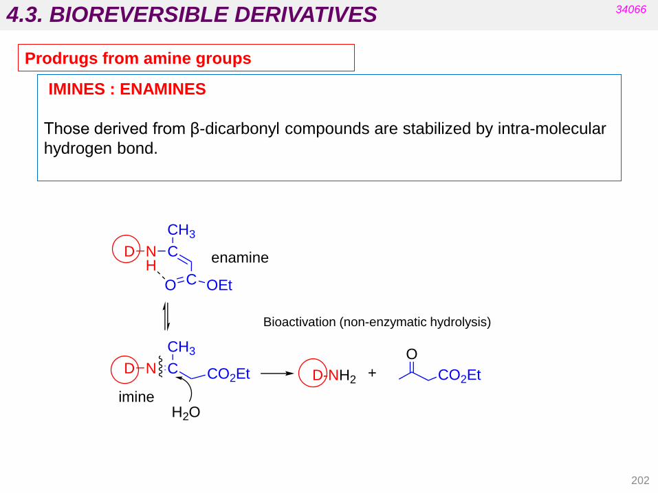
D N C
CH3
CO2Et
H2O
D-NH2
O
CO2Et+
D N C
CH3
CO OEt
Henamine
imine
4.3. BIOREVERSIBLE DERIVATIVES
202
IMINES : ENAMINES
Those derived from β-dicarbonyl compounds are stabilized by intra-molecular
hydrogen bond.
Bioactivation (non-enzymatic hydrolysis)
34066
Prodrugs from amine groups

4.3. BIOREVERSIBLE DERIVATIVES
203
AMIDES: MANNICH-BASE PRODRUGS
FORMATION: drug + formaldehyde + secondary amine.
BIOACTIVATION: does not need enzymatic hydrolysis (very unstable).
IMIDES: ACYLOXYMETHYL DERIVATIVES
Prodrugs from amide and imide groups
34066
D
HN
O
N
R
RD NH2
O
+ H2C NR
R
H2C O + R-NH-R
+ H+
+ H2O

4.3. BIOREVERSIBLE DERIVATIVES
204
Prodrugs from carbonyl groups
Acetals
Thiazolidines/ oxazolidines
O-carboxymethyl oximes
34066

4.3. BIOREVERSIBLE DERIVATIVES
205
34066
Prodrugs from carbonyl groups
Bioactivation
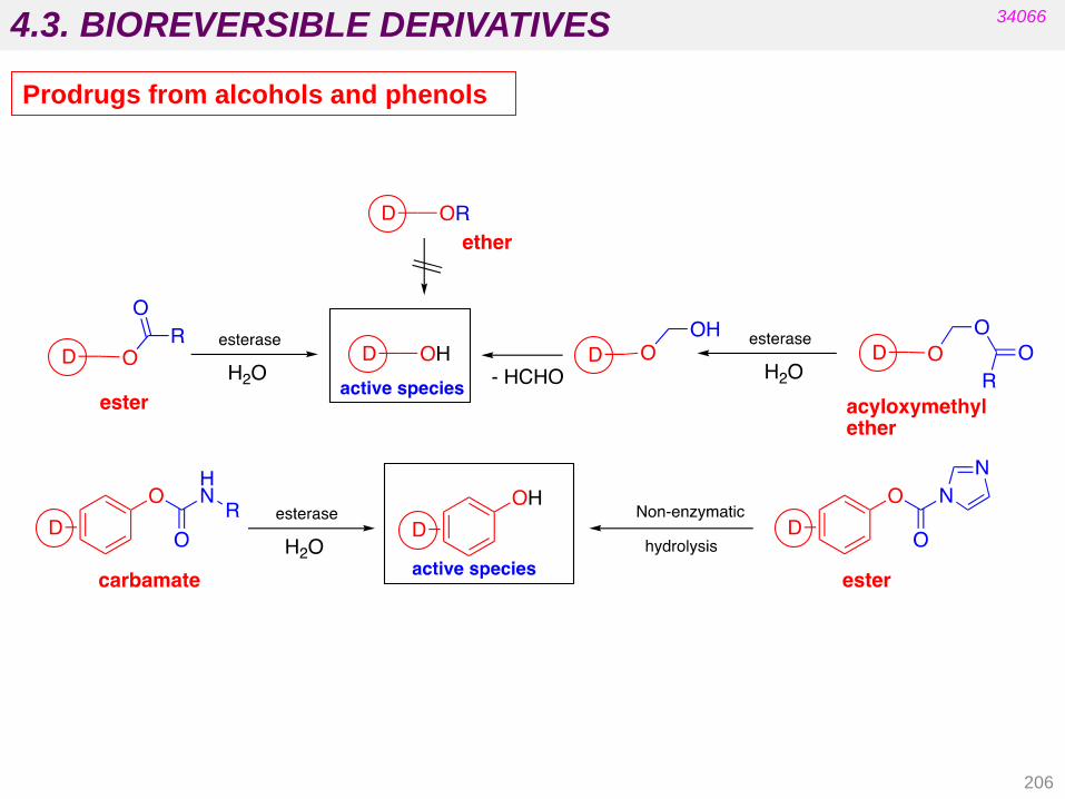
4.3. BIOREVERSIBLE DERIVATIVES
206
34066
Prodrugs from alcohols and phenols

4.3. BIOREVERSIBLE DERIVATIVES
207
34066
ESTERS
EXCEPTION: SUCCINATE HEMI-ESTERS
Hydrolysis does not require esterases because it is facilitated by intra-
molecular attack from the free carboxylate group:
Prodrugs from alcohols and phenols
D O C
O
Resterase
D-OH
H2O
C
O
OHR+
D O
O
O
O
D-O- + O
O
O
HOH
D-OH +-OOC COO-

D O O
O
R
H2O
D O O-HHO
O
R+
D-OH + CH2
O
4.3. BIOREVERSIBLE DERIVATIVES
208
PHENOLS:
CARBAMATES (also esters and acyloxymethyl ethers such as alcohols)
34066
ACYLOXYMETHYL ETHERS
Prodrugs from alcohols and phenols
esterase
D
OC
O
HN R
esterase
H2O
OH
D
O
H-O NH-R
CO O + R-NH2

4.3. BIOREVERSIBLE DERIVATIVES
Imidazol-derived phenol carbamates:
the phenolic drug is liberated by non-enzymatic hydrolysis.
34066
209
Prodrugs from phenols

4.3. BIOREVERSIBLE DERIVATIVES
210
34066
Prodrugs from thiols and thiophenols

4.3. BIOREVERSIBLE DERIVATIVES
211
34066
Non-enzymatic
process
Singular example: activation of omeprazol (proton pump inhibitor in the stomach)
Omeprazol
(prodrug)
Sulfenic acid
Sulfenamide
Active species

4.4. HARD DRUGS AND SOFT DRUGS
212
34066
Hard drugs are biologically active drugs that are resistant to metabolism and
remain unchanged in the body.
They are poor substrates for the metabolizing enzymes.
The potentially metabolically-sensitive parts of these drugs are either sterically
hindered or the hydrogen atoms are substituted for halogens to block oxidation.
acetylcholine
carbachol
Electronic or steric changes in the structure of acetylcholine prolong its action.
Drugs may be defined as hard or soft depending on their metabolic
susceptibility.
R R‟ hydrolysis rate
H H 500
CH3 H 15
CH3 CH3 0
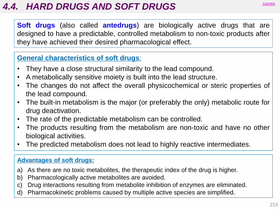
4.4. HARD DRUGS AND SOFT DRUGS
213
34066
General characteristics of soft drugs:
• They have a close structural similarity to the lead compound.
• A metabolically sensitive moiety is built into the lead structure.
• The changes do not affect the overall physicochemical or steric properties of
the lead compound.
• The built-in metabolism is the major (or preferably the only) metabolic route for
drug deactivation.
• The rate of the predictable metabolism can be controlled.
• The products resulting from the metabolism are non-toxic and have no other
biological activities.
• The predicted metabolism does not lead to highly reactive intermediates.
Soft drugs (also called antedrugs) are biologically active drugs that are
designed to have a predictable, controlled metabolism to non-toxic products after
they have achieved their desired pharmacological effect.
Advantages of soft drugs:
a) As there are no toxic metabolites, the therapeutic index of the drug is higher.
b) Pharmacologically active metabolites are avoided.
c) Drug interactions resulting from metabolite inhibition of enzymes are eliminated.
d) Pharmacokinetic problems caused by multiple active species are simplified.
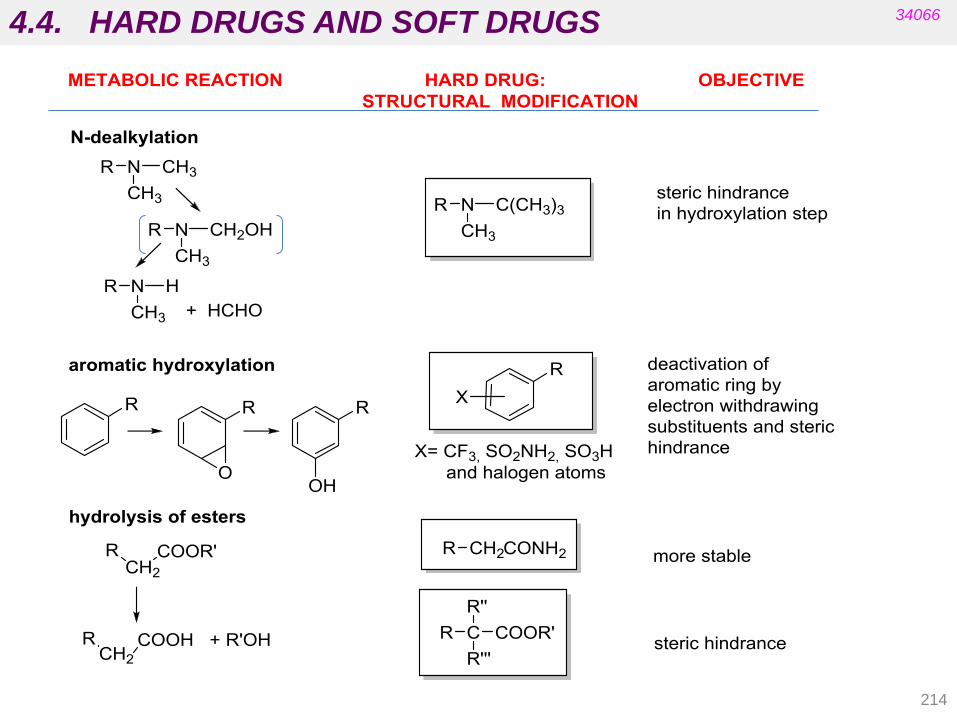
4.4. HARD DRUGS AND SOFT DRUGS
214
34066
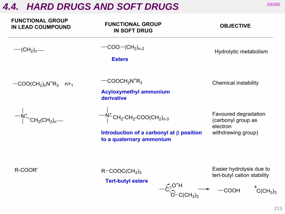
4.4. HARD DRUGS AND SOFT DRUGS
215
34066

4.4. HARD DRUGS AND SOFT DRUGS
216
34066
aryloxypropanolamine
Soft analogue of an
aryloxypropanolamine
acetylcholine esters
Soft analogue of an
anticholinergic

4.4. HARD DRUGS AND SOFT DRUGS
217
34066
Cetylpyridinium salts to local-use antiseptics
soft analogue Non-enzymatic step
(H2O)

4.4. HARD DRUGS AND SOFT DRUGS
218
34066
Acyloxymethyl amonium derivatives
Introduction of a b-carbonyl to to quaternary ammonium

PHARMACEUTICAL CHEMISTRY
UNIT 5. DESIGN AND DEVELOPMENT OF NEW DRUGS
34066
5.1. Evolution of research and drug discovery
methods. Current methods for discovering lead
compounds.
(Patrick‟s 5th ed. Ch. 12 and 16; Silverman‟s 3
rd ed. Ch. 1 and 2, Wermuth‟s 4
th ed. Ch. 4-8)
5.2. Qualitative relationships chemical structure-
biological activity (SAR). Concept of pharmacophore
and auxophoric group. Drug design based on
molecular modelling.
5.3. Pharmacomodulation.
(Patrick‟s 5th ed. Ch. 13, 14; Silverman‟s 3
rd ed. Ch. 2, Wermuth 4
th ed Ch. 4 and 12)
5.4. Examples of rational strategies in drug design:
receptor activation and/or deactivation and enzyme
inhibition. (Patrick‟s 5th ed. Chapters 7 and 8)
219

5.1. EVOLUTION OF RESEARCH AND DRUG DISCOVERY
220
Drugs discovered without rational design
• Medicinal chemistry folklore
• Serendipitous drug discovery
• Discovery through metabolism studies
• Discovery through clinical observations
DRUGS or Lead Compounds Targets (mechanism)
Historically, compounds produced by medicinal chemists have been screened by
pharmacologists in vivo in whole-animal models of disease.
Many important drugs, such as aspirin and penicillin, were marketed without
knowledge of their mechanism of action.
The Past
Medicinal chemistry involves Lead discovery, Lead optimization, Process
chemistry and development, and determination of the mechanisms of drug action.
It is a discipline at the intersection of Synthetic Organic Chemistry and
Pharmacology that focuses on small organic molecules (not on biologics and
inorganic compounds).
34066

5.1. EVOLUTION OF RESEARCH AND DRUG DISCOVERY
Also:
reserpine, ephedrine, scopolamine, quinine, digoxin, atropine tubocurarine, etc.
These natural products are used as drugs but have also inspired the design of
synthetic analogues. (MEDICINAL CHEMISTRY)
221
34066
• Without rational design: Medicinal chemistry folklore
Papaver
somniferun OPIUM morphine
PAIN TREATMENT
(PHARMACOGNOSY)

5.1. EVOLUTION OF RESEARCH AND DRUG DISCOVERY
222
34066
penicillins
(Penicillium notatum) cephalosporins
tetracyclines chloramphenicol
• Without rational design: serendipity (observations in microorganisms)
From penicillin to
new antibiotics:
semi-synthetic
analogues were
designed and
systematic
screening from
natural sources
was developed.
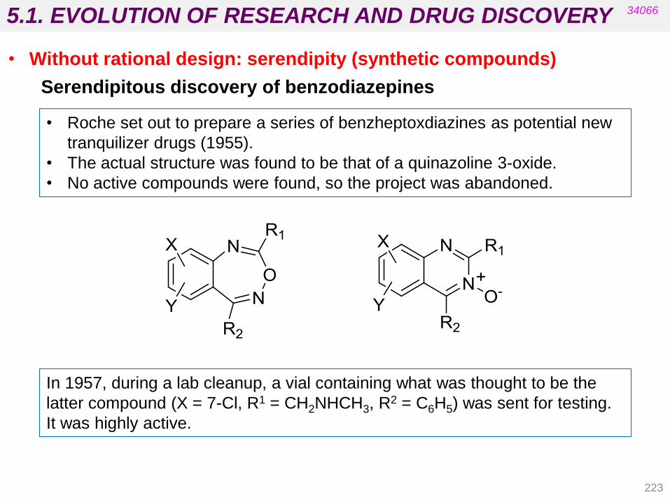
5.1. EVOLUTION OF RESEARCH AND DRUG DISCOVERY
Serendipitous discovery of benzodiazepines
• Roche set out to prepare a series of benzheptoxdiazines as potential new
tranquilizer drugs (1955).
• The actual structure was found to be that of a quinazoline 3-oxide.
• No active compounds were found, so the project was abandoned.
In 1957, during a lab cleanup, a vial containing what was thought to be the
latter compound (X = 7-Cl, R1 = CH2NHCH3, R2 = C6H5) was sent for testing.
It was highly active.
34066
• Without rational design: serendipity (synthetic compounds)
223

5.1. EVOLUTION OF RESEARCH AND DRUG DISCOVERY
The actual structure of the compound was benzodiazepine 4-oxide, Librium®,
(obtained from an unexpected reaction of the corresponding chloromethyl
quinazoline 3-oxide with methylamine).
Librium
34066
• Without rational design: serendipity (synthetic compounds)
224

5.1. EVOLUTION OF RESEARCH AND DRUG DISCOVERY
promethazine
(antihistamine
with sedative effects)
chlorpromazine
(neuroleptic
prototype)
Also: chlorothiazide (antidiabetic sulphonamide) from antibacterial sulphanilamide
More recently: sildenafil (Viagra), ritonavir, aztreonam, etc.
34066
• Without rational design: clinical observations (observations in humans)
imipramine
(antidepressant)
225
sildenafil valdenafil
(The SOSA approach : Selective Optimization of Side Activities)

5.1. EVOLUTION OF RESEARCH AND DRUG DISCOVERY
226
34066
• The use of nitrous oxide and ether as narcotic gases in surgery
resulted from the observation that people who inhaled these chemicals
[at parties] did not experience any pain after injury.
• The vasodilator activity of amyl nitrite and nitroglycerin was
discovered by chemists who developed strong headaches after
inhaling or ingesting small amounts.
• A wrong working hypothesis on chloral hydrate, which was supposed
to degrade metabolically to narcotic chloroform, led to its application
as a strong sedative (in reality, the metabolite trichloroethanol is the
active form).
• Warfarin was used as rat poison.
• Without rational design: serendipity and side effects
(synthetic compounds)
(Find more examples on page 207 of Patrick’s 5th ed.)
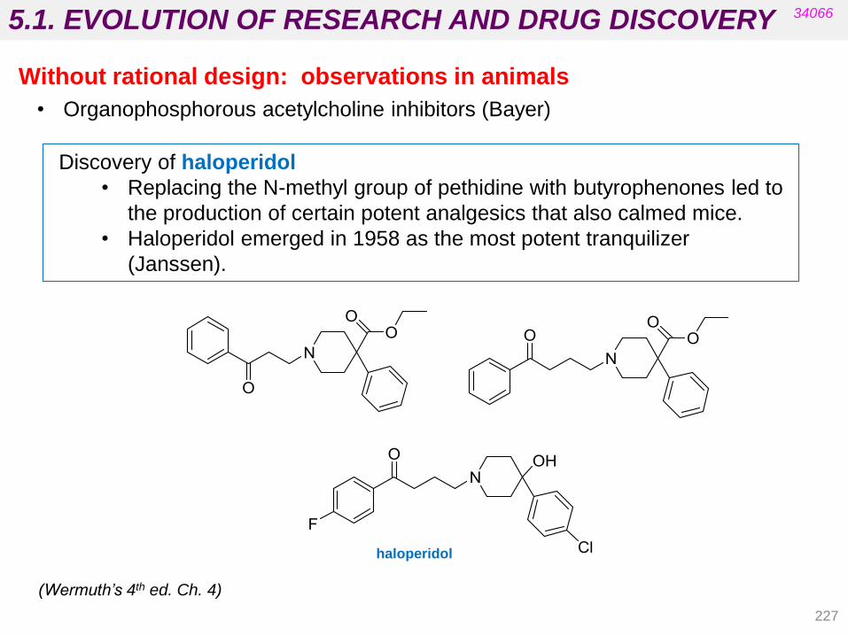
5.1. EVOLUTION OF RESEARCH AND DRUG DISCOVERY
227
• Organophosphorous acetylcholine inhibitors (Bayer)
34066
(Wermuth’s 4th ed. Ch. 4)
Without rational design: observations in animals
haloperidol
Discovery of haloperidol
• Replacing the N-methyl group of pethidine with butyrophenones led to
the production of certain potent analgesics that also calmed mice.
• Haloperidol emerged in 1958 as the most potent tranquilizer
(Janssen).

5.1. EVOLUTION OF RESEARCH AND DRUG DISCOVERY
228
34066
(Wermuth’s 4th ed. Ch. 4) New Uses for Old Drugs
Thalidomide was launched as sedative/hypnotic drug but was withdrawn
because of its teratogenicity.
It is now used to treat a complication in the chemotherapy of leprosy. Some
analogues have also been obtained.
• Without rational design: clinical observations (observations in humans)
thalidomide panalidomide

5.1. EVOLUTION OF RESEARCH AND DRUG DISCOVERY
ANALGESICS
ANTIDEPRESSANTS
acetanilide acetophenetidin
imipramine desipramine
34066
• Without rational design: metabolism studies
229
paracetamol
5.1. EVOLUTION OF RESEARCH AND DRUG DISCOVERY
230
• Biological information constitutes a rich source of new lead molecules for
research.
• It offers creative approaches that do not depend on routine pharmacological
models.
• Once the lead molecule is identified, it is immediately the object of thorough
research to determine its molecular mechanism of action.
• Simultaneous synthesis of structural analogues and establishment of structure-
activity relationships (SAR) proceed in order to optimize all indispensable
parameters for their development (potency, selectivity, metabolism,
bioavailability, toxicity and cost price).
• New lead compounds have often arisen form research projects conducted in a
different field (keeping an open mind is crucial when testing biological activities).
• Even if the initial discovery was purely fortuitous, a great effort of rationalization
is necessary in subsequent research.
• Exploitation of Biological Information
34066
(Wermuth’s 4th ed. Ch. 4)
• Observations Made in Humans
• Observations Made in Animals
• Observations Made in the Plant Kingdom and Microbiology

5.1. EVOLUTION OF RESEARCH AND DRUG DISCOVERY
Since the last few decades of the 20th century:
• In the past most drugs were “discovered“ either by identifying the active
ingredient from traditional remedies or by serendipitous discovery rather than
“rational” drug discovery.
• Diseases are controlled at the molecular and physiological levels.
• New technologies are helping to identify new targets.
• The shape of molecules at the atomic level is well understood.
• Very few drugs are now “discovered” without an optimization process from
the discovery of an active compound. Finding a lead is an important step in
drug discovery.
The most common approach to drug discovery has been based on mechanisms
of action (screening or designing compounds for their effect on a particular
biological target).
34066
Target Lead compounds to DRUGS Disease
Drug target
selection
Lead Discovery
Lead Modification
Preclinical and Clinical
Development
Regulatory Approval
231
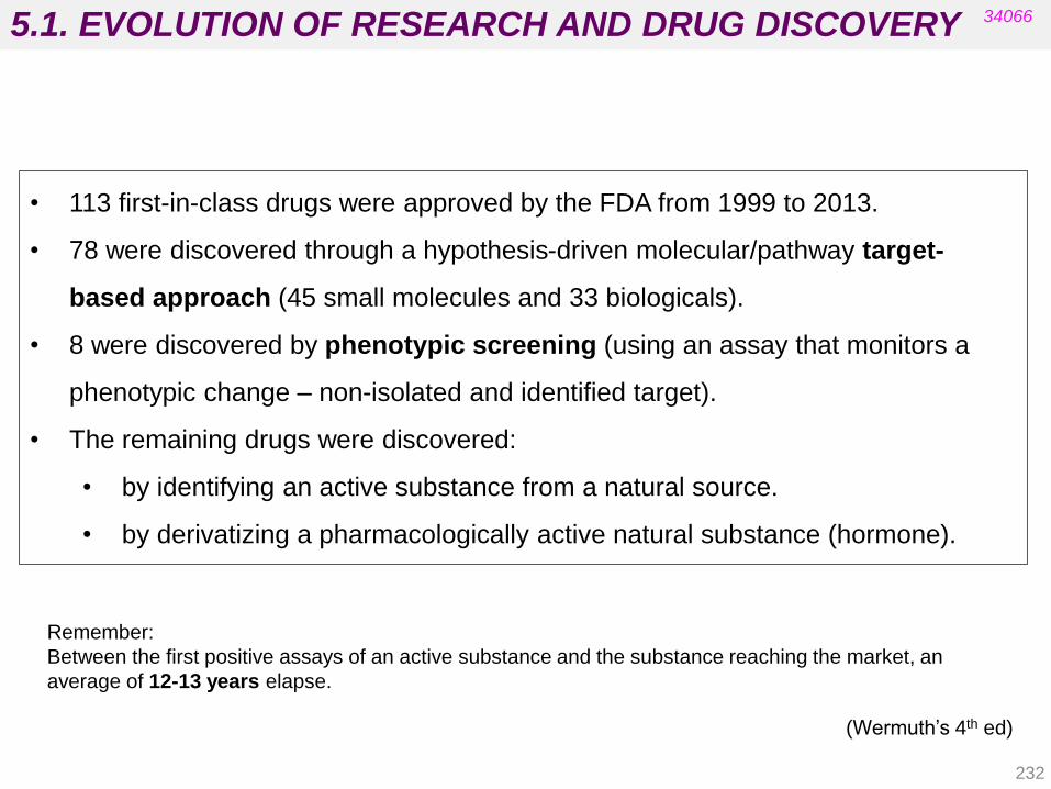
5.1. EVOLUTION OF RESEARCH AND DRUG DISCOVERY 34066
• 113 first-in-class drugs were approved by the FDA from 1999 to 2013.
• 78 were discovered through a hypothesis-driven molecular/pathway target-
based approach (45 small molecules and 33 biologicals).
• 8 were discovered by phenotypic screening (using an assay that monitors a
phenotypic change – non-isolated and identified target).
• The remaining drugs were discovered:
• by identifying an active substance from a natural source.
• by derivatizing a pharmacologically active natural substance (hormone).
(Wermuth‟s 4th ed)
Remember:
Between the first positive assays of an active substance and the substance reaching the market, an
average of 12-13 years elapse.
232

5.1. EVOLUTION OF RESEARCH AND DRUG DISCOVERY
233
34066
(Wermuth‟s 4th ed)
Active compounds are called “hits” when only activity in in vitro assays has been
tested. Once other important features have been evaluated, the term “lead” should be
used.
Lead optimization provides “drug candidates” that can finally be considered drugs
when they are approved and reach the market.
(see unit 1).
In this section we will use the term “lead” even when “hit” may be more appropriate in
some cases (a “hit –to-lead” optimization process would be necessary in these
cases).
The main paradigm for discovering lead compounds has been based on the
“one target-one disease” philosophy (see Unit 1).
It uses in vitro high-throughput technologies (HTS) and computer-aided
drug design.
More recent strategies are based on advances in NMR or X-Ray
crystallography.
Some diseases are still inadequately treated by the one target-one disease
approach.
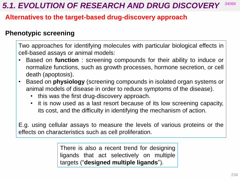
5.1. EVOLUTION OF RESEARCH AND DRUG DISCOVERY
234
Alternatives to the target-based drug-discovery approach
Phenotypic screening
34066
Two approaches for identifying molecules with particular biological effects in
cell-based assays or animal models:
• Based on function : screening compounds for their ability to induce or
normalize functions, such as growth processes, hormone secretion, or cell
death (apoptosis).
• Based on physiology (screening compounds in isolated organ systems or
animal models of disease in order to reduce symptoms of the disease).
• this was the first drug-discovery approach.
• it is now used as a last resort because of its low screening capacity,
its cost, and the difficulty in identifying the mechanism of action.
E.g. using cellular assays to measure the levels of various proteins or the
effects on characteristics such as cell proliferation.
There is also a recent trend for designing
ligands that act selectively on multiple
targets (“designed multiple ligands”).

5.1. METHODS FOR DISCOVERING LEAD COMPOUNDS 34066
Strategies (different books have different classification criteria)
235
• From existing drugs: “me too” and “me better” and the SOSA approach
• Screening
• Screening of natural products
• Medicinal folklore and more
• Screening the “libraries” of synthetic compounds
• Combinatorial and parallel synthesis
• Computerized searching of structural databases
• Rational design
• Design from natural ligands or modulators
• Computer-aided design
• Fragment-based lead discovery (based on NMR spectroscopy,
X-ray crystallography, etc.)
• Serendipity and prepared mind
(Patrick‟s 5th ed. Chapter 12; Silverman‟s 3rd ed. Chapters 1 and 2)

5.1. METHODS FOR DISCOVERING LEAD COMPOUNDS
236
34066
• Analogue Design : “me-too” compounds (chemical and pharmacological
similarity)
• Systematic Screening:
• Extensive Screening
• Random Screening
• High-Throughput Screening
• Screening of Synthesis Intermediates
• New Leads from Old Drugs: The SOSA Approach
• Exploitation of Biological Information
• Observations Made in Humans
• Observations Made in Animals
• Observations Made in the Plant Kingdom and Microbiology
• Planned Research and Rational Approaches
• Applying Biophysical Technologies and Computational Methods
• Biophysical Technologies
• Computational Methods
(Wermuth’s 4th ed. Ch. 4) Strategies
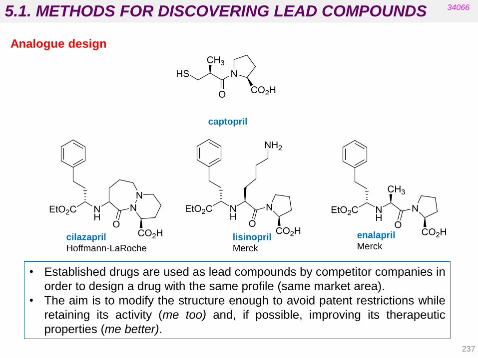
5.1. METHODS FOR DISCOVERING LEAD COMPOUNDS
237
34066
Analogue design
• Established drugs are used as lead compounds by competitor companies in
order to design a drug with the same profile (same market area).
• The aim is to modify the structure enough to avoid patent restrictions while
retaining its activity (me too) and, if possible, improving its therapeutic
properties (me better).
captopril
cilazapril
Hoffmann-LaRoche
lisinopril
Merck
enalapril
Merck

5.1. METHODS FOR DISCOVERING LEAD COMPOUNDS
238
• Analogues from losartan (DuPont), an angiotensin AT1 antagonist launched
as a vasodilator in 1994 (patented in 1986).
34066
Analogue design
losartan
DuPont eprosartan
SmithKline Beecham
valsartan
Novartis
(Wermuth’s 4th ed. Ch. 4)

5.1. METHODS FOR DISCOVERING LEAD COMPOUNDS
239
34066
• Involves screening new molecules on an animal model or in a biological test
without having any hypothesis regarding its therapeutic potential.
• Uses in vitro rather than in vivo tests (binding assays, enzyme inhibition
measurements, activity on isolated organs or cell cultures).
Systematic screening
• Can be achieved in two ways:
• Extensive screening: exhaustive pharmacological research on a small number of
chemically sophisticated and original molecules.
(central nervous, cardiovascular, pulmonary and digestive systems,
antiviral, antibacterial, or chemotherapeutic properties, etc.).
A successful systematic screening campaign: paclitaxel (Taxol).
• Random screening: from a vast number of molecules (hundreds or thousands), the
aim is to find one that may be active in a given indication. The therapeutic objective
is set in advance.
This was very useful for discovering new antibiotics (samples of earth collected in
countries from all over the world).
More recent successes include the discovery of lovastatin.

5.1. METHODS FOR DISCOVERING LEAD COMPOUNDS
240
34066
Systematic screening
zopiclone (hypnotic - used in insomnia)
paclitaxel
Drugs discovered
by random screening.
R1 = R2 = H mevastatin (natural product)
R1 = Me, R2 = H lovastatin (natural product)
R = Me simvastatin (semi-synthetic
analogue)
pravastatin (semi-synthetic)
• Statins are used for lowering
cholesterol
• Pravastatin is less lipophilic and
presents fewer central side-
effects.
• The ring-opened form is the
actual active in vivo.
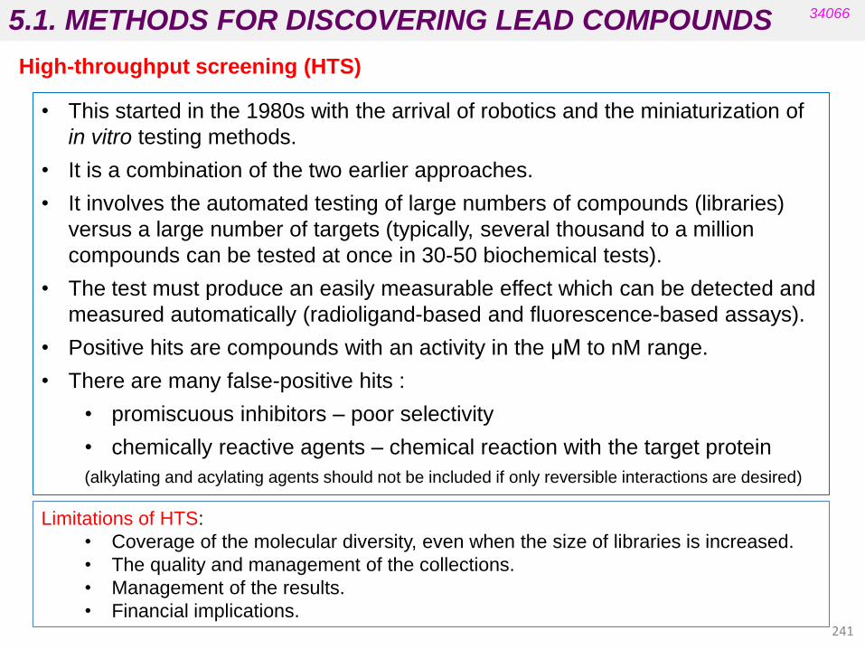
5.1. METHODS FOR DISCOVERING LEAD COMPOUNDS
241
High-throughput screening (HTS)
34066
• This started in the 1980s with the arrival of robotics and the miniaturization of
in vitro testing methods.
• It is a combination of the two earlier approaches.
• It involves the automated testing of large numbers of compounds (libraries)
versus a large number of targets (typically, several thousand to a million
compounds can be tested at once in 30-50 biochemical tests).
• The test must produce an easily measurable effect which can be detected and
measured automatically (radioligand-based and fluorescence-based assays).
• Positive hits are compounds with an activity in the μM to nM range.
• There are many false-positive hits :
• promiscuous inhibitors – poor selectivity
• chemically reactive agents – chemical reaction with the target protein
(alkylating and acylating agents should not be included if only reversible interactions are desired)
Limitations of HTS:
• Coverage of the molecular diversity, even when the size of libraries is increased.
• The quality and management of the collections.
• Management of the results.
• Financial implications. 241

5.1. METHODS FOR DISCOVERING LEAD COMPOUNDS
242
34066
This strategy has delivered numerous drugs:
High-throughput screening (HTS)
imitanib
anticancer (tyrosine kinase inhibitor) maraviroc
antiretroviral
treatment of HIV infection
Failure rate is high between the number of hits and those compounds which
are identified by HTS as authentic lead compounds (in same cases screening
highlights no hits because the selected target is not clinically relevant or the
hits do not show appropriate physicochemical properties).
5.1. METHODS FOR DISCOVERING LEAD COMPOUNDS
243
More rational design (HTS)
34066
• Several cheminformatics and modelling tools can be used (in silico screening) to
generate better collections:
• Diversity:
• Several representatives of each compound scaffold.
• Removing unwanted compounds:
• Filters (Lipinsky‟s rule of five for oral bioavailability and many others).
• Designing target-focused compound collections:
• When protein crystal structures are available:
• Library based on structural information using computers.
Known active compounds on these targets can be used as starting
points for a ligand-based design search and the corresponding
generation of specialized collections.
• For a random HTS, a major consideration is the source of the large number
of compounds that are usually required. Preparing the compound collection
for screening is crucial.
• Targeted screening implies applying prior knowledge in order to select
compounds that are considered most likely to interact with the target.
High-throughput screening (HTS)

5.1. METHODS FOR DISCOVERING LEAD COMPOUNDS
A) Natural
B) Synthetic
C) Virtual
Plants
Microorganisms
Animals
Biochemicals
Marine life
Chemical synthesis (traditional)
Combinatorial synthesis
Computer-aided drug design
34066
Sources of compounds for screening
244

5.1. METHODS FOR DISCOVERING LEAD COMPOUNDS
245
A) Isolation and purification
solvent-solvent extraction
chromatography
crystallisation
distillation
B) Structure determination
elemental analysis
molecular weight
mass spectrometry
infrared spectroscopy
ultraviolet spectroscopy
NMR (1H, 13C, 2D) spectroscopy
X-ray crystallography
Identifying lead compounds
34066
Thousands or millions of compounds are required in very small amounts for HTS:
chemical libraries and combinatorial and parallel synthesis have been developed.
VERY EXPENSIVE
AND TIME
CONSUMING!!!!
Natural compounds and those obtained by “traditional” organic synthesis were
isolated and their structure determined before testing.
(Patrick‟s 5th ed. chapter 16)
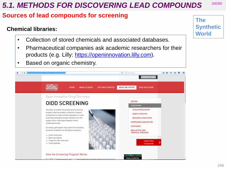
5.1. METHODS FOR DISCOVERING LEAD COMPOUNDS
246
• Collection of stored chemicals and associated databases.
• Pharmaceutical companies ask academic researchers for their
products (e.g. Lilly: https://openinnovation.lilly.com).
• Based on organic chemistry.
34066
Chemical libraries:
The
Synthetic
World
Sources of lead compounds for screening
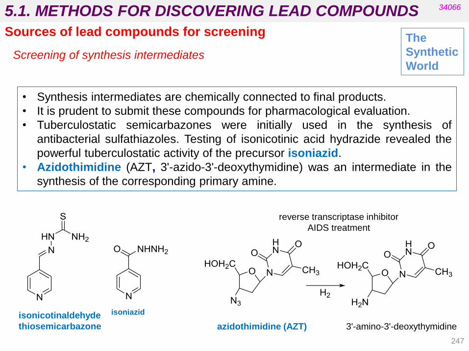
5.1. METHODS FOR DISCOVERING LEAD COMPOUNDS
247
34066 34066
Screening of synthesis intermediates
• Synthesis intermediates are chemically connected to final products.
• It is prudent to submit these compounds for pharmacological evaluation.
• Tuberculostatic semicarbazones were initially used in the synthesis of
antibacterial sulfathiazoles. Testing of isonicotinic acid hydrazide revealed the
powerful tuberculostatic activity of the precursor isoniazid.
• Azidothimidine (AZT, 3'-azido-3'-deoxythymidine) was an intermediate in the
synthesis of the corresponding primary amine.
The
Synthetic
World
Sources of lead compounds for screening
reverse transcriptase inhibitor
AIDS treatment
isonicotinaldehyde
thiosemicarbazone
isoniazid
azidothimidine (AZT) 3'-amino-3'-deoxythymidine

5.1. METHODS FOR DISCOVERING LEAD COMPOUNDS
248
• Mercaptopurine is an intermediate in the synthesis of methotrexate
and is also active as an inhibitor of dihydrofolate-reductase.
However, it is toxic. Subsequent optimization led to azathioprine (a
prodrug that releases mercaptopurine in vivo).
• Azathioprine is a potent immunosuppressive agent that was used
in all organ transplantation until the advent of cyclosporine.
• Allopurinol also inhibits xanthine-oxidase and is therefore used to
treat gout.
34066 34066
azathioprine mercaptopurine allopurinol (Wermuth‟s)
The
Synthetic
World
Sources of lead compounds for screening
Screening of synthesis intermediates
methotrexate

5.1. METHODS FOR DISCOVERING LEAD COMPOUNDS
249
34066
The
Synthetic
World
Sources of lead compounds for screening
• Enables the use of a defined reaction route to produce a large
number of structurally related compounds in a short space of
time.
• The full set of compounds produced in this way is called a
compound library.
• Reactions are conducted on a small scale and the process can
be automated or semi-automated.
• Reactions are conducted in several reaction vessels at the same
time under identical conditions using different reagents for each
vessel.
• Is based on solid-phase techniques.
Combinatorial and parallel synthesis:
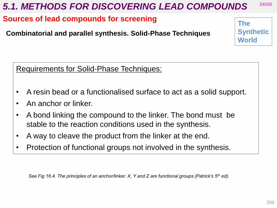
5.1. METHODS FOR DISCOVERING LEAD COMPOUNDS
Requirements for Solid-Phase Techniques:
• A resin bead or a functionalised surface to act as a solid support.
• An anchor or linker.
• A bond linking the compound to the linker. The bond must be
stable to the reaction conditions used in the synthesis.
• A way to cleave the product from the linker at the end.
• Protection of functional groups not involved in the synthesis.
See Fig 16.4. The principles of an anchor/linker. X, Y and Z are functional groups (Patrick’s 5th ed).
250
34066
Combinatorial and parallel synthesis. Solid-Phase Techniques
The
Synthetic
World
Sources of lead compounds for screening
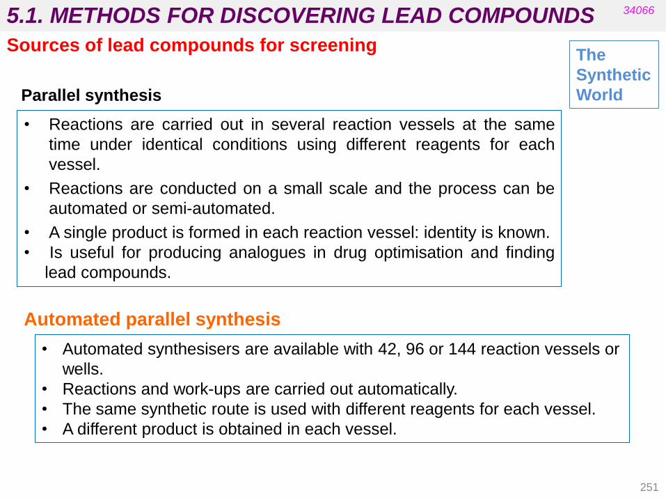
5.1. METHODS FOR DISCOVERING LEAD COMPOUNDS
251
• Reactions are carried out in several reaction vessels at the same
time under identical conditions using different reagents for each
vessel.
• Reactions are conducted on a small scale and the process can be
automated or semi-automated.
• A single product is formed in each reaction vessel: identity is known.
• Is useful for producing analogues in drug optimisation and finding
lead compounds.
• Automated synthesisers are available with 42, 96 or 144 reaction vessels or
wells.
• Reactions and work-ups are carried out automatically.
• The same synthetic route is used with different reagents for each vessel.
• A different product is obtained in each vessel.
Automated parallel synthesis
34066
Parallel synthesis
The
Synthetic
World
Sources of lead compounds for screening
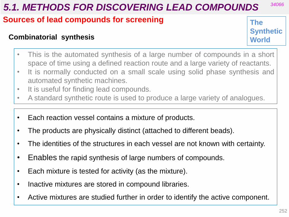
5.1. METHODS FOR DISCOVERING LEAD COMPOUNDS
252
• This is the automated synthesis of a large number of compounds in a short
space of time using a defined reaction route and a large variety of reactants.
• It is normally conducted on a small scale using solid phase synthesis and
automated synthetic machines.
• It is useful for finding lead compounds.
• A standard synthetic route is used to produce a large variety of analogues.
• Each reaction vessel contains a mixture of products.
• The products are physically distinct (attached to different beads).
• The identities of the structures in each vessel are not known with certainty.
• Enables the rapid synthesis of large numbers of compounds.
• Each mixture is tested for activity (as the mixture).
• Inactive mixtures are stored in compound libraries.
• Active mixtures are studied further in order to identify the active component.
34066
Combinatorial synthesis
The
Synthetic
World
Sources of lead compounds for screening

5.1. METHODS FOR DISCOVERING LEAD COMPOUNDS
Example - Synthesis of all possible dipeptides using 5 amino acids
Standard methods would involve 25 separate syntheses.
The mix and split procedure involves five separate syntheses.
The Mix and Split Method
Standard method
34066
Combinatorial synthesis
253
The
Synthetic
World
Sources of lead compounds for screening
Note:
Each experiment involves the protection of amino acids, coupling and deprotection.
Glycine (Gly) Alanine (Ala) Phenylalanine (Phe) Valine (Val) Serine (Ser)
25 separate experiments
Gly-Gly Gly-Ala Gly-Phe Gly-Val Gly-Ser
Ala-Gly Ala-Ala Ala-Phe Ala-Val Ala-Ser
Phe-Gly Phe-Ala Phe-Phe Phe-Val Phe-Ser
Val-Gly Val-Ala Val-Phe Val-Val Val-Ser
Ser-Gly Ser-Ala Ser-Phe Ser-Val Ser-Ser
See Fig 16.29. Synthesis of five different dipeptides using the mix and split strategy (Patrick’s 5th ed.).
Mix and split strategy
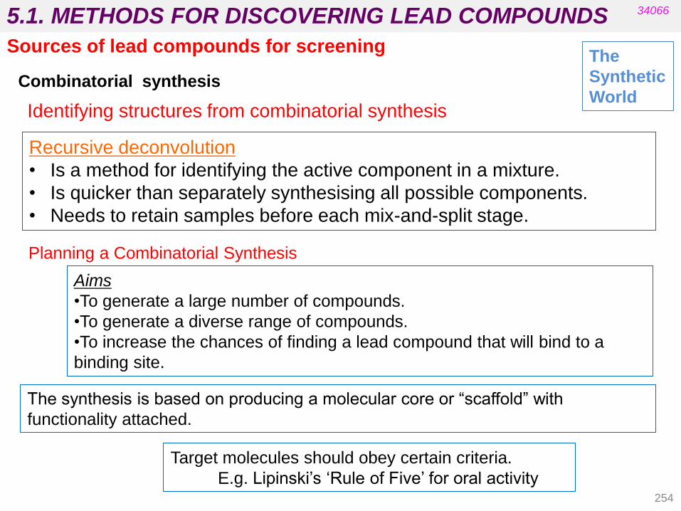
5.1. METHODS FOR DISCOVERING LEAD COMPOUNDS
Recursive deconvolution
• Is a method for identifying the active component in a mixture.
• Is quicker than separately synthesising all possible components.
• Needs to retain samples before each mix-and-split stage.
Identifying structures from combinatorial synthesis
34066
Combinatorial synthesis
254
The
Synthetic
World
Sources of lead compounds for screening
Planning a Combinatorial Synthesis
Aims
•To generate a large number of compounds.
•To generate a diverse range of compounds.
•To increase the chances of finding a lead compound that will bind to a
binding site.
The synthesis is based on producing a molecular core or “scaffold” with
functionality attached.
Target molecules should obey certain criteria.
E.g. Lipinski‟s „Rule of Five‟ for oral activity

5.1. METHODS FOR DISCOVERING LEAD COMPOUNDS
„Tadpole‟ scaffolds
- variation is restricted to a specific region around the molecule.
- there is less chance of favourable interactions with a binding site.
Privileged scaffolds
Scaffolds which are common in medicinal chemistry and are associated with
a diverse range of activities:
(benzodiazepines, hydantoins, benzenesulphonamides, etc. )
34066
Combinatorial synthesis
The
Synthetic
World Scaffolds
Sources of lead compounds for screening
255
„Spider‟ scaffolds are preferable for exploring conformational space.
Enable variation of functional groups around a whole molecule in order to
increase the chances of finding suitable binding interactions.

5.1. METHODS FOR DISCOVERING LEAD COMPOUNDS
•Good scaffolds
•Spider-like
•Low molecular weight
•Variety of synthetic routes available
Scaffolds
34066
Combinatorial synthesis
(captopril, enalapril)
orally active
The
Synthetic
World
Sources of lead compounds for screening
256

5.1. METHODS FOR DISCOVERING LEAD COMPOUNDS
• Spider-like and small molecular weight
• Multiple OH groups
• Difficult to vary R1-R5 independently
• MW relatively high
• Restricted number of functional groups allowed
for MW
• Relatively few positions where substituents can
be added
• Tadpole-like scaffold
• Restricted region of variability
Scaffolds
34066
Combinatorial synthesis The
Synthetic
World
Sources of lead compounds for screening
257

5.1. METHODS FOR DISCOVERING LEAD COMPOUNDS
258
From http://www.brandon-russell.com/pdf/dos.pdf
34066
Diversity-oriented synthesis The
Synthetic
World
Sources of lead compounds for screening
Comparison of Target-Oriented, Combinatorial, and Diversity-Oriented Syntheses
Target-oriented synthesis and combinatorial chemistry use retrosynthetic analysis
to interrogate a single point in chemical space or its immediate surroundings,
respectively.
Diversity-oriented synthesis tries to maximize skeletal diversity and thus coverage
of chemical space.
THE BASICS OF DIVERSITY-ORIENTED SYNTHESIS
Kieron M. G. O‟Connell, Warren R. J. D. Galloway, and David R. Spring
Diversity-Oriented Synthesis: Basics and Applications in Organic Synthesis, Drug Discovery, and Chemical Biology
First Edition. Edited by Andrea Trabocchi
© 2013 John Wiley & Sons, Inc. Published
http://www-spring.ch.cam.ac.uk/publications/pdf/2013_DOS_1.pdf

5.1. METHODS FOR DISCOVERING LEAD COMPOUNDS 34066
The
Virtual
World
Sources of lead compounds for screening
In silico screening or virtual screening:
• Uses a set of computational methods to analyse large databases or
collections of known compounds in order to identify potential hit
candidates by predicting compound binding to a drug target.
• Is less expensive than HTS.
• Can complement HTS or be performed prior to experimental
screening.
(Wermuth’s 4th Chapter 6) 259
Two main approaches:
• Ligand-based virtual screening: 2D or 3D chemical structures or molecular
descriptors of known active and inactive molecules are used to retrieve other
compounds of interest from a database by seeking a common substructure,
pharmacophore, or shape parameters.
• Target-based virtual screening: the library of database compounds are
docked into a binding site and then ranked using one or several scoring
functions (knowledge of the 3D structure of the target and binding site is
required).
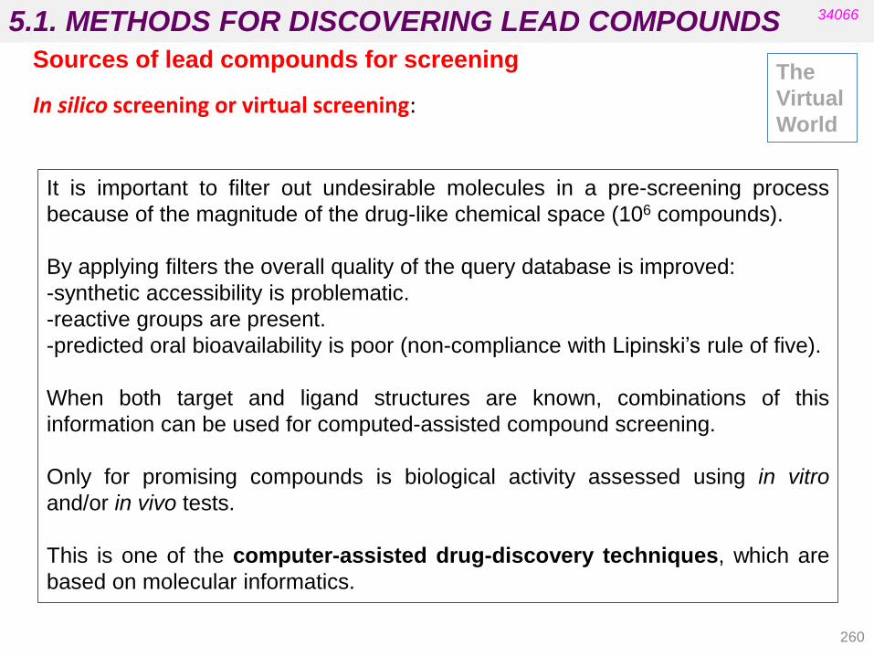
5.1. METHODS FOR DISCOVERING LEAD COMPOUNDS
260
34066
The
Virtual
World
Sources of lead compounds for screening
In silico screening or virtual screening:
It is important to filter out undesirable molecules in a pre-screening process
because of the magnitude of the drug-like chemical space (106 compounds).
By applying filters the overall quality of the query database is improved:
-synthetic accessibility is problematic.
-reactive groups are present.
-predicted oral bioavailability is poor (non-compliance with Lipinski‟s rule of five).
When both target and ligand structures are known, combinations of this
information can be used for computed-assisted compound screening.
Only for promising compounds is biological activity assessed using in vitro
and/or in vivo tests.
This is one of the computer-assisted drug-discovery techniques, which are
based on molecular informatics.

5.1. METHODS FOR DISCOVERING LEAD COMPOUNDS
261
Many molecules have been developed with the help of in silico approaches.
tirofiban
reversible antagonist of fibrinogen
Antiplatelet
One of the first FDA-approved drugs
developed from a pharmacophore-
based virtual screening design.
cevoglitazar
peroxisome-proliferator activated receptor
(PPAR- α/γ) dual agonist
Antidiabetic
Developed by combining the virtual
screening of 3D databases and structure-
based drug design.
34066
(Wermuth’s 4th ed. Ch. 4)
• Strategies in the search for new lead compounds
In silico screening or virtual screening:
The
Virtual
World

5.1. METHODS FOR DISCOVERING LEAD COMPOUNDS 34066
Current methods for discovering lead compounds (different books have different classification criteria)
• From existing drugs: “me too” and “me better” and the SOSA approach
• Screening
• Screening of natural products
• Medicinal folklore and more.
• Screening the “libraries” of synthetic compounds
• Combinatorial and parallel synthesis.
• Computerized searching of structural databases.
262
• Rational design
• Design from natural ligands or modulators.
• Computer-aided design.
• Fragment-based lead discovery
(based on NMR spectroscopy, X-ray crystallography, etc.).
• Serendipity and prepared mind.
(Patrick‟s 5th ed. Chapter no. 12; Silverman‟s 3rd ed. Chapters 1 and 2)
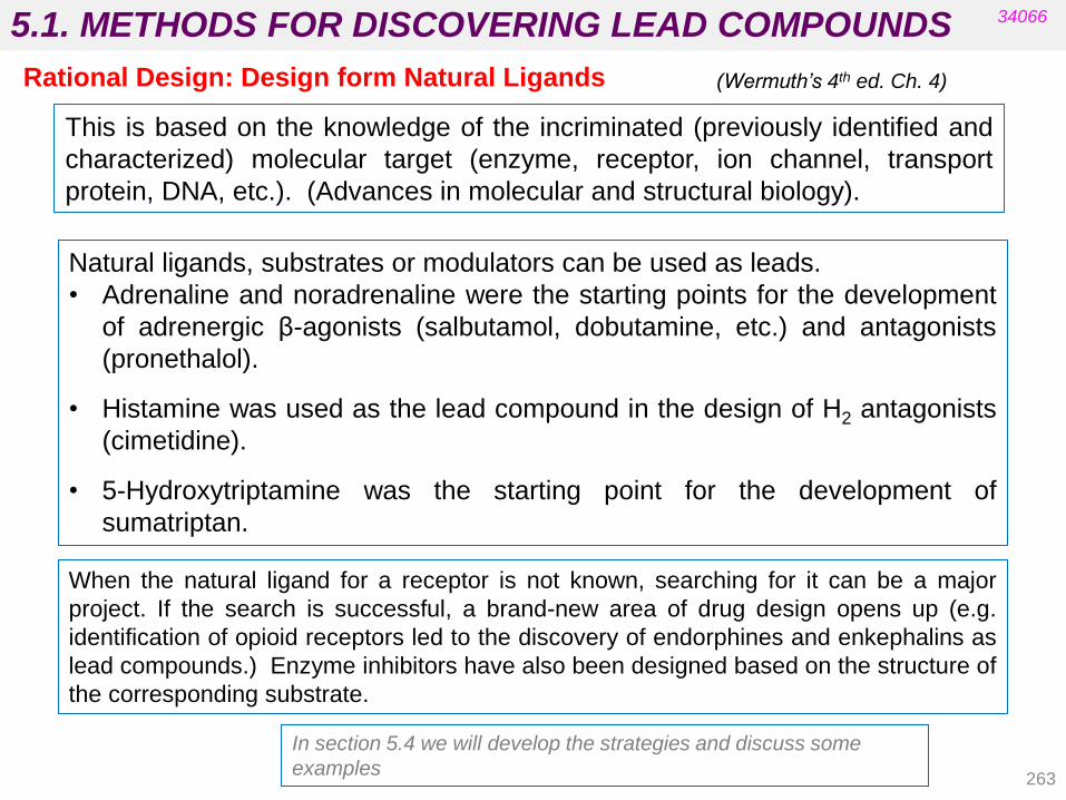
5.1. METHODS FOR DISCOVERING LEAD COMPOUNDS
263
34066
(Wermuth’s 4th ed. Ch. 4) Rational Design: Design form Natural Ligands
This is based on the knowledge of the incriminated (previously identified and
characterized) molecular target (enzyme, receptor, ion channel, transport
protein, DNA, etc.). (Advances in molecular and structural biology).
Natural ligands, substrates or modulators can be used as leads.
• Adrenaline and noradrenaline were the starting points for the development
of adrenergic β-agonists (salbutamol, dobutamine, etc.) and antagonists
(pronethalol).
• Histamine was used as the lead compound in the design of H2 antagonists
(cimetidine).
• 5-Hydroxytriptamine was the starting point for the development of
sumatriptan.
When the natural ligand for a receptor is not known, searching for it can be a major
project. If the search is successful, a brand-new area of drug design opens up (e.g.
identification of opioid receptors led to the discovery of endorphines and enkephalins as
lead compounds.) Enzyme inhibitors have also been designed based on the structure of
the corresponding substrate.
In section 5.4 we will develop the strategies and discuss some
examples

5.1. METHODS FOR DISCOVERING LEAD COMPOUNDS
De novo design generates virtual compounds for computational analysis as an
extension to the in silico screening of physically available compounds stored in
databases. The main aim is to find innovative scaffolds for further development.
34066
(Wermuth’s 4th ed. Ch. 6)
(Patrick’s 5th ed. Ch. 17)
Computer-Assisted Drug Design (CADD)
“ De novo” Drug Design
264
De novo design can also be divided into structure-based approaches (when
the 3D structure of the target is known) and ligand-based approaches.
Manual
• An operator directs the study.
• It enables the input of the designer‟s ideas.
• It is useful for identifying a single lead compound.
• It is slow and limited to the designer‟s originality.
Automated
• The program is automated and therefore much faster.
• No bias is introduced by the operator.
• It is useful for generating a large number of possible lead compounds.
• It produces novel structures.
• It may generate impractical structures for synthesis.
• Scoring structures for binding strengths are unreliable.
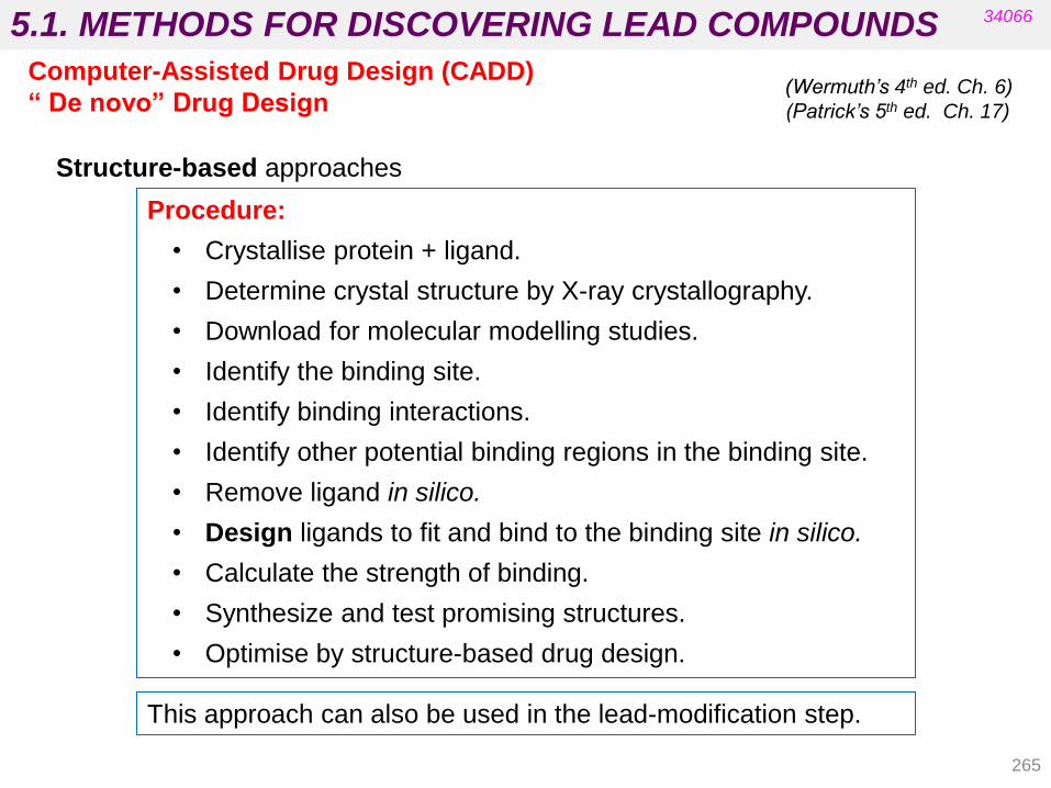
5.1. METHODS FOR DISCOVERING LEAD COMPOUNDS
265
Procedure:
• Crystallise protein + ligand.
• Determine crystal structure by X-ray crystallography.
• Download for molecular modelling studies.
• Identify the binding site.
• Identify binding interactions.
• Identify other potential binding regions in the binding site.
• Remove ligand in silico.
• Design ligands to fit and bind to the binding site in silico.
• Calculate the strength of binding.
• Synthesize and test promising structures.
• Optimise by structure-based drug design.
Structure-based approaches
34066
(Wermuth’s 4th ed. Ch. 6)
(Patrick’s 5th ed. Ch. 17)
Computer-Assisted Drug Design (CADD)
“ De novo” Drug Design
This approach can also be used in the lead-modification step.
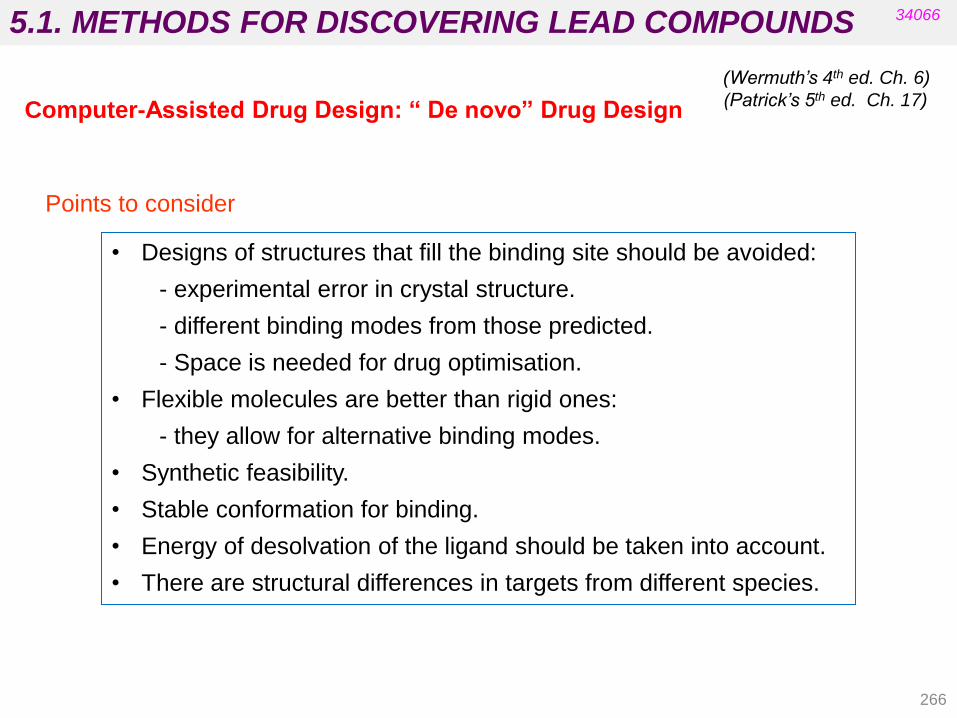
5.1. METHODS FOR DISCOVERING LEAD COMPOUNDS
Points to consider
• Designs of structures that fill the binding site should be avoided:
- experimental error in crystal structure.
- different binding modes from those predicted.
- Space is needed for drug optimisation.
• Flexible molecules are better than rigid ones:
- they allow for alternative binding modes.
• Synthetic feasibility.
• Stable conformation for binding.
• Energy of desolvation of the ligand should be taken into account.
• There are structural differences in targets from different species.
34066
(Wermuth’s 4th ed. Ch. 6)
(Patrick’s 5th ed. Ch. 17) Computer-Assisted Drug Design: “ De novo” Drug Design
266

5.1. METHODS FOR DISCOVERING LEAD COMPOUNDS
SYNOPSIS:
•Is an automated program.
•Is a fragment-based process.
•Generates synthetically feasibly structures.
•Synthetic rules are incorporated into the structure-building process.
•Fragments must be commercially available.
•Fragments are only linked if synthetically feasible.
•The program provides a possible synthetic route.
Several computer software programs are available:
LUDI: works by fitting molecular fragments to different regions of the binding
site and then linking the fragments together.
SPROUT: the interaction sites used consist of atom-sized spheres.
LEGEND: does not use fragments to generate skeletons. The skeletons are
grown one atom at a time using random choices at each stage of the process.
GROW: is limited to peptides.
34066
(Wermuth’s 4th ed. Ch. 6)
(Patrick’s 5th ed. Ch. 17) Computer-Assisted Drug Design (CADD)
“ De novo” Drug Design
267
See Case study no 5. (Patrick’s 5th ed).
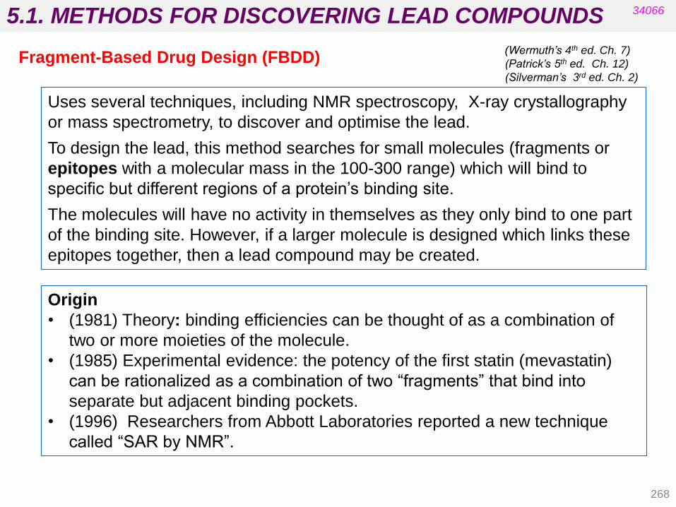
5.1. METHODS FOR DISCOVERING LEAD COMPOUNDS
268
34066
(Wermuth’s 4th ed. Ch. 7)
(Patrick’s 5th ed. Ch. 12)
(Silverman’s 3rd ed. Ch. 2)
Fragment-Based Drug Design (FBDD)
Origin
• (1981) Theory: binding efficiencies can be thought of as a combination of
two or more moieties of the molecule.
• (1985) Experimental evidence: the potency of the first statin (mevastatin)
can be rationalized as a combination of two “fragments” that bind into
separate but adjacent binding pockets.
• (1996) Researchers from Abbott Laboratories reported a new technique
called “SAR by NMR”.
Uses several techniques, including NMR spectroscopy, X-ray crystallography
or mass spectrometry, to discover and optimise the lead.
To design the lead, this method searches for small molecules (fragments or
epitopes with a molecular mass in the 100-300 range) which will bind to
specific but different regions of a protein‟s binding site.
The molecules will have no activity in themselves as they only bind to one part
of the binding site. However, if a larger molecule is designed which links these
epitopes together, then a lead compound may be created.

5.1. METHODS FOR DISCOVERING LEAD COMPOUNDS
269
34066
(Wermuth’s 4th ed. Ch. 7)
(Patrick’s 5th ed. Ch. 12)
(Silverman’s 3rd ed. Ch. 2)
Fragment-Based Drug Design (FBDD)
• The first step involves screening a library of small compounds (10 at a time)
by observing the 15N chemical shift (δ) for a specific amide nitrogen of the
protein.
• Once a fragment is identified that causes a notable change in δ, a library of
similar analogues is screened to identify compounds with optimal binding at
this site.
• A second library of compounds is then screened in the presence of an excess
of the first optimized ligand to find a compound that binds a nearby site.
• The location and orientation of each ligand is determined, and compounds
are synthesized in which the two ligands are covalently attached.
Is applied to proteins of known structure which are labelled with 15N (or 13C) (the
amide bond in the protein will have an identifiable peak) even if they are not
studied by X-Ray crystallography.
• Overexpression of these proteins in microorganisms is possible when 15NH4Cl is used as their sole nitrogen source.
• The complete 3D and 4D of the purified protein is determined so that the
position of every amino acid residue in the protein is known.
NMR

5.1. METHODS FOR DISCOVERING LEAD COMPOUNDS
270
34066
Fragment-Based Design (FBDD)
Figure 12.16. Epitope mapping. (Patrick’s 5th ed.)
(Based on figure 7.16 Wermuth’s 4th ed. Ch. 7)
• Vemurafenib (Zelboraf®) is a selective kinase B-Raf enzyme inhibitor that is
used for the treatment of metastatic melanoma.
• It is the first drug approved that was derived from a hit found using FBDD
(2012 in Europe).
• It is a good example for fragment elaboration where selectivity was achieved
during the lead optimization process from the suitable selection of fragments
during FBDD.
NMR

5.1. METHODS FOR DISCOVERING LEAD COMPOUNDS
271
Attractive features (compared to HTS):
- A higher proportion of the atoms of a fragment can directly interact with the target.
- The number of fragments that need to be screened is lower.
- The subsequent structural optimization of a hit fragment has many more options
and can result in a higher success rate for generating novel chemical structures.
- Starting with a low molecular mass and a low lipophilic fragment is likely to
produce leads with small, simple structures (lipophilicity can be increased during
the lead optimization process). Small drugs can also be used as fragments and
see if they have other activities (SOSA approach) (good pharmacokinetics and
non-toxic).
Limitations:
• Fragments do not always orient in the same way individually as when combined
together in an optimized structure: induced conformational changes can occur.
• In-house and commercial sources for ligands can lack extensive structural
diversity.
• Very good understanding of the target binding sites through NMR or X-ray
crystallography is needed.
• Characterization of some targets (membrane proteins) is difficult.
34066
(Wermuth’s 4th ed. Ch. 7)
(Patrick’s 5th ed. Ch. 12)
(Silverman’s 3rd ed. Ch. 2)
Fragment-Based Drug Design (FBDD)
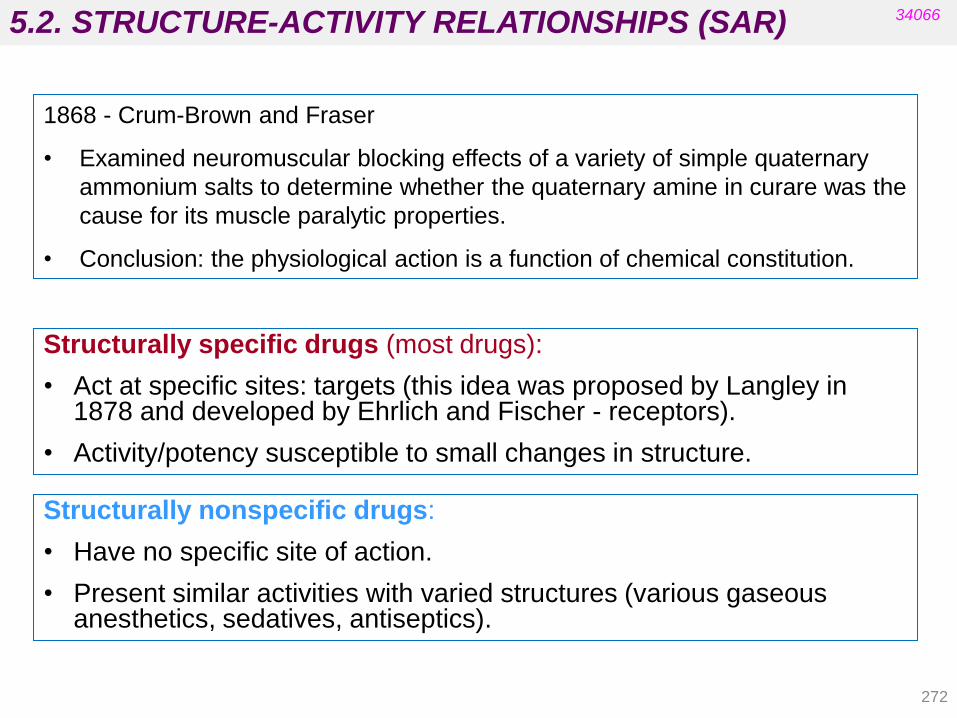
5.2. STRUCTURE-ACTIVITY RELATIONSHIPS (SAR)
272
34066
1868 - Crum-Brown and Fraser
• Examined neuromuscular blocking effects of a variety of simple quaternary
ammonium salts to determine whether the quaternary amine in curare was the
cause for its muscle paralytic properties.
• Conclusion: the physiological action is a function of chemical constitution.
Structurally specific drugs (most drugs):
• Act at specific sites: targets (this idea was proposed by Langley in 1878 and developed by Ehrlich and Fischer - receptors).
• Activity/potency susceptible to small changes in structure.
Structurally nonspecific drugs:
• Have no specific site of action.
• Present similar activities with varied structures (various gaseous anesthetics, sedatives, antiseptics).

5.2. STRUCTURE-ACTIVITY RELATIONSHIPS (SAR)
273
34066
• These molecules present structural elements of identical chemical features in
sterically consistent locations and their activity and potency are highly
susceptible to small changes.
• Depending on how the lead was discovered, only a small part of the lead
compound may be involved in the appropriate interactions.
• Small variations in these parts of the molecule can dramatically influence the
activity, whereas variations in other parts cause only minor changes in
biological activity.
• A multitude of molecular modifications could be made on the structure of a
lead.
• Important binding interactions between the lead compound and its target can
be identified after crystallization of the complex. When this is not possible:
• For structurally specific drugs acting at specific sites:
• interactions of drugs with targets are highly specific: highly specific
3D binding elements exist in targets that serve as complementary
binding sites for the active molecules.
• compounds exerting similar activities on the same target possess
closely related binding properties.
Structure-Activity Relationships (SAR) studies
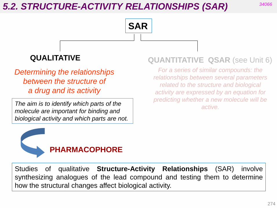
5.2. STRUCTURE-ACTIVITY RELATIONSHIPS (SAR)
Studies of qualitative Structure-Activity Relationships (SAR) involve
synthesizing analogues of the lead compound and testing them to determine
how the structural changes affect biological activity.
QUALITATIVE QUANTITATIVE QSAR (see Unit 6)
Determining the relationships
between the structure of
a drug and its activity
PHARMACOPHORE
The aim is to identify which parts of the
molecule are important for binding and
biological activity and which parts are not.
34066
SAR
For a series of similar compounds: the
relationships between several parameters
related to the structure and biological
activity are expressed by an equation for
predicting whether a new molecule will be
active.
274

5.2. STRUCTURE-ACTIVITY RELATIONSHIPS (SAR)
• Obtain structural analogues (alter, remove or mask a functional group).
• Test the analogues for activity.
• Conclusions will depend on the method of testing:
• in vitro – tests for binding interactions with target.
• in vivo – tests for target binding interactions and/or
pharmacokinetics.
• If in vitro activity drops, the group is important for binding.
• If in vivo activity is unaffected, the group is not important for binding.
Traditional methodology for determining qualitative SAR:
34066
• Modifications may disrupt binding due to electronic/steric effects.
• The easiest analogues to make are those made from the lead
compound.
• Possible modifications may depend on other groups present.
• Some analogues may have to be made via a full synthesis:
(e.g. replacing an aromatic ring with a cyclohexane ring).
• Enables the pharmacophore to be identified. (review Unit 3)
Patrick’s 5th ed. Chapter 13.1: Binding interactions that are possible for different functional groups
and analogues that may be synthesized to establish whether they are involved in binding.
275
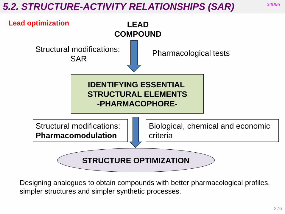
5.2. STRUCTURE-ACTIVITY RELATIONSHIPS (SAR)
276
34066
LEAD
COMPOUND
IDENTIFYING ESSENTIAL
STRUCTURAL ELEMENTS
-PHARMACOPHORE-
STRUCTURE OPTIMIZATION
Structural modifications:
SAR Pharmacological tests
Biological, chemical and economic
criteria
Structural modifications:
Pharmacomodulation
Lead optimization
Designing analogues to obtain compounds with better pharmacological profiles,
simpler structures and simpler synthetic processes.
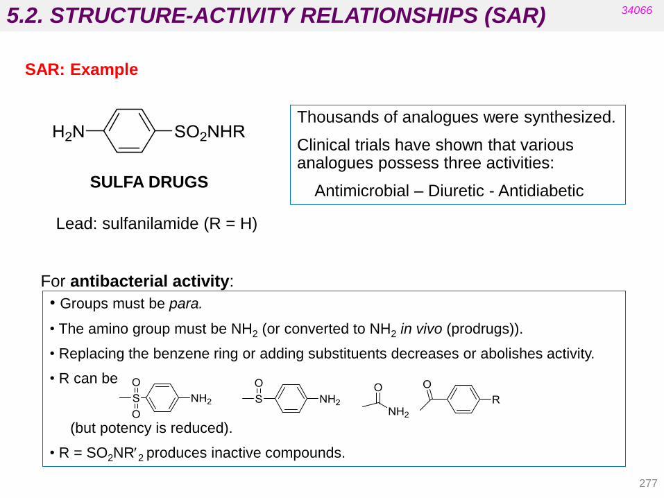
5.2. STRUCTURE-ACTIVITY RELATIONSHIPS (SAR)
277
34066
Lead: sulfanilamide (R = H)
Thousands of analogues were synthesized.
Clinical trials have shown that various analogues possess three activities:
Antimicrobial – Diuretic - Antidiabetic
• Groups must be para.
• The amino group must be NH2 (or converted to NH2 in vivo (prodrugs)).
• Replacing the benzene ring or adding substituents decreases or abolishes activity.
• R can be
(but potency is reduced).
• R = SO2NR2 produces inactive compounds.
For antibacterial activity:
SAR: Example
SULFA DRUGS

5.2. PHARMACOPHORE AND AUXOPHORE
278
Official definition for pharmacophore recommended by the IUPAC:
“A pharmacophore is the ensemble of steric and electronic features
that is necessary to ensure the optimal supramolecular interaction with
a specific biological target structure and to trigger (or block) its
biological response”. (Wermuth’s chapter 21)
34066
The highest common denominator of a group of ligands that exhibit a
similar biological effect recognized by the same binding site is called a
“pharmacophore”.
A pharmacophoric element or feature is generally defined as an
atom or groups of atoms common to active compounds with respect to
a target and essential for the activity (e.g. an amino group, an
aromatic group, an oxygen atom, etc.)
The other atoms of the molecule are commonly referred to as
auxophore and may be modified to improve pharmacodynamics or
pharmacokinetics in the lead optimization process.

5.2. PHARMACOPHORE AND AUXOPHORE
279
34066
The active conformation must be identified in order to identify the 3D
pharmacophore. For flexible molecules with large numbers of
conformations it is easier to compare activities of rigid analogues.
3D pharmacophore models can be used for database screening in
order to retrieve new compounds based on the similarity of
pharmacophoric features. (Pharmacophore-Based Virtual Database
Screening).
A pharmacophore model may be regarded as the representation of
a collection of pharmacophore features.
Parallel screening against several pharmacophores in order to predict
multitarget effects is also possible and very useful.

5.2. PHARMACOPHORE AND AUXOPHORE
280
34066
• Computers and modelling programs enable 3D structures to be
displayed and manipulated.
• Automated pharmacophore discovery programs have been
developed: (reliable 3D structural and biological data for all of
compounds under study must be available: e.g. atomic valences,
bond orders, protonation state, stereochemistry, consideration of
possible tautomers, etc.).
Pharmacophore modelling in computer-aided drug design
(Wermuth’s chapter 21)
• The steps for automated pharmacophore identification are:
a) Select a set of active ligands known to bind the same binding
site.
b) Perform conformational analysis of all the ligands.
c) Assign pharmacophoric features.
d) Perform molecular superimposition of the ligand conformations in
order to develop a common 3D pharmacophore.

5.2. PHARMACOPHORE AND AUXOPHORE
281
• Starting from a preliminary pharmacophore model, a hypothetical receptor
consisting of individual amino acid residues can be constructed surrounding
a set of superimposed ligands.
• A complex system can be generated guided by permanent correlation of
biological data and model-derived calculated free energies of binding.
• This complex can be useful as a model for the interaction pattern of a real
binding site.
• The resulting hypothetical receptor model is called a “mini-receptor” or
“pseudo-receptor”.
• The pseudo-receptor can be used to obtain a model that can predict the
biological activities of new ligands and enable the design of novel molecules
that fit the model (3D Quantitative Structure-Activity Relationships).
• The number of examples that successfully predict biological activity using
pharmacophore methods is constantly growing.
34066
Pharmacophore modelling in computer-aided drug design
(Wermuth’s chapter 21)
(To learn more about QSAR, see Unit 6)
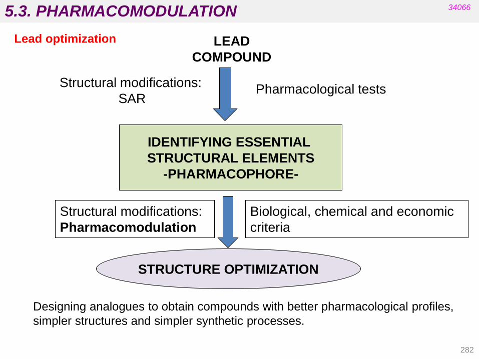
5.3. PHARMACOMODULATION
282
34066
LEAD
COMPOUND
IDENTIFYING ESSENTIAL
STRUCTURAL ELEMENTS
-PHARMACOPHORE-
STRUCTURE OPTIMIZATION
Structural modifications:
SAR Pharmacological tests
Biological, chemical and economic
criteria
Structural modifications:
Pharmacomodulation
Lead optimization
Designing analogues to obtain compounds with better pharmacological profiles,
simpler structures and simpler synthetic processes.

5.3. PHARMACOMODULATION
PROBLEMS LIMITING THE EFFECTIVENESS OF A POTENTIAL DRUG
pharmacodynamics / pharmacokinetics / chemistry / galenics
DRUG
poor absorption
fast absorption
low specificity
chemical instability
formulation
problems
difficulty of patient
acceptance
insolubility
in water toxicity
does (not) cross the
blood brain barrier
biotransformation
before arriving to
the site of action
34066
Lead optimization
283

5.3. PHARMACOMODULATION
284
Approaches with regard to changes in the lead structure size or complexity:
• simplification (disjunctive approaches):
- Involves deleting functions, structural elements or cycles.
- Is also used in SAR studies (to identify the pharmacophoric elements).
- Is useful for complex substances (many natural compounds).
• conservation of the same level of complexity (modulative approaches):
- Involves interconversion of atoms and functional groups, etc.
- Is also used in SAR studies.
• enlargement by adding elements (conjunctive approaches):
- Attaching additional structural elements (extension).
- Association of two separate drugs (association).
34066
Lead modification: strategies
The strategy for designing the analogues we will synthesize and test
depends on the size and degree of complexity of the lead‟s structure
and our main objective.

5.3. PHARMACOMODULATION
285
34066
Lead modification: examples
simplification
modulation
extension
simpler synthesis
acetylcholine propantheline
muscarinic antagonist
cocaine procaine
tolbutamide chlorpropamide
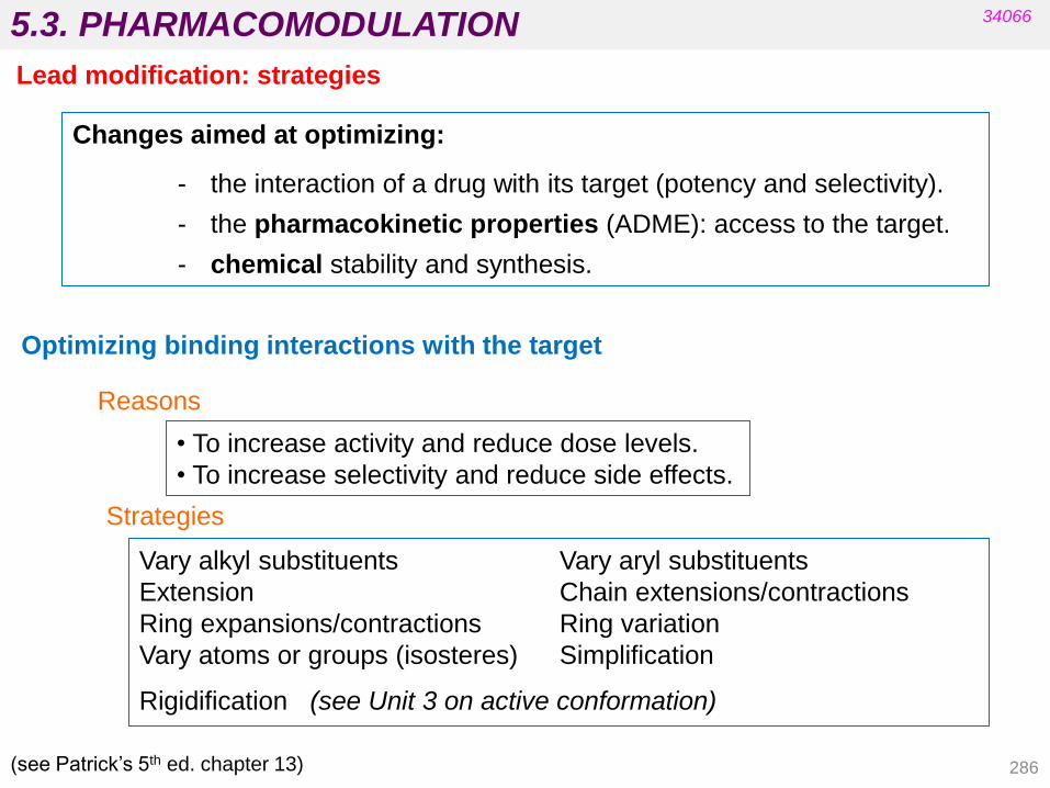
5.3. PHARMACOMODULATION 34066
Lead modification: strategies
Changes aimed at optimizing:
- the interaction of a drug with its target (potency and selectivity).
- the pharmacokinetic properties (ADME): access to the target.
- chemical stability and synthesis.
Optimizing binding interactions with the target
• To increase activity and reduce dose levels.
• To increase selectivity and reduce side effects.
Strategies
Vary alkyl substituents Vary aryl substituents
Extension Chain extensions/contractions
Ring expansions/contractions Ring variation
Vary atoms or groups (isosteres) Simplification
Rigidification (see Unit 3 on active conformation)
Reasons
(see Patrick‟s 5th ed. chapter 13) 286
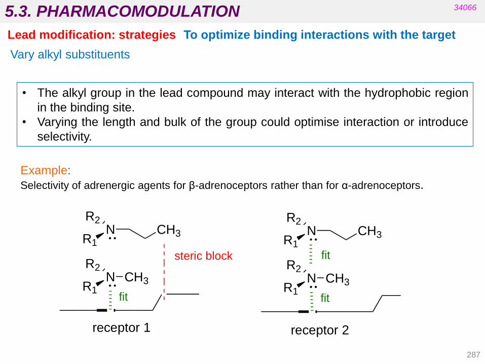
5.3. PHARMACOMODULATION
287
Vary alkyl substituents
To optimize binding interactions with the target
34066
Lead modification: strategies
• The alkyl group in the lead compound may interact with the hydrophobic region
in the binding site.
• Varying the length and bulk of the group could optimise interaction or introduce
selectivity.
Example:
Selectivity of adrenergic agents for β-adrenoceptors rather than for α-adrenoceptors.
N CH3R1
R2
NR1
R2CH3
steric block
fit
N CH3R1
R2
NR1
R2CH3
fit
fit
receptor 2receptor 1

5.3. PHARMACOMODULATION
288
Homology
Monoalkylated derivatives
Cyclomethylene derivatives
Difunctional derivatives
Ring’s extension or
contraction
Varying alkyl substituents
34066
Lead modification: strategies
• The similarity of the organic compounds of a series in which each member
differs from its adjacent compounds by a fixed increment, e.g. by CH2.
• Compounds in the series are named homologues.
Vary length
Vary length
(distance between groups)

5.3. PHARMACOMODULATION
289
Parabolic distribution
Continuous increase
Sawtooth profile
Asymmetric profile with a maximum
Continuous decrease
activity
No. of methylene groups
Varying alkyl substituents
34066
Lead modification: strategies
Homology
The effect that increasing the methylene groups has on the activity of a drug
depends on the importance of the modified alkyl chain: whether the chain
takes part in binding or not.
Changes in lipophilicity are also important.

5.3. PHARMACOMODULATION
290
Chain Extension/Contraction - Homologation
Optimizing binding interactions with the target
34066 34066
Lead modification: strategies
This strategy is employed to find the ideal length for the best interaction by
shortening or lengthening the chain length when two important binding groups
are linked together by a chain
Optimum chain length n = 2
Binding group Binding group

5.3. PHARMACOMODULATION
Synthetic feasibility of analogues
•It is feasible to replace alkyl substituents on heteroatoms with other alkyl
substituents.
•It is difficult to modify alkyl substituents on the carbon skeleton of a lead
compound.
•Total synthesis is usually required to vary alkyl substituents that are on the
carbon skeleton.
Varying alkyl substituents
Optimizing binding interactions with the target
34066
Lead modification: strategies
291
See Fig 13.32. Methods of modifying an alkyl group (Patrick’s 5th ed).
Ring expansion/contraction - Homologation
Expanding or contracting a ring puts other rings in different positions relative
to each other.
Better interactions are possible.
See Fig 13.40. Ring expansion (Patrick’s 5th ed).

5.3. PHARMACOMODULATION
292
Ring variations
Optimizing binding interactions with the target
34066 34066
Lead modification: strategies
Replacing aromatic/heterocyclic rings with other ring systems sometimes results
in improved properties.
It is often done for patent reasons.
Varying aryl substituents
• Varying substituents or the substitution pattern
Nitroanilines:
•The binding strength of NH2 as HBD is affected by the relative
position of NO2.
•This effect is stronger when NO2 is at the para position.
•Amine N is a weaker HBA due to these effects.
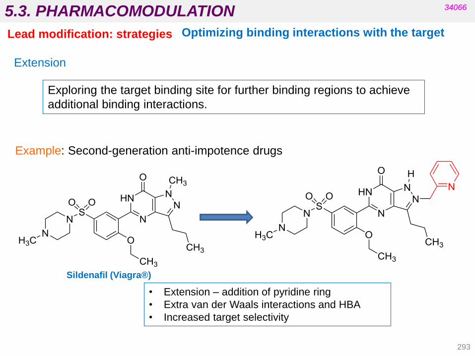
5.3. PHARMACOMODULATION
293
Extension
Optimizing binding interactions with the target
34066 34066
Lead modification: strategies
Exploring the target binding site for further binding regions to achieve
additional binding interactions.
Example: Second-generation anti-impotence drugs
• Extension – addition of pyridine ring
• Extra van der Waals interactions and HBA
• Increased target selectivity
Sildenafil (Viagra®)

5.3. PHARMACOMODULATION
294
Isosteric Replacement
Optimizing binding interactions with the target
34066 34066
Lead modification: strategies
Replacing a functional group with a group of the same valence (isostere)
e.g. OH replaced by SH, NH2, CH3 or O replaced by S, NH, CH2
• Leads to more controlled changes in steric/electronic properties.
• May improve binding but may also modify pharmacokinetics, toxicity,
stability and synthetic processes.
Isosteres are atoms, ions or molecules that contain the same number and
arrangement of electrons and the same number of atoms.
Classic Isosterism (Langmuir, 1919)

5.3. PHARMACOMODULATION
Classic isosterism
Grimm‟s Hydride
Displacement Law (1925)
34066 34066
Lead modification
Addition of hydride to an
atom gives to the
resulting pseudoatom the
properties of the atom
with the next highest
atomic number.
Isosteric and Bioisosteric Replacement
295

5.3. PHARMACOMODULATION
296
Erlenmeyer‟s Definition (1932):
“Isosteres are elements, molecules or ions in which the peripheral layers of
electrons may be considered identical”.
Expansions:
• The whole group of elements present in a given column of the periodic table
E.g.: Si and C , S and O
• Pseudoatoms (from Grimm‟s law and including groups which seem totally
different but which possess similar properties)
E.g.: -Cl and -CN, -CN and -SCN
• The ring equivalents: equivalence between –CH=CH- and -S-
E.g.: benzene and thiophene
Classic isosterism
34066 34066
Lead modification Isosteric Replacement
Isosterism criteria:
Isosteric molecules must present similar (if not identical) volume, shape and
physical properties.
Some properties may be different: e.g. dipolar moment, polarity, size, etc.
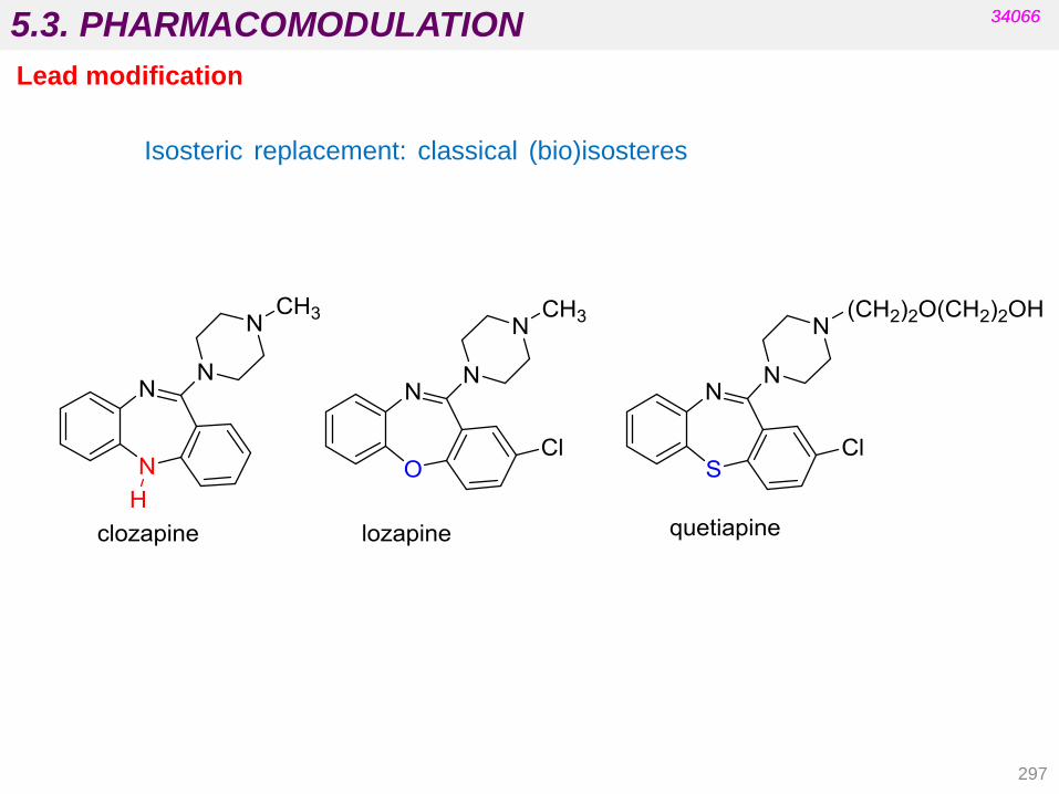
5.3. PHARMACOMODULATION
297
34066 34066
Lead modification
Isosteric replacement: classical (bio)isosteres

5.3. PHARMACOMODULATION
Bioisosterism
Friedman (1951):
Bioisosteres are compounds that fit the broadest definition for isosteres and
have the same type of biological activity (classical bioisosteres).
Thornber (1979):
Bioisosteres are groups or molecules which have chemical and physical
similarities producing broadly similar biological effects.
(antagonism is included)
A bioisostere is a group that can be used to replace another group while retaining
the desired biological activity.
Isosteric and Bioisosteric Replacement
34066 34066
Lead modification
Non-classical bioisosteres:
Do not have the same number of atoms or electrons and do not fit the rules of
classical isosteres but show similar biological activity.
298

5.3. PHARMACOMODULATION
BIOISOSTERES BY ELECTRONIC EFFECTS OR HYDROGEN BOND ABILITY
BIOISOSTERES BY ACIDIC CHARACTER
catechol/benzimidazole
(electron pairs, hydrogen
bonds)
pyridine/nitrobenzene
(electron density)
pyridinium/anilinium
(charge density)
carboxyl sulphonamide 5-tetrazolyl
34066 34066
Lead modification: strategies Bioisosteric Replacement
• They are ionized at physiological pH.
• They have a plane structure.
• Tetrazole is more lipophilic (better
absorption).
E.g. losartan
299

5.3. PHARMACOMODULATION
Carboxylic acids analogues
34066 34066
Lead modification: strategies Bioisosteric Replacement
Ring variation
ibrupoxam
(hydroxamic acid) ibuprofen
phenbenzamine tripenelamine
300

5.3. PHARMACOMODULATION
301
34066 34066
Lead modification: strategies Isosteric and Bioisosteric Replacement
Two bioisosteric groups are similar in some properties but may differ in others.
Properties that may be affected by bioisosteric replacements:
a) Electronic distribution
b) Hybridation angles
c) Size and volume of the groups
d) Solubility
e) Acidity
f) Ability to form hydrogen bonds
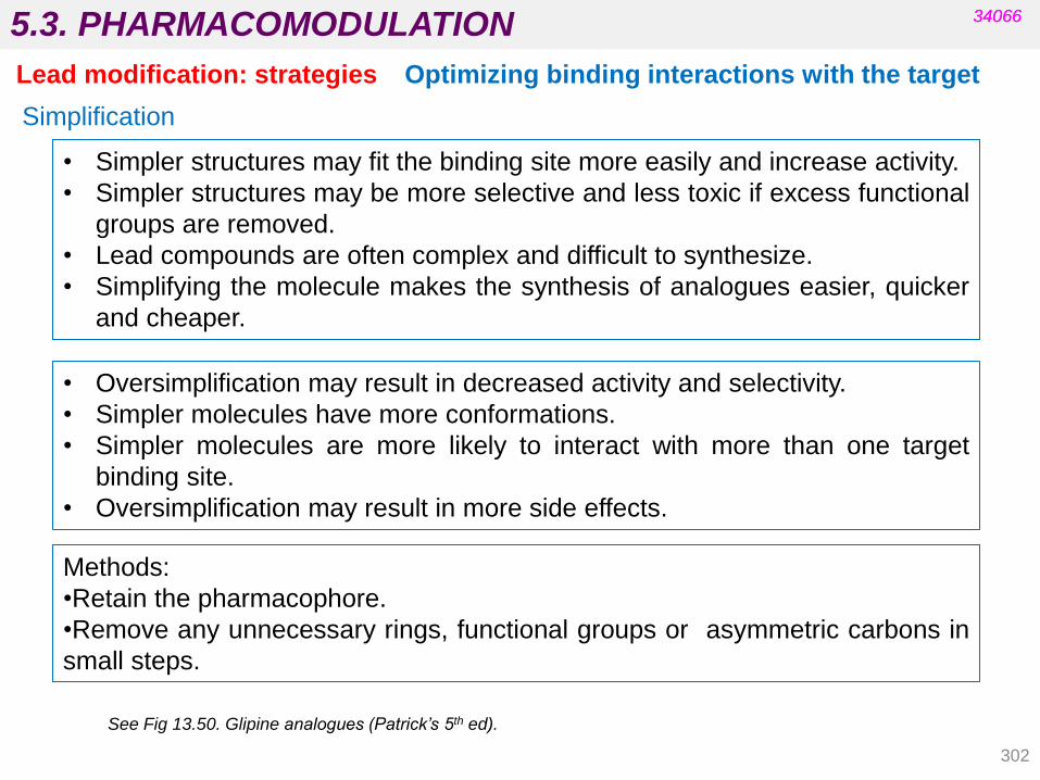
5.3. PHARMACOMODULATION
302
Simplification
Optimizing binding interactions with the target
34066 34066
Lead modification: strategies
• Simpler structures may fit the binding site more easily and increase activity.
• Simpler structures may be more selective and less toxic if excess functional
groups are removed.
• Lead compounds are often complex and difficult to synthesize.
• Simplifying the molecule makes the synthesis of analogues easier, quicker
and cheaper.
• Oversimplification may result in decreased activity and selectivity.
• Simpler molecules have more conformations.
• Simpler molecules are more likely to interact with more than one target
binding site.
• Oversimplification may result in more side effects.
Methods:
•Retain the pharmacophore.
•Remove any unnecessary rings, functional groups or asymmetric carbons in
small steps.
See Fig 13.50. Glipine analogues (Patrick’s 5th ed).

5.3. PHARMACOMODULATION 34066
Lead modification: strategies
Changes aimed at optimizing:
- the interaction of a drug with its target (potency and selectivity)
- the pharmacokinetic properties (ADME): access to the target
- chemical stability and synthesis
To optimize binding interactions with the target
• To increase activity and reduce dose levels.
• To increase selectivity and reduce side effects.
Strategies
Varying alkyl substituents Varying aryl substituents
Extension Chain extensions/contractions
Ring expansions/contractions Ring variation
Varying atoms or groups (Isosteres) Simplification
Rigidification (see Unit 3 on active conformation)
Reasons
(see Patrick‟s 5th ed. chapter 13) 303

5.3. PHARMACOMODULATION 34066
Lead modification: strategies
304
Optimizing pharmacokinetic properties
(see Patrick’s 5th ed. chapter 14)
Varying alkyl substituents
larger alkyl groups increase hydrophobicity (homologous).
Caution: may interfere with target binding for steric reasons.
Masking or removing polar groups
increases the hydrophobic character.
Caution: do not modify the necessary polar groups for target
binding (see prodrugs).
Methods:
ethers from alcohols, amides from amines, esters from acids, etc.
Bioisosteric replacement (e.g. –COOH and tetrazole ring).
To improve membrane permeability
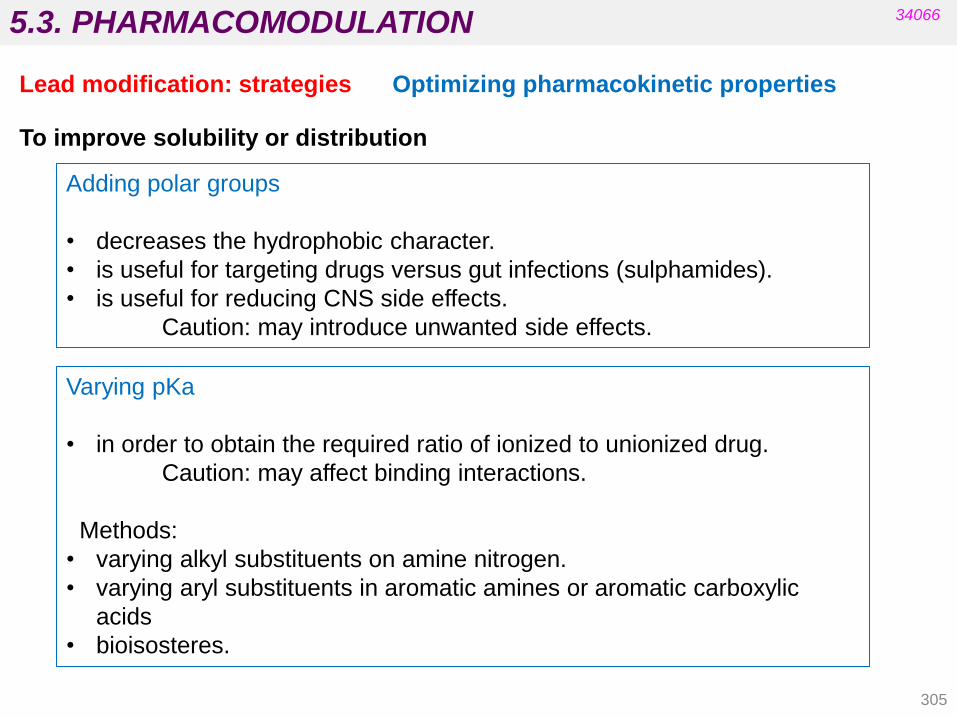
5.3. PHARMACOMODULATION
305
34066
Lead modification: strategies Optimizing pharmacokinetic properties
To improve solubility or distribution
Varying pKa
• in order to obtain the required ratio of ionized to unionized drug.
Caution: may affect binding interactions.
Methods:
• varying alkyl substituents on amine nitrogen.
• varying aryl substituents in aromatic amines or aromatic carboxylic
acids
• bioisosteres.
Adding polar groups
• decreases the hydrophobic character.
• is useful for targeting drugs versus gut infections (sulphamides).
• is useful for reducing CNS side effects.
Caution: may introduce unwanted side effects.

5.3. PHARMACOMODULATION
306
Crosses the blood brain barrier
and has important side effects.
IONIC SALT
atropine
central and peripheral
anticholinergic ipratropium bromide
exclusively peripheral
anticholinergic
34066
Lead modification: strategies Optimizing pharmacokinetic properties
To improve solubility or distribution

5.3. PHARMACOMODULATION
307
Steric shields
Introduce bulky group as a shield.
Protect a susceptible functional group from hydrolysis.
Hinder attack by nucleophiles or enzymes.
Electronic stabilization of labile functional groups
e.g. Bioisosteres. Replacing esters with carbamates or amides.
Steric and electronic effects in combination
(See the example in questions from procaine to lidocaine.)
Metabolic blockers
Introduce groups at a susceptible site to block the reaction.
Bioisosteres (e.g. replacing H with F).
Removing or replacing a susceptible group with a metabolically more stable one
Bioisosteres (replacing methyl groups with Cl).
e.g. modification of tolbutamide to chlorpropamide.
Shifting susceptible metabolic groups that are important for binding
e.g. salbutamol.
(see Unit 4 – soft and hard drugs)
34066
Lead modification: strategies Optimizing pharmacokinetic properties
To increase drug stability

5.3. PHARMACOMODULATION
Bioisosterism between H and F
cinnarazine
Shifting a susceptible OH phenolic group
34066
Lead modification: strategies Optimizing pharmacokinetic properties
To increase drug stability
Fluorine atoms prevent para-hydroxylation.
Benzylic alcohol is not a good substrate for
COMT.
The presence of a HBD is important for activity.
flunarazine
Cerebral vasodilator
noradrenaline
salbutamol
308
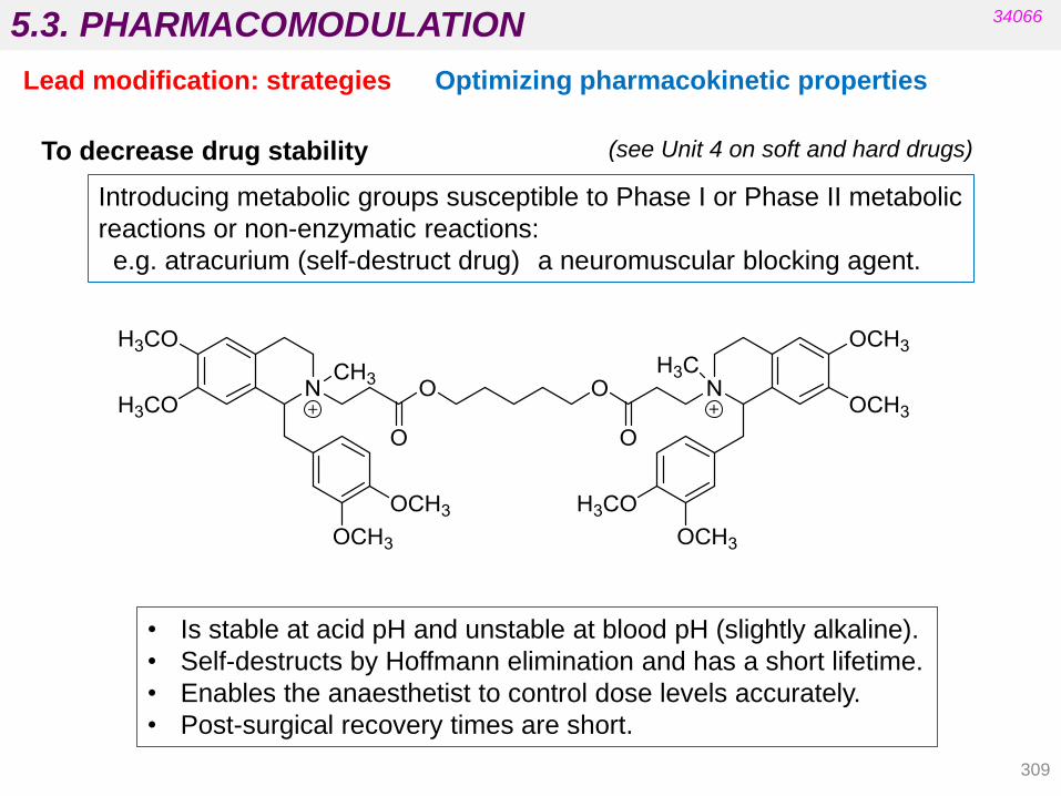
5.3. PHARMACOMODULATION
309
(see Unit 4 on soft and hard drugs)
34066
Lead modification: strategies Optimizing pharmacokinetic properties
Introducing metabolic groups susceptible to Phase I or Phase II metabolic
reactions or non-enzymatic reactions:
e.g. atracurium (self-destruct drug) a neuromuscular blocking agent.
• Is stable at acid pH and unstable at blood pH (slightly alkaline).
• Self-destructs by Hoffmann elimination and has a short lifetime.
• Enables the anaesthetist to control dose levels accurately.
• Post-surgical recovery times are short.
To decrease drug stability
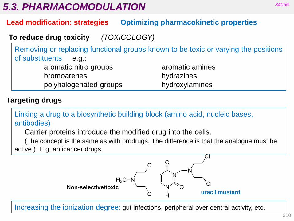
5.3. PHARMACOMODULATION
310
34066
Lead modification: strategies Optimizing pharmacokinetic properties
Targeting drugs
To reduce drug toxicity (TOXICOLOGY)
Removing or replacing functional groups known to be toxic or varying the positions
of substituents e.g.:
aromatic nitro groups aromatic amines
bromoarenes hydrazines
polyhalogenated groups hydroxylamines
Linking a drug to a biosynthetic building block (amino acid, nucleic bases,
antibodies)
Carrier proteins introduce the modified drug into the cells.
(The concept is the same as with prodrugs. The difference is that the analogue must be
active.) E.g. anticancer drugs.
Non-selective/toxic uracil mustard
Increasing the ionization degree: gut infections, peripheral over central activity, etc.

5.3. PHARMACOMODULATION
311
34066
Lead modification: strategies Increasing chemical stability
Increase resistance to acid hydrolysis. PENICILLINS
Electron withdrawing group
by inductive effect
ampicillin
amoxicillin phenethicillin

5.3. PHARMACOMODULATION
DISJUNCTIVE
MODULATIVE
CONJUNCTIVE
Removing rings and functional groups
Removing asymmetric centres
Ring variations: opening, closing, reorganizing
Homology
Extension
Vinylogy
Isomerism
Bioisosterism
Molecular combination/Twin drugs
34066
Lead modification: techniques
The
se
tech
niq
ue
s a
re a
lso in
clu
de
d in
ou
r co
urs
e g
uid
e…
312
approach technique
Extension
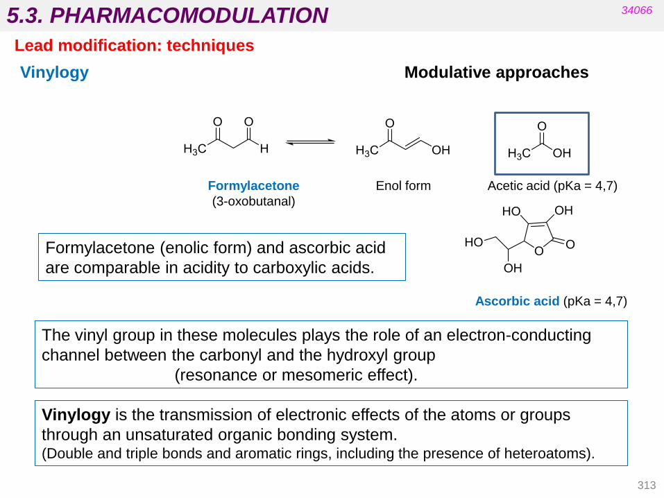
5.3. PHARMACOMODULATION
313
Enol form Acetic acid (pKa = 4,7)
Ascorbic acid (pKa = 4,7)
The vinyl group in these molecules plays the role of an electron-conducting
channel between the carbonyl and the hydroxyl group
(resonance or mesomeric effect).
Formylacetone
(3-oxobutanal)
Vinylogy
Formylacetone (enolic form) and ascorbic acid
are comparable in acidity to carboxylic acids.
Vinylogy is the transmission of electronic effects of the atoms or groups
through an unsaturated organic bonding system. (Double and triple bonds and aromatic rings, including the presence of heteroatoms).
34066
Lead modification: techniques
Modulative approaches

5.3. PHARMACOMODULATION
314
34066
Lead modification: techniques
Vinylogy
When X and Y are groups where electrons can be delocalized.
Modulative approaches
Vinylogous Arenologous
The electronic properties of vinylogues are similar but the lipophilicity, shape
and flexibility of the molecule are modified.
Example:
phenylbutazone styrylbutazone
X - Y

5.3. PHARMACOMODULATION
doxepine
(E isomer)
doxepine
(Z isomer)
methadone pethidine
isomethadone alphaprodine
34066
Lead modification: techniques
Isomerism
double bond position inverse ester
Important changes in the shape and position of certain groups
Modulative approaches
315

5.3. PHARMACOMODULATION
316
34066
Lead modification: techniques
Drug duplication: symmetrical twin drugs Conjunctive approaches
ethambutol
(tuberculostatic)
probucol
(anti-hyperlipidemic)
dicumarol
(anticoagulant)
Metabolically non-reversible – different phamacokinetics

5.3. PHARMACOMODULATION
317
34066
Lead modification: techniques
Drug association: non-symmetrical twin drugs Conjunctive approaches
benorilate
aspirine + paracetamol
(analgesic)
etofibrate
clofibric acid + nicotinic acid
(cholesterol and triglyceride reducer)
Metabolically reversible:
Prodrugs whose bioactivation leads to two different drugs with synergic affects.
Co-drugs or hybrids

5.3. PHARMACOMODULATION
antihypertensive
chlorothiazide derivative
(diuretic)
pindolol
(adrenergic b1 blocker)
ACE inhibitor
34066
Lead modification: techniques
Drug association: non-symmetrical twin drugs Conjunctive approaches
Metabolically non-reversible: multi-target drugs (take a single tablet)
Co-drugs or hybrids
318

• The possibilities for molecular variations around the lead structure are
immense.
• Deciding which compounds to prepare and which ones to reject is not easy.
• If information about the ligand-target interactions is available from
experimental or computational data:
Is the lead relevant to a computer-aided design?
• When no information is available:
design analogues to identify which molecular features are favourable
to the activity and which are detrimental (not only activity against the
target but also other important properties of the future drug
molecules with regard to pharmacokinetics, chemical stability and
toxicity).
Wermuth’s 4th ed, chapter no 12
Tema 5: Diseño y Desarrollo de Nuevos Fármacos 34066 5.3. PHARMACOMODULATION
The lead optimization process: guidelines
319
1. The minor modification rule
When designing analogues, first use simple organic reactions
(hydrogenations, methylations, acetylations, changes to substituents,
and isosteric replacements, etc.).
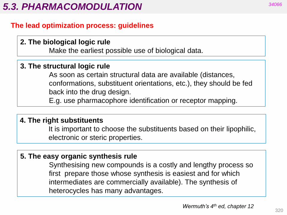
5.3. PHARMACOMODULATION
320
5. The easy organic synthesis rule
Synthesising new compounds is a costly and lengthy process so
first prepare those whose synthesis is easiest and for which
intermediates are commercially available). The synthesis of
heterocycles has many advantages.
Wermuth’s 4th ed, chapter 12
34066
The lead optimization process: guidelines
2. The biological logic rule
Make the earliest possible use of biological data.
3. The structural logic rule
As soon as certain structural data are available (distances,
conformations, substituent orientations, etc.), they should be fed
back into the drug design.
E.g. use pharmacophore identification or receptor mapping.
4. The right substituents
It is important to choose the substituents based on their lipophilic,
electronic or steric properties.

5.3. PHARMACOMODULATION
321
7. The Pharmacological Logic Rule
When designing analogues, compare the results for your
compounds with those of reference compounds published by
competitors (resynthesize if necessary).
When SAR studies enable the molecular features associated with
high activity to be identified, also prepare at least one
compound that, according to your results, should be inactive.
Wermuth’s 4th ed, chapter 12
34066
The lead optimization process: guidelines
6. Eliminate chiral centres
Racemates and both enantiomers are usually three different
pharmacological entities (pharmacological, toxicological and
clinical research is required before it can be decided whether it is
advantageous to use racemates or enantiomers in clinical
practice).

5.4. RATIONAL STRATEGIES IN DRUG DESIGN
ACTIVATION AND/OR RECEPTOR BLOCKING RECEPTOR
AGONISTS AND ANTAGONISTS
AUTORECEPTOR
modulating neurotransmitter release and synthesis
MODULATION of the activity of chemical messengers can take place on
different levels:
STORAGE CARRIER PROTEINS REUPTAKE PROTEINS
ENZYMES (BIOSYNTHESIS AND DEGRADATION)
34066
Chemical messengers as leads
322
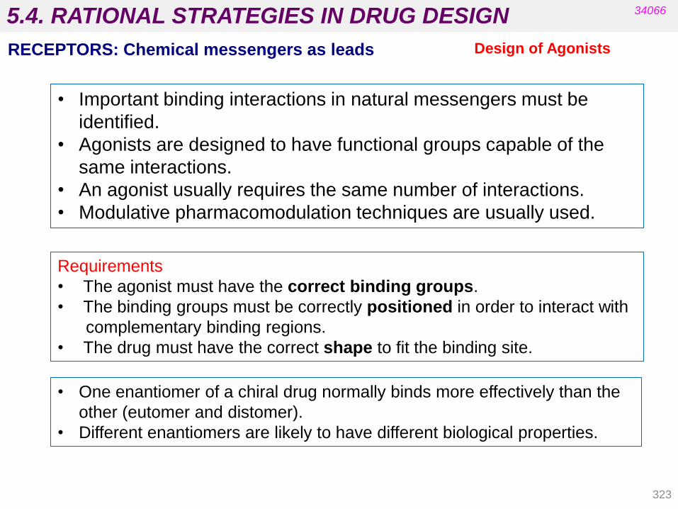
5.4. RATIONAL STRATEGIES IN DRUG DESIGN
Design of Agonists
Requirements
• The agonist must have the correct binding groups.
• The binding groups must be correctly positioned in order to interact with
complementary binding regions.
• The drug must have the correct shape to fit the binding site.
• Important binding interactions in natural messengers must be
identified.
• Agonists are designed to have functional groups capable of the
same interactions.
• An agonist usually requires the same number of interactions.
• Modulative pharmacomodulation techniques are usually used.
34066
RECEPTORS: Chemical messengers as leads
• One enantiomer of a chiral drug normally binds more effectively than the
other (eutomer and distomer).
• Different enantiomers are likely to have different biological properties.
323

5.4. RATIONAL STRATEGIES IN DRUG DESIGN
These are agents which enhance receptor activity by binding to an allosteric
binding site rather than to the messenger binding site.
The structure of allosteric modulators does not have to be similar to that
of the agonist since they do not share the same binding site (the steric and
electronic requirements are different).
Allosteric modulators
Design of Agonists
34066
RECEPTORS: Chemical messengers as leads
Many examples of drugs that act as agonists have been developed from the
corresponding chemical messengers.
Agonist
(treatment of
migraines)
5-hydroxytryptamine sumatriptan
324
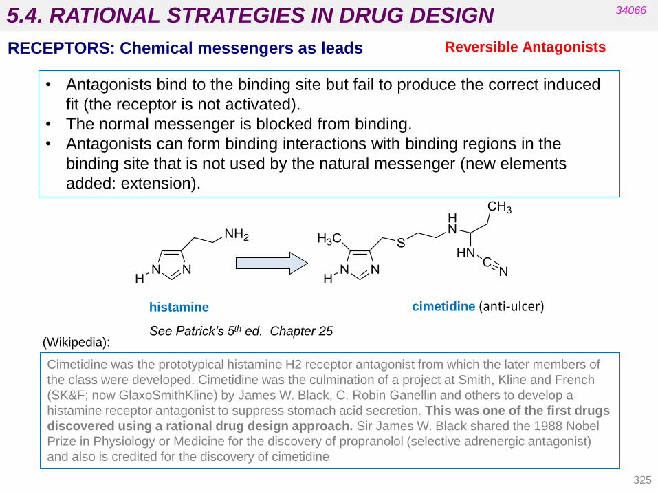
5.4. RATIONAL STRATEGIES IN DRUG DESIGN
Reversible Antagonists
• Antagonists bind to the binding site but fail to produce the correct induced
fit (the receptor is not activated).
• The normal messenger is blocked from binding.
• Antagonists can form binding interactions with binding regions in the
binding site that is not used by the natural messenger (new elements
added: extension).
34066
RECEPTORS: Chemical messengers as leads
histamine cimetidine (anti-ulcer)
Cimetidine was the prototypical histamine H2 receptor antagonist from which the later members of
the class were developed. Cimetidine was the culmination of a project at Smith, Kline and French
(SK&F; now GlaxoSmithKline) by James W. Black, C. Robin Ganellin and others to develop a
histamine receptor antagonist to suppress stomach acid secretion. This was one of the first drugs
discovered using a rational drug design approach. Sir James W. Black shared the 1988 Nobel
Prize in Physiology or Medicine for the discovery of propranolol (selective adrenergic antagonist)
and also is credited for the discovery of cimetidine
See Patrick’s 5th ed. Chapter 25 (Wikipedia):
325

5.4. RATIONAL STRATEGIES IN DRUG DESIGN
Irreversible Antagonists
• The antagonist binds irreversibly to the binding site.
• A different induced fit means that the receptor is not activated.
• A covalent bond is formed between the drug and the receptor.
• The messenger is blocked from the binding site.
• Increasing the messenger concentration does not reverse antagonism.
• Irreversible antagonists are often used to label receptors (not in
therapeutics).
• They have a similar structure to the chemical messenger.
• They have reactive functional groups: depending on the groups that act in
the catalysis.
• The antagonist binds reversibly to an allosteric binding site.
• Intermolecular bonds are formed between the antagonist and the binding site.
• The induced fit alters the shape of the receptor.
• The binding site is distorted and is not recognised by the messenger.
• Increasing the messenger concentration does not reverse antagonism.
• The structure of allosteric antagonists does not have to be similar to
that of the chemical messenger since they have different binding sites (the
steric and electronic requirements are different).
Allosteric Antagonists
34066
RECEPTORS: Chemical messengers as leads
326

5.4. RATIONAL STRATEGIES IN DRUG DESIGN 34066
1 Antibacterial agents (unit 8) Dihydropteroate synthetase, transpeptidase
2 Antiviral agents HIV reverse transcriptase, HIV protease,
viral DNA polymerase
3 Anti-inflammatory agents Cyclooxygenase
4 Cholesterol lowering agents HMG-CoA reductase
5 Antidepressants (unit 11) Monoamine oxidase
6 Anticancer agents Tyrosine kinase, dihydrofolate reductase,
thymidylate synthase, aromatase, etc.
7 Antihypertensive agents Renin, angiotensin converting enzyme
8 Treatment of male erectile dysfunction Phosphodiesterase
9 Anti-gout agents Xanthine oxidase
10 Anti-ulcer agents Proton pump
11 Alzheimer disease Cholinesterases
12 Diuretics Carbonic anhydrase
Enzyme targets for useful medications Enzymes
327

5.4. RATIONAL STRATEGIES IN DRUG DESIGN
reversible inhibition
By structural analogy with substrate: antimetabolites By structural analogy with transition state irreversible inhibition (or ENZYMATIC INACTIVATION )
Addressed to the active centre
Suicide inhibitors
Enzyme Inhibitors
34066
• The inhibitor binds reversibly to the active site.
• Intermolecular bonds are involved in binding.
• The inhibitor undergoes no reaction.
• Inhibition depends on the strength of inhibitor binding and inhibitor
concentration.
• The substrate is blocked from the active site.
• Increasing the substrate concentration reverses inhibition.
• The inhibitor is likely to be similar in structure to the substrate, product or
cofactor.
Reversible inhibitors
328

5.4. RATIONAL STRATEGIES IN DRUG DESIGN
By structural analogy with substrate
34066
ENZYMES: substrates as leads Reversible inhibitors
Gout Treatment
Adenine Guanine
Xanthine
oxidase Xanthine
oxidase
Xanthine
oxidase
Competitive
inhibition
Antimetabolite: This
compound is similar to a
substrate that suffers the
reaction by the enzyme. It
inhibits enzymes that are
crucial to the normal
metabolism of the cell.
It is used in antibacterial
and anticancer therapy.
329

5.4. RATIONAL STRATEGIES IN DRUG DESIGN
330
Drugs designed to mimic the transition state of an enzyme-catalysed reaction:
• Are likely to bind more strongly than drugs that mimic the substrate or product.
• Are high-energy, transient species that cannot be isolated or synthesised.
Drug design may be based on reaction intermediates which are closer in character
to transition states than to substrates or products.
The drug mimics the stereochemistry and binding properties of the reaction
intermediate but is stable.
34066
ENZYMES: substrates as leads
•Renin inhibitors block the
synthesis of angiotensin I and II.
• Angiotensin II constricts blood
vessels and raises blood
pressure.
• Renin inhibitors act as
antihypertensives (drugs to lower
blood pressure).
Reversible inhibitors Transition state inhibitors
Renin inhibitors
Angiotensinogen
Angiotensin I
Angiotensin II
Renin
ACE (angiotensin converting
enzyme)

5.4. RATIONAL STRATEGIES IN DRUG DESIGN 34066
It is important to know the structure of the active site and the mechanism of the
catalytic reaction.
ENZYMES: substrates as leads
Transition state inhibitors
Reversible inhibitors
Examples of reversible inhibitors
ACE Inhibitors Diuretics Statins
Protease inhibitors Sulphonamides Antidepressants
Kinase inhibitors
L-Phe-L-Pro saquinavir
331
NHN
NH
N
O
O
OH
O
NH
H
H
H
O
H2N

5.4. RATIONAL STRATEGIES IN DRUG DESIGN
332
The inhibitor binds irreversibly to the active site:
• A covalent bond is formed between the drug and the enzyme.
• The substrate is blocked from the active site.
• Increasing substrate concentration does not reverse inhibition.
• The inhibitor is likely to be similar in structure to the substrate.
• Electrophilic functional groups are present in irreversible inhibitors:
Examples
34066
ENZYMES: substrates as leads Irreversible inhibitors
Nerve gases Penicillins Cephalosporins Disulfuram
Proton pump inhibitors Orlistat Aspirin
Hydroxyl, amino or thiol groups from amino acids in the active site of the
enzyme react with these groups.
Alkyl halides Epoxides α,β-Unsaturated ketones
Fluorophosphonates β-lactams Lactones

5.4. RATIONAL STRATEGIES IN DRUG DESIGN
333
• The enzyme is blocked from digesting fats in the intestine.
• Fatty acids and glycerol are therefore less absorbed.
• Biosynthesis of fat in the body is reduced.
34066
ENZYMES: substrates as leads Irreversible inhibitors
Orlistat is an anti-obesity drug that inhibits pancreatic lipase.
See Box 7.2 Patrick’s 5th ed. Chapter 7 p.90
OO
C6H13
C11H23
O
NHCHO
O
H3C CH3

5.4. RATIONAL STRATEGIES IN DRUG DESIGN 34066
ENZYMES: substrates as leads Irreversible inhibitors
More examples
334

5.4. RATIONAL STRATEGIES IN DRUG DESIGN
335
34066
ENZYMES: substrates as leads Irreversible inhibitors
Penicillin is an irreversible inhibitor of bacterial transpeptidase.
Penicillin is a substrate of β-lactamase.
See Fig 7.10 (Patrick’s 5th ed). Clavulanic acid acting as a suicide substrate
Clavulanic acid is a suicide inhibitor of β-lactamase with a
similar structure and reactive latent functionality.
N
S
O
NH H
CH3
CH3
CO2H
O
R
HO - Ser
HN
S
O
NH H
CH3
CH3
CO2H
O
R
O
Ser
b-lactamaseHN
S
O
NH H
CH3
CH3
CO2H
O
R
HO Ser
b-lactamase
+ H2O
OH
N
O
O
H
CO2H
CH2OH
Suicide
substrates
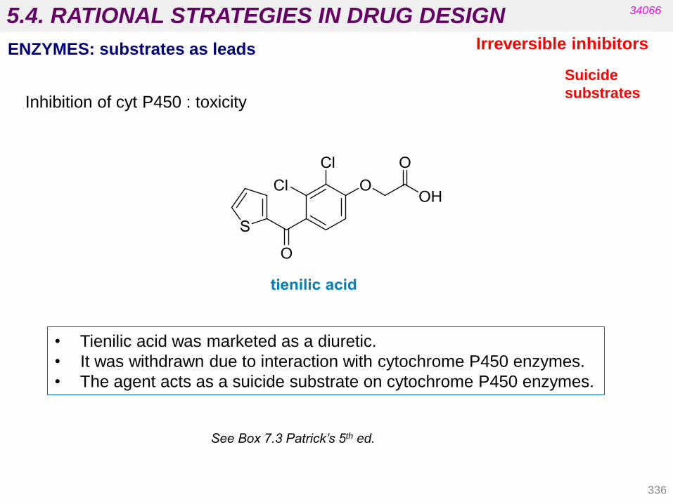
5.4. RATIONAL STRATEGIES IN DRUG DESIGN
• Tienilic acid was marketed as a diuretic.
• It was withdrawn due to interaction with cytochrome P450 enzymes.
• The agent acts as a suicide substrate on cytochrome P450 enzymes.
34066
ENZYMES: substrates as leads Irreversible inhibitors
Inhibition of cyt P450 : toxicity
See Box 7.3 Patrick’s 5th ed.
336
Suicide
substrates

5.4. RATIONAL STRATEGIES IN DRUG DESIGN
Suicide
substrates
34066
ENZYMES Irreversible inhibitors
337

5.4. RATIONAL STRATEGIES IN DRUG DESIGN
• The inhibitor binds reversibly o irreversibly to the allosteric site.
• Induced fit alters the shape of the enzyme and the active site is distorted
and not recognised by the substrate.
• Increasing the substrate concentration does not reverse inhibition.
• The inhibitor is not similar in structure to the substrate.
• The inhibitor can be similar in structure to the final product of a
multistep metabolic route.
6-Mercaptopurine
• Inhibits the first enzyme in the biosynthesis of purines.
• Blocks the biosynthesis of purines and DNA.
• Is used to treat leukaemia.
34066
ENZYMES
Inhibitors acting at
allosteric binding sites
338
6.1. Introduction.
6.2. Physicochemical parameters: Hammett
equation (electronic effects), Taft equation (steric
factors) and Hansch equation (hydrophobic
factors).
6.3. QSAR examples. Methods used to correlate
physical and chemical parameters with biological
activity. Examples.
PHARMACEUTICAL CHEMISTRY -
UNIT 6. QUANTITATIVE STRUCTURE - BIOLOGICAL
ACTIVITY RELATIONSHIPS (QSAR)
34066
Patrick’s 5th ed. Ch. 17 and 18
Silverman’s 3rd ed. Ch. 2
Delgado’s Ch. 8
339

6.1. INTRODUCTION TO QSAR
340
34066
THIS UNIT:
• Quantitative structure-activity relationships (QSAR) are rational
approaches for deciding which substituents to use.
• Classical QSAR consider only 2D structures and approaches attempt to
identify and quantify the physicochemical properties or structural
elements of a drug and determine whether any of them affect the drug‟s
biological activity.
• 3D-QSAR analysis (Correlative Molecular Fields Analysis (CoMFA))
starts from 3D structures and correlates biological activities with 3D-
property fields.
ALREADY SEEN IN PREVIOUS UNITS:
• Changes in shape or changes in functional groups or substituents can
improve pharmacodynamics and/or pharmacokinetics.
• Synthesising analogues of a lead compound is one strategy used in the
design of drugs.
• The number of possible analogues is almost infinite.

6.1. INTRODUCTION TO QSAR
341
34066
Historical Overview
1868: Crum-Brown and Fraser predicted that a mathematical
relationship between structure and activity would be expressed.
1962: Hansch and Fujita attempted to quantify the effects of
certain substituent modifications and developed QSARs.
1988: Cramer developed 3D QSAR.
Hansch paradigm
1. Biological activity depends on the drug structure.
2. The drug structure involves certain global properties such as hydrophobicity,
charge and volume in a certain position of the molecule.
3. These global and local properties can be quantified by physicochemical
parameters.
4. There is always a function which relates changes in biological activity with
changes in global and local properties, though it may be neither simple nor
obvious.
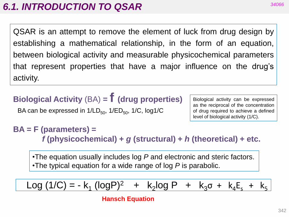
6.1. INTRODUCTION TO QSAR
342
34066
Biological Activity (BA) = f (drug properties)
BA can be expressed in 1/LD50, 1/ED50, 1/C, log1/C
BA = F (parameters) =
f (physicochemical) + g (structural) + h (theoretical) + etc.
QSAR is an attempt to remove the element of luck from drug design by
establishing a mathematical relationship, in the form of an equation,
between biological activity and measurable physicochemical parameters
that represent properties that have a major influence on the drug‟s
activity.
•The equation usually includes log P and electronic and steric factors.
•The typical equation for a wide range of log P is parabolic.
Hansch Equation
Biological activity can be expressed
as the reciprocal of the concentration
of drug required to achieve a defined
level of biological activity (1/C).
Log (1/C) = - k1 (logP)2 + k2log P + k3σ + k4Es + k5
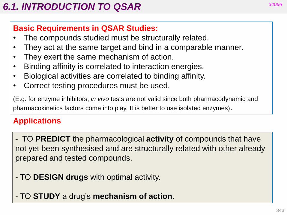
6.1. INTRODUCTION TO QSAR
343
34066
Applications
- TO PREDICT the pharmacological activity of compounds that have
not yet been synthesised and are structurally related with other already
prepared and tested compounds.
- TO DESIGN drugs with optimal activity.
- TO STUDY a drug‟s mechanism of action.
Basic Requirements in QSAR Studies:
• The compounds studied must be structurally related.
• They act at the same target and bind in a comparable manner.
• They exert the same mechanism of action.
• Binding affinity is correlated to interaction energies.
• Biological activities are correlated to binding affinity.
• Correct testing procedures must be used.
(E.g. for enzyme inhibitors, in vivo tests are not valid since both pharmacodynamic and
pharmacokinetics factors come into play. It is better to use isolated enzymes).
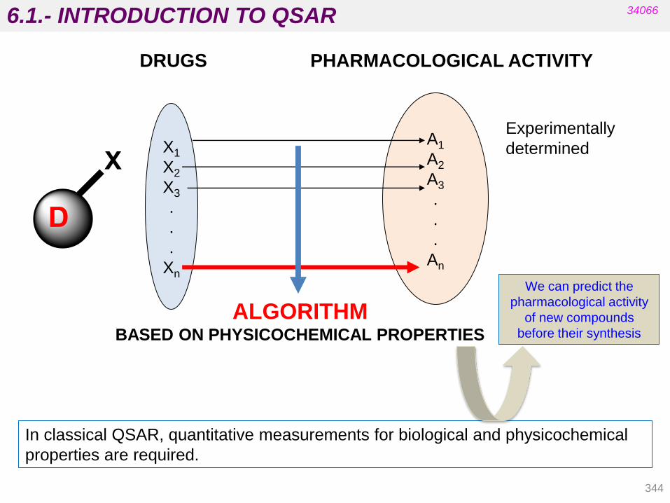
6.1.- INTRODUCTION TO QSAR
344
34066
X1
X2
X3
.
.
.
Xn
A1
A2
A3
.
.
.
An
DRUGS PHARMACOLOGICAL ACTIVITY
Experimentally
determined
ALGORITHM BASED ON PHYSICOCHEMICAL PROPERTIES
We can predict the
pharmacological activity
of new compounds
before their synthesis
In classical QSAR, quantitative measurements for biological and physicochemical
properties are required.
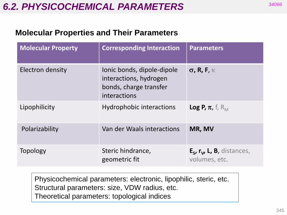
6.2. PHYSICOCHEMICAL PARAMETERS 34066
Molecular Properties and Their Parameters
Molecular Property Corresponding Interaction Parameters
Electron density Ionic bonds, dipole-dipole interactions, hydrogen bonds, charge transfer interactions
s, R, F, k
Lipophilicity Hydrophobic interactions Log P, p, f, RM
Polarizability Van der Waals interactions MR, MV
Topology Steric hindrance, geometric fit
ES, rV, L, B, distances, volumes, etc.
345
Physicochemical parameters: electronic, lipophilic, steric, etc.
Structural parameters: size, VDW radius, etc.
Theoretical parameters: topological indices

6.2. HAMMETT EQUATION. ELECTRONIC FACTORS
•The constant (s) is a measure of the e-withdrawing or e-
donating influence of substituents.
•It can be measured experimentally and tabulated.
E.g. For aromatic substituents, s is measured by comparing the
dissociation constants of substituted benzoic acids with benzoic acid.
34066
346
electron withdrawing
electron donor
1933: Hammett Structure/Reactivity Relationship
Electronic effects: Hammett Substituent Constant (s).
Aromatic substituents
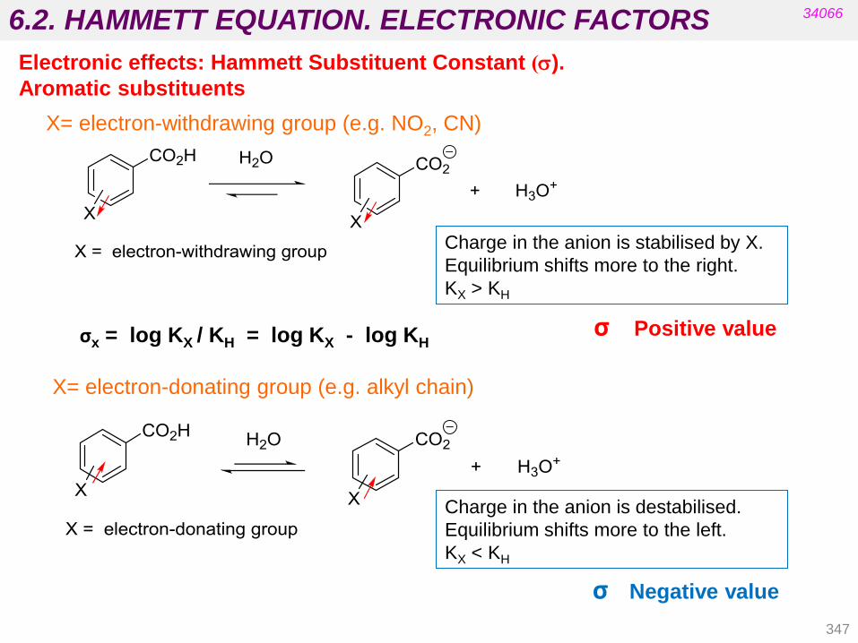
6.2. HAMMETT EQUATION. ELECTRONIC FACTORS
X= electron-withdrawing group (e.g. NO2, CN)
Charge in the anion is stabilised by X.
Equilibrium shifts more to the right.
KX > KH
σ Positive value
347
34066
X= electron-donating group (e.g. alkyl chain)
Charge in the anion is destabilised.
Equilibrium shifts more to the left.
KX < KH
σ Negative value
Electronic effects: Hammett Substituent Constant (s).
Aromatic substituents
σx = log KX / KH = log KX - log KH
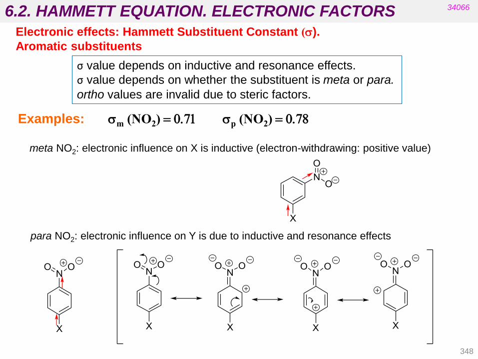
6.2. HAMMETT EQUATION. ELECTRONIC FACTORS 34066
σ value depends on inductive and resonance effects.
σ value depends on whether the substituent is meta or para.
ortho values are invalid due to steric factors.
Examples: sp (NO2) = 0.78 sm (NO2) = 0.71
348
Electronic effects: Hammett Substituent Constant (s).
Aromatic substituents
meta NO2: electronic influence on X is inductive (electron-withdrawing: positive value)
para NO2: electronic influence on Y is due to inductive and resonance effects

6.2. HAMMETT EQUATION. ELECTRONIC FACTORS
sm (OH) = 0.12 sp (OH) = - 0.37 EXAMPLES:
34066
349
Electronic effects: Hammett Substituent Constant (s).
Aromatic substituents
meta OH : electronic influence on X is inductive (electron-withdrawing: positive value)
para OH: electronic influence on X is dominated by resonance effects (electron-donating)

6.2. HAMMETT EQUATION. ELECTRONIC FACTORS
substituent substituent
34066
350
(Delgado‟s book, chapter 8)
Electronic effects: Hammett Substituent Constant (s).
Aromatic substituents

6.2. HAMMETT EQUATION. ELECTRONIC FACTORS
Some deviations are observed for substituents that are directly conjugated to
the reaction site: constants s + and s – are defined to correct them.
s¯ for withdrawing substituents
s for electron donor substituents
Phenol
ionization
Benzyl chlorides solvolysis
34066
351 (Delgado‟s book, chapter 8)
Electronic effects: Hammett Substituent Constant (s).
Aromatic substituents

6.2. HAMMETT EQUATION. ELECTRONIC FACTORS
substituent substituent
Example: QSAR Equation for insecticidal activity of diethyl phenyl phosphates:
r is close to 1 (95% of the data is accounted for by the s parameter).
This is one of the few examples where activity is related to electronic factors alone (which is a good
indication that the drugs do not have to pass into or through a cell membrane to have activity).
Acetyl cholinesterase is the target and is situated on the outside of cell membranes.
Conclusion?
34066
352
(Delgado‟s, chapter 8)
Electronic effects: Hammett Substituent Constant (s).
Aromatic substituents
log (1 /C) = 2.282σ - 0.348
(Patrick‟s)

6.2. HAMMETT EQUATION. ELECTRONIC FACTORS
• Are defined by sI
• Produce purely inductive effects.
• Are obtained experimentally by measuring the rates of hydrolysis of
aliphatic esters (methyl ethanoate is the parent ester).
• Hydrolysis rates are measured under basic and acidic conditions.
X= electron donating Rate σI < 0
X= electron withdrawing Rate σ I > 0
Basic conditions: Rate is affected by steric and electronic factors.
Gives sI after correction for steric effect.
Acidic conditions: Rate is affected by steric factors only (see Es).
353
34066
Electronic Effects: Hammett Substituent Constant (s)
Aliphatic substituents
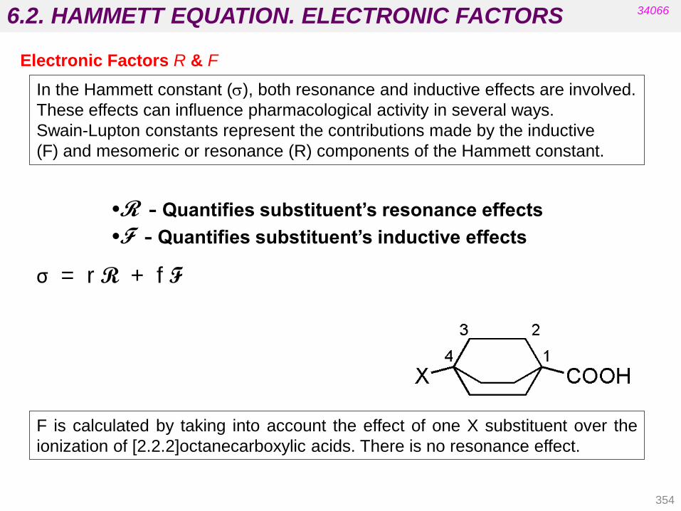
6.2. HAMMETT EQUATION. ELECTRONIC FACTORS 34066
354
Electronic Factors R & F
•R - Quantifies substituent‟s resonance effects
•F - Quantifies substituent‟s inductive effects
In the Hammett constant (s), both resonance and inductive effects are involved.
These effects can influence pharmacological activity in several ways.
Swain-Lupton constants represent the contributions made by the inductive
(F) and mesomeric or resonance (R) components of the Hammett constant.
F is calculated by taking into account the effect of one X substituent over the
ionization of [2.2.2]octanecarboxylic acids. There is no resonance effect.
σ = r R + f F

6.2. HAMMETT EQUATION. ELECTRONIC FACTORS
SWAIN and LUPTON CONSTANTS
F and R have negative values for electron donating substituents and positive values for
electron withdrawing substituents
substituent substituent
34066
Electronic Factors R & F
355
(Delgado‟s, chapter 8)

6.2. TAFT’S EQUATION. STERIC FACTORS
Steric Factors
Taft‟s Steric Factor (Es)
•Is obtained by comparing the rates of hydrolysis of substituted aliphatic esters
against a standard ester under acidic conditions.
Es = log kx - log ko kx represents the rate of hydrolysis of the substituted ester.
ko represents the rate of hydrolysis of the parent ester.
•Is limited to substituents which interact sterically with the tetrahedral transition
state for the reaction (not by resonance or internal HB).
•May undervalue the steric effect of groups in an intermolecular process (e.g. a
drug binding to a receptor).
•The reference ester is X=Me. Smaller substituents have positive values.
34066
• The interaction of a drug with a target involves the mutual approach of
two molecules.
• Another important parameter for QSAR is the steric effect.
• Steric properties are difficult to quantify.
• Several parameters have been used.
356
Substituent H F Me Et n-Pr n-Bu i-Pr iBu cyclopentyl
Es 1.24 0.78 0 -0.07 -0,36 -0,39 -0.47
-0.93
-0,51

6.2. TAFT’S EQUATION. STERIC FACTORS
Molar Refractivity (MR) - a measure of a substituent‟s volume
MR = (n 2 - 1)
(n 2 - 2) x
MW
d
Correction factor
for polarisation
Defines
volume
• MR is defined by the Lorentz-Lorentz equation, where “n” is the index
of refraction at the sodium D line, MW is the molecular weight, and d is
the density of the compound.
• The greater the positive MR value of a substituent, the larger the steric
or bulk effect.
• MR also measures the electronic effect and may therefore reflect
dipole-dipole interactions at the receptor site.
MR is the molar volume
corrected by the refractive
index. It represents the size
and polarizability of a
fragment or molecule.
357
34066
Steric Factors

6.2. LIPOPHILICITY FACTORS
• The hydrophobic character of a drug is crucial for crossing cell membranes
and may be important in receptor interactions.
• The hydrophobic character of a drug can be measured experimentally by
testing the drug‟s relative distribution in an n-octanol/water mixture. The
relative distribution is known as the PARTITION COEFFICIENT (P).
• Another parameter commonly used to relate distribution with biological
activity is the LIPOPHILICITY SUBSTITUENT CONSTANT (p).
• The P parameter refers to the whole molecule whereas p is related to
substituent groups.
34066
358
Hydrophobicity of the molecule
PARTITION COEFFICIENT P = [Drug] octanol
[Drug] water
High P High lipophilicity
From Unit 3:

6.2. LIPOPHILITICY FACTORS
•The activity of drugs is often related to P.
e.g. the binding of drugs to serum albumin (straight line – limited range of log P).
•Binding increases as log P increases.
•Binding is greater for hydrophobic drugs.
Log (1/C)
Log P
. . .
. . . . . .
0.78 3.82
34066
(n = 42, r = 0.960)
359
General anaesthetic activity of ethers (parabolic curve – wider range of log P values).
Optimum value of log P for anaesthetic
activity = log Po
Log P
Log (1/C)
Hydrophobicity of the molecule
log (1/C) = 0.75 log P + 2.30
log (1/C) = -0.22(log P)2 + 1.04 log P + 2.16
Log Po

6.2. LIPOPHILITICY FACTORS
360
34066
Substituent lipophilicity constants - Hansch and Fujita parameter
• Substituent lipophilic constants (πX ) are also known as hydrophobic constants.
• They represent the contribution made by a group or atom to the partition
coefficient and were defined by Hansch and co-workers by the equation:
πX = log PX – log PH = log ( PX / PH)
PX is the partition coefficient for the compound with substituent X.
PH is the partition coefficient for the parent molecule.
• Log P calculated from the lipophilic constants for substituents is called c log P.
c log P = ∑ π (p values depend on the pattern structure)
From unit 3

6.2. LIPOPHILITICY FACTORS 34066
Substituents more hydrophilic than hydrogen have negative p values.
Substituents more lipophilic than hydrogen have positive values.
361
Substituent hydrophobicity constant (p)
• πX depends on the characteristics of the group/atom: inductive, resonance
and steric effects are important (a positive value means that the substituent
increases the lipophilicity and a negative value means the opposite).
• The values of π for the most common substituents are tabulated
• Numerous software packages are now commercially available (ChemDraw,
etc.) but the results can differ widely and also differ from the experimental
value.
From unit 3

6.2. LIPOPHILITICY FACTORS
362
34066
Substituent hydrophobicity constant (p)
-
susbstituent substituent
Example:
Benzene Log P = 2.13
Chlorobenzene Log P = 2.84
Benzamide Log P = 0.64 pCl = 0.71 (arom) pCONH = -1.49 (arom)
2
(Delgado, chapter 8)

6.2. LIPOPHILITICY FACTORS 34066
• Log P can be calculated using p values.
• The value of p is valid only for parent structures.
• A QSAR equation may include both P and p.
• P measures the importance of a molecule‟s overall hydrophobicity
(relevant to absorption, binding etc.).
• p identifies specific regions of the molecule that may interact with
hydrophobic regions in the binding site.
Example:
meta -Chlorobenzamide
Log P(calculated) = log P(benzene) + pCl + pCONH
= 2.13 + 0.71 - 1.49
= 1.35
Log P(observed) = 1.51
2
363
Substituent hydrophobicity constant (p)

6.2. PHYSICOCHEMICAL PARAMETERS
Electronic effects Hammett constant s
s 0 more electron withdrawing than H
s 0 more electron donating than H
Steric factors Taft parameter Es
Es 0 group bigger than CH3
Es 0 group smaller than CH3
Lipophilicity Hansch and Fujita parameter p
p < 0 substituents more hydrophilic than H
p > 0 substituents more lipophilic than H
P = [drug]org/[drug]aq
P 1 (log P 0) more lipophilic than H
P 1 (log P 0) more hydrophilic than H
364
Summary:

6.3. METHODS USED IN QSAR
HANSCH and FUJITA EQUATION (physicochemical parameters)
Parabolic equation: when the log P values are spread over a wide range
Linear equation: when the range of lipophilicity values (or hydrophobicity)
is limited.
Conclusions
Activity increases if p is positive (i.e hydrofobic substituents).
Activity increases if s is negative (i.e. electron-donor substituents).
Example: Adrenergic blocking activity of b-halo-b-arylamines.
34066
365
Log (1/C) = 1.22 π - 1.59σ + 7.89

6.3. METHODS USED IN QSAR
Conclusions:
• Activity increases slightly as log P increases (the constant is small).
• The parabolic equation implies an optimum log Po value for activity.
• Activity increases for hydrophobic substituents (especially in ring Y).
• Activity increases for e-withdrawing substituents (especially in ring Y).
Example: Antimalarial activity of phenanthrene aminocarbinols
34066
366
HANSCH and FUJITA EQUATION (physicochemical parameters)
Log (1/C) =
= - 0.015 (logP)2 + 0.14 log P + 0.27∑ πX + 0.40∑ πY + 0.65 ∑ σX + 0.88 ∑ σY + 2.34

6.3. METHODS USED IN QSAR
Substituents must be chosen to satisfy the following criteria:
• There must be a range of values for each physicochemical property
studied.
• Values must not be correlated for different properties (i.e. they must
be orthogonal in value).
• At least 5 structures are required for each parameter studied.
Correlated values.
Are any differences in activity
due to p or MR?
No correlation in values
Valid for analysing the effects
of p and MR.
Substituent H Me Et n-Pr n-Bu
p 0.00 0.56 1.02 1.50 2.13
MR 0.10 0.56 1.03 1.55 1.96
Substituent H Me OMe NHCONH2 I CN
p 0.00 0.56 -0.02 -1.30 1.12 -0.57
MR 0.10 0.56 0.79 1.37 1.39 0.63
Choosing suitable substituents
34066
367
HANSCH and FUJITA EQUATION (physicochemical parameters)

6.3. METHODS USED IN QSAR 34066
PREDICTIVE VALUE OF THE HANSCH EQUATION
Practical development of the Hansch equation is governed by the
following criteria:
a) The selection of independent variables.
b) Justification of the choice of variables.
c) Principle of parsimony: When statistical significance is equal, we
choose the simplest model.
d) The number of studied compounds must be related to the
number of variables.
368
HANSCH and FUJITA EQUATION (physicochemical parameters)

6.3. METHODS USED IN QSAR
SCOPE AND LIMITATIONS OF HANSCH‟S METHOD
Advantages:
a) Simple parameters are used.
b) Quantitative predictions are made.
c) The method is economical and easy to use.
d) The conclusions can be extended to unused substituents.
Limitations:
The values of the parameters must be known.
a) A large number of compounds is required for statistical significance.
c) Results obtained with small molecules may be imperfect models
of biological systems.
34066
369
HANSCH and FUJITA EQUATION (physicochemical parameters)

6.3. METHODS USED IN QSAR
Craig Plot
The Craig plot shows values for two different physicochemical properties for various
substituents.
-s +p
+s +p
-s -p
+s -p
.
-0.25
0.75
0.50
1.0
-1.0
-0.75
-0.50
0.25
-0.4-0.8-1.2-1.6-2.0 2.01.61.20.80.4
.. . .
...
.
.. . .
..
.
.
......
CF3SO2
CF3
Me
Cl BrI
OCF3
F
NMe2
OCH3
OH
NH2
CH3CONH
CO2H
CH3CO
CN
NO2
CH3SO2
CONH2
Ett-Butyl
SF5
SO2NH2
+p -p
+s
-s
34066
• Allows easy
identification of
suitable substituents
for a QSAR analysis
which includes both
relevant properties.
• A substituent is
chosen from each
quadrant in order to
ensure orthogonality.
• Substituents are
chosen with a range
of values for each
property.
370

6.3. METHODS USED IN QSAR
Topliss Scheme
This is used to decide which substituents to use if compounds are
optimised one by one (where synthesis is complex and slow).
Example: Aromatic substituents
34066
371
Replace H with
para-Cl (+p and +s)
+p and/or +s
advantageous
Favourable p
unfavourable s
+p and/or +s
disadvantageous
Act. Little
change Act.
Add second Cl to
increase p and s
further
Replace with OMe
(-p and –s) Replace with Me
(+p and –s)
Further changes suggested based on arguments of p, s and steric strain.

6.3. METHODS USED IN QSAR
Bioisosteres
• Substituents with similar physicochemical properties (e.g. CN, NO2 and
COMe may be bioisosteres).
• Choose bioisosteres based on the most important physicochemical
property
(e.g. COMe & SOMe are similar in sp; SOMe and SO2Me are similar in p)
p -0.55 0.40 -1.58 -1.63 -1.82 -1.51
sp 0.50 0.84 0.49 0.72 0.57 0.36
sm 0.38 0.66 0.52 0.60 0.46 0.35
MR 11.2 21.5 13.7 13.5 16.9 19.2
34066
372
substituent
Figure 18.17 Physicochemical parameters for six substituents (Patrick’s 5th ed.)
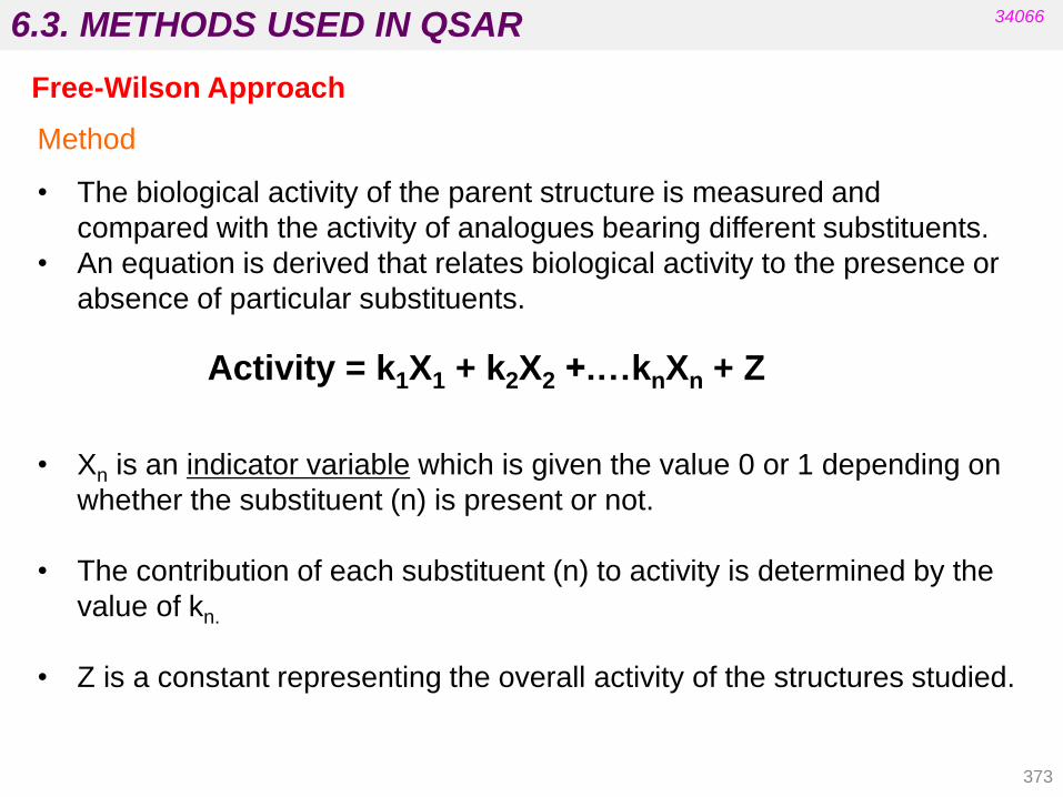
6.3. METHODS USED IN QSAR
Free-Wilson Approach
• The biological activity of the parent structure is measured and
compared with the activity of analogues bearing different substituents.
• An equation is derived that relates biological activity to the presence or
absence of particular substituents.
Activity = k1X1 + k2X2 +.…knXn + Z
• Xn is an indicator variable which is given the value 0 or 1 depending on
whether the substituent (n) is present or not.
• The contribution of each substituent (n) to activity is determined by the
value of kn.
• Z is a constant representing the overall activity of the structures studied.
Method
34066
373

6.3. METHODS USED IN QSAR
• Physicochemical constants or tables are not required.
• It is useful for structures with unusual substituents.
• It is useful for quantifying the biological effects of molecular features
that cannot be quantified or tabulated by the Hansch method.
Advantages
Disadvantages
• A large number of analogues need to be synthesised in order to
represent each different substituent and each different position of a
substituent.
• It is difficult to rationalise why specific substituents are good or bad for
activity.
• The effects of different substituents may not be additive (e.g.
intramolecular interactions).
Free-Wilson Approach
34066
374
Free-Wilson/Hansch Approach
• Indicator variables may be used as part of a Hansch equation.
Advantages
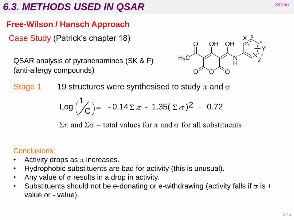
6.3. METHODS USED IN QSAR
Stage 1 19 structures were synthesised to study p and s
C Log
1
= - 0.14 S p - 1.35( S s ) 2 - 0.72
Sp and Ss = total values for p and s for all substituents
Case Study (Patrick‟s chapter 18)
QSAR analysis of pyranenamines (SK & F)
(anti-allergy compounds)
Conclusions:
• Activity drops as p increases.
• Hydrophobic substituents are bad for activity (this is unusual).
• Any value of s results in a drop in activity.
• Substituents should not be e-donating or e-withdrawing (activity falls if s is +
value or - value).
34066
Free-Wilson / Hansch Approach
375
H3C
O O O
NH
OH OHX
Y
Z
O
3
4
5

6.3. METHODS USED IN QSAR
Conclusions:
• Increasing the hydrophilicity of substituents allows the identification of an optimum
value for p (Sp = -5). The equation is now parabolic (-0.034 (Sp)2).
• The optimum value of Sp is very low and implies a hydrophilic binding site.
• R-5 implies that resonance effects are important at position 5.
• HB-INTRA equals 1 for H-bonding groups ortho to each other (act. drops -0.86).
• HB-INTRA equals 0 if H-bonding groups are not ortho to each other.
• The steric parameter is no longer significant and is not present.
Stage 5
34066
376
Case Study
Free-Wilson/Hansch Approach
Log (1/C) = - 0.034(Sp)2 – 0.33Sp + 4.3 (F-5) – 1.3(R-5) – 1.7(Ss)2 + 0.73(3,4,5-HBD)
- 0.86 (HB-INTRA) – 0.69(NHSO2) + 0.72(4-OCO) - 0.59
H3C
O O O
NH
OH OHX
Y
Z
O
3
4
5

6.3. METHODS USED IN QSAR
Stage 6 Optimum Structure and binding theory
34066
Free-Wilson / Hansch Approach
377
Based on Figure 18.19 Hypothetical binding interactions between a pyranenamine and
the target binding site (Patrick’s 5th ed.)
H3C
O O O
NH
OH OHO
NH
NH
O
O
O
O
O
O
H HX
HX
NH3+
H
XY
H

6.4. 3D-QSAR
In three-dimensional quantitative structure-activity relationships (3D-
QSARs), the 3D properties of a molecule are considered as a whole rather than
considering individual substituents or moieties.
34066
378
Molecular modelling has previously been described in lead discovery and lead
optimization. E.g.:
-virtual screening from a database of chemical structures.
-identification of the 3D-pharmacophore in SAR studies.
Classical QSAR methods use computational methods in lead modification but
consider only 2D structures.
General approach:
• Select a group of molecules, each of which has been assayed for a
particular activity.
• Align the 3D conformations of the molecule according to some
predetermined orientation rules.
• Calculate a set of spatially dependent parameters for each molecule
determined in the receptor space surrounding the aligned series.
• Derive a function that relates each molecule‟s spatial parameters to its
respective biological property.
• Establish self-consistency and predictability of the derived function.
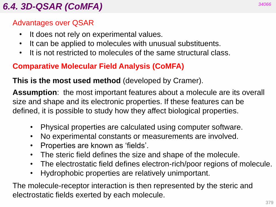
6.4. 3D-QSAR (CoMFA)
Comparative Molecular Field Analysis (CoMFA)
This is the most used method (developed by Cramer).
34066
379
• Physical properties are calculated using computer software.
• No experimental constants or measurements are involved.
• Properties are known as „fields‟.
• The steric field defines the size and shape of the molecule.
• The electrostatic field defines electron-rich/poor regions of molecule.
• Hydrophobic properties are relatively unimportant.
Assumption: the most important features about a molecule are its overall
size and shape and its electronic properties. If these features can be
defined, it is possible to study how they affect biological properties.
The molecule-receptor interaction is then represented by the steric and
electrostatic fields exerted by each molecule.
Advantages over QSAR
• It does not rely on experimental values.
• It can be applied to molecules with unusual substituents.
• It is not restricted to molecules of the same structural class.

6.4. 3D-QSAR (CoMFA)
•The results are represented as a 3D contour map in which contours of
various colours represent locations on the structure where lower or higher
steric or electrostatic interactions would increase or decrease binding.
34066
380
Limitations of Comparative Molecular Field Analysis:
• The molecules are assumed to bind with similar orientations in the
target, which may not be the case.
• Correct alignments are almost impossible for compounds with a large
number of rotatable bonds.
Comparative Molecular Field Analysis (CoMFA)
See Chapter 18 (Patrick’s 5th ed.)
6.4. 3D-QSAR (CoMFA)
Alternative Ligand-Based Approaches are dependent on alignment
rules:
Comparative Molecular Similarity Indices Analysis (CoMSIA):
• Uses a different potential function.
• Not only steric and electrostatic but also hydrophobic fields are
calculated.
Comparative Binding Energy Analysis (COMBINE):
• Is based on structural data of ligand-receptor complexes.
• Hypothesis: the free energy of binding can be correlated with a subset
of energy components calculated from the structures of ligand and
receptors in bound and unbound forms.
34066
381

6.4. 3D-QSAR
Another popular 3D-QSAR method is topomer similarity searching.
A topomer is a molecular descriptor that focuses on the shape of a molecule
as represented by a combination of the shapes of different fragments (used
to search for 3D molecular structures in conventional structural databases).
CoMFA and topomer similarity have been merged into a new 3D-QSAR
method called Topomer CoMFA:
• Structures in a series are each broken into two or more fragments that
are common to the series.
• Topomer 3D models are constructed for each fragment.
• A set of steric and electrostatic fields is generated for each topomer set.
Advantages:
• This method minimizes the preparation needed for 3D-QSAR analysis.
• It automates the creation of models for predicting biological activity.
• It is more user-friendly than traditional CoMFA analysis.
34066
382