Multilevel Voltage-Source-Converter Topologies for Industrial Medium-Voltage Drives
435
J. Gen. Physiol.
© The Rockefeller University Press
•
0022-1295/2000/04/435/19 $5.00Volume 115 April 2000 435–453http://www.jgp.org/cgi/content/full/115/4/435
Permeation of Large Tetra-alkylammonium Cations through Mutant and Wild-Type Voltage-gated Sodium Channels as Revealed by Relief of Block at High Voltage
Chien-Jung Huang,* Isabelle Favre,*
and
Edward Moczydlowski*
‡
From the *Department of Pharmacology and
‡
Department of Cellular and Molecular Physiology, Yale University Medical School,New Haven, Connecticut 06520-8066
abstract
Many large organic cations are potent blockers of K
1
channels and other cation-selective channelsbelonging to the P-region superfamily. However, the mechanism by which large hydrophobic cations enter andexit the narrow pores of these proteins is obscure. Previous work has shown that a conserved Lys residue in theDEKA locus of voltage-gated Na
1
channels is an important determinant of Na
1
/K
1
discrimination, exclusion of
Ca
2
1
, and molecular sieving of organic cations. In this study, we sought to determine whether the Lys(III) residueof the DEKA locus interacts with internal tetra-alkylammonium cations (TAA
1
) that block Na
1
channels in a volt-age-dependent fashion. We investigated block by a series of TAA
1
cations of the wild-type rat muscle Na
1
channel(DEKA) and two different mutants of the DEKA locus, DEAA and DERA, using whole-cell recording. TEA
1
andlarger TAA
1
cations block both wild-type and DEAA channels. However, DEAA exhibits dramatic relief of block by
large TAA
1
cations as revealed by a positive inflection in the macroscopic I–V curve at voltages greater than
1
140mV. Paradoxically, relief of block at high positive voltage is observed for large (e.g., tetrapentylammonium) butnot small (e.g., TEA
1
) symmetrical TAA
1
cations. The DEKA wild-type channel and the DERA mutant exhibit asimilar relief-of-block phenomenon superimposed on background current rectification. The results indicate: (a)hydrophobic TAA
1
cations with a molecular diameter as large as 15 Å can permeate Na
1
channels from inside tooutside when driven by high positive voltage, and (b) the Lys(III) residue of the DEKA locus is an important de-terminant of inward rectification and internal block in Na
1
channels. From these observations, we suggest that hy-drophobic interfaces between subunits, pseudosubunits, or packed helices of P-region channel proteins may func-tion in facilitating blocker access to the pore, and may thus play an important role in the blocking and permeationbehavior of large TAA
1
cations and potentially other kinds of local anesthetic molecules.
key words:
local anesthetic • ionic selectivity • Na
1
channel • selectivity filter • tetraethylammonium
I N T R O D U C T I O N
Tetra-alkylammonium (TAA
1
)
1
cations such as TEA
1
are known to inhibit ionic currents through channelproteins by transiently binding in the ion conductionpathway and blocking the flow of current (Tasaki andHagiwara, 1957; Armstrong and Binstock, 1965; Hille,1967). The fact that K
1
channels often differ in sensitiv-ity to block by internal and external TAA
1
s has led tothe picture that these channels have distinct internaland external binding sites for such molecules locatedin antechambers separated by a narrower tunnel in themiddle of the pore that cannot readily be penetratedby large organic cations (Armstrong, 1992;Hille, 1992).
Structure–activity studies have identified certain aminoacid residues in K
1
channel proteins that influenceTEA
1
block (MacKinnon and Yellen, 1990; Kavanaughet al., 1991; Yellen et al., 1991; Heginbotham andMacKinnon, 1992; Choi et al., 1993). In the crystalstructure of a K
1
channel protein (KcsA), residues ho-mologous to those that determine the binding affinityof external and internal TEA
1
in related K
1
channelsare located precisely at the respective outer and innerentrances of the narrowest region of the pore calledthe selectivity filter (Doyle et al., 1998). In the structure
of KcsA, this filter is a narrow tunnel that measures
z
3Å in diameter by 12 Å in length.
Voltage-gated Na
1
and Ca
2
1
channels are structurallyrelated to K
1
channels as members of a homologous su-perfamily of cation-selective channels (Pongs et al.,1988; Jan and Jan, 1990). In particular, these three typesof channels share a structural motif called the P-regionlocated between the M1/M2 transmembrane helices ofKcsA or between the corresponding S5/S6 presumedhelices of voltage-gated K
1
, Na
1
, and Ca
2
1
channels(MacKinnon, 1995; Moczydlowski, 1998). In all three
Address correspondence to Edward Moczydlowski, Depart-ment of Pharmacology, Yale University Medical School, 333Cedar St., New Haven, CT 06520-8066. Fax: 203-785-7670;E-mail: [email protected]
1
Abbreviations used in this paper:
m
-CTX,
m
-conotoxin GIIIB; MA
1
,methylammonium; STX, saxitoxin; TAA
1
, tetra-alkylammonium;TBA
1
, tetrabutylammonium; THexA
1
, tetrahexylammonium; TMA
1
,tetramethylammonium; TPA
1
, tetrapropylammonium; TPeA
1
, tetra-pentylammonium; TTX, tetrodotoxin.
on Novem
ber 14, 2013jgp.rupress.org
Dow
nloaded from
Published April 1, 2000
436
Voltage-dependent Relief of Na
1
Channel Block by Large Organic Cations
types of channels, specific conserved residues in theP-region determine ionic selectivity (Heinemann et al.,1992; Yang et al., 1993; Heginbotham et al., 1994). Thestructure of KcsA revealed that K
1
selectivity in tet-rameric K
1
channels originates from a K
1
-binding re-gion consisting of four rings of carbonyl oxygen atomsof the peptide backbone contributed by residues of astrongly conserved signature sequence. However, manystudies indicate that the mechanism of ionic selectivityand the structure of the analogous selectivity filter arelikely to be quite different for Na
1
and Ca
2
1
channelsversus K
1
channels. Ionic selectivity in pseudotetramericNa
1
and Ca
2
1
channels is known to be intimately relatedto a set of mostly charged residues located near theouter mouth of the pore (Heinemann et al., 1992; Yanget al., 1993; Favre et al., 1996). These residues are calledthe EEEE locus in Ca
2
1
channels and the DEKA locus inNa
1
channels. The notation EEEE/DEKA correspondsto the single letter code for conserved amino acid resi-dues that can be readily aligned in homologous domainsI, II, III, and IV of Ca
2
1
/Na
1
channels, respectively. In apreceding study from our laboratory, the conserved Lysresidue in domain III of the DEKA locus of Na channels,K(III), was identified as a major determinant of the mo-lecular sieving behavior of the rat skeletal muscle Na-channel (
m
1) with respect to organic cations (Sun et al.,1997). Mutation of this Lys residue to Ala, as in the mu-tation of DEKA to DEAA, produces a channel that is per-meable to many organic cations ranging in size frommethylammonium (3.8-Å diameter) to TEA
1
(8.2-Å di-ameter), as demonstrated by direct measurement of in-ward currents carried by these cations. This latter behav-ior is quite different from native Na
1
channels, whichare effectively impermeable to external organic cationslarger than guanidinium, a fact that has been used to es-tablish 3.2
3
5.2 Å as the cutoff area of the narrowestcross-section of the native pore (Hille, 1971, 1972).
The enhanced permeability of the DEAA mutant tolarge organic cations provides a unique opportunity toexplore the mechanism of block by TAA
1
cations andrelated local anesthetic drugs that are known to prefer-entially block voltage-gated Na
1
channels from the in-tracellular side. If the K(III) residue of the DEKA locuscorresponds to the location of a major energy barrierfor the movement of large cations through the pore,then lowering this energy barrier by mutation may en-hance the permeability of blocking cations that enterthe channel from the inside. Alternatively, if other sig-nificant energy barriers are located between the DEKAlocus and the intracellular pore entrance, then suchmutant Na
1
channels may exhibit asymmetric perme-ability to organic cations. On this basis, we hypothesizedthat removal of a structural element such as a Lys resi-due that limits conduction of organic cations may revealor enhance a phenomenon known as voltage-depen-
dent relief of block (French and Wells, 1977; Frenchand Shoukimas, 1985), in which a large positive voltageapplied on the same side as a cationic blocker allowssuch blocking ions to permeate through the channel.To address the question of whether the K(III) residue ofthe DEKA locus corresponds to such an energy barrier,we studied internal block of a DEAA mutant Na
1
chan-nel by a series of symmetrical TAA
1
s ranging in sizefrom tetramethylammonium (TMA
1
) to tetrahexylam-monium (THexA
1
). We found that internal block ofoutward alkali cation current through the DEAA mu-tant by large TAA
1
cations such as tetrapentylammo-nium (TPeA
1
) is indeed relieved in a steeply voltage-dependent fashion. Paradoxically, this effect is enhancedfor large versus small TAA
1
derivatives, implying that hy-drophobic interactions with long
n
-alkyl groups actuallyfacilitate the voltage-driven permeation of very large or-ganic cations through this aqueous pore. In addition,we find that the wild-type Na
1
channel and a DERA mu-tant, in which the K(III) residue is replaced by Arg, alsoexhibit voltage-dependent relief of block by large chainTAA
1
s, but with apparently different efficiency. The re-sults show that a positively charged residue at the K(III)position of the DEKA locus is not an absolute structuralimpediment to the movement of large cations throughthe pore. Implications of these findings for the interpre-tation of molecular sieving studies of Na
1
channels andblocking interactions of TAA
1
cations are discussed.
M A T E R I A L S A N D M E T H O D S
Expression of Wild-Type and Mutant Na
1
Channels
Na
1
channels studied in this paper are the wild-type
m
1 rat skele-tal muscle isoform (Trimmer et al., 1989) and two mutants of thisclone that contain substitutions, K1237A or K1237R. The wild-type and the latter two mutants are referred to, respectively, asDEKA, DEAA, and DERA according to the single letter code forresidues in domains I–IV of the DEKA locus, a conserved regionin Na
1
channels that aligns with the EEEE locus of Ca
2
1
channels(Heinemann et al., 1992; Sun et al., 1997). DEKA and DEAAclones were studied in stably transfected human fibroblastHEK293 cells. The DERA clone was studied in HEK293 cells thatwere transiently transfected with a DERA/pcDNA3 vector usingEffectene reagent (QIAGEN Inc.). Construction of the muta-tions, subcloning into pcDNA3 expression vector, and othermethods for transfection, generation, and propagation of stablecell lines have been described in previous publications (Favre etal., 1996; Sun et al., 1997).
Solutions and Electrophysiology
The standard extracellular Na
1
bath solution was (mM) 140NaCl, 3 KCl, 2 MgCl
2
, 2 CaCl
2
, 10 glucose, and 10 HEPES-NaOH,pH 7.3. The standard intracellular Cs
1
/Na
1
pipette solution was(mM) 125 CsF, 2 MgCl
2
, 1.1 EGTA, 10 glucose, 20 Na
1
-HEPES,pH 7.3. Intracellular pipette solutions containing tetrapropyl-ammonium (TPA
1
), tetrabutylammonium (TBA
1
), TPeA
1
,THexA
1
, and QX-314
1
were prepared by adding these blockersto standard Cs
1
/Na
1
pipette solution at the desired concentra-tion. The composition of other solutions for testing permeability
on Novem
ber 14, 2013jgp.rupress.org
Dow
nloaded from
Published April 1, 2000
437
Huang et al.
to external TEA
1
, TMA
1
, MA
1
, and Ca
2
1
or block by internalTEA
1
are given in the figure legends.Patch-clamp electrodes were constructed with a commercial pi-
pette puller using Kimax 50 borosilicate capillary glass (Fisher Sci-entific Co.) without additional fire polishing. The measured pi-pette resistance was 1–2 M
V
when filled with standard Cs
1
/Na
1
pipette solution. Whole-cell voltage-clamp recording was per-formed at room temperature (
z
22
8
C) using an amplifier (EPC-9;HEKA Electronik) with Pulse and Pulse-fit software (InstrutechCorp.). Stably transfected HEK293 cells were seeded for growthon glass cover slips and used for electrophysiological recordingwithin 12–36 h. Transiently transfected cells expressing the DERAmutant channel were used for recording 48 h after transfection.The peak inward current in standard Na
1
bath solution and Cs
1
/Na
1
pipette solution of typical cells selected for recording DEKA,DEAA, and DERA current was
2
4.3
6
0.4 nA (mean
6
SEM,
n
5
6),
2
4.1
6
0.7 nA (
n
5
12), and
2
1.6
6
0.4 nA (
n
5
5), respec-tively. Cancellation of residual capacitance transients and linearleak subtraction was carried out using a programmed P/4 nega-tive pulse protocol delivered at
2
120 mV. The series resistancecompensation function of the amplifier was routinely used at 74–79% compensation to minimize voltage error. Nevertheless, someexperiments for the DEAA mutant (e.g., see Figs. 2 and 5) in-volved measurements of outward currents as large as 50–100 nA atthe highest positive voltage (1200 mV). In such cases, the effect ofvoltage errors on our analysis and interpretation was minimizedby discarding data sets from cells that exhibited .50 nA of out-ward current at 1200 mV. In addition, the effect of voltage errordue to series resistance, RS, was estimated for the data of Figs. 2and 5 by correcting the applied voltage by the voltage drop acrossthe noncompensated fraction, fN, of measured RS (2.3 6 0.2 MV,n 5 5), using Ohm’s Law, DV 5 IfNRS (Marty and Neher, 1995).
Current–voltage data were collected by recording responses toa consecutive series of step voltage pulses increasing by 110 mVfrom a holding potential of 2120 to 1200 mV with a pulse dura-tion of 10 ms. The interval between consecutive voltage pulseswas 0.5 or 1 s as noted, except in experiments where it was variedfrom 0.5 to 10 s for investigation of use-dependent block. Datacollection was begun 10 min after whole cell break-in when in-ward Na1 current reached a relatively stable level after intracellu-lar perfusion. Data were collected during continuous gravity-fedperfusion of the extracellular bath solution using a commercialpatch perfusion chamber (Warner Instrument Co.).
The permeability of extracellular TEA1 and MA1 was investi-gated by measuring the change in reversal potential, DVR, uponchanging the extracellular solution from the standard Na1 bathsolution to a different solution containing the cation of interest(e.g., see Figs. 1 A and 7). Permeability ratios of these extracellu-lar cations relative to Na1 were calculated by the method of Hille(1971) using DVR values corrected for junction potentials andequations described previously (Sun et al., 1997).
Data Analysis and Modeling
Peak current values were measured and plotted as a function ofthe pulse voltage. In compiling the results, peak I-V data was of-ten normalized by dividing the measured current by the maxi-mum inward current of a given cell. Such data sets collectedfrom three to eight cells were then averaged and plotted as themean normalized current with error bars given (6SEM).
In Fig. 2 B (below), current values for 10 and 50 mM internalTPeA1 were normalized by dividing each point by the expectedpeak inward current in the absence of internal TPeA1. The mag-nitude of the unblocked current was estimated by fitting datapoints at voltages less than 180 mV to a simple model of voltage-dependent block as described by Eqs. 1 and 2 with outward per-
meation of the blocker disregarded by setting k22 equal to 0. Thisprocedure yields a reasonable estimate of the maximal conduc-tance, Gmax, in the absence of blocker. This estimate of Gmax isthen used to generate an I-V relation for the expected unblockedcurrent by setting the blocker concentration, [Bin], equal to zeroin Eq. 1. The absolute value of the maximal expected inward cur-rent is then used to normalize the measured current in the pres-ence of internal TPeA1 to produce a macroscopic I-V curve thatis appropriately scaled with respect to that expected in the ab-sence of an internal blocker. This procedure is mathematicallysimilar to that used in Figure 5 of O’Leary and Horn (1994),which describes the blocking effect of internal TEA1, TPA1, andTBA1 on the human heart Na1 channel as measured by whole-cell recording.
Nonlinear regression fitting of peak I-V data to the model ofScheme I using Eqs. 1 and 2 was performed using the Marquardt-Levenberg algorithm as part of the Sigmaplot 4.0 software pack-age (SPSS Inc.).
Materials
Chloride salts of TAA1 cations were purchased from AldrichChemical Co. (TPA1, TBA1, TPeA1) or Sigma Chemical Co.(TEA1, THexA1). Saxitoxin and tetrodotoxin were purchasedfrom Calbiochem Corp. m-Conotoxin GIIIB was obtained fromBachem. The quaternary lidocaine derivative QX-3141 was ob-tained from Alomone Laboratories.
R E S U L T S
Voltage-gated Na1 channels are preferentially blockedby many different hydrophobic organic cations fromthe intracellular side. For example, QX-3141, the qua-ternary ammonium derivative of lidocaine, a local anes-thetic, readily blocks Na1 channels when present onthe intracellular side of axons, cells, or planar bilayers(Strichartz, 1973; Moczydlowski et al., 1986; Wang,1988; Gingrich et al., 1993; Zamponi et al., 1993). Vari-ous measurements of the steady state voltage depen-dence of block of open Na1 channels by QX-3141,TAA1 derivatives, and related molecules, indicate thatthere is an internal site (or sites) for organic cationsthat senses 40–70% of the applied voltage from the in-side of the membrane (Yamamoto and Yeh, 1984; Wanget al., 1991; Gingrich et al., 1993; French et al., 1998).At the opposite side of Na1 channels, there is anotherblocking site for guanidinium toxins such as tetrodo-toxin (TTX) and saxitoxin (STX). Amino acid residuesthat determine the affinity for TTX/STX binding arelocated at or close to the COOH-terminal side of resi-dues that comprise the DEKA locus, an apparent ring-like structure of mostly charged residues that controlthe relative permeability to Na1, K1, and Ca21 (Terlauet al., 1991; Heinemann et al., 1992). The D, E, and K,residues of the DEKA locus also determine molecularsieving properties of the m1 Na1 channel as monitoredby inward current carried by various organic cations inthe extracellular solution (Sun et al., 1997). This sug-gests that the DEKA locus forms a constricted regionthat separates the toxin binding site on the outside
on Novem
ber 14, 2013jgp.rupress.org
Dow
nloaded from
Published April 1, 2000
438 Voltage-dependent Relief of Na1 Channel Block by Large Organic Cations
from the blocking site for TAA1 cations on the inside.If the DEKA locus forms such a constriction in struc-tural terms, or is the major energy barrier in kineticterms, then one might expect that internal organic cat-ions would become permeable or lose their blockingefficiency in a mutant such as DEAA that displays en-hanced permeation of organic cations from the out-side. To test this idea, we first examined whether inter-nal organic cations can carry outward currents in theDEAA mutant expressed in HEK293 cells.
Asymmetry of Organic Cation Permeation through the DEAA Mutant Na1 Channel
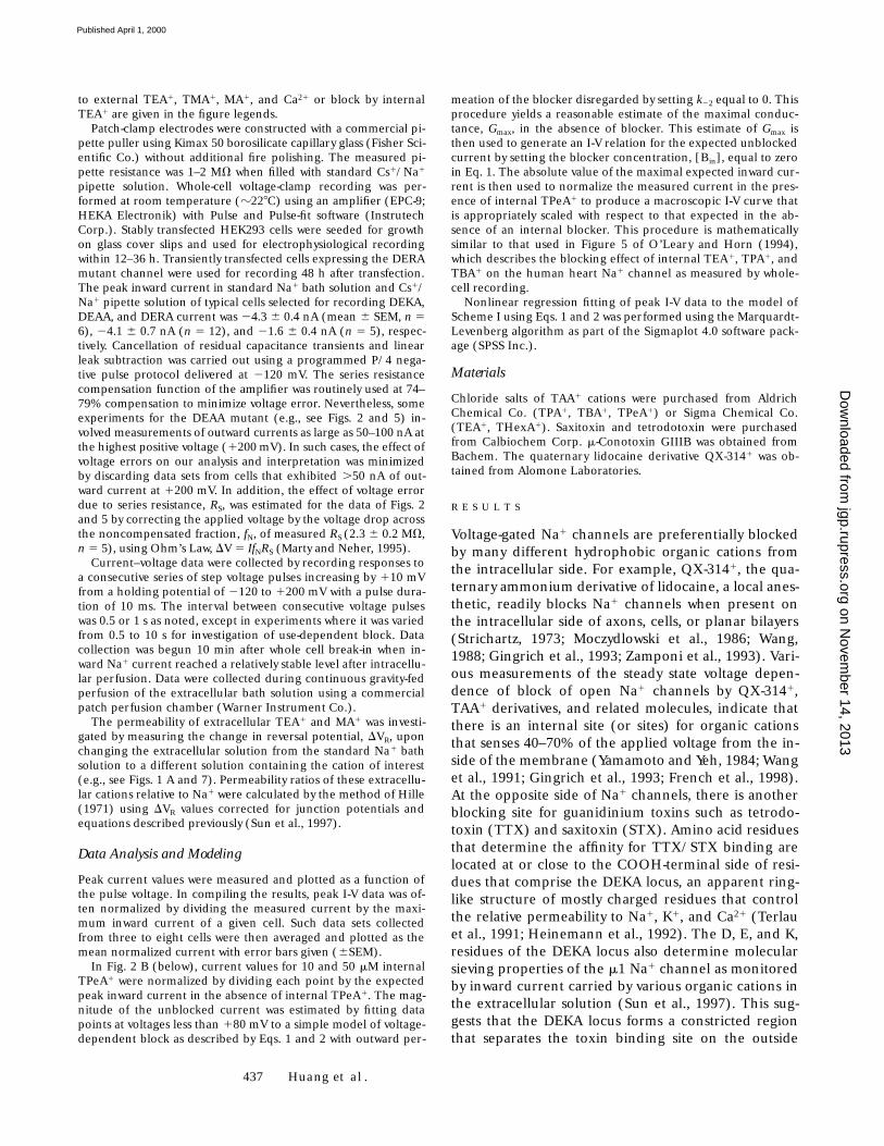
The control experiment of Fig. 1 shows that replace-ment of a standard extracellular Na1 solution with a so-lution containing TEA1 as the major external cation re-sults in a small inward current and a negative shift ofthe reversal potential of whole-cell peak current for theDEAA mutant. The magnitude of this shift (224.6 60.3 mV) corresponds to a permeability ratio ofPTEA(out)/PNa 5 0.37 6 0.01 (n 5 4), confirming thatexternal TEA1 can permeate through this channel.TEA1 is the largest cation that has thus far been foundto exhibit measurable inward current for the DEAAmutant (Sun et al., 1997). When the standard intracel-lular solution containing 125 mM Cs1 plus 20 mM Na1
was replaced with a pipette solution containing 115mM TEA-Cl, outward current could not be satisfactorilyresolved since it was ,0.1 nA at 1200 mV. This observa-tion suggests that the DEAA channel is essentially im-permeable to internal TEA1. To improve the possibilityof observing outward current carried by an organic cat-ion, we tested TMA1, a smaller TAA1 derivative. Fig. 1B shows typical currents and peak I-V data obtained forcells recorded in the presence of 115 mM internalTMA1 and 145 mM external Na1. This experiment suc-cessfully resolved a small outward current carried byTMA1, but strong rectification observed in the positivevoltage range implies that the conductance supportedby internal TMA1 is small in comparison with externalNa1. The nominal reversal potential in this experimentcorresponds to a permeability ratio of PTMA(in)/PNa of0.07, or at least sevenfold less2 than the value ofPTMA(out)/PNa 5 0.50 that was previously measuredwith TMA1 on the outside of the DEAA mutant (Sun etal., 1997). These results indicate that permeation ofsmall TAA1 cations through the DEAA mutant is asym-metric in nature. TMA1 carries current through this
channel in both directions, but both TMA1 and TEA1
are significantly more permeable when tested in the in-ward versus the outward direction.
Relief of Block by Internal TPeA1 at High Positive Voltage in DEAA Mutant and Wild-Type Na1 Channels
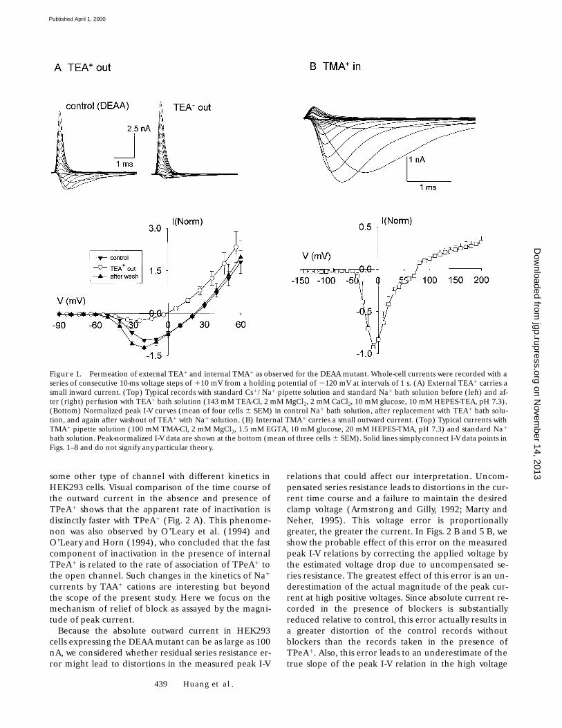
The next question we addressed was whether block ofoutward alkali cation current by large hydrophobicTAA1s is altered in the DEAA mutant compared withwild type. Internal TPeA1 has been previously character-ized as a potent blocker of outward Na1 current for thewild-type human heart Na1 channel (O’Leary et al.,1994; O’Leary and Horn, 1994). In this latter study,block was described by voltage-dependent binding ofTPeA1 to a site located at an apparent electrical distanceof 0.41 from the inside with a Kd of 9.8 mM at 0 mV. Fig.2 shows typical currents and peak I-V data for HEK293cells expressing the DEAA mutant. Current records ob-tained under control conditions with 120 mM Cs1 plus20 mM Na1 in the internal solution were compared withsimilar recordings with 1, 10, or 50 mM TPeA1 added tothe pipette solution. The control records show thatlarge rapidly inactivating outward currents are observedunder these conditions. The outward current is a mix-ture of Cs1 and Na1 current since this mutant is rathernonselective for alkali cations with PCs/PNa 5 0.57 forCs1 tested on the outside (Sun et al., 1997). The controlpeak current in the DEAA mutant exhibits nearly ohmicbehavior in the positive voltage range up to 1200 mV(Fig. 2 B, control). Fig. 2 B shows that addition of 10 or50 mM TPeA1 to the internal solution strongly sup-presses outward current carried by alkali cations. In thepositive voltage range up to about 1140 mV, TPeA1 be-haves as a voltage-dependent blocker as reported for theheart Na1 channel (O’Leary et al., 1994).
However, at voltages greater than 1140 mV, there is asharp upturn of the peak I-V relationship with 10 and50 mM TPeA1 (Fig. 2 B). Inspection of the correspond-ing current traces (Fig. 2 A) shows that outward currentsrecorded with 10 and 50 mM TPeA1 display typicalrapid activation and inactivation kinetics of voltage-gated Na1 channels. A positive inflection in the peakI-V relations of Fig. 2 B arises from a rather abrupt in-crease in transient current at voltages greater than1140 mV (Fig. 2 A). This kind of behavior is consistentwith voltage-dependent relief of TPeA1-blocked Na1
channels, rather than an artifact due to activation of
2This estimate of 0.07 for PTMA(in)/PNa was calculated using the ap-parent reversal potential and an appropriate form of the Goldmann-Hodgkin-Katz voltage reversal equation for major monovalent cat-ions. However, this calculation does not take into account the changein junction potential that occurs in whole-cell recording after diffu-sional equilibration between the pipette contents and cell interior.As described by Marty and Neher (1995), the actual membrane po-
tential will be more positive than the amplifier reading when thedominant cation (TMA1) in the pipette solution is less mobile thanthe dominant anion (Cl2). This means that the true reversal poten-tial in this experiment is more positive than the measured value andthat our estimate of the relative permeability of TMA1(in) is an up-per limit. Thus, this uncertainty does not compromise the conclusionthat internal TMA1 has lower permeability than external TMA1.
on Novem
ber 14, 2013jgp.rupress.org
Dow
nloaded from
Published April 1, 2000
439 Huang et al.
some other type of channel with different kinetics inHEK293 cells. Visual comparison of the time course ofthe outward current in the absence and presence ofTPeA1 shows that the apparent rate of inactivation isdistinctly faster with TPeA1 (Fig. 2 A). This phenome-non was also observed by O’Leary et al. (1994) andO’Leary and Horn (1994), who concluded that the fastcomponent of inactivation in the presence of internalTPeA1 is related to the rate of association of TPeA1 tothe open channel. Such changes in the kinetics of Na1
currents by TAA1 cations are interesting but beyondthe scope of the present study. Here we focus on themechanism of relief of block as assayed by the magni-tude of peak current.
Because the absolute outward current in HEK293cells expressing the DEAA mutant can be as large as 100nA, we considered whether residual series resistance er-ror might lead to distortions in the measured peak I-V
relations that could affect our interpretation. Uncom-pensated series resistance leads to distortions in the cur-rent time course and a failure to maintain the desiredclamp voltage (Armstrong and Gilly, 1992; Marty andNeher, 1995). This voltage error is proportionallygreater, the greater the current. In Figs. 2 B and 5 B, weshow the probable effect of this error on the measuredpeak I-V relations by correcting the applied voltage bythe estimated voltage drop due to uncompensated se-ries resistance. The greatest effect of this error is an un-derestimation of the actual magnitude of the peak cur-rent at high positive voltages. Since absolute current re-corded in the presence of blockers is substantiallyreduced relative to control, this error actually results ina greater distortion of the control records withoutblockers than the records taken in the presence ofTPeA1. Also, this error leads to an underestimate of thetrue slope of the peak I-V relation in the high voltage
Figure 1. Permeation of external TEA1 and internal TMA1 as observed for the DEAA mutant. Whole-cell currents were recorded with aseries of consecutive 10-ms voltage steps of 110 mV from a holding potential of 2120 mV at intervals of 1 s. (A) External TEA1 carries asmall inward current. (Top) Typical records with standard Cs1/Na1 pipette solution and standard Na1 bath solution before (left) and af-ter (right) perfusion with TEA1 bath solution (143 mM TEA-Cl, 2 mM MgCl2, 2 mM CaCl2, 10 mM glucose, 10 mM HEPES-TEA, pH 7.3).(Bottom) Normalized peak I-V curves (mean of four cells 6 SEM) in control Na1 bath solution, after replacement with TEA1 bath solu-tion, and again after washout of TEA1 with Na1 solution. (B) Internal TMA1 carries a small outward current. (Top) Typical currents withTMA1 pipette solution (100 mM TMA-Cl, 2 mM MgCl2, 1.5 mM EGTA, 10 mM glucose, 20 mM HEPES-TMA, pH 7.3) and standard Na1
bath solution. Peak-normalized I-V data are shown at the bottom (mean of three cells 6 SEM). Solid lines simply connect I-V data points inFigs. 1–8 and do not signify any particular theory.
on Novem
ber 14, 2013jgp.rupress.org
Dow
nloaded from
Published April 1, 2000
440 Voltage-dependent Relief of Na1 Channel Block by Large Organic Cations
range. Thus, data distortion caused by series resistanceerror cannot explain the dramatic upturn in the peakI-V relations measured in the presence of TPeA1.
An inconvenient aspect of quantitative analysis of in-ternal blockers by whole-cell patch clamp is that the in-ternal solution cannot be readily changed, so that thelevel of unblocked control current for a given cell withinternal blocker is difficult to establish. However, sinceTAA1 cations are well behaved voltage-dependent block-ers of Na1 channels in the low voltage range of 180 mVor less (O’Leary and Horn, 1994) and the control out-ward current of the DEAA mutant is fairly ohmic, it ispossible to estimate the fractional amount of current in-hibition from the known blocking behavior. For exam-ple, in Fig. 2 B, data for 10 and 50 mM TPeA1 were nor-malized to the unblocked current expected for each cellin the absence of TPeA1 by a procedure that involved fit-ting data in the low voltage range to a simple Boltzmannmodel of voltage-dependent block (see materials andmethods). Thus, the I-V data for 0, 10, and 50 mMTPeA1 are displayed in a way that reflects the underlyingconcentration dependence of current inhibition in thepresence of TPeA1. When viewed in this fashion, the re-sults of Fig. 2 strongly suggest that the positive inflection
in the I-V data for 10 and 50 mM internal TPeA1 arisesfrom genuine relief of block due to voltage-driven per-meation of this blocker through the DEAA mutant Na1
channel. Similar relief-of-block behavior has been previ-ously documented for many types of ions channels andblockers. For example, relief of block at high voltage hasbeen observed for block of the squid axon K1 channelsby internal Na1 (French and Wells, 1977; French andShoukimas, 1985), block of acetylcholine-receptor chan-nels by high concentrations of external acetylcholine(Sine and Steinbach, 1984), and block of anthrax toxinchannels by large TAA1 cations (Blaustein and Finkel-stein, 1990a,b). The interesting feature here is that reliefof block by such a large and bulky organic cation (diame-ter of TPeA1 . 13.2 Å) has not been previously reportedfor a cation-selective channel that has a rather narrow se-lectivity filter, on the order of 3 3 5 Å for the wild-typeNa1 channel (Hille, 1971, 1972; Sun et al., 1997).
One mechanism by which large molecules might passthrough a small pore could involve physical deforma-tion or a transient enlargement of the limiting regionof the filter. To investigate this possibility, we tested thesensitivity of the relief-of-block phenomenon to toxinsthat block the Na1 channel from the external side. The
Figure 2. Block and relief-of-block at high positive voltage byinternal TPeA1 as observed forthe DEAA mutant. The voltagepulse protocol is the same as thatof Fig. 1 at pulse intervals of 0.5 s.(A) Typical current records withstandard Cs1/Na1 pipette solu-tion and standard Na1 bath solu-tion in the absence (left) andpresence of 10 mM (middle) and50 mM (right) TPeA1 in the pi-pette solution. Currents tracesfor different conditions are nor-malized to maximum peak out-ward current in the absence ofTPeA1. (B) Relative I-V behaviorof the DEAA mutant with 0, 1,10, and 50 mM internal TPeA1.I-V data for 0 and 1 mM TPeA1 arenormalized relative to the peakinward current of a given cell be-fore averaging. Similarly normal-ized I-V data for 10 and 50 mMTPeA1 are scaled relative to theexpected maximal conductanceof a control cell without TPeA1
based on the fitted level of volt-age-dependent block in the volt-age range less than 180 mV (seematerials and methods). Theinset is an expanded view of data
in the negative voltage range. Closed symbols are plotted versus nominal applied voltage. Open symbols represent approximate correc-tions of applied voltage for series resistance error at large voltage steps. Data points are the mean 6 SEM for three to five cells.
on Novem
ber 14, 2013jgp.rupress.org
Dow
nloaded from
Published April 1, 2000
441 Huang et al.
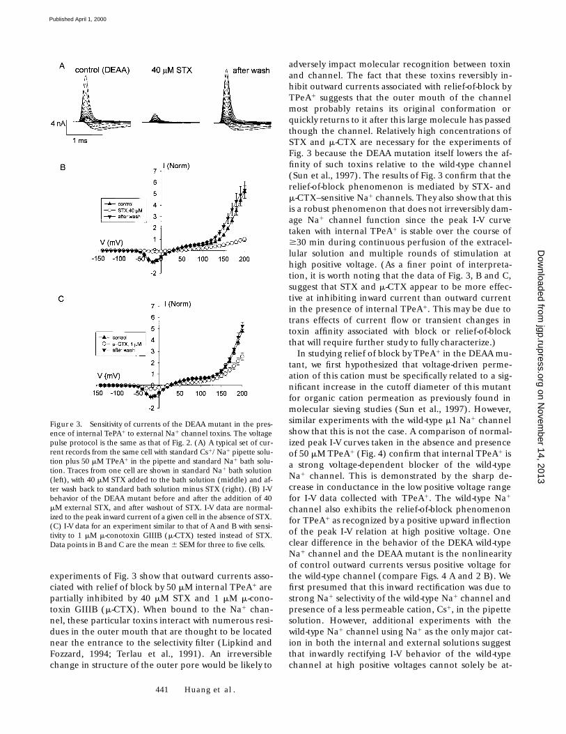
experiments of Fig. 3 show that outward currents asso-ciated with relief of block by 50 mM internal TPeA1 arepartially inhibited by 40 mM STX and 1 mM m-cono-toxin GIIIB (m-CTX). When bound to the Na1 chan-nel, these particular toxins interact with numerous resi-dues in the outer mouth that are thought to be locatednear the entrance to the selectivity filter (Lipkind andFozzard, 1994; Terlau et al., 1991). An irreversiblechange in structure of the outer pore would be likely to
adversely impact molecular recognition between toxinand channel. The fact that these toxins reversibly in-hibit outward currents associated with relief-of-block byTPeA1 suggests that the outer mouth of the channelmost probably retains its original conformation orquickly returns to it after this large molecule has passedthough the channel. Relatively high concentrations ofSTX and m-CTX are necessary for the experiments ofFig. 3 because the DEAA mutation itself lowers the af-finity of such toxins relative to the wild-type channel(Sun et al., 1997). The results of Fig. 3 confirm that therelief-of-block phenomenon is mediated by STX- andm-CTX–sensitive Na1 channels. They also show that thisis a robust phenomenon that does not irreversibly dam-age Na1 channel function since the peak I-V curvetaken with internal TPeA1 is stable over the course of$30 min during continuous perfusion of the extracel-lular solution and multiple rounds of stimulation athigh positive voltage. (As a finer point of interpreta-tion, it is worth noting that the data of Fig. 3, B and C,suggest that STX and m-CTX appear to be more effec-tive at inhibiting inward current than outward currentin the presence of internal TPeA1. This may be due totrans effects of current flow or transient changes intoxin affinity associated with block or relief-of-blockthat will require further study to fully characterize.)
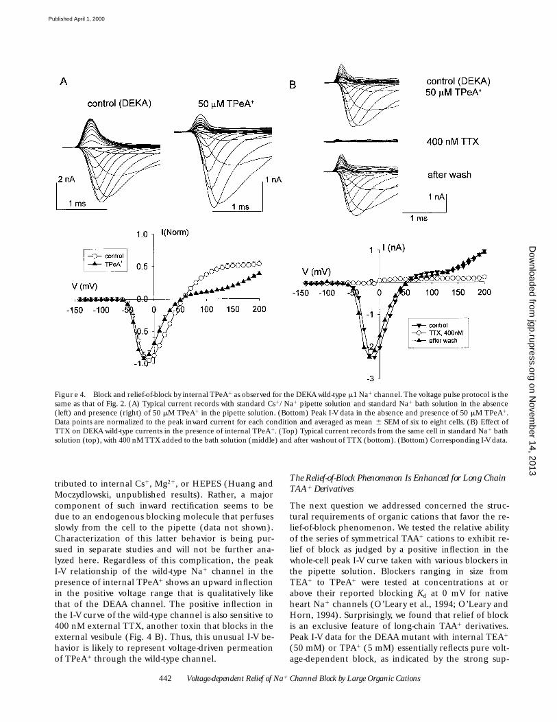
In studying relief of block by TPeA1 in the DEAA mu-tant, we first hypothesized that voltage-driven perme-ation of this cation must be specifically related to a sig-nificant increase in the cutoff diameter of this mutantfor organic cation permeation as previously found inmolecular sieving studies (Sun et al., 1997). However,similar experiments with the wild-type m1 Na1 channelshow that this is not the case. A comparison of normal-ized peak I-V curves taken in the absence and presenceof 50 mM TPeA1 (Fig. 4) confirm that internal TPeA1 isa strong voltage-dependent blocker of the wild-typeNa1 channel. This is demonstrated by the sharp de-crease in conductance in the low positive voltage rangefor I-V data collected with TPeA1. The wild-type Na1
channel also exhibits the relief-of-block phenomenonfor TPeA1 as recognized by a positive upward inflectionof the peak I-V relation at high positive voltage. Oneclear difference in the behavior of the DEKA wild-typeNa1 channel and the DEAA mutant is the nonlinearityof control outward currents versus positive voltage forthe wild-type channel (compare Figs. 4 A and 2 B). Wefirst presumed that this inward rectification was due tostrong Na1 selectivity of the wild-type Na1 channel andpresence of a less permeable cation, Cs1, in the pipettesolution. However, additional experiments with thewild-type Na1 channel using Na1 as the only major cat-ion in both the internal and external solutions suggestthat inwardly rectifying I-V behavior of the wild-typechannel at high positive voltages cannot solely be at-
Figure 3. Sensitivity of currents of the DEAA mutant in the pres-ence of internal TePA1 to external Na1 channel toxins. The voltagepulse protocol is the same as that of Fig. 2. (A) A typical set of cur-rent records from the same cell with standard Cs1/Na1 pipette solu-tion plus 50 mM TPeA1 in the pipette and standard Na1 bath solu-tion. Traces from one cell are shown in standard Na1 bath solution(left), with 40 mM STX added to the bath solution (middle) and af-ter wash back to standard bath solution minus STX (right). (B) I-Vbehavior of the DEAA mutant before and after the addition of 40mM external STX, and after washout of STX. I-V data are normal-ized to the peak inward current of a given cell in the absence of STX.(C) I-V data for an experiment similar to that of A and B with sensi-tivity to 1 mM m-conotoxin GIIIB (m-CTX) tested instead of STX.Data points in B and C are the mean 6 SEM for three to five cells.
on Novem
ber 14, 2013jgp.rupress.org
Dow
nloaded from
Published April 1, 2000
442 Voltage-dependent Relief of Na1 Channel Block by Large Organic Cations
tributed to internal Cs1, Mg21, or HEPES (Huang andMoczydlowski, unpublished results). Rather, a majorcomponent of such inward rectification seems to bedue to an endogenous blocking molecule that perfusesslowly from the cell to the pipette (data not shown).Characterization of this latter behavior is being pur-sued in separate studies and will not be further ana-lyzed here. Regardless of this complication, the peakI-V relationship of the wild-type Na1 channel in thepresence of internal TPeA1 shows an upward inflectionin the positive voltage range that is qualitatively likethat of the DEAA channel. The positive inflection inthe I-V curve of the wild-type channel is also sensitive to400 nM external TTX, another toxin that blocks in theexternal vesibule (Fig. 4 B). Thus, this unusual I-V be-havior is likely to represent voltage-driven permeationof TPeA1 through the wild-type channel.
The Relief-of-Block Phenomenon Is Enhanced for Long Chain TAA1 Derivatives
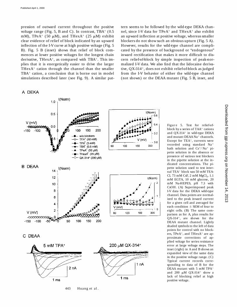
The next question we addressed concerned the struc-tural requirements of organic cations that favor the re-lief-of-block phenomenon. We tested the relative abilityof the series of symmetrical TAA1 cations to exhibit re-lief of block as judged by a positive inflection in thewhole-cell peak I-V curve taken with various blockers inthe pipette solution. Blockers ranging in size fromTEA1 to TPeA1 were tested at concentrations at orabove their reported blocking Kd at 0 mV for nativeheart Na1 channels (O’Leary et al., 1994; O’Leary andHorn, 1994). Surprisingly, we found that relief of blockis an exclusive feature of long-chain TAA1 derivatives.Peak I-V data for the DEAA mutant with internal TEA1
(50 mM) or TPA1 (5 mM) essentially reflects pure volt-age-dependent block, as indicated by the strong sup-
Figure 4. Block and relief-of-block by internal TPeA1 as observed for the DEKA wild-type m1 Na1 channel. The voltage pulse protocol is thesame as that of Fig. 2. (A) Typical current records with standard Cs1/Na1 pipette solution and standard Na1 bath solution in the absence(left) and presence (right) of 50 mM TPeA1 in the pipette solution. (Bottom) Peak I-V data in the absence and presence of 50 mM TPeA1.Data points are normalized to the peak inward current for each condition and averaged as mean 6 SEM of six to eight cells. (B) Effect ofTTX on DEKA wild-type currents in the presence of internal TPeA1. (Top) Typical current records from the same cell in standard Na1 bathsolution (top), with 400 nM TTX added to the bath solution (middle) and after washout of TTX (bottom). (Bottom) Corresponding I-V data.
on Novem
ber 14, 2013jgp.rupress.org
Dow
nloaded from
Published April 1, 2000
443 Huang et al.
pression of outward current throughout the positivevoltage range (Fig. 5, B and C). In contrast, TBA1 (0.5mM), TPeA1 (50 mM), and THexA1 (25 mM) exhibitclear evidence of relief of block indicated by an upwardinflection of the I-V curve at high positive voltage (Fig. 5B). Fig. 5 B (inset) shows that relief of block com-mences at lower positive voltages for the longest chainderivative, THexA1, as compared with TBA1. This im-plies that it is energetically easier to drive the largerTHexA1 cation through the channel than the smallerTBA1 cation, a conclusion that is borne out in modelsimulations described later (see Fig. 9). A similar pat-
tern seems to be followed by the wild-type DEKA chan-nel, since I-V data for TPeA1 and THexA1 also exhibitan upward inflection at positive voltage, whereas smallerblockers do not show such an obvious upturn (Fig. 5 A).However, results for the wild-type channel are compli-cated by the presence of background or “endogenous”inward rectification that makes it more difficult to dis-cern relief-of-block by simple inspection of peak-nor-malized I-V data. We also find that the lidocaine deriva-tive, QX-3141, does not exhibit relief-of-block as judgedfrom the I-V behavior of either the wild-type channel(not shown) or the DEAA mutant (Fig. 5 B, inset, and
Figure 5. Test for relief-of-block by a series of TAA1 cationsand QX-3141 in wild-type DEKAand mutant DEAA Na1 channels.Except for TEA1, currents wererecorded using standard Na1
bath solution and Cs1/Na1 pi-pette solution in the absence orpresence of various test blockersin the pipette solution at the in-dicated concentrations. The pi-pette solution used to test inter-nal TEA1 block was 50 mM TEA-Cl, 73 mM CsF, 2 mM MgCl2, 1.1mM EGTA, 10 mM glucose, 20mM Na-HEPES, pH 7.3 withCsOH. (A) Superimposed peakI-V data for the DEKA wild-typechannel. Data points are normal-ized to the peak inward currentfor a given cell and averaged foreach condition 6 SEM of four toeight cells. (B) The same com-parison as for A, plus results forQX-3141, are shown for theDEAA mutant channel. Lightlyshaded symbols to the left of datapoints for control with no block-ers, TPeA1, and THexA1 are ap-proximate corrections of ap-plied voltage for series resistanceerror at large voltage steps. Theinset (right) in A and B shows anexpanded view of the same datain the positive voltage range. (C)Typical current records corre-sponding to data of B for theDEAA mutant with 5 mM TPA1
and 200 mM QX-3141 show alack of blocking relief at highpositive voltage.
on Novem
ber 14, 2013jgp.rupress.org
Dow
nloaded from
Published April 1, 2000
444 Voltage-dependent Relief of Na1 Channel Block by Large Organic Cations
C). In summary, the results of Fig. 5 indicate that volt-age-dependent relief of block is enhanced with increas-ing length of n-alkyl chains for the series of symmetricalTAA1 cations. The fact that QX-3141 does not exhibitrelief-of-block at 1200 mV or less suggests that there arerather specific chemical structural requirements forvoltage-driven permeation of quaternary ammoniumcations through the external end of the pore.
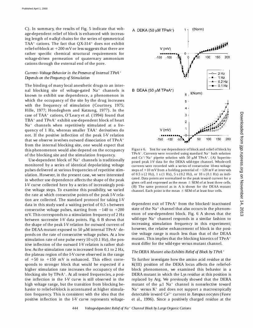
Current–Voltage Behavior in the Presence of Internal TPeA1 Depends on the Frequency of Stimulation
The binding of many local anesthetic drugs to an inter-nal blocking site of voltage-gated Na1 channels isknown to exhibit use dependence, a phenomenon inwhich the occupancy of the site by the drug increaseswith the frequency of stimulation (Courtney, 1975;Hille, 1977; Hondeghem and Katzung, 1977). In thecase of TAA1 cations, O’Leary et al. (1994) found thatTBA1 and TPeA1 exhibit use-dependent block of heartNa1 channels when repetitively stimulated at a fre-quency of 1 Hz, whereas smaller TAA1 derivatives donot. If the positive inflection of the peak I-V relationthat we observe involves outward dissociation of TPeA1
from the internal blocking site, one would expect thatthis phenomenon would also depend on the occupancyof the blocking site and the stimulation frequency.
Use-dependent block of Na1 channels is traditionallymonitored by a series of identical depolarizing voltagepulses delivered at various frequencies of repetitive stim-ulation. However, in the present case, we were interestedin whether use dependence affects the shape of the peakI-V curve collected here by a series of increasingly posi-tive voltage steps. To examine this possibility, we variedthe rate at which consecutive points of the peak I-V rela-tion are collected. The standard protocol for taking I-Vdata in this study used a waiting period of 0.5 s betweenconsecutive voltage pulses, starting from 2140 to 1200mV. This corresponds to a stimulation frequency of 2 Hzbetween successive I-V data points. Fig. 6 B shows thatthe shape of the peak I-V relation for outward current ofthe DEAA mutant exposed to 50 mM internal TPeA1 de-pends on the rate of consecutive voltage pulses. At a lowstimulation rate of one pulse every 10 s (0.1 Hz), the pos-itive inflection of the outward I-V relation is rather shal-low. As the stimulation rate is increased from 0.1 to 2 Hz,the plateau region of the I-V curve observed in the rangeof 150 to 1150 mV is enhanced. This effect corre-sponds to stronger block that would be expected if ahigher stimulation rate increases the occupancy of theblocking site by TPeA1. At all tested frequencies, a posi-tive inflection in the I-V curve is still observed in thehigh voltage range, but the transition from blocking be-havior to relief-of-block is accentuated at higher stimula-tion frequency. This is consistent with the idea that thepositive inflection in the I-V curve represents voltage-
dependent exit of TPeA1 from the blocked/inactivatedstate of the Na1 channel that also occurs in the phenom-enon of use-dependent block. Fig. 6 A shows that thewild-type Na1 channel responds in a similar fashion toincreasing stimulation frequency in this experiment;however, the relative enhancement of block in the posi-tive voltage range is much less than that of the DEAAmutant. This implies that the blocking kinetics of TPeA1
must differ for the wild-type versus mutant channel.
The DERA Mutant also Exhibits Relief of Block by TPeA1
To further investigate how the amino acid residue at theK(III) position of the DEKA locus affects the relief-of-block phenomenon, we examined this behavior in aDERA mutant in which the Lys residue at this position isreplaced by Arg. We previously showed that the DERAmutant of the m1 Na1 channel is nonselective towardNa1 versus K1 and does not support a macroscopicallydetectable inward Ca21 current in Xenopus oocytes (Favreet al., 1996). Since a positively charged residue at the
Figure 6. Test for use dependence of block and relief of block byTPeA1. Currents were recorded using standard Na1 bath solutionand Cs1/Na1 pipette solution with 50 mM TPeA1. (A) Superim-posed peak I-V data for the DEKA wild-type channel. Whole-cellcurrents were recorded with a series of consecutive 10-ms voltagesteps of 110 mV from a holding potential of 2120 mV at intervalsof 0.5 s (2 Hz), 1 s (1 Hz), 5 s (0.2 Hz), or 10 s (0.1 Hz) as indi-cated. Data points are normalized to the peak inward current for agiven cell and expressed as the mean 6 SEM of at least three cells.(B) The same protocol as in A is shown for the DEAA mutantchannel. Each point is the mean 6 SEM of at least four cells.
on Novem
ber 14, 2013jgp.rupress.org
Dow
nloaded from
Published April 1, 2000
445 Huang et al.
K(III) position seems to function in preventing inwardpermeation of divalent cations, this led to the conclusionthat the mechanism of exclusion of external Ca21 in-volves a repulsive electrostatic interaction with the sidechain of the K(III) residue. The question addressed hereis whether a different positively charged residue in the se-lectivity filter such as Arg would prevent voltage-drivenpermeation of internal TPeA1 through the channel.
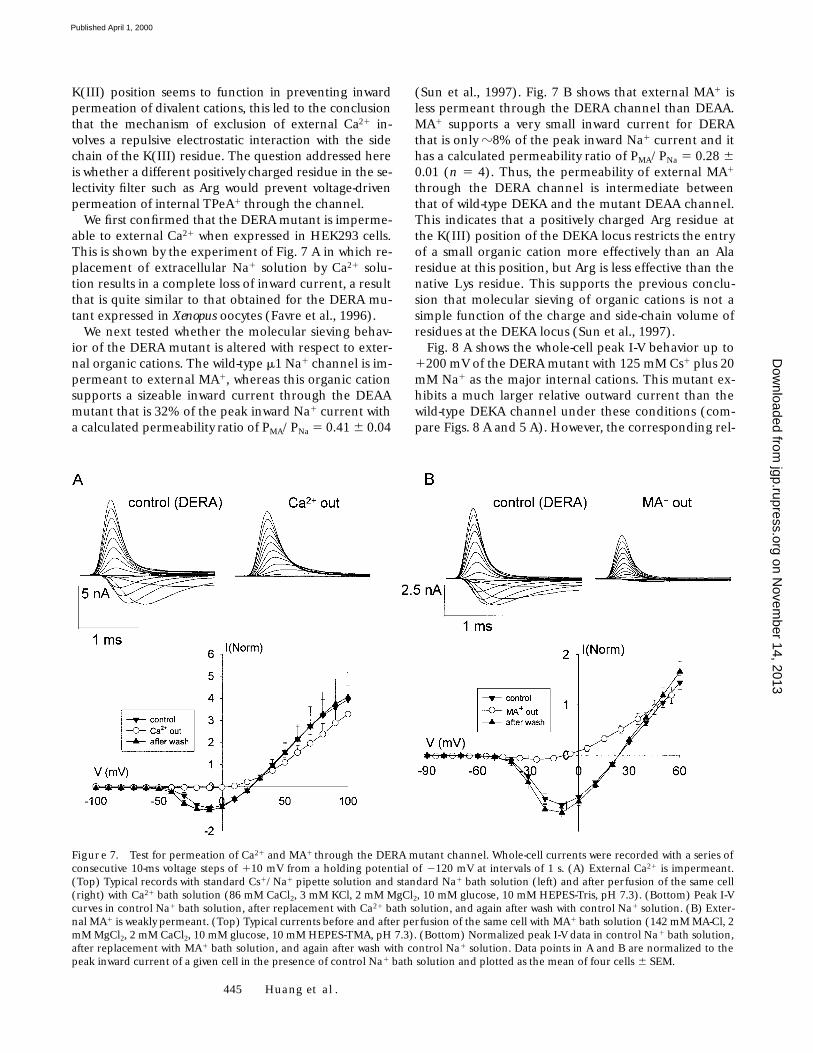
We first confirmed that the DERA mutant is imperme-able to external Ca21 when expressed in HEK293 cells.This is shown by the experiment of Fig. 7 A in which re-placement of extracellular Na1 solution by Ca21 solu-tion results in a complete loss of inward current, a resultthat is quite similar to that obtained for the DERA mu-tant expressed in Xenopus oocytes (Favre et al., 1996).
We next tested whether the molecular sieving behav-ior of the DERA mutant is altered with respect to exter-nal organic cations. The wild-type m1 Na1 channel is im-permeant to external MA1, whereas this organic cationsupports a sizeable inward current through the DEAAmutant that is 32% of the peak inward Na1 current witha calculated permeability ratio of PMA/PNa 5 0.41 6 0.04
(Sun et al., 1997). Fig. 7 B shows that external MA1 isless permeant through the DERA channel than DEAA.MA1 supports a very small inward current for DERAthat is only z8% of the peak inward Na1 current and ithas a calculated permeability ratio of PMA/PNa 5 0.28 60.01 (n 5 4). Thus, the permeability of external MA1
through the DERA channel is intermediate betweenthat of wild-type DEKA and the mutant DEAA channel.This indicates that a positively charged Arg residue atthe K(III) position of the DEKA locus restricts the entryof a small organic cation more effectively than an Alaresidue at this position, but Arg is less effective than thenative Lys residue. This supports the previous conclu-sion that molecular sieving of organic cations is not asimple function of the charge and side-chain volume ofresidues at the DEKA locus (Sun et al., 1997).
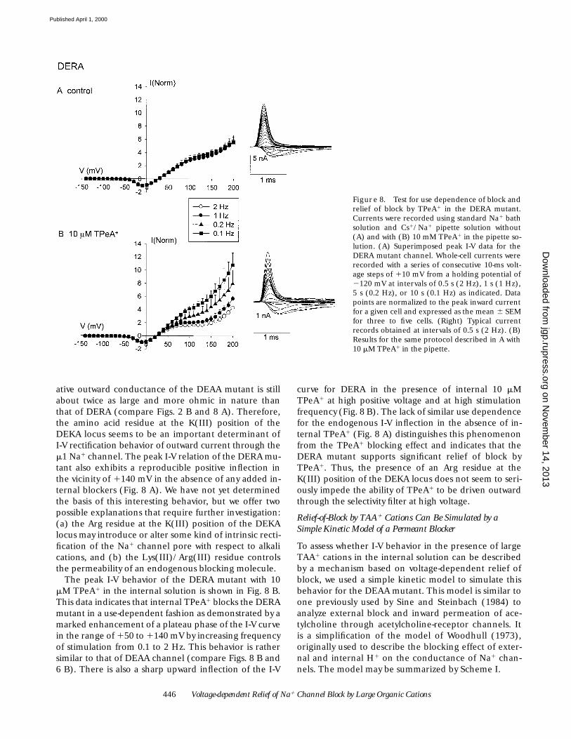
Fig. 8 A shows the whole-cell peak I-V behavior up to1200 mV of the DERA mutant with 125 mM Cs1 plus 20mM Na1 as the major internal cations. This mutant ex-hibits a much larger relative outward current than thewild-type DEKA channel under these conditions (com-pare Figs. 8 A and 5 A). However, the corresponding rel-
Figure 7. Test for permeation of Ca21 and MA1 through the DERA mutant channel. Whole-cell currents were recorded with a series ofconsecutive 10-ms voltage steps of 110 mV from a holding potential of 2120 mV at intervals of 1 s. (A) External Ca21 is impermeant.(Top) Typical records with standard Cs1/Na1 pipette solution and standard Na1 bath solution (left) and after perfusion of the same cell(right) with Ca21 bath solution (86 mM CaCl2, 3 mM KCl, 2 mM MgCl2, 10 mM glucose, 10 mM HEPES-Tris, pH 7.3). (Bottom) Peak I-Vcurves in control Na1 bath solution, after replacement with Ca21 bath solution, and again after wash with control Na1 solution. (B) Exter-nal MA1 is weakly permeant. (Top) Typical currents before and after perfusion of the same cell with MA1 bath solution (142 mM MA-Cl, 2mM MgCl2, 2 mM CaCl2, 10 mM glucose, 10 mM HEPES-TMA, pH 7.3). (Bottom) Normalized peak I-V data in control Na1 bath solution,after replacement with MA1 bath solution, and again after wash with control Na1 solution. Data points in A and B are normalized to thepeak inward current of a given cell in the presence of control Na1 bath solution and plotted as the mean of four cells 6 SEM.
on Novem
ber 14, 2013jgp.rupress.org
Dow
nloaded from
Published April 1, 2000
446 Voltage-dependent Relief of Na1 Channel Block by Large Organic Cations
ative outward conductance of the DEAA mutant is stillabout twice as large and more ohmic in nature thanthat of DERA (compare Figs. 2 B and 8 A). Therefore,the amino acid residue at the K(III) position of theDEKA locus seems to be an important determinant ofI-V rectification behavior of outward current through them1 Na1 channel. The peak I-V relation of the DERA mu-tant also exhibits a reproducible positive inflection inthe vicinity of 1140 mV in the absence of any added in-ternal blockers (Fig. 8 A). We have not yet determinedthe basis of this interesting behavior, but we offer twopossible explanations that require further investigation:(a) the Arg residue at the K(III) position of the DEKAlocus may introduce or alter some kind of intrinsic recti-fication of the Na1 channel pore with respect to alkalications, and (b) the Lys(III)/Arg(III) residue controlsthe permeability of an endogenous blocking molecule.
The peak I-V behavior of the DERA mutant with 10mM TPeA1 in the internal solution is shown in Fig. 8 B.This data indicates that internal TPeA1 blocks the DERAmutant in a use-dependent fashion as demonstrated by amarked enhancement of a plateau phase of the I-V curvein the range of 150 to 1140 mV by increasing frequencyof stimulation from 0.1 to 2 Hz. This behavior is rathersimilar to that of DEAA channel (compare Figs. 8 B and6 B). There is also a sharp upward inflection of the I-V
curve for DERA in the presence of internal 10 mMTPeA1 at high positive voltage and at high stimulationfrequency (Fig. 8 B). The lack of similar use dependencefor the endogenous I-V inflection in the absence of in-ternal TPeA1 (Fig. 8 A) distinguishes this phenomenonfrom the TPeA1 blocking effect and indicates that theDERA mutant supports significant relief of block byTPeA1. Thus, the presence of an Arg residue at theK(III) position of the DEKA locus does not seem to seri-ously impede the ability of TPeA1 to be driven outwardthrough the selectivity filter at high voltage.
Relief-of-Block by TAA1 Cations Can Be Simulated by a Simple Kinetic Model of a Permeant Blocker
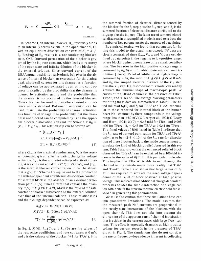
To assess whether I-V behavior in the presence of largeTAA1 cations in the internal solution can be describedby a mechanism based on voltage-dependent relief ofblock, we used a simple kinetic model to simulate thisbehavior for the DEAA mutant. This model is similar toone previously used by Sine and Steinbach (1984) toanalyze external block and inward permeation of ace-tylcholine through acetylcholine-receptor channels. Itis a simplification of the model of Woodhull (1973),originally used to describe the blocking effect of exter-nal and internal H1 on the conductance of Na1 chan-nels. The model may be summarized by Scheme I.
Figure 8. Test for use dependence of block andrelief of block by TPeA1 in the DERA mutant.Currents were recorded using standard Na1 bathsolution and Cs1/Na1 pipette solution without(A) and with (B) 10 mM TPeA1 in the pipette so-lution. (A) Superimposed peak I-V data for theDERA mutant channel. Whole-cell currents wererecorded with a series of consecutive 10-ms volt-age steps of 110 mV from a holding potential of2120 mV at intervals of 0.5 s (2 Hz), 1 s (1 Hz),5 s (0.2 Hz), or 10 s (0.1 Hz) as indicated. Datapoints are normalized to the peak inward currentfor a given cell and expressed as the mean 6 SEMfor three to five cells. (Right) Typical currentrecords obtained at intervals of 0.5 s (2 Hz). (B)Results for the same protocol described in A with10 mM TPeA1 in the pipette.
on Novem
ber 14, 2013jgp.rupress.org
Dow
nloaded from
Published April 1, 2000
447 Huang et al.
In Scheme I, an internal blocker, Bin, reversibly bindsto an internally accessible site in the open channel, O,with an equilibrium dissociation constant of K1 5 k21/k1. Binding of Bin results in a nonconducting blockedstate, O•B. Outward permeation of the blocker is gov-erned by the k22 rate constant, which leads to recoveryof the open state and infinite dilution of the blocker inthe external solution. Since outward current of theDEAA mutant exhibits nearly ohmic behavior in the ab-sence of internal blocker, an expression for simulatingpeak whole-cell current for this channel as a functionof voltage can be approximated by an ohmic conduc-tance multiplied by the probability that the channel isopened by activation gating and the probability thatthe channel is not occupied by the internal blocker.Ohm’s law can be used to describe channel conduc-tance and a standard Boltzmann expression can beused to simulate the probability of channel activationas a function of voltage. The probability that the chan-nel is not blocked can be computed by using the appar-ent blocker dissociation constant for Scheme I: KB 5(k21 1 k22)/k1. This relationship can be written as:
(1)
where Gmax is the maximal conductance, VR is the rever-sal potential, q is an effective gating charge for voltageactivation, V0.5 is the midpoint voltage of activation gat-ing, A is a constant equal to RT/F, or 25.4 mV, and [Bin]is the internal blocker concentration. It can be shownthat KB(V) for Scheme I is equivalent to the product ofthe voltage-dependent equilibrium dissociation constantfor internal block in the absence of an external perme-ation path, K1(V), times a term that contains the quan-tity, R(V) 5 k22(V)/k21(V), which is the ratio of the rateconstant of blocker dissociation to the external solutionover that of the internal solution. These relationshipsand their voltage dependence can be expressed as:
(2)
In Eq. 2, K1(0), k22(0), and k21(0) are the values ofthe respective equilibrium and rate constants at 0 mV,and z is the valence of the blocker (11 for TAA1). d1 is
I Gmax V VR–( )[ ]=
1 q V V0.5–( )– A⁄[ ]exp+{ }⋅ 1–
1 Bin[ ]+ KB V( )⁄{ }⋅ 1–
KB V( ) K1 V( ) 1 R V( )+[ ]=
K1 V( ) K1 0( ) zδ1V A⁄–( )exp=
R V( )k 2– 0( )k 1– 0( )--------------- zδ2V A⁄( )exp=
the summed fraction of electrical distance sensed bythe blocker for the k1 step plus the k21 step, and d2 is thesummed fraction of electrical distance attributed to thek22 step plus the k21 step. The latter use of summed electri-cal distances in this simplified model is used to reduce thenumber of free parameters for the purpose of data fitting.
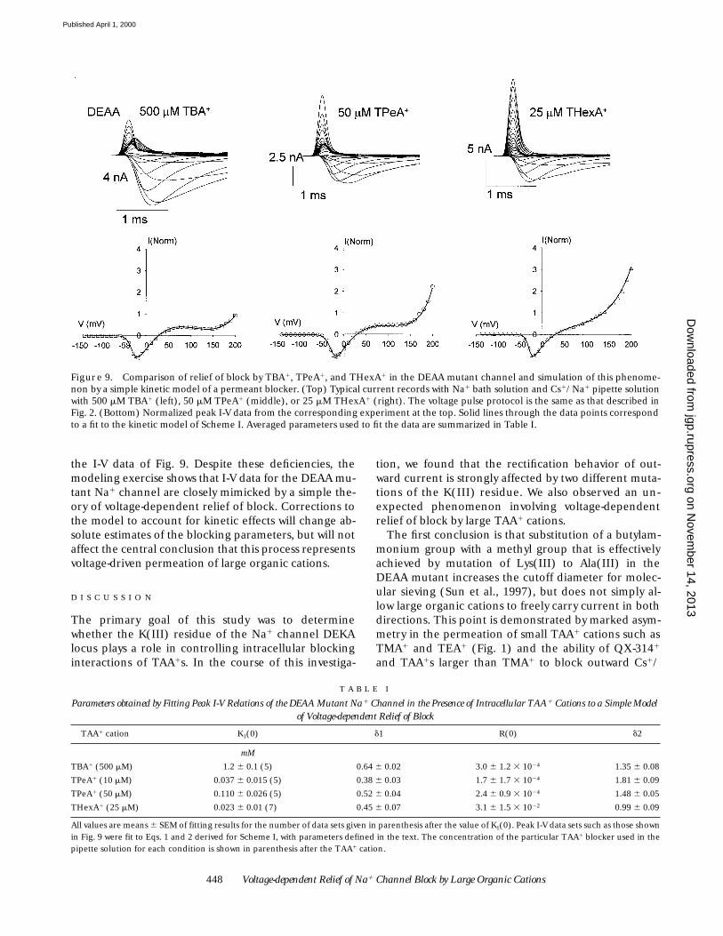
By empirical testing, we found that parameters for fit-ting this model to the actual macroscopic I-V data areclosely constrained since Gmax, VR, q, and V0.5 are well de-fined by data points in the negative to low positive range,where blocking phenomena have only a small contribu-tion. The behavior in the high positive voltage range isgoverned by KB(0) and d1, which determine current in-hibition (block). Relief of inhibition at high voltage isgoverned by R(0), the ratio of k22(V)/k21(V) at 0 mV,and d2, the lumped electrical distance of the k22 stepplus the k21 step. Fig. 9 shows that this model can readilysimulate the unusual shape of macroscopic peak I-Vcurves of the DEAA channel in the presence of TBA1,TPeA1, and THexA1. The average blocking parametersfor fitting these data are summarized in Table I. The fit-ted values of K1(0) and d1 for TBA1 and TPeA1 are simi-lar to those reported for internal block of the humanheart Na1 channel by these compounds in the voltagerange less than 180 mV (O’Leary et al., 1994; O’Learyand Horn, 1994): K1(0) 5 0.48 mM for TBA1 and 0.098mM for TPeA1; d1 5 0.46 for TBA1 and 0.41 for TPeA1.The fitted values of R(0) listed in Table I indicate thatthe k22 rate of outward permeation for TBA1 and TPeA1
only has to be z2–3 3 1024 of the k21 rate for dissocia-tion of these blockers back to the inside compartment tosimulate the kind of blocking relief observed in this sys-tem. Table I also shows that the enhanced relief of blockobserved for THexA1 can be explained by a 100-fold in-crease in the value of R(0) for this particular molecule.This implies that THexA1 is able to exit through thechannel to the outside much more readily that TBA1
and TPeA1. Table I also shows that large values of d2
$1.0 are required to simulate the steep voltage depen-dence of the relief of block observed at high positivevoltage. This indicates that additional charge-dependentprocesses besides the simple interaction of a single cat-ion with a site in the transmembrane electric field are in-volved in generating this phenomenon.
We must also caution that these simulations have cer-tain quantitative limitations. The model assumes thatthe measured peak Na1 currents are proportional tothe steady state interaction of the blockers with theopen channel. This does not take into account theshortening of the apparent rate of channel inactivationthat is evident in the current traces with large TAA1 cat-ions. This effect is especially dramatic at high positivevoltage for current records in the presence of TBA1
shown in Fig. 9. The simulations also do not considerthe use or frequency dependence inherent in collecting
(SCHEME I)
on Novem
ber 14, 2013jgp.rupress.org
Dow
nloaded from
Published April 1, 2000
448 Voltage-dependent Relief of Na1 Channel Block by Large Organic Cations
the I-V data of Fig. 9. Despite these deficiencies, themodeling exercise shows that I-V data for the DEAA mu-tant Na1 channel are closely mimicked by a simple the-ory of voltage-dependent relief of block. Corrections tothe model to account for kinetic effects will change ab-solute estimates of the blocking parameters, but will notaffect the central conclusion that this process representsvoltage-driven permeation of large organic cations.
D I S C U S S I O N
The primary goal of this study was to determinewhether the K(III) residue of the Na1 channel DEKAlocus plays a role in controlling intracellular blockinginteractions of TAA1s. In the course of this investiga-
tion, we found that the rectification behavior of out-ward current is strongly affected by two different muta-tions of the K(III) residue. We also observed an un-expected phenomenon involving voltage-dependentrelief of block by large TAA1 cations.
The first conclusion is that substitution of a butylam-monium group with a methyl group that is effectivelyachieved by mutation of Lys(III) to Ala(III) in theDEAA mutant increases the cutoff diameter for molec-ular sieving (Sun et al., 1997), but does not simply al-low large organic cations to freely carry current in bothdirections. This point is demonstrated by marked asym-metry in the permeation of small TAA1 cations such asTMA1 and TEA1 (Fig. 1) and the ability of QX-3141
and TAA1s larger than TMA1 to block outward Cs1/
Figure 9. Comparison of relief of block by TBA1, TPeA1, and THexA1 in the DEAA mutant channel and simulation of this phenome-non by a simple kinetic model of a permeant blocker. (Top) Typical current records with Na1 bath solution and Cs1/Na1 pipette solutionwith 500 mM TBA1 (left), 50 mM TPeA1 (middle), or 25 mM THexA1 (right). The voltage pulse protocol is the same as that described inFig. 2. (Bottom) Normalized peak I-V data from the corresponding experiment at the top. Solid lines through the data points correspondto a fit to the kinetic model of Scheme I. Averaged parameters used to fit the data are summarized in Table I.
T A B L E I
Parameters obtained by Fitting Peak I-V Relations of the DEAA Mutant Na1 Channel in the Presence of Intracellular TAA1 Cations to a Simple Model of Voltage-dependent Relief of Block
TAA1 cation K1(0) d1 R(0) d2
mM
TBA1 (500 mM) 1.2 6 0.1 (5) 0.64 6 0.02 3.0 6 1.2 3 1024 1.35 6 0.08
TPeA1 (10 mM) 0.037 6 0.015 (5) 0.38 6 0.03 1.7 6 1.7 3 1024 1.81 6 0.09
TPeA1 (50 mM) 0.110 6 0.026 (5) 0.52 6 0.04 2.4 6 0.9 3 1024 1.48 6 0.05
THexA1 (25 mM) 0.023 6 0.01 (7) 0.45 6 0.07 3.1 6 1.5 3 1022 0.99 6 0.09
All values are means 6 SEM of fitting results for the number of data sets given in parenthesis after the value of K1(0). Peak I-V data sets such as those shownin Fig. 9 were fit to Eqs. 1 and 2 derived for Scheme I, with parameters defined in the text. The concentration of the particular TAA1 blocker used in thepipette solution for each condition is shown in parenthesis after the TAA1 cation.
on Novem
ber 14, 2013jgp.rupress.org
Dow
nloaded from
Published April 1, 2000
449 Huang et al.
Na1 current in a voltage-dependent manner (Fig. 5 B).At voltages less than 1100 mV, inhibition of outwardmacroscopic current of the DEAA mutant by largeTAA1 molecules is similar to previously described intra-cellular blocking behavior of native Na1 channels byvarious local anesthetic-type agents. For example, volt-age-dependent block by TEA1, TPA1, TBA1 and TPeA1
illustrated here by the data of Fig. 5 B and the parame-ters of Table I is reminiscent of previous observationsthat larger, more hydrophobic alkylammonium deriva-tives generally have higher affinity than TMA1 andTEA1, (Moczydlowski et al., 1986; Wang et al., 1991;O’Leary and Horn, 1994) and that this internal block-ing site typically exhibits a d in the range 0.4–0.7 basedon the Woodhull (1973) blocking model. The results ofFig. 6 B demonstrate that use-dependent behavior ofinternal TPeA1 like that previously described for a wild-type heart Na1 channel (O’Leary et al., 1994) is alsopresent in the DEAA mutant. Thus, the internal block-ing site for hydrophobic organic cations is functionallypreserved in the DEAA mutant. In mechanistic terms,substitution of the K(III) residue with Ala does not sim-ply correspond to the removal of an energy barrier atan impassable location in the pore that controls volt-age-dependent block by internal TAA1s.
An interesting new feature of the DEAA channel de-scribed here is that whole-cell current carried by inter-nal alkali cations (Cs1, Na1) in the absence of otheradded blockers is nearly ohmic at positive voltages up to1200 mV (e.g., Fig. 5 B). This stands in strong contrastto inwardly rectifying or sublinear I-V behavior of thewild-type DEKA channel in this voltage range (e.g., Fig.5 A) recorded under the same conditions with Na1 out-side and Cs1 inside. This feature of the DEAA mutantmakes it easier to recognize the dramatic relief of blockby large TAA1 cations at voltages greater than 1140 mV(e.g., Fig. 2 B). The wild-type DEKA channel also appar-ently exhibits a similar phenomenon (Fig. 4), but it ismore difficult to analyze because of the background in-ward rectification. In this respect, a second conclusionof our study is that the K(III) residue of the DEKA locuscontrols the rectification behavior of outward macro-scopic current of the m1 Na1 channel expressed inHEK293 cells. The outward I-V relation of the DERAmutant with the Lys(III) to Arg substitution is more lin-ear than the wild-type DEKA channel, but a distinctivenegative inflection in the I-V curve of the DERA mutantseen with Cs1/Na1 pipette solution (Fig. 8 A) furtherestablishes that the particular residue at the K(III) posi-tion is a major determinant of current rectification.
The mechanism underlying the latter current inflec-tion of the DERA channel and the sublinear I-V behaviorof the wild-type channel is presently undetermined.However, our results provide clues that may help in itselucidation. Since block by various internal organic cat-
ions produces sublinear I-V behavior for the DEAA mu-tant, and certain mutations of the K(III) residue en-hance permeation of organic cations, an obvious possi-bility is that an endogenous internal blocking molecule ispartially responsible for inward rectification of the wild-type channel. As mentioned in results, our initial ex-periments suggest that Mg21, Cs1, or HEPES, which areall present in the standard pipette solution, are not theonly molecular species that may be responsible for thiseffect. At present, we suspect that other cellular cationssuch as polyamines may be involved in the endogenousrectification of wild-type Na1 channels, since polyamineblock has been found to underlie inward rectification be-havior of other classes of ion channels (Williams, 1997).
The third conclusion of this work is that voltage-dependent relief of block by the series of symmetricalTAA1 cations is facilitated by increasing length ofn-alkyl chains. Where this phenomenon is easiest to study,in the DEAA mutant, there is little hint of relief of inter-nal block due to TEA1, TPA1, or QX3141 by positivevoltage up to 1200 mV (Fig. 5, B and C). In contrast, adistinct upturn in the I-V curve appears to commenceat progressively lower voltages for the series, TBA1,TPeA1, and THexA1 (Figs. 5 B and 9). As demon-strated by the modeling exercise of Fig. 9, this behavioris consistent with voltage-driven permeation of largeTAA1 cations to the outside of the channel. The struc-ture–activity dependence of this effect implies that thepermeation process is facilitated by hydrophobic inter-actions. However, this is a rather unusual type of hydro-phobic interaction. The increase in hydrophobicitywith increasing n-alkyl chain length of symmetricalTAA1 cations is accompanied by a proportional in-crease in molecular diameter, which would ordinarilybe expected to inhibit permeation through a fixed-diameter pore in the usual manner of molecular siev-ing. Using energy-minimized molecular models ofTAA1 cations (Hyperchem software from Hypercube),we find that the molecular diameter of these moleculesincreases by z2 Å per symmetrical addition of a methyl-ene group, as in the series: TMA1 (6.0 Å), TEA1 (8.2 Å),TPA1 (9.8 Å), TBA1 (11.6 Å), TPeA1 (13.2 Å), andTHexA1 (15.2 Å), with the measured diameter given inparenthesis. The latter measurements are also in accordwith an independent molecular dynamics analysis ofthe conformations and size of TAA1 cations in the ab-sence of solvent (O’Leary et al., 1994). Thus, the basicquestion posed by this phenomenon is: How do longn-alkyl chains of TAA1 molecules facilitate movementof these blockers through a protein pore that has an ef-fective cutoff size of 3 3 5 Å (Hille, 1971, 1972) for thewild-type Na1 channel and an external cutoff diameterof z8.2 Å (Sun et al., 1997) for the DEAA mutant?
Similar questions have been contemplated previouslyin the literature. In the case of inhibition of outward
on Novem
ber 14, 2013jgp.rupress.org
Dow
nloaded from
Published April 1, 2000
450 Voltage-dependent Relief of Na1 Channel Block by Large Organic Cations
K1 current through squid axon delayed-rectifier K1
channels, block by both internal Na1 and Cs1 exhibitssteeply voltage-dependent relief when external K1 con-centration is low (French and Wells, 1977; French andShoukimas, 1985). In pondering the basis for this ef-fect, the latter authors suggested that energy suppliedby high voltage could be sufficient to permit dehy-drated Na1 ions to move through the K1-selective re-gion of the channel, by a process that is energeticallyprohibitive under normal conditions. As another possi-bility, they speculated that the structure of the channelcould be physically distorted under the stress of highvoltage acting upon impermeant ions in the pore. In adifferent study of a sarcoplasmic reticulum K1 channelusing a series of bis-quaternary ammonium blockers(bis-QA1) of increasing n-alkyl chain length, Miller(1982) observed an anomalous increase in the appar-ent voltage dependence of block by the longest bis-QA1 molecules. He suggested that this could be ex-plained by the binding of the blocker molecule in a“bent-over” conformation, such that this channel mightsimultaneously accommodate two charges of a 10-car-bon bis-QA1 molecule folded in a strained conforma-tion. As applied to the present situation, these mecha-nisms require that either the narrow filter region of theNa1 channel protein undergo deformation by a tran-sient enlargement of the pore aperture, or the chemi-cal conformation of the TAA1 molecule physically con-tract to allow such a molecule to squeeze through theouter end of the pore and exit to the outside. Both ofthese mechanisms seem unlikely to fully explain thephenomenon encountered here. If the Na1 channelprotein is able to transiently deform to produce a widerpore, it is not clear why this should occur more readilyfor larger molecules such as TPeA1 and not to any mea-surable extent for TEA1 or TPA1. Likewise, strainedconformations of TPeA1 that can be achieved by rota-tion about carbon–carbon bonds of n-alkyl chains can-not physically produce a structure as compact as TEA1.So these two mechanisms do not explain why smallTAA1 cations are inhibited from permeating throughthe channel under duress of high voltage, whereas thepermeation of large TAA1 cations is facilitated.
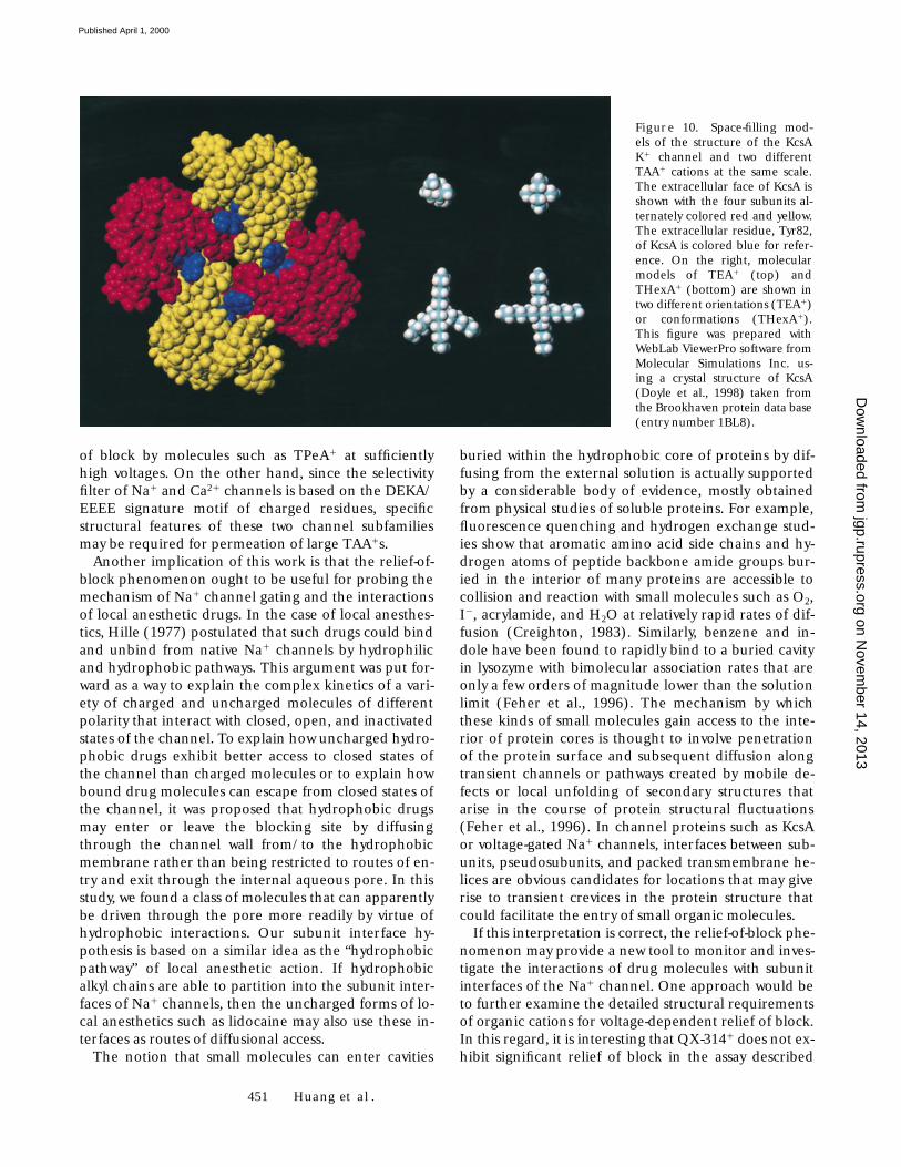
One mechanism that may account for the observedpreference for large TAA1 molecules can be consid-ered in reference to the molecular graphics illustrationof Fig. 10. For the sake of discussion, this figure showsspace-filling models of the KcsA K1 channel, TEA1,and THexA1 at the same scale. The image on the left isa model of the KcsA structure with the four subunits al-ternately colored red and yellow. For orientation,Tyr82, a residue that constitutes an external aromaticbinding site for TEA1 in related K1 channels (Hegin-botham and MacKinnon, 1992) is colored blue. Theview is looking down upon the external membrane sur-
face of the protein along the central pore axis. In thisorientation, the diameter of the whole KcsA tetramermeasures 54 Å. Three K1 ions and a water molecule inthe pore have been removed from the structure toshow the limiting diameter of the pore at the outer se-lectivity filter. The images on the right are molecularmodels of TEA1 at the top and THexA1 on the bottom.Each horizontal row shows two views (TEA1) or possi-ble conformations (THexA1) of the same molecule.The view on the left is meant to emphasize the tetrahe-dral orientation of the alkyl chains. The view on theright shows a particular orientation with each of the n-alkylchains located at a 908 planar rotation around the cen-tral nitrogen atom. In this fashion, four n-alkyl chainssurrounding the central tetrahedral nitrogen of a TAA1
molecule can be aligned with the four subunit inter-faces of a tetrameric (KcsA) or pseudotetrameric (Na1
channel) channel protein. We suggest that large TAA1
cations that originally enter such a channel through aninternal vestibule may diffuse across the narrow filterregion to the outside by forming an alignment and in-ter-digitation of all four n-alkyl chains between the fourprotein subunit interfaces. Since the subunit interfacesare primarily hydrophobic contact surfaces, longern-alkyl chains would have a greater propensity to parti-tion into this environment (i.e., a stronger hydropho-bic interaction energy) than shorter n-alkyl chains.Thus, this mechanism would naturally explain whylarge symmetrical TAA1 molecules are more readilysubject to relief-of-block under inducement of highvoltage, whereas short TAA1s are seemingly imperviousto this driving force. A similar subunit hypothesis waspreviously proposed in reference to interactions ofTAA1 cations with squid axon K1 channels (Frenchand Shoukimas, 1981) and monazomycin channels(Heyer et al., 1976).
Although the illustration of Fig. 10 is based on thecrystal structure of a K1 channel protein, the pore do-mains of voltage-gated Na1 and Ca21 channels arelikely to have a similar structure since they belong to asuperfamily of channel proteins consisting of evolu-tionarily related sequences (Jan and Jan, 1990; Pongs etal., 1988). In particular, intersubunit interfaces of K1
channels would correspond to pseudosubunit inter-faces of Na1 and Ca21 channels since these latter pro-teins are actually composed of a single a subunit thatcontains four internally homologous domains, I, II, III,and IV. In any case, the molecular cutoff diameter ofthe selectivity filter of native Na1 channels (3 3 5 Å) isonly slightly larger than that of K1 channels (3 Å). Fig.10 is thus a reasonable representation of the physicallimitations that must underlie the improbability of elec-trodiffusion of large TAA1s through the Na1 channelpore. One implication of this subunit interface hypoth-esis is that K1 and Ca21 channels may also exhibit relief
on Novem
ber 14, 2013jgp.rupress.org
Dow
nloaded from
Published April 1, 2000
451 Huang et al.
of block by molecules such as TPeA1 at sufficientlyhigh voltages. On the other hand, since the selectivityfilter of Na1 and Ca21 channels is based on the DEKA/EEEE signature motif of charged residues, specificstructural features of these two channel subfamiliesmay be required for permeation of large TAA1s.
Another implication of this work is that the relief-of-block phenomenon ought to be useful for probing themechanism of Na1 channel gating and the interactionsof local anesthetic drugs. In the case of local anesthes-tics, Hille (1977) postulated that such drugs could bindand unbind from native Na1 channels by hydrophilicand hydrophobic pathways. This argument was put for-ward as a way to explain the complex kinetics of a vari-ety of charged and uncharged molecules of differentpolarity that interact with closed, open, and inactivatedstates of the channel. To explain how uncharged hydro-phobic drugs exhibit better access to closed states ofthe channel than charged molecules or to explain howbound drug molecules can escape from closed states ofthe channel, it was proposed that hydrophobic drugsmay enter or leave the blocking site by diffusingthrough the channel wall from/to the hydrophobicmembrane rather than being restricted to routes of en-try and exit through the internal aqueous pore. In thisstudy, we found a class of molecules that can apparentlybe driven through the pore more readily by virtue ofhydrophobic interactions. Our subunit interface hy-pothesis is based on a similar idea as the “hydrophobicpathway” of local anesthetic action. If hydrophobicalkyl chains are able to partition into the subunit inter-faces of Na1 channels, then the uncharged forms of lo-cal anesthetics such as lidocaine may also use these in-terfaces as routes of diffusional access.
The notion that small molecules can enter cavities
buried within the hydrophobic core of proteins by dif-fusing from the external solution is actually supportedby a considerable body of evidence, mostly obtainedfrom physical studies of soluble proteins. For example,fluorescence quenching and hydrogen exchange stud-ies show that aromatic amino acid side chains and hy-drogen atoms of peptide backbone amide groups bur-ied in the interior of many proteins are accessible tocollision and reaction with small molecules such as O2,I2, acrylamide, and H2O at relatively rapid rates of dif-fusion (Creighton, 1983). Similarly, benzene and in-dole have been found to rapidly bind to a buried cavityin lysozyme with bimolecular association rates that areonly a few orders of magnitude lower than the solutionlimit (Feher et al., 1996). The mechanism by whichthese kinds of small molecules gain access to the inte-rior of protein cores is thought to involve penetrationof the protein surface and subsequent diffusion alongtransient channels or pathways created by mobile de-fects or local unfolding of secondary structures thatarise in the course of protein structural fluctuations(Feher et al., 1996). In channel proteins such as KcsAor voltage-gated Na1 channels, interfaces between sub-units, pseudosubunits, and packed transmembrane he-lices are obvious candidates for locations that may giverise to transient crevices in the protein structure thatcould facilitate the entry of small organic molecules.
If this interpretation is correct, the relief-of-block phe-nomenon may provide a new tool to monitor and inves-tigate the interactions of drug molecules with subunitinterfaces of the Na1 channel. One approach would beto further examine the detailed structural requirementsof organic cations for voltage-dependent relief of block.In this regard, it is interesting that QX-3141 does not ex-hibit significant relief of block in the assay described
Figure 10. Space-filling mod-els of the structure of the KcsAK1 channel and two differentTAA1 cations at the same scale.The extracellular face of KcsA isshown with the four subunits al-ternately colored red and yellow.The extracellular residue, Tyr82,of KcsA is colored blue for refer-ence. On the right, molecularmodels of TEA1 (top) andTHexA1 (bottom) are shown intwo different orientations (TEA1)or conformations (THexA1).This figure was prepared withWebLab ViewerPro software fromMolecular Simulations Inc. us-ing a crystal structure of KcsA(Doyle et al., 1998) taken fromthe Brookhaven protein data base(entry number 1BL8).
on Novem
ber 14, 2013jgp.rupress.org
Dow
nloaded from
Published April 1, 2000
452 Voltage-dependent Relief of Na1 Channel Block by Large Organic Cations
here (Fig. 5, B and C). This molecule has been previ-ously observed to block the cardiac isoform of Na1
channels from the outside of cells more readily thanbrain or muscle isoforms of Na1 channels (Qu et al.,1995; Sunami et al., 1997). Since this latter effect hasbeen proposed to occur by diffusion of QX-3141
through the external Na1 channel pore to an internalsite (Qu et al., 1995; Sunami et al., 1997), it would be in-teresting to test whether voltage-driven relief of TAA1
or QX3141 block also varies among heart, brain, andmuscle isoforms of Na1 channels. The fact that signifi-cant voltage-driven relief of block by intracellular QX-314 is not observed for the DEAA mutant suggests thatelectrodiffusion of this molecule directly through theaqueous pore is a very unfavorable process. Other ac-cess routes, such as hydrophobic pseudosubunit inter-faces, may have to be considered to explain such phe-nomena. Finally, since the DEAA mutant can be used tomonitor the mobility of large TAA1 molecules throughentire permeation pathway of the Na1 channel, it wouldbe interesting to examine the differential accessibility ofclosed, open, and inactivated gating states of the chan-nel to this process. Such studies may be helpful in refin-ing our knowledge of the detailed conformationalchanges of the pore that occur in normal gating.
This research was supported by a grant from the National Insti-tute of General Medical Sciences of the National Institutes ofHealth (GM-51172 to E. Moczydlowski).
Submitted: 14 December 1999Revised: 17 February 2000Accepted: 22 February 2000
R E F E R E N C E S
Armstrong, C.M. 1992. Voltage-dependent ion channels and theirgating. Physiol. Rev. 72:S5–S13.
Armstrong, C.M., and L. Binstock. 1965. Anomalous rectification inthe squid giant axon injected with tetraethylammonium chlo-ride. J. Gen. Physiol. 48:859.
Armstrong, C.M., and W.F. Gilly. 1992. Access resistance and spaceclamp problems associated with whole-cell patch clamping. Meth-ods Enzymol. 207:100–122.
Blaustein, R.O., and A. Finkelstein. 1990a. Voltage-dependentblock of anthrax toxin channels in planar bilayer membranes bysymmetric tetraalkylammonium ions: effects on macroscopicconductance. J. Gen. Physiol. 96:905–919.
Blaustein, R.O., and A. Finkelstein. 1990b. Voltage-dependentblock of anthrax toxin channels in planar phospholipid bilayermembranes by symmetric tetraalkylammonium ions: single-chan-nel analysis. J. Gen. Physiol. 96:921–942.
Choi, K.L., C. Mossman, J. Aube, and G. Yellen. 1993. The internalquaternary ammonium receptor of Shaker potassium channels.Neuron. 10:533–541.
Courtney, K.R. 1975. Mechanism of frequency-dependent inhibi-tion of sodium currents in frog myelinated nerve by the lidocainederivative GEA 968. J. Pharmacol. Exp. Ther. 195:225–236.
Creighton, T.E. 1983. Proteins: Structures and Molecular Princi-ples. W.H. Freeman and Co., New York, NY. 515 pp.
Doyle, D.A., J.M. Cabral, R.A. Pfuetzner, A. Kuo, J.M. Gulbis, S.L.
Cohen, B.T. Chait, and R. MacKinnon. 1998. The structure of thepotassium channel: molecular basis of K1 conduction and selec-tivity. Science. 280:69–77.
Favre, I., E. Moczydlowski, and L. Schild. 1996. On the structuralbasis for ionic selectivity among Na1, K1, and Ca21 in the voltage-gated sodium channel. Biophys. J. 71:3110–3125.
Feher, V.A., E.P. Baldwin, and F.W. Dahlquist. 1996. Access ofligands to cavities within the core of a protein is rapid. Nat. Struct.Biol. 3:516–521.
French, R.J., and J.J. Shoukimas. 1981. Blockage of squid axon po-tassium conductance by internal tetra-N-alkylammonium ions ofvarious sizes. Biophys. J. 34:271–291.
French, R.J., and J.J. Shoukimas. 1985. An ion’s eye view of the po-tassium channel: the structure of the permeation pathway assensed by a variety of blocking ions. J. Gen. Physiol. 85:669–698.
French, R.J., and J.B. Wells. 1977. Sodium ions as blocking agentsand charge carriers in the potassium channel of the squid giantaxon. J. Gen. Physiol. 70:707–724.
French, R.J., G. Zamponi, and I.E. Sierralta. 1998. Molecular andkinetic determinants of local anesthetic action on sodium chan-nels. Toxicol. Lett. 100–101:247–254.
Gingrich, K.J., D. Beardsley, and D.T. Yue. 1993. Ultra-deep block-ade of Na1 channels by a quaternary ammonium ion: catalyis bya transition-intermediate state? J. Physiol. 471:319–341.
Heginbotham, L., Z. Lu, T. Abramson, and R. MacKinnon. 1994.Mutations in the K1 channel signature sequence. Biophys. J. 66:1061–1067.
Heginbotham, L., and R. MacKinnon. 1992. The aromatic bindingsite for tetraethylammonium ion on potassium channels. Neuron.8:483–491.
Heinemann, S.H., H. Terlau, W. Stühmer, K. Imoto, and S. Numa.1992. Calcium channel characteristics conferred on the sodiumchannel by single mutations. Nature. 356:441–443.
Heyer, E.J., R.U. Muller, and A. Finkelstein. 1976. Inactivation ofmonazomycin-induced voltage-dependent conductance in thinlipid membranes. I. Inactivation produced by long chain quater-nary ammonium ions. J. Gen. Physiol. 67:703–729.
Hille, B. 1967. The selective inhibition of delayed potassium cur-rents in nerve by tetraethylammonium ion. J. Gen. Physiol. 50:1287–1302.
Hille, B. 1971. The permeability of the sodium channel to organiccations in myelinated nerve. J. Gen. Physiol. 58:599–619.
Hille, B. 1972. The permeability of the sodium channel to metalcations in myelinated nerve. J. Gen. Physiol. 59:637–658.
Hille, B. 1977. Local anesthetics: hydrophilic and hydrophobicpathways for the drug-receptor reaction. J. Gen. Physiol. 69:497–515.
Hille, B. 1992. Ionic Channels of Excitable Membranes. 2nd ed.Sinauer Associates, Inc., Sunderland, MA. 607 pp.
Hondeghem, L.M., and B.G. Katzung. 1977. Time- and voltage-dependent interactions of antiarrhythmic drugs with cardiac so-dium channels. Biochim. Biophys. Acta. 472:373–398.
Jan, L.Y., and Y.N. Jan. 1990. A superfamily of ion channels. Nature.345:672.
Kavanaugh, M.P., M.D. Varnum, P.B. Osborne, M.J. Christie, A.E.Busch, J.P. Adelman, and R.A. North. 1991. Interaction betweentetraethylammonium and amino acid residues in the pore ofcloned voltage-dependent potassium channels. J. Biol. Chem. 266:7583–7587.
Lipkind, G.M., and H.A. Fozzard. 1994. A structural model of thetetrodotoxin and saxitoxin binding site of the Na1 channel. Bio-phys. J. 66:1–13.
MacKinnon, R. 1995. Pore loops: an emerging theme in ion chan-nel structure. Neuron 14:889–892.
MacKinnon, R., and G. Yellen. 1990. Mutations affecting TEA
on Novem
ber 14, 2013jgp.rupress.org
Dow
nloaded from
Published April 1, 2000
453 Huang et al.
blockade and ion permeation in voltage-activated K1 channels.Science. 250:276–279.
Marty, A., and E. Neher. 1995. Tight-seal whole-cell recording. InSingle-Channel Recording. B. Sakman and E. Neher, editors. Ple-num Publishing Corp., New York, NY. 31–52.
Miller, C. 1982. Bis-quaternary ammonium blockers as structuralprobes of the sarcoplasmic reticulum K1 channel. J. Gen. Physiol.79:869–891.
Moczydlowski, E. 1998. Chemical basis for alkali cation selectivity inpotassium channel proteins. Chem. Biol. (Lond.) 5:R291–R301.
Moczydlowski, E., A. Uehara, and S. Hall. 1986. Blocking pharma-cology of batrachotoxin-activated sodium channels. In Ion Chan-nel Reconstitution. C. Miller, editor. Plenum Publishing Corp.,New York, NY. 405–428.
O’Leary, M.E., and R. Horn. 1994. Internal block of human heartsodium channels by symmetrical tetra-alkylammoniums. J. Gen.Physiol. 104:507–522.
O’Leary, M.E., R.G. Kallen, and R. Horn. 1994. Evidence for a di-rect interaction between internal tetra-alkylammonium cationsand the inactivation gate of cardiac sodium channels. J. Gen.Physiol. 104:523–539.
Pongs, O., N. Kecskemethy, R. Müller, I. Krah-Jentgens, A. Bauman,H.H. Kiltz, I. Canal, S. Llamazares, and A. Ferrus. 1988. Shakerencodes a family of putative potassium channel proteins in thenervous system of Drosophila. EMBO (Eur. Mol. Biol. Organ.) J.7:1087–1096.
Qu, Y., J. Rogers, T. Tanada, T. Scheuer, and W.A. Catterall. 1995.Molecular determinants of drug access to the receptor site forantiarrhythmic drugs in the cardiac Na1 channel. Proc. Nat. Acad.Sci. USA. 92:11839–11843.
Sine, S.M., and J.H. Steinbach. 1984. Agonists block currentsthrough acetylcholine receptor channels. Biophys. J. 46:277–284.
Strichartz, G.R. 1973. The inhibition of sodium currents in myeli-nated nerve by quaternary derivatives of lidocaine. J. Gen. Physiol.62:37–57.
Sun, Y.-M., I. Favre, L. Schild, and E. Moczydlowski. 1997. On thestructural basis for size-selective permeation of organic cations
through the voltage-gated sodium channel. Effect of alanine mu-tations at the DEKA locus on selectivity, inhibition by Ca21, andH1, and molecular sieving. J. Gen. Physiol. 110:693–715.
Sunami, A., S.C. Dudley, Jr., and H.A. Fozzard. 1997. Sodium chan-nel selectivity filter regulates antiarrhythmic drug binding. Proc.Natl. Acad. Sci. USA. 94:14126–14131.
Tasaki, I., and S. Hagiwara. 1957. Demonstration of two stablestates in the squid giant axon under tetraethylammonium chlo-ride. J. Gen. Physiol. 40:859–885.
Terlau, H., S.H. Heinemann, W. Stühmer, M. Pusch, F. Conti, K.Imoto, and S. Numa. 1991. Mapping the site of block by tetrodo-toxin and saxitoxin of sodium channel II. FEBS Lett. 293:93–96.
Trimmer, J.S., S.S. Cooperman, S.A. Tomiko, J. Zhou, S.M. Crean,M.B. Boyle, R.G. Kallen, Z. Sheng, R.L. Barchi, F.J. Sigworth, etal. 1989. Primary structure and functional expression of a mam-malian skeletal muscle sodium channel. Neuron. 3:33–49.
Wang, G.K. 1988. Cocaine-induced closures of single batra-chotoxin-activated Na1 channels in planar lipid bilayers. J. Gen.Physiol. 92:747–765.
Wang, G.K., R. Simon, and S.-Y. Wang. 1991. Quaternary ammo-nium compounds as structural probes of single batrachotoxin-activated Na1 channels. J. Gen. Physiol. 98:1005–1024.
Williams, K. 1997. Interactions of polyamines with ion channels.Biochem. J. 325:289–297.
Woodhull, A.M. 1973. Ionic blockage of sodium channels in nerve.J. Gen. Physiol. 61:678–708.
Yamamoto, D., and J.Z. Yeh. 1984. Kinetics of 9-aminoacridineblock of single Na channels. J. Gen. Physiol. 84:361–377.
Yang, J., P.T. Ellinor, W.A. Sather, J.-F. Zhang, and R.W. Tsien. 1993.Molecular determinants of Ca21 selectivity and ion permeationin L-type Ca21 channels. Nature. 366:158–161.
Yellen, G., M.E. Jurman, T. Abramson, and R. MacKinnon. 1991.Mutations affecting internal TEA blockade identify the probablepore-forming region of a K1 channel. Science. 251:939–942.
Zamponi, G.W., D.D. Doyle, and R.J. French. 1993. Fast lidocaineblock of cardiac and skeletal muscle sodium channels: one sitewith two routes of access. Biophys. J. 65:80–90.
on Novem
ber 14, 2013jgp.rupress.org
Dow
nloaded from
Published April 1, 2000
Copyright © 2022 FDOKUMEN


![Tetra-kis(μ(2)-cyanido-κ(2)C:N)dicyanido-tetra-kis-[tris-(2-amino-eth-yl)amine-κ(3)N,N',N'',N''']tetra-copper(II)iron(II) bis[pentacyanidonitrosoferrate(II)] hexahydrate](https://static.fdokumen.com/doc/165x107/634199118718ae62200b4f38/tetra-kism2-cyanido-k2cndicyanido-tetra-kis-tris-2-amino-eth-ylamine-k3nnnntetra-copperiiironii.jpg)