Perimenopause as a Sensitive Period for Women's Health and ...
93
Perimenopause as a Sensitive Period for Women’s Health and Aging: A Review of the Chronic Disease Literature and Two Empirical Tests of Significance By April Michelle Falconi A dissertation submitted in partial satisfaction of the requirements for the degrees of Doctor of Philosophy in Health Services & Policy Analysis in the Graduate Division of the University of California, Berkeley Committee in charge: Professor Ralph Catalano, Chair Professor William Dow Professor Julianna Deardorff Spring 2015
-
Upload
khangminh22 -
Category
Documents
-
view
2 -
download
0
Transcript of Perimenopause as a Sensitive Period for Women's Health and ...
!
1!
Perimenopause as a Sensitive Period for Women’s Health and Aging: A Review of the Chronic Disease Literature and Two Empirical Tests of Significance
By
April Michelle Falconi
A dissertation submitted in partial satisfaction of the
requirements for the degrees of
Doctor of Philosophy
in
Health Services & Policy Analysis
in the
Graduate Division
of the
University of California, Berkeley
Committee in charge:
Professor Ralph Catalano, Chair
Professor William Dow Professor Julianna Deardorff
Spring 2015

!
!
2!

!
! 1!
Abstract
Perimenopause as a Sensitive Period for Women’s Health and Aging: A Review of the Chronic Disease Literature and Two Empirical Tests of Significance
by
April Michelle Falconi
Doctor of Philosophy in Health Services & Policy Analysis
University of California, Berkeley
Professor Ralph Catalano, Chair
The critical and sensitive periods model, a key component of the life course health and development (LCHD) framework, describes windows of growth, development, or change during which exposures can permanently affect the structure or function of the body and influence its trajectories for health. Research on critical and sensitive periods has traditionally focused on the impact of early life influences on later life health. This focus on infancy and youth is due to the rapid pace at which development and growth occurs, which can make individuals particularly susceptible to the influences of risk and protective factors.
Perimenopause—the transition period bridging women’s reproductive and post-reproductive years—is another window, however, in which rapid physiological changes occur, yet minimal research has investigated this time as a critical or sensitive period. The three papers included in this dissertation investigate the health of women at mid-life and explores how their experiences during the menopausal transition affect their chronic disease risk and longevity.
The first paper, a scoping review, compares the physiological similarities between perimenopause and puberty and their respective associations with some of the most prevalent chronic conditions in the United States. The second paper, a longitudinal cohort analysis of women undergoing the menopausal transition, explores whether perimenopause represents a sensitive window for stress responsivity. Using survey data from the Study of Women’s Health Across the Nation (SWAN), a national study of 3,300 women at midlife, the relation between psychological stress, perimenopause, and fibrinogen—a biomarker for systemic inflammation—is examined. The third paper, a time series analysis of population cohorts in France and England in Wales during the 19th Century, tests whether the age range at which perimenopause occurs at the population level is a sensitive window for longevity. The relation between cohort mortality at ages 45-49 and life expectancy at age 60, specifically, is examined using data from the Human Mortality Database.
The primary findings from these three papers are as follows: 1) Perimenopause shares many physiological similarities with puberty, a well-documented sensitive window for a number of health outcomes. The interaction of certain behaviors or exposures with the significant hormonal shifts during perimenopause, moreover, appears associated with risk for mood

!
! 2!
disorders, metabolic-related morbidities, autoimmune diseases, cardiovascular disease, cancer, musculoskeletal disorders, and premature mortality. 2) Women’s perceptions of stress appear to change during the course of the menopausal transition; however, such changes are not associated with adverse physiological effects, as measured through changes in fibrinogen. 3) Adverse environmental conditions during perimenopause appear significantly related to decreased longevity among women in France and England and Wales during the 19th Century.

!
! i!
Dedication
I dedicate this dissertation to my husband, Edward, for always supporting me in my get-rich-quick schemes.

!
! ii!
Acknowledgements
I would like to express my sincere gratitude for my dissertation committee, Ray Catalano, Will Dow, and Julianna Deardorff. Their support and thoughtful feedback on this dissertation was a
tremendous learning experience for me. I particularly appreciated Julie’s expertise as it related to Paper 1 and for introducing me to my co-author for that manuscript, Lindsay Hoyt. I am
especially grateful for Will’s guidance on Paper 2 as it related to using biomarkers, as well as his expertise on the econometric methods used in this paper. Lastly, I sincerely thank Ray for his
guidance and technical assistance with carrying out the time series analysis in Paper 3.
I wish to acknowledge Lindsay Hoyt for her work and expertise on the pubertal sections of Paper 1 in this dissertation. I learned a lot from her and appreciated and enjoyed working with her to
get the manuscript published.
I also would like to acknowledge Ellen Gold and Imke Janssen for their helpful feedback on Paper 2 of this dissertation, which helped improved the precision, accuracy, and clarity of the
paper.
!
! iii!
Introduction The number of individuals aged 65 years and older in the United States is projected to
more than double in the upcoming decades, rising from 40.2 million (13 percent of the total population) in 2010 to an estimated 88.5 million (21 percent of the total population) by 2050 (1). Women will comprise the majority of the aging population, as they have historically, and are projected to represent 55 percent of the age 65+ population in the year 2050. Among the oldest old—individuals age 85 years and older—women are expected to assume an even larger proportion (62 percent) of the population (2).
The growth of the aging population—particularly the aging women demographic—has important implications for the organization, financing, and delivery of health care (3). Older women have a higher prevalence of many chronic conditions compared with men (e.g., high blood pressure, osteoporosis, arthritis, depression, Alzheimer’s disease), and are more likely to have been diagnosed with multiple chronic conditions. Perhaps relatedly, older women are also more likely to experience functional limitations and utilize health services at a higher rate compared with older men (4, 5). As chronic conditions are associated with increased health care needs and higher medical costs (6), the health of aging women is of significant public health and economic significance.
Research shows that poor health is not, however, an inevitable consequence of aging (7). Although pathobiological changes that increase women’s risks for chronic diseases may occur during the menopausal transition (i.e., perimenopause) (8), this window may also represent a period of opportunity to reverse negative health trajectories or improve health (9). The aim of this dissertation, therefore, is to assess whether perimenopause acts as a point of inflection in healthy aging trajectories. The three papers included in this dissertation investigate the health of women at mid-life and how their experiences during the menopausal transition affect their chronic disease risk and longevity.
The Life Course Health Development (LCHD) framework offers a conceptual framework for addressing this research topic. Based on this framework, macropathways (i.e., an organism’s external environment) interact with the microcontext (i.e., an organism’s physiological systems) to produce health outcomes. The effect of the external environment, however, may be more powerful during different life phases and may affect physiological systems differently over time, such as during critical and sensitive periods (10).
The critical periods model is a key component of the LCHD framework and is defined by windows when exposures affect the structure or function of organs, tissues, or body systems that are not modified in any dramatic way by later experience (11). Sensitive periods are a related concept, but there is more scope to modify or reverse changes occurring outside the time window (11).
Much work on critical and sensitive periods has focused on the long reach of exposures and experiences in childhood on adult health, which appears logical given that rapid pace of developmental change that occurs during infancy and youth (12). Perimenopause is another period, however, in which dramatic physiological and somatic changes occur during a relatively brief window of time (13, 14). The concept of perimenopause as a sensitive window for late life health forms the basis of this dissertation and is evaluated in three separate, but related, papers.
Paper 1 entails a scoping review of the physiological similarities between perimenopause and puberty and their respective associations with some of the most prevalent chronic conditions in the U.S. Like perimenopause, puberty is characterized by significant neuroendocrine changes that occur over a relatively narrow span of time. After a comparison of the physiological changes

!
! iv!
between puberty and perimenopause, the associations between females’ experiences during these two transitional periods and chronic disease risk is reviewed. A summary of the relation between timing of puberty and perimenopause with chronic disease risk follows. A comparison of the similarities and differences between perimenopause and puberty and their respective associations with chronic diseases concludes the paper.
An empirical test of perimenopause as a sensitive window for stress responsivity is the subject of Paper 2. The relation between perimenopause and fibrinogen, a biomarker for systemic inflammation, is analyzed using five waves of data spanning 1996-2004 from the Study of Women’s Health Across the Nation (SWAN), a longitudinal, community-based study comprised of 3,300 women at midlife. Mixed regression models are first fit to determine whether psychological stress is heightened during perimenopause. The interaction between psychological stress and menopausal status and its association with fibrinogen are next tested for significance. Models with lagged stress and menopausal status variables are also analyzed to determine if stress showed any enduring associations with fibrinogen changes.
Paper 3 involves another empirical test of perimenopause as a sensitive period. Unlike Paper 2, in which the unit of analysis is the individual, the unit of analysis is at the aggregate level in Paper 3. The relation between cohort mortality at mid-life (i.e., ages 45-49) and life expectancy at age 60 is analyzed in France (1816-1919) and in England and Wales (1841-1919) using data drawn from the Human Mortality Database. The age range 45-49 is chosen to connote the occurrence of perimenopause, given research indicating the average age of perimenopause begins between 45 and 47 in western societies and lasts for approximately four years (15). Most literature indicates, moreover, that the age of menopause in westernized countries has not changed remarkably over time (16, 17).
Together, these three papers help inform population health research on aging women by providing insight on the causes of differences in women’s risks for chronic diseases and death. These papers show how the experiences and environmental conditions during the menopausal transition can influence differential aging patterns and contribute to disparities in aging women’s health.
As the average per capita health spending for older individuals (age 64 and over) is more than triple that of younger adults (ages 34-44) (18), even a slight shift in women’s health trajectories during perimenopause could have substantial consequences for health-related expenses (8). Knowledge about the health of these “near-elderly” women, therefore, could facilitate efforts to predict and plan for future health service utilization and health care spending, as well as offer a critical window for interventions aimed at reducing aging women’s disease burden (8).

!
! v!
Works Cited 1. Vincent G, Velkoff V. The older population in the United States: 2010 to 2050. United States Census Bureau, 2010. 2. Ortman J, Velkoff V. An aging nation: The older population in the United States. United States Census Bureau, 2014. 3. Lee R, Mason A. Some macroeconomic aspects of global population aging. Demography. 2010;47 Suppl:S151-S72. 4. Robinson K. Trends in health status and health care use among older women. National Center for Health Statistics, 2007. 5. Bertakis K, Azari R, Helms L, Callahan E, Robbins J. Gender differences in the utilization of health care services. Journal of Family Practice. 2000;49(2):147-52. 6. Bodenheimer T, Fernandez A. High and risking health care costs. Part 4: Can costs be controlled while preserving quality? Annals of Internal Medicine. 005;143:26-31. 7. Healthy aging: Helping people live long and productive lives and enjoy a good quality of life. Centers for Disease Control and Prevention, 2011. 8. Appt S, Ethun K. Reproductive aging and risk for chronic disease: Insights from studies of nonhuman primates. Maturitas. 2010;67:7-14. 9. Workshop Summary. Network on Reversibility: Mid-Life Reversibility of Early Established Biobehavioral Risk Factors; 2013 February 26-27, 2013; Bethesda, Maryland: National Institute on Aging National Institutes of Health. 10. Halfon N, Hochstein M. Life course health development: An integrated framework for developing health, policy, and research. Milbank Quarterly. 2002;80(3):433-79. 11. Kuh D, Ben-Shlomo Y, Lynch J, Hallqvist J, Power C. Life course epidemiology. Journal of Epidemiology and Community Health. 2003;57:778-83. 12. Aging NIo, editor Meeting summary. Network on reversibility: Mid-life reversibility of early established biobehavioral risk factors; February 26-27, 2013; Bethesda, MD. 13. Soules MR, Sherman S, Parrott E, Rebar R, Santoro N, Utian W, et al. Executive Summary: Stages of the Reproductive Aging Workshop. Climacteric. 2001;97:267-72. 14. Burger H, Hale G, Robertson D, Dennerstein L. A review of hormonal chnages during the menopausal transition: Focus on findings from the Melbourne Women's Midlife Health Project. Human Reproduction Update. 2007;13(6):559-65. 15. McKinlay SM. The Normal Menopause Transition: An Overview. Maturitas. 1996;23(2):137-45. 16. Boldsen J, Jeune B. Distribution of age at menopause in two Danish samples. Human Biology. 1990;62(2):291-300. 17. Sievert L. Menopause as a measure of population health: An overview. American Journal of Human Biology. 2001;13(4):429-33. 18. Reinhardt U. Does the aging of the populatin really drive the demand for health care. Health Affairs. 2003;22(6):27-39.
!
! 1!
Paper 1: Windows in Women’s Health: Reproductive Transitions as Sensitive Periods for Chronic Diseases ABSTRACT A central component of the life course health development framework is the concept of critical and sensitive periods, which are said to occur amidst times of rapid changes and growth. Many studies on critical and sensitive periods focus, consequently, on the long reach of exposures that occur during infancy and youth. This scoping review examines whether existing literature would suggest that perimenopause represents another sensitive period for women’s health. Synthesizing research on the transitions into and out of reproductive capability in women’s lives, this paper compares the hormonal shifts in perimenopause with those in puberty and maps their respective associations with some of the leading causes of morbidity and mortality in the United States. Review of the literatures on puberty and perimenopause reveal many similarities—but also some inconsistencies—between the endocrine changes that occur during these windows and the onset and progression of disease. These findings suggest that puberty and perimenopause are not simply markers of the same underlying process, and more importantly, that perimenopause appears to represent an additional window of sensitivity for numerous health outcomes. Evidence suggests that sensitive periods extend beyond the early years in women’s lives, and that examination of exposures and health behaviors during perimenopause may offer insight into women’s disease risks and help explain differences in women’s health trajectories as they age. Keywords: Puberty; Perimenopause; HPG axis; HPA axis; Chronic disease; Women’s health; Life course health
!
! 2!
INTRODUCTION The health and functionality of aging individuals varies widely, with some individuals
who are able to maintain high levels of cognitive, physical, and social functionality into old age, while others experience difficulties and varying degrees of dependencies that compromise their quality of life (1, 2). Although research on risk and protective factors related to healthy aging abounds, the accumulation of general system damage and resulting aging phenotype does not follow the same path under different circumstances with human populations (3). The determinants of biological aging and frailty appear to emerge irregularly across the life course (4) and are sensitive to the environments and social circumstances in which they are embedded (3).
The life course health development (LCHD) framework explains how health develops over an individual’s lifetime, influenced by different environmental, physiological, behavioral, and social contexts (5). These factors interact continuously to impact individuals’ health risks and long-term health trajectories (5, 6). Central to this framework is the idea that health and health trajectories may be altered more readily during critical periods, in which exposures exert more profound or enduring effects on the structure or function of organs, tissues, and body systems and cannot be modified in any significant way by later experience (4). A related concept—sensitive periods—is also characterized by windows of rapid change. There is greater ability to modify or reverse changes outside of sensitive periods, however, relative to critical periods (4). During sensitive periods, exposures have a stronger effect on disease risk and health outcomes than they would have at other times (5, 7).
Life course epidemiology research and studies using the critical periods model traditionally have focused on the long term effects of child and adolescent risk factors on later life health (8, 9). This body of research has provided invaluable knowledge of health processes and an understanding of variation in the development of health trajectories (10, 11). The critical periods model has been criticized, however, for appearing overly deterministic (6) because it suggests that an individual’s health is largely determined early in life. Evidence suggests, moreover, that environmental conditions may continue to affect health and aging trajectories well into adulthood. Molecular mechanisms of gene expression (i.e., epigenetics), for example, can remain dynamic through adult life (9). Based on the LCHD framework I propose that the menopausal transition (i.e., perimenopause) represents a sensitive period when threats or stimuli more profoundly affects their health relative to other stages of the life course.
Perimenopause bears many physiological and psychosocial similarities with puberty, another window in women’s lives in which much literature has shown enhanced sensitivity to their environments (12-14). Just as adolescents experience significant hormonal fluctuations as they shift from a pre-reproductive to reproductive state, perimenopausal women also experience dramatic hormonal variability as they transition from a reproductive to post-reproductive state (15). These changes occur against a backdrop of concurrent psychosocial changes in women’s roles and identities. Examination of risk and protective factors experienced during these transitional periods, therefore, may provide insight into the development and programming of health trajectories across women’s lives (16).
Key components of the critical/sensitive period model are timing and effect of exposure, such that an exposure at a specific time in the life course has profound or enduring effects that can affect physiological function and lead to disease (8, 17). In this paper, I review evidence linking both components (i.e., timing and effect of exposures) to the etiology of some of the most common chronic diseases in the U.S. After first providing a description of the major endocrine
!
! 3!
changes that occur during puberty and perimenopause, the importance of exposure during these windows to disease causation is reviewed. Literature included in this section suggests that with some conditions, increase in disease risk appear temporary. The hormonal changes occurring during perimenopause, for example, are thought to trigger the onset of mood disorders among vulnerable women, which often resolve once hormones stabilize in post-menopause (18). Literature on other diseases, however, suggests more permanent changes in disease risk. For example, the rate at which changes in hemostasis (i.e., blood coagulation) occur during perimenopause has lasting effects on women’s risk for developing heart disease (19).
Following the section on exposures and their associations with health outcomes is a review of the evidence linking the progression and timing of puberty and perimenopause with chronic diseases. The importance of timing is illustrated by studies showing how the timing (i.e., early vs. late) of puberty and perimenopause influences disease risk. Major life events, transitions, and turning points can have different social meanings and different health effects depending on their timing and the age of an individual at which an exposure or event occurs (17). This review describes how both early and late timing of puberty and perimenopause appear to differentially influence health outcomes. I conclude this paper with an assessment of the evidence for perimenopause as a potential sensitive window for later life health and discuss the implications of these findings.
Definitions of pubertal and menopausal transitions
As markers of the beginning and end of women’s reproductive life span, respectively, puberty and perimenopause are characterized by major physiological change and somatic restructuring (10). The onset of puberty is initiated in late childhood through endocrine changes that ultimately result in physical growth, sexual maturation, and reproductive capability. Comprised of two independent but overlapping stages, pubertal maturation in females begins around ages 6-9 with adrenarche, the appearance of adrenal androgen production, and is followed by gonadarche, when reactivation of the hypothalamic-pituitary-gonadal (HPG) axis occurs (20). (The initial activation of the HPG axis occurs during the fetal and neonatal period, but then is followed by a period of quiescence) (20). Menarche, the initiation of the menstrual cycle, does not occur until later in puberty, approximately at ages 12-13 in the U.S. (21).
Perimenopause is defined as the period immediately preceding menopause when endocrinological, biological, and clinical features of approaching menopause commence. This window also includes the first 12 months after the final menstrual period (menopause) has occurred (22, 23). As with puberty, perimenopause is comprised of two stages (i.e., early and late perimenopause) (24). During early perimenopause, women experience variability in menstrual cycle length, which increases as women progress through the menopausal transition. Amenorrhea (i.e., cessation of menses) of 60 days or longer occurs as women enter late perimenopause (25, 26). The shift from a reproductive state to non-reproductive state begins for most women in the U.S. during their mid-to late 40s and is comprised of two stages (i.e., early and late perimenopause) (24, 27). Women typically remain in the menopausal transition for approximately 4-5 years before reaching menopause (28, 29). ENDOCRINE SIMILARITIES IN PUBERTY AND PERIMENOPAUSE
The endocrine system modulates the rates of growth and timing of developmental transitions, in particular, puberty and perimenopause (18, 30). Through the production and
!
! 4!
regulation of hormones, the endocrine system also mediates the relationship between an individual and her environment so that the body can maintain allostasis (i.e., physiological stability) and avoid the effects of a deleterious environment (18). Shifts in endocrine action and regulation occurring during puberty and perimenopause are noteworthy because they are similar to each other but are unique in terms of the rest of the life course. Such disruptions in hormone patterns, via changes in the ratio of stimulus to endocrine response, temporal patterns of endocrine release, as well as the bioavailability of hormones occur (30) can be linked to increases in risk for pathogenesis. The following section provides an overview of the endocrine changes that females experience during the pubertal and perimenopausal transitions, followed by sections that link numerous health outcomes to these hormone shifts. Hormonal shifts in the hypothalamo-pituitary-gonadal (HPG) axis
The HPG axis controls reproductive maturation and regulates reproductive function through the production and secretion of various hormones, including gonadotropin-releasing hormone (GnRH), the gonadotropins—luteinizing hormone (LH) and follicle-stimulating hormone (FSH)—and sex steroids, estrogen and progesterone (30, 31). Characteristic of both puberty and perimenopause are the significant hormonal changes that occur in the HPG axis (see Table 1 for a summary). GnRH, which plays an essential role in reproduction and controls the synthesis and secretion of LH and FSH (32), is released in pulses during puberty. These pulses increase in amplitude and frequency (33, 34) to stimulate the secretion of LH and FSH into the bloodstream, which in turn stimulate oogenesis (i.e., the growth process through which an ovum or egg cell develops and matures) and sex steroid production (31). In contrast, the frequency of GnRH pulses is thought to slow during perimenopause (34, 35), although evidence suggests that the amplitude and secretion of GnRH may increase (31, 34).
Both pubertal girls and perimenopausal women experience episodic changes in LH and FSH, which together regulate ovarian follicle growth and ovulation (36, 37). These periods are characterized by dramatic increases in concentration of FSH, followed later by increases in LH (see Figure 1) (23, 30). One longitudinal study of women undergoing the menopausal transition, for example, reported a 535% increase in FSH levels during late perimenopause relative to premenopause (38).
Production of sex steroids—estradiol and progesterone—also changes significantly during the pubertal and menopausal transitions. Estradiol, the most abundant form of endogenous estrogen during women’s reproductive years, is responsible for reproductive and sexual function (39), and it increases erratically during puberty until reaching menarche. At menarche, estradiol levels stabilize and subsequently follow a monthly cyclical pattern with each menstrual cycle until the menopausal transition (40). During perimenopause, estradiol levels increase and fluctuate, although levels eventually decline (one estimate reported a decline of 70%, relative to pre-menopause) and stabilize in post-menopause (see Figure 2) (38, 40, 41). Progesterone, which modulates reproductive behavior through regulation of the menstrual cycle (32), is initiated with menarche (42) and decreases during perimenopause. As with estradiol, progesterone exhibits significant fluctuations during puberty and perimenopause (23, 28, 36).
Gonadal hormones, in sum, are a major physiological factor influencing the health of females. Social, cultural, and individual factors (e.g., nutrition, stress, physical activity) that affect women’s wellbeing can also influence the amount of ovarian activity over the life course, which can in turn influence women’s disease risk (30). Epidemiological studies, for example, consistently have linked amount of circulating ovarian steroid levels to cancer risk (30).

!
! 5!
Therefore, understanding how ecological and behavioral factors affect and interact with women’s hormone patterns during reproductive transitions is critical for mapping the pathways of women’s health.
Hormonal shifts in the hypothalamo-pituitary-adrenal (HPA) axis
The HPA axis is involved in the body’s response to stress. It elicits the physiological adjustments necessary for the maintenance and preservation of homeostasis (43, 44). Overall basal activity of HPA-axis increases during puberty, thereby releasing higher average levels of cortisol (one of the primary hormonal outputs of the HPA axis) throughout the day (45-51). Some research also suggests that females experience increased cortisol reactivity to stressful tasks during puberty, although these findings have come from studies with small sample sizes (<100) of pubertal females (52, 53). In addition to cortisol changes, concentrations of dehydroepiandrosterone (DHEA) and its sulfate (DHEA-S) increase during adrenarche, which may provide females increased protection from development of cardiovascular disease, cancer, autoimmune diseases, mood and memory disorders (30, 54). Relatively less is known about HPA-axis changes during perimenopause, although some research has found a significant (84%), albeit transient, increase in cortisol levels as women transition from early to late perimenopause (55, 56). This increase is comparable to levels found in women with psyschophysiological insomnia (56). Studies suggest that this increase is more likely a function of biological underpinnings rather than a response to social factors—either due to elevations in FSH (55, 56)—or fluctuations in estrogen (41, 57). Some longitudinal studies have found that DHEA also increases during perimenopause (58-60). The largest of these studies—a national, multiethnic, population-based survey of menopausal women (n=2,886)—found a small (4%), significant inflection in DHEA concentration during perimenopause (59). This 4% increase, although seemingly small in magnitude, is large enough to contribute to the conversion of sex steroids at the time of declining estradiol and increasing FSH production, thereby explaining differences among women in menopausal symptoms (59).
These changes in the HPA axis have important implications for the development and functioning of women’s tissues and organs (61). For example, activation of the HPA axis during the menopausal transition, as shown through increases in cortisol and DHEA, has been associated with increases in central adiposity, reduced bone density, and mood disorders (56, 58). Shifts in the response of the HPA axis during women’s life course, therefore, may reflect differences in their vulnerability to psychosomatic stressors and differential risks for various morbidities and mortality (30).
In summary, the initiation and culmination of women’s reproductive lives are associated with major endocrine changes distinct from other periods of the life course. Rapid hormonal changes occur in the HPG axis during puberty and perimenopause, some of which mirror each other, including erratic increases in GnRH, FSH, LH, and estradiol, and other changes that are the reverse of each other, such as shifts in progesterone. Although evidence on the precise changes occurring in the HPA axis appears less conclusive, research suggests that at minimum, activation of the HPA axis occurs during the pubertal and perimenopausal transitions.
The changes that occur in one endocrine system are not without effects on the other, and the interactions between the HPA and HPG axes are complex (62). The activity of one axis often modulates the activity of another axis and regulates the allocation of the body’s scare biological resources between competing demands (63). The adrenal axis regulates gonadal function, for example, through inhibitory effects of stress on reproductive behavior and the release of sex
!
! 6!
steroids (64), but the relation is not unidirectional. The HPA axis is also subject to gonadal influences, as evidenced by gender differences in basal cortisol levels, stress reactivity of the HPA system, and prevalence rates of stress-related diseases (62, 65, 66). HPG axis changes, combined with HPA-axis modifications, may increase women’s vulnerability to various morbidities during puberty and perimenopause (14, 67). Through analyzing the shared biological processes and associated health outcomes related to both puberty and perimenopause, this paper provides insight on the associations between the pubertal and perimenopausal experience, as well as the timing of these two transitions, with chronic disease. METHODS
To examine the extent, range, and nature of these literatures, a comprehensive scoping review was conducted to summarize the breadth of evidence on chronic diseases and conditions related to puberty and perimenopause. Following methodological guidelines for this type of literature review (68-70), the research focus was defined as links between reproductive transitions and health outcomes. All literature was included that presented theoretical or empirical approaches to this broad area and that directly pertained to a health condition and puberty or menopause or whose study participants were pubertal or menopausal females. The focus was narrowed to include evidence from industrialized countries only and selected papers published between 1990 and 2014. Exceptions included significant theoretical works published earlier or older papers related to particular diseases when more recent papers could not be identified.
All studies and reviews from peer-reviewed journals and books were identified using electronic databases (Pub Med, Google Scholar, Psych Info), key health journals relevant in the areas of puberty, menopause, and women’s health, and topic-related expert networks and websites (e.g., Global Library of Women’s Medicine, NIH Office of Research on Women’s Health.) Reference lists of relevant articles were also reviewed to identify additional studies our search strategy may have missed.
Keywords for the literature search were selected from two broad areas: female reproductive transition (e.g., puberty, menarche, pubertal timing; perimenopause, menopause, menopause timing) and health outcome (e.g., cancer, cardiovascular disease, life expectancy, mortality, diabetes, mental health, depression, anxiety, obesity, insulin resistance, autoimmune disease). Search terms for specific types of cancer (e.g., breast, endometrial, ovarian), and specific types of autoimmune diseases (e.g., lupus, rheumatoid arthritis) were also used. Data from the literature review were charted to identify specific disease outcomes relevant to both reproductive transitions. For instances when evidence for a health outcome was found for one transition, but not the other, another round of selection and review was conducted.
Five major health outcomes were identified: mental health, cardiometabolic health, autoimmune conditions, cancer, and mortality. Over 5,000 articles were assessed in the initial screening process, although only 300 representative studies, reviews, and chapters were ultimately included in the final stage of review and systematized using a bibliographic-managing software (EndNote®). These studies were charted according to key issues and themes, and discrepancies between literatures from the two reproductive periods were mapped. Finally, the reviewed literature was systematically reported, with results structured thematically along each dimension of health.

!
! 7!
RESULTS Associations of Puberty and Perimenopause with Health Outcomes
Periods of hormonal fluctuations or instability have been linked with increased vulnerability to the development or exacerbation of various diseases (71, 72). The biological susceptibility hypothesis suggests that disturbances of the neuroendocrine rhythmicity, such as during puberty and perimenopause, may cause females to become particularly sensitive to psychosocial, environmental, and physiological factors (73, 74). In support of this logic, research shows the following health conditions have well-documented associations with puberty and perimenopause. Mental health. During puberty many psychological disorders emerge among girls, including anxiety and depression (75, 76). Puberty has been identified as an important window for the establishment of a gender gap in depression (i.e., women are 1.5 to 3 times as likely to experience clinical depression as men (72, 77)) and this gap persists throughout the reproductive years (78, 79). Findings are not consistent, however, on whether or when this gap dissipates with age. Some studies suggest that the female preponderance in depression prevalence decreases once women reach the age of menopause (79), while other research finds no evidence of a convergence in the gender gap in depression at midlife (80). Inconsistencies also lie in whether women experience a temporary increase in depression while they undergo the menopausal transition. Some studies link an increased risk of depression with the initiation of perimenopause, even after adjusting for variables such as history of depression, premenstrual syndrome, hot flashes, numerous health behaviors, BMI, age, race, and employment status (72, 81-83). Other studies have found, however, no association between menopausal status and psychological symptoms (84) or that only women who have a history of depression are more likely to experience high depressive symptoms levels during perimenopause (but no increased risk of depression among perimenopausal women in general) (85). Most studies that find no association between menopausal status and depression are cross-sectional, whereas longitudinal studies have more consistently found that the odds of high depression are greater during perimenopause relative to premenopause (86). Pubertal and perimenopausal females also appear more susceptible to experiencing high levels of anxiety (87, 88). The interaction of hormones with psyschosocial factors related to gender differences appears associated with increases in anxiety symptoms (88, 89).
Research that ties a higher prevalence of depression, anxiety, and other mood disorders with puberty and perimenopause suggests that increases and fluctuations in sex hormones occurring in these windows may at least partially explain for the gender gap in mood disorders (40, 72, 81-83, 90-92). Gonadal steroids have widespread influences on the brain (93), and estrogen has been shown to influence the regulation of mood, behavior, and cognition (77). Both estrogen and progesterone also have been shown to affect neuronal plasticity (93) and regions of the brain that are involved in the modulation of mood and behavior, including the prefrontal cortex, hippocampus, thalamus, and brain stem (48). More recent biological research has focused, however, not on the direct effects of ovarian hormones on moods but on the moderating effects of hormones in response to stress. Rapid changes in ovarian hormone levels can trigger a dysregulation of the stress response during puberty and perimenopause, which can make some women more vulnerable to mood disorders and psychopathologies when confronted with stress (94, 95).
!
! 8!
Several non-biological factors may also contribute to gender-linked differences in mood disorders, including changes in social roles, perception of health and body image, and vasomotor symptoms (among perimenopausal women) (79, 85, 89). Major life events may also trigger the onset of depressive episodes among at-risk females, who may be genetically predisposed or who possess highly reactive, anxious dispositions (79, 85). Metabolic diseases and cardiovascular health. Hormone shifts, combined with changes in body composition and health behaviors, place pubertal and perimenopausal females at increased risk for unhealthy weight gain, cardiovascular disease, and other metabolic-related morbidities. During puberty, increases in estradiol and progesterone affect metabolism and promote fat deposition, which, in combination with the formation of dietary and physical activity patterns, implicate this window as a sensitive period for the development of excess weight gain and obesity (96-99). Studies similarly find increased risk for unhealthy weight gain and excessive adiposity during perimenopause (100, 101). One small study found that, independent of aging, intra-abdominal adiposity increased up to 22 percent during the menopausal transition, coinciding with a decrease in lean mass (101). No significant changes occurred with respect to trunk or subcutaneous abdominal fat. Independent, but related, changes in insulin sensitivity also occur during puberty and perimenopause, placing women at increased risk for insulin resistance (102-107). Although modest changes in insulin resistance occur in early puberty—caused by changes in growth and body fat distribution—continued increases in insulin resistance later in puberty can lead to unhealthy weight gain and increased risk for type-2 diabetes (108). Changes in insulin resistance are initiated during early perimenopause as well, attributed to shifts in hormonal patterns, such as decreases in FSH and estradiol levels (109). Metabolic shifts in body fat distribution also occur during perimenopause that are associated with slight decreases in insulin sensitivity and significant increases (23-34%) in insulin resistance. These shifts in adiposity distribution continue, however, into post-menopause and place perimenopausal and post-menopausal women alike at increased risk for cardiovascular disease (102, 110, 111).
Other evidence suggests that women’s increased vulnerability to developing cardiovascular disease during the menopausal transition may be due to changes in hemostasis (i.e. process of blood coagulation), shifts in lipid characteristics, and the appearance of vasomotor menopausal symptoms. Hormone changes during perimenopause tend to be procoagulant, which are thought to increase risk for venous thrombosis and coronary heart disease (19, 112). Data show that late perimenopausal women, for example, have increased levels of hemostatic factors, including, Factor VII, fibrinogen, and tPA-ag, relative to pre-menopausal women (112). Although elevated levels of tPA-ag and fibrinogen are also found among post-menopausal women relative to pre-menopausal women, levels are not necessarily as high as they are during perimenopause (112).
Several small studies also suggest that lipid profiles change in relation to menopausal status (113), and that the rate of change appears most rapid during perimenopause. One study has shown, for example, that the thickness and diameter of the carotid artery significantly changes during perimenopause. Intima-media thickness increases 0.017 mm/year compared with pre-menopause (0.007 mm/year and adventitial diameter increases 0.024 mm/year relative to pre-menopause (-0.032mm/year) (114) (see Figure 3). Another study has shown that apolipoprotein (Apo) B, low-density lipoprotein (LDL) cholesterol (i.e., “bad” cholesterol) and total cholesterol rise during late perimenopause to increase women’s risk of developing cardiovascular disease as

!
! 9!
well (see Figure 4) (115). Although lipid markers between late perimenopausal and post-menopausal women often do not differ significantly, it has been hypothesized that the rise in coronary heart disease incidence among post-menopausal women is due to, at least in part, the earlier changes in lipids that occur with the menopausal transition (115). Additionally, other studies have found associations between vasomotor menopausal symptoms and risk of future coronary heart disease (116, 117).
The rate at which hormones decline during perimenopause profoundly affects women’s cardiovascular outcomes (i.e., higher rates of change are positively correlated with risk factors for coronary heart disease) (118). Additional shifts in adiposity, insulin sensitivity, blood lipids, and hemostatic markers, combined with age-related changes in risk factors for cardiovascular disease, such as decreased exercise and increased weight gain (118), implicate perimenopause as a sensitive window for the development of cardiovascular disease. Psychosocial factors operating across the life course, such as socioeconomic position and social support, may also interact with hormonal changes to influence cardiovascular disease risk (105). Although the precise biological mechanisms—and the degree of their influence—have yet to be specified, puberty and perimenopause are believed to perturb the ‘normal’ trajectories of cardio-metabolic markers (113). Musculoskeletal disorders. Musculoskeletal disorders are one of the most significant causes of morbidity among women in western populations, and musculoskeletal health is significantly affected by changes occurring during puberty and perimenopause (119). Hormone changes that initiate puberty also control the skeletal growth spurt (120). Rising estradiol concentrations in females drives bone mineral accrual, which is thought to prepare the skeleton for pregnancy and lactation (119). Because individuals gain up to 30-40% of their total bone mass during puberty, this window is a sensitive period for bone development (119). Girls with low calcium intake or inadequate nutrition during puberty can compromise their long term skeletal health, leading to an increased risk for bone fractures and development of osteoporosis (120, 121). As with puberty, perimenopausal women also experience significant musculoskeletal changes, although hormone shifts drive bone mineral density loss during this window rather than growth. During women’s reproductive life spans, frequent bone turnover occurs to ensure that microfractures are repaired and so that bone can remodel in response to changes in load (120). In perimenopause, bone turnover slows and bone loss of up to 1-2% per year occurs due to increased bone resorption (120, 122). Bone resorption has been linked with increases in FSH that occurs during perimenopause and is widely regarded as one of the key contributing elements to subsequent risk of osteoporosis (122). Women who are genetically predisposed to low bone mass or who have sedentary lifestyles, therefore, may exacerbate their risk for bone fracture during this window (120).
Some studies show that declining levels of estradiol, which decreases bone mineral density, is the biological mechanism behind women’s increased risk for osteoporosis during perimenopause (123). Other studies suggests that higher FSH concentrations place females at greater risk for osteoporosis (122), given research indicating that spine and hip bone mineral density is associated with FSH changes—not estradiol changes (124). Although the exact biological mechanisms underlying perimenopausal women’s bone changes remains unclear, both perimenopausal and pubertal females’ susceptibility to influences on musculoskeletal health appears heightened due to hormonal changes.

!
! 10!
Autoimmune conditions. Autoimmune conditions are significantly more prevalent among females relative to males, and reproductive status appears associated with alterations in risk, onset, and progression of numerous autoimmune diseases, including systemic lupus erythematosus (SLE), rheumatoid arthritis, type-1 diabetes, multiple sclerosis, and autoimmune thyroid conditions (71, 125, 126). Explanations for females’ greater risk during these windows are tied to HPG-axis modulation of the immune system—in particular, the influence of GnRH, estradiol, and progesterone (71, 127, 128). Changes in the sex hormone milieu during puberty and perimenopause are thought to mediate changes in antibody production (e.g., immunoglobulin levels, cytokine production, antiapoptotic factors), thereby affecting the pathogenesis of autoimmune disorders (129, 130). Disease is often most severe when estrogen and progesterone levels are at their lowest. Incidence of rheumatoid arthritis and autoimmune thyroid disease peaks, therefore, around menopause (71).
Other autoimmune diseases, however, show the most significant increases in disease incidence with the onset of puberty, such as SLE, which decreases in incidence after menopause (130). Most studies on the associations between reproductive transitions with autoimmune diseases, however, assess how symptoms and disease onset differ between pre- versus post-pubertal or pre- versus post-menopausal women. Minimal work has investigated how hormone changes during these transitions influence disease onset or severity of symptoms (131, 132), and is a subject area in need of further research. Of note, genes and environmental factors also contribute to susceptibility of autoimmune diseases, as does both excess or inadequate stress hormone response (i.e., excess is associated with increased susceptibility to infection, and inadequate stress hormone response is associated with inflammatory and allergic autoimmune diseases) (133). Such factors may interact with hormone changes during puberty and perimenopause to affect autoimmune disease onset and symptomatology.
Cancer. Most research on female susceptibility to cancer is associated with the timing of menarche and menopause. A small number of studies have found, however, that the pubertal and menopausal transitions in and of themselves are associated with changes in cancer risk. For example, pubertal girls exposed to carcinogens may increase their risk for cancer as a result of increased susceptibility of the mammary epithelial cells to insults and mutations (66, 67). Some research conducted in the 1980s also has suggested that breast cancer risk is higher during perimenopause in response to elevated estrogen, although no recent research corroborates these findings (134-137). The timing of initiation of hormone replacement therapy, however, may influence cancer risk. Results from a prospective cohort study of approximately 100,000 French women indicate that estrogen-progestagen menopausal hormone therapy (EP-MHT) initiated close to menopause (rather than later) among some women (i.e., women with relatively short durations of EP-MHT) was associated with an increase in breast cancer risk (138). Timing of Puberty and Perimenopause
Although changes in the endocrine system during the pubertal and menopausal transitions are linked with many chronic conditions, much of the research relating puberty and perimenopause with health outcomes focuses on the relative timing of these transitions rather than their experience. Time is a fundamental concept in the sensitive periods model, and the effect of events or exposures on health outcomes may be dependent on their duration or timing (7).

!
! 11!
Pubertal timing is influenced by a variety of factors, many of which are interrelated, including: genetic factors (e.g., genetic polymorphisms, race/ethnicity), intrauterine conditions, postnatal influences, psychosocial and physical stress, body weight, and environmental factors (e.g., exposure to endocrine-disrupting chemicals (EDCs)) (139, 140). One study has found that up to half of the variance in timing of menarche is genetically determined (141), and several other studies have found that, especially in the United States, black and Mexican American girls had earlier ages of menarche relative to non-Hispanic white girls (142, 143). This association appears largely driven by indicators of socioeconomic status and childhood overweight/obesity (144).
Corroborating these findings is research suggesting that nutrition—of both a girl and her mother—influences age of menarche. Poor maternal nutrition that results in low birth weight has been found associated with an earlier age of menarche in their daughters (139). An association was also found between mothers with high pre-pregnancy BMI and excessive gestational weight gain with their daughters’ earlier ages at menarche (145). Some evidence suggests that postnatal obesity is associated with an earlier age of menarche (107) as well, while postnatal nutritional deprivation—particularly close to sexual maturity—has been found associated with a later age of menarche (140). Other stressors, such as low socioeconomic status (SES) and early pubertal development, have also been linked to timing of menarche, although results are not consistent (146, 147). Discordant findings may be to due to varying measures of socioeconomic status and differences in association by race/ethnicity. An association between low family income and an earlier age of menarche has been found among black and Hispanic girls, for example, but not among white girls (144). Alternatively, mothers’ unmarried status has been found related to an earlier age of menarche among Hispanic and white girls, but not among black girls (144). Lastly, environmental factors, particularly EDCs, appear to affect timing of menarche, with evidence implicating exposure to chemicals found in pesticides with an earlier age of menarche (139, 140).
From an evolutionary-development perspective, menarcheal timing varies in response to girls’ environments (e.g., physical, emotional, psychosocial, etc) in order to maximize reproductive success. The psychosocial acceleration theory posits that high levels of psychosocial or physical stress leads to an earlier age of menarche in order to maximize her chances of producing offspring (148). The stress-suppression theory proposes, in contrast, that such adversity causes a delay in pubertal development until better times. Alternatively, a stress reactivity theory suggests that either highly protective or adverse childhood environments can trigger, or interact with, stress reactivity systems to affect maturation of the HPG axis, thereby delaying timing of menarche (148-150).
Unlike the life history theories posited to explain the timing of menarche, less work has been developed to explain variability in the timing of menopause (151, 152). Debate about the evolutionary underpinnings of menopause tends to revolve around whether it is an adaption (to provide care to offspring and maximize their health and future reproductive success); a tradeoff (that favors efficient early fertility because of the costs involved with prolonging fertility); or an artifact of increasing lifespans (153). Most evolutionary theory on menopausal timing relates to the ‘grandmother hypothesis,’ which suggests the duration of a woman’s postmenopausal survival affects the reproductive success of her children and the survival of her grandchildren.
Findings from epidemiological research indicate that timing of menopause is strongly influenced by intrinsic factors, such as the reproductive history of individuals (154). Studies have found that nulliparity or having fewer children is associated with an earlier age of menopause
!
! 12!
(155, 156). The timing of natural menopause also appears to vary by race/ethnicity (i.e. many studies report that African American and Latina women experience menopause earlier than non-Hispanic white and Asian women) (156).
Research shows menopausal timing may be influenced by lifestyle factors as well. Some studies have reported, for example, that an increased BMI and upper body fat distribution were associated with a later age of menopause, although other studies have found no association between BMI and age at natural menopause (156). More consistent findings have linked environmental influences with menopausal timing. Women who smoke stop menstruating 1-2 years earlier than comparable non-smokers, and an earlier age at menopause has been found among women exposed to certain endocrine disruptors as well (156).
As all adaptations have their benefits and costs, shifts in timing of menarche and menopause may help maximize reproductive success, but changes toward a relatively early or relatively late age of menarche or menopause are also associated with pernicious health effects.
Association of Pubertal and Perimenopausal Timing with Health Outcomes Early timing of reproductive events and adverse health outcomes Cardiovascular Disease. Early pubertal timing appears associated with risk of cardiovascular disease (CVD) and CVD precursors, such as hypertension (157, 158), despite research suggesting that estrogen has cardioprotective characteristics (105). In fact, a recent meta-analysis showed a 15% increase in cardiovascular disease (CVD) risk associated with earlier menarcheal timing (159).
Some researchers contend that early menarche is only a marker—not a predictor—of cardiovascular disease, and that elevated childhood BMI contributes to an earlier age of menarche, which subsequently leads to a higher BMI in adulthood and increased risk for cardiovascular disease (160). Although not much research provides evidence of a biological mechanism linking pubertal timing with cardiovascular disease risk (121, 160), at least one study suggests that early pubertal timing is associated with metabolic derangements that are independent of childhood BMI (161). Sex-specific hormones do not necessarily drive this increased risk for cardiovascular disease, however, as early pubertal onset has been associated with an increased risk for cardiovascular disease among both males and females (161).
Research on the association between timing of menopause and cardiovascular risk appears more conclusive, with studies generally finding that an increased risk (estimated at 25%) for CVD is associated with an early (i.e., before age 45) age at menopause (162, 163). A challenge in determining the relationship between timing of menopause and cardiovascular disease lies in differentiating the effect of menopausal status from age-related effects (105) and health-related behaviors (164). Although several studies have reported adverse relationships between menopause and lipid profiles, blood pressure, and weight gain, these factors also vary with age (105). Smoking is also a well-known risk factor for CVD and is associated with a decreased age of menopause. A meta-analysis of early menopause and risk factors for cardiovascular disease found, however, that the effects of smoking are unclear. Although the relationship between postmenopausal status and CVD disappears upon controlling for smoking, the effect of early menopause on CVD was also more pronounced after controlling for smoking (162).
Biological mechanisms proposed to explain the association between age at menopause and CVD include the role of estrogen in the maintenance of immune function (163) and a cardioprotective effect of estrogen (165, 166). Decreasing estrogen levels during the menopausal

!
! 13!
transition have been linked to adverse vascular changes. In particular, low-density lipoprotein cholesterol (LDL-C) levels increase, while high-density lipoprotein cholesterol (HDL-C) levels remain stable or slightly increase. These changes have consistently been found associated with increased risk of CVD (167). Autoimmune Diseases. Nearly all autoimmune diseases are more prevalent among women (e.g., systemic lupus erythematosus (SLE), rheumatoid arthritis, multiple sclerosis, celiac disease), and the severity of symptoms also appears to vary by gender (although severity does not clearly favor one gender over another; it varies by disease) (132). Hormonal or reproductive factors, therefore, have been posited as strong determinants of disease pathogenesis (168). The underlying basis for the sex bias in autoimmune diseases has yet to be determined (132). The incidence of autoimmune diseases appears to vary relative to onset of puberty (132), although study findings are not consistent. Some studies have found that an earlier age of menarche is inversely related to rheumatic arthritis, for example (169, 170), while other studies have found that an early timing of menarche is directly associated with rheumatic arthritis (171, 172), as well as with other autoimmune conditions, such as SLE (173), and multiple sclerosis (174).
More consistent observations have been observed between an earlier age at menopause (i.e., prior to age 45) and autoimmune diseases, including disorders involving the thyroid and adrenal glands, pernicious anemia, alopecia, Crohn’s disease, SLE, and rheumatoid arthritis (24, 125, 131, 169, 173). The physiological mechanisms explaining this relationship are likely complex, and several different factors have been proposed that relate timing of menopause and the incidence or progression of autoimmune diseases. One hypothesized explanation for this relationship is an “insufficient HPA axis,” which is thought to lead to the development of diseases (and rheumatoid arthritis, in particular) (169). Another hypothesized mechanism is that exposure to estrogen may protect against the onset of disease. The decline of estrogen during the menopausal transition, therefore, may increase women’s risk for autoimmune diseases (131, 173). Lastly, a number of immunological changes occur with the menopausal transition, such as an increased production of pro-inflammatory cytokines, decreased secretion of anti-inflammatory cytokines, and decreased lymphocyte levels. These changes, combined with changes in the endocrine system, may influence the onset and risk of autoimmune diseases associated with the menopausal transition (132).
Mortality. Epidemiologic research suggests that early menarcheal and menopausal timing is associated with increased mortality and shorter life expectancy. Studies in the United Kingdom, Norway, and United States find that all-cause mortality is reduced by 2.4 to 4.5% for each year delay in onset of menarche (157, 175). Similarly, prospective cohort studies in Norway and the Netherlands found that a one-year delay in age of menopause was associated with a 1.6 to 2% decreased risk of death (165, 175). Other research has found that women who experience a late menopause, in general, live two years longer than women with an early age of menopause (165). Research also indicates that women who experienced menopause prior to age 40 years were subject to 35-95% higher mortality rates compared with women who reported menopause occurring at age 50 years or older (176).
Many mechanisms have been proposed to explain the relationship between an earlier age at menarche and of an earlier age at menopause and mortality, including genetic factors, behavioral and environmental factors, and hormonal mechanisms (176, 177). Some evidence suggests that the higher mortality rate observed among women with an early menopause is
!
! 14!
mediated through higher comorbidities, and that an early age of menopause may be a marker for accelerated somatic aging (178). Discordant timing of reproductive events and adverse health outcomes Cancer. The relationships between timing of the pubertal and menopausal transitions and various forms of cancer do not parallel each other. Although studies show an association between early pubertal timing and cancer, cancer risk appears to increase with a later age at menopause. Numerous studies have shown that earlier pubertal timing is associated with breast cancer, endometrial cancer, and ovarian cancer (126, 179-184). In contrast, a large body of literature links a later age of menopause with breast and endometrial cancer (165, 183, 185). A later age of menopause has also been found associated with colon cancer, although the link between colon cancer and menopausal age is less well established compared to the associations with breast and endometrial cancers (186).
The hypothesized biological mechanism underlying these relationships is lifetime exposure to estrogen and progesterone (185, 187, 188). Some studies report that girls who experience an early menarche may have higher cycling levels of estrogen, at least into young adulthood, which is associated with elevated breast cancer risk (189). Research also indicates that early menarche and late menopause are associated with a greater number of ovulatory cycles, which increases females’ exposure to high levels of estrogen, thereby increasing risk of breast cancer (190). Although early menarche and late menopause increase cancer risk, the effects are not necessarily equivalent. Excess risk for breast cancer is greater if women’s reproductive years are extended by one year at menarche compared with excess risk associated with lengthening one year at menopause (relative risk of 1.05 vs. 1.03, respectively) (191). Still, timing of menopause appears to significantly influence breast cancer risk. One study found that women who experienced a natural menopause before the age of 45 had only half the risk for breast cancer compared with women who experience menopause after age 55 (192).
The relationship between the timing of the pubertal and menopausal transitions with cancer is potentially confounded or modified by health behaviors, such as an unhealthy BMI and cigarette smoking. Evidence is inconclusive about the relationship between timing of menarche and menopause and BMI, but some research suggests that an elevated BMI is associated with an earlier age of puberty (107) and a later age of menopause (193). Because body fat secretes estrogen this may affect the concentration of circulating sex hormones, (193, 194), and as such an elevated BMI may augment the risk of breast, endometrial, and colon cancer (195). Obesity may also increase oxidative stresses, an independent risk factor a wide range of cancers (196, 197).
Cigarette smoking is one of the most strongest and consistently associated factors for an earlier age of menopause (198-200), and the inverse association found between smoking and endometrial cancer, for example, may be due to the anti-estrogenic effects of cigarette smoking (201, 202). Cigarette smoking may also affect future risk of cancer among those with early puberty (121), given research indicating that early developing girls initiated substance use earlier, and smoking rates are higher—at least throughout adolescence—among early-maturing girls (203). Musculoskeletal disorders. Estrogen is integral in bone formation and growth in women, and as such, the timing of the pubertal and menopausal transitions may influence women’s risk for osteoporosis and bone fractures (120, 204). Some research has found an inverse relation between
!
! 15!
timing of menarche and bone mineral density in adulthood (119, 205). A later age of menarche has been shown to impair bone mineral accretion, leading to osteoporosis and increased risk of bone fracture (121). Specifically, an association has been found between delayed timing of menarche and low bone mineral density in the forearm, spine, and proximal femur (206). Other research finds no association between age of menarche and later life bone mineral density or fracture risk (205). Such research has restricted analyses, however, to include health outcomes only among adults over the age of 60 (205). Years of menstruation has also been suggested as a more important predictor of osteoporosis, relative to timing of menarche (205). Still, epidemiological studies indicate that the influence of menarcheal age is not negligible, because fracture risk as the proximal femur, spine, and forearm would be greater with a late menarche compared with earlier menopause for the same lifetime exposure to estrogen (206). Research supporting the association between menopausal timing and osteoporosis is more conclusive, and evidence indicates that early menopause is one of the primary risk factors for osteoporosis (204). Several studies have shown that women with early menopause have lower bone mineral density compared with women who have a normal or late age of menopause (204). With the bone mass decreasing 3-5% in the years following menopause, a later age of menopause can significantly decrease risk of bone fracture at older ages (119, 204). Because women experience a premature loss of estrogen with an early age of menopause, their risk for fracture increases substantially (120). Biological mechanisms proposed to explain the relationship between timing of reproductive events and peak bone mass relate to differences in the duration or in the intensity of sex steroid exposure (207). The female reproductive system helps regulate the acquisition and loss of bone by the skeleton from menarche through senescence. Secretion of sex steroids at puberty is one of the major factors responsible for skeletal growth and gains in bone mineral density. Estrogen suppresses bone resorption and increases bone formation and progesterone may also help stimulate bone formation (207). Women with a later menarche or early menopause, therefore, have less exposure to hormones that increase or help maintain the growth and strength of bones (120). Mental health. Early pubertal timing is associated with an increase in the prevalence and intensity of depressive symptoms, anxiety, and other psychopathologies (208-211). Findings are not consistent whether this association dissipates with time (211), of if mental disorders persist throughout the lifetime (209). Biological explanations for these associations are tied to the secretion of sex hormones, likely in interaction with social transitions. Research shows that early maturing girls secrete higher levels of estradiol from puberty to adulthood, which may cause heightened sensitivity to stressful life events (212). Additionally, early pubertal timing also exposes the brain to sex hormones early, which may influence the developmental trajectory of neural maturation and risk for sex-biased psychopathologies (13, 213). At least one study cites contradicting evidence, however, asserting that estrogen has stimulating effects on the serotonergic system in the brain. Girls who experience early puberty, therefore, may experience fewer depressive symptoms or lower intensity of depressive symptoms into adulthood than later maturing girls. Many other protective and risk factors related to depression, however, would need to be taken into account in future research (214).
Psychosocial explanations have also been posited for the emergence of mental health problems in relatively early developing girls The “early timing hypothesis” suggests that girls who experience puberty early are faced with new norms and expectations before they are
!
! 16!
necessarily prepared, psychologically or cognitively, for such challenges. Girls who experience early menarche, therefore, may have increased distress related to body image or harassment from peers associated with starting to exhibit the physical manifestations of puberty (e.g., secondary sex characteristics) before their peers (208, 215). A related theory, the “off-time” hypothesis, suggests that both early and late pubertal timing are associated with heightened stress related to social comparisons (216). An interaction hypothesis, which suggests that only pubertal timing combined with stressful life events increases the risk for depression (217, 218), may help to explain inconsistencies in the literature.
Evidence is less conclusive about the effect of menopausal timing on mental health outcomes. One study found that psychological distress was unrelated to the timing of the menopausal transition (219), while other studies have found an inverse relationship between menopausal age and late-life depression (220, 221). Women with relatively lower levels of education especially appear at risk of depression from an earlier age at menopause (220). As with puberty, the early timing hypothesis suggests that menopause timing occurring significantly earlier than the average age in the population may be a source of psychological distress for some women (222). The relationship between depression and earlier age of menopause may also stem from prolonged exposure to a hypo-estrogenic state (220). DISCUSSION
The aims of this paper were to evaluate the physiological changes occurring at the beginning and culmination of women’s reproductive life cycles and to compare their respective associations with later life health outcomes. The more narrow foci of the studies included in this paper provided critical information about pubertal and perimenopausal females’ health with respect to specific health outcomes. This review offers a broad perspective of how the changes occurring during puberty and perimenopause influence women’s health in general—which often occurs in very similar ways.
Changes in the HPG and HPA axes during puberty and perimenopause are associated with some of the leading causes of morbidity and mortality among women in industrialized countries. Secretion of hormones produced by the HPG axis, including GnRH, FSH, LH, estradiol, and progesterone, becomes erratic during the pubertal and menopausal transitions. Combined with HPA-axis changes, such as increases in DHEA and cortisol, these shifts in hormone patterns appear to mediate the pathways for the development or progression of cardiovascular disease, mental health conditions, autoimmune diseases, musculoskeletal disorders, cancer, and mortality. The evidence presented in this review suggests that perimenopause represents a sensitive period for many of the same health outcomes that have been previously established as sensitive periods for puberty.
The pathways tying exposures during puberty and perimenopause to morbidities in later life are complex, however, and involve factors interacting at multiple levels (e.g., community, neighborhood, family, individual). Associations between puberty and perimenopause and various health conditions are complicated by the many social and environmental exposures that interact with a woman’s physiology across her lifespan. It is likely that many different pathways link early life exposures to later outcomes, and there may be several critical or sensitive periods that influence a health trajectory and affect health outcomes for chronic disease. Factors acting independently, cumulatively, or interactively throughout the life course need to be considered in the understanding of women’s health (223). This review suggests that protective and risk factors
! 17!
may influence women’s later life health in a profound way similar to how puberty influences the development of chronic diseases.
The importance of interactions between different environmental contexts (e.g., social, physical, behavioral, etc) and time-dependent processes during puberty and perimenopause has been highlighted in this paper, although other life course epidemiological models may also be in effect (5). The accumulation of risk model, for example, suggests that exposures or insults gradually accumulate over the life course from episodes of illness, injury, adverse environmental conditions, or poor health behaviors (224). This model does not, however, necessarily preclude factors acting at critical or sensitive developmental periods from having a greater effect on health (4). A “chain of risk” or pathways model is special version of the accumulation model, in which a sequence of linked exposures impairs or leads to impairment of function or increased disease risk because one adverse exposure or experience leads to another (4). The chain of risk, accumulation, and critical/sensitive periods models are not mutually exclusive and may operate simultaneously. In fact, a sensitive periods model may be considered a variation of the accumulation model, whereby exposures accumulate over time, but the way in which they accumulate is differential (7).
Some have suggested that reproductive characteristics in puberty and perimenopause may simply represent markers of the same underlying process (225). Inconsistencies are apparent, however, between events occurring during puberty and perimenopause and their associated risk for adverse health outcomes. For example, earlier ages of puberty and perimenopause have been linked with higher risk of CVD and decreased life expectancy. In contrast, an earlier age of menarche is associated with increased risk for breast and endometrial cancer, while a later age of menopause is associated with increased risk for such cancers. Additionally, although an earlier age of menarche has been linked to increased rates of depression, no association has been found linking timing of menopause with depression. Similarly, an earlier age of menopause is associated with higher risk of cardiovascular disease, but the evidence is equivocal on whether timing of menarche has an association with cardiovascular disease risk. These inconsistencies with timing of the pubertal and menopausal transitions and their respective associations with health outcomes suggest that the reproductive events are not markers of the same process. Hormone changes present during the pubertal and menopausal transitions appear, however, to similarly influence women’s susceptibility for a number of diseases (40).
Implications
The timing (i.e., early vs. late) and nature of puberty and perimenopause has been found significantly associated with risk for a number of chronic health conditions. These findings suggest the importance of research designed to understand how females’ health and wellbeing may be influenced during these windows, because the implications extend across the life course. Major life events/high levels of perceived stress, in combination with females’ changing hormone milieu during these windows, can trigger the onset of depression, anxiety, metabolic-related morbidities, and autoimmune disease. Women with genetic predisposition for any of the health conditions described in this review are also at heightened risk for disease during the reproductive transitions. Health behaviors, such as inadequate nutrition and low levels of physical activity or sedentary behavior, can interact with women’s biology to increase their risks for cardiovascular disease, mood-related disorders and musculoskeletal conditions. Other health behaviors, such as elevated BMI and smoking, can influence health outcomes indirectly through the timing of menopause, thereby influencing health outcomes. Whereas elevated BMI is
! 18!
associated with a later age of menopause and an increased risk of cancer, smoking is associated with an early age of menopause and shorter life expectancy.
Such information about women’s health throughout the life course—in particular of the risk and protective factors during transitional periods and how these periods relate to each other—offers insight on who, how, and when to target health programs, interventions, and services. The findings in this review suggest that health trajectories of women at risk for the development of chronic disease, due to genetic predisposition, for example, may be modified during perimenopause similar to the way in which certain aspects of health can be “programmed” during puberty. As women at midlife experience rapid neuroendocrine changes, they may have the opportunity to profoundly shift their risk for the development of osteoporosis, for example, by changes in diet and activity. Alternatively, interventions targeted at women at elevated risk for certain conditions, such as cardiovascular disease, for example, may help reduce the deleterious effects of triggers (e.g., a highly stressful event), on an adverse health outcome (e.g., myocardial infarction).
Future research may consider how the tempo or duration—not just the timing and nature—of puberty and perimenopause may also affect females’ risk or susceptibility for chronic diseases. The rate at which females progress through puberty and perimenopause is as important as when it occurs (9). Some research, for example, suggests that a fast tempo not only is associated with psychological challenges, but also may increase risk for physical health outcomes (Ellis 2011; Do 2000). Timing and tempo, however, have often been conflated in prior research (9).
Another area for future research that some life course researchers have suggested is a sensitive window during adulthood is pregnancy (Rich-Edwards, 2002). Although this review has focused on the beginning and end of the reproductive life cycle for women, pregnancy represents another window of vast hormonal and physiological changes and may be similarly linked with long-term health outcomes. Research indicates that pregnancy produces significant changes in estrogen levels as well as suppression of the HPA axis during this time (Steiner et al., 2003) (Mastorakos & Ilias, 2000), and changes in neuroendocrine activity may imply increased vulnerability or sensitivity to psychosocial, environmental, and physiological factors (Steiner et al., 2003). Microchimerism, the persistence of fetal cells in women after a pregnancy, may also increase women’s disease susceptibility, particularly to autoimmune diseases.
Lastly, most of the literature on the relation of hormone changes during puberty and perimenopause and their relation to health is focused on sex steroids, namely estradiol. Relatively little research focuses on the integrative functioning of the HPG and HPA axes (Nolen-Hoeksema, 2001; N. F. Woods, Mitchell, & Smith-DiJulio, 2009). Although this review has addressed some of the ways in which the HPA and HPG axis may interact to affect health, how sex steroids operate within the central nervous system to regulate the HPA axis mostly remains unresolved. Future studies should examine the interactive and bidirectional effects of stress steroids and glucocorticoids during these important reproductive transitions to help identify underlying causes of health and disease. Conclusion
Much epidemiological research on adult chronic diseases has emphasized adult risk factors, yet a growing body of research has moved beyond looking at proximal influences to taking a life course perspective and investigating influences across the life span (5). This review suggests that changes in risk for numerous chronic diseases and health conditions changes

! 19!
significantly during the menopausal transition, similar to shifts in risk occurring during puberty. These findings imply that mid-life is not just a time for maintaining function of capacities in the face of accumulating risks. Rather, perimenopause represents a window of sensitivity, in which women’s physical and social exposures and behaviors have a more profound effect on health compared with other stages of the life course. As such, perhaps perimenopause also represents a window of opportunity for aging women to improve their relative health and well-being through behavioral modifications.
The contribution of this review to existing literature is the linkage of the physiological similarities between puberty and perimenopause and their relation to various health outcomes. This review has illustrated the complexity of the relationship between reproductive transitions, their timing, and health outcomes, and has shown how hormone changes during puberty and perimenopause often produce similar, but not necessarily symmetric responses.
!
! 20!
Works Cited
1. Satariano W. Epidemiology of Aging: An Ecological Approach. Sudbury, Massachusetts: Jones and Bartlett Publishers; 2006. 2. Barondess J. In: Kuh D, Cooper R, Hardy R, Richards M, Ben-Shlomo Y, editors. A Life Course Approach to Healthy Aging. Oxford, UK: Oxford University Press; 2014. 3. Yang Y, Lee L. Dynamics and Heterogeneity in the Process of Human Frailty and Aging: Evidence from the U.S. Older Adult Population. Journal of Gerontology: Social Sciences. 2010;65B(2):246-55. 4. Kuh D, Ben-Shlomo Y. Introduction. In: Kuh D, Ben-Shlomo Y, Susser E, editors. A Life Course Approach to Chronic Disease Epidemiology. Oxford, UK: Oxford University Press; 2004. 5. Halfon N, Hochstein M. Life course health development: An integrated framework for developing health, policy, and research. Milbank Quarterly. 2002;80(3):433-79. 6. Halfon N, Larson K, Lu M, Tullis E, Russ S. Lifecourse Health Development: Past, Present, and Future. Journal of Maternal and Child Health. 2014;18:344-65. 7. Kuh D, Ben-Shlomo Y, Lynch J, Hallqvist J, Power C. Life course epidemiology. Journal of Epidemiology and Community Health. 2003;57:778-83. 8. Ben-Shlomo Y, Kuh D. A life course approach to chronic disease epidemiology: conceptual models, empirical challenges, and interdisciplinary perspectives. International Journal of Epidemiology. 2002;31(2):285-93. 9. Workshop Summary. Network on Reversibility: Mid-Life Reversibility of Early Established Biobehavioral Risk Factors; 2013 February 26-27, 2013; Bethesda, Maryland: National Institute on Aging National Institutes of Health. 10. Ben-Shlomo Y, Kuh D. A life course approach to chronic disease epidemiology: conceptual models, empirical challenges and interdisciplinary perspectives. International journal of Epidemiology. 2002;31(2):285-93. 11. Barker D. In Utero Programming of Chronic Disease. Clinical Science. 1998;95:115-28. 12. Rich-Edwards J. A life course approach to women's reproductive health. In: Kuh D, Hardy R, editors. A Life Course Approach to Women's Health. Oxford, UK: Oxford University Press; 2002. p. 23-37. 13. Sisk CL, Zehr JL. Pubertal hormones organize the adolescent brain and behavior. Frontiers in Neuroendocrinology. 2005;26(3):163-74. 14. Romeo R. Puberty: a period of both organizational and activational effects of steroid hormones on neurobehavioural development. Journal of neuroendocrinology. 2003;15(12):1185-92. 15. Soules MR, Sherman S, Parrott E, Rebar R, Santoro N, Utian W, et al. Executive Summary: Stages of the Reproductive Aging Workshop. Climacteric. 2001;97:267-72. 16. Boyce WT, Quas J, Alkon A, Smider NA, Essex MJ, Kupfer DJ. Autonomic reactivity and psychopathology in middle childhood. The British Journal of Psychiatry. 2001;179(2):144-50. 17. Lynch J, Davey Smith G. A life course apporach to chronic disease epidemiology. Annual Review of Public Health. 2005;26(1):1-35. 18. McEwen BS, Wingfield JC. The concept of allostasis in biology and biomedicine. Hormones and Behavior. 2003;43(1):2-15.

!
! 21!
19. Konkle B, Ginsburg D, editors. Cardiovascular Discussion Section. Biology of Perimenopause: Impact on Health and Aging; 2004; Bethesda, Maryland. 20. Dorn L, Dahl R, Woodward H, Biro F. Defining the boundaries of early adolescence: a user's guide to assessing pubertal status and pubertal timing in research with adolescents. Applied Developmental Science. 2006;10(1):30-56. 21. Patton GC, Viner R. Pubertal transitions in health. Lancet. 2007;369(9567):1130-9. 22. Prior J. Perimenopause: The Complex Endocrinology of the Menopausal Transition. Endocrine Reviews. 1998;19:397-428. 23. Hale G, Burger H. Hormonal changes and biomarkers in late reproductive age, menopausal transition, and menopause. Best Practice & Research Clinical Obstetrics and Gynaecology. 2009;23:7-23. 24. Leidy L. Biological aspects of menopause: Across the lifespan. Annual Review of Anthropology. 1994;23:231-53. 25. Harlow S, Gass M, Hall JE, Lobo R, Maki P, Rebar RW, et al. Executive summary of the Stages of Reproductive Aging Workshop+ 10: addressing the unfinished agenda of staging reproductive aging. Climacteric. 2012;15(2):105-14. 26. Burger H, Hale G, Dennerstein L, Robertson D. Cycle and Hormone Changes During Perimenopause: The Key Role of Ovarian Function. Menopause. 2008;15(4 Part 1):603-12. 27. McKinlay SM. The Normal Menopause Transition: An Overview. Maturitas. 1996;23(2):137-45. 28. Prior J, Hitchcock C. The endocrinology of perimenopause: Need for paradigm shift. Frontiers in Bioscience. 2011;3:474-86. 29. Nelson H. Menopause. The Lancet. 2008;371:760-70. 30. Worthman CM. Endocrine pathways in differential well-being across the life course. In: Kuh DH, Rebecca, editor. A Life Course Approach to Women's Health. Oxford: Oxford University Press; 2002. 31. Meethal S, Atwood C. The role of the hypothalamic-pituitary-gonadal hormones in the normal structure and functioning of the brain. CMLS Cellular and Mollecular Life Sciences. 2005;62:257-70. 32. Bashour N, Wray S. Progesterone directly and rapidly inhibits GnRH neuronal activity via progesterone receptor membrane component 1. Neuroendocrinology. 2012;153:4457-69. 33. Forbes E, Dahl R. Pubertal development and behavior: hormonal activatio of social and motivational tendencies. Brain and Cognition. 2010;72(1):66-72. 34. Bellino F. Chapter 10: Female Reproductive Aging and Menopause. In: Timiras P, editor. Physiological Basis of Aging and Geriatrics. 4th ed. New York: Informa Healthcare; 2007. 35. Schmidt P, Rubinow D. Sex Hormones and Mood in the Perimenopause. Annals of the New York Academy of Sciences. 2009;1179:70-85. 36. Klein N, Soules M. Endocrine Changes of the Perimenopause. Clinical Obstetrics and Gynecology. 1998;41(4):912-20. 37. Burger H. The menopausal transition - endocrinology. Journal of Sexual Medicine. 2008;5:2266-73. 38. Burger H, Cahir N, Roberston D, Groome R, Dudley E, Green A, et al. Serum inhibins A and B fall differentially as FSH rises in perimenopausal women. Clinical endocrinology. 1998;48(6):809-13. 39. Gruber C, Tschugguel W, Schneeberger C, Huber J. Production and actions of estrogens. New England Journal of Medicine. 2002;346:340-52.
!
! 22!
40. Steiner M, Dunn E, Born L. Hormones and mood: from menarche to menopause and beyond. Journal of Affective Disorders. 2003;74(1):67-83. 41. Prior J. Ovarian aging and the perimenopausal transition. Endocrine. 2005;26(3):297-300. 42. Lee P, Xenakis T, Winer J, Matsenbaugh S. Puberty in girls: correltion of serum levels of gonadotropins, prolactin, androgens, estrogens, and progestin with physical changes. Journal of Clinical Endocrinology & Metabolism. 1976;43(4):775-84. 43. Weinert B, Timiras P. Theories of aging. Journal of Applied Physiology. 2003;95:1706-16. 44. Frodl T, O'Keane V. How does the brain deal with cumulative stress? A review with focus on developmental stress, HPA axis function and hippocampal structure in humans. Neurobiology of Disease. 2013;52:24-37. 45. Gunnar MR, Wewerka S, Frenn K, Long JD, Griggs C. Developmental changes in hypothalamus–pituitary–adrenal activity over the transition to adolescence: normative changes and associations with puberty. Development and psychopathology. 2009;21(01):69-85. 46. Adam E. Transactions among adolescent trait and state emotion and diurnal and momentary cortisol activity in naturalistic settings. Psychoneuroendocrinology. 2006;31(5):664-79. PubMed PMID: 16584847. eng. 47. Shirtcliff EA, Granger DA, Booth A, Johnson D. Low salivary cortisol levels and externalizing behavior problems in youth. Development and Psychopathology. 2005;17(1):167-84. 48. Netherton C, Goodyer I, Tamplin A, Herbert J. Salivary cortisol and dehydroepiandrosterone in relation to puberty and gender. Psychoneuroendocrinology. 2004;29(2):125-40. 49. Schiefelbein VL, Susman EJ. Cortisol levels and longitudinal cortisol change as predictors of anxiety in adolescents. The Journal of Early Adolescence. 2006;26(4):397-413. 50. Legro RS, Lin HM, Demers LM, Lloyd T. Urinary free cortisol increases in adolescent Caucasian females during perimenarche. Journal of Clinical Endocrinology & Metabolism. 2003;88(1):215-9. 51. Stroud LR, Foster E, Papandonatos GD, Handwerger K, Granger DA, Kivlighan KT, et al. Stress response and the adolescent transition: Performance versus peer rejection stressors. Development and psychopathology. 2009;21(1):47-68. 52. Stroud LR, Papandonatos GD, Williamson DE, Dahl RE. Sex differences in cortisol response to corticotropin releasing hormone challenge over puberty: Pittsburg Pediatric Neurobehavioral Studies. Psychoneuroendocrinology. 2011;36(8):1226-38. 53. Stroud LR, Papandonatos GD, Williamson DE, Dahl RE. Sex differences in the effects of pubertal development on responses to a corticotropin.releasing hormone challenge: The Pittsburgh psychobiologic studies. Annals of the New York Academy of Sciences. 2004;1021(1):348-51. 54. Dorn L, Dahl RE, Woodward H, Biro F. Defining the boundaries of early adolescence: a user's guide to assessing pubertal status and pubertal timing in research with adolescents. Applied Develomental Science. 2010;10(1):30-56. 55. Woods N, Mitchell E, K S-D. Cortisol levels during the menopausal transition and early postmenopause: Observatiosn from the Seattle Midlife Women's Health Study. Menopause. 2009;16(4):708-18.

!
! 23!
56. Woods N, Carr M, Tao E, Taylor H, ES M. Increased urinary cortisol levels during the menopausal transition. Menopause. 2006;13:212-21. 57. Santoro N, Brown J, Adel T, Skurnick J. Characterization of reproductive hormonal dynamics in the perimenopause. The Journal of Clinical Endocrinology & Metabolism. 1996;81(4):1495-501. 58. Lasley B, Santoro N, Randolf J, Gold E, Crawford S, Weiss G, et al. The relationship of circulating dehydropepiandrosterone, testosterone, and estradiol to stages of the menopausal transition and ethnicity. The Journal of Clinical Endocrinology & Metabolism. 2002;87(8):3760-7. 59. Crawford S, Santoro N, Laughlin G, Sowers M, McConnell D, Sutton-Tyrell K, et al. Circulating dehydroepiandrosterone sulfate concentrations during the menopausal transition. Journal of Clinical Endocrinology & Metabolism. 2009;94(8):2945-51. 60. McConnell D, Stanczyk F, Sowers M, Randolph JJ, Lasley B. Menopausal transition stage-specific changes in circulating adrenal androgens. Menopause. 2012;19(6):658-63. 61. Barret-Connor E. Commentary on 'endocrine pathways in differential well-being across the life course'. In: Kuh D, Hardy R, editors. A Life Course Approach to Women's Health. Oxford, UK: Oxford University Press; 2002. 62. Viau V. Functional cross-talk between the hypothalamic-pituitary-gonadal and -adrenal axes. Journal of Neuroendocrinology. 2002;14(506-513). 63. VItzthum V. Evolutionary models of women's reproductive functioning. Annual Review of Anthropology. 2008;37:53-73. 64. Viau V. Functional cross.talk between the hypothalamic-pituitary-gonadal and -adrenal axes. Journal of Neuroendocrinology. 2002;14(6):506-13. 65. Kirschbaum C, Kudielka BM, Gaab J, Schommer NC, Hellhammer DH. Impact of gender, menstrual cycle phase, and oral contraceptives on the activity of the hypothalamus-pituitary-adrenal axis. Psychosomatic Medicine. 1999;61(2):154-62. 66. Dallman MF, Viau VG, Bhatnagar S, Gomez F, Laugero K, Bell M. Corticotropin-releasing factor, corticosteroids, stress, and sugar: Energy balance, the brain, and behavior. Hormones, Brain and Behavior. 2002;1:571-631. 67. Summary of an NIH Workshop. Biology of the Perimenopause: Impact on Health and Aging; 2004; Bethesda, MD. 68. Levac D, Colquhoun H, O’Brien KK. Scoping studies: advancing the methodology. Implement Sci. 2010;5(1):1-9. 69. Arksey H, O'Malley L. Scoping studies: towards a methodological framework. International journal of social research methodology. 2005;8(1):19-32. 70. Armstrong R, Hall BJ, Doyle J, Waters E. ‘Scoping the scope’of a cochrane review. Journal of Public Health. 2011;33(1):147-50. 71. Wilder R. Neuroendocrine-immune system interactions and autoimmunity. Annual Review of Immunology. 1995;13:307-38. 72. Halbreich U, Kahn L. Role of estrogen in the aetiology and treatment of mood disorders. CNS Drugs. 2001;15:797-817. 73. Steiner M. Female-specific Mood Disorders. Psychiatry. 2006;5(4):131-4. 74. Worthman CM. Energetics, Sociality, and Human Reproduction: Life History Theory in Real Life. In: Wachter K, Bulatao R, editors. Offspring: Human Fertility Behavior in Biodemographic Perspective. Washington, DC: National Academies Press; 2003. p. 289-321.

!
! 24!
75. Joinson C, Heron J, Araya R, Paus T, Croudace T, Rubin C, et al. Association between pubertal development and depressive symptoms in girls from a UK cohort. Psychological Medicine. 2012;42(12):2579-89. 76. Angold A, Costello EJ, Worthman CM. Puberty and depression: The roles of age, pubertal status and pubertal timing. Psychological Medicine. 1998;28(1):51-61. 77. Maughan B. Depression and psychological distress: a life course perspective. In: Kuh D, Hardy R, editors. A Life Course Approach to Women's Health. Oxford, UK: Oxford University Press; 2002. 78. Kessler RC. Epidemiology of women and depression. Journal of Affective Disorders. 2003;74(1):5-13. 79. Cyranowski JM, Frank E, Young E, Shear MK. Adolescent onset of the gender difference in lifetime rates of major depression: A theoretical model. Archives of General Psychiatry. 2000;57(1):21. 80. Cairney J, Wade T. The influence of age on gender differences in depression. Social Psychiatry and Psychiatric Epidemiology. 2002;37:401-8. 81. Schmidt PJ. Mood, depression, and reproductive hormones in the menopausal transition. The American Journal of Medicine. 2005;118(12):54-8. 82. Freeman EW, Sammel MD, Lin H, Nelson DB. Associations of hormones and menopausal status with depressed mood in women with no history of depression. Archives of General Psychiatry. 2006;63(4):375-82. 83. Freeman EW, Sammel MD, Liu L, Gracia CR, Nelson DB, Hollander L. Hormones and menopausal status as predictors of depression in women in transition to menopause. Archives of General Psychiatry. 2004;61(1):62-70. 84. Kuh D, Hardy R, Rodgers B, Wadsworth M. Lifetime risk factors for women's psychological distress in midlfie. Social Science & Medicine. 2002;55:1957-73. 85. Bromberger J, Matthews K, Schott L, Brockwell S, Avis N, Kravitz H, et al. Depressive symptoms during the menopausal transition: The Study of Women's Health Across the Nation. Journal of Affective Disorder. 2007;103(1-3):267-72. 86. Bromberger J, Kravitz H, Chang Y-F, Cyranowski J, Brown C, Matthews K. Major Depression During and After the Menopausal Transition: Study of Women's Health Across the Nation. Psychological Medicine. 2011;41:1879-88. 87. Huerta R, Brizuela-Gamino O. Interaction of pubertal status, mood, and self-esteem in adolescent girls. Journal of Reproductive Medicine. 2002;47(3):217-25. 88. Bromberger J, Kravitz H, Chang Y, Randolph JJ, Avis N, Gold E, et al. Does risk for anxiety increase during the menopausal transition? Study of women's health across the nation. Menopause. 2013;20(5):488-95. 89. Deardorff J, Hayward C, Wilson K, Bryson S, Hammer L, Agras S. Puberty and gender interact to predict social anxiety symptoms in early adolescence. Journal of Adolescent Health. 2007;41:102-4. 90. Goodyer I, Park R, Netherton C, Herbert J. Possible role of cortisol and dehydroepiandrosterone in human development and psychopathology. The British Journal of Psychiatry. 2001;179(3):243-9. 91. Goodyer IM, Tamplin A, Herbert J, Altham P. Recent life events, cortisol, dehydroepiandrosterone and the onset of major depression in high-risk adolescents. The British Journal of Psychiatry. 2000;177(6):499-504.
!
! 25!
92. Angold A, Costello EJ, Erkanli A, Worthman CM. Pubertal changes in hormone levels and depression in girls. Psychological Medicine. 1999;29(5):1043-53. 93. McEwen BS, editor It ain't just sex anymore! Biology of the Perimenopause: Impact on Health and Aging; 2004; Bethesda, Maryland. 94. Cameron J. Interrelationships between hormones, behavior, and affect during adolescence. Annals of the New York Academy of Sciences. 2004;1021:134-42. 95. Nolen-Hoeksema S. Gender differences in depression. Current directions in psychological science. 2001;10(5):173-6. 96. Daniels SR, Arnett DK, Eckel RH, Gidding SS, Hayman LL, Kumanyika S, et al. Overweight in children and adolescents pathophysiology, consequences, prevention, and treatment. Circulation. 2005;111(15):1999-2012. 97. Alberga A, Sigal R, Goldfield G, Prud'Homme D, Kenny G. Overweight and obese teenagers: Why is adolescence a critical period? Pediatric Obesity. 2012;7(4):261-73. 98. Dietz WH. Critical periods in childhood for the development of obesity. The American Journal of Clinical Nutrition. 1994;59(5):955-9. 99. Lawlor D, Chaturvedi N. Treatment and prevention of obesity—are there critical periods for intervention? International Journal of Epidemiology. 2006;35(1):3-9. 100. Sternfeld B, Aradhana B, Wang H, Sharp T, Quesenberry CJ. Menopause, physical activity, and body composition/fat distribution in midlife women. Medicine & Social Science in Sports & Exercise. 2005;37(7):1195-202. 101. Toth M, Tchernof A, Sites C, Poehlman E. Menopause-related changes in body fat distribution. Annals of the New York Academy of Sciences. 2000;904:502-6. 102. Schianca G, Castello L, Rapettie R, LImoncini A, Bartoli E. Insulin sensitivity: Gender-related differences in subjects with normal glucose tolerance. Nutrition, Metabolism, & Cardiovascular Disease. 2006;16:339-44. 103. Ferrara C, Lynch N, Nicklas B, Ryan A, Berman D. Differences in adipose tissue between postmenopausal and perimenopausal women. Journal of Clinical Endocrinology & Metabolism. 2002;87(9):4166-70. 104. Tchernof A, Calles-Escandon J, Sites C, Poehlman E. Menopause, central body fatness, and insulin resistance: effects of hormone replacement therapy. Coronary Artery Disease. 1998;9(8):503-11. 105. Lawlor D, Ebrahim S, Smith G. A Life Course Approach to Coronary Heart Disease and Stroke. In: Kuh D, Hardy R, editors. A Life Course Approach to Women's Health. New York: Oxford University Press; 2002. 106. Roemmich J, Rogol A. Hormonal changes during puberty and their relationship to fat distribution. American Journal of Human Biology. 1999;11:209-24. 107. Jasik C, Lustig R. Adolescent obesity and puberty: the "perfect storm". Annals of the New York Academy of Sciences. 2008;1135:265-79. 108. Goran MI, Gower BA. Longitudinal study on pubertal insulin resistance. Diabetes. 2001;50(11):2444-50. 109. Santoro N, editor Perimenopausal protection against chronic diseases of aging: Re-thinking the paradigm. Biology of the Perimenopause: Impact on Health and Aging; 2004; Bethesda, Maryland. 110. Carr M, editor Adipose Discussion Section. Biology of the Perimenopause: Impact on Health and Aging; 2004; Bethesda, Maryland.

!
! 26!
111. Matthews K, Meilahn E, Kuller L, Kelsey S, Caggiula A, Wing R. Menopause and risk factors for coronary heart disease. New England Journal of Medicine. 1989;321(10):641-6. 112. Matthews K, Schott L, Bromberger J, Cyranowski J, Everson-Rose S, Sowers M. Associations between depressive symptomd and inflammatory/hemostatic markers in women during the menopausal transition. Psychosomatic Medicine. 2007;69:124-30. 113. Lawlor D, Hardy R. Vascular and metabolic function across the life course. In: Kuh D, Cooper R, Hardy R, Richards M, Ben-Shlomo Y, editors. A Life Course Approach to Healthy Aging. Oxford, UK: Oxford University Press; 2014. 114. El Khoudary S, Wildman R, Matthews K, Thurston R, Bromberger J, Sutton-Tyrell K. Progression rates of carotid intima-media thickness and adventitial diameter during the menopausal transition. Menopause. 2013;20(1):8-14. 115. Matthews K, Crawford S, Chae C, Everson-Rose S, Sowers M, Sternfeld B, et al. Are changes in cardiovascular disease risk factors in midlife women due to chronological aging or to the menopausal transition. Journal of the American College of Cardiology. 2009;54(25):2366-73. 116. Thurston R, Sutton-Tyrell K, Everson-Rose S, Hess R, Matthews K. Hot flashes and subclinical cardiovascular disease: findings from the Study of Women's Health Across the Nation. Circulation. 2008;118(12):1234-40. 117. Gast G, Pop V, Samsioe G, Grobbee D, Nilsson P, Keyzer J, et al. Vasomotor menopausal symptoms are associated with increased risk of coronary heart disease. Menopause. 2011;18(2):146-51. 118. Do K-A, Green A, Guthrie J, Dudley E, Burger H, Dennerstein L. Longitudinal study of risk factors for coronary heart disease across the menopausal transition. American Journal of Epidemiology. 2000;151(6):584-93. 119. Ward K, Adams J, Prentice A, Sayer A, Cooper C. A life course approach to healthy musculoskeletal aging. In: Kuh D, Cooper R, Hardy R, Richards M, Ben-Shlomo Y, editors. A Life Course Approach to Healthy Aging. Oxford, UK: Oxford University Press; 2014. p. 162-72. 120. Bassey J, Sayer A, Cooper C. A life course approach to musculoskeletal aging: muscle strength, osteoporosis, and osteoarthritis. In: Kuh D, Hardy R, editors. A Life Course Approach to Women's Health. Oxford, UK: Oxford University Press; 2002. p. 141-54. 121. Patton GC, Viner R. Pubertal transitions in health. The Lancet. 2007;369(9567):1130-9. 122. Sowers M, Greendale G, Bondarenko I, Finkelstein J, Cauley J, Neer R, et al. Endogenous hormones and bone turnover markers in pre- and perimenopausal women: SWAN. Osteoporosis International. 2003;14:191-7. 123. Dennerstein L, Lehert P, Guthrie J, Burger H. Modeling women's health during the menopausal transition: a longitudinal analysis. Menopause. 2007;14(1):53-62. 124. Sowers MF, Jannausch M, McConnell D, Little R, Greendale G, Finkelstein J, et al. Hormone predictors of bone mineral density changes during the menopaual transition. The Journal of Clinical Endocrinology & Metabolism. 2006;91(4):1261-7. 125. Sammaritano L. Menopause in patients with autoimmune diseases. Autoimmunity Review. 2012;11:A430 - A6. 126. Apter D, Vihko R. Early menarche, a risk factor for breast cancer, indicates early onset of ovulatory cycles. The Journal of Clinical Endocrinology & Metabolism. 1983;57(1):82-6. 127. Verthelyi D. Sex hormones as immunomodulators in health and disease. International Immunopharmacology. 2001;1:983-93. 128. Lamason R, Zhao P, Rawat R, Davis A, Hall J, Chae J, et al. Sexual dimorphism in immune response genes as a function of puberty. BMC Immunology. 2006;7(2).
!
! 27!
129. Tanriverdi F, Silveira L, MacColl G, Bouloux P. The hypothalamic-pituitary-gonadal axis: immune function and autoimmunity. Journal of Endocrinology. 2003;176:293-304. 130. Ackerman L. Sex hormones and the genesis of autoimmunity. Archives of Dermatology. 2006;142(3):371-6. 131. Bove R. Autoimmune diseases and reproductive aging. Clinical Immunology. 2013;149:251-64. 132. Ngo S, Steyn F, McCombe P. Gender Differences in Autoimmune Disease. Frontiers in Neuroendocrinology. 2014;35:347-69. 133. Sternberg E. Neuroendocrine regulation of autoimmune/inflammatory disease. Journal of Endocrinology.169:429-35. 134. Alexander FE, Roberts MM. The menopause and breast cancer. Journal of Epidemiology and Community Health. 1987;41(2):94-100. 135. Korenman S. Oestrogen window hypothesis of the aetiology of breast cancer. The Lancet. 1980;315(8170):700-1. 136. Prior J. Complex endocrinology of perimenopause. Endocrine Reviews. 1998;19(4):397-428. 137. Metcalf M, MacKenzie J. Menstrual cycle and exposure to estrogens unopposed by progesterone: relevance to studies on breast cancer incidence. Journal of Endocrinology. 1985;104:137-41. 138. Fournier A, Mesrine S, Boutron-Ruault M-C, Clayel-Chapelon F. Estrogen-progestagen menopausal hormone therapy and breast cancer: Does delay from menopause onset to treatment initation influence risks? Journal of Clincal Oncology. 2009;27(31):5138-43. 139. Parent A-S, Teilmann G, Anders J, Skakkebaek NE, Toppari J, Bourguignon J-P. The timing of normal puberty and the age limits of sexual precocity: variations around the world, secular trends, and changes after migration. Endocrine Reviews. 2003;24(5):668-93. 140. Gluckman P, Hanson M. Evolution, development, and timing of puberty. TRENDS in Endocrinology and Metabolism. 2006;17(1):7-12. 141. Towne B, Czerwinski S, Demerath E, Biangero J, Roche A, Siervogel R. Heritability of age at menarche in girls from the Fels longitudinal study. American Journal of Physical Anthropology. 2006;128:210-9. 142. Chumlea WC, Schubert CM, Roche AF, Kulin HE, Lee PA, Himes JH, et al. Age at menarche and racial comparisons in US girls. Pediatrics. 2003;111(1):110-3. 143. Kaplowitz PB, Slora EJ, Wasserman RC, Pedlow SE, Herman-Giddens ME. Earlier onset of puberty in girls: relation to increased body mass index and race. Pediatrics. 2001;108(2):347-53. 144. Deardorff J, Abrams B, Ekwaru J, Rehkopf D. Socioeconomic status and age at menarche: An examination of mulitiple indicators in an ethnically diverse cohort. Annals of Epidemiology. 2014;24:727-33. 145. Deardorff J, Berry-Millett R, Rehkopf D, Luecke E, Lahiff M, Abrams B. Maternal pre-pregnancy BMI, gestational weight gain, and age at menarche in daughters. Journal of Maternal and Child Health. 2013;17:1391-8. 146. Braithwaite D, Moore DH, Lustig RH, Epel ES, Ong KK, Rehkopf DH, et al. Socioeconomic status in relation to early menarche among black and white girls. Cancer Causes & Control. 2009;20(5):713-20.
!
! 28!
147. Parent A-S, Teilmann G, Juul A, Skakkebaek NE, Toppari J, Bourguignon J-P. The timing of normal puberty and the age limits of sexual precocity: variations around the world, secular trends, and changes after migration. Endocrine reviews. 2003;24(5):668-93. 148. Ellis BJ. Timing of pubertal maturation in girls: an integrated life history approach. Psychological Bulletin. 2004;130(6):920. 149. Mishra GD, Cooper R, Tom SE, Kuh D. Early life circumstances and their impact on menarche and menopause. Womens Health. 2009;5(2):175-90. 150. Ellis BJ, Shirtcliff EA, Boyce T, Deardorff J, Essex M. Quality of early family relationships and the timing and tempo of puberty: Effects depend on biological sensitivity to context. Development and Psychopathology. 2011;23:85-99. 151. Hawkes K. The grandmother effect. Nature. 2004;428:128-9. 152. Hill K. Life history theory and evolutionary anthropology. Evolutionary Anthropology. 1993;2(3):78-88. 153. Peccei J. Menopause: Adaptation or Epiphenomenon? Evolutionary Anthropology. 2001;10:43-57. 154. Thomas F, Renaud F, Benefice E, De Meeus T, Guegan J. International variability of ages at menarche and menopause: patterns and main determinants. Human Biology. 2001;73(2):271-90. 155. Hardy R, Kuh D. Reproductive characteristics and the age at inception of the perimenopause in a British national cohort. American Journal of Epidemiology. 1999;149(7):612-20. 156. Gold E. The timing of the age at which natural menopause occurs. Obstetrics and Gynecology Clinics of North America. 2011;38(3):425-40. 157. Jacobsen B, Oda K, Knutsen S, Fraser G. Age at menarche, total mortality and mortality from ischaemic heart disease and stroke: The Adventist Health Study, 1976-88. International Journal of Epidemiology. 2009;38(1):245-52. 158. Lakshman R, Forouhi NG, Sharp SJ, Luben R, Bingham SA, Khaw KT, et al. Early age at menarche associated with cardiovascular disease and mortality. Journal of Clinical Endocrinology & Metabolism. 2009;94(12):4953-60. 159. Prentice P, Viner R. Pubertal timing and adult obesity and cardiometabolic risk in women and men: A systematic review and meta-analysis. International Journal of Obesity. 2012:1-8. 160. Kivimäki M, Lawlor D, Smith G, Elovainio M, Jokela M, Keltikangas-Jarvinen L, et al. Association of age at menarche with cardiovascular risk factors, vascular structure, and function in adulthood: the Cardiovascular Risk in Young Finns study. The American Journal of Clinical Nutrition. 2008;87(6):1876-82. 161. Widen E, Silventoinen K, Sovio U, Ripatti S, Cousminer D, Hartikainen A-L, et al. Pubertal timing and growth influences cardiometabolic risk factors in adult males and females. Diabetes Care. 2012;35:850-6. 162. Atsma F, Bartelink M-L, Grobbee DE, van der Schouw YT. Postmenopausal status and early menopause as independent risk factors for cardiovascular disease: A meta-analysis. Menopause. 2006;13(2):265-79. 163. Mondul A, Rodriguez C, Jacobs E, Calle E. Age at natural menopause and cause-specific mortality. American Journal of Epidemiology. 2005;162(11):1089-97. 164. van der Schouw YT, van der Graaf Y, Steyerberg E, Eijkemans M, Banga J. Age at Menopause as a Risk Factor for Cardiovascular Mortality. The Lancet. 1996;347:714-8.

!
! 29!
165. Ossewaarde M, Bots M, Verbeek A, Peeters P. Age at menopause, cause-specific mortality and total life expectancy. Epidemiology. 2005;16(4):556-62. 166. Roeters van Lennep J, Westerveld T, Erkelens D, van der Wall E. Risk factors for coronary heart disease: Implications of gender. Cardiovascular Research. 2002;53:538-49. 167. Woodard G, Brooks M, Barinas-Mitchell E, Mackey R, Matthews K, Sutton-Tyrell K. Lipids, menopause, and early atherosclerosis in Study of Women's Health Across the Nation Heart women. Menopause. 2011;18(4):376-84. 168. Simard J, Costenbader K. What can epidemiology tell us about systemic lupus erythematosus? International Journal of Clinical Practice. 2007;61(7):1170-80. 169. Pikwer M, Bergstrom U, Nilsson J-A, Jacobsson L, Turesson C. Early menopause is an independent predictor of rheumatoid arthritis. Annals of the Rheumatic Diseases. 2012;71:378-81. 170. Pedersen M, Jacobsen S, Klarlund M, Pedersen BV, Wiik A, Wohlfahrt J, et al. Environmental risk factors differ between rheumatoid arthritis with and without auto-antibodies against cyclic citrullinated peptides. Arthritis Research & Therapy. 2006;8(4):R133. 171. Avila MH, Liang MH, Willett WC, Stampfer MJ, Colditz GA, Rosner B, et al. Reproductive factors, smoking, and the risk for rheumatoid arthritis. Epidemiology. 1990;1(4):285-91. 172. Karlson EW, Mandl LA, Hankinson SE, Grodstein F. Do breast.feeding and other reproductive factors influence future risk of rheumatoid arthritis?: Results from the Nurses' Health Study. Arthritis & Rheumatism. 2004;50(11):3458-67. 173. Costenbader KH, Feskanich D, Stampfer MJ, Karlson EW. Reproductive and menopausal factors and risk of systemic lupus erythematosus in women. Arthritis & Rheumatism. 2007;56(4):1251-62. 174. Ramagopalan S, Valdar W, Criscuoli M, DeLuca G, Dyment D, Orton S-M, et al. Age of puberty adn the risk of multiple sclerosis: a population based study. European Journal of Neurology. 2009;16:342-7. 175. Jacobsen B, Heuch I, Kvale G. Association of low age at menarche with increased all-cause mortality: A 37-year follow-up of 61,319 Norwegian women. American Journal of Epidemiology. 2007;166(12):1431-7. 176. Gold E, Bromberger J, Crawford S, Samuels S, Greendale G, Harlow S, et al. Factors associated with age at natural menopause in a multiethnic sample of midlife women. American Journal of Epidemiology. 2001;153(9):865-74. 177. Chisholm JS, Quinlivan JA, Petersen RW, Coall DA. Early stress predicts age at menarche and first birth, adult attachment, and expected lifespan. Human Nature. 2005;16(3):233-65. 178. Svejme O, Ahlborg H, Nilsson J-A, Karlsson M. Early menopause and risk of osteoporosis, fracture and mortality: A 34-year prospective observational study in 390 women. BJOG: An International Journal of Obstetrics and Gynaecology. 2012;119:810-6. 179. Ahlgren M, Melbye M, Wohlfahrt J, Sorensen TIA. Growth patterns and the risk of breast cancer in women. New England Journal of Medicine. 2004;351(16):1619-26. 180. Hsieh CC, Trichopoulos D, Katsouyanni K, Yuasa S. Age at menarche, age at menopause, height and obesity as risk factors for breast cancer: Associations and interactions in an international case.control study. International Journal of Cancer. 1990;46(5):796-800. 181. Kelsey JL, Bernstein L. Epidemiology and prevention of breast cancer. Annual review of public health. 1996;17(1):47-67.
!
! 30!
182. Kaaks R, Lukanova A, Kurzer MS. Obesity, endogenous hormones, and endometrial cancer risk A synthetic review. Cancer Epidemiology Biomarkers & Prevention. 2002;11(12):1531-43. 183. Purdie DM, Green AC. Epidemiology of endometrial cancer. Best Practice & Research Clinical Obstetrics & Gynaecology. 2001;15(3):341-54. 184. Jordan SJ, Webb PM, Green AC. Height, age at menarche, and risk of epithelial ovarian cancer. Cancer Epidemiology Biomarkers & Prevention. 2005;14(8):2045-8. 185. DeLellis Henderson K, Bernstein L, Henderson B, Kolonel L, Pike MC. Predictors of the timing of natural menopause in the multiethnic cohort study. American Journal of Epidemiology. 2008;167(11):1287-94. 186. Franceschi S, Gallus S, Talamini R, Tavani A, Negri E, La Vecchia C. Menopause and colorectal cancer. British Journal of Cancer. 2000;82(11):1860-2. 187. Key T, Appleby P, Reeves G, Roddam A, Helzlsouer K, Alberg A, et al. Circulating sex hormones and breast cancer risk factors in postmenopausal women: Reanalysis of 13 studies. British Journal of Cancer. 2011;105(5):709-22. 188. Huang M, Crandall C, Gold E, Greendale G. Effect of dietary isoflavone intake on sex steroid levels. American Journal of Epidemiology. 2007;165 Suppl:S151. 189. Kelsey J, Gammon M, John E. Reproductive factors and breast cancer. Epidemiologic Reviews. 1993;15(1):36-47. 190. Feigelson H. Breast Cancer: Epidemiology and Molecular Endocrinology. In: Henderson B, Ponder B, Ross R, editors. Hormones, Genes, and Cancer. Oxford, UK: Oxford University Press; 2003. p. 120-33. 191. Collaborative Group on Hormonal Factors in Breast Cancer. Menarche, menopause, and breast cancer risk: Indivdiual participant meta-analysis, including 118,964 women with breast cancer from 117 epidemiological studies. Lancet Oncology. 2012;13(11):1141-51. 192. dos Santos Silva I, De Stavola B. Breast cancer eetiology: Where do we go from here? In: Kuh D, Hardy R, editors. A Life Course Approach to Women's Health. New York: Oxford University Press; 2002. 193. Akahoshi M, Soda M, Nakashima E, Tominaga T, Ichimaru S, Seto S, et al. The effects of body mass index on age at menopause. International Journal of Obesity. 2002;26(7):961-8. 194. Gold E, Crawford S, Avis N, Crandall C, Matthews K, Waetjen L, et al. Factors related to age at natural menopause: Longitudinal analyses from SWAN. American Journal of Epidemiology. 2013;178(1):70-83. 195. Reeves G, Pirie K, Beral V, Green J, Spencer E, Bull D, et al. Cancer incidence and mortality in relation to body mass index in the Million Women Study: cohort study. BMJ. 2007;335(7630):1134-45. 196. Dai Q, Gao Y-T, Shu X-O, Yang G, Milne G, Cai Q, et al. Oxidative stress, obesity, and breast cancer risk: results from the Shanghai Women's Health Study. Journal of Clinical Oncology. 2009;27(15):2482-8. 197. Vincent HK, Taylor AG. Biomarkers and potential mechanisms of obesity-induced oxidant stress in humans. International Journal of Obesity. 2006;30(3):400-18. 198. Harlow B, Signorello L. Factors associated with early menopause. Maturitas. 2000;35(1):3-9. 199. Sowers MR, La Pietra M. Menopause: Its Epidemiology and Potential Association with Chronic Diseases. Epidemiologic Reviews. 1995;17(2):287-302.
!
! 31!
200. Hardy R, Kuh D, Wadsworth M. Smoking, body mass index, socioeconomic status, and the menopausal transition in a British national cohort. International Journal of Epidemiology. 2000;29(5):845-51. 201. Terry P, Rohan T. Cigarette smoking and the risk of breast cancer in women: A review of the literature. Cancer Epidemiology Biomarkers & Prevention. 2002;11(10 Pt 1):953-71. 202. Zhou B, Yang L, Sun Q, Cong R, Gu H, Tang N, et al. Cigarette smoking and the risk of endometrial cancer: A meta-analysis. American Journal of Medicine. 2008;121(6):501-8. 203. van Jaarsveld C, Fidler J, Simon A, Wardle J. Persistant impact of pubertal timing on trends in smoking, food choice, activity, and stress in adolescence. Psychosomatic Medicine. 2007;69:798-806. 204. Francucci C, Romagni P, Camilletti A, Fiscaletti P, Amoroso L, Cenci G, et al. Effect of natural early menopause on bone mineral density. Maturitas. 2008;59:323-8. 205. Parker S, Troisi R, Wise L, Palmer J, Titus-Ernstoff L, Strohsnitter W, et al. Menarche, menopause, years of menstruation and the incidence of osteoporosis: the influence of prenatal exposure to diethylstilbestrol. Journal of Clinical Endocrinology & Metabolism. 2014;99:594-601. 206. Chevalley T, Bonjour J, Ferrari S, Rizzoli R. The influence of pubertal timing on bone mass acquisition: a predetermined trajectory detectable five years before menarche. Journal of Clinical Endocrinology & Metabolism. 2009;94(9):3424-31. 207. Clarke B, Khosla S. Female reproductive system and bone. Archives of Biochemistry and Biophysics. 2010;503:118-28. 208. Ge X, Conger RD, Elder GH. Coming of age too early: Pubertal influences on girls' vulnerability to psychological distress. Child Development. 1996;67(6):3386-400. 209. Graber J, Lewinsohn P, Seeley J, Brooks-Gunn J. Is psychopathology associated with the timing of pubertal development? Journal of the American Academy of Child & Adolescent Psychiatry. 1997;36(12):1768-76. 210. Harlow B, Cohen L, Otto M, Spiegelman D, Cramer D. Prevalence and predictors of depressive symptoms in older premenopausal women: the Harvard Study of Moods and Cycles. Archives of general psychiatry. 1999;56(5):418-24. 211. Natsuaki MN, Biehl MC, Ge X. Trajectories of depressed mood from early adolescence to young adulthood: The effects of pubertal timing and adolescent dating. Journal of Research on Adolescence. 2009;19(1):47-74. 212. Apter D, Reinilä M, Vihko R. Some endocrine characteristics of early menarche, a risk factor for breast cancer, are preserved into adulthood. International Journal of Cancer. 1989;44(5):783-7. 213. Sisk CL, Foster D. The neural basis of puberty and adolescence. Nature Neuroscience. 2004;7(10):1040-7. 214. Herva A, Jokelainen J, Pouta A, Veijola J, Timonen M, Karvonen JT, et al. Age at menarche and depression at the age of 31 years: Findings from the Northern Finland 1966 Birth Cohort Study. Journal of psychosomatic research. 2004;57(4):359-62. 215. Mendle J, Turkheimer E, Emery RE. Detrimental psychological outcomes associated with early pubertal timing in adolescent girls. Developmental Review. 2007;27(2):151-71. 216. Weichold K, Silbereisen RK, Schmitt-Rodermund E. Short-term and long-term consequences of early versus late physical maturation in adolescents. In: Hayward C, editor. Gender differences at puberty. New York, NY: Cambridge Univerisity Press; 2003. p. 241-76.
!
! 32!
217. Ge X, Conger RD, Elder Jr GH. Pubertal transition, stressful life events, and the emergence of gender differences in adolescent depressive symptoms. Developmental Psychology. 2001;37(3):404-17. 218. Hyde JS, Mezulis AH, Abramson LY. The ABCs of depression: Integrating affective, biological, and cognitive models to explain the emergence of the gender difference in depression. Psychological Review. 2008;115(2):291. 219. Busch C, Zonderman A, Costa Jr P. Menopausal transition and psychological distress in a nationall representative sample: Is menopause associated with psychological distress? Journal of Aging and Health. 1994;6:209-28. 220. Ryan J, Carrière I, Scali J, Ritchie K, Ancelin M-L. Lifetime hormonal factors may predict late-life depression in women. International Psychogeriatrics. 2008;20(06):1203-18. 221. Harlow B, Wise L, Otto M, Soares C, Cohen L. Depression and its influence on reproductive endocrine and menstrual cycle markers associated with perimenopause. Archives of General Psychiatry. 2003;60:29-36. 222. Lennon M. The psychological consequences of menopause: The importance of timing of a life stage event. Journal of Health and Social Behavior. 1982;23(4):353-66. 223. Ben-Shlomo Y, Kuh D. Conclusions. In: Kuh D, Ben-Shlomo Y, Susser E, editors. A Life Course Approach to Chronic Disease Epidemiology. Oxford, UK: Oxford Scholarship; 2004. 224. Ben-Shlomo Y, Mishra G, Kuh D. Life Course Epidemiology. In: Ahrens W, Pigeot I, editors. Handbook of Epidemiology. 2nd ed. New York: Springer Reference; 2014. p. 1521-49. 225. Kuh D, Hardy R. A life course approach to women's health: linking the past, present, and future. In: Kuh D, Hardy R, editors. A Life Course Approach to Women's Health. Oxford, UK: Oxford University Press; 2002.
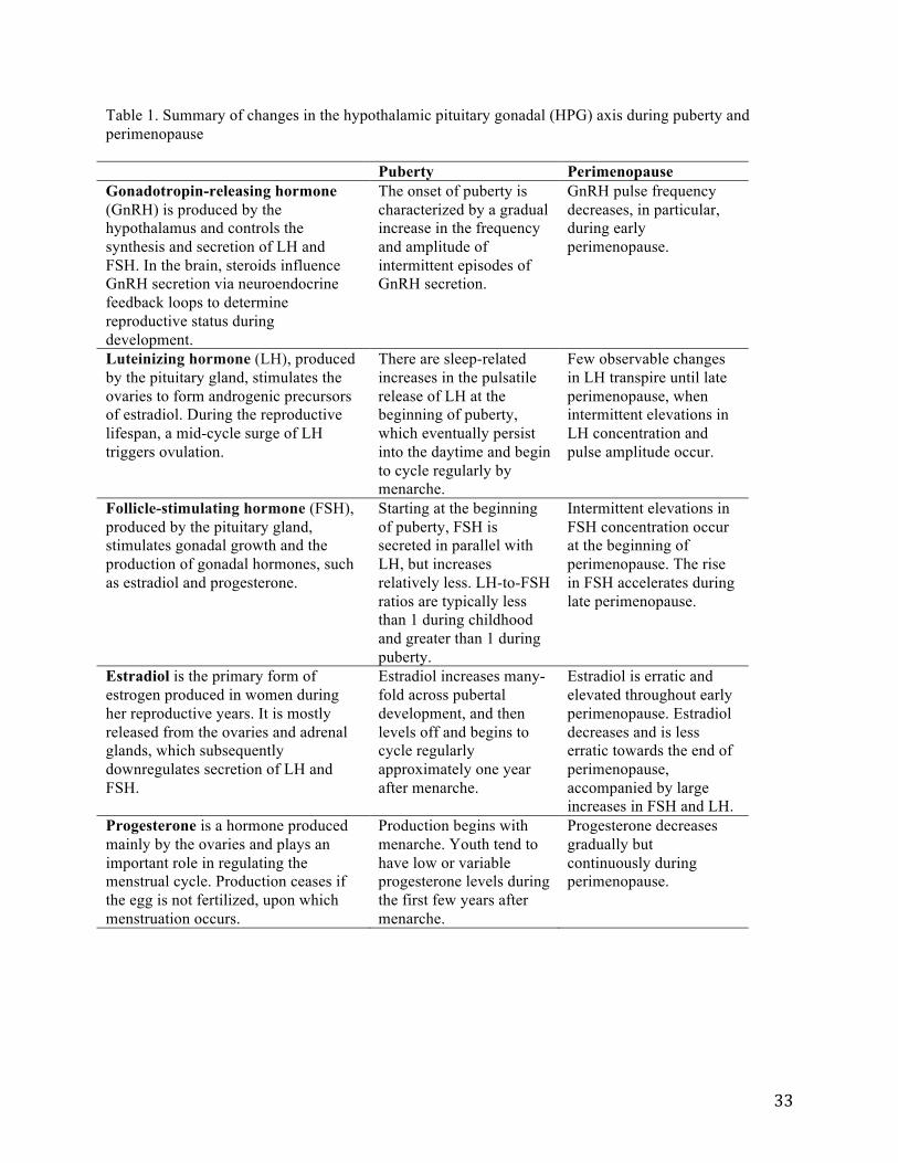
!
! 33!
Table 1. Summary of changes in the hypothalamic pituitary gonadal (HPG) axis during puberty and perimenopause Puberty Perimenopause Gonadotropin-releasing hormone (GnRH) is produced by the hypothalamus and controls the synthesis and secretion of LH and FSH. In the brain, steroids influence GnRH secretion via neuroendocrine feedback loops to determine reproductive status during development.
The onset of puberty is characterized by a gradual increase in the frequency and amplitude of intermittent episodes of GnRH secretion.
GnRH pulse frequency decreases, in particular, during early perimenopause.
Luteinizing hormone (LH), produced by the pituitary gland, stimulates the ovaries to form androgenic precursors of estradiol. During the reproductive lifespan, a mid-cycle surge of LH triggers ovulation.
There are sleep-related increases in the pulsatile release of LH at the beginning of puberty, which eventually persist into the daytime and begin to cycle regularly by menarche.
Few observable changes in LH transpire until late perimenopause, when intermittent elevations in LH concentration and pulse amplitude occur.
Follicle-stimulating hormone (FSH), produced by the pituitary gland, stimulates gonadal growth and the production of gonadal hormones, such as estradiol and progesterone.
Starting at the beginning of puberty, FSH is secreted in parallel with LH, but increases relatively less. LH-to-FSH ratios are typically less than 1 during childhood and greater than 1 during puberty.
Intermittent elevations in FSH concentration occur at the beginning of perimenopause. The rise in FSH accelerates during late perimenopause.
Estradiol is the primary form of estrogen produced in women during her reproductive years. It is mostly released from the ovaries and adrenal glands, which subsequently downregulates secretion of LH and FSH.
Estradiol increases many-fold across pubertal development, and then levels off and begins to cycle regularly approximately one year after menarche.
Estradiol is erratic and elevated throughout early perimenopause. Estradiol decreases and is less erratic towards the end of perimenopause, accompanied by large increases in FSH and LH.
Progesterone is a hormone produced mainly by the ovaries and plays an important role in regulating the menstrual cycle. Production ceases if the egg is not fertilized, upon which menstruation occurs.
Production begins with menarche. Youth tend to have low or variable progesterone levels during the first few years after menarche.
Progesterone decreases gradually but continuously during perimenopause.
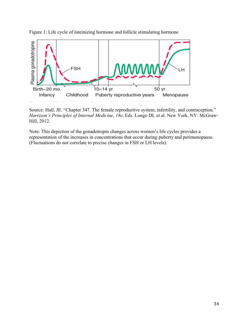
!
! 34!
Figure 1: Life cycle of luteinizing hormone and follicle stimulating hormone
Source: Hall, JE. “Chapter 347. The female reproductive system, infertility, and contraception.” Harrison’s Principles of Internal Medicine, 18e, Eds. Longo DL et al. New York, NY: McGraw-Hill, 2012.
Note: This depiction of the gonadotropin changes across women’s life cycles provides a representation of the increases in concentrations that occur during puberty and perimenopause. (Fluctuations do not correlate to precise changes in FSH or LH levels).
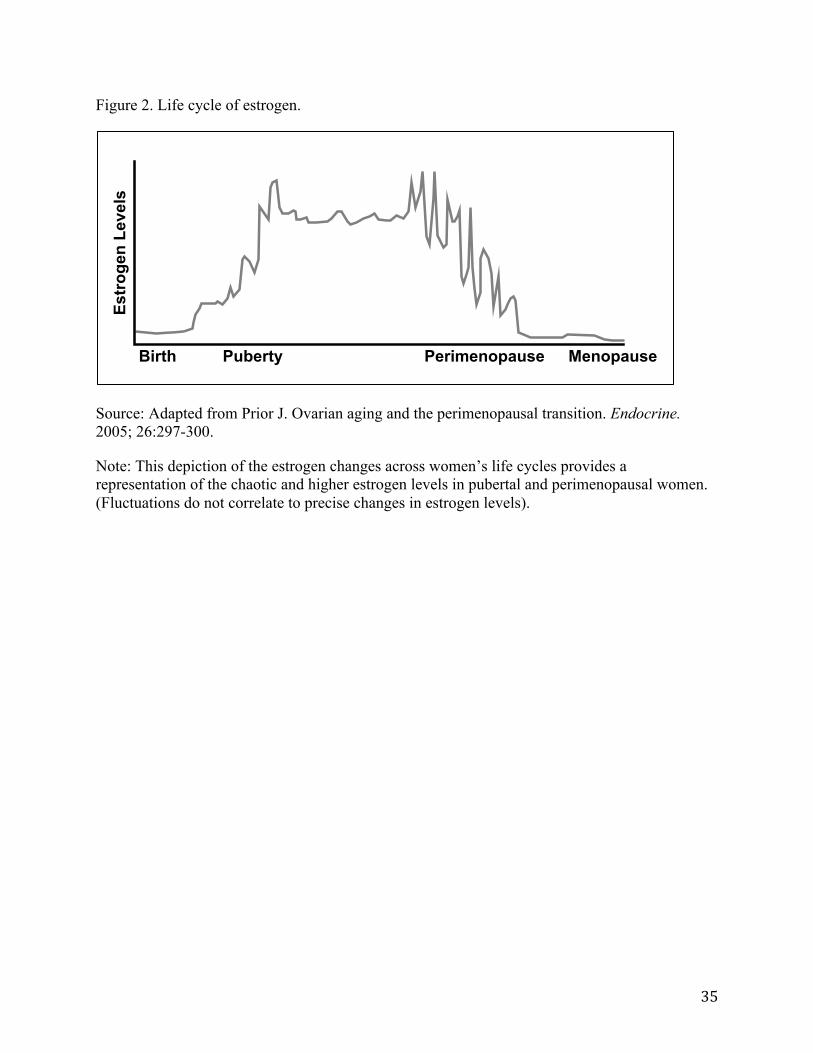
!
! 35!
Figure 2. Life cycle of estrogen.
Source: Adapted from Prior J. Ovarian aging and the perimenopausal transition. Endocrine. 2005; 26:297-300.
Note: This depiction of the estrogen changes across women’s life cycles provides a representation of the chaotic and higher estrogen levels in pubertal and perimenopausal women. (Fluctuations do not correlate to precise changes in estrogen levels).
Birth Puberty Perimenopause Menopause
Estr
ogen
Lev
els
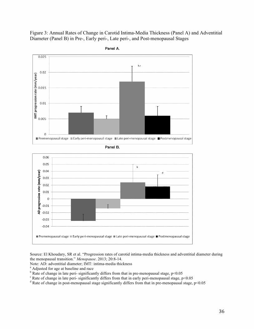
!
! 36!
Figure 3: Annual Rates of Change in Carotid Intima-Media Thickness (Panel A) and Adventitial Diameter (Panel B) in Pre-, Early peri-, Late peri-, and Post-menopausal Stages
Source: El Khoudary, SR et al. “Progression rates of carotid intima-media thickness and adventitial diameter during the menopausal transition.” Menopause. 2013; 20:8-14. Note: AD: adventitial diameter; IMT: intima-media thickness a Adjusted for age at baseline and race b Rate of change in late peri- significantly differs from that in pre-menopausal stage, p<0.05 c Rate of change in late peri- significantly differs from that in early peri-menopausal stage, p<0.05 d Rate of change in post-menopausal stage significantly differs from that in pre-menopausal stage, p<0.05

!
! 37!
Figure 4: Annual Mean Values of Total Cholesterol and Low-Density Cholesterol (LDL-C) Among Women Undergoing the Menopausal Transition
Source: Matthews, KA et al. “Are changes in cardiovascular disease risk factors in midlife women due to chronological aging or to the menopausal transition?” Journal of the American College of Cardiology. 2009; 54:2366-2373.
Note: Data in figures A and B describe women from the Study of Women’s Health Across the Nation. Women have been categorized into tertiles by their weight at baseline in the study.

!
! 38!
Paper 2: The Longitudinal Relation of Stress during the Menopausal Transition to Fibrinogen Concentrations: Results from the Study of Women’s Health Across the Nation ABSTRACT Objective: Life course theory suggests that exposures occurring during critical or sensitive periods have particularly profound effects on health. Most research on this subject has focused on the occurrence of such windows early in life, during periods of rapid change and development. I investigated whether perimenopause, a period of dramatic neuroendocrine changes, represents a sensitive period for stress responsivity by evaluating the relation of stress to fibrinogen, a biomarker for inflammation. Methods: The study sample was comprised of participants in the Study of Women’s Health Across the Nation, a longitudinal study that included annual interviews about women’s health at midlife and the collection of blood and biosamples (n=3,287). Linear mixed effects models were fitted to estimate the longitudinal relationship between stress and menopausal stage and the association between stress and fibrinogen over the menopausal transition. Results: Women in early and late perimenopause reported perceiving higher levels of stress than premenopausal women (p<0.05), adjusted for confounding variables. However, fibrinogen was not higher during perimenopause, nor did fibrinogen exhibit a higher response to stress during this period. Conclusions: Although neuroendocrine changes during the menopausal transition may exacerbate the negative health effects of stress, the findings of this study do not suggest such interaction, as measured by changes in fibrinogen. The significant association observed between perceived stress and menopausal status may still have important implications, however, given prior literature linking perceived stress with numerous health outcomes. Keywords: Perimenopause, Critical period, Psychological stress, Fibrinogen
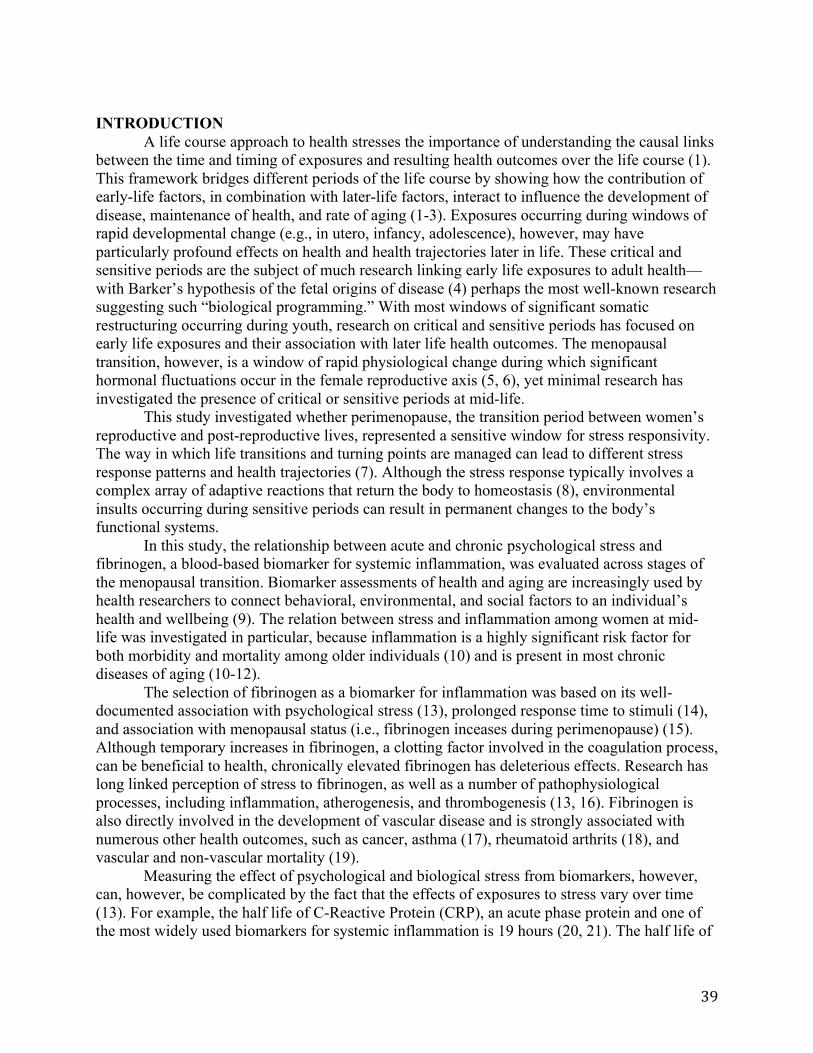
!
! 39!
INTRODUCTION
A life course approach to health stresses the importance of understanding the causal links between the time and timing of exposures and resulting health outcomes over the life course (1). This framework bridges different periods of the life course by showing how the contribution of early-life factors, in combination with later-life factors, interact to influence the development of disease, maintenance of health, and rate of aging (1-3). Exposures occurring during windows of rapid developmental change (e.g., in utero, infancy, adolescence), however, may have particularly profound effects on health and health trajectories later in life. These critical and sensitive periods are the subject of much research linking early life exposures to adult health—with Barker’s hypothesis of the fetal origins of disease (4) perhaps the most well-known research suggesting such “biological programming.” With most windows of significant somatic restructuring occurring during youth, research on critical and sensitive periods has focused on early life exposures and their association with later life health outcomes. The menopausal transition, however, is a window of rapid physiological change during which significant hormonal fluctuations occur in the female reproductive axis (5, 6), yet minimal research has investigated the presence of critical or sensitive periods at mid-life.
This study investigated whether perimenopause, the transition period between women’s reproductive and post-reproductive lives, represented a sensitive window for stress responsivity. The way in which life transitions and turning points are managed can lead to different stress response patterns and health trajectories (7). Although the stress response typically involves a complex array of adaptive reactions that return the body to homeostasis (8), environmental insults occurring during sensitive periods can result in permanent changes to the body’s functional systems.
In this study, the relationship between acute and chronic psychological stress and fibrinogen, a blood-based biomarker for systemic inflammation, was evaluated across stages of the menopausal transition. Biomarker assessments of health and aging are increasingly used by health researchers to connect behavioral, environmental, and social factors to an individual’s health and wellbeing (9). The relation between stress and inflammation among women at mid-life was investigated in particular, because inflammation is a highly significant risk factor for both morbidity and mortality among older individuals (10) and is present in most chronic diseases of aging (10-12).
The selection of fibrinogen as a biomarker for inflammation was based on its well-documented association with psychological stress (13), prolonged response time to stimuli (14), and association with menopausal status (i.e., fibrinogen inceases during perimenopause) (15). Although temporary increases in fibrinogen, a clotting factor involved in the coagulation process, can be beneficial to health, chronically elevated fibrinogen has deleterious effects. Research has long linked perception of stress to fibrinogen, as well as a number of pathophysiological processes, including inflammation, atherogenesis, and thrombogenesis (13, 16). Fibrinogen is also directly involved in the development of vascular disease and is strongly associated with numerous other health outcomes, such as cancer, asthma (17), rheumatoid arthrits (18), and vascular and non-vascular mortality (19).
Measuring the effect of psychological and biological stress from biomarkers, however, can, however, be complicated by the fact that the effects of exposures to stress vary over time (13). For example, the half life of C-Reactive Protein (CRP), an acute phase protein and one of the most widely used biomarkers for systemic inflammation is 19 hours (20, 21). The half life of
!
! 40!
fibrinogen, in comparison, is a little more than 4 days (100 hours) (16). The relatively prolonged response time of fibrinogen compared with other acute phase proteins allowed for detection of changes in its concentration days after the triggering stimuli. The longer duration in which fibrinogen remains elevated also potentially allowed for an enhanced ability to measure changes in inflammation in response to stressors that have not necessarily occurred or been perceived on the day of (or immediately preceding) the collection of samples. In order to determine if women undergoing perimenopause experience increased susceptibility to the adverse effects of psychological stress, as measured by changes in fibrinogen, the following three hypotheses were tested:
Hypothesis 1. Stress is not a function of menopausal status, such that perimenopausal women perceive an equivalent level of stress with pre- and post-menopausal women. Hypothesis 2. Menopausal status influences the association of stress with fibrinogen, such that stress experienced during perimenoause is associated with elevated fibrinogen concentrations compared with stress experienced during pre- or post-menopause. Hypothesis 3. Stress perceived during perimenopause has an enduring effect on fibrinogen, resulting in elevated levels of fibrinogen in time periods beyond the contemporaneously perceived stress.
An additional exploratory aim was to determine if any relation of stress to fibrinogen differed by race/ethnicity, given some research suggesting that women’s physiological response to stress varies according to race/ethnicity (22, 23).
The mechanisms involved in the interactive effects of stress on health are varied, but include behavioral as well as neurobiological, endocrine, and immunologic measures (24). The present analyses provided insight on whether physiological changes during the menopausal transition influence women’s vulnerability to perceived stress and its effects on a marker of inflammation. METHODS Participants
The study sample included participants in the Study of Women’s Health Across the Nation (SWAN). SWAN is a seven-site, longitudinal, community-based study devoted to examining the natural history of the menopausal transition and women’s health at midlife—a period of life that has traditionally played a subordinate role in research in the social and behavioral sciences and is often considered a nondramatic phase of life (25). SWAN has annually assessed the health of a multi-racial/ethnic cohort of 3,302 women as they transitioned from pre- or early perimenopause to post-menopause. Through its collection of medical and health data, psychosocial measures, biological measures, and anthropometry, SWAN has sought to describe the effects of the menopausal transition and its associated characteristics on subsequent health and risk factors for age-related chronic diseases (26). A complete description of the study design, data collection, and sampling and recruitment has previously been published (27).
Eligibility criteria for the longitudinal cohort included: aged 42-52 years, had an intact uterus, had at least one menstrual period and no use of exogenous reproductive hormones (e.g., birth control pills, hormone replacement therapy) in the three months preceding the baseline
!
! 41!
interview, and self-identification with one of the study site’s designated racial/ethnic groups. Each site recruited about 450 non-Hispanic Caucasian women and one designated minority group: African American women in Pittsburgh, Pennsylvania, Boston, Massachusetts, the Detroit area, Michigan, and Chicago, Illinois; Chinese women in Oakland, California; Hispanic women in Newark, New Jersey; and Japanese women in Los Angeles, California. The study protocols were approved by the institutional review boards of the participating sites, and all women provided signed, written informed consent for participation in the studies.
The present analyses were based upon on data from the five biannual visits (i.e., visits 0, 1, 3, 5, and 7) at which fibrinogen was assessed. Women were censored at the visit when their menopausal status was undetermined (usually due to menopausal hormone use for undetermined menopausal stage) or fibrinogen concentions was undetermined or missing. Data from subsequent examinations were included once their menopausal status was determined and they provided fibrinogen data. Women who reported having a hysterectomy or oopohorectomy were censored from that visit forward. Of the 3,302 participants who began the study at baseline, 3,287 women had fibrinogen data in at least one subsequent followup visit. Measures Outcome: Fibrinogen. Physiological response to stress was measured through changes in fibrinogen, a glycoprotein found in plasma (16). Fibrinogen was determined for participants using assays of blood samples that were drawn the morning following an overnight fast. The blood draw was targeted to the early follicular phase of the menstrual cycle (days 2 to 5) in menstruating women to standardize and minimize the hormonal fluctuations that occur during the remainder of the menstrual cycle. Fibrinogen was measured in frozen citrated plasma (MLA ELECTRA 1400C, Medical Labory Automation Inc.) using a clot-based turbidometric detection system. The New Jersey site did not complete in-person clinic visits in the final year of analysis. Fibrinogen data were not collected, consequently, for Hispanic women from visit 7. For budgetary reasons, fibrinogen was assayed for approximately one-third of the sample from visit 7. Fibrinogen data missingness was not associated with the primary independent variable of interest.
Independent Variables: Psychological Stress. Stress was assessed in SWAN in two different ways and were therefore examined separately in analysis. One measure asked participants a series of “life stress” questions about the occurrence and perceived stressfulness of major life events (e.g., death of someone close, legal problems, money problems, marriage or divorce, job changes) that occurred within the past year. This assessment was a modified version of the Psychiatric Epidemiology Research Interview (PERI) life events scale (28), which has been shown to exhibit high internal consistency and reliability across racial and ethnic groups (29, 30). Participants were asked 18 questions about events occurring within the past year, and if the events occurred, how upsetting or stressful they were perceived. SWAN created a summary variable ranging from 0 to 18 that equaled the number of upsetting or stressful major life events that occurred in the past year (score of 18 indicated maximum level of perceived stress). Instrumentation for assessing life events stress changed slightly over the study’s follow-up period. At baseline, 34 questions were used to assess life stress, but only 20 questions were used for visits 01-02. Questions prior to visit 03 were truncated in subsequent visits so that a maximum of 18 questions were used to score the total number of life events.
!
! 42!
Stress was also assessed through a measure of general perceived stress that referenced the two weeks prior to the participant’s study interview. A summary variable, “perceived stress,” was created based upon participants’ responses to four questions: whether they felt “unable to control important things in their lives,” “confident about their ability to handle personal problems,” “that things were going their way,” and “that difficulities were piling so high that they could not overcome them. Scores could range from 4 to 20, with a score of 20 indicating maximum perceived stress.
Menopausal Status was defined by SWAN based on participants’ responses to questions regarding the timing and regularity of menstrual bleeding. The following five categories were used: premenopausal (menses occurring in the past three months, with no change in regularity in the past 12 months), early perimenopausal (menses occurring within the past three months with some changes in regularity in the past 12 months), late perimenopausal (no menses occuring within the past three months, but some menstrual bleeding within the past 12 months), post-menopausal (no menses occurring in at least the past 12 months), or surgically postmenopausal (hysterectomy and/or both ovaries removed). Women who used menopausal hormone therapy before their final menstrual period had an undetermined menopausal status unless they stopped use of such therapy and had a subsequent menstrual bleed. Covariates.
Demographic variables assessed for confounding (covariates identified by the literature and that changed the relation between stress and fibrinogen by more than 10 percent) included age and race. Age and race/ethnicity were self-reported and were collected at baseline during screening for eligibity into the longitudinal cohort. Age was also reported in interviews with participants at each wave of follow-up.
Health behaviors assessed for potential confounding included time-varying smoking, alcohol consumption, and physical activity levels. Alcohol consumption was calculated from four questions: the first referred to any consumption of beer, wine, or liquour since the last study visit and the other three question related to the amount of consumption per day, week, or month. Consumption was categorized as none/low (alchohol use < once a month, moderate (> than once per month and < than two times per week), or high (> two times per week) alcohol use. Smoking status was assessed as whether participants reported smoking currently and was defined as ‘yes’ to anyone who reported having smoked regularly since the last visit. Activity level was assessed on a 5-point scale that classified the frequency participants sweated from exertion from 1=never or less than once a month to 5=more than once a week. Physical activity level was assessed using a modified version of the Kaiser Physical Activity Survey (KPAS), adapted from the Baecke questionnaire. For this analysis, two activity indices were included: involvement in sports or exercise and sweating from exertion. Participants were asked to classify the frequency of these items on a 5-point scale, ranging from 1=never or less than once a month to 5=more than once a week, and these scores were averaged to create a single composite score. Physical activity level was not assessed at visit 7; therefore, all data from this visit were not included in models adjusted for physial activity level.
Psychosocial variables were time-varying and included depressive symptoms and social support. Depressive symptoms were assessed via the Center for Epidemiologic Study-Depression (CES-D) Questionnaire and was calculated as a dichotomous score equalt to ‘1’ for those with a score > 16, indicating potential for clincally relevant depressive symptoms, and ‘0’ otherwise. Social support was calculated based on the sum of four types of emotional and instrumental

!
! 43!
support, with responses ranging from 0 (none of the time) to 4 (all of the time). A total score was summed in each year ranging from 0 for least social support to 16 for most social support. Social support was not ascertained in follow up visit 7; therefore, visit 7 data were not included in analysis of models adjusted for social support.
Body mass index (BMI) was computed as weight (kg)/[height (m)]2 based on measured weight and height at each annual visit. BMI was another time-varying covariate tested as a potential confounder. Statistical Analysis
Descriptive statiscs were calculated using median, interquartile range, and standard deviation for fibrinogen, life events stress, perceived stress, and for each covariate. Fibrinogen levels and stress scores were compared over the different stages of the menopausal transition using t-tests and Pearson and Spearman correlation coefficients. The relationship between the two measures of stress was also assessed to determine how closely these variables were correlated.
Because it was possible that undergoing the menopausal transition in itself may have altered stress levels, a mixed effects linear regression model (i.e., random intercept grouped by individual, with time fixed effects) using STATA’s xtmixed function was used to estimate the longitudinal relationship between stress and menopausal status. The model used to test whether stress varies with menopausal stage was represented by equation 1:
Equation 1: Stress = ƒ(Menopausal stage | year, Z). This equation determined if an individual’s stress at time t was a function of menopausal stage (e.g., early perimenopause, late perimenopause, or post-menopause, with pre-menopause as the reference group ) and year, controlling for confounders represented by term Z.
Mixed effects linear regression models (i.e., random intercept grouped by individual, with time fixed effects) were then fit to assess longitudinal associations between stress and fibrinogen levels as women progressed from pre- to post-menopause. These models included a random intercept for each woman to decrease between-women variation, indicated by Equation 2. The dependent variable, fibrinogen, was not normally distributed and was therefore log-transformed for analyses.
Equation 2: Yit = α + δStressit + γMenopausal_Stageit + βStressit*Menopausal_Stageit + ω1Y98t + ω2Y00t + ω3Y02t + ω4Y04t + τZi + µi + εit
Estimators were defined as the following: Y was the corresponding plasma fibrinogen concentration for the ith participant at time t; α represented the constant term; Stress was measured on two different scales: Perceived stress was assessed on a scale of 4 through 20, where a score of 4 indicated the participant had not experienced stress at time t, and a score of 20 indicated the participant had experienced a maximum level of stress. Life events stress was evaluated on a scale of 0-18, with a score of 0 indicating no stressful events and a score of 18 indicating the maximum number of stressful events within the prior year. Menopausal_Stage represented the possibility of being classified as early perimenopausal, late perimenopausal, or post-menopausal, with premenopause as the referrant category; Stress*Menopausal_Stage was the interaction term representing the effect of stress as it varied with menopausal stage; Y98

!
! 44!
through Y04 = 1 for the year in which data was collected, = 0 if otherwise (reference year is 1996); Z represented all control variables; ε was the error term composed of a time-invariant component µ and a time-varying component v.
Covariates that were identified by the literature as possible confounders and that changed the relation between stress and fibrinogen by more than 10 percent were included in the multivariable regressions. Changes in variances of the remaining estimators were also examined upon inclusion of a covariate to determine if they changed signficanty due to multicollinearity. The model that used life events stress was adjusted for BMI, social support, and overall activity level. The model that used general perceived stress was adjusted for social support, alcohol consumption, BMI, and depressive symptoms. Results of both models were stratified by race/ethnicity (with and without the inclusion of covariates) to test for effect modification by racial/ethnic group.
Lastly, lagged versions of the mixed effects linear regression models were created to determine whether perimenopausal women were more likely to carry increases in fibrinogen forward into subsequent time periods. Two lagged models were created. Equation 3a included contemporary and lagged stress scores and menopausal status, and Equation 3b included only lagged stress and menopausal status.
Equation 3a: ∆Yit = α + δ1Stressit + δ2Lagged_Stressit + γ1Menopausal_Statusit + γ2Lagged_Menopausal_Statusit + β1Stressit*Menopausal_Statusit + β2Lagged_Stressit*Lagged_Menopausal_Statusit + ω1Y98t + ω2Y00t + ω3Y02t + ω4Y04t + τZi + εit
Equation 3b: ∆Yit = α + δ1Lagged_Stressit + γ1Lagged_Menopausal_Statusit + β1Lagged_Stressit*Lagged_Menopausal_Statusit + ω1Y98t + ω2Y00t + ω3Y02t + ω4Y04t + τZi + εit The Breusch-Pagan/Cook-Weisberg test for heteroscedasticity revealed heteroscedastic
error variances of within-individual observations; therefore, robust standard errors were calculated for all models. Analyses were performed with STATA version 11. RESULTS
African-American, Chinese, Japanese, and Hispanic women comprised greater proportions of the study sample than their respective proportions in the general U.S population, reflecting the study design to oversample these groups. At baseline, half of participants reported no stressful life events within the prior year, yet most women perceived moderate or high levels of stress within the prior two weeks (Table 1). A majority of women also reported moderate or high levels of social support at baseline, consumed less than one alcoholic drink per month, and were considered overweight on average. Approximately half (54%) of participants were classified as premenopausal, and nearly half (47%) were classified as early perimenopausal at baseline. By the final wave of follow-up, over half (58%) of the participants had reached postmenopause.
The median fibrinogen for women at baseline was 282 mg/dL, which appeared to increase around age 47 (see Figure 1). This increase is consistent with prior epidemiological literature indicating that fibrinogen increases during perimenopause (15) (and that
!
! 45!
perimenopause begins on average at approximately age 47 at the population level) (31). Fibrinogen concentration appeared to decrease over time, however, with the highest levels observed at the baseline visit and the lowest levels observed at the final visit included in analysis (see Figure 2).
An analysis of variance indicated significant within-person variation in both measures of stress. At baseline, over one-quarter (29%) of women reported experiencing more than two life events they perceived were very stressful within the past year, and 19% reported they perceived a high level of stress within the prior two weeks of being interviewed. The variable measuring stressful life events within the past year was weakly correlated with the variable measuring perceived stress within the past two weeks (r2 = 0.30), suggesting that either perceived stress was not enduring or that these two variables assessed different aspects or types of stress in women’s lives. The results of analyses testing Hypothesis 1, which was that stress is not a function of menopausal status, revealed statistically significant associations (p<0.01) between perceived stress experienced within the prior two weeks and early and late perimenopause (Table 2). These findings suggest that menopausal transition stage was related to women’s perception of their stress level, such that as women reached and progressed through perimenopause, their perceived level of stress increased above pre-menopausal levels. The significance of these findings was slightly attenuated, although still statistically significant (p<0.05), upon controlling for social support, depressive symptoms, BMI, and alcohol consumption. General perceived stress during perimenopause did not, however, vary significantly from perceived stress during post-menopause. Longitudinal analyses of life events stress within the past year were significantly associated (p<0.05) with late perimenopause during the five visits, adjusting for social support, BMI, and activity level. Women in late perimenopause reported fewer stressful events compared with pre-menopausal—but not post-menopausal women. In sum, the results did not provide support for Hypothesis 1; rather, stress appeared to be associated with menopausal status.
The results of analyses testing Hypothesis 2, which was that menopausal status influenced the association of stress with fibrinogen, did not indicate an interaction between perceived stress (Table 3a) or life events stress (Table 3b) with menopausal status on changes in fibrinogen. The narrow confidence intervals for the coefficients of interest provided further indication that differences between menopausal transition stages in these associations were minimal and not statistically meaningful. The results did not, in sum, provide support for Hypothesis 2. The results of analyses testing Hypothesis 3, which was that stress perceived during the menopausal transition had an association with fibrinogen, revealed no statistically significant associations between measures of perceived stress (Table 4a) or life events stress (Table 4b) and increases in fibrinogen beyond the contemporaneous time period. These findings did not provide support for Hypothesis 3 and suggested that stress perceived during the menopausal transition did not exhibit an enduring association with changes in fibrinogen. The narrow confidence intervals again provided indication that differences between lagged menopausal status were minimal and not statistically meaningful. The F-test statistic for joint significance of the explanatory variables for both models was statistically significant. The F-statistic for the contemporary and lagged stress scores regression was 1.62 (p<0.05), and the F-statistic for the regression using only lagged stress was 2.88 (p<0.01).
!
! 46!
Sensitivity analyses Given that race/ethnicity was a potential effect modifier of the relationship between
stress, menopausal status, and fibrinogen, mixed regression models were stratified by race/ethnicity. Results of these analyses showed a significant (p<0.05) association between perceived stress within the prior two weeks—but not stress from life events occurring over the prior year—and menopausal stage among Chinese women only. The association was not statistically significant (p=0.08) after adjusting for confounding variables. The relatively small sample size in this subgroup, however, likely provided inadequate statistical power to detect a statistically significant association.
Some variables included in the models as covariates arguably could have been mediators of the relationship between stress and fibrinogen instead of confounders, which would imply overspecification of the previous models. BMI, smoking, and alcohol consumption, for example, are associated with fibrinogen concentration (fibrinogen decreased with moderate alcohol intake and increased with higher BMI and smoking), and these variables also reflect stress-coping behaviors (32, 33). The signifiance of associations from the regressions of menopausal stage on stress increased (p<0.01) when alcohol consumption and BMI were removed as covariates. Results of the regressions of stress (life events and perceived) on fibrinogen remained robust, however, omission of BMI, smoking, and alcohol consumption from the regression models. Neither the magnitude of the coefficients nor the variability of the estimates changed significantly.
Socioeconomic status (defined by education and income), though a potential confounder, was ultimately not included as a covariate in the main models or in the sensitivity analyses. As a time-invariant variable, education dropped from fixed effects analysis. Previous research has indicated, moreover, that the inverse relationship between socioeconomic status and fibrinogen operates predominantly through income, rather than through education and income independently. Change in income was not included as a separate covariate because this event was considered a stressor in itself and was captured through the stressful life events variable, which asked whether participants experienced “major money problems” in the prior year.
Lastly, although time was held fixed in all regressions, analyses were run in which the time dummy variables were replaced with a continuous time measure. This approach did not decrease the standard errors and instead just shifted the values of the coefficients. The time dummy variables were retained, therefore, to control for the effect of time on the intercept. DISCUSSION
Analysis of the relationship between perceived stress and menopausal transition stage revealed that recently perceived stress increased during perimenopause relative to pre-menopause but was not significantly different from post-menopause. Stress perceived from major life events occurring over the prior year was associated with a slightly—although statistically significant—decrease during late perimenopause. These findings may help explain the inconsistent results from prior literature about whether perimenopause is associated with the occurrence of substantially more negative life events (34). Whereas women may experience little to no difference in levels of stress from life events across stages of the menopausal transition, their general perception of stress appears to vary significantly by menopausal stage, thereby causing differential reporting. Hormone or age-related changes may at least partially explain why perceived stress appears to increase during perimenopause. Gonadal steroids (e.g., estrogen,
!
! 47!
progesteron) have a widespread influence on the brain and on the regulation of mood and behavior (35). These hormone changes, in combination with changes in social roles, perception of health and body image, and vasomotor symptoms initiated during perimenopause may influence their perception of stress (36, 37).
Irrespective of the different types of stress and their respective associations with menopausal stage, no significant differences in fibrinogen response to stress appeared across menopausal stages. Even though perimenopausal women may feel more stressed, this perception did not appear to incur adverse changes in the inflammation marker, fibrinogen, in the total sample of women. These findings should not be interpreted to mean that psychological stress during perimenopause does not “get under the skin” to affect women’s biology, particularly given prior research linking perceived stress with physiological dysregulation (38) and the incidence of disease (39). Alternatively, the present study findings suggest that the mechanism linking women’s heightened levels of perceived stress and their increased susceptibility to adverse health outcomes is through other mechanisms, such as psychological processes or perception of control.
Evidence increasingly suggests, moreover, that different patterns of physiological response to stress can occur based on how individuals appraise their situations. Response to stress is not uniform; different constructs of stress can incur different physiological effects (40). Future research might consider alternative biomarkers or measures of health to gain a more comprehensive understanding of the interrelationship between the hormone changes during perimenopause, stress, and health outcomes.
Although the relationship between stress and fibrinogen over the menopausal transition had not been previously assessed prior to the present study, the relationship of depression over the menopausal transition with hemostatic and inflammatory markers over time has been explored. Such research has found that depressive symptoms appear associated with hypercoagulability via elevated fibrinogen among perimenopausal women (15). These findings are noteworthy given prior research strongly linking stress and depression (41). The contrasting findings of this study with the study on depression and fibrinogen possibly suggests differing physiologic effects of stress and depression on hypercoaguability among perimenopausal women.
Multiple and varied stressors occur in mid-life, including demanding jobs, retirement, illness and/or death of parents, caregiving, and changes in relationships with partners and offspring (42, 43), let alone the dramatic physiological changes that transpire with the culmination of a woman’s reproductive cycle. The relationship of stress, fibrinogen, and health, however, has often been investigated only in the context of socioeconomic stressors or job stress (33). This analysis more broadly assessed the association between stress and fibrinogen, and provides insight into the interrelationship between hormonal changes with perimenopause, stress, and inflammation.
A strength of this study is that it incorporated the two major approaches to examining the interrelationships between stress, health, and aging, that is, stress from major life events and daily stressors that arise from day-to-day living) (9). The life events variable addressed the concept of polonged arousal derived from specific events occurring over the prior year. The potentially long duration from when an event occurred to when a participant was surveyed, however, may have resulted in an inability to detect the physiological impact of such stressors, at least through acute phase proteins, such as fibrinogen. The recently perceived stress variable allowed for closer proximity between apparent stressors and the time at which participants were

!
! 48!
surveyed. Such stress is associated with spikes in arousal, which may have allowed for improved detection of the physiological impact of stress. This measure, however, may be more vulnerable to issues of endogeneity. Perimenopause in itself may cause women to feel more stressed and therefore report stress differently than in pre- or postmenopause. Including both measures of stress in this study made parsing the relationship of stress with inflammation possible and provided insight into how the different measures of stress relate to fibrinogen.
Other strengths of this study include the longitudinal analysis of a racially and ethnically diverse sample of women across the country. Analyzing true panel data—rather than pooled cross-sections—with mixed effects methods allowed for more precise estimates of changes in stress and fibrinogen over time and reduced potential confounding from unobserved heterogeneity or omitted variables. The large, diverse study sample also allowed for generalizability of results to community-dwelling populations in major metropolitan areas in the United States.
Limitations of this study included that results are based on women’s progression through the menopausal transition after only seven visits. The experiences of the subset of women who had progressed to post-menopause by this point may fundamentally differ from women in the earlier stages of the menopausal transition who had not yet transitioned further. Women who smoke, for example, reach menopause at an earlier age on average compared with non-smoking women (44). Approximately 15% of the sample identified themselves as smokers, however, while more than half of participants reached post-menopause by visit 7. Smoking was not considered a confounder in analyses, and so any bias this introduced is not clear.
The inclusion of data from only the first seven visits also presented an issue of inadequate statistical power when analyzing data by race/ethnicity. For example, only 67 Chinese women reached late perimenopause by the final visit included in the analysis. As the present study lacked adequate sample size and thus statistical power to examine the relationships among all race/ethnicity groups, stress, and fibrinogen in a meaningful way, studies with larger sample sizes of women from different racial/ethnic groups examined longitudinally could address this under-researched area.
Another potential data issue was missing fibrinogen data in visit 7: fibrinogen was assayed for only one-third of the total sample. Although these missing data created an unbalanced panel, the missing data were not correlated with the dependent variable; therefore, it is unlikely that selection bias from missing data biased the results. The adjusted models, moreover, include analysis only up through visit 5 because one of the covariates—social support—was not assessed in visit 7. Analyses with and without the inclusion of visit 7 data did not result in significant differences in this study’s findings. Conclusion
In summary, the results of these analyses suggest that while perceived stress appeared heightened among perimenopaual women, this stress was not associated with a significant physiological response as measured by changes in fibrinogen. Any changes in inflammation during the menopausal transition, moreover, were not associated with enduring changes in fibrinogen. Studies with longer follow-up time would need to be conducted to determine if stress experienced during the menopausal transition differentially affects health outcomes as women age post-menopausally.
The results of this study lend insight into the aging process in women, as inflammation is increasingly considered one of the primary mechanisms underlying the aging process (45). Even
!
! 49!
though study findings suggest that perceived stress increased during the menopausal transtion, such stress did not appear to interact with neuroendocrine changes to lead to significant increases in systemic inflammation, reflected by fibrinogen levels. Of course, individual differences make some individuals more or less vulnerable to stress, and identical stimuli can cause varying responses to stress (12). Future research may consider how racial/ethnic differences contribute to varying stress responses to inflammation.
ACKNOWLEDGEMENTS
The Study of Women’s Health Across the Nation (SWAN) has grant support from the National Institutes of Health (NIH), DHHS, through the National Institute on Aging (NIA), the National Institute of Nursing Research (NINR) and the NIH Office of Research on Women’s Health (ORWH) (Grants U01NR004061; U01AG012505, U01AG012535, U01AG012531, U01AG012539, U01AG012546, U01AG012553, U01AG012554, U01AG012495). The content of this manuscript is solely the responsibility of the author and does not necessarily represent the official views of the NIA, NINR, ORWH or the NIH.
Clinical Centers: University of Michigan, Ann Arbor – Siobán Harlow, PI 2011 – present, MaryFran Sowers, PI 1994-2011; Massachusetts General Hospital, Boston, MA – Joel Finkelstein, PI 1999 – present; Robert Neer, PI 1994 – 1999; Rush University, Rush University Medical Center, Chicago, IL – Howard Kravitz, PI 2009 – present; Lynda Powell, PI 1994 – 2009; University of California, Davis/Kaiser – Ellen Gold, PI; University of California, Los Angeles – Gail Greendale, PI; Albert Einstein College of Medicine, Bronx, NY – Carol Derby, PI 2011 – present, Rachel Wildman, PI 2010 – 2011; Nanette Santoro, PI 2004 – 2010; University of Medicine and Dentistry – New Jersey Medical School, Newark – Gerson Weiss, PI 1994 – 2004; and the University of Pittsburgh, Pittsburgh, PA – Karen Matthews, PI.
NIH Program Office: National Institute on Aging, Bethesda, MD – Winifred Rossi 2012 - present; Sherry Sherman 1994 – 2012; Marcia Ory 1994 – 2001; National Institute of Nursing Research, Bethesda, MD – Program Officers.
Central Laboratory: University of Michigan, Ann Arbor – Daniel McConnell (Central Ligand Assay Satellite Services).
Coordinating Center: University of Pittsburgh, Pittsburgh, PA – Maria Mori Brooks, PI 2012 - present; Kim Sutton-Tyrrell, PI 2001 – 2012; New England Research Institutes, Watertown, MA - Sonja McKinlay, PI 1995 – 2001.
Steering Committee: Susan Johnson, Current Chair, Chris Gallagher, Former Chair. I thank the study staff at each site and all the women who participated in SWAN.
!
! 50!
Works Cited
1. Lynch J, Davey Smith G. A life course apporach to chronic disease epidemiology. Annual Review of Public Health. 2005;26(1):1-35. 2. Kuh D, Hardy R, editors. A life course approach to women's health. Oxford: Oxford University Press; 2002. 3. Ben-Shlomo Y, Kuh D. A life course approach to chronic disease epidemiology: conceptual models, empirical challenges and interdisciplinary perspectives. International journal of Epidemiology. 2002;31(2):285-93. 4. Barker D. In Utero Programming of Chronic Disease. Clinical Science. 1998;95:115-28. 5. Burger H, Dudley E, Robertson D, Dennerstein L. Hormonal changes in the menopausal transition. Recent Progress in Hormone Research. 2002;57:257-75. 6. Prior J, Hitchcock C. The endocrinology of perimenopause: Need for paradigm shift. Frontiers in Bioscience. 2011;3:474-86. 7. Halfon N, Hochstein M. Life course health development: An integrated framework for developing health, policy, and research. Milbank Quarterly. 2002;80(3):433-79. 8. Behavior IoMCoHa. Biobehavioral factors in health and disease. Health and behavior: The Interplay of Biological, Behavioral, and Societal influences. Washington, DC: National Academies Press; 2001. 9. Piazza J, Alameida D, Dmitrieva N, Klein L. Frontiers in the use of biomarkers of health in research on stress and aging. Journal of Gerontology: Psychological Sciences. 2010;65B:513-25. 10. Franceschi C, Campisi J. Chronic Inflammation (Inflammaging) and Its Potential Contribtution to Age-Associated Diseases. Biological Sciences. 2014;69(S1):S4-S9. 11. Finch CE. The Biology of Human Longevity. USA: Academic Press; 2007. 12. Epel E, Lithgow GL. Stress Biology and Aging Mechanisms: Toward Understanding the Deep Connection Between Adaptation to Stress and Longevity. Journals of Gerontology: Biological Sciences. 2014;69(S1):S10-S6. 13. Theorell T. Job Stress and Fibrinogen. European Heart Journal. 2002;23:1799-801. 14. HeartLab B. Reference Manual: Fibrinogen [cited 2014 September 11]. Available from: http://www.bhlinc.com/clinicians/clinical-references/reference-manual/chapter14. 15. Matthews KA, Schott LL, Bromberger JT, Cyranowski J, Everson-Rose SA, Sowers MF. Associations Between Depressive Symptoms and Inflammatory/Hemostatic Markeres in Women During the Menopausal Transition. Psychsomatic Medicine. 2007;69:124-30. 16. Kamath S, Lip G. Fibrinogen: Biochemistry, Epidemiology, and Determinants. QJM: An International Journal of Medicine. 2003;96(10):711-29. 17. Jousilahti P, Salomaa V, Hakala K, Rasi V, Vahtera E, Palosuo T. The Association of Sensitive Systemic Inflammation Markers with Bronchial Asthma. Annals of Allergy, Asthma, & Immunology. 2002;89:381-5. 18. Rooney T, Scherzer R, Shigenaga J, Graf J, Imboden J, Grunfeld C. Levels of Plasma Fibrinogen are Elevated in Well-Controlled Rheumatoid Arthritis. Rheumatology. 2011;50(8):1458-65. 19. Collaboration TFS. Associations of Plasma Fibrinogen Levels with Established Cardiovascular Disease Risk Factors, Inflammatory Markers, and Other Characteristics:
!
! 51!
Individual Participant Meta-Analysis of 154,211 Adults in 31 Prospective Studies. American Journal of Epidemiology. 2007;166(8):867-79. 20. Pepys M, Hirschfield G. C-reactive protein: a critical update. The Journal of Clinical Investigation. 2003;111:1805-12. 21. Vincent J-L, Donadello K, Schmidt X. Biomarkers in the criticall ill patient: C-reactive protein. Critical Care Clinics. 2011;27(2):241-51. 22. Christian L, Glaser R, Porter K, Iams J. Stress-Induced Inflammatory Responses in Women: Effects of Race and Pregnancy. Psychosomatic Medicine. 2013;75:658-69. 23. Anderson N, Lane J, Taguchi F, Williams R, Houseworth S. Race, Parental History of Hypertension, and Patterns of Cardiovascular Reactivity in Women. Psychophysiology. 1989;26(1):39-47. 24. McEwen BS, Steller E. Stress and the Individual: Mechanisms Leading to Disease. JAMA Internal Medicine. 1993;153(18):2093-101. 25. Dainese SM, Allemand M, Ribeiro N, Bayram S, Martin M, Ehlert U. Protective Factors in Midlife: How Do People Stay Healthy? GeroPsych.24(1):19-29. 26. Gerontology NIoADoGaC. The Study of Women's Health across the Nation (SWAN) Repository [cited 2014 September 1]. Available from: http://www.nia.nih.gov/research/dgcg/study-womens-health-across-nation-swan-repository. 27. Sowers MF, Crawford SL, Sternfeld B, Morganstein D, Gold EB, Greendale GA, et al. SWAN: A Multicenter, Multiethic, Community-Based Cohort Study of Women and the Menopausal Transition. In: Lobo RA, Kelsey J, Marcus R, editors. Menopause: Biology and Pathology. San Diego, CA: Academic Press; 2000. p. 175-88. 28. Bromberger JT, Assmann SF, Avis NE, Schocken M, Kravitz HM, Cordal A. Persistent Mood Symptoms in a Multiethnic Community Cohort of Pre- and Perimenopausal Women. American Journal of Epidemiology. 2003;158(4):347-56. 29. Stressful Life Events: Their Nature and Effects. Dohrenwend B, Dohrenwend B, editors. New York: John Wiley & Sons; 1974. 30. Vernon S, Roberts R. Measuring Nonspecific Psychological Distress and Other Dimensions of Psychopathology. Further Observations on the Problem. Archives of General Psychiatry. 1981;38:1239-47. 31. McKinlay SM. The Normal Menopause Transition: An Overview. Maturitas. 1996;23(2):137-45. 32. Butler L, Santoro N. The Reproductive Endocrinology of the Menopausal Transition. Steroids. 2011;76:627-35. 33. Kannel WB, D'Agostino RB, Belanger AJ. Update on Fibriogen as a Cardiovascular Risk Factor. Annals of Epidemiology. 1992;2(4):457-66. 34. Schmidt P, Murphy J, Haq N, Rubinow D, Danaceau M. Stressful Life Events, Personal Losses, and Perimenopause-Related Depression. Archives of Women's Mental Health. 2004;7:19-26. 35. Maughan B. Depression and psychological distress: a life course perspective. In: Kuh D, Hardy R, editors. A Life Course Approach to Women's Health. Oxford, UK: Oxford University Press; 2002. 36. Bromberger J, Matthews K, Schott L, Brockwell S, Avis N, Kravitz H, et al. Depressive symptoms during the menopausal transition: The Study of Women's Health Across the Nation. Journal of Affective Disorder. 2007;103(1-3):267-72.

!
! 52!
37. Cyranowski JM, Frank E, Young E, Shear MK. Adolescent onset of the gender difference in lifetime rates of major depression: A theoretical model. Archives of General Psychiatry. 2000;57(1):21. 38. Goldman N, Glei DA, Seplaki C, Liu I-W, Weinstein M. Perceived Stress and Physiological Dysregulation in Older Adults. Stress. 2005;8(2):95-105. 39. Richardson S, Shaffer JA, Falzon L, Krupka D, Davidson KW, Edmondson D. Meta-Analysis of Perceived Stress and Its Association With Incident Coronary Heart Disease. The American Journal of Cardiology. 2012;110(12):1711-6. 40. Kemeny M. The psychobiology of stress. Current Directions in Psychologial Science. 2003;12(4):124-9. 41. Hammen C. Stress and Depression. Annual Review of Clinical Psychology. 2005;1:293-319. 42. Ballinger S. Stress as a Factor in Lowered Estrogen Levels in Early Postmenopause. Annals of New York Academy of Sciences. 1990;592(June):95-113. 43. Alexander JL, Dennerstein L, Woods NF, McEwen BS, Halbreich U, Kotz K, et al. Role of Stressful Life Events and Menopausal Stage in Wellbeing and Health. Expert Review of Neurotherapeutics. 2007;7(11s):S93-S113. 44. Gold E, Bromberger J, Crawford S, Samuels S, Greendale G, Harlow S, et al. Factors associated with age at natural menopause in a multiethnic sample of midlife women. American Journal of Epidemiology. 2001;153(9):865-74. 45. Tosato M, Zamboni V, Ferrini A, Cesari M. The Aging Process and Potential Interventions to Extend Life Expectancy. Clinical Interventions in Aging. 2007;2(3):401-12.
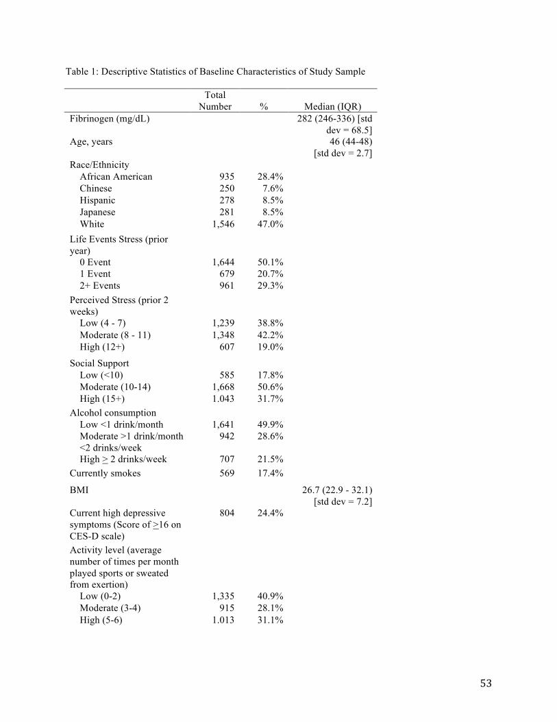
!
! 53!
Table 1: Descriptive Statistics of Baseline Characteristics of Study Sample
Total
Number % Median (IQR) Fibrinogen (mg/dL) 282 (246-336) [std
dev = 68.5] Age, years 46 (44-48)
[std dev = 2.7] Race/Ethnicity African American 935 28.4% Chinese 250 7.6% Hispanic 278 8.5% Japanese 281 8.5% White 1,546 47.0% Life Events Stress (prior year)
0 Event 1,644 50.1% 1 Event 679 20.7% 2+ Events 961 29.3% Perceived Stress (prior 2 weeks)
Low (4 - 7) 1,239 38.8% Moderate (8 - 11) 1,348 42.2% High (12+) 607 19.0%
Social Support Low (<10) 585 17.8% Moderate (10-14) 1,668 50.6% High (15+) 1.043 31.7% Alcohol consumption Low <1 drink/month 1,641 49.9% Moderate >1 drink/month <2 drinks/week
942 28.6%
High > 2 drinks/week 707 21.5% Currently smokes 569 17.4%
BMI 26.7 (22.9 - 32.1) [std dev = 7.2]
Current high depressive symptoms (Score of >16 on CES-D scale)
804 24.4%
Activity level (average number of times per month played sports or sweated from exertion)
Low (0-2) 1,335 40.9% Moderate (3-4) 915 28.1% High (5-6) 1.013 31.1%
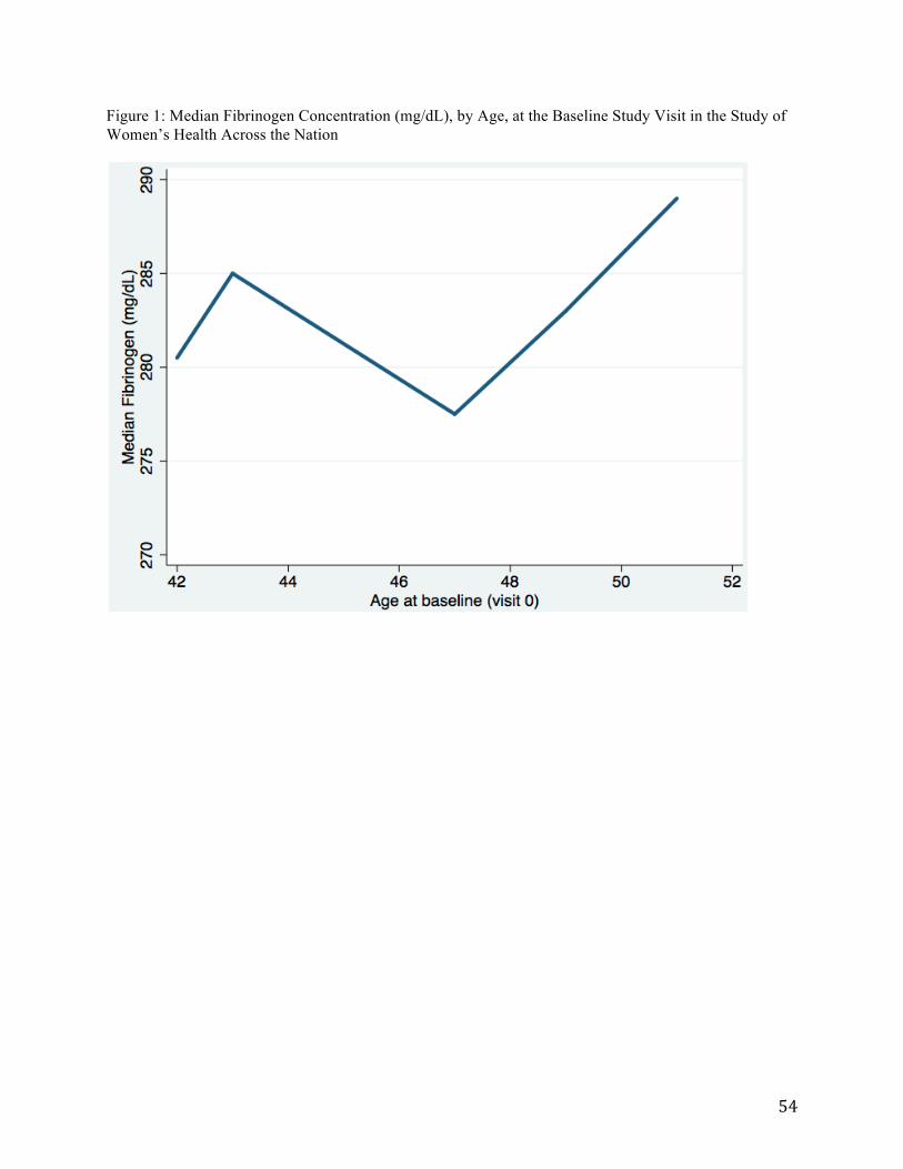
!
! 54!
Figure 1: Median Fibrinogen Concentration (mg/dL), by Age, at the Baseline Study Visit in the Study of Women’s Health Across the Nation
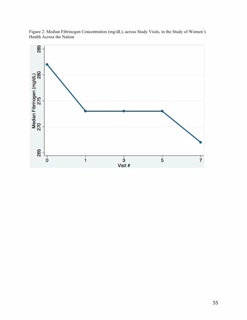
!
! 55!
Figure 2: Median Fibrinogen Concentration (mg/dL), across Study Visits, in the Study of Women’s Health Across the Nation
!
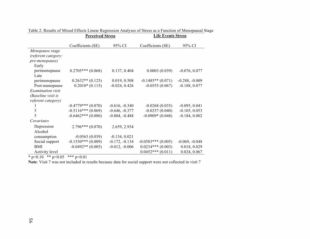
!
!
56!
Table 2: Results of Mixed Effects Linear Regression Analyses of Stress as a Function of Menopausal Stage Perceived Stress Life Events Stress
Coefficients (SE)
95% CI Coefficients (SE)
95% CI Menopause stage (referent category: pre-menopause) Early perimenopause 0.2705*** (0.068) 0.137, 0.404 0.0003 (0.039) -0.076, 0.077 Late perimenopause 0.2632** (0.125) 0.019, 0.508 -0.1485** (0.071) -0.288, -0.009 Post-menopause 0.2010* (0.115) -0.024, 0.426 -0.0555 (0.067) -0.188, 0.077 Examination visit (Baseline visit is referent category) 1 -0.4779*** (0.070) -0.616, -0.340 -0.0268 (0.035) -0.095, 0.041 3 -0.5116*** (0.069) -0.646, -0.377 -0.0257 (0.040) -0.105, 0.053 5 -0.6462*** (0.080) -0.804, -0.488 -0.0909* (0.048) -0.184, 0.002 Covariates Depression 2.796*** (0.070) 2.659, 2.934 Alcohol consumption -0.0563 (0.039) -0.134, 0.021 Social support -0.1530*** (0.009) -0.172, -0.134 -0.0583*** (0.005) -0.069, -0.048 BMI -0.0492** (0.005) -0.012, -0.006 0.0234*** (0.003) 0.018, 0.029 Activity level 0.0452*** (0.011) 0.024, 0.067
* p<0.10 ** p<0.05 *** p<0.01 Note: Visit 7 was not included in results because data for social support were not collected in visit 7
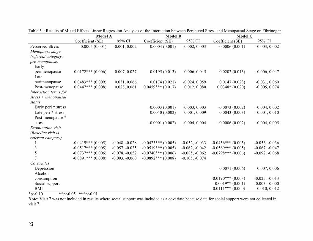
!
!
57!
Table 3a: Results of Mixed Effects Linear Regression Analyses of the Interaction between Perceived Stress and Menopausal Stage on Fibrinogen Model A Model B Model C Coefficient (SE) 95% CI Coefficient (SE) 95% CI Coefficient (SE) 95% CI Perceived Stress 0.0005 (0.001) -0.001, 0.002 0.0004 (0.001) -0.002, 0.003 -0.0006 (0.001) -0.003, 0.002 Menopause stage (referent category: pre-menopause) Early perimenopause 0.0172*** (0.006) 0.007, 0.027 0.0195 (0.013) -0.006, 0.045 0.0202 (0.013) -0.006, 0.047 Late perimenopause 0.0483*** (0.009) 0.031, 0.066 0.0174 (0.021) -0.024, 0.059 0.0147 (0.023) -0.031, 0.060 Post-menopause 0.0447*** (0.008) 0.028, 0.061 0.0459*** (0.017) 0.012, 0.080 0.0348* (0.020) -0.005, 0.074 Interaction terms for stress + menopausal status Early peri * stress -0.0003 (0.001) -0.003, 0.003 -0.0073 (0.002) -0.004, 0.002 Late peri * stress 0.0040 (0.002) -0.001, 0.009 0.0043 (0.003) -0.001, 0.010 Post-menopause * stress -0.0001 (0.002) -0.004, 0.004 -0.0006 (0.002) -0.004, 0.005 Examination visit (Baseline visit is referent category) 1 -0.0419*** (0.005) -0.048, -0.028 -0.0423*** (0.005) -0.052, -0.033 -0.0456*** (0.005) -0.056, -0.036 3 -0.0517*** (0.005) -0.057, -0.035 -0.0519*** (0.005) -0.062, -0.042 -0.0569*** (0.005) -0.067, -0.047 5 -0.0737*** (0.006) -0.078, -0.052 -0.0740*** (0.006) -0.085, -0.062 -0.0798*** (0.006) -0.092, -0.068 7 -0.0891*** (0.008) -0.093, -0.060 -0.0892*** (0.008) -0.105, -0.074 Covariates Depression 0.0071 (0.006) 0.007, 0.006 Alcohol consumption -0.0190*** (0.003) -0.025, -0.013 Social support -0.0019** (0.001) -0.003, -0.000 BMI 0.0111*** (0.000) 0.010, 0.012
*p<0.10 **p<0.05 ***p<0.01 Note: Visit 7 was not included in results where social support was included as a covariate because data for social support were not collected in visit 7.

!
!
58!
Table 3b: Results of Mixed Effects Linear Regression Analyses of the Interaction between Life Events Stress and Menopausal Stage on Fibrinogen
Model A Model B Model C Coefficient (SE) 95% CI Coefficient (SE) 95% CI Coefficient (SE) 95% CI
Life Events Stress 0.0029** (0.001) 0.000, 0.005 0.0050** (0.002) 0.001, 0.009 0.0036 (0.002) -0.001, 0.008 Menopause stage (referent category: pre-menopause) Early perimenopause 0.0174*** (0.005) 0.008, 0.027 0.0211*** (0.006) 0.010, 0.032 0.0170*** (0.006) 0.006, 0.028 Late perimenopause 0.0469*** (0.008) 0.030, 0.064 0.0477*** (0.010) 0.029, 0.066 0.0486*** (0.010) 0.029, 0.068 Post-menopause 0.0441*** (0.008) 0.028, 0.060 0.0468*** (0.009) 0.029, 0.064 0.0400*** (0.010) 0.021, 0.058 Interaction terms for stress + menopausal status Early peri * stress -0.0034 (0.003) -0.008, 0.002 -0.0037 (0.003) -0.009, 0.001 Late peri * stress -0.0003 (0.005) -0.010, 0.009 -0.0027 (0.005) -0.013, 0.008 Post-menopause * stress -0.0024 (0.003) -0.009, 0.004 -0.0016 (0.004) -0.009, 0.006 Examination visit (Baseline visit is referent category) 1 -0.0403*** (0.004) -0.048, -0.032 -0.0404*** (0.004) -0.048, -0.032 -0.0409*** (0.004) -0.049, -0.033 3 -0.0513*** (0.005) -0.061, -0.042 -0.0514*** (0.005) -0.061, -0.042 0.0554*** (0.005) -0.065, -0.046 5 -0.0736*** (0.006) -0.085, -0.062 -0.0738*** (0.006) -0.085, -0.063 -0.0800*** (0.006) -0.091, -0.068 7 -0.0881*** (0.008) -0.103, -0.073 -0.0884*** (0.008) -0.103, -0.073 Covariates Social support -0.0022*** (0.001) -0.004, -0.001 BMI 0.0111*** (0.000) 0.010, 0.012 Activity level -0.0019 (0.001) -0.005, 0.001
*p<0.10 **p<0.05 ***p<0.01 Note: Visit 7 was not included in results where social support was included as a covariate because data for social support were not collected in visit 7.

!
! 59!
Table 4a: Results of Mixed Effects Linear Regression Analyses of the Interaction between Lagged Perceived Stress and Menopausal Stages on Fibrinogen
*p<0.10 **p<0.05 ***p<0.01 Note: Lag was equal to one follow-up visit.
Contemporary and Lagged Variables Lagged Variables Only
Coefficients (SE) 95% CI Coefficients (SE) 95% CI Perceived stress 0.0018 (0.003) -0.004, 0.007 Menopause stage (referent category: pre-menopause) Early perimenopause 0.0415 (0.026) -0.010, 0.093 Late perimenopause 0.0150 (0.035) -0.054, 0.083 Post-menopause 0.0368 (0.032) -0.026, 0.099 Interaction terms for stress + menopausal status Early peri * stress -0.0035 (0.003) -0.010, 0.003 Late peri * stress 0.0031 (0.004) -0.005, 0.011 Post-menopause * stress -0.0011 (0.004) -0.008, 0.006 Examination visit (Baseline visit is referent category) 3 0.0304*** (0.008) 0.014, 0.047 0.0386*** (0.007) 0.025, 0.053 5 0.0086 (0.008) -0.008, 0.025 0.0222*** (0.007) 0.009, 0.036 7 0.0169 (0.011) -0.004, 0.038 0.0290*** (0.010) 0.010, 0.048 Lagged Terms Lagged stress 0.0010 (0.002) -0.006, 0.009 0.0015 (0.002) -0.001, 0.004 Lagged early perimenopause -0.0261 (0.021) -0.110, 0.028 0.0018 (0.016) -0.030, 0.034 Lagged late perimenopause -0.0062 (0.037) -0.189, 0.128 0.0101 (0.032) -0.053, 0.073 Lagged post-menopause 0.0108 (0.035) -0.115, 0.096 0.0344 (0.028) -0.021, 0.090 Lagged Interaction Terms Lagged early peri * stress 0.0020 (0.002) -0.003, 0.007 -0.0006 (0.002) -0.004, 0.003 Lagged late peri * stress -0.0037 (0.004) -0.012, 0.005 -0.0035 (0.004) -0.011, 0.004 Lagged post- menopause * stress -0.0030 (0.004) -0.011, 0.005 -0.0039 (0.003) -0.010, 0.003

!
! 60!
Table 4b: Results of Mixed Effects Linear Regression Analyses of the Interaction between Lagged Life Events Stress and Menopausal Stages on Fibrinogen
*p<0.10 **p<0.05 ***p<0.01 Note: Lag was equal to one follow-up visit.
Contemporary and Lagged Variables Lagged Variables Only
Coefficients (SE) 95% CI Coefficients (SE) 95% CI Life events stress 0.0060 (0.004) -0.002, 0.014 Menopause stage (referent category: pre-menopause) Early perimenopause 0.0181 (0.010) -0.001, 0.037 Late perimenopause 0.0448 (0.014) 0.018, 0.072 Post-menopause 0.0353 (0.014) 0.008, 0.063 Interaction terms for stress + menopausal status Early peri * stress -0.0041 (0.005) -0.013, 0.005 Late peri * stress -0.0048 (0.008) -0.020, 0.010 Post-menopause * stress -0.0069 (0.06) -0.018, 0.004 Examination visit (Baseline visit is referent category) 3 0.0284*** (0.006) 0.016, 0.041 0.0288*** (0.006) 0.017, 0.040 5 0.0162** (0.007) 0.002, 0.031 0.0212*** (0.007) 0.008, 0.034 7 0.0227** (0.010) 0.003, 0.043 0.0271*** (0.009) 0.009, 0.046 Lagged Terms Lagged stress -0.0021 (0.003) -0.008, 0.004 -0.0007 (0.003) -0.006, 0.005 Lagged early perimenopause -0.0130 (0.008) -0.029, 0.003 -0.0062 (0.006) -0.019, 0.007 Lagged late perimenopause -0.0400** (0.017) -0.073, -0.007 -0.0226** (0.014) -0.050, 0.004 Lagged post- menopause -0.0074 (0.016) -0.039, 0.024 0.0099 (0.013) -0.015, 0.034 Lagged Interaction Terms Lagged early peri * stress 0.0016 (0.004) -0.005, 0.009 0.0011 (0.003) -0.005, 0.008 Lagged late peri * stress 0.0073 (0.008) -0.009, 0.024 0.0058 (0.008) -0.010, 0.022 Lagged post- menopause * stress -0.0024 (0.006) -0.015, 0.010 -0.0048 (0.006) -0.016, 0.007

!
! 61!
Paper 3: Predicting old age life expectancy with conditions at perimenopause: A test of the sensitive periods model in adulthood
ABSTRACT Studies using the sensitive periods framework have traditionally focused on the effects of early life exposures on later life health. The menopausal transition (i.e., perimenopause), however, also has been suggested as a sensitive period because it is a narrow, well-defined window of dramatic physiological change. The application of the sensitive periods model to perimenopause was tested in this paper through an exploratory analysis of the relationship between female cohort mortality at ages 45-49 and longevity. Using time series methods, elevated mortality rates occurring during the age at which the perimenopause presumably occurred in two historic populations (France (1814-1919) and England and Wales (1841-1919)) were analyzed to determine how predictive they were of life expectancy at age 60. The results indicated a significant inverse association between mortality at ages 45-49 and life expectancy at age 60. The test was repeated among males of the same age in both countries, yielding no significant association in France. A significant association was found, however, between mortality at ages 45-49 and life expectancy at 60 among males in England and Wales. Additional testing in more countries and more contemporary settings will need to be conducted to more conclusively determine whether perimenopause plausibly explains the significant correlation of mortality at midlife with longevity found among women in this study. Keywords: Perimenopause, Critical Periods, Longevity, Life expectancy, Mortality

!
! 62!
INTRODUCTION Life course epidemiology suggests that various biological and social factors
occurring throughout the life span influences health in adulthood (1). Certain windows of time, however, represent critical or sensitive periods, during which environmental stimuli or threats can have particularly enduring effects on health and development (2). In critical periods, an exposure acting during a confined window of time has lasting effects on the structure or function of organs, tissues, and body systems, which cannot be modified in any significant way later in life (3). Sensitive periods are windows during which exposures have a stronger effect than they would have at other times. Outside these windows, excess disease risk associated with exposure is weaker (1, 4).
Studies testing the critical periods hypothesis have traditionally focused on the effects of early life exposures on later life health, which was pioneered by Barker’s work on the fetal programming of cardiovascular disease (1, 5). Much work since then has linked the fetal environment with various other morbidities, including diabetes mellitus, stroke, lung abnormalities, depression, and immune dysfunction (6). Such studies suggest that factors acting in prenatal and early postnatal life play an important role in the etiology of these diseases (2, 3). Researchers increasingly are finding that childhood and adolescence are critical periods for adult health outcomes as well. Studies show, for example, that environmental conditions and behaviors during childhood and adolescence can profoundly affect the risk for obesity and osteoporosis (7, 8).
A relatively scant body of evidence—in comparison to that on infancy and youth—also has investigated the role of critical and sensitive periods in adulthood. An example of such research is the clinical trials on the role of estrogen therapy during perimenopause, the transition period between regular pre-menopausal menstruation and the last menstrual period. This research has shown that hormone replacement therapy initiated during perimenopause—but not post-menopause—may prevent or delay cognitive decline in aging women (9). The significance of the sensitive periods model in adulthood is tested in this paper through an exploratory analysis of the relationship between women’s environmental conditions at mid-life and longevity.
Both individual-level and cohort-level studies have investigated the association between early life conditions and life expectancy, but the results of these studies implicate different age groups as sensitive periods. Evidence reporting the first year of life as a sensitive period shows that adverse circumstances experienced prior to age 1 are associated with significant decreases in cohort life expectancy (10, 11). Other research suggests the first five years of life are a sensitive period for life expectancy, indicating that deviations from trend in cohort mortality before age 5 is associated with shifts in later life cohort life expectancy (12, 13). Puberty also has been implicated as a sensitive period for longevity, with research showing an association between cohorts that experienced life-threatening stressors during adolescence and elevated mortality in adulthood, at least among males (14, 15).
No study, to my knowledge, has tested whether the presence of sensitive periods for longevity occur in adulthood. Individual level data, however, suggests that changes in risk for numerous diseases and health conditions—including cancer and cardiovascular disease—occurs during perimenopause (16). Adverse circumstances during this period of time, therefore, may have a more profound effect on health and life expectancy akin to the critical and sensitive windows occurring earlier in life. This paper used the sensitive
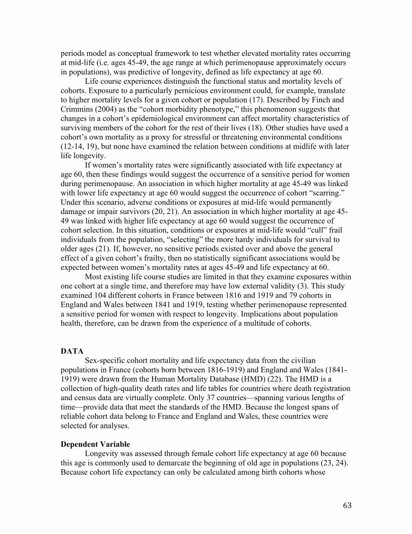
!
! 63!
periods model as conceptual framework to test whether elevated mortality rates occurring at mid-life (i.e. ages 45-49, the age range at which perimenopause approximately occurs in populations), was predictive of longevity, defined as life expectancy at age 60.
Life course experiences distinguish the functional status and mortality levels of cohorts. Exposure to a particularly pernicious environment could, for example, translate to higher mortality levels for a given cohort or population (17). Described by Finch and Crimmins (2004) as the “cohort morbidity phenotype,” this phenomenon suggests that changes in a cohort’s epidemiological environment can affect mortality characteristics of surviving members of the cohort for the rest of their lives (18). Other studies have used a cohort’s own mortality as a proxy for stressful or threatening environmental conditions (12-14, 19), but none have examined the relation between conditions at midlife with later life longevity.
If women’s mortality rates were significantly associated with life expectancy at age 60, then these findings would suggest the occurrence of a sensitive period for women during perimenopause. An association in which higher mortality at age 45-49 was linked with lower life expectancy at age 60 would suggest the occurrence of cohort “scarring.” Under this scenario, adverse conditions or exposures at mid-life would permanently damage or impair survivors (20, 21). An association in which higher mortality at age 45-49 was linked with higher life expectancy at age 60 would suggest the occurrence of cohort selection. In this situation, conditions or exposures at mid-life would “cull” frail individuals from the population, “selecting” the more hardy individuals for survival to older ages (21). If, however, no sensitive periods existed over and above the general effect of a given cohort’s frailty, then no statistically significant associations would be expected between women’s mortality rates at ages 45-49 and life expectancy at 60.
Most existing life course studies are limited in that they examine exposures within one cohort at a single time, and therefore may have low external validity (3). This study examined 104 different cohorts in France between 1816 and 1919 and 79 cohorts in England and Wales between 1841 and 1919, testing whether perimenopause represented a sensitive period for women with respect to longevity. Implications about population health, therefore, can be drawn from the experience of a multitude of cohorts. DATA
Sex-specific cohort mortality and life expectancy data from the civilian populations in France (cohorts born between 1816-1919) and England and Wales (1841-1919) were drawn from the Human Mortality Database (HMD) (22). The HMD is a collection of high-quality death rates and life tables for countries where death registration and census data are virtually complete. Only 37 countries—spanning various lengths of time—provide data that meet the standards of the HMD. Because the longest spans of reliable cohort data belong to France and England and Wales, these countries were selected for analyses.
Dependent Variable
Longevity was assessed through female cohort life expectancy at age 60 because this age is commonly used to demarcate the beginning of old age in populations (23, 24). Because cohort life expectancy can only be calculated among birth cohorts whose

!
! 64!
members have virtually all died, the time series for France and England and Wales end in 1919. Life expectancy at age 60 in both France and England and Wales gradually increased over the time series analyzed, although was interrupted by a few major events during the 19th and 20th centuries. In France, epidemics of cholera (1832, 1849, 1854) and the Franco-Prussian War (1870-1871) for example, temporarily decreased life expectancy. Likewise, in England and Wales, outbreaks of typhoid and flu (1846-1848) and cholera (1832, 1849, and 1865-1866) caused temporary spikes in mortality that affected life expectancy. General trends of increasing life expectancy, as well as the temporary interruptions caused by war and epidemics, were controlled in analysis. Independent Variables
Female cohort mortality rates at ages 45-49 were used to indicate the conditions or circumstances to which a given cohort was exposed during perimenopause. Elevated mortality rates that occurred significantly above what would be statistically expected implied the presence of adverse conditions affecting the population. Cohort mortality rates at infancy (age 0) and five-year intervals which prior literature has indicated as critical or sensitive periods (i.e., early childhood (ages 1-4) and puberty (ages 10-14)) were tested for significance as potential predictors of female longevity. (Studies on menarcheal timing (i.e., first menses, which is a marker of puberty) and pubertal duration show the age range 10-14 likely encompasses the start and progression of pubertal events for most girls in France and England & Wales during the mid-19th Century (25-27).) The mortality rate for ages 55-59 was also included in the models to control for recency effects on life expectancy at age 60.
The age classification designated to represent perimenopause is indicative of the average female in France and England and Wales during the mid to late 1800s. Research indicates the average age of menopause occurs between 49 and 52 years of age in westernized countries (e.g., Europe, United States of America) (28-30), and most studies find that menopausal age has not changed significantly over time (31-33). With research reporting that perimenopause lasts approximately four years and begins on average between the ages of 45 and 47.5 (31, 34), the age range of 45-49 would encompass the start and progression for most of perimenopause.
As with life expectancy, mortality rates improved over the 19th and 20th centuries. This general decreasing trend for all age groups is controlled for in analysis.
METHODS
This test assessed whether life expectancy at age 60 changed from its statistically expected value given increases in mortality rates between the ages 45-49, controlling for cohort mortality rates in other presumed sensitive periods for female longevity, as well for endogenous cohort frailty. Several studies have provided evidence showing that a cohort’s life expectancy is most significantly influenced by the environmental conditions or exposures at certain ages. These sensitive windows for longevity include infancy (age 0), early childhood (ages 1-4), and puberty (ages 10-14) (12-14, 35). Cohort mortality rates from these age groups, therefore, were included as covariates. Because phenomena that would cause a cohort to die more frequently at midlife and at age 60 would be expected to manifest at other ages throughout the life course, cohort frailty was controlled
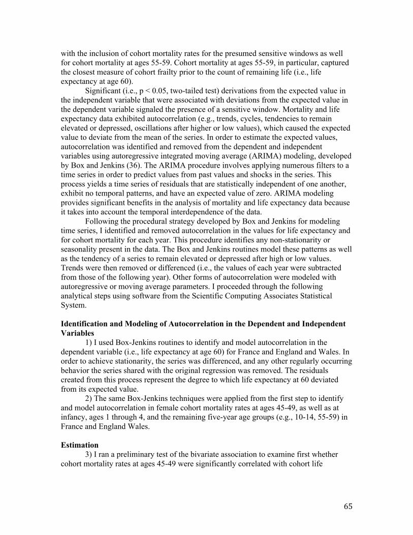
!
! 65!
with the inclusion of cohort mortality rates for the presumed sensitive windows as well for cohort mortality at ages 55-59. Cohort mortality at ages 55-59, in particular, captured the closest measure of cohort frailty prior to the count of remaining life (i.e., life expectancy at age 60).
Significant (i.e., p < 0.05, two-tailed test) derivations from the expected value in the independent variable that were associated with deviations from the expected value in the dependent variable signaled the presence of a sensitive window. Mortality and life expectancy data exhibited autocorrelation (e.g., trends, cycles, tendencies to remain elevated or depressed, oscillations after higher or low values), which caused the expected value to deviate from the mean of the series. In order to estimate the expected values, autocorrelation was identified and removed from the dependent and independent variables using autoregressive integrated moving average (ARIMA) modeling, developed by Box and Jenkins (36). The ARIMA procedure involves applying numerous filters to a time series in order to predict values from past values and shocks in the series. This process yields a time series of residuals that are statistically independent of one another, exhibit no temporal patterns, and have an expected value of zero. ARIMA modeling provides significant benefits in the analysis of mortality and life expectancy data because it takes into account the temporal interdependence of the data.
Following the procedural strategy developed by Box and Jenkins for modeling time series, I identified and removed autocorrelation in the values for life expectancy and for cohort mortality for each year. This procedure identifies any non-stationarity or seasonality present in the data. The Box and Jenkins routines model these patterns as well as the tendency of a series to remain elevated or depressed after high or low values. Trends were then removed or differenced (i.e., the values of each year were subtracted from those of the following year). Other forms of autocorrelation were modeled with autoregressive or moving average parameters. I proceeded through the following analytical steps using software from the Scientific Computing Associates Statistical System.
Identification and Modeling of Autocorrelation in the Dependent and Independent Variables
1) I used Box-Jenkins routines to identify and model autocorrelation in the dependent variable (i.e., life expectancy at age 60) for France and England and Wales. In order to achieve stationarity, the series was differenced, and any other regularly occurring behavior the series shared with the original regression was removed. The residuals created from this process represent the degree to which life expectancy at 60 deviated from its expected value. 2) The same Box-Jenkins techniques were applied from the first step to identify and model autocorrelation in female cohort mortality rates at ages 45-49, as well as at infancy, ages 1 through 4, and the remaining five-year age groups (e.g., 10-14, 55-59) in France and England Wales.
Estimation 3) I ran a preliminary test of the bivariate association to examine first whether cohort mortality rates at ages 45-49 were significantly correlated with cohort life

!
! 66!
expectancy at age 60. This step involved adding the residuals of the age 45-49 mortality series to the equation formed from modeling female cohort life expectancy. 4) The parameters for the full Box-Jenkins models for both countries were estimated using maximum likelihood estimation. In this step, the residuals of the other age-specific mortality series generated from the second step were added to the model generated in step 3. The test equations that emerged from this process for France and England and Wales are as follows:
∇YtFrance = C + ω1∇X0t + ω2∇X1-4t +ω3∇X10-14t + ω4∇X45-49t + ω5∇X55-59t +
at
∇YtEngland & Wales = C + ω1X0t + ω2∇X1-4t +ω3∇X10-14t + ω4∇X45-49t + ω5∇X55-59t +
at ∇ is the difference operator indicating that Y has been differenced from Yt-1 to remove trends and cycles from the series and render stationarity in its mean. Yt equals female cohort life expectancy at age 60 at year t. C is a constant. ω1 through ω5 is the estimated parameters for the residuals of the age-specific cohort mortality rates. X0t through X55-59t are the residuals of the best fitting models of cohort mortality rate for each age group. Bn is the backshift operator that either or !acts on the value of the error term ‘a’ at year t-q or t-p.
is the moving average parameter. !is the autoregressive parameter
at is the error term at year t. RESULTS
Cohort life expectancy at age 60 increased for French females between 1816 and 1919, rising 11 years—from 14.2 (age 74.2) to 25.2 (age 85.2) remaining years of life (see Figure 1). Cohort life expectancy at age 60 also increased for females in England and Wales during the time series (1841-1919), rising 7.4 years, from 15.3 (age 75.3) to 22.7 (age 82.7) remaining years of life (see Figure 2). Table 1 shows the equations modeling cohort life expectancy at age 60 for these two countries. The difference operator,
€
∇, in the equations for both France and England and Wales indicates there was a general increasing trend in life expectancy at age 60 throughout the series. The autoregressive parameters (i.e., 0.387, standard error = 0.095; and 0.536, standard error = 0.079) in the model of French life expectancy suggests that high or low values carried into succeeding years, with “echoes” of such values appearing 1 and 10 years later, but decreased geometrically. The positive, albeit small, coefficient (0.090, standard error = 0.018) in the model for England and Wales indicates that life expectancy slightly drifted above the general upward trend during many of the years between 1841 and 1919.

!
! 67!
Table 2 shows the equations that modeled cohort mortality rates in France and England and Wales. A decreasing trend in female mortality rates in nearly all age groups was observed in both countries. This trend was removed by taking the first differences of the series. Each of the age groups also exhibited various moving average and autoregressive patterns, which were subtracted from the cohort mortality series. The negatively signed constant values for several of the equations indicate the rates drifted below the general downward trend for many of the years in the series.
The results in Table 3 show that cohort mortality at ages 45-49 was predictive of life expectancy at age 60 in the test of bivariate association in France (p<0.01) after autocorrelation was removed from both variables. In England and Wales, the association approached significance (p=0.053). Cohort mortality at ages 45-49 was significant (p<0.01) for both countries in the full models that controlled for cohort mortality in the other presumed sensitive windows. Results indicate that life expectancy at age 60 among females in France between 1816 and 1919 and in England and Wales between 1841 and 1919 varied inversely with female cohort mortality rates between the ages 45-49.
Cohort mortality rates for ages 55-59 were also significantly associated with life expectancy at age 60 in both countries, along with cohort mortality at infancy—but only in England and Wales. The coefficient for cohort mortality at ages 55-59 was the most significant variable in the models for both countries, suggesting that conditions immediately preceding the measure of life expectancy have the greatest predictive power on life expectancy at age 60. The coefficient for cohort mortality at ages 45-49 was the next most significant covariate for both countries, followed lastly by the covariate for infancy in England and Wales.
The coefficients shown in Table 3 indicate that life expectancy at age 60 among female cohorts in France and England and Wales decreased by 0.1292 years (1.55 months) and 0.2029 years (2.43 months) respectively, for each increase of 1 per thousand in the age 45-49 mortality rate. To translate into a more interpretable context, mortality rates for age 45-49 were transformed from a continuous variable to a binary variable equal to 1 for cohorts exhibiting greater than expected death rates during ages 45-49 and 0 otherwise. The test equations were run again, with results of the coefficient for the binary variable revealing that life expectancy at age 60 decreased 0.045 years (0.54 months) on average for French female cohorts that experienced higher than expected mortality at ages 45-49. Among similarly aged females in England and Wales, life expectancy at age 60 decreased 0.059 years (0.71 months). As a frame of reference, cohort life expectancy at age 60 for French females increased 11 years, or an average of 1.27 months per year, over the 104 years analyzed for this study. Female cohort life expectancy in England and Wales increased 7.4 years, or an average of 1.12 months, over the 79 years in this study. These findings suggest that “threatened” perimenopausal females lost approximately half (43% in France and 63% in England and Wales) of the gains in life expectancy that females in this age group acquired due to general improvements in health, modernization, etc. that occurred during the 19th Century.
As a robustness test for the female-specific findings, additional models were run in which cohort mortality for infancy and all five-year age groups leading up to 60 (e.g., age 0, 1-4, 5-9….55-59) were included as covariates. These analyses did not yield significantly different outcomes from the results of the original models, with the age 45-

!
! 68!
49 category remaining the most statistically significant predictor of life expectancy at age 60 (after cohort mortality at age 55-59) for both countries.
The analyses described above for modeling cohort life expectancy and cohort mortality rates was repeated for males, for whom theory would predict no significant association, given that they do not experience an equivalent—and abrupt—shift in hormone concentrations at mid-life (37). The results in Table 4 show there was no significant bivariate association between male cohort mortality at ages 45-49 and male cohort life expectancy at age 60 for both France and England and Wales. When models were fitted that included the five age groups included in the equivalent female models, however, the age 45-49 category appeared significantly associated with life expectancy at age 60 for males in England and Wales only. Only the age 55-59 group was significantly associated with life expectancy at age 60 among males in France. DISCUSSION
Results of analyses showed a significant, inverse association between conditions at mid-life and life expectancy at age 60 among females. Mortality rates among females ages 45-49 were predictive of life expectancy at age 60, such that unexpected increases in mortality were related to concomitant decreases in life expectancy at age 60. Stressors or adverse exposures occurring during this window appear to have a greater association with life expectancy at age 60 than similar exposures in other age groups, including infancy. These results suggest the “scarring” or “damaged cohort” hypothesis, where adverse conditions appear to permanently damage or impair survivors of the cohort.
It is unlikely that these results occurred due to frailty associated with some cohorts that happened to die more frequently at ages 45-49 as well as in later life, given that endogenous cohort frailty was controlled with the inclusion of cohort mortality at infancy, childhood, adolescence, and ages 55-59. The remaining variance in mortality at ages 45-49 therefore, was unique and significant above and beyond any variance that mortality rates in this age group shared with mortality rates in other age groups.
The finding of a significant association between mortality rates at ages 55-59 and life expectancy was somewhat expected, given that conditions occurring in this window immediately precede the measure of the dependent variable. The significant association of the age 45-49 category with life expectancy at age 60 plausibly implicates perimenopause as the explanation for this finding. Perimenopause represents a time of physiological reorganization in a woman’s life, during which rapid changes occur in hormonal levels (38, 39). This physiological restructuring may leave women particularly vulnerable to both environmental threats and stimuli as they transition from a reproductive to non-reproductive state (16). These changes are consistent with the notion of critical and sensitive periods, during which intrinsic changes in the organization of an organism occur rapidly and when regulatory pathways are being constructed or modified (1, 4).
While the lack of an analogous relationship among French males lends further support to the proposed explanation of perimenopause as a significant period for longevity, the significant association among males in England and Wales weakens this inference. Additional research testing this study’s hypothesis in other countries and in contemporary populations would need to be conducted in order to determine whether the
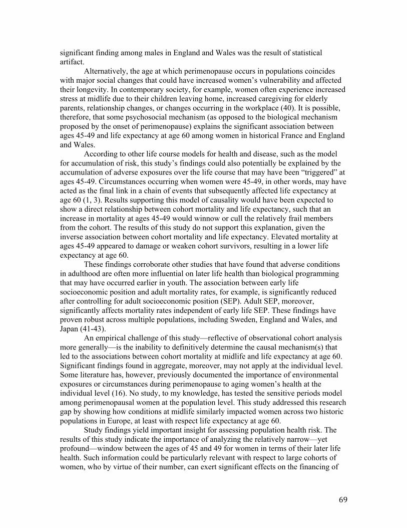
!
! 69!
significant finding among males in England and Wales was the result of statistical artifact.
Alternatively, the age at which perimenopause occurs in populations coincides with major social changes that could have increased women’s vulnerability and affected their longevity. In contemporary society, for example, women often experience increased stress at midlife due to their children leaving home, increased caregiving for elderly parents, relationship changes, or changes occurring in the workplace (40). It is possible, therefore, that some psychosocial mechanism (as opposed to the biological mechanism proposed by the onset of perimenopause) explains the significant association between ages 45-49 and life expectancy at age 60 among women in historical France and England and Wales. According to other life course models for health and disease, such as the model for accumulation of risk, this study’s findings could also potentially be explained by the accumulation of adverse exposures over the life course that may have been “triggered” at ages 45-49. Circumstances occurring when women were 45-49, in other words, may have acted as the final link in a chain of events that subsequently affected life expectancy at age 60 (1, 3). Results supporting this model of causality would have been expected to show a direct relationship between cohort mortality and life expectancy, such that an increase in mortality at ages 45-49 would winnow or cull the relatively frail members from the cohort. The results of this study do not support this explanation, given the inverse association between cohort mortality and life expectancy. Elevated mortality at ages 45-49 appeared to damage or weaken cohort survivors, resulting in a lower life expectancy at age 60.
These findings corroborate other studies that have found that adverse conditions in adulthood are often more influential on later life health than biological programming that may have occurred earlier in youth. The association between early life socioeconomic position and adult mortality rates, for example, is significantly reduced after controlling for adult socioeconomic position (SEP). Adult SEP, moreover, significantly affects mortality rates independent of early life SEP. These findings have proven robust across multiple populations, including Sweden, England and Wales, and Japan (41-43).
An empirical challenge of this study—reflective of observational cohort analysis more generally—is the inability to definitively determine the causal mechanism(s) that led to the associations between cohort mortality at midlife and life expectancy at age 60. Significant findings found in aggregate, moreover, may not apply at the individual level. Some literature has, however, previously documented the importance of environmental exposures or circumstances during perimenopause to aging women’s health at the individual level (16). No study, to my knowledge, has tested the sensitive periods model among perimenopausal women at the population level. This study addressed this research gap by showing how conditions at midlife similarly impacted women across two historic populations in Europe, at least with respect life expectancy at age 60.
Study findings yield important insight for assessing population health risk. The results of this study indicate the importance of analyzing the relatively narrow—yet profound—window between the ages of 45 and 49 for women in terms of their later life health. Such information could be particularly relevant with respect to large cohorts of women, who by virtue of their number, can exert significant effects on the financing of

!
! 70!
and demand for health and social services. For example, the height of the baby boom in the United States occurred in 1957. As the largest cohort of women in the U.S. history approaches 60 in the year 2017, it may be useful to factor into forecasts how their health status affects the demand for and costs of services. Assessing the health of these women during the not-too-distant past (i.e., between 2002-2006) could provide insight on what is to come for aging women. Conclusion
In sum, results from this study suggest a degree of plasticity associated with women’s aging. The period of perimenopause may indicate a sensitive period for women in terms of the development or modification of health trajectories into old age. Although women’s reproductive health is integral to their overall health and wellbeing, little is known about the interrelationship between social and lifestyle factors and reproductive health and how these vary across populations and cultures (40). Assessing the health of women during this window could provide insight for the timing and targeting of preventive health interventions.
!
! 71!
Works Cited
1. Kuh D, Ben-Shlomo Y, Lynch J, Hallqvist J, Power C. Life course epidemiology. Journal of Epidemiology and Community Health. 2003;57:778-83. 2. Cameron N, Demerath E. Critical periods in human growth and their relationship to diseases of aging. Yearbook of Physical Anthropology. 2002;46:159-84. 3. Ben-Shlomo Y, Kuh D. A life course approach to chronic disease epidemiology: conceptual models, empirical challenges, and interdisciplinary perspectives. International Journal of Epidemiology. 2002;31(2):285-93. 4. Halfon N, Hochstein M. Life course health development: An integrated framework for developing health, policy, and research. Milbank Quarterly. 2002;80(3):433-79. 5. Barker D. Fetal origins of coronary heart disease. BMJ. 1995;311:171-4. 6. Calkins K, Devaskar S. Fetal origins of adult disease. Pediatric and Adolescent Health Care. 2011;41(6):158-76. 7. Del Rio L, Carrascosa A, Pons F, Gusinye M, Yeste D, Domenech F. Bone mineral density of the lumbar spine in white Mediterranean Spanish children and adolescencts: Changes realtd to age, sex, and puberty. Pediatric Research. 1994;35(3):362-6. 8. Dietz WH. Critical periods in childhood for the development of obesity. The American Journal of Clinical Nutrition. 1994;59(5):955-9. 9. Sherwin B. Estrogen therapy: is time of initiation critical for neuroprotection? Nature Reviews Endocrinology. 2009;5:620-7. 10. Bengtsson T, Lindstrom M. Childhood misery and disease in later life: The effects on mortality in old age of hazards experienced in early life, southern Sweden, 1760-1894. Population Studies. 2000;54(3):263-77. 11. Costa D, Lahey J. Predicting older age mortality trends. Journal of the European Economic Association. 2005;3(2-3):487-93. 12. Myrskyla M. The effects of shocks in early life mortality on later life expectancy and mortality compression: A cohort analysis. Demographic Research. 2010;22(12):289-320. 13. Catalano R, Bruckner T. Child mortality and cohort lifespan: A test of diminished entelechy. International Journal of Epidemiology. 2006;35:1264-9. 14. Falconi A, Gemmill A, Dahl R, Catalano R. Adolescent experience predicts longevity: Evidence from historical epidemiology. Journal of Developmental Origins of Health and Disease. 2014;5(3):171-7. 15. Horiuchi S. The long-term impact of war on mortality: Old age mortality of the first world war survivors in the Federal Republic of Germany. Population Bulletin of the United Nations. 1983;15:80-92. 16. Hoyt L, Falconi A. Puberty and perimenopause: Reproductive transitions and their implications for women's health. Social Science & Medicine. 2015;132:103-12. 17. Manton K, Stallard E, Corder L. Changes in the age dependence of mortality and disability: Cohort and other determinants. Demography. 1997;34(1):135-57. 18. Finch C, Crimmins E. Inflammatory exposure and historical changes in human life-spans. Science. 2004;305:1736-9.

!
! 72!
19. Kermack W, McKendrick A, McKinlay P. Lancet. 1934;1(5770):667-718. 20. Elo I, Preston S. Effects of early-life conditions on adult mortality: A review. Population Index. 1992;58(2):186-212. 21. Preston S, Hill M, Drevenstedt G. Childhood conditions that predict survival to advanced ages among African-Americans. Social Science & Medicine. 1998;47(9):1231-46. 22. Human Mortality Databse. University of California, Berkeley (USA) Max Planck Institute for Demographic Research (Germany); Available at www.mortality.org or wwww.humanmortality.de (data downloaded on March 1, 2015). 23. Lutz W, Sanderson W, Scherbov S. The coming acceleration of global population ageing. Nature. 2008;451(7):716-9. 24. Roebuck J. When does "old age" begin?: The evolution of the English definition. Journal of Social History. 1979;12:416-28. 25. Parent A-S, Teilmann G, Anders J, Skakkebaek NE, Toppari J, Bourguignon J-P. The timing of normal puberty and the age limits of sexual precocity: variations around the world, secular trends, and changes after migration. Endocrine Reviews. 2003;24(5):668-93. 26. Mishra G, Cooper R. Early life circumstances and their impact on menarche and menopause. Womens Health (Lond Engl). 2009;5(2):175-90. 27. Marshall W, Tanner J. Variations in pattern of pubertal changes in girls. Archives of Disease in Childhood. 1969;44:291-303. 28. Gold E. The timing of the age at which natural menopause occurs. Obstetrics and Gynecology Clinics of North America. 2011;38(3):425-40. 29. Collaborative Group on Hormonal Factors in Breast Cancer. Menarche, menopause, and breast cancer risk: Indivdiual participant meta-analysis, including 118,964 women with breast cancer from 117 epidemiological studies. Lancet Oncology. 2012;13(11):1141-51. 30. Burger H, Dudley E, Roberston D, Dennerstein L. Hormonal changes in the menopausal transition. Recent Progress in Hormone Research. 2002;57:257-75. 31. McKinlay SM. The Normal Menopause Transition: An Overview. Maturitas. 1996;23(2):137-45. 32. Boldsen J, Jeune B. Distribution of age at menopause in two Danish samples. Human Biology. 1990;62(2):291-300. 33. Sievert L. Menopause as a measure of population health: An overview. American Journal of Human Biology. 2001;13(4):429-33. 34. Leidy L. Biological aspects of menopause: Across the lifespan. Annual Review of Anthropology. 1994;23:231-53. 35. Beltran-Sanchez H, Crimmins E, Finch C. Early cohort mortality predicts the cohort rate of aging: An historical analysis. Journal of Developmental Origins of Health and Disease. 2012;3:380-6. 36. Box G, Jenkins G, Reinsel G. Time Series Analysis: Forecasting and Control. 3rd ed. London: Prentice Hall; 1994. 37. Gould D, Petty R, Jacobs H. The male manopause--does it exist? BMJ 2000;320:858-61. 38. Hill K. The demography of menopause. Maturitas. 1996;23(2):113-27.
!
! 73!
39. Cook M. Perimenopause: An opportunity for health promotion. Journal of Obstetric, Gynecologic & Neonatal Nursing. 1993;22(3):223-8. 40. Mishra Gea. InterLACE: A new International Collaboration for a Life Course Apporach to Women's Reproductive Health and Chronic Disease Events. Maturitas. 2013;74:235-40. 41. Ben-Shlomo Y, Smith G. Deprivation in infancy or in adult life: Which is more important for mortality risk? Lancet. 1991;337(8740):530-4. 42. Rosvall M, Chaix B, Lynch J, Lindstrom M, Merlo J. Similar support for three different life course socioeconomic models on predicting premature cardiovascular mortality and all-cause mortality. BMC Public Health. 2006;6(203). 43. Engelaer F, Milne E, van Bodegom D, Saito Y, Westendorp R, Kirkwood T. Malleability of human aging: The curious case of old-age mortality in Japan. Annual Reviw of Gerontology of Geriatrics. 2013;33:49-59.
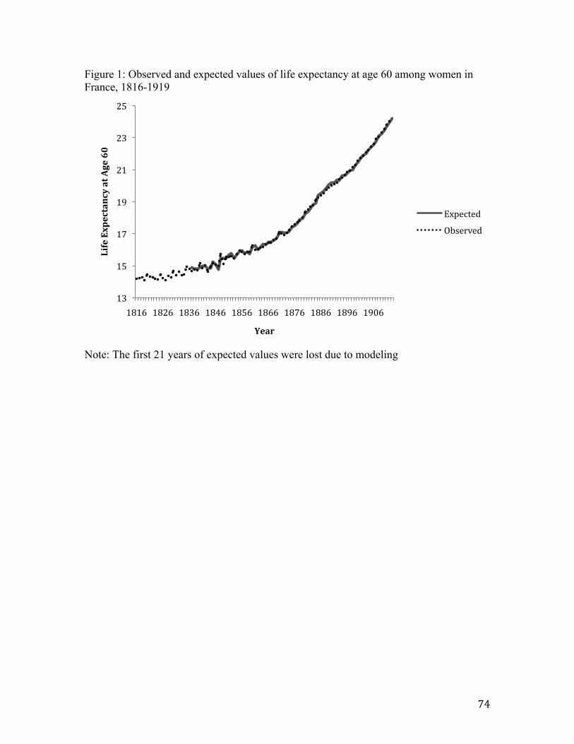
!
! 74!
Figure 1: Observed and expected values of life expectancy at age 60 among women in France, 1816-1919
Note: The first 21 years of expected values were lost due to modeling
13!
15!
17!
19!
21!
23!
25!
1816! 1826! 1836! 1846! 1856! 1866! 1876! 1886! 1896! 1906!
Life%Expectancy%at%Age%60%
Year%
Expected!Observed!

!
! 75!
Figure 2: Observed and expected values of life expectancy at age 60 among women in England and Wales, 1816-1919
Note: The first 17 years of expected values were lost due to modeling
13!
15!
17!
19!
21!
23!
25!
1850! 1860! 1870! 1880! 1890! 1900! 1910!
Life%Expectancy%at%Age%60%
Year%
Expected!Observed!

!
! 76!
Table 1. Box-Jenkins equations for female cohort life expectancy at age 60 for France (1816 through 1919) and England and Wales (1841 through 1919).
Country Box-Jenkins Equation
France
England and Wales
€
∇Ζ t = 0.0899 + at

!
!
77!
Table 2. Box-Jenkins equations for female cohort mortality rates at ages 0, 1-4, 10-14, 45-49, and 55-59 for France (1816 through 1919) and England and Wales (1841 through 1919).
France England and Wales
Age 0 !
Ages 1-4 !
Ages 10-14 !
!
Ages 45-49 ! !
Ages 55-59 ! !
€
∇Ζ t =1+ 0.3720B( ) 1+ 0.2352B4( )
1+ 0.2540Β5( )at
€
Ζ t = 0.1318 +1
1+ 0.9097Β( ) 1+ 0.3390Β6( )at
€
∇Ζ t =1− 0.2516B( ) 1+ 0.3206B4( )
1+ 0.3987Β5( )at
€
∇Ζ t = −0.0003+1
1− 0.3088Β3( )at
€
∇Ζ t =1+ 0.3506B6( )1+ 0.2596Β( )
at
€
∇Ζ t = −0.00005 + at
€
∇Ζ t = −0.0001+1+ 0.2859B9( )1+ 0.2214Β10( )
at
€
∇Ζ t = −0.0001+1
1− 0.2828Β( )at
€
∇Ζ t = −0.0001+1+ 0.4230B( )1− 0.3730Β5( )
at
€
∇Ζ t = −0.0002 +1
1− 0.0730Β2( ) 1+ 0.1615Β20( )at

!
!
78!
Table 3: Coefficients and standard errors for predictors of female cohort life expectancy at age 60 in France (1816-1919) and England and Wales (1841-1919) France England and Wales Model of bivariate association Model with covariates Model of bivariate
association Model with covariates
Variable Coefficient Standard Error Coefficient Standard
Error Coefficient Standard Error Coefficient Standard
Error Constant 0.1408*** 0.0197 0.1347*** 0.0187 0.0967*** 0.0161 0.1054*** 0.0096 Cohort death rate age 0 -0.8920 1.1891 -5.4115** 1.8838 Cohort death rate age 1-4 9.5157 11.3851 -14.2032 11.6961 Cohort death rate age 10-14 -68.6985 64.9568 -147.4615 102.0756 Cohort death rate age 45-49 -141.0330** 47.3646 -129.2326** 46.5874 -87.1607 44.5225 -202.8933*** 58.6037 Cohort death rate age 55-59 -133.2267*** 25.6761 -191.3639*** 50.5740 Box Jenkins parameters φΒ = -0.3590***
φΒ10 = 0.4418*** 0.1036 0.0920
φΒ = -0.0729 φΒ10 = 0.3385**
0.1144 0.1018
θΒ12 = 0.3564** φΒ5 = 0.4051***
0.1268 0.1109
θΒ12 = 0.1044 φΒ5 = 0.0191
0.1718 0.1316
*p<0.05, 2-tailed test **p<0.01, 2-tailed test ***p<0.001, 2-tailed test

!
!
79!
Table 4: Coefficients and standard errors for predictors of male cohort life expectancy at age 60 in France (1816-1919) and England and Wales (1841-1919) France England and Wales Model of bivariate association Model with covariates Model of bivariate association Model with covariates Variable Coefficient Standard
Error Coefficient Standard Error Coefficient Standard
Error Coefficient Standard Error
Constant 0.1038* 0.0464 0.1007* 0.0448 0.0439** 0.0142 0.0273* 0.0136 Cohort death rate age 0 -0.0791 0.5957 -4.9726* 1.9729 Cohort death rate age 1-4 10.1492 6.7549 2.3160 10.7743 Cohort death rate age 10-14 14.0645 47.2611 -122.6822 141.4054 Cohort death rate age 45-49 -7.6010 8.1314 -13.8859 8.9075 -56.1752 29.7463 -105.5038*** 28.7543 Cohort death rate age 55-59 -35.5029* 14.0658 -104.2389*** 23.7755 Box Jenkins parameters φΒ2 = 0.4509***
φΒ10 = 0.5987*** 0.0967 0.0814
0.4600*** 0.5898***
0.9866 0.0849
θΒ12 = 0.4024** φΒ = 0.3555**
0.1283 0.1101
*p<0.05, 2-tailed test **p<0.01, 2-tailed test ***p<0.001, 2-tailed test
!
!
! 80!
Conclusion Healthy aging is said to consist of three components: 1) survival to old age, 2) delay in
the onset of disease and disability, and 3) maintaining optimal physical and cognitive functioning for as long as possible (19). The results of the three papers in this dissertation contribute to the research base on all three components as they relate to women’s health. The scoping review in paper 1 compares the physiological and psychosocial changes females experience during perimenopause to puberty, a previously established sensitive period for some of the leading causes of disease and disability in the U.S. This review describes how environmental conditions and experiences during women’s reproductive transitions can profoundly affect their health as they age. The interaction of environmental and psychosocial stressors, health behaviors, genetics, or some combination of these factors, along with females’ rapidly changing hormones during puberty and perimenopause can trigger the onset of or profoundly affect the risk for depression, anxiety, metabolic-related morbidities, autoimmune disease, cardiovascular disease, cancer, musculoskeletal disorders, and premature mortality. The timing and nature perimenopause and its association with health outcomes are not identical to puberty, however, suggesting that these events are not simply markers of the same process. Rather, hormone changes during these two transitions appear to similarly influence women’s susceptibility for a number of diseases.
The results of the analyses in paper 2 show that while perimenopausal women perceived higher levels of stress, the increased perception of stress is not related to significant changes in fibrinogen, a biomarker involved in a number of pathophysiological mechanisms. Prior studies testing the interaction of biological reactivity by environment have been limited by the fact that they assume reactivity remains static throughout the life course (20). Evidence suggests, however, that physiological systems, in particular the HPA axis, are dynamic and are adaptive to an individual’s environment and stage in the life course. In other words, context matters, and physiological reactivity may exert weaker or stronger influences on sensitivity depending on a variety of different factors, (e.g., age, social environment, economic status) (21). The longitudinal study in paper 2 shows that while women’s psychological stress appears to change during the menopausal transition, the physiological effects of this perceived stress is not significantly modified. Additional research using different biomarkers would need to be conducted in order to more definitively determine whether and how the biochemical changes during perimenopause interact with the physiological effects of stress.
The results of the cohort analysis in paper 3 show how experiences at midlife, during the time when perimenopause was assumed to occur in historic France and England and Wales, appear predictive of women’s longevity. Elevated cohort mortality, indicative of adverse environmental exposures, is associated with decreases in life expectancy at age 60. The results suggest that survival into the postmenopausal years appears predicated on women’s experiences as they transition from a pre-menopausal to post-menopausal state. Once women reach mid-life, conditions during perimenopause are more associated with their likelihood of survival to old age than conditions and experiences in infancy and childhood.
Both policy and clinical implications related to the health of aging women emerge from the set of findings in this dissertation. From a policy perspective, knowledge of how women’s risks for chronic diseases and future life expectancy may change as a result of conditions during the menopausal transition can help improve forecasts of health and predictions of demand for health services. Models for forecasting mortality, morbidity, and disability in elderly populations are critical for planning and managing national and state health and social programs (22). The assumptions, however, about the trends in age-specific morbidity and health care use are often
!
! 81!
not self-evident (23). The reliability of such forecasts may be improved by the inclusion of information about women’s health at mid-life, which may significantly influence their health trajectories as they age.
From a clinical perspective, study findings from this dissertation suggest the importance of establishing interventions for health promotion strategies during the menopausal transition. One of the key components of the life course perspective is detecting periods in which health trajectories shift or when decline begins to accelerate—in particular, during critical and sensitive periods (19). The window defined by the menopausal transition, therefore, may offer opportunities to intervene in the trajectory of disease progression that may be difficult to alter if interventions are not begun until post-menopause (8).
Interventions targeted at addressing the areas of health in which women’s disease risk shifts during the menopausal transition may be the most strategic areas to explore. The initiation of pharmacologic interventions, such as hormone therapy, could potentially modify risk for conditions or diseases for women with genetic predispositions for conditions (e.g., autoimmune conditions) or whose timing of perimenopause may place them at increase risk for disease (e.g., cancer). Diet and behavioral interventions designed to improve nutrition and reduce sedentary behavior among perimenopausal women have the potential to reduce future risk for cardiovascular disease, metabolic disorders, and osteoporosis. Lastly, psychosocial interventions may offer women agency in how to manage stress and anxiety and how to process the somatic and psychological changes associated with aging.
Such information is particularly relevant given the apparent decline in health at older ages in the U.S. relative to most other wealthy countries (24). Improvements in mortality have stagnated among middle-aged and older U.S. women relative to U.S. men and to women in other wealthy nations. Much attention from the National Institutes of Health has focused, consequently, on determining the reasons for this deficit and exploring potential remedies (24). The physiological changes occurring during perimenopause that make women more sensitive to adverse conditions may also make them more responsive to interventions designed to counteract the effects of adversity (21). Perimenopause, therefore, may represent a period of opportunity for modifying prior trajectories of adverse health outcomes and establishing healthy aging trajectories.
Given the paucity of research on the role of critical and sensitive periods in adulthood, many questions remain unanswered, leaving open several different directions for further research. This dissertation empirically tested only a couple of health outcomes for which perimenopause may act as a sensitive window. More research is needed that tests the influence of environmental conditions during perimenopause with other measures of health.
Although the aging process and development of chronic diseases involve interactions among a variety of different factors, biomarkers can provide a specific characterization of physical, social, and behavioral environments. No single biomarker, however, will identify all of the environmental circumstances influencing health (25). Different types of psychological stress, in particular, appear to have very different effects on biomarkers (25). Research that builds upon the relation between psychological stress and biomarkers for systemic inflammation, as was analyzed in paper 2 in this dissertation, or other biomarkers for health more broadly, would provide a more comprehensive understanding of stress responsivity during the menopausal transition.
Another area prime for further research is in the field of reversibility of early adverse events. Research on critical periods has demonstrated the importance of timing in terms of

!
! 82!
exposures and their effects on health. Critical and sensitive periods may, however, also represent windows of opportunity (12). Animal research has shown, for example, that the effects of reduced maternal care on cognitive function in rats can be reversed with an enriched environment during puberty (26). Although the mechanism leading to this association is unknown, hormonal factors are posited as having an instrumental component. As another window of rapid hormonal change, perimenopause should be investigated as a window of plasticity for risk mechanisms (12).
Lastly, in order to understand the contributions of various stages of the life course on adult health and chronic disease, research and observation that involves decades, rather than just months or years, is needed (27). The cohort analysis in paper 3 attempts to address this issue by controlling for other critical and sensitive periods in the modeling of old age life expectancy and mortality rates at mid-life. The findings from this analysis revealed that conditions in mid-life appear more important at predicting old age life expectancy relative to early life conditions. Additional analyses of data that span the life course could perhaps provide a clearer idea of the extent and magnitude to which behaviors and exposures affect aging trajectories and the risk of developing chronic conditions.
In sum, this dissertation demonstrates how the biological and social changes occurring during perimenopause influence trajectories of aging and health. Although women may experience increased sensitivity to environmental stimuli that could potentially place them on trajectories of accelerated decline, this period may also allow for increased responsivity to the beneficial effects of health interventions.

!
! 83!
Works Cited 1. Vincent G, Velkoff V. The older population in the United States: 2010 to 2050. United States Census Bureau, 2010. 2. Ortman J, Velkoff V. An aging nation: The older population in the United States. United States Census Bureau, 2014. 3. Lee R, Mason A. Some macroeconomic aspects of global population aging. Demography. 2010;47 Suppl:S151-S72. 4. Robinson K. Trends in health status and health care use among older women. National Center for Health Statistics, 2007. 5. Bertakis K, Azari R, Helms L, Callahan E, Robbins J. Gender differences in the utilization of health care services. Journal of Family Practice. 2000;49(2):147-52. 6. Bodenheimer T, Fernandez A. High and risking health care costs. Part 4: Can costs be controlled while preserving quality? Annals of Internal Medicine. 005;143:26-31. 7. Healthy aging: Helping people live long and productive lives and enjoy a good quality of life. Centers for Disease Control and Prevention, 2011. 8. Appt S, Ethun K. Reproductive aging and risk for chronic disease: Insights from studies of nonhuman primates. Maturitas. 2010;67:7-14. 9. Workshop Summary. Network on Reversibility: Mid-Life Reversibility of Early Established Biobehavioral Risk Factors; 2013 February 26-27, 2013; Bethesda, Maryland: National Institute on Aging National Institutes of Health. 10. Halfon N, Hochstein M. Life course health development: An integrated framework for developing health, policy, and research. Milbank Quarterly. 2002;80(3):433-79. 11. Kuh D, Ben-Shlomo Y, Lynch J, Hallqvist J, Power C. Life course epidemiology. Journal of Epidemiology and Community Health. 2003;57:778-83. 12. Aging NIo, editor Meeting summary. Network on reversibility: Mid-life reversibility of early established biobehavioral risk factors; February 26-27, 2013; Bethesda, MD. 13. Soules MR, Sherman S, Parrott E, Rebar R, Santoro N, Utian W, et al. Executive Summary: Stages of the Reproductive Aging Workshop. Climacteric. 2001;97:267-72. 14. Burger H, Hale G, Robertson D, Dennerstein L. A review of hormonal chnages during the menopausal transition: Focus on findings from the Melbourne Women's Midlife Health Project. Human Reproduction Update. 2007;13(6):559-65. 15. McKinlay SM. The Normal Menopause Transition: An Overview. Maturitas. 1996;23(2):137-45. 16. Boldsen J, Jeune B. Distribution of age at menopause in two Danish samples. Human Biology. 1990;62(2):291-300. 17. Sievert L. Menopause as a measure of population health: An overview. American Journal of Human Biology. 2001;13(4):429-33. 18. Reinhardt U. Does the aging of the populatin really drive the demand for health care. Health Affairs. 2003;22(6):27-39. 19. Kuh D, Richards M, Cooper R, Hardy R, Ben-Shlomo Y. Life course epidemiology, ageing resarch, and maturing cohort studies: a dynamic combination for understanding healthy ageing. In: Kuh D, Cooper R, Hardy R, Richards M, Ben-Shlomo Y, editors. A Life Course Approach to Healthy Ageing. Oxford, UK: Oxford University Press; 2014. p. 3-15.
!
! 84!
20. Boyce W, Ellis B. Biological sensitivity to context: I. An evolutionary-developmental theory of the origins and functions of stress reactivity. Development and Psychopathology. 2005;17:271-301. 21. Obradovic J, Boyce W. Individual differences in behavioral, physiological, and genetic sensitivities to contexts: Implications for development and adaptation. Developmental Neuroscience. 2009;31:300-8. 22. Manton K, Singer B, Suzman R. The scientific and policy needs for improved health forecasting models for elderly populations. In: Manton K, Singer B, Suzman R, editors. Forecasting the Health of Elderly Populatiions. New York: Springer-Verlag; 1993. p. 3-36. 23. Evans R, McGrail K, Morgan S, Barer M, Hertzman C. Apocalypse NO: Population aging and the future of health care systems. Canadian Journal on Aging. 2001;20:160-91. 24. International Differences in Mortality at Older Ages: Dimensions and Sources. Crimmins E, Preston S, Cohen B, editors. Washington, DC: National Academies Press; 2011. 428 p. 25. Ewbank D. Biomarkers in social science research on health and aging: A review of theory and practice. In: Weinstein M, Vaupel J, Wachter K, editors. Biosocial Surveys. Washington, DC: National Academies Press; 2008. 26. Bredy T, Zhang T, Grant R, Diorio J, Meaney M. Peripubertal environmental enrichment reverses the effects of maternal care on hippocampal development and glutamate receptor subunit expression. European Journal of Neuroscience. 2004;20:1355-62. 27. Hertzman C. Human development, life course, intervention, and health. In: Pickles A, Maughan B, Wadsworth M, editors. Epidemiological Methods in Life Course Research. Oxford, UK: Oxford University Press; 2007. p. 111-36.