Symptômes internalisés, comportements externalisés et traits ...
Upload
independentCategory
view
3download
0
Peramorphic Traits in the Tokay Gecko Skull
Juan D. Daza,1* Aurelia A. Mapps,1 Patrick J. Lewis,1 Monte L. Thies,1 and Aaron M. Bauer2
1Department of Biological Sciences, Sam Houston State University, 1900 Avenue I, Huntsville Texas, 773412Biology Department, Villanova University, 800 Lancaster Avenue, Villanova, Pennsylvania 19085
ABSTRACT Traditionally, geckos have been conceivedto exhibit paedomorphic features relative to other liz-ards (e.g., large eyes, less extensively ossified skulls,and amphicoelous and notochordal vertebrae). In con-trast, peramorphosis has not been considered an impor-tant process in shaping their morphology. Here, westudied different sized specimens of Gekko gecko todocument ontogenetic changes in cranial anatomy,especially near maturity. Comparison of this specieswith available descriptions of other geckos resulted inthe identification of 14 cranial characteristics that areexpressed more strongly with size increase. These char-acteristics become move evident in later stages of post-hatching development, especially near maturation, andare, therefore, attributed to peramorphosis (hyperossifi-cation). ACCTRAN and DELTRAN character optimiza-tions were applied to these characters using a tree of11 genera derived from a gekkotan molecular phylog-eny. This analysis revealed that G. gecko expresses themajority of these putative peramorphic features nearmaturity, and that some of these features are alsoexpressed in species closely related to G. gecko. Thecharacters studied have the potential to be applied infuture phylogenetic and taxonomic studies of this groupof lizards. J. Morphol. 000:000–000, 2015. VC 2015 Wiley
Periodicals, Inc.
KEY WORDS: Gekko gecko; heterochrony; osteology;skeletal maturity; ontogeny
INTRODUCTION
The tokay gecko (Gekko gecko) is one of the mostanatomically well-studied gekkotan species. One ofthe earliest studies on the osteology of this speciesis part of a series of illustrations published by Blan-chard 1852–1864, and includes what is perhaps oneof the best and most detailed renderings of a geckoskeleton available. Other studies have analyzedvarious aspects of tokay skull morphology (e.g.,Boulenger, 1912; H€afferl, 1921; Lakjer, 1927; Well-born, 1933; Rieppel, 1984a; Ma and Xia, 1990; Her-rel et al., 1999, 2000; Conrad and Norell, 2006;Conrad, 2008; Evans, 2008; Payne et al., 2011;Daza et al., 2012) and other structures associatedwith the skull, such as the brain (Stoof et al., 1987;Russchen and Jonker, 1988; Gonzalez et al., 1990;Hoogland et al., 1994; Shen et al., 1998; Tanget al., 2000; Ma et al., 2002; Pang et al., 2005; Liet al., 2006; Wang et al., 2008, 2011); hearing appa-ratus (K€oppel and Authier, 1995; Vossen et al.,
2010; Christensen-Dalsgaard et al., 2011); eye(Brewster, 1836; Denton, 1956; Dunn, 1969;Werner, 1969; Citron and Pinto, 1973; Murphy andHowland, 1986; Loew, 1994; Loew et al., 1996);postcranial elements such as vertebrae, limbs andhip joint (Home, 1816; Hoffstetter and Gasc, 1969;Russell, 1975; Zaaf et al., 2001; Autumn, 2002;Gauthier et al., 2012; Tsai and Holliday, 2014);integumentary system (Chiu and Maderson, 1975,1980; Maderson and Chiu, 1970, 1981; Lauff et al.,1994); vocal cords, trachea, and head muscles(Sanders, 1870; Schwenk, 1988; Russell and Bauer,1990; Moore et al., 1991; Herrel et al., 1999, 2007);and the vascular system (Russell, 1981, 1982).Despite the abundance of anatomical studies,details about intraspecific functional and structuralchanges during skeletal ontogeny remain unknown.
Heterochrony, changes in the expression ofancestral characters through time, is recognizedas an important factor influencing phenotypic het-erogeneity (de Beer, 1930; McNamara, 1986).According to Alberch et al. (1979), heterochronicchanges can be grouped into two classes: paedo-morphosis “promotes” earlier developmental stagesof ancestors to adult stages in the organism understudy, while during peramorphosis the perturba-tions result in new morphologies, whose presenceis not inferred in the ancestor of the lineage lead-ing to the organism. Earlier definitions of newmorphologies considered them as the result of anincrease of complexity of organization by the
Contract grant sponsor: Department of Biological Sciences, Col-lege of Sciences, and the Office Research and Sponsored Programsat Sam Houston State University (to J.D.D., A.A.M., P.J.L.,M.L.T.).; Contract grant sponsor: National Science Foundation (toA.M.B.); Grant number’s: DEB 0844523 and 1019943; Contractgrant sponsor: Endowed Chair Fund at Villanova University (toA.M.B.).
*Correspondence to: Juan D. Daza, Department of Biological Sci-ences, Sam Houston State University, 1900 Avenue I, Huntsville,TX 77341. E-mail: [email protected]
Received 7 November 2014; Revised 26 February 2015;Accepted 7 March 2015.
Published online 00 Month 2015 inWiley Online Library (wileyonlinelibrary.com).DOI 10.1002/jmor.20389
VC 2015 WILEY PERIODICALS, INC.
JOURNAL OF MORPHOLOGY 00:00–00 (2015)

addition of parts or additional metamorphoses ofconstituent elements (the Meckel-Serres Law ofterminal addition, see Rieppel 1988 for a revisionand relevant references). However, it has beenargued that the new morphologies attributable toperamorphosis are merely modified charactersthat were already present in the ancestor (Brock,2000).
Gekkotans are regarded as the classic exampleof lizards with paedomorphic features. Some of theosteological characters identified as paedomorphicinclude amphicoelous vertebrae with notochordalcentra, paired premaxillary and parietal bones,proportionally larger eyes, reduction of circumorbi-tal bones resulting in an incomplete postorbitalbar, and loss of the supratemporal arch (Camp,1923; Kluge, 1967; Werner, 1971; Rieppel, 1984a;Bauer, 1990a; Abdala, 1996; Daza et al., 2008;Daza and Bauer, 2010).
Miniaturization is suggested to be a consequenceof paedomorphosis, with small size achieved bydeletion of the terminal adult ontogenetic stages ofthe larger ancestor (Gould, 1966; Hanken andWake, 1993). There are many gekkotan lineagesthat have experienced miniaturization (e.g., mostsphaerodactyls, Ebenavia, Saurodactylus, Cryptac-tites, Hemiphyllodactylus, Lygodactylus, Aprasia,Ophidiocephalus), but demonstrate no increase ofpaedomorphic features compared with larger spe-cies. In fact, small gekkotans show paedomorphicfeatures, such as paired premaxillae and completesynostosis of the basicranium, less frequently thando larger forms (Rivero-Blanco, 1976; Daza et al.,2008; Gamble et al., 2011).
Allometry, in its broadest sense, describes how thefeatures of living creatures change with size (Shin-gleton, 2010 and references therein). One classicexample of a scaling relationship is relating changein relative limb size with body size increase(Schmidt-Nielsen, 1984). This has been demon-strated in large varanid lizards, which develop mas-sive limbs as a response to the physical demands ofsupporting their bodies in sprawling limb posture(Christian and Garland, 1996). The skull of lizardsand other vertebrates also are affected by changesin size (Emerson and Bramble, 1993; Daza et al.,2009; Uros�evic et al., 2014). Although changes asso-ciated with miniaturization are relatively well stud-ied among gekkotans (e.g. Rieppel, 1984a, 1984b;Hanken, 1993; Daza et al., 2008), anatomicalchanges in large species remain less well studied.
It is known that size is an important factoraffecting bone shape (Alexander, 1971). The gekko-tan skull is assumed to be lightly built (Evans,2003, 2008), in part due to the loss of the postorbi-tal and supratemporal bars (Camp, 1923; Rieppel,1984a) which results in a pronounced cranial kine-sis (Herrel et al., 2000; Herrel et al. 2007). Thecombination of a delicate and highly kinetic skullmight appear incompatible with the mechanical
requirements of feeding on large (sometimes hard)and mobile prey species; however, observations oflarge gekkotans (e.g., G. gecko, Uroplatus gigan-teus, Phelsuma gigas, Eublepharis fuscus, Rhaco-dactylus leachianus; Bauer and Russell, 1991;Meiri, 2008) handling such types of prey indicatethat there should be anatomical modifications towithstand the strong physical demands in feedingperformance (See also Bauer 1990b).
In this article, we study changes in skull charac-teristics of subadult and adult specimens of G.gecko to analyze the effects of growth on skull anat-omy. We describe the changes in cranial features asanimals approach maximum size. The morphologi-cal changes are associated with increased ossifica-tion, and become more evident as the animalsapproach to maturity. Further examination of theseosteological characteristics in several closely relatedtaxa allows the identification of character statechanges in the phylogeny of these gekkotans. Infor-mation derived from this study can be applied infuture taxonomic studies, which are needed as cur-rent research (Brown et al., 2012; Heinicke et al.,2012) has revealed that the genus Gekko, as pres-ently construed, is paraphyletic.
MATERIALS AND METHODS
We examined the cranial osteology of G. gecko using theapproach suggested by Bell and Mead 2014, whereby severalskeletal specimens (both material and digital) were examined(Appendix 1). A total of 21 subadult and adult specimens wereexamined, including articulated and semi-articulated skulls ofboth juvenile and adult individuals. Additionally, we used 62specimens from 13 closely related species for comparison andoptimization of character states (Appendix 1).
The largest G. gecko specimen (SHSVM-H-0001-2014) wasexamined using three-dimensional models based on high-resolution x-ray computed tomographies (HRXCT). An additionalspecimen was available from the digital morphology library at theUniversity of Texas at Austin, Digimorph (FMNH 186818; TheDeep Scaly Project, 2008). All HRXCT specimens were scanned atthe University of Texas at Austin CT lab: specimen SHSVM-H-0001-2014 was scanned using 450 images (1024 3 1024 pixels)taken along the coronal plane, with each image representing athickness of 57mm (Fig. 1). Alternative digital cuts along otherplanes of reference (sagittal and horizontal) and digital segmenta-tion of bones, including removal of the osteoderms (Fig. 1A, C),were done using VGStudio MAX 2.2 (Volume Graphics GmbH,Heidelberg, Germany) and AvizoVR Standard 8.1.0 (VSG, Visual-ization Sciences Group, Burlington, Massachusetts, USA). Digitalradiographs were obtained at the Smithsonian InstitutionNational Museum of Natural History using a KevexTM PXS10-16W X-ray source and Varian Amorphous Silicon DigitalX-RayDetector PaxScanH 4030R set to 130 kV at 81 mA. Anatom-ical terms are consistent with recent anatomical descriptions ofgekkotans, the sphaerodactylids Sphaerodactylus rooselvelti andChatogekko amazonicus (Daza et al., 2008; Gamble et al., 2011),the gekkonid Narudasia festiva (Daza et al., 2012), and the pygo-podid Aprasia repens (Daza and Bauer, 2015).
Skeletal specimens often lack data on sex, ontogenetic age,body mass, locality, date of collection, and even basic measure-ments (Bell and Mead, 2014), which is the case of the majorityof the skeletonized specimens used. Measurements wereobtained using ImageJ v. 1.47 (Rasband, 2015). We used onlycomplete and articulated specimens to measure snout-ventlength (SVL) and skull length. The postcranial length was
2 DAZA ET AL.
Journal of Morphology

calculated by subtracting these two values; SVL was approxi-mated as the measurement from the tip of the snout to theintervertebral joint of caudal (pygal) vertebrae I and II, whichcorresponds to the position of the cloaca, based on observationsof cleared-and-stained specimens.
Morphological characters were optimized on a subtree from arecent phylogenetic analysis of the gekkotan genera using fivenuclear genes (RAG1, RAG2, CMOS, ACM4, and PDC) and 1mitochondrial gene (ND2) (Gamble et al., 2012). The treeincluded 11 terminals. The genus Gekko belongs to a clade ofchiefly Indo-Pacific gekkotans, and is closely related to Pseudo-gekko, Lepidodactylus, Luperosaurus, and Ptychozoon (the lat-ter two of which might actually make Gekko paraphyletic;Heinicke et al., 2012; Brown et al., 2012). This clade morebroadly includes the genera Nactus, Dixonius, and Heteronotia,hereafter the Indopacific gecko group, or IPGG. We also added
Gehyra mutilata as a representative species from the nearestclade to the IPGG and a gecko from the Family Sphaerodatyli-dae in which the identified peramorphic states are all absent(Sphaerodactylus roosevelti; Daza et al. 2008). We used thecomputer program WinClada (Nixon, 1990) and executed thedelayed transformation (DELTRAN) and accelerated transfor-mation (ACCTRAN) optimization functions to investigate char-acter state changes.
RESULTSOsteological Changes With Increasing Size inG. gecko
Size in this section is used in reference to skulllength. The bones that showed the greatest
Fig. 1. HRXCT of the skull of G. gecko (SHSVMH-0001-2014). A–B, dorsal; C–D, lateral; E, ventral views of the cranium; and F,lateral and G, medial views of the jaw. A and C show the distribution of osteoderms in relation to the cranium. Abbreviations: ar, artic-ular; bo, basioccipital; c, choana; cal, crista alaris; cob, compound bone; cor, coronoid; cpro, crista prootica; d, dentary; ect, ectopterygoid;en, external nares; ept, epipterygoid; f, frontal; fe, fenestra exochoanalis; j, jugal; msy, mandibular symphysis; mx, maxilla; n, nasal;occ, occipital condyle; oto, otooccipital; pal, palatine; par, parietal; pbsh, parabasisphenoid; pmx, premaxilla; pof, postorbitofrontal; pop,paroccipital process; prf, prefrontal; pro, prootic; pt, pterygoid; q, quadrate; rap, retroarticular process; sa, surangular; spht, sphenoocci-pital tubercle; spl, splenial; sa, surangular; so, supraoccipital; sof, suborbital fenestra; sq, squamosal; vo, vomer. Scale bar 5 50 mm.
3PERAMORPHOSIS IN GEKKO GECKO
Journal of Morphology
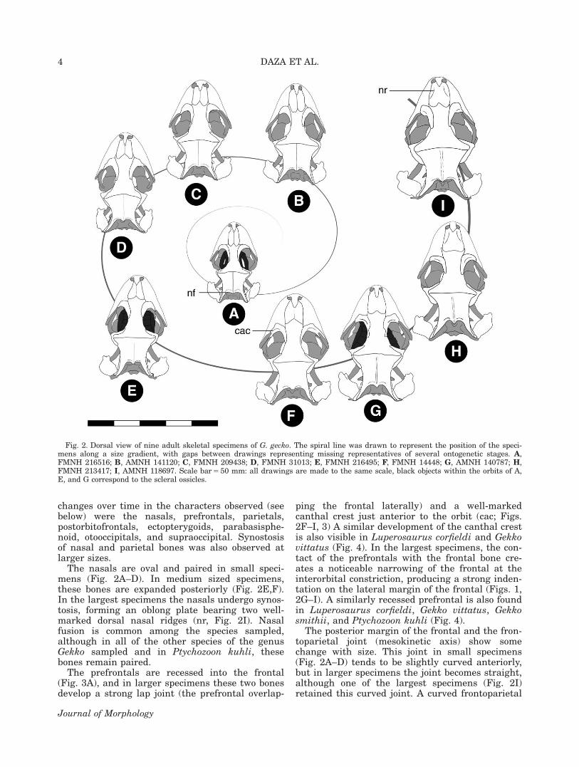
changes over time in the characters observed (seebelow) were the nasals, prefrontals, parietals,postorbitofrontals, ectopterygoids, parabasisphe-noid, otooccipitals, and supraoccipital. Synostosisof nasal and parietal bones was also observed atlarger sizes.
The nasals are oval and paired in small speci-mens (Fig. 2A–D). In medium sized specimens,these bones are expanded posteriorly (Fig. 2E,F).In the largest specimens the nasals undergo synos-tosis, forming an oblong plate bearing two well-marked dorsal nasal ridges (nr, Fig. 2I). Nasalfusion is common among the species sampled,although in all of the other species of the genusGekko sampled and in Ptychozoon kuhli, thesebones remain paired.
The prefrontals are recessed into the frontal(Fig. 3A), and in larger specimens these two bonesdevelop a strong lap joint (the prefrontal overlap-
ping the frontal laterally) and a well-markedcanthal crest just anterior to the orbit (cac; Figs.2F–I, 3) A similar development of the canthal crestis also visible in Luperosaurus corfieldi and Gekkovittatus (Fig. 4). In the largest specimens, the con-tact of the prefrontals with the frontal bone cre-ates a noticeable narrowing of the frontal at theinterorbital constriction, producing a strong inden-tation on the lateral margin of the frontal (Figs. 1,2G–I). A similarly recessed prefrontal is also foundin Luperosaurus corfieldi, Gekko vittatus, Gekkosmithii, and Ptychozoon kuhli (Fig. 4).
The posterior margin of the frontal and the fron-toparietal joint (mesokinetic axis) show somechange with size. This joint in small specimens(Fig. 2A–D) tends to be slightly curved anteriorly,but in larger specimens the joint becomes straight,although one of the largest specimens (Fig. 2I)retained this curved joint. A curved frontoparietal
Fig. 2. Dorsal view of nine adult skeletal specimens of G. gecko. The spiral line was drawn to represent the position of the speci-mens along a size gradient, with gaps between drawings representing missing representatives of several ontogenetic stages. A,FMNH 216516; B, AMNH 141120; C, FMNH 209438; D, FMNH 31013; E, FMNH 216495; F, FMNH 14448; G, AMNH 140787; H,FMNH 213417; I, AMNH 118697. Scale bar 5 50 mm: all drawings are made to the same scale, black objects within the orbits of A,E, and G correspond to the scleral ossicles.
4 DAZA ET AL.
Journal of Morphology

joint is found in adult specimens of all speciesreviewed in this study, except Heteronotia binoei.
The frontal and parietal bones are well bracedby the postorbitofrontal, especially the parietalwhich is supported by a dorsal shelf and insertsinto a medial socket of the postorbitofrontal. Theparietals are initially paired as is the case in thevast majority of gekkotans (Fig. 2A–C), but at anintermediate size they display partial fusionwhile retaining a flat surface (Fig. 2E). In largespecimens, the parietals fuse to become a singleunit (Fig. 1, 2F), and the seam between thesebones rises and develops a low sagittal crestalong the midline (Fig. 2G–I). Fusion of parietalsalso occurs to some extent in Luperosaurus cor-fieldi and Gekko smithii (partial fusion), andPseudogekko smaragdinus (total synostosis),whereas in G. vittatus and Ptychozoon kuhli theyremain paired. Among the species sampled onlyGekko gecko has a sagittal crest. The two skullsof Gekko smithii (which can grow to be nearly aslarge as G. gecko) examined are from a juvenile
and adult (skull length 5 15 mm, and 41 mm),but although they show features similar tomature G. gecko specimens at larger adult sizes,the degree of fusion of bones is less pronounced.The posteromedial process of the parietal extendsposteriorly, flanking the nuchal fossae (nf, Fig2A). Ventrally, the parietals are flat, althoughthey develop descending processes.
The joint between the ectopterygoid and ptery-goid is very well buttressed (Fig. 3B). Unlike inmany other gekkotans, the ectopterygoid is forkedposteriorly: these processes clasp around theanterolateral process of the pterygoid creating atongue-and-groove joint comparable to the samejoint in non-gekkotan lizards such as iguanians(Presch, 1969) and gymnophthalmids (Bell et al.,2003). The pterygoid also develops a wide jointwith the palatine, which contrasts with the greatlyreduced hypokinetic joint between these two bonesin other gekkotans (Rieppel, 1984a). The ectopter-ygoid is flanked laterally by the tall coronoid pro-cess of the lower jaw. When the mouth is closed,
Fig. 3. Illustration of some of the characters described. A, Luperosaurus corfieldi (CAS 182570); B and C transverse cuts at thelevel of the ectoterygoid-pterygoid joint, B, planar join (Lepidodactylus lugubris, CAS 224273), C, tongue and groove joint (Gekkogecko, SHSVMH-0001-2014). D and E, transverse cut anterior to the braincase, D, Gekko gecko (SHSVMH-0001-2014), E. G. gecko(FMNH 186818). Abbreviations: bp, basipterygoid process; hvc cor, coronoid; dpso, dorsal process of supraoccipital; ect, ectopterygoid;f, frontal; hvc, course of lateral head vein (0 5 open, 1 5 closed); par, parietal; parcr, parietal crest; pt, pterygoid; dpso, dorsal processof the supraocipital; prf, prefrontal; pt, pterygoid; so, suproccipital. Scale bars, A 5 50 mm, B 5 1 mm, C, D, E 5 10 mm.
5PERAMORPHOSIS IN GEKKO GECKO
Journal of Morphology

the coronoid apex extends above the palatal plane,thereby constraining lateral excursions of the jaw.Thus, the jaw creates a locking mechanism, espe-cially when the gape is shallow.
All of the bones that form the braincase (exceptthe orbitosphenoids) are fused. The braincase isperhaps the component of the skull that shows themost osteological modifications among the speciessampled. One of these changes occurs along withmodifications in the parietal. The supraoccipitaldevelops a dorsal process that forms a smoothfacet that contacts ventrally the posteromedialprocess of the parietal(s). A dorsal process of thesupraoccipital is not common among gekkotans(Daza et al., 2013), but is also developed in Pseu-dogekko smaragdinus, Luperosaurus corfieldi, andall of the Gekko species sampled. This dorsal pro-cess of the supraoccipital is located in the sameposition as the tectum of other lizards (tectumsynoticum 1 tectum posterius) but in Pseudogekko,Luperosaurus, and Gekko this process is compara-tively stouter, and appears more like a promontory
(Fig. 4C). Additionally, the presence of these skullroof elements in gekkotans is ambiguous, andalthough some of these parts have been reportedas being present only transiently during develop-ment (e.g., in Ptyodactylus, El-Toubi and Kamal,1961), the absence of the cartilaginous ascendingprocess of the tectum synoticum is regarded as asynapomorphy of the Gekkota (Rieppel, 1984a;Daza et al., 2013).
Modifications in the rear part of the braincaseare due to a proportional increase in width. Thisposterior enlargement is evidenced by a lateralelongation of the paroccipital process of the otooc-cipital, which changes the overall position of somebones of the skull. In the smallest specimens (skulllength<30 mm), the lateral edge of the paroccipi-tal process is aligned with the jugal-prefrontal(Fig. 2A–D), and as size increases the lateral edgeof this process shifts laterally and lines up withthe posterior tip of the jugal (Fig. 2G–I). Thisbroadening affects the position of the quadrate,squamosal, and parietal, and how they interact
Fig. 4. HRXCT of the skull of some of the comparative species from the IPGG. A, Lepidodactyluslugubris (CAS 224273); B, Pseudogekko smaragdinus (CAS 62344); C, Luperosaurus corfieldi (CAS182570), D, Gekko vittatus (CAS 20857), E, Nactus pelagicus (CAS 119003). Scale bar 5 20 mm.
6 DAZA ET AL.
Journal of Morphology

with the otooccipital. The paroccipital process alsodevelops circular facets on the anterior surface toform a joint with the cephalic condyle of the quad-rate, forming a type of quadrate suspension ingekkotans known as paroccipital abutting (Riep-pel, 1984a). As such, a proportional lateral shiftingof the paroccipital process with respect to the restof the skull would have repercussions for the posi-tion of the quadrate, the quadrate-articular joint,and in the orientation of the rami of the lower jawwith the cranium. Long paroccipital processes werepresent in all of the species sampled except Lepido-dactylus lugubris and Heteronotia binoei. In thesespecies, the paroccipital process is short and stoutand is consistent with previous observations onother miniaturized species (Daza et al., 2008, 2009)
Additional changes in the braincase of Gekkogecko occur in the prootic and parabasisphenoid.The prootic is not perforated by the mandibularbranch of the trigeminal nerve (Cranial nerve V;Conrad and Norell, 2006; Conrad and Daza, inpress); therefore, this nerve branch does not havea bony enclosure or lacks the medially locatedcrista trigeminalis (Daza et al., 2013), which inthe majority of gekkotans transforms the incisuraprootica into a foramen. This character is also
known in pygopodids (where the braincase isextremely modified) and in the eublepharid genusAleuroscalabotes (Conrad and Norell, 2006). In theparabasisphenoid, the enclosure of the lateralhead vein through the basipterygoid processes hasbeen reported as variable in Gekko gecko (Conradand Norell, 2006). We also found it variable inGekko vittatus and Lepidodatylus lugubris.
Optimization of Proposed PeramorphicCharacters
Peramorphs exhibit derived features that arenot inferred as present in the ancestor. Conse-quently, whether a character is peramorphic ornot can be tested by optimizing these characterson a tree that includes closely related taxa. Weoptimized 14 morphological characters of G. geckoand closely related species (Fig. 5; see also Table1). These characters include two binary homolo-gous character states where the second one isputatively attributed to paramorphism, and there-fore polarity is implied:
1. Nasal: internasal fusion, absent (0), present (1).2. Nasal: nasal ridge, absent (0), present (1).3. Pefrontal: dorsal process markedly recessed
into the frontal, absent (0), present (1).4. Prefrontal: canthal crest, absent (0), present (1).5. Ectopterygoid-pterygoid: tongue-and-groove
joint, absent (0), present (1).6. Ectopterygoid-coronoid interaction: ectoptery-
goid flanked laterally by the tall coronoid pro-cess (jaw locking mechanism), absent (0),present (1).
7. Parietal-postorbitofrontal joint: postorbitofron-tal with a dorsal shelf supporting the parietal,absent (0), present (1).
8. Parietal: Interparietal fusion, absent (0), pres-ent (1).
9. Parietal: parietal crest, absent (0), present (1).10. Parietal: nuchal fossae, subtle or absent (0),
conspicuous (1).
Fig. 5. Character optimization using the A, fast (ACTTRAN), and B, slow (DELTRAN) func-tions. White circles indicate homoplasy, solid black circles synapomorphic character. Tree topologybased on a subtree from a recent multigene phylogenetic analysis (Gamble et al., 2012).
TABLE 1. Character scores of species used for optimization. Seethe text for character description, on the result section
1 2 3 4 5 6 7 8 9 10 11 12 13 14
S. roosevelti 0 0 0 0 0 0 0 0 0 0 0 0 0 0G. mutilata 0 0 0 0 0 1 0 0 0 0 0 0 1 0L. lugubris 1 0 0 0 0 1 0 0 0 0 0 0 0 0P. smaragdinus 1 1 1 0 0 1 0 1 0 1 0 1 1 1L. corfieldi 1 1 1 1 1 1 0 0 0 1 0 0 1 1P. kuhli 0 ? 1 ? ? ? ? 0 0 ? ? ? 1 ?G. vittatus 0 1 1 1 1 1 0 0 0 1 0 0&1 1 1G. gecko 1 1 1 1 1 1 1 1 1 1 1 0&1 1 1D. siamensis 0 0 0 0 0 1 0 0 0 0 0 ? 1 0H. binoei 0 0 0 ? ? ? ? 0 0 ? ? ? 0 0N. pelagicus 1 0 0 0 1 1 0 0 0 0 0 0 1 0
7PERAMORPHOSIS IN GEKKO GECKO
Journal of Morphology

11. Prootic: mandibular branch of the trigeminalnerve with bony enclosure, present (0), absent (1).
12. Parabasisphenoid: basipterygoid process enclo-ses the lateral head vein course, absent (0),present (1).
13. Otoocciptial: paroccipital process, short andsquarish (0), long (1).
14. Supraoccipital: dorsal process, absent (0), pres-ent (1).
The characters optimized were also present inother groups outside the IPGG. A coronoid flankingthe ectopterygoid (Character 6-1) and a long paroc-cipital process (Character 13-1) were also found inGehyra mutilata; Lepidodactylus lugubris has ashort paroccipital process (Character 13-0), but inthis species this character state is interpreted asbeing a reversal. Additionally, some of the putativeperamorphic character states were not exclusive toG. gecko (Fig. 5), occurring in other members of theIPGG. Fused nasals (Character 1-1) and a tongue-and-groove joint between the ectopterygoid andpterygoid (Character 5-1) were also found in most
of the IPGG members, although other reversalswere found in the clades Dixonius siamen-sis 1 Heteronotia binoei and Ptychozoon kuh-li 1 Gekko vitattus (i.e., unfused nasals, character1-0). Additionally, the clade D. siamensis 1 H.binoei lacks a tongue-and-groove joint between theectopterygoid and pterygoid (Character 5-0)
The optimization functions clearly indicated thatfour (DELTRAN, Fig. 5A) or five (ACCTRAN, Fig.5B) characters identified as peramorphic supportthe clade formed by G. gecko, G. vitattus, P. kuhli,L. corfieldi, L. lugubris, and Ptychozoon smaragdi-nus. These are: the presence of a nasal ridge inthe nasal bone (Character 2-1), dorsal process ofthe frontal recessed into the frontal (Character 3-1), conspicuous nuchal fossae (Character 10-1),and the presence of a dorsal process in the supra-occipital (Character 14-1). All of these features areinterpreted as reversions in L. lugubris on thebasis of the optimization results; in this species,these character states might be lost due to minia-turization. Under ACCTRAN optimization, thepresence of a canthal crest (Character 4-1) sup-ports this clade, with a reversal in the Lepidodac-tylus lugubris 1 Pseudogekko smaragdinus clade.
From the list of characters including putativeperamorphic character states, G. gecko has threeautapomorphic characters: postorbitofrontal with ashelf for supporting the parietal bone (Character7-1), parietal crest (Character 9-1), and prootic notenclosing the mandibular branch of the trigeminalnerve (Character 11-1). Fusion of nasal bones(Character 1-1) and parietals (Character 8-1)occurred in many other sampled species, but theco-occurrence of these two characters was foundonly in G. gecko and Pseudogekko smaragdinus(fused nasals and partially fused parietals are alsofound in Luperosaurus corfieldi).
Allometric Scaling of the Skull of G. gecko
The record SVL for G. gecko is 178 mm (R€osleret al., 2011; Bauer, 2013); the largest specimenused in this analysis approximates this size (SVL=167 mm, Table 2). We plotted log postcraniallength (PL) against log skull length (SL) to explorethe relationship between these two linear varia-bles (Fig. 6). The equation resulting was
Fig. 6. Log skull length (SL) versus Log postcranial length(PL) of specimens of G. gecko. Small specimen USNM 163790,large specimen USNM 163791.
TABLE 2. Snout vent length (SVL), skull length (SL), and postcranial length (PL=SVL 2 SL) measurements from complete andarticulated specimens of Gekko gecko.
Specimen number SVL (mm) SL (mm) PL (mm) Log (SLV) Log (SL) Log (PL)
USNM 163790 47.66 16.02 31.64 1.6782 1.2047 1.5002USNM 163789 75.21 22.54 52.67 1.8763 1.353 1.7216USNM 163788 119.91 31.55 88.36 2.0789 1.499 1.9463USNM 163787 127.53 34.94 92.59 2.1056 1.5433 1.9666SHSVMH-0003-2014 134.49 38.9 95.59 2.1287 1.5899 1.9804SHSVMH-0002-2014 135.87 43.14 92.73 2.1331 1.6349 1.9672USNM 163791 149.27 39.35 109.92 2.174 1.5949 2.0411SHSVMH-0001-2014 167 53.17 113.83 2.2227 1.7257 2.0563
8 DAZA ET AL.
Journal of Morphology

y 5 0.8433x 2 0.0852, and the correlation betweenthese variables was very strong (R2 5 0.908). Theslope for this trend line indicates that the linearvariables considered approximate to an isometricrelationship: skull length increases proportionallywith postcranial length.
DISCUSSION
The Gekkonidae sensu stricto, currently recog-nized as comprising 56 genera (Uetz et al., 2015),is one of the seven families within Gekkota. Whileour sampling concentrates on a particular clade ofthe Gekkonidae, comparisons of the skull anatomyof Gekko gecko with all the available osteologicaldescriptions of geckos (See Daza et al., 2008 for alist) confirm the distinctiveness of the proposedperamorphic characters. In comparison with othergekkotans, the skull of G. gecko shows a remark-able amount of cranial modification, especiallyassociated with hyperossification. This observationalso is supported by a large database of 394 mor-phological characters of gekkotans (Daza, 2008).
The demonstrated peramorphic characters gen-erate new structures not widespread in other gek-kotans (e.g. sagittal and canthal crests,development bone processes, synostosis betweennasals and parietals). These characters are basi-cally hyperossifications of the skeleton (i.e., bonegrowth or some other form of calcification [usuallyinvolving cartilage] in excess of that found in theancestor), which has been considered an exampleof peramorphosis, although hyperossification hasonly been reported as being characteristic of mini-aturized forms (Hanken, 1993).
In the taxa sampled, we found some of thedescribed characters in miniaturized members ofthe IPGG, for example Lepidodactylus lugubrishas fused nasals, but shows reversion in themajority of the characters (Fig. 5).
These new character states need to be evaluatedin more gekkotans, especially within Gekkonidae.For example, the majority of gekkotans havepaired nasals, but fusion of nasals (character 1-1)has been found in many genera outside thesampled taxa, including Ailuronyx, Afroedura,Afrogekko, Cryptactites, Lygodactylus, Matoatoa,Paroedura, Phelsuma, Ramigekko, and Uroplatus(Heinicke et al., 2014; Daza, 2008).
Another variable feature is the fusion of parie-tals (character 8-1). This character state is moreinfrequent among gekkotans: in addition to G.gecko, it was found in Luperosaurus corfieldi(where fusion is partial) and Pseudogekko smar-agdinus. Outside the sampled taxa, fusion ofparietals has been found in the gekkonids Chon-drodactylus angulifer, Colopus wahlbergii, Crypt-actites peringueyi, Phelsuma lineata, andRamigekko swartbergensis, the Eublepharidae(Grismer, 1988; Heinicke et al., 2014; Daza,
2008), the diplodactylids Rhacodactylus leachia-nus, Rhacodactylus trachyrhynchus, and theextinct giant gecko Hoplodactylus delcourti(Bauer and Russell, 1986; Bauer, 1990b), and thepygopodid Lialis (McDowell and Bogert, 1954;Evans, 2008). In addition to the three species ofthe IPGG that have fusion of both the nasals andparietals, only Ailuronyx seychellensis, Rami-gekko swartbergensis, and Phelsuma lineata areknown to have this combination of characters.
The development of the parietal crest can beindependent of parietal fusion. In G. gecko, theparietal crest (Character 9-1) follows the fusion ofparietals; therefore, it seems to be a single struc-ture and not a double structure that arises fromboth parietals. The diplodactylids Rhacodactylusauriculatus and Correlophus ciliatus have a mid-dorsal parietal crest, while maintaining discreteparietals until maturity (Bauer, 1990a), indicatingthat in these species the crest is a double structurearising from both parietals. In the eublepharidHemitheconyx taylori, the fused parietals develop ashort posteromedial single sagittal crest (JDD per-sonal observation).
This optimization of characters also allows test-ing for homology of some of the charactersdescribed (reciprocal illumination). The dorsal pro-cess on the supraocccipital bone is in the same posi-tion, and possibly has the same function as theprocessus ascendens tectum synoticum of other liz-ards. This structure is an uncommon feature ofgekkotans (Daza et al., 2013). In fact, the closesttaxon to gekkotans in which the processus ascen-dens tectum synoticum has been described is thefossil gekkonomorph AMNH FR 21444 (Conrad andNorell, 2006; Conrad and Daza, in press). Giventhe nested phylogenetic position of Gekkonidaewithin the Gekkota (Gamble et al., 2012), it is verylikely that these structures are convergent.
Considering morphofunctional aspects, cranialanatomy defines feeding performance, and in manycases it is possible to establish a direct associationbetween cranial structures and diet. For instance,the gekkotan Lialis is a saurophagous specialistthat exhibits several adaptations in the skull, suchas a curved palatal arch, a very flexible mesokineticjoint, and recurved teeth. This type of association isless evident in nonspecialist species, which mayhave a more general skull structure and for whichdietary preferences change through their ontogeny,such as the situation in G. gecko. However, changesin dietary breadth may be a possible by-product ofthe hyperossification of the skull. Large G. geckospecimens consume small vertebrates such as liz-ards (including cannibalism), birds, small mammalsand snakes (Boulenger, 1912; Smith, 1935; Dollin-ger, 1971; Obst et al., 1984; Tikader and Das, 1985;Halliday and Adler, 1986; Bauer, 1990b; Aowpholet al., 2006; Chan et al., 2006; Bucol and Alcala,2013). Although some of these prey items may not
9PERAMORPHOSIS IN GEKKO GECKO
Journal of Morphology

require large crushing forces (e.g., small mice),others that are large and highly mobile mayrequire the production of high forces, especiallyduring prey capture and processing.
Forces generated by jaw muscles are not theonly factors important in shaping skull morphol-ogy. Soon after an individual G. gecko capturesprey such as a mouse, its head undergoes a seriesof nodding motions accompanied by subtle lateralextension, resulting in a hyperextension of thehead and neck and a strong diagonal impact of theprey against the supporting surface (Herrel et al.,1995; Montuelle et al., 2009; Bucol and Alcala,2013). These movements take place several timesuntil the prey is immobilized. The cervical musclesthat produce the strong flexion and extensionmovements should be exerting additional mechani-cal stress in other areas of the skull, especially onthe parietals and supraoccipital. Structural char-acters such as the interparietal fusion, conspicu-ous nuchal fossae, and bulky dorsal process of thesupraoccipital may provide a mechanical advant-age during the nodding cycles used to impact theprey. They may also help to prevent collapsing ofthe skull table by the bending forces generated byadductor muscles, which may be intensified by theforces generated by the strong impact of the jawagainst the supporting surface.
Among squamates there seems to be a linkbetween the development of a light skull andreduction in relative mass of the jaw adductors(Herrel et al., 2007). Although gekkotans havedeveloped more lightly built skulls (Evans, 2008),G. gecko deviates from this pattern in that itexhibits bulkier jaw adductor muscles than doother lizards of comparable skull length, includingthe juveniles of much larger forms (e.g., Chameleocalyptratus, Broadleysaurus major, Varanus niloti-cus; see Fig. 7, and Herrel et al., 2007).
CONCLUSIONS
In reference to the inferred ancestral characterreconstruction condition for the clade containing G.gecko, we attribute the development of some thenew character states to peramorphosis. Althoughthe optimization on a molecular topology did notconfirm all these characters as being exclusive toG. gecko, this morphological analysis revealed amore inclusive group of peramorphs in the IPGG,especially in the clade formed by the genera Gekko,Ptychozoon, Luperosaurus, Lepidodactylus, andPseudogekko, indicating that G. gecko displays thehighest number of peramorphic features.
The skull of G. gecko in later ontogenetic stageschanges from lightly built to a more solid andwell-braced and hyperossified morphology. Thesechanges can be linked to an expansion in therange of dietary items taken by large specimens,which includes small vertebrates. Large specimensalso develop large adductor muscles required tohandle these larger prey. Explicit biomechanicalmodels are required (e.g., multibody dynamic andfinite element analyses) for this species along asize gradient to understand cranial stress distribu-tions during feeding performance. These modelscan be used to test for differences in cranial config-urations and their relationship to feeding on dif-ferent prey items (e.g., unfusedparietals 1 invertebrate feeding vs. fused parie-tals 1 small vertebrate feeding). Such studies havethe potential to clarify our understanding of thedescribed morphological characters and theirrelationship to variable cranial stresses experi-enced in later ontogenetic stages. Because theestimates derived from modeling are extremelysensitive to changes in the variables considered,these models should use very precise data andshould be subject- (individual-) specific (i.e.,Gr€oning et al., 2013), and can be contrasted withdata acquired on bone strain and bite force dur-ing feeding obtained from live lizards (includingG. gecko) (Porro et al., 2014; Ross et al., 2015). Infact, these recent experimental studies indicatethat lizards experience very high strains in thefrontal and parietal, which is consistent withsome of the morphological changes described inthis article.
Peramorphosis has not previously been consid-ered to be an important process in the developmentof gekkotans, and in fact there are few empiricalstudies that report on this process in squamates.Examples have been presented for the large fossilvaranid Megalania prisca (Erickson et al., 2003)and for insular populations of Podarcis sicula (Raiaet al., 2010). Our study demonstrates that despitemore than a century of accumulated informationabout the morphology of one species, underlyingphylogenetic and ontogenetic patterns of cranialdevelopment remain poorly understood.
Fig. 7. Dorsal views of the head of G. gecko showing the largeproportion of the head occupied by the jaw muscles. A–B,SHSVMH-0001-2014. B, indicates the particularly large spaceoccupied by the jaw muscles, between the dermal layer and theskull. Scale bar 5 50 mm.
10 DAZA ET AL.
Journal of Morphology

ACKNOWLEDGMENTS
We want to thank Tony Gamble (University ofMinnesota) for sharing data about gekkotan phy-logeny. We also want to thank Jessica A. Maisano(Jackson School of Geosciences, University ofTexas at Austin) for assistance with the CT scansand image processing. For access to specimens andobtaining X-rays, we would like to thank KennethTighe, Jeremy Jacobs, and Kevin de Queiroz(Smithsonian Institution National Museum of Nat-ural History), Jens V. Vindum (California Academyof Sciences), William I. Lutterschmidt, ClaytonSublett, and Elizabeth Glynne (Sam Houston StateUniversity and The Sam Houston State VertebrateMuseum), Darrel R. Frost and David Kizirian(American Museum of Natural History), and AlanResetar (The Field Museum of Natural History).This manuscript benefited from comments fromAnthony Herrel, Daniel Paluh, and an anonymousreviewer who encouraged us to expand ourinterpretations.
APPENDIX 1
List of specimens examined. Codes for Institu-tional Collections: AMB, Aaron M. Bauer, personalcollection, Villanova, Pennsylvania, USA; AMNH,American Museum of Natural History, New York,USA; CAS, California Academy of Sciences, SanFrancisco, USA; FMNH, The Field Museum of Nat-ural History, Chicago IL, USA; USNM, NationalMuseum of Natural History, Smithsonian Institu-tion, Washington DC, USA; SHSVMH-H, The SamHouston State Vertebrate Museum, HerpetologyCollection, Huntsville, TX, USA; UPRRP, Museo deZoolog�ıa, Universidad de Puerto Rico, R�ıo Piedras,Puero Rico. Preparation type is indicated accordingto the following key: C&S 5 cleared and stained,EtOH 5 ethanol-preserved specimens, Sk 5 dry skel-eton, XR= X-ray, HRXCT 5 high-resolution X-raycomputed tomography. Dixonius siamensis (USNM159390 [C&S]; USNM 205730–205731[XR]; CAS95254 [HRXCT]); Gehyra mutilata (USNM 584526–584527, [XR]; CAS 251893 [HRXCT]); Gekko gecko(AMB two uncataloged specimens [C&S]; SHSVMH-0002-2014–SHSVMH-0003-2014 [EtOH]; AMNH R-118697, AMNH R-140787, AMNH R-141120, FMNH14448, FMNH 209438, FMNH 213417, FMNH216495, FMNH 216516, FMNH 31013 [Sk]; USNM163787–163791 [XR]; FMNH 186818, SHSVMH-0001-2014 [HRXCT]); Gekko vittatus (USNM212996 2212998, USNM 512854–512857, USNM564835–564838, USNM 573671 [XR]; CAS 20857[HRXCT]); Gekko smithii (BMNH 1964.1792 [Sk];CAS 9595 [HRXCT])); Heteronotia binoei (USNM531904 [C&S]; USNM 203931–203932 [XR]; CAS74923 [HRXCT]); Lepidodactylus lugubris (USNM494236, USNM 494238, USNM 494338 [XR]; CAS224273 [HRXCT]); Lepidodactylus moestus (USNM
564586–564587 [C&S]); Luperosaurus corfieldi (CAS182570 [HRXCT]); Luperosaurus macgregori(USNM 508306–508308 [XR]); Nactus pelagicus(USNM 342831, USNM 343833, USNM 343836[XR]; CAS 119003 [HRXCT]); Pseudogekko smarag-dinus (USNM 564588–564590 [C&S]; USNM519314, USNM 519319, USNM 519320, USNM519322 [XR]; CAS 62344 [HRXCT]); Ptychozoonkuhli (USNM 43533; USNM 43535, USNM 43570,USNM 43776, USNM 43778 [XR]), Sphaerodactylusroosevelti (UPRR 6381 [C&S], UPRR 6484, UPRR6487, UPRR 6490 [Sk]; USNM 326986–326987,USNM 326996, USNM 327042, USNM 327042 [XR];Description by Daza et al., 2008).
LIETERATURE CITED
Abdala V. 1996. Osteolog�ıa craneal y relaciones de los geconinossudamericanos (Reptilia: Gekkonidae). Rev Esp Herpetol 10:41–54.
Alberch P, Gould SJ, Oster GF, Wake DB. 1979. Size and shapein ontogeny and phylogeny. Paleobiology 5:296–317.
Andrews C, Bertram JEA. 1997. Mechanical work as a determi-nant of prey-handling behavior in the tokay gecko (Gekkogecko). Physiol Zool 70:193–201.
Aowphol A, Thirakhupt K, Nabhitabhata J, Voris HK. 2006.Foraging ecology of the tokay gecko, Gekko gecko in a resi-dential area in Thailand. Amphibia-Reptilia 27:491–503.
Alexander RMN. 1971. Size and shape. London: EdwardArnold. 59 p.
Autumn K, Sitti M, Liang YA, Peattie AM, Hansen WR,Sponberg S, Kenny TW, Fearing R, Israelachvii JN, Full R.2002. Evidence for van der waals adhesion in gecko setae.Proc Natl Acad Sci USA 99:12252–12256.
Bauer AM. 1990a. Phylogenetic systematics and biogeographyof the Carphodactylini (Reptilia: Gekkonidae). Bonn ZoolMonogr 30:1–217.
Bauer AM. 1990b. Gekkonid lizards as prey of invertebratesand predators of vertebrates. Herpetol Rev 21:83–87.
Bauer AM. 2013. Geckos: The animal answer guide. Baltimore:The John Hopkins University Press. 159 p.
Bauer AM, Russell AP. 1986. Hoplodactylus delcourti n. sp.(Reptilia: Gekkonidae), the largest known gecko. New Zea-land J Zool 13:141–148.
Bauer AM, Russell AP. 1991. The maximum size of giant geckos:A cautionary tale. Bull Chicago Herpetol Soc 26:25–26.
Bell CJ, Evans SE, Maisano JA. 2003. The skull of the gym-nophthalmid lizard Neusticurus ecpleopus (Reptilia: Squa-mata). Zool J Linn Soc 103:283–304.
Bell CJ, Mead JI. 2014. Not enough skeletons in the closet:Collections-based anatomical research in an age of conserva-tion conscience. Anat Rec 297:344–348.
Benton MJ, Kirkpatrick R. 1989. Heterochrony in a fossil rep-tile: Juveniles of the rhynchosaur Scaphonyx fischeri fromthe late triassic of Brazil. Palaeontology 32:335–353.
Blanchard E. 1852–1864. L’organisation du regne animal. 3.Classe des Reptiles, Sauriens. Paris: Published by the author.80 p. 1 40 pl.
Boulenger GA. 1912. Vertebrate fauna of the Malay Peninsula.Reptiles and batrachians London: Taylor and Francis.xiii 1 294 p.
Brewster D. 1833. On the anatomical and optical structure ofthe crystalline lenses of animals, particularly that of the cod.Phil Trans R Soc London 323–332.
Brock JP. 2000. The evolution of adaptive systems: The generaltheory of evolution. San Diego: Academic Press. 642 p.
Brown RM, Siler CD, Das I, Min PY. 2012. Testing the phyloge-netic affinities of southeast Asia’s rarest geckos: Flap-leggedgeckos (Luperosaurus), flying geckos (Ptychozoon) and their
11PERAMORPHOSIS IN GEKKO GECKO
Journal of Morphology

relationship to the pan-Asian genus Gekko. Mol Phyl Evol 63:915–921.
Bucol A, Alcala A. 2013. Tokay gecko, Gekko gecko (sauria: gek-konidae) predation on juvenile house rats. Herpetol Notes 6:307–308.
Camp CL. 1923. Classification of the lizards. Bull Am Mus NatHist 48:289–307.
Chan SKF, Cheung K, Ho C, Lam F, Tang W. 2006. The geckosof Hong Kong. Hong Kong Biodiversity 2006:1–9.
Chiu KW, Maderson PFA. 1975. The microscopic anatomy of epi-dermal glands in two species of gekkonine lizards, with someobservations on testicular activity. J Morphol 147:23–39.
Chiu KW, Maderson PFA. 1980. Observations on the interac-tions between thermal conditions and skin shedding fre-quency in the tokay (Gekko gecko). J Herpetol 14:245–254.
Christensen-Dalsgaard J, Tang Y, Carr CE. 2011. Binauralprocessing by the gecko auditory periphery. J Neurophysiol105:1992–2004.
Christian A, Garland TJ. 1996. Scaling of limb proportions inmonitor lizards (Squamata: Varanidae). J Herpetol 30:219–230.
Citron MC, Pinto LH. 1973. Retinal image: Larger and moreilluminous for a nocturnal than for a diurnal lizard. VisionRes 13:873–876.
Conrad JL. 2008. Phylogeny and systematics of Squamata (Rep-tilia) based on morphology. Bull Am Mus Nat Hist 310:1–182.
Conrad JL, Daza JD. Naming and re-diagnosing the Cretaceousgekkonomorph (Reptilia, Squamata) from €O€osh (€Ov€orkhangai,Mongolia). J Vert Paleontol (in press).
Conrad JL, Norell M. 2006. High-resolution X-ray computedtomography of an Early Cretaceous gekkonomorph (Squa-mata) from €O€osh ( €Ov€orkhangai; Mongolia). Hist Biol 18:405–431.
Daza JD. 2008. Cladistic analysis of the Gekkota (Reptilia) bymeans of craniological data. Unpublished Ph.D. dissertation.R�ıo Piedras, Puerto Rico: University of Puerto Rico.xvi 1 227 p.
Daza JD, Abdala V, Thomas R, Bauer AM. 2008. Skull anatomyof the miniaturized gecko Sphaerodactylus roosevelti (Squa-mata: Gekkota). J Morphol 269:1340–1364.
Daza JD, Aurich J, Bauer AM. 2012. Anatomy of an enigma:An osteological investigation of the namibian festive gecko(Narudasia festiva: Gekkonidae: Gekkota). Acta Zool 93:465–486.
Daza JD, Bauer AM. 2010. The circumorbital bones of the Gek-kota (Reptilia: Squamata). Anat Rec 293:402–413.
Daza JD, Bauer AM. 2015. Cranial anatomy of the pygopodidlizard Aprasia repens, a gekkotan masquerading as a scoleco-phidian. In: Bininda-Emonds ORP, Powell GL, JamniczkyHA, Bauer AM, Theodor J, editors. All Animals are Interest-ing: A Festschrift in Honour of Anthony P. Russell. Olden-burg, Germany: BIS Verlag.
Daza JD, Bauer AM, Snively E. 2013. Gobekko cretacicus (Rep-tilia: Squamata) and its bearing on the interpretation of gek-kotan affinities. Zool J Linn Soc 167:430–448.
Daza JD, Herrera A, Thomas R, Claudio HJ. 2009. Are youwhat you eat? A geometric morphometric analysis of gekko-tan skull shape. Biol J Linn Soc 97:677–707.
de Beer GR. 1930. Embryology and evolution. Oxford: Claren-don Press. ix 1 116 p.
Denton EJ. 1956. The responses of the pupil of Gekko gekko toexternal light stimulus. J Gen Physiol 40:201–216.
Dollinger P. 1971. Tod durch Verhalten bei Zootieren [disserta-tion]. Zurich: University of Z€urich. 228 p.
Dunn RF. 1969. The dimensions of rod outer segments relatedto light absorption in the gecko retina. Vision Res 9:603–609.
El-Toubi MR, Kamal AM. 1961. The development of the skull ofPtyodactylus hasselquistii. III. The osteocranium of a lateembryo. J Morphol 108:193–202.
Emerson SB, Bramble DM. 1993. Scaling, allometry and skulldesign. In: Hanken J, Hall BK, editors. The Skull, Volume 3.Functional and Evolutionary Mechanisms. Chicago: The Uni-versity of Chicago Press. p 384–421.
Erickson GM, de Ricqles A, de Buffr�enil V, Molnar RE, BaylessMK. 2003. Vermiform bones and the evolution of gigantism inMegalania—how a reptilian fox became a lion. J Vert Paleon-tol 23:966–970.
Evans SE. 2003. At the feet of the dinosaurs: The early historyand radiation of lizards. Biol Rev 78:513–551.
Evans SE. 2008. The skull of lizards and tuatara. In: Gans C,Gaunt AS, Adler K, editors. Biology of the Reptilia, Vol. 20,Morphology H. Ithaca, NY: Society for the Study of Amphib-ians and Reptiles. pp 1–347.
Gamble T, Daza JD, Colli GR, Vitt LJ, Bauer AM. 2011. A newgenus of miniaturized and pug-nosed gecko from South America(Sphaerodactylidae: Gekkota). Zool J Linn Soc 163:1244–1266.
Gamble T, Greenbaum E, Jackman TR, Russell AP, Bauer AM.2012. Repeated origin and loss of adhesive toepads in geckos.PLoS One 7:e39429.
Gauthier J, Kearney M, Maisano JA, Rieppel O, Behlke A.2012. Assembling the squamate tree of life: Perspectives fromthe phenotype and the fossil record. Bull Peabody Mus NatHist 53:3–308.
Gonzalez A, Russchen FT, Lohman AHM. 1990. Afferent con-nections of the striatum and the nucleus accumbens in thelizard Gekko gecko. Brain Behav Evol 36:39–48.
Gould SJ. 1966. Allometry and size in ontogeny and phylogeny.Quart Rev Biol 41:587–640.
Grismer LL. 1988. Phylogeny, taxonomy, classification, and bio-geography of eublepharid geckos. In: Estes R, Pregill G, edi-tors. Phylogenetic Relationships of the Lizard Families.Essays Commemorating Charles L. Camp. Stanford: StanfordUniversity Press. pp 369–469.
Gr€oning F, Jones MEH, Curtis N, Herrel A, O’Higgins P, EvansSE, Fagan MJ. 2013. The importance of accurate muscle mod-elling for biomechanical analyses: A case study with a lizardskull. J R Soc Interface 10:20130216
H€afferl A. 1921. Das knorpelige Neurocranium des Gecko (Pla-tydactylus annularis). Z Anat Entwicklungs 62:433–518.
Halliday T, Adler, K. 1986. The encyclopedia of reptiles andamphibians. New York: Facts on File. 160 p.
Hanken J. 1993. Adaptation of bone growth to miniaturizationof body size. In: Hall BK, editor. Bone, Volume 7: BoneGrowth—B. Boca Raton, FL: CRC Press. pp 79–104.
Hanken J, Wake DB. 1993. Miniaturization of body size: Organ-ismal consequences and evolutionary significance. Ann RevEcol Syst 24:501–519.
Heinicke MP, Daza JD, Greenbaum E, Jackman TR, BauerAM. 2014. Phylogeny, taxonomy and biogeography of acircum-indian ocean clade of leaf-toed geckos (Reptilia: Gek-kota), with a description of two new genera. Syst Biodivers 12:23–42.
Heinicke MP, Greenbaum E, Jackman TR, Bauer AM. 2012.Evolution of gliding in southeast asian geckos and other ver-tebrates is temporally congruent with dipterocarp forestdevelopment. Biol Lett 8:994–997.
Herrel A, Aerts P, De Vree F. 2000. Cranial kinesis in geckoes:Functional implications. J Exp Biol 203:1415–1423.
Herrel A, Cleuren J, De Vree F. 1995. Prey capture in the lizardAgama stellio. J Morphol 224:313–329.
Herrel A, De Vree F, Delheusy V, Gans C. 1999. Cranial kinesisin gekkonid lizards. J Exp Biol 202:3687–3698.
Herrel A, Schaerlaeken V, Meyers JJ, Metzger KA, Ross CF.2007. The evolution of cranial design and performance insquamates: Consequences of skull-bone reduction on feedingbehavior. Integr Comp Biol 47:107–117.
Hoffstetter P, Gasc JP. 1969. Vertebrae and ribs of modern rep-tiles. In: Gans C, Bellairs A d’A, Parsons TS, editors. Biologyof the Reptilia, Volume 1, Morphology A. London and NewYork: Academic Press. pp 201–310.
Home E. 1816. Some account of the feet of those animals whoseprogressive motion can be carried on in opposition to gravity.Phil Trans Royal Soc London 106:149–155.
Hoogland PV, Mart�ınez-Garc�ıa F, Vermeulen-Vanderzee E.1994. Are rostral and caudal parts of the hippocampus of the
12 DAZA ET AL.
Journal of Morphology

lizard Gekko gecko related to different types of behaviour?Eur J Neurosci 32:275–278.
Kluge AG. 1967. Higher taxonomic categories of gekkonid liz-ards and their evolution. Bull Am Mus Nat Hist 135:1–60. 5pls.
K€oppel C, Authier S. 1995. Quantitative anatomical basis for amodel of micromechanical frequency tuning in the tokaygecko, Gekko gecko. Hearing Res 82:14–25.
Lakjer T. 1927. Studien €uber die Gaumenregion bei Sauriernim Vergleich mit Anamniern und primitiven sauropsiden.Zool Jahrb, Abt Anat Ontog Thiere 49:57–356.
Lauff RF, Russell AP, Bauer AM. 1994. Topography of the digi-tal cutaneous sensilla of the tokay, Gekko gecko (Reptilia,Gekkonidae) and their potential role in locomotion. Can JZool 71:2462–2472.
Li J, Yang S, Pan S, Wu G, Bai H. 2006. The cell configurationand connections of pallial thickening in lizard Gekko gecko.J Yulin Teach Coll 2006:105–108.
Loew ER. 1994. A third, ultraviolet-sensitive, visual pigment inthe tokay gecko (Gekko gecko). Vision Res 34:1427–1431.
Loew ER, Govardovskii VI, R€ohlish P, Sz�el �A. 1996. Microspec-trophotometric and immunocytochemical identification ofultraviolet photoreceptors in geckos. Vis Neurosci 13:247–256.
Ma K, Xia L. 1990. Anatomy of the skeletal system of Gekkogecko, and comparison with Phrynocephalus frontalis. In:Zhao E, editor. From Water onto Land. Beijing: China For-estry Press. pp. 60–68.
Ma Y, Jiang S, Tang Z. 2002. In vitro spontaneous discharge ofthe rostrolateral area of ADVR in lizard Gekko gecko.J Guangxi Normal Univ Nat Sci 2002:03.
McDowell SBJ, Bogert CM. 1954. The systematic position ofLanthanotus and the affinities of the anguinomorphan liz-ards. Bull Am Mus Nat Hist 105:1–42.
McNamara KJ. 1986. A guide to the nomenclature of hetero-chrony. J Paleontol 60:4–13.
Maderson PFA, Chiu KW. 1970. Epidermal glands in gekkonidlizards: Evolution and phylogeny. Herpetologica 26:233–238.
Maderson PFA, Chiu KW. 1981. The effects of androgens on theb-glands of the tokay (Gekko gecko): Modification of a hypoth-esis. J Morphol 167:109–118.
Meiri S. 2008. Evolution and ecology of lizard body sizes. GlobalEcol Biogeogr 17:724–734.
Montuelle SJ, Herrel A, Libourel PA, Reveret L, Bels VL. 2009.Locomotor-feeding coupling during prey capture in a lizard(Gerrhosaurus major): Effects of prehension mode. J Exp Biol212:768–777.
Moore BA, Russell AP, Bauer AM. 1991. Structure of the larynxof the tokay gecko (Gekko gecko), with particular reference tothe vocal cords and glottal lips. J Morphol 210:227–238.
Murphy CJ, Howland HC. 1986. On the gekko pupil andscheiner’s disc. Vision Res 26:815–817.
Nixon KC. 1999. Winclada (BETA) version 0.9.9, Ithaca, NewYork: Published by the author.
Obst FJ, Richter K, Jacob U. 1984. Lexikon der Terraristik undHerpetologie. Hannover: Landbuch Verlag. 466 p.
Pang S, Yang S, Fang Y. 2005. Cytomorphological study on divi-sion of anterior dorsal ventricular ridge in lizard Gekko gecko.Sichuan J Zool 2005:3.
Raia P, Guarino FM, Turano M, Polese G, Rippa D, CarotenutoF, Monti DM, Cardi M, Fulgione D. 2010. The blue lizardspandrel and the island syndrome. BMC Evol Biol 10:289.
Payne SL, Holliday CM, Vickaryous MK. 2011. An osteologicaland histological investigation of cranial joints in geckos. AnatRec 294:399–405.
Porro LB, Ross CF, Iriarte-Diaz J, O’Reilly J, Evans SE, FaganMJ. 2014. In vivo cranial bone strain and bite force in theagamid lizard Uromastix geyri. J Exp Biol 217:1983–2002.
Presch W. 1969. evolutionary osteology and relationships of thehorned lizard genus Phrynosoma (family Iguanidae). Copeia1969:250–275.
Rasband WS. 2015. ImageJ: Image processing and analysis inJava, version 1.47. Bethesda MD: US National Institutes ofHealth. Available at: http://imagej.nih.gov/ij/
Rieppel O. 1984a. The structure of the skull and jaw adductormusculature of the Gekkota, with comments on the phyloge-netic relationships of the Xantusiidae (Reptilia: Lacertilia).Zool J Linn Soc 82:291–318.
Rieppel O. 1984b. Miniaturization of the lizard skull: Its func-tional and evolutionary implications. In: Ferguson MWJ, edi-tor. The structure, development and evolution of reptiles.London: The Zoological Society of London, Academic Press.pp 503–520.
Rieppel O. 1988. Fundamentals of comparative biology. Basel:Birkh€auser. 202 p.
Rivero-Blanco CV. 1976. Osteology of the lizard Gonatodeshumeralis (Guichenot) and other representative species of thegenus. Unpublished M. Sc. dissertation. College Station (TX):Texas A&M University. 244 p.
R€osler H, Bauer AM, Heinicke MP, Greenbaum E. Jackman T,Nguyen TQ, Ziegler T. 2011. Phylogeny, taxonomy, and zoogeog-raphy of the genus Gekko Laurenti, 1768 with the revalidation ofG. reevesii Gray, 1831 (Sauria: Gekkonidae). Zootaxa 2989: 1–50.
Ross CF, Fagan M, Evans S, Herrel A, Walsh T, Porro L. 2015.In vivo bone strain and the design of lizard crania. Abstractbook from the Society for Integrative and Comparative Biol-ogy Annual Meeting. West Palm Beach, Florida: SICB. p 273.
Russchen FT, Jonker AJ. 1988. Efferent connections of thestriatum and the nucleus accumbens in the lizard Gekkogecko. J Comp Neurol 276:61–80.
Russell AP. 1975. A contribution to the functional analysis ofthe foot of the tokay, Gekko gecko (Reptilia: Gekkonidae).J Zool London 176:437–476.
Russell AP. 1981. Arteries of the antebrachium and manus ofthe tokay (Gekko gecko) (Reptilia: Gekkonidae). Can J Zool59:573–583.
Russell AP. 1982. descriptive and functional anatomy of the digitalvascular system of tokay, Gekko gecko. J Morphol 169:293–323.
Russell AP, Bauer AM. 1990. Structure of the vocal apparatusof the tokay (Gekko gecko). Am Zool 30:450.
Sanders A. 1870. Notes on the myology of Platydactyls japoni-cus. Proc Zool Soc London 1870:413–426.
Schmidt-Nielsen K. 1984. Scaling: Why is animal size so impor-tant? Cambridge: Cambridge University Press. xi 1 241 p.
Schwenk K. 1988. Comparative morphology of the lepidosaurtongue and its relevance to squamate phylogeny. In: Estes R,Pregill G, editors. Phylogenetic Relationships of the LizardFamilies. Essays commemorating Charles L. Camp. Stanford:Stanford University Press. pp 569–598.
Shingleton A. 2010. Allometry: The study of biological scaling.Nat Educ Knowl 3:2.
Shen D, Zhang X, Jiang S, Wang X. 1998. Electron microscopicstudy on synapses in the rostrolateral area of ADVR of tele-ncephen of Gekko gecko. J Chinese Electron Microsc Soc 17:213–219.
Smith MA. 1935. The fauna of British India, including Ceylonand Burma. Reptilia and Amphibia. Volume II. – Sauria.London: Taylor and Francis. xiii 1 440 1 1 key 1 1 plate 1 1map.
Stoff JC, Russchen FT, Verheijden PFHM, Hoogland PVJM.1987. A comparative study of the dopamine-acetylcholineinteraction in telencephalic structures of the rat and of a rep-tile, the lizard Gekko gecko. Brain Res 403:273–281.
Tang Z, Jiang S, Shi M. 2000. The Golgi-cox research of ante-rior dorsal ventricular in Gekko gecko. Anat Res 22:1–4.
The Deep Scaly Project. 2008. Gekko gecko (On-line), DigitalMorphology. Available at: http://digimorph.org/specimens/Gekko_gecko/.
Tikader BK, Das AK. 1985. Glimpses of animal life of Andaman& Nicobar Islands. Calcutta: Zoological survey of India. 170 p.
Tsai HP, Holliday CM. 2014. Articular soft tissue of the archo-saur hip joint: Structural homology and functional implica-tions. J Morphol. 00:1–30. Early view).
Uetz P, Ho�sek J, Hallermann J. 2015. The Reptile Database.Available at: http://www.reptile-database.org.
Uros�evic A, Ljubisavljevic K, Ivanovic A. 2014. Variation inskull size and shape of the common wall lizard (Podarcis
13PERAMORPHOSIS IN GEKKO GECKO
Journal of Morphology

muralis): Allometric and non-allometric shape changes. Con-trib Zool 83:67–77.
Vossen C, Christensen-Dalsgaard J, van Hemmen JL. 2010.Analytical model of internally coupled ears. J Acoust Soc Am128:909–918.
Wang W, Dai Z, Tan H, Guo C, Sun J. 2008. A stereotaxicmethod and apparatus for the Gekko gecko. Chinese Sci Bull53:1107–1112.
Wang W, Fan J, Cai L, Dai Z. 2011. Study on eliciting inversusspinal bending movements of Gekko gecko by electrical mes-encephalon stimulation. Sichuan J Zool 2011:4.
Wellborn V. 1933. Vergleichende osteologische Untersuchungenan Geckoniden, Eublephariden and Uroplatiden. Sber GesNaturf Freunde Berlin 1933:126–199.
Werner YL. 1969. Eye size in geckos of various ecological types(Reptilia: Gekkonidae and Sphaerodactylidae). Israel J Zool18:291–316.
Werner YL. 1971. The ontogenetic development of the vertebraein some gekkonid lizards. J Morphol 133:41–92.
Zaaf A, Van Damme R, Herrel A, Aerts P. 2001. Limb jointkinematics during vertical climbing and level running in aspecialist climber: Gekko gecko linnaeus, 1758 (Lacertilia:Gekkonidae). Belg J Zool 131:173–182.
14 DAZA ET AL.
Journal of Morphology
Copyright © 2022 FDOKUMEN