Pediatric Orthoses— Part II - Podiatry Management
12
147 maker who later became a podiatrist, fabricated Dr. Whitman’s devices. Since these devices originally caused pain in the navicular region, Schuster modified the plate by dimpling the navicular re- gion to make it more tolerable. 2 In 1917, Dr. Otto Schuster au- thored the first book on mechanical dysfunction of the foot entitled Foot Orthopedics. 2 It is interesting to note that as a boy, Richard O. Schuster, DPM, while working in his uncle Ot- to’s brace-making laboratory, used to deliver Dr. Whitman’s braces to his Manhattan office. Historical Perspective In 1845, Durlacher, an English chi- ropodist, used built-up leather in an attempt to support the arch of a flatfoot deformity by altering its position with- in the shoe. 54 In 1874, an English ortho- pedist, Hugh Owen Thomas, employed a lateral sole wedge and medial heel extension to the navicular for flatfoot conditions. 21 Dr. Thomas was the first to teach a conservative philosophy for the management of orthopedic foot problems. The Thomas heel is still in use to this day. In 1896, Royal Whitman, MD de- vised the Whitman plate to address Continued on page 148 Welcome to Podiatry Management’s CME Instructional program. Podiatry Management Magazine is approved by the Council on Podiatric Medical Education as a provider of continuing education in podiatric medicine. Podiatry Management Magazine has approved this activity for a maximum of 1.5 continuing education contact hours. This CME activity is free from commercial bias and is under the overall management of Podiatry Management Magazine. You may enroll: 1) on a per issue basis (at $28.00 per topic) or 2) per year, for the special rate of $229 (you save $51). You may submit the answer sheet, along with the other information requested, via mail, fax, or phone. You can also take this and other exams on the Internet at www.podiatrym.com/cme. If you correctly answer seventy (70%) of the questions correctly, you will receive a certificate attesting to your earned credits. You will also receive a record of any incorrectly answered questions. If you score less than 70%, you can retake the test at no additional cost. A list of states currently honoring CPME approved credits is listed on pg. 156. Other than those entities currently accepting CPME-approved credit, Podiatry Management cannot guarantee that these CME credits will be acceptable by any state licensing agency, hospital, managed care organization or other entity. PM will, however, use its best efforts to ensure the widest acceptance of this program possible. This instructional CME program is designed to supplement, NOT replace, existing CME seminars. The goal of this program is to advance the knowledge of practicing podiatrists. We will endeavor to publish high quality manuscripts by noted authors and researchers. If you have any questions or comments about this program, you can write or call us at: Program Management Services, P.O. Box 490, East Islip, NY 11730, (631) 563-1604 or e-mail us at [email protected]. Following this article, an answer sheet and full set of instructions are provided (pg. 156).—Editor www.podiatrym.com JUNE/JULY 2019 | PODIATRY MANAGEMENT mechanical dysfunction in flatfoot con- ditions, especially those in the pediat- ric population, which he classified as weak foot or flatfoot. Whitman demon- strated that the term “flatfoot” was misleading “for the symptoms of flat- foot do not result because the foot is flat, but because it is becoming flat” and will eventually end in progressive subtalar joint dislocation. 55 The cast was taken in an off- weight-bearing supinated position. This steel device possessed a medial and lateral flange and a narrow forefoot but did not have a heel cup. Otto F. Schus- ter, a German-trained orthopedic brace Pediatric Orthoses— Part II The prescription of custom foot orthoses in children utilizes growth and skeletal maturation to produce improvement in structure and function. BY JOSEPH C. D’AMICO, DPM ORTHOTICS AND BIOMECHANICS Continuing Medical Education Goals and Objectives To discuss the unique characteristics of pediatric orthoses To present a his- torical perspective on their design To enumerate their benefits To review their indications and types To introduce the functional UCBL orthotic
-
Upload
khangminh22 -
Category
Documents
-
view
6 -
download
0
Transcript of Pediatric Orthoses— Part II - Podiatry Management
147
maker who later became a podiatrist, fabricated Dr. Whitman’s devices. Since these devices originally caused pain in the navicular region, Schuster modified the plate by dimpling the navicular re-gion to make it more tolerable.2
In 1917, Dr. Otto Schuster au-thored the first book on mechanical dysfunction of the foot entitled Foot Orthopedics.2 It is interesting to note that as a boy, Richard O. Schuster, DPM, while working in his uncle Ot-to’s brace-making laboratory, used to deliver Dr. Whitman’s braces to his Manhattan office.
Historical Perspective In 1845, Durlacher, an English chi-ropodist, used built-up leather in an attempt to support the arch of a flatfoot deformity by altering its position with-in the shoe.54 In 1874, an English ortho-pedist, Hugh Owen Thomas, employed a lateral sole wedge and medial heel extension to the navicular for flatfoot conditions.21 Dr. Thomas was the first to teach a conservative philosophy for the management of orthopedic foot problems. The Thomas heel is still in use to this day. In 1896, Royal Whitman, MD de-vised the Whitman plate to address Continued on page 148
Welcome to Podiatry Management’s CME Instructional program. Podiatry Management Magazine is approved by the Council on Podiatric Medical Education as a provider of continuing education in podiatric medicine. Podiatry Management Magazine has approved this activity for a maximum of 1.5 continuing education contact hours. This CME activity is free from commercial bias and is under the overall management of Podiatry Management Magazine. You may enroll: 1) on a per issue basis (at $28.00 per topic) or 2) per year, for the special rate of $229 (you save $51). You may submit the answer sheet, along with the other information requested, via mail, fax, or phone. You can also take this and other exams on the Internet at www.podiatrym.com/cme. If you correctly answer seventy (70%) of the questions correctly, you will receive a certificate attesting to your earned credits. You will also receive a record of any incorrectly answered questions. If you score less than 70%, you can retake the test at no additional cost. A list of states currently honoring CPME approved credits is listed on pg. 156. Other than those entities currently accepting CPME-approved credit, Podiatry Management cannot guarantee that these CME credits will be acceptable by any state licensing agency, hospital, managed care organization or other entity. PM will, however, use its best efforts to ensure the widest acceptance of this program possible. This instructional CME program is designed to supplement, NOT replace, existing CME seminars. The goal of this program is to advance the knowledge of practicing podiatrists. We will endeavor to publish high quality manuscripts by noted authors and researchers. If you have any questions or comments about this program, you can write or call us at: Program Management Services, P.O. Box 490, East Islip, NY 11730, (631) 563-1604 or e-mail us at [email protected]. Following this article, an answer sheet and full set of instructions are provided (pg. 156).—Editor
www.podiatrym.com JUNE/JULY 2019 | PODIATRY MANAGEMENT
mechanical dysfunction in flatfoot con-ditions, especially those in the pediat-ric population, which he classified as weak foot or flatfoot. Whitman demon-strated that the term “flatfoot” was misleading “for the symptoms of flat-foot do not result because the foot is flat, but because it is becoming flat” and will eventually end in progressive subtalar joint dislocation.55
The cast was taken in an off-weight-bearing supinated position. This steel device possessed a medial and lateral flange and a narrow forefoot but did not have a heel cup. Otto F. Schus-ter, a German-trained orthopedic brace
Pediatric Orthoses—
Part IIThe prescription of custom
foot orthoses in children utilizes growth and skeletal maturation
to produce improvement in structure and function.
By Joseph C. D’AmiCo, Dpm
ORTHOTICS AND BiomeChANiCsContinuing
medical education
Goals and Objectives
To discuss the unique characteristics of pediatric orthoses
To present a his-torical perspective on their design
To enumerate their benefits
To review their indications and types
To introduce the functional UCBL orthotic
Dr. Whitman may have been the first practitioner to note the relation-ship between flatfoot pathomechanics and postural problems.55 In 1897, N.M. Shaffer, MD designed a metal sole plate that functioned as a true arch support and was therefore more tolerable than that of Whitman.21
In 1912, Percy Roberts, MD de-signed a steel plate similar to the Whit-man model. Roberts’ version had an inverted heel and medial and lateral clips to hold the calcaneus in a more vertical position.21 In the 1920s, Otto F. Schuster redesigned the Whitman plate with Roberts’ modifications for a more efficient and better tolerated Rob-erts-Whitman device. During the 1920s, podiatry-designed orthoses incorporat-ed deepened heel seats for improved rear foot control.
In the late 1920s, ‘30s and ‘40s, Dudley J. Morton, MD published and lectured on the role of atavism regard-ing a short first metatarsal segment and its role in the production of foot patho-mechanics.57-59 The Morton’s extension was designed to functionally lengthen the first metatarsal segment and is still in widespread use today. Morton was also the first to discuss hypermobility of the first ray as an accompanying find-ing in the pathologically functioning foot of modern man. In 1948, two chiropodists, Sch-reiber and Weinerman, proposed the concept of medial (varus) and lateral (valgus) imbalance of the forefoot.60,61 They also stated that in order to me-
chanically man-age the foot, the position of the forefoot must be accurately and precisely mea-sured and then “balanced”. To obtain this mea-surement, the rear foot was aligned perpendicular to the leg. This tech-nique was re-in-troduced 10 years later with forefoot varus and valgus posting by Merton Root, DPM.62
In 1956, an orthopedic sur-g e o n A r t h u r Helfet, MD designed a heel stabilizer
to limit calcaneal eversion in an attempt to control the marked val-gus that accompanies the exces-sively pronated pediatric flatfoot.63 Dr. Helfet noted that if the calca-neus is held in a vertical position,
there is the creation of a normal arch, and since the growing foot would develop and function ac-cording to the shape in which
it was held over time, a normal arch would develop.64 In essence, heel stabi-lizers are only one segment of pediatric foot orthoses since they only address the rear foot component of the condi-tion. Heel stabilizers are better tolerat-ed in younger age groups and should employ rigid materials such as graphite composites, fiberglass, polypropylene, Ortholon, et al.
Acrylics Beginning with the Root functional orthotic and for years to come, almost every biomechanical device was fabri-cated from an orange-colored, inflexible thermoplastic called Rohadur™. Virtual-ly overnight, replacing steel acrylics of-
www.podiatrym.comJUNE/JULY 2019 | PODIATRY MANAGEMENT
148
Contin
uing
medica
l edu
cation
ORTHOTICS AND BiomeChANiCs
Pediatric Orthoses (from page 147)
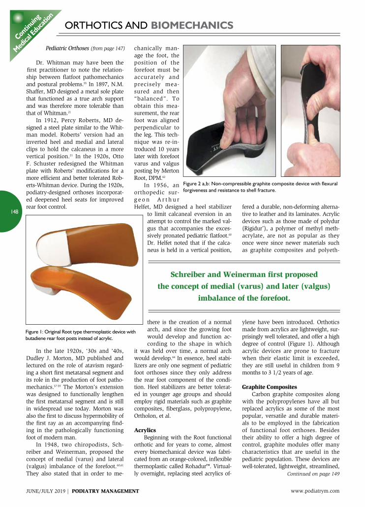
Figure 1: Original Root type thermoplastic device with butadiene rear foot posts instead of acrylic.
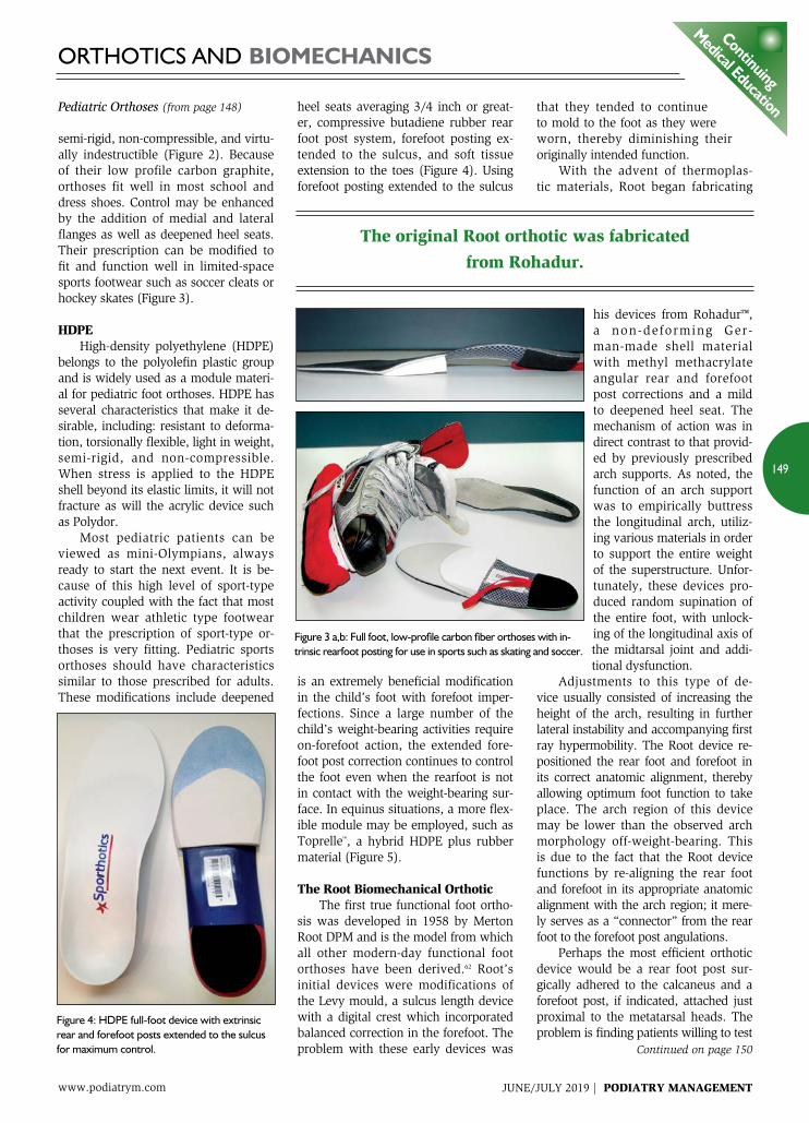
Figure 2 a,b: Non-compressible graphite composite device with flexural forgiveness and resistance to shell fracture.
fered a durable, non-deforming alterna-tive to leather and its laminates. Acrylic devices such as those made of polydur (Rigidur™), a polymer of methyl meth-acrylate, are not as popular as they once were since newer materials such as graphite composites and polyeth-
ylene have been introduced. Orthotics made from acrylics are lightweight, sur-prisingly well tolerated, and offer a high degree of control (Figure 1). Although acrylic devices are prone to fracture when their elastic limit is exceeded, they are still useful in children from 9 months to 3 1/2 years of age.
Graphite Composites Carbon graphite composites along with the polypropylenes have all but replaced acrylics as some of the most popular, versatile and durable materi-als to be employed in the fabrication of functional foot orthoses. Besides their ability to offer a high degree of control, graphite modules offer many characteristics that are useful in the pediatric population. These devices are well-tolerated, lightweight, streamlined,
Schreiber and Weinerman first proposed the concept of medial (varus) and later (valgus)
imbalance of the forefoot.
Continued on page 149
that they tended to continue to mold to the foot as they were worn, thereby diminishing their originally intended function. With the advent of thermoplas-tic materials, Root began fabricating
his devices from Rohadur™, a non-deforming Ger-man-made shell material with methyl methacrylate angular rear and forefoot post corrections and a mild to deepened heel seat. The mechanism of action was in direct contrast to that provid-ed by previously prescribed arch supports. As noted, the function of an arch support was to empirically buttress the longitudinal arch, utiliz-ing various materials in order to support the entire weight of the superstructure. Unfor-tunately, these devices pro-duced random supination of the entire foot, with unlock-ing of the longitudinal axis of the midtarsal joint and addi-tional dysfunction.
Adjustments to this type of de-vice usually consisted of increasing the height of the arch, resulting in further lateral instability and accompanying first ray hypermobility. The Root device re-positioned the rear foot and forefoot in its correct anatomic alignment, thereby allowing optimum foot function to take place. The arch region of this device may be lower than the observed arch morphology off-weight-bearing. This is due to the fact that the Root device functions by re-aligning the rear foot and forefoot in its appropriate anatomic alignment with the arch region; it mere-ly serves as a “connector” from the rear foot to the forefoot post angulations. Perhaps the most efficient orthotic device would be a rear foot post sur-gically adhered to the calcaneus and a forefoot post, if indicated, attached just proximal to the metatarsal heads. The problem is finding patients willing to test
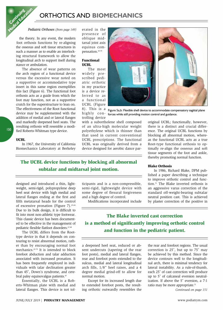
semi-rigid, non-compressible, and virtu-ally indestructible (Figure 2). Because of their low profile carbon graphite, orthoses fit well in most school and dress shoes. Control may be enhanced by the addition of medial and lateral flanges as well as deepened heel seats. Their prescription can be modified to fit and function well in limited-space sports footwear such as soccer cleats or hockey skates (Figure 3).
HDPE High-density polyethylene (HDPE) belongs to the polyolefin plastic group and is widely used as a module materi-al for pediatric foot orthoses. HDPE has several characteristics that make it de-sirable, including: resistant to deforma-tion, torsionally flexible, light in weight, semi-rigid, and non-compressible. When stress is applied to the HDPE shell beyond its elastic limits, it will not fracture as will the acrylic device such as Polydor. Most pediatric patients can be viewed as mini-Olympians, always ready to start the next event. It is be-cause of this high level of sport-type activity coupled with the fact that most children wear athletic type footwear that the prescription of sport-type or-thoses is very fitting. Pediatric sports orthoses should have characteristics similar to those prescribed for adults. These modifications include deepened
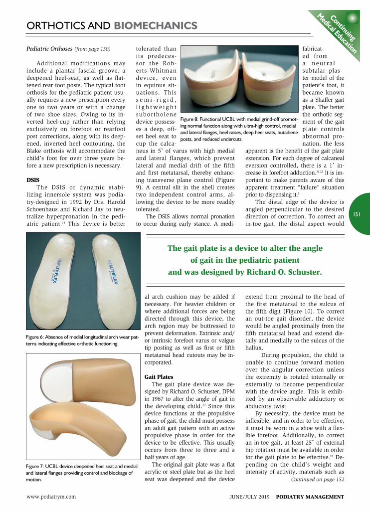
heel seats averaging 3/4 inch or great-er, compressive butadiene rubber rear foot post system, forefoot posting ex-tended to the sulcus, and soft tissue extension to the toes (Figure 4). Using forefoot posting extended to the sulcus
is an extremely beneficial modification in the child’s foot with forefoot imper-fections. Since a large number of the child’s weight-bearing activities require on-forefoot action, the extended fore-foot post correction continues to control the foot even when the rearfoot is not in contact with the weight-bearing sur-face. In equinus situations, a more flex-ible module may be employed, such as Toprelle™, a hybrid HDPE plus rubber material (Figure 5).
The Root Biomechanical Orthotic The first true functional foot ortho-sis was developed in 1958 by Merton Root DPM and is the model from which all other modern-day functional foot orthoses have been derived.62 Root’s initial devices were modifications of the Levy mould, a sulcus length device with a digital crest which incorporated balanced correction in the forefoot. The problem with these early devices was
www.podiatrym.com JUNE/JULY 2019 | PODIATRY MANAGEMENT
149
Continuing
medical education
ORTHOTICS AND BiomeChANiCs
Pediatric Orthoses (from page 148)
Continued on page 150
Figure 3 a,b: Full foot, low-profile carbon fiber orthoses with in-trinsic rearfoot posting for use in sports such as skating and soccer.
Figure 4: HDPE full-foot device with extrinsic rear and forefoot posts extended to the sulcus for maximum control.
The original Root orthotic was fabricated from Rohadur.
original UCBL; functionally, however, there is a distinct and crucial differ-ence. The original UCBL functions by blocking all abnormal motion, where-as the functional UCBL acts as a true Root-type functional orthosis to op-timally re-align the osseous and soft tissue segments of the foot and ankle, thereby promoting normal function.
Blake Orthosis In 1986, Richard Blake, DPM pub-lished a paper describing a technique to improve orthotic control and func-tion.51 The Blake inverted orthosis is an aggressive varus correction of the standard off-weight-bearing subtalar neutral position cast. This is achieved by plaster correction of the positive in
the rear and forefoot regions. The usual correction is 25˚, but up to 75˚ may be achieved by this method. Since the device contours well to the longitudi-nal arch, there is minimal tendency for lateral instability. As a rule-of-thumb, each 25˚ of cast correction will produce up to 5˚ of calcaneal eversion neutral-ization. If above the 5˚ eversion, a 7:1 ratio may be more appropriate.51
the theory. In any event, the modern foot orthosis functions by re-aligning the osseous and soft tissue structures in such a manner as to enable an interlock-ing structural framework to allow the longitudinal arch to support itself during stance or ambulation. The absence of wear patterns on the arch region of a functional device versus the excessive wear noted on a supportive or accommodative type insert in this same region exemplifies this fact (Figure 6). The functional foot orthosis acts as a guide from which the foot may function, not as a supportive crutch for the superstructure to lean on. The effectiveness of the Root functional device may be supplemented with the addition of medial and or lateral flanges and markedly deepened heel seats. The resulting orthosis will resemble a modi-fied Roberts-Whitman type device.
UCBL In 1967, the University of California Biomechanics Laboratory at Berkeley
designed and introduced a thin, light-weight, semi-rigid, polypropylene deep heel seat device with high medial and lateral flanges ending at the first and fifth metatarsal heads for the control of excessive pronation (Figure 7).65,66 Due to its bulk design, it is difficult to fit into most non-athletic type footwear. This classic device has been document-ed to be effective in the management of pediatric flexible flatfoot disorders.67,68
The UCBL differs from the Root-type device in that it depends on con-touring to resist abnormal motion, rath-er than by encouraging normal foot mechanics.65-70 It is intended to block forefoot abduction and talar adduction associated with increased pronation. It has been frequently employed in indi-viduals with talar declination greater than 45˚, Down’s syndrome, and cere-bral palsy equinovalgus patients.67
Essentially, the UCBL is a Rob-erts-Whitman plate with medial and lateral flanges. This device is not tol-
erated in the presence of oblique mid-tarsal joint axis equinus com-pensation.69,70
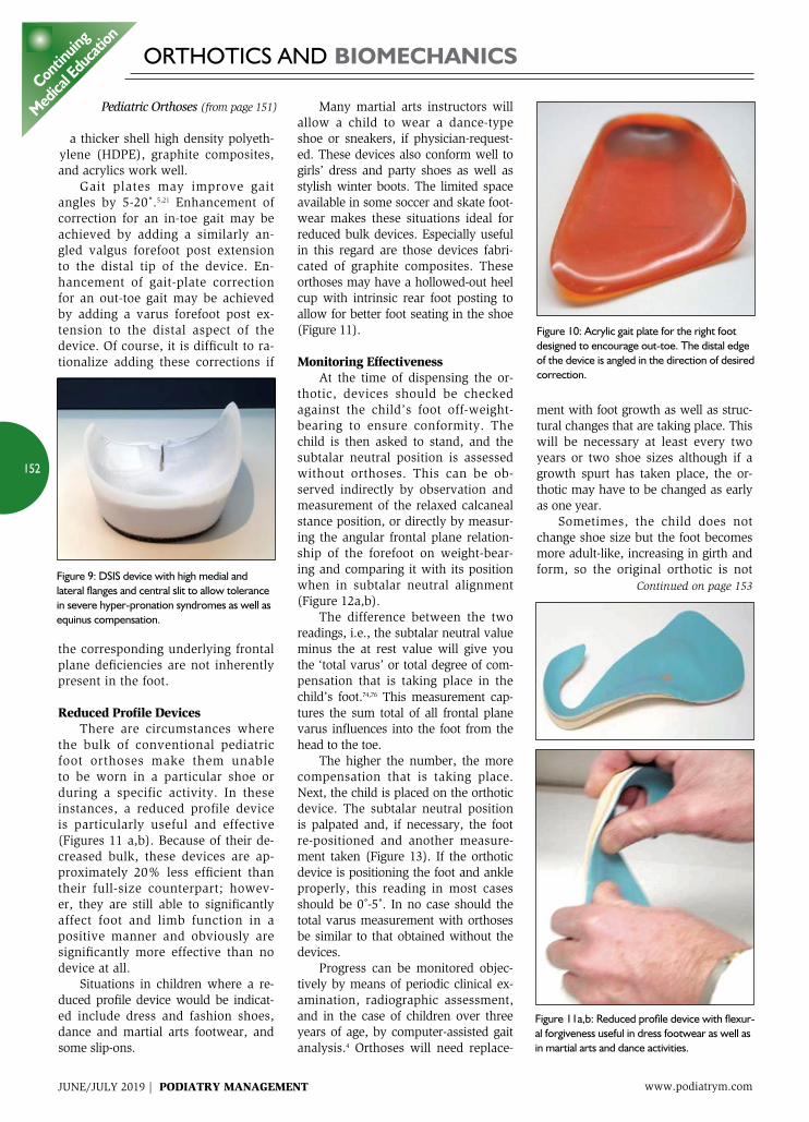
Functional UCBL The most wide ly pre -scribed pedi-atric orthotic in my practice is a device re-ferred to as a functional UCBL (Figure 8). This is a h igh ly con-trolling device with a subortholene shell composed of an ultra-high molecular weight polyethylene which is thinner than that used in current conventional UCBL prescriptions. The functional UCBL was originally derived from a device designed for aerobic dance par-
ticipants and is a non-compressible, semi-rigid, lightweight device with some degree of flexural forgiveness and a high degree of control. Modifications incorporated include
a deepened heel seat, reduced or ab-sent undercuts (tapering of the rear foot posts), medial and lateral flanges, rear and forefoot posts extended to the sulcus, medial and lateral longitudinal arch fills, 1/8” heel raises, and a 4 degree medial grind-off to allow for normal motion. Except for its increased length due to extended forefoot posts, the result-ing orthotic outwardly resembles the
www.podiatrym.comJUNE/JULY 2019 | PODIATRY MANAGEMENT
150
Contin
uing
medica
l edu
cation
ORTHOTICS AND BiomeChANiCs
Pediatric Orthoses (from page 149)
Continued on page 151
The UCBL device functions by blocking all abnormal subtalar and midtarsal joint motion.
Figure 5a,b: Flexible shell device to accommodate compensatory sagittal plane forces while still providing motion control and guidance.
The Blake inverted cast correction is a method of significantly improving orthotic control
and function in the pediatric patient.
fabricat-ed from a neu t ra l subtalar plas-ter model of the patient’s foot, it became known as a Shaffer gait plate. The better the orthotic seg-ment of the gait plate controls abnormal pro-nation, the less
apparent is the benefit of the gait plate extension. For each degree of calcaneal eversion controlled, there is a 1˚ in-crease in forefoot adduction.21,22 It is im-portant to make parents aware of this apparent treatment “failure” situation prior to dispensing it.5
The distal edge of the device is angled perpendicular to the desired direction of correction. To correct an in-toe gait, the distal aspect would
extend from proximal to the head of the first metatarsal to the sulcus of the fifth digit (Figure 10). To correct an out-toe gait disorder, the device would be angled proximally from the fifth metatarsal head and extend dis-tally and medially to the sulcus of the hallux. During propulsion, the child is unable to continue forward motion over the angular correction unless the extremity is rotated internally or externally to become perpendicular with the device angle. This is exhib-ited by an observable adductory or abductory twist By necessity, the device must be inflexible; and in order to be effective, it must be worn in a shoe with a flex-ible forefoot. Additionally, to correct an in-toe gait, at least 25˚ of external hip rotation must be available in order for the gait plate to be effective.22 De-pending on the child’s weight and intensity of activity, materials such as
Additional modifications may include a plantar fascial groove, a deepened heel-seat, as well as flat-tened rear foot posts. The typical foot orthosis for the pediatric patient usu-ally requires a new prescription every one to two years or with a change of two shoe sizes. Owing to its in-verted heel-cup rather than relying exclusively on forefoot or rearfoot post corrections, along with its deep-ened, inverted heel contouring, the Blake orthosis will accommodate the child’s foot for over three years be-fore a new prescription is necessary.
DSIS The DSIS or dynamic stabi-lizing innersole system was podia-try-designed in 1992 by Drs. Harold Schoenhaus and Richard Jay to neu-tralize hyperpronation in the pedi-atric patient.73 This device is better
tolerated than its predeces-sor the Rob-erts-Whitman device, even in equinus sit-uations. This s e m i - r i g i d , l i g h t w e i g h t subortholene device possess-es a deep, off-set heel seat to cup the calca-neus in 5˚ of varus with high medial and lateral flanges, which prevent lateral and medial drift of the fifth and first metatarsal, thereby enhanc-ing transverse plane control (Figure 9). A central slit in the shell creates two independent control arms, al-lowing the device to be more readily tolerated. The DSIS allows normal pronation to occur during early stance. A medi-
al arch cushion may be added if necessary. For heavier children or where additional forces are being directed through this device, the arch region may be buttressed to prevent deformation. Extrinsic and/or intrinsic forefoot varus or valgus tip posting as well as first or fifth metatarsal head cutouts may be in-corporated.
Gait Plates The gait plate device was de-signed by Richard O. Schuster, DPM in 1967 to alter the angle of gait in the developing child.22 Since this device functions at the propulsive phase of gait, the child must possess an adult gait pattern with an active propulsive phase in order for the device to be effective. This usually occurs from three to three and a half years of age. The original gait plate was a flat acrylic or steel plate but as the heel seat was deepened and the device
www.podiatrym.com JUNE/JULY 2019 | PODIATRY MANAGEMENT
151
Continuing
medical education
ORTHOTICS AND BiomeChANiCs
Pediatric Orthoses (from page 150)
Continued on page 152
Figure 7: UCBL device deepened heel seat and medial and lateral flanges providing control and blockage of motion.
Figure 8: Functional UCBL with medial grind-off promot-ing normal function along with ultra-high control, medial and lateral flanges, heel raises, deep heel seats, butadiene posts, and reduced undercuts.
Figure 6: Absence of medial longitudinal arch wear pat-terns indicating effective orthotic functioning.
The gait plate is a device to alter the angle of gait in the pediatric patient
and was designed by Richard O. Schuster.
ment with foot growth as well as struc-tural changes that are taking place. This will be necessary at least every two years or two shoe sizes although if a growth spurt has taken place, the or-thotic may have to be changed as early as one year. Sometimes, the child does not change shoe size but the foot becomes more adult-like, increasing in girth and form, so the original orthotic is not
a thicker shell high density polyeth-ylene (HDPE), graphite composites, and acrylics work well. Gait plates may improve gait angles by 5-20˚.5,21 Enhancement of correction for an in-toe gait may be achieved by adding a similarly an-gled valgus forefoot post extension to the distal tip of the device. En-hancement of gait-plate correction for an out-toe gait may be achieved by adding a varus forefoot post ex-tension to the distal aspect of the device. Of course, it is difficult to ra-tionalize adding these corrections if
the corresponding underlying frontal plane deficiencies are not inherently present in the foot.
Reduced Profile Devices There are circumstances where the bulk of conventional pediatric foot orthoses make them unable to be worn in a particular shoe or during a specific activity. In these instances, a reduced profile device is particularly useful and effective (Figures 11 a,b). Because of their de-creased bulk, these devices are ap-proximately 20% less efficient than their full-size counterpart; howev-er, they are still able to significantly affect foot and limb function in a positive manner and obviously are significantly more effective than no device at all. Situations in children where a re-duced profile device would be indicat-ed include dress and fashion shoes, dance and martial arts footwear, and some slip-ons.
Many martial arts instructors will allow a child to wear a dance-type shoe or sneakers, if physician-request-ed. These devices also conform well to girls’ dress and party shoes as well as stylish winter boots. The limited space available in some soccer and skate foot-wear makes these situations ideal for reduced bulk devices. Especially useful in this regard are those devices fabri-cated of graphite composites. These orthoses may have a hollowed-out heel cup with intrinsic rear foot posting to allow for better foot seating in the shoe (Figure 11).
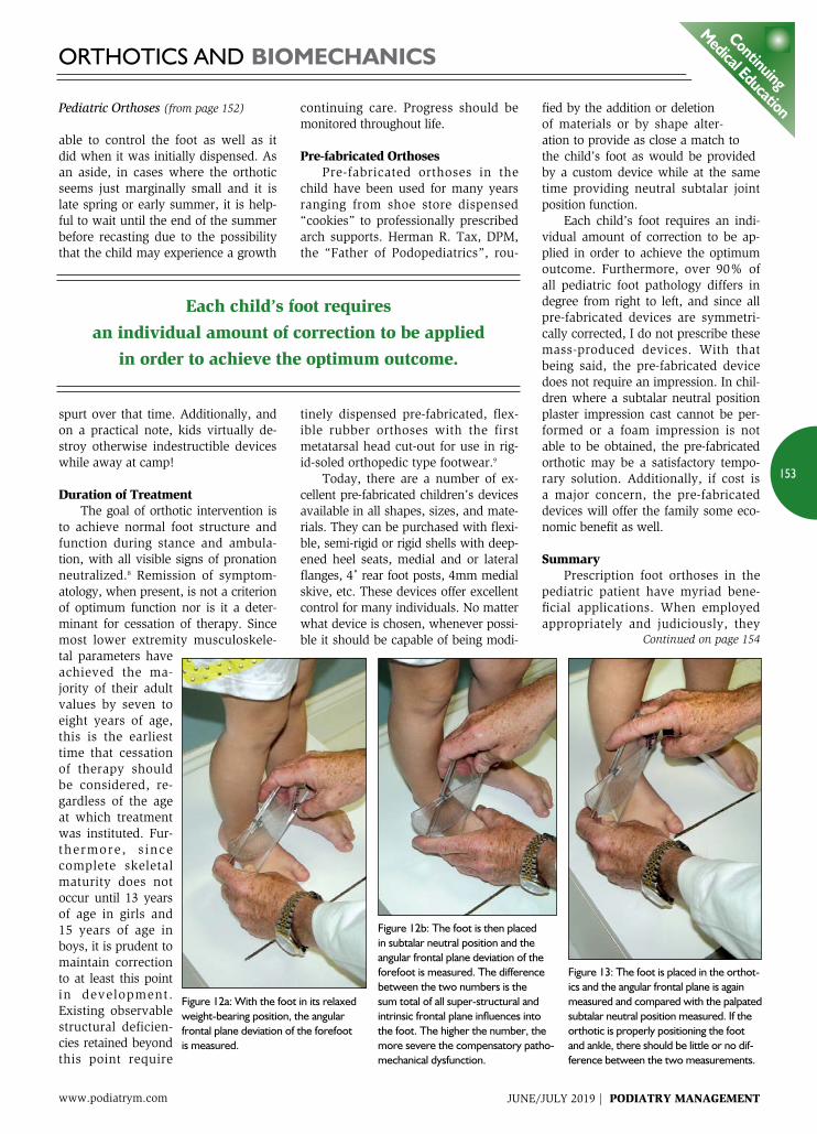
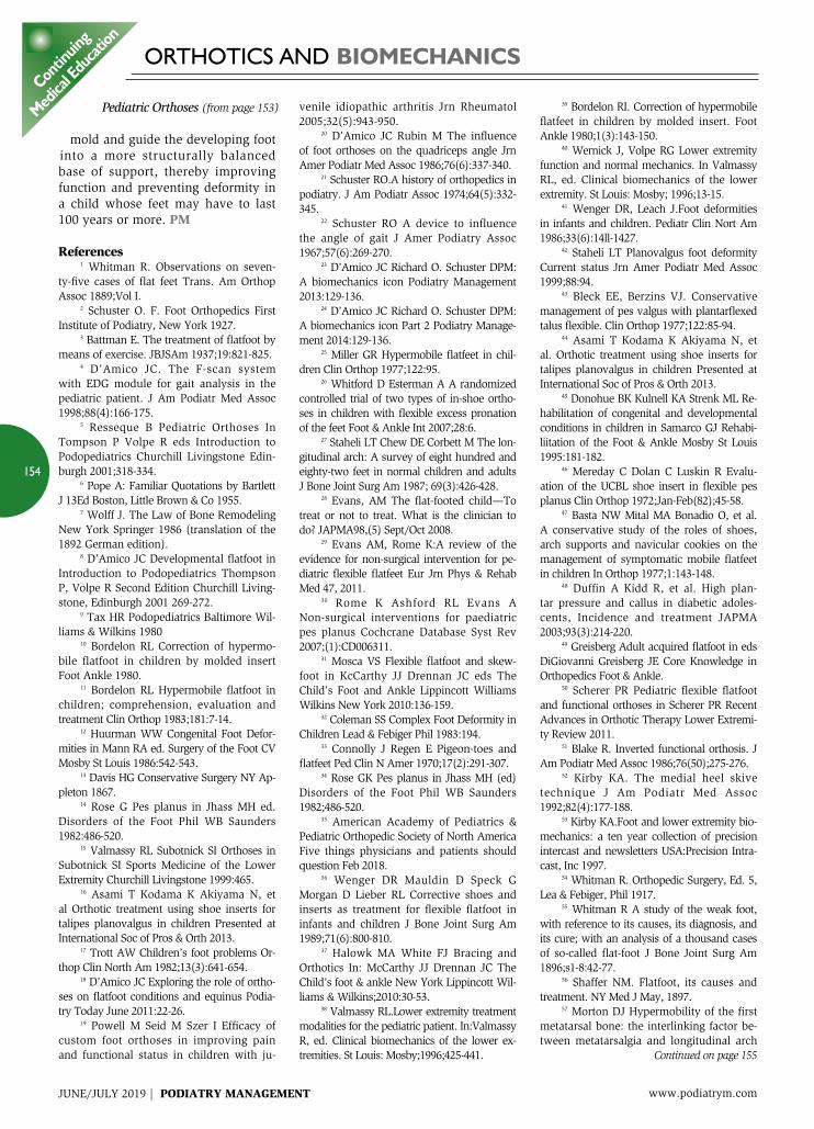
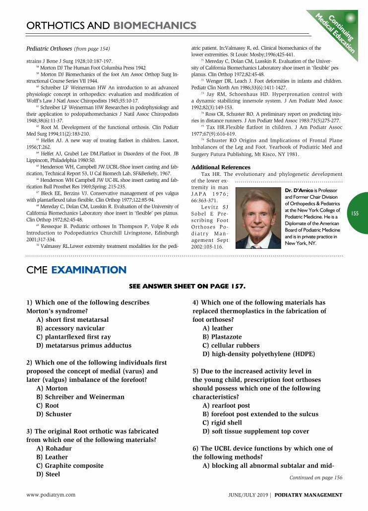
Monitoring Effectiveness At the time of dispensing the or-thotic, devices should be checked against the child’s foot off-weight-bearing to ensure conformity. The child is then asked to stand, and the subtalar neutral position is assessed without orthoses. This can be ob-served indirectly by observation and measurement of the relaxed calcaneal stance position, or directly by measur-ing the angular frontal plane relation-ship of the forefoot on weight-bear-ing and comparing it with its position when in subtalar neutral alignment (Figure 12a,b). The difference between the two readings, i.e., the subtalar neutral value minus the at rest value will give you the ‘total varus’ or total degree of com-pensation that is taking place in the child’s foot.74,76 This measurement cap-tures the sum total of all frontal plane varus influences into the foot from the head to the toe. The higher the number, the more compensation that is taking place. Next, the child is placed on the orthotic device. The subtalar neutral position is palpated and, if necessary, the foot re-positioned and another measure-ment taken (Figure 13). If the orthotic device is positioning the foot and ankle properly, this reading in most cases should be 0˚-5˚. In no case should the total varus measurement with orthoses be similar to that obtained without the devices. Progress can be monitored objec-tively by means of periodic clinical ex-amination, radiographic assessment, and in the case of children over three years of age, by computer-assisted gait analysis.4 Orthoses will need replace-
www.podiatrym.comJUNE/JULY 2019 | PODIATRY MANAGEMENT
152
Contin
uing
medica
l edu
cation
ORTHOTICS AND BiomeChANiCs
Pediatric Orthoses (from page 151)
Continued on page 153Figure 9: DSIS device with high medial and lateral flanges and central slit to allow tolerance in severe hyper-pronation syndromes as well as equinus compensation.
Figure 10: Acrylic gait plate for the right foot designed to encourage out-toe. The distal edge of the device is angled in the direction of desired correction.
Figure 11a,b: Reduced profile device with flexur-al forgiveness useful in dress footwear as well as in martial arts and dance activities.
fied by the addition or deletion of materials or by shape alter-ation to provide as close a match to the child’s foot as would be provided by a custom device while at the same time providing neutral subtalar joint position function. Each child’s foot requires an indi-vidual amount of correction to be ap-plied in order to achieve the optimum outcome. Furthermore, over 90% of all pediatric foot pathology differs in degree from right to left, and since all pre-fabricated devices are symmetri-cally corrected, I do not prescribe these mass-produced devices. With that being said, the pre-fabricated device does not require an impression. In chil-dren where a subtalar neutral position plaster impression cast cannot be per-formed or a foam impression is not able to be obtained, the pre-fabricated orthotic may be a satisfactory tempo-rary solution. Additionally, if cost is a major concern, the pre-fabricated devices will offer the family some eco-nomic benefit as well.
Summary Prescription foot orthoses in the pediatric patient have myriad bene-ficial applications. When employed appropriately and judiciously, they
able to control the foot as well as it did when it was initially dispensed. As an aside, in cases where the orthotic seems just marginally small and it is late spring or early summer, it is help-ful to wait until the end of the summer before recasting due to the possibility that the child may experience a growth
spurt over that time. Additionally, and on a practical note, kids virtually de-stroy otherwise indestructible devices while away at camp!
Duration of Treatment The goal of orthotic intervention is to achieve normal foot structure and function during stance and ambula-tion, with all visible signs of pronation neutralized.8 Remission of symptom-atology, when present, is not a criterion of optimum function nor is it a deter-minant for cessation of therapy. Since most lower extremity musculoskele-tal parameters have achieved the ma-jority of their adult values by seven to eight years of age, this is the earliest time that cessation of therapy should be considered, re-gardless of the age at which treatment was instituted. Fur-thermore, s ince complete skeletal maturity does not occur until 13 years of age in girls and 15 years of age in boys, it is prudent to maintain correction to at least this point in development. Existing observable structural deficien-cies retained beyond this point require
continuing care. Progress should be monitored throughout life.
Pre-fabricated Orthoses Pre-fabricated orthoses in the child have been used for many years ranging from shoe store dispensed “cookies” to professionally prescribed arch supports. Herman R. Tax, DPM, the “Father of Podopediatrics”, rou-
tinely dispensed pre-fabricated, flex-ible rubber orthoses with the first metatarsal head cut-out for use in rig-id-soled orthopedic type footwear.9
Today, there are a number of ex-cellent pre-fabricated children’s devices available in all shapes, sizes, and mate-rials. They can be purchased with flexi-ble, semi-rigid or rigid shells with deep-ened heel seats, medial and or lateral flanges, 4˚ rear foot posts, 4mm medial skive, etc. These devices offer excellent control for many individuals. No matter what device is chosen, whenever possi-ble it should be capable of being modi-
www.podiatrym.com JUNE/JULY 2019 | PODIATRY MANAGEMENT
153
Continuing
medical education
ORTHOTICS AND BiomeChANiCs
Pediatric Orthoses (from page 152)
Continued on page 154
Figure 12a: With the foot in its relaxed weight-bearing position, the angular frontal plane deviation of the forefoot is measured.
Figure 12b: The foot is then placed in subtalar neutral position and the angular frontal plane deviation of the forefoot is measured. The difference between the two numbers is the sum total of all super-structural and intrinsic frontal plane influences into the foot. The higher the number, the more severe the compensatory patho-mechanical dysfunction.
Figure 13: The foot is placed in the orthot-ics and the angular frontal plane is again measured and compared with the palpated subtalar neutral position measured. If the orthotic is properly positioning the foot and ankle, there should be little or no dif-ference between the two measurements.
Each child’s foot requires an individual amount of correction to be applied
in order to achieve the optimum outcome.
39 Bordelon RI. Correction of hypermobile flatfeet in children by molded insert. Foot Ankle 1980;1(3):143-150. 40 Wernick J, Volpe RG Lower extremity function and normal mechanics. In Valmassy RL, ed. Clinical biomechanics of the lower extremity. St Louis: Mosby; 1996;13-15. 41 Wenger DR, Leach J.Foot deformities in infants and children. Pediatr Clin Nort Am 1986;33(6):14ll-1427. 42 Staheli LT Planovalgus foot deformity Current status Jrn Amer Podiatr Med Assoc 1999;88:94. 43 Bleck EE, Berzins VJ. Conservative management of pes valgus with plantarflexed talus flexible. Clin Orthop 1977;122:85-94. 44 Asami T Kodama K Akiyama N, et al. Orthotic treatment using shoe inserts for talipes planovalgus in children Presented at International Soc of Pros & Orth 2013. 45 Donohue BK Kulnell KA Strenk ML Re-habilitation of congenital and developmental conditions in children in Samarco GJ Rehabi-liitation of the Foot & Ankle Mosby St Louis 1995:181-182. 46 Mereday C Dolan C Luskin R Evalu-ation of the UCBL shoe insert in flexible pes planus Clin Orthop 1972;Jan-Feb(82);45-58. 47 Basta NW Mital MA Bonadio O, et al. A conservative study of the roles of shoes, arch supports and navicular cookies on the management of symptomatic mobile flatfeet in children In Orthop 1977;1:143-148. 48 Duffin A Kidd R, et al. High plan-tar pressure and callus in diabetic adoles-cents, Incidence and treatment JAPMA 2003;93(3):214-220. 49 Greisberg Adult acquired flatfoot in eds DiGiovanni Greisberg JE Core Knowledge in Orthopedics Foot & Ankle. 50 Scherer PR Pediatric flexible flatfoot and functional orthoses in Scherer PR Recent Advances in Orthotic Therapy Lower Extremi-ty Review 2011. 51 Blake R. Inverted functional orthosis. J Am Podiatr Med Assoc 1986;76(50);275-276. 52 Kirby KA. The medial heel skive technique J Am Podiatr Med Assoc 1992;82(4):177-188. 53 Kirby KA.Foot and lower extremity bio-mechanics: a ten year collection of precision intercast and newsletters USA:Precision Intra-cast, Inc 1997. 54 Whitman R. Orthopedic Surgery, Ed. 5, Lea & Febiger, Phil 1917. 55 Whitman R A study of the weak foot, with reference to its causes, its diagnosis, and its cure; with an analysis of a thousand cases of so-called flat-foot J Bone Joint Surg Am 1896;s1-8:42-77. 56 Shaffer NM. Flatfoot, its causes and treatment. NY Med J May, 1897. 57 Morton DJ Hypermobility of the first metatarsal bone: the interlinking factor be-tween metatarsalgia and longitudinal arch
mold and guide the developing foot into a more structurally balanced base of support, thereby improving function and preventing deformity in a child whose feet may have to last 100 years or more. PM
References 1 Whitman R. Observations on seven-ty-five cases of flat feet Trans. Am Orthop Assoc 1889;Vol I. 2 Schuster O. F. Foot Orthopedics First Institute of Podiatry, New York 1927. 3 Battman E. The treatment of flatfoot by means of exercise. JBJSAm 1937;19:821-825. 4 D’Amico JC. The F-scan system with EDG module for gait analysis in the pediatric patient. J Am Podiatr Med Assoc 1998;88(4):166-175. 5 Resseque B Pediatric Orthoses In Tompson P Volpe R eds Introduction to Podopediatrics Churchill Livingstone Edin-burgh 2001;318-334. 6 Pope A: Familiar Quotations by Bartlett J 13Ed Boston, Little Brown & Co 1955. 7 Wolff J. The Law of Bone Remodeling New York Springer 1986 (translation of the 1892 German edition). 8 D’Amico JC Developmental flatfoot in Introduction to Podopediatrics Thompson P, Volpe R Second Edition Churchill Living-stone, Edinburgh 2001 269-272. 9 Tax HR Podopediatrics Baltimore Wil-liams & Wilkins 1980 10 Bordelon RL Correction of hypermo-bile flatfoot in children by molded insert Foot Ankle 1980. 11 Bordelon RL Hypermobile flatfoot in children; comprehension, evaluation and treatment Clin Orthop 1983;181:7-14. 12 Huurman WW Congenital Foot Defor-mities in Mann RA ed. Surgery of the Foot CV Mosby St Louis 1986:542-543. 13 Davis HG Conservative Surgery NY Ap-pleton 1867. 14 Rose G Pes planus in Jhass MH ed. Disorders of the Foot Phil WB Saunders 1982:486-520. 15 Valmassy RL Subotnick SI Orthoses in Subotnick SI Sports Medicine of the Lower Extremity Churchill Livingstone 1999:465. 16 Asami T Kodama K Akiyama N, et al Orthotic treatment using shoe inserts for talipes planovalgus in children Presented at International Soc of Pros & Orth 2013. 17 Trott AW Children’s foot problems Or-thop Clin North Am 1982;13(3):641-654. 18 D’Amico JC Exploring the role of ortho-ses on flatfoot conditions and equinus Podia-try Today June 2011:22-26. 19 Powell M Seid M Szer I Efficacy of custom foot orthoses in improving pain and functional status in children with ju-
venile idiopathic arthritis Jrn Rheumatol 2005;32(5):943-950. 20 D’Amico JC Rubin M The influence of foot orthoses on the quadriceps angle Jrn Amer Podiatr Med Assoc 1986;76(6):337-340. 21 Schuster RO.A history of orthopedics in podiatry. J Am Podiatr Assoc 1974;64(5):332-345. 22 Schuster RO A device to influence the angle of gait J Amer Podiatry Assoc 1967;57(6):269-270. 23 D’Amico JC Richard O. Schuster DPM: A biomechanics icon Podiatry Management 2013:129-136. 24 D’Amico JC Richard O. Schuster DPM: A biomechanics icon Part 2 Podiatry Manage-ment 2014:129-136. 25 Miller GR Hypermobile flatfeet in chil-dren Clin Orthop 1977;122:95. 26 Whitford D Esterman A A randomized controlled trial of two types of in-shoe ortho-ses in children with flexible excess pronation of the feet Foot & Ankle Int 2007;28:6. 27 Staheli LT Chew DE Corbett M The lon-gitudinal arch: A survey of eight hundred and eighty-two feet in normal children and adults J Bone Joint Surg Am 1987; 69(3):426-428. 28 Evans, AM The flat-footed child—To treat or not to treat. What is the clinician to do? JAPMA98,(5) Sept/Oct 2008. 29 Evans AM, Rome K:A review of the evidence for non-surgical intervention for pe-diatric flexible flatfeet Eur Jrn Phys & Rehab Med 47, 2011. 30 Rome K Ashford RL Evans A Non-surgical interventions for paediatric pes planus Cochcrane Database Syst Rev 2007;(1):CD006311. 31 Mosca VS Flexible flatfoot and skew-foot in KcCarthy JJ Drennan JC eds The Child’s Foot and Ankle Lippincott Williams Wilkins New York 2010:136-159. 32 Coleman SS Complex Foot Deformity in Children Lead & Febiger Phil 1983:194. 33 Connolly J Regen E Pigeon-toes and flatfeet Ped Clin N Amer 1970;17(2):291-307. 34 Rose GK Pes planus in Jhass MH (ed) Disorders of the Foot Phil WB Saunders 1982;486-520. 35 American Academy of Pediatrics & Pediatric Orthopedic Society of North America Five things physicians and patients should question Feb 2018. 36 Wenger DR Mauldin D Speck G Morgan D Lieber RL Corrective shoes and inserts as treatment for flexible flatfoot in infants and children J Bone Joint Surg Am 1989;71(6):800-810. 37 Halowk MA White FJ Bracing and Orthotics In: McCarthy JJ Drennan JC The Child’s foot & ankle New York Lippincott Wil-liams & Wilkins;2010:30-53. 38 Valmassy RL.Lower extremity treatment modalities for the pediatric patient. In:Valmassy R, ed. Clinical biomechanics of the lower ex-tremities. St Louis: Mosby;1996;425-441.
www.podiatrym.comJUNE/JULY 2019 | PODIATRY MANAGEMENT
154
Contin
uing
medica
l edu
cation
ORTHOTICS AND BiomeChANiCs
Pediatric Orthoses (from page 153)
Continued on page 155
atric patient. In:Valmassy R, ed. Clinical biomechanics of the lower extremities. St Louis: Mosby;1996;425-441. 71 Mereday C, Dolan CM, Lusskin R. Evaluation of the Univer-sity of California Biomechanics Laboratory shoe insert in ‘flexible’ pes planus. Clin Orthop 1972;82:45-48. 72 Wenger DR, Leach J. Foot deformities in infants and children. Pediatr Clin North Am 1986;33(6):1411-1427. 73 Jay RM, Schoenhaus HD. Hyperpronation control with a dynamic stabilizing innersole system. J Am Podiatr Med Assoc 1992;82(3):149-153. 74 Ross CR, Schuster RO. A preliminary report on predicting inju-ries in distance runners. J Am Podiatr Med Assoc 1983:73(5)275-277. 75 Tax HR.Flexible flatfoot in children. J Am Podiatr Assoc 1977;67(9):616-619. 76 Schuster RO Origins and Implications of Frontal Plane Imbalances of the Leg and Foot. Yearbook of Podiatric Med and Surgery Futura Publishing, Mt Kisco, NY 1981.
Additional References Tax HR. The evolutionary and phylogenetic development of the lower ex-tremity in man J A P A 1 9 7 6 ; 66:363-371. Levi tz SJ Sobel E Pre-scribing Foot Orthoses Po-d i a t r y Man -agement Sept 2002:103-116.
Continuing
medical education
155
www.podiatrym.com JUNE/JULY 2019 | PODIATRY MANAGEMENT
ORTHOTICS AND BiomeChANiCs
strains J Bone J Surg 1928;10:187-197. 58 Morton DJ The Human Foot Columbia Press 1942 59 Morton DJ Biomechanics of the foot Am Assoc Orthop Surg In-structional Course Series VII 1944. 60 Schreiber LF Weinerman HW An introduction to an advanced physiologic concept in orthopedics: evaluation and modification of Wolff’s Law J Natl Assoc Chiropodists 1945;35:10-17. 61 Schreiber LF Weinerman HW Researches in podophysiology and their application to podopathomechanics J Natil Assoc Chiropodists 1948;38(6):11-37. 62 Root M. Development of the functional orthosis. Clin Podiatr Med Surg 1994;11(2):183-210. 63 Helfet AJ. A new way of treating flatfeet in children. Lancet, 1956;T:262. 64 Helfet AJ, Grubel Lee DM.Flatfoot in Disorders of the Foot. JB Lippincott, Philadelphia 1980:50. 65 Henderson WH, Campbell JW.UCBL-Shoe insert casting and fab-rication, Technical Report 53, U Cal Biomech Lab, SF&Berkely, 1967. 66 Henderson WH Campbell JW UC-BL shoe insert casting and fab-rication Bull Prosthet Res 1969;Spring: 215-235. 67 Bleck EE, Berzins VJ. Conservative management of pes valgus with plantarflexed talus flexible. Clin Orthop 1977;122:85-94. 68 Mereday C, Dolan CM, Lusskin R. Evaluation of the University of California Biomechanics Laboratory shoe insert in ‘flexible’ pes planus. Clin Orthop 1972;82:45-48. 69 Resseque B. Pediatric orthoses In Thompson P, Volpe R eds Introduction to Podopediatrics Churchill Livingstone, Edinburgh 2001;317-334. 70 Valmassy RL.Lower extremity treatment modalities for the pedi-
Pediatric Orthoses (from page 154)
1) Which one of the following describes Morton’s syndrome? A) short first metatarsal B) accessory navicular C) plantarflexed first ray D) metatarsus primus adductus
2) Which one of the following individuals first proposed the concept of medial (varus) and later (valgus) imbalance of the forefoot? A) Morton B) Schreiber and Weinerman C) Root D) Schuster
3) The original Root orthotic was fabricated from which one of the following materials? A) Rohadur B) Leather C) Graphite composite D) Steel
4) Which one of the following materials has replaced thermoplastics in the fabrication of foot orthoses? A) leather B) Plastazote C) cellular rubbers D) high-density polyethylene (HDPE)
5) Due to the increased activity level in the young child, prescription foot orthoses should possess which one of the following characteristics? A) rearfoot post B) forefoot post extended to the sulcus C) rigid shell D) soft tissue supplement top cover
6) The UCBL device functions by which one of the following methods? A) blocking all abnormal subtalar and mid-
CME eXAmiNATioNSee anSwer Sheet on page 157.
Continued on page 156
Dr. D’Amico is Professor and Former Chair Division of Orthopedics & Pediatrics at the New York College of Podiatric Medicine. He is a Diplomate of the American Board of Podiatric Medicine and is in private practice in New York, NY.

JUNE/JULY 2019 | PODIATRY MANAGEMENT
156
PM’sCme program
Welcome to the innovative Continuing Education Program brought to you by Podiatry Management Magazine. Our journal has been approved as a sponsor of Continuing Medical Education by the Council on Podiatric Medical Education.
Now it’s even easier and more convenient to enroll in pm’s Ce program! You can now enroll at any time during the year and submit eligible exams at any time during your enrollment period. Cme articles and examination questions from past issues of Podiatry Management can be found on the internet at http://www.podiatrym.com/cme. Each lesson is approved for 1.5 hours continuing education contact hours. Please read the testing, grading and payment instructions to decide which method of participa-tion is best for you. Please call (631) 563-1604 if you have any questions. A personal operator will be happy to assist you. Each of the 10 lessons will count as 1.5 credits; thus a maximum of 15 CME credits may be earned during any 12-month period. You may select any 10 in a 24-month period.
The podiatry management magazine CME program is approved by the Council on Podi-atric Education in all states where credits in instructional media are accepted. This article is approved for 1.5 Continuing Education Contact Hours (or 0.15 CEU’s) for each examination successfully completed.
PM’s privacy policy can be found at http:// podiatrym.com/privacy.cfm.
This CME is valid for CPME-approved credits for three (3) years from the date of publication.
$
CME eXAmiNATioNCon
tinuin
g
medica
l edu
cation
tarsal joint motion B) realignment of rearfoot and forefoot
osseous segments C) encouraging adaptive phase pronation D) encouraging normal function
7) Characteristics of the functional UCBL are represented by which one of the following? A) medial grind-off B) reduced or absent undercuts C) appropriate rear and forefoot posting
extended to the sulcus D) all of the above
8) A method of significantly improving orthotic control and function in the pediatric patient may be achieved by which one of the following? A) Blake inverted cast correction B) Morton’s extension C) Increased shell flexibility D) Metatarsal pad
9) A particularly valuable, highly controlling pediatric orthotic device with medial and lat-eral control arms is referred to as: A) Whitman plate B) Levy mould C) Helfet heel cup D) DSIS (Dynamic Stabilizing Innersole
System)
10) The gait plate is a device to alter the angle of gait in the pediatric patient and was designed by which one of the following? A) Richard O. Schuster B) Dudley Morton C) Merton Root D) Kevin Kirby
See anSwer Sheet on page 157.
The author(s) certify that they have NO affiliations with or involvement in any organization or entity with any financial interest (such as honoraria; educational grants; participation in speakers’ bureaus; member-ship, employment, consultancies, stock ownership, or other equity interest), or non-financial interest (such as personal or professional relationships, affiliations, knowledge, or beliefs) in the subject matter or materi-als discussed in this manuscript.

Please print clearly...Certificate will be issued from information below.
Name ____________________________________________________________________ Email Address______________________________Please Print: FIRST MI LAST
Address_____________________________________________________________________________________________________________
City__________________________________________________ State_______________________ Zip________________________________
Charge to: _____Visa _____ MasterCard _____ American Express
Card #________________________________________________Exp. Date____________________ Zip for credit card_________________
Note: Credit card is the only method of payment. Checks are no longer accepted.
Signature__________________________________ Email Address_________________________ Daytime Phone_______________________
State License(s)___________________________ Is this a new address? Yes________ No________
Check one: ______ I am currently enrolled. (If faxing or phoning in your answer form please note that $2.95 will be charged to your credit card.)
______ I am not enrolled. Enclosed is my credit card information. Please charge my credit card $28.00 for each exam submitted. (plus $2.95 for each exam if submitting by fax or phone).
______ I am not enrolled and I wish to enroll for 10 courses at $229.00 (thus saving me $51 over the cost of 10 individual exam fees). I understand there will be an additional fee of $2.95 for any exam I wish to submit via fax or phone.
Note: If you are mailing your answer sheet, you must complete all info. on the front and back of this page and mail with your credit card information to: program management services, p.o. Box 490, east islip, Ny 11730.
TesTiNg, grADiNg AND pAymeNT iNsTruCTioNs (1) Each participant achieving a passing grade of 70% or higher on any examination will receive an official computer form stating the number of CE credits earned. This form should be safeguarded and may be used as documentation of credits earned. (2) Participants receiving a failing grade on any exam will be notified and permitted to take one re-examination at no extra cost. (3) All answers should be recorded on the answer form below. For each question, decide which choice is the best answer, and cir-cle the letter representing your choice. (4) Complete all other information on the front and back of this page. (5) Choose one out of the 3 options for testgrading: mail-in, fax, or phone. To select the type of service that best suits your needs, please read the following section, “Test Grading Options”.
TesT grADiNg opTioNs Mail-In Grading To receive your CME certificate, complete all information and mail with your credit card information to: program management services, p.o. Box 490, east islip, Ny 11730. pLeAse Do NoT seND WiTh sigNATure reQuireD, As These WiLL NoT Be ACCepTeD.
eNroLLmeNT Form & ANsWer sheeT
$
There is no charge for the mail-in service if you have al-ready enrolled in the annual exam CME program, and we receive this exam during your current enrollment period. If you are not en-rolled, please send $28.00 per exam, or $229 to cover all 10 exams (thus saving $51 over the cost of 10 individual exam fees).
Facsimile Grading To receive your CME certificate, complete all information and fax 24 hours a day to 1631-532-1964. Your CME certificate will be dated and mailed within 48 hours. This service is available for $2.95 per exam if you are currently enrolled in the annual 10-exam CME program (and this exam falls within your enrollment period), and can be charged to your Visa, MasterCard, or American Express. If you are not enrolled in the annual 10-exam CME program, the fee is $28 per exam.
Phone-In Grading You may also complete your exam by using the toll-free service. Call 1-800-232-4422 from 10 a.m. to 5 p.m. EST, Monday through Friday. Your CME certificate will be dated the same day you call and mailed within 48 hours. There is a $2.95 charge for this service if you are currently enrolled in the annual 10-exam CME program (and this exam falls within your enrollment period), and this fee can be charged to your Visa, Mastercard, American Express, or Discover. If you are not current-ly enrolled, the fee is $28 per exam. When you call, please have ready: 1. Program number (Month and Year) 2. The answers to the test 3. Credit card information
Over, please
Continuing
medical education
enrollment/Testing informationand Answer sheet
157
www.podiatrym.com JUNE/JULY 2019 | PODIATRY MANAGEMENT
In the event you require additional CME information, please contact PMS, Inc., at 1-631-563-1604.

158
www.podiatrym.comJUNE/JULY 2019 | PODIATRY MANAGEMENT
Contin
uing
medica
l edu
cation
eNroLLmeNT Form & ANsWer sheeT (continued)
$
medical education Lesson evaluation Strongly Strongly agree Agree Neutral Disagree disagree [5] [4] [3] [2] [1]
1) This CME lesson was helpful to my practice ____
2) The educational objectives were accomplished ____
3) I will apply the knowledge I learned from this lesson ____
4) I will makes changes in my practice behavior based on this lesson ____
5) This lesson presented quality information with adequate current references ____
6) What overall grade would you assign this lesson? A B C D
7) This activity was balanced and free of commercial bias.
Yes _____ No _____
8) What overall grade would you assign to the overall management of this activity? A B C D
How long did it take you to complete this lesson?
______hour ______minutes
What topics would you like to see in future CME lessons ? Please list :__________________________________________________
__________________________________________________
__________________________________________________
__________________________________________________
__________________________________________________
1. A B C D
2. A B C D
3. A B C D
4. A B C D
5. A B C D
6. A B C D
7. A B C D
8. A B C D
9. A B C D
10. A B C D
Circle:
eXAm #5/19pediatric orthoses—part ii
(D’Amico)