Pathomorphological studies on Hydatidosis in Western Libyan slaughtered animals: with special...
17
Sahar et al., Congrès Maghrèbin Vètèrinaire, Le 16 Mai 2008, Alger. Pathomorphological studies on Hydatidosis in Western Libyan slaughtered animals: with special reference to its epidemiological status in human in the same localities. Sahar S. Abd El-Rahman 1 , Shahera M.R.Abdel-Haseeb 2 and A.A.Abd-Allah 3 . 1. Dept. of Pathology, Faculty of Veterinary Medicine, Cairo University, Egypt. 2. Animal Health Research Institute, Dept. of Pathology, Dokki, Egypt. 3. Dept. of Infectious Diseases, Faculty of Veterinary Medicine, 7 th of April University, Libya. Corresponding author : Sahar S. Abd El-Rahman Address: Cairo University, Faculty of Vet. Med., Giza, Egypt 12211, Department of Pathology. Fax: 5725240 (Dept. of Pathology). E-mail:[email protected] Abstract: Hydatidosis is caused by the tapeworm Echinococcus granulosus. It is a common parasite of dog; other animals usually conduct the disease when it acts as intermediate host. The present work was conducted to investigate the prevalence of hydatidosis among sheep, goat, cattle and camel in El-Nekhat El-Khams- western Libya, together with a preliminary assessment of the public health impact of the disease in the same locality. The prevalence of infestation was estimated among 1440 sheep, 576 goats, 384 cattle and 240 camels in the official abattoirs in the surveyed localities, with an infestation percent of 26.8%, 15.9%, 6.2% and 22.9% respectively. Generally it was found that female animals were more infected than males and the highest incidence of organ infestation was in liver and lung. Diagnosis of hydatidosis was based on both gross examination and microscopic findings. Histopathological examination of the cyst structure and tissue specimens collected from the liver and lungs of infected animals was carried out. Epidemiological study of the disease in human in the same locality revealing that hydatidosis prevalence in human was 1.9% with wide range of age distribution and significantly more infested females than males. Introduction: Cystic echinococcosis due to Echinococcus granulosus is a serious public health and livestock economy problem in Libya (Kassem, 2006). Echinococcosis is one of the major zoonotic parasitic diseases in the Middle East and Arabic North Africa from Morocco to Egypt. Hydatid disease is a parasitic infestation caused by a cestode of Genus Echinococus. Various surveys have indicated that hydatid cysts are commonly found in sheep, cattle, goats and camels throughout the Middle East and Arabic North Africa. Sheep are the most infected animals in such regions (Sajjadi, 2006). The most important and widespread of these parasites is Echinococcus granulosus, which causes 46
Transcript of Pathomorphological studies on Hydatidosis in Western Libyan slaughtered animals: with special...

Sahar et al., Congrès Maghrèbin Vètèrinaire, Le 16 Mai 2008, Alger.
Pathomorphological studies on Hydatidosis in Western Libyan slaughteredanimals: with special reference to its epidemiological status in humanin the same localities.
Sahar S. Abd El-Rahman1, Shahera M.R.Abdel-Haseeb 2and A.A.Abd-Allah 3.1. Dept. of Pathology, Faculty of Veterinary Medicine, Cairo University,
Egypt.2. Animal Health Research Institute, Dept. of Pathology, Dokki, Egypt.3. Dept. of Infectious Diseases, Faculty of Veterinary Medicine, 7th of
April University, Libya.Corresponding author: Sahar S. Abd El-RahmanAddress: Cairo University, Faculty of Vet. Med., Giza,Egypt 12211, Department of Pathology.Fax: 5725240 (Dept. of Pathology).E-mail:[email protected]: Hydatidosis is caused by the tapeworm Echinococcus granulosus. It is acommon parasite of dog; other animals usually conduct the disease whenit acts as intermediate host. The present work was conducted toinvestigate the prevalence of hydatidosis among sheep, goat, cattle andcamel in El-Nekhat El-Khams- western Libya, together with a preliminaryassessment of the public health impact of the disease in the samelocality. The prevalence of infestation was estimated among 1440 sheep, 576goats, 384 cattle and 240 camels in the official abattoirs in thesurveyed localities, with an infestation percent of 26.8%, 15.9%, 6.2%and 22.9% respectively. Generally it was found that female animals weremore infected than males and the highest incidence of organ infestationwas in liver and lung. Diagnosis of hydatidosis was based on both grossexamination and microscopic findings. Histopathological examination ofthe cyst structure and tissue specimens collected from the liver andlungs of infected animals was carried out. Epidemiological study of thedisease in human in the same locality revealing that hydatidosisprevalence in human was 1.9% with wide range of age distribution andsignificantly more infested females than males.Introduction: Cystic echinococcosis due toEchinococcus granulosus is a seriouspublic health and livestockeconomy
problem in Libya (Kassem, 2006).Echinococcosis is one of themajor zoonotic parasitic diseasesin the Middle East and ArabicNorth Africa from Morocco toEgypt. Hydatid disease is a
parasitic infestation caused by acestode of Genus Echinococus. Various surveys have indicatedthat hydatid cysts are commonlyfound in sheep, cattle, goats andcamels throughout the Middle Eastand Arabic North Africa. Sheepare the most infected animals insuch regions (Sajjadi, 2006). The most important andwidespread of these parasites isEchinococcus granulosus, which causes
46

Sahar et al., Congrès Maghrèbin Vètèrinaire, Le 16 Mai 2008, Alger.
hydatid disease. The disease isendemic in certain areas in theworld where close contact betweenhuman and sheep exists. Humaninfections are most common in theMediterranean region, sub-SaharanAfrican, Russia, China and SouthAmerica (Lightowlers, 1999).
Echinococcus granulosus is a commonparasite of dog, and it can occurin anywhere in the world. Thelife cycle involves two mammalianhosts. The disease is produced inanother animal when it acts asintermediate host, as a result ofswallowing ova from dog’s faeces.Dogs and other canids areinfected with the parasite in thesmall intestine and eggs arereleased with faeces. Ingestion of eggs by a widevariety of herbivorous animalsleads to the growth of hydatidcysts in tissues. When infectedtissues are eaten by a dog, thelife cycle is completed. Humanbecome infected by accidentalingestion of microscopic eggsderived from dog faeces. Most ofstudies on human hydatidosis havebeen focused on surgical reportsalthough several populationstudies have been performed usingserological and imagingtechniques. Sajjadi, 2006reported that human cysticechinococcosis (CE) is prevalentin the Middle East and ArabicNorth Africa. Moreover, it ishyper endemic in Iran, Turkey,Iraq, Jordan, Morocco, Libya,Tunisia, and Algeria, and endemicin Egypt. Studies on the strainspecificities of E. granulosus was
carried out by Sajjadi, 2006 inthe Middle East revealed thatsheep strain (G1) is present insheep, goats, cattle, camels andhumans, and the camel strain (G6)in camels, sheep, cattle as wellas humans. Also, he added thatthe strain specifications of E.granulosus in Arabic North Africashowed that sheep/dog strain (G1)have been reported from Tunisiaand Libya both from humans andanimals. However, in Egypt, thereported human cases due tocamel/dog strain. Hydatid cysts (H. cysts) occurmost frequently in liver andlungs, though they may occur inany other sites. Hydatid cystforms a slowly growing spaceoccupying mass up to 20cm Ø. Itis multilocular containing manydaughter cysts (Jubb et al,1993).Purpose of the study: The present work was conductedto investigate the prevalence ofEchinococcus granulosus infestation inanimals as well as to investigatethe epidemiological associationbetween the prevalence of thedisease in animals and man in El-Nekat El-Khams-West Libya.Materials and methods: Hydatidosis infestation wasinvestigated among sheep, goats,cattle and camels during theperiod of September 2006 to May2007. The investigation was carriedout randomly in the officialabattoirs in El-Nekat El-khams(west-Libya). Diagnosis of E.infestation based on both gross andthe subsequent microscopicexamination.
47

Sahar et al., Congrès Maghrèbin Vètèrinaire, Le 16 Mai 2008, Alger.
Thorough P.M. examination wasperformed for all animals, fordetecting the infested cases. Tissue specimens were collectedfrom liver and lungs of infectedcases and were fixed in formalinesaline 10%, then routinelyprocessed and sectioned at 4-5umthickness for histopathologicalexamination. Paraffin sections were stainedwith Hematoxyline and Eosin,(Bancroft et al 1996).Occasionally other stains ,particularly PAS, Geimsa andMasson’s trichrome stains wereused (Bancroft et al, 1996). As a preliminary assessment ofthe public health impact of thedisease in residents of ruralareas, data of human contractedthe disease were surveyed andcollected from records of thesurgical departments in thecentral hospitals in the samelocalities. Those data dependedon the surgically confirmed caseswith cystic Echinococcosis duringthe period from Jan. 2006 to June2007. On admission to thehospital, patients were examinedby x-ray for localization of thecysts followed by Ultrasoundconfirmation. The age and sex ofthe cases were obtained from thehospitals records. The particularcysts were accurately excisedafter being injected with 10%formalin. Statistical analysis: Resultsof the obtained data werestatistically analyzed using chi-square test. The differences wereconsidered to be significant at p< 0.05 (Snedecor and Cochran,1980).
Results: Investigation of hydatidosis,revealed a high percentage ofinfestation among both animalsand human (according to hospitalsdata) in the surveyed localities.I- Incidence of animalinfestation: Table (1) presented theprevalence of E. granulosusinfestation among animals. Fromthat table we noticed that E.granulosus infestation wasdetected in 386 out of 1440sheep, 92 out of 576 goat, 24out of 384 cattle and 55 out of240 camels. With an infestationpercent of 26.8%, 15.9%, 6.2% and22.9% respectively. The highest prevalence ofinfestation was found in sheepfollowed by camel, goat andfinally cattle. Generally, femaleanimals were more often infestedthan males in the investigatedanimal species.II- Post mortem examination: P.M. examination of theinfested animals revealed theexistence of many Echinococcuscysts in the animals’ viscera(lungs, liver, peritoneal cavity,kidenys and spleen). Table (2) presented the organdistribution of the cysts indifferent animal species. Thehighest prevalence of organinfestation was observed in liverof sheep and goat followed by thelungs. While, in camel lungsinfestation predominated. Fewanimals showed presence of thecysts hanging from the peritonealcavity. The conspicuous observation wasthat; the cysts in differentorgans particularly in liver and
48

Sahar et al., Congrès Maghrèbin Vètèrinaire, Le 16 Mai 2008, Alger.
lung were varied in size, shapeand development. Some of themshowed the normal cysticstructure, while others weredegenerated, organized withcalcification. Regarding the infested livers,they presented strong destructivechanges particularly in the areassurrounding the cysts. Thoselivers were pale and friable withmarked distension of the gallbladder. Sometimes the tissuearound the cysts was eitherapparently normal or evencongested. The observed cystswere of various sizes, shapes andnumber. The size ranged frompeanut to pigeon egg size. Singleor multiple cysts were observed.Most of the large-sized cystswere multilocular containing manydaughter cysts (Fig. 1 a and b). Livers of some animals wereinfested with up to 29 cysts andtheir diameter ranged from lessthan 10mm to 15cm. Generally, cysts with diameterless than 10mm were immature, andthose more than 15 cm werefertile and developedprotoscoleces. Concerning lungs, the cystswere multiple either protrudingfrom the lung surface or embeddedin the pulmonary tissue. Somecysts appeared opaque anddepressed. The size of the cystsvaried from 0.5- 12cm in diameter(Fig. 1 c). According to the cut sectionand the contents of the cysts,three types could be identifiedgrossly. The first type wasconfined to the larger waterycysts that splashed on cutting.The second type contained cheesy
caseated yellowish whitematerial. While the third one wasconfined to the smaller cyststhat were hard in consistencywith a gritty sound could behared in cut section. Thecontents of the later werecalcified and admixed withcaseous like material. Moreover, the surroundingpulmonary tissue showed differentstages of pneumonia as reddishto brownish areas ofconsolidation which were usuallyconfined to the cranial parts ofthe lung, in addition toobturation of the bronchioles(as an occasional finding).Also emphysematous and collapsedareas were common with occasionalareas of abscessiation.III- Microscopic examination: Histomorphological examinationof the collected tissue specimensfrom lungs and liver revealedgreat tissue alterations in thoseorgans.III-a- Histological structure ofthe cyst: Histologially hydatid cystsdiffered in their structure, someof them showed the usualstructure, while others showedvarious stages of degeneration orfull organization with or withoutcalcification. The predominant structure ofthe cyst wall consisted of threedistinctive layers by PAS andMasson’s trichrome stains. Theinner most was a germinal layerwith many belbs-like structuresthat will grow and give rise toprotoscoleces. The second layerwas the cuticular layer consistedof pale acellular lamellatedhomogenous esinophilic,
49

Sahar et al., Congrès Maghrèbin Vètèrinaire, Le 16 Mai 2008, Alger.
structurless chitinous material.The advential layer enclosed thecuticular layer and consisted ofdense collagenous fibrous tissueinfiltrated with large number ofesinophiles (Fig.1 d). Usually the cysts weresurrounded by a granulomatouscellular reaction of large numberof mononuclear cells asmacrophages, lymphocytes,epitheloid cells , plasmacytesand many esinophiles (Fig.1 e).The later cells predominated inmany cases and showed markeddegranulation as revealed byGeimsa stain (Fig 1 f). The lumen of some large cystswas lacking the broad capsule atthe time of examination (immaturecysts). While the lumen of otherlarge cysts showed presence ofprotoscoleces that were adheredto the germinal layer ( Fig 2 aand b). Some of the cysts revealedpresence of broad capsule thatconsisted of a number of scolecessurrounded by part of thegerminal layer of the cyst walland still adhered to the germinallayer of the cyst (Fig. 2 c). Large numbers of hydatid sandwere observed in most of thelarge cysts and appeared asscoleces free in the cyst fluid(Fig. 2 d). Moreover the lumen of fewlarger cysts showed presence ofmultiple daughter cysts as fewscoleces surrounded by bothgerminal layer and part of thelamellated acellular layer. The afromentioned scolecesappeared histologically asrounded or oval bodies, some werecompound cellular and others
appeared with thick darkperiphery.III-b- Pathology of the liver: Concerning the examinedinfested livers, they presentedstrong destructive changes in theparenchymal cells as variousdegenerative changes,disorganization of the hepaticcords together with hepatocytesdissociation that reachedhepatocyte necrosis and earlyfibrous replacement ( Fig. 2 e).Such changes were more obvious inthe parenchymal cells adjacent tothe cyst especially the necroticchanges (Fig. 2 f). Some livers showed theformation of fibrotic capsulesaround biliary tracts and portalveins. Meanwhile, premalignantchanges were seen in two ewes asdifferent foci particularlyaround the biliary tracts andportal veins. The later changes were theappearance of multiple clearcells as foci among the hepaticparenchymal cells. These clearcells appeared larger in sizethan the adjacent hepatocyteswith large vesicular nuclei. In addition, hepatic parenchymaof some few animals showedpresence of early larvalinfestation surrounded by anempty zone, which was not obviouson necked eye examination level.Those areas will give rise tocysts formation (Fig. 3 a ).III-c- Pathology of the lung: Histopathological examinationof different sections of lungs ofinfested animals revealed alsomarked and severe tissuealterations.
50

Sahar et al., Congrès Maghrèbin Vètèrinaire, Le 16 Mai 2008, Alger.
The most conspicuous of which,was interstitial pneumonia ofvariable degrees as thickenedinterstitial tissue with largenumber of mononuclearinflammatory cells and largenumber of esinophiles (Fig 3 b).Few polymorphs were evident inlungs of some animals togetherwith proliferation of alvelocytestype II. That thickenedinterstitial tissue compressedthe alveolar lumen which appearedsmaller or irregular slit-like orcompletely obstructed. Thosemultiple areas of lung collapsepresented with adjacent areas ofcompensatory emphysema (Fig. 3c). The alveoli in the formerappeared slit-like, while in thelater appeared giant-like, amongboth a granulomatous inflammatorycellular infiltrates wereobvious. Few lungs revealed presence ofearly larval infestation thatappeared in histological sectionsas dark esinophilic bodies ofdifferent shapes with appearanceof early cystic formationembedded in pulmonary tissue(Fig. 3 d).. The bronchial tree revealedalso marked inflammatory changeswith the existence ofinflammatory exudates containinglarge number of esinophilsobliterating their lumen. Thebronchial and bronchiolarepithelium showed markedproliferation and greatmetaplastic changes to gobletcells accompanied with markedhyperplastic changes (Fig. 3 e). In some few other animals themetaplastic bronchial epitheliumappeared irregularly arranged, of
different shapes with pleomorphicnuclei as well as markedperibronchial and peribrochiolarfibrosis (Fig. 3 f) withobturation of bronchioli, as anoccasional finding.IV- Analysis of the collecteddata about hydatidosis in human: Concerning human data collectedfrom the surgical departments ofthe central hospitals in El-NekatEl-Khams -West Libya locality,the epidemiological, and clinicalultrasonographic and laboratorydata were analyzed for betterdetail of the characteristicfeatures of human infection inthese localizations. Theprevalence of surgicallyconfirmed cases with cysticEchinococcosis in theafromentioned locality was 1.9 %as presented in table (3), withsignificantly more females (75 %)than males (25 %). Hospitals records showed thatthere was a wide range of ageinfection and hydatidosis was notcorrelated with specific age, asthe mean patient age was 30 years(range from 7 to 50). Allpatients lived in rural areaswhere sheep were raising at homesand contact with dogs was common.All patients were x-rayed,Ultrasonography examined and thensurgically confirmed ashydatidosis. Moreover, it was found thathydatid cysts can occur in anyorgan, but it was most common inliver (50%) followed by theperitoneal cavity (35 %), lung(10.7 %) and spleen (3.6%). Generally, the prevalence ofcystic echinococcosis increasedsignificantly with age and
51

Sahar et al., Congrès Maghrèbin Vètèrinaire, Le 16 Mai 2008, Alger.
females were significantly moreaffected than males. Cases ofcystic echinococcosis were mostlydistributed among housewives andfarmers.
Discussion: Hydatid disease is a parasiticinfestation that causeswidespread morbidity andmortality among livestockanimals, particularly sheep. Manand other animals are involved asintermediate hosts. The presentwork was directed to investigatethe prevalence of hydatidinfection among animals in El-Nekat El-Khams-West Libya, withcorrelation of such data withthat corresponding humaninfection in the same locality. Our survey revealed highprevalence of hydatidosisinfestation among animals thatwere slaughtered for humanconsumption in western Libya, inaddition to the increasedhyadatidosis prevalence amonghuman in the same locality. The prevalence percent inanimals was 26.8 %in sheep, 15.9%in goats, 6.2% in cattle and22.9% in camels. Generally, itwas evident that the infestationrate was higher in females thanin males. Previous studies on theprevalence of hydatidosis amonganimals in various localities inLibya were conducted by manyauthors with variable results ofprevalence. Kassem and Gdoura,2006 found 50 out of 1380 (3.62%)local bred camels harboured H.cyst. with more prevalence infemales (4.42%) than males(3.07%), in Sirt abattoirs-Libya.
Ibraheem and Craig, 1998, on anabattoir study in northern Libyanoted that the prevalence ofhydatidosis was 48% in camels,15.8% in sheep and 3.8% in goats.Tashani et al, 2002 reported theprevalence of infection byEchinococcus granulosus in easternLibya, as 20% in sheep, 3.4% ingoats, 13.6% in camels and 11% incattle. Also, Gusbi et al, 1987found high prevalence ofhydatidosis in sheep from 10localities in Libya, as 402 caseswere positive out of 5118 sheep.Finally, Gusbi et al, 1990recorded high prevalence of E.granulosus on their survey of 14Libyan abattoirs in 1985-1987. Such results come to a largeextent in harmony with ours. Onthe other hand, the obtainedresults are considered highercomparing with those previouslyreported by others in otherlocalities in Libya such asNahed, 1998 who recordedhydatidosis infestation in 8.7%in sheep, 5.4% in goats, 6.4% incattle and 35% in camels in Al-Jabal Al-Akhdar- Libya. In our opinion such previousvariation in the infection ratein the same country, could beattributed to either thedifference in the age of theanimal or the number of theexamined male and female animalsin the group or both. Added tothat the role of the importedinfested animals and the role ofthe free living dogs could not beneglected. Also, the rate andchance of exposure as well as thenature of the surveyed locality. It was found that, liver andlungs were the commonly affected
52

Sahar et al., Congrès Maghrèbin Vètèrinaire, Le 16 Mai 2008, Alger.
organs followed by peritonealcavity, spleen, kidneys and heartin all the investigated animalspecies except camels at whichlung infestation predominated.Organ infestation percent asobserved in this study issomewhat similar to thoserecorded by Gosbi et al, 1987 whofound that liver was the commonlyinfected organ (97.26% followedby the lung (58.70%), kidney(1.76%), spleen (0.74%), andheart, mesentry and muscles(0.24% each) in sheep infestedwith Echinococcosis. The increased incidence of lunginfestation in the surveyedanimals could be attributed tothe circulatory way of migrationfrom the gut to the lung tissuepaving easier access for theirestablishment. In addition, lungtissue may supply the nutrients,PH, and tissue spaces requiredfor development of such cysts. The morphohistologicalstructure of the examined cystsin our work was greatly agreedwith that reported by previousworks. In our work, the cyst wallconsisted fundamentally from 3layers; germinal, lamellated andfibrous. The thickness of eachlayer differed with each of theused stains (H&E, PAS andMasson’s trichrome stain). Thesefindings agreed with thosementioned by Rashed et al, 2004.Also, Jubb et al, 1993 statedthat hydatid cyst has a cuticularlayer that consists of pale acellular laminated chitinousmaterial (which is being digestedby some macrophages). Thecuticular layer is enclosed byadvential layer of dense
collagenous fibrous tissueinfiltrated with large number ofesinophils. In our work as well as in manyother papers hydatid cysts variedin shape, size and type. Somewere mature and others wereimmature, some were fertile.Those variations could bereclaimed to the age of thecysts, the viability of the broadcapsule and the defence mechanismof the host. The associated pathologicallesions in both lung and liversuch as pneumonia, emphysema,hepatitis and early fibrousreplacement, all were coordinatedwith the observed lesions bySvilenov et al, 1984, whodescribed hepatic strongdestructive changes particularlyin areas surrounding the cystswith interstitial andinflammatory changes in lungs ofsheep with hydatidosis. Theappearance of connective tissuein our work may be aimed to postnecrotic replacement or even aresponse to stimulation from thecyst. Both the cysts and tissuereaction around them resulted inthe marked compressionatelectasis and compensatoryemphysema. The inflammatorylesions were noticed more clearerin the vicinity of the cysts.While, the observed proliferatedbronchial epithelium and alveolarepithelization could be aresponse to the parasiticstimulation. Most of thepreviously mentioned lesions weremore or less similar to thosedescribed by Jubb et al, 1993.Also hepatic pathology was noted
53

Sahar et al., Congrès Maghrèbin Vètèrinaire, Le 16 Mai 2008, Alger.
by Rashed et al, 2004 in Najdisheep with hydatidosis. A great association wasdemonstrated between human andanimals contracted the disease inthe surveyed areas as proved byour data of human infestationwith Echinococcosis in westernLibya. This opinion is supportedby many authors as Elmahdi et al,2004 who suggested a greatrelation between animalinfestation and human conductingthe disease in rural urban areason their preliminary assessmentof the public health impact ofthe disease in Sudan. Also, Brecker et al, 2005 andPapanikolaou et al, 2005,reported cases of humaninfestation in rural- sheepfarming or raising areas wherehydatid disease is endemic.Dilege et al, 2003 supported thatEchynococcosis is endemic incertain areas of the world whereclose contact between human andsheep exists, and the diseasemost commonly involves the liverand other solid organs. Records of the surgicaldepartment of the centralhospital in El-Nekat El-khams(west-Libya) revealed asignificant incidence of humaninfection with hydatidosis inthese rural areas with a percentof 1.8. Also, those recordsrevealed increased incidence ofinfected females than males withwide age range in both. The increased incidence ofinfestation among human in thesurveyed localities in westernLibya may be related to theprimitive and traditional way ofslaughtering animals. As sheep
and goats are slaughtered athomes in these areas away fromhygienic control rules especiallyin the private occasions asweddings and festivals…ect. The infected viscera of thoseanimals are fed to stray dogs, apattern that increases the numberof the infected dogs and completethe life cycle of the worm. However the prevalence was morein females (75 %) than males (25%), which could be attributed toincreased exposure of females tothe ova during cleaning ofvegetables. As it was proved byAl-Gadzi, 1993 that 34% of themarket vegetables werecontaminated with the eggs of E.granulosus. According to thehospital records, hydatidosiscould occur at any age, notlimited to a particular age andmostly the infection levelincreased with the increased age.Moreover, records revealed thatmost cases contained one largecircular cyst in various areas ofthe body, the larger cysts werefound in aged people, whichsuggest that the immunity has norole in limitation of the cyst.Localization of the cysts in theperitoneal cavity and theabdominal viscera suggests thatingestion of food contaminatedwith the ova is the principle wayof human infection, such opinionis agreed with that stated byNehad , 1998. Hospitals records revealedthat, the highest incidence oforgan infestation was in liverand peritoneal cavity followed bylung and spleen. Mcfall et al,2004 reported that liver is themost commonly involved, although
54

Sahar et al., Congrès Maghrèbin Vètèrinaire, Le 16 Mai 2008, Alger.
many other organs including lungand brain may also be affected. Interestingly, many authorsreported abnormal sites ofhydatid cysts as in the brain byRadhakrishann et al, 1987 inBenghazi, in the heart of a childby Ben-Musa et al, 1990 inBenghazi-Libya, in heart byBrecker et al, 2005, in musclesof the lower limbs by Mseddi etal, 2005, in tarsal bones byPapanikolaou et al, 2005. Those authors noted a higherincidence of suspicion isnecessary for hydatid diseasediagnosis in those abnormalorgans, especially in patientswho live in or travel to sheepraising areas where the diseaseis endemic. Several factors wouldexplain the exceptional nature ofthose abnormal organslocalizations of the cysts; suchas the efficacy of the hepaticand pulmonary barriers and theenvironment of the infectedorgans is favourable or not forthe growth of the hydatid larvae.Also Mseddi et al, 2005 mentionedthe importance for diagnosispreoperatively in order to limitthe risk of anaphylactic shock ordissemination in the event ofpuncture or accidental opening ofthe cyst during surgical removalbecause surgery is ideallyrequired for treatment by totalpericystectomy.
Conclusion: From our previously mentioneddata, it is obvious thathydatidosis constituting a realproblem in the served localitiesin West-Libya that could be namedas endemic or even hyperendemic.
Control programs for hydatiddisease must be undertaken onfocus either nationally or inregional areas. These programsshould be relied on public. Also,control programs for eradicationof stray dogs must not beneglected. Moreover, publicshould be relied for restrictingthe slaughtering of the animalsin abattoirs not at homes, andinfected viscera should becondaminated not fed to dogs.Finally, further studies areneeded to investigate theprevalence of hydatidisis amongdogs in the surveyed locality.
Acknowledgement: I would like to thank thesurgeons in the central hospitalsin El-Nekat El-khams (west-Libya)who assisted in collecting thedata for this paper.
55

Sahar et al., Congrès Maghrèbin Vètèrinaire, Le 16 Mai 2008, Alger.
Referances:1- Al-Gadzi AA. Hydatidosis, Al-Shallal News paper, 312-7 (inArabic).2- Bancroft JD; Stevens A; TurnerDR. Theory and practice ofHistological Techniques. 4th ed.Churchill Livingstone, New York,London, San Fancisco, Yokyo.1996.3- Ben-Musa AA, Singh H, ShembeshAH, Chugh JC. Cardiac hydatidcyst in a child. Clin. Pediatr.(Phila). 1990 Jul.;29(2): 409-11.4- Brecker SJ, Mandal K, HarrisonT, Griffin G, Varghese A, PennellDJ, Lester Se, Jahangiri M.hydatid disease of the heart.Ann. R Coll Surg engl. 2005Mar;87(2):W1-4.
5- Dilege S, Aksoy M, Okan I,Toker A, Kalayci G, Demiryont M.Hydatid cystic disease of thesoft tissues with pulmonary andhepatic involvement: reportedcase. Surg. Today.2003;33(1):69-71.6- Elmahdi IE, Ali QM, MagzoubMM, Ibrahim AM, Saad MB, Romig T.cystic echinococcosis oflivestock and humans in centralSudan. Ann Trop Med parasitol.2004 Jul;98(5): 473-9.7- Gusbi AM, Awan MA, Beesley WN.Echinococcosis in Libya, IIPrevalence of hydatidosis(Echinococcus granulosus) insheep. Ann Trop. Med. Parasitol.1987 Feb; 81(1): 35-41.8- Gusbi AM, Awan MA, Beesley WN.Echinococcosis in Libya, IVPrevalence of hydatidosis(Echinococcus granulosus) in
goats, cattle and camels. Ann.Trop. Med. Parasitol. 1990, Oct;84 (5): 477-82.9- Ibrahem MM and Craig PS.Prevalence of cysticechinococcosis in camels(Camelus dromedaries) in Libya. JHelminthol.1998 Mar; 72(1):27-31.10- Kassem HH. Hydatidosis-chinococcosis in Libya (reviewarticle). J Egypt Soc Parasitol.2006 Aug;36 (2 Suppl): 27-31.11- Kassem HH and Gdoura NA. Abiochemical study of cystic stagefluids of Echinococcus granulosusof animals origin in Libya. JEgypt Soc parasitol 2006 Dec;36(3):1017-22. 12- Lightowlers MW. Vaccinationof humans against HydatidDisease. (Discussion paper). Theuniversity of Melbourne, 250princes Highway werribee,Victoria,3030 Australia.13- Mcfall B, Yousaf M, CalvertH, Diamond T, Epanomeritakis M.Surgical treatment of hepatichydatid cyst. Int. J. Clin.Pract. 2004 May; 58(5): 479-82.14- Mseddi M, Mtaoumi M, DahmeneJ, Ben Hamida R, Siala A, MoulaT, Ben Ayeche ML. Hydatid cystsin muscles: eleven cases. RevChir Orthop Reparatrice Apparmot. 2005 May;91(3): 267-71.15- Nahed Walli Al-Khalidi.Hydatidosis in human being in Al-Jabal Al-akhdar area in Libya.El-mokhtar for Sciences, 5th Ed.1998.16- Papanikolaou A, Antoniou N,Pavlakis D, Garas G. Hydatiddisease of the tarsal bones. Acase report. J. Foot Ankle Surg.2005 Sep-Oct;44(5):396-400.17- Radhakrishnan K, Maloo JC,Poddar SK, Mousa ME. Central
56
Sahar et al., Congrès Maghrèbin Vètèrinaire, Le 16 Mai 2008, Alger.
nervous system infections inBenghazi, Libya; Experimentalfrom a community –based adultmedical neurology set-up. J.Trop. Med. Hyg. 1987Jun;90(3):123-6. 18- Rashed Aa, Omer HM, Fouad MA,Al-Shareef Am. The effect ofsevere cystic hydatidosis on theliver of a Najdi sheep withspecial reference to cysthistology and histchemestry. JEgypt Soc Parasitol. 2004Apr;34(10: 297-304.19- Sadjjadi SM. Presentsituation of echinococcosis inthe Middle East and Arabic Nortth
Africa. Parasitol Int. 2006;55Suppl:S197-202 Epub 2005 Dec 6.20- Snedecor, G.W. and Cochran,W.G. Statistical methods.(1980)7th Ed. Allid pacific,Bombay.21- Svilenov D, Vinarova M,Khasan T, Tsolov B.Pathomorphological studies ofexperimental echinococcosis insheep. Vet Med Nauki. 1984;21(9): 120-32.22- Tashani OA, Zhang LH, Jegi A,McManus Dp. Epidemiology andstrain characteristics ofechinococcus granulosus area ofeastern Libya. Ann Trop MedParasitol. 2002 Jun; 96:369-81.
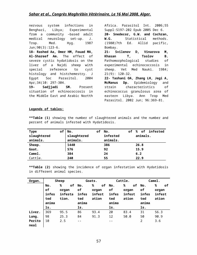
Legends of tables:
**Table (1) showing the number of slaughtered animals and the number andpercent of animals infested with Hydatidosis.
Type ofslaughteredanimals.
No. ofslaughteredanimals.
No. ofinfestedanimals.
% of infestedanimals.
Sheep. 1440 386 26.8Goat. 576 92 15.9Camel. 384 24 6.2Cattle. 240 55 22.9
**Table (2) showing the incidence of organ infestation with Hydatidosisin different animal species.
Organ. Sheep Goats. Cattle. Camel.No.ofinfestedanimals.
% oforganinfestation.
No.ofinfestedanimals.
% oforganinfestation
No.ofinfestedanimals.
% oforganinfestation
No.ofinfestedanimals.
% oforganinfestation
Liver. 369 95.5 86 93.4 20 83.4 31 56.3Lung. 98 25.3 84 91.3 12 50.0 50 90.9Peritoneal
10 2.5 -- -- 2 3.6
57

Sahar et al., Congrès Maghrèbin Vètèrinaire, Le 16 Mai 2008, Alger.
cavity.Spleen.
1 0.26 1 1.08 --
Kidney.
-- -- 1 1.08 -- 1 1.8
Heart. -- -- -- -- 1 1.8
**Table (3) showing the prevalence of Hydatidosis in human.
No. ofcasesadmitted tothesurgical Dept.
No. ofcasesinfestedinfectedwithHydatidosis.
Sex of thepositive cases.
Position of the cysts in the body of theinfected cases.
Males. Females.
Liver. Peritonealcavity.
Lung. Spleen.
1478 28 7 21 14 10 3 11.9% 25% 75% 50% 35% 10.7% 3.6%
58

Sahar et al., Congrès Maghrèbin Vètèrinaire, Le 16 Mai 2008, Alger.
Fig (1): a and b, liver of sheep infested with hydatidosis showing largenumber of hydatid cysts of variable size and shape. c- Lung of sheepinfested with hyadatidosis showing multiple cysts embedded or protrudedfrom the surface. d- Wall of hydatid cyst consists of three layers(arrow), the upper most is the germinal layer followed by lamellatedacellular layer enclosed by adventitial fibrous layer infiltrated bylarge number of inflammatory cells (H&E X100). e- Granulomatous reactionsurrounding the cyst in the liver (H&E X100). f- Interstitialinfiltration of large number of esinophilic granular cells in lunginfested with hydatidosis. (Giemsa X400).
59

Sahar et al., Congrès Maghrèbin Vètèrinaire, Le 16 Mai 2008, Alger.
Fig (2): a-d hydatid cyst in liver and lung of sheep showing: a and bpresence of protoscoleces in the lumen of the cyst and still adhered tothe germinal layer, notice the oval shape (a) and the granularappearance (b) of the protoscoleces. c- The broad capsule present in thecyst lumen surrounded by part of the germinal layer and still adhered to
60

Sahar et al., Congrès Maghrèbin Vètèrinaire, Le 16 Mai 2008, Alger.
it. d- Hydatid sand as protoscoleces free in the lumen of the cystfluid. e- Liver of sheep infested with hydatidosis presenting variousdegenerating changes accompanied with hepatocytes dissociation. f- Liverof sheep infested with hydatidosis revealing marked hepatocytes necrosisin the vicinity of the cyst (notice, the granulomatous cellular reactionaround the cyst. (a-d , PAS X200, e and f, H&E X200).
Fig (3): a- liver of sheep infested with hydatidosis revealing presenceof the larval stage surrounded by empty zone in the hepatic parenchyma.b-f: lung of sheep infested with hydatidosis showing: b- Interstitialpneumonia with marked distension of the interstitial tissue with largenumber of mononuclear cells and esinophils. c- Marked collapse with
61

Sahar et al., Congrès Maghrèbin Vètèrinaire, Le 16 Mai 2008, Alger.
adjacent areas of compensatory emphysema. d- Early cystic formationembedded in the pulmonary tissue. e and f- Marked hyperplastic andmetaplastic changes of the bronchiolar epithelium with luminalobliteration of large number of esinophils. f- Peribronchial fibrosistogether with pleomorphic metaplastic cells. ( a-d PAS, e and f H&E X200)
62