
paterson institute for cancer research
79
paterson institute for cancer research scientific report 2008
-
Upload
khangminh22 -
Category
Documents
-
view
0 -
download
0
Transcript of paterson institute for cancer research
paterson institute for cancer
research
scientific report
2008

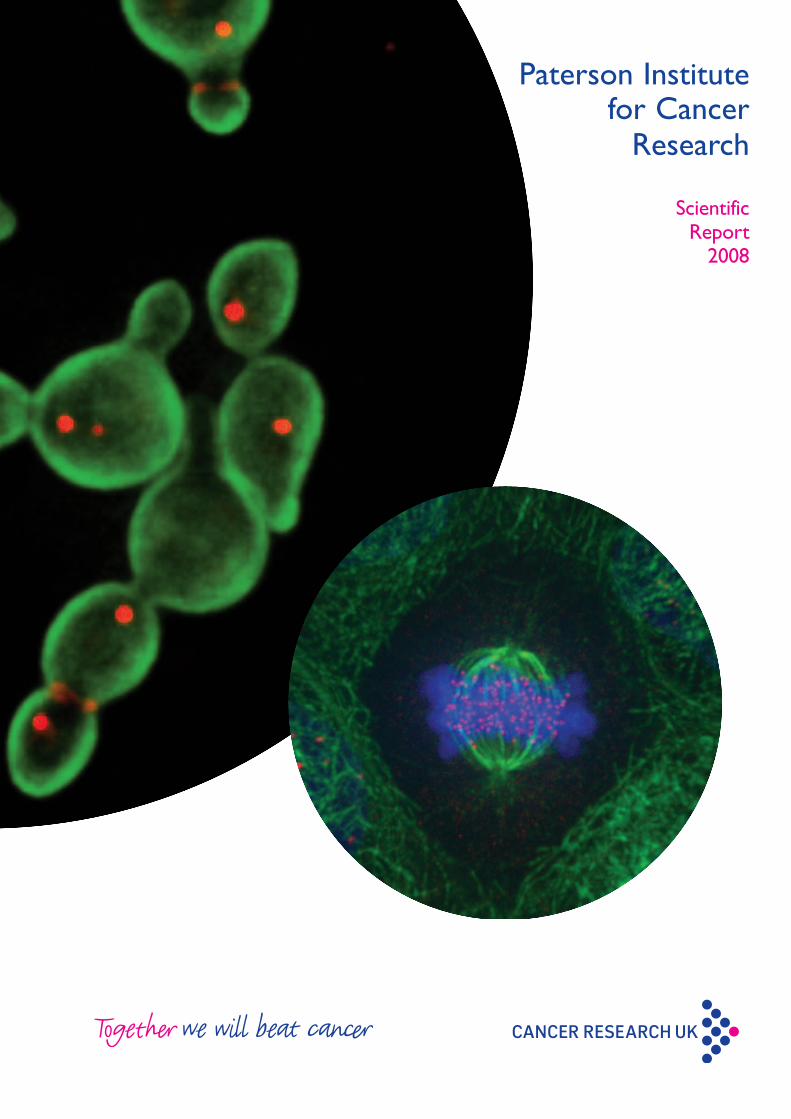
cover images:
Main image supplied by Karim Labib and Alberto
sanchez-Diaz (cell cycle Group).
Budding yeast cells lacking the inn1 protein are
unable to complete cytokinesis. these cells express a
fusion of a green fluorescent protein to a marker of
the plasma membrane, and have red fluorescent
proteins attached to components of the spindle poles
and actomyosin ring (sanchez-Diaz et al., nature cell
Biology 2008; 10: 395).
Additional images:
front cover image supplied by Helen rushton, simon
Woodcock and Angeliki Malliri (cell signalling
Group). the image is of a mitotic spindle in fixed
MDcK (Madin-Darby canine kidney) epithelial cells,
which have been stained with an anti-beta tubulin
antibody (green), DApi (blue) and an anti-centromere
antibody (crest, red) which recognises the
kinetochores of the chromosomes. the image was
taken on the spinning disk confocal microscope using
a 150 x lens.
rear cover image supplied by Andrei ivanov and tim
illidge (targeted therapy Group). Visualisation of
tubulin (green) and quadripolar mitosis (DnA stained
with DApi), Burkitt’s lymphoma namalwa cell after 10
Gy irradiation.
issn 1740-4525
copyright 2008 © cancer research UK
Paterson Institute forCancer Research
Scientific Report 2008
2 Paterson Institute for Cancer Research Scientific Report 2008
Contents
4 Director’s Introduction
Researchers’ pages – Paterson Institute for Cancer Research
8 Crispin Miller Applied Computational Biology and Bioinformatics
10 Geoff Margison Carcinogenesis
12 Karim Labib Cell Cycle
14 Iain Hagan Cell Division
16 Nic Jones Cell Regulation
18 Angeliki Malliri Cell Signalling
20 Caroline Dive &
Malcolm Ranson Clinical and Experimental Pharmacology
22 Peter Stern Immunology
24 Nullin Divecha Inositide Laboratory
26 Tim Somervaille Leukaemia Biology
28 Georges Lacaud Stem Cell Biology
30 Valerie Kouskoff Stem Cell and Haematopoiesis
32 Akira Orimo Stromal-Tumour Interaction
Researchers’ pages – The University of Manchester School of Cancer and Imaging Sciences
34 Catharine West Academic Radiation Oncology: Translational Radiobiology Group
36 Robert Hawkins &
Peter Stern Biological, Immune and Gene Therapy
38 Vaskar Saha Children’s Cancer Group
40 Tim Illidge Targeted Therapy
42 Robert Hawkins Medical Oncology: Cell Therapy
44 John Gallagher Medical Oncology: Glyco-Oncology
46 Gordon Jayson Medical Oncology: Translational Angiogenesis
Paterson Institute for Cancer Research Scientific Report 2008 3
48 Research Services
48 Steve Bagley Advanced Imaging Facility
49 Biological Resources Unit
51 Stuart Pepper Cancer Research UK GeneChip Microarray Service
51 Morgan Blaylock Flow Cytometry Facility
52 Garry Ashton Histology
53 Steve Glover Kostoris Library
54 Mark Craven Laboratory Services
54 Maurice Cowell Logistics
54 Stuart Pepper Molecular Biology Core Facility
54 Duncan Smith MBCF Biological Mass Spectometry Facility
56 Publications
66 Seminars
68 Postgraduate Education
70 Operations Services
73 Local Engagement and Development
74 Acknowledgement of Funding
75 Career opportunities
76 How to find us
4 Paterson Institute for Cancer Research Scientific Report 2008
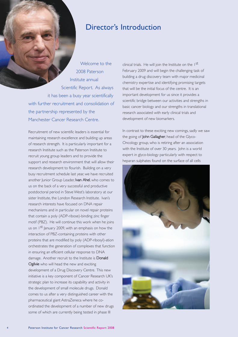
Welcome to the
2008 Paterson
Institute annual
Scientific Report. As always
it has been a busy year scientifically
with further recruitment and consolidation of
the partnership represented by the
Manchester Cancer Research Centre.
Recruitment of new scientific leaders is essential for
maintaining research excellence and building up areas
of research strength. It is particularly important for a
research Institute such as the Paterson Institute to
recruit young group leaders and to provide the
support and research environment that will allow their
research development to flourish. Building on a very
busy recruitment schedule last year, we have recruited
another Junior Group Leader, Ivan Ahel, who comes to
us on the back of a very successful and productive
postdoctoral period in Steve West’s laboratory at our
sister Institute, the London Research Institute. Ivan’s
research interests have focused on DNA repair
mechanisms and in particular on novel repair proteins
that contain a poly (ADP-ribose)-binding zinc finger
motif (PBZ). He will continue this work when he joins
us on 1st January 2009, with an emphasis on how the
interaction of PBZ-containing proteins with other
proteins that are modified by poly (ADP-ribosyl)-ation
orchestrates the generation of complexes that function
in ensuring an efficient cellular response to DNA
damage. Another recruit to the Institute is Donald
Ogilvie who will head the new and exciting
development of a Drug Discovery Centre. This new
initiative is a key component of Cancer Research UK’s
strategic plan to increase its capability and activity in
the development of small molecule drugs. Donald
comes to us after a very distinguished career with the
pharmaceutical giant AstraZeneca where he co-
ordinated the development of a number of new drugs
some of which are currently being tested in phase III
clinical trials. He will join the Institute on the 1st
February 2009 and will begin the challenging task of
building a drug discovery team with major medicinal
chemistry expertise and identifying promising targets
that will be the initial focus of the centre. It is an
important development for us since it provides a
scientific bridge between our activities and strengths in
basic cancer biology and our strengths in translational
research associated with early clinical trials and
development of new biomarkers.
In contrast to these exciting new comings, sadly we saw
the going of John Gallagher, head of the Glyco-
Oncology group, who is retiring after an association
with the Institute of over 30 years. John is a world
expert in glyco-biology particularly with respect to
heparan sulphates found on the surface of all cells
Director’s Introduction
Director’s Introduction 5
where they function as important co-receptors for a
number of growth factors including the fibroblast
growth factors and hepatocyte growth factor. John’s
work has highlighted the complexity of different
sulphation patterns seen with the heparan sulphates
and how these patterns affect such key biological
processes such as stem cell differentiation and
malignant transformation. John shone through with his
enthusiasm, dedication and above all his modesty. He
will be sorely missed.
During this last year, the Clinical and Experimental
Pharmacology (CEP) group in the Institute was site-
visited by an international panel of experts. These site
visits provide a stringent assessment of the
international standing of the research programme in
question by examining the progress and impact of the
research over the last five years and a view on the
potential and importance of the proposed research
programme for the next five years. This was a
particularly important review for the Institute since
CEP is a relatively new group which is at the heart of
the Institute’s translational research efforts and which
has grown rapidly over the last few years. It was
therefore very pleasing and reassuring that the group
received a very positive review and whose research
was judged to be at the international forefront. CEP
works very closely with the early phase clinical trials
unit (The Derek Crowther Unit, DCU) and has as its
mission the discovery and validation of biomarkers that
provide indications on the efficacy and toxicity of new
molecules being tested in clinical trials. This type of
research is crucially important for modern drug
development and lies at the heart of the need to
identify early in the development programme, drugs
that are likely to be effective and patients are likely to
respond to – in other words, the implementation of
personalised medicine. If the results of such research
are used to inform clinical decision making, then it has
to be conducted under very strict regulatory
guidelines. The CEP has established itself as one of the
leading centres internationally with this capability. CEP
is lead by Caroline Dive working closely with the head
of the DCU, Malcolm Ranson. They were both very
deservedly congratulated by the visiting party for
developing such an exciting and important research
programme which exemplified the power of a strong
and effective clinical-laboratory interface. Over the
next few years further development and investment in
this research area will take place, the most significant
development being the expansion of the clinical trials
facility as part of the £35M development by The
Christie NHS Foundation Trust. This will result in a 3-
fold increase in the capacity of the DCU, creating one
of the largest early clinical trials units in the world.
6 Paterson Institute for Cancer Research Scientific Report 2008
There were a number of research highlights during the
year. The Stem Cell Biology group led by Georges
Lacaud characterised in detail the developmental steps
leading to the generation of haematopoietic cells, which
despite receiving a lot of attention from many groups
over a number of years, remained controversial with
two conflicting theories predominating. One theory
hypothesised that haematopoietic cells arise from a
mesodermal progenitor called the haemangioblast
whilst the other suggested that such cells arise from a
specialised endothelial cell that has haematopoietic
potential, the haemogenic endothelium. Studies from
Georges’s laboratory have beautifully reconciled these
opposing views showing that both theories are merged
into a single developmental process where the
haemangioblast gives rise to a haemogenic endothelium
intermediate which then further differentiates to
generate haematopoietic cells. Furthermore, they
identified specific transcription factors that regulate
different stages of this differentiation pathway. This
important work will be published early in 2009 in the
journal Nature. The Cell Signalling Group led by
Angeliki Malliri has been studying a regulator (Tiam 1)
of the Rac GTPase which, amongst many of its roles,
functions to control cell-cell adhesion. Previous work
from the group had shown that deletions of Tiam1
confer resistance to the formation of both skin and
intestinal tumours in appropriate mouse models. They
have now discovered that Tiam1 is phosphorylated and
regulated by the oncoprotein Src, a tyrosine-kinase
implicated in malignant transformation. This
phosphorylation occurs preferentially at cell-cell
contacts resulting in Tiam1 degradation and disruption
of cell adhesion. This regulation of Tiam1 is likely to be
important for Src-mediated cancer cell invasion and
metastasis. Interestingly, in a range of different cancers,
they found a correlation between Tiam1
phosphorylation and Src-activity consistent with this
regulatory mechanism operating in tumour
malignancies. This work will be published early in the
New Year in the journal Molecular Cell. The Cell Cycle
laboratory headed by Karim Labib has been using yeast
as a model system to investigate the regulation of
DNA replication and cell division. During the last year
they reported in Nature Cell Biology the discovery of a
novel factor called Inn1 which is essential right at the
end of the cell cycle for cytokinesis, the process
whereby the eukaryotic cell divides into two. This has
to be regulated in time to ensure that it does not
occur before nuclear division, and in space to ensure
that the division plane lies between the two nuclei
formed during mitosis. The characterisation of Inn1 has
provided new insight into a relatively poorly
understood yet vital process. Powerful technologies
are now available to quantitate the cellular levels of
RNA transcripts through microarray analysis and
proteins through quantitative mass spectrometry
proteomics. The Applied Computational Biology and
Bioinformatics laboratory led by Crispin Miller has been
developing new tools to integrate microarray and
proteomics data. A new and powerful approach was
recently refined and published in BMC Bioinformatics.
These are just a few selected examples of the research
progress that has been made over the last year.
The Manchester Cancer Research Centre (MCRC)
which is now in its 3rd year continues to make good
progress in integrating cancer research efforts across
Manchester and realising the benefits and opportunities
that the partnership between The University of
Manchester, The Christie NHS Foundation Trust and
Cancer Research UK can provide. It is galvanising
researchers to work together to develop areas of
research strength, to develop research infrastructure, to
co-ordinate research training and to ensure close
alignment of basic and clinical research. Significant
advances have been made in a number of areas: early
phase clinical trials and biomarker research as
Director’s Introduction 7
described above; centralised tumour biobanking
involving five NHS Trusts across the Manchester
conurbation; development of radiation-related research;
development of molecular pathology through the
recruitment of a new Chair (Goran Landberg);
consolidation and expansion of the formal alliance with
AstraZeneca which supports research and training
across a number of research areas. The MCRC is very
much in line with the major initiative of Cancer
Research UK to develop a number of centres across
the Country where research activities are closely linked
with patient care and public engagement. The MCRC
already fulfils much of what is envisaged in such a
centre.
In December the results of the national Research
Assessment Exercise (RAE) were announced. This
exercise provides a comprehensive assessment of the
quality of research in UK universities. It was therefore
tremendous news that cancer research in Manchester
was officially ranked as the best in the UK. This was a
great endorsement of the research taking place in the
Paterson, The University of Manchester and The
Christie NHS Foundation Trust and a major boost to
the MCRC partnership.
As always the coming year will provide many new
challenges and opportunities for the Institute. The 5-
year Institute Review will take place in June 2009.
These reviews are obviously important for Cancer
Research UK since they judge the overall success and
strategy of the Institute. They can also be valuable to
the Institute and it was discussions at the last review
that precipitated talks that lead to the development of
the MCRC. The development of the Drug Discovery
Centre will also be high on the agenda. The big
challenge will be to continue to develop the Institute
and support all our current research activities in a
particularly difficult economic climate.
Nic Jones
Director
8 Paterson Institute for Cancer Research Scientific Report 2008
Group Leader Crispin Miller
Applied Computational Biology &
Bioinformatics Grouphttp://www.paterson.man.ac.uk/bioinformatics
Paterson Institute for Cancer Research
Genome annotation
Until recently, the genome was viewed as primarily involved
in defining proteins, via a set of protein coding genes
separated by large tracts of ‘junk’ DNA. Over the last few
years, this perspective has changed markedly. We now know
that as much as 90% of the human genome can, in some
circumstances, be transcribed, and that much of this
transcription leads to RNA that, although never translated
into protein, is functional in its own right. In addition, the
process of alternative splicing, by which a cell can
systematically include or exclude parts of a gene’s sequence
in order to yield a set of distinct gene products from a single
locus, has also been shown to be prevalent. Both non-coding
RNA and alternative splicing have been implicated in many
cellular processes and pathways, and both are known to be
involved in a variety of human diseases, including cancer. We
are interested in developing software tools and databases
(e.g. X:Map; xmap.picr.man.ac.uk) to help describe and
interpret these regions within the genome, in order to better
exploit the data generated by new technologies such as next
generation sequencing, SNP, exon and tiling arrays, and
quantitative proteomics.
Affymetrix exon arrays
DNA microarrays consist of an inert substrate, such as a glass
slide, onto which groups of single stranded DNA molecules
are fixed at known locations. Each group, or ‘spot’, contains
millions of identical molecules, which are designed so that
their sequence is complementary to that of a particular
transcript or region of interest. When labelled, fragmented,
DNA or RNA is applied to the surface of the array it will
hybridize when it encounters a spot with the appropriate
complementary sequence, bringing its (typically fluorescent)
label with it. The hybridized array can then be imaged and
analysed using a computer to measure the relative amount of
binding at each spot, thus providing an estimate of the
amount of material in the original sample. Microarrays make
it possible to generate global measurements of gene
expression for many thousands of transcripts in parallel, or to
look for Single Nucleotide Polymorphisms (SNPs) or changes
in DNA copy number. Unlike most microarrays, for which
there is generally a one-one mapping between probe(set)
and gene, Affymetrix exon arrays aim to include a separate
probeset for every known and predicted exon in the entire
genome. This allows differential expression to be considered
at the exon level, important given the prevalence of
alternative splicing. We are collaborating with a number of
fundamental and clinical research groups to use these
software tools [exonmap (Okoniewski et al. 2008); X:Map
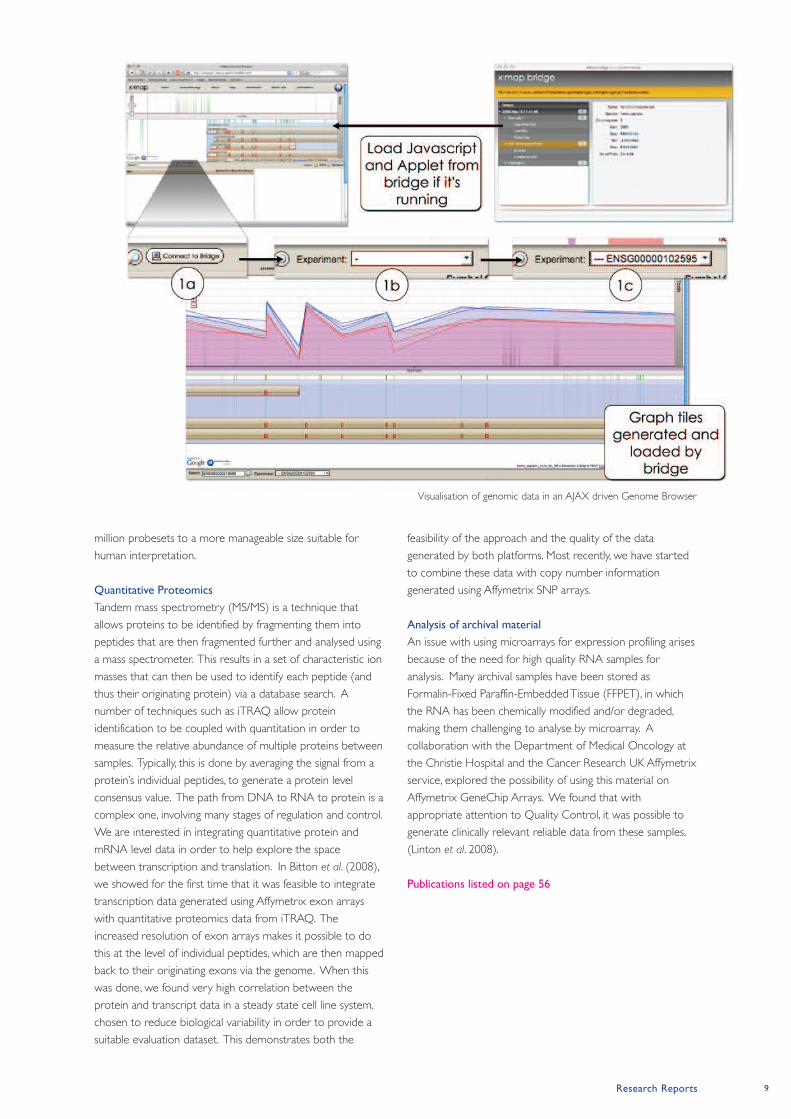
(Yates et al. 2008)] to help explore alternative splicing in
cancer related datasets (see figure). We also make these
tools available to other researchers, and they are used
internationally. Current research is focused on developing
novel approaches for interpreting exon array data, and in
particular, on generating the meaningful summaries that are
required in order to reduce the data from the arrays’ 1.4
Bioinformatics and Computational Biology
exist to help us make sense of the
complex datasets generated by research in
the biosciences. In the Applied
Computational Biology and Bioinformatics
Group, we are interested in the
development and application of software
tools and analytical strategies for the
analysis of cancer related datasets. We
collaborate closely with other groups
working on both the clinical and the
molecular biology aspects of cancer
research.
Postdoctoral Fellows
Carla Möller-Levet (joint
with Translational
Radiobiology)
John Hall (joint with
Translational Radiobiology)
James Bradford
Research ApplicationsProgrammer Manager
Tim Yates
Systems Administrator
Zhi Cheng Wang
Graduate Students
Danny Bitton
Andrzej Rutkowski (joint
with Immunology)
Research Reports 9
million probesets to a more manageable size suitable for
human interpretation.
Quantitative Proteomics
Tandem mass spectrometry (MS/MS) is a technique that
allows proteins to be identified by fragmenting them into
peptides that are then fragmented further and analysed using
a mass spectrometer. This results in a set of characteristic ion
masses that can then be used to identify each peptide (and
thus their originating protein) via a database search. A
number of techniques such as iTRAQ allow protein
identification to be coupled with quantitation in order to
measure the relative abundance of multiple proteins between
samples. Typically, this is done by averaging the signal from a
protein’s individual peptides, to generate a protein level
consensus value. The path from DNA to RNA to protein is a
complex one, involving many stages of regulation and control.
We are interested in integrating quantitative protein and
mRNA level data in order to help explore the space
between transcription and translation. In Bitton et al. (2008),
we showed for the first time that it was feasible to integrate
transcription data generated using Affymetrix exon arrays
with quantitative proteomics data from iTRAQ. The
increased resolution of exon arrays makes it possible to do
this at the level of individual peptides, which are then mapped
back to their originating exons via the genome. When this
was done, we found very high correlation between the
protein and transcript data in a steady state cell line system,
chosen to reduce biological variability in order to provide a
suitable evaluation dataset. This demonstrates both the
feasibility of the approach and the quality of the data
generated by both platforms. Most recently, we have started
to combine these data with copy number information
generated using Affymetrix SNP arrays.
Analysis of archival material
An issue with using microarrays for expression profiling arises
because of the need for high quality RNA samples for
analysis. Many archival samples have been stored as
Formalin-Fixed Paraffin-Embedded Tissue (FFPET), in which
the RNA has been chemically modified and/or degraded,
making them challenging to analyse by microarray. A
collaboration with the Department of Medical Oncology at
the Christie Hospital and the Cancer Research UK Affymetrix
service, explored the possibility of using this material on
Affymetrix GeneChip Arrays. We found that with
appropriate attention to Quality Control, it was possible to
generate clinically relevant reliable data from these samples.
(Linton et al. 2008).
Publications listed on page 56
Visualisation of genomic data in an AJAX driven Genome Browser
10 Paterson Institute for Cancer Research Scientific Report 2008
Carcinogenesis Grouphttp://www.paterson.man.ac.uk/carcinogenesis
Paterson Institute for Cancer Research
Background
Alkylating agents are a family of chemicals that are used in the
treatment of several types of cancer including brain tumours
(glioma) and skin tumours (melanoma) but there is
considerable room for improvement in their effectiveness.
These agents generate a dozen different types of damage in
DNA and there is increased understanding of the
mechanisms by which some of these result in cell killing. Thus
agents such as Dacarbazine and Temozolomide generate O6-
methylguanine in DNA and this kills cells via the action of the
post replication mismatch repair system. Cell killing by this
pathway can be prevented by the prior action of the damage
reversal protein, O6-alkylguanine-DNA alkyltransferase
(MGMT). This protein most probably evolved to protect
organisms against the toxic effects of endogenously or
environmentally generated lesions in DNA. However, it can
also reduce the effectiveness of these chemotherapeutics and
hence there is increasing interest in ablating the activity of
MGMT in tumours in order to improve clinical outcome.
One of the strategies we have pursued has involved the
development and use of Lomeguatrib, a drug that has now
completed phase II clinical trials in combination with
Temozolomide in malignant melanoma.
MGMT removes alkyl groups from the O6-position of guanine
by stoichiometric transfer to a cysteine residue in its active
site, a process that results in its irreversible inactivation. We
recently discovered, using computer based amino acid
sequence analysis, several proteins that have extensive
homology to MGMT, but without this critical cysteine residue.
These proteins are present in a number of organisms,
including E. coli and the fission yeast, S. pombe. We have
named this family the alkyltransferase-like (ATL) proteins and
we are currently intensively investigating their modus operandi.
Clinical trials of Lomeguatrib
Lomeguatrib (O6-[4-bromothenyl]guanine, previously called
PaTrin-2) is one of the products of a very fruitful
collaboration with Prof Brian McMurry and the late Dr
Stanley McElhinney (and their group at the Chemistry
The treatment of cancer often involves
the use of drugs that kill cells by damaging
their DNA. That some tumours do not
respond to such treatments can be
attributed to the presence of repair
processes which can remove potentially
lethal damage from DNA. Understanding
how DNA damage leads to cell death, and
how these repair systems process the
damage, may provide opportunities to
improve the effectiveness of existing
cancer therapies, and develop new agents.
Our main focus is on DNA damage and
repair following exposure to certain types
of alkylating agents, for example the CR-
UK drug, Temozolomide. Clinical trials of a
drug that was designed to ablate one of
the repair processes that is critical in
Temozolomide resistance are now
essentially completed. In fission yeast, we
found a different mechanism for dealing
with toxic DNA damage, and our efforts
are now directed towards the
characterisation of this novel repair
pathway.
Postdoctoral Fellows
Amna Butt
(until September)
Vitaly Latypov
(from October)
Barbara Verbeek
(until April)
Scientific Officers
Gail McGown
Mary Thorncroft
Mandy Watson
Graduate Student
Andrew Marriott
Undergraduate Students
Catia Caetano
Nick Peel
Volunteer Workers
Jonathan Doyle
Rita Matos
Group Leader Geoff Margison
Research Reports 11
Department, Trinity College, Dublin). It is a very potent
inactivator of MGMT and it effectively sensitises human cells
and tumour xenografts to the killing effect of Temozolomide
and other agents of that type. With the support of CR-UK
Drug Development and Formulation Units and also Cancer
Research Technology, Phase I clinical trials of this drug started
here at Christie Hospital in 2000 in the hands of Drs
Malcolm Ranson and Mark Middleton (now at the Churchill
Hospital, Oxford). These trials established a dose
combination of Lomeguatrib and Temozolomide for use in
Phase II trials, which have been carried out under the
auspices of KuDOS pharmaceuticals, to whom Lomeguatrib
was licensed. The clinical trials required us to develop and
validate to Good Clinical Laboratory Practice standards,
quantitative assays for both functionally active and total
MGMT protein. Malignant melanoma patients were recruited
from several centres in the UK and Australia. They were
treated with either Temozolomide alone or a Lomeguatrib-
Temozolomide combination. In addition, patients displaying
disease progression on Temozolomide (alone) therapy were
treated with the Lomeguatrib-Temozolomide combination to
assess if this would reverse Temozolomide resistance. The
outcome of the trial was disappointing: neither the response
rates nor survival were improved in the Lomeguatrib-
Temozolomide combination treated patients in comparison
with Temozolomide alone, including the group progressing on
the latter. However, laboratory analyses of tumour biopsy
samples collected during these studies showed that MGMT
activity was recovering in tumour tissue very soon after
treatment. Therefore a period of treatment with
Lomeguatrib alone was included at the end of the
combination treatment, and subsequently extended, in
attempts to maintain suppression of MGMT activity. This also
produced no clinical benefit. Neither did the Lomeguatrib-
Temozolomide combination provide any clinical benefit in
colorectal cancer patients. The reasons for this outcome have
yet to be established and are the subject of much speculation.
Alkyltransferase-like (ATL) proteins
We previously reported the isolation of the ATL-encoding
genes from E. coli and S. pombe and we named the latter, Atl1.
We have shown that, in vitro, the Atl1 protein binds to
methylated DNA and by doing so, can inhibit the action of
MGMT. In collaboration with David Williams (University of
Sheffield) we initially showed using gel shift assays that a
purified fusion protein of maltose binding protein and the
ATL from E. coli binds to short single- or double-stranded
oligonucleotides containing a number of O6-alkyl-substituted
guanines including methyl, benzyl, hydroxyethyl and 4-
bromothenyl (i.e Lomeguatrib embedded into an
oligonucleotide, which we have shown to be the most potent
MGMT inactivating agent so far described). However, whilst
binding to these potentially toxic lesions can be
demonstrated, there is no evidence of an MGMT-like alkyl
transfer, nor of glycosylase or endonuclease activity, so the
protein seems likely to be a specific DNA damage sensing
protein that signals to other DNA repair networks. We have
now extended these studies using native Atl1 protein in
enzyme-linked immunosorbent assays and surface plasmon
resonance methods to quantify these molecular interactions.
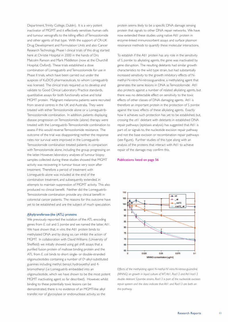
To establish if the Atl1 protein has any role in the sensitivity
of S. pombe to alkylating agents, the gene was inactivated by
gene disruption. The resulting deletants had similar growth
characteristics to the wild type strain, but had substantially
increased sensitivity to the growth inhibitory effects of N-
methyl-N-nitro-N-nitrosoguanidine, a methylating agent that
generates the same lesions in DNA as Temozolomide. Atl1
also protects against a number of related alkylating agents, but
there was no detectable effect on sensitivity to the toxic
effects of other classes of DNA damaging agents. Atl1 is
therefore an important protein in the protection of S. pombe
against the toxic effects of these alkylating agents. Exactly
how it achieves such protection has yet to be established, but,
crossing the atl1 deletant with deletants in established DNA
repair pathways (epistasis analysis) has suggested that Atl1 is
part of, or signals to, the nucleotide excision repair pathway
and not the base excision or recombination repair pathways
(see Figure). Further studies of this type along with an
analysis of the proteins that interact with Atl1 to achieve
repair of the damage may confirm this.
Publications listed on page 56
Effects of the methylating agent N-methyl-N’-nitro-N-nitroso-guanidine
(MNNG) on growth in liquid culture of WT, Atl1, Rad13 and Atl1/rad13
double deletant S.pombe strains. Rad13 is part of the nucleotide excision
repair system and the data indicate that Atl1 and Rad13 are both on
this pathway.
12 Paterson Institute for Cancer Research Scientific Report 2008
Cell Cycle Grouphttp://www.paterson.man.ac.uk/cellcycle
In a systematic screen for new cell cycle proteins we
identified a previously uncharacterised protein that is
required for cell division. We modified the chromosomal
locus of each of the essential budding yeast genes so that the
encoded protein was fused to the heat-inducible degron,
allowing us to analyse the effects of rapid depletion
(Kanemaki et al., Nature 2003; 423: 720). In this way we
found that inactivation of the budding yeast protein Ynl152w
blocked cytokinesis, and we named the protein Inn1 because
it is required for Ingression of the plasma membrane
(Sanchez-Diaz et al., 2008).
Using a strain in which Inn 1 was fused to Green Fluorescent
Protein (GFP) we found that Inn1 is recruited at the end of
mitosis to the site of the contractile actomyosin ring (Figure
1). Recruitment of Inn1 was dependent upon essential
components of the actomyosin ring, and we found that Inn1
co-purified from cell extracts with other previously described
components of the actomysin ring such as the SH3-domain
protein Hof1 and the IQGAP-domain protein Iqg1 (Sanchez-
Diaz et al., 2008).
In animal cells and in yeasts, the actomyosin ring defines the
site at which cell division will occur. At the end of mitosis the
ring becomes activated and then contracts into the
cytoplasm, and this is coupled by an unknown mechanism to
ingression of the plasma membrane. We found that the
actomyosin ring can still form in the absence of Inn1, and
contraction is still initiated in all cells at the end of mitosis. In
contrast, however, ingression of the plasma membrane does
not occur in the absence of Inn1 (Figure 2). The Inn1 protein
is thus required in some way for contraction of the
actomyosin ring to be coupled to ingression of the plasma
membrane at the end of mitosis.
The first 134 amino acids of the Inn1 protein are predicted to
form a C2 domain, which is a class of membrane targeting
domain that was first described in Protein Kinase C. The C2
domain comprises eight β-strands that form a sandwich of
two β-sheets. From one side of the sandwich, two or three
loops protrude and these usually interact with the targets of
the C2 domain, which can be the head groups of membrane
lipids, or other proteins. We found that mutation of
Our group studies the mechanisms that
drive the eukaryotic cell cycle. During
2008 we described a novel factor called
Inn1 that is required for cytokinesis in
budding yeast. The Inn1 protein is
associated with the contractile actomyosin
ring that defines the site at which cell
division will occur at the end of mitosis. In
the absence of Inn1, the actomyosin ring
still forms and contracts on schedule but
ingression of the plasma membrane does
not occur, resulting in failure of cell
division. We found that the amino
terminus of the Inn1 protein forms a ‘C2-
domain’ that is essential for membrane
ingression, whereas the carboxy terminus
of Inn1 is required to recruit the Inn1
protein to the cleavage site. Our data
indicate that the key to the function of
Inn1 is recruitment of the C2 domain to
the cleavage site, where it plays an
essential role by allowing ingression of the
plasma membrane to be coupled to
contraction of the actomyosin ring.
Postdoctoral Fellows
Giacomo de Piccoli
Luis Garcia-Rodriguez
Alberto Sanchez-Diaz
Sugopa Sengupta
Scientific Officer
Frederick van Deursen
Graduate Students
Asli Devrekanli
Tim Maculins
Hiroko Morohashi
Magdalena Foltman
Visiting Student
Bianca Targosz
Group Leader Karim Labib
Research Reports 13
conserved positively charged residues in Loop1 of the C2
domain of Inn1 blocks membrane ingression and cytokinesis,
but does not prevent recruitment of the Inn1 protein to the
cleavage site. This indicates that the C2 domain is essential
for ingression of the plasma membrane during cytokinesis.
Removal of the C2-domain blocks cytokinesis but does not
prevent the regulated targeting of the rest of the protein to
the cleavage site. In contrast, the remainder of Inn1 after the
C2-domain is required for localisation at the cleavage site,
and contains multiple ‘PXXP’ motifs that are likely to be
binding sites for SH3 proteins. These findings suggested a
model for the action of the Inn1 protein, whereby the
majority of the protein serves to target Inn1 to the cleavage
site so that the C2-domain can then fufill some essential role
during cytokinesis. To test this model, we generated a diploid
strain lacking one copy of the INN1 gene and in which we
had fused artificially the C2-domain to the Hof1 component
of the actomyosin ring. Upon sporulation of this diploid we
found that haploid cells that lacked the INN1 gene and that
expressed wild type HOF1 were inviable and formed chains
of undivided cells, indicating a failure of cytokinesis. In
contrast, however, expression of the C2-Hof1 fusion protein
was able to suppress the defects normally associated with
absence of Inn1, and restore ingression of the plasma
membrane during cytokinesis, so that inn1∆ C2-HOF1 cells
grew as well as wild type cells (Sanchez-Diaz et al., 2008).
These findings indicate that the recruitment of the C2-
domain of Inn1 to the cleavage site is a key requirement for
ingression of the plasma membrane during cytokinesis in
budding yeast. We are currently trying to understand how
Inn1 is recruited to the cleavage site, how recruitment is
regulated during the cell cycle, and how the C2-domain
promotes ingression of the plasma membrane.
Publications listed on page 57
0’ 2’ 4’ 6’ 8’
Myo1-Tomato
Inn1-GFP
Merge
Figure 1. Inn1 is recruited to the actomyosin ring at the end of mitosis.
The figure shows timelapse analysis of the contracting actomyosin ring in
a cell that expresses Inn1-GFP and that also has the red fluorescent
protein Tomato fused to the budding yeast type II myosin, Myo1.
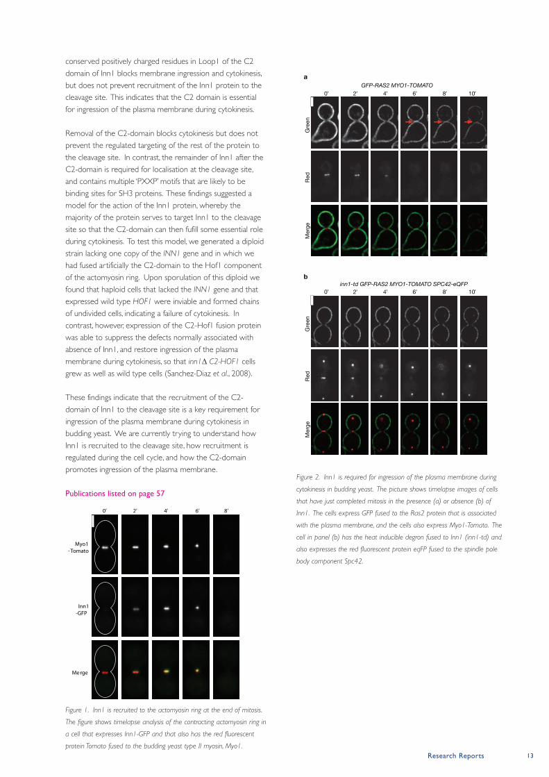
Figure 2. Inn1 is required for ingression of the plasma membrane during
cytokinesis in budding yeast. The picture shows timelapse images of cells
that have just completed mitosis in the presence (a) or absence (b) of
Inn1. The cells express GFP fused to the Ras2 protein that is associated
with the plasma membrane, and the cells also express Myo1-Tomato. The
cell in panel (b) has the heat inducible degron fused to Inn1 (inn1-td) and
also expresses the red fluorescent protein eqFP fused to the spindle pole
body component Spc42.
14 Paterson Institute for Cancer Research Scientific Report 2008
Cell Division Grouphttp://www.paterson.man.ac.uk/celldivision
Paterson Institute for Cancer Research
MAP kinase cascades
MAP kinase cascades lie at the heart of most stress response
pathways. They comprise three kinases that are activated in
sequence: a MAP kinase kinase kinase (MAPKKK) stimulates a
MAP kinase kinase (MAPKK)
to activate the MAPK (figure
1). In all organisms these
cascades are activated by a
variety of stimuli to promote
the phosphorylation of a
number of targets to modify
cell metabolism and
physiology in order to cope
with the environmental
changes. As major changes
in cell physiology are needed
to adapt to the new
environment, transcription
factors are prime targets.
The activation of these
transcription factors in turn
promotes the transcription of a cohort of genes to adapt to
the new environment. For example, cells produce a number
of reducing agents in response to heightened levels of
oxidative stress.
Stress responses, cell division and growth control
In addition to changing cell constitution to tolerate stress,
stress response pathways can arrest growth and division until
the adaptation is complete. Presumably this ensures fidelity
of division and minimises the long-term impact of stress,
should either process be attempted whilst any damage
remains un-repaired. Once the adaptive response has dealt
with the novel environment, specific signalling events are
required to re-initiate growth and division. These recovery
pathways often rely upon the same stress response pathways
that instigated the arrest in the first place. Quite how they
do this is largely unclear at present. A broad goal of our
research effort is therefore to understand how stress
responses exploit cell cycle regulators and growth controls to
regulate the timing and execution of cell division and growth
following stress. We employ fission yeast as a model system
for these studies as its cell cycle controls and stress response
pathways mirror those of humans.
Like us cells live in an ever-changing world.
For cells this involves fluctuation in
exposure to nutrients, oxygen and a range
of physical and chemical insults. These
environmental changes are sensed by
signal transduction cascades called “stress
response pathways” that alter metabolism
and physiology to adapt to the changing
environment. Because the growth and
division of cancer cells deviates from
normal tissues, transformed cells invariably
experience heightened levels of stress.
Understanding how stress responses work
and how they are triggered by
environmental insults can therefore
identify novel approaches to specifically
target the stressed, cancer, cells while
leaving the unstressed cells of normal
tissue untouched. Because these stress
response pathways are highly conserved,
studying them in the relatively simple and
highly malleable yeast can greatly
accelerate the analysis of the more
complex networks of man.
Postdoctoral Fellows
Marisa Alonso-Nuñez
Agnes Grallert
Nimesh Joseph
Tamara Krsmanovic
Marisa Madrid
Najma Rachidi
Scientific Officer
Deepti Wilks
Graduate Students
Elvan Boke
Dorota Feret
Avinash Patel
Rotation Student
Julian Blaser
Visiting Scientist
Victor Alvarez-Tallada
Group Leader Iain Hagan
MAPKKKMAPKKK
MAPKKMAPKK
MAPKMAPK
Environmental stimuli
transcription factors
transcription factors
other targetsother targetsother targetsother targets
Figure 1. A cartoon depicting the
organisation of a MAPK kinase
cascade
Stress responses in fission yeast
Two MAP kinase cascades maintain homeostasis in fission
yeast. The so called “stress response pathway” (SRP)
incorporates the MAPK Sty1/Spc1. Like its mammalian
JNK/p38 counterparts the SRP responds to a broad range of
stresses, including oxidative, osmotic, heavy metal ion and
heat stresses. The “cell integrity pathway” (CIP) utilises the
MAP kinase, Pmk1, and is required to maintain cell integrity in
response to osmotic shock and hydrostatic pressure as well
as regulating cytokinesis. There is interplay between these
two pathways as SRP signalling both promotes and attenuates
CIP signalling depending upon the circumstances.
Specific stress recovery pathways
Phosphorylation of the conserved cell cycle regulator polo
kinase on serine 402 restores both growth and division of
cells after they have been arrested in a mild heat shock
induced stress response (Petersen and Hagan, Nature 2005;
435: 507). Curiously, serine 402 phosphorylation is not
stimulated by other stresses such as osmotic stress, even
though these stresses also arrest cell growth. We therefore
studied the impact of osmotic stress upon the cell growth
machinery over the last year.
Growth in fission yeast
Cell growth in fission yeast mirrors the interplay between the
actin and microtubule cytoskeletons that underlies growth,
migration and signalling in human cells. Cell extension is
promoted as a consequence of polarisation of the actin
cytoskeleton by the same GTPase cascades that promote the
polarisation of the actin cytoskeleton to drive migration of
human cells. In yeast the polarisation of the actin
cytoskeleton directs the secretion of cell wall material to
generate the robust polysaccharide wall that acts as an
“exoskeleton” to define the rod shape of wild type fission
yeast cells (figure 2). This cell wall contains a range of
polysaccharides including 1−3β D-glucan that is stained by
the fluorescent probe calcofluor. The selection of the site at
which the actin becomes polarised to generate growth of the
cell tip is mediated by microtubules. Therefore, perturbation
of the microtubule cytoskeleton, or the microtubule
associated factors that communicate the inputs from
microtubules to the actin cytoskeleton, cause cells to branch.
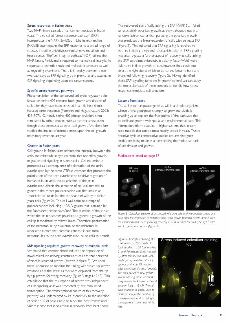
SRP signalling regulates growth recovery at multiple levels
We found that osmotic shock induced the deposition of
novel calcofluor staining structures at cell tips that persisted
after cells resumed growth (arrows in figure 3). We used
these landmarks to monitor the timing with which tip growth
resumed after the stress as foci were displaced from the tip
by tip growth following recovery (figure 3 stage1>2>3). This
established that the resumption of growth was independent
of CIP signalling as it was promoted by SRP stimulated
transcription. The transcriptional nature of this recovery
pathway was underscored by its insensitivity to the mutation
of serine 402 of polo kinase to block the post-translational
SRP response that is so critical in recovery from heat shock.
The recovered tips of cells lacking the SRP MAPK Sty1 failed
to re-establish polarised growth, as they ballooned out in a
random fashion rather than pursuing the polarised growth
that produces the linear extension of cells with an intact SRP
(figure 2). This indicated that SRP signalling is required to
both re-initiate growth and re-establish polarity. SRP signalling
may also regulate a further aspect of recovery as cells lacking
the SRP associated microtubule polarity factor Wsh3 were
able to re-initiate growth on cue, however they could not
select the right site at which to do so and became bent and
branched following recovery (figure 2). Having identified
these SRP signalling functions in growth control we can study
the molecular basis of these controls to identify how stress
responses modulate cell structure.
Lessons from yeast
The ability to manipulate genes at will in a simple organism
whose primary purpose is simply to grow and divide is
enabling us to explore the finer points of the pathways that
co-ordinate growth with spatial and environmental cues. This
information informs studies in higher systems that, in turn,
raise models that can be most readily tested in yeast. This re-
iterative cycle of comparative studies ensures that great
strides are being made in understanding the molecular basis
of cell division and growth.
Publications listed on page 57
Wild type no stress
wsh3.Δafter recovery from stress
sty1.Δafter recovery from stress
Figure 2. Calcofluor staining of unstressed wild type cells and two mutant strains one
hour after the imposition of osmotic stress when growth patterns clearly deviate from
the linear extension seen following recovery of cells in which the wild type sty1+ and
wsh3+ genes are present (figure 3).
1
1
23
Stress induced calcofluor stainingfoci
3
foci
Figure 3. Calcofluor staining of a
mixture of cdc10.v50 cells, 30
(cells marked 1), 60 (cell marked
2) and 90 minutes (cells marked
3), after osmotic stress at 36°C.
Bright foci of calcofluor staining
appear at the tip 30 minutes
after imposition of stress (arrows).
The dots persist as new growth
initiates driving these landmarks
progressively back towards the cell
equator (cells 1>2>3). The cell
cycle mutation is simply used to
block division for the duration of
the experiment and so highlight
the apparent “movement” of the
foci.
Research Reports 15
16 Paterson Institute for Cancer Research Scientific Report 2008
Cell Regulation Grouphttp://www.paterson.man.ac.uk/cellregulation
Paterson Institute for Cancer Research
Previous efforts in our laboratory have focused on two
mammalian MAP kinase targets, the transcription factors
ATF2 and ATF7, which are both regulated by
phosphorylation mediated by the stress activated MAPKs
p38 and JNK. ATF2 and ATF7 are both members of the AP-
1 family which controls the transcription of an extensive
repertoire of genes including cell cycle and apoptotic
regulators, numerous cytokines and growth factors. We
have analysed the biological role of ATF2 and ATF7 using the
genetically amenable mouse model system. Germ line
mutations uncovered new insights into their role in
development: simultaneous deletion of both genes leads to
embryonic lethality as a result of massive apoptosis in the
embryonic liver involving both developing hepatocytes and
haematopoietic cells. Characterisation of cultured cells
derived from mutant embryos revealed a role in limiting cell
proliferation resulting in increased cell growth at high
density. Interestingly, upon oncogenic transformation with K-
Ras, cells lacking ATF2 and ATF7 showed a profound
increase in tumour growth upon grafting into appropriate
recipient mice. In both the embryonic phenotype and the
cell growth phenotype the role of the ATF factors in
regulating MAPK signalling is critical. A number of dual
specificity phosphatases, including MKP1, constitute major
downstream targets and in the mutant background their
transcription is decreased resulting in loss of essential
negative feedback regulation of MAP kinase pathways.
The scope for in vivo analysis using mouse knockouts is
limited by the early lethality of the mutations. To circumvent
this difficulty we have developed a number of tissue-specific
ATF2 knockouts. These models have revealed roles for ATF2
in specific tissues and disease conditions: a brain specific
deletion of ATF2 leads to defects in hindbrain development
and cerebellum functions resulting in death soon after birth
due to a respiratory defect that resembles meconium
aspiration syndrome; deletion of ATF2 specifically in
endothelial cells leads to defects in the microvasculature of
the gut upon postnatal intestinal growth. These results
emphasise the importance of ATF2 in the development and
homeostasis of mammalian tissue.
Conditional knockout models have also provided additional
insights into a potential role for these factors in
tumourigenesis. A tumour suppressor role has been
revealed in a skin tumourigenesis model using a mutant
mouse where ATF2 is specifically deleted in keratinocytes.
Upon tumour initiation and promotion, the mutant animals
demonstrate a significantly earlier onset of papillomas as
well as greater numbers. The potential role of ATF2 in the
progression of papillomas to malignant carcinomas is
currently being assessed. Likewise, irradiation of mice with
ATF2 specifically deleted in T-cells results in earlier onset of
T-cell lymphomas further supporting a tumour suppressor
role.
In contrast, in other tumour contexts there is indirect
evidence for ATF2 being pro-tumourigenic. In order to
assess this possibility directly, appropriate tumour models
The MAP (mitogen activated protein)
kinase signalling pathways are central to
the ability of the cell to respond to
various stress conditions and in so doing
protect the cell from potential damage
that could arise. The highly conserved
pathways are essential for a wide variety
of biological activities which in mammalian
cells range from cell proliferation and
differentiation to regulation of apoptosis;
their deregulation has been associated
with numerous disease conditions such as
inflammation and cancer.
Associate Scientists
Wolfgang Breitwieser
Caroline Wilkinson
Postdoctoral Fellows
Yujun Di
Clare Lawrence
Graduate Students
Malgorzata Gozdecka
Orestis Mavroudis-
Chocholis
Jacek Walczynski
Lu Zhang
Scientific Officers
Keren Dawson
Steve Lyons
Group Leader Nic Jones
Research Reports 17
are being developed and characterised. For example, we
are characterising a B-cell specific ATF2 knockout model and
analysing the effect of ATF2 deletion on lymphoma initiation
and development following B-cell specific expression of the
c-myc oncogene. Similarly, the role of ATF2 in
hepatocellular carcinoma (HCC) is being examined using
two different approaches: one involves the chemical
induction of HCC in control and liver specific ATF2
knockout mice which will determine whether the absence
of functional ATF2 (and ATF7) leads to significant changes in
onset, frequency and burden of HCC; the second utilises
the recently developed orthotopic transplantation of
hepatocyte precursors (hepatoblasts) to produce chimaeric
models of liver tumours.
Homologues of the AP-1 family are found in all eukaryotic
organisms and their involvement in stress response is highly
conserved. Model organisms could provide useful models
for understanding the role and regulation of AP-1. Fission
yeast is a particularly pertinent model system – stress
responses are coordinated through the Sty1 signalling
pathway which is analogous to the mammalian p38 pathway.
Furthermore, many of the changes in the transcriptional
profile of cells following stress is orchestrated through the
Atf1 and Pap1 transcription factors which are related to
mammalian ATF2 and cJun respectively.
The transcriptional changes following stress are extensive
and complex: a common set of genes are activated in
response to all stresses accompanied by the activation of
genes that are specific to the particular stress being
imposed. Therefore each stress has its own transcriptional
pattern resulting in the up-regulation of the appropriate
defence and repair mechanisms. The complexity of the
response is best illustrated by characterising the events
following exposure to oxidative stress: different patterns of
transcriptional activation are seen dependent upon the
exact nature of the oxidant as well as the dose
Given the central role that Atf1 plays in the stress response,
we have characterised in detail its regulation and its
interaction with the Sty1 kinase. Using ChIP assays we
demonstrated that Atf1 is essential to target and tether the
Sty1 kinase to stress-response gene promoters. Both the
targeting and transcriptional activation is dependent upon
Sty1 kinase activity. Surprisingly however, the key kinase
target is not Atf1 itself since Sty1 is found at promoters in
the presence of Atf1 protein that can no longer be
phosphorylated (Atf1-11M mutant). We hypothesise that
Sty1 phosphorylates and regulates another factor(s) found
at the promoter, possibly a component of chromatin
remodelling complexes or the polymerase complex itself. A
number of approaches are being taken to identify Sty1
targets.
Phosphorylation of Atf1 by Sty1 regulates its stability;
phosphorylation results in an increase in the half-life of Atf1
and its accumulation to significantly higher levels in the cell
following stress. This increase in levels of Atf1 is critical for a
robust transcriptional response upon exposure to H2O2 and
for the ability of cells to mount an adaptive response
providing resistance to acute levels of osmotic stress. The
key regulatory step in Atf1 degradation is the interaction of
the transcription factor with the E3 ligase, SCFFbh1. This
interaction results in the ubiquitination of Atf1 and its
destruction via the proteasome. The substrate specificity of
the SCFFbh1 complex is determined by the F-box protein,
Fbh1. Crucially, the interaction between Atf1 and Fbh1 is
sensitive to Atf1 hyper-phosphorylation; accordingly the
interaction is lost upon stress and Atf1 is stabilised.
Consistent with these findings, disruption of the interaction
between Fbh1 and the rest of the SCF E3 ligase complex,
results in an increase in Atf1-11M protein and a rescue of
the phenotypes displayed by the atf1-11M mutant.
Interestingly, Fbh1 levels appear to be regulated by Atf1
suggesting that a complex interplay exists between the F-
box protein and its target which serve to finely tune the
abundance of the Atf1 transcription factor upon stress.
Publications listed on page 57
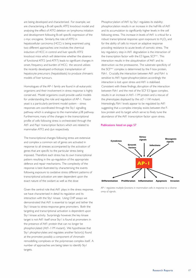
AP-1
Cytokines
Growth factors
Stresssignals
Infections
Oncogenic signals
Differentiation Proliferation Apoptosis InvasionInflammation
AP-1 regulates multiple functions in mammalian cells in response to a diverse
array of signals.
18 Paterson Institute for Cancer Research Scientific Report 2008
Cell Signalling Grouphttp://www.paterson.man.ac.uk/cellsignalling
Paterson Institute for Cancer Research
Rho proteins, such as Rac1, RhoA and Cdc42, are guanine
nucleotide binding proteins that cycle between an inactive
GDP-bound state and an active GTP-bound state. The
activity of Rho proteins is controlled by guanine nucleotide
exchange factors (GEFs) and GTPase-activating proteins
(GAPs). GEFs activate small GTPases by promoting the
exchange of GDP for GTP, whereas GAPs enhance the
intrinsic rate of hydrolysis of bound GTP for GDP, leading to
inactivation. Tiam1 (for T-lymphoma invasion and metastasis
protein) belongs to the GEF family of proteins and selectively
activates Rac in response to growth factors and cell-substrate
interactions. Precisely how these upstream events engage the
Tiam1-Rac signalling module is unclear. One possible
mechanism is suggested by the observed association of Tiam1
with the second messenger Ras through a Ras-binding
domain (RBD). Activated Ras and Tiam1 then cooperate to
activate Rac (Lambert et al., Nature Cell Biol 2002; 4: 621).
Significantly, Tiam1-deficient cells are resistant to Ras-induced
cellular transformation (Malliri et al., Nature 2002; 417: 867),
implying that this interaction is important for tumourigenesis.
Tiam1/Rac signalling and tumourigenesis in vivo
Mice deficient for Tiam1 are resistant to the formation of skin
tumours induced by topical application of chemical
carcinogens and consequent oncogenic activation of the c-
Ha-Ras gene (Malliri et al., Nature 2002; 417: 867).
Tiam1-deficient tumours were not only fewer but also
smaller than wild-type tumours and this correlated with
increased apoptosis and reduced proliferation in carcinogen-
exposed Tiam1-deficient mice. Tiam1 is also a potent
modifier of intestinal tumourigenesis (Malliri et al., J Biol Chem
2006; 281: 543). The majority of intestinal tumours are
caused by mutations in the canonical Wnt signaling pathway,
leading to aberrant expression of Wnt-responsive genes.
Tiam1 is a Wnt-responsive gene, and is expressed in crypts of
the adult mammalian intestine as well as in adenomas from
patients and Min (multiple intestinal neoplasia) mice. In each
of these settings, the Wnt pathway is activated. Further, by
comparing tumour development in Min mice expressing or
lacking Tiam1, it was found that Tiam1 deficiency significantly
reduces the formation as well as growth of polyps in vivo
(Malliri et al., J Biol Chem 2006; 281: 543).
These two studies on tumourigenesis in vivo demonstrated
that two independent oncogenic signalling pathways of major
clinical significance (Ras and Wnt) recruit the Tiam1-Rac
signalling pathway by specific, albeit distinct mechanisms. In
Tumour initiation and progression result
from inappropriate activation of
intracellular signalling cascades. Rho-like
GTPases are molecular switches in
signalling pathways that regulate
cytoskeletal and junctional organisation, as
well as gene transcription. In this way, Rho
proteins influence cell morphology,
adhesion, motility, as well as cell cycle
progression and cell survival. Rho proteins
are transforming in vitro and are essential
for Ras-mediated in vitro transformation.
Moreover, data has emerged to directly
implicate Rho proteins in tumour initiation
and progression in vivo. Our group
investigates how the activities of certain
regulators of the Rho protein Rac are
controlled. We are also identifying
signalling events downstream of Rac that
modulate tumour susceptibility and
disease progression.
Postdoctoral Fellows
Sonia Castillo-Lluva
(funded by an EMBO Long-
Term fellowship)
Helen Rushton
(since April 2008)
Simon Woodcock
Scientific Officer
Gavin White
Graduate Students
Lucy Dalton
Natalie Reeves
Claire Rooney
Chong Tan
(since September 2008)
Group Leader Angeliki Malliri
Research Reports 19
the context of oncogenesis, activation of this signalling
module promotes tumour initiation and growth. Moreover,
this role is specific to Tiam1 since its loss cannot be
compensated for by other Rho GEFs.
Tiam1/Rac signalling and the regulation of cell-cell adhesion
The skin carcinogenesis model revealed an additional role for
Tiam1 in tumourigenesis. The few skin tumours arising in
Tiam1-deficient mice progressed more frequently to
malignancy than those in wild-type mice, suggesting that
Tiam1 deficiency promotes malignant conversion (Malliri et
al., Nature 2002; 417: 867). Analysis of Tiam1 expression in
skin tumours of wild-type mice revealed that benign
papillomas maintained high levels of Tiam1 expression,
whereas expression was reduced in squamous cell
carcinomas and was completely lost in highly invasive spindle
cell carcinomas. The increased Ras signalling associated with
advanced skin malignancies (resulting from amplification of
the mutated Ras allele) seems to be responsible for the
reduction or loss of Tiam1 expression in the later stages of
tumour progression, as demonstrated in vitro for Ras-
transformed MDCK cells (Zondag et al., J Cell Biol 2000; 149:
775). Thus, while Tiam1/Rac co-operate with Ras in
establishing tumours, they antagonize Ras during tumour
invasion.
One probable mechanism by which Tiam1 and Rac suppress
malignant progression is through their ability to stimulate
cell–cell adhesion. In vitro studies have shown that over-
expression of activated Rac or Tiam1 can promote the
formation of adherens junctions (AJs) and the accompanying
induction of an epithelial-like phenotype in a number of
mesenchymal cell lines (Malliri & Collard, Curr Opin Cell Biol
2003; 15: 583). Moreover, using both RNA interference and
cells derived from Tiam1-deficient mice, it has been shown
that endogenous Tiam1 is required for both the formation as
well as the maintenance of cadherin-based adhesions (Malliri
et al., J Biol Chem 2004; 279: 30092). The oncoprotein Src, a
non-receptor tyrosine kinase implicated in malignant
progression, potently induces epithelial–mesenchymal
transition (EMT) by targeting AJs for dissassembly. We
recently showed that direct phosphorylation of Tiam1 by Src
is required for Src-induced EMT. Moreover, we identified a
novel post-translational mechanism of regulating Tiam1 levels.
We showed that Src phosphorylates Tiam1 on tyrosine 384
(Y384). This occurs predominantly at AJs during the initial
stages of Src-induced EMT and creates a docking site on
Tiam1 for Grb2. We found that Tiam1 is constitutively
associated with extracellular signal-regulated kinase (ERK).
Following recruitment of the Grb2-Sos1 complex, ERK
becomes activated and triggers the localised degradation of
Tiam1 at AJs through, in turn, activating calpain proteases.
Significantly, we demonstrated that in human lung, colon, and
head and neck cancers phosphorylation of Y384 of Tiam1
positively correlated with Src activity, while total levels of
Tiam1 were inversely correlated with Src activity, consistent
with the above-mentioned post-translational regulatory
mechanism operating in malignancies. Abrogating Tiam1
phosphorylation and degradation suppressesed Src-induced
AJ disassembly. As a consequence, cells expressing a non-
phosphorylatable Tiam1 showed a marked decrease in
wound closure in response to Src. These data establish a
new paradigm for regulating local concentrations of Rho-
GEFs, as well as linking Tiam1-Rac signalling with a further
oncoprotein.
It is increasingly apparent that Rho GEFs do more than simply
activate Rho molecules; several studies now point to their
role in influencing the choice of biological response elicited by
a given Rho protein. GEFs have been shown to bind to
effectors directly or to scaffold proteins that complex with
components of effector pathways. Thus Tiam1 interacts with
IB2/JIP2, a scaffold that promotes Rac activation of p38 kinase
cascade over JNK MAP kinase cascade (Buchsbaum et al., Mol
Cell Biol 2002; 22: 4073), and also with spinophilin, a scaffold
that promotes Rac activation of p70 S6K over Pak1, a
different Rac effector (Buchsbaum et al., J Biol Chem 2003;
278: 18833). In our lab, we are using biochemical approaches
to identify Rac and Rac GEF interacting proteins involved in
different aspects of transformation including malignant
progression (acquisition of invasiveness).
Publications listed on page 57
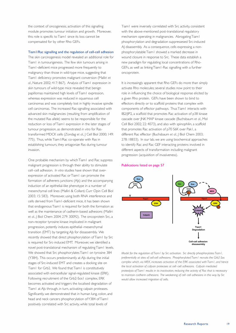
Tiam1Y
Tiam1Y
ERKERK ERKERK
P
P
Grb2
Sos
MEK
Tiam1 Degradation
Grb2
Sos
Calpain
Calpain
Cell-cell adhesion dissasembly
Src
Model for the regulation of Tiam1 by Src activation. Src directly phosphorylates Tiam1,
preferentially at sites of cell-cell adhesions. Phosphorylated Tiam1 recruits the Grb2-Sos
complex which, via MEK, increases activation of the ERK associated with Tiam1, and hence
the local activation of calpain proteases at cell–cell adhesions. Calpain mediated
proteolysis of Tiam1 results in its inactivation, reducing the activity of Rac that is necessary
to maintain cadherin adhesions. The weakening of cell–cell adhesions in this way by Src
would allow increased migration of cells.
20 Paterson Institute for Cancer Research Scientific Report 2008
Clinical and Experimental
Pharmacology Grouphttp://www.paterson.man.ac.uk/cep
Paterson Institute for Cancer Research
Clinical Trials at the Christie Hospital’s DCU
Research in CEP is predicated on those novel agents entering
clinical trial at the DCU. DCU typically supports c100-120
trials with c6400 patient visits p.a. In order to meet research
and service requirements, proposals for a new £35M Cancer
Treatment Centre were developed in 2008. The new Cancer
Treatment Centre will provide comprehensive facilities for
clinical trials, experimental treatment, and service
chemotherapy and is due to open in 2010. The expansion of
the DCU within the treatment centre will make it one of the
largest early clinical trials centres worldwide and integral to
this development are enhanced laboratory facilities for
translational research to strengthen further the CEP-DCU
axis. One example of the CEP DCU axis in action is the
biomarker enhanced CR-UK Phase I trial of AEG35156
(Aegera Therapeutics, antisense XIAP). We reported this first
in class, first in man clinical trial in the Journal of Clinical
Oncology (Dean et al., 2008). CEP has also developed a
suite of biomarkers to accompany ongoing early clinical trials
of the BH-3 mimetic class of apoptosis promoting drugs that
target interactions between Bcl-2 family proteins.
The development of molecularly targeted
anticancer drugs mandates a parallel
development of biomarkers in order to
give the right patient the right dose and
schedule of treatment. CEP develops and
validates pharmacokinetic (PK),
pharmacodynamic (PD) and predictive
biomarker assays and implements
biomarker qualification within clinical trials
at the Christie Hospital’s Derek Crowther
Unit (DCU) focusing on novel agents
targeted to apoptosis and angiogenesis. In
2008, biomarker research included
development of a robust plasma
proteomics workflow for biomarker
discovery, enumeration and
characterisation of circulating tumour cells,
detection of oncogene mutations in
circulating free DNA and multiplexing
circulating and tissue biomarker assays.
Disease orientated translational research
focus on lung, colorectal and paediatric
cancers and pre-clinical studies includes
drug target validation, drug combination
optimisation and the impact of hypoxia on
drug efficacy.
Disease Focus Team
Leaders
Fiona Blackhall
Guy Makin
Andrew Renehan
Staff Scientists
Jeff Cummings
Tim Ward
Postdoctoral Fellows
Luke Harrison
Sarah Holt (AZ second-
ment)
Jian Mei Hou
Dominic James (AZ
secondment)
Tetanya Klymenko
Flavia Moreira Leite
Lee Lancashire
David Moore
Christopher Morrow
Darren Roberts
Kathryn Simpson
Clinical Fellows
Ruth Board
Emma Dean
Alastair Greystoke
Sarah Hughes
Matthew Krebs
Gireesh Kumaran
Scientific Officers
Karen Brookes
Fouziah Butt
Martin Dawson
Olive Denenny
Martin Greaves
Grace Hampson
Cassandra Hodgkinson
Karen Morris
Lyndsey Priest
Robert Sloane
Nigel Smith
Brian Trueman
Zaira Yunis
Graduate Students
Jane Barraclough
Martin Brandenburg
Cristina Martin-Fernandez
Dimitra Micha
Elizabeth Sweeney
Shaun Villa
Undergraduate Student
Laura Glass
Laboratory Aide
Matthew Lancashire
Group Leaders Caroline Dive and Malcolm Ranson
Research Reports 21
Biomarker Research in CEP in 2008 is exemplified by the
following highlights i) application of cytokeratin 18 (CK18)
based circulating biomarkers of cell death as
pharmacodynamic biomarkers; ii) demonstration of the
potential of circulating tumour cells (CTCs) as
pharmacodynamic and predictive biomarkers; iii) the potential
predictive utility of oncogene mutation detection in circulating
free DNA and iv) the development of multiplexed circulating
biomarkers of angiogenesis.
(i) CK18 based circulating biomarkers of cell death
In collaboration with Abbott Laboratories, we demonstrated
using a small cell lung cancer (SCLC) xenograft model that
the BH-3 mimetic ABT 737 provoked tumour cell apoptosis
that was reported by the appearance of full length and
caspase cleaved CK18 in the blood stream as assessed by
M65 and M30 ELISAs respectively (Micha et al., 2008). These
circulating biomarkers were elevated rapidly after drug
administration before detectable tumour shrinkage and
informed on appropriate blood sampling times for the
ongoing Phase I clinical trial where biomarker analysis is
underway.
(ii) Circulating Tumour Cells
We evaluated CTCs as a useful biomarker for trials of BH-3
mimetics in SCLC. In preparation for upcoming trials, CTC
number was evaluated in patients on standard chemotherapy
using the Veridex CellSearch system; CTC numbers
decreased post drug treatment in accordance with patient
response in cycle 1 of therapy. The levels of drug targets in
CTCs may serve to predict which patients will respond to
BH-3 mimetics thus assays were developed to identify
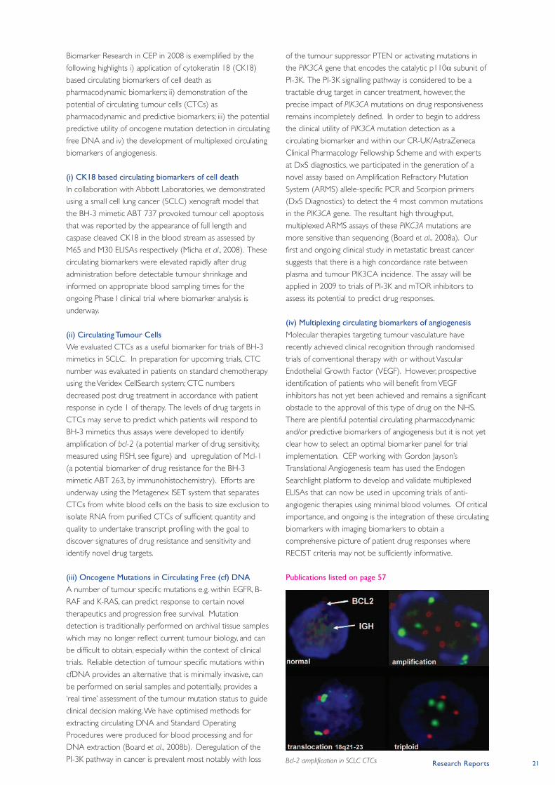
amplification of bcl-2 (a potential marker of drug sensitivity,
measured using FISH, see figure) and upregulation of Mcl-1
(a potential biomarker of drug resistance for the BH-3
mimetic ABT 263, by immunohistochemistry). Efforts are
underway using the Metagenex ISET system that separates
CTCs from white blood cells on the basis to size exclusion to
isolate RNA from purified CTCs of sufficient quantity and
quality to undertake transcript profiling with the goal to
discover signatures of drug resistance and sensitivity and
identify novel drug targets.
(iii) Oncogene Mutations in Circulating Free (cf) DNA
A number of tumour specific mutations e.g. within EGFR, B-
RAF and K-RAS, can predict response to certain novel
therapeutics and progression free survival. Mutation
detection is traditionally performed on archival tissue samples
which may no longer reflect current tumour biology, and can
be difficult to obtain, especially within the context of clinical
trials. Reliable detection of tumour specific mutations within
cfDNA provides an alternative that is minimally invasive, can
be performed on serial samples and potentially, provides a
‘real time’ assessment of the tumour mutation status to guide
clinical decision making. We have optimised methods for
extracting circulating DNA and Standard Operating
Procedures were produced for blood processing and for
DNA extraction (Board et al., 2008b). Deregulation of the
PI-3K pathway in cancer is prevalent most notably with loss
of the tumour suppressor PTEN or activating mutations in
the PIK3CA gene that encodes the catalytic p110α subunit of
PI-3K. The PI-3K signalling pathway is considered to be a
tractable drug target in cancer treatment, however, the
precise impact of PIK3CA mutations on drug responsiveness
remains incompletely defined. In order to begin to address
the clinical utility of PIK3CA mutation detection as a
circulating biomarker and within our CR-UK/AstraZeneca
Clinical Pharmacology Fellowship Scheme and with experts
at DxS diagnostics, we participated in the generation of a
novel assay based on Amplification Refractory Mutation
System (ARMS) allele-specific PCR and Scorpion primers
(DxS Diagnostics) to detect the 4 most common mutations
in the PIK3CA gene. The resultant high throughput,
multiplexed ARMS assays of these PIKC3A mutations are
more sensitive than sequencing (Board et al., 2008a). Our
first and ongoing clinical study in metastatic breast cancer
suggests that there is a high concordance rate between
plasma and tumour PIK3CA incidence. The assay will be
applied in 2009 to trials of PI-3K and mTOR inhibitors to
assess its potential to predict drug responses.
(iv) Multiplexing circulating biomarkers of angiogenesis
Molecular therapies targeting tumour vasculature have
recently achieved clinical recognition through randomised
trials of conventional therapy with or without Vascular
Endothelial Growth Factor (VEGF). However, prospective
identification of patients who will benefit from VEGF
inhibitors has not yet been achieved and remains a significant
obstacle to the approval of this type of drug on the NHS.
There are plentiful potential circulating pharmacodynamic
and/or predictive biomarkers of angiogenesis but it is not yet
clear how to select an optimal biomarker panel for trial
implementation. CEP working with Gordon Jayson’s
Translational Angiogenesis team has used the Endogen
Searchlight platform to develop and validate multiplexed
ELISAs that can now be used in upcoming trials of anti-
angiogenic therapies using minimal blood volumes. Of critical
importance, and ongoing is the integration of these circulating
biomarkers with imaging biomarkers to obtain a
comprehensive picture of patient drug responses where
RECIST criteria may not be sufficiently informative.
Publications listed on page 57
Bcl-2 amplification in SCLC CTCs
22 Paterson Institute for Cancer Research Scientific Report 2008
Immunology Grouphttp://www.paterson.man.ac.uk/immunology
Paterson Institute for Cancer Research
Immune regulation and escape in TroVax vaccinated patients
We have recently reported the results of a phase II trial in
which two TroVax (MVA-5T4) vaccinations were given to
patients both pre- and post-surgical resection of liver
metastases secondary to colorectal cancer (CRC). 5T4-
specific cellular responses were assessed at entry and two
weeks after each vaccination by proliferation of fresh
lymphocytes and ELISA for antibody responses; 18 of 19
CRC patients mounted a 5T4-specific cellular and/or humoral
response (see also Biological, Immune and Gene Therapy
Report). We have now completed a comparison of individual
and between patient responses over the course of the
treatments using cryopreserved peripheral blood
mononuclear cells (PBMC) samples from baseline until after
the fourth vaccination at 14 weeks using a proliferation assay
with 5T4-Fc fusion protein, overlapping 32mer 5T4 peptides,
MVA-LacZ and MVA-5T4 infected autologous monocytes.
Significant responses to 32mer peptide pools were seen in 8
of 19 CRC patients by time of surgery and 13 of 19 who
received 2 more vaccinations by week 14. We also assessed
the levels of systemic T regulatory cells, plasma cytokine
levels, phenotype of tumour infiltrating lymphocytes including
T regulatory cells and tumour MHC class I loss of expression.
Approximately half of the patients showed phenotypes
consistent with relative immune suppression and/or escape
highlighting the complexity of positive and negative factors
challenging any simple correlation with clinical outcome.
Mouse models of 5T4 immunotherapy
We have shown that transient depletion of T regulatory cells
(Tregs) can enhance 5T4 immunity in renal cell carcinoma
patients (Thistlethwaite et al., 2008), suggesting that Treg
modulation might improve the efficacy of the 5T4 vaccine
(TroVax). To investigate the role of Tregs in 5T4 immunity we
utilised 5T4 knockout (KO) C57BL6 mice to map the 5T4 T
cell repertoire. By immunising wild type (WT) and 5T4 KO
mice with a single dose of a replication defective adenovirus
vaccine encoding murine 5T4 (Ad-m5T4) and re-stimulating
these primed cells in vitro with overlapping 32-mers, we
identified m5T4 derived epitopes recognized by CD4 and
CD8 T cells using interferon gamma (IFNγ) ELISPOT. 5T4
KO mice generated strong and high avidity 5T4-specific CD4
and CD8 T cell responses. In WT mice, 5T4-specific CD4 T
cells were either absent or anergized and the CD8 T cell
response weaker and lower avidity. The latter might result
from a lack of help and/or active suppression by Tregs. To
look for evidence of a natural 5T4 specific Treg cell
population, CD4+CD25+ T cells were isolated from naïve
The Immunology Group has a major goal
of understanding and exploiting the
function and/or expression of 5T4
oncofoetal molecules in the context of
cancer associated changes in motility, and
adhesion contributing to metastasis.
Successful translational studies have led to
ongoing Phase 3 clinical trials of 5T4
directed immunotherapies. This report
details studies of both animal model and
human immune responses to 5T4 antigen
aimed at improving both vaccine and
antibody delivered therapies for cancer. In
addition progress on the development of
immunotherapies for HPV associated
cancers are reported.
Postdoctoral Fellows
Fernanda Castro
Owen McGinn
Tom Southgate
Clinical Fellows
Emma Crosbie
(Walport Fellow)
Sai Daayana
(with Henry Kitchener)
Christy Ralph
(with Robert Hawkins)
Scientific Officers
Kate Mulryan
Debbie Burt
(seconded to BIGT)
Graduate Students
Mariam Al-Muftah
(with David Gilham)
Georgi Marinov
Andrzej Rutkowski
(with Crispin Miller)
Group Leader Peter L. Stern
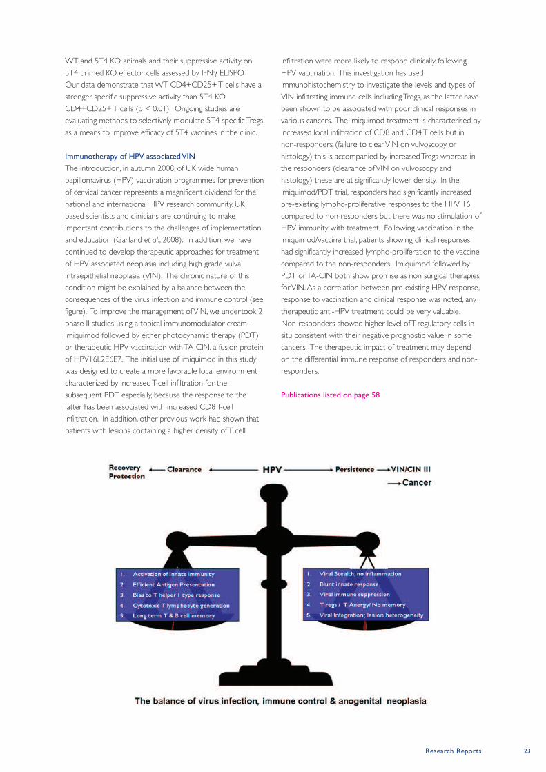
Research Reports 23
WT and 5T4 KO animals and their suppressive activity on
5T4 primed KO effector cells assessed by IFNγ ELISPOT.
Our data demonstrate that WT CD4+CD25+ T cells have a
stronger specific suppressive activity than 5T4 KO
CD4+CD25+ T cells (p < 0.01). Ongoing studies are
evaluating methods to selectively modulate 5T4 specific Tregs
as a means to improve efficacy of 5T4 vaccines in the clinic.
Immunotherapy of HPV associated VIN
The introduction, in autumn 2008, of UK wide human
papillomavirus (HPV) vaccination programmes for prevention
of cervical cancer represents a magnificent dividend for the
national and international HPV research community. UK
based scientists and clinicians are continuing to make
important contributions to the challenges of implementation
and education (Garland et al., 2008). In addition, we have
continued to develop therapeutic approaches for treatment
of HPV associated neoplasia including high grade vulval
intraepithelial neoplasia (VIN). The chronic nature of this
condition might be explained by a balance between the
consequences of the virus infection and immune control (see
figure). To improve the management of VIN, we undertook 2
phase II studies using a topical immunomodulator cream –
imiquimod followed by either photodynamic therapy (PDT)
or therapeutic HPV vaccination with TA-CIN, a fusion protein
of HPV16L2E6E7. The initial use of imiquimod in this study
was designed to create a more favorable local environment
characterized by increased T-cell infiltration for the
subsequent PDT especially, because the response to the
latter has been associated with increased CD8 T-cell
infiltration. In addition, other previous work had shown that
patients with lesions containing a higher density of T cell
infiltration were more likely to respond clinically following
HPV vaccination. This investigation has used
immunohistochemistry to investigate the levels and types of
VIN infiltrating immune cells including Tregs, as the latter have
been shown to be associated with poor clinical responses in
various cancers. The imiquimod treatment is characterised by
increased local infiltration of CD8 and CD4 T cells but in
non-responders (failure to clear VIN on vulvoscopy or
histology) this is accompanied by increased Tregs whereas in
the responders (clearance of VIN on vulvoscopy and
histology) these are at significantly lower density. In the
imiquimod/PDT trial, responders had significantly increased
pre-existing lympho-proliferative responses to the HPV 16
compared to non-responders but there was no stimulation of
HPV immunity with treatment. Following vaccination in the
imiquimod/vaccine trial, patients showing clinical responses
had significantly increased lympho-proliferation to the vaccine
compared to the non-responders. Imiquimod followed by
PDT or TA-CIN both show promise as non surgical therapies
for VIN. As a correlation between pre-existing HPV response,
response to vaccination and clinical response was noted, any
therapeutic anti-HPV treatment could be very valuable.
Non-responders showed higher level of T-regulatory cells in
situ consistent with their negative prognostic value in some
cancers. The therapeutic impact of treatment may depend
on the differential immune response of responders and non-
responders.
Publications listed on page 58
24 Paterson Institute for Cancer Research Scientific Report 2008
Inositide Laboratoryhttp://www.paterson.man.ac.uk/
Paterson Institute for Cancer Research
Understanding how PtdIns(4,5)P2 levels are regulated and
how changes in PtdIns(4,5)P2 are transduced in to regulating
cellular function.
PtdIns(4,5)P2 is present in the plasma membrane where its
levels can be regulated in response to receptor activation.
PtdIns(4,5)P2 is also synthesised within the nucleus where its
levels can be regulated distinctly from the plasma membrane
pool. Understanding how these two pools of lipids are
controlled and which cellular pathways they regulate
represents a major goal of the laboratory. PtdIns(4,5)P2 can
be synthesised by the action of two distinct but related
kinases (collectively termed PIP kinases). PIP5Ks
phosphorylate PtdIns4P on the 5’ position, while PIP4Ks
phosphorylate PtdIns5P on the 4’ position. It is likely that
PIP5Ks are the major producers of PtdIns(4,5)P2 while the
role of PIP4K may be to regulate cellular PtdIns5P levels
and/or a distinct minor pool of PtdIns(4,5)P2. PIP5Ks are
upregulated in nearly all cancer cell lines tested and
overexpression of PIP5K can induce dramatic changes in cell
morphology, increase migratory capacity and attenuate
apoptosis in response to cellular stressors. PIP4Ks on the
other hand appear to regulate the levels of cellular PtdIns5P
which can impinge on the activity of the tumour suppressor
protein p53 and a number of other transcriptional regulators
that are implicated in the development of cancer and on PKB
activation.
Phosphoinositides are a family of lipid
second messengers interlinked by the
activities of an extensive and highly
regulated network of kinases and
phosphatases that modulate
phosphoinositide levels in response to
environmental changes. Alterations in
phosphoinositide levels can regulate many
different cancer-relevant cellular pathways
including survival, proliferation, migration,
cell substratum interactions and
transcription. PtdIns(4,5)P2 is at the heart
of phosphoinositide signalling, being the
substrate for both phosphatidylinositol-3-
kinase (PI3K) and phospholipase C (PIC).
The PI3K /PTEN pathway is deregulated in
tumours promoting cell survival and
proliferation through the activation of PKB
and flux through the PIC pathway is
upregulated in human tumours.
Furthermore PtdIns(4,5)P2 is itself a
regulator of cytoskeletal dynamics, cell
survival pathways and cell polarity (see
Panbianco et al., 2008). Cells monitor
PtdIns(4,5)P2 levels and a decrease in their
levels can lead to the induction of
apoptosis. As PtdIns(4,5)P2 levels are
sensitive to extracellular matrix signalling,
the induction of apoptosis in response to
decreases in PtdIns(4,5)P2 may constitute a
metastasis suppressive mechanism. It is
likely that tumour cells must by-pass this
process to become metastatic.
Interestingly a number of oncogenes that
suppress cell death in response to loss of
extracellular matrix signalling also suppress
the decrease in PtdIns(4,5)P2 and are
consequently able to maintain growth
factor signalling.
Associate Scientist
David Jones
Postdoctoral Fellows
Iman van den Bout
Laura Norton
Scientific Officer
Yvette Bultsma
Graduate Students
Willem-Jan Keune
Lilly Sommer
Group Leader Nullin Divecha
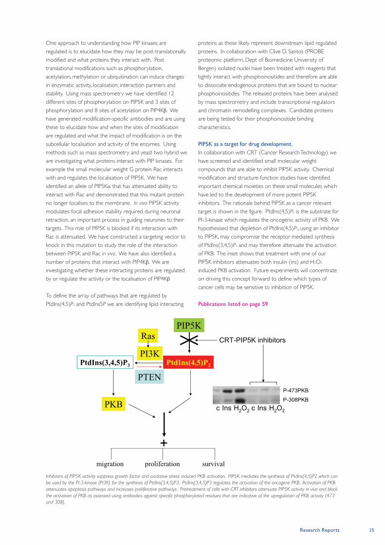
Research Reports 25
One approach to understanding how PIP kinases are
regulated is to elucidate how they may be post-translationally
modified and what proteins they interact with. Post
translational modifications such as phosphorylation,
acetylation, methylation or ubiquitination can induce changes
in enzymatic activity, localisation, interaction partners and
stability. Using mass spectrometry we have identified 12
different sites of phosphorylation on PIP5K and 3 sites of
phosphorylation and 8 sites of acetylation on PIP4Kβ. We
have generated modification-specific antibodies and are using
these to elucidate how and when the sites of modification
are regulated and what the impact of modification is on the
subcellular localisation and activity of the enzymes. Using
methods such as mass spectrometry and yeast two hybrid we
are investigating what proteins interact with PIP kinases. For
example the small molecular weight G protein Rac interacts
with and regulates the localisation of PIP5K. We have
identified an allele of PIP5Kα that has attenuated ability to
interact with Rac and demonstrated that this mutant protein
no longer localises to the membrane. In vivo PIP5K activity
modulates focal adhesion stability required during neuronal
retraction, an important process in guiding neurones to their
targets. This role of PIP5K is blocked if its interaction with
Rac is attenuated. We have constructed a targeting vector to
knock in this mutation to study the role of the interaction
between PIP5K and Rac in vivo. We have also identified a
number of proteins that interact with PIP4Kβ. We are
investigating whether these interacting proteins are regulated
by or regulate the activity or the localisation of PIP4Kβ
To define the array of pathways that are regulated by
PtdIns(4,5)P2 and PtdIns5P we are identifying lipid interacting
proteins as these likely represent downstream lipid regulated
proteins. In collaboration with Clive D. Santos (PROBE
proteomic platform, Dept of Biomedicine University of
Bergen) isolated nuclei have been treated with reagents that
tightly interact with phosphoinositides and therefore are able
to dissociate endogenous proteins that are bound to nuclear
phosphoinositides. The released proteins have been analysed
by mass spectrometry and include transcriptional regulators
and chromatin remodelling complexes. Candidate proteins
are being tested for their phosphoinositide binding
characteristics.
PIP5K as a target for drug development.
In collaboration with CRT (Cancer Research Technology) we
have screened and identified small molecular weight
compounds that are able to inhibit PIP5K activity. Chemical
modification and structure-function studies have identified
important chemical moieties on these small molecules which
have led to the development of more potent PIP5K
inhibitors. The rationale behind PIP5K as a cancer relevant
target is shown in the figure. PtdIns(4,5)P2 is the substrate for
PI-3-kinase which regulates the oncogenic activity of PKB. We
hypothesised that depletion of PtdIns(4,5)P2, using an inhibitor
to PIP5K, may compromise the receptor mediated synthesis
of PtdIns(3,4,5)P3 and may therefore attenuate the activation
of PKB. The inset shows that treatment with one of our
PIP5K inhibitors attenuates both insulin (ins) and H2O2
induced PKB activation. Future experiments will concentrate
on driving this concept forward to define which types of
cancer cells may be sensitive to inhibition of PIP5K.
Publications listed on page 59
PtdIns(4,5)P2PtdIns(3,4,5)P3
PKB
PTEN
PI3K
RasPIP5K
migration survivalproliferation
+
CRT-PIP5K inhibitors
P-473PKB
P-308PKBc Ins H2O2 c Ins H2O2
Inhibitors of PIP5K activity suppress growth factor and oxidative stress induced PKB activation. PIP5K mediates the synthesis of PtdIns(4,5)P2 which can
be used by the PI-3-kinase (PI3K) for the synthesis of PtdIns(3,4,5)P3. PtdIns(3,4,5)P3 regulates the activation of the oncogene PKB. Activation of PKB
attenuates apoptosis pathways and increases proliferative pathways. Pretreatment of cells with CRT inhibitors attenuate PIP5K activity in vivo and block
the activation of PKB as assessed using antibodies against specific phosphorylated residues that are indicative of the upregulation of PKB activity (473
and 308).
26 Paterson Institute for Cancer Research Scientific Report 2008
Leukaemia Biology Grouphttp://www.paterson.man.ac.uk/leukaemia
Paterson Institute for Cancer Research
CSCs (also called tumour-propagating or tumour-initiating
cells) were first formally described in human acute myeloid
leukaemia (AML) as rare cells that share an
immunophenotype with normal hematopoietic stem cells
(HSCs). This paradigm has recently been substantially revised
based on two significant observations. First, in murine models
of human leukaemia induced by the MLL-AF9 oncogene, self-
renewing leukaemia stem cells (LSCs) may account for up to
a quarter of cells within the leukaemia clone and exhibit
mature myeloid immunophenotypes (Somervaille and Cleary,
Cancer Cell 2006; 10: 257). Secondly, protocols that enhance
engraftment of human leukaemia cells in xenogeneic
transplant assays demonstrate the presence of LSCs in
leukaemia cell sub-populations previously considered to be
devoid of them.
Since LSCs may be more numerous and mature than
originally proposed, the nature and generality of the
hierarchical organization of malignancies has recently been
questioned. However, consistent with the CSC model, only a
subset of AML cells have clonogenic potential in in vitro
assays, and human AML blast cells undergo differentiation in
vivo to mature granulocytes, as may murine LSCs initiated by
MLL-AF9 (Somervaille and Cleary, Cancer Cell 2006; 10: 257).
To further elucidate the hierarchical disposition of AML, a
major goal is to identify transcriptional programs, genes and
pathways that specifically correlate with and promote the
retention of LSCs within the self-renewing compartment of
leukaemias. It is not known whether such LSC maintenance
programs are synonymous with programs responsible for
leukaemia initiation, for example Hoxa/Meis in MLL
leukaemogenesis. It is also not clear whether they share
features with transcriptional programs expressed in adult or
embryonic stem cells (ESCs), or whether there is a
relationship with genes and pathways implicated in the
function of AML stem cells such as NFκB,
phosphatidylinositide-3-kinase, CTNNB1, Bmi1, Pten and Junb.
In work performed at the Paterson Institute in the past year
by members of the Leukaemia Biology group, and also
previously by Tim Somervaille at Stanford University in the
United States, we investigated the genetic determinants that
maintain LSC frequencies and leukaemia cell hierarchies using
a mouse model that faithfully recapitulates many of the
pathologic features of AML induced by chromosomal
The cancer stem cell (CSC) model posits
that many human malignancies consist of
two functionally distinct cell types: (i)
CSCs, which are self-renewing cells with
the capacity to initiate, sustain and expand
the disease, and (ii) non-self-renewing
progeny cells, derived from CSCs through
differentiation, which may make up the
bulk of the tumour and account for
disease symptomatology. In order for
malignancies to be cured, it may be
necessary and sufficient to exclusively
eliminate CSCs. Consequently, there is
considerable interest in further
understanding the biologic and molecular
properties of these cells, by comparison
with both their non-self-renewing
downstream progeny and their normal
adult stem cell counterparts.
Group Leader Tim Somervaille
Postdoctoral Fellow
Xu Huang
Scientific Officer
Gary Spencer
Graduate Student
William Harris
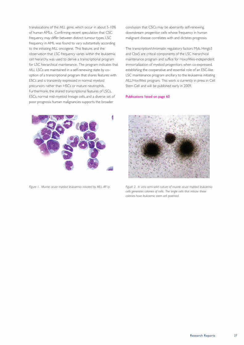
Research Reports 27
translocations of the MLL gene, which occur in about 5-10%
of human AMLs. Confirming recent speculation that CSC
frequency may differ between distinct tumour types, LSC
frequency in AML was found to vary substantially according
to the initiating MLL oncogene. This feature, and the
observation that LSC frequency varies within the leukaemia
cell hierarchy, was used to derive a transcriptional program
for LSC hierarchical maintenance. The program indicates that
MLL LSCs are maintained in a self-renewing state by co-
option of a transcriptional program that shares features with
ESCs and is transiently expressed in normal myeloid
precursors rather than HSCs or mature neutrophils.
Furthermore, the shared transcriptional features of LSCs,
ESCs, normal mid-myeloid lineage cells, and a diverse set of
poor prognosis human malignancies supports the broader
conclusion that CSCs may be aberrantly self-renewing
downstream progenitor cells whose frequency in human
malignant disease correlates with and dictates prognosis.
The transcription/chromatin regulatory factors Myb, Hmgb3
and Cbx5 are critical components of the LSC hierarchical
maintenance program and suffice for Hoxa/Meis-independent
immortalization of myeloid progenitors when co-expressed,
establishing the cooperative and essential role of an ESC-like
LSC maintenance program ancillary to the leukaemia initiating
MLL/Hox/Meis program. This work is currently in press in Cell
Stem Cell and will be published early in 2009.
Publications listed on page 60
Figure 1. Murine acute myeloid leukaemia initiated by MLL-AF1p. Figure 2. In vitro semi-solid culture of murine acute myeloid leukaemia
cells generates colonies of cells. The single cells that initiate these
colonies have leukaemic stem cell potential.
28 Paterson Institute for Cancer Research Scientific Report 2008
Stem Cell Biology Grouphttp://www.paterson.man.ac.uk/stemcellbiology/
Paterson Institute for Cancer Research
RUNx1 and leukemia.
Human acute leukemias are characterized by the presence of
recurrent chromosomal abnormalities, which frequently result
in the formation of chimeric transcription factors. The core
binding factors AML1/RUNX1 and CBFβ are the most
frequent targets of these genetic alterations. The t(8;21)
translocation resulting in AML1-ETO fusion and the inv(16)
generating the SHMMC-CBFβ fusion accounts together for
more than 20% of all the AML cases. Animal models have
indicated that the full length AML-ETO, expressed either
upon viral transfer or as a transgene, is not able to induce
alone leukemia in mice. However an alternatively spliced
form of AML1-ETO has recently been shown to cause
following retroviral transfer a rapid development of leukemia
in mice. Based on this new finding, we are developing an
animal model in which the expression of this form of AML1-
ETO is inducible. This new tool will allow us to study the
molecular events leading upon expression of AML-ETO to
the development of leukemia.
Early haematopoietic development
The earliest site of blood cells development in the mouse
embryo is the yolk sac where blood islands, consisting of
haematopoietic cells surrounded by a layer of angioblasts,
develop at approximately day 7.5 of gestation. The parallel
development of these two lineages in close association
provided the basis for the hypothesis that they arise from a
common precursor, a cell called the haemangioblast. A
conflicting theory instead associates the first haematopoietic
cells to a phenotypically differentiated endothelial cell with
haematopoietic potential, i.e. a haemogenic endothelium.
Support for the haemangioblast concept was initially
provided by the identification during embryonic stem (ES)
cells differentiation of a clonal precursor, the blast colony-
forming cell (BL-CFC), which gives rise after 4 days to blast
colonies with both endothelial and haematopoietic potential.
Although recent studies have now provided evidence for the
presence of this bipotential precursor in vivo, the precise
mechanism of generation of haematopoietic cells from the
haemangioblast still remains completely unknown.
A new model of haematopoietic development
We performed a series of studies to determine the cellular
and molecular events leading to the generation of blast
colony from BL-CFC. Our data demonstrate that the
haemangioblast generates haematopoietic cells through the
formation of a haemogenic endothelium intermediate,
providing the first direct link between these two precursor
populations. This haemogenic endothelial cell population is
transiently generated during blast development and is also
detected in gastrulating embryos. At the molecular level, we
have demonstrated that the transcription factor SCL/TAL1 is
indispensable for the establishment of this haemogenic
endothelium cell population from the haemangioblast
The transcription factor AML1/RUNX1 is
a frequent target of gene rearrangements
and mutations in human acute
myelogenous leukemia (AML) and acute
lymphoblastic leukemia (ALL). Consistent
with its implication in leukemias, RUNX1
has also been shown to be critical for
haematopoietic development. The MOZ
gene is involved in three independent
myeloid chromosomal translocations
fusing MOZ to the partner genes CBP,
P300 or TIF2. Our group studies the
function of MOZ and RUNXI in
haematopoietic development and
maintenance with the aim to better
understand how alterations of these
functions lead to leukemogenesis. We use
complementary approaches such as in vitro
differentiation of mouse embryonic stem
(ES) cells and in vivo mouse models.
Postdoctoral Fellows
Cristina Ferreras
Christophe Lancrin
Michael Lie-A-Ling
Flor Perez-Campo
Scientific Officers
Catherine Gavin
Ting Zheng
Graduate Students
Monika Antkiewicz
Patrycja Sroczynska
Olga Tsoulaki
Group Leader Georges Lacaud
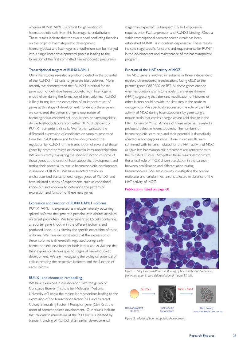
Research Reports 29
whereas RUNX1/AML1 is critical for generation of
haematopoietic cells from this haemogenic endothelium.
These results indicate that the two a priori conflicting theories
on the origin of haematopoietic development,
haemangioblast and haemogenic endothelium, can be merged
into a single linear developmental process leading to the
formation of the first committed haematopoietic precursors.
Transcriptional targets of RUNx1/AML1
Our initial studies revealed a profound defect in the potential
of the RUNX1-/- ES cells to generate blast colonies. More
recently we demonstrated that RUNX1 is critical for the
generation of definitive haematopoietic from haemogenic
endothelium during the formation of blast colonies. RUNX1
is likely to regulate the expression of an important set of
genes at this stage of development. To identify these genes,
we compared the patterns of gene expression of
haemangioblast-enriched-cell-populations or haemangioblast-
derived-cell-populations from either RUNX1 deficient or
RUNX1 competent ES cells. We further validated the
differential expression of candidates on samples generated
from the ES/EB system and further documented the
regulation by RUNX1 of the transcription of several of these
genes by promoter assays or chromatin immunoprecipitation.
We are currently evaluating the specific function of some of
these genes at the onset of haematopoietic development and
testing their potential to rescue haematopoietic development
in absence of RUNX1. We have selected previously
uncharacterized transcriptional target genes of RUNX1 and
have initiated a series of experiments, such as conditional
knock-out and knock-in, to determine the pattern of
expression and function of these new genes.
Expression and Function of RUNx1/AML1 isoforms
RUNX1/AML1 is expressed as multiple naturally occurring
spliced isoforms that generate proteins with distinct activities
on target promoters. We have generated ES cells containing
a reporter gene knock-in in the different isoforms and
produced knock-outs altering the specific expression of these
isoforms. We have demonstrated that the expression of
these isoforms is differentially regulated during early
haematopoietic development both in vitro and in vivo and that
their expression defines specific stages of haematopoietic
development. We are investigating the biological potential of
cells expressing the respective isoforms and the function of
each isoform.
RUNx1 and chromatin remodelling
We have examined in collaboration with the group of
Constanze Bonifer (Institute for Molecular Medicine,
University of Leeds) the molecular mechanisms leading to the
expression of the transcription factor PU.1 and its target
Colony-Stimulating-Factor 1 Receptor gene (CSF1R) at the
onset of haematopoietic development. Our results indicate
that chromatin remodeling at the Pu.1 locus is initiated by
transient binding of RUNX1 at an earlier developmental
stage than expected. Subsequent CSFR-1 expression
requires prior PU.1 expression and RUNX1 binding. Once a
stable transcriptional haematopoietic circuit has been
established, RUNX1 is in contrast dispensable. These results
indicate stage specific functions and requirements for RUNX1
in the development and maintenance of the haematopoietic
program.
Function of the HAT activity of MOZ
The MOZ gene is involved in leukemia in three independent
myeloid chromosomal translocations fusing MOZ to the
partner genes CBP, P300 or TIF2. All these genes encode
enzymes containing a histone acetyl transferase domain
(HAT) suggesting that aberrant modification of histones or
other factors could provide the first step in the route to
oncogenicity. We specifically addressed the role of the HAT
activity of MOZ during haematopoiesis by generating a
mouse strain that carries a single amino acid change in the
HAT domain of MOZ. Analysis of these mice has revealed a
profound defect in haematopoiesis. The numbers of
haematopoietic stem cells and their potential is dramatically
affected in homozygous mice. These in vivo results were
confirmed with ES cells mutated for the HAT activity of MOZ
as again less haematopoietic precursors are generated with
the mutated ES cells. Altogether these results demonstrate
the critical role of MOZ driven acetylation in the balance
between proliferation and differentiation during
haematopoiesis. We are currently investigating the precise
molecular and cellular mechanisms affected in absence of the
HAT activity of MOZ.
Publications listed on page 60
Figure 1. May Grumwald/Giemsa staining of haematopoietic precursors
generated upon in vitro differentiation of mouse ES cells.
Figure 2. Model of haematopoietic development.
Haemangioblast (BL-CFC)
Haemogenic Endothelium
Blast ColonyHaematopoietic precursors
Scl / Tal1 Runx1 / AML1
30 Paterson Institute for Cancer Research Scientific Report 2008
Stem Cell and Haematopoiesis Grouphttp://www.paterson.man.ac.uk/sch
Paterson Institute for Cancer Research
A model system to study haematopoietic specification.
The in vitro differentiation of Embryonic Stem (ES) cells offers
a powerful approach to advance our understanding of many
developmental processes. Murine ES cells can be induced to
differentiate and generate primitive and definitive
haematopoietic precursors, an in vitro process that accurately
recapitulates the in vivo development of yolk sac
haematopoiesis. This progressive differentiation, leading to
the formation of fully mature blood cells, can be monitored
by the measurement of gene expression via real-time
polymerase chain reaction (PCR), the analysis of cell surface
expression by multi-colour flow-cytometry and the
quantitative analysis of biological potential using clonogenic
replating assays. The first known progenitor specified to the
blood program is the haemangioblast. This precursor is
characterized by its expression of Flk1, the VEGF (vascular
endothelial growth factor) receptor 2, and its ability to
generate both primitive and definitive haematopoietic
precursors as well as endothelium and smooth muscle
lineages. The hemangioblast, in presence of VEGF, give rise to
fully committed blood precursors, characterized by their
expression of CD41, the alpha2b integrin chain. Using
microarray expression profiling of subpopulations at various
stages of differentiation, we have identified novel cell surface
markers and transcription factors implicated in the
specification of blood precursors from the mesodermal germ
layer.
Multi-parameter analysis of blood specification.
In our effort to define novel cell surface markers expressed
during the specification of the haematopoietic lineages, we
identified CD40 and Icam2 as two molecules substantially up-
regulated upon haemangioblast commitment. Both genes
code for proteins expressed on the cell surface of adult
leukocytes and are actively implicated in immunological
responses. To define a possible function for these two
molecules at the onset of blood formation, we first
investigated their pattern of expression during the
differentiation of ES cells to blood progenitors. Interestingly,
we observed a progressive and sequential up-regulation of
these two cell surface molecules during the maturation of
mesodermal precursors to fully committed blood cells.
Figure 1 illustrates the dramatic shift in CD40 and Icam2
profile observed between day 3 and 4 of ES cell
differentiation. Detailed studies allowed us to establish that
low levels of CD40 expression specifically define the Flk1+
haemangioblast subpopulation. As these precursors
differentiated to generate blood-restricted precursors Icam2
expression is switched on while CD40 expression becomes
significantly higher. In multi-parameter analysis, the integration
of CD40 and Icam2 expression pattern into previously
defined pathway of blood differentiation allowed us to
further refine and identify discrete steps during the
specification of mesoderm into fully restricted blood cells.
The emergence of haematopoietic cells
during embryonic life occurs soon after
gastrulation. A tight coordination of
proliferation, differentiation and migration
during the generation of these first blood
cells ensures the proper survival and
growth of the developing embryo.
Understanding the molecular mechanisms
that control the formation of these blood
precursors from the mesodermal germ
layer is the major focus of our laboratory.
Several lines of evidence suggest that
during adult life leukemogenesis can result
from the re-initiation of an embryonic
program or from the inappropriate
expression of genes controlling critical
steps of this embryonic program. A clear
understanding of the molecular
mechanisms orchestrating the onset of
haematopoietic specification should help
us to better define the basis of de-
regulated proliferation and differentiation
observed in haematological malignancies.
Postdoctoral Fellows
Arnaud Gandillet
Alicia Gonzalez-Serrano
Scientific Officer
Stella Pearson
Graduate Students
Katalin Boros
Guilherme Costa
Sarah Lewis
Group Leader Valerie Kouskoff
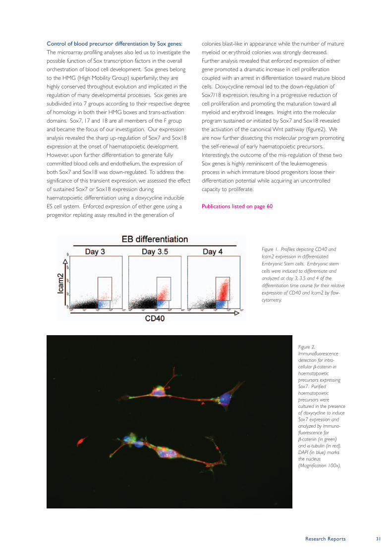
Research Reports 31
Control of blood precursor differentiation by Sox genes:
The microarray profiling analyses also led us to investigate the
possible function of Sox transcription factors in the overall
orchestration of blood cell development. Sox genes belong
to the HMG (High Mobility Group) superfamily; they are
highly conserved throughout evolution and implicated in the
regulation of many developmental processes. Sox genes are
subdivided into 7 groups according to their respective degree
of homology in both their HMG boxes and trans-activation
domains. Sox7, 17 and 18 are all members of the F group
and became the focus of our investigation. Our expression
analysis revealed the sharp up-regulation of Sox7 and Sox18
expression at the onset of haematopoietic development.
However, upon further differentiation to generate fully
committed blood cells and endothelium, the expression of
both Sox7 and Sox18 was down-regulated. To address the
significance of this transient expression, we assessed the effect
of sustained Sox7 or Sox18 expression during
haematopoietic differentiation using a doxycycline inducible
ES cell system. Enforced expression of either gene using a
progenitor replating assay resulted in the generation of
colonies blast-like in appearance while the number of mature
myeloid or erythroid colonies was strongly decreased.
Further analysis revealed that enforced expression of either
gene promoted a dramatic increase in cell proliferation
coupled with an arrest in differentiation toward mature blood
cells. Doxycycline removal led to the down-regulation of
Sox7/18 expression, resulting in a progressive reduction of
cell proliferation and promoting the maturation toward all
myeloid and erythroid lineages. Insight into the molecular
program sustained or initiated by Sox7 and Sox18 revealed
the activation of the canonical Wnt pathway (figure2). We
are now further dissecting this molecular program promoting
the self-renewal of early haematopoietic precursors.
Interestingly, the outcome of the mis-regulation of these two
Sox genes is highly reminiscent of the leukemogenesis
process in which immature blood progenitors loose their
differentiation potential while acquiring an uncontrolled
capacity to proliferate.
Publications listed on page 60
Figure 1. Profiles depicting CD40 and
Icam2 expression in differentiated
Embryonic Stem cells. Embryonic stem
cells were induced to differentiate and
analyzed at day 3, 3.5 and 4 of the
differentiation time course for their relative
expression of CD40 and Icam2 by flow-
cytometry.
Figure 2.
Immunofluorescence
detection for intra-
cellular β-catenin in
haematopoietic
precursors expressing
Sox7. Purified
haematopoietic
precursors were
cultured in the presence
of doxycycline to induce
Sox7 expression and
analyzed by immuno-
fluorescence for
β-catenin (in green)
and α-tubulin (in red).
DAPI (in blue) marks
the nucleus
(Magnification 100x).
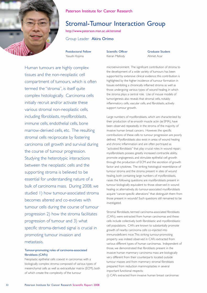
32 Paterson Institute for Cancer Research Scientific Report 2008
Stromal-Tumour Interaction Group http://www.paterson.man.ac.uk/stromal
Paterson Institute for Cancer Research
Tumour-promoting roles of carcinoma-associated
fibroblasts (CAFs)
Neoplastic epithelial cells coexist in carcinomas with a
biologically complex stroma composed of various types of
mesenchymal cells as well as extracellular matrix (ECM), both
of which create the complexity of the tumour
microenvironment. The significant contribution of stroma to
the development of a wide variety of tumours has been
supported by extensive clinical evidence; this contribution is
highlighted by the higher incidence of tumour formation in
tissues exhibiting a chronically inflamed stroma as well as
those undergoing various types of wound healing, in which
the stroma plays a central role. Use of mouse models of
tumorigenesis also reveals that stromal cells, notably
inflammatory cells, vascular cells, and fibroblasts, actively
support tumour growth.
Large numbers of myofibroblasts, which are characterized by
their production of α-smooth muscle actin (α-SMA), have
been observed repeatedly in the stroma of the majority of
invasive human breast cancers. However, the specific
contributions of these cells to tumour progression are poorly
defined. Myofibroblasts also exist in areas of wound healing
and chronic inflammation and are often portrayed as
“activated fibroblasts” that play crucial roles in wound repair ;
myofibroblasts possess greatly increased contractile ability,
promote angiogenesis, and stimulate epithelial cell growth
through the production of ECM and the secretion of growth
factor and cytokines. The striking histological resemblance of
tumour stroma and the stroma present in sites of wound
healing, both containing large numbers of myofibroblasts,
raises the following questions: are myofibroblasts present in
tumour biologically equivalent to those observed in wound
healing or, alternatively, do tumour-associated myofibroblasts
acquire “cancer-specific alterations” that distinguish them from
those present in wounds? Such questions still remained to be
investigated.
Stromal fibroblasts, termed carcinoma-associated fibroblasts
(CAFs), were extracted from human carcinomas and these
cells include collectively both fibroblastic and myofibroblastic
cell populations. CAFs are known to substantially promote
growth of nearby carcinoma cells co-injected into
immunodeficient mice. This striking tumour-promoting
property was indeed observed in CAFs extracted from
various different types of human carcinomas. Independent of
those, we demonstrated that fibroblasts present in the
invasive human mammary carcinoma mass are biologically
very different from their counterparts located outside
tumour masses and from mammary stromal fibroblasts
prepared from reduction mammoplasties in several
important functional respects;
(i) CAFs extracted from invasive human breast carcinomas
Human tumours are highly complex
tissues and the non-neoplastic cell
compartment of tumours, which is often
termed the “stroma”, is itself quite
complex histologically. Carcinoma cells
initially recruit and/or activate these
various stromal non-neoplastic cells,
including fibroblasts, myofibroblasts,
immune cells, endothelial cells, bone
marrow-derived cells, etc. The resulting
stromal cells reciprocate by fostering
carcinoma cell growth and survival during
the course of tumour progression.
Studying the heterotypic interactions
between the neoplastic cells and the
supporting stroma is believed to be
essential for understanding nature of a
bulk of carcinoma mass. During 2008, we
studied 1) how tumour-associated stroma
becomes altered and co-evolves with
tumour cells during the course of tumour
progression 2) how the stroma facilitates
progression of tumour and 3) what
specific stroma-derived signal is crucial in
promoting tumour invasion and
metastasis.
Postdoctoral Fellow
Yasushi Kojima
Scientific Officer
Kieran Mellody
Graduate Student
Ahmet Acar
Group Leader Akira Orimo
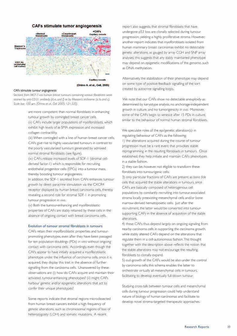
Research Reports 33
are more competent than normal fibroblasts in enhancing
tumour growth by comingled breast cancer cells.
(ii) CAFs include larger populations of myofibroblasts, which
exhibit high levels of α-SMA expression and increased
collagen contractility.
(iii) When comingled with a line of human breast cancer cells,
CAFs give rise to highly vascularized tumours in contrast to
the poorly vascularized tumours generated by admixed
normal stromal fibroblasts (see figure).
(iv) CAFs release increased levels of SDF-1 (stromal cell-
derived factor-1) which is responsible for recruiting
endothelial progenitor cells (EPCs) into a tumour mass,
thereby boosting tumour angiogenesis.
In addition, the SDF-1 secreted from CAFs enhances tumour
growth by direct paracrine stimulation via the CXCR4
receptor displayed by human breast carcinoma cells, thereby
revealing a second role for stromal SDF-1 in promoting
tumour progression in vivo.
(v) Both the tumour-enhancing and myofibroblastic
properties of CAFs are stably retained by these cells in the
absence of ongoing contact with breast carcinoma cells.
Evolution of tumour stromal fibroblasts in tumours
CAFs retain their myofibroblastic properties and tumour-
promoting phenotypes, even after they have been passaged
for ten population doublings (PDs) in vitro without ongoing
contact with carcinoma cells. Accordingly, even though the
CAFs appear to have initially acquired a myofibroblastic
phenotype under the influence of carcinoma cells, once it is
acquired, they display this trait in the absence of further
signalling from the carcinoma cells. Unanswered by these
observations are (i) how do CAFs acquire and maintain their
activated, tumour-enhancing phenotypes? (ii) might CAFs
harbour genetic and/or epigenetic alterations that act to
confer their unique phenotypes?
Some reports indicate that stromal regions microdissected
from human breast cancers exhibit a high frequency of
genetic alterations, such as chromosomal regions of loss of
heterozygosity (LOH) and somatic mutations. A recent
report also suggests that stromal fibroblasts that have
undergone p53 loss are clonally selected during tumour
progression, yielding a highly proliferative stroma. However,
another report indicates that myofibroblasts isolated from
human mammary breast carcinomas exhibit no detectable
genetic alterations, as gauged by array CGH and SNP array
analyses; this suggests that any stably maintained phenotype
may depend on epigenetic modifications of the genome, such
as DNA methylation.
Alternatively, the stabilization of their phenotype may depend
on some type of positive-feedback signalling of the sort
created by autocrine signalling loops.
We note that our CAFs show no detectable aneuploidy as
determined by karyotype analysis, no anchorage-independent
growth in culture, and no tumorigenicity in vivo. Moreover,
some of the CAFs begin to senesce after 15 PDs in culture,
similar to the behaviour of normal human stromal fibroblasts.
We speculate roles of the epi/genetic alteration(s) in
regulating behaviour of CAFs as the following:
1) the alterations acquired during the course of tumour
progression must be a rare event that provokes stable
reprogramming in the resulting fibroblasts in tumours. Once
established, they help initiate and maintain CAFs phenotypes
in a stable fashion.
2) they can be, however, not eligible to transform these
fibroblasts into tumourigenic cells.
3) only particular fractions of CAFs are present as bona fide
cells that acquired the stable alterations in tumours, since
CAFs are basically composed of heterogenous cell
populations by constantly recruiting into tumour-associated
stroma locally preexisting mesenchymal cells and/or bone
marrow-derived hematopoietic cells. Just after the
recruitment, the latter would be converted into tumour-
supporting CAFs in the absence of acquisition of the stable
alterations.
4) these CAFs thus depend largely on ongoing signaling from
nearby carcinoma cells in supporting the carcinoma growth,
while stably altered CAFs depend on the alterations that
regulate them in a cell-autonomous fashion. This thought
together with the description above reflects the notion that
the stable alterations may not encourage the resulting
fibroblasts to clonally expand.
5) out-growth of the CAFs would be also under the control
by carcinoma cells; this schema enables the latter to
orchestrate virtually all mesenchymal cells in tumours,
facilitating to develop eventually full-blown tumour.
Studying cross-talk between tumour cells and mesenchymal
cells during tumour progression could help understand
nature of biology of human carcinomas and facilitate to
develop novel stroma-targeted therapeutic approaches.
CAFs stimulate tumour angiogenesis
Sections from MCF-7-ras human breast tumours containing various fibroblasts were
stained by anti-CD31 antibody (d, e, and f) or by Masson’s trichrome (a, b, and c).
Scale bar, 100 μm. (Orimo et al., Cell 2005; 121:335).
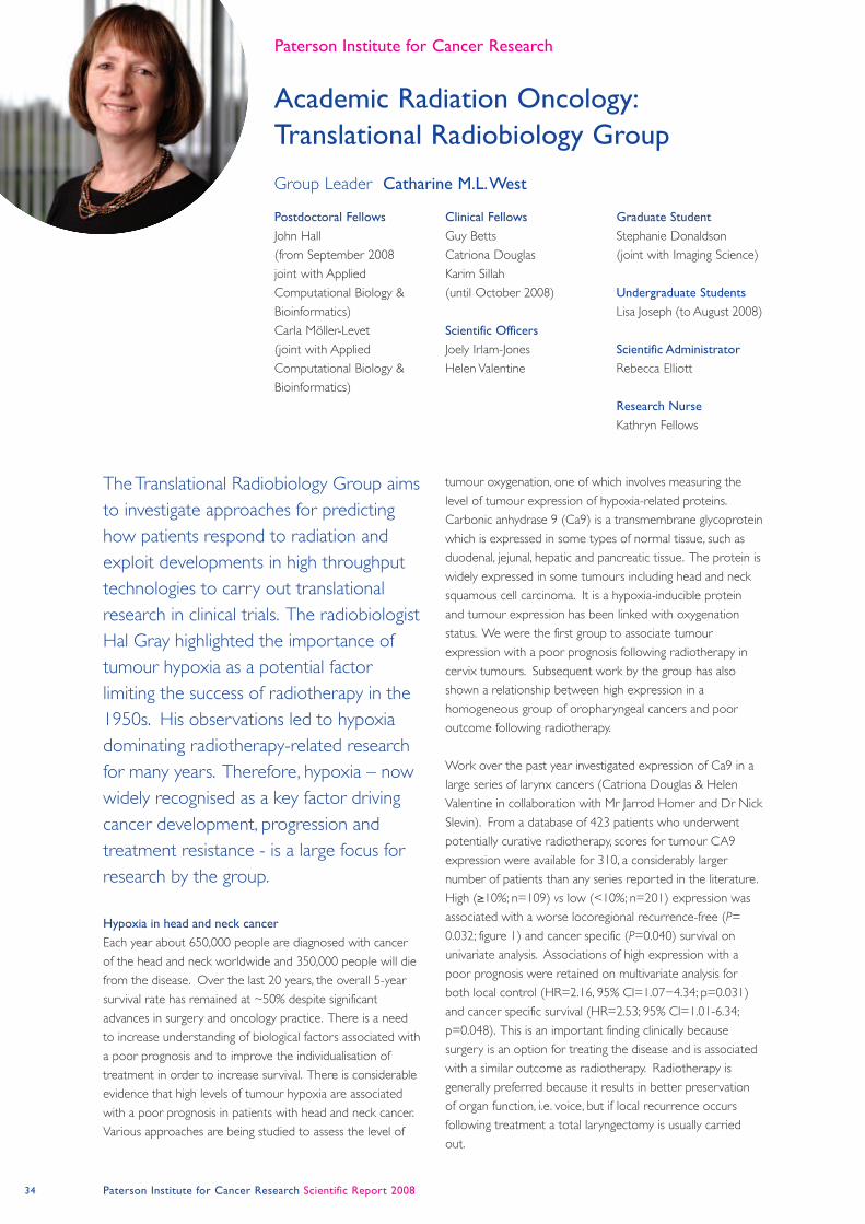
34 Paterson Institute for Cancer Research Scientific Report 2008
Academic Radiation Oncology:
Translational Radiobiology Group
Paterson Institute for Cancer Research
Hypoxia in head and neck cancer
Each year about 650,000 people are diagnosed with cancer
of the head and neck worldwide and 350,000 people will die
from the disease. Over the last 20 years, the overall 5-year
survival rate has remained at ~50% despite significant
advances in surgery and oncology practice. There is a need
to increase understanding of biological factors associated with
a poor prognosis and to improve the individualisation of
treatment in order to increase survival. There is considerable
evidence that high levels of tumour hypoxia are associated
with a poor prognosis in patients with head and neck cancer.
Various approaches are being studied to assess the level of
tumour oxygenation, one of which involves measuring the
level of tumour expression of hypoxia-related proteins.
Carbonic anhydrase 9 (Ca9) is a transmembrane glycoprotein
which is expressed in some types of normal tissue, such as
duodenal, jejunal, hepatic and pancreatic tissue. The protein is
widely expressed in some tumours including head and neck
squamous cell carcinoma. It is a hypoxia-inducible protein
and tumour expression has been linked with oxygenation
status. We were the first group to associate tumour
expression with a poor prognosis following radiotherapy in
cervix tumours. Subsequent work by the group has also
shown a relationship between high expression in a
homogeneous group of oropharyngeal cancers and poor
outcome following radiotherapy.
Work over the past year investigated expression of Ca9 in a
large series of larynx cancers (Catriona Douglas & Helen
Valentine in collaboration with Mr Jarrod Homer and Dr Nick
Slevin). From a database of 423 patients who underwent
potentially curative radiotherapy, scores for tumour CA9
expression were available for 310, a considerably larger
number of patients than any series reported in the literature.
High (≥10%; n=109) vs low (<10%; n=201) expression was
associated with a worse locoregional recurrence-free (P=
0.032; figure 1) and cancer specific (P=0.040) survival on
univariate analysis. Associations of high expression with a
poor prognosis were retained on multivariate analysis for
both local control (HR=2.16, 95% CI=1.07−4.34; p=0.031)
and cancer specific survival (HR=2.53; 95% CI=1.01-6.34;
p=0.048). This is an important finding clinically because
surgery is an option for treating the disease and is associated
with a similar outcome as radiotherapy. Radiotherapy is
generally preferred because it results in better preservation
of organ function, i.e. voice, but if local recurrence occurs
following treatment a total laryngectomy is usually carried
out.
The Translational Radiobiology Group aims
to investigate approaches for predicting
how patients respond to radiation and
exploit developments in high throughput
technologies to carry out translational
research in clinical trials. The radiobiologist
Hal Gray highlighted the importance of
tumour hypoxia as a potential factor
limiting the success of radiotherapy in the
1950s. His observations led to hypoxia
dominating radiotherapy-related research
for many years. Therefore, hypoxia – now
widely recognised as a key factor driving
cancer development, progression and
treatment resistance - is a large focus for
research by the group.
Postdoctoral Fellows
John Hall
(from September 2008
joint with Applied
Computational Biology &
Bioinformatics)
Carla Möller-Levet
(joint with Applied
Computational Biology &
Bioinformatics)
Clinical Fellows
Guy Betts
Catriona Douglas
Karim Sillah
(until October 2008)
Scientific Officers
Joely Irlam-Jones
Helen Valentine
Graduate Student
Stephanie Donaldson
(joint with Imaging Science)
Undergraduate Students
Lisa Joseph (to August 2008)
Scientific Administrator
Rebecca Elliott
Research Nurse
Kathryn Fellows
Group Leader Catharine M.L. West

Research Reports 35
Development of a hypoxia-associated gene signature in
head and neck cancer
With our collaborators in Oxford (Prof Adrian Harris, Dr
Francesca Buffa) and Applied Computational Biology &
Bioinformatics (Dr Crispin Miller) we have continued work
on our hypoxia-associated gene expression signature (Guy
Betts & Carla Möller-Levet). Over the past year the signature
has been streamlined and reduced from 99 to 26 genes, and
a comparison made of Exon and Affymetrix U133plus2
GeneChip data. With the recent award of an MRC
Biomarker grant, the group is looking forward to validating
and qualifying the signature using a PCR approach in multiple
clinical datasets.
RAPPER and VORTEx-BIOBANK
The Translational Radiobiology group co-ordinates the
biobanking associated with several national radiotherapy
trials. RAPPER (Radiogenomics: Assessment of
Polymorphisms for Predicting the Effects of Radiotherapy) is
collecting samples from a number of national trials. The CR-
UK funded project aims to explore associations between
genetic variation expressed as single nucleotide
polymorphisms and radiation toxicity. This year we exceeded
our planned recruitment of 2,200 patients with breast,
prostate, gynaecological or rectal cancer (figure 2). Day-to-
day administration is carried out by Rebecca Elliott and
Kathryn Fellows recruited patients at the Christie Hospital.
Collaborators include Drs Neil Burnet & Alison Dunning
(Cambridge), Prof Søren Bentzen (Wisconsin) and numerous
clinical oncologists locally and nationally. Sample collection
will continue from national radiotherapy trials for a second
validation phase of genotyping. The first sample for VORTEX-
BIOBANK (Joely Irlam-Jones) was received in 2007 and we
have now banked 36 matched fresh tumour and normal
tissue samples and 32 blood samples.
Publications listed on page 60
Figure 1. Kaplan-Meier survival curve showing the locoregional recurrence-free survival of
310 patients with squamous cell carcinoma of the larynx who underwent radiotherapy.
Patients are stratified according to high (≥10% positive tumour cells) vs low tumour Ca9
expression. Examples of tumours with low and high expression of Ca9 are also shown.
Figure 2. RAPPER study target and cumulative accrual.
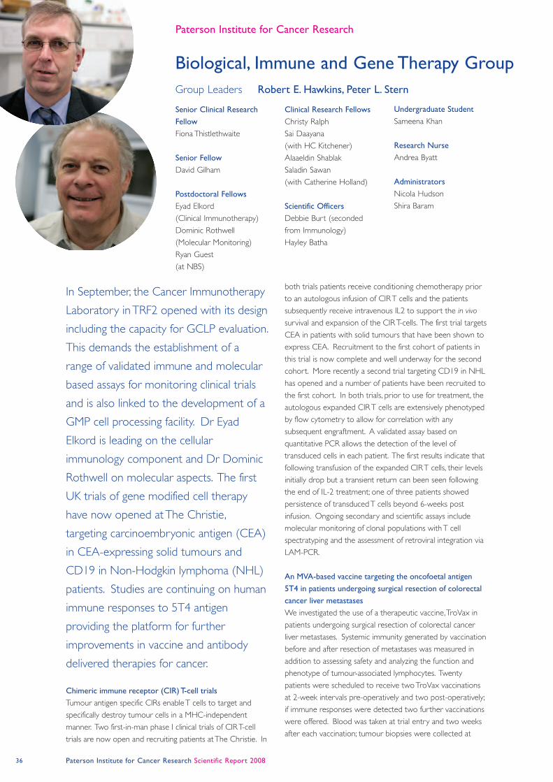
36 Paterson Institute for Cancer Research Scientific Report 2008
Biological, Immune and Gene Therapy Group
Paterson Institute for Cancer Research
Chimeric immune receptor (CIR) T-cell trials
Tumour antigen specific CIRs enable T cells to target and
specifically destroy tumour cells in a MHC-independent
manner. Two first-in-man phase I clinical trials of CIR T-cell
trials are now open and recruiting patients at The Christie. In
both trials patients receive conditioning chemotherapy prior
to an autologous infusion of CIR T cells and the patients
subsequently receive intravenous IL2 to support the in vivo
survival and expansion of the CIR T-cells. The first trial targets
CEA in patients with solid tumours that have been shown to
express CEA. Recruitment to the first cohort of patients in
this trial is now complete and well underway for the second
cohort. More recently a second trial targeting CD19 in NHL
has opened and a number of patients have been recruited to
the first cohort. In both trials, prior to use for treatment, the
autologous expanded CIR T cells are extensively phenotyped
by flow cytometry to allow for correlation with any
subsequent engraftment. A validated assay based on
quantitative PCR allows the detection of the level of
transduced cells in each patient. The first results indicate that
following transfusion of the expanded CIR T cells, their levels
initially drop but a transient return can been seen following
the end of IL-2 treatment; one of three patients showed
persistence of transduced T cells beyond 6-weeks post
infusion. Ongoing secondary and scientific assays include
molecular monitoring of clonal populations with T cell
spectratyping and the assessment of retroviral integration via
LAM-PCR.
An MVA-based vaccine targeting the oncofoetal antigen
5T4 in patients undergoing surgical resection of colorectal
cancer liver metastases
We investigated the use of a therapeutic vaccine, TroVax in
patients undergoing surgical resection of colorectal cancer
liver metastases. Systemic immunity generated by vaccination
before and after resection of metastases was measured in
addition to assessing safety and analyzing the function and
phenotype of tumour-associated lymphocytes. Twenty
patients were scheduled to receive two TroVax vaccinations
at 2-week intervals pre-operatively and two post-operatively;
if immune responses were detected two further vaccinations
were offered. Blood was taken at trial entry and two weeks
after each vaccination; tumour biopsies were collected at
In September, the Cancer Immunotherapy
Laboratory in TRF2 opened with its design
including the capacity for GCLP evaluation.
This demands the establishment of a
range of validated immune and molecular
based assays for monitoring clinical trials
and is also linked to the development of a
GMP cell processing facility. Dr Eyad
Elkord is leading on the cellular
immunology component and Dr Dominic
Rothwell on molecular aspects. The first
UK trials of gene modified cell therapy
have now opened at The Christie,
targeting carcinoembryonic antigen (CEA)
in CEA-expressing solid tumours and
CD19 in Non-Hodgkin lymphoma (NHL)
patients. Studies are continuing on human
immune responses to 5T4 antigen
providing the platform for further
improvements in vaccine and antibody
delivered therapies for cancer.
Senior Clinical Research
Fellow
Fiona Thistlethwaite
Senior Fellow
David Gilham
Postdoctoral Fellows
Eyad Elkord
(Clinical Immunotherapy)
Dominic Rothwell
(Molecular Monitoring)
Ryan Guest
(at NBS)
Clinical Research Fellows
Christy Ralph
Sai Daayana
(with HC Kitchener)
Alaaeldin Shablak
Saladin Sawan
(with Catherine Holland)
Scientific Officers
Debbie Burt (seconded
from Immunology)
Hayley Batha
Undergraduate Student
Sameena Khan
Research Nurse
Andrea Byatt
Administrators
Nicola Hudson
Shira Baram
Group Leaders Robert E. Hawkins, Peter L. Stern
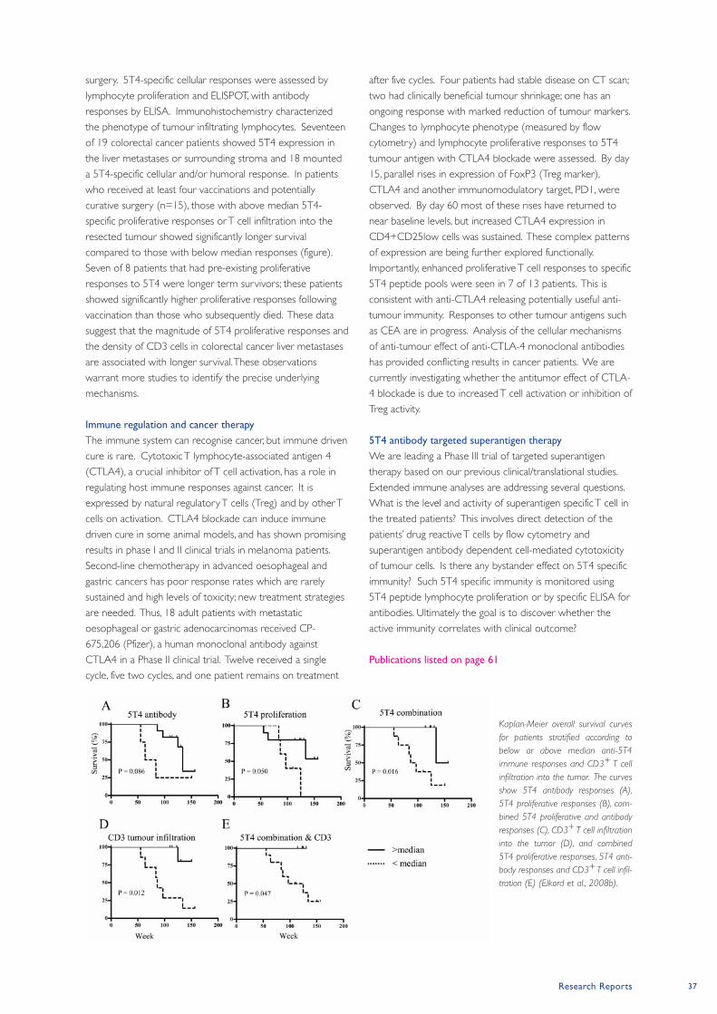
Research Reports 37
surgery. 5T4-specific cellular responses were assessed by
lymphocyte proliferation and ELISPOT, with antibody
responses by ELISA. Immunohistochemistry characterized
the phenotype of tumour infiltrating lymphocytes. Seventeen
of 19 colorectal cancer patients showed 5T4 expression in
the liver metastases or surrounding stroma and 18 mounted
a 5T4-specific cellular and/or humoral response. In patients
who received at least four vaccinations and potentially
curative surgery (n=15), those with above median 5T4-
specific proliferative responses or T cell infiltration into the
resected tumour showed significantly longer survival
compared to those with below median responses (figure).
Seven of 8 patients that had pre-existing proliferative
responses to 5T4 were longer term survivors; these patients
showed significantly higher proliferative responses following
vaccination than those who subsequently died. These data
suggest that the magnitude of 5T4 proliferative responses and
the density of CD3 cells in colorectal cancer liver metastases
are associated with longer survival. These observations
warrant more studies to identify the precise underlying
mechanisms.
Immune regulation and cancer therapy
The immune system can recognise cancer, but immune driven
cure is rare. Cytotoxic T lymphocyte-associated antigen 4
(CTLA4), a crucial inhibitor of T cell activation, has a role in
regulating host immune responses against cancer. It is
expressed by natural regulatory T cells (Treg) and by other T
cells on activation. CTLA4 blockade can induce immune
driven cure in some animal models, and has shown promising
results in phase I and II clinical trials in melanoma patients.
Second-line chemotherapy in advanced oesophageal and
gastric cancers has poor response rates which are rarely
sustained and high levels of toxicity; new treatment strategies
are needed. Thus, 18 adult patients with metastatic
oesophageal or gastric adenocarcinomas received CP-
675,206 (Pfizer), a human monoclonal antibody against
CTLA4 in a Phase II clinical trial. Twelve received a single
cycle, five two cycles, and one patient remains on treatment
after five cycles. Four patients had stable disease on CT scan;
two had clinically beneficial tumour shrinkage; one has an
ongoing response with marked reduction of tumour markers.
Changes to lymphocyte phenotype (measured by flow
cytometry) and lymphocyte proliferative responses to 5T4
tumour antigen with CTLA4 blockade were assessed. By day
15, parallel rises in expression of FoxP3 (Treg marker),
CTLA4 and another immunomodulatory target, PD1, were
observed. By day 60 most of these rises have returned to
near baseline levels, but increased CTLA4 expression in
CD4+CD25low cells was sustained. These complex patterns
of expression are being further explored functionally.
Importantly, enhanced proliferative T cell responses to specific
5T4 peptide pools were seen in 7 of 13 patients. This is
consistent with anti-CTLA4 releasing potentially useful anti-
tumour immunity. Responses to other tumour antigens such
as CEA are in progress. Analysis of the cellular mechanisms
of anti-tumour effect of anti-CTLA-4 monoclonal antibodies
has provided conflicting results in cancer patients. We are
currently investigating whether the antitumor effect of CTLA-
4 blockade is due to increased T cell activation or inhibition of
Treg activity.
5T4 antibody targeted superantigen therapy
We are leading a Phase III trial of targeted superantigen
therapy based on our previous clinical/translational studies.
Extended immune analyses are addressing several questions.
What is the level and activity of superantigen specific T cell in
the treated patients? This involves direct detection of the
patients’ drug reactive T cells by flow cytometry and
superantigen antibody dependent cell-mediated cytotoxicity
of tumour cells. Is there any bystander effect on 5T4 specific
immunity? Such 5T4 specific immunity is monitored using
5T4 peptide lymphocyte proliferation or by specific ELISA for
antibodies. Ultimately the goal is to discover whether the
active immunity correlates with clinical outcome?
Publications listed on page 61
Kaplan-Meier overall survival curves
for patients stratified according to
below or above median anti-5T4
immune responses and CD3+ T cell
infiltration into the tumor. The curves
show 5T4 antibody responses (A),
5T4 proliferative responses (B), com-
bined 5T4 proliferative and antibody
responses (C), CD3+ T cell infiltration
into the tumor (D), and combined
5T4 proliferative responses, 5T4 anti-
body responses and CD3+ T cell infil-
tration (E) (Elkord et al., 2008b).
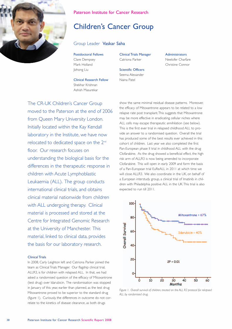
38 Paterson Institute for Cancer Research Scientific Report 2008
Clinical Trials
In 2008, Carly Leighton left and Catriona Parker joined the
team as Clinical Trials Manager. Our flagship clinical trial,
ALLR3, is for children with relapsed ALL. In that, we had
asked a randomised question of the efficacy of Mitoxantrone
(test drug) over Idarubicin. The randomisation was stopped
in January of this year, earlier than planned, as the test drug
Mitoxantrone proved to be superior to the standard drug
(figure 1). Curiously, the differences in outcome do not cor-
relate to the kinetics of disease clearance, as both drugs
show the same minimal residual disease patterns. Moreover,
the efficacy of Mitoxantrone appears to be related to a low
relapse rate post transplant. This suggests that Mitoxantrone
may be more effective in eradicating cellular niches where
ALL cells may escape therapeutic annihilation (see below).
This is the first ever trial in relapsed childhood ALL to pro-
vide an answer to a randomised question. Overall the trial
has produced some of the best results ever achieved in this
cohort of children. Last year we also completed the first
Pan-European phase II trial in childhood ALL with the drug
Clofarabine. As the drug showed a beneficial effect, the high
risk arm of ALLR3 is now being amended to incorporate
Clofarabine. This will open in early 2009 and form the basis
of a Pan-European trial EuReALL in 2011 at which time we
will close ALLR3. We also coordinate in the UK, on behalf of
a European interstudy group, a clinical trial of Imatinib in chil-
dren with Philadelphia positive ALL in the UK. This trial is also
expected to run till 2011.
The CR-UK Children’s Cancer Group
moved to the Paterson at the end of 2006
from Queen Mary University London.
Initially located within the Kay Kendall
laboratory in the Institute, we have now
relocated to dedicated space on the 2nd
floor. Our research focuses on
understanding the biological basis for the
differences in the therapeutic response in
children with Acute Lymphoblastic
Leukaemia (ALL). The group conducts
international clinical trials, and obtains
clinical material nationwide from children
with ALL undergoing therapy. Clinical
material is processed and stored at the
Centre for Integrated Genomic Research
at the University of Manchester. This
material, linked to clinical data, provides
the basis for our laboratory research.
Postdoctoral Fellows
Clare Dempsey
Mark Holland
Jizhong Liu
Clinical Research Fellow
Shekhar Krishnan
Ashish Masurekar
Clinical Trials Manager
Catriona Parker
Scientific Officers
Seema Alexander
Naina Patel
Administrators
Neelofer Charfare
Christine Connor
Children’s Cancer Group
Paterson Institute for Cancer Research
Group Leader Vaskar Saha
Figure 1. Overall survival of children, treated on the ALL R3 protocol for relapsed
ALL by randomised drug.
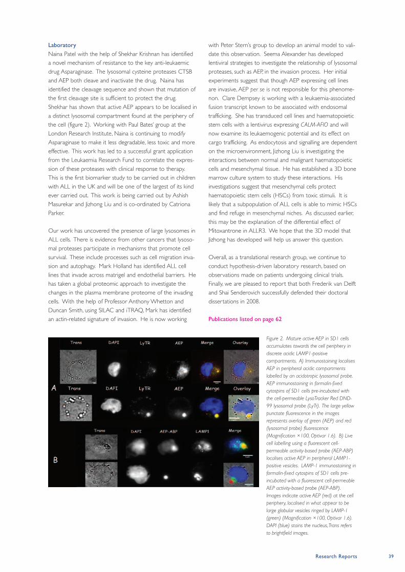
Research Reports 39
Laboratory
Naina Patel with the help of Shekhar Krishnan has identified
a novel mechanism of resistance to the key anti-leukaemic
drug Asparaginase. The lysosomal cysteine proteases CTSB
and AEP both cleave and inactivate the drug. Naina has
identified the cleavage sequence and shown that mutation of
the first cleavage site is sufficient to protect the drug.
Shekhar has shown that active AEP appears to be localised in
a distinct lysosomal compartment found at the periphery of
the cell (figure 2). Working with Paul Bates’ group at the
London Research Institute, Naina is continuing to modify
Asparaginase to make it less degradable, less toxic and more
effective. This work has led to a successful grant application
from the Leukaemia Research Fund to correlate the expres-
sion of these proteases with clinical response to therapy.
This is the first biomarker study to be carried out in children
with ALL in the UK and will be one of the largest of its kind
ever carried out. This work is being carried out by Ashish
Masurekar and Jizhong Liu and is co-ordinated by Catriona
Parker.
Our work has uncovered the presence of large lysosomes in
ALL cells. There is evidence from other cancers that lysoso-
mal proteases participate in mechanisms that promote cell
survival. These include processes such as cell migration inva-
sion and autophagy. Mark Holland has identified ALL cell
lines that invade across matrigel and endothelial barriers. He
has taken a global proteomic approach to investigate the
changes in the plasma membrane proteome of the invading
cells. With the help of Professor Anthony Whetton and
Duncan Smith, using SILAC and iTRAQ, Mark has identified
an actin-related signature of invasion. He is now working
with Peter Stern’s group to develop an animal model to vali-
date this observation. Seema Alexander has developed
lentiviral strategies to investigate the relationship of lysosomal
proteases, such as AEP, in the invasion process. Her initial
experiments suggest that though AEP expressing cell lines
are invasive, AEP per se is not responsible for this phenome-
non. Clare Dempsey is working with a leukaemia-associated
fusion transcript known to be associated with endosomal
trafficking. She has transduced cell lines and haematopoietic
stem cells with a lentivirus expressing CALM-AFIO and will
now examine its leukaemogenic potential and its effect on
cargo trafficking. As endocytosis and signalling are dependent
on the microenvironment, Jizhong Liu is investigating the
interactions between normal and malignant haematopoietic
cells and mesenchymal tissue. He has established a 3D bone
marrow culture system to study these interactions. His
investigations suggest that mesenchymal cells protect
haematopoietic stem cells (HSCs) from toxic stimuli. It is
likely that a subpopulation of ALL cells is able to mimic HSCs
and find refuge in mesenchymal niches. As discussed earlier,
this may be the explanation of the differential effect of
Mitoxantrone in ALLR3. We hope that the 3D model that
Jizhong has developed will help us answer this question.
Overall, as a translational research group, we continue to
conduct hypothesis-driven laboratory research, based on
observations made on patients undergoing clinical trials.
Finally, we are pleased to report that both Frederik van Delft
and Shai Senderovich successfully defended their doctoral
dissertations in 2008.
Publications listed on page 62
Figure 2. Mature active AEP in SD1 cells
accumulates towards the cell periphery in
discrete acidic LAMP1-positive
compartments. A) Immunostaining localises
AEP in peripheral acidic compartments
labelled by an acidotropic lysosomal probe.
AEP immunostaining in formalin-fixed
cytospins of SD1 cells pre-incubated with
the cell-permeable LysoTracker Red DND-
99 lysosomal probe (LyTr). The large yellow
punctate fluorescence in the images
represents overlay of green (AEP) and red
(lysosomal probe) fluorescence
(Magnification ×100, Optivar 1.6). B) Live
cell labelling using a fluorescent cell-
permeable activity-based probe (AEP-ABP)
localises active AEP in peripheral LAMP1-
positive vesicles. LAMP-1 immunostaining in
formalin-fixed cytospins of SD1 cells pre-
incubated with a fluorescent cell-permeable
AEP activity-based probe (AEP-ABP).
Images indicate active AEP (red) at the cell
periphery, localised in what appear to be
large globular vesicles ringed by LAMP-1
(green) (Magnification ×100, Optivar 1.6).
DAPI (blue) stains the nucleus, Trans refers
to brightfield images.
40 Paterson Institute for Cancer Research Scientific Report 2008
Targeted Therapy Group
Paterson Institute for Cancer Research
Mechanisms of action of radioimmunotherapy (RIT)
Our recent work has focused on the molecular mechanisms
of action of RIT induced tumour cell death in vitro. We have
investigated the downstream signalling events in a variety of
human B cell lymphomas after treatment with RT and anti-
CD20 mAb using a number of clinically relevant anti-CD20
mAb. Anti-CD20 mAb can be broadly sub-divided into
either type I (eg. rituximab) or type II (eg. tositumomab).
Rituximab and other Type I anti-CD20 mAb engage
complement effectively and cause target cell lysis
In contrast type II mAb such as tositumomab (B1) are
generally potent at inducing cell death in target cells. We
observed increased tumour cell death with tositumomab and
RT, which was not seen with rituximab and RT. This increased
tumour cytotoxicity was reversed with the MEK inhibitors
(U0126, PD98059) as well as siRNA targeting MEK1 or
MEK2. Furthermore the addition of U0126 reversed the loss
of clonogenic survival triggered by combining tositumomab
with RT. Phosphorylated ERK1/2 (pERK) was found to
accumulate in the nucleus following tositumomab and the
nuclear accumulation of pERK was greatly enhanced in
combination with RT. In contrast rituximab caused early ERK
phosphorylation but this was not sustained and remained
within the cytoplasm suggesting that this might underlie the
lack of additive tumour cell death. In summary our data
indicate that activation and nuclear accumulation of pERK
appear to be required to produce the synergistic effect
produced by combining tositumomab and RT (Ivanov et al.,
2008).
Recently, in collaboration with Dr Mark Cragg’s group in
Southampton we have investigated a new form of mAb
induced cell death in B-cell lymphomas. Using both
lymphoma cell lines and primary chronic lymphocytic
leukemia (CLL) cells, we have demonstrated for the first time
the importance of lysosome-mediated cell death for antibody
therapy elicited by clinically relevant mAb directed against
two different target antigens namely CD20 and HLA DR
(figure). By virtue of a detailed and kinetic approach we have
determined that death is preceded by homotypic adhesion
with both adhesion and death being dependent upon actin
redistribution. Malignant B cells, undergoing homotypic
adhesion, actively communicate via ~ 5 nm wide temporary
inter-cytoplasmic bridges.
The formation of these channels is accompanied by the
mutual exchange of plasma membrane components and
importantly the extent of plasma membrane swapping
correlates with the extent of cell death induced by both anti-
CD20 and anti-HLA DR mAb. To our knowledge there are
no known precedents for these phenomena recorded for
these cell types. The fact that similar findings have been
The goal of the Targeted Therapy Group is
to define the optimal way to combine
radiotherapy (RT) with immunotherapy in
the treatment of cancer by enhancing our
understanding of the underlying
mechanisms of action. This will be
achieved by the specific objectives which
are i) to investigate how the recognition of
radiotherapy (RT) induced tumour cell
death by different antigen presenting cells
in the tumour microenvironment can
impact on the ensuing immune response;
ii) to investigate the role of myeloid
derived suppressor cells (MDSC) in
tumour regrowth after RT and to develop
strategies to enhance RT tumour control
by modifying host immune response after
RT and iii) to translate our experimental
research findings into developing early
phase clinical trials
Preclinical Group
Postdoctoral Fellows
James Hainsworth
Jamie Honeychurch
Andrei Ivanov
Ruth Swann
Clinical Fellow
Nick Brown
Graduate Students
Waleed Alduaij
Monique Melis
Clinical Radioimmunotherapy
Group Senior Clinical Scientist
Maureen Zivanovic
Clinical scientist
(dosimetry)
Jill Tipping
Research nurses
Caroline Hamer
Susan Neeson
Group Leader Tim Illidge
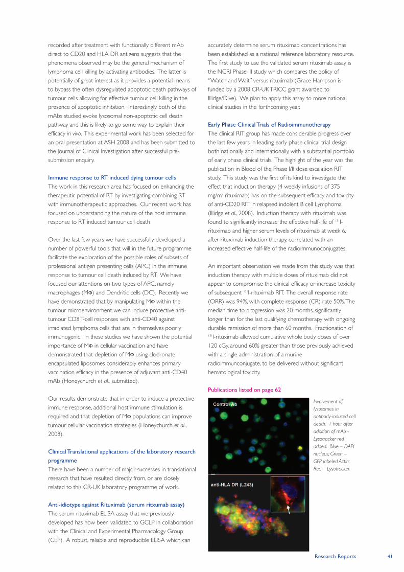
Research Reports 41
recorded after treatment with functionally different mAb
direct to CD20 and HLA DR antigens suggests that the
phenomena observed may be the general mechanism of
lymphoma cell killing by activating antibodies. The latter is
potentially of great interest as it provides a potential means
to bypass the often dysregulated apoptotic death pathways of
tumour cells allowing for effective tumour cell killing in the
presence of apoptotic inhibition. Interestingly both of the
mAbs studied evoke lysosomal non-apoptotic cell death
pathway and this is likely to go some way to explain their
efficacy in vivo. This experimental work has been selected for
an oral presentation at ASH 2008 and has been submitted to
the Journal of Clinical Investigation after successful pre-
submission enquiry.
Immune response to RT induced dying tumour cells
The work in this research area has focused on enhancing the
therapeutic potential of RT by investigating combining RT
with immunotherapeutic approaches. Our recent work has
focused on understanding the nature of the host immune
response to RT induced tumour cell death
Over the last few years we have successfully developed a
number of powerful tools that will in the future programme
facilitate the exploration of the possible roles of subsets of
professional antigen presenting cells (APC) in the immune
response to tumour cell death induced by RT. We have
focused our attentions on two types of APC, namely
macrophages (MΦ) and Dendritic cells (DC). Recently we
have demonstrated that by manipulating MΦ within the
tumour microenvironment we can induce protective anti-
tumour CD8 T-cell responses with anti-CD40 against
irradiated lymphoma cells that are in themselves poorly
immunogenic. In these studies we have shown the potential
importance of MΦ in cellular vaccination and have
demonstrated that depletion of MΦ using clodronate-
encapsulated liposomes considerably enhances primary
vaccination efficacy in the presence of adjuvant anti-CD40
mAb (Honeychurch et al., submitted).
Our results demonstrate that in order to induce a protective
immune response, additional host immune stimulation is
required and that depletion of MΦ populations can improve
tumour cellular vaccination strategies (Honeychurch et al.,
2008).
Clinical Translational applications of the laboratory research
programme
There have been a number of major successes in translational
research that have resulted directly from, or are closely
related to this CR-UK laboratory programme of work.
Anti-idiotype against Rituximab (serum ritxumab assay)
The serum rituximab ELISA assay that we previously
developed has now been validated to GCLP in collaboration
with the Clinical and Experimental Pharmacology Group
(CEP). A robust, reliable and reproducible ELISA which can
accurately determine serum rituximab concentrations has
been established as a national reference laboratory resource.
The first study to use the validated serum rituximab assay is
the NCRI Phase III study which compares the policy of
“Watch and Wait” versus rituximab (Grace Hampson is
funded by a 2008 CR-UK TRICC grant awarded to
Illidge/Dive). We plan to apply this assay to more national
clinical studies in the forthcoming year.
Early Phase Clinical Trials of Radioimmunotherapy
The clinical RIT group has made considerable progress over
the last few years in leading early phase clinical trial design
both nationally and internationally, with a substantial portfolio
of early phase clinical trials. The highlight of the year was the
publication in Blood of the Phase I/II dose escalation RIT
study. This study was the first of its kind to investigate the
effect that induction therapy (4 weekly infusions of 375
mg/m2 rituximab) has on the subsequent efficacy and toxicity
of anti-CD20 RIT in relapsed indolent B cell Lymphoma
(Illidge et al., 2008). Induction therapy with rituximab was
found to significantly increase the effective half-life of 131I-
rituximab and higher serum levels of rituximab at week 6,
after rituximab induction therapy, correlated with an
increased effective half-life of the radioimmunoconjugates
An important observation we made from this study was that
induction therapy with multiple doses of rituximab did not
appear to compromise the clinical efficacy or increase toxicity
of subsequent 131I-rituximab RIT. The overall response rate
(ORR) was 94%, with complete response (CR) rate 50%. The
median time to progression was 20 months, significantly
longer than for the last qualifying chemotherapy with ongoing
durable remission of more than 60 months. Fractionation of131I-rituximab allowed cumulative whole body doses of over
120 cGy, around 60% greater than those previously achieved
with a single administration of a murine
radioimmunconjugate, to be delivered without significant
hematological toxicity.
Publications listed on page 62
Involvement of
lysosomes in
antibody-induced cell
death. 1 hour after
addition of mAb -
Lysotracker red
added. Blue – DAPI
nucleus; Green –
GFP labeled Actin;
Red – Lysotracker.
42 Paterson Institute for Cancer Research Scientific Report 2008
Medical Oncology:
Cell Therapy Group
Paterson Institute for Cancer Research
The Chimeric Immune Receptor: Molecular Interactions
with the T cell Receptor
The gene that is introduced into the patient’s T cells encodes
for a protein called a Chimeric Immune Receptor. This
receptor consists of an extracellular domain that can bind to
target proteins present on the cell surface of the target cell
and is fused to the transmembrane and cytoplasmic domain
of the CD3ζ receptor; which is one of the components of
the T cell receptor (TCR) complex. Natural CD3ζ proteins
bind to the TCR through electrostatic interactions between
charged amino-acids present within their transmembrane
domains. We have shown that our chimeric receptor is also
incorporated within the TCR complex present on the surface
of the gene-modified T cell by biochemical and by
Fluorescence Resonance Energy Transfer (FRET, Figure 1).
Mutations in the charged amino-acids reduce the ability of
the chimeric receptor to interact with the TCR; consequently,
this significantly reduces the sensitivity of the chimeric
receptor function suggesting that the formation of the TCR-
chimeric receptor complex is critical for optimal anti-tumour
activity of the T cell. However, important questions arise
from this observation. Does the chimeric receptor interfere
with the activity of the normal T cell? Could there be safety
issues associated with the long-term formation of the
TCR/chimeric receptor complex? To explore these
questions, we have developed two model systems (based
upon our clinical trials) to investigate the potency of chimeric
receptors and to assess the long-term safety of gene-
modified T cell therapy.
Genetic Modification of Mouse T-cells
In order to test chimeric receptor technology in suitable
tumour models, we have had to develop and improve
protocols to allow high levels of gene transfer into primary
mouse T cells. For reasons which are not entirely
understood, mouse T cells tend to be more refractory to the
viral gene transfer systems which work efficiently in human T
cells. We have optimised retroviral gene transfer into primary
mouse T cells and have established culture conditions which
permit the expansion of these T cells to high numbers ex vivo
for use in adoptive transfer experiments. We have passed on
our methods to collaborators who have confirmed the
robustness of the protocol (Prof. Zelig Eshhar, Weizmann
Institute, Israel). Using these improved protocols, we are
currently examining how the culture condition and duration
of culture impacts upon T cell persistence and anti-tumour
activity in vivo.
Models of gastrointestinal cancer and B-cell lymphoma.
Our first trial (sponsored by Cancer Research UK) involves
generating T cells with specificity for CEA through the
expression of a chimeric immune receptor called MFEz. The
second (sponsored by the Kay Kendall Leukaemia Fund)
involves targeting B cell lymphoma using T cells armed with a
chimeric receptor specific for the CD19 protein (CD19z). In
The Cell Therapy Group has focused upon
manipulating the immune system of the
patient in order to drive the eradication of
tumours. This manipulation involves the
insertion of a gene encoding a tumour
targeting receptor into patient’s T-cells
which then can kill tumour cells. This
approach is now in clinical trial in
Manchester (targeting gastro-intestinal
cancer and B-cell lymphoma). Our
current work is based upon using model
systems to try and understand the key
factors that may influence this approach in
the patient with the aim of improving the
treatment.
Senior Fellow
David Gilham
Postdoctoral Fellows
John Bridgeman (from July)
Eleanor Cheadle
Simon Dovedi
(until February)
Jennifer Loconto
Vivien Watson
Scientific Officers
Allison Robinson
Vicky Sheard
Hayley Batha
(until October)
Graduate Students
Grazyna Lipowska-Bhalla
Erik Alcantar Orozco
Mariam Al-Muftah (with
Immunology)
Group Leader Robert Hawkins
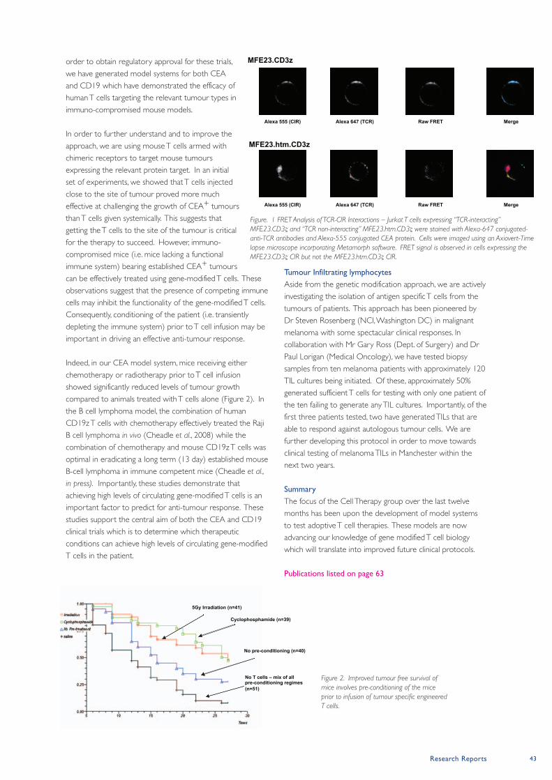
Research Reports 43
order to obtain regulatory approval for these trials,
we have generated model systems for both CEA
and CD19 which have demonstrated the efficacy of
human T cells targeting the relevant tumour types in
immuno-compromised mouse models.
In order to further understand and to improve the
approach, we are using mouse T cells armed with
chimeric receptors to target mouse tumours
expressing the relevant protein target. In an initial
set of experiments, we showed that T cells injected
close to the site of tumour proved more much
effective at challenging the growth of CEA+ tumours
than T cells given systemically. This suggests that
getting the T cells to the site of the tumour is critical
for the therapy to succeed. However, immuno-
compromised mice (i.e. mice lacking a functional
immune system) bearing established CEA+ tumours
can be effectively treated using gene-modified T cells. These
observations suggest that the presence of competing immune
cells may inhibit the functionality of the gene-modified T cells.
Consequently, conditioning of the patient (i.e. transiently
depleting the immune system) prior to T cell infusion may be
important in driving an effective anti-tumour response.
Indeed, in our CEA model system, mice receiving either
chemotherapy or radiotherapy prior to T cell infusion
showed significantly reduced levels of tumour growth
compared to animals treated with T cells alone (Figure 2). In
the B cell lymphoma model, the combination of human
CD19z T cells with chemotherapy effectively treated the Raji
B cell lymphoma in vivo (Cheadle et al., 2008) while the
combination of chemotherapy and mouse CD19z T cells was
optimal in eradicating a long term (13 day) established mouse
B-cell lymphoma in immune competent mice (Cheadle et al.,
in press). Importantly, these studies demonstrate that
achieving high levels of circulating gene-modified T cells is an
important factor to predict for anti-tumour response. These
studies support the central aim of both the CEA and CD19
clinical trials which is to determine which therapeutic
conditions can achieve high levels of circulating gene-modified
T cells in the patient.
Tumour Infiltrating lymphocytes
Aside from the genetic modification approach, we are actively
investigating the isolation of antigen specific T cells from the
tumours of patients. This approach has been pioneered by
Dr Steven Rosenberg (NCI, Washington DC) in malignant
melanoma with some spectacular clinical responses. In
collaboration with Mr Gary Ross (Dept. of Surgery) and Dr
Paul Lorigan (Medical Oncology), we have tested biopsy
samples from ten melanoma patients with approximately 120
TIL cultures being initiated. Of these, approximately 50%
generated sufficient T cells for testing with only one patient of
the ten failing to generate any TIL cultures. Importantly, of the
first three patients tested, two have generated TILs that are
able to respond against autologous tumour cells. We are
further developing this protocol in order to move towards
clinical testing of melanoma TILs in Manchester within the
next two years.
Summary
The focus of the Cell Therapy group over the last twelve
months has been upon the development of model systems
to test adoptive T cell therapies. These models are now
advancing our knowledge of gene modified T cell biology
which will translate into improved future clinical protocols.
Publications listed on page 63
Alexa 555 (CIR) Alexa 647 (TCR) Raw FRET Merge
Alexa 555 (CIR) Alexa 647 (TCR) Raw FRET Merge
MFE23.CD3z
MFE23.htm.CD3z
Figure. 1 FRET Analysis of TCR-CIR Interactions – Jurkat T cells expressing “TCR-interacting”
MFE23.CD3ζ and “TCR non-interacting” MFE23.htm.CD3ζ were stained with Alexa-647 conjugated-
anti-TCR antibodies and Alexa-555 conjugated CEA protein. Cells were imaged using an Axiovert-Time
lapse microscope incorporating Metamorph software. FRET signal is observed in cells expressing the
MFE23.CD3ζ CIR but not the MFE23.htm.CD3ζ CIR.
Figure 2. Improved tumour free survival of
mice involves pre-conditioning of the mice
prior to infusion of tumour specific engineered
T cells.
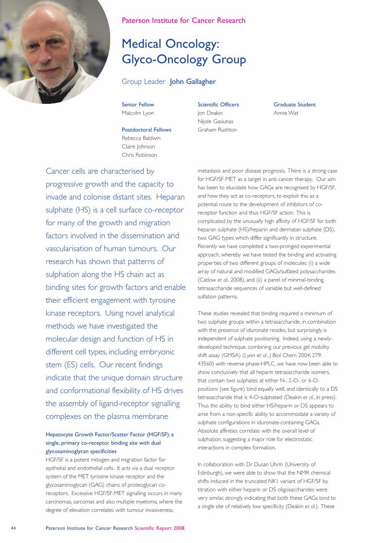
44 Paterson Institute for Cancer Research Scientific Report 2008
Medical Oncology:
Glyco-Oncology Group
Paterson Institute for Cancer Research
Hepatocyte Growth Factor/Scatter Factor (HGF/SF): a
single, primary co-receptor binding site with dual
glycosaminoglycan specificities
HGF/SF is a potent mitogen and migration factor for
epithelial and endothelial cells. It acts via a dual receptor
system of the MET tyrosine kinase receptor and the
glycosaminoglycan (GAG) chains of proteoglycan co-
receptors. Excessive HGF/SF-MET signalling occurs in many
carcinomas, sarcomas and also multiple myeloma, where the
degree of elevation correlates with tumour invasiveness,
metastasis and poor disease prognosis. There is a strong case
for HGF/SF-MET as a target in anti-cancer therapy. Our aim
has been to elucidate how GAGs are recognised by HGF/SF,
and how they act as co-receptors, to exploit this as a
potential route to the development of inhibitors of co-
receptor function and thus HGF/SF action. This is
complicated by the unusually high affinity of HGF/SF for both
heparan sulphate (HS)/heparin and dermatan sulphate (DS),
two GAG types which differ significantly in structure.
Recently we have completed a two-pronged experimental
approach, whereby we have tested the binding and activating
properties of two different groups of molecules: (i) a wide
array of natural and modified GAGs/sulfated polysaccharides
(Catlow et al., 2008), and (ii) a panel of minimal-binding
tetrasaccharide sequences of variable but well-defined
sulfation patterns.
These studies revealed that binding required a minimum of
two sulphate groups within a tetrasaccharide, in combination
with the presence of iduronate resides, but surprisingly is
independent of sulphate positioning. Indeed, using a newly-
developed technique, combining our previous gel mobility
shift assay (GMSA) (Lyon et al., J Biol Chem 2004; 279:
43560) with reverse-phase-HPLC, we have now been able to
show conclusively that all heparin tetrasaccharide isomers,
that contain two sulphates at either N-, 2-O- or 6-O-
positions (see figure) bind equally well, and identically to a DS
tetrasaccharide that is 4-O-sulphated (Deakin et al., in press).
Thus the ability to bind either HS/heparin or DS appears to
arise from a non-specific ability to accommodate a variety of
sulphate configurations in iduronate-containing GAGs.
Absolute affinities correlate with the overall level of
sulphation, suggesting a major role for electrostatic
interactions in complex formation.
In collaboration with Dr Dusan Uhrín (University of
Edinburgh), we were able to show that the NMR chemical
shifts induced in the truncated NK1 variant of HGF/SF by
titration with either heparin or DS oligosaccharides were
very similar, strongly indicating that both these GAGs bind to
a single site of relatively low specificity (Deakin et al.). These
Cancer cells are characterised by
progressive growth and the capacity to
invade and colonise distant sites. Heparan
sulphate (HS) is a cell surface co-receptor
for many of the growth and migration
factors involved in the dissemination and
vascularisation of human tumours. Our
research has shown that patterns of
sulphation along the HS chain act as
binding sites for growth factors and enable
their efficient engagement with tyrosine
kinase receptors. Using novel analytical
methods we have investigated the
molecular design and function of HS in
different cell types, including embryonic
stem (ES) cells. Our recent findings
indicate that the unique domain structure
and conformational flexibility of HS drives
the assembly of ligand-receptor signalling
complexes on the plasma membrane
Senior Fellow
Malcolm Lyon
Postdoctoral Fellows
Rebecca Baldwin
Claire Johnson
Chris Robinson
Scientific Officers
Jon Deakin
Nijole Gasiunas
Graham Rushton
Graduate Student
Annie Wat
Group Leader John Gallagher
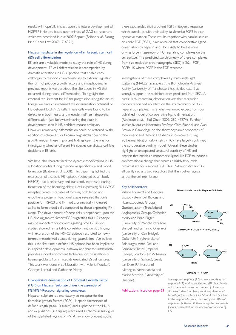
Research Reports 45
results will hopefully impact upon the future development of
HGF/SF inhibitors based upon mimics of GAG co-receptors
which we described in our 2007 Report (Raiber et al., Bioorg
Med Chem Lett 2007; 17: 6321).
Heparan sulphate in the regulation of embryonic stem cell
(ES) cell differentiation
ES cells are a valuable model to study the role of HS during
development. ES cell differentiation is accompanied by
dramatic alterations in HS sulphation that enable each
cell/organ to respond characteristically to extrinsic signals in
the form of peptide growth factors and morphogens. In
previous reports we described the alterations in HS that
occurred during neural differentiation. To highlight the
essential requirement for HS for progression along the neural
lineage we have characterised the differentiation potential of
HS-deficient Ext1-/- ES cells. These cells were found to be
defective in both neural and mesodermal/haematopoietic
differentiation (see below), mimicking the block in
development seen in HS-deficient mouse embryos.
However, remarkably differentiation could be restored by the
addition of soluble HS or heparin oligosaccharides to the
growth media. These important findings open the way for
investigating whether different HS species can dictate cell fate
decisions in ES cells.
We have also characterised the dynamic modifications in HS
sulphation motifs during mesoderm specification and blood
formation (Baldwin et al., 2008). This paper highlighted the
expression of a specific HS epitope (detected by antibody
HS4C3) that is selectively and transiently expressed during
formation of the haemangioblast, a cell expressing Flk1 (VEGF
receptor) which is capable of forming both blood and
endothelial progeny. Functional assays revealed that cells
positive for HS4C3 and Fk1 had a dramatically increased
ability to form blood cells compared to those expressing Flk1
alone. The development of these cells is dependant upon the
HS-binding growth factor VEGF, suggesting this HS epitope
may be important for correct signaling of VEGF. In vivo
studies showed remarkable correlation with in vitro findings,
with expression of the HS4C3 epitope restricted to newly
formed mesodermal tissues during gastrulation. We believe
this is the first time a defined HS epitope has been implicated
in a specific developmental pathway and that this additionally
provides a novel enrichment technique for the isolation of
haemangioblasts from mixed differentiated ES cell cultures.
This work was done in collaboration with Valerie Kouskoff,
Georges Lacaud and Catherine Merry.
Co-operative dimerisation of Fibroblast Growth Factor
(FGF) on Heparan Sulphate drives the assembly of
FGF/FGF-Receptor signalling complexes
Heparan sulphate is a mandatory co-receptor for the
fibroblast growth factors (FGFs). Heparin saccharides of
defined length (8-to-10 sugars) and sulphated at the N-, 2-
and 6- positions (see figure) were used as chemical analogues
of the sulphated regions of HS. At very low concentrations,
these saccharides elicit a potent FGF2 mitogenic response
which correlates with their ability to dimerise FGF2 in a co-
operative manner. These results, together with parallel studies
on acidic FGF (FGF1), have revealed that co-operative ligand
dimerisation by heparin and HS is likely to be the main
driving force in assembly of FGF signalling complexes on the
cell surface. The predicted stoichiometry of these complexes
from size exclusion chromatography (SEC) is 2:2:1 FGF:
FGFR: HS where FGFR is the FGF-receptor
Investigations of these complexes by multi-angle light
scattering (MALLS) available at the Biomolecular Analysis
Facility (University of Manchester) has yielded data that
strongly support the stoichiometries predicted from SEC. A
particularly interesting observation was that saccharide
concentration had no effect on the stoichiometry of FGF-
heparin complexes. This is what we would expect from our
published model of co-operative ligand dimerisation.
(Robinson et al., J Biol Chem 2005; 280: 42274). Further
studies by our collaborators Professor Tom Blundell and Alan
Brown in Cambridge on the thermodynamic properties of
monomeric and dimeric FGF-heparin complexes using
isothermal titration calorimetry (ITC) have largely confirmed
the co-operative binding model. Overall these studies
highlight an unexpected structural plasticity of HS and
heparin that enables a monomeric ligand like FGF to induce a
conformational change that creates a highly favourable
proximal site for a second FGF. This HS-bound dimeric FGF
efficiently recruits two receptors that then deliver signals
across the cell membrane.
Key collaborators
Valerie Kouskoff and Georges
Lacaud (Stem Cell Biology and
Haematopoiesis Groups),
Gordon Jayson (Translational
Angiogenesis Group), Catherine
Merry and Brian Bigger
(University of Manchester), Tom
Blundell and Ermanno Gherardi
(University of Cambridge),
Dušan Uhrín (University of
Edinburgh), Anne Dell and
Berangere Tissot (Imperial
College, London), Jim Wilkinson
(University of Salford), Gerdy
ten Dam (University of
Nijmegen, Netherlands) and
Marios Stavridis (University of
Dundee).
Publications listed on page 63
O O
O
COO-
H
SO3-
NHSO3-
O
OHOH
O [
GlcNSO3 (+/- 6-OSO3) 1 – 4 IdoA, 2-OSO3
CH2OH
O O
O
COO-
O
OHOH
OHNHAc
GlcNH.Ac 1 – 4 GlcA
A)
B)
Disaccharide Units in Heparan Sulphate
SO3-
H
CH2O [
The heparan sulphate (HS) chain is made up of
sulphated (A) and non-sulphated (B) disaccharide
units; these units occur in a series of clusters or
domains rather than being randomly distributed.
Growth factors such as HGF/SF and the FGFs bind
to the sulphated domains but recognise different
sulphation patterns. Pattern recognition by growth
factors is essential for the co-receptor function of
HS.
46 Paterson Institute for Cancer Research Scientific Report 2008
Medical Oncology:
Translational Angiogenesis Group
Paterson Institute for Cancer Research
Biomarkers for Anti-Angiogenic therapy (Collaborators:
Dive, Jackson, Parker)
Over the last few years the group has identified and
evaluated a series of imaging based biomarkers for anti-
angiogenic therapy. Within the last year we have reported a
phase I trial of a novel anti-VEGFR2 di-Fab compound (Ton et
al., 2007 Clin Cancer Res; 13: 7113), which differed from
other broader spectrum VEGF inhibitors in that it did not
impact on Ktrans, the endothelial permeability-surface area
product. On the other hand, biopsy of hemangiomata that
were induced by the compound confirmed that the
compound was present in areas of non-phosphorylated
receptor, that is, that the biopsy data were compatible with
the proposed mechanism of action. By changing the
traditional phase I drug design to expand the number of
patients at each dose level we were able to increase the
statistical power of the study and, through measurements of
tumour volumetrics, were able to detect a dose-level vs
volumetric response. This is important as the traditional
method of reporting radiological evaluation of tumour
response, RECIST, failed to detect this relationship. Thus our
findings highlight tumour volumetric measurements as a
potential novel biomarker for mechanism based therapeutics.
In a second programme we have developed and validated to
the standards of GCLP multiplex and singleplex assays of
sixteen angiogenic proteins. Traditional assays are based on
singleplex ELISAs that are costly and consume significant
amounts of plasma. By multiplexing nine proteins, for the first
time, we have impacted significantly on the cost, time and
amount of plasma needed for these assays. The protocols
have been submitted for publication and will be used in
forthcoming evaluations of plasma samples in ICON7, a
randomised trial of carboplatin and paclitaxel with and
without bevacizumab in the first line treatment of ovarian
cancer. Additional samples that will be evaluated will be
obtained from randomised trials of anti-angiogenic agents in
pancreatic and colorectal cancer. Finally, within the next
twelve months we will apply the technology within an in-
house study that unites the single and multiplex studies
developed in the last year, with our longstanding imaging
programme.
Recognising that the data obtained from imaging would be
significantly augmented through the inclusion of
measurements of circulating anti-angiogenic biomarkers we
have nearly completed a study where we have compared
dynamic MR and dynamic CT measurements of patients
undergoing treatment for ovarian cancer. This trial will be
finished within the next few months and the data are
revealing novel insights into the relationship between imaging
and circulating biomarkers. In a second combination
Over the last year the group has
progressed in two directions; the GCLP
validation and implementation of imaging
and blood borne biomarkers for anti-
angiogenic agents and the second
approach has been to develop research
platforms that will assess the contribution
of heparan sulfate proteoglycans in human
epithelial ovarian cancer. The translational
proteoglycans programme has emerged
from the basic science studies that our
group has performed over the last few
years within Professor John Gallagher’s
laboratory.
Senior Fellows
Egle Avizienyte
Postdoctoral Fellows
Marek Barath
Claire Cole
Steen Hansen
Clinical Fellows
Gireesh Kumaran
Claire Mitchell
Nishanth Murukesh
Scientific Officers
Alison Backen
Karen Brookes
Graham Rushton
Postgraduate Student
Karl Broburg
Rotation Student
Kelvin Wilkinson
Group Leader Gordon Jayson
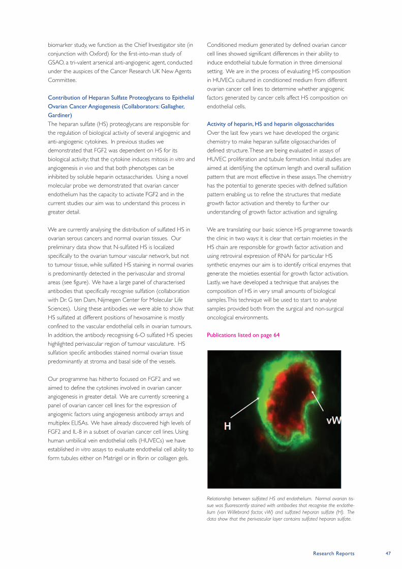
Research Reports 47
biomarker study, we function as the Chief Investigator site (in
conjunction with Oxford) for the first-into-man study of
GSAO, a tri-valent arsenical anti-angiogenic agent, conducted
under the auspices of the Cancer Research UK New Agents
Committee.
Contribution of Heparan Sulfate Proteoglycans to Epithelial
Ovarian Cancer Angiogenesis (Collaborators: Gallagher,
Gardiner)
The heparan sulfate (HS) proteoglycans are responsible for
the regulation of biological activity of several angiogenic and
anti-angiogenic cytokines. In previous studies we
demonstrated that FGF2 was dependent on HS for its
biological activity; that the cytokine induces mitosis in vitro and
angiogenesis in vivo and that both phenotypes can be
inhibited by soluble heparin octasaccharides. Using a novel
molecular probe we demonstrated that ovarian cancer
endothelium has the capacity to activate FGF2 and in the
current studies our aim was to understand this process in
greater detail.
We are currently analysing the distribution of sulfated HS in
ovarian serous cancers and normal ovarian tissues. Our
preliminary data show that N-sulfated HS is localized
specifically to the ovarian tumour vascular network, but not
to tumour tissue, while sulfated HS staining in normal ovaries
is predominantly detected in the perivascular and stromal
areas (see figure). We have a large panel of characterised
antibodies that specifically recognise sulfation (collaboration
with Dr. G ten Dam, Nijmegen Center for Molecular Life
Sciences). Using these antibodies we were able to show that
HS sulfated at different positions of hexosamine is mostly
confined to the vascular endothelial cells in ovarian tumours.
In addition, the antibody recognising 6-O sulfated HS species
highlighted perivascular region of tumour vasculature. HS
sulfation specific antibodies stained normal ovarian tissue
predominantly at stroma and basal side of the vessels.
Our programme has hitherto focused on FGF2 and we
aimed to define the cytokines involved in ovarian cancer
angiogenesis in greater detail. We are currently screening a
panel of ovarian cancer cell lines for the expression of
angiogenic factors using angiogenesis antibody arrays and
multiplex ELISAs. We have already discovered high levels of
FGF2 and IL-8 in a subset of ovarian cancer cell lines. Using
human umbilical vein endothelial cells (HUVECs) we have
established in vitro assays to evaluate endothelial cell ability to
form tubules either on Matrigel or in fibrin or collagen gels.
Conditioned medium generated by defined ovarian cancer
cell lines showed significant differences in their ability to
induce endothelial tubule formation in three dimensional
setting. We are in the process of evaluating HS composition
in HUVECs cultured in conditioned medium from different
ovarian cancer cell lines to determine whether angiogenic
factors generated by cancer cells affect HS composition on
endothelial cells.
Activity of heparin, HS and heparin oligosaccharides
Over the last few years we have developed the organic
chemistry to make heparan sulfate oligosaccharides of
defined structure. These are being evaluated in assays of
HUVEC proliferation and tubule formation. Initial studies are
aimed at identifying the optimum length and overall sulfation
pattern that are most effective in these assays. The chemistry
has the potential to generate species with defined sulfation
pattern enabling us to refine the structures that mediate
growth factor activation and thereby to further our
understanding of growth factor activation and signaling.
We are translating our basic science HS programme towards
the clinic in two ways: it is clear that certain moieties in the
HS chain are responsible for growth factor activation and
using retroviral expression of RNAi for particular HS
synthetic enzymes our aim is to identify critical enzymes that
generate the moieties essential for growth factor activation.
Lastly, we have developed a technique that analyses the
composition of HS in very small amounts of biological
samples. This technique will be used to start to analyse
samples provided both from the surgical and non-surgical
oncological environments.
Publications listed on page 64
Relationship between sulfated HS and endothelium. Normal ovarian tis-
sue was fluorescently stained with antibodies that recognise the endothe-
lium (von Willebrand factor, vW) and sulfated heparan sulfate (H). The
data show that the perivascular layer contains sulfated heparan sulfate.
48 Paterson Institute for Cancer Research Scientific Report 2008
Early in 2008 Steve Murray left the Institute and this
precipitated a review of the requirements for a TEM service.
Given the costs of maintaining, housing and staffing the facility
together with very limited and intermittent requirements for
TEM, the decision was made to transfer the equipment to
the University, with access available to Institute Groups as
required.
Some much-needed refurbishments were completed at the
beginning of the year, giving better facilities for the Molecular
Biology Core Facility, the Flow Cytometry Facility and for the
Mass Spectrometers. We also increased the space for the
Histology Facility because of both an increased workload and
the inclusion of the MCRC Biobank within their remit.
To maintain cutting edge services we need to constantly
upgrade or replace equipment, and this year has been no
exception. Details of each new development are given in the
appropriate section below, but installation of a new Nipkow
spinning disk confocal microscope, a Zeiss Mirax whole slide
scanning system, a Nano Acquity UPLC system and LTQ-
Orbitrap XL, an XY clone laser, an LSRII and a BD InFlux
cytometer, and imminently a new laser capture
microdissection system have all taken place in the last twelve
months.
Advanced Imaging Facility
Head: Steve Bagley
Over the year we have considerably extended the imaging
capabilities of the Institute with the installation and
development of new equipment. The requirement for
complex imaging techniques has increased considerably to
embrace a range of applications. Extensive examination of
cellular morphology, quantification of protein levels,
quantification of turnover rates and interactions between
multiple low light signals are now all covered in the portfolio
of supported technologies.
A Nipkow spinning disk confocal microscope was introduced
early in the year. This technology is particularly good for
imaging multiple fluorescent proteins in a 3-D volume. The
scanning technology it employs enables the user to record
absolute localization in multiple channels in 3-D and so
generate a detailed map of spatial interactions. The extended
sensitivity of this system and high-end optics permit the
visualization of very faint signals that would be below the
threshold of a conventional wide-field microscope. This
enhanced sensitivity also reduces the levels of illumination
required for imaging, which in turn reduces photo-bleaching
and thus greatly extends the periods over which a sample can
be imaged. The system also supports the visualization of
multiple XY positions so that thousands of cells can be
imaged during a single investigation, considerably reducing the
amount of microscope time required to gain statistical valid
datasets. The system is currently being employed for studies
ranging from yeast cell division to the dynamics of the
cytoskeleton during the migration and adhesion of
mammalian cells.
A whole slide imaging system has transformed histological
imaging within the Institute. Over 8.5 TB of data have been
generated by nine research groups over the 10 months up to
October 2008. The system scans both tissue sections and
Research Services
The Paterson Institute prides itself on
the quality of its Research Services
which support all research activities based
within the Institute. 2008 has seen some significant
changes notably the closure of the Transmission
Electron Microscopy (TEM) Service and the
transfer of the CR-UK Microarray Service
from Research Services to Institute
management.
Head of Research Services Jenny Varley
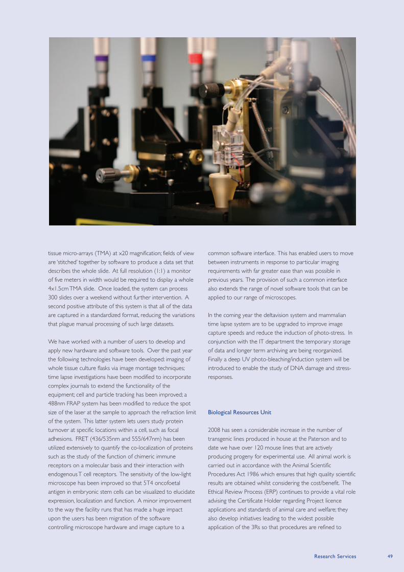
Research Services 49
tissue micro-arrays (TMA) at x20 magnification; fields of view
are ‘stitched’ together by software to produce a data set that
describes the whole slide. At full resolution (1:1) a monitor
of five meters in width would be required to display a whole
4x1.5cmTMA slide. Once loaded, the system can process
300 slides over a weekend without further intervention. A
second positive attribute of this system is that all of the data
are captured in a standardized format, reducing the variations
that plague manual processing of such large datasets.
We have worked with a number of users to develop and
apply new hardware and software tools. Over the past year
the following technologies have been developed: imaging of
whole tissue culture flasks via image montage techniques;
time lapse investigations have been modified to incorporate
complex journals to extend the functionality of the
equipment; cell and particle tracking has been improved; a
488nm FRAP system has been modified to reduce the spot
size of the laser at the sample to approach the refraction limit
of the system. This latter system lets users study protein
turnover at specific locations within a cell, such as focal
adhesions. FRET (436/535nm and 555/647nm) has been
utilized extensively to quantify the co-localization of proteins
such as the study of the function of chimeric immune
receptors on a molecular basis and their interaction with
endogenous T cell receptors. The sensitivity of the low-light
microscope has been improved so that 5T4 oncofoetal
antigen in embryonic stem cells can be visualized to elucidate
expression, localization and function. A minor improvement
to the way the facility runs that has made a huge impact
upon the users has been migration of the software
controlling microscope hardware and image capture to a
common software interface. This has enabled users to move
between instruments in response to particular imaging
requirements with far greater ease than was possible in
previous years. The provision of such a common interface
also extends the range of novel software tools that can be
applied to our range of microscopes.
In the coming year the deltavision system and mammalian
time lapse system are to be upgraded to improve image
capture speeds and reduce the induction of photo-stress. In
conjunction with the IT department the temporary storage
of data and longer term archiving are being reorganized.
Finally a deep UV photo-bleaching/induction system will be
introduced to enable the study of DNA damage and stress-
responses.
Biological Resources Unit
2008 has seen a considerable increase in the number of
transgenic lines produced in house at the Paterson and to
date we have over 120 mouse lines that are actively
producing progeny for experimental use. All animal work is
carried out in accordance with the Animal Scientific
Procedures Act 1986 which ensures that high quality scientific
results are obtained whilst considering the cost/benefit. The
Ethical Review Process (ERP) continues to provide a vital role
advising the Certificate Holder regarding Project licence
applications and standards of animal care and welfare; they
also develop initiatives leading to the widest possible
application of the 3Rs so that procedures are refined to
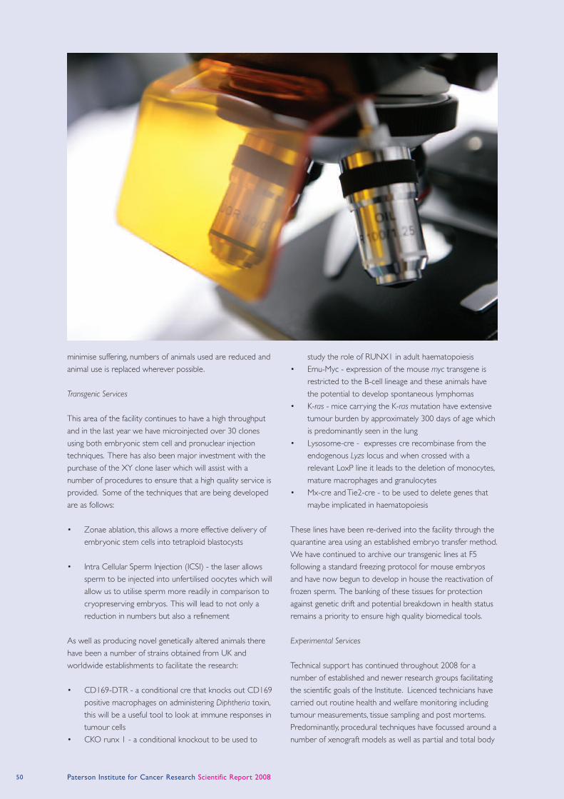
minimise suffering, numbers of animals used are reduced and
animal use is replaced wherever possible.
Transgenic Services
This area of the facility continues to have a high throughput
and in the last year we have microinjected over 30 clones
using both embryonic stem cell and pronuclear injection
techniques. There has also been major investment with the
purchase of the XY clone laser which will assist with a
number of procedures to ensure that a high quality service is
provided. Some of the techniques that are being developed
are as follows:
• Zonae ablation, this allows a more effective delivery of
embryonic stem cells into tetraploid blastocysts
• Intra Cellular Sperm Injection (ICSI) - the laser allows
sperm to be injected into unfertilised oocytes which will
allow us to utilise sperm more readily in comparison to
cryopreserving embryos. This will lead to not only a
reduction in numbers but also a refinement
As well as producing novel genetically altered animals there
have been a number of strains obtained from UK and
worldwide establishments to facilitate the research:
• CD169-DTR - a conditional cre that knocks out CD169
positive macrophages on administering Diphtheria toxin,
this will be a useful tool to look at immune responses in
tumour cells
• CKO runx 1 - a conditional knockout to be used to
study the role of RUNX1 in adult haematopoiesis
• Emu-Myc - expression of the mouse myc transgene is
restricted to the B-cell lineage and these animals have
the potential to develop spontaneous lymphomas
• K-ras - mice carrying the K-ras mutation have extensive
tumour burden by approximately 300 days of age which
is predominantly seen in the lung
• Lysosome-cre - expresses cre recombinase from the
endogenous Lyzs locus and when crossed with a
relevant LoxP line it leads to the deletion of monocytes,
mature macrophages and granulocytes
• Mx-cre and Tie2-cre - to be used to delete genes that
maybe implicated in haematopoiesis
These lines have been re-derived into the facility through the
quarantine area using an established embryo transfer method.
We have continued to archive our transgenic lines at F5
following a standard freezing protocol for mouse embryos
and have now begun to develop in house the reactivation of
frozen sperm. The banking of these tissues for protection
against genetic drift and potential breakdown in health status
remains a priority to ensure high quality biomedical tools.
Experimental Services
Technical support has continued throughout 2008 for a
number of established and newer research groups facilitating
the scientific goals of the Institute. Licenced technicians have
carried out routine health and welfare monitoring including
tumour measurements, tissue sampling and post mortems.
Predominantly, procedural techniques have focussed around a
number of xenograft models as well as partial and total body
50 Paterson Institute for Cancer Research Scientific Report 2008

irradiations with a variety of strains used including genetically
altered, immuno-competent and immuno-compromised.
There has also been the opportunity to learn and develop a
number of unfamiliar techniques such as:
• Intra-femoral injection of bone marrow cells post
ablation
• Intra-splenic/intra-hepatic injection for orthotopic
transplantation
• IVIS imaging - a non invasive procedure to track tumour
growth
Equipment Purchase
There has been considerable investment undertaken with the
purchasing of the following equipment,
• Three replacement cage changing stations for carrying
out routine husbandry tasks and animal manipulation
• XY Clone Laser Ablator to facilitate a number of
transgenic procedures
• Controlled Rate Freezer for Cryopreservation
Cancer Research UK GeneChip Microarray Service
Head: Stuart Pepper
The microarray facility at the Paterson maintains an ongoing
program of development to ensure that array services
offered to CR-UK funded scientists are state of the art. Over
the last couple of years we have seen a growing number of
publications featuring data sets we have generated; in this
report three publications are described which show the
range of work that the facility undertakes.
In April our work with paraffin embedded archival material
lead to a joint publication with Dr Kim Linton (Christie
Hospital) examining the expression profiles of sarcoma
samples (Linton et al., Br J Cancer 2008; 98: 1403). There
have been very few publications so far on these challenging
samples and even fewer with significant validation at both
RNA and protein level. Since this paper came out we have
continued to develop expression profiling of archival samples
and have just submitted a follow up publication examining the
use of Affymetrix Exon arrays for work with FFPE material.
For the last couple of years we have been encouraging the
use of Exon arrays as they give a more complete picture of
transcription than 3’ expression arrays. Exon arrays have
much higher content than conventional arrays with each
transcript being interrogated by multiple probe sets along the
full length rather than just at the 3’ end. Several publications
describing the use of Exon arrays have already come from
this facility. In October we contributed to a joint paper with
Alvin Lee and Charles Swanton looking at the accuracy of
gene level summaries generated by Exon arrays and
concluding that these gave excellent comparability to qPCR
results (Lee et al., Cell Cycle 2008; 7: 3947).
Over several years of operation the facility has collected
various comparison data sets; when testing new arrays or
labelling protocols our usual approach is to run replicates of
two cell lines, MCF7 and MCF10A, and compare the data to
previous data sets. Last year Andy Sims (Breast Biology
Group) was looking at ways to combine and analyse data
from different projects and our in house data sets proved to
be of great value for testing possible algorithms. This work
was written up and published in September this year (Sims et
al., BMC Med Genomics 2008; 1: 42).
Finally, this year is seeing a major change in how the facility
will be managed. As part of a major restructuring within
Cancer Research UK the service will be leaving the Research
Services division to be managed directly by the Paterson
Institute. As the year draws to a close we will be saying a
fond farewell to our many colleagues in Research Services as
we look ahead to the exciting challenges of 2009.
Flow Cytometry Facility
Head: Morgan Blaylock
The Cytometry Facility at the Paterson provides state-of-the-
art instrumentation, education and expert technical assistance
to investigators for the successful performance of flow
cytometry based studies. The goal of the facility is both to
support current research applications and to continuously
extend the repertoire of flow cytometric methods available
to users.
The facility currently has three bench top cytometers
including the newly purchased BD LSRII which is a 17-colour
instrument. We also have three BD sorters ranging from the
Vantage SE which is capable of basic 2-way sorting, the BD
FACSAria which offers complex 9-colour 4-way sorts to our
most recent acquisition the BD InFlux which offers 14-colour
4-way sorting capabilities with added bio-safety.
The LSRII was purchased to assist with our increasing
workload and allow us to move into the field of
polychromatic flow, the new system allows us to detect up to
18 colours from four different laser sources with a much
higher cellular throughput. In addition this system is fully
digital providing accurate logarithmic conversions and a full
compensation matrix which allows automatic compensation
of every fluorochrome in an antibody cocktail.
The BD InFlux was initially purchased as a replacement for
the FACS Vantage SE which is over 15 years old, however the
facility has seen an increase of over 70% in the level of
Research Services 51
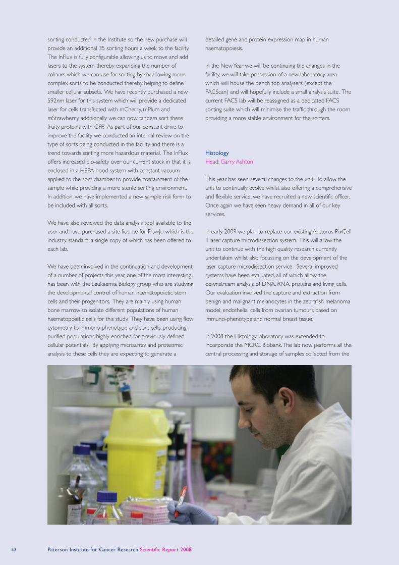
52 Paterson Institute for Cancer Research Scientific Report 2008
sorting conducted in the Institute so the new purchase will
provide an additional 35 sorting hours a week to the facility.
The InFlux is fully configurable allowing us to move and add
lasers to the system thereby expanding the number of
colours which we can use for sorting by six allowing more
complex sorts to be conducted thereby helping to define
smaller cellular subsets. We have recently purchased a new
592nm laser for this system which will provide a dedicated
laser for cells transfected with mCherry, mPlum and
mStrawberry, additionally we can now tandem sort these
fruity proteins with GFP. As part of our constant drive to
improve the facility we conducted an internal review on the
type of sorts being conducted in the facility and there is a
trend towards sorting more hazardous material. The InFlux
offers increased bio-safety over our current stock in that it is
enclosed in a HEPA hood system with constant vacuum
applied to the sort chamber to provide containment of the
sample while providing a more sterile sorting environment.
In addition, we have implemented a new sample risk form to
be included with all sorts.
We have also reviewed the data analysis tool available to the
user and have purchased a site licence for FlowJo which is the
industry standard, a single copy of which has been offered to
each lab.
We have been involved in the continuation and development
of a number of projects this year, one of the most interesting
has been with the Leukaemia Biology group who are studying
the developmental control of human haematopoietic stem
cells and their progenitors. They are mainly using human
bone marrow to isolate different populations of human
haematopoietic cells for this study. They have been using flow
cytometry to immuno-phenotype and sort cells, producing
purified populations highly enriched for previously defined
cellular potentials. By applying microarray and proteomic
analysis to these cells they are expecting to generate a
detailed gene and protein expression map in human
haematopoiesis.
In the New Year we will be continuing the changes in the
facility, we will take possession of a new laboratory area
which will house the bench top analysers (except the
FACScan) and will hopefully include a small analysis suite. The
current FACS lab will be reassigned as a dedicated FACS
sorting suite which will minimise the traffic through the room
providing a more stable environment for the sorters.
Histology
Head: Garry Ashton
This year has seen several changes to the unit. To allow the
unit to continually evolve whilst also offering a comprehensive
and flexible service, we have recruited a new scientific officer.
Once again we have seen heavy demand in all of our key
services.
In early 2009 we plan to replace our existing Arcturus PixCell
II laser capture microdissection system. This will allow the
unit to continue with the high quality research currently
undertaken whilst also focussing on the development of the
laser capture microdissection service. Several improved
systems have been evaluated, all of which allow the
downstream analysis of DNA, RNA, proteins and living cells.
Our evaluation involved the capture and extraction from
benign and malignant melanocytes in the zebrafish melanoma
model, endothelial cells from ovarian tumours based on
immuno-phenotype and normal breast tissue.
In 2008 the Histology laboratory was extended to
incorporate the MCRC Biobank. The lab now performs all the
central processing and storage of samples collected from the
Research Services 53
five collaborating NHS Trusts. The aim of the Biobank is to
facilitate translational research by allowing researchers access
to high quality samples from targeted cancer disease groups.
Standard operating procedures have been compiled that
ensure that sample collection and processing is performed in
a uniform manner and is of the highest standard. Initial
quality control checks on the samples have been very
encouraging. The Biobank has recently been expanded to
include the collection of haematological malignancies.
Tissue microarrays allow for high throughput and
standardisation of methodology, whilst also preserving rare
samples. In response to the anticipated future demand from
new and existing groups and the MCRC Biobank we are
currently evaluating a new high throughput automated
platform, whilst still retaining the manual platform for more
specialised construction. The new automated platform will
also allow more accurate donor core acquisition using an H
& E reference slide as guidance. Cell pellets are also being
used to make disease specific tissue microarrays. Disease
areas include paediatric (Ewing’s sarcoma, neuroblastoma),
lung (small- and non small-cell), breast, ovarian, colorectal and
prostate cancers. These specific tissue microarrays will enable
rapid screening of cell lines for immunohistochemical markers
of interest and help identify suitable controls for use alongside
patient tissue samples in clinical trials.
Immunohistochemistry and the optimisation of novel
antibodies comprises a large proportion of our work.
Multiple chromogenic and fluorescent labelling studies have
been performed and alternative sample handling and fixation
regimes have been explored.
Immunohistochemical analyses of tissues derived from mouse
knockout experiments have continued. Specifically tissue
specific Cre lines restricted in their expression to
haematopoietic and vascular tissues were tested. To
differentiate tissue types we used myeloid specific markers
(e.g. F4/80 for macrophages) and vascular markers (e.g.
CD31, VE-Cadherin) in the presence of Cre-dependent
reporter genes (e.g GFP, lacZ). In cases where tissue specific
gene mutations produced inflammatory phenotypes we
evaluated inflammatory markers (e.g. P-IKK) leukocyte
infiltration markers (e.g. CD45, F4/80) and apoptotic markers
(e.g. cleaved Caspase 3).
Immunohistochemistry has also been used to identify
genetically modified T cells in studies looking at their ability to
eradicate tumours. Using mouse models, tagged T cells were
identified at the site of tumour and within the spleen and
lymph nodes of treated animals. Importantly, this process is
being used to identify and enumerate T cells in other tissues
that could be potentially involved with tissue toxicity, such as
CEA-specific T cells residing within the mucosal tissue of the
gastro-intestinal tract expressing the normal tissue level of the
target antigen.
Similarly the role played by IGF-I signalling in colorectal cancer
initiation and progression is being investigated, by
immunohistochemically determining and correlating the
distribution of the IGF-I receptor (IGF-IR) and markers of cell
lineage, proliferation and receptor activity. Staining will also
be correlated with serum levels of IGF-I related proteins and
BMI to determine the role that obesity plays in the disease
In collaboration with the Translational Radiobiology Group, in
situ hybridisation is being used to examine the role of viral
oncogenes in head and neck squamous cell carcinoma
(HNSCC). Epstein Barr Virus (EBV) is an important
etiological factor in nasopharyngeal carcinoma but has only
recently been implicated in the development of
oropharyngeal carcinomas. Similarly, high risk human
papilloma virus (HPV 16) has long been associated with the
development of cervical cancer but recent studies have
shown that it may also play a role in a distinct subset of
HNSCCs. Investigation of these viruses in a large series of
patients is now being undertaken.
Kostoris Library
Head: Steve Glover
The Kostoris Library provides a service to staff and students
at The Christie NHS Foundation Trust and the Paterson
Institute for Cancer Research. The library works closely with
the John Rylands University Library service at The University
of Manchester to provide staff with desktop access to the
world’s primary research in life sciences. In addition to over
5000 online journals the library provides access to databases
including PubMed, Medline, BIOSIS, ISI Web of Knowledge,
EMBASE, and Scopus. The library also offers a personalised
database surveillance service in which regular search queries
are run on a monthly basis to keep staff up-to-date with the
latest published research. The library also provides an
electronic table of contents service with links to full text
delivered to the desktop.
This year the library has launched an Institutional Repository
of Research Publications that currently holds over 2000
articles going back to 2001. The repository contains peer-
reviewed papers published by the staff of the Paterson
Institute and The Christie NHS Foundation Trust. Newly
published records are added on a monthly basis whilst
retrospective deposits are due to go back as far as 1950. The
database is hosted on the Christie website and is accessible
to be searched on the Internet. Most records are linked to
the publishers’ websites using Digital Object Identifiers
(DOIs) which will provide persistent links via a resolving
service. Whilst the repository currently holds journal articles,
reviews and editorials it is hoped that conference abstracts
and posters may be added in the future.

The Kostoris Library achieved a stage 3 award when it
underwent external accreditation in 2008. This was the third
consecutive time since 2000 that the library has achieved this
award which is the highest level of accreditation for NHS
libraries. The Health Care Libraries Unit (HCLU) carried out
the accreditation visit in May on behalf of the NHS North
West (NHS NW) Strategic Health Authority.
Laboratory Services
Head: Mark Craven
During 2008 Laboratory Services became a stand-alone
service and a new head of the service was appointed. Since
then the provision of media services within the Institute has
been reviewed. The media service has adapted to a doubling
in the requirements for liquid media over the year and
continues to adapt to meet this challenge. Currently over
1000 litres of media per month are supplied throughout the
Institute. Alongside a general upgrading of facilities within the
department, we have created a clean room for the
production of microbiological plates. This allows us better
control and flexibility to meet the demands from our
customers within the Institute.
Laboratory Services continue to provide daily sterile
glassware and plastics deliveries and the removal of used
glassware for washing and autoclaving. Laboratory aides are
assigned to each research group and they can undertake a
range of technical and laboratory management duties.
Logistics
Head: Maurice Cowell
The Logistics department provides a comprehensive and
efficient portering service for the Institute. The main duties
are the receipt, checking, booking in and distribution of goods
ordered by numerous personnel in the Institute, accurately
and efficiently. The porters are responsible for the collection
and removal of waste, be it general rubbish, clinical or GM
waste. They are also responsible for the collection of liquid
nitrogen containers from laboratories, transporting them to
the loading bay for refilling and returning to the labs.
Ordering and distribution of the Central Stores stock via the
intranet email is also the duty of the Logistics department,
ensuring adequate stock levels are maintained at all times. In
addition Logistics maintain the Institute freezers (Sigma,
Invitrogen, Roche, Promega and Qiagen), having responsibility
for the ordering, distribution and stock levels. The Institute’s
usage of gas cylinders is looked after by the porters who are
in charge of replacement and ordering as and when
necessary.
The department works closely with all groups and helps out
where necessary, for example, by tracing and confirming
delivery of goods with suppliers, dealing with missing,
damaged or incorrect items, managing the moving of heavy
equipment or furniture, and setting up various meeting rooms
for numerous events.
Molecular Biology Core Facility
Head: Stuart Pepper
The Molecular Biology Core Facility comprises three separate
teams; two posts provide a range of core services described
here, two further teams provide comprehensive microarray
and proteomics services detailed separately in this section.
One of the major tasks for the core team is provision of a
genotyping service for biological samples. This service
processes around 400 samples per week and aims to return
data within 10 days. For many samples multiple PCR
reactions are needed meaning that typically we run around
2000 PCR reactions per week. High throughput for this
service is achieved by keeping all work in 96-well format,
including the final analysis on agarose gels.
Demand for the DNA sequencing service has increased this
year however since the upgrade of our 3100 to a 3130xl we
have been able to increase the number of samples run each
day to keep up with demand. Alongside the DNA
sequencing we have maintained our miniprep service;
scientists on site are able to have plasmid DNA prepared
each morning in time for inclusion in sequencing runs later in
the same day. This offers excellent turnaround for projects
requiring sequence checking of clones.
The core facility also provides support for expression
profiling by quantitative PCR. An ABI7900 in conjunction with
an Eppendorf epMotion allows qPCR to be carried out in
384 well formats, facilitating large projects to be processed
efficiently. For projects looking at large numbers of genes we
also have a Beckman GeXP system. This system allows a high
level of multiplexing of PCR reactions allowing for a greater
throughput than traditional real time PCR approaches.
MBCF Biological Mass Spectrometry Facility
Head: Duncan Smith
The remit of the biological mass spectrometry facility is to
support all the protein and peptide mass spectrometry
requirements of the Institute’s research groups. This support
covers a whole spectrum of activities ranging from provision
of routine services, implementation of new technologies
onsite, through to dedicated collaborations designed to
54 Paterson Institute for Cancer Research Scientific Report 2008
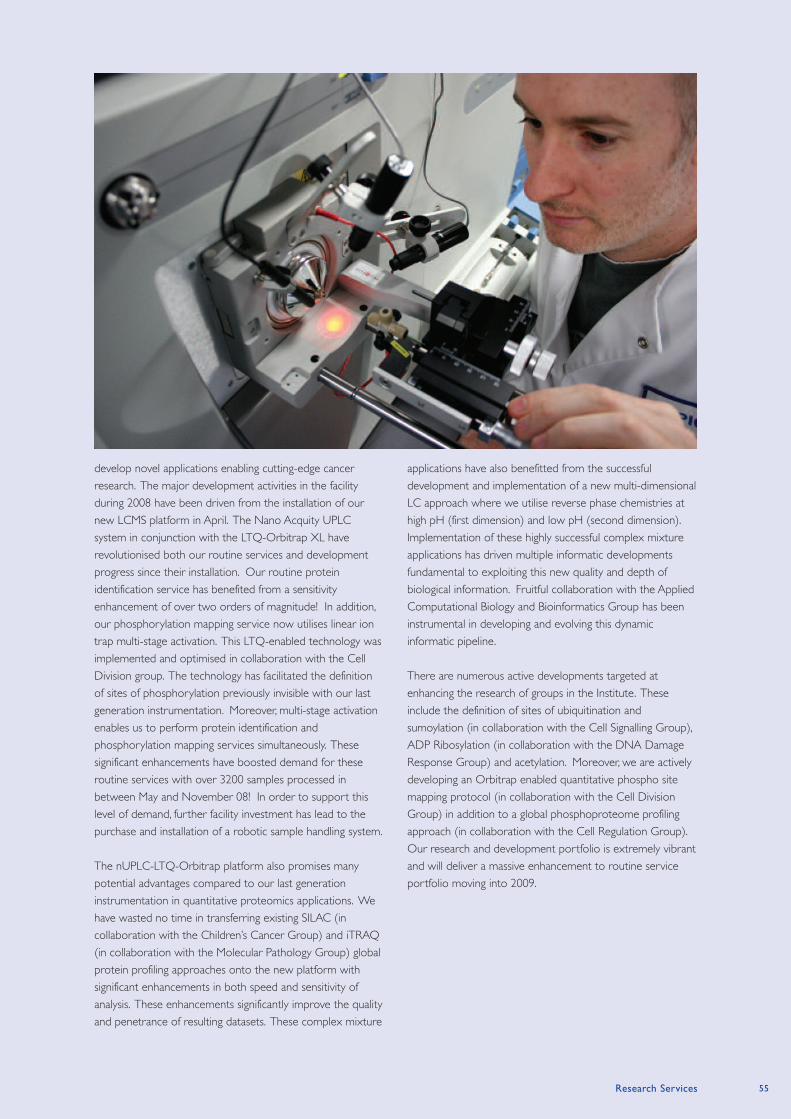
develop novel applications enabling cutting-edge cancer
research. The major development activities in the facility
during 2008 have been driven from the installation of our
new LCMS platform in April. The Nano Acquity UPLC
system in conjunction with the LTQ-Orbitrap XL have
revolutionised both our routine services and development
progress since their installation. Our routine protein
identification service has benefited from a sensitivity
enhancement of over two orders of magnitude! In addition,
our phosphorylation mapping service now utilises linear ion
trap multi-stage activation. This LTQ-enabled technology was
implemented and optimised in collaboration with the Cell
Division group. The technology has facilitated the definition
of sites of phosphorylation previously invisible with our last
generation instrumentation. Moreover, multi-stage activation
enables us to perform protein identification and
phosphorylation mapping services simultaneously. These
significant enhancements have boosted demand for these
routine services with over 3200 samples processed in
between May and November 08! In order to support this
level of demand, further facility investment has lead to the
purchase and installation of a robotic sample handling system.
The nUPLC-LTQ-Orbitrap platform also promises many
potential advantages compared to our last generation
instrumentation in quantitative proteomics applications. We
have wasted no time in transferring existing SILAC (in
collaboration with the Children’s Cancer Group) and iTRAQ
(in collaboration with the Molecular Pathology Group) global
protein profiling approaches onto the new platform with
significant enhancements in both speed and sensitivity of
analysis. These enhancements significantly improve the quality
and penetrance of resulting datasets. These complex mixture
applications have also benefitted from the successful
development and implementation of a new multi-dimensional
LC approach where we utilise reverse phase chemistries at
high pH (first dimension) and low pH (second dimension).
Implementation of these highly successful complex mixture
applications has driven multiple informatic developments
fundamental to exploiting this new quality and depth of
biological information. Fruitful collaboration with the Applied
Computational Biology and Bioinformatics Group has been
instrumental in developing and evolving this dynamic
informatic pipeline.
There are numerous active developments targeted at
enhancing the research of groups in the Institute. These
include the definition of sites of ubiquitination and
sumoylation (in collaboration with the Cell Signalling Group),
ADP Ribosylation (in collaboration with the DNA Damage
Response Group) and acetylation. Moreover, we are actively
developing an Orbitrap enabled quantitative phospho site
mapping protocol (in collaboration with the Cell Division
Group) in addition to a global phosphoproteome profiling
approach (in collaboration with the Cell Regulation Group).
Our research and development portfolio is extremely vibrant
and will deliver a massive enhancement to routine service
portfolio moving into 2009.
Research Services 55
Crispin Miller (page 8)
Applied Computational Biology and Bioinformatics Group
Refereed Research Papers
Bitton, D.A., Okoniewski, M.J., Connolly, Y. and Miller, C.J.
(2008) Exon level integration of proteomics and microarray
data. BMC Bioinformatics, 9, 118.
Linton, K.M., Hey, Y., Saunders, E., Jeziorska, M., Denton, J.,
Wilson, C.L., Swindell, R., Dibben, S., Miller, C.J., Pepper, S.D.,
Radford, J.A. and Freemont, A.J. (2008) Acquisition of
biologically relevant gene expression data by Affymetrix
microarray analysis of archival formalin-fixed paraffin-
embedded tumours. Br J Cancer, 98, 1403-1414.
Pierce, A., Unwin, R.D., Evans, C.A., Griffiths, S., Carney, L.,
Zhang, L., Jaworska, E., Lee, C.F., Blinco, D., Okoniewski, M.J.,
Miller, C.J., Bitton, D.A., Spooncer, E. and Whetton, A.D. (2008)
Eight-channel iTRAQ enables comparison of the activity of six
leukemogenic tyrosine kinases. Mol Cell Proteomics, 7, 853-
863.
Sims, A.H., Smethurst, G.J., Hey, Y., Okoniewski, M.J., Pepper,
S.D., Howell, A., Miller, C.J. and Clarke, R.B. (2008) The
removal of multiplicative, systematic bias allows integration of
breast cancer gene expression datasets - improving meta-
analysis and prediction of prognosis. BMC Med Genomics, 1,
42.
Williamson, A.J., Smith, D.L., Blinco, D., Unwin, R.D., Pearson, S.,
Wilson, C., Miller, C., Lancashire, L., Lacaud, G., Kouskoff, V. and
Whetton, A.D. (2008) Quantitative proteomic analysis
demonstrates post-transcriptional regulation of embryonic
stem cell differentiation to hematopoiesis. Mol Cell
Proteomics, 7, 459-472.
Yates, T., Okoniewski, M.J. and Miller, C.J. (2008) X:Map:
annotation and visualization of genome structure for
Affymetrix exon array analysis. Nucleic Acids Res, 36, D780-
786.
Other Publication
Okoniewski, M.J. and Miller, C.J. (2008) Comprehensive
analysis of affymetrix exon arrays using BioConductor. PLoS
Comput Biol, 4, e6.
Geoff Margison (page 10)
Carcinogenesis Group
Refereed Research Papers
Crosbie, P.A., McGown, G., Thorncroft, M.R., O’Donnell, P.N.,
Barber, P.V., Lewis, S.J., Harrison, K.L., Agius, R.M., Santibanez-
Koref, M.F., Margison, G.P. and Povey, A.C. (2008) Association
between lung cancer risk and single nucleotide
polymorphisms in the first intron and codon 178 of the DNA
repair gene, O6-alkylguanine-DNA alkyltransferase. Int J
Cancer, 122, 791-795.
Khan, O.A., Ranson, M., Michael, M., Olver, I., Levitt, N.C.,
Mortimer, P., Watson, A.J., Margison, G.P., Midgley, R. and
Middleton, M.R. (2008) A phase II trial of lomeguatrib and
temozolomide in metastatic colorectal cancer. Br J Cancer,
98, 1614-1618.
Milsom, M.D., Jerabek-Willemsen, M., Harris, C.E., Schambach,
A., Broun, E., Bailey, J., Jansen, M., Schleimer, D., Nattamai, K.,
Wilhelm, J., Watson, A., Geiger, H., Margison, G.P., Moritz, T.,
Baum, C., Thomale, J. and Williams, D.A. (2008) Reciprocal
relationship between O6-methylguanine-DNA
methyltransferase P140K expression level and
chemoprotection of hematopoietic stem cells. Cancer Res,
68, 6171-6180.
Other Publication
Verbeek, B., Southgate, T.D., Gilham, D.E. and Margison, G.P.
(2008) O6-Methylguanine-DNA methyltransferase inactivation
and chemotherapy. Br Med Bull, 85, 17-33.
Active Patents
O6-Substituted guanine derivatives, a process for their
preparation and their use in treating tumour cells. McMurry,
T.B.H., McElhinney, R.S., McCormick, J.E., Elder, R.H., Kelly, J.,
Margison, G.P., Rafferty, J.A., Watson, A.J. and Willington, M.A.
International Patent Application PCT/IE94/0031, published
(WO 94/29312, 70 pages) through CR Technology Ltd.
(Chem. Abs. 1995, 122, 239458e).
Pyrimidine derivatives and guanine derivatives, and their use
in treating tumour cells. McMurry, T.B.H., McElhinney, R.S.,
McCormick, J.E., Donnelly, D.J., Murray, P., Carola, C., Elder, R.H.,
Kelly, J., Margison, G.P., Watson, A.J., Rafferty, J.A., Willington,
M.A. and Willington, M.R. International Patent Application
PCT/IE96/00084, published (WO97/20843, 129 pages)
through CR Technology Ltd. (Chem. Abs., 1997, 127, 108802t).
Potentiation of temozolomide in human tumour cells. Baer, J.,
Freeman, A.A., Newlands, E.S., Watson, A.J., Rafferty, J.A., and
Margison, G.P. (USP: 5731304) through CR Technology Ltd.
Research Publications
56 Paterson Institute For Cancer Research Scientific Report 2008
Karim Labib (page 12)
Cell Cycle Group
Refereed Research Papers
Sanchez-Diaz, A., Marchesi, V., Murray, S., Jones, R., Pereira, G.,
Edmondson, R., Allen, T. and Labib, K. (2008) Inn1 couples
contraction of the actomyosin ring to membrane ingression
during cytokinesis in budding yeast. Nat Cell Biol, 10, 395-
406.
Other Publications
de la Cueva-Mendez, G. and Labib, K. (2008) New insights
into the chromosome cycle. Conference on the Replication &
Segregation of Chromosomes. EMBO Rep, 9, 1177-1181.
Labib, K. (2008) Making connections at DNA replication
forks: Mrc1 takes the lead. Mol Cell, 32, 166-168.
Iain Hagan (page 14)
Cell Division group
Refereed Research Papers
Robertson, A.M. and Hagan, I.M. (2008) Stress-regulated
kinase pathways in the recovery of tip growth and
microtubule dynamics following osmotic stress in S. pombe. J
Cell Sci, 121, 4055-4068.
Other Publication
Hagan, I.M. (2008) The spindle pole body plays a key role in
controlling mitotic commitment in the fission yeast
Schizosaccharomyces pombe. Biochem Soc Trans, 36, 1097-
1101.
Nic Jones (page 16)
Cell Regulation Group
Refereed Research Papers
Bhoumik, A., Fichtman, B., Derossi, C., Breitwieser, W., Kluger,
H.M., Davis, S., Subtil, A., Meltzer, P., Krajewski, S., Jones, N. and
Ronai, Z. (2008) Suppressor role of activating transcription
factor 2 (ATF2) in skin cancer. Proc Natl Acad Sci u S A, 105,
1674-1679.
Chen, D., Wilkinson, C.R., Watt, S., Penkett, C.J., Toone, W.M.,
Jones, N. and Bahler, J. (2008) Multiple pathways differentially
regulate global oxidative stress responses in fission yeast. Mol
Biol Cell, 19, 308-317.
Dawson, K., Toone, W.M., Jones, N. and Wilkinson, C.R. (2008)
Loss of regulators of vacuolar ATPase function and ceramide
synthesis results in multidrug sensitivity in
Schizosaccharomyces pombe. Eukaryot Cell, 7, 926-937.
Reiter, W., Watt, S., Dawson, K., Lawrence, C.L., Bahler, J., Jones,
N. and Wilkinson, C.R. (2008) Fission yeast MAP kinase Sty1
is recruited to stress-induced genes. J Biol Chem, 283, 9945-
9956.
Udagawa, T., Nemoto, N., Wilkinson, C.R., Narashimhan, J., Jiang,
L., Watt, S., Zook, A., Jones, N., Wek, R.C., Bahler, J. and Asano,
K. (2008) Int6/eIF3e promotes general translation and Atf1
abundance to modulate Sty1 MAPK-dependent stress
response in fission yeast. J Biol Chem, 283, 22063-22075.
Angeliki Malliri (page 18)
Cell Signalling Group
Refereed Research Paper
Witt, E., Maliri, A., McGrouther, D.A. and Bayat, A. (2008)
RAC Activity in Keloid Disease: Comparative Analysis of
Fibroblasts from Margin of Keloid to its Surrounding Normal
Skin. Eplasty, 8, e19.
Caroline Dive and MalcolmRanson (page 20)
Clinical and Experimental Pharmacology
Refereed Research Papers
Atatreh, N., Stojkoski, C., Smith, P., Booker, G.W., Dive, C.,
Frenkel, A.D., Freeman, S. and Bryce, R.A. (2008) In silico
screening and biological evaluation of inhibitors of Src-SH3
domain interaction with a proline-rich ligand. Bioorg Med
Chem Lett, 18, 1217-1222.
57Research Publications

58 Paterson Institute For Cancer Research Scientific Report 2008
Board, R.E., Thelwell, N.J., Ravetto, P.F., Little, S., Ranson, M.,
Dive, C., Hughes, A. and Whitcombe, D. (2008a) Multiplexed
assays for detection of mutations in PIK3CA. Clin Chem, 54,
757-760.
Board, R.E., Williams, V.S., Knight, L., Shaw, J., Greystoke, A.,
Ranson, M., Dive, C., Blackhall, F.H. and Hughes, A. (2008b)
Isolation and extraction of circulating tumor DNA from
patients with small cell lung cancer. Ann N Y Acad Sci, 1137,
98-107.
Cummings, J., Hodgkinson, C., Odedra, R., Sini, P., Heaton, S.P.,
Mundt, K.E., Ward, T.H., Wilkinson, R.W., Growcott, J., Hughes,
A. and Dive, C. (2008) Preclinical evaluation of M30 and M65
ELISAs as biomarkers of drug induced tumor cell death and
antitumor activity. Mol Cancer Ther, 7, 455-463.
de Haas, E.C., di Pietro, A., Simpson, K.L., Meijer, C., Suurmeijer,
A.J., Lancashire, L.J., Cummings, J., de Jong, S., de Vries, E.G.,
Dive, C. and Gietema, J.A. (2008) Clinical evaluation of M30
and M65 ELISA cell death assays as circulating biomarkers in
a drug-sensitive tumor, testicular cancer. Neoplasia, 10,
1041-1048.
Greystoke, A., Cummings, J., Ward, T., Simpson, K., Renehan, A.,
Butt, F., Moore, D., Gietema, J., Blackhall, F., Ranson, M., Hughes,
A. and Dive, C. (2008) Optimisation of circulating biomarkers
of cell death for routine clinical use. Ann Oncol, 19, 990-995.
Micha, D., Cummings, J., Shoemaker, A., Elmore, S., Foster, K.,
Greaves, M., Ward, T., Rosenberg, S., Dive, C. and Simpson, K.
(2008) Circulating biomarkers of cell death after treatment
with the BH-3 mimetic ABT-737 in a preclinical model of
small-cell lung cancer. Clin Cancer Res, 14, 7304-7310.
Seenath, M.M., Roberts, D., Cawthorne, C., Saunders, M.P.,
Armstrong, G.R., O’Dwyer, S.T., Stratford, I.J., Dive, C. and
Renehan, A.G. (2008) Reciprocal relationship between
expression of hypoxia inducible factor 1alpha (HIF-1alpha)
and the pro-apoptotic protein bid in ex vivo colorectal
cancer. Br J Cancer, 99, 459-463.
Simpson, K.L., Whetton, A.D. and Dive, C. (2008) Quantitative
mass spectrometry-based techniques for clinical use:
Biomarker identification and quantification. J Chromatogr B
Analyt Technol Biomed Life Sci, epub Nov 18.
Sweeney, E., Ward, T.H., Gray, N., Womack, C., Jayson, G.,
Hughes, A., Dive, C. and Byers, R. (2008) Quantitative
multiplexed quantum dot immunohistochemistry. Biochem
Biophys Res Commun, 374, 181-186.
Williamson, A.J., Smith, D.L., Blinco, D., Unwin, R.D., Pearson, S.,
Wilson, C., Miller, C., Lancashire, L., Lacaud, G., Kouskoff, V. and
Whetton, A.D. (2008) Quantitative proteomic analysis
demonstrates post-transcriptional regulation of embryonic
stem cell differentiation to hematopoiesis. Mol Cell
Proteomics, 7, 459-472.
xenaki, G., Ontikatze, T., Rajendran, R., Stratford, I.J., Dive, C.,
Krstic-Demonacos, M. and Demonacos, C. (2008) PCAF is an
HIF-1alpha cofactor that regulates p53 transcriptional activity
in hypoxia. Oncogene, 27, 5785-5796.
Other Publications
Cummings, J., Ward, T.H., Greystoke, A., Ranson, M. and Dive, C.
(2008) Biomarker method validation in anticancer drug
development. Br J Pharmacol, 153, 646-656.
Renehan, A.G., Roberts, D.L. and Dive, C. (2008) Obesity and
cancer: pathophysiological and biological mechanisms. Arch
Physiol Biochem, 114, 71-83.
Ward, T.H., Cummings, J., Dean, E., Greystoke, A., Hou, J.M.,
Backen, A., Ranson, M. and Dive, C. (2008) Biomarkers of
apoptosis. Br J Cancer, 99, 841-846.
Peter Stern (page 22)
Immunology Group
Refereed Research Papers
Castro, F.V., Tutt, A.L., White, A.L., Teeling, J.L., James, S., French,
R.R. and Glennie, M.J. (2008) CD11c provides an effective
immunotarget for the generation of both CD4 and CD8 T
cell responses. Eur J Immunol, 38, 2263-2273.
Corradi-Perini, C., Southgate, T.D., Besley, G.T., Cooper, A.,
Deakin, J.A., Wraith, E., Fairbairn, L.J., Wynn, R.F. and
Bellantuono, I. (2008) Retrovirally-mediated genetic
correction of mesenchymal stem cells from patients affected
by mucopolysaccharidosis type II (Hunter Syndrome). Gene
Ther Mol Biol, 12, 167-174.
Elkord, E., Burt, D.J., Drijfhout, J.W., Hawkins, R.E. and Stern, P.L.
(2008) CD4(+) T-cell recognition of human 5T4 oncofoetal
antigen: implications for initial depletion of CD25(+) T cells.
Cancer Immunol Immunother, 57, 833-847.
Elkord, E., Dangoor, A., Drury, N.L., Harrop, R., Burt, D.J.,
Drijfhout, J.W., Hamer, C., Andrews, D., Naylor, S., Sherlock, D.,
Hawkins, R.E. and Stern, P.L. (2008) An MVA-based vaccine
targeting the oncofetal antigen 5T4 in patients undergoing
surgical resection of colorectal cancer liver metastases. J
Immunother, 31, 820-829.
Karanam, B., Gambhira, R., Peng, S., Jagu, S., Kim, D.-J., Ketner,
G.W., Stern, P.L., Adams, R.J. and Roden, R.B.S. (2008)
Vaccination with HPV16 L2E6E7 fusion protein in GPI-0100
adjuvant elicits protective humoral and cell-mediated
immunity. Vaccine doi:10.1016/j.vaccine.2008.11.099.
Marignol, L., Foley, R., Southgate, T.D., Coffey, M., Hollywood, D.
and Lawler, M. (2008) Hypoxia response element-driven
cytosine deaminase/5-fluorocytosine gene therapy system: a
highly effective approach to overcome the dynamics of
tumour hypoxia and enhance the radiosensitivity of prostate
cancer cells in vitro. J Gene Med, epub Dec 2.
Thistlethwaite, F.C., Elkord, E., Griffiths, R.W., Burt, D.J., Shablak,
A.M., Campbell, J.D., Gilham, D.E., Austin, E.B., Stern, P.L. and
Hawkins, R.E. (2008) Adoptive transfer of T(reg) depleted
autologous T cells in advanced renal cell carcinoma. Cancer
Immunol Immunother, 57, 623-634.

Winters, U., Daayana, S., Lear, J.T., Tomlinson, A.E., Elkord, E.,
Stern, P.L. and Kitchener, H.C. (2008) Clinical and
immunologic results of a phase II trial of sequential imiquimod
and photodynamic therapy for vulval intraepithelial neoplasia.
Clin Cancer Res, 14, 5292-5299.
Other Publications
Elkord, E. (2008) Novel therapeutic strategies by regulatory
T cells in allergy. Chem Immunol Allergy, 94, 150-157.
Elkord, E., Hawkins, R.E. and Stern, P.L. (2008) Immunotherapy
for gastrointestinal cancer: current status and strategies for
improving efficacy. Expert Opin Biol Ther, 8, 385-395.
Garland, S.M., Cuzick, J., Domingo, E.J., Goldie, S.J., Kim, Y.T.,
Konno, R., Parkin, D.M., Qiao, Y.L., Sankaranarayanan, R., Stern,
P.L., Tay, S.K. and Bosch, F.x. (2008) Recommendations for
cervical cancer prevention in Asia Pacific. Vaccine, 26 Suppl
12, M89-98.
Southgate, T., Kroeger, K.M., Liu, C., Lowenstein, P.R. and
Castro, M.G. (2008) Gene transfer into neural cells in vitro
using adenoviral vectors. In: Curr Protoc Neurosci, 4, Unit
4.23.
Stern, P.L. (2008) HPV vaccination. HPV Today, Vaccines Issue.
Stern, P.L. (2008) HPV vaccines: an overview. HPV Forum, 4,
7-13.
Stern, P.L. and Kitchener, H.C. (2008) Vaccines for prevention
of cervical cancer. Oxford University Press.
Thistlethwaite, F.C., Stern, P.L. and Hawkins, R.E. (2008)
Biological therapies. Cytokine and adoptive cell therapy. In:
Treatment of Cancer. Eds Price, P., Sikora, K. and Illidge, T. pp
186-203.
Verbeek, B., Southgate, T.D., Gilham, D.E. and Margison, G.P.
(2008) O6-Methylguanine-DNA methyltransferase inactivation
and chemotherapy. Br Med Bull, 85, 17-33.
Active Patents
Stern, P.L. and Hole, N. (1989). Improvement relating to
antigens. Publication information EP0336562.
Stern PL and Hole N (1999). 5T4 antigen from human
trophoblasts Publication information US5869053.
Ward C.M and Stern (2005) 5T4 antigen expression as an
indicator of stem cell differentiation. Publication information
US2005260591.
Nullin Divecha (page 24)
Inositide Laboratory
Refereed Research Papers
Los, A.P., de Widt, J., van Blitterswijk, W.J. and Divecha, N.
(2008) Is there a role for diacylglycerol kinase-zeta in cell
cycle regulation? Adv Enzyme Regul, 48, 31-39.
Panbianco, C., Weinkove, D., Zanin, E., Jones, D., Divecha, N.,
Gotta, M. and Ahringer, J. (2008) A casein kinase 1 and PAR
proteins regulate asymmetry of a PIP(2) synthesis enzyme for
asymmetric spindle positioning. Dev Cell, 15, 198-208.
van den Bout, I., van Rheenen, J., van Angelen, A.A., de Rooij, J.,
Wilhelmsen, K., Jalink, K., Divecha, N. and Sonnenberg, A.
(2008) Investigation into the mechanism regulating MRP
localization. Exp Cell Res, 314, 330-341.
Weinkove, D., Bastiani, M., Chessa, T.A., Joshi, D., Hauth, L.,
Cooke, F.T., Divecha, N. and Schuske, K. (2008)
Overexpression of PPK-1, the Caenorhabditis elegans Type I
PIP kinase, inhibits growth cone collapse in the developing
nervous system and causes axonal degeneration in adults.
Dev Biol, 313, 384-397.
Other Publications
Halstead, J., Snell, M.H., Meeuws, S., Jones, D.R. and Divecha, N.
(2008) Assaying endogenous PIP5K activities. In: Lipid
Signalling Protocols. Eds Larijani, Woscholski and Rosser. pp
391-402. Humana Press.
Jones, D.R., Bultsma, Y., Keune, W.-J. and Divecha, N. (2008)
Methods for the determination of the mass of nuclear
PtdIns4P, PtdIns5P and PtdIns(4,5)P2. In: Lipid Signalling
Protocols. Eds Larijani, Woscholski and Rosser. pp 75-88.
Humana Press.
59Research Publications
60 Paterson Institute For Cancer Research Scientific Report 2008
Tim Somervaille (page 26)
Leukaemia Biology Group
Refereed Research Papers
Viatour, P., Somervaille, T.C., Venkatasubrahmanyam, S., Kogan,
S., McLaughlin, M.E., Weissman, I.L., Butte, A.J., Passegue, E. and
Sage, J. (2008) Hematopoietic stem cell quiescence is
maintained by compound contributions of the retinoblastoma
gene family. Cell Stem Cell, 3, 416-428.
Wang, Z., Smith, K.S., Murphy, M., Piloto, O., Somervaille, T.C.
and Cleary, M.L. (2008) Glycogen synthase kinase 3 in MLL
leukaemia maintenance and targeted therapy. Nature, 455,
1205-1209.
Georges Lacaud (page 28)
Stem Cell Biology Group
Refereed Research Papers
Baldwin, R.J., Ten Dam, G.B., van Kuppevelt, T.H., Lacaud, G.,
Gallagher, J.T., Kouskoff, V. and Merry, C.L. (2008) A
developmentally-regulated heparan sulfate epitope defines a
sub-population with increased blood potential during
mesodermal differentiation. Stem Cells, 26, 3108-3118.
Pearson, S., Sroczynska, P., Lacaud, G. and Kouskoff, V. (2008)
The stepwise specification of embryonic stem cells to
hematopoietic fate is driven by sequential exposure to Bmp4,
activin A, bFGF and VEGF. Development, 135, 1525-1535.
Pimanda, J.E., Chan, W.Y., Wilson, N.K., Smith, A.M., Kinston, S.,
Knezevic, K., Janes, M.E., Landry, J.R., Kolb-Kokocinski, A.,
Frampton, J., Tannahill, D., Ottersbach, K., Follows, G.A., Lacaud,
G., Kouskoff, V. and Gottgens, B. (2008) Endoglin expression
in blood and endothelium is differentially regulated by
modular assembly of the Ets/Gata hemangioblast code. Blood,
112, 4512-4522.
Williamson, A.J., Smith, D.L., Blinco, D., Unwin, R.D., Pearson, S.,
Wilson, C., Miller, C., Lancashire, L., Lacaud, G., Kouskoff, V. and
Whetton, A.D. (2008) Quantitative proteomic analysis
demonstrates post-transcriptional regulation of embryonic
stem cell differentiation to hematopoiesis. Mol Cell
Proteomics, 7, 459-472.
Valerie Kouskoff (page 30)
Stem Cell and Haematopoiesis Group
Refereed Research Papers
Baldwin, R.J., Ten Dam, G.B., van Kuppevelt, T.H., Lacaud, G.,
Gallagher, J.T., Kouskoff, V. and Merry, C.L. (2008) A
developmentally-regulated heparan sulfate epitope defines a
sub-population with increased blood potential during
mesodermal differentiation. Stem Cells, 26, 3108-3118.
Pearson, S., Sroczynska, P., Lacaud, G. and Kouskoff, V. (2008)
The stepwise specification of embryonic stem cells to
hematopoietic fate is driven by sequential exposure to Bmp4,
activin A, bFGF and VEGF. Development, 135, 1525-1535.
Pimanda, J.E., Chan, W.Y., Wilson, N.K., Smith, A.M., Kinston, S.,
Knezevic, K., Janes, M.E., Landry, J.R., Kolb-Kokocinski, A.,
Frampton, J., Tannahill, D., Ottersbach, K., Follows, G.A., Lacaud,
G., Kouskoff, V. and Gottgens, B. (2008) Endoglin expression
in blood and endothelium is differentially regulated by
modular assembly of the Ets/Gata hemangioblast code. Blood,
112, 4512-4522.
Williamson, A.J., Smith, D.L., Blinco, D., Unwin, R.D., Pearson, S.,
Wilson, C., Miller, C., Lancashire, L., Lacaud, G., Kouskoff, V. and
Whetton, A.D. (2008) Quantitative proteomic analysis
demonstrates post-transcriptional regulation of embryonic
stem cell differentiation to hematopoiesis. Mol Cell
Proteomics, 7, 459-472.
Catharine West (page 34)
Academic Radiation Oncology: Translational Biology Group
Refereed Research Papers
Charnley, N., Airley, R., Du Plessis, D., West, C., Brock, C.,
Barnett, C., Matthews, J., Symonds, K., Bottomly, M., Swindell, R.
and Price, P. (2008) No relationship between 18F-
fluorodeoxyglucose positron emission tomography and
expression of Glut-1 and -3 and hexokinase I and II in high-
grade glioma. Oncol Rep, 20, 537-542.
Griffiths, E.A., Pritchard, S.A., McGrath, S.M., Valentine, H.R.,
Price, P.M., Welch, I.M. and West, C.M. (2008) Hypoxia-
associated markers in gastric carcinogenesis and HIF-2alpha in
gastric and gastro-oesophageal cancer prognosis. Br J Cancer,
98, 965-973.
Silva, P., Slevin, N.J., Sloan, P., Valentine, H., Cresswell, J., Ryder,
D., Price, P., Homer, J.J. and West, C.M. (2008) Prognostic
significance of tumor hypoxia inducible factor-1alpha
expression for outcome after radiotherapy in oropharyngeal
cancer. Int J Radiat Oncol Biol Phys, 72, 1551-1559.
West, C.M., Joseph, L. and Bhana, S. (2008) Epidermal growth
factor receptor-targeted therapy. Br J Radiol, 81 Spec No 1,
S36-44.
Robert Hawkins and PeterStern (page 36)
Biological, Immune and Gene Therapy
Refereed Research Papers
Cheadle, E.J., Gilham, D.E. and Hawkins, R.E. (2008) The
combination of cyclophosphamide and human T cells
genetically engineered to target CD19 can eradicate
established B-cell lymphoma. Br J Haematol, 142, 65-68.
Elkord, E., Burt, D.J., Drijfhout, J.W., Hawkins, R.E. and Stern, P.L.
(2008a) CD4(+) T-cell recognition of human 5T4 oncofoetal
antigen: implications for initial depletion of CD25(+) T cells.
Cancer Immunol Immunother, 57, 833-847.
Elkord, E., Dangoor, A., Drury, N.L., Harrop, R., Burt, D.J.,
Drijfhout, J.W., Hamer, C., Andrews, D., Naylor, S., Sherlock, D.,
Hawkins, R.E. and Stern, P.L. (2008b) An MVA-based vaccine
targeting the oncofetal antigen 5T4 in patients undergoing
surgical resection of colorectal cancer liver metastases. J
Immunother, 31, 820-829.
Harrop, R., Drury, N., Shingler, W., Chikoti, P., Redchenko, I.,
Carroll, M.W., Kingsman, S.M., Naylor, S., Griffiths, R., Steven, N.
and Hawkins, R.E. (2008) Vaccination of colorectal cancer
patients with TroVax given alongside chemotherapy (5-
fluorouracil, leukovorin and irinotecan) is safe and induces
potent immune responses. Cancer Immunol Immunother, 57,
977-986.
Jones, R.J., Hawkins, R.E., Eatock, M.M., Ferry, D.R., Eskens, F.A.,
Wilke, H. and Evans, T.R. (2008) A phase II open-label study
of DHA-paclitaxel (Taxoprexin) by 2-h intravenous infusion in
previously untreated patients with locally advanced or
metastatic gastric or oesophageal adenocarcinoma. Cancer
Chemother Pharmacol, 61, 435-441.
Karanam, B., Gambhira, R., Peng, S., Jagu, S., Kim, D.-J., Ketner,
G.W., Stern, P.L., Adams, R.J. and Roden, R.B.S. (2008)
Vaccination with HPV16 L2E6E7 fusion protein in GPI-0100
adjuvant elicits protective humoral and cell-mediated
immunity. Vaccine doi:10.1016/j.vaccine.2008.11.099.
Ravaud, A., Hawkins, R., Gardner, J.P., von der Maase, H., Zantl,
N., Harper, P., Rolland, F., Audhuy, B., Machiels, J.P., Petavy, F.,
Gore, M., Schoffski, P. and El-Hariry, I. (2008) Lapatinib versus
hormone therapy in patients with advanced renal cell
carcinoma: a randomized phase III clinical trial. J Clin Oncol,
26, 2285-2291.
Thistlethwaite, F.C., Elkord, E., Griffiths, R.W., Burt, D.J., Shablak,
A.M., Campbell, J.D., Gilham, D.E., Austin, E.B., Stern, P.L. and
Hawkins, R.E. (2008) Adoptive transfer of T(reg) depleted
autologous T cells in advanced renal cell carcinoma. Cancer
Immunol Immunother, 57, 623-634.
Winters, U., Daayana, S., Lear, J.T., Tomlinson, A.E., Elkord, E.,
Stern, P.L. and Kitchener, H.C. (2008) Clinical and
immunologic results of a phase II trial of sequential imiquimod
and photodynamic therapy for vulval intraepithelial neoplasia.
Clin Cancer Res, 14, 5292-5299.
Other Publications
Clarke, R., Waddell, T., Gallagher, J., Lee, K., Radhakrishna, G.,
Cavet, J., Hawkins, R.E. and Saunders, M.P. (2008) A postcode
lottery still exists for cancer patients with ‘exceptional
circumstances’. Clin Oncol (R Coll Radiol), 20, 771-772.
Elkord, E. (2008) Novel therapeutic strategies by regulatory
T cells in allergy. Chem Immunol Allergy, 94, 150-157.
Elkord, E., Hawkins, R.E. and Stern, P.L. (2008) Immunotherapy
for gastrointestinal cancer: current status and strategies for
improving efficacy. Expert Opin Biol Ther, 8, 385-395.
Garland, S.M., Cuzick, J., Domingo, E.J., Goldie, S.J., Kim, Y.T.,
Konno, R., Parkin, D.M., Qiao, Y.L., Sankaranarayanan, R., Stern,
P.L., Tay, S.K. and Bosch, F.x. (2008) Recommendations for
cervical cancer prevention in Asia Pacific. Vaccine, 26 Suppl
12, M89-98.
Stern, P.L. and Kitchener, H.C. (2008) Vaccines for prevention
of cervical cancer. Oxford University Press.
Thistlethwaite, F.C., Stern, P.L. and Hawkins, R.E. (2008)
Biological therapies. Cytokine and adoptive cell therapy. In:
Treatment of Cancer. Eds Price, P., Sikora, K. and Illidge, T. pp
186-203.
Verbeek, B., Southgate, T.D., Gilham, D.E. and Margison, G.P.
(2008) O6-Methylguanine-DNA methyltransferase inactivation
and chemotherapy. Br Med Bull, 85, 17-33.
61Research Publications
62 Paterson Institute For Cancer Research Scientific Report 2008
Vaskar Saha (page 38)
Children’s Cancer Group
Active Patent
Modified Escherichia coli L-asparaginase II enzyme resistant to
cleavage by human AEP - useful for treating neoplastic
conditions e.g. non-Hodgkin lymphoma. Saha, V.
PCT/GB2007/000124, published as WO-2007083100. Patent
application filed and managed by Cancer Research
Technology Ltd.
Tim Illidge (page 40)
Targeted Therapy Group
Refereed Research Papers
Byers, R.J., Sakhinia, E., Joseph, P., Glennie, C., Hoyland, J.A.,
Menasce, L.P., Radford, J.A. and Illidge, T. (2008) Clinical
quantitation of immune signature in follicular lymphoma by
RT-PCR based gene expression profiling. Blood, 111, 4764-
4770.
Erenpreisa, J., Ivanov, A., Wheatley, S.P., Kosmacek, E.A., Ianzini,
F., Anisimov, A.P., Mackey, M., Davis, P.J., Plakhins, G. and Illidge,
T.M. (2008) Endopolyploidy in irradiated p53-deficient
tumour cell lines: Persistence of cell division activity in giant
cells expressing Aurora-B kinase. Cell Biol Int, 32, 1044-1056.
Illidge, T., Bayne, M., Brown, N.S., Chilton, S., Cragg, M.S., Du, Y.,
Glennie, M.J., Lewington, V., Smart, J., Zivanovic, M. and Johnson,
P.W. (2008) Phase I/II study of fractionated 131I-rituximab in
low grade B-cell lymphoma: the effect of prior rituximab
dosing and tumor burden on subsequent radiotherapy. Blood,
epub Dec 2008.
Ivanov, A., Krysov, S., Cragg, M.S. and Illidge, T. (2008)
Radiation therapy with tositumomab (B1) anti-CD20
monoclonal antibody initiates extracellular signal-regulated
kinase/mitogen-activated protein kinase-dependent cell death
that overcomes resistance to apoptosis. Clin Cancer Res, 14,
4925-4934.
Zinzani, P.L., d’Amore, F., Bombardieri, E., Brammer, C., Codina,
J.G., Illidge, T., Jurczak, W., Linkesch, W., Morschhauser, F.,
Vandenberghe, E. and Van Hoof, A. (2008) Consensus
conference: implementing treatment recommendations on
yttrium-90 immunotherapy in clinical practice - report of a
European workshop. Eur J Cancer, 44, 366-373.
Other Publications
Illidge, T. and Bayne, M.C. (2008) Radioimmunotherapy in
haematological cancer. In: Treatment of Cancer, 5th edition.
Eds Illidge, T., Price, P. and Sikora, K. Hodder Arnold.
Illidge, T. and Chan, C. (2008) How have outcomes for
patients with follicular lymphoma changed with the addition
of monoclonal antibodies? Leuk Lymphoma, 49, 1263-1273.
Illidge, T. and Du, Y. (2008) Radioimmunotherapy of
hematological malignancy. In: Advances in Nuclear Oncology:
Diagnosis and Therapy. Eds Bombardieri, E., Buscombe, J.,
Lucignani, G. and Schober, O. Informa Healthcare UK.
Illidge, T. and Hainsworth, J. (2008) Radioimmunotherapy of
hematological malignancy. In: Innovative Leukemia and
Lymphoma Therapy Eds Kaspers, G.J., Coiffier, B., Heinrich, M.C.
and Estey, E.H. Informa Healthcare USA.
Illidge, T., Price, P. and Sikora, K. (2008) Editors: Treatment of
Cancer. 5th edition. Hodder Arnold.
Illidge, T. and Tolan, S. (2008) Current treatment approaches
for diffuse large B-cell lymphoma. Leuk Lymphoma, 49, 663-
676.
Ivanov, A., Swann, R. and Illidge, T. (2008) New insights into
the mechanisms of action of radioimmunotherapy in
lymphoma. J Pharm Pharmacol, 60, 987-998.
Robert Hawkins (page 42)
Medical Oncology: Cell Therapy Group
Refereed Research Papers
Cheadle, E.J., Gilham, D.E. and Hawkins, R.E. (2008) The
combination of cyclophosphamide and human T cells
genetically engineered to target CD19 can eradicate
established B-cell lymphoma. Br J Haematol, 142, 65-68.
Elkord, E., Burt, D.J., Drijfhout, J.W., Hawkins, R.E. and Stern, P.L.
(2008) CD4(+) T-cell recognition of human 5T4 oncofoetal
antigen: implications for initial depletion of CD25(+) T cells.
Cancer Immunol Immunother, 57, 833-847.
Elkord, E., Dangoor, A., Drury, N.L., Harrop, R., Burt, D.J.,
Drijfhout, J.W., Hamer, C., Andrews, D., Naylor, S., Sherlock, D.,
Hawkins, R.E. and Stern, P.L. (2008) An MVA-based vaccine
targeting the oncofetal antigen 5T4 in patients undergoing
surgical resection of colorectal cancer liver metastases. J
Immunother, 31, 820-829.
Harrop, R., Drury, N., Shingler, W., Chikoti, P., Redchenko, I.,
Carroll, M.W., Kingsman, S.M., Naylor, S., Griffiths, R., Steven, N.
and Hawkins, R.E. (2008) Vaccination of colorectal cancer
patients with TroVax given alongside chemotherapy (5-
fluorouracil, leukovorin and irinotecan) is safe and induces
potent immune responses. Cancer Immunol Immunother, 57,
977-986.
Jones, R.J., Hawkins, R.E., Eatock, M.M., Ferry, D.R., Eskens, F.A.,
Wilke, H. and Evans, T.R. (2008) A phase II open-label study
of DHA-paclitaxel (Taxoprexin) by 2-h intravenous infusion in
previously untreated patients with locally advanced or
metastatic gastric or oesophageal adenocarcinoma. Cancer
Chemother Pharmacol, 61, 435-441.
Ravaud, A., Hawkins, R., Gardner, J.P., von der Maase, H., Zantl,
N., Harper, P., Rolland, F., Audhuy, B., Machiels, J.P., Petavy, F.,
Gore, M., Schoffski, P. and El-Hariry, I. (2008) Lapatinib versus
hormone therapy in patients with advanced renal cell
carcinoma: a randomized phase III clinical trial. J Clin Oncol,
26, 2285-2291.
Thistlethwaite, F.C., Elkord, E., Griffiths, R.W., Burt, D.J., Shablak,
A.M., Campbell, J.D., Gilham, D.E., Austin, E.B., Stern, P.L. and
Hawkins, R.E. (2008) Adoptive transfer of T(reg) depleted
autologous T cells in advanced renal cell carcinoma. Cancer
Immunol Immunother, 57, 623-634.
Whitfield, R.J., Battom, S.E., Barut, M., Gilham, D.E. and Ball, P.D.
(2008) Rapid high-performance liquid chromatographic
analysis of adenovirus type 5 particles with a prototype
anion-exchange analytical monolith column. J Chromatogr A,
epub Nov 11.
Other Publications
Clarke, R., Waddell, T., Gallagher, J., Lee, K., Radhakrishna, G.,
Cavet, J., Hawkins, R.E. and Saunders, M.P. (2008) A postcode
lottery still exists for cancer patients with ‘exceptional
circumstances’. Clin Oncol (R Coll Radiol), 20, 771-772.
Elkord, E., Hawkins, R.E. and Stern, P.L. (2008) Immunotherapy
for gastrointestinal cancer: current status and strategies for
improving efficacy. Expert Opin Biol Ther, 8, 385-395.
Thistlethwaite, F.C., Stern, P.L. and Hawkins, R.E. (2008)
Biological therapies. Cytokine and adoptive cell therapy. In:
Treatment of Cancer. Eds Price, P., Sikora, K. and Illidge, T. pp
186-203.
Verbeek, B., Southgate, T.D., Gilham, D.E. and Margison, G.P.
(2008) O6-Methylguanine-DNA methyltransferase inactivation
and chemotherapy. Br Med Bull, 85, 17-33.
John Gallagher (page 44)
Medical Oncology: Glyco-Oncology Group
Refereed Research Papers
Baldwin, R.J., Ten Dam, G.B., van Kuppevelt, T.H., Lacaud, G.,
Gallagher, J.T., Kouskoff, V. and Merry, C.L. (2008) A
developmentally-regulated heparan sulfate epitope defines a
sub-population with increased blood potential during
mesodermal differentiation. Stem Cells, 26, 3108-3118.
63Research Publications
Catlow, K.R., Deakin, J.A., Wei, Z., Delehedde, M., Fernig, D.G.,
Gherardi, E., Gallagher, J.T., Pavao, M.S. and Lyon, M. (2008)
Interactions of hepatocyte growth factor / scatter factor with
various glycosaminoglycans reveal an important interplay
between the presence of iduronate and sulfate density. J Biol
Chem, 283, 5235-5248.
Corradi-Perini, C., Southgate, T.D., Besley, G.T., Cooper, A.,
Deakin, J.A., Wraith, E., Fairbairn, L.J., Wynn, R.F. and
Bellantuono, I. (2008) Retrovirally-mediated genetic
correction of mesenchymal stem cells from patients affected
by mucopolysaccharidosis type II (Hunter Syndrome). Gene
Ther Mol Biol, 12, 167-174.
Deakin, J.A. and Lyon, M. (2008) A simplified and sensitive
fluorescent method for disaccharide analysis of both heparan
sulfate and chondroitin/dermatan sulfates from biological
samples. Glycobiology, 18, 483-491.
Deakin, J.A., Blaum, B.S., Gallagher, J.T., Uhrin, D.A. and Lyon, M.
(2008) The binding properties of minimal oligosaccharides
reveal a common heparan sulfate / dermatan sulfate-binding
site in hepatocyte growth factor / scatter factor that can
accommodate a wide variety of sulfation patterns. J Biol
Chem, epub Dec 29.
Goodger, S.J., Robinson, C.J., Murphy, K.J., Gasiunas, N., Harmer,
N.J., Blundell, T.L., Pye, D.A. and Gallagher, J.T. (2008) Evidence
that heparin saccharides promote FGF2 mitogenesis through
two distinct mechanisms. J Biol Chem, 283, 13001-13008.
Hershkovitz, O., Jarahian, M., Zilka, A., Bar-Ilan, A., Landau, G.,
Jivov, S., Tekoah, Y., Glicklis, R., Gallagher, J.T., Hoffmann, S.C.,
Zer, H., Mandelboim, O., Watzl, C., Momburg, F. and Porgador, A.
(2008) Altered glycosylation of recombinant NKp30 hampers
binding to heparan sulfate: a lesson for the use of
recombinant immunoreceptors as an immunological tool.
Glycobiology, 18, 28-41.
Schmidt, C.Q., Herbert, A.P., Kavanagh, D., Gandy, C., Fenton,
C.J., Blaum, B.S., Lyon, M., Uhrin, D. and Barlow, P.N. (2008) A
new map of glycosaminoglycan and C3b binding sites on
factor H. J Immunol, 181, 2610-2619.
Tan, K., Duquette, M., Liu, J.H., Shanmugasundaram, K.,
Joachimiak, A., Gallagher, J.T., Rigby, A.C., Wang, J.H. and Lawler,
J. (2008) Heparin-induced cis- and trans-dimerization modes
of the thrombospondin-1 N-terminal domain. J Biol Chem,
283, 3932-3941.
Tissot, B., Ceroni, A., Powell, A.K., Morris, H.R., Yates, E.A.,
Turnbull, J.E., Gallagher, J.T., Dell, A. and Haslam, S.M. (2008)
Software Tool for the Structural Determination of
Glycosaminoglycans by Mass Spectrometry. Anal Chem,
epub Oct 29.
Gordon Jayson (page 46)
Medical Oncology: Translational Angiogenesis Group
Refereed Research Papers
Byrd, L.M., Shenton, A., Maher, E.R., Woodward, E., Belk, R., Lim,
C., Lalloo, F., Howell, A., Jayson, G.C. and Evans, G.D. (2008)
Better life expectancy in women with BRCA2 compared with
BRCA1 mutations is attributable to lower frequency and later
onset of ovarian cancer. Cancer Epidemiol Biomarkers Prev,
17, 1535-1542.
Clamp, A.R., Schoffski, P., Valle, J.W., Wilson, R.H., Marreaud, S.,
64 Paterson Institute For Cancer Research Scientific Report 2008

Govaerts, A.S., Debois, M., Lacombe, D., Twelves, C., Chick, J.
and Jayson, G.C. (2008) A phase I and pharmacokinetic study
of OSI-7904L, a liposomal thymidylate synthase inhibitor in
combination with oxaliplatin in patients with advanced
colorectal cancer. Cancer Chemother Pharmacol, 61, 579-585.
Goncalves, V., Jayson, G. and Tarrier, N. (2008) A longitudinal
investigation of psychological morbidity in patients with
ovarian cancer. Br J Cancer, 99, 1794-1801.
Sweeney, E., Ward, T.H., Gray, N., Womack, C., Jayson, G.,
Hughes, A., Dive, C. and Byers, R. (2008) Quantitative
multiplexed quantum dot immunohistochemistry. Biochem
Biophys Res Commun, 374, 181-186.
Other Publications
Cole, C.L. and Jayson, G.C. (2008) Oligosaccharides as anti-
angiogenic agents. Expert Opin Biol Ther, 8, 351-362.
Collinson, F.J., Hall, G.D., Perren, T.J. and Jayson, G.C. (2008)
Development of antiangiogenic agents for ovarian cancer.
Expert Rev Anticancer Ther, 8, 21-32.
Kumaran, G., Clamp, A.R. and Jayson, G.C. (2008)
Angiogenesis as a therapeutic target in cancer. Clin Med, 8,
455-458.
Kumaran, G.C., Jayson, G.C. and Clamp, A.R. (2008)
Antiangiogenic drugs in ovarian cancer. Br J Cancer, epub
Nov 11.
O’Connor, J.P., Jackson, A., Asselin, M.C., Buckley, D.L., Parker,
G.J. and Jayson, G.C. (2008) Quantitative imaging biomarkers
in the clinical development of targeted therapeutics: current
and future perspectives. Lancet Oncol, 9, 766-776.
O’Connor, J.P.B., Rosa, D.D., Jackson, A. and Jayson, G.C. (2008)
Molecular imaging of targets and therapeutics in tumour
angiogenesis. In: Tumor Angiogenesis. Eds Marme and Fusenig.
pp 511. Springer.
Additional Publications
Allen, T.D., Rutherford, S.A., Murray, S., Drummond, S.P.,
Goldberg, M.W. and Kiseleva, E. (2008) Scanning electron
microscopy of nuclear structure. Methods Cell Biol, 88, 389-
409.
Drummond, S.P. and Allen, T.D. (2008) From live-cell imaging
to scanning electron microscopy (SEM): the use of green
fluorescent protein (GFP) as a common label. Methods Cell
Biol, 88, 97-108.
Lee, A.J., East, P., Pepper, S., Nicke, B., Szallasi, Z., Eklund, A.C.,
Downward, J. and Swanton, C. (2008) Concordance of exon
array and real-time PCR assessment of gene expression
following cancer cell cytotoxic drug exposure. Cell Cycle, 7,
3947–3948.
Murray, S. (2008) High pressure freezing and freeze
substitution of Schizosaccharomyces pombe and
Saccharomyces cerevisiae for TEM. Methods Cell Biol, 88, 3-
17.
Murray, S. and Kiseleva, E. (2008) A protocol for isolation and
visualization of yeast nuclei by scanning electron microscopy.
Methods Cell Biol, 88, 367-387.
Prenant, C., Gillies, J., Bailey, J., Chimon, G., Smith, N., Jayson,
G.J. and Zweit, J. (2008) Synthesis of [18F]
fluoroacetaldehyde. Application to [18F] fluoroethylation of
benzylamine under reductive alkylation conditions. J Lab
Comp Radiopharm, 51, 262-267.
65Research Publications
66 Paterson Institute For Cancer Research Scientific Report 2008
Peter AdamsCR-UK Beatson Institute, Glasgow
Kurt Ballmer-HoferPaul Scherrer Institute, Switzerland
Jiri BartekDanish Cancer Centre, Copenhagen, Denmark
Clare BennettRoyal Free Hospital, London
Constanze BoniferInstitute of Molecular Medicine, Leeds
Hugh BradyGreat Ormond Street Hospital, London
Cathrin BriskenSwiss Institute for Cancer Research, Epalinges sur
Lausanne, Switzerland
Keith BurridgeUniversity of North Carolina, USA
Susan ChanUniversity of Strasbourg, France
Mark CraggUniversity of Southampton
Michaela FryeWellcome Trust Centre for Stem Cell Research,
Cambridge
Simone FuldaUniversity Children’s Hospital, Ulm, Germany
Michelle GarrettInstitute for Cancer Research, Haddow Laboratories,
Sutton
Susan GasserFriedrich Miescher Institute for Biomedical Research,
Basel, Switzerland
David GillespieCR-UK Beatson Institute, Glasgow
Angela GladingUniversity of Pittsburgh, Pittsburgh, USA
Nadia HarbeckUniversity of Munich, Germany
We had another exceptionally good seminar series
again in the Institute, with speakers from around the
world. This series is complemented by seminars
within the Christie Hospital, a Gene Therapy
seminar series with a mixture of local, national
and international speakers and many seminars
at The University of Manchester. Finally the
postdoctoral lunchtime seminar series
was once again very popular.
Seminar Series 2008
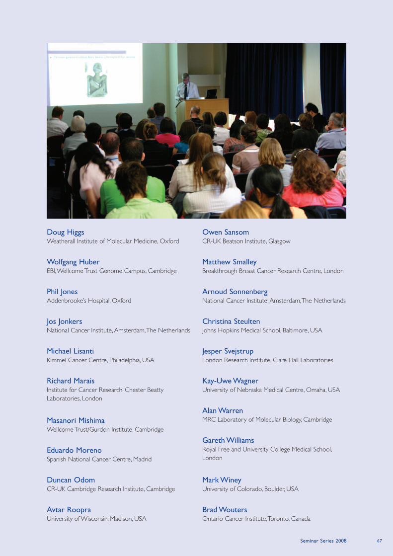
Seminar Series 2008 67
Doug HiggsWeatherall Institute of Molecular Medicine, Oxford
Wolfgang HuberEBI, Wellcome Trust Genome Campus, Cambridge
Phil JonesAddenbrooke’s Hospital, Oxford
Jos JonkersNational Cancer Institute, Amsterdam, The Netherlands
Michael LisantiKimmel Cancer Centre, Philadelphia, USA
Richard MaraisInstitute for Cancer Research, Chester Beatty
Laboratories, London
Masanori MishimaWellcome Trust/Gurdon Institute, Cambridge
Eduardo MorenoSpanish National Cancer Centre, Madrid
Duncan OdomCR-UK Cambridge Research Institute, Cambridge
Avtar RoopraUniversity of Wisconsin, Madison, USA
Owen SansomCR-UK Beatson Institute, Glasgow
Matthew SmalleyBreakthrough Breast Cancer Research Centre, London
Arnoud SonnenbergNational Cancer Institute, Amsterdam, The Netherlands
Christina SteultenJohns Hopkins Medical School, Baltimore, USA
Jesper SvejstrupLondon Research Institute, Clare Hall Laboratories
Kay-Uwe WagnerUniversity of Nebraska Medical Centre, Omaha, USA
Alan WarrenMRC Laboratory of Molecular Biology, Cambridge
Gareth WilliamsRoyal Free and University College Medical School,
London
Mark WineyUniversity of Colorado, Boulder, USA
Brad WoutersOntario Cancer Institute, Toronto, Canada
World-class research is backed up with access to world-class
facilities, and we aim to match these with a challenging but
rewarding graduate training programme that helps students
reach their full potential. Recruitment is highly competitive,
and generally involves two days of interviews with group
leaders and members of the Education Committee. The goal
is to find exceptional students with research interests that
closely match those of their prospective supervisor. This year
we brought recruitment forward to the last week of
November when we spent four days interviewing students
for the 2009 intake. Overall the interview process was very
successful and we expect to recruit again at a similar time
next year.
The Paterson Institute’s major role within the Manchester
Cancer Research Centre (MCRC) provides an opportunity to
design projects that sit at the interface between fundamental
and translational research, through collaborations with other
groups at the Christie Hospital and The University of
Manchester. The Clinical Fellowship scheme introduced in
2007, run in collaboration with AstraZeneca, continues to be
successful and has developed further over the past year.
2008 has seen a number of changes in postgraduate
education, including the appointment of a new Postgraduate
Tutor for Cancer, Dr Crispin Miller and the establishment of
an education sub-committee (Crispin Miller, Iain Hagan, Jenny
Varley, Julie Edwards) to help streamline the administration
process.
The Paterson Graduate Programme
Paterson PhDs are four years long, structured with a set of
key milestones. These include talks, literature reports and
project planning meetings, and provide formal points at which
progress (of both the student and the project) can be
assessed, and research goals developed and discussed.
Graduate training is monitored by the Education Committee,
which features Group Leaders, senior clinicians and scientists,
and student representatives. Each project is peer reviewed in
advance and considered in terms of its potential scientific
contribution and training opportunity. Every student is
assigned an Advisor (similar to a personal tutor on an
undergraduate programme), whose role is to provide
impartial advice and support while further support can also
be provided by the Postgraduate Tutor and a Student Welfare
Group.
The Paterson runs an external seminar series, featuring talks
from many of the key players in cancer research, and students
are also expected to attend postdoctoral seminars and to
present their work at lab meetings within the Institute. This
year also featured the introduction of a student seminar
series, conceived and organised by our graduate students as
an additional forum in which to discuss their work.
Ultimately though, it is the direct research experience that is
most important, and this is perhaps best measured by the
number and quality of publications featuring work by our
Graduate Students.
Postgraduate Education
A fundamental aspect of work in a
research institute such as the Paterson is a
thriving Graduate Programme, not simply to train the
researchers of tomorrow but also for the valuable
contributions the students bring to the labs they are
working in. At the Paterson Institute we have
students from around the globe, conducting
research in fields as diverse as yeast genetics,
stem cells, DNA repair, computer science
and clinical research.
Postgradute Tutor Crispin Miller
Postgraduate Education Manager
Julie Edwards
http://www.paterson.man.ac.uk/education/
Paterson Institute For Cancer Research Scientific Report 200868
Postgraduate Education
Student representative 2008
Dorota Feret (until September)
Natalie Reeves (until September)
Monique Melis (from September)
Andrzej Rutkowski (from September)
Education Committee 2008
Iain Hagan – Chair
Fiona Blackhall
Richard Cowan
Caroline Dive
Julie Edwards
David Gilham
Tim Illidge
Gordon Jayson
Valerie Kouskoff
Karim Labib
Crispin Miller
Vaskar Saha
Tim Somervaille
Jenny Varley
Catharine West
69
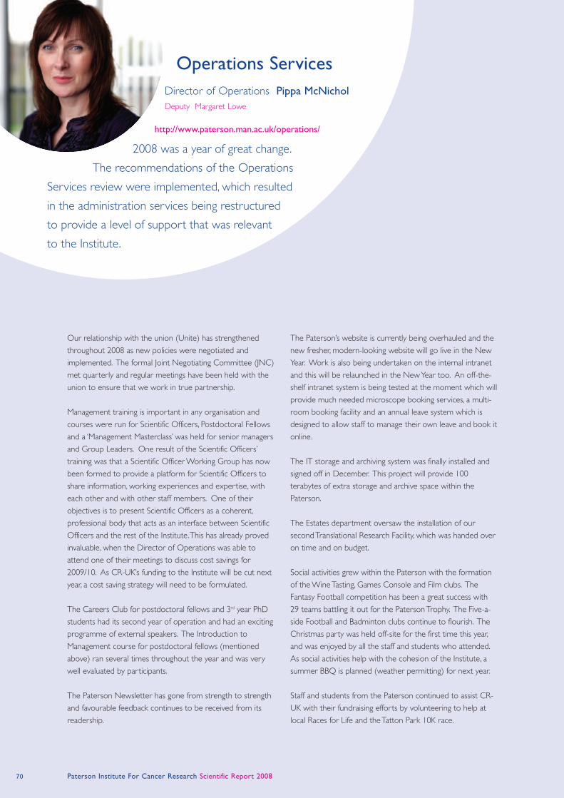
Our relationship with the union (Unite) has strengthened
throughout 2008 as new policies were negotiated and
implemented. The formal Joint Negotiating Committee (JNC)
met quarterly and regular meetings have been held with the
union to ensure that we work in true partnership.
Management training is important in any organisation and
courses were run for Scientific Officers, Postdoctoral Fellows
and a ‘Management Masterclass’ was held for senior managers
and Group Leaders. One result of the Scientific Officers’
training was that a Scientific Officer Working Group has now
been formed to provide a platform for Scientific Officers to
share information, working experiences and expertise, with
each other and with other staff members. One of their
objectives is to present Scientific Officers as a coherent,
professional body that acts as an interface between Scientific
Officers and the rest of the Institute. This has already proved
invaluable, when the Director of Operations was able to
attend one of their meetings to discuss cost savings for
2009/10. As CR-UK’s funding to the Institute will be cut next
year, a cost saving strategy will need to be formulated.
The Careers Club for postdoctoral fellows and 3rd year PhD
students had its second year of operation and had an exciting
programme of external speakers. The Introduction to
Management course for postdoctoral fellows (mentioned
above) ran several times throughout the year and was very
well evaluated by participants.
The Paterson Newsletter has gone from strength to strength
and favourable feedback continues to be received from its
readership.
The Paterson’s website is currently being overhauled and the
new fresher, modern-looking website will go live in the New
Year. Work is also being undertaken on the internal intranet
and this will be relaunched in the New Year too. An off-the-
shelf intranet system is being tested at the moment which will
provide much needed microscope booking services, a multi-
room booking facility and an annual leave system which is
designed to allow staff to manage their own leave and book it
online.
The IT storage and archiving system was finally installed and
signed off in December. This project will provide 100
terabytes of extra storage and archive space within the
Paterson.
The Estates department oversaw the installation of our
second Translational Research Facility, which was handed over
on time and on budget.
Social activities grew within the Paterson with the formation
of the Wine Tasting, Games Console and Film clubs. The
Fantasy Football competition has been a great success with
29 teams battling it out for the Paterson Trophy. The Five-a-
side Football and Badminton clubs continue to flourish. The
Christmas party was held off-site for the first time this year,
and was enjoyed by all the staff and students who attended.
As social activities help with the cohesion of the Institute, a
summer BBQ is planned (weather permitting) for next year.
Staff and students from the Paterson continued to assist CR-
UK with their fundraising efforts by volunteering to help at
local Races for Life and the Tatton Park 10K race.
Operations Services
2008 was a year of great change.
The recommendations of the Operations
Services review were implemented, which resulted
in the administration services being restructured
to provide a level of support that was relevant
to the Institute.
Director of Operations Pippa McNichol
Deputy Margaret Lowe
http://www.paterson.man.ac.uk/operations/
Paterson Institute For Cancer Research Scientific Report 200870

Paterson Institute for Cancer Research Scientific Report 2008
Administration and Reception Services
The administration department has seen a number of
changes during the year with the streamlining of processes
and procedures to ensure a time efficient and cost effective
structure. The catering function has increased and new
contracts have been negotiated, which have resulted in cost
savings. The Operations Manual has been significantly
updated to ensure that all practices are now current and fully
documented. The administration department continues to
oversee the Reception services and now provides lunchtime
cover for the Receptionist.
Some of the highlights over the last year include the
opportunity to provide the Assistant Director (Research)
with full support in the organisation of this year’s annual
Colloquium in September, which was very successful. In
addition, the department was able to provide administrative
support to the Clinical and Experimental Pharmacology
group on their recent quinquennial review, the outcome of
which was very positive.
Director’s Office
The Paterson’s Seminar Series had an impressive list of
external speakers during 2008. The series has proved to be
informative, varied and exceptionally popular within the
Institute.
One of the main administrative duties of this office has been
to support the Director in the search and appointment of
senior staff.
Estates
June 2008 saw the completion of the Institute’s second
Translational Research Facility. This £1.8m project has
provided much-needed additional accommodation of 800m2
for research staff.
A number of minor projects have also been completed which
have improved the environment and services for the Flow
Cytometry Department, Molecular Biology Core Facility and
Purchasing.
The Estates team has been pro-active throughout 2008 and
has identified plant and building fabric that requires attention
or replacement over the coming years. They have also
helped to reduce the amount of backlog maintenance within
the Institute.
There are some interesting projects that will be undertaken
next year, including the replacement of the existing
emergency generators and an increase in capacity.
The team endeavors to provide a quality service to reduce
the amount of disruption to research and the scientists. The
Estates team are working alongside The University of
Manchester’s Estates team to achieve compliance with
legislation, such as the control of Legionella in buildings.
Finance and Purchasing
The department continues to strive to provide a
comprehensive purchasing, travel and finance service on a
daily basis to the Research Groups and Service Units within
the Institute. New groups have been incorporated into the
system and all necessary training has been arranged for new
staff.
The relationship with the Faculty of Medical and Human
Sciences continues to develop. This year has seen the
devolvement of Research Administration to school level. This
has resulted in additional training courses for the various
University research systems and has also enabled the
department to network with the Research Business
Managers of the different research schools within the Faculty.
The University will be implementing a new grant-costing tool
in the New Year which should prove very useful as the level
of Contract Research Agreements and Grant Applications
continue to increase.
Health and Safety
A review of equipment maintenance covered by statutory
requirements was undertaken. This included local exhaust
ventilation (e.g. microbiological safety cabinets and fume
cupboards), pressurised vessels, all portable electrical
equipment (approximately 8,000 items), fire extinguishers and
alarm systems. This resulted in some changes to the service
providers, with subsequent cost savings and improved levels
of service. Also, the installation of a new access control
system is underway, which will further enhance security within
the Institute, and build on the improvements made last year.
Evaluation of online health and safety training was
undertaken, using staff in the Administration and Logistics
departments. Whilst this exercise proved useful and met
with some approval amongst staff, it is evident that there is
still a place for a more formalised presentation style of
training. Accordingly, more bespoke health and safety training
will be devised, such as the induction session, biological
agents, hazardous chemicals and risk assessment and activities
such as office safety may be rolled out as online training.
The annual health and safety inspection programme was
undertaken and reports produced for each department. The
inspection programme identified relatively minor non-
compliance issues, which were department specific. These
have been, or will be, addressed in due course. Health and
Safety advice and guidance was given to staff on a daily basis,
71
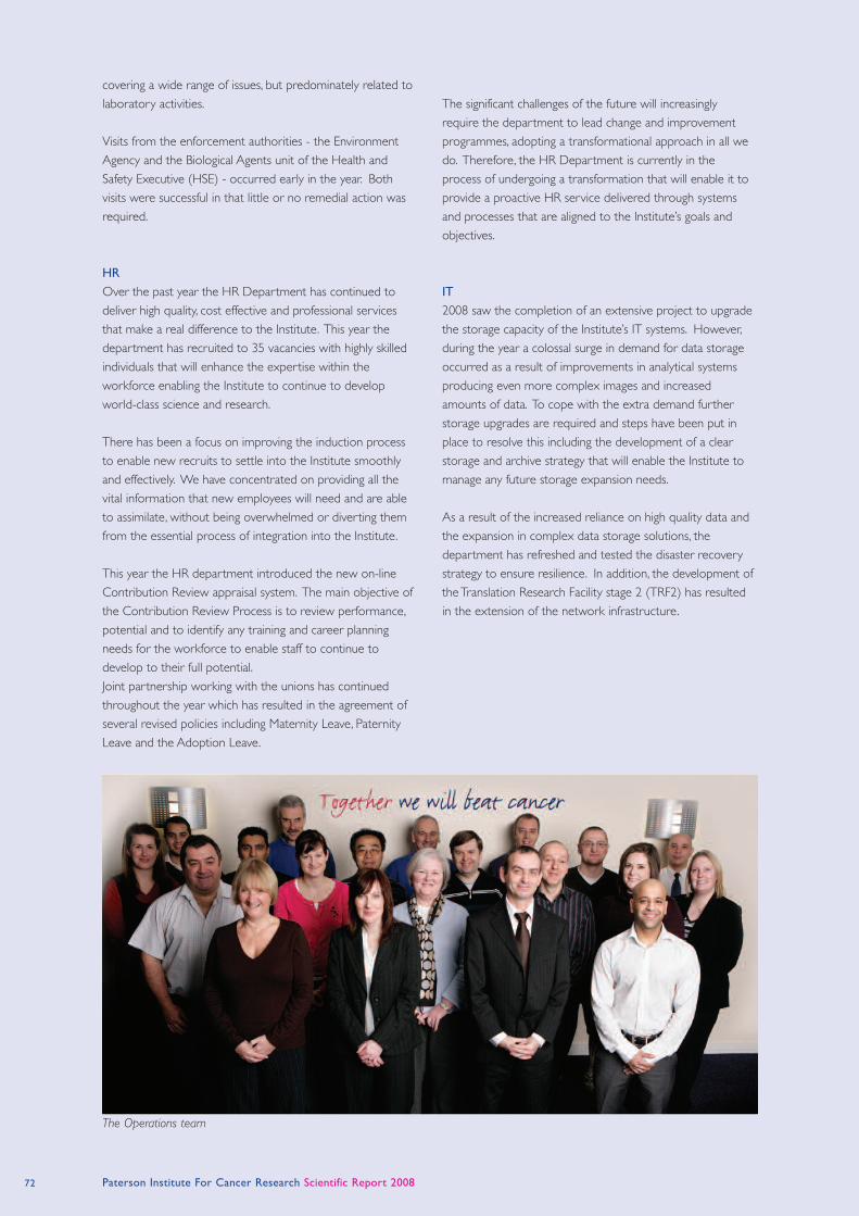
The Operations team
Paterson Institute For Cancer Research Scientific Report 2008
covering a wide range of issues, but predominately related to
laboratory activities.
Visits from the enforcement authorities - the Environment
Agency and the Biological Agents unit of the Health and
Safety Executive (HSE) - occurred early in the year. Both
visits were successful in that little or no remedial action was
required.
HR
Over the past year the HR Department has continued to
deliver high quality, cost effective and professional services
that make a real difference to the Institute. This year the
department has recruited to 35 vacancies with highly skilled
individuals that will enhance the expertise within the
workforce enabling the Institute to continue to develop
world-class science and research.
There has been a focus on improving the induction process
to enable new recruits to settle into the Institute smoothly
and effectively. We have concentrated on providing all the
vital information that new employees will need and are able
to assimilate, without being overwhelmed or diverting them
from the essential process of integration into the Institute.
This year the HR department introduced the new on-line
Contribution Review appraisal system. The main objective of
the Contribution Review Process is to review performance,
potential and to identify any training and career planning
needs for the workforce to enable staff to continue to
develop to their full potential.
Joint partnership working with the unions has continued
throughout the year which has resulted in the agreement of
several revised policies including Maternity Leave, Paternity
Leave and the Adoption Leave.
The significant challenges of the future will increasingly
require the department to lead change and improvement
programmes, adopting a transformational approach in all we
do. Therefore, the HR Department is currently in the
process of undergoing a transformation that will enable it to
provide a proactive HR service delivered through systems
and processes that are aligned to the Institute’s goals and
objectives.
IT
2008 saw the completion of an extensive project to upgrade
the storage capacity of the Institute’s IT systems. However,
during the year a colossal surge in demand for data storage
occurred as a result of improvements in analytical systems
producing even more complex images and increased
amounts of data. To cope with the extra demand further
storage upgrades are required and steps have been put in
place to resolve this including the development of a clear
storage and archive strategy that will enable the Institute to
manage any future storage expansion needs.
As a result of the increased reliance on high quality data and
the expansion in complex data storage solutions, the
department has refreshed and tested the disaster recovery
strategy to ensure resilience. In addition, the development of
the Translation Research Facility stage 2 (TRF2) has resulted
in the extension of the network infrastructure.
72
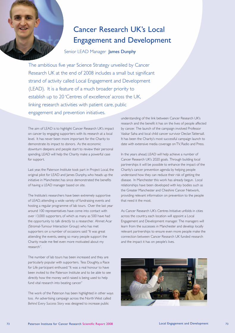
73 Paterson Institute for Cancer Research Scientific Report 2008
The aim of LEAD is to highlight Cancer Research UK’s impact
on cancer by engaging supporters with its research at a local
level. It has never been more important for the Charity to
demonstrate its impact to donors. As the economic
downturn deepens and people start to review their personal
spending, LEAD will help the Charity make a powerful case
for support.
Last year, the Paterson Institute took part in Project Local, the
original pilot for LEAD and James Dunphy, who heads up the
initiative in Manchester, has since demonstrated the benefits
of having a LEAD manager based on site.
The Institute’s researchers have been extremely supportive
of LEAD, attending a wide variety of fundraising events and
hosting a regular programme of lab tours. Over the last year
around 100 representatives have come into contact with
over 13,000 supporters, of which as many as 500 have had
the opportunity to talk directly to a researcher. Ahmet Acar
(Stromal-Tumour Interaction Group) who has met
supporters on a number of occasions said: “It was great
attending the events, seeing so many people support the
Charity made me feel even more motivated about my
research”.
The number of lab tours has been increased and they are
particularly popular with supporters. Tess Doughty, a Race
for Life participant enthused: “It was a real honour to have
been invited to the Paterson Institute and to be able to see
directly how the money we’d raised is being used to help
fund vital research into beating cancer.”
The work of the Paterson has been highlighted in other ways
too. An advertising campaign across the North West called
Behind Every Success Story was designed to increase public
understanding of the link between Cancer Research UK’s
research and the benefit it has on the lives of people affected
by cancer. The launch of the campaign involved Professor
Vaskar Saha and local child cancer survivor Declan Tattersall.
It has been the Charity’s most successful campaign launch to
date with extensive media coverage on TV, Radio and Press.
In the years ahead, LEAD will help achieve a number of
Cancer Research UK’s 2020 goals. Through building local
partnerships it will be possible to enhance the impact of the
Charity’s cancer prevention agenda by helping people
understand how they can reduce their risk of getting the
disease. In Manchester this work has already begun. Local
relationships have been developed with key bodies such as
the Greater Manchester and Cheshire Cancer Network,
providing relevant information on prevention to the people
that need it the most.
As Cancer Research UK’s Centres Initiative unfolds in cities
across the country, each location will appoint a Local
Engagement and Development manager. The managers will
learn from the successes in Manchester and develop locally
relevant partnerships to ensure even more people make the
connection between Cancer Research UK funded research
and the impact it has on people’s lives.
Cancer Research UK’s Local
Engagement and Development
The ambitious five year Science Strategy unveiled by Cancer
Research UK at the end of 2008 includes a small but significant
strand of activity called Local Engagement and Development
(LEAD). It is a feature of a much broader priority to
establish up to 20 ‘Centres of excellence’ across the UK,
linking research activities with patient care, public
engagement and prevention initiatives.
Senior LEAD Manager James Dunphy
Local Engagement and Development 73
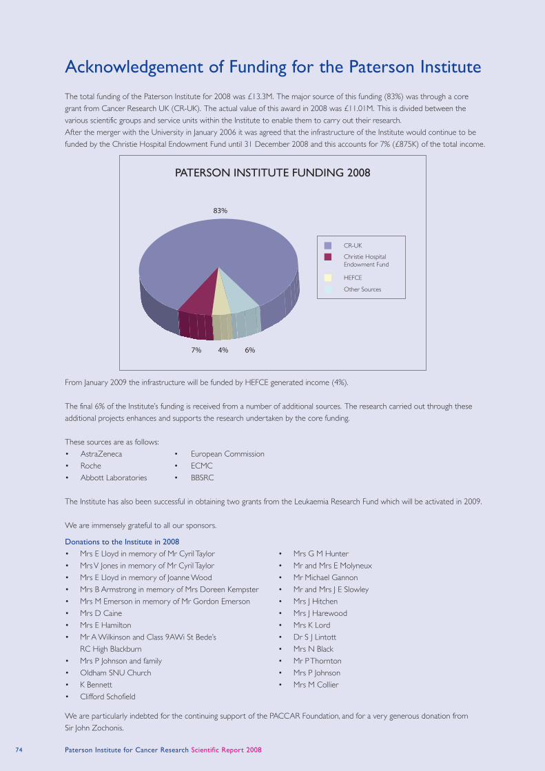
74 Paterson Institute for Cancer Research Scientific Report 2008
Donations to the Institute in 2008
• Mrs E Lloyd in memory of Mr Cyril Taylor
• Mrs V Jones in memory of Mr Cyril Taylor
• Mrs E Lloyd in memory of Joanne Wood
• Mrs B Armstrong in memory of Mrs Doreen Kempster
• Mrs M Emerson in memory of Mr Gordon Emerson
• Mrs D Caine
• Mrs E Hamilton
• Mr A Wilkinson and Class 9AWi St Bede’s
RC High Blackburn
• Mrs P Johnson and family
• Oldham SNU Church
• K Bennett
• Clifford Schofield
• Mrs G M Hunter
• Mr and Mrs E Molyneux
• Mr Michael Gannon
• Mr and Mrs J E Slowley
• Mrs J Hitchen
• Mrs J Harewood
• Mrs K Lord
• Dr S J Lintott
• Mrs N Black
• Mr P Thornton
• Mrs P Johnson
• Mrs M Collier
Acknowledgement of Funding for the Paterson Institute
The total funding of the Paterson Institute for 2008 was £13.3M. The major source of this funding (83%) was through a core
grant from Cancer Research UK (CR-UK). The actual value of this award in 2008 was £11.01M. This is divided between the
various scientific groups and service units within the Institute to enable them to carry out their research.
After the merger with the University in January 2006 it was agreed that the infrastructure of the Institute would continue to be
funded by the Christie Hospital Endowment Fund until 31 December 2008 and this accounts for 7% (£875K) of the total income.
From January 2009 the infrastructure will be funded by HEFCE generated income (4%).
The final 6% of the Institute’s funding is received from a number of additional sources. The research carried out through these
additional projects enhances and supports the research undertaken by the core funding.
These sources are as follows:
• AstraZeneca • European Commission
• Roche • ECMC
• Abbott Laboratories • BBSRC
The Institute has also been successful in obtaining two grants from the Leukaemia Research Fund which will be activated in 2009.
We are immensely grateful to all our sponsors.
We are particularly indebted for the continuing support of the PACCAR Foundation, and for a very generous donation from
Sir John Zochonis.
83%
7% 4% 6%
CR-UK
Christie HospitalEndowment Fund
HEFCE
Other Sources
PATERSON INSTITUTE FUNDING 2008
75 Paterson Institute For Cancer Research Scientific Report 2008 Career Opportunities
This is an extremely exciting development which is enhancing
all aspects of cancer research, education and treatment. The
Institute offers excellent laboratory facilities and outstanding
core facilities, including molecular services, a microarray
platform, proteomics, flow cytometry, histology, the
production of knock-in/knock-out animal models, real-time
PCR and advanced imaging. Details of all groups and facilities
are given throughout this report, and can guide interested
parties to the appropriate contacts.
Opportunities exist at a number of levels in the Institute. We
have a well-established programme of degrees by research
which is described in the section on Postgraduate Education
(page 68). We encourage applications from suitable qualified
graduates to apply to join either the PhD or MD
programmes. Graduates with a first or 2.1 honours degree in
a biological science can apply each year to train for a four-
year PhD in one of our research laboratories. First year
students will complement their laboratory skills by attending
a small number of specialised postgraduate taught and
training courses allowing them to gain a sound knowledge
base of the latest developments in cancer treatment and
research. The Institute also has a well-developed process for
ensuring suitable pastoral care and mentoring for all students.
Postdoctoral applicants of high calibre are regularly sought.
Although post docs will be encouraged to apply for their
own fellowships, funded positions are available for
outstanding candidates. Interested applicants should contact
the Group Leaders directly, with details of their area of
interest and recent experience.
In addition to postgraduate and postdoctoral opportunities,
the Institute is still seeking to recruit outstanding candidates
to the positions of Junior and Senior Group Leaders. The
packages provided are extremely attractive and
commensurate with the experience of the applicant, with
significant funding for personnel, recurrent expenditure and
equipment. Junior Group Leaders are appointed for an initial
six-year period, with Senior Group Leaders appointed to
non-time limited positions.
Specific vacancies can be found on our web pages
(http://www.paterson.man.ac.uk/jobs/), but suitably qualified
and enthusiastic individuals should contact the Institute at any
time to enquire about career possibilities.
Career Opportunities at the Paterson Institute
The Paterson Institute is located alongside The Christie NHS
Foundation Trust, and has a strong programme of basic and
translational research. There are very close links with
clinical and translational research groups throughout the
Christie Hospital site. The Manchester Cancer
Research Centre was created nearly three years ago
with partners including the Paterson Institute, The
Christie NHS Foundation Trust, The University
of Manchester and Cancer Research UK.
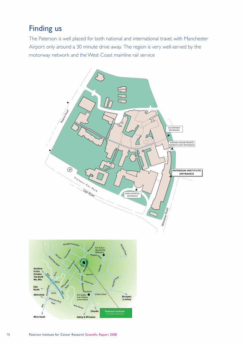
76 Paterson Institute for Cancer Research Scientific Report 2008
Finding usThe Paterson is well placed for both national and international travel, with Manchester
Airport only around a 30 minute drive away. The region is very well-served by the
motorway network and the West Coast mainline rail service
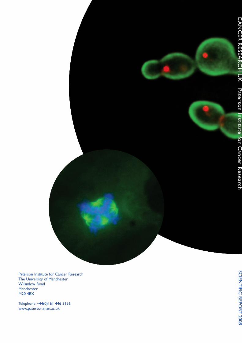
paterson institute for cancer research
the University of Manchester
Wilsmlow road
Manchester
M20 4BX
telephone +44(0)161 446 3156
www.paterson.man.ac.uk
cA
nc
er
reseA
rc
H U
K p
ate
rson in
stitute
for c
ancer r
ese
arc
hsc
ien
tif
ic r
epo
rt
2008