Paterimitra pyramidalis from South Australia: scleritome, shell structure and evolution of a lower...
30
PATERIMITRA PYRAMIDALIS FROM SOUTH AUSTRALIA: SCLERITOME, SHELL STRUCTURE AND EVOLUTION OF A LOWER CAMBRIAN STEM GROUP BRACHIOPOD by CECILIA M. LARSSON 1 *, CHRISTIAN B. SKOVSTED 2 , GLENN A. BROCK 3 , UWE BALTHASAR 4 , TIMOTHY P. TOPPER 5 and LARS E. HOLMER 1 1 Department of Earth Sciences, Palaeobiology, Uppsala University, Villav€ agen 16, SE-752 36, Uppsala, Sweden; e-mails: [email protected], [email protected] 2 Department of Palaeozoology, Swedish Museum of Natural History, Box 50007, SE-104 05, Stockholm, Sweden; e-mail: [email protected] 3 Department of Biological Sciences, Macquarie University, Sydney, NSW2109, Australia; e-mail: [email protected] 4 School of Geographical and Earth Sciences, University of Glasgow, Glasgow, G12 8QQ, UK; e-mail: [email protected] 5 Geological Museum, Øster Volgade 5-7, DK-1350, Copenhagen, Denmark; e-mail: [email protected] *Corresponding author. Typescript received 19 June 2012; accepted in revised form 24 June 2013 Abstract: The tommotiid Paterimitra pyramidalis Laurie, 1986, is redescribed based on well-preserved material from the lower Cambrian Wilkawillina, Wirrapowie and Ajax limestones of the Flinders Ranges, South Australia. The material shows that the scleritome of Paterimitra pyramidalis includes three sclerite morphotypes (S1, S2 and L). Detailed shell microstructure stud- ies show striking similarities with both the paterinid brachiopod Askepasma toddense and the tommotiid Eccentrotheca helenia, which strengthens the suggested evolutionary link between tom- motiids and brachiopods. Based on the partly articulated speci- mens and similarities in shell microstructure and sclerite morphology with Eccentrotheca, Paterimitra pyramidalis is reconstructed as a tube-dwelling, epifaunal, sessile, filter-feeder with an organic pedicle-like attachment structure. The proposed reconstruction of the scleritome comprises a basal unit com- posed of one S1 and one S2 sclerite, as well as an unresolved number of L sclerites lining a coniform tubular structure. Key words: Tommotiida, Brachiopoda, Paterimitra, scleri- tome structure, lower Cambrian, South Australia. T OMMOTIIDS constitute an extinct group of small meta- zoans represented by variously shaped minute organo- phosphatic sclerites found in lower Cambrian strata around the world (Rozanov et al. 1969; Landing 1984; Missarzhevsky 1989; Bengtson et al 1990; Conway Mor- ris and Chen 1990; Esakova and Zhegallo 1996). Their scientific history dates back to Tate’s (1892) report on Cambrian fossils from South Australia, but it was only just over 40 years ago that tommotiids were proposed to represent a unique fossil group (Fonin and Smirnova 1967; Rozanov et al. 1969; Bengtson 1970). Since then, several genera of enigmatic, phosphatic small shelly fos- sils (SSF) from the early Cambrian have been assigned to this group, for example, Micrina Laurie, 1986; Dai- lyatia Bischoff, 1976; Eccentrotheca Landing, Nowlan and Fletcher, 1980; and Porcauricula Qian and Bengtson, 1989. Tommotiid sclerites usually occur as distinct scler- ite morphs and were originally part of external multi- component protective shells or scleritomes (Bengtson 1970). Tommotiids have commonly been regarded as a prob- lematic group, mainly due to the fact that they appear almost exclusively as disarticulated sclerites in the fossil record (although ontogenetically fused elements do occur, see, e.g. Landing 1984, 1995; Demidenko 2004; Li and Xiao 2004). The cataphract nature of tommotiid sclerites has undoubtedly complicated interpretations of scleritome construction, biology, ecology and systematic position, but they are generally considered to belong to the lopho- trochozoan/spiralian clade (Ushatinskaya 2002; Williams and Holmer 2002; Skovsted et al. 2008; Kouchinsky et al. 2012; Murdock et al. 2012). Although the order Tommotiida Landing, 1984, has gained wide acceptance, no consensus exists regarding the family-level classification of tommotiids (see discussion in Esakova and Zhegallo 1996). However, Skovsted et al. (2009b) suggested a supergrouping of tommotiids in two major clades: the camenellan clade comprising the fami- lies Lapworthellidae, Kennardiidae and Tommotiidae; and another clade comprising the Tannuolinidae and the © The Palaeontological Association doi: 10.1111/pala.12072 417 [Palaeontology, Vol. 57, Part 2, 2014, pp. 417–446]
-
Upload
independent -
Category
Documents
-
view
0 -
download
0
Transcript of Paterimitra pyramidalis from South Australia: scleritome, shell structure and evolution of a lower...

PATERIMITRA PYRAMIDALIS FROM SOUTH
AUSTRALIA: SCLERITOME, SHELL STRUCTURE AND
EVOLUTION OF A LOWER CAMBRIAN STEM GROUP
BRACHIOPOD
by CECILIA M. LARSSON1*, CHRISTIAN B. SKOVSTED2, GLENN A. BROCK3,
UWE BALTHASAR4, TIMOTHY P. TOPPER5 and LARS E. HOLMER1
1Department of Earth Sciences, Palaeobiology, Uppsala University, Villav€agen 16, SE-752 36, Uppsala, Sweden; e-mails: [email protected],
[email protected] of Palaeozoology, Swedish Museum of Natural History, Box 50007, SE-104 05, Stockholm, Sweden; e-mail: [email protected] of Biological Sciences, Macquarie University, Sydney, NSW2109, Australia; e-mail: [email protected] of Geographical and Earth Sciences, University of Glasgow, Glasgow, G12 8QQ, UK; e-mail: [email protected] Museum, Øster Volgade 5-7, DK-1350, Copenhagen, Denmark; e-mail: [email protected]
*Corresponding author.
Typescript received 19 June 2012; accepted in revised form 24 June 2013
Abstract: The tommotiid Paterimitra pyramidalis Laurie,
1986, is redescribed based on well-preserved material from the
lower Cambrian Wilkawillina, Wirrapowie and Ajax limestones
of the Flinders Ranges, South Australia. The material shows that
the scleritome of Paterimitra pyramidalis includes three sclerite
morphotypes (S1, S2 and L). Detailed shell microstructure stud-
ies show striking similarities with both the paterinid brachiopod
Askepasma toddense and the tommotiid Eccentrotheca helenia,
which strengthens the suggested evolutionary link between tom-
motiids and brachiopods. Based on the partly articulated speci-
mens and similarities in shell microstructure and sclerite
morphology with Eccentrotheca, Paterimitra pyramidalis is
reconstructed as a tube-dwelling, epifaunal, sessile, filter-feeder
with an organic pedicle-like attachment structure. The proposed
reconstruction of the scleritome comprises a basal unit com-
posed of one S1 and one S2 sclerite, as well as an unresolved
number of L sclerites lining a coniform tubular structure.
Key words: Tommotiida, Brachiopoda, Paterimitra, scleri-
tome structure, lower Cambrian, South Australia.
TOMMOTI IDS constitute an extinct group of small meta-
zoans represented by variously shaped minute organo-
phosphatic sclerites found in lower Cambrian strata
around the world (Rozanov et al. 1969; Landing 1984;
Missarzhevsky 1989; Bengtson et al 1990; Conway Mor-
ris and Chen 1990; Esakova and Zhegallo 1996). Their
scientific history dates back to Tate’s (1892) report on
Cambrian fossils from South Australia, but it was only
just over 40 years ago that tommotiids were proposed
to represent a unique fossil group (Fonin and Smirnova
1967; Rozanov et al. 1969; Bengtson 1970). Since then,
several genera of enigmatic, phosphatic small shelly fos-
sils (SSF) from the early Cambrian have been assigned
to this group, for example, Micrina Laurie, 1986; Dai-
lyatia Bischoff, 1976; Eccentrotheca Landing, Nowlan and
Fletcher, 1980; and Porcauricula Qian and Bengtson,
1989. Tommotiid sclerites usually occur as distinct scler-
ite morphs and were originally part of external multi-
component protective shells or scleritomes (Bengtson
1970).
Tommotiids have commonly been regarded as a prob-
lematic group, mainly due to the fact that they appear
almost exclusively as disarticulated sclerites in the fossil
record (although ontogenetically fused elements do occur,
see, e.g. Landing 1984, 1995; Demidenko 2004; Li and
Xiao 2004). The cataphract nature of tommotiid sclerites
has undoubtedly complicated interpretations of scleritome
construction, biology, ecology and systematic position,
but they are generally considered to belong to the lopho-
trochozoan/spiralian clade (Ushatinskaya 2002; Williams
and Holmer 2002; Skovsted et al. 2008; Kouchinsky et al.
2012; Murdock et al. 2012).
Although the order Tommotiida Landing, 1984, has
gained wide acceptance, no consensus exists regarding the
family-level classification of tommotiids (see discussion in
Esakova and Zhegallo 1996). However, Skovsted et al.
(2009b) suggested a supergrouping of tommotiids in two
major clades: the camenellan clade comprising the fami-
lies Lapworthellidae, Kennardiidae and Tommotiidae; and
another clade comprising the Tannuolinidae and the
© The Palaeontological Association doi: 10.1111/pala.12072 417
[Palaeontology, Vol. 57, Part 2, 2014, pp. 417–446]

eccentrothecimorph tommotiids Eccentrotheca, Paterimitra
Laurie, 1986; Kulparina Conway Morris and Bengtson (in
Bengtson et al., 1990); Porcauricula and Sunnaginia Mis-
sarzhevsky (in Rozanov et al., 1969).
Traditionally, most reconstructions of tommotiid body
plans involve vagrant, bilateral, slug-like constructions
(Bengtson 1970, 1977; Landing 1984; Evans and Rowell
1990; Holmer et al. 2002; Demidenko 2004; Li and
Xiao 2004), with the coeval Halkieria Poulsen, 1967
(more specifically Halkieria evangelista Conway Morris
and Peel, 1995), serving as a model (but see Bischoff
1976 for an alternative view). However, Skovsted et al.
(2008) documented articulated scleritome material of Ec-
centrotheca demonstrating a tubular scleritome, suggesting
that the animal was a sessile filter-feeder (see Skovsted
et al. 2011a for a detailed description of this taxon).
Following the reinterpretation of Eccentrotheca, the eccen-
trothecimorph tommotiids have been reconsidered as
members of the stem group of the lophophorate phyla
(i.e. Brachiopoda and Phoronida; see Skovsted et al.
2008, 2009a, b, 2011a; Holmer et al. 2008, 2011; Baltha-
sar et al. 2009; Kouchinsky et al. 2010; Murdock et al.
2012).
Brachiopods are marine filter-feeding lophotrochozoans
with a mineralized bivalved shell. They have an extensive
fossil record and were among the dominant shell-bearing
suspension feeders during the early Cambrian (Ushatins-
kaya 2001, 2008; Zhang et al. 2008). As members of the
lophotrochozoan clade, they have been regarded as form-
ing a monophyletic group together with the Phoronida
(Cohen 2000; Cohen and Weydmann 2005; Helmkampf
et al. 2008; Giribet et al. 2009; Santagata and Cohen
2009; Sperling et al. 2011), although their exact relation-
ship with other lophotrochozoans is still the focus of
ongoing debate (Passamaneck and Halanych 2006;
Helmkampf et al. 2008; Yokobori et al. 2008; Sperling
et al. 2011). The necessity of combining research fields
(e.g. palaeobiology, ecology and molecular biology) to
illuminate the evolution of different animal taxa in rela-
tion to each other has been argued by many (Gould and
Calloway 1980; L€uther and Bartholomaeus 1997; Carlson
2001; Benton and Donoghue 2007). In this context, stud-
ies of morphology and stratigraphic distribution of stem
and crown groups (Budd and Jensen 2000) such as tom-
motiids and brachiopods provide essential data for recon-
structing the tree of animal life. The oldest known
organophosphatic brachiopods are the paterinids, a group
that combines a strophic shell with an organophosphatic
shell composition (Williams et al. 1998; Laurie 2000;
Topper et al. 2013). Eccentrothecimorph tommotiids
show many similarities to early paterinid brachiopods,
including morphological (Skovsted et al. 2009a, 2011a),
ontogenetic (Holmer et al. 2011) and microstructural
(Balthasar et al. 2009, Topper et al. 2013) features, sug-
gesting a close phylogenetic relationship.
In this regard, the South Australian tommotiid and
proposed stem group brachiopod Paterimitra pyramidalis
Laurie, 1986, is of particular interest, because it com-
bines both specific tommotiid and brachiopod features.
Until recently, only a handful of specimens of a single
sclerite type of Paterimitra had been described (Laurie
and Shergold 1985; Laurie 1986; Bengtson et al. 1990;
Gravestock et al. 2001). However, Skovsted et al. (2009a)
documented partly articulated specimens, from collec-
tions yielding several hundred disarticulated sclerites
including three different sclerite types: symmetrical S1
and S2 and asymmetrical L sclerites from several sections
intersecting lower Cambrian (unnamed Cambrian Stages
2–3) carbonates in South Australia. Based on the striking
similarities in shell structure and mineralogy, inferred
scleritome construction and, to a certain extent, overall
sclerite morphology between Paterimitra and Eccentrot-
heca, it has been suggested that these two taxa are
closely related sessile filter-feeding stem group lophoph-
orates (Balthasar 2009; Skovsted et al. 2009a, 2011a). As
noted in Laurie’s (1986) original description, the name
Paterimitra reflects the similarity in structure and orna-
ment between the sclerites and the shells of the paterinid
brachiopods Paterina Beecher, 1891, and Micromitra
Meek, 1873. The shell microstructure with polygonal
compartments resulting in a fine reticular ornament
externally and first- and second-order lamination show
strong similarities to the paterinid brachiopod Askepasma
Laurie, 1986 (Balthasar et al. 2009; Topper et al. 2013
and herein). At the same time, Paterimitra also shows
morphological similarities to another paterinid, Salanygo-
lina Ushatinskaya, 1987 (Holmer et al. 2009, 2011), from
the lower Cambrian of Mongolia, which will be dis-
cussed below.
This study reports new information on P. pyramidalis
based on more than 1400 specimens derived from more
than 60 stratigraphic horizons across 12 stratigraphic sec-
tions measured through key carbonate units exposed
across the Arrowie Basin in South Australia. All are of
early Cambrian age, obtained from both available older
collections and recently collected material, parts of which
were included in Skovsted et al. (2009a). Ontogenetically
fused sclerite elements are relatively rare in Paterimitra,
but the studied material contains several articulated speci-
mens comprising more than one sclerite type. Based on
the presence and characteristics of the two additional
sclerite types originally reported by Skovsted et al.
(2009a), we present a revised diagnosis for Paterimitra.
Furthermore, we discuss a tubular reconstruction of the
Paterimitra pyramidalis scleritome and its position in the
brachiopod/eccentrothecimorph–tommotiid clade.
418 PALAEONTOLOGY , VOLUME 57

GEOLOGICAL SETTING, AGE ANDSTRATIGRAPHY
Sclerites and partially articulated scleritomes of Paterimi-
tra pyramidalis documented herein are derived from a
combination of spot samples and systematic sampling
along measured stratigraphic sections through transgres-
sive to high-stand system tract deposits, which crop out
in the vicinity of the Bunkers Range/Graben, Heysen
Range, Chace and Druid Ranges and Mt. Scott Range in
the Arrowie Basin (Gravestock and Cowley 1995; Grave-
stock and Shergold 2001; Zang 2002; Zang et al. 2004;
Paterson and Brock 2007; Fig. 1). These thick carbonate-
dominated successions represent depositional systems
across a wide spectrum of facies representing sheltered
upper intertidal to lagoonal environments (Wirrapowie
Limestone), shallow-water periplatform with archaeocy-
ath–calcimicrobe bioherms (Brasier 1976; James and
Gravestock 1990; Clarke 1990; Paterson and Brock 2007)
to intrashelf open depressions and shoals (Wilkawillina
and Ajax limestones). The majority of stratigraphic sec-
tions mentioned herein have previously been described in
some detail by numerous authors (e.g. Paterson and
Brock 2007; Skovsted et al. 2009b, 2011a; Topper et al.
2011a, b) and so are summarized in Appendix S1.
MATERIALS, METHODS,COLLECTIONS, PRESERVATION
The material utilized for the present study is composed of
acid-macerated and picked SSF collections on loan to GAB
from the South Australian Museum that includes strati-
graphic sections measured through Bunyeroo Gorge (Bun
3*, 9, 10, 11, 12) and Wilkawillina Gorge (Wilk <6, H, I, K,
L, M, P, Q, R, S) originally collected by the late Brian Daily
in the late 1960s and the early 1970s; the RS sample
(RS319) was collected as part of stratigraphic section mea-
sured through the Wilkawillina type section by the late
David Gravestock in the 1980s. Samples Aus 92-19 and 20
(Wilkawillina Limestone type section) and Aus 92-29 and
30 (Ajax Limestone, Mt Scott Range) were collected by
LEH in 1992. The stratigraphic sections AJX-M, AJX-N,
MMF, 10 MS-W and CR1 (Chace Range) and Wirrapowie
Limestone (spot sample from Druid Range) were sampled
by GAB, CBS, TPT, CML, LEH and co-workers from 2003
to 2008 (see Appendix S1 for details concerning geological
setting, stratigraphy, sample levels, etc.). The collections
originate from etching residues obtained from bulk samples
dissolved in 10 per cent acetic acid. All sclerites were
picked from the residues using a binocular stereomicro-
scope, and scanning electron microscope (SEM) photo-
graphs of the illustrated specimens were taken at the SEM
facilities at the Unit for Biological Structure Analysis
(BSA), Uppsala University; the Macquarie University
Microscopy Unit in the Department of Biological Science,
Sydney; and the Swedish Museum of Natural History
(Naturhistoriska Riksmuseet, NRM), Stockholm. The
height of S1 sclerites was estimated from SEM photographs
and under the stereomicroscope.
S1 sclerites with a preserved apex were counted as indi-
vidual specimens, whereas other S1 parts were regarded
as fragments. Altogether, the studied material comprises
1430 sclerites: 1037 S1 sclerites, 186 S1 sclerite fragments,
26 S2 sclerites, 169 L sclerites, 12 partly articulated speci-
mens (including 2–7 sclerites) and a few additional scle-
rites of uncertain type (Appendix S2). The sample from
the base of the MMF section (Fig. 1; MMF/0.0) was the
richest sample, comprising a total of 640 S1 sclerites (plus
74 additional fragments), 14 S2 sclerites and 38 L sclerites
(for details on abundance of sclerites in all samples see
Appendix S2). Half of the partly articulated scleritome
material (n = 6 specimens) is also derived from this sam-
ple. The sclerites from MMF/0.0 also represent the best
preserved specimens in the collections.
The absence of S2 and L sclerites in many of the Daily
and Holmer SSF collections, as well as in a few of the newer
collections, is probably explained by the fact that they were
not identified as constituents of the Paterimitra scleritome
until very recently and were not recognized in residues
from which these collections were retrieved. In cases where
the remaining residues have been available, these have been
scanned to retrieve potentially remaining S2 and L sclerites,
but with mostly negative results. The S2 sclerites are con-
siderably smaller than the S1 and L sclerites and are there-
fore more likely to: (1) be destroyed in taphonomic
processes; (2) disappear during the preparation process,
depending on how the macerated samples were decanted
and the size of sieves used to ensure smaller fractions are
not lost; (3) remain undetected in the residues. Once the S2
sclerites were recognized, a smaller sieve size was used for
all new samples. As mentioned in the study by Skovsted
et al. (2009a, 2011a), the L sclerites of Paterimitra and the
high, laterally compressed sclerites of Eccentrotheca are con-
fusingly similar in size and shape and, in most cases, only
distinguishable by their external micro-ornamentation. As
a result, many L sclerites have probably ended up in Eccen-
trotheca collections; to distinguish between the two taxa,
specimens would have to be examined using SEM because
the micro-ornament is not distinguishable to the naked eye
or even under a stereo microscope. A very time-consuming
project considering the huge number of Eccentrotheca
sclerites available in collections described by Skovsted et al.
(2011a).
All illustrated specimens are housed in the palaeonto-
logical collection of the South Australian Museum,
Adelaide (SAMP). In some cases, incorrect sample num-
bers, stratigraphic information and SAMP numbers have
LARSSON ET AL . : PATER IMITRA PYRAMIDAL I S FROM SOUTH AUSTRAL IA 419

been given for illustrated specimens in previous publica-
tions. Deviations in terminology and errors concerning
sample numbers, stratigraphy and SAMP numbers in pre-
vious publications are accounted for in Appendix S3.
SCLERITE TYPES: MORPHOLOGY ANDTERMINOLOGY
Due to widely contrasting terminology used in previous
publications on Paterimitra, we have chosen to redefine
and clarify the morphological terminology first outlined in
the study by Skovsted et al. (2009a). The terms described
in the following sections are illustrated in Fig. 2.
Prior to the short report documenting the scleritome
of Paterimitra pyramidalis in the study by Skovsted et al.
(2009a), only one sclerite type had been described and
assigned to Paterimitra (Laurie 1986; Bengtson et al.
1990; Gravestock et al. 2001). However, judging from
available samples, it is evident that the scleritome of Pat-
erimitra pyramidalis comprises at least three different,
clearly distinctive sclerite types (Skovsted et al. 2009a):
B A G
F
E
CD
F IG . 1 . A–G, locality map showing position of sampled stratigraphic sections and spot localities. A, general position of field area in
South Australia; B, geographical position of localities in the Flinders Ranges; C, simplified geological map of Mt Scott area with posi-
tions of sections AJX-M and AJX-N; D, simplified geological map of Bunkers Range showing location of section MMF; E, simplified
geological map showing location of Bunyeroo Gorge; F, simplified geological map of the Bunkers Graben, showing location of section
10MS and the Wilkawillina type section; G, simplified geological map of the Chace Range and Druid Range, showing position of CR1
section through the Chace Range and spot locality in the Druid Range.
420 PALAEONTOLOGY , VOLUME 57

two bilaterally symmetrical sclerites – a high pyramidal,
relatively large, sclerite (S1 sclerite; Figs 2A–D, 3, 4), anda low, saddle-shaped to triangular, sclerite (S2 sclerite;
Figs 2E–F, 5); and highly variable, asymmetrical, laterally
compressed and partially twisted sclerites (L sclerites;
Figs 2G–I, 6). Paterimitra sclerites are apatitic by original
composition and grow by basal-internal shell accretion
(Balthasar et al. 2009). All sclerite types are united by pos-
sessing a characteristic reticulate external micro-ornament
consisting of regular polygonal compartments (Fig. 3B),
which occasionally contain small spherical grains
(Fig. 3C). Growth lamellae are distinctly visible from the
exterior and sometimes partly exfoliate from each other
(Balthasar et al. 2009; Skovsted et al. 2009a; see also
Fig. 3D). The internal sclerite surface is covered by a
poorly defined network of pustules or polygonal depres-
sions (Fig. 3D). For details regarding shell microstructure,
see the section below.
A B E
G
H
IFC D
F IG . 2 . A–I, schematic drawing illustrating the morphology of the three different sclerite types of Paterimitra pyramidalis (S1, S2 and
L) and the terminology used in this study. A–D, sclerite type S1. A, apical view showing posterior margin, subapical flange and width;
B, posterior view showing subapical flange, triangular notch and lateral plates; C, lateral view showing apex, subapical flange, lateral
plate and anterior plate; and D, anterior view showing anterior plate, anterior boundaries, anterior sinus and height. E–F, sclerite type
S2. E, planar view showing width and length; and F, lateral view showing upturned flange. G–I, sclerite type L. G, lateral view showing
height and width; H, apical view showing apical twist; and I, basal view showing basal margin. Not to scale.
A B C D
F IG . 3 . A–D, S1 sclerites of Paterimitra pyramidalis from the Flinders Ranges, South Australia. A–C, SAMP 46320. A, posterior view;
and B–C, detail of external reticular micro-ornament. D, SAMP 47839, posterior view, delamination of individual shell layers. Speci-
men in A–C from sample AJX-M/256, Ajax Limestone, AJX-M section, Mt Scott Range; specimen in D from sample MMF/0.0, Win-
nitinny Creek Member, Wilkawillina Limestone, base of MMF section, Bunkers Range (Appendix S3f). Scale bars represent 200 lm(A, D) and 5 lm (B–C).
LARSSON ET AL . : PATER IMITRA PYRAMIDAL I S FROM SOUTH AUSTRAL IA 421

S1 sclerite
The relatively large (height ranges from 200 to 1700 lm)
S1 sclerite (Figs 2A–D, 3, 4) of Paterimitra pyramidalis is
bilaterally symmetrical, with a pyramidal outline and a
rectangular to trapezoidal cross section (Figs 2A–B, 3A,
4A–B). The S1 sclerite has a rounded apex (Appendix
S3a) slightly displaced towards one long side (arbitrarily
referred to as the posterior side; Figs 2C, 4C), which is
drawn out into a protruding, laterally compressed subapi-
cal flange (Figs 2A–C, 4A–C; Appendix S3b) overhanging
the posterior margin (Figs 2A, 4A; Appendix S3c). The
A
D
GH
I
E
F
B C
F IG . 4 . A–I, S1 sclerites of Paterimitra pyramidalis from the Flinders Ranges, South Australia. A, SAMP 46315, apical view. B, SAMP
47840, posterior view. C, SAMP 47841, lateral view. D, SAMP 47842, anterior view. E, SAMP 43303, posterior view. F, SAMP 47843,
posterior view. G, SAMP 47844, posterior view. H, SAMP 47845, lateral view. I, SAMP 47846, lateral view. Specimen in A from sample
AJX-M/267.5, Ajax Limestone, AJX-M section, Mt Scott Range (Appendix S3 g); specimen in B from sample Bunyeroo 9, Wilkawillina
Limestone, Bunyeroo Gorge; specimens in C–D and F from sample 10MS-W/390, Winnitinny Creek Member, Wilkawillina Limestone,
10MS-W section, Bunkers Graben; specimen in E from sample MMF/0.0, Winnitinny Creek Member, Wilkawillina Limestone, base of
MMF section, Bunkers Range; specimen in G from sample CR/449, Wirrapowie Limestone, CR/1 section, Chace Range; specimen in H
from sample WILK/Q, Second Plain Member, Wilkawillina Limestone, Wilkawillina Gorge type section (WILK), Bunkers Graben; spec-
imen in I from sample WILK/S, Second Plain Member, Wilkawillina Limestone, Wilkawillina Gorge type section (WILK), Bunkers
Graben. All scale bars represent 200 lm.
422 PALAEONTOLOGY , VOLUME 57
posterior side of the sclerite exhibits a deep triangular
notch (Figs 2B, 4B). On each lateral side is one lateral
plate (Appendix S3d), which is long, relatively narrow
and strongly flexured towards the posterior (Figs 2B–C,4B–C). The anterior exhibits a short, slightly U-shaped
anterior plate (Appendix S3e), which may be almost flat
or convex (Figs 2C–D, 3A, 4A, D), but in some speci-
mens even concave. The anterior plate is sharply circum-
scribed by two radial anterior boundaries, usually
developed as furrows (Figs 2D, 3A, 4A, D). These define
the anterior edges of the lateral plates. The outline of the
anterior plate and the anterior boundaries defines a semi-
circular anterior sinus (Figs 2D, 4D).
The general size of retrieved S1 sclerites is highly var-
iable. Apart from this, there is a broad variation in
terms of proportions of width to height between differ-
A B C
D E F
G
HI
F IG . 5 . S2 sclerites of Paterimitra pyramidalis from the Flinders Ranges, South Australia. A, SAMP 43306, apical view. B, SAMP
47847, lateral view. C, SAMP 43305, posterior view. D, G, SAMP 47848. D, apical view; and G, lateral view. E, SAMP 47849, posterior
view. F, SAMP 47850, posterior view. H, SAMP 46316, apical view. I, SAMP 47851, apical view. Specimen in A from sample WILK/I,
Second Plain Member, Wilkawillina Limestone, Wilkawillina Gorge type section (WILK), Bunkers Graben; specimens in B and D–Gfrom sample MMF/0.0, Winnitinny Creek Member, Wilkawillina Limestone, base of MMF section, Bunkers Range; specimen in C from
sample AJX-M/256, Ajax Limestone, AJX-M section, Mt Scott Range; specimen in H from sample Bunyeroo 12, Wilkawillina Lime-
stone, Bunyeroo Gorge (Appendix S3h); specimen in I from sample WILK/Q, Second Plain Member, Wilkawillina Limestone,
Wilkawillina Gorge type section (WILK), Bunkers Graben. All scale bars represent 100 lm.
LARSSON ET AL . : PATER IMITRA PYRAMIDAL I S FROM SOUTH AUSTRAL IA 423

ent S1 sclerites (Fig. 2A, D). Most of the examined S1
sclerites are quite high and pointed (Figs 3D, 4B, E),
but in some cases, the S1 is rather low, even approach-
ing flattened or compressed morphology (Fig. 4F).
There is also variation in the proportions of length to
width of the lateral plates; most specimens have fairly
broad lateral plates (Figs 3D, 4A–C, E–F), and some
specimens have lateral plates, which are very narrow
with the flexure towards the posterior weakly developed
(Fig. 4G–H).
A
C
L
N
P Q
O
M
D E
G
H
I
J
K
R
B F
424 PALAEONTOLOGY , VOLUME 57
Yet another parameter that may vary between different
S1 sclerites is the development of the subapical flange; in
most specimens, this structure represents approximately
one-third of sclerite height (Fig. 2B–C), but several speci-mens exhibit a strongly extended, extravagantly devel-
oped, hood-like, subapical flange (Fig. 4H–I). This kind
of variation occurs between separate sclerites within the
same sample. Furthermore, judging from the size of the
subapical flange in the fragmentary SAMP 47845 illus-
trated in Figure 4H, this must originally have been a
fairly large specimen, indicating that the actual maximum
height of S1 sclerites was considerably larger than the one
recorded and accounted for above.
S2 sclerite
The S2 sclerite (Figs 2E–F, 5) of Paterimitra pyramidalis
is acutely triangular or slightly saddle-shaped and bilater-
ally symmetrical (Figs 2E, 5A). They are considerably
smaller than the majority of S1 sclerites, ranging from
approximately 400 to 800 lm in length (Fig. 2E). The
narrowly pointed posterior end is raised and developed
into a more or less well-exposed upturned flange, giving
the sclerite a distinct flexure along in anteroposterior axis
(Figs 2F, 5B), although when viewed in lateral cross sec-
tion, the sclerite is convex (Fig. 5C). Some specimens
exhibit growth disturbances such that the entire sclerite
may be more or less skewed, and/or the upturned flange
may be displaced (Fig. 5D–F). The width (Fig. 2E) of the
S2 sclerite ranges from about 0.5 to 1.0 of the length, but
rarely exceeds the length. The degree of concavity varies
(Fig. 5B, G), as does the general triangular to saddle-
shaped outline (Fig. 5H–I).
L sclerites
The asymmetrical L sclerites (Figs 2G–I, 6) of Paterimitra
pyramidalis are highly variable, irregular and ridge-shaped
units. These are high (Figs 2G, 6A–B), laterally com-
pressed sclerites (Fig. 6C–E, M), which may be moder-
ately to strongly twisted in apical view (Figs 2H, 6F–K),sometimes possessing multiple apices (Fig. 6L). The L
sclerites have a moderately to strongly curved basal mar-
gin (Figs 2I, 6M), which often defines a distinct arcuate
shape in some specimens (Fig. 6N), while others express
only modest curvature (Fig. 6O). The width of the basal
margin and the sclerite height were estimated from SEM
micrographs of 30 individual L sclerites and 2 fused L
sclerite units (Fig. 2G; Appendix S4). Measurements indi-
cate that L sclerites where the basal margin width is low
relative to the sclerite height tend to lack, or possess a
very low, curvature of the basal margin (Fig. 6P–Q).These sclerites also lack the characteristic apical twist,
although the lateral sides may be slightly bent or even
curled up (Fig. 6R). The relatively high L sclerites are rep-
resented by one composite of fused sclerites (composite
base 900 lm) and three individual sclerites (basal margin
width ranging from 400 to 642 lm, with the exception of
one extremely high specimen with a basal margin width
of 953 lm). L sclerites exhibiting a pronounced curvature
of the basal margin tend to be very low compared with
the basal margin width, with more than half of the mea-
sured specimens having a basal margin width exceeding
1100 lm (minimum 571 lm, maximum 1600 lm,
Appendix S4), and all possess a relatively strong apical
twist.
As mentioned in the study by Skovsted et al. (2009a),
L sclerites can be confusingly similar to the laterally com-
pressed sclerites of Eccentrotheca, a fact clearly illustrated
in Skovsted et al. (2008, fig. 2a, b) where a Paterimitra L
sclerite was incorrectly identified and described as a later-
ally compressed sclerite of E. helenia. The crucial differ-
ence is that all Paterimitra sclerites possess the same
characteristic external micro-ornament of regular polygo-
nal compartments. This micro-ornament is completely
absent in Eccentrotheca sclerites, which are externally
smooth or ornamented by irregular growth lines and sim-
ple wrinkles, although they may exhibit a vague, regular
polygonal pattern internally (Balthasar et al. 2009;
Skovsted et al. 2009a).
F IG . 6 . A–R, L sclerites of Paterimitra pyramidalis from the Flinders Ranges, South Australia. A, F, SAMP 43311. A, lateral view; and
F, apical view. B, SAMP 47852, lateral view. C, SAMP 47853, oblique lateral view. D, SAMP 47854, oblique lateral view. E, SAMP
47855, oblique lateral view. G, SAMP 47856, apical view. H, M, SAMP 47857. H, apical view; and M, basal view. I, SAMP 47858, api-
cal view. J, SAMP 43313, apical view. K, O, SAMP 47859. K, apical view; and O, lateral view. L, SAMP 43314, lateral view, detail of L
sclerite with multiple apices. N, SAMP 43312, lateral view. P, SAMP 47860, lateral view. Q, SAMP 47861, lateral view. R, SAMP
47862, lateral view of curled up L sclerite. Specimens in A–F, H–I, K, M and O from sample WILK/Q, Second Plain Member, Wilka-
willina Limestone, Wilkawillina Gorge type section (WILK), Bunkers Graben; specimen in G from sample WILK/S, Second Plain Mem-
ber, Wilkawillina Limestone, Wilkawillina Gorge type section (WILK), Bunkers Graben; specimens in J, N and Q from sample WILK/
R, Second Plain Member, Wilkawillina Limestone, Wilkawillina Gorge type section (WILK), Bunkers Graben (Appendix S3i, j); speci-
mens in L and R from sample AJX-N/213, Ajax Limestone, AJX-N section, Mt Scott Range; specimen in P from sample Bunyeroo 12,
Wilkawillina Limestone, Bunyeroo Gorge. All scale bars represent 100 lm.
LARSSON ET AL . : PATER IMITRA PYRAMIDAL I S FROM SOUTH AUSTRAL IA 425

ONTOGENY AND GROWTH
There is a relatively clear trend regarding the proportions
of height to width of S1 sclerites during ontogeny. Juve-
niles (height 200–300 lm) tend to have relatively low and
broad S1 sclerites, with an almost horizontal-to-semihori-
zontal subapical flange (Fig. 7A–B). At a height between
600 and 900 lm, the height-to-width proportion is close
to 1:1 (Fig. 7C–D). At this size, sclerites have attained the
subpyramidal shape characteristic for adult Paterimitra.
The vast majority of S1 sclerites in available collections
can be assigned to this early adult ontogenetic stage.
Beyond this stage, the width is stabilized and remains rel-
atively uniform, while the height continues to increase
throughout the life of the organism, resulting in a high
and pointed sclerite in older adults (Fig. 7E–F). These
sclerites are much rarer in the studied collection and are
often fragmentary.
The overall shape of each S2 sclerite is individual. Gen-
erally speaking, the S2 sclerite increases in size succes-
sively with every growth increment; however, while the
increase in width is rather low (declining with every new
increment), the sclerite grows continuously in length. In
the L sclerites, the overall width/length ratio is individual;
the general pattern is that the increase in width reaches a
limit and declines, while growth in height appears to be
continuous.
Holmer et al. (2011) described the early ontogeny of
Micrina and Paterimitra, illustrating evidence for bivalved
embryonic and larval stages, which would support a tom-
motiid origin of the bivalved morphology of brachiopods.
However, in the case of Paterimitra, investigation of a lar-
ger set of sclerites herein prompts a reinterpretation of
some of the structures discussed by Holmer et al. (2011).
The larval shell of S1 sclerites is defined by a 300-to
500-lm-wide growth disturbance of the same overall out-
line as the adult sclerite, but with an open anterior inden-
tation, which eventually becomes covered by the anterior
plate and a smooth dome or saddle-shaped protegulum
forming the start of the subapical flange (Holmer et al.
2011, fig. 2A, C–E, H, J). The described larval shell is visi-
ble in most S1 specimens investigated (Fig. 8A, C–F). Allspecimens exhibit a posteriorly extended tongue similar
to the protegulum, which is more or less clearly demar-
cated from the rest of the larval shell (Fig. 8A, C–F).According to Holmer et al. (2011), the larval shell of
the S2 sclerites is acutely triangular or saddle-shaped and
essentially similar in shape to the adult sclerites. The lar-
val shell is 150–200 lm wide with an upturned flange
and a posteriorly located and poorly defined crescent-
shaped protegulum with a shallow posterior indentation
(Holmer et al. 2011, fig. 2B, F–G, K–L). In total, we have
studied 26 S2 sclerites, including those illustrated in the
study by Holmer et al. (2011). Many S2 sclerites display
what could be interpreted as a larval shell (Fig. 8B, G–K)of roughly the size and shape described by Holmer et al.
(2011), although not infrequently the earliest shell layers
seem to be missing, probably due to delamination
(Fig. 8L). The presence of the suggested protegulum in
the S2 specimen illustrated in the study by Holmer et al.
(2011, fig. 2B, F; SAMP46316) is not entirely convincing
when the sclerite is viewed from different angles
(Fig. 8M–N), and this structure could be interpreted to
represent a deformation of a subsequent growth incre-
ment along the posterior margin of the specimen. No
traces of similar structures could be observed in any of
the remaining S2 specimens investigated here (Figs 5A, D,
G, I, 8G–L), and the presence of a well-defined protegulum
in the S2 sclerites of Paterimitra remains to be tested.
However, several of the studied S2 specimens exhibit a cen-
trally placed subrectangular structure smaller than the lar-
val shell, which we suggest represents the protegulum (Fig.
8G–I, K, M). Consequently, the ontogenetic model for the
S2 sclerite of Paterimitra proposed by Holmer et al. (2011)
must be revised. Rather than being crescent-shaped and
posteriorly located, the protegulum was more likely a
A B
D
F
C
E
F IG . 7 . A–F, growth stages of S1 sclerites of Paterimitra pyra-
midalis. A, SAMP 48142, juvenile S1 sclerite, posterior view. B,
schematic drawing of juvenile S1 sclerite, posterior view. C,
SAMP 47863, adult S1 sclerite, posterior view. D, schematic
drawing of juvenile S1 sclerite, posterior view. E, SAMP 47839,
old adult S1 sclerite, posterior view. F, schematic drawing of old
adult S1 sclerite. Specimen in A from sample AJX-M/266, Ajax
Limestone, AJX-M section, Mt Scott Range (Appendix S3k);
specimen in C from sample AJX-M/262.7, Ajax Limestone, AJX-
M section, Mt Scott Range; specimen in E from sample MMF/
0.0, Winnitinny Creek Member, Wilkawillina Limestone, base of
MMF section, Bunkers Range. Scale bars in A, C and E represent
100 lm. B, D and F, not to scale.
426 PALAEONTOLOGY , VOLUME 57
A
C
F
K L M
N
J
G H I
D E
B
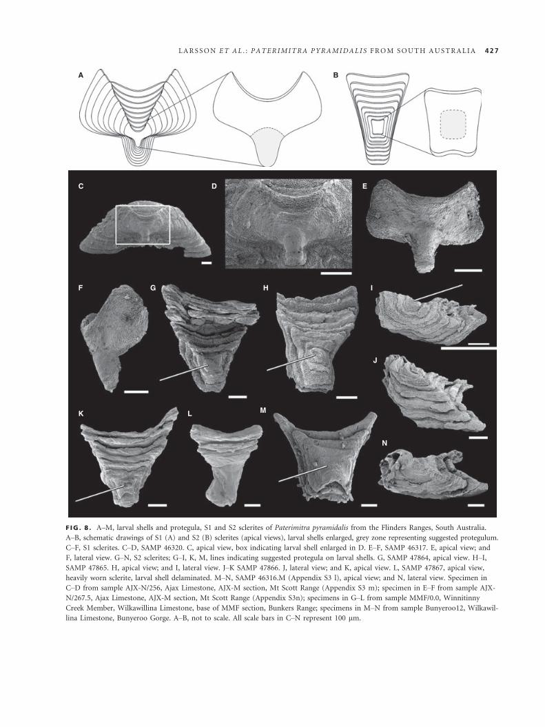
F IG . 8 . A–M, larval shells and protegula, S1 and S2 sclerites of Paterimitra pyramidalis from the Flinders Ranges, South Australia.
A–B, schematic drawings of S1 (A) and S2 (B) sclerites (apical views), larval shells enlarged, grey zone representing suggested protegulum.
C–F, S1 sclerites. C–D, SAMP 46320. C, apical view, box indicating larval shell enlarged in D. E–F, SAMP 46317. E, apical view; and
F, lateral view. G–N, S2 sclerites; G–I, K, M, lines indicating suggested protegula on larval shells. G, SAMP 47864, apical view. H–I,SAMP 47865. H, apical view; and I, lateral view. J–K SAMP 47866. J, lateral view; and K, apical view. L, SAMP 47867, apical view,
heavily worn sclerite, larval shell delaminated. M–N, SAMP 46316.M (Appendix S3 l), apical view; and N, lateral view. Specimen in
C–D from sample AJX-N/256, Ajax Limestone, AJX-M section, Mt Scott Range (Appendix S3 m); specimen in E–F from sample AJX-
N/267.5, Ajax Limestone, AJX-M section, Mt Scott Range (Appendix S3n); specimens in G–L from sample MMF/0.0, Winnitinny
Creek Member, Wilkawillina Limestone, base of MMF section, Bunkers Range; specimens in M–N from sample Bunyeroo12, Wilkawil-
lina Limestone, Bunyeroo Gorge. A–B, not to scale. All scale bars in C–N represent 100 lm.
LARSSON ET AL . : PATER IMITRA PYRAMIDAL I S FROM SOUTH AUSTRAL IA 427

rounded or rectangular plate situated at the centre of the
larval shell.
ABNORMAL GROWTH AND DAMAGE
All three sclerite types (S1, S2, and L) are very distinct in
their shapes and outlines and generally easily recognized.
However, there are some sclerites that deviate substantially
from the typical pattern of growth. Both S2 and L sclerites
can be theoretically derived from the S1 form by stretching
and twisting (L sclerites) or compression of proportions
(S2 sclerites). Some sclerites initially identified as S2 scle-
rites when studied using light microscopy revealed typical
characters comparable to the subapical flange and lateral
plates of S1 sclerites when imaged using SEM (Fig. 9A–D).In other instances, some S1 sclerites have such narrow lat-
eral plates and are almost crescent-shaped in anterior or
posterior view that they take on the shape of larger L scle-
rites (Fig. 9E–F). A few abnormal (possibly pathological)
sclerites exhibit the same mode of growth and reticulate
micro-ornament typical of Paterimitra, but cannot be
accommodated with confidence into any of the recognized
three sclerite types (Fig. 9G–M).
Several sclerites show clear evidence of disturbance in
the normal shell growth. For example, in some S1 scle-
rites, the growth of the subapical flange has been mark-
edly disturbed, resulting in a change in growth direction.
In one specimen, the flange appears to have been strongly
deformed, resulting in an almost 90 degrees shift in the
direction of growth (Fig. 10A–B). Other specimens dis-
play a modest twist of the subapical flange (Fig. 10C–F).There are also at least two S1 sclerites that have pecu-
liar internal growth disturbances (Fig. 11A–P). These
specimens show growth disturbances on the anterior
plate, close to the anterior sinus. In SAMP 47875
(Fig. 11A–F), the growth disruption forms a distinct
lump with the same texture as the internal sclerite surface
(Fig. 11B), indicating that it was secreted by the Paterimi-
tra animal during growth. This lump may have been pro-
duced to encapsulate or ‘seal off’ a foreign object
invading the living chamber from the outside. Externally,
this is indicated by a break in the growth lamellae and a
hole piercing the anterior plate (Fig. 11D–F). In SAMP
47876 (Fig. 11G–P), the S1 sclerite appears to have grown
around a circular or cylindrical element (now lost).
Whether this reflects a similar internal growth disturbance
as the one described above remains unanswered, but
judging from the uneven surface of the edge of this circu-
lar ingrowth, something has been broken off (Fig. 11G–K). Exteriorly, the inner growth disturbance appears to be
opposed by an additional L-like sclerite attached along
the anterior sinus of SAMP 47877 (Fig. 11J, L–P).
ARTICULATED SCLERITOMECOMPOSITES
There are 12 partly articulated specimens of Paterimitra
in available collections. In most cases where sclerites have
been preserved as articulated elements (n = 10), the spec-
imens exhibit a fragmentary composite consisting of one
saddle-shaped S2 sclerite nested within the triangular
notch of a pyramidal S1 sclerite (Figs 12, 13). Of the
remaining partly articulated specimens, one specimen rep-
resents the association of a S1 sclerite with multiple L
sclerites, while a second specimen is composed of L scle-
rites only. Both specimens are described more fully below.
In the preserved S1-S2 composite specimens, the subapi-
cal flange of the S1 sclerite is opposed to the margin of
the upturned flange in the S2 sclerite forming a (in some
specimens almost perfectly circular) posterior opening
(Figs 12, 13A–B, D–E). As illustrated in the study by
Skovsted et al. (2009a, fig. 1.1n, o), fusion of the S1 and
S2 sclerites produces a drawn-out tube-like structure
extending from the posterior margin of the S1 sclerite
(Fig. 13C, F). However, especially in larger specimens, the
triangular notch of the S1 sclerite is not fully covered by
the saddle-shaped S2 sclerite, and a shallow posterior
sinus is formed behind it (Figs 12, 13A). All articulated
specimens show signs of deformation or growth irregular-
ities: either the subapical flange of the S1 sclerite and/or
the upturned flange of the S2 sclerite has been twisted
(Fig. 14A–H), or the S2 sclerite appears to have cracked
F IG . 9 . A–M, sclerites with deviating features and odd specimens of Paterimitra pyramidalis from the Flinders Ranges, South Austra-
lia. A–B, SAMP 47845, S1 sclerite with unusually large subapical flange. A, apical view; and B, lateral view. C–D, SAMP 47868,
deformed S1 sclerite. C, apical view; and D, lateral view. E, SAMP 47869, S1 sclerite, apical view, unusually wide and high, lateral
plates so narrow that it resembles an L sclerite. G–J, SAMP 47870, sclerite of unsure affinity. G, apical view; H, lateral view; I, posterior
view; and J, lateral view. K–L, SAMP 47861, unusually irregular L sclerite, lateral views. M, SAMP 47871, gravely disturbed sclerite,
probably S1, apical view. Specimens in A–D and G–J from sample WILK/Q, Second Plain Member, Wilkawillina Limestone, Wilkawil-
lina Gorge type section (WILK), Bunkers Graben; specimen in E from sample CR/322.8, Wirrapowie Limestone, CR/1 section, Chace
Range; specimen in F from sample CR/449, Wirrapowie Limestone, CR/1 section, Chace Range; specimen in K–L from sample WILK/
R, Second Plain Member, Wilkawillina Limestone, Wilkawillina Gorge type section (WILK), Bunkers Graben; specimen in M from
sample WILK/A, Second Plain Member, Wilkawillina Limestone, Wilkawillina Gorge type section (WILK), Bunkers Graben. All scale
bars represent 200 lm.
428 PALAEONTOLOGY , VOLUME 57
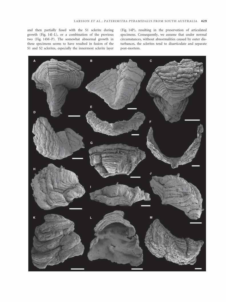
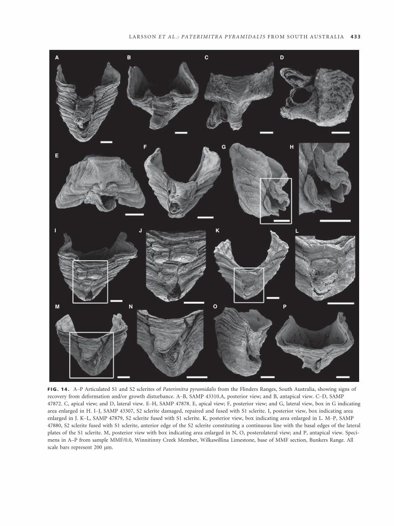
and then partially fused with the S1 sclerite during
growth (Fig. 14I–L), or a combination of the previous
two (Fig. 14M–P). The somewhat abnormal growth in
these specimens seems to have resulted in fusion of the
S1 and S2 sclerites, especially the innermost sclerite layer
(Fig. 14P), resulting in the preservation of articulated
specimens. Consequently, we assume that under normal
circumstances, without abnormalities caused by outer dis-
turbances, the sclerites tend to disarticulate and separate
post-mortem.
A B C
D
H
K L M
I
J
E
G
F
LARSSON ET AL . : PATER IMITRA PYRAMIDAL I S FROM SOUTH AUSTRAL IA 429

Almost all L sclerites in the studied collections were
found as individual units (unfused sclerites). Only one
sclerite composite composed exclusively by narrow-based
L sclerites was retrieved (Fig. 15A–F). These L sclerites
are fused along their basal margins, and each sclerite is
strongly curved, resulting in a highly recurved plate-like
structure. No ring-like L sclerite compounds as described
for Eccentrotheca helenia have been found in the available
collections, suggesting that L sclerites were generally not
naturally fused during ontogeny.
One composite specimen (SAMP 43314), briefly
described by Skovsted et al. (2009a, fig. 2f-l), exhibits
narrow-based, compressed, asymmetrical L sclerites fused
to the lateral plates of a S1 sclerite (Fig. 16). The S1 scler-
ite of this composite specimen is slightly deformed, and
the anterior sinus is not very well defined (Fig. 16A, C);
the anterior plate is swollen and partly exfoliated
(Fig. 16B, C), and the subapical flange is missing
(Fig. 16A–B, I). The posteriorly facing parts of the lateral
plates are narrow and slightly depressed with an unusually
wide triangular notch (Fig. 16A, I). In spite of the defor-
mation, there are no less than three distinct asymmetrical
L sclerites attached to left lateral plate of the subpyrami-
dal S1 sclerite (in Fig. 16B, E–G, J–K, labelled i, ii and
iii), and one small additional L sclerite is fused to the
right lateral plate of the S1 (Fig. 16D, H, J). L sclerite i
on the left side possesses three apices and is attached
directly to the anterior border of the S1 lateral plate along
the basal margin, with the apices directed towards the
posterior of the S1 sclerite (Fig. 16B–C, G, J). L sclerites
ii and iii are fused along their longitudinal axes, and the
composite overlaps the basal margin of L sclerite i and
the lateral plate of the S1 sclerite (Fig. 16B, G). The
curved shape of the basal margin of the L sclerites is
apparently accommodating the curvature of the anterior
sinus of the S1 sclerite. As described in the study by
Skovsted et al. (2009a), the internal central cavities of all
three L sclerites are easily distinguishable and show that
the sclerites were fused by secretion of sheet-like shell lay-
ers partly draping the walls between them.
There are a few additional specimens exhibiting what
appear to be small L-like sclerites attached to S1 sclerites
either posteriorly or anteriorly. In SAMP 43310 (Fig. 17A,
D), also illustrated by Skovsted et al. (2009a, fig. 1p, q), a
narrow, highly tapered sclerite is fused to the margin of
the S1 sclerite behind and lateral to the fused S2 sclerite.
As mentioned above, another specimen SAMP 47876
(Fig. 17B–C, E–F) exhibits a small, flattened and pointed
L-like sclerite attached to the anterior plate, along the
anterior sinus.
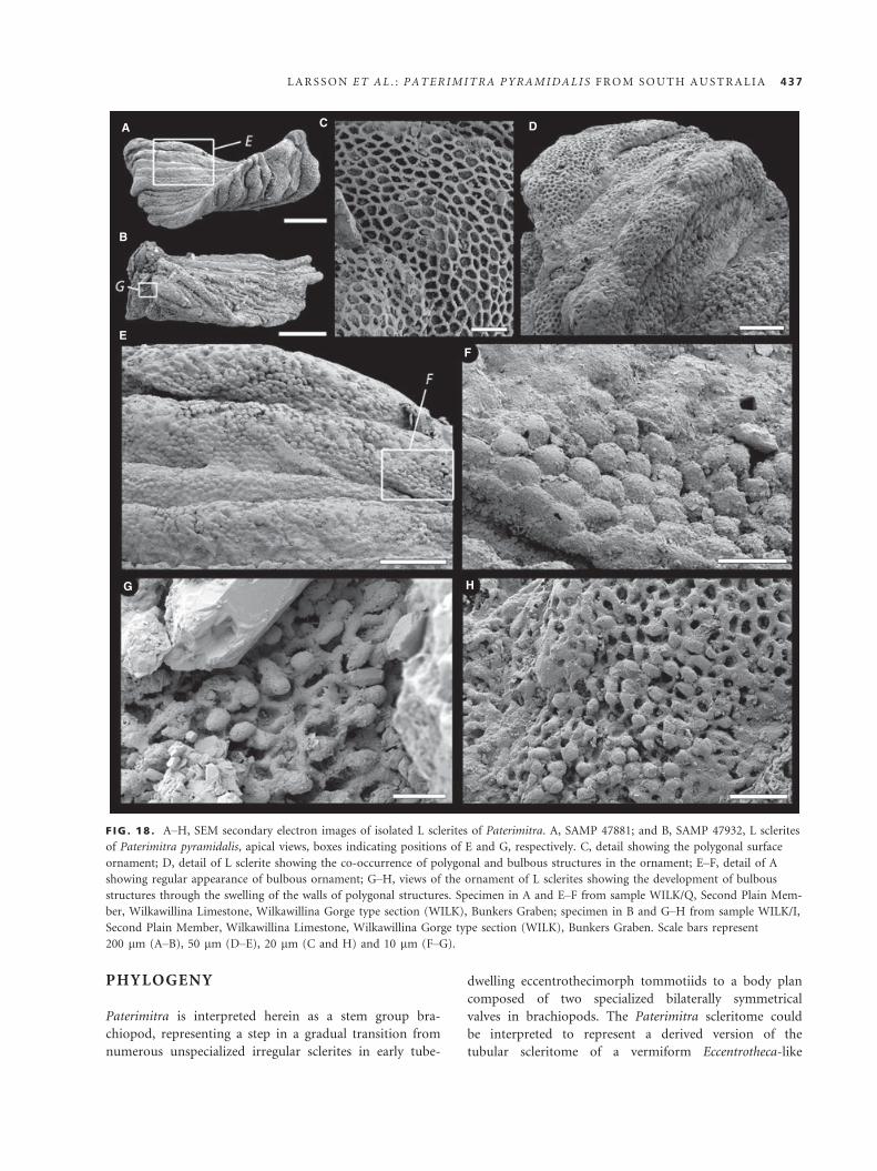
SHELL STRUCTURE AND EXTERNALORNAMENTATION
The shell of Paterimitra has a distinct primary and sec-
ondary layer. The primary layer includes an ornament of
well-defined polygonal compartments that range up to
10 lm in diameter (Fig. 18C). The walls of these com-
partments are anvil-shaped in cross section with their
external face swelling in size and show frequent spinose
to granular intrusions (Fig. 19F; see also Balthasar et al.
2009, fig. 1E). The external swelling of the walls of some
compartments can lead to their partial closure, and in L
A
C D
F
E
B
F IG . 10 . A–F, S1 sclerites of Paterimitra pyramidalis from the
Flinders Ranges, South Australia, with shifted subapical flange.
A–B, SAMP 47872, apical views. A, box indicating disturbed
posterior area with subapical flange enlarged in B (X indicating
fused, damaged S2 sclerite, for details see Fig. 14C–D); B, close-up of gravely disturbed subapical flange, showing about 90-
degree shift in growth direction (gd) in subapical flange as indi-
cated by arrows 1 (primary gd) and 2 (secondary gd). C, SAMP
47873, apical view, disturbed growth direction in subapical
flange. D–F, SAMP 47874, disturbed growth direction in subapi-
cal flange. D, apical view, box indicating area enlarged in E; E,
apical view; and F, posterior view. Specimens in A–B and D–Ffrom sample MMF/0.0, Winnitinny Creek Member, Wilkawillina
Limestone, base of MMF section, Bunkers Range; specimen in C
from sample AJX-N/368, Ajax Limestone, AJX-N section, Mt
Scott Range. All scale bars represent 100 lm.
430 PALAEONTOLOGY , VOLUME 57

sclerites, the swelling often results in bulbous structures
that can close off the underlying compartment (Fig. 18D–H). Together with a slightly convex bottom, the cross sec-
tion of these polygonal compartments is commonly bowl-
shaped (Fig. 19F).
The polygonal compartments continue into the sec-
ondary layer and commonly form pillar-like structures
normal to the shell surface (Fig. 20B). However, unlike
the primary layer, polygonal compartments within the
secondary layer are filled by a distinctly laminated
deposit of apatite (Fig. 19H–K) and appear as raised
structures with depressed walls in etched samples
(Fig. 20C). This expression of polygons extends to
include the internal surface of etched sclerites (Fig. 20D).
A B
E
F
JI
MN O
P
K L
G
H
C D
F IG . 11 . A–P, S1 sclerites of Paterimitra pyramidalis from the Flinders Ranges, South Australia, exhibiting repaired growth distur-
bance/damage close to the edge of the anterior plate. A–F, SAMP 47875, with finger-like outgrowth extending inwards from the inter-
nal side of the anterior plate. A, posterior view, box indicating area enlarged in B; B, close-up of outgrowth exhibiting similar surface
structure as the remaining internal surface of the sclerite; C, internal (antapical) view; D, anterior view, box indicating area enlarged in
E; F anteroapical view, line indicating damaged area with hole penetrating anterior plate from the exterior inwards. G–P, SAMP
47876, L sclerite-like on-growth covering growth disturbance. G, posterior view, box indicating area enlarged in H; I, internal (antapi-
cal) view, box indicating area enlarged in J; J, close-up of growth disturbance and covering sclerite; K, close-up of growth disturbance
in J, showing uneven surface around the upper part of the disturbance; L, lateral view, box indicating area with L sclerite-like on-
growth enlarged in M; N, anterior view, box indicating area enlarged in O; P, apical view. Specimens in A–P from sample MMF/0.0,
Winnitinny Creek Member, Wilkawillina Limestone, base of MMF section, Bunkers Range. All scale bars represent 100 lm.
LARSSON ET AL . : PATER IMITRA PYRAMIDAL I S FROM SOUTH AUSTRAL IA 431
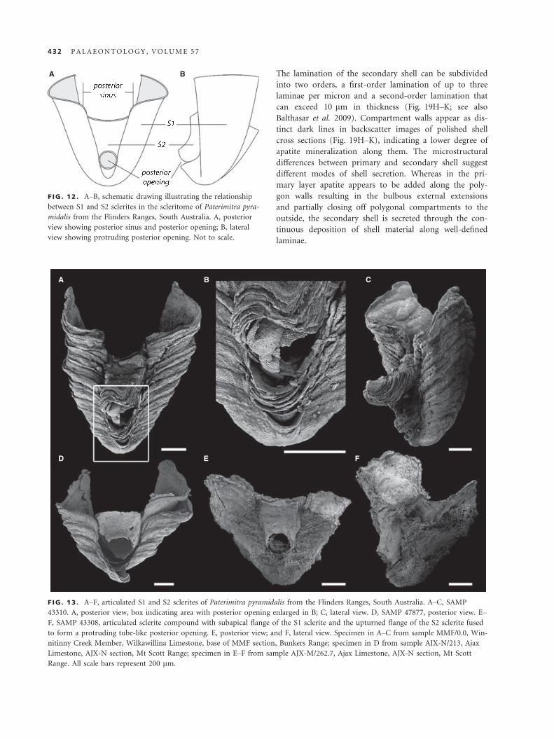
The lamination of the secondary shell can be subdivided
into two orders, a first-order lamination of up to three
laminae per micron and a second-order lamination that
can exceed 10 lm in thickness (Fig. 19H–K; see also
Balthasar et al. 2009). Compartment walls appear as dis-
tinct dark lines in backscatter images of polished shell
cross sections (Fig. 19H–K), indicating a lower degree of
apatite mineralization along them. The microstructural
differences between primary and secondary shell suggest
different modes of shell secretion. Whereas in the pri-
mary layer apatite appears to be added along the poly-
gon walls resulting in the bulbous external extensions
and partially closing off polygonal compartments to the
outside, the secondary shell is secreted through the con-
tinuous deposition of shell material along well-defined
laminae.
A B
F IG . 12 . A–B, schematic drawing illustrating the relationship
between S1 and S2 sclerites in the scleritome of Paterimitra pyra-
midalis from the Flinders Ranges, South Australia. A, posterior
view showing posterior sinus and posterior opening; B, lateral
view showing protruding posterior opening. Not to scale.
A
D E F
B C
F IG . 13 . A–F, articulated S1 and S2 sclerites of Paterimitra pyramidalis from the Flinders Ranges, South Australia. A–C, SAMP
43310. A, posterior view, box indicating area with posterior opening enlarged in B; C, lateral view. D, SAMP 47877, posterior view. E–F, SAMP 43308, articulated sclerite compound with subapical flange of the S1 sclerite and the upturned flange of the S2 sclerite fused
to form a protruding tube-like posterior opening. E, posterior view; and F, lateral view. Specimen in A–C from sample MMF/0.0, Win-
nitinny Creek Member, Wilkawillina Limestone, base of MMF section, Bunkers Range; specimen in D from sample AJX-N/213, Ajax
Limestone, AJX-N section, Mt Scott Range; specimen in E–F from sample AJX-M/262.7, Ajax Limestone, AJX-N section, Mt Scott
Range. All scale bars represent 200 lm.
432 PALAEONTOLOGY , VOLUME 57
A
E
F G H
I J K L
M N O P
B C D
F IG . 14 . A–P Articulated S1 and S2 sclerites of Paterimitra pyramidalis from the Flinders Ranges, South Australia, showing signs of
recovery from deformation and/or growth disturbance. A–B, SAMP 43310.A, posterior view; and B, antapical view. C–D, SAMP
47872. C, apical view; and D, lateral view. E–H, SAMP 47878. E, apical view; F, posterior view; and G, lateral view, box in G indicating
area enlarged in H. I–J, SAMP 43307, S2 sclerite damaged, repaired and fused with S1 sclerite. I, posterior view, box indicating area
enlarged in J. K–L, SAMP 47879, S2 sclerite fused with S1 sclerite. K, posterior view, box indicating area enlarged in L. M–P, SAMP
47880, S2 sclerite fused with S1 sclerite, anterior edge of the S2 sclerite constituting a continuous line with the basal edges of the lateral
plates of the S1 sclerite. M, posterior view with box indicating area enlarged in N, O, posterolateral view; and P, antapical view. Speci-
mens in A–P from sample MMF/0.0, Winnitinny Creek Member, Wilkawillina Limestone, base of MMF section, Bunkers Range. All
scale bars represent 200 lm.
LARSSON ET AL . : PATER IMITRA PYRAMIDAL I S FROM SOUTH AUSTRAL IA 433

Along the external surface, the shell sometimes peels
off to form fringes of primary and secondary shell. In
cross section, these fringes have the characteristic micro-
ornament of polygonal compartments followed by about
10 lm of secondary shell, the equivalent of second-order
lamina (Fig. 19A, G, E). When cut transversely and proxi-
mally, these fringes can give the impression of longitudi-
nal cavities with a base of polygonal ornament (Fig. 19E).
At the lateral margins of S1 sclerites, the shell lamination
commonly flares out and twists inwards giving a hooked
appearance in cross section (Fig. 19A, C–D, J). In some
of the studied S1 sclerites, the lateral margins were split
with finger-like extensions (Fig. 19D). The overall exter-
nal surface of S1 sclerites frequently shows irregular bul-
bous and often fringed outgrowths (Fig. 19A–C), whereasthe internal surface is much smoother and only in some
specimens showed minor changes in growth that corre-
sponded to external irregularities (Figs 4B, 11, 13C).
FUNCTIONAL MORPHOLOGY ANDLIFE-HABIT
Paterimitra pyramidalis represents only the second tom-
motiid taxon where articulated material has been recov-
ered. It is evident that the S1 and S2 sclerites were
intimately associated, with the S2 nested within the trian-
gular notch of the S1 to form the basal part of the scleri-
tome (Figs 12–14, 21). The confluence between the
upturned flange on the S2 sclerite and the subapical
flange of the S1 sclerite (Figs 12, 13) forms a near-circular
posterior opening, which Skovsted et al. (2009a, p. 5)
suggested was most likely occupied by some kind of short
attachment structure, equivalent to a pedicle (Fig. 21).
Evidence from rare articulated specimens (Figs 15, 16,
21) indicates that the narrow-based L sclerites were
arranged as closely spaced, imbricating sets along the
margins of the lateral plates and the anterior and poster-
ior sinuses of the S1/S2 unit, producing an open tube-like
construction (Fig. 21). Broad-based, more arched L scle-
rites probably dominated the distal portion of this tube,
although direct evidence is not available. The soft parts of
the organism (including a presumed lophophore-like
feeding apparatus) would have been housed within the
central cavity formed by the tubiform scleritome.
Although no ring-like elements such as in Eccentrotheca
are associated with the Paterimitra scleritome, the mode
of growth characterized by limited increase in width in all
sclerite types, but continuous increase in length in the S2
and height in the S1 and L sclerites, favours the forma-
tion of an elongated scleritome, supporting the interpreta-
tion of a fixed, tube-dwelling mode of life for this
organism. The absence of articulated broad-based, highly
arched L sclerites in the studied material suggests that L
A
DE F
B C
F IG . 15 . A–F, plate-like sclerite compound consisting of curled up/bent L sclerites of Paterimitra pyramidalis from the Flinders
Ranges, South Australia, SAMP 47862. A, lateral view with box indicating area enlarged in D, B, lateral view; and C, basal view; E, pla-
nar view, box in E indicating area enlarged in F. Specimen in A–F from sample AJX-N/213, Ajax Limestone, AJX-N section, Mt Scott
Range. Scale bars represent 200 lm (A–E) and 50 lm (F).
434 PALAEONTOLOGY , VOLUME 57
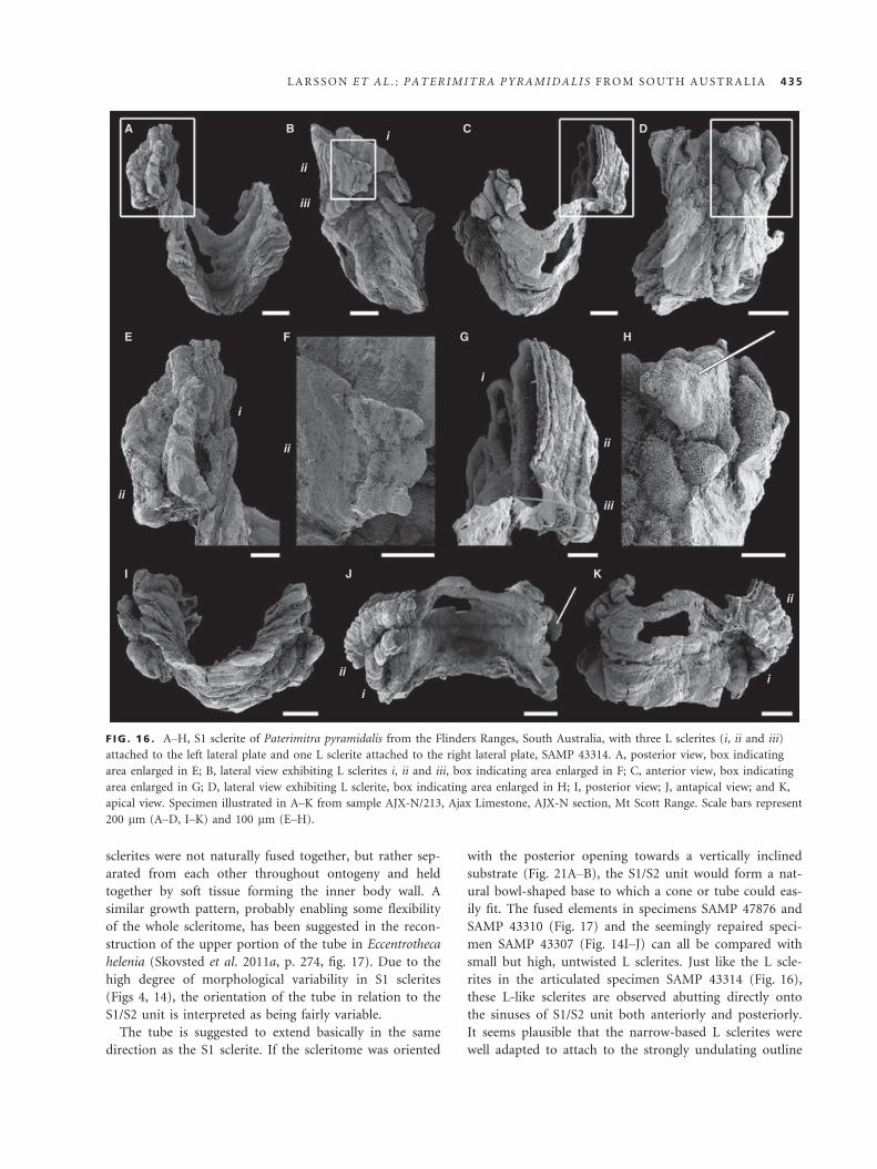
sclerites were not naturally fused together, but rather sep-
arated from each other throughout ontogeny and held
together by soft tissue forming the inner body wall. A
similar growth pattern, probably enabling some flexibility
of the whole scleritome, has been suggested in the recon-
struction of the upper portion of the tube in Eccentrotheca
helenia (Skovsted et al. 2011a, p. 274, fig. 17). Due to the
high degree of morphological variability in S1 sclerites
(Figs 4, 14), the orientation of the tube in relation to the
S1/S2 unit is interpreted as being fairly variable.
The tube is suggested to extend basically in the same
direction as the S1 sclerite. If the scleritome was oriented
with the posterior opening towards a vertically inclined
substrate (Fig. 21A–B), the S1/S2 unit would form a nat-
ural bowl-shaped base to which a cone or tube could eas-
ily fit. The fused elements in specimens SAMP 47876 and
SAMP 43310 (Fig. 17) and the seemingly repaired speci-
men SAMP 43307 (Fig. 14I–J) can all be compared with
small but high, untwisted L sclerites. Just like the L scle-
rites in the articulated specimen SAMP 43314 (Fig. 16),
these L-like sclerites are observed abutting directly onto
the sinuses of S1/S2 unit both anteriorly and posteriorly.
It seems plausible that the narrow-based L sclerites were
well adapted to attach to the strongly undulating outline
A B C D
E
I J
ii
ii
i
ii
i
i
ii
ii
i
iii
iiiii
i
K
F G H
F IG . 16 . A–H, S1 sclerite of Paterimitra pyramidalis from the Flinders Ranges, South Australia, with three L sclerites (i, ii and iii)
attached to the left lateral plate and one L sclerite attached to the right lateral plate, SAMP 43314. A, posterior view, box indicating
area enlarged in E; B, lateral view exhibiting L sclerites i, ii and iii, box indicating area enlarged in F; C, anterior view, box indicating
area enlarged in G; D, lateral view exhibiting L sclerite, box indicating area enlarged in H; I, posterior view; J, antapical view; and K,
apical view. Specimen illustrated in A–K from sample AJX-N/213, Ajax Limestone, AJX-N section, Mt Scott Range. Scale bars represent
200 lm (A–D, I–K) and 100 lm (E–H).
LARSSON ET AL . : PATER IMITRA PYRAMIDAL I S FROM SOUTH AUSTRAL IA 435

formed by the lateral plates and sinuses of the S1/S2 unit
and to cover uneven areas in a mosaic manner. This
would adapt the outline to the curvature of the elongate
and strongly arched L sclerites, which would be more
suitable for lining a tubular extension. Considering this, it
is possible to argue the general pattern to be that high,
narrow L sclerites were more proximally associated with
the S sclerite complex (S1 + S2) attaching to the anterior
and posterior sinuses, respectively, followed successively
in subsequent rows by bigger ones; low, broad L sclerites
occupying the more distal parts of the assumed (more or
less conical) tube, not fusing with each other to the same
extent as the high ones. This would enable the arrange-
ment of large L sclerites into ring-like structures around
the tube, slightly inclined in an Eccentrotheca-like manner,
preferably towards the anterior (Fig. 21A), which in turn
might explain the twisting of large L sclerites in both
directions. Twisting and curling of sclerites, which then
end up folding down close/parallel to the tube/cone sur-
face instead of standing out at a right angle from the
body wall, could offer advantages by reducing the width
of the organism, which would be favourable in competi-
tion for limited space and enhance balancing of the tube/
cone in the water column. The twisting and folding of
sclerites would also make them less vulnerable to
mechanical breakage.
Skovsted et al. (2011a) suggested that E. helenia was
attached and hanging as pendant forms from ceilings and
crevices in cryptic environments or attached to the lateral
sides of large archaeocyaths/spongiomorphs: an assump-
tion based on comparisons with the problematic calcare-
ous cribricyaths (elongate horn-shaped tubes inhabiting
cryptic reef environments) and the fact that E. helenia co-
occur with archaeocyaths (although not yet found in
situ). The ecology of Paterimitra is more complex than
that of Eccentrotheca. Paterimitra inhabited the same types
of biohermal environments as E. helenia and was thus
exposed to the same selective pressures. However, Pater-
imitra occurs in a wider variety of carbonate-dominated
facies, including low-energy restricted lagoonal systems
and open carbonate sand environments. Paterimitra is
more abundant in biohermal environments, but is not
restricted to such environments. The variation and defor-
mation observed in sclerites of Paterimitra (in particular
S1 sclerites) likely reflect crowding and variation in sub-
strate.
A B C
D E F
F IG . 17 . A–F, other L-like sclerites fused to S1 sclerites of Paterimitra pyramidalis from the Flinders Ranges, South Australia. A, D,
SAMP 43310, L-like sclerite fused to base of lateral plate. A, posteroapical view, box indicating area enlarged in D. B–C, E–F, SAMP
47876, L-like sclerite fused to anterior plate. B, anterior view, box indicating area enlarged in E; C, lateral view, box indicating area
enlarged in F. Specimens in A–F from sample MMF/0.0, Winnitinny Creek Member, Wilkawillina Limestone, base of MMF section,
Bunkers Range. All scale bars represent 100 lm.
436 PALAEONTOLOGY , VOLUME 57
PHYLOGENY
Paterimitra is interpreted herein as a stem group bra-
chiopod, representing a step in a gradual transition from
numerous unspecialized irregular sclerites in early tube-
dwelling eccentrothecimorph tommotiids to a body plan
composed of two specialized bilaterally symmetrical
valves in brachiopods. The Paterimitra scleritome could
be interpreted to represent a derived version of the
tubular scleritome of a vermiform Eccentrotheca-like
A C D
B
E
G H
F
F IG . 18 . A–H, SEM secondary electron images of isolated L sclerites of Paterimitra. A, SAMP 47881; and B, SAMP 47932, L sclerites
of Paterimitra pyramidalis, apical views, boxes indicating positions of E and G, respectively. C, detail showing the polygonal surface
ornament; D, detail of L sclerite showing the co-occurrence of polygonal and bulbous structures in the ornament; E–F, detail of Ashowing regular appearance of bulbous ornament; G–H, views of the ornament of L sclerites showing the development of bulbous
structures through the swelling of the walls of polygonal structures. Specimen in A and E–F from sample WILK/Q, Second Plain Mem-
ber, Wilkawillina Limestone, Wilkawillina Gorge type section (WILK), Bunkers Graben; specimen in B and G–H from sample WILK/I,
Second Plain Member, Wilkawillina Limestone, Wilkawillina Gorge type section (WILK), Bunkers Graben. Scale bars represent
200 lm (A–B), 50 lm (D–E), 20 lm (C and H) and 10 lm (F–G).
LARSSON ET AL . : PATER IMITRA PYRAMIDAL I S FROM SOUTH AUSTRAL IA 437

A1 A2 B C
D
G
H
JK
I
E
F
438 PALAEONTOLOGY , VOLUME 57

ancestor, a more crown-ward member of the brachiopod
stem, introducing bilateral symmetry to the scleritome
coupled with reduction in number and specialization of
the basal sclerites (Fig. 22). According to this model,
the bilaterally symmetrical S1/S2 unit represents an
expanded and larger equivalent to the low, cap-shaped
plates in the basal ring of Eccentrotheca. The L sclerites
correspond to the larger set of high, laterally com-
pressed, triangular sclerites present in the upper portion
of the Eccentrotheca scleritome. However, as discussed
below, Paterimitra also show distinct similarities to the
oldest fully bivalve phosphatic-shelled brachiopods, the
paterinids, especially in terms of shell structure and
micro-ornament. Consequently, Paterimitra pyramidalis
is regarded as an intermediate between the tommotiid
Eccentrotheca and the paterinids (Fig. 22).
Paterimitra and the evolution of brachiopod shell secretion
Skeletal microstructure is widely recognized as phyloge-
netically greatly informative at high taxonomic levels
(Williams et al. 1996). To some extent, skeletal micro-
structure also records the biological regime of shell secre-
tion, particularly in modern brachiopods where newly
formed epithelial cells first secrete the organic periostra-
cum along the mantle edge and then successively change
their secretion regime to form the primary and secondary
layers (Williams 1997). Shell secretion in extant organo-
phosphatic brachiopods is characterized by the cyclic
deposition of nanometric apatite granules, glycosamino-
glycans (GAGs) and collagen or chitin (Williams et al.
1992, 1994). The nature of this depositional cycle is evi-
dent in the distinct lamination at some tens of microns
that characterize all extant and extinct microstructures of
organophosphatic brachiopods (Holmer 1989; Cusack
et al. 1999). The vastly different diagenetic stability of the
various organic compounds and apatite granules creates
an environment prone to support very early diagenetic re-
mobilization of apatite within shell laminae (Balthasar
2007). This principle of localized diagenetic fate within
different shell layers is crucial in understanding stem
group microstructures where these lack extant representa-
tives. Based on the general occurrence of cyclic deposition
in extant organophosphatic brachiopods and the charac-
teristic threefold succession of periostracum, primary and
secondary layers, it must be assumed that these founda-
tions of organophosphatic shell secretion first evolved in
the stem group.
Paterimitra is the only tommotiid that exhibits a well-
defined distinction between primary and secondary layers.
Although both primary and secondary layers are charac-
terized by a framework of presumably organic polygonal
compartments, the approach to the mineralization of this
polygonal scaffolding differs strikingly between primary
and secondary layers. Whereas the polygonal organic walls
within the primary layer were mineralized from the walls
inwards, mineralization of the secondary shell occurred
by incremental deposition of dense laminae within the
organic compartments. More than a mere microstructural
differentiation of the primary and secondary shell layers,
this difference in mineralization points towards a funda-
mental shift in mineralization strategy between the depo-
sitions of both layers.
Apart from Paterimitra, this distinctive mode of shell
secretion, with a pervasive polygonal network and differ-
ential mineralization of primary and secondary layers, is
only found in the paterinid brachiopod Askepasma tod-
dense (Balthasar et al. 2009; Topper et al. 2013; Fig. 23).
The only difference between the microstructures of
A. toddense and Paterimitra is the occasional occurrence
of elliptical cavities in A. toddense. When A. toddense and
Paterimitra co-occur, as in parts of the Flinders Ranges,
particularly smaller shell fragments are commonly indis-
tinguishable in polished cross section. Intriguingly, the
newly described Askepasma saproconcha (Topper et al.
2013) suggests that the primary layer might have been
covered by an organic periostracum, while its secondary
shell exhibits a pronounced reduction in mineralization
similar to that seen in tannuolinids.
While polygonal imprints are common among tommo-
tiids (see discussion in Conway Morris and Chen 1990),
these alone are insufficient to recognize this sequential
mode of shell secretion. Indeed, superficial polygonal
imprints are known from a variety of phosphatic fossils
including other tommotiids (see discussion in Conway
Morris and Chen 1990), but also brachiopods (e.g. Bal-
thasar 2009; Winrow and Sutton 2012) and conodonts
(Conway Morris and Harper 1988). A pervasive shell-
penetrating scaffolding of polygonal layers has only been
F IG . 19 . A–K, SEM backscatter images of polished cross sections of isolated Paterimitra S1 sclerites that were imbedded in araldite
resin. A–C, overview of specimens indicating positions of figures D–K; A1 and A2 are slices of the same specimens separated by a few
microns; D, lateral margin with a digitate hooked edge; E, portion of shell with closely spaced proximal sections of external fringes that
create the impression of internal cavities; F, detail of E showing anvil-shaped cross sections of the walls of the polygonal ornament; G,
posterior portion of sclerite with abundant fringes stacked on top of each other; H–K, details of sclerites showing the lamination with
cross sections through polygonal compartments. Scale bars represent 500 lm (A), 200 lm (B–C), 100 lm (D, G), 50 lm (E, H, J, K),
20 lm (I) and 5 lm (F).
LARSSON ET AL . : PATER IMITRA PYRAMIDAL I S FROM SOUTH AUSTRAL IA 439

detected in Askepasma, Salanygolina, Eccentrotheca, Pater-
imitra and probably tannuolinids (Ushatinskaya 1987;
Balthasar et al. 2009; Holmer et al. 2009; Topper et al.
2013).
It should be stressed that the detection of polygonal
structures of a pervasive nature is challenging. This requires
either unusually preserved material, as in the case of par-
tially silicified specimens of Salanygolina (Ushatinskaya
1987; Holmer et al. 2009), or the careful study of highly
polished cross sections using high-energy backscatter imag-
ing, a method that is largely underused but has proven to
yield unique microstructural information (Balthasar et al.
2009; Balthasar et al. 2011). In tannuolinids, the reduced
mineralization of the secondary shell further complicates
the detection of such a pervasive network of polygonal
compartments. However, the distribution of polygonal
imprints on successive internal laminae in Micrina and
Tannuolina (Conway Morris and Chen 1990; Balthasar
et al. 2009, suppl. material) suggests the existence of a
pervasive network of organic-walled compartments very
similar to eccentrothecimorph tommotiids and some
paterinids. The characteristic ‘hollowed out’ nature of the
tannuolinid shell structure (see for example Williams and
Holmer 2002; Murdock et al. 2012) is largely composed of
cement (Balthasar et al. 2009, suppl. fig. 1) derived from
remobilization of biogenic apatite within the shell.
Independent of overall morphology, skeletal micro-
structure provides further evidence for distinct tommotiid
clades. The camenellan clade as defined by morphology
lacks a pervasive polygonal framework, and members of
this clade exhibit a dense micron-scale lamination (first-
order lamination; Balthasar et al. 2009; Skovsted et al.
A
B
C
D
F IG . 20 . A–D, SEM secondary electron images of limestone-
hosted etched Paterimitra S1 sclerite. A, overview with positions
of B–D; B, detail showing how polygonal compartments form
pillar-like structures normal to the sclerite surface; C, detail
showing the pervasive occurrence of polygonal compartments
throughout the shell; D, tilted view showing the lamination and
the expression of polygonal structures on the internal surface of
the sclerite. Scale bars represent 200 lm (A) and 50 lm (B–D).
A B
F IG . 21 . A–B, hypothetical reconstruction of Paterimitra pyra-
midalis, schematic drawing illustrating the association between
S1 and S2 sclerites, orientation of the L sclerite tube relative to
the S1/S2 unit and the general orientation of the scleritome rela-
tive to the substrate. A, lateral view, scleritome oriented in right
angle to a vertical substrate, L sclerite tube following the general
vertical direction of the S1/S2 unit; and B, posterior view. Not
to scale.
440 PALAEONTOLOGY , VOLUME 57
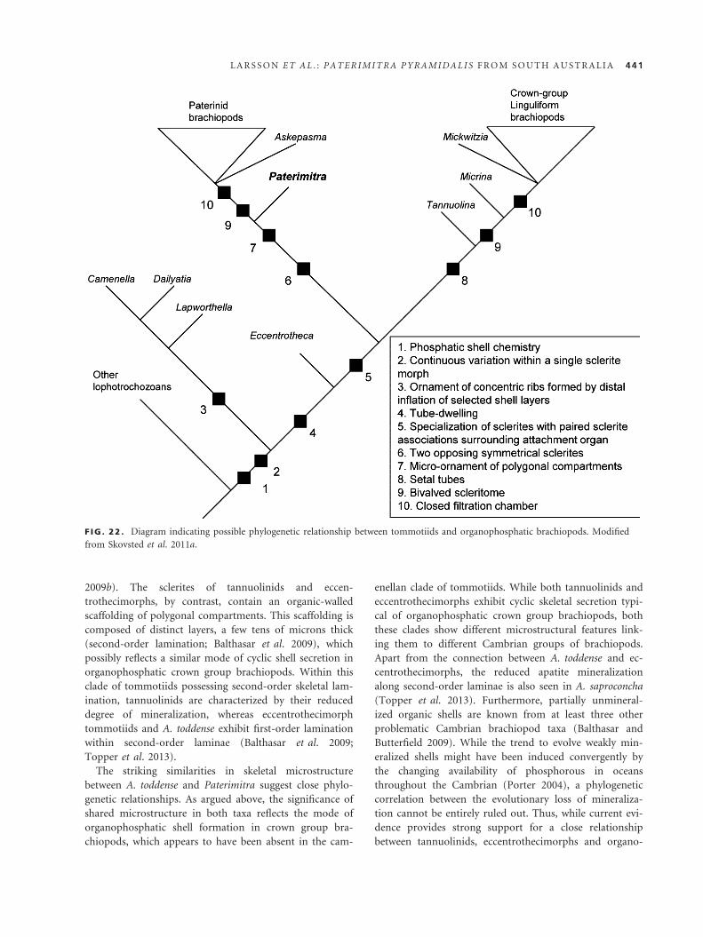
2009b). The sclerites of tannuolinids and eccen-
trothecimorphs, by contrast, contain an organic-walled
scaffolding of polygonal compartments. This scaffolding is
composed of distinct layers, a few tens of microns thick
(second-order lamination; Balthasar et al. 2009), which
possibly reflects a similar mode of cyclic shell secretion in
organophosphatic crown group brachiopods. Within this
clade of tommotiids possessing second-order skeletal lam-
ination, tannuolinids are characterized by their reduced
degree of mineralization, whereas eccentrothecimorph
tommotiids and A. toddense exhibit first-order lamination
within second-order laminae (Balthasar et al. 2009;
Topper et al. 2013).
The striking similarities in skeletal microstructure
between A. toddense and Paterimitra suggest close phylo-
genetic relationships. As argued above, the significance of
shared microstructure in both taxa reflects the mode of
organophosphatic shell formation in crown group bra-
chiopods, which appears to have been absent in the cam-
enellan clade of tommotiids. While both tannuolinids and
eccentrothecimorphs exhibit cyclic skeletal secretion typi-
cal of organophosphatic crown group brachiopods, both
these clades show different microstructural features link-
ing them to different Cambrian groups of brachiopods.
Apart from the connection between A. toddense and ec-
centrothecimorphs, the reduced apatite mineralization
along second-order laminae is also seen in A. saproconcha
(Topper et al. 2013). Furthermore, partially unmineral-
ized organic shells are known from at least three other
problematic Cambrian brachiopod taxa (Balthasar and
Butterfield 2009). While the trend to evolve weakly min-
eralized shells might have been induced convergently by
the changing availability of phosphorous in oceans
throughout the Cambrian (Porter 2004), a phylogenetic
correlation between the evolutionary loss of mineraliza-
tion cannot be entirely ruled out. Thus, while current evi-
dence provides strong support for a close relationship
between tannuolinids, eccentrothecimorphs and organo-
F IG . 22 . Diagram indicating possible phylogenetic relationship between tommotiids and organophosphatic brachiopods. Modified
from Skovsted et al. 2011a.
LARSSON ET AL . : PATER IMITRA PYRAMIDAL I S FROM SOUTH AUSTRAL IA 441

phosphatic brachiopods, microstructure alone does cur-
rently not resolve, which tommotiid clade was most prox-
imal to brachiopods.
With respect to the origin of calcareous shells in bra-
chiopods, the existence of a pervasive organic framework
might well represent a precursor of the organic envelopes
that define the shape of calcite fibres in rhynchonelliform
brachiopods as proposed by Holmer et al. (2009). An
alternative path of evolving calcareous shells from or-
ganophosphatic ancestors has been proposed based on
the microstructure of calcareous obolellids (Balthasar
2008), which appear to lack well-developed elongate cal-
cite fibres with organic envelopes, a condition closer to
the microstructure of modern craniids. While there
remains little doubt that the original shell composition of
the brachiopod–tommotiid clade was organophosphatic,
the evolution of calcareous shells from these origins
might have occurred independently along the stems of
the two calcareous subphyla.
Identification of brachiopod valve homologies and ontogeny
in Paterimitra
The bilateral symmetry of the basal sclerites (S1 and S2)
of Paterimitra suggests that these sclerites, in particular in
the context of the close similarities in shell ultrastructure
and proposed phylogenetic relationship between Paterimi-
tra and paterinid brachiopods, are direct homologues of
brachiopod valves. In particular, the S1 sclerite exhibits
similarities to the ventral valves of paterinids. The sub-
apical flange of the S1 sclerite shows strong resemblance
to the ventral homeodeltidium of a young adult Micromi-
tra (compare Fig. 4A to Williams et al. 1998, pl. 9,
fig. 1). The anterior plate of the S1 sclerite is also
remarkably similar to the colleplax of the ventral valve of
Salanygolina (compare Fig. 3A to Holmer et al. 2009,
text-fig. 2B). And the larval shell of the Paterimitra S1 is
very closely comparable to the transversely elongate bre-
phic shell of the ventral valve in Salanygolina, with the
protegulum and incipient subapical flange in Paterimitra
in the same position as the posterior delthyrium-like pro-
jection of the ventral brephic shell in Salanygolina (com-
pare Fig. 8D to Holmer et al. 2009, text-fig. 4B).
Available evidence thus suggests that the S1 sclerite of
Paterimitra is homologous to the ventral valve of pateri-
nid brachiopods. Logically, the S2 sclerite could be con-
sidered to be homologous to the dorsal valve of
brachiopods, but the morphological evidence for this
interpretation is less clear. In particular, the position of
the S2 sclerite, nested within the triangular notch (inter-
area?) of the S1 sclerite, is difficult to compare with the
position of the dorsal valve in paterinids or other bra-
chiopods. However, the presence of a well-defined larval
shell and the association of the posterior margin of the
S2 sclerite with the pedicle-like attachment structure may
support the proposed homology.
As suggested by Holmer et al. (2011), the presence of
brachiopod-like larval shells in the opposing symmetrical
sclerites suggests that the larvae of Paterimitra were
effectively bivalved and that L sclerites were added
during later stages of ontogeny, perhaps starting only
after settlement. Consequently, the bivalved condition in
brachiopods may be a retained juvenile character,
and the bivalved brachiopod body plan, a result of
paedomorphosis.
SYSTEMATIC PALAEONTOLOGY
Repository. All illustrated specimens are deposited in
South Australian Museum, Adelaide (SAMP). Original
type specimens are deposited in Commonwealth Palaeon-
tological Collection, Australian Geological Survey Organi-
sation, Canberra (CPC).
A
B
C
F IG . 23 . A–C, SEM secondary electron images of limestone-
hosted etched Askepasma toddense. A, overview showing the
positions B and C, which show the shell lamination and the nat-
ure of the distribution of polygonal structures. Scale bars repre-
sent 1 mm (A), 50 lm (B) and 20 lm (C).
442 PALAEONTOLOGY , VOLUME 57

Order TOMMOTIIDA Missarzhevsky, 1970
Remarks. For a thorough discussion regarding the Order
Tommotiida, see Skovsted et al. (2011a).
Genus PATERIMITRA Laurie, 1986
Figures 2–21
*1986 Paterimitra Laurie, p. 446.
1990 Paterimitra Laurie; Conway Morris in Bengtson
et al., p. 142.
Type and only species. Paterimitra pyramidalis Laurie, 1986.
Emended diagnosis. Sclerites microscopic, apatitic by ori-
ginal composition, growing via basal-internal accretion.
Scleritome comprising at least three clearly distinctive
sclerite types: symmetrical sclerite types S1 and S2 and
laterally compressed asymmetrical L sclerites. S1 sclerites
pyramidal with posterior triangular notch and U-shaped
anterior plate. S2 sclerites concave and acutely triangular.
External surface with micro-ornament consisting of
polygonal compartments. Scleritome consisting of one S2
sclerite nested within triangular notch of one S1 sclerite;
multiple L sclerites lining antapical margins in additional
rows forming an open tube.
It differs from Eccentrotheca by the presence of two
specialized bilaterally symmetrical sclerites in the scleri-
tome and the distinct polygonal external micro-ornament.
Differs from Kulparina by more regular and less com-
pressed sclerite shape and the distinct polygonal external
micro-ornament. Differs from Sunnaginia by the presence
of individual sclerite types, the bilateral symmetry of S1
and S2 sclerites and the absence of right- and left-hand
sclerite pairs, polygonal micro-ornament and the shell
microstructure.
Discussion. Laurie (1986) suggested that Ambonychia
macroptera Tate, 1892, and Plate type III of Dailyatia ajax
Bischoff, 1976, could be referred to the genus Paterimitra
as ?P. macroptera, based on the external micro-ornament,
which differs from the type species of Dailyatia, D. ajax
Bischoff, 1976, and resembles that of Paterimitra pyrami-
dalis. This suggestion was questioned by Evans and Ro-
well (1990), who argued that although there are indeed
similarities to Paterimitra, the external micro-ornament
of A. macroptera shows strong similarities to the Antarc-
tic species Dailyatia bradocki Evans and Rowell, 1990,
and Dailyatia odyssey Evans and Rowell, 1990. A. macrop-
tera, hence, probably represents a poorly known species
of Dailyatia. Unpublished data from South Australia
(Skovsted pers. obs. 2012) support this interpretation.
Consequently, we consider the genus Paterimitra to be
monotypic.
Paterimitra pyramidalis Laurie, 1986
Figures 2–21
1985 Problematicum A Laurie and Shergold, p. 88,
fig. 7a–b.
? 1985 Eccentrotheca sp. Laurie and Shergold, p. 88, fig. 7c.
* 1986 Paterimitra pyramidalis Laurie, p. 446, fig. 9f–j.
? 1986 Eccentrotheca cf. kanesia Landing, Nowlan and
Fletcher; Laurie, p. 446 fig. 9a–d.
1990 Paterimitra pyramidalis Laurie; Conway Morris in
Bengtson et al., p. 142, fig. 92.
2001 Paterimitra pyramidalis Laurie; Gravestock et al.,
p. 116, pl. 8, figs 10, 11.
p. 2008 Eccentrotheca sp. Skovsted et al., fig. 2a, b, figs
1a–n, 2c, d.
2009a Paterimitra sp. Skovsted et al., figs 1, 2.
2010 Paterimitra sp. Balthasar et al., figs 1b, d, e, k, 2d.
2011 Paterimitra sp. Holmer et al., text-fig. 2.
2011a Paterimitra sp. Skovsted et al., text-fig. 20.
Holotype. CPC23676; paratypes: CPC23677–23688 (Laurie
1986).
Emended diagnosis. As for genus by monotypy.
Occurrence and age. Lower Cambrian late Terreneuvian Stage 2
to Cambrian Series 2, Stage 3 (Pararaia tatei trilobite zone) of
the Wilkawillina, Wirrapowie and Ajax limestones in the Flin-
ders Ranges, South Australia. Cambrian Series 2, Stage 3 of the
Todd River Dolomite, Northern Territory. Cambrian Series 2,
Stage 3 of the Kulpara Formation, Yorke Peninsula, South Aus-
tralia. See above for details.
Acknowledgements. We thank Ian and Di Fargher, owners of An-
gorichina Station; Warren and Barbara Fargher, owners of Wir-
realpa Station; and Graham Ragless, owner of Beltana Station, for
access to field localities and accommodation. The South Austra-
lian Museum (Adelaide) is thanked for making specimens from
the Brian Daily collections available for study. Sandra Pettersson
Stolk, John Paterson, Jim Jago, James Valentine, Bo Jonak, Tho-
mas Brock, Joel Brock, Daniel Holmer, Peter Cockle, Brett Pye-
mont and Bec Smart are thanked for assistance in the field.
Financial support from the Royal Swedish Academy of Sciences
(KVA) to C.M.L, Swedish Research Council (VR) to CBS, LEH
(VR 2009-4395, 2012-1658). and UB, the Australian Research
Council Discovery Project (#DP 120104251) to GAB, the Volkswa-
gen Foundation and the EU-Synthesys to UB (SE-TAF-1001),
Macquarie University Research Grants and the Wenner-Gren
Foundation to CBS and GAB, the Carlsberg Foundation Grant to
TPT is greatly acknowledged. Uppsala University is thanked for
financing of the PhD project of CML Reviews provided by two
anonymous referees improved the quality of the final manuscript,
and for this, we are truly grateful.
LARSSON ET AL . : PATER IMITRA PYRAMIDAL I S FROM SOUTH AUSTRAL IA 443

Editor. Phil Lane
SUPPORTING INFORMATION
Additional Supporting Information may be found in the online
version of this article:
Appendix S1. Detailed description of sampled sections and
stratigraphical correlation of sampled horizons.
Appendix S2. Sclerite data.
Appendix S3. Table accounting for deviating sclerite termi-
nology and incorrect sample numbers, stratigraphic information
and SAMP numbers given in previous publications featuring
Paterimitra pyramidalis.
Appendix S4. L sclerite data.
REFERENCES
BALTHASAR, U. 2007. An Early Cambrian organophosphatic
brachiopod with calcitic granules. Palaeontology, 50, 1319–1325.
-2008. Mummpikia gen. nov. and the origin of calcitic-
shelled brachiopods. Palaeontology, 51, 263–279.-2009. The brachiopod Eoobolus from the Early Cambrian
Mural Formation (Canadian Rocky Mountains). Pal€aontologi-
sche Zeitschrift, 83, 407–418.-and BUTTERFIELD N. J. 2009. Early Cambrian “soft-
shelled” brachiopods as possible stem-group phoronids. Acta
Palaeontologica Polonica, 54, 307–314.-SKOVSTED, C. B., HOLMER, L. E. and BROCK, G.
A. 2009. Homologous skeletal secretion in tommotiids and
brachiopods. Geology, 37, 1143–1146.-CUSACK, M., FARYMA, L., CHUNG, P., HOLMER,
L. E., J IN , J., PERCIVAL, I. G. and POPOV, L. E. 2011.
Relic aragonite from Ordovician–Silurian brachiopods: implica-
tions for the evolution of calcification. Geology, 39, 967–970.BEECHER, C. E. 1891. Development of the Brachiopoda, Part
1. Introduction. American Journal of Science, Series 3, 41, 343–357.
BENGTSON, S. 1970. The Lower Cambrian fossil Tommotia.
Lethaia, 3, 363–392.-1977. Aspects of problematic fossils in the early Palaeozoic.
Acta Universitatis Upsaliensis, Abstracts of Uppsala Dissertations
from the Faculty of Science 415, 71 pp.
-CONWAY MORRIS , S., COOPER, B. J., JELL , P. A.
and RUNNEGAR, B. N. 1990. Early Cambrian fossils from
South Australia. Memoirs of the Association of Australasian
Palaeontologists, 9, 1–364.BENTON, M. J. and DONOGHUE, P. C. J. 2007. Paleonto-
logical evidence to date the tree of life. Molecular Biology and
Evolution, 24, 26–53.BISCHOFF, G. C. O. 1976. Dailyatia, a new genus of the
Tommotiidae from Cambrian strata of SE Australia (Crusta-
cea, Cirripedia). Senckenbergiana Lethaea, 57, 1–33.BRASIER, M. D. 1976. Early Cambrian intergrowths of ar-
chaeocyathids, Renalcis, and pseudostromatolites from South
Australia. Palaeontology, 19, 223–245.
BUDD, G. E. and JENSEN, S. 2000. A critical reappraisal of
the fossil record of the bilaterian phyla. Biological Reviews, 75,
253–295.CARLSON, S. J. 2001. Ghosts of the past, present and future in
brachiopod systematics. Journal of Paleontology, 75, 1109–1118.-1990. An Early Cambrian carbonate platform near Wilka-
willina Gorge, South Australia. Australian Journal of Earth
Sciences, 37, 471–483.COHEN, B. L. 2000. Monophyly of brachiopods and phoro-
nids; reconciliation of molecular evidence with Linnean classi-
fication (the subphylum Phoroniformea nov.). Proceedings of
the Royal Society of London B, 267, 226–231.-and WEYDMANN A. 2005. Molecular evidence that
phoronids are a subtaxon of brachiopods (Brachiopoda:
Phoronata) and that genetic divergence of metazoan phyla
began long before the Early Cambrian. Organisms, Diversity
and Evolution, 5, 253–273.CONWAY MORRIS , S. and CHEN, M. 1990. Tommotiids
from the lower Cambrian of South China. Journal of Paleon-
tology, 64, 169–184.-and HARPER E. 1988. Genome size in conodonts (Chor-
data): inferred variations during 270 million years. Science,
241, 1230–1232.-and PEEL J. S. 1995. Articulated Halkieriids from the
Lower Cambrian of North Greenland and their role in early
protosome evolution. Philosophical Transactions of the Royal
Society of London, Series B, 347, 305–358.CUSACK, M., WILLIAMS, A. and BUCKMAN, J. O.
1999. Chemico-structural evolution of linguloid brachiopod
shells. Palaeontology, 42, 799–840.DEMIDENKO, YU. E. 2004. New data on the sclerite mor-
phology of the tommotiid species Lapworthella fasciculata.
Paleonotological Journal, 38, 134–140.ESAKOVA, N. V. and ZHEGALLO, E. A. 1996. Biostratigra-
phy and fauna of the lower Cambrian of Mongolia. Trudy,
Sovmestnaya Rossiysko-Mongol’skaya Paleontologicheskaya Ek-
speditsiya, 46, 214 pp. [In Russian].
EVANS, K. R. and ROWELL, A. J. 1990. Small shelly fossils
from Antarctica: an Early Cambrian faunal connection with
Australia. Journal of Paleontology, 64, 692–700.FONIN, V. D. and SMIRNOVA, T. N. 1967. New group of
problematic Early Cambrian organisms and methods of pre-
paring them. Paleontological Journal, 1967, 7–18.GIRIBET, G., DUNN, C. W., EDGECOMBE, G. D., HEJ-
NOL, A., MARTINDALE, M. Q. and ROUSE, G. W.
2009. Assembling the spiralian tree of life. 52–64. In TEL-
FORD, M. J. and LITTLEWOOD, D. T. J. (eds). Animal
evolution: genes, genomes, fossils and trees. Oxford University
Press, Oxford, UK, 264 pp.
GOULD, S. J. and CALLOWAY, C. B. 1980. Clams and
brachiopods – ships that pass in the night. Paleobiology, 6,
383–396.-and COWLEY W. M. 1995. Arrowie Basin. 21–30. In
DREXEL, J. F. and PRIESS , W. V. (eds). The geology of
South Australia, Vol. 2: the Phanerozoic. Mines and Energy,
South Australia, Bulletin 54, 347 pp.
-and SHERGOLD J. H. 2001. Australian Early and Middle
Cambrian Sequence biostratigraphy with implications for spe-
444 PALAEONTOLOGY , VOLUME 57

cies diversity and correlation. 105–136. In ZHURAVLEV, A
YU and RIDING, R. (eds.) The ecology of the Cambrian radi-
ation. Columbia University Press, New York, 525 pp.
-ALEXANDER, E. M., DEMIDENKO, YU. E., ESAK-
OVA, N. V., HOLMER, L. E., JAGO, J. B., L IN, T.,
MELNIKOVA, L. M., PARKHAEV, P. YU., ROZA-
NOV, A. YU., USHATINSKAYA, G. T., ZANG, W-L.,
ZHEGALLO, E. A. and ZHURAVLEV, A. YU. 2001. The
Cambrian Biostratigraphy of the Stansbury Basin, South Aus-
tralia. Transaction of the Palaeontological Institute, Russian
Academy of Sciences 282, 1–344.HELMKAMPF, M., BRUCHHAUS, I. and HAUSDORF,
B. 2008. Phylogenomic analyses of lophophorates (brachio-
pods, phoronids and bryozoans) confirm the Lophotrochozoa
concept. Proceedings of the Royal Society B, 275, 1927–1933.HOLMER, L. E. 1989. Middle Ordovician phosphatic inarticu-
late brachiopods from V€asterg€otland and Dalarna, Sweden.
Fossils and Strata, 26, 1–172.-SKOVSTED, C. B. and WILLIAMS, A. 2002. A stem
group brachiopod from the Lower Cambrian – support for a
Micrina (halkieriid) ancestry. Palaeontology, 45, 875–882.--BROCK, G. A., VALENTINE, J. L. and PATER-
SON, J. R. 2008. The Early Cambrian tommotiid Micrina, a
sessile bivalved stem group brachiopod. Biology Letters, 4,
724–728.-PETTERSSON STOLK, S., SKOVSTED, C.B., BAL-
THASAR, U. and POPOV, L. E. 2009. The enigmatic Early
Cambrian Salanygolina- a stem group of rhynchonelliform
chileate brachiopods? Palaeontology, 52, 1–10.-SKOVSTED, C. B., LARSSON, C., BROCK, G. A.
and ZHANG, Z. 2011. First record of a bivalved larval shell
in Early Cambrian tommotiids and its phylogenetic signifi-
cance. Palaeontology, 54, 235–239.JAMES, N. P. and GRAVESTOCK, D. I. 1990. Lower Cam-
brian shelf and shelf margin build-ups, Flinders Ranges, South
Australia. Sedimentology, 37, 455–480.KOUCHINSKY, A., BENGTSON, S. and MURDOCK,
D. J. E. 2010. A new tannuolinid problematic from the lower
Cambrian of the Sukharikha River in northern Siberia. Acta
Palaeontologica Polonica, 55, 321–331.--RUNNEGAR, B., SKOVSTED, C., STEINER,
M. and VENDRASCO, M. 2012. Chronology of early Cam-
brian biomineralization. Geological Magazine, 149, 221–251.LANDING, E. 1984. Skeleton of Lapworthellids and the supra-
generic classification of tommotiids (early and middle Cam-
brian phosphatic problematica). Journal of Paleontology, 58,
1380–1398.-1995. Upper Placentian–Branchian Series of mainland
Nova Scotia (middle–upper lower Cambrian): faunas, paleoen-
vironments, and stratigraphical revision. Journal of Paleontol-
ogy, 69, 475–495.-NOWLAN, G. S. and FLETCHER, T. P. 1980. A
microfauna associated with Early Cambrian trilobites of the
Callavia Zone, northern Antigonish Highlands, Nova Scotia.
Canadian Journal of Earth Sciences, 17, 400–418.LAURIE, J. R. 1986. Phosphatic fauna of the Early Cambrian
Todd River Dolomite, Amadeus Basin, central Australia.
Alcheringa, 10, 431–454.
-2000. Paterinata. 147–157. In KAELSER, R. (ed.). Trea-
tise on invertebrate paleontology. Part H, Brachiopoda (Revised)
2. Geological Society of America, Boulder, and University
Press of Kansas Press, Lawrence, 423 pp.
-and SHERGOLD J. H. 1985. Phosphatic organisms and the
correlation of Early Cambrian carbonate formations in Austra-
lia. BMR Journal of Australian Geology & Geophysics, 9, 83–89.LI , G. and XIAO, S. 2004. Tannuolina and Micrina (Tannuo-
linidae) from the Lower Cambrian of eastern Yunnan, South
China, and their scleritome reconstruction. Journal of Paleon-
tology, 78, 900–913.L €UTHER, C. and BARTHOLOMAEUS, T. 1997. The phy-
logenetic position of Brachiopoda – a comparison of morpho-
logical and molecular data. Zoologica Scripta, 26, 245–253.MEEK, F. B. 1873. Lists of fossils collected. Sixth Annual Report
of the U.S. Geological Survey of the territories, embracing por-
tions of Montana, Idaho, Wyoming and Utah, 463–478.MISSARZHEVSKY, V. V. 1970. New generic name Tommo-
tia, nom. nov. Paleontologicheskiy Zhurnal, 1970, 100 pp. [In
Russian].
-1989. Oldest skeletal fossils and stratigraphy of Precam-
brian and Cambrian boundary beds. Trudy Akademia Nauk
SSSR, 443, 238 pp. [In Russian].
MURDOCK, D. J. E., DONOGHUE, P. C. J., BENGT-
SON, S. and MARONE, F. 2012. Ontogeny and micro-
structure of the enigmatic Cambrian tommotiid Sunnaginia
Missarzhevsky, 1969. Palaeontology, 55, 661–676.PASSAMANECK, Y. and HALANYCH, K. M. 2006.
Lophotrochozoan phylogeny assessed with LSU and SSU data:
evidence of lophophorate polyphyly. Molecular Phylogenetics
and Evolution, 40, 20–28.PATERSON, J. R. and BROCK, G. A. 2007. Early Cambrian
trilobites from Angorichina, Flinders Ranges, South Australia,
with a new assemblage from the Pararaia bunyerooensis Zone.
Journal of Paleontology, 81, 116–142.PORTER, S. M. 2004. Closing the phosphatization window:
testing for the influence of taphonomic megabias on the pat-
tern of small shelly fossil decline. Palaios, 19, 178–183.POULSEN, C. 1967. Fossils from the Lower Cambrian of
Bornholm. Matematisk-Fysiske Meddelelser udgivet Af Det Kon-
gelige Danske Videnskabernes Selskab, 36, 1–48.QIAN, YI and BENGTSON, S. 1989. Palaeontology and bio-
stratigraphy of the Early Cambrian Meishucunian Stage in
Yunnan Province, South China. Fossils and Strata, 24, 156 pp.
ROZANOV, A. YU., MISSARZHEVSKY, V. V., VOLK-
OVA, N. A., VORONOVA, L. C., KRYLOV, I. N., KEL-
LER, B. M., KOROLYUK, I. K., LENDZION, K.,
MICHNIAK, R., PYKHOVA, N. G. and SIDOROV, A.
D. 1969. The Tommotian Stage and the Cambrian lower
Boundary problem. Trudy Geologocheskogo Instituta Akademia
Nauk SSSR, 206, 380 pp. [In Russian].
SANTAGATA, S. and COHEN, B. L. 2009. Phoronid phy-
logenetics (Brachiopoda; Phoronata): evidence from morpho-
logical cladistics, small and large subunit rDNA sequences,
and mitochondrial cox1. Zoological Journal of the Linnean
Society, 157, 34–50.SKOVSTED, C. B., BROCK, G. A., PATERSON, J. R.,
HOLMER, L. E. and BUDD, G. E. 2008. The scleritome of
LARSSON ET AL . : PATER IMITRA PYRAMIDAL I S FROM SOUTH AUSTRAL IA 445
Eccentrotheca from the Lower Cambrian of South Australia:
Lophophorate affinities and implications for tommotiid phy-
logeny. Geology, 36, 171–174.-HOLMER, L. E., LARSSON, C. M., H €OGSTR €OM,
A. E. S., BROCK, G. A., TOPPER, T. P., BALTHASAR,
U., PETTERSON STOLK, S. and PATERSON, J. R.
2009a. The scleritome of Paterimitra: an Early Cambrian stem
group brachiopod from South Australia. Proceedings of the
Royal Society of London B, 276, 1651–1656.-BALTHASAR, U., BROCK, G. A. and PATERSON,
J. R. 2009b. The tommotiid Camenella reticulosa from the
early Cambrian of South Australia: morphology, scleritome
reconstruction, and phylogeny. Acta Palaeontologica Polonica,
54, 525–540.-BROCK, G. A., HOLMER, L. E. and PATERSON,
J. R. 2009c. First report of the early Cambrian stem group bra-
chiopod Mickwitzia from East Gondwana. Gondwana Research,
16, 145–150.--TOPPER, T. P., PATERSON, J. R. and HOL-
MER, L. E. 2011a. Scleritome construction, biofacies, biostra-
tigraphy and systematics of the tommotiid Eccentrotheca
helenia sp. nov. from the early Cambrian of South Australia.
Palaeontology, 54, 253–286.---2011b. Sclerite fusion in the problematic early
Cambrian spine-like fossil Stoibostrombus from South Austra-
lia. Bulletin of Geosciences 86, 651–658.SPERLING, E. A., PISANI , D. and PETERSON, K. J.
2011. Molecular paleobiological insights into the origin of the
Brachiopoda. Evolution & Development, 13, 290–303.TATE, R. 1892. The Cambrian fossils of South Australia.
Transactions and Proceedings of the Royal Society of South Aus-
tralia, 15, 183–189.TOPPER, T. P., SKOVSTED, C. B., BROCK, G. A. and
PATERSON, J. R. 2011a. The oldest bivalved arthropods
from the early Cambrian of East Gondwana: systematics, bio-
stratigraphy and biogeography. Gondwana Research, 19(1:SI),
310–326.----2011b. Microdictyon plates from the lower
Cambrian of South Australia: implications for species taxon-
omy and diversity. Alcheringa 35, 427–443.-HOLMER, L. E., SKOVSTED, C. B., BROCK, G. A.,
BALTHASAR, U., LARSSON, C. M., PETTERSSON
STOLK, S. and HARPER, D. A. T. 2013. The oldest bra-
chiopods from the lower Cambrian of South Australia. Acta
Palaeontologica Polonica, 58, 93–109.USHATINSKAYA, G. T. 1987. Unusual inarticulate brachio-
pods from the Lower Cambrian of Mongolia. Paleontologiches-
kiy Zhurnal, 2, 62–68.-2001. Brachiopods. 350–369. In ZHURAVLEV, A. YU
and RIDING, R. (eds.) The ecology of the Cambrian radia-
tion. Columbia University Press, New York, 525 pp.
-2002. Genus Micrina (small shelly fossils) from the Lower
Cambrian of South Australia: morphology, microstructures,
and possible relation to halkieriids. Paleontological Journal, 36,
9–19.-2008. Origin and dispersal of the earliest brachiopods.
Paleontological Journal, 42, 776–791.WILLIAMS, A. 1997. Shell structure. H267–H320. In KAES-
LER, R. L. (ed.). Treatise on invertebrate paleontology, Part
H. Brachiopoda Revised 1. Geological Society of America,
Boulder, CO, and University of Kansas Press, Lawrence, KS,
539 pp.
-and HOLMER L. E. 2002. Shell structure and inferred
growth, functions and affinities of the sclerites of the problem-
atic Micrina. Palaeontology, 45, 845–873.-MACKAY, S. and CUSACK, M. 1992. Structure of the
organo-phosphatic shell of the brachiopod Discina. Philosophi-
cal Transactions of the Royal Society of London, Series B, 337,
83–104.---1994. Collagenous chitinophosphatic shell of the
brachiopod Lingula. Philosophical Transactions of the Royal
Society of London Series B, 364, 223–266.-CARLSON, S. J., BRUNTON, C. H. C. and HOL-
MER, L. E. 1996. A supra-ordinal classification of the Bra-
chiopoda. Philosophical Transactions of the Royal Society of
London, Series B, 351, 1171–1193.-POPOV, L. E., HOLMER, L. E. and CUSACK, M.
1998. The diversity and phylogeny of the paterinate brachio-
pods. Palaeontology, 41, 221–262.WINROW, P. and SUTTON, M. D. 2012. Epithelial cell
moulds in acrotretoid brachiopods. Historical Biology: An
International Journal of Paleobiology, 24, 557–565.YOKOBORI , S. I., SETO, T., ASAKAWA, S., SASAKI ,
T., SHIMIZU, N., YAMAGISHI , A., OSHIMA, T.
and HIROSE, E. 2008. Complete nucleotide sequences of
mitochondrial genomes of two solitary entoprocts, Loxoco-
roneallax and Loxosomellaaloxiata: implications for lophotro-
chozoan phylogeny. Molecular Phylogenetics and Evolution,
47, 612–628.ZANG, W. 2002. Sequence analysis and petroleum potential in
the Arrowie Basin, South Australia. South Australia. Department
of Primary Industries and Resources. Report Book 2002/024, 170 pp.
-JAGO, J. B., ALEXANDER, E. M. and PARASCHI-
VOIU, E. 2004. A review of basin evolution, sequence analy-
sis and petroleum potential of the frontier Arrowie Basin,
South Australia. 243–256. In BOULT, P. J., JOHNS, D. R.
and LANG, S. C. (eds.) Eastern Australasian Basins Sympo-
sium II. Special Publications, Petroleum Exploration Society of
Australia, 744 pp.
ZHANG, Z., ROBSON, S. P., EMIG, C. C. and SHU, D.
2008. Early Cambrian radiation of brachiopods: a perspective
from South China. Gondwana Research, 14, 241–254.
446 PALAEONTOLOGY , VOLUME 57