Parasitology - The Carter Center
284
LECTURE NOTES For Medical Laboratory Technology Students Parasitology Girma Mekete Mohamed Awole Adem Jimma University In collaboration with the Ethiopia Public Health Training Initiative, The Carter Center, the Ethiopia Ministry of Health, and the Ethiopia Ministry of Education January 2003
-
Upload
khangminh22 -
Category
Documents
-
view
0 -
download
0
Transcript of Parasitology - The Carter Center

LECTURE NOTES
For Medical Laboratory Technology Students
Parasitology
Girma Mekete Mohamed Awole Adem
Jimma University
In collaboration with the Ethiopia Public Health Training Initiative, The Carter Center, the Ethiopia Ministry of Health, and the Ethiopia Ministry of Education
January 2003

Funded under USAID Cooperative Agreement No. 663-A-00-00-0358-00.
Produced in collaboration with the Ethiopia Public Health Training Initiative, The Carter Center, the Ethiopia Ministry of Health, and the Ethiopia Ministry of Education.
Important Guidelines for Printing and Photocopying Limited permission is granted free of charge to print or photocopy all pages of this publication for educational, not-for-profit use by health care workers, students or faculty. All copies must retain all author credits and copyright notices included in the original document. Under no circumstances is it permissible to sell or distribute on a commercial basis, or to claim authorship of, copies of material reproduced from this publication. ©2003 by Girma Mekete and Mohamed Awole Adem All rights reserved. Except as expressly provided above, no part of this publication may be reproduced or transmitted in any form or by any means, electronic or mechanical, including photocopying, recording, or by any information storage and retrieval system, without written permission of the author or authors.
This material is intended for educational use only by practicing health care workers or students and faculty in a health care field.

Parasitology
1
Preface
The problem faced today in the learning and teaching of Parasitology for
laboratory technicians in universities, colleges, health institutions, training
health centers and hospitals emanates primarily from the unavailability of
textbooks that focus on the needs of Ethiopian students. This lecture
note has been prepared with the primary aim of alleviating the problems
encountered in the teaching of Medical Parasitology course and in
minimizing discrepancies prevailing among the different teaching and
training health institutions. It can also be used in teaching any
introductory course on medical parasitology and as a reference material.
This lecture note is devoted to providing general aspects of parasitology
in addition to covering human parasites in two major groups -the
protozoa and helminths- including their distribution, habitat, morphology,
life cycle, pathogenicity, prevention and control, laboratory diagnosis and
their relevance to Ethiopia. It has also appendices, which discuss the
collection of laboratory specimens, preservatives of stool sample,
frequently used parasitological diagnostic methods and reagent
preparation. Finally, it contains a glossary, which summarizes important
terminologies used in the text. Each chapter begins by specific learning
objectives and after each objective and after each class of parasites
review questions are also included.
No systemic study has been conducted on the prevalence of human
parasites in different ecological zones of Ethiopia but past surveys
indicate the presence of all parasites except some that are found in the
Far East, South East Asian and Latin American countries and which
require specific intermediate hosts. This lecture note tries as far as
possible to summarize local literatures that deal with parasite prevalence

Parasitology
2
in Ethiopia so that it may address itself particularly to the needs of
Ethiopian students.
We welcoming the reviewers and users input regarding this edition so
that future editions will be better.

Parasitology
3
Acknowledgments
We would like to acknowledge The Carter Center for its initiative,
financial, material and logistic supports for the preparation of this
teaching material. We are indebted to The Jimma University and other
institutions that support directly or indirectly for the visibility of this lecture
note preparation.
Our deepest gratitude to Professor Dennis Carlson with out whom these
lectures note preparation is not visible. We greatly thank him for his
attitude, knowledge, and practice and above all his commitment, concern
and dedication toward solving the health problem of Ethiopia.
We extend our appreciation to the reviewers and teaching staffs in the
different institution for their unreserved contribution for the materialization
of this lecture note preparation.

Parasitology
4
Contents
I. Preface i
II. Acknowledgement ii
III. Overview iii
III. General objectives v
CHAPTER ONE - INTRODUCTION 1
1.1 Definition of terms used in parasitology 1
1.2 Sources of exposure to
parasitic infections 5
1.3 Mode of transmission 6
1.3.1 Direct mode of transmission 6
1.3.2 Indirect mode of transmission 7
1.4 Route of transmission 7
1.5 Host parasite relationship 8
1.5.1 Effects of parasites on
there hosts 8
1.5.2 Host susceptibility factors 8
1.5.3 Escape mechanism of the parasite
from the immune system
1.6 General life cycle of parasites 9 1.6.1
Direct life cycle 9
1.6.2 Indirect life cycle 9
1.7 Types of specimen used for
parasitological examination 9
1.8 Classification of Parasites 10
1.9 Major differences between parasitic

Parasitology
5
protozoa and metazoa 11
CHAPTER TWO - MEDICAL PROTOZOOLOGY 13
2.1 Class- Rhizopoda ( Amoebae)
2.1.1Free living amoeba 11 2.1.2
Free living pathogenic amoebae 28
2.2 Class - Zoomastigophora ( Flagellates) 32
2.2.1 The Oro-intestinal and Urogenital
flagellates 32
2.2.2 The Haemo-Somatic Flagellates 45
2.3. Class -Telosporidia 63
2.3.1 Intestinal and tissue
Coccidian Parasite 64
2.3.2 Haemosporidia 72 2.4 Class -Ciliatea ( Ciliates) 87
CHAPTER THREE -MEDICAL HELMINTHOLOGY 91
3.1 Platyhelminths 92
3.1.1 Class cestoda (tapeworm) 92
3.1.2 Class trematodes(flukes)
3.1.2.1 Blood Flukes
(Schistosmes) 115
3.1.2.2 Liver Flukes 131
3.2.2.3 Intestinal Flukes 139
3.2.2.4Lung Flukes 142
3.2. Nemathelminths 145
3.3.1. Class Nematoda 145
3.3.1.1Intestinal nematodes 145
Parasitology
6
3.3.1.2. Tissue nematodes 168
Appendix I - Laboratory Examination of Specimens 191 A. Stool 193
B. Urine 206
C. Vaginal and urethral discharge 208
D. Blood 209
E. Skin 216
F. The Gram scotch 217
Appendix II - Preservatives of specimens 219
Glossary 228
Bibliography 241

Parasitology
7
Overview
The earliest book devoted to Parasitology was published in 1684 by
Redi, who provided descriptions of reproductive organs and eggs of
Ascaris. In 1817 Lancisi recorded studies of mosquitoes and Vague
Surmises about their role in the cause of intermittent fevers. Goldfuss, in
1817, first used the word ‘’protozoa,’’ which was given modern meaning
in 1845 by Siebold, Leeuwenhoek (1632-1723) had devised and used
simple microscopes, but achromatic objectives in a compound
microscope were not used in England until 1824. Gross, in 1849, was the
first to describe an amebic parasite in man Entamoeba gingival and
Losch identified E. histolytica in 1875. Then came the discovery of
mosquito hosts for filariae by Manson (1877 -1878) and Plasmodia by
Laveran in 1880, transmission of babesiosis by ticks by Smith and
Kilburne in 1894, trypanosomes and their transmission by tsetse flies by
Bruce (1895 - 1896), and mosquito transmission of plasmodia by Ross
(1897- 1898).
After these discoveries, the science of parasitology expanded rapidly.
With the aid of microscopes, morphological characters of various
parasites were first studied and species and group characteristics were
determined. Then several stages of the organisms were related to one
another in life-cycle sequence. This provided important information on
extrinsic as well as intrinsic development and paved the way for
epidemiologic studies. Moreover, the relationship between parasite to
host provided a background for studying the pathogenesis of infection in
man and reservoir hosts and, indirectly, for understanding clinical
aspects.

Parasitology
8
Recent investigations have been largely concerned with the ecology of
parasitic infections, anatomy and physiology as revealed by electron
microscopy, metabolism of the parasite in its host, immunologic
phenomena, and the rationale of chemotherapy. Meanwhile, practical
methods are being developed to control these infections and to reduce
human exposure to them.
A landmark of great significance was the publication in 1978 of a two-
volume work by B.H. Kean, K.E. Mott and A.J. Russell, which brought
together in English translation as the articles on the major discoveries
and earliest descriptions by workers in tropical medicine and
parasitology. Having set the historical background; now we will see the
importance and the scope of Parasitology.
Parasitology is a science that deals with an organism that lives in or on
another organism in order to have shelter and /or nutrition. Medical
parasitology study parasites that is capable of causing disease in
humans. In the context of this ecture note, the term parasite refers to
organisms, which belong to protozoa (i.e., Rhizopoda, flagellets ciliates,
sporozoans, coccidians and Microsporidian) Helminths (Nematodes,
Cestodes and Trematodes).
Human welfare has suffered greatly because of parasites. A lot of
economic loss occurs as a result of infection of domestic animals by
parasite, which causes diseases such as fascioliasis and
trypanosomiasis.
The study of parasitology has more importance to developing countries
where the social and economic conditions require great deal of
improvement in terms of better clothing, shelter, food, provisions of wells

Parasitology
9
and latrines and sewages and other waste disposal facilities together
with the means of controlling vectors.
Most of the developing countries lie within the tropics. The tropical or the
semi-tropical nature of places where most of the people of the developing
nations live not only provides better environmental conditions for larval
development of parasites than that of temprate regions where most of the
developed countries are found, but also provides better conditions for the
multiplication of vectors.
This lecture note will discuss specific parasite of medical importance,
including the information necessary to assist in the diagnosis.
Parasitology
10
General Objectives
- Understand the concepts of parasitism, the relationships between
parasites and hosts,
between parasites and environment and the cultural and
socioeconomic factors affecting the transmission of parasites
- Know the general epidemiological aspects of parasites that affect
human
- Apply simple preventive measures for specific parasites
- Know the life cycle of specific parasites and identify the important
parasitic agents affecting human health
- Be able to prepare reagents necessary for parasitology lab.
- Use effectively the basic laboratory equipment
- Apply the necessary procedures for the diagnosis of parasites in the
medical laboratory and reporting of results properly
- Apply the basic methods of specimens collection, preservation and
processing
Keep up the basic laboratory safety regulation

Parasitology
11
CHAPTER ONE
Introduction
Specific Learning Objectives
At the end successful student will be able to:
1. Identify organisms which parasitizes man
2. Define common terms used in Medical Parasitology
3. List the various environmental, cultural and socioeconomic
factors that affect the distribution of parasites
4. Explain effect caused by parasites
5. Describe the classification and characteristics of parasite groups
6. Explain mode of transmission, source of infection, and portal of
entry of parasites
1.1 Definition of Terms Used in Parasitology
Parasitology:- is a science that deals with parasites.
Medical Parasitology:- Is the study of parasites that causes disease in
man.
Parasite:- is an organism living temporarily or permanently in or on
another organism (host) from which is physically or physiologically
dependant upon other. Nature of Parasites- A parasite could be unicellular, worm or an
arthropode.
Features of Parasites
1. Smaller than their host,
2. Outnumber the host,

Parasitology
12
3. Short life span than their host, and
4. Have greater reproductive potential than their host.
Association of Organisms
When there is an association between two organisms their relation will be
one of the following type:
1. Mutualism;- Mutual benefit is derived from the association.
2. Symbiosis:- Permanent association between two different organisms, so dependant on each other, that their life part is
impossible. 3. Commensalism:- When the parasite benefited from the host
while the host neither benefited nor harmed. 4. Parasitism:-One organism live at the expense of the other,
The later usually suffers from the association.
Parasites can be Classified:-
I. According to their habitat:
1. Ectoparasites: parasites living on or affecting the skin
surface of the host. E.g. lice, tick, etc.
2. Endoparasites: Parasites living within the body of the host.
E.g. Leishmania species, Ascaris lumbricoides, etc.
II. According to their dependence on the host:
1. Permanent (obligate) parasites: The parasite depends completely
upon its host for metabolites, shelter, and transportation. This
parasite can not live outside its host. E.g. Plasmodium species,
Trichmonas vaginalis, etc.

Parasitology
13
2. Temporary (facultative) parasite: The parasite is capable of
independent existence in addition to parasitic life. E.g. Strongyloids
stercolaris, Naegleria fowleri, etc.
III. According to their Pathogenicity:
1. Pathogenic parasites:- It causes disease in the host.
E.g., E. histolytica
2. Non-Pathogenic (commensal) parasite:-The parasite derives food
and protection from the host without causing harm to the host. E.g.
Entamoeba coli
3. Opportunistic parasites:- Parasites which cause mild disease in
immunologically healthy individuals, but they cause severe disease
in immuno-deficient hosts.
E.g. Pneumocystis carnii, Toxcoplasma gondii, Isospora belli
Host :- Hosts are organism which harbors the parasite.
Types of Hosts:-
1. Definitive host:- Depending on the parasitic species, it is either a
host which harbors the adult stage of a parasite or most highly
developed form of the parasite occurs; or sexually mature stages of
a parasite and fertilization takes place in it, e.g., man is the definitive
host of Taenia saginata. When the mature or most highly developed
form is not obvious the definitive host is the mammalian host, e.g.,
human is the definitive host for trypanosomes that cause African
trypanosomiasis.
2. Intermediate host:- Is a host harboring sexually immature or larval
stage of a parasite and in which no fertilization takes place in it.
E.g. Cow is the intermediate host for Taenia saginata
Parasitology
14
Amplifier host- Intermediate hosts in which parasites under go
multiplication.
3. Reservoir host:- A wild or domestic animal which harbors a
parasite and acts as sources of infection to humans. 4. Carrier host:- A host harboring and disseminating a parasite but
exhibiting no clinical sign.
5. Accidental (Incidental) host:- Infection of a host other than
the normal host species. A parasite may or may not continue full
development in this host.
Vector:- Any arthropod or other living carrier which transports a
pathogenicmicroorganisms from an infected to non-infected host.
A. Biological vectors:-Those vectors that complete the life cycle a
parasite
E.g. Anopheles (Vector of Plasmodium), Phlebotomus
(Vector of Leishmania), Glossina (vector of Trypanosoma), Simulium
(Vector of Onchocerca), etc.
B. Mechanical (Parathenic or transport) Vectors: They are passive
carriers of parasites, not essential in the life cycle. E.g. House fly and
Chocroach as a mechanical vector for Amoebae, Giardia, etc.
Diagnostic Stage:- A developmental stage of a pathogenic organism
that can be detected in stool, blood, urine, sputum, CSF or other human
body secretions.
Infective Stage:- The stage of parasite at which it is capable of entering
the host and continue development within the host.
Infection:- Invasion of the body by any pathogenic organism
(except )arthropods and the reaction of the hosts tissue to the presence
of the parasite or related toxins.

Parasitology
15
Infestation:- The establishment of arthropods upon or within a host.
Zoonosis:- Diseases of animals. Today this term is applied for those
diseases that are transmittable to man.
Biological Incubation (Prepatent) Period:- It is time elapsing between
initial infection with the parasite and demonstration of the parasites or
their stages in excreta, blood, aspirate and other diagnostic material.
Clinical Incubation Period:- It is the interval between exposure and the
earliest manifestation or infestation.
Autoinfection:- An infected individual acts as a source for
hyperinfection to himself.
Superinfection (Hyperinfection):- When an individual harboring the
parasite is reinfected by the same parasite.
Retroinfection:- A retrograde infection caused by the newly hatched
larva of E. vermicularis from the perianal region to reach the colon,
where the adolescent form of the parasite develop.
1.2 Sources of Exposure to Parasitic Infections
A. Contaminated soil:- Soils polluted with human excreta is commonly
responsible for exposure to infection with Ascaris lumbricoides,
S.stercolaris, Trichuris trichuria and hook worms.
B. Contaminated water:- Water may contain
(a) Viable cysts of Amoeba, flagellates and T. solium eggs,
(b) Cercarial stages of human blood fluke,
(c) Cyclops containing larva of Dracunculus medinesis,
(d) Fresh water fishes which are sources for fish tape worm, and
intestinal flukes infection
(e) Crab or cray fishes that are sources for lung fluke and
(f) Water plants which are sources for Fasciolopsis buski.

Parasitology
16
C. Insufficiently cooked meat of pork and beef which contains
infective stage of the parasite. E.g., Trichenilla spiralis, Taenia species.
D. Blood sucking arthropods:-These are responsible for
transmission of: e.g.,
1. Malaria parasites by female anopheles mosquito
2. Leishmania by phlebotomus
3. Trypanosoma by tsetse fly
4. Wuchreria by Culicine mosquito
E. Animals (a domestic or wild animals harboring the parasite), e.g.
1. Dogs are direct sources for human infection with the hydatid cyst caused by E. granulosus and cutaneous larva migrans caused by Toxocara canis,
2. Herbivores animals commonly constitute the source for human infection with Trychostrongylus species.
F. Human beings:-Another person his clothing, bedding or the immediate environment that he contaminated are directly responsible for all or a considerable amount of infection with a pathogenic amoeba E. histolytica, E. vermicularis,
H. nana .
G. Sexual intercourse :- e.g., Trichomonas vaginalis
H. Autoinfection :- e.g., S. stercoralis, E. vermicularis, and T. solium
1.3 Mode of Transmission
1.3.1 Direct mode of Transmission:-
The parasite dose not require biological vectors and/or intermediate hosts and require only a single host to complete its life cycle. It may use mechanical vectors for transmission.
Parasitology
17
Direct Mode of Transmission can be classified as:
I. Horizontal Direct Mode of Transmission: Transmission is mainly effected through:- Feco-oral route:
Most intestinal parasites transmitted in this way. - Sexual intercourse - Blood transfusion - Direct skin penetration (soil transmitted helminthes)
II. Vertical Direct Mode of Transmission: Transmission of the parasite is from the mother to child through: - Congenital / transplacental - Transmammary (breast milk)
1.3.2 Indirect Mode of Transmission
The parasite has complex life cycle and requires biological vectors and/or
one or more intermediate hosts for transmission.
1.4 Route of Transmission
The infective stage of the parasite may be transmitted in the following
ways:
I. By ingesting infective stage of parasites:
1. In food, water or from hands that have been contaminated with
faeces,
E.g. E. histolytica, E. vermicularis
2. In raw or undercooked meat, e.g. T. saginata, T. solium, T.
spiralis
3. In raw or undercooked fish, crab, or water vegetation e.g.
intestinal flukes
4. Water containing Cyclopes e.g., D. medinensis

Parasitology
18
II. Penetration of Skin When in Contact with
1. Faecally polluted soil, e.g., S.stercoralis, Hook worms 2. Water containing infective stages of the parasite
E.g., Cercaria of Schistosome species.
III. Through Insect Bite
e.g, filarial worms, Trypanosoma species, Plasmodium
species , Leishmania species
IV. Sexual Contact, e.g., Trichomonas vaginalis
V. Transmammary, e.g., S. stercoralis
VI. Inhalation of contaminated air, e.g., E. vermicularis, P. carnii
VII. Transplacental, e.g., T. gondii
VIII. Kissing, e.g., Trichomonas gingivalis, Trichomonas tenax
1.5 Host Parasite Relationship
1.5.1 Effects of Parasites on their Hosts
A Parasite can affect the host in a number of ways such as:-
1. Consumption of the nutritive elements of the host E.g. Hookworm –sucks blood, D. latum selectively remove V B12.
2. Obstruction of passages E.g., heavy infection with adult Ascaris may cause intestinal obstruction
3. Bleeding e.g. Schistosomes eggs 4. Destruction of tissues: e.g. Trophozoites of E. histolytica causes
necrosis of liver, Leishmania donovani results marked destruction of marrow elements.
5. Compression of organs, e.g. Hydatid cysts in liver, brain cause pressure

Parasitology
19
6. Release of toxic substances, e.g., Rupture of E. granulosus cyst result anaphylactic shock
7. Opening path way to secondary infections e.g. Ulcer formed as a result of D. medinensis infection exposes to Bacterial, Viral infection
8. Allergy development, e.g., Bite of arthropode 9. Transmission of pathogens to man, e.g., lice transmitting Rickettsia 10. Predisposition to malignancy-e.g., Infection with bilharziasis
predisposes to maliganacy 11. Chronic immune stimulation leading to unresponsiveness to
infections.
1.5.2. Host Susceptibility Factors
Not all parasitic infection causes disease of clinical significance. Both host and parasitic factors are involved.
1.5.2.1 Host Factors
1. Genetic constitution 2. Age 3. Sex 4. Level of immunity: natural and acquired immunity. 5. Nutrition (malnutrition or under nutrition) 6. Intensity and frequency of infections
7. Presence of co-existing disease or conditions which reduces immune response. e.g. Pregnancy, HIV
8. Life style and occupation
1.5.2.2 Parasite factors
1. Strain of the parasite and adaptation to human host 2. Parasite load ( number of parasite ) 3. Site (s) occupied in the body

Parasitology
20
4. Metabolic process of the parasite, particularly the nature of any waste products or toxins produced by the parasite during its growth and reproduction.
1.5.3. Escape mechanism of parasite from the immune
system
That parasitism is wide spread in almost all species of animals would
imply that parasites have developed the capacity to escape or render
ineffective the host internal defense mechanisms. Parasites can evade
the host immune responses by variety mechanisms:
1. Site
Intracellular parasites as T. cruzi, Leishmania and the intracellular stage
of Plasmodia are to some extent protected from the action of antibodies
as are those forming cysts as T. gondii and larva of T. solium,
Echinococcus and Trichinella spiralis.
Parasite living in macrophages as Toxoplasma, T. cruzi and Leishmania
are able to avoid or inactivate the lysosomal enzymes, which are the
cells weapons of offences against microbial organisms.
2. Avoidance of recognition :
This can be accomplished by:
2.1. Production of successive waves of progeny with different surface
antigens
(i.e., variation of antigens) as in African trypanosomes.
2.2. Molecular mimicry: Certain parasites are recognized as self and
consequently do not stimulate immunologic reactions in their
host. Thus Schistosome worms are capable of masking their
foreigners by acquiring a surface layer of host antigens which
possibly protect them from antibody damage. These are called
“eclipsed” antigens, since these antigens by resembling those of
the host are not recognized as foreign and therefore are hidden

Parasitology
21
from the immune recognition. This phenomenon of antigen
sharing between a parasite and a host is called Molecular
mimicry.
3. Suppression of immune response:
Several parasitic species e.g, Plasmodium, Toxoplasm,
Trypanosoma and Trichinella are able to suppress the ability of the
host to respond immunologically. This sometimes, results in an
increase in the severity of any viral or bacterial infection also present.
Immuno-supression is due to production by the parasite of large
quantities of soluble antigens which:
3.1. Combine with the antibody and preventing it from attaching
to the parasite
3.2. Induce B or T-cell tolerance either by blocking antibody
forming cells or by depleting the stock of mature antigen-
specific lymphocites ( clonal exhaustion ).
3.3. Activating specific suppressor cells (T-cells or macrophages).

Parasitology
22
1.6 General Life Cycles of Parasites
1.6.1. Direct Life Cycle
A parasite that can complete its life cycle in a single host. E.g., S. stercoralis, Hook worms, G. lamblia, E,. histolytica, etc.
1.6.2. Indirect Life Cycle: When a parasite requires an intermediate host or vector to complete its development. E.g., Plasmodium species, Leishmania species , Taenia species. etc.
1.7 Types of Specimen Used For Parasitological Examination
Stool :-.e. g., intestinal nematodes, cestodes, trematodes and protozoa. Blood :- e.g., Haemoparasites Urine :- e.g., S. hematobium, T. vaginalis, Sputum :- e.g., P. westermani. Skin :- e.g., L. aethopica, O. volvulus, D. medinensis and E.
vermiculari Cerebro-Spinal fluid:- e.g., Trypanosoma rhodisense and Naegleria fowleri. Bone marrow:- e.g., L. donovani and T.gondii Lymphgland aspirates:- e.g Trypanosoma rhodisense, L..donovani and
T. gondii Liver aspirate :e.g.,E.histolytica, L..donovani and T.gondii Spleen aspirate:- e.g L..donovani and T.gondii Muscle biopsy:- e.g., T. spiralis Rectal scraping:- e.g., Schistosoma species Duodenal aspirate:- e.g., G. lamblia, F. hepatica and S. stercoralis Bronchial biopsy :- e.g., P.carnii Perianal swab:- e.g.,E.vermicularis

Parasitology
23
1.8 Classification of Parasites
Nomenclature
All animals and plants must have names by which they can be distinguished. Although common names are frequently used for this purpose, these are not universally understood, partly because of language barriers and partly because of a common name not necessarily applied to the same organism in different countries. To overcome this difficulty, a binomial scientific name is used, consisting of a generic and a specific designation based on the International Code of Zoological Nomenclature. The first name in the binomial is that of the genus to which the organism belongs, and the second is that of the species. This combination of in designating an animal or plant species is termed binomial nomenclature.
Taxonomic classification of medically important parasites of man belong to the kingdom of Animalia and most parasites are members of three phyla:
- Phylum Protozoa - Phylum Platyhelminths and - Phylum Nemathelminths.

Parasitology
24
Basic Classification of Parasites of Medical Importance
Kingdom Animalia I ___________ I_____________ I I Sub-kingdom Protozoa Metazoa I I I ________________________ I I I I Phylum Protozoa Platyhelminthes I Arthropoda I I Nemathelminthes I Class I 1) Trematoda I I I 2)Cestoda I 1) Insecta, 1) Rhizopoda 2) Arachnid, 2) Zoomastigophora I etc. 3) Telosporidea Nematoda 4) Ciliata
1.9 Major Differences between Parasitic Protozoa and Metazoa
Differences Protozoa Metazoa
1. Number of cells Unicellular Multicellular 2. Mode of multiplication Asexual(withexception) Sexual(with exception) 3. Infection caused by Multiplication Accumulation 4. Rate of Multiplication Fast Slow 5. Longivity Short Long

Parasitology
25
Review Questions 1. What is the difference between?
a. Commenslalism and parasitism
b. Obligate and temporary parasite
c. Definitive and intermediate host
d. Biological vector and mechanical vector
2. List the sources of exposure to parasite.
3. Mention possible parasites that are found in the following specimens
a. Stool
b. Blood
c. Urine
d. Sputum
e. CSF
4. Identify the possible sources of specimen for the following parasites
a. Schistosoma mansoni
b. E. vermicularis
c. Trypanosome species
d. Filaria worms
e. Giardia lamblia
5. Parasites that have indirect life cycle are more difficult to control.
Why?

Parasitology
26
CHAPTER TWO
Medical Protozoology
Specific Learning Objectives At the end, successful students will be able to 1. To recognize the general epidemiological aspects of protozoa.
2. Identify pathogenic and non-pathogenic protozoa.
3. Discuss the characteristics of each class of protozoan parasite in
general and each parasite in particular.
4. Illustrate the life cycle of each parasite.
5. Apply the necessary procedures for the diagnosis of protozoan
parasites and be able to identify them in the procedures used.
Introduction
Protozoa consists of a vast assemblage of single cell micro-organisms
that are placed in the subkingdom, or phylum protozoa. They are made
of a mass of protoplasm differentiated in to cytoplasm and nucleoplasm
.The cytoplasm consists of ectoplasm and endoplasm. The ectoplasm
function in protection, locomotion, ingestion of food, excretion,
respiration. The endoplasm is concerned with metabolism. It contains
the nucleus and many organelles. Reproduction and maintenance of life
is performed by the nucleus. The protozoa of medical importance to
humans include Amoebas, Flagellates, Ciliates, Coccidia, sporozoa and
Microsporidia.
Many protozoan species are not pathogenic. However, they may be
difficult to differentiate from pathogenic species. For this reason the
laboratory technician must be familiar with characteristic of pathogenic as

Parasitology
27
well as non-pathogenic species. Protozoa may colonize or infect
intestinal tract, pharynx, and the uro-genital tract of humans. The majority
of this parasite belongs to the Amoeba or Flagellate; however infection
with Ciliate, Coccidian or Microsporidian parasite may also be
encountered. These organisms are generally of world wide distribution
and almost are acquired by fecal- oral contamination. In review of stool
specimens examined for intestinal parasite in United State, non
pathogenic protozoa were detected in 10.8% of specimens. There is no
such similar review in Ethiopia.
The protozoa of blood and tissues include the sporozoan parasites
Plasmodium, Babesia and Toxoplasma gondii; the hemoflagellates
Leishmania and Trypanosoma; and the free living amoeba Naegleria and
Acathamoeba. Pnumocystis carnii is also included as a sporozoan tissue
parasite, although it will likely be classified as fungus based on recent
studies of ribosomal RNA sequence.
The major clinical manifestations of the protozoa causing blood stream
infection (malaria and babesia) are secondary to the destruction of RBCs
or slugging of infected RBCs in the microvasculature of the brain and
other organs.
The protozoa causing tissue infections cause significant damage to
specific organs such as the eyes (toxoplasmosis, acanthamoeba,
keratitis), the brain (toxoplasmosis, amoebic meningoencephalitis,
African sleeping sickness), the heart (toxoplasmosis, chagas disease),or
gastrointestinal tract (chagas disease). Pneumocystis carnii primarily
causes pneumonia; however invasion of other sites such as the eye have
been reported.
As a parasite protozoa play a double role; they can attack man and
cause disease or they can affect him economically by attacking domestic

Parasitology
28
animals. Malaria is still the world’s most important disease.
Trypanosomes have made many a grazing land in Africa inaccessible to
livestock.
The general procedures utilized for diagnosis of the protozoa vary
according to where the parasite is found in the body. The malarial
parasites and blood or tissue flagellates (Trypanosome or Leishmania)
are usually detected in stained smears of blood or tissue. In the case of
the blood or tissue flagellates cultivation procedures and animal
inoculations are often important tools. The intestinal and atrial parasites,
with few exceptions, may be found in stool as a motile trophozoite stage
or a non-motile, resistant cyst stages.
An increasing number of parasites are being associated with human
immonodeficiency virus (HIV) / AIDS; where most of them belong to
protozoa. They include:
- Cryptosporidium parvum, I.bellie and Cyclospora catayenesis,
causing enterities with secretory diarrhea
- Microsporidia species causing a diarrhea with wasting, eye disease,
and disseminated disease
- Pneumocytis carnii, causing life threatening pneumonia
- Blastocystis hominis which can cause severe enteritis
Other parasites causing opportunistic infections in those infected with
HIV, include: Leishmania species – emerging as a major pathogens in
HIV persons
- T.gondii, causing cerebral, toxoplasmosis
- Acanthamoeba species, causing ulceration of skin and infections in
other tissues
- T. cruzi causing meningoencephalitis

Parasitology
29
2.1 Class Rhizopoda (Amoebae) (Rhiza = root, pod= foot) Amoebae
Protozoan parasite belongs to the class Rizopodea characteristically
move by pseudopodia which present the organ of locomotion. Seven
ameba, are belonging to the order amoebida, are found in man. One of
them is found in the oral cavity and the remaining six species are found
in the large intestine, these include: Entamoeba histolytica, E.dispar, E.
Coli, Endolimax nana, Iodamoeba butschlii and Entamoeba polecki; of
these only one, i.e. E.histolytica is pathogenic to man, E. nana and other
amoebae may coexist in the large gut as commensals. E. gingivalis is
commonly found carious teeth disease gum and tonsils.
All human intestinal amoebae have: 1) a trophozoite from which is motile
organism, feed, and reproduce, and, 2) a cystic form which is the non-
feeding, non motile, dormant stage of protozoa. Among amoeba, E.
gingivalis has only a trophozoite from. The trophozoite stage consists of
a shapeless mass of moving cytoplasm which is divided into granular
endoplasm and clear ectoplasm. Digested food substances are stored as
glycogen and chromatoid bodies. Amoeba reproduce asexually by simply
dividing into two (binnary fission).
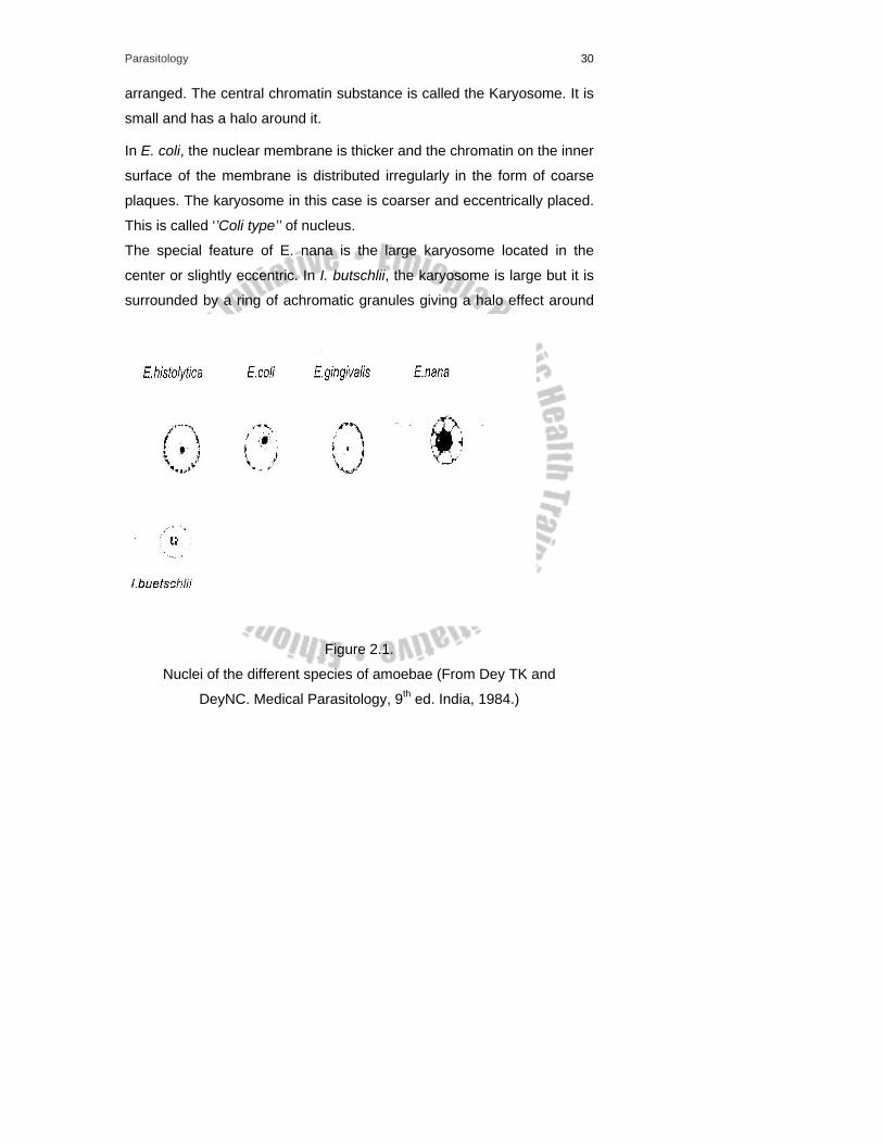
Before going into structural details here for each of them, their nuclear
character for identification is considered:
E. histolytica, E. coli and the mouth ameba E gingivalis., have
conspicuous peripheral chromatin, arranged on the inner surface of
nuclear membrane; where as such chromatin material is lacking in the
remaining other three amoebae. The nuclear membrane in E. histolytica
is delicate and the chromatin substance on the inner side of the nuclear
membrane appears as fine beads, which are uniform in size and evenly
Parasitology
30
arranged. The central chromatin substance is called the Karyosome. It is
small and has a halo around it.
In E. coli, the nuclear membrane is thicker and the chromatin on the inner
surface of the membrane is distributed irregularly in the form of coarse
plaques. The karyosome in this case is coarser and eccentrically placed.
This is called ‘’Coli type’’ of nucleus.
The special feature of E. nana is the large karyosome located in the
center or slightly eccentric. In I. butschlii, the karyosome is large but it is
surrounded by a ring of achromatic granules giving a halo effect around
the karyosome.
Figure 2.1.
Nuclei of the different species of amoebae (From Dey TK and
DeyNC. Medical Parasitology, 9th ed. India, 1984.)

Parasitology
31
Entamoeba histolytica
Geographical Distribution:- Cosmopolitan distribution, mainly in the
tropics and subtropics, and is mainly related to inadequate personal
hygiene environmental sanitation, lack of safe water supply and poor
socioeconomic situation.
Habitat:-Trophozoite:- Large intestine, liver abscesses and other extra-
intestinal organs
Cyst:- found in the stools of chronic dysenteric patients and carriers.
Morphology
Trophozoite:
Size:- 12 to 35μm, Usually as long as 3 or 4 red blood cells .
Shape:- elongated form when actively motile and rounded form
when at rest.
Motility:- Active, Progressive , directional amoeboid motility in fresh
warm stool specimen.
Pseudopodia:- Finger like, broadly rounded end.
Cytoplasm:- Well differentiated into ectoplasm and endoplasm.
- May contain ingested host’s red blood cells in dysenteric
specimens
Nucleus:- Single nucleus, not visible in the motile form but in iodine
stained smear clearly seen.
Cyst:- Size: 12-15μm (1½-2 red blood cells)
Shape: spherical
Nuclei: 1-4 nuclei
Nuclear membrane: thin, regular and circular lined with fine
chromatin granules internally , and
small, compact central karyosome.

Parasitology
32
Cytoplasm: Yellowish-gray and granular in iodine stained
smear.
Stored food: Sausage shaped chromatoidal bars with blunt
ends and glycogen mass in immature cysts
with one or two nuclei.
Life cycle:
Entamoeba histolytica requires a single host to complete its life cycle.
When mature tetra-nucleated cyst from contaminated food or drink or
form hands contaminated with feces is ingested it excysts in the small
intestine to produce metacystic trophozoite by a process of binary fission.
The immature trophozoites migrate to the colon and grow to become
mature trophozoite stage, multiply by binnary fission to invade the mucus
membrane of the large intestine. Some times it can perforate the
intestinal wall causing extra-intestinal amoebiasis. The trophozoite stage
may pass with diarrhea or dysentery.
After a period of growth and multiplication, encystment occurs in the
large intestine. In the process of cyst formation, the trophozoite discharge
undigested food appears spherical in shape and condense to become
pre-cyst. The pre-cyst secrets cyst wall to form a mono-nucleated cyst
which is followed by a nuclear division to produce a bi-nucleated and
then a tetra-nucleated mature cyst. Cyst and precyst will also pass in
semi- formed or formed stool, where cyst is infective if it is ingested by
any means of transmission.

Parasitology
33
Figure 2.2.
Life cycle of E.histolytica. (From Jeffrey HC and Leach RM.
Atlas of Medical Helminthology and Protozoology, 1975.)

Parasitology
34
Mode of Transmission
Infective stage: Tetra-nucleated mature cyst.
Man acquires infection of E.histolytica from:
- Ingestion of food or drink contaminated by infective cyst.
Clinical Features and Pathology
May be asymptomatic or exhibit amoebic dysentery or extra-intestinal
amoebiasis in the liver, brain, spleen, lung, etc. Amoebic dysentery
occurs when E histolytica trophozoites invade the wall of the large
intestine and multiply in the submucosa, forming large flask shaped
ulcers. The amoeaba ingest red cells from, damaged capillaries.
Amoebic Liver Abscess:
Occasionally amoeba is carried to the liver in the portal circulation and
form abscesses, usually in the right lobe. There is pain & tenderness
over the liver, wasting and fever with chills & night sweats. Patients with
large or multiple abscess may become jaundiced and anemic.
Formerly a pathogenic invasive strain & a non pathogenic strain, of E.
histolytica were thought to exist. The two ‘strains’ have now been
recognized as separate species. E. histolyrtica is the invasive pathogenic
and E. dispar has been designated the non- invasive non-pathogenic
species. The two species are morphologically identical.
Prevention and Control
1. Cooking of food and vegetables
2. Hand washing after defecation and before eating
3. Safe water supply (treatment, boiling, filtration, etc.)
4. Control of mechanical vectors
Parasitology
35
5. Avoid use of night soil as a fertilizer proper sanitary disposal of
faeces.
6. Treatment of infected individuals and health education.
Laboratory Diagnosis
Laboratory diagnosis of intestinal amoebiasis is based on:
1) Examination of a fresh diarrheic or dysenteric faecal specimen or
rectal scraping for motile amoebae using saline, or
2) Examination of formed or semi-formed faeces for cyst stages. Such
stool can be examined by direct saline and/or iodine smear, and Zinc
sulphate floatation or centrifugal floatation method.
- Charcot-Leyden crystals, representing the crystallized contents
of granules from eosinophil leukocytes may also be found in a
fecal smear.
- Specimens must be examined without delay
otherwiseidentification of the trophozoites becomes impossible
because the amoeba lose their motility, extrude vacoules
containing red cells, round up
- With the recognition that E.histolytica is morphologically identical
but genetically distinct form E.dispar, cysts, formerly reported as
E.histolytica should be now reported as E.historlytica / E.dispar.
Differential Diagnosis of Amoebic Dysentery and Bacillary Dysentery Amoebic dysentery Bacillary dysentery Odor Offensive Odorless Color of blood Dark red Bright red Exudate Few pus cells Many pus cells E.histolytica- Present Absent Consistency Mucus adherent to the Not adherent
Parasitology
36
container Has charcoat -Leyden crystals No crystal Appearance - Fecal matter with stratum of Hardly any fecal matter
blood and mucus seen over the consists of blood and surface mucus Bacteria Numerous, motile Scanty, non motile
Relevance to Ethiopia
E. histolytica has a worldwide distribution but it is more common in
developing countries. The parasite is very common amongst children.
General parasitological survey indicates that the infection rate is variable.
Of the seven species of amoeba which inhabit the human intestinal tract,
Only E.histolyica is pathogenic to man, as the agent of amoebiasis.
Slightly over 80, 000 new cases of amoebiasis were reported by the
Ethiopian MOH in 1988-89, based on outpatient reports. The infection
was common in all age groups, and even 3,716 children under 1 year
were treated for this disease (MOH, Addis Abeba, 1991).
A wider range in prevalence has been reported from community surveys
in other parts of the country. Extremely high E. histolytica infection rates
(55%) were found among the isolated Saysay shifting cultivators in the
Blue NileGorge in Welega. In a survey of 1,850 school children in 50
farming communities on the central and northern plateaus covering five
administrative regions, the parasite was found in 94% of the
communities, with prevalence rates ranging from 3% to 50 %; on the
average, 19% of the school population were infected (Lo CT, et al. 1980).
Twelve percent of 698 school-aged children in 17 rural communities in
the highlands of Showa were infected (Kloos H et al. 1980).
Low to intermediate levels of amoebiasis prevalence have been reported
from towns, apparently due to the effect of urbanization on transmission;
0.5% of 468 school children in Addis Ababa and 2.2% of 90 school

Parasitology
37
children in Debre Zeit were found to be infected. Rates were highest
among poor children (2.8%). [Taticheff et al, 1981].
The highest prevalence of amoebiasis in Ethiopia was found the potential
for nosocomial infections in Ethiopian health institutions (Editorial, Ethiop
Med J, 1972).
In a relatively recent country wide survey of amoebiasis, a totalof 12,457
persons in 97 communities was stool examined by formol ether
concentration technique. The cyst passsers, inschoolchildren and non-
schoolchildren coimmunities were 15.0% and 3.5%, respectively. Slightly
more females (18.4%) than males (14.2%) were infected among school
children but the the difference was not significant non-
schoolcommunities.There was a tendency but not a statistically
significant decline of cyst –excretion with increasing age. So far as our
survey goes, the influence of altitude on the prevalence of amoebiasis
appeared not to be significant.Health education,improvement of
sanitationand personal hygiene are suggested as realistic measures
toreducethe transmissionof the parasite (Erko B, et al. Trop Geog Med,
1995).
The above stuidies did not differentiate between E.histolytica and E.
dispar. A single study done in Wongi areas of central Ethiopia, where an
increased incidence of amoebic infection has been reported, of the 29
amoebic isolates successfully stabilized, cloned and characterized
bybSargeaunt’s electrophoretic technique, 27 (93.1%)were of E.dispar
zymodemes and two were (6.9%)wereof E.histolytica (Gatti S et al, Ann
Trop Med Parasitol, 1998)

Parasitology
38
Entamoeba Hertmanni
Geographical Distribution: Cosmopolitan Habitat: both trophozoite and cyst live in the small intestine
Morphology
Known as “small race” of E.histolytica because of similarity in their
morphology. It feeds on bacteria not red blood cells.
Trophozoite
4-12μm, smaller than E.histolytica
Active but less vigorous directional motility by finger-like pseudopodia
Single nucleus with fine bead-like coarse chromatine granules on the
thick nuclear membrane and has large centeral karyosome.
Cyst:- 5-10μm in size and spherical in shape.
1-4 nuclei; Minute rice grain shaped chromatoidal bars and
glycogen mass in the immature cyst stage.
Life cycle Similar to the life cycle of E.histolytica. It requires a single host and has
five main developmental stages.
Mode of Transmission
Through contaminated food or drink, or from hands contaminated with
faeces.
Pathology:- Harmless commensal
Tetranucleated cyst→Metacyst→Metacystic
trophozoite→Trophozoite→Precyst→Uninucleated cyst

Parasitology
39
Laboratory Diagnosis: - Finding the characteristic trophozoite and cyst stages in stool
specimen. Differential characters:
- Cyst of E.hertmanni is similar to that of E.histolytica / E. dispar but
the former is smaller in size.
- Cyst of E.hertmanni is also similar to that of E.nana but the later has 4-
hole like nucleus and don’t have chromatoid body
Entamoeba coli
Geographical Distribution: Cosmopolitan.
Habitat Both trophozoite and cysts in the large intestine of man
Morphology Trophozoite:-
Size: 15-50μm(average 25μm), usually bigger than E.histolytica
Shape : Oval or elongated
Motility:- Sluggish, non -progressive and non-directional
- Short blunt pseudopodia
Cytoplasm: Ectoplasm and endoplasm not well differentiated.
Nucleus: Single nucleus, visible in the fresh state without
staining.
Thick nuclear membrane lined with coarse chromatin
granules and eccentric karyosome .
Inclusions: Bacteria, Yeasts, but never red blood cells.
Cyst:-
Size: 12-20μm a little larger than the cyst of E.histolytica.
Shape: rounded or slightly oval.
Nucleus: 1-8 nuclei; thick irregular nuclear membrane large,
diffuse,often eccentric karyosome

Parasitology
40
Cytoplasm: bright pale yellow in iodine stained smear.
Life cycle Similar to the life cycle of E.histolytica.
Mode of transmission
Ingestion of contaminated food or drink by infective cyst. Pathology:- Harmless commensal, may cause diarrhea in children.
Laboratory diagnosis:- Finding the characteristic trophozoite and cyst stages in stool specimen.
Can be differentiated from E.histolytica by its larger size. The cyst of
E.coli shows a greater variation in shape and size than those of
E.histolytica.
Entamoeba gingivalis
Geographical distribution: world wide distribution Habitat: Oral cavity
Morphology:-Has trophozoite stage only, no cyst stage
Trophozoite:-
Size:-10-20 nm
Motility: sluggish
Cytoplasm: well differentiate into ectoplasm and endoplasm
Pseudopodia:- multiple
Nucleus:- single, delicate nuclear membrane lined with fine
chromatin granules and Small central karyosome.
Octanucleated cyst Metacyst Metacystic
Trophozoite Trophozoite precyst unincleated
cyst binucleated cyst Tetranucleated cyst

Parasitology
41
Life cycle:-It is reproduced by binary fission and transmitted from one
person to another through kissing , droplets spray from
the mouth,contaminated spoons or cups.
Pathology: non pathogenic commensal amoebae Laboratory Diagnosis:-Finding the characteristic trophozoite stage from
swab of the oral cavity. It is the only species to ingest host’s leukocytes
and has numerous food vacuoles.It should be differentiated from
Trichomonas tenax which belong to flagellates and found in oral cavity.
Endolimax nana
Geographical distribution: cosmopolitan. Habitat: Trophozoite and cyst in the large intestine. Morphology:-
Trophozoite
Size: 6-15μm (average 10μm)
Motility: multiple small rounded blunt pseudopodia moving slowly
in all direction.
Cytoplasm: very granular with small vacuoles
Inclusion: Bacteria.
Nucleus: single large irregularly shaped eccentric karyosome
and thick nuclear membrane with out lining of
chromatin granules.
Cyst:-
Size: 8-10μm
Shape: oval
Nucleus: 1-4 nuclei large, irregular karyosome
Cytoplasm: Deep yellowish color in iodine smear.
Life Cycle:-Trphozoite stage reproduces by binary fission and man
acquires infection from contaminated food or drink with

Parasitology
42
mature cyst stage.
Laboratory Diagnosis:-Finding of the cyst and trophozoite stages in
fecal smear.
Iodamoeba butschili
Geographical distribution: Cosmopolitan.
Habitat: both trophozoite and cyst in the large intestine.
Morphology:- Trophozoite:-
Size: 10-25μm
Shape: compact, leaf -like
Motility: sluggish by clear, rounded, finger like pseudopodia.
Nucleus: single, no chromatin granules on the nuclear
membrane and has large karyosome surrounded by achromatic
granules.
Inclusion: bacteria; large food vacuole packed with glycogen
mass that stains deep brown with iodine solution.
Cyst:-
Size: 8-10 μm
Shape: rounded, oval or irregular
Nucleus: single, very large oval karyosome surrounded with cluster
of granules.
Vacuole: very large glycogen vacuole often taking up half part of
the cyst and stains brownish red by iodine solution hence
the name was given as Iodamoeba.
Life Cycle:-The trophozoite reproduces in the large intestine by binary
fission. The infective stage is the mature cyst stage and man acquires
infection from contaminated food or drink.
Parasitology
43
Pathology: It is non-pathogenic.
Laboratory diagnosis:-Finding the characteristic trophozoite and cyst
stages in the direct fecal smear examination or using concentration
technique.
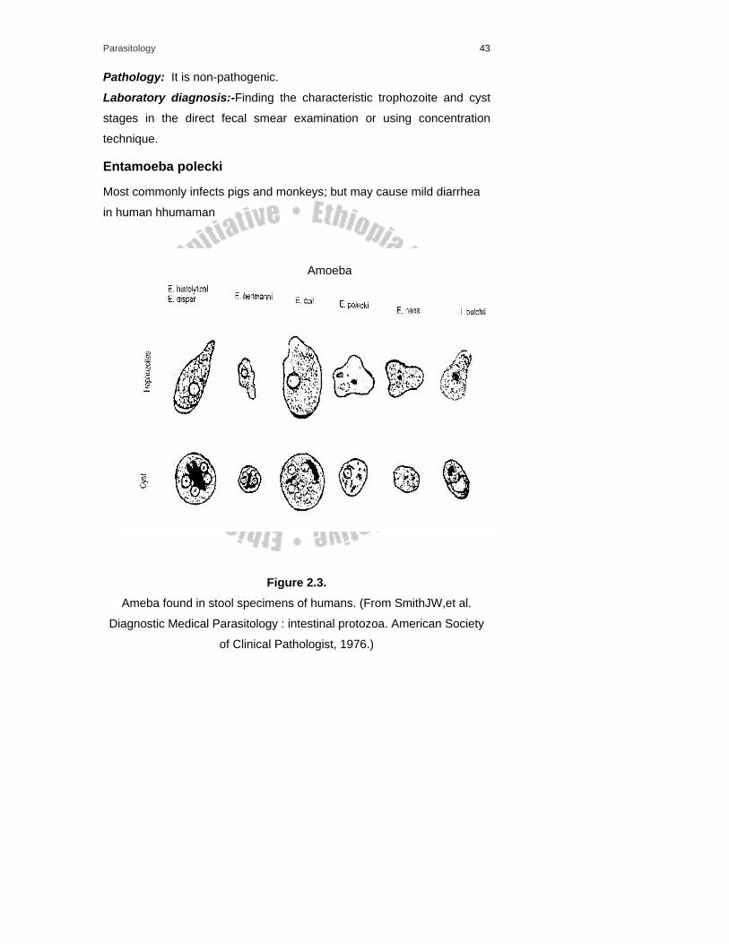
Entamoeba polecki
Most commonly infects pigs and monkeys; but may cause mild diarrhea
in human hhumaman
Figure 2.3. Ameba found in stool specimens of humans. (From SmithJW,et al.
Diagnostic Medical Parasitology : intestinal protozoa. American Society
of Clinical Pathologist, 1976.)
Amoeba

Parasitology
44
2.1.2 Free living Pathogenic Amoebae
The free living amoeba constitute a large group, inhabiting fresh,
brackish and salt water, moist soil and decaying vegetation. Some are
coprozoic. For convenience this large and diverse group is separated into
two groups on the bases of their ability to undergo transformation from an
amoeba to a flagellate stage. Naegleria belongs to the family
Vahkampfiidea, members of which are characterized as
amoebaflagellates able to assume a temporary flagellate form while
completely devoid of flagella in their amoeboid stage. Acanthamoeba,
belonging to the family Acanthamoebidea, never produce flagella. Most
cases of primary amebic meningoencephalitis can be ascribed to
infection with Naegleria; less commonly Acanthamoeba is involved.
Naegleria fowleri
Geographical Distribution:-It is free living pathogenic amoebae with
worldwide distribution.
This genus of amoeboflagellates has an amoeboid phase, which
alternates with one possessing two flagella. The forms found in the
tissues are ameboid, and in the tissue they are distinguished with
difficulty from acanthamoeba. Most case have occurred during the hot
summer months in young persons who within the preceding week swam
or dived in fresh or brackhish water, lakes , hot springs and swimming
pools have been apparent sources of the infection.
That the source of infection is not always aquatic is illustrated by a case
report from Nigeria where the organisms were apparently inhaled during
a dust storm by an 8-month old infant, from whose nasal mucosa and
spinal fluid the organism were recovered prior to death. Another
interesting report from Nigeria concerns a Muslim farmer thought to have

Parasitology
45
become infected during the ritual washing before prayer, which involved
sniffing water up his nose.
Habitat: Nasal cavity, Central nervous system,
Morphology Trophozoites
Size: 10-15μm
Cytoplasm - Well differentiated into ectoplasm and endoplasm
Motility - actively motile with broad pseudopodia elongated
Nucleus - Single with central karyosome
Inclusion -does not ingest host RBCs
Cyst - One nucleus and thin cystcyst wall which has no pore.
Flagellate Shape - Oval
Nuclues - similar as above
Flagella - two flagella equal in length and longer than broad
Pathology
Causes primary or acute meningoencephalitis. Major symptoms include
fever, stiff neck (meningitis), frontal headache, altered test (food or drink),
vomiting, white blood cell count about 24,000 with many neutrophils.
Prevention and Control:-Infection can be prevented by avoiding
contact, swimming and snuffing in waters of ponds, lakes, treatment of
water and proper sewage disposal, treatment of infected individuals and
health education.
Laboratory Diagnosis:-Only amoeboid trophozoite stage is found in
man. Finding the amoeboid trophozoite stage in cerebrospinal fluid
(CSF). It can be found in unstained or Giemsa stained CSF smear. The

Parasitology
46
CSF appears to be claudy, purulent and may contain eosinophils and red
blood cells.
Acanthamoeba Species
Similar with that of N.fowleri except the following differences:
1. Trophozoiutes are larger 15-25μm
2. Pseudopodea arefilamentous (spiky pseudopodia or acathopodia)
3. It does notforma flagellate stage in water
4. Cysts are thickwalled with many pores
5. It may encysting the tissue
6. Pathology:- Causes secondary or chronic meningoencephalitis..
Symptoms include fever, headache, rhinitis, meningoencephalitis,
conjunctivitis, corneal ulceration, keratitis and loss of vision
7. Notkilledbydessication
Review Questions
1. What are the differences among cysts and trophozote of E.hisolytica,
E.hertmani and E.coli?
2 Which amoeba do not have cyst stage in its life cycle?
3 Explain the general prevention and control methods of amoeba.
4 List pathogenic amoeba.
5. Discuss the general mode of transmission and laboratory diagnosis
of amoeba.
6. How do you differentiate amoebic dysentery from bacilliary?
7. What is the clinical significance of studying Naegleria and
Acanthamoeba species.

Parasitology
47
2.2 Class: Zoomastigophorea
Mastigophora: Flagellates
Flagellates infecting man are divided into two groups.
1. The oro-intestinal and urogenital flagellates and
2. The Hemo-somatic flagellates.
The oro-intestinal and urogenital flagellates are found in the intestine,
oral cavity and genital tract. Many of them are not pathogenic. They are
classified in to 2 orders, namely; Protomonadida and Diplonadida, The
former is characterized by one nucleus and flagella at the anterior end;
where as the latter has a pair of nuclei and flagella, which are
symmetrically distributed at the interior end.
The hemoflagellates are present in the blood and invade various tissues
of the body; remain either in the intercellular fluid, bathing the cells; as in
trypanosoma, or are engulfed by the Red cells and leucocytes as in
Leshmania. Of the six genera the parasite athogenic to man belong to
two genera under the family Trypansomatidae, these are Trypanosoma
and Leishmania.
2.2.1 The Oro-intestinal and Urogenital Flagellates
General Characteristics
1. Uses flagellum as locomotory organell
2. Reproduce by simple binary fission
3. Complete their life cycles in a single host and a second host whom
they infect is necessary for the continuation of the species.
4. Most are commensal forms except G.lamblia, T.vaginalis and
D.fragilis
5. The infective stage may be either the trophozoite or the cyst stage

Parasitology
48
6. Except the species of Trichomonas and Dienatamoeba fragilis, all
have both cyst and trophozoite stages.
Dientamoeba fragilis
Dientamoeba fragilis originally classified as an amoeba, is now
considered an amoeba like flagellate more closely related to the genus
Trichomonas. Electron microscopic studies have revealed that the
internal structures are typical of flagellate.
Geographical Distribution: World wide Habitat: In the large intestine. Morphology: Has trophozoite stage only, No cyst stage.
Trophozoite:-
Size: 6-15μm
Motility: Either non-motile (most often), or very actively motile in
very fresh fluid stools with fan-like multiple pseudopodia. It
becomes non-motile under the cover slip or disintegrates
immediately.
Cytoplasm: clear ectoplasm.
Nucleus: Usually one or two nuclei but 3or 4 nuclei may be found
rarely. Karyosomes split into 4-6 granules.
Inclusion bodies: Bacteria
Life cycle:- The mode of transmission is uncertain but most likely is
feco-oral nature. It is postulated that the delicate trophozoite is
transported from person to person inside the protective shell of helminth
ova such as Entrobius vermicularis. It reproduces asexually by binary
fission. It is considered to be harmless commensal Laboratory Diagnosis:-The trophozoite stage is highly fragile and
disintegrates explosively in water immediately. Hence it needs

Parasitology
49
immediate examination of fresh stool specimen to find the trophozoite
stage.
Chilomastix mesnili
Geographical Distribution: cosmopolitan but mostly prevalent in warm
climates. Habitat: Trophozoite and cyst live in the colon and caecum of the large
intestine.
Morphology
Trophozoite:- Size: 6-20 by 3-10μm
Shape: Triangular and tapered at one end
Motility: spiral in one definite direction.
Cytoplasm:
- Spiral groove that makes asymmetrical flagellate
- cytostome (mouth-like cleft) at the rounded end.
Nucleus: one nucleus, easily visible in unstained preparation
Flagella: Six flagella. Three anterior free flagella, one delicate
flagellum lying in the cytostome and two flagella on the lateral
margin of the cytostome
Cyst:- Size: 6-8 by 4-6μm
Shape: pear or lemon shaped
Cystostome and remains of locomotory organelles can be
seen.
Nucleus: single; Thick nuclear membrane with small central
karyosome.

Parasitology
50
Life Cycle
Cyst→Excystation→Trophozoite→Binary fission→encystation→Cyst in the faeces
Trophozoite stage reproduces by binary fission. The infective stage is the
cyst from contaminated food or drink. Excystation occurs in the large
intestine and trophozoite multiplies by binary fission.
Pathology: It is commensal
Laboratory Diagnosis:-Finding the trophozoite and cyst stages in stool
specimen. The trophozoite stage is very similar to Giardia lamblia and
Trichomonas hominis; and needs careful identification.
Giardia lamblia
Also called Giardia intestinalis and G.duodenale
Geographical Distribution:- Cosmopolitan distribution in warm climate
and is more prevalent in children than in adults. It is the most commonly
diagnosed flagellate of the human intestinal tract. High prevalence occurs
in young, malnourished children in large families, orphan asylums, and
elementary schools.
Habitat: Upper parts of the small intestine mainly in the duodenum and
jejunum.
Morphology:
Trophozoite:-Size: 10-21 by 5-15μm
Shape: pyriform (pear-shaped), i.e. rounded anteriorly and
pointed posteriorly.
Motility: Progressive, rapid, tumbling and spinning often
linked to a “falling leaf” type of motility in fresh liquid stools.
Bilaterally symmetrical
Covex dorsal surface and a flattened ventral side
Contents:

Parasitology
51
- Anteriorly there are two sucking discs each contains a
nucleus, 4 pairs (8) flagella, Parabasal body and axonemes
Figure 2.5. Trophozoite of G.lamblia. (From Jeffrey HC and Leach
RM. Atlas of Medical Helminthology and Protozoology, 1975.)
Cyst :- Size: 8-12μm, oval shape with thick cyst wall.
Finely granular cytoplasm clearly separated from cyst wall.
2-4 oval nuclei at one pole, each with small, central
karyosome.
Cytoplasm: clear when unstained; yellowish green or bluish
in iodine solution.
Fibril: thread-like remains of flagella; axonemes and parabasal
bodies folded as S-shaped placed length wise in the center of
the cyst.
Life Cycle
Requires a single host to complete its cycle and reproduces by a simple
longitudinal binary fission
Cyst ingested→excystation→Trophozoite→binary fission→Encystation→cyst in faeces

Parasitology
52
Infection occurs by ingestion of mature tetranucleated cyst with
contaminated food, drink, finger, etc. Following ingestion, the cyst excyst
in the upper part of the small intestin to form flagellates. They become
attached to the intestinal wall by a sucking disc and absorb nourishment
through their body surface. They multiply by longitudinal binar fission and
some of them are carried down the intestinal tract to undergo
encystation. The trophozoites and infective cysts are excreted in the
faeces.
Clinical Feature and Pathology:-Major symptoms includes duodenitis,
excess secretion of mucus or malabsorption of fat (steatorrhoea), sugar
and vitamins, dehydration, diarrhoea, weight loss, poor appetite,
vomiting, lethargy bile passage obstruction
Figure 2.6. Life cycle of Giardia lamblia. (From Nasir NT.
Review of Human Parasitology,2nd ed. The Scientific Book Center, Cairo)
Parasitology
53
Prevention and Control:
1. Improving personal, family and group sanitation and hygiene.
2. Avoid contamination of food, drink and hands with the faeces.
3. Safe water supply and latrine construction.
5. Treatment of infected individuals and health education.
Laboratory Diagnosis:-Finding the trophozoite and cyst stages in stool
specimen. The stool is usually offensive, bulky, pale, mucoid (fatty),
diarrheic (watery) but there is no blood in the stool. Several specimens
collected at different time need to be examined because trophozoites and
cysts are excreted irregularly.
Intestinal and non-pathogenic flagellate that require differentiation from
G.lamblia include: C.mesnili and Pentatrichomonas hominis (formerly
T.hominis).
Trophozoites of the above mentioned flagellates can be easily
differentiated from G.lamblia by their shape and movement (in fresh
sample) and bcause they have only one nucleus (and fewer flagella). The
only other trophozote that has two nuclei is D.fragilis but this organism
has no flagella or median bodies and look likes a small amoeba.
Cyst of intestinal flagellates can be easily differentiated from those of
G.lamblia because they are smaller and do not have the same
characterstic appearance of G. lamblia (do not contain remains of
flagella). C.mensili cysts are lemon shape and D. fagilis does not has
cyst stage.
Relevance to Ethiopia
Infection by Giardia lamblia has a cosmopolitan distribution both in
developed and developing nations. Infection rates ranging from 1% to
50% or so have been reported from various parts of the world. In African,

Parasitology
54
Asian and Latin American countries, about 200 million cases of Giardia
lamblia infections have been estimated to occur annually. The infection
may be endemic as in the tropics where it is a familial infection passed
around by faecal-oral route, sporadic as in travelers, or epidemic as
waterborne or institutional outbreaks (Helmut K and Zein AZ, 1993)
Giardiasis is wide spread in Ethiopia. A countrywide survey of giardiasis,
using formal-ether concentration method, among school children and
residents showed overall prevalence rates of 8.9% and 3.1%
respectively. The corresponding rate for non-school children (5-19 years
of age), however, was 4.4% showing that the School children are more
significantly infected than their school children counter parts. Giardia
lamblia infection was generally found to be more prevalent in children
than in adult. Among children of school population those in their first
decade of life were more affected (Hailu and Berihanu, 1995).
Although these and other prevalence data are not strictly comparable
due to differences in sample selection and diagnostic methods used in
different institutions, they indicate that while urbanization resulted in the
reduction of the prevalence of giardiasis, it remains common infections in
urban population. Infection rates reported here can not represent the
actual prevalence rate giardiasis in Ethiopia as stool examination alone is
not reliable to rule out infection of Giardia the cysts of which are excreted
episodically.
Trichomonas hominis
Geographical Distribution: Next to Giardia lamblia, it is probably the
most common and most cosmopolitan of the intestinal flagellates of man. Habitat: Large intestine.
Morphology: has trophozoite stage only.
Trophozoite

Parasitology
55
Size: 10-15μm, pyriform (oval with two pointed poles) in shape
Motility: whirls and turns (jerky) in all directions, seeming to
vibrate. Most resistant flagellate that remains motile even in old
stool specimens.
Undulating membrane and costa reach 2/3 or full length of the
body Nucleus: Single nucleus with central karyosome.
Flagellum: 3-5, usually 4 anterior free flagella and another flagellum
on the margin of undulating membrane with a free trailing posterior
end. Conspicuous cytostome opposite to the undulating membrane,
has semi-rigid axostyle and parabasal body
Life Cycle:-The trophozoite stage reproduces by binary fission and
requires direct host to host transmission through contaminated food
and/or drink. It has high prevalence in children and more common in
warm climates.
Pathology: It is non-pathogenic but may cause diarrhoea and infection
can be prevented by personal hygiene and sanitation.
Laboratory Diagnosis: Finding the trophozoite stage in fresh stool
specimen.
Trichomonas vaginalis
Geographical Distribution:-World wide distribution and mainly common
in the temperate region.
Habitat:-In the genital tract of male and commonly in female, especially
the vagina, cervix, urinary bladder, prostate and seminal vesicles.
Morphology: Has trophozoite stage only.
Trophozoite
Size 15-25 by 5-12μm, is the largest Trichomonas.

Parasitology
56
Shape: pyriform
Moility: Jerky (on-spot), non-directional motility in fresh specimen.
Short undulating membrane: extending along one third of the body.
Nucleus: Single with uniformly distributed chromatin granules
Flagella: 4 anterior free flagella and one on the margin of the
undulating membrane Axostyle may be split into several fibrils anteriorly.
Less conspicuous cytostome and has marked parabasal body.
Life Cycle:-The trophozoite stage reproduces by longitudinal binary
fission and mode of transmission is usually via sexual intercourse but
also by communal bathing, sharing of washclothes, toilet equipment
seats and mother to daughter during birth.
Figure 2.7. Life cycle of T.vaginalis (From Nasir NT. Review
of Human Parasitology,2nd ed. Cairo: The Scientific Book Center.)

Parasitology
57
Pathology: Causes trichomoniasis. Major symptoms are Vaginitis,
urethritis, prostatitis, chaffing of vulva, cervical erosion, burning
sensation, yellowish prulent discharge, reversiable sterility in male.
Prevention and Control 1. Personal hygiene and sanitation
2. Simultaneous treatment of both partners.
Laboratory Diagnosis:-Finding the trophozoites in unstained or stained
preparation of vaginal or urethral discharges, urine sediment, vaginal
swab, prostate secretions.
Relevance to Ethiopia
Trichomonas vaginalis is fairly common in Ethiopia as the level of
hygiene is very low. Forty of 216(15%) prostitute in A.A tested were
positive in 1995. (Kloos &Zein Ahmed, 1995). Another study done in A.A
revealed 20% of women attending antenatal clinic harbour this parasite
(Duncan ME, et al. Ethiop J Health Dev, 1995). Recent Unpublished
study conducted inJimmatown detect a prevalence of 12.3% inwoman
attending gynecology OPD (Tariku L, 2002).
Trichomonas tenax
Geographical Distribution
World wide.distribution with high incidence in warm climates.
Habitat: oral cavity.
Morphology: Has trophozoite stage only.
Trophozoite:-Size: 5-12μm
Shape: pyriform
Motility: active jerky motility
Undulating membrane: reaches two third of the body length.

Parasitology
58
Nucleus: Single
Flagella: four anterior free flagella and one flagellum on the
undulating membrane.
Thick axostyle extending a considerable distance behind the body. Has
parabasal body
Life Cycle:-The trophozoite stage reproduces by binary fission and
transmission is direct from mouth to mouth through kissing or communal
use of contaminated food and drinking utensils.
Pathology:-It is non-pathogenic
Laboratory Diagnosis:-Finding the trophozoites in unstained or stained
smear in swab taken from the oral cavity.
Parasitology
59
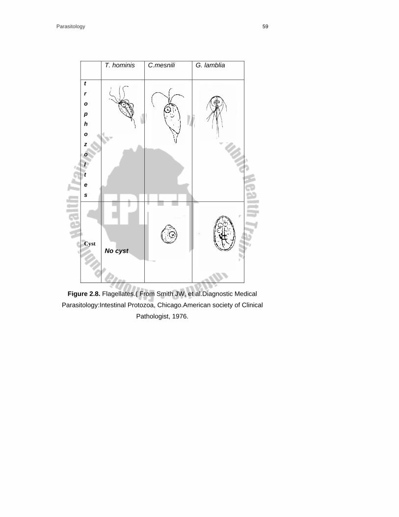
Figure 2.8. Flagellates.( From Smith JW, et al.Diagnostic Medical
Parasitology:Intestinal Protozoa, Chicago.American society of Clinical
Pathologist, 1976.
T. hominis C.mesnili G. lamblia
t
r o
p
h
o z
o
I
t
e
s
Cyst
No cyst

Parasitology
60
D.fragilis T.vaginalis
t
r
o p
h
o
z o
I
t
e
s
c
y
s
t
No cyst No cyst
Continuation of
Figure 2.8. Flagellates.( From Smith JW, et al.Diagnostic Medical
Parasitology:Intestinal Protozoa, Chicago.American society of Clinical
Pathologist, 1976.
Parasitology
61
Review Questions
1. Among the oro-intestinal flagellates which of them are more clinically
important than others.
2. What is the typical diagnostic movement of G.lamblia trophozoite?
3. Write the sources of specimen for the following flagellates:
a. Giardia lamblia
b. Trichomonas vaginalis
c. Trichomonas tenax
4. Which species have only trophozoite stage in their life cycle?
5. How do you differentiate among trophozoites of oro-intestinal
flagellates?

Parasitology
62
2.3 Blood and Tissue Flagellates
The blood and tissue of humans may be infected by one of the several
species of flagellate protozoa belonging to the family Trypomastidea.
There are six genera but only two of them are responsible to cause
disease to man. These are Genus Leishmania and genus Trypanosoma.
All of these organisms have developmental stages in blood sucking
arthropodes (intermediate host) and in humans (definitive host), and may
have a non human mammalian reservoir host.
Leishmania species are transmitted by Sandflies (Phlebotomus,
Lutzomia) and the trypanosomes are transmitted by either the tsetse fly
Glossina (for Afrcian trypanosomes) or Triatomid bugs (for American
Trypanosomes). The hemoflagellates may occur in a variety of stages in
the human host and the insect vectors.
General Characteristics
1. Reproduces by simple longitudinal binary fission
2. The infective stage is always the vegetative form.
3. Transmission occurs through biological insect vectors as
intermediate hosts and man as definitive host
4. The species are morphologically indistinguishable, but they can be
differentiated on the basis of on their clinical features primarily and
also on their geographical distribution, serologic tests, cultural
characteristics, vectors, reservoir hosts, biochemical tests,
immunological tests, etc.
5. The different developmental forms are differentiated on the basis of
a) Presence or absence of free flagellum
b) Presence or absence of undulating membrane
c) Position of the kinetoplast relative to the nucleus.

Parasitology
63
6. They have the following main body parts; Flagellum, Kinetoplast
divided into blelopharoplast and parabasal body, axoneme, nucleus,
undulating membrane,
7. Based on their development in the insect vector and their mode of
transmission, Trypanosomes are grouped into two,
These are:-
I. Saliverian Group.The parasites develop in the mid and fore gut of
their vectors and transmitted to man by inoculation of the parasites.
II. Stercorarian Group. The parasites develop in the hind gut of their
vectors and transmitted to man by the contamination bited area with
the faces of their vectors.
The following are the the main developmental forms
I. Amastigote /leishmania/ form
Spherical, no free flagellum, No undulating membrane, The only intracellular forms of all leishmania species and Trypanosome cruzi.
Figure. 2.9. Amastigote stage
II. Promastigote /Leptomonad/ forms
Elongated, Single free flagellum, single nucleus, no undulating membrane, nucleus is.near the middle. The kinetoplast is just in the anterior portion. This form is found in the invertebrate host, and in culture media (of all Leishmania species) and in man as a transitional form for Tryponosoma cruzi.
Fig. 2.10 Promastigote stage

Parasitology
64
III. Epimastigote /crithidial/ forms
Elongated body, single free
flagellum, single nucleus,
undulating membrane, the
kinetoplast is just anterior to the
nucleus. It is found in the
invertebrate host and in culture
media (of Trypanosome
species).
Figure 2.11 Epimastigote stage
IV. Trypomastigote /Trypanosomal/ forms
Figure 2.12. Trypomastigote stage
Pleomorphic, it can be as “U” or “C” shaped, single free flagellum, single
nucleus, undulating membrane. The kinetoplast and axonemes are found
at the posterior end relative to the nucleus.This form is found in the
peripheral blood of vertebrates and is the diagnostic stage of
Trypanosome species.
V. Metacyclic Trypomastigote /Trypanosomal/ Forms
Morphologically similar to trypomastigote stage but it is short and stumpy.
It is the final developmental stage in the gut of the insect vectors and is
the infective stage from the insect vector to man.

Parasitology
65
General life cycle of Leishmania species
Promastigotes inoculated into the skin when sandfly take a blood meal.
The promastigotes are taken by macrophages and become amastigotes.
Amastigotes multiply by binary fission.
Amastigotes are ingested by the insect vector when it takes a blood meal
and becomes promastigote in the gut of the insect vector. The
promastigotes multiply by binary fission and migrate to the head and
mouth parts.
Figure 2.13. The life cycle of Leishmania species. (From
StricklandGT.Hunter's tropical medicine 7th ed. Philadelphia:WB
Saunders,1991.)

Parasitology
66
Leishmania tropica minor
Geographical Distribution:-Widely distributed in the urban area of
Middle East, Europe, India, Eastern Mediterranean countries, Morocco,
Algeria, Tunisia.
Habitat: -Amastigotes: In the endothelial cells of cutaneous tissues,
lymph nodes, ulcers.
Promastigotes: In the gut of sandfly
Morphology:-It has Amastigote and promastigote Stages only.
Life Cycle:- Definitive host: Man
Intermediate host: Female sandfly
Reservoir host: Dog, cats, mice, etc.
It requires a female Phlebotomous sandfly as a biological vector and man
as its main definitive hosts to complete its life cycle. The vector
inoculates the promastigotes in to the cutaneous tissue while taking a
blood meal. The promastigotes are taken up by reticuloendothelial cells
and develop into amastigotes within the cell. The amastigotes multiply
and are ingested by a female sandfly vector when it sucks a blood meal.
The amastigotes become (flagellated) promastigote in the midgut of the
sandfly. The promastigotes multiply and fill the gut of the insect vector.
Some of the vectors that are involved in this life cycle are P.papatasi,
P.sergenti and P.pernicious.
Mode of Transmission 1. Inoculation by infected sandfly
2. Direct contact with the ulcer
3. Autoinfection

Parasitology
67
Pathology-
Causes cuaneous leishmaniasis (dry, urban, chronic, old world oriental
sore). At the site of bite there is dry painless ulcer, 25-70mm in diameter,
usually self-healing after 1-2 years often leaving disfiguring scar. Multiple
unhealing lesion known as leishmaniasis recidivans may develop
sometimes. The infection is usually heals and confers long lasting
immunity to reinfection.
Leishmania tropicam major
Similar with Leishmaniatropica minor except with the following
differences:
Geographical Distribution:-Wider distribution than Leishmania tropica
minor and found in rural areas of sub-Saharan Africa from Senegal to
central Sudan, Middle East, India, Pakistan, central Asia, North Africa.
Infections are zoonotic.
Life cycle Same as Leishmania tropica minor but man acquires infection upon
invading enzootic areas because sand rat and the gerbils are also the
main reservoir hosts. Important vectors are P.papatasi, P.alexandri,
P.caucasicus, and P.grove
Clinical Feature and Pathology:-Causes moist (wet), rural, acute,
Old world cutaneous Leishmaniasis or oriental sore. Forms a papule that
develops to a large uneven ulcer or multiple lesions and is self-healing
within 3-6 months. This infection protect against reinfection and against
infection with L.t.minor. There is sporadic human infection.
Parasitology
68
Leishmania aethiopica
Similar with the above leishmania except with the following differences.
Geographical distribution
Southern Yemen, and the highlands of Ethiopia and Kenya
Life Cycle:-Man acquires infection by the bite of the infected female
phlebotomus sandflies. The main vectors are P.longipes and P.pedifer.
Infections are zoonotics with rocky hyraxes (Procavia habessinica) and
tree hyraxes (Heterohyrax brucei) serving as reservoir hosts.
Pathology: Causes old world coetaneous and diffuse cutaneous
leishmaniasis.
⇒ Leishmania tropica minor, L. t. major, and L. aethopica have
similar prevention and control, and laboratory diagnosis, i.e.,
Prevention and Control Leishmania Species:
1. Avoid insect bites,
2. Control of insect vectors,
3. Protection of lesion from insect bites,
4. Avoid auto-infection /self-infection,
5. Treatment and health education.
Laboratory Diagnosis of Leishmania species:-
1. Amastigotes in stained smears taken from ulcers, lesions, nodules
2. Promastigotes in culture media.
3. Montenegroimmunologic/ leishmanin test.
Leishmania donovani
Geographical Distribution: India, Central Asia, China, Kenya, Sudan,
Ethiopia, Somalia, Central and South America.
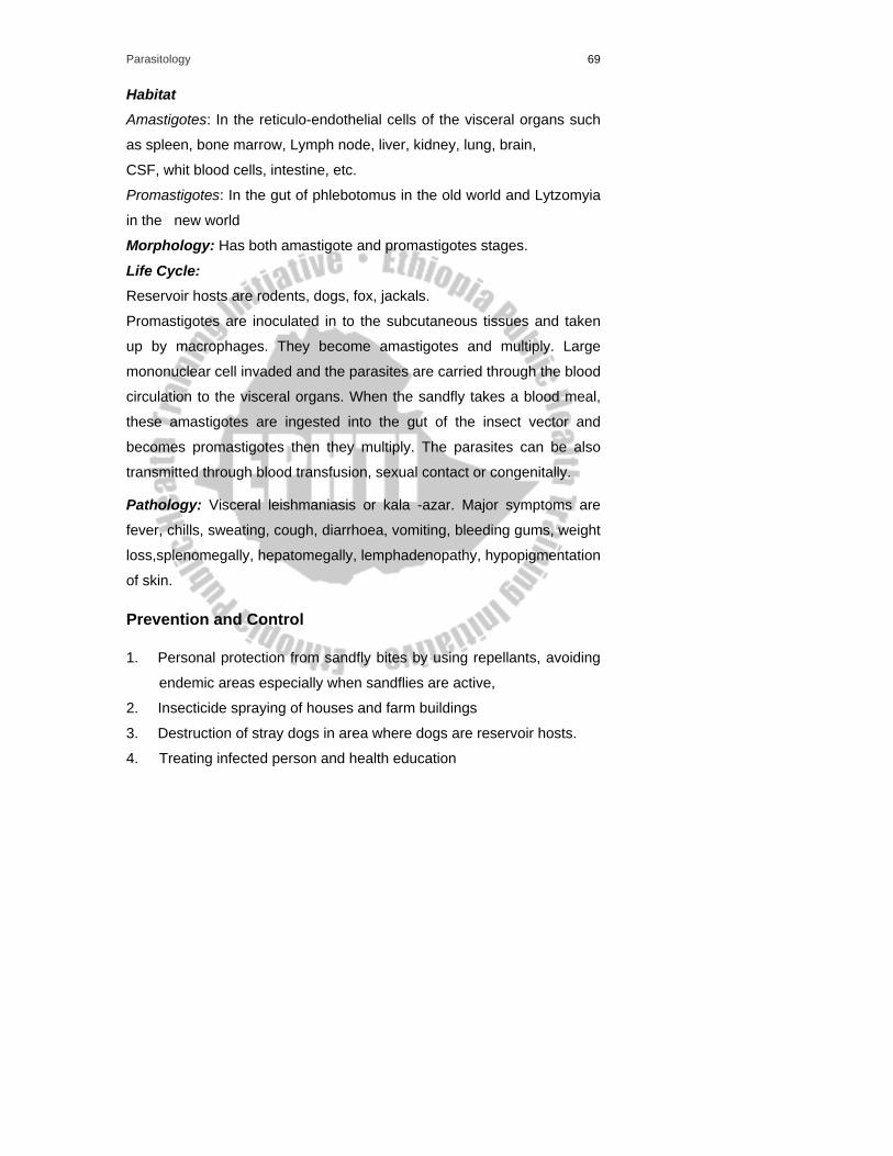
Parasitology
69
Habitat Amastigotes: In the reticulo-endothelial cells of the visceral organs such
as spleen, bone marrow, Lymph node, liver, kidney, lung, brain,
CSF, whit blood cells, intestine, etc.
Promastigotes: In the gut of phlebotomus in the old world and Lytzomyia
in the new world
Morphology: Has both amastigote and promastigotes stages.
Life Cycle: Reservoir hosts are rodents, dogs, fox, jackals.
Promastigotes are inoculated in to the subcutaneous tissues and taken
up by macrophages. They become amastigotes and multiply. Large
mononuclear cell invaded and the parasites are carried through the blood
circulation to the visceral organs. When the sandfly takes a blood meal,
these amastigotes are ingested into the gut of the insect vector and
becomes promastigotes then they multiply. The parasites can be also
transmitted through blood transfusion, sexual contact or congenitally.
Pathology: Visceral leishmaniasis or kala -azar. Major symptoms are
fever, chills, sweating, cough, diarrhoea, vomiting, bleeding gums, weight
loss,splenomegally, hepatomegally, lemphadenopathy, hypopigmentation
of skin.
Prevention and Control
1. Personal protection from sandfly bites by using repellants, avoiding
endemic areas especially when sandflies are active,
2. Insecticide spraying of houses and farm buildings
3. Destruction of stray dogs in area where dogs are reservoir hosts.
4. Treating infected person and health education

Parasitology
70
Laboratory Diagnosis
1. Amastigotes in aspirates of spleen bone marrow, enlarged lymph
nodes, and in peripheral blood monocytes.
2. Promastigotes in culture media
3. Testing serum for leishmanial antibodies
4. Formal get test; is a non-specific screening test for marked increases
in IgG
Relevance to Ethiopia
In Ethiopia, Ieishmaniasis is caused by four species of Leishmania,
namely, L. donovani,L. aethioplca.L.Major and L. tropica . The former, L.
donovoni, causes visceral leishmaniasis (VL) and is widely distributed
throughout the lowlands of north-western Ethiopia (Asrat H and Frommel
D, 1993). The latter three are agents of Cutanceous leishmaniasis (CL)
with L. aethiopica prevailing in the highlands of Ethiopia. P. martini and
P. celiae known vectors of Visceral leishmaniasis in south western
Ethiopia (Mengesha et al, 1999).
Visceral leishmaniasis (VL) occurs in Ethiopia mainly in arid and semiarid
lowlands below 1,300 m altitude. Many lowlands surrounding the central
Ethiopian Highlands are known to be endemic areas. The disease affects
the age group between 14 and 40 years. Mostly males are affected
because of occupational activity (Helmut K and Ahmed Z, 1993).
Cutaneous leishmaniasis caused by L. aethiopica is endemic and
widespread at altitudes between 1400 and 2700 m in most administrative
regions. Prevalence rates of active infection between 5.5 and 40 per
1000 population were reported from villages in Shewa , Wello and Gamo
Gofa administrative regions, with the highest rate in Ocholo village in
Gamo Gofa. The ecology of Ethiopian Highlands cutaneous
leishmaniasis is unique. Phlebotomus longipes and P.pedifer, the main

Parasitology
71
vectors, do not serve as vectors of leishmaniasis anywhere else.
Procavia habessinica and Heterohyrax brucei are the main animal
reservoir hosts for L. aethiopica (Helmut K and Ahmed Z, 1993).
Leishmaniasis and HIV Infection
Viceral leishmanisisis usually accompanied byimmunosuppression.The
specific and the nono-specific immunosuppresion in VL is reported as a
predisposing factor to in ntercurrent viral, bacterial, fungal and parasitic
infection.
In ares where both leishmaniasis and HIV infection occur, Viceral
leishmanisis (VL) is being increasingly reported in those with
immunosupression caused by HIV. Frequently parasites infect not just
the reticuloendothelial system but also the lungs, central nervous system,
normal skin and blood. Parasites have been found in phagocytic cells in
peripheral blood in upto 75% of patients (98%) in bone marrow aspirate.
There is a clear association between VL and immunosupression. In 1996
WHO epidemiological analysis, 90.4% of patients with Leishmania/HIV
co-infection. Patients with Leishmania / HIV coinfection do not respond
well to teatment. There are also reports of severe VL being caused by
Leishmania strains normally of low or no virulence.
The risk of Leishmania / HIV co-infection is increasing as VL is becoming
more urbanized, e.g. in the suburbs of cities such as Rio de Janeiro and
Natal, and HIV infection is becoming progressively more common in rural
areas, Kenya and Ethiopia.
In the study of VL/HIV co-infections in Ethiopia it was recommended that
tests for Leishmania should be carried out when patients with HIV have
fever of unknown origin, anemia, and enlarged spleen, liver or lymph
nodes. Patients with VL who respond poorly to treatment or relapse

Parasitology
72
frequently should be investigated for HIV infection. In the Ethiopian study
(endemic VL area), the clinical and laboratory findings were those
associated with classical VL.
Severe diffuse cutaneous leishmaniasis and recurring cutaneous and
mucocutaneous leishmaniasis are. laso being increasingly reported in
persons with Leishmania /HIV coinfection (Monica C, 2000).
Leishmania Mexican complex
Geographical Distribution:-Central and southern America mainly in the
rain forest of Mexico, Brazil, Guatemala, Venezuela.
Habitat: Amastigote: Reticulo endothelial cells of the skin promastigote: In the gut
of Lutzomyia sandfeies
Morphology: Has amastigote and promastigote
Life Cycle:-Same as the life cycle of Leishmania tropica except that the
vectors are new world sand flies
Pathology:-Causes new world coetaneous leishmaniasis.
Relevance to Ethiopia:-This parasite is not recorded in Ethioipia.

Parasitology
73
Leishmania braziliensis complex
Geographical Distribution:-Tropical forests of South America and
Central America. Reservoir hosts are rodents and some domestic
animals
Habitat: Amastigote:- In the reticulo-endothelial cells of muco-cutaneous tissues
of nose, mouth, lips, larynx.
Promastigote:- In the gut of Lutzomyia sandfeies
Morphology: Has amastigote and promastigote stages.
Life cycle:-Lutzomyia sandfeies are the main vectors and man acquires
infection from enzootic area.
Pathology:-Mucocutaneous leishmaniasis (espundia). Chronic ulceration
of mucus membrane of the mouth nose, throat, etc. with destruction of
bone and cartilage.
Leishmania Mexican complex and Leishmania braziliensis complex have
similar prevention and control methods and laboratory diagnosis as
presented below:
Prevention and Control
1. Avoid endemic areas
2. Avoid insect bites
3. Treatment of infected individuals
4. Health education
Laboratory Diagnosis
1. Amastigotes in stained smears taken from infected ulcers, lesions,
sores and nodules
2. Promastigotes from culture media.
3. Immunologic tests

Parasitology
74
Relevance to Ethiopia:- The parasite is not recorded in Ethiopia.
Trypanosoma gambiense
Geographical Distribution: West and western central Africa, extending
from Senegal across to Sudan and down to Angola.
Trypanosoma brucei complex (group)
Trypanosoma brucei brucei (infective to animal but not humans) and the
human pathogens Trypanosoma brucei rhodesiense (causing acute
trypanosomiasis) and T.b. gambiense (causing chronic trypanosomiasis)
are morphologically indistinguishable.
Habitat:
Trypomastigotes: In blood vessels and intercellular spaces of Lymph
nodes, spleen, liver, Brain, CSF etc.
Metacyclic trypomastigotes: In the mid and fore gut of the Glossina
(tsetse flies)
Morphology: has trypomastigote and metacyclic trypomastigote stages
Life cycle: It requires two hosts to complete its life cycle, species of
Glossina as an intermediate host and man and other animals as a
reservoir host. It reproduces asexually by binary fission.
Trypanosoma gambiense is a saliverian trypanosome in which the
trypomastigotes develop in the mid and fore gut of the insect vector so
that infection is acquired through inoculation of the metacyclic
trypomastigotes into the subcutaneous tissues with the saliva. In the
blood vessels the metacyclic trypomastigots transforms into
trypomastigots stage. There is multiplication of the parasites in the
mammalian host and the insect vectors. The parasites can also be

Parasitology
75
transmitted through blood transfusion, and congenitally. Important
vectors are G.palpalis, G.fuscipes, and G.tachinoides.
Clinical Features and Pathology: Gambian trypanosomiasis or chronic
African sleeping sickness. Major symptoms are chancer, fever,
haedache, sweating, post cervical enlargement of the lymph node(winter
bottom’s sign), splenomegally, hepatomegally, meningoencephalilis,
Figure 2.14. The life cycle of T.b.gambiense and
T.b.rhodesiense
(From Strickland GT. Hunter's tropical medicine 7th ed.
Philadelphia:WB Saunders,1991.)
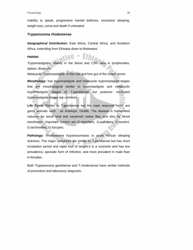
Parasitology
76
inability to speak, progressive mental dullness, excessive sleeping,
weight loss, coma and death if untreated.
Trypanosoma rhodesiense
Geographical Distribution: East Africa, Central Africa, and Southern
Africa, extending from Ethiopia down to Botswana.
Habitat:
Trypomastigotes: Mainly in the blood and CSF, also in lymphnodes,
spleen, Brain etc.
Metacyclic Trypomastigote: In the mid and fore gut of the insect vector.
Morphology: has trypomastigote and metacyclic trypomastigote stages
that are morphological similar to trypomastigote and metacyclic
trypomastigote stages of T.gambiense but posterior nucleated
trypomastigote stages are common.
Life Cycle:-Similar to T.gambiense but the main reservoir hosts are
game animals such as Antelops, Giraffe. The disease is transmitted
naturally by wood land and savannah tsetse flies and also by blood
transfusion. Important vectors are G.morsitans, G,pallidipes, G.austeni,
G.tachinoides, G.fuscipes.
Pathology: Rhodesiense trypanosomiasis or acute African sleeping
sickness. The major symptoms are similar to T.gambiense but has short
incubation period and rapid loss of weight.It is a zoonosis and has low
prevalence, sporadic form of infection, and more prevalent in male than
in females.
Both Trypanosoma gambiense and T.rhodesiense have similar methods
of prevention and laborarory diagnosis.

Parasitology
77
Prevention and Control
1. Vector control:
- By spraying vehicles with insecticide as they enter and leave the
tse-tse fly infested area,
-By using and maintaining insecticide impregnated tse-tse fly traps,
-By selectively clearing the bush and wood areas especially around
game reserves, water holes, bridges, and along river blanks.
2. Detecting and treating human infections at early stage.
3. Restricting the movement of game animals to within fenced game
reserves.
4. Health education.
Laboratory Diagnosis
1. Trypomastigotes in wet blood film -to observe motilit,
2. Thick or thin stained blood films or buffy coat from Micro-haematocrit
or capillary tube centrifugal concentration technique.
3. Examination of aspirate from enlarged lymph gland, chancher fluid,
CSF
4. Testing serum for anti-trypanosomal antibodies.
Relevance to Ethiopia
Animal trypanosomiasis (Nagna or gendi) always has been a problem in
many parts of Ethiopia (Tedla S, A.A Introduction to Parasitology, 1986)
T.b. rhodesiense is singled out as the species occuring in Ethiopia (Baker
JR McConnel, Ethiop Med J, 1974).
The species of vector of trypanosomiasis are to be found in Ethiopia are:
1) From the palpalis group- Glossina fuscipes fuscipes and
G.tachniodes

Parasitology
78
2) From the moristans group- G.pallidipes and G.moristans sub
moristans also called Ugandanesis.
3) From fusca group- G. longipennis
These Glossina species are confined to the southern and Western
regions. The seven adminstrative regions infested are Shewa, Gojam,
Welega, Illubabor, Kefa, Gamo Gofa and Sidamo. Although the belt is
quite extensive in Ethiopia the Sleeping sickness foci are limited to
Gambela (the areas along Baro, Gilo and Akobo rivers), Gamo Gofa
(from Mursi-Bodi district), Kefa (from maji), and Welega (from the
settlement area in the Anger-Didesa valley).The following domestic
animals are commonly found in the tsetse belt of Ethiopia: Cattle, sheep,
goats, donkeys, mules, dogs and chickens (Kloos H and Zein AZ, 1993).
Trypanosoma cruzi
Geographical distribution: Central and South America
Habitat:
Amastigotes: Intracellular forms in the reticuloendothelial cells and
tissues of brain, muscles, Lymph nodes, liver, Spleen, bone marrow, etc.
Promastigotes: Transitional stage
Epimastigotes: In the mid-gut of the insect vector (bug)
Trypomastige: In the mid-gut of the vector and; in the blood circulation
and intercellular spaces of man.
Metacyclic Trypomastigote: In the mid gut and in the faeces of the insect
vector

Parasitology
79
Morphology:
Has all the developmental stages of haemoflagellates Amastigote stages are similar to amastigote stages of Leishmania
species. The Trypomastigote are monomorphic forms about 2μm in size
with “C” or “U” shape.
Mode of Transmission of T. cruzi
1. Contact with the faeces of an infected blood sucking triatoma bug.
2. Blood transfusion.
3. Less commonly trans-placental transmission occurs with a fetus
being infected from an asymptomatic mothers.
5. It can occur also if viable parasites (even very few) penetrate the
skin, conjuctiva, or mucous membrane.
Life Cycle
In most infections, metacyclic (infective) trypomastigote contained in the
faeces of an infected bug (Triatoma) penetrate the skin through the bite
wound or enter through the conjunctiva of the eye or the membrane of
the mouth or nose. The feace are deposited as the bug feeds or soon
after. The trypomastgotes invade the reticuloendothelial cells near the
point of entry and multiply intracellularly as amastigotes. The amastigotes
develop into Trypomastigotes which are released into the blood when the
cell ruptures. The trypomastigote become amastigotes and multiply,
forming masses known as Pseudocysts.
Within the pseudocyst, a proportion of amastigote become elongateed
and develop first into epimastigotes and then into trypomastigotes which
ar released into the blood when the host cell ruptures. Some of these
trypomastigites continue to circulate while the majority infect further
tissue cells.
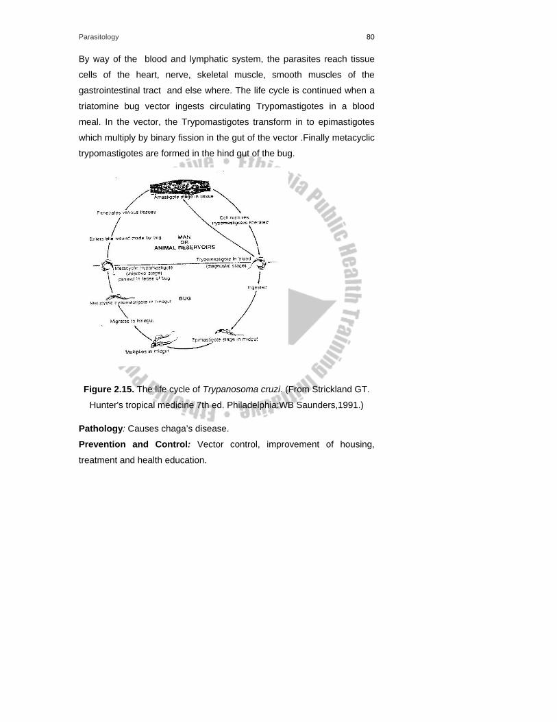
Parasitology
80
By way of the blood and lymphatic system, the parasites reach tissue
cells of the heart, nerve, skeletal muscle, smooth muscles of the
gastrointestinal tract and else where. The life cycle is continued when a
triatomine bug vector ingests circulating Trypomastigotes in a blood
meal. In the vector, the Trypomastigotes transform in to epimastigotes
which multiply by binary fission in the gut of the vector .Finally metacyclic
trypomastigotes are formed in the hind gut of the bug.
Figure 2.15. The life cycle of Trypanosoma cruzi. (From Strickland GT.
Hunter's tropical medicine 7th ed. Philadelphia:WB Saunders,1991.)
Pathology: Causes chaga’s disease.
Prevention and Control: Vector control, improvement of housing,
treatment and health education.

Parasitology
81
Laboratory Diagnosis:-
1. Blood film -wet film for motility
-thin and / or thick stained blood films
2. Xenodiagnosis (in chronic and subacute infections where their
number in the blood is usually very few).
Xenodiagnosis in this method uninfected, susceptible, laboratory
reared triatomine bugs are starved for 2 weeks and then fed on the
patients' blood. If trypanosomes are ingested they will multiply and
develop into epimastigotes which can be found 25-30 days later in
the faeces or rectum of the bug
3. Culture of blood on blood agar slopes in the later stages of infection
when facilities for xenodiagnosis is not available.
4. Serological diagnosis to detect anti- T.cruzi antibodies
Review Questions
1. List hemo-somatic flagellates that are relevant to Ethiopia.
2. Discuss the general laboratory diagnosis of Leismania and
Trypanosoma species.
3. Enumerate the vectors involved for the transmission of
a. Leishmania donovani
b. Leishmania tropica minor and major
c. Trypanosima gambiense
4. Identify hemoflagellates for the presence or absence of the following
developmental stages in their life cycle: Amastigote, promastigote,
epimastrogte and trypomastiogte
5. Outline the general prevention and control of vector-transmitted
diseases.
6. How can you differentiate between species of African sleeping
sickness since both species have similar diagnostic morphology?

Parasitology
82
2.4 Class Telosporidea
Telosporidea
The members of the subphylum sporozoa do not possess any organells
of the locomotion like flagella or cilia, but they show change of from by
sluggish amoeboid movement. They reproduce asexually by a process of
sporulation called schizogony, alternating with sexual reproduction by
union or syngamy called sporogony.
The organisms of the subphylum sporozoa are divided into two classes;
namely Telosporidea and Sarcosporidea. Telosporidea contains the
order coccidiida, having the sub -orders coccidiidea and
haemosporidiidea.
Coccidiidea:-These are intestinal sprozoa. In these case, the maturation
oocyst occurs outside the body in the passed faeces or in the soil and
infection takes place in the susceptible host by contamination through the
oral route. The coccidiidea has the family Eimeriidae which has two
genera: Eimearia and Isospora.
Haemosporiidea are parasite of blood and blood forming organs. They
have sexual and asexual union in this case takes place in an insect, the
definitive host, and the infection takes place in man or other vertebrates
by the bite of an insect vector, usually a mosquito. The suborder
Haemosporiidea contains three families: Babasiidae, Haemoproteidae
and Plasmociidae.

Parasitology
83
General Life Cycle
Sporozoites→Trophozoites→Schizontes→Merozoites→
Gametocytes→Gametes→Zygote→Ookinet→Oocyst
(Sporoblasts→Sporocysts).
2.4.1 Intestinal and Tissue Coccidian Parasites
Isospora belli:
Geographical Distribution: Widely distributed in the tropical and
subtropical countries
Habitat: The epithelial cells/villi of the small intestine.
Morphology: Oocyst:
Size: 20-30μm
Shape: oval /flask shaped/
Colour: transparent or occasionally pale yellow.
Content: immature when released in the faeces and contain
granular mass and two sporocysts each with four sporozoites when
the oocyst gets mature, smooth thin and colourless with two layers
of cyst wall.
Life Cycle
I.belli complete its life cycle in a single host. The infective stage is the
mature oocyst containing sporozoites and following ingestion, with
contaminated food or drink the parasites excyst and the sporozoites
enter epithelial cells of the small intestine where they develop and
multiply by schizogony (merogony). Merozoite infect new cells . Some
merozoite form male and female gametes. Fertilization→ Zygotes.
Zygotes→Oocyst. Oocysts are excreted in the faeces. Feces

Parasitology
84
containing oocyst contaminate water supply, food, etc. The oocysts are
immature when passed with the faeces and maturation(sporogonic
reproduction) is completed in the external environment. Sporozoites are
produced in oocyst by sporogony.
Figure 2.16.Life cycle of I.belli. (From Hegazi M. Applied Human
Parasitology. 1st ed. 1994; Cairo, the Scientific Book Centers)

Parasitology
85
Pathology: Infection I. belli in the immunocompetent is generally
associated with no disease or self limiting gastroenteritis, but in some
cases the symptoms may be protracted with diarrhea, loss of weight or
low grade fever. But in immunocompromized such as AIDS patient it is
responsible for significant chronic diarrheal disease.
Prevention and Control:
Infection can be prevented by sanitation of food and drink, and Personal
hygiene.
Laboratory Diagnosis: Finding I.belli oocyst in the faeces
Usually only immature oocysts are found in the faeces but occasionally
mature oocysts can be seen. In about 50% of infected patients Charcot
Leyden crystalare are found in the faeces.
Cryptosporidium Parvum
Geographical Distribution: world wide
Habitat:- Just under the surface membrane/with in the brush-border/of
the epithelial cells of the villi of the small intestine
Morphology:
Oocyst: size: 4-5μm
Shape: spherical Contents four elongated naked sporozoites,
no sporocyst stage. It is infective when passed in feces.
Life Cycle:
Has similar life cycle to I. belli and man acquires infection from
contaminated hand, food or drink. Its life cycle is similar with the life cycle
of I.belli. It will also infects a wide range of domestic animals and wild life.

Parasitology
86
Pathology: It is an important cause of diarrheal disease in young
children and toddlers in developing countries. In persons with abnormal
immune response, particularly those with AIDS, infection with C.parvum
can cause acute often fatal diarrheal disease and also respiratory
disease (in disseminated) cryptosporidiosis. Autoinfection can occur with
infective oocyst sporulating in the intestine.
Prevention and Control:
1. Sanitation of food and drink
2. Personal hygiene
3. Latrine construction.
4. Treatment of infected individuals and reservoir hosts
Laboratory Diagnosis: Finding the oocysts in watery and non-offensive
stool sample prepared:
1) With modified Ziehl-Neelsen or safranin-methylene blue staining
technique following Sheather’s flotation or formol-ether
concentration.
2) With acridine orange staining if fluorescent microscopic is available
3) Serological technique using ELISA
Cyclospora cayetanesis
Geographical Distribution:-It is thought to has a world wide distribution,
It has only recently been recognized as a human pathogen.
It is transmitted by fecal-oral route with infective oocyst being ingested.
Its oocyst is not infective when passed in stool. They require 3-5 days in
the environment in which to mature. Water born outbreaks are of
C.parvum and C.cayetanesis have been reported from tropical countries
and elsewhere.

Parasitology
87
Life cycle:-Its life cycle is similar to I.belli.
Clinical Feature
It has been described as a cause of prolonged diarrhea in tropical
countries and elsewhere. Increasingly, It is being reported as causing
severe prolonged diarrhea in those infected with HIV. The specimens
are usually watery and often have an offensive smell.
Laboratory Diagnosis
Finding oocyst in wet fecal specimen preparation. It is more easily
identified in smears stained by the modified Ziehl-Neelsen Method
following concentration by formol-ether oocyst concentration technique.
The detail procedure is found in the appendix.
Relevance to Ethiopia
Among opportunistic protozoa parasites C.parvum, I.belli, and C.
catyenesis oocyst were isolated from 6(11%), 4 (7.4%) and 2 (3.7%) of
HIV infected patients with chronic diarrhea of JImma HIV/ AIDS patients
respectively Mohammed A et al, Ethiop J Health Dev, 2002). Two
studies done in Addis Abeba detected a higher prevalence of C.parvum
(25-32%) than this done in Jimma town. I belli were detected in a higher
rate (7.4%) than reported in Addis Abeba (1.4%) Of the protozoan
infections, Cryptosporidium parvum, Isospora belli and Cyclospora
cayetenesis are opportunistic infections that are consistently found in
adult HIV/AIDS patients with chronic diarrhea. ( Mengesha B,E Afr Med
J, 1994 ; Fisseha B et al, E Afr Med J,1998),

Parasitology
88
Toxoplasma gondii
Geographical Distribution: world wide
Habitat:-In the reticulo-endothelial cells of heart, lymph nodes, lung,
spleen, bone marrow, mononuclear leukocytes, brain, CSF, spleen, etc
of man, domestic and wild animals.
Morphology: -There are five main developmental forms in the life cycle
but only trophozoite (toxoplasm) and cyst stages are found in man but all
stages occur in the felines (cats).
Toxoplasm (trophozoite):-Two forms
I. Tachyzoite/endozoite:-occurs in the early acute stage of infection.
Size: 3μm by 7 μm Shape: crescent or oval in shaped, one end is
rounded and the other end is pointed content: In Giemsa stain,
paranuclear body- stains red, nucleus stains dark red and
cytoplasm stains blue.
Quickly multiplying forms that form pseudocyst (aggregation of a
parasites inside a macrophage)
II. Bradyzoites/cryptozoites:-Occurs in the chronic stage of infection,
develops slowly and multiplies in the tissues to form a true cyst.
Cyst:-10-100μm and may contain about 3,000 trophozoites
Life Cycle:
Definitive host: Cat and Lynx
Intermidiate host: Man and other animals
The life cycle can be either heteroxenous (requiring two hosts) or
monoxenous (one host). Both sexual and asexual reproduction occurs in
cat but only sexual reproduction occurs in man. Mode of multiplication
can be endodyogony (repeated division in to two by internal budding),
ectomerogony (division in to several organisms simultaneously by
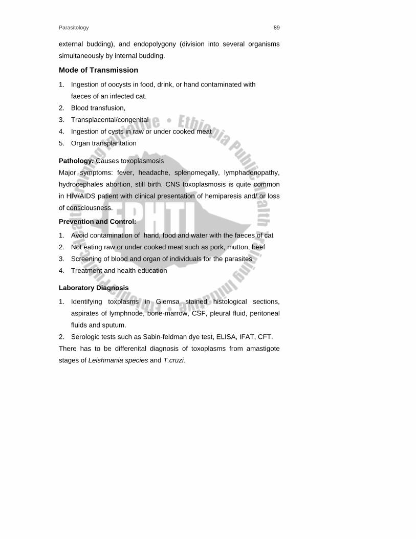
Parasitology
89
external budding), and endopolygony (division into several organisms
simultaneously by internal budding.
Mode of Transmission
1. Ingestion of oocysts in food, drink, or hand contaminated with
faeces of an infected cat.
2. Blood transfusion,
3. Transplacental/congenital
4. Ingestion of cysts in raw or under cooked meat
5. Organ transplantation
Pathology: Causes toxoplasmosis
Major symptoms: fever, headache, splenomegally, lymphadenopathy,
hydrocephales abortion, still birth. CNS toxoplasmosis is quite common
in HIV/AIDS patient with clinical presentation of hemiparesis and/ or loss
of consciousness.
Prevention and Control:
1. Avoid contamination of hand, food and water with the faeces of cat
2. Not eating raw or under cooked meat such as pork, mutton, beef
3. Screening of blood and organ of individuals for the parasites
4. Treatment and health education
Laboratory Diagnosis
1. Identifying toxplasms in Giemsa stained histological sections,
aspirates of lymphnode, bone-marrow, CSF, pleural fluid, peritoneal
fluids and sputum.
2. Serologic tests such as Sabin-feldman dye test, ELISA, IFAT, CFT.
There has to be differenital diagnosis of toxoplasms from amastigote
stages of Leishmania species and T.cruzi.

Parasitology
90
Pneumocystis carnii
Geographical Distribution: world wide
Habitat:-In the interstitial plasma cells of the lung (alveoli, epithelial cells)
of man and other animals
Morphology:-Trophozoite Foam-like mass in the lung
Amoeboid form
Has single nucleus
Cyst:- size: 4-5μm in diameter
Shape: spherical
Contents eight comma shaped sporozoites
Resembles a fungus
Life Cycle:-The complete life cycle of this parasite is unknown. The
trophozoite stage reproduces by binary fission. Man acquires infection
from droplets sprayed from the mouth.
Figure 2.17.Life cycle of P.carnii. From Hegazi M. Applied Human
Parasitology. 1st ed. 1994; Cairo, the Scientific book centers

Parasitology
91
Pathology: Causes a severe a typical interstitial plasma cell pneumonia,
impaired ventilation, and death due to respiratory failure. Common in
immunodeficient patients. P.carnii is a leading cause of death in patient
with AIDS.
Prevention and Control:
1. Sanitation of food and drink
2. Treatment of infected individuals
Laboratory Diagnosis
1) Finding the trophozoites and cysts in Geimsa stained
-sputum or bronchial biopsy
-Bronchial alveolar lavage
2) Serological detection using PCR, IFT
Blastocystis Hominis
Blastocystis is a common inhabitat of the human intestine. Previously it
was regarded as a non-pathogenic yeast; however, more recent studies
confirm its identity as a sporozoan and place it in the family sporozoa.
B.hominis is more than a harmless commensal or it is a potential
pathogen. It should be borne in mind when causes of intestinal disorder
are sought, esoecially in the absence of other identified causes of
diarrhea or gastrointestinal symptoms should be assessed for the
presence of B.hominis.
It is found in stool specimens obtained from asymptomatic individuals as
well as those with persistent diarrhea. Although it has been suggested
that the finding of large numbers of B.homins (5 or more organisms /oil
immersion field) in the stool of symptomatic patients consistent with its
role in disease. It may also be detected in wet mounts or trichrome-
stained smears of faecal specimen.

Parasitology
92
It has a round shape an about the size of an E.coli cyst but shows great
variation in size. It has a peripheral cytoplasm, a central vacuole but no
nucleus
Relevance to Ethiopia
There is only a few recent studies which determined the prevalence of
B.hominis infection in Ethiopia. In one study done in AIDS patient in A.A,
two cases among the 190 patients were detected (Fisseha B et al,
Ethiop J Health Dev. 1999).
Review Questions
1. What is the importance of studying intestinal and tissue sporozoa?
2. Explain the laboratory diagnosis of Isospora belli and C.parvum?
3. Discuss the sexual and asexual reproduction of T.gondii?
4. List the diagnostic and infective stages of the following parasites.
a. I. belli ______________ _____________
b. C.parvum ______________ _____________
c. P.carnii ______________ _____________
d. T.gondii ______________ _____________
5. Illustrate the developmental stages of parasites listed above.

Parasitology
93
2.4.2 Haemosporidia (The Malaria Parasites)
Malaria is the most important of all protozoan disease; it annually infects
over 250 million individuals and is a leading cause of illness and death in
the developing world. In many endemic areas it is becoming increasingly
difficult to control because of Anopheline mosquito vector and the
parasite to develope resistance to various eradication and treatment
options.
General Characteristics
1. Intracellular obligate parasites.
2. Man is intermediate host.
3. Female Anopheles mosquitoes are the definitive hosts.
4. Those species which infect human being are P.vivax, P.falciparum,
P.malariae and P.ovale
5. Has no animal reservoir host except P.malariae in which monkeys
are the reservoir hosts
6. Infective stage to man from the insect vector is sporozoites and to
the insect vector from man is gametocytes.
Geographical Distribution
Malaria is endemic in 91 countries with about 40% of the world
population is at risk. Plasmodium falciparum is the most prevalent
species in the hotter and more humid regions of the world. P.vivax is the
most widely distributed in the temperate, subtropics and some parts of
the tropics. Unlike the other species, it is more common and well adapted
to the temperate region than in the tropics.
P.malariae has much lower prevalence than P.vivax, P.falciparum and
P.ovale. It is confined mainly to tropical Africa. Also it is found in South
America and South west Asia. Infection rates in Ethiopia are about 60%,

Parasitology
94
40%, 1% and less than 1% for P.falciparum, P.vivax, P.malariae and
P.ovale, respectively.
Habitat: The parasite enters the blood and carried to the parenchyma cells of
liver, where they multiply enormously. This is called the pre-erythrocytic
or tissue phase. By rupture of the infected cells they enter the RBCs, the
erythrocytic phase (Schizogony) and reach all the organs of the blood via
the circulating blood, producing parasitaemia.
Morphology
There are sequential developmental stages with distinct morphological
features that helps for species identification in Romanowsky stains of
Peripheral blood films. This presented for each species below:
P.falciparum
Young Trophozoite (Ring forms)
Stage frequently found in blood film
Size: - Small rings, 0.15-0.5 diameters of RBCs which is unaltered
in size.
Shape: small fine pale blue ring
Chromatin: 1 or 2 small red dots.
Often with double chromatin dot
May lie on red cell membrane (accole forms)
Pigment: absent:
Mature Trophozoite Stage rarely seen in peripheral blood
RBC unaltered in size, sometimes stippled, pale
Shape: compact thin blue ring , comma or exclamation mark
shaped

Parasitology
95
Chromatin: 1 or 2 red dots
Pigment: black or dense brown mass
Schizont RBC unaltered in size , sometimes stippled, pale.
Size: parasite about 0.6RBC
Merozoites: 8-32; average 24
Pigment: clumped black
Not usually seen in peripheral blood
Gametocytes RBC is distorted.
Fairly frequently found
Size: larger than red cell
Shape: crescent or banana or kidney or sickle shaped
Rounded forms may be seen if film dries slowly
Stippling Maurer's cleft
Infected red blood cells All age group of red blood cells are infected.
Often contain several parasites
Density: often high density of parasites
Plasmodium Vivax
Rarely more that 2% of cells infected
Young Trophozoites
Size : 0.3-0.5 diameter of RBCwhich is unaltered in size.
Cytoplasm-circle, thin
Chromatin : fine dot some times two
Pigment: absent.

Parasitology
96
Mature Trophozoites
Size : large ( RBC enlarged,stippled)
Parasite- irregular amoeboid
Chromatin: Dots or threads of red colour
Pigment: Golden brown and scattered
Schizonts
RBC much enlarged
Size: Almost fills red blood cells
Shape: amoeboid or segmented, parasite large, filling enlaged RBC Cytoplasm: pale blue
Merozoites: 14-24; average 16
Pigment: Golden brown central loose mass
Gametocytes
RBC enlarged, stippled.
Parasite large, rounded filling enlarged RBC
Stippling
Schuffner's dot seen indistinctly around parasites.
Infected Red Blood Cells
Youngest erythrocytes are infected
Parasite Density: medium
Plasmodium Malariae
Young Trophozoite
Size: Up to 1/3 Red blood cell.
Cytoplasm -circle,thicker
Chromatin: one large red dot

Parasitology
97
Pigment: absent
Mature Trophozoite
RBC unaltered
Parasite- compact often band or rounded shape.
Chromatin: round do or red band
Pigment: dark brown or black pigment, often concentrated
along one edge of the band
Band forms can be seen in thin films
Occasionally “birds-eye” ring form may be seen
Schizont
RBC unaltered Size: Nearly fills red cells
Merozoites 6-12; average 8, sometimes forming rosette
Pigment: Brown aggregated in centre.
Gametocytes
RBC unaltered, parasite small round filling RBC
Infected Red Blood Cells
Oldest erythrocytes are infected
Stippling- None (Ziemanns dots often after prolonged leishmania
staining
Parasite Density: low density, rarely more than 1% of cells infected
(easily missed in Laboratory diagnosis)

Parasitology
98
Plasmodium Ovale
Young Trophozoite
Size: 1/3 Red blood cell
Shape: Regular compact dense blue ring (can resemble P.malariae
in thick films)
Chromatin : One medium sized red dot
Pigment: absent
Mature trophozoite
Size : Small
Shape: Round compact, very blue cytoplasm Chromatin: One large red dot Pigment: -small amount of brown pigment
Schizont
Size: small Shape: segmented Merozoites: 6-12; average 8 in a rosette Pigment: Brown
Gametocytes
Shape: large, oval or round, dense blue Pigment: Brown particles Difficult to differentiate from late trophozoites
Infected Red Blood Cells
Slightly enlarged Pale in colour Oval in shape, fimbriated at one side
Stippling
Has schuffner's (or James) dots. Youngest erythrocytes are infected

Parasitology
99
Density: Medium Density.
Rarely more than 2% of cells infected - About 20-30% of cells may become oval with fimbriated
( ragged ) ends
General Life Cycle of Malaria parasites:
-Malaria parasites require two hosts to complete their life cycle. Female Anopheles mosquitoes as the definitive host, where sexual reproduction (sporogony) takes place and human being as the intermediate host, where the asexual reproduction (schizogony) takes place.
- Sporozoites from infected female Anopheles mosquito are
injected with the saliva into the blood circulation of man when the
vector takes a blood meal. After circulating in the blood stream
for not more than one hour, the sporozoites enter into the liver
cells.
- Two cycles occur in man, in the liver as exo-erythrocytic
schizogonic reproduction and in the red blood cells as
erythrocytic schizogolnic reproduction. In the liver the parasites
multiply and develop into schizonts. When mature the schizont,
the liver cell rupture releasing large number of merozoites. The
merozoites enter the red blood cells and develop to trophozoite
stage. The trophozoite feeds on haemoglobin and forms malaria
pigment (haemozoin). The trophozoite stage develops into
schizontes.
- In the schizont nuclear division takes place to produce large
number of merozoites that are released from the schizont to
invades new red blood cells. After several erythrocytic
schizogonic reproductions, the merozoites develop into
gametocytes.

Parasitology
100
- To continue the life cycle, the gametocytes are ingested by a
female Anopheles mosquito while taking a blood meal. In the
stomach of the mosquito, the male and female gametocytes
under go fertilization and produces a zygote. the zygote develops
into a motile ookinete which penetrates the stomach wall of the
mosquito to form an oocyst. Inside the oocyst large numbers of
sporozoites are formed. The oocyst ruptures releasing the
sporozoites that also enter into the salivary gland to be
transmitted to another individual when the insect takes a blood
meal.
- Recrudescence (due to small number of erythrocytic parasites
remaining in the blood after a previous attack) occurs in
Plasmodium falciparum and Plasmodium malariae over a long
period of malariae infection.
- Relapse due to hypnozoite (dormant forms which precede
schizont development in the liver occur in vivax malaria and less
commonly in Ovale malaria.
Modes of Transmissions
1) Bite of infected female Anopheles mosquitoes. The main vectors
in Ethiopia are A.gambiae, A.funestus, A.nili, A. arebiansis and
A.pharonensis
2) Blood transfusion causes only erytrocytic infection.
3) Contaminated syringes and needles
4) Congenital / transplacental
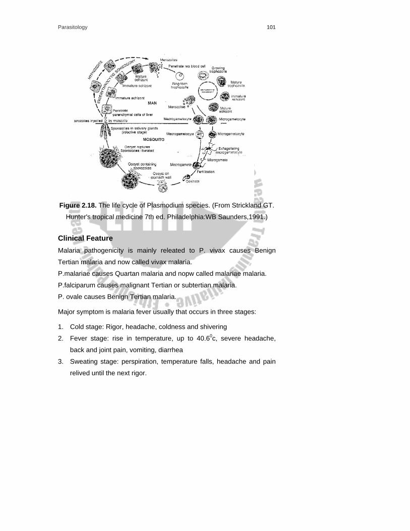
Parasitology
101
Figure 2.18. The life cycle of Plasmodium species. (From Strickland GT.
Hunter's tropical medicine 7th ed. Philadelphia:WB Saunders,1991.)
Clinical Feature Malaria pathogenicity is mainly releated to P. vivax causes Benign
Tertian malaria and now called vivax malaria.
P.malariae causes Quartan malaria and nopw called malariae malaria.
P.falciparum causes malignant Tertian or subtertian malaria.
P. ovale causes Benign Tertian malaria.
Major symptom is malaria fever usually that occurs in three stages:
1. Cold stage: Rigor, headache, coldness and shivering
2. Fever stage: rise in temperature, up to 40.60c, severe headache,
back and joint pain, vomiting, diarrhea
3. Sweating stage: perspiration, temperature falls, headache and pain
relived until the next rigor.

Parasitology
102
The malaria fever is due to the rupture of the infected red blood cells
containing mature schizonte stage releasing malaria pigment, toxins,
metabolic by products, debris of red blood cells and merozoites that can
infect other red blood cells
Level of Parasitaemia in the Red Blood Cells
P.falciparum. Up to 30-40% of the red blood cells can be infected, and it
is considered as sever if more than 5% of RBC are infected.
P.vivax. 2% of the RBC can be infected
P.ovale. more than 2% of the RBC can be infected
P.malariae. up to 2% of the RBC can be infected
Number of Merozoites Released From a Single Pre-erythrocytic Schizonts
P.falciparum: 30,000 merozoites
P.vivax: 10,000merozoites
P.ovale : 15,000 merozoites
P.malariae : 15,000 merozoites
Factors for Malignance of P.falciparum
1. Rapid multiplication
2. Infected red blood cells become "stick"
3. Infects all age group of red blood cells
4. A single red blood cell can be infected by more than one parasites
5. Erythrocytic schizogonic reproduction takes place in the deep
capillaries of organs such as brain, lung, heart, spleen, bone-
marrow, placenta, intestine, etc.

Parasitology
103
Factors That Provide Protection against Malaria Infections are:
1) Glucose-6-phosphate dehydrogenase deficiency , Scickle cell
anemia Ovalocytosis and Adenosine tri-phosphate deficiency in
non-immune black males provides protection against P.falciparum
infection
2) Thalassemia, Duffy blood group antigens (i.e., Fya and Fyb) negative
RBCs and Hemoglobin E provides protection against P.vivax
infection.
3) Human fetal hemoglobin gives protection against all forms of malaria
infection
Dominance of Malaria Parasites
P.falciparum repress the parasitaemia of P.vivax and P.malariae
P.vivax dominates over P.malariae
Prevention and Control
Prevention and control of malaria become difficult as a result of
resistance of the parasites to the drugs and failure of control measures.
Besides this population movement, climatic changes and economic
problems are also considered currently as factors that related with
malaria spread. However, the following are some of the measures to be
taken as prevention and control measures;
1) Avoid mosquito bites by
- Selecting healthy sites for houses and screening windows and
doors with mosquito net.
- Using mosquito bed nets
- Wearing protective clothes such as long trouthers
- Using mosquito repellents

Parasitology
104
2) Destroy adult mosquitoes by
- Indoor residual regular effective spraying
3) Preventing breeding of mosquitoes by
- Altering the habitat to discourage breeding
- Flooding or flushing of breeding places
- Drainage to remove surface water, filling in ponds, pot holes, etc.
- Spraying breeding places with effective chemicals particularly with
larvicides
4) Using drugs to
- Prompt diagnosis and treatment of malaria cases
- Prevent infections using chemoprophilaxis, especially in non-
immune persons visiting or going to malarious areas or in
persons with reduced immunity such as pregnant women.
5) Health education.
6) Blood screening for malaria before providing for those who need
blood.
Laboratory Diagnosis
1) Malaria parasites are detected in thin or thick blood films stained by
wright’s stain, Giemsa stain, leishman stain or Field stain. Take blood
films when the patient feels febrile because the parasites are usually
most numerous in the blood towards the end of an attack of fever.
Always collect the blood before anti-malarial drugs are taken.
Field stain is recommended for smears stained straight away and
Giemsa stain for smears to be stained after a few days.
2) Using a rapid immunodiagnostic tests such as ParaSight F, ICT
malaria Pf / Pv and, OptiMALr. These are recently discovered
groupof techniques, which proved tobe valuable and highly sensitive

Parasitology
105
and specific tests. All of themdepend on the “dipstick” format and
they are termed collectively as”rapid test for malaria.
- The principleof these test kits depends on detection of P.falciparum
Histidine Richprotien-2 antigen (Pf HRP-2)which is only produced by
P.falciparum or by detection of Plasmodial Lactate Dehydrogenase
enzyme(LDH) isoenzymes produced by both P.falciparum and
P.vivax.
- Detection of PfHRP-2 can not differentiate between dead and live
parasites, this antigen canpersist in the serum for months after
parasitological clearance of the disease. On the other hand,
identification of specific LDH verifies live parasite only and inturn it
appears more specific fordiagnosisof active infection.
3) ELISA
4) PCR
Babesia
Apicomlexan parasites belonging to the genus Babesia have long been
known as parasites of domestic and wild animals, causing at times
inapparent infections but also causing such economically important
disease as Texas cattle fever and malignant jaundice in dogs. Humans
are accidental hosts. It is transmitted in nature by ixodid, or hard -bodied,
ticks.It can be differentiated from malaria by the absence of pigment
within infected erythrocytes. The organisms infect the red cells, in which
they appear some what poleomorphic ring like structures. Most
resembles ring stage of Plasmodia. The small parasites appearing much
like P.falciparium can be differentiated from malaria parasite by the
absence of pigment in the infected erythrocytes.

Parasitology
106
Human infection is diagnosed by identifying the intra-erythrocytic parasite
in Giemsa -stained blood films. It can also be diagnosed by serologic test
with the indirect immunoflorescent antibody test; which is the most useful
in diagnosis.
Relevance to Ethiopia:-
Tropical Africa is generally endemic to falciparum malaria and due to the
absence of Fya/Fyb duffy antigens. It is however reported to be allopatric
to P. ovale, which is endemic in the western part of the continet. Based
upon this geographical distribution. Western and south western Ethiopian
lowlands lye at the eastern tip of the P. ovale belt. thus Setit Humera
along with Gambela and Arbaminch, was identified as one of the
endemic sites of ovale malaria.
The rise of the sibling species, P. vivax. in once P. ovale endemic area
is considered to be related to the migration of settlers and agricultural
laborers form the P. falciparum: P. vivax codominant north central
Ethiopian highlands . On the other had the prevalence of P. malariae in
Humera, Like the rest of Ethiopia, Is descrete and focal. The overall
prevalence of P. malaria was shown to 1% and below.
Malaria in the lowlands has contributed to overpopulation, overcultivation,
deforestation, soil erosion, and drought and famine in the weyna dega
and dega zones of the Ethiopian Highlands. According to the 1984
census, about 37% of the Ethiopian population lived above 2,200 m, 45%
between 1,500 and 2,200m, and the remaining 18% below 1,500 m.
Encouraged by the initial success of the malaria Eradication Service in
the 1960s and increasing economic opportunities, many highland people
settled in, or seasonally worked in, the lowlands, a trend which
accelerated during the government’s resettlement program in the mid-
1980s and subsequent famines and military activities.

Parasitology
107
With the exception of perennial transmission which prevails around lakes
and swamps and irrigation systems and riverine areas of the large rivers,
particularly the Awash, Baro, Blue Nile, Didesa, Beles, Takeze, Wabe
Shebele, and Omo, malaria is highly seasonal in the kolla and weyna
dega A. gambiae s.l. is found in all administrative regions of Ethiopia.
This is the second most frequent and widely distributed vector of malaria
in Ethiopia comprising 18.9% of the 19,352 specimens collected during
the period 1984 – 1988.
An. funestus is the third most common vector of malaria in Ethiopia,
comprising 3.2% of all the specimens collected during the period 1984-
1988.
An. Nili is the least common species, and it is more localized, being
confined to the southwestern, western, and northwestern parts of the
country.
Anopheles culicifacies adenensis is the only species found in the Aseb
port area along the Red Sea coast, where there is a known indigenous
transmission of malaria.
Of the four Plasmodium species known to cause human malaria in
Ethiopia, the two epidemilogically important species are Plasmodium
falciparum, comprising 60% of all the cases of malaria, and Plasmodium
vivax, constituting nearly 40% of all malaria cases. Plasmodium malariae
comprises less than 1% of all cases and is most frequently reported from
the Arba Minch area. Plasmodium ovale is rarely reported. The
geographic distribution of all four parasites at the regional level was
mapped by Gebremariam.
Parasitology
108
Plasmodium falciparum
P. falciparum causes the most frequent and fatal episodes of malaria in
Ethiopia. It is the cause of severe and complicated malaria in which the
case fatality rate is about 10% in hospitalized adults and up to 33% in
children less than 12 years old. A study conducted on 1,261 subjects
from eight different Ethiopian ethnic groups revealed that glucose-6-
phosphate dehydrogenase deficiency was present only in the Anuak and
Nuer of the southwestern lowlands, as well as in the Afar in the Danakil
Depression, with a prevalence of 1.4%, 6.7%, and 6.3%, respectively.
Only one case of sickle cell trait (Hb As) was found in the same study.
Plasmodium vivax
P. vivax is also widely distributed in Ethiopia and often precedes the
transmission of Plasmodium falciparum. P. vivax is more common during
the dry season, and whether this is due to active transmission or
relapses has not been clearly determined. This species is not a major
cause of mortality but is an important cause of morbidity due to the
phenomenon of relapse. A study conducted in the Gambela area
demonstrated that residents speaking Nilotic languages are more
resistant to P. vivax infections than are the more recent arrivals to these
lowlands, who speak semitic and Cushitic languages. Later studies
established the absence of Duffy antigen in Nilotic speakers.

Parasitology
109
2.5 Class Ciliatae (Ciliates)
Balantidium coil
Geographical Distribution: World wide being more commonly found
amongst those who keep pigs, and uses pig faeces as fertilizer
vespecially in warmer climates. In Ethiopia it was reported from Debre
Brahne.
Habitat: Trophozoite and cyst in the large intestine of pig and rarely
man.
Morphology:
Trophozoite
Size: 50-200 μm by 40-70μm
Shape: Oval, with one pole more rounded than the other.
Motility: Rapid motility, crossing the field in a definite direction and
sometimes turning in circle.
Cilia: Cover the whole body and many around the cytostome
Nucleus: a large kidney shaped macronucleus and a small
micronucleus .
Cytostome: a sort of “mouth” that contracts and expands to draw in
debris also has cytopyge
Cyst
Size : 50-70μm, very large cyst /the size of a round worm egg.
Shape: round
Shell: thin double wall
Nuclei: one large kidney shape macronucleus and one small
Micronucleus beside the large nucleus.
Cytoplasm: granular, greenish, filled with inclusion bodies.

Parasitology
110
Life Cycle
As soon as the thick wall cysts are excreted in the feces, they are
infective. Man acquires infection from contaminated food or drink or from
hands contaminated with faeces. Following ingestion, the cyst excyst in
the intestine producing trophozoites; where each cyst producing a single
trophozoite. The ciliate multiply in the colon by simple binnary division
often following conjugation. Conjugation takes place by a process, when
the two trophozoite are found to be in state of union and enclosed in a
cyst, with the exchange of nuclear material. This is followed by
separation of the two rejuvenated individuals. Pig is the main reservoir
host.
Figure 2.18. Life cycle of B.coli.(From Nasr NT.Review of Human
Parasitology,2nd ed. Cairo: The Scientific Book Center)
Pathogenicity:- It is the only ciliate that parasitize humans. Causes
balantidial dysentery. Infection with B.coli can be without symptoms
unless the ciliates invade the intestinal wall. Invasion can cause

Parasitology
111
infammation and ulceration, leading to dysentrywith blood and mucus
being passed with faeces.
Prevention and Control:- 1. Avoid contamination of food or drink.
2. Improving personal hygiene especially those who keep pigs.
3. Treatment and health education.
Laboratory Diagnosis:-Finding the tropozoites in dysenteric faecal
specimens and the cysts in formed or semiformed faeces.
- In dysenteric specimens the ciliates usually contain ingested red cell.
Relevance to Ethiopia
The parasite is not common in Ethiopia A few cases have been reported
in Debre Berhan.
Review Questions 1. What characters help to differentiate one plasmodium parasite from
other species?
2. What is the significance of thin and thick blood in the laboratory
diagnosis of malaria parasites?
3. What factors give natural protection against plasmodium infection?
4. Which plasmodium species is more malignant that others? Why?
5. Explain methods used to estimate parasitemia load of malaria
parasites.
6. Identify a ciliate which infect human.
7. What is the diagnostic and infective stage of the above parasite?

Parasitology
112
CHAPTER THREE
Medical Helminthology
Helminths (Worms)
The word, helminths from Greek means "Worm" and originally referred to
intestinal worms but it is more usually interpreted to include both parasitic
& free-living species of round worms (phylum Nemathoda )"hair snakes"
or gordiid worms (Phylum – Nematomorpha), tubellarians, flukes & tape
worms ( phylum-plathyhelminthes) & thorny - headed worms (phylum-
Acanthecephala). The helminths are generally macroscopic, and the
adult worms vary tremendously in size from barely visible to 10 meters in
length.
The life cycles of helminths may be quite complex and include both direct
and indirect cycles. The clinical sign and symptoms of helminthic
infections depend on the location of the organisms and may be caused
by adults, larva, or eggs. The host response to the presence of parasite
may be prominent and often includes eosinophilia, especially in the early
stages of infections when the parasites are in tissue. The final diagnosis
is usually dependant on detection and identification of a mature or
developmental (larva, embryo, egg) stage of the parasite. Occasionally
the diagnosis may be made clinically or serologically.
The majority of helminths produce characteristics eggs that are passed in
feces and serve as the chief means of diagnosing infections. The
identification of eggs should be approached in a systematic manner
taking into account the size, and shape of the egg, the thickness of the
shell, the presence or absence of specialized structures such as spines,

Parasitology
113
knobs or opercula. The presence and characteristics of larva present
within the eggs may be useful.
3.1 Platyhelminths
3.1.1 Class Cestoda (Tape worms)
Tape worm infection in man is less limited in their distribution than are
human fluke infections. They are frequently not restricted to any
specialized group of intermediate hosts, as species- of Mollusks that may
be local in their distribution or confined by certain meteorological
conditions. Some of them are dependent primarily on exclusively on man
as a definitive host for the continuation of their life cycles (T.segina,
T.solium, H. nana D.latum); on the other cases man is incidental to
continued propagation of the parasite (E.granulosus, H. duminuta and
Diplidium caninum). In all known tapeworm infection, except in certain
varieties of sparganosis, in which exposure in topical, the portal of entry
is mouth hence, strict care not to swallow raw or inadequately, cooked
beef, pork or fish or food or water contaminated with faeces or vermin,
will ensure protection of the individuals.
General characteristics:
1. Dorso-venterally flattened (leaf or tape-like)
2. Bilaterially symmetrical.
3. They are provided with a nervous system and an elaborate excretory
apparatus.
4. Digestive system may be absent, or when present it is rudimentary
and without anus. It obtains its nutrient by absorption through cuticle.
5. Respiratory, circulatory system and body cavity are absent.
6. Tape worms are hermaphrodites and have well developed
reproductive system.

Parasitology
114
7. Each unit of chain (segements) is known proglottides. The entire
chain of proglottids is called strobila
8. The body is divided into three main body regions; this are
Head (scolex): attachment organ and may have grooves, suckers, and
rostellum armed with hooklets; this varies with species.
Neck: growth region, proglottids proliferate from this region.
Strobila: varies in number, shape, size, and maturity. It is divided into
three regions:
a/ immature : sex organs are immature.
b/ mature: sex organs are fully mature.
c/ gravid : reduced or atrophied primary genital organs,
uterus is filled with eggs
Figure 3.1.Body parts of tape worm. (From Jeffrey HC and Leach RM.
Atlas of Medical Helminthology and Protozoology, 1975.)
N.B: Infection persists as long as the scolex and the neck region
remain attached to the intestinal wall.

Parasitology
115
9. The entire body is covered with active homogenous, elastic,
resistant and continuous cuticle/integument from one proglottid to
the next through out the entire body.
10. Elaborate and well developed reproductive system with complete
set of male and female genital organs which are found in a single
worm.
11. Man is;
a. The only or main definitive host for T.saginata, T.solium, H.
nana and D.latum
b. Intermediate host for E.granulosus and E.multilcularis
c. Both as definitive and intermediate host for H.nana and
T.solium
Based on the following differences criteria, cestodes are classified into two orders:
Order- Pseudophillidea Order-Cyclophillidea Differences 1. Scolex - Spoonshaped, grooves - globular with 4 suckers
2. Genital pore - venteral - marginal
3. Utrine pore - Present(ventral) - absent
4. Uterus - coiled - sacular tubular or branched
5. Ova - operculated - non-operculated
6. Onchosphere - ciliated - non-ciliated
7. Rostellum - absent - present
8. Progilottids - broader than long - longer than broader
9. Larval forms - solid - cystic

Parasitology
116
Figure 3.2.Criteria used for the differentiation of Pseudophyllidea from
Cyclophilidea. (From Jeffrey HC and Leach RM. Atlas of Medical
Helminthology and Protozoology, 1975.)

Parasitology
117
Egg: - Two type
1. Operculated, immature when voided to the external environment.
2. Non-operculated ,fully embryonated when voided to the external
environment.
Larvae: -Generally two type
I. Solid : eg. Procercoid, Plerocercoid, cysticercoid
II. Cystic( true bladder): can be with:- Single scolex eg. Cysticercus;
Many scolexes and/or with daughter cyst eg. hydatid cyst, coenurus
cyst, etc.
Taenia saginata
(Beef tape worm)
Geographical Distribution
World wide distribution where cattle are raised and beef is eaten raw or
under cooked. More common parasite of man unlike Taenia solium. It is
very common in Ethiopia.
Habitat Adult: In small intestine of man
Larvae: In muscular tissues of cattle
Eggs: In faeces of man or in gravid segments.
Morphology: Adults: Size: Total worm: 3-10 m long
Mature segment: 1-2cm long.
Colour: ivory white
Scolex (head): quadrate, with four
suckers, no looks, no rostellum
Strobila : 1000-2000 proglottides

Parasitology
118
Mature Proglottides:
- Broader than long
- Genital pores are arranged irregularly alternate on the lateral margin
of each segment
Gravid proglottide - Detach when fully develop and pass through the anus
independently.
- 15-30; average 10 compound lateral uterine branches.
Larvae: Known as Cysticercus bovis
Found in skeletal and muscular tissues of cattle
Has four suckers and no rostellum and hooklets
Egg (Embryophore): -
identical with the egg of T.solium.
Size: - 33-40 μm
Shape: -Round
Colour: - Shell-dark yellowish-brown, content light
yellowish gray.
Shell:-Thick, Smooth, brown, radially straited (embryophore)
Content: - A round granular mass enclosed by a fine
membrane with six hooklets
Stains red (acid fast) in Ziehl-Neelsen staining technique, this
character helps to differentiate from T.solium which do not have red
color in such staining.
Life cycle Requires two hosts to complete its life cycle. Man as a definitive host and
cattle as intermediate host.
Egg(hexacanth embryo→larva(Cysticercus bovis) →Adult

Parasitology
119
Man acquires infection from raw or under cooked infected meat.
Following ingestion, the larvae become attached to the wall of the small
intestine with its suckers. Proglottides are formed from the neck region
and the larvae grow into a long adult tapeworm. When fully developed
the gravid segments become detached and the eggs are discharged only
after the gravid segments have been separated from the worm. Gravid
segments containing eggs and eggs from ruptured segments are passed
in the faeces. Cattle ingest the eggs with contaminated grass while
grazing. The embryos escape from the eggs and pass through the
intestinal wall into a blood vessel. Through the blood circulation they are
carried to muscles and develop into infective cystic larvae called
cysticercus bovis.
Figure 3.3. Life cycle of Taenia species. (From Nasir NT.Review of Human Parasitology,2nd ed.
TheScientific Book Center, Cairo)

Parasitology
120
Pathogenicity: - Causes taeniasis
Major symptoms are loss of appetite, weight loss, hunger, acute intestinal
obstruction, eosinophilia, and discomfort by the crawling of segments
through the anus. Proglottides of T.saginata have a strong tendency to
crawl from the anus during the day when its host is active unlike
Entrobius vermicularis.
Prevention and Control
1. Avoid eating raw or insufficiently cooked meat which may contain
infective larvae.
2. Inspecting meat for larvae
3. Provide latrine for the proper waste disposal
4. Not using untreated human faeces as fertilize for pasture land
5. Protection of cattle from grazing on faeces or sewage polluted grass.
6. Treating infected person and providing health education
Laboratory Diagnosis
1. Detecting eggs in faeces. Morphologically eggs of T.saginata and T.
solium are indistinguishable unless stained by AFB.
2. Identifying gravid segments and scolex recovered from clothing or
passed in faeces. Single rectangular segments found in underclothes
and bed clothes.
Relevance to Ethiopia
Taeniasis. High T.saginata infection rates in Ethiopia are due to the
widespread custom in the agricultural highlands of eating raw beef, and
the habit of defecating in open fields coupled with the tradition of allowing
cattle to graze in such fields. Infections are under-reported. Owing to the
inherent problem of missing many infections during routine stool
examination. Estimates made by different investigators of the prevalence

Parasitology
121
of taeniasis in Ethiopia vary widely, from 2% -16% to over 70% (Kloos H
et al, 1993, Yared M et al 2001).
Taeniasis is so common in the country and the tradition of self-treatment
is so well developed that most people do not use the health services for
diagnosis and treatment. Instead, Ethiopians use about dozens of
traditional plant medicines, including Kosso (Hagenia abyssinica).
Enkoko (Embelia schimperi), and Metere (Glinus lotoides) upon noticing
proglottids in the feces or when experimenting abdominal discomfort,
usually every 2 months (Shibru T, 1986).
Taenia solium
(Pork tape worm)
Geographical Distribution:-Widely distributed where human faeces
reach pigs and pork is eaten raw or insufficiently cooked.
Habitat: Adult: In the small intestine of man
Larva: In muscular tissues of pig
Egg: In the faeces of man and in gravid segment.
Morphology: Adult Size: Total worm 2-7m
Mature segment :0.5-1.5 cm
Colour: pale blue
Scolex (head): Quardate with four large deep suckers, rostellum
with two rows of hooklets

Parasitology
122
Strobila: 800-1000 Proglottides
Immature proglottide are broader than long
Mature proglottide are nearly square and genital pores are arranged
irregularly alternate on the lateral margin.
Gravid Proglottides:- 7 to 12, on average 10 lateral compound uterine
branches. Small chains of 3-4 rectangular segments found in the faeces.
Larvae:- Known as Cysticercus cellulosae
Found in skeletal and muscular tissues of pig
Has four sucers, rostellum and two raws of hooklets
Egg (embryophore): -morphologically identical with the egg of
T.saginata.
Size: - 31-43 (m
Shape: -Round
Colour:- Shell-dark yellowish-brown, content light yellowish gray.
Shell: -Thick, Smooth, brown, radially straighten (embryophore)
Content: - A round granular mass enclosed by a fine membrane with 6
hooklets.
Does not stains red (acid fast) in Ziehl-Neelsen staining technique
Life Cycle:
The life cycle of T.solium is similar to that of T. saginata except pig
serving as an intermediate host for the development of larvae known as
Cysticercus cellulosae.
Egg(hexacanth embryo) →larva(Cysticercus cellulosae) →Adult

Parasitology
123
Man acquires infection from eating raw or under cooked pork that
develops into adult in the intestine or from contaminated food or drink
with faeces containing the eggs and develops into larval stage in visceral
organs.
Mode of Transmission can be - Eating raw or under cooked pork meat
- Eggs in food or drink
- Internal autoinfections
Pathology: Taeniasis and cysticercosis
Major symptoms are as a result of the adult worm. These include
abdominal pain, loss of appetite, and infection with larvae cause cystic
nodules in subcutaneous and muscles. If it is in the brain it causes
epilepsy and other CNS disorders.
Prevention and Control:
1. Avoid eating raw or insufficiently cooked pork meat
2. Ensuring pigs do not have access to human faeces.
3. Inspecting meat for larvae
4. Treating infected person, providing health education and adequate
sanitary facilities
Laboratory Diagnosis
1. Detecting eggs in the faeces which is morphologically
indistinguishable from the egg of Taenia saginata.
2. Identifying gravid segments and scolex in the faeces after treatment.
3. Finding calcified larvae in histological or X-rays examination .

Parasitology
124
Relevance to Ethiopia
The parasite has not been reported from Ethiopia. The presence of
T.solium in Ethiopia is doubtful at though some people keep pig the main
reason for its possible absence is the fact that both Moslems & orthodox
follow the religious prohibition against eating pork (Kloos H et al,1993).
Hymenolepis nana
(Dwarf Tape Worm)
Geographical Distribution:-
H.nana is widely distributed in countries with warm climates than in cold
climates and fairly common in Ethiopia. Children are more commonly
infected than adults.
Habitat: Adult: small intestine of man, rat and mice
Cysticercoid larvae: in the intestinal villi of man, rat and mice.
Eggs: In the faeces of man, rat and mice
Morphology:
Adult Size: 10-44 mm
Scolex with 4 suckers, short retractile rostellum with single crown of
hooklets.
Stroblia: 100-200 proglottides, the size is inversely proportional to the
number of worms present in the intestine of their host.
Mature Segment: Unilateral common genital pore
80-180 eggs in gravid segment
Egg:
Size: 35-50μm
Shape: oval, almost round

Parasitology
125
Shell: double; thin external membrane and internal membrane often
thicker at the poles. Thread like polar filaments coming from both
poles.
Colour: colour less or very pale gray
Content: Rounded mass (embryo) with six refractile hooklets arranged in
fan shaped.
Life Cycle: H. nana has a direct life cycle with a human host serving as
both definitive and intermediate host.
Egg(hexacanth embryo) →Cysticercoide larvae→ Adult
Eggs are the infective stages which are ingested contaminated hands,
food and drink. Following the ingestion of eggs, the embryos are freed in
the small intestine. They penetrate villi and develop into infective
cysticercoid larvae. When fully mature, the larvae rupture out of the villi
into the lumen of the intestine. They attach to the intestinal wall by their
scolex and grow rapidly into mature tapeworms. Gravid segments
detached and eggs are released in the intestine. Some of the eggs are
passed in the faeces while others remain in the intestine to cause internal
autoinfection. The eggs are infective when passed in the faeces.
Mode of Transmisssion: - 1. Ingestion of egg with contaminated food, drink of finger.
2. Autoinfection.

Parasitology
126
Figure 3.4. Life cycle of H.nana (From Nasir NT. Review of
Human Parasitology,2nd ed. Cairo:The Scientific Book Center.)
Pathology: Although many H.nana tapeworms can be found in the
same host due to internal autoinfection, the life span of the adult worm is
only a few month. Symptoms of infection are rarely detected except in
children when many tapeworms may cause abdominal pain, diarrhoea,
anorexia and lassitude. Toxins released from the worms can cause
allergic reactions.

Parasitology
127
Prevention and Control:
1. Personal hygiene, washing of hands before eating and after
defecation
2. Sanitary disposal of faeces into latrines
3. Avoiding eating uncooked food
4. Protection of food, and drink from contamination with faeces
5. Treatment and health educations.
Laboratory Diagnosis
1. Usually eggs of the parasite in the faeces.
2. Some times adult worms in the faeces
Hymenolepis diminuta
(Rat tape worm)
Geographical Distribution: Cosmopolitan with sporadic human infection
in the world. It is fairly common in Ethiopia.
Habitat: Adult: Ileum of rat, mice and rarely man
Larva: body cavity of insects (fleas and cockroaches)
Egg: In the faeces of rat, mice and man
Morphology:
Adult -
20-60 cm
Scolex with 4 suckers and retractile rostellum without hook-lets.
Colour: Transparent or pale yellow
Stroblia: 800-1000 segments
Egg
Yellow-brown or bile pigmented.

Parasitology
128
70 by 60μm
Shell with double shell and with out thread like polar filaments.
Content: A rounded embryo containing six hooklets arranged in fan
shape.
Life Cycle:
Requires two hosts to complete its life cycle. Intermediate hosts are
fleas, beetles, cockroaches. Definitive hosts are rat, mice and man.
Egg(hexacanth embryo) →Cysticercoide larvae→Adult
Eggs in the faeces of the definitive hosts are ingested by the insect
vectors and hatches releasing the oncosphere. The oncosphere migrate
into the body cavity and develop into cysticercoid larva. Man acquires
infection by accidentally ingesting the infected insect vector.
Figure 3.5. Life cycle of H.dimunita(From Jeffrey HC and Leach
RM. Atlas of Medical Helminthology and Protozoology, 1975.)

Parasitology
129
Pathology: Causes hymenolopiasis and major symptoms are abdominal
pain, diarrhoea, restlessness.
Prevention and Control
1. Avoid insect vectors
2. Avoid their reservoir hosts
3. Protection of food and drink from insect vectors
4. Health education
Laboratory Diagnosis
1. Eggs in the faeces
Relevance to Ethiopia
Hymenolepiasis. Both Hymenolepis nana and H. diminuta have a
cosmopolitan distribution, but H.diminuta is relatively rare and its
existence in Ethiopia has been revealed only recently. H. nana is fairly
common in Ethiopia and most common in children and is easily
transmitted from person to person through the feco-oral route. In a
survey of schoolchildren in 26 towns and villages in Harerge, only 1 case
of H.diminuta but 65 cases of H. nana (in 20 communities) were found.
The highest recorded prevalence of H. nana in Ethiopia was 61% among
school children in Kemise town in southern Wello. Of 50 communities in
the central and northern highlands, 78% had positive cases, with a mean
prevalence of 12%. Among the general population of the Lake Zeway
islands and the outpatients of Zeway Health Centre, H.nana was found in
6.9% and 2.0%, respectively (Kloos M et al. 1993).

Parasitology
130
Echinococcus granulosus
(Hydatid worm)
Geographical Distribution:-Common in sheep and cattle raising
countries mainly in Kenya, Middle East, North and South Africa, India,
Australia. Also it is found in South and South East Ethiopia.
Habitat Adult: mucus membrane of small intestine of carnivores such as dog, fox,
Hydatid cyst/larvae: in the different body parts (liver, lung, brain, etc) of
man and herbivorous animals.
Egg: in the faece s of dog, fox, and jackals
Morphology
Adult Size: 3-6mm
Scolex with 4 suckers, rostellum with two rows of hooklets.
Stroblia: three proglottides. One immature, one mature and one
gravid segment.
Egg: morphologically indistinguishable from the ova of Taenia species
Size: 30-37μm
Larvae (Hydatid Cyst)
Contains brood capsules with many scolice, free brood capsules and
scolice, hydatid sand, cystic fluid containing salt, enzymes and toxins.

Parasitology
131
Life Cycle:-
Figure 3.6. Life cycle of E.granulosus. (From Hegazi M. Applied Human
Parasitology. 1st ed. 1994; Cairo, the Scientific book centers)
Requires two hosts to complete its life cycle. Carnivores such as dog,
fox, jackles are the definitive hosts, man and herbivorous animals.
Egg (hexacanth embryo) →Hydatid cyst(larvae) → Adult
Man acquires infection from ingesting eggs in contaminated food, drink
and fingers. The eggs hatche in the intestine and penetrate the intestinal
wall and disseminated through out the body through the blood stream
and become hydatid cyst. There is no development of the parasite as
adult forms in man.

Parasitology
132
Mode of Transmission:
- Contaminated food, drink or finger with infected faeces of dog, fox,
Jackals.
- Handling infected dogs.
Pathology: Causes hydatid disease.
Major symptoms are obstruction and pressure on vital organs,
anaphylactic shock due to rupture of the cyst, Jacksonian epilepsy,
jaundice, erosion and fracture of bones.
Prevention and Control:
1. Personal hygiene, washing of hands before eating
2. Avoid handling dogs
3. Avoiding eating uncooked food
4. Protection of food, and drink from contamination with faeces
5. Treatment and health education.
Laboratory Diagnosis
1. Histological examination to find larvae
2. X-ray examination to find larvae
3. Examination of cystic fluid for brood capsules and protoscoleces
4. Casoni's skin test
Relevance to Ethiopia
This parasite is common among the pastoral people in the South and
South-East of Ethiopia. These people have close contact with their dogs.
Although rarely indicate that settled agriculturalists as well as urban
dwellers do have the parasite (Shibru T, 1986).

Parasitology
133
Diphyllobothrium latum
(Fish tapeworm)
Geographical Distribution:-Widely distributed in the lake areas of
Europe, Asia, Far East, North America, South America and Central
Africa.
Habitat Adult: small intestine of man, cat, dogs, pig
Eggs: passed in the faeces of man
Larval forms:
Coracidium: free in water
Procercoid: body cavity of copepod / Cyclopes
Plerocercoid (sparganum) larvae: in the flesh of fresh water
fish such as pike, perch, salmon, eel, barbel, ruff, trout
Morphology Adult: the largest tapeworm
Size: 10m or more
Grayish-yellow in colour
Scolex is elongated, spoon shaped, longitudinal suctorial
groove/bothria/slits with no rostellum and hooklets.
Long and slender neck
Strobila: 3000-4000 proglottids
- Broader than long
- Mature and immature segments can not be distinguished.
- Genital pore and uterine pore open on the ventral surface
- Has coiled uterus opened by uterine pore for the passage of
individual egg
- Gravid segments are retained by the worm, and eggs are discharged
periodically but as they cease to function gradually disintegrate.

Parasitology
134
Egg: 58-76μm by 40-51μm Broadly ovoid Light golden yellow, Operculated Thick shell Contains immature embryo
Life Cycle: - D.latum requires three hosts to complete its life cycle. Definitive hosts: man dog, pig Intermediate hosts: - Primary Intermediate hosts: Crustacean - Secondary Intermediate hosts: Fresh water fish Man acquires infection by eating raw or inadequately cooked fresh water fish.
Figure 3.7. Life cycle of D.latum (From Brown HW and Neva FA, Basic
clinical parasitology, 5th ed. USA: Applenton- century Crofts,1983).

Parasitology
135
Pathology: Competes for vitamin V 12 and and cause megaloblastic
anemia.
Major symptoms are abdominal pain, diarrhea, constipation, loss of
weight, intestinal obstruction, pernicious anemia and eosinophilia.
Prevention and Control:
1. Avoid eating raw or undercooked fish
2. Proper disposal of faeces
3. 0Fish inspection for larvae
4. Treatment of infected individuals and health education.
Laboratory Diagnosis
1. Eggs in the faeces
2. Scolex in the faeces
3. Adult worms in the faeces
Relevance to Ethiopia
A few people, particularly indigenous inhabitants of the lake Zway
Islands, eat row fish as part of their diet, but stool exam. of 100 local
fisherman didn't reveal any eggs of fish tapeworm, D. latum ( Yared M,
et al, 2001).
Dipylidium caninum
(Dog Tapeworm)
Geographical Distribution :-World wide distribution
Habitat: Adult: mucus membrane of small intestine of carnivores such as dog,
cat, Man Cysticercoid larvae: In the body cavity of insects
Egg: in the faeces of dog, cat, man

Parasitology
136
Morphology
Adult 20-60cm
Scolex with 4 suckers, retractile rostellum with several rows of
hooklets.
Stroblia: many proglottides each with two sets of genital organs.
Egg: 5-15 eggs in capsule
40μm in size and yellowish brown in color
Larvae(Cysticercoid):-Evaginated scolex with several rows of hooklets
Life Cycle:-Requires two hosts to complete its life cycle. Carnivores
such as dog, fox, and occasionally man are the definitive hosts, and fleas
and other insects are intermediate host. Man acquires infection from
accidentally ingesting infected insect vectors.
Pathology: Causes dipylidiasis
Prevention and Control: Similar to E.granulosus
Laboratory Diagnosis:-Finding gravid segments and eggs in the feces
Review Questions 1. eggs in contaminates food of drink? 2. List parasites that have auto infection in their life cycle.
Enumerate the possible diagnostic stages of cestodes
3. Identify a cestode that does not require intermediate host in its life cycle.
4. What are the differences and similarities between T.saginata and T.solium?
5. Among the cestodes which of them are acquired by ingestion of 6. At makes T.solium more sever than T.saginata? 7. What are the basic differences between the two order of cestodes?

Parasitology
137
3.1.2 Class Trematoda (Flukes)
Trematodes or flukes are members of the platyhelminths and are
generally flat, fleshy flat, leaf shaped worms. Trematods vary in size
from species just visible to the naked eye, like H.heterophyes and
M.yokogawi to large fleshy species, like Fasciola and Fasciolopsis. The
most Characteristic external structures are the acceptable (i.e., suckers)
which caused the early workers in this group to call them trematoda, i.e.
"body with holes." They posses two muscular suckers: one oral,
surrounding the opening to the primitive digestive tract, and one ventral
sucker for attachment. The digestive system consists of a muscular
pharynx and esophagus and bilateral ceca that end blindly near the
posterior aspect of the worm.
The flukes may divide into two major categories based on their
reproductive systems. The majority of flukes are hermaphrodites. The
adult hermaphrodites contains both male and female sex gonads and
produce operculated eggs. The Schistosomes constitute the second
major category and include organism with separate sexes. The female
Schistosomes deposits only non-operculated eggs.
Both Schistosomes and hermaphroditic flukes have similar lifecycles that
include one or more intermediate hosts. Another means of classifying the
trematodes that cause human infection is by the anatomic location of the
parasite in the human host. Thus we have the intestinal trematodes, the
liver and lung trematodes and the blood trematodes.
General Characteristics
1. Attached to their host by means of suckers. They have oral sucker
which surrounds the mouth and ventral sucker on the ventral surface

Parasitology
138
2. The digestive system consists of a mouth and an esophagus which
divides to form two intestinal caeca. In some species the caeca are
branched. There is no anus.
3. For the development, eggs must reach water.
4. Adult flukes live in the bile duct (liver flukes) intestinal tract (intestinal
flukes), portal veins (blood flukes) and lung (lung flukes) according to
species.
5. Reproductive structure is similar to the tapeworm's
6. Require asexual and sexual generations to complete their life cycle
in two or more hosts.
Developmental Forms of Tematodes
Egg:- Can be
-Embryonated or non-embryonated
-Operculated or spined
Miracidium: - is first larval stage, ciliated, swims freely in water
- is infective stage to the molluscan host
Sporocyst: - The second larval stage in the molluscan host.
- Produce either daughter sporocyst or redia from the
germ cells
Redia: - The third larval stage in the molluscan host
This stage is absent in the life cycle of Schistosomes.
Cercariae: -The fourth larval stage sheded from the molluscan host.
Based on the morphology of the tail, it can be named as:-
1. Furcocercus (forked tail as in Schistosomes species),
2. Microcercus(short stumpy and rudimentary tail as in Paragonimus
westermani),
3. Lophocercus( large fluted tail as in Clonorichis, Heterophyes,
Fasciola, Fasciolobsis, Metagonimus) and

Parasitology
139
4. Pleurolophocercus(long tail with fin folds as in Opisthorchis)
- Cercaria is the infective stage of Schistosomes to the definitive
host
Metacercariae: -Is encysted cercaria with out a tail
This stage is the infective stage of flukes except Schistosomes.
3.1.2.1 Blood Flukes (Schistosomes)
General Characteristics:
1. Develop in the portal venous system and the adults flukes
depending on species live in the veins that drains the intestine or
urinary bladder
2. Sexes are separate(diecious)
3. They are cylindrical; other flukes have flat shape
4. No redia and metacercariae stages
5. Males are broader and females are filiform and larger than male
6. Male has gynaecophoric canal where the female resides after
mating
7. Some fresh water snails serve as intermediate hosts, no
requirement of secondary intermediate host .
8. Humans are the most significant definitive host. Cercariae is the
infective stage from water bodies.
9. The immature stage that migrates in the body after infection is known
as schistosomulum

Parasitology
140
Schistosoma mansoni (Manson’s blood fluke)
Geographical Distribution
S.mansoni is widespread in many African counties including Sudan,
Kenya, Madagascar, South America, Middle East, Brazil, India. In
Ethiopia, it is found at 2000m above sea level. It was reported from all
administratoive regions. The major sites are small streams and lakes.
Habitat
Adult: In the mesenteric venous (haemorroidal) plexuses draining the
large intestine (colon and rectum)
Larvae: In fresh water snails.
Egg: In the faeces, and rarely in the urine of man.
Cercariae:- In fresh water, Infective to man
Morphology:
Adults: Male: - Size: 10-14 mm
Marked tuberculated integument
6-9 testes
Intestinal caeca reunion in the anterior half of the body
Female: Size: 7-17 mm; longer and thicker than male
Ovary: In the anterior half of the body
Uterus: short, contains only one or two eggs at a time.
100-300 eggs are laid per day per female worm
Egg: `Size : 114-17µm 45-68µm
Shape: Oval, with one well rounded pole and one more conical
pole
Colour: pale yellow-brown

Parasitology
141
Spine: large, triangular lateral spine near the rounded end
Shell: smooth, very thin
Content: fully embryonated (developed miracidium) when
discharged with the faeces
Life Cycle: Requires two hosts to complete if life cycle, man as a
definitive host and species of fresh water snails known as Biomphilaria
as an intermediate hosts.
Embryonated egg→Miracidium→Sporocyst→cercariae→
schistosomulum→ Adult
Man acquires infection when in contact with water containing infective
cercariae. The cercariae penetrate unbroken skin, shed their tails and
develop into schistosomulum which migrates through subcutaneous
tissue into blood vessels. They are carried through the heart and lung.
From the lung the schistosomul pass through the left side of the heart
and enter the abdominal aorta o reach the portal circulation. The young
flukes reach maturity and undergo mating.
Following mating, the paired flukes migrate to the inferior mesenteric vein
which drains the large intestine. The female lays eggs in the venules and
penetrate through to the lumen of the bowel to be excreted in the faeces.
In the water, the miracidia hatch and swim in the water to infect
Biomphilaria snails. In the snails the miracidia become sporcysts,
reproduce and finally produce infective cercariae which are shed from the
snails during day-light hours.

Parasitology
142
Figure 3.8. Life cycle of S.mansoni ( From Cheesbrough M. Medical
laboratory Manual for tropical countries, Vol. I, 2nd ed. Britain:The Bath
Press.1987)
Pathology
Causes Intestinal schistosomiasis / bilharziasis/.
They may be irritation and a skin rash at the site of cercarial penetration
(swimmer’s itch). The majority of S.mansoni eggs penetrate the intestinal
wall and are excreted in the faeces sometimes with blood and mucus.
Host reaction to eggs lodged in the intestinal mucosa leads to the
formation of granulomata, ulceration, thickening of the bowel wall.
A portion of the eggs reach the liver through the portal vein. In the liver ,
reaction to the eggs may eventually cause thickening of the portal
vessels known as claypipe-stem fibrolysis. Salmonella infection in patient
with S.mansoni tend to be chronic and prolonged.
Parasitology
143
Prevention and Control (General for all Schistosome species)
1. Avoid contact with water known to contain cercariae.
2. Safe water supply
3. Construction of bridges on streams and rivers
4. Providing safe recreational bathing and swimming sites
5. Avoid contamination of water with the faeces of man
6. Latrine construction and sanitary disposal of faeces and urine
7. Sitting settlements away from irrigation canals, drains, dams,
8. Destroying snail hosts and their breeding sites
9. Treatment of infected individuals and giving health education.
Laboratory Diagnosis
1. Finding the eggs in faeces by direct examination or more
commonly by using concentration; occasionally eggs may also be
found in urine often following faecal contamination. Mucus and
blood are often present in the faecal specimen
2. Examining a rectal biopsy for eggs when they cannot be found in
faeces.
3. Immunodiagnosis using ELISA, RIA, Latex `agglutination are
helpful particularly in prepatent period, and in chronic and ectopic
cases in which eggs are difficult to be demonstrated in the faeces.
These assays detect circulating antibodies in the serum.
Relevance to Ethiopia
Schistosomiasis appears to have existed in Ethiopia since ancient times,
as suggested by the evolution of the human Schistosomes in lakes
region of East Africa and the discovery of high Schistosoma mansoni
infection rates in isolated hunting and gathering populations in the south
western and western parts of the country. Increasing population
movements in recent decades and deteriorating living conditions have

Parasitology
144
facilitated the spread of schistosomiasis to areas where it was previously
absent ( Kloos H and Zein,1993).
The incidence of schistosomiasis in many developing countries is high. In
Ethiopia, S. mansoni is more common than S. haematobium, being
found at 2000 m above sea level. Although the right snail hosts have
been collected at higher altitudes, no transmission takes place. It is
believed that at this altitude the temperature is too low for larval
development ( Kloos H and Zein,1993).
In children the incidence of S .mansoni is low below the age of five. The
infection is more common in rural than in urban communities. It is
perhaps associated with means of water supply. Schistosomiasis is more
important in developing countries, as are nearly all other parasitic
diseases, not solely because of the low sanitary level, but also because
of greater dependence on agricultural products produced mostly by
irrigation and the fact that most people are engaged in agricultural
practices (Kloos H and Zein, 1993).
The schistosomiasis surveys carried out by the Institute of pathobiology
in 219 communities in all 14 administrative regions between 1978 and
1982 found 15% of 29,451 people infected with Schistosoma mansoni.
The national schistosomiasis survey of 1988-89 among 27658 people in
291 communities reported an overall prevalence of 25%. Although the
results of these two surveys are not strictly comparable due to the
preference given by the latter to high-risk areas (agricultural
development schemes, water resource development projects, and
resettlement sites), comparison of rates in the same communities studied
by both surveys shows and overall increase in prevalence during the 10
year interim period. Assuming a 20% prevalence nationwide, 10 million
Ethiopians would be infected in 1992 (Kloos H and Zein, 1993).

Parasitology
145
Schistosomiasis is distributed in most administrative regions of the
country. At present the disease is spreading and new transmission foci,
including Addis Abeba are being reported (Melakebrhane, 1999).
The geographic distribution of S.mansoni infection and of its intermediate
snail hosts in Ethiopia-Biomphalaria pfeifferi and B.sudanica--have been
described by various investigators; B. pfeifferi, which prefers small
streams and irrigation canals, is by far the most common snail host. The
highly localized distribution of the infection is largely due to altitude,
through its effect on temperature, rain fall topogaphy, population density,
and the distribution and nature of surface waters (Kloos H and Zein,
1993).
About 90% of the known endemic communities are villages and small
towns by small rivers and streams at intermediate altitudes between
1300 and 2,000m. The greatest concentrations of high-prevalence
communities are in the lake Tana Basin and favorable areas in Welega,
Arsi, and Harerge, around lakes Zeway, Abaya, and Chamo, and in the
irrigation schemes in the upper part of the Awash Valley.At lower
elevations high temperatures prevent the survival of B. pfeifferi, although
high mineral content and instability of many water bodies during the dry
season are contributing factors (Kloos H and Zein, 1993).
S. mansoni transmission is highly seasonal in Ethiopia, concentrated at
the end of the dry season, before snails are flushed out of their habitats
during the rains. It is during the hot, dry season that human contact with
streams and lakes is most intense assuring intense transmission (Kloos
H and Zein, 1993)
Infections are common in urban populations in towns with more than
10,000 residents' poor water supply and sanitation, and dependence on
local streams put many urban dwellers at high risk of schistosomiasis
infection (Kloos H and Zein, 1993).

Parasitology
146
Studies conducted among children in different parts of the country
reported S.mansoni infection rates ranging from30 % t0 70%. Study
done in three communities: Zarima, Gorgora and Dek in School
attendingand non-attending youth revealed that the over all prevalence of
the former is greater (66%-86%) than the latter (35-57%). A possibl;e
explanation given for this occurrence is that, the school attenders by
virtue of their knowledge of hygiene, wash themselves more frequently
than the non-attenders and thus, are more risk of infection with
S.mansoni. (Moges T et al. 2001)
Schistosoma haematobium
( Urinary schistosomiasis)
Common Name - Vesical blood fluke or Urinary schistosomiasis
Geographical Distribution Widely spread in most parts of Africa, Madagascar, Middle East,
Southern Europe, and Western Asia. In Ethiopia - Awash, Wabeshebelle,
and Assosa.
Habitat: Adult: In the vesical pelvic venous plexuses surrounding the urinary
bladder, prostate, seminal vesicle and lower thirds of uterus.
Eggs: In the urine, rarely in faeces
Larval stages: Fresh water snails
Cercariae: Free swimming in fresh water, Infective stage.
Morphology
Adults
Male: Size; 10-15 mm
Finely tuberculated integument
Parasitology
147
4-5 testes, the ventral sucker is larger
Intestinal caeca reunite in the middle body of the worm
Female:
Size: 20-25 mm, long and slender
Ovary: In the posterior half of the body
Uterus: 20-100 ova at a time.20-200 eggs per day per female worm
Appears darker than male due to the presence of blood pigment in
the gut. The suckers are sub equal
Egg:
Size : 120 - 170 µm by 40-70µm
Shape: oval, with one well rounded pole
Spine : Terminal spine at one pole
Shell: smooth, very thin except minute spines on the sucker
Colour: pale yellow-brown
Contain fully developed miracidium when laid
Shell is not acid fast in Ziehl-Neeelsen staining, but the egg shell of
other terminally spined Schistosoma species is acid fast
Life cycle:
Embryonated egg→Miracidium→Sporocyst→cercariae→
schistosomulum→ Adult
The life cycle of S. haematobium is similar to the life cycle of S.mansoni
with a few exceptions following mating; the paired flukes migrate via the
inferior mesenteric veins into the vesical venous surrounding the bladder.
The fresh water snail hosts are species of Bulinus. The main
intermediate hosts are B.truncatus, B.africanus and B.abyssinicus. The
latter is known to transmit in Ethiopia.

Parasitology
148
Figure 3.8. Life cycle of S.haematobium( From Cheesbrough M. Medical
laboratory Manual for tropical countries, Vol. I, 2nd ed. Britain:The Bath
Press.)
Clinical Features and Pathology
Causes Vesical or urinary schistosomiasis /bilharziasis.
1. Cercarial dermatitis (Swimmers itch) within 24hrs of infection an
intense irritation & skin rash, may occur at the site of cercarial
penetration.
2. Migration
- Passage of cercaria in the lungs leads to minute hemorrhages
& pneumonia
3. Ovipositor & tissue reaction
- Some eggs trapped in the tissue bladder results hematuria

Parasitology
149
- About 20% of the egg remain inside the tissue of the bladder &
this egg finally die & calcified giving rise to the so called sandy
patch, appearance
4. In heavy infections eggs can be carried to other parts of the body
5. Following prolonged untreated infection
- The ureter may become obstructed
- The bladder wall thickened
- Eventually, Obstructive renal disease kidney damage
Prevention and Control
Similar as above.
Laboratory Diagnosis:
1. Finding eggs or occasionally the hatched miracidia in urine.
Urine contains blood and appears red or red-brown and cloudy. Eggs
may not be present in the urine all the time; it is neccessary to
examine urine collected over several days.
2. Less frequently detecting eggs in faeces, rectal biopsy or bladder
mucosal biopsy when an infection is light.
3. Immunodiagnosis: a variety of serodiagnostic methods are currently
available. These include: RIA, ELISA IHA. These methods are
especially useful in diagnosis of ectopic schistosomiasis where no
eggs can be detected in the urine or faeces, and cases with
symptoms during the late prepatent period.

Parasitology
150
Differentiating non-viable from viable schistosome eggs
In assessing active infection or in judging whether treatment had been
successful, it is helpful to know whether the schistosome eggs detected
are viable or non-viable.
Although it is often possible to see flame cell movement in viable eggs.
Relevance to Ethiopia:-
In Ethiopia all the three known S.haematobium endemic areas--in the
middle and lower parts of the Awash valley, in the lower Wabe Shebele
Valley, and along the Sudanese border in Welega are below 800 m. Only
in the Welega focus do the two schistosome species overlap. Community
prevalence in the endemic areas ranged from 5% to 58% in the 1970s.
Most infected communities and the highest prevalence rates were in the
Awash valley . In the past, the only ethnic group infected with
S.haematobium in the Awash Valley were indigenous Afar pastoralists
living near swamps along the Awash River. Whereas in other endemic
areas males are most highly infected, Afar females have rates almost
twice as high as males, due to division of work involving the collection of
food and fiber plants in and around the swamps, the only transmission
sites (Shibru T, 1986).
Bulinus abyssinicus, an exclusive swamp breeder in Ethiopia in the past
transmits S.haematobium in the Awash Valley and apparently in the
Wabe Shebele Valley.
B africanus is the only transmitter in the Wellega focus. Although none of
the other eight Bulinus species endemic in Ethiopia have been found
naturally infected with S. haematobium, at least two of them, B. truncatus
and B. octaploidus, are potential transmitters , based o laboratory finding
and the fact that the former is the most important host snail in North
Africa and South west Asia (Klooos et al.,1993).

Parasitology
151
Schistosoma japonicum
(Oriental blood fluke)
Geographical Distribution:
China, Philippines , Indonesia, Japan, Thailand, Philippine.
Habitat:
Adult: superior mesenteric portal veins of the small intestine of man
Eggs: In the faeces
Larvae: In amphibious snail hosts.
Cercariae: free swimming in fresh water; Infective stage
Morphology
Adult
Male -Size: 12-20 mm by 0.5-0.55mm
Non-tubercular body /smooth/
Intestinal caeca reunite at the posterior body part.
Female -26 mm by 0.3mm
Ovary lies at the middle part of the body
Uterus contain 50 or more eggs at a time
1500-3500 eggs per day per female worm
Egg: Size: 70-80μm
Shape: oval, almost round
Colour: transparent or pale-yellow
Spine: very small hook- like spine laterally
Contain a fully developed miracidium

Parasitology
152
Life Cycle
Embryonated egg→Miracidium→Sporocyst→cercariae→
schistosomulum→ Adult
It is similar to the life cycle of other schistosomes with the exception of
that after mating they migrate to the superior mesenteric veins in the wall
of the small intestine and the intermediate hosts are species of
Oncomelania.
Pathology: About 2-60 days after infection a severe skin reaction to the
products of young flukes and eggs may occur. It is known as Katayama
reaction and takes the form of an acute illness with fever, muscular and
abdominal pain, spleen enlargement, urticaria and eosinophilia. Although
Figure 3.10. Life cycle of S.japonicum ( From
Cheesbrough M. Medical laboratory Manual for tropical
countries, Vol. I, 2nd ed. Britain:The Bath Press.)

Parasitology
153
this can also occur with other schistosomes infection, it is more common
with S.japonicum.
The clinical feature and pathology of S.japonicum infection are similar to,
but more often more severe, than those of S.mansoni infection. As the
eggs are smaller than both S.mansoni and S.haematobium, they are
readily conveyed in the portal system to the liver; in which in many cases
becomes cirrhotic causing portal hypertension and enlargement of the
spleen.
Prevention and control: Similar to the prevention and control methods
of other Schistosomes
Laboratory Diagnosis
1. Eggs in the faeces, the feaces is usually mucoid and bloody
2. Eggs from rectal biopsy.
Schistosoma Intercalatum
Geographical Distribution
West and central Africa i.e. zaire, Chad, Congo, Cameroon, Nigeria,
Tanzania
Habitat:
Adult: mesenteric portal veins
Eggs: In the faeces
Larvae: In snail hosts.
Cercariae: free swimming in fresh water; Infective stage
Parasitology
154
Morphology: Adult
Male Size: 11-14 mm by 0.3-0.4mm
Body covered with tubercules and fine spines
4-6 tests
Female 11-14mm by 0.15-0.18mm
Ovary lies at the middle part of the body
Uterus contain 5-50 eggs at a time
Egg Resembles with the egg of S.haematobium but can be
differentiated by acid fast stain.
Size: 175µm by 60µm
Shape: large, elongated
Colour: yellow-brown
Spine: long terminal spine
Contain a fully developed miracidium
Life Cycle
It is similar to the life cycle of other schistosomes but the intermediate
hosts are species of Bulinus.
Pathology: Causes intestinal schistosomiasis.
Major symptoms are swimmer’s itch at the site of insect bite, abdominal
pain, fever, pain, splenomegally, urticaria, and eosinophilia, liver fibrosis,
dysentery, and asites.
Prevention and Control: Similar to the prevention and control methods
of other Schistosomes.
Parasitology
155
Laboratory Diagnosis
Finding
1. Eggs in the faeces, feaces is mucoid and bloody
2. Eggs from rectal biopsy.
3.1.2.2 Liver Flukes
General Features
1. Adults are large and live in the live or biliary duct
2. Testes are large and branched
3. Eggs are large and contain undeveloped ovum when passed in the
faeces.
4. They are hermaphrodite
Fasciola hepatica
(sheep liver fluke)
Geographical Distribution
Cosmopolitan; Prevalent in most sheep and cattle raising countries,
especially in the temperate countries, America, Europe, Asia, Africa, and
in the highlands of Ethiopia.
Habitat: Adult: In the bile duct of sheep, goat, cattle,
Egg: In faeces
All larva stages: Fresh water snail
Metacercaria: on water vegetations

Parasitology
156
Morphology: Adult:
size: 30 mm by 12 mm
Shape: fleshy, flat, leaf-like
Colour: grey brown
Cone shaped prominant two “shoulders”
Branched intestinal caeca
Genital pore anterior to the venteral sucker
Tests in tendem and highly branched
Branched and fan-shaped ovary
Has no seminal receptacle
Figure 3.11. Adult stage of F.hepatica. (From Jeffrey HC and
Leach RM. Atlas of Medical Helminthology and Protozoology, 1975.)

Parasitology
157
Egg: Size: 130-150(m by 60-90
Shape: oval with rounded poles
Shell: smooth with a double line and marked
operculum at one pole
Content: a mass of large indistinct cells with
clear, granular nuclei
Colour: bile-stained or yellow to dark brown
Non-embryonated.
Life Cycle: TRANSMISSION
1. Metacecareia ingested on aquatic plants 8. Cercaria leave
snail
AQUATIC PLANTS
Cercaria encyst on aquatic plants
Become metacercaria
HUMAN HOST SNAIL HOST
2. Metacercria excyst in duodenum 7. Miracidium enter snail
3. Immature flukes penetrate liver miracidia- sporocyst-
4. Become immature fluke in biliary tract Multiply-rediae
5. Eggs produced and passed in faeces Redia-cercaria
FRESH WATER
6. Eggs hatch miracidium
Egg→miracidium→ sporocyst→Redia→ercariae→
metacercariae→Adult

Parasitology
158
Sheep, cattle, man and other herbivorous animals are definitive host and
species of Lymanae snails are intermediate hosts. Following ingestion,
the metacercariae excyst in the duodenum and the young flukes migrate
in to the peritoneal cavity. They reach the bile ducts of the liver by
penetrating through the liver capsule and become adult worms. Immature
eggs are excreted in the faeces. The egg develops in fresh water and
hatches miracidium which enter snails of the genus Lymnaea. In the
snail, the miracidium develop in to sporocyst and produces generation of
rediae and then cercariae. The cercaria is shaded from the snail host and
undergoes encystation on water vegetation to become metacercariae.
Man acquires infection by eating wild water cress or other water
vegetation on which metacercariae have encysted.
Clinical Manifestation
- Light infections are usually asymptomatic
- In heavy infection
• Local irritation during migration of the young warms to the liver
• Fever, Sweating, abdominal pain,
• Colic & Obstructive jaundice
• Acute epigastria pain & abdominal tenderness
• Persistent diarrhea
• In the bile duct flukes cause inflammation
Prevention and Control
1. Avoid eating uncooked water plants
2. Treating infected animals
3. Destroying snail hosts
4. Sanitary disposal of faeces
5. Treating infected individuals and giving health education

Parasitology
159
Laboratory Diagnosis:
Finding 1. Eggs in the faces in chronic infection
2. Eggs in aspirates & in bile if eggs are absent in stool.
3. Serological diagnosis by testing serum for antibodies is
particularly valuable in the early stages of infection when the immature
flukes are migrating through the liver and causing serious symptoms but
not yet producing eggs.
Note: If eggs are found in human faces it must be confirmed that they are
present due to a Fasciola infection & not from eating animal liver
containing fascioliasis eggs (false fascioliasis)
False Fascioliasis - due to ingestion of animal liver containing Fasciola
egg, with the passage of eggs in stool, is at time mistaken for actual
infection
Rules out keep the patient on liver free diet for three days. If egg is found in
repeated exam the infection is true.
Relevance to Ethiopia
The parasite does not play an important role in human health in Ethiopia.
There are only as few reported cases of the disease; even those reports
may have been the result of finding eggs in the stools of people who had
consumed infected liver of sheep or cattle. Sheep liver is considered to
be a delicacy and is normally eaten raw. F.hepatica causes serious
economic loss throughout the highlands in Ethiopia by infecting cattle
and sheep. Human fascioliasis caused by F.gigantica is less common
than F.hepatica.

Parasitology
160
Fasciola giagantica
(The giant liver fluke)
Geographical Distribution:-Widely distributed in tropical Africa including
Ethiopia, and Far East, south and south East Asia. In some area, e.g.,
Egypt and Islamic republic of Iran F.hepatica and F. gigantica occur
together.
Habitat:-Adult: In the bile duct of sheep, goat, cattle; it is a relatively
commonparasite of hebivorous mammals
Egg: In faeces
All larva stages: Fresh water snail
Metacercaria: on water vegetations
Morpology: Similar to F.hepatica but the eggs are larder.
Adult:- Size 25-75 mm by 12 mm, larger and more lanceolate than
F.hepatica
Branched intestinal caeca, short cephalic cone and large ventral
sucker
Egg: 160-190 μm by 70-90μm, operculated and non-embryonated
Life cycle
Egg→miracidium→ sporocyst→Redia→ercariae→
metacercariae→Adult
Sheep, cattle, man and other herbivorous animals are definitive host and
species of Lymanae snails are intermediate hosts. The life cycle of
F.gigantica is similar to the life cycle of F.hepatica except the species of
snail hosts infected by F.gigantica are aquatic not amphibious.

Parasitology
161
Pathology: similar to F.hepatica but tess adapted to humans. However,
its infection in cattle leads to considerable economic loss especially in
some African countries.
Prevention and Control
1. Avoid eating uncooked water plants
2. Treating infected animals to reduce the egg out put.
3. Destroying snail habitats when feasible
4. Proper sanitary disposal of faeces
5. Treating infected individuals and giving health education
6. Cultivating watercress in water free from fecal pollution
Laboratory Diagnosis:
1. Eggs in the faeces
2. Eggs in aspirates of the duodenal fluid.
Clonorchis sinesis
(The Chinese Liver fluke)
Geographical Distribution:-Far East, Japan, Korea, Taiwan,
High infection rates are found especially in those parts of china where
fish are cultured in pond that are fertilized with human or animal feaces.
Habitat: Adult: bile duct of man and fish eating animals including cat, dog, pig,
etc.
Eggs: In the faeces
Metacercariae: under the scale of fresh water fish
Morphology:
Adult- Size: 10-25 mm by 3-5 mm

Parasitology
162
Boat shaped, Smooth cuticle with out spine
Oral sucker is larger than ventral sucker
Simple unbranched caeca
Egg- Size: 25-30μm
Colour: shell; yellowish brown; contents pale yellow
Fine and smooth shell
Operculum: At the narrow end of the egg, fitting into
thickened rim of the shell.
A small knob-like boss at the wide end of the egg
Contains a well organized ciliated embryo
Life cycle:
Egg→miracidium→ sporocyst→Redia→ercariae→
metacercariae→Adult
Definitive host: man Intermediate hosts: Primary intermediate host is
Bulimus snail.
Secondary intermediate host is fresh water fish.
Man acquires infection from eating raw or inadequately cooked fresh
water fish containing metacerariae. The metacercariae excysts in the
intestine and migrates to the liver to become adult worm. The adult worm
lays mature eggs and the eggs are excreted with the faeces. The
miracidium hatches in water and develops into sporocyst. The sporocyst
reproduces and further develops to rediae stage. The rediae develop to
cercariae and shaded into the water. The cercariae swim resides under
the scales of fresh water fish and become metacercariae.
Pathology: Causes clonorchiasis
Major symptoms are diarrhea, jaundice, cirrhosis, biliary obstruction,
hepatomegally

Parasitology
163
Prevention and Control
1. Avoid eating raw fish
2. Sanitary disposal of faeces and not using faeces as a night soil
3. Destroy the snails
4. Inspection of fish
5. Treating infected person and giving health education
Laboratory Diagnosis
Finding:
1. Eggs in the faeces
2. Eggs in aspirates of duodenal fluids
3.2.2.3 Intestinal Flukes
General Characteristics
1. Adults live in the intestine
2. Eggs are large and contain undeveloped ovum when passed in the
faeces.
3. They are hermaphrodite
Fasciolopsis buski
(The giant intestinal fluke)
Geographical Distribution
China, Taiwan, Thailand, Vietnam, Indonesia, etc.
Habitat: Adults: small intestine of man , pig, dog,
Eggs: In the faeces of man Pig, dog,
Larval forms: Fresh water snails
Metacercariae: encysted on certain aquatic vegetation.

Parasitology
164
Morphology:
Adult:
Size: 20-75mm by 8-20mm
Large, fleshy, flat worm
Has no cephalic cone and shoulder
Oral sucker is smaller than the ventral sucker
Intestinal caeca is not branched
Tests are highly branched and in tandem position
Egg: Size: 130 - 140 (m by 80 - 85(m pale yellow-brown in colour
Shape: oval
Small operculum
Unembroynate
Life Cycle: -
Egg→miracidium→ sporocyst→Redia→ercariae→
metacercariae→Adult
- Definitive host: Man
- Intermidate host: Segmentina species which are fresh water
snails
- Man gets infection by feeding on infected water vegetation
containing metacercariae.
Pathology: Diarrhae, Ulceration and inflammation of the intestine,
malabsorption, eosinophilia.
Prevention and Control
1. Avoid eating uncooked water plants which may be infected
2. Construction of latrine

Parasitology
165
3. Avoid use of human faces as a fertilizer
4. Destroy snails and their habitat
5. Treating infected individuals and giving health education
Laboratory Diagnosis
Finnding
1. Eggs in the faeces, and
2. Adult worms in the faeces occasionally.
Heterophyes heterophyes
Geographical Distribution: China, Japan, Egypt, Korea, Taiwan
Habitat:-Adult: In small intestine of man, cat, dog, fox
Egg : In the faeces
Larval forms: In fresh water snails
Metacercariae: fresh water fish
Morphology:
Adult: Size: 1-2mm
Shape: elongated pyriform
Has three suckers; oral, ventral and genital suckers.
Tests are ova and side by side
Numerous integumantary scales / spines
Egg: Similar to the egg of Clonorchis sinensis
Size: 25-30μm
Shape: more oval, the operculum does not overlap
Yellow to dark brown in colour
Shell: Slightly thicker than that of Clonorchis sinensis
Contain developed miracidium

Parasitology
166
Life Cycle:
Egg→miracidium→ sporocyst→Redia→ercariae→
metacercariae→Adult
Requires three hosts to complete its life cycle.
Definitive host: Man
Intermediate host:
First intermediate host: Fresh water snail such as Pirenella
Second intermediate host: Brackish water fish such as Tilapia,
mullet.
Man acquires infection from eating infected raw fish containing
metacercariae.
Prevention and Control
1. Avoid eating raw or undercooked fish
2. Proper waste disposal of faeces in latrine
3. Avoid use of human faces as a fertilizer
4. Destroy snails and their habitat
5. Inspection of fish for metacercariae
6. Treating infected individuals and giving health education
Laboratory Diagnosis
Finding the characteristics eggs in the faeces
3.2.2.4. Lung Fluke
Paragonimus westermani (Oriental lung fluke)
Geographical Distribution:-Extensively distributed in the Far East, and
focally in West African countries such as Zaire, Nigeria, Cameroon and
also in South America.

Parasitology
167
Habitat: Adults: In the lung of man
Eggs: In the sputum of man
Metacercariae: Fresh water crabs and crayfish
Morphology: Adult:
Size: 7.5mm-12mm by 4-6mm
Redish-brown in color and resembles one half of a pea
Integument covered with toothed spines
Tests are side by side
Egg:-Size: 70-100μm by 50-65μm
Yellow-brown or brow in colour
Shape: oval but asymmetrical
Has flattened operculum
Contain unsegmented ovum and mass of yolk cells
Life Cycle
Require three hosts to completely its life cycle
Definitive host: Man
Intermediate host ;
Primary Intermediate hosts are Semisulcospira (Melania) species of
snails
Secondary intermediate hosts are fresh water crabs and crayfish
Egg→miracidium→ sporocyst→Redia→ercariae→metacercariae→Adult
Man gets infection by eating raw or undercooked crabs and crayfish containing metacercariae,

Parasitology
168
Figure 3.12. Life cycle of P.westermani. (From Jeffrey HC and
Leach RM. Atlas of Medical Helminthology and Protozoology, 1975.)
Pathology: Causes paragonimiasis , pulmonary distomiasis or endemic
haemoptisis
Major symptoms are fever, chronic coughing, haemoptysis, diarrhea and
enlargement of liver
Prevention and Control
1. Avoid eating raw or uncooked crabs and crayfish
2. Avoid contamination of water with sputum or faeces
3. Destroy snails and their habitat
4. Inspecting crabs and crayfish for metacercariae
5. Treating infected individuals and giving health education
Laboratory Diagnosis
1. Eggs in the sputum. The sputum is usually bloody, mucoid and
rusty brown
2. Eggs in aspirates of pleural fluid and occasionally in faeces
Parasitology
169
Review Questions
Trematoda
1. Explain the general mode of transmission of trematoda by giving
example
2. Illustrate the classification of trematodes according to their
habitat in human host.
3. Identify trematodes which have clinical significance in Ethiopia.
4. What are the main differences between blood flukes from other
flukes?
5. How do you differentiate viable Schistosoma eggs from dead
eggs?
6. What hosts are required to complete the life cycle of medically
important lung flukes ?
7. While P.westermani is a lung fluke its eggs are occasionally
found in stool.
Comment.

Parasitology
170
3.3 Nemathelminths
3.3.1 Class Nematoda
Nematodes are the most common helminths parasitizing humans and
includes intestinal nematodes as well as blood and tissue nematodes.
The most common nematode of medical importance are those inhabiting
the intestinal tract . Most of these have a direct life cycle and their
presence may be confirmed by detecting the characteristics eggs in
feces.
The filaria are among the most important of blood and tissue nematodes.
The filarai are long, slender round worms that parasitize the blood,
lymph, subcutaneous and connective tissue of humans. All of the filaria
are transmitted by insect vectors and most produce larva called
microfilaria that may be demonstrated in the blood, lymph or connective
tissue of the human host.
General Characteristics
1. Non segmented cylindrical or round worms
2. Possess a shiny cuticle which may be smooth, spined, or ridged
3. Mouth is surrounded by lips or papillae
4. Sexes are separate with the male worms being smaller than the
female
5. In the male there is a testis at the distal end of a long tube which
terminates in copulatory organs consisting of one or two
projections called spicules
6. Copulatory bursa, caudal alae or genital papillae
7. Females are either viviparous (produce larvae ) or oviparous (lay
eggs)
8. Nematodes which infect humans live in the tissues or intestinal
tract.
Parasitology
171
9. Tissue nematodes are transmitted mainly by insect vectors and
most intestinal nematodes are feco-oral route and soil
transmitted.
3.3.1.1. Intestinal Round Worms (Nematodes)
General Characteristics
1. Adult worms live in the intestinal tract
2. Female worms are oviparous (lay eggs)
3. Humans are the only or the most significant hosts
4. Most species are soil transmitted
5. Before becoming adults in their human host, the larvae of A.
lumbricoides, S. stercoralis , and hookworms have heart lung
migration.
Ascaris Iumbricoides
(Round worm)
Geographical Distribution: Cosmopolitans. A. lumbricoides is one of
the commonest and most wide spread of all human parasites.
Habitat: Adult: In the small intestine
Egg: In the faeces Morphology:-Adult: colour: pinkish
Male: size: about 15cm
curved tail and two copulatory spicules of unequal size
Female: size 2--25cm , with a straight tail.
Eggs:-There are five types of Ascaris eggs.
A. Fertilized Egg With Double Shell
Size: about 70μmShape: oval, or some times round
Shell: The two layer are distinct, rough , brown, covered with
little lumps external shell and smooth, thick, colourless

Parasitology
172
internal shell.
Colour: brown external shell, and the contents are colorless or
pale yellow.
Content: a Single rounded granular central mass.
B. Unfertilized Egg With Double Shell
size: 80-90μm
shape; more elongated (elliptical)
shell: brown, puffy external shell and thin internal shell.
content: full of large round very refractile granules
C. Semi-decorticated Fertilized Egg Similar to Type A but With out the External Shell
shell: single , smooth, thick and colourless or very pale yellow.
Content: a single rounded colourless granular central mass.
D. Semi-Decorticated Unfertilized Egg
Shell: a single smooth thin colourless shell (double line)
Content: large rounded colourless refractile granules.

Parasitology
173
E. Embryonated egg
Figure 3.14. Ascaris lumbricoides ova. (From Jeffrey HC and
Leach RM. Atlas of Medical Helminthology and Protozoology, 1975.)
Life cycle:
Egg→Larve→Adult
The infective stage is the egg containing second stage rhabditiform larva.
Infection occurs by ingestion of the infective egg in contaminated food or
drink, from contaminated hand. Following ingestion the larvae hatch in
the small intestine and penetrate blood vessels in the small intestinal
wall. The larvae follow a heart lung migrate from and develop. After
migrating up the trachea, the larvae are swallowed. In the small intestine,
they grow into mature worms.
After mating the female produces large number of eggs (200,000
eggs/day/ female) which are passed in the feces. In shaded soil. the egg
develop and contain infective larva. The larva does not hatch until the
egg is swallowed.

Parasitology
174
Figure 3.15. Lifecycle of Ascaris lumbricoides. (From Hegazi M. Applied
Human Parasitology. 1st ed. 1994; Cairo, the Scientific Book Centers)
Pathology: During their migration, Ascaris larva can cause inflammatory
and hypersensitive reactions including pneumonia like symptoms, attacks
of coughing, and bronchial asthma.
-Developing and mature worms in the intestine frequently cause pain,
nausea, diarrhea and vomiting.

Parasitology
175
-Its infection in children is known to affect gastrointestinal function.
Infected children are often Vitamin A deficient and have low serum
albumin levels .Frequent exposure to infection may result in impairment
of physical and intellectual development.
Prevention and Control:
1. Prevent soil contamination by sanitary disposal of faeces in
latrines and avoid the use of night soil as a fertilizer and
washing hands before eating
2. Around eating uncooked foods such as vegetables, green salads
and fruits
3. Treatment and health education.
Laboratory Diagnosis
1. Finding the eggs in faeces
2. Identifying adult worms expelled through the anus or mouth.
Relevance to Ethiopia:
Ascaris lumbricoides is one of the commonest and most widespread
human parasites in the world. This parasite, most common in the least
developed countries, is estimated to infect a quarter of the world’s
population.
A. lumbricoides is the commonest nematode parasite of man in
Ethiopia. Out of a total of 28,696 stool specimens obtained from all
districts, 57.1% were found to be positive for this species. Highest rates
of infection are recorded from children in the age group 5 to 9 years old.
The distribution appears to be affected by altitude. being more common
in higher than in lower altitudes.

Parasitology
176
Ascariasis is found in practically every Ethiopian community and is
probably the most common communicable disease in the country,
particularly in the malaria -free highlands. The most extensive survey of
ascariasis in Ethiopia reported 44% of 32,276 persons , two thirds of
them school children, infected. The highest rates were found in the
highlands above 2,500 m elevation (59.2%) and the lowest rates in the
lowlands below 1000 m (7.8%) apparently due to greater moisture
availability in the former and thus greater survival rate of the ova and
larvae in the soil. The overriding role of climate is also indicated by the
distinct geographical distribution of the infection. Thus, between 50% and
75% of the children examined in Kefa, Gojam, Welega, and Gonder were
infected; between 10% and 40% in Ilubabor, Sidamo, Wello, Tigray,
Gamo Gofa, Shewa, Bale, and Arsi; and below 10% in the semiarid
regions of Eritrea and Harerge. The highest prevalence rates were in
children aged 15 to 18 (46.2%) but no significant sex differences in
infection were observed (Kloos M, et al. 1993).
Prevalence rate of Ascaris lumbricoides in recent studies conducted in
Ethiopia ranges from 17% to 77.7% ( Yared M, 2001).
In one comparative study The prevalence of A. lumbricoides infection
was 29% in highlands, 35% in the temperate areas and 38% in the
lowlands (Leyekun J, 1998).
Entrobius vermicularis
(Pin Worm) Geographical Distribution:-Cosmopolitant more common in temperate
and cold climates than in warm climates more commonly infected than
adults.
Habitat: Adult: small intestine (terminal ileum)

Parasitology
177
Gravid female: Caecum and rectum
Eggs : In faeces or deposited on perianal skin
Morphology:
Adults: Color: yellow white
Male: Size 2-5mm
Coiled tailed with a single spicule .
Female: 8-13mm, thin pointed tail
wing like expansion of cervical alae
Egg: Size: 50-60μm
Shape: oval but flattened on one side, rounded on the other side
Smooth and thin but with double shell
Content: either a small granular mass or a small curved up
larvae.
- The egg is usually more easily found on the folds of skin round
the anus.
Figure 3.16. E.vermicularis adult and egg. (From Jeffrey HC and Leach
RM. Atlas of Medical Helminthology and Protozoology, 1975.)

Parasitology
178
Life Cycle
Egg→Larve→Adult
Man gets infection with egg containing infective larva from contaminated
hand food or drink. Following ingestion of infective eggs, the larvae hatch
in the intestine and develop into adult worms in the large intestine. After
mating , the female worms migrate to the rectum. the gravid females
pass out of the anus and lay their eggs on the perianal skin, with in about
six hours each egg contains infective larva . Man also acquires infection
from clothing , bedding, air borne eggs autoinfection or retroinfection.
Pathology: Its infection rarely causes serious symptoms. There is
usually intense irritation around the anus. Worms in the appendix can
cause appendicitis.
Prevention and Control:
1. Treating all members of a family in witch infection has occurred.
2. Washing of the anal skin each morning soon after waking.
3. Washing of clothing worn at night.
Laboratory Diagnosis
1. Finding eggs from perianal skin using cellulose adhesive tape
2. Finding eggs in the faeces
3. Finding adult worms in the faeces.
Relevance to Ethiopia
Most past surveys of intestinal parasitism have reported low Enterobius
vermicularis infection rates largely because it is underreported due to
failure of routine stool examination methods to detect the eggs in infected

Parasitology
179
persons. The finding that 5% of 569 school children in rural communities
in Gonder region had E. Vermicularis eggs under their finger nails and
that only 0.5% of them were found to shed eggs in the stool indicates that
this parasite is much more common than reported in the literature (Kloos
M et al,1993).
Recent studies done using routine stool examination method, a
prevalence rate up to 1% were reported (Erko B, 1993 and Assefa T,
1998)
Trichuris trichiura (The Whipworm)
Geographical Distribution:-Cosmopolitan: more common in moist warm
climates. It is rarely found in arid areas and at high altitudes.
Habitat Adult: large intestine (caecum) and vermiform appendix
Eggs : In the faeces, not infective when passed
Morphology
Adults: whip-like shape, anterior 3/5th of the warm resembles a whip
& hence the name the posterior 2/5th are thick.
Male : Size 30-45 mm , coiled tail with a single spicule
Female: 35-50mm, straight thick tail.
Egg:
Size: 50-54μm
Shape: barrel-shaped with a colorless protruding mucoid plug at
each end Shell: fairly thick and smooth, with two layers; & bile
stained
Color: yellow brown

Parasitology
180
Content: a central granular mass which is unsegmented ovum
Life Cycle
Figure 3.17 T.trichuria adult and egg. (From Jeffrey HC and Leach
RM. Atlas of Medical Helminthology and Protozoology, 1975.) Life Cycle
Egg→Larvae→Adult
A person becomes infected by ingesting eggs containing infective larvae
from contaminated hands, food or drink. The infective eggs are ingested
and the larva hatch and penetrate the villa of the small intestine. The
larvae migrate to the large intestine and develop into adult worm. After
mating the female worms lay eggs which are passed in the faeces. In a
damp warm soil the larvae develop and each egg contains an infective
larva.
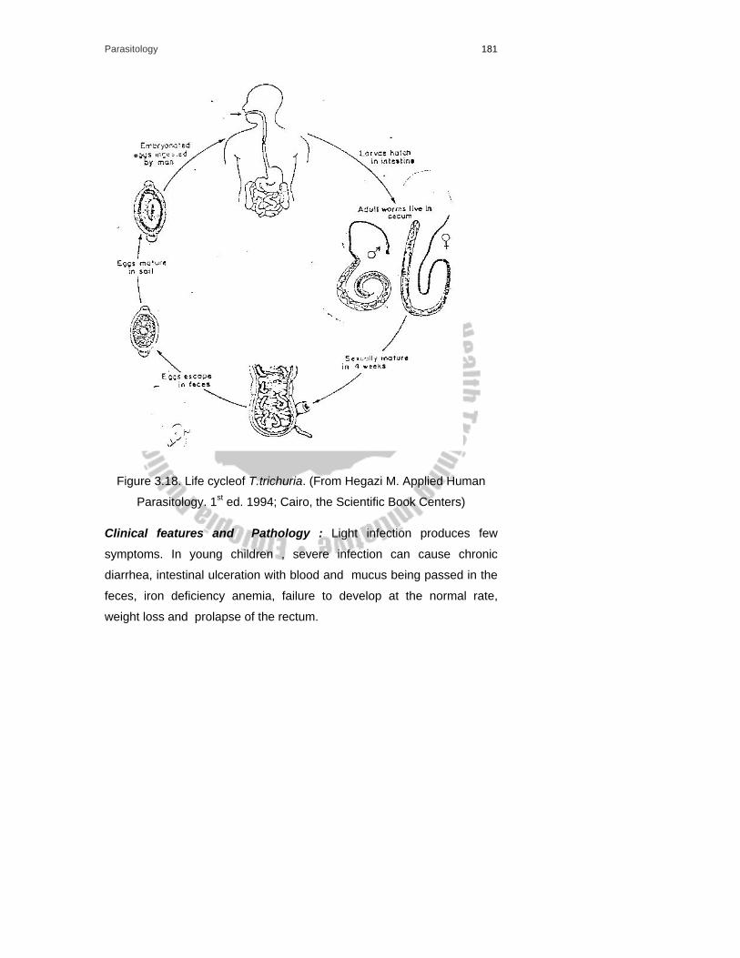
Parasitology
181
Figure 3.18. Life cycleof T.trichuria. (From Hegazi M. Applied Human
Parasitology. 1st ed. 1994; Cairo, the Scientific Book Centers)
Clinical features and Pathology : Light infection produces few
symptoms. In young children , severe infection can cause chronic
diarrhea, intestinal ulceration with blood and mucus being passed in the
feces, iron deficiency anemia, failure to develop at the normal rate,
weight loss and prolapse of the rectum.
Parasitology
182
Prevention and Control
1. Sanitary disposal of faeces in latrine
2. Avoid the use of night soil as a fertilizer
3. Treatment of infected individuals and health education.
Laboratory Diagnosis:-Finding the characteristic eggs in the faeces.
Relevance to Ethiopia:-
- In a national survey in which 28, 696 stool specimens were
examined, 36.1% were found positive for T. trichiura infection.
This makes it a fairly common parasite of man in Ethiopia (Shibru
T, 1986 ).
- As with the other intestinal helminths, pathology depends on worm
burden , light infections being asympotomatic, This parasite
commonly occurs together with A.lumbricoides and likewise mainly
affects children. On the central and northern plateaus T.trichuria was
found in more than 90% of the 50 communities surveyed for
intestinal parasites , with a mean prevalence of 49% . Bure (Gojam)
had a prevalence of 100% Whereas Mendida (shewa) was found to
be free of trichuriasis (Kloos M et al. 1993).
- T. trichuria infection exhibited similar prevalences in lowlands,
temprate and highlands regions 13% on average. The prevalence of
infection due to Ascaris lumbricoides and T.trichuria was significantly
correlated in the lowlands hinting the closely related distribution and
co-occurrence of these parasites. The prevalence of infection due to
hook worms and T.trichuria in the temprate area showed some
pattern of association. A similar pattern also has also been noted
between the prevalence of infection due to A. lumbricoides and
T.trichuria in the highlands . The co-occurrence of A. lumbricoides

Parasitology
183
and T.trichuria infections in the lowlands and in the highlands
suggest that a concurrent intervention against infection due to these
two parasites using the same control methods would be
advantageous(Jemane L, 1998).
Strongyloides stercoralis (The dwarf thread worm)
Geographical Distribution:
world wide distribution in the warm moist climates of tropical and
subtropical countries. Sporadic in the temperate and cool climates.
Habitat: Has both free living and parasitic generations
Parasitic Adults: buried in the mucosal epithelium of the small intestine of
man.
Rhabditiform larvae: Passed in the faeces and external environments
Filariform larvae: soil and water the infective stage
Morphology:
Adult
Male ( free living ):-size = 1.7mm with rhabiditiform esophagus. It has no
parasitic male.
Female (parasitic)
- 2.2mm in length cylinderical Oesophagus
Female (free living)
- 1mm in length and rhabiditiform oesophagus
First Stage Rhabditiform Larvae
Size: 200-300 μm long ; 15μm thick
Motility: very actively motile in the stool

Parasitology
184
Tail: Moderately tapered
Short buccal cavity and rhabiditiform esophagus
Filariforml Larave
- About 600-700μm
- Cylinderical esophagus
- Bifid tail end
Figure 3.19. S.stercoralis adult, larrva and egg. (From Jeffrey HC
and Leach RM. Atlas of Medical Helminthology and Protozoology, 1975.)
Life Cycle:
Egg→Larvae→Adult
The free living generation is the basic life cycle of the parasite and almost
constantly present in the warm moist climates. It results considerable
multiplication of potential infection.
In parasitic way of life, usually man acquires infection by filariform larva
penetrating the skin. Following penetration, the larvae enter blood
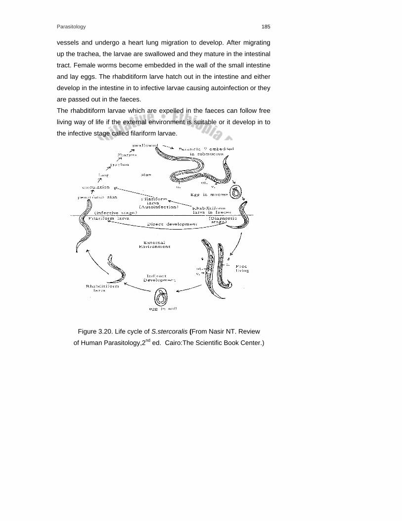
Parasitology
185
vessels and undergo a heart lung migration to develop. After migrating
up the trachea, the larvae are swallowed and they mature in the intestinal
tract. Female worms become embedded in the wall of the small intestine
and lay eggs. The rhabditiform larve hatch out in the intestine and either
develop in the intestine in to infective larvae causing autoinfection or they
are passed out in the faeces.
The rhabditiform larvae which are expelled in the faeces can follow free
living way of life if the external environment is suitable or it develop in to
the infective stage called filariform larvae.
Figure 3.20. Life cycle of S.stercoralis (From Nasir NT. Review
of Human Parasitology,2nd ed. Cairo:The Scientific Book Center.)
Parasitology
186
Clinical feature and Pathology: Causes strongyloidiasis, the disease
is also known as cochin china diarnhea because in the feces of french
cololnial troop who had been suffering in uncontrolled diarrhea in Cochin
China.
The female adult worm by their invasion of the intestine cause
inflammatory changes in the mucosa of small intestine leading to the
development of gastrointestinal symptoms.
It is usually asymptomatic, in symptomatic cases shows the following
phases:
1.Cutaneous phase: Infection caused by large number of larva produce
itching and erythema at the site of infection within 24 hours of invasion.
2. Pulmonary phase: The migratory larva in the lung produces a
considerable degree of host damage and injury to the alveoli and
bronchial epithelium thereby producing bronchopneumonia and full blown
pneumonitis.
3. Intestinal phase : Invasion by adult worms may produce abdominal
pain and mucus diarrhea which alleviate constipation. Indigestion,
nausea vomiting and anemia may also occur.
Heavy infection especially in children may result in malabsorbtion,
steatorrhea and dehydration.
Auto- and hyper-infection syndromes occurs when the immune status of
the host suppressed by either drugs, malnutrition or the concurrent
diseases:
In these conditions, larva invade and are found in many of the tissues
and serous cavities of the body producing a serious infection, which is
fatal. In auto- and hyper-infection the mucosal inflammation is severe.
The sigmoidal colon and rectum become frequently thickened and
edematous .

Parasitology
187
Prevention and Control
1. Sanitary disposal of faeces in latrine
2. avoid use of night soil as a fertilizer
3. Wearing protective footwear
4. Treatment of infected individuals and Health education.
Laboratory Diagnosis
Finding the larvae in faeces or in duodenal aspirates using direct or
concentration method.
In hyper-infection syndrome the larva may be found in sputum and in
other specimens.
Note: The egg of Strongloides steorcolaris is seldom found in faeces. It
is
- Color-transparent
- Oval in shape and measures 50µm long and 35 μm thick
- When passed, it contains a partially developed larva.
Relevance to Ethiopia:
Strongloides fulleborni: is a parasite of primates chimpanzees,
baboons, and monkeys and it has been reported from man in Africa and
southeast Asia. The parasite is endemic in rain forest regions, sporadic in
secondary forest zones and savannah, with a very wide geographical
distribution area involving tropical Africa from Sierra Leone to Ethiopia,
from Central African Republic to Zimbabwe. In Africa, including Ethiopia,
the parasite Co-exists with S. stercoralis.
Strongyloid infections are caused by the opportunistic nematode
Strongyloides stercoralis, a fecally rather than soil-transmitted helminth.
In Ethiopia strongyloidiasis is not highly prevalent in most areas and is
Parasitology
188
found in the same geographical areas and is found in the same
geographical areas as hookworm infection: Nevertheless, rates up to
44% have been reported, S. stercoralis infection were reported from 41
of the 50 communities studied in central and northern Ethiopia . The
infection is rare or absent in many arid lowland areas, including the
Ogaden and pastoral areas in the Awash Valley.
Strongyloides fulliborni
Geographical Distribution:-Widely distributed in Zimbabwe, Zambia,
Papua New Guinea, co-exists with S.stercoralis in Ethiopia. It is a
common parasite of old world monkeys and apes. Infects dogs, cats, and
especially young children and infants.
Habitat:-Has both free living and parasitic life.
It is natural parasites of monkey and dog but it can also infects humans.
Morphology:-Morphologically resembles S.stercoralis with certain
exceptions
Adult:- Adult parasitic female does not have straight ovaries like
S.stercoralis but it is spiral around the intestine.
-Free living female has marked constriction posterior to the
vulva and the vulva lips are not prominent.
Egg:-Resembles eggs of hookworms but are shorter and smaller
-Colorless, Oval and 50 by 35μm in size
-Contain partially developed larvae

Parasitology
189
Life cycle
Egg→Larve→Adult
The life cycle is similar to the life cycle of S.stercoralis except it shed
eggs in the feaces unlike S.stercoralis which sheds rhabiditiform larvae.
Mode of Transmission
1. Skin penetration by filariform larvae
2. Transmammary
Pathology:-In Papua New Guinea, the sub species s. fuelleborni kelly
infections are associated with an acute often l infantile disease known as
swollen belly illness.
Prevention and Control
1. Sanitary disposal of faeces in latrine
2. avoid use of night soil as a fertilizer
3. Wearing protective foot-ware
4. Treatment of infected individuals and Health education.
Laboratory Diagnosis
Finding eggs in stool fresh specimens. Many eggs may be present and
typically appear embryonated.
N.B. If there is a delay in examining the feaces , the larva will hatch.

Parasitology
190
Ancylomstoma duodenale and Necator americanus (old world hookworm) (New world hookworm)
Except some differences which described below both species are similar
in many aspects.
Geographical Distribution: (N. americanus) -Wide spread in the
western hemisphere. It is the predominant species in all parts of the
tropics. For East, South Asia, pacific Islands, Tropical Africa, Central and
South America.
Geographical Distribution: (A. duodenale) World wide; It is essentially northern species occurring in Northern parts
of China, Japan , Europe, North Africa, southern Europe, Middle East. It
is less common in Ethiopia than N.americanus.
Habitat: Adult: Jejunum and less often in the duodenum of man
Eggs: In the faeces; not infective to man
Infective larvae: free in soil and water
Morphology: A.duodenale: - Adult- longer & thicker than N.americanus
- Creamy white appearance
- Large mounth cavity (Buccal capsule);
broader than long
-Two pairs of teeth, two plates & two
subventral lancets
-Club shaped esopagus
-Female lays 20,000 eggs/day
Size - male and female measures 8 mmand 10-13mm respectively

Parasitology
191
N.americanus:- Adult-short & thinner than A. dudenale -Buccal capsule longer than broad -2 cutting plates, 2 plates & 2- subdorsal lancets -club shaped Oesophagus Size -male and female measures 7-9mm and 9 to 11mm respectively Rhabiditiform Larvae Filariform Larvae
Size - 250-500μm - about 600- 700 μm
- long buccm cavity - short bucal cavity - Riabiditoform Oesophages - Cylinderical esophagus 1/3 body length) (1/4 body length) - Pointed tail end - Sharply pointed tail end
Egg:- Size : 65-40μm Shape: oval
Shell: very thin and appears as black line Colour: the cells inside are pale gray Content: varies according to the degree of maturity. It contains an ovum which appears segmented usually 4-8 stage.
Figure 3.21. Hookworm adult and egg. (From Jeffrey HC and Leach
RM. Atlas of Medical Helminthology and Protozoology, 1975.)

Parasitology
192
Life Cycle
Egg→Larve→Adult
The worms requires one host to complete their life cycle and the
definitive host is man. Infection may occur in two ways: (1) through
penetration of the skin and (2) ingesting filariform larvae. The larvae
penetrate the skin and enter small blood vessels and following a heart-
lung migration. After migrating up the trachea, the larvae are swallowed.
In the small intestine they develop and mate.The worm attach to the wall
of the small intestine by sucking part of the mucus and blood from the
host. Female worms lay eggs which are passed in the faeces. In the
external environment the egg develop and hatch the rhabditiform larvae.
It feeds and moult twice to become infective larvae.
Pathology: causes hookworm infection. Major symptoms are severe
itching at the site of skin penetration known as "ground itch", mild
pneumonia with cough, sore throat, bloody sputum, headache,
weakness, bloody diarrhea and anemia.
Adult hookworm cause chronic blood loss. It has been estimated that a
single A.duodenale worm ingests about 0.15ml of blood/day and a
N.americanus worm about 0.03ml.
Prevention and control:-
1. Sanitary disposal of faeces
2. Avoid the use of night as fertilizer
3. Wearing adequate protective foot ware
4. Treatment and health education.

Parasitology
193
Laboratory Diagnosis:-Finding eggs in faeces.
Relevance to Ethiopia:
The hookworms which most commonly infect humans are Ancylostoma
duodenale and Necator americanus. Both parasites are found in
Ethiopia, the latter being by far the more common than the former. A.
dudenale is associated with areas of poor soil coverage and high rates of
drainage, whereas N. americanus is found in red soil areas on flat
plains. Altitude and moisture appear to be the major factors affecting the
distribution of the two species. Hookworm infection is absent in the low,
hot and dry areas of Ethiopia. Infection has not been recorded in places
above 2500 m above sea level. The limiting factor in the former areas
may be moisture whereas in the latter it is likely to be temperature.
Hookworm infections are most widespread in Ethiopia in the 800-1,200
altitudinal zone and in the humid western lowlands at even lower
elevations, where moisture is assured through most of the year and
where the average temperature of the coldest month is above 18NC.
Some of the highest infection rates, between 60% were found in lowlands
of Ilubabor, Kefa and Welega and lower rates in both dry lowland areas
of eastern, southern, and northern Ethiopia, and the highland areas. Of
95 communities surveyed in 12 administrative regions (excluding Eritrea
and Tigray), 82 had hookworm infections, and in 20 communities both
hookworm species were found. Twenty percent of the population was
found to have N.americanus and 1% A.duodenale infections. The
relatively wider distribution of N.americanus is in agreement with studies
in other endemic areas , where this species was found to be more
tolerant to a wider range of temperatures than A. duodenale. Soil type
was also associated with the occurrence of the two hookworm species ;
where as N. americanus was more common in flat areas with sandy clay

Parasitology
194
soil, A.duodenale occurred more frequently in well-drained sandy loam
slopes.
Prevalence rate in A.A and other larger towns are low reflecting the
influence of the urban physical environment and the greater use of shoes
by the urban dwellers.
Trichostrongylus colubriformis
Are mainly parasites of ruminant equines and rodents
Geographical distribution: Cosmopolitan
Habitat Adult: Jejunum and less often in the duodenum of man
Eggs: In the faeces; not infective to man
Infective larvae: free in soil and water
Morphology: Adult: slender and hair-like, the head cutting plates or teeth
-Anterior extremity attenuated
-Yellowish or reddish in color and club-shaped esophagus
Male :-Size 5mm
Has copulatory bursa with two spicules
Long, slender, fused at the tip, barbed copulatory spicules
Female :-Size: 7mm
Egg:-The egg is almost identical with that of A.duodenale
Size 80 by 40 μm
Shape : one pole pointed and the other is blunt
Translucent Content: immature ovum (32 cell stage)

Parasitology
195
-When compared the egg of hook worm; hook worm has size of 60 x 40
μm oval in shape blunt pole and 4-cell stage in its contents
Rhabditiform and Filariform Larvae are similar to hookworm but
have a minute knob at the tip of the tail.
Life cycle
Similar to hookworms except that transmission is due to ingestion of the
filariform larvae in contaminated food or drink.
Egg→Larvae→Adult
Normally it is parasite of herbivore animals but man is accidentally
infected by ingesting the filariform larvae in contaminated vegetables or
water. The adult it the small intestine of man and other animals with the
anterior end penetrating in the mucosa wall.
Figure 3.22. Life cycle of T.colubriforms. (From Nasir NT. Review of
Human Parasitology,2nd ed. The Scientific Book Center, Cairo)

Parasitology
196
Pathology. Infection causes inflammation of the mucosa, abdominal
pain, and anemia
Prevention and Control
1. Proper washing of vegetables or cooking before eating them.
2. Sanitary disposal of feces
3. Treatment and health education
Laboratory Diagnosis
Finding eggs in the feces
Review Questions
1. Identify the common name of the following parasite
a) Ascaris lumbricoides
b) E. vermicularis
c) S.stercoralis
d) N.americanus
e) T.trichuria
2. Discuss the main differences between rhabiditiform and filariform
larvae of hook worm and S.stercoralis.
3. Which intestinal nematodes do not have heart lung migration in
their life cycle?
4. What are the difference and similarities between N. americanus
and A. duodenale ?
5. List the possible sources of specimen and diagnostic stages of
nematodes.

Parasitology
197
3.3.2 Tissue Nematodes
Tissue nematodes are those, adults of which live in the tissues of man.
Adult worms of this group are filaria, guinea worm and Trichinella. They
are parasites of the lymphatic system & subcutaneous tissues and
muscle of man respectively.
The filaria nematodes are widely distributed in nature . the adults are
long and slender measuring many centimeters in length, may inhabit
virtually any tissue, blood and lymphatic vessels, pleural and peritoneal
cavities, subcutaneous tissue, heart and brain. Some species migrate in
the tissues others remains localized and may become encased in a
fibrous tissue reaction. The adults mate in the tissue, producing many
progeny called microfilaria. The microfilaria are small (200-300μm),
slender, motile forms which may be found in the circulating blood or
migrating in the subcutaneous tissue, depending on the species.
Microfilaria are classified by morphological characteristics, geographical
location and type of clinical infection seen. The majority of divisions of
microfilaria is by the presence or absence of sheath surrounding the
parasite. Unsheathed microfilaria include Onchocerca volvulus,
Mansonella ozzardi, M.perstans and M. Streptocerca. The sheathed
microfilaria include Wuchreria bancrofti, Burgi malayi and Loa loa.
Definitive identification to species is based on the presence and number
of nuclei seen in the tail of the microfilaria.
Alternatively, these parasites can be divided as causes of cutaneous,
lymphatic, or body cavity infections. Species identification of blood
microfilaria is particularly important, because some may cause serious
disease while others rarely do.

Parasitology
198
General Features
1. Female worms are viviparous, produce live larvae but does not
lay egg
2. Long tread like worms
3. The immature first stage larva of filarial worms is called a
microfilariae
4. In order to complete its life cycle the larva requires an
intermediate host to develop to infective form, and there is no
reproduction in the insect vector
5. Diagnosis of filariasis is based of finding the microfilaria with
specific morphologic features such as
• Size
• Presence or absence of “ sheath”
• Periodicity as Nocturnal, diurnal, sub-periodic, or aperiodic (non-
periodic) forms
• Source of larvae (specimen) used for laboratory diagnosis such as
blood, skin, nodule, urine, ulcer, etc.
• Appearance i.e. curvature, Kinks, coiling etc.
• The arrangement of the column of nuclei; and whether it reaches
the very tip of the tail.
• Percentage distance of internal anatomical “land marks” from
anterior end. These anatomical” land marks “are nerve ring,
excretory pore, excretory cell, genital or rectal cells and anal pore.
• The arrangement of the column of nuclei and whether it retches
the very tip of the tail.

Parasitology
199
Wuchereria bancrofti
Geographical Distribution:-The periodic form is the most widely
distributed of the filarial worms in subtropics and tropics, Asia, Africa,
America, Middle East, Far East, Including Ethiopia. The sub-periodic
form is found in Eastern Pacific, Thailand and Vietnam
Habitat Adults: Coiled in lymphatic glands, or lying in lymphatic vessels,
superficial abscesses, or wondering in retroperitoneal tissues. Found
usually in lymphatic of the lower limb.
Microfilariae: In lymphatic vessels, and in the peripheral blood normally
at night but during day in lung and other internal organs.
Infective larvae: In the gut and muscles including mouth parts of certain
species of mosquitoes
Morphology:
Adults: -Creamy white with smooth surface
Male:-23-40mm by 0.1mm
` Tapered and rounded swollen head
Sharply curved tail with two spicules.
Unequal spicules
Female:-50-65mm by 0.16mm
Curved tail, tapered front end and Vulva at cervical area
Microfilaria:-Size: 275-300μm by 8-10μm
-Has a sheath which stains pink with Giemsa and palely with
Haematoxylin stain
-Body nuclei are fewer and more distinct than in other species
-There are no nuclei in the end of the pointed tail
-Body curves are smooth and few with Smooth cuticle
-Bluntly rounded anteriorly and pointed caudally

Parasitology
200
Life Cycle:
Infective filariform larvae→Adult worm→microfilariae
It requires two hosts to complete its life cycle :
1) Definitive host: man
2) Intermediate hosts: species of female culex, Anopheles and Aedes
mosquitoes
The infective filariform larvas are deposited on human skin when an
infected female mosquito vector takes a blood meal. The larvae
penetrate the skin through the bite and enter into the blood vessels and
lymph nodes. Development takes place in the lymphatics and the adult
worm mate to produce many microfilariae that enter the blood stream.
The microfilariae are taken up by a mosquito vector when it takes a blood
meal. In the stomach of the insect vector, the microfilariae lose their
sheath and migrate from the mid-gut to the thorax of the vector where
they develop into infective larvae and develop into infective form. The
larvae are ready to be transmitted when the insect next takes a blood
meal.
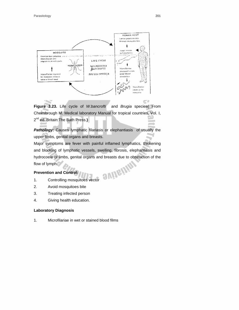
Parasitology
201
Figure 3.23. Life cycle of W.bancrofti and Brugia species( From
Cheesbrough M. Medical laboratory Manual for tropical countries, Vol. I,
2nd ed. Britain:The Bath Press.)
Pathology: Causes lymphatic filariasis or elephantiasis of usually the
upper limbs, genital organs and breasts.
Major symptoms are fever with painful inflamed lymphatics, thickening
and blocking of lymphatic vessels, swelling, fibrosis, elephantiasis and
hydrocoele of limbs, genital organs and breasts due to obstruction of the
flow of lymph.
Prevention and Control:
1. Controlling mosquitoes vector
2. Avoid mosquitoes bite
3. Treating infected person
4. Giving health education.
Laboratory Diagnosis
1. Microfilariae in wet or stained blood films

Parasitology
202
2. Microfilariae in aspirates of hydrocele and lymph gland fluid.
3. Occasionally Microfilariae in chylous urine or hydrocoele fluid
In chronic banchroftian filariasis, a condition called chyluria will
occur, i.e., passing of chyle in urine. In such urine microfilaria
can often b found.
4. Serological diagnosis
Collection Time of Blood for W.bancrofti microfilariae
Periodic W.bancrofti microfilariae: Collecting blood between 22.00-04.00
hours (time of peak density is 24.00 hour)
Nocturnal subperiodic W.bancrofti microfilariae: Collecting blood between
20.00-22.00 hours (time of peak density is 21.00 hour)
Diurnal subperiodic W.bancrofti microfilariae: Collecting blood between
14.00-18.00 hours (time of peak density is 16.00 hour)
The diurnal subperiodic variant W.bancrofti is found mainly in Eastern
pacific. The nocturnal subperiodic variant is found especially in Thiland
and Vietnam.
W. bamcrofti is found with B.malayi in parts of south East Asia & South
India
Relevance to Ethiopia
In people living in two communities adjecent to the Baro river, near the
town of Gambella in 1993. The overall microfilaria prevalence, using
counter chamber technique, was 20.7% with males and females showing
microfilaria rates 23.7% and 18.5%, respectively. In males, 20.3% had
hydrocoele and this condition was noted above the age of 35. About 40%
of those with hydroceole had microfilaremia (Jemaneh L et al, 1995).

Parasitology
203
Brugia malayi
Geographical Distribution:-It is distributed in countries such as Asia,
Malaysia, India, Philippines, Vietnam, China, and Korea. The nocturnal
periodic form is the most widely spread in swamps and rice growing
areas whereas the nocturnal subperiodic form is found in fresh water
swamps and forests along major rivers.
Habitat
Adult: In dilated lymphatic Vessels of man; usually in the upper limb.
Microfilariae: Lymph and peripheral blood at night, and in the lung and
internal organs during day timeInfective filariform larvae: In
the gut of mosquitoes.
Morphology Adults :- Male: 13-23mm
Female: 43-55 mm
Microfilariae :- Size: 200-275μm by 5-6μm
-Body is usually coiled and kinked (has small angular curves)
-Has a sheath which stains dark pink with Giemsa and pink-
mauve with haematoxylin.
-Body nuclei are dense and stain darkly.
-There are two discrete nuclei in the end of the tail which tapers
irregularly.

Parasitology
204
Life cycle
The life cycle of B.malayi is similar to the life cycle of W.bancrofti. Man is
the definitive host. Female anopheles and Mansoni mosquitoes are the
intermediate hosts. Man gets infection by the bite of infected insect
vector when it takes a blood meal.
Pathology: Causes malayan filariasis or elephantiasis of the lower limbs.
Symptoms develop rapidly and children are more affected.
Prevention and Control :- similar method like W.bancrofti.
Laboratory Diagnosis
Finding the characteristic Microfilariae in wet or stained blood films
Collection Time for B.malayi Microfilariae
Periodic B.malayi Microfilariae:
Collect blood between 22.00-04.00 hours (time of peak density is 24.00
hour)
Subperiodic B.malayi Microfilariae:
Collect blood between 20.00-22.00 hours (time of peak density is 21.00
hour)
- Peroidic B.malayi is commonly found in open swamps and rice
growing areas of coastal regions. The subperiodic variant is
found mostly in fresh water swamp in forests along major rivers.
- B. tumori shows nocturnal periodicity. It is found only in the
lesser Sunda of Indonesia the species takes its name from the
island of Timor which forms part of the group. it is found in low
lying riverine and coastal areas.

Parasitology
205
Loa loa (Eye worm)
Geographical Distribution:
The Distribution of L.loa is restricted to the equatorial rain forest area of
west and central Africa.
Habitat: Adults: In connective tissues under the skin, in the mesentery and the
parietal peritoneum. They commonly migrate rapidly in the body
and may be seen in the subconjunctival tissue of the eye or in
thin skinned areas.
Microfilariae: In peripheral blood of man during day time.
Infective larvae: In the gut, mouth parts and muscles of
tabanide flies of the genus Chrysops.
Morphology
Adults:- Cylinderical and transparent Male: 30-34mm
Female: 60mm
Microfilariae:
-Size: 250-300μm long and 8-10μm thick
-Has several curves and kinks
-Has a sheath which stains best with haematoxylin.
-Body nuclei are not distinct and appear more dense than
those of W.bancrofti
-Nuclei extend to the end of the tail which is rounded.
Life cycle
Natural Definitive hosts are- Man & Monkeys. Reservoir host are simian
hosts.

Parasitology
206
Similar to the life cycle of W. bancrofti but the habitat of the adult worms
is in the subcutaneous tissues and they are freely moving in these
tissues. The intermediate hosts are species of chrysops(horsefly).
Pathology: Characterized by the formation of swelling known as calabar
swellings. The arms most frequently affected. Adult worms also migrate
in sub-conjunctiva tissues. They can cause inflammation and irritation but
not blindness.
Prevention and Control: Similar with the previous filaria worms.
Laboratory Diagnosis
1. Finding the characteristic microfilariae in stained blood films
taken during the day time.
2. Occasionally the microfilariae can be found in joint fluid.
Onchocerca volvulus
Geographical Distribution:- occurs most widely along the courses of
fast running rivers in the forests and Savannah areas of west and central
Africa. It is endemic from Senegal in the west to Uganda and Ethiopia in
the East and as far as south as Zambia. It also occurs in the Yemen Arab
Republic. Saudi Arabia and in central America(Mexico and Guatimala). It
is also found in South America.
Habitat: Adults:- Subcutaneous nodules and in skin
Microfilariae:- Skin,eye and other organs of the body.
Infective larvae: In the gut, mouth parts and muscles of Simulium black
fly.

Parasitology
207
Morphology: Adults: -male: 25-40mm, curved and bulbous tail
-Female; 33-55cm in length
Microfilariae:-Size 240-360μm long and 5-9μm thick
-Has no sheath and head end is slightly enlarged
-Anterior nuclei are positioned side by side
-There are no nuclei in the end of the tail which is long and
pointed.
Life Cycle
It requires two hosts man as definitive and Simulum as intermediate host.
Infective larvae are deposited on the skin when an infected vector takes
blood meal. It enters through the bite wound and develop into male and
female worms in subcutaneous tissue. They mate and viviparous female
produces many unsheathed microfilaria that can be found just below the
surface of the skin in the lymph spaces and in the connective tissues.
They can also be found in the fluid of nodules. The microfilaria also
migrate to the eye. The microfilariae are ingested by its most
commonest vector Simulium damnosum complex as it takes a blood
meal and develop into infective larvae that migrate to the mouth parts of
the blackfly ready to be transmitted when the fly next takes a blood meal.

Parasitology
208
Figure 3.24. Life cycle of O.volvulus( From Cheesbrough M. Medical
laboratory Manual for tropical countries, Vol. I, 2nd ed. Britain:The Bath
Press.)
Pathogenicity:-It causes Onchocerciasis or river blindness
The clinical feature of onchocerciasis are caused mainly by the
inflammatory reactions around damaged & dead microfilaria. In chronic
onchocerciasis the skin loses its elasticity & becomes wrinkled which
makes people more aged than they are (known as elephant skin ). When
the skin around the groin become affected hanging groin develops. The
term Leopard skin refers to a spotted depigmentaion of the skin. The
main clinical features are the formation of nodules, dermatitis &
inflammatory reactions in the eye leading to blindness.

Parasitology
209
Prevention and Control
1. Destruction of simulim including
- Selective use of insecticides
Introducing other method to reduce the breeding of black
files in rivers & streams .
2. Avoiding simulium bites by covering using as far as possible
those parts of the body most at risk.
3. Identification of infected communities followed by treatment of
communities by
Laboratory Diagnosis-Finding the characteristic Microfilariae in skin
snips.
Note: In heavy Infection and following treatment, the microfilariae can
also be found in urine, blood, and most body fluids. Unlike W.bancrofti
which may also be found in urine the microfilaria of O.volvulus is
unsheathed.
Differentiation of O.Volvulus from Mansonella species
- In west & central Africa O.volvulus requires differentation from
streptocerca
- In West Indies & Central and South America.
- O.volvulus requires differentation from M.ozzardi.
Relevance to Ethiopia
It is endemic in a a large fertile Western, Southern and Northwestern
part of Ethiopia. However, the ocular form which in other parts of the
world causes serious lesion that leads to loss of vision and blindness is
considered to be mild in Ethiopia. Although ocular manifestations are
considered to be rare in Ethiopia, there is no sufficient explanation.

Parasitology
210
Onchocerchiasis is known to be more prevalent among older age groups
and males and in this study the sex difference is seen to be the
dominance of males in activities such as collection of firewoods, fishing
and swimming as well as the habit of wearing light clothes and shorts by
males and thus more exposure to the vectors.Bathing, swimming and
fishing at the rivers were significantly associated with infection. This
could be due to long stay and nakedness with these practices and thus
more exposure to the vectors as compared to fetching water or crossing
over the rivers(Habtamu A,1999).
The distribution of the disease covers savannah and forest ecology of
the south, south western, north and north western Ethiopia, extending
from Omo valley in the south to the Atbara and Teccaze drainage
systems in the north bordering the Sudan. Although the prevalence of
onchocenciasis and blindness due to the disease throughout the country
is not well studied , it is now estimated that about one million people are
suffering from the disease .
Presently, 29 Simulium species are recorded in the country . It is
reported that two species viz . S.damnasum s.l. and S. ethiopiense are
the vectors involved in transmission : the former playing a role at lower
and higher altitudes while the later is mainly at higher altitudes .
A survey to determine the prevalence of onchocerciasis in a sample
population aged 5 years and over was made in pawe western Ethiopia. A
total of 986 persons (636 settlers and 350 indigene) were examined for
parasitological (in skin snip) and clinical manifestations, of which 310
(27.8% settlers and 38.6% indgenious ) were found infected with
Onchocerca volvulus.
Males had significantly higher rate and density of infection but the
difference by age was not significant. The prevalence of infection was
Parasitology
211
significantly higher among the students who had reported frequent
bathing, swimming, fishing and collecting firewood at/or near the rivers
identified as the probable breeding sites of the vector. The rate
determined for different communities of Kaffa indicate that half of the
studied areas have a prevalence of more than 20% in youngsters which
indicates that this is a high prevalence area for Onchocechiasis.
Mansonella Perstans
Geographical Distribution
Widely distributed in South America and tropical Africa.
Habitat Adults: In the serous cavities, mesentery, and connective tissue,
especially the retroperitoneal.
Microfilariae: In peripheral blood at any time because non-periodic guts
mouth parts .
Habitat: Adults: In connective tissues under the skin, in the mesentery and the
parietal peritoneum. They commonly migrate rapidly in the body
and may be seen in the subconjunctival tissue of the eye or in
thin skinned areas.
Microfilariae: In peripheral blood of man during day time.
Infective larvae: In the gut, mouth parts and muscles of chrysops fly.
Morphology: Adults : Male: about 40mm
Female: 7 cm - 8 cm
Microfilariae
Size: 190-240 μm long by 4.5 μm thick
- Has no sheath, body nuclei are irregular Large nucleus in tip vectors

Parasitology
212
- Nuclei extend to the end of the tail which is rounded and there is a
large nuclie at the tip.
- Nonperiodic lie found in day and night blood.
Life Cycle
Man is the definative host and female culicoides are the intermediate
hosts. Man is infected by the bite of infected culicoides.
Pathology: non-pathogenic or of low pathogenicity.
Prevention and Control:- 1. Avoid the bite of vectors
2. Destruction of insect vectors
3. Treatment and health education.
Laboratory Diagnosis
Finding the characteristic microfilaria in the stained blood films at any
time.
Differentiated from L.loa and w.bancrofti by its smaller in size, absence
of sheath, tail features and non periodicity.
Mansonella Ozzardi
Geographical Distribution:-West Indies and South America.
Habitat Adults: In the mesentery, retroperitoneal tissue, abdominal wall, and
lymphatic vessels of man.
Microfilariae: In peripheral blood or skin of man any time.
Infective larvae: In the gut, and mouth parts of culicoides and simulium
black flies

Parasitology
213
Morphology: Adults:-Male: about 35mm
-Female: 70mm
Microfilariae:-Size: 150-200 μm long and 4.5 μm thick
-Has no sheath and body nuclei are not distinct but the anterior
nuclei are positioned side by side.
-There are no nuclei in the end of the tail which is long and
pointed.
Life cycle
Man is the definitive host and the infective larvae are transmitted by the
bite of culicoides and simulium species as an intermediate hosts.
Pathology: non-pathogenic or of low pathogenicity. Most infections are
asymptomatic or cause chronic arthritis, skin rashes and other
symptoms.
Prevention and control:- Similar method llike M.perstance.
Laboratory Diagnosis
Finding the microfilariae in stained blood film and occasionally in skin
snip at any time since it is nonperiodic.
Mansonella Streptocerca
Geographical Distribution:- Found only in the rain forest of Africa
especially in Ghana, Nigeria, Zaire and
Cameroon.
Habitat Adults: In cutaneous connective tissues of the chimpanzee,

Parasitology
214
Microfilariae: In the skin of man by day and by night, but are not found in
the blood
Infective larvae: In the gut, muscle tissue and mouth parts of Culicoides
midges.
Morphology Adults: are recovered only in animal hosts and in man only
microfilariae is known.
Microfilariae
Size: 180-240 μm long and 4.5 μm thick
Nuclei extends to tip of the tail
Has no sheath
When immobile, the tail usually appears hooked and its tip is
rounded. In fresh preparations, the tip of the tail may appear
forked. Life cycle
Like other filarial worms It requires two hosts to complete its life cycle
man as a definitive host and culicoides as its intermediate host.
Pathology: Non-Pathogenic or of low pathogenicity. Most infections are
asymptomatic or sometimes cause an itching dermatitis, hypopigmented
macules and thickening of the skin.
Prevention and Control:-Like other mansonella species.
Laboratory Diagnosis:-Finding the microfilariae in skin snip at any time.
Differentiation from O.volvulus is by its smaller size, single file anterior
nuclei and tail features. Less motile than O.volvulus in wet prepare

Parasitology
215
Figure 2.25. Microfilaria of humans: diagnostic characterstics ( From
Brown HW and Neva FA, Basic clinical parasitology, 5th ed. USA:
Applenton-century Crofts,1983.)
Trichinella Spiralis
Geographical Distribution:-T.spiralls contain three subspecies:
1) T.S.Spiralis which is found in temprate regions,
2) T.S nativa which is found in the Arctic and
3) T.S nelsoni which is found in Africa & southern Europe.
Habitat Adults: Embedded by its anterior part in mucosa of muscular epithelium
of duodenum and Jejunum of Man, Dog, Rate, Cat, Pigs and
many wild Carnivores. viviparous

Parasitology
216
Larvae: encysted in the straited muscle of the body of meat eating
animals including man.
Egg: No eggs passed in the faeces , female gives birth to larvae.
Morphology:
Adults:-minute thread-like worms, white in color and attenuated anterior
end
- cellular oesopagus. Male: up to 1.5mm
Female: up to 4 mm, viviparous
Larva: Size: 0.5 mm long and 0.2 mm thick and lies along the muscle
fibers Shape: Usually seen coiled inside a lemon shaped cyst.
larvae appear black
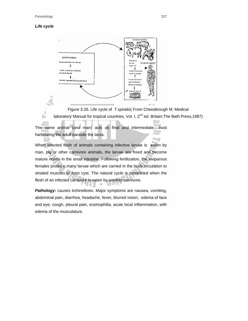
Parasitology
217
Life cycle
The same animal (and man) acts as final and intermediate host
harbouring the adult parasite the larva.
When infected flesh of animals containing infective larvae is eaten by
man, pig or other carnivore animals, the larvae are freed and become
mature norms in the small intestine. Following fertilization, the viviparous
females produce many larvae which are carried in the body circulation to
striated muscles to form cyst. The natural cycle is completed when the
flesh of an infected carnivore is eaten by another carnivore.
Pathology: causes trchinellosis. Major symptoms are nausea, vomiting,
abdominal pain, diarrhea, headache, fever, blurred vision, edema of face
and eye, cough, pleural pain, eosinophilia, acute local inflammation, with
edema of the musculature.
Figure 3.26. Life cycle of T.spiralis( From Cheesbrough M. Medical
laboratory Manual for tropical countries, Vol. I, 2nd ed. Britain:The Bath Press,1987)

Parasitology
218
Prevention and Control:- 1. Avoid eating of raw or undercooked pork
2. Inspecting meat for infective larvae
3. Not feeding raw garbage to pigs.
Laboratory Diagnosis:-
1) Finding the larvae in striated muscle.
2) Testing serum for Trichinella antibodies.
Relevance to Ethiopia
Like other parasite that are transmitted through eating of pork meat, it is
uncommon. A case of Trichichinosis due to T.spirails and acquired by
eating raw wartag meat was reported from Jimma.
Dracunculus Medinensis
(Guinea or Medina worm)
Geographical Distribution:- Since 1986, the global prevalence of
drancunculiasis has been reduced by 97% and it is expected that the
disease will be eradicate in the near feature. It is still endemic in 18
countries, 1.e., India, Yemen and 16 countries in sub-Sahara Asfricawith
77% of infections being found in Sudan.
Habitat:
Adults: thread like
Female in the subcutaneous tissues and intermusular connective
tissues the of the lower extrimites; especially around the ankle.
Male resides in the retroperitoneal connective tissues and dies
shortly after copulation
First stage larvae: In the ulcers or blisters.
Infective Filariform larvae : In the hemoceole of Cyclops .

Parasitology
219
Morphology
Adults: White with smooth surface
Male: 12-29mm , coiled posterior end.
Female : 70-120 cm (average 100 cm)
The longest nematode of man
Has cylinderical oesophagus
Viviparous
Larva: Size: 500-700 μm
Rounded anterior end
Long and pointed tail
Has Rhabiditiform Oesophagus
Life cycle
Figure 3.27 Life cycle of D.medinensis. (From Jeffrey HC and Leach
RM. Atlas of Medical Helminthology and Protozoology, 1975.)

Parasitology
220
Man is the most important definitive host. It has also been found in dogs
and cats. Cyclops or related crustaceans are the intermediate hosts.
When Person swallows water containing cyclops that are infected with
the larvae. The larvae are freed and penetrate through the duodenal wall.
After development and fertilization in the connective tissues, the female
worm migrates to the connective tissues of the lower limbs where within
about a year it becomes fully mature.
The female worm buries its anterior end in the dermis forming a blister
that ulcerates. when the blister (ulcer) is bathed in the water the uterus of
the female protrudes through the ulcer and ruptures. large number of first
stage larvae are released in to the water.
The larvae are unable to swim but are actively motile, coiling and
uncoiling.
For the life cycle to be continued the larvae must be ingested by a
Cyclops. In the body cavity of the Cyclops, the larvae develop in to
infective larvae.
Pathogenicity in Man
- Disease Guinea worm ulcer disease
- toxic histamine like substances are liberated by the female
guinea worm as soon as she starts migration, cousing profound
ellergic symptoms
- This is followed by the appearance of worm under the skin
associated with blister formation which bursts & the larvae are
discharged after coming in contact with water.
- Premature liberation of embroys in the tissue cause irritation and
inflammation this happens particularly when the its extraction
prematurely

Parasitology
221
- Secondary infection may also occurs leading to cellulitis and
occasionally septicaemia
- If a joint is involved, arthritis may develop
Prevention and Control:
The easiest parasite to be Prevented controlled
1. Prevent water contamination with the larvae
2. Avoid drinking infected water by boiling
3. Destroy the Cyclops by using chemicals example,
organophosphorus or chlorine
4. Covering the blister with a water proof dressing
Laboratory Diagnosis:-
Finding the gravid worm in the blister.
A diagnosis is usually made when the blister has ruptured & the anterior
end of the female worm can be seen. If required, laboratory confirmation
of the diagnosis can be made as follows:
1. Place few drops of water on the ulcer to encourage discharge of
larvae from the uterus of the worm.
2. After and left for a few minutes collect the water in a pipette and
prepare a wet mount.
3. Transfer the water to a slide & examine microscopically for the
motile larvae using the 10 x objective with the iris diaphram
closed sufficiently to give good contrast.

Parasitology
222
Review Questions
1. Explain typical diagnostic characteristics which differentiate one
filaria from the rest.
2. Describe. filaria worms according to their pathogenicity, habitat in
human, presence or absence of sheath and their periodicity.
3. Why trichinella spiralis is not commonly found in our country ?
4. Identify the diagnostic and infective stages of tissue nematodes.
5. Enumerate the possible sources of specimen and thelaboratory
diagnosis oftissue nematodes.

Parasitology
223
Appendix I
Laboratory Examination of Specimens
Learning Objectives
At the end, successful students be able to 1. Prepare necessary materials for the collection of parasitological
specimens.
2. Select appropriate specimen for detection and identification of
parasites.
3. Perform direct, concentration and staining techniques for stool
specimens .
4. Differentiate between pathogenic and non-pathogenic parasitic
organisms, observed under microscopic examination.
5. Apply the Scotch-tape technique for the laboratory diagnosis of
E.varmicularis.
6. Apply the kato thick smear method for the diagnosis of ova of
schistosomes and round worms.
7. Process and examine urine specimens for the diagnosis of
urinary parasite.
8. Perform ,collect, process and examine blood specimens for the
diagnosis of haemoparasites
9. Select the appropriate site of skin and perform examination for
the laboratory diagnosis of Onchocerca volulus and
Ieishmaniasis.
10. Prepare staining solutions and reagents (Giemsa, Wright, iodine,
slaine, formalin and others).
11. Record and report all parasitological results in the appropriate
formats; including all neccesary information

Parasitology
224
Introduction
In the field of diagnostic Medical Parasitology, proper specimen
collection is critical since the final laboratory results are based on
parasite recovery and identification will depend on the initial quality of the
samples taken. Unless the appropriate specimens is properly collected,
preserved and processed, these infections may not be detected;
therefore, as a part of any overall continuous programme for the
laboratory, the generation of test results must begin with stringent criteria
for specimens acceptance or rejection.
Laboratory procedures detects organisms within clinical specimens using
morphological criteria, rather culture or biochemical tests and/or physical
growth characterstics. Many clinical specimens, such as those from
intestinal tract, contain multiple artifacts that complicate differentiation of
parasites from surrounding derbis.
The main ways in which laboratory diagnosis of parasitic infections
include:
1) Microscopy:- the majority of intestinal, blood, urinary and skin
parasites are usually detected by microscopically in stained or
unstained; either directly or following concentations.
2) Culture:- only minority of parasitic infections are diagnosed
routinely by culture techniques. Relatively few of protozoa and
none of the helminth parasites, can be cultured in a manner that
is useful for laboratory identification. Other than strictly for
research purpose , the only culture methods in general use are
for the isolation of such as E.histolytica, T.vaginalis, T.cruzi and
Leishmania species. are identified by this method.

Parasitology
225
3) Immunodiagnosis:-it is based on the detection of :
A. Antibody in a person's serum, produced in response to a
particular parasitic infection. The antibody may persist
for a long period of time in the serum after an infection
has ended and therefore antibody tests are unable to
distinguish between past or present infection. When
used to assist in diagnosing parasitic disease , antibody
tests need to be interpreted with care.
B. Antigen, which is excreted by parasites and can be
found in the serum, urine, CSF, feces or other
specimens. Antigen tests provide evidence of present
infection and are therefore greater value than antibody
tests in the clinical diagnosis of parasitic infections.
Immunodiagnostic techniques are required when:
a) Parasites live in the tissue of internal organ and
can not therefore easily remove for examination.
b) Parasites can be found in specimens only in
certain stages of infection, e.g., in the acute
stage not in the chronic stage.
c) Parasites are present intermittently or in too few
numbers to be easily detected in the specimens.
d) The techniques used to detect parasiteds are
complex or time consuming.
Those parasitic disease for which immunodiagnosis is of particular value
include:
- South American trypanosomiasis , Chronic stage
- African trypanosomiasis, when parasitaemia is low
- Leishmaniasis

Parasitology
226
- Filariasis
- Amoebic liver abscess - Trichinolosis
- Toxoplasmosis - Toxocarisis
- Hydatid disease - Schistosomiasis
The principal type of immunodiagnostic tests are intradermal and
serological.
With the introduction of immunoassays, there are now many more
options avilable to the diagnostic laboratory, different laboratories will
select different approaches. In this appendix only the microscopy
method will be dealt in detail.
A. Stool Examination
The most frequently performed parasitological procedure is the stool
examination. The detection and identification of parasites; such as adult
worms, larvae, eggs, trophozoites and cysts depends on its proper
collection.
Procedure
1. Provide the patients with specimen containers with tight-fitting lids.
Note:- Three specimens are usually required at a three alternate days to
detect all intestinal parasitic infections. The stool should be collected
before radiological examination is carried out using barium. Stool
specimens containing barium are unacceptable for examination, and
intestinal protozoa may be undetectable for 5-10 days after barium is
administered to the patient.
- Some substances and medications also interfer with the detection of
intestinal protozoa, including mineral oil, bismuth, antibiotics,
antimalarial agents and nonabsorbable antidiarrhoeal preparations.
After adminstration of any of these compounds to the patient ,

Parasitology
227
parasite may not be recovered for a week or more. 2. Collect sufficient quantity of stool. It should contain at least 4ml
(4cm3) of Stool.
3. Examine the stool as soon as possible.
As it is not possible to predict what organisms will be present in the
specimen, however, the most conservative time frames should be
used for parasite recovery. The examination of liquid specimens
should occur within 30 minutes of passages, not 30 min. the time
they reach the laboratory. Soft specimens may have a mixture of
protozoan trophozoties and cysts and should be examined within 1
hour of passage. Formed specimens can be examined at any time
within 24hrs.
4. If specimens cannot be examined in the above time frame, put them
in available preservatives.
I. Macroscopic Examination
Stool specimen is examined with the naked eye for :
1. Presence of worms:- may have adult helminthes or segments
Example: Ascaris, Taenia species, E.vermicularis and
gravid Taenia species.
2. Consistency (degree of moisture)- It varies in diet but certain clinical
conditions associated with parasite presence may be suggested by
particular consistencies.
- It will be described as hard, formed, semi-formed and
diarrhoeic( watery).
3. Colour:- any abnormal colour
E.g., pale yellowish passed in steatorrhoeac conditions such as
gardiasis,dark or black-stools occur when

Parasitology
228
iron or bismuth is taken or when there is intestinal hemorrage
4. Pathologic odour Offensive, non-offensive
5. Abnormal features seen (composition): mucus, blood or fat
globules.
II. Microscopic Examination
The detection and identification of species of parasites require
microscopic examination of specimens
1. Direct Microscopic
Routine microscopic examination of stool specimen with physiological
saline and Dobell’s iodine solution helps to detect and identify the stages
of some parasitic organisms.
1.1. Direct Microscopic Examination of Stool Specimen with
Physiological Saline and Dobell's Iodine Solutions
Material and Methods Wooden applicator sticks
Microscopic slides
Cover slips
Dropping bottles containing physiological saline(0.85%w/v) and Dobell's
Iodine solutions
Microscope
Pasture pipette
Procedure
1. Place a drop of physiological saline (0.85%w/v) in the center of the
left half of the slide and place a drop of Dodell's Iodine solution in
the center of the right half of the slide.
Parasitology
229
2. With an applicator stick, pick up a small portion of the feces
(Approximately 2mg which is about the size of a match head) and
put on the drop of saline. Add a similar portion of stool sample to the
drop of iodine.
3. Mix the feces with the drops to form homogeneous suspensions.
4. Cover each drop with a cover slip by holding the cover slip at an
angle of 300, touching the edge of the drop, and gently lowering the
cover slip onto the slide so that air bubbles are not produced.
5. Examine the saline preparations using the 10X objective for motile
forms, cyst and oocyst of intestinal protozoa and for any ova or larva
of helminths.
6. Examine the iodine solution preparation using 40X objective to
identify the cyst stages of protozoa. The iodine will stain the nuclei
and the glycogen mass of the cyst.
1.2 Modified Ziehl-Neelsen technique (Acid-Fast Stain)
Modified Ziehl-Neelsen staining of fecal smear helps to detect oocysts of
Cryptosporidium, Cyclospora species and I.belli.
Material and Methods: - Carbol fuchsin stain
- 0.25% Malachite green ( Methylene blue)
- 1% acid alcohol
- Slides
- Cover slip
- Microscope
Procedure
1. Prepare a thin fecal smear on a slide then air dry.

Parasitology
230
2. Fix the smear with methanol for 2-3 minutes.
3. Stain the smear with cold carbol fuchsin for 5-10 minutes.
4. Wash off then stain with clean tap water.
5. Decolorize with 1% acid alcohol for 10-15' until color ceases to
flow out of the smear.
6. Rinse in tap water and counter stain with 0.5% malachite green
(or methylene blue) for 30'
7. Wash off the stain with tap water.
8. Stand the slide in a draining rack for the smear to dry.
9. Examine the smear microscopically using100X objective to
detect and identify oocyst.
3. Concentration Methods for Fecal Specimens
The concentration and the separation of protozoa cysts and helminths
egg from other elements of the fecal specimen can be of great advantage
in diagnosis. This can be accomplished by sedimentation, flotation and
combination of the two. Feces normally contain a great variety of
materials, most of which are either lighter or denser, smaller or larger
than the cysts, eggs and larva of parasites.
The concentration of parasites in parasitological specimens is some
times called the '' enrichment technique '' because it enables to examine
greater quantity of stools in less volume.The purpose in using a
concentration technique is to separate as completely as possible
parasites from all other elements of the Stool.
In general the concentration technique may be necessary:
1. To detect parasites when they are not found in a direct saline
wet mount examination but the symptoms of intestinal parasitic
infection continue.
2. To detect the eggs of parasites which are often few in number
Parasitology
231
such as those of Schistosome or Taenia species.
3. To check whether treatment has been successful.
4. To investigate the prevalence and incidence of a parasitic
infection as part of epidemiological survey .
The choice of concentration technique depends on
1. The species of parasite
2. The number of specimen to be examined
3. The equipment and time available
A direct microscopical examination of stool must always be done
before preparing a concentration because motile forms of flagellates,
ciliates , and amoebae die during the concentration procedure.
Concentration Technique of Fecal Specimen is Divided into two :-
1. Floatation Techniques
Floatation technique concentrates the cysts and eggs of parasites at the
top because their density is less than that of the suspending medium.
The waste products, crystals, body cells etc. have a higher specific
gravity therefore these substances will sink to the bottom. The top layer
can be removed and placed on a slide to be examined under the
microscope.
Zinc Sulfate Floatation Technique
Zinc sulfate floatation technique is one of the most widely used method of
concentration. It has a special merit of being suitable for routine
examination of both cyst of protozoa and eggs of most helminths.
Operculated eggs of Trematodes and Cestodes, infertile Ascaris ova and
larva of nematodes are not concentrated because they have greater
specific gravity than the suspending medium. However; the eggs of

Parasitology
232
Clonorchis, Opisthorchis and some small trematodes are satisfactorily
concentrated. The technique is not also suitable for concentrating eggs
or cysts in fatty faeces.
A Zinc sulfate solution which is used for the concentration of parasite
has a specific gravity (relative density) of 1.180-1.200 Faeces are
emulsified in the solution and the suspension is left undisturbed for the
eggs and cysts to float to the surface where they are collected on a cover
glass or can be collected by pasteur pipettes.
Materials and Reagents
1. Lipless test tube about 10ml capacity.
2. ZnSO4 solution 33 W/V (about 33 grams of crystals in 100 ml
solution) specific gravity of 1,180-1,200 . use a hydrometer to
check the specific gravity (relative density) of the solution is
corrected. Adjust with distilled water or more chemical if required.
3. Cover glass
4. Microscope slide
5. Applicator stick
Procedure: -
1. About one quarter fill the tube with the zinc sulfate solution.
2. Add an estimated 0.5 gram of faeces and using a rod or stick,
emulsify the specimen in the solution.
3. Fill the tube with the zinc sulfate solution and mix well.
4. Stand the tube in a completely vertical position in a rack.
5. Using a plastic bulb pipette or pasteur pipette, add further
solution to ensure that the tube is filled to the brim.
6. Carefully place a completely clean (grease free) cover glass on
top of the tube, avoiding trapping any air bubbles.

Parasitology
233
7. Leave undisturbed for 30-45 minutes to give time for the cysts
and eggs to float.
Note: -After 60 minutes, the eggs will begin to sink.
8. Carefully lift the cover glass from the tube by a straight pull
upward. place the cover glass face downwards on a slide.The
eggs and cysts will be found adhering to the cover glass.
9. Examine microscopically the entire preparation using the 10x
objective use the 40x objective and run a drop of iodine solution
under the cover glass to identify the cysts.
Other flotation methods includes:
a) Brine ( Saturated NaCl ) flotation
b) Saturated sugar flotation
c) ZnSo4 centrifugal flotation
2. Sedimentation Techniques
In this technique cysts and eggs of parasites settle and are concentrated
at the bottom because they have greater density than the suspending
medium. The cysts and eggs can be sedimented by natural gravity or by
accelerating the process by centrifugation.
Formalin-Ether Centrifugal Sedimentation Technique
This method is recommended as the best technique for concentrating the
eggs and larvae of helminths and moderately satisfactory for cysts of
protozoa. It is most useful for detecting the eggs of Schistosome in feces.
The formalin is used for fixation and preservation of the morphology of

Parasitology
234
parasites. The fecal debris absorbs ether and becomes lighter than
water.
Advantages of this technique includes:
A) It is rapid and suitable for fresh or preserved stool
B) It is also used for concentrating parasites on which Zinc sulfate
floatation has given poor results due to excessive amounts of
fats and fatty acids, and for operculated ova of some trematodes
and cestodes.
C) The morphology of most parasite is retained for easy
identification
D) It will cover most intestinal parasite
Materials and Reagents:-
- Electric centrifuge - 1.5 ml Conical centrifuge tubes
- Stoppers - Funnel
- Gauze /Sieve/ - Graduated cylinder /Pipette/
- Beaker - Applicator stick
- Slide - Coverslip
- 10% formalin solution - Ether
- Normal Saline - Lugol iodine solution
Procedure
1. Take about 2g or 2ml of stool and mix it in about 10ml of normal
saline solution.
2. Filter through two layers of gauze into a centrifuge test tube
3. Centrifuge for one minute at medium speed (2000-5000 rpm) . If
the supernatant fluid is very cloudy, wash the deposit again i.e.
mix it with 10ml of normal saline solution ,

Parasitology
235
4. Centrifuge for one minute at medium speed and pour of the
supernatant fluid.
5. Add 10ml of formaldehyde solution to the sediment.
6. Stir or mix a suspension well and let it stand for five minute.
7. Add 3ml of ether.
Note :- Ether is highly flammable, therefore make sure there is no open
flame in the laboratory and it is well ventilated because ether
vapor is anesthetic.
8. Stopper the tube, turn it on its side and shake vigorously for 30
seconds or one minute.
9. Remove the stopper carefully and centrifuge for one minute at
low speed (1500 rpm).
There will be four layers in the tube:-
1st layer : Ether
2nd layer : Debris
3rd layer : Formaldehyde solution.
4th layer : The deposit containing stages of parasites
(cyst, egg and/or larvae)
10. Free the layer of debris by rotating the tip of a wooden applicator
stick between it and the sides of the tube. Tilt the tube and pour
of all the supernatant fluid. use a cotton swab to remove any
debris adhering to the side of the tube.
11. Mix the remaining fluid well with the deposit by tapping the tube
gently.
12. Place two drops of the deposit on a slide. Add a some drop of
iodine solution to the second drop of deposit only.
13. Place cover slips over both drops .

Parasitology
236
14. Examine microscopically the entire preparation using the 10x
objective for eggs and larvae of helminths, and 40x objective for
cysts of protozoa .
15. Identify the stages and species of parasites and count the
number of each type of parasites in the entire preparation and
report the result.
Note:- For formalin preserved specimen follow the same steps but in
step one use distilled water instead of normal saline solution.
Other sedimentation techniques are:
a) Sedimentation in water, either by gravity or centrifugation
b) Acid-ether sedimentation
4. Kato-thick Smear Egg Count
Materals and Reagents
1. Applicator sticks, wooden
2. Screen , stainless steel, plastic: 60- 105 mesh.
3. Template, Stainless steel, plastic, or cardboard templates of
different sizes have been produced in different contries. A hole of
9mm on a 1 mm thick template will deliver 50 mg of faeces; a
hole of 6 mm on a 1.5 mm thick template, 41. 7 mg; and a hole of
6.5 mm on a 0.5 mm thick template, 20 mg. The templates
should be standardized in the country and the same size of
templates should always be used to ensure reputability and
comparability of prevalence and intensity data.
4. Spatula, plastic.
5. Microscope slides (75x25mm).
6. Hydrophilic cellophane, 40-50um thick, strps 25x30 or 25x35 mm
in size

Parasitology
237
7. Flat- bottom jar with lid
8. Forceps.
9. Toilet paper or absorbent tissue.
10. Newspaper.
11. Glycerol- maiachite green or glycerol- methylene blue solution
(1ml or 3% aqueous maiachite green or 3% methylene blue is
added to 100 ml of glycerol and 100 ml of distilled water and
mixed well).This solution is poured onto the cellophane strips in a
jar and left for at least 24 h prior to use.
Procedure
1. Place a small amount of faecal material on newspaper or scrap
paper and press the small screen on top so that some of the
faeces are sieved through the screen and accumulate on top
2. Scalp the flat- sided spatula across the upper surface of the
screen to collect the sieved faeces
3. Place template with hole on the center of a microscope slide and
faeces from the spatula so that the hole is completely filled.
Using the side of the spatula pass remove excess faeces from
the edge of the hole
4. Remove the tempate carefully so that the cylinder of faeces is
left on the slide Cover the fecal material with the pre- soaked
cellophane strip. The strip must be very wet if the faeces are dry
and less so if the faeces are soft (if excess glycerol solution is
present on upper surface of cellophane wipe with toilet paper) in
dry climates excess glycerol will retard but not prevent drying.
5. Invert microscope slide and filmy press the faecal sample against
the hydcellophane strip in another microscope slide or on a
smooth hard surface such as a piece of tile or on a flat table. The
faecal material will be spread evenly between the microscope

Parasitology
238
slide and the cellophane strip. It should be possible to read
newspaper print through the smear after clarification
6. Carefully remove slide by gently sliding it sideways to avoid
separating the cellophane strip or lifting it off. Place the slide on
the bench with the cellophane upwards. Water evaporate while
glycerol clears the faeces.
7. For all ova except hookworm eggs, keep slide for one or more
hours at ambient temperature to clear the faecal material prior to
examination under the microscope. To speed up clearing and
examination, the slide can be placed in a 40 degree centigrade
incubator or kept in direct sunlight for several minutes.
8. Ascaris and Trichuris eggs wiil remain visible and recognizable
for many months in these preparations Hookworm eggs clear
rapidly and will no longer be vessel after 30-60 minutes
Schistosomiasis endemic area to examine the slide preparations
within 24 hours.
9. The smear should be examined in a systematic manner and the
number of eggs of each species reported. Later multiply by the
appropriate number to give the number of eggs per gram of
faeces (by 20 if using a 50 mg template: by 50 for a 20 mg
template: and by 24 for 3 41.7 mg template ) with high egg
counts. To maintain a rigorous approach while reducing reading
time, the stoll quantitative dilution technique with 0.1 mol/liter
NaOH may be recommended.
Key to Identification of Eggs
1. Size- their size are generally within specific range.
2. Shape -each species has its own particular shape

Parasitology
239
3. Stage of development when passed- in some species the eggs
consists of a single cell; in some there may be several cells;
some species are usually embryonated.
4. Thickness of egg shell- some species like Ascaris have thick egg
shell,others like hookworm , have thin shell
5. Color- Some eggs are colorless(e.g., Hookworm, Entrobius)
others are bile stained (yellow-brown)
e.g., Ascaris, Trichuris.
6. Presence of characterstics like opercula(lids), spines .plugs,
hooklets, or mammilated outer coats.
Reporting Results of Stool Examination
The following details should be given when recording the result of a stool
examination:
1. Macroscopic characterstics
2. Parasites fond by microscopic examination specifying:
Species Example. Giardia lamblia
Stage of development:- Cyst, trophozoite, ova, larva.
Quantity:- count the eggs and larvae,if specifically requested.
Otherwise it will not have any effecct on treatment.
Describe the quantity of parasites as follow:-
Scanty /Occasional................1.3 per slide
A few...........................….......4-10 Per slide
Moderate number....…….....11-20 Per slide
Many...................................21-40 Per slide
Very many..........................More than 40 per slide

Parasitology
240
Pseudoparasites
Pseudoparasites (artifacts):- are elements found in stool which
structurally resemble parasites.
Examples:-
- Vegetable cells resembles cyst but differentiated by their
thick cellulose walls and striation
- Plant hair and Muscle fiber resembles larva.
- Starch granule, Yeast/spore, Pollen grain, Air bubble and
Fat globule resembles cyst/egg
- Soap, epithelial cell, WBC, pus
cell............................cyst/egg
B. Collection of urine specimen
When urine sample is received with request to find parasites, the best
method to use is the one descibed below for the diagnosis of
schistosomes. This will concentrate most of the parasite to be found.
1. Collect random urine sample, preferably at midday, into a sterile
container.
The number of ova in the urine varies throughout the day, being
highest in urine obtained between 10:00h and 14:00h.
N.B. It may be necessary to examine several specimens collected on
different days. Even when persons are heavily infected, eggs
may not present in the urine at all time. Neither exercising before
passing the urine nor collecting terminal urine (last few drops),
increase the number of eggs present in the specimen ( as once
was thought).

Parasitology
241
2. Centrifuge 10ml of urine at 1500RPM for 2 min. to deposite the
ova. Decant the supernatant. 3. Place a drop of deposit onto a microscope slide and cover with a
cover slip.
Examine the whole preparation for ova using 10X objective .
- In the earlt stages of urinary schistosomiasis , the egg count is
an indicator of the severity of the disease. So count the number
o f eggs in the preparation and report the number /10mlof urine.
If more than 50 eggs are present , there is no need to continue
counting. Report the countas 'more than 50eggs/10ml of urine'.
Such count indicate heavy infection.
- Ova of E.vemicularis may occasionally be seen in a centrifuged
deposit of urine particulrly in female children. This is as a result
of the adult female crawling from the anal area and depositing
eggs in the proximity of the urethra.
- Trophozoites of T. vaginalis may also be observed if it is
present.
This parasite is usually found either in - Vaginal and urethral
discharge, or
- Fresh urine sediments.
To make a permanent preparation, make a smear of the the
urine sediment on a slide, allow it to dry and fix it in methanol.
Stain with Giemsa diluted 1:25 buffered water pH 6.8 for 25 min.

Parasitology
242
Collection of Urine for Diagnosis of Microfilariae
Microfilaria are occasionally found in the urine when the lymphatic
system is severly obstructed. Obstruction is severly caused by the
following species: Wuchereria bancrofti,B. malayi and Onchocerca
volvulus. The urine appears milky due to the presence of that fat globules
from the lymph. This appearance is known as chyluria.
1. Place approximately 9ml of urine in capped bottle,add 3ml of
ether and shake well (to dissolve the fat globules). Transfer to a
centrifuge tube.
2. Centrifuge the urine at 2500RPMfor 2min.
3. Place a drop of the deposit onto a microscope slide cover with a
cover slip and examine using 10x objective for microfilaria.
4. For species diagnosis the microfilaria can be stained in a smear
from the deposit using hematoxylin or Giemsa stain.
If urine is contaminated with stool, parasites, which can be found in stool
specimens, may also be found in urine deposit.
C. Viginal and Urethral Discharge
Vaginal and urethral material are exmined for the presence of
Trichomonas vaginalis, a flagellate parasite of urogential system. It
parasitize both men and women, put men are usually asymptomatic. T.
vaginalis is usually identified in wet mounts of vaginal and urethral
material.( In stained preparations these organisms are badly distorted
and may not be recognizable)
Technique
1. With a sterile cotton swab, collect the vaginal or urethral
disehage.
Parasitology
243
2. Put the swab immediately into a sterile tube containing 3 ml of
sterile saline, the top of the stick can be broken off it is too long
for the tube.
3. Smears for staining can be made if desired – for these, collect
more material with a second sterile swab and smear in the slide.
Allow to dry.
4. Label tubes and slides with patients name or number, and the
date of collection.
N.B If the patient can come to the laboratory, wet mounts can be
examined directly: tubes are not needed.
Direct Examination of Viginal and Urethral Smear
1. If the patient can come to the laboratory, obtain some of the
vaginal or urethral discharge with a steril swab and put into a
drop of saline on a microscope slide.
2. Cover with a cover slip and examine with X10 and X40 objective
for motile flagellates.
D. Collection of Blood Specimen
Next to feces, the blood provides the most common medium for recovery
various stages of animal parasites. From blood specimen a diagnosis is
routinely made for malaria, African trypanosmiasis, Visceral leishmanisis
and most types of filariasis less frequently of chagas disease; and rarely
of toxoplasmosis.
Blood examination of malaria parasites needs to be collected and when
ever possible reported before treatment is started. Careful attention is
necessary in the collection and preparation of blood films.
The following care should be taken while collecting blood.

Parasitology
244
1. Collect sufficient quantity of blood.
a. Capillary blood from finger prick , toes, or ear lobes
b. Venous blood.
The collected blood should be enough to make wet unstained film,
stained thin and/or thick films, or to be used for concentrating the
parasites.
2. Time of collection
Collect the blood specimen at the appropriate time based on the clinical
investigations. E.g. For microfilariae, malarial parasites, etc. Usually
most malaria parasites are found in the blood towards the end of an
attack of fever. Always collect blood for malaria parasite investigation
before anti-malaria drugs are given to the patients. Blood should be
collected in accordance with the periodicity of microfilariae of filarial
worms.
3. If anti-coagulated blood specimen is to be used.
Use a suitable anticoagulant. E.g. Acid citrate dextrose (ACD) or sodium
citrate for microfilariae, EDTA for malaria parasites and Trypanosomes.
4. After collection, protect specimens adequately.
The following types of blood examination can be carried out for the
laboratory diagnosis of haemoparasites:
1. Wet Blood Films:-For microfilaria:
Tube centrifugation lyzed blood technique
10ml of venous blood is lyzed in saponin-saline. The microfilaria are
concentrated by centrifugation . the addition of blue nuclear stain helps
to identify the species. The number of microfilaria (mf) counted divided
by 10 gives the number of mf/ml.

Parasitology
245
Microhaematocrit tube technique
Capillary blood (preferably ear lobe blood ) is collected into heparinized
capillary tubes or about 100µl is first collected into EDTA anticoagulant
and transferred to plain capillary tubes . The blood is centrifuged in a
microhaematocrit centrifuge and the buffy coat is examined for motile
microfilaria. In areas where the species is known and Mansonella mf are
not found , this is the rapid technique for detecting microfilaria.
Wet slide preparation
Collect 0.02ml of blood and mix with 2 drops of water ( to lyze the
RBCs) on a slide. Cover with cover glass and examine for motile mf
using 10x objective, preferably by dark field microscopy. The technique is
sometimes used as a screening test but it is not as sensitive as the
above procedures.
2. Thick Blood Film
Disinfect the tip of the finger and puncture with a quick jub of a needle or
lancet, using and gauze sponge after ward to wipe away traces of the
disinfectant and blood.
Touch a clean slide to a drop of blood and using the corner of another
slide, spread the blood in a rectangular pattern so that the blood slowly
flows down and does not immediately form a drop at the lower edge
when the slide is tilted side ways. A good thick film is the size of the
postage stamp and so thick that you can just see the hands of a watch or
news prints through it. The slide should be allowed to dry in a flat position
such as table top, and do not use hear to dry the film. Then stain with
Giemsa.

Parasitology
246
3. Thin Blood Film
Collect a drop of blood on a clean slide usually from the finger tip. Touch
a clean slide to a small drop of blood so the blood is near one end of the
slide. Place the slide blood side up on the table. Quickly take a second
new clean slid. And holding it at an angle of 30 degrees to the first slide:
draw it back until lt touches the drop of blood and then push forward so
that blood spreads out behind. The amount of blood should be small
enough so that it is used before the spreader slide reaches the end of the
first slid. In this manner a smooth film. One-cell in thickness can be
prepared. Such a film is best for the study of blood cells as well as
parasites. Then air dry and stain either with Geimsa or wright stain.
Significance of Thick and thin Blood film
- About twenty times more blood can be examined in a thick film
than in a thin film in the same period of time. A thick film is
therefore the most suitable for the rapid detection of malaria
parasites. In area where P.malariae exists, unless a thick film is
examined, infection is likely to be missed because parasitaemia
is normally low with this species .
- More blood can be examined in a thick film because the film is
not fixed and therefore the red cells are lyzed during staining.
The parasites are not destroyed and after being stained they can
be detected among the white cells, against a background of
lightly stained hemoglobin.
- A thin blood film is required to confirm the Plasmodium species if
this is not clear from the thick film. A thin film is fixed and
therefore the parasites can be seen in the red cells. Depending
on the species, parasitized red cells may become enlarged, oval

Parasitology
247
in shape and show stippling. These features, together with the
parasitic forms present can greatly assist in confirming a mixed
infection and in identifying P.ovale and P.malariae which are
more difficult to differentiate in thick film.
Making of Thick and Thin Blood film:
A thick and thin film can be made on the same which has the advantage
that fewer slides are used. The thin film can be used for labeling the
specimen when slides with frosted ends (for labeling) are not used.
To ensure good staining and standardization of reporting, the amount of
blood used, particularly y to make thick films should be kept as constant
as possible and the blood should be spread evenly over a given area of
the slide.
Blood from a finger prick is usually used for malaria smears. The thin and
thick smear may be stained with wrights or Giemsa stain. A thin smear is
best stained with wrights and thick best stained with Giemsa. If both are
to be stained with Giemsa. The thick smear must be fixed in methyl
alcohol. Giemsa stain removes the heamog;obin.
Staining of thin Blood Film With
A. Wright Stain:
1. Dry the smear in air
2. Put the slide on staining rack
3. Cover the slide with wright stain and wait for 2-3 minute
4. Add equal amount of distilled water
5. Mix the distilled water with weight stain by blowing until metallic
scum appears.
6. Wait for 8 minutes.
7. With out disturbing the slide, flood with distilled water and wash

Parasitology
248
until thinner parts of the film become pinkish.
8. Stand the glass slide on end to dry.
Staining thick and films with Giemsa stain:
1. Allow the thick and thin blood smear to dry
2. Place the smear on staining rack (film side up)
3. First fix 1-3 minute. (Take care that methanol must not touch
thick smear)
4. Cover the thick and thin blood smear with diluted Giemsa stain
Leave the diluted stain for 30 minutes
5. Wash the stain with distilled water
6. Allow the smear to dry and
7. Examine with the oil immersion objective
Result of Staining of Blood Films
Malaria Parasites
Cytoplasm of parasite..................................................blue
Nuclear chromatin dot.................................................dark red
Malaria pigment..........................................................brown- black
Red cells.................................................................... grey to pale mauve
Leukocytes.................................................................stain darkly
James, Schuffener’s and Maurer’s dots........................ purplish red
Trypanosomes Parasites
Nucleus.................................................................. mauve red
Kinstoplast.............................................................. dark mauve -red
Cytoplasm.............................................................. Pale mauve

Parasitology
249
Leishmania Parasites
Nucleus................................................................. Mauve red
Kinetoplast............................................................ Dark-mauve red
Cytoplasm............................................................. Pale-mauve
Reporting L. Donovani amastigote Number in Spleen aspirate Smears
The following grading system is recommended using 10 x eye piece and
100x oil immersion objective
Grade Average parasite density 0 0/1000 fields
1+ 1-10/1000 fields
2+ 1-10/100 fields
3+ 1-10/10 fields
4+ 1-10/ field
5+ 10-100/ Field
6+ >100/ field
Microfilariae
Nuclei...........................................................dark purple
Sheath of W.bancrofti................................... pink
Sheath of B. malayi.......................................dark pink
Sheath of L.loa.............................................pale grey or unstained
Reporting Results.
Reporting results of blood examination for haemoparasites parasite
Specify:-
A. The species of parasite found
B. The stage of development

Parasitology
250
C. The parasite density
Techniques to Estimate Malaria Parasite Density
A. Counting the Percentage of Parasitized Red Cells in a thin film
To estimate the parasitized cells, count a total of at least 500 red cells
making a note of the number that contain parasites excluding
gametocytes and at the end report in percentage.
For this procedure the best method is to insert in the eye piece of the
microscope a disc with a central square to reduce the size of the field.
This will make counting easier by reducing the number of red cells soon
in the field.
Note:- A serious P.falciparum infection is indicated when 5% of more
of the red cells are infected. E.g. When 10-20% of the cells are
parasitized the prognosis is serious, with 20-30% it is grave
and over 30% it is exceptionally grave.
B. Counting of Malaria Parasites Against with cells in a thick mm.
Absolute number of parasites can be estimated in thick film by counting
the parasites against white cells and using a fixed white cell count values
or patients known WBC count (More accurate) to calculate number of
parasites per microliter of blood. Counting of the parasites is made easier
if a grid is placed in the eye piece.
1. Select part of the thick film where the white cells are evenly
distributed and the parasites are well stained.
2. Using the immersion objective systematically count 100 white
blood cells (WBC) estimating at the same time the numbers of
parasites (asexual ) in each field covered. Repeat this in two
other areas of the film and take an average of the three counts,
3. Count the number or parasites per micro litre or blood as follows.

Parasitology
251
Parasite Count: WBC count x Parasites counted against 100 WBC 100 Example:- Patient WBC count = 5, 000/microliter
Parasite counted against 100 WBC - 500
Parasite count = 500 x 500 = 25,00/micro-litre
100
C. Reporting the parasites and approximate number of parasites (trophozoites, schizonts, and gametocytes ) found in thick film using plus signs.
1- 10 per 100 high power fields.................................. +
11-100 per 100 high power fields................................ ++
1- 10 in every high power field................................... +++
More than 10 in every high power field...................... ++++
D. Collection of Skin Specimen
Human cutaneous (skin) and subcutaneous tissues may contain
parasites such as:
Microfilriae of O.volvulus and M.streptocerca.
Amastigote stage of Leishmania,
First larval stage of D.medinensis
A very small pieces of the patient’s skin is collected to see the highly
motile microfilariae in a normal saline mount preparation of the skin snip.
Scalpel, and needles or punches can be used to take the skin specimen.
For microfilariae, the skin snip is collected from;
I. Patients With Nodules - On the chest (over the ribs)
- On the hips
- On the legs (tibia, calves)
- On the back (shoulder blades)

Parasitology
252
The nodules are round and hard, 1-5cm in diameter; when pushed with
the fingertips they slide about under the skin. Take the specimen from
the skin in the center of the nodule.
II. Patients without nodules
Take the skin snip from:
- The top of the buttocks (the upper outer part where intra -muscular
injections are given)
- The upper outer part of the calf
- The back (center of shoulder-blade)
Method
1. Cleanse the skin ( or the nodulated area) with swab. Allow the
area to dry.
2. Quickly flame a needle and let it cool. Do not let it red hot, or it
will get blunt.
3. Push the needle into the skin where you want to take the snip.
Hold it flat & only just push the point in.
4. Lift up the needle, & this will raise a fold of skin.
5. Cut off skin just under the needle with a sterile scalpel or sterile
razor blade. The skin snip will stick tot he needle.
6. Put the skin snip on the slide, put a drop of saline on it cover it
with cover slip.
7. Wait 2 – 3 minutes, examine microscopically using the low.
Power dry objective. The micro filarial are seen coming out of the
piece of the skin & start moving actively in the saline.

Parasitology
253
E. The Grham Scotch Cellophane Tape Method Technique for Pin-Worm (E. Vermicularis) eggs collection.
Perianal fecal debris exam:-
The highest number of eggs can usually be recovered in the morning
soon after waking and before bathing. Eggs can be collected from skin
around the anus or from clothing by applying clear adhesive tape or
saline swab. The eggs are detected microscopically by sticking the tape
directly on a slide. They rarely appear in the stool.The fecal material from
around the anus is usually examined for diagnosis of Enterobius
infections. Often Taenia eggs, undetected by direct saline exam. are also
found in perianal specimen.
The fecal swab should be collected either late at night or early in the
morning before bathing or defection 3 or 4 samples collected on alternate
nights, should be examined before are diagnosis for pinworm is made.
Scotch tape Method
1. Take a strip of cellophane ( ‘’Scotch ‘’tape ) & fold it around the
bottom of a test tube or Tongue depressor or glass slide with its
sticky side out – words.
2. Press the stickly surface of the ‘’Scotch ‘’ tape firmly on the skin
round the anus (peri-anal –feld). Flatten the felds of the skin as
your process the eggs on the skin will stick to the cellophane
tape.
3. Cut off the middle part of the strip of cellophane tape which has
been pressed on the skin.
4. Put 3 or 4 drops of Xylene or toluene on the slide & stick the
middle part of the tape stickly side down on top of it.

Parasitology
254
5. Look at he strip with the low power and dry objective & you will
easily see the ova of pin worm.
Swab Method
1. Moisten a cotton wool swab in fresh physiological saline. Swab
around the perianal area .
2. Agitate the swab in a container of 4-5ml of saline to wash off the
eggs. Cap the container and safely discard the swab
3. Transfer the saline to aconical tube and centrifuge to sediment
eggs. Discard the supernatant fluid and without delay , transfer
the sediment to a slide , cover with a cover glass.
4. Examine microscopicaly using 10x objective to detect the eggs
and 40x objectiveto identify them.
Parasitology
255
Appendix II
Preservation of Specimens
To preserve protozoan morphology and to prevent the continued
development of some helimenth eggs and larva e, stool specimen can be
placed in preservative s either immediately after passage ( by the patient
using collection kit) or once the specimen is received by the laboratory.
There are sevseral fixatives available the more commen ones being :
feormalin , Merthiolet -iodine-formalin(MIF) Sodium acetate -acetic acid -
formalin (SAF), Shaudinn’s fluid and polyvinyl alcohol (PVA). When using
any of this fixatives , adequate mixing of the specimen, and preservatives
is essential.
When possible, examination of faeces should be carried out immediately.
Fixatives are required for preserving parasites in faeces if:-
1. Specimens need to be sent to a reference laboratory for
identification.
2. Microscopical examinations are not available locally.
3. Specimens are sent from reference laboratory to peripheral
laboratories as part of a parasitology qualify assessment
program.
4. Large number of faecal specimens are collected in the field for
epidemiological survey.
5. Individual laboratory wants to preserve the sample for teaching
purpose
Methods of Preservation:-
1. Refrigeration
Refrigeration at 3-5°C can preserve trophozoite for several days in
dysenteric stools and cysts in normal feces may remain viable for more

Parasitology
256
than a month. Freezing should be avoided.
2. Formalin
Formalin is an all purpose fixative appropriate for helminth eggs and
larva, protozoan cyst, coccidian oocysts and microsporidian spores. Two
concentrations are commonly used: 5% which is recommended for
preservation of protozoan cysts; and 10%, which is recommended for
helminth egg and larvae. Although either concentration can be used,
most commercial manufacturers provide 10%, which is more likely to kill
all helminth egg. To help maintain organism morphology, the formalin
can be buffered with sodium phosphate buffers, i.e., neutral formalin.
Hot (60°C) formalin can be used for specimens containing helminth eggs
since in cold formalin some thick -shelled eggs may continue to develop,
become infective, and remain viable for long periods( Ascaris
lumbricods).
3. 10%v/v Formol saline:-
This solution preserves cysts and eggs for some months. It also
preserves the larvae and adult worms.
4. Merthiolate-Iodine-Formaldehyde (MIF):-
This fixative contains iodine and eosin, therefore it is used both as a
fixative and a stain.
It is a good stain for most parasites found in faeces. It is especially used
for field surveys. It is used with all common types of stools and
aspirates; protozoa, eggs and larva can be diagnosed without further
staining in temporary wet mounts, either made immediately after fixation
or prepared several weeks later.. the MIF preservative is prepared in two
stock solutions, stored separately and mixed immediately before use.
They are well preserved for a year or more.

Parasitology
257
5. Sodium Acetic Acid Formaldehyde (SAF) SAF preserved faecal specimens can be examined using both the
concentration and the permanent stained smear and the fixative has the
advantage of not containing mercuric chloride , as is found in Schudinn’s
fluid and PVA.
SAF has a long shelf life and is easy to prepare, the smear preparation
technique may be slightly more difficult for less experienced laboratory
personnel. Helminths eggs and larvae, protozoan trophozoites and
cysts, and coccidian oocyts and microsporidian spores can be preserved
using this method. If specialist stains are to be used, e.g. Iron
haematoxylin , a suitable fixative is SAF. Emulsify approximately 1g of
faeces in about 10 ml of SAF solution.
6. Schaudinns Fluid
This fixative is used to preserve trophozoite stages of protozoa up to one
year.
It is designed to be used with fresh stool specimens or samples from the intestinal mucosal
surface. Permanent stained smears are then prepared from fixed material.
7. Polyvinyl Alcohol (Pva) PVA is as plastic resin that can be incorporated into schaudinn’s fixative.
The PVA powder serve as an adhesive for the stool material: when the
stool PVA mixture is spread onto the glass slide and allowed to dry, the
stool material adheres because of the PVA component. Fixation still
accomplished by the Schauudin’s fluid itself. Perhaps the greatest
advantage in the use of PVA is the fact that a permanent stained smear
can be prepared. PVA fixation solution is highly recommended as a
means of preserving cysts and trophozoites for examination at a later
time.

Parasitology
258
Three parts of fixative are used to emulsify on part of faeces (1:4).
8. Beyer’s Solution:-
It is recommended for preserving cysts and eggs in faeces. It maintains
their morphology for long periods and allows the specimen to be
examined as a direct preparation or after concentration by the formol
ether technique.
9. Domestic Bleach Solution
S.haematobium eggs can be preserved in urine by adding
- 1 ml of 1% v/v domestic bleach solution to every 10 ml of urine or
- 1ml of undiluted formalin (37%) to each 100ml of urine or
- 1ml of hydrochloric acid (20 drops) and 2ml of commercial
bleach
(40 drops) for every 100ml of urine.
The urine can be examined either by sedimentation or filtration
technique.

Parasitology
259
Table 5: Summary of advantages and disadvantages of commonly used fixatives Fixative Advantages Disadvantages
Formation Good overall fixative for stool concentrate; easy to
prepare, long shelf life
Concentrated sediment (FA)/ unconcentrated
material (EIA) can be used with immunoassays
Does not preserve trophozoites well nor adequately
preserve organism morphology for good permanent stained
smear
MIF Components both fix and provide stain color easy
to prepare, long shelf life
Contains no mercury compounds
Useful for field surveys
Morphology of organisms on permanent stained smears
generally not as good as that with Schaudinn’s fluid /PVA
(mercuric chloride base)
SAF Can be used for concentration and permanent
stained smears
Easy to prepare, long shelf life
Contain no mercury compounds
Concentrated sediment (FA) unconcentrated
material (EIA) can be used with immunoassays
Poor adhesive properties, albumin-coated slides
recommended
Ptotozoan morphology better if iron hematoxylin stain used
for permanent stained smears
Slightly more difficult to use but really a limiting factor
Schaudinn’s fluid Fixative for smears prepared from fresh faecal
specimens or samples from intestinal mucosal
surfaces
Excellent preservation of protozoan trophozoites
and cysts
Not generally recommended for use in concentration
procedures
Contains mercuric chloride
Poor adeshive-qualities with liquid or mucoid specimens

Parasitology
260
Continuation of table 5 PVA Can prepare permanent stained smears
and perform concentration techniques
Excellent preservation of protozoan
trophozoites and cysts
Long shelf life (months to years in
tightly sealed contains at room
temperature)
Allows specimens to be shipped to
laboratory for subsequent examination
Trichuris trichiura eggs and Glaridia lamblia cysts
not concentrated as easily as from formaline-based
fixatives
Strongyloides stercoralis larval morphology poor
(formalin-based preservation better)
Isospora belli oocysts may not be visible from PVA
– preserved material (formalin-fixed specimens
better)
Contains mercury compounds (Schaudinn’s fluid)
May turn white and gelatinous when dehydrates or
refrigerated
Difficult to prepare in laboratory
Preserved specimens cannot be used with
immunoassay diagnostic reagents
N.B: PVA, SAF and Schaudinn's fixatives are used especially for making stained preparation of the preserved
specimen for the diagnosis of protozoa.

Parasitology
261
Chemical Tests
Occult Blood Test
Bleeding into the gastrointestinal tract may be easily seen with the naked
eye. When the bleeding is chronic with only small amounts of blood
being passed in the faeces. It is not recognized in the faeces. Bleeding
in the gut that cannot be seen is called occult Bleeding. occult means
hidden. Occult blood testes are usually made to investigate the cause of
iron deficiency anaemia or to assist in the diagnosis of hookworm
infection, peptic ulcer; etc.
Principle :-
Haemoglobin and its derivatives react in a similar way to peroxidase
enzyme, that is, they catalyze the transfer of an oxygen atom from a
peroxide such as hydrogen peroxide, to a chromogen such as benzidine,
o-tolidine, guaiacum, 2,6-dichlorophenolindophenol or aminophenazone.
Oxidation of the chromogen is shown by the production of a blue, blue-
green, or pink colour.
Hgb + H2O2 → O2
O2 + colorless chromogen → colored chromogen
Occult Blood Test with Aminophenazone.
Reagents :
1. 10%v/v acetic acid
2. 95% v/v alcohol (ethanol)
3. Hydrogen peroxide (fresh 10-vol. solution)
4. Aminophenazone crysals.
5. Working Aminophenazone reagent.

Parasitology
262
95%v/v ethyl alcohol ............................. 15ml
10%v/v Acetic acid .............................. 1ml
4-Aminophenazone ............................. 0.4g
The amounts given are sufficient for one test with positive and negative
controls. Always prepare fresh working reagents.
Dissolve the aminophenazone in the alcohol solution and immediately
before use add the acetic acid. Mix well.
Procedure
1. Dispense about 7ml of distilled water into a wide bore test tube.
2. Add a sample of faeces about 10-15mm in diameter taken from
various parts of the specimen. Using a glass or plastic rod,
emulsify the faeces in the water.
3. Allow the faecal particles to settle or centrifuge the emulsified
specimen.
4. Take three completely clean tubes and lable them :
T .......................................... Patient’s test
Neg. ...................................... Negative control
Pos. ...................................... Positive control.
5. Add into each tube as follows.
Tube
T- 5ml supernatant fluid from emulsified faeces
Neg. ............ 5ml distilled water.
Pos. ............. 5ml distilled water in which 0.05ml of whole blood
has been mixed.
6. Layer 5ml of working aminophenazone reagent on top of the fluid
in each tube. Do not mix.
Parasitology
263
7. Add 10 drops of the 10 vols. hydrogen peroxide solution. Do not
mix. Allow to stand for one minute.
8. Look for the appearance of a blue colour where the
aminophenazone reagent meets the sample or control solutions.
Report the results as follow
No colour change ................................ Negative
Pale blue (positive reaction) ................ +
Dark blue (strong positive reaction) ..... ++
Blue black(very strong positive reaction .. +++
Negative control : This should show no colour change
Positive control : This should show a colour change
False Reactions:-
A false positive reaction may occur if the faeces contains peroxidase like
substances particularly meat (myoglobin), fish, green vegetables. To
avoid this it is best if the patient does not eat these foods, take any drink
containing iron compounds and brush his teeth vigorously for at least one
day before the test specimen is obtained.
A false negative reaction may be obtained if the faeces contains a high
concentration of ascorbic acid.
Preparation and storage of reagents that are commonly used in parasitology laboratory
When you prepare reagents try to apply the following things.
1. Weigh as accurately as possible chemicals that are to be used to
make solutions and reagents.

Parasitology
264
2. Always label clearly all reagents. Include the full name of a
reagent and where appropriate, its concentration and date of
preparation.
3. If a reagent is, harmful, toxic, corrosive or flammable, indicate on
the bottle label.
4. Store reagent in completely clean bottles that have leak proof
screw – caps or stoppers, use brown bottles for storing light
sensitive reagents.
5. Protect all reagents from sun light and excessive heat. Store light
sensitive reagents and stock solutions of stains in the dark and
unstable reagents at 2-8oC.
6. Filter stains as required, into stain dispensing containers.

Parasitology
265
Glossary
Accidental (or incidental): infection of a host than the normal host
species, a parasite may or may not continue full development in an
accidental host.
Accloe: On the outer edge.
Acetabula (sing. acetabulum): Muscular suckers found on the ventral
surface of the trematodes.
Anaphylaxis (Anaphylactic shock): An exaggerated reaction by the
host's body to foreign protein or other substances
Anorexia: Loss of appetite.
Anthroponosis: Parasites that are normally maintained by man alone
and animals are accidental hosts
Aperiodic (non-periodic): The microfilariae present in peipheral blood
throughout the 24 hours constantly.
Arthropod: A phylum of animals having a chitinous exoskeleton and
jointed leg. Includes insects and arachnids among others.
Atria (sing. atrium): Refers to the mouth, vagina and urethra.
Autoinfection: When the host himself is the direct sources of reinfection
to himself. E.g. H.nana, Strongyloides.
Axoneme. The intracellular portion of the flagellum.
Axostyle: The axial rod functioning as a support in flagellates.
Biological Incubation Period /prepatent period: The time interval
between infection and detection of parasites in the specimens.
Brood Capsule: a structure within the daughter cyst in E. granulosus in

Parasitology
266
which many scolices grow.
Buccal Capsule (cavity): Oral cavity or roundworms (in the case of
hookworm the cavity contains either cutting plates or cutting teeth.)
Bursa (Pl. busae): Fan- shaped cartilage expansion at the posterior end
of some
Carrier: A host harboring and disseminating a parasite but exhibiting no
clinical signs or symptoms.
Ceostoda: A subclass of Cestoidea within the phylum platyhelminthes,
which includes the tapeworms. These helminths have elongated,
ribbnlike segment, has no alimentary canal with a scolex at one end.
bodies.
Cercaia (pl. cercariae): The stage of the fluke life cycle that develops
from germ cells in a daughter sporocyst or redia. This is the final
developmental stage in the snail host, consisting of a body and a tail that
aids in swimming.
Chromatin: Basophilic nuclear DNA.
Chromatoidal Body (or bar): A rod-shaped structure of condensed RNA
material within the cytoplasm of some protozoa cysts.
Cilia: Hair like processes attached to a free surface of a cell, functions
for motility of fluids at the surface of the cell.
Ciliata: A class of the phylum protozoa containing animals, which move
by means of cilia.
Cutaneous Larval Migrans: A disease caused by the migration of
larvae or Ancylostoma braziliense or Ancylostoma cainum (dog or cat
hookworm) under the skin of humans. Larval migration is marked by thin
red. Papular lines of eruption also timed creeping eruption

Parasitology
267
Commensal: The association of two different species of organisms in
which one partner is benefited and the other is neither benefited nor
injured.
Commonsalism: The association of two different species of organisms
in which one partner is benefited and the other is neither benefited nor
injured.
Copularory spicules: needlelike bodies possessed by some male
nematodes: these lie in pouches rear ejaculatory ducts and may be
inserted in the vagina of the female worm during copulation;
Coracidium: A ciliated hexacanth embryo; D. latum eggs develop to this
stage and then hatch in fresh water. Corticated: possession an outer, mammillated albumious coating, as on
the eggs of Ascaris lumbrcoides.
Crithidia Form: A flattened, spindle-shaped, flagellated form seen
primarily in the gut (e.g. reduviid bug) or salivary glands (e.g. tsetse fly)
of the vectors in the life cycle of trypanosomes. It has an undulating
membrane extending form the flagellum (attached at the anterior end of
the organism) to the small kinetoplast located just anterior to the large
nucleus located at the midpoint of the organism.
Cryptozoite: The stage of plasmodium spp., which develops in liver
cells from the inoculated sporozoites also, called the exoerthrocytic stage
or tissue stage.
Cutaneous: pertaining to the skin.
Cuticle: The surface of roundworms; a rough protective covering that is
resistant to digestion.
Cyst: The immotile stage protected by a cyst wall, in this stage the
protozoan is readily transmitted to a new host.

Parasitology
268
Cysticercoid: A larval stage of some tapeworms; a small bladder like
structure, containing little or no fluid, in which the scoles is enclosed (e.g.
H. nana)
Cysticercus: A thin- walled, fluid -filled bladder-like cyst, which encloses
a scolex and also termed a "bladder worm" (e. g. Taenia spp.)
Cytosome: The rudimentary mouth.
Dermatitis: inflammation of the skin.
Diagnostic stage: A developmental stage of a pathogenic organism that
can be detected in human body secretions, discharges, feces, blood or
tissue by chemical means or microscopic observation to aid in diagnosis.
Differential Diagnosis: The comparison of symptoms of similar
diseases in a manner designed to determine from which of these the
patient is suffering.
Digensis: Successive reproduction by sexual and sexual generations.
Disease: A definite morbid process having a characteristic train of
symptoms.
Distomiasis: A tumor or growth of lymphoid or other cells.
Diurnal Periodicity: Microfilariae are present in greatest numbers in the
peripheral blood during day hours.
Diurnal: Occurring during the daytime.
Dysentery: A disorder marked by bloody diarrhea and mucus in feces.
Ectoparasites: A parasite established on or in the exterior surface of a
host
Ectoplasm: The gelatinous material beneath the cell membrane.
Edema: Unusual excess fluid in tissue causing swelling.

Parasitology
269
Elephantiasis: Overgrowth of the skin and subcutaneous tissue due to
obstructed circulation in the lymphatic vessels; occurs in the presence of
some long-term filarial infections.
Embryination: The development of a fertilized helminthes embryo in to
a larva
Embryophore: the shell of Taenia eggs as these are found in feces.
Endoparasites: A parasite established within the body of its host.
Endoplasm: The fluid inner material of a cell.
Endosome: The dot or mass of chromatin within the nucleus,
comparable to a nucleolus of metazoan cells. (Also termed karyosome.) Enteritis: inflammation of the intestine.
Epidemlology: A field or science dealing with the relationships of the
various factors, which determine the frequency and distribution of an
infectious process or disease in a human community.
Excystation: Transformation from a cyst to a trophozoite after the cystic
form
Exflagellation: The processes whereby a sporozoan microgametocyte
releases haploid flagellated microgametes that can fertilize the
macrogamete and thus form a dipoid zygote (ookinete.)
Facultative Parasite: An organism capable of living an independent or
a parasitic existence. Not an obligatory parasite, but potentially parasitic.
Filaria (Pl. Filaria): A nematode worms of the superfamily Filarioidae:
requires an arthropod intermediate host for transmission.
Filariform Lava: infective, non-feeding, sheathed, third-staged larva
having a long, slender esophagus.

Parasitology
270
Flagellum (pl. flagella.): An extension of ectoplasm, which provides
locomotion, resembles a tail, which moves with a whip-like motion.
Fomite: An object, which can absorb and harbor organisms and can
cause from man and vise versa. (Parasitic infections that are normally
maintained by animals and man as accidental host.)
Gamete: A mature sex cell.
Gametocyte: The malaria sexual in human blood that can produce
gametes after entering the mosquito.
Gametogony: The phase or the develpment cycle of the malarial
parasite in humans in which male and female gametocytes are formed.
Generic name: The name given to an organism consisting of its
appropriate genus and species title
Genus (pl. genera): A taxonomic category subordinate to family ( and
tribe) and superior to species, grouping those organisms which are alike
in broad features but different in detail.
Gravid: pregnant, containing developing eggs or young.
has been swallowed by the host.
Hermaphroditic: Having both male and female reproductive organs
within the same individual
Hexacanth: A tapeworm larva having six hook-lets (see onchosphere)
Host: The species of animal or plant, which harbors a parasite and
provides some metabolic resources to the parasitic species.
Hydatid cyst: A vesicular structure formed by E. granulosus larvae in the
Hydatid Sand: Granular material consisting of free scolices; hook-lets
daughter cysts, and amorphous material. Found in the fluid of older cysts
of E. granulosus.

Parasitology
271
Incubation Period: The time from initial infection until the onset of
clinical symptoms of a disease.
Infection: invasion of the body by any pathogenic organism (except
arthropods) and the reaction of the host tissues to the presence of the
parasite or to released toxins.
Infective Stage: The stage of a parasite at which it is capable of entering
the host and continuing development within the host.
Infestation: The establishment of arthropods upon or within a host
(including insects, ticks. mites).
Intermediate Host: A host for only the larval or sexually immature
stages of parasite development.
Karyosome: see endosome.
Kinetoplast: This form is seen in the blood of humans with
trypanosomiasis and inside the insect vectors.
Kinetoplast: An accessory body found in many protozoa, especially in
the family
L D. body (Leishman - Donovan body): Each of the small ovoid
leishmanial forms found in tissue macrophages of the liver and spleen in
patients with leishmanial donovani infection.
Larva (pl. larvae): An immature stage in the development of a worm
before it becomes a mature adult. Nematodes molt several times during
development, and each subsequent larval stage is increasingly mature.
Leishmania Form: A small, ovoid non-flagellated form. Notable
structures include a kinetoplast and large nucleus. Also called L D. body
Leptomonas Form: A body similar to the crithidial form except that the
kinetoplast is located at the anterior end of the organism; therefore it has

Parasitology
272
on undulating membrane this form is seen in the mid-gut and pharynx of
vectors in the life cycle of the leishmania parasites. male nematodes,
e.g. hookworm)
Mastigophora: A superclass of the phylum protozoa containing forms,
which move by means of one or more flagella.
Merozoite: One of the trophozoites released from human red blood cells
or liver cells at maturation of the asexual cycle of malaria.
Metacercaria (pl. . meracercariae): The stage of the fluke life cycle
occurring when a cercaia has shed its tail, secreted a protective wall and
encysted as a resting stage on water plants or in a second intermediate:
host infective stage for humans.
Metazoa: A subkingdom of animals consisting of all multicellular animal
organisms which cells are differentiated to form tissue, includes all
animals except protozoa.
Microfilaria: A term used for the embryo of a filaria, Usually in the blood
or tissues of humans ingested by the arthropod intermediate host.
Miracidium (pl. miracidia): Ciliated first -swimming lava of a trematode,
which emerges from the egg and must penetrate the appropriate species
of snail in order to continue its life cycle development.
Molt. A process or replacement of the old cuticle with an inner new one
and subsequent shedding of the old cuticle is termed ecdysis
Nematode: roundworms.
Nocturnal Periodicity: microfilariae are present in greatest numbers in
the peripheral blood during the night hours.
Obligatory Parasite: A parasite, which cannot live apart from its host.
Onchosphere: The motile first- stage larva of certain cestodes armed

Parasitology
273
with six hook-lets.
Oocyst: The encysted form of the ookinete, which occurs on the
stomach wall of Anopheles spp. mosquito infected with malaria.
Ookinete: The motile zygote of plasmodium spp. formed by
microgamete (male) fertilization of a macrogamete (female). The
ookinete encysts (see oocyst).
Operculum: The lid or cap-like cover on certain helminth eggs.
Oviparous: Reproducing by laying eggs
Ovoviviparous: Reproducing by laying eggs and larvae as well
Parasitemia: The presence of parasites in the blood (e.g. malaria
forms)
Parasitism: The association of two different species of organisms in
which the smaller species lives upon or within the other, and has a
metabolic dependence on the larger host species.
Parasitosis: The state of infection or infestation with an animal
parasites.
Paratenic Host: A host in which parasites do not develop to final stage
(adult) but develop to the larva stage.
Paroxysm: The fever-chills syndrome in malaria. Spiking fever
corresponds to the release of merozites and toxic materials from the
parasitzed red blood cell (RBE) and shaking chills occur during schizont
development. Occurs in malaria cyclically every 36 to 72 hours,
depending on the species.
Partheongenic: Capable of unisexual reproduction; no fertilization is
required e.g. Strongyloides stercoralis.

Parasitology
274
Patent: Apparent or evident.
Pathogenic: production of tissue changes or disease.
Pathogenicity: The ability to produce pathogenic changes.
Periodicity: Recurring at a regular time period.
Phorentic Vectors: Intermediate hosts that mechanically transmit
parasites to man
Pleurocercoid: The larval stage in the development of D. latum that
develops after the proercoid stage is ingested by a freshwater fish, this
form has the scolex at one end and is infective if eaten by humans.
Pre-patent period: The time elapsing between initial infection with the
parasite and reproduction by the parasite.
Proercoid: The larval stage developing from the coracidium of D. latum.
It develops in the body of a freshwater crustacean
Proglottid: one of the segments of a tapeworm. Each contains male and
female reproductive organs when mature.
Protozoa: a phylum of the animal kingdom consisting of unicellular
animals.
Pruitus: intense itching.
Pseudocyst: An abnormal or dilated space resembling a cyst.
Pseudopod: A protoplasmic extension on the trophozoites of amoebae
allowing them to move and engulf food.
Racemose: Clusters with branching nodular terminations resembling a
bunch of grapes. Used in reference to larval cysticercosis caused by the
migration and development of T. solium larvae en the brain tissue of
humans.

Parasitology
275
Rectal Prolapse: weakening of the rectal musculature resulting in a
"falling down" of the rectum; occasionally seen in heavy whip-worm
infections, particularly in children.
Redia: The second or third larval stage of a trematode, which develops
within a spocyst. Elongated saclike organisms with a mouth and gut
many rediae develop
Reservoir host: An animal which harbors a species of parasite that is
also parasitic for man, and from which man may become infected.
Rhizopodea: Asexual multiplication of sporozoa multiple nuclear
division precedes cytoplasmic division.
Rostellum: The fleshy anterior protuberance of the scolex of some
tapeworms (genus specific); may bear a circular row (or rows) of hooks.
Sarcodina: a superclass of phylum protozoa containing amoebae, which
move by means of pseudopodia
Schizogony: Asexual multiplication of sporozoa, multiple nuclear
division precedes cytoplasmic division.
Schizont: The oocyst in which the sporozoites of plasmodium have
developed.
Scolex (pl. scolices): Anterior end of a tapeworm by which attachment
occurs to the wall of the intestine of the host.
Sporogony: sexual cycle of sporozoa.
Sporozoa: A subphylum (or class) of the phylum protozoa containing
animals whose life cycle alternates between sexual and asexual
generations.
Sporzoite: The form of plasmodium, which develops inside the
sporocyst and infects the salivary glands of the mosquito.
Parasitology
276
Subperiodic (Nocturnal or diurnal): microfilariae can be found in the
peripheral blood throughout the 24 hours with only a slight increase in
numbers during day or night hours.
Symbiosis: The association of two different species of organisms
exhibiting metabolic dependence by their relationship.
Tegument (integument): The absorptive body surface of platyhelminth.
Trematode: Are flukes, which are flattened, leaf - shaped bodies bearing
muscular suckers. Many species are hermaphroditic.
Trophozoite: The motile stage of protozoan, which feeds, multiplies and
maintains the colony within the host.
Trypanosome Form: A body similar to the crithidial form except that the
kinetoplast is located at the posterior end of the organism. and the
undulating membrane extends from the flagellum (anterior end ) to the
posterior end attached .
Undulating Membrane: A protoplasmic membrane, running like a fin
along the outer edge of the body of certain protozoa, which moves, in a
wavelike pattern.
Vector: Any arthropod or other living carrier, which transports a
pathogenic microorganism from an infected to a non-infected host. A
vector may transmit a disease passively (mechanical; vector) or may be
an essential host in the life cycle or the pathogenic organism (biologic
vector)
Viviparous: Bearing young instead of laying eggs
Xenodiagnosis: infections with Trypanosoma cruzi may be diagnosed
by allowing an uninfected reduviid bug to feed no the patient (the bite is
painless); the insect feces are examined for parasites (trypanosome
forms)

Parasitology
277
Zoonosis: The process of those infectious diseases that are
transmissible
Zygote: The cell resulting from the union of male and female gametes.

Parasitology
278
Bibliography Ali A.Visceral Leishmaniasis in South West Ethiopia I. Environmental and behavioural risk factors . Ethiop J Hea lth Dev. 1997; 11(2): 131-
137.
Ali A, Ashford RW, and Bulto T. Visceral leishmaniasis in Ethiopia I.
Cross sectional skin test in endemic locality. Ethiop J Hea lth Dev
1993; 7(2):124.
Ali A, Ashford RW, and Bulto T. Visceral leishmaniasis in Ethiopia II.
Annual leishmanin transformation in a population. Is positive leishmanin reaction a life long phenomenon? Ethiop J Hea lth Dev
1993;7(2): 125.
Aga A, Lemma A, Whitworth and Aynalem A. Clinico–
epidemiologicalsurvey on Onchocerchiasis in two villages, South
West Ethiopia. Ethiop J Health Dev. 1993;7(1): 125
Assefa T, Mohammed H, Abebe A, abebe S, Belachew T.
Cryptosporidiosis in Children seen at the children clinic of Yekatit 12 HospitalA.A., Ethiop Med J 1996; 34-43.
Assefa T, Woldemichael T, Dejene A. Intestinal parasitism among
studentsin three localities in South Wello, Ethiopia. Ethiop J health Dev.
1998: 12:231-235.
Beaver P.C, Jung R.C, and Cupp E.W, 1984. Clinical Parasitology, 9th
ed.
Birrie H, Balch F, Abebe F. Intestinal Parasitosis among under fives
in two communities in Ethiopia. Ethiop J Health Dev. 1998; 12
(1):63-67.

Parasitology
279
Birre H and birhanu E. Giardiasis in Ethiopia. Ethiop J Health Dev.
1995;9:77-80
Birre H , Ayele T, Tedla S and Ayele F. Transmission of S.mansonia in
three Ecological settings in Ethiopia (Epidemiological Aspect). Ethiop J Health Dev 1993; 7(2):71-77.
Brown HW and Neva FA, Basic clinical parasitology, 5th ed. USA:
Applenton- century Crofts,1983.
Cheesbrough M. 1987 Medical Laboratory Manual for Tropical
Countries. Vol. I, 2nd ed. Britain: The Bath Press.
Cheesbrough M, 1998. District Laboratory Practice In tropical countries. Part I
Crew,W. A Guide to Human Parasitology, 1977
Dagnew M. Status of S.mansoni infection at Gorgora, North West Ethiopia. Ethiop J Hea lth Dev. 1999; 13 (1):15-19
Dey TK and Dey NC, Medical Parasitology, 9th ed. Calcutta: Allied
Agency, 1984.
Duncan ME, Tibaus G, Pelzer Al.Prevalence and significance of STD among Ethiopian Women attending ANC in A.A. Ethiop J Health Dev,
1995
Endeshaw T, Kebede A, Haddis M, Tilahun T and Assefaw T. The
human Trypanosomiasis situtation in Gambella, South West Ethiopia. Ethiop J Health Dev 1997; 11(1):23-28
Erko B, Birrie H,Tedla S. Amoebiasis in Ethiopia.Trop Geogr Med
1995, 47(1):30-2

Parasitology
280
Erko B, Worku S. Intestinal helminthes infections at Zaghie, Ethiopia with emphasison schistosoma mansoni. Ethiop J health Dev.
1993;21-26
Fisseha B, Petros B and Woldemichal T. Cryptosporidium and other parasites in Ethiopia AIDS patients with chronic diarrhea. E Afr Med
J.1998; 75 ( 2): 100-101.
Fisseha B, Petros B and Woldemichal T, Mohammed H. Diarrrhea
associated parasitic infectious agents in AIDS patient within selected
A.A hospitals. Ethiop J health Dev. 1999; 13(3):169-72
Gatti S, Mahdi R, Bruno A, Cevini C, Scaglia M. A survey of amoebic infection in the Wonji area of Central Ethiopia. Ann Trop Med
Parasitol.1998;47(1):30-2
Habtamu A. Onchocerchisis among school children in Kafa, south west Ethiopia.1999: Ethiop Med J 37:223-236
Hegazi M.M, Elkhouly E.S, and Ali M.M, Applied Human Parasitology.
Cairo:TheScientific Book Center, 1994.
Henry J.B, 1984. Clinical Diagnosis and Management By Laboratory methods. 17th edi.
Helmul K, Zein AZ. The Ecology of Health and Disease in Ethiopia,
MOH, A.A. 1993
Jeffrey H.C and Leach R.M. 1975. Atlas of Medical Helmintology and
Protozoology
Jemaneh L , Comparative prevalences of some commen helminths
infectons in different altitudinal regions in Ethiopia. Ethiop Med
J.1998; 36:1-8.

Parasitology
281
Jemaneh L and Kebede D. Clinico-epidemiological study of lymphatic filariasis southwestern Ethiopia. Ethiop Med J
1995;33(3):143-53
Kebede A and Seyoum T. Unusua prevalence of H.diminutain bebeka coffee plantation, South West Ethiopia. Ethiop J Health Dev. 1992; 6
(2): 55
Lemma A. Bilharziasisin Awash Valley I. An epidemiological study with emphasis on its posssible future public health importance.
Ethiopi.Med. J 1969;7:147-176
Leventhal, R. 1997. Medical Parasitology, A self Instruction Text.
Hegazi M. Applied Human Parasitology. 1st ed. 1994; Egypt
Mengesha B, Cryptosporidiosis among medical patients with AIDS in Tikur Anbessa Teaching hospital , Ethiopia. E Afr Med J. 1994; 71(4 ):376-8
Minstry of Health, Comprehensive health directory 1988/89, A.A:MOH,
1991
Moges T, Fantahun M, Kassu A, Tiruneh G, Lieshout LV, Polderman AM.
Schistosoma mansoni in School attenders and and non-attenders in NW Ethiopia. Ethiop J health Dev. 2001;15(2): 117-123
Nasr N.T. Review of Human Parasitology 1ed
Nesibu A , Metiku W, Dejene T,et al. The Epidemiology of
Onchocerchiasis in the resettled and indigenous population in pawe, Western Ethiopia.
Nigatu W, Petros B, Lulu M, Adugna N, Wrift R and Dejene T. Some aspect of malaria prevalence, vecter infectivity and DDT resistance studies in Gambella region south West Ethiopia, 1994; 8(1):1-10

Parasitology
282
Parasitology Handbook, 1998. London School of Hygiene and Tropical
Medicine.
Smith JW,et al.Diagnostic Medical Parasitology. Chicago: American
Society of Clinical Pathologoy,1976.
Strickland GT.Hunter's tropical medicine. 7th ed. Philadeliphia: WB
Saunders, 1991.
Sullivan J.T, 1997. A color Atlas of Parasitology.
Taticheeff , Seyoum, Yahya A, Fisseha HM. Intestinal parasitic infgection in school children in A.A. Ethiop Med J. 1981; 19:35-40
Tedla S. 1986. Introduction To Parasitology 1ed. A.A University Press
Tesfaye M, Yohannes T, and Hailu Y. The epidemiology of malaria.
Ethiop Med J, 37-49. 1999
WHO. 1980. Manual of Basic Technique for a Health Laboratory.
WHO. 1991, Basic Malaria Microscopy. Part I Learner's Guide
Who. 1994, Bench Aids For the Diagnosis of Intestinal Parasites.
Yeneneh H. Survey of intestinal parasitesin Bure area, Illubabour region. Ethiop J Health Dev. 1992; 6(2): 55.
Zein AZ and Kloos (eds.) The Ecology of Health and disease in Ethiopia.A.A.:MOH, 1988
Zein AZ and Kloos (eds.) The Ecology of Health and disease in Ethiopia.A.A.:MOH, 1993