Pancreatic VIPomas: Subject Review and One Institutional Experience
12
Pancreatic VIPomas: Subject Review and One Institutional Experience Amir A. Ghaferi & Karen A. Chojnacki & William D. Long & John L. Cameron & Charles J. Yeo Published online: 18 May 2007 # 2007 The Society for Surgery of the Alimentary Tract Abstract VIPomas are rare pancreatic endocrine tumors associated with a well-defined clinical syndrome characterized by watery diarrhea, hypokalemia, and metabolic acidosis. The objective of this study was to review a single institution’ s experience with VIPomas, as well as to review the English literature. A retrospective review of the Johns Hopkins pancreatic database revealed four cases of VIPoma, with three patients being male. All patients presented with watery diarrhea, hypokalemia, hypercalcemia, and acidosis. All patients had no family history of multiple endocrine neoplasia. Computed tomography revealed the primary pancreatic tumor in all patients, with three tumors located in the tail of the pancreas. One tumor involved the entire pancreas. Computed tomography and/or octreotide radionuclide scans identified hepatic metastasis in three patients. Mean serum vasoactive intestinal polypeptide levels were 683 pg/ml (range 293 to 1,500 pg/ml). All patients underwent resection of the pancreatic primary tumor. Two patients also had simultaneous liver resections. All patients had evidence of malignancy as defined by the presence of metastatic lymph nodes and/or hepatic metastases. Two patients had complete resolution of symptoms after surgical resection. One patient required radioablation of liver metastases and adjuvant octreotide therapy for control of symptoms. One patient died of progressive metastatic disease 96 months after surgery, whereas the other three remain alive. Extended, meaningful survival can be achieved for VIPoma patients, combining an aggressive surgical approach with additional strategies for treatment of unresected disease. Keywords VIPomas . Diarrhea . Primary pancreatic tumor Introduction Vasoactive intestinal polypeptide (VIP)-secreting tumors of the pancreas are rare islet cell tumors associated with secretory diarrhea. The annual incidence of these tumors is estimated to be about 1 per 10,000,000 individuals in the general population. 1 At the time of presentation, over 70% of patients have metastases identified, 2 and the great majority of these tumors are malignant based on the presence of either hepatic, distant, or lymph node metasta- ses. 3 Ninety percent of VIPomas in adults are primary tumors of the pancreas, although they have been described in the colon, bronchus, adrenals, liver, and sympathetic ganglia. 4 In children, however, these tumors are most commonly found in the adrenal glands and sympathetic ganglia. The clinical syndrome that accompanies this tumor most commonly includes watery diarrhea, hypokalemia, and achloryhdria (or metabolic acidosis); thus, it is commonly referred to as the WDHA syndrome. Other names for the syndrome include watery diarrhea syndrome, pancreatic cholera syndrome, endocrine cholera, and the Verner–Morrison syndrome. The first description of watery diarrhea and hypokalemia in relation to a pancreatic islet cell tumor was by Priest and Alexander in 1957. 5 They described a 56-year-old woman that had previously undergone resection of an islet-cell tumor from the body and tail of her pancreas. At the time of resection her only symptom was left-sided abdominal pain. J Gastrointest Surg (2008) 12:382–393 DOI 10.1007/s11605-007-0177-0 A. A. Ghaferi : J. L. Cameron Department of Surgery, Johns Hopkins Medical Institutions, Baltimore, MD, USA K. A. Chojnacki : W. D. Long : C. J. Yeo (*) Department of Surgery, Thomas Jefferson University, 1015 Walnut Street, Suite 620 College Building, Philadelphia, PA 19107, USA e-mail: [email protected]
-
Upload
independent -
Category
Documents
-
view
0 -
download
0
Transcript of Pancreatic VIPomas: Subject Review and One Institutional Experience

Pancreatic VIPomas: Subject Reviewand One Institutional Experience
Amir A. Ghaferi & Karen A. Chojnacki &William D. Long & John L. Cameron & Charles J. Yeo
Published online: 18 May 2007# 2007 The Society for Surgery of the Alimentary Tract
Abstract VIPomas are rare pancreatic endocrine tumors associated with a well-defined clinical syndrome characterized bywatery diarrhea, hypokalemia, and metabolic acidosis. The objective of this study was to review a single institution’sexperience with VIPomas, as well as to review the English literature. A retrospective review of the Johns Hopkinspancreatic database revealed four cases of VIPoma, with three patients being male. All patients presented with waterydiarrhea, hypokalemia, hypercalcemia, and acidosis. All patients had no family history of multiple endocrine neoplasia.Computed tomography revealed the primary pancreatic tumor in all patients, with three tumors located in the tail of thepancreas. One tumor involved the entire pancreas. Computed tomography and/or octreotide radionuclide scans identifiedhepatic metastasis in three patients. Mean serum vasoactive intestinal polypeptide levels were 683 pg/ml (range 293 to1,500 pg/ml). All patients underwent resection of the pancreatic primary tumor. Two patients also had simultaneous liverresections. All patients had evidence of malignancy as defined by the presence of metastatic lymph nodes and/or hepaticmetastases. Two patients had complete resolution of symptoms after surgical resection. One patient required radioablation ofliver metastases and adjuvant octreotide therapy for control of symptoms. One patient died of progressive metastatic disease96 months after surgery, whereas the other three remain alive. Extended, meaningful survival can be achieved for VIPomapatients, combining an aggressive surgical approach with additional strategies for treatment of unresected disease.
Keywords VIPomas . Diarrhea . Primary pancreatic tumor
Introduction
Vasoactive intestinal polypeptide (VIP)-secreting tumors ofthe pancreas are rare islet cell tumors associated withsecretory diarrhea. The annual incidence of these tumors isestimated to be about 1 per 10,000,000 individuals in thegeneral population.1 At the time of presentation, over 70%of patients have metastases identified,2 and the great
majority of these tumors are malignant based on thepresence of either hepatic, distant, or lymph node metasta-ses.3 Ninety percent of VIPomas in adults are primarytumors of the pancreas, although they have been describedin the colon, bronchus, adrenals, liver, and sympatheticganglia.4 In children, however, these tumors are mostcommonly found in the adrenal glands and sympatheticganglia. The clinical syndrome that accompanies this tumormost commonly includes watery diarrhea, hypokalemia,and achloryhdria (or metabolic acidosis); thus, it iscommonly referred to as the WDHA syndrome. Othernames for the syndrome include watery diarrhea syndrome,pancreatic cholera syndrome, endocrine cholera, and theVerner–Morrison syndrome.
The first description of watery diarrhea and hypokalemiain relation to a pancreatic islet cell tumor was by Priest andAlexander in 1957.5 They described a 56-year-old womanthat had previously undergone resection of an islet-celltumor from the body and tail of her pancreas. At the time ofresection her only symptom was left-sided abdominal pain.
J Gastrointest Surg (2008) 12:382–393DOI 10.1007/s11605-007-0177-0
A. A. Ghaferi : J. L. CameronDepartment of Surgery, Johns Hopkins Medical Institutions,Baltimore, MD, USA
K. A. Chojnacki :W. D. Long : C. J. Yeo (*)Department of Surgery, Thomas Jefferson University,1015 Walnut Street, Suite 620 College Building,Philadelphia, PA 19107, USAe-mail: [email protected]
Six years later, she presented with symptoms of intractablewatery diarrhea and hypokalemia. She was medicallymanaged for approximately 1 year before her death. Atautopsy, she was found to have a recurrent islet-cell tumorin the pancreatic remnant with no evidence of metastases.In 1958, Verner and Morrison described two male patients,a 67-year-old and a 19-year-old, who had similar presenta-tions with refractory watery diarrhea and hypokalemia.Both patients died secondary to cardiac arrhythmias relatedto their hypokalemia and each patient had a pancreatic isletcell tumor without metastases at the time of autopsy.6 In1973, Bloom et al. found an association between thissyndrome, an elevated plasma VIP level, and an increasedtumor content of VIP.7 In 1983, Kane et al.8 successfullyreproduced the clinical syndrome by infusing five healthyhuman subjects with porcine VIP to achieve VIP levelssimilar to those of patients with VIPomas. They found thatall the subjects developed profuse watery diarrhea within4 h of infusion, thus solidifying the assertion that VIP is themediator of the WDHA syndrome.
Vasoactive intestinal polypeptide is a 28-amino acidpolypeptide with close structural homology to secretin.Unlike the hormone secretin, VIP normally functionsexclusively as a neurotransmitter. In addition to beingpresent in enteric neurons, VIP is also present in neurons ofthe brain, spinal cord, lung, urogenital system, and otherendocrine organs. Vasoactive intestinal polypeptide has ahalf-life of less than 1 min in the circulation. Plasma levelsin normal individuals are quite low and unresponsive to theingestion of a meal. Among the potential normal actions ofVIP are stimulation of enteric smooth muscle,9 stimulationof pancreatic exocrine and intestinal secretion,10 inhibitionof gastric acid secretion,11 and modification of immunefunction and gastrointestinal blood flow.12 Direct effects onenteric smooth muscle cells and modulatory effects oninterneurons have been demonstrated.13 Two VIP receptorshave been cloned: VIP1 (or VPAC1) and VIP2 (or VPAC2)receptors. Both are typical members of the secretin familyof G protein-coupled receptors. Vasoactive intestinal poly-peptide is also well recognized by the PACAP (or PAC1)receptor. Secretin is recognized weakly by the VIP1receptor and not at all by the VIP2 receptor.14 The specifictissue and cellular distribution of these receptors iscurrently being characterized.
We present our institutional experience with surgicallyresected pancreatic VIPomas, along with a review of theEnglish language literature describing reports of bothsurgically resectable and unresectable tumors.
Patients: Clinical History And Management
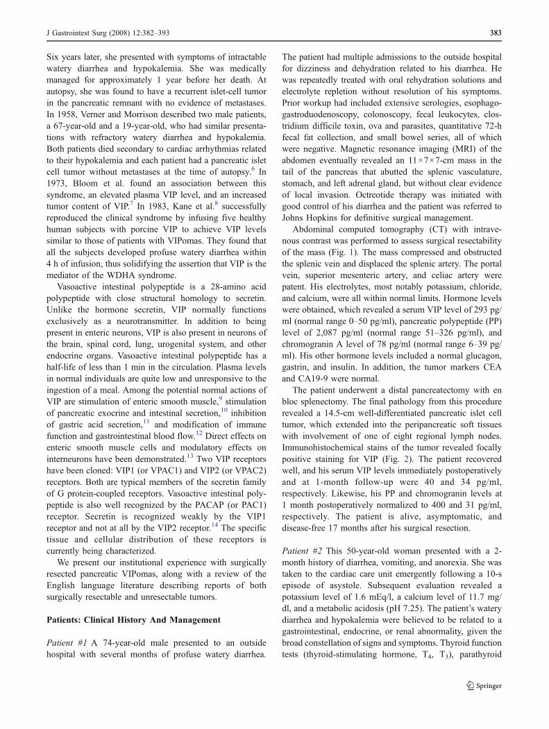
Patient #1 A 74-year-old male presented to an outsidehospital with several months of profuse watery diarrhea.
The patient had multiple admissions to the outside hospitalfor dizziness and dehydration related to his diarrhea. Hewas repeatedly treated with oral rehydration solutions andelectrolyte repletion without resolution of his symptoms.Prior workup had included extensive serologies, esophago-gastroduodenoscopy, colonoscopy, fecal leukocytes, clos-tridium difficile toxin, ova and parasites, quantitative 72-hfecal fat collection, and small bowel series, all of whichwere negative. Magnetic resonance imaging (MRI) of theabdomen eventually revealed an 11×7×7-cm mass in thetail of the pancreas that abutted the splenic vasculature,stomach, and left adrenal gland, but without clear evidenceof local invasion. Octreotide therapy was initiated withgood control of his diarrhea and the patient was referred toJohns Hopkins for definitive surgical management.
Abdominal computed tomography (CT) with intrave-nous contrast was performed to assess surgical resectabilityof the mass (Fig. 1). The mass compressed and obstructedthe splenic vein and displaced the splenic artery. The portalvein, superior mesenteric artery, and celiac artery werepatent. His electrolytes, most notably potassium, chloride,and calcium, were all within normal limits. Hormone levelswere obtained, which revealed a serum VIP level of 293 pg/ml (normal range 0–50 pg/ml), pancreatic polypeptide (PP)level of 2,087 pg/ml (normal range 51–326 pg/ml), andchromogranin A level of 78 pg/ml (normal range 6–39 pg/ml). His other hormone levels included a normal glucagon,gastrin, and insulin. In addition, the tumor markers CEAand CA19-9 were normal.
The patient underwent a distal pancreatectomy with enbloc splenectomy. The final pathology from this procedurerevealed a 14.5-cm well-differentiated pancreatic islet celltumor, which extended into the peripancreatic soft tissueswith involvement of one of eight regional lymph nodes.Immunohistochemical stains of the tumor revealed focallypositive staining for VIP (Fig. 2). The patient recoveredwell, and his serum VIP levels immediately postoperativelyand at 1-month follow-up were 40 and 34 pg/ml,respectively. Likewise, his PP and chromogranin levels at1 month postoperatively normalized to 400 and 31 pg/ml,respectively. The patient is alive, asymptomatic, anddisease-free 17 months after his surgical resection.
Patient #2 This 50-year-old woman presented with a 2-month history of diarrhea, vomiting, and anorexia. She wastaken to the cardiac care unit emergently following a 10-sepisode of asystole. Subsequent evaluation revealed apotassium level of 1.6 mEq/l, a calcium level of 11.7 mg/dl, and a metabolic acidosis (pH 7.25). The patient’s waterydiarrhea and hypokalemia were believed to be related to agastrointestinal, endocrine, or renal abnormality, given thebroad constellation of signs and symptoms. Thyroid functiontests (thyroid-stimulating hormone, T4, T3), parathyroid
J Gastrointest Surg (2008) 12:382–393 383383

hormone, and serum cortisol were normal. An abdominalCT scan revealed a 3.5-cm mass in the tail of her pancreasand multiple hypodense lesions in the liver, consistent withmetastases. Her serum VIP level was 770 pg/ml, with aserum gastrin level of 500 pg/ml. After intravenoushydration and electrolyte repletion, the patient was startedon parenteral octreotide in preparation for surgery.
The patient underwent a distal pancreatectomy with enbloc splenectomy, open cholecystectomy, and wedgeresection of several hepatic metastases. The final pathologyrevealed a 4.5-cm islet cell tumor of the body of thepancreas with infiltration into the peripancreatic soft tissuesand involvement of one of 14 resected regional lymphnodes. The liver masses measured from 0.5 to 2 cm andwere biopsy-confirmed to represent metastatic islet celltumor. Postoperatively, her serum VIP and gastrin levelsdeclined to less than 35 and 118 pg/ml, respectively. Thepatient received octreotide therapy for management of her
metastatic disease and died 8 years after her pancreatectomyfrom tumor cachexia related to advanced metastatic disease.
Patient #3 A 66-year-old male presented with a 6-monthhistory of watery diarrhea and a 40-lb weight loss. Hislaboratory values at the time of presentation were notablefor a metabolic acidosis (pH 7.21) and hypokalemia(2.8 mEq/l). His serum VIP level was 169 pg/ml. A CTscan of the abdomen demonstrated an exophytic massinvolving the tail of the pancreas measuring 3×5×6 cmwithout evidence of adenopathy or hepatic involvement(Fig. 3). He also underwent an octreotide radionuclidestudy, which demonstrated scattered radiotracer uptake inthe liver, most consistent with hepatic metastases (Fig. 4).
The patient underwent a distal pancreatectomy with enbloc splenectomy, open cholecystectomy, and resection ofsegments II and III of his liver. Final pathology of theresected tissue demonstrated a 5-cm malignant endocrine
Figure 2 Left: PancreaticVIPoma in patient #1 demon-strating classic features of aneuroendocrine lesion with tra-becular architecture and lowgrade, uniformly round nucleiwith finely speckled chromatin(hematoxylin and eosin, ×400).Right: Immunohistochemicalstaining of the tumor demon-strating focal positivity for VIP(VIP immuno stain, ×400).
Figure 1 Abdominal CT scanof patient 1 showing large massof the tail of the pancreas(arrow) with displacement ofthe splenic artery (double arrow).
384 J Gastrointest Surg (2008) 12:382–393
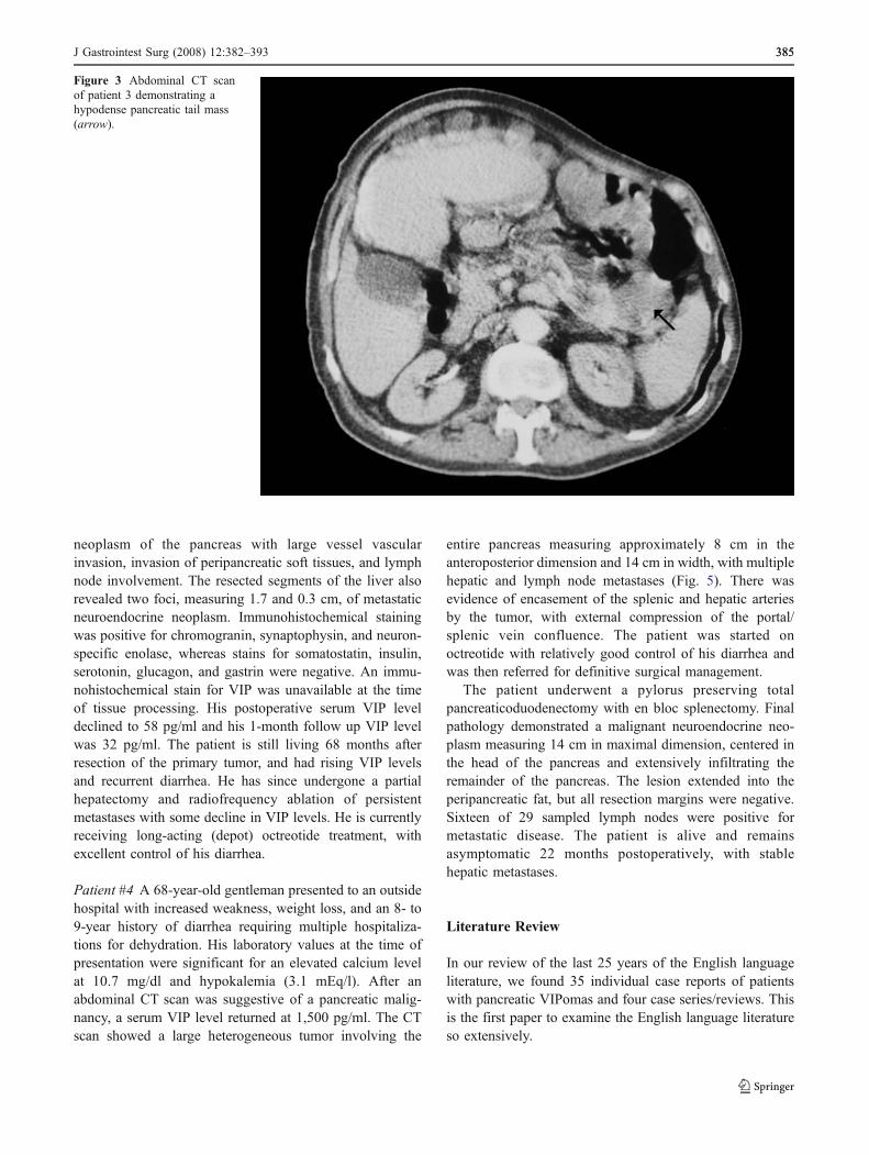
neoplasm of the pancreas with large vessel vascularinvasion, invasion of peripancreatic soft tissues, and lymphnode involvement. The resected segments of the liver alsorevealed two foci, measuring 1.7 and 0.3 cm, of metastaticneuroendocrine neoplasm. Immunohistochemical stainingwas positive for chromogranin, synaptophysin, and neuron-specific enolase, whereas stains for somatostatin, insulin,serotonin, glucagon, and gastrin were negative. An immu-nohistochemical stain for VIP was unavailable at the timeof tissue processing. His postoperative serum VIP leveldeclined to 58 pg/ml and his 1-month follow up VIP levelwas 32 pg/ml. The patient is still living 68 months afterresection of the primary tumor, and had rising VIP levelsand recurrent diarrhea. He has since undergone a partialhepatectomy and radiofrequency ablation of persistentmetastases with some decline in VIP levels. He is currentlyreceiving long-acting (depot) octreotide treatment, withexcellent control of his diarrhea.
Patient #4 A 68-year-old gentleman presented to an outsidehospital with increased weakness, weight loss, and an 8- to9-year history of diarrhea requiring multiple hospitaliza-tions for dehydration. His laboratory values at the time ofpresentation were significant for an elevated calcium levelat 10.7 mg/dl and hypokalemia (3.1 mEq/l). After anabdominal CT scan was suggestive of a pancreatic malig-nancy, a serum VIP level returned at 1,500 pg/ml. The CTscan showed a large heterogeneous tumor involving the
entire pancreas measuring approximately 8 cm in theanteroposterior dimension and 14 cm in width, with multiplehepatic and lymph node metastases (Fig. 5). There wasevidence of encasement of the splenic and hepatic arteriesby the tumor, with external compression of the portal/splenic vein confluence. The patient was started onoctreotide with relatively good control of his diarrhea andwas then referred for definitive surgical management.
The patient underwent a pylorus preserving totalpancreaticoduodenectomy with en bloc splenectomy. Finalpathology demonstrated a malignant neuroendocrine neo-plasm measuring 14 cm in maximal dimension, centered inthe head of the pancreas and extensively infiltrating theremainder of the pancreas. The lesion extended into theperipancreatic fat, but all resection margins were negative.Sixteen of 29 sampled lymph nodes were positive formetastatic disease. The patient is alive and remainsasymptomatic 22 months postoperatively, with stablehepatic metastases.
Literature Review
In our review of the last 25 years of the English languageliterature, we found 35 individual case reports of patientswith pancreatic VIPomas and four case series/reviews. Thisis the first paper to examine the English language literatureso extensively.
Figure 3 Abdominal CT scanof patient 3 demonstrating ahypodense pancreatic tail mass(arrow).
J Gastrointest Surg (2008) 12:382–393 385385

Individual Case Reports
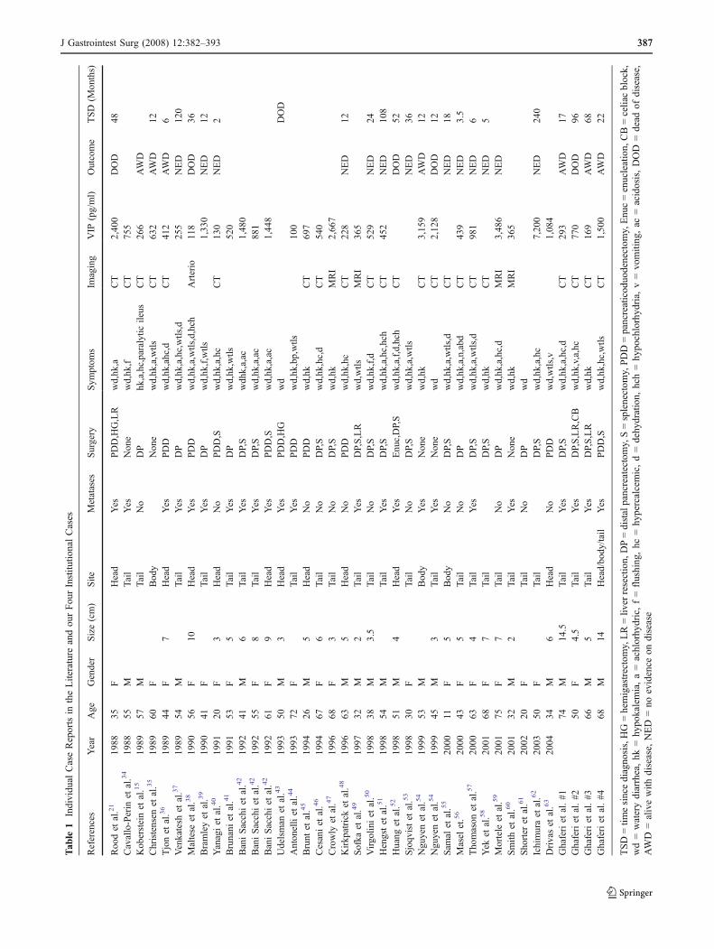
The 35 case reports are summarized in Table 1.
Age and sex Thirty five patients were identified with 20females and 15 males, ranging in age from 11 to 75 yearsold (mean 48, median 51).
Presenting signs and symptoms The most common clinicaland laboratory findings in the 35 patients are outlined inTable 1. As expected, nearly 100% of patients presentedwith watery diarrhea. Only one patient, presented byKoberstein et al.,15 did not present with watery diarrhea.This 57-year-old male presented with a paralytic ileus ofunknown origin. The patient had a transient episode ofloose, melanotic stools several days prior to admissionfollowing an episode of prolonged epistaxis. The patienthad not mentioned this episode during the initial interview.This unusual presentation, in conjunction with the labora-tory findings of hypokalemia, hypercalcemia, and acidosis,which were refractory to intravenous repletion, led thephysicians to consider a neuroendocrine etiology such asVIPoma. Indeed, the patient was found to have a mass inthe tail of the pancreas, confirmed to be a VIPoma, withserum VIP levels ranging from 173 to 266 pg/ml.
VIP radioimmunoassay Values were reported in 29 of the35 cases reviewed. The values ranged from 100 to7,200 pg/ml (mean 1,209 pg/ml, median 632 pg/ml).
Figure 5 Abdominal CT scanof patient 4. The pancreaticparenchyma is largely replacedby tumor (arrows).
Figure 4 Octreotide radionuclide scan of patient 3. The liver (L),spleen (S), and bladder (B) are seen. Scattered hepatic metastases areevident (MET).
386 J Gastrointest Surg (2008) 12:382–393
Tab
le1
Individu
alCaseReportsin
theLiterature
andou
rFou
rInstitu
tionalCases
References
Year
Age
Gender
Size(cm)
Site
Metatases
Surgery
Sym
ptom
sIm
aging
VIP
(pg/ml)
Outcome
TSD
(Mon
ths)
Roo
det
al.21
1988
35F
Head
Yes
PDD,HG,LR
wd,hk
,aCT
2,40
0DOD
48Cavallo-Perin
etal.34
1988
55M
Tail
Yes
Non
ewd,hk
,fCT
755
Kob
ersteinet
al.15
1989
57M
Tail
No
DP
hk,a,hc,paralytic
ileus
CT
266
AWD
Christensen
etal.35
1989
60F
Bod
yNon
ewd,hk
,a,wtls
CT
632
AWD
12Tjonet
al.36
1989
44F
7Head
Yes
PDD
wd,hk
,ahc,d
CT
412
AWD
6Venkatesh
etal.37
1989
54M
Tail
Yes
DP
wd,hk
,a,hc,wtls,d
255
NED
120
Maltese
etal.38
1990
56F
10Head
Yes
PDD
wd,hk,a,wtls,d,hch
Arterio
118
DOD
36Bramleyet
al.39
1990
41F
Tail
Yes
DP
wd,hk
,f,wtls
1,33
0NED
12Yanagiet
al.40
1991
20F
3Head
No
PDD,S
wd,hk
,a,hc
CT
130
NED
2Brunani
etal.41
1991
53F
5Tail
Yes
DP
wd,hk
,wtls
520
BaniSacchiet
al.42
1992
41M
6Tail
Yes
DP,S
wdh
k,a,ac
1,48
0BaniSacchiet
al.42
1992
55F
8Tail
Yes
DP,S
wd,hk
,a,ac
881
BaniSacchiet
al.42
1992
61F
9Head
Yes
PDD,S
wd,hk
,a,ac
1,44
8Udelsman
etal.43
1993
50M
3Head
Yes
PDD,HG
wd
DOD
Anton
elliet
al.44
1993
72F
Tail
Yes
PDD
wd,hk
,bp,wtls
100
Brunt
etal.45
1994
26M
5Head
No
PDD
wd,hk
CT
697
Cesaniet
al.46
1994
67F
6Tail
No
DP,S
wd,hk
,hc,d
CT
540
Crowly
etal.47
1996
68F
3Tail
No
DP,S
wd,hk
MRI
2,66
7Kirkp
atrick
etal.48
1996
63M
5Head
No
PDD
wd,hk
,hc
CT
228
NED
12Sofka
etal.49
1997
32M
2Tail
Yes
DP,S,LR
wd,wtls
MRI
365
Virgo
liniet
al.50
1998
38M
3.5
Tail
No
DP,S
wd,hk
,f,d
CT
529
NED
24Hengstet
al.51
1998
54M
Tail
Yes
DP,S
wd,hk
,a,hc,hch
CT
452
NED
108
Huang
etal.52
1998
51M
4Head
Yes
Enu
c,DP,S
wd,hk
,a,f,d,hch
CT
DOD
52Sjoqv
istet
al.53
1998
30F
Tail
No
DP,S
wd,hk
,a,wtls
NED
36Ngu
yenet
al.54
1999
53M
Bod
yYes
Non
ewd,hk
CT
3,15
9AWD
12Ngu
yenet
al.54
1999
45M
3Tail
Yes
Non
ewd
CT
2,12
8DOD
12Sam
alet
al.55
2000
11F
5Body
No
DP,S
wd,hk,a,wtls,d
CT
NED
18Masel
et.56
2000
43F
5Tail
No
DP
wd,hk
,a,n,abd
CT
439
NED
3.5
Tho
mason
etal.57
2000
63F
4Tail
Yes
DP,S
wd,hk,a,wtls,d
CT
981
NED
6Yek
etal.58
2001
68F
7Tail
DP,S
wd,hk
CT
NED
5Mortele
etal.59
2001
75F
7Tail
No
DP
wd,hk
,a,hc,d
MRI
3,48
6NED
Smith
etal.60
2001
32M
2Tail
Yes
Non
ewd,hk
MRI
365
Sho
rter
etal.61
2002
20F
Tail
No
DP
wd
Ichimuraet
al.62
2003
50F
Tail
DP,S
wd,hk
,a,hc
7,20
0NED
240
Drivaset
al.63
2004
34M
6Head
No
PDD
wd,wtls,v
1,08
4Ghaferiet
al.#1
74M
14.5
Tail
Yes
DP,S
wd,hk
,a,hc,d
CT
293
AWD
17Ghaferiet
al.#2
50F
4.5
Tail
Yes
DP,S,LR,CB
wd,hk
,v,a,hc
CT
770
DOD
96Ghaferiet
al.#3
66M
5Tail
Yes
DP,S,LR
wd,hk
CT
169
AWD
68Ghaferiet
al.#4
68M
14Head/bo
dy/tail
Yes
PDD,S
wd,hk
,hc,wtls
CT
1,50
0AWD
22
TSD=tim
esincediagno
sis,HG=hemigastrectomy,LR=liv
erresection,
DP=distalpancreatectomy,S=splenectom
y,PDD=pancreaticod
uodenectom
y,Enu
c=enucleation,
CB=celiacblock,
wd=waterydiarrhea,hk
=hy
pokalemia,a=achlorhy
dric,f=flushing
,hc
=hy
percalcemic,d=dehy
dration,
hch=hy
pochlorhyd
ria,
v=vo
miting
,ac
=acidosis,DOD
=dead
ofdisease,
AWD
=alivewith
disease,
NED
=no
evidence
ondisease
J Gastrointest Surg (2008) 12:382–393 387387

Radiologic features/modalities The imaging modality usedto diagnose a pancreatic mass was reported in 23 of the 35cases. The most common diagnostic study was CT (18/23,78%). Magnetic resonance imaging and selective angiogra-phy were utilized in four cases and in one case, respective-ly. The primary tumor was always identified using one ofthe aforementioned modalities. Furthermore, metastaticdisease to regional lymph nodes or liver was often diagnosedvia imaging. Of the 35 case reports, 32 reported on thepresence or absence of metastases, with 19 of the 32 (59%)reporting the presence of metastases, most commonly to theliver.
Site The site of primary disease in the pancreas wasidentified in all of the reported cases. The distribution ofthe primary tumors was as follows: 25 in the body and tail ofthe pancreas (72%) and 10 in the head of the pancreas (28%).
Size The primary tumors ranged in size from 2 to 10 cm intheir greatest dimension, reported in 23 cases. The mean andmedian sizes were both 5 cm. Histologic confirmation of thetumor was reported in 18 cases with either routinehematoxylin and eosin staining or VIP immunohistochem-ical staining.
Treatment (surgery/none) Surgical intervention was re-ported in 30 of 35 cases (86%). The procedures includeddistal pancreatectomy (54%), pancreaticoduodenectomy(29%), splenectomy (43%), hemigastrectomy (6%), liverresection (6%), and tumor enucleation (3%).
Outcome The outcomes were reported with varying followup periods. The mean follow up time was 40 months, with amedian of 15 months. Outcome data were reported in 22 ofthe cases. Fifty nine percent of the patients were reported asalive with no evidence of disease, 23% had died of disease,and 18% were alive with disease.
Case Series
Summaries of the case series are tabulated in Table 2. Sogaet al.16 published the largest review of reported VIPomacases including 241 patients found in an internationalliterature search. The authors identified 179 patients withpancreatic VIPomas and compared the clinically reporteddata for this group of patients to the group diagnosed withextrapancreatic neurogenic tumors (n=48). They foundstatistically significant differences (p<0.05) between thetwo groups (pancreatic vs. extrapancreatic) in the rate ofassociated syndrome (84 vs. 96%), tumor size larger than20 mm (79 vs. 100%), rate of metastases (56 vs. 29%), rate T
able
2Sum
maryof
CaseSeriesReportin
gon
VIPom
asandou
rInstitu
tion’sCases
CaseSeries
Num
berof
Cases
M:F
MeanAge
(Range)
MeanTum
orSize(cm)
MeanVIP
(pg/ml)
PresentingSym
ptom
s(%
)Locationof
Tum
or(%
)
Diarrhea
Weigh
tLoss
Dehyd
ratio
nHyp
okalem
iaFlushing
Head
Bod
yTail
Other
Sog
aet
al.16
179
84:95
51(15-82
)5.4
9836
8914
298
603
Smith
etal.17
18fs
9:9
51(23-74
)4.4
698
8972
4467
2811
2250
17Penget
al.18
3116
:15
48(26-73
)5.4
963
100
100
100
3352
1329
6Nikou
etal.19
117:4
53(2-83)
100
4545
8118
955
18Ghaferiet
al.
43:1
65(50-74
)9.5
683
100
5025
100
00
2575
0
388 J Gastrointest Surg (2008) 12:382–393

of malignancy (64 vs. 33%), and rate of resection of theprimary lesion (69 vs. 88%). The 5-year actuarial survivalrate for patients with pancreatic VIPoma was found to be69%. A significant difference existed between those withmetastases at diagnosis vs. those without: 60% 5-yearsurvival in patients with metastases vs. 94% 5-year survivalin those without evidence of metastatic disease.
Smith et al.17 presented the Mayo Clinic’s 15-yearexperience with VIP-secreting islet cell tumors (Table 2).They reported on 18 patients with a mean age of 51 years(range 23–74) at presentation. There were equal numbers ofmale and female patients. As expected, secretory diarrheawas the most common presenting symptom in 89% ofpatients, and the most common location of the tumor wasthe tail of the pancreas (50%). The mean survival was3.6 years, with the longest survival reaching 15 years.
Peng et al.18 in 2004 presented a case report and clinicalreview of 31 cases of VIPoma in China (Table 2). Theyreported typical clinical manifestations, imaging features,surgical procedures, and pathologic findings. They foundthat the mean age of presentation was 48 years, with themean size of the primary being 5.4 cm. The meanpreoperative VIP level was 963 pg/ml (range 68–2,100),and the mean postoperative VIP value was 132 pg/ml(range 20–450).
In 2005, Nikou et al.19 presented 11 patients withVIPoma (Table 2). Seven of the 11 patients were male,with an age range of 2 to 83 years (mean age 53.1 years).All patients presented with chronic secretory diarrhea thatpersisted despite fasting. Nine (81%) patients also presentedwith hypokalemia. Weight loss was observed in 45% ofpatients. Vasoactive intestinal polypeptide levels were three
Secretory
Diarrhea
Exclude
Infection
Standard bacterial
pathogens
Other pathogens:
"Standard" ova & parasites
Coccidia
Microsporidia
Giardia antigen
Exclude
structural
disease
Small bowel
biopsy and
aspirate for
quantitative
culture
CT scan of
abdomen
Sigmoidoscopy or
colonoscopy with biopsySmall bowel
radiographs
Selective
testing
Plasma peptides:
Gastrin
Calcitonin
VIP
Somatostatin
Urine:
5'-HIAA
Metanephrines
Histamine
Other tests:
TSH
ACTH stimulation
Serum protein
electrophoresis
ImmunoglobulinsCholestyramine trial for bile
acid diarrhea
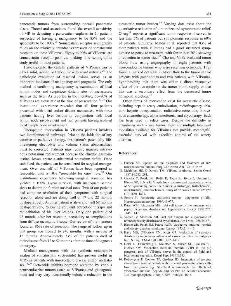
Figure 6 Algorithm for the identification of an etiology for secretory diarrhea. 5-HIAA, 5-hydroxyindoleacetic acid; TSH, thyroid-stimulatinghormone; ACTH, adrenocorticotropic hormone (adapted from Schiller64).
J Gastrointest Surg (2008) 12:382–393 389389
times normal in seven of the 11 patients and 10 timesnormal in the remaining four patients. Serum chromograninA levels were elevated in all patients. Fifty four percent oflesions were detected by CT or MRI, whereas EUS orangiography detected 4/11 lesions (36%). Octreoscandetected the primary lesion in 10/11 (91%) and themetastases in 3/4 (75%). The primary lesion was located inthe pancreatic tail in 6/11 (55%), the pancreatic body in 1/11(9%), and the second portion of the duodenum in 2/11(18%). A 2-year-old child included in the review had theprimary tumor located in the retroperitoneum. Four patientshad metastatic disease at the time of diagnosis. Sixtythree percent of the patients underwent resection. At thetime of the report, six patients were alive with no evidenceof disease, two were alive with disease, and three had diedof disease.
Discussion
VIPomas are rare tumors that often elude prompt diagnosis.As demonstrated by our review of the literature, nearly100% of patients present with a primary complaint ofwatery diarrhea refractory to traditional medical manage-ment. Chronic diarrhea is defined as that which lasts at least4 to 6 weeks; the prevalence of chronic diarrhea isestimated to approximate 3–5% of the US population.20
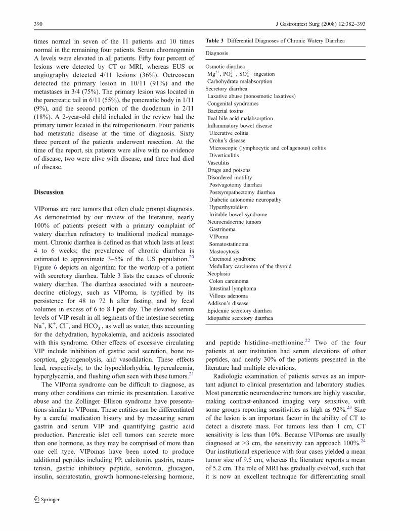
Figure 6 depicts an algorithm for the workup of a patientwith secretory diarrhea. Table 3 lists the causes of chronicwatery diarrhea. The diarrhea associated with a neuroen-docrine etiology, such as VIPoma, is typified by itspersistence for 48 to 72 h after fasting, and by fecalvolumes in excess of 6 to 8 l per day. The elevated serumlevels of VIP result in all segments of the intestine secretingNa+, K+, Cl−, and HCO�
3 , as well as water, thus accountingfor the dehydration, hypokalemia, and acidosis associatedwith this syndrome. Other effects of excessive circulatingVIP include inhibition of gastric acid secretion, bone re-sorption, glycogenolysis, and vasodilation. These effectslead, respectively, to the hypochlorhydria, hypercalcemia,hyperglycemia, and flushing often seen with these tumors.21
The VIPoma syndrome can be difficult to diagnose, asmany other conditions can mimic its presentation. Laxativeabuse and the Zollinger–Ellison syndrome have presenta-tions similar to VIPoma. These entities can be differentiatedby a careful medication history and by measuring serumgastrin and serum VIP and quantifying gastric acidproduction. Pancreatic islet cell tumors can secrete morethan one hormone, as they may be comprised of more thanone cell type. VIPomas have been noted to produceadditional peptides including PP, calcitonin, gastrin, neuro-tensin, gastric inhibitory peptide, serotonin, glucagon,insulin, somatostatin, growth hormone-releasing hormone,
and peptide histidine–methionine.22 Two of the fourpatients at our institution had serum elevations of otherpeptides, and nearly 30% of the patients presented in theliterature had multiple elevations.
Radiologic examination of patients serves as an impor-tant adjunct to clinical presentation and laboratory studies.Most pancreatic neuroendocrine tumors are highly vascular,making contrast-enhanced imaging very sensitive, withsome groups reporting sensitivities as high as 92%.23 Sizeof the lesion is an important factor in the ability of CT todetect a discrete mass. For tumors less than 1 cm, CTsensitivity is less than 10%. Because VIPomas are usuallydiagnosed at >3 cm, the sensitivity can approach 100%.24
Our institutional experience with four cases yielded a meantumor size of 9.5 cm, whereas the literature reports a meanof 5.2 cm. The role of MRI has gradually evolved, such thatit is now an excellent technique for differentiating small
Table 3 Differential Diagnoses of Chronic Watery Diarrhea
Diagnosis
Osmotic diarrheaMg2+, PO3�
4 , SO2�4 ingestion
Carbohydrate malabsorptionSecretory diarrheaLaxative abuse (nonosmotic laxatives)Congenital syndromesBacterial toxinsIleal bile acid malabsorptionInflammatory bowel diseaseUlcerative colitisCrohn’s diseaseMicroscopic (lymphocytic and collagenous) colitisDiverticulitisVasculitisDrugs and poisonsDisordered motilityPostvagotomy diarrheaPostsympathectomy diarrheaDiabetic autonomic neuropathyHyperthyroidismIrritable bowel syndromeNeuroendocrine tumorsGastrinomaVIPomaSomatostatinomaMastocytosisCarcinoid syndromeMedullary carcinoma of the thyroidNeoplasiaColon carcinomaIntestinal lymphomaVillous adenomaAddison’s diseaseEpidemic secretory diarrheaIdiopathic secretory diarrhea
390 J Gastrointest Surg (2008) 12:382–393
pancreatic tumors from surrounding normal pancreatictissue. Thoeni and associates found the overall sensitivityof MR in detecting a pancreatic neoplasm in 20 patientssuspected of having a malignancy to be 85% and thespecificity to be 100%.25 Somatostatin receptor scintigraphyrelies on the relatively abundant expression of somatostatinreceptors on these VIPomas. Eighty to 90% of VIPomas aresomatostatin receptor-positive, making this scintigraphicstudy useful in most patients.
Histologically, the cellular patterns of VIPomas can beeither solid, acinar, or trabecular with scant mitoses.26 Thepathologic evaluation of resected lesions serves as animportant indicator of malignancy and prognosis. The onlymethod of confirming malignancy is examination of locallymph nodes and suspicious distant sites of metastases,such as the liver. As reported in the literature, 60–80% ofVIPomas are metastatic at the time of presentation.22,27 Ourinstitutional experience revealed that all four patientspresented with local and/or distant metastases, with threepatients having liver lesions in conjunction with locallymph node involvement and two patients having isolatedlocal lymph node involvement.
Therapeutic intervention in VIPoma patients involvestwo interconnected pathways. Prior to the initiation of anycurative or palliative therapy, the patient’s potentially life-threatening electrolyte and volume status abnormalitiesmust be corrected. Patients may require massive intrave-nous potassium replacement because the chronic gastroin-testinal losses create a substantial potassium deficit. Oncestabilized, the patient can be considered for surgical manage-ment. Over one-half of VIPomas have been reported asresectable, with a 10% “resectable for cure” rate.28 Ourinstitutional experience following surgical resection hasyielded a 100% 1-year survival, with inadequate samplesizes to determine further survival rates. Two of our patientshad complete resolution of their symptoms with surgicalresection alone and are doing well at 17 and 22 monthspostoperatively. Another patient is alive and well 68 monthspostoperatively, following adjuvant octreotide therapy andradioablation of his liver lesions. Only one patient died96 months after her resection, secondary to complicationsfrom diffuse metastatic disease. Our review of the literaturefound an 86% rate of resection. The range of follow up inthat group was from 2 to 240 months, with a median of15 months. Approximately 23% of the patients died oftheir disease from 12 to 52 months after the time of diagnosisor surgery.
Medical management with the synthetic octapeptideanalog of somatostatin (octreotide) has proven useful inVIPoma patients with unresectable disease and/or metasta-ses.18,29 Octreotide inhibits hormone secretion by variousneuroendocrine tumors (such as VIPomas and glucagono-mas) and may very occasionally induce a reduction in the
metastatic tumor burden.30 Varying data exist about thequantitative reduction of tumor size and symptomatic relief.Oberg31 reports a significant tumor response observed inless than 5% of patients but symptomatic response in 60%of patients. Similarly, Maton et al. reported that 83% oftheir patients with VIPomas had a good sustained symp-tomatic response to treatment, with fewer than 20% showinga reduction in tumor size.32 Cho and Vinik evaluated tumorblood flow using angiography in eight patients withneuroendocrine tumors who were receiving octreotide. Theyfound a marked decrease in blood flow to the tumor in twopatients with gastrinomas and two patients with VIPomas,hypothesizing that there was either a direct vasoactiveeffect of the octreotide on the tumor blood supply or thatthis was a secondary effect from the decreased tumorhormonal secretion.33
Other forms of intervention exist for metastatic disease,including hepatic artery embolization, radiofrequency abla-tion, hepatic transplantation, radioactive octreotide, intrave-nous chemotherapy, alpha interferons, and cryotherapy. Eachhas been used in select cases. Despite the difficulty indiagnosing such a rare tumor, there are multiple treatmentmodalities available for VIPomas that provide meaningful,extended survival with excellent control of the waterydiarrhea.
References
1. Friesen SR. Update on the diagnosis and treatment of rareneuroendocrine tumors. Surg Clin North Am 1987;67:379.
2. Mekhijian HS, O’Dorisio TM. VIPoma syndrome. Semin Oncol1987;14:282–291.
3. Capella C, Polak JM, Buffa R, Tapia FJ, Heitz P, Usellini L,Bloom SR, Solcia E. Morphologic patterns and diagnostic criteriaof VIP-producing endocrine tumors. A histologic, histochemical,ultrastructural, and biochemical study of 32 cases. Cancer 1983;52(10):1860–1874.
4. Ectors N. Pancreatic endocrine tumors: diagnostic pitfalls.Hepatogastroenterology 1999;46:679.
5. Priest WM, Alexander MK. Islet cell tumor of the pancreas withpeptic ulceration, diarrhea and hypokalemia. Lancet 1957;273:1145–1147.
6. Verner JV, Morrison AB. Islet cell tumour and a syndrome ofrefractory watery diarrhea and hypokalemia. Am JMed 1958;25:374.
7. Bloom SR, Polak JM, Pearse AGE. Vasoactive intestinal peptideand watery-diarrhea syndrome. Lancet 1973;2:14–16.
8. Kane MG, O’Dorisio TM, Krejs GJ. Production of secretorydiarrhea by intravenous infusion of vasoactive intestinal polypep-tide. N Engl J Med 1983;309:1482–1485.
9. Holst JJ, Fahrenkrug J, Knuhtsen S, Jensen SL, Poulsen SS,Nielsen OV. Vasoactive intestinal peptide (VIP) in the pigpancreas: role of VIPergic nerves in the control of fluid andbicarbonate secretion. Regul Pept 1984;8:245.
10. Robberecht P, Conlon TP, Gardner JD. Interaction of porcinevasoactive intestinal peptide with dispersed pancreatic acinar cellsfrom the guinea pig. Structural requirements for effects ofvasoactive intestinal peptide and secretin on cellular adenosine3′,5′-monophosphate. J Biol Chem 1976;251:4635.
J Gastrointest Surg (2008) 12:382–393 391391
11. Barbezat GO, Grossman MI. Intestinal secretion: stimulation bypeptides. Science 1971;174:422.
12. Fahrenkrug J. Transmitter role of vasoactive intestinal peptide.Pharmacol Toxicol 1993;72:354.
13. Biancani P, Walsh JH, Behar J. Vasoactive intestinal polypeptide.A neurotransmitter for lower esophageal sphincter relaxation. JClin Invest 1984;73:963.
14. Usdin TB, Bonner TI, Mezey É. Two receptors for vasoactiveintestinal polypeptide with similar specificity and complementarydistributions. Endocrinology 1994;135:2662.
15. Koberstein B, Layer P, Balzer K, Muller MK, Singer MV, GoebellH. Paralytic ileus responding to somatostatin therapy: Firstmanifestation of a VIPoma. Dig Dis Sci 1989;34(11):1803–1804.
16. Soga J, Yakuwa Y. Vipoma/diarrheogenic syndrome: a statisticalevaluation of 241 reported cases. J Exp Clin Cancer Res 1998;17(4):389–400.
17. Smith SL, Branton SA, Avino AJ, Martin JK, Klingler PJ,Thompson GB, Grant CS, van Heerden JA. Vasoactive intestinalpolypeptide secreting islet cell tumors: a 15-year experience andreview of the literature. Surgery 1998;124(6):1050–1055.
18. Peng SY, Li JT, Liu YB, Fang HQ, Wu YL, Peng CH, Wang XB,Qian HR. Diagnosis and treatment of VIPoma in China: (casereport and 31 cases review) diagnosis and treatment of VIPoma.Pancreas 2004;28(1):93–97.
19. Nikou GC, Toubanakis C, Niklolaou P, Giannatou E, Safioleas M,Mallas E, Polyzos A. VIPomas: An update in diagnosis andmanagement in a series of 11 patients. Hepatogastroenterology2005;52:1259–1265.
20. Fine KD, Schiller LR. AGA technical review on the evaluationand management of chronic diarrhea. Gastroenterology 1999;116:1464–1486.
21. Rood RP, DeLellis RA, Dayal Y, Donowitz M. Pancreatic cholerasyndrome due to vasoactive intestinal polypeptide-producingtumor: further insights into the pathophysiology. Gastroenterology1988;94:813.
22. Perry RR, Vinik AI. Clinical review 72: Diagnosis and manage-ment of functioning islet cell tumors. J Clin Endocrinol Metab1995;80(8):2273–2278.
23. Legmann P, Vignaux O, Dousset B, Baraza AJ, Palazzo L,Dumontier I, Coste J, Louvel A, Rouseu G, Couturier D,Bonnin A. Pancreatic tumors: Comparison of dual-phase helicalCT and endoscopic sonography. Am J Roentgenol 1998;170(5):1315–1322.
24. King CM, Reznek RH, Dacie JE, Wass JA. Imaging islet celltumours. Clin Radiol 1994;49(5):295–303.
25. Thoeni RF, Mueller-Lisse UG, Chan R, Do NK, Shyn PB.Detection of small functional islet cell tumors in the pancreas:Selection of MR imaging sequences for optimal sensitivity.Radiology 2000;214(2):483–490.
26. Capella C, Polak JM. Morphologic patterns and diagnostic criteriaof VIP-producing endocrine tumors. A histologic, histochemical,ultrastructural and biochemical study of 32 cases. Cancer1983;52:1860–1874.
27. Jensen RT. Pancreatic endocrine tumors: Recent advances. AnnOncol 1999;10:6 (Suppl).
28. Thompson GB, van Heerden JA, Grant CS, Carney JA, IlstrupDM. Islet cell carcinomas of the pancreas: a twenty yearexperience. Surgery 1988;104:1011–1017.
29. O’Dorisio T, Mekhjian HA, Gaginella TS. Medical therapy ofVIPomas. Endocrinol Metab Clin North Am 1989;18:545–556.
30. Kraenzlin ME, Ch’ng JL, Wood SM, Carr DH, Bloom SR. Long-term treatment of a VIPoma with somatostatin analogue resultingin remission of symptoms and possible shrinkage of metastases.Gastroenterology 1985;88(1 Pt 1):185–187.
31. Oberg K. Chemotherapy and biotherapy in the treatment ofneuroendocrine tumours. Ann Oncol 2001;12(Suppl 2):S111–S114.
32. Maton PN, Gardner JD, Jensen RT. Use of long-acting somato-statin analog SMS 201-995 in patients with pancreatic islet celltumors. Dig Dis Sci 1989;34:28S–39S.
33. Cho KJ, Vinik AI. Effect of somatostatin analogue (octreotide) onblood flow to endocrine tumors metastatic to the liver: angio-graphic evaluation. Radiology 1990;177(2):549–553.
34. Cavallo-Perin P, De Paoli M, Guiso G, Sapino A, Papotti M, CodaR, Pagano G. A combined glucagonoma and VIPoma syndrome.First pathologic and clinical report. Cancer 1988;62(12):2576–2579.
35. Christensen C. The somatostatin analogue SMS 201-995 in long-term treatment of vipoma. Case report. Acta Chir Scand 1989;155(10):541–543.
36. Tjon ATham RT, Jansen JB, Falke TH, Roelfsema F, Griffioen G,van den Sluys Veer A, Lamers CB. MR, CT, and ultrasoundfindings of metastatic vipoma in pancreas. J Comput Assist Tomogr1989;13(1):142–144.
37. Venkatesh S, Vassilopoulou-Sellin R, Samaan NA. Somatostatinanalogue: use in the treatment of vipoma with hypercalcemia AmJ Med 1989;87(3):356–357.
38. Maltese JY, Monge G, Giraud P, Salers P, Ouafik LH, Pelen F,Oliver C. Characterization of an alpha-amidating activity in ahuman pancreatic tumour secreting vasoactive intestinal peptide(VIP). Clin Endocrinol (Oxf) 1990;33(4):467–480.
39. Bramley PN, Lodge JP, Losowsky MS, Giles GR. Treatment ofmetastatic vipoma by liver transplantation. Clin Transplant 1990;4(5 part 1):276–278; discussion 279.
40. Yanagi H, Kusunoki M, Sakanoue Y, Shoji Y, Yamamura T, MuraiM, Kikkawa N, Utsunomiya J. Vipoma of the pancreas complicat-ing ulcerative colitis. Am J Gastroenterol 1991;86(8):1066–1069.
41. Brunani A, Crespi C, De Martin M, Dubini A, Piolini M,Cavagnini F. Four year treatment with a long acting somatostatinanalogue in a patient with Verner–Morrison syndrome. J EndocrinolInvest 1991;14(8):685–689.
42. Bani Sacchi T, Bani D, Biliotti G. Immunocytochemical andultrastructural abnormalities of islet tissue in patients with VIP-producing tumors of the pancreas. Pancreas 1992;7(5):601–610.
43. Udelsman R, Yeo CJ, Hruban RH, Pitt HA, Niederhuber JE,Coleman J, Cameron JL. Pancreaticoduodenectomy for selectedpancreatic endocrine tumors. Surg Gynecol Obstet 1993;177(3):269–278.
44. Antonelli A, Gambuzza C, Bertoni F, Baschieri L. Calcitonin, asSMS 201-995, ameliorates the VIPoma syndrome. J EndocrinolInvest 1993;16(1):57–59.
45. Brunt LM, Mazoujian G, O’Dorisio TM, Wells SA Jr. Stimulationof vasoactive intestinal peptide and neurotensin secretion bypentagastrin in a patient with VIPoma syndrome. Surgery1994;115(3):362–369.
46. Cesani F, Ernst R, Walser E, Villanueva-Meyer J. Tc-99msestamibi imaging of a pancreatic VIPoma and parathyroidadenoma in a patient with multiple type I endocrine neoplasia.Clin Nucl Med 1994;19(6):532–534.
47. Crowley PF, Slavin JL, Rode J. Massive amyloid deposition inpancreatic vipoma: a case report. Pathology 1996;28(4):377–379.
48. Kirkpatrick AW, Hanna SS, Skinner BA. Surgical treatment ofpancreatic cholera: a case report. Can J Surg 1996;39(2):155–158.
49. Sofka CM, Semelka RC, Marcos HB, Woosley JT. MR imaging ofmetastatic pancreatic VIPoma. Magn Reson Imaging 1997;15(10):1205–1208.
50. Virgolini I, Kurtaran A, Leimer M, Kaserer K, Peck-RadosavljevicM, Angelberger P, Hubsch P, Dvorak M, Valent P, Niederle B.Location of a VIPoma by iodine-123-vasoactive intestinal peptidescintigraphy. J Nucl Med 1998;39(9):1575–1579.
51. Hengst K, Nashan B, Avenhaus W, Ullerich H, Schlitt HJ,Flemming P, Pichlmayr R, Domschke W. Metastatic pancreaticVIPoma: deteriorating clinical course and successful treatment byliver transplantation. Z Gastroenterol 1998;36(3):239–245.
392 J Gastrointest Surg (2008) 12:382–393

52. Huang YH, Lee CH, Wu JC, Wang YJ, Chang FY, Lee SD.Functional pancreatic islet cell tumors with liver metastasis: the roleof cytoreductive surgery and transcatheter arterial chemoemboliza-tion: a report of five cases. Zhonghua Yi Xue Za Zhi (Taipei)1998;61(12):748–754.
53. Sjoqvist U, Permert J, Lofberg R, Larsson J, Gadaleanu V, AdrianTE. Life threatening diarrhoea ultimately cured by surgery. Eur JGastroenterol Hepatol 1998;10(11):963–967.
54. NguyenHN, Backes B, Lammert F,Wildberger J,Winograd R, BuschN, RiebandH,Matern S. Long-term survival after diagnosis of hepaticmetastatic VIPoma: report of two cases with disparate courses andreview of therapeutic options. Dig Dis Sci 1999;44(6):1148–1155.
55. Samal SC, Paul AC, Venkateswari S, Nair S, Venkatramani S,Perakath B, Simon A, Chandy G, Kurian G. VIPoma of pancreasin a child. Indian J Gastroenterol 2000;19(4):194–195.
56. Masel SL, Brennan BA, Turner JH, Cullingford GL, Cullen DJ.Pancreatic vasoactive intestinal polypeptide-oma as a cause ofsecretory diarrhoea. J Gastroenterol Hepatol 2000;15(4):457–460.
57. Thomason JW, Martin RS, Fincher ME. Somatostatin receptorscintigraphy: the definitive technique for characterizing vasoactiveintestinal peptide-secreting tumors. Clin Nucl Med 2000;25(9):661–664.
58. Yeh CN, Chen MF, Chen TC. Surgical treatment of pancreaticvasoactive intestinal polypeptide-secreting tumor: a case report.Hepatogastroenterology 2001;48(38):421–423.
59. Mortele KJ, Oei A, Bauters W, Timmermans F, Cuvelier C,Kunnen M, Ros PR. Dynamic gadolinium-enhanced MR imagingof pancreatic VIPoma in a patient with Verner–Morrison syn-drome. Eur Radiol 2001;11(10):1952–1955.
60. Smith CS, Houston M, Jensen B, Mlinar K, Toulson C, TillotsonLG. A 32-year-old man with copious, watery diarrhea. N C Med J2001;62(3):134–139.
61. Shorter NA, Glick RD, Klimstra DS, Brennan MF, Laquaglia MP.Malignant pancreatic tumors in childhood and adolescence: Thememorial Sloan–Kettering experience, 1967 to present. J PediatrSurg 2002;37(6):887–892.
62. Ichimura T, Kondo S, Okushiba S, Morikawa T, Katoh H. Acalcitonin and vasoactive intestinal peptide-producing pancreaticendocrine tumor associated with the WDHA syndrome. Int JGastrointest Cancer 2003;33(2–3):99–102.
63. Drivas I, Mansberg R, Roberts JM. VIPoma: a rare cause of apancreatic mass. Clin Nucl Med 2004;29(3):201–203.
64. Schiller LR. Chronic diarrhea. Gastroenterology 2004;127(1):287–293.
J Gastrointest Surg (2008) 12:382–393 393393