Ovarian steroids modulate leu-enkephalin levels and target leu-enkephalinergic profiles in the...
24
Ovarian steroids modulate leu-enkephalin levels and target leu- enkephalinergic profiles in the female hippocampal mossy fiber pathway Annelyn Torres-Reveron 1 , Sana Khalid 1 , Tanya J. Williams 1 , Elizabeth M. Waters 2 , Carrie T. Drake 1 , Bruce S. McEwen 2 , and Teresa A. Milner 1,2 1Division of Neurobiology, Department of Neurology and Neuroscience, Weill Cornell Medical College, 411 East 69th Street, New York, NY 10021 2Harold and Margaret Milliken Hatch Laboratory of Neuroendocrinology, The Rockefeller University, 1230 York Avenue, New York, NY 10065 Abstract In the hippocampal formation (HF), the enkephalin opioids and estrogen are each known to modulate learning and cognitive performance relevant to drug abuse. Within the HF, leu-enkephalin (LENK) is most prominent in the mossy fiber (MF) pathway formed by the axons of dentate gyrus (DG) granule cells. To examine the influence of ovarian steroids on MF pathway LENK levels, we used quantitative light microscopic immunocytochemistry to evaluate LENK levels in normal cycling rats and in estrogen-treated ovariectomized rats. Rats in estrus had increased levels of LENK- immunoreactivity (ir) in the DG hilus compared to rats in diestrus or proestrus. Rats in estrus and proestrus had higher levels of LENK-ir in CA3a-c compared to rats in diestrus. Ovariectomized (OVX) rats 24 hrs (but not 6 or 72 hrs) after estradiol benzoate (EB; 10 μg) administration had increased LENK-ir in the DG hilus and CA3c. Electron microscopy showed a larger proportion of LENK-labeled small terminals and axons in the DGthe DG hilus compared to CA3 which may have contributed to region-specific changes in LENK-ir densities. Next we evaluated the subcellular relationships of estrogen receptor (ER) α, ERβ and progestin receptor (PR) with LENK-labeled MF pathway profiles using dual-labeling electron microscopy. ERβ-ir colocalized in some LENK-labeled MF terminals and smaller terminals while PR-ir was mostly in CA3 axons, some of which also showed colocalization with LENK. ERα-ir was in dendritic spines, but no colocalization with LENK- labeled profiles was observed. The present studies indicate that estrogen can modulate LENK in subregions of the MF pathway in a dose- and time- dependent manner. These effects might be triggered by direct activation of ERβ or PR in LENK-containing terminals. Keywords dentate gyrus; CA3; estrogen; progestin; estrous cycle; opioid; immunoreactivity; rat *Address correspondence to: Drs. Annelyn Torres-Reveron and Teresa A. Milner, Division of Neurobiology, Weill Cornell Medical College, 411 East 69th Street, Room 101, New York, NY 10021, Phone: (212) 570-2900; FAX: (212) 988-3672, e-mail: [email protected]; [email protected]. Publisher's Disclaimer: This is a PDF file of an unedited manuscript that has been accepted for publication. As a service to our customers we are providing this early version of the manuscript. The manuscript will undergo copyediting, typesetting, and review of the resulting proof before it is published in its final citable form. Please note that during the production process errors may be discovered which could affect the content, and all legal disclaimers that apply to the journal pertain. NIH Public Access Author Manuscript Brain Res. Author manuscript; available in PMC 2009 September 26. Published in final edited form as: Brain Res. 2008 September 26; 1232: 70–84. doi:10.1016/j.brainres.2008.07.058. NIH-PA Author Manuscript NIH-PA Author Manuscript NIH-PA Author Manuscript
-
Upload
independent -
Category
Documents
-
view
2 -
download
0
Transcript of Ovarian steroids modulate leu-enkephalin levels and target leu-enkephalinergic profiles in the...
Ovarian steroids modulate leu-enkephalin levels and target leu-enkephalinergic profiles in the female hippocampal mossy fiberpathway
Annelyn Torres-Reveron1, Sana Khalid1, Tanya J. Williams1, Elizabeth M. Waters2, Carrie T.Drake1, Bruce S. McEwen2, and Teresa A. Milner1,2
1Division of Neurobiology, Department of Neurology and Neuroscience, Weill Cornell Medical College, 411East 69th Street, New York, NY 10021
2Harold and Margaret Milliken Hatch Laboratory of Neuroendocrinology, The Rockefeller University, 1230York Avenue, New York, NY 10065
AbstractIn the hippocampal formation (HF), the enkephalin opioids and estrogen are each known to modulatelearning and cognitive performance relevant to drug abuse. Within the HF, leu-enkephalin (LENK)is most prominent in the mossy fiber (MF) pathway formed by the axons of dentate gyrus (DG)granule cells. To examine the influence of ovarian steroids on MF pathway LENK levels, we usedquantitative light microscopic immunocytochemistry to evaluate LENK levels in normal cycling ratsand in estrogen-treated ovariectomized rats. Rats in estrus had increased levels of LENK-immunoreactivity (ir) in the DG hilus compared to rats in diestrus or proestrus. Rats in estrus andproestrus had higher levels of LENK-ir in CA3a-c compared to rats in diestrus. Ovariectomized(OVX) rats 24 hrs (but not 6 or 72 hrs) after estradiol benzoate (EB; 10 µg) administration hadincreased LENK-ir in the DG hilus and CA3c. Electron microscopy showed a larger proportion ofLENK-labeled small terminals and axons in the DGthe DG hilus compared to CA3 which may havecontributed to region-specific changes in LENK-ir densities. Next we evaluated the subcellularrelationships of estrogen receptor (ER) α, ERβ and progestin receptor (PR) with LENK-labeled MFpathway profiles using dual-labeling electron microscopy. ERβ-ir colocalized in some LENK-labeledMF terminals and smaller terminals while PR-ir was mostly in CA3 axons, some of which alsoshowed colocalization with LENK. ERα-ir was in dendritic spines, but no colocalization with LENK-labeled profiles was observed. The present studies indicate that estrogen can modulate LENK insubregions of the MF pathway in a dose- and time- dependent manner. These effects might betriggered by direct activation of ERβ or PR in LENK-containing terminals.
Keywordsdentate gyrus; CA3; estrogen; progestin; estrous cycle; opioid; immunoreactivity; rat
*Address correspondence to: Drs. Annelyn Torres-Reveron and Teresa A. Milner, Division of Neurobiology, Weill Cornell MedicalCollege, 411 East 69th Street, Room 101, New York, NY 10021, Phone: (212) 570-2900; FAX: (212) 988-3672, e-mail:[email protected]; [email protected]'s Disclaimer: This is a PDF file of an unedited manuscript that has been accepted for publication. As a service to our customerswe are providing this early version of the manuscript. The manuscript will undergo copyediting, typesetting, and review of the resultingproof before it is published in its final citable form. Please note that during the production process errors may be discovered which couldaffect the content, and all legal disclaimers that apply to the journal pertain.
NIH Public AccessAuthor ManuscriptBrain Res. Author manuscript; available in PMC 2009 September 26.
Published in final edited form as:Brain Res. 2008 September 26; 1232: 70–84. doi:10.1016/j.brainres.2008.07.058.
NIH
-PA Author Manuscript
NIH
-PA Author Manuscript
NIH
-PA Author Manuscript
1.0 INTRODUCTIONAddiction and drug taking behavior involve associative learning processes that require, amongother structures, the hippocampal formation (HF) (Berke and Hyman, 2000;Hyman andMalenka, 2001). Hippocampal associative learning as well as addictive processes and relapsediffer between males and females (Desmond and Levy, 1997;Lynch et al., 2002). In addition,drugs of abuse, such as cocaine, can alter endogenous opiates in the brain in a sex-dependentmanner (Torres-Reveron et al., 2007a). Drugs of abuse as well as ovarian steroids can influencehippocampal activity and cognitive processes (McEwen and Alves, 1999;Verdejo-Garcia etal., 2007).
The HF contains two well known families of endogenous opiates: enkephalins and dynorphins(Chavkin et al., 1985;Gall et al., 1981). Morphine and other addictive opiates can produce theiraddictive actions by activating µ opioid receptors (Matthes et al., 1996). Since opiates remainsone of the most abused drugs categories in the United States (NIDA, 2005) and enkephalin isthe main endogenous ligand for the µ opioid receptor (Janecka et al., 2004), this study focuseson enkephalin and modulation of its expression by ovarian steroids within the mossy fiberpathway.
Many reports have shown that ovarian steroids modulate endogenous opiates in particular brainareas, although relatively little is known about such interactions in the HF. In the rat striatumand nucleus accumbens, chronic administration of estrogen or agonists for the α or β subtypesof estrogen receptor (ER) increases proenkephalin mRNA levels as measured by in situhybridization (Le Saux and Di Paolo, 2005). In the ventromedial nucleus of the hypothalamus,estradiol treatment can increase proenkephalin mRNA in female rats (Lauber et al., 1990;Priestet al., 1995). In male rats, estradiol administration prevents the gonadectomy-induced decreasein proenkephalin mRNA in the ventromedial hypothalamus, suggesting that estrogen maintainsproenkephalin mRNA levels (Hammer, Jr. et al., 1993). Estrogen, moreover, can regulate thenumber of hypothalamic µ and δ opioid receptors (which preferentially bind enkephalin) asassessed by laser-scanning confocal microscopy (Eckersell et al., 1998;Sinchak and Micevych,2001). On the other hand, the administration of estradiol to ovariectomized rats increases µopioid receptor binding affinity in the HF; and this effect is attenuated by co-administration ofprogesterone (Piva et al., 1995). While recent pharmacological studies in the HF demonstratethat estrogen can modulate leu-enkephalin (LENK), regional specificity was not determined(Roman et al., 2006). These data suggest that direct interactions between ovarian steroids andenkephalin occur in several brain areas with perhaps more prominent effects in thehypothalamus and hippocampal formation.
In the HF, high-resolution anatomical studies have shown that ERα and ERβ are in extranuclearportions of neurons, including terminals and synapses (Adams et al., 2002;Milner et al.,2001;Milner et al., 2005). The extranuclear location of ERs suggests that these receptors canmodulate neuronal activity locally and on membranes (Woolley, 2007). ERs and LENKimmunoreactivity (ir) have each been found in several of the same hippocampal regions,including the mossy fiber pathway in the hilus of the dentate gyrus (DG) and CA3 region(Commons and Milner, 1995;Milner et al., 1999;Milner et al., 2005;Shughrue et al., 1997). Inaddition, progestin receptors (PRs) have been identified in bundles of presumed mossy fiberaxons and in axon terminals in the CA3 region of the hippocampus (Waters et al., 2008). Thisoverlapping distribution supports the hypothesis that estrogen and progestin receptors arepositioned to directly affect the processing and/or release of enkephalin peptides.
Ovarian hormones and enkephalin can both separately influence hippocampal excitability.Fluctuating levels of ovarian hormones in the rat estrous cycle and human menstrual cycleinfluence seizure susceptibility (Scharfman and MacLusky, 2006;Woolley and Schwartzkroin,
Torres-Reveron et al. Page 2
Brain Res. Author manuscript; available in PMC 2009 September 26.
NIH
-PA Author Manuscript
NIH
-PA Author Manuscript
NIH
-PA Author Manuscript
1998). This may in part be due to cyclic estradiol increase since estradiol administration canfacilitate kainic acid-induced seizure activity (Woolley, 2000). Long-term potentiation (LTP)also is facilitated during the proestrus stage (high estrogen) of the rat estrous cycle (Warren etal., 1995). Enkephalin has a similar effect on hippocampal excitability: it can increaseexcitability and promote seizures, and conversely epileptic activity increases enkephalin levelsin the DG (Mazarati et al., 1999). Since estrogen facilitates excitability and seizure activity canincrease enkephalin levels in the DG, one might expect that estrogen levels may influence theexpression of LENK under normal physiological conditions, through perhaps increased generalexcitability, direct genomic regulation, or region-specific targeting of enkephalinergic neurons.
In the present study, light and electron microscopic techniques were used to test the hypothesisthat estrogen can modulate enkephalin peptide expression in a region-specific fashion. Thisstudy focuses on the dorsal hippocampus because it is closely associated with the developmentand persistence of addictive behaviors (Degoulet et al., 2007) and is the site of the mostconsistently reported estrogen-induced morphological changes (Cooke and Woolley, 2005).First, it was determined whether changes in ovarian steroids affect LENK peptide levels in themossy fiber pathway in the dorsal DG and CA3. Normal cycling female rats at various stagesof the estrous cycle and ovariectomized rats at three time points after receiving estradiol wereexamined. Second, to determine if observed changes in LENK-ir levels correlated with thetypes of LENK-immunoreactive profiles, a semi-quantitative electron microscopic analysis ofLENK-labeled profiles within subregions of the DG hilus and CA3 stratum lucidum wasconducted. Third, to establish whether there is an anatomical foundation for potential directeffects of ovarian steroids on LENK-containing neurons, the subcellular relations of LENK-containing MF profiles and ERα, ERβ and PR were examined using dual immunoelectronmicroscopy. Some of the data presented here have been previously published in an abstract(Torres-Reveron et al., 2006).
2.0 RESULTS2.1 Distribution of LENK in the mossy fiber pathway
In agreement with previous studies (Commons et al., 1993;Gall et al., 1981), LENK-ir wasdetected mainly in four areas of the hippocampal formation: 1) the temporal portion of thelateral perforant pathway which originates in the entorhinal cortex and terminates on granulecell dendrites in the outer molecular layer and on distal dendrites of CA3 and CA1 pyramidalcells; 2) the hilus of the DG; 3) the stratum lucidum of the CA3 region which includes themossy fiber pathway; and 4) sparse labeling in the outer layer of stratum lacunosum-moleculareof CA1. For this study, analysis was conducted on the mossy fiber pathway in the hilus of theDG and stratum lucidum of CA3 (see Fig. 1A). The DG and CA3 were each divided into 3subfields (Fig. 1A). In stratum lucidum of CA3, which contains large mossy terminals, LENK-ir was observed in large punctate varicosities (Fig. 1B). In the DG hilus, LENK-ir was abundantand observed in both small and large punctate processes (Fig. 1C).
Semi-quantification of the number of labeled profiles with LENK was carried out in 3 randomlyselected proestrus animals (Table 1). For this, the number of profiles in 4800 µm2 for eachmossy fiber subregion outlined in Fig. 1A was counted in one block per animal. Consistentwith prior studies (Commons and Milner, 1996;Drake et al., 2002;Pierce and Milner, 2001),three types of LENK-labeled profiles were found in the mossy fiber pathway (Fig 2). LENK-ir was localized in unmyelinated axons (Fig. 2A) and in small (0.4 to 0.6 µm in diameter)terminals that contained small, synaptic vesicles and 1–2 dense-core vesicles (Fig. 2B). LENK-ir was also found in terminals with the characteristics of mossy fibers: they were large (1.0 to2.0 µm in diameter), contained many small synaptic vesicles and, a few dense-core vesiclesand were indented by spiny dendritic processes (Fig. 2C). In all three types of profiles, LENK-ir was densest on the dense-core vesicles. Labeled axons and small terminals (Fig. 2 A and B)
Torres-Reveron et al. Page 3
Brain Res. Author manuscript; available in PMC 2009 September 26.
NIH
-PA Author Manuscript
NIH
-PA Author Manuscript
NIH
-PA Author Manuscript
were most abundant in the tip of the DG-hilus as compared to all other regions. In general, thehilus contained more labeled axons and small terminals than the stratum lucidum CA3. Largemossy fiber terminals (Fig. 2C) were found in all subregions, but were most abundant in thebody and dorsal blade of the DG hilus. Overall, LENK-labeled profiles were most prominentin the DG hilus, especially in the tip. All three regions of the DG hilus together had 64 % morelabeled profiles than all three subregions of CA3 stratum lucidum.
2.2 Rats showed higher levels of LENK during estrus in selected laminaThe estrous cycle was found to influence the density of LENK-ir in the mossy fiber pathway(Fig. 3). A significant main effect of estrous stage was found in all three subregions of the DGhilus (Tip: F(2, 22)= 7.95, p< 0.01; Body: F(2, 22)= 15.56, p< 0.01; Dorsal blade: F(2, 22)=15.56, p< 0.01). Post-hoc analysis revealed that rats in the estrus stage of the cycle showedsignificantly higher LENK-ir in the tip, body and dorsal blade of the DG compared to rats indiestrus or proestrus (p< 0.05 for all comparisons). Similar results also were obtained in stratumlucidum of CA3, with a significant main effect of estrous stage observed for all three laminaof CA3 (CA3a: F(2, 22)= 10.46, p< 0.01; CA3b: F(2, 22)= 9.98, p< 0.01; CA3c: F(2, 22)=7.24, p< 0.01). Post-hoc analyses revealed that in CA3a and CA3b both proestrus and estrushad higher LENK-ir than rats in diestrus (CA3a: p< 0.01 both comparisons; CA3b: p< 0.05pro vs. di and p< 0.01 est vs. di). In CA3c, the estrus group was different from diestrus (p<0.01) but not from proestrus (p> 0.05). When all three DG hilus subregions were averagedtogether (Fig. 3A) we found a significant main effect of estrous cycle (F(2, 22)= 11.0, p< 0.01)and post-hoc analyses revealed that the estrus group showed higher LENK-ir than proestrusand diestrus groups (p< 0.01 both comparisons). Similarly, when all three subregions of CA3were averaged together there was a significant main effect of estrous cycle (F(2, 22)= 15.23,p< 0.01) showing that estrus and proestrus groups displayed higher LENK-ir than the diestrusgroup (p< 0.01, both comparisons). While there was a slight elevation in LENK-ir by the estrusgroup over the proestrus group, no significant difference was observed (p> 0.05). In summary,animals in estrus showed higher LENK-ir in the DG hilus than the other two groups, whileanimals in estrus or proestrus displayed higher LENK-ir than animals in diestrus in the CA3region.
2.3 LENK levels were influenced by estradiol in ovariectomized animalsSince our findings in the normal cycling rats suggested that ovarian steroids modulate LENKlevels, the effect of estradiol alone on the levels of hippocampal LENK-ir was examined.Because the effects of estrogen can vary depending on time after exposure (Smith andMcMahon, 2005;Tanapat et al., 2005), four different groups of rats receiving either vehicle orestradiol at 6, 24 or 72 hrs pre-sacrifice were compared (Fig. 4). Analysis showed a significantmain effect of estradiol treatment for the body and dorsal blade of the DG (Body: F(3, 19)=3.87, p< 0.05; Dorsal blade: F(3, 19)= 10.30, p< 0.01), while no differences were found at thetip of the DG (F(3, 19)= 1.25, p> 0.05). Post-hoc analysis of the body and dorsal bladesubregions revealed that LENK-ir was higher in animals assessed 24 hrs following a singleinjection of estradiol. Specifically, in the DG-body, LENK-ir in the 24-hr group wassignificantly higher than in the control and 72-hr group (p< 0.05 both comparisons). In the DG-dorsal blade, LENK-ir in the 24-hr group was higher than the other three groups (p≤ 0.05 allthree comparisons). When all three DG hilus subregions were averaged together (see Fig. 4A)a significant main effect of estrous cycle (F(3, 19)= 8.64, p< 0.01) was found. Post-hoc analysisconfirmed that LENK-ir in the 24-hr group was higher than all three other groups (p< 0.05 allcomparisons). In the stratum lucidum of CA3 we also found a significant main effect ofestradiol treatment only for the CA3c subregion (F(3, 19)= 3.97, p< 0.05). Post-hoc analysisrevealed that the 24- hr group was only significantly different from the 72-hr group (p< 0.05).However when we averaged all three subregions of the CA3 together we found a significantmain effect of estradiol treatment (F(3, 19)= 7.85, p< 0.01) and post-hoc analysis revealed that
Torres-Reveron et al. Page 4
Brain Res. Author manuscript; available in PMC 2009 September 26.
NIH
-PA Author Manuscript
NIH
-PA Author Manuscript
NIH
-PA Author Manuscript
the 24-hr group showed significantly higher LENK-ir than all other groups (p< 0.05 all threecomparisons), similar to the results observed for the hilus of the DG. In summary, 24 hrs ofestradiol treatment produced an increase in LENK-ir in both the DG and CA3 regions of thedorsal hippocampus.
2.4 By electron microscopy, leu-enkephalin colocalizes with ERβ and PR but not with ERαQuantitative light microscopic results suggested that ovarian steroids have complex effects onLENK levels in the mossy fiber pathway. To establish whether there are anatomical substratesfor direct extranuclear modulation of LENK by ovarian steroids, the subcellular relationshipof LENK-labeled profiles in the mossy fiber pathway with ERα, ERβ and PR was evaluatedusing dual label immunocytochemical electron microscopy (EM). Consistent with ourimmunoperoxidase data, LENK-ir identified using immunogold was found in axons, smallterminals and in large mossy fiber terminals (Fig. 5).
Extranuclear ERα-ir was found in dendrites, spines and in presynaptic profiles that includedaxons and axon terminals, consistent with prior observations (Milner et al., 2001;Towart et al.,2003). ERα-ir was also observed in small terminals and glia. ERα–ir was not observed inprofiles containing LENK-ir. In some instances, LENK-labeled terminals closely apposedERα-labeled profiles (Fig. 5A). Even though profiles were labeled with ERα or LENK in thesame field (within 0.42 ± 0.06 µm), no dual labeling was seen.
ERβ-ir was found in dendritic shafts approximately 1 – 2 µm in diameter and was localized tosmall terminals ranging from 0.5 µm to 0.8 µm in diameter, as reported previously (Milner etal., 2005). Notably, ERβ-ir was observed in large (about 1.0 µm in diameter) LENK-labeledterminals in the central hilus (body sub-region) of the DG. Within these terminals, ERβ-ir wasassociated with clusters of small synaptic vesicles and was close to the plasma membrane (Fig.5B). In addition, some LENK-labeled terminals formed asymmetric synapses on ERβ-labeleddendritic spines (< 0.7 µm; not shown). Finally, some ERβ-labeled profiles lacked LENKimmunoreactivity although immunoreageants were accessible as demonstrated by the presenceof LENK-labeled profiles nearby (within 0.51 ± 0.1 µm).
The subcellular relationships of LENK-labeling with PR-labeled profiles were examined inthe CA3 field only, since PR labeling in the DG is scarce (Waters et al., 2008). PR-ir wasmostly observed in bundles of axons in stratum lucidum that were consistent with mossy fiberaxons (Tabori et al., 2004). LENK-labeled terminals were found in the same field as PR labeledaxons (Fig. 5C) and in some instances PR-labeled axons contained LENK-ir (Fig. 5D).
2.5 Uterine weights and ovarian steroids levelsVaginal smear cytology was the main method used to determine estrous cycle stage. Howeverwe also determined uterine weights and ovarian steroids levels as a way of corroborating thecytology. Since our main objective for cycling animals was to compare LENK at differentestrous cycle stages rather than at particular ovarian steroid levels, we show the results as apercent change, in addition to absolute values. Average uterine weights were: proestrus: 0.80± 0.07 g; diestrus: 0.63 ± 0.07 g and estrus: 0.54 ± 0.05 g. Uterine weights were on average21.2 % and 32.5% heavier in the proestrus group compared to the diestrus and estrus groupsrespectively. One-way ANOVA revealed a significant difference in uterine weights betweengroups (F(2,14)= 5.36, p< 0.05) for which post hoc analyses revealed that the uterine weightsof proestrus group was higher than the estrus (p< 0.05) but not the diestrus group.
Plasma estradiol and progesterone levels were measured by radioimmunoassay. Average bloodlevels for estradiol were: proestrus: 33.9 ± 1.7 pg/ml; diestrus: 24.35 ± 1.2 pg/ml and estrus:11.9 ± 0.6 pg/ml. Average blood levels for estradiol were 62.1 % and 28.2 % higher in proestrus
Torres-Reveron et al. Page 5
Brain Res. Author manuscript; available in PMC 2009 September 26.
NIH
-PA Author Manuscript
NIH
-PA Author Manuscript
NIH
-PA Author Manuscript
compared to estrus and diestrus respectively. ANOVA showed a main effect of estrous cyclestage (F(2,15)= 87.89, p< 0.001). Post hoc analyses showed that estradiol in proestrus groupwas significantly higher than estrus (p< 0.01) and diestrus groups (p< 0.01). Average bloodlevels for progesterone were: proestrus: 10.14 ± 1.25 ng/ml; diestrus: 33.42 ± 5.12 ng/ml andestrus: 7.13 ± 1.22 ng/ml. Average blood levels for progesterone were 78.6 % and 69.6 %higher in diestrus compared to estrus and proestrus respectively. Similar to estradiol, ANOVArevealed a main effect of estrous cycle stage (F(2,13)= 25.59, p< 0.001). Post hoc analysesshowed that progesterone in diestrus group was significantly higher than estrus (p< 0.01) andproestrus groups (p< 0.01). Ovarian steroids values in cycling animals were all withinpreviously reported ranges for normal cycling female rats (Belanger et al., 1981;Smith et al.,1975).
In ovariectomized animals, unfortunately uterine weights were not collected; however, absenceof ovaries was verified following perfusion. Blood levels of estradiol for treated animals wereas follows: 6 hrs group: 88.13 ± 14.5 pg/ml; 24 hrs group: 32.4 ± 3.7 pg/ml; 72 hrs group: 29.7± 2.5 pg/ml. Blood estradiol levels in the control group were very low and fell under thedetectable capacity of the radioimmunoassay kit used. ANOVA analysis for the 6, 24 and 72hrs groups only revealed significant differences between groups (F(2,17)= 14.25, p< 0.01).Post hoc analyses showed that estradiol levels in the 6 hrs group were significantly higher thanthe other two groups (p< 0.05, both comparisons), but the 24 and 72 hrs groups were notdifferent from each other (p> 0.05).
3.0 DISCUSSIONThis study demonstrates that ovarian steroids, particularly estradiol, modulate LENK peptidelevels in the rat mossy fiber pathway in a regional and time-dependent manner. The regionaldifferences in LENK-ir may be linked to the observed region-specific distribution of the diversetypes of LENK-containing profiles. Additionally, this study provides anatomical evidence thatactivation of extranuclear ERβs and PRs may directly modulate processing or release of LENK.
3.1 Methodological considerationsWe first used the “gold standard” model of intact, cycling animals to examine if there werechanges in LENK in relationship to ovarian hormone fluctuations and subsequently used anestablished ovariectomy (OVX) model to elucidate the specific role of estradiol in LENKmodulation. Steroid hormone replacement of ovariectomized animals can only approximate aperfect model of the normal cycling animal, given that OVX may also alter the ratio of receptorsand the replacement will not result in the same pattern of hormone changes present in the intactanimal. Estradiol effects, moreover, may be difficult to observe as they depend strongly onhormone dose, time examined after steroid administration, and interval following OVX(Tanapat et al., 2005). In addition, the roles of estradiol and progesterone have been studiedusing many different OVX models (for example: Cyr et al., 2001;McEwen, 2001). While eachmodel has different strengths and weaknesses, future studies similar to or extending the presentstudy could make good use of the steroid administration paradigm using 24-hr estradiol-replaced animals.
To date, four different splice variants of the ERβ receptor have been identified (Weiser et al.,2007), of which the most abundant in the HF is ERβ1δ4 (Price, Jr. et al., 2000). ERβ1δ4 lacksexon 4 and, therefore, cannot localize to the nucleus and has an altered ligand-binding domain(Price, Jr. et al., 2000). The ERβ antibody used in these studies has been shown to most likelyrecognize the ERβ1δ4 splice variant and, consistent with a previous report (Milner et al.,2005) most of the observed localization was extranuclear. Although the ERβ1δ4 variant maybe most abundant, other variants such as the classic ERβ1 are present in the hippocampus andmay produce the observed effects of estrogen. Importantly, ERβ1δ4 can heterodimerize with
Torres-Reveron et al. Page 6
Brain Res. Author manuscript; available in PMC 2009 September 26.
NIH
-PA Author Manuscript
NIH
-PA Author Manuscript
NIH
-PA Author Manuscript

the ERβ1 splice variant and restore its capacity for ligand activation (Leung et al., 2006).Estrogen-induced changes in enkephalin reported for other brain areas such as the striatum(Le Saux and Di Paolo, 2005) that differ from results reported here in the HF could be explainedby differences in the assortment of ERβ splice variants expressed in the two regions. That is,estrogen would be expected to differentially regulate enkephalin levels depending on the typesand ratios of ERβ splice variants present in a particular brain region.
3.2 Ovarian hormones modulate LENK levels in the mossy fiber pathwayIn normal cycling female rats, we found that LENK-ir is higher during estrus, which is the dayfollowing the peak in estrogen and progesterone that occurs during proestrus. Functionally thismay lead to cyclic fluctuations in LENKs normal actions in the HF, which are thought to includethe modulation of, among others, learning processes associated with drug abuse (Stevens etal., 1991), adult neurogenesis (Eisch et al., 2000) and the actions of ovarian hormones(Slamberová et al., 2003). For example, µ opioid receptors in the CA3 modulate acquisitionand retrieval of spatial memory (Meilandt et al., 2004), which can also be affected by ovarianhormones since rats in proestrus or estrus show a significantly better performance in an objectplacement task than rats in diestrus (Frye et al., 2007). Thus it is conceivable that normal cyclicelevations in ovarian hormones lead to increased hippocampal LENK, which can activate µopioid receptors and subsequently affect learning processes.
One of the well-known mechanisms by which LENK can increase hippocampal excitability isby rapid inhibition of GABAergic transmission (Lupica and Dunwiddie, 1991;Madison andNicoll, 1988). LENK may also enhance hippocampal excitation on a slower time frame throughinteractions with brain-derived neurotrophic factor (BDNF). Similar to LENK, BDNF is highlyconcentrated in the mossy fiber pathway in the rat (Conner et al., 1997). BDNF mRNA in thedorsal HF is up-regulated by LENK through a δ and µ opioid receptor-dependent mechanism(Zhang et al., 2006). BDNF also appears to be regulated by estradiol; mossy fiber BDNF-ir ishigh during proestrus (Scharfman et al., 2003) and can increase as soon as 2 hrs after estradioladministration to ovariectomized rats (Scharfman et al., 2007) Also, estradiol replacement inyoung ovariectomized rats restores hippocampal BDNF expression (Sohrabji and Lewis,2006). These effects may well involve ERs since both ERα and ERβ are found in hippocampalCA3 pyramidal cells that also contain BDNF (Milner et al., 2005;Solum and Handa, 2002) andthe BDNF gene has an estrogen response element (ERE) homologous sequence (Sohrabji etal., 1995). Physiological evidence also shows that estrogen effects on hippocampal excitabilityare likely mediated by interaction with BDNF (Scharfman and MacLusky, 2005). The specificmechanism of how BDNF and LENK may interact in the mossy fiber pathway to increasehippocampal excitation is not yet characterized. However, given their anatomical overlap andthe upregulation of both by estrogen, it is likely that estrogen may enhance hippocampalexcitability through the combined actions on both LENK and BDNF.
In ovariectomized animals, the induction of LENK-ir by estradiol has a critical time windowand also depends on the dosing regimen. Too short a period (6 hr) following estradiol treatmentis insufficient for increasing LENK-ir. Similarly, LENK-ir assessed after too long a period (72hr) was not different from controls, perhaps because LENK has already been released frommossy terminals. Interestingly, we found the largest increase in LENK in intact rats was duringestrus, which has an onset of approximately 24 hrs after the surge in estradiol and progesteronehas occurred. Thus, data from the hormone replacement model confirms that the increase inLENK-ir observed in the mossy fiber pathway is due in part to the effects of increased estradiol.Our observations agree with electrophysiological data showing that, in ovariectomized rats,LTP is increased in rats that received estradiol 24 or 48 hours before assessment, but not 72hrs (Smith and McMahon, 2005). Thus, a period of approximately 24 hrs following estradioladministration is critical to observe an increase in LENK levels in the hilus and CA3 mossy
Torres-Reveron et al. Page 7
Brain Res. Author manuscript; available in PMC 2009 September 26.
NIH
-PA Author Manuscript
NIH
-PA Author Manuscript
NIH
-PA Author Manuscript
fibers. This finding is consistent with reports that the effects of estradiol require time to activategene transcription (Vasudevan and Pfaff, 2007) and subsequently induce LENK expression.
While the classic mechanism of estrogen action involves estrogen binding to an ERE in DNAand subsequent modulation of gene transcription (Tsai and O'Malley, 1994) non-genomicmechanisms might also be involved (Belcher and Zsarnovszky, 2001;Simoncini andGenazzani, 2003). We observed ERβ immunoreactivity colocalized with LENK in axonterminals. Such close proximity provides an anatomical substrate for estrogen to inducelocalized non-genomic changes in excitability or signal transduction cascades in the terminal(Yakovleva et al., 2006), which may in turn modulate the final processing of LENK peptidesor LENK release. Evidence for non-genomic effects on excitability include ERβ-selectiveinhibition of Ca++ currents in C1 neurons of the rostral ventrolateral medulla (Wang et al.,2006). Given the anatomical evidence presented here, it is possible that a similar mechanismof ERβ-selective non-genomic modulation can also operate in the HF.
The effects of ovarian steroids within the mossy fiber pathway were not homogeneous. Inovariectomized rats 24 hours after estradiol administration, LENK-ir increased more in thebody and dorsal blade of the DG. Moreover, CA3 stratum lucidum showed a minimal increasein LENK immunoreactivity that was only apparent when all three (CA3 a, b and c) sub-regionswhere averaged together. Regional differences could be due to several factors. One possibilityis the different organization of the two regions. The size, dense-core vesicle content andsynaptic connections vary for LENK terminals in the hilus and CA3 (Commons and Milner,1995). Semi-quantification of LENK distribution for each subregion of the DG and CA3confirmed that indeed, there is a higher concentration of LENK-labeled small axons andterminals in the DG hilus compared to the CA3 stratum lucidum. It is conceivable that the smallterminals and axons in the DG hilus, are more likely to undergo increases in LENK-ir thanlarger mossy fiber terminals in the CA3. Second, estradiol administration to ovariectomizedrats can increase the cytoplasmic distribution of µ-opioid receptors in larger diameter dendritesand terminals more than in smaller terminals of parvalbumin interneurons (Torres-Reveron etal., 2007b). This suggests compartment and size-specific targeting of opioid receptors betweendendrites and axons/terminals. Third, the distribution of axon collaterals that arise from granulecells differs depending on the location of the cell body: axons that arise from granule cells inthe suprapyramidal blade are more confined to the CA3c while those that arise from granulecells near the crest and infrapyramidal blade ramify extensively through the hilus (Claiborneet al., 1986). The heterogeneity of LENK distribution between the hilus and CA3 mossy fiberssubfields may underlie the observed localized effects exerted by ovarian steroids due toanatomical distribution in each subfield and compartment-selective effects of estradiol in theDG hilus and mossy fibers.
3.3 LENK profiles colocalized ERβ and PROur previous electron microscopic studies showed that profiles containing nuclear andextranuclear ERs are found in regions of the DG and CA3 known to contain LENK-labeledmossy fibers (Commons and Milner, 1995;Milner et al., 1999;Milner et al., 2005;Shughrue etal., 1997). The present study confirms and extends these findings. Notably, non-nuclear ERβ-ir was colocalized with LENK in mossy fiber terminals. Moreover, PR-ir was found in small,unmyelinated axons, some of which contained LENK. No colocalization of LENK-ir was foundwith extranuclear ERα-ir, but direct appositions between LENK- and ERα-labeled profileswere observed.
While the EM data suggest that ERβ mediates its effects through a non-genomic mechanism,our quantitative densitometry data are consistent with estradiol acting through genomicmechanisms to increase LENK levels since the effect was observed 24 hrs after estradiolexposure in both normal cycling and ovariectomized animals. It is important to remember that
Torres-Reveron et al. Page 8
Brain Res. Author manuscript; available in PMC 2009 September 26.
NIH
-PA Author Manuscript
NIH
-PA Author Manuscript
NIH
-PA Author Manuscript

the antibody against ERβ used in the present study recognizes mostly the extranuclear receptors(see Methodological Considerations), but it does not rule out the possibility that nuclear ERβare involved. Nuclear ERβ has been shown to be present in the CA3 pyramidal cell layer(Milner et al., 2005). Experimental evidence in the hypothalamus suggest that estrogen mightaffect LENK through direct interaction with its gene since the preproenkephalin gene is inducedby an ER mechanism (Vasudevan et al., 2001b;Vasudevan et al., 2001a). Also, the promoterof the preproenkephalin gene contains two ERE-like sequences that display specific bindingactivity (Zhu et al., 2001;Zhu and Pfaff, 1995). Although the same mechanism has not beenshown in the HF, it is likely that a similar method might be operating to increase LENK in thehilus and stratum lucidum. However, the presence of ERβ and PR in conjunction with LENKin axons and terminals suggests that both estrogen and progesterone may rapidly affect LENKprocessing and/or release through non-genomic actions. ERα was in dendritic spines and mayaffect LENK-ir, but would require an indirect mechanism beyond the scope of the present studyto discover. Taken together, our EM and other findings on different ER mechanisms of actionsuggest that estradiol and progesterone may directly modulate LENK levels through bothgenomic and non-genomic mechanisms by activating ERβ and PRs while leaving open thepossibility that ERα activation which alters pyramidal cell excitability, may indirectly produceeffects on LENK levels.
3.4 Clinical implicationsEstrogen has been shown to modulate neuronal activity. It is well documented that seizurefrequency changes during the menstrual cycle, a phenomenon known as catamenial epilepsy(Backstrom, 1976;Herzog et al., 2004). In rats, seizure susceptibility is increased duringproestrus (Terasawa and Timiras, 1968), and administration of estradiol to ovariectomized ratsreduces the threshold for achieving seizure activity (Buterbaugh and Hudson, 1991). However,estrogen may not always increase seizure susceptibility due to differences in steroid doses andexperimental preparations (Scharfman and MacLusky, 2006). The DG projection to CA3pyramidal neurons through the mossy fiber pathway is a putative high resistance gateway forseizure propagation (Nadler, 2003). The effect of estradiol in increasing seizure susceptibilityis attributed to an increase in excitability at the CA3-CA1 projections, while LENK releasefrom the mossy fiber pathway can increase excitability by decreasing GABAergic activity,contributing to the propagation and maintenance of status epilepticus (Madison and Nicoll,1988;Neumaier et al., 1988). The increase in LENK-ir in CA3 startum lucidum observed duringproestrus in this study suggests that LENK may be a factor in the increased susceptibility toseizures at this stage, and supports other findings that the normal cyclic changes in estrogenduring the estrous cycle result in changes in hippocampal excitability.
Changes in hippocampal excitability are especially important with regard to drug abuse.Gender and estrogen influences on drug dependence, withdrawal, reward perception andrelapse have been well established (Carroll et al., 2004). Since drug-seeking behavior is thoughtto involve associative learning (Berke and Hyman, 2000) and estrogen enhances drug-seekingbehavior in all phases of drug abuse (Carroll et al., 2004), this study provides new evidenceon how ovarian hormones affect hippocampal opioid systems and, by extension, associativelearning.
In conclusion, this is the first region-specific analysis of the effects of ovarian hormones andestradiol on LENK-ir in the rat dorsal HF. Ovarian hormone changes during the estrous cycleaffect the levels of LENK present within the DG and mossy fiber pathway. Estradiol displaysa transient effect on LENK-ir in particular subfields, revealing a critical window for estradiol-induced changes in LENK and hippocampal excitability.
Torres-Reveron et al. Page 9
Brain Res. Author manuscript; available in PMC 2009 September 26.
NIH
-PA Author Manuscript
NIH
-PA Author Manuscript
NIH
-PA Author Manuscript
4.0 EXPERIMENTAL PROCEDURE4.1 Animals and estrous cycle determination
Adult female Sprague Dawley rats (225–250 g at time of arrival; approximately 60 days old)from Charles River Laboratories (Wilmington, MA) were used. Rats were housed in pairs withad libitum access to food and water and with 12:12 light/dark cycles (lights on 0600 – 1800).All procedures were approved by the Weill Cornell Medical College Institutional Animal Careand Use Committee and were in accordance with the National Institutes of Health guidelines.Rats were allowed to acclimate for one week after which estrous cycle stage was determinedusing vaginal smear cytology (Turner and Bagnara, 1971). Only rats with regular, 4-day estrouscycles for 2 weeks were included in the study. Animals in proestrus, estrus and diestrus 2 stagesof the estrous cycle were analyzed. Rats in diestrus 1 (i.e., metestrus) were not used. Diestrus2 rather than metestrus was chosen to be certain that the animal was completely out of theestrus phase. For simplicity purposes, we will only use the term “diestrus”, referringspecifically to diestrus 2 for the rest of the paper. While vaginal smear cytology was the mainmethod used to determine estrous stages, stages were further verified by measuring uterineweights and levels of estradiol and progestin from blood samples collected during the perfusionprocedure. Plasma serum levels for progesterone and estradiol were determined byradioimmunoassay using Coat-A-Count kits from Diagnostics Products Corporation (LosAngeles, CA) with a sensitivity of 8 pg/ml of estradiol and a sensitivity of 0.02 ng/ml ofprogesterone. Rats destined for ovariectomy were not cycled.
4.2 Ovariectomy and steroid replacementOvariectomy was either performed at Weill Cornell following modified surgical guidelinespreviously published for ovariectomy surgery (Eddy, 1986) or rats were bought ovariectomized(OVX) from Charles River Laboratories (Wilmington, MA). Rats were anesthetized withisoflurane (2–3%) and during surgical procedures body temperature was monitored andmaintained at 37°C using a heating pad. After anesthesia reached a surgical level, the lumbardorsum was shaved on both sides and cleaned with betadine scrub then 70% ethanol inaccordance with surgical guidelines. Each ovary and fat around the ovary was identified,exposed, severed and removed. The muscle wall was closed with absorbable suture and theskin was closed using stainless steel wound clips.
The OVX model tested the effects of estradiol at different time points (6, 24, and 72 hours),since the effects of estrogen is highly time-sensitive (Smith and McMahon, 2005;Tanapat etal., 2005). In the 6 hr and 24 hr groups, OVX rats received a single s.c. injection of 10µg/ 0.2mL of estradiol benzoate (E; Sigma, St. Louis, MO) in sesame oil 6 or 24 hrs prior to perfusion.The 72 hr group received 2 injections of E 24 hrs apart and these rats were perfused 2 daysafter the last injection. The control or oil (O) group received an injection of sesame oil 24 hrsbefore perfusion. All injections started two weeks after ovariectomy.
4.3 AntiseraA mouse monoclonal anti-LENK antibody was purchased from Sera Labs (Crawley Down,UK) (Drake et al., 2002). The specificity of this antibody has been previously characterized(Commons and Milner, 1995;Milner et al., 1989). The rabbit polyclonal antibody used for thedetection of ERα was a generous gift from S. Hayashi (Yokohama City University, Yokohama,Japan). The antibody was raised against the native rat ERα and recognizes amino acids 61through the carboxyl terminus. The specificity of this antibody has been previously tested(Alves et al., 1998;Okamura et al., 1992). The rabbit polyclonal antibody used for the detectionof ERβ was obtained from Merck Research Laboratories (Rahway, NJ) and targeted theconserved sequence of N-terminus amino acids 64–82. This sequence is not present in theERα sequence (Mitra et al., 2003). This antibody has been previously characterized by Western
Torres-Reveron et al. Page 10
Brain Res. Author manuscript; available in PMC 2009 September 26.
NIH
-PA Author Manuscript
NIH
-PA Author Manuscript
NIH
-PA Author Manuscript
blot and preadsorption (Mitra et al., 2003) as well as immunolabeling of forebrain sectionswith no primary antibody (Milner et al., 2005). A rabbit polyclonal antibody against the DNAbinding domain of the human PR was purchased from DAKO (A0098, Carpinteria, CA). Itrecognizes N-terminus amino acids 533–547 present in both the A and B isoforms of PR; thespecificity of this antibody has been previously characterized (Kurita et al., 1998;Molenda etal., 2002;Tibbetts et al., 1999;Traish and Wotiz, 1990) and has been shown to recognize ratPR and other species by immunocytochemistry (Quadros et al., 2002).
4.4 Section preparation and light microscopy immunocytochemistryRats were deeply anesthetized with pentobarbital (150 mg/kg) on the morning of proestrus,diestrus or estrus (all animals perfused between 9:30 and 11:30 AM alternating animals atdifferent stages of the cycle) or following estradiol treatment (OVX groups) and their brainsfixed by aortic arch perfusion with: 1) 10 – 15 ml saline (0.9%) containing 1000 units of heparin;2) 50 ml of 3.75% acrolein (Polysciences, Washington, PA) mixed in 2% paraformaldehydein 0.1M phosphate buffer (PB; pH 7.6); and 3) 200 ml of 2% paraformaldehyde in PB (Milnerand Veznedaroglu, 1992). Before perfusion, aortic blood was collected for estradiol andprogesterone radioimmunoassay. After perfusion, the brains were removed from the skull, cutinto 5 mm coronal blocks using a brain mold (Activational Systems, Inc., Warren, MI), andpost-fixed for 30 minutes in the latter fixative. The block containing the hippocampal formationwas sectioned (40 µm thick) on a Vibratome (VT1000S, Leica, Wein, Austria) and collectedinto PB. Sections were then stored in cryoprotectant (30% sucrose and 30% ethylene glycol inPB) until immunocytochemical processing. To insure identical labeling conditions duringimmunocytochemistry (Pierce et al., 1999), sections of each treatment group were rinsed inPB, coded with hole punches in the cortex and pooled into single containers. Sections werethen treated with 1% sodium borohydride in PB for 30 minutes to neutralize free aldehydes.
For quantitative light microscopic localization of LENK, serial dilutions of the antibody wereestablished and a linear function of antibody concentration against labeling intensity wasobtained using densitometry, as previously described (Chang et al., 2000). A dilution of1:15000 was chosen since this concentration produces slightly less than half-maximal labelingof LENK. This labeling intensity allows for variations in LENK in either direction that mightbe produced by the different treatment groups examined (Chang et al., 2000). The tissue wasprocessed according to the avidin-biotin complex (ABC) method (Hsu et al., 1981). For this,tissue sections were rinsed in PB followed by Tris-buffered saline (TS; pH 7.6) and incubatedin: 1) in 0.5% bovine serum albumin (BSA) in TS, 30 min; 2) a 1:15000 dilution of LENKantisera in 0.25% Triton X100 and 0.1% BSA / TS for 18–24 hours at room temperature and24 hours at 4°C; 3) a 1:400 dilution of biotinylated horse anti-mouse immunoglobulin (IgG)(Vector Labs, Burlingame, CA), 30 min; 4) ABC (at twice the recommended dilution; Vector),30 min and 5) 3,3’-diaminobenzidine (DAB; Sigma, St. Louis, MO) and H2O2 in TS for 6minutes. All incubations were separated by washes in TS. Sections were mounted on gelatincoated slides, dehydrated in ascending concentrations of alcohols, cleared in xylene and cover-slipped with D.P.X. neutral mounting medium (Sigma, St. Louis, MO).
4.5 Electron microscopy immunocytochemistryFor single localization of LENK, sections were processed for the ABC procedure (above)except that the LENK antibody was diluted 1:100 in 0.025% Triton-X 100. For electronmicroscopic localization of LENK and ERs or PR, sections from proestrus rats were duallylabeled for ERα, ERβ or PR using immunoperoxidase and LENK using immunogold bymethods described previously (Towart et al., 2002). Since incubation requirements are differentfor the receptors antibodies compared to the LENK antibody, optimal labeling conditions werefirst established for an ERβ and LENK combination. For the ERβ-LENK dual labelingexperiment, 6 rats in proestrus and 6 rats in diestrus were analyzed. From these animals, a total
Torres-Reveron et al. Page 11
Brain Res. Author manuscript; available in PMC 2009 September 26.
NIH
-PA Author Manuscript
NIH
-PA Author Manuscript
NIH
-PA Author Manuscript
of 14 blocks were cut and a minimum of 3 ultra thin sections from each block werephotographed. For ERα-LENK and PR-LENK combinations, 2 rats in proestrus and 2 rats indiestrus were analyzed for each combination. A minimum of 2 blocks from each rat and 3 ultrathin sections from each block were photographed and analyzed. Sections were incubated firstin either ERα (1:3000 dilution), ERβ (1:2500 dilution) or PR (1:1000 dilution) antisera in 0.1%BSA / TS for 4 days at 4°C, followed by an incubation in LENK antisera (1:50 dilution) for24 hr at 4°C in 0.1% BSA / TS with 0.025% Triton. ER- or PR-labeling was visualized usingbiotinylated goat anti-rabbit IgG (Vector) and the ABC immunoperoxidase techniquedescribed above. Following the DAB reaction step, sections were processed for LENK labelingusing the silver-enhanced immunogold technique (Chan et al., 1990). For this, sections wererinsed in TS and incubated in a 1:50 dilution of donkey anti-mouse IgG conjugated to 1-nmgold particles (Electron Microscopy Sciences, EMS, Washington, PA) in 0.001% gelatin and0.08% BSA in PBS overnight at 4°C. Sections were rinsed in PBS, postfixed in 1.25%glutaraldehyde in PBS for 10 min, rinsed again in PBS followed by 1.2% sodium citrate buffer,pH 7.4. The conjugated gold particles were enhanced by incubation in silver solution (IntenSE;Amersham) for 5–7 min. Sections were fixed 1 hr in 2% osmium tetroxide, dehydrated inascending concentrations of ethanols and propylene oxide, and embedded in EMBed 812(EMS) between two sheets of Aclar plastic (Honeywell, Pottsville, PA). Ultrathin sections (70–72 nm thick) through the midseptotemporal dentate gyrus (Swanson level 32 or 33; Swanson,1992) were cut on a Leica UCT ultratome. Sections were counterstained with Reynold’s leadcitrate and uranyl acetate and examined with a FEI Tecnai Biotwin electron microscopeequipped with an Advanced Microscopy Techniques digital camera (software version 3.2;Danvers, MA).
For electron microscopic semi-quantification of LENK labeled profiles in each subregion ofthe mossy fiber pathway, three randomly selected animals in proestrus were used. Ultrathinsections through the mid-septotemporal DG or CA3 were cut (Swanson level 32–33). Fromeach animal, two different hippocampal sections and two grid squares (2400 µm2 each) persubregion in each section were quantified.
4.6 Analysis4.6.1 Light microscopic densitometry—For quantitative densitometry, images ofregions of interest (R.O.I.) were captured using a Dage MTI CCD-72 camera and NIH Image1.50 software on a Nikon Eclipse 80i microscope. R.O.I. in the dentate gyrus and CA3 wereoutlined as previously described (Pierce et al., 1999), and the mean gray value of pixel density(across a range of 256 gray levels) was determined for each R.O.I. Previously we showed thatlight microscopic pixel density linearly correlated with the density of dynorphin labeled densecore vesicles (average r= 0.92) (Pierce et al., 1999). Most neuropeptides like dynorphin andLENK are stored within dense core vesicles (Thureson-Klein and Klein, 1990). In addition,pixel density obtained at the light microscopy level from the hilus linearly correlated with thetransmittance obtained from neutral density filters with defined transmittance [Pearsoncorrelation, r= 0.998 (Pierce et al., 1999)]. Tissue from control and experimental animals wereprocessed together in the same crucibles. For each animal, one hippocampal section wasanalyzed. R.O.I. were outlined using Image J software. To compensate for background stainingand to control for variations in illumination level between images, the average pixel densityfor 3 small regions within the middle molecular layer, that has been shown to lack enkephalinlabeling (Gall et al., 1981) was subtracted. To make it easier to visualize relative changes inimmunocytochemistry between groups, net optical density values obtained after subtractingbackground values were converted to a percentage scale of 256 preset gray values ranging from0 to 100% using Image J. For statistical analyses, percentages were transformed by calculatingthe inverse sine of the proportion (Eisenhart and Hastay, 1947). Both, normal cycling and
Torres-Reveron et al. Page 12
Brain Res. Author manuscript; available in PMC 2009 September 26.
NIH
-PA Author Manuscript
NIH
-PA Author Manuscript
NIH
-PA Author Manuscript
ovariectomized animals were analyzed using a One Way ANOVA followed by Tukey post hocanalyses using SPSS v. 11.0 for Windows.
4.6.2 Electron microscopy—LENK labeled profiles for each subregion of the DG hilusand CA3 stratum lucidum were semi-quantified to elucidate possible differences inimmunocytochemistry between regions that might arise as a result of different morphologicaldistributions of labeled profiles. The subcellular relations of LENK profiles with ERs and PRin the mossy fiber pathway were also analyzed. Most of the analysis concentrated on proestrusanimals since it has been shown previously that ERα concentration is higher at the proestrusstage of the cycle (Romeo et al., 2005). Labeled profiles were classified according to theexisting nomenclature (Peters et al., 1991). Briefly, dendrite profiles were commonlypostsynaptic to axon terminals and usually contained microtubular arrays. Unmyelinated axonshad a small diameter (less than 0.2 µm) with few small synaptic vesicles and usually absentsynaptic junctions in the section. Terminal profiles contained numerous small synaptic vesicles,frequently had contacts with other neuronal profiles and had minimal diameters greater than0.2 µm. Astrocytic profiles had no microtubules, contained glial filaments and usuallyconformed to the boundaries of surrounding profiles.
Graphs were prepared with Graph Pad Prism 4.01 (Graph Pad Software, Inc., San Diego CA).For construction of figures, immunoperoxidase labeled sections were photographed with aNikon Eclipse 80i microscope equipped with bright-field and DIC optics and a Micropublisherdigital camera (Q-imaging, Barnaby, British Columbia). Figure 1, Figure 2 and Figure 5 wereprepared by adjusting levels, brightness and contrast in Adobe Photoshop 7.0.1 on a Dellcomputer. Final figures were assembled in Microsoft Power Point.
ACKNOWLEDGEMENTSWe acknowledge the support from NIH grants DA08259 (T.A.M. & C.T.D.), NS07080 (B.S.M.) and minoritysupplement to DA08259 (A.T.R.). We thank Ms. Nora Tabori, Ms. Katherine Mitterling, Mr. Scott Herrick and Dr.Russell Romeo (Barnard College) for technical assistance. Helpful advice from Dr. Joe Pierce is greatly appreciated.
Literature referencesAdams MM, Fink SE, Shah RA, Janssen WG, Hayashi S, Milner TA, McEwen BS, Morrison JH. Estrogen
and aging affect the subcellular distribution of estrogen receptor-alpha in the hippocampus of femalerats. J. Neurosci 2002;22:3608–3614. [PubMed: 11978836]
Alves SE, Lopez V, McEwen BS, Weiland NG. Differential colocalization of estrogen receptor β(ERβ) with oxytocin and vasopressin in the paraventricular and supraoptic nuclei of the female ratbrain: An immunocytochemical study. Proc. Natl. Acad. Sci. USA 1998;95:3281–3286. [PubMed:9501254]
Backstrom T. Epileptic seizures in women related to plasma estrogen and progesterone during themenstrual cycle. Acta Neurol Scand 1976;54:321–347. [PubMed: 973554]
Belanger A, Cusan L, Caron S, Barden N, Dupont A. Ovarian progestins, androgens and estrogenthroughout the 4-day estrous cycle in the rat. Biol. Reprod 1981;24:591–596. [PubMed: 7195288]
Belcher SM, Zsarnovszky A. Estrogenic actions in the brain: Estrogen, phytoestrogens, and rapidintracellular signalling mechanisms. J. Pharmacol. Exp. Ther 2001;299:408–414. [PubMed:11602649]
Berke JD, Hyman SE. Addiction, dopamine, and the molecular mechanisms of memory. Neuron2000;25:515–532. [PubMed: 10774721]
Buterbaugh GG, Hudson GM. Estradiol replacement to female rats facilitates dorsal hippocampal but notventral hippocampal kindled seizure acquisition. Exp. Neurol 1991;111:55–64. [PubMed: 1984433]
Carroll ME, Lynch WJ, Roth ME, Morgan AD, Cosgrove KP. Sex and estrogen influence drug abuse.Trends Pharmacol. Sci 2004;25:273–279. [PubMed: 15120494]
Torres-Reveron et al. Page 13
Brain Res. Author manuscript; available in PMC 2009 September 26.
NIH
-PA Author Manuscript
NIH
-PA Author Manuscript
NIH
-PA Author Manuscript
Chan J, Aoki C, Pickel VM. Optimization of differential immunogold-silver and peroxidase labeling withmaintenance of ultrastructure in brain sections before plastic embedding. J. Neurosci. Methods1990;33:113–127. [PubMed: 1977960]
Chang PC, Aicher SA, Drake CT. Kappa opioid receptors in rat spinal cord vary across the estrous cycle.Brain Res 2000;861:168–172. [PubMed: 10751578]
Chavkin C, Shoemaker WJ, McGinty JF, Bayon A, Bloom FE. Characterization of the prodynorphin andproenkephalin neuropeptide systems in rat hippocampus. J. Neurosci 1985;5:808–816. [PubMed:3838345]
Claiborne BJ, Amaral DG, Cowan WM. The light and electron microscopic analysis of the mossy fibersof the rat dentate gyrus. J. Comp. Neurol 1986;246:435–458. [PubMed: 3700723]
Commons K, Pierce J, Milner TA. Ultrastructural heterogeneity of enkephalin-containing terminals inthe hippocampal formation. Soc. Neurosci. Abstr 1993;19:353.
Commons KG, Milner TA. Ultrastructural heterogeneity of enkephalin-containing neurons in the rathippocampal formation. J. Comp. Neurol 1995;358:324–342. [PubMed: 7560290]
Commons KG, Milner TA. The ultrastructural relationships between leu-enkephalin and GABAcontaining neurons differ within the hippocampal formation. Brain Res 1996;724:1–15. [PubMed:8816250]
Conner JM, Lauterborn JC, Yan Q, Gall CM, Varon S. Distribution of brain-derived neurotrophic factor(BDNF) protein and mRNA in the normal adult rat CNS: Evidence for anterograde axonal transport.J. Neurosci 1997;17:2295–2313. [PubMed: 9065491]
Cooke BM, Woolley CS. Gonadal hormone modulation of dendrites in the mammalian CNS. J Neurobiol2005;64:34–46. [PubMed: 15884004]
Cyr M, Ghribi O, Thibault C, Morissette M, Landry M, Di Paolo T. Ovarian steroids and selective estrogenreceptor modulators activity on rat brain NMDA and AMPA receptors. Brain Res. Brain Res. Rev2001;37:153–161. [PubMed: 11744083]
Degoulet M, Rouillon C, Rostain JC, David HN, Abraini JH. Modulation by the dorsal, but not the ventral,hippocampus of the expression of behavioural sensitization to amphetamine. Int JNeuropsychopharmacol 2007:1–12. [PubMed: 17470315]
Desmond NL, Levy WB. Ovarian steroidal control of connectivity in the female hippocampus: Anoverview of recent experimental findings and speculations on its functional consequences.Hippocampus 1997;7:239–245. [PubMed: 9136053]
Drake CT, Chang PC, Harris JA, Milner TA. Neurons with mu opioid receptors interact indirectly withenkephalin-containing neurons in the rat dentate gyrus. Exp. Neurol 2002;176:254–261. [PubMed:12093103]
Eckersell CB, Popper P, Micevych PE. Estrogen-induced alteration of µ-opioid receptorimmunoreactivity in the medial preoptic nucleus and medial amygdala. J. Neurosci 1998;18:3967–3976. [PubMed: 9570823]
Eddy, CA. Experimental Surgery of the Genitalia System. In: Gay, WI.; Heavner, JE., editors. Methodsof Animal Experimentation. Orlando, FL: Academic Press; 1986. p. 191
Eisch AJ, Barrot M, Schad CA, Self DW, Nestler EJ. Opiates inhibit neurogenesis in the adult rathippocampus. Proc. Natl. Acad. Sci. USA 2000;97:7579–7584. [PubMed: 10840056]
Eisenhart, C.; Hastay, MW. Techniques of Statistical Analysis. New York, NY: McGraw-Hill; 1947.Frye CA, Duffy CK, Walf AA. Estrogens and progestins enhance spatial learning of intact and
ovariectomized rats in the object placement task. Neurobiol. Learn. Mem 2007;88:208–216.[PubMed: 17507257]
Gall C, Brecha N, Karten HJ, Chang K-J. Localization of enkephalin-like immunoreactivity to identifiedaxonal and neuronal populations of the rat hippocampus. J. Comp. Neurol 1981;198:335–350.[PubMed: 6263955]
Hammer RP Jr, Bogic L, Handa RJ. Estrogenic regulation of proenkephalin mRNA expression in theventromedial hypothalamus of the adult male rat. Brain Res. Mol. Brain Res 1993;19:129–134.[PubMed: 8361335]
Herzog AG, Harden CL, Liporace J, Pennell P, Schomer DL, Sperling M, Fowler K, Nikolov B, ShumanS, Newman M. Frequency of catamenial seizure exacerbation in women with localization-relatedepilepsy. Ann. Neurol 2004;56:431–434. [PubMed: 15349872]
Torres-Reveron et al. Page 14
Brain Res. Author manuscript; available in PMC 2009 September 26.
NIH
-PA Author Manuscript
NIH
-PA Author Manuscript
NIH
-PA Author Manuscript
Hsu SM, Raine L, Fanger H. Use of avidin-biotin-peroxidase complex (ABC) in immunoperoxidasetechniques: a comparison between ABC and unlabeled antibody (PAP) procedures. J. Histochem.Cytochem 1981;29:557–580.
Hyman SE, Malenka RC. Addiction and the brain: the neurobiology of compulsion and its persistence.Nat. Rev. Neurosci 2001;2:695–703. [PubMed: 11584307]
Janecka A, Fichna J, Janecki T. Opioid receptors and their ligands. Curr. Top. Med. Chem 2004;4:1–17.[PubMed: 14754373]
Kurita T, Young P, Brody JR, Lydon JP, O'Malley BW, Cunha GR. Stromal progesterone receptorsmediate the inhibitory effects of progesterone on estrogen-induced uterine epithelial celldeoxyribonucleic acid synthesis. Endocrinology 1998;139:4708–4713. [PubMed: 9794483]
Lauber AH, Romano GJ, Mobbs CV, Howells RD, Pfaff DW. Estradiol induction of proenkephalinmessenger RNA in hypothalamus: dose-response and relation to reproductive behavior in the femalerat. Brain Res. Mol. Brain Res 1990;8:47–54. [PubMed: 2166200]
Le Saux M, Di Paolo T. Chronic estrogenic drug treatment increases preproenkephalin mRNA levels inthe rat striatum and nucleus accumbens. Psychoneuroendocrinology 2005;30:251–260. [PubMed:15511599]
Leung YK, Mak P, Hassan S, Ho SM. Estrogen receptor (ER)-beta isoforms: a key to understanding ER-beta signaling. Proc. Natl. Acad. Sci. U. S. A 2006;103:13162–13167. [PubMed: 16938840]
Lupica CR, Dunwiddie TV. Differential effects of mu- and delta-receptor selective opioid agonists onfeedforward and feedback GABAergic inhibition in hippocampal brain slices. Synapse 1991;8:237–248. [PubMed: 1656539]
Lynch WJ, Roth ME, Carroll ME. Biological basis of sex differences in drug abuse: preclinical andclinical studies. Psychopharmacology 2002;213:1–28.
Madison DV, Nicoll RA. Enkephalin hyperpolarizes interneurons in the rat hippocampus. J. Physiol1988;398:123–130. [PubMed: 3392667]
Matthes HW, Maldonado R, Simonin F, Valverde O, Slowe S, Kitchen I, Befort K, Dierich A, Le MeurM, Dolle P, Tzavara E, Hanoune J, Roques BP, Kieffer BL. Loss of morphine-induced analgesia,reward effect and withdrawal symptoms in mice lacking the mu-opioid-receptor gene. Nature1996;383:819–823. [PubMed: 8893006]
Mazarati A, Liu H, Wasterlain C. Opioid peptide pharmacology and immunocytochemistry in an animalmodel of self-sustaining status epilepticus. Neuroscience 1999;89:167–173. [PubMed: 10051226]
McEwen BS. Invited review: Estrogens effects on the brain: multiple sites and molecular mechanisms.J. Appl. Physiol 2001;91:2785–2801. [PubMed: 11717247]
McEwen BS, Alves SE. Estrogen actions in the central nervous system. Endo. Rev 1999;20:279–307.Meilandt WJ, Barea-Rodriguez E, Harvey SA, Martinez JL Jr. Role of hippocampal CA3 micro-opioid
receptors in spatial learning and memory. J. Neurosci 2004;24:2953–2962. [PubMed: 15044534]Milner TA, Ayoola K, Drake CT, Herrick SP, Tabori NE, McEwen BS, Warrier S, Alves SE.
Ultrastructural localization of estrogen receptor beta immunoreactivity in the rat hippocampalformation. J. Comp Neurol 2005;491:81–95. [PubMed: 16127691]
Milner TA, McEwen BS, Hayashi S, Alves SE. Ultrastructural evidence that hippocampal alpha estrogenreceptors are located at extranuclear sites. Soc. Neurosci. Abstr 1999;25
Milner TA, McEwen BS, Hayashi S, Li CJ, Reagan LP, Alves SE. Ultrastructural evidence thathippocampal alpha estrogen receptors are located at extranuclear sites. J. Comp. Neurol2001;429:355–371. [PubMed: 11116225]
Milner, TA.; Pickel, VM.; Reis, DJ. Tyrosine hydroxylase and enkephalin in the rostral ventrolateralmedulla: Major synaptic contacts from opiod terminals on catecholaminergic neurons. In: Quiron,R.; Jhamandas, K.; Gianoulakis, C., editors. The International narcotic research conference (INRC)'89. New York: Alan R. Liss, Inc.; 1989. p. 195-198.
Milner TA, Veznedaroglu E. Ultrastructural localization of neuropeptide Y-like immunoreactivity in therat hippocampal formation. Hippocampus 1992;2:107–126. [PubMed: 1308177]
Mitra SW, Hoskin E, Yudkovitz J, Pear L, Wilkinson HA, Hayashi S, Pfaff DW, Ogawa S, Rohrer SP,Schaeffer JM, McEwen BS, Alves SE. Immunolocalization of estrogen receptor beta in the mousebrain: comparison with estrogen receptor alpha. Endocrinology 2003;144:2055–2067. [PubMed:12697714]
Torres-Reveron et al. Page 15
Brain Res. Author manuscript; available in PMC 2009 September 26.
NIH
-PA Author Manuscript
NIH
-PA Author Manuscript
NIH
-PA Author Manuscript
Molenda HA, Griffin AL, Auger AP, McCarthy MM, Tetel MJ. Nuclear receptor coactivators modulatehormone-dependent gene expression in brain and female reproductive behavior in rats.Endocrinology 2002;143:436–444. [PubMed: 11796496]
Nadler JV. The recurrent mossy fiber pathway of the epileptic brain. Neurochem. Res 2003;28:1649–1658. [PubMed: 14584819]
Neumaier JF, Mailheau S, Chavkin C. Opioid receptor-mediated responses in the dentate gyrus and CA1region of the rat hippocampus. J. Pharmacol. Exp. Ther 1988;244:564–570. [PubMed: 2894454]
NIDA. Prescription Drugs, Abuse and Addiction. National Institute on Drug Abuse Research ReportSeries; 2005. p. 1-12.
Okamura H, Yamamoto K, Hayashi S, Kuroiwa A, Muramatsu M. A polyclonal antibody to the ratoestrogen receptor expressed in Escherichia coli: Characterization and application toimmunohistochemistry. J. Endocrinol 1992;135:333–341. [PubMed: 1474341]
Peters, A.; Palay, SL.; Webster, Hd. The fine structure of the nervous system. Vol. 3rd ed.. New York:Oxford University Press; 1991.
Pierce JP, Kurucz O, Milner TA. The morphometry of a peptidergic transmitter system before and afterseizure. I. Dynorphin B-like immunoreactivity in the hippocampal mossy fiber system. Hippocampus1999;9:255–276. [PubMed: 10401641]
Pierce JP, Milner TA. Parallel increases in the synaptic and surface areas of mossy fiber terminalsfollowing seizure induction. Synapse 2001;39:249–256. [PubMed: 11169773]
Piva F, Limonta P, Dondi D, Pimpinelli F, Martini L, Maggi R. Effects of steroids on the brain opioidsystem. J. Steroid Biochem. Mol. Biol 1995;53:343–348. [PubMed: 7626478]
Price RH Jr, Lorenzon N, Handa RJ. Differential expression of estrogen receptor beta splice variants inrat brain: identification and characterization of a novel variant missing exon 4. Mol. Brain Res2000;80:260–268. [PubMed: 11038261]
Priest CA, Eckersell CB, Micevych PE. Estrogen regulates preproenkephalin-A mRNA levels in the ratventromedial nucleus: temporal and cellular aspects. Brain Res. Mol. Brain Res 1995;28:251–262.[PubMed: 7723624]
Quadros PS, Pfau JL, Goldstein AY, de Vries GJ, Wagner CK. Sex differences in progesterone receptorexpression: a potential mechanism for estradiol-mediated sexual differentiation. Endocrinology2002;143:3727–3739. [PubMed: 12239082]
Roman E, Ploj K, Gustafsson L, Meyerson BJ, Nylander I. Variations in opioid peptide levels during theestrous cycle in Sprague-Dawley rats. Neuropeptides 2006;40:195–206. [PubMed: 16540166]
Romeo RD, McCarthy JB, Wang A, Milner TA, McEwen BS. Sex differences in hippocampal estradiol-induced N-methyl-D-aspartic acid binding and ultrastructural localization of estrogen receptor-alpha.Neuroendocrin 2005;81:391–399.
Scharfman HE, Hintz TM, Gomez J, Stormes KA, Barouk S, Malthankar-Phatak GH, McCloskey DP,Luine VN, MacLusky NJ. Changes in hippocampal function of ovariectomized rats after sequentiallow doses of estradiol to simulate the preovulatory estrogen surge. Eur. J Neurosci 2007;26:2595–2612. [PubMed: 17970745]
Scharfman HE, MacLusky NJ. Similarities between actions of estrogen and BDNF in the hippocampus:coincidence or clue? Trends Neurosci 2005;28:79–85. [PubMed: 15667930]
Scharfman HE, MacLusky NJ. The influence of gonadal hormones on neuronal excitability, seizures,and epilepsy in the female. Epilepsia 2006;47:1423–1440. [PubMed: 16981857]
Scharfman HE, Mercurio TC, Goodman JH, Wilson MA, MacLusky NJ. Hippocampal excitabilityincreases during the estrous cycle in the rat: a potential role for brain-derived neurotrophic factor. J.Neurosci 2003;23:11641–11652. [PubMed: 14684866]
Shughrue PJ, Lane MV, Merchenthaler I. Comparative distribution of estrogen receptor-α and -β mRNAin the rat central nervous system. J. Comp. Neurol 1997;388:507–525. [PubMed: 9388012]
Simoncini T, Genazzani AR. Non-genomic actions of sex steroid hormones. Eur. J Endocrinol2003;148:281–292. [PubMed: 12611608]
Sinchak K, Micevych PE. Progesterone blockade of estrogen activation of mu-opioid receptors regulatesreproductive behavior. J. Neurosci 2001;21:5723–5729. [PubMed: 11466444]
Torres-Reveron et al. Page 16
Brain Res. Author manuscript; available in PMC 2009 September 26.
NIH
-PA Author Manuscript
NIH
-PA Author Manuscript
NIH
-PA Author Manuscript
Slamberová R, Rimanoczy A, Bar N, Schindler CJ, Vathy I. Density of mu-opioid receptors in thehippocampus of adult male and female rats is altered by prenatal morphine exposure and gonadalhormone treatment. Hippocampus 2003;13:461–471. [PubMed: 12836915]
Smith CC, McMahon LL. Estrogen-induced increase in the magnitude of long-term potentiation occursonly when the ratio of NMDA transmission to AMPA transmission is increased. J Neurosci2005;25:7780–7791. [PubMed: 16120779]
Smith MS, Freeman ME, Neill JD. The control of progesterone secretion during the estrous cycle andearly pseudopregnancy in the rat: prolactin, gonadotropin and steroid levels associated with rescueof the corpus luteum of pseudopregnancy. Endocrinology 1975;96:219–226. [PubMed: 1167352]
Sohrabji F, Lewis DK. Estrogen-BDNF interactions: implications for neurodegenerative diseases. FrontNeuroendocrinol 2006;27:404–414. [PubMed: 17069877]
Sohrabji F, Miranda RC, Toran-Allerand CD. Identification of a putative estrogen response element inthe gene encoding brain-derived neurotrophic factor. Proc. Natl. Acad. Sci. U. S. A 1995;92:11110–11114. [PubMed: 7479947]
Solum DT, Handa RJ. Estrogen regulates the development of brain-derived neurotrophic factor mRNAand protein in the rat hippocampus. J. Neurosci 2002;22:2650–2659. [PubMed: 11923430]
Stevens KE, Shiotsu G, Stein L. Hippocampal µ-receptors mediate opioid reinforcement in the CA3region. Brain Res 1991;545:8–16. [PubMed: 1677606]
Swanson, LW. Brain Maps: Structure of the Rat Brain. Amsterdam: Elsevier; 1992.Tabori NE, Stewart LS, Znamensky V, Romeo RD, Alves SE, McEwen BS, Milner TA. Ultrastructural
evidence that androgen receptors are located at extranuclear sites in the rat hippocampal formation.Neuroscience 2004;130:151–163. [PubMed: 15561432]
Tanapat P, Hastings NB, Gould E. Ovarian steroids influence cell proliferation in the dentate gyrus ofthe adult female rat in a dose- and time-dependent manner. J. Comp Neurol 2005;481:252–265.[PubMed: 15593136]
Terasawa E, Timiras PS. Electrical activity during the estrous cycle of the rat: cyclic changes in limbicstructures. Endocrinology 1968;83:207–216. [PubMed: 4874282]
Thureson-Klein AK, Klein RL. Exocytosis from neuronal large dense-core vesicles. Int. Rev. Cytol1990;121:67–126. [PubMed: 1972143]
Tibbetts TA, DeMayo F, Rich S, Conneely OM, O'Malley BW. Progesterone receptors in the thymus arerequired for thymic involution during pregnancy and for normal fertility. Proc. Natl. Acad. Sci. U.S. A 1999;96:12021–12026. [PubMed: 10518569]
Torres-Reveron A, Hurd YL, Dow-Edwards DL. Gender differences in prodynorphin but notproenkephalin mRNA expression in the striatum of adolescent rats exposed to prenatal cocaine.Neurosci. Lett 2007a;421:213–217. [PubMed: 17574751]
Torres-Reveron, A.; Khalid, S.; Drake, CT.; Milner, TA. Neuroscience Meeting Planner. Atlanta GA:Society for Neuroscience; 2006. Enkephalin and dynorphin immunoreactivities increase in the dorsalhippocampus in response to estrogens and colocalize with estrogen receptor beta in mossy fiberterminals. 724.6
Torres-Reveron, A.; Williams, TJ.; Prasad, P.; Drake, CT.; Milner, TA. Neuroscience Meeting Planner.San Diego, CA: Society for Neuroscience, 2007; 2007b. Ovarian steroids affect the subcellulardistribution of mu opioid receptors in parvalbumin interneurons of the dentate gyrus. Online 729.6
Towart LA, Alves SE, Znamensky V, Hayashi S. Cholinergic terminals in rat hippocampal formationcontain estrogen receptor α. Society for Neuroscience Abstracts 28. 2002
Towart LA, Alves SE, Znamensky V, Hayashi S, McEwen BS, Milner TA. Cellular relationships betweencholinergic terminals and estrogen receptor alpha in the dorsal hippocampus. The Journal ofComparitive Neurology 2003;463:390–401.
Traish AM, Wotiz HH. Monoclonal and polyclonal antibodies to human progesterone receptor peptide-(533–547) recognize a specific site in unactivated (8S) and activated (4S) progesterone receptor anddistinguish between intact and proteolyzed receptors. Endocrinology 1990;127:1167–1175.[PubMed: 2201532]
Turner, CD.; Bagnara, JT. General Endocrinology. Philadelphia: W.B. Saunders; 1971.
Torres-Reveron et al. Page 17
Brain Res. Author manuscript; available in PMC 2009 September 26.
NIH
-PA Author Manuscript
NIH
-PA Author Manuscript
NIH
-PA Author Manuscript
Vasudevan N, Koibuchi N, Chin WW, Pfaff DW. Differential crosstalk between estrogen receptor (ER)alpha and ERbeta and the thyroid hormone receptor isoforms results in flexible regulation of theconsensus ERE. Brain Res. Mol. Brain Res 2001a;95:9–17. [PubMed: 11687272]
Vasudevan N, Pfaff DW. Membrane-initiated actions of estrogens in neuroendocrinology: emergingprinciples. Endocr. Rev 2007;28:1–19. [PubMed: 17018839]
Vasudevan N, Zhu YS, Daniel S, Koibuchi N, Chin WW, Pfaff D. Crosstalk between oestrogen receptorsand thyroid hormone receptor isoforms results in differential regulation of the preproenkephalin gene.J. Neuroendocrinol 2001b;13:779–790. [PubMed: 11578528]
Verdejo-Garcia A, Perez-Garcia M, Sanchez-Barrera M, Rodriguez-Fernandez A, Gomez-Rio M.Neuroimaging and drug addiction: neuroanatomical correlates of cocaine, opiates, cannabis andecstasy abuse. Rev. Neurol 2007;44:432–439. [PubMed: 17420970]
Wang G, Drake CT, Rozenblit M, Zhou P, Alves SE, Herrick SP, Hayashi S, Warrier S, Iadecola C,Milner TA. Evidence that estrogen directly and indirectly modulates C1 adrenergic bulbospinalneurons in the rostral ventrolateral medulla. Brain Res 2006;1094:163–178. [PubMed: 16696957]
Warren SG, Humphreys AG, Juraska JM, Greenough WT. LTP varies across the estrous cycle: Enhancedsynaptic plasticity in proestrus rats. Brain Res 1995;703:26–30. [PubMed: 8719612]
Waters EM, Torres-Reveron A, McEwen B, Milner TA. Ultrastructural localization of extranuclearprogestin receptors in the rat hippocampal formation. J. Comp. Neurol. 2008in press
Weiser MJ, Foradori CD, Handa RJ. Estrogen receptor beta in the brain: From form to function. BrainRes. Rev. 2007in press
Woolley CS. Estradiol facilitates kainic acid-induced, but not flurothyl-induced, behavioral seizureactivity in adult female rats. Epilepsia 2000;41:510–515. [PubMed: 10802755]
Woolley CS. Acute effects of estrogen on neuronal physiology. Annu. Rev. Pharmacol. Toxicol2007;47:657–680. [PubMed: 16918306]
Woolley CS, Schwartzkroin PA. Hormonal effects on the brain. Epilepsia 1998;39:S2–S8. [PubMed:9915614]
Yakovleva T, Bazov I, Cebers G, Marinova Z, Hara Y, Ahmed A, Vlaskovska M, Johansson B,Hochgeschwender U, Singh IN, Bruce-Keller AJ, Hurd YL, Kaneko T, Terenius L, Ekstrom TJ,Hauser KF, Pickel VM, Bakalkin G. Prodynorphin storage and processing in axon terminals anddendrites. FASEB J 2006;20:2124–2126. [PubMed: 16966485]
Zhang H, Torregrossa MM, Jutkiewicz EM, Shi YG, Rice KC, Woods JH, Watson SJ, Ko MC.Endogenous opioids upregulate brain-derived neurotrophic factor mRNA through delta-and micro-opioid receptors independent of antidepressant-like effects. Eur. J. Neurosci 2006;23:984–994.[PubMed: 16519663]
Zhu YS, Cai LQ, You X, Duan Y, Imperato-McGinley J, Chin WW, Pfaff DW. Molecular analysis ofestrogen induction of preproenkephalin gene expression and its modulation by thyroid hormones.Brain Res. Mol. Brain Res 2001;91:23–33. [PubMed: 11457489]
Zhu YS, Pfaff DW. DNA binding of hypothalamic nuclear proteins on estrogen response element andpreproenkephalin promoter: modification by estrogen. Neuroendocrin 1995;62:454–466.
Torres-Reveron et al. Page 18
Brain Res. Author manuscript; available in PMC 2009 September 26.
NIH
-PA Author Manuscript
NIH
-PA Author Manuscript
NIH
-PA Author Manuscript

Figure 1. Distribution of LENK immunoreactivity in the rat hippocampal mossy fiber pathwayA: LENK-ir was found in the hilus of the dentate gyrus (DG) and stratum lucidum of the CA3region. Three different subregions of the dorsal DG (corresponding to levels between 3.80 and4.30 caudal to Bregma, level 32) (Swanson, 1992) were analyzed: (1) the tip, (2) body or centralregion and (3) dorsal blade. The CA3 region also was divided into 3 different regions: (1)CA3a, (2) CA3b and (3) CA3c based on the classical divisions of Lorente de Nó (Lorente deNo, 1934). B: Many large processes with LENK labeling are visible in stratum lucidum (SL)of CA3a. SP: Stratum pyramidale. C: In the body of the hilus (Hi), LENK-immunoreactivitywas seen in both small and large puncta. Some immunoreactive granule cells also were visible(arrow). GCL: Granule cell layer. Bar in A, 625 µm; bar in B, 125 µm. Pictures taken from aproestrus rat.
Torres-Reveron et al. Page 19
Brain Res. Author manuscript; available in PMC 2009 September 26.
NIH
-PA Author Manuscript
NIH
-PA Author Manuscript
NIH
-PA Author Manuscript

Figure 2. Electron micrographs showing LENK -ir in axons and axon terminals within allsubregions of the mossy fiber pathwayA: In CA3b, a LENK labeled axon (LE-A) is found among several unlabeled axons (uA). B:In the DG tip of the hilus, a small LENK-labeled terminal containing a dense-core vesicle (dcv)and several small synaptic vesicles (ssv) is found. For comparison, a nearby unlabeled terminal(uT) and unlabeled dendrite (uD) are shown. C: In the body of the hilus, a large LENKimmunoreactive mossy fiber terminal (mfT) contains numerous intensely labeled dense-corevesicles and unlabeled mitochondria (m) and contacts several unlabeled dendritic spines (uS)An unlabeled mossy fiber (u-mfT) is found nearby. Bars in A–C, 500 nm. All panels fromproestrus rats.
Torres-Reveron et al. Page 20
Brain Res. Author manuscript; available in PMC 2009 September 26.
NIH
-PA Author Manuscript
NIH
-PA Author Manuscript
NIH
-PA Author Manuscript
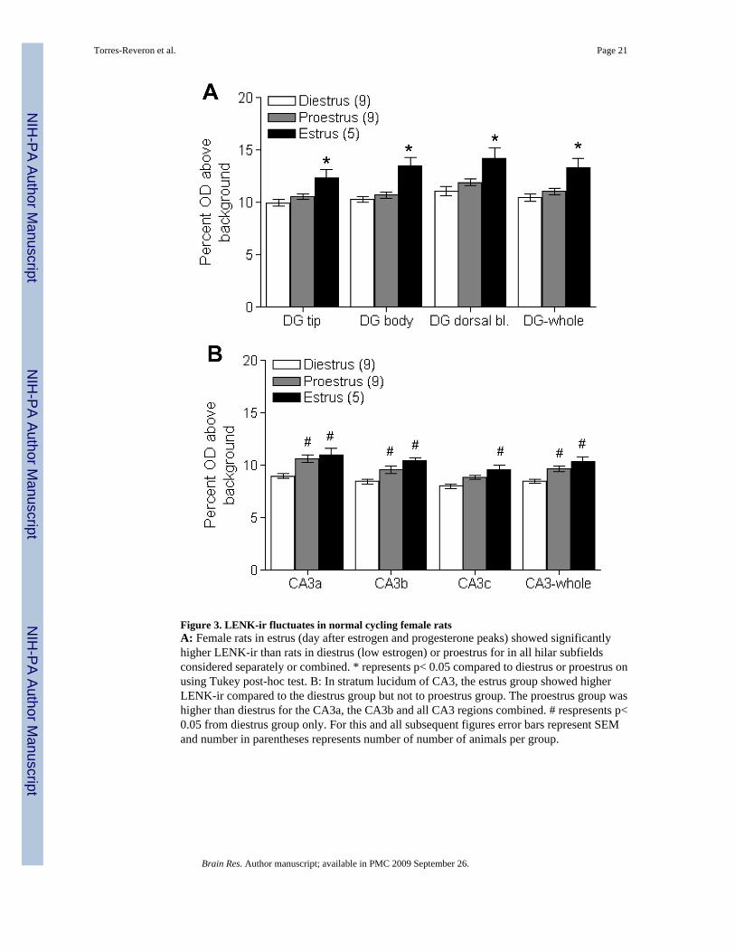
Figure 3. LENK-ir fluctuates in normal cycling female ratsA: Female rats in estrus (day after estrogen and progesterone peaks) showed significantlyhigher LENK-ir than rats in diestrus (low estrogen) or proestrus for in all hilar subfieldsconsidered separately or combined. * represents p< 0.05 compared to diestrus or proestrus onusing Tukey post-hoc test. B: In stratum lucidum of CA3, the estrus group showed higherLENK-ir compared to the diestrus group but not to proestrus group. The proestrus group washigher than diestrus for the CA3a, the CA3b and all CA3 regions combined. # respresents p<0.05 from diestrus group only. For this and all subsequent figures error bars represent SEMand number in parentheses represents number of number of animals per group.
Torres-Reveron et al. Page 21
Brain Res. Author manuscript; available in PMC 2009 September 26.
NIH
-PA Author Manuscript
NIH
-PA Author Manuscript
NIH
-PA Author Manuscript
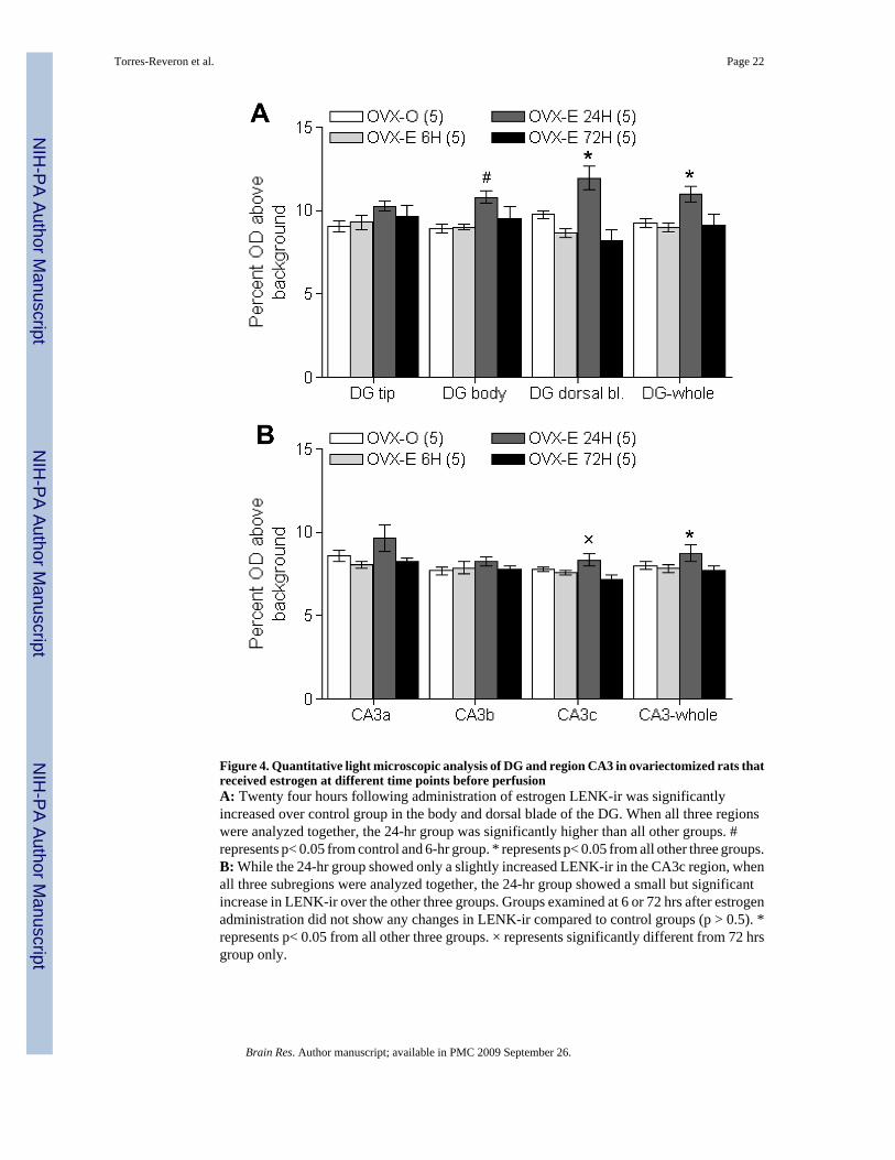
Figure 4. Quantitative light microscopic analysis of DG and region CA3 in ovariectomized rats thatreceived estrogen at different time points before perfusionA: Twenty four hours following administration of estrogen LENK-ir was significantlyincreased over control group in the body and dorsal blade of the DG. When all three regionswere analyzed together, the 24-hr group was significantly higher than all other groups. #represents p< 0.05 from control and 6-hr group. * represents p< 0.05 from all other three groups.B: While the 24-hr group showed only a slightly increased LENK-ir in the CA3c region, whenall three subregions were analyzed together, the 24-hr group showed a small but significantincrease in LENK-ir over the other three groups. Groups examined at 6 or 72 hrs after estrogenadministration did not show any changes in LENK-ir compared to control groups (p > 0.5). *represents p< 0.05 from all other three groups. × represents significantly different from 72 hrsgroup only.
Torres-Reveron et al. Page 22
Brain Res. Author manuscript; available in PMC 2009 September 26.
NIH
-PA Author Manuscript
NIH
-PA Author Manuscript
NIH
-PA Author Manuscript
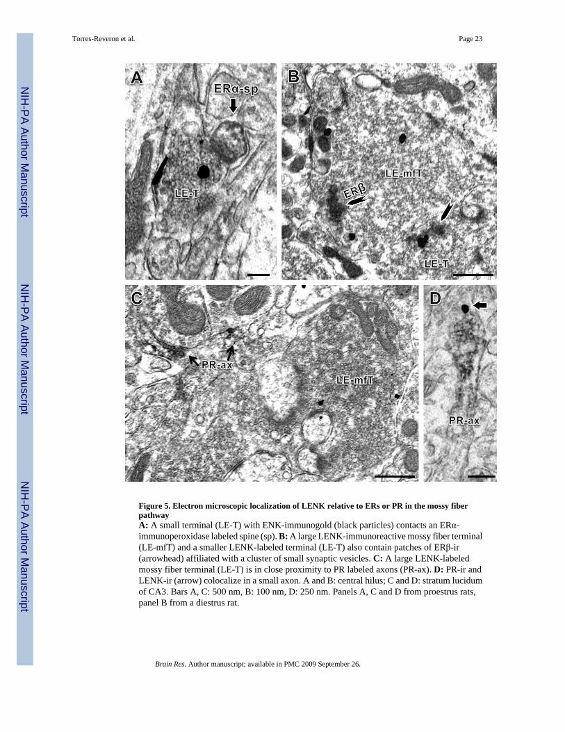
Figure 5. Electron microscopic localization of LENK relative to ERs or PR in the mossy fiberpathwayA: A small terminal (LE-T) with ENK-immunogold (black particles) contacts an ERα-immunoperoxidase labeled spine (sp). B: A large LENK-immunoreactive mossy fiber terminal(LE-mfT) and a smaller LENK-labeled terminal (LE-T) also contain patches of ERβ-ir(arrowhead) affiliated with a cluster of small synaptic vesicles. C: A large LENK-labeledmossy fiber terminal (LE-T) is in close proximity to PR labeled axons (PR-ax). D: PR-ir andLENK-ir (arrow) colocalize in a small axon. A and B: central hilus; C and D: stratum lucidumof CA3. Bars A, C: 500 nm, B: 100 nm, D: 250 nm. Panels A, C and D from proestrus rats,panel B from a diestrus rat.
Torres-Reveron et al. Page 23
Brain Res. Author manuscript; available in PMC 2009 September 26.
NIH
-PA Author Manuscript
NIH
-PA Author Manuscript
NIH
-PA Author Manuscript
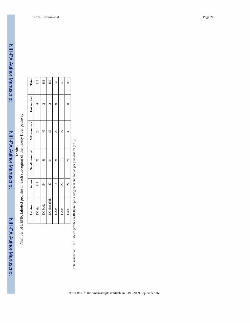
NIH
-PA Author Manuscript
NIH
-PA Author Manuscript
NIH
-PA Author Manuscript
Torres-Reveron et al. Page 24Ta
ble
1N
umbe
r of L
ENK
labe
led
prof
iles i
n ea
ch su
breg
ion
of th
e m
ossy
fibe
r pat
hway
.
Lam
ina
Axo
nsSm
all t
erm
inal
MF
term
inal
sU
nide
ntifi
edT
otal
DG
tip
118
7224
421
8
DG
bod
y54
8149
218
6
DG
dor
sal b
l.47
5456
215
9
CA
3a14
928
051
CA
3b22
1327
163
CA
3c24
2635
085
Tota
l num
ber o
f LEN
K-la
bele
d pr
ofile
s in
4800
µm
2 pe
r sub
regi
on in
one
sect
ion
per p
roes
trus r
at (n
= 3)
.
Brain Res. Author manuscript; available in PMC 2009 September 26.


![Antiapoptotic effects of delta opioid peptide [D-Ala2, D-Leu5]enkephalin in brain slices induced by oxygen-glucose deprivation](https://static.fdokumen.com/doc/165x107/631998d7e9c87e0c091032dc/antiapoptotic-effects-of-delta-opioid-peptide-d-ala2-d-leu5enkephalin-in-brain.jpg)