ournal - Plymouth Marine Science Electronic Archive (PlyMSEA)
208
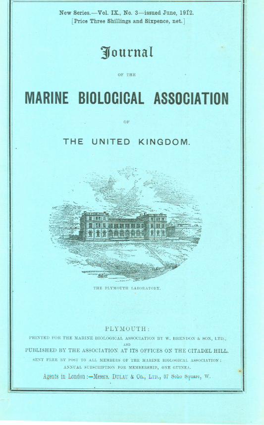
r New Series.~Vol. IX., NO.3-issued June, 1912. [Price Three Shillings and Sixpence, net.] ]ournal OF THE MARINEBIOlOCICAlASSOCIATION 0]' THE UNITED KINGDOM. THE PL¥)IOUTH LABORATORY. PLYl\IOUTH: PRDITED FOR THE MARIXE BIOLOGICAL ASSOCIATIOX BY W. BREXDOK & SOl'i', LTD., A"D PUBLISHED BY THE ASSOCIATION AT ITS OFFICES ON THE CITADEL HILL. SEXT FREE BY POST TO ALL ME)!BERS OF THE )IARU\E BIOLOGICAL ASSOCIATION: ANN1iAL S1iBSCRIPTION FOR ME)IBERSHIP, ONE G1iINEA. Agents in London :-Messrs. DULAl: &;00., LTD" 37 Sobo Squ~rc, w.
-
Upload
khangminh22 -
Category
Documents
-
view
4 -
download
0
Transcript of ournal - Plymouth Marine Science Electronic Archive (PlyMSEA)
rNew Series.~Vol. IX., NO.3-issued June, 1912.
[Price Three Shillings and Sixpence, net.]
]ournalOF THE
MARINEBIOlOCICAlASSOCIATION0]'
THE UNITED KINGDOM.
THE PL¥)IOUTH LABORATORY.
PLYl\IOUTH:
PRDITED FOR THE MARIXE BIOLOGICAL ASSOCIATIOX BY W. BREXDOK & SOl'i', LTD.,A"D
PUBLISHED BY THE ASSOCIATION AT ITS OFFICES ON THE CITADEL HILL.
SEXT FREE BY POST TO ALL ME)!BERS OF THE )IARU\E BIOLOGICAL ASSOCIATION:
ANN1iAL S1iBSCRIPTION FOR ME)IBERSHIP, ONE G1iINEA.
Agentsin London:-Messrs. DULAl:&;00., LTD" 37 SoboSqu~rc,w.

FATRON.HIS MAJESTY THE J(l~G.
OFFICERS AND COUNCIL.
President.
Sir E. RAY LANKESTER,K.C.B., LL.D., F.R.S.
V ice- P1"e.~idents.
The Duke of ABERCORN,K.G., C.B. I TheThe Duke of BEDFORD, K.G.
The Earl of ])UCIE, F.R.S.
The Earl of STRADBROKE, c.v.o., C.B.
Lord A VEBURY, F.R.S.
Lord IV ALSI:-IGHA~I, F.R.S.
The Right Hon. A. J. BALFOUR, ~LP.,. F.R.S.
"The Right HOIl. JOSEPH CHAMBER-LAI:-I, ~LP.
Right Hon. AUSTE;\! CHAMBER-LAI;\!, ~1.P.
W. IV. ASTOR, Esq., ~Ll'.
G. A. BOULE~GEH, Esq., F.IUS.
A. C. L. GUNTHER, Esq., F.R.S.
A. R. STEEL-MAITLA:-ID, Esq., M.P.Sir JOHN .:\IURRAY, K.C.B., .'.R.S.
Rev. Canon ~OR}iAN, D.C.L., F.R.S.
EDWIN VI[ATERHOUSE, E~q.
COUNCIL.
Elected Members.
Prof. J. P. HILL, D.Se.E. W. L. HOLT, Esq.Prof. E. IV. .:\L-\cBHIDE,D.Se., F.R.S.P. CHAL11ERS':\IITCHELL,Esq., D.Se.,
F.R.S.
EDGAR SCHUSTER,Esq., D.S~.GEOFFREYW. S}{ITH,Esq.Prof. D'ARCY IV. THmlPsoN, C.B.
G. L. ALWARD, Esq.
IV. T. CAL11A;\!, Esq., D.Se.
Prof. A. DE~mY, D.Se., F.R.S.Sir CHARLES ELIOT, K.C.1LG.
Prof. F. IV. GAMBLE, D.Se., F.R.S.
S. F. HAR11ER, .Esq., Se.D., F.R.S.Commander .:\1. IV. CA11PBELL HEP-
WORTH, C.B., R.;\!.R.
G. P. BIDDER, Esq., 11.A.The Prime Warden of the Fish.
mongers' Company.Sir RICHARD MARTIN, Bart. (Fish-
mongers' Company).W. L. BIRCH, Esq. (Fishmongers'
Company).
Governors.
Prof. G. C. BOURNE, D.Se., F.R.S. (Ox-ford University).
A. E. SHIPLEY,Esq., D.Se., F.H.S. (Cam-bridge University).
Prof. IV. A. HERmiAN, D.Se., F.B.S.(British Association).
Chai'/"/lUmof Council.A. E. SHIPLEY, Esq., D.Se., F.R.S.
Hon. 'l'reusw'er.
J. A. TRAVERS,Esq., Tortington, Arunde1.
Hon. Secretary.
E. J. ALLEN, Esq., D.Se., The Laboratory, Citadel Hill, Plymouth.---
PERMANENT STAFF.
D~:rector-E. J. ALLEN, Esq., D.SC.Hydrographej'--D. J. MATTHEWS,Esq.
Naturalist-F. J. BRIDGMAN,Esq., A.R.C.S.Assistant Naturalist-J. H. ORTON,Esq., B.SC.
[ 281 ]
Some Oases of New Growths in Fish.By
G. Harold Drew,Beit Memorial Research Fellow.
With Plate IV. cF
TABLE OF CONTENTS.PAGE
A Fibro-sarcoma of Raia macrorhynchus 281An Endothelioma of an Eel (Conger vulgaris) . 282A Fibro-sarcoma of a Plaice (Pleuronectes platessa) . 284A Tumour of a Whiting (Gadus merlangus) . . 284Haemangiomata of a Spotted Ray (Raia maculata) and of a Gurnard (Trigla lineata) 285,A pigmented Tumour of a Mackerel (Scomber scomber) of inflammatory origin' . 285
A FIERO-SAROOMA OF RAIA MAORORHYNOHUS.
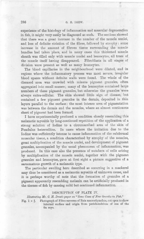
THIS specimen, obtained from one of the Plymouth trawlers, consistedof a large tumour on the dorsal surface, near the left angle of the fin(see Figs. 1 and 2). Only' part of the fish was available for examina-tion, so the presence of metastases and other details could not bedetermined.
The tumour was roughly circular, measuring about 4 inches indiameter, and was elevated above the skin about Ii inch~s. Itconsisted of a broad central pedicle, hard and fibrous and whitein colour, surrounded "by a broad cauliflower-like mass of a greyishcolour, and of much softer consistency than the central mass. Thisperipheral part of the tumour was covered by a very thin layer ofBpithelium which lined the outside of the pedicle and was continuouswith the epidermis; it extended into all the folds and hollows of theouter papilliform portion of the tumour, but was absent over the flatupper extremity of the pedicle. '
An incision made along a diameter of the growth, and carried downinto the tissues of the fish, revealed the fact that the tumour arosefrom the fibrous perichondrium of one of the fin rays. The centralmass consisted of closely packed strands of white fibrous tissue ofpearly whiteness, running at first in a direction perpendicular to theskin, and then branching out into the surrounding ring of softer tissue.
Sections of the pedicle showed that it consisted of strands of typicalwhite fibrous tissue; these were closely packed, but a few small roundcells, having a somewhat indefinite nucleus, and little or no cytoplasm,were present between the fibres, and occasionally the elongated nuclei of
NEW SERlES.-YOL.IX. No.3. JUNE, 1912. T
282 G. H. DREW.
the fibrous tissue cells were observable. Sections ot the sotter peripheralpart of the tumour (Fig. 3) showed irregular loosely packed strands offibrous tissue containing a few elongated nuclei, and large numbersof the small round cells, described above, often occurring in smallaggregations between the strands and fibrils of the fibrous tissuestroma. No definite blood-vessels were present, and blood spaces wererare. Superficially this portion of the growth was entirely covered bya single layer of squa.mous epithelial cells continuous with theepidermis, but there was nothing in this covering corresponding tothe other layers of the skin, and no denticles or mucous glands werepresent.
It would thus appear that the tumour had been originally a simplefibroma arising from the perichondrium of one of the fin rays, and thatlater this had taken on a sarcomatous type, and had proliferatedfreely. Considering the very poor blood supply to the peripheral partof the tumour, the fact that this portion should have become of adistinctly sarcomatous nature, its evident free proliferation, and theabsence of necrosis, is remarkable.
AN ENDOTHELIOMA OF AN EEL (CONGER VULGARIS).
This tumour was found on an eel caught at Plymouth. The fish wasan immature female, about four feet long, and appeared in goodcondition.
The growth consisted of a nearly spherical mass in the region of thebasi-hyal; it was about 1 inch in diameter and protruded aboutIi inches from the level of the skin. The tumour was of a whitishcolour, but in parts was somewhat haemorrhagic; the surface wasrough and irregular, with, in places, minute pits lined with thickenedepidermis. The skin was not continuous over the surface of the growth,but gradually thinned away at its margin until the junction of skinand tumour became indistinguishable and inseparable. The outerportion. of the growth was moderately soft, but it felt as though therewas a hard central part which was continuous with the basi-hyal; thearrangement suggested a considerable outgrowth of thickening of thebasi-hyal in an anterior direction, and that this outgrowth had pene-trated through the skin and become closely adherent to it at the margin.An incision made along a diameter of the tumour showed a centralbone-like core, apparently formed. by an outgrowth of the basichyal,and small areas of highly vascular tissue interspersed among patches ofwhite fibrous tissue, in some of which deposition of lime salts wastaking place. Other areas appeared semicartilaginous and some seemedmyxomatous.
.
NEW GROWTHS IN FISH. 283
Microscopic sections presented very varying pictures according to theparticular part of the tumour from which they were taken. The centralpart of the mass, after decalcification, could be recognised as consistingof fibrous tissue in which a considerable deposition of lime salts hadtaken place; this mass surrounded and merged into the bony tissue ofthe basi-hyal, and was penetrated in all directions by narrow bloodspaces. These spaces were filled with blood corpuscles and roundedcells with large nuclei and distinct nuceoli; the amount of surroundingcytoplasm varied, considerably in different cells, but was seldom great.From the fact that these blood spaces were more plentiful in the outerpart of the central mass and did not penetrate to the centre, it wouldseem probable that t~e hard fibrous tissue had first been formed and hadundergone partial calcification, and that then' it had been invaded bythe formation of ingrowing capillary blood spaces. Other sectionsfrom the softer parts of the tumour showed areas of loose and compactfibrous tissue, and other areas undergoing myxomatous degeneration:blood corpuscles and the rounded cells described above were present invarying numbers in almost."every part of the growth. The surface ofthe marginal part of the tumour was irregularly covered with thecutaneous epithelium which had a tendency to form ingrowths of com-pact masses of epithelial cells, but q.id y{ot show signs of becomingepitheliomatous. The more highly cellular portions of the growthpresented the appearance shown in Fig. 6. Masses of rounded epithelioidcells were present, and irregular channels containing blood corpusclescould be distinguished between the cell masses. The boundaries ofthese channels showed a more or less regular arrangement of theepithelioid cells, which in places had a tendency to become elongatedin the direction of the long a:x:isof the blood channel; many of thesecells were also present among the corpuscles in the blood spaces. Inaddition to these spaces with very ill-defined boundaries, other bloodchannels with more definite walls, usually circular in section, and moreresembling capillaries, were present. These channels were boundedby a very delicate sheath, but no endothelium within the sheath couldbe distinguished. Comparatively few mitoses were observed in any ofthe sections, so it is probable that the growth was not extending rapidlyat the time of examination.
The tumour can obviously be diagnosed as an endothelioma, arisingfrom the endothelium of the blood vessels, and it appears identical instructure, growth, and arrangement of the cells to similar endotheliomataoccurring in man.
No metastases were present.
284 G. H. DREW.
A FIBRO-SARCOMA OF A PLAICE (PLEURONECTES- PtATESSA).
This tumour was found on a plaice caught at Plymouth, and wasbrought up to the Laboratory a few hours after death. The fish was afemale, 12 inches long, and was in good condition.
The growth consisted of a white ovoid mass situated over the oper-culum on the ocular surface of the fish. It measured about! inch by! inch along its longest and shortest axes respectively. It was soft tothe touch and was covered with a very delicate epithelial layer con-taining a few pigment cells. Sections showed that the tumour was afibro-sarcoma, similar to the fibromata and fibro-sarcomata that arerelatively of such common occurrence on the opercula of plaice, but inthis case the sarcomatous element prevailed to a much greater extentthan usual. No metastases were present.
A TUMOUR OF A WHITING (GADUS MERL.ANGUS).
This tumour occurred in a male whiting, measuring 20 inches inlength, caught at Plymouth. Its position and relative size are shownin Fig 4. It was soft in consistency, greyish in colour, but fleckedwith red from the presence of blood-vessels. The surface was bareand uncovered by the cutaneous epithelium. .Amedian incision showedthat the tumour arose from the fibrous tissue layer forming the dermis;there was no tendency to invade the subjacent muscles, and nometastases were present. .
Sections (Fig. 5) showed that the growth consisted of a uniformreticulum of fine strands of some fibre-like substance, containing anumber of small rounded cells with little .or no cytoplasm, which wereusually arranged along the fibres. These cells were seldom aggregatedtogether into masses, and no mitoses were observed. A few moreelongated nuclei resembling those of fibroblasts were seen, and irregularspaces filled with blood corpuscles were present.
At first sight the tumour somewhat resembled a fibrinous exudate ofinflammatory origin, but a more careful examination and comparisonof the small round cells with the normalleucocytes of the blood of thewhiting showed that they had little in common, and the delicate reti-culum of which the growth was chiefly composed in reality bears littleresemblance to any exudate or tissue produced as an inflammatoryreaction.
It seems probable that the tumour arose from a peculiar type ofpathological multiplication of connective tissue cells, or fibroblasts,

NEW GROWTHS IN FISH. 285
and so is perhaps related to the sarcomata, but until more extendedobservations can be made on other cases, this must remain as themerest surmise.
HAEMANGIOMATA OF A SPOTTED RAY (RAIA MACULATA)AND OF A GURNARD (TRIG-LA LINEATA).
These tumours were accompanied by the presence of parasitic cope-pods; unfortunately in each case the body of the copepod had beenbroken off, leaving merely the haustoria imbedded in the growth, sothat their species could not be determined.
In the case of the gurnard a small reddish soft tumour was presenton the inner surface of the operculum; in the .case of the ray, a similartumour was present on the skin in the mid-ventral line of the body atthe level of the fifth gill arch.
Sections showed a condition identical with the capillary Haeman-giom~ta found in man. The tumours consisted of an irr~gular mass ofdilated thin-walled capillaries filled with blood cells: the haustorialbrandhes of the parasites could be easily recognised in the middle ofeach -
I
tumour. ,-
In.these cases it is impossible to say whether the tumours developedfirst, and then were attacked by the parasitic copepods, or whether theyrepresent a peculiar type of reaction on the part of the host to thepresence of the parasite. The former alternative would seem themore probabl~, since in by far the majority of cases of infection byparasitic copepods, little or no sign of an inflammatory reaction onthe part of the host is present.
A PIGMENTED TUMOUR OF A MACKEREL (SOOMBERSOOMBER) OF INFLAMMATORY ORIGIN.
This fish, a male, 11 inches in length, caught at Plymouth, showed alarge diffuse swelling on its side, situated about 3 inches from thetail. The surface of the skin was not broken, but was very darklypigmented.
On cutting through the skin and deep into the subjacent musculartissue, the cut surface appeared soft, haemorrhagic and degenerated,and was of a brownish colour; in places small black specks, due to theaggregation of pigment granules into masses, were visible to the nakedeye. The swelling was not circumscribed, but passed imperceptiblyinto the surrounding normal muscular tissue: the vertebral columnwas not affected.
Sections of the diseased area presented an appearance superficiallyresembling a melanotic sarcoma, so much so that without some
286 G. H. DREW.
experience of the histology of inflammation and muscular degenerationin fish, it might very easily be diagnosed as such. The sections showedthat there was a great increase in the number of the muscle nuclei,and loss of definite striation of the fibres, followed by atrophy: someincrease in the amount of fibrous tissue surrounding the musclebundles had taken place, and in many cases this thickened musclesheath was filled only with muscle nuclei and leucocytes, all trace oft4e muscle itself having disappeared. Fibroblasts in all stages ofdivision were present as well as many leucQcytes.
The blood capillaries in the neighbourhood were dilated, and inregions where the inflammatory process was most severe, irregularblood spaces without definite walls were found. The whole of thediseased area was crowded with minute pigment granules, oftenaggregated into small masses; many of the leucocytes contained largenumbers of these pigment granules, but otherwise the granules werealways extra-cellular. The skin showed little sign of disease, butcontained a few pigment granules in the dermis, deposited in thinlayers parallel to the surface; the most intense area of pigmentationwas between the dermis and the muscles, where an almost continuous
sheet of pigment had been formed.I have experimentally produced a condition closely resembling this
melanotic myositis by long-continued repetition of the application of astrong solution of Iodine to a circumscribed area of the skin ofFundulus heteroclitus. In cases where the irritation due to theIodine was sufficiently intense to cause inflammation of the subdermalmuscular tissue, a condition characterised by atrophy of the muscles,great multiplication of the m~scle nuclei, and development of pigmentgranules, accompanied by the usual phenomena of inflammation, wasproduced. In this case also the presence of nuinbers of cells arisingby multiplication of the muscle nuclei, together with the pigmentgranules and leucocytes, gave at first sight a picture suggestive of asarcomatous growth of a melanotic type.
The particular swelling here described as occurring in a mackerelmay thus be considered as a melanotic myositis of unknown cause, andit is perhaps worthy of note that the formation of granules of apigment apparently resembling melanin can be artificially produced inthe tissues of fish by causing mild but continued inflammation.
DESCRIPTION OF PLATE IV.
Illustrating Mr. G. H. Drew'spaper on " Some Casesof New Growthsin Fish."Fig. 1 x~. Photograph of Fibro-sarcoma of Raia macrorhynchus, cut open to show
internal surface and origin from perichondrium of one of thefin rays.
~U
Jl-e(...J0..
~~-
.~0,.......
~
~C)
H
~
fre.'-S;--'",,"
r.",..-
'"'.
',',,~
--~
..~~
..,.,",,',
~~
.ii"~
,~
QIi
, fit,
~iii
e\J,
'~'
~,-'
-,If
!I'
~~
,'
'T
,,
.fh,r(.
,~
.'',.
,~ '
~,'
e~
e!
~,.
",I
'.
,,",f
19.
e~
"'6
f\
1i
,i!!..".,,.,
19,!~:,~
~'",
~.,',
",.@
~J
~~
,"\.
~,i
t,~\!~
~i
~',~
t,!)0'
f,
'$@
j'ij).
,.;
i!>$
i~
j@
O~
'~d
~
",,,?"
,2<'"
f,J
9~,.i
."
~'
,.~'
-.
,. -
, '''',''
.
."
~'
"
"
~
.'
'iJ"-'I"
'",.,
/I'.
-
", '@3
',, 1,If
"'~'"i
~."
'."I'
e,"!\i',"I
,t; ~"
"1\'
<;
"i'I
fG
.0..;.
.I
-.
'f',.
~0
,~.
dI@
,41
'.."
"0;
,,f
i',b~
,
', '~Y "
",
', ~
J@,~
~, "
''', 'Ii,
...
Ii'
,''''',, ', t
teo:
"JI4>
'.~
,.e"
.'.~
"f' ."
,~
1\jfJ.
i~
..,,"
/J~
,'.",0"
Ii'.
..',
$""0'"
to,~
e""..
~.
-.',8
Q.."
II"....
..,..0
\..6
"I,
A,~
.'.
"~
6,II
'.~..
"'''''''"8'
."
.8
..)
<'.
.,4e\
IIe,""
~, '
iJI ?,f
,
'''',
.,
,8""f"<
"
~'
..'.lJ!"
4J.."I
I,,
jj'
, e!
",0;',.
'A
"'""
'",/
"J..w
If,'
",.
1t
',""q,\
~"e
,I'
$"".,'
8,",".
~!,
,""
~~
..i
''f
If
, ', '',.
, @~
.J..
,~,
~~
"0)
e0
e,t
,,
'~
..$,-",
&-..1,."
~,'/J
.'~
jI,'
".~
Y.,p:,',
~,
.&
,',.
Ie~
i.$
-.8fl.
.8~
/fI/ih,,e,
r'"t'i,
1.
.'
fIII'
\~
~f'
~,,-l'i."
'\."
"'..
.e
t),..,"~
,''1,
, ,eo
~t
~~
'e&
"e.fI,
~0.
ee!'
,'tJ'-"-
~~
,e~$~
~-
"--1...,6'~"-
-~,.
_L."""~.-&
~_f2
e
~..J
~u0VI,
VI
<{.....
0-CC~«::Ez~:)0-'")
...
------
-
r-!C>
~
!I
NEW GROWTHS IN FISH. 287
Fig. 2 x {r;. A.sFig. 1, but showing position of tumour on the fin.Fig. 3 x 150. Section of tumour shown in Figs. 1 and 2. Irregular strands of
fibrons tissue are present, with numbers of small round sarcomatouscells.
Fig. 4 x i. Photograph of Whiting, showing position of the tumour.Fig. 5 x 150. Section of tumour shown in Fig. 4. A. fine reticulum of a fibrous
nature forms the groundwork of the growth, and small ronndcells are situated on the strands forming this reticulum. Spacescontaining corpuscles are present.
Fig. 6 x 400. Section of endothelioma of Eel. Massesof endothelial cells dividedby irregular spaces containing blood corpuscles are present,together with some blood spaces with more definite walls.
(N.E.-For the sake of clearness the red blood corpnscles are represented withdense black nuclei, showing none of their nuclear structure.)
I,
j
--- ----
[ 288 ]
Notes on the Respiratory Mechanism of CorystesCassivela unus.
ByKathleen E. Zimmermann,B.Se.,
Uitiversity College of tv ales, Aberystwyth.
With Plate V.
I. INTRODUCTION.
IN his paper in the Journal of tlie Marine Biological Association forAugust, 1896, Garstang treated of some structural peculiarities ofGorystes cassivelaunus in relation to their biological significance. Abrief summary of his observations on the respiratory mechanism maybe given as follows: In Gorystes cassivelaunus the second antennaeare greatly elongated and are fringed by a ventral and a dorsal rowof hairs. The opposing rows of hairs interlock, with the resultingformation of a median" antennal tube." The double row of hairs is
continued back along the three basal joints of the antennae, which jointsare bent at right angles to one another; these hairs, projecting towardsthe median line, together with a median tuft of hairs springing fromthe rostrum, form the hairy roof of the proximal part of the antennaltube. The antennal tube opens posteriorly into a median" prostomialchamber," which in turn leads by a wide aperture to the branchialcavity of each side. The prostomial chamber is roofed by the rostrum,the antennal and epistomial sternites, and the pre labial plate. ItsHoor is imperfect, and is formed by the anterior part of the thirdmaxillipeds behind and in front by a quadrangular sieve of hairsspringing from the two basal joints of the second antennal, the anteriorpterygostomial processes, and a special anterior process of the fourthjoint of the external maxillipeds.
The habit of Corystes is to burrow beneath the sand, where itremains concealed, with only the tip of the antennal tube projectingabove the sand surface. A current of water (the respiratory current)is sucked down through the antennal tube, and passes backwards intothe prostomial chamber, where it divides into right and left streams,which pass into the right and left branchial chambers. The streameventually emerges from the branchial chamber along the whole extentof the edge of the branchiostegite.
---
RESPIRATORY MECHANISM OF CORYSTES CASSIVELAUNUS. 289
II. SOME STRUCTURAL PECULIARITIES WHICH APPEARTO ~AVE PASSED UNNOTICED BY GARSTANG.
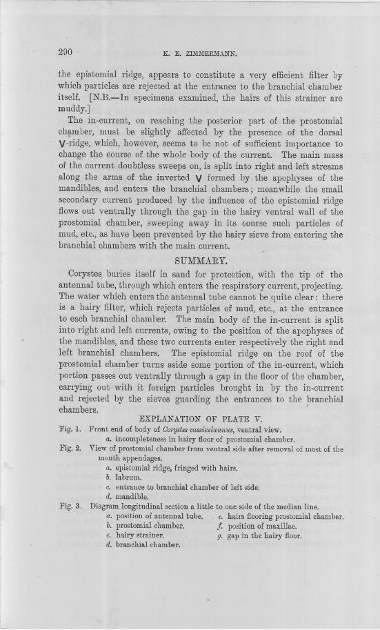
On that part of the ventral body wall which forms the posterior partof the dorsal wall of the prostomial chamber is a fairly prominentcalcified V-shaped ridge, the point of the V being directed backwards.This ridge is formed by the projecting anterior edge of the epistomialsternite, and is fringed with a row of fairly long hairs, which projectquite halfway across the entrance to each branchial chamber.
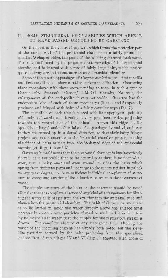
Some of the mouth appendages of G01'ystescassivelaunus-first maxillaand first maxillipede-show a rather curious modification. Comparingthese appendages with those corresponding to them in such a type asCancer (vide Pearson's" Cancer," L.M.B.C. Memoirs, No. xvi), theenlargement of the endopodite is very noticeable. Corystes has theendopodite lobe of each of these appendages (Figs. 4 and 5) speciallyproduced and fringed with hairs of a fairly complex type (Fig. 7).
The mandible of each side is placed with its" apophysis" pointingobliquely backwards, and forming a very prominent ridge projectingtowards the ventral side of the animal. Across this ridge lie thespecially enlarged endopodite lobes of appendages iv and vi, and overit they are turned up in a dorsal direction, so that their hairy fringesproject across the entrance to the branchial chamber proper, meetingthe fringe of hairs arising from the V-shaped ridge of the epistomialsternite (of. Figs. 1, 2 and 3).
Garstang himself notes that the prostomial chamber is but imperfectlyfloored; it is noticeable that to its central part there is no floor what-ever, even a hairy one; and even around its sides the hairs whichspring from different parts and converge to the centre neither interlockto any great degree, nor have sufficient individual complexity of struc-ture to constitute anything like a barrier to restrain the in-current ofwater.
The simple structure of the hairs on the antennae should be noted(Fig. 6) : there is complete absence of any kind of arrangement for filter-ing the water as it passes from the exterior into the antennal tube, andthence into the prostomial chamber. The habit of G01'ystescassivelaun1lsis to lie buried in sand; the water directly above the surface mustnecessarily contain some particles of sand or mud, and it is from thisby no means clear water that the supply for t~e respiratory stream isdrawn. The complete absence of any arrangement for filtering thewater of the incoming current has already been noted, but the sieve-like partition formed by the hairs projecting from the specialisedendopodites of appendages IV and VI (Fi~. 7), together with those of
- -
290 K. E. ZIMMERMANN.
the epistomial ridge, appears to constitute a very efficient filter bywhich particles are rejected at the entrance to the branchial chamberitself. [N.E.-In specimens examined, the hairs of this strainer aremuddy.]
The in-current, on reaching the posterior part of the prostomialch~mber, must be slightly affected by the presence of the dorsalv-ridge, which, however, seems to be not of sufficient importance tochange the course of the whole body of the current. The main massof the current doubtless sweeps on, is split into right and left streamsalong the arms of the inverted V formed by the apophyses of themandibles, and enters the branchial chambers; meanwhile the smallsecondary current produced by the influence of the epistomial ridgeflows out ventrally through the gap in the hairy ventral wall of theprostomial chamber, sweeping away in its course such particles ofmud, etc., as have been prevented by the hairy sieve from entering thebranchial chambers with the main current.
SUMMARY.
Oorystes/ buries itself in sand for protection, with the tip of theantennal tube, through which enters the respiratory current, projecting.The water which enters the antennal tube cannot be quite clear: thereis a hairy filter, which rejects particles of mud, etc., at the entranceto each branchial chamber. The main body of the in-current is splitinto right and left currents, owing to the position of the apophyses ofthe mandibles, and these two currents enter respectively the right andleft branchial chambers. The epistomial ridge on the roof of theprostomial chamber turns aside some portion of the in-current, whichportion passes out ventrally through a gap in the floor of the chamber,caFying out with it foreign particles brought in by the in-currentand rejected by the sieves guarding the entrances to the branchialcham bers.
Fig. 2.
EXPLANATION OF PLATE V.
Front end of bodyof Corystescassivelaunus,ventral view.a. incompleteness in hairy floor of prostomial chamber.
View of prostomial chamber from ventral side after removal of most of themouth appendages.
a. epistomial ridge, fringed with hairs.b. labrum.c. entrance to branchial chamber of left side.d. mandible.
Diagram longitudinal section a little to one side of the median line.a. position of antennal tube. e. hairs flooring prostomial chamber.b. prostomial chamber. f position of maxillae.c. hairy strainer. g. gap in the hairy floor.d. branchial chamber.
Fig. 1.
Fig..3.
JOURN,MAR.BIOL.Assoc.VOLIX PLATEV
FIG.I
bc.d
FIG.2
L
':dFIG.5.B.
RESPIRATORY MECHANISM OF CORYSTES CASSIVELAUNUS. 291
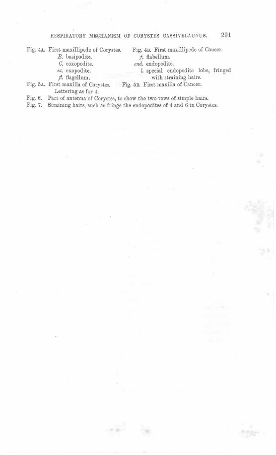
Fig. 4A. First maxillipede of Corystes.B. basipodite.C. coxopodite.ex. exopodite.fl. flagellum.
Fig. 5A. First maxilla of Corystes.Lettering as for 4.
Fig. 6. Part of antenna of Corystes, to show the two rows of simple hairs.Fig. 7. Straining hairs, such as fringe the endopodites of 4 and 6 in Corystes.
Fig. 4B. First maxillipede of Cancer.f. flabellum.
end. endopodite.l. special endopodite lobe, fringed
with straining hairs.Fig. 5B. First maxilla of Cancer.
[ 292 ]
On the Fauna of the Outer Western Area of the EnglishChannel.
ByL. R. Crawshay, M.A.
With Plate VI.
IN a previous nurnbel' of this Journal * a Report was published byMr. R. H. Worth on the geological collections made in the EnglishChannel by the Association's steamer Oithona in 1906, combined withother previously unpublished geological records relating to the samearea. The general features of the area concerned, with' details of thedredgings on .this occasion, were described bY me (4) in an accompany-ing paper. . It was hoped that the Report on the Fauna then collected,for which indeed the cruises were specially arranged, would be pub-lished long before now; but owing to unavoidable causes the completeworking out of the material has been unfortunately delayed for a longinterval.
The area of investigation as illustrated by the accompanying chartextends roughly from ten to fifty miles outside the Eddystone Light-house, in a S.W. Mag. direction, and ranges from 40 to 53 fathomsin depth, reaching about the mid-Channel line near the latter sounding.A few points already dealt with in the paper referred to may berepeated here. The nature of the ground over the whole area, withthe exception of the first few miles, may be generally "described asshell, sand, and gravel, largely intermixed with stones, which oftenreach very considerable dimensions, and show a gradual increase in.average size as the distance increases outwards, the highest averagebeing obtained near the outermost point that was reached. Theinner limit of exposure of these stones was found at fifteen miles tothe south-westward of the Eddystone. At positions falling inside thispoint the bottom deposit consists of a clean shell sand, much finer thanis found at any other point in the area concerned.
As regards the gear employed, the otter trawl was used at Positions3 and 4, within the fine sandy area last mentioned, and at Positions 7,8, 49, 64, 66, 68 and 78, outside it; but the frequent occurrence oflarge stones involved too great a risk to use the otter trawl oftenat the outer positions, and with the exception of those taken with theAgassiz trawl at Positions 45, 46, 52, 59 and 60, all the remaining
* Journ. Mar. Bioi. Assoc., N.S., Vol. VII!., p. 118.
Journ. Mar. BioI. Assoc. Vol. IX Plate VI.
.?'o""Hi.
' \-;
Iii \~ i;
\ , !". i
\., ./Loog,h;p,\(.'
I'.
i '_.- ..'''., .~ /
,"--'-iO-ip1'\,"
4<' ",' '"' "'" 20'
'0'20'1
;I
<.
.-'-. -'
. WOLf ROCK ~O",,{"
;; 0+'0003
t"",. G9.700 2
-"t; '~'':'.-~,/ ': \
I,
50'
I'
ilI'W
ill
'lil"1
~' ('j
\ ....""'../
./
007
036 '.-.or
035 032
39'5~0 3;0 1~~,~So 'OJ, 7
a 2
IB!l-''O .. 030 02S025
.-...-....-..\
\ ,\\\
roo'
Ii
,,~f~r".C.. t_/-
4'0042
,.{~ .f.~r.~.\',,"'-"'. ., ...
. ".
, ". -'''.., '-""'-" ..,.....,.
073.74043
~+7+6 00++
053 ~oooSi 4.S4.SS~SS %2 0 049 "'.
., . / \/"
i/
)./
""""'"''
S7"S~GOo
075
So', c'; ""''''''''~.~,.S~o.:'~L""~,_.._.....-
660 oS7 068oSS
7S~77 078
./
C)"""
'\
80",79 ./(
/ .' "
-
.. \.,""., ""
"'"'"
\.
/ '., \ \
ENCllSH CHANNEL.WESTERN AREA.
\
". \i \. t\ "-"'.
\.
\ .'-,,'
;,
CHART TO ILLUSTRATE THE DREDCINGS
CARRIEO OUT BY
s.s.OITHONA
IN 1906.
m',
'~?~~~~- ..-......-..
-'"
\.
//
//
-'~49'
j4o9'j \,-- ---------
<c',
5?.~~'-;'
;./
~.~
II
./
5" ""
Crawshay" Fauna of Outer Western Area of the Channel.
FAUNA OF THE OUTER WESTERN AREA OF THE CHANNEL. 293
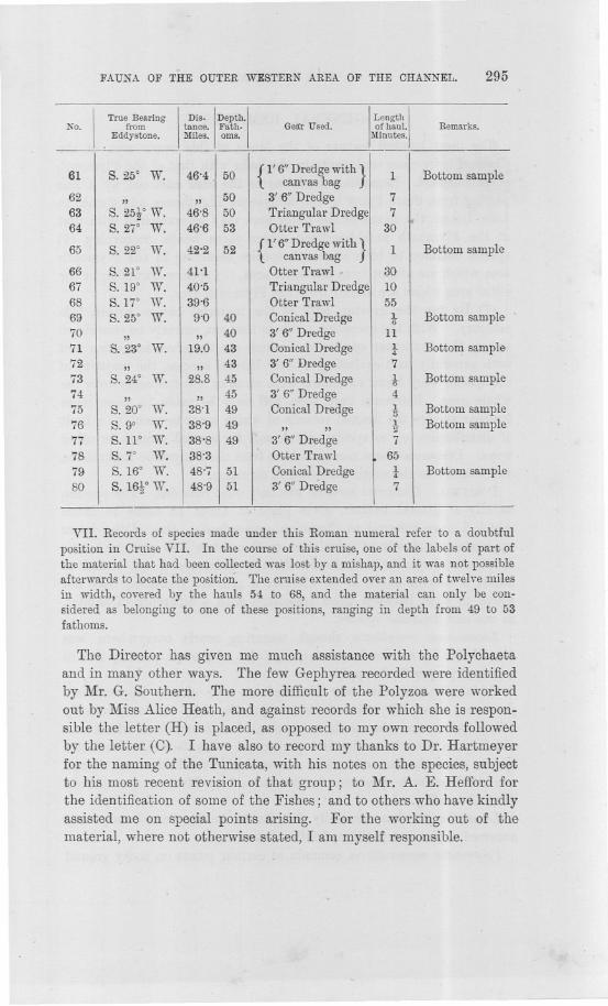
hauls were made with the dredges. Of these latter, fifteen werebottom samples, most of them not exceeding one minute in worki;ngduration; fifteen were made with a 3 ft. rectangular dredge, averagingtwelve minutes in duration; eighteen were made with a 3 ft. 6 in. rect-angular dredge, averaging eight minutes in duration; and seventeenwith an equiangular dredge, measuring 2 ft. on the side, averagingseven minutes ill duration. The short length of these hauls musttherefore be borne in mind in considering the intensity of occurrenceof any of the species recorded. In the following list of hauls allpositions are referred to their true bearing from the Eddystone Light-house. Samples 16 to :30 inclusive were taken more especially asrock samples, and in consequence only a rough record of species, madeon board at the time, was preserved. They have not therefore thevalue of others in which the unidentified material was brought homefor examination. They concern, moreover, a limited area, from eighteento twenty-three miles outside the Eddystone, and in point of speciesrecorded from them are merely additional to other hauls made withinthe same area.
LIST OF HAULS.
!
True Bearing
!
DiS'
!
Depth.
I
No. from tance. Fath.Eddystone. ,Miles. oms.
Gear Used.
I
Length
I
of hanl.Minntes.
Remarks.
~~~-
1 S.21° W. 8'3 40 3' 0" Dredge 15
2 S. 19° W. 8'1 40 {l' 6" Dredge with}3 Bottom sample. canvas bag
3 S. 15° W. 7'8 40 Otter Trawl 304 S.21° W. 7'6 " " 305 S. 19° W. 20'2 42. 3' 0" Dredge 156 S. 20° W. 20'4 42 " " 107 S. 23° W. 21'2 42 Otter Trawl 308 S. 27° W. 21'8 " " 309 S.31° W. 21-7 3' 0" Dredge 20
10 S. 26° W. 17'8 42t " " 511 " " 42t " ,, ]Q12 " " 42t " ,, 1013 " " 42t " ,, 1714 S.24° W. 20'0 " " 1515 S.27° W. 20'3 " " 1016 S. 29° W. 20'9 44 " " 1017 S. 28° W. 23'3 45 " " 1118 S. 29° W. 23'4 45 " " 1019 S. 28° W. 23'3 45 " " 1020 S.25° W. 20'5 44 " " 1021 " 21'2 44 Triangular Dredge 922 " 21'9 44 " ,, 4 .
~94 L. R. CRAWSHAY.
I
True Bearing
!
Dis'
l
Depth.
!
No. from tance. Fath.Eddystone. Miles. oms.
Gear Used.
I
Length
!
ofhan!.Min n tes.
Remarks.
,
Triangular Dredge{Bottom ample
23 S.25° W. 21'9 44 - from previoushaul
24 S.24° W. 22'5 " " 525 " 23'0 46 " " 826 S. 20° W. 18'4 44 " " 827 S.19° W. 18'3 44 " " 8
28}S.W W. 19'5 44 7
29 " "
30 S.21° W. 21'5 43t " ,, 1031 S.25° W. 15'0 40 3' 6" Dredge 1032
' 16'3 7" " "33 " 17'5 " " 734 S.2So W. 18'5 " " 835 S.32° W. IS'O " " 836 S. 37° W. 17'5 43 " " 837 S.41° W. 17'1 " " 93S S. 38f W. 22'2 44 " " 739 S.3S' W. 21'9 44 " " 440 " 21'7 44 Triangular Dredge 541 S. 36f W. 266 44 " " 742 S.W W. 26'4 44 " ,,' 643 S.21° W. 288 45 " ,, 1044 S.17° W. 29'S 46t " ,, 1045 S. ISo W. 30'1 47t Agassiz Trawl 1546 S, 19° W. 29'9 15
'" "
47 S. 19° W. 29'7 {l' 6"Dredge with} t Bottom samplecanvasbag
48 S.l1° W. 30'5 " " t Bottom sample49 S. go W. 30'4 Otter Trawl 30
50 S.16° W. 30'9 43 {l' 6"Dredge With}2 Bottom sample
canvas bag
Ig}
The two consecu
51 S. 15° W. 30'S 43{,Trianfiular Dredge
tive samples were3' 6" redge labelled" 51" in
52 S, 14° W. 31'0 43 Agassiz Trawl 25 error
53 S.22° W. 32'2 46 3' 6" Dredge 10
54 S.26° W. 34'5 49{I' 6" Dredge With}
1 Bottom samplecanvas bag
55 S.2WW. 34'4 49 " " 156 S. 25° W. 34'3 49 Triangular Dredge 4
57 S. 22° W. 39'0 49 {I' 6" Dredge with}2 Bottom sample
canvas bag58 " ,, 49 3' 6" Dredge 1059 " ,, 49 Agassiz Trawl 2560 S. 24° W. 40'0 " " 30
FAUNA OF THE OUTER WESTERN AREA OF THE CHANNEL. 295
I
True Bearing
I
Dis.
!
Depth.
!
No. from tance. Fath.
. Eddystone. Miles. oms.
Gel!r Used.
I
Length
I
of haul.Minutes.
Remarks.
VII. Records of species made under this Roman numeral refer to a doubtfulposition in Cruise VII. In the course of this cruise, one of the labels of part ofthe material that had been collected was lost by a mishap, and it was not possibleafterwards to locate the position. The cruise extended over an area of twelve milesin width, covered by the hauls 54 to 68, and the material can only be con-sidered as belonging to one of these positions,ranging in depth from 49 to 53fathoms.
The Director has given me much assistance with the Polychaetaand in many other ways. The few Gephyrea recorded were identifiedby Mr. G. Southern. The more difficult of the Polyzoa were workedout by Miss Alice Heath, and against records for which she is respon-sible the letter (H) is placed, as opposed to my own records followedby the letter. (0). I have also to record my thanks to Dr. Hartmeyerfor the naming of the Tunicata, with his notes on the species, subjectto his most recent revision of that group; to Mr. A. E. Hefford forthe identification of some of the Fishes; and to others who have kindlyassisted me on special points arising. For the working out of thematerial, where not otherwise stated, I am myself responsible.
--~..>
61I
S.25° W. 46'4 50 { l' 6" Dredge with}1 Bottom samplecanvasbag
62 " ,, 50 3'6"Dredge 763 S. 25f W. 46'8 50 Triangular Dredge 764 S. 27° W. 46'6 53 Otter Trawl 30
'
65 S. 22° W. 42'2 52 {I' 6" Dredge with}1 Bottom samplecanvas bag
66 S. 21° W. 41'1 Otter Trawl. 3067 S. 19° W. 40'5 Triangular Dredge 1068 S.17° W. 39'6 Otter Trawl 5569 S. 25° W. 9'0 40 Conical Dredge 1 Bottom sample'1170 " ,, 40 3' 6" Dredge 1171 S. 23° W. 19.0 43 Conical Dredge 1 Bottom sample72 " ,, 43 3' 6" Dredge 773 S. 24° W. 28.8 45 Conical Dredge t Bottom sample74 " ,, 45 3' 6" Dredge 475 S. 20° W. 38'1 49 Conical Dredge 1 Bottom sample:376 S.9° W. 38'9 49 " " J. Bottom sample277 S.11° W. 38'8 49 3' 6" Dredge 778 S.7° W. 38'3 Otter Trawl 6579 S. 16° W. 48'7 51 Conical Dredge 1. Bottom sample4-
80 s. 1%° W. 48'9 51 3' 6" Dredge 7
~------- - - -- ~
296 L. R. ORAWSHAY.
GENERAL REMARKS.
The most marked feature of the fauna of this outer area. of the
Ohannel is its close conformity in the main with that of the Plymouthneighbourhood. Regarding the latter as the area enclosed by a linepassing from Start Point to the Eddystone Lighthouse and thence toLooe Island, the fauna of the outer area may be compared with thatof the Plymouth neighbourhood under three heads, concerning (1)species common to both areas, (2) species occurring in the Plymoutharea which are absent from the outer area, (3) species occurring in theouter area which are absent from the Plymouth area.
(1) By far the greater bulk of the material comes within thiscategory. With the exception of those that can scarcely be consideredamong the commoner species, and which are therefore less often metwith generally, and excluding strictly littoral species, the majority ofthe species were found extending with more or less frequency over thewhole area. Reference here then will only be made to those more
. familiar species the limited records of which seem to point to a limitof distribution, or to species which call for special remark in otherways. Distances where mentioned are from the Eddystone Lighthouse,
- and roughly to the south-west~ard in direction.PORIFERA. ClcdMina coriaceawas only once recorded. The species,
usually of littoral habitat, gives place at about 18 miles to formswhich I have referred to ClatMina pri11wrdialis. One of the latterapproximates closely in spiculation to C. c01'iacea,and great as is thedifference of spiculation between the two extremes, I am bound toadmit a certain doubt as to whether a gradual transition may notprove to exist between them associated with a difference of habitat,in one and the same species.
Leucosolenia C011Lplicata,though occurring nearly everywhere, wasremarkable for its slender, straggling habit of growth, possibly due toa lack of proper food-supply.
Sycon ciliatu1n was only obtained at two closely approximatepositions about 22 miles distant. Outside this, the only closelyallied species was the southern species, Grantia capillosa, which wasobtained as close in as the first position, 8 miles distant. The latterspecies certainly also occurs near or even inside the Eddystone, thoughthe few Plymouth specimens in the Laboratory Museum are withoutdata of locality.
Leucandm fistulosa, generally distributed in the Sound, only onceoccurred at the first position, 8 miles distant.
POlY11wstiama11Lmillaris,common at certain points on rocky ground
FAUNA OF THE OUTER WESTERN AREA OF THE CHANNEL. 297
in the 'Sound, 'was obtained only at3Land 39 miles, in contrast toP.:robusta, which occurred fairly commonly over the whole area.
Ficulina ficus, though of common occurrence as far as 40 miles out,was always of remarkably small size-much more so 'than it oftenoccurs on the Eddystone Grounds. This reductioIl of growth, as con-trastBd with the comparatively enormous size it often attainswitJiinthe breakwater, is no doubt .attributable to the diminution (jf wa;steorganic matter on the distant grounds;, .. '
Suberitescarnosus, .comparatively common on 'the EddystollEJ,ChoundsWASonly twice found,. at '17 and 22 miles respectlYEJly. These alsowere extremely small specimens. .
HYDROMEDUSAE.Except at the first few positions, o.nthe fine sanefabout 8 miles distant, and agaiilattheoutermost point rea<;hed, wherEJin 51fathomg, two southern species showed a b.ealthy luxuriantgrowth, the examples recorded were on the whole remarkably small,and the occurrence of. well"grbwn coloJiiEJswas quite exceptional. Inthe samecoilnection.' the dwarfed form of Plul1~ulariq,setacea at. twoouter positions in about .50 fathoms is remarkable. .
. . Merona c01'm(copiaewas tal~en as far as 31 miles distant, whiCl:1.wa~10 miles beyond the outermost record of its common associate Dentaliu11~enfalis. . ,
Hydmctinia 'echinata was only taken at 31rniles. ,. .
Tub?dariawas only recorded at the first position, 8 j:nile~distant. ,
Haleciul1~ halecin?Mn,was riot recorded outside' about the 34-milepoint. ,
..A fragrirent'onlyof, Thuia?'ia a?,ticulatawas taken at 18 miles.,Antennularia ra?nosa,..though occurring I\S far out as 40 miles, was
not fourid c.ommon anywhere. The allied' species, A. antennina wascomnion over the whole area. . '.
PlUlntda?'ia catharina was the commonest of its geIJ.us obtain,ed,P.. pinnatc( alone approximating to it appreciably-in point of fre-.quency. The creeping variety, which occurred over the whole area,was: perhaps . the mQst 'frequent and. certainly th(j most flou~ishing inpoint of growth. It is difficult to assign a cause fQr this' Ip.ode ofgrowth:" A colony: ,fYfBougainvillia, reared by Mr: E. T. Bro-)vn~atthe Plymouth LaboratoJ:Y some years ago (cf. Jou?'l~~Ma1';Biol. Assoc:,N.S., VoL VUI,p. 3,7) assumed a 'persistent st,oloniferous habit. ',ofgrowth from the first. It was fed with mixed plankton regularly ~p.dgrew rapidly, but ii(theseveral months of its existence, except~n.:veryrare cases, it made:no attempt to assume the ordinary branching{1ajJi~,.even though it ultimately succumbed to an overgrowth o~ small !1!ga.:~.This single instance affords no evidence that fOQd-sl1Pply,/I,IOJ;l(bil!-
NEW SERlES.-YOL. IX. No.3. JVNE, 1912. U
298 L. R. CRAWSHAY.
fluences the manner of growth where the latter is variable. Yet theseeming scantiness of the Hydroid fauna over most of the outer area,coupled with the frequent records of small colonies, and distinctlydwarfed colonies in the case of P. setacea point to conditions that areunfavourable to healthy growth in the group.
ECHINODERMATA.Palmipes placenta was not found outside the 17-mile point, and at the latter only as small specimens.
Echinus acutu.~was not recorded inside a distance of 15 miles, whichis about the inner limit of the stony ground. E. esculentus, on theother hand, occurred oVer the whole area, and in considerably greaternumbers.
'POLYCHAETA. The outer limit of occurrence of Aph?'odita aculeat~was at 20 miles; that of the nearly allied Hermione hyst1'ix extendedto 46 miles~ The latter species seems generally to favour grounds ofa coarse character.
Halosydna gelatinosa occurred only at 39 miles.Onuphis conchilegawas not found beyond the 26-mile point.CRUSTACEA.Portunus depuratm', often an abundant species in the
Sound, and found abundantly by' Dr. Allen (1) 3 miles east of theEddystone, was only once obtained at 17 miles.
Atelecyclus septemdentat1fshas been recorded from as much as 100fath. and even 400 fath. (d. Allen, 1), but in the area here consideredit was not found beyond 30 miles. This species is scarcely likely tohave been much missed in the work owing to the constant use of thedredges with a special view to deep working. Allen considers that acertain amount of muddy deposit contributes to the most favourableconditions for the species, and it is possible that the almost entireabsence of any such deposit on the outer grounds may explain its in-frequency and even disappearance at the more distant positions visited.
MOLLUSCA.Craspedochilusonyx was not recorded beyond 20 miles.Capulus hungaricus, taken' on five grounds by Dr. Allen between
Start Point and the Eddystone, at 30 fathoms, was not found alive inthe area here under consideration, though. dead shells occurred as farout as 27 miles. .
Of Pecten 1naximus there is a noticeable scarcity at all points ascontrasted with P: °pC1'cula1'is,which was at times abundant. Aboutfive living specimehs were obtained at three positions, all situatedabout 20 miles out. At other positions from one to three onlyoccurred, and the total number obtained probably did not exceed forty.On the grounds hear the Eddystone'it was found by Dr. Allen withmuch great~r frequency, two or three specimens being generally taken.ill' each haul with t~e dredge.
FAUNA OF THE OUTEn WESTERN AREA OF THE CHANNEL." 299
lJentaliunt entalis was not found outside the i8-mile point.Nucula nucleus, which occurs commonly down to 30 fathoms on the
Eddystone Grounds, was only obtained at the first position, 8 niileadistant, where the large deposit of fine clean sand occurs.
Pedunculus glycimeris, occurring as far as 39 miles out, was remark-able for the small size of specimens obtained.
(Ja?'diumechinatum was of rare occurrence, being only once obtainedalive at 9 miles distant, while only one dead valve was recorded at apoint slightly closer in. -
(Jardium norvegicum, common on gravel on the Eddystone Grounds,was -only recorded at four points, and as far as 31 miles.
(2) Of the members of the Plymouth fauna that are absent from thelist, there is little of special interest to mention, these being for themost part essentially littoral species, or those favouring a rocky habitat,or such as are of too infrequent occurrence generally to serve for purposesof comparison. Among the absent species:-
Adamsia polypus (Sagartia parasitica) was conspicuously absentdespite the frequent occurrence of its host Eupagu?'Usbernha?'dus. Onthe Eddystone to Start Point Grounds, it is an interesting fact thaton gravels Dr. Allen always found this hermit-crab' without theanemone, though on the fine sands it was commonly associated with it.It is true the anemone did not occur in the few hauls made on the fine
sand of the outer area at about 8 miles, yet its non-occurrence in otherhauls suggests that the generally coarse ground of the latter, as in thecase of the Eddystone to Start Point gravels, may account for itsabsence. .
Holothuria nigra is generally found at Plymouth in close proximity torock ledges. Such too was the case in tli~ Eddystone to Start Pointfauna where the species occurred, only on gravel adjoining the Eddy-stone rocks. It is not improbable that SU9h rock ledges are stillexposed in places on the more Q.!stant grounds here dealt with, butthere was no clear evidence of this fact afforded' by the rock materialdredged up at any point.
Antedon bijida, which extends southward to the Mediterranean, andas deep as 100 fathoms (cf. Bell, 65), has not been recorded in thePlymouth fauna outside the Mewstone Ledge.
Echinocardium cm'datunt occurs on fine sand on the EddystoneGrounds to 35 fathoms, and was obtained occasionally by Dr. Allen onsimilar ground between the Eddystone and Start Point. It is recordedby Ludwig (72) from southern waters at Marseilles, Naples, and thewest coast of Italy, and as deep as 85 fathoms. -
Maia squinado is moderately common, especially among rocks in the
~ --~- ~---'-~ ~---
300 L. R. CRAWSHAY..
Plymouth. area, extending as far as the Eddystone Grounds. A fewspecimens only were obtained by Dr. Allen, on fine sand, between theEddystone and Start Point.
Gorystes cassivelaun1£sappears to be exclusively associated withdeposits of a fine nature, and such as were only met with on the firstposition at about 8 miles.
With these few species ma.y also be considered certain of thosementioned under the preceding heading, the infrequency of whichalmost amounts to their absence from the outer fauna. Such are
Sltberites cm"IWSUS,Hydmdinia echinata,- Tubularia sp., Thuiariaa1,tic1data, Halosydna gelatinosa, Gapulus hunga1'icus, and especiallyPcwtunus depumto1' and Ga1'diu'11£echinatu1n.
(3) Of the species hitherto unrecorded from the Plymouth area,Clath1,ina primordialis, as regarded by Haeckel (14), is of almostuniversal distribution. The remainder are divided as follows, thepresent records for the English Ohannel being included in thedistribution :~ '
(A) From Scandinavia through the region of the Shetland Is, andHebrides to Irish Waters and -English Ohannel.
Sertula1'ella tenella. (Including a,lso Arctic regions; S.W. Atlantic,and Pacific.) .
Pectinaria pusilla. (Scandinavia and West of Scotland 'only.)Thyone mphanus. (Excluding Scandinavia.) .
Tritonofusus p1'Opinq16us.(Including North Sea.)Anapagu1'us hyndmani. (Excluding Scimdinaviar, and indudi:q.g
Channel Is.) .Gubius scorpioides. (Excluding Shetlands and Hebrides.)
(B) The same area, and inclu~ing the Mediterranean. .
Peltogastc1'sulcatns. (Excluding Shetlands, Hebrides, and Ireland,and including Brazil and Pacific.). '. .
GonothY1'cagracilis. (~ncludlng Noit~ 'S~~.and S. America.)'(0) The same as (A), including the Bay of Biscay and Azores.JJit1'upa arietina. (Includi:q.g Mediterranean, Canary Is., tJ.nd
Pacific.) :. . '
Inachus leptochirus. (Exclliding Scandinavia and I~ish Wa~ers, an,d:including Channel Is., Adriatic, and Cape Verde Is.)
Diphasia alata. (Excl.uding Irish Waters.) '., Polyplumaria jlabellata. (Excluding Shetlands, Hebrides, and IrishWaters.) - .
Portunus tubC1'culatu~; (Excluding Scandinavia and Irish Waters,and including Mediterranean.)
FAUNA OF THE OUTER WESTERN AREA OF THE CHANNEL. 301
(D) From Irish Sea southwards.I Xantho tuberculatus. (Including Bay of Biscay, Coast of Portugal,N.W. African Coast and Cape Verde Is.) .-
Bathynectes longipes. (Including Channel Is., Mediterranean, AdriatiG,and Black Sea). Frequent at Plymouth in recent years.-
(E) From English Channel to Azores.Olathrina conto1'ta. (Including Mediterranean.)Rhizaxinella elongata. (Including Mediterranean.)Polymastia agglutinans.(F) English Channel and Adriatic.Gr-antia capillosa. .
PORIFERA.Calcarea.
CLATHRINIDAE, Minchin.
Clathrina coriacea (Fleming).One small specimen on a dead Pecten shell, at Position 33.Depth, 42 fath.
Clathrina primordialis (Haeckel).At Position 34, one, on shell of Fusus, occupied by Eupag.1lrus.
- Greatest measurement 9 mm." 38, one, on tube of Pallasia" ,,10 "" 45, one, on Inachus" 59, two, on dead valve of Pecte1L "" 62, one, on Volsella "
Depth 42-50 fath.These five small specimens which I have assigned to Haeckel's
Ascetta primorclialis show a good deal of individual variation. Thehabit of growth is in every case that of a simple network of anastomos-ing t':lbes, with a few short oscular processes, forming a thin investmenton the object of attachment. The skeleton is composed almost entirelyof equiangular triradiates with the component rays often of slightlyunequal length. The size of the spicules is fairly uniform in individuals,but between different individuals the average dimensions of the spicularrays range from about 65,«* in length'by 6'5,« in width at base of ray,to about 110,« in length by 10,« in width. The rays are graduallytapered to a rather sharp or sometimes a somewhat blunt point, thetapering being more strongly marked in the distal half of the ray. Inone specimen (No. 240), the rays are more linear and almost coriacea-like. In two specimens the skeleton is entirely composed of triradiates.
* The sign p.is used to designate '001 mm.
" 10 "
4 "

302 L. R. ORAWSHAY.
In the three others a few quadriradiates of the same ray-form and sizeare present, in which the fourth ray is rather smaller but not rouchshorter than the basal rays. These quadriradiates are so scarce thatthey might easily escape observation.
Under his" Connexive Varietaten" of Ascetta ]Jl'i11wl'dialis,and laterin the text, Haeckel (14) mentions the existence of this variety with atendency to form a gastral ray as Ascaltis ]Jl'inwl'dialis, though he givesno data concerning its occun'ence. In the present case it may be note-worthy that the three specimens possessing quadriradiates were ob-tained from depths between 42 and 44 fathoms, while those withoutthem were from between 47 and 50 fathoms. ,
Excepting specimens recorded by Hanitsch (15) from the LiverpoolDistrict which he subsequently referred (16) to G. lacunosa, the speciesdoes not seem to have been recorded north of the Mediterranean.
Clathrina lacunosa (Johnston).
At Position 34, one, on shell of ]lusus, growing beside G.pl'imol'dialis,Length 6 mm.
" 5"" 7"
" 47, one, on Sc1'upocclla1'ia" 62, one, on Sertularella
Depth, 42-50 fath.
Clathrina contorta (Bowerbank), Minchin (28).
A small patch of spicules undoubtedly belonging to this species wasfound attached to a surface section of a Renie1'a from Position 58, onor in close proximity to which the specimen would seem to havebeen growing.
A single quadriradiate spicule, apparently also of this species, occurssiroilarly on a section of Raspailia stuposa from Position 67.
Depth, 49-52 fath. . ... Though it may appear somewhat hazardous to record the occurrenceof this' species on the evidence of a. few spicules, and in the secondcas(j, of a single spicule, I have no doubt, after examining a specimenof contorta which Prof. Minchin kindly gave me, concerning the identityof the first record, and little doubt as to the second. In the formercase, both of two marked features of cont01'taare very distinct, namely,the very high proportion of quadriradiates, and,-more important,-the long and slender gastral ray of these. In the latter case, the singlequadriradiate spicule is of the same form. Monaxons are absent fromthe fragment from Position 58, a condition which Minchin regards(28, p. 14) as a juvenile feature. It is of interest to note that the twopositions lie close to one 'another, that is, as nearly as the reckoningfixes them, not more than, about two miles apart.
FAUNA OF THE OUTER WESTERN AREA OF THE CHANNEL. 303
The species has been recorded .from the Liverpool district byHanitsch (16, p. 233). An earlier British record by Carter is ql.J.eS,,-tioned, by Minchin (28, p. 18), who also leaves localities given byBowerbank (Channel Islands, Scarborough?) open to doubt, owingto a confusion of species in his material. The natural habitat ofC. contorta is in the more southern waters: Sark, Luc-sur-mer (Top,sent, 35); Roscoff (Topsent, 37; Minchin, 28); Belle Isle (Topsent, 36);Banyuls-sur-mer, extremely abundant (Minchin, 28); Azores, abundant(Topsent, 38); Adriatic (Lendenfeld, 22, pars. (?)~cf. Minchin 28,p.14). It is apparently one of those species that extend with difficultywithin the border line of the British Fauna.
LEUCOSOLENIIDAE, Minchin.
Leueosolenia eomplieata (Montagu).
Recorded from 24 positions--l, 3, 4, 1l~15, 31-37,40, 43, 45, 49,51, 52, 58, 59, 64.
Depth, 40-53 fath.Mostly on Hydroids, Cellaria and Cellepor({,also on shells of Pecten
and tubes Of Pallasia and on Inachus. At some positions severalspecimens were obtained, at twelve positions a single one only.
The habit of growth, which varies little among all the specimensobtained, is very different from that of the ordinary shore form. Thisis a straggling growth, often a confused tangle of slender ramblingtubes, in no case exceeding 1 mm. in diameter, usually co.nsiderablyless, and with no tendency to specialization. Many of the specimensare extremely small. Of the larger ones two es.pecially deserve men-tion: the first from Position 32, a thickly grown specimen withrambling tubes of less than Imm. in diameter, on Cellepom, measuringabout 35 m111.in extent; the second from Position 37, a very finespecimen of 50 mm. in breadth and 60 mm. in height, forming atangled shrub-like growth on a shell of Pecten opercularis.
SYCETTIDAE, Dendy (13).Byeon' eiliatum (Fabricius).
At Positions 38 (five), 40 (one).Depth, 44 fath.I make use of the name ciliaturn provisionally for the specimens
here recorded, on grounds of priority, because after examination ofmany specimens I am quite unable to separate this form fromHaeckel's Sycandm coronata as defined by him. In the main theyconform more to the latter type than to ciliaturn in point of the
304 .iL. R. CRAWSHAY.:, .
relativ'e lengthofctne gastral ray to'that of the facial rays in thegastral quadriradiittes; while in regard to the ,second point used by~aeckel (14);'namely the relative width ,of the monaxons to thatof the tiiradiatesand quadriradi'ates, the character appears 'to meto be too variable to 'serve for purposes of distinction. As regards thefirst character, however, both types are exemplified in the Plymouth formswith every gradation between the two extremes, and further, the shortgastral ray of the ciliat1~1ntype is apparently more characteristic of thein-shore specimens, while the longer corresponding ray of the C01'onatUl)~type commonly OCC11rsin the deeper water. A still more importantpoint arises in the occurrence of at least one instance I have seen, inwhich both the short and the long gastral ray are present in the samespecimen. A careful examination of a larger number of examplesis needed to establish the point satisfactorily, but in the meantimeI am unable, to, reg!1rdthe tw,o forms as, ~pecifiqally distinct. ,
IIi three 6f the six specimens here considered ,'the relative length ofthe gastral and facial rays is roughly as 7 to 8, 3 to 4, and 1 to 3,severally; while the average relative width of tile monaxons ,andradiates is abollt 1!-2i t,o 1.
. GRANTIDAE, Dendy., ,
Grantia capillosa (0. Schmidt).
At Positions 1 (one), 3 (one), 4 (three), 36 (one), 37 (two), 49 (onevery young), 53 (~mevery young), 70 (two), 80 (one).
Depth, 4,0-,,-51 f~th. ,
The genus Grantic~,to which this species is referred, is here regardedas it is defined by Dendy (13), but 'with the modification that it doesnot of necessity exclude the occurrence of, the monaxons in bundles atthe distal ends of the radial tubes. This reservation would seem alike
to involve Dendy's family Grantidae, although as defined by him (13)it is not literally restricted on the point. In other respects the speciescapillosa seems to have its proper position in this family and genus,owing to the presence of 'a distinct dermal cOrtex covering the distalends of the radial chambers. The arrangement o~ the mona}\ons israther irregular. For the most part they are grouped in bundles aboutthe ends of the radial chambers, this a,rrangement being often retainedeven where the latter are subject to branching, as frequently occurs atthe extreme apex. Less frequently they are disposed without muchregularity. But they are always 'large ,and stout, and usually penetratefor a considerable distance towards the gastral surface: Their disposi-tion is in fact near the border line between the two forms of arrange-ment which Dendy defines for the Sycettidae and implies for' the
FAUNA OF THE OUTER WESTERN .ARE.A OF THE CHANNEL. 305
Grantidae respectively. Sinoe, however, the arrangement of themonaxons depends, as Dendy observes: on the variation of the canalsystem, it seems to me inadvisable to limit the latter family toostringently in regard to this character, which-may in greater or lesserdegree still retain the Syconoid form, as in capillosa,after the branchingof the chambers has begun and a definite_cortex has been assumed. .
Mr. C. F. Jenkin first called my attention to this sponge among~ome unnamed material, and identified it as this species on Haeckel'sdescription. Recently Mr. Kirkpatrick has kin~ly afforded me theopportunity of examining at the British Museum i co-type of OscarSchmidt's labelled" Syconcapillosu11L"in his own handwriting, whichenables me without doubt to 'confirm Mr. Jenkin's identification. It
will not improbably be foimd that some confusion has arisen concern-ing the identity of the species, like ,many other Calcarea. Particularfeatures which characterize it are: (1) its tendency. to interrupt~on ofoutline, as though through injury, in the region of the osculuJn, asfigured by Scmidt (32, PI. I, Fig. 6); (2) the shape of the dermal~riradiates, approximating somewhat to the remarkable form of thosein Leucandm fistulosa, though much stouter, with longer basal ray, andsmaller unpaired angle than in that species; (:3)the slender sub-gastraltriradiates with very long tapering basal ray, shorter lateral raysnearly at right angles to it, and often with a fourth ray developed inabout the same plane as the latter.
The species was originally r:ecorded by Schmidt from Lebenico inthe Adriatic (32, p. 17). Haeckel (14) also records it from Lesina onhis own authority and that of Heller. Lendenfeld (22) adds Muggia,Pirano, and Rovigno to these localities. It has also been said to occurat Naples, but. as the only slide I have seen so labelled from thatlocality is undoubtedly of a different species, the latter record seems toneed confirmation, and apart from this there is apparently no previousrecord of its occurrence outside the Adriatic.
Leucandra fistulosa (Johnston).One specimen at Position 1.Depth, 40 fath.
Monaxonida.
HADROMERIN A, Topsent (40).TETHYIDAE.*
Tethya lyncurium (Linnaeus.One specimen at Position 62.Depth, 50 fath.* Dr. Hartmeyer on p..379 uses this name for an Ascidian family on the ground that
the Ascidian genus TethyuJn is of earJier date than the Sponge genus Tethya.
- ~-~--~
306 L. R. ORAWSHAY.
CLIONIDAE.Cliona sp.
Specimens of Oliona, in all cases I believe boring in dead shells ofPecten, PectunC1d1.lS,Lut1'aria, etc., were obtained, sometimes very com-monly, at Positions 4, 8, 9, 10, 11, 13, 18, 34, 44, 46, 59.
Depth, 40-49 fath. .
The specimens were unfortunately not retained for further examin-ation, and the species must therefore be left unnamed.
POLYMASTIDAE.
Polymastia .robusta, Bowerbank.
Six specimens were obtained as single examples at the Positions 8,14, 51, 55, 77, 80.
Depth, 43-51 fath. .Particulars are as follows, the measurements being made after pre-
servation in spirit :- .
At Position 8. Form, depressed hemispheroidal, with fistular pro-cesses very numerous. Measurement, 5g x 50 xabout 30 mm. in height. On a fiat stone.Depth, 43 fath.
14. (Specimen not retained.)51. Form tending to bulb-shaped, with surface very
even, and most of the fistular processes fusingtogether as one combined outgrowth from theupper surface. Measurement, 35 x 28 x 35 mm.in height. Depth, 43 fath.
55. Fistular processes numerous. Measurement, 50 x40 x 35 mm. in height. Broken from base.
77. :Fistular processes numerous. Measurement, 95 x75 x 50 mm. in height. Depth, 49 fath.
80. Fragment, torn from an apparently large specimen.Depth, 51 fath.
"
Polymastia mammillaris (0. F. Muller).
At Position 51. One specimen; forming an investing growth ondead Pecten valve, with about a dozen processes.Extent, 23 x 12 mm. Depth, 43 fath.

FAUNA OF THE OUTEl{ WESTERN AREA OF THE CHANNEL. 307
At Position !:is. One specimen; forming an investing growth ondead Pecten valve, with, base strongly hispid,50 x 30 mm. in extent, and with seven larges:tllooth fistulaI' processes 20-22 mm. 'in height.The differentiation between basal and fistulaI'
areas exceptionally well shown. Depth, 49 fath.
Polymastia agglutinans, Ridley and D.endy.Single specimens at Positions 13, 32 or 33 (?), 46, 55, 59.Depth, 42-49 fath.The form of the specimens is in all cases that of a depressed, more
<>1'less regular spheroidal or ovoid mass from 10 to 25 mm. in horizontalmeasurement, with from one to eight fistular processes of 6 to 12 mm.in height, radiating from different points of the surface, the body ofthe sponge forming an investing growth cementing together smallpebbles and fragments of shell into a compact mass. This peculiarhabit is common to all the specimens, and there seems no doubt oftheir identity with Ridley and Dendy's agglntinans (31), of which theexternal form and habit is so very similar. Professor Dendy, whokindly examined some slides I sent him and allowed me to see somepreparations of his original material, considers that there is no differ-ence of sufficient importance to constitute specific distinction. The maincharacters of the skeleton are almost identically the same, though in the.dimensions of the spicules there is a considerable difference. Ridleyand Dendy's measurements for the large tylostyli are 1200 fJ. by 15'7 fJ.;for the microsclera, 175 fJ. by 4 fJ.. In the specimens here consideredthe megasclera average from 450 to 500 fJ.,and sometimes exceed 700 fJ.,but in no case have I seen one reaching SOOfJ.. The width of thelargest is 14'S fJ.. The microsclera average about 140 fJ. by 3'7 fJ.. Thedifference may be a local one. The megasclera are rather more likethose of 1'obusta than those of l1Wl1L1nilla1'is.The microsclera are
like those of ?narmnillaris, but with much illore pronounced heads.The Challenger specimens (two) were taken off the Azores in
450 fath. on volcanic mud.
SUBERITIDAE.
Ficulina ficus (Linnaeus).
, Most of the specimensof this sponge obtained were of the massivecarcinoecious form, and generally occupied by a Pagurid, but nonewere.of large size. Occasionally, as where stated in the following par-ticulars, they occurred in the earlier stage of these investments onshells of molluscs.
308 ,L: ]1. ORAWSHA¥.
Greatest measurem~nt 20 mm:" " 28"
SmalLGreatest measurement 23, 35 mm.
16 mm.
22 "16, 17, 22,.24 mm., and
four, thinly investingshells of Natica, Tro-,chus, etc.
15, 19, 20, 26'h1m.12 mm., investing shell
of small Gastropod~Depth, 40-49 fath. ' .In all of three specimens closely examined, namely those from
Positions 7, 43, and 60, the centrotylote microsclera are abundant butextremely variable in size and form. .
Much confusion has arisen about the identity of this species,' whichhas frequently been reftJrred to as S~"berites dornuncula, a species-which apparently does not occur in the British fauna: For a dIs-cussion of the species wIth full synonymy, see Topsent's valuab10account (41, p. 203). The extremely large growth often attained bythe species on the inner grounds inside the breakwater seems ne'ver tooccur in the deeper water, the .difference being, apparently due to food-supply. .
"
1, one.7, one.
34, one.35, two.37; one.43, one..52, eight.
" "
At Position"
" " ""
" .59, four.60, one. ""
Suberites carnosus (Johnston).
At Position' 9. One small detaehedspecimen. Greatest measure-ment 5 mm;
32 or 33 (1). One small ,specim.en of ovoid form grow-ing on the basal portion of Polymastia agglutina.ns.Greatest measuremen"t 10 mm.
.Depth, 42 fath. .
Rhizaxinella elongata (R. and D.), Topsent.
A single specimen, evidently belonging to this species, was obtainedat Position 38, in 44 fathoms. Some uncertainty was felt as to its-identity owing to the presence of an important internal character powhich no allusion has been made in records of the species. This is thepossession of a series of longitudinal belts of spicules iil the form of abroken hollow cylinder surrounding and distinct from the axial core.It is difficult to understand that no mention should be made of a char-
FAUNA OF THE OUTER WESTERN AREA OF THE CHANNEL. 309
acter that is definitely shown in the longitudinal and the transversesections, but in all other respects it conforms so closely with thedescriptions of elongata that I can only regard it as the same species,and conclude that the point referred to has been overlooked,
'The specimen forms a slightly bent column of 60 mm. in height,widening gradually in diameter from, 5 mm. at base to 8 mm. near themiddle, 'beyond which it is sub-cylindrical, with the apex rounded.The colour in' spirit is pale yellowish white. There are numeroussmall oscula scattered at irregular intervals over a great part of thesurface, without any particular reference to the apex. The specimenwas broken from its attachment, but two small rootlets are preservedin connection with a rounded base. The texture is very tough andcompact.
The skeleton consists of ~ very compact central axial core of stylotespicules with a quantity of spongin, surrounded by a clear area, beyondwhich is a ring of spicular belts running parallel to the axialcore, the component belts following a spiral course. In the trans-verse section these belts are marked off from one another byslender strands of few spicules that radiate sub-spirally outwards andupwards from the axial core, across the clear area, and through thebelts to the surface. Similarly, spicular strands separate off indepen-dently from the outer side of the belts themselves and branch in aspreading fashion on their way to the surface, beyond which many ofthe spicules extend. At the surface they combine with innumerablel"adiating fasciculi of smaller and shorter styli, to form the dermal his-pidation, which has the form of a closely approximating series ofdefensive brushes.
The spicules of the longitudinal belts and their branches, and thoseof the axial core, are slender styli, often slightly curved, with simplerounded base, sometimes faintly tylote, and with sh.arp tapering points.They range from 900 to 1600 f.I. (averaging 1300 f.I.)in length, andirom 7 to 11 f.I. (averaging 9 f.I.)in width. The spicules of the dermalfasciculi, which are also present in small numbers, scattered betweenthe longitudinal belts and the surface, are styli of 200 to 440 f.I.
(averaging 280 f.I.)in length, and 2 to 6'5 f.I.(averaging 4'5 f.I.)in width..Manyof them are simple, but a large number-perhaps the majority-are strongly tylote, and usually with a second ring-like expansi~n beyondthe basal one, as in the spicules, e.g., of Sube1'itesca1'nosus, The hases ofthese dermal tylostyli are extremely like those of the latter species,and in the vertical view of the outer surface there is a striking simil-Itrity between the two sponges._.. The noteworthy points of difference from Ridley and Dendy's original
-~~T
:310 L. R. CRAWSHAY.
description of Su,beriteselongatus (31) are, besides that referred to, themore slender form of the large styli, and the absence of a truepedicel. In regard to the last point, several specimens of thesame species were collected during a subsequent cruise -at a moredistant and deeper position in the Channel. These all show thetypical slender pedicellate growth, characteristic of elon.gata,while thespiculation of two specimens examined shows no appreciable differencefrom the foregoing description, except that the numerical proportionof tylote to simple styli in the dermal fasciculi is lower.
The species has been recorded from the Bay of Biscay: one,in 248 m. (Topsent, 38); one, in 180 m. (Topsent, in 10); Coast ofRoussillon, two, in 94 m. (Topsent, in 10); Azores, eight, in 450 fath.(Ridley and Dendy, 31).
HALICHONDRINA, Vosmaer.
HAPLOSCLERIDAE, Topsent.
OHALININAE, Ridley and Dendy.
Siphonochalina montagui (Bowerbank)?
At Position 46, one specimen, broken from attachment-possiblyLepmlia; forming an erect compact growth ofirregularly inosculating, more or less tubularbranches, the whole somewhat depressed laterallyand with some external resemblance to certain
broadly expanded forms of Alcyonidiurn gelatino-sum,. with several oscula raised on low prominencesof 2-4 mm. in diameter. Height, 65 mm. Width,62 mm.
68, one broken specimen, on Lepmlia Joliosa,. with- massive basal portion, 50 x 40 mm. in extent,
tunnelled by tubular ramifications and sur-mounted bi at least one large tubular process,60 mm. in height by 25 mm. in diameter, withan osculum at summit, 9 mm. in diameter.
Depth, 47-52 fath.- The texture of the first specimen is compact and rigid, and similar ingeneral appearance to Bowerbank's figures for the species; that of thesecond, except for a certain rigidity about the base; is quite the opposite.In external characters the two specimens are quite distinct, but theinternal structure of both, including the form and dimensions of thespicules, shows so little difference that there seems no justification forseparating them. The skeleton is composed of two distinct elements :-
"
FAUNA OF THE OUTER WESTERN AREA OF THE CHANNEL. 311
(a) A primary interlacing network, ramifying through all parts ofthe sponge, of very clearly defined (?keratose) fibres, each composed ofbundles of fibrillae, and commonly enclosing a variable number ofoxeote spicules running longitudinally within them. Sometimes theenclosed spicules are very numerous, but often they are entirely absentfrom the fibres. In a tangential section of one specimen some of thelarger fibres attain, even close to the surfaae, a thickness of as muchas 100 fJ.. In a tubular portion of the same specimen a thick fibretraverses the centre of the tube, throwing off subdividing branches tothe periphery. Oxea occur likewise, though with extreme scarcity inthis axial fibre and even in its slender branches to the wall of the tube.
(b) A secondary Reniera-like, and to some extent regularly disposednetwork of unispicular meshes, with a decided tendency to assume inthe main lines an outwardly radiating direction from interior to surface.This appears to be quite independent of the primary network. Theends of the spicules are cemented together with deposits of spongin,usually to a distance of about 20 fJ. down the shaft from the point.The spicul~s composing this network are oxea of fairly uniform dimen-sions averaging about 90-100 fJ. in length by 5 fJ.in width. Withthem are associated, irregularly disposed, smaller oxea of about the.same length, and half or less than half the width, and very fine hair-like oxea of about 50-60 fJ. by 1 fJ..
The spicules of the primary skeleton are similar in form and dimen-'sions to those of the secondary skeleton, and include the slender hairlikeforms of the latter. The dimensions of the large oxea shown by Bower-bank's figure for the species are rather larger-about 124 fJ.by 6'5 fJ..
The tubular tendency of the sponge seems to place thespecies in the genus Siphonochalina as. defined by Schmidt (33,.p. 7), and by Ridley and Dendy (31, p. 29); but the remark-ably composite structure of the fibres of the primary skeleton,.very different from the clear fibres of, e.g., Cl~alinaoculat(t, leaves some.doubt as to its identity with the species to which it is here assigned, orindeed of its true position among the Ohalininae. The fibrillae of whichthe fibres are composed have themselves individually the form of a.string of beads, each bead contributing internally a separate rod-shaped element to form a centrally-placed strand running along thestring. Loisel'" describes an almost identically similar condition in cer-tain species of Reniem,so called. . But in the present examples I find no.evidence of the bead-like cells which secrete the elemental rods ulti-mately breaking' down, as Loisel.describes, so as to have a simple con-
* Contribution a l'histophysiologie des eponges. Journ. de l'anat. et de la physiol..-XXXIV. ]898.
~~ -~- ~-'- -
312 . L. .R. CRAWSHAY.
tinuous strand. They appear, on' the contrary, to be permanent, whilethe contained. rods remain separated at their ends from one another bya narrow' interval between adjacent beads. These fibrillae, and theircontained rods in .particular, give a deeper reaction to stains like eosinand methylene blue than the deposits of spongin about the ordinaryskeleton, and this fact, coupled with that of the independence of thetwo skeletons, suggests that the substance of the primary skeleton maybe of a slightly different constitution from that of true spongin.
RENIERINAE, Ridley and Dendy.Halichondri.a sp.
A fragment of a Hali(iwnd1'ia, too small for determination, occurredop.Inw<hus dorsettensis at Position 3.. Depth, 40 fath.
the skeleton is very irregular, with a confused and broken networkof loosely constructed spicular bundles forming the main lines. . It ischiefly composed of large oxea 250-350 JI. in length and 7-10 JI.in width, together with smaller oxea 200-250 JI.in length and .2-3./.1.in width, not very numerous; and thinly scattered very slender ona,100-200 JI.in length and 1 JI.in width. The. spiculation approximatesn.earlyto t9at of Bowerbank'sH. caduca. .
. Reniera,. Nardo.
. Without yet having had. an opportunity of examining any of Bower-bank's .original specimens of this difficult genus, the species hereconsidered are named .~.? far as possible from his descriptions andfigures alone." .
Reniera indistincta (Bowerbank).At Position 3, two specimens, each forming a shallow investing
growth on Inachus.. 12, one specimen, forming an irregular growth on a
. Pecten valve, with tubular processes; 10 mm. inexten t.
14-, two specimens, one forming a shallow investinggrowth on Inachus, the other an irregular mass,30 x 20 mm. in extent, intermingled with Hydroids
. and forming tumulous elevations. . . .
25, one specimen, with basal attachment 15mm.' indiameter, and one l~teral prominence, surmountedby a tubular process 15 mm. in height; on a
. Chaetopte1'UStube., Depth, 40-46 fath.' .
,.
"
"

FAUNA OF THE OUTER WESTERN AREA OF THE CHANNEL. 313
The large oxea range from about 140 to 170 fJ. in length by 5-6 fJ.in
width, as against dimensions shown by Bowerbank of about 158 fJ. by 4 fJ..
With these occur numerous more slender oxea, 120-140 fJ. by 2 fJ.,andalso slender hair-like oxea scattered irregularly, averaging about 100 fJ.
by 1 fJ. or less.
Reniera sp. (A.).
At Position 56, one specimen, forming a small growth on Inachusleptochi1'US.
Depth, 49 fath.Skeleton mostly unispicular and very similar to that of the preceding,
but the main skeleton spicules are longer and more slender in propor-tion, ranging from 150 to 200 fJ.by 4 to 5 fJ.. Smaller oxea and finehair-like oxea occur as in the last species.
Reniera sp. (B.).
At Position 6, one specimen, forming an irregular massive growth onstems of Antennula1'ia, etc.
Depth, 42 fath.Skeleton almost entirely unispicular, but with the reticulation very
irregular. Large oxea of much the same length, 150-170 fJ.,as inspecimens referred to indistincta, but much wider, 7-8 fJ.,and remark-ably uniform in size. Slender hair-like oxea scarce.
Reniera pygmaea (Bowerbank).
At Position 40, one specimen growing from dead base of Antennularia..Depth, 44 fath. .The specimen forms a short bifurcated growth of about 25 mm. in
height, and 6 mm. in diameter of branches, arising from a narrow stem. .In appearance and general texture it bears a certain resemblance to asmall Ohalina oculata, as Bowerbank remarks of specimens before him.A comparatively large quantity of spongin occurs at the ends of thespicules, which at times seem almost entirely invested by a thin layerof it. The oxea of the main skeleton, averaging 110 by 5 fJ.,aremore slender than Bowerbank shows for pygmaea as 118 by 7 fJ.. Anumber of more slender oxea occur in addition.
Reniera densa (Bowerbank).
At Position 1, one specimen, forming an irregular mass of densetexture about Hydroids. Height 25 mm.
Depth, 40 fath.The radiating lines of the I)lain skeleton are..distinct and largely
~EW SERIES.-YOL. IX. No.3. JUNE, 1912. X
314 L. R. CRAWSHAY.
....
llluitispiculous. The remainder of the reticulum is somewhat irregular.Spicules exceptionally uniform, with an almost entire absence of slenderforms. Dimensions from 120 by 6 p.. to 140 by 9 p... The measure-ments shown by Bowerbank are 130-145 p..by 10 p...
Reniera sp. (C.).
At Position 62, one specimen.Depth, 50 fath.The specimen is a fragment torn from its base, consisting of a thimble-
shaped process, 25 mm. in height by 17 mm. in width, with an osculumof 5 mm. in diameter at the summit. Texture soft and flexible.
Skeleton rather irregular, with spongin very little developed. Averagemeasurement of large oxea about 190-200 p..by 8-10 p... A few styliof about the same width but a little shorter are intermixed abnormally,also very occasionally short smooth strongyla of the same width.Slender oxea rather numerous and scattered, ranging in size from about100 by 2 p.. to 150 by 3 p...
POECILOSCLERIDAE, Topsent.
ESPERELLINAE, Ridley and Dendy.
. Esperiopsis paupera (Bowerbank).
At Position 3, one specimen, forming an irregular growth onSe1'tula?'ellagayi. Length 25 mm.
33, one small specimen on dead Pecten shell, 11 mm.by 5 mm., with one osculum, with ova at base.
36, one specimen, forming a small column, 9 mm. inheight, with narrow spreading base. On Pm'ellacomp1'e8sa.
38, one specimen, an irregular creeping growth, about10 mm. by 4mm. in extent, with a free raisedlobe. On Pallasia tube.
" 49, small patches on Inach~ts leptochi?'us.Depth, 40-47 fath.Ridley and Dendy (31) have included the Isodictya paupem of
Bowerbank, with a query, among the synonyms of his I. edwardsi, andit may be that the two forms are merely varieties of the same species.I have retained, however, the former name for the five specimens hereconsidered, because in no case do the main skeleton spicules approxi-mate to those shown by Bowerbank for I. edwa?'dsi,while their differ-ence from those of pa~"pe?'ais inappreciable. Excepting that from
"
"
"
FAUNA OF THE OUTER WESTERN AREA OF THE CHANNEL. 315
Position 33, in which there is a small raised osculum and the textureis rather more compact, the specimens have all the same meagrestraggling habit of growth, with loos~ attachment to their base. Theskeleton has a more or less regular arrangement of compact multi~spicular lines following a sinuous course from base to surface, withspicules connecting these largely at right angles in such a way as oftento enclose with the main lines a series of rough rectangles, in the verti-cal section. The spicular dimensions are very variable. The largermain skeleton styli average about 180-200 fJ. in length, by 6-7 fJ.inwidth. In the specimen from Position 36, the average width is lower'-about 5 fJ.. There is no clear line of separation between these andthe secondary styli, averaging about the same length and half thewidth. Very slender, irregularly disposed, hair-like styli of 120 to150 fJ. by 1 fJ.,or less than 1 fJ.,and isochelae averaging 18 to 20 fJ.in length, are both usually very numerous. In the specimen fromPosition 49 both are comparatively scarce. Except in this last-namedexample abnormalities are frequent, in the form of medial bulb-likeswellings in the spicules. Sometimes these occur more especially inthe intermediate-sized styli; often rather more so in those of the mainskeleton. The tendency is very pronounced in the specimen fromPosition 38, in which a considerable proportion of the larger stylishow this abnormality, and occasionally two such swellings appear inthe shaft. In this specimen a large oxeote spicule occurs in onesection, with the same swelling in the centre.
Esperiopsis sp.
At Position 5, one specimen, forming large nodulous growths, almostentirely covering a large specimen of InachufJ dorsettensis; withtwo large oscula, 5 mm. in diameter, raised on prominences,and numerous small ones, 1 to 2 mm. in diameter, scatteredover the surface.
Depth, 42 fath.While very distinct in its external form and more compact texture
from the preceding species, the internal structure and spiculation ofthis specimen differ little from it. The main lines of the skeletonhave nearly the same arrangement. The chief difference lies inthe dimensions of the spicules, and this is not very considerable.The larger styli average about 150 fJ. by 7 fJ.; intermediate styli,150 fJ. by 2-3 fJ.; hair-like styli, occasionally centrotylote, 100-150 fJ. in length, not very numerous; isochelae of the same form andsize as in the last species, very scarce. But for Bowerbank's remarkconcerning the great irregularity of the main skeleton of imitata, which
~-- - - ~
~~--
316 L. R. ORA WSHA Y.
can hardly be applied to this specimen, it would seem to approximateclosely to that species.
Esperella sp.At Position 72, one specimen, forming a thin even investment on one
valve of living Pecten ope1'cula1'is.Depth, 43 fath.I find no described species to which this specimen seems referable.
The main lines of the skeleton are composed of smooth styli, decidedlybut not strongly clavate; fairly uniform in size, and averaging about2-:1:0p. by 4 p.. Th.ese arise as numerous loose fasciculi, composed ofabout a dozen spicules, which subdivide and occasionally anastomose inrough curves, and split up internally or at the surface in fan-likeextensions. Irregularly disposed styli in the interspaces are not verynumerous. "The microsclera are of five forms: (1) large palmateanisochelae averaging about 30 J1. in length, arranged mostly inrosettes; (2) large' bidentate anisochelae of same size, often associatedin rosettes with the preceding, and possibly an immature form ofthem; (3) small bidentate anisochelae, variable in size, but averagingabout 12 J1. in length, mostly scattered, not very numerous; (4)sigmata, about 30 J1.(one only was observed in a preparation lyingin an unsuitable plane for measurement); (5) very slender toxa about130 J1.in length. The toxa are chiefly associated with embryos at thebase of the sponge, and one pole of one of these embryos is coveredwith rosettes of the third form of anisochelae as close to one another as
they can lie. It is quite possible that this specimen is an irregularform of Bowerbank's Raphiodes1na floreum. In habit of growth and inmost of its characters it strongly resembles his description of thatspecies. The noteworthy differences are that in the latter Bowerbankmakes no reference to the small anisochelae as tension spicula, butrefers to numerous small sigmata in their place which do not seem tooccur at all in this specimen. That he should make no mentionof toxa is perhaps not surprising, since they scarcely seem to exist inthe specimen apart from the embryos, whatever their function in thisrespect may be. Very fine styli of about 140 J1.in length are likewiseassociated with these embryos, and almost exclusively so.
Desmacidon fruticosus (Montagu).At Position 7, one, on valve of Pectuncnl1lsglycin1C1'is.Small sigmata
very numerous." one large specimen. Small sigmata scarce.
58, two detached specimens; the largest 70 mm. inheight. Sigmata and chelae very scarce.
""
---
FAUNA OF THE OUTER WESTERNA.R.EA OF THE CHANNEL. 317
At Position 59, one very young specimen, 7 mm. in diameter by 4 mm.in height. On dead valve of Pecten opercula1'is.Sigmata numerous. Several of the slender oxeawere observed to be strongly centrotylote.
vii., one, of irregular form, measuring 100 by 25 mm. Sig-mata numerous.
77, one large specimen.Other examples, not retained, occurred at Positions 8, 14, 34, 43, 44,
68, 76 (a few), 78 (a few).Depth, 42-52 fath.
DENDORIOINAE, Topsent.
Dendoryx incrustans (Esper).At Positions 3 (five), 4 (two), 13 (one), 14 (one), 43 (one), 52 (one),
60 (one), 64 (one).- Depth, 40-53 fath.
The specimens varied in habit from that of a thin investment onIi~achus, Cellaria, etc., to that of an irregular more or less massivegrowth on Hydroids and other objects, the largest measuring 65 mm.in height by 70 mm. in width.
Dendoryx (Iophon) nigricans (Bowerbank).At Positions 46 (one), 59 (one).Depth, 47-49 fath.Ridley and Dendy (31) united Bowerbank's four species Halichondria
pattersoni, H. scandens, H. hyndllLani, and H. nigricans, as varieties ofthe one species patte1'soni, under the genus Iophon, which is distin-guished -by the presence of bipocilli as microsclera. Topsent (31)objects to this, and particularly to the inclusion of patte1'soni, on thegrounds that Bowerbank makes no mention of bipocilli occurring inthat species. I follow Topsent in treating Ioplwn as a sub-genus ofDendoryx, and refer the two specimens here considered to nigricans asmore nearly in conformity with Bowerbank's description of that form,though in some particulars they vary from it.
The specimen from Position 46 is a large one, of very irregular, partlymassive, partly branching and anastomosing growth, evidently attachedto Lepralict foliosa and partly intermingled with a few hydroids. Thedimensions are about 140 mm. by 100 mm., the latter probably havingrepresented the height of the specimen, which was broken. That fromPosition 59 formed an irregular spreading growth on a fragment of aMactm valve, measuring 25 by 20 mm. in extent. Both examples areef a soft spongy texture, with irregular corrugated surface, and darkpurplish brown in. colour.
'.'
318 L. R. ORAWSHAY.
The styli of the main skeleton are faintly, sometimes strongly spined,chiefly at the -base, and the majority are curved. The average lengthis in the first specimen about 200 {J.;in the second about 190 {J.. Inneither case does it exceed 230 {J.,which is a good deal less than Bower-bank's figure shows. Many of the spicules are slightly wider in themiddle. The width is very variable, ranging from 4 to 7 {J.,andaveraging about 6'5 {J..
The tylota average in the first specimenabout 205 {J. by 4'5 {J.,in thesecondabout 215 {J. by 5 {J.. The ends are feebly expanded or oftensimple and faintly spined. The intermediate portion is smooth, withthe central part usually the widest.
Extremely slender hair-like styli often curved, of about 150 {J. inlength, irregularly dispersed, are numerous.
Anisochelae very scarce; length 22 {J..
Only very few bipocilli were observed after careful searching. Thesemeasure about 7 {J.in length, and are of the form figured by Bowerbankfor the species.
Dendoryx robertsoni (Bowerbank).At Position 78, one specimen. Form massive, irregular,
70 x 50 x 40 mm. in height. Broken from attachment-probablya stone.
Depth, 49 fath.Both in external form and skeleton, the specimen closely agrees with
Bowerbank's description of the species.Megasclem. Spined styli averaging about 185 {J. in length, and
mostly from 4 to 7 {J. in width. Tylota with ends sometimes bluntlypointed, of about the same average length, and 4'5 {J.in width at centreof shaft.
Microsclem. Sigmata, 33 {J.; large isochelae, 33 {J.;small isochelae,18 {J.. None of the microsclera are very numerous.
Dendoryx dujardini (Johnston).At Position 49, one specimen, thinly investing a living valve of
Pecten opercularis.67, one specimen, forming small patches on Inachus
leptochirus; with a quantity of brown pigmentpresent.
Depth, 47-52 fath.The skeleton has a rough arrangement of sinuous multispicular lines
with a large number of isolated spicules, irregularly dispersed betweenthem, and supporting a somewhat dense and very confused dermal net-work. The strongyla, of which the ends are often faintly tylote, mostly
"
FAUNA OF THE OUTE~ WESTERN AREA OF THE CHANNEL. 319
range in the first specimen from 190 p. to 220 p. in length, by about 3 p.in width. In the. second specimen they are longer and much moreslender on the average, about 220 by 2 p.. Spined tylostyli areextremely scarce, one only, in fact, having been observed in one speci-men, and none in the other., This spicule has the characteristicprominent head, and measures about 92 p.. Embryos are present inboth specimens; several in one case, irregularly located. In a fewinstances the tylota have a prominent bulbous swelling at the centre, orsome way from one end. The proportions of the spicules seem veryinstable in this species. In a specimen obtained from within two milesof the Eddystone Lighthouse they average about 185 p. (many falling to120 p.) by 1'5 p., or less, while in a preparation of another specimenonly a few are to be found at all.
BUBARINAE, Topsent.Bubaris vermiculata (Bowerbank).
At Position 15 (four), 38, 43, 44 (two), 47 (five), 59,60.Depth, 44-49 fath.The examples have all the form of a thin cementing investment about
dead valves and fragments of Pecten, Oa1'diurn,Linw, etc., and larger orsmaller pebbles. The maximum extent ranges from 9 to 45 mm.
EOTYONINAE, Ridley and Dendy.Stylostichon plumosum (Montagu).
At Position 10, one specimen, forming an irregular growth coatingtubes of Pallasia nl,umta, etc. Measurement,85 x 45 x 25 mm.
49, one specimen, forming a nodulous investing growth,cementing together shell fragments and gravel,with Oella1'ia, etc. Measurement, 50 x 33 x 20mm.
Depth, 42-47 fath. .Acanthostyli: Large, 180-280 p. (average about 226 p.) in length,
and 5'5-7'4 p. (average about 7'2 p.)in width. Snwll, 85-160 p. (averageabout 120 p.) in length, and 5'5-7'4 p. (average about 6'8 p.) in width.There is no distinct line of separation between these and the pre-ceding.
Oxea. Straight, suddenly pointed: 185-207 p. (average 195 p.) inlength, and ~'5-5'5 p. (average 5 p.) in width.
Isochelae. Mostly bidentate, but many tridentate: 14'5-18'5 p..Bowerbank mentions this coating variety, thinly investing stones, from
the Diamond Grounds, off Hastings. .
"
320 L. R. ORAWSHAY.
AXINELLIDAE, Ridley and Dendy.
Raspailia hispida (Montagu).
Specimens referred to this species occurred at Positions 3, 4 (two) f
7, 8, 33, 38, 40, 43, 52, 53, and 80 (two).Depth, 40-51 fath.Seven other specimens were obtained at positions of which the
record was lost, but which probably all lay between ten anq twentymiles to the south-westward of the Eddystone.
Among the numerous and often considerable differences in externalform and spiculation of these several examples, there appears to be nosingle character that may be regarded as affording safe grounds forseparating them. In the form and proportions of the spicules especially,the extent of variation is so great, not only between one individual andanother but often in the same individual, that careful examinationhas led me to include them all together as variations of the Dietyo-eylind7'us hispidus of Bowerbank. A single specimen has the externalform on which Bowerbank founded a separate species under the nameof Dietyocylind1'us rectangulus, but there is nothing in the spiculationto justify its separation from several examples having the simple,upright branching habit of growth throughout. Three specimens areof the free' form (one being the rectangulus type referred to), branchingat one or both ends, as figured by Bowerbank for D. hispidus, andapparently referred to by Montagu. Others have a similarly slendergrowth with few branches from their base of attachment. In anotherspecimen three branches arise almost simultaneously from a commonstem about 8 em. from the base of attachment. In some others
the growth is much stouter, and roughly dichotomous. In mostcases growth extends i,n about the same vertical plane. The greatestlength is attained in a free form of 36 em. Four specimens are deeplypigmented (with a dark ruddy brown colour, in spirit); the otherswere, from recollection, pale or brighter yellow in life.
The axial skeleton is somewhat loose and irregular, with a largenumber of the spicules crossing one another at varying angles, andoften lying. nearly or quite at right angles to the main axis. From it,with their bases often deeply placed, spicules arise separately or inindefinite groups and nearly at right angles outwards, and extend farbeyond the surface. Most of the latter are styli, but some strolJgylaoccur among them. They are generally very similar in size to thoseof the axial skeleton, but often rather stouter. Sometimes theirpoints are directed inwards. At or near the surface many of them aresurrounded by a radiating fasciculus of small slender styli of about
FAUNA OF THE OUTER WESTERN AREA OF THE CHAN~EL. 321
400-450 fJ. in length by 3 fJ. in width. A varying number of similarslender styli and oxea, of the same dimensions and larger, run longitu-dinally or irregularly through the column, and especially close belowthe surface. These are occasionally strongly curved and almostsigmatoid, and often occur in pairs. The axial megasclera are extremelyvariable in form and dimensions. They consist mainly of styli, usuallywith strongyla and oxea intermixed in greater or lesser proportion.
Styli often much curved, rarely (specimen from Position 40) verysharply pointed, usually bluntly pointed or even rounded, leadingto the strongylous form. Base generally simple, often more or lesstylote, or, in individual spicules' in certain specimens, very strongly so.Dimensions, 700-1900 fJ.in length and 11-22 fJ.in width; averages indifferent specimens, 1100-1700 fJ. in length and 14-17 fJ. in width,respectively.
Strongyla were not observed in specimens from 38, 40, 43,and 53; scarcein those from 4,7, and 8; common or numerous in those from 3, 4,33,52,77, and 80. They vary from short stout forms of 180-450 fJ.in length andas much as 33 fJ.in width to more slender formsof 800-1200 fJ. in lengthand 15-18 fJ. in width. The short stumpy forms were observed only inspecimens from 3,4, 52, 77, and in one of those of doubtful position,and appear to be often associated with the more slender habit ofgrowth. The fact that Bowerbank does not mention the occurrence ofstrongyla in his description of R. hispida cannot, I think, be con-sidered of sufficient importance to exclude from that species specimenswhich have them, often in large numbers. It is inconceivable thatMontagu's original specimens, including the familiar type he figurel:1-(29, PI. V), obtained by trawlers off the Devon coast, were distinctfrom some specimens here considered of identically similar habit, inwhich numerous strongylaoccui'. .
Oxea were not observed or scarce in specimens from 3, 4, 8, 40,- 53, and 80; numerous or very numerous, often strongly curved, in
those from 7, 8, 33, 43, and 77. In number they sometimes nearly'equal or exceed that of the styli. Their dimensions range from 700 to'1200 fJ.in length and 8 to 19 fJ.(average about 15 fJ.)in width.
Acantlwstyli were not observed or scarce in specimens from 3; 4, 7,8, 33, 40, and 80; numerous or very numerous in those from 4, 38, 43"52, and 53. Length, generally 85 to 100 fJ.,occasionally reaching 140 fJ..
Width at base (not including basal swelling when present), 5 to 7 fJ..
Raspailia ramosa (Montagu).Single specimens at Positions 46, 49, 67,77.Depth, 47-52 fatb.
322 L. R. CRAWSHAY.
The four examples which I assign to this species, though closelyallied to some specimens of the preceding species in general characters,are distinct from them in certain details, and notably in the shape ofthe acanthostyli, which with comparatively rare exceptions are muchlonger, more slender, and more finely pointed. In external form, twoof the specimens rather closely resemble that of Ridley and Dendy'sfigure of Dendropsis bidentife1"a(30); one, from Position 46, is of slender,straggling, long-branched growth; the fourth occupies an intermediateposition between these two forms. One is lightly, the others deeplypigmented, with a rufous-brown colour in spirit.
The main features of the spiculation are very similar to thosedescribed for the preceding species, but the spicules of the axial columnare rather more irregularly disposed. The large styli are comparativelystout. These range from 800 to 1600 p. in length, with an average ofabout 1100 p. (higher or lower in different examples), and from 1~ to18 p., with an average of about 16 p., in width. St1'ongyla are presentin specimens from 46 and 67; length, 450 to 1000 p. (average about650 p.); width, 16 to 22 p. (average ~bout 19 p.). Round-ended stylioccur in -the specimen from 49, but true strongyla were not observedin this or the specimen from 77. Oxea occur in the specimen from 46only; length 800 to 1100 p.; width 15 p..
Very slender styli and oxea occur, scattered more or less numerouslythrough the column, and commonly in pairs or small groups, as in thepreceding species. There is some difficulty in distinguishing many ofthese paired forms from what appear to be elements in process ofconstructing the larger styli.
Acanthostyli longer, more slender, and more sharply pointed thanin the preceding species. Length, 95 to 166 p. (average about 129 p.).Width, 4'5 to 6'5 p. (average 5 p.).
Acanthoxea occur in very small numbers, intermixed with the acantho-styli, in specimens from 46 and 49, but I have been unable to findthem in the other two. The example from 49 is one of the two alreadyreferred to as rather closely resembling Ridley and Dendy's figure ofIJendropsis bidentifera. It is an interesting fact that this species isespecially characterized by the presence of small acanthoxea, and thegenus IJendropsis was founded to receive it. In the present case, how-ever, I can,.only regard these spicules as abnormalities of the acantho-styli, which vastly outnumber them. They are nearly always centro-tylote, and in one instance observed the tylote expansion is elongatewith a distinct constriction in the centre of it. Length, 118 to 225 p.(average 187 p.). Width, not including expansion, 4'5 to 5'5 p. (average5 p.).
FAUNA OF THE OUTER WESTERN AREA OF THE CHANNEL. 323
Raspailia stuposa (Montagu).
Single specimens at Positions 31, 32, 51, 53, 60, 67, 80.Depth, 40-51 fath. .The smallest example is 22 mm., the largest 50 mm., in height.
The growth is fairly uniform and symmetrical, the branches spreadingroughly in the same plane, and being much compressed laterally jn aplane at right angles to that of the growth. Pigmentation (in lifedark brown) is absent in specimens from Positions 51, 53, and 60;moderately strong in those from 31 and 80; very deep, giving analmost black appearance superficially, in those from 32 and 67.
The stellate microsclera are very abundant in all the specimens.
COELENTERA TA.HYDROMEDUSAE.
CLAVIDAE.
Merona cornucopiae, Norman.
On IJentalium entalis at Positions 1 (common); 11 (two; one withgonophores); 36 (one); and on PectU1'l£ul1lsglycime1'isat Positions46 (one), and 51 (one).
Depth, 40-43 fath.
HYDRACTINIIDAE.
Hydractinia echinata, Fleming.
One colony on a young shell of Fusus islandicus at Position 52.Depth, 43 fath.
PODOCORYNIDAE.Podocoryne (1) sp.
At Position 49, in 47 fathoms, a small Hydroid colony was foundgrowing on a Macropodia, which, though lacking in certain adultcharacters of this genus, is provisionally recorded under it becausethere seems to be no other genus with which it can be associated.
The colony consists of a large number of simple short polypesarising directly from a hydrorhiza, which is composed of a close networkof anastomosing tubes. I have not been able to detect any trace of achitinous perisarc investing the hydrorhiza, or any cup-like processesfrom the hydrorhiza surrounding the bases of the hydranths. Thehydranths, which number about 200, are closely crowded together,and arise from a somewhat constricted base in direct continuation
with the simple tubular stolon. They were not examined in life, but in
324 L. R. CRAWSHAY.
their semi-contracted condition in spirit the largest do not exceed1'5 mm. in height, while the majority are considerably smaller thanthis. The form of the more extended ones is nearly cylindrical with awidth equal to about one-fourth or one-fifth of the height, with theapical portion somewhat claviform and surmounted by a roundedconical hypos tome. A short way below the hypostome there is a singlerow, or, perhaps more correctly, a double row of simple tentacles, whichoften show a distinct arrangement of large and small ones alternately,the smaller ones apparently arising slightly below the origin of theothers. The tentacles number from eight to twelve. There is nogonosome present in the colony.
EUDENDRIIDAE.
Eudendrium capillare, Alder.Small colonies at Positions 3, 6, 10, 11, 38, 40, 49, 59, 80. On
Cellaria and Hydroid sterns and Chaetopterus tubes.Depth, 40-51 fath.
Eudendrium sp.
A very small species, much like the preceding, but of rather stouterhabit, and perhaps distinct from it, was obtained at Positions 14,32, 40, and 53. On Cellaria and Hydroid stems.
Depth, 42-46 fath.
Eudendrium ramosum (Linn).Several small colonies at Position 34, and four colonies, from t to
1 inch in height, on a fragment ofa bivalve shell at Position 56.Depth; 42-49 fath.
BOUGAINVILLIIDAE.
Bougainvillia ramosa (van Beneden) 1
Small branching colonies of a Bou,qainvillia, probably referable to thisspecies, but with none bearing gonophores, were obtained atPosition 5, on Inachus dorsettensis, and at Positions 14, 35, and 59,on Hydroids and Cella1'ia.
Depth, 43-49 fath. .
TUBULARIIDAE.Tubularia sp;
A single small. Position 1.Depth, 40 fath.
Tubularia, which was not identified, was taken at

FAUNA OF THE OUTER WESTERN AREA OF THE CHANNEL. 325
CAMP ANULARIIDAE.
Olytia johnstoni (Alder).
Only observed at Position 5, on Inad~1lsdorsettensis.Depth, 42 fath.
Oampanularia flexuosa (Hincks).
At Positions 14, 31, 32, 35, 38, 43, 49, 51, 59, 60, 62, 80. Chieflyon Oellaria, also on other Hydroids, and one on a shell fragment.Mostly small colonies, not exceeding 8 mm. in height. The largestoccurred at Position 62, on a small shell fragment with severalbranching growths arising from the basal stolon. Specimens from32, 51, and 62 bore gonangia. A few of the others incline to themore slender form of angulata, but there seems little doubt thatthey all belong to the same species.
Depth, 40-51 fath.
Oampanularia hincksi, Alder.
At Positions 3, 4, 7, 12, 13, 31, 32, 33, 34, 35, 38, 42, 43, 45, 47, 49,53, 56, 60, 62, 80.
Depth, 40-51 fath.On Hydroids, Oella1'ia,dead shells, and one on Scalpell1lnL. A single
colony only occurred with gonangia at Position 62.
Oampanularia verticillata (Linnaeus).
One colony at Position 3, on Oellm'ia.Depth, 40 fath.
Oampanularia raridentata, Alder.
Scattered polyps, apparently identicalserved at Positions 4, 12, 13, and 49.
Depth, 40-47 fath.
with this species, were ob-
Gonothyrea gracilis (Sars).At Positions, 14, 33, 35, 51, 58, 59, 62.Depth, 42-50 fath.On Oellaria, occurring in most cases as single scattered polyps, but
occasionally of erect branching form. Gonangia were borne onspecimens from Positions 14, 35, and 58.
This species which has not hitherto been recorded in the Plymouthfauna has a wide distribution :-Norway, Baltic, Helgoland, Connemara,Liverpool Bay, Pas de Calais, Messina, South America (cp. Hartlaub,53; Broch, 50; Thornely, 63).

326 L. R. CRAWSHAY.
OAMP ANULINIDAE.
Opercularella lacerata (Johnston).At Positions 4, 10, 11, 13, 14, 32, 40, 62, 64, vii.Depth, 40-49 fath. or over.On Hydroids and GellaTia. The species is probably of commoner
occurrence over the area than these records show. Growth in most
cases simple, with single polyps arising from a stolon. Branchingspecimens occurred at Positions 13, 14, and 32. One specimen withgonangium at Position 32.
LAFOEIDAE.
Lafoea dumosa (Fleming).
In varying-sized colonies, creeping orposition, including the last, 80.
Depth, 40-52 fath.
branched, at nearly every
Lafoea fruticosa, M. Sars.
At Positions 1, 11, 12, 31, 32, 36, 45, 49, 58, 60, 62; occurring mostlyin very small colonies.
Depth, 40-50 fath.
Calycella fastigiata (Alder).
Only observed at Positions 36 and 40, on P01'ellaand Gella1'ia.Depth, 43-44 fath.
Cuspidella costata, Hincks.
At Positions 3, 49, 59, vii; growing on GellaTia.Depth, 40-49 fath. or over.
HALEOlID AE.
Halecium beani (Johnston).
At Positions 4, 35, 38, 43. Very small colonies, excepting a largespecimen at Position 35, on which gonangia were borne.
Depth, 40-45 fath.
Halecium halecinum (Linnaeus).At Positions 1, 3, 10, 12, 33, 51, 53, 56.Nowhere obtained in large quantities, and usually occurring as quite
small colonies.
Depth, 40-49 fath.

FAUNA OF THE OUTER WESTERN AREA OF THE CHANNEL. 327
Halecium muricatum (Ellis and Solander) ?At Positions 14 and 32.
Two small colonies in each case, growing on CeUaria and the deadstem of a Sertula1'ian respectively. Height, from 8 to 27 mm. Thesespecimens appear to belong to this species, but in the absence of anygonothecae their identity is left in doubt.
Depth, 42-44 fath.
Halecium labrosum, Alder.
At Position 12; one small colonyon a tube of PaUasia murata.
Depth, 42 fath.
of 20 mm. in height, growing
Halecium tenellum, Hincks.
At Positions 49, 59, 62, 80.On Antenmda1'ia, CeUaria, etc.
positions.Depth, 47-51 fath.
Plentiful at the two last-named
SERTULARIIDAE.
Sertularella gayi (Lamouroux).
At nearly all positions, including the last, 80.Depth, 40-51 fath.The species occurred rather plentifully on the fine sandy ground
covered by the first cruise, i.e. within 10 miles of the Eddystone. Onthe rougher ground outside this, fair-sized colonies were found atPositions 60 and 80, some specimens at the latter point bearing gon-angia. But in the great majority of the hauls the material obtainedwas small in quantity, and in many cases only very young colonieswere observed.
Sertularella polyzonias (Linnaeus).
This species was only recorded from Positions 33, 49, and 62, insmall colonies. It is not improbable that it was overlooked insome cases among the material of the preceding species.
Depth, 42-50 fath.
Sertularella tenella (Alder).At Positions 11, 14, 31, 35, 42, 49, 53, 56, 67, 80.Depth, 40-51 fath.On Sertularia abietina, SertulareUa gayi, Diphasia attenuata, Hydrall-
. mania, etc., and on CeUm'ia; many colonies occurring at Positions 14,35, and 56.

328 1. R. CRAWSHAY.
The species has not hitherto been recorded in the Plymouth fauna.Distribution: Northumberland; South Devon; Filey, Yorks; Peter-
head; Wick; Hebrides; Shetland (cp. Hincks, 55); Isle of Man(G. Wood, 64); Cuba; California; Rio de Janeiro (cp. Nutting, 60);Davis Strait; Smith Sound; Jones Sound; Jan Mayen; Spitzbergen;also in Sub-Antarctic Seas (cp. Broch, 50).
Diphasia attenuata (Hincks).At Positions 14, 35, 49, 53, 56, 59, 60, 62, vii, 80.Depth, 43-51 fath. or over.Chiefly on Oellaria,. occasionally on other Hydroids; at one position
on a Pallasia tube. Several colonies at most of the positions enume-rated. Especially well-grown colonies occurred at Position 62 witha few male gonangia, and at Position 80, with numerous femalegonangia, respectively.
Diphasia pinaster (Ellis and Solander).At Positions 1, 3, 6, 8, 10, 12, 13, 14, 51, 60, 80.Depth, 40-51 fath.A large colony occurred at Position 8. In other cases the specimens
were of rather small size or quite young colonies.
Diphasia pinnata (Pallas).
One or two specimens at Positions 14, 35 (dead), 43 (fragment), 60,62, vii, 80.
Depth, 43-51 fath. or over.Female gonangia were borne on colonies from 14, 60, and 62.
Diphasia alata, Hincks. .
Only obtained at Position 80, where a well-grown colony occurred.Depth, 51 fath.It is doubtful whether this species occurs in the Plymouth fauna.
There is one specimen in the Laboratory without data, and a second-a good-sized colony, separated from its attachment-which was dredgedup on the Mewstone Ledge in June, 1908. It is not certain that thelatter specimen may not have found its way there through the mediumof trawlers from outside, but in view of the fact that the natural habitat{)fthe species is on the rough ground in the deeper water of the Channel,and far outside the ordinary trawling grounds, it seems more probablethat the specimen grew where it was obtained. In the course of a.cruise made by the Oithona in 1910, the species was found growingluxuriantly in the deeper water between the 50-fathom and 60-fathomlines-that is to say, immediately outside the limits of the cruises dealt
FAUNA OF THE OUTEE, WESTERN AREA OF THE CHANNEL. 329<
with in the present report. Hincks (54) gives as localities of itsoccurrence: Shetland, ,and in 40 fathoms; Hebrides, Falmouth, Cornishcoast. The distribution of the species outside British waters appearsto be very limited: Stavanger, Norway, 50-100 fathoms (G. O. Sars,62). Hirondelle: Bay of Biscay, 131-300, m.; Azores, 130-318 ,m.(Pictet et Bedot, 61). Travailleu1': Bay of Biscay, 411 m. Talisrnan:Azores, 115 m. (Billard, 48). .
Sertularia abietina, Linnaeus.At Positions 9, 11, 14, 35, 43, 45,49, 53, 59, 60, 62, 67, vii, 80.Depth, 42-52 fath. or over.On dead Pecten shells, etc. Few of the examples wer~ in healthy
condition, and several were dead and overgrown.
Sertularia argentea, Ellis and Solander.At Positions 3, 7, 11, 12, 44,49, 53, 56, 60, 80.Depth, 40-51 fath.On shells of Pecten opercula1'is,P. 1naxirnus, Lutra1'ia, },[odiola, etc.
Mostly small or young colonies.
Hydrallmania falcata (Linnaeus).
This species was found generally distributed over the whole area,and was recorded at nearly all positions, including 62, 64, and 80.It was commonly attached to shells of Pecten, etc., and once Porella.It occurred very commonly on stones dredged up nearly everywhere,and in such cases often as good-sized colonies.
Depth, 40~51 fath.
Thuiaria articulata (Pallas).
A fragment only was obtained at Position 13.Depth, 42 fath.
PLUMULARIIDAE.
Antennularia antennina (Linnaeus).. At Positions 1, 3, 4, 6, 7-12, 14, 15, 32, 38,40,43,48,49, 56, 58,
62, 64, vii, 77, 80.Depth, 40-51 fath. or over.Colonies occurred growing on shells and stones at Positions 7 and
62, several being so attached at the latter point. Specimens boregonophores at 62 and at the doubtful position in Cruise VII. A finecolony was obtained at Position 64, but the growth was not otherwiseremarkably luxuriant in specimens anywhere, and the quantityobtained was in most cases small.
NEW RERIES.-YOL. IX. No.3. JUNE, 1912. y
330 L. R. ORAWSHAY.
Antennularia ramosa (Lamouroux).At Positions 1, 3, 7, 13, 14, 32 (dead), 43, 60.Depth, 40-50 fath.A colony at Position 1 carried several Scalpellu1n.
not numerous at any point.The species was
Aglaophenia myriophyllum (Linnaeus).
At Positions 11 (four), 14 (one), 32 (one), 33 (two), 37 (one), 40(one), 43 (two small), 46 (one), 56 (one), 60 (two), 62 (four),67 (one).
Depth, 40-50 fath.Gonangia were borne on one colony at Position 60.
Aglaophenia tubulifera (Hincks).
At Position 60; several colonies on Diphasia pinnata, one withgonangla.
Depth, 49 fath.
Plumularia catharina, Johnston.
At Positions 1, :3,4, 31, 33, 37, 38, 43, 52, 56, 59, 60, 62, vii, 80.Depth, 40-51 fath. or over.Generally of small or very small size; on tubes of Chaetopte1'usand
Pallasia, and on Scalpellura,Mac1'opodia,Hyas, Pecten,CeUaria,etc.; oftennumerous, several colonies occurring at the same position; plentiful atPosition 80. The creeping form described by Hincks (54) occurredexclusively at Positions 4, 37, 38, 52, 56, 59, 60; bearing gonangiaat 37, 38, 52; and predominated over the branching form at 62 and80. The branching form bore gonangia at Position 3.
Plumularia echinulata, Lamarck.
Small colonies at Positions 10, 31, 34.Depth, 40-42 fath.On a tube of PallasiCt, on Pecten operculCtris,and on PorellCt,respec-
tively. I have been unable to distinguish these specimens fromHincks' description of echinulatCt,despite the fact that he only recordsit from shallow water, and that it has only hitherto been recorded atPlymouth inside the breakwater, and in the Yealm Estuary.
Plumularia frutescens (Ellis and Solander).
A fragment at Position 52, on MCtC1'opodia.Depth, 43 fath.
FAUNA OF THE OUTER WESTERN AREA OF THE CHANNEL. 331
Plumularia similis, Hincks.
A small colony at Position 56 ;and on Macropodia at 64.
Depth, 49-50 fath.
several on SertulCi1"'iaabietina at 60 ;
Plumularia pinnata.At Positions 1, 3,4, 7, 35, 43,44, 45, 49, 51, 64, vii.Depth, 40-53 fath.On Diphasic(, Macropodia, Pisa, CeUaria, etc. Gonangia were borne
on colonies from Positions 3 and 4.
Plumularia setacea (Ellis).At Positions 3, 36, 62, vii.Depth, 40-50 fath. or over.Oolonies from all four positions bore gonophores.At Position 62, and the doubtful position in Oruise VII, several
colonies were obtained, growing on the creeping form of Plumula1'iacatharina, and all of very slender growth and small size. Inthe largest group, at vii, not exceeding 15 mm. in height, two of thecolonies bore numerous gonangia, and the greater number of thepinnae were monothecate. At 62 another group of four coloniesoccurred, of very minute size, not exceeding 3 mm. in height, and withall the pinnae mono thecate. One of these bore a single gonangium.Nutting (59) refers to a minute form obtained by the Albatross fromfloating drift weed, which may be comparable with this. He treats itas an aberrant form of setacea, but suspects that the gonosome ifpresent would prove it to be a distinct species. In regard to thespecimens here considered, there can be no doubt that they allbelong to setacea, while the entirely monothecate form at 62 isconnected with the ordinary type by the intermediate, partly mono-thecate, form at vii.
Polyplumaria flabellata, Sars.
One small colony of 20 mm. in height was obtained at Position 56,and one large well-grown colony at Position 80.
Depth, 49-51 fath.The species has not previously been recorded from British waters.Distribution: Stavanger, Norway, 50-100 fath. (Sars, 62); Bay of
Biscay, 134-300 m., and Azores, 130-318 m. (Hirondelle, Pictet et Bedot,61); Bay of Biscay, 411 m., and Azores, 128 m. (Tmvailleur et Talisman,Billard, 48). Pictet and Bedot (48) regard the lJiplopteron insigne ofAllman (45), obtained off the south-west coast of Spain in 364 fath.,
332 L. R. CRAWSHAY.
and the Polyplmnaria pU1nila of the same author (46), obtained bythe Ohallenge1'at the Azores in 450 fath., as synonymous withP. flabellata.
Several fine colonies of this species were obtained during a cruise ofthe Oithona in 1910, from the deeper water between the 50-fathomand 60-fathom lines, in company with the colonies of Diphasia alataalready alluded to under that species. .
ALCYONARIA.
ALCYONIDAE.
Alcyonium digitatum, Linnaeus.
Recorded from three-fourths of the positions, including bottomsamples; generally plentiful, and at 3, 53, and 68 abundant. Theoutermost point at which it was recorded was Position 66, distant41 m., depth 52 fath., where it was plentiful. Of the sevenhauls made outside this, three were bottom samples.
A single specimen of the yellow variety was obtained in 40 fath. atPosition 4 (cp. Hickson, 53, p. 349).
ZOANTHARIA.
CORALLIMORPHIDAE.
Corynactis viridis, Allman.
Several specimens of this species were obtained on stones and onshells of Pinnct at Position 80, but it occurred at no other position.
Depth, 51 fath.SAGARTIDAE.
Sagartia miniata (Gosse).
Chitonactis coronata (Gosse).
Paraphellia expansa (Haddon).
The records of these three species are incomplete. I am indebted toMr. C. L. Walton for having pointed out to me the identity of some ofthe specimens which he examined in life, admitting of the general state-ment that Saga1.tia miniata occurred at about fifteen positions, ex-tending over the whole area investigated, while the occurrence ofOhitonactis coronata and Pamphellia expansa was limited to compara-tively few positions, though hardly less limited in extent.
Adamsia palliata (Bohadsch).At Positions 6, 8, 11, 13, 35, 43, 46, 49, 59.Depth, 42-49 fath.
FAUNA OF THE OUTER WESTERN AREA OF THE CHANNEL. 333
In most cases as single specimens, and always with the speciesEupagu1'us prideauxi when a Pagurid was present (see p. 355).
It is remarkable that though Eupagurus bernhardus was frequentlyrecorded, its common associate Adamsia polypus never occurred(see p. 299).
ZOANTHIDAE.
Epizoanthus incrustatus, Diiben and Koren.
Two specimens were obtained at Position 43, ofform, with six and seven polyps respectively.
Depth, 45 fath.
the carcinoecious
Epizoanthus couchi, Johnston?
At Position 15, in 44 fath., two colonies apparently of this speciesoccurred, each growing on a stone. In each case the polyps, whichnumber seven and about fifteen respectively, are partly connected bya ribbon-shaped band, and partly isolated. All are strongly contracted,with the height not exceeding the diameter, which ranges from 5 mm,in the largest to 2 mm. in some of the young polyps. They are thicklyincrusted with sand.
Epizoanthus sp.
At Positions 7 (one) and 42 (one), in 42-44 fath., a free form ofEpizoanthus was obtained which rather closely resembles Holdsworth'sdescription of Zoanthus 1'ubrieomis (57), and may belong to thatspecies, but the colour of the tentacles was not observed in life. Thefirst specimen consists of two polyps arising at an acute angle from acommon base, the largest being 15 mm. in height by 4 mm. in diameterat apex. In the second specimen two polyps arise from a commonbase at an obtuse angle, their height and greatest diameter being20 mm. by 5 mm., and 9 mm. by 4 mm. respectively.
TURBINOLIDAE.
Caryophyllia smithi, Stokes.Recorded at Positions 7,8,9-13, 15, 18, 19, 33, 35, 37, 43-45,47,53,
56, 58, 59, 62, 64, 80.Depth, 42-53 fath.More than one and often several specimens occurred at each
position, on shells and stones; many specimens at Position 80. Thebarnacle Pyrgoma anglic1£mwas associated with individuals at 7 (one),11 (one), 45 (two), and 59 (one).
334' L. R. CRAWSHAY.
ECHINODERMATA.ASTEROIDEA.
ASTROPEOTINIDAE.
Astropecten irregularis (I:ennant).
A few specimens at Position 4, and single specimens at Positions 37,49, 59, and 68.
Depth, 40-52 fath.
Luidia sarsi, Diiben and Koren.
Single specimens at Positions 34 and 68.Depth, 42-52 fath.
Luidia ciliaris (Philippi).
At Positions 7 (a few), 10, 18, 26, 28, 34, 35,40, 43, 46 (juv.), 52,62 and 64 (one each), 68 (five), 72 (one), 78 (two).
Depth, 42-53 fath.In his Report on the fauna between the Eddystone Grounds and
Start Point, Dr. Allen (1) records three specimens of the precedingspecies but no occurrence of L. ciliaTis, and refers to the statement ofLudwig that L. ciliaTis is generally taken on hard ground, while saniprefers a muddy or sandy ground. The comparative frequency ofciliaTis and scarcity of sa1'siin the area at present under considerationmay perhaps be chiefly attributed to the frequently rough character ofthe ground in this region being more favourable to the former speciesthan to the latter. Ludwig's expression (73, p. 81) . . . "sandigenBoden, namentlich solchen, der mit kleinen Steinen, Oonchylien,Oorallineen und allerlei Detritus untermischt ist" nearly describes thegeneral character of this area, or at least the smoother parts of it.
DistTibution: Sicily, Gulf of Naples, Nice, Gulf of Marseilles, Gulfof La Oiotat, Banyuls, Minorca, Oape Verde Is., Arcachon, Oon-carneau, Roscoff, Plymouth, Polperro, Falmouth, Stackpole Head, Isleof Man and Irish Sea, S.W. Ooast of Ireland, in 55 fath., Kenmare R.,Arran,* Berwick Bay, Shetland Is., Scarborough, Faeroe Is., Jutland (?),tSkager Rak (cp. Ludwig, 73; Bell, 65; Forbes, 66).
VCTticalDistTibution, 4-159 m. (cp. Ludwig, 73).
* In quoting this record, which is apparently the same as that of Forbes, Bell does notrefer to the fact that Forbes does not in this 9ase make it clear to which" variety," as heterms it, the record applies, i.e. whether to ciliaris or sarsi, but it may be thatBell had definite data to refer to.
t Ludwig questions the certainty of this record (73, p. 81).
FAUNA OF THE OUTER WESTERN AREA OF THE CHANNEL. 335
GYMNASTERIIDAK
Porania pulvillus (0. F. Muller).At Positions 7 (one ?), 8 (two), 20 (one), 34 (three),
(one); 64 (one).Depth, 42-53 fath.
36, 37, 59
ASTERINIDAE.
Palmipes placenta (Pennant).At Positions 1 (one), 3 (one), 34 (one small), 37 (one small), 70
(a few).Depth, 40-43 fath.
SOLASTERIDAE.
Solaster papposus (Fabricius).At Positions 7 (a few), 8 (a few), 15 (one small), 20 (one), 31 (a few),
34 (one small), 43 (one young), 49, 62, 66 (one), 67 (one), 68 (two).Depth, 42-52 fath.
ECHINASTERIDAE.
Henricia sanguinolenta (0. F. Muller).At Positions 8 (four), 25, 44 (one), 45 (one), 59 (two), 77 (one).Depth, 43-49 fath.
ASTERIIDAE.
Asterias glacialis, Linnaeus.At Positions 3 (several), 7, 8 (a few), 11 (one), 12 (one), 28 (a few),
34 (one), 35 (two very large), 37 (one), 49 (one), 56, 64 (one),68, 70 (one).
Most of the records, excepting that at 35, are entered as "large"specimens.
Depth, 40-53 fath.
Asterias rubens, Linnaeus.At Positions 3 (several), 7, 8 (about a dozen), 22 (one), 31, 32 (two),
34 (two), 35 (three), 36 (one), 37 (four), 49 (one), 51 (one), 53(one young, diam. 35 mm). 59 (one small), 64 (one), 66 (one), 68(two), 70 (three), 72 (three), 78 (two).
Mostly entered as "large" specimens.Depth, 40-53 fath.
OPHIUROIDEA.
OPHIOLEPIDAE.Ophiura ciliaris (Linnaeus).
At Positions 1, 3, 7, 10, 11, 14, 31, 34, 37, 56, 58, 68, 72.Single or few specimens at each position. Perhaps 35 specimens
in all.
Depth, 40-52 fath.
336 L. R. CRAWSHAY.
Ophiura albida, Forbes.", -
At Positions 1, 2, 11, 12, 15, 31, 32, 37,43,44,47,53,55,56,63.A few. specimens, or more often single specimens, at each position,'
excepting several-about ten-at 43.Depth, 40-50 fath.
Ophiura affinis, Liitken. *' .
At .Positions 5Cfour), 46 (one).Depth, 42-47 fath.A single specimen of this species obtained by Dr. Allen on the
Bolt Head shell gravel ground in 1895 (1, p. 470) is the only previousrecord of its occurrence in 'the English ChanneL
Dist1'ibution: Seaham (Hodge, 68), Firth of Clyde, Shetland (veryabundant near Balta), Northumberland coast (Norman, 78,79; Hodge,69), Peterhead, Bass Rock, Dogger Bank, Skager Rak (:Mobius andBiitschli, 77), Christiania Fjord, The Sound (Liitken, 74), S.W. ofIreland (Haddon, 7), Lesina, Ragusa (Heller, 67), S.E. of Long Island (?)(Leyman, 76).
Vedical Distribution, 6-294 fath.
AMPHIURIDAE.
Ophiactis balli (Thompson).
Recorded from 33 positions in the following proportions :-From one to five specimens at Positions 1, 5, 7,40,47, 53.A few or several specimens at Positions 6, 8, 10, 11, 12, 16, 18, 32,
33,34,43,44,45,56,59,62,80.Many, or common at Positions 3, 14, 38, 49, 55.Very common or abundant at Positions 9, 13, 15, 70.Depth, 40-51 fath.While these terms serve as a rough approximation to the pro-
portionate distribution of the species, it must be added that theyprobably in many cases tend to show an under-estimate of actualnumbers, and with the exception of the fine sandy ground covered bythe hauls 1-4, it might be nearly correct to describe the species asusually very common and often abundant throughout the whole area.
OPHIOCOMIDAE.
Ophiocoma nigra (0. F. Miiller).Recorded from 41 positions, including the first ,and the last.Depth, 40~52 fath. -
The species was nowhere obtained in large numbers. Many,speci-* Mr. W. De Morgan kindly determined this species.
FAUNA OF THE OUTER WESTERN AREA OF THE CHANNEL. 337
mens, perhaps twenty or twenty-Jive, occurred at 31 and 53; about adozen at 21, 22, 30, 34-36, 44-46, 52, 56; few specimens or singlespecimens at all other positions, including 1 and 80.
OPHIOTHRIOIDAE.
Ophiothrix fragilis (0. F. Muller).
Recorded from 23 positions, including the first and 63 and 64,but not 80.
Depth, 40-53 fath.Nowhere obtained in large numbers. The greatest number, not
exceeding about a dozen, occurred at Positions 5, 59, and 70. At theother points only a few or single specimens occurred. Small or youngspecimens were recorded at ten positions, viz. 1, 4, 5, 9, 10, 33, 40,46, 53, 6& .
The frequent occurrence of these small specimens is remarkable,while no such entry occurs in the records of the preceding species.In point of numbers and frequency of occurrence it seems evident,from whatever cause, that Ophiocomanigra is by far the predominatingspecies on these outer grounds. Reducing the comparative terms ofentry used for the several records to rough numerical estimates wherefigures are not shown, I calculate that the total numbers recordedof the two species, O. jmgilis and O. nigm respectively stand in theproportion of about 1 : 2'75.
ECHINOIDEA.
ECHINIDAE.Echinus acutus, Lamarck.
At Positions 7, .31, 35, 36, 43, 49, 52, 72.Depth, 40-47 fath.These positions all lie between fifteen and thirty-one miles from the
Eddystone, at a depth of 40-43 fath. Very few specimens of thespecies occurred, not more than three being recorded from any position.
Echinus esculentus, Linnaeus.
In marked contrast with the prece~ing, the extent of occurrence ofthis species will best be shown by giving the records, as far as theywere made, in actual figures. Where numbers are omitted after aposition the number of specimens was not recorded, but may beregarded as one or few.
At Position 1 (one), 3 (few), 7, 8, 12 (one), 20 (few), 22 (one), 32(one), 34 (seven), 36 (one), 43 (two), 45 (four), 46 (nine), 49
338 L. R. CRAWSHAY.
(twenty, and two young), 51 (one)' 52 (five), 53 (three), 56 (one)58 (one young), 59 (one young), 66 (thirteen), 67 (two), 68 (eight),70 (few), 72 (few), 78 (eleven), 80.
Depth, 40-52 fath.The total number recorded is about 120, as opposed to about
15 of the preceding species. In his Report on the fauna betweenthe Eddystone Grounds and Start Point, Dr. Allen (1) discussesin detail the conditions influencing the distribution of thesetwo species. In regard to the great numerical difference betweenthem in the area now dealt with, it is probable that Dr. Allen'sremarks on the bare survival of acutus in the Plymouth areaas being on the outskirts of the deep-water area, where itabounds, are directly applicable to the present case, since the deepestpositions now under consideration extend very little beyond the50-fathom sounding. It is this line that apparently marks the innerlimits of the conditions especially favourable to the species, and itsnon-occurrence in the few hauls that were made near that depth maybe due to local conditions.
In a recently published number of this Journal, Shearer, De Morgan,and Fuchs* express a difficulty in distinguishing many specimens ofacutus and esculentus from one another, and even a doubt as to theirtrue specific distinction. As regards specimens here recorded, nosuch confusion between the two forms as separated was noticeable,though in most cases where acutus was found the two species occurredtogether.
Echinus miliaris, Linnaeus.
A single specimen was obtained at each of the positions 1 and 80.Depth, 40-51 fath.
OLYPEASTRIDAE.
Echinocyamus pusillus (0. F. Muller).
At Positions 1 (several), 45 (a few), 56 (one).Depth, 40-49 fath.
Spatangus purpureus, O. F. Miiller.
Large specimens at Positions 5,9,10,11,74 (one each), and 67 (two).Medium-sized specimens at Position 46 (a few).Small or young specimens at Positions 5 (a few), 17 (one), 20 (one),
31 (a few), 36 (one), 46 (several), 51 (a few), 56 (one), 58 (several).Depth, 40-49 fath.* " Preliminary Notice on the Experimental Hybridization of Echinoids," JouTn. AlaT.
Biol. Assoc., N.S., Vol. IX, 1911.

FAUNA OF THE OUTER WESTERN AREA OF THE CHANNEL. 339
The large proportion of small specimens is remarkable.The mollusc j!dontacutasubstriata was attached to some of the speci-
mens at Position 46, but it was not recorded in other cases.
SPATANGIDAE.
Echinocardium pennatifidum, Norman.
Five specimens on rough ground at Position 58.Depth, 49 fath.Length, 29, 34, 38, 39, 41 mm. respectively.
HOLOTHUROIDEA.
CUCUMARIIDAE.
Cucumaria brunnea, Thompson.At Positions 3 (one), 4 (one), 43 (two).Depth, 40-45 fath.
Cucumaria hyndmani, Thompson.One specimen at Position 35.Depth, 43 fath.
Thyone fusus (0. F. Muller).One specimen at each of the Positions 11 and 33.Depth, 42 fath.
Thyone raphanus, Duben and Koren.One specimen at Position 14.Depth, 44 fath.Two examples of this species, which has not previously been recorded
in the Plymouth fauna, were recently identified by Mr. J. H. Ortonfrom three miles south of Rame Head, where they were obtained inJuly,1911. Bell (65) gives as its distribution: British and Norwegianseas and Mediterranean; with the British localities: Faeroe Channel,570 fath.; Shetland; The Minch; Dingle Bay, 40 fath., off theB.W. coast of Ireland. Also Irish Sea (Herdman, 9).
ANNELLIDA.POLYCHAETA.
SYLLIDAE.
~yposyllis alternosetosa, de St. Joseph.At Positions 14 (one), 33 (one), 38 (two), 43 (one), 45 (one), 52 (one),
56 (one), 58 (one), 59 (one).Depth, 42-49 fath.
340 L. R. CRAWSHAY.
Typosyllis variegata, Grube.
At.Positions 38 (two), 43 (one), 53 (one), 58 (one).Depth, M-49 fath.The specimens appear only to differ from the account of Langerhans
in the smaller number of articulations in the dorsal cirri of the body.Langerhans gives these as 34 and 24 in alternate segments. In thesespecimens they number about 24 and 16 respectively. Grube (83)first described the chaetae as with simply hooked end-pieces. Sub-sequent authors have described them as bifid. In all the specimenshere considered they are very faintly bifid in some of the an-terior segments, but distinctly so in the median and posterior seg-ments. The transversely-placed 8-shaped dark marking, as describedby Langerhans, in the dorsal surface of the segments, is distinct in theanterior segments of some specimens but absent in others (as preservedin spirit).
HESIONIDAE.
Castalia punctata (0. F. Muller).
One specimen at Position 33. Length in spirit, 10 mm.Depth, 42 fath.
APHRODITIDAE.
Aphrodita aculeata, Linnaeus.
At Positions 6 (one), 11 (one), 31 (a few).Depth, 40-42 fath.
Hermione hystrix (Savigny).
At Positions 8,11,16,17 (one each), 18 (two), 19 (one), 20, 22, 25(two), 31, 35 (one), 37 (one), 38,43 (one), J4 (two), 58 (one), 59(three), 62.
Depth, 43-50 fath.The greater frequency: of this species in comparison with the pre-
ceding is probably due to the generally rough character of the groundbeing more favourable to it (cp. Allen, 1).
Lepidonotus squamatus (Linnaeus).
At Positions 13 (one), 34 (one), 70 (two).Depth, 40-42 fath.
Lagisca floccosa (Savigny).
At Positions 10, 12, 13, 14, 32, 33, 34, 35 (one each); 45 (three. young), 49,51 (two each), 58 (three), 59 (four), 60, 62 (two each),
70 (four), 72 (one), vii (two), 80 (seven).Depth, 42-51 fath. or over.

FAUNA OF THE OUTER WESTERN AREA OF THE CHANNEL. 341
HarmothOe setosissima (Savigny).
At Positions 46 (two), 52 (one), 53 (one), 56 (one).Depth, 43-49 fath.
HarmothOe fraser-thomsoni, McIntosh.
One specimen at each of the Positions 56 and 80.Depth, 49-51 fath.
HarmothOe lunulata (Della Ohiaje).
One specimen at the doubtful position in Oruise VII.Depth, 49 fath. or over.
Evarne impar (Johnston)?
One specimen at each of the Positions 10 and 32.Depth, 40-42 fath.The specimens only differ from the description and figures of
McIntosh (91) for the species in the- character of the ventral chaetae,in which the secondary process is very largely lacking. In one speci-men this is absent from about the inferior third of the series. In theother it is only visible as a very fine process in about ten of the ex-treme superior chaetae. With the absence of this process is associateda corresponding diminution and even entire absence of the spinulationon the chaetae concerned. In the second specimen referred to, especially,many of the inferior chaetae are entirely bare in this respect. Theseremarks refer to about the tenth foot in each case. Both are youngspecimens, the largest not exceeding 10 mm. in length, and the nearerapproximation of this latter to the type suggests that the differencemay be due to immaturity.
Halosydna gelatinosa (M. Sars).One specimen at Position 59.Depth, 49 fath.The specimen was broken into three pieces, but measured about 7 em.
AMPHINOMIDAE.
Euphrosyne foliosa, Audouin and Edwards.
One specimen at Position 60.Depth, 49 fath.
PHYLLODOOIDAE.
Phyllodoce rubiginosa, de St. Joseph.One specimen at each of the Positions 45, 46, 58, and two at 59.Depth, 46-49 fath.
342 L. R. ORAWSHAY.
NEREIDAE.Nereis fucata, Savigny.
One specimen at each of the Positions 6 and 46, associated in eachcase with Anapagu1'Us laevis.
Depth, 42-47 fath.
Nereis pelagica, Linnaeus.
One specimen at each of the Positions 7, 38, 46, 49, 58.Length as measured in spirit, 35, 40, 40, 25, 20 mm., severally.The specimen at 46 occurred in a Pallasia tube; the others were
free.
Depth, 42-49 fath.
EUNIOIDAE.
Eunice fasciata (Risso)= E.harClssii,Audouin and Edwards.One specimen at each of the Positions 4, 7, and 31. .Depth, 40-42 fath.
Eunice vittata (Della Chiaje).
One specimen at Position 32.Length in spirit, 53 mm.Depth, 42 fath.Not apparently recorded from Plymouth since the time of Bate.Distribution: Guernsey; POTCupineExpedition, Sta. xxvii, xxviii,
xxviiiCl; Galway; Polperro; Plymouth; shores of France; Madeira;Mediterranean; Japan; Adventure Bank, Porcupine, 92 fath.; Cape
- Verde Is. (cp. McIntosh, 93).
Onuphis conchilega, M. Sars.
At Positions 6 (two), 13 (one), 16 (one), 32 (nine), 37 (one), 42.Depth, 42-44 fath.
Eyalinoecia tubicola (Muller).
Recorded at 23 positions: 1, 5, 10-15, 17,19, 22, 31, 35-37, 41, 42,49, 51, 52, 56, 58, 67.
From one to about fifteen specimens at each point, the numberaveraging about four.
Depth, 40-52 fath.
Lumbriconereis fragilis, O. F. Muller?
A single specimen at each of the Positions 46 and 58.Depth, 47-49 fath.A considerable portion at each extremity is lost in both examples.

FAUNA OF THE OUTER WESTERN AREA OF THE CHANNEL. 343
The feet and bristles agree with those described and figured by McIntoshfor this species.
Distribution: At various stations off the British shores, in 15-1380fath. (Porcupine); W. and S.W. coast of Ireland, in 30-50 fath.;Connemara; Nova Zembla; Siberia and Behring Strait; Canada;(cp. McIntosh, 93).
SPHAERODORIDAE.Ephesia gracilis, Rathke.
Single specimens at Positions 11, 15, 32, 33, 38, 49 ;occurring at other points not definitely recorded.
Depth for recorded specimens, 42-47 fath.
and apparently
CHAETOPTERIDAE.
Chaetopterus variopedatus, Renier.
Living specimens were obtained at Positions 3,4, 7, 9,10, 12, 13, 43.58, 59, 68, 70, 80; single specimens or a few only, in each case; intubes of Pallasia, in a shell of Buccinll1n, winding" through thewhorls of F1lS11Sislandicus, etc. Empty tubes only were recordedat Positions 1, 2, and 55.
Depth, 40-51 fath.TEREBELLIDAE.
Polymnia nebulosa (Montagu).
At Positions 11, 31, 40, 45 (one each), 58 (two), 59 (three), 64(one), vii (five).
Depth, 40-50 fath. or over.Large specimens occurred at 59, and vii, attaining to 12 or 13 cm. in
length.
Polymnia nesidensis (Della Chiaje).One specimen at Position 60.Depth, 49 fath.
Lanice c.onchilega (Pallas).
Empty tubes only of this species were obtained at Positions 10, 11,"32, 35, 60.
Nicolea venustula (Montagu)?, de St. Joseph.
At Positions 11 (one), 12 (two), 43 (two), 59 (three), 60 (two), 62(one), 72 (one).
Depth, 42-50 fath.The specimens range from 2 to 5 cm. in length. All have two pairs
,344 L. R. CRAWSHAY.
:0£ gills, and so far as can be ascertained 17 setigerous segments. Theonly doubt as to the latter point concerns a few specimens that werepartly broken, and in which the number of segments is not quite clear.'The species is undoubtedly the Nicolea venustula of Marenzeller (96)'and de St. Joseph (101). But whatever may be said of the justifica-tion for Marenzeller's reference of this type to the Terebella venustulaof Montagu, with 3 pairs of gills, the evidence he quotes in favour ofhis conclusion that the Nicolea zostericolaof Oersted, sec. Grube et Malm-
.gren, is synonymous with venustula seems far from convincing, and hisview is not shared by de St. Joseph. Neither Marenzeller norde St. Joseph finds any departure in the examples of venustula personallyrecorded by them respectively, from the typical condition of 2 pairs ofgills and 17 setigerous segments. Such also is the condition, probablywithout exception, of the specimens recorded here. It is significantthat in Plymouth both types occur distinctly. Dr. Allen has foundzostericolacommon within tide-marks in Plymouth Sound. Among 14~xamples of his material recently examined, I have found the numberof setigerous segments invariably 15, excepting in one specimen whichwas slightly damaged and in which, probably in consequence, only 14were distinct. All of these are of small size, not exceeding (in spirit)2 em. in length. On the other hand, seven specimens of venustulaobtained by him from a position 32 miles S. of Start Point, in about 40fathoms, have, without exception, 17 setigerous segments. Some of theserange as low as 2 em. in length, so that the difference in the number ofsuch segments between the two types would seem to be independent ofsize or age.
These facts, and the occurrence of the two types near Plymouthunder different respective conditions, and apparently without varia-tion in the characters mentioned, favour the view that they are specific-ally distinct. In Plymouth at least, zostericolaappears to be essentiallya littoral form of comparatively slender habit and small size, whilevenustula frequents the deeper water and assumes a stouter form andlarger size. The number of setigerous segments moreover, 15 inzoste1'icolaand 17 in venustula, may apparently be regarded as a suffi-ciently stable character on which to separate the two species from one~:mother.
Thelepus cincinnatus, Fabricius.
: At Positions 1, 3 (a few), 14 (one), 32 (one), 33 (two), 34, 35,43 (one each), 45 (three), 49 (one), 51 (two), 56, 60 (three each),64 (one), 70 (two).
Depth, 40-50 fath.
FAUNA OF THE OUTER WESTERN AREA OF THE CHANNEL. 345
In referring the examples recorded to the species cincinnatus moreimportance has been attached to the character of the gills, which arepresent without exception on two segments only, than to the uncini,which are very variable in form. The specimens range from youngexamples of about 12 mm. in length to large ones of 10 cm. Betweensuch extremes the only noticeable difference in the uncini is one ofsize. The irregularity of form occurs in both, though the range ofvariation seems greater in the larger specimens. The uncini conformon the broad lines with the description and figure of de St. Joseph(103), but often differ much in detail.
Regarding the uncinus from the face and tracing it from belowupwards, there is first (a) the large central tooth, or "great fang," sotermed by McIntosh. Above this (b) are usually two, but very oftenthree, strong teeth, of which the central one, when present, has aslightly higher origin than the laterals. Next follows (c) a range withtwo small lateral teeth and one larger central one. This central toothis the homologue of the large central tooth in the (b) range, and whenpresent in the latter it is therefore absent from the range above in thereckoning adopted, and the (c) range then has the two small lateralsonly. Finally (d) occurs a range of very small teeth, about three tofive in number, which fill in the apical space. These occasionallyextend around the apical margin of the uncinus, so that their twoextreme laterals lie one on either side of the (c) range, and the latterthen appears to possess altogether five teeth, or four as the case maybe. In a preparation of a young specimen of 18 mm. in length, thecondition with three teeth in the (c) range is the most frequent, whileapically the arrangement is much confused, and the remaining teethare crowded together with little appearance of order. It must beadded that the use of the term" range" is quite artificial, all of theteeth lying closely apposed, so that any irregularity occurring is liableto confuse the scheme of arrangement considerably.
In the profile view a difference occurs from the figure by Malmgren(94), which shows the uncinus rather narrower in antero-posteriormeasurement, and with the apex simply rounded and without theslight projection where the extreme apical teeth are borne. Malm-gren's figures are as a rule so true to life that this difference presentssome difficulty. The second point, however, is involved in Marenzeller'sfigure of the species (96), where the apical projection is distinctlyshown. From the description of the species by Marenzeller and de St.Joseph the only important discrepancy is the frequent enlargement ofthe central tooth in the (c) range and its extension as a third toothinto the range immediately below. This character, which is not men-
NEW SERIES.-VOL. IX. No.3. JUNE, 1912. Z
346 L. R. ORAWSHAY.
tioned by either or these authors, occurs, I believe, in every:preparationof the species I have seen, including some specimens collected by theHuxley in the North Sea.
AMPHICTENIDAE.
Pectinaria (Petta) pusilla, Malmgren.
One specimen at each of the Positions 33 and 34. Length (in spirit),13 and 15 mm. respectively. The second example was associatedwith an Ascidian inside a valve of Pecten opercularis.
Depth, 42 fath.The specimens only differ from Malmgren's description (94) in the
point of the membranous fold under the paleolae. This is triangular, as inP. assi1nilis,McIntosh (90). The shape of this, however, is not clearlydefined by Malmgren, who figures it slightly emarginate in the dorsalview, and overhanging in th~ ventro-Iateral view of the whole animal.It is conceivable that in the single specimen he had before him thenatural outline of the process was indistinct. In all other respects theexamples are in close conformity with Malmgren's account of thespecies. Since the occurrence of Malmgren's specimen, which wasobtained by Loven off the coast of Bohus in the Skager Rak, thespecies seems only to have been recorded from the Firth of Clyde(cp. Gemmill, 82).
SABELLIDAE.
Sabella pavonina (Savigny).
Usually small specimens at Positions 1, 7, 8, 31, 34, 35,59,80.
Depth, 40-51 fath.
Dasychone bombyx (Dalyell).
At Positions 1 (several), 3 (one), 55 (a few), 59 (one), 60 (one),70(three), 72 (one), vii (one).
Depth, 40-49 fath. or over.
37, 40, 53,
SERPULIDAE.
Serpula vermicularis, Linnaeus.
At Positions 7 (a few), 8 (several), 9 (very common), 13 (two), 14 (afew), 32,43 (one), 49 (several large), 59 (one), 60 (two), 72 (one).
Depth, 42-49 fath.Commonly attached to stones and shells of Pecten. The records are
probably incomplete.
FAUNA OF THE OUTER WESTERN AREA OF THE CHANNEL. 347
Pomatoceros triqueter (Linnaeus).
At Positions 6, 7, 80, and probably in several other hauls.Depth, 42-51 fath.
Hydroides norvegica (Gunn).
Recorded at Positions 4, 6, 11, 13, 32, 58. About 50 examples on avalve of Pecten opercula1'is,at Position 13.
Depth, 40-49 fath.
Ditrupa arietina (0. F. Muller).
One specimen at Position 36.Depth, 43 fath.The species does not appear to have been previously recorded from
the English' Ohannel.Dist1'ibution: Gulf of Naples, Teneriffe, Madeira, Azores, Bay of
Biscay, S.W. of Belle Isle, N.W. coast of Ireland, W. coast of Scotland,Shetland, Norway, Philippine Is.-cp. Lo Bianco (88), Langerhans (87),McIntosh (89, 90), Roule (98), de St. Joseph (103), Johnston (85), Sars(105), Grube (84).
Filograna implexa (Berkeley)?At Positions 7, 8, :37-Depth, 42-43 fath.De St. Joseph (101) distinguishes Filograna i1nplexa, as with oper-
cula, from Salmacina dysteri as without them. Ounningham andRamage * treat the two as synonymous, a view which is shared by Prof.McIntosh. The specimens which were recorded in the preliminary listas Filograna i1nplexa were unfortunately not retained for examinationof this character, and the species is therefore left in doubt.
Protula tubularia (Montagu).
Single specimens at each of the Positions 9, 33, 59, vii.Depth, 42-49 fath. or over,
Spirorbis spirillum (Linna:eus) ?
A small Spi1'orbis,apparently of this species, occurred commonly onHydroids, especially on Se1'ttdaria abietina, at many points, andwas definitely recorded at Positions 11, 14, 35, 42, 43, 53, 60, 62,67,80.
Depth, 42-51 fath.
* "The Polychaeta Sedentaria of the Firth of Forth," Trans. Roy. Soc. Edinburgh,Vol. XXXII, p. 635.
348 L. R. CRAWSHAY.
HERMELLIDAE.
Sabellaria spinulosa, Leuckart.Recorded in small numbers at Positions 4-7, 13, 31-33, 35, 44, 45,
47,49, 51, 53, 56, 59, 60, 72, vii, 80.Most commonly on shells of Pecten, also on tubes of Pallasia and
on stones.
Depth, 40-51 fath. or over.
Pallasia murata, Allen.Tubes or portions of tubes of this species were obtained at Positions
5, 6, 7, 10, 11, 12, 14, 17, 19, 31, 32, 37, 46, 53, 56, 63, 72.Depth, 40-50 fath.Living specimens or fragments of such were obtained at Positions 17,
63, and 72. A large colony was passed through at PositiQn 17, whereportions of about six specimens of the living animal were brought upin the dredge. This was by far the largest settlement touched in thecourse of the work (cp. Crawshay, 4, p. 103), though the absence ofliving specimens in the great majority of the hauls is largely to beexplained by the difficulty of working the dredge deep enough tosecure them.
HIRUDINEA.Pontobdella muricata, Linnaeus.
One specimen at each of the Positions 44 and 52.Depth, 43-46 fath.At Position 44, in addition to the specimen, four lots of ova were
obtained in dead valves of Pecten opercularis.
SIPUNCULOIDEA.Phascolosoma vulgare (de Blainville).
One specimen at each of the Positions 15 and 38.20 mm. respectively.
Depth, 44 fath.Mr. G. Southern kindly identified this species.
Length 30 and
ARTHROPODA.CRUSTACEA.CIRRIPEDIA.
BALANIDAE.
Balanus crenatus, Bruguiere.Recorded only at Position 4, but probably present in other hauls.
Pyrgoma anglicum, Leach.On Ca1'yophylliasmithi, at Positions 7, 11, 44, 45, 59.Depth, 42-49 fath.
FAUNA OF THE OUTER WESTERN AREA OF THE CHANNEL. ;H9
VERRUCIDAE.
Verruca stroemia (0. F. Muller).
Recorded at Positions 4, 35, and 72, on shells of Pecten opercularisand Fusus islandimts. Probably present in other hauls.
Depth, 40-43 fath.LEP ADIDAE.
Scalpellum vulgare, Leach.
Generally in small numbers, at Positions 1, 3, 5, 9, 11-14, 17, 19, 31,32, 33, 35, 37,38, 40, 42, 43, 46, 49, 59, 60, 62, 64, 67, 70, 77, 80.On Antenmdaria antennina, A. ramosa, Aglaophenia myriophyllum,Halecium halecinUl1t,and other Hydroids; and at Position 64,about 50 specimens on tube of Lanice conchilega.
Depth, 40-51 fath.PELTOGASTRIDAE.
Sacculina carcini, Thompson.One specimen at each of the Positions 14 (on Pisa biaculeata), and
60 (on Macropodia longirostris).Depth, 44-49 fath.
Peltogaster sulcatus, Lilljeborg.Eight individuals of this rare species were obtained at Position 59,
parasitic on a specimen of Eupagu1'1tScuanensis.Depth, 49 fath.Geoffrey Smith (135, p. 108) recognizes, among the various names
given to the genus, only two certain species, P. pagu1'i and P. sulcat1ts.Under the synonyms of the latter he records, as the hosts and dis-tribution of the species: Pagurus cuanensis, chimcanthus and laevis,from Danish and Norwegian Seas; E. prideauxi and meticulosus, fromNaples; an unnamed host from French coasts; Pagurus sp. ? fromBrazil; and Ligella gracilis and affinis from Valparaiso.
The species is recorded by Norman (130, p. 226) "on examples ofPagur1tS cuanensis dredged in Teignmouth Bay." It was earlier re-corded by him (127, p. 185), as "gregaripusly parasitic on Paguruslaevis, off Bunderland. New to Britain."
AMPHIPODA.
AMPELISCIDAE.
Ampelisca spinipes, Boeck.One specimen at Position 1.Depth, 40 fath.
.350 L. R. ORAWSHA Y.
LEUCOTHOIDAE.
LeucothOe spinicarpa (Abildgaard).
At Positions 3 (four), 4 (two), 8 (two), 40 (one), 4.3 (one), 51 (oneyoung).
Depth, 40-45 fath.
PARAMPHITHOIDAE.
Epimeria cornigera (Fabricius).
At Positions 45 (four), 46 (one), 52 (one).Depth, 43-47 fath.Distribution: W. coast of Norway, Shetland and many localities
off the British Isles, Bay of Biscay, Naples (cp. Chevreux, 112). Manyexamples obtained by the Huxley in the Bay of Biscay in 1906 haverecently been recorded by Mrs. Sexton (1.34). Though recorded fromFalmouth and South Devon by Leach and Montagu, the species hasnot been observed in the Plymouth fauna of late years.
IPHIMEDIIDAE.
Iphimedia obesa, Rathke.One specimen at Position 1.Depth, 40 fath.
PHOTIDAE.
Gammaropsis erythrophthalma (Lilljeborg).Three specimens at Position 45.Depth, 47 fath.
J ASSIDAE.Jassa pusilla, G. O. Sl),rs.
At Positions 4 (two ovigerous females), and 5 (eleven examples).On a sponge coating Inachus d01'settensis.
Depth, 40-42 fath.Mrs. Sexton kindly examined these specimens, and separated those
from the latter position as follows:-Full-grown, ::Imales; 1 ovigerous female.
. Young 2,,; 5 " females.For details concerning these specimens, see her account of the species
(1.34, p. 216).Distribution: South and west coasts of Norway, Hammerfest, Cum-
brae (?), Firth of Forth, Port Erin, Eddystone, south-west of BelleIsle, Bay of Biscay (cp. Sexton, 1.34).
FAUNA OF THE OUTER WESTERN AREA OF THE CHANNEL. :151
OOROPHIIDAE.
Erichthonius abditus (Templeton).
Three specimens at Position 45.Depth, 47 fath.
OAPRELLIDAE.
Phytisca marina, Slabber.One male at Position 3.
Depth, 40 fath.
Protella phasma (Montagu).At Position 3, two females, one with young in pouch.
" 6, three males; five females, some with young." 56, twelve immature specimens. Length, 3-5~ mm.
Depth, 40-49 fath.
ISOPODA.
ANTHURIDAE.
Anthura gracilis (Montagu).
One specimen at Position 7.Depth, 42 fath.A particular point of interest attached to this specimen is the fact
that it was found deeply intruded, head downwards, in a tube of thePolychaet worm Sabellaria spinulosa, the only part visible being theends of the uropoda lying nearly flush with the opening of the tube.With these peculiarly shaped organs lying in this position, theirappearance was so deceptive to the eye that they might easily bemistaken at a rough glance for the anterior region of the originaloccupant of the tube. Whatever significance may be attached to thisresemblance, the main fact goes to suggest that the animal was preyingon the Sabellaria. This inference is supported by the view that theoral parts of the Anthuridae point to a parasitic habit, though the modeof parasitism has not hitherto been stated (cp. G. O. Sars, 132, p. 44).The peculiar shape of the animal and the unusual form of the uropodawould seem well adapted to association with any such host.
AEGIDAE.
Rocinela damnoniensis, Leach.
At Positions 1, 5, 10 (one each), 11 (two), vii (one).Depth, 40-49 fath. or over.
352 L. R. CRAWSHAY.
OIROLANIDAE.
Conilera cylindracea (Montagu).One specimen at Position 10.Depth, 42 fath.
AROTURIDAE.
Astacilla longicornis (Sowerby).Two specimens at Position 1.Depth, 40 fath.
DECAPODA.
MACRURA.
PANDALIDAE.
Pandalus brevirostris, Rathke.
At Positions 1 (four), 33 (one), 45 (six), 52 (three young).Depth, 40-47 fath.
HIPPOL YTIDAE.
Hippolyte varians, Leach.Four specimens at Position 45.Depth, 47 fath.
ALPHEIDAE.
Alpheus macrocheles (Hailstone).One at each of the Positions 58, 59, 62, 68.Length of specimens, 21, 17, 23, 40 mm., the last being an ovigerous
female.
Depth, 49-50 fath.ORANGONIDAE.
Crangon allmanni, Kinahan.One specimen at Position 60. Length, 38 mm.Depth, 49 fath.
AN OMURA.
GALATHEIDAE.
Galathea dispersa, Spence Bate.
Recorded from positions as follows :-1 (two), 5 (one), 13 (one), 40(two), 43 (three), 49 (two), 51 (one), 52 (three), 56 (two), 58 (one),59 (two), 62 (three), vii (one).
Depth, 40-50 fath. or over.The records of this species are probably incomplete.
FAUNA OF THE OUTER WESTERN AREA OF THE CHANNEL. 353
Galathea nexa, Embleton.
At Positions 58 (six), and 59 (one).Depth, 49 fath.The tendency in recent years has been to combine this species with
the preceding, but I have followed Bonnier (110) in retaining it asdistinct. The peculiar facies of G. nexa is remarkable to the naked eyeowing to the much stronger spination and hispidation of the firstperaeopods especially, and also in the generally shorter form of theseappendages than in G. dispersa. These distinctions cannot be sexualin character, for though in a different degree they occur in both sexes,and the chelae are more strongly spined in the females of nexa thanin the males of dispersa, the difference being especially marked in thelarge spine on the carpopodite.
The chief character of distinction used by Bonnier, namely, therelative length of the ischiopodite and meropodite of the thirdmaxillipede, seems to me of little value as compared with the characterof the large spine in the ventral region of the meropodite of thatappendage. This is centrally situated and isolated in nex(e as figuredby Bonnier (PI. XII, Fig. 7), but more distally situated and as a ruleaccompanied by a second smaller though prominent spine nearlyadjacent and distal to it in dispM'sa. Bonnier's figure of this joint indispe1'sais very misleading, owing conceivably to its having been drawnin such a position as to throw out the perspective, creating theimpression that the large spine has a distal position. Milne Edwardsand Bouvier (124, p. 72) correct this, describing the position as "vel'SIe milieu du bord in£ero-interne," which accurately represents theposition of the spine in the specimens now under consideration.It is chiefly owing to the same difficulty of Bonnier's figure that Hansen(114, p. 31) unites the species with nexa, and records his materialunder this as the prior name.
Another character of distinction which may prove of considerablevalue, is the nature of certain setae closely adjacent to the large spinein question. Bonnier makes no allusion to these setae, but figuresthem distinctly as faint pencil marks in his drawing of the thirdmaxillipede of G. ne;J;a(Pl. XII, Fig. 7). They are from two to four innumber, and arise close to the origin of the large spine. In all thespecimens of nexa I have examined, these setae are quite simple, whilein those of dispersa they are clearly pinnate. The only exceptionoccurs in a specimen of dispe1'sain which the largest of them is simpleor nearly so, the others being pinnate. This character needs con-firmation in a large number of examples, but the evidence points to itsbeing a reliable mark of distinction. The point is remarkable in view
354 L. R. ORAWSHAY.
of the fact that the condition is the reverse of that in the chelae. In
the latter case it is the strongly pinnate setae- the" longs poils serres"of Bonnier-covering the carpopodite and propodite, and forming thepronounced hispidation in ne.'.Ca,that are contrasted with the compara-tively scarce setae, only partially pinnate or quite simple, in dispe1'sa.
Dist1'ibution: Lofoten Islands, and southern coasts of Norway;British Islands, from Shetland to Oornwall; Galoper, Luc-sur-mer, andOhannel Islands (cp. Bonnier, 110). Kemp (117), in recording thespecies collected by the Huxley in the Bay of Biscay in 1906, followsHansen and others in treating dispe1'saand nexa as synonymous, andon grounds of priority uses the latter name. He finds a closerresemblance, however, in the maxillipedes to the dispersa of Bonnier,and as a specimen of his material I have since examined is certainlyof that species as here regarded, it is probable that the same appliesto all of them.
Galathea intermedia, Lilljeborg.One specimen at each of the Positions 1 and 43.Depth, 40-45 fath.
Galathea squamifera, Leach.Single specimens at Positions 10 and 33.Depth, 42-43 fath. .
PAGURIDAE.
Eupagurus bernhardus (Linnaeus).Single or few specimens at Positions 1, 6, 7, 14, 20, 31, 32, 34, 36,
37, 42, 43, 52, 56, 59, 70.In shells of BuccinU11tundatU11t,etc., and in the sponge Ficulina ficus.
The records are unfortunately incomplete in detail, but few largespecimens were obtained and there was no occurrence of the associatedanemone Adamsia polypus (see p. 299).
Depth, 40-49 fath.
Eupagurus cuanensis (Thompson).One or two specimens only at Positions 11, 12, 14, 16,37,52, 59, 60,72.In shells of Fusus islandicus, and once in Jfurex erinaceus, as far as
recorded. A specimen occurred at Position 59, infected with the rareparasite Peltogastel'sulcatus (see record of the latter species, p. 349).
Depth, 42-49 fath.
Eupagurus prideauxi (Leach).Single or few specimens at Positions 1, 5, 6, 7, 9, 11, 13, 17, 20, 37,
43, 46, 49, 52, 53, 56, 59; and several at Position 60.Depth, 40-49 fath.
FAUNA OF THE OUTER WESTERN AREA OF THE CHANNEL. 355
The associated anemone, .Ada'l1Miapalliata, was only definitelyrecorded in a few instances. It is most probable that this was anomission, due to the constancy of its occurrence with the species.
Eupagurus sculptimanus (Lucas).
From one to three specimens at Positions 10, 11, 12, 16, 19,34,36,37, 40, 52, 80.
In shells of TU1Titella,Dentaliu'I1~,etc.Depth, 42-51 fath.
Anapagurus laevis (Thompson).
Single or few specimens at Positions 5, 6, 11, 46, 60, and probablypresent in other hauls.
Depth, 42-49 fath.A single specimen was recorded with the commensal Polychaet,
Nereis fucatu, at Position 6.
Anapagurus hyndmanni (Thompson)?
Two specimens apparently belonging to this species were obtainedat Position 59.
Depth, 49 fath.The species has been recorded from Shetland, the Frith of Forth,
the Firth of Clyde, Portaferry and Belfast Bay, Liverpool Bay,coasts of Devon and Cornwall, and Channel Islands, cpo Bell (109),Norman (11, 127, 128), Scott (133), Walker (136).
BRACHYURA.
PORCELLANIDAE.
Porcellana longicornis (Linnaeus)., Recordedat Positions 1, 3, 4, 9, 10, 11, 32, 53, 56, 58, 59, 63.
Depth, 40-50 fath.LEUCOSIIDAE.
Ebalia cranchi, Leach.
At Positions 19 and 31. Apparently one specimen in each case.Depth, 40-45 fath.
Ebalia tumefacta (Montagu).One specimen at each of the Positions 19 and 63.Depth, 45-50 fath.The position of this species is far from satisfactory. The difficulty
of its identification appears to me to consist in the separation of it,

356 L. R. ORA WSHA Y.
not from E. tuberosa, as found by Walker (136, p. 98), but fromE. cranchi. E. tnberosa is readily distinguished from it, as from the-latter, by the 'more elongate" hand" in the first peraeopods, and alsoespecially by the much longer claws, both sexes showing these-characters alike. These points are mentioned by Montagu (126,p. 86) in his original description of CancC1't1tmefactus, and figuredby Leach and by Bell, while the last author even expresses his inabilityto account for the two species being confused with one another. Bell(109), as he interprets the species, describes the meropodite or "arm"of E. tnmefacta (= E. brye1'i)as not more than twice as long as broad,and that of E. cranchi as three times as long as broad, using this,.moreover, as one of the distinctive characters between them in hisdescription of the latter species (109, p. 149). On this interpreta-tion, while E. tn1nefacta is distinct in this appendage from E. tnberosaby the shorter meropodite and propodite, it is separated fromE. cranchi by the shorter meropodite only. The number of specimensof E. tU1nefactaI have seen, satisfactorily to be regarded as such, andbearing out this character, is very few, and unless the species isextremely local in its habitat, it is difficult to consider it as more thana variety of E. cranchi. Dr. Allen's record of it as abundant to the west-ward of the Eddystone (1) would support the former alternative, but inthis case some difficulty arises in the fact that the specimens concernedshowed a close similarity to E. tuberosa. In the absence of any clearevidence to the contrary, it seems best to retain the species as distinct,though probably it has often been confused with others in records ofthe genus.
Ebalia tuberosa (Pennant).
At Positions 1, 3 (one), 11 (one), 13 (two), 15 (one), 16 (three),19.cone), 22 (a few), 28 (one), 31 (a few), 32 (one), 36 (one),.37 (one), 42 (one), 43 (a few), 44 (two), 46 :(two), 53 (two), 6~(six), 63 (six).
Depth, 40-50 fath.IN AOHIDAE.
Macropodia aegyptia, A. Milne-Edwards.
Single specimens at Positions 43 and 52.Depth, 43-45 fath.
Macropodia longirostris (Fabricius).At Positions 4, 7, 10, 20, 43, 45, 49, 52, 59, 60, 64, 66, 67, 77, 78.Depth, 40-53 fath.Single or a few specimensoccurredin every case,except at 52,where'

FAUNA OF THE OUTER WESTERN AREA OF THE CHANNEL. 357
several were obtained. At Position 60 a female was infected withSacculina.
Macropodia rostrata (Linnaeus).From one to five specimens at Positions 1, 5, 6, 7, 37, 44, 45, 52.Depth, 40-47 fath.Only small specimens were recorded at Positions 1, 6, 37, and
44, that at 44 being an ovigerous female.
Inachus dorsettensis 0Pennant).At Positions 1 (several), 3, 4, 5 (two), 7 (one), 9 (one), 13 (three),
14 (one), 18 (two), 20 (one), 31 (a few), ::>4,35, ::>7,41(one), 43, 45(two), 49 (two), 52 (ten), 53,56 (a few), 59 (a few), 60, 78 (afew).
Depth, 40-49 fath.At Position 1, four specimens were dressed, severally, with Lafoea
f1'uticosa, Pl1~mularia catharina, Halecium sp., and Halichondria sp.No other record was preserved of examples illustrating this habit.
Inachus leptochirus, Leach.At Positions 45 (two), 49 (one), 52 (two), 56 (one), 67 (one).Depth, 47-49 fath.Distribution: Shetland (Norman, 11); Moray Firth (Gordon, 113);
Firth of Clyde (Elliot, Laurie, and Murdoch, 6); western coasts ofDevon or Cornwall and Bigbury Bay (Leach, 120); Falmouth (Norman,130); Channel Islands (Norman, 128); north side of Bay of Biscay(Kemp, 117); N.W. coast of Spain, N.E. of Cape Verde Islantls,Azores (Milne-Edwards et Bouvier, 125); Azores (Miers, 122), (Milne-Edwards et Bouvier, 123); Spalato in Adriatic (Heller, 115).
MAIIDAE.Pis a biaculeata (Montagu).
At Positions 14 (one female with Sa,cculina), and vii (oneovigerous female).
Depth, 42-49 fath. or over.
Hyas coarctatus, Leach.Single specimens at Positions 1, 14, 32 (ovigerous female), 43, 67.Depth, 40-52 fath.
PARTHENOPIDAE.
Eurynome aspera (Pennant).At Positions 1 (three), 6 (one), 10 (one), 11 (one), 31, 32 (one), 34, 35
(two), 36 (three), 37 (two), 38, 43 (a few), 44, 46 (one ovigerousfemale), 47 (one), 53 (one), 56 (one), 58 (one), 59 (four).
Depth, 40-49 fath.
358 L. R. CRAWSHAY.
CANCRIDAE. -
Cancer pagurus, Linnaeus.
One large specimen at Position 25, and two specimens at Position 64.Depth, 46-50 fath.At the former point the haul was one of eight minutes' duration,
made with the triangular dredge, the bottom consisting of shell andgravel mixed with small stones. At the latter point the otter trawlwas used for half an honr, and as no stones were touched by this, andfew by the dredge haul immediately preceding it, it may be assumedthat the ground was soft and comparatively free from them.
Xantho tuberculatus, Couch.
At Positions 9 (one ovigerous female), 28 (one ovigerous female),33 (one), 36 (one ovigerous female), 44 (one), 58 (one male), 59(two females), 63 (one), vii (one female), 80 (one).
Depth, 44-50 fath. or over.Of the three ovigerous females recorded, two (at Positions 9 and 36)
were secluded in empty shells of Buccinum unclatum, and the zoaealarvae hatched out off the last one on the following day.
The species was discovered by R. Q. Couch, who found it repeatedlyin the crevices of Lepralia Joliosa in the deep water in Mount's Bay,Cornwall, but states that it approaches the shore, being found understones in summer, and breeds in June. Though it has been recordedfrpm Liverpool Bay, and more recently from Oumbrae in Scotland,it is especially characteristic of the extreme south-western Britishfauna, and even its inclusion among the species of Plymouth Sound,properly speaking, is open to doubt. Garstang (6, p. 339) recordsits addition to the Plymouth type collection, but with no data as toits locality. An earlier record by Heape (8, p. 170), "Frequent,4-45 fathoms," included in a list compiled by Bate, involves thesame question of locality, since the material concerned is only referredto by the last author in his original report (2, p. 276), as collected"mostly between Bigbury Bay towards the east and the Dodmantowards the west," and" within a distance of about twenty miles ofthe shore in water that has not exceeded fifty fathoms in depth,"thus defining the area for the whole list recorded. The point is ofsome importance because Xantho tuberculatus is one of those specieswhicb appear to be rather definitely limited in distribution by certainphysical conditions associated with the extreme western area of theChannel, outside the influence of which they exist only in diminish.ing numbers and soon disappear from the fauna.
FAUNA OF THE OUTER WESTERN AREA OF THE CHANNEL. 359
DistTib1dion: South-west coast of Ireland (Bourne, 3, p. 314);Cumbrae (Patience, 35); Liverpool Bay (Walker, 137, p. 97);between Bigbury Bay and the Dodman, frequent on stony groundin 4-45 fath. (Bate, 2, p. 276); Mount's Bay, Cornwall (Bell,109); north side of Bay of Biscay (Walker, 139, p. 158); Bay ofBiscay, coast of Portugal, Azores, 166-793 m. and between 748 and1262 m. (Milne-Edwards et Bouvier, 123, p. 33, and 124, p. 3Q);Bay of Biscay (Koehler, 10); Bay of Biscay, north-west African coastto Oape Verde Islands (Milne-Edwards et Bouvier, 125, p. 93).
The records from Pirano and Lesina in the Adriatic by Heller,(115, p. 69), were found by Bouvier (111) to refer to Xantlw}loTidus. The species is not recorded by Norman from the OhannelIslands (128).
PORTUNIDAE.
Portunus depurator (Linnaeus).One specimen at Position 37.Depth, 43 fath.
Portunus marmoreus, Leach.
One rather small specimen at Position 46. Width of carapace19 mm.
Depth, 46 fath.
Portunus pusillus, Leach.
At Positions 1 (two; one an ovigerous female), 3 (one ovigerousfemale), 13 (one), 31 (one), 35 (two), 48 (one), 62 (three; width ofcarapace, 18, 14, and 11 mm.), 67 (one; width of carapace 17mm.).
Depth, 40-52 fath.
Portunus tuberculatus, Roux.
Single specimens at Positions 35, 43, and 59.Depth, 43-49 fath.DistTibution: Shetland, abundant in 80-120 fath. (Norman, 11,
p. 268); Mediterranean (Oosta); Gulf of Naples, rare (Heller, 115,p. 84); Bay of Biscay, 180 m. and between 300 and 400 m. (Oaullery.See Koehler, 10); north coast of Spain and Azores, 560 m. (Milne-Edwards et Bouvier, 125, p. 63); Azores, 454 lll.; Oape Finisterre andBay of Biscay, 136-250 m. (Milne-Edwards et Bouvier, 123, p. 25);north side of Bay of Biscay, 75-109 fath. (Kemp, 117, p. 417).
Bathynectes longipes (Risso).
At Positions 46 (one male) and 80 (one immature female).Depth, 47-51 fath.
360 L. R. CRAWSHAY.
Distribution: Cornwall, Swansea, cpo Bell (109, p. 361); Penzance,Mount's Bay, Falmouth, Polperro (cp. Norman, 130, p. 3); Plymouth,near Eddystone (Garstang); and of frequent occurrence in recent yearson different Plymouth grounds; Channel Is. (Sinel. See Norman,128); Genoa, Naples, Sicily; and in the Adriatic, at Quarnero, Lesina,and Lissa, in 30-40 fath.; Black Sea (cp. Heller, 115, p. 89).
'Subsequent to the occurrence of the species referred to by Garstangin 1897, it does not seem to have been observed at Plymouth till 1905,when a specimen was found far inside the breakwater, in Millbay Pit.In 1907 it was recorded on the Hame-Eddystone ground and the Mew-stone Ledge, and in every year subsequently occasional examples haveoccurred mostly on the Mewstone Ledge, but also again in MillbayPit. On the Mewstone Ledge, Mr. Orton tells me he has repeatedlyseen it in the crevices of Lepralia foliosa~.
It is conspicuously absent from Bay of Biscay records.
CORYSTIDAE.
Atelecyclus septemdentatus (Montagu).
At Positions 4 (one), 10 (one), 13 (one), 14 (three), 20 (one), 22(one), 31 (a few), 32 (one), 34 (a few), 35 (four), 36 (one), 37(three), 43 (two), 49 (one), 51 (one).
Depth, 40-47 fath.ARACHNIDA.
PYCNOGONIDA.
Mr. Hodgson kindly identified the species obtained in this group.
Nymphon brevirostre, Hodge.
One specimen at Position 62.Depth, 50 fath.
Achelia echinata, Hodge.
At Positions 49 (three), 56 (three), 59 (one), 60 (one), 62 (two).Depth, 47-50 fath.
Endeis spinosus (Montagu).
One specimen at Position 58.Depth; 49 fath.
BRYOZOA.
As with the rest of the fauna, such of the Bryozoa as could be easilynamed at sight were recorded on board in the course of the work, andthe rest were brought home for examination. The greater part of the
FAUNA OF THE OUTER WESTERN AREA OF THE CHANNEL. .36J-
latter, and such as presented no difficulty, were then w:orked out andadded to the list. Subsequently Miss Alice Heath kindly undertookto identify the more doubtful or difficult material that temained. Inconsequence of this, many of the species are absent from her list.Records by Miss Heath are distinguished by the letter H. TheDthers, for which I am responsible, are followed by the letter O. MissHeath writes the following note on the specimens submitted to her :-
" The greater part of the material sent to me for identification wasin a dry state; the remainder was preserved in alcohol. There provedto be 30 genera and 54 species. Twenty-one speGies are recorded fromDne station only. The classification and names given are those used inthe Plymouth Fauna List (Joum. Mar. Bid. Assoc., N.S., Vol. VII,p. 155). Lepralia foliosa was represented by only one small pieceworked up into a worm tube. In ltlembranipm'a flust1'oides the zooeciafitted rather more closely together, were less oval than in Hincks'sillustration, and the spines were represented by three only. On onesmall zooecium these spines were flattened and glossy; the others werelost, their points of attachment only showing. One species of Aleyoni-diu?n I have not been able to identify. Zoarium, erect, delicate, trans-parent, flattened, about 2~ em. in height by 1 em. in breadth; attachedto a piece of Peden shell; not as firm and opaque as either A. gela-tinoS1o111or A. 111ytili. Surface smooth. The superficial boundaries ofthe zooecia could be distinguished, but very. littlt of other details.Brown bodies apparently occupied the interior." .
AETEIDAE.
Aetea anguina (Linnaeus).At Positions 9, 49 (H); 80 (0).Depth, 42-51 fath.
Aetea recta, Hincks.At Positions 49 (H); 32 (0).Depth, 42-47 fath.
EUORATEIDAE.
Eucratea chelata (Linnaeus).
At Positions 12 (H); 62 (0).Depth, 42-50 fath.
OELLULARIIDAE.
:Scrupocellaria scruposa (Linnaeus). .
At Positions 35, 47, 49, 56, 64, 80 (H); 4,36, 59 (0).Depth, 40-51 fath.,NEW SERIES.-YOL. IX. No.3. JUNE, 1912. 2A
362 L. R. ORAWSH,A.Y.
BIOELLARIIDAE.
Bicellaria ciliata (Linnaeus).
At Positions 49, 80 (ll); 1, 3, 4, 10, 11, 12, 32,45, 56 (0).Depth, 40-51 fath.
Bugula avicularia (Linnaeus).
At Positions 4, 32, 45, 62 (0).Depth, 40-50 fath.
Bugula flabellata, J. E. Gray.
At Positions 4, 43, 49, vii (0).Depth, 40-49 fath. or over.
Bugula calathus, Norman.
At Position 36 (ll).Depth, 43 fath.
_Bugula turbinata, Alder.
At Positions 43, 80 (ll).Depth, 45-51 fath.
Beania mirabilis, Johnston.
At Positions 49 (ll) ; 11,12, 32, 33, 35,45,47,49, 59, 62, 80 (C).Depth, 42-51 fath. .On other Bryozoa and on llydroids : plentiful at 47 and 49.
MEMBRANIPORIDAE.
Membranipora catenularia (Jameson).
At Positions 43, 49, 59, 60 (ll).Depth, 45-49 fath.
Membranipora dumerili (Andouin).At Position 36 (ll).Depth, 43 fath.
Membranipora flemingi, Busk.,
At Positions 11, 36, 45, 59, 62 (ll).Depth, 42-50 fath.
Membranipora flustroides, llincks.
At Positions 46, 58 (ll).Depth, 47-49 fath.

FAUNA OF THE OUTER WESTERN ,AREA OF THE CHANNEL. 363
Membranipora lineata (Linnaeus.)
At Position 51 (ll).Depth, 43 fath.
MIOROPORIDAE.
Micropora coriacea (Esper).
At Positions 8, 43, 56, 60, 80 (II).Depth, 43-51 fath.
OELLARIIDAE.
Cellaria fistulosa (Linnaeus).At Positions 1, 3, 4, 8, 10, 11, 13, 14, 32, 33, 34, 35, 40, 49, 51, 56,
58, 59, 60, 62, 64, vii, 80 (0). .
Depth, 40-51 fath.This, the most common species of Oella1'iaobtained, seldom occurred
in any considerable quantity except at the first three positions abovenamed where, on the fine sandy ground, it was fairly plentiful.
Cellaria salicornioides, Lamouroux.,At Positions 3,4,8,10,11,13,14,33,47,49,56,59,60,62, vii, 80 (0).Depth, 40-51 fath. or over.This species occurred far less frequently than the preceding, but the
same general statement applies to it.
Cellaria sinuosa (Hassall). .
At Positions 40,45,47, 56, 58, 60,62,80 (ll); 13, vii (0).Depth, 42-51 fath. or over. .The comparative scarcity of this species, which is common on the
Eddystone Grounds, is remarkable. It is possible, however, that it wasoverlooked at some of the innermost positions,
ORIBRILINIDAE.
Cribrilina figularis (Johnston).
At Position 59 (H).Depth, 49 fath.
Cribrilina radiata (Moll).At Positions 8, 11, 12, 38,44, 45, 49, 56, 59, 60, 89 (H).Depth, 42-51 fath.
Membraniporella nitida (Johnston).,At Position 59 (H).Depth, 49 fath.
364 L. R. ORAWSHAY.
ESOHARIDAE.
Lepralia foliacea (Ellis and Solander).At Positions 51 (H); 46, 56, 58, 59, 63, 64, 68, vii, 78, 80 (0).Depth, 43-51 fath. or over.Plentiful at Positions 59 and 68.
Ohorizopora brongniarti (Audouin).At Positions 36, 45, 56, 59, 60 (H).Depth, 43-49 fath.
Porella concinna (Busk).At Positions 8, 13, 44, 53, 56, 60 (H).Depth, 42-49 fath.
Porella compressa (Sowerby).At Positions 36, vii (H); 9, 33, 34, 38, 46, 47, 53, 62 (0).Depth, 42-50 fath. or over.
Smittia reticulata (Macgillivray).At Position 36 (H).Depth, 43 fath.
Smittia trispinosa (Johnston).At Positions 8, 12,40, 45, 56, 59 (H).Depth, 42-49 fath.
Mucronella peachi (Johnston).At Positions 43, 49 (H).Depth, 45-47 fath.
Mucronella variolosa (Johnston).At Positions 8, 51, 58, 72 (H).Depth, 43-49 fath.
Mucronella ventricosa (Hassall).At Positions 9, 12, 13, 36,46, 59 (H).Depth, 42-49 fath.
Palmicellaria skenei (Ellis and Solander).At Positions 43, 62 (H).Depth, 45-50 fath.
Hippothoa distans, Macgillivray.At Positions 11, 12, 51, 56 (H).Depth, 42-49 fath.
FAUNA OF THE OUTER WESTERN "-1\REA OF THE CHANNEL. ~95
Hippothoa divaricata, Lamouroux.At Positions 11, 43, 49, 53 (H).Depth, 42-46 fath.
Schizoporella johnstoni, Quelch.At Position 51 (H).Depth, 43 fath.
Schizoporella linearis (Hassall).At Positions 9, 38, 43,-44,46, 47, 49, 56, 72 (H).Depth, 42-49 fath.
Schizoporella unicornis (Johnston).
At Positions 8, 12, 45 (H).Depth, 42-47 fath.
Schizoporella discoidea (Busk).
At Position 59 (H).Depth, 49 fath.
OELLEPORIDAE.
Cellepora avicularis, Hincks.
At Positions 8, 10, 11, 13, 34, 35, 38, 40, 43, 49, 51, 53, 56, 59, 60,62, 64, 70, 80 (H); 1, 3, 4, 6 (0).
Depth, 40-51.
Cellepora dichotoma, Hincks.
At Positions 10, 36, 49 (H).Depth, 42-47 fath.
Oellepora pumicosa, Linnaeus.
At Positions 10, 38, 44, 49 (H); 46 (0).Depth, 42-47 fath.
Cellepora ramulosa, Linnaeus.
At Positions 8, 11, 13, 33, 35, 38, 40, 43, 45., 47, 49, 52, 60, 62,72 (H); 1, 32, 37, 46, 80 (0).
Depth, 40-51 fath.
Crisia ramosa, Harmer?At Positions 40, 49, 52, 56 (H).Depth, 44-49 fath.
366 L. R. CRAWSHAY.
DIASTOPORIDAE.
Diastopora patina (Lamarck).At Positions 9, 38, 46, 56, 72 (H); 80 (0).Depth, 44-51 fath. or over.
TUBULIPORIDAE.
Tubulipora liliacea (Pallas).At Positions 36, 56, 62, 64 (H); 1, 3,4,8, 9,10, 11, 32, 35, 40, 49,
53, vii, 80 (0).Depth, 40-51 fath. or over.
Entalophora clavata (Busk). .
At Position 62 (H).Depth, 50 fath.
Stomatopora granulata (Milne-Edwards).At Position 50 (H).Depth, 49 fath.
Stomatopora johnstoni (Heller).
At Position 13 (H).Depth, 42 fath.
Stomatopora major (Johnston).At Position 11 (H).Depth, 42 fath.
Stomatopora dilatans (Johnston).At Position 80 (H).Depth, 51 fath.
Stomatopora deflexa (Oouch).At Positions 11, 13, 60, vii (H).Depth, 42-49 fath. or over. .
LIOHENOPORIDAE.
Lichenopora hispida (Fleming).At Positions 9, 11, 59, 60, 72 (H).Depth, 42-49 fath.
FAUNA OF THE OUTER WESTERN. A:l~EA OF THE CHANNEL. 367
ALOYONIDIIDAE.
Alcyonidium gelatinosum (Linnaeus).
At Positions 1, 3,4, 7, 31, 35, ;)7, 43, 47, 51, 52,56, 59 (O).Depth, 40-49 fath.As single or few colonies in all cases.
Alcyonidium mytili, Dalyel1.
At P08ition 67 (H).Depth, 52 fath.
Alcyonidium sp.At Position 40.
Depth, 44 fath.For description of this doubtful species, see Miss Heath's note at the
commencement of this group.
VALKERIIDAE.Valkeria uva (Linnaeus).
At Position 49 (H).Depth, 47 fath.Specimens apparently also belonging to this species were obtained
on Hydroids and Gellaria at Positions 3, 4, 14, 47, 59, and 62, andprobably occurred at several other points (0).
PEDICELLINIDAE.
Pedicellina cernua (Pallas).
At Positions 3, 10-14, 32-34, 37, 38, 49, 53, 56, 59, 62, 80 (0).Depth, 40-51 fath. '
Pedicellina gracilis, Sars.
At Positions 11-14, 32, 35, 37,40, 45, 47, 49, 59, 62, 80 (0).Depth, 42-51 fath.
MOLLUSOA.
Where not otherwise specified, records refer to living specimens.
AMPHINEURA.
NEOMENIIDAE.
Rhopalomenia aglaopheniae (Kowalevsky and Marion).
At Positions 37 (one), 38 (one), 43 (two), 56 (one); on Aglaopheniamyriophyllum.
Depth, 43-49 fath.

368 L. R. CRAWSHAY.
Myzomenia banyulensis (Pruvot).
At Positions 32 (two), 38 (one), 45, 59 (one), 60 (one); on Lafoead1~mosa.
Depth, 42-49 fath.ISCHNOCHITONIDAE.
Craspedochilus onyx (Spengler):At Positions 1 (several), 3 (one), 4 (several), 11 (one), 13 (two), 15
(one), 32 (two), 35 (one), 36 (one).Depth, 40-43 fath.
GASTROPODA.
PROSOBRANCHIATA.
FISSURELLIDAE.
Emarginula fissura (Linnaeus).Single specimensat Positions 33 and 58.Depth, 42-49 fath.
TROCHIDAE.
Gibbula tumida (Montagu).
One specimen at Position 33.Depth, 42 fath.
Calliostoma exasperatum (Pennant)?
A Trochus apparently belonging to this species was obtained atPositions 45 (two) and 52 (one).
Depth, 43-47 fath.
Calliostoma montagui (W. Wood).One dead shell at Position 52.
Depth, 43 fath.
Calliostoma granulatum (Born).
At Positions 32 (one), 52 (two), 60 (one).Depth, 42-49 fath.
Calliostoma striatum (Linnaeus).One dead shell at Position 43.
Depth, 45 fath.
Calliostoma zizyphinus (Linnaeus).
At Positions 1 (two), 17 (one), 52 (one), 59 (one),60 (one).Depth, 40-49 fath.
FAUNA OF THE OUTER WESTERN AREA OF THE CHANNEL. 369
CAPULIDAE.
Capulus hungaricus (Linnaeus).
Single dead shells at Positions 1, 7,41.Depth, 40-44 fath.
CYPRAEIDAE.
Trivia europaea (Montagu).Single specimens at Positions 45 and 51.Depth, 43-47 fath.The specimen at Position 51, measuring 9'5 mm., had a very smooth,
shiny surface, with the ribs but faintly marked, and the spire quitedistinct, showing three whorls, thus retaining the young characters toa remarkable degree.
Ovula patula (Pennant).At Positions 1, 3 (one), 10 (one), 53 (one), 59 (three).Depth, 40-49 fath.
Erato laevis (Donovan).One dead shell at Position 1.Depth, 40 fath.
NATICIDAE.
Natica alderi, Forbes.
At Positions 46 (two dead), 49 (one dead), 52 (two), 60 (one).Depth, 43-49 fath.
LAMELLARIIDAE.
Lamellaria perspicua (Linnaeus).
At Positions 32 (one) and 59.Depth, 42-49 fath.
SCALIDAE.Scala clathrus (Linnaeus).
One specimenat Position 1.Depth, 40 fath.
Scala turtoni (Turton).
One specimen at Position 36.Depth, 43 fath.
EULIMIDAE.
Eulima polita (Linnaeus).Single specimensat Positions 11 and 47.Depth, 42-47 fath.
370 L. R. CRAWSHAY.
TURRITELLIDAE.Turritella communis, Lamarck.
At Positions 1 (two) and 10 (one).Depth, 40-42 fath.
APORRHAIDAE.
Aporrhais pes-pelicani (Linnaeus).One dead shell at Position 1.Depth, 40 fath.
BUCCINIDAE.
Buccinum undatum (Linnaeus).At Position 3 (spawn only), 5 (one), 7 (one), 8 (two and spawn), 9
(one), 10 (one), 17 (one), 59 (one).Depth, 40-49 fath.The spawn obtained at Position 3 hatched out on the following day.
Tritonofusus gracilis (Costa).Specimens were obtained at Positions 10 (two), 11 (three), 12
(several), 14 (five), 25 (one), 32 (one), 34 (one), 35 (several), 43(one), 46 (one), 52 (a few), 58 (one), 59 (one), 60 (one), 67 (one),72 (five), 80 (one).
Depth, 42-51 fath.The great majority of these specimens were empty shells, or those
occupied by Eupagu1'us cuanensis or E. prideauxi, in most cases theformer. Living specimens were only recorded at Positions 25 (one)58 (one), 67 (one), 72 (two), and excepting perhaps those recordedwithout data at 10 and 34 these constitute the only ones obtained.
Tritonofusus propinquus (Alder).One specimen at Position 58.Depth, 49 fath.This species has not previously been recorded from Plymouth, but
three specimens were obtained by the Huxley at the north side of theBay of Biscay in 1906 (Rennel, 148, p. 382). The localities of occur-rence given by Jeffreys are: Coasts of Yorkshire, Durham, andNorthumberland, Berwick Bay, Aberdeenshire, Hebrides, Shetland;New Brighton, Liverpool; Dublin Bay; Cork; Finmark; Kullaberg;Kattegat; cpoJeffreys (147, iv, p. 339).
MURICIDAE.
Ocinebra erinacea (Linnaeus).At Positions 59 (spawn only), and 60
Eupag1lTuscuanensis).Depth, 49 fath.
(one shell occupied by
FAUNA OF THE OUTER WESTERN AREA OF THE CHANNEL. 371
Trophon muricatus (Montagu).
One shell at Position 6, occupied by Eupagurus cuanensis.Depth, 42 fath.
NASSIDAE.Nassa incrassata (Strom).
One specimen at Position 44.Depth, 46 fath.
PLEUROTOMIDAE.
Mangilia gracilis, P. Fischer.
At Positions 13 (one living), and 31 (one dead).Depth, 40-42 fath.
Clathurella linearis (Montagu).
At Positions 7 (one dead), and 11 (one living).Depth, 42 fath.
OPISTHOBRANCHIATA.
TEOTIBRANOHIATA.
SOAPHANDRIDAE.
S'caphander lignarius (Linnaeus).
At Positions 7 (six), 8 (two), 24, 43 (one), 52except that at 52.
Depth, 42-45 fath.PLEUROBRANOHIDAE.
Oscanius membranaceus (Montagu).
At Position 1 (one specimen, with spawn).Depth, 40 fath.
(one); all living
NUDIBRANOHIATA.
AEOLIDIIDAE.Galvina tricolor (Forbes).
At Positions 6 (one), 45 (five), 52 (several).Depth, 42-47 fath.
Facelina drummondi (Thompson).
At Position 6 (one).Depth, 42 fath.
DOTONIDAE.Doto coronata (Gmelin).
At Position 13 (one).Depth, 42 fath.
372 L. R. 9RA:WSHAY.-
Doto fragilis (Forbes).At Positions 8 (1)(spawn only), 19 (three), 34 (one).Depth, 44-45 fath.
DENDRONOTIDAE.
Dendronotus frondosus (Ascanius).
At Position 52 (one).Depth, 43 fath.
DORIDIDAE.
Goniodoris nodosa (Montagu).
At Position 47 (one).Depth, 47 fath.
Archidoris tuberculata (Ouvier).
At Positions 4 (four), 7 (one), 78 (one).Depth, 40-49 fath.
TRITONIIDAE.
Tritonia hombergi, Ouvier.
At Positions 4 (one), 52 (two), 60 (one), vii (one), 70 (one).Depth, 40-49 fath. or over.
Tritonia (Oandiella) plebeia, Johnston.
At Positions 4 (four), 7 (one), 11 (one), 45, 46 (one).Depth,40-47fath.
SOAPHOPODA.
Dentalium entalis, Linnaeus.
At Positions 1 (fifteen), 5 (one dead), 6 (one dead), 10 (one living,two dead), 11 (three living, one dead), 13 (one), 36 (one), 37(five living).
Depth, 40-43 fath.
PELEOYPODA.
PROTOBRANCHIATA.
NUCULIDAE.
Nucula nucleus (Linnaeus).
At Position 1 (seven).Depth, 40 fath.
FAUNA OF THE OUTER WESTERN AREA OF THE CHANNEL. 373
FILIBRANOHIATA.ANOMIIDAE.
Anomia ephippium, Linnaeus.
I have thought it best to include under this one species all thespecimens of Anomia that were recorded. Though it is possiblethat A. patellifol'rnis occurred among them, it was very difficult todistinguish among the extremely irregular forms assumed by thespecimens any definite external characters, such as those assignedto the latter form by Jeffreys (147). The shape and mouldingof the valves , seemed in all cases to be merely dependent on thatof the base of attachment.
Specimens were definitely recorded, sometimes in considerablenumbers, at Positions 1, 3, 4, 7-15, 19, 31-36, 43, 44, 46, 49,52, 53, 56, 59, 60, 64, vii, 72.
Depth, 40-50 fath. or over.Attached to shells of other molluscs, more frequently Pecten, and to
other bodies, especially stones on the rougher ground, it was one ofthe commonest and most generally distributed species that occurred.
ARCIDAE.
Peetuneulus glyeimeris (Linnaeus).At Positions 6, 7, 10, 14, 16, 17, 19, 32, 33, 36, 37, 43, 44, 46, 51,
53,' 58.Depth, 40-49 fath..At Position 7, only old valves occurred. Excepting at Positions 10,
14, 16, 17, 19, and 43, the specimens obtained were all of a remarkablysmall size.
Area tetragona, Polio.At Positions 34,43 (two), 55, vii (one).Depth, 42-49 fath. or over.
MYTILIDAE.
Volsella modiola (Linnaeus).
.At Positions 1 (one dead), 11 (one living).Depth, 40-42 fath.
Volsella barbata (Linnaeus).
. .At Position 4 (one).Depth, 40 fath.

374 L. R. ORAWSHAY.
Volsella phaseolina (Philippi).At Positions 4, 6, 8, 10, 11, 82, 33, 36, 37, 38,40,48, 45, 49,51, 53,
58, 60, 64, vii.Depth, 40-50 fath. or over.From OJ,1eto six specimens were recorded at the several positions,
measuring from 3 to 9 mm. in length.
Modiolaria marmorata (Forbes).
At Positions 1 (one), 3 (two), 4 (three), 44 (one), 58 (one), 59(one), 80 (one).
Depth, 40-51 fath.Though this species was occasionally found buried in the tests of
Ascidians, most of the spe9imens recorded occurred free. One wasattached by the byssus to the base of a colony of Oellaria.
PSEUDOLAMELLIBRANCHIATA.
PTERIIDAE.Pinna fragilis, Pennant.
Pairs of empty valves, or single valves or fragments of such,occurredat l>ositions7, 8, 10, 13, 20, 28, 35, 49, 52, 66, 68, vii, 78, ~O.1
No living specimen was obtained.Depth, 42-52 fath. or over.
PECTINIDAE.
Pecten maximus (Linnaeus).
At Positions 1 (empty shells), 7 (a few), 8 (one), 10 (one living, afew dead), 11 (one), 12 (three), 14 (one), 20 (a few), 30 (a few),43 (one young), 45 (two), 47 (one), 52 (one), 59 (two), 60 (three),62 (one), 68 (three), 80 (sev~ral dead). .
Depth, 40-51 fath. .
Pecten pusio (Linnaeus). .
One dead valve at Position 46, and one small living specimen atthe doubtful Position vii.
Depth, 47-49 fath. or over.
Pecten varius (Linnaeus).One young specimen at Position 7.Depth, 42 fath.
Pecten opercularis (Linnaeus).The numerical proportion in which this species was obtained may
be compared as follows:-
FAUNA OF THE OUTER WESTERN ,AREA OF THE CHANNEL. 375 .
Few or very few specimens at Positions 1, 5 (small), 6, 9, 10, 11,12, 18, 25, 37, 40, 41, 43, 44, 46 (small), 58 (small), 60 (small),68 (small).
Fairly plentiful at Positions 3, 14, 20, 30, 31, 35, 52 (about fifty,many quite small), 63 (small), 72.
Abundant at Positions 7, 8, 49.Dead specimens were obtained for the most part at Positions 10
and 37, and exclusively at Positions 13 (many), 47 (small), 51and 80 (many).
The variety lineata was recorded at Positions 7 (few), 8 (several),43 (one), 60 (one).
Specimens were recorded with both valves unicolorous at Positions40 (purple), and 60 (chrome-yellow).
Depth, 40-51 fath. .
Pecten tigerinus (Muller).
At Positions 1 (one), 6 (one valve), 7 (one), 13 (one), 31, 32(several), 33 (one), 34,43 (few), 44 (one).
Depth, 40-46 fath.LIMIDAE.
Lima hians (Gmelin).
At Positions 44, 58 (four), vii (one).Depth, 46-49 fath. or over.
Lima loscombi, G. B. Sowerby.
At Positions 6 (one dead valve, bored), 32 (one), 44 (one), 62 (one),80 (one).
Depth, 42-51 fath.
EULAMELLIBRANCHIA~rA.
ASTARTIDAE.
Astarte sulcata (da Costa).
At Positions 9 (one), 11 (one), 15 (one), 16 (three), 19 (one), 32(a few), 34, 35 (one), 36 (one), 37 (one), 40 (one), 44 (onesmall), 53 (one), 56 (one).
Depth, 42-49 fath.CYPRINIDAE.
Cyprina islandica (Linnaeus).
A single living specimen was obtained at Position 39.Dead specimens occurred at Positions 10 (a few), 12 (several), 13-
(one), 20 (several), 44 (one).Depth, 42-46 fath.
376 L. R. ORAWSHAY.
LUOINIDAE.Lucina borealis (Linnaeus).
One young specimen at Position 1.Depth, 40 lath.
Montacuta substriata.
At Position 46, attached to SpatanguspUl'pul'eus.Depth, 46 fath.
LEPTONIDAE.
Kellia sub orbicularis (Montagu).
At Positions 1, 11 (two), 32 (two), 38 (two).Depth, 40-44 fath.
SOROBI9ULARIIDAE. .
Syndosmya prismatica (Montagu).
Three specimens at Position 1.Depth, 40 fath.
TELLINIDAE.Tellina crassa (Gmelin).
At Positions 17 (one living), 53 (one overgrown valve).Depth, 45-46 fath.
MAOTRIDAE.Spisula elliptica (Brown).
At Positions 5 (two), 6 (three dead valves, one bored), 58 (one).Depth, 42-49 fath.
Lutraria elliptica, Lamarck.
One dead valve bored by Cliona, at Position 11.Depth, 42 fath.
VENERIDAE.
Dosinia exoleta (Linnaeus). .
One young specimen at Position 5." Depth, 42 fath.
Dosinia lincta (Pulteney).
At Positions 1 (one dead), 9 (three single valves), 10 (three living,one dead), 11 (one dead), 13 (one dead).
Depth, 40-42 fath.
Venus fasciata (da Oosta).
At Positions 40 (one), 44 (one), 46 (two), 53 (one dead).Depth, 44-46 fath.

FAUNA OF THE OUTER WESTERN AREA OF THE CHA.i."NEL. 377
Venus casina, Linnaeus.
.At Positions 6 (one dead valve), 9 (four living, one dead valve),10 (one living, several dead), 11 (one), 12 (one), 13 (one), 16 (two),17 (three), 19 (one), 34 (a few), 44, 46 (two young), 53.
Depth, 42-46 fath.
Venus ovata, Pennant.
.At Positions 1 (one young), 6 (one dead valve), 10 (one), 40 (one),43 (two), 44 (one).
Depth, 40-46 fath.
Tapes virgineus (Linnaeus).
.At Positions 1 (three young living, one dead), 6 (one bored valve),12 (one dead), 20 (one living), 43 (two dead), 53 (two dead), 56(one dead).
Depth, 40-49 fath.
Gouldia minima (Montagu).
.At Positions 6 (one dead valve), 43 (three), 44 (several).Depth, 42-46 fath.
C.ARDIID.AE.
Cardium echinatum, Linnaeus.
.At Positions 1 (old valves), 70 (one living).Depth, 40 fath.
Cardium norvegicum (Spengler).
At Positions 5 (one young), 1051 (two large, living).
Depth, 42-45 fath.
(one), 19 (one), 20 (dead valves),
Cardium tuberculatum, Linnaeus.
One young specimen, 9 llllll. in length, apparentlythis species, was obtained at Position 53.
Depth, 46 fath.
belonging to
G.ARID.AE.Gari costulata (Turton).
Single specimens at Position 10, 31, 37.Depth, 40-43 fath.
Gari ferroensis (Chemnitz).One dead valve at Position 1.Depth, 40 fath.
:NEW SERIES.-VOL. IX. NO.3. JUNE, 1912. 2 B
378 L. R. CRAWSHAY.
Gari tellinella (Lamarck).One dead, at the doubtful Position vii.Depth, between 49 and 53 fath.
SOLENIDAE.
Cultellus pellucidus (Pennant).
Two specimens at Position 1.Depth, 40 fath.
SAXICA VIDAE.
Saxicava arctic a (Linnaeus).
From one to about a dozen specimens were recorded at Positions 3,4,7,11,14,32,33,37,40,43,47,53,59,62,64,72.
Depth, 40-50 fath.
PHOLADIDAE.Pholas sp.
Old borings of a doubtful Pholas occurred in limestone at Position 46.Depth, 47 fath.
Pholadidea sp.
An unidentified species of Pholadidea occurred, boring in sandstone,at Position 34.
Depth, 42 fath.LYONSIIDAE.
Lyonsia norvegica (Chemnitz).
One pair of dead valves at Position 36.Depth, 43 fath.
CEPHALOPODA.
LOLIG INIDAE.Loligo media (Linnaeus).
One specimen at Position 7.Depth, 42 fath.
SEPIIDAE.Sepia elegans, d'Orbigny.
At Positions 3 and 60 (one).Depth, 4~-49 fath.
Sepia ofticinalis, Linnaeus.
At Positions 7 (one), 60 (one).Depth, 42-49 fath.
FAUNA OF THE OUTER WESTERN AREA OF THE CHANNEL. 37\1
Sepia sp.Two records under the generic name only, of single specimens that
were subsequently lost, occurred at Positions 1 and 68.Depth 40-52 fath.
SEPIOLIDAE.
Sepiola atlantica, d'Orbigny.
One specimen only was recorded at Position 6.Depth, 42 fath.
POLYPODIDAE.
Moschites cirrosa (Lamarck).
One specimen at Position 8.Depth, 43 fath.
TUNICATA.By
Dr. R. Hartmeyer.
CAESIRIDAE [MOLGULIDAE].
Eugyra glutinans (Moller).
At Positions 1 (one), 6 (one), 10 (three), 37 (two).Depth, 42-43 fath.
Caesira [Molgula] simplex (Alder and Hancock).At Positions 1 (one), 3 (four), 7 (seven), 8 (one), 10 (two), 11
(two), 12 (three), 31 (one), 32 (two), 34 (one), 49 (six), 70 (one).Depth, 40-47 fath.
Caesira [Molgula] oculata (Forbes).
At Positions 11 (one), 33 (one), 44 (one).Depth, 42-46 fath.
PYURIDAE [HALOCYNTHIIDAE].
Pyura [Halocynthia] savignyi (Philippi).
At Positions 3 (one), 4 (one), 8 (one), 15 (one), 34 (four), 43 (three),vii (two).
Depth, 40-49 fath. or over.
TETHYIDAE [STYELIDAE].
Pandocia [Polycarpa] singularis (Gunnerus).At Positions 1 (seven), 3 (seven), 4 (two very large, many smaller,
including a number quite young), 7 (three), 8 (six), 9 (one), 12
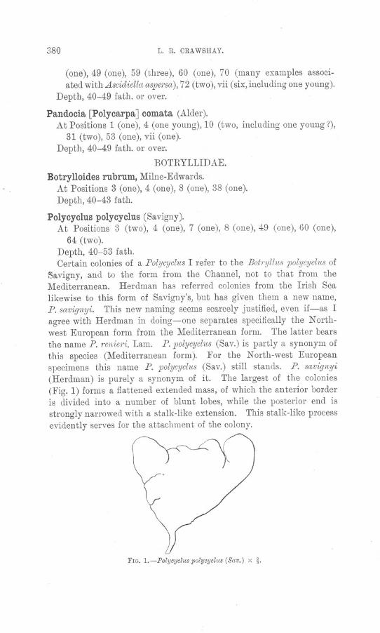
380 L. R. CRAWSHAY.
(one), 49 (one), 59 (three), 60 (one), 70 (many examples associ-ated with Ascidiella aspersa),72 (two), vii (six, including one young).
Depth, 40-49 fath. or over.
Pando cia [Polycarpa] comata (Alder).At Positions 1 (one), 4 (one young), 10 (two, including one young?),
31 (two), 53 (one), vii (one).Depth, 40-49 fath. or over.
BOTRYLLIDAE.
Botrylloides rubrum, Milne-Edwards.At Positions 3 (one), 4 (one), 8 (one), 38 (one).Depth, 40-43 fath.
Polycyclus polycyclus (Savigny).At Positions 3 (two), 4 (one), 7 (one), 8 (one), 49 (one), 60 (one),
64 (two).Depth, 40-53 fath.Oertain colonies of a Polycyclus I refer to the Botryllus polycyclus of
~avigny, and to the form from the Ohannel, not to that from theMediterranean. Herdman has referred colonies from the Irish Sealikewise to this form of Savigny's, but has given them a new name,P. savignyi. This new naming seems scarcely justified, even if-as Iagree with Herdman in doing-one separates specifically the N orth-west European form from the Mediterranean form. The latter bearsthe name P. renieri, Lam. P. polycyclus (Sav.) is partly a synonym ofthis species (Mediterranean form). For the North-west Europeanspecimens this name P. polycyclus (Sav.) still stands. P. savignyi(Herdman) is purely a synonym of it. The largest of the colonies(Fig. 1) forms a flattened extended mass, of which the anterior borderis divided into a number of blunt lobes, while the posterior end isstrongly narrowed with a stalk-like extension. This stalk-like processevidently serves for the attachment of the colony.
FIG. l.-Polycyclus polycyclus (Sav.) x !.
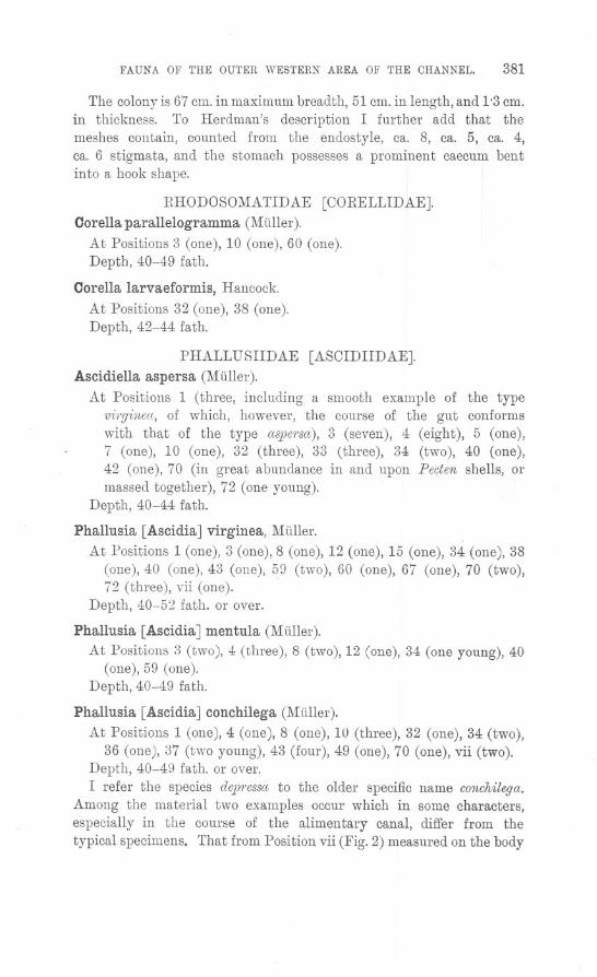
FAUNA OF THE OUTER WESTERN AREA OF THE CHANNEL. 381
The colony is 67 cm. in maximum breadth, 51 cm. in length, and 1'3 cm.in thickness. To Herdman's description I further add that themeshes contain, counted from the endostyle, ca. 8, ca. 5, ca. 4,ca. 6 stigmata, and the stomach possesses a prominent caecum bentinto a hook shape.
RHODOSOMATIDAE [CORELLIDAE].Corella parallelogramma (Muller).
At Positions 3 (one), 10 (one), 60 (one).Depth, 40-49 fath.
Corella larvaeformis, Hancock.
At Positions 32 (one), 38 (one).Depth, 42-44 fath.
PHALLUSIIDAE [ASCIDIIDAE].Ascidiella aspersa (Muller).
At Positions 1 (three, including a smooth example of the typevi1'ginea, of which, however, the course of the gut conformswith that of the type aspersa), 3 (seven), 4 (eight), 5 (one),7 (one), 10 (one), 32 (three), 33 (three), 34 (two), 40 (one),42 (one), 70 (in great abundance in and upon Pecten shells, ormassed together), 72 (one young).
Depth, 40-44 fath.
Phallusia [Ascidia] virginea, Muller. .At Positions 1 (one), 3 (one), 8 (one), 12 (one), 15 (one), 34 (one), 38
(one), 40 (one), 43 (one), 59 (two), 60 (one), 67 (one), 70 (two),72 (three), vii (one).
Depth, 40-5:2 fath. or over.
Phallusia [Ascidia] mentula (Muller).
At Positions 3 (two), 4 (three), 8 (two), 12 (one), 34 (one young), 40(one), 59 (one).
Depth, 40-49 fath.
Phallusia [Ascidia] conchilega (Muller).
At Positions 1 (one), 4 (one), 8 (one), 10 (three), 32 (one), 34 (two),36 (one), 37 (two young), 43 (four), 49 (one), 70 (one), vii (two).
Depth, 40-49 fath. or over.I refer the species dep1'essato the older specific name conchilega.
Among the material two examples occur which in some characters,especially in the course of the alimentary canal, differ from thetypical specimens. That from Position vii (Fig. 2) measured on the body
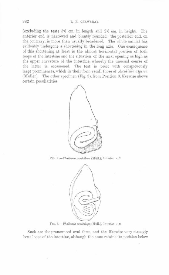
382 L. R. ORAWSHAY.
(excluding the test) 3'6 em. in length and 2'6 em. in height. Theanterior end is narrowed and bluntly rounded; the posterior end, onthe contrary, is more than usually broadened. The whole animal hasevidently undergone a shortening in the long axis. One consequenceof this shortening at least is the almost horizontal position of bothloops of the intestine and the situation of the anal opening as high asthe upper curvature of the intestine, whereby the unusual course ofthe latter is occasioned. The test is beset with conspicuouslylarge prominences, which in their form recall those of Ascidiella aspersa(Muller). The other specimen (Fig. 3), from Position 8, likewise showscertain peculiarities.
FIG. 2.-Phallusia conchilega(Miil!.), Interior x 2
FIG. 3.-Phallusia conchilega (Uiil!.), Interior x 2.
Such are the pronounced oval form, and the likewise very stronglybent loops of the intestine, although the anus retains its position below
FAUNA OF THE OUTER WESTERN AREA OF THE CHANNEL. 383
the upper curve of the latter. The free surface (the animal isattached by the whole left side of the body) is finely cornulated.
OIONIDAE.
Ciona intestinalis (Linnaeus).
At Positions 3 (two), 4 (one), 10 (one), 12 (one), 34 (two), 40 (one),44 (one), 49 (two), 59 (many massed together), 67 (one), 70 (manyon the inner surface of Pecten shells or massed together).
Depth, 40-52 fath.DIDEMNIDAE.
Didemnum [Leptoclinum] perspicuum (Giard).
At Position 3 (two).Depth, 40 fath.
Leptoclinum [Diplosoma] gelatinosum, Milne-Edwards.
At Positions 1 (one), 3 (one), 4 (two), 59 (one).Depth, 40-49 fath.
CEPHALOCHORDATA.Amphioxus lanceolatus (Pallas).
At Positions 5 (one), 11 (one), 21 (one), 48 (five), 50 (one), 57(eight), 58 (three).
Depth, 42-49 fath.
VERTEBRATA.PISOES.
The infrequent use of the trawls must be borne in mind with reference to thecomparatively few fishes recorded.
ELASMOBRANOHII.
SOYLLHDAE.
Scyllium canicula (Linnaeus).One specimen at Position 7.Depth, 42 fath.
RAHDAE.Raia clavata, Linnaeus.
Single specimens at Positions 8 and 66.Depth, 44-52 fath.
Raia circularis, Oouch.
At Positions 49 (one), and 66 (three).Depth, 47-52 fath.
384 L. R. ORAWSHA Y.
Raia sp.
Examples of Raia not specifically recorded occurred at Positions 3(one), 7 (three), 8 (one).
Depth, 40-43 fath.TELEOSTEI.
MURAENIDAE.
Conger conger (Linnaeus).
One specimen at Position 64.Depth, 53 fath.
GADIDAE.Gadus luseus, Linnaeus.
At Positions 3, 7 (two), 68 (thirty-two), 78 (six).Depth, 40-52 fath.
Gadus minutus, Linnaeus.At Positions 3, and 49 (several).Depth,40-47fath.
Gadus pollaehius, Linnaeus.
One specimen at Position 59.Depth, 49 fath.
Molva vulgaris, Fleming. .
One specimen at Position 3.Depth, 40 fath.
Motella trieirrata, Nilsson.
At Positions 58 (one), and vii (one).Depth, 49 fath. and (?) over.
SERRANIDAE.
Serranus eabrilla, Linnaeus.
At Positions 8 (one), 49 (three).Depth, 43-47 fath.Fishes obtained at Positions 64 (one), and 68 (four), which were not
retained, probably also belonged to this species.
CAPROIDAE.Capros aper (Linnaeus).
At Positions 3 (three), 7 (a few), 8 (one), 49 (a few), 59 (one), 60(two), 64 (two), 68 (eighty-five), 78 (two).
Depth, 40-52 fath.The haul at Position 68, where the large number of this species
FAUNA OF THE OUTER WESTERN AREA OF THE CHANNEL. 385
occurred, was one of fifty-five minutes with the otter trawl, taken in52 fathoms. It was one of the few cases in which the otter trawl was
used, with some risk, in the neighbourhood of decidedly rough ground.
PLEURONECTIDAE.
Zeugopterus norvegicus (Giinther).
At Positions 52 (one), 59 (one), 60 (one). Length, 6'9, 10'3, and5'7 em. severally.
Depth, 43-49 fath.A Zeugopterus which occurred at Position 58, probably also
belonged to this species.
Arnoglossus laterna (Walbaum).
At Positions 3 (one), 7 (a few), 8, 4964 (three), 68 (six), 78 (four).
Depth, 40-53 fath.
(eight), 52 (one), 60 (three),
Arnoglossus megastoma, Donovan.
At Positions 3 (a few), 7 (two).Depth, 40-42 fath.
Pleuronectes microcephalus, Donovan.At Positions 7 (one), 49 (one), 68 (three).Depth, 42-52 fath.
Solea variegata (Donovan).At Positions 35 (one), 51 (one), 52 (three), 68 (one).Depth, 43-52 fath.
GOBIIDAE.
Gobius jeffreysi, Gunther.
At Position 1 (one), 52 (one).Depth, 40-43 fath.
Gobius scorpioides, Collett.
At Positions vii (one), 62 (one). Length, 33 mm. and 31 mm.respectively.
Depth, 50 fath. and (1) over.Mr. Boulenger kindly identified this rare goby. With reference
to its occurrence, Holt and Byrne (152, p. 21) state that exceptingseveral specimens obtained from Ballynakill Harbour on the westcoast of Ireland, there are only five records of its captme, viz. fromthe Norwegian coast, the Cattegat, Falmouth, and 30 miles W.N.W.
386 L. R. ORAWSHAY.
of Oleggan Head, the depth of occurrence ranging from about 2 to74 fath.
The two specimens here recorded considerably exceed in length themeasurement, " less than 1 inch," given by Holt and Byrne.
AGONIDAE.
Agonus cataphractus, Linnaeus.
At Positions 5 (one), 52 (one). Length40 mm.
Depth, 42-43 fath.
Callionymus lyra, Linnaeus.
At Positions 3 (a few), 7 (two), 8 (one), 59 (two), 60 (three), 66(one), 68 (one).
Depth, 40-52 fath.
of the latter specimen
TRIG LIDAE.Trigla pini, Bloch.
At Positions 3 (one), 7 (a few), 8 (one), 49 (four), 60 (three small),64 (two), 68 (five).
The small specimens at Position 60 measured 6'4, 5'6, and 5'3 em.severally, in length.
Depth, 40-53 fath.
Trigla lineata, Gmelin.
Two specimens at Position 49.Depth, 47 fath.
TRAOHINIDAE.
Trachinus draco, Linnaeus.
Only recorded at Position 7.Depth, 42 fath.
GOBIESOOIDAE.
Lepadogaster bimaculatus, Fleming.
One specimen at Position 1.Depth, 40 fath.
Lepadogaster microcephalus, Brook. *
One specimen at Position 72.Depth, 43 fath.
LOPHIIDAE.
Lophius piscatorius,. Linnaeus.
One specimen at Position 64.Depth, 53 fath.
* Brook, G., P1'OC.Roy. Soc. Edinburgh, Vol. X, Pt. I, p. 166.
FAUNA OF THE OUTER WESTERN AREA OF THE CHANNEL. 387
LITERATURE.GENERAL.
1. ALLEN,E. J.-On the Fauna and Bottom-Depositsnmr the Thirty-FathornLine, from the Eddystone Grounds to Start Point. Jour. l\lar. BioI. Assoc.,N.S. Vol. V., p. 365, 1897-99.
2. BATE,C. SPENCE.-Rep01.tof the CommitteealJpointedto explO1'ethe MarineFauna and Flora of the South Coastof Devon and CornweLll,No.2. Brit.Assoc. Report, 1867, p. 275.
3. BOURNE,G. C.-Rep01.t of a Tmwling Cn.ise in H.M.S. "Resea?'ch,"off theSouth-West Coast of Ireland. Journ. Mar. BioI. Assoc., N.S. Vol. II.,p. 333, 189l.
4. CRAWSHAY,L. R.-On Rock Remains in the Bed of the English Channel. AnAccount of the Dredgings carried out by S.s. "Oithona" in 1906. J ourn.Mar. BioI. Assoc.,N.S. Vol. VIII., p. 99, 1907-10.
5. ELLIOT,G. F. SCOTT;LAURIE,M. ; ANDl\lURDOCK,J. B.-Faw~a, Flora, andGeologyof theClydeA rea. Glasgow, 1901.
6. GARSTAN(!,W.-Notes on the Marine Invel.tebmteFauna of Plymouth for 1892.Journ. Mal'. BioI. Assoc., N.S. Vol. I., p. 333, 1889-90.
7. HADDON,A. C.-Fil'st Report on the Marine Fauna of the South-West ofIreland. Proc. Royallrish Acad., Ser. 2, Vol. IV., 1886.
8. HEAPE,W.-P?'eliminal"y Report upon the Fauna and Flol'a of PlymouthSound. Jour. Mar. BioI.Assoc., O.S. Vol. I., p. 153, 1887-88.
9. HERDMAN,W. A.-The Marine Zoology,Botany, and Geologyof the Irish Sea.Fourth and Final Report of the Committee. Brit. Assoc.Report, 1896.
10. KOEHLER,R-Resultats Scientifiques de la Campagnedu " Caudan"danslaGolfede Gascogne. Aor1t-Septembre,1895. Paris, 1896.
11. NORMAN,A. M.-Shetland Final Dredging Report, Part II. Brit. Assoc.Report, 1868, p. 247.
PORIFERA.
12. BOWERBANK,J. S.-A Monograph of the British Spongiadae. Vols. I.-III.Ray Society, London, 1864-74. Vol. IV. (Supplementary), Edited by REV.A.M. NORMAN. RS., London, 1882.
13. DENDY,A.-Studies on the Comparative Anatomy of Sponges. V. Observationson the Stmcture and Classification of the CalcareaHetel'ocoela. Quart. Journ.l\1:icrosc. ScL V01. XXXI., p. 159, 1894.
14. HAECKEL,E.-Die Kalkschwamme. Berlin, 1872.15. HANITSOH,R-Second Report on the POI'ijera of the L.M.B. C. District. Trans.
Liverpool BioI. Soc. Vol. III., p. 155, 1889.16. HANITSCH,R-Third Report on the Porifera of the L.M.B.C. Dr:stl'ict. Trans.
Liverpool BioI. Soc. Vol. IV., p. 192, 1890.17. HANITSCH, R-Revision of the Generic Nomenclat1we and Classification in
Bowerbank's " British Spongiadae." Trans. Liverpool BioI. Soc. Vol. VIII.,p. 173, 1894. .
18. HIGGIN, T.-Report on the Pewifem of the L.M.B.C. District. First Repol.t
upon the Fauna of Liverpool Bay and the Neighboul'ing Seas. London, 1886.19. JOHNSTON,G.-A History of BI'itish Sponges and Lithophytes. 1842.20. KELLER, C. -Die Spongienfauna des l'othen Meeres. Zeitschr. f. Wiss. ZooL
VoJ. XLVIII., p. 3ll, 1889.

LENDENFELD,R. v.-Descriptive Catalogue of the Sponges in the AustralianMuseum, Sydney. London, 1888.
LENDENFELD, R. v. -Die Spongien de1' Ad1'ia. I. Die J{cd1cschwlimme.Zeitschr. f. Wiss. Zool. Vol. LIII., p. 185, 1892.
LUNDBECK,W.-Porifem (Part I.), Homon'haphidae and Heterorrhaphidae.Danish Ingolf Expedition. Vol. VI. Copenhagen, 1902.
LUNDBECK,W.-Pm'ifera (Part II.). Desrnacidonidae (Pan). Danish In-golf Expedition. Vol. VI. ]905.
LU~DBEcK, W.-Porifera (Part III.). Desmacidonidcw (Pars). Danish In-golf Expedition. Vol. VI. 1910.
MINCHIN, E. A.-Sponges. A Treatise on Zoology, edited by E. Ray Lan-kester. Part II. London, 1900.
MINCHIN, E. A.-The Chamcters and Synonymy of the Bn:tish Species of
Sponges of the Genus Leucosolenia. Proc. Zool. Soc., ]904, II., p. 349.MINCHIN, E. A.-On the Sponge Leucosolenia contorta Bowerbanlc, Ascandm
contorta Haeclcel, and Ascetta spinosc~Lendenfeld. Proc. Zool. Soc., London,1905, 11., p. 3.
MONTAGu,G.-An Essay on Sponges, with Desc1'iptions of all the Species thathave been discovered on the Coast of Great Britain. Wel'nerian Memoirs.Vol. II., p. 67, 1814.
POLEJAEFF, N.-Calcctrea. Challenger Reports. Zoology. Vol. VIII. 1883.RIDLEY, S. a., ANDDENDY,A.-2v.[onaxonida. Challenger Reports. Zoology.
VoJ. XX. 1887.
SCHMIDT,a.-Die Spongien de.gAdriatischen Meeres. Leipzig, 1862.SCHMIDT,a.-Die Spongien der [{iiste von Algier. Leipzig, 1868.SCHULZE, F. E.-Untersuchungen iiber den Ba1~ unci die Entwiclclung der
Spongien. Zweite Mittheilung: Die Gattung Halisarca. Zeitschr. f.Wiss. Zool. Vol. XXVIII., p. 1. 1877.
TOPSENT,E.-Eponges de la Manche. Mem. Soc. Zoo1. de France. TomeIII, p. 195. 1890.
TOPSENl', E.-Spongia1'es des Cotes Ocecmiques de Fmnce. Bull. Soc. Zool.de France, XVI., p. 125. 1891.
TOPSENT,E.-Essai SU1'fa Faune des Spongiares de Roscojj. Arch. Zool. Exp.Gen., 2° Ser., IX., p. 523. 1891.
TOPSENT, E.-Cont1'ibution a l'Etude des Spongim'es de l'Atlantique Nord.Res. Camp. Sci. Monaco, Fasc. II. 1892.
TOPSENT,E.- Une Refm'me dans In Classification des Hnlichond1'ina. Mem.Soc. Zool. de France, VII., p. 5. 1894.
TOPSENl', E.-Int1'od1~ction n l'Etude lV1onogmphique des Monnxonides de
j{'mnce. Classification des Had1'ome1'ina. Arch. Zoo1. Exp. Gen., 3e Ser.,VI., p. 91. 1898.
TOPSENT, E.-Etude Monogmphique des Sponginres de France, III.
Monaxonida. Hadrome1'inn. Arch. Zool. Exp. Gen., 3" Ser., VIII., p. 1.1900.
TOPSENl', E.-Sponginres des A9ores. Res Camp. Sci. Monaco, Fasc. XXV.1904.
VOSMAER,G. C. J.-Spongien.ells. 1887.
388
21.
22.
23.
24.
25.
26.
27.
28.
29.
ao.31.
32.33.34.
35.
36.
37.
38.
39.
40.
41.
42.
43.
L. R. CRAWSHAY.
Bronn's Klassen und ardungen des Thier-
FAUNA OF THE OUTER WESTERN AREA OF THE CHANNEL. 389
COELENTERATA.
44. ALLMAN,G. J.-A Monograph of the Gymnoblastic or T~Lbularian Hydj'oids.
Ray Society, London. 1871.45. ALLMAN,G. J.-Report on the Hydroida collected dw'ing the Expeditions of
H.M.S. "Porcupine." Trans. Zool. Soc., London, VII., p. 469. 1874.46. ALLMAN,G. J.-Hydroidtt. Paj.t I. Challenger Reports, Vol. VII. 1883.47. ALL~IAN, G. J.-Hydroida. Part II. Challenger Reports, Vol. XXIII,
1888.
48. BILLARD,A.-Hyd1'Ofdes. Expeditions Scientifiques du "Travailleur" et du" Talisman," 1906, p. 153.
49. BONNEVIE, K.-lI!eeresfauna von Bergen. Hydj'oiden. Bergen, 1901. .50. BROCH, H. J.-Die Hydroiden der arktis~hen Meere. Fauna Arctica, V.,
p. 127. Jena, 1910.51. GOSSE,P. H.-A History of the British Sea Anemones. London, 1860.52. HADDON,A. C., ANDSHACKLETON,A. M.-A Revision of the Bj'itish Actiniae.
Part II. The Zoantheae. Trans. Royal Dublin Soc., Ser. 2, Vol. IV.,p. 609. 1891.
53. HARTLAUB,C.-Die Coelenteraten Helgolands. Wissensch. Meeresunt, N. F.I. Heft 1, p. 161. 1894.
54. HICKSON, S. J.-The Anatomy of Alcyonium digitatum. Quart. Journ.Microsc. Sci., Ser. 2, Vol. XXXVII., p. 343.
55. HIXCKS, T.-A History of the British Hydroid Zoophytes. London, 1868.56. HOLDSWORTH,E. W.-On Zoanthus couchii, Johnston. Proc. Zool. Soc.,
London, 1858, p. 557.57. HOLDSWORTH,E. W.-On an undescribed S)Jeciesof British Zoanthus. Proc.
Zool. Soc., London, 1861, p. 99.58. JADERHOLM,E.-Die Hydroiden der Schwedischen Zoologischen Polar Expedi-
tion,1900. Bihang til K. Svenska Vet. Akad. Handlingar, 28, IV., 12.Stockholm, 1902.
59. NUTTING, C. C.-American Hydroids. The Plumularidae. Smithsonian
Institution. Special Bulletin. Washington. 1900.60. NUTTING, C. C.-American Hydroids. The Sertularidae. Smithsonian
Institution, Special Bulletin. Washington, 1904.61. PICT~:T,C., ET BEDOT,M.-Hydraires provenant des Campagnes de I'Hirondelle
(1886-1888). Res. Camp. ScL, Fasc. XVIII. Monaco, 1900.62. SARS, G. O.-Bidrag til Kundskaben om NorgesHydroider. Vidensk.-Selsk.
Forhandl. 1873.63. THORNELY,L.-Supplementary Reportupon Hydroid Zoophytesof theL.M.B.C.
District. Trans. Liverpool BioI. Soc., Vol. VIII., p. 140. 1894.64. WOOD,G.-RecOl'd of Additional Hydroida from the Isle of Man. Trans.
Liverpool BioI. Soc., Vol. VI., p. 94. 1892.
ECHINODERMATA.
65. BELL, F. J.- Catalogue of the British Echinoderms in the British Museum.London, 1892.
66. FORBES,E.-History of British Starfishe.~. London, 1841.67. HELLER, C.-Die Zoophyten und Echinodermen des Adriatischen Meeres.
K. K. Zoolog. Botan. Gesellschaft, Vienna, 1868.
390 L. R. ORA WSHA Y.
68. HODGE, G.-On a new Sand Stcw of the Genus Ophinm (Ophi1.ra normani)found on the Ooast of Northumberland and Durham. Trans. TynesideNaturalists' Field Club. Vol. V., p. 296. Newcastle-on-Tyne,1863.
HODGE, G.-Rep07.t on Dredging Operations on the Ooasts of Northumberlandand Durham in July cmd August, 1863. Report on the Echinodermata.Trans. Tyneside Naturalists' Field Club. Vol. Vr., p. 189. 1863-64.
HODGE.-Oatcdogue of the Echinodermata of Northumberland and Durham.
Nat. Hist. Trans. Northumberland and Durham. Vol. IV., p. 120. 1872.LJUNGMAN,A.-Ophiuroidea viventia huc usque cognita. Oversigtaf Kongl.
Vetenskaps-Akadamiens Forhandlingar, 1866.LUDWIG,H.-Die Echinodej'rnen des j11ittelmeeres. Mitt. Zool. Stat. Neapel,
Vol. 1., p. 523. Leipzig, 1879.LUDWIG,H.-Die Seesterne des ]',[ittelmeeres. Fauna und Flora des Golfes
von Neapel, 24 Monographie. Berlin, 1897.LUTKEN, C. F.-Additamenta ad historiam Ophiuridarum. Beskrivelsej' af
nye eller hidtil hm ufuldstcwndigt kjendte Arter af Slangestjerner. Kgl.danske Videnskab. Selskabs Skrifter, 5. V. Copenhagen, 1858.
LYMAN,T.-Ophit~roidea. Challenger Reports, Vol. V., 1882.LYMAN, T.-Report on the Ophiuroidea. (Collected by U.S. Coast Survey
Steamer" BJake.") Bull. Mus. Compo Zool., Harvard, Vol. X., p. 227.Cambridge, Mass., 1883.
MOBIUS, K.,AND BUTSCHLI,O.-Zoologische Ergebnisse der Nordseefahrt.(Pommerania) IV. Echinodermata. Jahresbericht del' Commission zurWissenschaftlichen Untersuchung del' Deutschen Meere in Kiel fiir dieJahre, 1872, 1873. Berlin, 1875.
NORMAN, A. M.-On Genera and Species of British Echinodermata. Ann.Mag. Nat. Hist., Feb., 1865.
NORMAN,A. !Yr.-Shetland Final Dredging Report. Part II. Brit. Assoe.Report, 1868, p. 312.
69.
70.
71.
72.
73.
74.
75.76.
77.
78.
79
POLYCHAETA.
80. CLAPAREDE,E.-Les Annelides Ohetopodes du Golfe de Naples. Geneve etBale, 1868.
81. EHLERS, E.-Die Borstenwfirmer.82. GEMMILL,J. F.-Marine Worms.
Area, p. 359. Glasgow, 1900.83. GRUBE, E.-Beschreibtmg ne1ter ode?'wenig bekannten Anneliden. Archiv fiir
Naturg., 1860, p. 71.84. GRUBE, E.-Annulata Semperiana.
St. Petersburg. XXV., 1878.85. JOHNSTON,G.-Oatalogue of the British non-parasitical Worms in the Oollection
of the British Museum. London, 18135.86. LANGERHANS,P.-Die Wurmfauna van Mctdeira. Zeitschr. Wiss. Zool.,
XXXII., 1879.87. LANGERHANS,P.-Die Wurmfauna van j11adeira. Zeitschr. Wiss. Zool.,
XXXIV., 1880.88. Lo BIANCO,S.-Gli A nnelidi Tubicoli trovate nel Golfodi Napoli. Naples, 1893.89. McINTOSH, W. C.-Repm.t on the Annelids dredged off the Shetland Islands by
Mr. Gwyn Jeffreys, 1867-68. Brit. Assoc. Report, 1868, p. 336.
1., Leipzig, 1864-1868.
Fauna, Flora, and Geology of the Clyde
Mem. de L'Acad. Imp. des Sciences de
FAUNA OF THE OUTER WESTERN AREA OF THE CHANNEL. 391
90. McINTOSH,W.O.-Annelida Polychaeta. Ohallenger Reports. VoL XII.,1885.
91. McINTOSH, W. O.-A Monograph of the British Annelids. PolychaetaAmphinomidae to Sigalionidae. Ray Society, London, 1900.
92. McINTOSH,W. O.-A Monograph of the B1'itish Annelids. Polychaeta.Nephthydidae to Syllidae. Ray Society, London, 1908.
93. McINTOSH,W. O.-A Monograph of the British Annelids. Polychaeta.Syllidae to Ariciidae. Ray Society, London, 1910.
94. MALMGREN,A. J.-Nordiska Hafs-Anmdater. (Hers. af K. Vet. Akad.Forh., 1865.
95. MALMGREN,A. J.-Annulata polychaetaSpetsbergiae,Gronlandiae, Islandiaeet ScandinCiviaehactenuscognita. Ofers. af K. Vet. Akad. Forh., 1867.
96. MARENZELLER,E. v.-Zur Kentniss der adriatischen Anneliden III.
(Terebellen.) Sitzb. Akad. Wien. L?CXXIX., p. 151, 1884.97. MARION,A. F., ETBOBRETZKY,N.-Etude des Annelides du Golfe de Mm'seille.
Ann. Sei. Nat. 6meSer. Zoologie, 11., 1875.98. ROULE,L.-Anne/ides. Resultats Seientifigues de Ia Oampagne du "Caudan"
dans Ie Golfe de Gaseogne. Aout-Septembre, 1895. Paris, 1896.99. SAINT.JOSEPH, BARON DE.-Les .Annelides Polychetes des Cotes de Dinard.
Ann. Sei. Nat. Zoo1. 7me Ser., I., 1886.100. SAINT-JOSEPH, BARON DE.-Les Anne/ides Polychetes des Cotes de Dincwd.
Ann. Sei. Nat. Zoo1. 7meSer., V., 1888.101. SAINT-JOSEPH, BARON DE.-Les Annelides Polychetes des Cotes de Dinard.
Ann. Sci. Nat. Zoo1. 7meSer., XVII., 1894.102. SAINT-JOSEPH, BARON DE.-Les Annelides Polychetes des Cotes de Dinard.
Ann. Sei. Nat. Zoo1. 7meSer., XX., 1895.103. SAINT-JOSEPH, BARON DE.-Les Annelides Polychetes des Cotes de France.
(Manche et Ocean). Ann. Sei. Nat. Zoo1. 8m. Ser., V., ] 898.104. SAINT-JOSEPH, BARON DE.-Les Annelides Polychetes des Cotes de France.
(Oceanet Cotesde Provence.) Ann. Sei. Nat. Zool. 9meSer., III. 1906.]05. SARS, M.-Beskrivelse1' og Jagttagelser over nogle mcwrkelige eller nye i Havet
ved den Bergenske Kyst lebende Dyr., p. 46. Annelider. Bergen, 1835.
ORUSTAOEA.
106. ApPELLOF, A.-Die Dekapoden Crustaceen. Meeresfauna von Berg~n.Heft 3. Bergen, 1906. .
107. BATE, O. SPENCE.-The Crustacea in Couch's Cornish Fauna, revised and addedto. J OUI'll.Royal Inst. Oornwall. Part II. No. XIX. 1878.
108. BATE, C. SPENCE,and WESTWOOD,J. O.-A History of the British Sessile-eyedCnistcicea. London 1863.
109. BELL, T.-A History of the British Stalk-eyed Crustacea. London, 1853.110. BONNIER, J.-Les Galatheidae des Cotes de Fmnce. Bull. Sci. France BeIg.,
Ser. 3, VoL I., p. 121. Paris, 1888.Ill. BOUVIER, E. L.-Sur les Xanthes des mers d'E1irope. Feuilles des Jeunes
Naturalistes, Ser. III, No. 332, p. 133. 1898.112. CHEVREUX,E.-Amphipodes provenant des Campagnes de I'Hirondelle (1885-
1888). Res. Camp. Sei., XVI. Monaco, 1900.113. GORDON,G.-A List of C1'ustaceansof the Moray Firth. Zoologist, December,
]852, p. 3678.
392 L. R. CRAWSHAY.
114. HANSEN,H. J.-Crustacea Malacostraca (I). Danish Ingolf Expedition.Copenhagen, 1908.
115. HELLER,C.-Die Crustaceendes Siidlichen Europct.CrustaceaPodophthalmia.Vienna, 1863.
116. HENDERSON,J. R-A Synopsis oftheBritish Paguridae. Proc. Roy. PhysicalSoc., Vol. IX, p. 65.
117. KEMP,S.-The Decapodacollectedby the" Huxley" fr'om the N or.thSide of theBcty of Biscay in Attgust, 1906. Journ. Mar. Bioi Assoc.,N.S., Vol. VIII,p. 407. 1907-10.
118. KINAHAN,J. R-On a Crangonnew to Science,with Notices of othernondescriptCrustacea,and observationson theDistribution of the CrustaceaPodophthalmiaof th" Eastern or Dublin Marine District of Ireland. Nat. Hist. Rev.,Vol. IV. 1857. Proceed. Soc., p. 80.
119. KINAHAN,J. R-Synopsis of the Species of the Families CrangonidaeandGalatheidaewhich inhabit theSeas around the British isles. Proc. R IrishAcad., 1862, p. 354.
t20. LEACH,W. E.-Malacostraca PodophthalmataBritanniae. Trans. Linn. Soc.,Vol. XL, p. 306. 1815.
121. MELVILLE,A. G.-Carcirwlogical Notes: Being a List of the Cr'ustaceaPodophthalmia of Galway Marine Districts, chieflymade during the summerof 1850. Nat. Hist. Rev., Vol. IV., 1857. Proc. Soc., p. 151.
122. MIERS,E. J.-Brachyum. Challenger Reports, Vol. XVII. 1886.123. MILNE-EDV(ARDS,A., ET BOUVIER,E. L.-Crustacis Decapodesprovenant des
Carnpagnesdu Yacht l' "Hirondelle" (1886, 1887, 1888). Res. Camp. Sci.Monaco. Fasc., VII. 1894.
124. MILNE-EDWARDS,A., ET BOUVIER,E. L.-CntStaces Decapodesprovenant desCampagnesde l' " Hirondelle" (Supplement)et de la " PrincesseAlice" (1891-1897). Res. Camp. ScL Monaco,Fasc. XIII. 1899. .
125. MILNE-EDWARDS,A., ETBOUVIER,E. L.-C~ustacisDecapodes. 1. Brachyi:reset Anornures. Expeditions Scientifiques du "Travailleur" et du "Talisman"pendant les Annees 1880, 1881, 1882, 1883. Paris, 1900.
126. MO:;TAGU,G.-Description of several Marine Animals found on the South Coast
of Devonshire. Trans. Linn. Soc., IX., p. 81. London, 1805.127. NORMAN,A. M.-Beport on Dredging Operations on the Coasts of Northumber'-
land and Durham in July and August, 1863. Rep01.t on the Crustacea.
. Trans. Tyneside Naturalists' Field Club, Vol. Vr., p. 183. 1863-64.128. NOR~fAN,A. M.-Notes on the Crustacea of the Channel Islands. Ann. Mag.
Nat. Hist., Ser. 7, Vol. XX., p. 356. 1907.129. NORMAN,A. M., AND BRADY,G. S.-The Crustacea of Northumberland and
Durham. Trans. Nat. Hist. Soc. Northumberland, Durham, and New-
castle-upon-Tyne, N.S., Vol. III., pt. 2.130. NOR~fAN,A. M., AND SCOTT,T.-The Crustacea of Dev01~ and Cornwall.
London, 1906.131. PATIENCE, A.-Carcinological Notes. Buteshire Nat. Hist. Soc. Trans.,
1909-10, p. 69.132. SARS, G. a.-An Account of the Crustacea of Norway. Arnphipoda. Chris-
tiania and Copenhagen, 1895.133. SCOTT,T.-A Revised List of the Crustacea of the Firth of Forth. Appendices
to Sixth Annual Report of the Fishery Board of Scotland, p. 235,1888.
FAUNA OF THE OUTER WESTERN AREA OF THE CHANNEL. 393
134. SEXTON,E. W.-The Amphipoda collectedby the" Huxley" from the North Sideof the Bay of Biscay, in August, 1906. Journ. Mar. BioI. Assoc., N.S.,Vol. IX., p. 199, 1911.
135. SMITH, G.-Rhizocephala. Fauna und Flora des Golfes von Neapel.Berlin, 1906.
136. WALKER,A. a.-Third Report of the Higher Crustaceaof theL.M.B.C. District.. Proc. Liverpool BioI. Soc.,Vol. III., p. 195, 1889.
137. WALKER,A. a.-Revision of the Podophthalnwta and Cumaceaof LiverpoolBay. Trans. Liverpool BioI. Soc., Vol. VI., p. 96, 189~.
138. WALKER,A. O.-Malacostracaf1'orlt the WlJ8tCoastof Ireland. Trans. Liver-pool BioI. Soc., Vol. XII., p. 159, 1898.
139. WALKER,A. a.-Crustacea collectedby the late Mr. R. J. Ashcroft and Mr.Harvey on the North Side of the Bay of Biscay. Ann. Mag. Nat. Hist.,Ser. 8, Vol. V., p. 158, 1910.
BRYOZOA.
140. HARMER,S. F.-On the British Speciesof Crisia. Quart. J ourn. Microsc.ScL, Ser. 2, Vol. XXXII., p. 127, 1891.
141. HARMER,S. F.-Polyzoa. Cambridge Natural History, 1896.142. HINCKS,T.-A Hist01'yof the British Ma1.inePolyzoa. London, 1880.
MOLLUSCA.
143. ALDER, J., AND HANCOCK,A.-A Monograph of the British NudibranchiateMollusca. Ray Society, London, 1865.
144. ALDER, J., AND HANCOCK,A.-A M01wgraph of the British NudibranchiateMoll1U!ca. Supplenwnt by SIR CHARLESELIOT. Ray Society, London, 1910.
145. CONCHOLOGICALSOCIETY.-List of British Marine Mollusca and Brachiopoda.Prepared by a Committee of the Conchological Society. Journal of Conchology,Vol. X., p. 9. London, 1901-03.
146. FORBES,E., ANDHANLEY,S.-A History of British Mollusca. Loudon, 1853.147. JEFFREYS,J. G.-British Conchology. J.ondon,1863-1869.148. REYNELL,A.-The Molluscacollectedby the" Huxley" from theNorth Sideof the
Bay of Biscay in August, 1906. Journ. Mar. BioI. Assoc., N.S., Vol. VrrL,p. 359, 1907-1910.
PISCES.
149. COUCH,J.-A Historyoft1w Fishesof theBritish Islands. London, 1862-1865.150. DAY,F.-The FishlJ8of GreatBritain and Ireland. London, 1880-1884.!L51. FRIES,B., EKSTROM,C. U., ANDSUNDEVALL,C.-A History of Scandinavian
Fishes. Second Edition. By F. A. S~nTT. Stockholm, Berlin, Paris andLondon.
152. HOLT,E. W., ANDBYRNE,L. W.-The British and Irish GobilJ8.Report onthe Sea and Inland 'Fisheries of Ireland for 1901. Part II. ScientificInvestigations. Appendix IlL, p. 37, 1903.
l'EW SERIES.-YOL. IX. NO. 3. JUNE, 1912. 2 0

( 394 ]
Some Notes upon the Feeding Habits of Mackerel andcertain Clupeoids in the English Channel.
ByG. E. Bullen.
SEVERALprevious authors* have shown that the food of the mackerel,when in coastal waters, is of two different kinds, and that the fish.adopts two distinct methods of procuring it. In the first place it feedsby a system of filtration upon planktonic organisms, and secondly upon.prey of a larger character which is hunted by sight.
The exact nature of the food taken whilst the fish is in its winter
quarters has not as yet been thoroughly investigated; but the presentwriter, in making examination of the stomach contents of mackereltaken on the ground in March, 1907, 25 miles S.W. of Start Point,found that it consisted entirely .of zoo- and phytoplankton of a naturesimilar to that existing in the water itself.t A close similarity ingeneral character appeared to exist between the nature of these samplesand otbers from fish taken in the Bristol Channel at the surface duringtbe early part of April of the same year.+
It was also observed that during the three months forming the moreimportant part of the" drifting" season in the western area, viz.April, May, and June, the fish were feeding exclusively upon plankton,and that the nature of this food gradually changed from being largelyvegetable to almost wholly animal.
It was not until the middle of June that food of a larger character,.but still wholly of the latter type, began to appear in stomach con-tents, and it is a well-known fact that at about this time every yearthe drift fishery in the area under consideration begins to decline, andhand-lining commences. This condItion is due to the fact that thedense shoals break up, and the system of nutrition, which has hithert(}been one of filtration, gives place to one of, what we may term, "selectivefeeding."
From the evidence before us, in the form of the plankton results for
" Vide Allen, E. J., "Rep. on the Present State of our Knowledge with Regard to the.Habits and Migrations of the Mackerel," Journ. Afar. BioI. Assoc., N.S., Vol. V, p. 9.
t "Plankton Studies in Relation to the Western Mackerel Fishery," Journ. Afar.Biol. Assoc., N.S., Vol. XVIII., pp. 285-6.
:t Id. Table No. IV, Nos. 76 to 83, and Table No. V, No. 111.
FEEDING HABITS OF MACKEREL. 395
the stations E5 and.E6 (the only two falling within the fishing area),published in the Bulletins Oonseil Inte1'national POU?'l'explomtion de laMer, it would appear that these several changes in the nature of thefood of the mackerel, viz. from phytoplankton to general zooplankton,and from thence to zooplankton of a larger character and animal foodgenerally, e.g. young fishes and material offered as bait, is largely, ifnot entirely, due to seasonal changes in the nature of the food supplyitself. Yet, as it has been abundantly shown that certain planktonicorganisms are. of sporadic distribution, and that there is some fairevidence to show that others occur in shoals of varying extent, itremains to be seen how far the mackerel, exercising these two distinctmethods of nutrition, is capable of discriminating between varyingtypes of food during the period that it is feeding by filtration.
The question arises, to begin with, whether in adopting the onesystem the fish is incapable of using the other, and two importantpoints in this connection may be considered. In the first place, theNewlyn drifters state emphatically that it is impossible to takemackerel, when closely shoaling, upon a hand-line; and secondly, it isa matter of common knowledge amongst fishermen that, when latein the summer mackerel are caught (in drift nets), when feedingheavily upon copepods, they are very liable to rapid decomposition inthe region of the stomach, and that this is due to the" soft" conditionof the fish themselves, and not to the increased warmth of theatmosphere. Now, with respect to this matter it may be stated thatthe present writer, in making examination of the stomach contentsof several hundreds of mackerel taken at various times of the year,has invariably found that the walls of the stomach appeared to becontracted and thickened in cases where phytoplankton formed thebulk of the food, whereas the organ in question was distended andthe walls extremely thin when zooplankton occurred. Moreover,although no careful histological comparison was made between the twotypes, it was found possible to distend, by means of an air-pump, to agreat extent an empty stomach of the latter type without unduepressure, whereas one of the thickened type would not respond tosuch treatment. Again, throughout an observation extending overnearly six years upon fish in many cases from not exactly determinedsources, the writer has found that, with very few exceptions, thethinning of the stomach walls is developed to the greatest extent asthe western drift fishery approaches its highest point of productive-ness, viz. in May and June in the case of large mackerel, and in Juneand July witq those 'of smaller size, which latter, as Dr. Allen'" has.
* Op. cit., p. 25.
396 G. E. BULLEN.
pointed out, appear to be solely on feeding migration. Moreover, inall of the early spring" hooked" mackerel that the writer hasexamined, the walls of the stomach have been of fair thickness.
The evidence before us is certainly insufficient to warraIl~ anassertion that a physiological change is brought about in the nature ofthe stomach by reason of a change of diet, but the fact remainsthat the writer has found it possible invariably to determine thegeneml nature of the food contained in the stomach of a mackerel or apilchard from an external examination of the organ itself, before it isopened.
With reference to the transitional stage from the one type of feed-ing to the other, the following note, received in the autumn of 1911from Mr. Mathias Dunn, of Newlyn, is of considerable interest. Thisobserver states :-
" I sent you yesterday some specimens of a rare Crustacean Mac1'opsis,which has been very abundant on our coasts this summer. The mackerelhave been feeding ravenously on these animals, and at times havepursued them into the harbour, where they have been so numerous asto appear like thick porridge. We nave had some excellent opportuni-ties of seeing mackerel feed, during the visit of. these little creatures.They have been hunting up and down the back of the pier, like a packof hounds, going the whole length, turning and returning again andagain. They were swimming about ten to twelve feet off the pier incompany with a number of scads, about twenty to thirty in each shoal,the scads in every case swimming close to the pier and the mackereljust outside. The mackerel were swimming in open order, closing in,rising and falling in graceful undulations, by which means they eitherdrove their prey on to the surface, where there was no escape, orturned it to the centre of the shoal, where it was also promptlydespatched."
Upon examination, the sample sent by Mr. Dunn was found to con-sist almost entirely of the Mysid Crustacean Mac1'opsisstabbe1'i,VanBeneden, * together with a few young Herring (surface-swimming stage),and a slight number of fish ova and the Copepod Gent1'opagestypiCllS,Kroyer.
Unfortunately no stomach material was examined, but the foregoingobservations tend to show amongst other things that the fishwere feeding by sight, and were exercising discrimination in theselection of their prey. At the time when these observations were
.For the determination of this species the writer is indebted to Mr. W. AI. Tattersall,M.Sc.
FEEDING HABITS OF MACKEREL. 397
made, however, viz. in July, the heavy summer drift fishery was far onthe decline* and line fishing had commenced, an indication that thefish had begun to take prey of larger type. .
With further reference to the feeding habits of mackerel and he~ring,in this instance kept in captivity at the Brighton Aquarium, the writeris indebted to Mr. E. W. Cowley, the Superintendent, for the followingnote.
This observer states: "The herring and mackerel are usually takenfrom the sea in October and November with the seine net, which isdrawn on to the beach. They are then conveyed to the Aquarium incans the size of an ordinary sanitary bin, about 20 or 30 in each can.Special care is taken to see that the cans are, not overcrowded, thefish are not handled in any way, and conveyed to the tanks with thegreatest possible speed. Even with these precautions we considerourselves fortunate in saving 10% of the original catch. After theyare placed in the tanks great care has to be exercised to see that thefish are in no way disturbed until they are acclimatized; but we havenot found it necessary to darken the tanks.
"In the case of the herring and mackerel we find that the placing ofa rock in the centre of the tank assists them considerably, unless ofcourse a tank 120 feet long is provided.
"We feed both species upon sand-hoppers and small shrimps, alive ifpossible, and vary the diet from time to time with whiting cut up intosmall pieces. Of course great care has to be used not to overfeed them.
"The feeding is done at regular times, viz. 12 noon and 4 p.m. Ihave repeatedly noticed that the fish seem to know the time of feeding,for at these hours they are always on the alert. Before the food fallsto the bottom of the tank it is devoured, and even when only a fewshrimps or sand-hoppers are left, the fish will dart hither and thitherafter them.
" Usually we keep the herring and mackerel in separate tanks, butsince last month (i.e. October, 1911) we have had them mixed, and Inotice that they are swimming intermingled.
"During the summer months they both swim nearer to the surfacethan in the winter, but they never seek the bottom, being continuallyin motion, night and day. Otherwise there is no difference in theirsummer and winter movements. The temperature of the water in thetanks, which hold about 3000 gallonS', is not so variable, 'I shouldthink, as that of the open sea.
u Usually the mackerel live about two to three years, but we havekept herring from four to five years. No examples have ever been
* Vide" Weekly Market Reports,;' Fish Trades Gazette.

398 G. E. BULLEN.
measured or weighed, but certain of both species appeared to havereached sexual maturity before they died. The greatest mortalitywith both herring and mackerel takes place during the summer, andthis may be due to the small quantity of water in the tanks, which arefurnished, however, with air tubes, which continually circulate thewater, day and night."
From personal observation of some mackerel which had recentlybeen installed in one of the tanks at the Brighton Aquarium inOctober, 1910, the present writer would estimate the average lengthto be not more than 9 inches. In this connection it may be noted thatCunningham* states that the smallest ripe specimens which he hadexamined were 11.6 in. to 12.9 in. long.
In specimens measuring about 9 in. in length, seined at Sidmouth in1911 towards the end of July, the writer observed no sexually maturefish, and the same condition obtained in several shoals taken by theseine at Brighton in October, 1909-10. It is fair to suppose, therefore,that the mackerel forming the subject of Mr. Cowley's observationswere not sexually mature fish when placed in the tanks, and accordingto Clmninghamt about a year old.
Now, although it may be urged that observations taken upon fishesacclimatized to the abnormal conditions obtaining in an aquarium areopen to question, certain points of interest present themselves whenMr. Cowley's remarks are considered. In the first place, it will beseen that mackerel and herring can exist, for a reasonable period, allthe year round upon food of a type similar to that which is huntedby sight-we may almost say food of a more solid character thanplankton. Secondly, although the mackerel is known to be an activeswimmer and voracious feeder, its appearance as it moves leisurelyround the tank at a pace of not more than two miles an hour when nodisturbing influence is present, might suggest that it is either awareof the fact that the tank water does not support sufficient plankton toform a good meal, or that it is indifferent to it in view of the factthat it knows that food of a more solid nature is offered to it at
regular intervals.Returning to the opening question raised in the first paragraph, it
may be suggested that here is further evidence to show that mackerel,feeding by sight upon prey of a large character, are incapable of takingnourishm13nt by means of filtration.
Mr. Dunn's observations tend to show that the larger planktonorganisms are hunted by the mackerel by sight, the fish therebyexercising a selective capability.
* Cunningham, Mark. Mar. Fishes, p. 315. t Id.
FEEDING HABITS OF MACKEREL. 399
To what extent this power, on the part of the fish, to discriminatebetween food of a wholesome character and that which is otherwise-
what we have termed" selective feeding "-is carried, may now beconsidered.
The writer, in a previous paper,* has remarked upon the finding ofalternating layers of certain species of zooplankton, in almost purecondition, in stomach material. The same thing has been recorded inthe case of the Irish mackerel. t
The explanation of this condition originally suggested was to theeffect that the fish might have been feeding first in a shoal of planktonorganisms of one type and then in another, or else in different layersof water.
Prof. Ehrenbaum, of Heligoland, upon hearing of this theory, in-formed the writer that he had on certain occasions observed herringand mackerel, which were kept alive in tanks, showing unmistakablesigns of feeding by .selection upon individual copepods and otherplankton organisms.:I:
Further, it may be mentioned that the present writer, in makingexhaustive examination of several hundreds of stomachs of mackerel
and pilchard (of the former for the purposes of the paper already cited),was never able to detect any sign of the presence of medusae. Dr.Allen, however,§ in summarizing published information regarding thefood of the mackerel, states: "In the first place, it feeds upon thesmaller forms of the plankton, e.g.copepods and other crustaceans, larvaeof crustaceans, molluscs, echinoderms and worms, diatoms and evensiphonophores and 1nedusae."11 That this may be food of a somewhatexceptional character is evidenced by the fact that mackerel and
* op. cit., p. 274.t Farran, Rep. on Sea and Inland Fisheries, Ireland, 1901, Pt. II, p. 122.::: In reply to an enqniry respecting this statement Prof. Ehrenbaum wrote: "With
reference to your enquiry, I desire to state that in the Heligoland aquarium mackerel, andat certain times also young herring, are kept alive for months, and both species have oftenbeen observed feeding in the manner described.
" Personally (and in this statement I think I ha,'e the support of many biologists), Iconsider that it cannot be doubted that all fish, which prey upon plankton, feed by selection.This can easily be demonstrated by alt investigation of the stomach contents. Suchmaterial is never found to consist of all the various plankton components occurring withinthe area from whence the fish have been taken, but inclndes only certain species, whichhave been selected by the feeding fish.
"In my opinion it is a fallacy that certain fish, e.g. the mackerel, habitually swimround with an open mouth. The filtering apparatus of the gill arches is not intended tocollect the plankton about to be swallowed, but serves to protect the tender gill-leafletsfrom possible damage caused by spurious forms of the plankton, which might occasionallybe taken in the act of breathing into the mouth."
§ Op. cU., p. 9./I The italics have been inserted.
400 G. E. BULLEN.
pilchard drift-fishermen at Newlyn, Mevagissey and elsewhere contendthat good catches are seldom made in water which" brimes" consider-ably. On one occasion at Mevagissey an old fisherman, on seeingseveral Hm'1niphora plu1nosa drop from the first net of a fleet shot forpilchard, informed the writer that he knew that there would be a poorcatch, which subsequently proved to be the case. The explanation,however, which was given, was to the effect that these ctenophores,becoming caught up on the meshes, rendered the nets conspicuous bytheir phosphorescence.
It may be mentioned, moreover, that boats fishing further out to seamade fair catches, and it is possible that these ctenophores occurredabundantly, as is often the case, in the estuarine water flowing out ofthe harbour, and in the fairly still water in the immediate vicinity.
The above statements constitute our sole direct evidence of the
extent to which the theory of selective feeding in mackerel and certainother drift fishes can be carried. We may therefore summarizesome other points which may lend further support to the suggestion.
There is a fair amount of evidence to show that mackerel, herring,and pilchard are continually in a state of voluntary movement whenengaged upon inshore migration.
With mackerel, the opinion is generally held by fishermen that oncefish are known to be congregated in certain areas, unproductive fishingis due to the fact that the fish are not at the surface. The continued
prevalence of N. and N.W. gales is well known to have a marked effectupon the western fishery, even at its height. The slight knowledgewhich we now possess respecting this fish's winter habitat* wouldstrengthen the idea that it would naturally seek deeper layers of water,when the surface was subject to inclement conditions of atmosphere.
Mr. Cowley's statement, already cited, that mackerel and herringkept in captivity are continually in motion night and day, is quite inaccordance with the views held by several intelligent fishermen,questioned on the subject by the present writer.
Moreover, the bare fact of the gradual progression shoreward ofmackerel spread over a wide area at the beginning of the fishingseason, resulting in a concentration of many thousands of shoalswithin the comparatively limited area afforded by the most productivefishing waters of May-i.e. within a 50-mile radius to the S. and S.W. ofScilly-would, to a great extent, preclude the suggestion that the fish aremuch affected in their inshore movements by the influence of currents..*Vide A. Cligny, "Les pretendus migrations du maque1"eau." Ann. Sta. Aquicole,Boulogne-sur-mer, Nouv. Ser., Vol. I, 1905.
FEEDING HABITS OF MACKEREL. 401
In the present state of our knowledge with regard to the winterhabitat of the species, it is impossible to even hazard a suggestion as tohow far many of these fish, caught in the height of the season, havetravelled, yet the fact remains that productive fishing is carried on.nearer and nearer to land as the season continues.
It is a somewhat difficult matter to obtain precise information withregard to the rate of progression of a single shoal of drift fish. MathiasDunn, senior, stated that this in the case of pilchards, when onmigration, was about 1~ miles an hour.*
Some information gathered at Sidmouth in 1911, moreover, may beworthy of record. The writer was informed by two boatmen who were con-stantly sailing over the area under consideration, that a shoal of smallmackerel was first observed off Start Point at the beginning of July.It passed too far out from Dartmouth and Exmouth to be taken in aseine, but was fished for by certain drifters within a few miles fromland. The shoal finally came close inshore to the west end of the sea-front at Sidmouth, where the greater part were taken in a seine, theremnant passing farther to the east. The time occupied in coveringthe total distance (which in a straight line is about 33 miles) wasstated to be about nine days.
From a consideration of the above statements, several questions per-tinent to the question of selective feeding naturally arise. In the firstplace, it may be suggested that when once distinct migration commences,whether it be a "spawning" or a" feeding" migration, mackerel makemore or less regular daily progress towards inshore waters in the areaunder discussion, their movement being undeterred by the influence ofcurrents. If this be the case, it naturally follows that were the fish toswim vigorously forward in the indiscriminate pursuit of plankton,at a pace sufficient to enhance the catching power of the mouth, theirprogress toward the land would be considerably more rapid than itappears to be from the slender evidence before us.
The speed, at which the small mackerel already mentioned crossedTorbay and the adjacent water, appears to have been about three tofour miles a day. From observations made at Sidmouth by the presentwriter upon fish of this type in the years 1910-11, the food consistedfor the greater part of caradid larvae with a few copepods, certainly,on the whole, organisms which might have been hunted by sight.How did these fish, therefore, take so long in covering the distancecited, unless whilst maintaining a shoal formation they were con-tinually rounding up their prey in ceaseless movement, similar to that
* Dunn, 1\1:.,"Some Habits of Picked Dogs, Herrings and Pilchards," 54th Ann.Rep. Royal Cornwall Polytechnic Soc., p. 5.
-- - - -
402 G. E. BULLEN.
described by Mr. Mathias Dunn, whilst at the same time travelling byalmost imperceptible stages toward land?
Again, if it can be suggested with any degree of certainty that aplankton organism such as a caradid larva, which is considerablysmaller than the mysid Macropsis, is hunted by sight-by a system ofselective feeding-it is a difficult matter to determine how small anorganism may be before a mackerel ceases to feed upon it withdiscrimination.
The present writer in a former paper* has shown that a markedcorrelation appears to exist between the extent of the inshore migra-tion of mackerel during the three months constituting the moreimportant period of the drift-fishing season in the western part of theEnglish Ohannel, viz. April, May, and June, and the paucity orabundance of the zooplankton occurring in the same area.
These observations, considered collectively, would tend to show thatmackerel prefer an animal to a vegetable diet, and that it is probable,with a thorough understanding of the seasonal changes occurring inthe pelagic plankton of the area under consideration, it may bedemonstrated that shoaling mackerel frequent, in greater numbers,those areas supporting a food supply most suited to their taste. Thereis considerable evidence, moreover, in support of a theory that shoalsof mackerel, when entering swarms of wholesome planktonic organisms,feed ravenously upon them while they last, whilst refraining from foodto a great extent when swimming in water containing, what we maysuppose to be, organisms of a distasteful character. It naturally followsthat in years when the coastal waters support in greater proportionfood of an inferior type, the extent of the inshore migration of mackerelis largely retarded.
The question naturally arises-Oan this condition be traced to thefish's own capability of discrimination in question of fGod? Oan it alsobe urged that if mackerel are capable of hunting, presumably by sight,the larger forms of the zooplankton, they are also able to discernwhen in sufficient numbers dense shoals of the more minute forms such
as the copepod Galanus finm.archicus Gunnier and others, there by stillexercising a system of selective feeding, though in this case in a moreexpansive sense, viz. the obtaining of large mouthfuls of suitableplankton by a selection of the type of water supporting such prey, when-ever possible.
To summarize briefly the deductions which may be drawn from aconsideration of the above observations, it may be stated-
* Op. eft., p. 278 and onward.
FEEDING HABITS OF MACKEREL. 403
(CL)That not only mackerel, but herring also Canexist for a reason-able period of time, whilst exercising a system of nutrition by selectivefeeding, irrespective of season.
(b) That this system of selective feeding in mackerel and scad mayextend to the larger forms of the plankton.
(c) That whilst it is impossible to determine how far the mackerelis assisted in its search for food by its power of vision, there is strongpresumptive evidence .to show that a capability for selective feeding,in a wider sense, may be extended to comparatively minute organisms,when they are present in sufficient numbers.
(d) And that this capability on the part of the fish to discriminatebetween food of a wholesome 'and that of a presumably unwholesomenature, would cause it to seek in greatest numbers water supportingthe most suitable type of food, the extent of inshore migration therebybeing largely dependent upon the planktonic condition of the .coastalwaters.
(e) Finally, that there is a by no means negligible amount of evidenceto show that, when feeding upon the minor forms of the plankton,mackerel are incapable of assimilating other larger prey-which theory,if more fully established, would lend further support to the deductionthat there is necessity for a suitable and abundant supply of planktonin the inshore waters before the shoals are induced to approach theland in sufficient numbers to form a profitable fishery.
In the preparation of this paper the writer's thanks are due toMr. Mathias Dunn and Mr. E. W. Cowley for their valuable observa-.tions, also to Dr. E. J. Allen for much assistance and advice.
[ 404 ]
On the Occurrence of a Northern Hydroid Halatractu8(Oorymorpha)nanU8 (Alder) at Plymouth.
ByDr. E. Stechow, Miinchen.
With Plate VII and Figure 1 in the Text.
ON looking over some hydroid material, for which I have to thank the-Biological Station at Plymouth, I found upon an alga that was thicklycovered with Eudendriull~ alb1L1n,Nutting,. between the stalks of the-Eudendrium a single specimen of the rare Halatractus (OorY1norpha)nanus (Alder), which has until now been considered an Arctic species.The specimen is of special interest owing to the fact that thia-form has only been exceptionally met with in subarctic waters,.and has only been recorded in England as a special rarity fromthe colder coast of Northumberland [Hincks, 1868; Allman, 1872;Pennington, 1885]. As far as can be gathered, it has only beenfound once even there. Its discovery now at Plymouth, on the-warm southern coast washed by the Gulf Stream, is therefore themore surprising. In the comprehensive list of the Marine Fauna ofPlymouth in this Journal (N.S., Vol. VII, pp. 155-298, 1904:)the species.is not included.
001'ymorpha nana. Alder, Catalogue of Zoophytes of Northum-berland and Durham. Trans. Tyneside Nat. Field Club,Vol. III, p.108, PI. 9, Figs. 7-8. 1858.
001'ym01'pha nctna. Hincks, A History of the British HydroidZoophytes, p. 130, PI. 22, Fig. 3. 1868.
Halat1Yictus nanus. Allman, Monograph Tubularian Hydroids,p. 391. 1872.
OO1'ymorphanana. Pennington, British Zoophytes, p. 78. 1885.OorY1no1'phanana. Bonnevie, Hydroida, Norske Nordhavs Ex-
pedition, p. 22, PI. 1, Fig. 7. 1899.001'Y1no1'}Jhanana. Bedot, Materiaux, 2me periode. Revue Suisse
de Zoologie. Tome 13, Faec. 1, p. 63, 1905.Oorynw1'}Jhanana. Broch, Hydroiden der arktischen Meere. 'Fauna
Arctica, Bd. 5, Lfg. 1, p. 194. 1909.Halat1'actus namLS. Bedot, Materiaux, 3meperiode. Revue Suisse-
de Zoologie. Tome 18, Fasc. 2, p. 304. 1910.
.. Jour. Mar. Biol. Assoc., N.S., IV, p. 146, 1896, and Ann. Mag. Nat. Hist., 7 ser.VoL I, p. 362, PI. 14, Fig. 1, 1898.
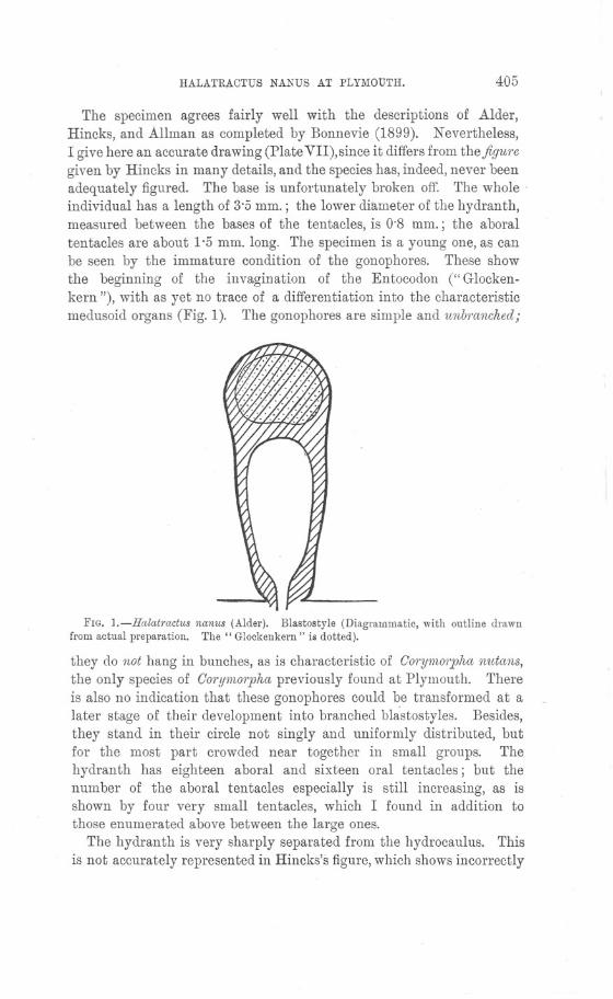
HALATRACTUS NANUS AT PLYMOUTH. 405
The specimen agrees fairly well with the descriptions of Alder,Hincks, and Allman as completed by Bonnevie (1899). Nevertheless,I give here an accurate drawing (Plate VII), since it differs from thefiguj"cgiven by Hincks in many details, and the species has, indeed, never beenadequately figured. The base is unfortunately broken off. The whole'individual has a length of 3'5 mm.; the lower diameter of the hydranth,measured between the bases of the tentacles, is 0'8 mm.; the aboraltentacles are about 1'5 mm. long. The specimen is a young one, as canbe seen by the immature condition of the gonophores. These showthe beginning of the invagination of the Entocodon (" Glocken-kern "), with as yet no trace of a differentiation into the characteristicmedusoid organs (Fig. 1). The gonophores are simple and unbj"anchedj
FIG. 1.-Halatractus nanus (Alder). Blastostyle (Diagrammatic, with outline drawnfrom actual preparation. The" Glockenkern" is dotted).
they do not hang in bunches, as is characteristic of GorYl1LOrphanutans,the only species of GorYl1tOrphapreviously found at Plymouth. Thereis also no indication that these gonophores could be transformed at alater stage of their development into branched blistostyles. Besides,they stand in their circle not singly and uniformly distributed, butfor the most part crowded near together in small groups. Thehydranth has eighteen aboral and sixteen oral tentacles; but thenumber of the aboral tentacles especially is still increasing, as isshown by four very small tentacles, which I found in addition tothose enumerated above between the large ones.
The hydranth is very sharply separated from the hydrocaulus. Thisis not accurately represented in Hincks's figure, which shows incorrectly
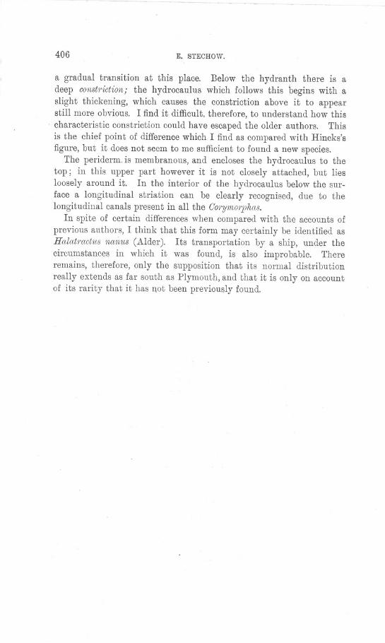
~ ~~~~- ~-~-~ - ~. -~ - - ~-~
406 E. STECHOW.
a gradual transition at this place. Below the hydranth there is adeep constriction; the hydrocaulus which follows this begins with aslight thickening, which causes the constriction above it to appearstill more obvious. I find it difficult, therefore, to understand how this
. characteristic constriction could have escaped the older authors. Thisis the chief point of difference which I find as compared with Hincks'sfigure, but it does not seem to me sufficient to found a new species.
The periderm. is membranous, and encloses the hydrocaulus to thetop; in this upper part however it is not closely attached, but liesloosely around it. In the interior of the hydrocaulus below the sur-face a longitudinal striation can be clearly recognised, due to thelongitudinal canals present in all the C'orymorphas.
In spite of certain differences when compared with the accounts ofprevious authors, I think that this form may certainly be identified asHalatmctus na1ius (Alder). Its transportation by a ship, under thecircumstances in which it was found, is also improbable. Thereremains, therefore, only the supposition that its normal distributionreally extends as far south as Plymouth, and that it is only on accountof its rarity that it has l10t been previously found.
JOURN.MAR.81OL,Assoc. Val. IX PLATE VII
HALATHACTU8 (COnYMOllPHA) NANUfi (ALDER).

r [ 407 ]
List of Nemerteans collected in the Neighbourhood ofPlymouth from May-September, 1910.
. ByDr. Gerarda Wynhoff.
With Figure 1 in the Text.
THE following list is one of the results of my work during a stay ofnearly four months at the Plymouth Laboratory of the Marine BiologicalAssociation. I desire to express my thanks ~o the Director and theStaff of the Laboratory for the interest they took in my work and fortheir kind assistance.
A few of the Nemerteans, recorded in the list of the Ply-mouth Marine Invertebrate Fauna, published in 1904, were notfound. F.or example, Oephalothrix linearis, which was dredged by Richesoutside the Breakwater, has never been found again, notwithstanding allthe trouble taken in searching for it. Amphiporus biomdatusJMcIntosh"known from one specimen only from a dredging in Millbay Ohannel, Ihave not seen, nor the two new species of the genus Oerstedia describedby Riches as O. nig1'a and O. immutabilis. To the forms missing in my'list I must add Malacobdellagrossa,Baseodiscuscurtus, Oxypolia beaumon-tiana, Mim'ella 1'ufa, and Cerebratuluspantherinus. With the exceptionof the two Oe1'stediaspecies each of the above species has been found insingle specimens on rare occasions. Instead of these nine species,however, I can add nineteen which have not yet been recorded fromPlymouth; the greater part of these are new to England or the AtlanticOcean, and four are new species.
As to the system accepted in this list,'I have followed neither Burgernor Coe. Bergendal's investigations on Carinoma and my own studies onthe family Oephalotrichidae resulted in the breaking up of Burger'sordo MesOnetne1'tini. Bergendal's work has revealed many peculiarities,in Ca1'inoma, which prove its near relationship to the Tubulanidae.Moreover certain anatomical features remind us very much of the.Heteronemerteans. The Cephalot1'ichidaeare quite typical Protonem,er-teans, but do 'not possess any character suggesting an intermediate.position between the other members of this group and the Metanemer-teems. Both genera have therefore been replaced in Burger's ordo P1'O-toneme1'tini, which means the restoration of Hubrecht's Palaeonemertini.
Certainly Hetero- and Palaeonemertini are much more closely related:.
408 G. WYNHOFF.
to each other than the Metaneme1.teans with either of them. The
position of the mouth behind the brain, the structure of the alimentarycanal, the tendency to a complicated structure of the body wall, the.development of an outer longitudinal muscle layer, the presence of aninner circular muscle layer of which traces may be found at least inevery family, the structure and position of the cerebral organs and theposition of the nervous system, which only secondarily migrates throughthe outer circular muscle layer, together with the absence of anystructure in the proboscis, which reminds us of its peculiar complicationin the ordo Metaneme1'teans,are so many points of resemblance, thatI do not hesitate to join Palaeonemertini and Heteronemertini togetherin one order, the ordo .Anopla of Max Schnltze.
Burger's Metanente1'tini= Hubrecht's Hoplonemertini constitute mysecond order, for which Max Schultze's name Enopla ought to berestored. They are characterised by the situation of the mouth infront of the brain, the presence of an oesophageal diverticulum, thestructure of the body-wall, the peculiar structure or the proboscis andthe presence of stylets, the position of the nervous system in theparenchyma of the body, the situation and structure of the cerebralprgans, the uniformly built vascular system.
PalaeonemC1.tiniand HeteronemC1.tinihave been reduced to the rank
of suborders. The nomenclature of "Das Tierreich " has been adopted.As iconographical works I have everywhere referred to:
r 1. BU1'gC1',O. Die Nemertinen des Golfes von Neapel. Fauna undFlora. Monogr. 22, 1895.
2. Joubin, L. Les Nemertiens. Faune fral}(;aise par R. Blanchardet J. de Guerne. Paris, 1894.
3. McIntosh. A Monograph of British Annelids. Part 1. Nemer-teans. Ray Society, London, 1873-1874.
ORDO 1. ANOPLA.
In addition to the outer circular and the inner longitudinal musclelayers an outer longitudinal and an inner circular muscle layermay be present. The nervous system is situated in the. body wall.Mouth always behind the brain. No diverticulum of the oesophagus.No stylets and no peculiarly built proboscis.
SUBORDO1. P ALAEONEMERTINI.
Lateral nerves and brain nearly always situated outside the muscularbody-wall. The body-wall consists of an epithelium, a circular musclelayer and an inner longitudinal muscle layer; the inner circular musclelayer may be absent.
PLYMOUTH NEIvIEHTEANS. 409
I. FAIvI.TUBULANIDAE, Biirger.
Oerebral organs nearly always present; the cephalic furrows merelyconsist of epithelial grooves. Often with lateral sense-organs. Nervoussystem situated between the epithelium and the basal membrane orbetween the basal membrane and the outer circular muscle layer. Theepithelium is very thick and contains many clusters of secretory cells.Inner circular muscle layer present. A median dorsal blood-vesselis not present.
GENUSTubulanus.
The nervous system is situated between the basal membrane and themuscular body-wall. Oerebral organs present.
1. Tubulanus polymorphus (Ren.). (Biirger. Monogr. Tat I, Fig. 4;Joubin. Les Nemertiens, PI. I, Fig. 8 et 9.)
Locality: Eddystone and Rame-Eddystone Grounds; once off theBreakwater.
. A kw specimens of this N emertean were always dredged in thedeep water near the Eddystone or on the Rame-Eddystone Grounds, ata depth of 45 m. or more. No external markings are present; thebody has a uniform orange colour and is rather soft. The head isseparated from the body; it is much broader and rounded. Lateralorgans are present.
Geographical distribution: Norway, England, France (both coasts),Mediterranean coasts.
2. Tubulanus linearis (McIntosh). (Biirger. Monogr. Taf. I, Fig. 2.)Locality: Several specimens known from different localities inside
the Breakwater (Queen's Grounds, Asia Shoal, Millbay Pit, DukeRock). Shallow-water form.
The head and oesophageal region of this Nemertean are milk-white;the other part has an orange-brown tint. This is due to the intestine.The head is very flat, and in the living animal very often makes aburrowing movemeilt. Notwithstanding the perfect transparency ofthis part I have not been able to distinguish the cerebral organs orcephalic slits. The rhynchodaeum is conspicuous as a milk-white linein front of the brain commissures, which are very long; the brain lobesare but small and do not approach each other at alL The proboscissheath does not extend into the hinder part of the body; it is, however,present at the beginning of the intestinal region.
Geographical distribution: Southern coast of England, Wimereux,Naples.
NEW SERIES.-VOL. IX. No.3. JUNE, 1912. 2 D
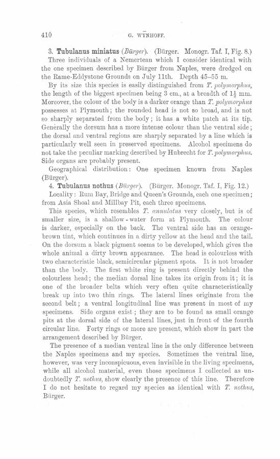
410-
G. WYNHOFF.
3. Tubulanus miniatus. (Burge?'). (Burger. Monogr. Taf. I, Fig. 8.)Three individuals of a Nemertean which I consider identical with
the one specimen described by Burger from Naples, were dredged onthe Rame-Eddystone Grounds on July 11th. Depth 45-55 m.
By its size this species is easily distinguished from T. poly'm01jJhus,the length of the biggest specimen being 3 cm., at a breadth of It mm.Moreover, the colour of the body is a darker orange than T. POlYl1W?lJhuspossesses at Plymouth; the rounded head is not so broad, and is notso sharply separated from the body j it has a white patch at its tip.Generally the dorsum has a more intense colour than the ventral side;the dorsal and ventral regions are sharply separated by a line which isparticularly well seen in preserved specimens. Alcohol specimens donot take the peculiar marking described by Hubrecht for T. polym01jJhus.Side organs are probably present.
Geographical distribution: One specimen known from Naples(Biirger).
4. Tubulanus nothus (Burger). (Biirger. Monogr. Taf. I, Fig. 12.)Locality: Rum Bay, Bridge and Queen's Grounds, each one specimen j
from Asia Shoal and Millbay Pit, each three specimens.This species, which resembles T. annulatus very closely, but is of
smaller size, is a shallow -water form at Plymouth. The colour
is darker, especially on the back. The ventral side has an orange-brown tint, which continues in a dirty yellow at the head and the tail.On the dorsum a black pigment seems to be developed, which gives thewhole animal a dirty brown appearance. The head is colourless withtwo characteristic black, semicircular pigment spots. It is not broaderthan the body. The first white ring is present directly behind thecolourless head; the median dorsal line takes its origin from it j it isone of the broader belts which very often q l1ite characteristicallybreak up into two thin rings. The lateral lines originate from thesecond belt j a ventral longitudinal line was present in most of myspecimens. Side organs exist; they are to be found as small orangepits at the dorsal side of the lateral lines, just in front of the fourthcircular line. Forty rings or more are present, which show in part thearrangement described by Burger.
The presence of a median ventral line is the only difference betweenthe Naples specimens and my species. Sometimes the ventral line,however, was very inconspicuous, even invisible in the living specimens,while all alcohol material, even those specimens I colleeted as un-doubtedly T. nothus, show clearly the presence of this line. ThereforeI do not hesitate to regard my species as identical with T. nothus,Burger.
PLYMOUTH NEMERTEANS. 411
'Geographical distribution: It has only been described from Naplesby Burger.
5. Tubulanus superbus (Kol/). (Burger. Monogr. Taf. I, FIgs. 5,7, 9, 11.) ,
Locality: On the Eddystone and Rame-Eddystone Grounds; oftenmet with. One specimen from Asia Shoal.
This Nemertean seems to be common on all grounds outside theBreakwater at a depth of 45 m. or more. A small number ofindividuals has been recorded from more shallow water; both Punnettand Beaumont found it in a sandbank in the river Yealm. J oubin and
Burger give a depth of 30-80 J.p..,which agrees with the ordinaryhabitat of this worm at Plymouth. The colour of this big Nemerteanis a reddish brown. There are four longitudinal white lines and agreat number of white rings. The head is broader than the body.Pigment spots are not to be seen. . The median dorsal line continuesto the tip of the snout, which itself is surrounded by white lines. Thecephalic grooves are situated at a short distance behind the first trans-verse dorsal line. Side organs are present. .
Geographical distribution: Mediterranean coasts, Sweden, GreatBritain, French coasts of the Ohannel.
6. Tubulanus annulatus (Mont.). (Joubin. Les Nemertiens, PI. I,Fig. 2 et 3. McIntosh. Monogr. Pt. I, PI. 8, Fig. 1.)
Locality: This Nemertean has been recorded from nearly all dredg-ing grounds inside and outside the Breakwater; more commonly it is metwith near the Mewstone and the EMystone, at a depth of at least 18 m.
T. annulatus can easily be distinguished from T. supe1"b1lsby theabsence of a median ventral line, its much smaller size and the brightorange-red colour. The white median dorsal line continues on thehead and bifurcates at the tip, enclosing a patch of the same colour asthe body. The head is coloured both on its dorsal and ventral side;the lateral lines originate from the first white belt, which is situatedat a short distance behind the 'cephalic grooves and just behind themouth. The cephalic grooves reveal exactly the same features as inOarinella aragoi, Joubin. "Hs occupent Ie fond du cou, et sont, non encreux, mais en relief; ils sont teintes en jaune. Sur la face ventrale;ils commencent audessus de la bouche, de chague cote, mais sans serejoindre sur la ligne mediane. Hs sont, dans toute cette portion, a.peu pres droits. Arrives sur Ie bord lateral, dans Ie prolongementdes lignes blanches dont il a ete que;:;tion plus haut, on apergoit unpetit orifice. Au dela, ils passent sur la face dorsale et forment .dessinuosites tout a fait caracteristiques'. La terminaison des deux sillons
412 G. WYNHOFF.
forme deux lignes separees par la bande blanche dorsale" (J oubin.1890. , "Archives de Zool.exper. Ser. II, T. VIII, p. 495).
As far as I 9an judge from the description and the figures given,'T. 'anmdatus, Montagu, which is doubtless the same species which Ifound at Plymouth, agrees completely with O.amgoi, Joubin, in externalfeatures. However, we cannot at present consider the species identical,as their anatomy is not at all the same. This seems to be a case likethe one described by Bergendal for ,Oarinella linearis and O. groen-lanclica. Neither do the Plymouth specimens agree with the descrip-tion and figures of Burger's species in the Naples monograph.
O. anmdata, Bergenclal,however, seems to exhibit the same internalstructure as the Plymouth specimen; but this species differs externallyfrom O. aragoi as Bergendal states. This complicates the question stillmore. Only the etudy of sections of all these Nemerteans can throwmore light on this interesting question. At any rate there can belittle doubt as to the identity of T. annulat1ls (M~ont.) with theNemertine described above.
7. Tubulanus albocapitatus. Nov. spec.Locality: Rame-Eddystone Grounds.
In 3 different dredgings, each time one specimen of thissmall Nemertean, which can easily be mistaken for a young
T. ann1!'latus,was collected. On closer inspection it differsconsiderably from that form. A complete individual had alength of 1t cm. and was t mm. broad in the oesophagealregion. The head is not sharply separated from the body,nor is it much broader. Pigment spots are absent. Theperfectly white head is followed by a brown-red belt ofthe same breadth, which is the darkest part of the wholebody. A yellow pigment is distributed all over thisregion. The cephalic furrows are situated in this ring;
TFbig
l' 1. the y are rather deep and reach half-way to the dor-u u auusalbocapitatus. sum; they are not lined with white or yellow, as is
. the case in T. annulldus, and are quite differently
shaped. The first circular white line separates this region from thebody. The median dorsal line, which is white too, passes through thisbelt into the dark region described above, but does not reach the whitehead. At both sides of this median longitudinal line, separated fromit by a translucent region, a reddish-brown stripe of a fainter tint ispresent. These stripes do not even reach the lateral lines, as a trans-parent region is developed between them too; the white pigmentpatches, which are dispersed at the sides, constitute a very incon-spicuous and incomplete lateral line. The ventral side is also trans-
~~~
: I~
: \~

PLY1\WUTH NEMERTEANS. 413
parent. Eleven white belts are present; the first constituting theseparation between the intense coloured and the oesophageal region.Just in front of the second ring one pair of white patches is con-spicuous; they are connected with this belt and are situated in thedorsal red stripes. I have not found any trace of side organs.
II. FAM. CALLINERIDAE.
Cerebral organs absent. Nervous system situated between the basalmembrane and the outer circular muscle layer. A thick nervous layeris present in the head. Inner circular muscle layer present. Amedian dorsal blood-ve.ssel fails; four blood-vessels in the head.RhynchodaeuI? with a separate layer of longitudinal muscle fibres.Proboscis anteriorly, with four longitudinal muscle bundles, followedby a diaphragm.
GENUS Carinesta.
No composite gland cells in the epithelium. Oesophageal nervespaired; no lateral sense organs. Diagonal muscle layer absent. Pro-boscis attached behind the mouth. Proboscis sheath without anypeculiarities of musculature.
1. Carinesta anglica. Nov. spec..
Locality: one specimen was collected from the river Yealm at lowtide by digging in a muddy sandbank; and one fragment crawlingabout in sand from Whitsand Bay.
This white nemertine somewhat resembled Punnett's description ofOxypolia beawnontiana. The snout is elongated and pointed; whencontracted the wrinkling is obvious. Colour a watery milk-whiteanteriorly; the gonads give the posterior part a rosy-brown tint. Themouth had been protruded obviously in the fragment from Whits andBay. No sense organs are present, nor could I distinguish the 'pro-boscis pore. The .proboscis itself and the intestinum are easily visiblebecause of the transparency of the animal.
The posterior part of the body was much swollen and contracted atirregular intervals when the animal was first examined; on touchingit broke into pieces.
Sectioning revealed the very interesting structure of this animal,which I was able to identify as a C((1'inestaspecies. From Punnett'stype specimen, however, which was collected at New Britain, it differsin the total absence of that part of the inner circular muscle layer,which is already disappearing in C. O1'ientalis. Moreover, a head glandexists in C. nngl.icnand fails in Punnett's specimens. I could not findany traces of a dorsal decussation of fibres, nor is there any speciallongitudinal muscle sheath at the ventral side of the oesophagus.
414 G. WYNHOFF.
III. FAM.OEPHALOTRICHIDAE.
Bodywall composed of an epithelium in which a separate layer ofgland-cells does not exist, the basal membrane, the' outer circular andthe inner longitudinal muscle layer. The inner circular muscle layeroften fails. The nervous system is situated in the middle of the innerlongitudinal muscle layer. Sense organs are. not developed. Fourthick cephalic nerves. Vascular system consisting of a pair of lateralblood-vessels only. A great number of nephridial organs.
GENUSCephalothrix.
Mouth widely separated from the brain. Body thread-like. Nosense organs. The oesophageal nerves are not paired. No bifurcationof the continuation of the dorsal brain lobes. An inner circular
muscle layer fails. The genital sacs are not developed in theoesophageal region.
1. Cephalothrix rufifrons (Johnston). (Burger. Monogr. Tat II,Fig. 24.)
, Locality: Oommon between tidemarks at Rum Bay, Mount Edgcumbe,Drake's Island, the Mewstone; in clean sand, between corallines,under stones, etc. Very thin, colourless or white species, with twosmall red or orange spots on the tip of the head. Length, 30-40 mm.;breadth, ~ mm.
Geographical distribution: Ooasts of Norway, Denmark, GreatBritain, France, and Italy.
SUBORDOII. HETERONEMER'TINI.
The body-wall is composed of the epithelium, cutis, outer longitudinal,outer circular and inner longitudinal muscle layers. The nervoussystem is situated between these muscle layers. The lateral nervesare always found outside the circular muscle layer.
I. FAM. BASEODISOIDAE (= Eupolidae, Bergendal).
Proboscis pore near the tip of the snout. The inner muscular layersare not developed in the head. Oerebral organs large, close to thebrain. A thick layer of connective tissue separates the epitheliumwith its gland cells from the outer longitudinal muscle layer. Probosciswith two layers, an outer circular and an inner longitudinal. Pro-boscis without muscular crosses. The proboscis sheath is short. Headmore or less rounded; usually with eyes.
GENUSPoliopsis.Head sharply separated from the body by a deep furrow. A dorsal
and a ventral median longitudinal cephalic furrow are present.
PLYMOUTH NEMERTEANS. 415
1. P.lacazei, Jo'ubin. (J oubin. Les N ernertitms. PI. I, Fig. 15 et 16.). Locality: One specimen frOnl the Eddystone.
On the 26th of May I got this single specimen, together with TubulanussupeTbus and T. polymoTphus, from the Eddystone Grounds. As theposition of the eyes, the bright pink colour and the two cephalic slitsagreed perfectly with J oubin's description, I did not hesitate toidentify my specimen as P.' lacazei. The internal structure proved thecorrectness of this identification.
Geographical distribution: J oubin found this species at Banyuls,Burger at Mauritius, Halleznear Calais.
ILFAM. LINEIDAE.
. Usually with a pair of deep horizontal cephalic furrows. Probosciswith three muscular coats and mostly with two muscular crosses.Cephalic gland consisting of very slender tubes, situated anterior to thebrain. "
SUB-FAMILY A. LINEINAE.
'Without caudal cirrus. Proboscis sheath usually much shorter thanthe body. .
. GENUS Lineus.
Body rounded or flattened, unusually long, very contractile. Headmostly somewhat broadened and spathulate. The worms are not ableto swim; as a rule they coil themselves up and make knots. Ocellipresent in most species. Proboscis sheath short.
1. Lineus longissimus (Gunn.) (McIntosh. Monogr. Pi. IX.)Locality: Rather common in dredgings from the Sound, Mewstone,
and the Yealm.
The colour is a blackish brown relieved throughout by the finepurplish" iridescence of the cilia." With darker and lighter stripeson the head and part of the body. Eyes numerous, arranged in amarginal row on each side of the head, which is somewhat broadenedand spathulate, not separated from the body.
Geographical distribution: Atlantic and Baltic coasts of Europe.
2. Lineus bilineatus (Ren.). (Joubin. Les Nemertiens, Pi. II, Fig.26 et 27.). Locality: Very common, especially in dredgings from the Sound;from the Rame-Eddystone Grounds, the Mewstone neighbourhood, butalso from the Cattewater and between tidemarks at the Yealm and
Rum Bay.Colour a pale brown, sometimes with a reddish shade, or nearly
white. With a pair of longitudinal white streaks, passing on from the
416 G. WYNHOFF.
tail to the tip of the head, where they usually join together in abroader white patch. Head ending bluntly, somewhat spathulate.Eyes wanting.
Geographical distribution : Sweden, Great Britain, France, Italy,Madeira.
3. Lineus lacteus (H. Rathke). (Joubin. Les Nemertiens. PI. II,Fig. 23; and McIntosh, Monogr. PI. V, Fig. 3.)
Locality: Two specimens from Asia Shoal, on one occasion.Body slender, threadlike, white, anteriorly pinki~h. Head broadened,
spathulate, not distinctly separated from the body. Eyes about fifteen,arranged in a row on each side of the snout. Mouth removed from thebrain. Length 100 mill., breadth 1 mm.
Geographical distribution: The Channel, Sweden (?), Mediterranean,and Pontic coasts.
4. Lineus ruber (Mull.). (McIntosh. Monogr. PI. V, Fig. 2.)Locality: Between tidemarks very common in Cawsand and Rum
Bay, at Drake's Island; from New Grounds in dredgings.Body flattened, 3 mm. broad, diminishing towards the tail. Colour
usually bright red. Head spathulate, separated from the body. Withfour to twelve eyes. Length 150-200 mm.
Geographical distribution: Coasts of the Northern Atlantic, withBaltic, Mediterranean, and Madeira coasts.
GENUSEuborlasia.
Body very thick, biconvex in section and usually not flattenedventrally. Lateral margins absent after preservation. Head notseparated from the body. In animals not perfectly extended theposterior part of the body is much thicker (3-6 times) than the anteriorregion. Proboscis sheath short. Without eyes.
1. Euborlasia spec.To this genus I refer two fragments of Nemerteans, which were
collected from a sandbank in the river Yealm. As in both fragmentsthe anterior portion of the body is wanting, I have not been able toidentify the species, or even the genus with certainty. One fragmentcame living into my possession. It had been dug up in the sandbank,high up the river ¥ealm,at a place where the water is nearly fresh.A great deal of sand has been removed from this sandbank during thelast ten years, and it is now very muddy. This Nemertean, which wasformerly rather common at this place, has since become quite rare.The other fragments, which also lack a head, were collected at thesame place in 1898. After preservation the body is deeply furrowed
PLYMOUTH NEMERTEANS. 417
and wrinkled, much as in CerebratuZuscorrllgatus. The anterior part,of my specimen got very thin by preservation (breadth 5 mm.); thefollowing region suddenly broadens till it has a breadth of 16 mm. Theliving specimen had a brownish colour, with more whitish margins.No markings were present. .
The sections revealed some features which decided my classificationof this species as a E1lborZasia,. I regard it, however, as a separatespecies from both E. eZisabethaeand il1tmacnZata. '
The proboscis sheath is very short, as already in the thinner portionof the body it is devoid of its muscular coats. The intestinal caecaare richly developed and branched. The gonads are placed in rowsbetween the caeca, alternating with their diverticula. They all opendorsally. Muscular septa, are entirely absent. The intestinum withits diverticula and the gonads are surrounded by a, thick layer ofconnective tissue, in which a great number of very narrow blood-vesselsare imbedded. The structure of the epithelium agrees perfectly withBurger's description; the large gland cells are especially conspicuous(Burger. Monograph Naples, T. 22, Fig. 39).
SUB-FAMILYB. MIORURINAE.
Oaudal cirrus present; proboscis sheath usually extends nearly orquite to the posterior end of the body.
GENUSMicrura.
Small, thin, mostly flattened and soft nemertines; head spathulate,ending bluntly, not separated from the body. Latm:al margins of thebody not ren}arkably thin; incapable of swimming. Often with agreat many eyes. The mouth is circular. Proboscis sheath short.
1. Micrura fasciolata (Ehrbg.). (McIntosh. Monogr. PI. VI,Fig. 2.) .
Locality: Oommon in the dredgillgs from the Sound, Eddystone,Mewstone, and from the Rame-Eddystone Grounds.
General colour red or red-brown, head and ventral side white.10-24 slender white transverse stripes across the dorsum. With 4-6small eyes. Length 120-200 mm., breadth 1-2 111m.
Geographical distribution: Sweden, Great Britain, Northern coastof France, Mediterranean.
2. Micrura aurantiaca (Grube). (McIntosh: Monogr. PI. VII,Fig. 4.)
Among some tubes of preserved nemertines I found one containingtwo nemertines from the Breakwater. They were supposed to belong
418 G: WYNHOFF.
to the same species, and must therefore have been very much alike inthe living state. After preservation, however, the difference was veryobvious, the one being 'rather flattened with lateral margins and theother being circular anteriorly and showing no margins even at themore futttened and weaker posterior part of the body. The colour ofthe more rounded nemertine' was moreover brownish, while the brightyellowish colour of the flattened specimen at once proved it to be asmall specimenof Cerebratulusroseus. So I suppose the colour of theother specimen to have been' a very faint red or rose. The internalstructure, however, makes it quite certain that this specimen must havebeen an abnormally faint colomed and rather large individual ofMicrum a1wantiaca. All traces of the 'original colour or of anymarkings had disappeared. Eyes are absent. The cutis is as thick asthe epithelium. There are no traces Qf a connective tissue layerseparating the cutis from the external longitudinal muscle layer, whichis much thicker than the circular or inner longitudinal muscle layer.The latter layer has ,been very much reduced. The cerebral organs areplaced above the lateral nerves. The dorsal part of the dorsal brainlobe is situated laterally to the ventral part; it ends anteriorly to thecerebral organs. The cephalic slits end where the cerebral canaloriginates from them. They reach as deep as the brain. The mouthis situated beneath the hinder end of the cerebral organs.
Geographical distribution: Both coasts of France and the Mediter-ranean.
3. Micrura purpurea (Dalyell). (McIntosh. Monogr. PI. VII,Fig. 3.)
Locality: Rather common in dredgings from the Sound; occasionallya specimen from the Rame-Eddystone and the Mewstone AmphioxusGrounds.
Colour brown; head white, with a bright yellow transverse bar;eyes absent. Length 100-200 mm., breadth 2-3 mm.
Geographical distribution: Sweden, Great Britain, North coast ofFrance, and Mediterranean.
4. Micrura candida, BU1'ger( = Mic1'ura lactea, Hubrecht). (J oubin.Les Nemertiens, T. 2, Fig. 23 bis.). Locality: Two specimens from the Mewstone and two from theMewstone Ledge.
Perfectly white. Eyes absent.Geographical distribution: Channel and Mediterranean.
. Two specimens are known from England, one from Stoke Point nearPlymouth, and one from Halfway Rock.
PLYMOUTH NEMERTEANS. 419
GENUSCerebratulus.
Body usually long and ribbon-like, much flattened, witl:t very thinlateral margins; well adapted for swimming. Head pointed, lancet-like. Eyes are usually absent. Mouth mostly a long slit.
1. Cerebratulus fuscus (},1~cIntosh). (McIntosh. Monogr. PI. VI,Fig. 3.)
Locality: In dredgings from Asia Shoal, Queen's and New Grounds,Millbay Ohannel, and once from Mewstone Ledge.
General colour usually pale yellow, only pigmented on the head;sometimes, however, especially in bigger worms, a brown pigment wasdistributed all over the dorsum. B'rain transparent. No eyes. Headspathulate, not separated from the body.
Geographical distribution: Sweden, Great Britain, France, Portugal,Mediterranean.
2. Cerebratulus roseus (Ohio/e). (Burger. Monogr. Taf. VI.,Fig. 12.
Locality: Specimens were collected from the Breakwater, May, 1910 ;from Salcombe (Millbay) by Mr. Potts, in 1908; from the sand underBatten Oastle, May, 1902; each time one complete specimen. A fourthf\pecimen, collected from the Breakwater, was found in a tube of pre-served specimens with Micrnra an1'antiacci.
The colour of the only living specimen I have seen was a dirty fleshcolour; this individual was a female, which whilst being brought inbroke into several pieces; however, no parts, not even the appendix,had been lost. The uncoloured margins were very conspicuous. Thecentre of the body probably took its darker colour from the eggs, whichhad an orange-brown tint and were partly deposited through the genitalpores, situated in two rows on the dorsum. The mouth is large; thecephalic slits even reach the region of the mouth. The preservedspecimens show the bright yellowish tint characteristic of Oerebratnlusroseus. Burger's figure moreover leaves no doubt as to the identity ofmy specimen with his C. roseus.
Geographical distribution: The French coast of the Ohannel and theMediterranean.
3. Cerebratulus alleni. Nov. spec.Locality: Yealm sandbank. .
This N emertine was collected on the 7th of November, 1907. Thesingle specimen was given to me last summer. It had been preservedin corrosive sublimate and was then in alcohol. The only descriptionI could get of the living animal was: colour, light flesh with white
420-
G. WYNHOFF.
snout. It was supposed to belong to the same species as the individualdescribed as Mic1'1l1'aaurant1:acaand a small specimen of Ge1'ebratulus1'oseus. From both, however, it differs in the shape of the head, whichis conspicuously swollen at its hinder part and not separated from the.body, so that it is best described as fig- or pear-like, The colour isa greyish brown ~nteriorly, which is replaced by a yellowish grey in thegreater part of the body, These two colours are sharply separatedfrom each other, just as in Tubulan1(s polymorphus. Sections revealedthe following peculiarities, which made me create a new species for thissingle individual.
The cephalic slits are rather long, extending nearly to the beginningof the mouth, and farther than the' cerebral organs themselves. Theyare not very deep, and reach only half-way to the brain. The cerebralpit, however, is deeper. The cerebral canal originates behind thedorsal brain lobe; this bifurcates into two lobes, the dorsal of whichends quite free in the longitudinal musculature in the region of thecerebral canal, separated from the ventral16be by a thick band of tissue,The ventral lobe continues directly as the cerebral organ. This isnever in contact with any blood-vessel, but lies internally to thecircular muscle layer. The dorsal lobe of the dorsal brain, the cerebralorgan, and the ventral brain lobe are situated above each other. Theblood-vessels form one large dorsal anastomose in the head; the veryshort cephalic blood-vessels unite again within the brain; from thisventral anastomose an unpaired oesophageal blood-vessel takes itsorigin. This unpaired vessel divides into two in the region of theoesophageal nerve commissure. These two communicate often withthe lateral blood-vessels situated on both sides of the proboscis sheath.In this region large gland cells are also conspicuous. They are buccalglands, which are enormously developed all round the mouth and theoesophagus. They are placed in clusters, and freely protrude into theblood-vessels, both into the lateral and into the oesophageal vessels,which frequently communicate with each other and form a network allround the oesophagus. They are even more richly developed than inMicrura alaskensis, Goe; they do not, however, extend outside thecircular musculature.
The epithelium is not very thick. A separate cutis layer cannot be:distinguished. The cutis glands are situated in the outer longitudinalmuscle layer. A small layer of longitudinal muscle fibres, as thick asthe epithelium itself, and traversed by the gland ducts, separates the-layer of glands from the epithelium. These are themselves sur-rounded by muscle fibres, and do not form a compact layer. They aresome two or three times as high as the epithelium. The bases of the
PLYMOUTH NEMERTEANS. 421
cutis glands are situated not quite half-way between the outer bodywalland the circular muscle layer, laterally at a third of that distance only.The circular muscle layer is separated from the outer longitudinal bya thick nervous layer. Diagonal muscle fibres are absent. The circularmuscle layer is rather feebly developed, and has twice the thickness ofthe epithelium, while the inner longitudinal muscle layer is twice tothree times as thick as the circular layer in the oesophageal region.Longitudinal muscle fibres are not present between the oesophagus andprcboscis sheath.
The gonads alternate regularly with the intestinal caeca; the genitalpores are situated above the proboscis sheath. In the intestinal regionthe outer longitudinal and circular muscle layers are very muchreduced. This is especially the case with the outer longitudinal musclefibres which form a layer of the same thickness as the cutis glandsdorsally and ventrally, so that the latter reach the circular musclelayer; laterally, however, the layer of longitudinal fibres is thicker.The circular muscle layer is as thick as the epithelium, but the innerlongitudinal muscle layer, which is five to six times as thick as thecircular, has the same breadth as in the oesophageal region.
On the 1st of June part of a Nemertine was collected in dredgingmaterials from the New Grounds. It turned out to be the posteriorpart of a very thin (1 mm.) nemertine, completely white, and long incomparison with the uniform breadth. The head failed, but an appen-dix was present. The structure of the body-wall reveals the charac-teristics of a Heteronemertean, as the outer longitudinal muscle layeris present. As with all the layers of the body-wall, this longitudinalmuscle coat is very much reduced in size when compared with othernemertines. There are no traces of a cutis nor of any clusters ofcomposite gland cells. Epithelium and outer longitudinal muscularcoat have about the same breadth and are separated by the thin base-ment membrane only. The epithelial cells constitute a single layer.The circular and inner longitudinal muscle layers together have butone-third of the thickness of the outer muscular coat. They haveabout the same breadth. The longitudinal muscle fibres surround thewide enteron and the proboscis sheath, which ends just before thepoint where the tail is attached. The intestinal pouches are veryshallow, and I have not been able to discern any septa. The gonadsalternate with the intestinal pouches, or perhaps they are the cause ofthe appearance of these unreal diverticula. The position of the lateralnerve cords is the usual lateral situation. I have not been able todetect a median dorsal blood-vessel.
422-
G. WYNHOFF.
. The point of greatest interest is the structure of the tail; whichdiffers widely from any structure of this kind yet described. Theepithelium has about the same height as in the body; many glandcells are present. I have not found any traces of the outer longitudinalmuscle layer. Circular and inner longitudinal muscle coat are presentas a single layer of muscle fibres. In the tail itself I have not beenable to trace the nervous system; from the posterior commissureof the body, however, nervous tissue is seen to reach the basis of thetail. The centre of the tail is occupied by the intestine, which isseen to communicate widely with the intestine of the body and opensto the exterior by the anal aperture at the tip of the appendix.There are no intestinal pouches, nor gonads nor rhynchocoelom in thetail, which moreover lacks all connective tissue. Even the basementmembrane could not be distinguished. If any nervous tissue is presentit must be still epithelial.. As to the vascular system, I have not been able to find the analanastomose nor anything like vessels in the appendix.
Certainly there can be no doubt that this specimen belongs to thesub-family Micrurinae. Had it not been for the tail, I should havefelt inclined to identify the fragment as Lineus niveus, Punnett, whichlives at the much greater depth of 100-140 m. near Bergen in Norway.As, however, Punnett describes the total absence of a tail in half adozen specimens he collected, and as I can only judge their relationshipby the hinder part of the body, I do not feel juetified in consideringthem identical. If my supposition is right, I am inclined to thinkthat a new genus ought to be created, on account of the structure ofthe appendix and the very primitive features which Lineus niveusreveals. The presence of an appendix certainly would approximateLine~ts niveus still closer to Zygeupolia and MicTella.
ORDO II. ENOPLA.
The body-wall consists of a single-layered epithelium, a basalmem-brane, the outer circular and. the inner longitudinal muscle layer.Mouth anterior to the brain, or the oesophagus opens into the rhyn-chodaeum. An oesophageal diverticulum is present. The nervoussystem is not situated in the body-wall, but in the central connectivetissue.
SUBORDO A. PRORHYNCHOCOELOMIA.
Worms with a very long and slender body; they like to coil them-selves up and to form knots. The proboscis is much shorter than thebody. The proboscis sheath never extends into the posterior third.
PLYMOUTH NEMERTEANS. 423
part of the body, and exists usually in the anterior third only.N eurochords or neurochord cells fail.
1. FAM. EMPLECTONEMATIDAE.
Mostly very long aIid flat forms. Proboscis short and rather stout,with one very differently shaped central stylet only. Very often withmany small eyes; seldom two or four eyes. No statocysts.
GENUSEmplectonema.
\Tery long and slender. Alimentary tract and proboscis open together.Oerebral organs very small, generally situated a long distance in frontof the brain. Mostly with many small eyes. The proboscis sheathdoes not quite extend to the second third of the body. Sexes separate.
. I1. Emplectonema gracile (Johnst.). (McIntosh. Monogr., PI. II,
Fig. 5.) I
Locality: In dredgings from the lVIewstone; between pdemarks atBreakwater, Drake's Island, Oawsand Bay. Far less common than the. Inext speCles. I
The colour of this Nemertean is usually a more greyish green thanthe figure in McIntosh's monograph indicates; ventral surface white.The head is lined with white and possesses a faint yellow transversebar. It is broader than the body, but not sharply separated. Oere-bral organs a long distance in front of the brain. 20-30 eyes in twogroups at both sides of the head. The handle of the central stylet istwice the length of the stylet itself; all stylets are curved. With twopouches each containing 5-6 accessory stylets. Length 20 cm. or !TI°re,breadth 1 mm.
Geographical distribution: This species is widely distributed. It isknown to occur on the West coast of. North America as well as in
Ohile and the Aleutian Islands, the coasts of France. and Germany,Madeira and the Mediterranean.
2. Emplectonema neesi (Gerst). (McIntosh. Monogr. PI. III, Fig. 6;and Joubin, Les Nemertiens, PI. III, Figs. 77-80.)
Locality: Oommon between tidemarks in the Sound; from Break-water and the Mewstone; occasionally met with in dredgings at adepth of 10-15 fathoms.
This Nemertean is much more abundant than E. gracile. Howeve.r,it is not so widely distributed. E. neesi has been recordedonly fromthe Atlantic coasts of Europe. It ranges from Iceland to the Ohannel.
Head broadened and of a yellowish colour, paler than the body.The dorsum has a brown hue in which flesh-coloured stripes occur;
424 G. WYNHOFF.
sometimes these stripes are broken lip into patches, which give theanimal a speckled appearance. The ventral surface is white. Oerebralorgans at a great distance in front of the brain. Numerous small eyesarranged in four clusters. The oesophageal diverticulum sends twopouches to the brain. Both the central stylet and its basis are shortand stout; they are of the same length. Two pouches, each with threeaccessory stylets. Length to 46 cm., breadth ~cm.
3. Emplectonema echinoderma (Mar.). (Burger. Monogr. T. II,Fig. 3.). Locality: One specimen from Millbay Pit.
Burger's figure gives a very good idea of the Plymouth specimen;'the head shows the same form and markings. The colour is generallya more orange-red. A number of very small eyes, arranged in a row,are situated on the margins of the head; they do not show very much,which is probably due to the want of clearness of the tissues of thehead, and they certainly were not so numerous as those described byBurger. The transparent hooks are quite characteristic. Length120 mm., breadth 1!-2 mm.
Geographical distribution: Mediterranean, Madeira, and Plymouth.
GENUSCarcinonemertes.
. Head' without distinct lateral grooves, not demarcated from thebody. Ocelli 2. Mouth and proboscis open together; oesophagusextremely short, opening broadly into the intestine through a largemuscular chamber. Proboscis but little developed, very small in sizeand '8xtremely short, without lateral pouches of reserve sty lets, butarmed with a central stylet and basis only.
1. Carcinonemertes carcinophila (K6ll). (J oubin. Les N 8mertiens,PI. III, Fig. 81. McIntosh. Monogr. PI. I, Fig. 5.)
This parasitic Nemertean lives in self-secreted tubes between theegg masses of Carcinus ?naenasand Portunus depumtor. Only two outof a great number of Carcini I examined were inhabited by this worm;one specimen in each. On Portunus, however, it was more common,'especially in specimens from dredgings. If the crabs had been collectedon shore I never found them inhabited by this parasite. The tube,in which the orange or pale reddish animal folds itself together, is'attached to the axis of the feet that bear the eggs. I have not beenable to find any nemertine on the gills either of Portunus or of Carcinus'01'Galathea.
. This species was with certainty known only to occur on CarcinusmaenClSj probably Galathea strigosa and Xantho jloridus may also be
PLYMOUTH NEMERTEANS. 425
infected with them.occllatns.
Geographical distribution: a. wrcinophila has not been foundpreviously in England; it has been recorded from the Atlantic coastsof Belgium and France, from the Mediterranean and from NewEngland.
In New England they live on Platyonychus
GENUSNemertopsis.
As E1nplcctone1nc~,but instead of many,. only four eyes, situated in arectangle. The distance between the eyes of one pair is smaller thanthat between the eyes of one side.
1. Nemertopsis flavida (McIntosh), Beat~1nont. (McIntosh. Monogr.PI. IV, Fig. 1; and J oubin, Les N emertiens, PI. II, Fig. 61.)
Locality: Oommon in the Sound, both from dredgings and betweentidemarks. Once from a dredging near the Mewstone.
Under this name I unite both the Nemertopsis species, described byBeaumont (1900, p. 817 and 818). It seems to me very doubtfulwhether .N. tennis must be distinguished from .N. flavida. The onlydifference between the two so-called species is to be found in the,colour, which is quite white in .N. flavida and a very faint pink in.N. tent~is. This, however, might be due to the colour of the blood,which is decidedly red in the last species and cannot be seen in thefirst. Moreover, the colour of Tetrastemmc~flavida, McIntosh, is pinkish,or pale peach, which agrees with Nemertopsis tennis, not with .N.flavida.Beaumont states a difference in the extension of the proboscis sheath.This I have not been able to ascertairi. A difference in body length ofthe animals might have caused this just as well, especially as theworms very easily break into pieces. No other difference between.N.flavicla and tennis has been described. As to the value of the red-blood colour, this is not a character on which alone to base a newspecies. As long as no other difference between these two forms isknown, we cannot regard them as separate species. Perhaps they areonly varieties of one species, but even this seems questionableto me.
Moreover, Beaumont does not seem to be quite certain himself as tothe value of his distinction. "The doubtful status of Nemertopsis tennis.as a species distinct from .N.flavida, and the fact that they have rarelybeen distinguished with certainty, make it expedient to consider themtogether (W. 1. B.)." (Plymouth Marine Invertebrate Fauna, 1904.N emertea.)
In this case the name Nemertopsis tent~is,Biirger, has to be dropped;it must be replaced by Ne11Mrtopsisflavicla(McIntosh), Beanmont, which
NEW SERIES.-YOL. IX. No.3. JUNE, 1912. 2 E
426 G. WYNHOFF.
species is not to be regarded as synonymous with Tetmstel1t1naflavida,Bilrger.
Head attenuated; reddish or pale pink to white. With four minuteeyes. The very small cerebral organs are situated in front of thebrain. The central stylet and its base have nearly the same length.All stylets are slender. With two accessory pouches, containing eachthree accessory stylets. Length 4-5 em., breadth t mm.
Geographical distribution: Mediterranean, Channel coasts, Scotland.
SUBORDOB. HOLORHYNCHOCOELOMIA, Burger.
Body usually short and stout; most species do not show anytendency to coil themselves up. Proboscis at least as long as thebody. The proboscis sheath ends usually just in front of the analaperture, and always extends into the last third part of the body.
1. FAM.AMPHIPORIDAE.
Worms of a length of some centimetres, even of 10 em. sometimes;the smaller species are very stout, and the larger are rather stout too.The gonads do not alternate with the intestinal diverticula. Theybifurcate. The oesophageal diverticulum sends long pouches to thebrain. Proboscis sheath without lateral diverticula. Proboscis with
one central stylet and conical handle. Cerebral organs large. Nearlyalways with many large eyes. Number of proboscis nerves vari-able.
GENUSAmphiporus.Extremely contractile; in contraction the head is invisible as a rule.
They cannot swim. Usually numerous eyes, sometimes failing, neverfour. One central stylet, but often many pouches, each containing afew accessory stylets. The handle is always conical. Mouth and pro-boscis pore join together. Proboscis sheath without pouches. Sexe&separate.
1. Amphiporus lactifloreus, lJ{cIntosk. (McIntosh. Monogr. PI. I,Fig. 1).
Locality: In shallow water, near the coast, and between tidemarks.From all dredging grounds in the Sound, and once from a dredging atthe Mewstone Ledge (18-27 metres).
A flattened, very soft nemertine of a pale pink or white colour.The head is not sharply separated from the body. Cerebral organs in.front of the brain. A number of small eyes present on the head;.they are arranged in two groups on each side," the posterior group.generally forming a triangle, with one eye-speck (that most remotefrom the snout) much larger than the rest."
PLYMOUTH NEMERTEANS. 427
Geographical distribution: North Atlantic and Mediterraneancoasts.
2. Amphiporus dissimulans, Riches. (Joubin. Les Nemertiens,PI. III, Fig. 42, and p. 129, Fig. 16.)
Locality: Dredged from all grounds in the Sound and near theMewstone.
Length 5 cm. The colour is variable, most frequently a very palepink. Head broadly spathulate, pointed, sharply separated from thebody. The eyes are never divided into groups and are very numerous.Oephalic grooves as in J oubin's A. roseus. Tail oar-like.
I should not be surprised if the anatomy of the specimens describedby Riches and Beaumont under the above-cited names proved them tobelong to the same species, in this case A. lactiflore1~s.
As far as the internal structure is known every evidence of a differ-ence fails. "The shape of the head and of the tail, the number andarrangement of the eyes, the position of the cephalic grooves, and thedifference of habitat, A. lactifloreus being a shallow-water form, andthese specimens being never obtained in less than 15 fathoms, are thedistinguishing characters," says Riches.
As to the difference of habitat I cannot agree with Riches. BothA. dissintulcms and A. lactifloreus have been recorded in dredgings fromthe same spot, as Asia Shoal, Queen's Grounds, Millbay Ohannel;JP.oreover, A. lactiflore1~ proves not to be a shallow-water form alone,as I got quite a typical specimen from the Mewstone Ledge, at a depthof 10-15 fathoms; A. dissimulans, on the contrary, has been collectedat Drake's Island between tidemarks (I cite from the Invertebratelist). But even if a difference in habitat exists, as Riches supposes,the distinguishing characters might be due simply to this. Oertainlyit is remarkable that both species are distributed alike in the neigh-bourhood of Plymouth. If A. dissinwlans occurs in the deeper partsof the Sound, A. lactiflo1'eusoccupies the shores of the same parts(according to Riches). On the Mewstone it is the same; in shallowwater A. lactifloreus, from the Mewstone Ledge A. dissimulans. Tothese spots both species seem to be confined.
As a distinguishing character the arrangement of the eyes does nothold good either. I met with several specimens of so-called A. dis-simulans, the eyes of which were arranged in two groups as in A.lactifloreus. Other specimens had the lactifloreus habitus, but the eyesformed a continuous series on each side of the head. The characters
of habitus and arrangement of eyes proved to be quite insufficient todistinguish between the two species. As to the only remainingcharacter, the position of the cephalic grooves, I can only agree with
428 G. WYKHOFF.
Beaumont, that their arrangement in A. lactijloreus is very similar tothat in A. dissi1nulans (1895, p. ::160). In fact, I have not been able to
. see the slightest difference between them; in both species the cephalicfurrows are arranged as described by Beaumont.
All distinguishing characters given by Riches between A. lactiflO1'eusand A. dissil1tUlans fail. The varieties as described by him and byBeaumont certainly exist, but they are connected by a series of varia-tions of their characters, which makes it very probable that theythemselves are to be looked upon as varieties of one single species, inthis case Amphipo1'us lactifloreus.
3. Amphiporus allucens (Burg.). (Burger. Monogr. PI. IV, Fig. 35).Locality: One specimen from the Mewstone Ledge (10-15 fathoms)
and one from the Rame-Eddystone Grounds.The specimens recorded by me under this name fully resemble
A. pulche1'va?'.allucens,BU1'ge1'. Colour bright salmon, as in A. pulcherdescribed by Beaumont; the head is more yellow, with the centralnervous system showing through as a pale pink-co loured spot. Headnot separated from the body. The eyes are large and black and werearranged in a double continuous row on each side of the head. Thelast eye of the shorter row is larger and lies just in front of thebrain. The cephalic grooves join ventrally in front of the brain. Theabsence of secondary cephalic grooves and of a reserve central styletdecided me to separate this species from A. pulcher, Bilrger. As to itsrelations to A. pulcher, McIntosh, the number and arrangement of theeyes, the shape of the head and the whole habitus of the worm are sodifferent, that it seems impossible to confuse them.
Geographical distribution: Naples.
II. FAM. DREPANOPHORIDAE.
GENUSDrepanophorus.
Broad and ventrally flattened worms of some centimetres length.Intestinal diverticula not bifurcated; the oesophageal diverticulumsends pouches in the direction of the brain. Gonads alternatingregularly with the intestinal diverticula. Numerous eyes. Cerebralorgans at the sides of or posterior to the brain. Proboscis sheath withmetamerically arranged lateral pouches. Proboscis with many centralsty lets, situated on one crescent-shaped handle.
1. Drepanophorus spectabilis (Quatr.). (Burger. Monogr., Taf. III,Figs. 28 and 28A.)
Locality: Queen's and New Grounds (5-6 fath.) and MewstoneLedge (10-15 fath.).
PLYMOUTH NEMERTEANS. 429
The colour is yellowish pink, interrupted on the dorsal surface by sixlongitudinal brown stripes; the outer lines are confined to the body;they cease just before the cephalic furrows. On the head, which isnarrower than the body and well separated, four dorsal stripes continue.Many eyes are present, arranged centrally in two rows on each side.The cephalic grooves are conspicuously branched and show a brownpigment on the transverse ridges.
Geographical distribution: Plymouth is the most Northern habitatof this species; it has not been recorded for any other place in England.D. spectabilis occurs on the Atlantic coasts of France and at the OapeVerde Islands as well as in the Mediterranean.
III. FAll!. PROST9MATIDAE.
Usually short and slender, somewhat flattened Nemertines with foureyes. Gonads alternating with the intestinal diverticula which arenot bifurcated. The oesophageal diverticulum has no pouches. Oerebralorgans anterior to the brain. Proboscis with ten nerves.
GENUSPro stoma.
Usually nearly cylindrical, ventrally somewhat flattened, soft worms.Head sometimes sharply, often however slightly or not at all separatedfrom the body; spathulate or rounded, and often somewhat broader.Nearly always the head shows a characteristic pigmentation. Probos~iswith ten nerves. The central stylet and its handle are of equal length ;in the middle the base is narrowed. Only two stylet sacs, containingtwo to three accessory stylets each. Oerebral organs situated in frontof the brain.
1. Prostoma melanocephalum (Johnston). (McIntosh. Monogr.T: 2, Fig. 1.)
Locality: In dredgings from Queen's Grounds and between stonesfrom the Oattewater.
This species can be distinguished easily from all other Prostomasby the large, intense black spot on the head; this and the yellowcolour of the body, in which no traces of brown are to be seen, make itquite impossible to confuse them with P. c01'Onatu1n,as Beaumont andRiches have done. Moreover, a great difference in habitus existsbetween both forms, mature specimens of fnelanocephalu111having alength of 30-60 mm. with a breadth of 2 or 2t mm., while C01'onatumis one of the slender species of this genus.
P. melanocephalum is a shallow-water form, not at all common near
430 G. WYNHOFF.
Plymouth. I got it twice from Queen's Grounds, once from theCattewater, on each occasion a few specimens.
Geographical distribution: Sweden, the Atlantic coasts of England,France and Madeira, the Mediterranean and the Pontic coasts.
2. Prostoma coronatum (Quat?'.). (Burger. Monogr. T. 3, Figs.2,8.)
Locality: Fairly common in all dredgings from the Sound; Mewstoneand Cattewater; once between tidemarks at Rum Bay.
To this species must be referred the greater part of the nemertinesdescribed by Riches as Tet1"astel1tma?nelanocephalttm. P. co?'onatul1tnever attains the size of P. melanocephalum, its average length being12 mm. with a breadth of 0'5-1 mm. (even in sexually maturespecimens). The shape of the bTownpigment on the head, as indicatedin Burger's figure, is quite characteristic. It is the same as in P.diadema, Joubin (Les Nemertiens, PI. III, Fig. 66).
Geographical distribution: The Atlantic coasts of Norway and Franceand the Mediterranean are known as its habitat.
3. Prostoma vermiculus (Quat?'.). (Joubin.PI. III, Fig. 64.)
Locality: Rather common in dredgings from Asia Shoal; from someother dredging grounds in the Sound, near the Mewstone and Mew-stone Ledge, and once between tidemarks at Rum Bay.
Joubin's figure gives by far the best idea of this species. However,the individual reproduced is a small one, as ripe specimens of verrniculusare very large in comparison with P. co?'onatu'I1t. They have the samesize as P. melanocephalum and can be distinguished readily from bothcoronatul1tand melanocephalunt by the shape and the colour of the pig-ment spots, constituting a pair of longitudinal brown stripes, connectingthe eyes.
Geographical distribution: P. vermiculus has been recorded fromseveral spots on the English coasts, and is distributed all over theNorthern part of the Atlantic, from North America to Norway andMadeira and the Mediterranean.
4. Prostoma peltatum (BU?'[JeT).(Burger. Monogr. Tat III, Fig. 6.)Locality: Two specimens from the Bridge.This species is characterised by the presence of two pairs of eyes, of
which the anterior pair is at least twice as large as the posterior. Atransverse band of brown pigment is situated between the eyes; itdoes not hide them. Colour deer-brown. Both specimens were ~.Length 40-50 mm., breadth 2 mm.
Geographical distribution: Naples.
Les Nemertiens,
PLYMOUTH NEMERTEANS. 431
5. Prostoma longissimum (BU1'g.).(Burger. Monogr. Taf. III, Fig. 15.)Locality: One specimen in a dredging from the Mewstone.The single individual which I describe under this name is identified
only with hesitation with the Neapolitan species. The description:Burger gives in his Monograph agrees completely with the externalfeatures of my specimen, and so does the figure cited. The moststriking characteristic is the transverse bar of bright red pigmenton the head. However, a great difference of habitat exists betweenthe Mediterranean and English forms, the latter living at a depth of10 fathoms at least, the Naples specimens being very common on thebeach together with Emplectonema gracile.
Geographical dis~ribution: Naples.
6. Prostoma robertianae (McIntosh). (McIntosh. Monogr.PI. III, Fig. 1, and p. 167, text Fig. 8.)
Locality: Rame-Eddystone.Head separated from the body by a brown pigment ring, from which
a pair of longitudinal brown stripes proceed towards the tail. Amedian white line is also present on the dorsum. Colour brownishpink. The anterior pair of eyes is decidedly larger than the posteriorpair, which was not covered by the brown collar. Length 8 mm.,breadth i mm.
This rather characteristic Nemertean seems to live at a greater depththan most of its relatives. McIntosh describes it from the Hebrides
and the Shetland Island!;! at a depth of 6-8 fathoms; Beaumont fromthe Isle of :Man at 15 fathoms, and from Valencinia Harbour at 1-2fathoms; Bergendal from Kristineberg at 15-20 fathoms; while thePlymouth specimen lived at a depth of 25-30 fathoms.
7. Prostoma candidum (Muller). (Burger. Monogr. T. III, Figs.13 and 19.)
Locality: Asia Shoal and Queen's Grounds.A few specimens of this Nemertine were met with in dredgings from
Asia Shoal and Queen's Grounds. A characteristic brown pigment ispresent at the cephalic grooves. The colour of my specimens was notas green as in Burger's figure; it agreed better with that of McIntosh'sfigure, in which, however, the cephalic grooves have not been repre-sented. These cephalic grooves, the shape of the head and the brightyellow colour 'distinguish P. candidurn (Mull.) from the next species,to which probably the majority of P. candidum, Riches, belongs. Hedescribes, however, a specimen from Redding Point which seems to beidentical with Prostoma candidum (Mull.). Length 12 mm., breadth!mm.

432 G. WYNHOFF.
Geographical distribution: All coasts of the Northern part of theAtlantic, including the Channel, the North Sea and the Baltic, NorthAmerica, Madeira, and the Mediterranean.
8. Prostoma flavidum (EMbg.). (Burger. Monogr. Taf. III, Fig. 20.)Locality: From Queen's and New Grounds, Cattewater and Rame-
Eddystone in dredgings; between tidemarks at Rum Bay.This species includes one of the most common Nemerteans of Ply-
mouth Sound. The colour is a pale yellow or reddish tint, which mayeven be quite pink, as in a specimen dredged from the Rame-EddystoneGrounds. It can be distinguished from P. candidul1~by the colour andby the total absence of any pigment on the head. The reddish andyellow variety of P. candid~L1n,Riches, probably belongs to this species,which is not synonymous with T. flavidum of McIntosh, Riches, andBeaumont. Length 12 mm., breadth ~-1 mm.
Geographical distribution: Sweden, Belgium, coasts of France,Madeira, Mediterranean, and Red Sea.
9. Prostoma cephalophorum (Burg.). (Burger. Monogr. Taf. III,Fig. 22.)
Locality: A few Nemertines belonging to this species have beenfound in dredgings from New Grounds and the Bridge, Asia Shoal,and Queen's Grounds.
Head rhomboid, broader than the body, sharply separated from it,with four rather large eyes. No markings. The general colour isbrown; the head and the margins of the body, however, are lessdarkly coloured. My specimens agree fully with the description givenby Beaumont (1900).
Geographical distribution: Ireland, Isle of Man, coast of Cornwalland Naples.
10. Prostoma ambiguum, Riches.Locality: Common at all dredging and trawling grounds in the
Sound, in the neighbourhood of the Mewstone, and on the Rame-Eddystone Grounds, at a depth of 25-30 fathoms.
My specimens perfectly resemble those described by Riches.Length 10-15 mm., breadth 1 mm. Four brown eyes, the anteriorpair of which is at least twice as large as the posterior. Colour paleyellow, with a reddish brown pigment developed on the dorsum. Headbroadened and separated from the body.
Geographical distribution: Plymouth.
11. Prostoma helvolum (BU1'{J.).Fig. 16.)
(Burger. Monogr. Taf. III,
........
PLYMOUTH NEMERTEANS. 433
Locality: Mewstone Ledge and Rame-Eddystone Grounds. Depth10-30 fathoms.
This yellow Nemertine reminds one very much of P1'ostomacandidum;it is very slender, however, attaining a length of nearly 2 cm., at abreadth of less than 1 mm. This, the presence of a shining whiteglandular area on the tip of the head in which the anterior pair of eyesis situated, and of a similar very conspicuous glandular spot in theanal region, characterize P. helvolum.
Geographical distribution: Naples.
12. Prostoma quatrefagesi, BU1'g.Locality: One specimen from New Grounds.This species has no external markings. The colour is a bright
yellow and the animal was quite transparent. On account of thesecharacteristics it reminds one very much of P. candidu1n. However,the proboscis sheath does not extend into the posterior third part ofthe body. Moreover the number of the stylet sacs is quite uniqueamongst English Prostoma's. Four pouches, containing at least twelveaccessory stylets, are present.
Geographical distribution: Sicily and Naples.
13. Prostoma herouardi, Oxner. (Bull. Inst. Oceanogr. Monaco,1908, No. 127. PI. I, Fig. 1.)
Locality: :From hulks in Plymouth Sound.On9,e I got quite a number of these Nemertines from between
Laminaria roots. Their length did not exceed 5 mm. and they certainlywere not broader than i mm. The c.olour is a light flesh colour, witha dark wine-red stripe on the dorsal surface, which extends from thetip of the head to the end of the tail. Four eyes, aJ;'rangedso as to form atrapezium if the head is contracted, are situated in front of the brain.Cephalic grooves are not present on the dorsal surface of the head.The cerebral organs, however, are in front of the brain and are funnel.shaped. The proboscis sheath continues to the end of the body;proboscis with two stylet pouches, each containing two reserve stylets.Central stylets as long as the handle, which is not narrowed in themiddle. The median dorsal red stripe is due to epithelial pigment.
Geographical distribution: Roscoff en Finistere.
GENUSOerstedia.
Body cylindrical; head perfectly continuous with the body. Foursmall eyes.
1. Oerstedia dorsalis (Abildg.). (Burger. Monogr. PI. III, Figs.30 and 36.)
-- """'--
434 G. WYNHOFF.
Locality: Abundant everywhere in Plymouth Sound; Rame-EddystoneGrounds, and between weeds in Whitsand Bay.
I found several varieties of the species in dredgings; of theseO. dorsalis var. marmomta was the most common; I found it every-where in Plymouth Sound, at Whitsand Bay and in the Rame-Eddy-stone dredgings. From the last spot, however, I several times collectedthe variety viridis (Biirger, Monogr., PI. III, Figs. 34 and 34a), and onone occasion a single specimen of val'. cincta (Burger, Monogr., PI. III,Fig. 27). .
Geographical distribution: Atlantic Ocean, with the Baltic, NorthSea, the Channel and the Mediterranean.
UTRECHT, September, 1911.
[ 435 ]
On a Method of Rearing Larvae of Polyzoa.
ByDr. M. Hasper, Darmstadt.
COLONIESof Bowerbankia pustulosa, collected in July and August, 1911,in and near Plymouth Sound, cOJltained a great many ovicells andproduced numerous larvae, when kept in a flat glass trough and putunder circulation during the night. The small yellowish larvae havea decidedly positive phototropism. They gather on the surface of thewater on that side which is turned towards the light, from whichposition they sink to the bottom of the glass. Their movementsslacken gradually in the course of a few hours, and at last are limitedto revolving in a small circle until they fasten on the sides of theglass. Here they complete their metamorphosis, and in a few daysthe primary polypides are expanding their ciliated tentacles. Theresult was better when the just.hatched larvae were brought into ajar sterilised by hot water and filled with so-called outside water, i.e.water from beyond the Breakwater. This jar was fitted with a glassstand carrying a number of cover-glasses. When the larvae settled onthese, it was easy to make total preparations of them without detachingthem.
But this method is not sufficient when sections of a just-settledlarva or a young primary zooecium are to be made without injuring itby its separation from the substratum. In this case the larvae mustbe induced to fasten to a material which can easily be cut with theobject. For this purpose egg-shells proved useful. They were washedwith water, then kept in alcohol of 70 per cent. for about a day, andthe coagulated albumen was mechanically removed. The alcohol wasafterwards extracted by sea-water. Prepared in this way, the egg-shells were filled with outside water, charged with a number of swim-ming larvae and kept cool by running water underneath an invertedbell-glass. As long as the larvae were still active, the water wasrenewed with a pipette twice a day. When the metamorphosis wasaccomplished and the young polypide began to come out of its cell, itwas necessary to add some well-adapted food. I made use of twocultures of algae in sterilised water, kept in stock in the laboratoryand kindly placed at my disposal: Pleurococcusmucosus and Nitzschia
---
436 M. HASPER.
closteriUl1tfonna 1ninuta. In other cases the egg-shells were put intoa larger jar fed with water from the tanks by means of a siphon.
When the animals are in the stage required, they can be fixed in avery simple manner. To the younger stages the fixing fluid was addeddirectly after having poured out the water. If the individuals werefurther developed and were to be fixed:with expanded polypides, theywere paralysed by some crystals of menthol floating upon the surfaceof the water for some six hours and killed by a pipetteful of thefixative, squirted out directly upon them in order to prevent themfrom collapsing when the water was removed. As most of the fixa-tives contain some percentages of acid more or less, the innermostlayer of the calcareous shell is dissolved and so the inner skin is spon-taneously detached. When a mixture of a hundred parts of a saturatedsolution of corrosive sublimate and five parts of glacial acetic acid was.used, the inner skin was lifted up by bubbles of carbonic acid in a fewminutes, so that it is easy to detach the mem~rane after having washedthe objects in the shells, and cut the latter into small pieces. Thepieces of membrane with the attached polypides are then treated inthe usual way. The membrane becomes very transparent in xylol orcedarwood oil, and the object cannot be lost in the paraffin. The egg-skin serves as a means of orienting the object too, and can be sectionisedso excellently that it is not in the slightest degree an impediment.
I employed this method for the larvae of several species of Polyzoaand a Tunicate of the family of Didemnidae. It is especially useful~of course, when the larvae are of a dark colour, as, for instance, thoseof Bugula ne1'itina Oken (= Oellula1'ia ne1'itina Pallas), which contrastwith the white ground most excellently. These larvae settle innumbers just below the edge, or even on the free surface of the watertaking advantage of the surface-tension.
---
[ 437 ]
An Account of the Natural History of theSlipper-Limpet
(Grepidula jornioata),
With some remarks on its occurrence on the Oyster Grounds onthe Essex Coast. *
ByJ. H. Orton, B.Sc.
THE American limpet, or slipper-limpet, known to naturalists asCrepidula fornicata, was introduced into England about 1880, beingrecorded at that time by Mr. B. S. Dodd in the Proceedings of theMalacological Society for 1893. Dodd, it is interesting to note,sounded even at that early time a warning note against the possiblespread of this animal, and the probability of its becoming an enemyto the oyster farmer. There is no doubt that this limpet has been,and is probably still being introduced along with American oysters,on which it fixes itself. I have myself seen it unshipped along withoysters, but all the specimens I found had died recently. The spreadof the limpet appears to have been very rapid on some grounds, as,for example, at West Mersea, where, since its appearance about eightyears ago (from information obtained from local oyster fishermen),it has spread so- that it is now more common than oysters.
The rapid spread of Crepidula on this coast is probably due chieflyto the abundance of the kind of food which the animal likes, and I amable to show that Crepidula feeds on exactly the same food asoysters. Thus the suitability of the Essex coast for the culture ofoysters rendered it equally suitable for the spread of Crepidula. Themode of feeding of Crepidula is the same in principle as that ofthe oyster. Water is drawn in and expelled at the front end of theshell; the ingoing current entering on the left side, passing over theback of the animal, and out at the right side, as indicated in theaccompanying diagrams (seet page 447, Figs. 1 and 2). Between the in-going and outgoing currents the gill of the animal acts as a strainer,which collects all the food material that occurs floating in the
* Printed by the Kent and Essex Sea Fisheries Oommittee, January, 1912, for distributionamongst the Oommittee.
t These figures are incorporated in another and fuller account of the mode of feedingof Orepidula (see pp. 444-78 of this number) which was written subsequently to theprinting of this Report.

438 J. H. ORTON.
water. The collected food material is washed towards the mouth in
two main batches, according to whether it is coarse or fine. The coarseparticles on being drawn in with the water can be seen to be washedforwards along the left side of the animal towards a pouch which hangsdown in front of the mouth. (See Figs. 1 and 2 A.) In this pouchthe particles collect, and Crepidula can feed when it wants. Thegreater part of the fine particles, however, are treated differently.These, on being drawn against the gills, are washed towards the tipsof the gills, which just roof in a deep groove on the right side of theanimal. (See Fig. 1 B and Fig. 2 B.) In this groove the fine particlesare collected, and every now and then are shot forward towards themouth in a cylindrical mass. (See Fig. 1 B.) As the food mass passesforward, the animal seizes it in its mouth and eats it. I have fedCrepidula on diatoms, and watched it feeding in this way, and if veryfine coloured particles be added to the water, a coloured cylindricalmass may be seen collecting on the right side of the animal just behindthe" head," and the action of swallowing easily observed. Examinationof the gut contents of Crepidula and the oyster shows that the samekinds of diatom are found in both animals, and moreover, that thecommonest diatoms are the same in both animals. The frecesof slipper-limpets fed on a culture of diatoms consist wholly of diatom shellsembedded in mucus. Thus it will be seen that Crepidula can be fedon diatoms. A chain of six individuals lived for nine months in alarge-sized jam jar which contained sterilised water, to which suppliesof diatoms and other small organisms were added at intervals. Atpresent there are in our tanks a large number of living chains whichhave already been there nearly a year, and several other chains whichI have had about seventeen months. These are all undoubtedly feed-ing on the floating substances in the tanks. A more detailed accountof Crepidula's mode of feeding and of the gut contents of Crepidulaeand oysters will be published shortly in the Journal of the MarineBiological Association.
In accounting for the spread of Crepidula in its new environment,some allowance must also be made for the probable absence of someof the enemies which it had in its original home, and also for theprobability of an invigorating effect of the new environment. Withregard to this latter suggestion may be noted the apparently earlyspawning of English Crepidulae. I am informed by Prof. Conklinthat American Crepidulae begin to spawn in May and possibly inApril, while English Crepidulae begin to spawn in early March.*
* Since this Report was written, Crepidula spawned in the tanks at Plymouth in earlyFebruary.

NATURAL HISTORY OF SLIPPER-LIMPET. 439
The direct factor in the spread of Crepidula, however, lies in con-nection with the spawning habits, as may be gathered from whatfollows. Crepidula spawns during the period from early March totlie beginning of November, but the greater number of individualsappear to spawn about May. This limpet takes special care of itsspawn. It constructs about 50 to 60 membranous bags, into eachof which it passes about 250 eggs, and as the bags are made and filledwith eggs they are closed and fastened together by short cords. These
. cords are finally all stuck on to the surface on which the slipper-limpet happens to be sitting, so that when by taking away the spawn-ing individual the spawn is uncovered, it looks like a bundle ofballoons, each containing a number of eggs. Each spawning individual,therefore, lays about 13,000 eggs, which are carefully protectedbeneath the shell of the mother until they are hatched. It is unlikely.that individuals would spawn more than once a year, but beyond thefact that May seems to be a-maximum spawning period, I have as yetno definite information to offer.
* T.o return to the fate of the eggs of Crepidula after they are laid:the eggs are protected by the mother Crepidula for about a month,but about the end of the month holes appear in the egg-bags, and thedeveloping Crepidulae escape by swimming away from their parent.At this stage the Crepidulae resemble tiny shore sea-snails (Littm'ina),having, however, a transparent shell. In this condition they swimabout at the surface of the sea, according to Prof. Conklin, for abouttwo weeks. Towards the end of that time the young limpets begin toseek the bottom, and soon afterwards slipper-limpet spat having thetypical flat shell may be found on the various objects on the sea-bottom. Thus young Crepidulae develop from the egg in about six orseven weeks.t .
* The rapid spread of Crepidula along the Essex coast is now seen tobe easily possible, for young swimming forms could easily have beencarried by currents to the various parts of the coast, where they havesettled down and formed new centres for a wider distribution. It will
be an interesting problem to observe how far this species will spread inthe future.
Besides the remarkable rate at which Crepidula is over-running theoyster beds, the species presents another feature which the oyster
* These paragraphs were inadvertently omitted from the original Report.t The information given of the development of Crepidula has been drawn largely from
Prof. Conklin's work on Crepidula (see Journal of Morphology, Vol. XIII, pp. 17, 18). Byobservations similar to his I judge also that the period of development from egg to spat isabout six or seven weeks. 1.10redefinite information, however, is to be desired on thispoint.
440 J. H. ORTON.
"dredgers find very troublesome, namely, the curious habit of stickingtogether in long chains by one individual sitting on the back ofanother. These chains are without doubt permanent collections ofindividuals, as can be seen from the following facts :- -
(1) The accurate fitting of the edge of the shell of each animal intothe crevices and irregularities of the surface or shell upon which it isseated; hence, only short periods of separation could be possible.Thus the animal has, so to speak, grown in the position.
(2) In cases where a chain is attached to soft rock, the proximalindividuals wear in the rock itself a deep impression of the edge of theshell by a lateral movement, probably executed in the search for food.In this way the animal becomes seated on a boss of the rock with a pitall around it.
(3) Experiments on separating the members of chains, and givingthe animals an opportunity of re-chaining, indicate that the olderanimals can only re-attach themselves to anything with difficulty, butattach themselves most easily to a smooth surface. If a number ofsuch animals be left in a dish, they make no attempt to re-chain intheir previous order, and indeed large specimens appear unable to moveabout, or move only with great difficulty.
By supporting dismembered individuals of a chain in the samerelative positions which they occupied before being separated, I havesucceeded in re-forming a number of chains, but in order to effect thisit is necessary to place the animals close together, and to keep a closewatch on them, so as to replace them should they fall out of position.In many cases, however, even with such care, the animals are apparentlyunable to re-attach themselves.
(4) Prof. Conklin states that old individuals sometimes become per-manently fixed by a calcareous secretion of the foot, and recently Ihave observed several old individuals which appeared to be justbeginning to form such a calcareous attachment.
(5) I have kept a number of chains of Crepidula alive for as long aperiod as a year without the members of a chain separating, except in oneor two cases, which may thus be regarded without doubt as exceptional.
Thus there would appear to be no doubt that the chains are per-manent. The chains, however, are composed of almost entirelymiddle-aged or elderly Crepidulas, so to speak. But the very youngones are motile, and move about from place to place. I have shownelsewhere that Crepidula is a protandric hermaphrodite, that is to say,that all the individuals are born as males, and, passing through anapparently hermaphrodite stage, change into females. Consequently,chains are formed in the following manner :- .

NATURAL HISTORY OF. S~IPPER-LIMPET. ~1
The young male individuals creep about from place to place, andeve,ntually settle down either on shell or some similar surface, or onthe end of a chain. Suppose an individual to settle on an oystershell. The young male grows larger, and at the same time the edgeof its shell takes on the contour of the surface to which it is attached,so that they fit accurately together. As the individual increases inage, it begins to change into a female, but meanwhile another youngmale may creep on to its back and settle down. This latter individualin turn begins to change iqto a female, another young male in the:tlleantime having crept on to its back. With a repetition of thisprocess, longer and longer chains are formed, until in odd cases asmany as thirteen individuals may be found in chain. In this way itwill be seen that in any chain the bottom individuals will be females,and the end individuals males, while' between these may occurindividuals of all intermediate sex forms between male and female.
Regarding, therefore, the fact established that Orepidula is prot-andric, the occurrence of a graded series of sex forms from the outerto the attached ends of the chains is further evidence that the chains
.are permanent.With regard to the age of chains, I have not yet finished my investi-
ga1;ions, but judging from reports I have received from differentstations along the Essex coast, it would seem that the number ofindividuals in a chain will give also the number of years the chain is,old. The longest chains contain about as many individuals as yearsbave elapsed since the first appearance of the species. Thus, at WestMersea, the longest chains are composed of thirteen or fourteen indi-viduals, and I estimate that Orepidula would first appear on those:grounds about fourteen years ago. Local fishermen wiJl almost.certainly underestimate the length of time Orepidula has been:present in their locality.
With regard to the destruction of Orepidula on the oyster grounds,it does not seem at all possible to make any sweeping attack on thispest. However, an attempt might be made to make Orepidula saleableby trying various methods of cooking it to make it palatable. In thisway Orepidula might come to be rather a desirable acquaintance than,an enemy. There would appear to be every likelihood of Orepidulabeing equal in value to the common Whelk as an article of food. But.no doubt the animals should be taken out of their shells before being,cooked. After a little practice it is quite easy to take the animal outwhole. Fishermen at West Mersea say that Orepidula eats rather'tough and bitter, but I was told the limpets were cooked in their shells,.so that a good result. could not be expected.
NEW SERIES.-VOL. IX. No.3. JUNE, 1912. 2 F
--
442 J. H. ORTON.
Another suggested means of combating the spread of Crepidula isone which might be put into practice at once, if it has not already beenadopted, namely, that of encouraging dredgers to bring in the whole oftheir. catches of Crepidula and to destroy them. Possibly, also, some-thing might be done by transplanting enemies of Crepidula into th~oyster district, but great care should be taken that the enemies ofCrepidula should not turn out to be also enemies of oysters.
The slipper-limpet, however, is not the only animal on the dredginggrounds which is an enemy to the oyster. Sea-squirts, other bivalvesthan the oyster, many worms, barnacles, and all other animals whichfeed on the material found on or floating near the sea bottom, are like-wise enemies of the oyster, enemies in the sense that they compete forfood and space. On the other. hand, there may be quite enough food onthe sea bottom to support all these animals and many more, but atpresent we have no definite information as to how much of this kindof food there is, although there would seem to be plenty for all.
In the question of how to treat Crepidula, therefore, it would be ofgreat value to have some precise information as to how much oysterfood there is on or near the sea bottom, and it is to be hoped that beforelong the desired information will be obtained. .
It has been stated that the presence of Crepidula on oyster groundsis evidence that the grounds are healthy, and there can be no doubtnow that this is true, since Crepidula takes the same food as oysters.An interesting confirmation of this statement lies in the fact thatinshore Crepidulae at West Mersea are much finer than the specimensobtained on the outer grounds, and it is well known that the inshor~oyster grounds are much healthier than those outside.
The substance of this report may now be summed up as follows :-Crepidula feeds on the same kind of food as oysters, and its presence
on oyster grounds may therefore be taken as evidence of the groundsbeing healthy.
Crepidula takes special care of its spawn. Since all the maleCrepidulae change into females, every individual produces in its later-life at least 13,000 eggs per year.
The eggs develop into free swimming larvae, which may be scatteredfar and wide.
Whether Crepidula is harmful to oysters cannot be determined untillsome measurements have been made of the actual amount of food on
the sea bottom, excepting in cases where it occurs in such quantities. as to smother the oysters. Crepidula as an enemy to oysters must be
put in the same class as the mussel.There are two main problems to be attended to.
NATURAL HISTORY OF SLIPPER-LIMPET. 443
(1) To keep up the food supply of the oysters.(2) To destroy, besides the oysters' active enemies, as many as
possible of those animals which take the same food as oysters, as,for example, the slipper-limpet, mussels, most tube-dwelling worms,other animals similar to the oyster, barnacles, and all the differentkinds of sea-squirts.
It should be borne in mind that sea-squirts are nearly as commonon some grounds as Crepidula, and that they are just as likely to takeaway the oyster's food as is Crepidula.

~ -- -- -., ~
[444]
The mode of feeding of Crepidula, with an account ofthe current-producing mechanism in the mantlecavity, and some remarks on the mode of feedingin Gastropods and Lamellibranchs.
By
J. H. Orton, A.R.C.Sc., B.Sc.,Assistant Naturalist at the Plymouth Laboratory.
With Figures 1-20 in the text.
TABLE OF OONTENTS.
I. IntroductionII. The mode of feeding in Orepidula
III. Mechanism cansing the food-currents in OrepidulaIV. Summary of account of current-producing mechanism, and mode of feeding
in OrepidulaV. Function of the radula in Orepidula
VI. Resemblance of the function of gill of Orepidula to that of Lamellibranch gillVII. The mode of feeding in the oyster and other Lamellibranchs
VIII. An explanation of the direction of Evolution in Lamellibranchs .IX. The current-producing mechanism in LamellibranchsX. The ciliation of the gill of Nucula .
XI. The bearing of the Gill Oharacters of Nucula on the Relationship of theProtobranchia to the Filibranchia .
XII. A comparison of the mode of feeding in Lamellibranchs and OrepidulaXIII. The mode of feeding in the aUies of Ol'epidulaXIV. The current-producing mechanism in other Gastropods.
XV. Significance of chain formation in the mode of feeding of OrepidulaXVI. The mandibles of Orepidula
XVII. Summary
. PAGE
444446450
454456456456463465467
469470472472473475476
I. INTRODUOTION.
THE manner in which the slipper-limpet, Crepidula fomicata, feeds hasapparently puzzled all the naturalists who have interested themselvesin this animal, as may be gathered from what follows. Orepidula-like its allies the whelks and other pectinibranchiate "Gastropods-has a well-developed radula, a fact which leads one to infer that theanimal lives a marauding life. But both Oonklin' and myself haveshown that Orepidula settles down permanently at an early age to
~~-~--~--- ",...
FEEDING OF CREPIDULA. 440
a sedentary life, so that after settling down the animal must feeti-onwhatever food happens to be in its immediate neighbourhood. .
From my studies of the habits and anatomy of this sluggish animalI had formed a hazy idea that, since the gut is very strongly ciliatedthroughout, food was probably drawn in at the mouth in a currentof water. As a result of this idea, I concluded that the i'adula inlater life was an obsolete organ which the animal possessed merelyas a heritage from its ancestors. On my expressing this opinion toDr. Allen, he pointed out that 'if Orepidula possessed an obso-lete but well-developed radula, then the phenomenon appeared to bJ;Ja new one, which required to be carefully investigated. Subsequentlya careful examination was made of the gut contents of Orepidula,and a comparison established between these and the ingested foodof the native oyster, Ost1'ca cdulis, taken from the same grounds,namely, off the Essex coast in the Blackwater near West Mersea. Itmay be here remarked that as Orepidula has spread so rapidly on thj;Joyster grounds off the Kent and Essex coasts as to become a nuisance,it has become a mattJ;Jr of much importance to oyster farmers to havedefinite information about its food. The examination made of the
gut-contents of these two animals revealed a close similarity inthe kind of food-material, as far as skeletal remains indicate, andthe identity of the most common forms of diatoms found in bothanimals. The contents of the gut of both these animals are mainly:
1. Sand-grains.2. Sponge-spicules.3. Diatom shells.
4. Vegetable debris, Radiolarian, Foraminiferan,and Peridinian tests.
The most common diatoms * present in both animals are:t Actinoptychus undula.tus, Bail.t Paralia sulcata (Ehr.).
Navicula aspera, Ehr.Gocconeisscutellum, Ehr. and a val'. parva?Hyalodi-scusstelliger, Bail.Actinocyclus raifsii (W m. Sm.).
Among the less common, but, in the case of some of the larger,equally important forms are several species of tOoscinodiscus, Nitz-
* For the identification of diatoms, works by Van Heurck (3) and Gran (4) wereconsulted.
t It is not surprisin~ to find these plankton forms amongst the food of these animals.Both ~epidula and oysters were taken from depths of only a few fathoms and not farfrom the shore. In such a situation as this the plankton will doubtless be much mixedup with bottom-living organisms. .
~ -~ -~-
446 J. H. ORTON.
schia, Navicula, and Grammatophora, and occasional specimens of afew other species. As the majority of these forms were found livingin the washings from the shells of Orepidula and the oyster, there isno doubt that the animals were feeding on at least most of the formsmentioned. A species of Prorocentrum, probably P. l1Luans,was how-ever the organism found in the greatest numbers in the gut of theseanimals at the time they were examined, i.e. about the month ofOctober, 1911.
It therefore became evident that Orepidula takes the same kind offood as oysters, and as the oyster has no radula, I appeared to havegained my point about the radula of Orepidula, namely, that it is auseless organ. However, while examining Orepidula one evening Idetected a current in the mantle cavity, and subsequently observedthe mode of feeding, which established beyond doubt both the natureof the food-material and the use of the radula, as will be shown inthe following account.
II. THE MODE OF FEEDING IN CREPIDULA.
Orepidula feeds in the same way in principle as the oyster-that is,an ingoing and an outgoing current of water is established in themantle cavity along a definite pathway, while between the twocurrents the gill acts as a strainer, retaining even very fine particlesof suspended matter which may eventually reach the mouth. Thegill consists of a row of free filaments-more than four hundred fila-ments were counted in the gill of an adult specimen-placed parallelto one another, midway between the dorsal and ventral surfaces.The filaments stand out in a line along the left side of the mantlecavity, extending almost in a horizontal line across this cavity; theirtips rest along the edge of the right epipodium anteriorly, butposteriorly on the dorsal surface of the visceral mass. The gill thusforms a sheet across the mantle cavity, which it divides into a leftventro-lateral inhalent chamber, and a right dorso-lateral exhalentchamber. Fig. 1 gives a ventral view of the animal in the act offeeding; the arrows indicate the direction of the food-current. Infeeding, the front end of the .shell is raised slightly and a current isset up in the mantle cavity by the cilia on the gill-filaments. Wateris drawn in along the anterior half of the edge of the shell on theleft, passed through spaces between the gill-filaments, and is expelledalong the front half of the edge of the shell on the right (seeFig. 1).
FEEDING OF CREPIDULA. 447
B
FIG, 1.- Yentral view of Crepidula. The arrows indicate the direction of the food-current.(Drawn from life. x 2.)
A. Food.pouchforthe coarsefood-particles:the mainpart of the pouchis hiddenfromviewby the animal's" head."
B. Cylindricalmassoffoodin the food.groove; seenthroughthe translucentbody.wall.
OUT-GOINGCURRENT.
11\I
,~1\\
. \
A
B
FIG. 2.- "Ventral view of Crepidula with a part of the" neck" region supposed to be cutaway to show the gi1llying over the back of the animal.
A. Food.pouch.B. The pointer points at the exposed tips of a few gill.filaments, just in front of which
can be seen the food.groove in section.The little arrow between the cut surfaces of the animal indicates the direction
in which the fine food-particles travel.
Most of the fine particles of suspended matter are carried by thecurrent against the gills, and being caught by the cilia of the gill-filaments or in the mucus secreted by the gill, are hurried along
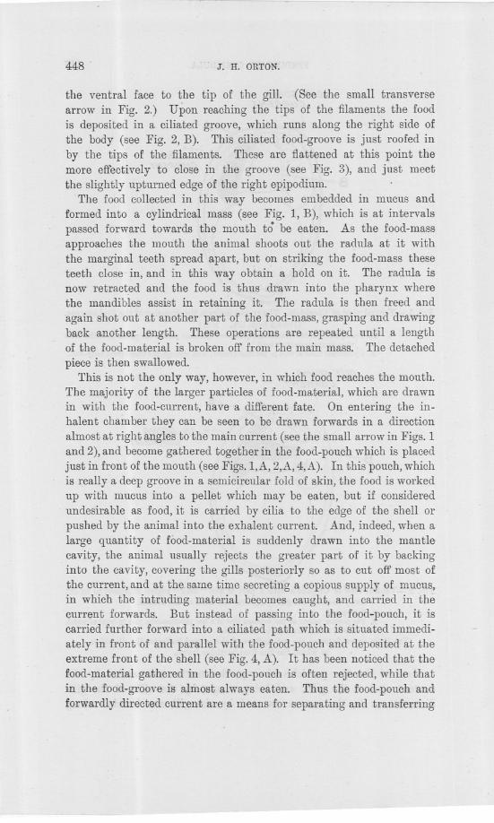
-- -- ----
448 . J. H. ORTON.
the ventral face to the tip of the gill. (See the small transversearrow in Fig. 2.) Upon reaching the tips of the filaments the foodis deposited in a ciliated groove, which runs along the right side ofthe body (see Fig. 2, B). This ciliated food-groove is just roofed inby the tips of the filaments. These are flattened at this point themore effectively to close in the groove (see Fig. 3), and just meetthe slightly upturned edge of the right epipodium.
The food collected in this way becomes embedded in mucus andformed into a cylindrical mass (see Fig. 1, B), which is at intervalspassed forward towards the mouth to' be eaten. As the food-massapproaches the mouth the animal shoots out the radula at it withthe marginal teeth spread apart, but on striking the food-mass theseteeth close in, and in this way obtain a hold on it. The radula isnow retracted and the food is thus drawn into the pharynx wherethe mandibles assist in retaining it. The radula is then freed andagain shot out at another part of the food-mass, grasping and drawingback another length. These operations are repeated until a lengthof the food-material is broken off from the main mass. The detached
piece is then swallowed.This is not the only way, however, in which food reaches the mouth.
The majority of the larger particles of food-material, which are drawnin with the food-current, have a differl(nt fate. On entering the in-halent chamber they can be seen to be drawn forwards in a directionalmost at right angles to the main current (see the small arrow in Figs. 1and 2), and become gathered together in the food-pouch which is placedjust in front of the mouth (see Figs.1,A, 2,A, 4,A). In this pouch, whichis really a deep groove in a semicircular fold of skin, the food is workedup with mucus into a pellet which may be eaten, but if consideredundesirable as food, it is carried by cilia to the edge of the shell orpushed by the animal into the exhalent current. And, indeed, when alarge quantity of food-material is suddenly drawn into the mantlecavity, the animal usually rejects the greater part of it by backinginto the cavity, covering the gills posteriorly so as to cut off most ofthe current, and at the same time secreting a copious supply of mucus,in which the intruding material becomes caught, and carried in thecurrent forwards. But instead of passing into the food-pouch, it iscarried further forward into a ciliated path which is situated immedi-ately in front of and parallel with the food-pouch and deposited at theextreme front of the shell (see Fig. 4, A). It has been noticed that thefood-material gathered in the food-pouch is often rejected, while thatin the food-groove is almost always eaten. Thus the food-pouch andforwardly directed current are a means for separating and transferring
~-~
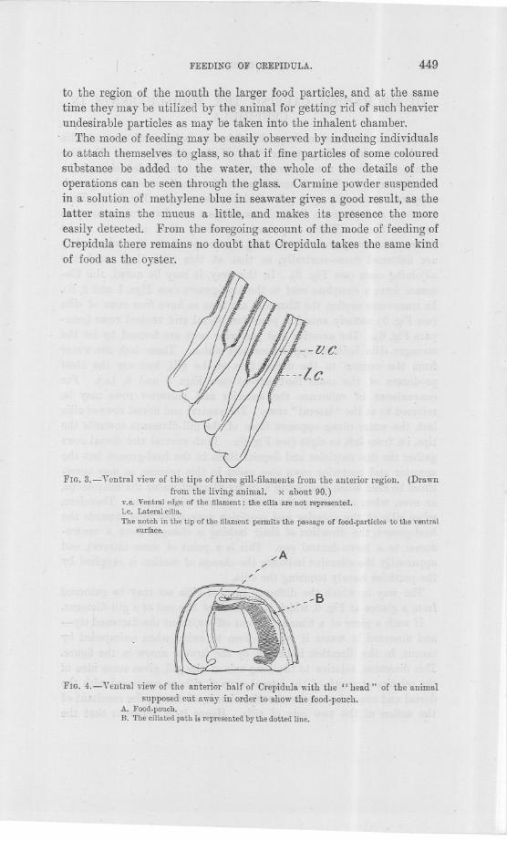
~--~=--~
FEEDIKG OF CREPIDULA. 449
to the region of the mouth the larger food particles, and at the sametime they may be utilized by the animal for getting rid' of such heavierun,desirable particles as may be taken into the inhalent chamber.
The mode of feeding may be easily observed by inducing individualsto attach themselves to glass, so that if fine particles of some colouredsubstance be added to the water, the whole of the details of theoperations can be seen through the glass. Carmine powder suspendedin a solution of methy]ene blue in seawater gives a good result, as thelatter stains the mucus a little, and makes its presence the moreeasily detected. From the foregoing account of the mode of feeding ofCrepidula there remains no doubt that Crepidula takes the same kindof food as the oyster. .
--lie.
---lc
FIG. 3.- Ventral view of the tips of three gill-filaments from the anterior region. (Drawn
from the living animal. x ahout 90.)v.c. Ventral edge of the filament: the cilia are not represented.I.e. Lateral cilia.
The notch in the tip of the filament permits the passage of food.particles to the ventralsurface.
/A/
//
/
~B
FIG. 4.- Ventral view of the anterior half of Crepidula with the" head" of the animal
. supposed cut away in' order to show the food.pouch.A. Food.pouch.B. The ciliated path is represented by the dotted line.
~-- --
450 J. H. ORTON.
III. MECHANISM CAUSING THE FOOD-CURRENTS IN
CREPIDULA..
The main food-current is produced by the lashings of rows of cilia'on the anterior and posterior faces of the gill-filaments. The fila-ments, it ha~ been noted, stanq. out in a row from the left side of themantle (seen in Figs. 1 and 2 below the small arrow), being eachsupported internally by a pair of chitinous rods (see Fig. 5, C). Theyare free, and placed a little distance apart so that water can passbetween them. When examined separately they are seen to beflattened antero-posteriorly (see Fig. 5), except at the tips, where theyare flattened dorso-ventrally, so that at this part they touch theadjoining ones (see Fig. 3). In this way, it may be noted, .the fila-ments form a complete roof to the food-groove (see Figs. 1 and 2, B).In transverse section the filaments are seen to have four rows of cilia
(see Fig. 15),namely anterior, posterior, dorsal and ventral rows (com-pare Fig. 6). The anterior and posterior rows are formed by far thestronger cilia both in appearance and action. These lash the waterfrom the ventral to the dorsal face of the gill, and are the chiefproducers of the main food-current (see Figs. 5 and 6, Lc.). Forconvenience of reference the anterior and posterior rows may bereferred to as the" lateral" rows. The ventral and dorsal rows of cilia
lash the water along opposite faces of the gill-filaments towards thetips, i.e. from left to right (see Fig. 6). Both ventral and dorsal rowsgather the fine particles and deposit them in the food-groove, but theanterior and posterior rows also assist in this process, as may some-times be seen when examining a living filament under the microscope,or even when examining the living animal with a lens. Therefore,wh.en cilia of the anterior and posterior rows wash food towards thefood-groove, the direction of their lashing is changed from a ventro-dorsal to a laevo-dextral one. This is a point of some interest, andapparently the stimulus inducing the change of motion is supplied bythe particles merely touching the cilia.
The way in which the different rows of cilia act may be gatheredfrom a glance at Fig. 6, which is 'a sketch of the end of a gill-filament.
If such a piece of a filament be cut off-without the flattened tip-and observed in water it will be seen to swim, when ~nimpeded bymucus, in the direction indicated by the lowest arrow in the figure.This direction, relative to the long axis of the gill, gives some idea ofthe relative strength of the lateral rows of cilia as compared with thedorsal and ventral rows, for the direction is, of course, the resultant ofthe action of the two sets of cilia. Hence it is apparent that the
FEEDING OF CREPIDULA. 451
lateral cilia are by far the stronger, just as one would expect to find,seeing that they have to draw a current of water through the mantlecavity, while the other rows merely pass on the food-particles.
The ventral rows of cilia lash in a direction from left to right, and, ashas already been remarked, are the main collectors of the fine food-particles.. The dorsal rows of cilia lash in the same direction as theventral rows, but on the opposite face of the gill; whatever particlesare passed on to them by the lashings of the lateral cilia they washalong the dorsal face of the filament, through a.notch in the tip of thelatter (see Fig. 3), and round to the large cilia on the ventral surface(see Fig. 6). The dorsal cilia, however, also assist in maintaining thefood-current, and in modifying the direction of the current formed bythe" lateral" cilia, for a glance again at Fig. 6 will show that the re-sultant direction of the water current produced by all the cilia on thegill is in a direction opposite to that in which the free filament swims.Thus, in the living animal the effect of the dorsal cilia on the currenton its passing through the gills is to turn it towards the right, namely,towards the exhalent aperture (see Figs. 7 and 8). The groups of largecilia on the ventral tips of the filaments are probably the chief agentsin pushing the collected food forwards towards the mouth, beingassisted in this by the cilia in the food-groove. The tips of the fila-ments are covered all over with cilia; those on the anterior and posteriorfaces doubtless assist in interlocking the filaments.
In connection with the gill-filaments, there still remains to be con-sidered the action of those cilia which occur on the floor of the posteriorpart of the mantle cavity, that is, on that part of the mantle lin-ing the dorsal surface of the visceral mass. In this region thecilia wash particles from left to right into a ciliated path on the rightside, which path is continuous with the food-gro~)Ve(see Fig. 2, B) in t,heanterior region. The mantle to the right of the ciliated path bearscilia which lash particles into the same path, working however ina direction mainly dorso-ventral.
The cause of the forwardly-directed current at the anterior end ofthe inhalent chamber is found in the presence of strong and activecilia on the lips of the food-pouch, on the inner side of the mantle, andespecially those on the dorsal surface of the left epipodium. The foodwhich is washed forwards by these groups of cilia is directed into thefood-pouch chiefly by the cilia on the dorsal lip of the latter, but it ispushed along inside the pouch by cilia, being assisted in this, however,by slow, wave-like pulsations of the side-walls. In the capture offood-particles there is no doubt that the secretion of mucus forentrapping the particles is a very important factor, and a more correct
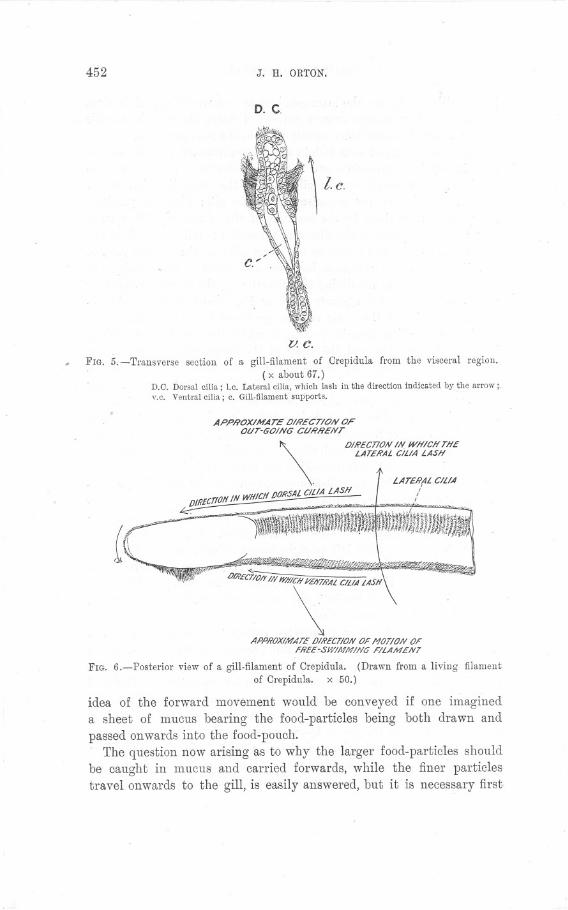
452 J. H. ORTON.
D. C.
v. e.FIG. 5.-Transverse section of a gill-filament of Orepidula from the visceral region.
(x about 67.)D.C. Dorsalcilia; I.e. Lateralcilia,which lash in the directionindicatedby the arrow;Y.c. Ventralcilia; c. Gill-filamentsnpports.
APPROXIMATE OIRECTION OFOUT-GOING CURRENT
OIRECllON IN WHICH THELATERAL CiUA LASH\
DIRECTIONIN WHICH /JO/ISAL CILIA LASH~ ~-,--~-~ ~h~__.:
LATE/ijAL C/L/AI
I
~~-iif%,?~$I!f!!f'§~~~"O
-"".. .wmg\~" a~ asPAPPROXI/11ATEOI/i'ECT/ONOFNOT/ON OF
Hi'EE-SWINNING FILAMENT
FIG. 6.-Posterior view of a gill-filament of Orepidula. (Drawn from a living filamentof Orepidula. x 50.)
.g;,j
idea of the forward movement would be conveyeda sheet of mucus bearing the food-particles beingpassed onwards into the food-pouch:
The question now arising as to why the larger food-particles shouldbe caught in mucus and carried forwards, while the finer particlestravel onwards to the gill, is easily answered, but it is necessary first.
if one imaginedboth drawn and
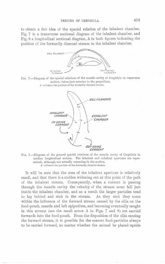
FEEDING OF CREPIDULA. 453
to obtain a fair idea of the spacial relation of the inhalent chamber.Fig. 7 is a transverse sectional diagram of the inhalent chamber, andFig. 8 a longitudinal sectional diagram, A in both figures indicating theposition of the forwardly .directed stream in the inhalent chamber.
"" ""H,",--~IN.GOING J . OUT-GOING
CURRENT CURRENt
FIG. 7.-Diagram of the special relations of the mantle cavity of Crepidula in transversesection, taken just anterior to the propodiurn.
A indicatesthe positionofthe forwardlydirectedstream.
INHALENT "
J1
CHAMBER ", :, ,, ....IN-GOING '.,,:
CU/i'RENT :
L f\,J ,/ 1
'<- OUT-GOINGCURRENT
GILL FILAMENTS
EXHALENT/CHAMBER
\/
FIG. S.-Diagram of the general spacial relations of the mantle cavity of Crepidula inmedian longitudinal section. The inhalent and exhalent. apertures are repre-sented, although not actually occurring in the section.
A indicatesthe positionof the forwardlydirectedstream.
It will be seen that the area of the inhalent aperture is relativelysmall, and that there is a sudden widening out at this point of the pathof the inhalent stream. Consequently, when a current is passingthrough the mantle cavity the velocity of the stream must fall justinside the inhalent chamber, and as a result the larger particles tendto lag behind and sink in the stream. As they sink they comewithin the influence of the forward stream caused by the cilia on thefood-pouch, mantle and left epipodium, and becoming eventually caughtin this stream (see the small arrow A in Figs. 7 and 8) are carriedforwards into the food-pouch. From the disposition of the cilia causingthe forward stream, it is possible for the co.arser food-particles alwaysto be carried forward, no matter whether the animal be placed upside
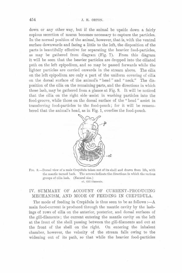
454 J. H. ORTON.
down or any other way, but if the animal be upside down a fairlycopious secretion of mucus becomes'necessary to capture the particles.In the normal position of the animal, however, that is, with the ventralsurface downwards and facing a little to the left, the disposition of theparts is beautifully effective for separating the heavier food-particles,as may be gathered from diagram (Fig. 7). From this diagramit will be seen that the heavier particles are dropped into the ciliatedpath on the left epipodium, and so may be passed forwards while thelighter particles are carried onwards in the stream above. The ciliaon the left epipodium are only a part of the uniform covering of ciliaon the dorsal surface of the animal's" head" and" neck." The dis-position of the cilia on the remaining parts, and the directions in whichthese lash, may be gathered from a glance at Fig. 9. It will be noticedthat the cilia on the right side assist in washing particles into thefood-groove, while those on the dorsal surface of the" head" assist intransferring food-particles to the food-pouch; for it will be remem-bered that the animal's head, as in Fig. 1, overlies the food-pouch.
-it
FIG. 9.-Dorsal view of a male Crepidula taken out of its shell and drawn from life, withthe mantle turned back. The arrows indicate the directions in which the various
gronps of cilia lash. (Natural size.)ct. Gillfilaments.
IV. SUMMARY OF ACCOUNT OF CURRENT-PRODUCING
MECHANISM, AND MODE OF FEEDING IN CREPIDULA.
The mode of feeding in Crepidula is thus seen to be as follows :-Amain food-current is produced through the mantle cavity by the lash-ings of rows of cilia on the anterior, posterior, and dorsal surfaces ofthe gill-filaments; the current entering the mantle cavity on the leftat the front of the shell passing between the gill-filaments and out atthe front of the shell on the right. On entering the inhalentchamber, however, the velocity of the stream falls owing to thewidening out of its path, so that while the heavier food-particles
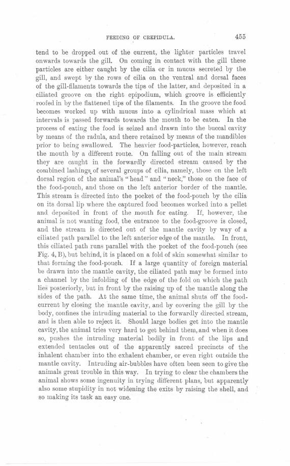
FEEDING OF CREPIDULA. 455
tend to be dropped out of the current, the lighter particles travelonwards towards the gill. On coming in contact with the gill theseparticles are either caught by the cilia or in mucus secreted by thegill, and swept by the rows of cilia on the ventral and dorsal facesof the gill-filaments towards the tips of the latter, and deposited in aciliated groove on the right epipodium, which groove is efficientlyroofed in by the flattened tips of the filaments. In the groove the foodbecomes worked up with mucus into a cylindrical mass which atintervals is passed forwards towards the mouth to be eaten. In theprocess of eating the food is seized and drawn into the buccal cavityby means of the radula, and there retained by means of the mandiblesprior to being swallowed. The heavier food-particles, however, reachthe mouth by a different route. On falling out of the main streamthey are caught in the forwardly directed stream caused by thecombined lashings. of several groups of cilia, namely, those on the leftdorsal region of the animal's" head" and" neck," those on the face ofthe food-pouch, and those on the left anterior border of the mantle.This stream is directed into the pocket of the food-pouch by the ciliaon its dorsal lip where the captured food becomes worked into a pelletand deposited in front of the mouth for eating. If, however, theanimal is not wanting food, the entrance to the food-groove is closed,and the stream is directed out of the mantle cavity by way of aciliated path parallel to the left anterior edge of the mantle. In front,this ciliated path runs parallel with the pocket of the food-pouch (seeFig. 4, B), but behind, it is placed on a fold of skin somewhat similar tothat forming the food-pouch. If a large quantity of foreign materialbe drawn into the mantle cavity, the ciliated path may be formed intoa channel by the infolding of the edge of the fold on which the pathlies posteriorly, but in front by the raising up of the mantle along thesides of the path. At the same time, the animal shuts off the food-current by closing the "mantle cavity, and by covering the gill by thebody, confines the intruding material to the forwardly directed stream,and is then able to reject it. Should large bodies get into the mantlecavity, the animal tries very hard to get behind ther~l,and when it doesso, pushes the intruding material bodily in front of the lips andextended tentacles out of the apparently sacred precincts of theinhalent chamber into the exhalent chamber, or even right outside themantle cavity. Intruding air-bubbles have often been seen to give theanimals great trouble in this way. In trying to clear the chambers theanimal shows some ingenuity in trying different plans, but apparentlyalso some stupidity in not widening the exits by raising the shell, andso making its task an easy one.
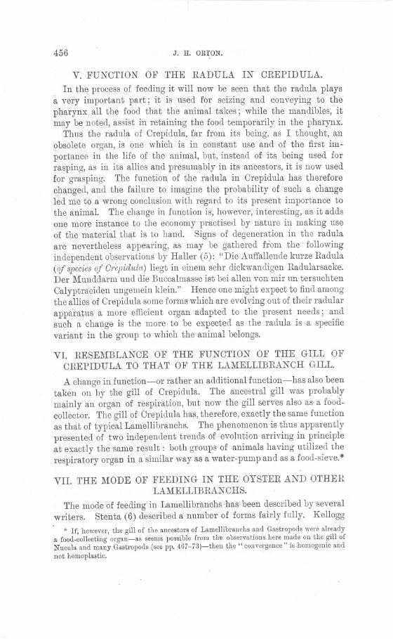
456 J. H. ORTON.
V. FUNOTION OF THE RADULA IN OREPIDUl.A.
In the process of feeding it will now be seen that the radula playsavery important part; it is used for seizing and conveying to thepharynx all the food that the animal takes; while the mandibles, itmay be noted, assist in retaining the food temporarily in the pharynx.
Thus the radula of Orepidula, far from its being, as I thought, anobsolete organ, is one which is in constant use and of the first im-portance in the life of the animal, but, instead of its being used forrasping, as in its allies and presumably in its ancestors, it is now usedfor grasping. The function of the radula in Orepidula has thereforechanged, and the failure to imagine the probability of such a changeled me to a wrong conclusion with regard to its present importance tothe animal. The change in function is, however, interesting, as it addsone more instance to the economy practised by nature in making useof the material that is to hand. Signs of degeneration in the radulaare nevertheless appearing, as 'may be gathered fl:bm the followingindependent observations by Haller (5): "Die Auffallende kurze Radula(of speeiesof Orepidula)liegt in einem sehr dickwandigenRadularsacke.Der Munddarm und die Buccalmasse ist bei alIen von mir un tersuchtenOalyptraeiden ungemein klein." Hence one might expect to find amongthe allies of Orepidula some forms which are evolving out of their radularapparatus a more efficient organ adapted to the present needs; andsuch a change is the more to be expected as the radula is a specificvariant in the group to which the animal belongs.
VI. RESEMBLANOE OF THE FUNOTION OF THE GILL OJ!'OREPIDULA TO THAT OF THE LAMELUBRANOH GILL.
A change in function-or rather an additional function-has also beentaken on by the gill of Orepidula. The ancestral gill was probablymainly an organ of respiration, but now the gill serves also as a food-collector. The gill of Orepidula has, therefore, exactly the same functionas that of typical Lamellibranchs. The phenomenon is thus apparentlypresented of two independent trends of evolution arriving in principleat exactly the same result: both groups of animals having utilized therespiratory organ in a similar way as a water-pump and as a food-sieve.*
VII. THE MODE OF FEEDING IN THE OYSTER AND OTHERLAMEI"UBRANOHS.
The mode of feeding in Lamellibranchs has been described by severalwriters. Stenta (6) described a number of forms fairly fully. Kellogg
* If, however, the gill of the ancestors of Lamellibranchs and Gastropods were alreadya food-collecting organ-as seems possible from thlJ observations here made on the gill ofNncnla and many Gastropods (see pp. 467-73)-thell the" convergence" is homogenic andnot homoplastic. .
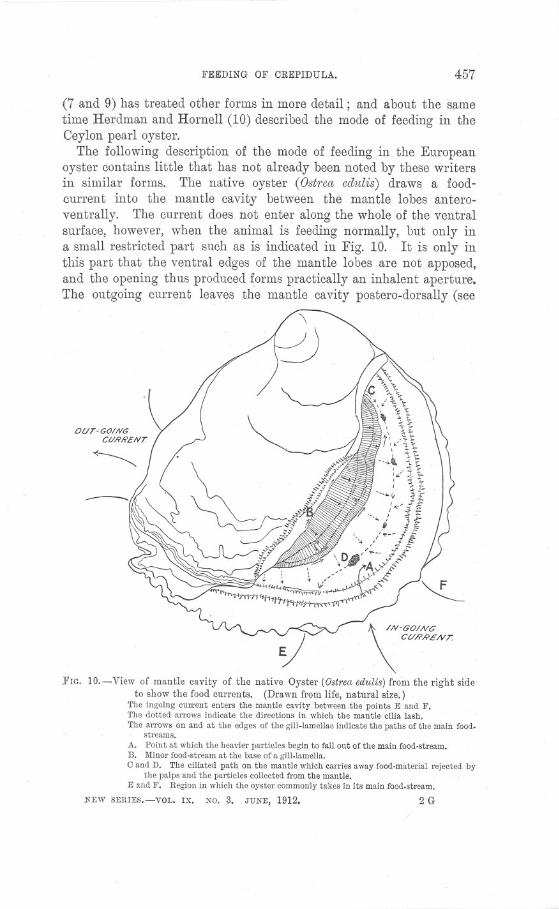
FEEDING OF CREPIDULA. 457
(7 and 9) has treated other forms in more detail; and about the sametime Herdman and Hornell (10) described the mode of feeding in theCeylon pearl oyster.
The following description of the mode of feeding in the Europeanoyster contains little that has not already been noted by these writersin similar forms. The native oyster (Ost1'eaedldis) draws a food-current into the mantle cavity between the mantle lobes antero-ventrally. The current does not enter along the whole of the ventralsurface, however, when the animal is feeding normally, but only ina small restricted part such as is indicated in Fig. 10. It is only inthis part that the ventral edges of the mantle lobes are not apposed,and the opening thus produced forms practically an inhalent aperture.The outgoing current leaves the mantle cavity postero-dorsally (see
\ /#-60/N6
\ CURRENT.
]'IG. 10.-View of mantle cavity of the native Oyster (Ostrea edulis) from the right sideto show the food currents. (Drawn from Jife, natural size.)
The ingoing cmrent enters the mantle cavity between the points E and F.The dotted arrows indicate the directions in which the mantle cilia lash.The arrows on and at the edges of the gill-lamellae indicate the paths of the main food.
streams.A. Point at which the heavier particles begin to fall ont of the main food-stream.B. Minor food-stream at the base of a gill-lamella.e and D. The ciliated path on the mantle which carries away food-material rejected by
the palps and the particles collected from the mantle.E and F. Region in which the oyster commonly takes in its main food.stream.
l'EW SERIES.-YOL. IX. No.3. JUNE, 1912. 2G

458 J. H. ORTON.
Fig. 10). On entering the mantle cavity the path of the ingoingstream is suddenly widened (see Fig. 14) and, as is the case in Orepi-dula, the heavier particles drop out of the current. These particles,however, are collected by the mantle cilia into a definite ciliated path,the cause of the" untere Riickenstrom" of Stenta, which conductsthem posteriorly to a point in about the middle of the inhalentchamber (see Fig. 10), whence the intruding material is expelled atintervals by sudden flappings of the shell-valves. This stream isprotected, or rather rendered possible, by the infolded mantle edges,which shield it from the main stream. Those particles which fall onthe mantle in the pos_terior part of the inhalent chamber are washedventrally, and are either shot out of the mantle cavity at any point, asindicated by the arrows in Fig. 10, or are caught up by the gill andcarried forwards towards the mouth.
In the American oyster there is a similar, posteriorly-directedciliated path on the anterior half of the mantle, but according toKellogg (7) there is also in the posterior half of the inhalent chambera forwardly-directed current, which carries intruding bodies forward tothe point where the current in the anterior part deposits whatever.material it may have collected. The whole of the foreign particles.collected by the mantle are then expelled at the point where the twopaths meet, that is, in about the middle of the edge of the inhalentchamber.
In Mytilus and Oardium (see Stenta, 6) the ciliated path collectsparticles from the whole of the mantle and washes them posteriorlyinto the exhalent chamber, but here, as also in Glycimeris glycimeris,the inhalent and exhalent apertures are more definite than in theoyster, both apertures, however, being posterior (see Figs. 11 and 12).The ciliated paths in Oardium and Mytilus are excellently arrangedfor expelling intruding bodies, for in the natural feeding position theseanimals lie with the ventral surface apposed to the substratum, andthe current enters the mantle postero-ventrally. Hence the wholelength of the mantle cavity is utilized for the weeding out of theheavier particles, which on falling out of the current drop straightinto the ciliated paths. Moreover, there is in Mytilus in the dorsalangle of the inhalent .aperture a fold of epidermis forming a sort ofcurtain (see Fig. 11, B) which prevents the ingoing current from im-.pinging .directly on to the gills by directing it ventrally. In this waythere doubtless results a more effective selection 01 the coarser particles.In Oardium a semicircular fold of the mantle between the inhalent
aperture and the posterior ends of the gills (see Fig. 12, B) doubtlessassists in the automatic selection of the heavier food-particles in the,
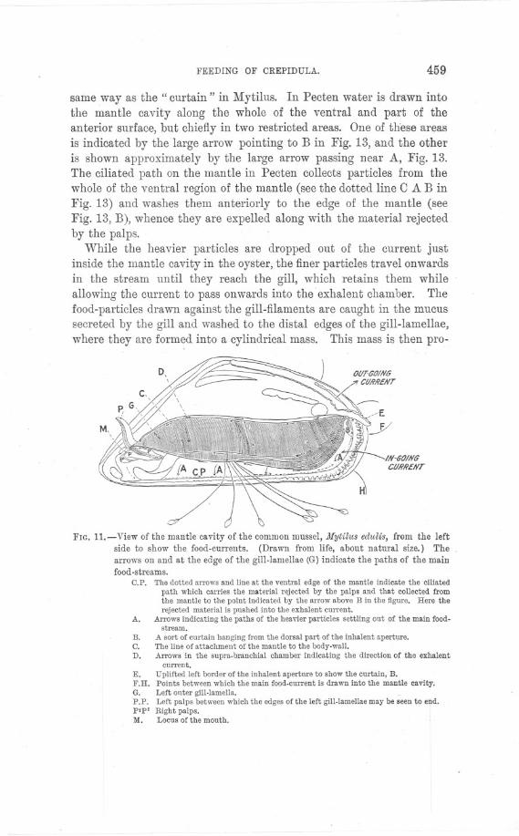
FEEDING OF CREPIDULA. 459
same way as the" curtain" in Mytilus. In Pecten water is drawn intothe mantle cavity along the whole of the ventral and part of theanterior surface, but chiefly in two restricted areas. One of these areasis indicated by the large arrow pointing to B in Fig. 13, and the otheris shown approximately by the large arrow passing near A, Fig. 13.The ciliated path on the mantle in Pecten collects particles from thewhole of the ventral region of the mantle (see the dotted line 0 A B inFig. 13) and washes them anteriorly to the edge of the mantle (seeFig. 13, B), whence they are expelled along with the material rejectedby the palps.
While the heavier particles are dropped out of the current justinside the mantle cavity in the oyster, the finer particles travel onwardsin the stream until they reach the gill, which retains them whileallowing the current to pass onwards into the exhalent chamber. Thefood-particles drawn against the gill-filaments are caught in the mucussecreted by the gill and washed to the distal edges of the gill-lamellae,where they are formed into a cylindrical mass. This mass is then pro-
IN-GO/NilCURRENT
FIG. 11. -View of the mantle cavity of the common mussel, Mytilus edulis, from the leftside to show the food-currents. (Drawn from life, about natural size.) The
arrows on and at the edge of the gill-lamellae (G) indicate the paths of the mainfood-streams.
C.P. The dotted arrows and line at the ventral edge of the mantle indicate the ciliatedpath which carries the material rejected by the palps and that collected fromthe mantle to the point indicated by the arrow above B in the figure. Here therejected material is pushed into the exhalent current.
A. Arrows indicating the paths of the heavier particles settling out of the main food-stream.
A sort of curtain hanging from the dorsal part of the inhalent aperture.The line of attachment of the mantle to the body-wall.Arrows in the supra-branchial chamber indicating the direction of the exhalent
current.E. Uplifted left border of tile inhalent aperture to show the curtain, B.F.H. Points between which the main food.current is drawn into the mantle cavity.G. Left onter gill-lamella.P.P. Left palps between which the edges of the left gill-lamellae may be seen to end.P'P' Right palps.M. Locus of the mouth.
B.C.D.
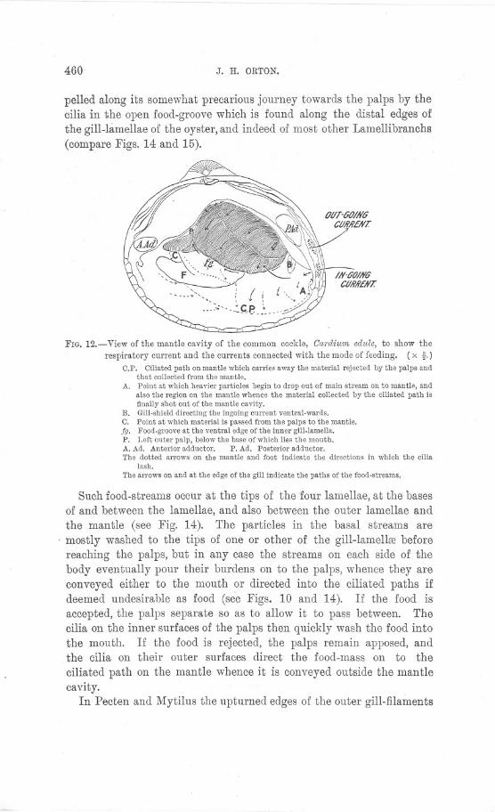
460 J. H. ORTON.
pelled along its somewhat precarious journey towards the palps by thecilia in the open food-groove which is found along the distal edges ofthe gill-lamellae of the oyster, and indeed of most other Lamellibranchs(compare Figs. 14 and 15).
FIG. 12.- View of the mantle cavity of the common cockle, Cardium edule, to show the
respiratory current and the currents connected with the mode of feeding. (x t.)C.P. Ciliated path on mantle which carries away the material rejected by the palps and
that coilected from the mantle.A. Point at which heavier particles begin to drop out of main stream on to mantle, and
also the region on the mantle whence the material coilected by the ciliated path isfinaily shot out of the mantle cavity.
B. Gill-shield directing the ingoing current ventral-wards.C. Point at which material is passed from the palps to the mantle.fg. Food-groove at the ventral edge of the inner gill-Iameila.P. Left outer palp, below the base of which lies the mouth.A. Ad. Anterior adductor. P. Ad. Posterior adductor.The dotted arrows on the mantle aud foot indicate the directions in which the cilia
lash.The arrows on and at the edge of the gill indicate the paths of the food-streams.
Such food-streams occur at the tips of the 'four lamellae, at the basesof and between the lamellae, and also between the outer lamellae andthe mantle (see Fig. 14). The particles in the basal streams are
. mostly washed to the tips of one or other of the gill-Iamellre beforereaching the paIps, but in any case the streams on each side of thebody eventually pour their burdens on to the palps, whence they areconveyed either to the mouth or directed into the ciliated paths ifdeemed undesirable as food (see Figs. 10 and 14). If the food isaccepted, the palps separate so as to allow it to pass between. Thecilia on the inner surfaces of the palps then quickly wash the food intothe mouth. If the food is rejected, the palps remain apposed, andthe cilia on their outer surfaces direct the food-mass on to theciliated path on the mantle whence it is conveyed outside the mantlecavity.
In Pecten and Mytilus the upturned edges of the outer gill-filaments
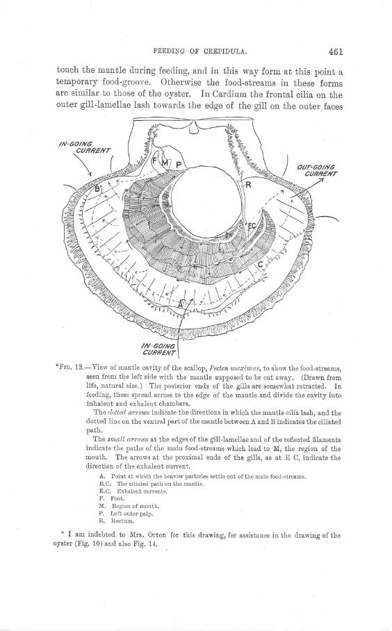
FEEDING- OF CREPIDULA. 461
touch the mantle during feeding, and in this way form at this point atemporary food-groove. Otherwise the food-streams in these formsare similar to those of the oyster. In Cardium the frontal cilia on theouter gill-lamellae lash towards the edge of the gill on the outer faces
OUT-GOINGCURRENT
~
IN-601N6CURRENT
*FIG. 13.- View of mantle cavity of the scallop, Pecten rnaxirnus, to show the food-streams,
seen from the left side with the mantle supposed to be cut away. (Drawnfromlife, natural size.) The posterior ends of the gills are somewhat retracted. In
feeding, these spread across to the edge of the mantle and divide the cavity intoinhalent and exhalent chambers.
The dotted arrows indicate the directions in which the mantle cilia lash, and thedotted line on the ventral part of the mantle between A and B indicates the ciliatedpath.
The srnall arrows at the edges of the gill-lamellae and of the reflected filaments
indicate the paths of the main food-streams which lead to M, the region of themouth. The arrows at the proximal ends of the gills, as at E C, indicate thedirection of the exhalent current.
A. Point at which the heavier particles settle out of the main food-streams.B.O. The ciliated path au the mantle.E.O. Exhalent currents.F. Foot.M. Region of mouth.P. Left onter palp.R. Rectum.
* I am indebted to Mrs. Orton for this drawing, for assistance in the drawing of theoyster (Fig. 10) and also Fig. 14.

462 J. H. ORTON.
but towards the base of the gill on the inner faces. On the innerlamellae the frontal cilia on both faces lash particles towards the freeedge of the gill into a well-defined food-groove. In Pecten, Kellogghas described that in the troughs of the gills particles are lashedtowards the base of the gill, while on the crests particles are lashedtowards the edge. On the other hand, Stenta has shown that inAnodon food-particles are washed towards the bases of the outerlamellae but to the tips of inner lamell::e. All these different modesof food collection and transportation may be indicated in diagrammaticform as in Fig. 14. This diagram will also indicate the mode of food-collection and food-transportation in Nucula. I find that in Nuculathe gills divide the posterior region of the mantle cavity into infra-and ~upra-branchial chambers as in the higher Lamellibranchs. The
/N-G17/#6'~ CUA'h'ENT
FIG. l4.-Diagram of the general mode of feeding in Lamellibranchs.The large thick-lined arrows indicate the paths of the main respiratory and
food-current.
The dotted thin-lined arrows indicate the directions in which the mantle and
gill cilia wash the food-streams. The dotted thick-lined arrows leading from theright gill-lamellae indicate the paths of the main food-streams towards the mouth.
Food-collection is indicated on the left gill-lamenae, and food-transportation
()!l the right gill-lamellae.A. Point at which the heavier particles begin to settle out of the current.B. The ciliated path on the mantle which carries away material rejected by the palps,
and that collected from the mantle.C.P. The ciliated path seen in section on the mantle.C. The subsidiary mantle streams.L. & R. Left and right valves of shell, and the beginning of the left and right mantle
lobes.f.g. JlIainfood-grooves.f.c. Food-channels at bases of gills-I.Ch. Inhalent chamber.E.Ch. Exhalent chamber.G.L. Gill-lamellae.JII. JlIouth.'To Ex. Ap. To exhalent aperture.
FEEDING OF CREPIDULA. 463
inner edges of the inner leaflets are apposed and kept interlocked bygroups{)f large cilia (see Fig. 18, Lc.d., p. 468). The outer edges of the outerleaflets and the posterior ends of the gills effect similar ciliary junctionswith the mantle by means of groups of large cilia (see Fig. 18, O.c.d.).In this way a complete partition of the mantle cavity is effected, andthe ventral surface of this partition is utilized as in the higher Lamel-libranchs for food-collection. Food-particles brought to the gill in themain current are arrested by the gill and washed along, the edges ofthe leaflets from the outer leaflet to the inner (see Fig. 18). At theventral end of the inner leaflets of both sides the collected food is
washed anteriorly towards the mouth. Food-particles collected fromthe gill in this way appear to be gathered up by the appendages of thepalps and transferred to the palps, which pass it along into the mouth.Food-particles may be rejected by the palps in Nucula in the sameway as in other Lamellibranchs. Rejected food is pushed off theposterior end of the palps on to the foot, and off the posterior face ofthe foot on to the mantle. The mantle cilia in Nucula collect particlesinto anterior and posterior ciliated paths which converge at the middleventral edge of the mantle just as in the American oyster. The obser-vations on the mode of feeding in Nucula are still being carried on.In the light of the observations already made on Nucula, and especiallyof those on the ciliation of the gill (see page 467), it would be worthwhile to re-examine the mode of feeding in Yoldia. Drew (11, pp. 15and 16) was unable to find out whether Yoldia uses its gills otherwisethan for pumping water. If, however, the whole gill be examinedalive while feeding it with carmine, the secret would soon be out. - Itis possible that the gill in this form may not be used for food-collecting,and if not, the condition is more interesting than if it is so used.However, from one of Drew's figures of the gill of Yoldia, although ageneral view (11, Fig.-20), there is good reason for believing that food-collecting occurs in the same way as is described' here for N ucula.
VIII. AN EXPLANATION OF THE DIRECTION OFEVOLUTION IN LAMELLIBRANCHS.
The mode of feeding in Lamellibranchs, it will now be seen, neces-sitates the sedentary habits which are exhibited by most members ofthis group. Moreover, there can be no doubt that adaptation to themode of feeding has been at least one of the main factors in determining.the direction of evolution in Lamellibranchs. From the foregoingaccount of the gill of Nucula, it is clear that the gill in this form pre-sents an early stage in the adaptation of the original respiratory organ

r -c
464 J. H. ORTON.
to a food-collecting organ; and further, recent work (see Pelseneer, 12,pp. 253-4, and Ridewood, 13) has confirmed the conclusion that thehigher Lamellibranchs have evolved mainly on the principle of foldingand consolidating the originally simple free gill-filaments to form gill-lamellae. It will now be seen to be highly probable that this com-plication and fusion of the gill-filaments is an adaptation for thepurpose of obtaining a more efficient feeding organ. Folding of the gill-filaments dorso-ventrally into demibranchs and-incipiently-antero-posteriorly into crests and troughs has increased the food-collectingsurface, while fusion of the filaments first by ciliary junctions andafterwards by organic connections has rendered the food-collectingorgan less liable to derangement. In the :Filibranchs there is muchdanger of the gill-filaments becoming separated, whereby the con-tinuity of the food-grooves at their tips is broken. As a result theanimal may have difficulty in feeding, and its nourishment be thusseriously interfered with. Hence adaptations which ensure a firm gillwould undoubtedly be advantageous-other things being the same-in preventing interference with the feeding process. The folding ofthe gill in an antero-posterior direction is also an adaptation in per-fecting the feeding process, for by this means the food-collecting sur-face of the gill is further increased (see various figures by Ridewood,13, pp. 242-263); moreover, greater opportunity is thereby givenfor effectively sieving the food-current, which has necessarily to passmore obliquely over the surface of the filaments to pass onwards intothe exhalent current, thus giving the frontal cilia of the gill-fila-ments a better chance of capturing food-particles. It will alsobe seen that this folding results in the form!ttion of secondary food-channels, thus the principal and apical filaments which occur in thetroughs and crests respectively of the folds of the gills of manyLamellibranchs (see Ridewood, 13, p. 163) probably function mainlyas the bearers of subsidiary food-grooves.
Along with the evolution of a more efficient food-collecting gill inLamellibranchs there have occurred a gradual fusion of the ventraledges of the mantle lobes and a development of inhalent and exhalentsiphons. It is highly probable that this fusion of the mantle lobesis primarily an adaptation of the same nature as the gill folding, thatis, tending towards perfecting the mode of feeding. For in Mytilus,Glycimeris, and Ostrea, and doubtless also in many other forms, thereis an attempt to limit the ingoing current to a definite area, and theeffect obtained is that of limiting the area over which the heavierparticles settle out of the food-stream to a part of the mantle adaptedfor expelling the undesirable material. In siphonate forms with the
FEEDING OF CREPIDULA. 465
mantle fused ventrally such a~ the higher Eulamellibranchs, the wholeof the ventral region of the mantle lobes may be utilized as a settlingarea from which undesirable material can be removed without
interfering unduly with the normal feeding process.
IX. THE CURRENT-PRODUCING MECHANISM IN
LAMELLIBRANCHS.
With regard to the cause of the main food-current in Lamellibranchsmost writers are vague. Herdman and Homell (10), however, haveinvestigated Margaritifera vulgaris and state cautiously that inthis species" the respiratory current is apparently due to the normalrhythmic lashing of the cilia on the large cells at the edges of the fila-ments; while the collection or the rejection of particles in the waterseems to be the result of special action stimulated apparently by theirritation. Particles arrested by the branchial filter are caught up bythe nearest cilia, which by local reversed lashing carry them outwardsto the free ventral edge of the lamella."
In Crepidula it is easy to make out with certainty the direction inwhich the several rows of cilia are working, as the filaments-relativeto those of Lamellibranchs-are large. In N ucula, Anomia, Mytilus,Glycimeris, Area, Modiola and Pecten, I also find that it is fairly easy tomake out that, as in Crepidula, the lateral cilia which lash across the lengthof the filaments (see Figs. 15 and 16) are the chief cause of the inhalentcurrent, and that the" frontal" cilia which lash towards the free edgeof the gill, collect the food-particles and wash them onwards towardsthe food-grooves at the edge of the gill (see Figs. 15 and 16).
£ll/?ECT/tJlI III JYII/CII LATERAL CILIA LASIt
."
FIG. 15.-Lateral view of a living filament of the left outer lamella of the gill of Mytilusedulis. (x about 84.)
Lc. Lateralcilia. !.f.c. Latero-frontalcilia. f.c. Frontalcilia.ab.f.c. Ab-frontalor innercilia. c.d. Ciliateddisc.A. Arrowindicatingroughly the directionin whichthe iatero-frontalciliaiash.
466 .J. H. ORTON.
In Mytilus there are also on the" inner" * or ab-rrontal side or thefilament cilia which lash in a direction opposite t9 that of the frontalcilia; they therefore help in producing the main current as in Crepi-dula. These cilia doubtles~ also assist in keeping clean the innersurfaces of the gill-filaments. The examination of living filaments ofMytilus revealed inaccuracies in the existing figures of the gill-cilia inthis form (see Peck, 14) and Ridewood (13, Fig. 11, c, p. 201). Sectionswere therefore prepared from well-preserved material, and a drawingof one of these made for Fig. 17 (p. 467).
"
OIRECTION IN WHICH LAffl?Al C/UA LAS#. cd
<II~~).~m~~~w~~ ~»j~)),~~~WNirJW\!j. , , '
/J/RECnOIYIIYWH/CIIFh'tJ//TA(C/UA,s1t. xc. ''Ie.
FIG. 16.-Lateral view of living filament of left outer lamella of gill of Pecten 'lnaXi'lnU8.(x about 130.)
I.e. Lateral cilia. f.c. Frontal cilia. c.d. Ciliated disc.
On now comparing the ciliation in Ostrea, Tapes and Cardium, with thatin the aforementioned forms, there can remain no doubt that the lateralcilia here also produce the main current by lashing across the length ofthe filament, while the frontal cilia collect the food-particles by lashingtowards the free edge of the gill. Therefore the statements by Kellogg(9, pp.416-423, see also 7, p. 36), andPelseneer (12, p. 230) that the lateralcilia in Lamellibranch gill-filaments serve mainly for straining the food-particles or for interlocking the filaments require correction. Therecan be no doubt, however, that the lateral cilia, as in Crepidula, mayhelp occasionally if necessary in washing particles towards the food-groove by local reversed lashing. The long cilia found at the sides ofthe frontal rows of cilia in many: Lamellibranchs (see Ridewood'sfigures passi1n as latero-frontal cili~, 13) are probably true strainingcilia. In Nucula, Anomia, Mytilus, Tapes, and Cardium, these are un-doubtedly straining cilia. They stand out from the sides of the filament,forming a sort of grating between them, and lash relatively slowly acrossthe length of and towards the middle of the frontal face of the filament(see Figs. 15, 17, and 18). Thus Nuculaand Mytilus have four kindsof cilia, the lateral cilia :producing the main current, the frontal for
* That is, the side away from the e~posed face of the gill-lamella.
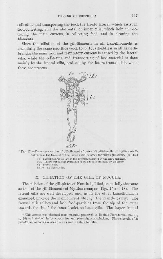
FEEDING OF CREPIDULA. 467
-collecting and transporting the food, the fronto-Iateral, which assist infood-collecting, and the ab-frontal or inner cilia, which help in pro-ducing the main current, in collecting food, and in cleaning thefilaments.
Since the ciliation of the gill-filaments in all Lamellibranchs is~ssentially the same (see Ridewood, 13, p. 163) doubtless in all Lamelli-.branchs the main food and respiratory current is caused by the lateralcilia, while the collecting and transporting of food-material is done-mainly by the frontal cilia, assisted by the latero-frontal cilia whenthese are pres ent.
II
I
I aofc. .-* FIG. 17.-Transverse section of gill-filament of outer left gill.lamella of lJfytilus edulis
taken near the free,end of the lamella and between the ciliary junctions. (x 418.)1.e, Lateralcili~whichlash in the directionindicatedby the arrowalongside.!.f.c. Latero.frpntalciliawhichlash in the directionindicatedby the arrow.f.c. Frontal cilia.
Iab.f.c. Ab.frontal cilia.
I
X. OILIA!ION OF THE GILL 'OF NUOULA.
The ciliation of the gill-plates of Nucula is, I find, essentially the same-as that of the gill-filaments of Mytilus (compare Figs. 15 and 18). Thelateral cilia are well Ideveloped, and, as in the other Lamellibranchs~examined,produce thJ main current through the mantle cavity. Thefrontal cilia collect all-d lash food-particles from the tip of the outertowards the tip of the inner leaflet on both gills. The, larger frontal
* This section was obta&ed from material preserved in Bouin's Picro-formol (see 19,p. 76) and stained in borh-carmine and picro.nigrosin solutions. Ficro.nigrosin afterpicroformol or corrosive.acetic is an excellent stain for cilia.
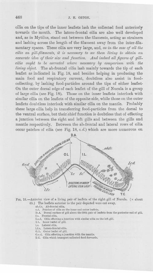
468 J. H. ORTON.
cilia on the tips of the inner leaflets lash the collected food anteriorlytowards the mouth. The latero-frontal cilia are also well developedand, as in Mytilus, stand out between the filaments, acting as strainersand lashing across the length of the filament away from the inter-fila-mentary spaces. These cilia are very large, and, as in the caseof all thecilia on gill-filanwnts, it is necessary to see them living to obtain anaccurate idea of their size and function. And indeed all figu1'esof gill-cilia ought to be corrected where necessa1'yby comparison with theliving object. The ab-frontal cilia lash mainly towards the tip of eachleaflet as indicated in Fig. 18, and besides helping in producing themain food and respiratory current, doubtless also assist in food-collecting, by lashing food-particles around the tips of either leaflet.On the outer dorsal edge of each leaflet of the gill of Nucula is a groupof large cilia (see Fig. 18). Those on the inner leaflets interlock withsimilar cilia on the leaflets of the opposite side, while those on the outerleaflets doubtless interlock with similar cilia on the mantle. Probablythese large cilia help in transferring food-particles from the dorsal tothe ventral surface, but their chief function is doubtless that of effectinga junction between the right and left gills and between the gills andmantle respectively. Between the ab-frontal and lateral rows of ciliaoccur patches of cilia (see Fig. 18, c.d.) which are more numerous OIl
D.A.~
u~~~f~lCl.
~~.f~O.~\C~~iI\\\\~ O.L.
(;.\'V~~ ~~
~~<£5}a
~q,-~«'<,Q-:4'~
~4'J'#. TC.
FIG. 18.-Anterior view of a living pair of leaflets of the right gill of Nucula. (x about;65.) The leaflets anterior to the pair depicted were cut away.
ab.f.c. Ab-frontalcilia.c.d. Patchesof ciliaon the innerandouter leallets.D.A. Dorsalsurfaceof gillabout the 30thpair of lealletsfromthe posteriorendof gill.f.c. Frontalcilia.I.c.d. Ciliaeffectingajunction with similarciliaon the leftgill.I.L. Inner lealletof gill.1.c. Lateralcilia.Lf.c. Latero-frontalcilia.D.L. Outerlealletof gill.O.c.d. Ciliaeffectingajunctionwiththe mantle.T.C. Ciliawhichtransport collectedfoodforwards.

,
FEEDING OF CREPIDULA. 469
the inner than on the outer leaflets, and indeed are apparently oftenabsent from the outer leaflets. These cilia arise from little spurs. oftissue and have the same curious rotary motion as those which occur inpatches on the gill-filaments of the Filibranchs. The action and dis-tribution of these patches of cilia leave no doubt that they effect ciliaryjunctions between the leaflets in the same way as those of the Fili-branchs effect junctions between filaments. There are also similarpatches of larger cilia immediately below and to the side of thegroups of the inter-locking cilia on the tips of each leaflet (see Fig. 18,c.d. below O.c.d. and I.c.d.)~ Doubtless these have also the functjonof interlocking the leaflets, and are similar to those occurring on thetips of the gill-filaments of Yoldia (see Kellogg, 9, Fig. 78) and .Anorniaaculeata. It is therefore evident that the gill of Nucula is undoubtedlyless primitive as compared with the gill of other Lamellibranchs thanhas formerly been thought. A fuller investigation of this gill is beingmade.
XI. THE BEARING OF THE GILL CHARACTERS OF NUCULAON THE RELATIONSHIPS OF THE PROTOBRANCHIA.
It is clear from the foregoing description that the gill of Nucula-and indeed those of Yoldia and Solenomya may be included-cannotnow be regarded as being so primitive and unique as to justify theclassification of the Proto branchia as a group co-equal with the Fili-branchia. Ridewood has shown that the gill of .Anomia aC1deatacon-sists on each side of two rows of filaments whose only difference fromthe leaflets of Nucula lies, I find, in their being narrow and filamentousinstead of broad and lamellate. This obviously constitutes only a minordifference. Indeed, the occurrence of ciliated discs on the gill-lamellaeof N ucula might fairly be advanced as evidence of higher specialization-along orthodox Lamellibranch lines-than occurs in the gill of.Anomia aculeata, since the filaments of this species of Anomia appearto be without ciliated discs excepting at the tips. Further, the ciliateddiscs at the tips of the Anomia filaments are matched by similar onesin the gill-leaflets of Nucula, and the action and function of the variousrows of cilia is, I find, the same in both animals. Moreover, the frontalcilia on the filaments of Anomia occur on the ventral surface as inNucula, and those on the outer filaments lash in the same direction asin Nucula (see Fig. 18). Ridewood (13, p. 194) has shown that in thisspecies of Anomia the posterior end of the gill on each side isfree and that the inner filaments of each side interlock, and thatthe outer filaments form a junction with tlie mantle by means ofcilia. All these features are found in the gills of Nucula (see Fig. 18).

-- --- - - --- ---
470 J. H. ORTON.
Thus the one feature of Proto branchia regarded by Pelseneer asunique-the occurrence of gill-leaflets-is undoubtedly robbed of itsglamour-and cannot now be reasonably regarded as of such greattaxonomic value as formerly. Another supposedly unique feature ofthe Protobranchs, namely, the absence of a subdivision of the mantlecavity (see Sedgwick 15, p. 345), must be abandoned, for Drew hasalready shown that in Yoldia (11, p. 14) there is a subdivision ofthe mantle cavity into inhalent and exhalent chambers, as is heredescribed for Nucula, and as will no doubt be found in all the Proto-branchs. With regard to the other primitive or special features ofProtobranchs, such as the occurrence of distinct pleural ganglia, aplantar surface to the foot, free communication between the cavitiesof the gonad, pericardium, and kidneys, and other features-withregard to these, Pelseneer has already shown (12, J?assim) that theyare matched in some adult members of the Filibranchs with the
exception of separate pleural ganglia, which at present are onlyknown in the developmental stages of other forms (as Modiolarca,Dreissensia, etc. See Pelseneer 12, p. 234). It is therefore clearthat the Protobranchs cannot now be classified as a group equiva- -lent to the Filibranchs. We must therefore be prepared to degrade-or rather elevate-the Protobranchia to a subordinate position in theFilibranchia of Pelseneer, or in the Eleutherorhabda of Ridewood. Itis a matter of much interest that Palaeontologists (16, p. 359) shouldalready have classified together the Protobranchia and the re-mainder of the Filibranchs into the order of Prionodesmacea, whose
diagnosis is concerned mainly with shell characters. As we maynow take for granted that Lamellibranchs have evolved mainly onthe principle of perfecting the gill as a feeding organ, it is clear-from the closely similar results attained by Palaeontologists andmodern zoologists-that there is a close correlation between shellcharacters and gill characters. Whether the shell characters arecapable of any functional explanation, similar to that of the gillcharacters, my knowledge of the group does not yet enable me tosay. It is probable that such an explanation may now be poss.ible.
XII. A COMPARISON OF THE MODE OF FEEDING INLAMELLIBRANCHS AND CREPIDULA.
In Lamellibranchs, as in Crepidula, it has been noted that there isan arrangement whereby an automatic selection of the heavier par-ticles takes place just inside the inhalent chamber. Thus the
....
FEEDING OF CREPIDULA.. 471
forwardly directed stream in the inhalent chamber in Crepidula isequivalent to that caused by the ciliated path on the mantle ofLamellibranchs; the purpose of the stream in both animals beingthat of expelling undesirable material from the inhalent chamber.There is this difference, however, that Crepidula, unlike the oyster,has the option of ingesting the automatically selected heavier par-ticles. On the other hand, in the oyster and other Lamellibranchsthe fringes of tentacles on the edge of the mantle form a coarse sieveby interlacing at the entrance to the mantle cavity, and in this wayprevent the entrance of coarse particles; it is possible, however, thatthe particles collected on the mantle in Lamellibranchs might bepicked up by the gill and conveyed to the mouth, as probably happensin the posterior region of the inh:'tlent chamber of the oyster and thescallop. Both animals have still another opportunity for selectingtheir food-material, namely, by refusing the food-masses which arebrought by the gills to the mouth. It has been observed that bothanimals do at times refuse such food, so that selection of foodMmaterialis undoubtedly exercised in this way. From my preparations of thegut contents of these two animals, I received the impression thatthose of the oyster were the finer, but as I have examined compara-tively few specimens, it is doubtful whether that observation has anysignificance. It would be necessary to examine and compare a largernumber of individuals to obtain a significant result. The fine food-particles are collected on the gill in both Crepidula and. Lamelli-branchs, and conveyed along food-grooves to the mouth, but Crepidulamay be regarded as having gained an advantage over Lamellibranchsby closing in its food-groove, and thus ensuring the capture of thefood. If disturbed while feeding an oyster would be much more liableto lose its food than Crepidula.
There is, however, an interesting difference in the position of thelateral cilia on the filaments in Crepidula and Lamellibranchs. InCrepidula these cilia are nearer the exhalent chamber (see Fig. 6, I.e.,p. 452), while in Lamellibranchs they are nearer the inhalent chamber(see Fig. 18, I.e., p. 468). An explanation of these phenomena willprobably be offered.when more Gastropod gills have been studied.
It will now be apparent how remarkably similar Crepidula, itsallies, and Lamellibranchs are in the details of their modes of feed-
ing. The closeness of the resemblances they offer may fairly beregarded as an expression of the similar tendencies they have derivedfrom their common origin.
472 J. H. ORTON.
XIII. THE MODE OF FEEDING IN THE ALLIES OFCREPIDULA.
The nearest allies of Crepidula doubtless all feed as Crepidula itselfdoes. Such a deduction may fairly be drawn at once from the simi-larity in the mode of life of those animals and the general similarityof their organs in the region of the mantle cavity. I have examinedOalyptmea chinensis alive, and find that it feeds in exactly the same wayas does Crepidula, collecting food both in its epipodial food-groove andin a food-pouch.
Oapulus hungaricus exhibits an interesting variation of the samemanner of taking food. In this animal there is no epipodium nor food-pouch. But instead of the former the lips have become elongated inthe form of a grooved proboscis, which appears to be held along the rightside of the animal to collect the food-particles from the tips of the gillswhen the animal is feeding. The forwardly-directed stream is present onthe edge of the mantle in the inhalent chamber, but the stream is rela-tively weak. There is an outgrowth of the foot between the propodiumand the" neck" region, known as the" operculum," which appears tobe used partly for side-tracking the food-current into the exhalentstream when the animal is not feeding, but I have not yet had theopportunity for investigating Capulus fully, and so must defer adetailed account until later.
The Hipponycidae are so similar in structure to the Capulidae as tohave been placed with them at one time in the same Order, and as theylive a sedentary life, it is almost certain that they will be found to feedin some similar manner to that of Capulus. There is, therefore, littledoubt that all the Calyptraeidae feed in the same way as Crepidula,and that the Capulidae feed in a similar manner: thus there is goodreason for suspecting that all sedentary Pectinibranchs may obtaintheir food in the same or in a similar manner.
XIV. THE CURRENT-PRODUCING MECHANISM IN OTHERGASTROPODS.
After seeing the gills of Crepidula, Calyptraea, and Capulus, andespecially the latter, whose gill is very similar to that of mostGastropods, I was stimulated to examine all the sedentary forms tobe had. But, on seeing Pelseneer's figures of sections of the gills ofsome Aspidobranchs (17, Figs. 99 to 104), I was induced to examineall the Gastropods available. It was found that in all the formsexamined, namely, Fissurella, Haliotis, Calliostoma, Gibbula, Murex,
FEEDING OF CREPIDULA. 473
Purpura, Nassa, Buccinum-in all these-the gill-filaments are ciliatedin essentially the same way as those of Orepidula. There are generallypresent lateral, frontal andab-frontalcilia, and the gill-filaments, or rathergill-leaflets, closely resemble those of the Protobranchs. The lateralcilia in all the forms examined produce the main current in the mantlecavity. The occurrence of frontal and ab-frontal cilia in all theseforms is a matter of much interest. In all cases these cilia collect
plankton from the ingoing current, but whether such collected food iseaten I am not yet able to say. It seems probable that all these formsmay be found to feed partly on plankton. A research into this matteris being made. In some cases (namely, Fissurella and Buccinum) thegill undoubtedly divides the mantle cavity into inhalent and exhalent
. chambers as occurs in Orepidula and its allies. At the tips of the fila-ments in all these forms there are interlocking cilia similar to those atthe tips of the gill-leaflets of Nucula. These cilia doubtless serve toeffect a junction between the gill and the opposite wall of the mantle.Thus there can be no doubt that most Gastropods on further investi-gation will he shown to have the mantle cavity divided by the gillinto two chambers.
A point of some interest presents itself at once on comparing thegill-filaments or gill-leaflets of the Aspidobranchs and many Pectini-branchs with the gill-filaments of Orepidula and Oalyptraea, namely,that the gill-leaflets of the former bear tbe same relation to thefilaments of the latter that the gill-leaflets of Nucula bear to the gill-filaments of the Filibranchs. Thus the Pectinibranchs already presentthe same range of gill-features that I propose should be united in theFilibranchiate Lameliibranchs. It may here be remarked that thesimilarity in the structure and function of the gills in Gastropods andLamellibranchs shown by the foregoing observations, emphasizes thatclose relationship between these groups, which Pelseneer has alreadypointed out (17).
A cursory exami'nation of the gills of Ohitons indicates that rows oflateral cilia on the gill-l.eaflets produce the main current through themantle cavity in a manner similar to that in Gastropods and Lamelli-branchs.
XV. SIGNIFICANOE OF OHAIN FORMATION IN THE MODEOF FEEDING O~' OREPIDULA.
It is well known that Crepidula has the curious habit of forming longchains by one individual settling on the back of another; as many asfourteen individuals may be found holding together in such a manner,and usually there is a gradation in size from the largest at the bottom
NEW SERIES.-VOL. IX. NO.3. JUNE, 1912. 2 H
~
n J
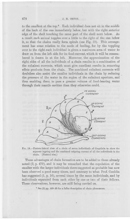
474 J. H. ORTON.
to the smallest at the top.* Each individual does not sit in the middleof the back of the one immediately below, but with the right anterioredge of the shell touching the same part of the shell next below. Asa result each animal topples over a little to the right of the one bel-owit, so that the chains really form spirals (see Fig. 19). This arrange-ment has some relation to the mode of feeding, for by the topplingover to the right €ach individual is given a maximum area of water todraw on from the left side for its food-current, which it will be remem-bered is drawn in at the left. Moreover, the approximation of theright sides of all the individuals of a.chain results in a combination ofthe exhalent currents, which must give excellent results in removingeffete products from the chain. The combined exhalent currents willdoubtless also assist the smaller individuals in the chain by reducingthe pressure of the water in the region of the exhalent aperture, andthus enabling them to pass a greater volume of food-bearing waterthrough their mantle cavities than they otherwise could.
IN-GOING//- CURRENT
/' J'
FIG. 19.-Postero-Iateral view of a chain of seven individuals of Crepidula to show theseparate ingoing and the combined outgoing current of all the individuals in thechain. .(Natural size.)
These advantages of chain formation are to be added to those alreadynoted (2, p. 479), and it may be remarked that the copulation of thesmaller with the larger individuals there suggested as probable has nowbeen observed a good many times, and contrary to what Prof. Conklinhas suggested (1, p. 16), several times by the same individuals, and byindividuals separated from each other by one or two of their fellows.These Qbservations, however, are still being carried on.
* See (2) pp. 469-80 for a fuller description of chain phenomena.
.
---

. .
FEEDING OF CREPIDULA. 475
In connection with the feeding habits of whole chains, an interestingadaptation has many times been observed: when a number of chainshave established themselves on a surface of limited extent, as on a valveof an oyster-shell, the chains are to be found with I their anterior endstowards the edge of the oyster-shell and their posterior ends all con-verging on the middle. Thus the animals are again found to bemaking the best use of the space at their disposal for ensuringan equally good respiratory and food-current to each member ofthe group.
From a consideration of the mode of feeding of Crepidula its habitsare easily explained, and it is now easy to understand why all indi-viduals but smal} ones run a great risk of death if they become dis-enchained, or detached from their surface of attachment. A detached
individual with its mantle cavity exposed is unable to produce aneffective respiratory and food-current, which along ~ith some un-known cause which gIves rise to the general discomfort usuallyexhibited eventually results in death. Moreover, if a new surface ofattachment is offered, it is necessary for the animal to be able to fitits shell fairly well to it in order to have a chance of living. If,however, an animal be offered a surface to which it is able to accom-modate its shell, I find that it is usually able to accept it, especiallyif the surface offered is smooth.
XVI. THE MANDIBLES OF CREPIDULA.
The location of the mandibles of Crepidula appears to have given somuch difficulty that even Troschel confesses (18), "Die beiden Kieferhabe ich nur einmal bei Crepidulajornicata gefunden. Sie Konnen alsoleichtubersehen werden. Del' eine Fund beweist dass sie vorhanden
sind, und dass die Meinung diese Familie sei kieferlos welcher ich langeZeit gehuldigt hatte, irrthumlich war."
During the foregoing research, however, a clue was given from themode of feeding as to the probable position of the mandibles, so that itwas possible to make a preparation to demonstrate them at once. AsCrepidula in eating takes food into the mouth between the roof of themouth and the dorsal surface of the mdula, one would expect to find themandibles in the dorsal wall of the buccal cavity. If this region beexposed after dissecting out the radula and its muscular apparatus, the.mandibles are to be found lying transversely on a prominence near themid-dorsal line and just behind the mid-dorsal anterior edge of thebuccal cavity as in Fig. 20. They are easily found by following theabove directions after soaking the head region in glycerine.
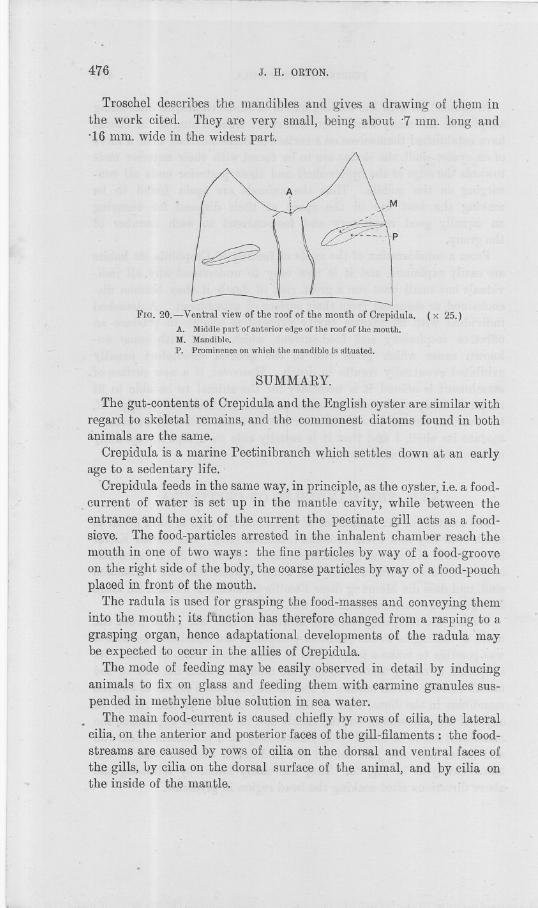
. .
476 J. H. ORTON.
Troschel describes the mandibles and gives a drawing of them inthe work cited. They are very small, being about '7 mm. long and'16 mm. wide in the widest part.
~
FIG. 20.- Ventral view of the roof of the mouth of Crepidula. (x 25.)A. Middle part of anterior edge of the roof of the mouth.M. Mandible.P. Prominence on which the mandible is situated.
SUMMARY.
The gut-contents of Orepidula and the English oyster are similar withregard to skeletal remains, and the commonest diatoms found in bothanimals are the same.
Orepidula is a marine Pectinibranch which settles down at an earlyage to a sedentary life.
Orepidula feeds in the same way, in principle, as the oyster, i.e. a food.. current of water is setup in the mantle cavity, while between the
entrance and the exit of the current the pectinate gill acts as a food.sieve. The food-particles arrested in the inhalent chamber reach themouth in one of two ways: the fine particles by way of a food-grooveon the right side of the body, the coarse particles by way of a food-pouchplaced in front of the mouth.
The radula is used for grasping the food-masses and conveying theminto the mouth; its function has therefore changed from a rasping to agraspil1g organ, hence adaptational developments of the radula maybe expected to occur in the allies of Orepidula.
The mode of feeding may be easily observed in detail by inducinganimals to fix on glass and feeding them with carmine granules sus-pended in methylene blue solution in sea water.
The main food-current is caused chiefly by rows of cilia, the lateralcilia, on the anterior and posterior faces of the gill-filaments: the food-streams are caused by rows of cilia on the dorsal and ventral faces ofthe gills, by cilia on the dorsal surface of the animal, and by cilia onthe inside of the mantle.
-
FEEDING OF CREPIDULA. 477
The gill of Crepidula, like that of Lamellibranchs, is at the same timea respiratory organ, a water-pump and a food-sieve.
The food-streams of Crepidula are comparable to those of Lamelli-branchs.
A partial selection of the 'coarser food-particles is effected in theoyster-and Lamellibranchs generally-in the same way in principle asin Crepidula.
There are special morphological arrangements in Mytilus and Cardiumto assist in the automatic selection of the finer food-particles.
The main food and respiratory stream in Lamellibranchs is caused bythe" lateral" cilia on the gill-filaments, while the collection and trans-portation of food is effected mainly by the frontal cilia of the filaments.The latero-frontal cilia in Nucula, Anomia, Mytilus, Tapes, 'andCardium, and therefore probably in all the Lamellibranchs. in whichthey occur,are true straining cilia. .
The gill-leaflets of Nucula and most Protobranchia possess. similarcilia having a similar function to those on the gills of higher Lamelli-branchs, and those of Nuc1da nucleus have also' ciliated discs.
The gill of Nucula divides the mantle cavity into infra- and supra-branchial chambers, and the ventral surface Q.fthe gill is used forfood-collection in the same way as in higher Lamellibranchs., Thusthe gill of Nucula is essentially similar to that of some Filibranchs- -for example, some species of Anomia and Dimya.
Hence the Protobranchia cannot now be considered as a groupco-equal with the Filibranchia, and should be elevated to a sub-divisionof the Filibranchia.
Evolution in Lamellibranchs, which has occurred mainly on th~principle of folding a)1d consolidating the gill-filaments, comprises aseries of adaptations tending towards a more perfect mode offeeding.
Oalyptraea chinensis feeds in exactly the same way as Crepidula, andOapulus hunga1'imtsfeeds in a similar way.
Doubtless, therefore, all the Calyptraeidae, all the Capulidae, andthere is reason to suspect that all sedentary Pectinibranchs, feed inthe same or in a similar manner.
Most Gastropods have gill-filaments essentially the same in structureand function as those of Lamellibranchs, i.e. lateral cilia occur gene-rally and produce the main current in the mantle cavity; frontal andab-frontal cilia are found, and these collect food-particles from theingoing current. In many Gastropods, and probably in most branchiateforms, the mantle cavity is divided by the gill into inhalent andexhalent chambers.
,
j
j..J
478 J. H. ORTON.
The manner of chaining in Crepidula is adapted to securing a goodfood and respiratory current. .
The mandibles in Orepidula are to be found just 15ehindand a littleto eitter side of the middle of the anterior border of the roof of themouth.
The author wishes here to express his thanks and indebtedness tothe Government Grant Oommittee for a grant out of which some ofthe expenses of the foregoing work were defrayed. He is also gratefulto Dr. Allen for useful criticism.
LITERATURE LIST.
1: E. G. CONKLIN.--Journalof Morphology. Vol. XIII, p. 11, 1897.2. J. H. ORTON.-P1'OC.Roy. Soc. B. Vol. LXXXI, p. 469, 1909.3. HENRI VAN HEURCK.--A Treatise on the Diatomaceae. Translated by
Wynne E. Baxter. London, 1896.4. H. H. GRAN.-Nordisches Plankton. Diatomeen. Dritte Lieferung. XIX.
Kiel und Leipzig, 1905.5. B. HALLER.-Morph. Jahrbuch. Vol. XVIII, p. 498, 1892. .6. M. STENTA.-Zur Kenntnis de1'St1'ihnungenin Mantelraume de1' Lamelli-
branchiaten. Arbeiten Zoolog. Instituten, Wien, XIV. 1903.7. J, L. KELLoGG.-ShellFish Industries. American Nature Series, 1910.8. J. L. ;KELLOGG.-Science(U.S.). Vol. XI, No. 266, pp. 172-3. 1900.9. J. L. KELLOGG.-Bull. U.S.]? Commission. Vol. X. 1890 (1892).
10. W. A. HERDMANand J. HORNELL. Ceylon Pearl Oyster Fisheries and Marine
Biology. Part II, pp. 45-6, 61, with figs. Royal Society, 1904.11. G. A. DREw.-Memoirs from the Biological Labomtory of the Johns Hopkins
University. Vol. IV, No.3. 1899.12. P. PELSENEER.-Mollusca. Vol. V, p. 253. wnkester's 'l'reatise of Zoology,
1906.13. W. G. RIDEWOOD.-Phil.Tmns. Roy. Soc., London. Vol. CXCV, pp. 147-
284. 1903.14. R. H. PEcK.-Quart. Journ. Micro.Sci. Vol. XVII, pp. 43-66. Plllte IV,
1877.15. A. SEDGWICK.-AStudent's Text-bookof Zoology. Vol. I, p. 345. 1898.16. K. A. ZITTEL.-Text-boolcof Palaeontology. Vol. I, p. 359. Translated by
Charles R. Eastman, 1900.17. P. PELSENEER.-Contributiona !'etudedes Lamellibranches' Arch. de Biol.,
Vol. XI. 1891.18. F. H. TROSCIIEL.-Gebissder Schneckenzur Beg1"iindungeiner Naturlichen
Classification. Vol. I, p. 157. 1856-63.19. A. BOLLEsLn.-The Mic1'otomist'sVadeMecum, p. 76. Sixth Edition. ]905.
PUBLIOATIONS OF THE ASSOOIATION.~
Journalof the MarineBiologicalAssociation.OLD SERIES.
No.1, AUGUST, 1887 (only a few copies left, reserved for Libraries).No.2, AUGUST, 1888. Price ]8.
NEW SERIES (Royal 8vo).
Volume 1., 1889-90, 472 pp., 28 plates.
Volume II., 1891-2,410 pp., 14 plates.
Volume IlL, 1893-4, xxxviii. and 458 pp., 5 plates and 25 woodcuts.Volume IV., 1895-7, iv. and 425 pp.
Volume V., 1897-9, 550 pp. and 16 plates.
Volume Vr., ] 899-1903, 676 pp., 3 charts and 7 plates.
Volume VIL, 1904-6,508 pp., 1 chart and 12 plates.
Volume VIIL, 1907-10, 519 pp. and 24 plates.Volume IX., Nos. 1,2,3,
Separate numbers (generally 4 to one volume), in wrappers, from 18. to 58. each,according to size.
London Agents: Messrs. DULAU& Co., 37 S?ho Square, W.
Cloth, 4to, 150 pp., 18 plate8 (\2 coloured).
A TREATISE ON THE OOMMON SOLE.BY
J. T. CUNNINGHAM, M.A., F.R.B.E.,Late Fe]]ow of University Oo]]ege, Oxford; Naturalist to the Association.
Price to Members, 208.; to Non-Members, 258.
Mediuln 8vo, 368 pages. 159 Illustmtions and two Maps. Price 7s. 6d. net.(Macrnillan &: Qo., London.)
THE NATURAL HISTORY OF THE MARKETABLEMARINE FISHES OF THE BRITISH ISLANDS.
Prepared expressly. for the use of those interested in the Sea-fishing Industries,BY
J. T. OUNNINGHAM, M.A.,FORMERLY FELLOW OF UNIVERSITY COLLEGE, OXFORD;
NATURALIST ON THE STAFF OF THE MARINE BIOLOGICAL ASSOCIATION.
'WtitlJ Jref:u:e hllE. RAY LANKESTER, M.A., LL.D., F.R.S.,
PROFESSOIl OF COMPAIlATIVE ANATOMY IN THE UNIVERSITY OF OXFOIlD.
INTERN ATIONAL FISHERYINVESTIGATIONS.
THE following Reports on work carried out by the MarineBiological Association have been published by H.M. StationeryOffice, and may be obtained from Messrs. WYMAN& SONS,Ltd.,Fetter Lane, London, E.C., or through any Bookseller.
NORTH SEA FISHERIES INVESTIGATION OOMMITTEE.REPORT (No.2-SOUTHERN AREA)
ON
FISHERY. AND HYDHOGRAPHICAL INVESTIGATIONS INTHE NORTH SEA AND ADJAOENT WATERS.
1902-1903.
[Being the First Report by the Marine Biological Association of the UnitedKingdom.] .
Published 1905. Blue Book Cd. 2670. Price 813.9d.
SECOND REPORT (SOUTHERN AREA)ON
FISHERY AND HYDROGRAPHICAL INVESTIGATIONS INTHE NORTH SEA AND ADJAOENT WATERS.
1904-1905.
[Being the Second Report by the Marine Biological Association of the United. Kingdom.]
PART 1. Published 1907. Blue Book Cd. 3837. Price 513.Id.PART II. Published 1909. Blue Book Cd. 4641. Price 813.9d.
THIRD REPORT (SOUTHERN AREA)ON
FISHERY AND HYDROGRAPHICAL INVESTIGATIONS INTHE NORTH SEA AND ADJ ACENT WATERS.
1906-1908.
[Being the Third Report by the Marine Biological Association of the UnitedKingdom.]
Published 1911. Blue Book Cd. 5546. Price 513.10d.
FOURTH REPORT (SOUTHERN AREA)ON
FISHERY AND HYDROGRAPHICAL INVESTIGATIONS INTHE NORTH SEA AND ADJAOENT WATERS.
1909.
[Being the Fourth Report by the Marine Biological Association of the UnitedKingdom.] .
Puuli8hed 1912. Blue Book Cd. 6125. Pdce 1313.Od.
/
OBJEOTSOF THE
marine 18iologicaI .association.OF THE UNITED KINGDOM.
THE ASSOOIATION was founded at a Meeting ca1led for the purpose in March, 1884,and held in the Rooms of the Royal Society of London.
The late Professor HUXLEY, at that time President of the Royal Society, took the chair,and amongst the speakers in support of the project were the late Duke of AHGYLL, the
late Sir Lyox PLAYFAIH, Lord AVEBURY, the late Sir JOHEPH HOOKER, the late Dr.CAHPENTER, Dr. GUNTHER, the late Lord DALHOUSIE, the late Professor MOSELEY, the lateMr. RO~!AXES, and Sir E. RAY LAXKESTER.
The Association owes its existence and its present satisfactory condition to a comhina-
tion of scientific naturalists, and of gentlemen who, from philanthropic or practical reasons,are specially interested in the great sea fisheries of the United Kingdom. It is universally
admitted that our knowledge of the habits and conditions of life of sea fishes is very sma1l,
and insufficient to enable either the practical fisherman or the Legislature to take measures~alculated to ensure to the country the greatest retmn from the" harvest of the sea."
Naturalists are, on the other hand, anxious to push further our knowledge of marine life
and its couditions. Heuce tl~e Association has erected at Plymouth a thoroughly efficientLaboratory, where naturalists may study the history of marine animals and plants in general,and where researches ou food.fjshes and molluscs may be carried out with the J)est,appliances.
The Laboratory and its fittings were completed in June, 1~88, at a cost of some £12,000.
Since that time investigations, practical and scientific, have been constantly pursued at
Plymouth. Practical investigations upon matters connected with sea-fishing are carried onunder the direction of the Oouncil; in addition, naturalists from England and from abroadhave come to the Laboratory, to carryon their own indepcndent researches, and have made
valuable additions to zoological and botanical science, at the expense of a small rent for the
use of a working table in the Laboratory and other appliances. The number of naturalists
who can be employed by the Association in special investigations on fishery questions, and
definitely retained for the purpose of carrying on those researches throughout the year,must depend on the funds subscribed by private individuals and public bodies for thepurpose. . The first charges on the revenue of the Association are the working of the sea.
water circulation in the tanks, stocking the tanks with fish and feeding the latter, the
payment of servants and fishermen, the hire and maintenance of fishing-boats, and thesalary of the Resident Director and Staff. At the commencement of this number will be
found the names of the gentlemen on the staff.
The purpose of the Association is to aid at the same time both seience ami industry. Itis national in character and constitution, allii its atJkirs are condu~ted by a representative
Council, by an Honorary Secretary and an Honorary Tl'easurer, without any. charge uponits funus, so that the whole of the. subscriptions and donations received are devoted
absolutely to the support of the Laboratory and the prosecution of researches by aid ofits appliances. The reader is referred to page 4 of the Oover for il1formation as tomembership of the Association.
CONTENTS OF NEW SERIES, Vol. IX., No.3.
1. SmIlE CASES OF NEW GROWTHS IN FISH.
Plate IV .
By G. HAROLD DREW.PAGE
With. 281
2. NOTES ON THE RESPIRATORY 'MECHANISM OF CORYSTES CASSIVELAUNUS.
By. KATHLEEN E. ZUDIEl\MANN. With Plate V . 288
3. ON THE F AU"A OF THE OUTER 'VESTEl\N AREA OF THE ENGT,ISH CHAN"EL.
By L. R. CRAWSHAY, With Plate VI . 292
4. SOME NOTES UPON THE FEEDING HABITS OF MACKEREL AXD CERTAIN
CLUPEOIDS IN THE ENGI,ISH CHANNEL. By G. E. BULLEN . 394
5. ON THE OCCURRENCEOF A NORTHER" HYDROIIJ HALATRACTUS (CORYlINRPHA)
NANUS (ALDER) AT PLYMOUTH. By DR. E. STECHOW. W'ith Plate VII and1 Text Figure . 404
6. LIST OF NEMERTEANS COLLECTED IN THE KEIGHBOURHOOD OF PLYMOUTH
FP:OM MAy-SEPTEMBER, 1910. By DR. GERAIlJ)A WYNHOFF. With 1
Text Fig/we . 407
7. ON A METHOD OF REARrNG LAHVAE OF POLYZOA. By DR. M. HASPER . 435
8. AN ACCOUXT OF THE N.-\.TURAL HISTORY OF TilE SLIPPER-LniPET (CREPlDULA
FORNICATA), WITH SOME REMAltKS ON ITS OCCURRENCE ON THE OYSTER
GROUNDS ON THE ESSEX COAST. By.r. H. ORTON. . 437
9. THE MODE OF FEEDING OF CREPJDULA, WITH AN ACCOUNT OF THE CURRENT-
j'IWDUCING MECHANIS1<I IN THE MANTLE CAVITY, AND SOME RE}IARKS ON
THE MODE OF FEEDING IN GASTROPODS AND LAMELLIBRANCHS. By J. H.
ORTOX. With 20 Text Figures . 444
NOTICE.The Council of the Marine Biological Association wish it to be understood that
they do not accept responsibility for stat€ments published in this J oumal except-ing when those statements are contained in an official report of the Council.
TERMS OF MEMBERSHIP.
Aromal MembersLife MembersFoundersGovernors
. per annumComposition Fee
.£ s. d.1 1 0
15 15 0100 0 0500 0 0
Members of the Association have the following rights and privileges: they electannually the Officers and Council; they receive the J oumal of the Association freeby post; they are admitted to view the Laboratory at Plymouth, and may introducefriends with them; they have the first claim to rent a place in the Laboratory forresearch, with use ,of tanks, boats, &c. ; and have access to the books in the Libraryat Plymouth.
All correspondence should be addressed to the Director, The Laboratory,Plymouth.