A Data Dissemination Algorithm A Data Dissemination Algorithm for Opportunistic Networks
Upload
massgeneralCategory
view
0download
0
Oral Mycobiome Analysis of HIV-Infected Patients:Identification of Pichia as an Antagonist of OpportunisticFungiPranab K. Mukherjee1,2, Jyotsna Chandra1, Mauricio Retuerto1, Masoumeh Sikaroodi3, Robert E. Brown3,
Richard Jurevic3, Robert A. Salata4, Michael M. Lederman5, Patrick M. Gillevet3,
Mahmoud A. Ghannoum1,2*
1 OHARA/ACTG Mycology Unit at Case Western Reserve University, Department of Dermatology, Cleveland, Ohio, United States of America, 2 Center for Medical
Microbiology, Department of Dermatology, School of Medicine, Case Western Reserve University and University Hospitals Case Medical Center, Cleveland, Ohio, United
States of America, 3 Microbiome Analysis Center, Department of Environmental Science and Policy, George Mason University, Fairfax, Virginia, United States of America,
4 Department of Biological Sciences, School of Dental Medicine, Case Western Reserve University, Cleveland, Ohio, United States of America, 5 Division of Infectious
Diseases and HIV Medicine, Case Western Reserve University, University Hospitals Case Medical Center, Cleveland, Ohio, United States of America
Abstract
Oral microbiota contribute to health and disease, and their disruption may influence the course of oral diseases. Here, weused pyrosequencing to characterize the oral bacteriome and mycobiome of 12 HIV-infected patients and matched 12uninfected controls. The number of bacterial and fungal genera in individuals ranged between 8–14 and 1–9, amonguninfected and HIV-infected participants, respectively. The core oral bacteriome (COB) comprised 14 genera, of which 13were common between the two groups. In contrast, the core oral mycobiome (COM) differed between HIV-infected anduninfected individuals, with Candida being the predominant fungus in both groups. Among Candida species, C. albicanswas the most common (58% in uninfected and 83% in HIV-infected participants). Furthermore, 15 and 12 bacteria-fungipairs were correlated significantly within uninfected and HIV-infected groups, respectively. Increase in Candida colonizationwas associated with a concomitant decrease in the abundance of Pichia, suggesting antagonism. We found that Pichiaspent medium (PSM) inhibited growth of Candida, Aspergillus and Fusarium. Moreover, Pichia cells and PSM inhibitedCandida biofilms (P = .002 and .02, respectively, compared to untreated controls). The mechanism by which Pichia inhibitedCandida involved nutrient limitation, and modulation of growth and virulence factors. Finally, in an experimental murinemodel of oral candidiasis, we demonstrated that mice treated with PSM exhibited significantly lower infection score(P = .011) and fungal burden (P = .04) compared to untreated mice. Moreover, tongues of PSM-treated mice had few hyphaeand intact epithelium, while vehicle- and nystatin-treated mice exhibited extensive fungal invasion of tissue with epithelialdisruption. These results showed that PSM was efficacious against oral candidiasis in vitro and in vivo. The inhibitory activityof PSM was associated with secretory protein/s. Our findings provide the first evidence of interaction among members ofthe oral mycobiota, and identifies a potential novel antifungal.
Citation: Mukherjee PK, Chandra J, Retuerto M, Sikaroodi M, Brown RE, et al. (2014) Oral Mycobiome Analysis of HIV-Infected Patients: Identification of Pichia asan Antagonist of Opportunistic Fungi. PLoS Pathog 10(3): e1003996. doi:10.1371/journal.ppat.1003996
Editor: Deborah A. Hogan, Geisel School of Medicine at Dartmouth, United States of America
Received December 28, 2013; Accepted January 21, 2014; Published March 13, 2014
Copyright: � 2014 Mukherjee et al. This is an open-access article distributed under the terms of the Creative Commons Attribution License, which permitsunrestricted use, distribution, and reproduction in any medium, provided the original author and source are credited.
Funding: This study was supported by funds from the NIDCR to MAG [RO1DE17846 and the Oral HIV AIDS Research Alliance (OHARA), grant number BRS-ACURE-S-11-000049-110229], NIAID/NEI to PKM (NEI/R21EY021303 and NIAID/R21AI074077), pilot funding from the Infectious Diseases Drug Development Center(IDDDC, Case) to PKM, and the CWRU/UH Center for AIDS Research (CFAR, NIH grant number P30 AI036219). The funders had no role in study design, datacollection and analysis, decision to publish, or preparation of the manuscript.
Competing Interests: The authors have declared that no competing interests exist.
* E-mail: [email protected]
Introduction
Organisms residing in the oral cavity (oral microbiota)
contribute to health and disease, and influence diseases like oral
candidiasis, the most common oral complication of HIV-infection
[1,2]. Pathogenesis of oral candidiasis is linked to variables like
changes in the CD4+ cell count and antiretroviral therapy (ART)
in HIV-1-infected patients [3]. Although the introduction of ART
has reduced mortality and morbidity as well as the incidence of
opportunistic infections among HIV-infected patients, oral candi-
diasis remains a significant disease, even in the era of ART. In this
regard, recent studies indicate that the decline of oral candidiasis
among ART-experienced HIV-infected patients is transient in
some HIV-infected individuals [4]. In addition, preliminary results
reported by Thompson et al. [5] showed that symptomatic oral
Candida infection occurred in one-third of patients with advanced
AIDS (n = 122), even in the setting of ART. More recently, Patel
et al. [6] reported symptomatic oral candidiasis in 27% (59/215)
HIV-infected patients. Therefore, even in the era of ART, oral
candidiasis remains a significant problem.
Characterization of the microbiota (bacteriome and myco-
biome) in health and disease is expected to expedite the discovery,
testing and validation of novel drugs [7]. Most studies that
characterized the human microbiome in health and disease have
focused on the bacteriome, in both oral and non-oral body sites
[8–12]. Recently, Iliev et. al. [13] showed that while no significant
PLOS Pathogens | www.plospathogens.org 1 March 2014 | Volume 10 | Issue 3 | e1003996
differences in major phyla of commensal bacteria was observed in
the intestinal microflora between wild-type and mice lacking
Dectin-1, members of the mycobiome interacted with the
intestinal immune system to influence inflammatory bowel disease
(IBD) highlighting the role of the fungal community in disease.
Earlier, we characterized the oral mycobiome in healthy
individuals using high-throughput multitag pyrosequencing
(MTPS), and reported that humans are colonized with up to 85
fungal genera [14]. Although these studies demonstrated the
complexity of the human oral microbiome, the specific contribu-
tion of the mycobiome to oral diseases was not investigated.
Previous studies have shown that alteration in the bacterial
population has direct impact on the development of Candida infections
[for reviews, see 15]. However, the interactions between members of
the oral microbiota and Candida in HIV disease setting have not been
investigated. In the current study, we identified the core oral
mycobiome (COM) and bacteriome (COB) [defined as those
organisms present in $20% of the subjects] in HIV-infected and
uninfected individuals, and demonstrated that the COM undergoes a
change in HIV disease. Furthermore, we noted that a decrease in
abundance of the yeast Pichia coincided with an increase in Candida
colonization, suggesting an antagonistic relation between these two
fungi. We also found that nutrient competition as well as Candida
growth and modulation of its virulence factors by Pichia is a
mechanism underlying this interaction. In addition, treatment with
Pichia Spent Medium (PSM) was efficacious against oral candidiasis
when tested in an experimental murine model. Our results provide the
first evidence of interaction among members of the oral mycobiome
community, particularly between Pichia and pathogenic fungi. These
findings could lead to the development of novel antifungals to prevent
and treat fungal infections including mucosal Candida infections,
thereby impacting the management of oral candidiasis and other
fungal infections. They also highlight the need to characterize the
mycobiome at different body sites which may lead to novel discoveries
of how fungi can be exploited to control health and disease.
Materials and Methods
Ethics StatementWritten informed consent was obtained from all study
participants. Study participants were recruited according to
protocol (#20070413) approved by the Human Subjects Institu-
tional Review Board (IRB) at University Hospitals Case Medical
Center. Oral rinse samples were obtained from 12 HIV-infected
and 12 uninfected individuals (matched for age, sex, and ethnicity).
Inclusion criteria for these participants were: .18 years of age and
no clinical signs of oral mucosal disease including oral candidiasis,
while the exclusion criteria were: recent use of antimicrobial or
antifungal agents (within a month), use of topical or systemic
steroids, pregnancy, and insulin-dependent diabetes mellitus. Oral
wash samples were collected using a standardized Standard
Operating Procedure developed by the Oral HIV/AIDS Research
Alliance (OHARA) as described earlier [14]. All animal experi-
mentation was performed in strict accordance with the Guide for
the Care and Use of Laboratory Animals of the National Institutes
of Health. The protocol for animal infection was approved by the
Institutional Animal Care and Use Committee (IACUC) at Case
Western Reserve University School of Medicine, Cleveland, Ohio
(protocol approval number 2013-0020). All procedures were
performed under general anesthesia and all efforts were made to
minimize animal suffering.
Microbiome AnalysisOral rinse samples were processed individually using the Fast
DNA Spin Kit following manufacturer’s instructions (BIO 101;
Vista, CA). Each extraction tube was agitated three times using a
Fast Prep FP120 instrument at a speed setting of 5 for 30 s. Tubes
were cooled on ice between agitations. Fungi and bacteria present in
these samples were identified with ITS-based and 16S probes,
respectively. The ITS1 region from DNA sample extracts was
amplified in triplicate using primers with high specificity for
ascomycete fungi (fluorescently labeled forward primer ITS1F
(CTTGGTCATTTAGAGGAAGTAA) and unlabeled reverse
primer ITS2 (GCTGCGTTCTTCATCGATGC). The ITS prim-
ers were selected in this study to detect the presence of various fungi
since these primers are able to detect consensus sequences present in
a broad range of fungi [16,17]. For bacterial identification,
extracted DNA was amplified by PCR using routinely employed
universal primers [fluorescently labeled forward primer 27F (59-
6FAM- AGAGTTTGATCCTGGCTCAG-39) and unlabeled re-
verse primer 355R59 (59- GCTGCCTCCCGTAGGAGT-39)]
[18], which amplify the first two hyper-variable regions of 16S
rRNA [19] and are commonly used for microbiome analysis
[10,20]. Microbiome analysis was performed using multitag 454
pyrosequencing (MTPS) technique, which was used for character-
ization of nucleic acids [21] (for details, please see Method S1).
StrainsCandida albicans [strains SC5312, 10341, GDH2346), Pichia
(MRL81)], Penicillium (MRL22345) and Cladosporium (MRL1458)
strains tested in this study were obtained from the OHARA
Repository at Case and the culture collection of the Center for
Medical Mycology. Fungal strains were maintained on Sabouraud
dextrose agar (SDA, [yeast extract, peptone, and dextrose at 1:2:1])
(Difco Laboratories, Detroit, MI) medium. Since species-level
identification of Pichia based upon morphological or physiological
features alone is usually not possible, we used a molecular approach
(based on sequence analysis of the internal transcribed spacer and
D1/D2 ribosomal DNA regions) [22] to confirm the identity of
Pichia MRL81 strain. Our analysis revealed that this strain was P.
farinosa. All strains were kept at 280uC for long-term storage.
Role of Nutrient LimitationTo determine whether inhibition of pathogenic fungi by Pichia
spent medium (PSM) is due to nutrient competition between Pichia
Author Summary
Oral microbiota contribute to health and disease, and theirdisruption may influence the course of oral diseases likeoral candidiasis. Here we identify the core oral mycobiome(COM) and core oral bacteriome (COB) in HIV-infected anduninfected individuals, and demonstrate that the COMdiffers between these two groups. Decrease in abundanceof Pichia (a resident oral fungus) in uninfected individualscoincided with increase in abundance of Candida, sug-gesting an antagonistic relationship. In vitro testingshowed that Pichia spent medium (PSM) inhibits growthof pathogenic fungi; these findings were validated in anexperimental mouse modal of oral candidiasis. Themechanism by which Pichia antagonizes Candida involvesnutrient competition and secretory factor/s that inhibit thelatter’s ability to adhere, germinate, and form biofilms. Thisstudy is the first to characterize the mycobiome and thebacteriome in the oral cavity of HIV infected patients, andprovides the first evidence that a fungus present in thesame host microenvironment antagonizes Candida andidentifies potential novel antifungal approach.
Oral Microbiome in HIV Disease Setting
PLOS Pathogens | www.plospathogens.org 2 March 2014 | Volume 10 | Issue 3 | e1003996
and Candida, we assessed Candida growth when mixed with Pichia at
different ratios. P. farinosa and a GFP-tagged C. albicans strain
(generous gift from Dr. B. Cormack) [23] were mixed together at
equivalent cell densities (103, 104, or 105 cells each) in yeast
nitrogen base (YNB) medium supplemented with glucose (0.9%
w/v) and uridine (100 mg/mL). These mixed cell suspensions were
incubated at 37uC and allowed to grow (as ‘‘mixed cultures’’).
Candida or Pichia cells alone were grown in the same growth
medium (‘‘mono cultures’’) serving as controls. At specific time
intervals (24, 48, 72 h), 1-mL aliquots were withdrawn from the
mixed- and mono-cultures, centrifuged to collect the cells in pellet,
and re-suspended in 250 mL phosphate buffered saline. Fifty mL of
the resuspended cell mixture was placed on a glass slide and
observed under phase-contrast and fluorescence microscope. The
remaining cell suspension (200 mL) was transferred to a 96-well
microtiter plate, and fluorescence was measured using a spectro-
fluorimeter (excitation and emission wavelength of 485 and
435 nm, respectively).
Biochemical Characterization of Pichia Spent Medium(PSM)
The effect of Pichia supernatant on Candida growth, germination,
adherence, biofilm formation, and its biochemical properties was
determined as described below.
Effect of Pichia Supernatant on the Growth of Candida,Aspergillus, and Fusarium
To evaluate the effect of Pichia supernatant on the growth of
various fungi, Pichia spent (‘‘conditioned’’) medium (PSM) was
obtained by centrifuging 100-mL culture of Pichia grown in
Sabouraud dextrose broth (SDB) for 48 h, and filter sterilizing it.
Next, fungal cells/conidia (16105 cells/mL) were incubated with
PSM at 35uC and growth was followed for 48 h. Aliquots were
collected at 2 h intervals and fungal growth was measured
spectrophotometrically at 600 nm.
Germination AssayEffect of Pichia spent media (PSM) on Candida germination was
determined using C. albicans strain SC5314, as described previously
[24]. Candida cells were grown planktonically in the absence or
presence of PSM. Germination rate was compared with that of
cells grown in SDB media containing fetal bovine serum (FBS)
(Hyclone, Thermo Fisher Scientific, Rockford, IL), a known
inducer of germination. Briefly, C. albicans cells were grown and
the cell density was adjusted to 16107 cells/mL in Hanks balanced
salt solution (HBSS) (Mediatech). In separate 1.5-mL tubes, 50 mL
of these cells were diluted to a density of 56105 cells/mL in HBSS
(blank control), FBS, or PSM, and incubated on a rocker at 37uCfor up to 4 h. At 15-min intervals, 10-mL samples from each media
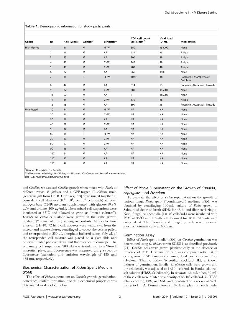
Table 1. Demographic information of study participants.
Group ID Age (years) Gender1 Ethnicity*CD4 cell count(cells/mm3)
Viral load(U/mL) Medication
HIV-infected 1 31 M H (W) 380 158000 None
2 56 M AA 639 75 Atripla
3 52 M AA 800 48 Atripla
4 40 M C (W) 947 48 Atripla
5 40 M C (W) 280 48 Atripla
6 22 M AA 966 1100 None
7 31 F H (W) 1029 48 Rotanivir, Fosamprenavir,Combivir
8 42 M AA 814 53 Rotanivir, Atazanavir, Truvada
9 22 M C (W) 581 115000 None
10 52 M AA 5 185000 None
11 31 M C (W) 670 68 Atripla
12 45 M AA 899 48 Rotanivir, Atazanavir, Truvada
Uninfected 1C 34 M H (W) NA NA None
2C 46 M C (W) NA NA None
3C 59 M AA NA NA None
4C 22 M C (W) NA NA None
5C 37 M AA NA NA None
6C 34 F H (W) NA NA None
7C 40 M C (W) NA NA None
8C 27 M C (W) NA NA None
9C 53 M AA NA NA None
10C 44 M AA NA NA None
11C 22 M AA NA NA None
12C 47 M AA NA NA None
1Gender: M – Male, F – Female.*Self-reported ethnicity: W = White; H = Hispanic; C = Caucasian; AA = African-American.doi:10.1371/journal.ppat.1003996.t001
Oral Microbiome in HIV Disease Setting
PLOS Pathogens | www.plospathogens.org 3 March 2014 | Volume 10 | Issue 3 | e1003996
type were microscopically examined using a hemacytometer. Total
cell count and germination (defined as a germ-tube length greater
than or equal to the blastospore diameter) was determined from an
average of 4 observations. One hundred to 200 cells were counted
per observation. The assays were discontinued when cells clumped
together, due to germination, which made it difficult to count
individual cells.
Adhesion AssayThe effect of Pichia or Penicillium (used as a control) cells or
supernatant on Candida adherence (using strain C. albicans SC5314, a
clinical isolate used conventionally in Candida adhesion and
germination assays) was determined as described earlier [24,25].
Briefly, standardized suspensions of 50 to 200 cells/mL were added
onto silicone elastomer disks for 90 min. Disks were then washed in
phosphate-buffered saline (PBS) to remove non-adherent cells and
placed in wells of 12-well tissue culture plates (Becton Dickinson,
Franklin Lakes, NJ). Two milliliters of warm (55uC) liquefied SDA
was added per well to completely cover the SE disks and allowed to
solidify. Plates were incubated overnight (37uC), and the number of
colonies adhering per disk was counted using a dissecting microscope.
Biofilm EvaluationThe effect of oral fungi (Pichia, Cladosporium or Penicillium, the
latter fungi were used as controls since Cladosporium was present
only in the uninfected subjects like Pichia, while Penicillium was
present in both infected and uninfected subjects to the same
abundance) or their supernatant on the ability of Candida to form
biofilms was evaluated using metabolic activity assay and confocal
microscopy, as described earlier [26–28]. Briefly, Candida cells
were incubated in the presence or absence of Pichia cells or spent
medium (supernatant, PSM) at different relative ratios (1:3, 1:1,
3:1), and allowed to form biofilms for 48 h on silicone elastomer
catheter discs. The amount of biofilm formed was assayed
colorimetrically using the XTT (2,3-bis (2-methoxy-4-nitro-5-
sulfophenyl)-5-[(phenylamino) carbonyl]-2H-tetrazolium hydrox-
ide, Sigma-Aldrich) metabolic activity assay in which XTT is
converted by metabolically active cells to a red formazan product
[26]. In addition, the effect of fungal supernatants on the
morphology and architecture of the formed biofilms was evaluated
using confocal scanning laser microscopy (CSLM) [26]. Briefly,
biofilms were stained with the fluorescently labeled polysaccha-
ride-indicating lectin Concanavalin Alexa Fluor 488 conjugate
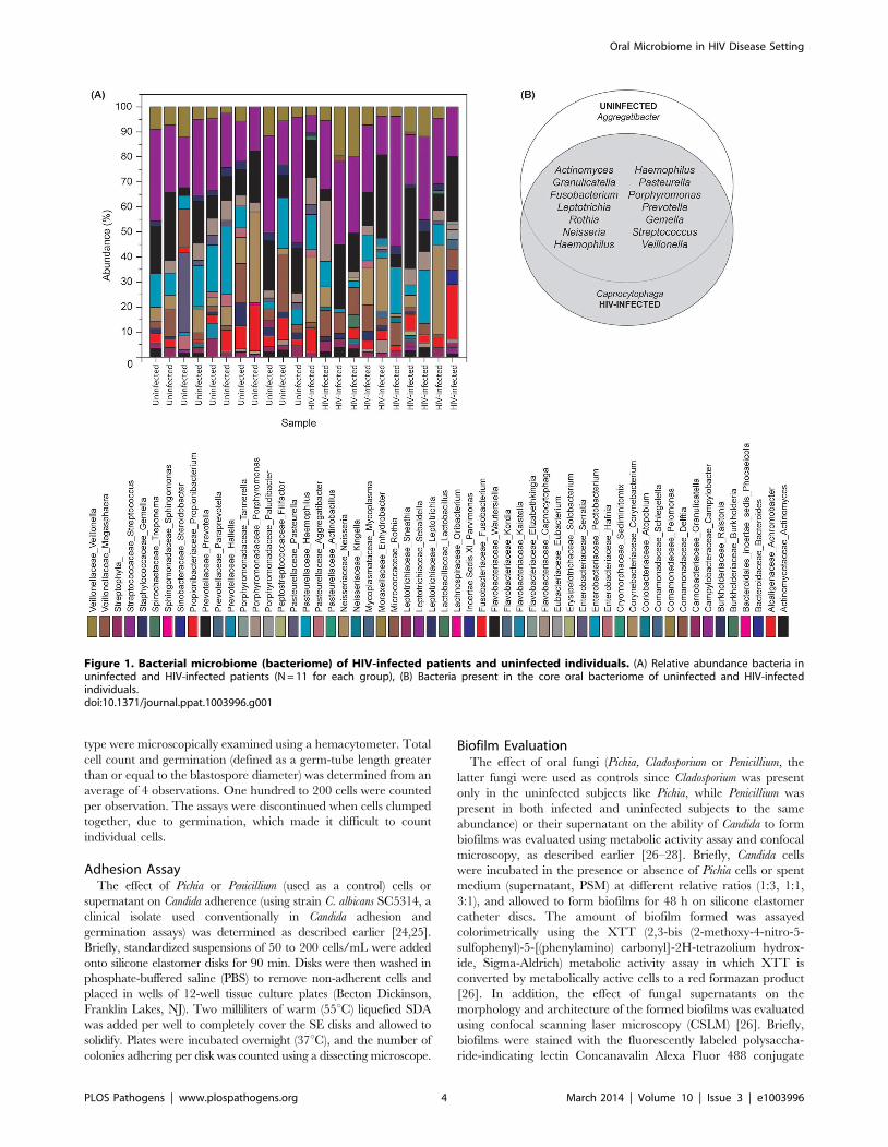
Figure 1. Bacterial microbiome (bacteriome) of HIV-infected patients and uninfected individuals. (A) Relative abundance bacteria inuninfected and HIV-infected patients (N = 11 for each group), (B) Bacteria present in the core oral bacteriome of uninfected and HIV-infectedindividuals.doi:10.1371/journal.ppat.1003996.g001
Oral Microbiome in HIV Disease Setting
PLOS Pathogens | www.plospathogens.org 4 March 2014 | Volume 10 | Issue 3 | e1003996
(CON-A, 25 mg/mL; Invitrogen) and metabolic activity indicator
dye FUN1TM (10 mM; Invitrogen). After staining, discs containing
biofilms were flipped and placed on a 35-mm-diameter glass-
bottom petri dish (MatTek Corp., Ashland, Mass.). Stained
biofilms were observed with a Zeiss LSM510 confocal scanning
laser microscope equipped with argon and HeNe lasers and
mounted on a Zeiss Axiovert100 M microscope (Carl Zeis, Inc.).
The objective used was a water immersion C-apochromat lens
(406; numerical aperture, 1.2).
In Vivo Model of Oral CandidiasisWild-type C57BL/6 mice (purchased from Charles River
Laboratories, Wilmington, MA) were immunosuppressed with
4 mg of cortisone acetate (Sigma Chemical Co., St. Louis, Mo.)
administered subcutaneously on the day before and 1 and 3 days
after challenge with Candida cells. Mice were given tetracycline
hydrochloride (Sigma Chemical Co., St. Louis, Mo.) in their
drinking water (0.5 mg/ml), starting the day before infection. On
the day of inoculation, mice were anesthetized and light scratches
made on the dorsum of the tongue followed by the introduction of
C. albicans GDH (108 blastospores). The scratches were superficial,
limited to the outermost stratum corneum, and did not cause
trauma or bleeding. Mice were divided into groups (n = 4); treated
with Pichia supernatant, 100 ml in the oral cavity twice a day, a
‘‘mock’’ vehicle control, and untreated control. Topical nystatin
(widely used clinically to treat oral candidiasis [29]) was used as a
comparator. Treatment began on day 4 post inoculation, mice
were sacrificed on day 7 and the tongues harvested for
enumeration of tissue fungal burden or histopathology with
Periodic acid-Schiff stain. Additionally, tongues were visually
assessed daily beginning day 1 post infections to assess severity of
the infection using a previously described scoring system [30]: A
score of 0 indicates the appearance of a normal tongue, with intact
light reflection and no visible signs of infection, a score of 1 denotes
isolated patches of fungus, a score of 2 when confluent patches of
fungus are observed throughout the oral cavity, and a score of 3
indicates the presence of wide-spread fungal plaques and erosive
mucosal lesions. The histology slides were assessed by an
independent pathologist who was blinded to the study arms.
The animal studies were repeated on different days to ensure
reproducibility.
Identification of PSM as a ProteinSince the antifungal activity of PSM was secretory in nature, we
determined whether this activity was due to a protein, carbohy-
drate or small molecule (metabolite). We exposed PSM to
proteinase K (which digests most proteins), NaOH (which
denatures carbohydrates) [31], or acetonitrile extraction (that
isolates metabolites) [32]. We also determined the effect of heat on
PSM activity by exposing it to 90uC temperature in a water bath
for 10 min. The ability of these differently treated PSM to inhibit
Candida biofilms was evaluated as above.
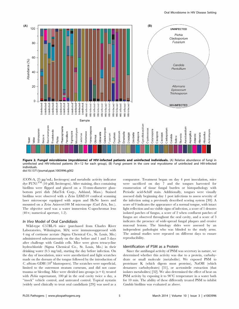
Figure 2. Fungal microbiome (mycobiome) of HIV-infected patients and uninfected individuals. (A) Relative abundance of fungi inuninfected and HIV-infected patients (N = 12 for each group), (B) Fungi present in the core oral mycobiome of uninfected and HIV-infectedindividuals.doi:10.1371/journal.ppat.1003996.g002
Oral Microbiome in HIV Disease Setting
PLOS Pathogens | www.plospathogens.org 5 March 2014 | Volume 10 | Issue 3 | e1003996
Statistical AnalysisMicrobiome data were analyzed using the QIIME and R
platforms [33,34]. Wilcoxon–Mann–Whitney rank sum test was
used for comparison between the two groups, with P-value of ,0.05
considered as a significant difference. For comparison of several
groups, Kruskal–Wallis one-way analysis of variance on ranks was
used, and pairwise multiple comparison procedures (Dunns method)
post-hoc test was used for multiple pairwise comparisons. Univariate
analyses was used to compare the prevalence of specific fungi and
bacteria between the two groups using a Pearson chi-squared test or
two-sample t-test (assuming unequal variances). For correlation
analysis, microbiome abundance data was divided into independent
data matrices (disease and no disease) and correlation analyses was
conducted using the ‘‘psych’’ package (corr.test function) in R
statistical platform (pairwise Spearman’s correlation and two-tailed
probability of t for each correlation) [34,35]. The function
‘‘Circle.corr’’ was used to graphically illustrate the correlation
coefficients and significant correlations (P,.05) in circle graph,
where red and blue circle indicate positive and negative correlations,
respectively. Numerical variables (e.g. metabolic activity, dry
biomass, thickness) were all assessed using paired or unpaired t-
test, or ANOVA as appropriate. Comparison of clinical scores were
performed using box-plots and non-parametric independent samples
Kruskal-Wallis test, while mean fungal burden (log CFU/g) were
compared using ANOVA. All statistical analyses were performed
using R [34] or SPSS (ver. 13) statistical software packages.
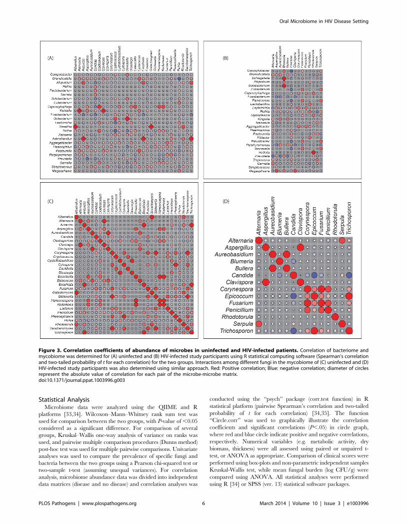
Figure 3. Correlation coefficients of abundance of microbes in uninfected and HIV-infected patients. Correlation of bacteriome andmycobiome was determined for (A) uninfected and (B) HIV-infected study participants using R statistical computing software (Spearman’s correlationand two-tailed probability of t for each correlation) for the two groups. Interactions among different fungi in the mycobiome of (C) uninfected and (D)HIV-infected study participants was also determined using similar approach. Red: Positive correlation; Blue: negative correlation; diameter of circlesrepresent the absolute value of correlation for each pair of the microbe-microbe matrix.doi:10.1371/journal.ppat.1003996.g003
Oral Microbiome in HIV Disease Setting
PLOS Pathogens | www.plospathogens.org 6 March 2014 | Volume 10 | Issue 3 | e1003996
Results
Participant DemographicsA total of 24 individuals were enrolled in the study, with 12
HIV-infected patients and 12 uninfected individuals (11 males and
one female in both study groups, Table 1). The mean age was 38.7
and 38.8 years in HIV-infected (age range: 22–56) and uninfected
(age range: 22–59) groups, respectively. Among the 12 HIV-
infected patients, eight had initiated antiretroviral therapy. In both
study groups, self-reported ethnicities were: six African-Americans,
two Hispanics, and four Caucasians. While all samples were
analyzed for fungal microbiota, one of the samples did not provide
robust signals for the bacterial microbiome, and hence was
excluded from the analysis. In addition, the corresponding
matched uninfected control sample was also excluded. As a result,
there were 12 uninfected-HIV-infected sample pairs for myco-
biome analysis but only 11 sample pairs for bacteriome analysis.
Oral Bacteriome of HIV-infected Participants Were Similarto That of Uninfected Individuals
Our results showed that the number of bacterial genera in the
oral microbiota of study participants ranged between 8–14 per
person among HIV-infected and uninfected individuals. Among
HIV-infected patients, Prevotella, Streptococcus and Rothia were the
most common genera; while in controls the most abundant
bacteria were Prevotella, Streptococcus and Fusobacterium (Fig. 1A, and
Table S1). The core oral bacteriome (COB) consisted of 14
genera in both HIV-infected and uninfected individuals, of which
13 (Actinomyces, Granulicatella, Fusobacterium, Leptotrichia, Rothia,
Neisseria, Haemophilus, Pasteurella, Porphyromonas, Prevotella, Gemella,
Streptococcus, and Veillonella) were common to both groups (Fig. 1B).
We found that Capnocytophaga was present only in HIV-infected
patients while Aggregatibacter was present in uninfected indivi-
duals only (Fig. 1B). These results suggest that the COB of
Table 2. Correlation between bacteriome and mycobiome in uninfected and HIV-infected study participants.
Uninfected Individuals HIV-Infected Patients
Bacteria Fungi P-value Correlation Bacteria Fungi P-value Correlation
Atopobium Antarctic 0.009 0.74 Megasphaera Aspergillus 0.009 0.74
Capnocytophaga Cladosporium 0.001 0.86 Campylobacter Candida 0.023 20.67
Rothia Cladosporium 0.021 0.68 Megasphaera Clavispora 0.009 0.74
Oribacterium Cryptococcus 0.009 0.74 Eubacterium Epicoccum 0.035 0.64
Rothia Cryptococcus 0.048 20.61 Parvimonas Epicoccum 0.035 0.64
Capnocytophaga Emericella 0.009 0.74 Paludibacter Epicoccum 0.035 0.64
Granulicatella Epicoccum 0.025 0.67 Tannerella Epicoccum 0.035 0.64
Pasteurella Epicoccum 0.012 0.72 Capnocytophaga Rhodotorula 0.035 0.64
Atopobium Exophiala 0.009 0.74 Eubacterium Trichosporon 0.035 0.64
Capnocytophaga Hanseniaspora 0.009 0.74 Parvimonas Trichosporon 0.035 0.64
Capnocytophaga Kodamaea 0.009 0.74 Paludibacter Trichosporon 0.035 0.64
Aggregatibacter Lactarius 0.035 0.64 Tannerella Trichosporon 0.035 0.64
Granulicatella Pichia 0.031 20.65
Sneathia Rhodotorula 0.009 0.74
Atopobium Trichosporon 0.009 0.74
For correlation analysis, microbiome abundance data was divided into independent data matrices (disease and no disease) and correlation analyses was conductedusing the ‘‘psych’’ package (corr.test function) in R statistical platform (pairwise Spearman’s correlation and two-tailed probability of t for each correlation) [34,35].doi:10.1371/journal.ppat.1003996.t002
Table 3. Correlation among oral fungi in uninfected studyparticipants.
Fungi Fungi P-Value Correlation
Corynespora Gibberella 0.001 0.98
Gibberella Saccharomyces 0.001 0.98
Cladosporium Emericella 0.035 0.64
Cladosporium Hanseniaspora 0.035 0.64
Cladosporium Kodamaea 0.035 0.64
Cystofilobasidium Penicillium 0.035 0.64
Dioszegia Penicillium 0.035 0.64
Albatrellus Rhodotorula 0.009 0.74
Aspergillus Emericella 0.009 0.74
Aspergillus Hanseniaspora 0.009 0.74
Aspergillus Kodamaea 0.009 0.74
Aureobasidium Epicoccum 0.009 0.74
Clavispora Epicoccum 0.009 0.74
Corynespora Cytospora 0.009 0.74
Corynespora Phaeosphaeria 0.009 0.74
Cryptococcus Davidiella 0.009 0.74
Cytospora Saccharomyces 0.009 0.74
Galactomyces Rhodotorula 0.009 0.74
Gibberella Lactarius 0.009 0.74
Phaeosphaeria Saccharomyces 0.009 0.74
Fusarium Gibberella 0.006 0.77
Corynespora Fusarium 0.005 0.78
Fusarium Saccharomyces 0.005 0.78
doi:10.1371/journal.ppat.1003996.t003
Oral Microbiome in HIV Disease Setting
PLOS Pathogens | www.plospathogens.org 7 March 2014 | Volume 10 | Issue 3 | e1003996
HIV-infected patients was similar to that of uninfected individ-
uals with minimal difference.
Oral Mycobiome of HIV-infected Patients ExhibitsDifferences from Uninfected Individuals
Our results showed that the number of fungal genera present in
oral wash samples ranged between 1–9 per person among
uninfected and HIV-infected individuals (Fig. 2A and Table S2).
Among HIV-infected patients, Candida, Epicoccum, and Alternaria
were the most common genera (present in 92%, 33%, and 25%,
respectively), while in uninfected participants, the most abundant
fungi were Candida, Pichia, and Fusarium (58%, 33%, and 33%,
respectively; Fig. 2A). The COM of HIV-infected and uninfected
individuals consisted of five genera (Fig. 2B); of these, Candida and
Penicillium were common between the two groups, while differing
in the remaining genera demonstrating that the COM of HIV-
infected patients differs from that of age- and sex-matched
uninfected controls. Among the Candida species detected, C.
albicans was the most common (58% in uninfected and 83% in
HIV-infected patients), followed by C. dubliniensis (17% in both
groups). Interestingly, C. intermedia and C. sake were present only in
uninfected (n = 1) and HIV-infected (n = 1) groups, respectively.
Correlation between Members of the Oral Bacteriomeand Mycobiome in HIV-Infected Patients
Next, we determined how the individual members of the oral
bacteriome and mycobiome are correlated within their respective
communities, and also across the two communities. We grouped
the microbiome abundance data into independent mycobiome
and bacteriome data matrices and conducted correlation analysis
using R statistical computing software. We found 15 bacteria-fungi
pairs that were correlated significantly in samples from non-
infected study participants (Fig. 3 A, Table 2). Among these
significant correlation pairs, two pairs (Rothia-Cladosporium and
Granulicatella-Cryptococcus) were negatively correlated (coefficient 2
0.61 and 20.65, respectively). The remaining 13 pairs of
significantly correlated pairs exhibited positive correlation with
coefficients ranging from 0.64 (Aggregatibacter-Lactarius) to 0.86
(Capnocytophaga-Cladosporium). In comparison, there were 12 statis-
tically significant bacteria-fungi pairs in HIV-infected patients,
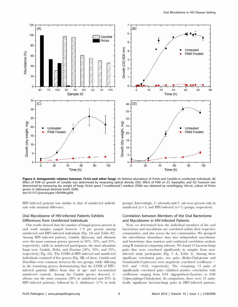
Figure 4. Antagonistic relation between Pichia and other fungi. (A) Relative abundance of Pichia and Candida in uninfected individuals. (B)Effect of PSM on growth of Candida was determined by measuring optical density (OD). Effect of PSM on (C) Aspergillus, and (D) Fusarium wasdetermined by measuring dry weight of fungi. Pichia spent (‘‘conditioned’’) medium (PSM) was obtained by centrifuging 100-mL culture of Pichiagrown in Sabouraud dextrose broth (SDB).doi:10.1371/journal.ppat.1003996.g004
Oral Microbiome in HIV Disease Setting
PLOS Pathogens | www.plospathogens.org 8 March 2014 | Volume 10 | Issue 3 | e1003996
with 11 positive (coefficient of 0.64 for 8 pairs, 0.74 for 2 pairs,
Fig. 3 B, Table 2) and one with negative correlation (Campylobacter-
Candida, coefficient 20.67).
We also evaluated the correlation between different members of
the mycobiome in the uninfected and HIV-infected groups. Our
analyses revealed that in the uninfected group, 23 fungal-fungal
interactions were statistically significant (P#0.035, Fig. 3C, Table 3),
while in the HIV-infected group, 6 fungus-fungus pairs were
significantly correlated (P-value of #0.03, Fig. 3D), which included
Candida-Epicoccum, Candida-Trichosporon, Epicoccum-Trichosporon, Peni-
cillium-Corynespora, Penicillium-Fusarium, and Alternaria-Serpula.
Pichia, a Member of the Core Oral Mycobiome, ExhibitsAntagonism against Candida
Having defined the core mycobiome, next we investigated
whether members of the core oral mycobiome are associated with
Candida, the most common oral fungal pathogen of HIV-infection
[36]. Our sequencing data indicated the presence of 3 Pichia
species, including P. guillermondii, P. burtonii, and P. jadinii in the
tested samples. We found that decrease in Pichia abundance
coincided with an increase in Candida colonization (Fig. 4A),
suggesting antagonism between Pichia and Candida. Furthermore,
we found that among the 4 uninfected subjects where Pichia was
present, 24 fungal genera were absent, including Aspergillus and
Cryptococcus (Table 4). In addition, in these 4 uninfected individuals,
9 fungal genera (including Fusarium) were present as co-colonizers
(Table 4). Analysis of the abundance profile of Fusarium in all
uninfected individuals (n = 12), revealed that its abundance was 3-
fold lower when Pichia was present (0.016%, n = 4) compared to
where Pichia was absent (0.048%, n = 8). Taken together, these
results show that Pichia interacts with other fungi, with an
antagonistic interaction with known pathogens including Candida,
Cryptococcus, Aspergillus and Fusarium.
Pichia Inhibits Growth of Candida, Aspergillus andFusarium
Next, we investigated the ability of Pichia to inhibit growth of C.
albicans, by allowing blastospores to grow in the presence or
absence of Pichia spent medium (PSM). As mentioned above, our
results revealed three Pichia species (P. guillermondii, P. burtonii, and
P. jadinii) in the tested samples. These Pichia spp, along with P.
farinosa, are common biocontrol agents used against plant
pathogens [37–40]. Moreover, literature search showed that P.
farinosa exhibits more biocontrol activity than P. guilliermondii [41].
Since P. farinosa and P. guilliermondii are very closely related to each
other based on their whole genome [38] and mitochondrial DNA
[42] sequences, we selected P. farinosa to investigate the interac-
tions between this species and pathogenic fungi including Candida.
As shown in Figure 4B, PSM completely inhibited Candida growth,
demonstrating a direct inhibitory effect of Pichia against Candida.
We also assessed the effect of PSM on growth of Aspergillus and
Fusarium by determining their dry weight. As shown in Figure 4C
and D, Aspergillus and Fusarium were unable to exhibit growth in
presence of PSM. These studies demonstrated that PSM exhibits
broad-spectrum activity against pathogenic fungi.
Next, we initiated studies to gain insight into the underlying
mechanism for the allowing Pichia to inhibit Candida. Since Pichia is
commonly used as a post-harvest biocontrol agent against plant
pathogens [43–47], and several studies have suggested that this
inhibitory activity involves nutrient competition, biofilm forma-
tion, germination, metabolites, secretory proteins [48–57], we
explored whether Pichia-mediated inhibition of Candida involves
these mechanisms.
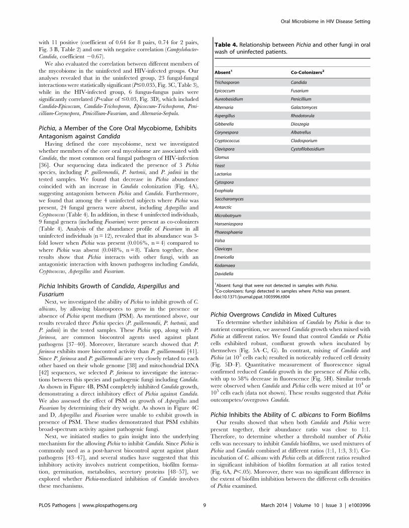
Pichia Overgrows Candida in Mixed CulturesTo determine whether inhibition of Candida by Pichia is due to
nutrient competition, we assessed Candida growth when mixed with
Pichia at different ratios. We found that control Candida or Pichia
cells exhibited robust, confluent growth when incubated by
themselves (Fig. 5A–C, G). In contrast, mixing of Candida and
Pichia (at 103 cells each) resulted in noticeably reduced cell density
(Fig. 5D–F). Quantitative measurement of fluorescence signal
confirmed reduced Candida growth in the presence of Pichia cells,
with up to 58% decrease in fluorescence (Fig. 5H). Similar trends
were observed when Candida and Pichia cells were mixed at 104 or
105 cells each (data not shown). These results suggested that Pichia
outcompetes/overgrows Candida.
Pichia Inhibits the Ability of C. albicans to Form BiofilmsOur results showed that when both Candida and Pichia were
present together, their abundance ratio was close to 1:1.
Therefore, to determine whether a threshold number of Pichia
cells was necessary to inhibit Candida biofilms, we used mixtures of
Pichia and Candida combined at different ratios (1:1, 1:3, 3:1). Co-
incubation of C. albicans with Pichia cells at different ratios resulted
in significant inhibition of biofilm formation at all ratios tested
(Fig. 6A, P,.05). Moreover, there was no significant difference in
the extent of biofilm inhibition between the different cells densities
of Pichia examined.
Table 4. Relationship between Pichia and other fungi in oralwash of uninfected patients.
Absent1 Co-Colonizers2
Trichosporon Candida
Epicoccum Fusarium
Aureobasidium Penicillium
Alternaria Galactomyces
Aspergillus Rhodotorula
Gibberella Dioszegia
Corynespora Albatrellus
Cryptococcus Cladosporium
Clavispora Cystofilobasidium
Glomus
Yeast
Lactarius
Cytospora
Exophiala
Saccharomyces
Antarctic
Microbotryum
Hanseniaspora
Phaeosphaeria
Valsa
Claviceps
Emericella
Kodamaea
Davidiella
1Absent: fungi that were not detected in samples with Pichia.2Co-colonizers: fungi detected in samples where Pichia was present.doi:10.1371/journal.ppat.1003996.t004
Oral Microbiome in HIV Disease Setting
PLOS Pathogens | www.plospathogens.org 9 March 2014 | Volume 10 | Issue 3 | e1003996
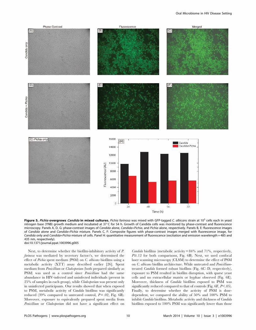
Next, to determine whether the biofilm-inhibitory activity of P.
farinosa was mediated by secretory factor/s, we determined the
effect of Pichia spent medium (PSM) on C. albicans biofilms using a
metabolic activity (XTT) assay described earlier [26]. Spent
medium from Penicillium or Cladosporium (both prepared similarly as
PSM) was used as a control since Penicillium had the same
abundance in HIV-infected and uninfected individuals (present in
25% of samples in each group), while Cladosporium was present only
in uninfected participants. Our results showed that when exposed
to PSM, metabolic activity of Candida biofilms was significantly
reduced (39% compared to untreated control, P = .02, Fig. 6B).
Moreover, exposure to equivalently prepared spent media from
Penicillium or Cladosporium did not have a significant effect on
Candida biofilms (metabolic activity = 84% and 71%, respectively,
P$.12 for both comparisons, Fig. 6B). Next, we used confocal
laser scanning microscopy (CLSM) to determine the effect of PSM
on C. albicans biofilm architecture. While untreated and Penicillium-
treated Candida formed robust biofilms (Fig. 6C–D, respectively),
exposure to PSM resulted in biofilm disruption, with sparse yeast
cells and no extracellular matrix or hyphae observed (Fig. 6E).
Moreover, thickness of Candida biofilms exposed to PSM was
significantly reduced compared to that of controls (Fig. 6F, P,.05).
Finally, to determine whether the activity of PSM is dose-
dependent, we compared the ability of 50% and 100% PSM to
inhibit Candida biofilms. Metabolic activity and thickness of Candida
biofilms exposed to 100% PSM was significantly lower than those
Figure 5. Pichia overgrows Candida in mixed cultures. Pichia farinosa was mixed with GFP-tagged C. albicans strain at 103 cells each in yeastnitrogen base (YNB) growth medium and incubated at 37uC for 54 h. Growth of Candida cells was monitored by phase-contrast and fluorescencemicroscopy. Panels A, D, G: phase-contrast images of Candida alone, Candida+Pichia, and Pichia alone, respectively. Panels B, E: fluorescence imagesof Candida alone and Candida+Pichia mixture. Panels C, F: Composite figures with phase-contrast images merged with fluorescence image, forCandida only and Candida+Pichia mixture of cells. Panel H: quantitative measurement of fluorescence (excitation and emission wavelength = 485 and435 nm, respectively).doi:10.1371/journal.ppat.1003996.g005
Oral Microbiome in HIV Disease Setting
PLOS Pathogens | www.plospathogens.org 10 March 2014 | Volume 10 | Issue 3 | e1003996
exposed to 50% PSM (Fig. 7A, B; P = .007 and .006, respectively).
Taken together, these results demonstrate that the Candida-
inhibitory activity of Pichia is specific and dose-dependent.
Pichia Inhibits Candida Germination and AdhesionSince adhesion and germination are key steps in mature Candida
biofilm formation [58–60] and are known Candida virulence
factors, we examined whether P. farinosa spent medium affects
these processes. Our data showed that while untreated Candida
formed robust hyphae (Fig. 7C), exposure to PSM resulted in
stunted Candida germ tubes (Fig. 7D), indicating that a secreted
component of Pichia inhibits Candida germination. We also found
that the number of Candida colony forming units (CFUs) adhering
to silicone elastomer catheter substrate when treated with Pichia
supernatant was significantly lower than untreated Candida cells
(Fig. 7E, P,.003). These results showed that Pichia inhibits the
ability of Candida to germinate and adhere to catheter substrate.
Active Ingredient of PSM Is a ProteinTo determine whether the active ingredient of PSM is a
metabolite or a protein, we evaluated Candida growth in presence
or absence of metabolites extracted [32] from PSM. Extracted
Pichia metabolites had no effect on Candida growth (Fig. 8A). Next,
we evaluated the influence of proteinase-, alkali-, or heat (90uC for
10 min)-treated PSM on C. albicans biofilms. Our data showed that
proteinase-K treatment abrogated the ability of PSM to inhibit
biofilms, while alkali- and heat-treatment did not have any effect,
as determined by analysis of biofilm thickness (Fig. 8B) and
architecture (Fig. 8C–G). These studies indicate that the active
component in PSM is proteinaceous, heat-stable, and non-
glycosylated.
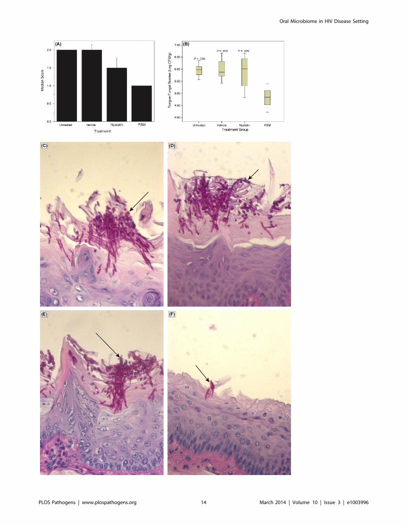
PSM Is Effective against Oral Candidiasis in anExperimental Murine Model
To determine whether the in vitro activity of PSM against
Candida is also exhibited in vivo, we evaluated the efficacy of PSM
in an experimental murine model of oral candidiasis. We found
that at the end of treatment (Day 7), clinical score of PSM-
treated mice was significant reduced compared to untreated,
vehicle-treated, or nystatin-treated mice (Fig. 9A, P = .002 by
Kruskal-Wallis test). The fungal burden of tongue from PSM-
treated mice was also significantly reduced compared to
Figure 6. Activity of Pichia spent medium (PSM) against fungal biofilms. (A) Effect of Pichia cells on the ability of Candida to form biofilms.Candida and Pichia were co-incubated [Candida:Pichia (C:P) = 3:1, 1:1, or 1:3] and biofilm formation was monitored (*P#.002, compared to Candida orPichia controls). (B) Effect of media supernatant obtained from Pichia, Penicillium, or Cladosporium on Candida biofilms. Mean 6 SD of $3 separateexperiments. (C–E) Confocal microscopy images of Candida biofilms formed in presence of (C) no media supernatant, (D) Penicillium supernatant or(E) Pichia supernatant. (F) Thickness of biofilms formed in presence of media supernatant of Pichia or Penicillium.doi:10.1371/journal.ppat.1003996.g006
Oral Microbiome in HIV Disease Setting
PLOS Pathogens | www.plospathogens.org 11 March 2014 | Volume 10 | Issue 3 | e1003996
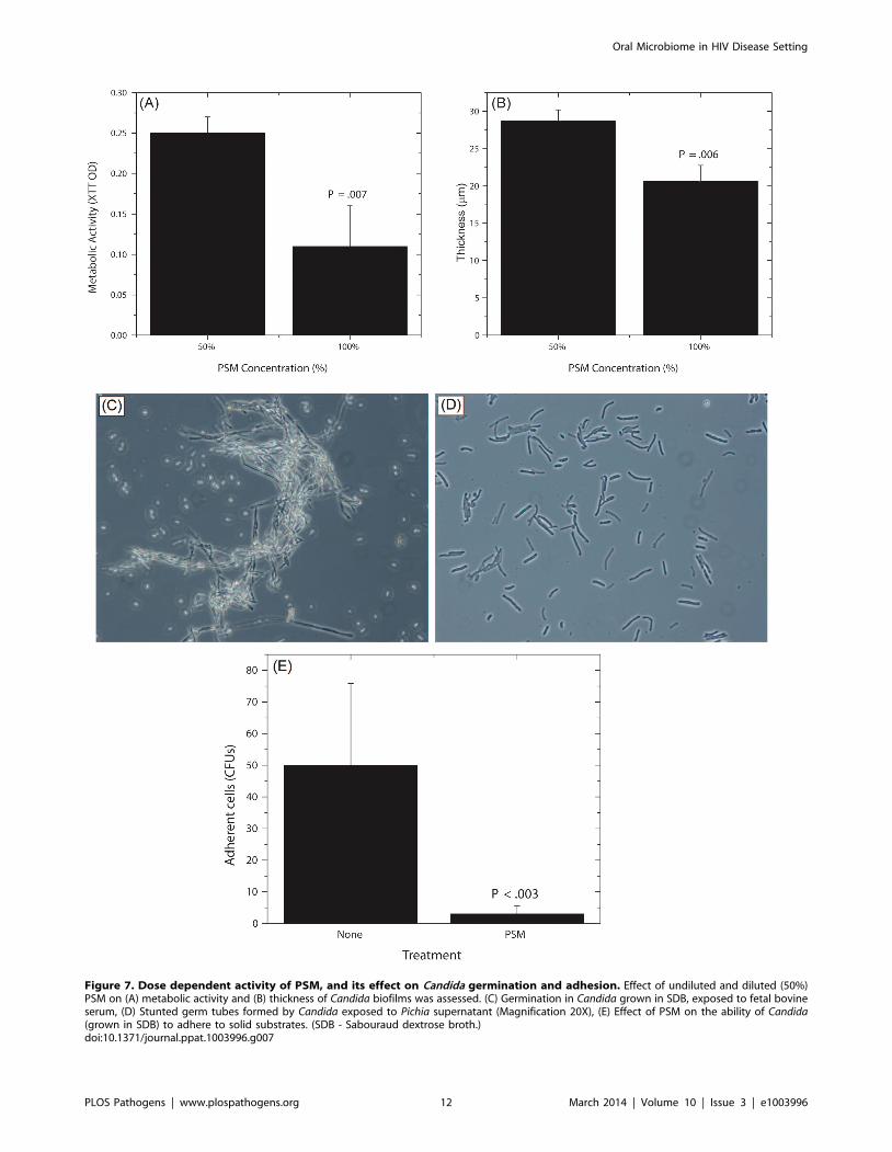
Figure 7. Dose dependent activity of PSM, and its effect on Candida germination and adhesion. Effect of undiluted and diluted (50%)PSM on (A) metabolic activity and (B) thickness of Candida biofilms was assessed. (C) Germination in Candida grown in SDB, exposed to fetal bovineserum, (D) Stunted germ tubes formed by Candida exposed to Pichia supernatant (Magnification 20X), (E) Effect of PSM on the ability of Candida(grown in SDB) to adhere to solid substrates. (SDB - Sabouraud dextrose broth.)doi:10.1371/journal.ppat.1003996.g007
Oral Microbiome in HIV Disease Setting
PLOS Pathogens | www.plospathogens.org 12 March 2014 | Volume 10 | Issue 3 | e1003996
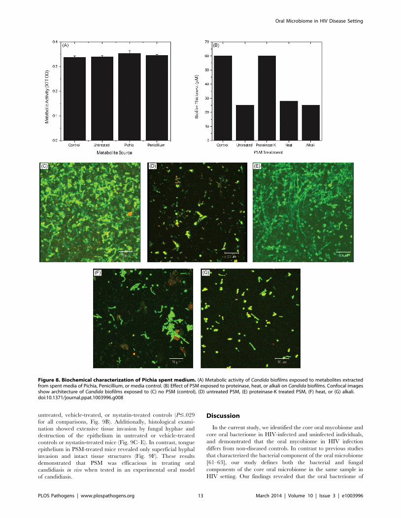
untreated, vehicle-treated, or nystatin-treated controls (P#.029
for all comparisons, Fig. 9B). Additionally, histological exami-
nation showed extensive tissue invasion by fungal hyphae and
destruction of the epithelium in untreated or vehicle-treated
controls or nystatin-treated mice (Fig. 9C–E). In contrast, tongue
epithelium in PSM-treated mice revealed only superficial hyphal
invasion and intact tissue structures (Fig. 9F). These results
demonstrated that PSM was efficacious in treating oral
candidiasis in vivo when tested in an experimental oral model
of candidiasis.
Discussion
In the current study, we identified the core oral mycobiome and
core oral bacteriome in HIV-infected and uninfected individuals,
and demonstrated that the oral mycobiome in HIV infection
differs from non-diseased controls. In contrast to previous studies
that characterized the bacterial component of the oral microbiome
[61–63], our study defines both the bacterial and fungal
components of the core oral microbiome in the same sample in
HIV setting. Our findings revealed that the oral bacteriome of
Figure 8. Biochemical characterization of Pichia spent medium. (A) Metabolic activity of Candida biofilms exposed to metabolites extractedfrom spent media of Pichia, Penicillium, or media control. (B) Effect of PSM exposed to proteinase, heat, or alkali on Candida biofilms. Confocal imagesshow architecture of Candida biofilms exposed to (C) no PSM (control), (D) untreated PSM, (E) proteinase-K treated PSM, (F) heat, or (G) alkali.doi:10.1371/journal.ppat.1003996.g008
Oral Microbiome in HIV Disease Setting
PLOS Pathogens | www.plospathogens.org 13 March 2014 | Volume 10 | Issue 3 | e1003996
Oral Microbiome in HIV Disease Setting
PLOS Pathogens | www.plospathogens.org 14 March 2014 | Volume 10 | Issue 3 | e1003996
HIV-infected individuals was similar to that of uninfected
individuals, indicating the presence of a shared bacteriome in
these individuals. The core oral bacteriome of uninfected
individuals in our study is in agreement with results reported
earlier by Zaura et al. [64] who showed that 15 bacterial genera
were present in the core oral bacteriome of healthy individuals.
Correlation analyses of the relationship between bacteriome
and mycobiome revealed that 15 and 12 bacteria-fungi pairs were
correlated significantly in samples from uninfected and HIV-
infected patients, respectively. It is possible that these correlations
indicate mutually dependent relationships within the oral
microbiome, in which bacteria may be assisting or scavenging
their fungal neighbors. Alternatively, fungal members of the
microbiome may impact bacterial growth and drug susceptibility.
Such interactions could influence the course and extent of oral
diseases in the HIV setting. In this regard, interactions between
bacteria and Candida have been investigated previously by Hogan
and Kolter [65], who showed that P. aeruginosa kills hyphal form
of C. albicans via biofilm formation. Furthermore, Candida-
bacterial interactions are also associated with diseases like
ventilator-associated pneumonia [66,67] and bloodstream infec-
tions [68]. Our analyses revealed no correlation between Candida
and bacteria in uninfected individuals, while in HIV-infected
patients Candida and Campylobacter were negatively correlated.
This correlation is in agreement with the findings of Navazesh et
al. [69], who showed that antiretroviral therapy increased the risk
for recovering bacteria (including Campylobacter species) with a
concomitant decrease in the recovery rate of Candida, in HIV-
infected women. Moreover, Workman et al. [70] reported that
proteins secreted by Campylobacter inhibit the growth of C. albicans.
The clinical relevance of this correlation remains to be
investigated.
In the current study, we also defined the oral mycobiome of
HIV-infected and uninfected study participants, and showed this
fungal community to comprise up to nine different genera in both
groups. To our knowledge, the only other study to have used a
sequencing-based approach to identify oral fungi in HIV setting
was that performed by Aas et al. [12], who analyzed sub-gingival
plaque of HIV-infected patients, and reported the presence of only
two fungal species (Saccharomyces cerevisiae and C. albicans in 4 and 2
patients, respectively). The difference in fungal profile between
these investigators and our study may be due to differences in
sample types (oral wash vs. sub-gingival plaque), detection probe
(pan-fungal ITS probe vs. 18S rDNA), and sequencing technique
(real-time pyrosequencing vs. rDNA sequencing).
Our study showed that the core oral mycobiome of HIV-
infected patients was different from that of uninfected individuals.
In a recent study, Iliev et al. [13] characterized the gut
microbiome in wild type mice and isogenic Clec7a2/2 mice that
lack Dectin-1 (the innate immune receptor) and exhibited
increased levels of chemically induced colitis. These investigators
reported that there were no significant differences in bacteriome
between wild type and Clec7a2/2 mice, while the mycobiome
profile differed between these isogenic mice, with an increase in
opportunistic pathogenic fungi (Candida and Trichosporon) during
colitis in Clec7a2/2 mice, and a decrease in the nonpathogenic
fungi (e.g. Saccharomyces). Thus, severe colitis in Dectin-1 knockout
mice was associated with alterations in the gut mycobiome (but not
the bacteriome). This study is similar to our findings, showing that
HIV disease is associated with changes in the oral mycobiome, and
highlight the importance of characterizing the mycobiome and its
role in disease.
Our analyses showed that increase in Candida colonization was
associated with a concomitant decrease in the abundance of Pichia,
suggesting an antagonistic relation between these two fungi. In
addition, these analyses also suggested that Pichia exhibits
antagonistic interaction with other known pathogens including
Cryptococcus, Aspergillus and Fusarium. These interactions were
confirmed using growth assays that demonstrated broad-spectrum
inhibitory activity of Pichia. The anti-Candida activity of Pichia was
validated in vivo using an experimental murine model of oral
candidiasis.
The biocontrol activity of Pichia against fungal plant pathogens
has been shown to involve multiple mechanisms, including biofilm
formation and germination [48–53]. The results of the current
study are in agreement with these findings, where we showed that
the anti-Candida activity of Pichia is mediated by inhibition of
Candida growth and virulence factors like germination, adherence,
and biofilm formation. It is well known that interfering with
virulence factors decreases the ability of Candida to cause infection
including oral candidiasis [71–79]. Therefore, Pichia, by inhibiting
Candida virulence factors may limit the ability of this pathogenic
fungus to cause infection. Such involvement of virulence factors is
a common theme in interactions among microorganisms in the
context of human infection [65].
Pichia biocontrol activity has also been attributed to nutrient
limitation [39,41,51,80]. Our results showed that Pichia
outcompetes Candida when the two fungi are mixed together
and allowed to grow. The possible reason for this phenomenon
could be due to the ability of Pichia to consume nutrients more
efficiently than Candida. Alternatively, it is possible that Pichia
secretes factor/s that attenuate the ability of Candida to grow.
Data in support of the latter possibility can be derived from our
findings that Pichia spent medium inhibits pathogenic fungi
including Candida, Aspergillus, Fusarium, and Cryptococcus. Further-
more, our results also demonstrate that the inhibitory activity of
PSM is proteinaceous in nature, and not a metabolite. Earlier
studies have shown that biocontrol activity of Pichia against
fungal plant pathogens is mediated by secretory metabolite or
proteins [48,53,81,82].
In conclusion, we identified the core bacteriome and myco-
biome in HIV setting, and identified HIV-specific changes in the
mycobiome. We also identified a critical antagonistic interaction
between Pichia and fungal pathogens including Candida. This
interaction was demonstrated using in vitro and in vivo models.
We also defined the mechanisms underlying the antagonistic
interaction between Pichia and Candida. Our findings show for the
first time that normal fungal community interacts with Candida in
the oral cavity. Detailed investigations are warranted to purify and
characterize the secretory factor/s mediating such interactions,
and their mechanism/s of action at the molecular level. Our
findings have wide implications regarding the discovery of novel
antifungal agents that new therapeutic approaches for the
management of fungal infections.
Figure 9. Efficacy of Pichia spent medium (PSM) in an experimental murine morel of oral candidiasis. Assessment of oral candidiasis inmice infected with Candida was performed by (A) clinical score and (B) tongue fungal burden. (A) Median clinical scores of oral candidiasis in miceafter treatment with PSM and nystatin. (B) Box-plot of tissue fungal burden (log CFUs/g) in different groups of mice infected with Candida (P-values,compared to PSM treatment). Histology analyses of tissue section of tongue from mouse infected with Candida, followed by (C) no treatment ortreated with (D) vehicle control, (E) nystatin, or (F) PSM. Arrows – fungal hyphae.doi:10.1371/journal.ppat.1003996.g009
Oral Microbiome in HIV Disease Setting
PLOS Pathogens | www.plospathogens.org 15 March 2014 | Volume 10 | Issue 3 | e1003996
Supporting Information
Method S1 Additional details of PCR, pyrosequencing and
correlation analyses.
(DOCX)
Table S1 Relative abundance of bacteria in oral wash of HIV-
infected and uninfected study participants.
(XLS)
Table S2 Relative abundance of fungi in oral wash of HIV-
infected and uninfected study participants.
(XLS)
Acknowledgments
Presented in part at the 49th Annual Meeting of the Infectious Diseases
Society of America (IDSA, Boston, MA; October, 2011) and 2013
Interscience Conference on Antimicrobial Agents and Chemotherapy
(ICAAC), Denver, CO (September 9–13, 2013). The authors would like to
thank Dr. Brian Wickes (University of Texas Health Science Center at San
Antonio, San Antonio, Texas) for molecular identification of the Pichia
isolate used in this study.
Author Contributions
Conceived and designed the experiments: PKM PMG MAG. Performed
the experiments: JC MR MS REB RJ. Analyzed the data: MR PKM REB.
Contributed reagents/materials/analysis tools: PKM PMG RAS MML
MAG. Wrote the paper: PKM MAG.
References
1. Jenkinson HF, Lamont RJ (2005) Oral microbial communities in sickness and in
health. Trends Microbiol 13: 589–595.
2. Patton LL, Phelan JA, Ramos-Gomez FJ, Nittayananta W, Shiboski CH, et al.
(2002) Prevalence and classification of HIV-associated oral lesions. Oral Dis 8:
98–109.
3. Chattopadhyay A, Caplan DJ, Slade GD, Shugars DC, Tien HC, et al. (2005)
Risk indicators for oral candidiasis and oral hairy leukoplakia in HIV-infected
adults. Community Dent Oral Epidemiol 33: 35–44.
4. Koletar SL, Smurzynski M, Wu K, Collier AC, Bosch RJ, et al. AIDS-defining
illnesses occurring in treatment-experienced HIV-1-infected persons followed in
the ACTG Longitudinal Linked Randomized Trials (ALLRT) study [Abstract
1017]. In Proceedings of the 15th Conference on Retroviruses & Opportunistic
Infections; February 3–6, 2008; Boston, MA. Available: http://retroconference.
org/. Accessed 2/3/13.
5. Thompson GR, 3rd, Patel PK, Kirkpatrick WR, Westbrook SD, Berg D, et al.
(2010) Oropharyngeal candidiasis in the era of antiretroviral therapy. Oral Surg
Oral Med Oral Pathol Oral Radiol Endod 109: 488–495.
6. Patel PK, Erlandsen JE, Kirkpatrick WR, Berg DK, Westbrook SD, et al. (2012)
The Changing Epidemiology of Oropharyngeal Candidiasis in Patients with
HIV/AIDS in the Era of Antiretroviral Therapy. AIDS Res Treat 2012:
262471.
7. Kinross JM, Darzi AW, Nicholson JK (2011) Gut microbiome-host interactions
in health and disease. Genome Med 3: 14.
8. Nelson KE, Weinstock GM, Highlander SK, Worley KC, Creasy HH, et al.
(2010) A catalog of reference genomes from the human microbiome. Science
328: 994–999.
9. Grice EA, Kong HH, Conlan S, Deming CB, Davis J, et al. (2009)
Topographical and temporal diversity of the human skin microbiome. Science
324: 1190–1192.
10. Huse SM, Ye Y, Zhou Y, Fodor AA (2012) A Core Human Microbiome as
Viewed through 16S rRNA Sequence Clusters. PLoS ONE 7: e34242.
11. Clemente JC, Ursell LK, Parfrey LW, Knight R (2012) The Impact of the Gut
Microbiota on Human Health: An Integrative View. Cell 148: 1258–1270.
12. Aas JA, Barbuto SM, Alpagot T, Olsen I, Dewhirst FE, et al. (2007) Subgingival
plaque microbiota in HIV positive patients. Journal of Clinical Periodontology
34: 189–195.
13. Iliev ID, Funari VA, Taylor KD, Nguyen Q, Reyes CN, et al. (2012)
Interactions Between Commensal Fungi and the C-Type Lectin Receptor
Dectin-1 Influence Colitis. Science 336: 1314–1317.
14. Ghannoum MA, Jurevic RJ, Mukherjee PK, Cui F, Sikaroodi M, et al. (2010)
Characterization of the Oral Fungal Microbiome (Mycobiome) in Healthy
Individuals. PLoS Pathogens 6: e1000713.
15. Peleg AY, Hogan DA, Mylonakis E (2010) Medically important bacterial-fungal
interactions. Nat Rev Microbiol 8: 340–349.
16. Landlinger C, Baskova L, Preuner S, Willinger B, Buchta V, et al. (2008)
Identification of fungal species by fragment length analysis of the internally
transcribed spacer 2 region. Eur J Clin Microbiol Infect Dis 28(6):613–22.
17. Borman AM, Linton CJ, Miles SJ, Johnson EM (2008) Molecular identification
of pathogenic fungi. Journal of Antimicrobial Chemotherapy 61: i7–12.
18. Gillevet P, Sikaroodi M, Keshavarzian A, Mutlu EA (2010) Quantitative
assessment of the human gut microbiome using multitag pyrosequencing. Chem
Biodivers 7: 1065–1075.
19. Chakravorty S, Helb D, Burday M, Connell N, Alland D (2007) A detailed
analysis of 16S ribosomal RNA gene segments for the diagnosis of pathogenic
bacteria. Journal of Microbiological Methods 69: 330–339.
20. Spear GT, Sikaroodi M, Zariffard MR, Landay AL, French AL, et al. (2008)
Comparison of the diversity of the vaginal microbiota in HIV-infected and HIV-
uninfected women with or without bacterial vaginosis. J Infect Dis 198: 1131–
1140.
21. Gillevet PM, inventor; BioSpherex LLC, assignee (2013 December 10) Multitag
Sequencing and Ecogenomic Analysis. United States Patent 8,603,749.
22. Romanelli AM, Sutton DA, Thompson EH, Rinaldi MG, Wickes BL (2010)
Sequence-based identification of filamentous basidiomycetous fungi from clinical
specimens: a cautionary note. J Clin Microbiol 48: 741–752.
23. Cormack BP, Bertram G, Egerton M, Gow NA, Falkow S, et al. (1997) Yeast-
enhanced green fluorescent protein (yEGFP) a reporter of gene expression in
Candida albicans. Microbiology 143: 303–311.
24. Swindell K, Lattif AA, Chandra J, Mukherjee PK, Ghannoum MA (2009)
Parenteral Lipid Emulsion Induces Germination of Candida albicans and Increases
Biofilm Formation on Medical Catheter Surfaces. J Infect Dis 200: 473–
480.
25. Kuhn DM, Mukherjee PK, Clarke TA, Pujol C, Chandra J, et al. (2004) Candida
parapsilosis characterization in an outbreak setting. Emerging Infectious Diseases
10: 1074–1081.
26. Chandra J, Mukherjee PK, Ghannoum MA (2008) In vitro growth and analysis
of Candida biofilms. Nature Protocols 3: 1909–1924.
27. Chandra J, Kuhn DM, Mukherjee PK, Hoyer LL, McCormick T, et al. (2001)
Biofilm formation by the fungal pathogen Candida albicans - development,
architecture and drug resistance. Journal of Bacteriology 183: 5385–5394.
28. Chandra J, Mukherjee PK, Leidich SD, Faddoul FF, Hoyer LL, et al. (2001)
Antifungal resistance of candidal biofilms formed on denture acrylic in vitro.
Journal of Dental Research 80: 903–908.
29. Pienaar ED, Young T, Holmes H (2010) Interventions for the prevention and
management of oropharyngeal candidiasis associated with HIV infection in
adults and children. Cochrane Database of Systematic Reviews 11: CD003940.
30. Hise AG, Tomalka J, Ganesan S, Patel K, Hall BA, et al. (2009) An essential role
for the NLRP3 inflammasome in host defense against the human fungal
pathogen Candida albicans. Cell Host Microbe 5: 487–497.
31. Tang N, Liu L, Kang K, Mukherjee PK, Takahara M, et al. (2004) Inhibition of
monocytic interleukin-12 production by Candida albicans via selective activation of
ERK mitogen-activated protein kinase. Infection and Immunity 72: 2513–2520.
32. Ghannoum MA, Mukherjee PK, Jurevic RJ, Retuerto M, Brown RE, et al.
(2011) Metabolomics Reveals Differential Levels of Oral Metabolites in HIV-
Infected Patients: Toward Novel Diagnostic Targets. Omics 17: 5–15.
33. Caporaso JG, Kuczynski J, Stombaugh J, Bittinger K, Bushman FD, et al. (2010)
QIIME allows analysis of high-throughput community sequencing data. Nat
Methods 7: 335–336.
34. R Core Team (2013) R: A language and environment for statistical computing.
Available http://www.R-project.org. Accessed 2 February 2013.
35. Revelle W (2013) psych: procedures for personality and psychological research.
Available http://CRAN.R-project.org/package = psych Version = 1.3.10. Ac-
cessed 20 December 2013.
36. Nokta M (2008) Oral manifestations associated with HIV infection. Curr HIV/
AIDS Rep 5: 5–12.
37. Schisler D, Kurtzman C, Bothast R, Slininger P (1995) Evaluation of yeasts for
biological control of Fusarium dry rot of potatoes. American Potato Journal 72:
339–353.
38. Starmer WT, Ganter PF, Phaff HJ (1986) Quantum and continuous evolution of
DNA base composition in the yeast genus Pichia. Evolution 40: 1263–1274.
39. Golubev W (2006) Antagonistic interactions among yeasts. Biodiversity and
Ecophysiology of Yeasts: Springer. pp. 197–219.
40. Antunes J, Aguiar C (2012) Search for killer phenotypes with potential for
biological control. Annals of microbiology 62: 427–433.
41. Druvefors UA, Schnurer J (2005) Mold-inhibitory activity of different yeast
species during airtight storage of wheat grain. FEMS Yeast Res 5: 373–378.
42. Jung PP, Friedrich A, Souciet JL, Louis V, Potier S, et al. (2010) Complete
mitochondrial genome sequence of the yeast Pichia farinosa and comparative
analysis of closely related species. Curr Genet 56: 507–515.
43. Walker GM (2010) Pichia anomala: cell physiology and biotechnology relative to
other yeasts. Antonie Van Leeuwenhoek 99(1):25–34.
44. Elad Y (2003) Biocontrol of foliar pathogens: mechanisms and application.
Commun Agric Appl Biol Sci 68: 17–24.
Oral Microbiome in HIV Disease Setting
PLOS Pathogens | www.plospathogens.org 16 March 2014 | Volume 10 | Issue 3 | e1003996
45. Cao S, Yuan Y, Hu Z, Zheng Y (2010) Combination of Pichia membranifaciens
and ammonium molybdate for controlling blue mould caused by Penicilliumexpansum in peach fruit. Int J Food Microbiol 141: 173–176.
46. Santos A, San Mauro M, Bravo E, Marquina D (2009) PMKT2, a new killer
toxin from Pichia membranifaciens, and its promising biotechnological propertiesfor control of the spoilage yeast Brettanomyces bruxellensis. Microbiology 155: 624–
634.47. Olstorpe M, Passoth V (2010) Pichia anomala in grain biopreservation. Antonie
Van Leeuwenhoek 99(1):57–62.
48. Kagan BL (1983) Mode of action of yeast killer toxins: channel formation in lipidbilayer membranes. Nature 302: 709–711.
49. Izgu F, Altınbay D, Acun T (2006) Killer toxin of Pichia anomala NCYC 432;purification, characterization and its exo-b-1,3-glucanase activity. Enzyme and
Microbial Technology 39: 669–676.50. Cao S, Zheng Y, Tang S, Wang K (2008) Improved control of anthracnose rot
in loquat fruit by a combination treatment of Pichia membranifaciens with
CaCl(2). Int J Food Microbiol 126: 216–220.51. Druvefors UA, Passoth V, Schnurer J (2005) Nutrient effects on biocontrol of
Penicillium roqueforti by Pichia anomala J121 during airtight storage of wheat. ApplEnviron Microbiol 71: 1865–1869.
52. Xu XB, Tian SP (2008) Reducing oxidative stress in sweet cherry fruit by Pichia
membranaefaciens: a possible mode of action against Penicillium expansum. J ApplMicrobiol 105: 1170–1177.
53. Zhao J, Mou Y, Shan T, Li Y, Zhou L, et al. (2010) Antimicrobial metabolitesfrom the endophytic fungus Pichia guilliermondii isolated from Paris polyphylla
var. yunnanensis. Molecules 15: 7961–7970.54. Jijakli MH, Lepoivre P (1998) Characterization of an Exo-beta-1,3-Glucanase
Produced by Pichia anomala Strain K, Antagonist of Botrytis cinerea on Apples.
Phytopathology 88: 335–343.55. Droby S, Hofstein R, Wilson CL, Wisniewski M, Fridlender B, et al. (1993) Pilot
Testing of Pichia guilliermondii: A Biocontrol Agent of Postharvest Diseases ofCitrus Fruit. Biological Control 3: 47–52.
56. Droby S, Wisniewski ME, Cohen L, Weiss B, Touitou D, et al. (1997) Influence
of CaCl(2) on Penicillium digitatum, Grapefruit Peel Tissue, and BiocontrolActivity of Pichia guilliermondii. Phytopathology 87: 310–315.
57. Giobbe S, Marceddu S, Scherm B, Zara G, Mazzarello VL, et al. (2007) Thestrange case of a biofilm-forming strain of Pichia fermentans, which controls
Monilinia brown rot on apple but is pathogenic on peach fruit. FEMS Yeast Res7: 1389–1398.
58. Kumamoto CA, Vinces MD (2005) Alternative Candida albicans lifestyles: Growth
on Surfaces. Annual Review of Microbiology 59: 113–133.59. Chandra J, Mukherjee PK, Ghannoum MA (2011) Candida Biofilms Associated
with CVC and Medical Devices. Mycoses 55: 46–57.60. Chandra J, Mukherjee PK, Ghannoum MA (2010) Fungal Biofilms in the
Clinical Lab Setting. Current Reports in Fungal Infection 4: 137–144.
61. Conlan S, Kong HH, Segre JA (2012) Species-level analysis of DNA sequencedata from the NIH Human Microbiome Project. PLoS One 7: e47075.
62. Li K, Bihan M, Yooseph S, Methe BA (2012) Analyses of the microbial diversityacross the human microbiome. PLoS One 7: e32118.
63. Zarco MF, Vess TJ, Ginsburg GS (2012) The oral microbiome in health anddisease and the potential impact on personalized dental medicine. Oral Dis 18:
109–120.
64. Zaura E, Keijser BJ, Huse SM, Crielaard W (2009) Defining the healthy ‘‘core
microbiome’’ of oral microbial communities. BMC Microbiol 9: 259.
65. Hogan DA, Kolter R (2002) Pseudomonas - Candida interactions: an ecological role
for virulence factors. Science 296: 2229–2232.
66. Azoulay E, Timsit J-F, Tafflet M, de Lassence A, Darmon M, et al. (2006)
Candida Colonization of the Respiratory Tract and Subsequent Pseudomonas
Ventilator-Associated Pneumonia. Chest 129: 110–117.
67. Nseir S, Jozefowicz E, Cavestri B, Sendid B, Di Pompeo C, et al. (2007) Impact
of antifungal treatment on Candida-Pseudomonas interaction: a preliminary
retrospective case-control study. Intensive Care Med 33: 137–142.
68. Dyess DL, Garrison RN, Fry DE (1985) Candida sepsis. Implications of
polymicrobial blood-borne infection. ArchSurg 120: 345–348.
69. Navazesh M, Mulligan R, Pogoda J, Greenspan D, Alves M, et al. (2005) The
effect of HAART on salivary microbiota in the Women’s Interagency HIV
Study (WIHS). Oral Surg Oral Med Oral Pathol Oral Radiol Endod 100: 701–
708.
70. Workman SN, Been FE, Crawford SR, Lavoie MC (2008) Bacteriocin-like
inhibitory substances from Campylobacter spp. Antonie Van Leeuwenhoek 93:
435–436.
71. Dupont PF (1995) Candida albicans, the opportunist. A cellular and molecular
perspective. JAmPodiatrMedAssoc 85: 104–115.
72. Martin MV, Craig GT, Lamb DJ (1984) An investigation of the role of true
hypha production in the pathogenesis of experimental oral candidosis.
Sabouraudia 22: 471–476.
73. Cannon RD, Holmes AR, Mason AB, Monk BC (1995) Oral Candida:
clearance, colonization, or candidiasis? JDentRes 74: 1152–1161.
74. Samaranayake YH, Wu PC, Samaranayake LP, So M, Yuen KY (1994)
Adhesion and colonisation of Candida krusei on host surfaces. JMedMicrobiol
41: 250–258.
75. De Bernardis F, Boccanera M, Rainaldi L, Guerra CE, Quinti I, et al. (1992)
The secretion of aspartyl proteinase, a virulence enzyme, by isolates of Candida
albicans from the oral cavity of HIV-infected subjects. EurJEpidemiol 8: 362–
367.
76. Sardi JC, Duque C, Hofling JF, Goncalves RB (2012) Genetic and phenotypic
evaluation of Candida albicans strains isolated from subgingival biofilm of
diabetic patients with chronic periodontitis. Med Mycol 50: 467–475.
77. Ghannoum MA, Abu-Elteen KH (1990) Pathogenicity determinants of Candida.
Mycoses 33: 265–282.
78. Ghannoum MA (2000) Potential role of phospholipases in virulence and fungal
pathogenesis. Clinical Microbiology Reviews 13: 122–143.
79. Richardson MD, Smith H (1981) Production of germ tubes by virulent and
attenuated strains of Candida albicans. J Infect Dis 144: 565–569.
80. Restuccia C, Giusino F, Licciardello F, Randazzo C, Caggia C, et al. (2006)
Biological control of peach fungal pathogens by commercial products and
indigenous yeasts. J Food Prot 69: 2465–2470.
81. Santos A, Marquina D (2004) Ion channel activity by Pichia membranifaciens
killer toxin. Yeast 21: 151–162.
82. Fredlund E, Broberg A, Boysen ME, Kenne L, Schnurer J (2004) Metabolite
profiles of the biocontrol yeast Pichia anomala J121 grown under oxygen
limitation. Appl Microbiol Biotechnol 64: 403–409.
Oral Microbiome in HIV Disease Setting
PLOS Pathogens | www.plospathogens.org 17 March 2014 | Volume 10 | Issue 3 | e1003996
Copyright © 2022 FDOKUMEN