organo oficial dela sociedad española de nutricion parenteral ...
Oral Activated Charcoal Prevents Experimental CerebralMalaria in Mice and in a Randomized Controlled ClinicalTrial in Man Did Not Interfere with the Pharmacokineticsof Parenteral ArtesunateJ. Brian de Souza1,2., Uduak Okomo3., Neal D. Alexander1, Naveed Aziz4, Benjamin M. J. Owens5,
Harparkash Kaur1, Momodou Jasseh1, Sant Muangnoicharoen6, Percy F. Sumariwalla7, David C.
Warhurst1, Stephen A. Ward6, David J. Conway1,3, Luis Ulloa8, Kevin J. Tracey8, Brian M. J. Foxwell7,
Paul M. Kaye5*, Michael Walther3*
1 Department of Infectious and Tropical Diseases, London School of Hygiene and Tropical Medicine, London, United Kingdom, 2 Division of Infection and Immunity,
Department of Immunology, University College London, London, United Kingdom, 3 Medical Research Council Laboratories, Fajara, Banjul, The Gambia, 4 The Technology
Facility, Department of Biology, University of York, York, United Kingdom, 5 Centre for Immunology and Infection, Hull York Medical School and Department of Biology,
University of York, York, United Kingdom, 6 Liverpool School of Tropical Medicine, Liverpool, United Kingdom, 7 Kennedy Institute of Rheumatology, Imperial College of
Science, London, United Kingdom, 8 Center of Immunology and Inflammation, North Shore-LIJ Research Institute, New York, New York, United States of America
Abstract
Background: Safe, cheap and effective adjunct therapies preventing the development of, or reducing the mortality from,severe malaria could have considerable and rapid public health impact. Oral activated charcoal (oAC) is a safe and welltolerated treatment for acute poisoning, more recently shown to have significant immunomodulatory effects in man. Inpreparation for possible efficacy trials in human malaria, we sought to determine whether oAC would i) reduce mortalitydue to experimental cerebral malaria (ECM) in mice, ii) modulate immune and inflammatory responses associated with ECM,and iii) affect the pharmacokinetics of parenteral artesunate in human volunteers.
Methods/Principal Findings: We found that oAC provided significant protection against P. berghei ANKA-induced ECM,increasing overall survival time compared to untreated mice (p,0.0001; hazard ratio 16.4; 95% CI 6.73 to 40.1). Protectionfrom ECM by oAC was associated with reduced numbers of splenic TNF+ CD4+ T cells and multifunctional IFNc+TNF+ CD4+
and CD8+ T cells. Furthermore, we identified a whole blood gene expression signature (68 genes) associated with protectionfrom ECM. To evaluate whether oAC might affect current best available anti-malarial treatment, we conducted arandomized controlled open label trial in 52 human volunteers (ISRCTN NR. 64793756), administering artesunate (AS) in thepresence or absence of oAC. We demonstrated that co-administration of oAC was safe and well-tolerated. In the 26 subjectsfurther analyzed, we found no interference with the pharmacokinetics of parenteral AS or its pharmacologically activemetabolite dihydroartemisinin.
Conclusions/Significance: oAC protects against ECM in mice, and does not interfere with the pharmacokinetics ofparenteral artesunate. If future studies succeed in establishing the efficacy of oAC in human malaria, then the characteristicsof being inexpensive, well-tolerated at high doses and requiring no sophisticated storage would make oAC a relevantcandidate for adjunct therapy to reduce mortality from severe malaria, or for immediate treatment of suspected severemalaria in a rural setting.
Trial Registration: Controlled-Trials.com ISRCTN64793756
Citation: de Souza JB, Okomo U, Alexander ND, Aziz N, Owens BMJ, et al. (2010) Oral Activated Charcoal Prevents Experimental Cerebral Malaria in Mice and in aRandomized Controlled Clinical Trial in Man Did Not Interfere with the Pharmacokinetics of Parenteral Artesunate. PLoS ONE 5(4): e9867. doi:10.1371/journal.pone.0009867
Editor: David Joseph Diemert, The George Washington University Medical Center, United States of America
Received September 9, 2009; Accepted February 17, 2010; Published April 15, 2010
Copyright: � 2010 de Souza et al. This is an open-access article distributed under the terms of the Creative Commons Attribution License, which permitsunrestricted use, distribution, and reproduction in any medium, provided the original author and source are credited.
Funding: British Medical Research Council (#G0400786 to PMK; http://www.mrc.ac.uk/index.htm), the Medical Research Council Laboratories (UK) core fundingto the MRC Gambia Unit, a North Shore-LIJ Faculty Award to LU, the NIGMS, and the North Shore-LIJ GCRC (#1MO1RR018535; http://www.feinsteininstitute.org/Feinstein/GCRC). BO is supported by a MRC Postgraduate Studentship. The funders had no role in study design, data collection and analysis, decision to publish,or preparation of the manuscript.
Competing Interests: WO 2007/015102 A1: ‘The use of charcoal for the treatment of inflammatory conditions’. Inventors: BMJF, PS, PMK, KJT, KennethKenigsberg and LU. The inventors place no restrictions on the use of AC for the treatment of malaria and agree to PLoS guidelines.
* E-mail: [email protected] (PMK); [email protected] (MW)
. These authors contributed equally to this work.
PLoS ONE | www.plosone.org 1 April 2010 | Volume 5 | Issue 4 | e9867

Introduction
Severe malaria encompasses a broad range of clinical
syndromes resulting mainly from infection with Plasmodium
falciparum, and is estimated to be responsible for the death of
0.5–1.0 million African children every year [1]. In addition to the
neurological syndrome of cerebral malaria (CM), death from
severe malaria in children may result from severe anaemia and/or
respiratory distress leading to metabolic acidosis. Although the
pathophysiologic basis of severe malaria is complex and may likely
include reduced deformability of [2,3] as well as cytoadherence of
[4–6] parasitized red blood cells, convincing arguments have
nevertheless been made for an important role of pro-inflammatory
cytokines in many aspects of disease [7–9]. Failure to break the
vicious cycle of metabolic changes induced by excess cytokine
production contributes significantly to the high mortality rates
observed. However, attempts to improve survival by targeting
individual cytokines, notably TNF, have been largely unsuccessful
[10]. Thus, we examined alternate approaches to overcome the
detrimental effects of this cytokine cascade, with a view to
identifying an intervention that was appropriate for use in
resource-poor countries and that, given the urgent clinical need,
might be rapidly available for clinical use.
Three observations led us to examine a potential role for
activated charcoal (AC) in the treatment of severe malaria. First, a
number of studies have demonstrated that in ex vivo haemofiltra-
tion, AC is highly effective at adsorbing a range of endotoxin-
induced cytokines from the bloodstream, including TNF, IL-1 and
IL-6 [11]. Second, TNF-dependent lethality in models of
endotoxemia is associated with delivery of TNF to the intestinal
lumen via the bile duct. Thus, bile duct cannulation protects rats
from lethal endotoxemia [12] (Ulloa et.al. unpublished), raising the
possibility that AC in the intestinal lumen might directly affect
cytokine availability. Third, oAC has for many years been used in
the clinic to suppress chronic kidney disease, through indirect
modulation of inflammation [13].
We now report in this study that oral administration of AC
(oAC) close to the time of onset of experimental CM (ECM)
protects mice from death and extends overall survival time even in
the face of high parasitemia. Analysis of splenic T cell cytokine
production found that oAC treatment was associated with reduced
numbers of CD4+ and CD8+ T cells committed to produce TNF
and IFNc, and gene expression profiling identified a whole blood
‘signature’ associated with oAC-mediated protection from ECM.
Based on these promising results, we conducted a clinical trial to
evaluate whether oAC would alter the pharmacokinetics of
parenteral artesunate (AS), currently the most efficient chemo-
therapy for severe malaria [14], and found that oAC had little
impact on the area under the curve (AUC) or the serum half life of
either AS or its active metabolite, dihydroartemisinin (DHA).
Together, these data encourage future studies to evaluate the
potential efficacy of oAC in human malaria.
Materials and Methods
Mice, Parasites and establishment of ECMC57BL/6 mice were purchased from Harlan and were housed
under barrier conditions at the London School of Hygiene and
Tropical Medicine. Mice used in all experiments were sex-
matched and used at 6 wk of age. P. berghei ANKA (PbA) was
originally obtained from Dr. N. Wedderburn (Royal College of
Surgeons, London, UK) and used in all experiments after one in
vivo passage in C57BL/6 mice. Mice were infected intravenously
with 104 parasitized red blood cells (pRBC). Actidose-AquaH
activated charcoal (0.2g charcoal/ml) was obtained from Paddock
laboratories, Inc. (Cat# NDC0574-0121-04), and at day 3 and
day 5 post infection (p.i.) mice were randomized to receive 130 mg
charcoal/kg (administered orally in 100 ul volume saline), based
on initial dose titration studies in a model of endotoxemia (Ulloa
et. al., unpublished) or saline alone. In some studies, Actidose-
AquaH charcoal was used after washing into sterile physiological
saline, or an alternate source of activated granulated charcoal was
used (Aktivkohle, Granulat, 1.5 mm, reinst by Caesar & Loretz
GmbH, Germany). Mice were not anesthetized or sedated during
dosing as this frequently resulted in airway contamination. Mice
were monitored for neurological signs of CM, including
convulsions, ataxia and paralysis at frequent intervals daily.
Parasite burden was determined from Giemsa stained blood
smears, and expressed as the percentage of pRBCs. RBCs were
counted using a haemocytometer by diluting 2 ml of tail blood in
1 ml RPMI. Animals were monitored every evening and killed by
cervical dislocation when death was deemed inevitable before the
next morning, according to UK Home Office guidelines (The
Animals (Scientific Procedures) Act 1986). Blood was harvested by
cardiac puncture for isolation of PBMC. Brain tissue was carefully
removed and fixed in 4% formol saline for wax embedding, and
preparing tissue sections for hematoxylin and eosin staining.
Cytokine production by splenic T cellsSpleens were isolated from uninfected mice, PbA-infected mice
and PbA-infected, AC-treated mice and single cell suspensions
generated by passing tissue through a 100 mm cell strainer.
Erythrocytes were lysed using Gey’s solution and cells were
washed twice in complete RPMI-1640 (RPMI supplemented with
2 mM L-glutamine, 100 U/ml penicillin and 100 mg/ml strepto-
mycin, plus 5% Foetal calf serum). Cells were either i) cultured
directly ex vivo in 10 mg/ml Brefeldin A (Sigma-Aldrich, UK) for
4 hours or ii) restimulated for 2 hours by incubation with 10 ng/
ml PMA (Sigma-Aldrich, UK) and 1 mg/ml Ionomycin (Sigma-
Aldrich, UK) before addition of Brefeldin A. After incubation, cells
were washed in PBS, 5 mM EDTA, 2% FCS and labeled with
phycoerythrin (PE)-Cy7 conjugated anti-CD3e (145-2C11,
eBioscience, UK), PerCP conjugated anti-CD4 (RM4-5) and
allophycocyanin (APC) conjugated anti-CD8a (53-6.7, both from
BD Pharmingen, San Diego, USA) for 30 minutes on ice. After
labeling cells were washed twice in PBS, 5 mM EDTA, 2% FCS
and fixed in paraformaldehyde. Cells were permeabilised by
washing in PBS containing 0.5% bovine serum albumin and 0.5%
saponin followed by labeling with combinations of Pacific Blue
conjugated anti- IFN-c (XMG1.2) and PE conjugated anti-TNF-a(MP5-XT22), or appropriate isotype controls (all from
eBioscience, UK) for 45 minutes on ice. After labeling, cells were
washed in saponin containing buffer and twice in saponin-free
buffer. Flow cytometric analysis was performed on .100, 000 cells
in a CyAn ADP flow cytometer with Summit analysis software
(Beckman Coulter).
Whole blood gene expression profilingA group of 20 female C57BL/6 mice were infected with PbA
and at day 3 and 5, 10 mice received oAC, as above. At day 6 p.i.,
and prior to the first deaths of untreated PbA-infected mice, 5
untreated PbA-infected mice (Group 1) and 5 oAC-treated PbA-
infected mice (Group 2) were killed and fresh blood (300–500 ml
per mouse) was collected and processed into RNA using a Mouse
RiboPureTM-Blood RNA isolation Kit (Ambion), according to the
manufacturer’s instructions. Blood was also taken from uninfected
control mice (‘baseline’). The remaining infected mice in each
group were then followed for the development of ECM. Total
Activated Charcoal in Malaria
PLoS ONE | www.plosone.org 2 April 2010 | Volume 5 | Issue 4 | e9867
RNA was isolated according to the manufacturer’s protocol. RNA
concentration and integrity was established using a 210 Bioana-
lyser (Agilent Technologies, Palo Alto, CA). RNA samples from
each mouse were treated independently. Extracted RNA was
reverse transcribed to cDNA using the Affymetrix GeneChip one-
cycle target labelling kit (Affymetrix, Santa Clara, CA) according
to the manufacturer’s recommended protocols and hybridised to
GeneChipH Mouse 430 2.0 Genome Array. Raw data processing
was performed by using the Affymetrix GCOS 1.2 software. After
hybridization and scanning, probe cell intensities were calculated
and summarized for the respective probe sets by means of the
MAS5 algorithm. MAS5 normalised data were collected and
analyzed by using the ArrayAssist Expression software, Version
5.5 (Stratagene). Raw data derived from individual whole blood
gene expression profiles were filtered for expression level,
discarding the lowest 20th percentile that represented non-
expressed genes, producing a 39,299 gene list. This list was first
filtered for genes that had a P value corrected by false discovery
rate (Benjamini and Hochberg False Discovery Rate) of less than
0.05 and then by specifying at least 2-fold difference in expression
between any of the three groups. The 12,365 gene list generated in
this way was further filtered for genes differentially expressed only
between oAC-treated and untreated PbA-infected mice, generat-
ing a list of 99 genes. This was filtered further manually to removal
duplicates and probes with unpredictable levels of cross-hybrid-
ization, and generated a 68 gene list. Hierarchical clustering (using
a Euclidean distance metric and a centroid linkage matrix) was
performed in GeneSpring v7.3.1, allowing construction of a heat
map that displayed both a sample tree and a gene tree. Data from
these studies have been deposited in the EBI ArrayExpress data
base (Accession #: E-MEXP-2594).
Data handling and Statistical analysis – mouse modelsFor assessing the impact of oAC, results represent pooled data
derived from 4 independent experiments involving a total of 23
control and 31 oAC-treated mice. Generation of survival curves,
log rank tests and the calculation of hazard ratios were all
performed in Prism v5.01 (GraphPad software Inc). For cytokine
analysis, data are pooled from two independent experiments
analyzed (n = 10 mice per treatment group and 10 control
uninfected mice). Data were analyzed using a Kruskal-Wallis
non parametric ANOVA (p,0.0001 for all comparisons), with
Dunn’s post test used to compare results for each cytokine. To
compare global gene expression profiles in mice with and without
treatment, three cohorts of 10 mice (uninfected ‘baseline’, infected
and treated, infected and not treated) were used. 5 mice per
treatment group were killed at d5 for gene expression analysis and
5 mice were allowed to proceed to develop ECM. Fisher’s Exact
Test was used to analyze protection from ECM at day 9. Data
from microarray analysis was analyzed as detailed above.
Open-label Phase I trial: Study population and designThe pharmacokinetic study was conducted as an open label
randomized controlled trial at the Armed Forces Provisional
Ruling Council (AFPRC) General Hospital in Farafenni, the
Gambia from February to June 2007 according to ICH/GCP
guidelines, and independently monitored by the MRC Gambia
Unit Clinical Trials Support Manager. The primary objective was
to evaluate whether oral AC altered the pharmacokinetics of
intravenously applied AS and its metabolite dihydroartemisinin
(DHA), when given simultaneously or after 1 hour. The protocol
for this trial and supporting CONSORT checklist are available as
supporting information; see Checklist S1 and Protocol S1.
After initial sensitisation visits to 18 villages all adult (aged 21–
45 years) inhabitants were invited for meetings, during which the
details of the study were explained and the information sheet made
available to those interested in the study. A total of 187
volunteered for the screening for which oral consent was obtained,
and were examined for malaria parasitaemia (OptiMAL- ITH test,
DiaMed AG, Cressier, Switzerland) and anaemia (Hemocue
photometer, Hemocue, Angelholm, Sweden). Upon presentation
at the hospital, parasite negative status was confirmed again by
slide microscopy and pregnancy was ruled out using a urine dip
stick. Fifty-two healthy adults (negative for malaria, Hb .11 g/dl,
no reported drug intake during the previous week, not pregnant),
were enrolled after written informed consent was obtained, and
hospitalized to the research ward for 24 hours. To ensure equal
numbers per group after every 15 participants, volunteers were
block-randomised to one of three study arms, using random
numbers generated by the random numbers generation function
from Excel, Microsoft. In order of appearance at the random
numbers generator, these numbers were allocated to group 1, 2 or
3, respectively by the PI, so that after each set of 15 numbers an
equal distribution to all three groups is guaranteed. Cards labelled
with the group numbers 1 to 3 were then put into envelopes
labelled with the numbers that have been randomized to each
group. The sealed envelopes were then arranged in ascending
order, and were allocated one after the other by the study
physician to eligible subjects in the order they were enrolled in the
study. The study consisted of the following arms: arm 1 (control),
receiving i.v. AS and water; arm 2, receiving i.v. AS and oral AC
simultaneously; and arm 3, receiving i.v. AS and oral AC 1 h later.
Each treatment schedule was administered twice to each
individual, starting on admission and 12 h thereafter.
A clinical assessment, full blood count (FBC), and routine
biochemistry were performed on all participants. AS in bicarbon-
ate (Guilin pharma) was given to all study participants intrave-
nously at a dose of 2.4 mg/kg bodyweight at 0 hours, and after
12 hours. 50 g of AC (Aktivkohle, Granulat, 1.5 mm; Caesar &
Loretz GmbH, Germany) in 350 mls of water were given orally.
The clinical assessment was repeated prior to administering the
second dose of drugs. Blood samples were taken 5, 10, 15, 30, 60
and 90 min, and 3 h and 6 h after the second dose of AS (at 12 h)
and centrifuged immediately (420 g, 10 min at ambient temper-
ature). Plasma was snap-frozen in liquid nitrogen without delay.
Prior to discharge routine biochemistry and clinical assessment
were repeated.
Measurement of AS and DHAThe trial endpoint was to measure the plasma levels of AS and
its metabolite DHA by High Performance Liquid Chromatogra-
phy (HPLC) - Mass Spectrometry (MS, Thermo Acella HPLC and
Thermo Quantum Access Mass Spectrometer) after the second
dose of artesunate had been given (at 12 hours), and to determine
their Cmax, tmax, tK and AUC. The MS was operated in positive
heated electrospray ionization mode with spray voltage 4.5 KV,
and capillary temperature 250uC. The mobile phase comprising of
60% acetonitrile and 40% 0.1 M ammonium acetate was run for
4 min at a flow rate 450 uL per minute. AS and DHA were
separated using a Thermo Betasyl Phenyl Hexyl 2.1650 mm
HPLC column connected with a guard column of the same
packing material. Artemisinin was used as internal standard.
Assays were validated from 1–2000 ng/mL. All samples were
coded with a three digit identifier, and investigators in the
laboratory were blinded to the group assignment of each
volunteer.
Activated Charcoal in Malaria
PLoS ONE | www.plosone.org 3 April 2010 | Volume 5 | Issue 4 | e9867

Data handling and Statistical analysis: clinical dataData were double entered into an Access database. The primary
endpoint was the area under each subject’s concentration-time
curve (AUC) for AS and DHA. Single compartment pharmaco-
kinetic models were fitted for each person by least squares, using
the nlminb function of S-PlusH. For some subjects, the one-
compartment model for AS levels yielded very highly correlated
parameter estimates, probably because the sampling scheme did
not track the very rapid initial rise in AS plasma levels. For this
reason, a simple exponential model was also fitted to each subject’s
AS data, and compared by analysis of variance (ANOVA) to the
single compartment model. Parameter estimates for AS were taken
from the single compartment model if the p value was less than
0.1, otherwise from the simple exponential. AUC was estimated as
the dose divided by CL/F, where CL is clearance and F is
bioavailability. Since F is unknown, CL/F is estimated as a single
parameter. AUC and other pharmacokinetic parameter estimates
were compared between the trial arms by ANOVA. Confidence
intervals for the between-arm differences in geometric means of
the parameter estimates were obtained by contrasts [15].
To achieve 80% power to detect a ratio of means of 1.5 of mean
AUC, or other pharmacokinetic parameters, between control and
either intervention arm, with coefficient of variation of 50% and
two-sided significance level of 5%, we calculated that 23 subjects
per arm would be necessary [16]. We were able to enrol 52
individuals (75% of the calculated sample size). Due to logistical
constraints, samples for 46% of the 52 study participants could
only be statistically analysed after 20 months storage. Unfortu-
nately, a rather implausible profile was obtained for DHA in that a
rapid decline was observed within 15–30 min, regardless of the
study arm, possibly due to the decay of the samples on storage
even at 280uC. These data were therefore excluded from further
analysis.
Scientific and Ethical ReviewAll animal experiments were performed in accordance with UK
Home Office regulations, under protocols approved by the
LSHTM Animal Ethics Procedures Committee. The clinical
study was reviewed and approved by the Scientific Coordinating
Committee of the MRC the Gambia, the LSHTM and University
of York Ethics Committees, and the Joint Gambian Government/
MRC Ethics Committee and registered with ISRCTN (Nr.
64793756).
Results
oAC protects mice from cerebral malariaTo test the hypothesis that oral administration of AC might
have a beneficial effect on the outcome of severe malaria, we used
the model of ECM caused by Plasmodium berghei ANKA (PbA)
infection in C57BL/6 mice. This is a well-accepted model for
many aspects of human disease; pro-inflammatory cytokines are
abundant; mice develop CNS lactic acidosis, increased blood-
brain barrier permeability, paralysis, seizures and death; and there
are similarities in brain histopathology [17]. All untreated mice
infected with PbA developed severe neurological symptoms,
including convulsions and ataxia from 5–6 days post-infection
(p.i.), and almost invariably died rapidly thereafter (in a time
Figure 1. AC treatment for experimental cerebral malaria. a) C57BL/6 mice were infected with PbA and either untreated (red line) or treated atd3 and d5 with oAC (Blue line). Survival was monitored over a 25 day period.The window for CM deaths (d6-9) is indicated by the hatched bar. Data arepooled from 5 independent experiments (n = 23 untreated and n = 31 oAC-treated mice) and are shown with 95% CI (dotted lines). Overall survival wassignificantly improved by oAC treatment (p,0.0001). b) Parasitemias in miceinfected with PbA (open squares) and in PbA-infected mice treated with oAC(closed squares) are shown. Data represent mean 6 SEM. Insert showsparasitemias over d1-7, enlarged for clarity. c–f) Brain histopathology ofnormal mice (c), mice infected for 6 days with PbA (d, e), and mice infected
with PbA and treated with oAC (f). In d) and e), inserts show regions ofperivascular hemorrhage and parasitized RBC (arrows). H&E staining; originalmagnification 640.doi:10.1371/journal.pone.0009867.g001
Activated Charcoal in Malaria
PLoS ONE | www.plosone.org 4 April 2010 | Volume 5 | Issue 4 | e9867

window of 6–9 days. As shown in Fig. 1a, in untreated mice
infected with PbA, only 5/23 (21.7%) mice survived past day 7
and 0/23 (0%) survived past day 9). In contrast, mice administered
AC (Actidose-AquaH) by oral gavage on days 3 and 5 p.i. were
highly resistant to the development of ECM, with 27/31 (87.1%)
surviving past day 7 and 17/31 (54.8%) surviving past day 9
(Fig. 1a). As no anti-malarial agents were administered, oAC-
treated mice eventually became hyper-parasitemic and died,
presumably from anemia. Nevertheless, oAC significantly pro-
longed overall survival time (Fig. 1a; x21 = 37.8, P,0.0001; hazard
ratio 16.4; 95% CI of ratio: 6.73–40.1). Strikingly, some treated
animals survived for long periods despite parasitaemias in excess of
75% (Fig. 1b). To confirm that these results were attributable to
the charcoal component of Actidose-AquaH and not some other
possible factor in the commercial diluent, we separated the AC
and administered this to mice re-suspended in physiological saline,
with other mice receiving the original diluent. In this experiment,
5/5 mice treated with diluent alone died from ECM at d7 p.i.,
whereas 0/5 mice treated with Actidose-AquaH - derived AC died
by day 9 p.i. and 3/5 survived beyond day 10 (x21 = 9; p = 0.003).
Next, we separately sourced a stock of AC in granule form and
after crushing to a powder, administered this as a suspension in
saline. This mixture was also highly effective at protecting against
ECM (40% protection vs. saline control, p = 0.002) and had no
impact on parasitemia or anemia (data not shown). To evaluate
whether the timing of oAC treatment was critical for its
effectiveness, we gave oAC in varying schedules. oAC given at
days 4, 5 and 6 p.i. was as effective at protecting from ECM as
oAC given at days 3 and 5p.i. (p = 0.54 for survival of mice treated
with oAC on d3 + 5 vs. d4 + 5 + 6). A single dose of oAC at day 6
p.i. was still sufficient to protect mice from rapidly succumbing to
ECM (100% vs. 40% survival at day 7 for oAC-treated and
untreated PbA-infected mice, respectively), though ultimately all
mice receiving only a single dose of oAC by day 9 died of ECM.
We conclude that oAC, while not having a major effect on
parasitemia, is highly protective against the development of ECM
when given shortly prior to or at the onset of symptoms. The effect
of repeated administration of oAC to mice after the onset of
symptoms could not be tested due to practical difficulties in oral
administration of AC to mice at such times.
To determine whether oAC treatment prevented the brain
histopathology associated with CM, brain sections were stained
with hematoxylin and eosin. Compared to normal brain (Fig. 1c),
brains from untreated PbA-infected mice showed evidence of
intra-cerebral injury, including peri-vascular haemorrhages con-
taining parasitized red blood cells (Fig. 1d–e) as described
previously [18,19]. In addition, many blood vessels were
extensively occluded with thrombi composed of parasitized
erythrocytes. In contrast, these histological changes were not
observed in PbA-infected mice treated with oAC (Fig. 1f).
oAC reduces pro-inflammatory cytokine production bysplenic T cells
Pro-inflammatory cytokines have a long-standing association
with the outcome of experimental and human malaria (reviewed in
[20–22]). To determine whether oAC affected production of pro-
inflammatory cytokines by splenic CD4+ and CD8+ T cells, we
used multi-parameter intracellular flow cytometry. By direct ex vivo
analysis, no differences were observed between the number of
CD4+ and CD8+ T cells producing TNF, IFNc or IL-17 (data not
shown). However, as direct ex vivo analysis of cytokine production
only reveals a snap-shot of T cell function and may not fully reflect
the capacity for polyfunctional cytokine responses [23], we also
examined the response following 2 h stimulation with PMA
(Fig. 2). The number of CD4+ T cells producing IFNc alone was
largely unaffected by oAC treatment. However, whereas CD4+ T
cells co-producing IFNc+ and TNF+ and those producing TNF
alone were clearly increased in frequency in untreated PbA-
infected mice compared to uninfected mice, this was not the case
in mice treated with oAC (Fig. 2a). A similar trend was also
observed for CD8+ T cells, though this was only significant in the
case of non-IFNc-producing, TNF-producing cells (Fig. 2b).
CD4+ and CD8+ T cells producing IL-10 were only detected in a
minority of mice irrespective of treatment and at very low
abundance (,104 per mouse; data not shown). In summary, oAC
treatment resulted in a reduced commitment for pro-inflammatory
cytokine production amongst CD4+ and CD8+ T cells when
compared to untreated PbA-infected mice.
Transcriptional profiling confirms a systemic effect ofoAC treatment
In order to determine whether oAC had further measurable
systemic effects, we performed global gene expression profiling on
Figure 2. AC treatment affects T cell cytokine production. PbA infected (grey bars) and PbA-infected oAC-treated (black bars) mice were killedon day 6 and cytokine measured after PMA stimulation. The absolute number of splenic IFNc+, TNF+ and IFNc+TNF+ CD4+ (a) and CD8+ (b) are shown.Uninfected mice (open bars) are shown as baseline. Data represent mean 6 SE (n = 10 individual mice from 2 independent experiments). *, p,0.05;**, p,0.01; ***, p,0.001.doi:10.1371/journal.pone.0009867.g002
Activated Charcoal in Malaria
PLoS ONE | www.plosone.org 5 April 2010 | Volume 5 | Issue 4 | e9867
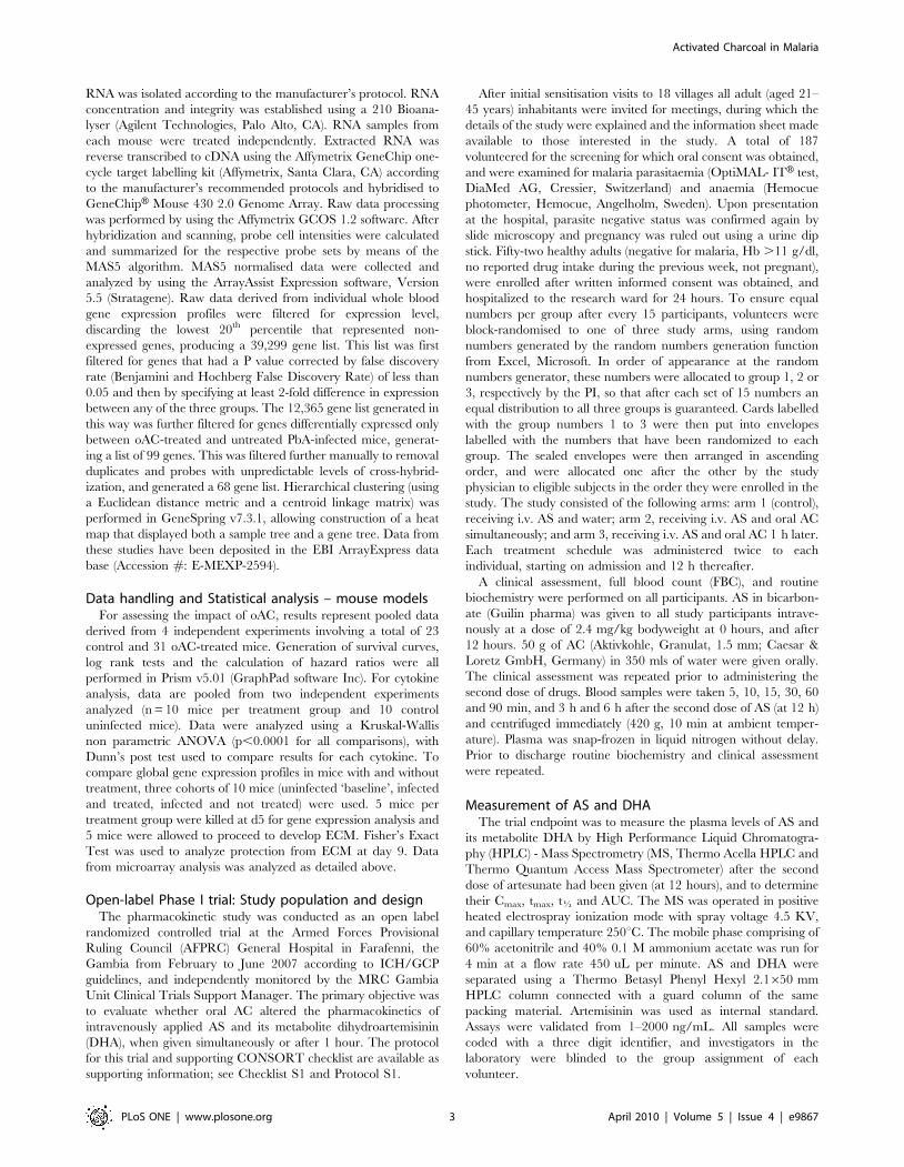
whole blood. The experimental design is summarised in Fig. 3a.
20 mice were infected with PbA and at d3 and d5, 10 were treated
with oAC (Group 1) and 10 with saline (Group 2). At d6 p.i., after
the onset of ECM symptoms but before any deaths had occurred
in Group 2, 5 mice from each group were killed, and whole blood
processed for gene chip analysis and flow cytometry. 5 naı̈ve mice
Figure 3. Transcriptional profiling of oAC-treated and untreated PbA-infected mice. a) 20 C57BL/6 mice were infected with PbA and at d3and d5, 10 were treated with oAC. At day 6, 5 mice per group were killed for gene expression analysis and the remainder monitored for survival. b)oAC-treated PbA-infected mice (blue line) were significantly protected from ECM compared to untreated PbA-infected mice (red line; p = 0.048). c)Heat map generated by hierarchical clustering of the 68 genes that passed the p-value threshold of a false discovery rate of 5% and were .2-folddifferentially expressed in whole blood of oAC-treated vs. untreated PbA-infected mice. Gene tree (side) and sample tree (top) are shown. Individualmice are numbered at bottom (oAC treated: 1.1–1.5; blue text, Untreated: 2.1–2.5; red text). Heat map intensity scale is also shown.doi:10.1371/journal.pone.0009867.g003
Activated Charcoal in Malaria
PLoS ONE | www.plosone.org 6 April 2010 | Volume 5 | Issue 4 | e9867

provided blood as reference (‘baseline’). The remaining infected
mice were used to monitor the development of ECM, again
showing that oAC significantly protected mice from ECM (Fisher’s
Exact test p = 0.048; RR = 0.2; Fig. 3b). At the time of sampling
for gene expression analysis, mice in both groups had similar
parasitemias, mean body weight, and frequencies of T cells
(CD3+), B cells (B220+) and NK cells (NK1.1+), whereas CD11b+
cells were slightly reduced in frequency in the blood of mice
treated with oAC (data not shown). We identified a whole blood
signature associated with oAC treatment of PbA-infected mice,
comprised of 68 genes with a false discovery rate corrected P value
of ,0.05 (ranging from p = 0.011 to p = 1.47e7) and which passed
a cut-off of 2-fold up- or down-regulation, relative to untreated
PbA-infected mice (Fig. 3c). This representation shows that there
was a high degree of similarity in the whole blood expression
profiles of untreated PbA-infected mice. However, within the
oAC-treated group, there was more variability, with some mice
(most notably #1.1 and to a lesser extent #1.3) having a gene
expression profile that more closely resembled that of untreated
mice. Given the level of protection against ECM observed in the
remainder of this cohort of oAC-treated mice (Fig 3b), this
analysis suggests that whole blood profiling may be able to identify
treatment failures, though further studies are required to
substantiate this claim. Nevertheless, collectively our microarray
and cytokine analysis confirmed that oAC treatment had a
significant and systemic impact on the immune and inflammatory
response in mice infected with PbA.
An open labelled Phase I trial to evaluate thepharmacokinetics of AS in combination with oAC
The protective efficacy and immunomodulatory effects of oAC,
as demonstrated above, supported a potential role for oAC in the
treatment of human malaria. Exploration of oAC as adjuvant
treatment for severe malaria has to take into account, however,
that oAC increases the elimination of quinine [24–26], up to now
the mainstay of treatment for severe malaria (SM). Recognizing
that quinine is likely to be replaced by AS for the treatment of
severe malaria [14], as an important first safety step, we therefore
sought to determine whether oAC altered the pharmacokinetics of
parenteral AS or its active metabolite DHA.
Fifty-two healthy adults were enrolled in the study (from 187
screened volunteers (Fig. 4). The study participants in each group
were similar with regard to age, weight, administered AS dose and
gender distribution (Table S1). The study drugs were safe and
well tolerated. No serious adverse event occurred. One participant
in arm 1 (AS alone) vomited once after administration of the i.v.
AS. No clinically relevant deviations from the normal ranges were
observed for the biochemistry and FBC tests.
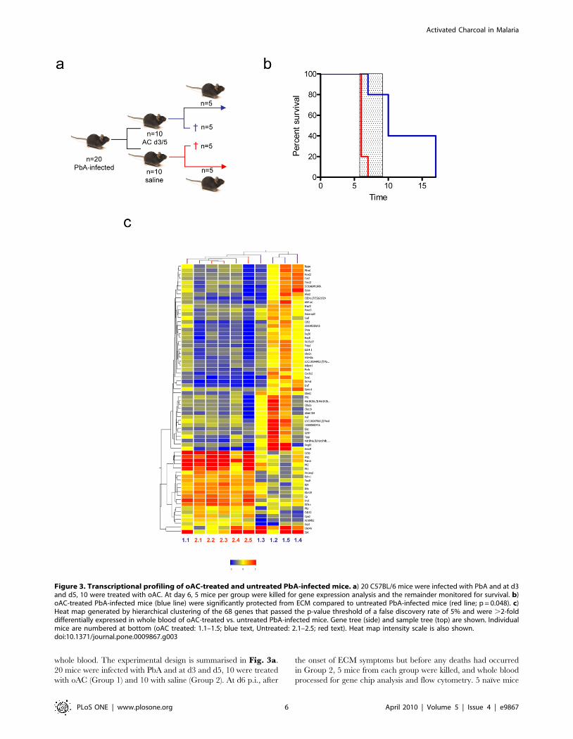
AS and DHA plasma concentrations were assessed for 8
individuals in each of arms 1 and 2, and 10 individuals in arm 3 as
described. Fig. 5 shows each fitted curve, and a one-compartment
model fitted through the geometric mean concentration for each
arm at each time point, using the geometric mean dose for that
arm. For both AS (Fig. 5a) and DHA (Fig. 5b), the geometric
mean AUC was similar between the three arms. ANOVA analysis
comparing the area under the curve (AUC, calculated as dose/
clearance) shows no significant difference between the three
groups (p = 0.92 for AS, and p = 0.55 for DHA). Compared to
controls, the largest difference of the four comparisons in
geometric means was 217%, which occurred for DHA in arm
3. None of the comparisons versus control of any of the
pharmacokinetic parameters reached statistical significance. For
AUC, the lowest of the four lower confidence limits was 243%
versus control (DHA; arm 3) and the highest of the upper limits
was +42% (AS; arm 2) (Table 1). Hence we were not able to
detect any effect of oAC on the pharmacokinetics of AS or DHA.
Even though the sample size was small, we were able to establish
that any effect on AUC was unlikely to be as much as 50%, a slight
effect compared to the ,200% variation observed between
individuals within each arm.
Discussion
Each year, infection with Plasmodium falciparum causes 400–600
million cases of malaria and up to 1 million childhood deaths in
Africa [1] Here, we demonstrate that oAC protects mice from
death due to ECM, with associated changes in T cell cytokine
production and in whole blood gene expression profiles.
Importantly, the changes in immune response and gene expression
profile observed following oAC treatment did not impact
negatively on host protection against PbA infection per se. In a
randomized controlled open label Phase I study, we found no
interference of oAC with the pharmacokinetics of the currently
best available anti-malarial drug AS. Collectively, these data pave
the way for clinical evaluation of oAC as a new safe, effective and
affordable adjunct treatment for severe malaria in man.
Our data indicate that oAC almost completely inhibited the
clinical and histopathological signs of ECM in mice. In those mice
that did develop ECM, onset was delayed, and in surviving mice
oAC also appeared to provide a degree of protection against death
due to high parasitemia, as measured by overall survival time.
However, further studies are clearly needed to understand the
mechanism(s) by which oAC mediates these striking effects. In
chronic kidney disease, it has been proposed that oAC serves as a
sink to block intestinal absorption of indole, thereby limiting
hepatic production of indole sulphate, a regulator of TGFbproduction [13]. Other data suggest the possibility of bile-
mediated transfer of cytokines to the intestinal lumen via the
entero-hepatic pathway, with charcoal acting as a presumptive
local adsorbant [12].
Two lines of evidence from the current study suggest that oAC
has broad systemic effects on immune and inflammatory processes.
First, oAC treatment significantly reduced the number of splenic
CD4+ and CD8+ T cells capable of TNF+ production and of CD4+
T cells co-producing TNF and IFNc, as determined after re-
stimulation. Although no effect of oAC was observed on cytokine
production measured directly ex vivo, such an observation is in
keeping with reports that serum pro-inflammatory cytokines,
included those measured above, peak earlier in infection than the
time point used in our analysis [27] and with the data of others
that indicates a greater ability to detect multi-functionality after re-
stimulation [23]. Similar associations between disease outcome
and cytokine levels have been observed in other models in which
the course of ECM has been altered. For example, in MyD882/2
C57BL/6 mice, which are resistant to ECM, production of IFNc,
TNF and IL-17 was reduced [27]. Similarly, in mice treated other
newly proposed adjunct therapies that show similar levels of
efficacy against ECM as demonstrated here for oAC, including
panthenine [28] and rosiglitazone [29], levels of TNF were also
reduced. For rosiglitazone, dampening of inflammation and
enhanced parasite clearance have also been observed in humans
given this drug as adjunct therapy (with atovaquone/proguanil) for
mild malaria [30], confirming the translational potential of studies
in murine models of disease.
Second, microarray analysis identified a clear whole blood
transcriptional signature that distinguished oAC-treated from
untreated PbA-infected mice. Gene expression analysis in ECM
has previously been largely restricted to using spleen cells or brain
Activated Charcoal in Malaria
PLoS ONE | www.plosone.org 7 April 2010 | Volume 5 | Issue 4 | e9867

tissue [31–33] making comparisons across either genetically
disparate hosts or using different parasite strains. In contrast, in
the application of this technology to human malaria has been
restricted to analysis of PBMC or whole blood [34–36], again
usually comparing individuals with distinct disease outcome. To
our knowledge, no direct comparisons of gene expression profile
have been made following interventions that seek to prevent the
development of severe disease. Further analysis of our gene
expression data, including refinement of the signature associated
with oAC treatment, will be published elsewhere. The data shown
here nevertheless provides evidence that oAC treatment directly or
indirectly affected gene regulation as determined in whole blood
from PbA-infected mice. The signature of 68 genes associated with
oAC treatment included multiple genes involved in the acute
phase response and inflammation, as well as genes involved in
heme biosynthesis and erythrocyte function (e.g. Fech, Epb4.1, and
Slc25a37). A case for the development of blood transcriptional
biomarkers has been extensively argued elsewhere [37,38], and the
application of a ‘modular’ approach to genomic analysis of the
response of SLE patients to treatment has been described recently
[39]. It will be important in the future to determine whether
similar or distinct signatures are associated with other interven-
tions designed to interrupt the progression of ECM, and when
oAC is evaluated for protection against CM and severe disease in
man, to likewise determine whether such signatures have cross-
species predictive value.
Although a molecular mode of action for oAC has yet to be
identified, we believe that the experimental data reported here
Figure 4. Study flow chart. The chart provides information on the number of individuals for each stage. The figures in brackets indicate thenumber of samples that could be analysed for AS and DHA.doi:10.1371/journal.pone.0009867.g004
Activated Charcoal in Malaria
PLoS ONE | www.plosone.org 8 April 2010 | Volume 5 | Issue 4 | e9867
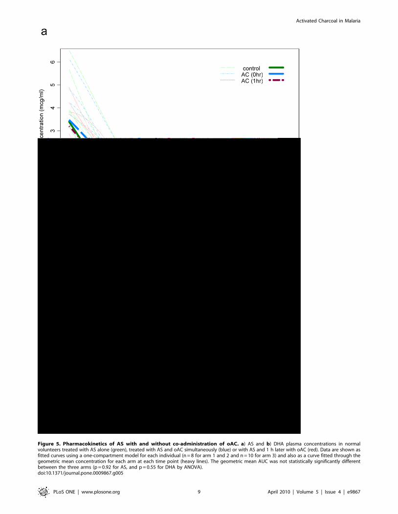
Figure 5. Pharmacokinetics of AS with and without co-administration of oAC. a) AS and b) DHA plasma concentrations in normalvolunteers treated with AS alone (green), treated with AS and oAC simultaneously (blue) or with AS and 1 h later with oAC (red). Data are shown asfitted curves using a one-compartment model for each individual (n = 8 for arm 1 and 2 and n = 10 for arm 3) and also as a curve fitted through thegeometric mean concentration for each arm at each time point (heavy lines). The geometric mean AUC was not statistically significantly differentbetween the three arms (p = 0.92 for AS, and p = 0.55 for DHA by ANOVA).doi:10.1371/journal.pone.0009867.g005
Activated Charcoal in Malaria
PLoS ONE | www.plosone.org 9 April 2010 | Volume 5 | Issue 4 | e9867

should nevertheless now provide a basis for the evaluation of oAC
as a treatment for severe malaria. oAC may provide a first line
therapy in the immediate absence of alternate treatment. Severe
malaria is an acute illness that may present with neurological
symptoms occurring often within 96 h of the onset of fever; much
of this time may be spent traveling from remote villages to health
clinics and consequently many children arrive in coma. If our
observations from the murine model would translate to man, oAC
given early in the course of infection could prevent the
development of CM and may reduce CM-associated mortality.
It is notable that AC has been used for many years in the
treatment of poisoning, and can be given orally or via a naso-
gastric tube, particularly in powdered form. It is well tolerated, has
an excellent, well-documented safety profile, and is relatively
inexpensive. Furthermore, AC has a long shelf life and can be
stored at ambient temperatures. Should clinical efficacy be proven,
these attributes would make AC highly suited for use in remote
rural communities where it could be administered at the first point
of care, for instance by a village health worker, and would
encourage the effective uptake of charcoal therapy in malaria-
endemic countries.
Nevertheless, given that specific treatment for malaria is
available, and that oAC treatment alone had no anti-malarial
activity in mice, the straightforward approach of assessing the
efficacy of oral AC as a stand-alone therapy would be unethical
in humans. Indeed, further exploration of the potential benefits
of oAC as adjuvant treatment have to take into account that
oAC is officially recommended to treat intoxication with quinine
[40], increasing the elimination of this drug [24–26]. However, it
was unknown whether the pharmacokinetics of AS, a drug now
regarded to be superior to quinine for the treatment of severe
malaria [14] and known to have a high endogenous clearance
rate [41], would be affected by co-administration of oAC. We
found no evidence of reduced plasma levels of AS or DHA due
to oAC, when given simultaneously with AS, or when
administered 1 h later. In arms 2 (AS and oAC simultaneously)
and 3 (oAC 1 h after AS), individuals received their first dose of
oAC 12 h prior to starting plasma sampling. The fact that the
AS and DHA concentrations found in these groups were not
lower than those observed in the control group support the
conclusion that even a high concentration of AC in the intestine
at the time of administration of AS did not interfere with the
pharmacokinetics of AS or DHA. Although the trial did not have
an equivalence design, all subjects who started therapy followed
the protocol schedule and there was no rescue therapy. This
reduces concerns about interpreting the results as if they had
come from an equivalence trial. Although the sample size
required for an equivalence trial would have been different, the
sample size actually achieved (which was reduced by the failure
to analyse some samples) is of course reflected in the width of the
confidence intervals. These data therefore suggest that clinical
trials of combined AS and oAC treatment in patients with
malaria could proceed without compromising the anti-malarial
activity of AS.
Finally, with the limited resources available at the primary
health care level, it is often not possible to reliably assign the
correct diagnosis to a severely ill child, especially to differentiate
between malaria and a bacterial infection (e.g. pneumonia,
typhoid/non-typhoid salmonella or meningitis) [42,43]. Treat-
ment is mainly empirical on a presumptive diagnosis, and clinical
assessment is often disturbingly poor [44] resulting in an over-
diagnosis of malaria at the expense of severe bacterial infections
[45,46]. For an adjuvant treatment to be employed successfully, it
needs to be safe and, ideally, beneficial irrespective of the precise
diagnosis. oAC could be the ideal candidate for this purpose due to
its non-specific anti-inflammatory potential, exploited successfully
for over a decade to prevent progression of chronic kidney disease
[13].
In conclusion, oAC prevented mortality associated with
ECM in a mouse model and when given to humans did not
interfere with the pharmacokinetics of AS or DHA. oAC
therefore has the potential to be a readily-implemented therapy
for the treatment of severe malaria. We suggest there is now an
urgent need for controlled clinical trials to evaluate the efficacy
of oAC in human malaria. We propose that the next step in the
development of oAC should be to evaluate its safety profile as an
adjuvant therapy in cases of uncomplicated malaria, a path
recently taken for the development of rosiglitazone [30], before
progressing to an evaluation of its potential benefit in cases of
severe malaria. Incorporation of gene expression profiling into
Table 1. Pharmacokinetic parameters for artesunate and DHA in the three trial arms.
drug and parameter AS + water (control) (n = 8) AS + charcoal simultaneously (n = 8) AS + charcoal 1 hour later (n = 10)
percent differencefrom control (95% CI)
percent differencefrom control (95% CI)
Artesunate
AUC (min6mg/ml) 48 (23–77) 49 (32–71) +2 (226, +42) 46 (33–67) 24 (229, +32)
t1/2 (min) 3.4 (1.8–5.2) 4.1 (2.9–5.3) +21 (211, +65) 4.0 (2.6–7.0) +18 (212, +58)
CL/F (liters/min) 3.1 (1.5–5.5) 3.1 (2.2–4.5) +2 (227, +44) 3.1 (2.0–4.3) +1 (227, +40)
V/F (liters) 15 (7.6–29) 19 (9.2–34) +24 (222, +98) 18 (11–34) +19 (223, +85)
DHA
AUC (min6mg/ml) 172 (108–310) 161 (91–268) 26 (237, +38) 142 (91–247) 217 (243, +19)
t1/2 (min) 34 (1–116) 40 (19–65) +20 (249, +183) 52 (32–109) +53 (232, +245)
CL/F (liters/min) 0.85 (0.46–1.27) 0.95 (0.57–1.56) +12 (224, +64) 1.00 (0.53–1.73) +16 (218, +70)
V/F (liters) 41 (2–113) 56 (30–132) +34 (243, +218) 75 (34–167) +80 (220, +309)
Cmax (mg/ml) 2.9 (1.4–29) 2.2 (1.3–4.1) 225 (261, +45) 1.7 (1.0–3.8) 243 (269, +7)
tmax (min) 0.8 (0–14) 0.7 (0–27) 29 (297, +2740) 2 (0–24) +162 (290, +6720)
doi:10.1371/journal.pone.0009867.t001
Activated Charcoal in Malaria
PLoS ONE | www.plosone.org 10 April 2010 | Volume 5 | Issue 4 | e9867

such trials should also yield further insights into the mode of
action of AC in man.
Supporting Information
Checklist S1 CONSORT Checklist S1
Found at: doi:10.1371/journal.pone.0009867.s001 (0.20 MB
DOC)
Protocol S1 Trial Protocol
Found at: doi:10.1371/journal.pone.0009867.s002 (0.19 MB
PDF)
Table S1 Characteristics of the study population according to
study group.
Found at: doi:10.1371/journal.pone.0009867.s003 (0.03 MB
DOC)
Acknowledgments
The study participants are gratefully acknowledged for their collaboration.
We are grateful to the management and staff of AFPRC General Hospital
Farafenni, MRC Farafenni and MRC Fajara for their valuable technical
assistance throughout the study. The authors also thank Prof. E. Riley and
Dr C. Engwerda for critical review of the manuscript and Prof. Brian M.
Greenwood for helpful discussions. This manuscript is dedicated to the
memory of Brian Foxwell.
Author Contributions
Conceived and designed the experiments: JBdS NDA NA HK DW SAW
DJC LU KJT BMJF PMK MW. Performed the experiments: JBdS UO
BMJO HK MJ SM. Analyzed the data: JBdS NDA NA HK DW PMK
MW. Contributed reagents/materials/analysis tools: PFS. Wrote the
paper: NDA PMK MW. Originally suggested the possibility of charcoal
therapy for malaria: LU KJT BMJF.
References
1. WHO World Malaria Report. 2008. http://whqlibdoc.who.int/publications/
2008/9789241563697_eng.pdf.
2. Cranston HA, Boylan CW, Carroll GL, Sutera SP, Williamson JR, et al. (1984)
Plasmodium falciparum maturation abolishes physiologic red cell deformability.
Science 223: 400–403.
3. Dondorp AM, Angus BJ, Hardeman MR, Chotivanich KT, Silamut K, et al.
(1997) Prognostic significance of reduced red blood cell deformability in severe
falciparum malaria. Am J Trop Med Hyg 57: 507–511.
4. Baruch DI, Pasloske BL, Singh HB, Bi X, Ma XC, et al. (1995) Cloning the P.
falciparum gene encoding PfEMP1, a malarial variant antigen and adherence
receptor on the surface of parasitized human erythrocytes. Cell 82: 77–87.
5. Berendt AR, Tumer GD, Newbold CI (1994) Cerebral malaria: the
sequestration hypothesis. Parasitol Today 10: 412–414.
6. Smith JD, Chitnis CE, Craig AG, Roberts DJ, Hudson-Taylor DE, et al. (1995)
Switches in expression of Plasmodium falciparum var genes correlate with
changes in antigenic and cytoadherent phenotypes of infected erythrocytes. Cell
82: 101–110.
7. Clark IA, Cowden WB (2003) The pathophysiology of falciparum malaria.
Pharmacol Ther 99: 221–260.
8. Schofield L, Mueller I (2006) Clinical immunity to malaria. Curr Mol Med 6:
205–221.
9. Clark IA, Budd AC, Alleva LM (2008) Sickness behaviour pushed too far–the
basis of the syndrome seen in severe protozoal, bacterial and viral diseases and
post-trauma. Malar J 7: 208.
10. van Hensbroek MB, Palmer A, Onyiorah E, Schneider G, Jaffar S, et al. (1996)
The effect of a monoclonal antibody to tumor necrosis factor on survival from
childhood cerebral malaria. J Infect Dis 174: 1091–1097.
11. Kellum JA, Venkataraman R (2002) Blood purification in sepsis: an idea whose
time has come? Crit Care Med 30: 1387–1388.
12. Jackson GD, Dai Y, Sewell WA (2000) Bile mediates intestinal pathology in
endotoxemia in rats. Infect Immun 68: 4714–4719.
13. Schulman G (2006) A nexus of progression of chronic kidney disease: charcoal,
tryptophan and profibrotic cytokines. Blood Purif 24: 143–148.
14. Dondorp A, Nosten F, Stepniewska K, Day N, White N (2005) Artesunate versus
quinine for treatment of severe falciparum malaria: a randomised trial. Lancet
366: 717–725.
15. Armitage P, Berry G, Matthews JNS (2001) Statistical methods in Medical
Research. Oxford: Blackwell Scientific Publications.
16. van Belle G, Martin DC (1993) Sample size as a function of coefficient of
variation and ratio of means. American Statistician 47: 163–167.
17. Hunt NH, Grau GE (2003) Cytokines: accelerators and brakes in the
pathogenesis of cerebral malaria. Trends Immunol 24: 491–499.
18. Engwerda CR, Ato M, Cotterell SE, Mynott TL, Tschannerl A, et al. (2002) A
role for tumor necrosis factor-alpha in remodeling the splenic marginal zone
during Leishmania donovani infection. Am J Pathol 161: 429–437.
19. Hearn J, Rayment N, Landon DN, Katz DR, de Souza JB (2000)
Immunopathology of cerebral malaria: morphological evidence of parasite
sequestration in murine brain microvasculature. Infect Immun 68: 5364–5376.
20. de Souza JB, Riley EM (2002) Cerebral malaria: the contribution of studies in
animal models to our understanding of immunopathogenesis. Microbes Infect 4:
291–300.
21. Riley EM, Wahl S, Perkins DJ, Schofield L (2006) Regulating immunity to
malaria. Parasite Immunol 28: 35–49.
22. Schofield L, Grau GE (2005) Immunological processes in malaria pathogenesis.
Nat Rev Immunol 5: 722–735.
23. Jankovic D, Kullberg MC, Feng CG, Goldszmid RS, Collazo CM, et al. (2007)
Conventional T-bet(+)Foxp3(-) Th1 cells are the major source of host-protective
regulatory IL-10 during intracellular protozoan infection. J Exp Med 204:
273–283.
24. Akintonwa A, Orisakwe OE (1990) The adsorption of quinine and quinidine to
activated charcoal with and without magnesium sulfate. Vet Hum Toxicol 32:
567–568.
25. Lockey D, Bateman DN (1989) Effect of oral activated charcoal on quinine
elimination. Br J Clin Pharmacol 27: 92–94.
26. Prescott LF, Hamilton AR, Heyworth R (1989) Treatment of quinine
overdosage with repeated oral charcoal. Br J Clin Pharmacol 27: 95–97.
27. Griffith JW, O’Connor C, Bernard K, Town T, Goldstein DR, et al. (2007) Toll-
like receptor modulation of murine cerebral malaria is dependent on the genetic
background of the host. J Infect Dis 196: 1553–1564.
28. Penet MF, Abou-Hamdan M, Coltel N, Cornille E, Grau GE, et al. (2008)
Protection against cerebral malaria by the low-molecular-weight thiol
pantethine. Proc Natl Acad Sci U S A 105: 1321–1326.
29. Serghides L, Patel SN, Ayi K, Lu Z, Gowda DC, et al. (2009) Rosiglitazone
modulates the innate immune response to Plasmodium falciparum infection and
improves outcome in experimental cerebral malaria. J Infect Dis 199:
1536–1545.
30. Boggild AK, Krudsood S, Patel SN, Serghides L, Tangpukdee N, et al. (2009)
Use of peroxisome proliferator-activated receptor gamma agonists as adjunctive
treatment for Plasmodium falciparum malaria: a randomized, double-blind,
placebo-controlled trial. Clin Infect Dis 49: 841–849.
31. Delahaye NF, Coltel N, Puthier D, Flori L, Houlgatte R, et al. (2006) Gene-
expression profiling discriminates between cerebral malaria (CM)-susceptible
mice and CM-resistant mice. J Infect Dis 193: 312–321.
32. Lovegrove FE, Gharib SA, Patel SN, Hawkes CA, Kain KC, et al. (2007)
Expression microarray analysis implicates apoptosis and interferon-responsive
mechanisms in susceptibility to experimental cerebral malaria. Am J Pathol 171:
1894–1903.
33. Lovegrove FE, Pena-Castillo L, Mohammad N, Liles WC, Hughes TR, et al.
(2006) Simultaneous host and parasite expression profiling identifies tissue-
specific transcriptional programs associated with susceptibility or resistance to
experimental cerebral malaria. BMC Genomics 7: 295.
34. Chakravorty SJ, Carret C, Nash GB, Ivens A, Szestak T, et al. (2007) Altered
phenotype and gene transcription in endothelial cells, induced by Plasmodium
falciparum-infected red blood cells: pathogenic or protective? Int J Parasitol 37:
975–987.
35. Griffiths MJ, Shafi MJ, Popper SJ, Hemingway CA, Kortok MM, et al. (2005)
Genomewide analysis of the host response to malaria in Kenyan children. J Infect
Dis 191: 1599–1611.
36. Ockenhouse CF, Hu WC, Kester KE, Cummings JF, Stewart A, et al. (2006)
Common and divergent immune response signaling pathways discovered in
peripheral blood mononuclear cell gene expression patterns in presymptomatic
and clinically apparent malaria. Infect Immun 74: 5561–5573.
37. He XS, Ji X, Hale MB, Cheung R, Ahmed A, et al. (2006) Global transcriptional
response to interferon is a determinant of HCV treatment outcome and is
modified by race. Hepatology 44: 352–359.
38. Wang E, Marincola FM (2008) Bottom up: a modular view of immunology.
Immunity 29: 9–11.
39. Chaussabel D, Quinn C, Shen J, Patel P, Glaser C, et al. (2008) A modular
analysis framework for blood genomics studies: application to systemic lupus
erythematosus. Immunity 29: 150–164.
40. Bradberry SM, Vale JA (1995) Multiple-dose activated charcoal: a review of
relevant clinical studies. J Toxicol Clin Toxicol 33: 407–416.
41. Hasan MM, el-Sayed YM, Abdelaziz AA (1990) The effect of oral activated
charcoal on the systemic clearance of gentamicin in rabbits with acute renal
failure. J Pharm Pharmacol 42: 85–88.
42. Berkley J, Mwangi I, Griffiths K, Ahmed I, Mithwani S, et al. (2005) Assessment
of severe malnutrition among hospitalized children in rural Kenya: comparison
of weight for height and mid upper arm circumference. JAMA 294: 591–597.
Activated Charcoal in Malaria
PLoS ONE | www.plosone.org 11 April 2010 | Volume 5 | Issue 4 | e9867

43. Brent AJ, Oundo JO, Mwangi I, Ochola L, Lowe B, et al. (2006) Salmonella
bacteremia in Kenyan children. Pediatr Infect Dis J 25: 230–236.
44. Reyburn H, Mwakasungula E, Chonya S, Mtei F, Bygbjerg I, et al. (2008)
Clinical assessment and treatment in paediatric wards in the north-east of the
United Republic of Tanzania. Bull World Health Organ 86: 132–139.
45. Makani J, Matuja W, Liyombo E, Snow RW, Marsh K, et al. (2003) Admission
diagnosis of cerebral malaria in adults in an endemic area of Tanzania:implications and clinical description. QJM 96: 355–362.
46. Reyburn H, Mbatia R, Drakeley C, Carneiro I, Mwakasungula E, et al. (2004)
Overdiagnosis of malaria in patients with severe febrile illness in Tanzania: aprospective study. BMJ 329: 1212.
Activated Charcoal in Malaria
PLoS ONE | www.plosone.org 12 April 2010 | Volume 5 | Issue 4 | e9867
Copyright © 2022 FDOKUMEN