DEVELOPMENT OF PARENTERAL DRUGS FOR THE ...
232
DEVELOPMENT OF PARENTERAL DRUGS FOR THE TREATMENT OF SEIZURE EMERGENCIES A DISSERTATION SUBMITTED TO THE FACULTY OF THE GRADUATE SCHOOL OF THE UNIVERSITY OF MINNESOTA BY Irene Vuu IN PARTIAL FULFILLMENT OF THE REQUIREMENTS FOR THE DEGREE OF DOCTOR OF PHILOSOPHY James Cloyd, PharmD, Advisor Lisa Coles, MS, PhD, Co-Advisor September 2019
-
Upload
khangminh22 -
Category
Documents
-
view
0 -
download
0
Transcript of DEVELOPMENT OF PARENTERAL DRUGS FOR THE ...

DEVELOPMENT OF PARENTERAL DRUGS FOR THE TREATMENT OF SEIZURE EMERGENCIES
A DISSERTATION SUBMITTED TO THE FACULTY OF THE GRADUATE SCHOOL
OF THE UNIVERSITY OF MINNESOTA BY
Irene Vuu
IN PARTIAL FULFILLMENT OF THE REQUIREMENTS FOR THE DEGREE OF
DOCTOR OF PHILOSOPHY
James Cloyd, PharmD, Advisor Lisa Coles, MS, PhD, Co-Advisor
September 2019

© Irene Vuu 2019
i
ACKNOWLEDGMENTS
I would like to thank everyone who contributed to my achievements during my graduate program. My advisor, Dr. James Cloyd, for believing in me from day one and making the time to advise me on career options. Without you and your remarkable passion for research and teaching, this dissertation and the dream job I never knew I wanted would not be possible. My co-advisor, Dr. Lisa Coles, for filling in all the gaps and always being there. From introducing me to pharmacokinetic modeling and simulations to trying out a barre class, and from tackling down applications and deadlines together to discussing the next restaurant I must try. You are amazing. Dr. Ned Patterson, for giving me the opportunity to learn about veterinary clinical research and for serving as the chair of my committee. Thanks are also due to Andrea Eckert for her crucial role in my projects. Dr. Ilo Leppik, for sharing his expertise in clinical research in the epilepsy field and supporting my scientific development. Dr. Michael Rogawski, for sharing his expertise and for serving on my committee. Many thanks are also due to Dr. Dorota Zolkowska and Dr. Chun-Yi Wu for their support in my project. Dr. Chap Le, for serving as the external committee member. Dr. Richard Brundage, for his patience and sharing his expertise on pharmacometrics. I have always enjoyed your courses and the stimulating discussions we have had. Without you, pharmacometrics would be much more abstract and less fascinating. The research group at the Center for Orphan Drug Research: Lori, Laurie, Usha, Reena and Marcia, for always lending me support. The department staff and faculty, for their encouragement and support throughout the years. My colleagues who have graduated: Natalie and Sam, for their friendship, guidance, and support on so many levels. My fellow pharmacometrics graduate students: Abhi, Ashwin, Siddhee, Fan, Ya-Feng and Shen, for their lively discussions in our ‘metrics courses. I am constantly impressed by you all.

ii
Angela, for planting the first seed of a career in research into my head. My dearest friends: Julie, Verna, Michelle, Susan, Lidia, Andrea and Valerie, for their never-ending support, advice, and encouragement. My “classmate” and better half, Jesse, for his understanding and support throughout the toughest of times.
iii
DEDICATION
To all patients affected by seizure emergencies
&
To my teachers for their endless patience and confidence in me
&
To my family and friends for their unconditional love and support
iv
ABSTRACT
The overall objective of my dissertation is to develop alternative therapies
for seizure emergencies. Status epilepticus is a condition defined as a
convulsive seizure lasting more than 5 minutes and is considered a seizure
emergency due to the increased risk for neuronal damage and mortality (Trinka
et al. 2015). Although relatively effective, first-line therapy fails to terminate status
epilepticus in 26-57% of cases, leading to increased risk of seizure refractoriness
and use of second- and third-line therapies that may increase the risk of systemic
complications and mortality (Treiman et al. 1998; Alldredge et al. 2001; Silbergleit
et al. 2012; Chamberlain et al. 2014). Three drugs were studied:
allopregnanolone (ALLO), lacosamide (LCM) and topiramate (TPM). The
pharmacokinetics and pharmacodynamics of investigational allopregnanolone
formulations following intravenous and intramuscular delivery were assessed for
the development as an early rescue therapy for seizure emergencies (Project 1).
I also explored the relationship between lacosamide and PR prolongation in the
critically-ill population to identify a subpopulation in whom it can be used safely
(Project 2). Finally, for topiramate, the pharmacokinetics and pharmacodynamics
of an investigational intravenous formulation was evaluated for adjunctive
therapy in seizure emergencies (Project 3).
Allopregnanolone, a progesterone derivative and GABAA positive allosteric
modulator, has demonstrated potential to treat status epilepticus in preclinical
models and pediatric and adult patient case reports. Given that first-line therapy
fails in the majority of cases, more effective early treatments are necessary to
v
prevent downstream seizure refractoriness and systemic complications. The
specific aims for Project 1 were to characterize the pharmacokinetics,
pharmacodynamics and safety following intravenous and intramuscular ALLO in
dogs. Five dogs (one on phenobarbital therapy) received single doses of ALLO:
one- to four-mg/kg intravenously, or one- to six-mg/kg intramuscularly, with a
washout period of at least one week. Plasma samples were collected pre-dose
and at regular intervals up to six hours post-dose. Clinical response was
assessed by behavioral response and intracranial electroencephalographic
(iEEG). I found that with IV ALLO, drug exposure and peak plasma concentration
increased proportionally with dose within the doses studied. Behavioral
responses and iEEG data illustrate the rapid onset of effect following IV ALLO
administration. The results of this study indicate that IV ALLO is a promising
agent for the early treatment of seizure emergencies, with evidence of rapid
penetration into the brain and a high safety profile. IM ALLO has great potential
to be useful as a first-line treatment for SE, but the current formulations do not
attain high enough plasma concentrations predicted to confer iEEG changes.
Therefore, alternative approached would be needed for a viable IM ALLO
product.
Intravenous LCM has shown safety and some efficacy as an adjunctive
therapy in refractory convulsive and non-convulsive status epilepticus. Outside of
seizure emergencies, it is also used in the critically-ill population to treat acute
breakthrough seizures or to maintain seizure control in patients who are unable
to take oral medications. Lacosamide is particularly appealing in this patient

vi
population due to its low potential for drug-drug interactions and serious systemic
complications. However, there are reports of PR interval prolongation, which
raises concern for patients who have a higher risk for developing cardiac
arrhythmias or conduction abnormalities. The specific aim of Project 2 was to
estimate the prevalence of PR prolongation in the critically-ill patient population
following intravenous LCM administration. I performed a retrospective chart
review and defined PR interval prolongation as a shift from normal to high PR
interval or an increase of 20% or more in PR interval from baseline. Logistic
regression analysis was performed in order to identify clinical factors that help
predict an increase in PR interval 20%. Eight percent of my patient sample
experienced PR prolongation, which is 20-times higher than the prevalence of
0.4% reported in ambulatory patients with epilepsy. The logistic regression
analysis suggested that the occurrence of PR prolongation following IV LCM
administration is positively associated with age, the total daily dose of LCM, and
serum potassium levels. However, considering that these results are generated
from a small number of events (n=7/88), the true impact of these predictors on
PR prolongation in this patient population needs to be explored further.
In addition to finding alternative early treatments for seizure emergencies,
better adjunctive treatments during refractory stages of status epilepticus are
also needed. Topiramate’s many mechanisms of action and preclinical evidence
of neuroprotection, which make it an ideal candidate to treat status epilepticus
that has become resistant to first-line therapies. Intravenous administration of
TPM offers an alternative that would allow more drug to get into the body and at

vii
a faster rate than current methods of its administration. The specific aims of
Project 3 were to characterize the pharmacokinetics, pharmacodynamics and
safety following intravenous TPM in dogs. Five dogs (three on phenobarbital
maintenance therapy) were used in this study. Ten and twenty mg/kg of stable-
labeled topiramate were infused intravenously over five minutes. One hour
following the 10 mg/kg infusion, each dog also received a 5 mg/kg dose of
unlabeled oral topiramate. Plasma samples were collected pre-dose and at
regular intervals up to nine hours post-dose. Sixteen electrode channels were
continuously recorded. Topiramate concentration-time data were analyzed using
noncompartmental and population compartmental approaches. Concentration-
time data were best fit by a two-compartment model, and co-medication with
phenobarbital was associated with a 5.6-fold higher clearance. The estimated
absolute oral bioavailability ranged from 62-102%. Statistically significant
increases in iEEG activity were observed within 30 minutes of infusion, which is
essential when treating seizure emergencies. Simulations suggest a different
dosing strategy for dogs on phenobarbital may be necessary to optimize drug
exposure. The results of this study indicate that development of an intravenous
TPM formulation with evidence of penetration into the brain and good tolerability
is feasible.
My research suggests that there are promising therapies in development for
the management of SE, which will significantly improve patient lives by offering
safer use of current antiseizure drugs or more effective therapies. There are
many pathways into which these projects can take, including conducting clinical

viii
trials in dogs with naturally-occurring SE and single- and multiple-ascending dose
studies in patients.

ix
TABLE OF CONTENTS
Acknowledgments ..................................................................................................... i
Dedication ................................................................................................................ iii
Abstract .................................................................................................................... iv
Table of Contents .................................................................................................... ix
List of Tables ........................................................................................................... xi
List of Figures ........................................................................................................ xiii
Chapter 1 ................................................................................................................ 1
Introduction .............................................................................................................. 1
1.1 Introduction and Orientation ..................................................................... 2
1.2 Human Epilepsy and Seizure Emergencies ............................................ 5
1.3 Using Canine Status Epilepticus as a Model of Human Status Epilepticus .......................................................................................................... 28
Chapter 2 .............................................................................................................. 47
Development of allopregnanolone for the early Treatment of status epilepticus 47
2.1 Introduction ............................................................................................. 48
2.2 Review of Allopregnanolone................................................................... 48
2.3 Pharmacokinetics and Safety of Intravenous Allopregnanolone for Early Treatment of Status Epilepticus in Dogs ........................................................... 56
2.4 Pharmacokinetics, Safety, and Optimization of an Intramuscular Allopregnanolone Formulation .......................................................................... 76
2.5 Pharmacodynamics of Intravenous and Intramuscular Allopregnanolone in Dog 93
2.6 Design of a Safety and Effectiveness Clinical Trial of Intravenous Allopregnanolone for the Treatment of Canine Status Epilepticus ................ 105
Chapter 3 ............................................................................................................ 116
Evaluating the Safety of Intravenous Lacosamide for the Treatment of Acute Seizures ............................................................................................................... 116
3.1 Introduction ........................................................................................... 117
3.2 Lacosamide ........................................................................................... 117
3.3 Intravenous Lacosamide Use and PR Interval Prolongation in the Critically-Ill Patient ........................................................................................... 121
3.4 Design of a Prospective Observational Study of PR Prolongation in Critically-Ill Patients on Intravenous Antiseizure Therapy: Focus on Intravenous Lacosamide ...................................................................................................... 134
Chapter 4 ............................................................................................................ 141

x
Development of an Investigational Intravenous Formulation of Topiramate for the Treatment of Refractory Status Epilepticus ........................................................ 141
4.1 Introduction ........................................................................................... 142
4.2 Topiramate ............................................................................................ 142
4.3 Intravenous Topiramate: Pharmacokinetics and Effect on Electroencephalograph Activity in Dogs with Naturally-Occurring Epilepsy .. 148
4.4 Development of Intravenous Topiramate for the Treatment of Refractory Status Epilepticus ............................................................................................ 166
Chapter 5 ............................................................................................................ 182
Conclusions ......................................................................................................... 182
References .......................................................................................................... 189

xi
LIST OF TABLES
Table 1.2.1-1 Drugs for the Management of Epilepsy (Part 1) ............................ 11
Table 1.2.1-2 Drugs for the Management of Epilepsy (Part 2) ............................ 12
Table 1.2.1-3 Drugs for the Management of Epilepsy (Part 3) ............................ 13
Table 1.2.1-4 Drugs for the Management of Epilepsy (Part 4) ............................ 14
Table 1.2.2-1 Status Epilepticus in Certain Electroclinical Syndromes.
Reproduced with permission from Trinka et al 2015. ........................................... 18
Table 1.2.2-2 Factors that Promote/Diminish Self-Sustaining Seizure Activity.
Reproduced with permission from Niquet et al 2016. .......................................... 21
Table 1.3.1-1 Antiseizure Drugs Used in the Management of Canine Epilepsy . 33
Table 2.3.2-1 Estimated and Calculated Three-compartment Model PK
Parameter Values. ................................................................................................. 59
Table 2.3.2-2 PK parameter values reported in the literature .............................. 59
Table 2.3.2-3 Number of Animals per IV Study Dose .......................................... 60
Table 2.3.2-4 Modified Glasgow Coma Scale. Reproduced with permission from
Platt et al 2001. ...................................................................................................... 65
Table 2.3.3-1 Animal Demographics .................................................................... 66
Table 2.3.3-2 Non-compartmental PK Parameter Estimates ............................... 67
Table 2.3.3-3 Individual Compartmental PK Parameter Estimates ..................... 70
Table 2.3.3-4 Population Compartmental PK Parameter Estimates ................... 71
Table 2.3.3-5 Individualized PK Parameter Estimates from a Population
Approach................................................................................................................ 72
Table 2.3.3-6 Behavioral Response Following IV Administration ........................ 73
Table 2.4.2-1 Number of Animals per IM Study Dose.......................................... 78
Table 2.4.3-1 Animal Demographics .................................................................... 82
Table 2.4.3-2 Non-compartmental PK Parameter Estimates ............................... 83
Table 2.4.3-3 Individual Compartmental PK Parameter Estimates ..................... 85
Table 2.4.3-4 Bioavailability Calculated by NCA and Deconvolution Methods ... 88
Table 2.4.3-5 PK Parameter Estimates Used for IM Simulations ........................ 88
Table 2.4.3-6 Behavioral Response Following IM Administration ....................... 89
Table 2.5.3-1 PKPD Parameter Estimates (iEEG Absolute Power Density) ....... 98

xii
Table 2.5.3-2 PKPD Parameter Estimates (iEEG Relative Power Density) ...... 100
Table 3.3.3-1 Patient Demographics .................................................................. 127
Table 3.3.3-2 Logistic Regression Results for PR Prolongation ........................ 128
Table 3.3.3-3 Logistic Regression Results for PR Increase >20% .................... 129
Table 4.3.3-1 Animal Demographics .................................................................. 156
Table 4.3.3-2 Pharmacokinetic parameter estimates generated from non-
compartmental analysis following IV administration ........................................... 158
Table 4.3.3-3 Pharmacokinetic parameter estimates generated from non-
compartmental analysis following PO administration ......................................... 159
Table 4.3.3-4 Pharmacokinetic parameter estimates from a population
compartmental analysis following an intravenous TPM (low and high doses) .. 160
Table 4.3.3-5 Difference between energy levels averaged across low gamma and
beta frequency bands before and after IV TPM in one dog ............................... 162

xiii
LIST OF FIGURES
Figure 1.2.1-1 ILAE Framework for Classification of Epilepsies. Reproduced with
persmission from Scheffer et al 2017. .................................................................... 7
Figure 1.2.1-2 ILAE Operation Classificaiton of Seizure Types. Reproduced with
permission from Fisher et al 2017. ......................................................................... 9
Figure 1.2.1-3 Mechanisms of Action of FDA-Approved Antiseizure Drugs.
Reproduced with permission from C. Landmark 2008 ......................................... 12
Figure 1.2.2-1 Ictal, Transition, and Post-Ictal States. Ictal, Transition, and Post-
Ictal States. ............................................................................................................ 19
Figure 1.2.2-2 Algorithm for Management of Convulsive Status Epilepticus.
Reproduced with permission from Glauser et al 2016. ........................................ 23
Figure 1.3.2-1 Management of Status Epilepticus in Dogs and Cats Reproduced
with permission from Blades Golubovic and Rossmeisl 2017b ........................... 36
Figure 2.3.2-1 Dosing Simulations for Initial IV ALLO Dose in Dogs................... 60
Figure 2.3.3-1 Plasma ALLO Concentration-Time Profile Following IV
Administration on a log-scale. cPB: Chronic PB cPB: Chronic PB ...................... 67
Figure 2.3.3-2 Dose-proportionality: Dose-normalized AUC0-INF ......................... 68
Figure 2.3.3-3 Dose-proportionality: Dose-normalized observed Cmax ................ 68
Figure 2.3.3-4 Goodness of fit plots for a two-compartment model. IWRES:
individual weighted residual; TAD: time after dose; IPRED: individual predicted
concentration ......................................................................................................... 69
Figure 2.3.3-5 Goodness of fit plots for a population two-compartment Model.
IPRED (red open circle): individual predicted concentration; PRED (blue open
circle): population predicted concentration; CWRES: conditional weighted
residual; TAD: time after dose .............................................................................. 70
Figure 2.3.3-6 Simulation of Plasma ALLO Concentration-Time Profiles Following
IV Dosing. Dashed line: highest peak plasma concentration (900 ng/mL)
associated with seizure termination in TETS SE mouse model; Dot-dashed line:
lowest peak plasma concentration (400 ng/mL) associated with seizure
termination in TETS SE mouse model.................................................................. 73

xiv
Figure 2.4.3-1 Plasma ALLO Concentration-Time Profile Following IM
Administration. cPB: Chronic PB. Blue: 1 mg/kg dose; Red: 2 mg/kg dose;
Green: 6 mg/kg dose ............................................................................................. 83
Figure 2.4.3-2 Bioavailability by Formulation Concentration................................ 84
Figure 2.4.3-3 Goodness of fit plots for a two-compartment model. IWRES:
individual weighted residual; TAD: time after dose; IPRED: individual predicted
concentration ......................................................................................................... 85
Figure 2.4.3-4 IM ALLO Absorption Rate Over Time ........................................... 86
Figure 2.4.3-5 IM ALLO Fraction Absorbed Over Time ....................................... 87
Figure 2.4.3-6 Bioavailability Estimated by NCA vs Deconvolution Methods. A:
NCA; B: deconvolution .......................................................................................... 88
Figure 2.4.3-7 Simulation of Plasma ALLO Concentration-Time Profiles Following
IM Administration. Dashed line: highest peak plasma concentration (900 ng/mL)
associated with seizure termination in TETS SE mouse model; Dot-dashed line:
lowest peak plasma concentration (400 ng/mL) associated with seizure
termination in TETS SE mouse model.................................................................. 89
Figure 2.5.3-1 Absolute Power Density and Plasma ALLO Concentration-Time
Profiles Following IV and IM ALLO Administration in One Dog on Chronic PB. . 96
Figure 2.5.3-2 Relative Power Density and Plasma ALLO Concentration-Time
Profiles Following IV and IM ALLO Administration in One Dog on Chronic PB. . 97
Figure 2.5.3-3 iEEG (absolute power density) effect-concentration profiles
following 1-2 mg/kg infused IV over 5 minutes (red and blue, respectively) and 1
mg/kg injected IM as a bolus (orange) in 1 dog on cPB. ..................................... 99
Figure 2.5.3-4 iEEG (relative power density) effect-concentration profiles
following 1-2 mg/kg infused IV over 5 minutes (red and blue, respectively) and 1
mg/kg injected IM as a bolus (orange) in 1 dog on cPB. ................................... 101
Figure 2.5.5-1 Determination of the Target Plasma ALLO Concentration......... 104
Figure 2.5.5-2 Determination of IV ALLO Dose for a Clinical Trial in Canine SE
............................................................................................................................. 105
Figure 2.6.3-1 IV ALLO Study Schematic ........................................................... 107
xv
Figure 3.3.3-1 Dotplot of Primary Outcomes. Each circle represents a patient.
Blue circles denote patients who experienced a PR prolongation within 24 hours
following IV LCM administration.......................................................................... 128
Figure 3.3.3-2 Relationships between Age, Total Daily Dose (TDD) of LCM,
Median-Normalized Serum Potassium, and PR Prolongation. The size and color
of the circles denote the age (i.e. larger circle, older age) and PR prolongation
(i.e. blue = shift from normal to prolonged PR interval), respectively. ............... 129
Figure 3.3.3-3 Pre-Dose PR Interval on Event Occurrence and the Effect of
Smoking Status ................................................................................................... 130
Figure 4.3.3-1 Plasma-Concentration Time Profiles Following IV Administration of
a Low (10 mg/kg) and High (20 mg/kg) dose of stable-labeled TPM ................ 157
Figure 4.3.3-2 Plasma-concentration time profiles following administration of a
single 5 mg/kg TPM oral dose ............................................................................ 158
Figure 4.3.3-3 Observed versus predicted individual and population
concentrations ..................................................................................................... 159
Figure 4.3.3-4 Visual predictive check plots of a population model for dogs not on
PB and dogs on PB, respectively. Red and black lines represent the observed
and simulated quantiles, respectively. ................................................................ 161
Figure 4.3.3-5 Simulated Plasma Concentration Time Profiles Across Varying
Doses Infused Over 5 Minutes in a dog not on PB and a dog on PB. Here, the
yellow band is the desired range, and the black vertical line denotes 30 minutes
post-dose. ............................................................................................................ 161
Figure 4.3.3-6 EEG normalized signals from beta and low gamma frequency
bands averaged over 1-minute intervals in one dog .......................................... 162
Figure 4.4.1-1 IV TPM SAD/MAD Study Schematic .......................................... 170
Figure 4.4.2-1 IV TPM Study Schematic ............................................................ 174

1
CHAPTER 1
INTRODUCTION

2
1.1 Introduction and Orientation
Status epilepticus (SE) is a life-threatening condition that requires rapid treatment
in order to prevent systemic complications and irreversible brain damage.
Although there are evidence-based guidelines for the management of convulsive
SE, including the use several antiseizure drugs with different mechanisms of
action, the case fatality rate within 30 days of the SE event ranges from 21-39%
(Logroscino et al. 1997; Vignatelli, Tonon, and Alessandro 2003).
As the duration of a seizure lengthens, the seizure becomes less likely to
terminate on its own and more difficult to treat with current therapies (J. W. Chen
and Wasterlain 2006; Fujikawa 1996; Mazarati, Baldwin, et al. 1998). After 30
minutes of prolonged seizure activity, the risk for neuronal cell damage
escalates. Therefore, time is essential in the management of SE, and rapid
intervention with the goal of seizure termination is key. Even with relatively
effective first-line therapies, roughly 30% of cases fail to respond and progress to
more serious conditions (Treiman et al. 1998). The current treatment of SE is
suboptimal, especially earlier on in treatment algorithm. There is a need for safe
and effective alternatives to better manage this condition.
In general, the rapid intervention of SE is determined by the routes of drug
administration. For this condition, the ideal intervention is one that can be
administered with ease and achieve therapeutic drug concentrations in the brain
within a short amount of time. For this to be possible, the drug must be able to be

3
formulated in a solution that allows for intravenous and depending on its
physicochemical properties, intramuscular, intranasal or subcutaneous
administration. While enteral and rectal routes of administration have been used
for SE treatment, they are limited by slower rates of absorption (and
consequently lower and delayed peak drug concentrations) and decreased social
acceptance, respectively (Bhattacharyya, Kalra, and Gulati 2006; Brigo et al.
2015). Thus, in this dissertation, the focus will be on parenteral formulations of
central nervous system-active (CNS-active) drugs.
The overarching objective of my dissertation is to develop alternative
therapies for seizure emergencies, which will have a significant impact on the
patients and families of those affected by offering safer use of current treatments
or more effective treatments. As part of this work, I will present a review of
human epilepsy and SE, followed by canine epilepsy and SE, and the
translatability of therapeutic and mechanistic research between the two diseases.
Although my primary focus is on treatment alternatives for SE and not the
management of epilepsy syndromes, it is essential to understand the underlying
pathophysiology of seizures and epileptogenesis before attempting to treat
prolonged seizures. The prospective therapies under development range in their
stages in the drug development pipeline, as well as their potential place in the
management of seizure emergencies. These include:

4
- Project 1: Allopregnanolone, a naturally-occurring neurosteroid that is a
positive allosteric modulate GABAA receptors, with potential as an early
treatment of SE
o Hypothesis: Allopregnanolone would be beneficial in the early
treatment of SE based on its novel mechanism of action and ability
to get into the brain quickly
o Specific aim: To characterize the pharmacokinetics,
pharmacodynamics, and safety/tolerability following
intravenous and intramuscular allopregnanolone in dogs
- Project 2: Lacosamide, an antiseizure drug that enhances the slow
inactivation of voltage-gated sodium channels, with potential as a treatment
for established SE but has concerns for cardiac safety
o Hypothesis: Intravenous lacosamide increases the risk for PR
prolongation, especially in the critically-ill population
o Specific aim: To estimate the prevalence of PR prolongation in
the critically-ill patient population following intravenous
lacosamide administration
- Project 3: Topiramate, an antiseizure drug that potentiates GABA current and
antagonize AMPA/kainite receptors, with potential as an adjunctive treatment
for refractory SE
o Hypothesis: Intravenous topiramate would be beneficial as an
adjunctive treatment for refractory SE based on its multiple
mechanisms of action and low potential for drug-drug interactions

5
o Specific aims: To characterize the pharmacokinetics,
pharmacodynamics, and safety/tolerability following
intravenous topiramate in dogs
As part of the review, I will refer to ratings of evidence and levels of
recommendation which are categorized based on systematic reviews conducted
by Glauser et al 2016 and Podell et al 2016. In general, these authors rate
evidence depending on the type of clinical studies conducted. High level of
evidence is from a prospective, blinded, randomized, controlled clinical trial
(RCT) with masked outcome assessment in a representative population.
Moderate level of evidence is from a prospective randomized matched group
cohort study. Low level of evidence is from uncontrolled studies, case series,
case reports, or expert opinion. Consequently, the level of recommendation for
specific therapies are based on the level of evidence available for an indication.
For example, a high recommendation is given if treatment is established with
high level of evidence as effective and should be given, while a moderate
recommendation is given if the treatment is probably effective and should be
considered (Glauser et al. 2016).
1.2 Human Epilepsy and Seizure Emergencies
1.2.1 Human Epilepsy
Epilepsy is a disease of the brain that is characterized by the presence and/or
predisposition for seizures. An epileptic seizure is a passing occurrence of

6
symptoms due to abnormal electrical brain activity (Fisher et al. 2005). The
International League Against Epilepsy (ILAE) is an organization founded in 1909
whose goals are to advance the knowledge of epilepsy, promote its research and
education, and improve the care of patients with epilepsy (About International
League Against Epilepsy 2019). As part of their mission, the ILAE is tasked with
defining and classifying seizures and epilepsy. In 2014, a practical definition of
epilepsy was established to aid in the diagnosis of the disease. Epilepsy is
diagnosed by the presence of any of the following: “1) at least two unprovoked
(or reflex) seizures occurring more than 24 hours apart; 2) one unprovoked
(reflex) seizure and a probability of further seizures similar to the general
recurrence risk (at least 60%) after two unprovoked seizures occurring over the
next 10 years; or 3) diagnosis of an epilepsy syndrome (Fisher et al. 2014).”
Moreover, epilepsy is considered “resolved” for patients who had age-dependent
epilepsy syndrome and are now past the applicable age, or those who have been
seizure-free for the past ten years without anti-seizure drugs (ASDs) for the last
five years.
1.2.1.1 Prevalence and Etiology
According to the Epilepsy Foundation, epilepsy is the fourth most common
neurological disorder. Its prevalence has been reported to be range between 2.3-
22.8 cases of epilepsy per 1,000 people in the general population worldwide
(0.23-2.3%), and 6.8-8.5 cases in 1,000 of insured people in the United States
alone (Bell, Neligan, and Sander 2014; H. Kim et al. 2016; Fiest et al. 2017;

7
Helmers et al. 2015). In 2017, ILAE commissioned a new classification system
for seizure types and epilepsy types to improve the intuitiveness of the
classification in addition to allowing for inclusion of previously unclassifiable
seizure and epilepsy types (Figure 1.2.1-1) (Falco-Walter, Scheffer, and Fisher
2018). These will be discussed in more detail in the following section. Along each
step of the diagnostic pathway, the ILAE recommends that the clinician should
attempt to identify the etiology of the patient’s epilepsy (Scheffer et al. 2017).
Within the new classification system, there are six non-hierarchical etiological
categories with management implications, including: structural (i.e. neuroimaging
finding inferred to cause the patient’s seizures which may have resulted from a
stroke, infection, trauma, genetic malformation, etc.), genetic (i.e. a known or
presumed specific disease-causing gene variant believed to be pathogenic for
Figure 1.2.1-1 ILAE Framework for Classification of Epilepsies. Reproduced with persmission from Scheffer et al 2017.

8
epilepsy), infectious (i.e. refers to a patient with seizures due to resolved
infection), metabolic (e.g. uremia, pyridoxine-dependent seizures, cerebral folate
deficiency), immune (i.e. when an autoimmune disease is the cause of new-
onset epilepsy, like anti-NMDA receptor encephalitis), and unknown. A patient’s
epilepsy may be classified into more than one etiologic category, and the
importance of each etiological group may depend on the patient’s circumstance
(e.g. a patient with tuberous sclerosis has a structural and genetic etiology, which
would be critical for surgical and pharmacological considerations).
1.2.1.2 Seizure Semiology and Clinical Diagnosis
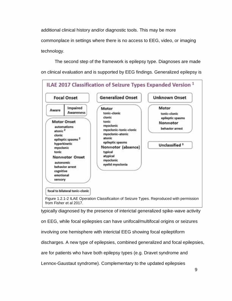
The starting point of the classification framework (Figure 1.2.1-1) is the
operational identification of the seizure type, outlined in Figure 1.2.1-2 (Fisher et
al. 2017). A seizure type is a grouping of seizure qualities for the purposes of
communication in research, clinical care, and education. The framework is non-
hierarchical, so that levels can be skipped or omitted with no other elaboration.
However, use of additional classifiers are encouraged. Classification starts with
the determination of the initial onset of the seizures (focal or generalized onset)
and allows for classifications of seizures where the onset may be missed or
obscured (unknown onset). If both motor and nonmotor seizures are present, the
motor signs are usually overshadowing, unless the non-motor symptoms are
obvious. Moreover, if a single seizure presents with a sequence of signs and/or
symptoms, then the initial sign/symptom is used for the naming of the seizure.
Finally, a seizure type of unknown onset can be classified at a later time with
9
additional clinical history and/or diagnostic tools. This may be more
commonplace in settings where there is no access to EEG, video, or imaging
technology.
The second step of the framework is epilepsy type. Diagnoses are made
on clinical evaluation and is supported by EEG findings. Generalized epilepsy is
typically diagnosed by the presence of interictal generalized spike-wave activity
on EEG, while focal epilepsies can have unifocal/multifocal origins or seizures
involving one hemisphere with interictal EEG showing focal epileptiform
discharges. A new type of epilepsies, combined generalized and focal epilepsies,
are for patients who have both epilepsy types (e.g. Dravet syndrome and
Lennox-Gaustaut syndrome). Complementary to the updated epilepsies
Figure 1.2.1-2 ILAE Operation Classificaiton of Seizure Types. Reproduced with permission from Fisher et al 2017.

10
categorization is an EEG diagnostic system composed by the ILAE
Neurophysiology Task Force that can be applied to all epilepsy syndromes
(Koutroumanidis et al. 2017). This system allows the clinician to determine the
strength of EEG diagnosis and suggest further EEG tests where conclusive
evidence is still lacking. Similar to seizure type classification, an unknown
epilepsy type exists here if the clinician is unable to determine the type based on
insufficient information (i.e. lack of EEG, or uninformative EEG).
Determination of an epilepsy syndrome is the last step of the framework.
An epilepsy syndrome refers to a group of seizure types, age-dependent, EEG
abnormalities, and imaging features that occur together. A syndrome may also
have distinctive associated co-morbidities such as developmental impairment
and/or psychiatric dysfunction. These features taken together may have
associated prognostic and treatment implications.
1.2.1.3 Management of Epilepsy
As evidenced by the updated ILAE diagnosis criteria for epilepsy, estimating the
recurrence risk following the first unprovoked seizure is essential not only to the
diagnosis of epilepsy, but also for deciding whether treatment should be initiated.
However, it should be noted that the decision to diagnose epilepsy is different
and separate from the decision to treat. When considering whether treatment
should be initiated, the clinician should be aware that the risk for a recurrent
seizure is greatest within the first two years after the first seizure (21-45%),
especially in the first year (Krumholz et al. 2015; Hauser et al. 1990; Annegers et

11
al. 1986; Hauser et al. 1982). The factors that have the highest level of evidence
to be associated with an increased risk for seizure recurrence are having a prior
brain insult and the presence of EEG epileptiform abnormalities (Krumholz et al.
2015). This risk for recurrent seizures appears to be lower for patients who are
treated with antiseizure drug (ASD) therapy (Krumholz et al. 2015).
Antiseizure drugs are the mainstay of initial treatment for the majority of
patients with epilepsy (Tables 1.2.1.1-4). Figure 1.2.1-3 depicts the different
mechanisms of action of FDA-approved antiseizure drugs. In general, these
mechanisms will result in decreased excitability of the postsynaptic neuron by
decreasing excitatory input, increasing inhibitory input, or antagonizing voltage-
gated cation channels.
Table 1.2.1-1 Drugs for the Management of Epilepsy (Part 1)
Antiseizure Drug
Antiseizure Mechanism(s) of Action
FDA-Approved Indications
Brivaracetam Inhibition of synaptic vesicle protein 2A
Focal onset seizures, 4+ years
Cannabidiol GPR55 antagonist and inhibition of VDAC1
Seizures associated with LGS or Dravet, 2+ years
Carbamazepine Inhibition of voltage-gated sodium channels
Focal onset seizures with complex symptomatology, generalized onset tonic-clonic seizures, mixed seizure patterns. Not to be used for absence seizures.
Clobazam GABAA receptor agonist Adjunctive treatment of seizures associated with LGS, 2+ years
Clonazepam GABAA receptor agonist Absence seizures in those who failed succinimides, seizures associated with LGS, akinetic and myoclonic seizures
Eslicarbazepine Inhibition of voltage-gated sodium channels
Focal onset seizures, 4+ years
GPR55: G protein-coupled receptor; VDAC1: adenosine reuptake channel; LGS: Lennox-
Gastaut Syndrome; GABA: -aminobutyric acid.

12
Table 1.2.1-2 Drugs for the Management of Epilepsy (Part 2)
Antiseizure Drug
Antiseizure Mechanism(s) of Action
FDA-Approved Indications
Ethosuximide Inhibition of T-type calcium channels
Absence seizures
Felbamate NMDA receptor antagonist, inhibition of L-type calcium- and sodium-channels
Adjunctive treatment of focal- and generalized-onset seizures associated with LGS in children 2-14 years old; focal onset seizures with or without bilateral tonic-clonic. Not indicated for first-line.
Gabapentin Inhibition of L-type calcium channels
Adjunctive treatment of focal onset seizures with or without bilateral tonic-clonic, 3+ years
Lacosamide Enhance slow inactivation of sodium channels
Focal onset seizures, 4+ years
Lamotrigine Inhibition of voltage-gated sodium channels
Adjunctive treatment of focal onset, primarily GTC, and generalized seizures of LGS in children 2+ years; focal onset seizures and seizures associated with LGS in adults
Levetiracetam Inhibition of synaptic vesicle protein 2A
Adjunctive treatment of focal onset seizures, 1+ month; adjunctive treatment of myoclonic seizures associated with juvenile myoclonic epilepsy, 12+ years; adjunctive treatment of primary GTC seizures, 6+ years
NMDA: N-methyl-D-aspartate; LGS: Lennox-Gastaut Syndrome; GTC: generalized tonic-clonic
Figure 1.2.1-3 Mechanisms of Action of FDA-Approved Antiseizure Drugs. Reproduced with permission from C. Landmark 2008

13
The selection of drug therapy will depend on patient-specific variables (e.g.
gender, age, co-morbidities, co-medications, insurance coverage and/or financial
situation) as well as ASD-specific variables (e.g. seizure type and/or epilepsy
syndrome specific effectiveness, teratogenicity, pharmacokinetics, interaction
potential, formulations, adverse effects) (Glauser et al. 2006). Non-
pharmacological therapies for specific subpopulations of patients with epilepsy
include a ketogenic diet (van der Louw et al. 2016; Nei et al. 2014), resective
surgery (Kwon et al. 2016; West et al. 2015), and neurostimulation (i.e. vagal
nerve stimulation, responsive neurostimulation) (Orosz et al. 2014; Hamilton et
al. 2018; H. Chen et al. 2017; Skarpaas, Jarosiewicz, and Morrell 2019).
Table 1.2.1-3 Drugs for the Management of Epilepsy (Part 3)
Antiseizure Drug
Antiseizure Mechanism(s) of Action FDA-Approved Indications
Oxcarbazepine Inhibition of voltage-gated sodium- and N-type calcium channels
Focal onset seizures, 4+ years; adjunctive treatment of focal onset seizures, 2+ years
Perampanel AMPA receptor antagonist Focal onset seizures with or without bilateral tonic-clonic, 4+ years; adjunctive treatment of primary GTC seizures, 12+ years
Phenobarbital GABAA receptor agonist
Phenytoin Inhibition of voltage-gated sodium channels
GTC and psychomotor seizures
Pregabalin Inhibition of L-type calcium channels
Adjunctive treatment of focal onset seizures, 1+ month
Primidone GABAA receptor agonist GTC, psychomotor, and focal seizures
GTC: generalized tonic-clonic; AMPA: -amino-3-hydroxy-5-methyl-4-isoxazolepropionic acid;
GABA: -aminobutyric acid

14
1.2.2 Seizure Emergencies: Status Epilepticus (SE)
1.2.2.1 Definition of Status Epilepticus: Differentiating from Acute Repetitive
Seizures and Seizure Clusters
The ILAE recently commissioned an updated definition of status epilepticus (SE)
that includes two operational dimensions indicating when treatment should be
initiated and when long-term consequences may appear (Trinka et al. 2015).
Status epilepticus is defined as “a condition resulting from either failure of
Table 1.2.1-4 Drugs for the Management of Epilepsy (Part 4)
Antiseizure Drug
Antiseizure Mechanism(s) of Action
FDA-Approved Indications
Rufinamide Prolongs inactive state of voltage-gated sodium channels
Adjunctive treatment of seizures associated with LGS, 1+ year
Tiagabine Inhibition of GAT-1 Adjunctive treatment of focal onset seizures, 12+ years
Topiramate GABAA receptor agonist, AMPA/kainate receptor antagonist, inhibition of L-type calcium channels, inhibition of carbonic anhydrase (isozymes II and IV)
Focal onset or primary GTC seizures, 2+ years; adjunctive treatment for seizures associated with LGS, 2+ years
Vigabatrin Irreversible inhibition of ABAT Adjunctive treatment of refractory focal onset impaired awareness seizures, 10+ years; infantile spasms, 1 month-2 years
Valproic Acid Inhibition of voltage-gated sodium channels and metabolism of GABA (via ABAT, ALDH5A1, and OGDH)
Focal onset impaired awareness seizures, absence seizures, and adjunctive treatment for patients with multiple seizure types that include absence, 10+ years
Zonisamide Inhibition of T-type calcium channels, inhibition of carbonic anhydrase
Adjunctive treatment of focal onset seizures in adults
GTC: generalized tonic-clonic; GABA: -aminobutyric acid; AMPA: -amino-3-hydroxy-5-
methyl-4-isoxazolepropionic acid; LGS: Lennox-Gastaut Syndrome; GAT-1: GABA transporter 1; ABAT: GABA transaminase; ALDH5A1: succinate semialdehyde dehydrogenase; OGDH: alpha-ketoglutarate dehydrogenase

15
mechanisms responsible for seizure termination or from the initiation of
mechanisms, which lead to abnormally prolonged seizures (after time point t1). It
is a condition, which can have long-term consequences (after time point t2),
including neuronal death, neuronal injury, and alternation of neuronal networks,
depending on the type and duration of seizures.” It is considered a life-
threatening condition due to its risk for systemic complications and permanent
brain injury. Tonic-clonic SE is defined as 5 minutes of tonic-clonic seizure
activity, with a high risk for irreversible brain damage after 30 minutes of
continued seizure activity. Both time points were determined from animal
experiments and clinical research of convulsive SE, however, there is a lack of
data for the other forms of SE. Focal SE with impaired consciousness is defined
as 10 minutes of seizure activity, with a high risk for long-term consequences
after at least 60 minutes. Finally, research is still ongoing and active to determine
the time frame for prolonged absence seizure activity, and the time point at which
long-term consequences is likely following absence SE.
Status epilepticus should be differentiated from another type of seizure
emergency, called seizure clusters. Like SE, the failure of seizure terminating
mechanisms appears to be the common pathophysiology in seizure clusters.
However, unlike SE, there has not been a consensus on the definition of seizure
clusters and is not listed in the ILAE Commission on Classification and
Terminology (Fisher et al. 2017). Often also referred to as “acute repetitive
seizures,” “flurries,” “cyclical, serial, repetitive, crescendo, and recurrent
seizures,” seizure clusters is generally defined as an acute series of seizures that

16
have short interictal periods with recovery of consciousness, have a recognizable
onset, and whose pattern is different from the patient’s usual seizure pattern
(Dreifuss et al. 1998). Many clinical definitions are based on a seizure rate, for
example, three or more seizures within 24 hours (Haut 2015). If left untreated,
seizure clusters can progress into SE, increase emergency room visits, and is
implicated as a risk for postictal psychosis (Haut 2015; Buelow et al. 2016;
Jafarpour et al. 2019).
1.2.2.2 Prevalence and Etiology
In the United States alone, SE diagnosis accounted for 0.07% of over one billion
hospitalizations recorded in the National Hospital Discharge Survey (NHDS)
between 1979-2010 (Dham, Hunter, and Rincon 2014). Within this sample, the
incidence increased from 3.5 to 12.5 per 100,000 person-years without a
significant change in in-hospital mortality over the study period (9.2%). The
incidence of SE has a bimodal distribution, with highest incidences in the first
decade of life and after the fifth decade of life. The increase in estimated
incidence has been attributed to more transparent and intuitive diagnostic
criteria, increase in longer-living elderly population, and wider availability of EEG
use in emergency departments (Betjemann et al. 2015; Leitinger et al. 2019).
Similarly, a meta-analysis consisting of 47 international studies comprising of
80,307 SE cases also reported a crude annual incidence rate of 12.6 per
100,000 person-years (Lv et al. 2017). From these studies, stroke, nonadherence
to antiseizure drug regimen, central nervous system infection, and trauma were
17
among the most significant causes of SE (Dham, Hunter, and Rincon 2014;
Leitinger et al. 2019; Lv et al. 2017).
1.2.2.3 Status Epilepticus Subtypes and Clinical Diagnosis
Status epilepticus is classified by four axes: semiology, etiology, EEG correlates,
and age (Trinka et al. 2015).
Axis 1: The semiology is the backbone of SE classification and refers to its
clinical presentation, namely whether there are motor symptoms (i.e. convulsive
versus nonconvulsive) and the degree of consciousness.
Axis 2: The etiology of SE is classified into whether the underlying cause is
known. SE may result from known causes such as stroke, intoxication, trauma,
brain tumor, or inappropriate ASD treatment.
Axis 3: Although there are no EEG criteria for SE and none of the ictal patterns is
specific to a particular type of SE, EEG is still essential for the diagnosis of
nonconvulsive SE. Specifically, the ILAE proposed the following terminology to
describe EEG patterns in SE: 1) location (generalized, lateralized, bilateral
independent, multifocal); 2) name of the pattern (periodic discharges, rhythmic
delta activity or spike-and-wave/sharp-and-wave and subtypes); 3) morphology
(sharpness, number of phases, polarity, absolute and relative amplitude); 4) time-
related features (prevalence, frequency, duration, daily pattern, onset, dynamics;
5) modulation (stimulus-induced or spontaneous); and 6) effect of intervention on
EEG.

18
Axis 4: Some forms of SE are seen more often in specific age groups (Table
1.2.2-1), some as a fundamental part of the electroclinical syndrome, while
others occur when specific triggers are present.
In addition to basing SE subtypes on clinical presentation of the condition,
SE can also be characterized by its responsiveness to drug therapy. Once
seizure activity is considered prolonged, the patient is considered to have “early
SE.” If the seizures still persist after an adequate dose of a benzodiazepine (first-
line drug therapy), the patient would have “established SE.” Similarly, patients
failing second-line drug therapies have “refractory SE.” Finally, if patients are
Table 1.2.2-1 Status Epilepticus in Certain Electroclinical Syndromes. Reproduced with permission from Trinka et al
2015.
19
unable to be weaned off of their anesthetizing third-line agent and/or have
breakthrough seizures while on third-line agent(s), they have “super-refractory
SE.”
1.2.2.4 Pathophysiology of Status Epilepticus and Mechanisms of Drug
Resistance
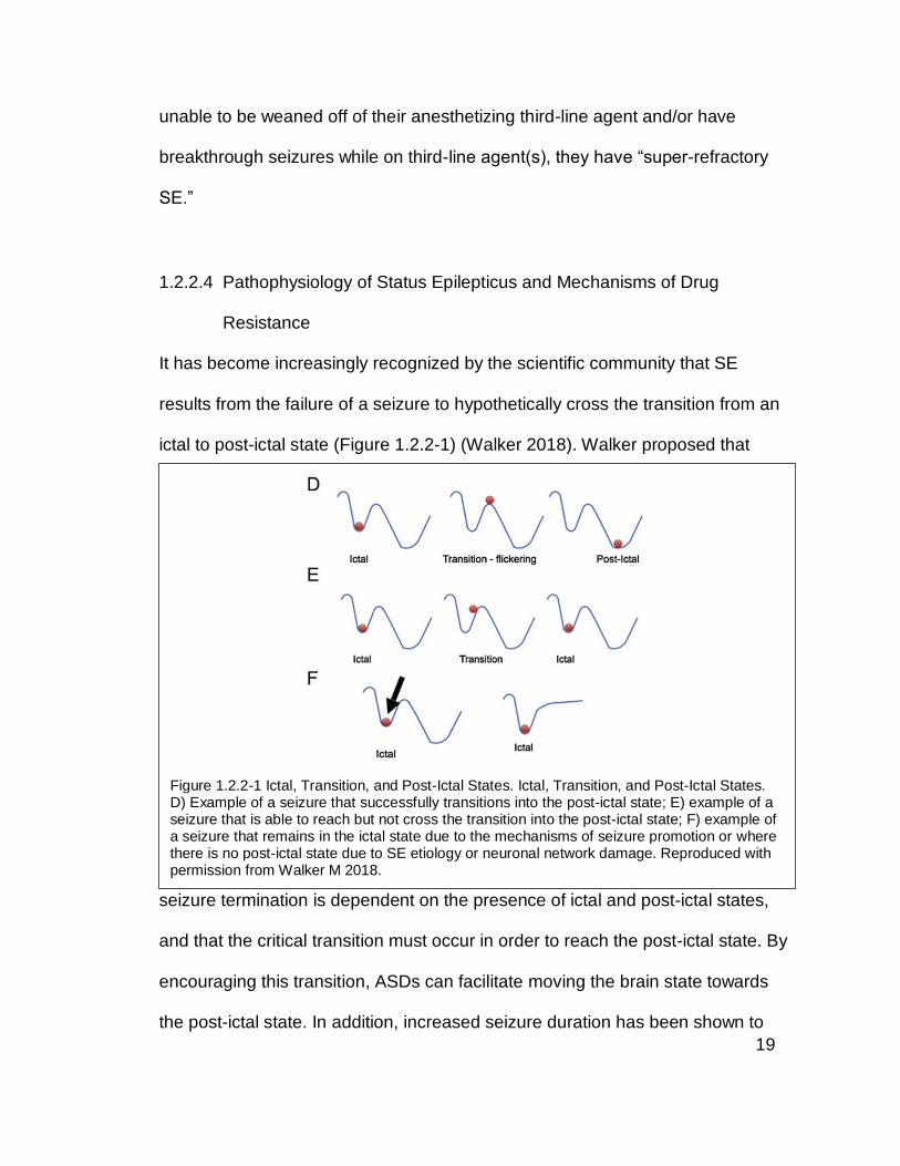
It has become increasingly recognized by the scientific community that SE
results from the failure of a seizure to hypothetically cross the transition from an
ictal to post-ictal state (Figure 1.2.2-1) (Walker 2018). Walker proposed that
seizure termination is dependent on the presence of ictal and post-ictal states,
and that the critical transition must occur in order to reach the post-ictal state. By
encouraging this transition, ASDs can facilitate moving the brain state towards
the post-ictal state. In addition, increased seizure duration has been shown to
Figure 1.2.2-1 Ictal, Transition, and Post-Ictal States. Ictal, Transition, and Post-Ictal States. D) Example of a seizure that successfully transitions into the post-ictal state; E) example of a seizure that is able to reach but not cross the transition into the post-ictal state; F) example of a seizure that remains in the ictal state due to the mechanisms of seizure promotion or where there is no post-ictal state due to SE etiology or neuronal network damage. Reproduced with permission from Walker M 2018.

20
increase the chance of self-sustained seizure activity in animal models of SE and
patients with SE (Mazarati, Wasterlain, et al. 1998; Shinnar et al. 2008;
Delorenzo et al. 1999).
The self-sustaining nature of SE is reminiscent of long-term potentiation
(LTP), the phenomenon behind memory and learning. LTP is a process
characterized by the strengthening of synaptic connections between neurons
following frequent stimulation (Purves et al. 2001). Following a strong
depolarization of the postsynaptic neuron and with continued stimulation, there is
increased surface expression of postsynaptic -amino-3-hydroxy-5-methyl-4-
isoxazolepropionic acid (AMPA) receptors allowing for a stronger connection
between the two neurons. In fact, the perforant path stimulation model of SE is
followed by increased LTP in the perforant pathway (Mazarati, Wasterlain, et al.
1998; Reddy and Kuruba 2013).
Pathophysiological changes on the cellular and molecular level promote
continued seizure activity and pharmacoresistance. Following prolonged seizure
activity, N-methyl-D-aspartate (NMDA) receptors increase in surface expression
(Naylor et al. 2013), presynaptic adenosine A1 receptor, neuronal potassium-
chloride cotransporter (KCC2) and GABAB receptor activities become
downregulated (Avsar and Empson 2004; Hamil, Cock, and Walker 2012;
Silayeva et al. 2015; Kaila et al. 2014; Chandler et al. 2003; Leung 2019), and
AMPA receptors lose their GluA2 subunit (Rajasekaran, Todorovic, and Kapur
2012; Malkin et al. 2016). These AMPA receptors then become permeable to
calcium, amplifying the accumulation of intracellular calcium and increasing the

21
risk for neuronal death (Cull-Candy, Kelly, and Farrant 2006). Taken together,
these observations support the concept that continued seizure activity can
strengthen seizure-promoting and/or deplete seizure-terminating mechanisms
(Table 1.2.2-2).
When seizures become self-sustaining, resistance to drugs, particularly
benzodiazepines, develops progressively over time (Kapur and Macdonald
1997). Synaptic -aminobutyric acid (GABA)A receptors (those containing a -
subunit) internalize after one hour of lithium/pilocarpine-induced SE in vivo
(Naylor, Liu, and Wasterlain 2005). This phenomenon explains why
benzodiazepines are highly effective within the first five minutes of seizure
activity, but not effective after 45 minutes (Kapur and Macdonald 1997). This
process is initiated by the activation of NMDA receptors and consequently the
calcium-dependent internalization of synaptic GABAA receptors (Rice and
Delorenzo 1999; Niquet et al. 2016). As shown in vivo, the decrease in inhibitory
post-synaptic potentials from the loss of synaptic GABAA receptors causes a loss
Table 1.2.2-2 Factors that Promote/Diminish Self-Sustaining Seizure Activity. Reproduced with permission from Niquet et al 2016.
22
in inhibitory tone of hippocampal circuits and promotes a pro-seizure state
(Naylor, Liu, and Wasterlain 2005).
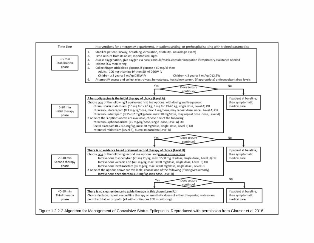
1.2.2.5 Management and Prognosis of Convulsive Status Epilepticus
While the optimal therapy for convulsive SE is still uncertain (i.e. there is no
intervention that is successful in 100% of cases), there are established guidelines
for its management. In 2016, the American Epilepsy Society and Epilepsy
Foundation published a treatment algorithm consisting of the best current
medical management of convulsive SE based off of clinical trial evidence
(Glauser et al. 2016). In total, 38 relevant published randomized, controlled trials
and four meta-analyses were identified, and pharmaceutical companies provided
information on three randomized, controlled trials. The following is a summary of
the consensus guidelines (Figure 1.2.2-2), whereas safety and effectiveness of
specific therapies are discussed in the next section.
23
Figure 1.2.2-2 Algorithm for Management of Convulsive Status Epilepticus. Reproduced with permission from Glauser et al 2016.

24
1.2.2.5.1 Early SE
Within the first five minutes of a convulsive seizure, patients should be stabilized
(airway, breathing, circulation), blood glucose should be evaluated, intravenous
(IV) access should be attempted for collection of serum electrolytes, complete
blood count, toxicology screen, ASD level (if applicable), and administration of
drugs. The goal of these early assessments is to rectify any reversible causes for
seizure activity (e.g. hypoglycemia, drug withdrawal, electrolyte disturbance).
After five minutes of convulsive seizure activity, a benzodiazepine should
be given either intravenously (lorazepam 0.1 mg/kg/dose or diazepam 0.15-0.2
mg/kg/dose, may repeat once) or intramuscularly (IM; midazolam 10 mg if >40
kg, given once). The goal of drug therapy is rapid termination of seizures and
prevention of recurrent seizure activity. Benzodiazepines (BZDs) have
demonstrated their safety, efficacy, and tolerability as the first-line therapy for SE
with a high level of evidence from four RCTs (Treiman et al. 1998; Alldredge et
al. 2001; Silbergleit et al. 2012; Chamberlain et al. 2014). Looking across these
studies, 43-74% of cases successfully terminated within 20 minutes of
benzodiazepine administration, 11-39% of these cases had seizure recurrence
within the study period, 29-57% of all cases required intensive care unit
admissions, and up to 27% of all cases resulted in death.
Most of the clinical trials were conducted using the IV route of
administration, requiring a trained technician to establish IV access. Ideally, SE
treatment would be administered immediately following the start of seizure

25
activity and likely in the pre-hospital setting by a parent or caregiver. The Rapid
Anticonvulsant Medication Prior to Arrival Trial (RAMPART) demonstrated that
rapid administration of treatment impacts outcome (Silbergleit et al. 2012).
RAMPART compared the efficacy of intramuscular midazolam (IM MDZ) to
intravenous lorazepam (IV LZP) in stopping SE prior to emergency department
arrival without requiring rescue therapy, and showed that although the time until
seizure termination was similar in both treatment groups, the time saved by using
the IM route significantly affected its efficacy positively.
A disadvantage of BZD use is that common side effects include impaired
cognition, psychomotor slowing and sleepiness that can last into the next day,
decreasing the patient’s ability to return to school or work (Roehrs et al. 1986;
Kay et al. 2016; Griffin et al. 2013). Furthermore, BZDs like diazepam and
midazolam are metabolized by the cytochrome P450 isozyme 3A4 (CYP3A4),
and are vulnerable to drug-drug interactions with common ASDs like phenytoin,
phenobarbital, carbamazepine (Griffin et al. 2013; Indiana University Department
of Medicine Clinical Pharmacology 2019). In addition, as seizure duration
increases, synaptic GABAA receptors become increasingly internalized, and
BZDs lose their efficacy (Wasterlain and Chen 2008). Therefore, although
effective in most cases, the first-line management of SE could be improved. The
development of an alternative therapy that can be administered either
intravenously or intramuscularly has the potential to improve outcomes in
patients with SE.

26
1.2.2.5.2 Established SE (ESE)
Second-line therapies include IV fosphenytoin (20 mg phenytoin-equivalent /kg,
single dose), valproic acid (40 mg/kg, single dose), or levetiracetam (60 mg/kg,
single dose). The goal of drug therapy is rapid termination of seizures and
prevention of recurrent seizures. There is a moderate level of evidence
demonstrating lack of significant difference in efficacy between these three
therapies (Malamiri et al. 2012; Agarwal et al. 2007; W. B. Chen et al. 2011; U.
Misra, Kalita, and Maurya 2012; Lyttle et al. 2019; Dalziel et al. 2019; Gujjar et al.
2017; Mundlamuri et al. 2015; Nene et al. 2019). Across these studies, 50-88%
of cases successfully terminated without seizure recurrence within at least six
hours following study drug administration, 20-73% of these cases had seizure
recurrence within 24 hours, 23-64% of all cases required intensive care unit
admissions, and up to 43% of cases resulted in death. There is large variability in
response depending primary outcomes of interest, and possibly in the open-label
nature of the studies.
The use of these second-line therapies do not come without risks.
Systemic complications such as systemic hypotension, Stevens-Johnson
Syndrome, hyperammonemia, and hematologic abnormalities (e.g.
thrombocytopenia, pancytopenia, agranulocytosis) have been reported in clinical
trials and post-marketing settings for these ASDs (KEPPRA® [package insert]
2017; Depacon [package insert] 2019; CEREBYX® [package insert] 2019).
These observations emphasize the need for more effective therapies to prevent

27
the large proportion of intensive care unit admissions following failure of second-
line therapies.
1.2.2.5.3 Refractory SE (RSE)
There is currently insufficient evidence to guide therapy due to the rarity of the
condition and difficulty in the interpretation of findings due to the complex
interaction of drugs used in parallel and co-morbidities at this later stage of SE.
Third-line therapies provided in the guidelines include repeating second-line
therapy or anesthetic medications including IV midazolam, thiopental,
pentobarbital, or propofol. Due to the ethical challenges in randomization of
interventions in intensive care settings, there is a lack of prospective,
randomized, blinded and controlled studies in refractory SE. Instead, numerous
small, prospective, open-label studies that compare the safety and effectiveness
of ketamine, continuous infusion of midazolam or diazepam, propofol, and
barbiturates are available (Rosati et al. 2012; Rossetti et al. 2011; Morrison et al.
2006; Mehta, Singhi, and Singhi 2007; Koul et al. 2002; Ulvi et al. 2002). Without
the rigorous controlled trials, registries and audits could also provide useful
information on general consensus of the management of refractory SE with some
limitations. Early results of a multinational, prospective audit of 488 patients with
refractory and super-refractory SE reported that the most widely used anesthetic
as first-choice is midazolam, followed by propofol and barbiturates (Ferlisi et al.
2015). From this survey, 74% of cases recovered from RSE, 22% died, and 4%
had treatment withdrawn due to futility. Although anesthetic agents are useful in
suppressing seizures, they are associated with a higher risk of systemic

28
complications death independent of underlying medical conditions (Sutter et al.
2014). There is an unmet need for better control of refractory SE, ideally before
the need for burst-suppression.
1.2.2.5.4 Super-refractory SE (SRSE)
There remains no standard of care for the treatment of SRSE for reasons similar
to refractory SE. Interventions that have been evaluated at this stage of SE
include perampanel (Beretta et al. 2018; Rohracher et al. 2015; Brigo et al.
2018), allopregnanolone (Broomall et al. 2014; Rosenthal et al. 2017), ketamine
(Höfler et al. 2016), stiripentol (A. Strzelczyk et al. 2015; Uchida et al. 2018),
rufinamide (Thompson and Cock 2016), cannabidiol oil (Rosemergy, Adler, and
Psirides 2016), inhaled anesthetics, barbiturates, electroconvulsive therapy
(Pinchotti, Abbott, and Quinn 2018; Chan et al. 2018), thalamic deep brain
stimulation (Lehtimäki et al. 2017), and ketogenic diet (Farias-Moeller et al. 2017;
Appavu et al. 2016; Thakur et al. 2014).
1.3 Using Canine Status Epilepticus as a Model of Human Status Epilepticus
1.3.1 Canine Epilepsy
1.3.1.1 Prevalence and Etiology
Canine epilepsy is practically defined as having at least two unprovoked epileptic
seizures greater than 24 hours apart (Mette Berendt et al. 2015). In veterinary
practice, dogs with epilepsy are among the most common neurological diagnosis.
The true prevalence of epilepsy in dogs is unknown but has been estimated to
range between 0.55-5.7% in the general dog population (Loscher et al. 1985;

29
Heske et al. 2014; Kearsley-Fleet et al. 2013; Michael Podell, Fenner, and
Powers 1995). The etiology of canine epilepsy as varied as that of human
epilepsy. Following the classification and terminology system published by the
ILAE for human epilepsy, the International Veterinary Epilepsy Task Force
(IVETF) has adopted proposals for the canine epilepsy classification and
terminology system that reflect the evolving understanding of the human disease.
Epilepsy classified by etiology are divided into two categories: idiopathic (purely
genetic, a combination of genetic and epigenetic influences, or unknown cause
and no indication of structural epilepsy), and structural (identified cerebral
pathology) (Mette Berendt et al. 2015). In contrast, human epilepsy etiology is
broken into six categories (i.e. structural, genetic, infectious, metabolic, immune,
and unknown) and more than one category can be used to describe a patient’s
epilepsy (Scheffer et al. 2017). Some breeds with suggested inherited idiopathic
epilepsies include Beagles, Boxers, Border Collies, German Shepherds,
Labrador Retrievers, and Vizlas (Monteiro et al. 2012; Ekenstedt, Patterson, and
Mickelson 2012; Bielfelt, Redman, and McClellan 1971; Nielen, Janss, and Knol
2001; Jaggy et al. 1998; Patterson et al. 2003).
1.3.1.2 Seizure Semiology and Clinical Diagnosis
Epileptic seizures are classified as either focal (clinical signs indicating activity
starts in a localized area in the brain), generalized (clinical signs indicating
activity starts in both cerebral hemispheres from the start), and focal epileptic
seizure evolving to become generalized (clinical signs indicating activity starts in

30
a localized area in the brain and spreads to involve both cerebral hemispheres)
(Mette Berendt et al. 2015). Focal epileptic seizures can present as motor (e.g.
facial twitches, repeated rhythmic jerks of one extremity, or rhythmic blinking),
autonomic (e.g. dilated pupils, hypersalivation, vomiting), or behavioral (e.g.
episodic change in behavior such as anxiousness, unexplainable fear reactions,
or abnormal attention seeking). Generalized epileptic seizures most often present
as tonic, clonic or tonic-clonic epileptic seizures in dogs sometimes with
expulsion of urine or feces. Non-convulsive generalized epileptic seizures
(atonic) in dogs, called ‘drop attacks’, are caused by the sudden loss of muscle
tone. Finally, the most common seizure type observed in dogs is focal epileptic
seizures evolving into generalized epileptic seizures. The focal epileptic seizure
is brief (seconds to minutes) and is followed by a convulsive stage with bilateral
tonic, clonic, or tonic-clonic activity.
Diagnosis of epileptic seizures includes two steps: establish whether
events animal are demonstrating are truly representative of epileptic seizures,
and identifying the cause of the epilepticus seizure (De Risio et al. 2015). The
first step is particularly difficult without observation of characteristic
electroencephalographic (EEG) changes and physical manifestation of seizures.
However, this is not practical in veterinary medicine and there is no standard
protocol for acquiring EEG in dogs. Therefore, the current practice is to obtain a
detailed and accurate history of events from pet owners, and completion of a
standardized epilepsy questionnaire with video recording when available. The
veterinarian must be able to distinguish epileptic seizures from other non-

31
epileptic episodic paroxysmal events (e.g. syncope, narcolepsy, idiopathic head
tremor).
After the diagnosis of epileptic seizures, the next step is the determination
of their cause which will have implications on the treatment and prognosis.
Reactive seizures can result from intoxications (e.g. organophosphates, ethylene
glycol) or from systemic metabolic disorders (e.g. electrolyte imbalance,
hypoglycemia, hypothyroidism). Structural disorders resulting from infectious,
inflammatory, traumatic, or neoplastic disease can result in epileptic seizures.
Neurological examination if often abnormal and may present as asymmetric
neurological deficits in dogs. Magnetic resonance imaging (MRI) of the brain and
cerebrospinal fluid (CSF) analysis is recommended to rule out structural epilepsy.
After exclusion of reactive seizures, MRI is CSF analysis is recommended in
dogs with age of seizure onset <6 months or >6 years, status epilepticus or
cluster seizure, interictal neurological abnormalities, or a previous presumptive
diagnosis of idiopathic epilepsy and drug resistance with a single antiseizure
drug titrated to the highest tolerable dose. The criteria for the diagnosis of
idiopathic epilepsy is three-tiered: 1) a history of two or more unprovoked
epileptic seizures occurring at least 24 hours apart, the age of seizure onset
between 6 months and 6 years of age, unremarkable interictal physical and
neurological examination, and no clinically significant abnormalities on blood
tests and urinalysis; 2) unremarkable fasting and post-prandial bile acids, brain
MRI, and CSF analysis; and 3) identification of ictal or interictal EEG

32
abnormalities characteristic for seizure disorders (criteria derived from human
medicine).
1.3.1.3 Management of Canine Epilepsy
Antiseizure drugs (ASD) are the mainstay of therapy for idiopathic epilepsy
(Bhatti et al. 2015; M. Podell et al. 2016; Marios Charalambous, Brodbelt, and
Volk 2014) (Table 1.3.1-1). In contrast to the goal of ASD therapy in humans of
seizure freedom, the goal of therapy in dogs is decrease seizure frequency,
duration, or severity with limited/acceptable side effects to maximize the dog’s
and owner’s quality of life. When the decision has been made to initiate ASD
therapy, the selection of ASD is made by a veterinarian’s recommendation and
depends on the dog (i.e. the seizure type, frequency, etiology), the drug (i.e. side
effect profile, drug interactions, frequency of administration), and the owner (i.e.
financial situation, lifestyle).
Aside from ASD therapy, there are nonpharmacological interventions for
the management of canine epilepsy. These include vagal nerve stimulation,
medium chain triglyceride (MCT)-based diet, and acupuncture (Munana et al.
2002; Hong Law et al. 2015; Goiz-Marquez et al. 2009; Klide, Farnbach, and
Gallagher 1987).

33
Table 1.3.1-1 Antiseizure Drugs Used in the Management of Canine Epilepsy
Antiseizure Drug
Antiseizure Mechanism(s) of Action
Place in Therapy
Phenobarbital GABAA receptor agonist First-line; high recommendation for monotherapy and moderate recommendation for adjunctive therapy
Imepitoin Partial GABAA receptor agonist First-line; high recommendation for monotherapy and low recommendation for adjunctive therapy
Bromide Hyperpolarization of neuron via bromide influx
Adjunctive to PB or monotherapy if hepatoxicity occurs with PB; moderate recommendation for monotherapy and adjunctive therapy
Primidone GABAA receptor agonist (phenobarbital pro-drug)
No advantage to using primidone over phenobarbital; not recommended for monotherapy or adjunctive therapy
Felbamate NMDA receptor antagonist, inhibition of L-type calcium- and sodium-channels
Adjunctive to PB; insufficient evidence to recommend its use
Gabapentin Inhibition of L-type calcium channels
Adjunctive to PB; insufficient evidence to recommend its use
Pregabalin Inhibition of L-type calcium channels
Adjunctive to PB; insufficient evidence to recommend its use
Levetiracetam Inhibition of synaptic vesicle protein 2A
Adjunctive to PB; low recommendation for monotherapy and moderate recommendation for adjunctive therapy
Topiramate GABAA receptor agonist, AMPA/kainate receptor antagonist, inhibition of L-type calcium channels, inhibition of carbonic anhydrase (isozymes II and IV)
Adjunctive to PB; insufficient evidence to recommend its use
Zonisamide Inhibition of T-type calcium channels, inhibition of carbonic anhydrase
Adjunctive to PB; low recommendation for monotherapy and moderate recommendation for adjunctive therapy
GABA: -aminobutyric acid; NMDA: N-methyl-D-aspartate; AMPA: -amino-3-hydroxy-5-
methyl-4-isoxazolepropionic acid

34
1.3.2 Canine Status Epilepticus
1.3.2.1 Prevalence and Etiology
As in humans, SE in dogs is defined as continuous seizure activity lasting for at
least five minutes or as two or more discrete seizures without complete recovery
of consciousness in between (Blades Golubovic and Rossmeisl 2017a).
Epidemiologic studies on SE in dogs report a prevalence ranging 2.5-59% in
dogs admitted into a teaching hospital for seizures (Zimmermann et al. 2009;
Saito et al. 2001; Bateman and Parent 1999) and 0.44-0.7% in the all dogs
admitted into a teaching hospital (Zimmermann et al. 2009; Bateman and Parent
1999). Although a rare condition in the general dog population, SE occurs more
often in dogs without idiopathic epilepsy. A retrospective case-control study done
in 50 dogs that exhibited generalized convulsive tonic-clonic (GCTC) SE
compared with 50 dogs that exhibited non-SE GCTC seizures found that dogs in
the non-SE group were more than twice as likely to have idiopathic epilepsy than
symptomatic/reactive epileptic seizures (Platt and Haag 2002). Similarly,
Zimmerman et al found that dogs with reactive seizures had a 1.87 relative risk of
developing SE compared to all other dogs (Zimmermann et al. 2009).
1.3.2.2 Canine Status Epilepticus Subtypes and Clinical Diagnosis
Similar to human SE, canine SE is classified into convulsive and nonconvulsive
SE. However, contrary to human SE, canine SE does not yet have
subclassifications of early, established, refractory, and super-refractory SE. The

35
majority of animals that develop SE will present with a phenotype consistent with
convulsive GCTC SE. Although it has been recognized by veterinary medicine,
nonconvulsive SE has not been frequently documented, likely due to the
underutilization of EEG in the diagnosis of epileptic seizures. A retrospective
case series recently reported that 20% and 12% of 104 dogs and cats with any
type of EEG procedure performed had electrographic seizures or electrographic
SE, and were associated with a 48% and 50% in-hospital mortality rates,
respectively (Granum et al. 2019). In addition, 81% of these animal patients with
electrographic seizures had no or only subtle signs of seizure activity,
emphasizing the need for future standardizing EEG procedures to detect
electrographic seizure activity.
The diagnostic algorithm for SE for dogs consists of physical diagnostics
(i.e. physical exam, neurological exam), point of care tests (e.g. blood glucose,
electrolytes, electrocardiogram, complete blood count), and patient-specific
diagnostics (e.g. evidence of toxic or metabolic encephalopathy, brain MRI, CSF
analysis, serum ASD concentration) (Blades Golubovic and Rossmeisl 2017a).
Pet owners should be questioned about the patient’s history of seizures or
neurological disease, history of metabolic disease, potential exposure to toxins,
and possibility of trauma.

36
1.3.2.3 Management and Prognosis of Canine Status Epilepticus
The goals of treatment of canine SE is similar to that of the treatment of human
SE and include seizure termination and prevention of further seizures while
managing systemic complications of SE and, if possible, the underlying cause
(Glauser et al. 2016; Blades Golubovic and Rossmeisl 2017b). The first step of
treating SE is stabilization of the canine patient by assess airway, breathing, and
circulation (Figure 1.3.2.3-1). An intravenous line should be established for
pretreatment blood sampling, drug delivery, and intravenous fluids. Blood
glucose, electrolytes, complete blood count, toxicology screen (if exposure is
known or suspected), and ASD concentrations should be measured at this time.
The recommended first-line agent is a benzodiazepine (i.e. diazepam 0.5-
2 mg/kg IV/IN/PR, lorazepam 0.1-0.2 mg/kg IV, or midazolam 0.1-0.3 mg/kg
Figure 1.3.2-1 Management of Status Epilepticus in Dogs and Cats Reproduced with permission from Blades Golubovic and Rossmeisl 2017b

37
IV/IN/IM) with or without a manual vagal maneuver. Benzodiazepines rapidly
distribute into the central nervous system (CNS) and redistributes into fat and
muscle quickly. Therefore, repeat dosing can cause accumulation in the CNS
and unexpected CNS depression. For this reason, after two to three doses, a
constant rate infusion (CRI) of another ASD should be initiated. Of note,
diazepam and lorazepam are solubilized with the addition of propylene glycol.
With rapid IV administration, propylene glycol can cause hypotension and
phlebitis. Alternatively, instead of a second or third dose of a BZD bolus, a
diazepam CRI 0.1-0.5 mg/kg/hr can be administered followed by second-line
parenteral dosing of LEV 30-60 mg/kg IV/SC or PB 2-6 mg/kg IV (Patterson
2014b).
If seizures terminate after the initial BZD dose, the canine patient will
receive second-line parenteral dosing of a maintenance ASD (potentially more
than once) before initiation of oral maintenance ASD (PB 2-3 mg/kg by mouth
every 12 hours or LEV 20-30 mg/kg by mouth every 8 hours). However, if
seizures fail to terminate within 20 minutes after several doses of BZD and
parenteral maintenance ASD bolus, third-line parenteral treatments used for
refractory SE (propofol 2-6 mg/kg IV bolus + 0.1-0.5 mg/kg/min IV CRI, ketamine
3-5 mg/kg IV bolus + 0.1-0.5 mg/kg/hr CRI, or fosphenytoin 15 mg/kg [phenytoin
equivalent] IV bolus). Finally, if seizures continue after third-line agents,
isoflurane 1-2% minimum alveolar concentration should be used to induce
seizure suppression.
38
The mortality rates of canine patients with SE have been reported to range
between 25-45%, with death occurring in 2.1-8% and mostly from euthanasia
(Zimmermann et al. 2009; Bateman and Parent 1999; Saito et al. 2001). Dogs
with structural epilepsy were found to have lower survival probably following SE
than dogs with idiopathic epilepsy (HR 0.11 [95% CI 0.03;0.37]) and dogs with
reactive seizures (HR 0.16 [95% CI 0.05;0.58]) (Zimmermann et al. 2009).
1.3.2.4 Canine Status Epilepticus Clinical Trials
In general, there is a lack of extensive published clinical trial evidence for canine
SE. Much of the treatment recommendations are based on human clinical trial
experience. Dosing for ASDs recommended in the treatment of canine SE largely
stems from targeting the serum drug levels within the range considered
therapeutic for people. To date, there are a handful of case series and controlled
clinical trials for the treatment of early and refractory canine SE.
Aside from one published case series on the use of rectal diazepam (PR
DZP) for canine cluster seizures, there is one open-labeled, randomized, parallel
group clinical trial comparing the effectiveness of an atomized intranasal
midazolam (IN MDZ) to PR DZP for the first-line management of canine SE in
client-owned animals (Michael Podell 1995; M. Charalambous et al. 2017).
Success was defined as seizure termination within 5 minutes without recurrence
of seizures within 10 minutes. IN MDZ (n=20) and PR DZP (n=15) terminated SE
in 70- and 20-% of cases, respectively, and all dogs showed sedation and ataxia.
Median time to seizure cessation was 47 seconds (range, 6-280) in the IN MDZ

39
group, whereas it was 214 seconds (range, 204-290) in the PR DZP group.
Twenty-one percent of successful IN MDZ cases did not have a relapse of
seizures, while all of the successful PR DZP cases relapsed (median time to
relapse was 904 and 645 seconds in the IN MDZ and PR DZP groups,
respectively). IN MDZ was more effective in terminating SE, with a higher rate of
seizure termination, faster onset of action, and lower rate of seizure relapse.
For refractory canine SE, one case series and two single case reports
described success using rectal LEV, IV ketamine, and controlled hypothermia,
respectively (Cagnotti et al. 2018; Serrano, Hughes, and Chandler 2006; Hayes
2009). Two randomized, double-masked, placebo-controlled trials evaluated the
clinical efficacy of IV LEV and IV fosphenytoin (FOS) in addition to IV DZP for the
treatment of canine SE or acute repetitive seizures (Hardy et al. 2012; Patterson
et al. 2015). In the IV LEV study, a responder was defined as a dog that had no
additional seizures after study drug administration for the following 24 hours. Of
the 19 cases (n=9 IV LEV, n=10 placebo), 56% in the IV LEV group responded to
treatment compared to 10% in the placebo group (p=0.06). There was no
difference in the mortality rates in either group (p=0.6), and no dogs required
pentobarbital or propofol treatment. IV LEV was safe and potentially effective for
the treatment of SE in addition to IV DZP. In contrast, a responder in the IV FOS
study was defined as a dog that no additional seizures after drug administration
for the following 2- and 12-hours. Of the 32 dogs (n=22 IV FOS, n=9 IV placebo),
64% of IV FOS cases were responders compared to 22.2% of placebo cases
(p=0.043). There was also a significant difference in the 2-hour responder rate,
40
with 95.4% responders in IV FOS group vs 55.5% in the placebo group (p=0.02).
There were no significant differences in any adverse effects or in-hospital
mortality rates between the two groups (p=0.63). IV FOS is safe and effective for
the treatment of SE following IV DZP.
1.3.3 Translatability of Therapeutic Research between Human and Canine
Status Epilepticus
Historically, animal models of epilepsy, typically induced chemically or by
electrical stimulation, are used for the screening of potential antiseizure drugs.
Although these have proven useful for the development of many first- and
second-generation ASDs, they are not representative of human epilepsy or
natural epileptogenesis. Rodent models of chronic epilepsy exhibit a low level of
complexity regarding interindividual differences in cellular and molecular changes
as well as genetic involvement (Potschka et al. 2013). Furthermore, these animal
models are not able to predict for the approximate 30% of patients who develop
pharmacoresistant epilepsy (Leppik 1992). Traditional animal models of epilepsy
do not take into account the progression of epilepsy, or the development of drug
resistance (Loscher et al. 1985). As a result, the current strategies for ASD
development use models that are known to respond to presently marketed drugs,
which may hamper the development of drugs with different mechanisms of
actions that may be useful in treating pharmacoresistant seizures. There is a
need for better animal models in the preclinical screening and assessment of
ASD candidates (Potschka et al. 2013).

41
The ideal animal model of epilepsy has the following: development of
spontaneously occurring recurrent seizures, a type of seizure similar in its clinical
presentation to those occurring in human epilepsy, clinical seizures should be
associated with epileptic-like activity in the electroencephalogram,
pharmacokinetics of ASDs similar to those in people allowing for maintenance of
effective drug concentrations, and effective plasma ASD concentrations similar to
those reported for human epilepsy (Loscher 1984). As stated best by the British
statistician George Box, “all models are wrong, some are useful.” It is likely that
no model can meet all these criteria, but canine epilepsy may serve as a useful
one for human epilepsy and has been compared to the human condition for
decades (Barker 1973; Loscher et al. 1985; Patterson 2014a).
When evaluating animal models for their usefulness, one should consider
whether the cause of disease is mirroring clinical etiology (i.e. etiological validity),
whether their symptoms and pathology mimic those in people (i.e. face validity),
and whether their response to therapy is similar to those in humans (i.e.
predictive validity) (Potschka et al. 2013). As described, dogs have naturally-
occurring epilepsy and SE with etiologies and pathology that closely mimic the
human condition and are the only genetic animal model of epilepsy where
interindividual variability exists that allows for the ability to select between
animals that develop pharmacoresistant epilepsy and those that remain sensitive
to currently marketed ASDs (Wolfgang Löscher 1997). In addition to breeds with
suggested inherited idiopathic epilepsies, canine genetic studies have revealed
genes found in canine progressive myoclonic epilepsies that are orthologous to

42
those causing human epilepsy syndromes (Ekenstedt, Patterson, and Mickelson
2012). For instance, the LGI2 gene (an ortholog of the human epilepsy gene
LGI1) in the Lagotto Romagnolo breed was found to cause recessive benign
familial juvenile epilepsy (Jokinen et al. 2007; Seppälä et al. 2011) and the EPM2
gene (an ortholog of human genes EPM2A or EPM2B) was discovered to cause
Lafora disease in Miniature Wirehaired Dachshunds (Lohi et al. 2005). Similarly,
causes for reactive seizures in dogs are comparable to those in humans,
including but not limited to hypoglycemia, electrolyte disturbances, hyperthermia,
and ethylene glycol poisoning (Brauer, Jambroszyk, and Tipold 2011; Levy 1994;
Hutchinson et al. 2012; Bruchim et al. 2006; Keller and Goddard 2012). Finally,
structural seizures in dogs, as they do in humans, occur following traumatic brain
injury, inflammatory disease, neoplasia (Fredsø et al. 2017; Steinmetz, Tipold,
and Löscher 2013).
The types of seizures and clinical presentation of seizures and SE in dogs
are comparable to those observed in people, so much so that the terms used to
describe canine seizures are adapted from human seizure terminology. As noted
previously, seizures in dogs are categorized into three: focal, generalized and
focal epileptic seizure evolving to become generalized. These are analogous to
the focal, generalized, and focal to bilateral tonic-clonic seizures. Similarly, dogs
present most often with generalized convulsive SE, but nonconvulsive SE has
been observed when EEG is available. The largest difference in categorization is
that awareness may be more difficult to assess in the canine patient. As a result,
veterinary categorization of seizure types do not include impaired awareness

43
focal onset seizures, nor cognitive focal onset seizures (Berendt et al. 2015;
Fisher et al. 2017). Furthermore, EEG presentation of seizures have been shown
to be similar between dogs and people (Holliday, Cunningham, and Gutnick
1970; M. Berendt et al. 1999; Davis et al. 2011). In fact, seizure forecasting
algorithms have been built and demonstrated 0.84 area under the classification
curve using open access chronic ambulatory intracranial EEG from five dogs with
naturally-occurring epilepsy and two humans undergoing prolonged intracranial
EEG monitoring (Brinkmann et al. 2016). Finally, recent studies have reported
blood-brain barrier (BBB) dysfunction in dogs with idiopathic epilepsy, along with
evidence of altered neurogenesis, neuroinflammation, increased expression of P-
glycoprotein, and hippocampal atrophy (Borschensky et al. 2012; Patterson
2013; Dirrig and Lamb 2016; Hanael et al. 2019; Pekcec et al. 2009; Kuwabara
et al. 2010; Czerwik et al. 2018), pathology paralleling observations in epileptic
foci of patients (Thom et al. 2005; Zhong, Ren, and Tang 2016; Marchi and
Lerner-Natoli 2013; Feldmann et al. 2013; Blümcke et al. 2013).
Clinical studies conducted in canine SE have also shown to be useful in
mirroring human responder rates. For the treatment of SE, studies comparing the
effectiveness of IN MDZ and PR DZP have been conducted mostly in children
(Bhattacharyya, Kalra, and Gulati 2006; Holsti et al. 2010, 2007; Fisgin et al.
2002; De Haan et al. 2010). In general, these studies considered a success as
seizure termination within 10-15 minutes of drug administration and found that IN
MDZ was at least as effective, if not superior, as PR DZP. In one pediatric study,
effectiveness to stop prolonged seizure activity before emergency department

44
arrival without recurrence was compared between IN MDZ (using a Mucosal
Atomization Device [MAD]) administered by paramedics and PR DZP historical
controls (Holsti et al. 2007). Seizures recurred in the ED in 38% and 72% of
patients in the IN MDZ and PR DZP groups, respectively. These results are
similar to those seen in the IN MDZ (MAD) and PR DZP study in dogs in which
the primary endpoint was seizure termination within 5 minutes without recurrence
in 10 minutes in an emergency department setting (M. Charalambous et al.
2017). Seventy percent and 20% of cases had success, respectively, with a
recurrence rate of 79% and 100%. For studies in established SE, IV FOS
compared to placebo had a 64% 12-hr responder rate, and was similar to the
56%-69% success rate reported in the literature in people (Patterson et al. 2015;
Treiman et al. 1998; Gujjar et al. 2017; Chakravarthi et al. 2015; Mundlamuri et
al. 2015). Furthermore, Patterson et al demonstrated that this response rate was
achieved at the same unbound plasma FOS concentration range that had been
demonstrated to be therapeutic in humans. These results further supported the
observation that dogs have similar responses to ASDs as humans, and the
translation of therapeutic and mechanistic research between canine and human
epilepsy.
1.3.3.1 Advantages of Using Canine SE as a model of Human SE
There are several advantages to using canine SE as a translational platform for
therapeutic and mechanistic research between preclinical rodent models and
human SE. First, they are large enough to evaluate behavioral responses and to

45
accommodate human devices (Potschka et al. 2013). Second, as noted in the
previous sections, there are many similarities in clinical and neurophysiological
presentation of SE in dogs and people (Patterson 2014a). The presence of
nonconvulsive and convulsive SE, and the subdivision of nonmotor and motor
focal seizures is astonishing, as are the similarities in epileptiform and epileptic
activity in EEG (Davis et al. 2011). Next, their response rate to therapies
approved to treat human SE syndromes are comparable, and as a result, many
of the drugs used to treat canine SE are also used to treat human SE, including
using BZDs as a first-line agent in SE treatment guidelines (Glauser et al. 2016;
Blades Golubovic and Rossmeisl 2017b).
1.3.3.2 Limitations of Using Canine SE as a model of Human SE
One important limitation of using canine SE as a model for human SE is we
cannot interview canine patients and are restricted to pet owner recounts of
description and frequency SE episodes (Mette Berendt et al. 2015). Second,
although NCSE is often diagnosed using EEG in people, scalp EEG is not routine
nor practical in canine patients, because animals are often moving excessively,
and only way to get a good reading is to sedate them which leaves an EEG
artifact on its own. Furthermore, canine skulls are surrounded by a layer of
muscle, which could also leave electrical artifacts (Potschka et al. 2013). Another
key limitation is that some owners decline diagnostic investigation of SE due to
financial concerns (Mette Berendt et al. 2015), which may contribute to
inaccurate diagnosis and epidemiological findings of canine SE. A few

46
consequences of the natural occurrence of seizures and SE in canines is that the
investigators are unable to elicit seizures and SE to screen potential therapeutic
agents, it is much more difficult to recruit for canine clinical trials than it would be
to screen ASDs in rodent models. In addition, it is much more costly in time and
money and not pragmatic to select for and breed epileptic sublines of dogs to
have enough subjects for experimental study (Loscher and Meldrum 1984).
Finally, dogs metabolize drugs differently than people do. In general, drugs are
metabolized at a faster rate in dogs compared to humans, and different
pharmacokinetics would have to be considered for potential therapies (Frey and
Löscher 1985). However, this can be overcome by targeting therapeutic drug
concentrations rather than doses. Dogs may also have different metabolite
profiles, which may prohibit them from using specific drugs and limit translation to
humans (Dalgaard 2015; Martignoni, Groothuis, and de Kanter 2006; Yoshida et
al. 2018).

47
CHAPTER 2
DEVELOPMENT OF ALLOPREGNANOLONE FOR THE EARLY TREATMENT
OF STATUS EPILEPTICUS

48
2.1 Introduction
In this chapter, I will discuss the development of intravenous and intramuscular
allopregnanolone for early treatment of convulsive SE. The central hypothesis is
that ALLO would be beneficial in the early treatment of SE based on its novel
mechanisms of action and ability to rapidly diffuse into the brain. The specific aim
of my study was to characterize the pharmacokinetics (PK), pharmacodynamics
(PD) and safety/tolerability following IV and IM ALLO in dogs. The objectives of
these studies were to 1) develop PK models that describe concentration-time
profiles following single ascending doses of IV and IM ALLO, 2) sequentially build
a PK-PD model to describe the plasma concentration-intracranial EEG data, and
3) assess the safety and tolerability of IV and IM ALLO. First, I will provide a brief
review of ALLO and a rationale for the development ALLO for the treatment of
SE, followed by a summary of the studies conducted in the following order: IV
ALLO PK and safety, IM ALLO PK and safety, and PK-PD modeling. The last
section describes the design of a clinical trial of IV ALLO for the treatment of
CSE.
2.2 Review of Allopregnanolone
2.2.1 Endogenous Allopregnanolone
Allopregnanolone is a progesterone derivative that is produced in the brain,
adrenals, and gonads (Reddy and Rogawski 2012; Corpéchot et al. 1981). In
men and women, the steady-state plasma ALLO concentration is less than 2
ng/mL (Pierucci-Lagha et al. 2006; Girdler et al. 2001; Ottander et al. 2005;

49
Droogleever Fortuyn et al. 2004). However, plasma ALLO concentrations have
been shown to increase in response to stress, menstruation, and throughout
each trimester of pregnancy (Purdy et al. 1991; Droogleever Fortuyn et al. 2004;
Reddy 2009; Herzog 2015; Luisi et al. 2000). It functions as an endogenous
anxiolytic and uterine myorelaxant via GABAA modulation (Bali and Jaggi 2014;
Putnam et al. 1991). In healthy, pregnant women, ALLO levels have been
reported to reach as high as 150 nM in the third trimester (Luisi et al. 2000).
2.2.2 Physicochemical Properties
Allopregnanolone (molecular formula C21H34O2) has a molecular weight of 318.5
g/mol and a topological polar surface area of 37.3 A2 (National Center for
Biotechnology Information 2019a). With a logP of 4.9, it is insoluble in water,
slightly soluble in methanol, soluble in 2-methyl-tetrahydrofuran, and freely
soluble in tetrahydrofuran (National Center for Biotechnology Information 2019a;
FDA CDER Other Review(s) 2019). The low molecular weight, small polar
surface area and lipophilicity suggests it would easily cross biological
membranes, namely the blood-brain barrier. These are ideal characteristics for a
drug intending to treat seizure emergencies. Its generic name, provided by the
United States Adopted Names Council, is brexanolone and will be used
interchangeably with allopregnanolone throughout this chapter.

50
2.2.3 Known Mechanisms of Action
Allopregnanolone is a positive allosteric modulator of GABAA receptors, and
binds on the -subunit and in the interface between - and -subunits, enabling
potency at synaptic and extrasynaptic GABAA receptors (Hosie et al. 2006). At
nanomolar concentrations (300 nM), ALLO potentiates GABA currents and
requires GABA for chloride channel activation, while at micromolar (1 M)
concentrations, it functions as a direct agonist of the channel (Majewska et al.
1986). ALLO has also been shown to have neuroprotective effects in traumatic
brain injury, ischemia, and neurodegenerative diseases models (He et al. 2004;
He, Hoffman, and Stein 2004; Irwin, Solinsky, and Brinton 2014; Irwin et al. 2015;
Djebaili, Hoffman, and Stein 2004; Moralí et al. 2011; Napoli et al. 2019; Mellon,
Gong, and Schonemann 2008). In addition, the neurosteroid has been shown to
increase superoxide dismutase 2 enzyme (decreases neuronal death as a result
of reactive oxygen species production) following pilocarpine-induced SE in mice
and increase phosphorylation and membrane insertion of extrasynaptic GABAA
receptors via protein kinase C (Cho et al. 2018; Lejri et al. 2017; Abramian et al.
2014; Modgil et al. 2017).
2.2.4 Clinical Pharmacokinetics
Brexanolone has low oral bioavailability of <5% (FDA CDER 2019). Its volume of
distribution is approximately 3 L/kg and is highly plasma protein bound (>99%)
(ZULRESSO [package insert] 2019). With an estimated total clearance of 1
L/h/kg, its terminal half-life is approximately 9 hours(ZULRESSO [package

51
insert] 2019). Brexanolone is extensively metabolized by non-cytochrome P540
(CYP) pathways, including aldo-keto-reduction, glucuronidation, and sulfation
(FDA CDER 2019). The three major metabolites in humans are sulfated or
glucuronidated conjugates of the C20-reduced form of brexanolone, and are not
pharmacologically active (FDA CDER 2019). Less than 1% of brexanolone is
excreted via urine or feces unchanged (FDA CDER 2019). Due to its extensive
metabolism via multiple pathways, the sponsor claims that brexanolone is less
susceptible to metabolic drug-drug interactions (FDA CDER 2019). In vitro
studies suggested that brexanolone showed potential to inhibit CYP2C9.
However, co-administration with phenytoin, a CYP2C9 substrate, did not result in
clinically significant changes in phenytoin pharmacokinetics (ZULRESSO
[package insert] 2019).
In dogs, brexanolone is rapidly and extensively metabolized to produce 48
quantifiable compounds (FDA CDER 2019). Oxidation was the exclusive
biotransformation mechanism identified in dogs (FDA CDER 2019). Only a
minimal amount of the parent compound was excreted in the bile, urine, and
feces (FDA CDER 2019).
2.2.5 FDA-Approved Indications and Marketed Formulations
Brexanolone is indicated for the treatment of moderate-to-severe post-partum
depression (ZULRESSO [package insert] 2019). It is marketed as a clear,
colorless, and preservative-free injectable solution. One milliliter of ZULRESSO

52
contains 5 mg brexanolone, 250 mg of betadex sulfobutyl ether sodium, 0.265
citric acid monohydrate, 2.57 mg sodium citrate dihydrate, and water.
2.2.6 Present State of Knowledge of Allopregnanolone in Status Epilepticus
Allopregnanolone has been shown to be effective in terminating seizures in
several SE rodent models, including pilocarpine, kainate, perforant path
stimulation, and tetramethylenedisulfotetramine (Rogawski et al. 2013; Kokate et
al. 1996; C A Frye 1995; Cheryl A Frye and Scalise 2000; Zolkowska, Wu, and
Rogawski 2018). Clinically, ALLO has only been studied in super-refractory SE
(Vaitkevicius et al. 2017; Broomall et al. 2014; Rosenthal et al. 2017; Sage
Therapeutics 2015). An open-label Phase I/II study in adult and pediatric (2
years) patients with SRSE was conducted using a brexanolone loading dose
(286.6 g/kg for 1 hour), followed by a four-day maintenance infusion (standard:
86 g/kg/hr or high-dose: 156 g/kg/hr) and one-day taper period (Rosenthal et
al. 2017). The inclusion criteria included patients who failed to respond to at least
one first-line agent, one second-line agent, and who were unable to be weaned
off of third-line agents (TLAs) after 24 hours or had breakthrough seizures after
6 hours of initiation of burst-suppression from third-line therapies. The standard
maintenance infusion rate was chosen to attain a steady-state concentration of
150 nM (approximately 50 ng/mL), which is the physiological maximum plasma
ALLO concentration observed in healthy pregnant women (Luisi et al. 2000;
Broomall et al. 2014; Vaitkevicius et al. 2017). Responders were defined as
patients who were successfully weaned from TLAs without reinitiation of other

53
TLAs during administration of brexanolone. Brexanolone administration was well-
tolerated, with response rate of 77% (n=17/22, 13 standard dose, 4 high-dose)
and improved overall functional status measured at follow-up day 29.
This success was followed by a failed Phase III placebo-controlled trial of
brexanolone for SRSE in 132 adult and pediatric (2 years) patients (Sage
Therapeutics 2015). Similar to the Phase I/II trial, brexanolone was administered
as a 300 g/kg loading dose in the first hour, followed by a constant rate infusion
at 90 g/kg/hr, followed by a slow taper off brexanolone in the last 24 hours. In
contrast to the Phase I/II study, the brexanolone infusion was designed to last
one additional day, the inclusion criteria required patients to fail a qualifying wean
(even if they had failed at least one wean prior to screening), there was an open-
label high-dose arm (150 g/kg/hr for 119 hours) if the patient failed the primary
endpoint, and the response was defined with more strict criteria (i.e. no
reinitiation of TLAs within 24 hours following study drug infusion). Brexanolone
was no different than placebo in meeting the primary endpoint (43.9% vs 42.4%,
respectively). The investigators found that during the screening of the Phase III
study, 52% (144/276) of patients were able to successfully wean off of their
TLAs. It is possible given the differences in study design and inclusion criteria
that the patients in the Phase III study were harder to treat than those included in
the Phase I/II study. Further, the lack of control arm made it difficult to estimate
the placebo response. In addition, nearly 40% of the study population (27 in
placebo arm, 24 in brexanolone arm) opted into the open-label high-dose arm. It
is possible that clinicians may have been tempted to declare an early failure in

54
order for their patient to receive “active” drug. Finally, the targeted steady-state
concentration was based off of the highest observed concentration healthy,
pregnant women, which may be safe but not sufficient for treating SE.
Nonetheless, the results of this Phase III trial provides a number of
considerations and lessons for future development, including whether the SRSE
population is where brexanolone use could be most beneficial.
One reason why brexanolone would not be an ideal choice to treat SRSE
is that its main mechanism of action is equivalent to those used by TLAs to
induce burst/seizure suppressions, i.e. via GABAA receptor potentiation. In the
Phase III study, SRSE was defined when patient experienced breakthrough
seizures during the induction of burst/seizure suppression or when a patient had
breakthrough seizures while attempting a wean from TLAs. Since an anesthetic
agent will inevitably induce seizure suppression if the dose is high enough
(Shorvon and Ferlisi 2012), the more common diagnosis of SRSE was likely due
to the latter reasoning (per personal communication with Dr. M. A. Rogawski,
August 2019). Physical dependence is a physiological phenomenon that occurs
with CNS-acting drugs, including those that act via GABAA receptors (e.g.
ethanol, benzodiazepines, barbiturates) (Boisse et al. 1990; Okamoto, Hinman,
and Aaronson 1981; Rosenberg and Chiu 1985; Miranda and Pinardi 1998;
Cicero et al. 1971). Following chronic administration and subsequent abrupt
withdrawal of GABAA-potentiating drugs, rebound conditions of the symptoms
treated originally with the drug (such as seizures or anxiety) occur. Therefore,
one could hypothesize that following chronic seizure suppression with a TLA

55
following by the administration of brexanolone, patients would have a high risk of
rebound seizures. A monotherapy or combination of therapies with at least one
different mechanism of action (i.e. not via GABAA receptor potentiation) may
have a higher likelihood of success for the treatment of SRSE.
2.2.7 Rationale for Developing Allopregnanolone for Early Treatment of SE
In addition to having activity at synaptic GABAA receptors, like the current first-
line therapies, ALLO has exhibited additional mechanisms of action that would be
beneficial in terminating SE, including activity at extrasynaptic GABAA receptors
which mediate tonic inhibition, and insertion of additional extrasynaptic GABAA
receptors (Hosie et al. 2006; Abramian et al. 2014; Modgil et al. 2017). Similar to
benzodiazepines, ALLO has ideal physicochemical properties (low molecular
weight and high logP) that enable its rapid distribution into the brain (Irwin et al.
2015). The rapid brain penetration also manifests as a fast onset of action of
sedative (within 5 minutes following IM injection and 30 seconds following IV
injection) and antiseizure effects (within 1 minute of intraperitoneal injection) in
rodent models (Irwin et al. 2015; Rogawski et al. 2013). ALLO, at 2- and 3-mg/kg
given intraperitoneal and intramuscularly, respectively, has shown preclinical
efficacy in seizure termination following drug-induced SE in early and
benzodiazepine-refractory stages (Rogawski et al. 2013; Zolkowska, Wu, and
Rogawski 2018).
The development of ALLO for treatment of SE is currently limited by its
failure in SRSE. Evaluating the safety and effectiveness of drugs in the SRSE
56
patient population is particularly difficult and ethically complex given that these
patients 1) are the smallest subset of patients experiencing SE, 2) are critically-ill
and have the highest morbidity and mortality risk (and consequently, a higher
futility risk), and 3) there are no widely-accepted standards of practice to
compare to active drug arm or even standard practices between clinical sites.
Therefore, developing ALLO for early termination of SE would have the
advantage of inclusion of active, standardized comparators, larger patient
population, and a lower risk of confounding medical complications. Moreover,
due to the desire to quickly attain peak brain concentrations and/or the ability to
treat SE in the pre-hospital setting with relative ease, pursuing the IV and IM
routes of administration would be ideal.
2.3 Pharmacokinetics and Safety of Intravenous Allopregnanolone for Early
Treatment of Status Epilepticus in Dogs
2.3.1 Introduction
The central hypothesis is that ALLO possesses the requisite pharmacologic,
physicochemical, and pharmacokinetic (PK) properties to serve as an early
treatment for SE, either in combination with BZDs or as a replacement. One
approach to testing this hypothesis is the use of dogs with naturally-occurring
epilepsy, which is similar to human disorder in its electroencephalographic
presentation and response to therapy (Chapter 1.3). Although IV ALLO
pharmacokinetics has been characterized in rodents and humans (Irwin et al.
2015; Luisi et al. 2000), there is a lack of PK data in dogs.

57
My working hypotheses are 1) ALLO exhibits linear pharmacokinetics with
respect to dose, and 2) ALLO is safe to administer IV. The specific aim of this
study is to characterize IV ALLO PK and safety following single ascending doses.
The primary objectives were to develop a PK model to describe the
concentration-time data, evaluate its safety and tolerability, and simulate dosing
regimens that can be used to inform dosing recommendations for a clinical study
of CSE.
2.3.2 Methods
2.3.2.1 Study Animals and Safety Monitoring
Five dogs with (n=2) and without (n=3) a history of seizures were used. One of
the dogs with a history of seizures had recurrent seizures despite being on
phenobarbital (PB) maintenance regimen, while the other dog had not had
seizures in the last 7 years and was not on an ASD. Approval to conduct the
study was obtained through the Institutional Animal Care and Use Committee of
the University of Minnesota. The dogs were housed at the University of
Minnesota’s College of Veterinary Medicine. The dog on PB was previously
implanted with a device which wirelessly transmits continuous iEEG recordings
(Kremen et al. 2018). On study days, each dog participating in the study was
removed from their kennel to have a central-line catheter inserted an hour prior to
study start. On days where IV ALLO was administered, a peripheral-line catheter
was also inserted. Drug administration took place in a procedure room away from
the dog’s kennel.

58
2.3.2.2 Study Drug
ALLO concentrate was provided by the Rogawski laboratory at the University of
California, Davis. The concentrate consists of 6% allopregnanolone in 24%
sulfobutyl ether β-cyclodextrin (Dexolve) in a 0.9% sodium chloride solution
(normal saline). The concentrate was prepared using chemically pure, laboratory
grade ALLO using Good Manufacturing Practices and shipped frozen to the
University of Minnesota. Frozen ALLO was thawed and diluted with normal saline
(1 part ALLO: 1-3 parts normal saline, as directed by the manufacturing
laboratory) prior to IV administration for a final ALLO concentration of 1.5-3
mg/mL.
Dexolve (CycloLab) is a solubilizing agent comprised of a ring of a
hydrophilic exterior and lipophilic interior environment and is considered a
“generic” of the cyclodextrin Captisol (Ligand). Both are cyclic
oligosaccharrides with a sodium sulfonate salt separated from the ring structure
by a butyl ether group (Captisol). The cyclodextrin is able to dissolve the poorly
water-soluble compounds via noncovalent interactios within the lipophilic cavity
(Loftsson and Brewster 1996; Varan et al. 2017). Captisol is exclusively renally
eliminated and has been shown to be safe in humans. The technology has
received FDA approval for its use in parenteral formulations of carbamazepine,
amiodarone, and voriconazole.

59
2.3.2.3 Starting Dose Rationale
Basal levels of ALLO in healthy, pregnant women have been reported to reach
as high as 50 ng/mL (Luisi et al. 2000). Using allometric scaling of preclinical and
clinical data reported in the literature (Tables 2.3.2-1 and 2.3.2-2), PK
parameters for dogs were determined. These parameter estimates were used to
simulate concentration-time profiles and determine doses and dose regimens
predicted to attain plasma concentrations in the 50 ng/mL range for
approximately an hour following infusion (Figure 2.3.2-1). A 1 mg/kg dose infused
over 5 minutes was chosen based on my simulations.
Table 2.3.2-2 PK parameter values reported in the literature
Species Wt (kg)
Dose (mg/kg)
Cmax (ng/mL)
AUCinf (hr*ng/mL)
kel (hr-1)
t1/2
(hr) Vd (L/kg)
Cl (mL/hr/kg)
Mouse1 0.03 1.5 214.6 155.6 0.174 4 55.4 9640.1
Rabbit1 3.6-4.5
3 1176 625.1 0.209 3.3 23 4799.2
Human (female)2
70 Cumulative 0.09 mg/kg
47.15 0.156 4.35 12.5 1956
1Irwin et al 2015, 2Timby et al 2006
Table 2.3.2-1 Estimated and Calculated Three-compartment Model PK Parameter Values.
Volume of distribution (mL/kg) Clearance (mL/(kg*min)
Species V V2 V3 CL CL2 CL3
Rat 0.15 1527 1944 101.2 3275 72.94
Dog 0.06 598.3 761.8 38.09 1233 27.47
Rat data provided by the Rogawski Lab. Dog PK parameters calculated by allometric scaling using preclinical and clinical data in Table 2.3.2-1.

60
2.3.2.4 Study Design
Three healthy dogs and one dog with a history of seizures were given single IV
doses of ALLO ranging from 1-4 mg/kg as a 5-minute infusion via a catheterized
peripheral vein (Table 2.3.2-3). One to two dogs were studied at each dose. Whole
blood samples (~2 to 5 mL) were collected via a catheterized central vein at pre-
dose and at approximately 3, 5, 15, 30, and 45 minutes, 1, 2, 4, and 6 hours post-
infusion. There was a washout period of at least 1 week.
After completion of the IV dose escalation study, two healthy dogs were
given a standard PB dose (2 mg/kg) until steady state was reached (at least 15
days). Once at steady state, the ALLO PK study was repeated in these dogs and
Table 2.3.2-3 Number of Animals per IV Study Dose
Route IV
Dose (mg/kg) 1 2 3 4
Number of Dogs (total) 4 4 2 2
Number of Dogs on PB 2 2 0 0
Number of Dogs with EEG 1 1 0 0
Figure 2.3.2-1 Dosing Simulations for Initial IV ALLO Dose in Dogs
0 20 40 60 80 100 12010
100
1000
10000
Time (min)Pla
sm
a A
LL
O C
on
cen
trati
on
(n
g/m
L) 5 min infusion
0.5 mg/kg
1 mg/kg
1.5 mg/kg
2 mg/kg
4 mg/kg
50 ng/mL

61
one dog with a history of seizures on chronic PB (1-2 mg/kg). PB was
discontinued in both healthy dogs immediately after IV study completion.
2.3.2.5 Allopregnanolone Assay
Whole blood was collected in EDTA-containing purple-top vacutainer tubes, and
centrifuged for plasma separation. The red blood cells and plasma were
immediately frozen (−80°C) until analysis. An ultra high-performance liquid
chromatography-mass spectrometry (UPLC-MS/MS) method developed and
validated at the UC Davis laboratory was used to measure total plasma ALLO
concentrations (Zolkowska, Wu, and Rogawski 2018).
2.3.2.6 Pharmacokinetic Analysis
2.3.2.6.1 Non-compartmental Analysis
ALLO concentration–time data were analyzed using non-compartmental analysis
(Phoenix 64, Build 8.0.0.3176, Certara L.P., Princeton, NJ, USA). Data was
uniformly weighted. Pharmacokinetic parameters determined included first
observed concentration (C1), terminal rate constant (k), and terminal half-life
(t1/2). k was calculated using WinNonlin default setting, including estimating k
using a regression with the largest adjusted R2 value with the largest number of
concentration-time points used. Each individual concentration-time profile was
visually checked to make sure the concentration-time data points were adequate
and sufficient concentration-time points to characterize k. If not, user-defined
concentration-time points were used. t1/2 was calculated as 0.693/k. The area

62
under the time–concentration curve from time 0 to infinity (AUC) was calculated
using the linear up log down method and equation (1), where Cp is the plasma
ALLO concentration, tlast is the time at which the last plasma sample was
measured, Cplast is the last measured plasma ALLO concentration, and kel is the
terminal rate constant. Clearance (CL) and volume of distribution (Vd) were
calculated using equations (2) and (3), respectively, where the bioavailability (F)
is assumed to be 100% for an IV dose. Dose proportionality was determined by
comparing log-normalized AUC across all doses using a one-way ANOVA test.
Concentration–time profiles were created using the GraphPad Prism 7 (Version
7.0a, GraphPad Software, Inc., La Jolla, CA, USA).
Equations:
(1)
(2)
(3)
2.3.2.6.2 Compartmental Analysis
PK parameters estimates were also determined using compartmental analysis
(Phoenix Non-Linear Mixed Effects 8.0). PK parameter values were estimated
from individual concentration-time profiles using a first-order conditional
estimation extended least squares method. Additive, multiplicative, and
combined error models for residual unexplained variability were evaluated. The
best fit model was determined using visual inspection, goodness of fit plots (e.g.

63
conditional weighted residual plots and individual observed versus predicted),
and precision of model parameters. Arithmetic and geometric means and
standard deviation of parameter values were calculated using Phoenix
Descriptive Statistics function.
A population approach was used to determine PK parameter “typical
values.” An exponential error model for between-subject variability was used.
Additive, multiplicative, and combined error models for residual unexplained
variability were evaluated. The best fit model was determined as stated above,
with the addition of evaluating objective function value (OFV, as determined by
the log-likelihood function) and Akaike’s Information Criterion.
Normalized weight with fixed allometric exponents of 0.75 and 1 was
added as a covariate on clearance and volume, respectively. Weight was
normalized to median weight. The relationship of the covariate and PK parameter
was modeled by the equation 𝑃𝐾 = (𝑊𝑇
19.2)0.75 𝑜𝑟 1 ∗ 𝑡𝑣𝑃𝐾 ∗ 𝑒𝜂𝑃𝐾, where PK is the
PK parameter, tvPK is the typical value of that parameter from the population,
dPK is the estimated value of the covariate effect, and PK is the between-
subject variability of that parameter.
Pharmacokinetic parameter estimates from the population model were
used to simulate concentration-time profiles from a “typical dog” with a weight of
19.2 kg (median weight of dogs in my study) receiving a single 5-minute infusion
of 1-4 mg/kg ALLO.

64
2.3.2.7 Safety and Behavioral Response Evaluations
A modified Glasgow coma scale (mGCS) was used to quantitate degree of
sedation pre-dose and at scheduled blood sampling times (Table 2.3.2-4).
Cardiorespiratory activity (blood pressure, heart rate, and respiratory rate) was
assessed at blood sampling times up to 30 minutes. In addition to sedation and
vitals, the dogs were monitored for vomiting, diarrhea, and lethargy prior to and
for 60 minutes after drug administration, and at each blood sampling time.
Behavioral and iEEG activity were monitored by veterinary staff for seizure
activity. As doses were escalated, the maximal tolerated toxicity, as determined
by the supervising veterinarian, was 20 minutes of sedation with stable
cardiorespiratory activity (respiratory rate greater than 6 or less than 60 bpm,
systolic blood pressure greater than 60 mmHg, heart rate greater than 50 but
less than 160 bpm).

65
Table 2.3.2-4 Modified Glasgow Coma Scale. Reproduced with permission from Platt et al 2001.
Score
Motor activity
Normal gait, normal spinal reflexes 6
Hemiparesis, tetraparesis, or decerebrate activity 5
Recumbent, intermittent extensor rigidity 4
Recumbent, constant extensor rigidity 3
Recumbent, constant extensor rigidity with opisthotonus 2
Recumbent, hypotonia of muscles, depressed or absent spinal reflexes 1
Brain stem reflexes
Normal pupillary light reflexes and oculocephalic reflexes 6
Slow pupillary light reflexes and normal to reduced oculocephalic reflexes 5
Bilateral unresponsive miosis with normal to reduced oculocephalic reflexes
4
Pinpoint pupils with reduced to absent oculocephalic reflexes 3
Unilateral, unresponsive mydriasis with reduced to absent oculocephalic reflexes
2
Bilateral, unresponsive mydriasis with reduced to absent oculocephalic reflexes
1
Level of consciousness
Occasional periods of alertness and responsive to environment 6
Depression or delirium, capable of responding but response may be inappropriate
5
Semicomatose, responsive to visual stimuli 4
Semicomatose, responsive to auditory stimuli 3
Semicomatose, responsive only to repeated noxious stimuli 2
Comatose, unresponsive to repeated noxious stimuli 1

66
2.3.3 Results
The demographics of the dogs are listed in Table 2.3.3-1.
Table 2.3.3-1 Animal Demographics
ID Age (years)
Gender Weight (kg)
Breed Seizure Type
Seizure Frequency
Co-medications
3 9 Male, neutered
16 Beagle History of one
witnessed seizure
None None
6 13 Male, neutered
16 Keeshound Mix
Focal, with generalized
seizures
Focal cluster
seizures every 14-60 days.
With secondarily generalized
seizures every 1-2 months.
Phenobarbital (PB)
7 1 Female, intact
17 Coonhound Mix
(healthy) None None
8 1 Female, intact
22 Coonhound Mix
(healthy) None None
9 1 Male, intact
22 Coonhound Mix
(healthy) None None

67
2.3.3.1 Non-compartmental PK Analysis
The concentration-time profiles of IV are shown in Figures 2.3.3-1.
Pharmacokinetic parameter estimates using noncompartmental analysis are
summarized in Table 2.3.3-2. Following IV dosing, clearance ranged from 3.4-
20.1 L/hr/kg, while volume of distribution ranged from 1.9-6.0 L/kg. The terminal
half-life ranged between 7-33 minutes.
Figure 2.3.3-1 Plasma ALLO Concentration-Time Profile Following IV Administration on a log-scale. cPB: Chronic PB cPB: Chronic PB
0 50 100 1501
10
100
1000
10000
Time (min)
Pla
sm
a A
LL
O C
on
cen
trati
on
(n
g/m
L)
3 @ 2 mg/kg3 @ 3 mg/kg6 @ 1 mg/kg + cPB
6 @ 2 mg/kg + cPB7 @ 1 mg/kg
7 @ 4 mg/kg7 @ 1 mg/kg + PB8 @ 2 mg/kg
8 @ 4 mg/kg8 @ 2 mg/kg + PB9 @ 3 mg/kg
Table 2.3.3-2 Non-compartmental PK Parameter Estimates
Dose (mg/kg)
n z
(hr-1)
t1/2 (hr)
Cmax
(ng/mL) CL (L/hr/kg)
V (L/kg)
AUC∞/Dose (ng/mL*hr/(ng/kg)) x 10-5
1 2 (1)
7.2-8.1
(3.6)
0.09-0.10 (0.2)
425-1340
12.1-14.2 (5.6)
1.5-2.0 (1.7)
7.0-8.2 (16.9)
2 3 (1)
1.2-4.0
(1.8)
0.17-0.56 (0.4)
1654-5287
6.2-9.5 (3.2)
2.0-5.0 (1.8)
10.4-12.0 (31.1)
3 2 2.1-2.3
0.31-0.33
2440-4661
6.2-7.9 2.9-3.4 12.6-16.1
4 2 1.4-1.7
0.40-0.50
3660-5172
5.9-8.2 4.0-4.6 12.2-17.9
Estimates are presented as a range. Values in parentheses are from one dog on chronic PB.
z: terminal phase slope; t1/2: terminal half-life; Cmax: observed peak plasma concentration; CL:
clearance; V: volume of distribution; AUC/Dose: dose-normalized area under the
concentration-time curve from time 0 extrapolated to infinity

68
As shown in Figures 2.3.3-2 and 2.3.3-3, dose-normalized AUC0-INF and
observed Cmax across doses were similar.
Figure 2.3.3-3 Dose-proportionality: Dose-normalized observed Cmax
1 mg/k
g
2 mg/k
g
3 mg/k
g
4 mg/k
g
0
5
10
15
ln(D
ose-n
orm
alized
Co
ncen
trati
on
) (n
g/m
L)
Dose-normalized Observed Cmax
F (3, 7) = 2.592
P=0.1351
Figure 2.3.3-2 Dose-proportionality: Dose-normalized AUC0-INF

69
2.3.3.2 Individual Compartmental PK Analysis Following IV Administration
A two-compartment model with first-order elimination and a combined error
model best fit all ALLO concentration data following IV administration (goodness
of fit plots included in Figure 2.3.3-4). The model was unable to estimate PK
parameter values with precision for one animal, 6J. This may be due to the small
number of concentration-time data above the lower limit of quantitation available
for that animal. However, goodness of fit plots do not show gross signs of model
misspecification. Individual and averaged parameter estimates are summarized
in Table 2.3.3-3.
Figure 2.3.3-4 Goodness of fit plots for a two-compartment model. IWRES: individual weighted residual; TAD: time after dose; IPRED: individual predicted concentration
0 50 10010
100
1000
10000
Time (min)
Pla
sm
a A
LL
O C
on
cen
trati
on
(n
g/m
L) Individual Observed Versus Predicted
0 1000 2000 30000
1000
2000
3000
Observed ALLO Concentration (ng/mL)
Pre
dic
ted
AL
LO
Co
ncen
trati
on
(n
g/m
L)
Observed versus Predicted
50 100 150
-2
-1
0
1
2IWRES vs TAD
Time After Dose (min)
IWR
ES
1000 2000 3000
-2
-1
0
1
2IWRES vs IPRED
Individual Predicted Concentration (ng/mL)
IWR
ES

70
2.3.3.3 Population Compartmental PK Analysis Following IV Administration
A population approach was also used to fit the IV concentration-time data. A two-
compartment model with first-order elimination with proportional error model best
Table 2.3.3-3 Individual Compartmental PK Parameter Estimates
ID CL (L/hr)
Q (L/hr)
V (L)
V2 (L)
Proportional Error (%)
Additive Error (ng/mL)
3G 122.3 (5.5%)
19.3 (11.6%)
7.7 (10.3%)
7.5 (14.1%)
8.0 (31.8%)
1.4 (100%)
6J 6.3 (294%)
0.1 (586%)
0.2 (348%)
0.1 (582%)
11.1 (45%)
3.4 (69.5%)
7D 180.8 (6.7%)
33.8 (14.1%)
12.9 (13.3%)
15.9 (21.8%)
12.8 (21.7%)
0.002 (---)
8N 155.2 (13.9%)
29.8 (28.6%)
13.0 (24.9%)
18.9 (29.5%)
20.5 (20.4%)
0.002 (---)
9B 171.7 (0.2%)
32.6 (1.8%)
13.5 (0.8%)
11.5 (2.5%)
--- 0.8 (31.6%)
Mean (SD)
127.3 (71.2)
23.1 (14.1)
9.4 (5.7)
10.7 (7.4)
10.5 (7.5)
1.1 (1.4)
Geometric Mean (SD)
81.9 (0.3)
9.1 (0.8)
4.9 (0.01)
4.2 (0.01)
--- ---
Values are shown with coefficient of variation (%). CL: clearance from central compartment; Q: intercompartment clearance; V: volume of distribution from central compartment; V2: volume of distribution from peripheral compartment.
Figure 2.3.3-5 Goodness of fit plots for a population two-compartment Model. IPRED (red open circle): individual predicted concentration; PRED (blue open circle): population predicted concentration; CWRES: conditional weighted residual; TAD: time after dose
0 50 10010
100
1000
10000
Time (min)
Pla
sm
a A
LL
O C
on
cen
trati
on
(n
g/m
L)
Individual Observed Versus Predicted
0 1000 2000 30000
1000
2000
3000
Observed ALLO Concentration (ng/mL)
Pre
dic
ted
AL
LO
Co
ncen
trati
on
(n
g/m
L)
Observed versus Predicted
DV
IPRED
PRED
50 100 150
-4
-2
0
2
4 CWRES vs TAD
Time After Dose (min)
CW
RE
S
1000 2000 3000
-4
-2
0
2
4
PRED
CW
RE
S
CWRES vs PRED

71
described all of the IV data (Figure 2.3.3-5). Goodness of fit plots do not show
signs of model misspecification. PK parameter estimates and individualized PK
parameter estimates (post-hoc) are summarized in Tables 2.3.3-4 and 2.3.3-5.
Between-subject variability on CL was estimated to have a variance of 0.011. I
was unable to estimate between-subject variability of volume without high eta
shrinkage. For that reason, it was excluded from the final model. Allometric
scaling on CL and V centered on the median weight improved the fit of the model
(decrease in OFV of 8.0597) and were included in the final model. A proportional
error model was used to describe the residual unexplained variability.
Table 2.3.3-4 Population Compartmental PK Parameter Estimates
MODEL PARAMETER ESTIMATE STD. ERROR
CV%
FIXED EFFECT V (L) 11.40 0.22 1.94
V2 (L) 16.15 2.17 13.4
CL (L/hr) 145.16 6.86 4.72
Q (L/hr) 28.74 2.58 8.97
Weight on CL 0.75 --- ---
Weight on V 1 --- ---
RANDOM EFFECT
Estimate Std.
Error RSE% Shrinkage%
BSVCL
0.011 0.008 7.23 4.32
RESIDUAL UNEXPLAINED VARIABILITY
Estimate Std.
Error CV%
Proportional error (%)
18.18 1.65 9.08
PK parameter values estimated by pooling together all data. V: typical value of volume of distribution from central compartment; V2: typical value of volume of distribution from peripheral compartment; CL: typical value of clearance from central compartment; Q: typical value of intercompartment flow; BSV: between-subject variability; Stderr: standard error; CV%: coefficient of variation; RSE%: relative standard error.
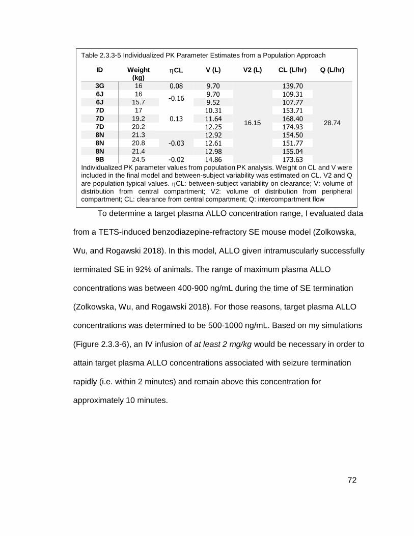
72
To determine a target plasma ALLO concentration range, I evaluated data
from a TETS-induced benzodiazepine-refractory SE mouse model (Zolkowska,
Wu, and Rogawski 2018). In this model, ALLO given intramuscularly successfully
terminated SE in 92% of animals. The range of maximum plasma ALLO
concentrations was between 400-900 ng/mL during the time of SE termination
(Zolkowska, Wu, and Rogawski 2018). For those reasons, target plasma ALLO
concentrations was determined to be 500-1000 ng/mL. Based on my simulations
(Figure 2.3.3-6), an IV infusion of at least 2 mg/kg would be necessary in order to
attain target plasma ALLO concentrations associated with seizure termination
rapidly (i.e. within 2 minutes) and remain above this concentration for
approximately 10 minutes.
Table 2.3.3-5 Individualized PK Parameter Estimates from a Population Approach
ID Weight (kg)
CL V (L) V2 (L) CL (L/hr) Q (L/hr)
3G 16 0.08 9.70
16.15
139.70
28.74
6J 16 -0.16
9.70 109.31 6J 15.7 9.52 107.77 7D 17
0.13
10.31 153.71
7D 19.2 11.64 168.40 7D 20.2 12.25 174.93 8N 21.3
-0.03 12.92 154.50
8N 20.8 12.61 151.77
8N 21.4 12.98 155.04 9B 24.5 -0.02 14.86 173.63
Individualized PK parameter values from population PK analysis. Weight on CL and V were included in the final model and between-subject variability was estimated on CL. V2 and Q
are population typical values. CL: between-subject variability on clearance; V: volume of
distribution from central compartment; V2: volume of distribution from peripheral compartment; CL: clearance from central compartment; Q: intercompartment flow

73
2.3.3.4 Safety and Tolerability
As shown in Table 2.3.3-6, IV ALLO 1-3 mg/kg infused over 5 minutes was
shown to be safe and tolerable in dogs. There was a dose-dependent increase in
ataxia and sedation. At 4 mg/kg IV, dogs were immobile and briefly unarousable
even with pain stimulation for 1-3 minutes with stable vital signs. Ataxia occurred
within 1.5-3 minutes following the start of infusion for a duration of 10-18.5
minutes. In healthy dogs, the onset of sedation occurred at 3.5-5 minutes
following the start of infusion and lasted up to 6 minutes at doses greater than 2
mg/kg. The dog on chronic PB had greater sedation at 2 mg/kg, which was
Table 2.3.3-6 Behavioral Response Following IV Administration
Dose (mg/kg)
n Cmax
(ng/mL) Ataxia Sedation
Ataxia Onset (min post-
injection)
Ataxia Duration
(min)
Sedation Onset (min post-
injection)
Sedation Duration
(min)
1 3 425-1340
33% 0% 3 6
2 4 1654-5287
100% 25% 1.5-2 10-16 3.5 10
3 2 2440-4661
100% 50% 1.5-3 13-13.5 3.5 4.5
4 2 3660-5172
100% 100% 1.5 15.5-
18.5 4.5-5 5-6
Data are shown was percentage of animals within each dose.
Figure 2.3.3-6 Simulation of Plasma ALLO Concentration-Time Profiles Following IV Dosing. Dashed line: highest peak plasma concentration (900 ng/mL) associated with seizure termination in TETS SE mouse model; Dot-dashed line: lowest peak plasma concentration (400 ng/mL) associated with seizure termination in TETS SE mouse model.
0 10 20 3010
100
1000
10000
Time (min)
Pla
sm
a A
LL
O C
on
cen
trati
on
(n
g/m
L)
1 mg/kg
2 mg/kg
3 mg/kg
4 mg/kg

74
associated with higher plasma ALLO concentrations. There were no infusion site
reactions observed.
2.3.4 Discussion
Intravenous ALLO is a compound with a short elimination half-life and a
moderately large volume of distribution in dogs. Across the doses studied, IV
ALLO exhibited linear pharmacokinetics with respect to dose. This information
will be useful for instances where a manipulation of ALLO exposure (e.g.
doubling the dose to double the exposure, up to 4 mg/kg) may be necessary
during clinical trials. This is in agreement Zulresso PK, as it has been shown to
exhibit dose-proportional increases in exposure within the dose range of 0.72-
6.48 mg/kg/day (FDA CDER 2019).
Despite including an allometric exponent of weight on clearance and
volume to explain some of the residual variability, there remains large
interindividual variability that I am unable to explain given the small number of
animals and experiments in the present study. The inability to estimate the
between-subject variability on volume of distribution without high eta shrinkage
may be due to the small number of observations in several animals (Xu et al.
2012). It should also be noted that in the animals such as 6J in whom there were
as few as three concentration-time data points available, the PK parameter
estimates may be biased since the data points were not sampled at optimized
time points and because the weight of each data point is much more significant
when there are only a few. With a larger sample size and optimized sampling

75
times, I may be able to estimate the interindividual variability of
intercompartmental clearance and volumes of distribution (central and peripheral)
in addition to evaluating covariate effects such as sex or PB.
While the simulations suggested a dose of at least 2 mg/kg would be
necessary to attain target plasma concentrations, the safety data provided insight
into plasma concentrations associated with heavy sedation (i.e. >2440 ng/mL).
Ideally, a dose that can attain high enough plasma concentrations without
eliciting deep sedation would be desirable. Therefore, a 2 mg/kg dose infused IV
over 5 minutes would be an appropriate dose to test for efficacy in treating
canine established SE given these criteria. This dose compares to 1.1 mg/kg in
humans using the FDA conversion factor to determine a human equivalent dose
and is predicted to attain plasma ALLO concentrations above the target
concentration of 1000 ng/mL rapidly without causing sedation in most animals.
2.3.5 Conclusions
This study demonstrates IV ALLO exhibits dose-proportional increases in
exposure within 1-4 mg/kg. This would be valuable information to know when
designing a clinical trial in canine SE in order to easily calculate the appropriate
dose to attain specific target plasma concentrations. IV ALLO is safe and
tolerable when administered at doses between 1-3 mg/kg. The onset of ataxia
and sedation following IV infusion is rapid and transient, both desirable for the
termination of and recovery from SE.

76
2.4 Pharmacokinetics, Safety, and Optimization of an Intramuscular
Allopregnanolone Formulation
2.4.1 Introduction
Although IV administration is the best option to rapidly attain high ALLO
concentrations in blood and the CNS, this route is limited by the need for trained
personnel to establish an IV line and administer the drug. This often results in a
delay in treatment. Ideally, the drug should be administered at the onset of SE,
which most often occurs in a pre-hospital setting. Parenteral routes offer the
advantage of bypassing the difficulty of oral administration in a seizing patient
and avoiding first-pass metabolism. Of these alternative routes, IM administration
is a route that requires little technical training or the need to remove much
clothing (as compared to a rectal administration).
My working hypotheses are 1) IM ALLO will have relatively high
bioavailability, 2) IM injection of ALLO is safe in dogs, and 3) formulation
concentration and injection volume will affect bioavailability. The specific aim of
this study was to characterize the PK and safety of IM ALLO. The primary
objectives of this study were to develop a PK model that best fit concentration-
time data, estimate the PK parameter values specifically bioavailability at all
doses, assess its safety and tolerability, and simulate dosing regimens that can
be used to inform dosing recommendations for a clinical study of CSE. The
results of this study will be used to advise further development of an IM
formulation and dosing recommendations for a clinical study of CSE.
77
2.4.2 Methods
2.4.2.1 Study Animals and Safety Monitoring
Four dogs with (n=2) and without (n=2) a history of seizures were used. One of
the dogs with a history of seizures had recurrent seizures despite being on
phenobarbital (PB) maintenance regimen. Approval was obtained through the
Institutional Animal Care and Use Committee of the University of Minnesota prior
to the initiation of the study. The dogs were housed at the University of
Minnesota’s Veterinary College. The dog on PB was previously implanted with a
device which wirelessly transmits continuous iEEG recordings (Kremen et al.
2018). On study days, each dog participating in the study was removed from their
kennel to have a central-line catheter implanted an hour prior to study start.
implanted. Drug administration took place in a procedure room away from the
dog’s kennel. The dogs were fasted prior to and fed no sooner than 2 hours after
drug administration.
2.4.2.2 Study Drug
ALLO concentrate was provided by the Rogawski laboratory at the University of
California, Davis. The concentrate was prepared using chemically pure,
laboratory grade ALLO and shipped frozen to the University of Minnesota. Three
formulations were used:
• 6% ALLO in 24% sulfobutyl ether β-cyclodextrin (Dexolve) in 0.9%
sodium chloride solution (normal saline) diluted and undiluted prior to IM
administration for a final concentration of 3-6 mg/mL

78
• 11% ALLO in 40% Dexolve in normal saline, not diluted prior to IM
administration
• 14% ALLO in 40% Dexolve in normal saline, not diluted prior to IM
administration
2.4.2.3 Starting Dose Rationale
Assuming 100% IM bioavailability, a starting dose should be one with an
acceptable safety profile following IV administration. In healthy dogs without a
history of seizures (regardless of two weeks of PB therapy), 2 mg/kg IV ALLO
showed no signs of ataxia or sedation. In one dog on chronic PB therapy, 2
mg/kg IV caused ataxia and sedation. Therefore, the starting dose for IM
administration was 2 mg/kg.
2.4.2.4 Study Design
PK studies were
completed following IM
administration in two
healthy dogs and two dogs
with a history of seizures (one on chronic PB) in a dose escalation manner,
however, in addition to varying the doses, I also administered different injection
volumes to test my hypothesis that a high formulation concentration
(consequently, a lower injection volume) would increase bioavailability. After the
first 2 mg/kg IM study, the bioavailability was approximately 50%. In order to
Table 2.4.2-1 Number of Animals per IM Study Dose
Dose (mg/kg) 1 2 6
Formulation (mg/mL) 11 3 6 14 6
Number of Dogs (total) 1 1 1 1 2
Number of Dogs on PB 1 0 0 0 0
Number of Dogs with EEG 1 0 0 0 0

79
attempt to attain concentrations comparable to the 3 mg/kg IV dose, the following
two dogs were dosed at 6 mg/kg. In total, three doses (1-,2- and 6-mg/kg) across
four ALLO concentrations (3-, 6-, 11-, and 14-mg/mL) were evaluated. Whole
blood samples (~2 to 5 mL) were collected via a catheterized central vein at pre-
dose and at 1-, 3-, 5-, 15-, 30-, 45-minutes, 1-, 2-, 4-, and 6-hours post-injection.
A washout period of at least 1 week was used.
2.4.2.5 Allopregnanolone Assay
Whole blood was collected in EDTA-containing purple-top vacutainer tubes and
centrifuged for plasma separation as described in Chapter 2.3.2.5.
2.4.2.6 Pharmacokinetic Analysis
ALLO concentration–time data were analyzed using non-compartmental analysis
(NCA) (Phoenix 64, Build 8.0.0.3176, Certara L.P., Princeton, NJ, USA). Data
was uniformly weighted as described in Section 2.3.2.6.1, with the addition of
determining the maximum concentration (Cmax), time at which maximum
concentration is achieved (tmax), and bioavailability (F). Due to the large
interindividual variability, IM bioavailability (F%) was calculated using equation
(4), where the individual’s IV and IM AUC0-last’s were compared. AUC0-last
estimates were used rather than AUC0-INF due to the large percent extrapolated
AUC in 3G (>30%).
Equation:

80
(4)
PK parameters were also determined using individual compartmental
modeling (Phoenix Non-Linear Mixed Effects 8.0). Due to the exploratory and “n
of 1” nature of these IM studies, a population approach was not used. First-order
conditional estimation extended least squares method was used as described in
Section 2.3.2.6.2.
Individual PK parameter estimates from one dog in whom multiple doses
(2- and 6-mg/kg) and formulations (3- and 6-mg/mL) were studied were used to
simulate plasma ALLO concentration-time profiles of a “typical” dog given 1-8
mg/kg of IM ALLO using a formulation concentration of 6 mg/mL.
2.4.2.7 Deconvolution Analysis
In addition to calculating bioavailability via the standard method of dividing drug
exposure (AUC, determined by NCA) following two routes of administration,
bioavailability can be calculated as the cumulative fraction absorbed using
deconvolution. Deconvolution is based on the assumptions of the linear
superposition principle (Rowland and Tozer 2011). Deconvolution can also be
used to determine drug input rate into systemic circulation from varying drug
formulations (e.g. oral immediate-release vs extended-release tablets, dermal
patches, etc.).
Deconvolution is based on the idea that the concentration-time data of an
extravascular dose is a convolution of the input rate and drug disposition
(Rowland and Tozer 2011). Therefore, with knowledge of two of these
81
components, one could calculate the third component. For example, once
concentration-time data following IV and IM administration are known, the input
rate into the compartment where concentration measurements are drawn (i.e. the
central compartment, plasma) and the cumulative amount absorbed can be
calculated.
Phoenix WinNonlin deconvolution function was performed to determine
the input rate following IM administration using individual PK parameters
following IV administration as the exponential terms of the unit impulse function
(Table 2.3.3-3). Concentration-time data from the IM administration were used as
the response function.
2.4.2.8 Safety and Behavioral Response Evaluations
A modified Glasgow Coma Scale (mGCS) was used to quantitate degree of
sedation pre-dose and at scheduled blood sampling times as described in
Chapter 2.3.2.7, with the addition of inspecting the injection site for swelling,
tenderness, and redness. A muscle biopsy was performed in the dog who
received the formulation of highest concentration to assess for evidence of
inflammation and/or tissue damage (3G, 14 mg/mL).

82
2.4.3 Results
The demographics of the dogs are listed in Table 2.4.3-1.
Table 2.4.3-1 Animal Demographics
ID Age (years)
Gender Weight (kg)
Breed Seizure Type
Seizure Frequency
Co-medications
3 9 Male, neutered
16 Beagle History of one
witnessed seizure
None None
6 13 Male, neutered
16 Keeshound Mix
Focal, with generalized
seizures
Focal cluster
seizures every 14-60 days.
With secondarily generalized
seizures every 1-2 months.
PB
7 1 Female, intact
17 Coonhound Mix
(healthy) None None
8 1 Female, intact
22 Coonhound Mix
(healthy) None None

83
2.4.3.1 Non-compartmental Analysis
The concentration-time profiles following IM administration are shown in Figure
2.4.3-1. Pharmacokinetic parameters estimated by NCA are summarized in
Table 2.4.3-2. Apparent clearance ranged from 6.0-12.2 L/hr/kg and apparent
volume of distribution ranged from 4.9-44.7 L/kg. Dose-normalized Cmax following
IM administration ranged between 4.7-20.5 x 10⁻⁵ ng/mL at 3-10 minutes, with a
terminal phase half-life between 34-152 minutes. The IM bioavailability calculated
by the NCA method was estimated to range between 51-112%. There was a
Figure 2.4.3-1 Plasma ALLO Concentration-Time Profile Following IM Administration. cPB: Chronic PB. Blue: 1 mg/kg dose; Red: 2 mg/kg dose; Green: 6 mg/kg dose
0 50 100 1501
10
100
1000
10000
Time (min)
Pla
sm
a A
LL
O C
on
cen
trati
on
(n
g/m
L)
7 @ 6 mg/kg (6 mg/mL)
8 @ 2 mg/kg (3 mg/mL)
8 @ 2 mg/kg (6 mg/mL)8 @ 6 mg/kg (6 mg/mL)6 @ 1 mg/kg (11 mg/mL) + cPB
3 @ 2 mg/kg (14 mg/mL)
Table 2.4.3-2 Non-compartmental PK Parameter Estimates
Fo
rm.
(mg
/mL
)
Do
se
(m
g/k
g)
n tmax (min)
Cmax (ng/mL)
z
(hr-1)
t1/2 (hr)
CL/F (L/hr/kg)
V/F (L/kg)
AUC0-
>∞/Dose (ng/mL*hr/(ng/kg)) x 10-5
F (%)
3 2 1 5 94 0.50 1.4 11.4 22.9 8.78 51 6 2 1 3 328 1.23 0.56 10.1 8.2 9.86 62 6 6 2 3-10 323-
877 0.27-0.60
1.2-2.5
9.5-12.2
15.9-44.7
8.19-10.5 56-65
11 1 1* 15 205 1.25 0.56 6.0 4.8 16.6 96 14 2 1 10 53 0.32 2.13 5.1 15.8 19.5 112
Estimates are presented as a range. (*) denotes dog on chronic PB. tmax: time at maximum
concentration; Cmax: maximum observed plasma concentration; z: terminal phase slope; t1/2:
elimination half-life; CL: clearance; Vd: volume of distribution; AUC: area under the
concentration-time curve from time 0 extrapolated to infinity; F: bioavailability

84
formulation concentration-related increase in bioavailability with the 14 mg/mL
formulation resulting in greater than 100% bioavailability (Figure 2.4.3-2).
Figure 2.4.3-2 Bioavailability by Formulation Concentration
3 6 11 140.0
0.5
1.0
1.5
Bio
availab
ilit
y (
%)
ALLO Formulation (mg/mL)
8N - 2
mg/k
g 8N - 2
mg/k
g7D
- 6 mg/k
g8N
- 6 mg/k
g
6J - 1 mg/k
g
3G - 2
mg/k
g
13mL
6.5mL
20mL
1.5mL
2.1mL

85
2.4.3.2 Individual Compartmental PK Analysis Following IM Administration
A two-compartment model with first-order elimination with a proportional error
model best fit the ALLO concentrations following IM administration (goodness of
fit plots included in Figure 2.4.3-3). Individual parameter estimates are
Table 2.4.3-3 Individual Compartmental PK Parameter Estimates
ID Dose (mg/kg)
Form. (mg/mL)
ka (1/hr)
CL/F (L/hr)
Q/F (L/hr)
V/F (L)
V2/F (L)
Proportional Error (%)
8N 2 3 0.68 (52%)
--- 312.0 (2286%)
14.8 (67%)
940.1 (5061%)
15.3 (28%)
8N 2 6 2.87 (28%)
202.9 (---)
161.6 (---)
0.8 (---)
137.4 (---)
11.6 (22%)
8N 6 6 2.06 (19%)
193.3 (5%)
115.0 (32%)
7.7 (46%)
164.3 (13%)
14.3 (23%)
7D 6 6 2.60 (33%)
234.0 (6%)
671.9 (35%)
14.0 (48%)
642.2 (11%)
12.4 (22%)
6J 1 11 3.88 (146%)
100.7 (11%)
44.5 (36%)
28.0 (162%)
60.0 (53%)
30.9 (23%)
3G 2 14 0.39 (281%)
160.1 (32%)
91.6 (754%)
10.1 (282%)
151.1 (725%)
26.8 (23%)
Form.: ALLO formulation; ka: absorption rate constant; CL/F: apparent clearance from central compartment; Q/F: apparent intercompartment clearance; V/F: apparent volume of distribution from central compartment; V2/F: apparent volume of distribution from peripheral compartment.
Figure 2.4.3-3 Goodness of fit plots for a two-compartment model. IWRES: individual weighted residual; TAD: time after dose; IPRED: individual predicted concentration
0 100 200 300 4001
10
100
1000
Time (min)
Pla
sm
a A
LL
O C
on
cen
trati
on
(n
g/m
L)
Observed versus Predicted
0 200 400 600 800 10000
200
400
600
800
1000Individual Observed vs Predicted
Observed ALLO Concentration (ng/mL)
Pre
dic
ted
AL
LO
Co
ncen
trati
on
(n
g/m
L)
100 200 300 400
-4
-2
0
2
4 IWRES vs TAD
Time After Dose (min)
IWR
ES
200 400 600 800
-4
-2
0
2
4 IWRES
Individual Predicted Concentration (ng/mL)
IWR
ES

86
summarized in Table 2.4.3-3. With the exception of the 14 mg/mL formulation,
the rate of absorption increased with formulation concentration.
2.4.3.3 IM Absorption Rate and Bioavailability by Deconvolution Analysis
Across all experiments, the calculated IM absorption rate is highest in the initial
10-15 minutes following injection (Figure 2.4.3-4). Excluding the 14 mg/mL
formulation, the higher absorption rates occurred at the combination of higher
dose and higher concentration.
Cumulative fraction absorbed over time is illustrated in Figure 2.4.3-5. The
absorption follows a first-order rate, except in one dog (3G) administered 2 mg/kg
at the highest formulation concentration tested (14 mg/mL). Absorption following
this IM injection appears to follow a zero-order absorption rate, which raises
concern for the possibility of agglomeration, crystallization, and/or precipitation of
drug. However, the tissue biopsy completed in this animal yielded inconclusive
Figure 2.4.3-4 IM ALLO Absorption Rate Over Time
0 50 1000
1000000
2000000
3000000
Time (min)
Inp
ut
Rate
(n
g/m
in)
6 mg/kg (6 mg/mL)
6 mg/kg (6 mg/mL)1 mg/kg (11 mg/mL)
2 mg/kg (6 mg/mL)2 mg/kg (3 mg/mL)
2 mg/kg (14 mg/mL)

87
results (no muscle tissue in sample). In one animal (8N) in whom multiple
formulations and doses were studied, there appeared to be a formulation
concentration-dependent increase in the cumulative fraction absorbed, which
supports the observation of increasing absorption rate constant with higher
formulation concentration noted in the individual PK analysis.
Figure 2.4.3-5 IM ALLO Fraction Absorbed Over Time
0 100 200 300 4000.0
0.5
1.0
1.5
Time (min)
Fra
cti
on
Ab
so
rbed
(%
)
2 mg/kg (6 mg/mL)
6 mg/kg (6 mg/mL)1 mg/kg (11 mg/mL)
6 mg/kg (6 mg/mL)
2 mg/kg (3 mg/mL)
2 mg/kg (14 mg/mL)

88
The bioavailability estimated by the NCA approach was compared to the
cumulative fraction absorbed estimated by deconvolution. The results are shown
in Figure 2.4.3-6 and Table 2.4.3-4.
Based on our simulations of a “typical dog” using a 6 mg/mL ALLO
formulation (Table 2.4.3-5), an IM dose of 8 mg/kg would be required to attain the
target plasma ALLO concentration of 900 ng/mL and maintain above this
concentration for about 5 minutes.
Figure 2.4.3-6 Bioavailability Estimated by NCA vs Deconvolution Methods. A: NCA; B: deconvolution
A B A B A B A B A B A B0.0
0.5
1.0
1.5
Bio
availab
ilit
y (
%)
Table 2.4.3-4 Bioavailability Calculated by NCA and Deconvolution Methods
Animal 8N 8N 8N 7D 6J 3G
Dose (mg/kg) 2 2 6 6 1 2 Formulation
(mg/mL) 3 6 6 6 11 14
Calculation Method NCA 0.51 0.62 0.65 0.56 0.96 1.12
Deconvolution 0.57 0.73 0.78 0.65 1.17 0.76
Table 2.4.3-5 PK Parameter Estimates Used for IM Simulations
ka (min-1) CL/F (L/hr) Q (L/hr) V/F (L) V2/F (L)
0.034 193.31 114.96 7.65 164.32

89
2.4.3.4 Safety and Tolerability
The onset of ataxia without sedation occurred 3-5 minutes following the 6 mg/kg
dose and lasted for 17-19 minutes (Table 2.4.3-5). Pain associated with injection
volume was observed only at the 6 mg/kg IM dose, and is likely associated with
the large injection volume (~20 mL).
2.4.4 Discussion
The development of IM ALLO for treatment of SE is limited by the slow
absorption rate of ALLO from the IM injection site. When comparing the
concentration-time profiles following IV and IM administration (Figures 2.3.3-1
Figure 2.4.3-7 Simulation of Plasma ALLO Concentration-Time Profiles Following IM Administration. Dashed line: highest peak plasma concentration (900 ng/mL) associated with seizure termination in TETS SE mouse model; Dot-dashed line: lowest peak plasma concentration (400 ng/mL) associated with seizure termination in TETS SE mouse model.
0 20 40 600
200
400
600
800
1000
Time (min)
Pla
sm
a A
LL
O C
on
cen
trati
on
(n
g/m
L)
1 mg/kg
2 mg/kg
4 mg/kg
6 mg/kg
8 mg/kg
Table 2.4.3-6 Behavioral Response Following IM Administration
Dose (mg/kg)
n Cmax
(ng/mL) Ataxia Sedation
Ataxia Onset (min post injection)
Ataxia Duration (min)
1 1 205 0% 0% 2 3 53-328 0% 0%
6 2 322-876 100% 0% 3-5 17-19

90
and 2.4.3-1), one can notice the loss of the biexponential decay in the IM
concentration-time profiles. This apparent monoexponential decay in plasma
ALLO concentration is likely due to an absorption rate constant much slower than
the elimination rate constant or “flip-flop” kinetics. This is also seen by the
difference in z estimated following IV (Chapter 2.3.3.1) and IM dosing (ranges:
1.2-8.1 versus 0.27-1.23, respectively). The terminal rate constant has become
limited by the slower absorption rate constant (i.e. the body can only eliminate
drug as fast as it is being absorbed). The longer half-life with IM dosing may
provide an advantage of maintaining plasma ALLO concentrations above a
specific concentration for a longer duration. Due to the exploratory nature of
these experiments (“n of 1”), there is large variability that I am unable to explain.
Simulations suggest an 8 mg/kg IM dose would be necessary to achieve target
plasma concentration window of 500-1000 ng/mL using a 6 mg/mL formulation,
however this would result in a large injection volume (~27 mL for a 20 kg dog)
which would limit the absorption rate and cause pain upon injection similar in
severity to what was observed at the 6 mg/kg dose.
By evaluating the PK across different doses and formulation strengths, I
found that a higher formulation concentration (and most importantly, a lower
injection volume) was associated with a faster absorption rate. It has been noted
that injection volume does have an inverse relationship with absorption rate for
IM injections regardless of the water solubility of the drug (Pfeffer and Van
Harken 1981; Hirano, Ichihashi, and Yamada 1981). Other factors that may affect
the absorption rate constant include the particle size, cohesiveness of the

91
dissolved particles, initial injection concentration, injection speed and pressure,
and the physiological state of the injection site (Hirano, Ichihashi, and Yamada
1981). I also found that at the highest formulation concentration of 14 mg/mL, the
estimated absorption rate constant was not as high as I expected based on the
formulation-absorption trend observed at previous doses. This suggests that 14
mg/mL may be too high of an initial injection concentration and has negatively
affected the absorption rate. It is tempting to conclude that the ideal IM
formulation concentration must be between 11- and 14 mg/mL. However, one
caveat to consider is that the 11- and 14-mg/mL formulations were studied in one
dog each. It is difficult to determine whether these changes in absorption rate are
due to formulation alone or the individual animal. A crossover study would be
necessary to answer this question.
The results of the deconvolution analysis were comparable to those of the
NCA approach. The advantage of performing a deconvolution analysis is the
ability to calculate the absorption profile of each IM injection, which may reveal
complex absorption patterns. Although the individual PK analysis was able to
estimate a first-order absorption rate, it is unable to capture any absorptive
activity following the initial peak. In the present study, there was large
interindividual and inter-formulation variability in absorption profiles. However,
due to the small number of animals and number of IM studies per animal, it is
difficult to attribute the large variability to drug disposition kinetics alone. A
second peak was seen in all dogs (except for one occasion at 2 mg/kg [6
mg/mL]) at 20-45 minutes post-injection, and in one dog, a third peak was

92
observed at 50 minutes post-injection. These secondary peaks in absorption rate
suggest a delayed absorption, which may be explained by increased blood flow
to the injection site (e.g. walking, playing, or receiving pets from veterinary
technicians) and/or a depot effect. In addition, small differences in the estimated
bioavailability may be due to the sensitivity of the deconvolution method to errors
in the unit impulse function (based on exponential terms provided by individual
PK parameter values following IV administration).
2.4.5 Conclusions
This study demonstrates that ALLO is safe and tolerable when administered IM
at doses up to 2 mg/kg and at injection volumes less than 10 mL per injection
site. Following the 6 mg/kg dose, the onset of ataxia occurred within 5 minutes of
IM injection and lasted for almost 20 minutes, which suggests relatively quick
brain penetration and moderate residence time. However, at the 6 mg/mL
formulation, my simulations suggest an 8 mg/kg IM dose would be necessary to
achieve target plasma concentration range of 500-1000 ng/mL, which would
result in an injection volume (~27 mL in a 20 kg dog) large enough to limit the
absorption rate and cause pain with injection. Therefore, based on absorption
rate limitations, the development of IM ALLO would require alternative
approaches to be feasible.

93
2.5 Pharmacodynamics of Intravenous and Intramuscular Allopregnanolone in
Dog
2.5.1 Introduction
Allopregnanolone has been shown to effectively terminate seizures in several
acute seizure and SE rodent models (Rogawski et al. 2013; Kokate et al. 1996;
Cheryl A Frye and Scalise 2000; Zolkowska, Wu, and Rogawski 2018). It has
also been reported to affect electroencephalograph parameters in a fashion
similar to benzodiazepines (BZDs) in rats, causing increases in higher-frequency
(beta) activity (Lancel et al. 1997; Slawecki, Purdy, and Ehlers 2005). Based on
behavioral observation of our study animals alone, ALLO appears to have a fast
onset of action, inducing ataxia as early as 90 seconds and sedation just 3.5
minutes following the start of IV infusion. However, it remains unknown whether
ALLO can affect electrical brain activity in a manner that could stop and/or
prevent further seizures in dogs.
For these studies, I hypothesized that 1) ALLO has a quick onset of effect
on intracranial electroencephalograph (iEEG) activity in dogs, and 2) based on its
potentiation of GABAA currents, its effect on iEEG is similar to that of
benzodiazepines (i.e. increase in beta activity) (Mandema, Kuck, and Danhof
1992; Mandema and Danhof 1992). Alternative hypotheses include 1) ALLO has
a moderate or slow onset of effect on iEEG, and 2) its effect on iEEG can be
differentiated from those due to benzodiazepine administration. The specific aim
of this study is to develop a PK-PD model relating plasma ALLO concentrations
to iEEG response in one dog. The primary objectives of this study were to

94
estimate the concentration that produces 50% of the maximum response (EC50)
and the effect compartment rate constant (ke0) of IV and IM ALLO. The results of
this study will be used to inform the target therapeutic concentration and dosing
recommendations for a clinical study in CSE.
2.5.2 Methods
2.5.2.1 Study Design
One dog with a history of seizures was used. Despite being on a PB antiseizure
maintenance regimen, he occasionally had breakthrough seizures. Approval was
obtained through the Institutional Animal Care and Use Committee of the
University of Minnesota prior to the initiation of the study. The dog was housed at
the University of Minnesota’s Veterinary College. This dog was previously
implanted with a device which wirelessly transmits continuous iEEG recordings
(Kremen et al. 2018). On study days, the dog was removed from his kennel to
have a central-line catheter emplaced an hour prior to study start. On days where
IV ALLO was administered, a peripheral-line catheter was also emplaced. Drug
administration took place in a procedure room away from the dog’s kennel. The
dogs were fasted prior to and fed no sooner than 2 hours after drug
administration.
iEEG data were collected in the dog during two IV studies (1-2 mg/kg) and
one IM study (1 mg/kg). Each collection period started the day before and
finished the day after IV injection to determine baseline data. iEEG data were
recorded continuously at a sampling rate of 250 Hz.

95
2.5.2.2 Intracranial Electroencephalographic (iEEG) Analysis
iEEG data were analyzed using custom algorithms (Matlab, version 2018b) for
characterizing iEEG power in bands of interest and its temporal dynamics (Delta
(1 -3 Hz), Theta (>3 – 7 Hz), Alpha (>7 - 12 Hz), Beta (>12 – 25 Hz), High Beta
(>20 -25 Hz)) as described previously (Kremen et al. 2017). Absolute and relative
power densities across frequency bands were used as the pharmacodynamic
response. Absolute power density refers to the absolute power (voltage) of the
patient’s iEGG, while relative power density refers to the percentage of power a
frequency band (such as beta) compared to the sum of all frequency bands
(delta, theta, alpha, and beta).
2.5.2.3 Pharmacokinetic-Pharmacodynamic (PK-PD) Modeling
Individualized PK parameters were estimated using a population approach as
described in Section 2.3.2.6.2 (Table 2.3.3-5), fixed, then indirect-link Emax/Imax
PD models were explored. An indirect-link was used due to the hysteresis that
was observed. The best fit model was determined using visual inspection and
precision of model parameters.

96
2.5.3 Results
2.5.3.1 Intracranial Electroencephalographic Analysis
2.5.3.1.1 Absolute power density
Across all iEEG frequency bands, absolute power density increased during and
transiently after IV ALLO (Figure 2.5.3-1). The beta frequency band (purple)
appears to occur at the lowest plasma ALLO concentrations. As the dose
increased, the amplitude and duration of these iEEG changes also increased.
Following IM injection, plasma ALLO concentrations did not reach high enough
concentrations to produce iEEG changes comparable to the IV infusion at the
same dose and changes were more spread out over time. However, changes in
absolute power densities at all doses were statistically significantly different
Figure 2.5.3-1 Absolute Power Density and Plasma ALLO Concentration-Time Profiles Following IV and IM ALLO Administration in One Dog on Chronic PB. Colored solid lines: iEEG, separated into four frequency bands; dashed black line: plasma ALLO concentration

97
compared to pre-dose and non-study day baselines irrespective of route of
administration.
2.5.3.1.2 Relative power density
High beta frequency band (20-25 Hz) relative power density decreased, while
alpha frequency band (7-12 Hz) relative power density increased during and
transiently after IV infusion (Figure 2.5.3-2). These changes occurred for a longer
duration but not to a larger extent at the higher dose. As with absolute power
density, relative power density changes following IM injection were not as drastic
as changes seen following IV infusion.
Figure 2.5.3-2 Relative Power Density and Plasma ALLO Concentration-Time Profiles Following IV and IM ALLO Administration in One Dog on Chronic PB. Purple solid line: Relative beta power density; green solid line: relative alpha power density; dashed black line: plasma ALLO concentration
0 5 10 150.0
0.2
0.4
0.6
0
2000
4000
6000
Time (min)
Rela
tive P
ow
er
Den
sit
y
Pla
sm
a A
LL
O C
on
cen
tratio
n (n
g/m
L)
1 mg/kg IV Infusion
AlphaHigh BetaConcentration (ng/mL)
0 5 10 150.0
0.2
0.4
0.6
0
2000
4000
6000
Time (min)
Rela
tive P
ow
er
Den
sit
y
Pla
sm
a A
LL
O C
on
cen
tratio
n (n
g/m
L)
2 mg/kg IV Infusion
AlphaHigh BetaConcentration (ng/mL)
0 20 40 600.0
0.2
0.4
0.6
0
2000
4000
6000
Time (min)
Rela
tive P
ow
er
Den
sit
y
1 mg/kg IM Injection
AlphaHigh BetaConcentration (ng/mL)
Pla
sm
a A
LL
O C
on
cen
tratio
n (n
g/m
L)

98
2.5.3.2 Pharmacokinetic-Pharmacodynamic Analysis
For all frequency bands, changes in absolute power density in response to
plasma ALLO concentrations were best described by an indirect-link sigmoidal
Emax model (Table 2.5.3-1 and Figure 2.5.3-3). There was an overall increase in
brain activity following ALLO administration, especially via the IV route. Following
IV administration, the delay equilibrium half-life ranged from 8-32 seconds,
suggesting the time required to reach the full response in the brain is less than 3
minutes. The delay equilibration rate constant could not be calculated with
precision for most frequency bands following IM injection, likely due to a lack of
significant change in absolute power density. The beta frequency band
(comprised of the low and high beta frequency range) had the lowest EC50 (270
Table 2.5.3-1 PKPD Parameter Estimates (iEEG Absolute Power Density)
Frequency Band
ke0 (1/min) EC50
(ng/mL) E0 (106) Emax (106)
Route IV IV IV IV
Dose 1 2 1 2 1 2 1 2
Delta (1-3 Hz)
2.93 (18.2)
1.42 (24.4)
652 (2.0)
700 (20.3)
2.6 (9.9)
0.1 (>500)
9.1 (6.6)
37.1 (23.2)
Theta (3-7 Hz)
3.75 (29.6)
1.32 (27.8)
666 (2.6)
538 (6.1)
2.9 (38.2)
9.4 (36.4)
32.3 (8.6)
68.5 (6.9)
Alpha (7-12 Hz)
5.05 (35.4)
1.32 (13.7)
681 (2.3)
499 (4.1)
2.3 (86.8)
8.8 (81.0)
67.7 (7.8)
92.3 (9.2)
Beta (12-25 Hz)
2.00 (22.4)
1.60 (26.3)
276 (6.7)
270 (25.9)
4.5 (55.3)
13.0 (39.6)
27.7 (10.7)
32.5 (16.5)
Values are presented with coefficient of variation (%). ke0: delay equilibrium rate constant; EC50: concentration in the effect compartment required to elicit half of the maximum response; E0: baseline response; Emax: maximum response

99
ng/mL) compared to the other frequency bands, suggesting it could be a
surrogate marker useful in clinical studies as a signal for CNS penetration. This
would approximate 400 ng/mL in the plasma.
Figure 2.5.3-3 iEEG (absolute power density) effect-concentration profiles following 1-2 mg/kg infused IV over 5 minutes (red and blue, respectively) and 1 mg/kg injected IM as a bolus (orange) in 1 dog on cPB. Effect compartment concentrations are predicted using an indirect-link between PK and sigmoidal Emax PD models. Colored circles are observed data; solid lines represent predicted data. IPRED: predicted effect; DV: observed effect.
0 500 1000 1500 20000.0
5.0×10⁷
1.0×10⁸
1.5×10⁸
Concentration in the Effect Compartment (ng/mL)
Ab
so
lute
Po
wer
Den
sit
y
Delta (1-3 Hz)
IPRED 1mg/kg IVDV 1mg/kg IVIPRED 2mg/kg IVDV 2mg/kg IVIPRED 1mg/kg IMDV 1mg/kg IM
0 500 1000 1500 20000.0
5.0×10⁷
1.0×10⁸
1.5×10⁸
Concentration in the Effect Compartment (ng/mL)
Ab
so
lute
Po
wer
Den
sit
y
Theta (3-7 Hz)
IPRED 1mg/kg IVDV 1mg/kg IVIPRED 2mg/kg IVDV 2mg/kg IVIPRED 1mg/kg IMDV 1mg/kg IM
0 500 1000 1500 20000.0
5.0×10⁷
1.0×10⁸
1.5×10⁸ Alpha (7-12 Hz)
Concentration in the Effect Compartment (ng/mL)
Ab
so
lute
Po
wer
Den
sit
y
IPRED 1mg/kg IVDV 1mg/kg IVIPRED 2mg/kg IVDV 2mg/kg IVIPRED 1mg/kg IMDV 1mg/kg IM
0 500 1000 1500 20000.0
5.0×10⁷
1.0×10⁸
1.5×10⁸
Concentration in the Effect Compartment (ng/mL)
Ab
so
lute
Po
wer
Den
sit
y
Beta (15-25 Hz)
IPRED 1mg/kg IVDV 1mg/kg IVIPRED 2mg/kg IVDV 2mg/kg IVIPRED 1mg/kg IMDV 1mg/kg IM

100
Concentration-iEEG data using relative power densities were also
examined. Only alpha and high beta frequency bands had consistent changes in
relative power density with an increasing IV dose. In contrast to changes
observed in absolute power density, relative power density of high beta
decreased after administering ALLO, while relative power density of the alpha
band increased. In other words, relative to all the increasing brain activity, the
input higher beta frequency band is decreasing in response to ALLO. The relative
power density changes and concentration-time data for high beta and alpha
frequency bands were best fit by an indirect-link sigmoidal Imax and Emax
models, respectively (Table 2.5.3-2 and Figure 2.5.3-4). Similar to absolute
power density changes, relative power densities had a larger change and for a
longer duration at the higher IV dose, no significant changes following IM dosing,
and changes occurred at lower concentrations in the high beta frequency band
compared to alpha frequency band. The EC50 in the theoretical effect
compartment to produce changes in the high beta frequency band is
approximately 720 ng/mL in the plasma.
Table 2.5.3-2 PKPD Parameter Estimates (iEEG Relative Power Density)
Frequency Band
ke0 (1/min) EC50
(ng/mL) E0 (106)
Emax /Imax (106)
Route IV IV IV IV
Dose 1 2 1 2 1 2 1 2
Alpha (7-12 Hz)
2.35 (25.0)
1.20 (10.6)
759 (14)
455 (4.2)
0.09 (19.6)
0.10 (25.4)
0.57 (36.6)
0.32 (8.8)
High Beta (20-25 Hz)
2.27 (26.5)
0.92 (5.1)
635 (10)
329 (3.0)
0.43 (4.7)
0.52 (3.3)
0.47 (22.4)
0.45 (4.2)
Values are presented with coefficient of variation (%). ke0: delay equilibrium rate constant; EC50/IC50: concentration in the effect compartment required to elicit half of the maximum (inhibitory) response; E0: baseline response; Emax/Imax: maximum (inhibitory) response

101
2.5.4 Discussion
Overall, I observed higher absolute power in all bands after administering the
drug, suggesting that ALLO increases brain activity. Following increases in IV
dose, the extent and duration of iEEG changes also increased. With the higher
dose of ALLO, the iEEG changes were greater, the onset of duration was faster,
and occurred for a longer period of time. In contrast, following IM dosing, there
were statistically significant but minimal changes to iEEG, likely due to the
attainment of lower plasma ALLO concentrations. The peak plasma
concentration following IM dosing (205 ng/mL) was just below the lowest plasma
EC50 for absolute and relative power density changes estimated from the IV
infusions (~400- and 720-ng/mL for beta frequency).
In contrast, increases in absolute and relative power density of the beta
frequency band in response to benzodiazepine intervention have been widely
cited (Mandema and Danhof 1992; Mandema, Kuck, and Danhof 1992;
Figure 2.5.3-4 iEEG (relative power density) effect-concentration profiles following 1-2 mg/kg infused IV over 5 minutes (red and blue, respectively) and 1 mg/kg injected IM as a bolus (orange) in 1 dog on cPB. Effect compartment concentrations are predicted using an indirect-link between PK and sigmoidal Emax/Imax PD models. Colored circles are observed data; solid lines represent predicted data. IPRED: predicted effect; DV: observed effect.
0 500 1000 1500 20000.0
0.2
0.4
0.6 Alpha (7-12 Hz)
Concentration in the Effect Compartment (ng/mL)
Rela
tive P
ow
er
Den
sit
y
IPRED 1mg/kg IV
DV 1mg/kg IV
IPRED 2mg/kg IV
DV 2mg/kg IV
IPRED 1mg/kg IMDV 1mg/kg IM
0 500 1000 1500 20000.0
0.2
0.4
0.6
Concentration in the Effect Compartment (ng/mL)
Rela
tive P
ow
er
Den
sit
y
High Beta (20-25 Hz)
IPRED 1mg/kg IV
DV 1mg/kg IV
IPRED 2mg/kg IV
DV 2mg/kg IV
IPRED 1mg/kg IM
DV 1mg/kg IM

102
Greenblatt et al. 1989; Van Lier et al. 2004; Friedman et al. 1992). In contrast to
what I observed following IV ALLO, Van Lier et al reported that diazepam elicits
increases in relative power density of high beta (defined as 21-30 Hz) while
decreasing alpha (defined as 9-10 Hz) in rats (Van Lier et al. 2004). This
suggests that although ALLO and BZDs potentiate GABAA receptors, they induce
differential effects on EEG activity. This may be due to ALLO’s activity at synaptic
and extrasynaptic GABAA receptors while BZDs have activity at only synaptic
GABAA receptors.
The frequency band that appears to have highest sensitivity for plasma
ALLO concentrations is beta (15-25 Hz). For absolute and relative power density
changes, beta frequency changes occur at EC50/IC50 lower than other frequency
bands. Interestingly, the estimated EC50 for beta changes following IV ALLO are
comparable to what has been observed following a clinically-relevant IV
diazepam dose (0.15 mg/kg) for percent change over baseline beta activity in
healthy human volunteers (400 ng/mL vs. 270 ng/mL, respectively) (Greenblatt et
al. 1989).
Another PD model that could have been used to describe the iEEG data is
an indirect-response model, using either stimulation of the “rate in” or inhibition of
the “rate out” (Gabrielsson and Hjorth 2016). However, many of the previous
exposure-response models developed with benzodiazepines used Emax models
(Mandema and Danhof 1992; Mandema, Kuck, and Danhof 1992; Greenblatt et
al. 1989; Van Lier et al. 2004; Friedman et al. 1992). Furthermore, using an
indirect-response model would require the estimation of more parameters, which
103
may be difficult given the limited data that I have. Therefore, in order to generate
comparable results, especially with respect to the beta frequency band, a
sigmoidal Emax model was used.
It is important to note that this animal (6J) had higher concentrations at the
doses administered compared to the other animals who have been administered
the same dose. This should be taken into consideration when designing a clinical
trial for canine SE, knowing that many of dogs that present in a veterinary
emergency department with canine SE are on PB chronically to maintain seizure
control.
Zolkowska’s work demonstrated that IM ALLO has great potential to be
useful as a first-line treatment for SE, but the current formulations do not meet
the ideal criteria for a first-line SE treatment (Zolkowska, Wu, and Rogawski
2018). The largest limitation of the IM route of administration is the inability to
attain high enough plasma concentrations to elicit iEEG changes. Strategies to
increase plasma concentration using clinically-relevant volumes include but are
not limited to: 1) increasing its water solubility by synthesizing a pro-drug or using
alternative delivery systems; 2) using different administration strategies like
multiple injections of changing the needle gauge and/or length; or 3) optimizing
the formulation by using multiple solvents.
2.5.5 Conclusion
The onset of effect of IV ALLO, as seen from iEEG changes and behavioral
response, was rapid and transient, both desirable for the termination of and
recovery from SE. Only apparent following IV infusion, I observed a dose-related

104
increase in extent and duration of absolute and relative power densities. Because
change in absolute and relative power densities for the beta frequency band
occurred at the lowest EC50 compared to other frequencies, it may be useful as a
surrogate marker of drug brain penetration. Although IM ALLO would be ideal for
pre-hospital settings, at the IM formulations studied, plasma ALLO
concentrations following IM administration are not high enough to induce
changes in iEEG activity comparable to those seen following IV infusion due to
low absorption rates.
Compiling all of the PKPD and safety data from this project and what has
been reported by my collaborators at UC Davis (Figures 2.5.5-1 and 2.5.5-2), I
propose that a 2 mg/kg dose infused over 5 minutes is an appropriate starting
dose to test as a first-line agent to treat canine SE, as this dose is predicted to
attain the target concentrations (500-1000 ng/mL) without causing heavy
sedation. Although a 1 mg/kg dose would also attain plasma concentrations that
fall within the range between the EC50 and those associated with heavy sedation,
Figure 2.5.5-1 Determination of the Target Plasma ALLO Concentration
PK• IVALLOexhibitsdose-
proportionality
between1-4mg/kg• IMALLOislimitedby
itsslowabsorption
PD• Rapidonset
ofaction
• PlasmaEC50forbetachanges~400ng/mL
TETS• Cmax rangingfrom400-
900ng/mLterminated
SEin12/13mice
Safety• Minimal
effects
occurred322-1320ng/mL
• Heavysedation
>2440ng/mL
• Route:IV• Targetplasmaconcentration:500-1000ng/mL

105
only the 2 mg/kg dose is predicted to attain the peak concentration observed in
the TETS SE mouse study (900 ng/mL) and as early as 3 minutes into the start
of infusion.
2.6 Design of a Safety and Effectiveness Clinical Trial of Intravenous
Allopregnanolone for the Treatment of Canine Status Epilepticus
In spite of having established guidelines for the management of convulsive SE,
upwards of 40% of patients are clinically non-responsive to benzodiazepines
(BZDs) and there remains a critical need to identify a treatment for SE that is
efficacious for all patients. Without such a therapeutic agent or regimen, 31-40%
SE cases are at risk for untimely death due to prolonged seizure episodes
(Hocker et al. 2013; Leitinger et al. 2019). My long-term goal is to find a more
effective first-line drug for SE treatment that has the potential of replacing or
being used in conjunction with BZDs. IV ALLO is an ideal agent to treat this
Figure 2.5.5-2 Determination of IV ALLO Dose for a Clinical Trial in Canine SE
0 10 20 3010
100
1000
10000
Time (min)
Pla
sm
a A
LL
O C
on
cen
trati
on
(n
g/m
L)
1 mg/kg
2 mg/kg
3 mg/kg
4 mg/kg
HeavySedation
BelowPlasmaEC50

106
emergent condition based on its mechanisms of action and its ability to rapidly
diffuse into the CNS.
The development path for potential approval of IV ALLO for the treatment
of seizure emergencies requires conducting clinical trials that safety and
effectiveness. Because IV ALLO has been approved for another indication, much
of the toxicity and safety studies may not be required. A unique pathway to
encourage development of IV ALLO for SE in people is first establishing safety
and efficacy in canine patients prior to human use. Although this approach would
not replace the need for safety and effectiveness trials in people, the results
would inform the design of a clinical trial in human SE. The following proposed
study have a primary objective of characterizing the safety and effectiveness of
ALLO for the treatment of SE in dogs. Evaluating potential therapies for SE in
dogs with naturally-occurring epilepsy and SE have the added advantage of
developing life-changing treatments for dogs and people.
2.6.1 Study Objective
The primary objective for this study is to demonstrate that the effectiveness and
safety of IV ALLO as an agent for early CSE is as effective as IV DZP.
2.6.2 Study Population
Dogs (5-40 kg in body weight) admitted into an emergency/urgent veterinary
medical clinic with a clinical diagnosis of convulsive status epilepticus and have
received at least one adequate dose of a BZD will be enrolled. CSE is defined

107
as continuous convulsions lasting >5 min, or 2 or more recurrent convulsions
without regaining consciousness between seizures within the last 12 hours and
seizures are continuing to occur or likely to reoccur without recovery in between.
Canine patients may be excluded from the study for the following reasons: owner
not wishing to participate, anoxic cause for status epilepticus, or metabolic cause
for status epilepticus (i.e. must have normal blood glucose, calcium, bilirubin, etc.
on admission bloodwork).
2.6.3 Study Design
This will be a double-blinded, randomized, multicenter non-inferiority study in
canine patients with SE who have failed first-round or second round
benzodiazepines study (Figure 5.2.2-1). This study design will be modeled after a
prospective, multicenter, randomized parallel-group study comparing the
Figure 2.6.3-1 IV ALLO Study Schematic
Patient in SE
BZD
ALLOBZD
Standard of
Care
SuccessFailure
Follow-up
1:1 Randomization
Screen,
Enroll,
Consent
+12 hours
Primary endpoint

108
effectiveness of intranasal midazolam and rectal DZP in terminating CSE within 5
minutes without seizure recurrence in 10 minutes (M. Charalambous et al. 2017).
Although I am interested in developing IV ALLO as first-line treatment, it would
be unethical to test its effectiveness without giving the canine patient a known
effective treatment first.
2.6.4 Endpoints
2.6.4.1 Primary Endpoint
Termination of seizure within 5 minutes of infusion without recurrence of seizures
within 10 minutes of dosing. Clinical cessation of SE consists of absence of
clinical seizures and improving responsiveness. Absence of apparent seizures
will be determined clinically. Responsiveness will be determined by patient’s
response to verbal command or noxious stimuli.
2.6.4.2 Secondary Endpoints
Secondary endpoints include: Safety/tolerability (degree and duration of ataxia
and sedation, heart rate, blood pressure, EKG monitoring, SpO2 measurement,
need for intubation within 60 minutes of start of study drug infusion, seizure
cessation at 60 minutes after drug administration, need for rescue therapy, time
to next seizure, 12-hour responder rate, discharge status, and presence of subtle
CSE via EEG monitoring (when available).
2.6.4.3 Blinding/Unblinding and Randomization
Patients, veterinary emergency department study team members, PIs, and
clinical coordinating centers will be blinded to the treatment assignment.

109
Emergency unblinding may be required if the treating team feels that a patient’s
care requires knowledge of what study drug was given. Emergency unblinding
will not be performed within 60 minutes of the start of study drug infusion with the
exception of veterinarian judgment that it is necessary for the safety or care of
the patient or because of unanticipated situations accommodated by study
procedures. The PK study PIs and laboratory scientists will know the assay
results but will remain blinded to response until the completion of the study. Drug
concentration data will not be disseminated until the completion of study.
Randomization for this study will be 1:1 between 2 mg/kg IV ALLO and a
second-/third-round BZD (0.5 mg/kg IV DZP).
2.6.5 Sample Size
The primary objective of this study is to demonstrate that the number of patients
whose seizures terminate within 5 minutes of study drug infusion without seizure
recurrence in the IV ALLO group is not inferior to that in the IV DZP group by
more than a noninferiority margin of 10%. The null hypothesis of inferiority will be
tested using a one-sided test with an 80% power and a significance of 5% (one-
sided probability of a type I error of 0.05). The estimate of IV BZD success rate of
55% is from a prospective, double-masked, randomized parallel-group study of
IV benzodiazepine (BZD, lorazepam or DZP) + saline infusion and IV BZD + IV
fosphenytoin (FOS) for the treatment of CSE (Patterson et al. 2015). In this
study, once a seizure occurred in the emergency department, canine patients
were given an IV BZD followed immediately by either IV FOS or normal saline,

110
and success was defined as seizure termination without recurrence within 2
hours. Therefore, the sample size required is 342 per treatment arm (a total of
684 canine patients), which takes into account a 10% inflation to account for loss
of follow up, protocol deviation (inclusion/exclusion criteria violation), and/or
repeat enrollment of the same subject. This conservative approach of assuming
ALLO has an efficacy rate of 55% is to ensure the study has a large enough
sample size and sufficient power.
2.6.6 Study Drug
IV ALLO (6 mg/mL in 24% Dexolve, manufactured by University of California
Davis) and IV DZP (commercially supplied as 5 mg/mL injectable solution) will be
used for this study.
2.6.6.1 Dose Rationale
Based on the population pharmacokinetic model presented in Chapter 2.3.3.3, 2
mg/kg IV ALLO infused over 5 minutes is predicted to attain the target plasma
concentration within 2 minutes of the start of infusion and maintain
concentrations above this target for approximately 5 minutes without causing
heavy sedation. The corresponding dose for DZP is 0.5 mg/kg, representing the
standard of care dose for treatment of SE in canines. The concentrations were
chosen so that volumes of the two study drugs would be identical based on
weight. A Dosing Chart will be provided to enable veterinary clinicians to
administer the same volume of either drug based on kg bodyweight. For
example, a 5 kg dog will be given 2 mL, while a 40 kg dog will be given 16 mL.

111
2.6.7 Study Protocol
Dogs arriving at the study centers for emergency treatment of seizures will be
considered for enrollment. If they meet the inclusion criteria, and informed
consent is obtained, dogs will be entered into the study and assigned a
randomized, blinded treatment. The dog may have received 1-2 previous doses
of BZDs en route to the study center or upon presentation at the study center.
The standard of care for dogs is to initially treat up to 3 times with IV BZDs as
they have a shorter half-life in dogs than in humans. If a dog presents to the
study veterinary clinic in a continuous seizure, an IV BZD will be given, and then
consent for the study offered. A central-line catheter will be placed, and dogs will
be admitted to the veterinary ICU for monitoring for 5 hours consisting of: 1)
continuous seizure watch; and 2) hourly checks for alertness, vomiting, diarrhea,
or salivation; temperature, pulse, and respirations. If the subject recovers fully,
the subject will be discharged from the study and $500 of the bill will be covered
by the study. If the seizure continues or another seizure occurs within 5 hours or
a seizure is continuing after consent is obtained, the canine study patient will
receive the randomized treatment: either 0.5 mg/kg DZP IV or 2 mg/kg ALLO IV
as a slow bolus over 5 minutes. An aggressive rescue treatment plan, based on
the standardized treatment protocol in place at each institution, will be initiated if
the canine patient’s convulsions do not diminish 10 min after the completion of
the infusion or completely stop by 15 min or if motor seizure activity re-occurs
within 12 hours. All subjects entered into the randomization phase will be

112
observed for at least 12 hours after treatment with the study drug and the owners
will receive $1500 towards their veterinary care. Appropriate tests will be
performed to diagnose the cause of the CSE. If a dog does not survive, a post-
mortem exam will be requested.
2.6.8 Data Analysis Plan
All canine patients who receive study medication will be included in the analyses.
Continuous demographic and baseline variables such as sex, age, weight,
laboratory values (complete blood count, serum chemistry, and bile acids),
number of previous BZDs prior to study treatment, and duration of SE and/or
number of seizures prior to arrival, will be tested between treatment groups using
a two-sample t-test; categorical variables such as sex, neuter status, breed (if
known), history of epilepsy diagnosis, seizure etiology, and seizure type (if
history of epilepsy) will be tested using a Chi-square test. If any of the subgroups
are expected to be less than 5 in count, Fisher’s exact test will be used.
The primary endpoint will be compared between BZD and ALLO groups
using the Chi-square test.
Secondary endpoints including the time to next seizure and duration of
ataxia and sedation will be compared between groups using a log rank test.
Seizure cessation at 60 minutes after drug administration, need for rescue
therapy, need for intubation within 60 minutes of start of study drug infusion,
presence of EKG abnormalities, 12-hour responder rate, discharge status, and
presence of subtle CSE via EEG monitoring will be compared between groups

113
using a Chi-square test. Continuous variables such as heart rate, blood pressure,
SpO2 measurement, Ranked/scored endpoints such as degree of ataxia and
sedation will be compared between groups using a Mann-Whitney test. In
addition, binary secondary endpoints will be compared across clinic sites using
the Cochran-Mantel-Haenszel test. If there are statistically significant differences
in demographic covariates and/or factors, an exploratory logistic regression
analysis can be performed evaluate the influence of these covariates/factors on
the probability of treatment outcome.
2.6.9 Challenges and Limitations
One major challenge of this study design is the large sample size required, which
may be difficult to recruit for given the low prevalence of CSE (Chapter 1.3.1.1).
To overcome this, involving more veterinary medicine centers and extending the
study internationally may help with recruitment. However, this approach would
also require more time for study set up and staff training. Alternative approaches
are discussed in the following section that would require a smaller sample size.
Because this study will be done in emergency departments, complete adherence
to all aspects of the protocol may be difficult. Deviations can be expected.
Collecting blood within the pre-defined collection windows within the emergency
department environment may be a challenge. Incomplete data collection will be
monitored, and additional training will be implemented where and when needed.
The number of patients arriving with CSE varies from time to time, and
enrollment may be slower than estimated. To offset this, we can increase the

114
number of clinic sites. Finally, subjects may be enrolled than later are found to
not have met the entry criteria due to the cause of the CSE not having been
accurately diagnosed initially. In our study, there will be little need for unblinding,
as the study drug administered will not affect the subsequent standard of care, if
necessary.
2.6.10 Expected Results and Alternative Approaches
I anticipate that IV ALLO will prove to achieve seizure cessation within 5 min of
dosing without seizure recurrence within 60 min as frequently as IV DZP, and
that it will be as safe as IV DZP. This will lead to consideration of IV ALLO as a
treatment for CSE and will encourage human clinical trials.
If IV ALLO does not exhibit a noninferior effect compared with IV DZP, it is
possible that our dose selection based on studies in mice was not appropriate.
An alternative approach I can consider is an adaptive dose-escalation study, as
Hardy et al had designed when evaluating the effectiveness of IV LEV for CSE
(Hardy et al. 2012). Applying this approach, if the 2 mg/kg dose does not cause
adverse effects in the first cohort of animals, the following cohort would receive a
higher dose that has been shown to be well-tolerated in dogs (e.g. 3 mg/mL).
Another alternative approach would be to conduct an adaptive Bayesian
study design where the study can be stopped earlier if it has been determined
that ALLO is much more superior or inferior compared to IV DZP. This approach
may allow a smaller number of required canine patients, however it would require
information on the expected efficacy of IV ALLO in the treatment of CSE. Since

115
the efficacy in CSE is unknown, I may use the efficacy rate of 92% from a mouse
model of BZD-refractory SE while making large assumptions or conduct a
smaller pilot study to better estimate the effectiveness on CSE (Zolkowska, Wu,
and Rogawski 2018).
Rather than compare IV ALLO to IV DZP directly, I could compare the
effectiveness of IV DZP + normal saline with IV DZP + IV ALLO. This design
would be considered a placebo-controlled trial, however, given that both study
drugs potentiate GABAA current, it is possible they will exhibit a synergistic effect
and increase the risk for severe sedation (Ying-Qing and Rong 2001; Gunter et
al. 2016). Furthermore, a placebo-controlled trial typically test for superiority
rather than noninferiority, which may require a larger sample size.
If animals exhibit toxicity (i.e., strong sedation) but superior seizure
protection is not obtained, I will conclude that it is unlikely that the treatment
represents an improved approach to treat SE and human subjects will be spared
from expensive clinical trials that may subject them to risk.

116
CHAPTER 3
EVALUATING THE SAFETY OF INTRAVENOUS LACOSAMIDE FOR THE
TREATMENT OF ACUTE SEIZURES

117
3.1 Introduction
In this chapter, I will discuss the cardiac safety concerns associated with the use
of intravenous lacosamide (IV LCM) in patients under critical care. The central
hypothesis is that IV LCM administration increases the likelihood for PR
prolongation, which is considered an event unlikely to occur due to chance alone
in healthy volunteers and ambulatory patients with epilepsy (Nada et al. 2013;
Mason et al. 2007). The specific aims were to investigate the relationship
between PR prolongation and IV LCM administration in this vulnerable and
medically-complex patient population. The objectives of this study were to 1)
estimate the proportion of patients who shift from a normal to prolonged PR
interval and whose PR interval increase >20% of their baseline PR interval within
24 hours of the first IV LCM administration, and 2) determine clinical covariates
that may help explain the variability in PR prolongation event occurrence. First, I
will provide a brief review of LCM and a rationale for the development IV LCM for
the treatment of seizure emergencies, including acute seizures and SE, followed
by a manuscript.
3.2 Lacosamide
3.2.1 Physicochemical Properties
Lacosamide (molecular formula C13H18N2O3) is a functionalized amino acid
compound with a molecular weight of 250.3 g/mol and a topological polar surface
area of 67.4 A2 (National Center for Biotechnology Information 2019b). With a

118
logP of 0.3, its predicted water solubility is 0.465 mg/mL (The Governors of the
University of Alberta 2019), and is slightly soluble in acetonitrile and ethanol.
3.2.2 Known Mechanisms of Action
Although its precise antiseizure mechanisms are unknown, in vitro studies have
shown that LCM selectively enhances the slow inactivation of voltage-gated
sodium channels (VIMPAT® [package insert] 2019; Errington et al. 2008).
Lacosamide has also been shown have neuroprotective properties. In vitro and in
silico studies have shown that LCM inhibits collapsin response mediator protein
2, which decreases tubulin polymerization and neurite outgrowth (Zhang and
Koch 2017; Wilson et al. 2012; Wilson and Khanna 2015; Y. Wang et al. 2010).
As a result, LCM has been shown to prevent axon sprouting and decrease
neuron loss following trauma and status epilepticus (Wilson et al. 2012; X. Wang
et al. 2018; Licko et al. 2013).
3.2.3 FDA-Approved Indications and Marketed Formulations
Lacosamide is indicated for the management of focal onset seizures in patients
at least 4 years of age (VIMPAT® [package insert] 2019). The safety of the
injectable formulation has not been tested in pediatrics and is only indicated for
treatment of focal onset seizures in patients who are at least 17 years old. Its
marketed formulations include tablets, oral solution, and injection.

119
3.2.4 Clinical Pharmacokinetics
Lacosamide is completely absorbed following oral administration (approximately
100% bioavailability) (VIMPAT® [package insert] 2019). An average peak plasma
concentration of 8.5 g/mL occurs approximately 1-4 hours (VIMPAT® [package
insert] 2019). Its volume of distribution is 0.6 L/kg, approximating the volume of
total body water, and has low plasma protein binding (<15%) (VIMPAT®
[package insert] 2019). The elimination half-life is approximately 13 hours
(VIMPAT® [package insert] 2019). Approximately 40% of the dose is excreted in
the urine unchanged and 30% as its major O-desmethyl metabolite (VIMPAT®
[package insert] 2019). CYP3A4, 2C9, and 2C19 are responsible for the
formation of its O-desmethyl metabolite, which has no known pharmacological
activity (VIMPAT® [package insert] 2019). VIMPAT demonstrated no clinically
significant changes on the pharmacokinetics of co-administered antiseizure
drugs in patients with epilepsy, including valproic acid, carbamazepine,
levetiracetam, lamotrigine, topiramate, oxcarbazepine, phenytoin, phenobarbital,
gabapentin, clonazepam, and zonisamide (VIMPAT® [package insert] 2019). In
patients with partial-onset seizures, small reductions in lacosamide
concentrations occurred when co-administered with carbamazepine,
phenobarbital, or phenytoin (VIMPAT® [package insert] 2019).
3.2.5 Present State of Knowledge of Lacosamide in Status Epilepticus
Most studies and case reports on the use of LCM for SE have been with the
intravenous formulation with promising results in convulsive and nonconvulsive

120
SE (Bauer et al. 2017; Adam Strzelczyk et al. 2017; Paquette et al. 2015;
Sebastián Ortiz De La Rosa et al. 2018). Much of the evidence is from
retrospective and prospective observational open-label cohort studies or case
reports. One systematic review of the evidence reports an overall efficacy of 57%
(out of 522 episodes) in treating SE, with comparable efficacy in convulsive and
nonconvulsive SE, and decreasing efficacy with later use of LCM in the
management of SE (Adam Strzelczyk et al. 2017). In the retrospective pediatrics
studies, 45-100% of refractory SE cases were successfully treated when used as
a third- or fourth-line agent (Arkilo, Gustafson, and Ritter 2016; Grosso et al.
2014; Shiloh-Malawsky et al. 2011; Jain and Harvey 2012; Poddar, Sharma
Mbbs, and Ng 2016). There is no paucity of retrospective open-label studies in
adult refractory SE. Of the few larger studies, IV LCM was successful in
terminating 33-88% of refractory SE cases and was well-tolerated (C.
Kellinghaus et al. 2011; C Kellinghaus et al. 2014; Sutter et al. 2013; Höfler et al.
2011; Santamarina et al. 2018; Newey et al. 2017; Garcés et al. 2014). In
addition, 36-80% of prospective open-label refractory adult SE cases were
successfully treated when IV LCM was used as a third- or fourth-line agent
(Legros et al. 2014; Miró et al. 2013; D’orsi et al. 2016). To date, only one
prospective, randomized, open-label, comparative cohort study has been
published evaluating the effectiveness of IV LCM compared to IV valproate
(VPA) in lorazepam-resistant SE (U. K. Misra, Dubey, and Kalita 2017). Seizure
termination within one hour was no different between the two groups (63.6%
LCM, 69.7% VPA), but the LCM group had a statistically insignificant lower
121
proportion of cases with 24-hour seizure freedom (45.5% LCM, 60.6% VPA).
These results suggest that LCM is as effective as VPA in lorazepam-resistant SE
and warrants the development of LCM as a treatment for established SE.
3.2.6 Rationale for Developing Intravenous Lacosamide for Treatment of SE
The unique mechanisms of action of LCM, neuroprotective properties, lack of
significant drug-drug interactions, low protein binding, availability of intravenous
and oral formulations, and lower risk for systemic complications make LCM an
ideal agent for the treatment of seizure emergencies like SE and/or in medically-
complex patients. Considering the systemic complications SE alone, employing a
therapy that does not increase risk for hypotension (like phenytoin), hepatoxicity
(like VPA), or respiratory depression (like barbiturates) would be highly desirable
(Hawkes and Hocker 2018). Although IV LCM has shown great promise, its use
is somewhat limited by its potential to cause PR prolongation and other cardiac
dysrhythmias (Rudd et al. 2015; Nizam et al. 2011).
3.3 Intravenous Lacosamide Use and PR Interval Prolongation in the
Critically-Ill Patient
3.3.1 Introduction
Seizures are a common occurrence in critically-ill patients. The prevalence of
seizures in this population has been estimated to range between 16-27% (Koppel
et al. 2001; Brandon Westover et al. 2015; Herman et al. 2015). Moreover, the
presence of electrolyte abnormalities, hepatic and/or renal impairment,
122
requirement of mechanical ventilation, and polypharmacy (enzyme
inducers/inhibitors, central nervous system depressants, and drugs that
affect/maintain cardiovascular function, etc) further complicate the hemodynamic
and metabolic status of critically-ill patients. Although there are a number of
antiseizure drugs (ASDs) available to treat seizures in the inpatient setting, many
of these drugs have a high risk for drug-drug interactions and can cause
systemic complications, including hypotension, Stevens-Johnson Syndrome, and
respiratory depression. Therefore, an ideal ASD for this population should be
available as an intravenous (IV) formulation, have a rapid onset of action, not be
highly protein bound, not induce/inhibit enzymes, and have a low risk for serious
adverse effects.
Among the ASDs with available IV formulations, lacosamide (LCM) is
appealing for use in critically-ill patients due to its novel mechanisms of action
and low potential for drug-drug interactions. However, LCM has been associated
with cardiac conduction abnormalities in the form of PR interval prolongation. In a
study of 944 patients with partial-onset seizures across four oral dose groups
(placebo, 200-, 400-, and 600-mg), dose-related mean increases in PR interval
length were observed. Of those patients, 0.4% developed asymptomatic first-
degree atrioventricular block (PR segment prolongation >0.2 msec) (Rudd et al.
2015). In addition to PR interval prolongation, LCM has been associated with
serious cardiac arrhythmias including atrial fibrillation, bradyarrhythmia, and
ventricular tachycardia (Shaibani et al. 2009; Krauss et al. 2010; Berei, Lillyblad,
and Almquist 2018). These concerns for PR interval prolongation or other

123
potential cardiac issues limits the use of IV LCM in patients at high-risk for
developing cardiac arrhythmias or conduction abnormalities.
Although there is information on the overall efficacy and safety of IV LCM
in the critically-ill, there is a lack of detailed information on PR interval changes in
this patient population (Sutter et al. 2013; Ramsay et al. 2015; Newey et al.
2017). These papers capture PR intervals before and after IV LCM
administration, however details on when PR intervals were recorded are limited,
or the times at which PR intervals were collected do not permit determination of
association between PR interval prolongation with IV LCM exposure.
We performed a retrospective chart review to investigate the relationship
between IV LCM use and PR interval prolongation in patients in an intensive-care
unit (ICU) setting. Our primary objectives were to 1) estimate the proportion of
critically-ill patients whose PR interval shifts from normal (200 msec) to
prolonged (>200 msec) after receiving at least one dose of IV LCM, and 2)
estimate the proportion of critically-ill patients who have a PR interval increase of
>20% from baseline after receiving at least one dose of IV LCM. Our secondary
objective was to evaluate clinical factors that may help explain the variability in
PR prolongation event occurrence.
3.3.2 Methods
3.3.2.1 Study Design
We performed a retrospective chart review of all patients (aged 18-89 years)
admitted into either a cardiac, medical, or neurological ICU following admission

124
at Abbott Northwestern Hospital who received at least one dose of IV LCM
between October 2008 and June 2017. The study was approved by the Allina
Health and University of Minnesota institutional review boards.
3.3.2.2 Inclusion Criteria
Patients must have also had at least one PR interval reading within 24 hours
before and after IV LCM administration. We used the longest pre-dose and post-
dose PR intervals for statistical analyses.
3.3.2.3 Determination of PR Interval Prolongation
We defined PR interval prolongation as a shift from normal (200 millisecond
[ms]) to prolonged (>200 ms) PR interval or an increase of >20% in PR interval
from pre-dose PR interval. Although an increase of >20% in PR interval is not a
known surrogate marker of PR interval prolongation or cardiac arrhythmias, this
cutoff was based on the observation from descriptive electrocardiographic
studies of ambulatory healthy volunteers across several clinical trials that the
occurrence of either event in PR interval would be considered rare and unlikely to
occur by chance alone (Nada et al. 2013; Mason et al. 2007). Percent change
from baseline PR interval was calculated by subtracting the pre-dose PR interval
from the post-dose interval and dividing by the pre-dose interval. Proportions of
PR interval prolongation and their respective 95% confidence intervals (CI) were
calculated using R (Version 3.4.3).

125
3.3.2.4 Evaluation of Clinical Variables
A covariate analysis was performed in order to identify clinical factors that help
predict PR prolongation. Variables of interest included: age upon admission, sex,
race, weight, body mass index (BMI), length of stay (LOS), condition on
discharge (alive/expired), serum creatinine (Scr), glomerular filtration rate (GFR),
blood urea nitrogen, glucose, smoking status (never, current, former), serum
electrolytes (potassium, sodium, chloride, calcium, phosphorus, magnesium,
bicarbonate), anion gap, dose, total daily dose (TDD), pre-dose PR interval, post-
dose PR interval, new cardiac arrhythmias, and concomitant medications (drugs
known to cause PR interval prolongation or cardiac disturbances, strong and
moderate CYP2C9 inhibitors, and strong and moderate CYP3A4 inhibitors). The
distribution of all predictors were examined visually; those that were heavily
skewed were log-normalized, while all others were normalized to the median
value. Missing values for continuous predictors were imputed with the median
value. Missing values for factors were left blank. Logistic regression analysis (
where is the probability of an event
occurrence, is the odds of an event occurrence, 0 is an intercept, i is the
coefficient of the predictor) was used, with PR interval prolongation coded as “1”.
Forwards and backwards stepwise covariate inclusion were used. A p-value <
0.01 was considered statistically significant. Selection criteria during the model
development process were based on changes in Akaike’s Information Criterion,
residual unexplained variability (deviance), and parameter estimates and their
standard errors.

126
3.3.3 Results
3.3.3.1 Study Demographics
Of the 162 patients screened, 34 were excluded due to lack of recorded pre- or
post-dose PR intervals, sixteen had a pacemaker, and one was older than 90
years. Our final analysis included 111 patients. Sixty-three patients were male,
53 had epilepsy-related primary diagnoses, 87 were Caucasian, eleven were
African American, fourteen expired prior to discharge, and twelve patients were
on a co-medication of interest. The median pre- and post-dose PR intervals of
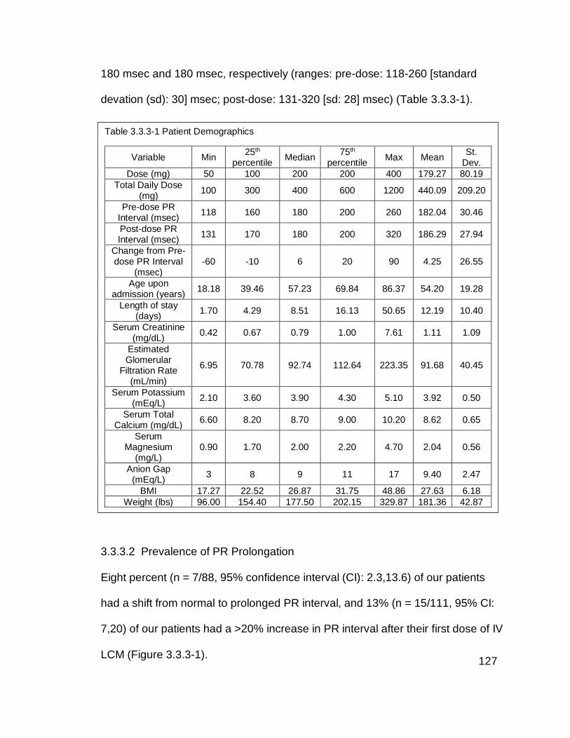
127
180 msec and 180 msec, respectively (ranges: pre-dose: 118-260 [standard
devation (sd): 30] msec; post-dose: 131-320 [sd: 28] msec) (Table 3.3.3-1).
3.3.3.2 Prevalence of PR Prolongation
Eight percent (n = 7/88, 95% confidence interval (CI): 2.3,13.6) of our patients
had a shift from normal to prolonged PR interval, and 13% (n = 15/111, 95% CI:
7,20) of our patients had a >20% increase in PR interval after their first dose of IV
LCM (Figure 3.3.3-1).
Table 3.3.3-1 Patient Demographics
Variable Min 25th
percentile Median
75th percentile
Max Mean St.
Dev.
Dose (mg) 50 100 200 200 400 179.27 80.19
Total Daily Dose (mg)
100 300 400 600 1200 440.09 209.20
Pre-dose PR Interval (msec)
118 160 180 200 260 182.04 30.46
Post-dose PR Interval (msec)
131 170 180 200 320 186.29 27.94
Change from Pre-dose PR Interval
(msec) -60 -10 6 20 90 4.25 26.55
Age upon admission (years)
18.18 39.46 57.23 69.84 86.37 54.20 19.28
Length of stay (days)
1.70 4.29 8.51 16.13 50.65 12.19 10.40
Serum Creatinine (mg/dL)
0.42 0.67 0.79 1.00 7.61 1.11 1.09
Estimated Glomerular
Filtration Rate (mL/min)
6.95 70.78 92.74 112.64 223.35 91.68 40.45
Serum Potassium (mEq/L)
2.10 3.60 3.90 4.30 5.10 3.92 0.50
Serum Total Calcium (mg/dL)
6.60 8.20 8.70 9.00 10.20 8.62 0.65
Serum Magnesium
(mg/L) 0.90 1.70 2.00 2.20 4.70 2.04 0.56
Anion Gap (mEq/L)
3 8 9 11 17 9.40 2.47
BMI 17.27 22.52 26.87 31.75 48.86 27.63 6.18
Weight (lbs) 96.00 154.40 177.50 202.15 329.87 181.36 42.87
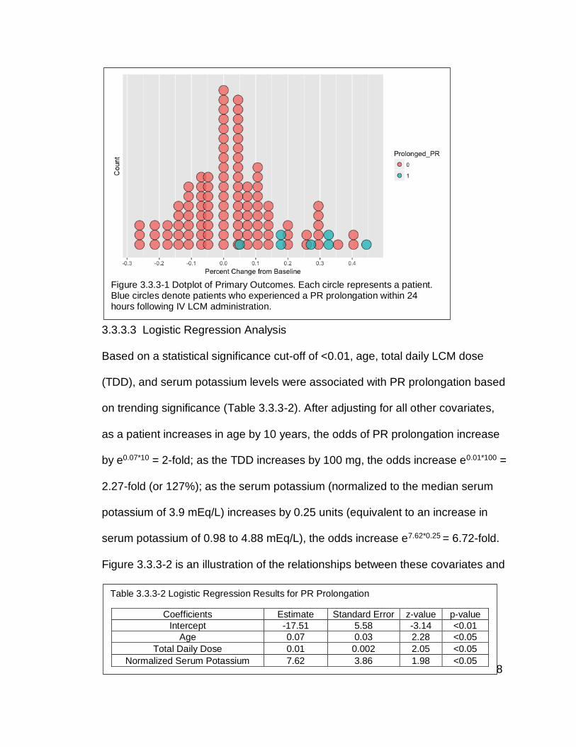
128
3.3.3.3 Logistic Regression Analysis
Based on a statistical significance cut-off of <0.01, age, total daily LCM dose
(TDD), and serum potassium levels were associated with PR prolongation based
on trending significance (Table 3.3.3-2). After adjusting for all other covariates,
as a patient increases in age by 10 years, the odds of PR prolongation increase
by e0.07*10 = 2-fold; as the TDD increases by 100 mg, the odds increase e0.01*100 =
2.27-fold (or 127%); as the serum potassium (normalized to the median serum
potassium of 3.9 mEq/L) increases by 0.25 units (equivalent to an increase in
serum potassium of 0.98 to 4.88 mEq/L), the odds increase e7.62*0.25 = 6.72-fold.
Figure 3.3.3-2 is an illustration of the relationships between these covariates and
Figure 3.3.3-1 Dotplot of Primary Outcomes. Each circle represents a patient. Blue circles denote patients who experienced a PR prolongation within 24 hours following IV LCM administration.
Table 3.3.3-2 Logistic Regression Results for PR Prolongation
Coefficients Estimate Standard Error z-value p-value
Intercept -17.51 5.58 -3.14 <0.01
Age 0.07 0.03 2.28 <0.05
Total Daily Dose 0.01 0.002 2.05 <0.05
Normalized Serum Potassium 7.62 3.86 1.98 <0.05

129
PR interval prolongation. The cases (blue circles) tended to be either on or above
the median TDD, on or above the median serum potassium, and older in age.
On the other hand, a shorter baseline PR interval and current/former
smoking status are associated with a baseline-adjusted increase in PR interval
>20% following IV LCM (Table 3.3.3-3). After adjusting for all other covariates,
as pre-dose PR interval increases by ten ms, the odds of having a >20%
increase from baseline PR interval increases by a factor of e-0.07*10 = 0.5 (or
decrease by 50%); if patient is a current or former smoker, the odds increases by
Table 3.3.3-3 Logistic Regression Results for PR Increase >20%
Coefficients Estimate Standard Error z-value P-value
Intercept 8.19 2.98 2.75 <0.01
Pre-dose PR Interval -0.07 0.02 -3.48 <0.001
Current/Former Smoker 2.50 1.06 2.36 <0.05
Figure 3.3.3-2 Relationships between Age, Total Daily Dose (TDD) of LCM, Median-Normalized Serum Potassium, and PR Prolongation. The size and color of the circles denote the age (i.e. larger circle, older age) and PR prolongation (i.e. blue = shift from normal to prolonged PR interval), respectively.

130
a factor of e2.50 = 12.2. Figure 3.3.3-3 depicts the binomial regression of the event
of a >20% increase from pre-dose PR interval on its two covariates. The
regression is less steep for current/former smokers, which suggests that the odds
of a >20% baseline-adjusted increase stays relatively high for current/former
smokers compared to a patient who has never smoked. For example, at the
same baseline PR interval of 150 msec, the odds of an event occurrence are
approximately 0.4, whereas for a nonsmoker, they close to 0.
3.3.4 Discussion
Intravenous LCM is known to be an effective and well-tolerated treatment for
seizures in hospitalized adult and pediatric patients (Christoph Kellinghaus,
Berning, and Besselmann 2009; Luk et al. 2012; Arkilo, Gustafson, and Ritter
2016; Ngampoopun et al. 2018; Welsh et al. 2017). While several studies have
suggested that LCM is a relatively safe ASD, there are a growing number of
reports describing its adverse effects on cardiac conduction. A recent (November
Figure 3.3.3-3 Pre-Dose PR Interval on Event Occurrence and the Effect of Smoking Status

131
2018) FDA alert described new prescribing information for LCM which now
contains additional serious warnings about cardiac arrhythmias, including
ventricular arrythmias. Few studies have systematically evaluated LCM effect on
cardiac function in the critical care setting. Our study sought to evaluate the
relationship between IV LCM use and PR interval prolongation in patients in the
ICU. An important finding in our study is that IV LCM use did not cause
significant change in median PR interval when comparing pre- and post-drug
exposure within the first 24 hours of administration. However, we observed that
8% patients had prolonged PR interval and 13% patients had PR interval
increases >20% of baseline PR interval following IV LCM administration. The 8%
of patients who experienced PR prolongation is greater than the previously
reported prevalence of 0.4% in ambulatory patients (Rudd et al. 2015).
One of our goals was to determine if there was a specific subpopulation of
patients who were more likely to experience significant increases in PR interval.
Our logistic regression analyses revealed that older age, higher TDD, and higher
serum potassium levels are weakly associated with PR prolongation. Many of
these covariates are expected. Older age has long been associated with longer
PR intervals, and there is increasing awareness of its increased risk for atrial
fibrillation (Mason et al. 2007; Magnani et al. 2013; Cheng et al. 2015;
Macfarlane et al. 2011). There may be confounding variables that may explain
the relationship between older age and PR prolongation, including heart rate
(Soliman and Rautaharju 2012). In addition, oral and IV LCM has been reported
to increase PR interval in a dose-dependent manner and cause first-degree

132
atrioventricular block in a small number of cases (Rudd et al. 2015; Shaibani et
al. 2009; Krauss et al. 2010; Ramsay et al. 2015; Nizam et al. 2011).
Interestingly, higher serum potassium levels are typically associated with a lower
PR interval (Noordam et al. 2019). Therefore, it would seem counterintuitive that
these patients would be more likely to experience PR prolongation. However, we
did not find a correlation between serum potassium levels and pre-dose PR
intervals in our study sample. Therefore, at least in our sample, serum potassium
levels were associated with PR prolongation independent from baseline PR
interval.
With a p-value cut-off of <0.01, only pre-dose PR interval was a strong
predictor of a >20% increase in PR interval following IV LCM administration. This
seems rather intuitive, since a lower baseline would require a smaller increase to
be considered a case. In contrast, the relationship between smoking status and
PR interval increase was unexpected. The association between smoking status
and a higher odds of PR interval increase >20% of baseline may be due to the
effect of nicotine on cardiac conduction. Nicotine increases AV conduction and
decreases the refractory period of the AV node, and is a risk factor for atrial
fibrillation (Haass and Kiibler 1996; Watanabe 2018). In fact, several
prospective, longitudinal cohort studies (including the Health, Aging, and Body
Composition Study) also found that smoking status was a useful factor in
predicting incident cardiovascular disease with increases in PR intervals (Cheng
et al. 2015; Magnani et al. 2013). There may also be confounding variables that
we were unable to tease out given the small sample size, such as the presence

133
of cardiovascular or respiratory condition, age, and sex. It would have also been
of interest to assess whether these patients were had concomitant use of
varenicline, bupropion, or other smoking cessation drugs.
This study is one of the largest reported series of IV LCM use in critically ill
patients to date and used a systematic protocol to assess PR intervals. The
limitations of our study included the retrospective design, lack of control group,
and small sample size. Furthermore, we lacked information on arrhythmias in our
sample. We were able to obtain new arrhythmia data in the electronic medical
record in 2 out of 7 patients who had PR prolongation and 9 out of 15 patients
who had a PR interval increase >20% from baseline. Of those patients, only 2
had a new cardiac arrhythmia diagnosis (atrial fibrillation, cardiac arrest,
paroxysmal ventricular tachycardia, supraventricular tachycardia, unspecified
atrial fibrillation, or other specified cardiac dysrhythmias). It is unclear whether
these event occurrences are important predictors for cardiac dysrhythmia at this
time. Finally, the absence of a control group prohibited our ability to determine
whether the higher event occurrence is attributable to the drug effect, the
circumstances that lead to ICU admission, and/or the complex interaction of co-
morbidities in this patient population.
3.3.5 Conclusions
The prevalence of PR interval prolongation and of PR interval increase of >20%
of baseline following IV LCM administration in the critically-ill patient population
are 8% and 13%, respectively. These event occurrences are higher than what

134
has been reported in the literature for ambulatory patients taking LCM oral tablets
and more common than expected in healthy volunteer Phase I clinical trials.
Larger, controlled, prospective studies are needed to confirm the prevalence of
these events and impact of clinical variables on PR interval changes.
3.4 Design of a Prospective Observational Study of PR Prolongation in
Critically-Ill Patients on Intravenous Antiseizure Therapy: Focus on
Intravenous Lacosamide
Despite the available evidence-based recommendations for management of
established SE, 12-50% of cases are resistant to current therapies (Malamiri et
al. 2012; Agarwal et al. 2007; W. B. Chen et al. 2011; U. Misra, Kalita, and
Maurya 2012; Lyttle et al. 2019; Dalziel et al. 2019; Gujjar et al. 2017;
Mundlamuri et al. 2015; Nene et al. 2019). My long-term goal is to find a safe
drug for the treatment of established SE. Fortunately, since the its FDA approval,
IV LCM has shown promise in the acute treatment of seizures and refractory SE.
Its neuroprotective properties, lack of significant drug-drug interactions and lower
risk for systemic complications make it a particularly attractive agent for the
treatment of seizure emergencies and use in critically-ill patients.
The development path for potential approval of IV LCM for the treatment
of seizure emergencies includes conducting clinical trials demonstrating its safety
and effectiveness. However, its use in established SE is limited by the lack of
strong evidence (i.e. prospective, blinded, randomized comparative or
noninferiority trials) and the concerns for its safety in patients with cardiac

135
dysfunction. Therefore, there remains a need to further evaluate the safety of IV
LCM, with particular focus on cardiac rhythmicity. The following proposed study
has a primary objective of comparing the prevalence of PR prolongation following
IV LCM administration in critically-ill patients to matched controls. The results of
this study will help address concerns for its use in the critically ill population
and/or seizure emergencies.
3.4.1.1 Study Rationale
The retrospective study revealed that patients in critical-care who have received
at least one dose of IV LCM are more likely to experience PR interval
prolongation compared to healthy individuals and ambulatory patients with
epilepsy who have taken oral LCM. The major limitations from my retrospective
study is the lack of a control group and missing data in the medical charts.
Without a control group, I was unable determine whether the increased
prevalence of PR interval prolongation was due to IV LCM use alone or to the
circumstances of the critically-ill population (e.g. co-morbidities or reason for ICU
admission). Without data on new arrhythmia diagnoses, I was unable to
determine the clinical significance of PR prolongation following IV LCM
administration. Thus, a prospective observational study can be conducted to
address these issues.

136
3.4.1.2 Study Objective
The primary objective of this study is to compare the proportion of critically-ill
patients who develop PR prolongation within 24 hours immediately following IV
LCM administration to the proportion of critically-ill patients matched by
admission date, ICU, age, and sex who were administered an IV antiseizure drug
(excluding LCM).
3.4.1.3 Study Population
All patients (aged 18-89 years) admitted into either a cardiac, medical, or
neurological ICU following admission at a tertiary care hospital will be screened
over a ten year period. Consent and enrollment will occur if it is likely that a
patient will require an IV antiseizure drug (i.e. fosphenytoin, phenytoin, valproic
acid, levetiracetam, brivaracetam, LCM, or lamotrigine) or if the patient received
one 24 hours prior to enrollment (e.g. if drug needed to be administered quickly
in an emergent situation). Patients must have at least one PR interval reading
within 24 hours before and after initial antiseizure drug (and/or IV LCM)
administration.
3.4.1.4 Study Design
A prospective observational study will be performed. Control patients will be
matched to IV LCM patients by admission date, ICU, age (+/- 5 years), and sex
to a ratio of 3:1 (control patients to IV LCM patients). Control patients are defined
as patients who required at least one IV antiseizure drug and did not receive IV

137
LCM during the studied hospital admission. Oftentimes, a patient may have had
at least one IV antiseizure drug before and/or after requiring the use of IV LCM.
For those patients, antiseizure drugs used within 24 hours prior to and after IV
LCM administration will be recorded for covariate testing. Control patients will be
matched on the number of antiseizure drugs prior to IV LCM administration. For
example, if a patient received IV LCM after trying two other IV antiseizure drugs,
three control patients of the same sex and age, from the same admission date, in
the same type of ICU, who have also received three antiseizure drugs will be
matched. The PR intervals prior to and after the use of the third antiseizure drug
will be recorded and used for statistical analyses. After the study period, a data
extraction team will be utilized to pull data of interest from electronic medical
records.
3.4.1.5 Sample Collection
All of the following will be extracted from the electronic medical records: age
upon admission, sex, race, weight, body mass index (BMI), length of stay (LOS),
condition on discharge (alive/expired), serum creatinine (Scr), glomerular
filtration rate (GFR), blood urea nitrogen, glucose, smoking status (never,
current, former), serum electrolytes (potassium, sodium, chloride, calcium,
phosphorus, magnesium, bicarbonate), anion gap, dose, total daily dose (TDD),
pre-dose PR interval, post-dose PR interval, new cardiac arrhythmias,
concomitant medications (drugs known to cause PR interval prolongation or
cardiac disturbances, strong and moderate CYP2C9 inhibitors, strong and

138
moderate CYP3A4 inhibitors, and smoking cessation drugs), and antiseizure
drugs used within 24 hours prior to and following the first dose of IV LCM (or
antiseizure drug in the equivalent sequence of IV LCM in matched patient).
3.4.1.6 Data Analysis Plan
The longest PR interval before and after IV antiseizure drug and/or IV LCM
administration will be used for statistical analyses. PR interval prolongation will
be defined as a shift from a normal (200 millisecond [ms]) to prolonged (>200
ms) PR interval. Proportions of PR interval prolongation and their respective 95%
confidence intervals (CI) will be calculated using R (Version 3.4.3). Continuous
demographic and baseline variables such as sex, age, weight, laboratory values
(i.e. basic metabolic panel), and number of previous antiseizure drugs within 24
hours prior to IV LCM treatment will be tested between treatment groups using a
two-sample t-test or Mann-Whitney test; categorical variables such as sex, race,
primary diagnosis, and smoking status will be tested using a Chi-square test. If
any of the subgroups are expected to be less than 5 in count, Fisher’s exact test
will be used.
The distribution of all predictors will be examined visually; those that are
heavily skewed will be log-normalized, while all others will be normalized to the
median value. Missing values for continuous predictors will be imputed with the
median value. Missing values for factors will be left blank. Logistic regression
analysis ( where is the probability of an event
occurrence, is the odds of an event occurrence, 0 is an intercept, I is the

139
coefficient of the predictor) will be used, with PR interval prolongation coded as
“1”. Stepwise covariate method will be used to determine significant covariates
with a forward-inclusion criterion of p<0.05 and a backwards-inclusion criterion of
p<0.01. Selection criteria during the model development process will be based on
changes in Akaike’s Information Criterion, residual unexplained variability
(deviance), and parameter estimates and their standard errors.
3.4.1.7 Challenges and Limitations
One of the biggest challenges noted from the initial retrospective study is the
small number events (PR interval prolongation). This affected my ability to search
for significant predictors. To offset this, I can lengthen the time over which patient
data will be collection period and recruit more tertiary hospital centers within the
same health system (to minimize system-wide differences in practice). Another
challenge is missing data in the electronic medical record. It was found to be
relatively common for screened patients to be missing either a pre- or post-dose
PR interval, laboratory values, and/or new diagnoses of arrhythmias. It was also
difficult to determine whether these data are missing data completely at random,
missing at random, and missing not at random. For example, are patients with
missing arrhythmia diagnosis data arrhythmia-free (missing not at random), or
was the arrhythmia not charted correctly (missing at random or missing
completely at random), or were neither of possibilities listed above not true but
data are somehow missing (missing completely at random). To minimize this
limitation, ICU staff can be trained to collect all data of interest for all patients
140
who receive IV antiseizure drugs, especially PR intervals and arrhythmia
diagnosis data.
3.4.1.8 Expected Results and Alternative Approaches
I expect that the proportion of critically-ill patients who experience PR
prolongation following IV LCM administration will not be statistically different from
those who do not receive IV LCM. It is possible that the co-morbidities and
complications that lead a patient to intensive care may contribute cardiovascular
and autonomic stress, resulting in a longer PR interval. This is reflected in the
observed high average PR interval at baseline in my retrospective study and that
which was reported by Luk et al (Luk et al. 2012). Given these assumptions, I
expect that the proportions of PR prolongation in critically-ill patients will be
statistically different (and greater) than those from healthy volunteers or
ambulatory patients with epilepsy. An alternative approach to answering the
same question would be to conduct larger retrospective study and include
matched controls as mentioned above. However, the challenge with retrospective
studies is that one would have less control over what data will be available in the
medical records. Finally, given the medically-complex circumstances, ethical
considerations limit the study of IV LCM in critically-ill patients who have
conditions for which IV LCM is not indicated, in those who do not need an IV
antiseizure drug, and in those whose hospitalists would not use IV LCM as first
choice therapies in.

141
CHAPTER 4
DEVELOPMENT OF AN INVESTIGATIONAL INTRAVENOUS
FORMULATION OF TOPIRAMATE FOR THE TREATMENT OF
REFRACTORY STATUS EPILEPTICUS

142
4.1 Introduction
Topiramate is a drug used for focal onset or generalized tonic-clonic seizures
and for adjunctive treatment for seizures associated with Lennox-Gastaut
Syndrome for patients at least 2 years of age (Chapter 1.2.1.3). An intravenous
formulation in the development phase and could be beneficial as an adjunctive
treatment for SE based on its multiple mechanisms of action and low potential for
drug-drug interactions as described in more detail below. The objective of my
work presented here was to characterize the pharmacokinetics and
pharmacodynamics of IV TPM in dogs. The specific aims were to 1) characterize
the pharmacokinetics of IV and oral TPM, 2) describe changes in iEEG activity,
and 3) simulate dosing regimens that would have the highest likelihood of
achieving plasma concentrations associated with SE response to inform the
design of a clinical trial in canine SE. In this chapter, I will provide a brief review
of topiramate and a rationale for the development IV TPM for the treatment of
SE, followed by published manuscript which describes my research.
4.2 Topiramate
4.2.1 Physicochemical Properties
Topiramate (molecular formula C12H21NO8S) is a sulfamate-substituted fructose
analog with a molecular weight of 339.36 g/mol and a topological polar surface
area of 124 A2 (National Center for Biotechnology Information 2019c). With a
logP of -0.8, its water solubility is 9.8 mg/mL, and is soluble in most alkaline
solutions (pH 9-10) containing sodium hydroxide/phosphate and freely soluble in
acetone, dimethylsulfoxide, and ethanol. Its saturated solution has a pH of 6.3.

143
Taken together, these properties suggest TPM is highly soluble in neither water
nor lipids, and transport across biological membranes is likely not by passive
diffusion.
4.2.2 Known Mechanisms of Action
Although its precise antiseizure and migraine prophylaxis mechanisms are
unknown, preclinical studies suggest that at pharmacologically relevant
concentrations, topiramate inhibits voltage-gated sodium channels, enhances
activity at GABAA receptors subtypes, blocks AMPA/kainate glutamate receptors,
and antagonizes carbonic anhydrase isozymes II and IV (TOPAMAX [package
insert] 2019). Furthermore, there is evidence of neuroprotective properties in
animal models of limbic SE, methylphenidate-induced toxicity, hypoxia-ischemia,
and stroke (Niebauer and Gruenthal 1999; Motaghinejad et al. 2017; Schubert et
al. 2005; Liu et al. 2004; Cha et al. 2002). More specifically, these studies
illustrated that TPM reduced neuronal cell death in the area of the brain most
susceptible to seizures and improved cognitive function. The multiple
mechanisms of action and neuroprotective properties of TPM make it an
attractive agent for treating SE.
4.2.3 FDA-Approved Indications and Marketed Formulations
Topiramate is indicated for monotherapy and adjunctive therapy in patients at
least 2 years of age with partial onset or primary generalized tonic-clonic
seizures, for patients at least 2 years of age with seizures associated with

144
Lennox-Gastaut syndrome, and for migraine prophylaxis in patients 12 years of
age or older (TOPAMAX [package insert] 2019). Its marketed formulations are
limited to tablets, immediate-release sprinkle capsules, and extended-release
capsules. Extemporaneous compounding of an oral suspension using the
immediate-release tablets can be made using a mixture of Ora-Sweet and Ora-
Plus (Allen 2017).
4.2.4 Clinical Pharmacokinetics
Topiramate is well-absorbed after oral administration (TOPAMAX [package
insert] 2019). An average peak plasma concentration of 27 g/mL occurs
approximately 2 hours following 400 mg multiple oral dose administration every
12 hours (FDA CDER 2004). Clinically relevant plasma concentration range of up
to 33 g/mL (FDA CDER 2004). TPM is 15-41% plasma protein bound, and its
mean elimination half-life is approximately 21 hours (TOPAMAX [package insert]
2019). TPM exhibits dose-proportional PK over 100-400 mg dose range (FDA
CDER 2004). In 19 studies of human PK and PD submitted by the sponsor, TPM
was found not to be extensively metabolized (FDA CDER 2004). The major route
of elimination is by renal excretion, with 80% excreted unchanged in 24 hours
(FDA CDER 2004). However, when changed from PHT or CBZ + TPM therapy to
TPM monotherapy, TPM oral clearance was reduced by 50% (Bourgeois 2007;
R. Sachdeo et al. 2002; R. C. Sachdeo et al. 1996). As a result, two-fold lower
plasma TPM concentrations have been observed (Contin et al. 2013; Yamamoto
et al. 2017). Although the enzymes responsible for its metabolism have not been

145
fully characterized, it is possible that the increase in oral clearance may be due to
increased expression of intestinal efflux transporters like P-glycoprotein (Wang-
Tilz et al. 2006; Atasayar et al. 2016).
In dogs, 90% of TPM is excreted unchanged over 96 hours (Caldwell 2005).
However, TPM is metabolized more extensively in dogs compared to people. The
most dominant metabolic pathway was found to be hydrolysis at the 2,3-O-p-
isopropylidene group.
4.2.5 Present State of Knowledge of Topiramate in Status Epilepticus
Cases reported in the literature have suggested that TPM was well-tolerated and
effective in stopping more than 50% of cases with different types of SE (Shorvon
and Ferlisi 2012; Wasterlain and Chen 2008). The first case reports of enteral
TPM used in adult refractory and super-refractory SE were published in 2002 and
2003 (Reuber, Evans, and Bamford 2002; Towne et al. 2003; Bensalem and
Fakhoury 2003). After these reports, much of the literature on TPM use in
refractory and super-refractory SE were from use in pediatrics, which showed
termination of SE in 77% (17/22) of cases (Kahriman et al. 2003; Perry, Holt, and
Sladky 2006; Blumkin et al. 2005; Akyıldız and Kumandaş 2011). Since then, a
few retrospective chart analyses have been published and one prospective open-
label non-randomized clinical trial (Stojanova and Rossetti 2012; W. Kim et al.
2011; Synowiec et al. 2011; Hottinger et al. 2012; Asadi-Pooya et al. 2015). In
total, the retrospective studies reported success in terminating refractory SE in

146
70% cases (61/87). However, there are a number of confounders, including the
number of other ASDs administered, the place in SE management where TPM
was used (e.g. administration as the third ASD versus sixth ASD), or the effect of
the combination of medications rather than TPM alone (Hottinger et al. 2012;
Synowiec et al. 2011). In addition, in many of these studies, TPM tablets were
crushed to a powder and mixed with water (Towne et al. 2003; Synowiec et al.
2011). Given that TPM is not highly soluble in water, it is possible that the lack of
clinical response may be due to limited bioavailability of this “formulation.” In the
prospective study, TPM was administered as an adjunctive therapy via
nasogastric tube 30 mins after administration of second-line therapy (phenytoin)
(Asadi-Pooya et al. 2015). Five of the twenty (25%) patients successfully
responded (SE termination within 24 hours following TPM introduction without
modification of concomitant ASDs). Although prospective, this study lacks a
control, and it is difficult to distinguish whether seizures stopped due to the
addition of phenytoin, topiramate, or both. Although there is a lack of
randomized, double-blinded, controlled trials evaluating TPM safety and
effectiveness for either refractory or super-refractory SE, the enteral TPM
appears to show promise in the treatment of SE.
4.2.6 Rationale for Developing Intravenous Topiramate for Treatment of SE
The multiple mechanisms of action of TPM, especially the potentiation of GABA-
ergic inhibition via a benzodiazepine-insensitive pathway and antagonization of
glutaminergic AMPA/kainate receptors (White et al. 2000), would be particularly

147
useful when considering SE pathophysiology. That is, TPM has the potential to
be effective after the internalization of benzodiazepine-sensitive GABAA
receptors and increased externalization of AMPA/NMDA receptors in the
synaptic cleft.
Considering all of its potential benefits, the use of TPM for acute seizure
emergencies like SE is limited by its route of administration. Despite an oral
absolute bioavailability of approximately 100%, peak plasma concentrations are
achieved at approximately two hours following a solid oral dose (Clark, Kriel,
Leppik, White, et al. 2013a; Clark, Kriel, Leppik, Marino, et al. 2013; Cipla USA
2017). Although the time to peak concentration following the typical TPM
preparation comprised of crushed TPM tablets in water is unknown, the earliest
time to SE termination following enteral TPM was twelve hours (Towne et al.
2003; Stojanova and Rossetti 2012), with the highest cumulative response rate at
72 hours (Hottinger et al. 2012; Stojanova and Rossetti 2012; Synowiec et al.
2011; W. Kim et al. 2011; Bensalem and Fakhoury 2003). Further, these results
are likely confounded by the various factors, including inconsistent doses and
titration schedules, lack of controls, and possible disrupted gastrointestinal
motility due to SE or other acute co-morbidities (Deane et al. 2019). Therefore,
an intravenous formulation would enable a more rapid loading of TPM that would
bypass the need for enteral movement and the variability in gastrointestinal
absorption. This would allow for attainment of higher peak concentrations in a
shorter length of time compared to the same dose given enterally in this patient
population. Furthermore, an intravenous route of administration would remove

148
the need for an enteral feeding tube (e.g. nasogastric or gastrostomy tubes), and
enable TPM use earlier on in the management of SE and prior to the need for
mechanical ventilation (i.e. third-line therapy).
4.3 Intravenous Topiramate: Pharmacokinetics and Effect on
Electroencephalograph Activity in Dogs with Naturally-Occurring Epilepsy1
4.3.1 Introduction
Status epilepticus (SE) is defined as a condition characterized by abnormally
prolonged seizures that can lead to long-term consequences, including
permanent neuronal injury (Trinka et al. 2015). SE has been reported to have an
incidence between 2.5-59% in dogs with idiopathic epilepsy, and 32% in dogs
with secondary epilepsy (Saito et al. 2001; Platt and Haag 2002; Monteiro et al.
2012). In dogs that have had at least one episode of SE, overall mortality rates
(primarily from euthanasia) were 32%-38% (Saito et al. 2001; Zimmermann et al.
2009). In humans, SE occurs with an incidence between 0.04-0.06% in the
United States, and its complications result in an overall mortality rate of 22%
(DeLorenzo et al. 1995). While benzodiazepines are the standard first line of care
for SE in both dogs and humans (Michael Podell 1995; Brophy et al. 2012),
approximately one third fail to respond to first line therapy (Hocker et al. 2013).
There remains a need for safe alternatives for early and rapid first- and/or
second-line therapy of SE to reduce the probability of recurring seizures,
1 This chapter has been published by Frontiers Media SA as: Intravenous Topiramate: Pharmacokinetics and Effect on Electroencephalograph Activity in Dogs with Naturally-Occurring Epilepsy, Frontiers in Veterinary Science, (2016) 3:107. doi: 10.3389/fvets.2016.00107. Reproduction rights for this dissertation were not required by this publisher.

149
minimize associated complications, and improve patient outcomes.
One of the barriers to developing new treatments for SE is the
experimental model used to find and evaluate investigational therapies.
Oftentimes in rodent models, epilepsy is induced by chemical or electrical insult
and may not be truly representative of epilepsy pathophysiology (Pitkänen and
Mcintosh 2006). Dogs with naturally-occurring epilepsy have been proposed as
appropriate models to examine new antiepileptic therapies prior to human trials
(Coles et al. 2015). Canine epilepsy is strikingly similar to the human condition in
both disease presentation and response to treatment. Holliday et al.
demonstrated that intracranial electroencephalograms (EEGs) of dogs and
humans during focal onset seizure are indistinguishable (Holliday, Cunningham,
and Gutnick 1970). Moreover, studies of antiseizure drugs (ASDs), such as
fosphenytoin and levetiracetam, have shown comparable efficacy in both dogs
and humans for SE (Coles et al. 2015; Hardy et al. 2012).Given these
similarities, assessing new therapies for SE in dogs will facilitate drug
development and increase the chance of successful translation for both canine
and human SE.
Among the newer ASDs with injectable formulations, topiramate (TPM)
is an attractive candidate for evaluation in the treatment of SE. TPM is a second-
generation, broad-spectrum ASD that inhibits of voltage-gated sodium channels
and enhances gamma-aminobutyrate (GABA) activity at specific GABAA receptor
subtypes (Topiramate [package insert] Cipla 2017). TPM also has mechanisms
of action that differ from those exhibited by current therapies, including

150
antagonizing AMPA/kainate glutamate receptors, and inhibiting specific carbonic
anhydrase isozymes.
Our group has studied the pharmacokinetics (PK) of a novel intravenous
(IV) TPM formulation in humans. However, the pharmacokinetics of IV TPM and
its effect on EEG has not been characterized in dogs. This study evaluated TPM
in dogs with naturally occurring epilepsy to characterize its PK and effect on EEG
activity. The aims of this study were to 1) characterize TPM PK following an IV
and oral dose of TPM, 2) evaluate effect of TPM on intracranial EEG (iEEG)
features, and 3) simulate doses to attain target concentrations of 20-30 µg/mL.
4.3.2 Methods
4.3.2.1 Study Animals and Safety Monitoring
Five dogs with naturally-occurring epilepsy were used in this study. Three of the
dogs have uncontrolled seizures despite being on antiseizure maintenance
regimens. Approval was obtained through the Institutional Animal Care and Use
Committee of the University of Minnesota prior to the initiation of the study. The
dogs were housed at the University of Minnesota’s Veterinary College. Each dog
was previously implanted with a device which wirelessly transmits iEEG
recordings (Davis et al. 2011; Coles et al. 2013). Dogs were monitored
continuously via iEEG and video for five to ten days to obtain baseline data, and
throughout the study for vomiting, diarrhea, and lethargy prior to and for 90
minutes after drug administration, and at each blood sampling time. In the event
of a seizure emergency (seizure lasting >5 minutes) or repetitive seizures (2+

151
seizures within 1 hour, or 3+ seizures within 4 hours), the on-call veterinarian
received an automated text message and confirmed the seizure activity using
remote video monitoring. The rescue therapy protocol consisted of midazolam 12
mg administered as a single IM dose.
4.3.2.2 Study Drug
For this study, a stable-isotope labeled TPM compound containing six 13C,
resulting in a mass 6 units greater than the unlabeled molecule was used for the
IV formulation (10 mg/mL in 10% Captisol®). This formulation was manufactured
by the University of Iowa under Good Manufacturing Practices and has been
licensed to Ligand/CuRx Pharmaceuticals. Unlabeled TPM tablets (25 mg)
purchased from the University of Minnesota Veterinary Pharmacy (Cipla USA
Inc) were used for the oral treatment arm. Using a labeled IV formulation and
non-labeled oral tablets allowed us to simultaneously administer both
formulations and characterize TPM pharmacokinetics by each route. This
approach also reduces inter-occasion variability caused by dosing on different
days and/or times (Marino et al. 2012).
4.3.2.3 Dose Rationale
Based on reports of doses associated with efficacy in human SE, we estimated
corresponding target plasma TPM concentrations of 20-30 µg/mL. A previous
single IV dose study in one dog reported TPM concentrations from which we
calculated an apparent volume of distribution (Vd) of 0.6 L/kg (Streeter et al.

152
1995). Using this Vd, we estimated that IV doses of 10 and 20 mg/kg would
produce initial concentrations (C0) of approximately 16 and 32 μg/mL,
respectively.
4.3.2.4 Study Design
Low dose IV/oral TPM study: Four dogs were used in this study (ID 1-4, Table 1).
Two of the four dogs were on ASD maintenance regimen including phenobarbital
(PB). Each dog was fasted overnight prior to receiving a 10 mg/kg dose of stable-
labeled IV TPM infused over 5 minutes. One hour following the IV bolus, each
dog also received a 5 mg/kg dose of unlabeled oral TPM. This delay in oral
administration was by design to allow evaluation of the IV dose on iEEG for one
hour after dosing. Blood samples were collected from an indwelling catheter prior
to dosing and at 0.083, 0.25, 0.5, 0.75, 1, 1.5, 2, 2.5, 3, 4, 6, 8, and 9 hours
following the IV bolus.
High dose IV TPM study: Three dogs were used in this study (ID 3-5, Table 1).
One dog was on PB maintenance therapy. Each dog was fasted overnight prior
to receiving a 20 mg/kg dose of stable-labeled IV TPM infused over 5 minutes.
Blood samples were collected from an indwelling catheter prior to dosing and at
0.083, 0.25, 0.5, 0.75, 1, 1.5, 2, 2.5, 3, 4, 6, 8, and 9 hours following the IV bolus.

153
Diazepam (DZP) positive control: IV diazepam (0.5 mg/kg) was administered to
two dogs who were having uncontrolled seizures (ID 1 & 2) during an interictal
period as a positive control as it has been shown to elicit iEEG change.
4.3.2.5 Plasma TPM Measurements
A 250 µL sample of whole blood was aliquoted for TPM analysis. The remaining
blood was placed on ice and plasma was separated. All samples were
immediately frozen (-20 C) until analysis. Each dog was fed no sooner than 2
hours after the oral dose. A high-performance liquid chromatography-mass
spectrometry (HPLC-MS) method developed and validated at the Center for
Orphan Drug Research was used to measure TPM concentrations in dog
plasma. Seven calibration standards (run in triplicate) and nine quality control
standards (low, medium, high run in triplicates) were prepared in plasma. Study,
calibration, and quality control samples were extracted using methyl tert-butyl
ether. TPM and stable-labeled TPM were analyzed using the Hewlett Packard
Agilent 1100 Model G1946 liquid chromatography mass spectrometry detection
system and Agilent ChemStation software. The analytes were separated using a
Zorbax C18 column (150 mm x 3.0 mm, 3 µm) and the mobile phase consisted of
an ammonium acetate buffer and methanol. The quantization was performed
using the selective ion monitoring in the negative mode, with deuterated TPM
(d10) as the internal standard. The mass-to-charge ratios were 338 m/z and 244
m/z for TPM and stable-labeled TPM, respectively. The calibration curves were
linear (r 2 = 0.998) in the concentration range of 0.05–50 µg/mL for TPM and

154
0.05-10 µg/mL for stable-labeled TPM in plasma. The limit of detection and
quantitation were 0.05 ng/mL and 0.05 µg/mL, respectively. The precision for
both TPM and stable-labeled TPM ranged from 3-6%, and accuracy values were
between 95-114% and 86-105%, respectively.
4.3.2.6 Pharmacokinetic Analysis
Topiramate concentration-time data were analyzed using non-compartmental
analysis (Phoenix WinNonLin, version 6.4, Pharsight Corporation, Mountain
View, CA, USA). Pharmacokinetic parameter values included maximum
concentration (Cmax), time at which maximum concentration is achieved (tmax),
elimination rate half-life (t1/2), and the area under the time-concentration curve
(AUCINF) calculated using the equation 𝐴𝑈𝐶 = ∫ 𝐶𝑝 ∗ 𝑑𝑡𝑡=∞
𝑡=0 (where Cp is the
plasma TPM concentration) and a linear-log trapezoidal method. Oral
bioavailability (F%) was calculated using the equation F (%) =
AUC(oral)∗Dose(IV)
AUC(IV)∗Dose(oral)x 100. Clearance (CL) and Vd were calculated using the
equations 𝐶𝐿 = 𝐷𝑜𝑠𝑒∗𝐹
𝐴𝑈𝐶 and 𝐶𝐿 = 𝑘𝑒 ∗ 𝑉𝑑, respectively, where ke is the elimination
rate constant. Concentration-time profiles were created using GraphPad Prism 7
(Version 7.0a, GraphPad Software, Inc., La Jolla, CA, USA).
PK parameters were also determined using population compartmental
modeling (Phoenix Non-Linear Mixed Effects software, version 1.3, Pharsight
Corporation, Mountain View, CA, USA). First order conditional estimation
extended least squares method was used throughout the model building process.
One- and two-compartment models were evaluated. A proportional error model
155
for between subject variability was used. Both additive and multiplicative error
models for residual variability were evaluated. The best fit model was determined
using visual inspection, goodness of fit plots, weighted residual plots, weighted
sum of squared residuals, Akaike’s Information Criterion, and precision of model
parameters.
The presence of a CYP3A4-inducing co-medication (such as PB) was
evaluated as a covariate for its influence on TPM clearance. The relationship of
the covariate and TPM clearance was modeled by the equation 𝐶𝑙 = 𝑡𝑣𝐶𝑙 ∗ 𝑒𝑑𝐶𝑙 ∗
𝑒𝜂𝐶𝑙, where Cl is the clearance from the central compartment, tvCl is the typical
value of the clearance from the population, dCl is the estimated value of the
inducer effect, and Cl is the between-subject variability of clearance. A
covariate was considered statistically significant if inclusion of the covariate
resulted in a decrease in the objective function value (OFV) of at least 6.64 (p <
0.01, 𝜒2, degree of freedom = 1). The final model was used to simulate of 5-, 10-,
and 15-minute infusions IV TPM at doses ranging from 10-30 mg/kg.
4.3.2.7 Electroencephalographic Analysis
Sixteen electrode channels were continuously sampled at 399.6 Hz. A band-pass
filter was applied to create 6 frequency bands: delta (1-4 Hz), theta (4-8 Hz),
alpha (8-12 Hz), beta (12-25 Hz), low gamma (25-40 Hz), and high gamma (40-
120 Hz). In order to evaluate differences in EEG features, energy of each
electrode within each frequency band was calculated in 1-second intervals by
summing the square of the EEG signal amplitude within the 1 second window.

156
The average energy level was calculated for three 15-minute ranges: starting
from 15-minutes pre-dose to dosing, from dosing to 15-minutes post-dose, and
from 15-minutes post-dose to 30-minutes post-dose. The difference between
averaged energy levels at pre-dose and each post-dose interval were calculated.
P-values were generated by the Kruskal-Wallis test comparing the averaged
energy level from pre-dose to the two averaged energy levels post-dose.
4.3.3 Results
4.3.3.1 Demographics and Adverse Events
Demographics of the dogs are represented in Table 4.2.3-1. No adverse events
were observed for either dose group throughout the course of the study.
Table 4.3.3-1 Animal Demographics
Subject Weight
(kg) Breed Seizure Type Co-medications
1 33 Coonhound Mix Focal and grand
mal seizures
Levetiracetam,
zonisamide, phenobarbital
2 29 Labrador
Retriever Mix Cluster seizures
Levetiracetam,
zonisamide, phenobarbital,
potassium bromide
3 15 Beagle None N/A
4 29 Coonhound Mix None N/A
5 35 Coonhound Mix Focal and grand
mal seizures Phenobarbital
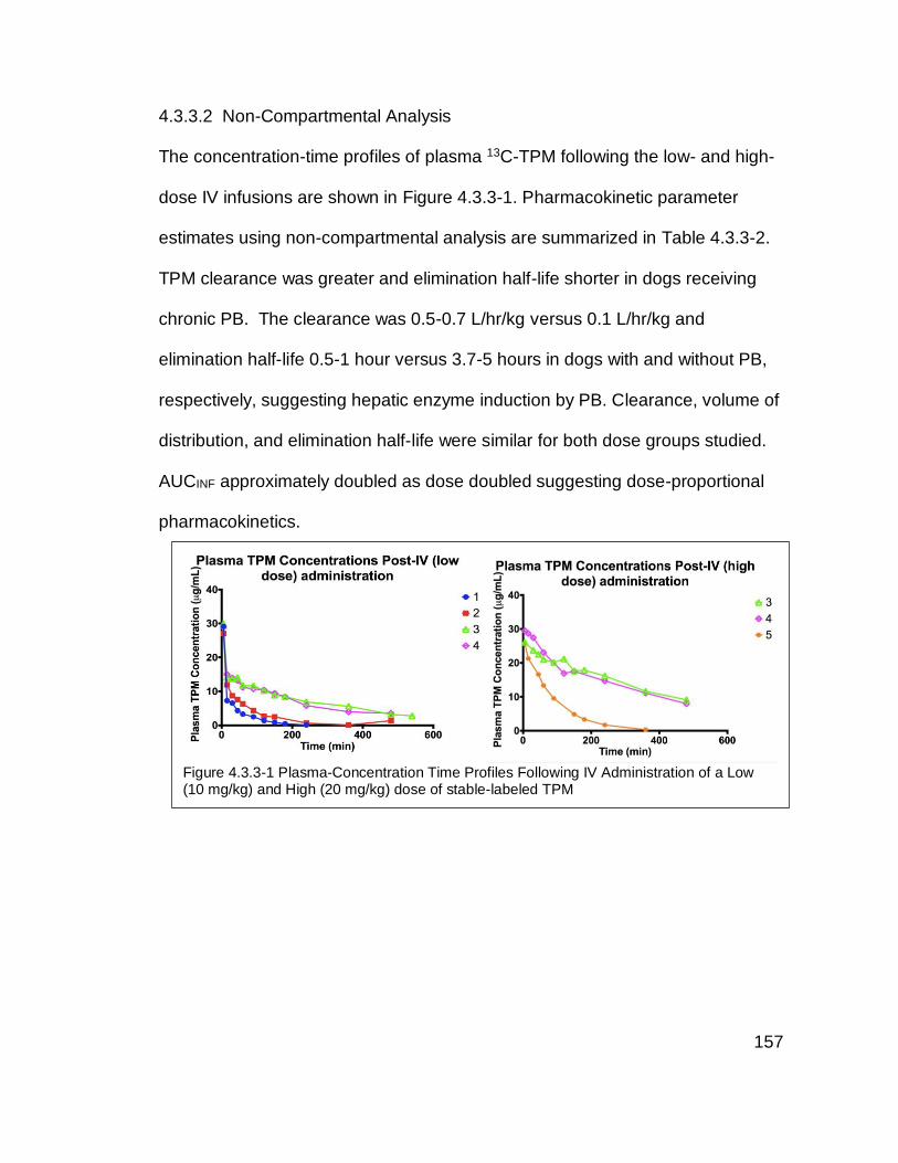
157
4.3.3.2 Non-Compartmental Analysis
The concentration-time profiles of plasma 13C-TPM following the low- and high-
dose IV infusions are shown in Figure 4.3.3-1. Pharmacokinetic parameter
estimates using non-compartmental analysis are summarized in Table 4.3.3-2.
TPM clearance was greater and elimination half-life shorter in dogs receiving
chronic PB. The clearance was 0.5-0.7 L/hr/kg versus 0.1 L/hr/kg and
elimination half-life 0.5-1 hour versus 3.7-5 hours in dogs with and without PB,
respectively, suggesting hepatic enzyme induction by PB. Clearance, volume of
distribution, and elimination half-life were similar for both dose groups studied.
AUCINF approximately doubled as dose doubled suggesting dose-proportional
pharmacokinetics.
Figure 4.3.3-1 Plasma-Concentration Time Profiles Following IV Administration of a Low (10 mg/kg) and High (20 mg/kg) dose of stable-labeled TPM

158
Plasma TPM concentration-time profiles following oral administration are
depicted in Figure 4.3.3-2. Cmax following oral administration ranged between 1.9-
2 μg/mL at 1-1.5 hours (Tmax), with a t1/2 between 1.7-2 hours in the two dogs on
PHB. In the two dogs not on PB, a Cmax of 4.7-5.5 μg/mL at 0.5-1 hour was
observed, with a t1/2 of 4 hours. Individual oral bioavailability ranged between 61-
102%. These results are summarized in Table 4.3.3-2. Similar to the IV
Table 4.3.3-2 Pharmacokinetic parameter estimates generated from non-compartmental analysis following IV administration
ID Group t1/2
(hr)
C1
(µg/mL)
AUCINF_obs
(µg*hr/mL)
V
(L/kg)
CL
(L/hr/kg)
1 LOW 0.47 29.2 13.7 0.50 0.73
2 LOW 0.75 27.1 20.6 0.53 0.49
3 LOW 3.71 30 83.5 0.64 0.12
4 LOW 4.05 28.9 101 0.58 0.1
3 HIGH 4.99 26.1 194 0.74 0.1
4 HIGH 4.52 29.5 176 0.74 0.11
5 HIGH 0.95 25.7 38.4 0.71 0.52
LOW = 10 mg/kg dose. HIGH = 20 mg/kg dose. t1/2: elimination half-life; C1: first measured concentration; AUCINF_obs: observed area under the curve from time 0 to infinity; V: volume of distribution; CL: clearance.
Figure 4.3.3-2 Plasma-concentration time profiles following administration of a single 5 mg/kg TPM oral dose

159
administration, the two dogs on PHB exhibited higher clearance rates, and
consequently, shorter half-lives compared to the two dogs not on PB.
4.3.3.3 Population Compartmental Analysis
A two-compartment model with first-order elimination best fit the TPM
concentration data following IV administration (Figure 4.3.3-2). Parameter
estimates are provided in Table 4.3.3-4. A systematic bias in clearance based on
dose was observed. The inclusion of whether the dog was on an enzyme-
inducing co-medication as a covariate resulted in a decrease in the OFV from the
base model (difference in OFV = 25) and an improvement in the goodness of fit
Table 4.3.3-3 Pharmacokinetic parameter estimates generated from non-compartmental analysis following PO administration
ID t1/2
(hr)
Tmax
(min)
Cmax
(µg/mL)
AUCINF_obs
(µg*hr/mL)
V
(L/kg)
CL
(L/hr/kg)
F
(%)
1 2.02 90 1.92 4.73 2.13 0.73 69.2
2 1.66 60 2.08 10.5 1.16 0.49 102
3 3.98 30 4.73 25.7 0.69 0.12 61.7
4 4.08 60 5.53 36.9 0.58 0.1 73.0
Volume and clearance were adjusted for bioavailability. t1/2: elimination half-life; Tmax: time at peak concentration; Cmax: peak concentration, AUCINF_obs: observed area under the curve from time 0 to infinity; V: volume of distribution; CL: clearance; F: bioavailability.
Figure 4.3.3-3 Observed versus predicted individual and population concentrations

160
plots and precision of parameter estimates. Therefore, the effect of an enzyme
inducer on clearance was included in the final model. The presence of PB is
estimated to affect TPM clearance by a factor of 5.64. Except for peripheral
compartment clearance, all model-fitted parameters were estimated with good
precision with all coefficients of variation below 25%. A multiplicative error model
best described the residual error with an estimate of 15%, which is consistent
with analytical error. Visual predictive check plots (Figure 4.3.3-4) illustrated the
observed data percentiles fall within the 90% (5%-95%) model-predicted
intervals.
Table 4.3.3-4 Pharmacokinetic parameter estimates from a population compartmental analysis following an intravenous TPM (low and high doses)
Model Parameter Estimate Stderr CV%
Fixed Effect tvV (mL/kg)
376 72.4 19.2 ---
tvV2 (mL/kg)
298 56.0 18.7 ---
tvCL (mL/(kg*min))
1.84 0.08 4.52 ---
tvCL2 (mL/(kg*min))
21.0 9.37 44.7 ---
dCL 1.73 0.13 7.66 ---
Random Effect Estimate Stderr RSE% Shrink%
BSVV 0.08 0.02 24.6 9.3
BSVCl 0.02 0.01 53.4 9.18
Residual error, CV%
14.9 1.71 11.5 ---
tvV: Typical value of volume of distribution from central compartment; tvV2: Typical value of volume of distribution from peripheral compartment; tvCL: Typical value of clearance from central compartment; tvCL2: Typical value of intercompartment clearance; dCL: Effect of PB presence on CL; BSV: between-subject variability; CV%: coefficient of variation; RSE%: relative standard error

161
4.3.3.4 Simulation Analysis
Using the final model above, various infusion rates and doses were simulated
(Figure 4.3.3-5). For dogs not on enzyme-inducing co-medications, simulated
time-concentration profiles suggest that a 5-minute infusion of 20 mg/kg would
achieve target concentration range of 20-30 μg/mL at 30-minutes post-dose.
However, in dogs on enzyme-inducing co-medications, a dose between 25-30
mg/kg infused over 5 minutes would be required to attain the same target range
Figure 4.3.3-4 Visual predictive check plots of a population model for dogs not on PB and dogs on PB, respectively. Red and black lines represent the observed and simulated quantiles, respectively.
Figure 4.3.3-5 Simulated Plasma Concentration Time Profiles Across Varying Doses Infused Over 5 Minutes in a dog not on PB and a dog on PB. Here, the yellow band is the desired range, and the black vertical line denotes 30 minutes post-dose.

162
4.3.3.5 Electroencephalographic Analysis
EEG data of good quality were not attained for all animals due to electrical
malfunctioning of the electrodes and/or data cards as some of the devices had
been implanted for up to 5 years. In the dogs with a functioning device for EEG
acquisition, intravenous TPM produced EEG changes shortly after the infusion
which continued in the subsequent 40-60 minutes (Figure 4.3.3-6). Statistically
significant positive energy differences in all 6 frequency bands across all 16
channels were seen comparing the pre-dose time (-15 to 0 mins) to both post-
Figure 4.3.3-6 EEG normalized signals from beta and low gamma frequency bands averaged over 1-minute intervals in one dog
Table 4.3.3-5 Difference between energy levels averaged across low gamma and beta frequency bands before and after IV TPM in one dog
Time Ranges
(min)
Frequency Bands
Pre-
dose
Post-
dose
delta
(1-4 Hz)
theta
(4-8 Hz)
alpha
(8-12
Hz)
beta
(12-25
Hz)
low
gamma
(25-40 Hz)
high
gamma
(40-120
Hz)
(-15,
0)
(0, 15) 16631.6
0*
3997.93
*
1250.46
*
1214.93* 342.61* 265.21*
(-15,
0)
(15,
30)
17765.5
7*
3710.96
*
1127.09
*
972.23* 333.26* 220.43*

163
dose times (0 to 15 mins and 15 to 30 mins) (Table 4.3.3-5). Intravenous DZP
also increased energy in frequencies >4 Hz (most prominently in beta and
gamma frequency bands), and significantly decreased delta frequency energy in
most channels (data not shown).
4.3.4 Discussion
This study is unique in that it evaluated both the pharmacokinetic and
pharmacodynamics aspects of TPM in dogs with naturally occurring epilepsy.
This is also the first study characterizing the effect of PB on TPM
pharmacokinetics in dogs. We observed a 5.6 times greater CL in dogs on PB
compared to those who were not. In addition, the three dogs on PB exhibited
lower peak TPM plasma concentrations following oral dosing. This increase in
clearance suggests that there is induction of hepatic enzymes that metabolize
TPM. This has also been noted in humans, although the effect of enzyme
induction in humans is more modest (~145% increase in clearance) (Clark, Kriel,
Leppik, White, et al. 2013b). To explain this discrepancy, Caldwell et al found
that while 82% of TPM is excreted unchanged in the urine in humans, only 28%
is excreted unchanged in dogs (Caldwell 2005).Phenobarbital is a known inducer
of CYP 3A4, the major enzyme responsible for TPM metabolism. A recent study
evaluating the effects of chronic administration of PB on the pharmacokinetics of
levetiracetam in dogs with epilepsy found similar results (Muñana, Nettifee-
Osborne, and Papich 2015). Potential drug-drug interactions should be taken into
consideration when dosing TPM in both dogs and humans. Dose adjustments

164
are likely needed when TPM is used in conjunction with chronic enzyme-inducing
or enzyme-inhibiting co-medications in dogs.
Although a limitation of the study is the small number of animals used to
construct the population PK model, the utility of the model is to simulate
concentration profiles for dogs both on and not on enzyme-inducing co-
medications. It has been noted that dogs respond to many of the same ASD
therapies as humans. With the use of simulations, we can predict drug exposure
from different dosing strategies and determine the optimal dosing regimen to
attain the same concentrations that are considered therapeutic in human SE, as
we did successfully in dogs with a PK study and a randomized clinical trial with IV
fosphenytoin (Coles et al. 2015; Patterson et al. 2015). Based on case reports of
TPM oral suspensions used to treat refractory SE, our goal target concentration
range was 20-30 µg/mL. Our simulations suggest that these doses should be
used in designing future a clinical trial in canine SE.
The significant changes between pre-dose EEG energy levels and those
up to 30 minutes after IV TPM administration suggest sufficient and timely
diffusion into the brain, suggesting that IV TPM may be a good candidate for
treatment of seizure emergencies. Benzodiazepines in both rodent and dog
model show significant increases in energy in frequencies greater than 4 Hz and
decreases in delta frequency energy (Table 4.3.3-5), which is expected from prior
studies. After IV TPM administration, we also see changes in these frequencies
at 15 minutes. These observations suggest IV TPM may be beneficial for the
treatment of status epilepticus.
165
In conclusion, IV TPM doses of 10 and 20 mg/kg infused over 5
minutes were shown to be safe and tolerable in dogs. Concurrent administration
of PB increased the clearance of TPM approximately 5.6-fold. Simulations
suggest that doses of 20 and 25 mg/kg of IV TPM are necessary to achieve a
target concentration between 20-30 µg/mL in dogs not on PB and dogs on PB,
respectively. A key strength of this study is the use of animals with naturally
occurring epilepsy. The results of this study provide information on optimizing
TPM therapy for future studies of canine SE, which will subsequently guide the
design of IV TPM clinical trials of human SE. Future work includes conducting a
phase II/III efficacy study in canine SE using the dose strategy determined from
the PK modeling results of this study.
Disclosures
Drs. Cloyd and Leppik are paid consultants for CuRx Pharmaceuticals. Dr. Cloyd
also receives payments from a licensing agreement between the University of
Minnesota and Ligand Pharmaceuticals. We confirm that we have read the
Journal’s position on issues involved in ethical publication and affirm that this
report is consistent with those guidelines.
Acknowledgements
We would like to acknowledge the American Kennel Club Foundation,
NIH/NINDS R21-NS072166, and NIH U01-NS073557 (GW) which funded this

166
research. We also acknowledge and thank Andrea Eckert for her care of the
animals and sample collection.
4.4 Development of Intravenous Topiramate for the Treatment of Refractory
Status Epilepticus
There is currently insufficient evidence to guide refractory SE therapy due to the
rarity of the condition and difficulty in the interpretation of findings due to the
complex interaction of drugs used in parallel and co-morbidities at this later stage
of SE. Early results of a multinational, prospective audit of 488 patients with
refractory and super-refractory SE reported that 74% of cases recovered from
RSE, 22% died, and 4% had treatment withdrawn due to futility (Ferlisi et al.
2015). Although anesthetic agents are useful in suppressing seizures, they are
associated with a higher risk of systemic complications death independent of
underlying medical conditions (Sutter et al. 2014). There is an unmet need for
better control of refractory SE, ideally before the need for burst-suppression. The
multiple mechanisms of action and neuroprotective properties of TPM make it an
ideal drug to use when resistance to the recommended antiseizure drugs has
occurred.
Because oral TPM is an FDA-approved drug, the development of IV TPM
has two possible routes: 1) temporary replacement for oral TPM therapy by
showing that the IV formulation can be safe and bioequivalent to an oral product,
or 2) conduct controlled safety and efficacy trials for a new indication, such as
treatment for acute seizures and/or seizure emergencies. Single-doses of 100

167
mg IV TPM infused over 15 minutes has already been shown to be bioequivalent
to 100 mg oral TPM and safe in healthy volunteers and a PK and safety study in
patients with epilepsy or migraines taking oral TPM (Clark, Kriel, Leppik, Marino,
et al. 2013; Clark, Kriel, Leppik, White, et al. 2013b). The next step for this
direction could be to conduct a single- and multiple-ascending dose study to
assess the safety and tolerability of higher doses of TPM and longer exposure of
IV TPM. This information is needed for its development as a temporary
replacement for oral TPM therapy considering patients with epilepsy can be
prescribed at least the maximum recommended daily dose of 400 mg divided into
two doses. For example, in case reports where TPM preparations were used to
treat super-refractory SE, patients were administered up to 1000 mg TPM per
day (Brigo, Bragazzi, and Igwe 2017). In addition, it would be useful to know the
maximum tolerated duration of IV therapy for this same indication. This study
would not replace the need for a safety and effectiveness clinical trial in SE, but
these data would inform the safety of IV TPM for the treatment of seizure
emergencies.
The following proposed studies have a primary objective of characterizing
the safety of higher IV TPM doses and its effectiveness in treating refractory SE
in dogs.
168
4.4.1 IV Topiramate for the Substitution of Oral Topiramate
4.4.1.1 Study Rationale
Multiple dose studies have not been conducted to evaluate the safety of larger
single doses of IV TPM and multiple infusions of IV TPM with respect to infusion
site adverse effects.
4.4.1.2 Study Objective
The primary objective of this study is to characterize the safety and
pharmacokinetics of single- and multiple-ascending doses (SAD/MAD) of IV
TPM. Pharmacokinetic parameters estimated from the SAD studies will be used
to simulate exposures in the MAD studies, and adjust infusion rates if necessary.
The results of these studies will inform labelling recommendations on the
maximum duration of replacement therapy and the maximum tolerated dose that
could be safely infused in situations of emergent seizures.
4.4.1.3 Study Population
Equal proportions of male and non-pregnant and non-breastfeeding female
patients >18 years of age stable on TPM at daily doses between 300-400 mg will
be recruited for the single ascending (SAD, six patients per dose cohort and two
planned cohorts). Patients who are stable on TPM at daily doses between 200-
400 mg will be recruited for the multiple ascending dose studies (MAD, six
patients per dose cohort and three planned cohorts). Patients must not be on any
interacting co-medications (a comprehensive “excluded concomitant medication”

169
will be provided). Being stable on oral TPM is defined as having received the
same dosing regimen for at least the past 4 weeks, and without plans to adjust
the regimen during the study period. The SAD/MAD study can be conducted in
parallel.
4.4.1.4 Study Design
The SAD study will be an open-label study. Patients will receive their half of their
daily TPM dose (150-200 mg) as a stable-labeled infusion over 15 minutes
(stable-labeled IV TPM described in Clark et al 2013). All patients will be
instructed to take the remaining half of their daily dose as oral tablets 12 hours
after study drug infusion, and as prescribed every day after for the remainder of
the 96-hour study period. Infusion sites will be monitored for adverse effects such
as tenderness and swelling. If two of six patients have a DLT, the next cohort will
receive the same dose. If only one patient experiences a DLT, the next cohort
will receive the next higher dose of 200 mg IV TPM infused over 15 minutes. The
study will repeat until three or more people per SAD level have a DLT or 200 mg
IV TPM has been completed. If the 150 mg single dose is determined to be safe,
simulations will be done to predict exposures for the Cohort 4. This will also be
done following the 200 mg single dose.
The MAD (Cohorts 3-5) study will also be an open-label study. The first
MAD cohort (Cohort 3) will receive 100 mg of stable-labeled IV TPM over 15
minutes twice daily for two weeks. In Cohorts 4 and 5, patients will receive half of
their daily dose of IV TPM infused over 15 minutes twice daily for two weeks if

170
the corresponding infusion was determined to be safe during the SAD study. The
study will repeat until three or more people per MAD level have a DLT or a daily
dose of 400 mg IV TPM has been completed.
4.4.1.5 Sample Collection
For the SAD studies, blood samples pre-injection, and at 15- and 30-minutes, 1,
2, 4, 6, 12, 24, 48, 72, 96 hours following IV infusion will be collected to
determine the pharmacokinetics. For the MAD studies, blood samples pre-
injection, and at 1,12, 24, 48, 72, 96, 120, 240, and 336 hours following IV
infusion will be collected to determine the steady-state pharmacokinetics.
Figure 4.4.1-1 IV TPM SAD/MAD Study Schematic
150 mg
IV TPM
200 mg
IV TPM
100 mg
IV TPM x 2
150 mg
IV TPM x 2
200 mg
IV TPM x 2
Simulate
multiple dosing
exposure
Single
Ascending
Dose
Multiple
Ascending
Dose
Simulate
multiple dosing
exposure

171
4.4.1.6 Pharmacokinetic Analysis
Plasma TPM concentration-time data will be analyzed using non-compartmental
analysis and population compartmental modeling approaches. First order
conditional estimation extended least squares method will be used throughout
the model building process. The best fit model will be determined using visual
inspection, goodness of fit plots, weighted residual plots, weighted sum of
squared residuals, Akaike’s Information Criterion, and precision of model
parameters. PK parameters estimated from the SAD cohorts will be used to
predict exposures in the MAD cohorts and to adjust infusion rates, if needed.
Infusion rates may need to be adjusted at higher dose levels to ensure the
maximum concentration (Cmax) does not exceed those observed following oral
TPM dosing. For example, if the Cmax following 200 mg dose is higher than
predicted based on data from previous studies (Clark, Kriel, Leppik, Marino, et al.
2013; Clark, Kriel, Leppik, White, et al. 2013a), the infusion length can be
increased.
4.4.1.7 Challenges and Limitations
A challenge to this particular study may be the number of dropouts due to the
neuropsychiatric adverse effects such as decreased working memory, verbal
fluency, attention and psychomotor speed, especially in the MAD cohorts (Ngee
Lim et al. 2016; Ahmed et al. 2014).

172
4.4.1.8 Expected Results and Alternative Approaches Based on the demonstrated bioequivalence between the 100 mg oral and IV
formulations, I expect to see a bioequivalence up to the single 200 mg doses.
Furthermore, based on the safety and tolerability seen in dogs at up to 20 mg/kg
infused over 5 minutes (using the FDA HED, this would be equivalent to a 775
mg dose in an average 70 kg adult), I expect to see drowsiness, mild sedation,
and impaired psychomotor functions at 400 mg daily dose, which may or may not
be more severe than what the patient has already experienced on stable therapy
of oral TPM. This may also not be of great concern, considering patients who will
be requiring replacement of oral therapy will likely be in an acute care setting,
where these impairments can be monitored. Single- and multiple-ascending dose
studies are common first-in-human studies used to determine the maximum
tolerated dose and to perform preliminary food/formulation effect and drug-drug
interaction testing (Shen et al. 2019). This study is designed to show the
safety/tolerability of higher doses and of multiple infusions. The results of the
MAD study will be used to determine whether there is an upper limit to the
duration of IV replacement therapy. An alternative approach is to conduct the
SAD/MAD study in healthy volunteers. This would eliminate some confounders
such as co-medications, co-morbidities, and/or bias in recruiting patients who
require higher doses of TPM (who may have more severe forms of epilepsy).
However, with healthy volunteers, one can ethically compare the adverse effects
of IV TPM infusion with a placebo infusion.
173
4.4.2 Design of a Clinical Study of Intravenous Topiramate for the Treatment
Established Status Epilepticus in Dogs
4.4.2.1 Study Rationale
I previously demonstrated that 20 mg/kg IV TPM infused over 5 minutes was
safe, well-tolerated, and able to attain therapeutic concentrations for at least 20
minutes following the start of infusion in dogs with naturally-occurring epilepsy.
Although I observed changes iEEG power within 15- and 30-minutes of IV TPM
infusion, I still lack the evidence to conclude that IV TPM is able to terminate
CSE. Therefore, a double-blinded, randomized, controlled study is needed to
evaluate the safety and effectiveness of IV TPM for the treatment of established
CSE. The results of this study would be used to inform the design of a clinical
trial in human established SE.
4.4.2.2 Study Objective
The primary objective for this study is to demonstrate the effectiveness and
safety of IV TPM as a treatment for established CSE is as effective and as safe
as IV LEV.
4.4.2.3 Study Population
Dogs (5-40 kg in body weight) admitted into an emergency/urgent veterinary
medical clinic with a clinical diagnosis of convulsive status epilepticus defined as
continuous convulsions lasting >5 min, or 2 or more recurrent convulsions
without regaining consciousness between seizures within the last 12 hours and

174
seizures are continuing to occur or likely to reoccur without recovery in between,
have received adequate doses of a BZD either before arriving in the hospital or
after a dose in the hospital, and have a recurrent seizure prior to consenting.
Canine patients may be excluded from the study for the following reasons: owner
not wishing to participate, anoxic cause for status epilepticus, or metabolic cause
for status epilepticus (i.e. must have normal blood glucose, calcium, bilirubin, etc.
on admission bloodwork).
4.4.2.4 Study Design
A double-blinded, randomized, multicenter non-inferiority study in canine patients
with SE who have failed adequate rounds of benzodiazepines (Figure 5.4.2-1).
The study involves the collection of one blood sample immediately post-infusion,
Figure 4.4.2-1 IV TPM Study Schematic
CaninePatientinSE
TPMLEVbolus
StandardofCare
SuccessFailure
Follow-up
1:1Randomization
Screen,Enroll,Consent
Primaryendpoint+10mins
BZDx3
+2- and12-hours
cPB noPB
cPB:onchronicPBtherapy

175
one 10 minutes after the start of study drug infusion, and one at 2 hours after the
start of study drug infusion (at measurement of primary endpoint).
4.4.2.5 Endpoints
4.4.2.5.1 Primary Endpoint
Termination of seizure within 2 hours of infusion without recurrence of seizures
within 12 hours of dosing. Clinical cessation of SE consists of absence of clinical
seizures and improving responsiveness. Absence of apparent seizures will be
determined clinically. Responsiveness will be determined by patient’s response
to verbal command or noxious stimuli. This study design was modeled after a
prospective, double-blinded, randomized, placebo-controlled study assessing the
effectiveness of IV FOS terminating CSE within 2 hours without seizure
recurrence in 12 hours (Patterson et al. 2015).
4.4.2.5.2 Secondary Endpoints
Safety/tolerability (degree and duration of ataxia and sedation, heart rate, blood
pressure, EKG monitoring, SpO2 measurement, need for intubation) within 2
hours of study drug infusion, seizure cessation at 2 hours after drug
administration, need for rescue therapy, time to next seizure, 12-hour responder
rate, discharge status, presence of subtle CSE via EEG monitoring (when
available), and plasma drug concentrations at pre-infusion, 10 minutes, and 2
hours after the start of study drug infusion. The collection of these secondary
endpoints will not only inform on the safety of the study infusions, but also how
drug exposures may affect the primary and secondary endpoints.

176
4.4.2.5.3 Blinding/Unblinding and Randomization
Patients, veterinary emergency department study team members, PIs, and
clinical coordinating centers are blinded to the treatment assignment. Emergency
unblinding may be required if the treating team feels that a patient’s care requires
knowledge of what study drug was given. Emergency unblinding will not be
performed within 2 hours of the start of study drug infusion with the exception of
veterinarian judgment that it is necessary for the safety or care of the patient or
because of unanticipated situations accommodated by study procedures. The PK
study PIs and laboratory scientists will know the assay results but will remain
blinded to response until the completion of the study. Drug concentration data will
not be disseminated until the completion of study.
Randomization for this study will be 1:1 between 20 mg/kg IV TPM and 60
mg/kg IV LEV.
4.4.2.6 Sample Size
The primary objective of this study is to demonstrate that the number of patients
whose seizures terminate within 2 hours of study drug infusion without seizure
recurrence in 12 hours in the IV TPM group is not inferior to that in the IV LEV
group by more than a noninferiority margin of 10%. The null hypothesis of
inferiority will be tested using a one-sided test with an 80% power and a
significance of 5% (one-sided probability of a type I error of 0.05). IV LEV
demonstrated a 24-hour responder rate of 56% in a prospective, double-blind,
randomized placebo-controlled study of IV LEV and IV saline for the treatment of

177
CSE (Hardy et al. 2012). In this study, once a seizure occurred in the emergency
department, canine patients were given an IV BZD followed immediately by
either IV LEV or normal saline, and success was defined as seizure termination
without recurrence within 24 hours. The sample size required is 340 per
treatment arm (a total of 680 canine patients), which takes into account a 10%
inflation to account for loss of follow up, protocol deviation (inclusion/exclusion
criteria violation), and/or repeat enrollment of the same subject.
4.4.2.7 Study Drug
IV TPM (10 mg/mL in 10% Captisol®, manufactured by Ligand Pharmaceuticals)
and IV LEV (commercially available as a 100 mg/mL injectable solution, diluted
with normal saline to formulate a 30 mg/mL solution) will be used for this study.
4.4.2.7.1 Dose Rationale
A 20 mg/kg TPM infused over 5 minutes in dogs will attain plasma concentrations
associated with successful termination of refractory SE in people. Although my
simulations suggest that a 20 mg/kg IV TPM dose would only stay above the
target concentrations for 20 minutes after start of drug infusion in canine patients
chronically taking PB, in order to preserve a double-blinded study, only one dose
of IV TPM can be used. The chronic use of PB or other enzyme-inducing co-
medications will be recorded and used as a stratification in the statistical
analysis. The corresponding dose for LEV is 60 mg/kg infused over 5 minutes,
representing the standard of care dose for treatment of BZD-refractory SE. The

178
concentrations were chosen so that volumes of the two study drugs would be
identical based on weight. A Dosing Chart will be provided to enable veterinary
clinicians to administer the same volume of either drug based on kg bodyweight.
4.4.2.8 Study Protocol
Dogs arriving at the study centers for emergency treatment of seizures will be
considered for enrollment. If they meet the inclusion criteria, and informed
consent is obtained, dogs will be entered into the study and assigned a
randomized, blinded treatment. A central-line catheter will have been placed at
this time per standard of care protocol. After consent is obtained, the canine
study patient will receive the randomized treatment: either 20 mg/kg IV TPM
infused or 60 mg/kg IV LEV infused over 5 minutes. An aggressive rescue
treatment plan, based on the standardized treatment protocol in place at each
institution, will be initiated if the canine patient’s convulsions do not diminish 10
min after the completion of the infusion or completely stop by 15 min or if motor
seizure activity re-occurs within 12 hours. All subjects entered into the
randomization phase will be observed for at least 12 hours after treatment with
the study drug and the owners will receive $1500 towards their veterinary care.
Appropriate tests will be performed to diagnose the cause of the CSE. If a dog
does not survive, a post-mortem exam will be requested.
4.4.2.9 Sample Collection
Blood samples will be collected from the central-line catheter pre-infusion, and at
10 minutes and 2 hours following study drug infusion.

179
4.4.2.10 Data Analysis Plan
All canine patients who receive study medication will be included in the analysis
and stratified based on the use of chronic enzyme-inducing medications such as
PB. Continuous demographic and baseline variables such as sex, age, weight,
laboratory values (complete blood count, serum chemistry, and bile acids),
number of previous BZDs prior to study treatment, and duration of SE and/or
number of seizures prior to arrival, will be tested between treatment groups using
a two-sample t-test or Mann-Whitney test; categorical variables such as sex,
neuter status, breed (if known), history of epilepsy diagnosis, seizure etiology,
and seizure type (if history of epilepsy) will be tested using a Chi-square test. If
any of the subgroups are expected to be less than 5 in count, Fisher’s exact test
will be used.
The primary endpoint will be compared between TPM and LEV groups
using the Chi-square test.
Secondary endpoints including the time to next seizure will be compared
between groups using a log rank test. Seizure cessation at 2 hours after drug
administration, need for rescue therapy, 12-hour responder rate, discharge
status, and presence of subtle CSE via EEG monitoring will be compared
between groups using Chi-square test. In addition, these binary secondary
endpoints will be compared across clinic sites using the Cochran-Mantel-
Haenszel test.

180
4.4.2.10.1 Pharmacokinetic Analysis
TPM and LEV concentration-time data will be analyzed using non-compartmental
analysis and population compartmental modeling approaches. First order
conditional estimation extended least squares method will be used throughout
the model building process. The best fit model will be determined using visual
inspection, goodness of fit plots, weighted residual plots, weighted sum of
squared residuals, Akaike’s Information Criterion, and precision of model
parameters. Covariates of interest will include presence of co-medications (such
as bromide, imepitoin, etc), sex, neuter status, breed, age, weight, serum
laboratory values (particularly liver function tests), and clinic sites.
4.4.2.11 Challenges and Limitations
The challenges and limitations of this study are highly similar to those mentioned
previously in section 5.2.2.10. The additional challenge of this particular study is
recruitment. Due to the more advanced stage of SE, it would not be unexpected
for owners to feel that additional treatment may be futile. To offset this, a stipend
will be rewarded to be put towards the cost of hospitalization.
4.4.2.12 Expected Results and Alternative Approaches
I anticipate that IV TPM will prove to achieve seizure cessation within 2 hours of
dosing without seizure recurrence within 12 hours as effectively as IV LEV, and
that it will be as safe as IV LEV. This will lead to consideration of IV TPM as a
treatment for CSE and will encourage human clinical trials. If IV TPM does not
exhibit a noninferior effect compared with IV LEV, it is possible that our dose

181
selection was not appropriate, and perhaps a higher dose may have been
necessary. An alternative approach to account for this would be to introduce an
adaptive dose-escalation design, adding a higher dose if the initial dose appears
safe but inadequate. This approach was used in a study evaluating the
effectiveness of IV levetiracetam for the treatment of CSE and acute repetitive
seizures (Hardy et al. 2012). If animals exhibit toxicity and/or seizure protection is
not obtained, I will conclude that it is unlikely that the treatment represents an
improved approach to treat established SE and human subjects will be spared
from expensive clinical trials that may subject them to risk. Another approach
would be to compare the effectiveness of IV LEV + normal saline with IV LEV +
IV TPM. This design would be considered a placebo-controlled trial in which I
would test for superiority instead. The advantage of conducting a noninferiority
trial instead is the smaller sample size, especially if the difference in efficacy of IV
LEV and IV TPM is small.

182
CHAPTER 5
CONCLUSIONS

183
Status epilepticus is a life-threatening neurological emergency defined as
abnormally prolonged seizure (>5 mins) that can have long-term consequences
including neuronal cell injury and rewiring of neuronal networks (Trinka et al.
2015). Despite having evidence-based guidelines for the management of
convulsive SE, approximately 30% of cases fail to respond to first-line therapies
and progress to more serious conditions with high mortality rates (Treiman et al.
1998; Logroscino et al. 1997; Vignatelli, Tonon, and Alessandro 2003). Further,
up to 50% of established SE cases fail to respond to second-line therapies, and
as high as 94% of refractory SE cases do not respond to a third treatment
(Malamiri et al. 2012; Agarwal et al. 2007; W. B. Chen et al. 2011; U. Misra,
Kalita, and Maurya 2012; Lyttle et al. 2019; Dalziel et al. 2019; Gujjar et al. 2017;
Mundlamuri et al. 2015; Nene et al. 2019; Treiman et al. 1998). Thus, there
remains an unmet need for more effective and safer drugs to better manage this
condition at all stages.
The main objective of my dissertation is to develop alternative therapies for
seizure emergencies. As part of my work, I provided a review of human and
canine epilepsy and SE and discussed the translatability of research between the
two diseases. My work in CNS-acting drugs spanned across the drug
development pipeline and included the development of 1) ALLO for the treatment
of SE, 2) LCM for the treatment of established SE, and 3) TPM for the treatment
of established SE.
184
Allopregnanolone is a naturally-occurring neurosteroid that is a positive allosteric
modulate GABAA receptors that has potential as an early treatment of SE. My
working hypothesis is ALLO would be beneficial in the early treatment of SE
based on its novel mechanism of action and ability to get into the brain quickly.
The specific aims of my project were to characterize the PK, PD, and
safety/tolerability following single doses of IV and IM ALLO in dogs. I found that
following IV dosing, ALLO exposure increases proportionately with dose within
the doses used in my studies (1-4 mg/kg). Behavioral responses and iEEG data
illustrate the rapid onset of effect following IV ALLO administration which were
dose-dependent. This information was used to determine the dose for a future
efficacy study. A dose of 2 mg/kg infused IV over 5 minutes is predicted to result
in plasma ALLO concentration well above the plasma EC50 but below levels
associated with heavy sedation. IM ALLO has great potential to be useful as a
first-line treatment for SE, but the current formulations do not attain high enough
plasma concentrations to alter iEEG activity. Therefore, in order to continue IM
ALLO development, alternate approaches to increased early drug concentrations
should be explored. A limitation to IM dosing is that given the current formulation,
administration of a larger dose within a small injection volume is not feasible.
Alternatives to overcome this limitation include increasing the water solubility,
using different administration strategies, and/or optimizing the IM formulation by
adding multiple co-solvents and/or small volumes of an organic solvent. As a
next step in the development of IV ALLO for the first-line treatment of SE, I
designed a prospective, double-blinded, randomized non-inferiority study in

185
canine patients with SE to test my hypothesis that IV ALLO is just as effective as
IV DZP. An alternative first-line treatment for SE will improve patient (canine and
human) care by preventing progression to later, more severe stages of SE and
critical care admission.
Lacosamide is an antiseizure drug that enhances the slow inactivation of voltage-
gated sodium channels and has potential as a treatment for established SE. Its
use, particularly in critically-ill patients, is limited by cardiac safety concerns. My
working hypothesis is that IV LCM increases the risk for PR prolongation,
especially in the critically-ill population. The specific aim of my project was to
estimate the prevalence of PR prolongation in the critically-ill patient population
following IV LCM administration. While I found no significant difference between
the median pre- and post-dose PR interval, the prevalence of PR prolongation
was estimated to be 8%, which is higher than the prevalence of 0.4% reported in
ambulatory patients with epilepsy. I also found that the occurrence of PR
prolongation following IV LCM administration is positively associated with age,
the total daily dose of LCM, and serum potassium levels. This suggests there is a
subpopulation of critically-ill patients who are at higher risk of PR prolongation.
However, considering that these results are generated from a small number of
events (n=7/88) without a control group, additional work should be conducted to
verify my findings. For example, a prospective study to calculate the true
prevalence of PR prolongation and identify clinical predictors of PR prolongation
would be used to further help address concerns for its use in the critically ill

186
population and/or seizure emergencies. To address this need, I designed a
prospective observational study of PR prolongation in critically-ill patients who
have been administered IV LCM matched to a control group by age, admission
date, and ICU. Developing IV LCM as an alternative treatment for established SE
by characterizing the patients in whom it can be used safely (without concern for
cardiac conduction disturbances) would improve patient care by preventing the
progression to refractory stages of SE and the use of TLAs, which have an
inherent risk of systemic complications on their own and increased risk for
rebound seizures.
Topiramate is an antiseizure drug that potentiates GABA current and antagonize
AMPA/kainite receptors and has potential as an adjunctive treatment for
refractory SE. My working hypothesis is that IV TPM would be beneficial as an
adjunctive treatment for established SE based on its multiple mechanisms of
action and low potential for drug-drug interactions. The specific aims of my
project were to characterize the PK, PD, and safety/tolerability following single
doses of IV TPM in dogs. My major finding included IV TPM at 10- and 20-mg/kg
was 1) safe and well-tolerated, 2) resulted in a statistically significant change in
iEEG activity within 15 minutes of drug infusion, and 3) co-medication with PB
was associated a 5.6-fold higher clearance. With these results, I determined a
dosing regimen for a clinical trial design in CSE. In order to inform the design of
clinical trial demonstrating safety and efficacy in human established SE, I
designed a double-blinded, randomized, multi-center noninferiority study in

187
canine patients with SE who have failed three rounds of BZDs to test my
hypothesis that IV TPM is just as effective as IV LEV. Another pathway to the
development of IV TPM is as a temporary replacement for oral TPM therapy.
Single-dose 100 mg IV TPM bioequivalence has been demonstrated, however
the safety of higher loading doses and repeated doses of IV TPM is still
unknown. To address these safety concerns, I designed an open-labeled single-
and multiple-ascending dose study in patients stable on oral TPM. IV TPM as
another alternative treatment for the treatment of established SE would improve
patient (canine and human) care by preventing progression to refractory stages
of SE and the need for TLAs.
The majority of my work was conducted in dogs with and without naturally-
occurring epilepsy. Although studies conducted in animals are typically thought of
as “preclinical,” I would like to emphasize that our animals are patients, too. The
studies conducted resemble Phase I/II dose-escalation studies aimed at
characterizing the PK, safety, and early markers of PD of a new compound in
people/patients. As mentioned in Chapter 1.3.3, dogs have naturally-occurring
epilepsy and SE with etiologies, pathology, and phenomenology that closely
mimic the human disorder. The advantages of using canine SE as a translational
platform for therapeutic research include the ability to evaluate behavioral
responses, accommodate human devices, and estimate a response rate to
potential therapies for human SE. Therefore, the results of my canine studies will
provide, at the very least, considerations for clinical trial designs in people.

188
My research suggests that there are promising therapies in development for the
management of SE, which will significantly improve patient lives by offering safer
use of current antiseizure drugs or more effective therapies. There are many
pathways into which these projects can take, including conducting clinical trials in
dogs with naturally-occurring SE and single- and multiple-ascending dose studies
in patients.

189
REFERENCES
Abramian, Armen M, Eydith Comenencia-Ortiz, Amit Modgil, Thuy N Vien, Yasuko Nakamura, Yvonne E Moore, Jamie L Maguire, et al. 2014. “Neurosteroids Promote Phosphorylation and Membrane Insertion of Extrasynaptic GABA A Receptors.” PNAS 111 (19): 7132–37.
Agarwal, Puneet, Navneet Kumar, Rakesh Chandra, Gaurav Gupta, Arun Raj Antony, and Niren Garg. 2007. “Randomized Study of Intravenous Valproate and Phenytoin in Status Epilepticus.” Seizure 16: 527–32.
Ahmed, Ghada F, Susan E Marino, Richard C Brundage, Serguei V S Pakhomov, Ilo E Leppik, James C Cloyd, Annie Clark, and Angela K Birnbaum. 2014. “Pharmacokinetic-Pharmacodynamic Modelling of Intravenous and Oral Topiramate and Its Effect on Phonemic Fluency in Adult Healthy Volunteers Correspondence.” British Journal of Clinical Pharmacology 79 (5): 820–30.
Akyıldız, Başak N, and Sefer Kumandaş. 2011. “Treatment of Pediatric Refractory Status Epilepticus with Topiramate.” Child’s Nervous System 27: 1425–30.
Alldredge, Brian K, Alan M Gelb, S Marshal Isaacs, Megan D Corry, Faith Allen, SueKay Ulrich, Mildred D Gottwald, et al. 2001. “A Comparison of Lorazepam, Diazepam, and Placebo for the Treatment of Out-of-Hospital Status Epilepticus.” N Engl J Med 345 (9): 631–37.
Allen, Loyd V. 2017. “Topiramate 20 Mg/ML Oral Suspension.” US Pharmacist 42 (5): 46–47.
Annegers, John F, Sally B Shirts, W A Hauser, and L T Kurland. 1986. “Risk of Recurrence After an Initial Unprovoked Seizure.” Epilepsia 27 (1): 43–50.
Appavu, Brian, Lisa Vanatta, John Condie, John F Kerrigan, and Randa Jarrar. 2016. “Ketogenic Diet Treatment for Pediatric Super-Refractory Status Epilepticus.” Seizure 41: 62–65.
Arkilo, Dimitrios, Mary Gustafson, and Frank J Ritter. 2016. “Clinical Experience of Intravenous Lacosamide in Infants and Young Children.” European Journal of Paediatric Neurology 20: 212–17.
Asadi-Pooya, Ali A, Maryam Jalali Jahromi, Sadegh Izadi, and Yasaman Emami. 2015. “Treatment of Refractory Generalized Convulsive Status Epilepticus with Enteral Topiramate in Resource Limited Settings.” Seizure 24: 114–17.
Atasayar, Gulfer, Isil Ezgi Eryilmaz, Necdet Karli, Unal Egeli, Mehmet Zarifoglu, Gulsah Cecener, Ozlem Taskapilioglu, et al. 2016. “Association of MDR1 , CYP2D6 , and CYP2C19 Gene Polymorphisms with Prophylactic Migraine Treatment Response.” Journal of the Neurological Sciences 366 (July): 149–54.
Avsar, Emin, and Ruth M Empson. 2004. “Adenosine Acting via A1 Receptors, Controls the Transition to Status Epilepticus-like Behaviour in an in Vitro Model of Epilepsy.” Neuropharmacology 47 (3): 427–37.
Bali, Anjana, and Amteshwar Singh Jaggi. 2014. “Multifunctional Aspects of Allopregnanolone in Stress and Related Disorders.” Progress in Neuro-Psychopharmacology and Biological Psychiatry 48: 64–78.
190
Barker, John. 1973. “Epilepsy in the Dog - a Comparative Approach.” J Small Ani Prac 14: 281–89.
Bateman, Shane W, and Joane M Parent. 1999. “Clinical Findings, Treatment, and Outcome of Dogs with Status Epilepticus or Cluster Seizures: 156 Cases (1990-1995).” J Am Vet Med Asso 215 (10): 1463–68.
Bauer, Sebastian, Laurent M Willems, Esther Paule, Christine Petschow, Johann P Zollner, Felix Rosenow, and Adam Strzelczyk. 2017. “The Efficacy of Lacosamide as Monotherapy and Adjunctive Therapy in Focal Epilepsy and Its Use in Status Epilepticus: Clinical Trial Evidence and Experience.” Therapeutic Advances in Neurological Disorders 10 (2): 103–26.
Bell, Gail S, Aidan Neligan, and Josemir W Sander. 2014. “An Unknown Quantity — The Worldwide Prevalence of Epilepsy.” Epilepsia 55 (7): 958–62.
Bensalem, Meriem K, and Toufic A Fakhoury. 2003. “Topiramate and Status Epilepticus: Report of Three Cases.” Epilepsy & Behavior 4: 757–60.
Berei, Theodore J, Matthew P Lillyblad, and Adrian K Almquist. 2018. “Lacosamide-Induced Recurrent Ventricular Tachycardia in the Acute Care Setting.” Journal of Pharmacy Practice 31 (2): 222–26.
Berendt, M., H. Høgenhaven, A. Flagstad, and M. Dam. 1999. “Electroencephalography in Dogs with Epilepsy: Similarities between Human and Canine Findings.” Acta Neurologica Scandinavica 99 (5): 276–83.
Berendt, Mette, Robyn G Farquhar, Paul J J Mandigers, Akos Pakozdy, Sofie F M Bhatti, Luisa De Risio, Andrea Fischer, et al. 2015. “International Veterinary Epilepsy Task Force Consensus Report on Epilepsy Definition, Classification and Terminology in Companion Animals.” BMC Veterinary Research 11: 182.
Beretta, Simone, Giada Padovano, Andrea Stabile, Anna Coppo, Graziella Bogliun, Leonello Avalli, and Carlo Ferrarese. 2018. “Efficacy and Safety of Perampanel Oral Loading in Postanoxic Super-Refractory Status Epilepticus: A Pilot Study.” Epilepsia 59 (November 2017): 243–48.
Betjemann, John P, S Andrew Josephson, Daniel H Lowenstein, and James F Burke. 2015. “Trends in Status Epilepticus—Related Hospitalizations and Mortality Redefined in US Practice Over Time.” Journal of American Medical Association Neurology 72 (6): 650–55.
Bhattacharyya, Madhumita, Veena Kalra, and Sheffali Gulati. 2006. “Intranasal Midazolam vs Rectal Diazepam in Acute Childhood Seizures.” Pediatric Neurology 34 (5): 355–59.
Bhatti, Sofie F M, Luisa De Risio, Karen Muñana, Jacques Penderis, Veronika M Stein, Andrea Tipold, Mette Berendt, et al. 2015. “International Veterinary Epilepsy Task Force Consensus Proposal: Medical Treatment of Canine Epilepsy in Europe.” BMC Veterinary Research 11: 176.
Bielfelt, SW, HC Redman, and RO McClellan. 1971. “Sire- and Sex-Related Differences in Rates of Epileptiform Seizures in a Purebred Beagle Colony.” American Journal of Veterinary Research 12 (12): 2039–48.
Blades Golubovic, Susan, and John H. Rossmeisl. 2017a. “Status Epilepticus in Dogs and Cats, Part 1: Etiopathogenesis, Epidemiology, and Diagnosis.” Journal of Veterinary Emergency and Critical Care 27 (3): 278–87.

191
———. 2017b. “Status Epilepticus in Dogs and Cats, Part 2: Treatment, Monitoring, and Prognosis.” Journal of Veterinary Emergency and Critical Care 27 (3): 288–300.
Blümcke, Ingmar, Maria Thom, Eleonora Aronica, Dawna D. Armstrong, Fabrice Bartolomei, Andrea Bernasconi, Neda Bernasconi, et al. 2013. “International Consensus Classification of Hippocampal Sclerosis in Temporal Lobe Epilepsy: A Task Force Report from the ILAE Commission on Diagnostic Methods.” Epilepsia 54 (7): 1315–29.
Blumkin, Lubov, Tally Lerman-Sagie, Tzion Houri, Eli Gilad, Andrea Nissenkorn, Mira Ginsberg, and Nathan Watemberg. 2005. “Pediatric Refractory Partial Status Epilepticus Responsive to Topiramate.” Journal of Child Neurology 20 (3): 239–41.
Boisse, Norman R, Nicholas Quaglietta, Gary M Samoriski, and John J Guarino. 1990. “Tolerance and Physical Dependence to a Short-Acting Benzodiazepine, Midazolam.” Journal of Pharmacology and Experimental Therapeutics 252 (3): 1125–33.
Borschensky, C M, J S Woolley, A Kipar, and C Herden. 2012. “Neurogenesis in a Young Dog With Epileptic Seizures.” Veterinary Pathology 49 (5): 766–70.
Bourgeois, Blake F. D. 2007. “Drug Interaction Profile of Topiramate.” Epilepsia 37 (s2): S14–17.
Brandon Westover, M, Mouhsin M Shafi, Matt T Bianchi, Lidia M V R Moura, Deirdre O’rourke, Eric S Rosenthal, Catherine J Chu, et al. 2015. “The Probability of Seizures during EEG Monitoring in Critically Ill Adults.” Clinical Neurophysiology 126: 463–71.
Brauer, Christina, Melanie Jambroszyk, and Andrea Tipold. 2011. “Metabolic and Toxic Causes of Canine Seizure Disorders: A Retrospective Study of 96 Cases.” The Veterinary Journal 187 (2): 272–75.
Brigo, Francesco, Nicola Luigi Bragazzi, and Stanley C Igwe. 2017. “Topiramate in the Treatment of Generalized Convulsive Status Epilepticus in Adults: A Systematic Review with Individual Patient Data Analysis.” Drugs 77: 67–74.
Brigo, Francesco, Simona Lattanzi, Alexandra Rohracher, Emilio Russo, Stefano Meletti, Elisabetta Grillo, and Eugen Trinka. 2018. “Perampanel in the Treatment of Status Epilepticus: A Systematic Review of the Literature.” Epilepsy & Behavior 86: 179–86.
Brigo, Francesco, Raffaele Nardone, Frediano Tezzon, and Eugen Trinka. 2015. “Nonintravenous Midazolam versus Intravenous or Rectal Diazepam for the Treatment of Early Status Epilepticus: A Systematic Review with Meta-Analysis.” Epilepsy & Behavior 49: 325–36.
Brinkmann, Benjamin H, Joost Wagenaar, Drew Abbot, Phillip Adkins, Simone C Bosshard, Min Chen, Quang M Tieng, et al. 2016. “Crowdsourcing Reproducible Seizure Forecasting in Human and Canine Epilepsy.” Brain 139: 1713–22.
Broomall, Eileen, Jo Anne E Natale, Michele Grimason, Joshua Goldstein, Craig M. Smith, Celia Chang, Stephen Kanes, Michael A. Rogawski, and Mark S. Wainwright. 2014. “Pediatric Super-Refractory Status Epilepticus Treated with Allopregnanolone.” Annals of Neurology 76 (6): 911–15.

192
Brophy, Gretchen M, Rodney Bell, Jan Claassen, Brian Alldredge, Thomas P Bleck, Tracy Glauser, Suzette M LaRoche, et al. 2012. “Guidelines for the Evaluation and Management of Status Epilepticus.” Neurocritical Care 17: 3–23.
Bruchim, Yaron, Eyal Klement, Joseph Saragusty, Efrat Finkeilstein, Philip Kass, and Itamar Aroch. 2006. “Heat Stroke in Dogs: A Retrospective Study of 54 Cases (1999-2004) and Analysis of Risk Factors for Death.” J Vet Intern Med 20: 38–46.
Buelow, Janice M, Patricia Shafer, Ruth Shinnar, Joan Austin, Sandra Dewar, Lucretia Long, Kathryn O’hara, and Nancy Santilli. 2016. “Perspectives on Seizure Clusters: Gaps in Lexicon, Awareness, and Treatment.” Epilepsy & Behavior 57: 16–22.
Cagnotti, Giulia, Rosangela Odore, Giulia Gardini, Stefano Amedeo, Iride Bertone, Giulia Guerriero, Laura Lentini, Elena Dappiano, and Antonio D ’ Angelo. 2018. “Pharmacokinetics of Rectal Levetiracetam as Add-on Treatment in Dogs Affected by Cluster Seizures or Status Epilepticus.” BMC Veterinary Research 14: 189.
Caldwell, GW. 2005. “Metabolism and Excretion of the Antiepileptic/Antimigraine Drug, Topiramate in Animals and Humans.” European Journal of Drug Metabolism and Pharmacokinetics 30 (3): 151–64.
CEREBYX® [package insert]. New York, NY; Pfizer Inc: 2019. Cha, Byung Ho, Diosely C Silveira, Xianzeng Liu, Yingchun Hu, and Gregory L
Holmes. 2002. “Effect of Topiramate Following Recurrent and Prolonged Seizures during Early Development.” Epilepsy Research 51 (3): 217–32.
Chakravarthi, Sudheer, Manoj Kumar Goyal, Manish Modi, Ashish Bhalla, and Parampreet Singh. 2015. “Levetiracetam versus Phenytoin in Management of Status Epilepticus.” Journal of Clinical Neuroscience 22 (6): 959–63.
Chamberlain, James M., Pamela Okada, Maija Holsti, Prashant Mahajan, Kathleen M. Brown, Cheryl Vance, Victor Gonzalez, et al. 2014. “Lorazepam vs Diazepam for Pediatric Status Epilepticus: A Randomized Clinical Trial.” JAMA - Journal of the American Medical Association 311 (16): 1652–60.
Chan, Eric LY, WC Lee, CK Koo, Horace ST King, CT Woo, and SH Ng. 2018. “Electroconvulsive Therapy for New-Onset Super-Refractory Status Epilepticus.” Hong Kong Medical Journal 24 (3): 307–10.
Chandler, Kate E, Alessandra P Princivalle, Ruth Fabian-Fine, Norman G Bowery, Dimitri M Kullmann, and Matthew C Walker. 2003. “Development/Plasticity/Repair Plasticity of GABA B Receptor-Mediated Heterosynaptic Interactions at Mossy Fibers After Status Epilepticus.” Journal of Neuroscience 23 (36): 11382–91.
Charalambous, M., S. F.M. Bhatti, L. Van Ham, S. Platt, N. D. Jeffery, A. Tipold, J. Siedenburg, et al. 2017. “Intranasal Midazolam versus Rectal Diazepam for the Management of Canine Status Epilepticus: A Multicenter Randomized Parallel-Group Clinical Trial.” Journal of Veterinary Internal Medicine 31 (4): 1149–58.
Charalambous, Marios, David Brodbelt, and Holger A Volk. 2014. “Treatment in Canine Epilepsy -- a Systematic Review.” BMC Veterinary Research 10:

193
257. Chen, Hai, Patricia Dugan, Derek J Chong, Anli Liu, Werner Doyle, and Daniel
Friedman. 2017. “Application of RNS in Refractory Epilepsy: Targeting Insula.” Epilepsia Open 2 (3): 345–49.
Chen, James Wy, and Claude G Wasterlain. 2006. “Status Epilepticus: Pathophysiology and Management in Adults.” The Lancet Neurology 5 (3): 246–56.
Chen, W. B., R. Gao, Y. Su, J. W. Zhao, Y. Z. Zhang, L. Wang, Y. Ren, and C. Q. Fan. 2011. “Valproate versus Diazepam for Generalized Convulsive Status Epilepticus: A Pilot Study.” European Journal of Neurology 18 (12): 1391–96.
Cheng, Min, Xiangfeng Lu, Jianfeng Huang, Shu Zhang, and Dongfeng Gu. 2015. “Electrocardiographic PR Prolongation and Atrial Fibrillation Risk: A Meta-Analysis of Prospective Cohort Studies.” Journal of Cardiovascular Electrophysiology 26 (1): 36–41.
Cho, Inja, Won-Joo Kim, Hyun-Woo Kim, · Kyoung Heo, · Byung, In Lee, and Yang-Je Cho. 2018. “Increased Superoxide Dismutase 2 by Allopregnanolone Ameliorates ROS-Mediated Neuronal Death in Mice with Pilocarpine-Induced Status Epilepticus.” Neurochemical Research 43 (7): 1464–75.
Cicero, T.J., S.R. Snider, V.J. Perez, and L.W. Swanson. 1971. “Physical Dependence on and Tolerance to Alcohol in the Rat.” Physiology & Behavior 6 (2): 191–98.
Topiramate (Tablet) [package insert] Sunrise, FL; Cipla USA: 2017. Clark, Anne M, Robert L Kriel, Ilo E Leppik, Susan E Marino, Usha Mishra,
Richard C Brundage, and James C Cloyd. 2013. “Intravenous Topiramate: Comparison of Pharmacokinetics and Safety with the Oral Formulation in Healthy Volunteers.” Epilepsia 54 (6): 1099–1105.
Clark, Anne M, Robert L Kriel, Ilo E Leppik, James R White, Thomas R Henry, Richard C Brundage, and James C Cloyd. 2013a. “Intravenous Topiramate : Safety and Pharmacokinetics Following a Single Dose in Patients with Epilepsy or Migraines Taking Oral Topiramate.” Epilepsia 54 (6): 1106–11.
———. 2013b. “Intravenous Topiramate: Safety and Pharmacokinetics Following a Single Dose in Patients with Epilepsy or Migraines Taking Oral Topiramate.” Epilepsia 54 (6): 1106–11.
Coles, Lisa D, E Leppik, E Patterson, Zachary Rivers, Usha Mishra, and James C Cloyd. 2015. “Use of IV Fosphenytoin Pharmacokinetics to Determine the Loading Dose for a Clinical Trial of Canine Status Epilepticus.” Epilepsia 56 (6): 888–94.
Coles, Lisa D, Edward E Patterson, W Douglas Sheffield, Jaideep Mavoori, Jason Higgins, Michael Bland, Kent Leyde, et al. 2013. “Feasibility Study of a Caregiver Seizure Alert System in Canine Epilepsy.” Epilepsy Research 106: 456–60.
Contin, Manuela, Fiorenzo Albani, Roberto Riva, Carmina Candela, Susan Mohamed, and Agostino Baruzzi. 2013. “Topiramate Therapeutic Monitoring in Patients with Epilepsy: Effect of Concomitant Antiepileptic Drugs.”

194
Therapeutic Drug Monitoring 35 (6): 849–52. Corpéchot, Colette, Paul Robel, Magnus Axelsont, Jan Sjovallt, and Etienne-
Emile Baulieu. 1981. “Characterization and Measurement of Dehydroepiandrosterone Sulfate in Rat Brain (Adrenal Androgen/Stress/A5-3,3hydroxysteroids/Testosterone).” Proc. Natd Acad. Sci. USA 78 (8): 4704–7.
Cull-Candy, Stuart, Leah Kelly, and Mark Farrant. 2006. “Regulation of Ca2+-Permeable AMPA Receptors: Synaptic Plasticity and Beyond.” Current Opinion in Neurobiology 16 (3): 288–97.
Czerwik, Adriana, Marta Płonek, Przemyslaw Podgórski, and Marcin Wrzosek. 2018. “Comparison of Electroencephalographic Findings with Hippocampal Magnetic Resonance Imaging Volumetry in Dogs with Idiopathic Epilepsy.” Journal of Veterinary Internal Medicine 32 (6): 2037–44.
D’orsi, Giuseppe, Maria Grazia Pascarella, Tommaso Martino, Elena Carapelle, Francesca Pacillo, Maria Teresa Di Claudio, Daniela Mancini, Marina Trivisano, Carlo Avolio, and Luigi M Specchio. 2016. “Intravenous Lacosamide in Seizure Emergencies: Observations from a Hospitalized in-Patient Adult Population.” Seizure 42: 20–28.
Dalgaard, Lars. 2015. “Comparison of Minipig, Dog, Monkey and Human Drug Metabolism and Disposition.” Journal of Pharmacological and Toxicological Methods 74: 80–92.
Dalziel, Stuart R, Meredith L Borland, Jeremy Furyk, Megan Bonisch, Jocelyn Neutze, Susan Donath, Kate L Francis, et al. 2019. “Levetiracetam versus Phenytoin for Second-Line Treatment of Convulsive Status Epilepticus in Children (ConSEPT): An Open-Label, Multicentre, Randomised Controlled Trial.” The Lancet 393: 2135–45.
Davis, Kathryn A, Beverly K Sturges, Charles H Vite, Vanessa Ruedebusch, Gregory Worrell, Andrew B Gardner, Kent Leyde, W Douglas Sheffield, and Brian Litt. 2011. “A Novel Implanted Device to Wirelessly Record and Analyze Continuous Intracranial Canine EEG.” Epilepsy Research 96 (1–2): 116–22.
Deane, Adam M, Marianne J Chapman, Annika Reintam Blaser, Stephen A McClave, and Anton Emmanuel. 2019. “Pathophysiology and Treatment of Gastrointestinal Motility Disorders in the Acutely Ill.” Nutrition in Clinical Practice 34 (1): 23–36.
Delorenzo, R J, L K Garnett, A R Towne, Y E J Waterhouse, J G Boggs, L Morton, Afzal Choudhry, T Barnes, and I D Ko. 1999. “Comparison of Status Epilepticus with Prolonged Seizure Episodes Lasting from 10 to 29 Minutes.” Epilepsia 40 (2): 164–69.
DeLorenzo, Robert J, John M Pellock, Alan R Towne, and Jane G Boggs. 1995. “Epidemiology of Status Epilepticus.” Clin Neurophys, 316–25.
Depacon [package insert]. North Chicago, IL; AbbVie, Inc: 2019. Dham, Bhavpreet S., Krystal Hunter, and Fred Rincon. 2014. “The Epidemiology
of Status Epilepticus in the United States.” Neurocritical Care 20 (3): 476–83.
Dirrig, Helen, and Christopher R. Lamb. 2016. “Magnetic Resonance Imaging of

195
Intracranial Inflammatory Conditions in Dogs: Sensitivity of Subtraction Images Versus Pre- and Post-Gadolinium T1-Weighted Image Pairs.” Veterinary Radiology & Ultrasound 57 (4): 410–16.
Djebaili, M, S W Hoffman, and D G Stein. 2004. “ALLOPREGNANOLONE AND PROGESTERONE DECREASE CELL DEATH AND COGNITIVE DEFICITS AFTER A CONTUSION OF THE RAT PRE-FRONTAL CORTEX.” Neuroscience 123: 349–59.
Dreifuss, Fritz E, N Paul Rosman, James Cloyd, John M Pellock, Ruben I Kuzniecky, Warren D Lo, Fumisuke Matsuo, et al. 1998. “A Comparison of Rectal Diazepam Gel and Placebo for Acute Repetitive Seizures.” New England Journal of Medicine 338 (26): 1869–75.
Droogleever Fortuyn, Hal A., Frank Van Broekhoven, Paul N. Span, Torbjörn Bäckström, Frans G. Zitman, and Robbert J. Verkes. 2004. “Effects of PhD Examination Stress on Allopregnanolone and Cortisol Plasma Levels and Peripheral Benzodiazepine Receptor Density.” Psychoneuroendocrinology 29 (10): 1341–44.
Ekenstedt, Kari J, Edward E Patterson, and James R Mickelson. 2012. “Canine Epilepsy Genetics.” Mammalian Genome 23 (1–2): 28–39.
Errington, Adam C, Thomas Stö Hr, Cara Heers, and George Lees. 2008. “The Investigational Anticonvulsant Lacosamide Selectively Enhances Slow Inactivation of Voltage-Gated Sodium Channels.” Mol Pharmacol 73: 157–69.
Falco-Walter, Jessica J, Ingrid E Scheffer, and Robert S Fisher. 2018. “The New Definition and Classification of Seizures and Epilepsy.” Epilepsy Research 139: 73–79.
Farias-Moeller, Raquel, Luca Bartolini, Archana Pasupuleti, R D Brittany Cines, Amy Kao, and Jessica L Carpenter. 2017. “A Practical Approach to Ketogenic Diet in the Pediatric Intensive Care Unit for Super-Refractory Status Epilepticus.” Neurocritical Care 26: 267–72.
FDA CDER. 2004. “CDER APPLICATION NUMBER: 020505 Clinical Pharmacology Biopharmaceutics Review(s): Part 2.” Silver Springs, MD.
———. 2019. “CDER APPLICATION NUMBER: 211371Orig1s000 MULTI-DISCIPLINE REVIEW.” Silver Springs, MD.
FDA CDER Other Review(s). 2019. “CDER APPLICATION NUMBER: 211371Orig1s000 OTHER REVIEW(S).” Silver Springs, MD.
Feldmann, Maria, Marie-Claude Asselin, Joan Liu, Shaonan Wang, Adam McMahon, José Anton-Rodriguez, Matthew Walker, et al. 2013. “P-Glycoprotein Expression and Function in Patients with Temporal Lobe Epilepsy: A Case-Control Study.” The Lancet Neurology 12 (8): 777–85.
Ferlisi, M, S Hocker, M Grade, E Trinka, S Shorvon, Gagandeep Singh, Marko Ercegovac, et al. 2015. “Preliminary Results of the Global Audit of Treatment of Refractory Status Epilepticus.” Epilepsy & Behavior 49: 318–24.
Fiest, Kirsten M, Khara M Sauro, Samuel Wiebe, Scott B Patten, Churl-Su Kwon, Jonathan Dykeman, Tamara Pringsheim, Diane L Lorenzetti, and Nathalie Jetté. 2017. “Prevalence and Incidence of Epilepsy.” Neurology 88 (3): 296–303.
196
Fisgin, Tunc, Yavuz Gurer, Tahsin Tezic, Nesrin Senbil, Pelin Zorlu, Cetin Okuyaz, and Deniz Akgun. 2002. “Effects of Intranasal Midazolam and Rectal Diazepam on Acute Convulsions in Children: Prospective Randomized Study.” Journal of Child Neurology 17 (2): 123–26.
Fisher, Robert S., J. Helen Cross, Jacqueline A. French, Norimichi Higurashi, Edouard Hirsch, Floor E. Jansen, Lieven Lagae, et al. 2017. “Operational Classification of Seizure Types by the International League Against Epilepsy: Position Paper of the ILAE Commission for Classification and Terminology.” Epilepsia 58 (4): 522–30.
Fisher, Robert S, Carlos Acevedo, Alexis Arzimanoglou, Alicia Bogacz, J Helen Cross, Christian E Elger, Jerome Engel Jr, et al. 2014. “A Practical Clinical Definition of Epilepsy.” Epilepsia 55 (4): 475–82.
Fisher, Robert S, Walter van Emde Boas, Warren Blume, Christian Elger, Pierre Genton, Phillip Lee, and Jerome Engel. 2005. “Epileptic Seizures and Epilepsy: Definitions Proposed by the International League Against Epilepsy (ILAE) and the International Bureau for Epilepsy (IBE).” Epilepsia 46 (4): 470–72.
Fredsø, N, N Toft, A Sabers, and M Berendt. 2017. “A Prospective Observational Longitudinal Study of New-Onset Seizures and Newly Diagnosed Epilepsy in Dogs.” BMC Veterinary Research 13 (1): 54.
Frey, H-H, and Wolfgang Löscher. 1985. “Pharmacokinetics of Anti‐epileptic Drugs in the Dog: A Review.” Journal of Veterinary Pharmacology and Therapeutics 8 (3): 219–33.
Friedman, H., David J. Greenblatt, Gary R. Peters, Carl M. Metzler, Melody D. Charlton, Jerold S. Harmatz, Edward J. Antal, Elmer C. Sanborn, and Steven F. Francom. 1992. “Pharmacokinetics and Pharmacodynamics of Oral Diazepam: Effect of Dose, Plasma Concentration, and Time.” Clinical Pharmacology & Therapeutics 52 (2): 139–50.
Frye, C A. 1995. “The Neurosteroid 3alpha-,5alpha-THP Has Antiseizure and Possible Neuroprotective Effects in an Animal Model of Epilepsy.” Brain Research 696: 113–20.
Frye, Cheryl A, and Trudy J Scalise. 2000. “Anti-Seizure Effects of Progesterone and 3a,5a-THP in Kainic Acid and Perforant Pathway Models of Epilepsy.” Psychoneuroendocrinology 25: 407–20.
Fujikawa, Denson G. 1996. “The Temporal Evolution of Neuronal Damage from Pilocarpine-Induced Status Epilepticus.” Brain Research 725: 11–22.
Gabrielsson, Johan, and Stephan Hjorth. 2016. “Pattern Recognition in Pharmacodynamic Data Analysis.” AAPS Journal 18 (1): 64–91.
Garcés, Mercedes, Vicente Villanueva, José Angel Mauri, Ana Suller, Carolina García, Javier López González, Xiana Rodríguez Osorio, et al. 2014. “Factors Influencing Response to Intravenous Lacosamide in Emergency Situations: LACO-IV Study.” Epilepsy & Behavior 36: 144–52.
Girdler, Susan S, Patricia A Straneva, Kathleen C Light, Cort A Pedersen, and A Leslie Morrow. 2001. “Allopregnanolone Levels and Reactivity to Mental Stress in Premenstrual Dysphoric Disorder.” Biological Psychiatry 49 (9): 788–97.

197
Glauser, Tracy, Elinor Ben-Menachem, Blaise Bourgeois, Avital Cnaan, David Chadwick, Carlos Guerreiro, Reetta Kalviainen, Richard Mattson, Emilio Perucca, and Torbjorn Tomson. 2006. “ILAE Treatment Guidelines: Evidence-Based Analysis of Antiepileptic Drug Efficacy and Effectiveness as Initial Monotherapy for Epileptic Seizures and Syndromes.” Epilepsia 47 (7): 1094–1120.
Glauser, Tracy, Shlomo Shinnar, David Gloss, Brian Alldredge, Ravindra Arya, Jacquelyn Bainbridge, Mary Bare, et al. 2016. “Evidence-Based Guideline: Treatment of Convulsive Status Epilepticus in Children and Adults: Report of the Guideline Committee of the American Epilepsy Society.” Epilepsy Currents 16 (1): 48–61.
Goiz-Marquez, G, S Caballero, H Solis, C Rodriguez, and H Sumano. 2009. “Electroencephalographic Evaluation of Gold Wire Implants Inserted in Acupuncture Points in Dogs with Epileptic Seizures.” Research in Veterinary Science 86 (1): 152–61.
Granum, Liana K, William W Bush, D Colette Williams, Mark M Stecker, Christine E Weaver, and Stephen R Werre. 2019. “Prevalence of Electrographic Seizure in Dogs and Cats Undergoing Elecroencephalography and Clinical Characteristics and Outcome for Dogs and Cats with and without Electrographic Seizure: 104 Cases (2009-2015).” JAVMA 254 (8): 967–73.
Greenblatt, D J, B L Ehrenberg, J Gunderman, A Locniskar, J M Scavone, J S Harmatz, and R I Shader. 1989. “Pharmacokinetic and Electroencephalographic Study of Intravenous Diazepam, Midazolam, and Placebo.” Clinical Pharmacology and Therapeutics 45 (4): 356–65.
Griffin, Charles E, Adam M Kaye, Franklin Rivera Bueno, and Alan D Kaye. 2013. “Benzodiazepine Pharmacology and Central Nervous System-Mediated Effects.” The Ochsner Journal 13: 214–23.
Grosso, Salvatore, Nelia Zamponi, Arnaldo Bartocci, Elisabetta Cesaroni, Silvia Cappanera, Rosanna Di Bartolo, and Paolo Balestri. 2014. “Lacosamide in Children with Refractory Status Epilepticus. A Multicenter Italian Experience.” European Journal of Paediatric Neurology 18: 604–8.
Gujjar, Arunodaya R, Ramachandiran Nandhagopal, Poovathoor C Jacob, Abdulhakeem Al-Hashim, Khalfan Al-Amrani, Shyam S Ganguly, and Abdullah Al-Asmi. 2017. “Intravenous Levetiracetam vs Phenytoin for Status Epilepticus and Cluster Seizures: A Prospective, Randomized Study.” Seizure 49 (July): 8–12.
Gunter, Barak W., Sherman A. Jones, Ian A. Paul, Donna M. Platt, and James K. Rowlett. 2016. “Benzodiazepine and Neuroactive Steroid Combinations in Rats: Anxiolytic-like and Discriminative Stimulus Effects.” Psychopharmacology 233: 3237–47.
Haan, Gerrit Jan De, Peter Van Der Geest, Gerard Doelman, Edward Bertram, and Peter Edelbroek. 2010. “A Comparison of Midazolam Nasal Spray and Diazepam Rectal Solution for the Residential Treatment of Seizure Exacerbations.” Epilepsia 51 (3): 478–82.
Haass, Markus, and Wolfgang Kiibler. 1996. “Nicotine and Sympathetic Neurotransmission.” Cardiovascular Drugs and Therapy. Vol. 10.

198
Hamil, Ynicola E, R Cock, and Matthew C Walker. 2012. “Acute Down-Regulation of Adenosine A 1 Receptor Activity in Status Epilepticus.” Epilepsia 53 (1): 177–88.
Hamilton, Preci, Imad Soryal, Prince Dhahri, Welege Wimalachandra, Anna Leat, Denise Hughes, Nicole Toghill, et al. 2018. “Clinical Outcomes of VNS Therapy with AspireSR® (Including Cardiac-Based Seizure Detection) at a Large Complex Epilepsy and Surgery Centre.” Seizure 58 (May): 120–26.
Hanael, Erez, Ronel Veksler, Alon Friedman, Guy Bar-Klein, Vladimir V. Senatorov, Daniela Kaufer, Lilach Konstantin, et al. 2019. “Blood-Brain Barrier Dysfunction in Canine Epileptic Seizures Detected by Dynamic Contrast-Enhanced Magnetic Resonance Imaging.” Epilepsia 60 (5): 1005–16.
Hardy, B. T., E. E. Patterson, J. M. Cloyd, R. M. Hardy, and I. E. Leppik. 2012. “Double-Masked, Placebo-Controlled Study of Intravenous Levetiracetam for the Treatment of Status Epilepticus and Acute Repetitive Seizures in Dogs.” Journal of Veterinary Internal Medicine 26: 334–40.
Hauser, W Allen, V Elving Anderson, Ruth B Loewenson, and Stella M McRoberts. 1982. “Seizure Recurrence After a First Unprovoked Seizure.” New England Journal of Medicine 307 (9): 522–28.
Hauser, W Allen, Stephen S Rich, John F Annegers, and V Elving Anderson. 1990. “Seizure Recurrence after a 1st Unprovoked Seizure : An Extended Follow-Up.” Neurology 40 (8): 1163–70.
Haut, Sheryl R. 2015. “Seizure Clusters: Characteristics and Treatment.” Current Opinion in Neurology 28 (2): 143–50.
Hawkes, Maximiliano A, and Sara E Hocker. 2018. “Systemic Complications Following Status Epilepticus.” Curent Neurology and Neuroscience Reports 18 (2): 7.
Hayes, Galina M. 2009. “Severe Seizures Associated with Traumatic Brain Injury Managed by Controlled Hypothermia, Pharmacologic Coma, and Mechanical Ventilation in a Dog: Case Report.” Journal of Veterinary Emergency and Critical Care 19 (6): 629–34.
He, Jun, Chheng-Orn Evans, Stuart W Hoffman, Nelson M Oyesiku, and Donald G Stein. 2004. “Progesterone and Allopregnanolone Reduce Inflammatory Cytokines after Traumatic Brain Injury.” Experimental Neurology 189: 404–12.
He, Jun, Stuart W Hoffman, and Donald G Stein. 2004. “Allopregnanolone, a Progesterone Metabolite, Enhances Behavioral Recovery and Decreases Neuronal Loss after Traumatic Brain Injury.” Restorative Neurology and Neuroscience 22 (1): 19–31.
Helmers, Sandra L, David J Thurman, Tracy L Durgin, Akshatha Kalsanka Pai, and Edward Faught. 2015. “Descriptive Epidemiology of Epilepsy in the U.S. Population : A Different Approach.” Epilepsia 56 (6): 942–48.
Herman, Susan T, Nicholas S Abend, Thomas P Bleck, Kevin E Chapman, Frank W Drislane, Ronald G Emerson, Elizabeth E Gerard, et al. 2015. “Consensus Statement on Continuous EEG in Critically Ill Adults and Children, Part I: Indications.” J Clin Neurophysiol 32 (2): 87–95.

199
Herzog, Andrew G. 2015. “Catamenial Epilepsy: Update on Prevalence, Pathophysiology and Treatment from the Findings of the NIH Progesterone Treatment Trial.” Seizure: European Journal of Epilepsy 28: 18–25.
Heske, L, A Nødtvedt, K Hultin Jäderlund, M Berendt, and A Egenvall. 2014. “A Cohort Study of Epilepsy among 665,000 Insured Dogs: Incidence, Mortality and Survival after Diagnosis.” The Veterinary Journal 202: 471–76.
Hirano, Koichiro, Teruhisa Ichihashi, and Hideo Yamada. 1981. “Studies on the Absorption of Practically Water-Insoluble Drugs Following Injection. II. Intramuscular Absorption from Aqueous Suspensions in Rats.” Chemical and Pharmaceutical Bulletin 29 (3): 817–27.
Hocker, Sara E, Jeffrey W Britton, Jayawant N Mandrekar, EFM Wijdicks, and Alejandro A Rabinstein. 2013. “Predictors of Outcome in Refractory Status Epilepticus.” JAMA Neurology 70 (1): 72–77.
Höfler, Julia, Alexandra Rohracher, Gudrun Kalss, Georg Zimmermann, Judith Dobesberger, Georg Pilz, Markus Leitinger, et al. 2016. “(S)-Ketamine in Refractory and Super-Refractory Status Epilepticus: A Retrospective Study.” CNS Drugs 30: 869–76.
Höfler, Julia, Iris Unterberger, Judith Dobesberger, Giorgi Kuchukhidze, Gerald Walser, and Eugen Trinka. 2011. “Intravenous Lacosamide in Status Epilepticus and Seizure Clusters.” Epilepsia 52: e148–52.
Holliday, T A, J G Cunningham, and M J Gutnick. 1970. “Comparative Clinical and Electroencephalographic Studies of Canine Epilepsy.” Epilepsia 11: 281–92.
Holsti, Maija, Nanette Dudley, Jeff Schunk, Kathleen Adelgais, Richard Greenberg, Cody Olsen, Aaron Healy, Sean Firth, and Francis Filloux. 2010. “Intranasal Midazolam vs Rectal Diazepam for the Home Treatment of Acute Seizures in Pediatric Patients with Epilepsy.” Archives of Pediatrics and Adolescent Medicine 164 (8): 747–53.
Holsti, Maija, Benjamin L. Sill, Sean D. Firth, Francis M. Filloux, Steven M. Joyce, and Ronald A. Furnival. 2007. “Prehospital Intranasal Midazolam for the Treatment of Pediatric Seizures.” Pediatric Emergency Care 23 (3): 148–53.
Hong Law, Tsz, Emma S S Davies, Yuanlong Pan, Brian Zanghi, Elizabeth Want, and Holger A Volk. 2015. “A Randomised Trial of a Medium-Chain TAG Diet as Treatment for Dogs with Idiopathic Epilepsy.” British Journal of Nutrition 114 (9): 1438–47.
Hosie, Alastair M, Megan E Wilkins, Helena M A Da Silva, and Trevor G Smart. 2006. “Endogenous Neurosteroids Regulate GABA A Receptors through Two Discrete Transmembrane Sites.” Nature 444: 486–89.
Hottinger, Annalena, Raoul Sutter, Stephan Marsch, and Stephan Rü. 2012. “Topiramate as an Adjunctive Treatment in Patients with Refractory Status Epilepticus An Observational Cohort Study.” CNS Drugs 26 (9): 761–72.
Hutchinson, Dana, Lisa M Freeman, Robert McCarthy, John Anastasio, Scott P Shaw, and James Sutherland-Smith. 2012. “Seizures and Severe Nutrient Deficiencies in a Puppy Fed a Homemade Diet.” JAVMA. Vol. 241.
Indiana University Department of Medicine Clinical Pharmacology. 2019. “Flockhart Table TM - Drug Interactions.” 2019. https://drug-

200
interactions.medicine.iu.edu/Main-Table.aspx. Irwin, Ronald W., Christine M. Solinsky, and Roberta Diaz Brinton. 2014.
“Frontiers in Therapeutic Development of Allopregnanolone for Alzheimer’s Disease and Other Neurological Disorders.” Frontiers in Cellular Neuroscience 8 (July): 1–19.
Irwin, Ronald W., Christine M. Solinsky, Carlos M. Loya, Francesco G. Salituro, Kathleen E. Rodgers, Gerhard Bauer, Michael A. Rogawski, and Roberta Diaz Brinton. 2015. “Allopregnanolone Preclinical Acute Pharmacokinetic and Pharmacodynamic Studies to Predict Tolerability and Efficacy for Alzheimer’s Disease.” PLoS ONE 10 (6): e0128313.
Jafarpour, Saba, Lawrence J. Hirsch, Marina Gaínza-Lein, Christoph Kellinghaus, and Kamil Detyniecki. 2019. “Seizure Cluster: Definition, Prevalence, Consequences, and Management.” Seizure 68 (May): 9–15.
Jaggy, A., D. Faissler, C. Gaillard, P. Srenk, and H. Graber. 1998. “Genetic Aspects of Idiopathic Epilepsy in Labrador Retrievers.” Journal of Small Animal Practice 39 (6): 275–80.
Jain, Vivek, and A. Simon Harvey. 2012. “Treatment of Refractory Tonic Status Epilepticus with Intravenous Lacosamide.” Epilepsia 53 (4): 761–62.
Jokinen, TS, L Metsähonkala, L Bergamasco, R Viitmaa, P Syrjä, H Lohi, M Snellman, J Jeserevics, and S Cizinauskas. 2007. “Benign Familial Juvenile Epilepsy in Lagotto Romagnolo Dogs.” J Vet Intern Med 21: 464–71.
Kahriman, Mustafa, Daniela Minecan, Ekrem Kutluay, Linda Selwa, and Ahmad Beydoun. 2003. “Efficacy of Topiramate in Children with Refractory Status Epilepticus.” Epilepsia 44 (10): 1353–56.
Kaila, Kai, Eva Ruusuvuori, Patricia Seja, Juha Voipio, Martin Puskarjov, and Kai KaiKaila. 2014. “GABA Actions and Ionic Plasticity in Epilepsy.” Current Opinion in Neurobiology 26: 34–41.
Kapur, Jaideep, and Robert L Macdonald. 1997. “Rapid Seizure-Induced Reduction of Benzodiazepine and Zn 2 Sensitivity of Hippocampal Dentate Granule Cell GABA A Receptors.” Journal of Neuroscience 17 (19): 7532–40.
Kay, Gary G, Howard I Schwartz, Mark A Wingertzahn, Shyamalie Jayawardena, and Russell P Rosenberg. 2016. “Next-Day Residual Effects of Gabapentin, Diphenhydramine, and Triazolam on Simulated Driving Performance in Healthy Volunteers: A Phase 3, Randomized, Double-Blind, Placebo-Controlled, Crossover Trial.” Human Psychopharmacology: Clinical & Experimental 31: 217–26.
Kearsley-Fleet, L, D G O’neill, H A Volk, D B Church, and D C Brodbelt. 2013. “Prevalence and Risk Factors for Canine Epilepsy of Unknown Origin in the UK.” Vet Rec. 172 (13): 338.
Keller, N., and A. Goddard. 2012. “First Report of Suspected Ethylene Glycol Poisoning in 2 Dogs in South Africa : Clinical Communication.” Journal of the South African Veterinary Association 76 (2): 116–19.
Kellinghaus, C., S. Berning, I. Immisch, J. Larch, F. Rosenow, A. O. Rossetti, C. Tilz, and E. Trinka. 2011. “Intravenous Lacosamide for Treatment of Status Epilepticus.” Acta Neurologica Scandinavica 123 (2): 137–41.

201
Kellinghaus, C, S Berning, F St€, and Christoph Kellinghaus. 2014. “Intravenous Lacosamide or Phenytoin for Treatment of Refractory Status Epilepticus.” Acta Neurol Scand 129: 294–99.
Kellinghaus, Christoph, Sascha Berning, and Michael Besselmann. 2009. “Intravenous Lacosamide as Successful Treatment for Nonconvulsive Status Epilepticus after Failure of First-Line Therapy.” Epilepsy & Behavior 14: 429–31.
KEPPRA® [package insert]. Smyrna, GA; UCB, Inc: 2017. Kim, Hyunmi, David J Thurman, Tracy Durgin, Edward Faught, and Sandra
Helmers. 2016. “Estimating Epilepsy Incidence and Prevalence in the US Pediatric Population Using Nationwide Health Insurance Claims Data.” Journal of Child Neurology 31 (6): 743–49.
Kim, Woojun, Yoon Kwon, A Hyun Cho, Sung Chul Lim, Yeong In Kim, and Young-Min Shon. 2011. “Effectiveness of Topiramate in Medically Complicated Patients with Status Epilepticus or Acute Refractory Seizures.” Journal of Epilepsy Research 1 (2): 52–56.
Klide, AM, GC Farnbach, and SM Gallagher. 1987. “Acupuncture Therapy for the Treatment of Intractable Idiopathic Epilepsy in Five Dogs.” Acupuncture & Electro-Therapeutics Research 12 (1): 71–74.
Kokate, T G, A L Cohen, E Karp, and M A Rogawski. 1996. “Neuroactive Steroids Protect Against Pilocarpine-and Kainic Acid-Induced Limbic Seizures and Status Epilepticus in Mice.” Neuropharmacology 35 (8): 1049–56.
Koppel, Barbara S, W. Allen Hauser, Chrissoula Politis, David van Duin, and Michael Daras. 2001. “Seizures in the Critically Ill: The Role of Imipenem.” Epilepsia 42 (12): 1590–93.
Koul, Roshan, Alexander Chako, Hashim Javed, and Kassim Al Riyami. 2002. “Eight-Year Study of Childhood Status Epilepticus: Midazolam Infusion in Management and Outcome.” Journal of Child Neurology 17 (12): 907–9.
Koutroumanidis, Michalis, Alexis Arzimanoglou, Roberto Caraballo, Sushma Goyal, Anna Kaminska, Pramote Laoprasert, Hirokazu Oguni, et al. 2017. “The Role of EEG in the Diagnosis and Classification of the Epilepsy Syndromes : A Tool for Clinical Practice by the ILAE Neurophysiology Task Force ( Part 1 ) *.” Epileptic Disorders 19 (3): 233–98.
Krauss, Gregory, Elinor Ben-Menachem, Ruta Mameniskiene, Nerija Vaiciene-Magistris, Melissa Brock, John G. Whitesides, and Martin E. Johnson. 2010. “Intravenous Lacosamide as Short-Term Replacement for Oral Lacosamide in Partial-Onset Seizures.” Epilepsia 51 (6): 951–57.
Kremen, Vaclav, Benjamin H Brinkmann, Inyong Kim, Hari Guragain, Mona Nasseri, Abigail L Magee, Tal Pal Attia, et al. 2018. “Integrating Brain Implants With Local and Distributed Computing Devices: A Next Generation Epilepsy Management System.” IEEE Journal of Translational Engineering in Health and Medicine 6: 2500112.
Kremen, Vaclav, Juliano J Duque, Benjamin H Brinkmann, Brent M Berry, Michal T Kucewicz, Fatemeh Khadjevand, Jamie Van Gompel, et al. 2017. “Behavioral State Classification in Epileptic Brain Using Intracranial
202
Electrophysiology HHS Public Access.” J Neural Eng 14 (2): 26001. Krumholz, Allan, Samuel Wiebe, Gary S Gronseth, David S Gloss, Ana M
Sanchez, Arif A Kabir, Aisha T Liferidge, et al. 2015. “Evidence-Based Guideline: Management of an Unprovoked First Seizure in Adults.” Neurology 84 (16): 1705–13.
Kuwabara, Takayuki, Daisuke Hasegawa, Masanori Kobayashi, Michio Fujita, and Hiromitsu Orima. 2010. “Clinical Magnetic Resonance Volumetry of the Hippocampus in 58 Epileptic Dogs.” Veterinary Radiology & Ultrasound 51 (5): 485–90.
Kwon, Churl-Su, Jonathan Neal, Jose Telléz-Zenteno, Amy Metcalfe, Kathryn Fitzgerald, Lizbeth Hernandez-Ronquillo, Walter Hader, Samuel Wiebe, Nathalie Jetté, and ( K Fitzgerald. 2016. “Resective Focal Epilepsy Surgery – Has Selection of Candidates Changed? A Systematic Review.” Epilepsy Research 122: 37–43.
Lancel, Marike, Johannes Faulhaber, Thomas Schiffelholz, Elena Romeo, Flavia Di Michele, Florian Holsboer, and Rainer Rupprecht. 1997. “Allopregnanolone Affects Sleep in a Benzodiazepine-Like Fashion.” THE JOURNAL OF PHARMACOLOGY AND EXPERIMENTAL THERAPEUTICS 282 (3): 1213–18.
Legros, Benjamin, Chantal Depondt, Marcel Levy-Nogueira, Noémie Ligot, Nicolas Mavroudakis, Gilles Naeije, and Nicolas Gaspard. 2014. “Intravenous Lacosamide in Refractory Seizure Clusters and Status Epilepticus: Comparison of 200 and 400 Mg Loading Doses.” Neurocritical Care 20: 484–88.
Lehtimäki, Kai, Jaakko W. Långsjö, Jyrki Ollikainen, Hanna Heinonen, Timo Möttönen, Timo Tähtinen, Joonas Haapasalo, et al. 2017. “Successful Management of Super-Refractory Status Epilepticus with Thalamic Deep Brain Stimulation.” Annals of Neurology 81 (1): 142–46.
Leitinger, Markus, Eugen Trinka, Georg Zimmermann, Cristina Florea, Alexandra Rohracher, Gudrun Kalss, Caroline Neuray, et al. 2019. “Epidemiology of Status Epilepticus in Adults : A Population ‐ Based Study on Incidence , Causes , and Outcomes.” Epilepsia 60: 53–62.
Lejri, Imane, Amandine Grimm, Michel Miesch, Philippe Geoffroy, Anne Eckert, and Ayikoe Guy Mensah-Nyagan. 2017. “Allopregnanolone and Its Analog BR 297 Rescue Neuronal Cells from Oxidative Stress-Induced Death through Bioenergetic Improvement.” Biochimica et Biophysica Acta - Molecular Basis of Disease. https://doi.org/10.1016/j.bbadis.2016.12.007.
Leppik, Ilo E. 1992. “Intractable Epilepsy in Adults.” Epilepsy Research Suppl 5: 7–11.
Leung, L. Stan. 2019. “Long-Lasting Changes in Hippocampal GABAB-Receptor Mediated Inhibition Following Early-Life Seizures in Kindling-Prone but Not Kindling-Resistant Rats.” Brain Research Bulletin 150 (August): 231–39.
Levy, Julie K. 1994. “Hypoglycemia Seizures Attributable to Hypoadrenocorticism in a Dog.” Journal of the American Veterinary Medical Association 204 (4): 526–28.
Licko, Thomas, Natalie Seeger, Christina Zellinger, Vera Russmann, Alain
203
Matagne, and Heidrun Potschka. 2013. “Lacosamide Treatment Following Status Epilepticus Attenuates Neuronal Cell Loss and Alterations in Hippocampal Neurogenesis in a Rat Electrical Status Epilepticus Model.” Epilepsia 54 (7): 1176–85.
Lier, Hester Van, Wilhelmus H I M Drinkenburg, Yvonne J W Van Eeten, and Anton M L Coenen. 2004. “Effects of Diazepam and Zolpidem on EEG Beta Frequencies Are Behavior-Specific in Rats.” Neuropharmacology 47: 163–74.
Liu, YiQing, John D Barks, G Xu, and Faye S Silverstein. 2004. “Topiramate Extends the Therapeutic Window for Hypothermia-Mediated Neuroprotection After Stroke in Neonatal Rats.” Stroke 35: 1460–65.
Loftsson, Thorsteinn, and Marcus E Brewster. 1996. “Pharmaceutical Applications of Cyclodextrins. 1. Drug Solubilization and Stabilization.” Journal of Pharmaceutical Sciences 85 (10): 1017–25.
Logroscino, Giancarlo, Gregory Cascino, John F Annegers, and W Allen Hauser. 1997. “Short-Term Mortality After a First Episode of Status Epilepticus.” Epilepsia 38 (12): 1344–49.
Lohi, Hannes, Edwin J Young, Susan N Fitzmaurice, Clare Rusbridge, Elayne M Chan, Mike Vervoort, Julie Turnbull, et al. 2005. “Expanded Repeat in Canine Epilepsy.” Science 307.
Loscher, Wolfgang. 1984. “Genetic Animal Models of Epilepsy as a Unique Resource for the Evaluation of Anticonvulsant Drugs. A Review.” Methods and Findings in Experimental and Clinical Pharmacology 6 (9): 531–47.
Loscher, Wolfgang, and B.S. Meldrum. 1984. “Evaluation of Anticonvulsant Drugs in Genetic Animal Models of Epilepsy.” Federation Proceedings 43 (2): 276–84.
Loscher, Wolfgang, D Schwartz-Porsche, H-H Frey, and D Schmidt. 1985. “Evaluation of Epileptic Dogs as an Animal Model of Human Epilepsy.” Drug Research 35 (1): 82–87.
Louw, Elles van der, Dorine van den Hurk, Elizabeth Neal, arbel Leiendecker, Georgiana Fitzsimmon, Laura Dority, Lindsey Thompson, et al. 2016. “Ketogenic Diet Guidelines for Infants with Refractory Epilepsy.” European Journal of Paediatric Neurology 20: 798–809.
Luisi, S., F. Petraglia, C. Benedetto, R. E. Nappi, F. Bernardi, M. Fadalti, F. M. Reis, M. Luisi, and A. R. Genazzani. 2000. “Serum Allopregnanolone Levels in Pregnant Women: Changes during Pregnancy, at Delivery, and in Hypertensive Patients.” The Journal of Clinical Endocrinology & Metabolism 85 (7): 2429–33.
Luk, Marilyn E, William O Tatum, Alden V Patel, Karen M Nau, and William D Freeman. 2012. “The Safety of Lacosamide for Treatment of Seizures and Seizure Prophylaxis in Adult Hospitalized Patients.” The Neurohospitalist 2 (3): 77–81.
Lv, Rui-Juan, Qun Wang, Tao Cui, Fei Zhu, and Xiao-Qiu Shao. 2017. “Status Epilepticus-Related Etiology, Incidence and Mortality: A Meta-Analysis.” Epilepsy Research 136: 12–17.
Lyttle, Mark D, Naomi E A Rainford, Carrol Gamble, Shrouk Messahel, Amy

204
Humphreys, Helen Hickey, Kerry Woolfall, et al. 2019. “Levetiracetam versus Phenytoin for Second-Line Treatment of Paediatric Convulsive Status Epilepticus (EcLiPSE): A Multicentre, Open-Label, Randomised Trial.” The Lancet 393: 2125–34.
Macfarlane, Peter W, Heather Murray, Naveed Sattar, David J Stott, Ian Ford, Brendan Buckley, J Wouter Jukema, Rudi G J Westendorp, and James Shepherd. 2011. “The Incidence and Risk Factors for New Onset Atrial Fibrillation in the PROSPER Study.” Europace 13: 634–39.
Magnani, Jared W, Na Wang, Kerrie P Nelson, Stephanie Connelly, Rajat Deo, Nicolas Rodondi, Erik B Schelbert, et al. 2013. “Electrocardiographic PR Interval and Adverse Outcomes in Older Adults. The Health, Aging, and Body Composition Study.” Circ Arrhythm Electrophysiol 6: 84–90.
Majewska, Maria Dorota, Neil L Harrison, Rochelle D Schwartz, Jeffery L Barker, and Steven M Paul. 1986. “Steroid Hormone Metabolites Are Barbiturate-Like Modulators of the GABA Receptor.” Science 232 (4753): 1004–7.
Malamiri, Reza Azizi, Mahdieh Ghaempanah, Nahid Khosroshahi, Ali Nikkhah, Behrouz Bavarian, and Mahmoud Reza Ashrafi. 2012. “Efficacy and Safety of Intravenous Sodium Valproate versus Phenobarbital in Controlling Convulsive Status Epilepticus and Acute Prolonged Convulsive Seizures in Children: A Randomised Trial.” European Journal of Paediatric Neurology 16: 536–41.
Malkin, Sergey L., Dmitry V. Amakhin, Ekaterina A. Veniaminova, Kira Kh. Kim, Olga E. Zubareva, Lev G. Magazanik, and Aleksey V. Zaitsev. 2016. “Changes of AMPA Receptor Properties in the Neocortex and Hippocampus Following Pilocarpine-Induced Status Epilepticus in Rats.” Neuroscience 327 (July): 146–55.
Mandema, Jaap W, and Meindert Danhof. 1992. “Electroencephalogram Effect Measures and Relationships Between Pharmacokinetics and Pharmacodynamics of Centrally Acting Drugs.” Clin Pharmacokinet 23 (3): 191–215.
Mandema, Jaap W, Maarten T Kuck, and Meindert Danhof. 1992. “Differences in Intrinsic Efficacy of Benzodiazepines Are Reflected in Their Concentration-EEG Effect Relationship.” Br. J. Pharmacol 105: 164–70.
Marchi, Nicola, and Mireille Lerner-Natoli. 2013. “Cerebrovascular Remodeling and Epilepsy.” The Neuroscientist 19 (3): 304–12.
Marino, S E, A K Birnbaum, I E Leppik, J M Conway, L C Musib, R C Brundage, R E Ramsay, et al. 2012. “Steady-State Carbamazepine Pharmacokinetics Following Oral and Stable-Labeled Intravenous Administration in Epilepsy Patients: Effects of Race and Sex.” Clinical Pharmacology & Therapeutics 91 (3): 483–88.
Martignoni, Marcella, Geny MM Groothuis, and Ruben de Kanter. 2006. “Species Differences between Mouse, Rat, Dog, Monkey and Human CYP-Mediated Drug Metabolism, Inhibition and Induction Species Differences between Mouse, Rat, Dog, Monkey and Human CYP-Mediated Drug Metabolism, Inhibition and Induction.” Expert Opin. Drug Metab. Toxicol 2 (6): 875–94.
Mason, Jay W, Douglas J Ramseth, Dennis O Chanter, Thomas E Moon, Daniel

205
B Goodman, and Boaz Mendzelevski. 2007. “Electrocardiographic Reference Ranges Derived from 79,743 Ambulatory Subjects.” Journal of Electrocardiology 40 (3): 228–34.
Mazarati, Andrey M., Claude G. Wasterlain, Raman Sankar, and Don Shin. 1998. “Self-Sustaining Status Epilepticus after Brief Electrical Stimulation of the Perforant Path.” Brain Research 801 (1–2): 251–53.
Mazarati, Andrey M, Roger A Baldwin, Raman Sankar, and Claude G Wasterlain. 1998. “Time-Dependent Decrease in the Effectiveness of Antiepileptic Drugs during the Course of Self-Sustaining Status Epilepticus.” Brain Research 814: 179–85.
Mehta, Vishal, Pratibha Singhi, and Sunit Singhi. 2007. “Intravenous Sodium Valproate Versus Diazepam Infusion for the Control of Refractory Status Epilepticus in Children: A Randomized Controlled Trial.” Journal of Child Neurology 22 (10): 1191–97.
Mellon, Synthia H, Wenhui Gong, and Marcus D Schonemann. 2008. “Endogenous and Synthetic Neurosteroids in Treatment of Niemann Pick Type C Disease.” Brain Res Rev 57 (2): 410–20.
Miranda, H F, and G Pinardi. 1998. “Antinociception, Tolerance, and Physical Dependence Comparison Between Morphine and Tramadol.” Pharmacology Biochemistry and Behavior 61 (4): 357–60.
Miró, J, M Toledo, E Santamarina, A C Ricciardi, V Villanueva, A Pato, J Ruiz, R Juvany, and M Falip. 2013. “Efficacy of Intravenous Lacosamide as an Add-on Treatment in Refractory Status Epilepticus: A Multicentric Prospective Study.” Seizure: European Journal of Epilepsy 22: 77–79.
Misra, UK, J Kalita, and PK Maurya. 2012. “Levetiracetam versus Lorazepam in Status Epilepticus: A Randomized, Open Labeled Pilot Study.” Journal of Neurology 259 (4): 645–48.
Misra, Usha K, Deepanshu Dubey, and Jayantee Kalita. 2017. “Comparison of Lacosamide versus Sodium Valproate in Status Epilepticus: A Pilot Study.” Epilepsy & Behavior 76: 110–13.
Modgil, Amit, Manasa L. Parakala, Michael A. Ackley, James J. Doherty, Stephen J. Moss, and Paul A. Davies. 2017. “Endogenous and Synthetic Neuroactive Steroids Evoke Sustained Increases in the Efficacy of GABAergic Inhibition via a Protein Kinase C-Dependent Mechanism.” Neuropharmacology 113: 314–22.
Monteiro, R., V. Adams, D. Keys, and S. R. Platt. 2012. “Canine Idiopathic Epilepsy: Prevalence, Risk Factors and Outcome Associated with Cluster Seizures and Status Epilepticus.” Journal of Small Animal Practice 53 (9): 526–30.
Moralí, Gabriela, Pedro Montes, Lucía Hernández-Morales, Tomás Monfil, Claudia Espinosa-García, and Miguel Cervantes. 2011. “Neuroprotective Effects of Progesterone and Allopregnanolone on Long-Term Cognitive Outcome after Global Cerebral Ischemia.” Restorative Neurology and Neuroscience 29 (1): 1–15.
Morrison, Gavin, Elizabeth Gibbons, William Patrick Whitehouse, G Morrison, E Gibbons, and W P Whitehouse. 2006. “High-Dose Midazolam Therapy for

206
Refractory Status Epilepticus in Children.” Intensive Care Med 32: 2070–76. Motaghinejad, Majid, Manijeh Motevalian, Sulail Fatima, • Tabassom Beiranvand,
and Shiva Mozaffari. 2017. “Topiramate via NMDA, AMPA/Kainate, GABA-A and Alpha2 Receptors and by Modulation of CREB/BDNF and Akt/GSK3 Signaling Pathway Exerts Neuroprotective Effects against Methylphenidate-Induced Neurotoxicity in Rats.” Journal of Neural Transmission 124: 1369–87.
Munana, Karen R., Susanne M. Vitek, W. Brent Tarver, Miyoko Saito, Todd M. Skeen, Nicholas J. H. Sharp, Natasha J. Olby, and Michael M. Haglund. 2002. “Use of Vagal Nerve Stimulation as a Treatment for Refractory Epilepsy in Dogs.” Journal of the American Veterinary Medical Association 221 (7): 977–83.
Muñana, Karen R, JA Nettifee-Osborne, and MG Papich. 2015. “Effect of Chronic Administration of Phenobarbital, or Bromide, on Pharmacokinetics of Levetiracetam in Dogs with Epilepsy.” J Vet Intern Med 29: 614–19.
Mundlamuri, R C, S Sinha, D K Subbakrishna, P V Prathyusha, M Nagappa, P S Bindu, A B Taly, G S Umamaheswara Rao, and P Satishchandra. 2015. “Management of Generalised Convulsive Status Epilepticus (SE): A Prospective Randomised Controlled Study of Combined Treatment with Intravenous Lorazepam with Either Phenytoin, Sodium Valproate or Levetiracetam-Pilot Study.” Epilepsy Research 114: 52–58.
Nada, Adel, Gary A. Gintant, Robert Kleiman, David E. Gutstein, Christer Gottfridsson, Eric L. Michelson, Colette Strnadova, et al. 2013. “The Evaluation and Management of Drug Effects on Cardiac Conduction (PR and QRS Intervals) in Clinical Development.” American Heart Journal 165 (4): 489–500.
Napoli, Eleonora, Andrea Schneider, Jun Yi Wang, Aditi Trivedi, & Nika, Roa Carrillo, Flora Tassone, Michael Rogawski, Randi J Hagerman, and Cecilia Giulivi. 2019. “Allopregnanolone Treatment Improves Plasma Metabolomic Profile Associated with GABA Metabolism in Fragile X-Associated Tremor/Ataxia Syndrome: A Pilot Study.” Molecular Neurobiology 56: 3702–13.
National Center for Biotechnology Information. 2019a. “Allopregnanolone, CID=92786.” PubChem Database. 2019.
———. 2019b. “Lacosamide, CID=219078.” PubChem Database. 2019. ———. 2019c. “Topiramate, CID=5284627.” PubChem Database. 2019. Naylor, David E., Hantao Liu, Jerome Niquet, and Claude G. Wasterlain. 2013.
“Rapid Surface Accumulation of NMDA Receptors Increases Glutamatergic Excitation during Status Epilepticus.” Neurobiology of Disease 54 (June): 225–38.
Naylor, David E, Hantao Liu, and Claude G Wasterlain. 2005. “Trafficking of GABA A Receptors, Loss of Inhibition, and a Mechanism for Pharmacoresistance in Status Epilepticus.” The Journal of Neuroscience 25 (34): 7724–33.
Nei, Maromi, Ly Ngo, Joseph I. Sirven, and Michael R. Sperling. 2014. “Ketogenic Diet in Adolescents and Adults with Epilepsy.” Seizure 23 (6):

207
439–42. Nene, Devavrat, Ravindranadh Chowdary Mundlamuri, Parthasarathy
Satishchandra, Parthipulli Vasuki Prathyusha, Madhu Nagappa, Parayil Sankaran Bindu, Kenchaiah Raghavendra, et al. 2019. “Comparing the Efficacy of Sodium Valproate and Levetiracetam Following Initial Lorazepam in Elderly Patients with Generalized Convulsive Status Epilepticus (GCSE): A Prospective Randomized Controlled Pilot Study.” Seizure: European Journal of Epilepsy 65: 111–17.
Newey, CR, NM Le, C Ahrens, P Sahota, and S Hantus. 2017. “The Safety and Effectiveness of Intravenous Lacosamide for Refractory Status Epilepticus in the Critically Ill.” Neurocritical Care 26 (2): 273–79.
Ngampoopun, Monsicha, Piradee Suwanpakdee, Nattapon Jaisupa, and Charcrin Nabangchang. 2018. “Effectiveness and Adverse Effect of Intravenous Lacosamide in Nonconvulsive Status Epilepticus and Acute Repetitive Seizures in Children.” Neurology Research International 2018: 8432859.
Ngee Lim, Chay, Angela K Birnbaum, Richard C Brundage, Ilo E Leppik, James C Cloyd, Annie Clark, Susan E Marino, and Corresponding Author. 2016. “Pharmacokinetic-Pharmacodynamic Modeling of Intravenous and Oral Topiramate and Its Effect on the Symbol-Digit Modalities Test in Adult Healthy Volunteers HHS Public Access.” J Clin Pharmacol 56 (6): 714–22.
Niebauer, Marivi, and Michael Gruenthal. 1999. “Topiramate Reduces Neuronal Injury after Experimental Status Epilepticus.” Brain Research 837: 263–69.
Nielen, Angelique LJ, Luc LG Janss, and Bart W Knol. 2001. “Heritability Estimations for Diseases, Coat Color, Body Weight, and Height in a Birth Cohort of Boxers.” AJVR 62 (8): 1198–1206.
Niquet, Jerome, Roger Baldwin, Lucie Suchomelova, Lucille Lumley, David Naylor, Roland Eavey, and Claude G Wasterlain. 2016. “Benzodiazepine-Refractory Status Epilepticus : Pathophysiology and Principles of Treatment.” Annals of the New York Academy of Sciences 1378: 166–73.
Nizam, Ahmad, Krishna Mylavarapu, Ydwithiya Thomas, Klara Briskin, Brenda Wu, Ydeepak Saluja, and Stephen Wong. 2011. “Lacosamide-Induced Second-Degree Atrioventricular Block in a Patient with Partial Epilepsy.” Epilepsia 52 (10): e153–55.
Noordam, Raymond, William J Young, Reem Salman, Jørgen K Kanters, Marten E Van Den Berg, Diana Van Heemst, Henry J Lin, et al. 2019. “Effects of Calcium, Magnesium, and Potassium Concentrations on Ventricular Repolarization in Unselected Individuals.” Journal of the American College of Cardiology 73 (24): 3118–31.
Okamoto, Michiko, Donald J Hinman, and Laura M Aaronson. 1981. “Comparison of Ethanol and Barbiturate Physical Dependence.” Journal of Pharmacology and Experimental Therapeutics 218 (3): 701–8.
Orosz, Iren, David Mccormick, Nelia Zamponi, Sophia Varadkar, Martha Feucht, Paul Boon, Christopher Rittey, et al. 2014. “Vagus Nerve Stimulation for Drug-Resistant Epilepsy : A European Long-Term Study up to 24 Months in 347 Children.” Epilepsia 55 (10): 1576–84.

208
Ottander, Ulrika, Inger Sundström Poromaa, Erik Bjurulf, Åsa Skytt, Torbjörn Bäckström, and Jan I. Olofsson. 2005. “Allopregnanolone and Pregnanolone Are Produced by the Human Corpus Luteum.” Molecular and Cellular Endocrinology 239: 37–44.
Paquette, Vanessa, Celia Culley, Erica D Greanya, and Mary H H Ensom. 2015. “Lacosamide as Adjunctive Therapy in Refractory Epilepsy in Adults: A Systematic Review.” Seizure 25: 1–17.
Patterson, Edward E. 2013. “Epileptogenesis and Companion Animals.” Topics in Companion Animal Medicine 28 (2): 42–45.
———. 2014a. “Canine Epilepsy: An Underutilized Model.” Institute for Laboratory Animal Research Journal 55 (1): 182–86.
———. 2014b. “Status Epilepticus and Cluster Seizures.” Veterinary Clinics of NA: Small Animal Practice 44: 1103–12.
Patterson, Edward E, Ilo E Leppik, Lisa D Coles, Michael Podell, Charles H Vite, William Bush, and James C Cloyd. 2015. “Canine Status Epilepticus Treated with Fosphenytoin: A Proof of Principle Study.” Epilepsia 56 (6): 882–87.
Patterson, Edward E, James R Mickelson, Yang Da, Monica C Roberts, Alistair S Mcvey, Dennis P O’brien, Gary S Johnson, and P Jane Armstrong. 2003. “Clinical Characteristics and Inheritance of Idiopathic Epilepsy in Vizslas.” J Vet Intern Med 17: 319–25.
Pekcec, A, B Unkrüer, V Stein, J P Bankstahl, J Soerensen, A Tipold, W Baumgärtner, and H Potschka. 2009. “Over-Expression of P-Glycoprotein in the Canine Brain Following Spontaneous Status Epilepticus.” Epilepsy Research 83: 144–51.
Perry, M Scott, Philip J Holt, and John T Sladky. 2006. “Topiramate Loading for Refractory Status Epilepticus in Children.” Epilepsia 47 (6): 1070–71.
Pfeffer, Morris, and Donald R Van Harken. 1981. “Effect of Dosing Volume on Intramuscular Absorption Rate of Aminoglycosides.” Journal of Pharmaceutical Sciences 70 (4): 449–52.
Pierucci-Lagha, Amira, Jonathan Covault, Richard Feinn, Rahul Khisti, A Leslie Morrow, Christine E Marx, Lawrence J Shampine, and Henry R Kranzler. 2006. “Subjective Effects and Changes in Steroid Hormone Concentrations in Humans Following Acute Consumption of Alcohol.” Psychopharmacology 186: 451–61.
Pinchotti, Dana M, Christopher Abbott, and Davin K Quinn. 2018. “Targeted Electroconvulsive Therapy for Super Refractory Status Epilepticus: A Case Report and Literature Review.” Psychosomatics 59 (3): 302–5.
Pitkänen, Asla, and Tracy K Mcintosh. 2006. “Animal Models of Post-Traumatic Epilepsy.” Journal of Neurotrauma 23 (2): 241–61.
Platt, S. R., and M. Haag. 2002. “Canine Status Epilepticus: A Retrospective Study of 50 Cases.” Journal of Small Animal Practice 43 (4): 151–53.
Poddar, Karan, Rohan Sharma Mbbs, and Yu-Tze Ng. 2016. “Intravenous Lacosamide in Pediatric Status Epilepticus: An Open-Label Efficacy and Safety Study.” Pediatric Neurology 61: 83–86.
Podell, M., H. A. Volk, M. Berendt, W. Löscher, K. Muñana, E. E. Patterson, and S. R. Platt. 2016. “2015 ACVIM Small Animal Consensus Statement on

209
Seizure Management in Dogs.” Journal of Veterinary Internal Medicine 30 (2): 477–90.
Podell, Michael. 1995. “Management of Cluster Seizures in Dogs.” J Vet Intern Med 9 (2): 68–74.
Podell, Michael, William R Fenner, and Jean D Powers. 1995. “Seizure Classification in Dogs from a Nonreferral-Based Population.” JAVMA 206 (11): 1721–28.
Potschka, Heidrun, Andrea Fischer, Eva Lotta Von Rüden, Velia Hülsmeyer, and Wolfgang Baumgärtner. 2013. “Canine Epilepsy as a Translational Model?” Epilepsia 54 (4): 571–79.
Purdy, Robert H, A Leslie Morrowt, Perry H Moore, and Steven M Paul. 1991. “Stress-Induced Elevations of y-Aminobutyric Acid Type A Receptor-Active Steroids in the Rat Brain.” Neurobiology 88 (May): 4553–57.
Purves, D, GJ Augustine, D Fitzpatrick, and et al., eds. 2001. Long-Term Synaptic Potentiation. Neuroscience. 2nd ed. Sunderland, MA: Sinaeur Associates.
Putnam, Carla D, Darrell W Brann, Ralph C Kolbeck, and Virendra B Mahesh. 1991. “Inhibition of Uterine Contractility by Progesterone and Progesterone Metabolites: Mediation by Progesterone and Gamma Amino Butyric Acid, Receptor. Systems.” BIOLOGY OF REPRODUCTION 45: 266–72.
Rajasekaran, Karthik, Marko Todorovic, and Jaideep Kapur. 2012. “Calcium-Permeable AMPA Receptors Are Expressed in a Rodent Model of Status Epilepticus.” Annals of Neurology 72: 91–102.
Ramsay, R Eugene, Vivek Sabharwal, Fawad Khan, Hina Dave, Cyrus Kafai, Rachel Shumate, and Megan Irland. 2015. “Safety & PK of IV Loading Dose of Lacosamide in the ICU.” Epilepsy & Behavior 49: 340–42.
Reddy, Doodipala Samba. 2009. “The Role of Neurosteroids in the Pathophysiology and Treatment of Catamenial Epilepsy.” Epilepsy Research 85 (1): 1–30.
Reddy, Doodipala Samba, and Ramkumar Kuruba. 2013. “Experimental Models of Status Epilepticus and Neuronal Injury for Evaluation of Therapeutic Interventions.” International Journal of Molecular Sciences 14 (9): 18284–318.
Reddy, Doodipala Samba, and Michael A Rogawski. 2012. Neurosteroids — Endogenous Regulators of Seizure Susceptibility and Role in the Treatment of Epilepsy. Jasper’s Basic Mechanisms of the Epilepsies.
Reuber, M, J Evans, and J M Bamford. 2002. “Topiramate in Drug-Resistant Complex Partial Status Epilepticus.” European Journal of Neurology 9: 111–12.
Rice, A C, and R J Delorenzo. 1999. “N-METHYL-d-ASPARTATE RECEPTOR ACTIVATION REGULATES REFRACTORINESS OF STATUS EPILEPTICUS TO DIAZEPAM.” Neuroscience 93: 117–23.
Risio, Luisa De, Sofie Bhatti, Karen Muñana, Jacques Penderis, Veronika Stein, Andrea Tipold, Mette Berendt, et al. 2015. “International Veterinary Epilepsy Task Force Consensus Proposal: Diagnostic Approach to Epilepsy in Dogs.” BMC Veterinary Research 11: 148.

210
Roehrs, Timothy, Nancy Kribbs, Frank Zorick, and Thomas Roth. 1986. “Hypnotic Residual Effects of Benzodiazepines With Repeated Administration.” Sleep 9 (2): 309–16.
Rogawski, Michael A., Carlos M. Loya, Kiran Reddy, Dorota Zolkowska, and Christoph Lossin. 2013. “Neuroactive Steroids for the Treatment of Status Epilepticus.” Epilepsia 54 (SUPPL. 6): 93–98.
Rohracher, A, J Höfler, G Kalss, M Leitinger, G Kuchukhidze, I Deak, J Dobesberger, et al. 2015. “Perampanel in Patients with Refractory and Super-Refractory Status Epilepticus in a Neurological Intensive Care Unit.” Epilepsy & Behavior 49: 354–58.
Rosati, Anna, Manuela L’Erario, Lucrezia Ilvento, Costanza Cecchi, Tiziana Pisano, Lorenzo Mirabile, and Renzo Guerrini. 2012. “Efficacy and Safety of Ketamine in Refractory Status Epilepticus in Children.” Neurology 79 (24): 2355–58.
Rosemergy, Ian, Jonathan Adler, and Alex Psirides. 2016. “Cannabidiol Oil in the Treatment of Super Refractory Status Epilepticus. A Case Report.” Seizure 35: 56–58.
Rosenberg, Howard C, and Ted H Chiu. 1985. “Time Course for Development of Benzodiazepine Tolerance and Physical Dependence.” Neuroscience & Biobehavioral Reviews 9: 123–31.
Rosenthal, Eric S., Jan Claassen, Mark S. Wainwright, Aatif M. Husain, Henrikas Vaitkevicius, Shane Raines, Ethan Hoffmann, Helen Colquhoun, James J. Doherty, and Stephen J. Kanes. 2017. “Brexanolone as Adjunctive Therapy in Super-Refractory Status Epilepticus.” Annals of Neurology 82 (3): 342–52.
Rossetti, Andrea O, Tracey A Milligan, Serge Vulliémoz, Costas Michaelides, Manuel Bertschi, and Jong Woo Lee. 2011. “A Randomized Trial for the Treatment of Refractory Status Epilepticus.” Neurocrit Care 14: 4–10.
Rowland, Malcolm, and Thomas N Tozer. 2011. “Wagner-Nelson Method.” In Clinical Pharmacokinetics and Pharmacodynamics: Concepts and Applications, Fourth, 713–16. Baltimore, MD: Lippincott Williams & Wilkins.
Rudd, G. D., W. Haverkamp, J. W. Mason, T. Wenger, G. Jay, D. Hebert, P. Doty, and R. Horstmann. 2015. “Lacosamide Cardiac Safety: Clinical Trials in Patients with Partial-Onset Seizures.” Acta Neurologica Scandinavica 132 (5): 355–63.
Sachdeo, Rajesh C, S K Sachdeo, S A Walker, L D Kramer, R K Nayak, and D R Doose. 1996. “Steady-State Pharmacokinetics of Topiramate and Carbamazepine in Patients with Epilepsy During Monotherapy and Concomitant Therapy.” Epilepsia 37 (8): 774–80.
Sachdeo, RC, SK Sachdeo, R H Levy, A J Streeter, F E Bishop, K L Kunze, G G Mather, et al. 2002. “Topiramate and Phenytoin Pharmacokinetics During Repetitive Monotherapy and Combination Therapy to Epileptic Patients.” Clinical Research Epilepsia 43 (7): 691–96.
Sage Therapeutics. 2015. “Clinical Study Report; Protocol #547-SSE-301/NCT02477618.” Cambridge, MA.
Saito, Miyoko, Karen R Muñana, Nicholas JH Sharp, and Natasha J Olby. 2001. “Risk Factors for Development of Status Epilepticus in Dogs with Idiopathic

211
Epilepsy and Effects of Status Epilepticus on Outcome and Survival Time: 32 Cases (1990-1996).” J Am Vet Med Asso 219 (5): 618–23.
Santamarina, Estevo, Montserrat González-Cuevas, Manuel Toledo, Marta Jiménez, Juan Luis Becerra, Alex Quílez, Ana Suller, et al. 2018. “Intravenous Lacosamide (LCM) in Status Epilepticus (SE): Weight-Adjusted Dose and Efficacy.” Epilepsy & Behavior 84: 93–98.
Scheffer, Ingrid E., Samuel Berkovic, Guiseppe Capovilla, Mary B. Connolly, Jacqueline French, Laura Guilhoto, Edouard Hirsch, et al. 2017. “ILAE Classification of the Epilepsies: Position Paper of the ILAE Commission for Classification and Terminology.” Epilepsia 58 (4): 512–21.
Schubert, Susanne, Ulrich Brandl, Michael Brodhun, Christiane Ulrich, Jens Spaltmann, Nico Fiedler, and Reinhard Bauer. 2005. “Neuroprotective Effects of Topiramate after Hypoxia–Ischemia in Newborn Piglets.” Brain Research 1058 (1–2): 129–36.
Sebastián Ortiz De La Rosa, Johann, Lady Diana Ladino, Paula Juliana Rodríguez, Camila Rueda, Juan Pablo Polanía, and Angie Catalina Castañeda. 2018. “Efficacy of Lacosamide in Children and Adolescents with Drug-Resistant Epilepsy and Refractory Status Epilepticus: A Systematic Review.” Seizure 56: 34–40.
Seppälä, E H, T S Jokinen, M Fukata, Y Fukata, and M T Webster. 2011. “LGI2 Truncation Causes a Remitting Focal Epilepsy in Dogs.” PLoS Genetics 7 (7): 1002194.
Serrano, Sergi, Dez Hughes, and Kate Chandler. 2006. “Use of Ketamine for the Management of Refractory Status Epilepticus in a Dog.” J Vet Intern Med 20: 194–97.
Shaibani, Aziz, Salah Fares, Jean-Louis Selam, Armen Arslanian, Jeffrey Simpson, David Sen, and Sabine Bongardt. 2009. “Lacosamide in Painful Diabetic Neuropathy: An 18-Week Double-Blind Placebo-Controlled Trial.” The Journal of Pain 10 (8): 818–28.
Shen, Jie, Brandon Swift, Richard Mamelok, Samuel Pine, John Sinclair, and Mayssa Attar. 2019. “Design and Conduct Considerations for First‐in‐Human Trials.” Clin Transl Sci 12: 6–19.
Shiloh-Malawsky, Yael, Zheng Fan, Robert Greenwood, and Michael Tennison. 2011. “Successful Treatment of Childhood Prolonged Refractory Status Epilepticus with Lacosamide.” Seizure: European Journal of Epilepsy 20: 586–88.
Shinnar, S, D R Nordli, D V Lewis, and S L Moshe. 2008. “Phenomenology of Prolonged Febrile Seizures Results of the FEBSTAT Study.” Neurology 71 (3): 170–76.
Shorvon, Simon, and Monica Ferlisi. 2012. “The Outcome of Therapies in Refractory and Super-Refractory Convulsive Status Epilepticus and Recommendations for Therapy.” Brain 135: 2314–28.
Silayeva, Liliya, Tarek Z Deeb, Rochelle M Hines, Matt R Kelley, Michaelanne B Munoz, Henry H C Lee, Nicholas J Brandon, et al. 2015. “KCC2 Activity Is Critical in Limiting the Onset and Severity of Status Epilepticus.” Proceedings of the National Academy of Sciences of the United States of

212
America 112 (11): 3523–28. Silbergleit, Robert, Valerie Durkalski, Daniel Lowenstein, Robin Conwit, Arthur
Pancioli, Yuko Palesch, William Barsan, and NETT Investigators. 2012. “Intramuscular versus Intravenous Therapy for Prehospital Status Epilepticus.” The New England Journal of Medicine 366 (7): 591–600.
Skarpaas, Tara L, Beata Jarosiewicz, and Martha J Morrell. 2019. “Brain-Responsive Neurostimulation for Epilepsy (RNS ® System).” Epilepsy Research 153: 68–70.
Slawecki, Craig J., Robert H. Purdy, and Cindy L. Ehlers. 2005. “Electrophysiological Effects of Allopregnanolone in Rats with a History of Ethanol Exposure.” Alcoholism: Clinical and Experimental Research 29 (1): 66–74.
Soliman, Elsayed Z, and Pentti M Rautaharju. 2012. “Heart Rate Adjustment of PR Interval in Middle-Aged and Older Adults.” Electrocardiology 45: 66–69.
Steinmetz, Sonja, Andrea Tipold, and Wolfgang Löscher. 2013. “Epilepsy after Head Injury in Dogs: A Natural Model of Posttraumatic Epilepsy.” Epilepsia 54 (4): 580–88.
Stojanova, V, and AO Rossetti. 2012. “Oral Topiramate as an Add-on Treatment for Refractory Status Epilepticus.” Acta Neurologic Scandinavica 125: e7–11.
Streeter, Anthony J, Paul L Stahle, Mary L Holland, J Frederick Pritchard, and Adrienne R Takacs. 1995. “Pharmacokinetics and Bioavailability of Topiramate in the Beagle.” Drug Metabolism and Disposition 23 (1): 90–93.
Strzelczyk, A., L. M. Kortland, S. Knake, and F. Rosenow. 2015. “Stiripentol for the Treatment of Super-Refractory Status Epilepticus.” Acta Neurologica Scandinavica 132 (6): 435–39.
Strzelczyk, Adam, Johann Philipp Zöllner, Laurent M. Willems, Julie Jost, Esther Paule, Susanne Schubert-Bast, Felix Rosenow, and Sebastian Bauer. 2017. “Lacosamide in Status Epilepticus: Systematic Review of Current Evidence.” Epilepsia 58 (6): 933–50.
Sutter, Raoul, Stephan Marsch, Peter Fuhr, Peter W Kaplan, and Stephan Rüegg. 2014. “Anesthetic Drugs in Status Epilepticus: Risk or Rescue?” Neurology 82: 656–64.
Sutter, Raoul, Stephan Marsch, Stephan Rü, R Sutter, Á S Marsch, and Á S Rüegg. 2013. “Safety and Efficacy of Intravenous Lacosamide for Adjunctive Treatment of Refractory Status Epilepticus: A Comparative Cohort Study.” CNS Drugs 27: 321–29.
Synowiec, Andrea S, Kristin A Yandora, Vamsi Yenugadhati, James P Valeriano, Carol J Schramke, and Kevin M Kelly. 2011. “The Efficacy of Topiramate in Adult Refractory Status Epilepticus: Experience of a Tertiary Care Center.” Epilepsy Research 98: 232–37.
Thakur, Kiran T, John C Probasco, Sara E Hocker, Kelly Roehl, RD Bobbie Henry, RD H Eric Kossoff, Peter W Kaplan, et al. 2014. “Ketogenic Diet for Adults in Super-Refractory Status Epilepticus From the Departments of Neurology.” Neurology 82: 665–70.
The Governors of the University of Alberta. 2019. “Lacosamide, DB06218.” Drug
213
Bank. 2019. Thom, Maria, Lillian Martinian, Gareth Williams, Kai Stoeber, and Sanjay M
Sisodiya. 2005. “Cell Proliferation and Granule Cell Dispersion in Human Hippocampal Sclerosis.” J Neuropathol Exp Neurol 64 (3): 194–201.
Thompson, A G B, and H R Cock. 2016. “Successful Treatment of Super-Refractory Tonic Status Epilepticus with Rufinamide: First Clinical Report.” Seizure 39: 1–4.
TOPAMAX [package insert]. Titusville, NJ; Janssen Pharmaceuticals, Inc: 2019. Towne, A R, L K Garnett, E J Waterhouse, L D Morton, and R J Delorenzo. 2003.
“The Use of Topiramate in Refractory Status Epilepticus.” Neurology 60: 332–34.
Treiman, David M, Patti D Meyers, Nancy Y Walton, Joseph F Collins, Cindy Colling, A James Rowan, Adrian Handforth, et al. 1998. “A Comparison of Four Treatments for Generalized Convulsive Status Epilepticus.” The New England Journal of Medicine 339 (12): 792–98.
Trinka, Eugen, Hannah Cock, Dale Hesdorffer, Andrea O. Rossetti, Ingrid E. Scheffer, Shlomo Shinnar, Simon Shorvon, and Daniel H. Lowenstein. 2015. “A Definition and Classification of Status Epilepticus - Report of the ILAE Task Force on Classification of Status Epilepticus.” Epilepsia 56 (10): 1515–23.
Uchida, Y, | K Terada, | Y Madokoro, | T Fujioka, | M Mizuno, | T Toyoda, | D Kato, and | N Matsukawa. 2018. “Stiripentol for the Treatment of Super-Refractory Status Epilepticus with Cross-Sensitivity.” Acta Neurol Scand 137: 432–37.
Ulvi, H, T Yoldas, B Mungen, and R Yigiter. 2002. “Continuous Infusion of Midazolam in the Treatment of Refractory Generalized Convulsive Status Epilepticus.” Neurol Sci 23: 177–82.
Vaitkevicius, Henrikas, Aatif M. Husain, Eric S. Rosenthal, Jonathan Rosand, Wendell Bobb, Kiran Reddy, Michael A. Rogawski, and Andrew J. Cole. 2017. “First-in-Man Allopregnanolone Use in Super-Refractory Status Epilepticus.” Annals of Clinical and Translational Neurology 4 (6): 411–14.
Varan, Gamze, Cem Varan, Nazlı Erdo Gar, A Atilla Hıncal, and Erem Bilensoy. 2017. “Amphiphilic Cyclodextrin Nanoparticles.” International Journal of Pharmaceutics 531: 457–69.
Vignatelli, Luca, Caterina Tonon, and Roberto D ’ Alessandro. 2003. “Incidence and Short-Term Prognosis of Status Epilepticus in Adults in Bologna, Italy.” Epilepsia 44 (7): 964–68.
VIMPAT® [package insert]. Smyrna, GA; UCB, Inc.: 2019. Walker, Matthew C. 2018. “Pathophysiology of Status Epilepticus.” Neuroscience
Letters 667 (February): 84–91. Wang-Tilz, Ying, Christian Tilz, Bing Wang, Gernot P Tilz, and Hermann Stefan.
2006. “Influence of Lamotrigine and Topiramate on MDR1 Expression in Difficult-to-Treat Temporal Lobe Epilepsy.” Epilepsia 47 (2): 233–39.
Wang, Xue, Yang Yu, Rui Ma, Na Shao, and Hongmei Meng. 2018. “Lacosamide Modulates Collapsin Response Mediator Protein 2 and Inhibits Mossy Fiber Sprouting after Kainic Acid-Induced Status Epilepticus.” NeuroReport 29

214
(16): 1384–90. Wang, Yuying, Joel M Brittain, Brian W Jarecki, Ki Duk Park, Sarah M Wilson, Bo
Wang, Rachel Hale, Samy O Meroueh, Theodore R Cummins, and Rajesh Khanna. 2010. “In Silico Docking and Electrophysiological Characterization of Lacosamide Binding Sites on Collapsin Response Mediator Protein-2 Identifies a Pocket Important in Modulating Sodium Channel Slow Inactivation.” Journal of Biological Chemistry 285 (33): 25296–307.
Wasterlain, G, and W Y Chen. 2008. “Mechanistic and Pharmacologic Aspects of Status Epilepticus and Its Treatment with New Antiepileptic Drugs.” Epilepsia 49(Suppl.: 63–73.
Watanabe, Ichiro. 2018. “Smoking and Risk of Atrial Fibrillation.” Journal of Cardiology 71: 111–12.
Welsh, Sarah S., Nan Lin, Alexis A. Topjian, and Nicholas S. Abend. 2017. “Safety of Intravenous Lacosamide in Critically Ill Children.” Seizure 52 (November): 76–80.
West, S, SJ Nolan, J Cotton, S Gandhi, J Weston, A Sudan, R Ramirez, and R Newton. 2015. “Surgery for Epilepsy.” Cochrane Database of Systematic Reviews, no. 7.
White, H Steve, S Douglas Brown, J H Woodhead, Gwendolyn A Skeen, and Harold H Wolf. 2000. “Topiramate Modulates GABA-Evoked Currents in Murine Cortical Neurons by a Nonbenzodiazepine Mechanism.” Epilepsia 41 (Supp.): 17–20.
Wilson, Sarah M, and Rajesh Khanna. 2015. “Specific Binding of Lacosamide to Collapsin Response Mediator Protein 2 (CRMP2) and Direct Impairment of Its Canonical Function: Implications for the Therapeutic Potential of Lacosamide.” Molecular Neurobiology 51 (2): 599–609.
Wilson, Sarah M, Wenhui Xiong, Yuying Wang, Xingjie Ping, Jessica D Head, Joel M Brittain, Pravin D Gagare, P Veeraraghavan Ramachandran, Xiaoming Jin, and Rajesh Khanna. 2012. “Prevention of Posttraumatic Axon Sprouting by Blocking CRMP2-Mediated Neurite Outgrowth and Tubulin Polymerization.” Neuroscience 210: 451–66.
Wolfgang Löscher. 1997. “Animal Models of Intractable Epilepsy.” Progress in Neurobiology 53: 239–58.
Xu, Steven, Min Yuan, Mats O Karlsson, Adrian Dunne, Partha Nandy, and An Vermeulen. 2012. “Shrinkage in Nonlinear Mixed-Effects Population Models: Quantification, Influencing Factors, and Impact.” The AAPS Journal 14 (4): 927–36.
Yamamoto, Yoshiaki, Yukitoshi Takahashi, Katsumi Imai, Yoshiyuki Kagawa, and Yushi Inoue. 2017. “Effect of CYP Inducers/Inhibitors on Topiramate Concentration.” Therapeutic Drug Monitoring 39 (1): 55–61.
Ying-Qing, Lu, and Yu Rong. 2001. “Synergistic Protection of Allopregnanolone and Phenobarbital against Maximal Electroshock Seizures in Mice.” Acta Pharmacol 22 (4): 361–64.
Yoshida, Tomohiro, Tatsuki Fukami, Takaya Kurokawa, Saki Gotoh, Akifumi Oda, and Miki Nakajima. 2018. “Difference in Substrate Specificity of Carboxylesterase and Arylacetamide Deacetylase between Dogs and
215
Humans.” European Journal of Pharmaceutical Sciences 111 111: 167–76. Zhang, Jian-Nan, and Jan C Koch. 2017. “Collapsin Response Mediator Protein-
2 Plays a Major Protective Role in Acute Axonal Degeneration.” Neural Regeneration Research 12 (5): 692–95.
Zhong, Qin, Bo-Xu Ren, and Feng-Ru Tang. 2016. “Neurogenesis in the Hippocampus of Patients with Temporal Lobe Epilepsy.” Current Neurology and Neuroscience Reports 16: 20.
Zimmermann, R., V. I. Hülsmeyer, C. Sauter-Louis, and A. Fischer. 2009. “Status Epilepticus and Epileptic Seizures in Dogs.” Journal of Veterinary Internal Medicine 23 (5): 970–76.
Zolkowska, Dorota, Chun-Yi Wu, and Michael A Rogawski. 2018. “Intramuscular Allopregnanolone and Ganaxolone in a Mouse Model of Treatment-Resistant Status Epilepticus.” Epilepsia 59 (supp 2): 1–8.
ZULRESSO [package insert]. Cambridge, MA; Sage Therapeutics: 2019.