Visualisation of active normal fault scarps in the Apennines, Italy

1
OMP-ZsGreen fluorescent protein transgenic mice for visualisation of olfactory sensory
neurons in vivo and in vitro
Jenny A.K. Ekberg1, Daniel Amaya
1, Fatemeh Chehrehasa
1, Katie Lineburg
1, Christina
Claxton2, Louisa C.E. Windus
1, Brian Key
2, Alan Mackay-Sim
1, James A. St John
1 (family
name is St John)
1National Centre for Adult Stem Cell Research, Eskitis Institute for Cell and Molecular
Therapies, Griffith University, Brisbane, Australia.
2School of Biomedical Sciences, University of Queensland, Brisbane, Queensland, Australia
Corresponding author: James St John, National Centre for Adult Stem Cell Research, Eskitis
Institute for Cell and Molecular Therapies, Griffith University, Nathan 4111, Brisbane, QLD,
Australia
email: [email protected]
phone: +61 7 3735 3660, Facsimile: +61 7 3735 4255
*Manuscript (With Page Numbers)Click here to view linked References

2
Abstract
Research into the biology of the mammalian olfactory system would be greatly enhanced by
transgenic reporter mice with cell-specific fluorescence. To this end we previously generated
a mouse whose olfactory ensheathing cells (OECs) express DsRed driven by the S100ß
promoter. We present here a transgenic reporter mouse whose olfactory sensory neurons
express ZsGreen, driven by the Olfactory Marker Protein (OMP) promoter. ZsGreen was very
strongly expressed throughout the cytoplasm of olfactory sensory neurons labelling them in
living cells and after fixation. Labelled sensory neurons were seen in all olfactory regions in
the nose and fluorescent axons coursed through the lamina propria and into the main and
accessory bulbs. We developed methods for culturing embryonic and postnatal olfactory
sensory neurons using these mice to visualise living cells in vitro. ZsGreen was expressed
along the length of axons providing exceptional detail of the growth cones. The ZsGreen
fluorescence was very stable, without fading during frequent imaging. The combination of
OMP-ZsGreen and S100ß-DsRed transgenic mice is ideal for developmental studies and
neuron-glia assays and they can be bred with mutant mice to dissect the roles of various
molecules in neurogenesis, differentiation, axon growth and targeting and other aspects of
olfactory sensory neuron and glia biology.
Keywords: axon, olfactory marker protein, glia, fascicle, olfactory bulb, olfactory
ensheathing cell
3
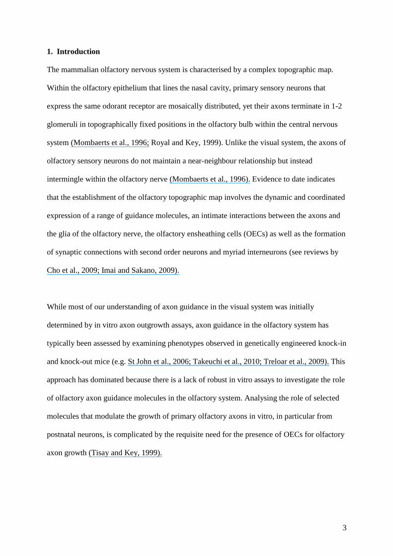
1. Introduction
The mammalian olfactory nervous system is characterised by a complex topographic map.
Within the olfactory epithelium that lines the nasal cavity, primary sensory neurons that
express the same odorant receptor are mosaically distributed, yet their axons terminate in 1-2
glomeruli in topographically fixed positions in the olfactory bulb within the central nervous
system (Mombaerts et al., 1996; Royal and Key, 1999). Unlike the visual system, the axons of
olfactory sensory neurons do not maintain a near-neighbour relationship but instead
intermingle within the olfactory nerve (Mombaerts et al., 1996). Evidence to date indicates
that the establishment of the olfactory topographic map involves the dynamic and coordinated
expression of a range of guidance molecules, an intimate interactions between the axons and
the glia of the olfactory nerve, the olfactory ensheathing cells (OECs) as well as the formation
of synaptic connections with second order neurons and myriad interneurons (see reviews by
Cho et al., 2009; Imai and Sakano, 2009).
While most of our understanding of axon guidance in the visual system was initially
determined by in vitro axon outgrowth assays, axon guidance in the olfactory system has
typically been assessed by examining phenotypes observed in genetically engineered knock-in
and knock-out mice (e.g. St John et al., 2006; Takeuchi et al., 2010; Treloar et al., 2009). This
approach has dominated because there is a lack of robust in vitro assays to investigate the role
of olfactory axon guidance molecules in the olfactory system. Analysing the role of selected
molecules that modulate the growth of primary olfactory axons in vitro, in particular from
postnatal neurons, is complicated by the requisite need for the presence of OECs for olfactory
axon growth (Tisay and Key, 1999).

4
Intense research is being undertaken into the biology and function of the different cells within
the main olfactory system as well as comparisons with cells from different sensory organs in
the nose including the accessory olfactory system, the organ of Masera (Pedersen and Benson,
1986; Weiler and Farbman, 2003) and the septal organ of Grüneberg (Roppolo et al., 2006;
Storan and Key, 2006). The unambiguous identification of living neurons and glia in vivo and
in vitro would greatly facilitate the study of neuron-glia interactions, calcium imaging,
electrophysiology experiments and the development of axon guidance and cell behaviour
assays.
We previously generated the S100ß-DsRed transgenic line of mice which provide excellent
visualisation of OECs (Windus et al., 2007; Windus et al., 2010). The use of these mice has
enabled us to determine the cell behaviour of OECs and to detect distinct differences between
subtypes of OECs (Windus et al., 2007; Windus et al., 2010). However, the use in vitro assays
of primary olfactory neurons have been hampered by the lack of suitable markers in living
olfactory neurons. Primary olfactory neurons have previously been visualised in mice in
which the coding sequence of olfactory marker protein (OMP) was replaced by GFP (Fig. 1B)
(Potter et al., 2001). OMP protein is expressed by primary olfactory neurons soon after they
have differentiated, but is not expressed by immature neurons (Baker et al., 1989) and is
considered to be an excellent marker of primary olfactory neurons. While the OMP-GFP mice
have provided excellent results about the structure of the olfactory system in fixed tissue
(Potter et al., 2001), they are unsuitable for in vitro assays as the deletion of OMP has been
shown to affect axon guidance (St John and Key, 2005) and in our hands the GFP rapidly
fades during live cell imaging. Thus there is now a need to identify primary sensory neurons
in vivo and in vitro with a marker that will enable living cells to be easily visualised.

5
An alternative green fluorescent marker is the coral protein ZsGreen (Bourett et al., 2002;
Wenck et al., 2003). In transgenic mouse models, ZsGreen is brightly fluorescent with high
photo-stability making it suitable for live cell imaging, and is not affected by
paraformaldehyde fixation and so provides excellent detail within fixed tissue (Wouters et al.,
2005). In mammalian cancer cell lines re-introduced into mouse models, the intensity of
ZsGreen fluorescence is suitable for whole body imaging (Harrell et al., 2006; Vlashi et al.,
2009).
In order to unambiguously visualise and identify olfactory sensory neurons in vivo and in
vitro, we have now generated a transgenic reporter line of mice, the OMP-ZsGreen line, in
which the OMP promoter drives expression of the ZsGreen fluorescent protein. In these mice,
the primary sensory neurons in all olfactory regions express ZsGreen from early in
development. The bright fluorescence provides exceptional detail of individual neurons and
the trajectory of axons can be easily traced. Furthermore, we have developed novel methods
for culturing robust axonal growth of both embryonic and postnatal olfactory sensory neurons
from these mice. In vitro, the primary axons are unambiguously identified and when
combined with the S100ß-DsRed transgenic mice (Windus et al., 2007; Windus et al., 2010)
we can clearly visualise and distinguish both olfactory sensory neurons and OECs. The use of
these mice will prove valuable for investigating the development of the olfactory system and
for the generation of novel in vitro assays of olfactory sensory neurons and OECs.

6
2. Materials and methods
2.1. Generation of OMP-ZsGreen transgenic mice
Transgenic mice expressing ZsGreen in olfactory sensory neurons were generated. In these
mice, the full length (5.5kb) olfactory marker protein (OMP) promoter (Danciger et al., 1989)
drove the expression of ZsGreen1 fluorescent protein from pZsGreen Vector (Clontech, Palo
Alto, CA). ZsGreen1 is a variant of wildtype ZsGreen which has been engineered for brighter
fluorescence. The pZsGreen was amplified using PCR from the pZsGreen1-1 vector using
forward primer 5’ gacgtctgtggcagtggtggcagtggcaacagctgtagcacttgggccATGgcccagtccaagcac
3’ and reverse primer 5’ ccatggtcagggcaaggcggag 3’. The ZsGreen fragment was amplified,
sequenced by Australian Genome Research Facility (Brisbane, Australia), and then cloned
into the coding region of rat OMP using AatII and KnpI. The OMP-ZsGreen transgene was
liberated from pBluescript using EcoR1 restriction sites, purified and then injected into
fertilised mouse oocytes of a C57/Bl6 background at the Transgenic Animal Service of
Queensland (University of Queensland, Brisbane). Successful integration of the transgene was
confirmed by expression of ZsGreen fluorescence in the olfactory system of living neonatal
animals. To obtain mice expressing ZsGreen in olfactory neurons and DsRed in glia, the
OMP-ZsGreen transgenic mice were crossed with S100ß-DsRed transgenic mice that had
been previously generated (Windus et al., 2007). All procedures were carried out with the
approval of, and in accordance with, the Griffith University Animal Ethics Committee and the
Australian Commonwealth Office of the Gene Technology Regulator.
2.2. Tissue collection and preparation
Embryonic and postnatal animals were killed by decapitation and adult mice were killed by
CO2 asphyxiation; heads were fixed in 4% paraformaldehyde for up to 24 h at 4 oC. Various

7
wholemount presentations were prepared: the skin covering the head was removed; the head
was bissected longitudinally to reveal the rostral-caudal extent of the olfactory system, or
coronally with the cortex removed to enable visualisation of the caudal region of the olfactory
bulb. Coronal and parasagittal sections (30 μm) through the olfactory region were collected
on a cryostat microtome. Immunohistochemistry was performed using antibodies against
OMP (1:600, Wako Chemicals USA, Richmond, VA, USA) followed by rabbit anti-goat
secondary antibodies conjugated Alexa Fluor594
(1:400; Molecular Probes, Carlsbad, CA,
USA). Some sections were stained with 4',6-diamidino-2-phenylindole (DAPI). Sections were
mounted with Vectashield mounting medium (Vector Laboratories Inc, Burlingame, CA) and
coverslipped. Quantification of cells that expressed ZsGreen and OMP protein was performed
by counting cells in 200 μm spans of the epithelium lining the septum of coronal sections of
P7 animals (n=3 sections in each of 3 animals)
2.3. In vitro axon growth and timelapse imaging
DsRed-OECs and OEC-conditioned medium: OECs were purified from S100ß-DsRed
transgenic mice. Pups were killed by decapitation at P7.5 and the olfactory mucosa was
dissected from the posterior half of the nasal septum. Explants of the mucosa were plated in
plastic 24-well plates coated with Matrigel basement membrane matrix (1:10; Collaborative
Research, Bedford, MA) and incubated in a medium [OEC medium] that promotes glial
growth: Dulbecco’s Modified Eagle Medium (DMEM, Imperial Laboratories, Salisbury, UK)
containing 10% fetal bovine serum (FBS, GIBCO Invitrogen Corporation, Melbourne,
Australia), G5 supplement (Gibco, 200 M), gentamicin (Gibco, 50 mg/ml) and L-glutamine
(Gibco, 200 M) at 37°C with 5% CO2 for 5 days. OECs were then passaged onto coverslips
coated with Matrigel; the OECs proliferated and formed a monolayer over the next few days.
To obtain OEC-conditioned medium, OECs were grown to confluency and then the medium

8
replaced with 500 μl of fresh OEC medium in each well of a 24-well plate; the medium
remained on the cells for 3 days and then collected.
2.4. Embryonic olfactory sensory neuron culture: Timed pregnant OMP-GFP mice (Potter et
al., 2001), OMP-ZsGreen transgenic mice or OMP-ZsGreen x S100ß-DsRed transgenic mice
were sacrificed by 5% CO2 gas asphyxiation and embryos were decapitated. Olfactory
mucosa was dissected from the nasal septum and explants were plated on Matrigel basement
membrane matrix (BD Bioscience, 1:10) coated on coverslips. The explants were cultured in
Neurobasal medium (Gibco Invitrogen Corporation) supplemented with 0.4% methyl
cellulose (BDH Chemical Ltd), B27 serum-free supplement (Gibco Invitrogen Corporation),
0.08 mM L-glutamine (Gibco Invitrogen Corporation), 10 mM HEPES and 5 g/ml
gentamycin (Gibco Invitrogen Corporation) at 37 ºC and 5% CO2. Some embryonic explants
were plated onto a monolayer of DsRed-OECs.
2.5. Postnatal olfactory sensory neuron culture: olfactory epithelium from P1-P21 pups was
dissociated using dispase (Worthington Biochemical Corporation, New Jersey, USA)
followed by trituration through needles. The dissociated cells were then plated onto
monolayers of DsRed-OECs and incubated with OEC-conditioned medium (see above). For
extended timelapse imaging of live cells on the microscope, the explants were incubated in
CO2 independent medium (Invitrogen, catolog number 18045088), rather than Neurobasal or
DMEM medium, with the supplements listed above.
2.6. Live cell and fixed tissue microscopy
For live cell imaging, coverslips with the explants were maintained in CO2 independent
medium at 37 °C in an incubator chamber on an Olympus CellR epifluorescent microscope

9
fitted with differential inference contrast optics. Timelapse images of living axons were
collected at intervals ranging from every 10 s over a period of 5 min up to every 5 min over a
period of 8 hr. Images were compiled using ImageJ software and colour-balanced in Adobe
Photoshop CS3 without further digital manipulation. Images of fixed tissue cryostat sections
were collected on an Olympus FV-1000 confocal microscope; images of wholemount
preparations were collected on an Olympus SZX7 microscope.
2.7. Stability of fluorophore emission
For live axons, timelapse sequences were collected as described above using a 40 X
UPLANAPO objective with a fixed exposure time of 100 ms so that cells were exposed for
100 ms of epifluorescent light every 30 s to 5 min. For fixed tissue, sections were
continuously exposed to epifluorescent light for 30 min on an Olympus BX50 microscope
using either a 10 X or 20 X UPLANFL objective; images obtained every 10 s using a Spot
camera using fixed exposure time. The exposure times were set so that the maximum intensity
pixel in the field of view was below saturation. The change in intensity was determined by
measuring the mean grey value in the area of interest over the timelapse sequence using
ImageJ 1.43U software.

10
3. Results
3.1. ZsGreen is expressed in all olfactory regions of the nasal cavity
OMP-ZsGreen transgenic mice were generated in which the full length (5.5kb) olfactory
marker protein (OMP) promoter (Danciger et al., 1989) drove expression of ZsGreen protein
(Fig. 1A). The mice were generated by random insertion of the transgene into the genome.
The generation of the OMP-ZsGreen reporter line of transgenic mice resulted in ZsGreen
being expressed by neurons lining the olfactory organs of the nose. ZsGreen fluorescent
protein in the nasal cavity was visible through the skin of paraformaldehyde-fixed E11.5
mouse embryos (Fig. 1C). By E12.5, strong expression of ZsGreen was visible in the nasal
cavity as well as around the frontal surface of the telencephalon (Fig. 1D). In older embryos,
ZsGreen fluorescence was easily visible in the nasal cavity and developing olfactory bulbs as
well as in the septal organ of Grüneberg (Fig. 1E). In neonatal animals, ZsGreen fluorescence
in the main olfactory bulbs and nasal cavity was visible through the skin of living (Fig. 1F)
and paraformaldehyde-fixed heads (not shown). In adults, ZsGreen fluorescence in the
olfactory region was visible through the skull and bone covering the nasal cavity (Fig. 1G),
although the thick cribriform plate prevented detection of fluorescence in the upper central
region of the nasal cavity. In longitudinally bisected heads of adults, ZsGreen was present in
all olfactory regions including main olfactory epithelium and bulbs, the organ of Masera (Fig.
1H, Fig. 2D), and the vomeronasal organ and the accessory olfactory bulbs (Fig. 1H-I).
ZsGreen expression was also present in restricted regions elsewhere in brain as previously
described for OMP expression (Baker et al., 1989).
ZsGreen protein was ectopically expressed within the oral cavity (Fig. 1E) and in the
anogenital region (Fig. 1J) of embryos from E16.5 onwards and adults; however,

11
immunohistochemistry using anti-OMP antibodies did not detect OMP immunoreactivity in
these regions (data not shown). Higher magnification views of the vomeronasal organ (VNO)
in coronal and parasagittal cryostat sections revealed that ZsGreen was expressed in sensory
neurons throughout the epithelium lining the VNO and in the VNO nerves (Fig. 2A-B). When
the OMP-ZsGreen mice were bred with the S100ß-DsRed mice so that neurons and glia
would be visualised and separately identified, the glomeruli of the accessory olfactory bulb
were distinctly visible below the accessory nerve layer (Fig. 2C). Higher magnification views
demonstrated the structure of the septal organs of Masera and Grüneberg and their associated
axons (Fig. 2D-F).
3.2. ZsGreen is expressed by olfactory sensory neurons throughout the main olfactory system.
We next examined the spatial expression pattern of ZsGreen in detail using
paraformaldehyde-fixed, sagittal and coronal cryostat sections of the main olfactory system.
In E12.5 embryos, ZsGreen- positive neurons illustrated the extent of the developing olfactory
epithelium within the nasal cavity and axons lining the surface of the telencephalon were
easily visible (Fig. 3A). At E15.5, ZsGreen expression illustrated growth of the olfactory
epithelium and axon fascicles en route to the olfactory bulb were visible (Fig. 3B). At E18.5,
ZsGreen was visible in glomeruli in the main and accessory olfactory bulb (Fig. 3C). In the
early postnatal animal (Fig. 3D) and in the adult (Fig. 3E), ZsGreen-positive neurons clearly
show the full extent of the olfactory epithelium within the nasal cavity and in the nerve fibre
and glomerular layers of the olfactory bulb (Fig. 3D).
In higher magnification views of the main olfactory epithelium, ZsGreen was strongly and
distinctly expressed by the olfactory sensory neurons, with the cell bodies, dendrites and
axons clearly labelled (Fig. 4A-B). Some neurons expressed ZsGreen at high levels, while

12
others expressed ZsGreen at lower levels. Immunohistochemistry with anti-OMP antibodies
revealed that the majority of neurons that expressed OMP protein also expressed ZsGreen
protein (Fig. 4B-D). Infrequently, neurons that expressed OMP protein did not express
ZsGreen (unfilled arrowhead Fig. 4B-D). However, some neurons expressed lower levels of
ZsGreen compared to neurons in the more apical layers and these neurons often exhibited
negligible staining by OMP antibodies (unfilled arrows in Fig. 4B-D). These cells were
located towards the basal layer of the epithelium where neurons are differentiating from stem
cells. We quantified the cells that expressed ZsGreen and/or OMP antibody staining in
postnatal day 7 animals. We assumed that the total number of olfactory mature and immature
neurons were cells that expressed either ZsGreen or OMP antibodies or both. The vast
majority of neurons expressed ZsGreen (98.3 +/- 0.5%) whereas fewer neurons were detected
by OMP antibodies (74.6 +/- 5.6%) which is consistent with OMP protein being expressed on
cells that are further differentiated. Thus around one-quarter (25.8 +/- 0.5%) of neurons that
expressed ZsGreen were not detected by OMP antibodies. A small proportion (1.7 +/- 0.5%)
of cells was detected by OMP antibodies, but did not express ZsGreen protein. We also
examined sections in which the vomeronasal nerves could be seen originating from the
vomeronasal organ and projecting amongst fascicles of main olfactory axons, or in coronal
sections where the vomeronasal nerves passed deeper to the main olfactory axon fascicles. In
these sections the nerve fascicles formed by the main olfactory axons (arrowhead in Fig. 4B)
strongly expressed ZsGreen fluorescence and were distinct from the larger vomeronasal nerve
fascicles in which ZsGreen and OMP were expressed at lower levels (dotted line in Fig. 4B,
arrow in Fig. 5A).
Within the nerve fibre and glomerular layers of the olfactory bulb, immunohistochemistry
with anti-OMP antibodies again verified that ZsGreen colocalised with OMP protein (Fig. 4E-

13
G). Higher magnification views of glomeruli and the external plexiform layer revealed
considerable detail of the trajectory of individual axons (Fig. 4H-J, Movie 1).
The strong and robust expression of ZsGreen along the length of the axons from the
epithelium to the olfactory bulb let us more closely examine axons within the fascicles that
projected through the lamina propria. A lower magnification view of the fascicles revealed
that the thin, numerous main olfactory axon fascicles were easily distinguished from the
larger vomeronasal nerves (Fig. 5A). The trajectory of individual axons exiting the basal layer
of the epithelium and projecting towards fascicles (Fig. 5B) could be easily traced and axons
coalescing into fascicles (Fig. 5C) were readily detected by ZsGreen fluorescence.
To facilitate identification of olfactory ensheathing cells (OECs) as well as axons, we crossed
the OMP-ZsGreen mice with S100ß-DsRed transgenic mice. In fixed tissue coronal sections
through the olfactory epithelium of P7 animals, the green fluorescent olfactory sensory
neurons were clearly distinct from the red fluorescent OECs (Fig. 5D-F).
3.3. ZsGreen robustly labels individual axons in vitro
We have previously attempted timelapse imaging of axons growing out from explants of
olfactory epithelium dissected from the OMP-GFP mice that were generated by Potter et al.,
(2001). However we had limited success since the GFP in the living axons faded rapidly when
subjected to frequent repeated exposures of epifluorescent light particularly when viewed
using high magnification objectives. We were therefore only able to capture images of living
axons using short exposure times at lower magnification which limited the resolution of the
images and we were unable to obtain detail of the growth cones and filopodia (Fig. 6A-C).
The strong expression of ZsGreen in olfactory sensory axons of OMP-ZsGreen mice led us to

14
develop in vitro assays in which we could unambiguously identify axons and OECs in culture
and use high resolution timelapse imaging over extended periods.
For embryonic olfactory sensory neuron assays, we cultured explants of olfactory epithelium
from embryos using a neuron growth promoting medium. Axons emerged from the explant
within 12 hr of plating and by 24-48 hr numerous ZsGreen axons had extended with many
coalescing into fascicles (Fig. 6D). Robust axon outgrowth from explants was achieved at all
embryonic ages from E11 onwards (data not shown) with healthy growth of the explants
continuing for at least 4-7 days.
Higher magnification of individual axons growing out of the explant revealed excellent detail
of the growth cone and filopodia (Fig. 6E). Visualisation of the axons using DIC optics
revealed that axons were in contact with numerous other cells that were not fluorescent (Fig.
6F-G).
3.4. Monolayers of OECs and OEC-conditioned medium promote axon growth in postnatal
neurons
The easy visualisation of primary olfactory axons in vitro enabled us to rapidly assess whether
axon outgrowth could be achieved from older animals. We prepared monolayers of DsRed
OECs that were purified from S100ß-DsRed transgenic mice (Fig. 6H) and cultured them for
3 days. Dissociated cells as well as small explants from the olfactory epithelium of OMP-
ZsGreen mice were then plated onto the established monolayers of DsRed OECs and
incubated in medium that had been conditioned by OECs for 3 days. Robust axon growth
occurred within 12 hr (Fig. 6I-J) and continued for at least 5 days with the axons maintaining
close contact with the OECs (arrowheads Fig. 6H-J). Olfactory sensory neurons were

15
successfully cultured from postnatal mice of ages ranging from the ages P1 to P21 (data not
shown). These results demonstrate that the OMP-ZsGreen transgenic mice facilitate the
culturing of robust axon growth in vitro from embryonic and postnatal animals up to P21 and
when combined with the S100ß-DsRed transgenic mice they provide an excellent tool for
neuron-glia assays.
3.5. ZsGreen fluorescence was resistant to fading in live and fixed preparations
The robust outgrowth and strong fluorescence of the olfactory sensory axons led us to
determine the stability of the fluorescence during timelapse imaging of live cells. When living
growth cones were exposed to epifluorescent light using a 40 X objective, fine detail of the
growth cone and filopodia was achieved (Fig. 7A). Repeated exposures of 100 ms every 10 s
for 5 min did not alter the intensity of the fluorescence and the growth cones continued to be
highly active during the imaging period (Fig. 7A-F, Movie 2). Quantification of the intensity
of the fluorescence during repeated time-lapse imaging of live cells revealed there was no
deterioration in fluorescence signal intensity when imaged at high frequency for short periods
(every 1 min for 10 min; Fig. 7G) or less frequently over longer periods (every 5 min for 8 h;
Fig. 7H). Robust and active growth of axons was observed for at least 48 h in cultures that
were maintained on the incubated microscope stage in CO2-independent medium (data not
shown).
ZsGreen fluorescence was also very stable in fixed, cryostat sections of olfactory sensory
axons within the nerve fibre and glomerular layers. Continuous exposure to epifluorescent
light for 30 min decreased the intensity of the ZsGreen fluorescence by only 20-40% when
using a 10X or 20X objective, respectively (Fig. 7I).

16
4. Discussion
In this study we describe a new line of transgenic reporter mice, OMP-ZsGreen mice in which
bright and robust green fluorescence is expressed in olfactory sensory neurons. The intensity
and stability of the fluorescence make it useful for live cell imaging and imaging of fixed
tissues. The fluorescence is bright enough to be seen in live animals and is seen in all
chemosensory organs in the nasal cavity. We also report optimised culture protocols for
growing olfactory sensory neurons in vitro using feeder layers of and conditioned medium
from OECs. ZsGreen fluorescence clearly labels olfactory sensory neuron cell bodies and
axons, including growth cones and fine filopodia. They can be distinguished from OECs if the
latter are derived from our S100ß-DsRed reporter mice (Windus et al., 2007). ZsGreen
fluorescence is extremely stable and fade resistant whether exposed repeatedly during time-
lapse imaging of live cultures or exposed continually in fixed sections.
The strong expression of ZsGreen in olfactory sensory neurons in the early developing
embryo provided exceptional detail of individual axons along their entire trajectory. In
contrast, OMP antibodies detect neurons that have undergone more advanced differentiation
and do not provide such detail of axons in early embryos. Consistent with previous reports
(Wouters et al., 2005) the fluorescence of the ZsGreen protein was unaffected by
paraformaldehyde fixation and was highly stable during repeated imaging of fixed tissue
making it suitable for use in conjunction with immunohistochemistry studies. When combined
with the S100ß-DsRed reporter mice (Windus et al., 2007), the interactions between neurons
and glia can be readily visualised. These mice therefore are a convenient tool for investigating
the anatomy of the olfactory system during development and regeneration. For example, the
brightness of the ZsGreen allowed the visualisation of individual axons that were exiting the
basal layer of the epithelium and we were easily able to trace their trajectory. With the

17
superior detection of olfactory sensory axons by ZsGreen in comparison to anti-OMP
immunohistochemistry subtle defects in axon trajectories are more likely to be detected when
analysing the OMP-ZsGreen axons in the knockout mice. Thus the OMP-ZsGreen mice can
be bred with knockout mice to aid the examination of the effect of specific molecules in the
olfactory system.
ZsGreen was expressed in all olfactory organs within the nose and within the main and
accessory olfactory bulbs. The easy identification of the neurons in the different regions (main
olfactory, accessory olfactory and the organs of Masera and Grüneberg) will facilitate the
purification of those neurons by anatomical dissection and by fluorescence activated cell
sorting. When combined with the S100ß-DsRed reporter mice OECs from the different
regions can also be easily purified. Preparing neurons and glia in this way would be ideal for
comparative biology studies of the cells from these different olfactory regions using targeted
patch clamping and electrophysiology, gene expression comparisons and cell behaviour
assays (see review by Ma, 2007).
The OMP-ZsGreen mice offer several advantages in comparison to the OMP-GFP line of
mice (Potter et al., 2001). In the OMP-GFP knockout mice, the coding sequence for OMP
protein has been replaced by GFP so that OMP protein is not expressed by olfactory sensory
neurons (Fig. 1B). OMP is thought to be involved in regulating cAMP kinetics during
olfactory odour signal transduction (Reisert et al., 2007) and we have previously
demonstrated that loss of OMP results in numerous axons misprojecting within the olfactory
bulb (St John and Key, 2005). Therefore the use of these mice may not be suitable for many
studies. The OMP-GFP mice have previously been used for explant assays to analyse
olfactory axon growth and provided good resolution of axons using a single time point

18
(Luxenhofer et al., 2008). However, we found that the GFP expression in living axons was
weak and faded rapidly with repeated exposure and hence we were unable to obtain high
magnification, high resolution images of OMP-GFP axons using timelapse microscopy. In
contrast, in the OMP-ZsGreen reporter transgenic mice, OMP protein continues to be
expressed and the robust, bright and stable expression of ZsGreen in living neurons makes it
highly suitable for timelapse microscopy.
Within the field of olfactory axon guidance and axon-OEC interactions, the lack of markers of
living cells in vitro has hampered the analyses of the roles of candidate molecules. The clear
visualisation of olfactory sensory axons in vitro is hindered by similarities in the gross
morphology of axons and the processes of OECs and also because axons typically migrate
over the surface of these cells in co-cultures (Tisay and Key, 1999). Thus axons can typically
only be visualised at the termination of the assay using immunocytochemistry with a marker
such as NCAM (St John et al., 2000; Treloar et al., 2009). Interestingly, OMP protein is
thought to be expressed by more mature neurons (Farbman and Margolis, 1980; Verhaagen et
al., 1990) and has not yet been reported to be strongly expressed by axons in vitro. In
comparison, in vivo we detected numerous neurons that expressed ZsGreen but did not
express OMP protein and as these neurons resided in the deeper layer of the epithelium it is
likely that they are less differentiated neurons. In vitro, ZsGreen is strongly expressed by the
axons, growth cones and filopodia. Thus the expression of ZsGreen allows the detection of
olfactory sensory neurons and enables the unambiguous visualisation of living axons in vitro.
The use of OEC-conditioned medium and a DsRed-OEC monolayer greatly improves
postnatal axon outgrowth and the use of DsRed-OECs allows clear distinction between axons
and OECs. We have confirmed that olfactory axons maintained in CO2 independent medium

19
continue to actively grow with the growth cones rapidly altering their structure throughout the
imaging period. Importantly, during imaging the ZsGreen fluorescence remained bright even
with frequent exposure over many hours during the observation period. Hence the culturing
and imaging methods for the embryonic and postnatal ZsGreen olfactory axons are highly
suitable for frequent and extensive repetitive imaging. The use of the OMP-ZsGreen mice
together with the S100ß-DsRed transgenic mice now path the way for the use of in vitro
assays for the study of axon behaviour, axon-OEC interactions and to probe the function of
candidate axon guidance molecules at the individual cellular level.
The bright ZsGreen fluorescence of the olfactory region through the skin of neonatal OMP-
ZsGreen transgenic mice provides an easy method for verifying the phenotype/genotype of
the offspring. The ectopic expression of ZsGreen in the oral cavity and genitalia is likely due
to regulatory elements associated with the insertion site of the OMP-ZsGreen transgene, but
this provides an easy means of verifying expression of the transgene in live adult mice and
thus the maintenance of mouse stocks does not require PCR genotyping.
5. Conclusion
In summary, the generation of the OMP-ZsGreen reporter transgenic mouse line provides a
convenient and unambiguous marker of olfactory sensory neurons in vivo and in vitro. In
conjunction with the S100ß-DsRed transgenic mice, in vitro timelapse assays in which living
neurons and OECs can be easily visualised can now be developed to investigate the biology
of olfactory sensory neurons and axon-OEC interactions.

20
Acknowledgements
We thank Ms Jessica Cornock for maintaining the OMP-ZsGreen and the S100ß-DsRed
mouse colonies. This work was supported by a grant from the National Health and Medical
Research Council to J.S and B.K [511006], by a Postdoctoral Fellowship from the Australian
Research Council to J.E [DP0986294], Australian Postgraduate Awards to D.A. and to L.W,
and by funding to A.M.S for the National Centre for Adult Stem Cell Research from the
Australian Government Department of Health and Aging.

21
References
Baker H, Grillo M, Margolis FL. Biochemical and immunocytochemical characterization of
olfactory marker protein in the rodent central nervous system. J Comp Neurol, 1989; 285:
246-61.
Bourett TM, Sweigard JA, Czymmek KJ, Carroll A, Howard RJ. Reef coral fluorescent
proteins for visualizing fungal pathogens. Fungal Genet Biol, 2002; 37: 211-20.
Cho JH, Prince JE, Cloutier JF. Axon guidance events in the wiring of the mammalian
olfactory system. Mol Neurobiol, 2009; 39: 1-9.
Danciger E, Mettling C, Vidal M, Morris R, Margolis F. Olfactory marker protein gene: its
structure and olfactory neuron-specific expression in transgenic mice. Proc Natl Acad Sci
U S A, 1989; 86: 8565-9.
Farbman AI, Margolis FL. Olfactory marker protein during ontogeny: immunohistochemical
localization. Dev Biol, 1980; 74: 205-15.
Harrell JC, Dye WW, Allred DC, Jedlicka P, Spoelstra NS, Sartorius CA, Horwitz KB.
Estrogen receptor positive breast cancer metastasis: altered hormonal sensitivity and
tumor aggressiveness in lymphatic vessels and lymph nodes. Cancer Res, 2006; 66: 9308-
15.
Imai T, Sakano H. Odorant receptor gene choice and axonal projection in the mouse olfactory
system. Results Probl Cell Differ, 2009; 47: 57-75.
Luxenhofer G, Breer H, Strotmann J. Differential reaction of outgrowing olfactory neurites
monitored in explant culture. J Comp Neurol, 2008; 509: 580-93.
Ma M. Encoding olfactory signals via multiple chemosensory systems. Crit Rev Biochem
Mol Biol, 2007; 42: 463-80.
Mombaerts P, Wang F, Dulac C, Chao SK, Nemes A, Mendelsohn M, Edmondson J, Axel R.
Visualizing an olfactory sensory map. Cell, 1996; 87: 675-86.

22
Pedersen PE, Benson TE. Projection of septal organ receptor neurons to the main olfactory
bulb in rats. J Comp Neurol, 1986; 252: 555-62.
Potter SM, Zheng C, Koos DS, Feinstein P, Fraser SE, Mombaerts P. Structure and
emergence of specific olfactory glomeruli in the mouse. The Journal of Neuroscience,
2001; 21: 9713-23.
Reisert J, Yau KW, Margolis FL. Olfactory marker protein modulates the cAMP kinetics of
the odour-induced response in cilia of mouse olfactory receptor neurons. J Physiol, 2007;
585: 731-40.
Roppolo D, Ribaud V, Jungo VP, Luscher C, Rodriguez I. Projection of the Gruneberg
ganglion to the mouse olfactory bulb. Eur J Neurosci, 2006; 23: 2887-94.
Royal SJ, Key B. Development of P2 olfactory glomeruli in P2-internal ribosome entry site-
tau-LacZ transgenic mice. J Neurosci, 1999; 19: 9856-64.
St John JA, Claxton C, Robinson MW, Yamamoto F, Domino SE, Key B. Genetic
manipulation of blood group carbohydrates alters development and pathfinding of primary
sensory axons of the olfactory systems. Dev Biol, 2006; 298: 470-84.
St John JA, Key B. Olfactory marker protein modulates primary olfactory axon overshooting
in the olfactory bulb. J Comp Neurol, 2005; 488: 61-9.
St John JA, Tisay KT, Caras IW, Key B. Expression of EphA5 during development of the
olfactory nerve pathway in rat. J Comp Neurol, 2000; 416: 540-50.
Storan MJ, Key B. Septal organ of Gruneberg is part of the olfactory system. J Comp Neurol,
2006; 494: 834-44.
Takeuchi H, Inokuchi K, Aoki M, Suto F, Tsuboi A, Matsuda I, Suzuki M, Aiba A, Serizawa
S, Yoshihara Y, Fujisawa H, Sakano H. Sequential arrival and graded secretion of
Sema3F by olfactory neuron axons specify map topography at the bulb. Cell, 2010; 141:
1056-67.

23
Tisay KT, Key B. The extracellular matrix modulates olfactory neurite outgrowth on
ensheathing cells. J Neurosci, 1999; 19: 9890-9.
Treloar HB, Ray A, Dinglasan LA, Schachner M, Greer CA. Tenascin-C is an inhibitory
boundary molecule in the developing olfactory bulb. J Neurosci, 2009; 29: 9405-16.
Verhaagen J, Oestreicher AB, Grillo M, Khew-Goodall YS, Gispen WH, Margolis FL.
Neuroplasticity in the olfactory system: differential effects of central and peripheral
lesions of the primary olfactory pathway on the expression of B-50/GAP43 and the
olfactory marker protein. J Neurosci Res, 1990; 26: 31-44.
Vlashi E, Kim K, Lagadec C, Donna LD, McDonald JT, Eghbali M, Sayre JW, Stefani E,
McBride W, Pajonk F. In vivo imaging, tracking, and targeting of cancer stem cells. J Natl
Cancer Inst, 2009; 101: 350-9.
Weiler E, Farbman AI. The septal organ of the rat during postnatal development. Chem
Senses, 2003; 28: 581-93.
Wenck A, Pugieux C, Turner M, Dunn M, Stacy C, Tiozzo A, Dunder E, van Grinsven E,
Khan R, Sigareva M, Wang WC, Reed J, Drayton P, Oliver D, Trafford H, Legris G,
Rushton H, Tayab S, Launis K, Chang YF, Chen DF, Melchers L. Reef-coral proteins as
visual, non-destructive reporters for plant transformation. Plant cell reports, 2003; 22:
244-51.
Windus LC, Claxton C, Allen CL, Key B, St John JA. Motile membrane protrusions regulate
cell-cell adhesion and migration of olfactory ensheathing glia. Glia, 2007; 55: 1708-19.
Windus LC, Lineburg KE, Scott SE, Claxton C, Mackay-Sim A, Key B, St John JA.
Lamellipodia mediate the heterogeneity of central olfactory ensheathing cell interactions.
Cell Mol Life Sci, 2010; 67: 1735-50.

24
Wouters M, Smans K, Vanderwinden JM. WZsGreen/+: a new green fluorescent protein
knock-in mouse model for the study of KIT-expressing cells in gut and cerebellum.
Physiol Genomics, 2005; 22: 412-21.

25
Figure captions
Figure 1. OMP-ZsGreen expression in transgenic mice. (A) The OMP-ZsGreen transgene
consisted of the full length 5.5 kb OMP promoter sequence which drove expression of the
ZsGreen coding sequence. The mice were generated by random insertion into the genome. (B)
The previously generated OMP-GFP knockout mice (Potter et al., 2001) were generated by
targeted insertion of the GFP coding sequence which replaced the coding sequence of OMP.
Panels C-J show wholemount preparations of OMP-ZsGreen transgenic mice. (C) In a frontal
view of an E11.5 embryo, ZsGreen expression was visible in the olfactory epithelium of the
nasal cavity (arrowheads); t is telencephalon, e is eye. (D) In E12.5 embryos, ZsGreen
expression was visible in the nasal cavity (arrowheads) as well as lining the medial regions of
the telencephalon of the presumptive olfactory bulb (t). (E) In E16.5 embryos, ZsGreen
expression was widespread through the nasal cavity (arrowhead) and olfactory bulbs (arrow)
as well as in the septal organ of Grüneberg (unfilled arrowhead). ZsGreen was ectopically
expressed in the oral cavity (arrow with tail) and in the margins of the eyelids. (F) In a dorsal
view of a living P2 OMP-ZsGreen x S100β-DsRed mouse, the ZsGreen fluorescence in the
nasal cavity (arrowhead) and olfactory bulb (arrow) was clearly visible through the skin.
DsRed fluorescence was strongly expressed in the eyes. (G) In paraformaldehyde-fixed adult
OMP-ZsGreen x S100β-DsRed mouse with the skin removed, the ZsGreen fluoresence was
visible in the nasal cavity (arrowheads) and olfactory bulbs (arrow), although fluorescence
was not visible through the bony cribiform plate (c). (H) In a sagittal view of a longitudinally
bisected adult head, ZsGreen fluorescence was present in the vomeronasal organ (vno), organ
of Masera (om), olfactory epithelium (oe), olfactory bulb (ob) and accessory olfactory bulb
(arrow); rostral is to the left and dorsal to the top. (I) In a view of the rear of the olfactory
bulbs in a head that has been coronally bisected and the cortex removed, the accessory

26
olfactory bulbs (arrows) expressed ZsGreen; glomeruli lining the periphery of the main
olfactory bulb are also visible (arrowheads); dorsal is up, lateral edges are to left and right. (J)
In the anogenital region, ZsGreen was ectopically expressed in the labioscrotal swellings
(arrowhead) and surrounding the anus (arrow). Scale bar is 150 μm in C; 225 μm in D; 500
μm in E,F; 300 μm in G,H; 600 μm in I,J.
Figure 2. ZsGreen is expressed in all olfactory organs. (A) In a coronal section and (B) in a
sagittal section, ZsGreen was expressed neurons within the epithelium of the vomeronasal
organ (ve) and in the axons of the vomeronasal nerve (arrowhead); s is septum. (C) In a
sagittal section through the accessory olfactory bulb of an OMP-ZsGreen x S100β-DsRed
mouse, ZsGreen was uniformly expressed by axons, whereas DsRed expression (red) in the
glia was restricted to the nerve fibre layer and was not in the glomeruli; rostral is to the left
and dorsal to the top. (D) ZsGreen was expressed in the organ of Masera seen here from the
epithelial surface of a wholemount. (E) In a ventral view of a horizontally bisected nasal
cavity, the organ of Grüneberg (arrow) and associated axon bundles (arrowhead) projecting
toward the olfactory bulb expressed ZsGreen; dotted line shows septum; rostral/caudal and
lateral orientations are indicated by arrows. (F) A sagittal cryostat section shows ZsGreen
expression by neurons of the organ of Grüneberg; rostal is to the left, dorsal to the top. Panels
B, C, F are stained with DAPI. Scale bar is 200 μm in A; 350 μm in B, E; 100 μm in C,F; 175
μm in D.
Figure 3. ZsGreen is strongly expressed by olfactory sensory neurons after fixation. Panels A-
C are sagittal sections with rostral to the left and dorsal to the top. (A) In an E12.5 embryo,
ZsGreen was expressed by olfactory sensory neurons in the developing olfactory epithelium
(oe) and their axons lining the surface (arrow) of the frontal region of the telencephalon (t).

27
In E15.5 embryos, ZsGreen was also expressed in the thickening nerve fibre layer (arrow) of
the olfactory bulb (ob). In the E18.5 embryo, ZsGreen was also expressed in glomeruli
(arrow) of the main olfactory bulb as well as in the accessory olfactory bulb (arrowhead). Nc
is nasal cavity; v is ventricle of the telencephalon. Panels A and B are stained with DAPI. In
coronal sections of (D) a P9 pup and (E) an adult animal, ZsGreen was expressed throughout
the olfactory epithelium (oe) and in the outer layers and glomeruli of the olfactory bulb
(arrow). Dorsal is to the top in all panels. Scale bar is 400 μm in A,D; 500 μm in B,C,E.
Figure 4. ZsGreen is expressed by olfactory sensory neurons and their axons. (A) In a section
of the olfactory mucosa of an E13.5 embryo, ZsGreen was strongly expressed by some
neurons (arrowhead) within the olfactory epithelium (oe), while others expressed ZsGreen at
lower levels (arrow with tail). ZsGreen was present in the dendrites (unfilled arrowhead) and
axons (arrow) in the lamina propria (lp). (B-D) In sections of the olfactory mucosa from P2
animals, immunohistochemistry with anti-OMP antibodies revealed that almost all neurons
expressed both ZsGreen (green) and OMP protein (red). Occasionally neurons expressed
OMP protein but not ZsGreen (unfilled arrowhead). However, more frequently neurons
expressed ZsGreen, but no detectable levels of OMP protein (unfilled arrows). Main olfactory
axons within fascicles strongly expressed ZsGreen (filled arrowhead) whereas VNO axons
within the accessory olfactory nerve (dotted lines) expressed ZsGreen at lower levels. (E-G)
In sections of the olfactory bulb of P2 animals, immunohistochemistry with anti-OMP
antibodies revealed that OMP protein (red) colocalised with ZsGreen (green) in the nerve
fibre layer (nfl) and glomerular layer (gl). Within the external plexiform layer (epl) “over-
projecting” axons (arrow) were more clearly visualised by ZsGreen, but less easily with anti-
OMP immunochemistry. (H-J) In various postnatal ages (P2, P9 and P16), individual axons
could be seen in the nerve fibre layer and often extending beyond glomeruli into the external

28
plexiform layer (arrow in I). A three-dimensional reconstruction of the glomerulus in J is
shown in Movie 1. Scale bar is 60 μm in A,H; 50 μm in B-D; 80 μm in E-G; 40 μm in I, 30
μm in J.
Movie 1. Three-dimensional image of a glomerulus in a P16 animal. Confocal images through
the depth of the 30 m section were compiled to reconstruct the 3D structure of the
glomerulus that is shown in Figure 4J.
Figure 5. OMP-ZsGreen axons and fascicles of axons are easily visualised in the lamina
propria. (A-C) In sagittal sections of the olfactory mucosa of P9 animals, individual axons as
well as fascicles were clearly visible in the lamina propria underlying the olfactory epithelium
(oe). (A) Fascicles of main olfactory axons (arrowhead) and fascicles of accessory olfactory
axons (arrow). (B) An individual neuron (arrowhead) with an axon (arrow) projecting to a
fascicle (unfilled arrowhead). (C) Axons entering fascicles (arrowhead) and within fascicles
(arrow). (D-F) In a coronal section through the olfactory mucosa of a P7 OMP-ZsGreen x
S100β-DsRed mouse, the olfactory sensory neurons (green) were visible in the olfactory
epithelium (oe) and their axons form fascicles in the lamina propria (lp). The axon fascicles
were surrounded by OECs that expressed DsRed (red). Scale bar is 200 μm in A; 80 μm in B,
100 μm in C, 35 μm in D-F.
Figure 6. OMP-ZsGreen neurons are visualised reliably in vitro. (A-C) Explants of olfactory
epithelium from E14.5 OMP-GFP mice gave rise to axons that were weakly fluorescent and
could only be imaged infrequently at low magnification. Axon growth (arrow) could be
detected but detail of the growth cone and filopodia were not achieved. (D) In vitro explants
of E14.5 olfactory epithelium from an OMP-ZsGreen mouse extended numerous axons within

29
24 hr. (E-G) Higher magnification view of an axon (green) extending from an explant of
olfactory epithelium from an E14.5 animal. The axon had a large growth cone at the leading
edge as well as a smaller one (arrowhead) close to a bifurcation point. Other cells that were
present in the culture (arrow) were visible with differential inference contrast optics (DIC).
(H-J) Axons (arrowheads) projecting away from an explant. A neuron cell body (arrow,
green) which has left the explant sits on top of an olfactory ensheathing cell (red). Small
explants and dissociated neurons (arrow) from the olfactory epithelium of postnatal day 1
OMP-ZsGreen mice were cultured on monolayers of DsRed OECs. Axons (green in I, J) grew
out within 12 hr of plating with distinct growth cones and filopodia that maintained close
contact with the DsRed OECs (arrowheads). Scale bar is 20 μm in A-C; 150 μm in D; 10 μm
in E-G; 40 μm in H-J.
Figure 7. ZsGreen fluorescence is stable during repetitive live cell imaging. (A-F) A
timelapse sequence of a growth cone that was imaged every 10 seconds for 5 minutes. The
growth cone with filopodia (arrowhead) remained clearly visible and active during the
imaging period. An inactive neuron (arrow) was present in the field of view. A movie of the
timelapse sequence is shown in Movie 2. (G-I) Quantification of the change in intensity of
ZsGreen fluorescence. (G) Live growth cones imaged with epifluorescent light for 50 ms
every 1 min for 10 min; (H) live growth cones imaged with epifluorescent light for 50 ms
every 5 min for 8 hr; (I) in fixed tissue sections, continual exposure to epifluoresecent light
for 30 min at 10 X magnification (black line) and 20 X magnification (grey line). Scale bar is
10 μm in A-F.

30
Movie 2. Timelapse sequence of living growth cone. The growth cone was imaged every 10 s
for 5 min; time shown is min:sec. Selected images from the sequence are shown in Figure 7A-
F.

Figure 1Click here to download high resolution image

Figure 2Click here to download high resolution image

Figure 3Click here to download high resolution image
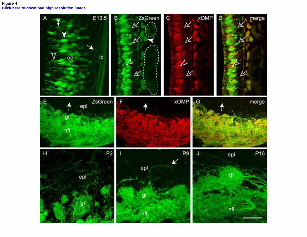
Figure 4Click here to download high resolution image

Figure 5Click here to download high resolution image

Figure 6Click here to download high resolution image

Figure 7Click here to download high resolution image

Supplementary movie 1Click here to download Supplementary file for online publication only: Supplemental movie 1.avi

Supplementary movie 2Click here to download Supplementary file for online publication only: Supplemental movie 2.avi
Copyright © 2022 FDOKUMEN