The Impact of Entrepreneurship Education on Entrepreneurial Intention
Upload
khangminh22Category
view
1download
0
HAL Id: hal-02990216https://hal.archives-ouvertes.fr/hal-02990216
Submitted on 5 Nov 2020
HAL is a multi-disciplinary open accessarchive for the deposit and dissemination of sci-entific research documents, whether they are pub-lished or not. The documents may come fromteaching and research institutions in France orabroad, or from public or private research centers.
L’archive ouverte pluridisciplinaire HAL, estdestinée au dépôt et à la diffusion de documentsscientifiques de niveau recherche, publiés ou non,émanant des établissements d’enseignement et derecherche français ou étrangers, des laboratoirespublics ou privés.
Biocultural diversity in Late Pleistocene/Early HoloceneAfrica: Olduvai Hominid 1 (Tanzania) biological affinity
and intentional body modificationJohn C Willman, Raquel Hernando, Marie Matu, Isabelle Crevecoeur
To cite this version:John C Willman, Raquel Hernando, Marie Matu, Isabelle Crevecoeur. Biocultural diversity in LatePleistocene/Early Holocene Africa: Olduvai Hominid 1 (Tanzania) biological affinity and intentionalbody modification. American Journal of Physical Anthropology, Wiley, 2020, 172, pp.664 - 681.�10.1002/ajpa.24007�. �hal-02990216�

R E S E A R CH A R T I C L E
Biocultural diversity in Late Pleistocene/Early Holocene Africa:Olduvai Hominid 1 (Tanzania) biological affinity and intentionalbody modification
John C. Willman1,2,3 | Raquel Hernando2,3 | Marie Matu4 | Isabelle Crevecoeur4
1Laboratory of Prehistory, CIAS – Research
Centre for Anthropology and Health,
Department of Life Sciences, University of
Coimbra, 3000-456, Coimbra, Portugal
2IPHES, Institut Català de Paleoecologia
Humana i Evolució Social , 43007 Tarragona,
Spain
3Àrea de Prehistòria, Universitat Rovira i Virgili
(URV), 43002 Tarragona, Spain
4Unité Mixte de Recherche 5199, PACEA, De
la Préhistoire à l'Actuel: Culture,
Environnement, et Anthropologie, Centre
National de la Recherche Scientifique,
Université de Bordeaux, Bordeaux, France
Correspondence
John C. Willman, Laboratory of Prehistory,
CIAS – Research Centre for Anthropology and
Health, Department of Life Sciences,
University of Coimbra, 3000-456 Coimbra,
Portugal.
Email: [email protected]
Funding information
Agence Nationale de la Recherche, Grant/
Award Number: ANR-14-CE31; Agència de
Gestió d'Ajuts Universitaris i de Recerca,
Grant/Award Number: 2017SGR1040; H2020
Marie Skłodowska-Curie Actions, Grant/
Award Number: H2020-MSCA-IF-2016
No. 749188; Ministerio de Ciencia e
Innovación, Grant/Award Number:
PGC2018-093925-B-C32; Universitat Rovira i
Virgili, Grant/Award Number: 2017PFR-URV-
B2-91; Martí-Franqués Research Grant, Grant/
Award Number: 2019PMF-PIPF-59
Abstract
Objectives: The dentition of Olduvai Hominid 1 (OH1) exhibits an anomalous pattern
of dental wear that was originally attributed to either intentional cultural modification
(filing) or plant processing behaviors. A differential diagnosis of the wear and assess-
ment of the biological affinity of OH1 is presented.
Materials and Methods: Macroscopic and microscopic observations of all labial and
buccal tooth surfaces were undertaken to assess wear patterns. A multivariate analy-
sis of mandibular morphology of OH1 compared to other Late Pleistocene, Holocene,
and recent modern humans was used to ascertain biological affinity.
Results: The morphological variation of the OH1 mandible is closely aligned with var-
iation in penecontemporaneous fossils from Africa and outside that of recent
humans. The concave wear facets exposing dentin on the labial surfaces of all three
preserved mandibular incisors is confirmed. Substantial loss of labial/buccal surfaces
was documented on the surfaces of all in situ maxillary and mandibular canines, pre-
molars, and molars ranging from distinct facets with well-defined edges, to blunting
or “polishing” around areas of maximum buccal curvature. The wear on both the
anterior and postcanine teeth closely resemble that caused by adornments (“labrets”)
worn in lower-lip and buccal facial piercings known from bioarchaeological and eth-
nographic contexts. The wear pattern suggests that the OH1 wore three facial
piercings—two buccal/lateral and a medial one in the lower lip.
Discussion: Our findings suggest that the expression of social identities through
intentional body modification is more diverse than previously documented elsewhere
in Africa during the Late Pleistocene (i.e., ablation) and Early Holocene (i.e., ablation,
chipping, and filing).
K E YWORD S
dental wear, facial piercing, labret, Later Stone Age, social identity
1 | INTRODUCTION
Human morphological variation across Africa during the Late Pleisto-
cene and Early Holocene is poorly understood despite its importance
for understanding prehistoric population dynamics within the
continent and the transition from anatomically archaic to modern
human morphology. This can be partly attributed to the sparse, highly
fragmentary, and often indirectly dated Late Pleistocene human fossil
record of Africa (Crevecoeur, 2008; Crevecoeur, Brooks, Ribot, Cor-
nelissen, & Semal, 2016; Crevecoeur, Rougier, Grine, & Froment,
Received: 29 August 2019 Revised: 24 December 2019 Accepted: 2 January 2020
DOI: 10.1002/ajpa.24007
Am J Phys Anthropol. 2020;1–18. wileyonlinelibrary.com/journal/ajpa © 2020 Wiley Periodicals, Inc. 1

2009; Grine, 2016; Grine et al., 2017; Stojanowski, 2014; Tryon et al.,
2015). Human remains from MIS 2 and early MIS 1 are more common
than fossils from the preceding MIS 6 to MIS 3 (Grine 2016), but the
regional distribution of human remains are skewed by a few, large
mortuary contexts from more recent periods in North Africa
(e.g., Afalou-bou-Rhummel and Grotte des Pigeons [Taforalt]) and the
Nile Valley (e.g., Jebel Sahaba, Wadi Halfa, Tushka, Al Khiday)
(Arambourg, Boule, Vallois, & Verneau, 1934; Ferembach, Dastugue, &
Poitrat-Targowla, 1962; Hachi, 1996; Humphrey, Bello, Turner,
Bouzouggar, & Barton, 2012; Stojanowski, Carver, & Miller, 2014;
Stojanowski, Johnson, Paul, & Carver, 2016; Usai et al., 2010;
Wendorf, 1968). With so few human fossils dated to the Late Pleisto-
cene and Early Holocene in Africa, and concomitant biases in regional
representation, the ones that are available for detailed analyses are of
great importance for understanding issues of human biocultural varia-
tion in African prehistory (Crevecoeur et al., 2016; Mounier et al.,
2018; Sawchuk & Willoughby, 2015; Scerri et al., 2018). Cranial, den-
tal, and postcranial analyses of Pleistocene and Holocene humans pro-
vide meaningful insights into population dynamics within Africa
(Armelagos, Van Gerven, Martin, & Huss-Ashmore, 1984; Benoiston,
Bayle, & Crevecoeur, 2018; Crevecoeur, 2008; Crevecoeur et al.,
2016; Crevecoeur et al., 2009; Greene, Ewing, & Armelagos, 1967;
Holliday, 2015; Irish, 2000, 2005; Irish, Black, Sealy, & Ackermann,
2014; Irish & Guatelli-Steinberg, 2003; Irish & Konigsberg, 2007;
Mounier et al., 2018; Pfeiffer & Harrington, 2018; Ponce de León
et al., 2018; Sawchuk & Willoughby, 2015; Stojanowski, 2014; War-
ren, Hall, & Ackermann, 2015), but biocultural approaches emphasiz-
ing intentional body modification have also proven useful as markers
of population movement, continuity, and replacement in African pre-
history (Barton et al., 2008; De Groote & Humphrey, 2016; Finucane,
Manning, & Touré, 2008; Humphrey & Bocaege, 2008; Irish, 2017;
Mercader, Garralda, Pearson, & Bailey, 2001; Stojanowski et al., 2014,
2016). Biocultural approaches also provide an additional level of infor-
mation on human lifeways and social identities that may otherwise be
ignored in pursuit of data on biological affinity alone.
Renewed interest in the paleobiology of Olduvai Hominid I (OH1,
Olduvai [Oldupai] Gorge, northern Tanzania)—principally concerning
morphological variation and burial taphonomy (Crevecoeur et al.,
2016; Matu, Crevecoeur, & Huchet, 2017; Tryon et al., 2015)—
provided an opportunity to reexamine the entire dentition with an
emphasis on the anomalous pattern of dental wear on the labial sur-
faces of the mandibular incisors and canine of this individual, which
was only briefly discussed elsewhere (Mollison, 1929; Parsche, 1993).
This study aims to provide a differential diagnosis of the OH1 dental
wear in light of literature now replete with examples of idiosyncratic
and non-dietary dental wear features (Bonfiglioli, Mariotti, Facchini,
Belcastro, & Condemi, 2004; d'Incau, Couture, & Maureille, 2012;
Milner & Larsen, 1991; P. Molnar, 2011; S. Molnar, 1972; Scott &
Jolie, 2008; Stojanowski et al., 2016), and a growing literature on
intentional body modification in the African archaeological record
(Barton et al., 2008; De Groote & Humphrey, 2016; Finucane et al.,
2008; Honegger, 2004; Humphrey & Bocaege, 2008; Irish, 2017;
MacDonald, 1999; Mercader et al., 2001; Petit-Maire & Riser, 1983;
Salvatori & Usai, 2009, 2014, 2016; Santoni, Sakka, & Garcier, 2006;
Stojanowski et al., 2014, 2016).
1.1 | Archaeological and paleobiological context
The archaeological context of the OH1 was initially published over
100 years ago (M. D. Leakey, 1978; Reck, 1914), and few studies con-
cerning this relatively complete skeleton have been published since
the 1930s (Boswell, 1932; Crevecoeur et al., 2016; Gieseler and Mol-
lison, 1929; L. S. B. Leakey, 1928; Matu et al., 2017; Mollison, 1929;
Parsche, 1993; Protsch, 1974; Twiesselman, 1973; Reck, 1926, 1933).
Direct dating provided an age of 16,920 ± 920 BP for the skeleton
(Protsch, 1974, 1975), but doubts have been cast on the dates
acquired through the radiocarbon and/or amino acid racemization dat-
ing by Protsch in the 1970s (Grine, 2016). A new attempt to re-date
OH1 was undertaken in 2018 on a fragment of femoral diaphysis that
was not varnished. Unfortunately, despite a careful pretreatment, no
reliable collagen fraction could be isolated and purified. The sample
could not be dated nor a δ15N/ δ14N measurement be done due to
this absence of collagen.
Previous analyses indicate that OH1 was a young adult male
(~20–35 years old at death) and deliberately buried (Matu et al.,
2017). Morphological analyses have been more illustrative of the bio-
logical and probable chronological affinity of the OH1 individual rela-
tive to other Later Stone Age human remains. For instance,
morphological analyses show that the mandibular and distal humeral
morphology of OH1 are outside of the 95% confidence interval ellipse
for recent African human comparative samples (Crevecoeur et al.,
2016). Furthermore, the OH1 mandibular metrics cluster with pene-
contemporaneous Later Stone Age humans from Ishango and Mum-
bwa, and distal humeral morphology follows the general pattern of
Late Pleistocene humans (Crevecoeur et al., 2016). The presence of
microlithic fragments found in potentially associated layers (Protsch,
1974) can be attributed to the temporospatially diverse Later Stone
Age of East Africa, regardless of what “industry” or technocultural var-
iant it was, or is currently, attributed to (Wilshaw, 2016). Thus, the
skeletal morphology and artifacts associated with the skeleton provide
support for a Late Pleistocene/Later Stone Age context for OH1.
1.2 | Previous assessments of the anomalousanterior dental wear
The OH1 dentition is incomplete, but the mandibular anterior denti-
tion does preserve the left I1 and right I1, I2, and C1—all of which
exhibit marked wear facets on the labial surfaces of each tooth. The
wear is most marked on the labial incisor surfaces, exposing dentin on
each tooth, and a flat labial facet is also apparent on the left C1
(Figure 1).
Early observations by Leakey (1928) report a lack of anterior
tooth chipping or filing commonly affiliated with Bantu populations,
and the observation was later confirmed by Mollison (1929) for both
2 WILLMAN ET AL.

the maxilla and mandible. However, Mollison (1929) noted the pres-
ence of the labial surface wear on the anterior mandibular dentition.
Mollison attributed this wear to the probable use of a sandstone file
through an analogy with dental filing practices among the Proto-
Malay, since similar labial abrasion had not yet been documented in
Africa. A subsequent investigation by Parsche (1993) also agreed that
the labial wear on the OH1 mandible has no equivalent among ethno-
graphically known examples of intentional dental modification in
Africa.
However, Parsche (1993) emphasized the high degree of asym-
metry of labial surface wear between anterior teeth and challenged
Mollison's assertion that the wear was derived from sandstone filing.
Parsche (1993) pursued two experiments to replicate the labial wear
pattern—the first, abrasion with sandstone, and the second, abrasion
with moist sisal fibers. After 30 minutes of abrasion with each mate-
rial, scanning electron microscopy (SEM) was used to examine the
experimentally worn teeth and comparisons with the incisors of OH1
were also made. He determined that the micro-grooving on the OH1
teeth was more consistent with wear induced by sisal fibers and
rejected the possibility of wear from a sandstone file. Parsche (1993)
ultimately attributed the labial wear to non-masticatory uses of the
anterior teeth as tools for fiber processing rather than any form of
intentional dental modification.
There are several major criticisms concerning the methods and
conclusions of Parsche (1993). First, the surfaces of the experimen-
tally worn teeth were not cleaned of residues to improve visualization
of the microwear patterns produced (Parsche 1993). Second, the ori-
entation of striations in the sandstone experiment are oblique and the
sisal fiber experiment produced horizontal striations (Parsche 1993),
which indicates that motions used to produce abrasion between
experiments lacked uniformity. Third, the microscopic examination of
each labially-facetted tooth was not conducted, and purportedly
unworn premolars and molars were not examined to determine base-
line levels of buccal preservation and wear across tooth types. Fur-
thermore, at the time of publication, the broad literature on non-
masticatory wear caused by manipulative behaviors (Barrett, 1977;
Formicola, 1988; Irish & Turner, 1987; Larsen, 1985; Milner & Larsen,
1991; S. Molnar, 1972; Pedersen & Jakobsen, 1989; Taylor, 1986;
Turner & Machado, 1983; Ubelaker, Phenice, & Bass, 1969) contained
a paucity of references to mandibular labial surface modification aside
from instrumental striations attributed to “stuff-and-cut” behaviors
(Arsuaga et al., 1989; Bermúdez de Castro, Bromage, & Jalvo, 1988;
Brace, 1975; Koby, 1956). Therefore, we suggest that there is little
ethnographic, bioarchaeological, or paleobiological support for non-
dietary manipulative behaviors that would produce the pattern of
labial wear exhibited on the anterior mandibular teeth of OH1.
We agree with Parsche (1993) that the OH1 labial dental wear is
unintentional; however, we argue that it is an unintentional byproduct
of intentional body modification. One such form of body modification
that produces unintentional dental (and occasionally, osseous) modifi-
cation is the use of facial piercings (e.g., “labrets”, “lip plugs”, “lip-
studs”, “lip discs”, etc.; henceforth: labrets) (Alt & Pichler, 1998;
Cybulski, 1974, 2010; Cybulski, Balkwill, Young, & Sutherland, 1992;
Dietze et al., 2007; Garve, Garve, Türp, & Meyer, 2017; MacDonald,
1999; Mukherjee, Trevor, & Rao, 1955; Pedersen, 1952, 1955; San-
toni et al., 2006; Torres-Rouff, 2003, 2011). Indeed, facial piercings
were documented historically (Colette, 1933; Gupta, 1986;
Huntingford, 1961; Labouret, 1952; Schweinfurth, 1875; also see
citations in Insoll, 2015; MacDonald, 1999; Seligman, 2015) and
archaeologically in Africa (Addison, 1949; Mukherjee et al., 1955;
Petit-Maire & Riser, 1983; also see citations in Insoll, 2015; MacDon-
ald, 1999) when Parsche (1993) conducted his analysis, but were not
considered in any previous evaluation of the OH1 labial wear. Thus, a
re-analysis is warranted.
2 | MATERIALS AND METHODS
2.1 | Morphometric analysis of biological affinity
Five measurements were taken on the relatively complete OH1 man-
dible following Martin (in Bräuer, 1988) for mandibular corpus length
(M68), bimental breadth (M67), symphyseal height (M69), breadth of
the corpus at the level of the mental foramen (M69[3]), and minimum
anteroposterior width of the ramus (M71a). Previously analyses show
that these measurements are a good proxy for overall mandibular
morphology, and are discriminant between groups (e.g., Crevecoeur,
2008; Crevecoeur & Trinkaus, 2004; Crevecoeur et al., 2009, 2016).
The use of only five measurements also ensures a large comparative
sample of fossil and recent human remains. A principle component
analysis (PCA) is used to assess the mandibular morphology of OH1
compared to geographically and chronologically defined groups com-
prised of Middle Stone Age humans (Loyangalani and Mumbwa X);
Middle Paleolithic modern humans from Southwest Asia (Skhul VI
and V, and Qafzeh 9); Late Pleistocene Homo sapiens from Nazlet
Khater (NK2), Ishango (#Ish15), Northeast Africa (Jebel Sahaba, Wadi
Kubbaniya, and Wadi Halfa), Northwest Africa (Taforalt), Southwest
Asia (Ohalo II H2, Mallaha, and Nahal Oren); Neolithic individuals
from Northwest Africa (Mechta el Arbi), East Africa (Gamble's Cave
and Lothagam), South Africa (Fish Hoek); and recent modern humans
from Africa. Morphological data from fossil and bioarchaeological
materials were collected by one of us (I. Crevecoeur) on original spec-
imens except for Wadi Kubbaniya (cast at the Smithsonian
F IGURE 1 Macroscopic details of anterior mandibular dentition inlateral and labial views. Note marked concave facets on each incisor.Modest canine facet is faint and difficult to view here. Scale is 10 mm
WILLMAN ET AL. 3

Institution, Washington D.C.). Recent modern human data are from
Ribot (2011).
2.2 | Dental wear and oral pathology
Digital photography and macroscopic observations were made on the
original specimen. High resolution molds of all teeth were made using
President Light Body polyvinylsiloxane (Coltene Whaledent), and
epoxy resin (Epo-Tek 301: Epoxy Technologies, Inc.) was used for
positive casts (Galbany, Martínez, & Pérez-Pérez, 2004). Casts were
prepared for environmental scanning electron microscopy (ESEM: FEI
Quanta 600) by sputter-coating with approximately 20 nm layer of
gold to improve the conductivity. Macroscopic observations were also
made to assess presence or absence of various dental wear features
(e.g., interproximal grooving, antemortem enamel chipping) and oral
paleopathological conditions (e.g., caries and periodontal status).
3 | RESULTS
3.1 | Biological affinity
The first three principle components (PC1, PC2, and PC3) for the PCA
on mandibular metrics account for 83.6% of total sample variation
(54.6%, 15.5%, and 13.5%, respectively; Table 1, Figures 2 and 3).
OH1 and most of the Late Pleistocene individuals plot in the upper
left quadrant of each projection and many of these individuals, includ-
ing OH1, are outside of the 95% concentration ellipse of recent mod-
ern human variation. The position of OH1 relative to other specimens
is driven by the absolute dimensions of the mandible, particularly cor-
pus length for PC1, symphysis height for PC2 (Figure 2), and corpus
breadth for PC3 (Figure 3). The Early Holocene East African individ-
uals from Lothagam (notably, KNM-LT 13704B) overlap with OH1
along PC1, but diverge along PC2 and PC3, reflecting shorter symphy-
seal height and thinner corpus breadth, respectively. Concentration
TABLE 1 Principle Component Analysis eigenvalues and factor loadings
Factors Eigenvalue % Total
PC1 2.730317 54.60634
PC2 0.773816 15.47631
PC3 0.674325 13.48649
PC4 0.475479 9.50957
PC5 0.346064 6.92128
Variable (Martin no.) Raw measurement (mm) Factor 1 Factor 2 Factor 3 Factor 4 Factor 5
Corpus length (M68) 98.00 −0.648200 0.447773 0.601108 −0.033152 0.130024
Bimental breadth (M67) 50.60 −0.800955 −0.110379 −0.027889 0.564091 −0.165260
Minimum anteroposterior
breath of the ramus (M71a)
41.22 −0.836580 −0.085194 −0.032869 −0.374825 −0.388976
Symphysis height (M69) 39.89 −0.670990 0.445330 −0.555887 −0.051454 0.199487
Corpus breadth at the level of the
mental foramen (M69[3])
14.97 −0.720091 −0.596284 0.046094 −0.114190 0.332791
F IGURE 2 Bivariate projection ofcomponent 1 and 2 from the principlecomponents analysis of mandibularmorphology. The 95% concentrationellipses for Late Pleistocene NortheastAfrica, Late Pleistocene Northwest Africa,and recent modern humans are shown.Abbreviations not indicated in legend:Loy = Loyangalani, Mum = Mumbwa X,Sk = Skhul, Qa = Qafzeh, OH2 = Ohalo2, WK = Wadi Kubbaniya, Lo = Lothagam,GC = Gamble's Cave
4 WILLMAN ET AL.

ellipses for the two largest fossil groups (Late Pleistocene Northeast
and Northwest Africa) are also provided. When PCA is performed
with size-adjusted data (the logged ratio of each variable by the geo-
metric mean of all variables; see: Darroch and Mosimann, 1985;
Jungers, Falsetti, Wall, 1995), no meaningful patterning is shown
between individuals and the various subgroupings. Overall, the PCA
reflects a massive, elongated mandible with a tall mandibular symphy-
sis and thick corpus for OH1.
3.2 | Dental wear and oral pathology
There are three large, concave facets exposing dentin on the labial
surfaces of each of the preserved mandibular incisors (Figures 1 and
4a,b). The incisor facets are most deeply faceted on the central inci-
sors and less so on the left I2 where only a small dentin exposure is
present on the mesial half of the cervical two-thirds of the labial face.
The cervical borders of each facet are the shape of inverted crescents.
The enamel surrounding each labial dentin exposure exhibits
mesiolaterally oriented microgrooves (Figure 4b). A shallow, concave
facet without dentin exposure is also present on the left mandibular
canine (Figure 4c). The canine facet is located on the middle third of
the labial face. The canine is less worn than the left I2, which further
emphasizes the diminishing wear gradient from the central incisors to
lateral incisor and canine. The right I1 exhibits normal occlusal wear
but lacks the labial wear of its mandibular antagonists.
Contrary to previous studies, we document marked buccal
faceting, blunting of buccal curvatures, and “polish” on most of the
postcanine teeth (Figure 5). All premolars display a substantial loss
of buccal enamel with flat facets where the crown should exhibit its
greatest buccal curvature, and with well-defined facet margins. The
buccal facets of maxillary and mandibular premolars form a continu-
ous, flat wear plane with adjacent first molars (Figure 6a). The maxil-
lary canines display blunting of their buccal surfaces, but it is less
F IGURE 3 Bivariate projection ofcomponent 1 and 3 from the principlecomponents analysis of mandibularmorphology. The 95% concentrationellipses for Late Pleistocene NortheastAfrica, Late Pleistocene Northwest Africa,and recent modern humans are shown.Abbreviations same as Figure 2
F IGURE 4 (a) ESEM micrograph of left I1 and I2 concave facets on labial surface. (b) Closer view of horizontal microgrooving around dentinexposure on the left I2. (c) Left C1 with subtle facet concavity outlined. Scale increments are 10 mm
WILLMAN ET AL. 5

marked than that of premolars and first molars (Figure 6b). Maxillary
second molars exhibit marked buccal facets (Figure 6c,d), albeit,
slightly less defined than in M1s. Similarly, the mesiobuccal faces of
each M3 exhibit subtle flattening. Together, this suggests a
decreasing gradient of buccal wear from M1 to M2 to M3, in both
the total area worn and depth of wear, which is also evident in the
decreasing visibility of the buccal grooves across the maxillary
molars (Figure 6c).
F IGURE 6 (a) Continuous buccal wear plan on left P3, P4, and M1 emphasized by orange line. Note that wear is most accentuated onpremolars. Scale is 10 mm. (b) ESEM micrograph of left C1 showing modest flatness to buccal curvature and rugose area (white areas) that borderfacet margin. Rugosity is unaffected by buccal wear agent. Orange arrows point to partially exfoliated shellac. Macrophotograph of same surfacein similar orientation. Scale is 1 mm. (c) Right M1 to M3 showing gradient of buccal wear. Note the decreasing visibility of buccal grooves from M1
to M3. Arrows indicate distobuccal margin of buccal wear facets. Dotted ellipse surrounds a white dentin exposure. Scale is 10 mm. (d) ESEMmicrograph of left M2 showing well defined buccal facet (black arrows). Note flatness of surface surrounding the buccal groove.Macrophotograph of the same surface with slightly different orientation. Scale is 1 mm
F IGURE 5 Approximate outlines of buccal wear on OH1 dentition. Blue shapes indicate strongly marked facets and red shapes indicate less-marked buccal wear exhibited as modest blunting of buccal curvatures and polish. Anterior dentition not outlined in this view. Scale incrementsare 10 mm
6 WILLMAN ET AL.

Dentin exposure is present near the mesiobuccal cervix of the right
M1, emphasizing the exceptional loss of buccal enamel on the tooth
(Figure 6c). Similar loss of enamel seems to be present on at least the
left P4 and M1 (Figure 7a). The damage to root and alveolar surfaces,
and the conspicuous glue/shellac coating them, makes it particularly dif-
ficult to identify antemortem features on non-enamel surfaces. How-
ever, the buccal wear penetrating into the dentin near the cervix of the
M1s and left P4 may be continuous from the enamel/dentin to the sur-
faces of roots. However, it is unclear if this is a taphonomic artifact asso-
ciated with postmortem breakage, the presence (or localized exfoliation)
of adhesive materials, or some combination of these factors (Figure 7a).
The buccodistal cervical root surface of the left M1 exhibits a wide, shal-
low grooved appearance that could be attributed to any of the factors
listed above, or possibly attributable to actions associated with the for-
mation of an interproximal groove (Figure 7a). Unfortunately, the condi-
tion of the root is poor, and diagnosis of etiology uncertain.
The only possible evidence of root surface caries is present on lin-
gual surface of the left M1, but again, this could be a product of post-
mortem breakage and repair, but the preservation is simply too poor
to be certain (Figure 7b). No other caries are identified, but it must be
reiterated that various forms of adhesive (glue, shellac, etc.), as well as
some postdepositional exfoliation of the left maxillary occlusal sur-
faces, prevented a thorough investigation of many surfaces. Most of
the discoloration of the enamel visible in photos can be attributed to
aging adhesives rather than decay.
Periodontal status is also difficult to assess due to damage and
reconstruction. Most in situ teeth were broken postmortem near the
cervix and glued into their current positions, which complicates obser-
vations of root and alveolar surfaces and distorts measurements.
Cementoenamel junction to alveolar crest distances (CEJ-AC) are best
preserved on the postcanine teeth—especially for the mandible and
the left maxilla. The degree of alveolar remodeling and CEJ-AC dis-
tances suggest mild to moderate periodontal disease for the right
maxilla and mandibular postcanine dentition (Lavigne & Molto, 1995;
Ogden, 2008), but observations of alveolar bone and CEJ-AC dis-
tances in the anterior dentitions are too poorly preserved to assess.
Antemortem and postmortem dental chipping is documented on
several teeth, but conclusions about diet or behavior drawn from
antemortem chipping should be cautious given overall enamel preser-
vation (Figure 7c,d). For instance, the surface exfoliation on some
teeth and adhesive on others makes the identification of some chips
impossible. Interproximal chips are the most difficult to identify given
the concentration of adhesive at the interface of the interstitial spaces
and the occlusal surface. Most antemortem enamel chips are small
(Grade 1: Bonfiglioli et al., 2004), but there is one large (Grade 3) ante-
mortem chip on the mesiobuccal cusp of the right M1 (Figure 7c).
To summarize, the buccal faceting, substantial blunting of buccal
curvature, and/or polish is present on the labial/buccal surfaces of all
in situ maxillary and mandibular canines and premolars, and first and
second molars. Buccal faceting is either less distinct, or absent, on
third molars. The pattern suggests that wear affected the buccal sur-
faces of the maxillary premolars and first molars most heavily, less so
anteriorly (C1), and also decreases from M1 to M3. The pattern of buc-
cal wear in the mandible is like that of the maxilla, but the wear angles
F IGURE 7 (a) Detail buccalenamel and root surfaces of right P4-M1. Blue arrows point to areas ofpossible root features continuouswith dentin exposure (white arrows)and buccal faceting. The M1 exampleappears as a wide, shallow groove.(b) Lingual surface of left M1-M2. Bluearrow pointing to possibletaphonomic damage or root surfacecaries. (c) Antemortem (white) andpostmortem (red) enamel chipping onright M1. The chip is the largestdocumented in the dentition. D:Antemortem (white) and postmortem(red) enamel chipping on the right C1-P4. Also note the presence ofadhesive on the occlusal surfaces andinterstitial spaces that makeidentification of features difficult.Scale is 10 mm
WILLMAN ET AL. 7

toward the occlusal surface. The premolars exhibit well-defined facets
in both jaws, but facets are better defined in maxillary molars than man-
dibular ones. OH1 exhibits mild to moderate periodontal disease, some
enamel chipping, but no conclusive evidence of caries. Unfortunately,
significant use of shellac and glue obscures most surfaces, making the
detailed microscopic examination of surfaces difficult.
4 | DISCUSSION
Biological affinity: The present study shows that the mandibular mor-
phology of OH1 aligns with other Late Pleistocene African fossils in
terms of absolute size, while remaining outside of the 95% concentra-
tion ellipse for multidimensional mandibular variability observed in
recent modern humans. Indeed, the PCA confirms patterns shown in
previous metric assessments of bimental breadth and corpus length
that created a cluster among OH1, Ishango (#Ish15), Mumbwa X,
Wadi Kubbaniya, and other Late Pleistocene individuals from North-
east and Northwest Africa while remaining outside the 95% confi-
dence intervals for recent human variation (Crevecoeur et al., 2016).
Interestingly, an individual from the Early Holocene site at Lothagam
(KNM-LT 13704B) has an absolutely wider bimental breadth, but
shorter mandibular corpus than all other Late Pleistocene African fos-
sils previously observed (Crevecoeur et al., 2016). Mounier et al.
(2018) also documented larger centroid size for the Lothagam individ-
uals using mandibular geometric morphometrics, albeit with substan-
tial overlap, compared to Early Holocene individuals from Nataruk
(West Turkana, Kenya) and Pleistocene North Africa (i.e., Afalou,
Taforalt, and Nazlet Khater 2). However, relatively little difference in
centroid size between a diverse, global sample of recent humans and
pre-dynastic African groups was found; and both Early Holocene and
Late Pleistocene African fossil groups exhibit absolutely larger cen-
troid sizes than the former (Mounier et al., 2018). These results are
like those in the present study, showing some overlap between Late
Pleistocene and Early Holocene individuals, but much less overlap
between the Late Pleistocene and recent modern human groups.
Together, these analyses provide a sense of regional and chrono-
logical mosaicism in mandibular morphology of Late Pleistocene and
Early Holocene humans of Northwest, Northeast, Central, and East
Africa. Not only is there a high level of morphological diversity docu-
mented in Late Pleistocene and Early Holocene humans from Africa,
but this diversity is also relatively distinct from the morphological
diversity of later Holocene and recent modern humans (Crevecoeur
et al., 2009, 2016; Harvati et al., 2011; Mounier et al., 2018; Tryon
et al., 2015), which has been attributed to a “Holocene filter” shaping
patterns of recent human diversity in Africa (Mirazón Lahr, 2016;
Mounier et al., 2018).
Differential diagnosis of dental wear: The wear on the labial and
buccal surfaces of the teeth of OH1 affect far more teeth than initially
acknowledged (Mollison, 1929; Parsche, 1993), but the patterning of
wear shows remarkable symmetry and patterning across tooth types.
The use of a sandstone file (Mollison, 1929) is an unlikely cause
of the labial wear. This is due, in part, to maxillary overbite and overjet
which can prevent ease of filing of the mandibular incisor and canine
labial surfaces (Burnett & Irish, 2017), and is seldom documented
compared to the filing and modification of the labial surfaces of maxil-
lary anterior tooth surfaces (Arcini, 2005; Bocquentin, Crevecoeur, &
Semal, 2013; Ikehara-Quebral et al., 2017; Milner & Larsen, 1991).
Second, the practice of intentional filing is generally related to the
expression of individual and group-level social identities, and there-
fore, outwardly visible. Filed mandibular (and cheek) teeth would be a
nondescript signal to others, thus making it difficult to ascribe such a
practice to the intentional marking and display of one's social identity.
Non-masticatory preparation of vegetal fibers (Parsche, 1993) is
also difficult to attribute to the mandibular labial surface wear of
OH1. The manipulation of vegetal fibers, sinews, cordage, thread, and
similar materials with the dentition is well-documented in both ethno-
graphic and bioarchaeological contexts, but commonly produces
occlusal and/or interproximal grooves (Brown & Molnar, 1990;
Cybulski, 1974; Erdal, 2008; Formicola, 1988; Lorkiewicz, 2011;
Lukacs & Pastor, 1988; Molleson, 2016; Schulz, 1977; Scott & Jolie,
2008; Sperduti et al., 2018; Ubelaker, 1969; Waters-Rist, Bazaliiskii,
Weber, Goriunova, & Katzenberg, 2010; Wheat, 1967). Most fiber
processing behaviors are reconstructed as pulling fibers across the
occlusal surfaces or around the lingual and interproximal surfaces of
teeth (see references above). However, the form of wear documented
on the mandibular labial surfaces of OH1 has no parallel with any
wear features documented in the bioarchaeological literature that
have been attributed to non-masticatory manipulative behaviors.
Holding fibrous materials between the teeth and lower lip to
soften them or to remove nutrients before expectorating an indigest-
ible, fibrous quid (e.g., Schoeninger, Bunn, Murray, & Marlett, 2001)
could produce localized abrasion or corrosion depending on the physi-
cal and chemical properties of the material placed between the lip and
teeth. However, it is difficult to reconstruct a scenario in which fibers
held between the lip and teeth would relate to the mediolateral move-
ments hypothesized by Parsche (1993) for the wear on the anterior
mandibular dentition. Furthermore, erosive wear can be ruled out
since there is a lack of macroscopic scooping of dentin on the labial
surfaces of the mandibular incisors (in addition to the buccal dentin
exposure on the right M1). Erosive wear is commonly found on the lin-
gual surfaces of the maxillary anterior teeth (Johansson, Omar, Carls-
son, & Johansson, 2012), but none is evident on the well-preserved
right I1 or either C1. There is modest cupping of the exposed dentin
on the first and second molars, which is often a sign of erosive pro-
cesses in clinical studies (d'Incau et al., 2012; Johansson et al., 2012),
but there is no cupping of anterior tooth dentin, and cupping of
exposed dentin on postcanine teeth is also a common outcome of
abrasive dentin wear in archaeological contexts (Hinton, 1981; Kai-
donis, 2008; Molnar, 1971). Likewise, the labial anterior dental wear is
most prominent on the incisal and middle third of each tooth, exhibits
sharp lateral edges, and terminates near the cervix—a pattern that is
at odds with the position of non-carious cervical lesions (Michael, Kai-
donis, & Townsend, 2010). Furthermore, the sharp cervical edge of
the concave wear facet would be rounded in a corrosive situation
(d'Incau et al., 2012). Furthermore, there is a predominance of
8 WILLMAN ET AL.
abrasive dietary and non-dietary wear on the labial and buccal sur-
faces that strongly favor mechanical (abrasive) wear over chemical
erosion of the surfaces (Coupal & Sołtysiak, 2017; Kieser et al., 2001).
The pattern of labial and buccal wear is also inconsistent with hard tis-
sue correlates of behaviors related to the use of materials like coca
(Indriati & Buikstra, 2001), betel nut (Reichart, Creutz, & Scheifele,
2006), or chewing sticks (Cook, Bastos, Lopes, Mendonça de Souza, &
Santos, 2015).
The pattern of diminishing wear from the mandibular central inci-
sors to the left C1 suggests a uniform pattern of wear by a fixed object
if one assumes the missing right I2 and C1 exhibited symmetrical wear.
Wear of this form is commonly documented among wearers of orna-
ments fixed in piercings through the lower lip in ethnographic, clinical,
and bioarchaeological contexts (Addison, 1949; Aigner & Veltre,
1976; Alt & Pichler, 1998; Croucher, 2012; Cybulski, 1974, 2010;
Cybulski et al., 1992; Dietze et al., 2007; Erdal, 2013; Garve et al.,
2017; Hole, Flannery, & Neely, 1969; Keddie, 1981; MacDonald,
1999; Mattingly et al., 2009; Mukherjee et al., 1955; Pedersen, 1952,
1955; Salvatori & Usai, 2009, 2016; Santoni et al., 2006; Torres-Rouff,
2003, 2011). The symmetry and depth of wear—decreasing from cen-
tral to lateral incisor and canine—are common characteristics of mid-
line, or “medial” (following Keddie, 1981), lower lip piercings in North
American bioarchaeological remains (Cybulski, 2010; Willman, per-
sonal observation). Such facial ornamentation is documented in
archaeological contexts across the African continent (Addison, 1949;
Honegger, 2004; MacDonald, 1999; Mattingly et al., 2009; Mukherjee
et al., 1955; Petit-Maire & Riser, 1983; Salvatori & Usai, 2016; Santoni
et al., 2006) and is well-known in African ethnohistoric records in
numerous forms (Aanestad & Poulsen, 1996; Garve et al., 2017;
Gauthier & Wangermez, 1964; Labouret, 1952; LaTosky, 2006; Mac-
Donald, 1999; Schweinfurth, 1874; Seligman, 2015; Wayland, 1931).
The microgrooves bordering the exposed dentin on the mandibu-
lar anterior facets may relate to the abrasive properties and form of
an adornment worn through the lower lip. While we cannot be certain
of the exact shape or surface texture of the portion of the adornment
abutting the teeth of OH1, but an adornment suspended in a piercing
that constrained its movement is a reasonable explanation for the
local formation of wear facets and microgrooves on the labial surfaces
of the anterior mandibular dentition.
The buccal faceting and wear of postcanine teeth is far less com-
monly documented in the literature, and is largely confined to exam-
ples from bioarchaeological contexts in North America where there is
cultural and direct evidence of “lateral” piercings (after Keddie, 1981)
for wearing labrets (Aigner & Veltre, 1976; Curtin, 1984; Cybulski,
2010; Murray, 1981; Pedersen, 1955; Severs, 1974), and archaeologi-
cal sites of the Mid-Upper Paleolithic (Gravettian, sensu lato) of Cen-
tral Europe (Drozdová, 2002; Hillson, 2006; Matiegka, 1924, 1929,
1934; Trefný, 2008; Vlcek, 1991, 1997; Willman, 2016). They have
been attributed to holding objects between the cheek and buccal sur-
faces of teeth, such as small pebbles to alleviate thirst (Matiegka,
1924, 1929, 1934; Vlcek, 1991), to a range of other possible behav-
iors (Hillson, 2006), and labret-use (Willman, 2016). However, the
archaeological examples from North America that occur in the context
of labret-use exhibit markedly similar patterns of buccal wear to OH1
(Aigner & Veltre, 1976; Curtin, 1984; Cybulski, 2010; Murray, 1981;
Pedersen, 1955; Severs, 1974) which lends support for the attribution
of labret use by OH1. Lukacs and Pastor (1988) documented a type of
interproximal groove on distal maxillary molar roots that was associ-
ated with buccal cervical abrasion of teeth proximal to the groove.
This form of manipulative wear superficially matches the cervical fea-
tures on the left side of the OH1 dentition. However, there is a lack
of right/left symmetry and a greater involvement of enamel (and den-
tin) than root surface for OH1. While the left M1 distal root surface
may exhibit an interproximal groove, it is not likely caused by the
same agent producing the blunting and faceting of the buccal
surfaces.
The more extreme buccal wear on the premolars and first molars,
compared to second and third molars, suggests that an object was in
contact with the more anterior postcanine teeth for a long enough
period of time to create a continuous, flat wear plane across multiple
adjacent teeth. Furthermore, the M1's exhibit greater buccal wear
than the M1's which is less probable if an object was placed inside the
mouth to rest between the buccal surfaces of the teeth and cheek.
This is because a small, hard object would preferentially settle against
the mandibular molars. Thus, both the consistent wear planes, and
wear patterning along the tooth row and between jaws, is more con-
sistent with ornaments that are fixed through piercings in the cheeks.
Periodontal disease is not uncommon in Late Pleistocene con-
texts (Cucina, Herrera Atoche, & Chatter, 2019; Lacy, 2014, 2015;
Trinkaus, Lacy, & Willman, 2016; Villotte, Ogden, & Trinkaus, 2018),
so it is relatively unsurprising for OH1. However, the mild to moder-
ate periodontal disease of OH1 co-occurs with the unique pattern of
buccal wear. Likewise, the largest enamel chip is also located on the
buccal edge of a mandibular, which is not only facetted, but situated
between, and in occlusion with, other facetted teeth. While a number
of factors can produce dental chipping and periodontal problems, con-
temporary clinical literature notes high rates of tooth damage and
periodontal issues associated with facial piercings (Hennequin-
Hoenderdos, Slot, & Van der Weijden, 2016; Otzel & Birch, 2016;
Schmidt, Calderaro, Weiger, & Walter, 2019). If the groove on the
buccal roots of the left M1 is not a taphonomic artifact, it too may
relate to gingival irritation caused by facial piercings.
Whether one agrees with the interpretation we present, the buc-
cal facets are a previously undocumented form of dental wear in Late
Pleistocene and Early Holocene contexts in Africa. Whatever the ulti-
mate cause of the buccal wear is, it does document a temporospatially
unique biocultural marker of past human behavior (Willman, 2016).
Labrets and social identity: Comparative analyses of facial orna-
ments in conjunction with dental wear are still needed to determine
how the number of labrets, material they are composed of, and their
style influences idiosyncratic patterns of dental wear. Such compara-
tive analyses are necessary because facial ornaments are often absent
in burials, as is the case here, either because they were not included
as burial goods, were made of organic material that decomposed, or
were not documented due to excavation techniques. However, the
pattern of wear can still be used to infer information about the type(s)
WILLMAN ET AL. 9
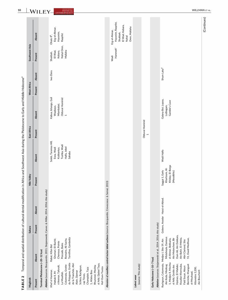
TABLE2
Tem
poraland
spatiald
istributionofcu
lturalde
ntalmodificationin
Africaan
dSo
uthw
estAsiadu
ring
thePleistocene
toEarlyan
dMiddle
Holocenea
Mag
hreb
Saha
raNile
Valley
EastAfrica
WestAfrica
SouthwestAsia
Present
Absen
tPresent
Absen
tPresent
Absen
tPresent
Absen
tPresent
Absent
Present
Absent
TerminalPleistoce
ne(~20–1
2ky
a)
Ablation(sources:Bocq
uentin,2
011;S
tojano
wski,Carve
r,&Miller,2
014,2
016;thisstud
y)
Khe
fel
Ham
mar,
HattabII,
lfri
n'Ammar,T
aforalt,
LaMouillah,
Columna
ta,G
rotte
duPolygo
ne,G
rotte
delaTranc
hée,Abri
Alain,D
jemar
Schk
ra,R
achg
oun
,
capTèn
es,
Cha
mplain,
Taza
1,A
falouBou
Rhu
mmel,M
echta
elArbi,Djebe
lTaya,
Kef-O
um-Touiza
Rab
at,K
ifan
Bel
Gho
mari,El-Bachir,
Che
noua
,Pointe
Pescade
,Bains
Romains,A
liSa
cha,
Ternifine
,Gam
betta
Soleb,
Taram
saHill,
Esna,W
adi
Kub
baniya,
Tushk
a,W
adi
Halfa,Jeb
el
Saha
ba
Kab
ua,lshan
go,G
uli
Waaba
yo,
Mlamba
lasi,
Olduv
aiHominid
1
lwoEleru
Shukb
ah,
ElW
ad,
Keb
ara,
Nah
alOren,
Mallaha
OhaloIIb,
Erg-el-Ahm
ar,
Hayonim
,
Raq
efet
Abrasionof
maxillarycentralincisor
labial
surfaces
(source:B
ocq
uentin,C
reve
coeu
r,&Se
mal,2
013)
Wad
i
Ham
meh
c
Erg-el-Ahm
ar,
Hayonim
,Raq
efet,
Shukb
ah,
ElW
ad,K
ebara,
Nah
al
Oren,M
allaha
Labret
wear
(source:T
hisstud
y)
Olduv
aiHominid
1
Early
Holoce
ne(~10–7
kya)
Ablation(sources:Stojano
wskie
tal.,2014,2
016;thisstud
y)
Rachg
oun
,Columna
ta,
Ain
Ked
a,Mesloug
1,M
edjezII,
Mechta
elArbi,Grottede
s
Hyè
nes,ElM
ahde
r,
Koud
iatKhe
rroub
a,
FaidSo
uar,Aioun
Berrich
e,Kha
ngue
t
el-M
ouh
aad,
Kilo
mètre
3,200,
Ain
Bouc
herit
Med
jezI,Site
59,A
in
Mistehe
yia,Dam
ous
el-A
hmar,B
ekkaria,
Ain
Meterch
em,B
ir
Oum
Ali,Ain
Dokk
ara,
Ain
Bah
ir,E
l-Mek
ta,
AbriC
lariond
,Site
51,O
uedMed
foun
,
Gobe
ro,A
sselar
Hassi-el-Abiod.
Sagg
ai1,E
arly
Kha
rtoum
,Al-
Khida
y,El-Barga
(Mesolithic)
Wad
iHalfa
Galan
aBoi,Lo
poy,
Lothagam
,
Gam
ble'sCave
Shum
Laka
d
(Continues)
10 WILLMAN ET AL.

TABLE2
(Continue
d)
Mag
hreb
Saha
raNile
Valley
EastAfrica
WestAfrica
SouthwestAsia
Present
Absen
tPresent
Absen
tPresent
Absen
tPresent
Absen
tPresent
Absent
Present
Absent
Labret
wear(source:T
hisstud
y)
el-B
arga
(Neo
lithic),
AlK
hida
y
1(16-D
-4),Al
Khida
y2(16-D
-5)
el-B
arga
(Mesolithic)
MiddleHoloce
ne(~7–3
kyad)
Ablation(sources:Stojano
wskie
tal.,2014,2
016;thisstud
y)
Doua
rDeb
agh,
Dar-
es-Soltan
16.
Sebk
haAmtal,
Tintan,
Cha
mi,Rio
Salado
,Grottede
s
troglody
tes,El
Cua
rtel,C
hamplain,
Djebe
lFartas,ke
fel
Agab
Mug
haretel'Aliya,Izriten,
Sebk
haLaasailia,
Sebk
haMah
ariat,
Sebk
haEdjaila,S
ebkh
a
Lemhe
iris,A
liBacha
,
LaMeskian
a,
Tessalit-Reg
de
Zaki,Kesret-
el-G
anl,
Amek
ni
1,Iba
lagh
en,
Koba
di,
Gobe
ro,W
adi
Howar,Y
ao
Uan
Muh
uggiag,
EmiL
ulu,
Adrar
Bous,
Iwelen
,
Hassi-el-
Abiod,
Men
tes,
Tam
ayaMellet,
inTud
uf,C
hin
Tafidet,A
funfun
,
Areschima,
Kouroun
korokale
Nab
taPlaya,Jeb
el
Shaq
adud
,Jeb
el
Moya,E
l-Barga
(Neo
lithic)
Wad
iHalfa,R
12
Njoro
River
Cave,
Willey
's
Kopje,
Naivasha
Railw
aySite,
Luke
nyaHill
(GvJm
202),
Ngo
rong
oro
Crater
Lowasera,Laikipia,
Hyrax
Hill,C
ole's
BurialS
ite,
Makalia
BurialS
ite,
Bromhe
ad's
Site,M
t.Su
swa
Rop,Laakp
a,
Shum
Laka?e
Chipp
ingan
dFiling(source:Irish,2
017)
Karkarich
inkat
Nord
Mouh
oun
Ben
d
Labret
wear(source:T
hisstud
y)
Tagno
ut
Cha
ggeret
Jebe
lMoya,A
l
Khida
y2(16-D
-5),
UA53
a“Present”or“A
bsen
t”co
lumns
indicateswhe
ther
thereisdo
cumen
tedev
iden
ce(present)o
rno
t(absen
t)ofagive
nform
ofde
ntalmodificationat
that
site.P
resence
oflabrets
islim
ited
toacco
unts
intheliterature
that
explicitly
note
wea
rcaused
byornam
ents
worn
infacialpiercing
s,orat
leastthemen
tionofan
insitu
labret,inclose
associationwiththejaws,that
cann
otbeco
nfusedwithornam
ents
worn
inea
rpiercings
(MacDona
ld,1
999).
bObservations
byone
ofus
(J.C
.Willman
)sug
gest
that
intentiona
linc
isorab
lationcann
otbe
ruledout
forOha
loIIH2.
c The
labialsurfaceab
rasionontheI1'sofW
adiH
ammeh
26Homo4issugg
estive
ofintentiona
lbody
modification,
buttask-related
beha
viors
cann
otbe
ruledout(Bocq
uen
tinet
al.,2013).
dW
estAfrican
sitesareas
youn
gas
2ky
agive
nho
wfew
sitesaredo
cumen
tedin
theregion(Stojano
wskie
tal.,2016).
eIrish(2017)n
otes“potential”UI1
ablationforSh
umLaka.
WILLMAN ET AL. 11

of ornaments and way(s) in which the ornaments are worn—key infor-
mation for understanding the temporospatial patterning of labret use
and its cultural significance to the wearer(s). In this vein, we posit that
it is unlikely that numerous, small labrets—like those from Jebel Moya
(see below)—would have produced the broad and uniform wear planes
on the OH1 dentition. Instead, we propose that three ornaments were
worn in three separate piercings (two buccal and one lower lip or
“medial”).
The precursor to wearing labrets is the act of facial piercing—an
event, or events, that are often associated with initiatory rites and the
transformation of one's social identity (Seeger, 1975; Turner, 1980;
LaTosky, 2006; Torres-Rouff, 2011; Reddish, 2013). Furthermore, the
piercings were likely to have been small at first but stretched over
time to incorporate increasingly larger labrets (e.g., Seeger, 1975;
Turner, 1980; LaTosky, 2006; Reddish, 2013). The degree of wear
across all three proposed piercing sites indicates that OH1 wore orna-
ments for long enough to create the substantial wear facets. The
labrets worn by OH1 not only mark an outwardly visible marker of
social identity but also the embodiment of the process of identity
transformation and/or social maturation.
Temporospatial distribution of body modification in African prehis-
tory: Body modification practices that result in observable trans-
figurement of skeletal and dental tissues have long been of interest to
scholars of prehistoric population movements, biological affinity, and
cultural practices in Africa (De Groote & Humphrey, 2016; Hum-
phrey & Bocaege, 2008; Irish, 2017; L. S. B. Leakey, 1928; Mollison,
1929; Parsche, 1993; Stojanowski et al., 2014, 2016; van Reenen,
1978, 1986). A particularly prominent practice of body modification in
Late Pleistocene and Holocene Africa is ablation, or the culturally
motivated practice of anterior tooth (incisor, canine, and sometimes
TABLE 3 Late Pleistocene to mid-Holocene facial piercings documented through dental wear and/or ornaments found in association withburials
Site Location Chronology 14C yearsEvidence of labretuse/description from literature Sources
Olduvai (Oldupai) Northern Tanzania Late Pleistocene n/a Anterior and buccal faceting
suggestive of one medial and
two lateral piercings for OH1.
No ornaments recovered from
burial.
This study
16-D-5 (Al-Khiday 2) Central Sudan Early Mesolithic 7,980 ± 40–7,710 ± 40 Stone lip plugs recovered that
are typologically different
from el-Barga types.
Salvatori & Usai, 2016;
Salvatori, Usai, &
Zerboni, 2011
16-D-4 (Al-Khiday 1) Central Sudan Middle Mesolithic 7,760 ± 90–7,530 ± 100 Lip-plug found in situ with
mandible in burial.
Salvatori & Usai, 2009;
Salvatori et al., 2011
el-Barga Northern Sudan Early Neolithic 7,045 ± 70–6,605 ± 60 Lip plugs in situ near upper
and/or lower lips. Most
commonly one lip plug,
although 2, 4, or 6 were found
in some burials. Elongated,
thickened at ends, carved from
ivory or stone (amazonite).
Honegger, 2004;
Honegger &
Williams, 2015
16-D-5 (Al-Khiday 2) Central Sudan Neolithic 5,470 ± 50 Lip plugs associated with grave
103 typical of Khartoum
Neolithic. Very rich grave
offerings.
Salvatori & Usai, 2014;
Salvatori et al., 2011
Tagnout Chaggeret Mali Neolithic 4,520 ± 100 Quartz labret associated with
burial.
MacDonald, 1999;
Petit-Maire & Riser,
1983
UA 53 Eastern Sudan Late Butana Group Fourth to early third
millennium BC
9 lip plugs associated with burial. Manzo, 2017
Jebel Moya South-Central
Sudan
Multi-phase
pastoralist
cemetery
3,245 ± 755–1,545± 535a
1 to 6 polished stone, ceramic,
bone, or ivory labrets found in
27% of graves. Labret wear on
47% of females, but 14% of
these individuals had no
labrets in graves. Labret wear
and mandibular incisor
ablation co-occur in some
individuals.
Addison, 1949; Brass &
Schwenniger, 2013;
MacDonald, 1999
aOptically stimulated luminescence (OSL) dates obtained from ceramics calibrated to years before 2012 (Brass & Schwenniger, 2013).
12 WILLMAN ET AL.

premolar) removal during an individual's lifetime. Ablation is well-
documented in the Late Pleistocene Old World (Willman,
Shackelford, & Demeter, 2016), and most thoroughly documented in
Africa (De Groote & Humphrey, 2016; Humphrey & Bocaege, 2008;
Stojanowski et al., 2014, 2016) and Natufian contexts in Southwest
Asia (Bocquentin, 2011). Another prominent pattern of body modifi-
cation involves the chipping and filing of the anterior dentition, which
is frequently associated with the “Bantu Expansion” (Irish, 2017; L. S.
B. Leakey, 1928; van Reenen, 1978, 1986). Despite extensive discus-
sion of ablation, chipping, and filing of teeth, very little discussion of
facial piercing is found in the literature. What follows is a brief review
of prehistoric labret-use in Africa and an integration of its tempo-
rospatial distribution into the discussion of body modification in Afri-
can prehistory.
The temporal and spatial trends for ablation strongly support a Ter-
minal Pleistocene (~18–12 kya) origin for the practice in the Maghreb,
subsequent diffusion into the Sahara and Nile Valley during the Early
Holocene (~10–7 kya), and a widespread distribution throughout Africa
during the Middle Holocene (~7–3 kya) (Stojanowski et al., 2014, 2016)
(Table 2). In contrast, there is complete absence of incisor and canine
chipping and filing in Africa until the Middle Holocene when examples
are first documented in the Sahara and West Africa (Finucane et al.,
2008; Irish, 2017; Maes, Irish, Holl, Walker, & Armelagos, 2004)
(Table 2). Following the “Bantu Expansion,” chipping and filing become
widespread despite being largely absent in sub-Saharan Africa prior to
the Bantu expansion (Irish, 2017).
The spatial distribution of prehistoric labret-use is limited to sev-
eral sites in the Nile Valley, Tagnout Chaggeret in the Sahara, and
Olduvai Gorge in East Africa (Tables 2 and 3). The probable Late Pleis-
tocene affiliation of OH1 makes it either the earliest case or pene-
contemporaneous with Early and Middle Mesolithic examples from
Al-Khiday (Salvatori & Usai, 2009, 2016). Labrets from Neolithic con-
texts differ in style from those of early periods in the Nile Valley
(Salvatori & Usai, 2016), and both labrets and dental wear associated
with labrets are found in great quantities among individuals interred in
the later pastoralist cemeteries at Jebel Moya (Addison, 1949; Brass &
Schwenniger, 2013; MacDonald, 1999) (Tables 2 and 3). Interestingly,
some degree of replacement between the Mesolithic and Early Neo-
lithic populations is probable at el-Barga on the basis of skeletal and
dental morphology between the Mesolithic and Early Neolithic indi-
viduals (Benoiston et al., 2018; Crevecoeur, Desideri, Chaix, & Honeg-
ger, 2012), which corresponds well with distinct differences in labret
styles recorded between Sudanese Mesolithic and Neolithic sites
summarized here (Table 3). Labrets, defined on stylistic grounds,
rather than association with burials or dental wear, are frequently
documented in archaeological contexts within the Nile Valley
(e.g., Arkell, 1949; Bobrowski, Jórdeczka, Sobkowiak-Tabaka, &
Binder, 2016; Chłodnicki & Kabaci�nski, 2015; Dachy et al., 2018;
Fernández, Jimeno, & Menéndez, 2003; Geus & Lecointe, 2003;
McDonald, 2016), and elsewhere in Africa (Gaussen & Gaussen, 1962;
MacDonald, 1999), and largely reinforce the temporospatial distribu-
tion presented here on the basis of labret-induced wear and labrets
associated with burials.
OH1 is one of the earliest examples of labret-use in Africa and
marks the southern-most extent of the currently known distribution
for the practice during the Late Pleistocene and early Holocene. Fur-
thermore, OH1 exhibits a unique labret configuration of buccal and
lower lip piercings—a markedly different pattern than that docu-
mented at sites from the Mesolithic and Neolithic Nile Valley
(Table 3). Thus, the OH1 body modification practices are temporally,
geographically, and stylistically idiosyncratic when compared to pene-
contemporaneous body modification practices in Africa.
5 | CONCLUSION
Little had been known about the paleobiology of OH1 prior to recent
analyses indicating that the young adult male (~20–35 years old at
death) was deliberately buried (Matu et al., 2017), and shares aspects
of cranial and postcranial morphology with other Late Pleistocene fos-
sils from Africa (Crevecoeur et al., 2016; Tryon et al., 2015). The
osteobiography of OH1 now provides evidence for the use of multiple
ornaments worn through facial piercings that would have outwardly
expressed, and embodied, aspects of individual and/or group-level
social identity.
Extensive morphological variation has been documented within
the sparse human fossil record from Late Pleistocene and Early Holo-
cene Africa, which suggests that we are only beginning to understand
how much variation existed prior to the biological homogenization
that occurred during the late Holocene and historic periods. Likewise,
the evidence for labret-use presented here adds to the diversity of
body modification practices already documented elsewhere in Africa
during the Late Pleistocene (e.g., dental ablation) and Early to Middle
Holocene (e.g., dental ablation, chipping, filing, and labret use). We
have provided a detailed analysis of labret wear, and a review of simi-
lar cases, to stimulate further documentation of this cultural practice
that together with inferences from biological variation and archeolo-
gical investigations, can provide additional means of understanding
inter- and intraregional population dynamics and interactions among
prehistoric peoples across Late Pleistocene and early Holocene Africa.
ACKNOWLEDGMENTS
George McGlynn and Mike Schweissing (Staatssammlung für
Anthropologie und Paläoanatomie) provided access to and assistance
with the OH1 skeletal material. Donatella Usai generously provided
information on the Al Khiday material, and G. Richard Scott provided
helpful suggestions and encouragement during the early stages of
research. The reassessment of the morphometric data of OH1 was
funded by the project “Big Dry: Ruptures et continuité dans le
peuplement de l'Afrique à la fin du Pléistocène: paléoanthropologie,
paléoenvironnement et archéologies comparées du Rift et du Nil dans
leur cadre continental” of the Agence Nationale de la Recherche
(ANR-14-CE31). JCW is supported by funding from the Marie
Skłodowska-Curie Actions (H2020-MSCA-IF-2016 No. 749188),
AGAUR (Ref. 2017SGR1040) and URV (Ref. 2017PFR-URV-B2-91)
Projects, and MICINN/FEDER (Ref. PGC2018-093925-B-C32). RH is
WILLMAN ET AL. 13

supported by a Martí i Franquès doctoral research fellowship
(2019PMF-PIPF-59). An associate editor and two anonymous
reviewers provided critical insights that greatly improved this article.
To all we are grateful.
DATA AVAILABILITY STATEMENT
All data related to the description and measurement of Olduvai Homi-
nid 1 are published in this manuscript. The comparative fossil human
dataset is not publicly available but may be made available to
researchers on a case-by-case basis by contacting Isabelle
Crevecoeur.
ORCID
John C. Willman https://orcid.org/0000-0001-7143-4533
Raquel Hernando https://orcid.org/0000-0002-4873-0657
REFERENCES
Aanestad, S., & Poulsen, S. (1996). Oral conditions related to use of the lip
plug (ndonya) among the Makonde tribe in Tanzania. Acta
Odontologica Scandinavica, 54(6), 362–364.Addison, F. (1949). The Wellcome excavations in The Sudan: Jebel Moya, vol-
ume 1: Jebel Moya, 1910–1914. Oxford: Oxford University Press.
Aigner, J. S., & Veltre, D. (1976). The distribution and pattern of Umqan
burial on southwest Umnak Island. Arctic Anthropology, 13(2),
113–127. https://doi.org/10.2307/40283945Alt, K. W., & Pichler, S. L. (1998). Artificial modifications of human teeth. In
K. W. Alt, F. W. Rösing, & M. Teschler-Nicola (Eds.), Dental anthropol-
ogy: Fundamentals, limits, and prospects (pp. 387–415). Wien: Springer
Verlag.
Arambourg, C., Boule, M., Vallois, H.-V., & Verneau, R. (1934). Les Grottes
Paléolithiques des Beni-Segoual (Algérie). Paris: Masson.
Arcini, C. (2005). The Vikings bare their filed teeth. American Journal of
Physical Anthropology, 128(4), 727–733. https://doi.org/10.1002/ajpa.20164
Arkell, A. J. (1949). Early Khartoum. London: Oxford University Press.
Armelagos, G., Van Gerven, D., Martin, D., & Huss-Ashmore, R. (1984).
Effects of nutritional change on the skeletal biology of northeast Afri-
can (Sudanese Nubian) populations. In J. Clark & S. Brandt (Eds.), From
hunters to farmers: The causes and consequences of food production in
Africa (pp. 132–146). Berkeley: University Press.
Arsuaga, J. L., Gracia, A., Martínez, I., Bermúdez de Castro, J. M., Rosas, A.,
Villaverde, V., & Fumanal, M. P. (1989). The human remains from Cova
Negra (Valencia, Spain) and their place in European Pleistocene human
evolution. Journal of Human Evolution, 18(1), 55–92. https://doi.org/10.1016/0047-2484(89)90023-7
Barrett, M. J. (1977). Masticatory and non-masticatory uses of teeth. In
R. V. S. Wright (Ed.), Stone tools as cultural markers: Change, evolution and
complexity (pp. 18–23). Canberra: Australian Institute of Aboriginal Studies.
Barton, N., Bouzouggar, A., Humphrey, L. T., Berridge, P., Collcutt, S.,
Gale, R., … Schwenninger, J.-L. (2008). Human burial evidence from
Hattab II cave and the question of continuity in late Pleistocene–Holocene mortuary practices in Northwest Africa. Cambridge Archaeo-
logical Journal, 18(02), 195–214.Benoiston, A.-S., Bayle, P., & Crevecoeur, I. (2018). Biological affinity of
the Mesolithic and Neolithic populations from el-Barga, Sudan: The
dental remains. In M. Honegger (Ed.), Nubian archaeology in the XXIst
century: Proceedings of the thirteenth international conference for Nubian
studies, Neuchâtel, 1st-6th September 2014 (pp. 805–816). Leuven:
Peeters.
Bermúdez de Castro, J. M., Bromage, T. G., & Jalvo, Y. F. (1988). Buccal
striations on fossil human anterior teeth: Evidence of handedness in
the middle and early upper Pleistocene. Journal of Human Evolution, 17
(4), 403–412. https://doi.org/10.1016/0047-2484(88)90029-2Bobrowski, P., Jórdeczka, M., Sobkowiak-Tabaka, I., & Binder, M. (2016).
Khor Shambat 1: New Neolithic site and cemetery in Omdurman
(Sudan). Polish Archeology in the Mediterranean, 25, 447–478. https://doi.org/10.5604/01.3001.0010.1870
Bocquentin, F. (2011). Avulsions dentaires et identité régionale chez les
Natoufiens. Tüba-Ar (Turkish Academy of Sciences Journal of Archaeol-
ogy), 14, 261–270.Bocquentin, F., Crevecoeur, I., & Semal, P. (2013). Artificial modification of
the central upper incisors of Homo 4 (plot XX J burial). In
P. C. Edwards (Ed.), Wadi Hammeh 27, an early Natufian settlement at
Pella in Jordan (pp. 383–387). Leiden: Brill.Bonfiglioli, B., Mariotti, V., Facchini, F., Belcastro, M. G., & Condemi, S.
(2004). Masticatory and non-masticatory dental modifications in the
Epipalaeolithic necropolis of Taforalt (Morocco). International Journal
of Osteoarchaeology, 14(6), 448–456. https://doi.org/10.1002/oa.726Boswell, P. G. H. (1932). The Olduvai human skeleton. Nature, 130,
237–238. https://doi.org/10.1038/130237b0Brace, C. L. (1975). Comment on “did La Ferrassie I use his teeth as tools?”.
Current Anthropology, 16, 396–397.Brass, M., & Schwenniger, J.-L. (2013). Jebel Moya (Sudan): New dates
from a mortuary complex at the southern Meroitic frontier. Azania:
Archaeological Research in Africa, 48(4), 455–472. https://doi.org/10.1080/0067270X.2013.843258
Bräuer, G. (1988). Osteometrie. In R. Knusmann (Ed.), Antropologie: Hand-
buch der vergleichenden Biologie des Menschen, Band 1 (pp. 160–231).Stuttgart: Spektrum Akademischer Verlag.
Brown, T., & Molnar, S. (1990). Interproximal grooving and task activity in
Australia. American Journal of Physical Anthropology, 81, 545–553.https://doi.org/10.1002/ajpa.1330810410
Burnett, S. E., & Irish, J. D. (2017). An introduction to a worldview of bio-
culturally modified teeth. In S. E. Burnett & J. D. Irish (Eds.), A world
view of bioculturally modified teeth (pp. 1–16). Gainesville: UniversityPress of Florida.
Chłodnicki, M., & Kabaci�nski, J. (2015). Radiocarbon dates from Kadero
revised. In J. Kabaci�nski, M. Chłodnicki, & M. Kobusiewicz (Eds.),
Hunter-gatherers and early food producing societies in northeastern
Africa Studies in African Archaeology (Vol. 14, pp. 195–217). Pozna�n:Pozna�n Archaeological Museum.
Colette, J. (1933). Le labret en Afrique et en Amerique. Bulletin de la
Societé Des Americanistes de Belgique, 13, 5–61.Cook, D. C., Bastos, M. Q. R., Lopes, C., Mendonça de Souza, S., &
Santos, R. V. (2015). Pretos Novos: Evidence for African oral hygiene
practices in Brazil, 1769–1830. International Journal of
Osteoarchaeology, 25(2), 238–244. https://doi.org/10.1002/oa.2278Coupal, I., & Sołtysiak, A. (2017). Dental erosion in archaeological human
remains: A critical review of literature and proposal of a differential
diagnosis protocol. Archives of Oral Biology, 84(Supplement C), 50–57.https://doi.org/10.1016/j.archoralbio.2017.09.011
Crevecoeur, I. (2008). Étude Anthropologique du Squelette du Paléolithique
Supérieur de Nazlet Khater 2 (Égypte). Leuven. Leuven: University Press.
Crevecoeur, I., & Trinkaus, E. (2004). From the Nile to the Danube: A com-
parison of the Nazlet Khater 2 and Oase 1 early modern human man-
dibles. L'Anthropologie, 42(3), 203–213.Crevecoeur, I., Brooks, A., Ribot, I., Cornelissen, E., & Semal, P. (2016). Late
stone age human remains from Ishango (Democratic Republic of
Congo): New insights on late Pleistocene modern human diversity in
Africa. Journal of Human Evolution, 96, 35–57. https://doi.org/10.
1016/j.jhevol.2016.04.003
Crevecoeur, I., Desideri, J., Chaix, L., & Honegger, M. (2012). First anthropological
insights on the early Holocene funerary assemblages from El-Barga. Docu-
ments de la Mission archéologique Suisse Au Soudan, 4, 19–28.Crevecoeur, I., Rougier, H., Grine, F. E., & Froment, A. (2009). Modern
human cranial diversity in the late Pleistocene of Africa and Eurasia:
14 WILLMAN ET AL.

Evidence from Nazlet Khater, Pestera cu Oase, and Hofmeyr. American
Journal of Physical Anthropology, 140(2), 347–358. https://doi.org/10.1002/ajpa.21080
Croucher, K. (2012). Death and dying in the Neolithic near east. Oxford Uni-
versity Press, Oxford.
Cucina, A., Herrera Atoche, R., & Chatters, J. C. (2019). Oral health and
diet of a young late Pleistocene woman from Quintana Roo, Mexico.
American Journal of Physical Anthropology, 170(2), 246–259.Curtin, A. J. (1984). Human skeletal remains from Namu (ELSx 1): A
descriptive analysis. Thesis. Burnaby: Simon Fraser University.
Cybulski, J. S. (1974). Tooth wear and material culture: Precontact patterns
in the Tsimshian area. British Columbia. Syesis, 7, 31–35.Cybulski, J. S. (2010). Labrets and teeth on the Northwest Coast. In
R. G. Matson (Ed.), The Crescent Beach site and the place of the Locarno
Beach phase. Vancouver: University of British Columbia.
Cybulski, J. S., Balkwill, D., Young, G. S., & Sutherland, P. D. (1992). A Green-
ville burial ground: Human remains and mortuary elements in British Colum-
bia coast prehistory. Quebec: Canadian Museum of Civilization Hull.
d'Incau, E., Couture, C., & Maureille, B. (2012). Human tooth wear in the
past and the present: Tribological mechanisms, scoring systems, dental
and skeletal compensations. Archives of Oral Biology, 57(3), 214–229.https://doi.org/10.1016/j.archoralbio.2011.08.021
Dachy, T., Briois, F., Marchand, S., Minotti, M., Lesur, J., & Wuttmann, M.
(2018). Living in an Egyptian oasis: Reconstruction of the Holocene
archaeological sequence in Kharga. African Archaeological Review, 35
(4), 531–566. https://doi.org/10.1007/s10437-018-9306-2Darroch, J. N., & Mosimann, J. E. (1985). Canonical and principal compo-
nents of shape. Biometrika, 72(2), 241–252. https://doi.org/10.1093/biomet/72.2.241
De Groote, I., & Humphrey, L. T. (2016). Characterizing evulsion in the
later stone age Maghreb: Age, sex and effects on mastication. Quater-
nary International, 413(Part A), 50–61. https://doi.org/10.1016/j.
quaint.2015.08.082
Dietze, S., Winkelmann, D., Garve, R., Blens, T., Fanghänel, J., Proff, P., …Maile, S. (2007). Ritually induced growth disturbances and deformities
of the orofacial system – A contribution to cranial morphogenesis.
Annals of Anatomy, 189(3), 304–308. https://doi.org/10.1016/j.aanat.2006.11.009
Drozdová, E. (2002). A rediscovered fragment of a human mandible from
Predmostí u Prerova (Czech Republic): Predmostí 21. Bulletins et
Mémoires de la Société d'Anthropologie de Paris, 14(1–2), 2–13.Erdal, Y. S. (2008). Occlusal grooves in anterior dentition among
Kovuklukaya inhabitants (Sinop, northern Anatolia, 10th century AD).
International Journal of Osteoarchaeology, 18(2), 152–166. https://doi.org/10.1002/oa.925
Erdal, Y. S. (2013). Life and death at Hakemi use. In O. Nieuwenhuyse,
A. Russell, P. Akkermans, & R. Bernbeck (Eds.), Interpreting the late
Neolithic of upper Mesopotamia (pp. 213–224). Turnhout: Brepols
Publishers.
Ferembach, D., Dastugue, J., & Poitrat-Targowla, M. (1962). La Nécropole
Épipaléolithique de Taforalt, Maroc Oriental: Etudes des Squelettes
Humain. Rabat: C.N.R.S.
Fernández, V. M., Jimeno, A., & Menéndez, M. (2003). Archaeological
excavations in prehistoric sites of the Blue Nile area, Central Sudan
Excavaciones arqueológicas en yacimientos prehistóricos del Nilo Azul.
Sudán Central. Complutum, 14, 273–344.Finucane, B., Manning, K., & Touré, M. (2008). Prehistoric dental modifica-
tion in West Africa–early evidence from Karkarichinkat Nord, Mali.
International Journal of Osteoarchaeology, 18(6), 632–640. https://doi.org/10.1002/oa.957
Formicola, V. (1988). Interproximal grooving of teeth: Additional evidence
and interpretation. Current Anthropology, 29(4), 663–671.Galbany, J., Martínez, L. M., & Pérez-Pérez, A. (2004). Tooth replication
techniques, SEM imaging and microwear analysis in primates: Method-
ological obstacles. L'Anthropologie, 42, 5–12.
Garve, R., Garve, M., Türp, J. C., & Meyer, C. G. (2017). Labrets in Africa
and Amazonia: Medical implications and cultural determinants. Tropical
Medicine & International Health, 22(2), 232–240. https://doi.org/10.1111/tmi.12812
Gaussen, J., & Gaussen, M. (1962). Aperçu sur les divers faciès
néolithiques du Tilemsi et nouveaux objets en quartz poli. Bulletin De
La Société Préhistorique Française, 59(1–2), 98–108.Gauthier, J.-G., & Wangermez, J. (1964). Caractères et variations
morphologiques des Fali du Tinguelin, Nord-Cameroun. Bulletins et
Mémoires de la Société d'Anthropologie de Paris, 6(4), 585–610. https://doi.org/10.3406/bmsap.1964.1291
Geus, F., & Lecointe, Y. (2003). Survey and excavation at el-Multaga, a
resettlement area related to the construction of the Merowe dam:
Preliminary results. Sudan & Nubia, 7, 33–40.Greene, D. L., Ewing, G. H., & Armelagos, G. J. (1967). Dentition of a meso-
lithic population from Wadi Halfa, Sudan. American Journal of Physical
Anthropology, 27(1), 41–55. https://doi.org/10.1002/ajpa.
1330270107
Gieseler, W., & Mollison, T. (1929). Untersuchungen über den
Oldowayfund. Der Fossilzustand und der Schadel. Verhandlungen der
Gesellschaft der Physischen. L'Anthropologie, 3, 60–67.Grine, F. E. (2016). The late quaternary Hominins of Africa: The skeletal
evidence from MIS 6-2. In C. S. Jones & A. B. Stewart (Eds.), Africa
from MIS 6–2: Population dynamics and Paleoenvironments
(pp. 323–381). Dordrecht: Springer Netherlands. https://doi.org/10.
1007/978-94-017-7520-5_17
Grine, F. E., Marean, C. W., Faith, J. T., Black, W., Mongle, C. S.,
Trinkaus, E., … du Plessis, A. (2017). Further human fossils from the
middle stone age deposits of die Kelders cave 1, Western Cape Prov-
ince, South Africa. Journal of Human Evolution, 109, 70–78. https://doi.org/10.1016/j.jhevol.2017.05.009
Gupta, K. (1986). "Ndonya" or lip plug in the Wamakonde Women. Tanza-
nia Dental Journal, 3(1), 37–38.Hachi, S. (1996). L'Ibéromaurusien, découvertes des fouilles d'Afalou.
L'Anthropologie, 100, 55–76.Harvati, K., Stringer, C., Grün, R., Aubert, M., Allsworth-Jones, P., &
Folorunso, C. A. (2011). The later stone age calvaria from Iwo Eleru,
Nigeria: Morphology and chronology. PLoS One, 6(9), e24024. https://
doi.org/10.1371/journal.pone.0024024
Hennequin-Hoenderdos, N. L., Slot, D. E., & Van der Weijden, G. A.
(2016). The incidence of complications associated with lip and/or
tongue piercings: A systematic review. International Journal of Dental
Hygiene, 14(1), 62–73. https://doi.org/10.1111/idh.12118Hillson, S. W. (2006). Dental morphology, proportions, and attrition. In
E. Trinkaus & J. Svoboda (Eds.), HEarly Modern Human Evolution in Cen-
tral Europe: The People of Dolní Vĕstonice and Pavlov (pp. 179–223).New York: Oxford University Press.
Hinton, R. J. (1981). Form and patterning of anterior tooth wear among
aboriginal human groups. American Journal of Physical Anthropology, 54
(4), 555–564.Hole, F., Flannery, K., & Neely, J. (1969). Prehistory and Human Ecology of
the Deh Luran Plain: An Early Village Sequence from Khuzistan, Iran. Ann
Arbor: Memoirs of the Museum of Anthropology University of
Michigan.
Holliday, T. W. (2015). Population affinities of the Jebel Sahaba skeletal
sample: Limb proportion evidence. International Journal of
Osteoarchaeology, 25(4), 466–476. https://doi.org/10.1002/oa.2315Honegger, M. (2004). Settlement and cemeteries of the Mesolithic and
early Neolithic at el-Barga (Kerma region). Sudan and Nubia, 8, 27–32.Honegger, M., & Williams, M. (2015). Human occupations and environmen-
tal changes in the Nile valley during the Holocene: The case of Kerma
in upper Nubia (northern Sudan). Quaternary Science Reviews, 130
(2015, 141–154. https://doi.org/10.1016/j.quascirev.2015.06.031Humphrey, L. T., Bello, S. M., Turner, E., Bouzouggar, A., & Barton, N.
(2012). Iberomaurusian funerary behaviour: Evidence from Grotte des
WILLMAN ET AL. 15

pigeons, Taforalt, Morocco. Journal of Human Evolution, 62(2),
261–273. https://doi.org/10.1016/j.jhevol.2011.11.003Humphrey, L. T., & Bocaege, E. (2008). Tooth evulsion in the Maghreb:
Chronological and geographical patterns. African Archaeological
Review, 25(1–2), 109–123. https://doi.org/10.1007/s10437-008-
9022-4
Huntingford, G. W. B. (1961). The distribution of certain culture elements
in East Africa. Journal of the Anthropological Institute of Great Britain
and Ireland, 91, 251–295.Ikehara-Quebral, R., Ryan, E. M., Parr, N., Walth, C., Liston, J.,
Pietrusewsky, M., & Douglas, M. T. (2017). Intentional dental modifica-
tion and oral-dental health in Western Micronesia. In S. E. Burnett &
J. D. Irish (Eds.), A worldview of bioculturally modified teeth
(pp. 193–210). Gainesville: University Press of Florida.
Indriati, E., & Buikstra, J. E. (2001). Coca chewing in prehistoric coastal
Peru: Dental evidence. American Journal of Physical Anthropology, 114
(3), 242–257. https://doi.org/10.1002/1096-8644(200103)114:3%
3C242::AID-AJPA1023%3E3.0.CO;2-J
Insoll, T. (2015). Material explorations in African archaeology. Oxford:
Oxford University Press.
Irish, J. D. (2000). The Iberomaurusian enigma: North African progenitor or
dead end? Journal of Human Evolution, 39(4), 393–410. https://doi.org/10.1006/jhev.2000.0430
Irish, J. D. (2005). Population continuity vs. discontinuity revisited: Dental
affinities among late Paleolithic through Christian-era Nubians. Ameri-
can Journal of Physical Anthropology, 128(3), 520–535. https://doi.org/10.1002/ajpa.20109
Irish, J. D. (2017). Knocking, filing, and chipping: Dental modification in
sub-Saharan Africans. In S. E. Burnett & J. D. Irish (Eds.), A world view
of bioculturally modified teeth (pp. 33–47). Gainesville: University Press
of Florida.
Irish, J. D., Black, W., Sealy, J., & Ackermann, R. R. (2014). Questions of
Khoesan continuity: Dental affinities among the indigenous Holocene
peoples of South Africa. American Journal of Physical Anthropology, 155
(1), 33–44. https://doi.org/10.1002/ajpa.22526Irish, J. D., & Guatelli-Steinberg, D. (2003). Ancient teeth and modern
human origins: An expanded comparison of African Plio-Pleistocene
and recent world dental samples. Journal of Human Evolution, 45(2),
113–144. https://doi.org/10.1016/s0047-2484(03)00090-3Irish, J. D., & Konigsberg, L. (2007). The ancient inhabitants of Jebel Moya
redux: Measures of population affinity based on dental morphology.
International Journal of Osteoarchaeology, 17(2), 138–156. https://doi.org/10.1002/oa.868
Irish, J. D., & Turner, C. G. (1987). More lingual surface attrition of the
maxillary anterior teeth in American Indians: Prehistoric panamanians.
American Journal of Physical Anthropology, 73(2), 209–213. https://doi.org/10.1002/ajpa.1330730207
Johansson, A.-K., Omar, R., Carlsson, G. E., & Johansson, A. (2012). Dental
erosion and its growing importance in clinical practice: From past to
present. International Journal of Dentistry, 2012, 1–17. https://doi.org/10.1155/2012/632907
Jungers, W. L., Falsetti, A. B., & Wall, C. E. (1995). Shape, relative size, and
size-adjustments in morphometrics. Yearbook of Physical Anthropology,
38(S21), 137–161. https://doi.org/10.1002/ajpa.1330380608Kaidonis, J. (2008). Tooth wear: The view of the anthropologist. Clinical
Oral Investigations, 12(Suppl 1), S21–S26. https://doi.org/10.1007/
s00784-007-0154-8
Keddie, G. R. (1981). The use and distribution of labrets on the North
Pacific rim. Syesis, 14, 59–80.Kieser, J. A., Dennison, K. J., Kaidonis, J. A., Huang, D.,
Herbison, P. G. P., & Tayles, N. G. (2001). Patterns of dental wear in
the early Maori dentition. International Journal of Osteoarchaeology, 11
(3), 206–217. https://doi.org/10.1002/oa.534Koby, F. E. (1956). Une incisive néandertalienne trouvée en Suisse.
Verhandenlungen der Nuturforschers Gesellschaft in Basal, 67, 1–15.
Labouret, H. (1952). A propos des labrets en verre de quelques
populations voltaïques. Bulletin Institut Francais de'Afrique Noire, 14,
1385–1401.Lacy, S. A. (2014). Oral health and its implications in late Pleistocene
Western Eurasian humans. Saint Louis: Washington University in Saint
Louis.
Lacy, S. A. (2015). The dental metrics, morphology, and oral paleopathol-
ogy of Oberkassel 1 and 2. In R. W. Schmitz (Ed.), Giemsch L
(pp. 1–17). Damstadt: The Late Glacial Burial from Oberkassel
Revisited. Verlag Phillip von Zabern.
Larsen, C. S. (1985). Dental modification and tool use in the western Great
Basin. American Journal of Physical Anthropology, 67, 393–402. https://doi.org/10.1002/ajpa.1330670411
LaTosky, S. (2006). Reflections on lip-plates of Mursi women as source of
stigma and self-esteem. In I. Strecker & J. Lydall (Eds.), The perils of
face: Essays on cultural contact, respect and self-esteem in Southern Ethi-
opia (pp. 382–396). Berlin: Lit Verlag.Lavigne, S. E., & Molto, J. E. (1995). System of measurement of the sever-
ity of periodontal disease in past populations. International Journal of
Osteoarchaeology, 5(3), 265–273.Leakey, L. S. B. (1928). The Oldoway skull. Nature, 121, 499–500. https://
doi.org/10.1038/121499b0
Leakey, M. D. (1978). Olduvai gorge 1911–75: A history of the investiga-
tions. Geological Society, London, Special Publications, 6(1), 151–155.https://doi.org/10.1144/GSL.SP.1978.006.01.12
Lorkiewicz, W. (2011). Nonalimentary tooth use in the Neolithic popula-
tion of the Lengyel culture in Central Poland (4600-4000 BC). Ameri-
can Journal of Physical Anthropology, 144(4), 538–551. https://doi.org/10.1002/ajpa.21435
Lukacs, J., & Pastor, R. (1988). Activity-induced patterns of dental abrasion
in prehistoric Pakistan: Evidence from Mehgarh and Harappa. Ameri-
can Journal of Physical Anthropology, 76, 377–398. https://doi.org/10.1002/ajpa.1330760310
MacDonald, R. M. (1999). In the teeth of the problem: Dental anthropology
and the reconstruction of African dietary regimes. London: University
College London.
Maes, K. C., Irish, J. D., Holl, A. F. C., Walker, P. L., & Armelagos, G. J.
(2004). Preliminary analysis of dental morphology and identity of an
early iron producing population in the Mouhoun Bend, Burkina Faso.
American Journal of Physical Anthropology, Suppl. 38, 140.
Manzo, A. (2017). Eastern Sudan in its setting. The archaeology of a region
far from the Nile Valley. Oxford: Archaeopress.
Matiegka, J. (1924). Un egrisage artificiel des dents observé sur les crânes
quaternaires de Předmostí (pp. 289–293). Prague: Institut Internationald'Anthropologie.
Matiegka, J. (1929). The skull of the fossil man Brno III, and the cast of its
interior. L'Anthropologie, 7, 90–107.Matiegka, J. (1934). Homo předmostensis, fosilní clovek z Předmostí na
Morave, I, Lebky. Praha: České Akademie ved a umení.
Mattingly, D., Lahr, M., Wilson, A., Abduli, H., Ahmed, M., Baker, S., …Sterry, M. (2009). DMP V: Investigations in 2009 of cemeteries
and related sites on the west side of the Taqallit promontory.
Libyan Studies, 40, 95–131. https://doi.org/10.1017/S0263718900004520
Matu, M., Crevecoeur, I., & Huchet, J. B. (2017). Taphonomy and Paleo-
ichnology of Olduvai hominid 1 (OH1), Tanzania. International Journal
of Osteoarchaeology, 27(5), 785–800. https://doi.org/10.1002/oa.
2593
McDonald, M. M. (2016). The pattern of neolithization in Dakhleh oasis in
the eastern Sahara. Quaternary International, 410, 181–197. https://doi.org/10.1016/j.quaint.2015.10.100
Mercader, J., Garralda, M. D., Pearson, O. M., & Bailey, R. C. (2001). Eight
hundred-year-old human remains from the Ituri tropical forest, Demo-
cratic Republic of Congo: The rock shelter site of Matangai Turu
northwest. American Journal of Physical Anthropology, 115(1), 24–37.
16 WILLMAN ET AL.

Michael, J. A., Kaidonis, J. A., & Townsend, G. C. (2010). Non-carious cervi-
cal lesions on permanent anterior teeth: A new morphological classifi-
cation. Australian Dental Journal, 55, 134–137.Milner, G. R., & Larsen, C. S. (1991). Teeth as artifacts of human behavior:
Intentional mutilation and accidental modification. In M. A. Kelley &
C. S. Larsen (Eds.), Advances in dental anthropology (pp. 357–378).New York: Wiley-Liss.
Mirazón Lahr, M. (2016). The shaping of human diversity: Filters, bound-
aries and transitions. Philosophical Transactions of the Royal Society of
London B: Biological Sciences, 371(1698), 20150241. https://doi.org/
10.1098/rstb.2015.0241
Molleson, T. (2016). The ordinary Neolithic people of Abu Hureyra. In
K. T. Lillios & M. Chazan (Eds.), Fresh fields and pastures new: Papers
presented in honor of Andrew MT Moore (pp. 187–205). Leiden:
Sidestone Press.
Mollison, T. (1929). Untersuchungen iiber den Oldoway-Fund. Der
Fossilzustand und der Schadel. Verhandlungen der Gesellschaft der
Physischen. L'Anthropologie, 3, 60–67.Molnar, P. (2011). Extramasticatory dental wear reflecting habitual behav-
ior and health in past populations. Clinical Oral Investigations, 15(5),
681–689. https://doi.org/10.1007/s00784-010-0447-1Molnar, S. (1971). Human tooth wear, tooth function and cultural variabil-
ity. American Journal of Physical Anthropology, 34(2), 175–189.Molnar, S. (1972). Tooth wear and culture: A survey of tooth functions
among some prehistoric populations. Current Anthropology, 13(5),
511–526. https://doi.org/10.1086/201284Mounier, A., Correia, M., Rivera, F., Crivellaro, F., Power, R., Jeffery, J., …
Mirazón Lahr, M. (2018). Who were the Nataruk people? Mandibular
morphology among late Pleistocene and early Holocene fisher-forager
populations of West Turkana (Kenya). Journal of Human Evolution, 121,
235–253. https://doi.org/10.1016/j.jhevol.2018.04.013Mukherjee, R., Trevor, J., & Rao, C. (1955). The ancient inhabitants of Jebel
Moya. Cambridge: Cambridge University Press.
Murray, J. (1981). Prehistoric skeletons from blue jackets creek (FlUa 4),
Queen Charlotte Islands, British Columbia. Contributions to Physical
Anthropology, 1979–1980, 127–168.Ogden, A. (2008). Advances in the paleopathology of teeth and jaws. In
R. Pinhasi & S. Mays (Eds.), Advances in human paleopathology
(pp. 293–307). Chichester: John Wiley and Sons, Ltd..
Oztel, M., & Birch, P. G. (2016). Periapical abscess of a lower central incisor
associated with a tongue piercing: A case report. Dental, Oral and Cra-
niofacial Research, 2(2), 228–229.Parsche, F. (1993). Peculiarities on the incisors in the mandible of the skull
Olduvai I. Homo, 44(1), 30–36.Pedersen, P. O. (1952). Some dental aspects of anthropology. Dental
Record, 72, 170–178.Pedersen, P. O. (1955). Eine besondere form der Abnutzung von
Eskimozähnen aus Alaska. Deutsche zahnärztliche Zeitschrift, 10,
41–46.Pedersen, P. O., & Jakobsen, J. (Eds.). (1989). Teeth and jaws of Qilakisoq
mummies Meddelelser om Grønland, Man & Society, Copenhagen,
Museum Tusculanum Press, (Vol. 12).
Petit-Maire, N., & Riser, J. (1983). Sahara ou Sahel?: Quaternaire Récent du
Bassin de Taoudenni (Mali). Paris: CNRS.
Pfeiffer, S., & Harrington, L. (2018). Regional continuity and local chal-
lenges to resilience among Holocene hunter-gatherers of the greater
cape floristic region, South Africa. In C. M. Stojanowski &
D. H. Temple (Eds.), Hunter-gatherer adaptation and resilience: A bio-
archaeological perspective (pp. 26–46). Cambridge: Cambridge Univer-
sity Press.
Ponce de León, M. S., Koesbardiati, T., Weissmann, J. D., Milella, M.,
Reyna-Blanco, C. S., Suwa, G., … Zollikofer, C. P. E. (2018). Human
bony labyrinth is an indicator of population history and dispersal from
Africa. Proceedings of the National Academy of Sciences, 115(16),
4128–4133. https://doi.org/10.1073/pnas.1717873115
Protsch, R. (1974). The age and stratigraphic position of Olduvai hominid I.
Journal of Human Evolution, 3(5), 379–385. https://doi.org/10.1016/0047-2484(74)90200-0
Protsch, R. (1975). The absolute dating of upper Pleistocene SubSaharan
fossil hominids and their place in human evolution. Journal of Human
Evolution, 4(4), 297–322. https://doi.org/10.1016/0047-2484(75)
90069-X
Reck, H. (1914). Erste vorläufige Mitteilung über den Fund eines fossilen
Menschenskelets aus Zentralafrika, Berlin.
Reck, H. (1926). Prähistorische Grab-und Menschenfunde und ihre
Beziehungen zur Pluvialzeit in Ostafrika. Mittler, Berlin.
Reck, H. (1933). Oldoway, die Schlucht des Urmenschen: die Entdeckung des
altsteinzeitlichen Menschen in Deutsch-Ostafrika. FA Brockhaus.
Reddish, J. (2013). Labrets: Piercing and stretching on the northwest coast
and in Amazonia. Mundo Amazónico, 4, 57–75.Reichart, P. A., Creutz, U., & Scheifele, C. (2006). The ‘skull from Bangkok’:
A skull of a betel quid chewer in the anthropological collection of Rud-
olf Virchow (Berlin). Journal of Oral Pathology & Medicine, 35(7),
410–412. https://doi.org/10.1111/j.1600-0714.2006.00431.xRibot, I. (2011). A study through skull morphology on the diversity of Holo-
cene African populations in a historical perspective British Archaeological
Reports International Series (Vol. 2215). Oxford: Archaeopress.
Salvatori, S., & Usai, D. (2009). El Salha project 2005: New Khartoum
Mesolithic sites from Central Sudan. Kush, 19, 87–96.Salvatori, S., & Usai, D. (2014). Archaeology at El-Khiday: New insight on
the prehistory and history of Central Sudan. In J. R. Anderson &
D. A. Welsby (Eds.), The Fouth cataract and beyond: Proceedings of the
12th international conference for Nubian studies (pp. 243–257). Leuvan:Peeters.
Salvatori, S., & Usai, D. (2016). Ghaba in context. In S. Salvatori, D. Usai, &
Y. Lecointe (Eds.), Ghaba: An early Neolithic cemetery in Central Sudan
(pp. 121–134). Frankfurt: Africa Magna Verlag.
Salvatori, S., Usai, D., & Zerboni, A. (2011). Mesolithic site formation and
palaeoenvironment along the White Nile (Central Sudan). African
Archaeological Review, 28(3), 177–211. https://doi.org/10.1007/
s10437-011-9095-3
Santoni, S., Sakka, L. J., & Garcier, J.-M. (2006). Dental wear study in a
14th century skull of the Sao tribe, Cameroon. Collegium
antropologicum, 30(1), 13–24.Sawchuk, E. A., & Willoughby, P. R. (2015). Terminal Pleistocene later
stone age human remains from the Mlambalasi rock shelter, Iringa
region, southern Tanzania. International Journal of Osteoarchaeology,
25(5), 593–607. https://doi.org/10.1002/oa.2323Scerri, E. M. L., Thomas, M. G., Manica, A., Gunz, P., Stock, J. T.,
Stringer, C., … Chikhi, L. (2018). Did our species evolve in subdivided
populations across Africa, and why does it matter? Trends in Ecol-
ogy & Evolution, 33(8), 582–594. https://doi.org/10.1016/j.tree.
2018.05.005
Schoeninger, M. J., Bunn, H. T., Murray, S. S., & Marlett, J. A. (2001). Com-
position of tubers used by Hadza foragers of Tanzania. Journal of Food
Composition and Analysis, 14(1), 15–25. https://doi.org/10.1006/jfca.2000.0961
Schulz, P. D. (1977). Task activity and anterior tooth grooving in prehis-
toric California Indians. American Journal of Physical Anthropology, 46
(1), 87–91. https://doi.org/10.1002/ajpa.1330460112Schmidt, J. C., Calderaro, S., Weiger, R., & Walter, C. (2019). On the associ-
ation between oral piercings and periodontal conditions—A case
series. International Journal of Dental Hygiene, 17(4), 318–326.Schweinfurth, G. (1874). Heart of Africa: Three Years Travels and Adventures
in the Unexplored Regions of Central Africa. From 1868 to 1871.
London: Sampson Low, Marston, Low, and Searle.
Schweinfurth, G. (1875). Au Coeur de l'Afrique 1868–1871 (Vol. 2). Paris:
Librairie Hachette et Cie.
Scott, G. R., & Jolie, R. B. (2008). Tool-use and yarn production in Norse
Greenland. Alaska Journal of Anthropology, 6(1–2), 253–264.
WILLMAN ET AL. 17

Seeger, A. (1975). The meaning of body ornaments: A Suya example. Eth-
nology, 14(3), 211–224. https://doi.org/10.2307/3773253Seligman, A. F. (2015). Lip ornaments and the domestication of trade
goods: Fashion in sixteenth and seventeenth century Central East
Africa. History in Africa, 42, 357–373. https://doi.org/10.1017/hia.
2015.15
Severs, P. D. S. (1974). Archaeological investigations at blue jackets creek,
F1Ua-4, Queen Charlotte Islands, British Columbia, 1973. Bulletin
(Canadian Archaeological Association), 6, 163–205.Sperduti, A., Giuliani, M. R., Guida, G., Petrone, P. P., Rossi, P. F.,
Vaccaro, S., … Bondioli, L. (2018). Tooth grooves, occlusal striations,
dental calculus, and evidence for fiber processing in an Italian
eneolithic/bronze age cemetery. American Journal of Physical Anthro-
pology, 167(2), 234–243. https://doi.org/10.1002/ajpa.23619Stojanowski, C. M. (2014). Iwo Eleru's place among late Pleistocene
and early Holocene populations of north and East Africa. Journal of
Human Evolution, 75(0), 80–89. https://doi.org/10.1016/j.jhevol.2014.02.018
Stojanowski, C. M., Carver, C. L., & Miller, K. A. (2014). Incisor avulsion,
social identity and Saharan population history: New data from the
early Holocene southern Sahara. Journal of Anthropological Archaeol-
ogy, 35(0), 79–91. https://doi.org/10.1016/j.jaa.2014.04.007Stojanowski, C. M., Johnson, K. M., Paul, K. S., & Carver, C. L. (2016). Indi-
cators of idiosyncratic behavior in the dentition. In J. D. Irish &
G. R. Scott (Eds.), A companion to dental anthropology (pp. 377–395).Malden: John Wiley & Sons, Inc..
Taylor, R. M. S. (1986). Seakskin softening by teeth - a Maori case? The
Journal of the Polynesian Society, 95(3), 357–369.Torres-Rouff, C. (2003). Oral implications of labret use: A case from pre-
Columbian Chile. International Journal of Osteoarchaeology, 13(4),
247–251. https://doi.org/10.1002/oa.691Torres-Rouff, C. (2011). Piercing the body: Labret use, identity, and mascu-
linity in prehistoric Chile. In A. Baadsgaard, A. T. Boutin, &
J. E. Buikstra (Eds.), Breathing new life into the evidence of death: Con-
temporary approaches to bioarchaeology (pp. 256–257). Sante Fe:
School for Advanced Research Press.
Trefný, P. (2008). The dental remains from Předmostí: Morphology, met-
rics and pathology. In J. Velemínská & J. Bružek (Eds.), Early modern
humans from Předmostí: A new Reading of old documentation
(pp. 103–112). Prague: Academia.
Trinkaus, E., Lacy, S. A., & Willman, J. C. (2016). Human burials and biology
at Dolní Vĕstonice II. In J. Svoboda (Ed.), Dolní Vĕstonice II:
Chronostratigraphy, Paleoethnology, Paleoanthropology Dolní Vestonice
studies 21 (pp. 328–344). Archeologický ústav AV ČR: Brno.Tryon, C. A., Crevecoeur, I., Faith, J. T., Ekshtain, R., Nivens, J.,
Patterson, D., … Spoor, F. (2015). Late Pleistocene age and archaeolog-
ical context for the hominin calvaria from GvJm-22 (Lukenya Hill,
Kenya). Proceedings of the National Academy of Sciences., 112,
2682–2687. https://doi.org/10.1073/pnas.1417909112Turner, C. G., & Machado, L. M. C. (1983). A new dental wear pattern and
evidence for high carbohydrate consumption in a Brazilian archaic
skeletal population. American Journal of Physical Anthropology, 61,
125–130. https://doi.org/10.1002/ajpa.1330610113Turner, T. S. (1980). The social skin. In J. Cherfas & R. Lewin (Eds.), Not
work alone: A cross-cultural view of activities superfluous to survival
(pp. 112–140). London: Temple Smith.
Twiesselmann, F. (1973). Évolution des dimensions et de la forme de la
mandibule, du palais et des dents de l'homme. Annale de Paléontologie
(Vertébrés), 59, 173–277.Ubelaker, D. H., Phenice, T. W., & Bass, W. M. (1969). Artificial interproxi-
mal grooving of the teeth of American Indians. American Journal of
Physical Anthropology, 30, 145–149. https://doi.org/10.1002/ajpa.
1330300118
Usai, D., Salvatori, S., Iacumin, P., Di Matteo, A., Jakob, T., & Zerboni, A.
(2010). Excavating a unique pre-Mesolithic cemetery in Central Sudan.
Antiquity, 84, 323.
van Reenen, J. F. (1978). Tooth mutilating practices amongst the Ovambo
and the West Caprivi bushmen of South West Africa (Namibia). Journal
of the Dental Association of South Africa, 33, 665–671.van Reenen, J. F. (1986). Tooth mutilating and extraction practices
amongst the peoples of South West Africa (Namibia). In R. Singer,
J. K. Lundy, & R. A. Dart (Eds.), Variation, culture and evolution in African
populations (pp. 159–169). Johannesburg: Witwatersrand University
Press.
Villotte, S., Ogden, A. R., & Trinkaus, E. (2018). Dental abnormalities and
Oral pathology of the Pataud 1 upper Paleolithic human. Bulletins et
Mémoires de la Société d'Anthropologie de Paris, 30(3–4), 153–161.Vlcek, E. (1991). Die Mammutjäger von Dolní Vestonice: Anthropologische
Bearbeitung der Skelette aus Dolní Vestonice und Pavlov Archäologie und
Museum Heft (Vol. 22). Liestal: Amt für Museen und Archäologie des
Kantons Baselland.
Vlcek, E. (1997). Human remains from Pavlov and the biological anthropol-
ogy of the Gravettian human population of South Moravia. In
J. Svoboda (Ed.), Pavlov I - northwest: The Upper Paleolithic burial and its
settlement context (Vol. 4, pp. 53–153). Brno: Archeologický ústav
AV ČR.Warren, K., Hall, S., & Ackermann, R. R. (2015). Cranio-dental evidence for
inter-population homogeneity in the archaeological record of southern
African iron age peoples. South African Archaeological Bulletin, 70(201),
76–88.Waters-Rist, A., Bazaliiskii, V. I., Weber, A. W., Goriunova, O. I., &
Katzenberg, M. A. (2010). Activity-induced dental modification in Holo-
cene Siberian hunter-fisher-gatherers. American Journal of Physical
Anthropology, 143(2), 266–278. https://doi.org/10.1002/ajpa.21313Wayland, E. J. (1931). Preliminary studies of the tribes of Karamoja. The
Journal of the Royal Anthropological Institute of Great Britain and Ireland,
61, 187–230. https://doi.org/10.2307/2843831Wendorf, F. (1968). The prehistory of Nubia (Vol. II). Dallas: Fort Burgwin
Research Center.
Wheat, M. M. (1967). Survival arts of the primitive Paiutes. Reno: University
of Nevada Press.
Willman, J. C. (2016). Dental wear at Dolní Vĕstonice II: Habitual behav-
iors and social identities written on teeth. In J. Svoboda (Ed.), Dolní
Vĕstonice II: Chronostratigraphy, Paleoethnology, Paleoanthropology
(pp. 353–371). Brno: Archeologický ústav AV ČR.Willman, J. C., Shackelford, L., & Demeter, F. (2016). Incisor ablation among
the late upper Paleolithic people of tam hang (northern Laos): Social iden-
tity, mortuary practice, and oral health. American Journal of Physical
Anthropology, 160(3), 519–528. https://doi.org/10.1002/ajpa.22988Wilshaw, A. (2016). The current status of the Kenya Capsian. African
Archaeological Review, 33(1), 13–27. https://doi.org/10.1007/s10437-016-9211-5
How to cite this article: Willman JC, Hernando R, Matu M,
Crevecoeur I. Biocultural diversity in Late Pleistocene/Early
Holocene Africa: Olduvai Hominid 1 (Tanzania) biological
affinity and intentional body modification. Am J Phys
Anthropol. 2020;1–18. https://doi.org/10.1002/ajpa.24007
18 WILLMAN ET AL.
Copyright © 2022 FDOKUMEN