13th International Exhibition - Hunt Institute for Botanical ...
Upload
independentCategory
view
3download
0
WallaceNutritional and Botanical Anti-Inflammatory Agents in Cancer
Nutritional and Botanical Modulation ofthe Inflammatory Cascade—Eicosanoids,Cyclooxygenases, and Lipoxygenases—As an Adjunct in Cancer Therapy
Jeanne M. Wallace, PhD, CNC
Emerging on the horizon in cancer therapy is an expansionof the scope of treatment beyond cytotoxic approaches to in-clude molecular management of cancer physiopathology.The goal in these integrative approaches, which extends be-yond eradicating the affected cells, is to control the cancerphenotype. One key new approach appears to be modula-tion of the inflammatory cascade, as research is expandingthat links cancer initiation, promotion, progression,angiogenesis, and metastasis to inflammatory events. Thisarticle presents a literature review of the emerging relation-ship between neoplasia and inflammatory eicosanoids (PGE2and related prostaglandins), with a focus on how inhibitionof their synthesizing oxidases, particularly cyclooxygenase(COX), offers anticancer actions in vitro and in vivo. Al-though a majority of this research emphasizes the pharma-ceutical applications of nonsteroidal anti-inflammatory drugsand selective COX-2 inhibitors, these agents fail to addressalternate pathways available for the synthesis of pro-inflammatory eicosanoids. Evidence is presented that sug-gests the inhibition of lipoxygenase and its by-products—LTB4, 5-HETE, and 12-HETE—represents an overlookedbut crucial component in complementary cancer therapies.Based on the hypothesis that natural agents capable of mod-ulating both lipoxygenase and COX may advance the effi-cacy of cancer therapy, an overview and discussion ispresented of dietary modifications and selected nutritionaland botanical agents (notably, omega-3 fatty acids, antioxi-dants, boswellia, bromelain, curcumin, and quercetin) thatfavorably influence eicosanoid production.
The molecular management of cancer physiopathologyis a promising focus for integrative cancer therapies.In addition to eradicating malignant cells, the expandedgoal in integrative medicine is to control the cancerphenotype, and thereby to improve the quality of lifeand extend the survival of cancer patients. Therapeu-tic approaches employing this paradigm strive to opti-mize host nutritional and metabolic status (whichaffect genetic stability and gene expression), controltumor-promoting messenger molecules (such as growthfactors and hormones), promote differentiation and
apoptosis, curtail angiogenesis, deter invasion andmetastasis, and bolster immune surveillance and tumorimmunoreactivity.1 A key strategy in this model, whichmay affect several of these targets, appears to be themodulation of inflammatory eicosanoids.
Eicosanoids are hormone-like compounds with localactivity, produced both by tissue cells and by tumor-infiltrating leukocytes. They are synthesized from poly-unsaturated fatty acids, predominately arachidonic acid.Collectively, eicosanoids have potent biological activi-ties in cell proliferation and tissue repair, blood clot-ting, blood vessel permeability, inflammation, andimmune cell behavior.2 Prostaglandins are a well-knownclass of eicosanoid, and prostaglandins in the 2 series(e.g., PGE2) have proinflammatory activities.
Prostaglandins are synthesized by the action ofprostaglandin synthase enzymes, also known ascyclooxygenases (COXs). Two distinct isoforms of COXhave been discovered, COX-1 and COX-2. COX-1 isconstitutively expressed and is cytoprotective. It isrequired for normal “housekeeping” functions, suchas protection of gastrointestinal mucosa, maintenanceof renal function, and platelet stabilization and activ-ity.3 A second isoform of COX, COX-2, is inducible andexpressed primarily following inflammatory insult.COX-2 synthesizes series-2 prostaglandins (e.g., PGE2,PGF2-α) that contribute to pain, inflammation, andswelling. COX-2 can be stimulated by inflammatorymediators, cytokines, growth factors, and tumor pro-moters. COX-2 is inhibited by steroids and nonsteroidalanti-inflammatory drugs (NSAIDs). A detailed expla-nation of the biochemical pathways of these and addi-tional eicosanoid compounds is presented later in thisarticle, following a literature review of the role ofCOX-2 and its byproducts in neoplastic diseases.
Nutritional and Botanical Anti-Inflammatory Agents in Cancer
INTEGRATIVE CANCER THERAPIES 1(1); 2002 pp. 7-37 7
JMW is at Nutritional Solutions, Inc., North Logan, Utah.
Correspondence: Jeanne M. Wallace, Nutritional Solutions, Inc.,2935 North, 1000 East, North Logan, UT 84341, USA. E-mail:[email protected].

COX-2 and PGE2 in Cancer:Review of the LiteratureThere is a large body of literature, dating back to1974,4 exploring the connection between prostaglandinsand cancer. Early research established that humanand experimental tumors have elevated amounts of Eseries prostaglandins and that NSAID blockade ofprostaglandin synthesis inhibits tumor growth in vitroand in vivo.5 The development of selective COX-2 in-hibitors, celecoxib and rofecoxib, and the prospect oftheir therapeutic application in chemoprevention andcancer therapy, has fueled an explosion of research onthe role of inflammatory modulation in cancer. Thefollowing discussion provides a review, for selectedtypes of cancer, of the recent literature on this topic.The studies reviewed here are a small but representa-tive sample of the published scientific literature from1995 through 2001. A summary of this research is pre-sented in Table 1.
Colon CancerThe relationship between COX-2 and cancer was ini-tially discovered, and has been most fully explored, incolon cancer. Research initially focused on chemo-prevention. Ample epidemiologic evidence supportsthe chemopreventive effect of NSAIDs (especially as-pirin) on both polyp formation and the risk of colorectalcancer, with some studies reporting as high as a 40% to50% reduction in cancer mortality.6-11 Chemopreventivestudies on selective COX-2 inhibitors have also demon-strated positive effects.12-14
In vivo studies show similar chemopreventive effects;for example, long-term (40-week) administration of aselective COX-2 inhibitor in rats reduces the inci-dence of azoxymethane-induced colon neoplasms from80% to 85% in the control group and from 45% to50% in the treatment group.15 Some of the strongestevidence implicating COX-2 in colorectal tumorigenesisderives from research on mice with multiple intestinalneoplasia (Min mice), who harbor a mutation in thetumor suppressor gene and are predisposed to multi-ple intestinal adenoma (forming up to 800 polyps)and subsequent colon cancer. Feeding COX-2 inhibi-tors to Min mice dramatically suppresses tumor growth.16
When Min mice are crossbred with COX-2 knockoutmice, their progeny develop markedly reduced num-bers of tumors.16-18
Controlled trials of patients with familial adenoma-tous polyposis (FAP) have achieved 30% reductions inpolyp burden (size and number) with daily adminis-tration of celecoxib.19,20 Because a variety of doses havebeen used in human studies to date, further researchis needed to clarify the dose that safely confers opti-mum chemopreventive effects. Identification of at-
risk individuals who may most benefit from thischemopreventive strategy is yet to be characterized,excepting those with FAP. The Food and Drug Admin-istration (FDA) approved celecoxib for use as anadjunct treatment for FAP in December 1999. Con-sidering the toxicity of chronic NSAID use, and thepotential although as yet not fully documented sideeffects of selective COX-2 inhibitors, the benefit-to-risk ratio needs to be determined. Consequently, blan-ket recommendation of COX inhibitors for colon can-cer prevention is premature. For this reason, muchresearch has turned its focus to the application ofCOX inhibition in cancer treatment beginning withstudies to characterize the up-regulation of COX-2 incancer and its biological effects.
COX-2 protein overexpression is demonstrated inmore than 80% of human colorectal adenoma andcarcinoma biopsy specimens, but it is absent in adja-cent histologically normal tissue.2l,22 Levels of PGE2 arealso significantly elevated in colon cancer samples ascompared to histologically normal mucosa.23
Administration of NSAIDs or selective COX-2 inhib-itors has growth-suppressive effects both in vitro andin vivo. NSAIDs (aspirin, indomethacin, naproxen,and piroxicam) reduce the proliferation of HT-29colon adenocarcinoma cells in vitro via cell cycle arrest—increasing the proportion of cells in G0/G1 phase andreducing the proportion of cells in G2/M and S phases—and programmed cell death.24 Xenografts of HCA-7colon cancer cells (which express high levels of COX-2) in nude mice sustain an 85% to 90% growth sup-pression with daily administration of celecoxib com-pared to no growth inhibition in the placebo group.22
An interesting relationship has been documentedbetween the activities of COX-2 and butyrate, a short-chain fatty acid derived from bacterial fermentation ofdietary fibers. Butyrate is known to induce differentia-tion and apoptosis in colorectal tumor cells in vitro.HT-29 colon carcinoma cells can be sensitized to thegrowth-inhibitory effects of butyrate by a selectiveCOX-2 inhibitor, methanesulfonamide (NS-398).25 Inthis study, treatment of the cell line with NS-398 alonedid not achieve detectable growth inhibition, and celllines not expressing COX-2, as determined by PGE2production, were not sensitized to butyrate. Furtherresearch is indicated to examine the potential interde-pendence of butyrate and COX-2 inhibition. If in vivoresearch confirms the relationship, it could explainthe lack of consistency in research on butyrate andoffer a means to increase the therapeutic efficacy ofstrategies to boost butyrate levels.
Increased COX-2 expression in tumor specimensfrom colorectal carcinoma patients is significantlycorrelated with unfavorable clinicopathological char-acteristics—such as tumor size and Dukes’ staging,
Wallace
8 INTEGRATIVE CANCER THERAPIES 1(1); 2002

Nutritional and Botanical Anti-Inflammatory Agents in Cancer
INTEGRATIVE CANCER THERAPIES 1(1); 2002 9
Table 1. Selected Research Findings for Cyclooxygenase-2 (COX-2) and PGE-2 in Cancer
Brain tumorsShono et al. (2001) COX-2 overexpression in brain tumor tissue is associated with clinically more aggressive gliomas
and is the strongest predictor of outcome for glioma patients, independent of all other variables31
Joki et al. (2000) Although present in all brain specimens sampled, COX-2 protein expression is significantly higherin tumor than normal brain, and considerably higher in high-grade than in low-grade gliomas;NS-398 inhibits growth of 3 human glioma cell lines in vitro in a dose-dependent manner29
Tatsuhiro et al. (2000) Significant positive correlation between COX-2 staining and tumor grade in glioma samples; NS-398 reduces cell proliferation and migration and induces apoptosis in vitro30
Petersen et al. (2000) Selective COX-2 inhibition decreased cell survival, induced apoptosis, and slowed tumor growth invitro and in vivo in human glioma cell line U25132
Breast cancerBlumenthal et al. (2001) Celebrex® has impressive in vitro growth inhibition on breast cancer cell lines38
Kundu et al. (2001) Elevated PGE2 confers increased metastatic potential in a murine model of breast cancer42
Koki et al. (2001) In 25 women with breast cancer, COX-2 overexpressed in 80% of HER-2/neu positive tumors45
Harris et al. (2000) Treatment with selective COX-2 inhibitor reduced the incidence of experimentally induced breastcancers in lab animals in vivo37
Harris et al. (1999) Prospective cohort study shows breast cancer rates declined by about 50% among those with regu-lar ibuprofen intake33
Gilhooly and Rose (1999) Modest breast cancer chemopreventive for selective COX-2 inhibitors35
Fischer et al. (1999) In 341 women with invasive breast carcinoma, regular nonsteroidal anti-inflammatory drug (NSAID)ingestion was inversely associated with size of primary tumor, lymph node status, and numberof involved axillary nodes40
Hwang et al. (1998) Modest chemopreventive role for COX-2 inhibitors in breast cancer36
Harris et al. (1996) Up to 40% reduction in risk of developing breast cancer among women who regularly use NSAIDs,3 to 7 times a week for 1 to 5+ years34
Liu and Rose (1996) Highly invasive MDA-MB-231 breast cancer cell line has high COX-2 expression (and high PGE2production) compared to a less invasive cell line41
Colorectal cancerMasunaga et al. (2000) Increased COX-2 expression correlates with tumor size, differentiation, vascularization, number of
metastatic lymph nodes, and Dukes’ stage; patients with COX-2 positive tumors have signifi-cantly shorter survival times26
Tomozawa et al. (2000) High COX-2 expression significantly correlated with tumor recurrence and metastasis28
Hao et al. (2000) COX-2 overexpression found in more than 80% of adenoma and carcinoma tissue21
Crew et al. (2000) COX-2 inhibitor sensitizes HT-29 colon carcinoma cells to the growth-inhibitory effects of butyrate25
Sheehan et al. (1999) Elevated COX-2 staining in colorectal cancer patients is correlated with advanced disease andpoorer prognosis27
Smalley et al. (1999) Up to 50% decrease in colorectal carcinoma mortality among aspirin or NSAID users8
Kawamori et al. (1998) Celecoxib exerts chemopreventive activity against colon carcinogenesis12
Fukutake et al. (1998) COX-2 inhibitor exerts chemopreventive effect in mice14
Hara et al. (1997) Selective COX-2 inhibitor induces apoptosis in human colorectal cancer cell lines13
Sheng et al. (1997) COX-2 overexpressed in mucosa of colorectal cancer patients but minimally expressed in normalcolonic epithelium22
Sheng et al. (1997) Compared to placebo, celecoxib suppresses growth of tumor xenografts by 85% to 90% in nudemice22
Shiff et al. (1996) NSAIDs reduce proliferation of HT-29 colon cancer cells in vitro via cell cycle arrest and apoptosis24
Thun (1996) Decreased risk of gastrointestinal cancers in NSAID users9
Oshima et al. (1996) COX-2 null mice have significant decrease in number of polyps18
Rigas et al. (1993) Biopsy samples of human colon cancer tissue contain significantly elevated levels of PGE2 com-pared to normal tissue samples from the same patients23
Gastric cancerKang et al. (2001) Overexpression of COX-2 observed in patients’ tumor tissues; aspirin reduces PGE2 production by
50% after 24-hour exposure in vitro; selective COX-2 inhibitors induce apoptosis in gastric can-cer cell line SNU-21649
Ohno et al. (2001) COX-2 indices significantly higher in gastric carcinoma with deep invasion51
Chen et al. (2001) Patients with COX-2-positive gastric adenocarcinoma have poorer prognosis than those with COX-2-negative tumors54
Lim et al. (2000) Up-regulation of COX-2 in gastric cancer tissues (from 104 surgically resected gastricadenocarcinoma patients) compared to normal paired mucosa, but no correlation betweenclinicopathological characteristics of gastric cancer patients and intensity of COX-2 expression52
Uefuji et al. (2000) COX-2 protein overexpressed in 74% of gastric cancers and intensity of COX-2 expression signifi-cantly correlated with lymph node involvement53
Ratnasinghe et al. (1999) COX-2 is overexpressed in gastric adenocarcinoma50
Sawaoka et al. (1998) Selective and nonselective COX inhibitors suppress cell proliferation in gastric cancer cell lines thatexpress COX-2 in vitro48
Gynecological cancerKulkarni et al. (2001) COX-2 detected in 12 of 13 cases of cervical cancer but undetectable in normal cervical tissue56
Sales et al. (2001) COX-2, PGE2 synthesis, and expression of PGE2 receptors are up-regulated in malignant tissue ofthe uterine cervix but not in normal cervix55
Gaffney et al. (2001) Increased COX-2 expression significantly correlates with diminished survival and disease-free sur-vival in patients with invasive carcinoma of the cervix treated with radiotherapy59
(continued)

differentiation, lymph node involvement, vascu-larization, and metastases—and is associated with sig-nificantly shorter survival time compared to patients
with COX-2 negative tumors.26,27 In addition, COX-2overexpression is correlated with recurrence of colo-rectal cancer.28
Wallace
10 INTEGRATIVE CANCER THERAPIES 1(1); 2002
Comerci et al. (2001) Staining intensity of COX-2 significantly greater in endometrial hyperplasia and carcinoma com-pared to normal endometrium57
Ryu et al. (2000) COX-2 expression observed in all surgical samples of 36 patients with cervical cancer and was sig-nificantly elevated in those with lymph node or parametrial involvement; COX-2 expression sig-nificantly stronger at the tumor invasion site58
Head and neck cancerGallo et al. (2001) COX-2 higher in tumor samples of squamous cell carcinoma of the head and neck (HNSCC)
patients than in normal mucosa, and COX-2 protein expression correlated with tumorvascularization, vascular endothelial growth factor expression in tumor tissue, and lymph nodemetastasis61
Sumitani et al. (2001) Selective COX-2 inhibition suppressed proliferation of squamous cell oral carcinoma cell line invitro; addition of PGE2 reversed this effect63
Nishimura et al. (1999) COX-2 inhibitor suppresses growth in human head and neck squamous carcinoma xenografts62
Mestre et al. (1999) Nearly 100-fold increase in COX-2 mRNA detected in HNSCC, and COX-2 protein detected in 6 of6 cases of HNSCC but undetectable in normal mucosa60
Lung cancerWilliams et al. (2000) Significantly reduced tumor growth in COX-2 null mice grafted with Lewis lung carcinoma; tumors
grown in COX-2 null mice have decreased vascular density67
Hosomi et al. (2000) COX-2 overexpression detected in more than 80% of precursor lesions of human lungadenocarcinoma64
Ochiai et al. (1999) COX-2 expression in non–small cell lung cancer significantly higher than in normal lung tissue, andsignificantly higher in adenocarcinoma than in squamous cell carcinoma autopsy samples66
Watkins et al. (1999) COX-2 highest in adenocarcinoma cells and lower in large cell and squamous cell carcinoma,respectively; COX-1 undetected65
Achiwa et al. (1999) Presence of COX-2 in human lung associated with poorer prognosis in stage I lung cancerpatients69
Pancreatic cancerKokawa et al. (2001) Inhibitory effect of aspirin on 4 pancreatic cell lines parallels their level of COX-2 expression74
Yip-Schneider et al. (2000) COX-2 expression significantly higher in pancreatic tumor than in matched normal adjacent tissue;COX inhibitors inhibit cell growth in pancreatic tumor cell lines, with greater inhibitory effect forcell lines with stronger COX-2 expression73
Tucker et al. (1999) COX-2 mRNA increased >60-fold in pancreatic cancer tissue compared to adjacent nontumoroustissue70
Koshiba et al. (1999) COX-2 expression identified in all pancreatic cancer tissues tested and in 60% of 5 pancreatic can-cer cell lines; no significant correlation between COX-2 expression and prognosis orclinicopathological factors in this study71
Okami et al. (1999) Moderate to strong overexpression of COX-2 found in 90% of pancreatic carcinomas compared tono or weak expression of COX-2 in benign tumors72
Prostate cancerSubbarayan et al. (2001) Up-regulation of COX-2 and PGE2 is correlated with decreased apoptosis in PC-3, LNCaP, and
DU145 prostate cancer cell lines78
Gupta et al. (2000) 3.4-fold increase in mean levels of COX-2 mRNA in prostate cancer tissue compared to benign tis-sue from same patient80
Fischer et al. (2000) Significant risk reduction for prostate cancer among NSAID users75
Liu et al. (2000) COX-2 inhibition suppresses PC-3 cell tumor growth in vivo79
Irani et al. (1999) Patients with increased peritumoral inflammation have significantly more postoperative biochemicalrecurrence than those with low inflammation81
Tjandrawinata andHughes-Fulford(1997)
Administration of exogenous PGE2 increases cell proliferation of PC-3 cell lines and up-regulatesCOX-2 expression77
Urinary bladder cancerRistimaki et al. (2001) COX-2 highly expressed in bladder carcinomas with highest expression of invasive tumors associ-
ated with invading cells84
Shirahama (2000) COX-2 expression undetected in normal urothelial samples but increased in tumors and signifi-cantly correlated to tumor stage in muscle-invasive tumors83
Khan et al. (2000) No detection of COX-2 in normal urinary bladder epithelium of canines, but increased COX-2expression in neoplastic epithelium in primary tumors and in metastatic lesions82
Komhoff et al. (2000) Elevated expression of COX-2 demonstrated in a high percentage of high-grade bladdercarcinomas362
Kitayama et al. (1999) Dose-dependent reduction in nitrosamine-induced bladder tumors in animals with COX-2 inhibitornimesulide86
Ziegler (1999) Celecoxib chemoprevention of nitrosamine-induced bladder tumors in vivo87
Okajima et al. (1998) Chemoprevention of rat urinary bladder cancer with COX-2 inhibitor85
Table 1. Continued

Brain CancerCOX-2 is constitutively expressed in normal brain tis-sue. Nonetheless, comparative evaluations of COX-2expression in human glioma tumors and normal brainsamples show that COX-2 expression is significantlyhigher in tumor and that COX-2 expression is signifi-cantly higher in high-grade glioma than in low-gradeglioma.29,30 Recent work at the University of Texas,M. D. Anderson Cancer Center confirms that elevatedCOX-2 expression in brain tumors is associated withclinically more aggressive gliomas and is a strong pre-dictor of poor survival, particularly for patients withglioblastoma multiforme.31 Immunohistochemistry eval-uations of tumor specimens from 66 patients foundthat high COX-2 expression (>50% of cells stainingpositive) was the strongest predictor of outcome, inde-pendent of all other variables.
In vitro application of selective COX-2 inhibitorshas been shown to reduce proliferation of humanglioma cell lines, impede tumor cell migration, andincrease the number of apoptotic cells.29,30 COX-2 inhi-bition also slows tumor growth rate in vivo in gliomamurine xenografts.32
Collectively, these findings strongly suggest a poten-tial role for COX-2 inhibitors as an adjunctive therapyfor brain tumors.
Breast CancerThe frontier of COX inhibition in breast cancer pre-vention and treatment is being probed by several linesof evidence. Epidemiologic research demonstrates a40% to 50% decrease in the risk of breast canceramong women who are chronic users of NSAIDs.33,34
The risk reduction is similar to that reported for coloncancer chemoprevention.8 A modest chemopreventiverole for selective COX-2 inhibitors has also been re-ported in breast cancer.35,36 Significant chemopreventionof chemically induced breast cancer in lab rats can beachieved by treatment with ibuprofen or celecoxib.37
In vitro analyses have demonstrated an IC50 forcelecoxib in 12 solid tumor lines ranging from 28 to 58µM, with impressive growth inhibition on breast can-cer cell lines.38 At very high doses, celecoxib (1500mg/kg/day) has significant in vivo antineoplastic activ-ity, achieving tumor regressions in 90% of tumor-bearing animals treated for 6 weeks.39 The excessivedoses employed in this study limit the relevance of itsfinding in clinical practice.
A retrospective analysis of 341 women with invasivebreast carcinoma found regular prior NSAID ingestionwas inversely associated with the size of the primarytumor, lymph node metastasis, and the number ofinvolved axillary nodes,40 suggesting NSAID use mayimpact favorably on factors that determine prognosisand clinical outcome of women with breast cancer.
The supposition that COX-2 is implicated in breastcancer invasion is supported by the observation thatthe highly invasive, metastatic MDA-MB-231 cell lineshows high COX-2 mRNA and protein and elevatedPGE2 production, whereas less invasive cell lines havelower COX-2 expression.41 In a murine model of meta-static breast cancer, PGE2 levels are positively corre-lated with increased tumorigenic and metastaticpotential.42
Provocative observations have been made on theassociation between estrogen and COX-2. The expres-sion and enzymatic activity of COX appears to vary inrelation to hormonal status. Ovariectomized rats hadsignificantly lower COX enzymatic activity than shamoperated animals. However, administration of estradioland progesterone in ovariectomized rats yields signifi-cant up-regulation of COX activity.43 Another compel-ling observation is that PGE2 stimulates the synthesisof estrogen proximal to breast tumor tissue by activat-ing aromatase, a chief enzyme in the biosynthesis ofestrogen.44 Together, these results suggest a reciprocalpromoting effect wherein estradiol increases COX-2activity and the subsequent COX-2 by-product, PGE2,further stimulates increased estrogen biosynthesis. Fur-ther work is needed to confirm these observations,which may ultimately explain the deregulation of estro-gen biosynthesis and metabolism that accompaniesbreast cancer.
Equally provocative is the correlation between COX-2 and HER-2/neu status. A study presented at theMarch 2001 meeting of the American Association forCancer Research reported that COX-2 was over-expressed in 80% of HER-2/neu-positive ductal, lobu-lar, or infiltrating breast cancers evaluated.45 Previousinvestigations into the nature of the relationship betweenCOX and HER-2/neu have employed colorectal can-cer models. Thwarting HER-2 signaling produces anunexpected reduction in COX-2 expression; conversely,activation of the HER-2 pathway up-regulates COX-2mRNA and protein and produces an accumulation ofPGE2 in the culture medium.46 Whereas either celecoxibor Herceptin® inhibit HCA-7 colon cancer cell growthin vitro and in vivo, combination therapy results inadditive effects.47 A trial sponsored by the NationalCancer Institute (NCI) on the combined use of celecoxiband Herceptin® in women with metastatic breast can-cer is currently under way.
Gastric CancerIn vitro research on various gastric cancer cell linesprovides supportive evidence that COX-2 is related totheir cell proliferation. In this research, COX inhibi-tion—via indomethacin or a selective COX-2 inhibitor(NS-398)—suppresses proliferation of gastric cancercells that overexpress COX-2 (MKN45) but has mini-
Nutritional and Botanical Anti-Inflammatory Agents in Cancer
INTEGRATIVE CANCER THERAPIES 1(1); 2002 11

mal effects on cell lines with lower COX-2 expression(KATOIII and MKN28).48 Exposure of SNU-216 gas-tric cancer cells to selective COX-2 inhibitors showspotent cytotoxicity via apoptosis.49
Examinations of COX-2 expression in patients withgastric cancer reveal a central role for COX-2 in thistype of cancer. COX-2 overexpression is demonstratedin human gastric adenocarcinoma biopsy specimensbut absent in adjacent histologically normal tissue.49-51
Whereas one study reports no correlation betweenCOX-2 expression and the clinico- pathological charac-teristics of gastric adenocarcinoma patients,52 other stud-ies have noted a significant relationship. For example,COX-2 mRNA expression is significantly correlatedwith the depth of invasion,51 lymph node involvement,53
and vascular invasion.54 In the latter study, patientswith COX-2 positive gastric adenocarcinoma had sig-nificantly poorer prognosis than those with COX-2negative tumors.54 There was no relationship betweenCOX-1 and prognosis or clinicopathological factors.
Gynecological CancersTissue specimens from patients with adenocarcinomaor squamous cell carcinoma of the uterine cervix showup-regulation of COX-2 mRNA and protein, elevatedsynthesis of PGE2, and increased expression of PGE2receptors.55,56 These findings were absent in specimensof normal cervix. Endometrial carcinoma shows a sim-ilar pattern, with COX-2 expression evident inendometrial carcinoma and hyperplasia but absent innormal endometrium.57
COX-2 appears to correlate with the invasive poten-tial of gynecological cancers, and COX-2 values haveprognostic significance in cervical cancer. COX-2 over-expression is correlated with lymph-vascular spaceinvasion in both endometrial57 and cervical cancer,58
with COX-2 staining particularly strong at the tumorinvasion site. A study of 24 patients with cervical carci-noma treated with radiotherapy evaluated the prog-nostic value of tumor size, stage and grade, radiother-apy dose, pretreatment and posttreatment hemoglobinlevels, and COX-2 distribution staining. Decreased COX-2 staining was the only factor associated with improvedsurvival. Five-year survival rates for patients with low-versus high-tumor COX-2 values were 75% and 35%respectively.59
Considered together, these studies suggest a rolefor COX-2 and PGE2 in cervical and endometrialcancers.
Head and Neck CancerEvidence to date confirms a role for COX-2 and PGE2in tumor cell proliferation, invasiveness, and metastasis insquamous cell carcinoma of the head and neck
(HNSCC). COX-2 protein is detected in all cases ofpatients with HNSCC but is undetectable in normalmucosa, and a comparison of COX-2 mRNA demon-strates a nearly 100-fold increase in HNSCC comparedto controls.60,61
Selective inhibitors of COX-2 suppress the growthof human HNSCC in a murine xenograft model.62
COX-2 inhibition also impedes the in vitro prolifera-tion of a squamous cell carcinoma cell line (NA),which constitutively expresses COX-2 mRNA.63 Thisinhibitory effect is reversed with the addition of PGE2,suggesting that COX-2 blockage inhibits the prolifera-tion of cancer cells in vitro via suppression of PGE2synthesis.
An evaluation of COX-2 mRNA and protein, andPGE2 levels in 35 patients with HNSCC lends supportto the relationship between COX-2 and tumor pro-gression. In this study, PGE2 levels were higher in thetumor front zone than in tumor core or normal mucosa.Lymph node metastasis was associated with higherCOX-2 protein expression and greater PGE2 levels.61
The researchers also reported a significant correlationbetween COX-2 values and vascular endothelial growthfactor (VEGF) expression in tumor tissue and betweenCOX-2 and tumor vascularization.
Lung CancerCOX-2 expression is detected in 70% to 80% of hu-man adenocarcinoma and its precursor lesions.64 Fur-thermore, there is a differential level of gene expressiondepending on tumor type. COX-2 levels in non–smallcell lung cancer (NSCLC) are greater than COX-2 val-ues in adenocarcinoma, which in turn exceed thosefound in squamous cell carcinoma.65,66
Research on COX-2 null mice lends support to therole of host-derived COX-2 in tumor growth, and pos-sibly angiogenesis, in Lewis lung carcinoma. The growthof implanted Lewis lung carcinoma in vivo is markedlyattenuated in COX-2 null mice but unchanged inCOX-l null or wild-type mice.67 In addition, decreasedvascular density is observed in tumors grown in COX-2(-/-) mice compared to that in wild-type mice.
The COX-2 inhibitor nimesulide inhibits theproliferation—in part via inducing apoptosis—ofNSCLC cell lines in vitro in a dose-dependent manner,even at clinically achievable low concentrations.68 Theinhibitory effect appears to be independent of p53 sta-tus. Notably, responsiveness of NSCLC lines to COX-2inhibitors in this study did not require the presence ofwild-type p53, but may be influenced by the degree ofCOX-2 expression.
A recent clinical study indicates that the presenceof COX-2 is associated with a negative prognosis instage I lung cancer patients.69
Wallace
12 INTEGRATIVE CANCER THERAPIES 1(1); 2002

Pancreatic CancerPreliminary investigations offer tentative support forthe association of COX-2 in pancreatic cancer, specifi-cally, but much additional work is needed to character-ize the role of COX-2 and PGE2 in tumor viability,invasion, and metastasis. Levels of COX-2 mRNA areincreased greater than 60-fold in pancreatic cancercompared to adjacent nontumorous tissue, and COX-2protein is present in cases of adenocarcinoma of thepancreas but undetectable in nontumorous pancre-atic tissue and weak or absent in benign tumors.70-73
COX inhibitors (sulindac, indomethacin, andmethanesulfonamide) inhibit cell growth in both COX-2 positive and negative cell lines; however, growth sup-pression is significantly greater in COX-2+ cell lines.73
The growth inhibitory effect of aspirin on 4 pancreaticcancer cell lines parallels their level of COX-2 expres-sion.74 Despite these provocative in vitro and in vivofindings, the only study to date that has examined therelationship between COX-2 expression and progno-sis in pancreatic cancer patients finds no significantcorrelation.71
Prostate CancerNSAID use has a documented chemopreventive effectagainst prostate cancer. Regular daily use of over-the-counter or prescription NSAIDs significantly decreasedthe risk of prostate cancer (odds ratio: 0.35; 95% confi-dence interval: 0.15-0.84) in a case control study.75
These findings represent a risk reduction of NSAIDsagainst prostate cancer surpassing that previously re-ported for colon cancer8 and breast cancer.33
In vitro and in vivo research links both PGE2 andCOX-2 to prostate tumor growth and suggests theirpotential role in prostate cancer progression. TheCOX-2 substrates, linoleic acid and arachidonic acid(AA), as well as the COX-2 by-product PGE2, stimulatecell proliferation and tumor growth in vitro in PC-3human prostate cancer cells.76,77 In PC-3, LNCaP, andDU145 prostate cancer cell lines, up-regulation ofCOX-2 and PGE2 is inversely correlated with apoptosis.78
COX-2 inhibitors suppress PC-3 tumor growth in vivo,achieving this effect via the induction of tumor cellapoptosis, down-regulation of VEGF, and decreasedangiogenesis.79
COX-2 mRNA and protein levels are present in nor-mal prostate epithelial cells, suggesting COX-2 mayplay an important role in healthy prostate function.Nonetheless, COX-2 mRNA levels are 3.4-fold higherin prostate cancer tissue compared to pair-matchedbenign tissue from the same patient.80 Patients withhigh levels of peritumoral inflammation in radicalprostatectomy specimens had significantly more post-operative biochemical recurrence than patents with
low levels of inflammation.81 An NCI-sponsored, phaseI trial of neoadjuvant celecoxib following prostatec-tomy in patients with localized prostate cancer is dueto open shortly at Johns Hopkins Oncology Center inBaltimore, Maryland.
Urinary Bladder CancerFindings similar to those presented thus far in othercancers exist for the association of COX-2 in urinarybladder cancer. COX-2 overexpression is documentedin urinary bladder tumors and metastatic lesions but isundetected in normal bladder epithelium.82,83 Whereasnoninvasive tumors display homogeneous patterns ofCOX-2 staining, tissue examinations of human transi-tional cell carcinoma of the bladder demonstrate COX-2 staining intensity is strongest among the invadingcells of invasive carcinomas.84 This observation sug-gests COX-2 is involved in the invasive process.
Administration of a selective COX-2 inhibitor canachieve chemoprevention of rat urinary bladder can-cer.85 Further animal studies show the addition of vari-ous COX-2 inhibitors to carcinogen-induced bladdertumors results in a dose-dependent reduction in tumorburden.86,87
COX-2 expression appears significantly correlatedwith tumor stage: 93% of human carcinoma in situspecimens expressed COX-2 whereas only 48% ofdysplasia samples expressed COX-2.83 This differentialexpression profile suggests COX-2 may be involved inthe development of transitional cell carcinoma of theurinary bladder.
An NCI-sponsored, phase II/III clinical trial oncelecoxib for prevention of recurrent bladder canceris under way at the University of Texas, M. D. AndersonCancer Center.
What Causes COX to Be Elevated/Up-Regulated in Cancer?Further research is needed on the mechanisms re-sponsible for COX-2 up-regulation in neoplasia. Pre-liminary investigations suggest COX-2 is inducible bycertain oncogenes (e.g., ras and scr), interleukin-1(IL-1), hypoxia, benzo[a]pyrene, ultraviolet light, epi-dermal growth factor, transforming growth factor beta(TGF-β), and tumor necrosis factor alpha (TNF-α).88
Together with the promoting action of various signal-ing molecules in cancer pathophysiology, there ap-pears to be a positive feedback loop wherein increasedaction of COX-2 produces greater concentrations ofPGE2, which in turn further increases the expressionof COX-2. Treatment with epidermal growth factor(EGF) induces COX-2 protein and COX-2 mRNA andstimulates COX-2 promoter activity in cervical cancercell lines.56 Of note, PGE2 has been shown to up-regu-
Nutritional and Botanical Anti-Inflammatory Agents in Cancer
INTEGRATIVE CANCER THERAPIES 1(1); 2002 13

late gene expression of its synthesizing enzyme, COX-2, in prostate, breast, and colon cancer cell lines.77,89
Physiopathological EventsAssociated with COX-2Thus far, an examination of the impact of COX-2 over-expression in various types of cancer has been pre-sented. But how does COX-2 exert its influence on themalignant phenotype? To date, several mechanismshave been identified by which COX-2, and its by-product PGE2, may contribute to tumor viability andprogression:90
1. Promotion of cell proliferation2. Inhibition of apoptosis3. Increased angiogenesis4. Increased invasiveness (and metastases)5. Immunosuppression.
A review of the relevant literature on these mecha-nisms follows.
Cell Cycle and ProliferationBoth COX-2 and PGE2 appear to have direct effectson cell proliferation. Tumor cells that lack the abilityto express COX-2 proliferate very slowly in vivo.91 Ex-ogenous PGE2 increases cellular proliferation in vari-ous cell lines, including LNCaP (androgen-dependent)and PC-3 prostate cancer cells, breast cancer MDA- MB-134 cells, and human colorectal carcinoma DiFi cells.77,92,93
Cell cycle arrest, or shift in profile of cell cycle parame-ters, has been documented in vitro with NSAIDs.94,95
Some criticism is due portions of the NSAID researchfor the large doses used to achieve growth inhibitoryeffects, which are not achievable clinically and aresupraphysiologic to the small doses required for COXinhibition. However, Eli et al.94 showed low-dose NSAIDswere effective in growth inhibition and cell cycle arrestin vitro, and attenuation of the growth of primary tu-mors and their metastases in vivo.
ApoptosisCOX-2 expression and apoptosis appear to be in-versely correlated. COX-2 inhibitors have been docu-mented to induce apoptosis in vitro in NSCLC,68 gastriccancer,49 and human colon cancer cell lines.7 It is un-known whether the decrease in programmed cell deathcan be attributed to a diminished AA content of cellmembranes, increased PGE2 (or other prostanoid)levels, or via the direct action of COX-2. Decreased cel-lular levels of unesterified AA appear to regulateapoptosis.96 Another study notes COX-2 mediated sup-pression of apoptosis may be controlled by increasedPGE2 levels (which modulate pro-apoptotic and anti-
apoptotic factors such as bcl-2, MAKs/ras, caspase-2,and Par-4).90 Further research is needed to character-ize the nature of the relationship between inflamma-tory events and apoptosis.
AngiogenesisStudies to date suggest a functional role for COX-2and inflammatory eicosanoids in tumor-inducedangiogenesis. This is not surprising given the knowneffect of inflammation in normal angiogenesis, but ap-pears to be excessive and long lasting in tumorangiogenesis.
Under normal physiological conditions, quiescentvasculature expresses only constitutive COX-1 whereasCOX-2 expression is observed in newly formed bloodvessels within and surrounding tumors in both ani-mals and humans.70,97,98 Vascular density is approxi-mately 30% lower in tumors grafted into COX-2 nullmice compared with tumors grown in animals with theactive COX-2 gene.67 COX-2 overexpression in humantumor specimens is directly correlated with microvesseldensity in metastasized HNSCC,61 NSCLC,99 gastriccancer,53 and colorectal carcinoma.26
Treatment with selective inhibitors of COX-2 effec-tively suppresses angiogenesis in in vivo models ofmany types of cancer.100-103 Several mechanisms appearto contribute to the pro-angiogenic effects of COX-2.Increased production of eicosanoid by-products (e.g.,PGE2, TXA2, and PGI2) may potentially reduce endo-thelial cell apoptosis and directly stimulate endothe-lial cell migration.104,105 COX-2 and its prostaglandinby-products increase the expression of pro-angiogenicgrowth factors, such as basic fibroblast growth factor,platelet-derived growth factor, and VEGF.90 In numer-ous studies, the anti-angiogenic effect of COX-2 inhib-itors appears to be mediated through down-regulationof VEGF.79,97,106-109 Fibroblasts from COX-2 null micehave a 94% reduction in the ability to produce VEGF,and the treatment of wild-type mouse fibroblasts witha selective COX-2 inhibitor reduced VEGF productionby 92%.67 Tumors grown in COX-2 null mice have sig-nificant reductions in VEGF expression.110
Invasion and MetastasisCOX-2 (and other inflammatory mediators) have arole in tumor invasiveness and metastasis. NSAIDs andselective COX-2 inhibiting agents reduce invasivenessof human prostate cancer cell lines, PC-3 and DU-145,in vitro, and the effect can be reversed by the additionof PGE2.111
Both in vitro and in vivo research suggest that COX-2 inhibition may be a potent approach to inhibit themetastasis of colorectal cancers. Of 4 colon cancer celllines investigated, the most invasive and metastatic
Wallace
14 INTEGRATIVE CANCER THERAPIES 1(1); 2002

variant (HT-29/lnv3) expresses the highest COX-2 val-ues and PGE2 production: etodolac, a COX-2 inhibi-tor, markedly suppresses the invasive property of thiscell line in vitro.112 COX-2 inhibitor treatment of micewith a highly metastatic colon cancer (LM-H3) reducedthe mean number of hepatic metastases in vivo from46.4 ± 18.9 nodules in controls to 3.6 ± 2.9 nodules intreated mice.113 Selective COX-2 inhibition has alsobeen shown to significantly reduce the number oflung metastases from colon cancer in mice.114
In human colorectal cancer patients, high COX-2expression significantly correlates with tumor recur-rence and hematogenous metastases.28 Excessive pro-duction of PGE2 has been linked with both tumormetastasis to bone and poor survival in breast cancerpatients.115 In patients with endometrial carcinoma,those with lymph-vascular space invasion demonstrateincreased COX-2 staining compared to those withoutevidence of invasion.57 In gastric carcinoma speci-mens, COX-2 overexpression in tumors is significantlycorrelated with local tumor invasion and lymph nodemetastasis.116
COX-2 may increase the invasive properties of tumorsby up-regulating metalloproteinases (e.g., MMP-2),thereby resulting in increased tumor cell migration.90,111
COX inhibitors significantly reduce levels of MMPs inculture.111,113
Immune SuppressionA detailed investigation of the complex interactionsbetween inflammatory events and host immune re-sponse is beyond the scope of this article. A brief surveyshows contradictory findings, which perhaps can be ex-plained by this statement: at low levels, inflammationenhances immune response, but when excessive orprolonged, it suppresses immune function.117 This cor-relation may be explained by the difference betweenCOX-1 and COX-2 mediated inflammatory events. Tu-mor-derived PGE2 appears to modify cytokine balanceand impair host immunocompetence.118 Tumor-derivedPGE2 plays a pivotal role in promoting the productionof IL-10 (a potent immunosuppressive cytokine) bylymphocytes and macrophages while simultaneously in-hibiting IL-12 production.119,120 In addition, PGE2 caninhibit the functional activity of lymphokine-activatedkiller cells and natural killer cells.121-123 These findingssuggest that abrogation of excessive inflammatory re-sponse may promote antitumor reactivity by restoringthe balance of IL-10 and IL-12 in vivo and enhancingthe function of natural killer cells.
Complementing Conventional TherapiesA promising area of therapeutic application for anti-inflammatory agents is as adjuncts to conventional
treatments, such as surgery, radiotherapy, and chemo-therapy.
SurgeryBecause surgery provokes an inflammatory response,and because inflammatory events may promote tumorgrowth and angiogenesis, the therapeutic efficacy ofcancer surgery may hypothetically be increased withconcomitant administration of anti-inflammatory agents.Some research supports this hypothesis. Specifically,radical nephrectomy for renal cell carcinoma is con-troversial. When preoperative markers of inflamma-tion are low, median survival of patients is significantlybetter in those who do not elect surgery (80.6 months)compared to those who do (50.2 months). In patientswith elevated inflammatory markers, however, there isno difference in treatment outcome between the 2groups.124 A similar relationship has been demonstratedin hepatocellular carcinoma, wherein up-regulatedCOX-2 expression in nontumorous samples of hepatictissue was associated with increased rates of relapseand shorter disease-free survival.125 The implication ofthese findings is that when inflammation status is nor-mal, tumor progression is slow and surgical treatmentmay actually worsen the prognosis. Conversely, surgi-cal resection may increase its therapeutic efficacy whencombined with steps to effectively control inflamma-tion. In patients with elevated inflammatory markerspreoperatively, it may be prudent to first pursue an ag-gressive anti-inflammatory protocol before proceed-ing with surgery. Investigations to test these hypothesesare an urgent priority.
RadiotherapyCOX-2 inhibition may act as a radiosensitizer. Re-cently published studies employing animal modelshave demonstrated that selective COX-2 inhibitors canaugment tumor response to radiotherapy without in-creasing the radioresponse of normal tissues.32,126,127
For example, administration of a selective COX-2 in-hibitor enhanced radiation-induced cell death in anadditive manner in COX-2 expressing human gliomacell line (U251) both in vitro and in vivo.32 The mecha-nism of action responsible for these effects is unknownand deserves further investigation. One possible ex-planation may be the antiangiogenic effects of COX-2inhibitors, described above. Cotreatment with anangiogenesis inhibitor (e.g., TNP-470) greatly improvestumor radioresponse in mice with human glioblastomaxenografts.128 Glioblastoma multiforme is a particu-larly aggressive and frequently radio- resistant humanbrain tumor. Taken together, these studies suggest alarge potential for improving radioresponse of tumorswith COX-2 inhibitors, which have direct radiosensitizing
Nutritional and Botanical Anti-Inflammatory Agents in Cancer
INTEGRATIVE CANCER THERAPIES 1(1); 2002 15

properties and may also enhance radioresponse viaangiogenesis inhibition.
ChemotherapyAnti-inflammatory drugs may also have the potentialto increase the cytotoxic effects of chemotherapy. InT98G human malignant glioma cell lines, NSAIDs en-hancethecytotoxic effects of doxorubicin and vincristine,but not carmustine (1,3-bis [2-chloroethyl]-1-nitrosourea), cisplatin, and several other agents.129
This potentiation was achieved independent of free-radical formation or free-radical scavenging effects.The COX-2 inhibitor, nimesulide, potentiates the ef-fects of doxorubicin in human colon carcinoma andlung cancer cell lines by 36% and 61%, respectively.130
At clinically achievable concentrations, nimesulide re-duced IC50 values of various anticancer agents by up to77% in NSCLC cell lines.66
Although further research is needed to elucidatethe mechanisms of action of this potentiation, therecent finding that COX-2 inhibition may offer anti-angiogenesis effects, as well as separate findings thatangiogenesis inhibition appears to provide additivetherapeutic benefit to chemotherapy, suggests a poten-tial mechanism for this finding.104 Of note, NSAIDs shouldnot be given concurrently with methotrexate, due totheir ability to greatly boost blood levels of the drug,leading to serious complications (e.g., diarrhea, fever,ulceration of mouth and gastrointestinal tract, nau-sea, vomiting, kidney failure, blood abnormalities dueto bone marrow damage, and death).131
Cancer-Related PainAnti-inflammatory agents also have a role in the man-agement and control of cancer pain, perhaps delayingthe need for narcotic agents. Tissue damage from tu-mor or treatment is associated with increased prosta-glandin production, which can subsequently sensitizepain receptors, reducing their response threshold forprostaglandin stimulation.132
Broadening the Scopeof Vision: Beyond COX-2An expansive body of research has focused in the past5 years on the relationship between COX-2, its by-product PGE2, and cancer. This focus continues to-day, and is perhaps driven by the pharmaceutical mar-ket for agents that selectively inhibit COX-2 (celecoxiband rofecoxib). In summary, the pathological overex-pression of COX-2 appears to be related to key eventsin tumor promotion, such as cellular hyperprolifera-tion, inhibition of programmed cell death, and tumorangiogenesis. COX-2 expression is up-regulated dur-
ing tumorigenesis and by tumor promoters, and tu-mor number and growth are affected by modulationof COX-2 (as in COX-2 null mice). COX-2 inhibi-tors—and in some cases NSAIDs—reduce tumorgrowth in vitro and in vivo. COX-2 overexpression isdocumented in the malignant tissues of cancer pa-tients and typically correlates with tumor size, tumorstage, tumor metastasis, and patient survival. These ef-fects have been documented in a wide variety of epi-thelial-type tumors: cancers of the breast, colon, headand neck, brain, lung, pancreas, urinary bladder, andothers. Taken as a whole, the research on COX-2 im-plies the conclusion that efficient tumor growth re-quires the presence and action of COX-2 in the tumorhost and that abrogation of COX-2 imparts chemo-preventive effects and potential benefits in cancertreatment. However, is this a complete picture of in-flammatory events and cancer?
Despite the preponderance of research on COX-2and its resulting series-2 prostaglandins, there arenumerous additional eicosanoids and alternate enzy-matic pathways for their metabolism. Eicosanoid by-products of AA, for example, may be produced notonly by COX but also via lipoxygenase (LOX) andcytochrome P450. These eicosanoids are generated bymost cancers and appear to play a significant role inpromoting the induction, proliferation, angiogenesis,and spread of cancers.133 An overview of eicosanoidmetabolism is presented next, followed by a briefreview of the research on leukotrienes and its implica-tion in cancer.
Eicosanoid BiosynthesisEicosanoids are biosynthesized from the fatty acidcomponents of the phospholipid structure of cell mem-branes, the composition of which is directly depend-ent on sources of fatty acids in the diet. Eicosanoids fallinto 3 general groups, prostaglandins, leukotrienes(LTs), and thromboxanes, all arising from 20-carbon(hence their prefix, “eicosa”) fatty acids: AA (20:4w6),eicosapentaenoic acid (EPA, 20:5w3), or dihomo-gammalinolenic acid (DGLA, 20:3w6). The eicosanoidsproduced by these fatty acids have differing actions,ranging from the highly pro-inflammatory action ofAA-derived compounds to the weakly inflammatory,and thereby favorable, actions of EPA-derived com-pounds.
Fatty acids are released from membrane phos-pholipids via the action of phospholipase A2 and thenacted upon by COX and LOX. Figure 1 shows thebiosynthesis of various eicosanoids.
AA is derived from dietary sources, such as meat,dairy products, and eggs, and can also be bio-ssynthesized from omega-6 fatty acids of vegetable
Wallace
16 INTEGRATIVE CANCER THERAPIES 1(1); 2002

origin. There are 3 known enzymatic pathways for thesynthesis of eicosanoids from AA. In the first, COXgenerates short-lived endoperoxides (e.g., PGG andPGH) that are immediately converted into series-2prostaglandins (e.g., PGE2, PGF2-α) and thromboxanes(e.g., TXA2, TXB2). The second pathway involves theLOX group of enzymes, which create hydroperoxy-eicosatetraenoic acids (HpETEs). HpETEs are con-verted into series-4 LTs and various hydroxy-eicosatetraenoic acids (e.g., 5-HETE, 12-HETE,15-HETE). The third pathway involves cytochrome P-450, which can directly catalyze the formation of 12-HETE and 16-HETE. AA-derived compounds havepotent pro-inflammatory effects, increase pain,increase vasoconstriction, and promote thrombosis.
DGLA is near nonexistent in the diet and is mostlyderived from vegetable-source omega-6 fatty acids(nuts, seeds, and vegetable oils). DGLA is metabolizedby the action of COX to create series-1 prostaglandins(e.g., PGE1) and thromboxanes (e.g., TXA1). LOXmetabolizes DGLA to create series-3 LTs. Series-1prostanoids function to dilate blood vessels, preventplatelet aggregation, lower arterial pressure, inhibitthrombosis, inhibit cholesterol synthesis, and inhibitinflammation.134
The action of COX upon EPA creates series-3 pros-taglandins (e.g., PGE3) and thromboxanes (e.g.,TXA3). LOX metabolizes EPA to create series-5 LTs(e.g., LTB5). Dietary sources of preformed EPA arecold-water fish. EPA can also be created via enzymebiosynthesis of omega-3 fats, such as linolenic acid(18:3w3) from linseed (flax) oil, and certain otheroils. EPA-derived eicosanoids block the production ofseries-2 compounds and offer anti-inflammatoryeffects.134
Dietary intake of fatty acids is a primary determi-nant of eicosanoid metabolism. The total concentra-tion of fatty acids present in the phospholipid struc-ture of the cell membrane determines which class ofeicosanoid by-products will predominate. Therefore,there is a direct link between the balance of specificfats in the diet and inflammatory responses.135 Reduc-tion of dietary AA intake is paramount. However, thisobservation often leads to an inappropriate recom-mendation: reduction of AA intake, but an additionalemphasis on both omega-6 and omega-3 fatty acids,which are precursors of PGE1 and PGE3, respectively.It is important to note that the omega-6 fatty acidDGLA can be diverted by delta-5 desaturase (∆5D) toproduce AA and the inflammatory series-2 prostanoids(see Figure 1).
Another important consideration governing thera-peutic intake of dietary fats is substrate competition, aspictured in Figure 1. AA and omega-6 and omega-3
polyunsaturated fats must compete for active bindingsites on desaturase, elongase, COX, and LOX. Thus,the predominant dietary fat present in a cell phos-pholipid will determine the direction of eicosanoidproduction.135
The binding affinity of desaturase enzymes increaseswith the number of double bonds present in the sub-strate fatty acid.135 For example, EPA (20:5W3) willbind delta-5 desaturase stronger than DGLA (20:3w6).However, in the absence of adequate EPA, omega-6metabolism is unchecked, favoring the production ofAA and generation of pro-inflammatory compounds.
The activity of desaturase is suppressed by excessiveintake of dietary saturated, monounsaturated, andtrans fats, insulin excess, and magnesium and/or zincdeficiency.134,135
A nonenzyme-mediated pathway for the produc-tion of inflammatory compounds has also been discov-ered. Free-radical catalyzed peroxidation of AA pro-duces isoprostanes, a stable isoform of prostaglandinsthat have much stronger inflammatory effects.134
Adequate control of inflammatory pathways musttake these considerations into the balance. Merelyblocking COX does not address the accumulation ofsubstrate AA, which can alternately be metabolized byLOX. LOX by-products, the LTs and HETEs, are alsoimplicated in tumor growth and progression, andthese effects are reviewed next.
Effects of LOX, LTs, and HETEs in CancerLOX by-products (5-HETE and 12-HETE) have prom-inent roles in the progression of cancer.136 Limited evi-dence to date suggests that, depending on the type ofcancer, LTs may play an even greater role than pros-taglandins in stimulating tumor growth.92,137-144 Unlikeprostaglandins and thromboxanes, which are short livedand synthesized only according to immediate needand then rapidly degraded, LTs are quite stable, with ahalf life approaching 4 hours.145 The impact of LTs onvarious types of cancer is summarized in Table 2.
COX-dependent moieties have been noted todecrease, and LOX by-products, LTB4 and 12-HETE,to increase considerably following tumor implanta-tion in animals.146 Gliomas produce 5-HETE and use itas an autocrine growth factor stimulating their prolif-eration and suppressing apoptosis; consequently,LOX inhibitors have demonstrated significant inhibi-tory effect on in vitro growth rate and cell prolifera-tion in human glioma cell lines.147-149 LOX inhibitorsalso exert growth inhibitory effects and apoptosis-inducing effects in vitro against human leukemia celllines,150-153 MDA-MB-231 human breast cancer cells,154,155
and human colon cancer cell lines (HT-29 and HCT-15).92 In some instances, LOX inhibitors demonstrated
Nutritional and Botanical Anti-Inflammatory Agents in Cancer
INTEGRATIVE CANCER THERAPIES 1(1); 2002 17

growth inhibitory effects whereas COX inhibitors hadno effect.149,153
While the growth of MCF-7 breast cancer cellsappears to be associated with both prostaglandin andLT production, incubation with a LOX-inhibiting agent(nordihydroguaiaretc acid) was more inhibitory ofcell growth in vitro in the presence of linoleic acidthan a COX inhibitor (indomethacin).156 Tumor growth
of murine adenocarcinomas may be inhibited invivo by LOX inhibitors.157
When the levels of COX and LOX metabolites ofAA were measured in the saliva of patients with HNSCCand compared with controls who had no history ofcancer, LTB4 was significantly increased in cancerpatients, but no significant differences were observedin PGE2 levels.158
Wallace
18 INTEGRATIVE CANCER THERAPIES 1(1); 2002
LINOLEIC ACID (LA) [18:2ω6] Most vegetable oils, Nuts and seeds
ALPHA LINOLENICACID (ALA) [18:3ω3]
Flax seed, canola, soybean andhemp oil
GAMMA-LINOLENIC ACID (GLA) [18:3ω6] Evening primrose, borage and black currant seed oils STEARIDONIC ACID
(SDA) [18:4ω3]Black currant seed oil
DIHOMO-GAMMA- LINOLENIC ACID (DGLA) [ 20:3ω6]
EICOSATETRAENOIC ACID [20:4ω3]
PGE1 LBT3and series-3 and series-3eicosanoids leukotrienes ARACHIDONIC EICOSAPENTAENOIC (favorable) ACID (AA) [20:4ω6] ACID (EPA) [20:5ω3]
Animal-source fats: meats, dairy, eggs Cold-water fish
˜15-HPETEà 15-HETE PGG2 PGE3 LTB512-HPETE à 12-HETE and series-3 and series-5
5-HPETE à 5-HETE eicosanoids leukotrienes (favorable)
PGH2 PGE2 PGD2
LTA4 PGF2LTB4LBC4 TXA2LTD4 LTE4 TXB2
Elongase
COX LOX ∆5-Desaturase
LOXcyp450 COX COX LOX
∆5-Desaturase
Elongase
∆6-Desaturase
5-LOX
∆6-Desaturase
˜
Figure 1 Biosynthesis of eicosanoids

The role of 5-HETE has been particularly well inves-tigated in prostate cancer cell lines. Exogenous AAmarkedly increases 5-HETE production by prostatecancer cell lines. When formation of 5-HETE is blocked,both hormone-responsive (LNCaP) and nonresponsive(PC-3) human prostate cancer cells quickly undergoprogrammed cell death in vitro.159-161 Addition of exoge-nous 5-HETE can rescue these cells, suggesting 5-HETE is a potent survival factor for human prostatecancer cells.
12-HETE promotes the proliferation of human coloncarcinoma cell lines,92 pancreatic cell lines,162 andbreast cancer cell lines.163 Elevated 12-LOX mRNA cor-relates with advanced stage and poor differentiationin human prostate cancer.164 12-HETE plays an impor-tant role in cell adhesion and promotion of metasta-sis.165-171 These effects of 12-HETE appear to be medi-ated via the activation of protein kinase C.172 12-HETEappears to play a crucial role in experimental mela-noma invasion and metastasis, and has been suggested
Nutritional and Botanical Anti-Inflammatory Agents in Cancer
INTEGRATIVE CANCER THERAPIES 1(1); 2002 19
Table 2. Research on Lipoxygenase (LOX) By-Products (LTB4, 5-HETE, and 12-HETE) in Various Cancers
Brain cancerBlomgren and
Kling-Andersson(1992)
LOX inhibitors block DNA synthesis in glioma cell lines more potently than COX inhibitors149
Gati et al. (1990) Potent, dose-dependent inhibition of proliferation of human glioma cell lines by agents that blockLOX147
Gati et al. (1990) 5-LOX inhibitors induce apoptosis in human glioma cell lines148
Breast cancerCunningham et al.
(1997)Incubation of MCF-7 cells with a LOX inhibitor was more inhibitory of cell growth in vitro than a COX
inhibitor (indomethacin)156
Liu et al. (1996) 12-LOX transfected human breast cancer cells have enhanced growth in vitro41
Natarajan and Nadler(1998)
12-HETE promotes proliferation of breast cancer cells in vitro163
Connelly and Rose(1998)
Enhanced growth in 12-LOX transfected human breast cancer cells in vitro171
Earashi et al. (1996) LOX inhibitors suppress growth of MDA-MB-231 cells in vitro154
Tripathi et al. (1996) MDA-MB-231 cell growth in vitro suppressed by LOX or COX inhibition155
LeukemiaAnderson et al. (1994,
1996)Inhibitors of 5-LOX induce apoptosis in vitro150,152
Anderson et al. (1995) Selective inhibitors of 5-LOX reduce blast cell proliferation and induce differentiation in chronicmyelogenous leukemia151
Snyder et al. (1989) LOX inhibitors have growth inhibitory effects against human leukemia cell lines153
Colon cancerBortuzzo et al. (1996) LTB4, 12-HETE stimulate proliferation in 2 human colon cancer cell lines (HT-29 and HCT-15) in
vitro; the effect is reversed with an LTB4 antagonist92
Head and neck cancerScioscia et al. (1997) Treatment with a LOX inhibitor (ketoconazole) resulted in significant inhibition of tumor growth and
reduced tumor weight in a murine model of squamous cell carcinoma of the head and neck363
Ondrey et al. (1996) Leukotriene inhibition (but not prostaglandin inhibition) markedly decreases DNA synthesis and cellproliferation in squamous carcinoma cell line SCC-25137
Malone and Snyderman(1994)
Levels of LTB4, but not PGE2, significantly elevated in squamous cell carcinoma of the head andneck patients compared to patients with no history of cancer158
MelanomaWiner et al. (2001) 12-HETE plays a crucial role in promoting experimental melanoma invasion and metastasis; may
be a marker for cancer progression in melanoma patients173
Prostate cancerMyers and Ghosh (1999) PC3 and LNCaP cells convert arachidonic acid to 5-HETE; when formation of 5-HETE is blocked in
vitro, human prostate cancer cells enter apoptosis in less than 1 hour and are dead within 2hours; exogenous 5-HETE can rescue these cells, suggesting 5-HETE is a potent survival factorfor human prostate cancer cells in vitro161
Ghosh and Myers (1998) Inhibition of 5-LOX, which completely blocks 5-HETE production, induces massive and rapidapoptosis in LNCaP and PC-3 cells in vitro160
Anderson et al. (1998) A 5-LOX inhibitor reduces proliferation in PC-3 cells in vitro364
Liu et al. (1997) 12-HETE increased motility of prostate cancer cells via selective activation of protein kinase Calpha172
Ghosh and Myers (1997) 5-HETE stimulates proliferation of prostate cancer cells in vitro; selective inhibition of COX, 12-LOX,5-LOX, and CP-450 shows 5-LOX to be most growth stimulatory; prostate cancer cells fedarachidonic acid have dramatic increase in 5-HETE production159
Gao et al. (1995) Elevated 12-LOX mRNA correlates with advanced stage and poor differentiation in human prostatecancer164
Pancreatic CancerDing et al. (1999) 12-HETE promotes proliferation of pancreatic cancer cells, and LOX inhibitors abolish the prolifera-
tion of human pancreatic cancer cells in vitro162

as a novel marker for cancer progression of mela-noma.173
LTs may also be involved in regulating angiogenesis.They have been reported to stimulate angiogenesis insome tissues without assistance from growth factors.174,175
For example, 12-LOX appears to stimulate angiogenesisin human prostate carcinoma cells170 and human breastcancer171 in vivo. LTs may induce angiogenesis in partvia inducing NF-kappaB (NFKB). Inhibition of LTactivity may reduce NKFB-induced angiogenesis. Con-versely, NFKB activity may induce angiogenesis in partby promoting LT production (NFKB appears to act asa transcription factor for the genes that control LOXand COX production).176
Because a majority of studies that have examinedthe role of prostaglandins have failed to control forthe effects of LTs, further carefully designed and con-trolled research is needed to elucidate the true impactof LOX-derived compounds.
Rationale for NaturalAnti-Inflammatory StrategiesDespite epidemiologic analyses suggesting chemo-preventive effects of chronic NSAID administration,the risk of toxicity limits the use of these agents in thistherapeutic application. Gastric ulceration, perfora-tion, or obstruction is reported in one third to nearlyone half of chronic NSAID users.177 Reports estimate10,000 to 20,000 NSAID-related deaths and 100,000related hospitalizations in the United States annu-ally.178 Selective COX-2 inhibitors (celecoxib, rofecoxib)have been heralded as much safer drugs. Unlike NSAIDs,they appear to have little or no increased risk of gastro-intestinal bleeding or peptic ulceration. Yet, theirlong-term safety has yet to be documented. COX-2 isconstitutively expressed in kidney, brain, spinal cord,pancreatic islet cells, osteoblasts, and reproductive tis-sues.179 The potential risks of selective COX-2 inhibi-tors appear to be related to kidney, liver, or gastro-intestinal complications. Of note, COX-2 is induced inthe healing of wounds (such as gastric ulcer), so gas-trointestinal side effects may prove problematic in pa-tients with previously asymptomatic gastric lesions, forexample, chronic NSAID users switching to Celebrex®or Vioxx®. Although concerns with regard to poten-tial, as yet undisclosed, side effects of selective COX-2inhibitors may limit their long-term use in chemo-prevention, these concerns are unlikely to deter theirsuccessful application in the treatment of humancancers.
Perhaps a more compelling limitation of pharma-ceutical COX-2 inhibitors is their inability to addressLOX. Moreover, COX inhibitors may actually increasethe production of LOX products via their sparing
action on AA.133,180,181 The administration of COX inhibi-tors alone as an anti-inflammatory strategy is liketrying to fight a fire with a single blast of water whilecontinuing to feed the flames with dry wood and flam-mable liquids.
These concerns underline the need for a nontoxicand comprehensive approach to controlling inflam-matory eicosanoids. The application of natural, non-toxic anti-inflammatory strategies, which modulateboth COX and LOX pathways, may be preferable inboth chemoprevention and cancer therapy. The remain-der of this article outlines such an approach. To date,no clinical investigations have directly tested the influ-ence of natural anti-inflammatory approaches in can-cer patients, and a call for research in that direction isappropriate.
Comprehensive Anti-Inflammatory ProtocolA comprehensive approach to modulate the impact ofinflammatory eicosanoids is multifaceted. Several fac-tors must be addressed, as summarized in Table 3.
In general, the goal of dietary modification is toreduce available substrate (AA) for the production ofseries-2 prostaglandins and series-4 LTs while sub-stantially increasing the substrate for anti-inflamma-tory compounds, such as PGE3. Compared to non-neoplastic cells, cancer cell membranes have greatlyincreased AA content, with up to 40% fatty acid com-position of the cell wall as AA.182 Consumption of ani-mal fats and omega-6 vegetable oils increases the AAcontent of cell membranes, particularly membranesof cancer cells.183 Dietary sources of AA should beactively restricted, emphasizing a low-fat, plant-baseddiet (i.e., near-vegetarian). In addition, plant oils richin omega-6 fatty acids—corn, safflower, peanut, soy-bean, sesame, and other vegetable oils—should beeliminated. Canola oil, soybean oil, black currant oil,and borage oil do contain small amounts of omega-3fatty acids; however, these oils are abundant in omega-6 polyunsaturated fatty acids (PUFAS) and shouldtherefore be avoided. Despite their potential to formthe favorable PGE1, omega-6 PUFAs should be limitedto reduce the risk of inadvertent production of inflam-matory eicosanoids via delta-5 desaturase action onDGLA, particularly in situations of elevated AA and/or deficient EPA. Over the long term, gamma linolenicacid supplementation increases tissue AA levels whiledecreasing tissue levels of EPA.184 As previously noted,the binding affinity of desaturase enzymes increaseswith the number of double bonds present in the sub-strate fatty acid (20:3w6 < 20:4w6 < 20:5w3).
Sources of omega-3 fatty acids should be markedlyincreased, particularly cold-water fish, but also good
Wallace
20 INTEGRATIVE CANCER THERAPIES 1(1); 2002

quality flax seed oil. Western diets are overly abundantin sources of omega-6 fats and deficient in sources ofomega-3 fatty acids, often exceeding a ratio of 10:1 to20:1 omega-6 to omega-3 fatty acids.185 Whereas in con-ditions of health a 4:1 ratio is considered ideal,186 thetherapeutic ratio in inflammatory conditions targets a1:1 ratio. Greatly increasing the omega-3 componentof the diet helps prevent enzyme competition byomega-6 fats. Desaturase enzymes favor PUFAs withhigher numbers of double bonds (as indicated by thesecond number in their abbreviation); however, in theabsence of adequate EPA (20:5w3, 5 double bonds),6∆D, and ∆5D will metabolize omega-6 PUFAs, AA(20:4w6), and DGLA (20:3w6), forming pro-inflam-matory compounds.
Dietary fats also appear to modulate cytokine biol-ogy. A review article on the subject notes that (1) fatsrich in w-6 increase the production and tissue respon-siveness to cytokines, (2) w-3 rich fats decrease produc-tion and tissue responsiveness to cytokines, and (3)TNF-induced production of IL-1 and IL-6 correlatespositively with linoleic acid intake.187
Provision of nutrient cofactors is essential to ensureproper function of the enzymes required in the metabo-lism of omega-3 PUFAs. Optimal function of delta-6desaturase requires pyridoxine, magnesium, and zinc.Delta-5 desaturase requires niacin, zinc, and vitaminC.134,188 A high-quality multiple vitamin and mineralproduct can be used to accomplish this goal. Inhibi-tors of desaturase—excess saturated, hydrogenated,and trans fatty acids, alcohol, hyperin- sulinemia, andelevated cholesterol levels—must be reduced. Bal-anced blood sugar regulation, with resolution ofhyperinsulinemia, can be very important in control-ling inflammation because excess insulin increases∆5D metabolism of DGLA189 (refer to Figure 1), shifting
eicosanoid production away from the desirable PGE1and PGE3 in favor of inflammatory AA metabolites.
Balanced redox status is paramount to controllingthe inflammatory cascade. The diet should emphasizeample intake of pigment-rich vegetables (5 to 7 servingsdaily) and fruits (1 to 2 servings daily) to reduce free-radical catalyzed synthesis of isoprostanes, stable com-pounds with pro-inflammatory activities exceeding thatof prostaglandins and LTs. Redox status can be evalu-ated via lab assessment, and supplemental antioxi-dants can be taken as needed.
Finally, nutritional and botanical anti-inflamma-tory agents may be employed to further modulate theinflammatory process. The application of multiplenatural agents is recommended to take advantage ofthe synergistic effects offered by combinations of natu-ral agents, which vary in their constituents and gener-ally offer multiple impacts at varying points in theinflammatory cascade. Additionally, botanical selec-tions can be made to offer both COX and LOX inhibi-tion. Detailed information on selected nutritional andbotanical agents that appear particularly promising asanti-inflammatory agents is presented below.
Fish Oils (EPA and Docosahexaenoic Acid)Fish oil supplements derived from cold-water fish,generally herring, mackerel, salmon, bluefish, andtuna, are rich in EPA and docosahexaenoic acid (DHA).Long-chain w-3 fatty acids are rapidly incorporatedinto cell membrane phospholipids, where they influ-ence cell metabolism. In addition to modulatingeicosanoid synthesis, they alter cell membrane fluidityto produce subtle changes in receptor function, alter-ations in cell-signaling mechanisms, and regulation ofgene expression.134 EPA, and to a lesser extent DHA,antagonize AA via several mechanisms: (1) they sup-
Nutritional and Botanical Anti-Inflammatory Agents in Cancer
INTEGRATIVE CANCER THERAPIES 1(1); 2002 21
Table 3. Checklist for a Comprehensive Anti-Inflammatory Protocol
1. Restrict intake of animal-based foods: meat, dairy, poultry (dietary sources of arachidonic acid, precursor to PGE2, LTB4, 5-HETE, and 12-HETE).
2. Substantially increase dietary sources of omega-3 polyunsaturated fatty acids (PUFAs), particularly eicosapentaenoic acid anddocosahexaenoic acid: cold-water fish and fish oil supplements (precursors to series-3 eicosanoids, block metabolism ofarachidonic acid).
3. Limit intake of plant-source omega-6 PUFAs, targeting a 1:1 ratio of w3 to w6 PUFAs (prevent enzyme competition and reduceinadvertent shunt to arachidonic acid and inflammatory eicosanoids).
4. Increase dietary antioxidants: 7 to 9 servings a day of deeply pigmented fruits and vegetables (reduce oxidative biosynthesis ofinflammatory eicosanoids and isoprostanes).
5. Eliminate hydrogenated or partially hydrogenated and trans-fatty acids, alcohol, simple sugars, and refined carbohydrates, andreduce elevated cholesterol levels (inhibitors of desaturase).
6. Ensure adequate intake of zinc, magnesium, ascorbate, niacin, and pyridoxine (coenzymes for desaturase metabolism of omega-3 PUFAS).
7. Optimize blood glucose regulation: address hyperinsulinemia (excess insulin shifts dihomogammalinolenic acid toward PGE2synthesis).
8. Provide a combination of several anti-inflammatory botanical agents (modulate inflammatory cascade through multiple and syner-gistic actions, including COX and LOX inhibition).
9. Monitor inflammatory markers (e.g., C-reactive protein, ceruloplasmin) at baseline and interval and adjust protocol as required.10. Consider pharmaceutical COX-2 inhibitors or nonsteroidal anti-inflammatory drugs on prn basis.

plant AA in membrane phospholipids, (2) they inhibitthe synthesis of AA from linoleic acid via their greateraffinity for desaturase enzymes, and (3) they competewith AA for active sites on LOXs and COXs.133 Thiscompetition limits the synthesis of pro-inflammatoryprostanoids and LTs,190 particularly as the LOX andCOX by-products of EPA do not increase cancer cellproliferation.92 Decreased synthesis of PGE2 and/orLTB4 is observed following inclusion of flax or fish oilin the diet.191 Fish oils have been shown to selectivelyinhibit COX-2—without affecting COX-1—in a dose-dependent manner in vitro192 and in vivo.193,194 EPA at 1to 2 µM in culture reduces the production of LTB4195
and 5-HETE.196 In addition, fish oil supplements mark-edly inhibit the synthesis of cytokines TNF-α and IL-1in humans.197 More than 20 human clinical trials havedocumented the anti-inflammatory effects of omega-3fatty acid supplementation, primarily in patients withrheumatic disorders.134 None of these studies have spe-cifically addressed inflammatory events in cancer pa-tients, although some research does support the benefitof fish oil supplements in cancer patients.
With regard to the potential of omega-3 fish oils toinhibit cancer proliferation and progression, a largenumber of in vitro and animal studies have been pub-lished. A majority of cell culture studies report that w-3fatty acids inhibit proliferation or invasion, promptcell cycle arrest or apoptosis, or induce differentiationof cancer cells.141,198-202 Some in vitro and in vivo researchalso suggests that fish oils may have antiangiogenicproperties.203-206
A large number of animal studies also report that w-3 fatty acids, particularly EPA, produce antitumoreffects. In studies of human tumors transplanted tomice, fish oil as 10% to 20% of the diet retards thegrowth of almost every type of cancer studied, includ-ing prostate,207 breast,138,208-211 lung,212,213 and colon carci-nomas.214-216 A fish oil concentrate (providing 51%EPA, 35% DHA, and 7% other fatty acids) completelyblocked the growth of preexisting cancers in rats fol-lowing a 6-week treatment, including a 63% reductionin the size of the largest tumors.217 Fish oil supple-mentation significantly inhibits the develop-ment andseverity of lung metastases in mice implanted withhighly metastatic colon carcinoma218 or MDA-MB-435human breast cancer cells.210 In both studies, linoleicacid stimulated tumor growth and metastasis. Survivaltime is prolonged in mice bearing myeloid leukemiacells fed a diet rich in fish oil.219
A limited number of studies on fish oil have beenconducted in human cancer patients, primarily focus-ing on immunomodulation and anticachectic effects.Advanced pancreatic cancer patients supplementedwith fish oil (2 g EPA, 1 g DHA daily for 4 weeks)achieved a stabilization of acute-phase protein response
markers of inflammation: C-reactive protein (CRP),ceruloplasmin, and fibrinogen.220 Fish oil supplementshave also demonstrated anticachectic effects in patientswith pancreatic cancer.221-223 Supplementation with fish oil(18 g/day over a 40-day period) significantly increased T-helper/T-suppressor cell ratio in cancer patients withsolid tumors.224 A randomized controlled trial of fishoil supplementation (18 g/day) in 60 patients withgeneralized malignancy showed significantly improvedratio of T-helper to T-suppressor cells and prolongedsurvival in patients taking fatty acid supplementationcompared to those on placebo.225
The application of fish oils as an adjunct to conven-tional treatments may also prove useful, as indicatedby preclinical studies. Omega-3 oils increase the cytotoxicefficacy of chemotherapy in vitro226-229 and in vivo.230-233
One mechanism of action underlying this effect isincreased drug delivery across tumor cell membranes.226
EPA and DHA (15 to 45 µM) increase radiation-induced cell kill in a rat astrocytoma cell line,234 sug-gesting the potential to increase the therapeutic effi-cacy of radiation in the treatment of gliomas.
The high doses (e.g., 10% to 20% of diet) of fish oilused in some animal studies would have a humanequivalent of 120 to 240 g per day. At excessive doses,the primary mechanism underlying tumor inhibitionis likely increased lipid peroxidation and not solelyeicosanoid modulation. Although some of these studieshave correlated the antiproliferative effect of fish oilswith increased lipid peroxidation,208,235-237 the applica-tion of this approach may be limited as separate researchlinks increased oxidation with cancer cell prolifera-tion and tumor progression,238 apparently by increas-ing cell mutation and altering cell signaling in waysfavorable to cancer cells. Of note, moderate doses ofEPA/DHA have also been reported to inhibit tumorgrowth and/or reduce metastasis in animals.183,210,239
These studies have employed doses of 1% to 2% of thediet (human equivalent 12 to 24 g per day). A humantrial also showed favorable results with moderate dosesof fish oil. In a retrospective study of 405 patientstreated with stereotactic radiotherapy for metastaticbrain lesions, supplementation with fish oil (3 g/day)and the bioflavonoid silymarin (200 mg/day), begin-ning 2 weeks posttreatment, resulted in a 64% increasein survival duration and significant decrease in radio-necrosis compared to unsupplemented patients.240
Dose recommendations for fish oils among nutri-tion-oriented practitioners vary widely, and furtherresearch is needed to characterize the optimum doseof fish oil in cancer patients. Research on inflamma-tory conditions reports effective oral doses rangingfrom 1.2 to 4.6 g/day of fish oil (600 to 2300 mg/dayEPA + DHA). Moderate doses of omega-3 PUFAs (540mg EPA and 360 mg DHA) favorably alter tissue fatty
Wallace
22 INTEGRATIVE CANCER THERAPIES 1(1); 2002

acid profiles in cancer patients,241 and a dose providing2.1 g EPA and 1.9 g DHA reduced PGE2 production inintestinal cells of healthy subjects.242
The tolerable upper limit for a fish oil concentrateoffering 63% EPA + DHA in human cancer patients isreported to be 0.3 g/kg body weight (e.g., for a 70-kgpatient, the tolerable dose is 21 g/day, providing 13 gEPA + DHA).243 Dose-limiting side effects in this studywere gastrointestinal in origin, mainly diarrhea andesophageal or gastric irritation. Taking fish oil supple-ments with meals, as well as coadministration of lipasedigestive enzyme, can limit side effects in some patientstaking high-dose fish oil supplements. In addition, it isessential to ensure high-quality fish oil products whichare stabilized and free of aldehydes and lipid perox-ides. When taking fish oil supplements, increased vita-min E intake is essential to protect against elevatedlipid peroxidation and depletion of antioxidants, par-ticularly tocopherols. Lipid peroxidation can be mon-itored with lab assessment (such as urine malondial-dehyde levels).
Despite concerns with regard to excessive bloodthinning or interaction with anticoagulant medica-tions, a controlled, randomized, double-blinded humanclinical trial found no interference on the anticoagu-lant status of patients receiving chronic warfarin ther-apy with fish oil supplementation in doses of 3 to 6g/day.244
Green-Lipped Mussel ExtractMarine lipid extracts from Perna canaliculus Martin(Mytilidae), the New Zealand green-lipped mussel,also deserve attention for their potent anti-inflamma-tory effects. In animal models, P. canaliculus extracts(PCEs) are significantly more effective at reducing in-flammation than aspirin, ibuprofen, or indometh-acin.245,246 PCE produces a dose-dependent reductionin LBT4, 5-HETE, and 12-HETE in vitro, with an inhib-itory concentration (IC50) between 20 and 50 µg/ml.247
The concentration is easily achieved clinically.Lipid-rich extracts of stabilized PCE have recently
become commercially available. They are safe andeffective in the management of inflammatory con-ditions and free of significant side effects. UnlikeNSAIDs, they offer a gastroprotective effect. To date,the effects of these extracts have not been studied incancer.
Antioxidant NutrientsThere is a reciprocal relationship between antioxi-dants and inflammation. The magnitude of inflamma-tion plays an important role in regulating circulatingconcentrations of vitamin antioxidants in cancer pa-tients, and antioxidants offer anti-inflammatory ef-fects. High levels of inflammation are known to deplete
host antioxidant stores, and anti-inflammatory agentshave been demonstrated to increase antioxidant re-serves in cancer patients.248 Whereas either a selectiveCOX-2 inhibitor (SC58125) or antioxidant as a singleagent is capable of reducing colorectal tumor growthin vivo, combined treatment demonstrates potent ad-ditive effects, markedly decreasing prostaglandin lev-els and resulting in tumor regression.249 Whereas se-lective COX-2 inhibitors block COX-2 catalytic activity,antioxidants decrease COX-2 expression at the trans-criptional level.249
A large number of substances have antioxidantactivities, including vitamins A, C, and E, selenium,zinc, carotenoids, flavonoids, coenzyme Q10, N-acetylcysteine, lipoic acid, and numerous other compounds.Accordingly, a thorough examination of the role ofthese compounds in the inflammatory events falls out-side the scope of this article. Here, we limit our discus-sion to vitamins A and E.
Vitamin ARetinoids, including all-trans retinoic acid (RA), 9-cis-RA, 13-cis-RA, and retinyl acetate, suppress EGF-medi-ated induction of COX-2 mRNA and protein in vitroand inhibit the synthesis of PGE2.60,250 Treatment ofsquamous carcinoma cells with retinoid (9-cis-retinoicacid) suppressed COX-2 expression and PGE2 bio-synthesis in concentration- dependent fashion andsignificantly inhibited cell growth.251 These findingssuggest that the combination of COX-2 inhibitors withretinoids may offer synergistic effects. Hypothetically,the therapeutic efficacy of 13-cis-retinoic acid (Accutane)in oncology may also be increased with concomitantadministration of COX-2 inhibitors or natural anti-inflammatory protocols. This hypothesis warrantsinvestigation.
Vitamin EVitamin E is the primary antioxidant responsible forthe protection of cell membrane lipids. Alphatocopherol inhibits the activity of phospholipase A2 invitro and in vivo.252 Alpha tocopherol offers anti-inflammatory effects in in vitro and animal studies.253
Macrophages from animals fed vitamin E deficient di-ets secrete significantly higher levels of PGE2, TXB2,LTB4, and 5-HETE.254 Oral administration of tocopheroldecreases production of 5-HETE by rat leukocytes.255
Whereas a 1987 clinical trial of vitamin E supple-mentation failed to demonstrate a reduction in mucosalrelease of PGE2 and LTB4 in patients with ulcerativecolitis,256 a recent study of diabetic patients at risk forcardiovascular disease found that vitamin E supple-mentation reduces inflammatory markers.257 A plausi-ble explanation for the varied findings of these andother studies evaluating antioxidants in humans may
Nutritional and Botanical Anti-Inflammatory Agents in Cancer
INTEGRATIVE CANCER THERAPIES 1(1); 2002 23

be the failure to measure the functional activity of thenutrient studied. It is inappropriate to derive conclu-sions from studies that standardize the dose, or evenblood level, of an antioxidant nutrient administered.Rather, the baseline oxidative status of the individualsmust be accounted for and individually tailored dosesof antioxidants provided to achieve a standardizedtarget level of oxidative stress, based on laboratorytesting. Furthermore, antioxidant activity cannot beconferred by a single nutrient but only through the co-ordinated activities of a network of vitamins, minerals,and phytochemicals. Recognition of the “antioxidantnetwork” may demand the modification of single-agent research from which conclusions about antioxi-dants have inappropriately been drawn.
The form of tocopherol selected may also be of par-ticular importance. One study reported that gammatocopherol reduces PGE2 synthesis in macrophagesand epithelial cells whereas alpha tocopherol has littleor no effect on PGE2 synthesis.258 Many questionsremain with regard to the impact of vitamin E onarachidonate metabolism and inflammatory events inhumans, and further research is needed.
Botanical Anti-Inflammatory AgentsNumerous botanical agents are capable of modulatingthe inflammatory response in humans. Phytomedicinesare available that offer not only COX-inhibiting butalso LOX-inhibiting activity, and thereby reduce theproduction of PGE2, LTB2, 5-HETE, 12-HETE, andother inflammatory compounds, which have been dem-onstrated to play significant roles in tumor viability,cell proliferation, angiogenesis, invasion, and metas-tasis. Each of the anti-inflammatory botanicals selectedfor review here has some in vitro and in vivo researchon its promise as an anticancer agent. Unfortunately,none of the research to date has investigated theanticancer activities of these herbs in relation to theirCOX- or LOX-inhibiting effects. This observation pro-vides a direction for future research.
The side effect profile of these botanical agents isexceedingly favorable in comparison to NSAIDs andCOX-2 inhibitors. Despite considerable gaps in theresearch, the demonstrated anti-inflammatory actionsand potential anticancer properties of these botanicals—considered together with their safety in human con-sumption—make a compelling case for their use inintegrative cancer therapies. In clinical practice, thecombination of several botanical agents, often at slightlyreduced doses individually, may achieve considerablesynergistic effects.
BoswelliaBoswellia [Boswellia serrata Roxb. Burseraceae] is anAyurvedic remedy (salai guggal) with a long history of
use in rheumatic conditions. The resinous extract isused, with primary efficacy deriving from its boswellicacids, well-tolerated pentacyclic triterpenes. Boswellicacids have been demonstrated to be highly selectiveand potent inhibitors of 5-LOX.259-264
Boswellic acids promote differentiation in variousleukemia cell lines265,266 and, in low micromolar con-centrations, induce apoptosis in glioma cell lines,independent of p53 status.267 Boswellic acids may beparticularly appealing anti-inflammatory agents foradjunct use in human brain tumors. They markedlyinhibit glioma growth in animal experiments andincrease survival time.268,269 Two uncontrolled humantrials of high-dose oral boswellic acids in patients withintracranial tumors showed effective reduction of tumor-associated edema and marked improvement in clini-cal symptoms in as little as 7 days.270,271 No major sideeffects were reported in either study, although a smallpercentage of patients taking boswellia report gastro-intestinal upset (which can often be eliminated byreducing the dose or taking the product with meals).
The recommended dose is based on the concentra-tion of boswellic acids in the extract and is approxi-mately 30 to 50 mg/kg/day. Crude preparations ofboswellia typically contain 43% boswellic acids, withsome commercial sources offering up to 65% boswellicacids.
BromelainBromelain refers to the mixture of sulfur-containingproteases obtained from the stem of the pineappleplant, Ananas comosus (L.) Merr. (Bromeliaceae). Sinceits introduction in 1957, more than 400 scientific pa-pers have been published on its therapeutic applica-tions. A review article272 documents the following ac-tions for bromelain: (1) interference with growth ofmalignant cells, (2) inhibition of platelet aggregation,(3) fibrinolytic activity, and (4) anti-inflammatory ac-tions. Historically, bromelain has been used to reduceinflammation in cases of arthritis, sports injury, trauma,and postsurgical swelling. Bromelain selectively stimu-lates the production of PGE1273 and inhibits the syn-thesis of pro-inflammatory PGE2 in a dose-dependentmanner.274 Bromelain’s inhibition of PGE2 biosynthesisexceeds the anti-inflammatory effects of prednisone,which requires a 10-fold greater dose to achieve thesame effect in rats.274 The anti-inflammatory actions ofbromelain in postsurgically treated patients have beendemonstrated in a double-blind crossover trial.275
Bromelain significantly decreased metastases of Lewislung cancer in mice.276,277 Two uncontrolled trials fromthe 1970s with oral bromelain (doses ranging from600 to 2400 mg/day) reported positive results, includ-ing subjective evidence of tumor regressions of ovar-ian and breast cancers and their metastases.278,279
Wallace
24 INTEGRATIVE CANCER THERAPIES 1(1); 2002

Bromelain stimulates immune response in breast can-cer patients,280 which may also in part explain its pro-posed antitumor activity.
Typical oral dose ranges from 250 to 750 mg, TID,on an empty stomach, of product standardized to pro-vide 1800 to 2400 mcu/g (1.5 gdu = 1.0 mcu).Bromelain enhances the absorption of drugs taken con-currently.272 Although bromelain is reported to haveblood-thinning and antithrombotic effects in animalstudies,281 human research has not confirmed this find-ing or substantiated the concern that bromelain mayinteract with oral anticoagulants.280,282
CurcuminCurcumin, diferuloylmethane, is the yellow pigmentand active component of turmeric, Curcuma longa L.(Zingiberaceae). A small molecular weight compound,curcumin constitutes 1% to 5% of the content of tur-meric. It has a long history of safe use in Ayurvedicmedicine, an indigenous system of medicine from In-dia, particularly in the treatment of inflammatory dis-orders. In addition to its anti-inflammatory properties,curcumin is a potent antioxidant, stronger than vita-min E in preventing lipid peroxidation in vitro.283,284
Curcumin inhibits both COX and LOX, reducingthe synthesis of PGE2, LTB4, and 5-HETE in vitro andin vivo.285-292 Numerous studies confirm curcumin’santi-inflammatory effect in vivo. Animals with coloncancer fed curcumin show a 50% decrease in phos-pholipase A2 activity and nearly 40% reduction in thelevels of PGE2 in colonic mucosa and tumor.293 Patientswith colorectal cancer fed an oral preparation ofcurcuminoids (36 to 180 mg curcuminoids per day)exhibited a dose-dependent reduction in COX-2expression and PGE2 production.294 Patients withpostoperative inflammation achieved a similar anti-inflammatory response with oral curcumin (1200 mg/day) compared to NSAID administration.295 In patientswith rheumatoid arthritis, curcumin (1200 mg/day)offers anti-inflammatory effects, without side effects,comparable to phenylbutazone (300 mg/day).296
Interest in the anticancer actions of curcumin isconsiderable, and research efforts are expanding inthis area. Curcumin (3 to 20 µM, occasionally up to 50µM) exhibits antiproliferative effects in vitro in a vari-ety of human cancer cell lines, including estrogen-dependent and estrogen-independent breast,297 pros-tate (both hormone-dependent and hormone-inde-pendent cell lines),298 colon,299,300 oral squamous carci-noma,301 melanoma,302 lymphoma,303 and leukemia304,305
cell lines.Curcumin exerts various influences on the patho-
physiology of malignant cells. In vitro studies on vari-ous cell lines have shown that curcumin promotes cell
cycle arrest299,306 and induces apoptosis.298,304,307,308 Limitedin vitro and in vivo evidence suggests curcumin mayalso have antiangiogenic effects.309-311 In animal studies,curcumin increased the life span of rodents with trans-planted tumors, inhibiting tumor growth and imped-ing metastasis.312,313
Preliminary evidence suggests curcumin may selec-tively enhance the cytotoxicity of chemotherapy whileoffering protective effects against various sideeffects. In animal experiments, very high doses ofcurcumin protect against bleomycin-induced andcyclophosphamide-induced pulmonary fibrosis anddeter chemotherapy-induced increases in biomarkersof the inflammatory response.314-316 In vivo protectionagainst Adriamycin-induced nephrotoxicity has alsobeen demonstrated.317 When given orally withcisplatin, curcumin (28 mg/kg) reduced progressionof fibrosarcoma in rats better than cisplatin alone.318
The human equivalent dose for this latter study isabout 450 mg/day.319
In the only human study to date, topical curcuminointment provided symptomatic relief for patientswith ulcerating oral or cutaneous squamous cell carci-nomas who had previously failed to respond to stan-dard treatments.320
Human studies on curcumin’s anti-inflammatoryeffect have generally used a dose of 750 to 1500 mg/day.Extrapolating from animal studies, the oral dose ofcurcumin that might be expected to offer a benefit incancer is 1500 mg/day or slightly greater, as tolerated.Curcumin is exceedingly safe and nontoxic, even atvery high concentrations. Doses exceeding 2.5 g/kg fedto rats, guinea pigs, and monkeys confer no evidenceof harm or genetic damage.321 Curcumin is contraindi-cated in individuals with biliary duct obstruction.322
QuercetinA ubiquitous flavonoid in plants, quercetin is the ma-jor bioflavonoid in the human diet. Quercetin inter-feres with many steps in eicosanoid metabolism. It inhibitsphospholipase A2 activity323 and blocks both the LOX324
and COX325 pathways of AA metabolism. At micromolarconcentrations (≤15-40 µM), quercetin inhibits COX-2 and LOX-5 activity.325,326 It is also a potent inhibitor ofTGF-α induced COX-2 activity.327
A review article on the anticancer actions of quercetinsuggests that the flavonoid may have significant poten-tial as an anticancer agent.328 The reported in vitro effectsof quercetin include down-regulation of mutant p53protein, cell cycle arrest at G1, tyrosine kinase inhibi-tion, inhibition of heat shock proteins, and suppres-sion of ras protein expression. Documented in vivoactivities reported in this review article include inhibi-tion of tyrosine kinase in cancer patients, inhibition of
Nutritional and Botanical Anti-Inflammatory Agents in Cancer
INTEGRATIVE CANCER THERAPIES 1(1); 2002 25

tumor growth, and increased life span in tumor-bear-ing animals. In experimental models, quercetin hasdemonstrated significant antitumor activity against awide range of cancers, including brain tumors,329
squamous cell carcinomas,330 non–small cell lung can-cers,331 and cancers of the breast,332 ovary,333 bladder,334
stomach,335 and colon.336,337 In vitro and in vivo studieson various cancer cell lines show quercetin potentiatesthe therapeutic efficacy of cisplatin, adriamycin,busulphan, and cyclophosphamide, and decreases resis-tance to gemcitabine and topotecan.328
For anticancer activity, doses of quercetin report-edly must achieve 10 µM or greater serum concentra-tions, and a suggested oral dose to achieve this concen-tration is 1500 mg/day.328 Quercetin has little toxicitywhen administered orally or intravenously, even inlarge doses, and is well tolerated in humans.
Additional Botanical Agents to ConsiderMany additional herbs have anti-inflammatory effectsand deserve consideration as adjuncts in cancer treat-ment or chemoprevention. These compounds havegrowing bodies of literature investigating their anti-cancer potential. Botanical compounds with docu-mented ability to inhibit COX-2 and/or reduce PGE2synthesis in experimental models are Aloe vera (L.)Burm.f. (Liliaceae)338; epigallocatechin gallate extractsof green tea, Camellia sinensis Kuntze (Theaceae)339;resveratrol340; and licorice, Glycyrrhiza glabra L.(Fabaceae).341 Allium species (e.g., garlic) derived ajoeneinhibits COX-2 enzyme activity in vitro and reducesthe release of PGE2 in a dose-dependent manner.342
Scutellaria baicalensis Georgi (Lamiaceae) containsbaicalein (a trihydroxyflavone), which is a selective in-hibitor of 5-LOX and 12-LOX in micromolar concen-trations.343,344 Herbs with traditional use as anti-inflammatories awaiting research documenting theireicosanoid-modulating effects include bilberry,Vaccinium myrtillus L. (Ericaceae); feverfew, Tanacetumparthenium Sch.-Bip. (Asteraceae); pine bark and grapeseed extracts; devil’s claw, Harpagophytum procumbensDC. (Pedaliaceae); picorrhiza Picrorhiza kurroa Benth.(Scrophulariaceae); hops, Humulus lupulus L.(Cannabaceae); Asian ginseng, Panax ginseng C.A.Meyer (Araliaceae); and milk thistle, Silybum marianumGaertn. (Asteraceae).
Lab Assessments toMonitor InflammationIn using complementary strategies such as dietary, nu-tritional, and botanical support, laboratory assessmentsare imperative to monitor the intervention and toguide individualized tailoring of protocol. Direct mea-surements of COX-2 and LOX activities, and quan-
tifications of PGE2, LTB4, 5-HETE, and 12-HETE,would be ideal but are not readily available. However,the following lab assessments may prove both practicaland worthwhile.
Fatty Acid AnalysisRed blood cell fatty acid analysis can provide valuableinsight into imbalances in eicosanoid substrates andalterations in their metabolism. The results can be par-ticularly helpful in assisting the practitioner to recom-mend appropriate dietary modifications and to monitorcompliance with the diet.
Interleukin-6Serum measurements of IL-6 may be useful, as thiscytokine up-regulates inflammation and also appearsinvolved in cachexia. In patients with colorectal can-cer, those with stage III-IV disease had higher levels ofserum IL-6 levels than those with stage I-II disease. Inaddition, 5-year survival was greater in patients with IL-6 less than 10 pg/mL compared to cases in which IL-6concentration was greater than 10 pg/mL.345 In lungcancer patients, serum IL-6 is frequently elevated, andthe elevation is associated with poorer nutritional sta-tus and shorter survival time.346
Acute Phase ProteinsAcute phase protein response (APPR) markers are aninnovative and invaluable means of monitoring in-flammatory status in cancer patients. During the in-flammatory response, CRP, ceruloplasmin, andfibrinogen concentrations increase whereas levels ofalbumin and transferrin decrease. With disease pro-gression in cancer patients, APPR markers also prog-ress: CRP, ceruloplasmin, and fibrinogen increasewhereas albumin and transferrin decrease.220
CRP appears particularly promising for use in thisapplication. CRP is an acute phase protein formed byhepatic cells via cytokine stimulation, particularly IL-6. CRP levels increase very rapidly in response totrauma, inflammation, and infection; they decreaserapidly with resolution of the condition.
CRP levels have been demonstrated to be elevatedin cancer patients. Compared to healthy controls,serum levels of CRP are significantly increased inpatients with gastrointestinal cancer248 and advancedpancreatic cancer.347
Tumor recurrence following treatment is associ-ated with a statistically significant elevation of CRPconcentration in patients with head-and-neck cancer348
and early stage colorectal cancers.349 Colorectal cancerpatients with elevated preoperative serum CRP hadsignificantly greater incidence of hepatic metastases,peritonitis carcinomatosa, lymph node involvement,
Wallace
26 INTEGRATIVE CANCER THERAPIES 1(1); 2002

and intravascular invasion than patients with negativeCRP.350
Moreover, elevated CRP correlates with poorer prog-nosis and decreased survival in cancer patients withunresectable pancreatic cancer,351 multiple myeloma,352
renal cell carcinoma,353 and advanced colorectal can-cer.350,354,355 Two prospective studies have shown a 3-foldincrease in survival duration for cancer patients with-out elevated CRP concentrations compared to those withelevated values. In unresectable pancreatic cancerpatients, median survival of those with acute-phaseprotein response (CRP > 10 mg/L) was 66 days com-pared to 222 days for those with no acute-phase pro-tein response.351 In patients with advanced gastrointes-tinal cancers, those with elevated CRP values had areduced survival duration (median survival: 136 days)compared to patients without CRP elevations (mediansurvival: 466 days).356 This association was independentof significant weight loss (median: 16.6% weight loss).
Treatment with anti-inflammatory agents can reduceCRP levels in cancer patients. For example, ibuprofen(1200 mg/d, po, q 8-11 days) reduces circulating con-centrations of CRP, IL-6, and cortisol, and also lowerselevated platelet counts in patients with gastrointesti-nal cancers.357
Technological advances have yielded a newer ver-sion of the test, the high-sensitivity CRP, which hassuperior sensitivity. CRP is elevated in inflammatorydiseases and bacterial infection (pneumonia, pancre-atitis, pelvic inflammatory disease, urinary tract infec-tion, appendicitis, meningitis, etc.).358 Tissue injury—such as accompanies surgery or radiotherapy—increasesCRP levels.359,360 Liver disease may also influence CRP.Consideration of these confounding variables must bemade in interpreting CRP values in cancer patients.
ConclusionPharmaceutical COX-2 inhibitors hold promise as ad-junctive therapies in the prevention and treatment ofcancer. A handful of NCI-sponsored phase I and II tri-als of Celebrex® in cancer chemoprevention and treat-ment are currently under way. Clinical investigationsneed to determine the dose schedule and duration oftreatment that afford optimum benefit. Inflammationfollows a circadian rhythm, and the timing of daily ad-ministration of COX-2 inhibitors may prove to be animportant variable. For example, administration ofcelecoxib in MCF-7 tumor bearing mice achieved 60%growth inhibition when given 7 hours after light onsetbut only 22% inhibition when administered 3 hoursafter light onset.38 Examinations of circadian effects inhumans taking COX-2 inhibitors may be advantageous.Accurate identification of those cancer patients mostlikely to benefit from COX-2 inhibitors is also needed.
Although in vitro and animal studies completed todate lend substantial credibility to the hypothesis thatnatural COX-2 and 5-LOX inhibitors may enhancecancer therapy, substantial investigations remain to becompleted, culminating in well-designed human tri-als. Today’s cancer patients—many facing dire prog-noses and an urgency that does not afford waiting forthe final results of clinical trials on complementaryapproaches—must evaluate the data currently avail-able and, together with the guidance a qualifiedoncologist and an experienced practitioner of integra-tive medicine, make informed treatment choices. Fromthe available data, it appears that these individualshave much to gain from the incorporation of naturalanti-inflammatory strategies that pose little risk ofadverse effect and offer reasonable promise of benefit.The outcome of these clinical choices needs to becarefully documented. And several directives for futureresearch into natural anti-inflammatory protocols areclear. Natural agents should be screened for LOX- andCOX-inhibiting effects. When natural agents are stud-ied for anticancer properties, the contribution of COXor LOX inhibition should be examined. Optimal doses,as well as characterization of the synergistic effects ofcombinations, must be determined.
A weakness of much research to date is the failure tomeasure and control for the influence of eicosanoidsother than the primary metabolite under investiga-tion. For example, very few studies evaluating COX-2(and PGE2) have accounted for the influence of LTs,thromboxanes, HETEs, and other compounds. Con-tradictory results observed in some of the research,such as studies that have shown that increased inflam-mation induces tumor regressions,361 may ultimatelybe explained by the confounding influence of LTs,HETEs, TXs, isoprostanes, or other as yet undiscov-ered eicosanoids. Further discoveries in eicosanoidresearch are no doubt forthcoming, and followingthese, investigation of their potential roles in tumorphysiopathology. Are there further isoforms of COX(COX-3, perhaps?), and what are their relationshipsto cancer? Beyond PGE2, what are the influences ofother families of prostaglandins (e.g., PGF, PGD, PGI,etc.)? PGF2 alpha has been characterized as a potentpro-inflammatory prostaglandin and deserves evalua-tion. What are the influences on cancer of throm-boxanes? What are the effects of the various isoformsof LOX and their LT metabolites (e.g., LTB5, LTB1)?What is the role of isoprostanes in cancer?
Many questions remain to be explored by futureresearch on eicosanoid modulation in cancer therapy,including the following:
1. Which types of cancer cell growth are mediated pref-erentially by COX- vs LOX-derived eicosanoids?
Nutritional and Botanical Anti-Inflammatory Agents in Cancer
INTEGRATIVE CANCER THERAPIES 1(1); 2002 27

2. Is there a synergistic inhibitory effect when both COXand LOX pathways are blocked?
3. Can natural anti-inflammatory protocols work as effec-tively as pharmaceutical COX-2 inhibitors (and LOXinhibitors, Zileuton®, soon to enter the market)?
4. What are the best selection criteria for patients mostlikely to benefit from anti-inflammatory protocols?Can lab assessments of inflammation (e.g., elevatedCRP, ceruloplasmin, IL-6) identify these patients?
5. Do natural anti-inflammatory agents synergize withpharmaceuticals? For example, can botanical LOXinhibitors enhance the effects of selective COX-2inhibitors?
6. Which natural anti-inflammatory agents are bestsuited—and which are inappropriate—for combina-tion with conventional treatments, such as surgery,radiation, and chemotherapy? Are immunotherapiespotentiated or hindered by COX or LOX inhibition?
7. What are the interrelationships of eicosanoids andhormones, such as estradiol, progesterone, dehydro-epiandrosterone, cortisol, and melatonin, in cancer?
8. Can other available drugs, perhaps HMGCoAreductase inhibitors, low-dose naltrexone, Accutane®,tamoxifen, butyrate, or cardiac glycosides, potentiatethe anticancer effects of COX-2 inhibitors or naturalanti-inflammatory compounds?
Modern oncology is being challenged to broadenits focus to include molecular management of thepatho- physiological events attending neoplasia. Inflam-matory deregulation represents one of these events,which is particularly amenable to dietary, nutritional,and botanical modulation. The field of eicosanoidresearch in cancer prevention and treatment is in itsadolescent years. Although still struggling through anawkward phase of self-discovery, it is imbued with greatexpectations. If these approaches can fulfill their expec-tations, cancer patients may one day be able to viewtheir disease not as a death sentence but as a chroniccondition requiring ongoing management, which mayultimately allow those afflicted to live out their naturallife span.
References1. Boik J. Natural Compounds in Cancer Therapy. Princeton, MN:
Oregon Medical Press; 2001.2. Fischer S, Slaga T. Arachidonic Acid Metabolism and Tumor Promo-
tion. Boston, MA: Martinus Nijhoff; 1985.3. Sharma S, Sharma SC. An update on eicosanoids and inhibitors
of cyclooxygenase enzyme systems. Indian J Exp Biol. 1997;35:1025-1031.
4. Stein-Werblowsky R. Prostaglandins and cancer. Oncology. 1974;30:169-176.
5. Karmali RA. Review: prostaglandins and cancer. ProstaglandinsMed. 1980;5(1):11-28.
6. Giovannucci E, Egan KM, et al. Aspirin and the risk of colorectalcancer in women. N Engl J Med. 1995;333:609-614.
7. Langman M, Boyle P. Chemoprevention of colorectal cancer.Gut. 1998;43:578-585.
8. Smalley W, Ray WA, et al. Use of nonsteroidal anti-inflamma-tory drugs and incidence of colorectal cancer: a population-based study. Arch Intern Med. 1999;159:161-166.
9. Thun MJ. NSAID use and decreased risk of gastrointestinal can-cers. Gastroenterol Clin North Am. 1996;l25:338-348.
10. Watson AJ. Chemopreventive effects of NSAIDs against colorectalcancer: regulation of apoptosis and mitosis by COX-1 andCOX-2. Histol Histopathol. 1998;13:591-597.
11. Tangrea JA, Albert PS, et al. Non-steroidal anti-inflammatorydrug use is associated with reduced risk of colorectal adenomas.Proc Am Assoc Cancer Res. 2001;42:177.
12. Kawamori T, Rao CV, et al. Chemopreventive activity of celecoxib,a specific cyclooxygenase-2 inhibitor, against colon carcinogenesis.Cancer Res. 1998;58:409-412.
13. Hara A, Yoshimi N, Niwa M, et al. Apoptosis induced by NS-398,a selective cyclooxygenase-2 inhibitor, in human colorectal can-cer cell lines. Jpn J Cancer Res. 1997;88:600-604.
14. Fukutake M, Nakatsugi S, et al. Suppressive effects of nimesulide,a selective inhibitor of cyclooxygenase-2 on azoxymethane-induced colon carcinogenesis in mice. Carcinogenesis. 1998;19:1939-1942.
15. Yoshimi N, Shimizu M, et al. Chemopreventive effect of N-(2-cyclohexyloxy-4-nitrophenyl)methane sulfonamide (NS-398),a selective cyclooxygenase-2 inhibitor, in rat colon carcinogenesisinduced by azoxymethane. Jpn J Cancer Res. 1999;90:406-412.
16. Williams CS, Luongo C, et al. Elevated cyclooxygenase-2 levelsin Min mouse adenomas. Gastroenterology. 1996;111:1134-1140.
17. Taketo MM. COX-2 and colon cancer. Inflamm Res. 1998;47(suppl 2):S112-S116.
18. Oshima M, Dinchuk JE, et al. Suppression of intestinal polyposisin Apc 716 delta knockout mice by inhibition of cyclooxygenase2 (COX-2). Cell. 1996;87:803-809.
19. Steinbach G, et al. The effect of celecoxib, a cyclooxygenase-2inhibitor, in familial adenomatous polyposis. N Engl J Med.2000;842:1946-1952.
20. Gottlieb S. COX 2 inhibitors might be useful in cancer preven-tion. Br J Med. 1999;319:1155.
21. Hao X, Bishop AE, Wallace M, et al. Early expression of cyclo-oxygenase-2 during sporadic colorectal carcinogenesis. J Pathol.2000;187:295-301.
22. Sheng H, Shao J, et al. Inhibition of human colon cancer cellgrowth by selective inhibition of cyclooxygenase-2. J Clin Invest.1997;99:2254-2259.
23. Rigas B, Goldman IS, Levine L. Altered eicosanoid levels inhuman colon cancer. J Lab Clin Med. 1993;122:518-523.
24. Shiff SJ, Koutsos MI, Qiao L, Rigas B. Nonsteroidal anti-inflammatory drugs inhibit the proliferation of colonadenocarcinoma cells: effects on cell cycle and apoptosis. ExpCell Res. 1996;222:179-188.
25. Crew TE, Elder DJ, Paraskeva C. A cyclooxygenase-2 (COX-2)selective non-steroidal anti-inflammatory drug enhances thegrowth inhibitory effect of butyrate in colorectal carcinomacells expressing COX-2 protein: regulation of COX-2 by butyr-ate. Carcinogenesis. 2000;21(1):69-77.
26. Masunaga R, Kohno H, et al. Cyclooxygenase-2 expression cor-relates with tumor neovascularization and prognosis in humancolorectal carcinoma patients. Clin Cancer Res. 2000;6:4064-4068.
27. Sheehan KM, Sheehan K, et al. The relationship betweencyclooxygenase-2 expression and colorectal cancer. JAMA. 1999;282:1254-1257.
28. Tomozawa S, Tsuno NH, et al. Cyclooxygenase-2 overexpressioncorrelates with tumor recurrence, especially haematogenousmetastasis, of colorectal cancer. Br J Cancer. 2000;83:324-328.
29. Joki T, Heese O, Nikas DC, et al. Expression of cyclooxygenase 2(COX-2) in human glioma and in vitro inhibition by a specificCOX-2 inhibitor, NS-398. Cancer Res. 2000;60:4926-4931.
Wallace
28 INTEGRATIVE CANCER THERAPIES 1(1); 2002

30. Tatsuhiro J, et al. Differential expression of cyclooxygenase-2(COX-2) in human gliomas and growth inhibition of gliomacell lines by COX-2 inhibitor in a three-dimensional culture sys-tem. Proc Am Assoc Cancer Res. 2000;42:515. Abstract No. 2773.
31. Shono T, Tofilon PJ, Bruner JM, et al. Cyclooxygenase-2 expres-sion in human gliomas: prognostic significance and molecularcorrelations. Cancer Res. 2001;61:4375-4381.
32. Petersen C, Petersen S, et al. Enhancement of intrinsic tumorcell radiosensitivity induced by a selective cyclooxygenase-2inhibitor. Clin Cancer Res. 2000;6:2513-2520.
33. Harris RE, Kasbari S, Farrar WB. Prospective study of non-steroidal anti-inflammatory drugs and breast cancer. Oncol Rep.1999;117:71-73.
34. Harris RE, Namboodiri KK, Farrar WB. Nonsteroidal anti-inflammatory drugs and breast cancer. Epidemiology. 1996;17:203-205.
35. Gilhooly EM, Rose DP. The association between a mutated rasgene and cyclooxygenase-2 expression in human breast cancercell lines. Int J Oncol. 1999;15:267-270.
36. Hwang D, Scollard D, et al. Expression of cyclooxygenase-1 andcyclooxygenase-2 in human breast cancer. J Natl Cancer Inst.1998;90:455-460.
37. Harris RE, Alshafie GA, et al. Chemoprevention of breast can-cer in rats by celecoxib, a cyclooxygenase-2 inhibitor. CancerRes. 2000;60:2101-2103.
38. Blumenthal RD, Waskewich C, et al. In vitro chemosensitivityand in vivo chronotherapy and chronotoxicity of the COX-2inhibitor, celecoxib. Proc Am Assoc Cancer Res. 2001;42:926.
39. Alshafie GA, Abou-lssa HM, Seibert K, Harris RE. Chemothera-peutic evaluation of celecoxib, a cyclooxygenase-2 inhibitor, ina rat mammary tumor model. Oncol Rep. 2000;7:1377-1381.
40. Fischer SM, Lo HH, et al. The effects of NSAIDs on breast can-cer prognostic factors. Oncol Rep. 1999;25:433-435.
41. Liu XH, Rose DP. Differential expression and regulation ofcyclooxygenase-1 and -2 in two human breast cancer cell lines.Cancer Res. 1996;56:5125-5127.
42. Kundu N, Yang Q, Dorsey A, Fulton AM. Increased cyclo-oxygenase-2 (COX-2) expression and activity in a murine modelof metastatic breast cancer. Int J Cancer. 2001;93:681-686.
43. Badawi AF, Archer MC. Effect of hormonal status on the expres-sion of the cyclooxygenase 1 and 2 genes and prostaglandinsynthesis in rat mammary glands. Prostaglandins Other LipidMediators. 1998;56(2-3):167-181.
44. Zhao Y, Agarwal VR, Mendelson CR, Simpson ER. Estrogenbiosynthesis proximal to a breast tumor is stimulated by PGE2via cyclic AMP, leading to activation of promoter II of the CYP19(aromatase) gene. Endocrinology. 1996;137:5739-5742.
45. Koki AT, Harmon J, Woerner M, et al. Characterization ofcyclooxygenase-2 (COX-2) expression and Her2/neu in humanbreast, colon, and prostate cancer. Paper presented at: AnnualMeeting of the American Association for Cancer Research;March 28, 2001; New Orleans, LA.
46. Vadlamudi R, Mandal M, Adam L, et al. Regulation of cyclo-oxygenase-2 pathway by HER2 receptor. Oncogene. 1999;18:305-314.
47. Mann M, Sheng H, Shao J, et al. Targeting cyclooxygenase-2and HER-2/neu pathways inhibits colorectal carcinoma growth.Gastroenterol. 2001;120:1713-1719.
48. Sawaoka H, Kawano S, et al. Effects of NSAIDs on proliferationof gastric cancer cells in vitro: possible implication of cyclo-oxygenase-2 in cancer development. J Clin Gastroenterol. 1998;27(suppl 1):S47-S52.
49. Kang JH, Hong YS, et al. Evaluation of COX-2 expression inhuman gastric tumors and COX-2 suppression by aspirin andSC0236 in human gastric cancer cells in vitro. Proc Am AssocCancer Res. 2001;42(89):17.
50. Ratnasinghe D, Tangrea JA, Roth MJ, et al. Expression ofcyclooxygenase-2 in human adenocarcinomas of the gastriccardia and corpus. Oncol Rep. 1999;79:965-968.
51. Ohno R, Yoshinaga K, Fujita T, et al. Depth of invasion parallelsincreased cyclooxygenase-2 levels in patients with gastric carci-noma. Cancer. 2001;91:1876-1881.
52. Lim HY, Joo HJ, Choi JH, et al. Increased expression ofcyclooxygenase-2 protein in human gastric carcinoma. ClinCancer Res. 2000;6:519-525.
53. Uefuji K, Ichikura T, Mochizuki H. Cyclooxygenase-2 expres-sion is related to prostaglandin biosynthesis and angiogenesisin human gastric cancer. Clin Cancer Res. 2000;6:135-138.
54. Chen CN, Sung CT, Lin MT, et al. Clinicopathologic associationof cyclooxygenase 1 and cyclooxygenase 2 expression in gastricadenocarcinoma. Ann Surg. 2001;233:183-188.
55. Sales KJ, Katz AA, Davis M, et al. Cyclooxygenase-2 expressionand prostaglandin E(2) synthesis are up-regulated in carcino-mas of the cervix: a possible autocrine/paracrine regulation ofneoplastic cell function via EP2/EP4 receptors. J Clin EndocrinolMetab. 2001;86:2243-2249.
56. Kulkarni S, Rader JS, et al. Cyclooxygenase-2 is overexpressedin human cervical cancer. Clin Cancer Res. 2001;7:429-434.
57. Comerci JT, Sloan C, et al. Cyclooxygenase-2 expression in nor-mal endometrium, endometrial hyperplasia, and carcinoma.Proc Am Assoc Cancer Res. 2001;42(90):17.
58. Ryu HS, Chang KH, Yang HW, et al. High cyclooxygenase-2expression in stage 1B cervical cancer with lymph node metas-tasis or parametrial invasion. Gynecol Oncol. 2000;76:320-325.
59. Gaffney DK, Holden J, et al. Elevated cyclooxygenase-2 expres-sion correlates with diminished survival in carcinoma of thecervix treated with radiotherapy. Int J Radiat Oncol Biol Phys.2001;49:1213-1217.
60. Mestre JR, Chan G, et al. Inhibition of cyclooxygenase-2 expres-sion: an approach to preventing head and neck cancer. Ann NYAcad Sci. 1999;889:62-71.
61. Gallo O, Franchi A, Magnelli L, et al. Cyclooxygenase-2 pathwaycorrelates with VEGF expression in head and neck cancer:implications for tumor angiogenesis and metastasis. Neoplasia.2001;3(1):53-61.
62. Nishimura C, Yanoma S, et al. A selective cyclooxygenase-2inhibitor suppresses tumor growth in nude mouse xenograftedwith human head and neck squamous carcinoma cells. Jpn JCancer Res. 1999;90:1152-1162.
63. Sumitani K, Kamijo R, Toyoshima T, et al. Specific inhibition ofcyclooxygenase-2 results in inhibition of proliferation of oralcancer cell lines via suppression of prostaglandin E2 produc-tion. J Oral Pathol Med. 2001;30(1):41-47.
64. Hosomi Y, Yokose T, et al. Increased cyclooxygenase-2 (COX-2)expression occurs frequently in precursor lesions of humanadenocarcinoma of the lung. Lung Cancer. 2000;30(2):73-81.
65. Oatkins DN, Lenzo JC, et al. Expression and localization ofcyclooxygenase isoforms in non-small cell lung cancer. EurRespir J. 1999;14:412-418.
66. Ochiai M, Oguri T, et al. Cyclooxygenase-2 (COX-2) mRNAexpression levels in normal lung tissues and non-small cell lungcancers. Jpn J Cancer Res. 1999;90:1338-1343.
67. Williams CS, Tsujii M, Reese J, et al. Host cyclooxygenase-2modulates carcinoma growth. J Clin Invest. 2000;105:1589-1594.
68. Hida T, Kozaki K, et al. Cyclooxygenase-2 inhibitor inducesapoptosis and enhances cytotoxicity of various anticancer agentsin non-small cell lung cancer cell lines. Clin Cancer Res. 2000;6:2006-2011.
69. Achiwa H, et al. Prognostic significance of elevated cyclo-oxygenase-2 expression in primary, resected lung adenocarci-nomas. Clin Cancer Res. 1999;5:1001-1005.
Nutritional and Botanical Anti-Inflammatory Agents in Cancer
INTEGRATIVE CANCER THERAPIES 1(1); 2002 29

70. Tucker ON, Dannenberg AJ, Yang EK, et al. Cyclooxygenase-2expression is up-regulated in human pancreatic cancer. CancerRes. 1999;59:987-990.
71. Koshiba T, Hosotani R, Miyamoto Y, et al. Immunohisto-chemical analysis of cyclooxygenase-2 expression in pancreatictumors. Int J Pancreatol. 1999;26(2):69-76.
72. Okami J, Yamamoto H, et al. Overexpression of cyclooxygenase-2 in carcinoma of the pancreas. Clin Cancer Res. 1999;5:2018-2024.
73. ip-Schneider MT, Barnard DS, et al. Cyclooxygenase-2 expres-sion in human pancreatic adenocarcinomas. Carcinogenesis.2000;21:139-146.
74. Kokawa A, Knodo H, Gotoda T, et al. Increased expression ofcyclooxygenase-2 in human pancreatic neoplasms and poten-tial for chemoprevention by cyclooxygenase inhibitors. Cancer.2001;91:333-338.
75. Fischer SM, Lo HH, et al. Inverse association of prostate cancerand non-steroidal anti-inflammatory drugs (NSAIDs): resultsof a case-control study. Oncol Rep. 2000;25:169-170.
76. Hughes-Fulford M, Chen Y, Tjandrawinata RR. Fatty acid regu-lates gene expression and growth of human prostate cancer PC-3 cells. Carcinogenesis. 2001;22:701-707.
77. Tjandrawinata RR, Hughes-Fulford M. Up-regulation of cyclo-oxygenase-2 by product—prostaglandin E2. Adv Exp Med Biol.1997;407:163-170.
78. Subbarayan V, Sabichi AL, et al. Differential expression ofcyclooxygenase-2 and its regulation by tumor necrosis factor-alpha in normal and malignant prostate cells. Cancer Res. 2001;61:2720-2726.
79. Liu XH, Kirschenbaum A, Yao S, et al. Inhibition of cyclo-oxygenase-2 suppresses angiogenesis and the growth of pros-tate cancer in vivo. J Urol. 2000;163(3 pt 1):820-825.
80. Gupta S, Srivastava M, et al. Over-expression of cyclooxygenase-2in human prostate adenocarcinoma. Prostate. 2000;42(1): 73-78.
81. Irani J, Goujon JM, Ragni E, et al. High-grade inflammation inprostate cancer as a prognostic factor for biochemical recur-rence after radical prostatectomy. Urol. 1999;54:467-472.
82. Khan KN, Knapp DW, et al. Expression of cyclooxygenase-2 intransitional cell carcinoma of the urinary bladder in dogs. Am JVet Res. 2000;61:478-481.
83. Shirahama T. Cyclooxygenase-2 expression is up-regulated intransitional call carcinoma and its preneoplastic lesions in thehuman urinary bladder. Clin Cancer Res. 2000;6:2424-2430.
84. Ristimaki A, Nieminen O, Saukkonen K, et al. Expression ofcyclooxygenase-2 in human transitional cell carcinoma of theurinary bladder. Am J Pathol. 2001;158:849-853.
85. Okajima E, et al. Chemopreventive effects of nimesulide, aselective cyclooxygenase-2 inhibitor, on the development of raturinary bladder carcinomas initiated by N-butyl-N-(4-hydroxybutyl) nitrosamine. Cancer Res. 1998;58:3028-3031.
86. Kitayama W, Denda A, et al. Increased expression of cyclo-oxygenase-2 protein in rat urinary bladder tumors induced byN-butyl-N-(4-hydroxybutyl) nitrosamine. Carcinogenesis. 1999;20:2305-2310.
87. Ziegler J. Early trials probe COX-2 inhibitors’ cancer-fightingpotential. J Natl Cancer Inst. 1999;91:1186-1187.
88. Fosslien E. Molecular pathology of cyclooxygenase-2 in neopla-sia. Ann Clin Lab Sci. 2000;30(1):3-21.
89. Tjandrawinata RR, Dahiya A, Hughes-Fulford M. Induction ofcyclooxygenase-2 mRNA by prostaglandin E2 in human pros-tate carcinoma calls. Br J Cancer. 1997;75:1111-1118.
90. Dempke W, Rie C, Grothey A, Schmoll HD. Cyclooxygenase-2: anovel target for cancer chemotherapy? J Cancer Res Clin Oncol.2001;127:411-417.
91. Zhang X, Morham SG, et al. Lack of cyclooxygenase-2 inhibitsgrowth of teratocarcinomas in mice. Exp Cell Res. 2000;254:232-240.
92. Bortuzzo C, Hanif R, et al. The effect of leukotrienes B andselected HETEs on the proliferation of colon cancer cells.Biochim Biophys Acta. 1996;1300:240-246.
93. Qiao L, Kozoni V, et al. Selected eicosanoids increase the prolif-eration rate of human colon carcinoma cell lines and mousecolonocytes in vivo. Biochim Biophys Acta. 1995;2358:215-223.
94. Eli Y, Przedecki F, Levin G, et al. Comparative effects ofindomethacin on cell proliferation and cell cycle progressionin tumor cells grown in vitro and in vivo. Biochem Pharmacol.2001;61:565-571.
95. Shiff SJ, Qiao L, et al. Sulindac sulfide, an aspirin-like com-pound, inhibits proliferation, causes cell cycle quiescence, andinduced apoptosis in HT-29 colon adenocarcinoma cells. J ClinInvest. 1995;96:491-503.
96. Cao Y, Pearman AT, Zimmerman GA, et al. Intracellularunesterified arachidonic acid signals apoptosis. Proc Natl AcadSci USA. 2000;97:11280-11285.
97. Masferrer JL, Koki A, Seibert K. COX-2 inhibitors: a new class ofantiangiogenic agents. Ann NY Acad Sci. 1999;889:84-86.
98. Chan G, Boyle JO, Yang EK, et al. Cyclooxygenase-2 expressionis up-regulated in squamous cell carcinoma of the head andneck. Cancer Res. 1999;59:991-994.
99. Marrogi AJ, Travis WD, et al. Nitric oxide synthase, cyclo-oxygenase 2, and vascular endothelial growth factor in theangiogenesis of non-small cell lung carcinoma. Clin Cancer Res.2000;6:4739-4744.
100. Sawaoka H, Tsuji S, et al. Cyclooxygenase inhibitors suppressangiogenesis and reduce tumor growth in vivo. Lab Invest.1999;79:1469-1477.
101. Masferrar JL, Leahy KM, Koki AT, et al. Antiangiogenic andantitumor activities of cyclooxygenase-2 inhibitors. Cancer Res.2000;60:1306-1311.
102. Jones MK, et al. Inhibition of angiogenesis by nonsteroidal anti-inflammatory drugs: insight into mechanisms and implicationsfor cancer growth and ulcer healing. Nat Med. 1999;5:1418-1423.
103. Yamada M, Kawai M, et al. The effect of selective cyclooxygenase-2 inhibitor on corneal angiogenesis in the rat. Curr Eye Res.1999;19:300-304.
104. Gately S. The contributions of cyclooxygenase-2 to tumorangiogenesis. Cancer Metastasis Rev. 2000;19(1-2):19-27.
105. Leahy KM, Koki AT, Masferrer JL. Role of cyclooxygenases inangiogenesis. Curr Med Chem. 2000;7:1163-1170.
106. Majima M, Isono M, Ikeda Y, et al. Significant roles of induciblecyclooxygenase (COX)-2 in angiogenesis in rat sponge implants.Jpn J Pharmacol. 1997;75:105-114.
107. Cheng T, Cao W, et al. Prostaglandin E2 induces vascularendothalial growth factor and basic fibroblast growth factormRNA expression in cultured rat Muller cells. Invest OphthalmolVis Sci. 1998;39:581-591.
108. Hoper MM, et al. Prostaglandins induce vascular endothelialgrowth factor in a human monocytic cell line and rat lungs viacAMP. Am J Respir Cell Mol Biol. 1997;17:748-756.
109. Bamba H, Ota S, et al. Prostaglandins up-regulate vascularendothelial growth factor production through distinct path-ways in differentiated U937 calls. Biochem Biophys Res Commun.2000;273:485-491.
110. Williams CS, Watson AJ, Sheng H, et al. Celecoxib preventstumor growth in vivo without toxicity to normal gut: lack of cor-relation between in vitro and in vivo models. Cancer Res.2000;60:6045-6051.
111. Attiga FA, Fernandez PM, Weeraratna AT, et al. Inhibitors ofprostaglandin synthesis inhibit human prostate tumor cell
Wallace
30 INTEGRATIVE CANCER THERAPIES 1(1); 2002

Nutritional and Botanical Anti-Inflammatory Agents in Cancer
INTEGRATIVE CANCER THERAPIES 1(1); 2002 31
invasiveness and reduce the release of matrix metalloproteinases.Cancer Res. 2000;60:4629-4637.
112. Chen WS, Wei SJ, et al. Tumor invasiveness and liver metastasisof colon cancer cells correlated with cyclooxygenase-2 (COX-2) expression and inhibited by a COX-2 selective inhibitor,etodolac. Int J Cancer. 2001;91:894-899.
113. Nagatsuka I, Yamada N, et al. Inhibitory effect of COX-2 inhib-itor JTE-522 on hepatic metastasis of colon cancer. Proc AmAssoc Cancer Res. 2001;42(4972):926.
114. Tomozawa S, Nagawa H, Tsuno N, et al. Inhibition of haema-togenous metastasis of colon cancer in mice by a selectiveCOX-2 inhibitor, JTE-522. Br J Cancer. 1999;81:1274-1279.
115. Carroll KK. Dietary fats and cancer 1-3. Am J Clin Nutr. 1991;53:S1064-S1067.
116. Murata H, Kawano S, Tsuji S, et al. Cyclooxygenase-2 over-expression enhances lymphatic invasion and metastasis inhuman gastric carcinoma. Am J Gastroenterol. 1999;94:451-455.
117. Young MR. Eicosanoids and the immunology of cancer. CancerMetastasis Rev. 1994;13(3-4):337-348.
118. Waymack PJ, Chance WT. Effect of prostaglandin E in multipleexperimental models: V: effect on tumor/host interaction. JSurg Oncol. 1990;45:110-116.
119. Huang M, Stolina M, et al. Non-small cell lung cancer cyclo-oxygenase-2-dependent regulation of cytokine balance in lym-phocytes and macrophages: up-regulation of interleukin 10and down-regulation of interleukin 12 production. Cancer Res.1998;58:1208-1216.
120. Stolina M, Sharma S, Lin Y, et al. Specific inhibition ofcyclooxygenase-2 restores antitumor reactivity by altering thebalance of IL-10 and IL-12 synthesis. J Immunol. 2000;164:361-370.
121. Barone J, Hebert JR. Dietary fat and natural killer cell activity.Med Hypothesis. 1988;25:223-226.
122. Baxevanis CN, Reclos GJ, Gritzapis AD, et al. Elevated prosta-glandin E2 production by monocytes is responsible for thedepressed levels of natural killer and lymphokine-activatedkiller cell function in patients with breast cancer. Cancer. 1993;72:491-501.
123. Cifone MG, Botti D, Festuccia C, et al. Involvement of phos-pholipase A2 activation and arachidonic acid metabolism inthe cytotoxic functions of rat NK-cells. Cell Immunol. 1993;148:247-258.
124. Matsui Y, Fujikawa K, Oka H, et al. Management of renal cellcarcinoma with inferior vena cava tumor thrombus. Urol Int.2001;66(1):4-8.
125. Kondo M, Yamamoto H, Nagano H, et al. Increased expressionof COX-2 in nontumor liver tissue is associated with shorterdisease-free survival in patients with hepatocellular carci-noma. Clin Cancer Res. 1999;5:4005-4012.
126. Kishi K, Petersen S, et al. Preferential enhancement of tumorradioresponse by a cyclooxygenase-2 inhibitor. Cancer Res.2000; 60:1326-1331.
127. Milas L, Kishi K, Hunter N, et al. Enhancement of tumorresponse to gamma radiat ion by an inhibitor ofcyclooxygenase-2 enzyme. J Natl Cancer Inst. 1999;91:1501-1504.
128. Lund EL, Bastholm L, Kristjansen PE. Therapeutic synergy ofTNP-470 and ionizing radiation: effects on tumor growth, ves-sel morphology, and angiogenesis in human glioblastomamulti- forme xenografts. Clin Cancer Res. 2000;6:971-978.
129. Roller A, Bahr OR, Streffer J, et al. Selective potentiation ofdrug cytotoxicity by NSAID in human glioma cells: the role ofCOX-1 and MRP. Biochem Biophys Res Commun. 1999;259:600-605.
130. Singh MS, Shaik M. Potentiation of selective COX-2 inhibitornimesulide against colon and lung cancer cell lines. Proc AmAssoc Cancer Res. 2001;42(4971):926.
131. Hansten PD, et al, eds. Drug Interactions and Updates Quarterly.Vancouver, WA: Applied Therapeutics; 1993.
132. Levin B, Mocharnuk R. COX-2 inhibitors and cancer chemo-prevention. 2000. Center for Biomedical Communication Inc.Available at: www. Medscape.com/Medscape/oncology/Treatment Update/2000/tu10/public/toc-tu10.html.
133. McCarty MF. Current prospects for controlling cancer growthwith non-cytotoxic agents—nutrients, phytochemicals, herbalextracts, and available drugs. Med Hypothesis. 2001;56:137-154.
134. Bland JE. Nutrition, cytokines and inflammation. In: NutritionalImprovement of Health Outcomes: The Inflammatory Disorders. GigHarbor, WA: Health Comm Inc; 1997:109-119. Health CommSeminar Series.
135. Lord RD, Bralley J. Fatty acid metabolism. In: Pizzorno JE,Murray MT, eds. Textbook of Natural Medicine. New York, NY:Churchill Livingstone; 1999:723-731.
136. Steele VE, Holmes CA, et al. Lipoxygenase inhibitors as poten-tial cancer chemopreventive. Cancer Epidemiol Biomarkers Prev.1999;8:467-483.
137. Ondrey FG, Juhn SK, Adams GL. Inhibition of head and necktumor cell growth with arachidonic acid metabolism inhibition.Laryngoscope. 1996;106(2 pt 1):129-134.
138. Rose DP, Connelly JM, et al. Influence of diets containingeicosapentaenoic or docosahexaenoic acid on growth and metas-tasis of breast cancer cells in nude mice. J Natl Cancer Inst.1995;87:587-592.
139. Connelly JM, Liu XH, Rose DP. Dietary linoleic acid-stimulatedhuman breast cancer cell growth and metastasis in nude miceand their suppression by indomethacin, a cyclooxygenase inhib-itor. Nutr Cancer. 1996;25:231-240.
140. Hussey HJ, Tisdale MJ. Effect of polyunsaturated fatty acids onthe growth of murine colon adenocarcinomas in vitro and invivo. Br J Cancer. 1994;70(1):6-10.
141. Noguchi M, Earashi M, et al. Effects of eicosapentaenoic anddocosahexaenoic acid on cell growth and prostaglandin E andleukotriene B production by human breast cancer cell line(MDA-MB-231). Oncology. 1995;52:458-464.
142. Noguchi M, Rose DP, Earashi M, Miyazaki I. The role of fattyacids and eicosanoid synthesis inhibitors in breast carcinoma.Oncology. 1995;52:265-271.
143. Tang DG, Chen YQ, Honn KV. Arachidonate lipoxygenases asessential regulators of cell survival and apoptosis. Proc Natl AcadSci USA. 1996;93:5241-5246.
144. Brown DM, Phipps RP. Bcl-2 expression inhibits prostaglandinE2-mediated apoptosis in B cell lymphomas. J Immunol. 1996;157:1359-1370.
145. Berdanier CD. CRC Desk Reference for Nutrition. New York, NY:CRC Press; 1998:101.
146. Vincent JE, Vermeer MA, et al. The formation of thromboxaneB2, leukotriene B4 and 12-hydroxyeicosatetraenoic acid by alve-olar macrophages after activation during tumor growth in therat. Biochim Biophys Acta. 1990;1042:255-258.
147. Gati I, Bergstrom M, et al. Effects of the 5-lipoxygenase inhibi-tors AA-863 and U-60,257 on human glioma cell lines.Prostaglandins Leukotrienes Essential Fatty Acids. 1990;40:117-124.
148. Gati I, Bergstrom M, Westerberg G. Effects of prostaglandin andleukotriene inhibitors on the growth of human glioma speroids.Eur J Cancer. 1990;26:802-807.
149. Blomgren H, Kling-Andersson G. Growth inhibition of humanmalignant glioma cells in vitro by agents which interfere withbiosynthesis of eicosanoids. Anticancer Res. 1992;12:981-986.
150. Anderson KM, Seed T, et al. Morphologic changes of apoptosisinduced in human chronic myelogenous leukemia “blast” cellsby SC41661 A (Searle), a selective inhibitor of 5-lipoxygenase.Scanning Microsc. 1994;8:675-684.
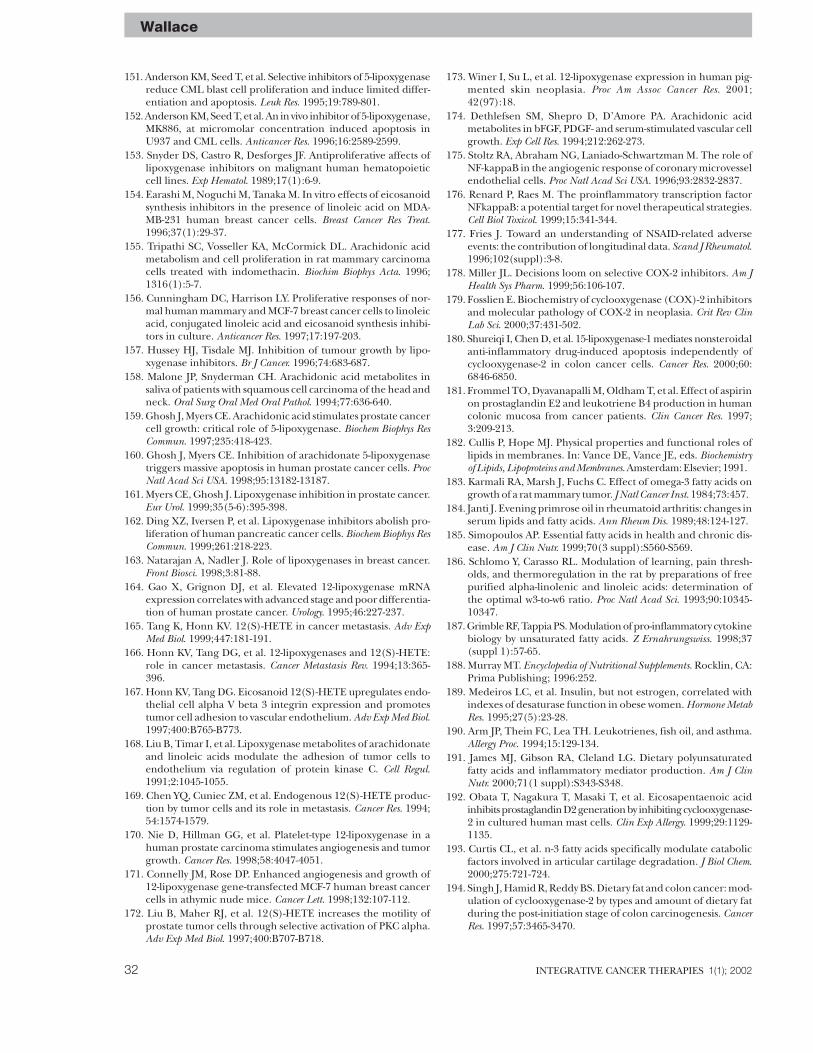
151. Anderson KM, Seed T, et al. Selective inhibitors of 5-lipoxygenasereduce CML blast cell proliferation and induce limited differ-entiation and apoptosis. Leuk Res. 1995;19:789-801.
152. Anderson KM, Seed T, et al. An in vivo inhibitor of 5-lipoxygenase,MK886, at micromolar concentration induced apoptosis inU937 and CML cells. Anticancer Res. 1996;16:2589-2599.
153. Snyder DS, Castro R, Desforges JF. Antiproliferative affects oflipoxygenase inhibitors on malignant human hematopoieticcell lines. Exp Hematol. 1989;17(1):6-9.
154. Earashi M, Noguchi M, Tanaka M. In vitro effects of eicosanoidsynthesis inhibitors in the presence of linoleic acid on MDA-MB-231 human breast cancer cells. Breast Cancer Res Treat.1996;37(1):29-37.
155. Tripathi SC, Vosseller KA, McCormick DL. Arachidonic acidmetabolism and cell proliferation in rat mammary carcinomacells treated with indomethacin. Biochim Biophys Acta. 1996;1316(1):5-7.
156. Cunningham DC, Harrison LY. Proliferative responses of nor-mal human mammary and MCF-7 breast cancer cells to linoleicacid, conjugated linoleic acid and eicosanoid synthesis inhibi-tors in culture. Anticancer Res. 1997;17:197-203.
157. Hussey HJ, Tisdale MJ. Inhibition of tumour growth by lipo-xygenase inhibitors. Br J Cancer. 1996;74:683-687.
158. Malone JP, Snyderman CH. Arachidonic acid metabolites insaliva of patients with squamous cell carcinoma of the head andneck. Oral Surg Oral Med Oral Pathol. 1994;77:636-640.
159. Ghosh J, Myers CE. Arachidonic acid stimulates prostate cancercell growth: critical role of 5-lipoxygenase. Biochem Biophys ResCommun. 1997;235:418-423.
160. Ghosh J, Myers CE. Inhibition of arachidonate 5-lipoxygenasetriggers massive apoptosis in human prostate cancer cells. ProcNatl Acad Sci USA. 1998;95:13182-13187.
161. Myers CE, Ghosh J. Lipoxygenase inhibition in prostate cancer.Eur Urol. 1999;35(5-6):395-398.
162. Ding XZ, Iversen P, et al. Lipoxygenase inhibitors abolish pro-liferation of human pancreatic cancer cells. Biochem Biophys ResCommun. 1999;261:218-223.
163. Natarajan A, Nadler J. Role of lipoxygenases in breast cancer.Front Biosci. 1998;3:81-88.
164. Gao X, Grignon DJ, et al. Elevated 12-lipoxygenase mRNAexpression correlates with advanced stage and poor differentia-tion of human prostate cancer. Urology. 1995;46:227-237.
165. Tang K, Honn KV. 12(S)-HETE in cancer metastasis. Adv ExpMed Biol. 1999;447:181-191.
166. Honn KV, Tang DG, et al. 12-lipoxygenases and 12(S)-HETE:role in cancer metastasis. Cancer Metastasis Rev. 1994;13:365-396.
167. Honn KV, Tang DG. Eicosanoid 12(S)-HETE upregulates endo-thelial cell alpha V beta 3 integrin expression and promotestumor cell adhesion to vascular endothelium. Adv Exp Med Biol.1997;400:B765-B773.
168. Liu B, Timar I, et al. Lipoxygenase metabolites of arachidonateand linoleic acids modulate the adhesion of tumor cells toendothelium via regulation of protein kinase C. Cell Regul.1991;2:1045-1055.
169. Chen YQ, Cuniec ZM, et al. Endogenous 12(S)-HETE produc-tion by tumor cells and its role in metastasis. Cancer Res. 1994;54:1574-1579.
170. Nie D, Hillman GG, et al. Platelet-type 12-lipoxygenase in ahuman prostate carcinoma stimulates angiogenesis and tumorgrowth. Cancer Res. 1998;58:4047-4051.
171. Connelly JM, Rose DP. Enhanced angiogenesis and growth of12-lipoxygenase gene-transfected MCF-7 human breast cancercells in athymic nude mice. Cancer Lett. 1998;132:107-112.
172. Liu B, Maher RJ, et al. 12(S)-HETE increases the motility ofprostate tumor cells through selective activation of PKC alpha.Adv Exp Med Biol. 1997;400:B707-B718.
173. Winer I, Su L, et al. 12-lipoxygenase expression in human pig-mented skin neoplasia. Proc Am Assoc Cancer Res. 2001;42(97):18.
174. Dethlefsen SM, Shepro D, D’Amore PA. Arachidonic acidmetabolites in bFGF, PDGF- and serum-stimulated vascular cellgrowth. Exp Cell Res. 1994;212:262-273.
175. Stoltz RA, Abraham NG, Laniado-Schwartzman M. The role ofNF-kappaB in the angiogenic response of coronary microvesselendothelial cells. Proc Natl Acad Sci USA. 1996;93:2832-2837.
176. Renard P, Raes M. The proinflammatory transcription factorNFkappaB: a potential target for novel therapeutical strategies.Cell Biol Toxicol. 1999;15:341-344.
177. Fries J. Toward an understanding of NSAID-related adverseevents: the contribution of longitudinal data. Scand J Rheumatol.1996;102(suppl):3-8.
178. Miller JL. Decisions loom on selective COX-2 inhibitors. Am JHealth Sys Pharm. 1999;56:106-107.
179. Fosslien E. Biochemistry of cyclooxygenase (COX)-2 inhibitorsand molecular pathology of COX-2 in neoplasia. Crit Rev ClinLab Sci. 2000;37:431-502.
180. Shureiqi I, Chen D, et al. 15-lipoxygenase-1 mediates nonsteroidalanti-inflammatory drug-induced apoptosis independently ofcyclooxygenase-2 in colon cancer cells. Cancer Res. 2000;60:6846-6850.
181. Frommel TO, Dyavanapalli M, Oldham T, et al. Effect of aspirinon prostaglandin E2 and leukotriene B4 production in humancolonic mucosa from cancer patients. Clin Cancer Res. 1997;3:209-213.
182. Cullis P, Hope MJ. Physical properties and functional roles oflipids in membranes. In: Vance DE, Vance JE, eds. Biochemistryof Lipids, Lipoproteins and Membranes. Amsterdam: Elsevier; 1991.
183. Karmali RA, Marsh J, Fuchs C. Effect of omega-3 fatty acids ongrowth of a rat mammary tumor. J Natl Cancer Inst. 1984;73:457.
184. Janti J. Evening primrose oil in rheumatoid arthritis: changes inserum lipids and fatty acids. Ann Rheum Dis. 1989;48:124-127.
185. Simopoulos AP. Essential fatty acids in health and chronic dis-ease. Am J Clin Nutr. 1999;70(3 suppl):S560-S569.
186. Schlomo Y, Carasso RL. Modulation of learning, pain thresh-olds, and thermoregulation in the rat by preparations of freepurified alpha-linolenic and linoleic acids: determination ofthe optimal w3-to-w6 ratio. Proc Natl Acad Sci. 1993;90:10345-10347.
187. Grimble RF, Tappia PS. Modulation of pro-inflammatory cytokinebiology by unsaturated fatty acids. Z Ernahrungswiss. 1998;37(suppl 1):57-65.
188. Murray MT. Encyclopedia of Nutritional Supplements. Rocklin, CA:Prima Publishing; 1996:252.
189. Medeiros LC, et al. Insulin, but not estrogen, correlated withindexes of desaturase function in obese women. Hormone MetabRes. 1995;27(5):23-28.
190. Arm JP, Thein FC, Lea TH. Leukotrienes, fish oil, and asthma.Allergy Proc. 1994;15:129-134.
191. James MJ, Gibson RA, Cleland LG. Dietary polyunsaturatedfatty acids and inflammatory mediator production. Am J ClinNutr. 2000;71(1 suppl):S343-S348.
192. Obata T, Nagakura T, Masaki T, et al. Eicosapentaenoic acidinhibits prostaglandin D2 generation by inhibiting cyclooxygenase-2 in cultured human mast cells. Clin Exp Allergy. 1999;29:1129-1135.
193. Curtis CL, et al. n-3 fatty acids specifically modulate catabolicfactors involved in articular cartilage degradation. J Biol Chem.2000;275:721-724.
194. Singh J, Hamid R, Reddy BS. Dietary fat and colon cancer: mod-ulation of cyclooxygenase-2 by types and amount of dietary fatduring the post-initiation stage of colon carcinogenesis. CancerRes. 1997;57:3465-3470.
Wallace
32 INTEGRATIVE CANCER THERAPIES 1(1); 2002

195. Kobayashi J, Yokoyama S, Katamura S. Eicosapentaenoic acidmodulates arachidonic acid metabolism in rat alveolar macro-phages. Prostaglandins Leukotrienes Essential Fatty Acids. 1995;52:259-262.
196. Chang WL, Chapkin RS, Lupton JR. Fish oil blocks azoxymethane-induced rat colon tumorigenesis by increasing cell differentia-tion and apoptosis rather than decreasing cell proliferation. JNutr. 1998;128:491-497.
197. Endres S, Von Schacky C. Omega-3 polyunsaturated fatty acidsand human cytokine synthesis. Curr Opinions Lipidol. 1996;7(13):48-52.
198. Connelly JM, Rose DP. Effects of fatty acids on invasion throughreconstituted basement membrane (“Matrigel”) by a humanbreast cancer cell line. Cancer Lett. 1993;75:137-142.
199. Maehle L, Eilersten E, et al. Effects of n-3 fatty acids during neo-plastic progression and comparison of in vitro and in vivo sensi-tivity of two human tumour cell lines. Br J Cancer. 1995;71:691-696.
200. Lai PB, Ross JA, et al. Cell cycle arrest and induction ofapoptosis in pancreatic cancer cells exposed to eicosa-pentaenoic acid in vivo. Br J Cancer. 1996;74:1375-1383.
201. Reich R, et al. Eicosapentaenoic acid reduces the invasive andmetastatic activities of malignant tumor cells. Biochem BiophysRes Commun. 1989;160:559-564.
202. Burns CP, Petersen ES, North JA, Ingraham LM. Effects ofdocosahexaenoic acid on rate of differentiation of HL-60 humanleukemia. Cancer Res. 1989;49:3252-3258.
203. Kanayasu T, Morita I, et al. Eicosapentaenoic acid inhibits tubeformation of vascular endothelial cells in vitro. Lipids. 1991;26:271-276.
204. Yang SP, Morita I, et al. Eicosapentaenoic acid attenuates vascu-lar endothelial growth factor-induced proliferation via inhibit-ing Flk-i receptor expression in bovine carotid artery endothe-lial cells. J Cell Physiol. 1998;176:342-349.
205. Rose DP, Connelly JM. Antiangiogenicity of docosahexaenoicacid and its role in the suppression of breast cancer cell growthin nude mice. Int J Oncol. 1999;15:1011-1015.
206. Mukutmoni-Norris M, Hubbard NE, Erickson KL. Modulationof murine mammary tumor vasculature by dietary n-3 fattyacids in fish oil. Cancer Lett. 2000;150:101-109.
207. Karmali RA, Reichel P, et al. The effects of dietary omega-3 fattyacids on the DU-145 transplantable human prostatic tumor.Anticancer Res. 1987;7:1173-1179.
208. Gonzalez MJ, Schemmel RA, et al. Effect of dietary fat ongrowth of MCF-7 and MDA-MB231 human breast carcinomasin athymic nude mice: relationship between carcinoma growthand lipid peroxidation product levels. Carcinogenesis. 1991;12:1231-1235.
209. Welsch CW, Oakley CS, et al. Suppression of growth by dietaryfish oil of human breast carcinomas maintained in three differ-ent strainsof immune-deficientmice.NutrCancer. 1993;20:119-127.
210. Rose DP, Connelly JM, Coleman M. Effect of omega-3 fatty acidson the progression of metastases after the surgical excision ofhuman breast cancer cell solid tumors growing in nude mice.Clin Cancer Res. 1996;2:1751-1756.
211. Senzaki H, Iwamoto S, et al. Dietary effects of fatty acids ongrowth and metastasis of KPL-1 human breast cancer cells invivo and in vitro. Anticancer Res. 1998;18:1621-1627.
212. DeBravo MG, DeAntueno RJ, et al. Effects of an eicosapentaenoicand docosahexaenoic acid concentrate on a human lung carci-noma grown in nude mice. Lipids.1991;26:866-870.
213. Maehle L, Lystad E, et al. Growth of human lung adenocarci-noma in nude mice is influenced by various types of dietary fatand vitamin E. Anticancer Res. 1999;19:1649-1655.
214. Sakaguchi M, Rowley S, et al. Reduced tumour growth of thehuman colonic cancer cell lines COLO320 and HT-29 in vivo bydietary n-3 lipids. Br J Cancer. 1990;62:742-747.
215. Sakaguchi M, et al. Relationship of the tissue and tumor fattyacids to dietary fat in experimental colorectal cancer. Gut.1989;30:1449.
216. Calder PC, Davis J, et al. Dietary fish oil suppresses humancolon tumour growth in athymic mice. Clin Sci (Colch). 1998;94:303-311.
217. Paulsen JE, Stamm T, Alexander J. A fish oil-derived concen-trate enriched in eicosapentaenoic and docosahexaenoic acidas ethyl esters inhibits the formation and growth of aberrantcrypt foci in rat colon. Pharmacol Toxicol. 1998;82(13):28-33.
218. Suzuki I, Ioga M, Ishikawa C, et al. Inhibitory effects of oleic anddocosahexaenoic acids on lung metastases by colon carcinoma-26 cells are associated with reduced matrix metalloproteinase-2and -9 activities. Int J Cancer. 1997;73:607-612.
219. Jenski LJ, et al. Omega-3 fatty acid-containing liposomes in can-cer therapy. Proc Soc Exp Biol Med. 1995;210:227-233.
220. Barber MD, Ross JA, et al. Fish oil-enriched nutritional supple-mentation attenuates progression of the acute-phase responsein weight-losing patients with advanced pancreatic cancer. JNutr. 1999;129:1120-1125.
221. Wigmore SJ, Ross JA, et al. The effect of polyunsaturated fattyacids on the progress of cachexia in patients with pancreaticcancer. Nutrition. 1996;12:527-530.
222. Barber MD, Ross JA, Fearon KC. The anticachectic effect offatty acids. Proc Nutr Soc. 1998;57:571-576.
223. Barber MD, Ross JA, Voss AC, et al. The effect of an oral nutri-tional supplement enriched with fish oil on weight loss inpatients with pancreatic cancer. Br J Cancer. 1999;81:80-86.
224. Gogos CA, et al. The effect of dietary omega-3 polyunsaturatedfatty acids on T-lymphocyte subsets of patients with solid tumors.Cancer Detect Prev. 1995;19:415-417.
225. Gogos CA, Ginopoulos P, Salsa B, et al. Dietary omega-3 polyun-saturated fatty acids plus vitamin E restore immunodeficiencyand prolong survival for severely ill patients with generalizedmalignancy: a randomized controlled trial. Cancer. 1998;82:395-402.
226. Tsai WS, Nagawa H, Muto T. Differential effects of polyunsatu-rated fatty acids on chemosensitivity of NIH3T3 cells and itstransformants. Int J Cancer. 1997;70:357-361.
227. Zijlstra JG, deVries ES, et al. Influence of docosahexaenoic acidin vitro on intracellular adriamycin concentration in lympho-cytes and human adriamycin-sensitive and -resistant small-celllung cancer cell lines, and on cytotoxicity in the tumor celllines. Int J Cancer. 1987;40:850-856.
228. Plumb JA, Luo W, Kerr DJ. Effect of polyunsaturated fatty acidson the drug sensitivity of human tumour cell lines resistant toeither cisplatin or doxorubicin. Br J Cancer. 1993;57:728-733.
229. Kinsella JE, Black JM. Effects of polyunsaturated fatty acids onthe efficacy of antineoplastic agents toward L5178Y lymphomacells. Biochem Pharmacol. 1993;45:1881-1887.
230. Horie T, Nakamaru M, Masubuchi Y. Docosahexaenoic acidexhibits a potent protection of small intestine from methotrexate-induced damage in mice. Life Sci. 1998;62:1333-1338.
231. Shao Y, Pardini L, Pardini RS. Intervention of transplantablehuman mammary carcinoma MX-1 chemotherapy with dietarymenhaden oil in athymic mice: increased therapeutic effectsand decreased toxicity of cyclophosphamide. Nutr Cancer. 1997;28(1):63-73.
232. Hardman WE, Moyer MP, Cameron IL. Fish oil supplementationenhanced CPT-11 (irinotecan) efficacy against MCF-7 breastcarcinoma xenografts and ameliorated intestinal side effects.Br J Cancer. 1999;81:440-448.
233. Germain E, Lavandier F, Chajes V, et al. Dietary n-3 polyunsatu-rated fatty acids and oxidants increase rat mammary tumor sen-sitivity to epirubicin without change in cardiac toxicity. Lipids.1999;34(suppl):S203.
Nutritional and Botanical Anti-Inflammatory Agents in Cancer
INTEGRATIVE CANCER THERAPIES 1(1); 2002 33

234. Vartak S, Robbins ME, Spector AA. Polyunsaturated fatty acidsincrease the sensitivity of 36B10 rat astrocytoma cells to radia-tion-induced killing. Lipids. 1997;32:283-292.
235. Das UN. Gamma-linolenic acid, arachidonic acid, and eicosa-pentaenoic acid as potential anticancer drugs. Nutrition. 1990;6:429-434.
236. Gonzalez MJ, Schemmel RA, Dugan L, et al. Dietary fish oilinhibits human breast carcinoma growth: a function of increasedlipid peroxidation. Lipids. 1993;28:827-832.
237. Gonzalez MJ. Fish oil lipid peroxidation and mammary tumorgrowth. J Am Coll Nutr. 1995;14:325-335.
238. Boik J. Natural Compounds in Cancer Therapy. Princeton, MN:Oregon Medical Press; 2001.
239. Calviello S, Palozza P, Piccioni E, et al. Dietary supplementationwith eicosapentaenoic and docosahexaenoic acid inhibits growthof Morris hepatocarcinoma 3924A in rats: effects on prolifera-tion and apoptosis. Int J Cancer. 1998;75:699-705.
240. Gramaglia A, Loi GR, Mongioj V, Baronzio GF. Increased sur-vival in brain metastatic patients treated with stereotactic radio-therapy, omega-three fatty acids and bioflavonoids. AnticancerRes. 1999;19(6C):5583-5586.
241. Bagga D, Capone S, Wang HJ, et al. Dietary modulation ofomega-3/omega-6 polyunsaturated fatty acid ratios in patientswith breast cancer. J Natl Cancer Inst. 1997;89:1123-1131.
242. Bertram HP, Gostner A, Scheppeach W, et al. Effects of fish oilon rectal cell proliferation, mucosal fatty acids, and prostaglan-din E2 release in healthy subjects. Gastroenterology. 1993;105:1317-1322.
243. Burns CP, Halabi S, Clamon GH, et al. Phase I clinical study offish oil fatty acid capsules for patients with cancer cachexia:cancer and leukemia group B study 9473. Clin Cancer Res.1999;5:3942-3947.
244. Bender NK, Kraynak MA, Chiquette E, et al. Effects of marinefish oils on the anticoagulation status of patients receivingchronic warfarin therapy. J Thromb Thrombolysis. 1998;5:257-261.
245. Macrides TA, Treschow AR, et al. The anti-inflammatory effectsof omega-3 tetraenoic fatty acids isolated from a lipid extract(Lyprinol®) from the New Zealand green-lipped mussel[abstract]. Presented at: 88th American Oil Chemists SocietyAnnual Meeting; May 1997; Seattle, WA.
246. Whitehouse MW, Macrides TA, et al. Anti-inflammatory activityof a lipid fraction (Lyprinol®) from New Zealand green-lippedmussel. Inflam Pharmacol. 1997;5:237-246.
247. Halpern GM. Antiinflammatory effects of a stabilized lipidextract of Perna canaliculus (Lyprinol®). Townsend Lett DoctorsPatients. 2000;202:109-113.
248. McMillan DC, Sattar N, Talwar D, et al. Changes in micro-nutrient concentrations following anti-inflammatory treatmentin patients with gastrointestinal cancer. Nutrition. 2000;15:425-428.
249. Chinery R, Beauchamp RD, Shyr Y, et al. Antioxidants reducecyclooxygenase-2 expression, prostaglandin production, andproliferation in colorectal cancer cells. Cancer Res. 1998;58:2323-2327.
250. Mestre JR, Subbaramaiah K, et al. Retinoids suppress epider-mal growth factor-induced transcription of cyclooxygenase-2 inhuman oral squamous carcinoma cells. Cancer Res. 1997;57:2890-2895.
251. Kanekura T, Higashi Y, Kanzaki T. Inhibitory affects of 9-cis-retinoic acid and pyrrolidinedithiocarbamate on cyclo-oxygenase (COX-2) expression and cell growth in human skinsquamous carcinoma cells. Cancer Lett. 2000;161:177-183.
252. Traber MG, Packer L. Vitamin E: beyond antioxidant function.Am J Clin Nutr. 1995;62(6 suppl):S1501-S1509.
253. Kamimura M. Antiinflammatory effect of vitamin E. J Vitaminol.1972;18:204-209.
254. Eskew ML, Zarkower A, Scheuchenzuber WJ, et al. Effects ofinadequate vitamin E and/or selenium nutrition on the releaseof arachidonic acid metabolites in rat alveolar macrophages.Prostaglandins. 1989;38(1):79-89.
255. Chan AC, Tran K, Pyke DD, Powell WS. Effect of dietary vitaminE on the biosynthesis of 5-lipoxygenase products by rat poly-morphonuclear leukocytes (PMNL). Biochim Biophys Acta. 1989;1005:265-269.
256. Lauritsen K, Laursen LS, Bukhave K, Rask-Madsen J. Does vita-min E supplementation modulate in vivo arachidonate metab-olism in human inflammation? Pharmacol Toxicol. 1987;61:246-249.
257. Upritchard JE, Sutherland WHF, Mann JI. Effect of supple-mentation with tomato juice, vitamin E and vitamin C on LDLoxidation and products of inflammatory activity in type 2 diabe-tes. Diabetes Care. 2000;23:733-738.
258. Jiang Q, Elson-Schwab I, Courtemanche C, Ames BN. Gamma-tocopherol and its major metabolic, in contrast to alphatocopherol, inhibit cyclooxygenase activity in macrophagesand epithelial cells. Proc Natl Acad Sci USA. 2000;97:11494-11499.
259. Safayhi H, Mack T, et al. Boswellic acids: novel, specific, nonredoxinhibitors of 5-lipoxygenase. J Pharmacol Exp Ther. 1992;261:1143-1146.
260. Safayhi H, Sailer ER, et al. Mechanism of 5-lipoxygenase inhibi-tion by acetyl-11-keto-beta-boswellic acid. Mol Pharmacol. 1995;47:1212-1216.
261. Safayhi H, Sailer ER, et al. 5-lipoxygenase inhibition, by acetyl-11-keto-beta-boswellic acid (AKBA) by a novel mechanism.Phytomedicine. 1996;3(1):71-72.
262. Sailer ER, Schweizer S, et al. Characterization of an acetyl-11-keto-beta-boswellic acid and arachidonate-binding regulatorysite of 5-lipoxygenase using photoaffinity labeling. Eur J Biochem.1998;256:364-368.
263. Ammon HP, Mack T, Singh GB, Safayhi H. Inhibition ofleukotriene B4 formation in rat peritoneal neutrophils by anethanolic extract of the gum resin exudate of Boswellia serrata.Planta Med. 1991;57:203-207.
264. Ammon HP, Safayhi H, Mack T, Sabieraj J. Mechanism ofantiinflammatory actions of curcumine and boswellic acids. JEthnopharmacol. 1993;38(2-3):113-119.
265. Shao Y, Ho CT, et al. Inhibitory activity of boswellic acids fromBoswellia serrata against human leukemia HL-60 cells in culture.Planta Med. 1998;64:328-331.
266. Jing Y, Nakajo S, et al. Boswellic acid acetate induces differentia-tion and apoptosis in leukemia cell lines. Leuk Res. 1999;23:43-50.
267. Glaser T, Winter S, Groscurth P, et al. Boswellic acids and malig-nant glioma: induction of apoptosis but no modulation of drugsensitivity. Br J Cancer. 1998;80(5-6):756-765.
268. Winking M, Sarikaya S, et al. Boswellic acids inhibit gliomagrowth. J Cancer Res Clin Oncol. 1998;124:8141.
269. Winking M, Sarikaya S, et al. Boswellic acids inhibit gliomagrowth: a new treatment option? J Neurooncol. 2000;46:97-103.
270. Boeker DK, Winking M. The role of boswellic acids in the ther-apy of malignant glioma [in German]. Deutsches Aerzteblatt.1997;94:A1197-A1199.
271. Janssen G, Bode U, Breu H, et al. Boswellic acids in the pallia-tive therapy of children with progressive or relapsed braintumors. Klin Padiatr. 2000;212:189-195.
272. Taussig S, Batkin S. Bromelain, the enzyme complex of pineap-ple (Ananas comosus) and its clinical application: an update. JEthnopharmacol. 1988;22:191-203.
273. Taussig S. The mechanism of the physiological action ofbromelain. Med Hypothesis. 1980;6:99-104.
Wallace
34 INTEGRATIVE CANCER THERAPIES 1(1); 2002

274. Vellini M, et al. Possible involvement of eicosanoids in the phar-macological action of bromelain. Arzneimittelforschung. 1986;36:110-112.
275. Tassman G, et al. A double-blind crossover study of plantproteolytic enzyme in oral surgery. J Dent Med. 1965;20:51-54.
276. Batkin S, et al. Antimetastatic effect of bromelain with or with-out its proteolytic and anticoagulant activity. J Cancer Res ClinOncol. 1988;114:507-508.
277. Batkin S, et al. Modulation of pulmonary metastasis (Lewislung carcinoma) by bromelain, an extract of the pineapplestem (Ananas comosus) [letter]. Cancer Invest. 1988;6:241-242.
278. Gerard C. Therapeutique anti-cancreuse et bromelaines.Agressologie. 1972;3:261-274.
279. Nieper HA. Bromelain in der kontrolle malignen washtums.Krebsgeschehen. 1976;1:9-15.
280. Eckert K, Grabowska E, et al. Effects of oral bromelain adminis-tration on the impaired immunocytotoxicity of mononuclearcells from mammary tumor patients. Oncol Rep. 1999;6:1191-1199.
281. Felton GE. Fibrinolytic and antithrombotic action of bromelainmay eliminate thrombosis in heart patients. Med Hypothesis.1980;6:1123-1133.
282. Harris JE. Interaction of dietary factors with oral anticoagu-lants: review and applications. J Am Diet Assoc. 1995;95:580-584.
283. Sharma OP. Antioxidant activity of curcumin and related com-pounds. Biochem Pharm. 1976;25:1811-1812.
284. Toda S, Miyase T, Arichi H, et al. Natural antioxidants: III:antioxidative components isolated from rhizome of Curcumalonga. Chem Pharmacol Bull. 1985;33:1725-1728.
285. Zhang F, et al. Curcumin inhibits cyclooxygenase-2 transcrip-tion in bile acid- and phorbol ester-treated human gastrointes-tinal epithelial cells. Carcinogenesis. 1999;20:445-451.
286. Gupta OP, Sharma N, Chand D. Application of papaya latex-induced rat paw inflammation: model for evaluation of slowlyacting antiarthritic drugs. J Pharmacol Toxicol Methods. 1994;31(2):95-98.
287. Srivastava R, Srimal RC. Modification of certain inflammation-induced biochemical changes by curcumin. Indian J Med Res.1985;81:215-223.
288. Srivastava R, Mustafa T. Spices, antiplatelet activity and prostanoidmetabolism. Prostaglandins Leukotrienes Essential Fatty Acids.1989;38:255-266.
289. Flynn DL, Rafferty MF, Boctor AM. Inhibition of 5-hydroxy-eicosatetraenoid acid (5-HETE) formation in intact humanneutrophils by naturally-occurring diarylheptanoids: inhibitoryactivities of curcuminoids and yakuchinones. ProstaglandinsLeukotrienes Med.1986;22(3):357-360.
290. Huang MT, Lysz T, Ferraro T, et al. Inhibitory activities ofcurcumin on in vitro lipoxygenase and cyclooxygenase activi-ties in mouse epidermis. Cancer Res. 1991;51:813-819.
291. JoeB,LokeshBR.Effectof curcuminandcapsaicinonarachidonicacid metabolism and lysosomal enzyme secretion by rat peritonealmacrophages. Lipids. 1997;32:1173-1180.
292. Conney AH, Lou YR, Xie JG, et al. Some perspectives on dietaryinhibition of carcinogenesis: studies with curcumin and tea.Proc Soc Exp Biol Med. 1997;216:234-245.
293. Rao CV, et al. Chemoprevention of colon carcinogenesis bydietary curcumin, a naturally occurring plant phenolic com-pound. Cancer Res. 1995;55:259-266.
294. Plummer SM, Sharma RA, et al. Curcuminoids inhibitcyclooxygenase-mediated prostaglandin E2 production andCOX-2 expression in human blood. Proc Am Assoc Cancer Res.2001;42(93):17-18.
295. Satoskar RH, Shah SJ, Shenoy SG. Evaluation of anti-inflamma-tory property of curcumin (diferuloyl methane) in patientswith postoperative inflammation. Int J Clin Pharmacol Ther Toxicol.1986;24:651-654.
296. Deodhar SD, Sethi R, Srimal RC. Preliminary studies on anti-rheumatic activity of curcumin (diferuloylmethane). Indian JMed Res. 1980;71:632-634.
297. Mehta K, Pantazis P, McQueen T, Aggarwal BB. Curcumin(diferuloylmethane) is a potent antiproliferative agent againsthuman breast tumor cell lines. Anticancer Drugs. 1997;8:470-481.
298. Dorai T, Gehani N, Katz A. Therapeutic potential of curcuminin human prostate cancer: II: curcumin inhibits tyrosine kinaseactivity of epidermal growth factor receptor and depletes theprotein. Mol Urol. 2000;4(1):1-6.
299. Chen H, Zhang ZS, Zhang YL, Zhou DY. Curcumin inhibits cellproliferation by interfering with cell cycle and inducingapoptosis in colon carcinoma cells. Anticancer Res. 1999;19(5A):3675-3680.
300. Goel A, et al. Inhibition of cyclooxygenase-2 (COX-2) expres-sion by dietary curcumin in HT-29 human colon cancer cells.Proc Am Assoc Cancer Res. 1999;40:528-529.
301. Elattar TM, Virji AS. The inhibitory effect of curcumin, genistein,quercetin and cisplatin on the growth of oral cancer cells invitro. Anticancer Res. 2000;20(3A):1733-1738.
302. Ramsewak RS, De Witt DL, Nair MG. Cytotoxicity, antioxidantand anti-inflammatory activities of curcumins I-III from Curcumalonga. Phytomedicine. 2000;7:303-308.
303. Kuttan R, et al. Potential anticancer activity of tumeric. CancerLett. 1985;29:197-202.
304. Kuo ML, et al. Curcumin, an antioxidant and anti-tumor pro-moter, induced apoptosis in human leukemia cells. BiochimBiophys Acta. 1996;1317:95-100.
305. Gautam SC, Xu YX, Pindolia KR, et al. Nonselective inhibitionof proliferation of transformed and nontransformed cells bythe anticancer agent curcumin (diferuloylmethane). BiochemPharmacol. 1998;55:1333-1337.
306. Moragoda L, Jaszewsky R, Majumdar AP. Curcumin inducedmodulation of cell cycle and apoptosis in gastric and colon can-cer cells. Anticancer Res. 2001;21(2A):873-878.
307. Jee SH, Shen SC, Tseng CR, et al. Curcumin induced a p53-dependent apoptosis in human basal cell carcinoma cells. JInvest Dermatol. 1998;111:656-661.
308. Jiang MC, Yang-Yen HF, Yen JJ, Lin JK. Curcumin inducesapoptosis in immortalized NIH 3T3 and malignant cancer celllines. Nutr Cancer. 1996;26:111-120.
309. Thaloor D, et al. Inhibition of angiogenic differentiation ofhuman umbilical vein endothelial cells by curcumin. Cell GrowthDiffer. 1998;9:305-312.
310. Singh AK, et al. Curcumin inhibits the proliferation and cellcycle progression of human umbilical vein endothelial cell.Cancer Lett. 1996;107:109-115.
311. Arbiser JL, Klauber N, Rohan R, et al. Curcumin is an in vivoinhibitor of angiogenesis. Mol Med. 1998;4:376-383.
312. Menon LG, Kuttan R, Kuttan G. Inhibition of lung metastasis inmice induced by B16F10 melanoma cells by polyphenolic com-pounds. Cancer Lett. 1995;95(1-2):221-225.
313. Menon LG, Kuttan R, Kuttan G. Anti-metastatic activity ofcurcumin and catechin. Cancer Lett. 1999;141(1-2):159-165.
314. Punithavathi D, Venkatesan N, Babu M. Curcumin inhibitionof bleomycin-induced pulmonary fibrosis in rats. Br J Pharmacol.2000;131:169-172.
315. Venkatesan N, Punithavathi D, Chandrakasan C. Curcuminprotects bleomycin-induced lung injury in rats. Life Sci. 1997;61(6):51-58.
316. Venkatesan N, Chandrakasan G. Modulation of cyclophosphamide-induced early lung injury by curcumin, an anti-inflammatoryantioxidant. Mol Cell Biocham. 1995;142(1):79-87.
317. Venkatesan N, Punithavathi D, Arumugam V. Curcumin pre-vents adriamycin nephrotoxicity in rats. Br J Pharmacol. 2000;129:231-234.
Nutritional and Botanical Anti-Inflammatory Agents in Cancer
INTEGRATIVE CANCER THERAPIES 1(1); 2002 35

318. Sriganth I, Premalatha B. Dietary curcumin with cisplatinadministration modulates tumor marker indices in experimen-tal fibrosarcoma. Pharm Res. 1999;39:175-179.
319. Boik J. Natural Compounds in Cancer Therapy. Princeton, MN:Oregon Medical Press; 2001:280.
320. Kuttan R, et al. Tumeric and curcumin as topical agents in can-cer therapy. Tumori. 1987;73(1):29-31.
321. Shankar T, et al. Toxicity studies on turmeric (Curcuma longa):acute toxicity studies in rats, guinea pigs and monkeys. Indian JExp Biol. 1980;18:73-75.
322. McGuffin M, Hobbs C, Upton R, Goldberg A. Botanical SafetyHandbook. New York, NY: CRC Press; 1997:39
323. Havsteen B. Flavonoids, a class of natural products of highpharmacological potency. Biochem Pharmacol. 1983;32:1141-1148.
324. Baumann J, et al. Flavonoids and related compounds as inhibi-tion of arachidonic acid peroxidation. Prostaglandins. 1980;20:627-639.
325. Welton AF, Hurley J, Will P. Flavonoids and arachidonic acidmetabolism. Prog Clin Biol Res. 1988;280:301-312.
326. Welton AF, Tobias LD, Fiedler-Nagy C, et al. Effect of flavonoidson arachidonic acid metabolism. Prog Clin Biol Res. 1986;213:231-242.
327. Mutoh M, et al. Suppression of cyclooxygenase-2 promoter-dependent transcriptional activity in colon cancer cells bychemopreventive agents with a resorcin-type structure.Carcinogenesis. 2000;21:959-963.
328. Lamson DW, Brignall MS. Antioxidants and cancer, part 3:quercetin. Alternative Med Rev. 2000;5:196-208.
329. Kandaswami C, Perkins E, Drzewiecki G, et al. Differential inhi-bition of proliferation of human squamous cell carcinoma,gliosarcoma and embryonic fibroblast-like lung cells in cultureby plant flavonoids. Anticancer Drugs. 1992;3:525-530.
330. Castillo MH, Perkins E, Campbell JH, et al. The effects of thebioflavonoid quercetin on squamous cell carcinoma of headand neck origin. Am J Surg. 1989;158:351-355.
331. Caltagirone S, Ranelletti FO, Rinelli A, et al. Interaction withtype II estrogen binding sites and antiproliferative activity oftamoxifen and quercetin in human non-small-cell lung cancer.Am J Respir Cell Mol Biol. 1997;17(1):51-59.
332. Singhal RL, Yey YA, Praja N, et al. Quercetin down-regulates sig-nal transduction in human breast carcinoma cells. BiochemBiophys Res Commun. 1995;208:425-431.
333. Scambia G, Ranelletti FO, Panici PB, et al. Inhibitory effect ofquercetin on OVCA 433 cells and presence of type II oestrogenbinding sites in primary ovarian tumours and cultured cells. Br JCancer. 1990;62:942-946.
334. Larocca LM, Giustacchini M, Maggiano N, et al. Growth-inhibi-tory effect of quercetin and presence of type II estrogen bind-ing sites in primary human transitional cell carcinomas. J Urol.1994;152:1029-1033.
335. Yoshida M, Sakai T, Hosokawa N, et al. The effect of quercetinon cell cycle progression and growth of human gastric cancercells. FEBS Lett. 1990;260(1):10-13.
336. Richter M, Eberman R, Marian B. Quercetin-induced apoptosisin colorectal tumor cells: possible role of EGF receptor signal-ling. Nutr Cancer. 1999;34(1):88-99.
337. Ranelletti FO, Ricci R, Larocca LM, et al. Growth-inhibitoryeffect of quercetin and presence of type-II estrogen-bindingsites in human colon-cancer cell lines and primary colorectaltumors. Int J Cancer. 1992;50:486-492.
338. Vazquez B, Avila G, Segura D, Escalante B. Anti-inflammatoryactivity of extracts from Aloe vera gel. J Ethnopharmacol. 1996;55:69-75.
339. Haqqi TM, et al. Prevention of collagen-induced arthritis inmice by a polyphenolic fraction from green tea. Proc Natl AcadSci USA. 1999;96:4524-4529.
340. Subbaramaiah K, Chung WJ, Michaluart P, et al. Resveratrolinhibits cyclooxygenase-2 transcription and activity in phorbolester-treated human mammary epithelial cells. J Biol Chem.1998;273:21875-21882.
341. Ohuchi K, et al. Glycyrrhizin inhibits prostaglandin E2 forma-tion by activated peritoneal macrophages from rats. Prostaglan-din Med. 1981;7:457-463.
342. Dirsch VM, Vollmar AM. Ajoene, a natural product with non-steroidal anti-inflammatory drug (NSAID)-like properties? BiochemPharmacol. 2001;61:587-593.
343. VanLoon IM. The golden root: clinical applications of Scutellariabaicalensis GEORGI flavonoids as modulators of the inflamma-tory response. Alt Med Rev.
344. Butenko IG, et al. Anti-inflammatory properties and inhibitionof leukotriene C4 biosynthesis in vitro by flavonoid baicaleinfrom Scutellaria baicalensis greorgy roots. Agents Actions. 1993:39(C):49-51.
345. Belluco C, Nitti D, Frantz M, et al. Interleukin-6 blood level isassociated with circulating carcinoembryonic antigen andprognosis in patients with colorectal cancer. Ann Surg Oncol.2000;7:133-138.
346. Martin F, Santolaria F, Batista N, et al. Cytokine levels (IL-6 andIFN-gamma), acute phase response and nutritional status asprognostic factors in lung cancer. Cytokine. 1999;11(1):80-86.
347. Barber MD, Fearon KC, Ross JA. Relationship of serum levels ofinterleukin-6, soluble interleukin-6 receptor and tumournecrosis factor receptors to the acute-phase protein responsein advanced pancreatic cancer. Clin Sci. 1999;96(1):83-87.
348. Kolebacz B. Concentrations of selected acute phase proteinsand immunity immunoglobulins in the treatment of head andneck carcinomas [in Polish]. Otalaryngol Pol. 1999;53:743-746.
349. McMillan DC, Wotherspoon HA, Fearon KC, et al. A prospec-tive study of tumor recurrence and the acute-phase responseafter apparently curative colorectal cancer surgery. Am J Surg.1995;170:319-322.
350. Nozoe T, Matsumata T, Kitamura M, Sugimachi K. Significanceof preoperative elevation of serum C-reactive protein as an indi-cator for prognosis in colorectal cancer. Am J Surg. 1998;176:335-338.
351. Falconer JS, Fearon KC, Ross JA, et al. Acute-phase proteinresponse and survival duration of patients with pancreatic can-cer. Cancer. 1995;75:2077-2082.
352. Barlogie B. High-dose therapy and innovative approaches totreatment of multiple myeloma. Semin Hematol. 2001;38(2 suppl 3):21-27.
353. Matsui Y, Fujikawa K, Oka H, et al. Management of renal cellcarcinoma with inferior vena cava tumor thrombus. Urol Int.2001;66(1):4-8.
354. Wigmore SJ, MacMahon AJ, Sturgeon CM, Fearon KC. Acute-phase protein response, survival and tumour recurrence inpatients with colorectal cancer. Br J Surg. 2001;88:255-260.
355. Nielsen HJ, Christensen IJ, Sorensen S, et al. Preoperativeplasma plasminogen activator inhibitor type-1 and serum C-reactive protein levels in patients with colorectal cancer. AnnSurg Oncol. 2000;7:617-623.
356. O’Gorman P, McMillan DC, McArdle CS. Prognostic factors inadvanced gastrointestinal cancer patients with weight loss. NutrCancer. 2000;37(1):36-40.
357. McMillan DC, Leen E, Smith J, et al. Effects of extendedibuprofen administration on the acute phase protein responsein colorectal cancer patients. Eur J Surg Oncol. 1995;21:531-534.
358. Clyne B, Olshaker JS. The C-reactive protein. J Emerg Med.1999;17:1019-1025.
359. Cengiz M, Akbulut S, Atahan IL, Grigsby PW. Acute phaseresponse during radiotherapy. Int J Radiat Oncol Biol Phys.2001;49:1093-1096.
Wallace
36 INTEGRATIVE CANCER THERAPIES 1(1); 2002

360. Hu RJ, Lee PH, Yu SC. Secretion of acute-phase proteins beforeand after hepatocellular carcinoma resection. J Formos MedAssoc. 1999;98(2):85-91.
361. Goddard DH, Bomalaski JS, et al. Phospholipase A2-mediatedinflammation induces regression of malignant glioma. CancerLett. 1996;102(1-2):1-6.
362. Komhoff M, Guan Y, Shappel HW, et al. Enhanced expressionof cyclooxygenase-2 in high grade human transitional cell blad-der carcinomas. Am J Pathol. 2000;157(1):29-35.
363. Scioscia KA, Snyderman CH, et al. Role of arachidonic acidmetabolites in tumor growth inhibition by nonsteroidal anti-inflammatory drugs. Am J Otolaryngol. 1997;18(1):1-8.
364. Anderson KM, Seed T, et al. 5-lipoxygenase inhibitors reducePC-3 cell proliferation and initiate nonnecrotic cell death. Pros-tate. 1998;37:161-173.
Editor’s NoteThis important review by Jeanne M. Wallace of the roleof eicosanoids in stimulation of the growth of tumors,
and the potential for inhibiting their activities by in-hibiting the action of lipoxygenase and cyclooxygenase(COX) enzymes, provides a scientific foundation forone of the most common strategies of integrative nu-tritional programs: the limitation of meat in the dietand its substitution with fish and other sources ofomega-3 fatty acids. Wallace’s exploration of the phar-macology of COX-2 inhibitors in various cancers is de-tailed and convincing, and she has done the integrativemedicine community a real service in reviewing thepotential use of natural COX-2 inhibitors. This phar-macology is truly where mechanism-based biology in-tersects directly with the therapeutic realities of bothconventional and complementary/alternative treat-ment, and this article provides a solid basis for under-standing COX-2 inhibitors for clinicians in both areas.
Nutritional and Botanical Anti-Inflammatory Agents in Cancer
INTEGRATIVE CANCER THERAPIES 1(1); 2002 37
Copyright © 2022 FDOKUMEN