NSRRC Activity Report 2017
164
-
Upload
khangminh22 -
Category
Documents
-
view
3 -
download
0
Transcript of NSRRC Activity Report 2017


NSRRC Activity Report
2 0 1 7

Message from the Director Preface
Research Highlights Physics and Materials Science 009 Stradivari’s Secret010 Resonant Inelastic Excitations Lead the Way015 It’s All About the Ending017 Now You See It018 Novel Route to Effective Doping at Hetero structure Interfaces021 Correlations and Dynamics of Spins in an XY-Like Spin-Glass (Ni0.4Mn0.6)TiO3 Single Crystal System 024 Orbital Anisotropy Drives the Charge-Density Wave Transition in SrFeO3-δ
Chemical Science029 Nanodiamonds Dispersed in Space030 Formation of Wannier-Mott Excitons in Solid Carbon Oxide031 Ultra-Bright Near-IR OLED 033 Future Porous Materials: A Highly Flexible Inorganic Framework 034 Plasmon-Induced Suppression of Hydrogen Peroxide Formation in Oxygen Reduction Reaction 036 Photo-Enhanced Ferromagnetism in K–Ni–Cr Prussian Blue Analogues
Soft Matter040 In-Situ Probing Nanostructural Evolution During Spin-Coating 042 Calligraphic Thin-Film Transistors 044 Handedness of Twisted Lamellae in Banded Spherulite
Table of Contents
046 Self-Assembly of Macromolecules in Nano-Sized Pores 048 “I WILL BE BACK! – THE RETURN OF RUBBER:” A New Mechanism to Overcome the Dilemma of Shape Fixing and Recovery in Biodegradable Polyurethane Elastomer
Life Science 053 The NlpI-Prc System in Escherichia coli (E. coli): A New Target for the Development of Antibac erial Agents 055 RNase R: A Proficient Enzyme Involved in the Decay of RNA 057 Preserved Collagen in an Early Jurassic Sauropodomorph Dinosaur 060 FIN219-FIP1: Two Vital Proteins Involved in Crosstalk Between FR Light Signaling and JA Response 062 Producing Irreversible Topoisomerase-II-Medi ated DNA Breaks 064 Parity-Dependent Slippage of DNA Hairpins for a Disease-Associated Repeat Expansion
Energy Science067 Phosphorus Doping Enlarges Hydrogen Evolution 068 Voltammetric Enhancement of Li-ion Con duction in Al-Doped Li7-xLa3Zr2O12 Solid Electrolyte 070 Molecular Design Drives Solar-Hydrogen Conversion 071 Probing the Structural Evolution of a Battery with X-rays 073 Fluoride Phosphors Illuminate a White LED 074 Dual Doping Strengthens Metal-Support Interactions

103 Facts & Figures
Appendix 130 List of Publications 154 Student Dissertations
Facility Status 078 Status of TLS and TPS Accelerators 079 The Rooftop Photovoltaic Systems at NSRRC 081 The TPS Post-Mortem System 085 X-ray Nanoprobe Investigation Toward the Nano World 087 Development of Soft-X-ray Tomography for Biomedical Research 088 The Installation of the Instrument for Bragg Coherent Diffraction Imaging 090 A Projection and Transmission X-ray Microscope 094 Time and Spatially Resolved X-ray Absorption Spectroscopy Beamine at TPS 095 Soft X-ray Spectroscopy at TPS 098 SIKA: Opportunities for Low-Energy Excitations Using Neutrons

Message from the Director
“T ime flies when you are having fun.” How true it is that my term as the Director is winding down in next few months. At NSRRC,
we began in the early eighties with a visionary decision to construct the first synchrotron light source in Taiwan. Today, after more than 30 years, NSRRC remains as a vibrant research facility with two operational synchrotron light sources, including 1.5 GeV Taiwan Light Source (TLS) and the state-of-the-art 3.0 GeV Taiwan Photon Source (TPS). In 2017, we had about 2,300 users (12,000 user visits) from 21 countries coming to TLS/TPS for their research endeavors in a wide range of disciplines, which produces about 350 scientific publications (20% of them in the top-tier journals of respective fields). Our diverse expertise in acceler-ator, instrumentation, experimental technique, and scientific research has helped users to pursue academic excellence, cross-disciplinary collaboration, and to elevate social impact by research outcomes.
Despite its long-term operation, TLS has maintained outstanding per-formance. The record of mean time between failures achieved a his-toric peak (~259 hours) in 2017. On the other hand, TPS has reached a stored electron current 400 mA in top-up operation. While most of TPS phase-I beamlines have been open to users, the phase-II beamline construction is well under way. With all these accelerator technologies and frontier scientific studies, NSRRC recently added a new dimension to its role to emerge as an important driving force in technological innovation and transfer by creating the Industrial Liaison Office, which is aimed to connect research and industry for social advancement.
In 2017, we had a joint press conference to announce a plan to establish the Max Planck–NSRRC/NCTU/NTHU Center for Complex Phase Materials in Hsinchu, Taiwan. This Center is intended to nurture young scientists and to strengthen interdisciplinary collaborations. Several home institutes of our users, such as Max-Planck-Ge-sellschaft, National Tsing Hua University (NTHU), Academia Sinica, and Tamkang University, have collaborated with NSRRC to build beamlines or endstations at TPS. NSRRC, as always, is dedicated to promote synchrotron-re-lated science and experimental techniques through workshops, training courses and joint degree programs. For the promotion of public awareness, we also organized events to reach out to the younger generations. Last summer, with National Space Organization (NSPO) in Hsinchu, we co-organized a successful science festival, cov-ering popular themes of space, arts, and paleontology. In this event, all attendees had an exclusive open-house experience at NSRRC and NSPO.
The development of synchrotron-based science and technology has matured rapidly in the last few decades. Nevertheless, we can still foresee an even brighter future. As usual, our mission is to foster partnerships that will engender scientific accomplishments to benefit our society. For this, I would like to acknowledge all the TLS/TPS users for their everlasting pursuit of scientific discoveries and the hard work from our staff toward the impec-cable facility operation. I look forward to your continued partnerships with NSRRC as we move steadily into the future.
Shangjr GwoDirector

Preface
I t is a great pleasure to present to you the 2017 issue of the NSRRC Activity Report. I thank you for your interest. Since 1995, NSRRC published an annual report every year describing organizations, research, facility develop-
ment, user statistics, events and summaries of our operation. These activity reports show the efforts of all mem-bers of NSRRC throughout the years. Their dedication to advancing synchrotron-based science and technology has produced many significant results, which can only be briefly highlighted in the reports.
This year, we continue our record of scientific accomplishments and facility progress at NSRRC. In the session of Scientific Highlights, we select representative articles performed by our users and staff in fundamental research and applied studies, and group them into five areas -- physics and material science, chemical science, soft matter, life science and energy science. We cover also three studies using neutrons, resulting from the Taiwan-Australia Neutron Project, of which NSRRC has taken charge since 2013. In the session of Facility Status, besides highlight-ing our newly completed rooftop solar-power system, we report the commissioning and operation status of three TPS phase-I beamlines – X-ray Nanoprobe, Coherent X-ray Scattering, and Submicron Soft X-ray Spectros-copy, as well as the progress in construction of three TPS phase-II beamlines – Soft X-ray Tomography, Projection X-ray Microscopy, and Quick-scanning X-ray Absorption Spectroscopy. SIKA, the Taiwanese neutron instrument at Australia Nuclear Science and Technology Organisation, is also included.
This report is being published in two media -- a printed version and an electronic version on our NSRRC website, http://www.nsrrc.org.tw/. We hope that you will find this publication informative and useful.
Di-Jing HuangDeputy Director


Research Highlights
Physics and Materials Science
Chemical Science
Soft Matter
Life Science
Energy Science

ACTIV
ITY REPO
RT 2017
A primary interest of condensed matter and ma-terials physics is to understand the properties of
natural and synthetic materials that are composed by a large collection of atoms. Because there are so many of them interacting with each other, the at-oms’ charge, spin, an orbital degree of freedom can be tangled in ways to mask out the key ingredient governing their collective behavior. To unravel the nature of complex materials/systems, scientists apply selected external stimuli and use instruments with ultra-high sensitivity to search those responsible step by step. In this year's section on physics and materi-als science, we introduce works explaining why the Stradivari violins created in the late 17th and early 18th century are so famous, how resonant inelastic excitations can untangle complex phenomena, and what characteristics are important in making smart windows. Combining messages obtained from multi-ple techniques available at NSRRC, scientists are able to clarify the roles of interfaces in various functional heterostructures. (by Der-Hsin Wei)
Physics and Materials Science

Physics and Materials Science
009
T he Stradivari violins, created by Anto-nio Stradivari in the late 17th and early
18th century, are considered to be one of best set of violins in the world. However, the origin behind the sound quality of the Stradivari violins has remained a mystery. This is all the more intriguing because vio-lins made by the following generations of the Stradivari family as well as other mod-ern violins have not been able to match the Stradivari violins, according to music connoisseurs. In a very interesting, recent study, scientists have uncovered several unique properties of the maple wood used to make the Stradivari violins.
Using nuclear magnetic resonance, X-ray dif-fraction and differential scanning calorimetry, Hwan-Ching Tai (National Taiwan University) and his co-workers showed that the maple used by Stradi-vari violins and cellos exhibit unique features which are not observed in tonewood used to make mod-ern violins. The authors could also confirm that the Stradivari violins were treated with complex mineral preservatives which contained Al, Ca, Cu, Na, K and Zn. The authors used nuclear magnetic resonance and synchrotron X-ray diffraction to show that while one-third of the hemicellulose had decomposed after nearly 300 years, accompanied by signs of lignin oxidation, but no apparent changes in cellulose could be detected in the experiments (Figs. 1(a) and 1(b)). The relative cellulose crystallinity plotted against rel-ative hemicellulose levels measured by nuclear mag-netic resonance was distinct for the Stradivari violins and cellos compared to other modern violins (Fig. 1(a)). However, the crystallite lengths and widths estimated from X-ray diffraction patterns were similar for all the types of violins and cellos investigated in the study (Fig. 1(b)). Further, only maples from the Stradivari violins exhibited unusual thermo-oxidation patterns, distinct from Stradivari cellos and natural modern maple wood (Fig. 1(c)). The authors could thus conclude that in their current state, the maple wood used for the Stradivari violins have very differ-ent chemical composition and properties compared to Stradivari cellos as well as modern violins. This was
Stradivari’s SecretScientists have succeeded to show that the maple wood used for making the famous Stradi-vari violins exhibit unique chemical and physical properties, which are attributed to the com-bined effects of aging, chemical treatments and vibrations.
attributed to the combined effects of aging, chemical treatments and vibrations.1
The authors hope that their study will inspire further investigations on tonewood processing for improving instrument making techniques in the future. (Report-ed by Ashish Chainani)
Fig. 1: (a) Observed (crosses) and fitted (solid lines) neutron powder diffraction patterns taken at 80 K. (b) Schematic drawing of the proposed crystal-line structure of the Rb–Co–Fe in the core and the K–Ni–Cr shell. [Repro-duced from Ref. 1]
Fig. 2: Differential scanning calorimetry thermograms of Stradivari violin compared to Stradivari cello and mod-ern maple, showing a distinct behavior of the Stradivari violins. [Reproduced from Ref. 1]
(a) (b)

ACTIV
ITY REPO
RT 2017
010
This report features the work of Hwan-Ching Tai and his co-workers published in PNAS 114, 27 (2017).
TLS 01C2 SWLS – X-ray Powder Diffraction• X-ray Diffraction• Materials Science
Reference 1. H.-C. Tai, G.-C. Li, S.-J. Huang, C.-R. Jhu, J.-H. Chung,
B. Y. Wang, C.-S. Hsu, B. Brandmair, D.-T. Chung, H. M. Chen, and J. C. C. Chan, PNAS 114, 27 (2017).
The Chimei Museum, located in Tainan, Taiwan, has one of the largest and finest collections of violins in the world. The pictures
of the Stradivari violin are provided by the courtesy of the Museum.
Resonant Inelastic Excitations Lead the WayResonant Inelastic X-ray Scattering (RIXS) is a very versatile probe for investigating emergent phenomena which originate from spin-charge-lattice coupling in strongly correlated electron systems. This article reports three classic RIXS studies on the electronic structure of: (i) the Ver-wey transition material magnetite Fe3O4 , (ii) the spin-state transition in LaCoO3 , and (iii) the quasi two-dimensional Nickelates La2-xSrxNiO4 (x = 0, 0.33, 0.45).
I n this article, we discuss three valuable recent re-sults which demonstrate the unique capabilities of
Resonant Inelastic X-ray Scattering (RIXS) for reveal-ing localized as well as dispersive spin-charge-orbital excitations with elemental and ionic-configuration specificity. TLS 05A1 was developed to carry out momentum-resolved RIXS using the energy-compen-sation principle of grating dispersion for the active grating monochromator (AGM) and the active grat-ing spectrometer (AGS). The design of the AGM-AGS system greatly enhances the measurement efficiency of inelastic soft X-ray scattering.1 After long and sustained efforts, it is now possible to routinely carry out RIXS measurements at a good energy resolution and reasonable count rates. Temperature dependent (20—550 K) momentum-resolved RIXS can be car-ried out with an energy resolution of ~108 meV at the Ni L3-edge ( ~850 eV). Using these capabilities, scientists have now succeeded to answer important long-standing questions on the electronic structure of strongly correlated transition metal oxides.
(i) Magnetic polarons in magnetite (Fe3O4): Mag-netite, or lodestone, is the first known magnet to mankind and was discussed in Greek and Chinese literature as early as the 4th to the 6th century BC. It
was used as a magnetic compass and the name ‘mag-net’ most probably comes from Magnesia, an ancient city in Greece where lodestones were found. The properties of magnetite attract significant scientific and technological interest even today, because of its applications in ultrafast magnetic sensors, palaeo-magnetism, nanomedicine, etc. Magnetite Fe3O4 be-comes ferrimagnetic below TC = 850 K, followed by an abrupt decrease in its electrical conductivity by two orders of magnitude as the temperature is cooled below TV = 122 K. The crystal structure of magnetite consists of tetrahedral FeO4 and octahedral FeO6 mo-tifs. Within its unit cell, one-third of the total number of Fe sites (the so-called A-sites) are nominal Fe3+ ions tetrahedrally (Td) coordinated with oxygen atoms ; the remaining two-thirds are termed B-sites with equal number of nominal Fe2+ and Fe3+ ions which are octahedrally (Oh) coordinated with oxygen atoms. An Fe2+– Fe3+ charge-ordering occurring on the B-sites was first suggested by Verwey as the driving force of this transition.2
Although numerous investigations have been carried out to verify the charge localization on the B-sites in the low temperature phase, the precursor to the charge-ordering pattern of magnetite in the high

Physics and Materials Science
011
temperature phase was not known. However, while it was known that the charge disproportionation in-volves changes in the nominal Fe2+– Fe3+ states asso-ciated with the B-sites, X-ray diffraction studies of the low-temperature phase of magnetite microcrystals re-vealed that the t2g electrons of the B-sites are not fully localized but are distributed over linear three-Fe-site units termed trimerons.3 The trimerons are coupled to the Jahn-Teller distortion of B-site Fe2+O6 octahedra, as illustrated in Fig. 1, while the B-site Fe3+O6 octahe-dra are Jahn-Teller inactive, to a first approximation. The Verwey transition is then essentially an ordering of trimerons. However, an extremely important ques-tion had remained unanswered: Do local distortions persist in the cubic phase at temperatures above TV, and if so, what was its character?
In an international collaboration led by Di-Jing Huang’s team at NSRRC together with groups from USA, Netherlands, South Korea, Germany and Taiwan, temperature dependent Resonant Inelastic X-ray Scattering (RIXS) at the Fe L3-edge was used to reveal the low-energy spin-orbital excitations of Fe2+ and Fe3+ ions in both, the low temperature monoclinic and high temperature cubic phases of magnetite (Fig. 2). In combination with crystal-field multiplet calculations, the authors succeeded to show that local distortions in the form of magnetic polarons exist in magnetite above TV and give rise to a low-en-ergy magnetic mode in RIXS (Fig. 3). The authors could further show that the magnetic polarons orig-inate in the Jahn-Teller distortion of Fe2+ sites. Thus,
Fig. 1: Trimeron scenario and t2g energy-level splitting. Illustra-tion of the orbital ordering of B-site Fe2+ in Fe3O4 and the corresponding t2g energy-level splitting for a Fe2+ ion in a negative Dt2g crystal field. A trimeron is indicated with a dashed oval. The elongation of the four Fe-O bonds in the xy plane are indicated with arrows. [Reproduced from Ref. 4]
Fig. 2: RIXS measurements of Fe3O4. (a) Fe L-edge X-ray absorption spectrum (XAS) spectrum measured in the fluorescence yield mode through the summation of all inelastic X-ray intensities taken at room temperature T = 300 K. The XAS is plotted with correction for self-absorption. The incident X-ray energy resolution was 0.5 eV. (b) Color map of RIXS intensity after correction for self-absorption in the plane of incident photon energy versus energy loss recorded at T = 80 K. (c)–(e) RIXS spectra plotted in terms of energy loss with a vertical offset for clarity. They were recorded by using p-polarized incident X-rays under the scattering geometry of the scattering angle φ = 90° and the incident angle φ = 20°. Panels c and d show data measured at 80 K, while panel e shows data measured at 550 K. [Reproduced from Ref. 4]
the magnetic polarons are the precursors leading to the monoclinic distortion seen below TV and solves a long-standing problem connecting its electronic structure with local crystal structure changes above the Verwey transition.4
(ii) The prototypical spin-state transition in LaCoO3: Spin-state transitions or crossovers between low-spin (LS) and high-spin (HS) states occur in diverse systems including solids, liquids and biomaterials.
(a)(c) (d) (e)
(b)

ACTIV
ITY REPO
RT 2017
012
The transition metal perovskite oxide LaCoO3 is a prototypical example of a spin-state transition in solids, and is undisputedly identified as being in a LS state below 20 K.5 Its magnetic susceptibility �(T) rises sharply with increasing temperature and exhibits a maximum around 100 K, before trailing off at high-er temperatures. This temperature dependence of �(T) was originally interpreted as implying a gradual population increase of HS states with a fixed acti-vation energy Δa, an energy required to excite the ground state to the first excited state. This scenario however led to an over- estimated �(T) and motivated an intermediate-spin (IS) description. Band-structure calculations with Coulomb correlations included gave a strong boost to the IS picture. In contrast, electron spin resonance, inelastic neutron scattering, and X-ray absorption spectroscopy (XAS) showed that the lowest-energy excited state is a HS state which exhib-its additional splitting due to spin-orbit interactions. Furthermore, the XAS work explained the transition using a temperature-dependent increase of Δa, but this leads to a puzzle:5 for the LS ground state, an increase in Δa implies an increase in bare ionic crys-tal-field splitting 10Dq, which is inconsistent with a reduction of 10Dq expected from the experimentally known expansion in Co–O bond lengths. These issues indicate that the origin of the spin-state transition of LaCoO3 was not fully resolved.
Recent calculations using dynamical mean-field theo-ry have implied the role of a temperature-dependent Hund’s exchange energy in driving the spin-state transition.6 In addition, first-principles calculations have shown that the screening by eg electrons is more efficient than t2g electrons in transition metal oxides. This is mainly because eg electrons strongly
hybridize with 2p states of nearest-neighbour oxy-gens and exhibit a tendency towards delocalization, forming broad σ bands. This leads to a very important question: Does the change in the 3d electronic states across the transition modify the screening of the Coulomb interaction, i.e. does it modify the effective Coulomb energy between electrons.
In a collaboration between the NSSRC, Tohoku Uni-versity and Utrecht University, scientists have exploit-ed RIXS to study the spin-state and metal-insulator transitions as a function of temperature in LaCoO3 (Fig. 4(a)). They could unequivocally characterize electronic excitations derived from different spin channels across the transitions by comparing with dif-ferent reference samples (Fig. 4(b)). Further, the au-thors could show that the screening of the Coulomb interaction of 3d electrons is orbital selective, and it is coupled to the thermal evolution of the spin-state
Fig. 3: Comparison of measured (expt) and calculated (calc) RIXS spectra. Open circles are measurements with inci-dent X-rays of 707 eV at 80 K; the solid line presents the calculated RIXS spectra at an incident X-ray energy 707.5 eV. The calculated results are consistent with experi-ment only on inclusion of a polaronic distortion and an exchange field, indicative of a magnetic polaron forma-tion. [Reproduced from Ref. 4]
Fig. 4: Temperature-dependent RIXS of LaCoO3. (a) RIXS spectra of single-crystal LaCoO3 at various temperatures. The spectra have been normalized to the incident photon flux. The red curve (LS cal) shows the calculated RIXS spectral weight of LS Co3+ with 10Dq = 0.595 eV. The vertical dashed line gives a guide to the eye. (b) RIXS of polycrystalline EuCoO3, LaCo0.5Ni0.5O3, and Sr2CoO3Cl at 20 K. By using π-polarized incident X-rays of energy set to L3-2.5 eV, all RIXS spectra plotted in (a) and (b) were recorded under the same conditions except for tempera-ture. Spectra are plotted with a vertical offset for clarity. [Reproduced from Ref. 7]
(a)
(b)

Physics and Materials Science
013
Fig. 5: (a)–(c) Simulated RIXS of LaCoO3 at temperatures 60, 100, and 300 K by using the LS and HS reference spectra discussed in the text. (d) LS and HS populations obtained from simulations for various temperatures. Open squares and closed circles are deduced spin-state populations from RIXS data. The solid line plots the calculated HS population fHS with Ei = 13, 35.5, and 72.5 meV and νi = 3, 5, and 7. The dotted line is the LS population fLS = 1 – fHS. Inset: calculated energies of the 5T2g states about the LS-HS transition. Green circles indicate the average energies of Jeff = 1, 2, and 3 at 10Dq = 0.595 eV. (e) Comparison between mea-sured �(T) from SQUID and deduced �(T) from RIXS data. (f) The energy shift ΔE between the measured and simulated energies of RIXS excitations from the ground state 1A1g to 1T1g without spin change as a function of temperature. [Reproduced from Ref. 7]
Fig. 6: (a) Schematic showing the tetragonally elongated NiO6 octahedra present in La2−xSrxNiO4, and corresponding energy level diagram plotted in blue. The electrons, in black, occupy levels in a 3d8
S = 1 configuration. (b) shows the known antiferromagnetic ordering of these spins. (c) La2NiO4 Ni L3-edge RIXS energy map collected at QII = (0.74π, 0). White bars correspond to calculated energies. (d) Ni L3 edge RIXS atomic multiplet calculation using parameters described in the text. [Reproduced from Ref. 10]
crossover in LaCoO3. Resonant Inelastic X-ray Scattering (RIXS) combined with charge-transfer multiplet calculations were used to reveal the renormalized crys-tal-field excitations and the results provided a measure of spin-state populations (Fig. 5). The authors could thus establish a gradual spin-state crossover preceding a Mott-type insulator to metal transition. RIXS was thus shown to be very effective for fingerprinting the renomalized dd-transitions as well as the temperature-de-pendent orbital selective spin-charge excitations across the spin crossover and the metal insulator transition in LaCoO3.7
(iii) Spin S = 1 quasi two di-mensional Nickelates exhibit dispersive magnetic excitations: In the past few years, while RIXS has emerged as an indispensable tool for probing spin, charge and orbital excitations in solids, most of these studies have focused on spin (or pseudospin) S = 1/2 based materials, such as cuprates or iridates. These materials represent a special case as only one Δms = 1 spin transition is allowed on a
(a)
(b)
(c)
(d)
(e)
(f)
(a) (c) (d)
(b)

ACTIV
ITY REPO
RT 2017
014
single atomic site (i.e., ms = −1/2 → ms = 1/2), which directly match-es the photon angular momen-tum. However, to date, the ability of RIXS to address the electronic interactions in higher spin-state compounds via measurements of collective magnetic excitations has not been established.
La2NiO4 shares the same structur-al motif as cuprates and iridates consisting of a transition metal ion surrounded by 6 oxygen atoms forming an octahedron (Figs. 6(a) and 6(b)). It is an an-tiferromagnetic Mott insulator in its ground state with a 3d8 con-figuration and S = 1 state. Thus, it is important to know whether RIXS can reveal the nature of this state and how it evolves with doping. Earlier studies on an S = 1 nickelate NiO had shown that RIXS couples to local Δms = 1 and 2 spin flips, rather than collective excitations.8,9
weakly dependent on doping. The authors could show that the larg-er Ni 3d character of the doped holes in LSNO (when compared to cuprates) leads to the reduction in magnon energy.10
These studies indicate the power of synchrotron-based momen-tum-resolved RIXS in addressing important questions in the physics of transition metal compounds, leading to new opportunities in spin-charge-lattice coupling derived emergent properties of materials. (Reported by Ashish Chainani)
This report features the work of : (1) Di-Jing Huang and his co-work-ers published in Nat. Commun. 8, 15929 (2017); (2) Di-Jing Huang and his co-workers published in Phys. Rev. Lett. 119, 196402 (2017); (3) M. P. M. Dean and his co-workers published in Phys. Rev. Lett. 118, 156402 (2017).
TLS 05A1 EPU – Soft X-ray Scattering• RIXS, XAS• Condensed Matter Physics, Ma-
terials Science
References 1. C. H. Lai, H. S. Fung, W. B. Wu,
H. Y. Huang, H. W. Fu, S. W. Lin, S.W. Huang, C. C. Chiu, D. J. Wang, L. J. Huang, T. C. Tseng, S. C. Chung, C. T. Chen and D. J. Huang, J. Synchrotron Rad. 21, 325 (2014).
2. E. J. W. Verwey, Nature 144, 327 (1939).
3. M. S. Senn, J. P. Wright, and J. P. Attfield, Nature 481, 173 (2012).
4. H. Y. Huang, Z. Y. Chen, R.-P. Wang, F. M. F. de Groot, W. B. Wu, J. Okamoto, A. Chainani, A. Singh, Z.-Y. Li, J.-S. Zhou, H.-T. Jeng, G. Y. Guo, J.-G. Park, L. H. Tjeng, C. T. Chen and D. J. Huang, Nat. Commun. 8, 15929 (2017).
5. R. Eder, Phys. Rev. B 81, 035101 (2010).
Fig. 7: (a) La2NiO4 low energy excitations QII dependence map collected at Ei = 853.2 eV. White circles correspond to the fitted magnon energies. (b) Fitting exam-ple at QII = (0.48π, 0.48π), black, green, and magenta lines account for elastic, single magnon, and multimagnon excitations. (c) La2NiO4 magnon dispersion (red squares) compared to inelastic neutron scattering results (black circles) and spin wave theory fits (green line). The error bars shown in panels (a) and (c) correspond to 95% confidence intervals obtained from the least square fitting algorithm. [Reproduced from Ref. 10]
In a recent study using RIXS car-ried out jointly at the Taiwan Light Source and the Swiss Light Source, a team from Brookhaven National Laboratory led by M. P. M. Dean and his co-workers have present-ed Ni L3-edge RIXS measurements of the 2D antiferromagnet La2-xSrx
NiO4 (LSNO) and demonstrated that RIXS can measure collective magnetic excitations in S = 1 transition metal oxides (Fig. 6(c)). Furthermore, ab-initio and atomic multiplet RIXS simulations were carried out to confirm the 3d8 S = 1 character, and obtain a precise description of its crystal field split-ting (Fig. 6(d)). From momentum resolved experiments (Fig. 7), the authors also showed that hole doping significantly reduces the zone boundary magnon energy (50% at x = 0.45). Such a reduction of magnon energy is in contrast with results from hole doped cu-prates, for which the zone bound-ary magnetic energy scale is very
(a) (b)

Physics and Materials Science
015
6. M. Karolak, M. Izquierdo, S. L. Molodtsov, and A. I. Lichtenstein, Phys. Rev. Lett. 115, 046401 (2015).
7. K. Tomiyasu, J. Okamoto, H. Y. Huang, Z. Y. Chen, E. P. Sinaga, W. B. Wu, Y. Y. Chu, A. Singh, R.-P. Wang, F. M. F. de Groot, A. Chainani, S. Ishihara, C. T. Chen, and D. J. Huang, Phys. Rev. Lett. 119, 196402 (2017).
8. S. G. Chiuzbaian, G. Ghiringhelli, C. Dallera, M. Grioni, P. Amann, X. Wang, L. Braicovich, and L. Patthey, Phys. Rev. Lett. 95, 197402 (2005).
9. G. Ghiringhelli, A. Piazzalunga, C. Dallera, T. Schmitt, V. N. Strocov, J. Schlappa, L. Patthey, X. Wang, H. Berger, and M. Grioni, Phys. Rev. Lett. 102, 027401 (2009).
10. G. Fabbris, D. Meyers, L. Xu, V. M. Katukuri, L. Hozoi, X. Liu, Z.-Y. Chen, J. Okamoto, T. Schmitt, A. Uldry, B. Delley, G. D. Gu, D. Prabhakaran, A. T. Boothroyd, J. van den Brink, D. J. Huang, and M. P. M. Dean, Phys. Rev. Lett. 118, 156402 (2017).
It’s All About the EndingEnergy level alignment at metal-organic interface is a key parameter controlling the elec-tric transport property of heterostructures. When a half-metallic ferromagnet La1-xSrxMnO3 (LSMO) is used for spin transport, the termination of oxide layer is another parameter in play.
S tacking multiple layers into a single structure is a way to construct artificial materials with novel
properties. Similarly, a complex oxide material like those in perovskite structure (ABO3) has distinct surface electronic structures when its topmost layer is terminated differently (AO or BO2). As the perfor-mance of a device depends strongly on the energy barrier at its heterojunction, the termination control of a metal oxide electrode offers an alternative to ad-just carrier injection barrier. Following the same idea, Yao-Jane Hsu (NSRRC) and her collaborators worked on a series of LSMO/Alq3 structures to demonstrate that the control of termination layer can be of great importance in spin transport too.
One recent advance in spintronics is the use of organ-ic semiconductor (OSC) to mediate spins.1 Because
carbon-based materials like OSC are good at preserv-ing spin coherence among carriers, the organic spin valve (an OSC layer sandwiched by two ferromagnet-ic electrodes) was thought capable of showing giant magnetoresistance (GMR) comparable to its inor-ganic counterparts. The problem is, despite using the same ferromagnetic (FM) LSMO electrode and Alq3 spacer, the experimental results reported by different research groups differ considerably in the magnitude and sign of MR. To clarify what might be responsible to the conflicting results, Hsu and her team took a close look at the same LSMO/Alq3 heterojunction with an emphasis on the possible roles played by the termination layer of LSMO electrode.2
LSMO is a half metal oxide that possesses nearly 100% spin polarization. The LSMO layer used in the study was grown on SrTiO3 (001) (STO) single crys-tal substrates. By varying STO terminations (TiO2-, SrO-, and mixed-terminations, respectively), the LSMO film with terminating layers of MnO2-, LSO-, and mixed-termination were fabricated (marked as MnO2-ter, LSO-ter, and Mixed-ter, respectively). Magneto-optical measurements on these specimen found out the magnetic hysteresis of pristine LSMO depends strongly on its termination layer; largest/smallest coercivity at LSMO with LSO-ter/MnO2-ter, and is insensitivity to the subsequently introduced Alq3 film. In the meantime, density functional theory (DFT) calculation revealed that, while Mn is responsi-ble to LSMO’s ferromagnetism, it is the Mn’s t2g states that contribute to LSMO’s half-metallicity. Further-more, depending on the termination layer, there is a significantly larger splitting between spin-up and
Fig. 1: Schematic drawing of the termination dependence; FM)T1
and FM)T2, at a half metal/organic hybrid interface. At left panel, both spin channels have a same energy barrier, whereas the energy barriers are different between the two spin channels in right panel. The termination result-ed energy shift at half-metal side is exaggerated in figure.

ACTIV
ITY REPO
RT 2017
016
spin-down bands in LSMO with LSO-ter. This finding implies that there should be termination dependence on magnetic coupling and spin filtering efficiency at LSMO/Alq3 junction, as illustrated in Fig. 1.
The conclusions from the DFT calculations was con-firmed by X-ray magnetic circular dichroism (XMCD) measurements. According to the Mn L-edge’s XMCD spectra taken from LSMO(mixed-ter)/Alq3, LSMO(Mn O2-ter)/Alq3, LSMO(LSO-ter)/Alq3 at 79 K, the LSMO with LSO-terminations gave the largest dichroic ratio and regardless of the termination layer, the adsorp-tion of Alq3 causes only a slight change to the dichro-ic ratio. This finding is in line with the magneto-op-tical measurements and therefore indicates that the ferromagnetism at LSMO/Alq3 interface is primarily determined by LSMO itself (including the termination control). Finally, a combined XPS/UPS study found no clear evidence of charge transfer at interface, but does have an interfacial dipole of extent 0.5, 0.5 and 0.7 eV at the mixed-ter/Alq3, MnO2-ter/Alq3 and LSO-ter/Alq3 interfaces with the positive pole toward the Alq3 molecules and the negative pole toward the LSMO film, as illustrated at Fig. 2.
In summary, the electronic, chemical and magnetic properties of Alq3 deposited on three types of LSMO
Fig. 2: Energy-level alignment diagram at mixed-ter/Alq3, MnO2-ter/Alq3, and LSO-ter/Alq3 interfaces. [Reproduced from Ref. 2]
A close view of the sample in the analysis chamber for XPS measurement.
terminated layers were investigated experimentally and theoretically. Given the finding that LSMO reacts weakly with Alq3, and the magnetic properties and half-metallicity of LSMO layer are termination depen-dent, it should be possible to tune the energy barriers of each spin channel (spin filtering) with the termina-tion layer of complex oxide electrode. (Reported by Der-Hsin Wei)
This report features the work of Yao-Jane Hsu and her collaborators published in J. Mater. Chem. C 5, 9128 (2017).
TLS 09A2 U50 – Spectroscopy • XPS, NEXAFS• Materials Science, Chemistry, Surface, Interface and
Thin-film Chemistry, Condensed Matter Physics
References 1. D. Sun, E. Ehrenfreund, and Z. V. Vardeny, Chem.
Commun., 50, 1781 (2014).2. T.-N. Lam, Y.-L. Huang, K.-C. Weng, Y.-L. Lai, M.-W.
Lin, Y.-H. Chu, H.-J. Lin, C.-C. Kaun, D.-H. Wei, Y.-C. Tseng, and Y.-J. Hsu, J. Mater. Chem. C 5, 9128 (2017).

Physics and Materials Science
017
Fig. 2: (a) In situ V L-edge XAS spectra recorded in real time with the V2O5 film exposed to H2/N2 (1:9) gas. (b) Spectral deconvolution using four different 3d orbital symmetries. Three spectra taken at 10, 30, and 60 min after gasochromic reaction are presented, (c) dx2–y2/dz2 ratios in spectra shown in (b). [Reproduced from Ref. 2]
Now You See ItSmart window that modulates the heat and/or light transmission can reduce the energy consumption of buildings. Monitoring their operation with operando X-ray spectroscopic measurements reveals how the electronic and atomic structures of a smart film evolve when turning the knob.
A room with a window letting in light but not heat takes less energy to cool down in summer, but
the smart window allowing independent control over its heat/light transmission can save energy all year long. After more than a decade of efforts, the so-called smart windows are making its way to mar-ket with its optical transmission adjusted simply by turning the knob or flicking a switch. The way a smart window works is to supply an external stimuli such as UV irradiation, temperature change, electric field, or gas exposure to modulate a glass’s optical char-acteristics.1 Under a given stimulation, the glass that responses stronger is considered the better candidate for smart windows. However, while the practical ap-plications of smart glass are marching forward, fun-damental problems continue to confront scientists. For example, there is little knowledge regarding if and how the atomic/electronic structure of a “glass” would change during its coloration.
Conceptually, the light-mat-ter interaction is where the optical property is rooted. It is therefore a good idea to monitor the evolution of electronic structure in real time so that any modifica-tions introduced by a stimu-lus can be easily identified. That is exactly what Chung-Li Dong (Tamkang Univer-sity) and his team did to investigate the gasochromic thin film of vanadium pent-oxide (V2O5).2 Using a home-made in-situ gas cell on at TLS 20A1, Dong’s team was able to set up a reaction environment (a mixture of H2 and N2 gases at 760 torr) in the ultra-high vacuum condition (in the order of 10-10 torr). They acquired the V L-edge X-ray absorption spec-tra from a sol–gel spin-coated
Fig. 1: Optical transmission of V2O5 film shows a significantmodulation in the range of visible wavelengths (550–800 nm) when exposed to H2/N2 (10:90) gas at 760 torr. Photographs shows colors of film in coloredand bleached states. [Reproduced from Ref. 2]
(a) (b)
(c)

ACTIV
ITY REPO
RT 2017
018
vanadium oxide (V2O5) thin film placed inside the gas cell. V2O5 thin film has been used in electrochromic coloration, but little is known regarding if, and how, the atomic/electronic structures of a V2O5 thin film were modulated at the same time.
Figure 2(a) illustrates the L-edge spectra taken from gasochromic V2O5 under color switching. Two more spectra recorded from VO2 and V2O5 crystals were added in to serve as references. A typical V L-edge spectrum has two broad peaks centered at about 518 and 525 eV that feature the transitions from two spin-orbit split 2p states to 3d states. Considering the 2p orbital is localized on V, the L-edge intensity is directly proportional to the unoccupied d-character. As shown in Fig. 1(a), the main peak of the V L3-edge shifts to a lower energy as the reaction proceeds (marked by arrows). This is an indication of vanadium receiving charge upon hydrogen absorption.
In addition to the change of absorption resonance energy, the profile of a XAS spectrum is informative too. As shown in Fig. 2(b), V L3 peak is decomposed into four components that each of them corresponds to a specific V 3d orbital symmetry (A: 3dxz & 3dyz, B: 3dxy, C: 3dx2–y2 and D: 3dz2). A fit of these spectra re-corded at three coloration stages (10, 30 and 60 min) returns with the findings that the coloration leads to different intensity variation among the four sub-com-ponents of L3-edge. In particular, with the increase of intensity at component A (more empty d states, weaker hybridization along z-axis) and decrease of intensity at component B (less empty d states, stronger hybridization at basal plane), it is apparent
that coloration leads to a strengthened interaction between V atoms and basal oxygen. This finding is consistent with the intensity variation found between components C and D that count the electron density distributed along all three axes (x,y,z). According to Fig. 2(c), the increase of intensity ratio between dx2-y2 and dz2 is a consequence of the central V atom mov-ing closer to the basal plane after the gasochromic coloration.
In summary, Dong used operando XAS measure-ments to reveal that, for a V2O5 film going through the gasochromic coloration process, there are not only changes on the charge state of vanadium, but also modification on the local atomic symmetry of V2O5. The injected hydrogen atoms cause a structural deformation from pyramid-like to octahedral-like symmetry. (Reported by Der-Hsin Wei)
This report features the work of Chung-Li Dong and his collaborators published in Phys. Chem. Chem. Phys. 19, 14224 (2017).
TLS 20A1 BM – (H-SGM) XAS • XANES, XFS, PSD, XPS, AES• Materials Science, Chemistry, Surface, Interface and
Thin-film Chemistry, Condensed Matter Physics
References 1. L. Long, and H. Ye, Sci. Rep. 4, 6427 (2014).2. Y.-R. Lu, T.-Z Wu, H.-W. Chang, J.-L. Chen, C.-L.
Chen, D.-H. Wei, J.-M. Chen, W.-C. Chou, and C.-L. Dong, Phys. Chem. Chem. Phys. 19, 14224 (2017).
Novel Route to Effective Doping at Heterostructure InterfacesDoping carriers across interfaces of thin film heterostructures is an extremely sensitive and important requirement for controlling their emergent properties. Atomically precise termi-nation is now shown to be a novel route to effectively dope superconductor/ferromagnet (YBa2Cu3O7-x/La0.7Ca0.3MnO3) heterostructures.
H eterostructures hold tremendous potential for creating emergent properties not seen in single phase bulk materials. Since the discovery of a 2-D (2-dimensional) electron gas at the LaAlO3-SrTiO3 interface,1,2 there
have been several important results revealing unexpected properties of interfaces: controlling a 2D electron gas with a ferroelectric,3 orbital reconstruction at superconductor-ferromagnet interfaces,4 etc.
In this article, we discuss the work carried out by Ying-Hao Chu (National Chiao Tung University) and his co-work-ers,5 which reported on the successful development and observation of termination control for effectively dop-

Physics and Materials Science
019
Fig. 1: Epitaxial design of heterointerfaces: schematic of the interfacial control of LCMO/YBCOd with different interfaces. (a) Shows the MnO2-terminated interface (La0.7Ca0.3O-MnO2-BaO-CuO2), for which the charges are very difficult to transfer because CuO chains are very far from the interface (indicated by a dashed line) while (b) shows the La0.7Ca0.3O-terminated (MnO2-La0.7Ca0.3O-CuO2-BaO) interface which uses an SRO layer, and for which electrons can transfer easily from LCMO to YBCO due to the CuO chains at the interface (indicated by solid lines). [Reproduced from Ref. 5]
Fig. 2: (a) Transport properties of LCMO/YBCOd with different interfaces: YBCO thickness dependence of the superconducting transi-tion temperature with either MnO2- or La0.7Ca0.3O-terminated interfaces. (b) Low-temperature magnetization as a function of the YBCO layer thickness for two different interfaces. (c) The Mn valence state vs. the absorption edge energy of the MnO2-ter-minated (black symbols) and La0.7Ca0.3O-terminated (red symbols) samples. La1-xCaxMnO3 (where x = 0, 0.3, 0.6, and 1) was used as the reference samples, combined with the Mn2O3 (Mn3+) and MnO2 (Mn4+) standard samples to determine the Mn valence state. (d) The Mn valence state as a function of YBCO thickness for the two different interfaces. [Reproduced from Ref. 5]
(a)
(a)
(c)
(b)
(b)
(d)

ACTIV
ITY REPO
RT 2017
020
ing a superconductor-ferromagnet heterostructure of YBa2Cu3O7-x/La0.7Ca0.3MnO3 (YBCO/LCMO). The authors achieved an atomically precise interface control of YBCO/LCMO heterostructures, that is, they succeeded to make heterostructures with a MnO2-ter-minated (Fig. 1(a)) interface and a La0.7Ca0.3O-termi-nated (Fig. 1(b)) interface. The MnO2-terminated het-erostructure corresponds to the STO(SrTiO3)/LCMO10 nm/YBCOd structure while the La0.7Ca0.3O-terminat-ed one corresponds to the STO/SRO2.5u.c. (SrRuO3)/LCMO10 nm/YBCOd structure. The authors first used high-angle annular dark-field scanning transmis-sion electron microscopy to confirm the two distinct terminations and interfaces. The authors then fixed the LCMO layer thickness to 25 units cells (~10 nm) and varied the thickness d of the YBCO layer. From a systematic set of electrical resistivity and magneti-zation measurements, they could establish that the MnO2-terminated samples showed a consistently higher superconducting transition temperature TC (Fig. 2(a)) and a larger magnetization (Fig. 2(b)). For the lowest thickness of d = 6 nm, an intriguing result was obtained: the La0.7Ca0.3O-terminated sample was insulating while the MnO2-terminated sample was superconducting with an onset TC ~40 K.
Subsequently, synchrotron based X-ray absorption spectroscopy (XAS) and X-ray magnetic circular di-chroism (XMCD) studies were carried out at NSRRC to understand the changes in the electronic structure. From a systematic study of the Mn K-edge and L-edge XAS, they could show that the Mn valence depends on the termination and changes systematically with the thickness d (Fig. 2(c)). In addition, the suppres-sion of the magnetization in the La0.7Ca0.3O-terminat-ed samples by magnetization and XMCD measure-ments was consistently confirmed. Thus, the authors could show that the MnO2-terminated samples al-ways showed (i) a larger magnetic moment of Mn, (ii) a lower valence state of Mn, and (iii) a higher super-conducting TC compared to the La0.7Ca0.3O-terminated samples (Fig. 2(d)).
Since the results clearly indicated a relationship be-tween larger FM fluctuations with stronger supercon-ductivity, it could be concluded that the conventional scenario of ferromagnetism competing superconduc-tivity does not hold for these heterostructures. And since it is known from earlier work that electronic charge gets transferred from Mn to Cu ions across
the interface and induces a major reconstruction of the orbital occupation and orbital symmetry in the interfacial CuO2 layers,3 the authors have proposed that the differences in the interfacial structure (Fig. 1) of the MnO2-terminated and La0.7Ca0.3O-terminated interfaces leads to a difference in charge-transfer for the two cases. This is fully consistent with the low-er Mn-valency of the MnO2-terminated interfaces and leads to their higher superconducting TC values. The results thus conclusively show that termination control is a useful degree of freedom to effectively manipulate the doping and physical properties of heterostructures. (Reported by Ashish Chainani)
This report features the work of Ying-Hao Chu and his co-workers published in App. Phys. Lett. 110, 032402 (2017).
TLS 11A1 BM – (Dragon) MCD, XASTLS 20A1 BM – (H-SGM) XAS• MCD, XAS• Condensed Matter Physics, Materials Science
References 1. A. Ohtomo and H. Y. Hwang, Nature 427, 423
(2004).2. J. Biscaras, N. Bergeal, A. Kushwaha, T. Wolf, A. Ras-
togi, R. C. Budhani, and J. Lesueur, Nat. Commun. 1, 89 (2010).
3. V. T. Tra, J. W. Chen, P. C. Huang, B. C. Huang, Y. Cao, C. H. Yeh, H. J. Liu, E. A. Eliseev, A. N. Morozovska, J.-Y. Lin, Y. C. Chen, M. W. Chu, P. W. Chiu, Y. P. Chiu, L. Q. Chen, C. L. Wu, and Y.-H. Chu, Adv. Mater. 25, 3357 (2013).
4. J. Chakhalian, J. W. Freeland, G. Cristiani, H.-U. Habermeier, G. Khaliullin, M. Van Veenendaal, and B. Keimer, Science 318, 1114 (2007).
5. V. T. Tra, R. Huang, X. Gao, Y.-J. Chen, Y. T. Liu, W. C. Kuo, Y. Y. Chin, H. J. Lin, J. M. Chen, J. M. Lee, J. F. Lee, P. S. Shi, M. G. Jiang, C. G. Duan, J. Y. Juang, C. T. Chen, H. T. Jeng, Q. He, Y.-D. Chuang, J.-Y. Lin, and Y.-H. Chu, App. Phys. Lett. 110, 032402 (2017).

Physics and Materials Science
021
Correlations and Dynamics of Spins in an XY-Like Spin-Glass (Ni0.4Mn0.6)TiO3 Single Crystal SystemElastic and inelastic neutron scattering studies of the spatial and temporal correlations of spins in the XY-like spin-glass state.
S pin-glass (SG) systems are examples of frustrated magnetism and have attracted much interest
as they exhibit a wide range of interesting physical phenomena, such as history-dependence and diver-gent nonlinear magnetic susceptibilities. Typically, the combination of competing exchange interactions and either site or bond disorder leads to an SG state. However, in some stoichiometric intermetallic com-pounds, such as PrAu2Si2 and PrRuSi3
1,2, which exhibit neither static disorder nor a geometrically frustrated lattice, transition to the SG state has been observed. Inelastic neutron scattering (INS) studies have shown that the SG behavior in these systems arises from dynamic fluctuations of the crystal field levels. These fluctuations destabilize the induced magnetic mo-ments and frustrate the development of long-range magnetic ordering. To date, neutron scattering has proven to be a powerful tool for elucidating the nature of static and dynamic magnetic correlations in the SG state. However, the different behaviors depending on whether one is dealing with the Ising-, XY- or Heisenberg-type of SG systems is still a subject of research.3,4 In the present work done by Way-Faung Pong (Tamkang University) and his co-work-ers,3 they focus on an XY-like SG system (Ni0.4Mn0.6)TiO3 (NMTO) that exhibits memory and relaxation effects and has more recently been reported to exhib-it linear magnetoelectric (ME) coupling.4,5
Figure 1(a) presents mesh scans of the single crys-tal of NMTO at Ef = 5.5 meV around the (0, 0, 1.5) position at a temperature (T) of 1.5 K. The reciprocal lattice point (0, 0, 1.5) represents the first antiferro-magnetic peak in the parent compound, NiTiO3. The T-dependence of this scattering in NMTO indicates that it is magnetic in origin similar to that observed in the neutron powder diffraction pattern. To eliminate contributions from nonmagnetic nuclear scattering and higher-order contaminations, data obtained at 30 K (Fig. 1(b)) were subtracted from those obtained at 1.5 K (Fig. 1(a)). Figure 1(c) displays the pattern following subtraction. The anisotropy of the pat-terns in the [0, 0, L] and [H, H, 1.5] directions is the signature of two spin-spin correlation lengths, ξ. To determine reliably spatial correlation, ξ value in both directions and their dependence on T ≈ TSG, ENS data were obtained systematically from 1.5 to 30 K.
Figure 2 displays the T-dependent ENS profiles for NMTO around the (0, 0, 1.5) reciprocal lattice point in (a) the inter-plane/layer ([0, 0, L]) direction and (b) the in-plane ([H, H, 1.5]) direction at several tem-peratures around TSG, obtained at Ef = 3.7 meV. The scattering pattern includes a central Bragg-like peak and background (BG). As the T increases from 1.5 K to above TSG, the intensity of central peak decreases, reaching close to zero at temperatures of above 12
K. The observed patterns are well fitted by a Lorentzian function, which rep-resents magnetic diffuse scattering from the sample with the addition of a constant BG term. Therefore, the total scattering function is:
where AL and KL represent the inte-grated intensity and HWHM of the Lorentzian function, respectively. Figures 2(a) and 2(b) show fitting results using the above function. Dif-ferent value of ξ (ξ ≈ KL
-1) was obtained in the two directions. At 1.5 K, in the inter-plane/layer direction, the ξ is ≈ (21 ± 1) Å, while in the in-plane di-
Fig. 1: Mesh scans around (0, 0, 1.5) reciprocal lattice point at (a) 1.5 K and (b) 30 K at Ef = 5.5 meV. To remove nonmagnetic contributions, 30 K data were subtracted from 1.5 K data and results are shown in (c). [Reproduced from Ref. 3]
I(Q)=BG+AL KL
π [KL2+ Q2 ]
(a) (c)
(b)

ACTIV
ITY REPO
RT 2017
022
Figs. 2(a) and 2(b) indicate the strong T-dependence of ξ and AL. Increasing the T reduces drastically both ξ and AL, which are close to zero at 20 K. The increase in AL below TSG suggests that the number of antifer-romagnetically correlated clusters increases as the T decreases. Further, integrated intensity of the mag-netic diffuse scattering is proportional to the square of local sublattice magnetization (order parameter). Near the SG transition it follows a power-law, I � (TSG - T)2β where β is the critical exponent related to the order parameter. The integrated intensities along two directions in the T range from 1.5 to 12 K have been fitted with the power-law using β and TSG as fitting parameters. The continuous lines in the insets corre-spond to the fitting with β ≈ (0.37 ± 0.02) and TSG ≈ (12.4 ± 0.1) K. The value of exponent, β is close to XY model and therefore confirms the XY-like nature of SG state of NMTO. The value obtained for TSG is higher than that obtained from the magnetization measure-ment due to the non-zero integrated intensity even above 9.1 K and can be assigned to the slow dynam-ics of SG systems. To establish the dynamics of the experimentally observed short-range spin-spin cor-relations, INS data were collected slightly away from the (0, 0, 1.5) point to avoid the strong elastic scatter-ing but sufficiently close to obtain a reasonable signal from the short-range spin-spin correlations.
Figures 3(a) and 3(b) present the INS spectra as functions of energy transfer (E) for NMTO from 1.5 to 50 K; two positions (a) (0, 0, 1.52) and (b) (0.01, 0.01, 1.50) corresponding to transverse and longitudinal displacements from (0, 0, 1.50) reciprocal lattice point are measured. Owing to the very low intensity of qua-si-elastic neutron scattering (QENS), the insets in Figs. 3(a) and 3(b) display a magnified view of the tail region. QENS profiles are indicated by arrows and can
Fig. 2: T-dependence of ENS spectra in (a) the inter-layer plane direction ([0, 0, L]) and (b) in-plane direction ([H, H, 1.5]) between 1.5 and 30 K. [Reproduced from Ref. 3]
Fig. 3: INS spectra as a function of energy transfer (E) for NMTO from 1.5 to 50 K in (a) (0, 0, 1.52) and (b) (0.01, 0.01, 1.50) positions. Insets in (a) and (b) show magnified view that show QENS at T from 1.5 to 12 K, indicated by arrows. [Reproduced from Ref. 3]
rection it is ≈ (73 ± 2) Å. Figure 2(a) reveals that the inter-plane/layer ξ exceeds the distance between the neighboring Mn/Ni layers (c/3 = 4.7066 Å). Generally, a small ξ corresponds to the nanometer-scale spin clusters. These features further indicate that the mag-netic spin-spin correlations are quasi 2D. The insets in
(a)
(b)
(a) (b)

Physics and Materials Science
023
be modeled using a Lorentzian func-tion. The total scattering function is:
where BG is the background, YG rep-resents the resolution-limited elastic Gaussian component and AL & ΓL are the integrated intensity and HWHM of the Lorentzian function, respectively. INS data at 50 K are closely described using a single Gaussian function, but the tails that arise from QENS are well fitted using a single Lorentzian com-ponent. Therefore, all INS data were analyzed using the aforementioned function. The HWHMs of the elastic Gaussian components (instrumental resolution) are (0.029 ± 0.007) and (0.030 ± 0.007) meV for (0, 0, 1.52) and (0.01, 0.01, 1.50) positions. These HWHMs are determined using vanadi-um scans under similar conditions and held fixed during fitting.
Figures 4(a) and 4(b) plot the varia-tions of the obtained parameters AL and ΓL as a function of T for the two positions. Figures 4(c) and 4(d) show the elastic Gaussian contributions (AG and ΓG). The AG of the elastic Gaussian component remains nearly constant for T ≥ 20 K but starts to increase rapidly as the T falls below 12 K. These results indicate that for the two posi-tions, the integrated intensity of the quasi-elastic Lorentzian component is zero for T ≥ 20 K, but that of the elastic Gaussian component is almost con-stant for T ≥ 20K with a HWHM that is still comparable to the instrumental resolution. The T-dependence of the integrated intensity of the QENS (AL) profiles is similar to those observed for some other SG systems and attributed to the slow dynamics of the spin cor-relations. However, the spin-relaxation rates (ΓL) decrease drastically as the T falls below TSG. The rapid decrease of the relaxation rate below TSG has been observed in many SG systems and saturates for T < 5 K to a value that is close to the resolution limit of the instrument. The spin-relaxation rates at 10 K for (0, 0, 1.52) and (0.01, 0.01, 1.50) positions are ΓL ≈ (0.21 ± 0.01)
and ≈ (0.16 ± 0.02) meV, respectively. The life-time of the dynamic correlations, τ ≈ ћ/ΓL, are approximately (16 ± 1) and (16 ± 2) ps at 10 K for the two positions, which are comparable to those of typical SG systems. However, the abrupt increase in the intensity of the elastic Gaussian component as the T falls below 12 K reveals that there are two magnetic contributions in the NMTO: first, short-range spin cor-relations give rise to QENS and can be described using a Lorentzian function and second, a slower component that appears static within our instrumental resolution can be described using the Gaussian function.
In summary, a XY-like SG system, NMTO, with ME coupling was stud-ied using various experimental techniques. Magnetization measure-ments show that the SG state of NMTO is stable below TSG ≈ 9.1 K. Neutron powder diffraction experiments verify strong magnetic dif-fuse scattering around the (0, 0, 1.5) reciprocal lattice point at 1.6 K which correlates with the AFM zone centre of the parent compound. ENS experiments on a single crystal of NMTO also reveal magnetic diffuse scattering around the (0, 0, 1.5) reciprocal lattice point for T ≤ 12K. The small values of ξ provide the evidence that magnetic cor-relations are quasi 2D. Moreover, critical exponent (β) obtained from the intensity of magnetic diffuse scattering lies close to the XY spin- glass system. INS results suggest that the dynamics of the spins start to freeze for the both positions below TSG, saturating at values that are close to the instrumental resolution. The life-time of the dynamic correlations, τ~ ћ/ΓL are approximately (16 ± 1) and (16 ± 2) ps at 10 K for the (0, 0, 1.52) and (0.01, 0.01, 1.50) positions. Therefore, the detailed investigation of T-dependent magnetization, powder neu-tron diffraction, ENS and INS data herein reveal that short-range-or-dered antiferromagnetic clusters with slow spin dynamics are char-acteristics of the SG state of NMTO. (Reported by Shang-Hsien Hsieh, Tamkang University)
Fig. 4: T-dependence of half width at half maximum (ΓL and ΓG) and integrated intensities of Lorentzian (QENS) and Gaussian (central elastic) components in (a) & (c) (0, 0, 1.52), and (b) & (d) (0.01, 0.01, 1.50) positions. [Reproduced from Ref. 3]
I(ω)=BG+ YG+AL ΓL
π [ΓL2+ (ћω)2 ]
(a)
(c)
(b)
(d)

ACTIV
ITY REPO
RT 2017
024
This report features the work of Way-Faung Pong and his collaborators published in Phys. Rev. B 95, 024425 (2017).
ANSTO-CG4 SIKA – Cold Neutron Triple-axis Spectrometer• ENS, INS, QENS• Materials Science, Solid State Physics, XY-like Spin Glass
References1. E. A. Goremychkin, R. Osborn, B. D. Rainford, R. T. Macaluso, D. T. Adroja, and M. Koza, Nat. Phys. 4, 766 (2008).2. V. K. Anand, D. T. Adroja, A. D. Hillier, J. Taylor, and G. André, Phys. Rev. B 84, 064440 (2011).3. R. S. Solanki, S. H. Hsieh, C. H. Du, G. Deng, C. W. Wang, J. S. Gardner, H. Tonomoto, T. Kimura, and W. F. Pong,
Phys. Rev. B 95, 024425 (2017).4. Y. Yamaguchi, T. Nakano, Y. Nozue, and T. Kimura, Phys. Rev. Lett. 108, 057203 (2012).5. Y. Yamaguchi, and T. Kimura, Nat. Commun. 4, 2063 (2013).
Orbital Anisotropy Drives the Charge-Density Wave Transition in SrFeO3-δFrom a combination of experiments, it is shown that orbital anisotropy is the origin for the temperature dependent anisotropic electrical transport, magnetization, nearest neighbor Fe–O bond lengths and linear dichroism in a single crystal perovskite oxide SrFeO3-δ, across the charge density wave transition.
T ransition metal compounds (TMCs) form an ex-tremely important class of compounds in the field
of strongly correlated electron systems due to their wide range of tunable properties. Among TMCs, the large variety of fascinating properties exhibited by perovskite oxides, such as high-temperature super-conductivity, spin- and charge-order, colossal/giant magnetoresistance (CMR/GMR), etc. makes it one of the most attractive family of compounds for materi-als research. It is well-accepted that the tunability of their properties arise from the tunability of transition metal-ligand chemical bonding. However, it is chal-lenging to experimentally probe the direct connec-tion between the temperature dependent electronic structure changes across phase transitions with the transport, magnetic and crystal structure changes in such materials. In this article, we discuss a very important study on a classic perovskite oxide system SrFeO3-δ, which displays a rich phase diagram as a function of temperature and oxygen content.
It is well-known that the structural, magnetic and transport properties of the perovskite SrFeO3-δ vary significantly with oxygen content and the valence state of Fe.1-4 The known phases of SrFeO3-δ include stoichiometric SrFeO3 (δ = 0), which has a cubic per-ovskite structure with a valence state of Fe4+, the mixed valent oxygen-deficient phases with tetragonal
(δ = 0.125) and orthorhombic (δ = 0.25) structures, and the purely Fe3+ brownmillerite (δ = 0.50) phase. In the oxygen-deficient SrFeO3-δ systems, giant negative magnetoresistance has been observed in the tetragonal phase, which coincides with a charge-density-wave (CDW) and magnetic ordering
Fig. 1: Temperature-dependence of electrical resistivity of a single crystal of SrFeO2.81, measured in ab-plane and along c-axis. Right inset shows temperature-dependence of magnetic susceptibility (�) measured along c-axis in ZFC and FC runs in a magnetic field of 1 Tesla, and left inset presents room-temperature x-ray diffraction profile showing (004) Bragg peak obtained in θ-scan. [Repro-duced from Ref. 5]

Physics and Materials Science
025
transition. This transition exhibits a wide thermal hys-teresis (~40 to ~75 K) in electrical resistivity and mag-netization, suggestive of a first-order phase transition (Fig. 1). The thermal hysteresis around the transition temperature (close to the temperature for suscepti-bility maximum, Tm ~75 K) has been attributed to the coexistence of antiferromagnetic and paramagnetic phases.
In an extensive investigation of the electronic struc-ture and crystal structure of tetragonal SrFeO2.81 by polarization dependent X-ray absorption near edge spectroscopy (XANES), valence-band photoemission spectroscopy (VB-PES) and extended X-ray absorption fine structure (EXAFS) carried out by Way-Faung Pong (Tamkang University) and his co-workers, the authors could establish the role of temperature dependent orbital occupancy of Fe 3d states and its relation with lattice distortion and thermal hysteresis in the elec-trical and magnetic properties.5 The authors could also show the band gap opening as a function of temperature across the CDW transition accompany-ing the electrical and magnetic transition. All experi-ments were carried out during warming and cooling runs with normal incidence (E//ab-plane, θ = 0°) and at a glancing incidence angle (near E//c-axis, θ = 70°).
Figure 2(a) and 2(b) shows the X-ray linear dichro-
ism (XLD) data measured at the Fe L-edge for Sr-FeO2.81 in the warming and cooling cycles. The data show that SrFeO2.81 exhibits a clear hysteresis in the linear dichroism data as a function of temperature in the warming and cooling cycles. In particular, a sign change in the linear dichroism signal is seen in the 40 K and 60 K spectra in the warming cycle compared to the cooling cycle. Figure 2(b) reveals that the sign of the XLD feature is negative during cooling down to 40 K, suggesting that Fe eg electrons preferentially occupy the out-of-plane 3d3z
2-r
2 orbitals. In contrast, Fig. 2(a) shows that during the warming process, the signs of the XLD spectra are reversed, being positive at 40 and 60 K, suggesting that the Fe eg electrons preferentially occupied the in-plane 3dx
2-y
2 orbitals. This confirms that the corresponding hysteresis in electrical transport and magnetism originates in the orbital occupancy of Fe 3d electrons.
In order to obtain detailed information about the temperature dependence of the local structure around the Fe atoms, Fig. 3 presents a magnified view of the main feature corresponding to the near-est-neighbor (NN) Fe–O bond length observed in the Fourier-transformed (FT)-EXAFS data of SrFeO2.81. The intensity of the main FT feature is a minimum at 300 K for both E//ab-plane and E//c-axis in both the warming and cooling processes. As the temperature
Fig. 2: Temperature-dependence of normalized Fe L3,2-edge XANES spectra of single crystal of SrFeO2.81 at two angles of incidence, θ = 0° and 70°, during warming and cooling. Bottom panels show corresponding XLD spectra. [Reproduced from Ref. 5]
(a) (b)

ACTIV
ITY REPO
RT 2017
026
increases from 40 K to 80 K, the intensity of the main FT feature increases, and then decreases as the tempera-ture is increased further. The Debye-Waller (DW) factors σ2 and NN Fe-O bond lengths obtained from the data are plotted in Fig. 4 and exhibit anisotropic behavior in the ab-plane and along the c-axis. It was seen that the DW factors in the ab-plane abruptly increase when the temperature decreases below Tm. The sudden increase below the transition temperature (Fig. 4(a)) with thermal hysteresis indicates that the crystal structure of Sr-FeO2.81 exhibits much greater static disorder below Tm than above Tm. This is due to the soft-phonon mode be-havior which is related to a breaking of the crystal symmetry in the ab-plane of SrFeO2.81. The unusually high DW factors in the ab-plane and the thermal hysteresis suggest that the local structural ordering of SrFeO2.81 differs between the ab-plane and the c-axis in both warming and cooling processes. The authors speculate that this difference may be due to the difference between the occupancy of the out-of-plane and in-plane Fe 3d orbitals, as observed in the temperature-dependent XLD spectra in Fig. 2.
Finally, the authors also carried out valence band photoemission spectroscopy with photon energy h� = 58 eV and O K-edge XANES spectra of SrFeO2.81 during warming and cooling processes to check for the band gap opening. The results showed a clear band gap opening with hysteresis indicating a metal-insulator transition in SrFeO2.81, thus confirming the CDW nature of the SrFeO2.81 single crystal at low temperatures.5 It is interesting to note that similar behavior of the orbital anisotropy accompanies the orbital ordering and nematicity phenomena in the iron pnictide based superconductors.6,7 (Reported by Ashish Chainani)
This report features the work of Way-Faung Pong and his co-workers published in Sci. Rep. 7, 161 (2017).
TLS 01C2 SWLS – X-ray Powder DiffractionTLS 11A BM – (Dragon) MCD, XASTLS 17C EXAFSTLS 21B1 U90 – Gas Phase
Fig. 3: Temperature-dependence of the main Fourier Transform feature A (corresponding to the NN Fe-O bond distance) of Fe K-edge EXAFS for (a,b) E//ab-plane and (c,d) E//c-axis in the warming and cooling process. [Reproduced from Ref. 5]
Fig. 4: Variation of (a) DW factors σ2 and (b) NN Fe–O bond lengths with temperature, obtained by tting tempera-ture-dependent Fe K-edge EXAFS for R from 1.15 to 1.96 Å with angle of incidence θ = 0°, and R from 1.04 to 1.77 Å with angle of incidence θ = 70°. [Reproduced from Ref. 5]
(a)
(b)

Physics and Materials Science
027
• X-ray Diffraction, XANES, X-ray Linear Dichroism, K-edge EXAFS, Valence Band Photoemission Spectroscopy• Charge-density-wave Transition, Orbital Anisotropy
References 1. Y. Tsujimoto, C. Tassel, N. Hayashi, T. Watanabe, H. Kageyama, K. Yoshimura, M. Takano, M. Ceretti, C. Ritter,
and W. Paulus, Nature 450, 1062 (2007). 2. Y. Takeda, K. Kanno, T. Takada, O. Yamamoto, M. Takano, N. Nakayama, and Y. Bando, J. Solid State Chem. 63,
237 (1986). 3. J. P. Hodges, S. Short, J. D. Jorgensen, X. Xiong, B. Dabrowski, S. M. Mini, and C. W. Kimball, J. Solid State Chem.
151, 190 (2000). 4. S. H. Lee, T. W. Frawley, C. H. Yao, Y. C. Lai, C. H. Du, P. D. Hatton, M. J. Wang, F. C. Chou, and D. J. Huang, New J.
Phys. 18, 093033 (2016). 5. S. H. Hsieh, R. S. Solanki, Y. F. Wang, Y. C. Shao, S. H. Lee, C. H. Yao, C. H. Du, H. T. Wang, J. W. Chiou, Y. Y. Chin,
H. M. Tsai, J.-L. Chen, C. W. Pao, C.-M. Cheng, W.-C. Chen, H. J. Lin, J. F. Lee, F. C. Chou, and W. F. Pong, Sci. Rep. 7, 161 (2017).
6. C.-C. Chen, J. Maciejko, A. P. Sorini, B. Moritz, R. R. P. Singh, and T. P. Devereaux, Phys. Rev. B 82, 100504(R) (2010)
7. Y. K. Kim, W. S. Jung, G. R. Han, K.-Y. Choi, C.-C. Chen, T. P. Devereaux, A. Chainani, J. Miyawaki, Y. Takata, Y. Tanaka, M. Oura, S. Shin, A. P. Singh, H. G. Lee, J.-Y. Kim, and C. Kim, Phys. Rev. Lett. 111, 217001 (2013).

ACTIV
ITY REPO
RT 2017
T his year, we have selected six remarkable pieces of research performed by our teams as highlights.
These selected works span from studies on the funda-mental molecular properties of matter to the applica-tions of lighting devices. In the field of fundamental science, an international collaboration analyzed the vacuum ultraviolet (VUV) absorption spectra of various molecular ices at various temperatures. The spontelectric effect and the sensitivity of VUV ab-sorption spectra to the deposition temperature were correlated to the formation of Wannier-Mott excitons in nanoscale molecular ices.
A research group at NSRRC, led by Bing-Ming Cheng, investigated the excitation spectrum from nanodia-monds with selected Far-UV light. They were able to reproduce features from interstellar emitting spectral in the 520−850 nm range. This study represents the first piece of solid evidence that nanodiamonds are a common component of dust in space.
On the application side and, in particular, in the field of green chemistry, a Taiwanese collaboration devel-oped novel Pt-based OLEDs that are bright and effi-cient in emitting near-infrared light. These OLEDs can be used in future applications ranging from biological imaging and medical therapy to night-vision devic-es. Another research team, including the scientists from National Tsing Hua University, National Cheng Kung University and NSRRC, focused its research on the development of porous materials for the green-house gas capture and chemical catalysis. This team of researchers has developed an efficient, systematic method to reproducibly fabricate flexible inorganic framework device with controllable nanosized pores.
Determining efficient reaction pathways in industrial and biological systems is a very active area research. The oxygen reduction reaction (ORR) is a crucial reaction in both biological respiration and energy converting reactions. A research team of National Taiwan University demonstrated that Ag–Pt nano-cages exhibit photo-dependent properties that give rise to a suppression of the peroxide formation in the ORR, enhancing its catalyst performance. It has been shown that a localized surface plasmon resonance allowed hot electron transfer, making the ORR more efficient. (by Yu-Jong Wu & Yu-Chun Chuang)
Chemical Science

Chemical Science
029
Nanodiamonds Dispersed in SpaceThe photoluminescence spectra of nanodiamonds in the 520—850 nm matched well with the astronomical observation of extended red emission bands suggesting nanodiamonds are a common component of dust in space.
D iamond in space has been proposed for de-cades. Early testing of this assumption was
made through detecting infrared emission bands from galactic nebulae and circumstellar mediums, and two prominent features at 3.43 and 3.53 μm emitted from the HD 97048 star indicated the pres-ence of interstellar diamonds. However, laboratory tests of terrestrial diamonds over the past years showed no satisfactory matches to the interstellar IR emission bands. In the last two decades, Huan-Cheng Chang (Academia Sinica) and his co-workers studied the IR absorptions of nanodiamonds in various forms including nanocrystal, single-crystal surface and hydrogenated thin film. They observed sharp absorp-tion features at 3.43 and 3.53 μm and assigned them to the CH stretching on C(111) and C(100) facets. This matching of peak positions, band widths and band profiles between the laboratory measurements and the astronomical observations confirmed the exis-tence of nanodiamonds in space.1
Nanodiamonds are used in many diverse areas of current technological importance, such as mi-cro-abrasion, drug delivery, catalysis, quantum computing, and so on. These applications are based mainly on the electronic and structural properties of the nitrogen-vacancy (NV) centers (defects) within the lattice structures of diamonds. These defects can exist in two different forms: NV0 (neutral), and NV− (negatively charged), which can be formed by high energy proton bombardment of diamond nanocrystals, followed by annealing up to 1000 K. Photoexcitation of the nanodiamonds with 532 nm shows a sharp zero phonon line (ZPL) at 638 nm accompanying a broad emission, extending to 900 nm, which arises from the fluorescent centers of NV− defects. Alternatively, the emission by NV0 defects appears in the region 550−700 nm with a sharp ZPL at 576 nm when illuminated with light at 170 nm. Compared to the remarkable emission of bulk diamonds in the wavelength region 300−500 nm, there is no emission below 500 nm detected upon far-UV excitation of nanodiamonds. In addition, the quantum yield was determined to surpass 10% when excitation in this region 125−675 nm, which indicates such diamonds are good candidate as carri-ers of the extended red emission (ERE).
Returning to the astronomical observation, there is a fundamental mystery in astrochemistry and astro-physics, the ERE. The ERE is a broad, unstructured emission band in the wavelength region 500—900 nm, observed in many nebulae and galaxies and although discovered more than 40 years ago, the carrier is still unknown. Proposed carriers include hydrogenated amorphous carbons, polycyclic aro-matic hydrocarbons and silicon nanoparticles. The photoluminescence of these proposed carriers all agree well with the ERE, however there is a question regarding their stability in diverse astrophysical en-vironments. Astrophysicists provided a helpful list of conditions to consider when looking for the candi-date carrier of the ERE.2 Nanodiamonds satisfies all of these conditions. The comparison of the ERE of NGC 7023 with the photoluminescence spectra recorded in the laboratory is shown in Fig. 1.3 The laboratory spectra were synthesized by combining the photolu-minescence signals of NV0 and NV− with (green curve) and without (red curve) the corrections of the red-dening effect. The satisfactory agreement between the astronomical ERE spectrum and the laboratory measurements confirms nanosized diamonds could be a common component of cosmic dust. (Reported by Yu-Jong Wu)
Fig. 1: Comparison of the ERE spectrum and the photolumi-nescence spectra of nanodiamonds. [Reproduced from Ref. 3]

ACTIV
ITY REPO
RT 2017
030
Formation of Wannier-Mott Excitons in Solid Carbon OxideThe spectral shift of the electronic transitions of solid carbon oxide nano-thin film upon change of deposition temperatures results from the formation of the spontelectric field and the presence of Wannier-Mott excitons.
Fig. 1: VUV absorption spectra of (a) solid CO and (b) solid N2 at various deposition temperatures. [Reproduced from Ref. 1]
This report features the work of Bing-Ming Cheng and his co-workers published in Angew. Chem. Int. Ed. 56, 14469 (2017).
TLS 03A1 BM – (HF-CGM) – Photoabsorption/Photoluminescence• VUV photoluminescence• Astrophysics, Astrochemistry
References 1. H.-C. Chang, J. Phys: Conf. Ser. 728, 062004 (2016).2. A. N. Witt, and U. P. Vijh, ASP Conf. Ser. 309, 115 (2004).3. H.-C. Lu, Y.-C. Peng, S.-L. Chou, J.-I. Lo, B.-M. Cheng, and H.-C. Chang, Angew. Chem. Int. Ed. 56, 14469 (2017).
energy in the region 0.1–1 eV. Frenkel excitons are usually found in materials with a small dielectric con-stant. A Wannier-Mott exciton is the second category of exciton. It is usually found in materials with a large dielectric constant and a low band gap. Yu-Jung Chen (National Central University) and his co-workers1 reported the presence of the Wannier-Mott exciton in solid CO, which is a material with opposite proper-ties (high band gap and low dielectric constant), by observing the strong temperature dependence of the spontelectric nature of solid CO.
The measurements of vacuum ultraviolet (VUV) absorption spectra of various pure molecular icy samples with a thickness in the nanoscale were performed at TLS 03A1. These icy samples includ-ed CO, N2O, N2, and CO2. The former two species are dipolar and possess a spontelectric behavior, whereas the latter two has no dipole moment and shows no such spontelectric behavior. Figure 1(a) shows VUV absorption spectra of solid CO in the (0,0) band of the A 1Π ← X 1Σ transition at various deposition temperatures. Spectra of solid N2 under similar conditions are shown in Fig. 1(b) for comparison. A change of a few degrees K in deposition temperature can shift the electronic absorption band of solid CO by several hundred wavenumbers. This observation of band shifts as a function of a deposition temperature results
from the spontelectric effect associated with the nature of the molecular disorder. However, this spon-
P hotoexcitation of insulators and semiconductors may generate electron-hole pairs, rather than
free charge carriers. The electron-hole pairs, also called excitons, are attracted to each other by the electrostatic Coulomb force. Now this phenomenon has been found in very diverse materials, including liquids, polymer-fullerene heterojunctions, and inor-ganic-organic hybrid materials. Excitons may be clas-sified into two major categories based on the prop-erties of the generated excitons in materials. If the generated excitons are entirely located on the same molecule, it is a Frenkel exciton which has a binding
(a) (b)

Chemical Science
031
telectric effect observed in solid CO may arise from four mechanisms: (1) the permanent dipole moment changes and the associated stark shift between the X and A states; (2) the induced dipole change through polarizability difference between two states; (3) the change in degree of dipole orientation associated to the change in deposition temperatures; (4) elec-tron-hole formation in the excited state of solid CO. Chen et al., reported the observation of Davydov split-ting of solid CO that arose from a force field of CO lat-tice with cubic symmetry and supports the formation of electron-hole pairs in the excited state of solid CO. This crystal field results in the separation of the ener-gy levels of the exciton into three components. They proposed a simple electrostatic model to describe how the spontelectric effect affects the separations of Davydov splitting. The experimental and theoret-ical results are in a good agreement on the spectral shifts along with various deposition temperatures. In contrast, solid nitrogen having no spontelectric field, shows no dependence between Davydov splitting and deposition temperature. This work explains the long-standing mystery for the sensitivity of the VUV spectra of the molecular solids on the various depo-sition temperatures correlating the formation of the Wannie-Mott excitons. (Reported by Yu-Jong Wu)
This report features the work of Yu-Jung Chen and his co-workers published in Phys. Rev. Lett. 119, 157703 (2017).
TLS 03A1 BM – (HF-CGM) – Photoabsorption/ Photoluminescence
• VUV Absorption• Molecular Science
Reference1. Y.-J. Chen, G. M. Muñoz Caro, S. Aparicio, A.
Jiménez-Escobar, J. Lasne, A. Rosu-Finsen, M. R. S. McCoustra, A. M. Cassidy, and D. Field, Phys. Rev.
Lett. 119, 157703 (2017).
The Interstellar Photoprocessing System (IPS) connected to the TLS 03A1 beamline.
Ultra-Bright Near-IR OLEDThe Pt(II) complex-based OLEDs emitting at 740 nm possess very high efficiency and radiance. The external quantum efficiency can reach 24% in a normal planar structure.
A dvances of light-emitting diodes (LEDs) since their discovery in the 60’s have changed the way we light up our daily lives in the past decade. Amongst these advances is the development of electroluminescent ma-
terial of the emissive layer, composed of organic compounds and called organic LEDs (OLEDs). Compared to the LEDs, the OLEDs possess many advantages for producing displays and/or lighting luminaires including their thin profile, flexible, wide view angle, high contrast and color gamut. Due to the technological needs of flat panel displays that are common in our daily life, the efficiency and radiance of OLEDs emitting in the visible light spec-trum has matured rapidly in recent years. In contrast, the development of OLEDs emitting in the near-IR (NIR) is just beginning. The function of NIR emitters has a great importance for applications in optical signal processing, night vision technologies, bioimaging, photodynamic therapy, and so on.1 However intrinsic quenching mecha-nisms via nonradiative processes limits the efficiency for NIR emissions of phosphors. If the energy between the electronic excited states and the ground state is close, particularly when the energy gap lies in the NIR region, the nonradiative process would be greatly enhanced through the coupling of vibrations in the two states; this is commonly called “energy gap law”. Therefore, the best reported external quantum efficiency (EQE) of NIR OLEDs is lower than 14.5%.

ACTIV
ITY REPO
RT 2017
032
To enhance the efficiency of NIR emission, strategies to suppress the quenching process have to be incor-porated into the design of a material. This means the overlap of vibrations between electronic excited and ground states must be weakened. Following this designing idea, the joint research groups led by Hao-Wu Lin (National Tsing Hua University), Pi-Tai Chou (National Taiwan University), and Yun Chi (National Tsing Hua University) synthesized three novel Pt(II) complexes, as shown in Fig.1, which contain the vari-ous characters to enhance the NIR emission including extended π conjugations of the peripheral chelate and shallow and/or repulsive potential energy surface of the ground states.2 The photolumines-cence measurements showed the emission bands of these complexes to be at 740, 703, and 673 nm and the quantum yields were also determined to be 81, 55, and 82% for complexes (1)−(3) respec-tively. In packaging a normal planar OLED structure, the complex (1) emitting at 740 nm exhibited EQE of 24% and radiance of 3.6×10⁵ mW sr-1 m-2. The light out-coupling hemisphere structure further boosted the EQE up to 55%.2 This is a remarkable improvement and both parameters are far ahead to the current NIR OLEDs. Further Grazing-incidence wide-angle X-ray scattering (GIWAXS) at TLS 23A1 was performed to reveal the ordered aggregations of these Pt(II) complexes. The complexes (1) and (2) each possesses a similar emission dipole orientation distribution and tended to lie horizontally on the substrate surface, whereas complex (3) showed dipoles in random distribution, but preferred hori-zontal orientation on average. Theoretical studies reveal the aggregation property is via dz2 and/or π interactions. A dominant dz2 contribution in the highest occupied molecular orbital (HOMO) is expected on expanding the aggregation to infinity, while the lowest unoccupied molecular orbital (LUMO) is mainly located at the ligand π* orbitals on aggregation. Such a metal-metal-li-gand-charge-transfer (MMLCT) property along the linear Pt–Pt linkage can be recognized as an exciton-like model. The created exciton may have a long diffusion length and hence be much less susceptible to exciton-optical phonon coupling or exciton-vibrational coupling. This results in permit-ting the anomalously high NIR emission yield of these Pt(II) complexes. (Reported by Yu-Jong Wu)
This report features the work of research groups of Hao-Wu Lin, Pi-Tai Chou, Yun Chi and their co-workers published in Nat. Photonics 11, 63 (2017).
Fig. 1: Chemical structure and optical properties of Pt(II) com-plexes 1–3. (a) Pt(II) complexes 1–3. Structurally charac-terized [Pt(hppz)2] (4), whose packing arrangement was used to simulate the dimer and trimer of 1 in the solid state35 is also shown. (b) The absorption spectra of 1–3 in THF. The corresponding absorption (unfilled symbols, righthand y axis) and emission spectra in solid film (filled symbols, righthand y axis) normalized at the peak wave-length are also shown. [Reproduced from Ref. 2]
TLS 23A1 IASW – Small/Wide Angle X-ray Scattering • GIWAXS• Photochemistry
References 1. V. J. Pansare, S. Hejazi, W. J. Faenza, and R. K.
Prud’homme, Chem. Mater. 24, 812 (2012).2. K. T. Ly, R.-W. Chen-Cheng, H.-W. Lin, Y.-J. Shiau, S.-
H. Liu, P.-T. Chou, C.-S. Tsao, Y.-C. Huang, and Y. Chi, Nat. Photonics 11, 63 (2017).
(a)
(b)

Chemical Science
033
Future Porous Materials: A Highly Flexible Inorganic FrameworkIn inorganic framework system, researchers have succeeded to develop an efficient method to perform a systematic reproducible synthesis of a flexible inorganic framework with differ-ent nanosized pores.
O ver the past decade, chemists have endeavored to develop a series of practical porous materi-
als for greenhouse gas capture and chemical catal-ysis. These porous materials include metal organic frameworks (MOFs),1 covalent organic frameworks (COFs)2 and inorganic frameworks. These materials are well known for their high thermal stability and exceptional surface area. In MOFs structures, the highly modular syntheses make modification of pore sizes possible and it is now known that the organic linker dominates the pore size. However, it is still a challenge to control inorganic frameworks in a similar manner to achieve a desirable pore sizes. In this work, Sue-Lein Wang (National Tsing Hua University) and her co-workers successfully developed a systematic synthesis procedure to control the pore size of an inorganic framework3 and this work provided a new insight into the design of porous inorganic frame-works, with practical applications.
A series of five zinc phosphite-phosphate frameworks (HA)2[Zn3(HPO3)4−x(HPO4)x] (A = C4H9NH3
+, C5H11NH3+,
C6H11NH3+, C6H13NH3
+, and C8H15NH3+; x = 0.3–1) with
nanometer-scale channels were synthesized. This kind of system has a low framework density (FD = 9.25), an extremely high surface area to volume (SAV) exceeding 60.3%, a large hydrophobic empty volume
in the interior of the framework (20% of the unit cell). Inside the crystal, the organic template molecules were aligned parallel to the channel direction and the hydrogen bondings were found between the ammo-nium functional group of template molecules and the wall of frameworks, which leads a high temperature stability (up to 210°C).
The formation of different templates is critical. Three different template assemblies were generated, which leads to three products: hexagonal-rod-like, tetrag-onal-rod-like and bi-layer sheet-like structures (Fig. 1(a)). In this study, the ratio between Triethylene gly-col (TEG) and water was manipulated to control these amine assemblies. For example, a high proportion of TEG facilitates the formation of the hexagonal phase structure.
It is rare that an inorganic framework system has flexibility due to the lack of adaptable structure units, such as organic ligand. Remarkably, as the unit cell volume increases while filling up with different amine template molecules, the void space expands signifi-cantly (Fig. 1(b)). More interestingly, from a crystal-lographic point of view, the bond distances remain similar in the five framework systems. However, the Zn–O–P bond angles were found to stretch (up to
Operation of the ~740 nm OLED using Pt(II) complex 1. [Reproduced from Ref. 2]

ACTIV
ITY REPO
RT 2017
034
Plasmon-Induced Suppression of Hydrogen Perox-ide Formation in Oxygen Reduction ReactionA photo-dependent property, corresponding to Localized Surface Plasmon Resonance (LSPR) in an Ag–Pt nanostructure, reveals that the plasmon-induced hot electron transfer provides the suppression of peroxide formation, which makes the oxygen reduction reaction more efficient.
Fig. 1: (a) Three different assemblies of amine assemblies. (b) Flexibility of framework system with different templates. (c) Lattice volume under high pressure. [Reproduced from Ref. 3]
maximum of 11.48°), resulting in great flexibility. In-situ pressure dependent powder X-ray diffraction was performed at TLS 17A1, no phase transition was observed in these framework systems when pressures up to 2.37 GPa were applied. The corre-sponding cell volume however reduced by 13% (Fig. 1(c)). These data suggest that these inorganic frameworks are the most flexible and compressible reported amongst microporous metal oxides. (Re-ported by Yu-Chun Chuang)
This report features the work of Sue-Lein Wang and her co-workers published in Dalton Trans. 46, 364 (2017).
TLS 17A1 W200 – X-ray Powder Diffraction• PXRD• Materials Science
References 1. T. H. Chen, I. Popov, W. Kaveevivitchai, Y. C. Ch-
uang, Y. S. Chen, A. J. Jacobson, and O. S. Miljan-ic, Angew. Chem. Int. Ed. 54, 13902 (2015).
2. . H. Chen, I. Popov, W. Kaveevivitchai, Y. C. Ch-uang, Y. S. Chen, O. Daugulis, A. J. Jacobson, and O. S. Miljanic, Nat. Commum. 5, 5131 (2014).
3. H. L. Huang, H. Y. Lin, P. S. Chen, J. J. Lee, J. Kung, and S. L. Wang, Dalton Trans. 46, 364 (2017).
O xygen reduction reaction (ORR) is mainly attributed to two pathways, from O2 to H2O or from O2 to H2O2. As previous studies demonstrated, the catalytic activity of O2 reduction reaction is highly correlated to element
type and to the crystalline facets of electrocatalysts. However, the formation of hydrogen peroxide H2O2 as an intermediate compound plays a key role in the reduction of the catalytic performances. Platinum is recognized as the best element to catalyze the oxygen reduction until now.1 The disadvantage of Pt is its expensive cost and motivated chemists to develop other systems with comparable activities. In this paper, Hao Ming Chen (National Taiwan University) and his co-workers demonstrated that in the bimetallic nanocage system Ag–Pt, the silver is
(a)
(c)
(b)

Chemical Science
035
Synergetic LSPR effects in Ag–Pt nanocages were observed, where Pt atom behaves as reactive sites with binding to oxygen and adjacent Ag atom makes the hot electron transfer into antibonding of oxygen more efficient. Plasmon induced hot electron transfer processes populate more electrons in antibonding orbital of O2 and weaken the bonding of O–O bond. This weakening of O–O bond can cause the bond breaking of O-O bond and leaving of desired H2O. Without the weakening effect, it might lead bond breaking of O–Pt bond and formation a peroxide ion HO2-(Fig. 1(d)).
Improvements in the performance of electrocata-lysts can provide novel opportunities in designing competitive reactions. The localized surface plasmon resonance in nanocages potentially offers synergetic strategies toward altering the chemical reactions or reaction pathways in various fields. (Reported by Yu-Chun Chuang)
This report features the work of Hao Ming Chen and his co-workers published in J. Am. Chem. Soc. 139, 2224 (2017).
TPS 09A Temporally Coherent X-ray Diffraction• HRPXRD• Materials Science
Fig. 1: (a) Schematic diagram of galvanic replacement between Ag and Pt4+. (b) X-ray diffraction patterns of four AgPt bimetallic nanocages. (c) Schematic diagram of Pt L3-edge XANES and hot electron transfer. (d) Schematic diagram of mechanism in suppressing hydrogen peroxide through hot electron transfer. [Reproduced from Ref. 2]
able to generate localized surface plasmon resonance (LSPR), reducing the formation of undesired hydrogen peroxide during the ORR.2
Ag nanocubes with well-control shape, a mean edge size of 81 nm and smooth {100} facets exposed to electrolyte, were synthesized and structurally and chemically characterized. Four Ag–Pt nanocage sam-ples, in different amount of PtCl6
2- solution, were introduced into Ag nanocubes solution, and a sub-sequent replacement reaction between Ag and Pt4+ occurred (Fig. 1(a)). The individual powder diffraction patterns of all samples showed the characteristic features of a bimetallic nature (Fig. 1(b)).
To realize the light-induced effects upon peroxide yield during the ORR, a custom-made ‘rotating disk electrode/rotating ring disk electrode’ (RDE/RRDE) station was designed. The sample with high Pt amount exhibited the smallest light-induced features in limiting current, suggesting that light-induced phenomenon might be attributed to the LSPR effects from Ag and not to Pt. The X-ray absorption near edge structure (XANES) of the Pt L3-edge was per-formed to further investigate the affects from LSPR upon the interaction. When the plasmon-induced hot electron transfer occurred, a decline in absorbance of Pt L3-edge XANES was observed, indicating a plas-monic hot electron transfer from Ag to Pt (Fig. 1(c)).
(a)
(c)
(b)
(d)

ACTIV
ITY REPO
RT 2017
036
TPS 09A: High-resolution PXRD by multi-crystal analyzer detector.
Photo-Enhanced Ferromagnetism in K–Ni–Cr Prus-sian Blue AnaloguesResearchers have succeeded to discover a large enhancement of the Ni and Cr ferromagnetic moments under UV light irradiation on K–Ni–Cr Prussian blue analogues.
P russian blue analogues (PBAs) demonstrate many fascinating magnetic behaviors. In this study
Wen-Hsien Li (National Central University) and his co-workers examine PBAs, with the general chemical formula of AxM[M’(CN)6]y·nH2O (A–M–M’), where M and M’ indicate divalent or trivalent transition metal ions and A indicates monovalent alkali ions that are accommodated in the voids enclosed by the MN6 and M’C6 octahedra. It has been shown that a significant reduction in magnetization occurs as a consequence of light irradiation in layered Rb–Ni–Cr/Rb–Co–Fe/Rb–Ni–Cr heterostructures comprised of a photo-sen-sitive Rb–Co–Fe film sandwiched between two pres-sure-sensitive Rb–Ni–Cr films.1,2 No photo-induced magnetism has been identified in isolated K–Ni–Cr, but light irradiation leads to a noticeable reduction (~8%) of the magnetization when this material is coated on a Rb–Co–Fe nano-cube.3 This work aims to develop K–Ni–Cr PBAs, where light irradiation will enhance the magnetic strength of the compound. Li’s group demonstrates that K–Ni–Cr can become photoactive in the high K+-containing compound.
They detected significant increases of the Ni as well as Cr magnetic moments upon UV light irradiation in a 55 nm thick high K+-containing K–Ni–Cr shell coated on a 240 nm Rb–Co–Fe cube. Surprisingly, the pho-to-enhancement of the magnetic moments for the K0.98–Ni–Cr0.70 phase was as large as that for the Rb0.76–Co–Fe0.74 phase.
The neutron diffraction measurements were conduct-ed at the Bragg Institute, ANSTO, Australia, using the high-intensity powder diffractometer Wombat, em-ploying an incident wavelength of λ = 2.41 Å defined by Ge (113) crystals. For these measurements, ~1 g of the sample was loosely loaded into a cylindrical alu-minum holder (9 mm in diameter and 30 mm long) with a shiny inner surface. The device was equipped with a quartz tube (5 mm in diameter) located along the central axis of the holder to facilitate light irradia-tion. The PBA powder was loosely packed 2 mm thick in the quartz tube allowing 35% light transmission, which, combined with the shiny inner face of the Al holder that acted a light reflector, allowed the light to
References 1. I. Katsounaros, S. Cherevko, A. R. Zeradjanin, and K. J. J. Mayrhofer, Angew. Chem. Int. Ed. 53, 102 (2014).2. S. C. Lin, C. S. Hsu, S. Y. Chiu, T. Y. Liao, and H. M. Chen, J. Am. Chem. Soc. 139, 2224 (2017).

Chemical Science
037
bounce back and forth inside the holder for uniform illumination of the PBA sample.
X-ray diffraction measurements were first used to construct the backbone of the crystalline structure. Neutron diffraction was then used to identify the atomic positions and the stoichiometric amounts of the transition metal ions and H2O molecules within the compound. The diffraction patterns were an-alyzed using the Rietveld method,4 employing the GSAS program.5 The observed and calculated X-ray and neutron diffraction patterns at 80 K are shown in Fig. 1(a). The chemical compositions obtained for the two phases after final refinement were Rb0.76Co[Fe(CN)6]0.74[(H2O)6]0.26·0.56H2O with a cubic lattice con-stant of a = 9.943(2) Å and K0.98Ni[Cr(CN)6]0.70[(H2O)6]0.30
·0.11H2O with a = 10.337(4) Å at 80 K. The proposed crystalline structure of the Rb–Co–Fe in the core and the K–Ni–Cr shell is shown in Fig. 1(b).
Two magnetic transitions, labeled Tm1 and Tm2, are clearly revealed in the isofield dc magnetization M(T) and ac magnetic susceptibility �’(T) curves (Fig. 2(a)).
Fig. 2: (a) Temperature dependence of the magnetization M and the in-phase component �' of the ac magnetic sus-ceptibility of the PBA assembly. (b) Magnetic intensities observed at 30 K, where the neutron diffraction intensi-ties observed at 80 K. [Reproduced from Ref. 1]
Fig. 1: (a) Observed (crosses) and fitted (solid lines) neutron powder diffraction patterns taken at 80 K. (b) Schematic drawing of the proposed crystalline structure of the Rb–C–Fe in the core and the K–Ni–Cr shell. [Reproduced from Ref. 1]
There is a large increase in the magnetization upon cooling below Tm1 = 72 K, which is linked to the mag-netic ordering of the Ni and Cr ions in the K–Ni–Cr phase on the shell. At 30 K, the Ni spins together with the Cr spins developed a ferromagnetic arrangement with magnetic moments of <μZ>Ni = 0.93(9) μB and <μZ>Cr = 1.50(9) μB pointing along the [111] crystal-lographic direction at 30 K (Fig. 2(b)). This magnetic diffraction pattern is indicative of the additional in-tensity that developed upon cooling from 80 to 30 K. It is analyzed employing the GSAS program, assuming the same spatial symmetry of the crystalline structure for the magnetic structure.
It is remarkable to see that the magnetic moments of both the K–Ni–Cr and Rb–Co–Fe phases increase significantly upon continuous irradiation with 365 nm UV light at 2.5 mW during the measurement, as revealed by the large increases of the neutron mag-netic intensities associated with both phases (Fig. 3). Surprisingly, the magnetic phase of K–Ni–Cr is more sensitive to light irradiation than that of Rb–Co–Fe, as reflected by the increase in the representative (200)
(a)(a)
(b) (b)

ACTIV
ITY REPO
RT 2017
038
Fig. 3: Difference intensities between the neutron diffraction patterns taken with and without UV light irradiation at 3 K. [Reproduced from Ref. 1]
magnetic intensity of the K–Ni–Cr phase which is 1.7 times larger than that of the Rb–Co–Fe phase. It is the magnitude of the magnetic moment that develops upon light irradiation which gives rise to the increase in the magnetic intensities. The magnetic moments of the Ni and Cr ions increase by 0.23 μB and 0.32 μB, respectively, upon irradiation with 365 nm UV light at 2.5 mW. Correspondingly, magnetic moments of 0.26(1) μB for the Co ions and 0.19(2) μB for the Fe ions are found to develop. Photo-irradiation drives the Cr ions from in the S = 1 to S = 3/2 magnetic state and the Ni ions from in the S = 1/2 to S = 1. The in-creases in the magnetic moments of Cr and Ni ions corresponds to 32% of the Cr ions being photo-ac-tive, with only 0.23/0.32 = 72% of the electron trans-fer reaching the Ni ions.
In summary, a large enhancement of the Ni and Cr ferromagnetic moments under UV light irradiation was detected in 55 nm thick K0.98Ni[Cr(CN)6]0.70[(H2O)6] 0.30·0.11H2O Prussian blue analogues. (Reported by Chi-Huang Lee, National Central University)
This report features the work of Wen-Hsien Li and his co-workers published in ACS Omega 2, 4227 (2017).
ANSTO-TG1 WOMBAT – High-intensity Powder Diffractometer
• ENS• Materials Science, Solid State Physics References1. C.-H. Lee, M.-Y. Wang, E. Batsaikhan, C.-M. Wu, C.-
W. Wang, and W.-H. Li, ACS Omega 2, 4227 (2017).2. D. M. Pajerowski, M. J. Andrus, J. E. Gardner, E. S.
Knowles, M. W. Meisel, and D. R. Talham, J. Am. Chem. Soc. 132, 4058 (2010).
3. D. M. Pajerowski, J. E. Gardner, F. A. Frye, M. J. An-drus, M. F. Dumont, E. S. Knowles, M. W. Meisel, and D. R. Talham, Chem. Mater. 23, 3045 (2011).
4 Matthieu F. Dumont, E. S. Knowles, A. Guiet, D. M. Pajerowski, A. Gomez, S. W. Kycia, M. W. Meisel, and D. R. Talham, Inorg. Chem. 50, 4295 (2011).
5. H. M. Rietveld, J. Appl. Crystallogr. 2, 65 (1969).6. A. C. Larson, and R. B. Von Dreele, LANL Report LA-
UR-86-748, (1990).

S oft matter is generalized to complicated systems, including colloids, polymers, gels, liquid crystals
and various biological materials. These materials generally assemble into mesostructures between the microscopic and macroscopic scales. The physical be-havior of mesostructures is difficult to predict, directly from their atomic or molecular units, in particular, for liquid crystals and polymers. The characteristics and interactions of the mesostructures could deter-mine their applications. Simultaneous small-angle and wide-angle X-ray scattering at TLS 23A and TPS 25A provides a powerful tool to capture information about these hierarchical structures from angstrom to nano scales for soft materials.
This section highlights five articles, in terms of polymer thin-film transistor, twisted lamellae in polymer-banded spherulite, self-assembly block copolymers, polyurethane elastomers and the struc-tural evolution of conjugated polymer thin films, extracted from publications of NSRRC users in 2017. The developments of understanding about both the mesostructures and the functionality of those soft materials are presented for biomedical engineering, polymer science, organic solar cells and optoelectron-ic devices from the use of X-ray scattering and diffrac-tion. (by Wei-Tsung Chuang)
Soft Matter

ACTIV
ITY REPO
RT 2017
040
In-Situ Probing Nanostructural Evolution During Spin-CoatingA top-down understanding of the nanostructural evolution provides new insights and inspires alternative strategy for the morphology control of polymer solar cells produced by spin coating.
P olymer solar cells (PSCs) with a bulk heterojunction (BHJ) structure have been widely explored for their
basic application as photovoltaic device because of their potential to become a low-cost renewable energy source that is also mechanically flexible. In solar cells of this type, the electron donor and acceptor are mixed together, form-ing a thin film of conjugated polymer/fullerene derivative blend. The morphology of the active layer of the BHJ thin film plays a key role in the device performance, which is formed by separation of the nanodomains of the conju-gated polymer and fullerene phases.
The spatial intercalation of nanoscale phase-separated polymer crystal (as donor) and fullerene cluster domains
(as acceptor) in BHJ thin films can constitute an interpenetrating network for effective separa-tion and subsequent transport of charge carriers toward their respective electrodes. At the TLS 23A1 endstation (Fig.1) the simultaneous mea-surements of the grazing incidence small-angle and wide-angle X-ray scattering (GISAXS/GI-WAXS),1 coupled further to time-resolved UV-vis reflectance techniques provides unique insights into the morphology of thin film layers used in polymer photovoltaic devices. GIWAXS can probe the molecular arrangement of the materi-al, including the crystal structure and the orien-tation of the crystalline regions with respect to the electrodes. GISAXS can capture nanostruc-tural features inside film, covering the length scale from subnanometer to several hundred nanometer. UV-vis reflectance further records the film thinning and layering process from several tens of micrometer down to a few nm. All relevant length scales of PSCs are detectable by combining time-resolved GISAXS/GIWAXS and UV-vis reflectance techniques at the TLS 23A1 endstation. Although extensive studies and significant advances have been made in the understanding and manipulation of the mor-phology of the active layers in thin films, the film morphology of solution-processed polymer elec-tronic devices, during spin-coating, have long-been speculated without concrete evidence due to a lack of appropriate methodology.
An international collaborative team led by Xin-hui Lu (The Chinese University of Hong Kong) has reported a detailed GISAXS/GIWAXS analy-sis for solution-processed organic photovoltaic devices to obtain the morphology and structural information of the active layer.2 Combining the fitting results of GIWAXS and GISAXS, GIWAXS provides detailed molecular level structural information of the active layer, such as lamellar spacing, π-π stacking spacing, crystallinity and molecular orientation, while GISAXS reveals the phase separation information of the bina-ry and ternary films, including semicrystallites of conjugated polymers, fullerene cluster and
Fig. 1: Schematic representation of a spin-coating system for time-re-solved and synchronized GISAXS/GIWAXS measurements, inte-grated further with UV-vis reflectance spectroscopy. [Repro-duced from Ref. 1 and 2]

Soft Matter
041
intermixing domain. It is helpful to understand the phase separation and establish an overall picture of the nanoscale morphology in the films. Base on the result, the measurement technology was carried out to search for the reason of device improvement due to morphology differences. It would be interesting to try new ternary systems that can incorporate more high efficiency polymer to further improve the pho-toabsorption.
Chun-Jen Su, U-ser Jeng and their co-workers of NSRRC have reported a detailed study3 of top-down nanostructural evolution during spin coating of thin film of conjugated polymer/fullerene derivative blend (poly(3-hexylthiophene)/[6,6]-phenyl-C61-butyric acid methyl ester). In this work, the developed on-line spin-coating with simultaneous GISAXS/GIWAXS, in-tegrated further with time-resolved UV-vis reflectance spectroscopy, finally can provide critical structural ev-idence for the long postulated mechanism on forma-tion of the film structural features during spin-coating of solution-processed solar cell thin films. The results suggests that the PSC mixture undergoes vertical liquid-liquid phase separation over the transition from flow- to evaporation-dominance to generate a surface layering structure for mutually confined and intercalated nanodomains of aggregates of the fuller-ene derivative and surface-oriented crystallites of the conjugated polymers during the early stage of spin coating. The mechanistic understanding of coupled vertical phase separation and local nano-segrega-tion starting from the solution surface, rather than from the bulk spin-coting solution commonly be-lieved previously, provides insights and alternative strategy to the morphology control of spin-coated polymer solar cells in particular and various na-no-films in general. The developed approach with time-resolved UV-vis reflectance spectra allows simultaneously observation of film thinning and formation of nanostructure and crystalline struc-tures, covering a wide range of length scale from micrometer, nanometer, to atomic length scale. This work significantly enhances a new concept of surface nano-layering effect in influencing the
final film morphology of polymer-solar-cell thin films, and would be valuable for general solution processed functional thin films. (Reported by Chun-Jen Su)
This report features the work of: (1) Chun-Jen Su, U-Ser Jeng and their colleagues published in ACS nano. 5, 6233 (2011) and Adv. Energy Mater. 7, 1601842 (2017); (2) Xinhui Lu and her co-workers published in J. Mater. Chem. A 5, 11739 (2017).
TLS 23A1 IASW – Small/Wide Angle X-ray Scattering • Grazing-incidence Small/Wide X-ray Scattering, UV-
vis Reflectance Spectroscopy, Thin Film Characteriza-tion and Spin-coating
• Materials Science, Soft Matter, Polymer Science and Thin Film
References1. W.-R. Wu, U.-S. Jeng, C.-J. Su, K.-H. Wei, M.-S.
Su, M.-Y. Chiu, C.-Y. Chen, W.-B. Su, C.-H. Su, A.-C. Su, ACS nano. 5, 6233 (2011).
2. J. Mai, H. Lu, T.-K. Lau, S.-H. Peng, C.-S. Hsu, W. Hua, N. Zhao, X. Xiao, and X. Lu, J. Mater. Chem. A 5, 11739 (2017).
3. W.-R. Wu, C.-J. Su, W.-T. Chuang, Y.-C. Huang, P.-W. Yang,P-C. Lin, C.-Y. Chen, T.-Y. Yang, A.-C. Su, K.-H. Wei, C.-M. Liu, U.-S. Jeng, Adv. Energy Mater. 7, 1601842 (2017).
TLS 23A1 IASW – Small/Wide Angle X-ray Scattering
Proposed structural development during spin coating of a solution of P3HT/PCBM

ACTIV
ITY REPO
RT 2017
042
Calligraphic Thin-Film TransistorsA facile approach to improving molecular alignment for high-performance thin film transistors.
S olution-processed, conjugated polymer thin film transistors (TFTs) have received considerable at-
tention due to their promising application in display backplanes and optoelectronic devices. Significant advance has been made in organic thin-film transis-tors with a high charge carrier mobility as a result of the excellent microstructure of the thin films. These new films have a high co-planarity and preferred oriented polymer with proper side chains. Aligning polymer chain axis towards the source and drain elec-trodes is crucial to further improve efficient charge transport.1 The secret of preparing a highly oriented polymer thin film is how to control the wetting (solu-tion casting) and dewetting (evaporation) process. This is the advancing and receding of the three-phase contact line (TCL) at the front edge of the liquid film, when the polymer solution is deposited onto the substrate. Well-controlled advancing and receding of the TCL encourages sufficient directional self-assem-bly process and facilitate the formation of optimal microstructure of the oriented films. Thus, developing a facile approach, that enables the aligning of poly-
mer chains on either large or small area, is needed. The ability of a technique to align the polymer back-bones is evaluated by the crystalline morphologies of polymers and these can be further correlated to the device performance. Grazing-incidence wide-an-gle X-ray scattering (GIWAXS) at TLS 01C2 is able to capture the nanostructures of D–A polymers in the polymer TFT devices.
An international research team led by Chain-Shu Hsu (National Chiao Tung University) and Huan Liu (Beihang University) performed a detailed study of directional solution coating by the Chinese brush to improve the performance of polymer TFTs (Fig. 1(a)).2 In their work, the Chinese brush enables directional wetting and detwetting confined by a micro-fibers array, which promises to guide the self-assembly of polymer chains via certain orientation during the brushing process. Two diketopyrrolopyrrole (DP-P)-based conducting polymers (DPPDTT and DPPBT) were selected as donor-acceptor (D–A) c–polymers (Figs. 1(b) and 1(c)), to fabricate orientated polymer
Fig. 1: (a) Schematic diagram of the brush-coating process. The chemical structures of the polymer (b) DPPDTT and (c) DPPBT. 2D GIWAXS pattern of the brush-coated (iii, iv) (d) DPPDTT and (e) DPPBT films. The corresponding in-plane profiles of 2D GIWAXS patterns in (d) and (e). The pattern (iii) was recorded with the incident X-ray beam parallel (//) to the source-drain axis, whereas the pattern (iv) was recorded with the incident X-ray beam perpen-dicular (┴) to the source-drain axis. [Reproduced from Ref. 2]
(a) (b) (c)
(d) (e)

Soft Matter
043
thin films due to their crystallinity and coplanar prop-erties.
Alignment of conducting polymers in the thin films was revealed with a polarized light optical micro-scope, an atomic-force microscope and GIWAXS (Figs. 1(d) and 1(e)). Figures 1(d) and 1(e) show 2D GIWAXS patterns of the DPPDTT and DPPBT films of conducting polymers after annealing. Peaks at (100), (200) and (300) along the out-of-plane direction and one at (010) in the plane indicate an edge-on packing orientation of both DPPDTT and DPPBT crystals of the copolymers when the pattern (iii) was recorded with the incident X-ray beam parallel to the source-drain axis. However, the in-plane diffraction of (010) disap-pears when the pattern (iv) was recorded with the in-cident X-ray beam perpendicular to the source-drain axis. This result demonstrates that the brush-coating can align polymer backbone and molecular packing.
The orientation of molecular packing dominates the electrical properties of the polymers. The brush-coat-ed, conducting polymer films show a drastically enhanced hole mobility of 11.2 cm2V-1s-1 (Fig. 2(a)). When the brushing is not parallel to source-drain axis, the performance of the OTFT devices decreases to1.8 cm2V-1s-1 (Fig. 2(a)). Thus, the ability of Chinese brush in generating highly oriented polymer film is attrib-utable to the confined wetting and dewetting under
directional stress, as schematically shown in Figs. 2(b)–(e). A large mass of polymer solution can be dynamically balanced within the brush by the coop-erative effects of the Laplace pressure difference FL, the asymmetrical retention force Fa and gravity G as shown in Fig. 2(b). Currently, the polymer chains are random distributed in the solution (Fig. 2(b)). When the brush moves at a certain speed V1, a directional stress is generated along the brushing direction (Fig. 2(c)), as a cooperative effect between the solution shearing (FL) and the surface tension (Fγ) at each neighboring fibers (Figs. 2(c)–2(e)) and is confined between the fibers. The number of parallel fibers in the brush plays a rather important role to modify the receding of the three-phase contact line via directions of multiple parallel Fγ, because the TCL can be divided by numerous parallel fibers into multiple short curves at each neighboring fibers (Figs. 2(c) and 2(e)). The directional stress on the polymer solution thus forces the polymer chain to be aligned at the edge of the TCLs (Fig. 2(e)). This is the crucial point to realizing highly oriented film and provides better controlla-bility compared to other directional solution coating approaches available today.
This work significantly enhances our understanding of the mechanisms involved in brush coating con-ducting polymers, advancing our knowledge about how the advancing and receding of the three-phase
Fig. 2: (a) Mobility distribution for the OTFT devices prepared by both brush-coating and spin-coating methods by using two kinds of substrates (G-s: nano-grooved substrate; F-s: flat substrate). The cartoons illustrate the brushing direction (green lines), groove direction (purple lines) and Au (yellow lines). Schematic cartoons of controllable polymer solution transfer process: (b) the static brush and (c) the moving brush with polymer solution, where the polymer solution was kept in a quasi-steady state under multiple forces. (d) The side-view cartoon of the brush-coating process, where the direction stress aroused by conical fibers and shearing were clearly shown. (e) The top-view cartoon of the TCLs aroused by fibers array, where multiple meniscus-shaped TCLs were generated because of the surface tension within two neighboring fibers, which helps to generate the direction stress along the fibers. [Reproduced from Ref. 2]
(a) (b) (c)
(d) (e)

ACTIV
ITY REPO
RT 2017
044
contact line (TCL) influences the alignment of poly-mer backbone and hole mobility. Such findings en-able us to control molecular packing for polymer TFT. (Reported by Fang-Ju Lin, National Taiwan University)
This report features the work of Chain-Shu Hsu, Huan Liu and their co-workers published in Adv. Mater. 29, 1606987 (2017).
TLS 01C2 SWLS – X-ray Powder Diffraction
• GIWAXS • Materials Science, Thin films, Soft Matter, Polymer
References1. L. H. Jimison, M. F. Toney, I. McCulloch, M. Heeney,
and A. Salle, Adv. Mater., 21, 1568 (2009).2. F.-J. Lin, C. Guo, W.-T. Chuang, C.-L. Wang, Q. Wang,
H. Liu, C.-S. Hsu, and L. Jiang, Adv. Mater., 29, 1606987 (2017).
Handedness of Twisted Lamellae in Banded SpheruliteSystematic study of the helicity of twisted lamellae in the banded spherulite of chiral polylactide
I n this article, we present important work of Rong-Ming Ho (Nation-al Tsing Hua University)1 and his coworkers, which demonstrates
a systematic study of a banded spherulite resulting from lamellar twisting due to imbalanced stresses at oppositely folded surfaces for isothermally crystallized chiral polylactides and their blends with poly(ethandiol) (PEG). The handedness of the twisted lamella in band-
Fig. 1: Observation of banded spherulites of (a) PLLA and (b) PDLA isothermally crystallized at 110 oC with a polarized-light microscope (PLM) and a gypsum plate. Vertical sections (red delimited rectangular areas in (a) and (b)) of (c) PLLA and (d) PDLA spherulites examined with PLM. The sample was rotated along the y axis in the right-handed positive sense during the PLM observa-tion. [Reproduced from Ref. 1]
ed spherulite was determined with a polarized-light microscope (Fig. 1). With the same growth axis along the radial direction evident from mi-cro-beam wide-angle X-ray diffraction (WAXD) of isothermally crystallized samples at various temperatures (Fig. 2), the twisted lamellae of chiral poly-lactides (poly(L-lactide) (PLLA) and poly(D-lactide) (PDLA)) display oppo-site handedness. The split-type Cotton effect on the C=O stretching motion of vibrational circular dichroism (VCD) spectra serves to determine the helix handedness (i.e., conformational chi-rality) (Fig. 3(a)). The results indicate that the conformational chirality can be defined by the molecular chirality through intramolecular chiral interac-tions. Moreover, the preferred sense of the lamellar twist in the banded spherulite corresponds with the twist-ing direction identified in the C–O–C vibrational motion of VCD spectra, reflecting the role of intermolecular chiral interactions in the packing of polylactide helices (Fig. 3(b)). Similar results were obtained in the blends of chiral polylactides and polyethandiol (PEG, a polymer compatible with poly-lactide), indicating that the impact of chirality is intrinsic regardless of the particular crystallization conditions. In contrast to chiral polylactides, the
(a)
(c)
(b)
(d)

Soft Matter
045
spectrum of the crystalline stereocomplex that associates PLLA and PDLA shows VCD silence. The spectral results are consistent with the morphological observations. No banded spherulites were ob-served in the stereocomplex crystallites because of the symmetric packing of mirror L- and D- chain conformations in the fold surfac-es and the crystallite core. (Reported by Hsiao-Fang Wang, Nation-al Tsing Hua University)
This report features the work of Hsiao-Fang Wang, Ming-Chia Li, Rong-Ming Ho, and their co-workers published in Macromolecules 50, 5466 (2017).
TPS 25A Coherent X–ray ScatteringTLS 07A1 IASW X–ray Scattering• Microbeam Wide-Angle X-ray Diffraction • Material Science, Soft Matter, Polymer Physics
Reference1. H.-F. Wang, C.-H. Chiang, W.-C Hsu, T. Wen, W.-T. Chuang, B.
Lotz, M.-C Li and R.-M. Ho, Macromolecules 50, 5466 (2017).
Fig. 2: Micro-beam WAXD two-dimensional patterns of a banded spherulite for PLLA (a) and PDLA (b) isothermally crystallized at 130 oC from a melt. [Reproduced from Ref. 1]
Fig. 3: (a) VCD and corresponding FT-IR absorption spectra of polylactides in dilute CH2Cl2 solution (concentration 2 wt%). VCD and corresponding FTIR absorption spectra of (b) C–O–C vibrations of PLLA and PDLA in the amorphous and crystalline states isothermally crystallized at 110 oC for 6 h. [Reproduced from Ref. 1]
(a) (b)
(a)
(b)

ACTIV
ITY REPO
RT 2017
046
Fig. 1: (a) Morphology characterization of the block copolymer/homopolymer (PS22k-b-PDMS21k/hPS24k) blend in the bulk state by SAXS measurements. (b) AFM images of PS22k-b-PDMS21k/hPS24k blend thin films with different blending ratios. (c) SEM and TEM characterizations of the block copolymer/homopolymer (PS22k-b-PDMS21k/hPS24k) blends with different blending ratios confined in cylindrical AAO nanopores before and after the selective etching process. [Reproduced from Ref. 1]
Self-Assembly of Macromolecules in Nano-Sized PoresTuning the microphase separation of block copolymer nanostructures using homopolymer additives.
I n our daily life, many structured colors and surface properties of materials are caused by the self-as-
sembly of molecules. Depending on their intrinsic molecular architectures and compositions, block copolymers (BCPs) can self-assemble into a variety of highly ordered microphase-separated nanostructures. This diversity makes them one of the most investigat-ed self-assembled materials. These self-assembled mesoscopic structures can be utilized in many differ-ent applications such as optoelectronics, drug deliv-ery, and membranes with selectivity. In recent years, there has been extensive interest in the field of nano-
science regarding the studies of block copolymers in confined nano-geometries, particularly in cylindrical nanopores. When confined in cylindrical nanopores, the geometric and energetic confinement will both dominate the morphology transition process and induce frustration on the block copolymer nano-structures. This in turn will lead to the formation of unusual, novel morphologies, which have not been characterized in the bulk or thin film state.
Although the fabrication of one-dimensional block copolymer nanostructures confined in cylindrical na-
(a)
(c)
(b)

Soft Matter
047
no-geometries can be achieved, the versatility of morphologies is still restricted by a few parameters, such as the intrinsic volume fractions of polymer segments, the geometry of the nanopores, and the inter-facial tensions of materials used.
A research team led by Jiun-Tai Chen (National Chiao Tung Uni-versity) has demonstrated the morphology control of block co-polymer nanostructures by introducing homopolymer additives. In this work, the morphologies of lamellae-forming BCP (polysty-rene-block-polydimethylsiloxane) (PS22k-b-PDMS21k) in different states (bulk, thin film, and confined in cylindrical nanopores) are controlled by the amounts and the molecular weights of homopolystyrene (hPS) introduced.1 By altering the blending ratios, transitions in the mor-phology have been observed. A concentric lamellar phase changes to a multihelical morphology, and finally to spherical-like morphology. By introducing hPS with different molecular weights, the compatibil-ity between block copolymer and homopolymer can subsequently change, leading to different morphology transitions of the polymer nanostructures. It should also be noted that various porous poly-
styrene (PS) nanostructures can also be generated by applying a selective etching process.
By fixing the molecular weight of ho-mopolystyrene (hPS24k) and controlling the blending ratio, the morphology transitions of the block copolymer/homopolymer blends can be tuned. First, small angle X-ray scattering (SAXS) at TLS 23A1 was used to char-acterize the microphase separation of the block copolymer/homopolymer blends in the bulk state. The relative q values gradually change from 1:2:3:4 to 1:2:√7 (Fig 1(a)), which indicate the morphology transfers from lamel-lar structure to a hexagonal-packed cylindrical morphology. Then, for the block copolymer/homopolymer blends in a thin film state, atomic force microscopy is performed to observe and characterize the surface morphologies and the microphase separation of the block copolymer/homopolymer (Fig. 1(b)). As shown in Fig. 1(c), when confined in anodic aluminum oxide (AAO) nanopores, one-dimensional block copolymer/ho-mopolymer nanostructures are creat-ed. By altering the blending ratios, the morphologies gradually transfer from concentric lamellar morphology, to multihelical morphology, and finally to spherical-like morphology. These mor-phologies, especially the multihelical morphology, are unique and different from those seen in the bulk and thin film samples, which we contributed to the confinement effect provided by the AAO nanopores.
In order to further confirm the three special morphologies (concentric lamellar morphology, multihelical morphology, and spherical-like mor-phology) of one-dimensional block copolymer/homopolymer nanostruc-tures, a solution of hydrofluoric acid is applied to remove, selectively the Si-containing polydimethylsiloxane domains.2,3 After this selective etching process, porous PS nanostructures with regular concentric and helical nanopores can be formed. The block copolymer/homopolymer nanostruc-
Fig.2: TEM images and the corresponding morphology diagram of the block copolymer/homopolymer (PS22k-b-PDMS21k/hPS) blend nanostructures with controlling homopolymer (hPS) molecular weights and blending ratios. [Reproduced from Ref. 1]

ACTIV
ITY REPO
RT 2017
048
“I WILL BE BACK! – THE RETURN OF RUBBER:” A New Mechanism to Overcome the Dilemma of Shape Fixing and Recovery in Biodegradable Polyurethane Elastomer Structural characteristics of shape-memory polyurethane elastomer dominate the shape-memory effect while retaining the elasticity.
T his is a story about the come back of rubber.
tures with spherical-like morphology, is however difficult to generate because the polydimethylsiloxane nano-spheres are encapsulated in the polystyrene matrix (Fig. 1(c)).
For further investigation of the effect of homopolymer (polystyrene) on the microphase-separated morphologies of one-dimensional block copolymer/homopolymer nano-structures, various molecular weights of homopolymer were applied. In this way, the compatibility between block copolymer and homopolymer could be tuned. Here, ho-mopolymers with three different molecular weights (4.7, 24, and 820 kg/mol of polystyrene, which are referred to as hPS4.7k, hPS24k, and hPS820k, respectively) are applied to the polymer blends. Transition electron microscopy was used to characterize the morphology of the block copo-lymer/homopolymer nanostructures and corresponding diagrams are constructed (Fig. 2). As the molecular weight of homopolymer increases, the compatibility and misci-bility between the homopolymer and block copolymer decreases, leading to the domination of macrophase-sepa-ration over microphase-separation, which would not gen-erate morphology transitions. For the block copolymer/small-molecular-weight homopolymer (hPS24k) nanostruc-tures, multihelical morphology and spherical morphology with higher ordering of polydimethylsiloxane spheres can be observed. While for the block copolymer/high-molec-ular-weight homopolymer (hPS820k) nanostructures, only concentric lamellar morphologies can be obtained, and a macrodomain of high-molecular-weight homopolymer (hPS820k) is also observed, as indicated by the red arrow in Fig 2.
This work demonstrates the control of micro-phase-separated morphologies of block copo-lymer/homopolymer nanostructures by tuning the blending ratios and the molecular weights of homopolymer. In addition, three special morphologies (concentric lamellar morpholo-gy, multihelical morphology, and spherical-like morphology) can be obtained. These unique one-dimensional nanostructures mighty be fur-ther utilized by refilling with functional metals or organic dyes for the applications of sensing and drug delivery. (Reported by Jiun-Tai Chen, National Chiao Tung University)
This report features the work of Ming-Hsiang Cheng, Jiun-Tai Chen and their co-workers pub-lished in ACS Appl. Mater. Interfaces 9, 21010 (2017)
TLS 23A1 IASW – Small/Wide Angle X-ray Scattering
• SAXS • Polymer Science, One-dimensional (1D) Nano-
structures, Soft Matter
References1. M. H. Cheng, Y. C. Hsu, C. W. Chang, H. W. Ko,
P. Y. Chung, and J. T. Chen, ACS Appl. Mater. Interfaces 9, 21010 (2017).
2. C. J. Chu, P. Y. Chung, M. H. Chi, Y. H. Kao, and J. T. Chen, Macromol. Rapid Commun. 35, 1598 (2014).
3. M. H. Cheng, H. W. Ko, P. Y. Chung, C. W. Chang, and J. T. Chen, Soft Mater. 12, 8087 (2016).
Rubber, more formally known as an elastomer, is a category of materials that respond to a force with instan-taneous or temporary deformation. This feature indicates also that an elastomer is quite absent-minded, i.e. it

Soft Matter
049
and poly-lactic-acid diol (PLLA diol) in ratio 8:2 in the soft domains, and the third is that with PCL diol and PLLA diol in ratio 6:4 in the soft domains. These three materials are abbreviated as PCL100, PCL80LL20 and PCL60LL40, respectively.
After synthesis of the three elastomers, experiments1 were conducted to verify their shape memory. A standard memory test was performed with U-bending, as summarized in Fig. 2. Materials were cut to 4 cm × 0.05 cm and maintained at 50 oC for 5 min to make a U shape. This shape was retained at -18 oC for 10 min to implant the memory (fixation); we measured angle θA to calculate the shape fixation ratios. After the installation of the memory, we put the material at 50 oC for 5 min again to make it revert to the predesigned shape, and we measured angle θB to calculate the shape-recovery ratios.
The shape-fixation and recovery ratios for the three model polymeric elastomers are shown in Table 1. PCL100 showed small fixation but large recovery ratios; i.e. it exhibits the nature of a rubber, and was lacking memory. PCL80LL20 shows large fixation but small recovery ratios; i.e. it can be easily installed, but is difficult to revert. Finally, PCL60LL40 shows appropriate fix-ation and recovery ratios, characteristic of shape-memory materials. The results reveal that the various elastic soft domains give rise to varied shape-meqmory properties. Only the latter polymer has overcome the dilemma of fixation and recovery for an elastomer with both fixation and recov-ery ratios greater than 80%. In particular, this polymer demonstrated almost 100% recovery in water at 37 oC. Why? To reveal the secret of the shape-memory effect in polyurethane, we conducted small- and wide-angle X-ray scattering combined with a tensile tester in situ.
We conducted the tensile test first on PCL100, for which we observed a large maximum in the WAXS profile, which means that the structure of PCL diol is amorphous i.e. the entire molecular struc-
Fig. 1: (a) Rubber is composed of many spring-like molecules. (b) The shape-memory materials require a brick-like crystalline domain to fix the shape. [Reproduced from Ref. 1]
generally does not remember its previous shape. Expressed al-ternatively, because elastomers do not memorize a shape, they can alter their shape freely according to the instructing force applied to them. According to this point of view, elastomers are philosophically in contrast to shape-memory materials. This concept is explained in Fig. 1.
Fig. 2: Evaluation of the shape-memory process. [Reproduced from Ref. 1]
Making rubber remember is not, however, entirely impossible. Shape memory is a valuable characteristic, especially of mate-rials used in medicine. Imagine having a small device enter the body as a result of minimally invasive surgery and then having the device expand in situ to repair a defective body part! It is thus highly valuable to have a shape-memory elastomer that is also biodegradable so that no secondary surgery is required to remove it from the body!
One key to develop such a biodegradable elastomer is to use the polymer polyurethane, which is a broad-category polymeric rubber material. A polyurethane molecule has soft domains that contribute to elasticity; we can make this part biodegrad-able. The remaining challenge is to give it memory. How can we make polyurethane resume the predesigned shape and, at the same time, let it remain free to alter shape as rubber?
The key point is the switch. When the switch is off, it is elastic with a shape-changing possibility; when the switch is on, it reverts to the shape that is preinstalled. To identify the critical reason for the rubber to memorize, we prepared three distinct materials: one is polyurethane with pure polycaprolactone diol (PCL diol) in the soft domains; the second is that with PCL diol
Shape fixationratio (%) = ×100%θA
180
×100%180 - θB
180Shape recovery ratio (%) =
(a) (b)

ACTIV
ITY REPO
RT 2017
050
ture is like a big spring. As we released the spring, it recovered its shape immediately; for that reason PCL100 exhibited no significant shape fixation. PCL80LL20 exhibited sharp maxima in its WAXS profile; these maxima defined the crystallinity of PCL and PLLA, which can serve as bricks in the structure. The maxima of PCL shifted at the stretching stage. In contrast to the SAXS profile, the circular pattern trans-formed to elliptical from the initial stage; we thus believe that the random bricks are arranged in the same direc-tion, which we call orientation at the stretching stage. At the recovery stage, the SAXS profile retained the elliptical pattern, which means that the orient-ed bricks did not revert to the initial state. PCL60LL40 showed PLLA crys-tallinity in its WAXS profile; this crys-tallinity exhibited no significant varia-tion at each stage. The SAXS pattern demonstrated a diamond shape at the stretching stage and the recovery stage, which means that the bricks re-mained oriented during the stretching stage, but the PLLA oriented bricks did not interfere with the recovery of the material. Data curves of WAXS/SAXS are illustrated in Fig. 3. Based on these observations, we conclude that the installation of the shape is controlled with the brick structure, and the rever-sion effect is influenced by the spring structure. PCL and PLLA oriented crys-tallinity can both fix the shape, where-as the amorphous PCL chains assist the reversion effect. The PCL crystallinity induced amorphous PCL to acquire oriented PCL crystallinity (indicated by the diminished maximum in the WAXS profile), but the PLLA crystallinity did not influence the amorphous PCL. The spring structure can hence help recov-er the shape in the case of PCL60LL40.
Using the tensile tester in situ linked with the TLS 23A1 SAXS facilities, a brand new mechanism for the rever-sion of biodegradable rubber was unveiled. First, the amorphous PCL chains constitute the element that is responsible for the reversion. Second, the oriented crystalline PLLA chains are the main element that is responsible
Polyurethane samples
Cycle 1 Cycle 2
Fixation (%) Recovery (%) Fixation (%) Recovery (%)
PCL100PCL80LL20PCL60LL40
36 ± 3.4100 ± 0
74.5 ± 5.0
100 ± 038.8 ± 7.587.2 ± 3.8
41.6 ± 7.1100 ± 0
80.5 ± 5.2
88.8 ± 2.328 ± 3.5
85.9 ± 4.4(~100% in water at 37 oC)
Table 1: Shape-fixation and recovery ratios of each material. The shape- memory test was performed with two cycles. [Reproduced from Ref. 1]
Fig. 3: (a) For WAXS/SAXS in situ, the film was installed on the tensile tester. Two-dimensional (2D) SAXS and WAXS patterns were recorded during the shape-memory test in situ for (b) PCL100, (c) PCL80LL20 and (d) PCL60LL40. [Reproduced from Ref. 1]
(a)
(b)
(c)
(d)

Soft Matter
051
for the memorization; switching the memory is hence based on the orientation instead of the degree of crystallinity. Third, molecules of water provide an ex-tra driving force for the reversion, instead of tempera-ture (a thermal switch) alone. Fourth, the oriented PLLA chains do not interfere with the elasticity of the entire polyurethane molecule; i.e., this rubber with an effective memory is still a rubber! Finally, all these soft chain elements are biodegradable, thanks to the modular structural versatility of polyurethane! Just plug in your design. The story of this new mechanism is portrayed in Figs. 4 and 5. The story is incomplete: the design of the biodegradable shape-memory elastomer (PCL60LL40) has practical applications. A sequel to this story, concerning the practical use in prospectively filling a bone defect and other biomed-ical applications, will be published in 2018. (Reported
by Shan-hui Hsu, National Taiwan University)
This report features the work of Shan-Hui Hsu and her co-workers published in ACS Appl. Mater. Interfaces 9, 5419 (2017).
TLS 23A IASW – Small/Wide Angle X-ray Scattering • SAXS and WAXS in situ • Soft Matter
Reference1. Y. C. Chien, W. T. Chuang, U. S. Jeng, and S. H. Hsu
ACS Appl. Mater. Interfaces, 9, 5419 (2017).
TLS 23A1 Small/Wide-angle X-ray Scattering
Fig. 4: (a) Schematics of polyurethane elastomer that consists of soft domains and hard domains. Soft domains might be chosen from biodegradable and crystalline polymeric materials (such as PLLA) for complete degradation to occur subsequently in a human body. (b) The crystalline soft domains (black blocks) serve as the fixing elements during the shape deformation processing. [Reproduced from Ref. 1]
Fig. 5: The new mechanism revealed in this work for the shape-memory and recovery process under air at 50 OC (i.e. thermal switch = 50 OC). The amorphous PCL segments in polyurethane are responsible for recovery; the oriented PLLA segments act as the fixing element. [Reproduced from Ref. 1]
(a)
(b)

ACTIV
ITY REPO
RT 2017
L ife Science is always a field of the greatest con-cern in human society because of its important
applications in medicine, pharmaceuticals, biotech-nology, agriculture, paleontology, evolution, living environment and the fundamentals of life. Synchro-tron radiation provides highly intense light from the infrared region to hard X-rays, so having a broad range of energy to study organic compounds, cells, proteins, nucleic acids, biological small molecules and biomaterials. The following research highlights are selected from the outcomes of life science and biological macromolecules conducted by our user communities in year 2017. Six reports include the discovery of preserved collagen in Jurassic dinosaurs by Yao-Chang Lee, irreversible topoisomerase- II-me-diated DNA breaks by Nei-Li Chan, parity-dependent slippage of DNA hairpins by Ming-Hon Hou, signaling and response proteins by Yi-Sheng Cheng, the de-velopment of a new antibacterial agent by Chung-I Chang, and the involvement of RNase R in RNA decay by Hanna S. Yuan.
Taiwan Photon Source (TPS) has opened new possibil-ities for broad applications and cutting-edge research in life science and structural biology by means of imaging, diffraction, scattering and spectra. We shall foresee many more fruitful results from our prospec-tive domestic and international users who benefit from all the biologically related beamlines at TPS and TLS. (by Chun-Jung Chen)
Life Science

Life Science
053
The NlpI-Prc System in Escherichia coli (E. coli): A New Target for the Development of Antibacterial AgentsThe important NlpI-Prc system is responsible for maintaining the cellular levels of MepS, a process necessary to regulate bacterial growth and viability.
P eptidoglycan (PG) is one component of bacterial cell walls that can form a cross-linked and mesh-
like structure to support the shape and strength of a cell. To ensure the bacterial growth and viability requires that hydrolases cleave the cross-links to insert new PG material. In E. coli, three PG hydrolases – MepS, MepM and MepH – have been characterized, such that any inactive mutant derived from these three hydrolases fails to incorporate new PG, causing lysis of the cell. Among them, the high cellular levels of MepS, a lipoprotein of the outer membrane (OM), can be detected during the exponential phase of growth, but its cellular levels decrease substantially at the stationary phase. The newly identified protease complex plays an important role in regulating the
levels of MepS; this protease complex is composed of NlpI, an OM lipoprotein with tetratricopeptide repeats (TPR), and Prc, a soluble periplasmic PDZ-pro-tease.1, 2 The NlpI-Prc system can degrade MepS com-pletely, which differs from other well studied C-ter-minal-processing PDZ proteases that merely cut the C termini of specific protein substrates. As the crystal structure of Prc was unavailable, a molecular mech-anism for the involvement of the NlpI-Prc system in the degradation of MepS was elusive before the work of a research team led by Chung-I Chang (Institute of Biological Chemistry, Academia Sinica); they solved the structure of sNlpI (sNlpI means that the NlpI has a soluble form without lipoprotein signal peptides) in a complex with Prc using molecular replacement with a 2.30 -Å data set at beamline TLS 15A1 of NSRRC.3
Fig. 1: (a)–(c) SDS-PAGE assays to monitor the degradation of sMepS. (d) Overall structure of the sNlpI-Prc complex. (e) & (f) Co-puri-fied peptides bound to the proteolytic site and the PDZ domain, respectively. [Reproduced from Ref. 3]
(a)
(d)
(b)
(e)
(c)
(f)

ACTIV
ITY REPO
RT 2017
054
In this work, biochemical assays were performed to investigate the relation between sNlpI, Prc and sMepS (sMepS means that the MepS is in a solu-ble form without lipoprotein signal peptides). The results of analytical ultracentrifugation (AUC) and size-exclusion chromatography with multiple- an-gle static light scattering (SEC-MALS) show that the purified sNlpI-Prc complex formed a 2:2 tetramer. The values of the melting temperature (Tm) revealed that the stability of Prc (for Prc alone Tm = 40.1 oC) is increased upon sNlpI binding (for NlpI-Prc Tm = 53.6 oC). Moreover, Figs. 1(a) and 1(b) indicate that the degradation activity of the substrate (e.g. sMepS) of Prc can be enhanced significantly in the presence of sNlpI. Figure 1(c), notably, clearly indicates that Prc-DPDZ (implying Prc without the PDZ domain) fails to degrade sMepS. Taken together, two conclusions can be formed: sNlpI plays a vital role in enhancing the proteolytic activity of Prc; the PDZ domain of Prc is essential for the degradation of sMepS.
Figure 1(d) depicts that the sNlpI-Prc complex adopts a tetrameric architecture that contains the dimeric sNlpI lipoproteins and two Prc enzymes binding to
each side of the symmetric sNlpI dimer, which is con-sistent with the AUC and SEC-MALS results. Two short peptides are observed, one in the proteolytic site and another in the PDZ domain of Prc. In the proteolytic site, the unidentified peptide is modeled as poly-Ala (Fig. 1(e)); in the PDZ domain, the peptide could be modeled as LSRS-COOH, corresponding to the C-ter-minal four residues of MepS (Fig. 1(f)).
Regarding the sNlpI-Prc complex, the main interac-tion between the parts is through the four exposed acidic residues (D113, E117, D120 and E124), from the TPR2b helix of sNlpI, that contribute extensive electrostatic interactions with Prc. To investigate the role of the four acidic residues in the sMepS degra-dation, pull-down and SDS-PAGE monitoring sMepS degradation assays were performed. The results show that both the triple mutant (TM: D113A/E117A/E124A) and the quadruple mutant (QM: D113A/E117A/D120A/E124A) produce significantly negative effects on the Prc-K477A (inactive mutant) binding and the sMepS degradation (Figs. 2(a) and 2(b)). To understand the mechanism of the sMepS degrada-tion, a docking model (NlpI-Prc-MepS) was prepared
Fig. 2: (a) Pull-down assay for PrcK477A binding. (b) SDS-PAGE assays to monitor the degradation of sMepS. (c) Magnified view showing a putative MepS binding site of NlpI. (d) ITC analysis of sMepS with sNlpI alone and Prc alone. (e) SDS-PAGE assay to monitor sNlpI-mediated sMepS degradation by Prc-L340G/L245A. [Reproduced from Ref. 3]
(a)
(d)
(b)
(e)
(c)

Life Science
055
that indicates that sMepS is bound to only sNlpI, not to Prc (Fig. 2(c)). An analysis with anisothermal titra-tion calorimeter (ITC) demonstrated that only sNlpI is involved in sMepS binding (Fig. 2(d)). A structural comparison with CtpB (a PDZ-containing protease) indicates that the two conserved hinge residues – L245 and L340 – in Prc might participate in sensing the PDZ ligand. To validate this hypothesis, two sin-gle mutants (Prc-L245A and Prc-L340A/G) and one double mutant (Prc-L340G/L245A) were generated, followed by sMepS degradation assays. These results show clearly that all mutants have an impaired activ-ity to degrade sMepS, especially the double mutant that was almost completely inactive (Fig. 2(e)).
In summary, these findings not only elucidate the vital role of two lipoproteins – NlpI and MepS – in regulating a cell-wall enzyme (Prc) but also provide a new strategy to design specific antibacterial agents to inhibit the proteolytic activity of Prc. (Reported by
Chun-Hsiang Huang)
This report features the work of Chung-I Chang and his co-workers published in Nat. Commun. 8, 1516 (2017).
TLS 15A1 Biopharmaceuticals Protein Crystallography
• MR, SAD, MAD, SIR, MIR, SIRAS, MIRAS• Protein Crystallography
References 1. S. K. Singh, S. Parveen, L. SaiSree, and M. Reddy,
Proc. Natl. Acad. Sci. USA 112, 10956 (2015).2. M. Ohara, H. C. Wu, K. Sankaran, and P. D. Rick, J.
Bacteriol. 181, 4318 (1999).3. M. Y. Su, N. Som, C. Y. Wu, S. C. Su, Y. T. Kuo, L. C.
Ke, M. R. Ho, S. R. Tzeng, C. H. Teng, D. Mengin-Lec-reulx, M. Reddy, and C. I. Chang, Nat. Commun. 8, 1516 (2017).
RNase R: A Proficient Enzyme Involved in the Decay of RNAAccording to the structural and biochemical basis of RNase R, this proficient enzyme is capa-ble of binding, unwinding and degrading structured RNA simultaneously during RNA decay.
B elonging to the RNase II family of ribonucleas-es, RNase R is involved in the decay of RNA in
all kingdoms of life. Previous work indicated that this family possesses a conserved catalytic domain (referred to as the RNB domain) with 3’-to-5’ exori-bonuclease activity to cleave RNA. Six RNase II family proteins, including RNase R and RNase II (from Esch-erichia coli), Rrp44 and DSS1 (from yeast), and Dis3L and Dis3L1 (which are RNase R homologues from human beings), have been characterized to partici-pate in the degradation of RNA. Some human diseas-es, such as multiple myeloma and Perlman syndrome, result from malfunctions of Dis3L and Dis3L1, indicat-ing that ribonucleases in the RNase II family play vital roles in RNA metabolism.1
The roles of RNase R and RNase II in RNA decay have been well studied in E. coli. RNase II cleaves only lin-ear RNA, but RNase R is capable of degrading struc-tured RNA with repetitive sequences. RNase R can degrade duplex RNA with 3’-overhang independent-ly, revealing that RNase R is a bifunctional enzyme for
RNA unwinding and degrading simultaneously.2
About the domain organization, in general, the RNase II family of ribonucleases consists of a RNB exo-ribonuclease domain, two cold-shock domains (CSD1 and CSD2) and a S1 domain. RNase R has two extra domains – a helix-turn-helix (HTH) domain and a K/R-rich domain, at the N- and C-terminal regions, respec-tively. According to previous reports on RNase R, the RNB domain is responsible for RNA unwinding and degradation; the remaining auxiliary domains are as-sociated with RNA binding. Two crystal structures of Rrp44 and Dis3l2 in a complex with its single-strand-ed RNA (referred to as ssRNA) have been solved. The crystal structure of ssRNA-bound Rrp44 shows that the ssRNA is located between the CSD1 and RNB domains (referred to as the side channel) for further RNA degradation; a distinct binding mode for RNA decay can be observed in that of Dis3l2, the ssRNA is bound between the CSD1 and S1 domains (referred to as the top channel). It is still elusive how duplex RNA with a 3’-overhang is bound and becomes un-

ACTIV
ITY REPO
RT 2017
056
wound by RNase R. To elucidate how RNase R degrades duplex RNA, a research team led by Han-na S. Yuan (Institute of Molecular Biology, Academia Sinica) solved the crystal structure of RNase R in a truncated form (RNase R DHTH-K -- only this construct can yield crystals) using a molecular replacement method with a 1.85 -Å data set at beamline TLS 13C1 of NSRRC.3
Based on the overall structure, RNase R DHTH-K has a unique feature in that it possesses two open channels – a top channel between the S1 and CSD1 domains and a side channel between RNB and CSD1 do-mains (Fig. 1(a)); this structure differs from other reported crystal structures, such as Rrp44-ssRNA and Dis3l2-ssR-NA. Comparison with related structures, including Dis3l2, Rrp44 and RNase II, indicates that the tri-helix region in the RNB domain (shown in pink in Fig. 1(b)) might be involved in RNA unwinding. To test this hypothesis, the authors con-structed two wedge mutants – a tri-helix mutant (RNase R D3H) and a single-helix re-placement mutant (RNase R 1H); subsequent analysis of circular dichroism and thermal melting assays demonstrated that the two wedge mutants have overall protein folding similar to that of the wild type for further RNase activ-ity assays. Figures 1(c) and 1(d) show clearly that the two wedge mutants can still degrade the ssRNA without a secondary structure, but they could not unwind completely and degrade the structured RNA because of a partial loss of their RNA-unwinding activ-ity. Regarding the RNA binding activity, this group performed binding-affinity assays to the full-length RNase R, RNase R DHTH-K,
RNase R D3H and RNase R 1H. Comparison of their dissociation parameters (Kd) indicated that the two wedge mutants do not significantly affect the RNA-bind-ing ability of RNase R.
On combining the structural in-formation and bioassay data, the authors provided two possible models to elucidate the mecha-nism of RNA unwinding and deg-radation. On comparison of the two models, two similarities were observed: the unwinding process
of the duplex region of RNA occurs at the tri-helix wedge region of the RNB domain; the degrading of unwound the 3’-overhang of RNA is conducted at the active site. Which channel is for the duplex region of RNA binding and which channel is for the 5’ non-scissile stand exiting are, however, still unclear (Figs. 2(a) and 2(b)). To answer this question, the co-crys-tallization of RNase R with struc-tured RNA continues. (Reported by Chun-Hsiang Huang)
Fig. 1: (a) The crystal structure of RNase RΔHTH-K shows two open channels and a Mg2+-bound active site. RNB domain (sky blue), S1 domain (green), CSD1 domain (orange yellow), CSD2 domain (yellow) and Mg2+ (green sphere). (b) A tri-helix wedge region in the RNB domain (pink). (c) & (d) RNase activity assay. [Reproduced from Ref. 3]
(a)
(d)
(b)
(c)

Life Science
057
This report features the work of Hanna S. Yuan and co-workers published in Nucleic Acid Res. 45, 12015 (2017).
TLS 13C1 SW60 – Protein Crystallography• MR, SIR, MIR• Protein Crystallography
References 1. R. Tomecki, K. Drazkowska, I. Kucinski, K. Stodus, R.
J. Szczesny, J. Gruchota, E. P. Owczarek, K. Kalisiak, and A. Dziembowski, Nucleic Acid Res. 42, 1270 (2014).
2. N. Awano, V. Rajagopal, M. Arbing, S. Patel, J. Hunt, M. Inouye, and S. Phadtare, J. Bacteriol. 192, 1344 (2010).
3. L. Y. Chu, T. J. Hsieh, B. Golzarroshan, Y. P. Chen, S. Agrawal, and H. S. Yuan, Nucleic Acid Res. 45, 12015 (2017).
Fig. 2: (a) & (b) Two possible working models for RNA binding, unwinding and degradation for RNase R. [Reproduced from Ref. 3]
Preserved Collagen in an Early Jurassic Sauropodomorph DinosaurProtein preservation in a terrestrial vertebrate is revealed inside the Haversian canals of a rib of a 195-million-year-old Lufengosaurus. This study was selected as one of the Discover’s 100 top stories of 2017.
T he opportunity to reveal a genomic connection between extinct ancient animals and extant
animals is strongly dependent on the DNA species in the fossil; fossilized organic remains are therefore crucial sources of possible genomic information to relate biological and evolutionary information.1 The half-life of DNA after an animal death is predicted to be ~521 years, based on the statistics of bone fossil from moa; it is quite rare to extract the DNA mole-cules from a multimillion-year-old fossil. Yao-Chang Lee (NSRRC) and Robert Reisz (University of Toronto) together with their co-workers reported SR-FTIR spectral evidence of protein preservation in a terres-trial vertebrate found inside the Haversian canals of a rib of a 195-million-year-old Lufengosaurus, in which the blood vessels and nerves would normally have been present in a living organism.2 The FTIR spectra acquired on utilizing synchrotron radiation-based
Fourier-transform infrared (SR-FTIR) measurements in situ revealed the characteristic IR absorption bands of amides A and B, amides I, II and III of collagen. Using a confocal Raman microscope, aggregated hematite particles (α-Fe2O3) of diameter about 6–8 mm were also identified inside the Haversian canals, in which the collagen and protein remains were preserved. These authors proposed that iron(II) ions likely had an antioxidant role in the preservation of the proteins before the formation of the micrometre-sized hema-tite particle, and might be remnants partially contrib-uted from hemoglobin and other iron-rich proteins from the original blood.
Rib fossils of an adult Lufengosaurus were collected and studied (specimens housed in the ChuXiong Prefectural Museum, catalogue CXPM Z4644). Rare or no evidence of soft tissue preservation exists for
(a)
(b)

ACTIV
ITY REPO
RT 2017
058
transversely sectioned fossil samples after a few trials of SR-FTIR measurement. The authors set up a longitudinal sectioning process coupled with washing with DI water for most procedures; alcohol was utilized in the last stage of washing sec-tioned rib samples. Some transparent flat fragments and infilling material mixed with dark-red aggregated microme-tre-sized hematite particles were found along and inside the osteonal central Haversian canals, as indicated in Figs. 1(b)–1(h). Preserved organic remains in-side the Haversian canals, transparent flat preserved protein fragments, were identi-fied using SR-FTIR spectra in situ; dark-red aggregated hematite particles in both Haversian canals and osteocyte housing, the so-called lacunae, were also clearly observed on using SR-TXM as shown in Figs. 1(i)–1(m). The SR-TXM tomographic image of the dark-red particles showed an aggregate-lamellar structure inside the Haversian canals, and an amorphous structure when found within the lacunae.
They utilized SR-FTIR spectra in situ to measure the preserved infilling material and transparent flat fragment on the surface of the longitudinal sectioned rib. SR-FTIR spectral lines of the preserved infilling material within the central vascu-lar canals were observed at 3279, 3052, 1649, 1637, 1545, 1292 and 1260 cm-1 as shown in Fig. 2, which were consis-tent with the characteristic IR absorption lines of collagen type I and elastin of an extant animal, and assigned to amide A band, amide B band, amide I band, triple helix of collagen type I, amide II band and amide III band attributed to the C–N stretching vibration and the N–H defor-mation absorption of collagen and elas-tin, respectively.
Figure 2 reveals that SR-FTIR spectra of the transparent flat protein fragments on the bone surface were similar to those
Fig. 1: Rib fragment (CXPM Z4644) of Lufengosaurus. (a) & (b) Transversely sectioned rib; dark-red circles are the central Haversian canals in the osteons. (c) Longitudinal section of the rib showing a distribution of infilled Haversian canals. (d)–(h) Close-up of preserved collagen infilling materials within the Haversian canals of the rib; flat transpar-ent preserved protein fragments that were washed out from the cut canals are indicated with red arrows. (f) & (h) are dark-field images of (e) & (g), respectively. (i) SR-TXM image of hematite within the Haver-sian canal, indicated with red squares. (j) Microcrystals of hematite in-side the Haversian canal. (k) SR-TXM images of hematite-aggregated particles at varied angles of view. (l) Lacuna within a bone matrix and (m) SR-TXM images of lacunae at varied angles of view. [Reproduced from Ref. 2]
of the preserved collagen infilling material inside the Haversian canals, with weak amide III bands at 1292 and 1260 cm-1. These infrared absorption lines of protein material were also matched as characteristic IR absorption bands with extant collagen type I extracted from the skin of a modern calf. Transparent flat preserved protein fragments were found inside the Haversian canals and near, around and along the canals, adhering to the bone surface as indicated in Figs. 1(d)–1(h). The SR-FTIR spectra also exhibit that the protein remains within the rib were mixed with carbonated apatite of the bone matrix, as shown in Fig. 2.
(a)
(d)
(g)
(j)
(l)
(b)
(e)
(h)
(k)
(m)
(c)
(f)
(i)

Life Science
059
SR-FTIR spectra in situ were employed to provide undeniable and clear spectral evidence to exclude contamination attributed to the bacteria biofilm and epoxy resin used as embedding material herein. There has been no or rare observation of the IR lines characteristic of absorption of bacteria, hydroxyl group (–OH) and glycosidic bonds (–C–O–C–) of poly-saccharides in the ranges 3700–3100 cm-1 and 1200–900 cm-1, normally attributed to the absorption of the cell wall of bacteria 25 as in the extant bacterial biofilm of Saccharomyces cerevisiae.
Herein end stations at TLS 14A1 for SR-FTIR microspectra in situ and at TLS 01B for confocal-Raman spectra and SR-TXM were utilized non-de-structively to identify the protein or collagen remains and the aggregated hematite microcrystals as compositional constituents of fossils. The result of investigation proved the oldest known organic remains, collagen type I and protein, inside a dinosaur fossil and more than 100 million years
The photo of the research team – (left to right)Cheng-Cheng Chiang (NSRRC), Rong-Seng Chang (National Central University), Yao-Chang Lee (NSRRC), Robert R. Reise (University of Toronto), was taken in Dinosaur Mountain, Yunnan Province, China.
older than that of previous inves-tigations without chemical treat-ment to prevent chemical con-tamination. Finally, the research team made a breakthrough for the duration of preservation of collagen type I or other organic remains across geologic time scales greater than previously considered possible. (Reported by Chun-Jung Chen)
This report features the work of Yao-Chang Lee, Robert Reisz, and their co-workers published in Nat. Commun. 8, 14220 (2017).
TLS 01B SWLS – X-ray Microscope
TLS 14A1 BM – IR Microscope• Fourier-transform Infrared Spec-
tra, Confocal-Raman Spectra, Transmission X-ray Microscope
• Dinosaurs, Collagen, Fossil, Life Science
References1. M. E. Allentoft, M. Collins, D.
Harker, J. Haile, C. L. Oskam, M. L. Hale, P. F. Campos, J. A. Samaniego, M. T. P. Gilbert, E. Willerslev, G. Zhang, R. P. Sco-field, R. N. Holdaway, and M. Bunce, Proc. R. Soc. B 279, 1745 (2012).
2. Y.-C. Lee, C.-C. Chiang, P.-Y. Huang, C.-Y. Chung, T. D. Huang, C.-C. Wang, C.-I. Chen, R.-S. Chang, C.-H. Liao, and, R. R. Reisz, Nat. Commun. 8, 14220 (2017).
Fig. 2: Representative baseline-corrected and normalized characteristic infrared spectra; line assignment of SR-FTIR spectra of infilling material, extant col-lagen type I, flat fragment, bone matrix, extant bacteria biofilm and epoxy resin. [Reproduced from Ref. 2]

ACTIV
ITY REPO
RT 2017
060
FIN219-FIP1: Two Vital Proteins Involved in Cross-talk Between FR Light Signaling and JA ResponseA FIP1-mediated FIN219 conformational change increases the rate of biosynthesis of JA-Ile that is an important JA derivative for plant-defense response.
In Arabidopsis thaliana, far-red (FR) insensitive 219 (FIN219) belongs to family Gretchen Hagen 3 (GH3)
of amido synthetases and serves as a positive regula-tor in phytochrome A (phyA)-mediated FR signaling. FIN219, also called as jasmonic acid or jasmonate re-sistant 1 (JAR1; AtGH3.11), catalyzes conjugation be-tween JA and amino acids, such as isoleucine (JA-Ile) and leucine (JA-Leu), in JA signaling for a response to plant defence. How FIN219/JAR1 couples FR light and JA signaling is still elusive. Preceding work showed that FIN219-interacting protein 1 (FIP1), a member of the GST tau family, interacts with FIN219 for further control of the FR light signaling,1 whereas some other experiments (e.g. knockdown and knock-out of FIP1, and microarray assays) indicate that FIP1 is strongly associated with JA signaling. Taken to-gether, FIN219 and FIP1 are considered to be two key components to connect the FR light signaling and the JA response.1,2 The structure of complex FIN219-JA-Ile that has been solved indicates that FIN219 belongs
Fig. 1: (a) Overall structure of the FIN219-FIP1 complex. (b) The active site of FIN219 was occluded by its two helices, α20 and α21, of the C-terminal domain. (c) Adenylation activity assay of FIN219-FIP1, FIN219 and two mutants, JAR1-3 (E334K) and JAR1-1 (S101F). (d) & (e) Kinetic assays of adenylation of FIN219 or the FIN219-FIP1, respectively. [Reproduced from Ref. 3]
to an adenylate-forming enzyme and consists of an N-terminal domain containing an active site, a flexible hinge linker tuning the dynamic C-terminal domain for substrate binding. How FIN219 interacts with FIP1, and how FIN219-FIP1 is involved in the JA response
(a)
(c)
(b)
(d)
(e)

Life Science
061
are still unclear. To investigate the binding mode be-tween FIN219 and FIP1, and the catalytic mechanism, a research team led by Yi-Sheng Cheng (Department of Life Science, National Taiwan University) deter-mined the structures of complex FIN219-FIP1 with various ligands – JA, ATP, Ile, Leu, Val, Met and Mg – at resolution 1.54–2.25 Å. The structure of FIP1 alone with native form was solved at resolution 1.65 Å. All diffraction data sets were collected at beamline TLS 13C1 of NSRRC.3
According to Fig. 1(a), the binding mode depicts clearly that the FIP1 dimer contacts the rotatable C-terminal domain (T437-F575) of FIN219. To distin-guish the FIP1-bound FIN219 fold from the AMP- and ATP-bound FIN219 fold (closed and open forms), the authors named it the complex form. Figure 1(b) rep-resents that a change in the orientation of the FIN219 C-terminal domain was observed upon FIP1 binding, which gives rise to the active site of FIN219 becoming blocked by two helices, α20 and α21, of the FIN219 C-terminal domain. After that, the re-orientated α20 structurally pushed the bound ATP into the interior of the active site for a more effective adenylation reac-tion. To understand whether FIP1 binding enhances
the FIN219 adenylation activity, a kinetic assay was performed; the result confirms that FIN219-FIP1 has greater adenylation activity than FIN219 alone (Fig. 1(c)). The conformational change of FIP1-mediated FIN219 hence accelerates the rate of JA-Ile biosynthesis. The binding and catalytic as-says demonstrate also that FIN219-FIP1 has a greater activity than FIN219 alone (Figs. 1(d) and 1(e)). On the basis of these data, the unique FIN219-FIP1 bind-ing mode is critical for the improvement of the syn-thetase activity of FIN219.
Based on the structural information and the bio-chemical results, a working model was proposed to elucidate the FR light-cou-pled JA response (Fig. 2). Briefly, the proposed mod-el shows that FIP1 (shown in green) is up-regulated
under phyA-mediated FR light signaling followed by binding to FIN219 for higher JA-Ile catalytic activity via C-terminal domain switching. In summary, the unique binding between FIN219 and FIP1 provides an alternative path to enhance the JA signaling efficient-ly under a continuous FR light condition. (Reported by Chun-Hsiang Huang)
This report features the work of Yi-Sheng Cheng and his co-workers published in PNAS 114, E1815 (2017).
TLS 13C1 Protein Crystallography• MR, SIR, MIR• Protein Crystallography
References 1. I. C. Chen, I. C. Huang, M. J. Liu, Z. G. Wang, S. S.
Chung, and H. L. Hsieh, Plant Physiol. 143, 1189 (2007).
2. D. P. Dixon, M. Skipsey, and R. Edwards, Phyto-chemistry 71, 338 (2010)
3. C. Y. Chen, S. S. Ho, T. Y. Kuo, H. L. Hsieh, and Y. S. Cheng, PNAS. 114, E1815 (2017).
Fig. 2: Possible working model of how FIN219-FIP1 increases JA signaling under the FR light condition. [Reproduced from Ref. 3]

ACTIV
ITY REPO
RT 2017
062
Producing Irreversible Topoisomerase-II-Mediated DNA Breaks A coordination bond formed between a transition-metal ion and a reactive side chain in a protein might exhibit a conditional liability and alter the spatial arrangement of ligands, pro-spectively employing metal coordination chemistry in drug development.
I mbalances in enzyme activity are etiological factors for numerous diseases, including inflammation,
metabolic disorders, cardiovascular irregularities and cancers. The modulation of an enzyme function with bioactive small molecules is hence a commonly em-ployed therapeutic strategy; many successful drugs are enzyme inhibitors or poisons. Most drugs bind their targets via non-covalent forces, rendering the interactions reversible in nature. In contrast, irre-versible inhibition has been achieved mainly via the formation of a covalent bond between an inhibitor and its target. Despite the superior potency in vitro displayed by these so-called covalent inhibitors, their broader clinical applications are generally limited by pronounced adverse effects due to off-target reac-tivity and potential immunogenicity arising from the resulting protein-inhibitor adducts. Knowing that the stability of coordination complexes is determined in part by the number and geometric distribution of metal-coordinating ligands, Nei-Li Chan and Tsai-Kun Li of National Taiwan University, Tun-Cheng Chien of National Taiwan Normal University and their co- workers envisaged that the coordination bond formed between a transition-metal ion incorporat-ed in an organic scaffold and a reactive side-chain functional group(s) in a target protein might exhibit a conditional liability. Perturbing the conformation-al state of the target protein might alter the spatial arrangement of ligands, leading to a rupture of the coordination linkage.
In previous work, structural analyses revealed drug intercalation between the base pairs flanking the DNA cleav-age site, which effectively stabilizes Top2cc by blocking religation of the cleaved DNA ends.1 The specificity dis-played by these drugs towards the site of Top2-induced DNA cleavage can be rationalized by their interactions with the surrounding protein residues. The presence of a methionine residue(s) in the drug-binding pocket in two hu-man Top2 isoforms hTop2α and hTop2β indicates that site-specific incorporation
of a Pt2+ reactive center into a drug might enable the formation of a Pt2+-thioether bond with the me-thionine side chain and boost the drug’s efficacy by strengthening its interaction with human Top2cc. The Top2-targeting anticancer drug etoposide is an ideal candidate to test this concept because of the well comprehended relation between structure and activity regarding its three constituting moieties. The tetracyclic aglycone core composed of rings A–D mediates DNA intercalation and the appended E-ring provides specific interactions with protein residues located on the DNA minor groove side; both moieties are required for optimal drug action and are sensitive to modifications.2 Conversely, the pocket that houses the glycosidic group on the DNA major groove side not only is spacious enough to accommodate struc-turally distinct chemical groups but also harbors a potentially Pt2+-reactive methionine residue(s). Re-placing the glycosidic moiety with a Pt2+-containing group might thus allow the formation of a Pt2+-thio-ether bond between the drug and hTop2 isoforms. A diammine linker has already been used to introduce Pt2+ into podophyllotoxin. The modeling analysis by the authors indicated that adjusting the length of the reported linker could place the Pt2+ within a favorable distance to conjugate to a nearby methionine and to confer a potent Top2-poisoning activity on the resulting compounds. They proposed to name these compounds etoplatins, representing Pt2+-conjugated etoposide derivatives (Fig. 1).
Fig. 1: Polycyclic aglycone rings A–D and pendant ring E of etoposide are labeled. A cis-dichlorodiammineplatinum(II) moiety was introduced via an amide linkage to the C4 position of the aglycone core in α and β configurations about ring E to produce etoplatin-N2α and N2β, respectively. [Reproduced from Ref.3]

Life Science
063
roplatinum(II) moiety in the general vicinity of the methionine residue(s) located in helix α4 of the Top2 winged-helix domain. For etoplatin-N2β, one chlo-ride ion is replaced by the methionyl Sδ of M782 in hTop2α (M766 in hTop2β) with electron density clear-ly connecting Sδ and Pt2+, indicating that a coordinate bond with a refined bond length 2.3 Å has been formed (Fig. 2). Because of the structural constraints imposed by the alternative stereochemistry at the C4 chiral center, the dichloroplatinum(II) moiety of etoplatin-N2α is directed nearer the +1/+4 base pair, which places the Pt2+ more distant (~4.3 Å) from the methionyl Sδ (Fig. 2). No coordinate bond formation was observed and both Pt2+-ligating chloride ions are retained in etoplatin-N2α. Together, the results ob-tained from crystallographic and modeling analyses provide convincing evidence that etoplatin-N2β acts as a potent and irreversible poison of human Top2 isoforms through its capability of forming a coordi-nate bond.
In summary, the authors performed a structure-based development of an etoposide derivative containing a dichloroplatinum(II) moiety to show that highly efficient enzyme-targeting is achievable on employ-ing Pt2+ coordination chemistry.3 Their work demon-strates a potential benefit of employing metal coordi-nation chemistry in drug development. (Reported by Chun-Jung Chen)
This report features the work of Nei-Li Chan, Tsai-Kun Li and their co-workers published in Nucleic Acids Res. 45, 10861 (2017).
To examine the effects of etoplatins on the catalytic functions of Top2, the authors first compared the potency of these platinum organometallic com-pounds in blocking the relaxation of Top2-mediated DNA with that of etoposide. Although etoplatin-N2α and etoposide displayed similar inhibitory activities, a concentration of etoplatin-N2β one twenty-fifth that of etoposide sufficed to produce a comparable effect, indicating that etoplatin-N2β is significantly more effective in inhibiting the relaxation activity of both hTop2α and hTop2β. Given that etoplatin-N2β is produced on replacing the glycosidic moiety of etoposide with a thioether-directed reactive center containing Pt2+, and that both human Top2 isoforms exhibited increased sensitivity towards etoplatin-N2β, they reasoned that the Pt2+ center of etoplatin-N2β most likely forms a coordinate bond with the side-chain thioether moiety of Met766 in hTop2α and the spatially equivalent Met782 in hTop2β. This specula-tion that a Pt2+-thioether coordinate bond is formed between etoplatin-N2β and Top2 predicted an enhanced stability of the resulting cleavage complex, presumably less reversible and thus more resistant to EDTA treatment. Whereas the DNA breaks in-duced by etoposide are readily reversible on pre-treating the cleavage complex with EDTA, as indicated by the disappearance of the smeared DNA fragments and res-toration of the full-length linear substrate DNA, the breakage resulting from eto-platin-N2β-mediated hTop2 poisoning can-not be resealed.
To confirm the proposed mechanisms of the action of etoplatins, this team performed X-ray crystallo-graphic analysis on the etoplatin-stabilized cleavage complexes of hTop2β using beamlines TLS 13B1 and TLS 15A1.3 Similar to etoposide, both etoplatins trap Top2cc on targeting the enzyme-mediated DNA breaks, with the aglycone core intercalating between the base pairs flanking the cleavage site and ring E protruding towards the DNA minor groove to interact with the surrounding residues (Fig. 2). As expected, the diammine linker extends towards the side with the DNA major groove and places the reactive dichlo-
Fig. 2: Detailed view of the etoplatin binding site. Both etoplatin-N2β and -N2α bind to the DNA cleavage sites in the hTop2βcc crystal structure as etoposide, but only etoplatin -N2β forms an irreversible Pt2+-thioether coordinate bond. [Reproduced from Ref. 3]
(a)
(b)

ACTIV
ITY REPO
RT 2017
064
TLS 13B1 SW60 – Protein CrystallographyTLS 15A1 Biopharmaceuticals Protein Crystallography• Protein Crystallography• Biological Macromolecule, Protein-DNA Structure, Life Science
References 1. C. C.Wu, T. K. Li, L. Farh, L. Y. Lin, T. S. Lin, Y. J. Yu, T. J. Yen, C. W. Chiang, and N. L. Chan, Science 333, 459
(2011).2. C. C. Wu, Y. C. Li, Y. R. Wang, T. K. Li, and N. L. Chan. Nucleic Acids Res. 41, 10630 (2013).3. Y. R. Wang, S. F. Chen, C. C. Wu, Y. W, Liao, T. S. Lin, K. T. Liu, Y. S. Chen, T. K. Li, T. C. Chien, and N. L. Chan. Nucle-
ic Acids Res. 45, 10861 (2017).
Parity-Dependent Slippage of DNA Hairpins for a Disease-Associated Repeat Expansion The structure of the repeat hairpin provides a clue to understand the initial expansion of re-petitive DNA sequences associated with neurological diseases.
R epetitive DNA sequences play a vital role in the maintenance of normal
function and pathology. The expansion of DNA repeats, even in non-coding regions of the genome, might disrupt cellular rep-lication, repair and recombination and ultimately lead to altered gene expression. DNA repeat expansions of many types are associated with neurological diseases that wreak devastating consequences.1,2 To com-plicate matters further, pathological DNA expansions might occur spontaneously, so there is a great interest in understanding their mechanism.
It is generally acknowledged that hairpin loops (Fig. 1(a)) are critical for the expan-sion of repetitive DNA sequences, but the relation between the hairpin structure and the initiation of expansion remains unclear. A collaborative team led by Ming-Hon Hou (National Chung Hsing University) and I-Ren Lee (National Taiwan Normal University) combined X-ray crystallography with vari-ous biophysical methods to provide clues to this initiation.3 They studied the behavior of a pentanucleotide TGGAA repeat hairpin, which is associated with a spinocerebel-lar ataxia type 31; using single-molecule fluorescence resonance-energy transfer (smFRET), they found that the hairpin was able to interconvert dynamically (slip) be-
Fig. 1: Structural characterization of d(TGGAA)n using single-molecule FRET. (a) Illustrations of the single-molecule assay used in this experiment. (b) EFRET histograms of d(TGGAA)3–6 (colored) and the assay used as a caliper of the end-to-end alignment (cartoon at bottom). (c) EFRET histo-gram of d(TGGAA)6,8 under various salt conditions. The fractions of EFRET > 0.8 increase with increasing concentrations of Mg2+. (d) EFRET histo-grams of d(TGGAA)n, with n = 6, 8 and 10. [Reproduced from Ref. 1]
(a)
(c)
(b)
(d)

Life Science
065
tween an end-to-end and an overhang configuration. Odd-numbered repeats favoured the end-to-end configuration, whereas even-numbered repeats favoured the overhang configuration (Fig. 1(b)). The overhang configuration was also more prone to melt, which might provide a protective effect against repeat expansion. Longer repeats allowed formation of end-to-end configurations even when the parity was unfavorable; the process could be modulated by the presence of divalent ions (Figs. 1(c) and 1(d)), but the physical rationale behind these observations remained elusive.
Elucidation of the physical basis would have been impractical without the structural work conducted at beamlines TLS 15A1 and TLS 13B1. Hou’s group solved the crystal structure of d(GTGGAATGGAAC) with the MAD method using a brominated oligo-nucleotide G [br5U] GGAATGGAAC.3 The oligonu-cleotide formed a self-complementary antiparallel duplex that serves as a perfect representation of the stem-loop region within the TGGAA repeat hairpin. The duplex contained a tandem repeated motif in which two unpaired central guanine bases from each strand of the duplex, flanked by two sheared G·A mis-matches, are intercalated and stacked on top of each other (Fig. 2(a)). The vertical stagger and stacking of these two unpaired guanines between the sheared G·A pairs in the two [GGA]2 motifs causes the two pentanucleotide segments of the decamer duplex to kink toward the minor groove at the central A5pT6 step (Fig. 2(b)). This sharp kink might act as a hot
spot to destabilize the duplex and to enable forma-tion of alternative DNA structures at room tempera-ture. Hou and his co-workers also observed that two Co(II) ions are bis-coordinated to O6 of two consecu-tive unpaired guanines with an incomplete hydration shell, which further stabilizes the stacking between the two unpaired guanines (Fig. 2(c)). The authors stipulated that the stabilization energy from the stem region counteracted the destabilization effect of the loop region in longer even-numbered TGGAA repeats, thus allowing the transition to an end-to-end configuration even when the number parity was not favourable.
As the TGGAA motif includes many structural features observed in other tri-, tetra-, and pentanucleotide re-peats, the information obtained from this work might be applicable to other DNA repeats, particularly those associated with neurological disorders. In addition, the dependence of the slippage phenomenon on the divalent ion might provide a way to manipulate the process and to extend its applicability to the devel-opment and nanotechnology of DNA-based sensors. (Reported by Chun-Jung Chen)
This report features the work of Ming-Hon Hou, I-Ren Lee, and their collaborators published in PNAS 114, 9535 (2017).
TLS 13B1 SW60 – Protein CrystallographyTLS 15A1 Biopharmaceuticals Protein Crystallography• Protein Crystallography• Biological Macromolecules, DNA Structures, Life
Science
References 1. Y. W. Chen, C. R. Jhan, S. Neidle, and M. H. Hou,
Angew. Chem. Int. Ed. Engl. 53, 10682 (2014).2. W. H. Tseng, C. K. Chang, P. C. Wu, N. J. Hu, G. H.
Lee, C. C. Tzeng, S. Neidle, and M. H. Hou, Angew. Chem. Int. Ed. Engl. 56, 8761 (2017).
3. T. Y. Huang, C. K. Chang, Y. F. Kao, C. H. Chin, C. W. Ni, H. Y. Hsu, N. J. Hu, L. C. Hsieh, S. H. Chou, I. R. Lee, and M. H. Kou, PNAS 114, 9535 (2017).
Fig. 2: (a) Structure of the dG(TGGAA)2C duplex. (b) Side view of the dG(TG-GAA)2C crystal structure in ribbon form. Close-up view of the molecular structure of dG(TGGAA)2C at the (c) G1 to A6 (left) and G13 to A18 (right) terminal base-pair steps. [Reproduced from Ref. 1]
(a)
(b) (c)

ACTIV
ITY REPO
RT 2017
066
The global energy crisis cannot be addressed with one unique solution; it is widely recognized that
we must develop varied systems and techniques in parallel and concurrently develop diverse strategies for a clean and sustainable generation of energy, whilst also taking care to consider the environmental, social and economic impacts. The following research highlights are selected from the energy science pub-lished by the user communities of year 2017. These highlights demonstrate the utility of synchrotron X-ray characterization for the study of both real de-vices under operating conditions and idealized model systems under precisely controlled environments. The science highlights from TLS 20A1, TLS 16A1 and TLS 01C1 show how X-ray absorption techniques can clarify the interactions between the dopant (P) and Mo2C for the evolution of hydrogen, and can probe the intermolecular interactions between polymers and graphitic carbon nitride for the conversion of solar energy to hydrogen. The selection from TPS 09A includes a detailed understanding of the micro-structure information in new fluoride phosphors. The highlight from TLS 17C1 demonstrates how defect formation affects the interactions between Pt and the singly or doubly doped TiO2 supports and ma-nipulates the physical and chemical properties of the resulting catalysts. The last highlight from TLS 05A1 shows how the local electronic state around the Co site alters during charging for a battery application. (by Yan-Gu Lin)
Energy Science

Energy Science
067
Phosphorus Doping Enlarges Hydrogen EvolutionElucidating feasible electronic regulation and the remarkably enhanced catalysis associated with controlled P-doping has paved the way for developing efficient noble-metal-free cata-lysts via rational surface engineering.
T he hydrogen evolution reaction (HER) by water electrolysis can sustainably produce high-purity
hydrogen, which is a promising solution to the energy crisis and several environmental issues. To this end, numerous efforts have been made to explore cost-ef-fective electrocatalysts based on earth-abundant elements. Among them, molybdenum carbide (MoCx) has attracted intensive interest due to its low-cost, wide pH applicability, and tunable phase and com-position. However, due to the unoccupied d-orbitals with large density, the strong Mo–H bonding restricts Hads desorption and leads to an intrinsic limitation in activity.
In the work1 done by Zhangping Shi (Fudan Universi-ty) and his co-workers, a facile and universal approach was proposed to engineer P-doping in carbides, which can increase the electron density around the Fermi level of Mo2C and introduce steric hindrance by P on the Mo2C surface, resulting in weakened Mo–H
bonding toward promoted HER kinetics. Remarkably, the optimal P-Mo2C@C nanowires with controlled P-doping (P: 2.9 wt%) deliver a low overpotential of 89 mV at a current density of -10 mA cm−2 and strik-ing kinetic metrics (onset overpotential: 35 mV, Tafel slope: 42 mV dec−1) in acidic electrolyte. Furthermore, the authors performed synchrotron-based X-ray ad-sorption techniques at TLS 20A1, TLS 16A1 and TLS 01C1 to clarify the interactions between the dopant (P) and active sites (Mo2C).
The Mo K-edge spectra and K space oscillation curves in Fig. 1(a) show gradual changes with different P concentrations. Figure 1(b) gives the correspond-ing R space curves after k2 [�(k)] functions Fourier transform, and two peaks at around 1.5 and 2.7 Å in Mo2C@C are associated with Mo–C/O and Mo–Mo bonding, respectively. Notably, a shoulder emerges after P-doping at 1.7 Å, which can be assigned to Mo–P coordination. Visibly, with increased P-doping,
Fig. 1: (a) Mo K-edge extended XAFS spectra and K space k2[�(k)] function oscillation (inset), (b) their corre-sponding R space spectra and P (c), and C (d) K-edge X-ray absorption near edge structure (XANES) spec-tra of the compounds of (I) Mo2C@C, (II) [email protected], (III) [email protected], (IV) [email protected], and (V) [email protected]. [Reproduced from Ref. 1]
(a)
(c)
(b)
(d)

ACTIV
ITY REPO
RT 2017
068
the intensity of Mo–P coordination increases, and the Mo–C/O bonds slightly shift to a lower radical dis-tance. These observations should result from the lat-tice distortion by P incorporation. Indeed, they clearly confirm the presence of P–Mo bonds in P-Mo2C@C and the direct interaction between the dopant and carbide. Additionally, P K-edge and C K-edge X-ray absorption near edge structure (XANES) spectra are also recorded. Figure 1(c) depicts the P K-edge XANES spectra of P-Mo2C@C, and the XANES spectra of MoP is also replotted from the reference. Except for the sharp peak around 2153 eV related with phosphates, the broad feature at the pre-edge region (around 2145 eV) further indicates the formation of Mo–P species, showing the increasing trend corre-sponding to the enhanced Mo–P coordination (Fig. 1(b)). The C K-edge XANES results are presented in Fig. 1(d) in which pronounced C=C π* and C–C σ* resonances located at 285 and 292 eV can be ob-served, respectively. The additional peak and shoulder at 288 eV and 284 eV respectively should be assigned to typical features of Mo2C. Another shoulder around 287 eV is typically associated to carbon bonded with nitrogen and phosphorus, demonstrating the co-dop-ing of N and P into the carbon matrix. Noticeably, the P-Mo2C@C and Mo2C@C samples display slight changes in the C K-edge XANES profiles, which indi-cates the negligible influence on the carbon matrix after varied P-doping.
In summary, controlled P-doping was developed to effectively optimize the electronic configuration and HER activity of Mo2C electrocatalysts. Remarkably, P-doping into Mo2C can increase the electron density around the Fermi level of Mo2C, leading to weakened Mo–H bonding promoting HER kinetics. The synergy of electron transfers into the anti-bonding orbitals of Mo–H and steric hindrance of H atoms on P-doped sites is responsible for the effectively weakened Mo–H. This work opens up a new opportunity to develop efficient noble-metal-free catalysts. (Reported by Yan-Gu Lin)
This report features the work of Zhangping Shi and his co-workers published in Energy Environ. Sci. 10, 1262 (2017).
TLS 01C1 SWLS – EXAFSTLS 16A1 BM – Tender X-ray Absorption, Diffraction TLS 20A1 BM – (H-SGM) XAS• XANES, EXAFS• Materials Science, Chemistry, Condensed Matter
Physics, Environmental and Earth Science
Reference 1. Z. Shi, K. Nie, Z.-J. Shao, B. Gao, Hu. Lin, H. Zhang,
B. Liu, Y. Wang, Y. Zhang, X. Sun, X.-M. Cao, P. Hu, Q. Gao, and Y. Tang, Energy Environ. Sci. 10, 1262 (2017).
Voltammetric Enhancement of Li-ion Conduction in Al-Doped Li7-xLa3Zr2O12 Solid ElectrolyteCyclic voltammetry has served as a tool to accelerate the Li-ion mobility within a gar-net-phase solid electrolyte, Al-doped Li7-xLa3Zr2O12; the modification of the local ionic ar-rangements has been studied by refinement of neutron powder diffraction (NPD) ex situ and correlated with the results of X-ray absorption near-edge spectra (XANES).
A cutting-edge Li-ion battery (LIB) utilizes liquid or organic-based electrolytes. Several challenges
and safety issues restrict their use in high-tempera-ture operation and small-scale devices. Solid-state Li-ion conductors are promising alternatives to liquid-based LIB electrolytes, mitigating the safety issues of dendritic Li growth.1 Among Li-ion con-ducting materials, Li7La3Zr2O12 (LLZO), possessing a garnet-type crystal structure, is of particular interest for application as a solid LIB electrolyte because of its suitable ionic conductivity, chemical stability in a wide potential range and ease of scaling for industrial
applications.2 LLZO can crystallize in cubic or tetrago-nal symmetry phases depending on the conditions of synthesis. Cubic LLZO possesses space group symme-try Ia3d with La, Zr and O atoms located at 24c, 16a and 96h sites, respectively, whereas Li occupies both 24d tetrahedral and 96h octahedral sites.3 Tetragonal LLZO possesses space group symmetry I41/acd with La, Zr and O atoms located at 8b (and 16e), 16c and 32g sites, respectively. The conductivity of tetragonal LLZO is about 1/100 that of cubic LLZO; doping with Al, which preferably occupies the 24d site, provides a method to stabilize the more favorable cubic LLZO phase.

Energy Science
069
in the electrochemical analyses, but the current re-port highlights only the non-blocking results from the published data.
Al-doped LLZO was synthesized through a solid-state reaction. The LLZO was ground into powder (#200 mesh) and then uniaxially pressed into pellets (diam-eter 12 mm) at 1734 MPa. The pellets were polished repeatedly to obtain a mirror-like surface and then assembled in a Swagelok cell using the Li | LLZO | Li configuration for further study. A complete low-fre-quency semicircle resulting from charge transfer of Li ions at the interface between LLZO and Li metal presents the reversible nature in such system. The total ionic conductivity of LLZO was increased from 3.4 × 10⁻⁴ S/cm before voltammetric treatment to 1.2 × 10⁻³ S/cm afterward.
Rietveld refinement profiles for the LLZO before and after voltammetric treatment are shown in Fig. 1 using NPD data. Before voltammetric treatment, the LLZO had fully occupied La, Zr and O sites and ~4.98 Li atoms per formula unit; after that treatment, the total Li content increased to ~5.55 Li atoms per for-mula unit, probably as a result of the inclusion of Li ions through the oxidation of Li metal during the voltammetric treatment.
The phenomenon is also present in XANES, through the energies at the absorption edge being smaller than for the corresponding oxide standard, shown in Fig. 2. Nevertheless, the chemical environment of LLZO differs from that of the corresponding oxides
Fig. 1: Rietveld refinement profiles for LLZO using neutron powder-diffraction data (a) before and (b) after voltam-metric treatment. [Reproduced from Ref. 4]
Fig. 2: XANES data at (a) La L3 and (c) Zr K-edge and first derivative of data at (b) La L3 and (d) Zr K-edge [Reproduced from Ref. 4]
The mechanism of Li-ion conduc-tion in LLZO has been studied with a focus on the bottlenecks. In this article, the study4 by Yu-Ting Chen (National Taiwan Universi-ty), Ru-Shi Liu (National Taiwan University) et al. is reported; a voltammetric treatment was used to investigate the electrochemical behavior of Al-doped LLZO. In-stead of a non-continuous current, a smoothly evolving potential was applied to LLZO. After that treatment, the ionic conductivity of LLZO became much enhanced without significantly increasing the electronic conductivity. Sub-sequent characterizations were conducted to deduce the mecha-nism of such an increment in ionic conduction without affecting the electronic migration. Systems with both Li (non-blocking) and Au (blocking) electrodes were utilized
(a)
(b)
(a)
(c)
(b)
(d)

ACTIV
ITY REPO
RT 2017
070
as a result of the three distinct cations, complicating the determination of the chemical state according to the pure oxide reference spectra. Importantly, an increased binding energy of both Zr and La follow-ing voltammetric treatment was found, which might have arisen from the inclusion of additional Li ions. (Reported by Ru-Shi Liu, National Taiwan University)
This report features the work of Ru-Shi Liu and his co-workers published in J. Phys. Chem. C 121, 15565 (2017).
ANSTO-TG1 ECHINDA – High-resolution Powder Diffractometer
TLS 01C1 SWLS – EXAFS TLS 17C1 W200 – EXAFS
• XANES, Oxidation State• Neutron Powder Diffraction, Crystal Structure, Ma
terials Chemistry
References1. S. Yu, R. D. Schmidt, R. Garcia-Mendez, E. Herbert,
N. J. Dudney, and J. B. Wolfenstine, J. Sakamoto, and D. J. Siegel, Chem. Mater. 28, 197 (2016).
2. S. Song, B. Yan, F. Zheng, H. M. Duong, and L. Lu, Solid State Ionics 268, 135 (2014.)
3. J. Awaka, A. Takashima, K. Kataoka, N. Kijima, Y. Idemoto, and J. Akimoto, Chem. Lett. 40, 60 (2011).
4. Y. T. Chen, A. Jena, W. K. Pang, V. K. Peterson, H. S. Sheu, H. Chang, and R. S. Liu, J. Phys. Chem. C 121, 15565 (2017).
Molecular Design Drives Solar-Hydrogen ConversionA rational molecular design of polymer heterojunctions is an effective strategy to benefit the exciton dissociation or light-harvesting ability for efficient conversion of solar energy.
T he key to solar-hydrogen conversion (SHC) is to develop an ideal photocatalyst with not only an
efficient and stable ability for hydrogen production driven with visible light but also a nontoxic and ter-restrially abundant elemental composition for prom-ising industrial application. The photocatalysts de-veloped so far are mainly inorganic semiconductors, but the necessity of noble or toxic metals might be a serious hindrance to a large-scale industrial applica-tion of those inorganic semiconductors. In contrast, organic semiconductors are more intriguing for pho-tocatalytic applications in terms of terrestrial abun-dance and environmental benignity, but most organic semiconductors have shown poor photocatalytic SHC efficiencies for three well known reasons: first, the large band gaps limit their harvest to a small portion of visible light; second, unlike free Wannier excitons photogenerated in inorganic semiconductors, photo-excitation of an organic semiconductor typically gen-erates Frenkel excitons with a large exciton binding energy, hence small dissociation probability, resulting in serious charge recombination; third, because of a lack of catalytic reaction sites on the surface of the semiconductor, even if the charge carriers survive recombination, they can contribute only to surface water reduction with a small probability. The further design of organic photocatalysts toward a highly efficient photocatalytic SHC is hence crucial.
In this work, Shaohua Shen and his collaborators reported polymer heterojunction (PHJ) photocatalysts consisting of polymers in the polyfluorene family (PF) and graphitic carbon nitride (g-C3N4) for an efficient SHC.1 A strategy of molecular design was executed to achieve an improved exciton dissociation and extend-ed light absorption of PHJ photocatalysts for highly efficient photocatalytic SHC. The authors applied synchrotron-based X-ray adsorption techniques at TLS 20A1 and TLS 16A1 to clarify the intermolecular interactions between electron-rich aromatic rings of the PF and electron-deficient heptazine rings of g-C3N4.
To acquire profound insight into the electron transfer processes in these PHJ, X-ray absorption near-edge structure (XANES) spectra of the C-, N- and S-edges of the samples as prepared were recorded both in dark-ness and under illumination. The S K-edge spectra, which probes S 3p unoccupied states, is displayed in Fig. 1(a). It can be observed that there is no spectral difference in pure PFBT with or without illumination, whereas the peak intensity of PFBT/CN decreases under illumination. Specifically, the electron transfer in PFBT/CN differs from that in PFO/CN or PCzF/CN, because of the introduction of the benzothiadiazole unit containing a S atom. Note that the N–C=N bonds exist only in g-C3N4; the C and N K-edge XANES spec-

Energy Science
071
Probing the Structural Evolution of a Battery with X-raysSpectra and scattering have given significant insight into battery reactions.
tra of the PHJ in Figs. 1(b) and 1(c) are derived from the heptazine rings of g-C3N4. The C K-edge spectra of g-C3N4 and PHJ upon irradi-ation showed greater intensity than under darkness, indicating that more empty states were created in the LUMO of g-C3N4. Notably, the decreased intensities of the PHJ are due to charge transfer from the LUMO of PF to the C sites in g-C3N4. The N K-edge spectra of both PFO/CN and PFBT/CN show enhanced intensities under illumina-tion, whereas the N K-edge intensity of PCzF/CN decreases under illumination. This fact indicates that electron transfer from PCzF to the N sites in g-C3N4 is more facile, which might account for the charge-transfer dynamics of PCzF/CN more accelerated than of PFO/CN and PFBT/CN. Together with the significantly increased intensity of the N K-edge in PFBT/CN under illumination, it can be deduced that there is an electron migration from the N site of the heptazine rings back to the S site in PFBT.
In summary, PHJ formed via intermolecular π–π interactions were developed for stable and efficient photocatalytic SHC. A strategy of molecular design was further proposed toward increased photo-catalytic activities, through modification of the polymer molecules for efficient exciton dissociation or extended light absorption. Po-tentially, through rational molecular design, the band energy levels of the organic semiconductors can be further optimized to attain wide-band optical absorption as well as efficient charge separation, to achieve high photocatalytic SHC efficiency over the entire solar spectrum. (Reported by Yan-Gu Lin)
This report features the work of Shaohua Shen and his co-workers published in Adv. Mater. 29, 1606198 (2017).
TLS 20A1 BM – (H-SGM) XASTLS 16A1 BM – Tender X-ray Absorption, Diffraction• XANES, EXAFS• Material Science, Chemistry, Condensed Matter Physics, Environ-
mental and Earth Science
Reference1. J. Chen, C.-L. Dong, D. Zhao, Y.-C. Huang, X. Wang, L. Samad, L.
Dang, M. Shearer, S. Shen, and L. Guo, Adv. Mater. 29, 1606198 (2017).
Fig. 1: (a) S K-edge XANES of PFBT and PFBT/CN in darkness and under illumination. (b) C K-edge XANES of PFO/CN, PCzF/CN, PFBT/CN and g-C3N4 in darkness and under illumination. (c) N K-edge XANES of PFO/CN, PCzF/CN, PFBT/CN, and g-C3N4 in darkness and under illumination. (dot-dashed lines: in dark-ness; solid lines: under illumination). [Reproduced from Ref. 1]
B atteries to store electric energy have attracted intense attention because of the steadily increasing demands of mobile and stationary applications. In principle, the capacity of an electrode to store charge is related to
the number of ionic intercalants that is limited by their size, and is also dependent on the structure and morphol-
(a)
(b)
(c)

ACTIV
ITY REPO
RT 2017
072
ogy of the electrode materials. There is a necessity to investigate electrode materials with a large ion-inter-calation capacity with minimal side reactions. Further, it is important to investigate and to understand the ion-electrode intercalation reactions in relation to the electrochemical voltage plateaus, which might hold the key to future battery development. In particular, to comprehend what happens in battery materials in the charge or discharge process, we should know the variation of the local electronic state in a valence-se-lective manner.
Bing-Joe Hwang (National Taiwan University of Sci-ence and Technology) and his co-workers recently developed a rechargeable Al-ion battery (AIB) using a
film of SP-1 natural graphite flakes (NG) with a polyvi-nylidene fluoride (PVDF) binder as the cathode.1 Em-ploying X-ray spectra at NSRRC beamline TLS 20A1, the authors studied the reversible structural evolution of NG particles during charging and discharging (Fig. 1). They found C–Cl binding on the surface or edges of NG. Such binding might be a side reaction of the AIB and be partly responsible for the non-ideal Cou-lombic efficiency (CE) of the cell, and the side reaction that occurs more readily on the edges or surface defects of NG.
Fig. 1: X-ray absorption spectra of graphite at the C–K-edge. (a) Fluorescence (fluorescence yield, FY) mode and (b) total- elec-tron-yield (TEY) modes of natural graphite in various charging and discharging states (denoted C and D, respectively) through the second cycle. HOPG: highly oriented pyrolytic graphite. [Reproduced from Ref. 1]
Fig. 2: RIXS spectra about the Co L3-edge of Nax Co[Fe(CN)6] films: (a) x = 1.6, (b) 1.1, and (c) 0.0; the spectra are normalized to the incident photon flux. [Reproduced from Ref. 2]
An understanding of the variation of the lo-cal electronic state of the battery materials in the charging (or oxidation) process was also reported2 by Yutaka Moritomo (University of Tsukuba) and Di-Jing Huang (NSRRC). Employing resonant inelastic X-ray scattering (RIXS) at TLS 05A1, these authors investigated how the local electronic state around the Co site alters in the charging process (Fig. 2). They found that that local electronic state around Co2+ is invariant, within the energy resolution, against partial oxidation. In addition, the local electronic state around the oxidized Co3+ is essentially the same as that of the fully charged film. Such a strong localization of the oxidized Co3+ state is advanta-geous for the reversibility of the redox process, as the localization decreases extra reaction with-in the materials and resultant deterioration.
In summary, the rate capability in a battery can be significantly improved on constructing a highly porous three-dimensional NG that allows rapid ion diffusion or intercalation. Furthermore, it has also been clarified how far the effect of the
oxidized site spreads and the nature of the electronic state of the oxidized site. The ability to probe electronic structures of electrode materials during charging and discharging makes synchrotron-based soft X-ray spectra and scattering important techniques that provide valuable insight for issues related to batteries. (Reported by Yan-Gu Lin)
This report features the work of: (1) Bing-Joe Hwang and his co-workers published in Nat. Commun. 8, 14283 (2017); (2) Yutaka Moritomo and his co-workers published in Sci. Rep. 7, 16579 (2017).
(a) (b)
(a) (b) (c)

Energy Science
073
TLS 20A1 BM – (H-SGM) XASTLS 05A1 EPU – Soft X-ray Scattering• XANES, RIXS• Material Science, Chemistry, Condensed Matter
Physics
References 1. D.-Y. Wang, C.-Y. Wei, M.-C. Lin, C.-J. Pan, H.-L.
Chou, H.-A. Chen, M. Gong, Y. Wu, C. Yuan, M. An-gell, Y.-J. Hsieh, Y.-H. Chen, C.-Y. Wen, C.-W. Chen, B.-J. Hwang, C.-C. Chen, and H. Dai, Nat. Commun. 8, 14283 (2017).
2. H. Niwa, M. Takachi, J. Okamoto, W.-B. Wu, Y.-Y. Chu, A. Singh, D.-J. Huang, and Y. Moritomo, Sci. Rep. 7, 16579 (2017).
Fluoride Phosphors Illuminate a White LEDA fluoride phosphor that forms a zero-phonon line and exhibits high quantum efficiency was applied successfully in diodes emitting white light.
T o enhance the color-rendering index (CRI) of a device, red-light phosphors are necessary to
enrich the red region of the spectrum. As the hu-man eye is least sensitive to red light within the visible region, we barely detect light emitted above wavelength 650 nm. As a result, the broadened band-emission maximum at approximately 650 nm might cause a loss of high energy in the usage of diodes emitting white light (WLED). A fluoride phos-phor with high intensity and a line spectrum, with maximum at 630 nm, can be detected by a human eye. Fluoride phosphors show no excitation at 550 nm, which can assist WLED devices to avoid re-ab-sorption, thus making a fluoride phosphor a suitable candidate for use in WLED devices. At present, fluo-ride phosphors are synthesized with varied chemical compositions. To modify the spectra from fluoride phosphors, a distortion of the crystal is necessary, to cause the formation of the zero-phonon line that gains another line with maximum about 620 nm.
In the study1 done by Ru-Shi Liu (National Taiwan Uni-versity) and his co-workers, a new fluoride phosphor, namely, Rb2GeF6:Mn4+ (RbGF), was synthesized with the formation of a zero-phonon line (ZPL), which can further improve the color-rendering index of WLED devices. The fabricated RbGF phosphor was applied in a LED, and the performance was compared with the commercial phosphor. The authors applied syn-chrotron-based X-ray diffraction (XRD) techniques at TPS 09A to clarify the detailed structural information of the RbGF samples.
The XRD pattern of RbGF, in which all diffraction signals can be indexed to hexagonal RbGF, indicates that pure single-phase RbGF can be obtained in a hexagonal system with particle size about 30–50
μm. The authors performed a Rietveld refinement to obtain further information about RbGF (Fig. 1(a)); this refinement indicates that Rp = 2.55% and Rwp = 4.80% adequately represent real data, with crystal parameters a = 5.958715(8) Å and c = 9.67058(2) Å belonging to the hexagonal system with space group P63mc. The geometry of the GeF6
2- site has been simulated (Fig. 1(b)); the results of the fit show that the F–Ge–F bond angle is slightly distorted by the
Fig. 1: (a) XRD refinement results of Rb2GeF6:Mn4+ as prepared, with one pure-phase fit. (b) Simulated refinement model of the GeF6
2- site into which activator Mn4+ was doped. [Reproduced from Ref. 1]
(a)
(b)

ACTIV
ITY REPO
RT 2017
074
crystal structure, with angle equal to 175.734(7)°. This angle differs from that of a commercial fluoride phosphor, such as K2SiF6:Mn4+ (KSF) that has an F–Si–F angle equal to 180° with point group Oh of the SiF6
2-
site. Consequently, once the MnF62- activator becomes
doped into RbGF in the GeF62- site, the MnF6
2- geom-etry might also become distorted; the MnF6
2- point group might alter to C3, which would directly affect the RbGF spectra.
In summary, the authors synthesized a fluoride phos-phor (RbGF) of a new chemical composition that exhibits a ZPL and high quantum efficiency (external quantum efficiency = 58%) that is almost equal to that of the commercial phosphor (KSF). The mecha-nism of the ZPL formation, and the relation between the ZPL and its sideband, were studied under various conditions such as low temperature and high pres-
sure. This new fluoride phosphor might be a critical material in revolutionizing WLED. (Reported by Yan-Gu Lin)
This report features the work of Ru-Shi Liu and his co-workers published in Chem. Mater. 29, 935 (2017).
TPS 09A Temporally Coherent X-ray Diffraction• XRD• Material Science, Surface, Interface and Thin Films,
Condensed Matter Physics
Reference1. W.-L. Wu, M.-H. Fang, W. Zhou, T. Lesniewski, S.
Mahlik, M. Grinberg, M. G. Brik, H.-S. Sheu, B.-M. Cheng, J. Wang, and R.-S. Liu, Chem. Mater. 29, 935 (2017).
Dual Doping Strengthens Metal-Support InteractionsDual-doped TiO2 provides an enhanced electron conductivity and improved activities in fuel cells.
A s a proton-exchange-membrane fuel-cell cat-alyst, platinum (Pt) has the best performance
among catalytic metals, but its great cost and the small rate of reaction at the cathode, which is the site of the oxygen reduction reaction (ORR), limit the widespread use of proton-exchange-membrane fuel cells. Moreover, Pt sintering and support degrada-tion remain unresolved problems in acidic solutions. The most commonly used support (as a Pt carrier) is TiO2, because of its electrochemical stability and its resistance to dissolution in acidic media, as found in working fuel cells. Although TiO2 allows for some degree of electron transfer from Pt to the electrode, its conductivity is much less than that of carbon. This decreased conductivity effectively limits the activity of a Pt/TiO2 catalyst, such that it is not comparable to a Pt/C catalyst. One common approach to enhance the conductivity of TiO2, and thus to increase the activity of the catalyst, is to dope other elements (cationic or anionic) into the TiO2 lattice. In addition to single-ion doping, the TiO2 lattice can simultaneously accommo-date both cationic and anionic dopants. An intriguing question naturally arises as to the utility, application and activity of dual-doped supports for Pt catalysts in fuel-cell applications.
In the study1 conducted by Bing-Joe Hwang (National
Taiwan University of Science and Technology) and his team, electrochemical and spectral data together with ORR activity studies indicated that Ti0.9Nb0.1Nx-
Oy and Ti0.8W0.2NxOy synthesized as dual-doped cata-lytic supports for Pt nanoparticles offer considerable promise as a new class of catalytic support for use in fuel cells. The authors recorded synchrotron-based X-ray absorption spectra (XAS) at TLS 17C1 to demonstrate how the defect formation affects the interactions between Pt and the singly or doubly doped TiO2 supports, and manipulates the physical and chemical properties of the resulting catalysts.
To investigate the electronic properties of Pt nanoparticles on Ti0.9Nb0.1NxOy and to verify the strong metal-support interaction (SMSI) state, Pt L3-edge X-ray absorption near-edge structure (XANES) spectra were recorded. The spectrum was plotted and compared with those of the Pt/TiNxOy and Pt/Ti0.9Nb0.1Oy catalysts; Pt foil and Pt/C were measured as references (Fig. 1(a)). In XANES spectra, the in-tensity of the white line is a direct measure of the d-band vacancy. The decrease in the white-line in-tensity can be attributed to SMSI that in turn lead to an electron transfer from the support to Pt. Addi-tionally, the SMSI decrease the adsorption strength of the oxygen intermediates that are formed during

Energy Science
075
the ORR on the Pt surface in the rate-determining step, thus leading to a more rapid reaction of oxygen reduction. Another important effect of the SMSI is an enhancement of the bonding strength between Pt and the metal-oxide support that prevents the migration and aggregation of Pt on the surface of the support in tests of long-term stability. Unex-pectedly, the white-line intensity decreased in the following order: Pt/TiNxOy > Pt/C > Pt/Ti0.9Nb0.1Oy > Pt foil > Pt/Ti0.9Nb0.1NxOy. Here, Pt/Ti0.9Nb0.1NxOy rep-resents the least white-line intensity, thus indicating that dual-doped TiO2 has a stronger metal-support interaction with Pt nanoparticles, thereby leading to increased activity and stability during electrochemi-cal testing. Pt/Ti0.8W0.2NxOy was also compared with singly doped TiO2. Because of the Pt L3-edge overlap with the W L2-edge, only the Pt L2-edge of the Pt foil,
Pt/C, Pt/Ti0.8W0.2Oy and Pt/Ti0.8W0.2NxOy was mea-sured (Fig. 1(b)).
Pt/Ti0.8W0.2NxOy showed the least intensity of the Pt L2-edge, thus indicating a strong driving force for electron transfer from the support to Pt be-cause of the SMSI. Compared with Pt/C, both Pt/Ti0.9Nb0.1Oy and Pt/Ti0.8W0.2Oy demonstrated a dona-tion of electrons from the support to Pt, unlike Pt/TiNxOy. Defects played an important role and sig-nificantly altered the properties of the deposited Pt. From the XAS results, the decreased intensity of the white line indicated that Pt was deposited at defect sites rather than at defect-free sites.
In summary, dual-doped TiO2-supported Pt cat-alysts, which were readily prepared and showed excellent activity and stability for oxygen reduction reactions, were developed. Dual doping not only enhances the electron conductivity but also alters the electronic state of Pt on the support materials, thus allowing for more active and stable catalysts. XAS were recorded to identify the electronic prop-erties of Pt on the Ti0.9Nb0.1NxOy and Pt/Ti0.8W0.2NxOy supports. This work opens a new path toward the development of novel catalysts for fuel-cell applica-tions. (Reported by Yan-Gu Lin)
This report features the work of Bing-Joe Hwang and his co-workers published in NPG Asia Mater. 9, e403 (2017).
TLS 17C1 W200 – EXAFS• XANES, EXAFS• Material Science, Chemistry, Condensed Matter
Physics, Environmental and Earth Science
Reference 1. B.-J. Hsieh, M.-C. Tsai, C.-J. Pan, W.-N. Su, J. Rick,
J.-F. Lee, Y.-W. Yang, and B.-J. Hwang, NPG Asia Mater. 9, e403 (2017).
Fig. 1: (a) Pt L3-edge XANES spectra of Pt/TiNxOy, Pt/Ti0.9Nb0.1Oy and Pt/Ti0.9Nb0.1NxOy. Pt foil and Pt/C served as referenc-es. (Inset) Enlarged regions of the maxima of the Pt L3-edge XANES white line. (b) Pt L2-edge XANES spectra of Pt/Ti0.8W0.2Oy and Pt/Ti0.8W0.2NxOy. Pt foil and Pt/C served as references. (Inset) Enlarged region of the maxima of the Pt L2-edge. [Reproduced from Ref. 1]
(a)
(b)


Facility Status

ACTIV
ITY REPO
RT 2017
078
Fig. 1: The summary of the beam availability, beam stability (ΔI/I0) and MTBF of the TLS user-model operation.
Table 2: Major operation parameters of the TLS storage ring
Table 1: Major parameters of the insertion devices installed in TLS
Taiwan Light Source (TLS)
Operation ParametersTables 1 and 2 show the major opera-tion parameters of the TLS storage ring and the associated insertion devices.
Operation SummaryThe performance indicators for the TLS operation are shown in Fig. 1. The TLS has shown an outstanding perfor-mance in 2017. The beam availability is 98.5% with a scheduled user time of 4,669 hours. The mean time between failures (MTBF) reached a historical high, 259.4 hours. The beam stability also achieved 97.5%, the best ever.
Status of TLS and TPS Accelerators
NSRRC accelerator team: (left to right) Chang-Hor Kuo (team leader), Cheng-Chih Liang, Sam Fann, Chun-Shien Huang, Szu-Jung Huang, Tsung-Yu Lee, Wei-Yu Lin, Bo-Ying Chen, Hung-Chiao Chen, Yao-Kwang Lin, and Hsin-Hui Chen.
Beam energy [GeV] 1.5 Number of buckets 200Beam current [mA] 360Beam emittance (εx/εy ) [nm-rad] 22 / 0.088Betatron tune (νx/νy) 7.302 / 4.17RF voltage [MV] 1.6Beam lifetime [hour] 7
W200 U50 U90 EPU56 SWLS SW60 IASWA IASWB IASWCType Hybrid Hybrid Hybrid Pure SC SC SC SC SCPeriod (mm) 200 50 90 56 250 60 61 61 61Photon energy (eV) 800–15k 60–1.5k 5–500 80–1.4k 2k–38k 5k–20k 5k–20k 5k–23k 5k–20k

Facility Status
079
Fig. 2: The summary of the beam availabil-ity and MTBF of the TPS user-model operation.
Table 3: Major operation parameters of the TPS storage ring
The Rooftop Photovoltaic Systems at NSRRC
Taiwan Photon Source (TPS)
Operation ParametersThe major operation parameters of the Taiwan Photon Source (TPS) are shown in Table 3.
Operation SummaryIn 2017, totally 60 beam trip events occurred with a mean recovery time of 1.11 hours. The superconducting RF system contributed to the downtime of 40%, due to the gas loading issue. The summary of the beam availability and MTBF of the TPS user-model operation is shown in Fig. 2. (Reported by Chang-Hor Kuo)
Beam energy [GeV] 3.0Circumference [m] 518.4Number of buckets 864Beam current (design) [mA] 400 (500)Beam emittance (εx/εy ) [nm-rad] 1.6 / 0.016Betatron tune (νx/νy) 24.18 / 13.28Natural chromaticity (ζx/ζy ) -75 / -26Momentum compaction (α1/α2 ) 0.0024 / 0.0021RF voltage [MV] 2.8Synchrotron tune (νs) 5.42 x 10⁻³
F acing deteriorating air quality issues and the availability of natural resources, renewable,
clean sources of energy are getting more and more attention in recent years. This kind of energy does not produce greenhouse gases, helps distributed power generating systems, strengthen the nation energy security, and assist its long-term economic growth. For more than a decade, it becomes a trend for governments worldwide to set up the renewable energy promoting policies. Taiwan has no fossil fuel resources, so the development of renewable energy techniques and their application is always a high-pri-ority policy of the government. Today, the renewable energy sources are only responsible for a few percent of the total energy consumed in Taiwan. Since 2016, the new government announced a new green energy policy that envisages the use of renewable energy
to exceed 20% of the energy supply nationwide by 2025. It is definitely a tough challenge and requires more incentive measures to carry it out.
Among the renewable energy sources, solar panels have many advantages. They generate the electricity without carbon emission and ash/waste products, and require no input other than sunlight. It does NOT generate the radioactive waste or increases the environmental risks associated with nuclear power and there is no risk of groundwater pollution during processes like extraction of natural gas or other hy-drocarbons. Furthermore, compared to all the other types of the power plants that generate the electricity via steam turbines, solar panels require little or even no water once installed.

ACTIV
ITY REPO
RT 2017
080
Benefitting from the economy of scale and the tech-nology improvement made in photovoltaic (PV) in recent years, the capital expenditure for setting up the photovoltaic system has decreased significantly over the past 10 years. These falling up front costs of construction and installation along with policy stimu-lation from the government, more and more people are motivated to build solar PV systems above their own homes.
To cope with the green energy policy, NSRRC initiat-ed the rooftop PV system project at the beginning of 2016. It had many objectives not only to produce clean energy, but also to reduce the load on the air-conditioning system, beautifies the landscape of the roof and helps prevent water leakage into our ageing buildings. Owing to the tighter and tighter budget condition from the government, NSRRC de-cided to construct the PV system within the original budget funding.
The proposal of rooftop PV system accompanied with a report of cost-benefit analysis was submitted to the NSRRC Board of Directors for the review in March of 2016, and was approved. Financing for this rooftop PV system came from the NSRRC Establishment Fund. After a prolonged and extensive purchasing process,
Fig. 1: Roof landscape (a) before and (b) after the installation of the PV systems.
Fig. 2: The monitoring system of the rooftop PV systems.
which included the open bid announcement and vender selection, the construction started rapidly. In June of 2017, three on-grid systems located on the roofs of the Instrumentation Building, Administration Building and Activity Center were completed. Four months later, three other systems located on the roofs of the Research, Utility and TLS Office Buildings were also completed. The photos in Fig. 1 shows the roof landscape in NSRRC before and after the installation of the PV systems. As shown in Fig. 1(a) the aerial view of the NSRRC campus has significantly improved and the aging rooftops are more beautiful-ly presented.
The capital expenditure of the PV system was ap-proximately 50 million TWD (~1.7 million USD) The installed PV system has the total capacity of 1094.46 kWp, and is expected to generate 1.1–1.2 million kWh annually. All the electricity generated by the PV systems will be sold to the power supplier, Tai-pow-er Company, at a negotiated price for the next 20 years. Figures 2 and 3 show the monitoring system of the rooftop PV systems, and the energy and pow-er produced in one day. They indicate the real-time electricity output, the daily produced energy, the accumulated energy, the carbon reduction, and the power-energy diagram of the PV systems. The energy produced by the PV systems is higher than estimated
(a)
(b)

Facility Status
081
due to the good weather and high temperature recently. With this high efficiency and PV capacity, the net income though the energy selling to the Tai-power Company might reach 5.5–6.0 million TWD per year. The payback period for the capital expenditure is thus between 8 and 9 years. The profit gained once the original expenditures are paid back will be stable and reliable and used to benefit all at the NSRRC by improving the synchrotron radiation research, the encouragement of the outstanding
Fig. 3: The energy and power produced in one day.
Fig. 1: The schematic layout of the TPS Post-mortem System for the diagnosis of beam trip.
performances, and the support of welfare activities of the staff.
The rooftop PV system project at the NSRRC has already helped the environment and will continue to for many years. However, for a beautiful earth, we must and will pay attention to the manufactur-ing processes of PV materials to minimize the possible pollution and establish an end-of-life re-cycling protocol for the existing PV panels. (Reported by Jau-Ping Wang)
This report features the NSRRC Roof-top PV System Project led by Ming-Tsung Lee and Jau-Ping Wang.
The TPS Post-Mortem System
T he Taiwan Photon Source (TPS) is a low emit-tance and high brightness synchrotron light
source, located in Hsinchu, Taiwan. The TPS operates as user mode since September 2016. From the very beginning, several electron beam trip events oc-curred at the TPS; those trip events mainly resulted from the failure and malfunction of the subsystem and some of these events are still not clarified yet. In order to figure out the root cause to trigger the elec-tron beam trip, the Instrumentation & Control (I&C) group established the TPS Post-mortem (PM) System to analyze trip events. This implementation has been followed by the developments of several useful util-ities, such as the trigger capture of the spontaneous firing caused by the pulser magnets, the monitoring of 3-phase line voltage provided by the power sup-ply, the report auto-generator and the web-based interface for the quick survey. With the help of the TPS PM system, the reliability and availability of the TPS operation were improved significantly.
Figure 1 shows the infrastructure of the TPS PM system, including the beam trip detector, EPICS (Experimental Physics and Industrial Control System) embedded standalone data recorders, the data stor-age server and the viewer. While the beam current stored in the storage ring of the TPS decreased more than 25 mA within 0.1 millisecond, the beam trip detector will output a trip signal as the trigger. The
trigger signal will be broadcasted to the beam posi-tion monitor (BPM) platforms and the data recorder via the event-based timing system for the synchroni-zation of the data capture. Then, the data recorder will record the relevant information. The PM data stored in the data storage server are used for the re-port auto-generation. Based on the PM viewer graph user interface (GUI) and/or the beam trip report, we could analyze the root cause of the trip event. Up to now, the recognizable sources which result in the trip of the electron beam involve the trip of RF system, the interlock of BPM system, the interlock of vacuum system, the interlock of front-end system, and the irregular firing of the injection pulsed magnet.

ACTIV
ITY REPO
RT 2017
082
The BPM system helps to avoid damage to acceler-ator components by high-energy particle beams or radiations. The interlock of BPM is activated by two scenarios: the electron beam position exceeds the defined limitation in the transverse plane and in case of saturated ADC (analog-to-digital converter) counts. The dedicated memory buffer of each BPM can contain 10,000 sets of turn-by-turn data for the PM system. These data could provide the information of transverse beam position and the signal change on each button for the trip analysis, helping to enhance the reliability of the accelerator.
The data storage server is designed to save the PM data automatically by monitoring the change of the BPM values through a background MATLAB process. While the trip occurs, a two-step saving process starts. In the first step, the process saves the setting param-eters of related subsystems immediately. Followed by the PV channel access, the second step stores the data as MATLAB format while the data recorder is ready. In order to identify the possible source, such as the trip resulted from an irregular firing of the kicker magnet, the saving process also performs a preliminary time analysis from the recorded signals. At the same time, the report generator will output an html-formatted report involving the event description to the database for the web browser access.
The TPS PM viewer GUI is developed to list and plot the trip event by utilizing the MATLAB GUIDE toolbox, as shown in Fig. 2. The PM viewer can list the trip events with a simple note and provide a check box filled with the saved signals for the display selection.
A customized toolbar of the PM viewer equipped with simple data adjustment function are shown in Fig. 3. Figure 4 shows the demonstration of the data cursor and dual cursor function of the PM viewer about the forward power, the reflected power and the gate voltage of 3rd superconducting RF system
installed in the storage ring of TPS. (The sampling points are displayed if the time scale is small enough to visualize them).
Two real trip events are shown in the following Figs. 5 and 6 for the demonstration of TPS PM system. In the first trip event, shown in Fig. 5, three kicker magnets un-
Device BPM platformData recorder (8 channels)
Quantity 173 4 1Sampling rate (kHz) ~578 100 50,000
Time span (ms) ~17.28 100 6Data length (point) 10,000 10,000 300,000
Group Signals DescriptionBeam signals Ib, Orbit Stored beam current and turn-by-turn BPM data
RF signals Pr, Pf, GV, RCRF system parameters: forward power, reflect power, gap voltage, and ready-chain signal
Interlock signalsBPM, vacuum, frontend, beam-line, safety
Subsystem interlocks to shut down the RF system
Pulser Kickers SR injection kicker waveform with trigger signalMachine parameters setting values Subsystem parameters and the alarm listPower line L1, L2, L3 3-phase voltage
Seismic signals X, Y, ZUp-down, north-south, and west-east acceleration (in planning)
Table 1: The data recorder units used in TPS. Each data recorder consists of eight input channels with two types of configurations.
Table 2: List of saved PM data
In addition, the embedded EPICS standalone data recorders are capable of distributed data acquisition from several subsystems. The data recorders installed in the TPS are listed in Table 1. The essential data saved for the TPS PM system are listed in Table 2.
Fig. 2: Main page of PM viewer graph user interface.

Facility Status
083
expectedly fired with the system trigger, and caused an in-stant particle loss. Lots of spontaneous firing events were observed to be trig-gered from the noise interference rather than the trigger from the control system. In
order to solve the possible noise interference, instead of the copper wire, a fiber link was implemented for the signal transmission with relatively small noise pickup.
The second demonstration in Fig. 6 is the four se-quential trip events occurred within one day caused by the drastic change of the beam position. Especially for the horizontal beam position, the change of the position can reach up to 6 mm within 10 milliseconds, and can trigger the interlock causing the beam abor-tion. Four sequential trip events have shown similar horizontal and vertical beam position patterns, indi-cating the possibly same trip source.
The TPS PM system identified as possible trip sourc-es ono sextupole magnet (SD125), one horizontal corrector magnet (HC125) and the HC125 physically winded on the body of the SD125. The SD125 had four unexpected transient spikes ahead of four in-dividual trip events whereas the HC125 had two transient spikes on the first and fourth trip events, as shown in Fig. 7. Based on the design of SD125, it was unable to detour a 6-mm orbit distortion and therefore the HC125 was the only possible suspect. It
turned out that 1-Hz sampling rate of the PM system was not fast enough to catch the response of the corrector magnet. In the end, it has been verified that the trip was due to the malfunction of the power supply for the HC125. At this event, the BPM post mortem data effectively helped to point out the suspicious components and quickly identified the true trip source.
Figure 8 shows the web-based main page of the TPS PV report system. It was devel-oped by the Python/Django with SQLite database. The auto-generated report is available by clicking the “Report Link” for the specific trip event. The information
Fig. 3: The text description of the toolbar of PM viewer.
Fig. 4: Demonstrations of the data cursor and dual cursors functions. Three plots from top to bottom show the for-ward power, the reflection power and the gate voltage of 3rd superconducting RF system, respectively.
Fig. 5: The beam trip caused by the spontaneous firing of kicker magnets K1, K3 and K4 while the kicker magnet K2 misfired.
within the report include the timestamp of the trip event, the note, the beam current, the waveforms of the kick-er magnets, the waveforms of the subsystem interlock signal, the history of the beam current and the machine parameter. Because of the web-based interface, the report can be reviewed by all web browsers from various electronics devices.

ACTIV
ITY REPO
RT 2017
084
The TPS Post-mortem system plays an important role on the trip event diagnosis since 2017. The main function of the PM system is to record the relevant information while the trip event occurs. The re-liability and availability of the TPS operation improved significantly with the help of PM system. In the past few months, more functions were added to enable the com-pleteness of the PM system. The MATLAB-based PM viewer and web-based viewer were devel-oped for the fast identification of the trip source. More useful information including the user’ feedbacks and an improved viewer interface will be implemented into the PM system in the next. With the help of the TPS PM system, a highly reliable TPS operation could be expected in the near future. (Reported by Chun-Yi Wu)
This report features the project developed by Chun-Yi Wu, Chih-Yu Liao and Pei-Chen Chiu.
Fig. 6: The horizontal (left panel) and vertical (right panel) beam positions of the BPMs from #58 to #88 are shown by different colors for the first 10,000 turns while the trip event occurred.
Fig. 7: (a) Four sequential trip events occurs within one day from 2017/11/21. (b) Four transient spikes occurred on SD125 ahead of four trip events accordingly. (c) Two transient spikes happened on HC125 ahead of first and fourth trip events.
Fig. 8: The webpage of the TPS post-mortem (beam trip) report system shown by the IE web browser.
(a)
(b)
(c)

Facility Status
085
Fig. 1: The first XANES spectrum at the Cu K-edge recorded from beamline TPS 23A
X-ray Nanoprobe Investigation Toward the Nano World
T he X-ray nanoprobe (XNP) beamline at Taiwan Photon Source (TPS) is designed
to utilize fully the brilliant TPS light source for the purpose of resolving the atomic, chemical and electronic structures of – but not limited to – semiconductor-based nano-devices, in tomographic and nondestructive manners with spatial resolution 40 nm, and even less than 10 nm with coherent X-ray techniques.
With innovative X-ray nano-focusing optics, this beamline provides X-ray probes, such as X-ray absorption spectra, X-ray imaging and X-ray diffraction, simultaneously correlative with techniques other than X-ray such as a scanning electron microscope (SEM) and pho-toluminescence (PL), with temporal resolution 20 ps, with an environment in situ and in operando at a temperature as low as 10 K.
The commissioning of the beamline began in Febru-ary 2017, directly following the green light of radia-tion-security inspection. The first monochromatic syn-chrotron X-ray passed through the entire beamline on March 16. The first XANES Cu K-edge spectrum (Fig. 1) was recorded the next day. The monochromatic photon-flux spectrum generated with undulator IU22 was measured, and elegantly fit both the design value and a simulation spectrum based on a measure-ment of the magnetic fields of the undulator (Fig. 2).
The challenge of the beamline commissioning ad-dressed the performance of a pair of elliptically shaped nested Montel mirrors. In contrast to the sequential Kirkpatrick-Baez (KB) optics, in the Montel optics the two mirrors are nested side by side. The focal spot is formed from two sequential reflections from each mirror. The numerical aperture is thus increased, theoretically by a factor 2½, resulting in a smaller diffraction-limited focus according to the same factor.
A mirror holder with ten degrees of freedom is de-signed to align the nested Montel mirrors with great accuracy (1.0–0.01 mrad). The instability of the holder was monitored in real time with three-axis laser inter-ferometers and positional sensitivity 0.5 nm. The focal spot was monitored in situ with a downstream zone plate (outermost zone width 50 nm) to image directly the focal spot at a downstream CCD detector. A 200-nm focal spot was obtained directly after the installa-tion of the Montel mirrors, followed by a 100-nm spot after installing an additional degree of movement of the mirror holder. During the summer shutdown this holder was subject to significant rebuilding to suppress further the ground vibrational resonance coupled to the holder. A symmetric 50-nm focus was eventually realized on applying scanning on the fly to a gold spoke (thickness 100 nm) pattern (Fig. 3).
For the sake of commissioning, X-ray fluorescence (XRF) and X-ray excited optical luminescence (XEOL) were measured for ZnO microrods of hexagonal shape (Figs. 4(a) and 4(b)). The simultaneous map-ping of the Zn Kα fluorescence and the XEOL images
Fig. 2: Measured undulator spectrum at gap 7 mm; the photon flux measured 4.7 х 1014 s-1 per 0.1% bw at 9.11 keV.

ACTIV
ITY REPO
RT 2017
086
at the near band edge (NBE) of a ZnO microrod provides a spatial correlation between the light emission and the elemental distri-bution along the rod. A Fabry-Per-ot-like interference pattern was found at the defect band from the ZnO XEOL spectrum (Fig. 4(c)). For comparison, the cathodolumi-nescence (CL) imaging of the ZnO wires was performed, using an electron beam from a built-in SEM as the exciting source (Fig. 4(d)).
The X-ray nanoprobe (XNP) beamline at TPS has produced the preliminary commissioning results. X-rays of energy 12.8 keV have been focused to 50 nm on adapt-ing innovative nested Montel mir-ror pairs. The X-ray fluorescence (XRF) and X-ray excited optical luminescence (XEOL) mappings on hexagonal shape ZnO wires were recorded. More challenging tasks are under planning for the year to come. (Reported by Mau-Tsu Tang)
Fig. 4: (a) XRF and (b) XEOL mappings of a ZnO wire were recorded simultaneously at TPS 23A. The spatial correlation between the light emission and the elemental distribution along the wire is visibly resolved. (c) At several particular angles of incidence, the XEOL spectra exhibit Fabry-Perot-like interference patterns at the defect band. (d) The electron beam from a built-in SEM served as the exciting source for cathodoluminescence (CL) imaging of the ZnO wire.
TPS 23A is designed to nondestructively probe and resolve the atomic, chemical, and electronic structures of semiconductor based devices, with spatial resolutions of tens of nanometers in tornographic and coherent modes.
Fig. 3: A gold spoke pattern with inner line width finer than 50 nm is well resolved.
(a)
(c) (d)
(b)

Facility Status
087
Fig. 1: Layout of the soft-X-ray tomography beamline and end station
Development of Soft-X-ray Tomography for Biomedical Research
T he soft-X-ray tomography (SXT) beamline is the first beamline in the second phase of construction
of Taiwan Photon Source (TPS). This beamline, cover-ing the energy range of 200–3,000 eV, is dedicated to a transmission full-field microscope to image 3D frozen-hydrated whole cells and tissue. Based on the organic composition of subcellular constituents, the energy in the water window, which is between the K-edge absorptions of carbon (284 eV) and oxygen (543 eV), can derive a high-absorption natural con-trast of a biological sample from the water environ-ment without staining. The depth of penetration of a biological specimen in the energy range of the water window is about 10 μm, which indicates that a native 3D cell can be imaged directly without sectioning.1,2 To increase the probing depth, we can increase the X-ray energy to 3,000 eV. Another window of energy range 2,000–3,000 eV is consequently designed for the phase contrast of a biological sample. High ener-gies can expand the depth of the focus and allow im-aging of the tissue sample of thickness up to 50 μm.
Located at TPS port 24, the SXT beamline adopted a horizontal acceptance 1.2 mrad from the bend-ing-magnet source. The photon beam from this source is collected with a pair of Kirkpatrick-Baez (KB) mirrors – a horizontal focusing mirror (HFM) and a vertical focusing mirror (VFM), to focus the beam horizontally on the position of the exit slit and verti-cally on the position of the entrance slit. To meet the
demand for an endstation of full-field transmission soft-X-ray tomography, the optics of a plane-grating monochromator with varied line spacing (VLS PGM) has been adopted to provide a virtual source with a fixed position for a condenser in a X-ray microscope.3 Three gratings are planned to cover the entire energy range, 200–3,000 eV. The last mirror, a vertical refo-cusing mirror (VRFM), refocuses the photons from a virtual image of the VLS PGM onto the position of the exit slit. Figure 1 displays a basic concept of the beamline and microscope. Figure 2 shows a photo-graph of the HFM and VFM that are located upstream of the beamline at 26 m and 28 m from the source, respectively. The photon flux at 520 eV is about 2.82 x 10¹¹ photons s-1. The microscope is designed with a combination of a capillary condenser and an objective Fresnel zone plate as the object lens.2 The light from the virtual source at the exit-slit position is collected and focused with a capillary condenser (CC) on the sample position. The light transmitted from the sample is refocused with a zone plate (ZP) onto the position of a charge-coupled device (CCD). This microscope is designed to include a low-energy re-gion, 200–1,200 eV, and a high-energy region, 1,200–3,000 eV. The microscope in the low-energy region can observe a 3D structure of nearly native cells; the magnification of an image is 1400 for energy in the water window. The microscope in the high-energy region can observe the 3D tissue structure; the mag-nification of image is 500 for energy 3,000 eV. Two objective zone plates with widths 25 and 40 nm of the outermost zone are provided. A spatial resolution 15–30 nm is expected to be achieved for 2D imaging and 50 nm routinely for 3D tomography. SXT can fill the gap between a fluorescence microscope and an
Fig. 2: HFM and VFM chambers

ACTIV
ITY REPO
RT 2017
088
electron microscope in biological investigations. As the location of functional proteins in a cell cannot be identified directly from SXT images, it is important to have a fluorescence microscope to complement SXT to image a cell in a region of interest.4-6 Herein, we adopted a high-resolution fluorescence structured- il-lumination microscope (SIM) that is correlated online with the SXT to derive from a biological specimen the desired structural and functional information. The light path of the fluorescence SIM is 70º off the beam of the SXT. Figure 3 shows a photograph of a cor-relation of SXT and fluorescence SIM; that correlative system is inside the vacuum chamber. To prevent radi-ation damage, a sample and its environment should be kept under cryogenic conditions, for which reason samples must be prepared by quick freezing using either a plunge freezer or a high-pressure freezer to avoid the formation of ice crystals. Some biomedical subjects can be implemented, including an investi-gation of molecular events within cells, changes in cellular architecture, interaction or communication between cells, interaction between host and microor-ganisms, and structural changes of tissue. The con-
struction of the beamline and end station in energy range 200–1,200 eV is complete; the commissioning began from the end of 2017. (Reported by Lee-Jene Lai )
References 1. C. A. Larabell, and K. A. Nugent, Curr Opin Struct
Biol. 20, 623 (2010).2. G. Schneider, P. Guttmann. S. Rehbein, S. Werner,
and R. Follath, J. Struct. Biol. 177, 212 (2012).3. E. Pereiro, J. Nicolás, S. Ferrer, and M. R. Howells, J.
Synchrotron. Rad. 16, 505 (2009).4. C. Hagen, P. Guttmann, B. Klupp, S.Werner, S.
Rehbein, T.C. Mettenleiter, G. Schneider, and K. Grunewald, J. Struct. Biol. 177, 193 (2012).
5. E. M. H. Duke, M. Razi, A. Weston, P. Guttmann, S. Werner, K. Henzler, G. Schneider, S. A. Tooze, and L. M. Collinson, Ultramicroscopy 143, 77 (2014).
5. H. Y. Chen, D. M. L. Chiang, Z. J. Lin, C. C. Hsieh, G. C. Yin, I. C. Weng, P. Guttmann, S. Werner, K. Henzler, G. Schneider, L. J. Lai, and F. T. Liu, Sci Rep. 6, 34879 (2016).
Fig. 3: Fluorescence SIM correlation with SXT inside the SXT chamber
The Installation of the Instrument for Bragg Coher-ent Diffraction Imaging
A real-space image is more interesting than a one-dimensional reduction scattering profile with
a model fitting curve. However, the resolution of the image is subject to the optics for the traditional X-ray microscopies. The lensless imaging technique, coher-ent X-ray diffraction imaging (CXDI), can overcome the resolution limit affected by the optics, but its de-velopment is limited by the coherent X-ray beam qual-
ity. The implementation of coherent X-ray scattering techniques has been initiated since high brightness synchrotron sources started producing highly coher-ent X-ray beams. The Coherent X-ray Scattering (CXS) Beamline, TPS 25A, is one of the dedicated beamlines designed for the coherent X-ray scattering experi-ments and it has been opened to users. In traditional microscopies, the optics is used to obtain the images.

Facility Status
089
Therefore the resolution corresponds to the quality of the optics. The high resolution imaging techniques to reveal the fine details of the microstructure are widely applied in physics, chemistry, or biology. Currently the spatial resolution achieved in the X-ray microscopies is around 10 nm,1 and there is room for improving the diffraction limit. Because of the technical limitation of optics manufacturing, CXDI is one of the alternative methods to extend the resolution beyond the dif-fraction limit. Different from the traditional imaging techniques, CXDI uses the phase retrieval algorithm to reconstruct the real-space image of an object from its coherent X-ray scattering pattern in the recipro-cal space.2 The resolution of the real-space image depends on the quality of the coherent scattering pattern collected from the experiments. To cope with different samples and experimental conditions, CXDI can also be performed in both transmission and reflection geometries. There are some other transfor-mations and extensions of CXDI such as Fresnel CXDI, Bragg CXDI, plane-wave CXDI and psychographic CXDI.3-5
Bragg CXDI is a promising tool for materials science studies. When the nanocrystals are illuminated by the coherent X-ray photon beam, the 2D profile of the Bragg diffraction peaks are recorded by an area detector. Due to the confinement effect the crystal could be highly strained in nanoscale. Conventionally X-ray diffraction is employed to analyze the strain. The result of the Bragg CXDI for the strain analysis can provide further information including the ions displaced from the reference lattice, which induces the phase shift of the diffraction beams. Consequent-ly the diffraction peak profiles would be varied. The coherent diffraction patterns of the strain crystals can be treated as a kind of interference pattern. Through the phase retrieval algorithm the image of the object can be reconstructed from the Bragg CXDI patterns. The phase of the reconstructed image implies the information of the strains or the ion displacements in the crystals. By collecting several Bragg reflections of the nanocrystal, the full strain tensor in the nano-crystal could also be determined.6 Based on the initial plan of the CXS beamline, the beamline design is for the experiments in the small q range; nevertheless the new demand for the Bragg geometry CXDI of the nanocrystal studies is increasing. The function of the Bragg CXDI was decided to be added on the CXS beamline and the installation has been accomplished.
The design concept of the instrument for the Bragg coherent diffraction imaging on the CXS beamline is to have a secondary detection system with a motor-ized stage for the angular movement. The rotation center of the stage is on the focal point of the X-ray
beam. The small q and the Bragg geometry detec-tion systems can be operated separated and they also can run the experiment simultaneously. The frequently-used range for the Bragg CXDI is up to 45 degree. For the speckle pixel matching, the sample to detector distance has to be 2 m. The varied range of the sample to detector distance is from 0.5 to 2 m for with different experiment setup. As the Fig. 1 shows, the upper vacuum pipe is for the Bragg geometry detector system. The rotation stage is behind the pipe and the additional supporting stage is connected with the frame, which holds the vacuum pipe. The de-tector mounted inside the vacuum pipe for the Bragg CXDI is a vacuum compatible Eiger X 1M. The signal cables, power cable and cooling water tubes are con-nected via the customized feedthrough. The angular range for the detector is from 0 to around 40 degree. Figure 2 shows the two cone-shaped pipes centered to the sample position and the focal position. Both of the two entrance windows of the pipes are SiN thin films of 1 μm in thickness and the diameter is 10 mm.
The CXS beamline has extended the function of the Bragg CXDI, which can benefit the studies in mate-rials science. Currently the CXDI and ptychrography are already opened to users at TPS 25A. For the strain and nano-crystal studies the Bragg CXDI is finished the installation and under commission. The detec-tor, vacuum, and the mechanism of the stages have gone through the final examination. In early 2018 the Bragg CXDI practice of the standard sample will be performed. For the innovative nano-material and composite material the CXDI or the Bragg CXDI is instructive in the microstructure analysis. We hope to provide a powerful tool for users to execute ad-vanced researches. (Reported by Jhih-Min Lin)
Fig. 1: HFM and VFM chambersThe rear view of the detection systems of Bragg geometry and the small q range. The upper vacuum pipe is for the Bragg CXDI.

ACTIV
ITY REPO
RT 2017
090
References A. Sakdinawat, and D. Attwood, Nat. Photonics 4, 840
(2010).J. Miao, P. Charalambous, J. Kirz, and D. Sayre, Nature
400, 342 (1999).I. K. Robinson, I. A. Vartanyants, G. J. Williams, M. A.
Pfeifer, and J. A. Pitney, Phys. Rev. Lett. 87, 195505 (2001).
J. Miao, T. Ishikawa, B. Johnson, E. H. Anderson, B. Lai, and K. O. Hodgson, Phys. Rev. Lett. 89, 088303 (2002).
J. M. Rodenburg, and H. M. L. Faulkner, Appl. Phys. Lett. 85, 4795 (2004).
M. C. Newton, S. J. Leake, R. Harder, and I. K. Robin-son, Nat. Mater. 9, 120 (2010).
Fig. 2: Both the Bragg grommetry and small q range detection systems are centered to the sample.
A Projection and Transmission X-ray Microscope
X -ray imaging was the first application of X-rays one hundred years ago. With a third-generation
synchrotron source, the X-ray imaging capability is greatly improved through advanced techniques of acquisition, detection and data analysis.
High-resolution X-ray imaging (direct) is currently able to attain a spatial resolution 30 nm or less.1-3 For high-speed X-ray imaging, the temporal resolution can be as great as several tens of microseconds;4 the resolution in tomography can be as great as 10 Hz.5,6 For a projection microscope, the phase contrast can be obtained with a grating interferometer7 or be propagation-based,8,9 and a method with one single shot has been proposed.10 In a transmission X-ray mi-croscope, the general approach to obtain the phase contrast is Zernike’s phase contrast,11 and is propaga-tion-based.12
For a material analysis, an X-ray absorption spectrum (XAS) is a widely used method involving scanning the energy near the absorption edge of a specific element. A Si(111) double-crystal monochroma-tor provides high resolution, whereas a multi-layer monochromator is applied for an application with greater flux.
A transmission X-ray microscope (TXM) was installed in TLS 01B in 2004. Its greatest optical resolution is better than 30 nm in 2D (third order) and better than 60 nm (first order), and is near 60 nm in 3D.2 This microscope is equipped for phase contrast with both
Zernike phase contrast and propagation-based.12 This instrument was the first TXM to use a capillary as a condenser; this design provides about ten times the intensity of the traditional type that uses a zone plate as a condenser. After that development in NSRRC, SSRL, APS, BNL and many other facilities adopted this concept to produce a new type of TXM. As this TXM was the first of this new type, some functions were not considered at the time that it was built, such as a capability to record an X-ray absorption spectrum (XAS). As that old type of TXM limited the sample size to about 15 mm, we plan to build a new endstation that accommodates a projection X-ray microscope (PXM) and a TXM at the same beamline.
The PXM provides a direct image, which means that no optical component intervenes between the source, sample and detector. The source can be a par-allel beam or a focused beam. In our new setup for TPS 31A, we use a parallel beam in order to maximize the photon energy and flux.
Beamline Design TPS 31A wiggler W100 is chosen for the PXM and TXM beamline, because its flux is about 100 times as great as that from a bending magnet at photon ener-gy 50 keV. The PXM beamline is designed for energy range 5–50 keV. The brilliance is 4.9 × 10¹⁷ to 5.8 × 10¹⁶ photons s-1 mrad-2 mm-2 per 0.1 % BW per 0.5A; the photon flux is 2.6 × 10¹⁴ to 8.9 × 10¹² photons s-1 mrad-1 per 0.1 % BW per 0.5A-1 for energy range 5–50 keV.

Facility Status
091
Fig. 1: Optical layout of the TPS 31A PXM/TXM beamline. (a) PXM in white-light mode; (b) PXM in double-multilayer monochro-mator (DMM) mode; (c) PXM in double-crystal monochromator (DCM) mode; (d) TXM in double-crystal monochromator (DCM) mode.
The optical layout of the TPS 31A PXM/TXM beam-line is shown in Fig. 1. Endstation PXM (ES PXM) is designed for a projection microscope (PXM) and a transmission X-ray microscope TXM (ES TXM). The fan of horizontal radiation of this beamline is 1.5 mrad. After the front-end section located inside the radiation-shielding wall, the photon beam becomes defined with a water-cooled aperture and passes through a Be filter (thickness 600 μm), which removes low-energy photons and decreases the radiant pow-er. One Be window (thickness 250 μm) is located between the Be filter chamber and the DCM chamber to protect the UHV upstream of the DCM. The other Be window (thickness 250 μm) is located at the end of the beamline chamber to separate the UHV section from the endstation at atmospheric pressure.
This beamline is designed with four operating modes, described as follows. (1) In a PXM with white light mode, the layout is shown as in Fig. 1(a). In addi-tion to the optical components mentioned above, we added a two-slit system to define the beam size and to monitor the beam position. (2) In a PXM with double-multilayer monochromator (DMM) mode,
the layout is illustrated as in Fig. 1(b). A DCM/DMM, with double Si(111) crystals and double Mo-B4C/Si multilayers cooled with liquid nitrogen, is located 29 m from the source. Operating in the DMM mode, we adopt double Mo-B4C/Si multilayers to select a mono-chromatized beam with energy resolution about 1% over energy range 5–30 keV. (3) In a PXM with double-crystal monochromator (DCM) mode (Fig. 1(c)), a tangential cylindrical vertical collimating mirror (VCM) with water cooling is located 25 m from the source to form a parallel beam for a double-crys-tal monochromator (DCM). Operating in the PXM DCM mode, we chose double Si(111) crystals to select the monochromatized beam with energy resolution = 1.37 × 10⁻⁴ to 2.5 × 10⁻⁴ over energy range 5–30 keV. (4) In a TXM with a double-crystal monochromator (DCM) mode (Fig. 1(d)), in addition to the VCM and DCM mentioned in (3) for the PXM with DCM mode, we added one horizontal focusing mirror (HFM) located 32 m and one vertical focusing mirror located 44.5 m for spatial resolution 30 nm as a transmission X-ray microscope (TXM). The flux of each mode is shown in Fig 2.
(a)
(c)
(d)
(b)

ACTIV
ITY REPO
RT 2017
092
Design of the endstationThis endstation can be divided into three main parts – the projection X-ray microscope (PXM) module, the transmission X-ray microscope module and the detector module, as shown in Fig. 3.
(1) TXM module. This transmission X-ray microscope is based on a zone plate with a capillary as the condenser. The designed energy range is 5–12 keV according to the best optical efficiency of a zone plate. This microscope, designed with resolution better than 30 nm,
Fig. 2: Photon flux of the TPS 31A PXM/TXM beamline at the sample position
Fig. 3: TXM consisting of three main parts – TXM module, PXM module and detector module.
Fig. 5: The conceptually design of the PXM module.Fig. 4: Conceptual design of the TXM module.
has a working distance larger than 3 cm at energy 5 keV. The distance from the zone plate to the detector is from 1 m to 4.6 m. The structure of the TXM module is illustrated in Fig. 4 below. The TXM consists of a stop, condenser, sample stage, zone-plate stage and a phase ring. A pinhole can be placed between the condenser and a sample stage if the direct beam is not com-pletely blocked.
(2) In the projection X-ray microscope (PXM) module, this PXM is aimed for direct projection, with no optics in this module; the resolution depends main-ly on the resolution of a scintillator, which is introduced in the detector module. The PXM module consists of a high-speed air-bearing rotation stage, and two linear stages. The rotation stage will have a three-axis positioner for adjustment of the sample position. A drawing of the conceptual design appears in Fig. 5.
(3) The detector module consists main-ly of a three-axis stage, a high-speed
area detector and optical objectives, which are for 5×, 10×, 20× and 100×. Its use is planned for both TXM and PXM modules. For TXM, a 20× objective serves for X-ray magnification about 50×, which is 1000× for a detector pixel size about 10 μm. The effective pixel size is about 10 nm, which provides sufficient over-sampling for a system of resolution 30 nm. For a PXM system, the optical objective can be from 100× for the greatest resolution, which is limited to about 0.5 μm by the scintillator, or another magnification can be selected for a large field of view (FOV). The structure of the detector module is depicted in Fig. 6.

Facility Status
093
The limiting bottleneck of the schedule of this beam-line is mainly the source – wiggler W100, which will be installed in TPS about the third quarter of 2020. The beamline will be completed then, but about two and half years from the present. For this reason, we continue to use the beamline of SP8 12B2 to test new instruments and samples. The outlook of the stage is shown in Fig. 7.
In the past year, we have finished the report about the conceptual design (CDR) of the beamline, and sent it to members of the international committee for review. We have also finished the procurement of the key components of the PXM system, which was par-tially assembled and tested and which will be shipped to SPring-8 in the third quarter in 2018. (Reported by Gung-Chian Yin)
References1. G.-C. Yin, Y.-F. Song, M.-T. Tang, F.-R. Chen, K.S.
Liang, F.W. Duewer, M. Feser, W. Yun, and H.-P. D. Shieh, Appl. Phys. Lett. 89, 221122 (2006).
2. G.-C. Yin, M.-T. Tang, Y.-F. Song, F.-R. Chen, K.S. Liang, F.W. Duewer, W. Yun, C.-H. Ko, and H.-P. D. Shieh, Appl. Phys. Lett. 88, 241115 (2006).
3. Y.-T. Chen, T.-Y. Chen, J. Yi, Y.S. Chu, W.-K. Lee, C.-L. Wang, I. M. Kempson, Y. Hwu, V. Gajdosik, and G. Margaritondo, Opt. Lett. 36, 1269 (2011).
4. R. Mokso, D. A. Schwyn, S. M. Walker, M. Doube, M. Wicklein, T. Müller, M. Stampanoni, G. K. Taylor, H. G. Krapp, Sci. Rep. 5, 8727 (2015).
Fig. 6: Conceptual design of the detector module.
Fig. 7: Air bearing stage for PXM stage.
5. R. Mokso, C. M. Schleputz, G. Theidel, H. Billich, E. Schmid, T. Celcer, G. Mikuljan, L. Sala, F. Marone, N. Schlumpf, and M. Stampanoni, J. Synchrotron Rad. 24, 1250 (2017).
6. Y. Wataru, N. Daiji, and K. Kentaro, Appl. Phys. Express, 10, 052501 (2017).
7. T. Weitkamp, A. Diaz, C. David, F. Pfeiffer, M. Stam-panoni, P. Cloetens, and E. Ziegler, Opt. Express 13, 6296 (2005).
8. P. Cloetens, R. Barrett, J. Baruchel, J.-P. Guigay, and M. Schlenker J. Phys. D: Appl. Phys. 29, 133 (1996).
9. K. A. Nugent, T. E. Gureyev, D. F. Cookson, D. Pa-ganin, and Z. Barnea, Phys. Rev. Lett. 77, 2961 (1996).
10. A. V. Bronnikov, Opt. Commun.171, 239 (1999).11. G. Schneider, Ultramicroscopy 75, 85 (1998).12. G.-C. Yin, F.-R. Chen, Y. Hwu, H.-P. D. Shieh, K. S.
Liang, Appl. Phys. Lett. 90, 181118 (2007).

ACTIV
ITY REPO
RT 2017
094
Time and Spatially Resolved X-ray Absorption Spectroscopy Beamine at TPS
S ince the first X-ray absorption spectrum (Ag, Br K-edge) was observed in 1913, X-ray absorption
spectroscopy (XAS) has become a powerful tool for scientific research.1 In particular, the construction of synchrotron light source facilities has led to tremen-dous growth in this technology. Because of the ease of measurements, elemental selectivity, and sample friendliness (thin film, powder and solution, etc.), this technique covers a wide range of different scientific fields including the molecular and condensed matter physics, materials sciences, engineering, chemistry, environmental sciences, earth sciences, and biology. The user group of X-ray absorption spectroscopy has therefore grown into a large user base of synchro-tron radiation research facilities. Recently, the short lived intermediate state of chemical reaction has been attracted scholarly attention. The synchrotron light source facilities in the world such as SLS2, ESRF3,4 and NSLS II developed time-resolved beamlines to provide the capability of time-resolution for in-situ
measurements. NSRRC constructs a new beamline, TPS 44A, to provide time-resolved technique to users groups. TPS 44A a new generation quick-scanning is equipped with monochromator (Q-mono) which allows users to collect an extended X-ray absorption fine structure spectroscopy in ten milliseconds. It can effectively observe a change in the materials structure with this time scale, such as the influence of catalyst on the reaction process, the behavior of plants ab-sorbing nutrients or metals from the soil. Moreover, the behavior of physical, electrical, magnetic and optical properties with the external environment can be resolved. Quick-scanning experimental technology greatly shortens the experimental time and allows new research topics to be developed.
TPS 44A uses bending magnet as a photon source with a magnetic field of 1.19 T and a critical photon energy of 7.12 keV. The optical design is a fairly con-ventional two stage focal condition with a collimat-
Fig.1: (a) Collimating mirror, (b) toroidal focusing mirror, (c) experiment station, and (d) quick-scanning monochromator of TPS 44A.
(a)
(c)
(b)
(d)
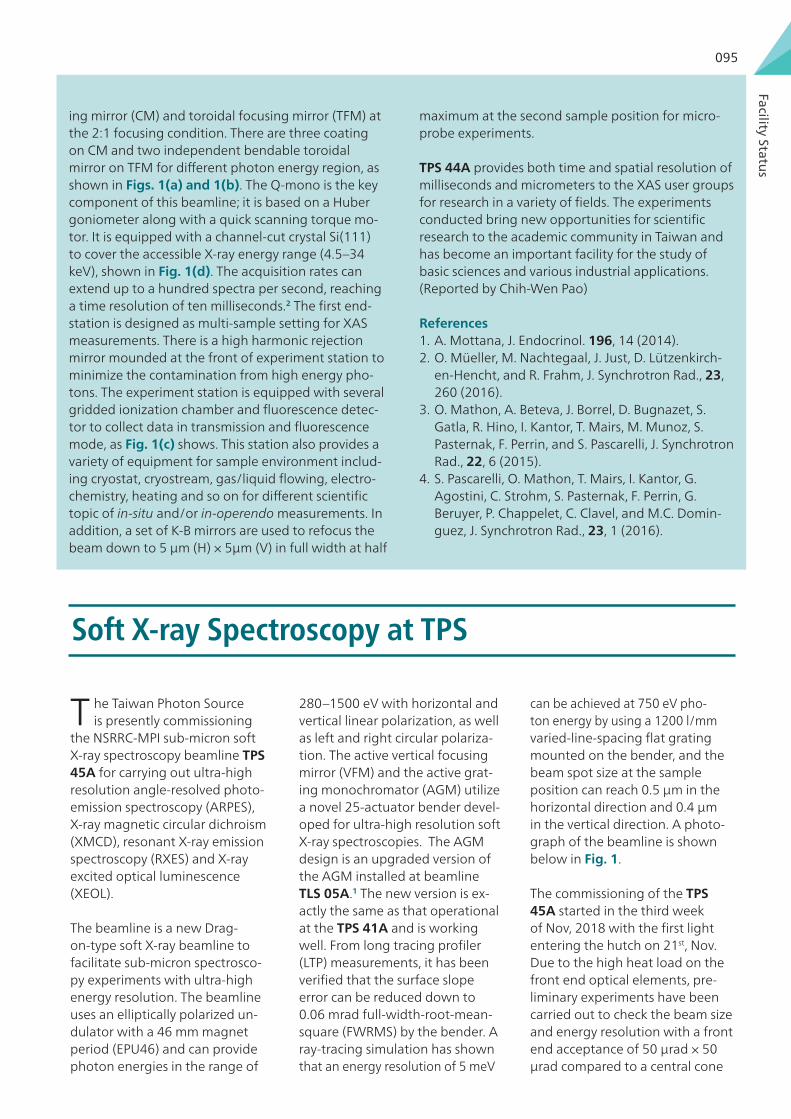
Facility Status
095
Soft X-ray Spectroscopy at TPS
T he Taiwan Photon Source is presently commissioning
the NSRRC-MPI sub-micron soft X-ray spectroscopy beamline TPS 45A for carrying out ultra-high resolution angle-resolved photo-emission spectroscopy (ARPES), X-ray magnetic circular dichroism (XMCD), resonant X-ray emission spectroscopy (RXES) and X-ray excited optical luminescence (XEOL).
The beamline is a new Drag-on-type soft X-ray beamline to facilitate sub-micron spectrosco-py experiments with ultra-high energy resolution. The beamline uses an elliptically polarized un-dulator with a 46 mm magnet period (EPU46) and can provide photon energies in the range of
280–1500 eV with horizontal and vertical linear polarization, as well as left and right circular polariza-tion. The active vertical focusing mirror (VFM) and the active grat-ing monochromator (AGM) utilize a novel 25-actuator bender devel-oped for ultra-high resolution soft X-ray spectroscopies. The AGM design is an upgraded version of the AGM installed at beamline TLS 05A.1 The new version is ex-actly the same as that operational at the TPS 41A and is working well. From long tracing profiler (LTP) measurements, it has been verified that the surface slope error can be reduced down to 0.06 mrad full-width-root-mean-square (FWRMS) by the bender. A ray-tracing simulation has shown that an energy resolution of 5 meV
ing mirror (CM) and toroidal focusing mirror (TFM) at the 2:1 focusing condition. There are three coating on CM and two independent bendable toroidal mirror on TFM for different photon energy region, as shown in Figs. 1(a) and 1(b). The Q-mono is the key component of this beamline; it is based on a Huber goniometer along with a quick scanning torque mo-tor. It is equipped with a channel-cut crystal Si(111) to cover the accessible X-ray energy range (4.5–34 keV), shown in Fig. 1(d). The acquisition rates can extend up to a hundred spectra per second, reaching a time resolution of ten milliseconds.2 The first end-station is designed as multi-sample setting for XAS measurements. There is a high harmonic rejection mirror mounded at the front of experiment station to minimize the contamination from high energy pho-tons. The experiment station is equipped with several gridded ionization chamber and fluorescence detec-tor to collect data in transmission and fluorescence mode, as Fig. 1(c) shows. This station also provides a variety of equipment for sample environment includ-ing cryostat, cryostream, gas/liquid flowing, electro-chemistry, heating and so on for different scientific topic of in-situ and/or in-operendo measurements. In addition, a set of K-B mirrors are used to refocus the beam down to 5 μm (H) × 5μm (V) in full width at half
maximum at the second sample position for micro-probe experiments.
TPS 44A provides both time and spatial resolution of milliseconds and micrometers to the XAS user groups for research in a variety of fields. The experiments conducted bring new opportunities for scientific research to the academic community in Taiwan and has become an important facility for the study of basic sciences and various industrial applications. (Reported by Chih-Wen Pao)
References 1. A. Mottana, J. Endocrinol. 196, 14 (2014).2. O. Müeller, M. Nachtegaal, J. Just, D. Lützenkirch-
en-Hencht, and R. Frahm, J. Synchrotron Rad., 23, 260 (2016).
3. O. Mathon, A. Beteva, J. Borrel, D. Bugnazet, S. Gatla, R. Hino, I. Kantor, T. Mairs, M. Munoz, S. Pasternak, F. Perrin, and S. Pascarelli, J. Synchrotron Rad., 22, 6 (2015).
4. S. Pascarelli, O. Mathon, T. Mairs, I. Kantor, G. Agostini, C. Strohm, S. Pasternak, F. Perrin, G. Beruyer, P. Chappelet, C. Clavel, and M.C. Domin-guez, J. Synchrotron Rad., 23, 1 (2016).
can be achieved at 750 eV pho-ton energy by using a 1200 l/mm varied-line-spacing flat grating mounted on the bender, and the beam spot size at the sample position can reach 0.5 μm in the horizontal direction and 0.4 μm in the vertical direction. A photo-graph of the beamline is shown below in Fig. 1.
The commissioning of the TPS 45A started in the third week of Nov, 2018 with the first light entering the hutch on 21st, Nov. Due to the high heat load on the front end optical elements, pre-liminary experiments have been carried out to check the beam size and energy resolution with a front end acceptance of 50 μrad × 50 μrad compared to a central cone

ACTIV
ITY REPO
RT 2017
096
Fig. 1: Photograph of the beamline TPS 45A.
Fig. 2: Vertical beam size measured using a knife-edge.
Fig. 3: The Ni L-edge X-ray absorption spectrum of NiO measured at TPS 45A.
full acceptance of 120 μrad × 120 μrad. Experiments were carried out at a typical photon energy set to ~850 eV. The horizontal focusing mirror was adjusted to obtain a horizontal focus of ~50 μm while the ver-tical focusing mirror was adjusted to obtain a vertical focus of less than 2 μm (Fig. 2).
Using the above obtained conditions, the Ni L3 and L2-edge X-ray absorption spectrum of NiO was mea-sured and it matched nicely with the well-known spectrum of NiO (Fig. 3).2
We then carried out photoemission experiments because the energy resolution is best determined by the Fermi edge of a clean gold spectrum measured at low temperature.
After initial measurements of the Au 4f core level spectrum, the valence band of gold was measured to confirm the cleanliness of the gold sample. This was followed by low-temperature (T = 20 K ) measure-ments of the Fermi edge of gold. Just before the end of the cycle on the 15th, Dec, 2018, we achieved a pho-toemission spectrum of the gold Fermi edge with an energy resolution of ΔE = 25 meV at a kinetic energy of E ~845 eV. This corresponds to an energy resolving power E/ΔE of 33,800. These results for the total en-ergy resolution are comparable to the best soft X-ray
photoemission measurements in the world today.
The above results were obtained with a preliminary tuning of the active grating monochromator. After the long shutdown, further experiments have been planned to measure the photon flux and optimize the active grating monochromator to increase the photon flux and improve the energy resolution to the target E/ΔE of ~50000 in the first half of 2018. The Max-Planck Institute endstation (Fig. 5) installed on branch A has capabilities to carry out molecular beam epitaxy thin film growth, thus facilitating ARPES mea-surements on in-situ grown films.
Based on the encouraging preliminary results of the high energy resolution achieved, it is envisaged that ultra-high resolution band dispersions and Fermi sur-face mapping using soft X-ray ARPES measurements on a variety of topical materials in thin film and bulk crystal forms can be actively pursued at TPS 45A. Fur-thermore, the Tamkang University endstation (Fig. 6) installed on branch B has capabilities for X-ray emis-

Facility Status
097
Fig. 4: The Fermi edge of gold measured at TPS 45 A.
Fig. 5: The Max Planck Institute endstation installed on branch A of TPS 45A.
Fig. 6: The Tamkang University endstation installed at TPS 45A.
sion spectroscopy, X-ray magnetic circular dichroism and X-ray excited optical luminescence studies. The scientific topics which can be fruitfully investigated at TPS 45A by the users’ group include topological insulators, carbon-based nanomaterials, photovol-taic materials, transition metal and rare-earth-based strongly correlated electron systems, magnetic ma-terials, oxide multilayers, etc. We look forward to a successful commissioning of the TPS 45A beamline and end-stations in the next few months.
References : 1 H. S. Fung, C. T. Chen, L. J. Huang, C. H. Chang, S.
C. Chuang, D. J. Wang, T. C. Tseng, and K. L. Tsang, AIP Conf. Proc. 705, 655 (2004).
2 D. Alders, L. H. Tjeng, F. C. Voogt, T. Hibma, G. A. Sawatzky, C. T. Chen, J. Vogel, M. Sacchi, and S. Iacobucci, Phys. Rev. B 57, 11623 (1998).

ACTIV
ITY REPO
RT 2017
098
SIKA: Opportunities for Low-Energy Excitations Using Neutrons
The triple-axis spectrometer has been used by neu-tron scatters to study many areas of condensed mat-ter physics for decades. A triple-axis spectrometer has the capability to investigate physical phenomena with high energy and momentum resolution with using cold neutrons. Currently time-of-flight spec-trometry is advanced at spallation neutron sources, while the cold triple-axis spectrometer still has ad-vantages of scanning S(Q, ω) space at each reciprocal point, measuring critical scattering, and availability of a number of sample environments.
Basic ComponentsThe layout of a typical triple axis spectrometer is shown in Fig.1. Table 1 shows the basic components of the cold triple axis spectrometer SIKA. One of the advantages of SIKA is a wide dance floor 55 m2 allowing us to choose incident energies Ei = 2.6–25 meV. The neutron flux at the sample position is mea-sured to be 1 × 10⁸ n/cm2s at λ = 2.1 Å. The analyzer drum hosts a 3He Single Detector for two-axis mode (diffraction detector, DD), a 3He Single Detector for triple-axis mode (single detector, SD), and a Position
Sensitive Detector (PSD). The collimators are available for requirement of users as shown in Table 1. The accessible ranges of momentum and energy transfer on SIKA with these components are shown in Fig.2. Energy resolutions estimated by Vanadium are also shown in Table 2. Both PG filter (Ei > 5 meV) and Be filter (Ei < 5 meV) are available on SIKA. Our cold triple axis spectrometer SIKA has very big advantage of studying condensed matter physic below 5 meV with high energy resolution such as below dE < 0.1 meV compared to typical thermal triple axis spectrometers.
Sample Environment The list of sample environment available on SIKA is provided in Table 3. The demands for dilution insert
Angular range28.4 degrees < 2θM < 120 degrees-100 degrees < 2θS < 100 degrees-90 degrees < 2θA < 90 degreesMonochromatorPyrolytic graphite (002)Flat, vertical, horizontal, and double focusingEi range: 2.43–30 meVFiltersPG filters (2 cm, 3 cm, 2 + 3 cm = 5 cm)Cooled Be filterFlux at sample position1 × 108 n/cm2s (at λ = 2.1 Å)Detectors3He Single Detector for diffraction and inelastic1D position sensitive detector (PSD)CollimatorsPre-monochromator (C1): Open, 20’, 40’, 60’Post-monochromator (C2): Open, 20’, 40’, 60’Pre-Analyzer (C3): Open, 20’, 40’, 60’Pre-single detector (C4s): Open, 20’, 40’, 60’Pre-PSD (C4r): Radial collimators
Collimations - Ei 14.87 8.07 5.11 2.620’-20’-20’-20’ 0.448 0.160 0.083 0.02440’-40’-40-Open 0.778 0.315 0.153 0.03560’-60’-60’-Open 0.769 0.323 0.146 0.041
Table 1: Components available on SIKA
Table 2: Energy resolution (meV) with vertical focusing mode with conditions estimated with Vanadium
Fig.1: Layout of a typical triple axis spectrometer.

Facility Status
099
Environment Sample Type OtherCF-4 4–300 K Bottom loading Closed cycle
OC-10.5–80 K(1)1.5–300 K
Top loading ILL- Orange (1)
CF-7 or CF-8 4–750 K Top loading Closed cycleCF-12 1.5–800 K Top loading Closed cycle (2)
AVM-150 mK–80 K1.5–300 K0–12 T
Top loadingVertical magnet(Oxford)
(3)
(1) OC-1 can be used with the 3He one-shot fridge insert to reach temperatures of 0.5 K to 80 K.(2) CF-12, if user wants to go above 300 K, sample stick for high temperature should be requested.(3) In conjunction with the Kelvinox dilution insert DL-1, a base temperature 50 mK can be achieved.* The above information is available at http://www.ansto.gov.au/ResearchHub/Bragg/Facilities/SampleEnvironments.
(~50 mK) and magnet (vertical up to 12 T) are high.
SoftwareThe SIKA team has developed the software SI-KA-SPICE based on the Spectrometer and Instrument Control Environment SPICE.1 SIKA-SPICE is built on client server architecture so you can control SIKA with any one of three computers located at dance floor, in the reactor beam hall, or in the SIKA cabin.
SIKA-client The SIKA client was built to control the instrument and has two displays. One views the current status on SIKA whereby monitor, axes for triple-axis, tem-peratures, and axes other than triple-axis are easily checked whilst controlling SIKA. The second display is for commands to control instrument and sample en-vironment. The command screen will also be used to edit scans, macros, sample information (single crystal,
powder, lattice parameters, compositions, and so on), and the UB-matrix for the experiment. The software and hardware limits for the instrument are also dis-played here (Fig. 3).
SIKA AnalysisSIKA analysis software (Fig. 4) has been developed. It is still improving its functionalities, due to comments from active and internal users during commissioning and the user program. The software allows us to plot and normalize data. Users can also compare, manipu-late, and export scans as text data. 2D plots are useful when you are observing dispersion relations in ener-gy and momentum space.
Experimental Capability The subjects proposed for SIKA are mainly high-ly-frustrated magnetism, superconductivity, magne-tism (general, 5d transition-metal, low-dimensional), multiferroics, soft matter thin film, etc.
We will now present you example of scientific capa-bility of SIKA. Figure 5 shows the magnon dispersion of MnF2 has been measured on SIKA. The magnon dispersion of MnF2 has been measured on SIKA. MnF2 is a classic material for studying spin waves in an antiferromagnet described also in textbooks.2 The experiment conditions were Ef-fixed with 8.07 meV, collimations of 60’(C1)-60’(C2)-60’(C3)-60’(C4) (for details, see Table 1). We needed only one minute for each point since Mn has a large magnetic moment; S = 5/2. The determined exchange parameters are J1 = 0.031, J2 = 0.153, and D = 0.143 meV by fitting the data based on equation (1).
Table 3: Sample environments normally requested on SIKA.
Fig.2: Accessible ranges of the momentum and energy transfer of SIKA at various final energies.

ACTIV
ITY REPO
RT 2017
100
Fig.3: SIKA client.
Fig.4: SIKA analysis software.

Facility Status
101
SIKA and Taiwan team.
Literature says J1 = 0.032, J2 = 0.155, and D = 0.11 meV.3
With s =5/2, z1 (nearest) = 2, and z2 (next nearest) = 8 and when we are scanning along qc.
Future ProgressWe are working on several improvements to enhance SIKA capabilities. One is taking advantage of a multiplex-ing analyzer, co-aligning 13 HOPG analyzer blades being 20 mm wide and 150 mm tall sitting on detector drum. With this multi-blade analyzer system, SIKA will have the capability of a RITA-type analyzer instrument.5 A polar-ized 3He neutron spin filter is under commissioning and will be available on SIKA to perform polarized neutron scattering experiments and polarization analysis. We are hoping for more new Taiwanese users in 2018. (Report-ed by Shinichiro Yano)
ζ = [D+8·s · j1 sin2 ·q z· c ]/16·s · j212
Equation (2)
γ = cos2 ·q z· c 12
Equation (3)
Fig.5: Magnon dispersion of MnF2 single crystal measured with SIKA.
Equation (1) hωq=16·s · j2 {(1+ζ )2+γ2}1/2
References 1. M. D. Lumsden, J. L. Robertson,
and M. Yethiraj, Physica B, 385-386, 1336 (2006).
2. G. Shirane, S. M. Shapiro, and J. M.Tranquada, Neutron Scatter-ing with a Triple-Axis Spectrom-eter. Cambridge University Press (2003).
3. A. Okazaki, K. C. Thurberfield, and R. W. H. Srevenson, Phys. Lett., 8, 9 (1964).
4. J. W. Lynn, Y. Chen, S. Chang, Y. Zhao, S. Chi, W. Ratcliff II, B. G. Ueland, and R. W.
Erwin, J. Res. Natl. Inst. Stand. Technol., 117, 61 (2012).
5. J. W. Lynn, Y. Chen, S. Chang, Y. Zhao, S. Chi, W. Ratcliff, B. G. Ueland, and R. W. Erwin, J. Res. NIST 117, 61 (2012).


Facts & Figures

ACTIV
ITY REPO
RT 2017
104
Organization
Board of Trustees 2015.3-2018.2
As of January 2018
Accelerator Operation Group
Power Supply Group
Light Source Division
Magnet Group
Vacuum Group
Precision Mechanical Engineering Group
Cryogenics Group
Utility & Civil Group
Beam Dynamics Group
Radio Frequency Group
Instrumentation & Control Group
Linac Group
High Brightness Injector Group
Instrumentation Development Division
Experimental Facility Division
Experimental Technique Group
Beamline Group
X-ray Absorption Group
X-ray Scattering Group
Protein Diffraction Group
SPring-8 Group
Industrial Application Group
X-ray & IR ImagingGroup
Scientific Research Division
Molecular Science Group
Nano Science Group
Condensed Matter Physics Group
Materials Science Group
Neutron Group
Administration Division
Personnel Office
Finance Office
Planning & Evaluation Office
Information Office
General Affairs Office
Procurement Office
User Administration & Promotion Office
Secretariat Office
Radiation & Operation Safety Division
Organization Ethics Office
Board of Trustees
Internal Audit
Directorate
Industrial Liaison Office
• Lih J. Chen (Chair), National Tsing Hua University• Chien-Te Chen, NSRRC • Tzong Chyuan Chen, Ministry of Science and Tech-
nology• Mei-Yin Chou, Academia Sinica• Bon-Chu Chung, Academia Sinica• Shangjr Gwo, NSRRC• Hsing-Jien Kung, Taipei Medical University
• Yuan-Pern Lee, National Chiao Tung University• Yuan-Tseh Lee, Academia Sinica • Chung-Yuan Mou, National Taiwan University• Yuen-Ron Shen, UC Berkeley, USA• Wei-Fang Su, National Taiwan University• Lee C. Teng, ANL, USA• Samuel C. C. Ting, CERN, Switzerland• Yu Wang, National Taiwan University
Back row (left to right): Kuo-Tung Hsu, Di-Jing Huang, Shangjr Gwo, Tzong-Chyuan Chen, Bon-Chu Chung, Li-Chyong Chen (Supervisor), Hsiu-Ming Lin (Supervisor), Yuan-Pern Lee, Chun-Chieh Wu, Chia-Hung Hsu.Front row (left to right): Chien-Te Chen, Yu Wang, Chung-Yuan Mou, Yuan-Tseh Lee, Lih J. Chen, Yuen-Ron Shen, Wei-Fang Su, Mei-Yin Chou.Not pictured: Lee C. Teng, Samuel C. C. Ting, Hsing-Jien Kung.

Facts & Figures
105
SAC 2015-2018
• Keith Hodgson (Chair), SLAC, USA• Oliver Bunk, PSI, Switzerland• Tai-Chang Chiang, UIUC, USA• John Hill, NSLS II, USA• Tetsuya Ishikawa, RIKEN SPring-8 Center, Japan• Chi-Chang Kao, SLAC, USA• Stephen Kevan, ALS, USA
Back row (left to right): Kuo-Tung Hsu (NSRRC), Jean Susini, Dmitri Svergun, Cheuk-Yiu Ng, John Hill, Oliver Bunk, Stephen Kevan, Chris-toph Quitmann, Di-Jing Huang (NSRRC).Front row (left to right): Keith Nugent, Shangjr Gwo (NSRRC), Chi-Chang Kao, Keith Hodgson, Tai-Chang Chiang, Tetsuya Ishikawa, G. Brian Stephenson.
• Cheuk-Yiu Ng, UC Davis, USA• Keith Nugent, La Trobe U., Australia• Christoph Quitmann, MAX IV, Sweden• G. Brian Stephenson, ANL, USA• Jean Susini, ESRF, France• Dmitri Svergun, EMBL, Germany
2017 User Executive Committee
• E-Wen Huang (Chair), National Chiao Tung University• Sheng-Fa Yu (Vice chair), Academia Sinica• Shu-Jui Chang, National Chiao Tung University• Chun-Jung Chen, NSRRC• Wei-Tsung Chuang, NSRRC
• Meng-Chiao Ho, Academia Sinica• Yu-Chang Lin, National Chiao Tung University• Chien-Lung Wang, National Chiao Tung University• Kuan-Wen Wang, National Central University• Chung-Lin Wu, National Cheng Kung University
2017 User Executive Committee

ACTIV
ITY REPO
RT 2017
106
Budget
2017 Neutron User Executive Committee
• E-Wen Huang (Chair), National Chiao Tung University• Ko-Wei Lin (Vice chair), National Chung-Hsing
University• Chun-Jung Chen, NSRRC• Jin-Ming Chen, NSRRC
0.00
10.00
20.00
30.00
40.00
50.00
60.00
70.00
80.00
90.00
100.00
2004 2005 2006 2007 2008 2009 2010 2011 2012 2013 2014 2015 2016 2017
Budg
et (i
n m
illio
ns o
f USD
)
Neutron experimental facility
TPS accelerator maintenance
TPS BL/endstation/ID
TPS accelerator
Civil construction
Administration & salaries
Utilities & land lease
Experimental Facility & ScientificResearch Divisions
Light Source & InstrumentationDevelopment Divisions
Year
The total budget for fiscal year 2017 was USD 54 million (based on exchange rate: 1 USD = 33 TWD), an increase of 4.50% over the prior year. Fiscal year 2017 budget covered the operating expenses associated with the follow-ing categories: Light Source & Instrumentation Development Divisions, Experimental Facility & Scientific Research Divisions, utilities & land lease, administration & salaries, TPS BL/endstation/ID, TPS accelerator maintenance, and neutron experimental facility.
• Chih-Hao Lee, National Tsing Hua University• Tsang-Lan Lin, National Tsing Hua University• Chih-Ying Liu, National Tsing Hua University• An-Chung Su, National Tsing Hua University• Chun-Chuen Yang, Chung Yuan Christian University

Facts & Figures
107
Manpower
Administrative Personnel 15%
Professions Education
Engineer/Technician 29%
Associate´s 4%
Doctoral 39%Master´s 47%
High School 2%
Bachelor´s 8%
Scientist 56%
As of January 2018, the NSRRC workforce comprises 441 staff members. The following pie charts show the man-power distributions by profession and by educational background.
User Statistics
From 1994 to 2017, the total number of beamlines opened to general users has increased from 3 to 31, compris-ing 24 TLS beamlines, 2 Taiwanese beamlines at SPring-8, and 5 TPS phase-I beamlines opened in September, 2016 (see Beamline Status on page112 ).
During year 2017, the User Administration & Promotion Office (UAO) issued 1096 new user cards, and provided services to 2,305 users coming from 149 affiliations (including 342 users from 95 foreign institutions) to use the above 31 NSRRC beamlines. Altogether, 589 proposals led by 361 principal investigators were performed, involv-ing 1,845 experiment runs and 12,124 user runs (visits).
The overall NSRRC beamtime (including SP 44XU*) allocated to users, excluding beamline maintenance and training courses, was 1,182 shifts for TPS, 13,115 for TLS, and 823.5 for SPring-8 (including 63 shifts for SP 44XU). Each shift consists of 8 consecutive hours.
In year 2017, the UAO processed 3,799 subventions for TLS/TPS experiments, and 174 for SPring-8 (including 42 for SP 44XU). Another 88 subventions, funded by the Ministry of Science and Technology, were given to Taiwan neutron users to conduct experiments at neutron facilities worldwide.
In total, 101 master’s theses and 23 doctoral dissertations utilizing NSRRC facilities were completed in 2017 (see Appendix). Operational data on beamlines/experimental stations and users are summarized in the following figures.
*Note: Institute for Protein Research, Osaka University (IPR-Osaka) and NSRRC have established a long-term col-laboration in protein structural research aiming to combine the expertise of both sides and to access each other’s facilities when needs arise. IPR-Osaka offers beamtime of its contract beamline SP 44XU at SPring-8 to Taiwanese users selected by NSRRC to study large macromolecular assemblies and membrane proteins.

ACTIV
ITY REPO
RT 2017
108
324
648 716
829
1,012
1,384
1,623 1,683
1,994
2,230 2,266 2,201
2,036 2,120
2,199
2,360 2,305
0
500
1000
1500
2000
2500
2001 2003 2005 2007 2009 2011 2013 2015 2017
Num
ber
of u
sers
Year
Note: In 2016, 305 out of 340 TPS users also used TLS/SP-8.
TPS: 340
Number of users 2001-2017
Geographic distribution of users and their numbers 2017
65875
1
196
584
122
312
14
19162
112
6
Note: 1. The geographic categories of the users are based on the locations of their affiliations.
2. Of the total 2,305 users, about 15% are international users.
Taiwan:Foreign countries:
Total:
1,963 (434 out of 526 TPS users also used TLS/SP-8)342 (20 out of 97 TPS users also used TLS/SP-8)2,305
Canada 2
Costa Rica 1
United States 18
Australia 12
New Zealand 1
China 138
France 6
Germany 17Austria 1
India 6
Italy 2 Turkey 3Japan 79
Korea 11
Netherlands 5Belgium 1
Russia 4
Singapore 26
Spain 1Switzerland 1
Thailand 4

Facts & Figures
109
Number of IR/VUV experiment runs 2001-2017
Number of X-ray experiment runs 2001-2017
0
100
200
300
400
500
600
2001 2003 2005 2007 2009 2011 2013 2015 2017
Expe
rimen
t run
s
Year
Received
Executed
1,977
1,102
329
0
200
400
600
800
1000
1200
1400
1600
1800
2000
2200
2001 2003 2005 2007 2009 2011 2013 2015 2017
Expe
rimen
t run
s
Year
Received
Executed/Materials
Executed/Bio

ACTIV
ITY REPO
RT 2017
110
Proposal distribution by research fields 2017
Distribution of beamtime used by domestic affiliations 2017
Others 1.36%
Surface, Interface and Thin Films 11.04%
Soft Matter 9.85%
Protein Crystallography 17.15%
Nanofabrication 0.34%
Methodology and Instrumentation 0.51%
Materials Science 24.45%
Environmental and Earth Science 10.7%
Condensed Matter Physics 11.2%
Chemistry 6.28%
Atomic and Molecular Science 4.58%
Applied and Industrial Research 2.54%
Natl. Taiwan Univ.
Academia Sin
ica
Natl. Tsin
g Hua Univ.
Natl. Cheng Kung Univ.
Natl. Chiao Tu
ng Univ.
Natl. Centra
l Univ.
Natl. Taiwan Univ.
of Sci. a
nd Tech.
Tamkang Univ.
Natl. Chung Hsin
g Univ.
Natl. Sun Ya
t-sen Univ.
Natl. Chiayi U
niv.
Natl. Health Rese
arch Instit
utes
Natl. Taipei U
niv. of Te
ch.
Natl. Yang-M
ing Univ.
Kaohsiung M
edical U
niv.
Linkou Chang Gung Hospital of Th
e C.G.M
.F.Yuan Ze
Univ.
Natl. Taiwan Norm
al Univ.
Tunghai Univ.
Tatung Univ.
Fu Jen Catholic
Univ.
0100
200
300
400
500
600
700
800
900
1000
1100
1200
1300
1400
1500
1600
Shift
Affiliation
Note: Eight hours per shift.

Facts & Figures
111
Publications
0 0
50
100
150
200
250
300
350
400
450
1997 1999 2001 2003 2005 2007 2009 2011 2013 2015 2017
Tota
l SCI
pap
ers
Year
315
280
245
210
175
140
105
70
35
Top 5%, 10%
, 15% SCI papers
Note:
78
144
221
345SCI total No.
Top 15%
Top 10%
Top 5%
As of 2018/01/30
Before 20141. Top 5%: I.F. ≥ 6.0 for physical science; I.F. ≥ 9.0 for life science.2. Top 10%: I.F. ≥ 4.5 for physical science; I.F. ≥ 6.0 for life science.3. Top 15%: I.F. ≥ 3.5 for physical science; I.F. ≥ 4.8 for life science.
2014-20171. Top 5%: I.F. ≥ 7.5 for physical science; I.F. ≥ 8.5 for life science.2. Top 10%: I.F. ≥ 4.5 for physical science; I.F. ≥ 5.5 for life science.3. Top 15%: I.F. ≥ 3.5 for physical science; I.F. ≥ 4.5 for life science.

ACTIV
ITY REPO
RT 2017
112
Beamlines at the Taiwan Photon Source
No. Beamline Mono type
Energy range(eV)
Res. power (E/ΔE)
StatusSpokesperson
E-mailTel. ext.
05AProtein Microcrystallogra-phy
DCM 5.7k–20k 7,000 in operationJean, Yuch-Cheng [email protected]
2051
09ATemporally Coherent X-ray Diffraction
DCM 5.6k–25k 7,000in operation
Sheu, Hwo-Shuenn [email protected]
2091HRM 14.4k 108
21A X-ray NanodiffractionWhite Beam
5k–30k7,000 in operation
Ku, Ching-Shun [email protected]
22114BCM 7k–25k
23A X-ray Nanoprobe HDCM 4k–15k 7,000 in operationTang, Mau-Tsu [email protected]
2231
24A Soft X-ray Tomography PGM 0.2k–3k 2,000 commissioningLai, Lee-Jene [email protected]
2242
25A Coherent X-ray Scattering DCM 5.56k–20k 7,000 in operationHuang, Yu-Shan [email protected]
2251
41A Soft X-ray Scattering AGM 0.4k–1.2k 60,000 commissioningHuang, Di-Jing [email protected]
2411
44AQuick-scanning X-ray Absorption Spectroscopy
Channel- cut DCM
4.5k–34k 7,000 commissioning TBD –
45ASubmicron Soft X-ray Spectroscopy
AGM 0.28k–1.5k 84,000 commissioningChainani, Ashish Atma [email protected]
2451
Beamlines at the Taiwan Light Source
No. Beamline Mono type
Energy range(eV)
Res. power (E/ΔE)
StatusSpokesperson
E-mailTel. ext.
01A1 SWLS − White X-ray none > 5k N/A in operationHwu, [email protected]
1011
01B1 SWLS − X-ray Microscopy DCM 8k–11k 1,000 in operationTang, [email protected]
1012
01C1 SWLS – EXAFSDCM 6k–33k 7,000
in operationChan, [email protected]
1013
01C2SWLS − X-ray Powder Diffraction
in operationSheu, [email protected]
1013
03A1BM − (HF-CGM) – Photoabsorption/ photoluminescence
CGM 4–40 50,000 in operationCheng, [email protected]
1031
04B1 BM − (Seya) SRCD SNM 4–40 5,000 in operationLin, [email protected]
1042
04C1 Dynamic SRCD NIM 4–10 3,000 in operationLin, [email protected]
1042
Beamline Status

Facts & Figures
113
No. Beamline Mono type
Energy range(eV)
Res. power (E/ΔE)
StatusSpokesperson
E-mailTel. ext.
04C2 Combustion Chemistry NIM 4–17 ~1,000 in operationLee, [email protected]
1043
05A1 EPU – Soft X-ray ScatteringAGM/SGM
60–1.5k 20,000
in operationHuang, [email protected]
1051
05B1 EPU − Soft X-ray Chemistry
SGM
in operationLiu, [email protected]
1050
05B2 EPU – PEEM in operationWei, [email protected]
1052
07A1 IASW − X-ray Scattering DCM 5k–23k 7,000 in operationSoo, [email protected]
1071
08A1 BM − (L-SGM) XPS, UPS SGM 15–200 20,000 in operationPi, [email protected]
1081
08B1 BM – AGM AGM 0.3k–1k 10,000 in operationPi, [email protected]
1082
09A1 U50 – SPEMSGM 60–1.5k 15,000
in operationChen, [email protected]
1101
09A2 U50 – Spectroscopy in operationHsu, [email protected]
1102
11A1 BM − (Dragon) MCD, XAS SGM 8–1.5k 15,000 in operationLin, [email protected]
1111
13A1 SW60 − X-ray Scattering ACCM 12k–14k 1,000 in operationLee, [email protected]
1131
13B1SW60 − Protein Crystallography
DCM 5k–20k 7,000 in operationJean, [email protected]
1132
13C1SW60 − Protein Crystallography
ACCM 12k–14k 1,000 in operationJean, [email protected]
1133
14A1 BM − IR Microscopy FTIR 0.05–0.5 N/A in operationLee, [email protected]
1141
15A1Biopharmaceuticals Protein Crystallography
DCM 5k–20k 7,000 in operationJean, [email protected]
1151
16A1BM − Tender X-ray Absorption, Diffraction
DCM 2k–8k 7,000 in operationChan, [email protected]
1161
17A1W200 − X-ray Powder Diffraction
ACCM 8k–12k 1,000 in operationLee, [email protected]
1171
17B1 W200 − X-ray Scattering DCM 4k–15k 7,000 in operationLee, [email protected]
1172
17C1 W200 – EXAFS DCM 4k–15k 7,000 in operationLee, [email protected]
1173
20A1 BM − (H-SGM) XAS SGM 70–1.2k 10,000 in operationChen, [email protected]
1201
21A1U90 − (White Light) Chemical Dynamics
none 4–500 50in operation
Lee, [email protected]
1211
21A2U90 − (White Light) Photochemistry
in operationCheng, [email protected]
1210

ACTIV
ITY REPO
RT 2017
114
No. Beamline Mono type
Energy range(eV)
Res. power (E/ΔE)
StatusSpokesperson
E-mailTel. ext.
21B1U90 − (CGM) Angle-resolved UPS
CGM 4–100 105
in operationTsuei, [email protected]
1212
21B2 U90 − Gas Phase in operationLee, [email protected]
1212
23A1IASW − Small/Wide Angle X-ray Scattering
DCM 5k–23k 7,000 in operationJeng, [email protected]
1231
24A1 BM − (WR-SGM) XPS, UPS SGM 15–1.5k 15,000 in operationYang, [email protected]
1241
Beamlines at SPring-8, Japan
No. BeamlineMono type
Energy range (eV)
Res. power (E/ΔE)
StatusSpokesperson
SP12B1 BM − Materials X-ray StudyDCM 5k–100k 7,000 in operation
Ishii, [email protected]
SP12B2BM − Protein X-ray Crystallography
Chen, Chun-Jung [email protected]
SP12U1 U32 − Inelastic X-ray Scattering DCM/HRM 5k–30k 106 in operationNozomu, [email protected]
SP12U2 HAXPES/Photoemission DM/HRM 6k–14.4k 300,000 in operationTsuei, [email protected]
Neutron Instrument at ANSTO, Australia
No. InstrumentIncident energy
(meV)Energy res.
(meV)Status Contact
CG4SIKA – Cold Neutron Triple-axis Spectrometer
2.6–25 0.026–2 in operationYano, [email protected]
4BCM: 4-Bounce Channel-cut MonochromatorACCM: Asymmetrically-cut Curved Crystal MonochromatorAGM: Active Grating MonochromatorBM: Bending MagnetCGM: Cylindrical Grating MonochromatorDCM: Double Crystal MonochromatorDM: Diamond Crystal MonochromatorEPU: Elliptically Polarized UndulatorEXAFS: Extended X-ray Absorption Fine StructureFTIR: Fourier Transform Infrared SpectroscopyHAXPES: Hard X-ray Photoelectron SpectroscopyH-SGM: High Energy Spherical Grating MonochromatorHDCM: Horizontal Double Crystal MonochromatorHF-CGM: High Flux Cylindrical Grating MonochromatorHRM: High Resolution Crystal MonochromatorIASW: In-Achromatic Superconducting WigglerIR: Infrared RadiationL-SGM: Low Energy Spherical Grating MonochromatorMCD: Magnetic Circular Dichroism
NIM: Normal Incidence MonochromatorPEEM: Photo-Emission Electron MicroscopePGM: Plane Grating monochromatorPRT: Participating Research TeamQCM: Quadruple Crystal MonochromatorSGM: Spherical Grating MonochromatorSNM: Seya-Namioka MonochromatorSPEM: Scanning Photoemission Electron MicroscopeSRCD: Synchrotron Radiation Circular DichroismSW60: Superconducting Wiggler-60 mmSWLS: Superconducting Wavelength ShifterU32: Undulator-32 mmU50: Undulator-50 mmU90: Undulator-90 mmUPS: Ultraviolet Photoelectron SpectroscopyW200: Wiggler-200 mmWR-SGM: Wide-Range Spherical Grating MonochromatorXAS: X-ray Absorption SpectroscopyXPS: X-ray Photoelectron SpectroscopyXRD: X-ray Diffraction

Facts & Figures
115
Major Events
Max Planck–NSRRC/NCTU/NTHU Center for Complex Phase Materials
NSRRC held a press conference on January 4 at the Ministry of Science and Technology (MOST) to announce the plan to establish the Max Planck–NSRRC/NCTU/NTHU Center for Complex Phase Materials (MPPNC-CPM) in Hsin-chu, Taiwan. The Center aims for the scientific collaboration and exchange of scientists and students between the Max Planck Society (MPS) and the NSRRC/NTHU/NCTU. The reputable MPS, famed as the “cradle of Nobel Laureates” has nurtured 18 Nobel laureates since its formation in 1948. The team led by Director Liu Hao Tjeng (Max Planck Institute for Chemical Physics of Solids (MPI-CPfS) in Dresden, Germany) has had a long standing scientific collaboration with the NSRRC for two decades. The MPS has invested 1.5 million Euros in constructing a beamline station at TPS. The MPS also signed a Memorandum of Understanding with National Chiao Tung University and National Tsing Hua University in 2013 and 2016, respectively. The official founding of the Max Planck––NSRRC/NCTU/NTHU Center for Complex Phase Materials by the MPS will strengthen bilateral collabora-tions. The MPS and institutes in Taiwan will pledge 400,000 Euros each year to the Center for supporting young scientists, post-doctoral researchers, and PhD students to study in the applicable fields.
NSRRC Board Meetings
The fifth NSRRC Board of Trustees (see page 104 for details) had its fourth and fifth Board Meetings on March 3 and September 30, in addition to two other meetings of Executive Board held on June 7 and December 15. The NSRRC Board of Trustees was established in 2003, and each Board serves a three-year term. The Board is respon-sible for ensuring that NSRRC is integral to the scientific community and serves its purposes.
These meetings were chaired by Prof. Lih J. Chen; the NSRRC management, including Director, Deputy Directors and Secretary General, gave status reports on the NSRRC operation, accelerator systems, experimental facility development, scientific progress, and budgetary planning. In March, the board approved the 2016 Annual Per-formance Outcome Report, and 2016 Financial Statements and Independent Auditors’ Report. In June, “Guide-lines for Shift Duty Allowance and Overtime Compensation” was approved. In September, changes in accounting system were approved. In December, the board approved 2019 Annual Work Plan and Budget Estimates, 2017 Internal Audit Report, and 2018 Internal Audit Plan.
Key figures of MPPNC-CPM and honored guests attended the press conference.

ACTIV
ITY REPO
RT 2017
116
The SAC members gathered for reviewing and advising.
Group photo of participants at the 23rd Users’ Meeting.
9th SAC Meeting
All 13 members of the NSRRC Science Advisory Committee (SAC, see page XX for the member list) convened the ninth SAC Meeting on July 10 and 11, chaired by Prof. Keith Hodgson, to review and to evaluate the NSRRC’s sci-entific achievement, beamline construction, technique development and next-phase planning. Besides the cur-rent operation status of TLS and the progress of TPS briefed by the NSRRC managers, the NSRRC scientists from different beamlines presented their scientific results, and construction of the TPS beamlines and endstations in the past year, as well as the planning for the TPS phase-I and II experimental facilities. This year, two users, Prof. Way-Feng Pong and Prof. Dengsung Lin were invited to report their TPS phase-II endstation projects with NSR-RC. This annual review meeting has provided NSRRC with a valuable opportunity to interact with reputed inter-national SR experts and to hear their insightful advice on the continuing projects and scientific directions.
National Synchrotron Radiation Research Center (NSRRC) Users' Meetings and Workshops was held from September 5 to 8, attended by 474 participants. It was organized by the Users’ Executive Committee (UEC) Chair, E-Wen Huang (National Chiao-Tung University, NCTU) and Wei-Tsung Chuang (NSRRC). Besides learning about NSRRC’s recent progress, research directions, facility capabilities, and the latest
user researches, participants were also able to take part in four topic-specific workshops, interact with exhibitors and colleagues from the community.
The first day began with the opening remarks by the UEC Chair. NSRRC Director Shangjr Gwo then gave an overview of the current status and future devel-opment of NSRRC. It was followed by a light source
23rd Users’ Meeting & Workshops

Facts & Figures
117
The plenary session on the opening day.
Personal interactions resulted from informal encounters during coffee breaks.The poster awards were presented at the banquet on the second day.
The poster session fueled lively discussion.
status report from the Light Source Division head, Ming-Chyuan Lin, and updates on the experimental facilities by Yu-Shan Huang, the head of the Exper-imental Facility Division. Mau-Chung Frank Chang, President of NCTU, was invited to make a keynote speech, titled “Sync to Innovate,” which was explicit about the power of cross-disciplinary research and joint-facility collaboration, using the example of Sys-tem-on-Chip for medical applications.
Four users were invited to present their recent dis-tinguished work using the NSRRC facilities – packing of 1D and 3D conjugated molecules by Chien-Lung Wang (NCTU), structural and functional studies of c-di-GMP by Shan-Ho Chou (National Chung Hsing University, NCHU), studies of ferrihydrite in environ-mental biogeochemistry by Yu-Ting Liu (NCHU), and visualization of electronic structure with SR photo-electron spectroscopy by Chung-Lin Wu (National Cheng Kung University, NCKU).
There were 247 posters on display, including 12 post-ers selected for oral presentation contests in Materi-als Science, Physics/Chemistry Science, and Biological Science. The “Glory of Taiwan Award” was presented to the winner of each group at the banquet the next
day. The day was also packed with the Users’ Town Meeting, five interest group meetings for Protein X-ray Crystallography, Powder X-ray Diffraction, SR-based Microscopy, X-ray Absorption Spectroscopy, and X-ray and Neutron Scattering. The day-program ended with a reception dinner.
Four topic-specific workshops took place during this period. Workshop I and II were held in parallel on September 6. Workshop I: Synchrotron for Industries was organized by Bor-Yuan Shew. Industrial experts from the fields of semiconductors, pharmaceuticals, green energy and polymers were invited to talk about their experiences, problems, and suggestions on using SR techniques. It was also expected to pro-mote SR as a helpful tool for industrial innovation so as to elevate Taiwan’s global competitive strength. Workshop II: High Flux Small-Angle X-ray Scattering on Biological Complex Structures was organized by U-Ser Jeng, aiming to share recent developments and research results in biological SAX scattering instru-mentation, protein solution measurements and data analysis, and ultra SAX scattering for large structure and biological structural kinetics in the millisecond to microsecond range. Workshop III: High Resolu-tion X-ray Spectroscopy of Quantum Materials was

ACTIV
ITY REPO
RT 2017
118
a two-day workshop from September 7 to 8. It was organized by Ashish Atma Chainani for introducing challenging problems and hot topics in quantum materials which can be addressed by Resonant Inelas-tic X-ray scattering (RIXS) and Angle-resolved Photo-emission Spectroscopy (ARPES). Workshop IV: TPS & Complementary Methods for Emerging Materials was held on September 7, and it was organized by E-Wen Huang. It was targeted to promote the newly devel-oped TPS and the NSRRC neutron program that offers complementary advanced technology for materials research, and to discuss the possibility of building a new platform for Integrated Computational Materials Engineering (ICME).
The four-day program offered an excellent forum for users to discuss issues concerning their experiments, as well as NSRRC’s user services and future devel-opment with the NSRRC managers and scientists. The diverse theme not only underlined the strong involvement of users to carry out basic sciences and applications at NSRRC, but also highlighted new technologies and possible solutions to current scien-tific challenges. Totally 63 domestic and international speakers talked on their expertise – all offering op-portunities to learn about the latest SR-related tech-nology and research.
Former NSRRC Director C. T. Chen and NSRRC User Andrew H.-J. Wang Named Recipi-ents of Taiwan's 2017 Presidential Science Prize
Established in 2001, the biennial Presidential Science Prize recognizes innovative researchers who have made outstanding contributions in the fields of mathematics and physical sciences, life sciences, social sciences, and applied sciences. It is considered the nation’s highest scientific honor. According to the Eligibility and Selection Process of the Prize, the Committee is composed of 15 members, including the President of Academia Sinica as the chair, and the Minister of Science and Technology as the vice chair. This year three winners stood out among 13 nominees: Academician Chien-Te Chen (NSRRC) in mathematics and physical sciences, Academician Andrew H.-J. Wang (Academia Sinica) in life sciences, and Dr. Douglas Yu (Taiwan Semiconductor Manufacturing Com-pany) in applied sciences.
Academician C. T. Chen is currently a Distinguished Scientist at NSRRC. Before returning to Taiwan, he had worked as an experimental physicist at AT&T Bell Labs for a decade. During that period, he invented the Dragon beamline and developed several high resolution soft X-ray spectroscopic techniques. Among them, magnetic circular dichroism (MCD) was the most well-known and conducive to magnetism and magnetic materials re-search. He and his collaborators had also completed high accuracy experiments on strongly correlated electron systems, including high Tc superconducting cuprates, alkali doped C60, giant magnetoresistance manganese oxides, magnetic multilayers, etc.
In 1995, Academician Chen was invited to join the NSRRC to develop the facility and scientific applications of the Taiwan Light Source (TLS). He held the post of Deputy Director from 1995 to 1997, and Director from 1997 to 2005. Under his leadership, NSRRC accomplished several milestones. For example, TLS employed a super-
The awarding ceremony was held in the Presidential palace and presented by the Presi-dent on November 21.
Academician Chien-Te Chen.

Facts & Figures
119
conducting radio frequency (SRF) cavity; TLS installed 8 insertion devices including 5 superconducting magnets; TLS reached the 1.5 GeV full energy top-up injection; TLS added 22 beamlines and 54 experimental stations; the “Taiwan Photon Source (TPS) Construction Plan” was initiated, etc. After his term ended, he continued to devote himself to the construction of TPS. He played a vital role in finalizing the TPS design and specifications in 2009. He then served as the Director General of the TPS Construction Project from 2010 to 2014 and led the NSRRC team to overcome many severe challenges. The TPS synchrotron shined its first light in December 2014 and the stored current swiftly exceeded the design goal of 500 mA in December 2015.
In addition to his pioneering contributions in spectroscopy and condensed matter physics, Academician Chen has made unparalleled contributions to the development of synchrotron radiation facility in Taiwan and raised the country’s competitiveness and international stature in scientific research. TPS, which inaugurated its opening in September 2016, will bring new opportunities for a broad spectrum of advanced scientific research for several decades to come.
Academician Andrew Wang is at present a Distinguished Research Fellow of Academia Sinica. His research inter-ests cover structural proteomics, anticancer drugs, X-ray crystallography, NMR and molecular design. Being an important user and a long-term collaborator of NSRRC, he not only served as a member of the NSRRC Board of Trustees, but also assisted NSRRC to initiate and build a world-class synchrotron-based protein crystallography facility. Protein crystallography is known as a powerful technique to determine three-dimensional structures of bio-macromolecules. The bright and tunable synchrotron radiation can improve the rate of crystal structure determinations; it is hence an ideal means for studying structural genomics and structural-based drug designs. Until now, NSRRC’s protein crystallography beamlines available to users include TLS 13B1, TLS 13C1, TLS 15A1, and TPS 05A. Taiwan has become a well-equipped and competitive player in the fields of proteomics and struc-tural genomics.
The PI and co-PI of the TPS Construction Project, and 5 PIs of its sub-projects.
Sharing new technologies and supporting scientific research activities has been a hallmark of the world-wide synchrotron light source community. NSRRC has, from its inception, been learning X-ray technolo-gies from other facilities, and now is able to develop innovative technology and collaborate with others to accomplish significant scientific goals, to evolve the technology even further, and to make effective use of facilities. Many opportunities open up by establishing bilateral collaboration and signing of Memoranda of Understanding (MOU), which initiate, continue, flourish and expand into exciting platforms that ben-efit scientists and people around the world.
In 2017, NSRRC has various valid MOUs and collabora-tion agreements in the following five aspects:
1. Contract beamlines/endstations• TPS 45A NSRRC-MPI Beamline with Max-Planck-In-
stitute for Chemical Physics of Solids• TPS 45A TKU Endstation with Tamkang University• TPS 27A STXM/Ptychography Endstation with Tam-
kang University• nanoARPES Endstation at TPS 39A with National
Tsing Hua University• Bio-SAXS Beamline (TPS 13A) with Academia Sinica • Brain Imaging Beamline (TPS 02A) with Academia
Sinica and National Tsing Hua University
MOUs and Collaboration

ACTIV
ITY REPO
RT 2017
120
2. International collaboration on beamline/ endstation operation and maintenance• Two Taiwanese contract beamlines at SPring-8,
Japan• SIKA – cold neutron triple-axis spectrometer at
ANSTO, Australia
3. Providing international facilities with professional services • A 3.5 T superconducting multipole wiggler (SMPW)
was designed by NSRRC through a collaboration between NSRRC and the Synchrotron Light Re-search Institute (SLRI). It will be installed in the storage ring of SLRI in 2018.
• NSRRC also provides SLRI with consulting service for accelerator radiation safety; the contract was signed in 2017.
4. MOUs signed with international institutes for collaboration• Asia and Oceania: IVPP/CAS (newly signed in
2017), IHEP/CAS, U. of Macau, Wuhan U., IPR/Osa-ka U., Okayama U., ISSP/ U. of Tokyo, Alagappa U, PAL, SLRI, ThEP, CELA, AS
• Europe: DESY, DLS • America: ANL, APS, BNL, NSLS
5. Joint graduate programs with Taiwanese universities• Graduate Program in Science and Technology of
Synchrotron Light Source with National Tsing Hua University
• Graduate Program for Science and Technology of Accelerator Light Source with National Chiao Tung University
• Structural Biology PhD Program with National Tsing Hua University, Academia Sinica, National Health Research Institutes
• International PhD Program for Synchrotron Radia-tion and Neutron Beam Applications with National Sun Yat-sen University
NSRRC signed MOU with Institute of High Energy Physics, Chinese Academy of Sciences
NSRRC and Alagappa University signed MOU.
NSRRC and SLRI signed MOU.

Facts & Figures
121
Participants of the Workshop on Data Collection and Analysis of X-ray Nanodiffraction.
A TPS tour was arranged during the 2017 Spring Symposium of the Asian and Oceanian Photochemistry Association in Taiwan.
Conferences & Workshops
The Symposium of the Asian and Oceanian Photo-chemistry Association in Taiwan was initiated in the fall of 2009, and has been held twice each year since then. The symposium aims for encouraging postdoc-toral associates, research associates, and graduate students to present their works through either oral or poster presentations in the spring symposium, while faculty members present their research in the fall symposium.
The 2017 Spring Symposium took place at NSRRC on January 18 with 82 attendees. Its scientific program covered not only all areas in photochemistry, photo-physics and photobiology, but also applications of spectroscopy, imaging, and microscopy to gaseous species, materials and biosystems. Taking the oppor-tunity of being in NSRRC, a facility tour was arranged for participants to see several newly completed TPS beamlines.
TPS 21A, X-ray Nanodiffraction (XND) organized the Workshop on Data Collection and Analysis of X-ray Nano-diffraction during March 1 to 4 for training purposes. Dr. Nobumichi Tamura from Advanced Light Source (ALS), USA and Prof. Kai Chen from Xi’an Jiaotong University, China were invited to NSRRC and lecture on software analyzing techniques. The NSRRC XND team also invited some users to measure their sample on site, which allowed the data to be used and analyzed by the workshop participants. In total 80 participants attended this workshop.
2017 Spring Symposium of the Asian and Oceanian Photochemistry Association in Taiwan
Workshop on Data Collection and Analysis of X-ray Nanodiffraction

ACTIV
ITY REPO
RT 2017
122
The Ninth International Workshop on Radiation Safe-ty at Synchrotron Radiation Sources (RadSynch17) was held at NSRRC during April 19 to 22, with 50 attendees. Presently there are nearly 40 operating synchrotron radiation sources in the world with a large scientific user community. There are also several more synchrotron radiation sources in different stag-es of design, construction and commissioning. This series of workshops has established a forum where the experts of radiation safety at the synchrotron radiation sources can meet and discuss the specific
9th International Workshop on Radiation Safety at Synchrotron Radiation Sources
radiation safety and protection issues of the facilities. This kind of dialogue immensely benefits the design, operation and upgrade of the present and future synchrotron radiation facilities and beamlines. The program of this year’s workshop focused on projects/upgrades, operational experiences, radiation safety design/assessment, shielding, interlock system/ra-diation protection for top-up operation, activation/decommissioning, and radiation safety issues for FEL/high intensity lasers.
RadSynch17 brought together the radiation physicists and radiation safety professionals of the synchrotron radiation facilities around the world in one forum.
Taiwan-Japan Seminar on Bioimaging
Attended by 34 scientists and students interested in the field of biomaging, Taiwan-Japan Seminar on Bioim-aging was held at NSRRC on May 4. Prof. Ando Masami from Tokyo University of Science, Prof. Gupta Rajiv from Massachusetts General Hospital of Harvard University, Prof. Yuasa Tetsuya from Yamagata University, and the research teams from NSRRC and National Tsing Hua University came together to share their research progress and collaborations in Bioimaging. Some emphasis were placed in this meeting in hope for benefiting disease diagnosis in the future, such as the development of applicable facilities at TPS, studies of bioimaging with syn-chrotron radiation, and collaborative projects with medical institutes and the issues may encounter.
Participants of Taiwan-Japan Seminar on Bioimaging.

Facts & Figures
123
35th Spectroscopy and Surface Science Symposium of Taiwan
Science Festival – Explore Space and Arts through Fun and Educational Science Activities
The 35th Spectroscopy and Surface Science Symposium of Taiwan was organized by NSRRC in Sun-Moon Lake Teachers’ Hostel from July 27 to 29, attended by 100 experts and students. This interdisciplinary meeting is an important gathering for Taiwanese researchers from the related fields of surface science, photonics and semi-conductor. It provides a great opportunity for them to discuss scientific issues and share their most recent progress. This year’s program covered topics not only in laser & photonics, surface & nano science, and spectros-copy, but also in optical imaging and soft X-ray imaging.
“Science Festival,” took place at NSRRC and the Na-tional Space Organization (NSPO) on July 29. The event attracted almost 800 adults and children. The purpose of this outreach event was to encourage interest in science amongst the young, to lay the foundation for science learning, and to inspire our future scientists.
Six “Science Classrooms” were organized, in which enthralling presentations through story-telling were delivered by Director General Wei-Hsin Sun of Na-tional Museum of Natural Science, Prof. Piero Baglioni of University of Florence, Dr. Tung-Ho Chen of Na-
Both oral talks and posters were presented at SSSST35.
“Science Festival,” a great public engagement opportunity, attracted almost 800 adults and children.
tional Palace museum, Dr. Chun-Chieh Hsiao of NSPO, and Drs. Yu-Jong Wu and Yao-Chang Lee of NSRRC. The “Science Classrooms” covered two themes: “A Space Odyssey” and “Arts, Culture, and Paleontology.” The audience was fascinated with educational talks on how hi-tech novel materials were used to restore ancient artifacts, how synchrotron light identified collagen within the oldest dinosaur embryo fossil, and how the secrets held by antiquities were revealed by light technology. In parallel, informative presen-tations about stargazing, the Taiwanese-made satel-lites, and creating Pluto in a lab were given under the space theme.

ACTIV
ITY REPO
RT 2017
124
All attendees had an exclusive open house experience at the NSRRC and NSPO. Youngsters were given the chance to take a close look at the TPS, learning how the brightest light on earth is produced and how TPS fa-cilitates scientific research and product development for human well-being. In addition, they saw the artificial satellites manufactured by the NSPO scientists, and walked through the galaxy corridor, imagining themselves as astronauts. While educational, a series of science activities surely brought everyone a fun day.
Workshop for NSRRC/TKU-Soochow University-Western University Synchrotron Radiation Research Center
Micro-beam Data Collection and Processing Workshop
In a continuing series of workshops for NSRRC/TKU-Soochow University-Western University Synchrotron Radiation Research Center, this year Tamkang Uni-versity and NSRRC co-organized the Workshop on Synchrotron-related Spectroscopy and Microscopy for Energy Materials. The energy-related materials with the diverse topics are studied by different synchrotron methods, offering the insight into the reactive mech-anism and relative performance. The workshop that
This workshop, attended by 58 users, was co-organized by NSRRC and the Crystallogra-phy Committee of ROC on September 29. The purpose of this workshop was to introduce new data collection and processing methods on TPS 05A Protein Microcrystallography beamline. These new methods can simplify the data collection and structure determina-tion. The one-day program included topics such as "What is radiation damage and why should we care," "Introduction of micro-beam data collection methods at TPS 05A" and "In-troduction of data processing at TPS 05A."
occurred on August 15 and 16 at Yangmingshan Tien Lai Spa and Resort was attended by more than 50 participants. The workshop aimed to offer a forum to present new interesting results about the latest tech-nology, mechanism, and application in a much closer community, and to bridge international cooperation with the unique idea in the physical and chemical property.
Workshop on Synchrotron-related Spectroscopy and Microscopy for Energy Materials.
Users attended the workshop to learn new methods for simplifying the data collection and structure determination.

Facts & Figures
125
2nd Joint International Symposium of NSRRC and IPR/Osaka University: Establishment of Structural Biology Network in Asia and Oceania
The 2nd Joint International Symposium of NSRRC and Institute for Protein Research/Osaka University: Establishment of Structural Biology Network in Asia and Oceania took place at NSRRC on 6 and 7 of December. Fol-lowing the successful 1st Joint Symposium: Networking the Asia Region in September 2015, the geographical scope was expanded this year. 90
The Joint International Symposium of NSRRC and IPR continued in Hsinchu, Taiwan
Winter School on FEL aimed to cultivate FEL accelerator technologies.
speakers and delegates came from 9 countries in Asia and Oce-ania, including Australia, China, India, Japan, Korea, Singapore, Thailand, Taiwan, and Vietnam, to exchange ideas in structural biolo-gy and to share experience in user facilities. In 2007, an agreement on academic exchange between NSRRC and IPR was concluded to promote collaborative research and academic exchanges. This joint symposium is organized un-der this MOU, reflecting the long-term collaboration between the two partner institutes and protein research activities in the region, using the synchrotron facilities as the core of the network.
Training Courses
Winter School on Free-electron Lasers
The Winter School on Free-electron Lasers 2017 took place at NSRRC during January 19 to 23. Well-known domestic and international speakers presented lectures on the essence of free-electron lasers (FEL) and their applica-tions for accelerators. 41 students were se-lected to participate in the five-day program. As a FEL has been deemed to be the most important fourth-generation light source of the future, the FEL will play a crucial role in the development of innovative technologies and emerging sciences. Linac Coherent Light Source (LCLS) at SLAC, USA and the SPring-8 Angstrom Compact free electron LAser (SA-CLA) at RIKEN, Japan are the two pioneering FEL sources came into operation; the Europe-an X-ray free-electron laser (European XFEL) has started the operation in September 2017. More FEL facilities are under construction or commissioning, such as PAL-XFEL by South Korea, Shanghai Free Electron Laser (SXFEL)

ACTIV
ITY REPO
RT 2017
126
by China, etc. This winter school, accordingly, aimed to familiarize students with FEL techniques and their appli-cations in various fields, to trigger their interest in profound knowledge and expertise, and eventually to nurture more future scientists who will commit themselves to FEL accelerator technologies.
Summer Internship
NTHU-NSRRC Summer Course: the Applications on Synchrotron Light Source
NSRRC organized its fifth summer internship from July 3 to August 4, attracting 46 students. To promote synchrotron-related sciences and plant roots at the undergraduate level, the program targeted college juniors and seniors who were interested in devoting five weeks to experience how synchrotron light sourc-es were used in scientific research. From the lectures given by NSRRC scientists during the first week, the students acquired a fundamental knowledge about accelerator science and diverse research applicable
The NTHU-NSRRC Summer Course, organized by Prof. Chih-Hao Lee (Dept. of Engineering & System Science, National Tsing Hua University), and jointly provided by NSRRC scientists and NTHU professors from July 25 to August 7. It was a three-credit graduate course designed for graduate and upper-year undergraduate students. In total 64 students across Taiwan enrolled in this course, which combined lectures, facility tours and hands-on activities. This course aimed to dissem-inate fundamental knowledge of synchrotron light sources and demonstrate how these sources can facilitate scientific research in various fields.
at NSRRC beamlines. From the second to the fifth week, students selected and joined the laboratories or the endstations in one of six groups: X-ray struc-tural analysis of materials, X-ray spectroscopy, syn-chrotron-based X-ray microscopy, molecular sciences, structural biology and accelerator technology. In the last week, each student gave a 15-minute presen-tation to briefly summarize what they had learned, to talk about what had inspired them, and to share ideas that had come across their mind.
The students met with the director and were given the certificate on the last day.
Summer course jointly provided by NSRRC and NTHU.

Facts & Figures
127
Training Courses on Protein Crystallography I & II
2017 Training Course on the Rietveld Method
The Training Courses on Protein Crystallography, co-organized by NSRRC and the Crystallography Committee of ROC, took place from August 7 to 11 and August 21 to 25. Each course enrolled 18 current users and consisted of lectures focusing on theory and hands-on sessions to practice experimental techniques, with an aim to dissem-inate experimental techniques of macromolecular crystallography to researchers or graduate students with an interest in using this specific method to further the scope of their research. This training course covered a broad spectrum of topics on synchrotron-based protein crystallography, ranging from crystallization of proteins, da-ta-collection strategy, phasing techniques, radiation damage to protein crystals, to the structural determination of proteins. Students were given opportunities to grow crystals, to collect and to process data from single-crystal diffraction, to use Gd-MAD, S-SAD and MR Phasing methods to determine three-dimensional protein structure, and to identify the lead compound of the target protein with the MR method. The knowledge and skills that they acquired in this course are essential for research and innovation in biotechnology and pharmaceuticals.
On August 10 and 11, scientists of NSRRC and Prof. I-Jui Hsu from National Taipei University of Technol-ogy jointly offered a training course on the Rietveld method to tackle the issues when analyzing structure from high-resolution powder diffraction data. Com-pared to hundreds of well-defined peaks in single crystal diffraction, most powder diffraction patterns have overlapping peaks. The Rietveld method ana-lyzes the whole diffraction pattern, minimizing the difference between observed data and the expected pattern from a model. First introduced in the late 60’s, the Rietveld refinement method is commonly used to interpret powder diffraction data. In this method, a least squares approach to refining a model to experimental data is performed. The height, width and position of the reflections can be used to deter-mine crystallographic parameters. Many software suites, both commercial and shareware use this tech-nique. There were 68 users taking this training course.
Training Courses on Protein Crystallography aimed to disseminate experimental techniques of macromolecular crystallography to re-searchers and graduate students.
Users took the training course to learn Rietveld analysis of XRD patterns.

ACTIV
ITY REPO
RT 2017
128
Summer School on X-ray Sciences: New Opportunity for Material Sciences
Training Courses on Data Analysis of X-ray Absorption Spectroscopy
Synchrotron light is the revolutionary new light source in this century. Synchrotron-based techniques are very powerful tools that can be applied to various frontier scientific research. The TPS phase-I beam-lines have become available to users worldwide in September 2016 to carry out their research projects. In order to promote the TPS, Tamkang University and NSRRC jointly organized the 8th X-ray Summer School on X-ray Sciences from August 10 to 13. This year 98 students attended the Summer School at Sun-Moon Lake Teachers’ Hostel, located in central
Two training courses on Data Analysis of X-ray Absorption Spectroscopy were designed to strengthen users’ concepts and techniques for data acquisition and analysis. In total 106 participants attended the courses held at NSRRC from August 21 to 22, and from August 24 to 25. During each course, the lecturers demonstrated Athena, Atoms, Feff and Artemis, which are the most common software currently in use for data acquisition and analysis of X-ray absorption spectra. In addition, a lecture regarding sample preparation was given to emphasize that the sample quality is crucial when acquiring data from X-ray absorption spectroscopy.
Taiwan. The summer school focused on four main topics: XEOL/XAS, in stiu XES/RIXS, Diffraction/Scat-tering, and STXM/Ptychography. In addition to the basic lectures of X-ray sciences, new beamlines at TPS such as nano diffraction, coherent, nano probe were also introduced. This summer school provided a good platform for scientists, young scholars and students to exchange scientific ideas, share knowledge and ad-vance the cooperation from interdisciplinary research fields.
8th Summer School on X-rays at Sun-Moon Lake Teachers’ Hostel.

Appendix

ACTIV
ITY REPO
RT 2017
130
List of Publications Publicaitons (including conference proceedings) listed here are based on the results obtained from facilities associated with NSRRC.
Note: 1. Asterisks (*) indicate corresponding authors. Their Chinese
names are shown in parentheses if applicable.2. Publications are sorted by endstation and listed in alphabetic
order according to the last name of the first author.3. Publications using more than one beamline are listed in corre-
sponding beamline sections.
TPS 05A Protein Microcrystallography • E. K. Astani, N. L. Hadipour, and C.-J. Chen* (陳俊榮), “Molecular
Interactions Investigated with DFT Calculations of QTAIM and NBO Anal-yses: An Application to Dimeric Structures of Rice α-amylase/subtilisin Inhibitor.” Chem. Phys. Lett. 672, 80 (2017).
• E. K. Astani, N.-C. Chen, Y.-C. Huang, A. Bahrami, L.-Y. Chen, P.-R. Lin, H.-H. Guan, C.-C. Lin, P. Chuankhayan, N. L. Hadipour, and C.-J. Chen* (陳俊榮), “DFT, QTAIM, and NBO Studies on the Trimeric Interactions in Theprotrusion Domain of a Piscine Betanodavirus.” J. Mol. Graph. Model. 78, 61 (2017).
• Y.-T. Chan, T.-P. Ko, S.-H. Yao, Y.-W. Chen, C.-C. Lee* (李政忠), and A. H. J. Wang* (王惠鈞), “Crystal Structure and Potential Head-to-middle Condensation Function of a Z, Z-Farnesyl Diphosphate Synthase.” ACS Omega 2, 930 (2017).
• H.-Y. Chang* (張欣暘), S.-T. Lin, T.-P. Ko, S.-M. Wu, T.-H. Lin, Y.-C. Chang, K.-F. Huang, and T.-M. Lee* (李澤民), “Enzymatic Charac-terization and Crystal Structure Analysis of Chlamydomonas Reinhardtii Dehydroascorbate Reductase and Their Implications for Oxidative Stress.” Plant Physiol. Biochem. 120, 144 (2017).
• C.-L. Chen, J.-C. Hsu, C.-W. Lin, C.-H. Wang, M.-H. Tsai, C.-Y. Wu, C.-H. Wong* (翁啟惠), and C. Ma* (馬徹), “Crystal Structure of a Homo-geneous IgG-Fc Glycoform with the N-glycan Designed to Maximize the Antibody Dependent Cellular Cytotoxicity.” ACS Chem. Biol. 12, 1335 (2017).
• X. Han, W. Liu, J.-W. Huang, J. Ma, Y. Zheng, T.-P. Ko, L. Xu, Y.-S. Cheng, C.-C. Chen, and R.-T. Guo* (郭瑞庭), “Structural Insight into Catalytic Mechanism of PET Hydrolase.” Nat. Commun. 8, 2106 (2017).
• A. Kato, M. Kuratani, T. Yanagisawa, K. Ohtake, A. Hayashi, Y. Amano, K. Kimura, S. Yokoyama*, K. Sakamoto*, and Y. Shiraishi*, “Extensive Survey of Antibody Invariant Positions for Efficient Chemical Conjugation Using Expanded Genetic Codes.” Bioconjugate Chem. 28, 2099 (2017).
TPS 09A Temporally Coherent X-ray Diffraction • M. Chen, Z. Xia* (夏志國), M. S. Molokeev, C.-C. Lin, C. Su, Y.-C.
Chuang, and Q. Liu, “Probing Eu2+ Luminescence from Different Crystal-lographic Sites in Ca10M(PO4)7: Eu2+ (M = Li, Na, and K) with β-Ca3(PO4)2–Type Structure.” Chem. Mater. 29, 7563 (2017).
• H.-J. Liao, Y.-M. Chen, Y.-T. Kao, J.-Y. An, Y.-H. Lai, and D.-Y. Wang* (王
迪彥), “Freestanding Cathode Electrode Design for High-performance Sodium Dual-ion Battery.” J. Phys. Chem. C 121, 24463 (2017).
• S.-C. Lin, C.-S. Hsu, S.-Y. Chiu, T.-Y. Liao, and H.-M. Chen* (陳浩銘), “Edgeless Ag-Pt Bimetallic Nanocages: In Situ Monitor Plasmon-induced Suppression of Hydrogen Peroxide Formation.” J. Am. Chem. Soc. 139, 2224 (2017).
• C.-C. Wang* (王志傑), S.-Y. Ke, K.-T. Chen, Y.-F. Hsieh, T.-H. Wang, G.-H. Lee, and Y.-C. Chuang* (莊裕鈞), “Reversible Single-crystal-to-sin-gle-crystal Structural Transformation in a Mixed-ligand 2D Layered Metal-organic Framework: Structural Characterization and Sorption Study.” Crystals 7, 364 (2017).
• H.-C. Wu, K. D. Chandrasekhar, J.-K. Yuan, J.-R. Huang, J.-Y. Lin, H. Berger, and H.-D. Yang* (楊弘敦), “Anisotropic Spin-flip-induced Multiferroic Behavior in Kagome Cu3Bi(SeO3)2O2Cl.” Phys. Rev. B 95, 125121 (2017).
• H.-C. Wu, W.-J. Tseng, P.-Y. Yang, K. D. Chandrasekhar, H. Berger, and H.-D. Yang* (楊弘敦), “Anisotropic Pressure Effects on the Kagome Cu3Bi(SeO3)2O2Cl Metamagnet.” J. Phys. D: Appl. Phys. 50, 265002 (2017).
• W.-L. Wu, M.-H. Fang, W. Zhou, T. Lesniewski, S. Mahlik, M. Grinberg, M. G. Brik, H.-S. Sheu, B.-M. Cheng, J. Wang, and R.-S. Liu* (劉如熹), “High Color Rendering Index of Rb2GeF6:Mn4+ for Light-emitting Diodes.” Chem. Mater. 29, 935 (2017).
TPS 25A Coherent X-ray Scattering • H.-F. Wang, C.-H. Chiang, W.-C. Hsu, T. Wen, W.-T. Chuang, B. Lotz,
M.-C. Li* (李明家), and R.-M. Ho* (何榮銘), “Handedness of Twisted Lamella in Banded Spherulite of Chiral Polylactides and Their Blends.” Macromolecules 50, 5466 (2017).
TLS 01A1 SWLS − White X-ray (PRT 75%)• C.-C. Wang* (王俊杰), C.-C. Chiang, B. Liang* (梁碧清), G.-C. Yin,
Y.-T. Weng, and L.-C. Wang, “Fast Projection Matching for X-ray Tomog-raphy.” Sci. Rep.-UK 7, 3691 (2017).
TLS 01B1 SWLS − X-ray Microscopy • J.-H. Cheng, A. A. Assegie, C.-J. Huang, M.-H. Lin, A. M. Tripathi, C.-C.
Wang, M.-T. Tang, Y.-F. Song, W.-N. Su, and B.-J. Hwang* (黃炳照), “Visualization of Lithium Plating and Stripping via in Operando Transmis-sion X-ray Microscopy.” J. Phys. Chem. C 121, 7761 (2017).
• D.-T. Chu, Y.-C. Chu, J.-A. Lin, Y.-T. Chen, C.-C. Wang, Y.-F. Song, C.-C. Chiang, C. Chen, and K. N. Tu*, “Growth Competition Between Lay-er-type and Porous-type Cu3Sn in Microbumps.” Microelectron. Reliab. 79, 32 (2017).
• A. Jena, C.-H. Lee, W.-K. Pang, V. K. Peterson, N. Sharma, C.-C. Wang, Y.-F. Song, C.-C. Lin, H. Chang* (張合), and R.-S. Liu* (劉如熹), “Ca-

Appendix
131
pacity Enhancement of the Quenched Li-Ni-Mn-Co Oxide High-voltage Li-ion Battery Positive Electrode.” Electrochim. Acta 236, 10 (2017).
• C.-H. Lim, B. Selvaraj, Y.-F. Song, C.-C. Wang, J.-T. Jin, S.-S. Huang, C.-H. Chuang, H.-S. Sheu, Y.-F. Liao, and N.-L. Wu* (吳乃立), “In-sight into Microstructural and Phase Transformations in Electrochemical Sodiation-desodiation of a Bismuth Particulate Anode.” J. Mater. Chem. A 5, 21536 (2017).
• C.-C. Wang* (王俊杰), C.-C. Chiang, B. Liang* (梁碧清), G.-C. Yin, Y.-T. Weng, and L.-C. Wang, “Fast Projection Matching for X-ray Tomog-raphy.” Sci. Rep.-UK 7, 3691 (2017).
• P.-T. Yu, C. Tsao, C.-C. Wang, C.-Y. Chang, C.-H. Wang, and J. C.-C. Chan* (陳振中), “High-Magnesium Calcite Mesocrystals: Formation in Aqueous Solution under Ambient Conditions.” Angew. Chem. Int. Edit. 56, 16202 (2017).
TLS 01C1 SWLS – EXAFS • W.-S. Chang, C.-S. Tu* (杜繼舜), P.-Y. Chen, C.-S. Chen, C.-Y. Lin, K.-C.
Feng, Y.-L. Hsieh, and Y.-H. Huang, “Effects of Fe 3d-O 2p and Bi 6sp-O 2p Orbital Hybridizations in Nd Doped BiFeO3 Ceramics.” J. Alloy. Compd. 710, 870 (2017).
• B.-A. Chen, J.-T. Lin, N.-T. Suen, C.-W. Tsao, T.-C. Chu, Y.-Y. Hsu, T.-S. Chan, Y.-T. Chan, J.-S. Yang, C.-W. Chiu, and H.-M. Chen* (陳浩銘), “In Situ Identification of Photo- and Moisture-dependent Phase Evolution of Perovskite Solar Cells.” ACS Energ. Lett. 2, 342 (2017).
• C.-J. Chen, K.-C. Yang, C.-W. Liu, Y.-R. Lu, C.-L. Dong, D.-H. Wei* (魏大華), S.-F. Hu* (胡淑芬), and R.-S. Liu* (劉如熹), “Silicon Microwire Arrays Decorated with Amorphous Heterometal-doped Molybdenum Sulfide for Water Photoelectrolysis.” Nano Energy 32, 422 (2017).
• Y.-T. Chen, A. Jena, W.-K. Pang, V. K. Peterson, H.-S. Sheu, H. Chang* (張合), and R.-S. Liu* (劉如熹), “Voltammetric Enhancement of Li-ion Conduction in Al-doped Li7-xLa3Zr2O12 Solid Electrolyte.” J. Phys. Chem. C 121, 15565 (2017).
• J.-L. Cui, C.-L. Luo, C.-W. Tang, and T.-S. Chan, and X.-D. Li* (李向東), “Speciation and Leaching of Trace Metal Contaminants from E-waste Contaminated Soils.” J. Hazard. Mater. 329, 150 (2017).
• S. K. Cushing, F. Meng, J. Zhang, B. Ding, C.-K. Chen, C.-J. Chen, R.-S. Liu* (劉如熹), A. D. Bristow, J. Bright, P. Zheng, and N. Wu* (吳年
), “Effects of Defects on Photocatalytic Activity of Hydrogen-treated Titanium Oxide Nanobelts.” ACS Catalysis 7, 1742 (2017).
• A. C. Gandhi, T.-S. Chan, J. Pant, and S.-Y. Wu* (吳勝允), “Strong Pinned-spin-mediated Memory Effect in NiO Nanoparticles.” Nanoscale Res. Lett. 12, 207 (2017).
• Y.-Z. Guo, S.-Y. Yan, C.-W. Liu, T.-F. Chou, J.-H. Wang* (王禎翰), and K.-W. Wang* (王冠文), “The Enhanced Oxygen Reduction Reaction Performance on PtSn Nanowires: The Importance of Segregation Energy and Morphological Effects.” J. Mater. Chem. A 5, 14355 (2017).
• S.-H. Hsieh, R. S. Solanki, Y.-F. Wang, Y.-C. Shao, S.-H. Lee, C.-H. Yao, C.-H. Du, H.-T. Wang, J.-W. Chiou, Y.-Y. Chin, H.-M. Tsai, J.-L. Chen, C.-W. Pao, C.-M. Cheng, W.-C. Chen, H.-J. Lin, J.-F. Lee, F.-C. Chou, and W.-F. Pong* (彭維鋒), “Anisotropy in the Thermal Hysteresis of Resis-tivity and Charge Density Wave Nature of Single Crystal SrFeO3-δ: X-ray Absorption and Photoemission Studies.” Sci. Rep.-UK 7, 161 (2017).
• C.-S. Hsu, N.-T. Suen, Y.-Y. Hsu, H.-Y. Lin, C.-W. Tung, Y.-F. Liao, T.-S. Chan, H.-S. Sheu, S.-Y. Chen, and H.-M. Chen* (陳浩銘), “Valence-
and Element-dependent Water Oxidation Behaviors: In Situ X-ray Diffraction, Absorption and Electrochemical Impedance Spectroscopies.” Phys. Chem. Chem. Phys. 19, 8681 (2017).
• Y.-H. Hsueh* (薛逸煌), K.-S. Lin* (林錕松), Y.-T. Wang, and C.-L. Chiang, “Copper, Nickel, and Zinc Cations Biosorption Properties of Gram-positive and Gram-negative MerP Mercury-resistance Proteins.” J. Taiwan Inst. Chem. Eng. 80, 168 (2017).
• S.-F. Hung, Y.-Y. Hsu, C.-J. Chang, C.-S. Hsu, N.-T. Suen, T.-S. Chan, and H.-M. Chen* (陳浩銘), “Unraveling Geometrical Site Confinement in Highly Efficient Iron-doped Electrocatalysts toward Oxygen Evolution Reaction.” Adv. Energy Mater. 2017, 1701686 (2017).
• S.-C. Lin, C.-S. Hsu, S.-Y. Chiu, T.-Y. Liao, and H.-M. Chen* (陳浩銘), “Edgeless Ag-Pt Bimetallic Nanocages: In Situ Monitor Plasmon-induced Suppression of Hydrogen Peroxide Formation.” J. Am. Chem. Soc. 139, 2224 (2017).
• C.-H. Liu, C.-Y. Lin, J.-L. Chen, K.-T. Lu, J.-F. Lee, and J.-M. Chen* (陳錦明), “SBA-15-supported Pd Catalysts: The Effect of Pretreatment Condi-tions on Particle Size and Its Application to Benzyl Alcohol Oxidation.” J. Catal. 350, 21 (2017).
• D. Mikhailova*, Z. Hu, C.-Y. Kuo, S. Oswald, K. M. Mogare, S. Agrestini, J.-F. Lee, C.-W. Pao, S.-A. Chen, J.-M. Lee, S.-C. Haw, J.-M. Chen, Y.-F. Liao, H. Ishii, K.-D. Tsuei, A. Senyshyn, and H. Ehrenberg, “Charge Transfer and Structural Anomaly in Stoichiometric Layered Perovskite Sr2Co0.5Ir0.5O4.” Eur. J. Inorg. Chem. 2017, 587 (2017).
• Z. Shi, K. Nie, Z.-J. Shao, B. Gao, Hu. Lin, H. Zhang, B. Liu, Y. Wang, Y. Zhang, X. Sun* (孫旭輝), X.-M. Cao* (曹宵銘), P. Hu, Q. Gao* (高慶生), and Y. Tang, “Phosphorus-Mo2C@carbon Nanowires Toward Efficient Electrochemical Hydrogen Evolution: Composition, Structural and Electronic Regulation.” Energ. Environ. Sci. 10, 1262 (2017).
• Y. Ting, C.-S. Tu* (杜繼舜), P.-Y. Chen, C.-S. Chen, J. Anthoniappen, V. H. Schmidt, J.-M. Lee, T.-S. Chan, W.-Y. Chen, and R.-W. Song, “Mag-netization, Phonon, and X-ray Edge Absorption in Barium-doped BiFeO3 Ceramics.” J. Mater. Sci. 52, 581 (2017).
• C.-C. Wang* (王志傑), S.-Y. Ke, C.-W. Cheng, Y.-W. Wang, H.-S. Chiu, Y.-C. Ko, N.-K. Sun, M.-L. Ho* (何美霖), C.-K. Chang, Y.-C. Chuang, and G.-H. Lee, “Four Mixed-ligand Zn(II) Three-dimensional Metal-or-ganic Frameworks: Synthesis, Structural Diversity, and Photoluminescent Property.” Polymers 9, 644 (2017).
• H.-T. Wang, M. K. Srivastava, C.-C. Wu, S.-H. Hsieh, Y.-F. Wang, Y.-C. Shao, Y.-H. Liang, C.-H. Du, J.-W. Chiou, C.-M. Cheng, J.-L. Chen, C.-W. Pao, J.-F. Lee, C.-N. Kuo, C.-S. Lue, M.-K. Wu, and W.-F. Pong* (彭維鋒), “Electronic and Atomic Structures of the Sr3Ir4Sn13 Single Crystal: A Possible Charge Density Wave Material.” Sci. Rep.-UK 7, 40886 (2017).
• T.-T. Wang, P. Raghunath, Y.-G. Lin, and M.-C. Lin* (林明璋), “Syner-gistic Effect of Hydrogenation and Thiocyanate Treatments on Ag-loaded TiO2 Nanoparticles for Solar-to-hydrogen Conversion.” J. Phys. Chem. C 124, 9681 (2017).
• H. Wen* (溫紅麗), B.-M. Cheng, and P. A. Tanner, “Optical Properties of Selected 4d and 5d Transition Metal Ion-doped Glasses.” RSC Adv. 7, 26411 (2017).
• L. Yan, J. Song, T. Chan, and C. Jing* (景傳勇), “Insights into Antimony Adsorption on {001} TiO2: XAFS and DFT Study.” Environ. Sci. Technol. 51, 6335 (2017).
• L. Yan, H. Tu, T. Chan, and C. Jing* (景傳勇), “Mechanistic Study of Simultaneous Arsenic and Fluoride Removal Using Granular TiO2-La

ACTIV
ITY REPO
RT 2017
132
Adsorbent.” Chem. Eng. J. 313, 983 (2017).
• L. Yang, J. L. Bourque, J. A. McLeod, P. Shen, K. M. Baines*, and L. Liu* (劉儷佳), “Beyond Oxidation States: Distinguishing Chemical States of Gallium in Compounds with Multiple Gallium Centers.” Inorg. Chem. 56, 2985 (2017).
TLS 01C2 SWLS − X-ray Powder Diffraction • T. A. Berhe, J.-H. Cheng, W.-N. Su* (蘇威年), C.-J. Pan, M.-C. Tsai,
H.-M. Chen, Z. Yang, H. Tan, C.-H. Chen, M.-H. Yeh, A. G. Tamirat, S.-F. Huang, L.-Y. Chen, J.-F. Lee, Y.-F. Liao, E. H. Sargent*, H. Dai, and B.-J. Hwang* (黃炳照), “Identification of the Physical Origin Behind Disor-der, Heterogeneity, and Reconstruction and Their Correlation with the Photoluminescence Lifetime in Hybrid Perovskite Thin Films.” J. Mater. Chem. A 5, 21002 (2017).
• P. Chandan, Y.-T. Chen, T.-M. Hsu, Y.-M. Lin, M.-K. Wu, H.-S. Chang, C.-C. Chang, H.-S. Sheu, and H.-Y. Tang* (唐宏怡), “V2O5/C3H6N6: A Hybrid Material with Reversible Lithium Intercalation/Deintercalation in a Wide Potential Range.” J. Electrochem. Soc. 164, A3191 (2017).
• C.-F. Chang* (張瓊芬), T.-Y. Chen, C.-J. M. Chin, and Y.-T. Kuo, “En-hanced Electrochemical Degradation of Ibuprofen in Aqueous Solution by PtRu Alloy Catalyst.” Chemosphere 175, 76 (2017).
• Y.-T. Chen, A. Jena, W.-K. Pang, V. K. Peterson, H.-S. Sheu, H. Chang* (張合), and R.-S. Liu* (劉如熹), “Voltammetric Enhancement of Li-ion Conduction in Al-doped Li7-xLa3Zr2O12 Solid Electrolyte.” J. Phys. Chem. C 121, 15565 (2017).
• F.-H. Cho, Y.-C. Lin, and Y.-H. Lai* (賴英煌), “Electrochemically Fabricated Gold Dendrites with High-index Facets for Use as Surface-en-hanced Raman-scattering-active Substrates.” Appl. Surf. Sci. 402, 147 (2017).
• A. C. Gandhi, T.-S. Chan, and S.-Y. Wu* (吳勝允), “Phase Diagram of PbBi Alloys: Structure-property Relations and the Superconducting Coupling.” Supercond. Sci. Tech. 30, 105010 (2017).
• A. C. Gandhi, T.-S. Chan, J. Pant, and S.-Y. Wu* (吳勝允), “Strong Pinned-spin-mediated Memory Effect in NiO Nanoparticles.” Nanoscale Res. Lett. 12, 207 (2017).
• S. Hirai*, S. Yagi, W.-T. Chen, F.-C. Chou, N. Okazaki, T. Ohno, H. Suzuki, and T. Matsuda, “Non-fermi Liquids as Highly Active Oxygen Evolution Reaction Catalysts.” Adv. Sci. 4, 1700176 (2017).
• C.-S. Hsu, N.-T. Suen, Y.-Y. Hsu, H.-Y. Lin, C.-W. Tung, Y.-F. Liao, T.-S. Chan, H.-S. Sheu, S.-Y. Chen, and H.-M. Chen* (陳浩銘), “Valence- and Element-dependent Water Oxidation Behaviors: In Situ X-ray Diffraction, Absorption and Electrochemical Impedance Spectroscopies.” Phys. Chem. Chem. Phys. 19, 8681 (2017).
• K.-P. Huang, G. T.-K. Fey* (費定國), Y.-C. Lin, P.-J. Wu, J.-K. Chang, and H.-M. Kao, “Magnetic Impurity Effects on Self-discharge Capacity, Cycle Performance, and Rate Capability of LiFePO4/C Composites.” J. Solid State Electrochem. 21, 1767 (2017).
• S.-H. Huang, G.-J. Shu, W.-W. Pai, H.-L. Liu, and F.-C. Chou* (周方正), “Tunable Se Vacancy Defects and the Unconventional Charge Density Wave in 1T-TiSe2-δ.” Phys. Rev. B 95, 045310 (2017).
• S.-F. Hung, Y.-Y. Hsu, C.-J. Chang, C.-S. Hsu, N.-T. Suen, T.-S. Chan, and H.-M. Chen* (陳浩銘), “Unraveling Geometrical Site Confinement in Highly Efficient Iron-doped Electrocatalysts toward Oxygen Evolution Reaction.” Adv. Energy Mater. 2017, 1701686 (2017).
• S. K. Karna*, Y. Zhao, R. Sankar, M. Avdeev, P.-C. Tseng, C.-W. Wang, G.-J. Shu, K. Matan, G.-Y. Guo, and F.-C. Chou* (周方正), “Sodium Layer Chiral Distribution and Spin Structure of Na2Ni2TeO6 with a Ni Honeycomb Lattice.” Phys. Rev. B 95, 104408 (2017).
• S.-Y. Ke, C.-T. Yeh, C.-C. Wang* (王志傑), G.-H. Lee, and H.-S. Sheu, “A 3d-4f Complex Constructed by the Assembly of a Cationic Template, [Cu(en)2]2+, and a 3D Anionic Coordination Polymer, [Sm2(C2O4)3(C5O5)(H2O)2]2-.” Z. Anorg. Allg. Chem. 643, 657 (2017).
• C.-H. Lee, C.-W. Wang, Y. Zhao, W.-H. Li* (李文献), J. W. Lynn, A. B. Harris, K. Rule, H.-D. Yang, and H. Berger, “Complex Magnetic Incom-mensurability and Electronic Charge Transfer through the Ferroelectric Transition in Multiferroic Co3TeO6.” Sci. Rep.-UK 7, 6437 (2017).
• I.-C. Liang, C.-Y. Weng, C. Huang, C.-K. Chang, H.-S. Sheu, J.-G. Lin, and K.-F. Hsu* (許桂芳), “New Metal Chalcogenides Found in Mn-N−1(Gd2−xInx) SN+2 (N = 3, 4, 5): Syntheses, Structures, and Magnetic Properties.” Dalton T. 46, 1228 (2017).
• F.-J. Lin, C. Guo, W.-T. Chuang, C.-L. Wang, Q. Wang, H. Liu* (劉歡), C.-S. Hsu* (許千樹), and L. Jiang, “Directional Solution Coating by the Chinese Brush: A Facile Approach to Improving Molecular Alignment for High-performance Polymer TFTs.” Adv. Mater. 29, 1606987 (2017).
• Y.-C. Liu, S. Nachimuthu, K.-H. Tsau, Y. Ku* (顧洋), and J.-C. Jiang* (江志強), “An Experimental Study on the Reduction Kinetics of Iron Titanium Based Oxygen Carriers with CO Validated by First Principle Calculations.” ChemistrySelect 2, 274 (2017).
• Y.-C. Liu, Y.-M. Su, K.-J. Chen, W.-C. Huang, Y.-L. Kuo* (郭俞麟), and S.-D. Lin* (林昇佃), “Electrocatalysis Enhancement of a Screen-printed Carbon Electrode by Modification with Trisoctahedral Gold Nanocrystals for H2O2 and NADH Sensing Application.” J. Chem. Technol. Biotechnol. 92, 2460 (2017).
• L.-M. Lyu, Y.-C. Kao, D. A. Cullen, B. T. Sneed, Y.-C. Chuang, and C.-H. Kuo* (郭俊宏), “Spiny Rhombic Dodecahedral CuPt Nanoframes with Enhanced Catalytic Performance Synthesized from Cu Nanocube Tem-plates.” Chem. Mater. 29, 5681 (2017).
• G. S. Murugan, P.-J. Chen, R. Sankar, I. P. Muthuselvam, G. N. Rao, and F.-C. Chou* (周方正), “Antiferromagnetism of Li2Cu5Si4O14 with Alter-nating Dimers and Trimers in Chains.” Phys. Rev. B 95, 174442 (2017).
• T.-Y. Ou-Yang, Y.-C. Zhuang, B. Ramachandran, W.-T. Chen, G.-J. Shu, C.-D. Hu, F.-C. Chou, and Y.-K. Kuo* (郭永綱), “Effect of Co Substitution on Thermoelectric Properties of FeSi.” J. Alloy. Compd. 702, 92 (2017).
• J. Patra, P. C. Rath, C.-H. Yang, D. Saikia, H.-M. Kao* (高憲明), and J.-K. Chang* (張仍奎), “Three-dimensional Interpenetrating Mesoporous Carbon Confining SnO2 Particles for Superior Sodiation/Desodiation Properties.” Nanoscale 9, 8674 (2017).
• R. Sankar*, G. N. Rao, I. P. Muthuselvam, C. Butler, N. Kumar, G. S. Murugan, C. Shekhar, T.-R. Chang, C.-Y. Wen, C.-W. Chen, W.-L. Lee, M.-T. Lin, H.-T. Jeng, C. Felser, and F.-C. Chou* (周方正), “Polymor-phic Layered MoTe2 from Semiconductor, Topological Insulator, to Weyl Semimetal.” Chem. Mater. 29, 699 (2017).
• G.-J. Shu, J.-C. Tian, C.-K. Lin, M. Hayashi, S.-C. Liou, W.-T. Chen, D. P. Wong, H.-L. Liou, and F.-C. Chou* (周方正), “Oxygen Vacancy-induced Magnetic Moment in Edge-sharing CuO2 Chains of Li2CuO2-δ.” New J. Phys. 19, 023026 (2017).
• Y.-M. Su* (蘇昱銘), W.-C. Huang, Y.-C. Liu, C.-K. Chang, and Y.-L. Kuo, “Utilization of Electric Arc Furnace Dust as Regenerable Sorbents for the Removal of Hydrogen Sulfide.” Ceram. Int. 43, S694 (2017).

Appendix
133
• H.-C. Tai* (戴桓青), G.-C. Li, S.-J. Huang, C.-R. Jhu, J.-H. Chung, B.-Y. Wang, C.-S. Hsu, B. Brandmair, D.-T. Chung, H.-M. Chen, and J. C.-C. Chan, “Chemical Distinctions between Stradivari’s Maple and Modern Tonewood.” P. Natl. Acad. Sci. USA 114, 27 (2017).
• F.-M. Wang* (王復民), S. A. Pradanawati, N.-H. Yeh, S.-C. Chang, Y.-T. Yang, S.-H. Huang, P.-L. Lin, J.-F. Lee, H.-S. Sheu, M.-L. Lu, C.-K. Chang, A. Ramar, and C.-H. Su, “Robust Benzimidazole-based Electro-lyte Overcomes High-voltage and High-temperature Applications in 5 V Class Lithium Ion Batteries.” Chem. Mater. 29, 5537 (2017).
• H.-F. Wang, C.-H. Chiang, W.-C. Hsu, T. Wen, W.-T. Chuang, B. Lotz, M.-C. Li* (李明家), and R.-M. Ho* (何榮銘), “Handedness of Twisted Lamella in Banded Spherulite of Chiral Polylactides and Their Blends.” Macromolecules 50, 5466 (2017).
• S.-F. Weng, H.-C. Hsieh, and C.-S. Lee* (李積琛), “Hydrogen Produc-tion from Oxidative Steam Reforming of Ethanol on Nickel-substituted Pyrochlore Phase Catalysts.” Int. J. Hydrogen Energ. 42, 2849 (2017).
• N. Zhang, Y.-T. Tsai, M.-H. Fang, C.-G. Ma* (馬崇庚), A. Lazarowska, S. Mahlik, M. Grinberg, C.-Y. Chiang, W. Zhou, J.-G. Lin, J.-F. Lee, J. Zheng, C. Guo* (郭崇峰), and R.-S. Liu* (劉如熹), “Aluminate Red Phosphor in Light-emitting Diodes: Theoretical Calculations, Charge Varieties, and High-Pressure Luminescence Analysis.” ACS Appl. Mater. Interfaces 9, 23995 (2017).
TLS 03A1 BM − (HF-CGM) – Photoabsorption/Photoluminescence
• Y.-J. Chen* (陳俞融), G. M. Muñoz Caro*, S Aparicio, A. Jiménez-Es-cobar, J. Lasne, A. Rosu-Finsen, M. R. S. McCoustra, A. M. Cassidy, and D. Field*, “Wannier-Mott Excitons in Nanoscale Molecular Ices.” Phys. Rev. Lett. 119, 157703 (2017).
• T.-P. Huang, H.-F. Chen, M.-C. Liu, C.-H. Chin, M. C. Durrant, Y.-Y. Lee, and Y.-J. Wu* (吳宇中), “Direct Infrared Observation of Hydrogen Chloride Anions in Solid Argon.” J. Chem. Phys. 147, 114301 (2017).
• M.-C. Liu, H.-F. Chen, C.-H. Chin, T.-P. Huang, Y.-J. Chen, and Y.-J. Wu* (吳宇中), “Identification of a Simplest Hypervalent Hydrogen Fluoride Anion in Solid Argon.” Sci. Rep.-UK 7, 2984 (2017).
• H.-C. Lu* (盧曉琪), Y.-C. Peng, M.-Y. Lin, S.-L. Chou, J.-I. Lo, and B.-M. Cheng* (鄭炳銘), “Analysis of Boron in Diamond with UV Photolumi-nescence.” Carbon 111, 835 (2017).
• H.-C. Lu* (盧曉琪), Y.-C. Peng, S.-L. Chou, J.-I. Lo, B.-M. Cheng* (鄭炳銘), and H.-C. Chang* (張煥正), “Far-UV-excited Luminescence of Nitrogen-vacancy Centers: Evidence for Diamonds in Space.” Angew. Chem. Int. Edit. 56, 14469 (2017).
• H. Wen* (溫紅麗), B.-M. Cheng, and P. A. Tanner, “Optical Properties of Selected 4d and 5d Transition Metal Ion-doped Glasses.” RSC Adv. 7, 26411 (2017).
• W.-L. Wu, M.-H. Fang, W. Zhou, T. Lesniewski, S. Mahlik, M. Grinberg, M. G. Brik, H.-S. Sheu, B.-M. Cheng, J. Wang, and R.-S. Liu* (劉如熹), “High Color Rendering Index of Rb2GeF6: Mn4+ for Light-emitting Diodes.” Chem. Mater. 29, 935 (2017).
• X. Zhang, M.-H. Fang, Y.-T. Tsai, A. Lazarowska*, S. Mahlik, T. Le-sniewski, M. Grinberg, W.-K. Pang, F. Pan, C. Liang, W. Zhou, J. Wang, J.-F. Lee, B.-M. Cheng, T.-L. Hung, Y.-Y. Chen, and R.-S. Liu* (劉如熹), “Controlling of Structural Ordering and Rigidity of β-SiAlON: Eu through Chemical Cosubstitution to Approach Narrow-Band-emission for
Light-emitting Diodes Application.” Chem. Mater. 29, 6781 (2017).
TLS 04B1 BM − (Seya) SRCD • Y.-P. Chen, H.-L. Wu, K. Boyé, C.-Y. Pan, Y.-C. Chen, N. Pujol, C.-W. Lin,
L.-Y. Chiu, C. Billottet, I. D. Alves, A. Bikfalvi*, and S.-C. Sue* (蘇士哲), “Oligomerization State of CXCL4 Chemokines Regulates G Pro-tein-coupled Receptor Activation.” ACS Chem. Biol. 12, 2767 (2017).
• X. Guo, D. Pei, H. Zheng, W. Li, J. L. Shohet*, S. W. King, Y.-H. Lin, H.-S. Fung, C.-C. Chen, and Y. Nishi, “Extrinsic Time-dependent Dielectric Breakdown of Low-k Organosilicate Thin Films from Vacuum-ultraviolet Irradiation.” J. Vac. Sci. Technol. A 35, 021509 (2017).
• L.-C. Hung, I. Jiang, C.-J. Chen, J.-Y. Lu, Y.-F. Hsieh, P.-H. Kuo, Y.-L. Hung, L. H.-C. Wang, M. D.-T. Chang* (張大慈), and S.-C. Sue* (蘇士哲), “Heparin-promoted Cellular Uptake of the Cell-penetrating Glycos-aminoglycan Binding Peptide, GBPECP, Depends on a Single Tryptophan.” ACS Chem. Biol. 12, 398 (2017).
TLS 04C2 Combustion Chemistry • X. Zhang, G. Wang, J. Zou, Y. Li* (李玉陽), W. Li, T. Li, H. Jin, Z. Zhou,
and Y.-Y. Lee, “Investigation on the Oxidation Chemistry of Methanol in Laminar Premixed Flames.” Combust. Flame 180, 20 (2017).
TLS 05A1 EPU – Soft X-ray Scattering • G. Fabbris*, D. Meyers, L. Xu, V. M. Katukuri, L. Hozoi, X. Liu, Z.-Y.
Chen, J. Okamoto, T. Schmitt, A. Uldry, B. Delley, G. D. Gu, D. Prabha-karan, A. T. Boothroyd, J. van den Brink, D.-J. Huang, and M. P. M. Dean*, “Doping Dependence of Collective Spin and Orbital Excitations in the Spin-1 Quantum Antiferromagnet La2−xSrxNiO4 Observed by X Rays.” Phys. Rev. Lett. 118, 156402 (2017).
• H.-Y. Huang, Z.-Y. Chen, R.-P. Wang, F. M. F. de Groot, W.-B. Wu, J. Okamoto, A. Chainani, A. Singh, Z.-Y. Li, J.-S. Zhou, H.-T. Jeng, G.-Y. Guo, J.-G. Park, L. H. Tjeng, C.-T. Chen, and D.-J. Huang* (黃迪靖), “Jahn-teller Distortion Driven Magnetic Polarons in Magnetite.” Nat. Commun. 8, 15929 (2017).
• D. Meyers*, H. Miao, A. C. Walters, V. Bisogni, R. S. Springell, M. d‘Astuto, M. Dantz, J. Pelliciari, H.-Y. Huang, J. Okamoto, D.-J. Huang, J. P. Hill, X. He, I. Bozovic, T. Schmitt, and M. P. M. Dean*, “Doping Dependence of the Magnetic Excitations in La2−xSrxCuO4.” Phys. Rev. B 95, 075139 (2017).
• H. Niwa*, M. Takachi, J. Okamoto, W.-B. Wu, Y.-Y. Chu, A. Singh, D.-J. Huang, and Y. Moritomo*, “Strong Localization of Oxidized Co3+ State in Cobalt-hexacyanoferrate.” Sci. Rep.-UK 7, 16579 (2017).
• K. Tomiyasu*, J. Okamoto, H.-Y. Huang, Z.-Y. Chen, E. P. Sinaga, W.-B. Wu, Y.-Y. Chu, A. Singh, R.-P. Wang, F. M. F. de Groot, A. Chainani, S. Ishihara, C.-T. Chen, and D.-J. Huang, “Coulomb Correlations Inter-twined with Spin and Orbital Excitations in LaCoO3.” Phys. Rev. Lett. 119, 196402 (2017).
TLS 05B2 EPU – PEEM • B.-Y. Wang* (王柏堯), M.-S. Tsai, C.-W. Huang, C.-W. Shih, C.-J. Chen,
K. Lin, J.-J. Li, N.-Y. Jih, C.-I. Lu, T.-H. Chuang, and D.-H. Wei, “Effects of the Antiferromagnetic Spin Structure on Antiferromagnetically Induced Perpendicular Magnetic Anisotropy.” Phys. Rev. B 96, 094416 (2017).

ACTIV
ITY REPO
RT 2017
134
TLS 07A1 IASW − X-ray Scattering• L. Chai, J. Yang, N. Zhang, P.-J. Wu, Q. Li* (李青竹), Q. Wang, H. Liu,
and H. Yi, “Structure and Spectroscopic Study of Aqueous Fe(III)-As(V) Complexes Using UV-Vis, XAS and DFT-TDDFT.” Chemosphere 182, 595 (2017).
• C.-F. Chang* (張瓊芬), T.-Y. Chen, C.-J. M. Chin, and Y.-T. Kuo, “En-hanced Electrochemical Degradation of Ibuprofen in Aqueous Solution by PtRu Alloy Catalyst.” Chemosphere 175, 76 (2017).
• H.-L. Chu, J.-J. Lai, L.-Y. Wu, S.-L. Chang, C.-M. Liu, W.-B. Jian* (簡紋濱), Y.-C. Chen* (陳煜璋), C.-J. Yuan, T.-S. Wu, Y.-L. Soo, M. D. Ventra, and C.-C. Chang* (張家靖), “Exploration and Characterization of the Memcapacitor and Memristor Properties of Ni-DNA Nanowire Devices.” NPG Asia Mater. 9, e430 (2017).
• M.-J. Deng* (鄧名傑), K.-W. Chen, Y.-C. Che, I.-J. Wang, C.-M. Lin, J.-M. Chen* (陳錦明), K.-T. Lu, Y.-F. Liao, and H. Ishii, “Cheap, High-per-formance, and Wearable Mn Oxide Supercapacitors with Urea-LiClO4 Based Gel Electrolytes.” ACS Appl. Mater. Interfaces 9, 479 (2017).
• B.-J. Hsieh, M.-C. Tsai, C.-J. Pan, W.-N. Su* (蘇威年), J. Ricka, H.-L. Chou, J.-F. Lee, and B.-J. Hwang* (黃炳照), “Tuning Metal Support Interactions Enhances the Activity and Durability of TiO2-supported Pt Nanocatalysts.” Electrochim. Acta 224, 452 (2017).
• H. T. Kreissl, M. M.-J. Li, Y.-K. Peng, K. Nakagawa, T. J. N. Hooper, J. V. Hanna, A. Shepherd, T.-S. Wu, Y.-L. Soo, and S.-C. E. Tsang* (曾適之), “Structural Studies of Bulk to Nanosize Niobium Oxides with Correlation to Their Acidity.” J. Am. Chem. Soc. 139, 12670 (2017).
• W.-C. Liu, Y.-H. Chiu, Y.-Y. Kung, P.-Y. Liao, C.-H. Cheng, Y.-C. Chih, Y.-W. Tsai, C.-H. Chu, C.-H. Lai, D.-J. Huang, Y.-L. Soo, and S.-L. Chang* (張石麟), “Revisiting La0.5Sr1.5MnO4 Lattice Distortion and Charge Order-ing with Multi-beam Resonant Diffraction.” Acta Crystallogr. A 73, 46 (2017).
• W.-T. Liu, S.-C. Tsai* (蔡世欽), T.-L. Tsai, C.-P. Lee, and C.-H. Lee* (李志浩), “Characteristic Study for the Uranium and Cesium Sorption on Bentonite by Using XPS and XANES.” J. Radioanal. Nucl. Chem. 314, 2237 (2017).
• F. Tang, C. Mei, P. Chuang, T. Song, H. Su* (蘇海林), Y. Wu* (吳玉程), Y. Qiao, J. C. A. Huang* (黃榮俊), and Y.-F. Liao, “Valence State and Magnetism of Mn-doped PbPdO2 Nanograin Film Synthesized by Sol-gel Spin-coating Method.” Thin Solid Films 623, 14 (2017).
• H.-F. Wang, C.-H. Chiang, W.-C. Hsu, T. Wen, W.-T. Chuang, B. Lotz, M.-C. Li* (李明家), and R.-M. Ho* (何榮銘), “Handedness of Twisted Lamella in Banded Spherulite of Chiral Polylactides and Their Blends.” Macromolecules 50, 5466 (2017).
• H.-T. Wang, M. K. Srivastava, C.-C. Wu, S.-H. Hsieh, Y.-F. Wang, Y.-C. Shao, Y.-H. Liang, C.-H. Du, J.-W. Chiou, C.-M. Cheng, J.-L. Chen, C.-W. Pao, J.-F. Lee, C.-N. Kuo, C.-S. Lue, M.-K. Wu, and W.-F. Pong* (彭維鋒), “Electronic and Atomic Structures of the Sr3Ir4Sn13 Single Crystal: A Possible Charge Density Wave Material.” Sci. Rep.-UK 7, 40886 (2017).
• T.-S. Wu, Y.-W. Chen, S.-C. Weng, C.-N. Lin, C.-H. Lai, Y.-J. Huang, H.-T. Jeng, S.-L. Chang, and Y.-L. Soo* (蘇雲良), “Dramatic Band Gap Reduction Incurred by Dopant Coordination Rearrangement in Co-doped Nanocrystals of CeO2.” Sci. Rep.-UK 7, 4715 (2017).
• J. Yang, J. Dai, Z. Liu, R. Yu, H. Hojo, Z. Hu, T. Pi, Y. Soo, C. Jin, M. Azu-ma*, and Y. Long* (龍有文), “High-pressure Synthesis of the Cobalt Pyrochlore Oxide Pb2Co2O7 with Large Cation Mixed Occupancy.” Inorg.
Chem. 56, 11676 (2017).
TLS 08A1 BM − (L-SGM) XPS, UPS • C.-P. Cheng* (鄭秋平), W.-S. Chen, K.-Y. Lin, G.-J. Wei, Y.-T. Cheng, Y.-
H. Lin, H.-W. Wan, T.-W. Pi* (皮敦文), R.-T. Tung, J. Kwo* (郭瑞年), and M. Hong* (洪銘輝), “Atomic Nature of the Schottky Barrier Height Formation of the Ag/GaAs(001)-2 × 4 Interface: An In-situ Synchrotron Radiation Photoemission Study.” Appl. Surf. Sci. 393, 294 (2017).
• Y.-T. Cheng, Y.-H. Lin, W.-S. Chen, K.-Y. Lin, H.-W. Wan, C.-P. Cheng, H.-H. Cheng, J. Kwo* (郭瑞年), M. Hong* (洪銘輝), and T.-W. Pi* (皮敦文), “Surface Electronic Structure of Epi Germanium (001)-2×1.” Appl. Phys. Express 10, 075701 (2017).
• M. Hong* (洪銘輝), H.-W. Wan, K.-Y. Lin, Y.-C. Chang, M.-H. Chen, Y.-H. Lin, T.-D. Lin, T.-W. Pi* (皮敦文), and J. Kwo* (郭瑞年), “Per-fecting the Al2O3/In0.53Ga0.47As Interfacial Electronic Structure in Pushing Metal-oxide-semiconductor Field-effect-transistor Device Limits Using In-situ Atomic-layer-deposition.” Appl. Phys. Lett. 111, 123502 (2017).
• M. Hong* (洪銘輝), H.-W. Wan, P. Chang, T.-D. Lin, Y.-H. Chang, W.-C. Lee, T.-W. Pi, and J. Kwo* (郭瑞年), “Effective Surface Passivation of In0.53Ga0.47As(001) Using Molecular Beam Epitaxy and Atomic Layer De-posited HfO2-A Comparative Study.” J. Cryst. Growth 477, 159 (2017).
• M.-K. Lin, Y. Nakayama, Y.-J. Zhuang, K.-J. Su, C.-Y. Wang, T.-W. Pi, S. Metz, T. A. Papadopoulos*, T.-C. Chiang, H. Ishii, and S.-J. Tang* (唐述中), “Control of the Dipole Layer of Polar Organic Molecules Adsorbed on Metal Surfaces via Different Charge-transfer Channels.” Phys. Rev. B 95, 085425 (2017).
• Y.-H. Lin, K.-Y. Lin, W.-J. Hsueh, L. B. Young, T.-W. Chang, J.-I. Chyi, T.-W. Pi* (皮敦文), J. Kwo* (郭瑞年), and M. Hong* (洪銘輝), “Interfacial Characteristics of Y2O3/GaSb(001) Grown by Molecular Beam Epitaxy and Atomic Layer Deposition.” J. Cryst. Growth 477, 164 (2017).
• T.-W. Pi* (皮敦文), W.-S. Chen, Y.-H. Lin, Y.-T. Cheng, G.-J. Wei, K.-Y. Lin, C.-P. Cheng* (鄭秋平), J. Kwo* (郭瑞年), and M. Hong* (洪銘輝), “Relevance of GaAs(001) Surface Electronic Structure for High Frequency Dispersion on n-type Accumulation Capacitance.” Appl. Phys. Lett. 110, 052107 (2017).
• H.-W. Wan, K.-Y. Lin, C.-K. Cheng, Y.-K. Su, W.-C. Lee, C.-H. Hsu, T.-W. Pi* (皮敦文), J. Kwo* (郭瑞年), and M. Hong* (洪銘輝), “GaAs Metal-oxide-semiconductor Push with Molecular Beam Epitaxy Y2O3-in Comparison with Atomic Layer Deposited Al2O3.” J. Cryst. Growth 477, 179 (2017).
TLS 08B1 BM – AGM • S. Agrestini, C.-Y. Kuo, D. Mikhailova*, K. Chen, P. Ohresser, T.-W. Pi,
H. Guo, A. C. Komarek, A. Tanaka, Z. Hu, and L. H. Tjeng, “Intricacies of the Co3+ Spin State in Sr2Co0.5Ir0.5O4: An X-ray Absorption and Magnetic Circular Dichroism Study.” Phys. Rev. B 95, 245131 (2017).
• Y.-Y. Chin, H.-J. Lin* (林宏基), Z. Hu, C.-Y. Kuo, D. Mikhailova, J.-M. Lee, S.-C. Haw, S.-A. Chen, W. Schnelle, H. Ishii, N. Hiraoka, Y.-F. Liao, K.-D. Tsuei, A. Tanaka, L. H. Tjeng, C.-T. Chen, and J.-M. Chen* (陳錦明), “Relation Between the Co-O Bond Lengths and the Spin State of Co in Layered Cobaltates: A High-pressure Study.” Sci. Rep.-UK 7, 3656 (2017).
• K. T. Lai, A. C. Komarek, M. T. Fernández-Díaz, P.-S. Chang, S. Huh, H. Rosner, C.-Y. Kuo, Z. Hu, T.-W. Pi, P. Adler, V. Ksenofontov, L. H. Tjeng,

Appendix
135
and M. Valldor*, “Canted Antiferromagnetism on Rectangular Layers of Fe2+ in Polymorphic CaFeSeO.” Inorg. Chem. 56, 4271 (2017).
• K. T. Lai* (黎 韜), P. Adler, Y. Prots, Z. Hu, C.-Y. Kuo, T.-W. Pi, and M. Valldor, “Successive Phase Transitions in Fe2+ Ladder Compounds Sr2Fe3Ch2O3 (Ch=S, Se).” Inorg. Chem. 56, 12606 (2017).
• Y.-T. Lin, P. V. Wadekar, H.-S. Kao, Y.-J. Zheng, Q. Y.-S. Chen, H.-C. Huang, C.-M. Cheng, N.-J. Ho, and L.-W. Tu* (杜立偉), “Enhanced Ferromagnetic Interaction in Modulation-doped GaMnN Nanorods.” Nanoscale Res. Lett. 12, 287 (2017).
• H. Liu, J. Zhou, L. Zhang, Z. Hu, C. Kuo, J. Li, Y. Wang, L. H. Tjeng, T.-W. Pi, A. Tanaka, L. Song* (宋禮), J.-Q. Wang* (王建 ), and S. Zhang* (張碩), “Insight into the Role of Metal-oxygen Bond and O 2p Hole in High-voltage Cathode LiNixMn2-xO4.” J. Phys. Chem. C 121, 16079 (2017).
• H. Niwa*, M. Takachi, J. Okamoto, W.-B. Wu, Y.-Y. Chu, A. Singh, D.-J. Huang, and Y. Moritomo*, “Strong Localization of Oxidized Co3+ State in Cobalt-hexacyanoferrate.” Sci. Rep.-UK 7, 16579 (2017).
• Y. Sakai, J. Yang, R. Yu, H. Hojo, I. Yamada, P. Miao, S. Lee, S. Torii, T. Kamiyama, T. Mizokawa, H. Yamamoto, T. Nishikubo, Y. Hattori, K. Oka, Y. Yin, J. Dai, W. Li, S. Ueda, A. Aimi, D. Mori, Y. Inaguma, Z. Hu, T. Uozumi, C. Jin, Y. Long, and M. Azuma*, “A-site and B-site Charge Orderings in an s-d Level Controlled Perovskite Oxide PbCoO3.” J. Am. Chem. Soc. 139, 4574 (2017).
• J. Yang, J. Dai, Z. Liu, R. Yu, H. Hojo, Z. Hu, T. Pi, Y. Soo, C. Jin, M. Azu-ma*, and Y. Long* (龍有文), “High-pressure Synthesis of the Cobalt Pyrochlore Oxide Pb2Co2O7 with Large Cation Mixed Occupancy.” Inorg. Chem. 56, 11676 (2017).
• L. Zhou, J. Dai, Y. Chai, H. Zhang, S. Dong, H. Cao, S. Calder, Y. Yin, X. Wang, X. Shen, Z. Liu, T. Saito, Y. Shimakawa, H. Hojo, Y. Ikuhara, M. Azuma, Z. Hu, Y. Sun, C. Jin, and Y. Long* (龍有文), “Realization of Large Electric Polarization and Strong Magnetoelectric Coupling in BiMn3Cr4O12.” Adv. Mater. 29, 1703435 (2017).
TLS 09A1 U50 – SPEM • L.-Y. Chang, Y.-C. Kuo, H.-W. Shiu, C.-H. Wang, Y.-C. Lee, Y.-W. Yang, S.
Gwo, and C.-H. Chen* (陳家浩), “n-alkanethiols Directly Grown on a Bare Si(111) Surface: From Disordered to Ordered Transition.” Langmuir 33, 14244 (2017).
• F.-H. Cho, S.-C. Kuo, and Y.-H. Lai* (賴英煌), “Surface-plasmon-in-duced Azo Coupling Reaction between Nitro Compounds on Dendritic Silver Monitored by Surface-enhanced Raman Spectroscopy.” RSC Adv. 7, 10259 (2017).
• F.-H. Cho, Y.-C. Lin, and Y.-H. Lai* (賴英煌), “Electrochemically Fabricated Gold Dendrites with High-index Facets for Use as Surface-en-hanced Raman-scattering-active Substrates.” Appl. Surf. Sci. 402, 147 (2017).
• C.-H. Chuang, S. C. Ray*, D. Mazumder, S. Sharma, A. Ganguly, P. Papakonstantinou, J.-W. Chiou, H.-M. Tsai, H.-W. Shiu, C.-H. Chen, H.-J. Lin, J. Guo, and W.-F. Pong* (彭維鋒), “Chemical Modification of Graphene Oxide by Nitrogenation: An X-ray Absorption and Emission Spectroscopy Study.” Sci. Rep.-UK 7, 42235 (2017).
• Y.-Z. Hong, W.-H. Chiang, H.-C. Tsai, M.-C. Chuang, Y.-C. Kuo, L.-Y. Chang, C.-H. Chen, J.-D. White, and W.-Y. Woon* (溫偉源), “Local Oxidation and Reduction of Graphene.” Nanotechnology 28, 395704
(2017).
• A. Johansson, P. Myllyperkio, P. Koskinen, J. Aumanen, J. Koivistoinen, H.-C. Tsai, C.-H. Chen, L.-Y. Chang, V.-M. Hiltunen, J. J. Manninen, W.-Y. Woon, and M. Pettersson*, “Optical Forging of Graphene into Three-di-mensional Shapes.” Nano Lett. 17, 6469 (2017).
• A. Johansson, H.-C. Tsai, J. Aumanen, J. Koivistoinen, P. Myllyper-kio, Y.-Z. Hung, M.-C. Chuang, C.-H. Chen, W.-Y. Woon* (溫偉源), and M. Pettersson*, “Chemical Composition of Two-photon Oxidized Graphene.” Carbon 115, 77 (2017).
• Z. Li, J. Zheng, Y. Zhang* (張豫鵬), C. Zheng, W.-Y. Woon, M.-C. Chuang, H.-C. Tsai, C.-H. Chen, A. Davis, Z.-Q. Xu, J. Lin, H. Zhang, and Q. Bao* (鮑橋梁), “Synthesis of Ultrathin Composition Graded Doped Lateral WSe2/WS2 Heterostructures.” ACS Appl. Mater. Interfaces 9, 34204 (2017).
• S.-J. Tsai* (蔡淑如), C.-Y. Lin, C.-L. Wang, J.-W. Chen, C.-H. Chen, and C.-L. Wu* (吳忠霖), “Efficient Coupling of Lateral Force in GaN Nanorod Piezoelectric Nanogenerators by Vertically Integrated Pyramided Si Substrate.” Nano Energy 37, 260 (2017).
TLS 09A2 U50 – Spectroscopy• J. Cao, H. Yu, S. Zhou, M. Qin, T.-K. Lau, X. Lu, N. Zhao* (趙鈮), and
C.-P. Wong, “Low-temperature Solution-processed NiOx Films for Air-sta-ble Perovskite Solar Cells.” J. Mater. Chem. A 5, 11071 (2017).
• G.-C. Chiou, M.-W. Lin, Y.-L. Lai, C.-K. Chang, J.-M. Jiang, Y.-W. Su, K.-H. Wei* (韋光華), and Y.-J. Hsu* (許瑤真), “Fluorene Conjugated Polymer/Nickel Oxide Nanocomposite Hole Transport Layer Enhances the Efficiency of Organic Photovoltaic Devices.” ACS Appl. Mater. Interfaces 9, 2232 (2017).
• P.-W. Hsu, Z.-H. Liao, T.-C. Hung, H. Lee, Y.-C. Wu, Y.-L. Lai, Y.-J. Hsu, Y. Lin, J.-H. Wang* (王禎翰), and M.-F. Luo* (羅夢凡), “Formation and Structures of Au-Rh Bimetallic Nanoclusters Supported on a Thin Film of Al2O3/NiAl(100).” Phys. Chem. Chem. Phys. 19, 14566 (2017).
• Y. Huang, Z. Xu, J. Mai, T.-K. Lau, X. Lu, Y.-J. Hsu, Y. Chen, A.-C. Lee, Y. Hou, Y.-S. Meng, and Q. Li* (李泉), “Revisiting the Origin of Cycling Enhanced Capacity of Fe3O4 Based Nanostructured Electrode for Lithium Ion Batteries.” Nano Energy 41, 426 (2017).
• T.-N. Lam, Y.-L. Huang, K.-C. Weng, Y.-L. Lai, M.-W. Lin, Y.-H. Chu, H.-J. Lin, C.-C. Kaun, D.-H. Wei, Y.-C. Tseng* (曾院介), and Y.-J. Hsu* (許瑤真), “Spin Filtering of a Termination-controlled LSMO/Alq3 Hetero-junction for an Organic Spin Valve.” J. Mater. Chem. C 5, 9128 (2017).
• P.-J. Yen, C.-C. Ting, Y.-C. Chiu, T.-Y. Tseng, Y.-J. Hsu, W.-W. Wu, and K.-H. Wei* (韋光華), “Facile Production of Graphene Nanosheets Compris-ing Nitrogen-doping through in Situ Cathodic Plasma Formation during Electrochemical Exfoliation.” J. Mater. Chem. C 5, 2597 (2017).
• J. Zhu, T.-K. Lau, S. Yang, J. Mai, Y.-L. Lai, Y.-J. Hsu, H. Luo, X. Lu, and X. Xiao, “New Route for Fabrication of High-quality Zn(S, O) Buffer Layer at High Deposition Temperature on Cu(In, Ga)Se2 Solar Cells.” IEEE J. Photovolt. 7, 651 (2017).
TLS 11A1 BM − (Dragon) MCD, XAS (PRT 75%) • Y.-Y. Chin, H.-J. Lin* (林宏基), Z. Hu, C.-Y. Kuo, D. Mikhailova, J.-M.
Lee, S.-C. Haw, S.-A. Chen, W. Schnelle, H. Ishii, N. Hiraoka, Y.-F. Liao, K.-D. Tsuei, A. Tanaka, L. H. Tjeng, C.-T. Chen, and J.-M. Chen* (陳錦明), “Relation Between the Co-O Bond Lengths and the Spin State of

ACTIV
ITY REPO
RT 2017
136
Co in Layered Cobaltates: A High-pressure Study.” Sci. Rep.-UK 7, 3656 (2017).
• A. K. Efimenko, N. Hollmann, K. Hoefer, J. Weinen, D. Takegami, K. K. Wolff, S. G. Altendorf, Z. Hu, A. D. Rata, A. C. Komarek, A. A. Nugroho, Y.-F. Liao, K.-D. Tsuei, H.-H. Hsieh, H.-J. Lin, C.-T. Chen, L. H. Tjeng*, and D. Kasinathan, “Electronic Signature of the Vacancy Ordering in NbO (Nb3O3).” Phys. Rev. B 96, 195112 (2017).
• S.-H. Hsieh, R. S. Solanki, Y.-F. Wang, Y.-C. Shao, S.-H. Lee, C.-H. Yao, C.-H. Du, H.-T. Wang, J.-W. Chiou, Y.-Y. Chin, H.-M. Tsai, J.-L. Chen, C.-W. Pao, C.-M. Cheng, W.-C. Chen, H.-J. Lin, J.-F. Lee, F.-C. Chou, and W.-F. Pong* (彭維鋒), “Anisotropy in the Thermal Hysteresis of Resis-tivity and Charge Density Wave Nature of Single Crystal SrFeO3-δ: X-ray Absorption and Photoemission Studies.” Sci. Rep.-UK 7, 161 (2017).
• H.-S. Hsu* (許華書), Y.-Y. Chang, Y.-Y. Chin* (秦伊瑩), H.-J. Lin, C.-T. Chen, S.-J. Sun* (孫士傑), S. M. Zharkov, C.-R. Lin, and S. G. Ovchin-nikov, “Exchange Bias in Graphitic C/Co Composites.” Carbon 114, 642 (2017).
• T.-N. Lam, Y.-L. Huang, K.-C. Weng, Y.-L. Lai, M.-W. Lin, Y.-H. Chu, H.-J. Lin, C.-C. Kaun, D.-H. Wei, Y.-C. Tseng* (曾院介), and Y.-J. Hsu* (許瑤真), “Spin Filtering of a Termination-controlled LSMO/Alq3 Hetero-junction for an Organic Spin Valve.” J. Mater. Chem. C 5, 9128 (2017).
• S. Nemrava, D. A. Vinnik*, Z. Hu, M. Valldor, C.-Y. Kuo, D. A. Zherebtsov, S. A. Gudkova, C.-T. Chen, L. H. Tjeng, and R. Niewa, “Three Oxidation States of Manganese in the Barium Hexaferrite BaFe12-xMnxO19.” Inorg. Chem. 56, 3861 (2017).
• V. T. Tra, R. Huang, X. Gao, Y.-J. Chen, Y.-T. Liu, W.-C. Kuo, Y.-Y. Chin, H.-J. Lin, J.-M. Chen, J.-M. Lee, J.-F. Lee, P.-S. Shi, M.-G. Jiang, C.-G. Duan, J.-Y. Juang, C.-T. Chen, H.-T. Jeng, Q. He, Y.-D. Chuang, J.-Y. Lin* (林俊源), and Y.-H. Chu* (朱英豪), “The Unconventional Doping in YBa2Cu3O7-x/La0.7Ca0.3MnO3 Heterostructures by Termination Control.” Appl. Phys. Lett. 110, 032402 (2017).
TLS 13A1 SW60 − X-ray Scattering• K. H.-M. Chen, H.-Y. Lin, S.-R. Yang, C.-K. Cheng, X.-Q. Zhang, C.-M.
Cheng, S.-F. Lee, C.-H. Hsu, Y.-H. Lee, M. Hong* (洪銘輝), and J. Kwo* (郭瑞年), “Van Der Waals Epitaxy of Topological Insulator Bi2Se3 on Single Layer Transition Metal Dichalcogenide MoS2.” Appl. Phys. Lett. 111, 083106 (2017).
• Y.-T. Chen, H.-S. Su, C.-H. Hung, P.-W. Yang, Y. Hu, T.-L. Lin* (林滄浪), M.-T. Lee, and U.-S. Jeng, “X-ray Reflectivity Studies on the Mixed Langmuir-blodgett Monolayers of Thiol-capped Gold Nanoparticles, Dipalmitoylphosphatidylcholine, and Sodium Dodecyl Sulfate.” Langmuir 33, 10886 (2017).
• M. E. Farahat, P. Perumal, W. Budiawan, Y.-F. Chen, C.-H. Lee* (李志浩), and C.-W. Chu* (朱治偉), “Efficient Molecular Solar Cells Pro-cessed from Green Solvent Mixtures.” J. Mater. Chem. A 5, 571 (2017).
• M.-H. Hsieh, Y.-S. Shiau, H.-H. Liou, U.-S. Jeng, M.-T. Lee* (李明道), and K.-L. Lou* (樓國隆), “Measurement of Hanatoxin-induced Membrane Thinning with Lamellar X-ray Diffraction.” Langmuir 33, 2885 (2017).
• W.-C. Ko, Y.-H. Hsu, S.-C. Weng, C.-K. Chang, M.-T. Lee, W.-T. Chuang, H.-C. Thong, M. Ali, and E.-W. Huang* (黃爾文), “Using In-situ Syn-chrotron X-ray Diffraction to Investigate Phase Transformation and Lattice Relaxation of a Three-way Piezo-phototronic Soft Material.” Semicond. Sci. Technol. 32, 074005 (2017).
• C. H. Lam, H.-Y. Chi, S.-M. Hsu, Y.-S. Li, W.-Y. Lee, I.-C. Cheng, and D.-Y. Kang* (康敦彥), “Surfactant-mediated Self-assembly of Nanocrystals to Form Hierarchically Structured Zeolite Thin Films with Controlled Crystal Orientation.” RSC Adv. 7, 49048 (2017).
• N. C. Mamillapalli, S. Vegiraju, P. Priyanka, C.-Y. Lin, X.-L. Luo, H.-C. Tsai, S.-H. Hong, J.-S. Ni, W.-C. Lien, G. Kwon, S.-L. Yau* (姚學麟), C. Kim*, C.-L. Liu* (劉振良), and M.-C. Chen* (陳銘洲), “Solution-pro-cessable End-functionalized Tetrathienoacene Semiconductors: Synthesis, Characterization and Organic Field Effect Transistors Applications.” Dyes Pigment. 145, 584 (2017).
• S. Vegiraju, D.-Y. Huang, P. Priyanka, Y.-S. Li, X.-L. Luo, S.-H. Hong, J.-S. Ni, S.-H. Tung, C.-L. Wang, W.-C. Lien, S.-L. Yau, C.-L. Liu* (劉振良), and M.-C. Chen* (陳銘洲), “High Performance Solution-processable Tetrathienoacene (TTAR) Based Small Molecules for Organic Field Effect Transistors (OFETs).” Chem. Commun. 53, 5898 (2017).
• S. Vegiraju, B.-C. Chang, P. Priyanka, D.-Y. Huang, K.-Y. Wu, L.-H. Li, W.-C. Chang, Y.-Y. Lai, S.-H. Hong, B.-C. Yu, C.-L. Wang, W.-J. Chang, C.-L. Liu* (劉振良), M.-C. Chen* (陳銘洲), and A. Facchetti*, “Intramolecular Locked Dithioalkylbithiophene-based Semiconductors for High-performance Organic Field-effect Transistors.” Adv. Mater. 29, 1702414 (2017).
• S. Vegiraju, G.-Y. He, C. Kim, P. Priyanka, Y.-J. Chiu, C.-W. Liu, C.-Y. Huang, J.-S. Ni, Y.-W. Wu, Z. Chen, G.-H. Lee, S.-H. Tung, C.-L. Liu* (劉振良), M.-C. Chen* (陳銘洲), and A. Facchetti*, “Solution-process-able Dithienothiophenoquinoid (DTTQ) Structures for Ambient-stable n-channel Organic Field Effect Transistors.” Adv. Funct. Mater. 27, 1606761 (2017).
• J.-T. Wang, S. Takshima, H.-C. Wu, C.-C. Shih, T. Isono, T. Kakuchi, T. Satoh*, and W.-C. Chen* (陳文章), “Stretchable Conjugated Rod-coil Poly(3-hexylthiophene)-blockpoly(Butyl Acrylate) Thin Films for Field Effect Transistor Applications.” Macromolecules 50, 1442 (2017).
• M.-C. Wu* (吳明忠), Y.-H. Chang, and T.-H. Lin, “Bismuth Doping Effect on Crystal Structure and Photodegradation Activity of Bi-TiO2 Nanoparticles.” Jpn. J. Appl. Phys. 1 56, 04CJ01 (2017).
• M.-C. Wu* (吳明忠), C.-H. Chen, W.-K. Huang, K.-C. Hsiao, T.-H. Lin, S.-H. Chan, P.-Y. Wu, C.-F. Lu, Y.-H. Chang, T.-F. Lin, K.-H. Hsu, J.-F. Hsu, K.-M. Lee, J.-J. Shyue, K. Kordás, and W.-F. Su, “Improved Solar-driven Photocatalytic Performance of Highly Crystalline Hydrogenated TiO2 Nanofibers with Core-shell Structure.” Sci. Rep.-UK 7, 40896 (2017).
• S.-L. Wu, Y.-F. Huang, C.-T. Hsieh, B.-H. Lai, P.-S. Tseng, J.-T. Ou, S.-T. Liao, S.-Y. Chou, K.-Y. Wu, and C.-L. Wang* (王建隆), “Roles of 3-ethylrhodanine in Attaining Highly Ordered Crystal Arrays of Ambipolar Diketopyrrolopyrrole Oligomers.” ACS Appl. Mater. Interfaces 9, 14967 (2017).
• Y.-C. Wu, W.-R. Liu* (劉維仁), H.-R. Chen, C.-H. Hsu, and W.-F. Hsieh* (謝文峰), “The Dominant Effect of Non-centrosymmetric Displacement on the Crystal-field Energy Splitting in the Strained A-plane ZnO Epi-films on R-plane Sapphires.” CrystEngComm 19, 3348 (2017).
• L. B. Young, C.-K. Cheng, G.-J. Lu, K.-Y. Lin, Y.-H. Lin, H.-W. Wan, M.-Y. Li, R.-F. Cai, S.-C. Lo, C.-H. Hsu* (徐嘉鴻), J. Kwo* (郭瑞年), and M. Hong* (洪銘輝), “Atomic Layer Deposited Single-crystal Hexagonal Perovskite YAlO3 Epitaxially on GaAs(111)A.” J. Vac. Sci. Technol. A 35, 01B123 (2017).
TLS 13B1 SW60 − Protein Crystallography

Appendix
137
• E. K. Astani, N. L. Hadipour, and C.-J. Chen* (陳俊榮), “Molecular Interactions Investigated with DFT Calculations of QTAIM and NBO Anal-yses: An Application to Dimeric Structures of Rice α-amylase/subtilisin Inhibitor.” Chem. Phys. Lett. 672, 80 (2017).
• E. K. Astani, N.-C. Chen, Y.-C. Huang, A. Bahrami, L.-Y. Chen, P.-R. Lin, H.-H. Guan, C.-C. Lin, P. Chuankhayan, N. L. Hadipour, and C.-J. Chen* (陳俊榮), “DFT, QTAIM, and NBO Studies on the Trimeric Interactions in Theprotrusion Domain of a Piscine Betanodavirus.” J. Mol. Graph. Model. 78, 61 (2017).
• K. Boonyapakron, A. Jaruwat, B. Liwnaree, T. Nimchua, V. Champreda, and P. Chitnumsub*, “Structure-based Protein Engineering for Thermo-stable and Alkaliphilic Enhancement of Endo-β-1,4-xylanase for Applica-tions in Pulp Bleaching.” J. Biotechnol. 259, 95 (2017).
• D. Chen, A. Jansson, D. Sim, A. Larsson, and P. Nordlund*, “Structural Analyses of Human Thymidylate Synthase Reveal a Site That May Control Conformational Switching Between Active and Inactive States.” J. Biol. Chem. 292, 13449 (2017).
• E. S.-W. Chen, J.-H. Weng, Y.-H. Chen, S.-C. Wang, X.-X. Liu, W.-C. Huang, T. Matsui, Y. Kawano, J.-H. Liao, L.-H. Lim, Y. Bessho, K.-F. Huang, W.-J. Wu, and M.-D. Tsai* (蔡明道), “Phospho-priming Con-fers Functionally Relevant Specificities for Rad53 Kinase Autophosphoryla-tion.” Biochemistry 56, 5112 (2017).
• W.-T. Chen, H.-K. Chang, C.-C. Lin, S.-M. Yang, and H.-S. Yin* (殷献生), “Chicken Interleukin-1β Mutants are Effective Single-dose Vaccine Adjuvants that Enhance Mucosal Immune Response.” Mol. Immunol. 87, 308 (2017).
• X. Chen, H.-F. Cheng, J. Zhou, C.-Y. Chan, K.-F. Lau, S. K.-W. Tsui, and S. W.-N. Au* (區詠娥), “Structural Basis of the PE-PPE Protein Interaction in Mycobacterium Tuberculosis.” J. Biol. Chem. 292, 16880 (2017).
• J. Gao, J.-W. Huang, Q. Li, W. Liu, T.-P. Ko, Y. Zheng, X. Xiao, C.-J. Kuo, C.-C. Chen* (陳純琪), and R.-T. Guo* (郭瑞庭), “Characterization and Crystal Structure of a Thermostable Glycoside Hydrolase Family 45 1,4-β-endoglucanase from Thielavia Terrestris.” Enzyme Microb. Technol. 99, 32 (2017).
• K. Hew, S. Veerappan, D. Sim, T. Cornvik, P. Nordlund*, and S.-L. Dahlroth*, “Structure of the Open Reading Frame 49 Protein Encoded by Kaposi’s Sarcoma-associated Herpesvirus.” J. Virol. 91, e01947 (2017).
• T.-Y. Huang, C.-K. Chang, Y.-F. Kao, C.-H. Chin, C.-W. Ni, H.-Y. Hsu, N.-J. Hu, L.-C. Hsieh, S.-H. Chou, I.-R. Lee* (李以仁), and M.-H. Hou* (侯明宏), “Parity-dependent Hairpin Configurations of Repetitive DNA Sequence Promote Slippage Associated with DNA Expansion.” P. Natl. Acad. Sci. USA 114, 9535 (2017).
• H. Y. K. Kaan, A. Y. L. Sim, S. K. J. Tan, C. Verma*, and H. Song*, “Tar-geting YAP/TAZ-TEAD Protein-protein Interactions Using Fragment-based and Computational Modeling Approaches.” PLoS One 12, e0178381 (2017).
• I.-M. Lee, I.-F. Tu, F.-L. Yang, T.-P. Ko, J.-H. Liao, N.-T. Lin, C.-Y. Wu, C.-T. Ren, A. H. J. Wang, C.-M. Chang, K.-F. Huang* (黃開發), and S.-H. Wu* (吳世雄), “Structural Basis for Fragmenting the Exopolysaccharide of Acinetobacter Baumannii by Bacteriophage ΦAB6 Tailspike Protein.” Sci. Rep.-UK 7, 42711 (2017).
• B.-L. Lin, C.-Y. Chen* (陳青諭), C.-H. Huang, T.-P. Ko, C.-H. Chiang, K.-F. Lin, Y.-C. Chang, P.-Y. Lin, H.-H. G. Tsai, and A. H. J. Wang* (王惠鈞), “The Arginine Pairs and C-termini of the Sso7c4 from Sulfolobus Solfataricus Participate in Binding and Bending DNA.” PLoS One 12,
e0169627 (2017).
• S.-M. Lin, S.-C. Lin, J.-Y. Hong, T.-W. Su, B.-J. Kuo, W.-H. Chang, Y.-F. Tu, and Y.-C. Lo* (羅玉枝), “Structural Insights into Linear Tri-ubiquitin Recognition by A20-Binding Inhibitor of NF-κB, ABIN-2.” Structure 25, 66 (2017).
• H.-W. Park, Z. Ma, H. Zhu, S. Jiang, R. C. Robinson, and S. A. Endow*, “Structural Basis of Small Molecule ATPase Inhibition of a Human Mitotic Kinesin Motor Protein.” Sci. Rep.-UK 7, 15121 (2017).
• G. Schwertz, M. C. Witschel, M. Rottmann, R. Bonnert, U. Leartsakul-panich, P. Chitnumsub, A. Jaruwat, W. Ittarat, A. Schafer, R. A. Aponte, S. A. Charman, K. L. White, A. Kundu, S. Sadhukhan, M. Lloyd, G. M. Freiberg, M. Srikumaran, M. Siggel, A. Zwyssig, P. Chaiyen, and F. Die-derich*, “Antimalarial Inhibitors Targeting Serine Hydroxymethyltransfer-ase(SHMT) with in Vivo Efficacy and Analysis of their Binding Mode Based on X-ray Cocrystal Structures.” J. Med. Chem. 60, 4840 (2017).
• G. Schwertz, M. S. Frei, M. C. Witsche, M. Rottmann, U. Leartsakulpan-ich, P. Chitnumsub, A. Jaruwat, W. Ittarat, A. Schafer, R. A. Aponte, N. Trapp, K. Mark, P. Chaiyen, and F. Diederich*, “Conformational Aspects in the Design of Inhibitors for Serine Hydroxymethyltransferase(SHMT): Biphenyl, Aryl Sulfonamide, and Aryl Sulfone Motifs.” Chem.-Eur. J. 23, 14345 (2017).
• B. Singal, A. M. Balakrishna, W. Nartey, M. S. S. Manimekalai, J. Jeya-kanthan, and G. Gruber*, “Crystallographic and Solution Structure of the N-terminal Domain of the Rel Protein from Mycobacterium Tuberculosis.” FEBS Lett. 591, 2323 (2017).
• J. Siritapetawee*, C. Talabnin, J. Vanichtanankul, C. Songsiriritthigul, K. Thumanu, C.-J. Chen, and N. Komanasin, “Characterization of the Binding of a Glycosylated Serine Protease from Euphorbia cf. Lactea Latex to Human Fibrinogen.” Biotechnol. Appl. Biochem. 64, 862 (2017).
• T.-W. Su, C.-Y. Yang, W.-P. Kao, B.-J. Kuo, S.-M. Lin, J.-Y. Lin, Y.-C. Lo* (羅玉枝), and S.-C. Lin* (林世昌), “Structural Insights into DD-fold As-sembly and Caspase-9 Activation by the Apaf-1 Apoptosome.” Structure 25, 407 (2017).
• Y. K. Toh, A. M. Balakrishna, M. S. S. Manimekalai, B. B. Chionh, R. R. C. Seetharaman, F. Eisenhaber, B. Eisenhaber, and G. Grüber*, “Novel Insights into the Vancomycin-resistant Enterococcus Faecalis (V583) Alkylhydroperoxide Reductase Subunit F.” BBA-Gen. Subjects 1861, 3201 (2017).
• W.-H. Tseng, C.-K. Chang, P.-C. Wu, N.-J. Hu, G.-H. Lee, C.-C. Tzeng, S. Neidle, and M.-H. Hou* (侯明宏), “Induced-fit Recognition of CCG Trinucleotide Repeats by a Nickel-chromomycin Complex Resulting in Large-scale DNA Deformation.” Angew. Chem. Int. Edit. 56, 8761 (2017).
• Y.-R. Wang, S.-F. Chen, C.-C. Wu, Y.-W. Liao, T.-S. Lin, K.-T. Liu, Y.-S. Chen, T.-K. Li* (李財坤), T.-C. Chien* (簡敦誠), and N.-L. Chan* (詹迺立), “Producing Irreversible Topoisomerase II-mediated DNA Breaks by Site-specific Pt(II)-methionine Coordination Chemistry.” Nucleic Acids Res. 45, 10861 (2017).
• Z. Xiao, Z. Ye, V. S. Tadwal, M. Shen, and E. C. Ren*, “Dual Non-contig-uous Peptide Occupancy of HLA Class I Evoke Antiviral Human CD8 T Cell Response and Form Neo-epitopes with Self-antigens.” Sci. Rep.-UK 7, 5072 (2017).
• S. Yang, Q. Shen, S. Wang, C. Song, Z. Lei, S. Han, X. Zhang, J. Zheng* (鄭積敏), and Z. Jia* (賈宗超), “Characterization of C-terminal Struc-ture of MinC and Its Implication in Evolution of Bacterial Cell Division.” Sci.

ACTIV
ITY REPO
RT 2017
138
Rep.-UK 7, 7627 (2017).
• H. Zhang, K. H. Lam, W. W. L. Lam, S. Y. Y. Wong, V. S. F. Chan, and S. W. N. Au* (區詠娥), “A Putative Spermidine Synthase Interacts with Fla-gellar Switch Protein FliM and Regulates Motility in Helicobacter Pylori.” Mol. Microbiol. 106, 690 (2017).
TLS 13C1 SW60 − Protein Crystallography • E. K. Astani, N. L. Hadipour, and C.-J. Chen* (陳俊榮), “Molecular
Interactions Investigated with DFT Calculations of QTAIM and NBO Anal-yses: An Application to Dimeric Structures of Rice α-amylase/subtilisin Inhibitor.” Chem. Phys. Lett. 672, 80 (2017).
• E. K. Astani, N.-C. Chen, Y.-C. Huang, A. Bahrami, L.-Y. Chen, P.-R. Lin, H.-H. Guan, C.-C. Lin, P. Chuankhayan, N. L. Hadipour, and C.-J. Chen* (陳俊榮), “DFT, QTAIM, and NBO Studies on the Trimeric Interactions in Theprotrusion Domain of a Piscine Betanodavirus.” J. Mol. Graph. Model. 78, 61 (2017).
• H.-Y. Chang* (張欣暘), S.-T. Lin, T.-P. Ko, S.-M. Wu, T.-H. Lin, Y.-C. Chang, K.-F. Huang, and T.-M. Lee* (李澤民), “Enzymatic Charac-terization and Crystal Structure Analysis of Chlamydomonas Reinhardtii Dehydroascorbate Reductase and Their Implications for Oxidative Stress.” Plant Physiol. Biochem. 120, 144 (2017).
• C.-Y. Chen, S.-S. Ho, T.-Y. Kuo, H.-L. Hsieh, and Y.-S. Cheng* (鄭貽生), “Structural Basis of Jasmonate-amido Synthetase FIN219 in Complex with Glutathione S-transferase FIP1 During the JA Signal Regulation.” P. Natl. Acad. Sci. USA 114, E1815 (2017).
• E. S.-W. Chen, J.-H. Weng, Y.-H. Chen, S.-C. Wang, X.-X. Liu, W.-C. Huang, T. Matsui, Y. Kawano, J.-H. Liao, L.-H. Lim, Y. Bessho, K.-F. Huang, W.-J. Wu, and M.-D. Tsai* (蔡明道), “Phospho-priming Con-fers Functionally Relevant Specificities for Rad53 Kinase Autophosphoryla-tion.” Biochemistry 56, 5112 (2017).
• L.-Y. Chu, T.-J. Hsieh, B. Golzarroshan, Y.-P. Chen, S. Agrawa, and H.-S. Yuan* (袁小琀), “Structural Insights into RNA Unwinding and Degrada-tion by RNase R.” Nucleic Acids Res. 45, 12015 (2017).
• K. Hew, S. Veerappan, D. Sim, T. Cornvik, P. Nordlund*, and S.-L. Dahlroth*, “Structure of the Open Reading Frame 49 Protein Encoded by Kaposi’s Sarcoma-associated Herpesvirus.” J. Virol. 91, e01947 (2017).
• Y.-H. Huang, H.-H. Guan, C.-J. Chen* (陳俊榮), and C.-Y. Huang* (黃晟洋), “Staphylococcus Aureus Single-stranded DNA-binding Protein SsbA Can Bind but Cannot Stimulate PriA Helicase.” PLoS One 12, e0182060 (2017).
• H. Y. K. Kaan, S. W. Chan, S. K. J. Tan, F. Guo, C. J. Lim, W. Hong* (洪萬進), and H. Song*, “Crystal Structure of TAZ-TEAD Complex Reveals a Distinct Interaction Mode from That of YAP-TEAD Complex.” Sci. Rep.-UK 7, 2035 (2017).
• I.-M. Lee, I.-F. Tu, F.-L. Yang, T.-P. Ko, J.-H. Liao, N.-T. Lin, C.-Y. Wu, C.-T. Ren, A. H. J. Wang, C.-M. Chang, K.-F. Huang* (黃開發), and S.-H. Wu* (吳世雄), “Structural Basis for Fragmenting the Exopolysaccharide of Acinetobacter Baumannii by Bacteriophage ΦAB6 Tailspike Protein.” Sci. Rep.-UK 7, 42711 (2017).
• B.-L. Lin, C.-Y. Chen* (陳青諭), C.-H. Huang, T.-P. Ko, C.-H. Chiang, K.-F. Lin, Y.-C. Chang, P.-Y. Lin, H.-H. G. Tsai, and A. H. J. Wang* (王惠鈞), “The Arginine Pairs and C-termini of the Sso7c4 from Sulfolobus Solfataricus Participate in Binding and Bending DNA.” PLoS One 12, e0169627 (2017).
• S.-M. Lin, S.-C. Lin, J.-Y. Hong, T.-W. Su, B.-J. Kuo, W.-H. Chang, Y.-F. Tu, and Y.-C. Lo* (羅玉枝), “Structural Insights into Linear Tri-ubiquitin Recognition by A20-Binding Inhibitor of NF-κB, ABIN-2.” Structure 25, 66 (2017).
• C. J. Schwalen, X. Feng, W. Liu, B. O-Dowd, T.-P. Ko, C.-J. Shin, R.-T. Guo, D. A. Mitchell*, and E. Oldfield*, “Head-to-head Prenyl Synthases in Some Pathogenic Bacteria.” ChemBioChem 18, 985 (2017).
• W.-H. Tseng, C.-K. Chang, P.-C. Wu, N.-J. Hu, G.-H. Lee, C.-C. Tzeng, S. Neidle, and M.-H. Hou* (侯明宏), “Induced-fit Recognition of CCG Trinucleotide Repeats by a Nickel-chromomycin Complex Resulting in Large-scale DNA Deformation.” Angew. Chem. Int. Edit. 56, 8761 (2017).
TLS 14A1 BM − IR Microscopy • C.-W. Chen, W.-J. Syu, T.-C. Huang, Y.-C. Lee, J.-K. Hsiao, K.-Y. Huang,
H.-P. Yu, M.-Y. Liao* (廖美儀), and P.-S. Lai* (賴秉杉), “Encapsu-lation of Au/Fe3O4 Nanoparticles into a Polymer Nanoarchitecture with Combined Near Infrared-triggered Chemo-photothermal Therapy Based on Intracellular Secondary Protein Understanding.” J. Mater. Chem. B 5, 5774 (2017).
• Y.-C. Lee* (李耀昌), C.-C. Chiang, P.-Y. Huang, C.-Y. Chung, T. D. Huang, C.-C. Wang, C.-I. Chen, R.-S. Chang, C.-H. Liao, and R. R. Reisz*, “Evidence of Preserved Collagen in an Early Jurassic Sau-ropodomorph Dinosaur Revealed by Synchrotron FTIR Microspectrosco-py.” Nat. Commun. 8, 14220 (2017).
TLS 15A1 Biopharmaceuticals Protein Crystal-lography
• E. K. Astani, N. L. Hadipour, and C.-J. Chen* (陳俊榮), “Molecular Interactions Investigated with DFT Calculations of QTAIM and NBO Anal-yses: An Application to Dimeric Structures of Rice α-amylase/subtilisin Inhibitor.” Chem. Phys. Lett. 672, 80 (2017).
• E. K. Astani, N.-C. Chen, Y.-C. Huang, A. Bahrami, L.-Y. Chen, P.-R. Lin, H.-H. Guan, C.-C. Lin, P. Chuankhayan, N. L. Hadipour, and C.-J. Chen* (陳俊榮), “DFT, QTAIM, and NBO Studies on the Trimeric Interactions in Theprotrusion Domain of a Piscine Betanodavirus.” J. Mol. Graph. Model. 78, 61 (2017).
• Y.-T. Chan, T.-P. Ko, S.-H. Yao, Y.-W. Chen, C.-C. Lee* (李政忠), and A. H. J. Wang* (王惠鈞), “Crystal Structure and Potential Head-to-middle Condensation Function of a Z, Z-Farnesyl Diphosphate Synthase.” ACS Omega 2, 930 (2017).
• E. S.-W. Chen, J.-H. Weng, Y.-H. Chen, S.-C. Wang, X.-X. Liu, W.-C. Huang, T. Matsui, Y. Kawano, J.-H. Liao, L.-H. Lim, Y. Bessho, K.-F. Huang, W.-J. Wu, and M.-D. Tsai* (蔡明道), “Phospho-priming Con-fers Functionally Relevant Specificities for Rad53 Kinase Autophosphory-lation.” Biochemistry 56, 5112 (2017).
• Y.-W. Chen, C.-H. Lee, Y.-L. Wang, T.-L. Li, and H.-C. Chang* (張煥正), “Nanodiamonds as Nucleating Agents for Protein Crystallization.” Langmuir 33, 6521 (2017).
• S.-Y. Chow, Y.-L. Wang, Y.-C. Hsieh, G.-C. Lee, and S.-H. Liaw* (廖淑惠), “The N253F Mutant Structure of Trehalose Synthase from Deinococ-cus Radiodurans Reveals an Open Active-site Topology.” Acta Crystallogr. F 73, 588 (2017).
• L.-Y. Chu, T.-J. Hsieh, B. Golzarroshan, Y.-P. Chen, S. Agrawa, and H.-S.

Appendix
139
Yuan* (袁小琀), “Structural Insights into RNA Unwinding and Degrada-tion by RNase R.” Nucleic Acids Res. 45, 12015 (2017).
• J. Gao, J.-W. Huang, Q. Li, W. Liu, T.-P. Ko, Y. Zheng, X. Xiao, C.-J. Kuo, C.-C. Chen* (陳純琪), and R.-T. Guo* (郭瑞庭), “Characterization and Crystal Structure of a Thermostable Glycoside Hydrolase Family 45 1,4-β-endoglucanase from Thielavia Terrestris.” Enzyme Microb. Technol. 99, 32 (2017).
• X. Han, W. Liu, J.-W. Huang, J. Ma, Y. Zheng, T.-P. Ko, L. Xu, Y.-S. Cheng, C.-C. Chen, and R.-T. Guo* (郭瑞庭), “Structural Insight into Catalytic Mechanism of PET Hydrolase.” Nat. Commun. 8, 2106 (2017).
• A. Harada, N. Kamimura, K. Takeuchi, H. Y. Yu, E. Masai, and T. Sen-da*, “The Crystal Structure of a New O-demethylase from Sphingobium sp. Strain SYK-6.” FEBS J. 284, 1855 (2017).
• J. Huen, C.-L. Lin, B. Golzarroshan, W.-L. Yi, W.-Z. Yang, and H.-S. Yuan* (袁小琀), “Structural Insights into a Unique Dimeric DEAD-box Helicase CshA that Promotes RNA Decay.” Structure 25, 469 (2017).
• A. Kato, M. Kuratani, T. Yanagisawa, K. Ohtake, A. Hayashi, Y. Amano, K. Kimura, S. Yokoyama*, K. Sakamoto*, and Y. Shiraishi*, “Extensive Survey of Antibody Invariant Positions for Efficient Chemical Conjugation Using Expanded Genetic Codes.” Bioconjugate Chem. 28, 2099 (2017).
• I.-M. Lee, I.-F. Tu, F.-L. Yang, T.-P. Ko, J.-H. Liao, N.-T. Lin, C.-Y. Wu, C.-T. Ren, A. H. J. Wang, C.-M. Chang, K.-F. Huang* (黃開發), and S.-H. Wu* (吳世雄), “Structural Basis for Fragmenting the Exopolysaccharide of Acinetobacter Baumannii by Bacteriophage ΦAB6 Tailspike Protein.” Sci. Rep.-UK 7, 42711 (2017).
• Y.-C. Li, V. Naveen, M.-G. Lin, and C.-D. Hsiao* (蕭傳鐙), “Structural Analyses of the Bacterial Primosomal Protein DnaB Reveal That it is a Tetramer and Forms a Complex with a Primosomal Re-initiation Protein.” J. Biol. Chem. 292, 15744 (2017).
• S. Sakakibara*, T. Arimori, K. Yamashita, H. Jinzai, D. Motooka, S. Na-kamura, S. Li, K. Takeda, J. Katayama, M. A. E. Hussien, M. Narazaki, T. Tanaka, D. M. Standley, J. Takagi, and H. Kikutani*, “Clonal Evolution and Antigen Recognition of Anti-nuclear Antibodies in Acute Systemic Lupus Erythematosus.” Sci. Rep.-UK 7, 16428 (2017).
• C. Songsiriritthigul*, S. Suebka, C.-J. Chen, P. Fuengfuloy, and P. Ch-uawong*, “Crystal Structure of the N-terminal Anticodonbinding Domain of the Nondiscriminating Aspartyl-tRNA Synthetase from Helicobacter Pylori.” Acta Crystallogr. F 73, 62 (2017).
• M.-Y. Su, N. Som, C.-Y. Wu, S.-C. Su, Y.-T. Kuo, L.-C. Ke, M.-R. Ho, S.-R. Tzeng, C.-H. Teng, D. Mengin-Lecreulx, M. Reddy, and C.-I. Chang* (張崇毅), “Structural Basis of Adaptor-mediated Protein Degradation by the Tail-specific PDZ-protease Prc.” Nat. Commun. 8, 1516 (2017).
• T.-W. Su, C.-Y. Yang, W.-P. Kao, B.-J. Kuo, S.-M. Lin, J.-Y. Lin, Y.-C. Lo* (羅玉枝), and S.-C. Lin* (林世昌), “Structural Insights into DD-fold As-sembly and Caspase-9 Activation by the Apaf-1 Apoptosome.” Structure 25, 407 (2017).
• Y.-R. Wang, S.-F. Chen, C.-C. Wu, Y.-W. Liao, T.-S. Lin, K.-T. Liu, Y.-S. Chen, T.-K. Li* (李財坤), T.-C. Chien* (簡敦誠), and N.-L. Chan* (詹迺立), “Producing Irreversible Topoisomerase II-mediated DNA Breaks by Site-specific Pt(II)-methionine Coordination Chemistry.” Nucleic Acids Res. 45, 10861 (2017).
• W.-L. Wu, M.-Y. Chen, I.-F. Tu, Y.-C. Lin, N. EswarKumar, M.-Y. Chen, M.-C. Ho* (何孟樵), and S.-H. Wu* (吳世雄), “The Discovery of Novel Heat-stable Keratinases from Meiothermus Taiwanensis WR-220 and Other Extremophiles.” Sci. Rep.-UK 7, 4658 (2017).
TLS 16A1 BM – Tender X-ray Absorption, Diffraction
• S. Agrestini*, C.-Y. Kuo, K.-T. Ko, Z. Hu, D. Kasinathan, H. B. Vasili, J. Herrero-Martin, S. M. Valvidares, E. Pellegrin, L.-Y. Jang, A. Henschel, M. Schmidt, A. Tanaka, and L. H. Tjeng, “Electronically Highly Cubic Conditions for Ru in α-RuCl3.” Phys. Rev. B 96, 161107(R) (2017).
• C.-C. Chang, J.-F. Lee, and S. Cheng* (鄭淑芬), “Highly Catalytically Active Micro/Meso-porous Ti-MCM-36 Prepared by a Grafting Method.” J. Mater. Chem. A 5, 15676 (2017).
• J. Chen, C.-L. Dong, D. Zhao, Y.-C. Huang, X. Wang, L. Samad, L. Dang, M. Shearer, S. Shen* (沈少華), and L. Guo, “Molecular Design of Polymer Heterojunctions for Efficient Solar-hydrogen Conversion.” Adv. Mater. 29, 1606198 (2017).
• Y.-C. Chuang* (莊裕鈞), C.-F. Sheu, G.-H. Lee, Y.-S. Chen, and Y. Wang* (王瑜), “Charge Density Studies of 3d Metal (Ni/Cu) Complexes with a Non-innocent Ligand.” Acta Crystallogr. B 73, 634 (2017).
• H.-L. Huang* (黃心亮), H.-H. Huang, and Y.-J. Wei, “Reduction of Toxic Cr(VI)-humic Acid in an Ionic Liquid.” Spectrochim. Acta B 133, 9 (2017).
• I. Ja’baz, J. Chen, B. Etschmann, Y. Ninomiya, and L. Zhang*, “High-temperature Tube Corrosion upon the Interaction with Victorian Brown Coal Fly Ash under the Oxy-fuel Combustion Condition.” Proc. Combust. Inst. 36, 3941 (2017).
• I. Ja’baz, J. Chen, B. Etschmann, Y. Ninomiya, and L. Zhang*, “Effect of Silica Additive on the High-temperature Fireside Tube Corrosion during the Air-firing and Oxy-firing of Lignite (Xinjiang Coal)-characteristics of Bulk and Cross-sectional Surfaces for the Tubes.” Fuel 187, 68 (2017).
• N. Karimian*, S. G. Johnston, and E. D. Burton, “Antimony and Arsenic Behavior During Fe(II)-induced Transformation of Jarosite.” Environ. Sci. Technol. 51, 4259 (2017).
• L. Lee, C.-Y. Hsu, and H.-W. Yen* (顏宏偉), “The Effects of Hydraulic Retention Time (HRT) on Chromium(VI) Reduction Using Autotrophic Cultivation of Chlorella Vulgaris.” Bioprocess. Biosyst. Eng. 40, 1725 (2017).
• X. Leng, K. H. Wu, B. J. Su, L. Y. Jang, I. R. Gentle, and D. W. Wang* (王大偉), “Hydrotalcite‐wrapped Co-B Alloy with Enhanced Oxygen Evolution Activity.” Chin. J. Catal. 38, 1021 (2017).
• Z. W. Li, H. Guo, Z. Hu, T.-S. Chan, K. Nemkovski, and A. C. Komarek*, “Single-crystal Growth and Physical Properties of 50% Electron-doped Rhodate Sr1.5La0.5RhO4.” Phys. Rev. Mater. 1, 044005 (2017).
• J.-Y. Liang, Y.-C. Pai, T.-N. Lam, W.-C. Lin, T.-S. Chan, C.-H. Lai, and Y.-C. Tseng* (曾院介), “Using Magnetic Structure of Co40Pd60/Cu for the Sensing of Hydrogen.” Appl. Phys. Lett. 111, 023503 (2017).
• C.-Z. Liao, Y. Tang, P.-H. Lee, C. Liu* (劉承帥), K. Shih* (施凱閔), and F. Li, “Detoxification and Immobilization of Chromite Ore Processing Resi-duein Spinel-based Glass-ceramic.” J. Hazard. Mater. 321, 449 (2017).
• W.-T. Liu, S.-C. Tsai* (蔡世欽), T.-L. Tsai, C.-P. Lee, and C.-H. Lee* (李志浩), “Characteristic Study for the Uranium and Cesium Sorption on Bentonite by Using XPS and XANES.” J. Radioanal. Nucl. Chem. 314, 2237 (2017).
• D. Mikhailova*, Z. Hu, C.-Y. Kuo, S. Oswald, K. M. Mogare, S. Agrestini, J.-F. Lee, C.-W. Pao, S.-A. Chen, J.-M. Lee, S.-C. Haw, J.-M. Chen, Y.-F. Liao, H. Ishii, K.-D. Tsuei, A. Senyshyn, and H. Ehrenberg, “Charge

ACTIV
ITY REPO
RT 2017
140
Transfer and Structural Anomaly in Stoichiometric Layered Perovskite Sr2Co0.5Ir0.5O4.” Eur. J. Inorg. Chem. 2017, 587 (2017).
• V. A. Schoepfer, E. D. Burton*, S. G. Johnston, and P. Kraal, “Phos-phate-imposed Constraints on Schwertmannite Stability under Reducing Conditions.” Environ. Sci. Technol. 51, 9739 (2017).
• Z. Shi, K. Nie, Z.-J. Shao, B. Gao, Hu. Lin, H. Zhang, B. Liu, Y. Wang, Y. Zhang, X. Sun* (孫旭輝), X.-M. Cao* (曹宵銘), P. Hu, Q. Gao* (高慶生), and Y. Tang, “Phosphorus-Mo2C@carbon Nanowires toward Efficient Electrochemical Hydrogen Evolution: Composition, Structural and Electronic Regulation.” Energ. Environ. Sci. 10, 1262 (2017).
• C.-P. Tso* (左致平) and Y.-H. Shih, “The Influence of Carboxymeth-ylcellulose (CMC) on the Reactivity of FeNPs toward Decabrominated Diphenyl Ether: The Ni Doping, Temperature, pH, and Anion Effects.” J. Hazard. Mater. 322, 145 (2017).
• T.-T. Wang, P. Raghunath, Y.-G. Lin, and M.-C. Lin* (林明璋), “Syner-gistic Effect of Hydrogenation and Thiocyanate Treatments on Ag-loaded TiO2 Nanoparticles for Solar-to-hydrogen Conversion.” J. Phys. Chem. C 124, 9681 (2017).
• L. Yan, J. Song, T. Chan, and C. Jing* (景傳勇), “Insights into Antimony Adsorption on {001} TiO2: XAFS and DFT Study.” Environ. Sci. Technol. 51, 6335 (2017).
• H.-W. Yen* (顏宏偉), P.-W. Chen, C.-Y. Hsu, and L. Lee, “The Use of Autotrophic Chlorella Vulgaris in Chromium (VI) Reduction under Differ-ent Reduction Conditions.” J. Taiwan Inst. Chem. Eng. 74, 1 (2017).
TLS 17A1 W200 − X-ray Powder Diffraction• Y.-W. Chen, Y.-C. Lin, H.-M. Kuo, and C.-K. Lai* (賴重光), “Fluorescent
Columnar Bis(Boron Difluoride) Complexes Derived from Tetraketonates.” J. Mater. Chem. C 5, 5465 (2017).
• Y.-W. Chen, G.-H. Lee, and C.-K. Lai* (賴重光), “Fluorescent Mesogen-ic Boron Difluoride Complexes Derived from Heterocyclic Benzoxazoles.” Dalton T. 46, 12274 (2017).
• C.-C. Cheng* (鄭智嘉), D.-J. Lee, and J.-K. Chen, “Self-assembled Supramolecular Polymers with Tailorable Properties that Enhance Cell Attachment and Proliferation.” Acta Biomater. 50, 476 (2017).
• Y.-H. Hsueh* (薛逸煌), P.-H. Tsai, and K.-S. Lin, “pH-dependent Antimicrobial Properties of Copper Oxide Nanoparticles in Staphylococcus Aureus.” Int. J. Mol. Sci. 18, 793 (2017).
• H.-L. Huang, H.-Y. Lin, P.-S. Chen, J.-J. Lee, H.-C. Kung, and S.-L. Wang* (王素蘭), “A Highly Flexible Inorganic Framework with Amphi-philic Amine Assemblies as Templates.” Dalton T. 46, 364 (2017).
• Z.-Y. Lei, H.-M. Kuo, and C.-K. Lai* (賴重光), “Mesogenic Heterocyclic Pyrazoles, Isoxazoles and 1,3,4-oxadiazoles.” Tetrahedron 73, 1650 (2017).
• C.-L. Liu and H.-L. Chen* (陳信龍), “Variable Crystal Orientation of Poly(ethylene oxide) Confined within the Tubular Space Templated by Anodic Aluminum Oxide Nanochannels.” Macromolecules 50, 631 (2017).
• N. C. Mamillapalli, S. Vegiraju, P. Priyanka, C.-Y. Lin, X.-L. Luo, H.-C. Tsai, S.-H. Hong, J.-S. Ni, W.-C. Lien, G. Kwon, S.-L. Yau* (姚學麟), C. Kim*, C.-L. Liu* (劉振良), and M.-C. Chen* (陳銘洲), “Solution-pro-cessable End-functionalized Tetrathienoacene Semiconductors: Synthesis, Characterization and Organic Field Effect Transistors Applications.” Dyes
Pigment. 145, 584 (2017).
• J. Patra, P. C. Rath, C.-H. Yang, D. Saikia, H.-M. Kao* (高憲明), and J.-K. Chang* (張仍奎), “Three-dimensional Interpenetrating Mesoporous Carbon Confining SnO2 Particles for Superior Sodiation/Desodiation Properties.” Nanoscale 9, 8674 (2017).
• S. Vegiraju, D.-Y. Huang, P. Priyanka, Y.-S. Li, X.-L. Luo, S.-H. Hong, J.-S. Ni, S.-H. Tung, C.-L. Wang, W.-C. Lien, S.-L. Yau, C.-L. Liu* (劉振良), and M.-C. Chen* (陳銘洲), “High Performance Solution-processable Tetrathienoacene (TTAR) Based Small Molecules for Organic Field Effect Transistors (OFETs).” Chem. Commun. 53, 5898 (2017).
• S. Vegiraju, B.-C. Chang, P. Priyanka, D.-Y. Huang, K.-Y. Wu, L.-H. Li, W.-C. Chang, Y.-Y. Lai, S.-H. Hong, B.-C. Yu, C.-L. Wang, W.-J. Chang, C.-L. Liu* (劉振良), M.-C. Chen* (陳銘洲), and A. Facchetti*, “Intramolecular Locked Dithioalkylbithiophene-based Semiconductors for High-performance Organic Field-effect Transistors.” Adv. Mater. 29, 1702414 (2017).
• S. Vegiraju, G.-Y. He, C. Kim, P. Priyanka, Y.-J. Chiu, C.-W. Liu, C.-Y. Huang, J.-S. Ni, Y.-W. Wu, Z. Chen, G.-H. Lee, S.-H. Tung, C.-L. Liu* (劉振良), M.-C. Chen* (陳銘洲), and A. Facchetti*, “Solution-process-able Dithienothiophenoquinoid (DTTQ) Structures for Ambient-stable n-channel Organic Field Effect Transistors.” Adv. Funct. Mater. 27, 1606761 (2017).
• H.-F. Wen, H.-C. Wu* (吳泓錦), J. Aimi, C.-C. Hung, Y.-C. Chiang, C.-C. Kuo* (郭霽慶), and W.-C. Chen* (陳文章), “Soft Poly(Butyl Acrylate) Side Chains toward Intrinsically Stretchable Polymeric Semiconductors for Field-effect Transistor Applications.” Macromolecules 50, 4982 (2017).
• J.-Y. Wu, M. G. Mohamed, and S.-W. Kuo* (郭紹偉), “Directly Syn-thesized Nitrogen-doped Microporous Carbons from Polybenzoxazine Resins for Carbon Dioxide Capture.” Polym. Chem. 8, 5481 (2017).
• S.-L. Wu, Y.-F. Huang, C.-T. Hsieh, B.-H. Lai, P.-S. Tseng, J.-T. Ou, S.-T. Liao, S.-Y. Chou, K.-Y. Wu, and C.-L. Wang* (王建隆), “Roles of 3-ethylrhodanine in Attaining Highly Ordered Crystal Arrays of Ambipolar Diketopyrrolopyrrole Oligomers.” ACS Appl. Mater. Interfaces 9, 14967 (2017).
• Y.-C. Yang, J. R. Deka, C.-E. Wu, C.-H. Tsai, D. Saikia, and H.-M. Kao* (高憲明), “Cage Like Ordered Carboxylic Acid Functionalized Meso-porous Silica with Enlarged Pores for Enzyme Adsorption.” J. Mater. Sci. 52, 6322 (2017).
• G.-Y. Yeap*, F. Osman, K. Ito, D. Takeuchi, C.-M. Lin, and H.-C. Lin, “Influence of Terminal Substituent on Non-linear S-shaped Oligomers Consisting of Azobenzene Moieties at the Peripheral Arm: Synthesis, Characterisation and Phase Transition Behaviour.” Liq. Cryst. 44, 809 (2017).
• G.-Y. Yeap*, F. Osman, N. Maeta, M. M. Ito, C.-M. Lin, and H.-C. Lin, “Unveiling the Influence of Inner Spacer Length of the Non-linear S-shaped Chiral Oligomers on Liquid Crystalline Phase.” J. Mol. Liq. 236, 1 (2017).
• L. Zhang, Y. Tao, P. Xiao, L. Dai, L. Song, Y. Huang* (黃又舉), J. Zhang, S.-W. Kuo, and T. Chen* (陳濤), “Air/Water Interfacial Forma-tion of “Clean” Tiny AuNPs Anchored Densely on CNT Film for Electrocata-lytic Alcohol Oxidation.” Adv. Mater. Interfaces 2017, 1601105 (2017).
TLS 17B1 W200 − X-ray Scattering • Y.-M. Chang, M.-L. Lin, T.-Y. Lai, C.-H. Chen, H.-Y. Lee, C.-M. Lin, Y.-C. S.

Appendix
141
Wu, Y.-F. Lin* (林彥甫), and J.-Y. Juang* (莊振益), “Broadband Om-nidirectional Light Trapping in Gold-decorated ZnO Nanopillar Arrays.” ACS Appl. Mater. Interfaces 9, 11985 (2017).
• Y.-C. Chen, J.-H. Hsu, Y.-G. Lin, and Y.-K. Hsu* (徐裕奎), “Silver Nanowires on Coffee Filter as Dual-sensing Functionality for Efficient and Low-cost SERS Substrate and Electrochemical Detection.” Sensor. Actuat. B-Chem. 245, 189 (2017).
• Y.-C. Chen, Z.-B. Chen, Y.-G. Lin, and Y.-K. Hsu* (徐裕奎), “Synthesis of Copper Phosphide Nanotube Arrays as Electrodes for Asymmetric Supercapacitors.” ACS Sustain. Chem. Eng. 5, 3863 (2017).
• Y.-C. Chen, Y.-G. Lin, and Y.-K. Hsu* (徐裕奎), “Biomimicry of Cuscuta Electrode Design Endows Hybrid Capacitor with Ultrahigh Energy Density Exceeding 2 mW h cm-2 at a Power Delivery of 25 mW cm-2.” J. Mater. Chem. A 5, 4779 (2017).
• Y.-C. Chen, J.-H. Hsu, Z.-B. Chen, Y.-G. Lin, and Y.-K. Hsu* (徐裕奎), “Fabrication of Fe3O4 Nanotube Arrays for High-performance Non-enzy-matic Detection of Glucose.” J. Electroanal. Chem. 788, 144 (2017).
• Y.-C. Chen, J.-H. Hsu, Y.-G. Lin, and Y.-K. Hsu* (徐裕奎), “Synthesis of Fe2O3 Nanorods/Silver Nanowires on Coffee Filter as Low-cost and Efficient Electrodes for Supercapacitors.” J. Electroanal. Chem. 801, 65 (2017).
• C.-K. Cheng, L. B. Young, K.-Y. Lin, Y.-H. Lin, H.-W. Wan, G.-J. Lu, M.-T. Chang, R.-F. Cai, S.-C. Lo, M.-Y. Li, C.-H. Hsu* (徐嘉鴻), J. Kwo* (郭瑞年), and M. Hong* (洪銘輝), “Single-crystal Hexagonal Perovskite YAlO3 Epitaxially on GaAs(111)A and (001) Using Atomic Layer Deposi-tion.” Microelectron. Eng. 178, 125 (2017).
• B.-J. Huang, L.-C. Kao, S. Brahma, Y.-E. Jeng, S.-J. Chiu, C.-S. Ku, and K.-Y. Lo* (羅光耀), “Epitaxial Zn Quantum Dots Coherently Grown on Si(111): Growth Mechanism, Nonlinear Optical and Chemical States Analyses.” J. Phys. D- Appl. Phys. 50, 175301 (2017).
• B.-C. Lin, C.-S. Ku, H.-Y. Lee* (李信義), and A. T. Wu* (吳子嘉), “Epi-taxial Growth of ZnO Nanorod Arrays via a Self-assembled Microspheres Lithography.” Appl. Surf. Sci. 414, 212 (2017).
• B.-C. Lin, C.-S. Ku, H.-Y. Lee* (李信義), S. Chakroborty, and A. T. Wu* (吳子嘉), “Analysis of Arrayed Nanocapacitor Formed on Nanorods by Flow-rate Interruption Atomic Layer Deposition.” Appl. Surf. Sci. 426, 224 (2017).
• M.-L. Lin, J.-M. Huang, C.-S. Ku, C.-M. Lin, H.-Y. Lee* (李信義), and J.-Y. Juang* (莊振益), “High Mobility Transparent Conductive Al-doped ZnO Thin Films by Atomic Layer Deposition.” J. Alloy. Compd. 727, 565 (2017).
• S.-K. Lin* (林士剛), Y.-C. Liu, S.-J. Chiu, Y.-T. Liu, and H.-Y. Lee, “The Electromigration Effect Revisited: Non-uniform Local Tensile Stress-driven Diffusion.” Sci. Rep.-UK 7, 3082 (2017).
• L. B. Young, C.-K. Cheng, G.-J. Lu, K.-Y. Lin, Y.-H. Lin, H.-W. Wan, M.-Y. Li, R.-F. Cai, S.-C. Lo, C.-H. Hsu* (徐嘉鴻), J. Kwo* (郭瑞年), and M. Hong* (洪銘輝), “Atomic Layer Deposited Single-crystal Hexagonal Perovskite YAlO3 Epitaxially on GaAs(111)A.” J. Vac. Sci. Technol. A 35, 01B123 (2017).
TLS 17C1 W200 – EXAFS • T. A. Berhe, J.-H. Cheng, W.-N. Su* (蘇威年), C.-J. Pan, M.-C. Tsai,
H.-M. Chen, Z. Yang, H. Tan, C.-H. Chen, M.-H. Yeh, A. G. Tamirat, S.-F. Huang, L.-Y. Chen, J.-F. Lee, Y.-F. Liao, E. H. Sargent*, H. Dai, and B.-J.
Hwang* (黃炳照), “Identification of the Physical Origin Behind Disor-der, Heterogeneity, and Reconstruction and Their Correlation with the Photoluminescence Lifetime in Hybrid Perovskite Thin Films.” J. Mater. Chem. A 5, 21002 (2017).
• H.-F. Chang, S.-L. Wang, and K.-C. Yeh* (葉國楨), “Effect of Gallium Exposure in Arabidopsis Thaliana is Similar to Aluminum Stress.” Environ. Sci. Technol. 51, 1241 (2017).
• C.-J. Chen, K.-C. Yang, C.-W. Liu, Y.-R. Lu, C.-L. Dong, D.-H. Wei* (魏大華), S.-F. Hu* (胡淑芬), and R.-S. Liu* (劉如熹), “Silicon Microwire Arrays Decorated with Amorphous Heterometal-doped Molybdenum Sulfide for Water Photoelectrolysis.” Nano Energy 32, 422 (2017).
• C.-W. Chen, P.-A. Chen, C.-J. Wei, H.-L. Huang, C.-J. Jou, Y.-L. Wei, and H.-P. Wang* (王鴻博), “Lithium Recovery with LiTi2O4 Ion-sieves.” Mar. Pollut. Bull. 124, 1106 (2017).
• H.-Y. Chen, J. Friedl, C.-J. Pan, A. Haider, R. Al-Oweini, Y.-L. Cheah, M.-H. Lin, U. Kortz, B.-J. Hwang* (黃炳照), M. Srinivasan*, and U. Stimming*, “In Situ X-ray Absorption Near Edge Structure Studies and Charge Transfer Kinetics of Na6[V10O28] Electrodes.” Phys. Chem. Chem. Phys. 19, 3358 (2017).
• H.-Y. T. Chen, J.-P. Chou, C.-Y. Lin, C.-W. Hu, Y.-T. Yang, and T.-Y. Chen* (陳燦耀), “Heterogeneous Cu-Pd Binary Interface Boosts Stability and Mass Activity of Atomic Pt Clusters in the Oxygen Reduction Reaction.” Nanoscale 9, 7207 (2017).
• T.-R. Chen* (陳存仁), P.-C. Liu, H.-P. Lee, F.-S. Wu, and K. H.-C. Chen, “Cyclometalated Iridium(III) Complexes with Ligand Effects on the Catalyt-ic C-H Bond Activation of Toluene.” Eur. J. Inorg. Chem. 2017, 2023 (2017).
• T.-Y. Chen* (陳燦耀), Y. Zhang, L.-C. Hsu, A. Hu* (胡琪怡), Y. Zhuang, C.-M. Fan, C.-Y. Wang, T.-Y. Chung, C.-S. Tsao, and H.-Y. Ch-uang, “Crystal Shape Controlled H2 Storage Rate in Nanoporous Carbon Composite with Ultra-fine Pt Nanoparticle.” Sci. Rep.-UK 7, 42438 (2017).
• Y.-T. Chen, A. Jena, W.-K. Pang, V. K. Peterson, H.-S. Sheu, H. Chang* (張合), and R.-S. Liu* (劉如熹), “Voltammetric Enhancement of Li-ion Conduction in Al-doped Li7-xLa3Zr2O12 Solid Electrolyte.” J. Phys. Chem. C 121, 15565 (2017).
• C.-L. Chiang, K.-S. Lin* (林錕松), and Y.-G. Lin, “Preparation and Char-acterization of Ni5Ga3 for Methanol Formation via CO2 Hydrogenation.” Top. Catal. 60, 685 (2017).
• C.-L. Chiang, K.-S. Lin* (林錕松), H.-W. Chuang, and C.-M. Wu, “Con-version of Hydrogen/Carbon Dioxide into Formic Acid and Methanol over Cu/CuCr2O4 Catalyst.” Int. J. Hydrogen Energ. 42, 23647 (2017).
• C.-L. Chiang and K.-S. Lin* (林錕松), “Preparation and Characteriza-tion of CuO-Al2O3 Catalyst for Dimethyl Ether Production via Methanol Dehydration.” Int. J. Hydrogen Energ. 42, 23526 (2017).
• C.-L. Chiang, K.-S. Lin* (林錕松), S.-H. Yu, and Y.-G. Lin, “Synthesis and Characterization of H3PW12O40/Ce0.1Ti0.9O2 for Dimethyl Carbonate Formation via Methanol Carbonation.” Int. J. Hydrogen Energ. 42, 22108 (2017).
• C.-L. Chiang, K.-S. Lin* (林錕松), P.-J. Hsu, and Y.-G. Lin, “Synthesis and Characterization of Magnetic Zinc and Manganese Ferrite Catalysts for Decomposition of Carbon Dioxide into Methane.” Int. J. Hydrogen Energ. 42, 22123 (2017).
• Y.-C. Chuang* (莊裕鈞), C.-F. Sheu, G.-H. Lee, Y.-S. Chen, and Y.

ACTIV
ITY REPO
RT 2017
142
Wang* (王瑜), “Charge Density Studies of 3d Metal (Ni/Cu) Complexes with a Non-innocent Ligand.” Acta Crystallogr. B 73, 634 (2017).
• S. K. Cushing, F. Meng, J. Zhang, B. Ding, C.-K. Chen, C.-J. Chen, R.-S. Liu* (劉如熹), A. D. Bristow, J. Bright, P. Zheng, and N. Wu* (吳年強), “Effects of Defects on Photocatalytic Activity of Hydrogen-treated Titanium Oxide Nanobelts.” ACS Catalysis 7, 1742 (2017).
• S. Dou, C.-L. Dong, Z. Hu, Y.-C. Huang, J.-L. Chen, L. Tao, D. Yan, D. Chen, S. Shen* (沈少華), S. Chou* (俞術雷), and S. Wang* (王雙印), “Atomic-scale CoOx Species in Metal-organic Frameworks for Oxygen Evolution Reaction.” Adv. Funct. Mater. 27, 1702546 (2017).
• Y.-Z. Guo, S.-Y. Yan, C.-W. Liu, T.-F. Chou, J.-H. Wang* (王禎翰), and K.-W. Wang* (王冠文), “The Enhanced Oxygen Reduction Reaction Performance on PtSn Nanowires: The Importance of Segregation Energy and Morphological Effects.” J. Mater. Chem. A 5, 14355 (2017).
• B.-J. Hsieh, M.-C. Tsai, C.-J. Pan, W.-N. Su* (蘇威年), J. Rick, J.-F. Lee, Y.-W. Yang, and B.-J. Hwang* (黃炳照), “Platinum Loaded on Dual-doped TiO2 as an Active and Durable Oxygen Reduction Reaction Catalyst.” NPG Asia Mater. 9, e403 (2017).
• B.-J. Hsieh, M.-C. Tsai, C.-J. Pan, W.-N. Su* (蘇威年), J. Ricka, H.-L. Chou, J.-F. Lee, and B.-J. Hwang* (黃炳照), “Tuning Metal Support Interactions Enhances the Activity and Durability of TiO2-supported Pt Nanocatalysts.” Electrochim. Acta 224, 452 (2017).
• S.-H. Hsieh, R. S. Solanki, Y.-F. Wang, Y.-C. Shao, S.-H. Lee, C.-H. Yao, C.-H. Du, H.-T. Wang, J.-W. Chiou, Y.-Y. Chin, H.-M. Tsai, J.-L. Chen, C.-W. Pao, C.-M. Cheng, W.-C. Chen, H.-J. Lin, J.-F. Lee, F.-C. Chou, and W.-F. Pong* (彭維鋒), “Anisotropy in the Thermal Hysteresis of Resis-tivity and Charge Density Wave Nature of Single Crystal SrFeO3-δ: X-ray Absorption and Photoemission Studies.” Sci. Rep.-UK 7, 161 (2017).
• Y.-H. Hsueh* (薛逸煌), P.-H. Tsai, K.-S. Lin, W.-J. Ke, and C.-L. Chiang, “Antimicrobial Effects of Zero-valent Iron Nanoparticles on Gram-positive Bacillus Strains and Gram-negative Escherichia Coli Strains.” J. Nanobio-tech. 15, 77 (2017).
• Y.-H. Hsueh* (薛逸煌), P.-H. Tsai, and K.-S. Lin, “pH-dependent Antimicrobial Properties of Copper Oxide Nanoparticles in Staphylococcus Aureus.” Int. J. Mol. Sci. 18, 793 (2017).
• Y.-H. Hsueh* (薛逸煌), K.-S. Lin* (林錕松), Y.-T. Wang, and C.-L. Chiang, “Copper, Nickel, and Zinc Cations Biosorption Properties of Gram-positive and Gram-negative MerP Mercury-resistance Proteins.” J. Taiwan Inst. Chem. Eng. 80, 168 (2017).
• H.-C. Huang, Y.-C. Lin, S.-T. Chang, C.-C. Liu, K.-C. Wang, H.-P. Jhong, J.-F. Lee, and C.-H. Wang* (王丞浩), “Effect of a Sulfur and Nitrogen Dual-doped Fe-N-S Electrocatalyst for the Oxygen Reduction Reaction.” J. Mater. Chem. A 5, 19790 (2017).
• H.-C. Huang, S.-T. Chang, H.-C. Hsu, H.-Y. Du, C.-H. Wang* (王丞浩), L.-C. Chen* (陳麗秋), and K.-H. Chen* (陳貴賢), “Pyrolysis of Iron-vi-tamin B9 as a Potential Nonprecious Metal Electrocatalyst for Oxygen Reduction Reaction.” ACS Sustain. Chem. Eng. 5, 2897 (2017).
• H.-L. Huang* (黃心亮), “Extraction of Copper Species from the Nanoporous Sorbent with an Ionic Liquid.” J. Mol. Liq. 230, 24 (2017).
• N. Karimian*, S. G. Johnston, and E. D. Burton, “Antimony and Arsenic Behavior During Fe(II)-induced Transformation of Jarosite.” Environ. Sci. Technol. 51, 4259 (2017).
• H.-Y. Li, C.-M. Tseng, C.-H. Yang, T.-C. Lee, C.-Y. Su, C.-T. Hsieh, and J.-K. Chang* (張仍奎), “Eco-Efficient Synthesis of Highly Porous CoCO3
Anodes from Supercritical CO2 for Li+ and Na+ Storage.” ChemSusChem 10, 2464 (2017).
• V. A. Schoepfer, E. D. Burton*, S. G. Johnston, and P. Kraal, “Phos-phate-imposed Constraints on Schwertmannite Stability under Reducing Conditions.” Environ. Sci. Technol. 51, 9739 (2017).
• L. Tao, Z.-K. Zhu, F.-B. Li* (李芳柏), and S.-L. Wang, “Fe(II)/Cu(II) Inter-action on Goethite Stimulated by an Iron-reducing Bacteria Aeromonas Hydrophila HS01 under Anaerobic Conditions.” Chemosphere 187, 43 (2017).
• V. T. Tra, R. Huang, X. Gao, Y.-J. Chen, Y.-T. Liu, W.-C. Kuo, Y.-Y. Chin, H.-J. Lin, J.-M. Chen, J.-M. Lee, J.-F. Lee, P.-S. Shi, M.-G. Jiang, C.-G. Duan, J.-Y. Juang, C.-T. Chen, H.-T. Jeng, Q. He, Y.-D. Chuang, J.-Y. Lin* (林俊源), and Y.-H. Chu* (朱英豪), “The Unconventional Doping in YBa2Cu3O7-x/La0.7Ca0.3MnO3 Heterostructures by Termination Control.” Appl. Phys. Lett. 110, 032402 (2017).
• Y.-F. Tsai, W.-I. Luo, J.-L. Chang, C.-W. Chang, H.-C. Chuang, R. Ramu, G.-T. Wei, J.-M. Zen* (曾志明), and S. S.-F. Yu* (俞聖法), “Elec-trochemical Hydroxylation of C3-C12 n-Alkanes by Recombinant Alkane Hydroxylase (AlkB) and Rubredoxin-2 (AlkG) from Pseudomonas Putida GPo1.” Sci. Rep.-UK 7, 8369 (2017).
• C.-P. Tso* (左致平) and Y.-H. Shih, “The Influence of Carboxymeth-ylcellulose (CMC) on the Reactivity of FeNPs Toward Decabrominated Diphenyl Ether: The Ni Doping, Temperature, pH, and Anion Effects.” J. Hazard. Mater. 322, 145 (2017).
• C. Wang*, A. A. Levin, J. Karel, S. Fabbrici, J. Qian, C. E. Violbarbosa, S. Ouardi, F. Albertini, W. Schnelle, J. Rohlicek, G. H. Fecher, and C. Fels-er*, “Size-dependent Structural and Magnetic Properties of Chemically Synthesized Co-Ni-Ga Nanoparticles.” Nano Res. 10, 3421 (2017).
• F.-M. Wang* (王復民), S. A. Pradanawati, N.-H. Yeh, S.-C. Chang, Y.-T. Yang, S.-H. Huang, P.-L. Lin, J.-F. Lee, H.-S. Sheu, M.-L. Lu, C.-K. Chang, A. Ramar, and C.-H. Su, “Robust Benzimidazole-based Electro-lyte Overcomes High-voltage and High-temperature Applications in 5 V Class Lithium Ion Batteries.” Chem. Mater. 29, 5537 (2017).
• H.-T. Wang, M. K. Srivastava, C.-C. Wu, S.-H. Hsieh, Y.-F. Wang, Y.-C. Shao, Y.-H. Liang, C.-H. Du, J.-W. Chiou, C.-M. Cheng, J.-L. Chen, C.-W. Pao, J.-F. Lee, C.-N. Kuo, C.-S. Lue, M.-K. Wu, and W.-F. Pong* (彭維鋒), “Electronic and Atomic Structures of the Sr3Ir4Sn13 Single Crystal: A Possible Charge Density Wave Material.” Sci. Rep.-UK 7, 40886 (2017).
• Y. Wei, H. Jia, H. Xiao, M.-M. Shang, C.-C. Lin* (林群哲), C. Su* (蘇昭瑾), T.-S. Chan, G.-G. Li* (李國崗), and J. Lin* (林君), “Emit-ting-tunable Eu(2+/3+)-doped Ca(8-x)La(2+x)(PO4)6-x(SiO4)xO2 Apatite Phosphor for n-UV WLEDs with High-color-rendering.” RSC Adv. 7, 1899 (2017).
• S.-F. Weng, H.-C. Hsieh, and C.-S. Lee* (李積琛), “Hydrogen Produc-tion from Oxidative Steam Reforming of Ethanol on Nickel-substituted Pyrochlore Phase Catalysts.” Int. J. Hydrogen Energ. 42, 2849 (2017).
• H.-C. Wu, T.-C. Chen, Y.-C. Chen, J.-F. Lee, and C.-S. Chen* (陳敬勳), “Formaldehyde Oxidation on Silica-supported Pt Catalysts: The Influence of Thermal Pretreatments on Particle Formation and on Oxidation Mech-anism.” J. Catal. 355, 87 (2017).
• Y. Wu, Z. Huang, Y. Luo, D. Liu, Y. Deng, H. Yi, J.-F. Lee, C.-W. Pao, J.-L. Chen, and A. Lei* (雷愛文), “X-ray Absorption and Electron Para-magnetic Resonance Guided Discovery of the Cu-catalyzed Synthesis of Multiaryl-substituted Furans from Aryl Styrene and Ketones Using DMSO as the Oxidant.” Org. Lett. 19, 2330 (2017).
• Z. Xiao, Y. Wang, Y.-C. Huang, Z. Wei, C.-L. Dong* (董崇禮), J. Ma,

Appendix
143
S. Shen, Y. Li, and S. Wang* (王雙印), “Filling the Oxygen Vacancies in Co3O4 with Phosphorus: An Ultra-efficient Electrocatalyst for Overall Water Splitting.” Energ. Environ. Sci. 10, 2563 (2017).
• H. Yi, H. Chen, C. Bian, Z. Tang, A. K. Singh, X. Qi, X. Yue, Y. Lan, J.-F. Lee, and A. Lei* (雷愛文), “Coordination Strategy-induced Selective C-H Amination of 8-aminoquinolines.” Chem. Commun. 53, 6736 (2017).
• H. Yi, D. Yang, J. Xin, X. Qi, Y. Lan, Y. Deng* (鄧軼), C.-W. Pao, J.-F. Lee, and A. Lei* (雷愛文), “Unravelling the Hidden Link of Lithium Ha-lides and Application in the Synthesis of Organocuprates.” Nat. Commun. 8, 14794 (2017).
• H. Yi, Z. Tang, C. Bian, H. Chen, X. Qi, X. Yue, Y. Lan, J.-F. Lee, and A. Lei* (雷愛文), “Oxidation-induced C-H Amination Leads to a New Avenue to Build C-N Bonds.” Chem. Commun. 53, 8984 (2017).
• L. Zeng, S. Tang, D. Wang, Y. Deng, J.-L. Chen, J.-F. Lee, and A. Lei* (雷愛文), “Cobalt-catalyzed Intramolecular Oxidative C(sp3)-H/N-H Carbonylation of Aliphatic Amides.” Org. Lett. 19, 2170 (2017).
• N. Zhang, Y.-T. Tsai, M.-H. Fang, C.-G. Ma* (馬崇庚), A. Lazarowska, S. Mahlik, M. Grinberg, C.-Y. Chiang, W. Zhou, J.-G. Lin, J.-F. Lee, J. Zheng, C. Guo (郭崇峰)*, and R.-S. Liu* (劉如熹), “Aluminate Red Phosphor in Light-emitting Diodes: Theoretical Calculations, Charge Varieties, and High-Pressure Luminescence Analysis.” ACS Appl. Mater. Interfaces 9, 23995 (2017).
• P. Zhang, C.-H. Liu, L. Chen, J.-M. Chen, Y. Guan* (關業軍), and P. Wu* (吳鵬), “Factors Influencing the Activity of SiO2 Supported Bimetal Pd-Ni Catalyst for Hydrogenation of α-angelica Lactone: Oxidation State, Particle Size, and Solvents.” J. Catal. 351, 10 (2017).
• X. Zhang, M.-H. Fang, Y.-T. Tsai, A. Lazarowska*, S. Mahlik, T. Le-sniewski, M. Grinberg, W.-K. Pang, F. Pan, C. Liang, W. Zhou, J. Wang, J.-F. Lee, B.-M. Cheng, T.-L. Hung, Y.-Y. Chen, and R.-S. Liu* (劉如熹), “Controlling of Structural Ordering and Rigidity of β-SiAlON: Eu through Chemical Cosubstitution to Approach Narrow-Band-emission for Light-emitting Diodes Application.” Chem. Mater. 29, 6781 (2017).
TLS 20A1 BM − (H-SGM) XAS • H.-W. Chang, C.-L. Dong* (董崇禮), Y.-R. Lu, Y.-C. Huang, J.-L. Chen,
C.-L. Chen, W.-C. Chou, Y.-C. Tsai* (蔡毓楨), J.-M. Chen, and J.-F. Lee, “X-ray Absorption Spectroscopic Study on Interfacial Electronic Properties of FeOOH/Reduced Graphene Oxide for Asymmetric Supercapacitors.” ACS Sustain. Chem. Eng. 5, 3186 (2017).
• W.-S. Chang, C.-S. Tu* (杜繼舜), P.-Y. Chen, C.-S. Chen, C.-Y. Lin, K.-C. Feng, Y.-L. Hsieh, and Y.-H. Huang, “Effects of Fe 3d-O 2p and Bi 6sp-O 2p Orbital Hybridizations in Nd Doped BiFeO3 Ceramics.” J. Alloy. Compd. 710, 870 (2017).
• J. Chen, C.-L. Dong, D. Zhao, Y.-C. Huang, X. Wang, L. Samad, L. Dang, M. Shearer, S. Shen* (沈少華), and L. Guo, “Molecular Design of Polymer Heterojunctions for Efficient Solar-hydrogen Conversion.” Adv. Mater. 29, 1606198 (2017).
• Y.-C. Chuang* (莊裕鈞), C.-F. Sheu, G.-H. Lee, Y.-S. Chen, and Y. Wang* (王瑜), “Charge Density Studies of 3d Metal (Ni/Cu) Complexes with a Non-innocent Ligand.” Acta Crystallogr. B 73, 634 (2017).
• S. K. Cushing, F. Meng, J. Zhang, B. Ding, C.-K. Chen, C.-J. Chen, R.-S. Liu* (劉如熹), A. D. Bristow, J. Bright, P. Zheng, and N. Wu* (吳年強), “Effects of Defects on Photocatalytic Activity of Hydrogen-treated
Titanium Oxide Nanobelts.” ACS Catalysis 7, 1742 (2017).
• M.-J. Deng* (鄧名傑), K.-W. Chen, Y.-C. Che, I.-J. Wang, C.-M. Lin, J.-M. Chen* (陳錦明), K.-T. Lu, Y.-F. Liao, and H. Ishii, “Cheap, High-per-formance, and Wearable Mn Oxide Supercapacitors with Urea-LiClO4 Based Gel Electrolytes.” ACS Appl. Mater. Interfaces 9, 479 (2017).
• X. Gao, J. Wang, D. Zhang, K. Nie, Y. Ma* (馬艷芸), J. Zhong, and X. Sun* (孫旭輝), “Hollow NiFe2O4 Nanospheres on Carbon Nanorods as a Highly Efficient Anode Material for Lithium Ion Batteries.” J. Mater. Chem. A 5, 5007 (2017).
• C.-S. Hsu, N.-T. Suen, Y.-Y. Hsu, H.-Y. Lin, C.-W. Tung, Y.-F. Liao, T.-S. Chan, H.-S. Sheu, S.-Y. Chen, and H.-M. Chen* (陳浩銘), “Valence- and Element-dependent Water Oxidation Behaviors: In Situ X-ray Diffraction, Absorption and Electrochemical Impedance Spectroscopies.” Phys. Chem. Chem. Phys. 19, 8681 (2017).
• B.-J. Huang, L.-C. Kao, S. Brahma, Y.-E. Jeng, S.-J. Chiu, C.-S. Ku, and K.-Y. Lo* (羅光耀), “Epitaxial Zn Quantum Dots Coherently Grown on Si(111): Growth Mechanism, Nonlinear Optical and Chemical States Analyses.” J. Phys. D- Appl. Phys. 50, 175301 (2017).
• S.-F. Hung, Y.-Y. Hsu, C.-J. Chang, C.-S. Hsu, N.-T. Suen, T.-S. Chan, and H.-M. Chen* (陳浩銘), “Unraveling Geometrical Site Confinement in Highly Efficient Iron-doped Electrocatalysts toward Oxygen Evolution Reaction.” Adv. Energy Mater. 2017, 1701686 (2017).
• P. Kaur, S. Kumar*, C.-L. Chen* (陳啟亮), K.-S. Yang, D.-H. Wei, C.-L. Dong, C. Srivastava, and S.-M. Rao, “Gd Doping Induced Weak Ferro-magnetic Ordering in ZnS Nanoparticles Synthesized by Low Tempera-ture Co-precipitation Technique.” Mater. Chem. Phys. 186, 124 (2017).
• M.-H. Lin, J.-H. Cheng, H.-F. Huang, U.-F. Chen, C.-M. Huang, H.-W. Hsieh, J.-M. Lee, J.-M. Chen, W.-N. Su, and B.-J. Hwang* (黃炳照), “Revealing the Mitigation of Intrinsic Structure Transformation and Oxygen Evolution in a Layered Li1.2Ni0.2Mn0.6O2 Cathode Using Restricted Charging Protocols.” J. Power Sources 359, 539 (2017).
• Y.-T. Lin, P. V. Wadekar, H.-S. Kao, Y.-J. Zheng, Q. Y.-S. Chen, H.-C. Huang, C.-M. Cheng, N.-J. Ho, and L.-W. Tu* (杜立偉), “Enhanced Ferromagnetic Interaction in Modulation-doped GaMnN Nanorods.” Nanoscale Res. Lett. 12, 287 (2017).
• Y.-R. Lu, T.-Z. Wu, H.-W. Chang, J.-L. Chen, C.-L. Chen, D.-H. Wei, J.-M. Chen, W.-C. Chou, and C.-L. Dong* (董崇禮), “Operando X-ray Spectroscopic Observations of Modulations of Local Atomic and Electronic Structures of Color Switching Smart Film.” Phys. Chem. Chem. Phys. 19, 14224 (2017).
• M. Sha, H. Zhang, Y. Nie, K. Nie, X. Lv, N. Sun, X. Xie, Y. Ma* (馬艷芸), and X. Sun* (孫旭輝), “Sn Nanoparticles@nitrogen-doped Carbon Nanofiber Composites as High-performance Anodes for Sodium-ion Batteries.” J. Mater. Chem. A 5, 6277 (2017).
• Z. Shi, K. Nie, Z.-J. Shao, B. Gao, Hu. Lin, H. Zhang, B. Liu, Y. Wang, Y. Zhang, X. Sun* (孫旭輝), X.-M. Cao* (曹宵銘), P. Hu, Q. Gao* (高慶生), and Y. Tang, “Phosphorus-Mo2C@carbon Nanowires Toward Efficient Electrochemical Hydrogen Evolution: Composition, Structural and Electronic Regulation.” Energ. Environ. Sci. 10, 1262 (2017).
• J. P. Singh*, S. Gautam, W. C. Lim, K. Asokan, B. B. Singh, M. Raju, S. Chaudhary, D. Kabiraj, D. Kanjilal, J.-M. Lee, J.-M. Chen, and K. H. Chae*, “Electronic Structure of Magnetic Fe/MgO/Fe/Co Multilayer Structure by NEXAFS Spectroscopy.” Vacuum 138, 48 (2017).
• Y. Ting, C.-S. Tu* (杜繼舜), P.-Y. Chen, C.-S. Chen, J. Anthoniappen, V. H. Schmidt, J.-M. Lee, T.-S. Chan, W.-Y. Chen, and R.-W. Song, “Mag-

ACTIV
ITY REPO
RT 2017
144
netization, Phonon, and X-ray Edge Absorption in Barium-doped BiFeO3 Ceramics.” J. Mater. Sci. 52, 581 (2017).
• V. T. Tra, R. Huang, X. Gao, Y.-J. Chen, Y.-T. Liu, W.-C. Kuo, Y.-Y. Chin, H.-J. Lin, J.-M. Chen, J.-M. Lee, J.-F. Lee, P.-S. Shi, M.-G. Jiang, C.-G. Duan, J.-Y. Juang, C.-T. Chen, H.-T. Jeng, Q. He, Y.-D. Chuang, J.-Y. Lin* (林俊源), and Y.-H. Chu* (朱英豪), “The Unconventional Doping in YBa2Cu3O7-x/La0.7Ca0.3MnO3 Heterostructures by Termination Control.” Appl. Phys. Lett. 110, 032402 (2017).
• J. J. Velasco-Vélez*, T. E. Jones, V. Pfeifer, C.-L. Dong, Y.-X. Chen, C.-M. Chen, H.-Y. Chen, Y.-R. Lu, J.-M. Chen, R. Schlögl, A. Knop-Gericke, and C.-H. Chuang* (莊程豪), “Trends in Reactivity of Electrodeposited 3d Transition Metals on Gold Revealed by Operando Soft X-ray Absorp-tion Spectroscopy during Water Splitting.” J. Phys. D- Appl. Phys. 50, 024002 (2017).
• C.-H. Wang, C.-H. Yang, and J.-K. Chang* (張仍奎), “High-selectivity Electrochemical Non-enzymatic Sensors Based on Graphene/Pd Nano-composites Functionalized with Designated Ionic Liquids.” Biosens. Bioelectron. 89, 483 (2017).
• D.-Y. Wang, C.-Y. Wei, M.-C. Lin, C.-J. Pan, H.-L. Chou, H.-A. Chen, M. Gong, Y. Wu, C. Yuan, M. Angell, Y.-J. Hsieh, Y.-H. Chen, C.-Y. Wen, C.-W. Chen, B.-J. Hwang* (黃炳照), C.-C. Chen* (陳家俊), and H. Dai*, “Advanced Rechargeable Aluminium Ion Battery with a High-quality Natural Graphite Cathode.” Nat. Commun. 8, 14283 (2017).
• T.-T. Wang, P. Raghunath, Y.-G. Lin, and M.-C. Lin* (林明璋), “Syner-gistic Effect of Hydrogenation and Thiocyanate Treatments on Ag-loaded TiO2 Nanoparticles for Solar-to-hydrogen Conversion.” J. Phys. Chem. C 124, 9681 (2017).
TLS 21A1 U90 − (White Light) Chemical Dy-namics (PRT 75%)
• J. D. Adams, P. G. Scrape, S.-H. Lee, and L. J. Butler*, “Dissociative Photoionization of the Elusive Vinoxy Radical.” J. Phys. Chem. A 121, 6262 (2017).
• J. D. Adams, P. G. Scrape, S. Li, S.-H. Lee, and L. J. Butler*, “Primary Product Branching in the Photodissociation of Chloroacetaldehyde at 157 nm.” J. Phys. Chem. A 121, 6732 (2017).
• A. Q. Hernandez*, S.-H. Lee, and A. M. Wodtke, “The Collision-free Photochemistry of Methyl Azide at 157 nm: Mechanism and Energy Release.” J. Chem. Phys. 147, 064307 (2017).
• C. Lee, Y.-C. Lin, S.-H. Lee, Y.-Y. Lee, C.-M. Tseng, Y.-T. Lee, and C.-K. Ni* (倪其焜), “Advantage of Spatial Map Ion Imaging in the Study of Large Molecule Photodissociation.” J. Chem. Phys. 147, 013904 (2017).
• P. G. Scrape, R. J. Xu, J. D. Adams, S.-H. Lee, and L. J. Butler*, “A Measurement of the Photoionization Cross Section of CH2Cl via Photof-ragment Translational Spectroscopy of Dichloromethane.” Chem. Phys. Lett. 687, 284 (2017).
• Y.-L. Sun, W.-J. Huang, and S.-H. Lee* (李世煌), “Formation of Octa-tetrayne (HC8H) from the Reaction of Butadiynyl (C4H) with Butadiyne (HC4H).” Chem. Phys. Lett. 690, 147 (2017).
• Y.-L. Sun, W.-J. Huang, and S.-H. Lee* (李世煌), “Formation of C9H2 and C10H2 from Reactions C3H + C6H2 and C4H + C6H2.” J. Phys. Chem. A 121, 9687 (2017).
TLS 21A2 U90 − (White Light) Photochemistry
• S.-L. Chou, J.-I. Lo, Y.-C. Peng, M.-Y. Lin, H.-C. Lu, B.-M. Cheng* (鄭炳銘), and J. F. Ogilvie, “Identification of cyc-B3H3 with Three Bridging B-H-B Bonds in a Six-membered Ring.” ACS Omega 2, 529 (2017).
• J.-I. Lo, S.-L. Chou, H.-C. Lu, Y.-C. Peng, M.-Y. Lin, B.-M. Cheng* (鄭炳銘), and J. F. Ogilvie, “Ultraviolet and Infrared Spectra of Diboron in Solid Neon at 4 K.” ChemPhysChem 18, 124 (2017).
• S. Pavithraa, R. R. J. Methikkalam, P. Gorai, J.-I. Lo, A. Das, B. N. R. Sekhar, T. Pradeep, B.-M. Cheng, N. J. Mason, and B. Sivaraman*, “Qualitative Observation of Reversible Phase Change in Astrochemical Ethanethiol Ices Using Infrared Spectroscopy.” Spectrochim. Acta A 178, 166 (2017).
• S. Pavithraa, D. Sahu, G. Seth, J.-I. Lo, B. N. R. Sekhar, B.-M. Cheng, A. Das, N. J. Mason, and B. Sivaraman*, “SH Stretching Vibration of Propanethiol Ice-a Signature for Its Identification in the Interstellar Icy Mantles.” Astrophys. Space Sci. 362, 126 (2017).
TLS 21B1 U90 − (CGM) Angle-resolved UPS • S.-H. Hsieh, R. S. Solanki, Y.-F. Wang, Y.-C. Shao, S.-H. Lee, C.-H. Yao,
C.-H. Du, H.-T. Wang, J.-W. Chiou, Y.-Y. Chin, H.-M. Tsai, J.-L. Chen, C.-W. Pao, C.-M. Cheng, W.-C. Chen, H.-J. Lin, J.-F. Lee, F.-C. Chou, and W.-F. Pong* (彭維鋒), “Anisotropy in the Thermal Hysteresis of Resis-tivity and Charge Density Wave Nature of Single Crystal SrFeO3-δ: X-ray Absorption and Photoemission Studies.” Sci. Rep.-UK 7, 161 (2017).
• Y. Liu, C. Chong* (張昌偉), W. Chen, J. C. A. Huang* (黃榮俊), C. Cheng, K. Tsuei, Z. Li, H. Qiu, and V. V. Marchenkov, “Growth and Characterization of MBE-grown (Bi1-xSbx)2Se3 Topological Insulator.” Jpn. J. Appl. Phys. 1 56, 070311 (2017).
• S.-H. Su, P.-Y. Chuang, S.-W. Chen, H.-Y. Chen, Y. Tung, W.-C. Chen, C.-H. Wang, Y.-W. Yang, J. C. A. Huang* (黃榮俊), T.-R. Chang* (張泰榕), H. Lin, H.-T. Jeng, C.-M. Cheng* (鄭澄懋), K.-D. Tsuei, H.-L. Su, and Y.-C. Wu, “Selective Hydrogen Etching Leads to 2D Bi(111) Bilayers on Bi2Se3: Large Rashba Splitting in Topological Insulator Heterostruc-ture.” Chem. Mater. 29, 8992 (2017).
• C.-M. Tu* (杜建明), Y.-C. Chen, P. Huang, P.-Y. Chuang, M.-Y. Lin, C.-M. Cheng, J.-Y. Lin, J.-Y. Juang, K.-H. Wu, J. C. A. Huang* (黃榮俊), W.-F. Pong, T. Kobayashi, and C.-W. Luo* (羅志偉), “Helicity-depen-dent Terahertz Emission Spectroscopy of Topological Insulator Sb2Te3 Thin Films.” Phys. Rev. B 96, 195407 (2017).
• Y. Tung, C.-W. Chong* (張昌偉), C.-W. Liao, C.-H. Chang, S.-Y. Huang, P.-Y. Chuang, M.-K. Lee, C.-M. Cheng, Y.-C. Li, C.-P. Liu, and J. C. A. Huang* (黃榮俊), “Tuning the Transport and Magnetism in a Cr-Bi2Se3 Topological Insulator by Sb Doping.” RSC Adv. 7, 47789 (2017).
TLS 21B2 U90 − Gas Phase • P.-C. Lin, Z.-H. Wu, M.-S. Chen, Y.-L. Li, W.-R. Chen, T.-P. Huang, Y.-Y.
Lee, and C.-C. Wang* (王家蓁), “Interfacial Solvation and Surface pH of Phenol and Dihydroxybenzene Aqueous Nanoaerosols Unveiled by Aerosol VUV Photoelectron Spectroscopy.” J. Phys. Chem. B 121, 1054 (2017).
• Y. Zhang, C. Cao, Y. Li* (李玉陽), W. Yuan, X. Yang, J. Yang* (楊玖重), F. Qi, T.-P. Huang, and Y.-Y. Lee, “Pyrolysis of n-Butylbenzene at Various Pressures: Influence of Long Side-chain Structure on Alkylben-zene Pyrolysis.” Energy Fuels 31, 14270 (2017).

Appendix
145
TLS 23A1 IASW − Small/Wide Angle X-ray Scattering
• C.-Y. Chang, Y.-C. Huang, C.-S. Tsao* (曹正熙), C.-A. Chen, C.-J. Su, and W.-F. Su* (林唯芳), “Quantitative Correlation of the Effects of Crys-tallinity and Additives on Nanomorphology and Solar Cell Performance of Isoindigo-based Copolymers.” Phys. Chem. Chem. Phys. 19, 23515 (2017).
• E. S.-W. Chen, J.-H. Weng, Y.-H. Chen, S.-C. Wang, X.-X. Liu, W.-C. Huang, T. Matsui, Y. Kawano, J.-H. Liao, L.-H. Lim, Y. Bessho, K.-F. Huang, W.-J. Wu, and M.-D. Tsai* (蔡明道), “Phospho-priming Con-fers Functionally Relevant Specificities for Rad53 Kinase Autophosphory-lation.” Biochemistry 56, 5112 (2017).
• C.-C. Cheng* (鄭智嘉), W.-T. Chuang, D.-J. Lee, Z. Xin, and C.-W. Chiu, “Supramolecular Polymer Network-mediated Self-assembly of Semic-rystalline Polymers with Excellent Crystalline Performance.” Macromol. Rapid Comm. 38, 1600702 (2017).
• C.-C. Cheng* (鄭智嘉), J.-H. Wang, W.-T. Chuang, Z.-S. Liao, J.-J. Huang, S.-Y. Huang, W.-L. Fan, and D.-J. Lee, “Dynamic Supramolecular Self-assembly: Hydrogen Bonding-induced Contraction and Extension of Functional Polymers.” Polym. Chem. 8, 3294 (2017).
• M.-H. Cheng, Y.-C. Hsu, C.-W. Chang, H.-W. Ko, P.-Y. Chung, and J.-T. Chen* (陳俊太), “Blending Homopolymers for Controlling the Mor-phology Transitions of Block Copolymer Nanorods Confined in Cylindrical Nanopores.” ACS Appl. Mater. Interfaces 9, 21010 (2017).
• H.-C. Chia, H.-S. Sheu, Y.-Y. Hsiao, S.-S. Li, Y.-K. Lan, C.-Y. Lin, J.-W. Chang, Y.-C. Kuo, C.-H. Chen, S.-C. Weng, C.-J. Su, A.-C. Su, C.-W. Chen* (陳俊維), and U.-S. Jeng* (鄭有舜), “Critical Intermediate Structure That Directs the Crystalline Texture and Surface Morphology of Organo-lead Trihalide Perovskite.” ACS Appl. Mater. Interfaces 9, 36897 (2017).
• Y.-C. Chien, W.-T. Chuang, U.-S. Jeng, and S.-H. Hsu* (徐善慧), “Preparation, Characterization, and Mechanism for Biodegradable and Biocompatible Polyurethane Shape Memory Elastomers.” ACS Appl. Mater. Interfaces 9, 5419 (2017).
• D.-Y. Chiou, F.-Y. Cao, J.-Y. Hsu, C.-E. Tsai, Y.-Y. Lai, U.-S. Jeng, J. Zhang, H. Yan, C.-J. Su, and Y.-J. Cheng* (鄭彥如), “Synthesis and Side-chain Isomeric Effect of 4,9-/5,10-dialkylated-β-angular-shaped Naphthodith-iophenes-based Donor-acceptor Copolymers for Polymer Solar Cells and Field-effect Transistors.” Polym. Chem. 8, 2334 (2017).
• Y.-J. Hsieh, Y.-C. Huang, W.-S. Liu, Y.-A. Su, C.-S. Tsao* (曹正熙), S.-P. Rwei, and L. Wang* (王立義), “Insights into the Morphological Insta-bility of Bulk Heterojunction PTB7-Th/PCBM Solar Cells upon High-tem-perature Aging.” ACS Appl. Mater. Interfaces 9, 14808 (2017).
• Y. Huang, Z. Xu, J. Mai, T.-K. Lau, X. Lu, Y.-J. Hsu, Y. Chen, A.-C. Lee, Y. Hou, Y.-S. Meng, and Q. Li* (李泉), “Revisiting the Origin of Cycling Enhanced Capacity of Fe3O4 Based Nanostructured Electrode for Lithium Ion Batteries.” Nano Energy 41, 426 (2017).
• J. Huen, C.-L. Lin, B. Golzarroshan, W.-L. Yi, W.-Z. Yang, and H.-S. Yuan* (袁小琀), “Structural Insights into a Unique Dimeric DEAD-box Helicase CshA that Promotes RNA Decay.” Structure 25, 469 (2017).
• C.-C. Hung, Y.-C. Chiu, H.-C. Wu, C. Lu, C. Bouilhac, I. Otsuka, S. Halila, R. Borsali*, S.-H. Tung* (童世煌), and W.-C. Chen* (陳文章), “Conception of Stretchable Resistive Memory Devices Based on Nano-structure-controlled Carbohydrate-block-polyisoprene Block Copolymers.” Adv. Funct. Mater. 27, 1606161 (2017).
• K.-E. Hung, C.-E. Tsai, S.-L. Chang, Y.-Y. Lai, U.-S. Jeng, F.-Y. Cao, C.-S. Hsu, C.-J. Su, and Y.-J. Cheng* (鄭彥如), “Bispentafluorophenyl-con-taining Additive: Enhancing Efficiency and Morphological Stability of Polymer Solar Cells via Hand-grabbing-like Supramolecular Pentafluo-rophenyl-fullerene Interactions.” ACS Appl. Mater. Interfaces 9, 43861 (2017).
• B. Jia, Y. Wu, F. Zhao, C. Yan, S. Zhu, P. Cheng, J. Mai, T.-K. Lau, X. Lu, C.-J. Su, C. Wang, and X. Zhan* (占肖衛), “Rhodanine Flanked Indacenodithiophene as Non-fullerene Acceptor for Efficient Polymer Solar Cells.” Sci. China-Chem. 60, 257 (2017).
• M.-T. Lee, W.-C. Hung, M.-H. Hsieh, H. Chen, Y.-Y. Chang, and H.-W. Huang* (黃惠文), “Molecular State of the Membrane-active Antibiotic Daptomycin.” Biophys. J. 113, 82 (2017).
• Y.-C. Li, V. Naveen, M.-G. Lin, and C.-D. Hsiao* (蕭傳鐙), “Structural Analyses of the Bacterial Primosomal Protein DnaB Reveal that it is a Tetramer and Forms a Complex with a Primosomal Re-initiation Protein.” J. Biol. Chem. 292, 15744 (2017).
• H.-J. Liao, Y.-M. Chen, Y.-T. Kao, J.-Y. An, Y.-H. Lai, and D.-Y. Wang* (王迪彥), “Freestanding Cathode Electrode Design for High-performance Sodium Dual-ion Battery.” J. Phys. Chem. C 121, 24463 (2017).
• P.-Y. Lin, E.-Y. Chuang, Y.-H. Chiu, H.-L. Chen, K.-J. Lin, J.-H. Juang, C.-H. Chiang, F.-L. Mi* (糜福龍), and H.-W. Sung* (宋信文), “Safety and Efficacy of Self-assembling Bubble Carriers Stabilized with Sodium Dodecyl Sulfate for Oral Delivery of Therapeutic Proteins.” J. Control. Release 259, 168 (2017).
• Y.-H. Lin, D.-C. Qiu, W.-H. Chang, Y.-Q. Yeh, U.-S. Jeng, F.-T. Liu, and J.-R. Huang* (黃介嶸), “The Intrinsically Disordered N-terminal Domain of Galectin-3 Dynamically Mediates Multisite Self-association of the Protein through Fuzzy Interactions.” J. Biol. Chem. 292, 17845 (2017).
• Y.-H. Lin, H.-L. Chen* (陳信龍), R. Goseki, and A. Hirao, “Phase Struc-ture of the Exact Graft Copolymer Synthesized by Iterative Methodology Based on Living Anionic Polymerization.” Macromol. Chem. Phys. 218, 1700150 (2017).
• G. Long, R. Shi, Y. Zhou, A. Li, B. Kan, W.-R. Wu, U.-S. Jeng, T. Xu, T. Yan, M. Zhang, X. Yang, X. Ke, L. Sun, A. Gray-Weales, X. Wan, H. Zhang, C. Li, Y. Wang* (王延廷), and Y. Chen* (陳永勝), “Molecular Origin of Donor- and Acceptor-rich Domain Formation in Bulk-hetero-junction Solar Cells with an Enhanced Charge Transport Efficiency.” J. Phys. Chem. C 121, 5864 (2017).
• K. T. Ly, R.-W. Chen-Cheng, H.-W. Lin* (林皓武), Y.-J. Shiau, S.-H. Liu, P.-T. Chou* (周必泰), C.-S. Tsao, Y.-C. Huang, and Y. Chi* (季昀), “Near-infrared Organic Light-emitting Diodes with Very High External Quantum Efficiency and Radiance.” Nat. Photonics 11, 63 (2017).
• J. Mai, H. Lu, T.-K. Lau, S.-H. Peng, C.-S. Hsu, W. Hua, N. Zhao, X. Xiao, and X. Lu* (路新慧), “High Efficiency Ternary Organic Solar Cell with Morphology-compatible Polymers.” J. Mater. Chem. A 5, 11739 (2017).
• P. Samanta, R. Srivastava, B. Nandan*, and H.-L. Chen* (陳信龍), “Crystallization Behavior of Crystalline/Crystalline Polymer Blends under Confinement in Electrospun Nanofibers of Polystyrene/Poly (Ethylene Oxide)/ Poly(ε-caprolactone) Ternary Mixtures.” Soft Matter 13, 1569 (2017).
• O. Shih, Y.-Q. Yeh, K.-F. Liao, T.-C. Sung, Y.-W. Chiang* (江昀緯), and U.-S. Jeng* (鄭有舜), “Oligomerization Process of Bcl-2 Associated X Protein Revealed from Intermediate Structures in Solution.” Phys. Chem. Chem. Phys. 19, 7947 (2017).

ACTIV
ITY REPO
RT 2017
146
• S. Singh, P. Samanta, R. Srivastava, A. Horechyy, U. Reuter, M. Stamm, H.-L. Chen, and B. Nandan*, “Ligand Displacement Induced Morphol-ogies in Block Copolymer/Quantum Dot Hybrids and Formation of Core-shell Hybrid Nanoobjects.” Phys. Chem. Chem. Phys. 19, 27651 (2017).
• T.-W. Su, C.-Y. Yang, W.-P. Kao, B.-J. Kuo, S.-M. Lin, J.-Y. Lin, Y.-C. Lo* (羅玉枝), and S.-C. Lin* (林世昌), “Structural Insights into DD-fold Assembly and Caspase-9 Activation by the Apaf-1 Apoptosome.” Struc-ture 25, 407 (2017).
• Y.-S. Sun* (孫亞賢), C.-F. Lin, S.-T. Luo, and C.-Y. Su, “Block-copoly-mer-templated Hierarchical Porous Carbon Nanostructures with Nitro-gen-rich Functional Groups for Molecular Sensing.” ACS Appl. Mater. Interfaces 9, 31235 (2017).
• B. Wang, W. Liu, H. Li, J. Mai, S. Liu, X. Lu, H. Li, M. Shi, C.-Z. Li* (李昌治), and H. Chen* (陳紅征), “Electron Acceptors with Varied Linkages between Perylene Diimide and Benzotrithiophene for Efficient Fuller-ene-free Solar Cells.” J. Mater. Chem. A 5, 9396 (2017).
• F.-S. Wang, T.-F. Wang, H.-H. Lu, W.-S. Ao-Ieong, J. Wang, H.-L. Chen* (陳信龍), and C.-H. Peng* (彭之皓), “Highly Stretchable Free-stand-ing Poly(Acrylic Acid)-block-poly(Vinylalcohol) Films Obtained from Cobalt-mediated Radical Polymerization.” Macromolecules 50, 6054 (2017).
• J. Wang, S. Wang, C. Duan* (段春暉), F. J. M. Colberts, J. Mai, X. Liu, X. Jia, X. Lu* (路新慧), R. A. J. Janssen*, F. Huang* (黃飛), and Y. Cao, “Conjugated Polymers Based on Difluorobenzoxadiazole toward Practical Application of Polymer Solar Cells.” Adv. Energy Mater. 7, 1702033 (2017).
• J.-T. Wang, S. Takshima, H.-C. Wu, C.-C. Shih, T. Isono, T. Kakuchi, T. Satoh*, and W.-C. Chen* (陳文章), “Stretchable Conjugated Rod-coil Poly(3-hexylthiophene)-blockpoly(Butyl Acrylate) Thin Films for Field Effect Transistor Applications.” Macromolecules 50, 1442 (2017).
• H. C. Wells, G. Holmes, U.-S. Jeng, W.-R. Wu, N. Kirby, A. Hawley, S. Mudie, and R. G. Haverkamp*, “A Small Angle X-ray Scattering Study of the Structure and Development of Looseness in Bovine Hides and Leather.” J. Sci. Food Agric. 97, 1543 (2017).
• W.-R. Wu, C.-J. Su* (蘇群仁), W.-T. Chuang, Y.-C. Huang, P.-W. Yang, P.-C. Lin, C.-Y. Chen, T.-Y. Yang, A.-C. Su, K.-H. Wei, C.-M. Liu, and U.-S. Jeng* (鄭有舜), “Surface Layering and Supersaturation for Top-down Nanostructural Development during Spin Coating of Polymer/Fullerene Thin Films.” Adv. Energy Mater. 7, 1601842 (2017).
• J.-Q. Xu, W. Liu, S.-Y. Liu, J. Ling, J. Mai, X. Lu, C.-Z. Li* (李昌治), A. K.-Y. Jen* (任廣禹), and H. Chen* (陳紅征), “A-D-A Small Molecule Donors Based on Pyrene and Diketopyrrolopyrrole for Organic Solar Cells.” Sci. China-Chem. 60, 561 (2017).
• G. Yang, H. Lei, H. Tao, X. Zheng, J. Ma, Q. Liu, W. Ke, Z. Chen, L. Xiong, P. Qin, Z. Chen, M. Qin, X. Lu, Y. Yan* (鄢炎發), and G. Fang* (方國家), “Reducing Hysteresis and Enhancing Performance of Perovskite Solar Cells Using Low-temperature Processed Y-Doped SnO2 Nanosheets as Electron Selective Layers.” Small 13, 1601769 (2017).
• H.-C. Yang* (楊小青), C.-H. Yang, M.-Y. Huang, J.-F. Lu, J.-S. Wang, Y.-Q. Yeh, and U.-S. Jeng, “Homology Modeling and Molecular Dynam-ics Simulation Combined with X-ray Solution Scattering Defining Protein Structures of Thromboxane and Prostacyclin Synthases.” J. Phys. Chem. B 121, 11229 (2017).
• P.-W. Yang, S. Thoka, P.-C. Lin, C.-J. Su, H.-S. Sheu, M.-H. Huang* (黃暄益), and U.-S. Jeng* (鄭有舜), “Tracing the Surfactant-mediated
Nucleation, Growth, and Superpacking of Gold Supercrystals Using Time and Spatially Resolved X-ray Scattering.” Langmuir 33, 3253 (2017).
• Y.-Q. Yeh, K.-F. Liao, O. Shih, Y.-J. Shiu, W.-R. Wu, C.-J. Su, P.-C. Lin, and U.-S. Jeng* (鄭有舜), “Probing the Acid-induced Packing Structure Changes of the Molten Globule Domains of a Protein near Equilibrium Unfolding.” J. Phys. Chem. Lett. 8, 470 (2017).
• H.-L. Yi, C.-H. Wu, C.-I. Wang, and C.-C. Hua* (華繼中), “Solvent-reg-ulated Mesoscale Aggregation Properties of Dilute PBTTT-C14 Solutions.” Macromolecules 50, 5498 (2017).
TLS 24A1 BM − (WR-SGM) XPS, UPS • J.-F. Chang* (張瑞芬), Y.-C. Lai, R.-H. Yang, Y.-W. Yang, and C.-H.
Wang, “Improvement of Vertical Organic Field-effect Transistors by Surface Modification of Metallic Source Electrodes.” Appl. Phys. Express 10, 111601 (2017).
• L.-Y. Chang, Y.-C. Kuo, H.-W. Shiu, C.-H. Wang, Y.-C. Lee, Y.-W. Yang, S. Gwo, and C.-H. Chen* (陳家浩), “n-alkanethiols Directly Grown on a Bare Si(111) Surface: From Disordered to Ordered Transition.” Langmuir 33, 14244 (2017).
• H.-Y. T. Chen, J.-P. Chou, C.-Y. Lin, C.-W. Hu, Y.-T. Yang, and T.-Y. Chen* (陳燦耀), “Heterogeneous Cu-Pd Binary Interface Boosts Stability and Mass Activity of Atomic Pt Clusters in the Oxygen Reduction Reaction.” Nanoscale 9, 7207 (2017).
• Y.-C. Chen, Z.-B. Chen, Y.-G. Lin, and Y.-K. Hsu* (徐裕奎), “Synthesis of Copper Phosphide Nanotube Arrays as Electrodes for Asymmetric Supercapacitors.” ACS Sustain. Chem. Eng. 5, 3863 (2017).
• Y.-C. Chen, J.-H. Hsu, Y.-G. Lin, and Y.-K. Hsu* (徐裕奎), “Synthesis of Fe2O3 Nanorods/Silver Nanowires on Coffee Filter as Low-cost and Efficient Electrodes for Supercapacitors.” J. Electroanal. Chem. 801, 65 (2017).
• Y.-C. Chen, J.-H. Hsu, Z.-B. Chen, Y.-G. Lin, and Y.-K. Hsu* (徐裕奎), “Fabrication of Fe3O4 Nanotube Arrays for High-performance Non-enzy-matic Detection of Glucose.” J. Electroanal. Chem. 788, 144 (2017).
• Y.-C. Chen, J.-H. Hsu, Y.-G. Lin, and Y.-K. Hsu* (徐裕奎), “Silver Nanowires on Coffee Filter as Dual-sensing Functionality for Efficient and Low-cost SERS Substrate and Electrochemical Detection.” Sensor. Actuat. B-Chem. 245, 189 (2017).
• B.-J. Hsieh, M.-C. Tsai, C.-J. Pan, W.-N. Su* (蘇威年), J. Ricka, H.-L. Chou, J.-F. Lee, and B.-J. Hwang* (黃炳照), “Tuning Metal Support Interactions Enhances the Activity and Durability of TiO2-supported Pt Nanocatalysts.” Electrochim. Acta 224, 452 (2017).
• B.-J. Hsieh, M.-C. Tsai, C.-J. Pan, W.-N. Su* (蘇威年), J. Rick, J.-F. Lee, Y.-W. Yang, and B.-J. Hwang* (黃炳照), “Platinum Loaded on Dual-doped TiO2 as an Active and Durable Oxygen Reduction Reaction Catalyst.” NPG Asia Mater. 9, e403 (2017).
• C.-Y. Huang, G.-Y. Lin, P.-T. Lin, J.-W. Chen, C.-H. Chen, and F. S.-S. Chien* (簡世森), “Influences of Sintering Temperature on Low-cost Carbon Paste Based Counter Electrodes for Dye-sensitized Solar Cells.” Jpn. J. Appl. Phys. 1 56, 082301 (2017).
• S. Sinha, C.-H. Wang, T. Mukherjee, and M. Mukherjee*, “Substrate Induced Molecular Conformations in Rubrene Thin Films: A Thickness Dependent Study.” Synthetic Met. 230, 51 (2017).
• S. Sinha*, C.-H. Wang, and M. Mukherjee, “Influence of Dielectric

Appendix
147
Substrate Modification and Deposition Temperature on Structure and Morphology of CuPc Thin Films: X-ray Reflectivity and Angle Dependent NEXAFS Study.” Physica E 93, 39 (2017).
• S. Sinha*, C.-H. Wang, and M. Mukherjee, “Rubrene on Differently Treated SiO2/Si Substrates: A Comparative Study by Atomic Force Micros-copy, X-ray Absorption and Photoemission Spectroscopies Techniques.” Thin Solid Films 638, 167 (2017).
• S. Sinha*, C.-H. Wang, and M. Mukherjee, “Energy Level Alignment and Molecular Conformation at Rubrene/Ag Interfaces: Impact of Contact Contaminations on the Interfaces.” Appl. Surf. Sci. 409, 22 (2017).
• S.-H. Su, P.-Y. Chuang, S.-W. Chen, H.-Y. Chen, Y. Tung, W.-C. Chen, C.-H. Wang, Y.-W. Yang, J. C. A. Huang* (黃榮俊), T.-R. Chang* (張泰榕), H. Lin, H.-T. Jeng, C.-M. Cheng* (鄭澄懋), K.-D. Tsuei, H.-L. Su, and Y.-C. Wu, “Selective Hydrogen Etching Leads to 2D Bi(111) Bilayers on Bi2Se3: Large Rashba Splitting in Topological Insulator Heterostruc-ture.” Chem. Mater. 29, 8992 (2017).
• Y.-S. Sun* (孫亞賢), C.-F. Lin, and S.-T. Luo, “Two-dimensional Nitrogen-enriched Carbon Nanosheets with Surface-enhanced Raman Scattering.” J. Phys. Chem. C 121, 14795 (2017).
• C.-M. Tsai, N. Mohanta, C.-Y. Wang, Y.-P. Lin, Y.-W. Yang, C.-L. Wang, C.-H. Hung, and E. W.-G. Diau* (刁維光), “Formation of Stable Tin Perovskites Co-crystallized with Three Halides for Carbon-based Meso-scopic Lead-free Perovskite Solar Cells.” Angew. Chem. Int. Edit. 56, 13819 (2017).
• A. S. Wotango, W.-N. Su, E. G. Leggesse, A. M. Haregewoin, M.-H. Lin, T. A. Zegeye, J.-H. Cheng, and B.-J. Hwang* (黃炳照), “Improved Interfacial Properties of MCMB Electrode by 1-(Trimethylsilyl) Imidazole as New Electrolyte Additive to Suppress LiPF6 Decomposition.” ACS Appl. Mater. Interfaces 9, 2410 (2017).
• Z.-X. Yang, S.-W. Chen, S.-H. Lee, T.-Y. Chen, and J.-L. Lin* (林榮良), “Comparative Study on the Reaction Pathways of 2-chloropropanoic Acid on Cu(100) and O/Cu(100).” J. Phys. Chem. C 121, 315 (2017).
• T. A. Zegeye, M.-C. Tsai, J.-H. Cheng, M.-H. Lin, H.-M. Chen, J. Rick, W.-N. Su, C.-F. J. Kuo* (郭中豐), and B.-J. Hwang* (黃炳照), “Control-lable Embedding of Sulfur in High Surface Area Nitrogen Doped Three Dimensional Reduced Graphene Oxide by Solution Drop Impregnation Method for High Performance Lithium-sulfur Batteries.” J. Power Sources 353, 298 (2017).
SP12B1 BM − Materials X-ray Study• T. A. Berhe, J.-H. Cheng, W.-N. Su* (蘇威年), C.-J. Pan, M.-C. Tsai,
H.-M. Chen, Z. Yang, H. Tan, C.-H. Chen, M.-H. Yeh, A. G. Tamirat, S.-F. Huang, L.-Y. Chen, J.-F. Lee, Y.-F. Liao, E. H. Sargent*, H. Dai, and B.-J. Hwang* (黃炳照), “Identification of the Physical Origin Behind Disor-der, Heterogeneity, and Reconstruction and Their Correlation with the Photoluminescence Lifetime in Hybrid Perovskite Thin Films.” J. Mater. Chem. A 5, 21002 (2017).
• H.-Y. T. Chen, J.-P. Chou, C.-Y. Lin, C.-W. Hu, Y.-T. Yang, and T.-Y. Chen* (陳燦耀), “Heterogeneous Cu-Pd Binary Interface Boosts Stability and Mass Activity of Atomic Pt Clusters in the Oxygen Reduction Reaction.” Nanoscale 9, 7207 (2017).
• Y.-Y. Chin, H.-J. Lin* (林宏基), Z. Hu, C.-Y. Kuo, D. Mikhailova, J.-M. Lee, S.-C. Haw, S.-A. Chen, W. Schnelle, H. Ishii, N. Hiraoka, Y.-F. Liao, K.-D. Tsuei, A. Tanaka, L. H. Tjeng, C.-T. Chen, and J.-M. Chen* (陳錦明), “Relation between the Co-O Bond Lengths and the Spin State of
Co in Layered Cobaltates: A High-pressure Study.” Sci. Rep.-UK 7, 3656 (2017).
• M.-J. Deng* (鄧名傑), K.-W. Chen, Y.-C. Che, I.-J. Wang, C.-M. Lin, J.-M. Chen* (陳錦明), K.-T. Lu, Y.-F. Liao, and H. Ishii, “Cheap, High-per-formance, and Wearable Mn Oxide Supercapacitors with Urea-LiClO4 Based Gel Electrolytes.” ACS Appl. Mater. Interfaces 9, 479 (2017).
• A. C. Gandhi and J.-G. Lin* (林昭吟), “Exchange Bias in Finite Sized NiO Nanoparticles with Ni Clusters.” J. Magn. Magn. Mater. 424, 221 (2017).
• A. C. Gandhi, R. Das, F.-C. Chou, and J.-G. Lin* (林昭吟), “Magne-tocrystalline Two-fold Symmetry in CaFe2O4 Single Crystal.” J. Phys.-Con-dens. Mat. 29, 175802 (2017).
• A. C. Gandhi and J.-G. Lin* (林昭吟), “Magnetic Resonance Study of Exchange-biased Ni/NiO Nanoparticles.” J. Phys.-Condens. Mat. 29, 215802 (2017).
• S. Hosokawa*, J. R. Stellhorn, T. Matsushita, N. Happo, K. Kimura, K. Hayashi, Y. Ebisu, T. Ozaki, H. Ikemoto, H. Setoyama, T. Okajima, Y. Yoda, H. Ishii, Y.-F. Liao, M. Kitaura, and M. Sasaki, “Impurity Position and Lattice Distortion in a Mn-doped Bi2Te3 Topological Insulator Inves-tigated by X-ray Fluorescence Holography and X-ray Absorption Fine Structure.” Phys. Rev. B 96, 214207 (2017).
• S.-C. Hou, Y.-F. Su, C.-C. Chang* (張家欽), C.-W. Hu, T.-Y. Chen, S.-M. Yang, and J.-L. Huang* (黃肇瑞), “The Synergistic Effects of Combining the High Energy Mechanical Milling and Wet Milling on Si Negative Electrode Materials for Lithium Ion Battery.” J. Power Sources 349, 111 (2017).
• B.-J. Hsieh, M.-C. Tsai, C.-J. Pan, W.-N. Su* (蘇威年), J. Rick, J.-F. Lee, Y.-W. Yang, and B.-J. Hwang* (黃炳照), “Platinum Loaded on Dual-doped TiO2 as an Active and Durable Oxygen Reduction Reaction Catalyst.” NPG Asia Mater. 9, e403 (2017).
• C.-S. Hsu, N.-T. Suen, Y.-Y. Hsu, H.-Y. Lin, C.-W. Tung, Y.-F. Liao, T.-S. Chan, H.-S. Sheu, S.-Y. Chen, and H.-M. Chen* (陳浩銘), “Valence- and Element-dependent Water Oxidation Behaviors: In Situ X-ray Diffraction, Absorption and Electrochemical Impedance Spectroscopies.” Phys. Chem. Chem. Phys. 19, 8681 (2017).
• C. Hu, Q. Ma, S.-F. Hung, Z.-N. Chen, D. Ou, B. Ren, H.-M. Chen* (陳浩銘), G. Fu* (傅鋼), and N. Zhen* (鄭南峰), “In Situ Electrochem-ical Production of Ultrathin Nickel Nanosheets for Hydrogen Evolution Electrocatalysis.” Chem 3, 122 (2017).
• J.-M. Lee, S.-C. Haw, S.-W. Chen* (陳世偉), S.-A. Chen, H. Ishii, K.-D. Tsuei, N. Hiraoka, Y.-F. Liao, K.-T. Lu, and J.-M. Chen* (陳錦明), “The Fluctuating Population of Sm 4f Configurations in Topological Kondo Insu-lator SmB6 Explored with High-resolution X-ray Absorption and Emission Spectra.” Dalton T. 46, 11664 (2017).
• Y.-Y. Li, H.-L. Chen, G.-J. Chen, C.-L. Kuo, P.-H. Hsieh, and W.-S. Hwang* (黃文星), “Investigation of the Defect Structure of Congruent and Fe-doped LiNbO3 Powders Synthesized by the Combustion Method.” Materials 10, 380 (2017).
• C.-H. Lim, B. Selvaraj, Y.-F. Song, C.-C. Wang, J.-T. Jin, S.-S. Huang, C.-H. Chuang, H.-S. Sheu, Y.-F. Liao, and N.-L. Wu* (吳乃立), “In-sight into Microstructural and Phase Transformations in Electrochemical Sodiation-desodiation of a Bismuth Particulate Anode.” J. Mater. Chem. A 5, 21536 (2017).
• W.-C. Liu, Y.-H. Chiu, Y.-Y. Kung, P.-Y. Liao, C.-H. Cheng, Y.-C. Chih, Y.-W. Tsai, C.-H. Chu, C.-H. Lai, D.-J. Huang, Y.-L. Soo, and S.-L. Chang* (張

ACTIV
ITY REPO
RT 2017
148
石麟), “Revisiting La0.5Sr1.5MnO4 Lattice Distortion and Charge Order-ing with Multi-beam Resonant Diffraction.” Acta Crystallogr. A 73, 46 (2017).
• Q. Ma, C. Hu, K. Liu, S.-F. Hung, D. Ou, H.-M. Chen* (陳浩銘), G. Fu* (傅剛), and N. Zheng* (鄭南峰), “Identifying the Electrocatalytic Sites of Nickel Disulfide in Alkaline Hydrogen Evolution Reaction.” Nano Energy 41, 148 (2017).
• D. Mikhailova*, Z. Hu, C.-Y. Kuo, S. Oswald, K. M. Mogare, S. Agrestini, J.-F. Lee, C.-W. Pao, S.-A. Chen, J.-M. Lee, S.-C. Haw, J.-M. Chen, Y.-F. Liao, H. Ishii, K.-D. Tsuei, A. Senyshyn, and H. Ehrenberg, “Charge Transfer and Structural Anomaly in Stoichiometric Layered Perovskite Sr2Co0.5Ir0.5O4.” Eur. J. Inorg. Chem. 2017, 587 (2017).
• S. Nishiyama, H. Fujita, M. Hoshi, X. Miao, T. Terao, X. Yang, T. Miyaza-ki, H. Goto, T. Kagayama, K. Shimizu, H. Yamaoka, H. Ishii, Y.-F. Liao, and Y. Kubozono*, “Preparation and Characterization of a New Graphite Superconductor: Ca0.5Sr0.5C6.” Sci. Rep.-UK 7, 7436 (2017).
• H. Yamaoka*, Y. Yamamoto, J.-F. Lin, J.-J. Wu, X. Wang, C. Jin, M. Yoshida, S. Onari, S. Ishida, Y. Tsuchiya, N. Takeshita, N. Hiraoka, H. Ishii, K.-D. Tsuei, P. Chow, Y. Xiao, and J. Mizuki, “Electronic Structures and Spin States of BaFe2As2 and SrFe2As2 Probed by X-ray Emission Spectroscopy at Fe and as K-absorption Edges.” Phys. Rev. B 96, 085129 (2017).
• H. Yamaoka*, P. Thunström, N. Tsujii, K. Katoh, Y. Yamamoto, E. F. Schwier, K. Shimada, H. Iwasawa, M. Arita, I. Jarrige, N. Hiraoka, H. Ishii, K.-D. Tsuei, and J. Mizuki, “Electronic Structure of Ferromagnetic Heavy Fermion, YbPdSi, YbPdGe, and YbPtGe Studied by Photoelectron Spectroscopy, X-ray Emission Spectroscopy, and DFT + DMFT Calcula-tions.” J. Phys.-Condens. Mat. 29, 475502 (2017).
• H. Yamaoka*, N. Tsujii, M.-T. Suzuki, Y. Yamamoto, I. Jarrige, H. Sato, J.-F. Lin, T. Mito, J. Mizuki, H. Sakurai, O. Sakai, N. Hiraoka, H. Ishii, K.-D. Tsuei, M. Giovannini, and E. Bauer, “Pressure-induced Anomalous Valence Crossover in Cubic YbCu5-based Compounds.” Sci. Rep.-UK 7, 5846 (2017).
SP12B2 BM − Protein X-ray Crystallography • E. K. Astani, N. L. Hadipour, and C.-J. Chen* (陳俊榮), “Molecular
Interactions Investigated with DFT Calculations of QTAIM and NBO Anal-yses: An Application to Dimeric Structures of Rice α-amylase/subtilisin Inhibitor.” Chem. Phys. Lett. 672, 80 (2017).
• E. S.-W. Chen, J.-H. Weng, Y.-H. Chen, S.-C. Wang, X.-X. Liu, W.-C. Huang, T. Matsui, Y. Kawano, J.-H. Liao, L.-H. Lim, Y. Bessho, K.-F. Huang, W.-J. Wu, and M.-D. Tsai* (蔡明道), “Phospho-priming Con-fers Functionally Relevant Specificities for Rad53 Kinase Autophosphoryla-tion.” Biochemistry 56, 5112 (2017).
• I.-M. Lee, I.-F. Tu, F.-L. Yang, T.-P. Ko, J.-H. Liao, N.-T. Lin, C.-Y. Wu, C.-T. Ren, A. H. J. Wang, C.-M. Chang, K.-F. Huang* (黃開發), and S.-H. Wu* (吳世雄), “Structural Basis for Fragmenting the Exopolysaccharide of Acinetobacter Baumannii by Bacteriophage ΦAB6 Tailspike Protein.” Sci. Rep.-UK 7, 42711 (2017).
• Y.-R. Wang, S.-F. Chen, C.-C. Wu, Y.-W. Liao, T.-S. Lin, K.-T. Liu, Y.-S. Chen, T.-K. Li* (李財坤), T.-C. Chien* (簡敦誠), and N.-L. Chan* (詹迺立), “Producing Irreversible Topoisomerase II-mediated DNA Breaks by Site-specific Pt(II)-methionine Coordination Chemistry.” Nucleic Acids Res. 45, 10861 (2017).
SP12U1 U32 − Inelastic X-ray Scattering • A. Bagger*, T. Haarman, A. P. Molina, P. G. Moses, H. Ishii, N. Hiraoka,
Y.-H. Wu, K.-D. Tsuei, I. Chorkendorff, and F. D. Groot, “1s2p Resonant Inelastic X-ray Scattering Combined Dipole and Quadrupole Analysis Method.” J. Synchrotron Radiat. 24, 296 (2017).
• Y.-Y. Chin, H.-J. Lin* (林宏基), Z. Hu, C.-Y. Kuo, D. Mikhailova, J.-M. Lee, S.-C. Haw, S.-A. Chen, W. Schnelle, H. Ishii, N. Hiraoka, Y.-F. Liao, K.-D. Tsuei, A. Tanaka, L. H. Tjeng, C.-T. Chen, and J.-M. Chen* (陳錦明), “Relation Between the Co-O Bond Lengths and the Spin State of Co in Layered Cobaltates: A High-pressure Study.” Sci. Rep.-UK 7, 3656 (2017).
• A. K. Efimenko, N. Hollmann, K. Hoefer, J. Weinen, D. Takegami, K. K. Wolff, S. G. Altendorf, Z. Hu, A. D. Rata, A. C. Komarek, A. A. Nugroho, Y.-F. Liao, K.-D. Tsuei, H.-H. Hsieh, H.-J. Lin, C.-T. Chen, L. H. Tjeng*, and D. Kasinathan, “Electronic Signature of the Vacancy Ordering in NbO (Nb3O3).” Phys. Rev. B 96, 195112 (2017).
• N. Hiraoka* (平岡望) and T. Nomura, “Electron Momentum Densities Near Dirac Cones: Anisotropic Umklapp Scattering and Momentum Broadening.” Sci. Rep.-UK 7, 565 (2017).
• W.-B. Jiang, M. Smidman*, W. Xie, J.-Y. Liu, J.-M. Lee, J.-M. Chen, S.-C. Ho, H. Ishii, K.-D. Tsuei, C.-Y. Guo, Y.-J. Zhang, H. Lee, and H.-Q. Yuan* (袁輝球), “Antiferromagnetism with Divalent Eu in EuNi5As3.” Phys. Rev. B 95, 024416 (2017).
• N. Kawamura*, E. Ikenaga, M. Mizumaki, N. Hiraoka, H. Yanagihara, and H. Maruyama, “Magnetic Circular Dichroism of X-ray Spectroscopy for Spinel-typeferrites in Hard X-ray Region: X-ray Absorption, X-ray Emis-sion, and X-ray Photoemission.” J. Electron Spectrosc. 220, 81 (2017).
• C.-Y. Kuo, T. Haupricht, J. Weinen, H. Wu, K.-D. Tsuei, M. W. Haverkort, A. Tanaka, and L. H. Tjeng*, “Challenges from Experiment: Electronic Structure of NiO.” Eur. Phys. J.-Spec. Top. 226, 2445 (2017).
• J.-M. Lee, S.-C. Haw, S.-W. Chen* (陳世偉), S.-A. Chen, H. Ishii, K.-D. Tsuei, N. Hiraoka, Y.-F. Liao, K.-T. Lu, and J.-M. Chen* (陳錦明), “The Fluctuating Population of Sm 4f Configurations in Topological Kondo Insu-lator SmB6 Explored with High-resolution X-ray Absorption and Emission Spectra.” Dalton T. 46, 11664 (2017).
• D.-D. Ni, L.-Q. Xu, Y.-W. Liu, K. Yang* (楊科), N. Hiraoka, K.-D. Tsuei, and L.-F. Zhu* (朱林繁), “Comparative Study of the Low-lying Valence Electronic States of Carbon Dioxide by High-resolution Inelastic X-ray and Electron Scattering.” Phys. Rev. A 96, 012518 (2017).
• O. Sichevych, Y. Prots*, Y. Utsumi, L. Akselrud, M. Schmidt, U. Bur-khardt, M. Coduri, W. Schnelle, M. Bobnar, Y.-T. Wang, Y.-H. Wu, K.-D. Tsuei, L. H. Tjeng, and Y. Grin*, “Intermediate-valence Ytterbium Com-pound Yb4Ga24Pt9: Synthesis, Crystal Structure, and Physical Properties.” Inorg. Chem. 56, 9343 (2017).
• Y. Utsumi*, D. Kasinathan, K.-T. Ko, S. Agrestini, M. W. Haverkort, S. Wirth, Y.-H. Wu, K.-D. Tsuei, D.-J. Kim, Z. Fisk, A. Tanaka, P. Thalmeier, and L. H. Tjeng, “Bulk and Surface Electronic Properties of SmB6: A Hard X-ray Photoelectron Spectroscopy Study.” Phys. Rev. B 96, 155130 (2017).
• Y.-H. Wu, Y.-W. Tsai, W.-C. Liu, Y.-C. Chih, and S.-L. Chang* (張石麟), “High-resolution Monochromator Using a High-efficiency Single-mode X-ray Resonator at Laue Incidence.” Opt. Lett. 42, 2575 (2017).
• H. Yamaoka*, N. Tsujii, M.-T. Suzuki, Y. Yamamoto, I. Jarrige, H. Sato, J.-F. Lin, T. Mito, J. Mizuki, H. Sakurai, O. Sakai, N. Hiraoka, H. Ishii,

Appendix
149
K.-D. Tsuei, M. Giovannini, and E. Bauer, “Pressure-induced Anomalous Valence Crossover in Cubic YbCu5-based Compounds.” Sci. Rep.-UK 7, 5846 (2017).
• H. Yamaoka*, Y. Yamamoto, J.-F. Lin, J.-J. Wu, X. Wang, C. Jin, M. Yoshida, S. Onari, S. Ishida, Y. Tsuchiya, N. Takeshita, N. Hiraoka, H. Ishii, K.-D. Tsuei, P. Chow, Y. Xiao, and J. Mizuki, “Electronic Structures and Spin States of BaFe2As2 and SrFe2As2 Probed by X-ray Emission Spectroscopy at Fe and as K-absorption Edges.” Phys. Rev. B 96, 085129 (2017).
• H. Yamaoka*, P. Thunström, N. Tsujii, K. Katoh, Y. Yamamoto, E. F. Schwier, K. Shimada, H. Iwasawa, M. Arita, I. Jarrige, N. Hiraoka, H. Ishii, K.-D. Tsuei, and J. Mizuki, “Electronic Structure of Ferromagnetic Heavy Fermion, YbPdSi, YbPdGe, and YbPtGe Studied by Photoelectron Spectroscopy, X-ray Emission Spectroscopy, and DFT + DMFT Calcula-tions.” J. Phys.-Condens. Mat. 29, 475502 (2017).
• B. Yue, F. Hong*, K.-D. Tsuei, N. Hiraoka, Y.-H. Wu, V. M. Silkin, B. Chen, and H.-K. Mao, “High-energy Electronic Excitations in a Bulk MoS2 Single Crystal.” Phys. Rev. B 96, 125118 (2017).
SP44XU U32 − Macromolecular Assemblies (International Collaboration)
• E. K. Astani, N. L. Hadipour, and C.-J. Chen* (陳俊榮), “Molecular Interactions Investigated with DFT Calculations of QTAIM and NBO Anal-yses: An Application to Dimeric Structures of Rice α-amylase/subtilisin Inhibitor.” Chem. Phys. Lett. 672, 80 (2017).
• E. S.-W. Chen, J.-H. Weng, Y.-H. Chen, S.-C. Wang, X.-X. Liu, W.-C. Huang, T. Matsui, Y. Kawano, J.-H. Liao, L.-H. Lim, Y. Bessho, K.-F. Huang, W.-J. Wu, and M.-D. Tsai* (蔡明道), “Phospho-priming Con-fers Functionally Relevant Specificities for Rad53 Kinase Autophosphory-lation.” Biochemistry 56, 5112 (2017).
• I.-M. Lee, I.-F. Tu, F.-L. Yang, T.-P. Ko, J.-H. Liao, N.-T. Lin, C.-Y. Wu, C.-T. Ren, A. H. J. Wang, C.-M. Chang, K.-F. Huang* (黃開發), and S.-H. Wu* (吳世雄), “Structural Basis for Fragmenting the Exopolysaccharide of Acinetobacter Baumannii by Bacteriophage ΦAB6 Tailspike Protein.” Sci. Rep.-UK 7, 42711 (2017).
• T.-W. Su, C.-Y. Yang, W.-P. Kao, B.-J. Kuo, S.-M. Lin, J.-Y. Lin, Y.-C. Lo* (羅玉枝), and S.-C. Lin* (林世昌), “Structural Insights into DD-fold Assembly and Caspase-9 Activation by the Apaf-1 Apoptosome.” Struc-ture 25, 407 (2017).
Accelerator Facility• C.-K. Chan* (詹哲鎧), C.-C. Chang, C. Shueh, I.-C. Yang, L.-H. Wu, B.-Y.
Chen, C.-M. Cheng, Y.-T. Huang, J.-Y. Chuang, Y.-T. Cheng, Y.-M. Hsiao, and A. Sheng, “Conditioning of the Vacuum System of the TPS Storage Ring without Baking in Situ.” Nucl. Instrum. Meth. A 851, 57 (2017).
• C.-K. Chan* (詹哲鎧), C.-C. Chang, C. Shueh, I.-C. Yang, L.-H. Wu, B.-Y. Chen, C.-M. Cheng, Y.-T. Huang, J.-Y. Chuang, Y.-T. Cheng, Y.-M. Hsiao, and A. Sheng, “Beam Cleaning of the Vacuum System of the TPS Storage Ring without Baking in Situ.” International Particle Accelerator Conference (IPAC), Copenhagen, Denmark (2017).
• C.-K. Chan* (詹哲鎧), C.-C. Chang, C. Shueh, I.-C. Yang, L.-H. Wu, C.-M. Cheng, Y.-T. Huang, J.-Y. Chuang, Y.-T. Cheng, Y.-M. Hsiao, B.-Y. Chen, and A. Sheng, “Replacement of the TPS Bending Chamber with an On-site Cutting Method.” Vacuum 143, 229 (2017).
• C.-C. Chang* (張進春), C.-K. Chan, L.-H. Wu, C. Shueh, I.-C. Shen, C.-M. Cheng, and I.-C. Yang, “Development and Fabrication of the Vacuum Systems for an Elliptically Polarized Undulator at Taiwan Photon Source.” Nucl. Instrum. Meth. A 853, 36 (2017).
• C.-C. Chang* (張進春), L.-H. Wu, C. Shueh, C.-K. Chan, I.-C. Shen, and C.-K. Kuan, “Evaluation of Microstructure and Mechanical Properties of Dissimilar Welding of Copper Alloy and Stainless Steel.” Int. J. Adv. Manuf. Technol. 91, 2217 (2017).
• F.-Y. Chang, L.-H. Chang, M.-H. Chang, L.-J. Chen, F.-T. Chung, M.-C. Lin, Z.-K. Liu, C.-H. Lo, C.-L. Tsai, Ch. Wang, M.-S. Yeh, and T.-C. Yu, “Digital Low Level RF Control System for the Taiwan Photon Source.” International Particle Accelerator Conference (IPAC), Copenhagen, Denmark (2017).
• J.-C. Chang* (張瑞麒), J.-C. Huang, Y.-C. Chang, F.-Z. Hsiao, S.-P. Kao, H.-C. Li, W.-R. Liao, and C.-Y. Liu, “Finite Element Analysis on Helium Discharge in the Storage Ring Tunnel.” International Particle Accelerator Conference (IPAC), Copenhagen, Denmark (2017).
• C.-H. Chen* (陳家祥), Y.-C. Liu, J.-Y. Chen, M.-S. Chiu, F.-H. Tseng, S. Fann, C.-C. Liang, C.-S. Huang, T.-Y. Lee, B.-Y. Chen, H.-J. Tsai, G.-H. Luo, and C.-C. Kuo, “Preliminary Study of Injection Transients in the TPS Storage Ring.” International Particle Accelerator Conference (IPAC), Copenhagen, Denmark (2017).
• C.-H. Chen* (陳家祥), Y.-C. Liu, J.-Y. Chen, M.-S. Chiu, F.-H. Tseng, S. Fann, C.-C. Liang, C.-S. Huang, T.-Y. Lee, B.-Y. Chen, H.-J. Tsai, G.-H. Luo, and C.-C. Kuo, “Preliminary Study of Injection Transients in the TPS Storage Ring.” J. Phys.-Conf. Ser. 874, 012060 (2017).
• C.-L. Chen* (陳慶隆), H.-P. Chang, K.-L. Tsai, C.-S. Fann, and K.-K. Lin, “TPS Linac Temperature Monitoring System.” International Particle Accelerator Conference (IPAC), Copenhagen, Denmark (2017).
• C.-L. Chen* (陳慶隆) and C.-C. Chang* (張進春), “New Automatic Welding Processes and Deformation Analysis for Large Aluminum Ultra-high Vacuum Chambers.” Key Eng. Mater. 730, 282 (2017).
• H.-C. Chen* (陳鴻樵), H.-H. Chen, Y.-K. Lin, C.-L. Chen, and C.-H. Kuo, “Observation of Beam Disturbance Caused by ID Gap Variation at TLS Storage Ring.” International Particle Accelerator Conference (IPAC), Copenhagen, Denmark (2017).
• H.-H. Chen* (陳信輝), Y.-K. Lin, C.-L. Chen, C.-S. Fann, H.-P. Chang, K.-L. Tsai, and K.-K. Lin, “Bz Calculation of TPS Linac Focusing Coils and a Toolkit for Bz Optimization.” International Particle Accelerator Confer-ence (IPAC), Copenhagen, Denmark (2017).
• J.-Y. Chen* (陳家益), C.-H. Chen, M.-S. Chiu, P.-C. Chiu, P.-J. Chou, S. Fann, K.-H. Hu, C.-S. Huang, C.-C. Kuo, T.-Y. Lee, C.-C. Liang, Y.-C. Liu, G.-H. Luo, H.-J. Tsai, and F.-H. Tseng, “A Fast Gain Calibration Algorithm for Beam Position Monitoring at Taiwan Photon Source.” International Particle Accelerator Conference (IPAC), Copenhagen, Denmark (2017).
• Y.-S. Cheng* (鄭永森), J. Chen, C.-Y. Liao, C.-Y. Wu, C.-H. Huang, D. Lee, K.-H. Hu, and K.-T. Hsu, “Upgrade of BTS Control System for the Taiwan Light Source.” International Particle Accelerator Conference (IPAC), Copenhagen, Denmark (2017).
• Y.-C. Chien, C.-Y. Liu, and B.-S. Wang* (王寶勝), “A Digital Fast Corrector Power Converter Design for TPS Ring.” ICIC Express Lett. pt. B 8, 1085 (2017).
• Y.-C. Chien, C.-Y. Liu, Y.-S. Wong, K.-B. Liu, and B.-S. Wang* (王寶勝), “Digital Learning Controller Design for the Booster Ring AC Power Sup-ples in Taiwan Photon Source.” ICIC Express Lett. pt. B 8, 827 (2017).

ACTIV
ITY REPO
RT 2017
150
• M.-S. Chiu* (邱茂森), T.-Y. Chung, F.-H. Tseng, J.-Y. Chen, C.-H. Chen, Y.-C. Liu, and P.-J. Chou, “Beam Dynamics Simulation for Epu200 in TPS.” International Particle Accelerator Conference (IPAC), Copenhagen, Denmark (2017).
• P.-C. Chiu* (邱斐珍), K.-H. Hu, C.-H. Huang, C.-Y. Liao, C.-C. Liang, Y.-C. Liu, C.-C. Kuo, F.-H. Tseng, H.-J. Tsai, and K.-T. Hsu, “Orbit Correction with Path Length Compensation Based on RF Frequency Adjusments in TPS.” International Particle Accelerator Conference (IPAC), Copenha-gen, Denmark (2017).
• P.-J. Chou* (周炳榮), C.-K. Chan, C.-C. Chang, K.-T. Hsu, K.-H. Hu, C.-K. Kuan, and I.-C. Sheng, “The Design Improvement of Horizontal Stripline Kicker in TPS Storage Ring.” International Particle Accelerator Conference (IPAC), Copenhagen, Denmark (2017).
• J.-Y. Chuang* (莊俊彥), C.-K. Kuan, I.-C. Sheng, Y.-Z. Lin, Y.-M. Hsiao, Y.-C. Yang, C.-K. Chan, and C.-S. Lin, “Development and Construction of Safety and Control Systems for the TPS Front End Interlock.” International Particle Accelerator Conference (IPAC), Copenhagen, Denmark (2017).
• F.-T. Chung* (鍾福財), M.-S. Yeh, T.-C. Yu, Ch. Wang, M.-H. Chang, L.-H. Chang, L.-J. Chen, C.-H. Lo, Z.-K. Liu, M.-C. Lin, C.-L. Tsai, and F.-Y. Chang, “Upgrade of the Existing Pid Controller and Oxygen Detection Alarm System for SRF Modules Operating in the Taiwan Light Source.” International Particle Accelerator Conference (IPAC), Copenhagen, Denmark (2017).
• T.-Y. Chung* (鍾廷翊), M.-S. Chiu, H.-W. Luo, C.-K. Yang, J.-C. Huang, J.-C. Jan, and C.-S. Hwang, “Optimized Undulator to Generate Low En-ergy Photons from Medium to High Energy Accelerators.” International Particle Accelerator Conference (IPAC), Copenhagen, Denmark (2017).
• T.-Y. Chung* (鍾廷翊), M.-S. Chiu, H.-W. Luo, C.-K. Yang, J.-C. Huang, J.-C. Jan, and C.-S. Hwang, “Optimized Undulator to Generate Low En-ergy Photons from Medium to High Energy Accelerators.” J. Phys.-Conf. Ser. 874, 012019 (2017).
• T.-Y. Chung* (鍾廷翊), C.-S. Yang, Y.-L. Chu, F.-Y. Lin, J.-C. Jan, and C.-S. Hwang, “Investigating Excitation-dependent and Fringe-field Effects of Electromagnet and Permanent-magnet Phase Shifters for a Crossed Undulator.” Nucl. Instrum. Meth. A 850, 72 (2017).
• C.-H. Huang* (黃至賢), P.-C. Chiu, C.-Y. Wu, Y.-S. Cheng, K.-H. Hu, C.-Y. Liao, and K.-T. Hsu, “Study of 60 Hz Beam Orbit Fluctuations in the Taiwan Photon Source.” International Particle Accelerator Conference (IPAC), Copenhagen, Denmark (2017).
• C.-S. Huang* (黃春憲), C.-H. Chen, C.-C. Liang, T.-Y. Lee, M.-S. Chiu, J.-Y. Chen, S. Fann, B.-Y. Chen, W.-Y. Lin, Y.-C. Liu, and C.-H. Kuo, “Intro-duction of Operating Procedures at TPS.” International Particle Accelera-tor Conference (IPAC), Copenhagen, Denmark (2017).
• J.-C. Huang* (黃睿哲), C.-K. Yang, C.-H. Chang, C.-S. Yang, T.-Y. Chung, J.-C. Jan, C.-S. Hwang, and H. Kitamura, “Development of a Cryogenic Permanent Magnet Undulator for the TPS.” International Particle Accelerator Conference (IPAC), Copenhagen, Denmark (2017).
• J.-C. Huang* (黃睿哲), H. Kitamura, C.-K. Yang, C.-H. Chang, C.-H. Chang, and C.-S. Hwang, “Challenges of In-vacuum and Cryogenic Per-manent Magnet Undulator Technologies.” Phys. Rev. Spec. Top.-Accel. Beams 20, 064801 (2017).
• Y.-T. Huang* (黃英子), C.-K. Chan, J.-Y. Chuang, Y.-C. Yang, and I.-C. Sheng, “Thermal Tests on TPS Beam Position Monitors.” International Particle Accelerator Conference (IPAC), Copenhagen, Denmark (2017).
• J.-C. Jan* (詹智全), H.-H. Chen, S.-D. Chen, F.-Y. Lin, C.-S. Hwang, C.-
H. Chang, T.-Y. Chung, and G.-H. Luo, “Design of a 3.5 T Superconduct-ing Multipole Wiggler.” International Particle Accelerator Conference (IPAC), Copenhagen, Denmark (2017).
• C.-K. Kuan* (管建銧), I.-C. Sheng, Y.-T. Cheng, J.-Y. Chuang, H.-Y. Yan, Y.-M. Hsiao, C. Shueh, and C.-K. Chan, “General Design of ID Front Ends in the TPS.” International Particle Accelerator Conference (IPAC), Copenhagen, Denmark (2017).
• T.-Y. Lee* (李宗諭), B.-Y. Chen, C.-H. Chen, J.-Y. Chen, M.-S. Chiu, S. Fann, C.-S. Huang, C.-C. Liang, W.-Y. Lin, Y.-C. Liu, H.-J. Tsai, and F.-H. Tseng, “Development of Automatic Turn-on System for TPS Machine.” International Particle Accelerator Conference (IPAC), Copenhagen, Denmark (2017).
• H.-C. Li* (李興傑), H.-H. Tsai, T.-F. Lin, W.-S. Chiou, S.-H. Chang, F.-Z. Hsiao, W.-R. Liao, and P. S. D. Chuang, “The Redundant Compressor System for the Helium Cryogenic Plant at TPS.” IOP Conf. Ser.: Mater. Sci. Eng. 171, 012041 (2017).
• Y.-T. Li* (李易達), K.-B. Liu, B.-S. Wang, C.-Y. Liu, and Y.-S. Wong, “Single-inductor Bipolar Outputs Power Converters.” International Parti-cle Accelerator Conference (IPAC), Copenhagen, Denmark (2017).
• C.-C. Liang* (梁成志), M.-C. Chou, A.-P. Lee, N.-Y. Huang, J.-Y. Hwang, W.-K. Lau, C.-H. Chen, T.-C. Yu, C.-S. Huang, T.-Y. Lee, W.-Y. Lin, B.-Y. Chen, and S. Fann, “Characterization of the THz Radiation-based Bunch Length Measurement System for the NSRRC Photoinjector.” International Particle Accelerator Conference (IPAC), Copenhagen, Denmark (2017).
• C.-Y. Liao* (廖志裕), C.-Y. Wu, C.-H. Huang, Y.-S. Cheng, K.-H. Hu, and K.-T. Hsu, “Post-mortem System for the Taiwan Photon Source.” Interna-tional Particle Accelerator Conference (IPAC), Copenhagen, Denmark (2017).
• K.-B. Liu, C.-Y. Liu* (柳振堯), Y.-C. Chien, B.-S. Wang, and Y.-S. Wong, “Rigorous Mathematical Modelling for a Fast Corrector Power Supply in TPS.” J. Instrum. 12, T04004 (2017).
• Y.-C. Liu* (劉毅志), C.-H. Chen, J.-Y. Chen, M.-S. Chiu, P.-J. Chou, C.-S. Huang, S. Fann, C.-C. Kuo, T.-Y. Lee, C.-C. Liang, G.-H. Luo, H.-J. Tsai, and F.-H. Tseng, “The Experience of Taiwan Photon Source Commis-sioning and Operation.” International Particle Accelerator Conference (IPAC), Copenhagen, Denmark (2017).
• Y.-H. Liu* (劉永慧), Y.-C. Chung, C.-K. Kuan, and Z.-D. Tsai, “The Data Acquisition System and Inspection Equipment on Vibration Evaluation for Deionized and Cooling Water Pumps in TPS.” International Particle Accelerator Conference (IPAC), Copenhagen, Denmark (2017).
• Z.-K. Liu* (劉宗凱), F.-Y. Chang, L.-H. Chang, M.-H. Chang, L.-J. Chen, F.-T. Chung, M.-C. Lin, C.-H. Lo, C.-L. Tsai, Ch. Wang, M.-S. Yeh, and T.-C. Yu, “Input Output Controller of Digital Low Level RF System in NSRRC.” International Particle Accelerator Conference (IPAC), Copenhagen, Denmark (2017).
• H.-W. Luo* (羅皓文), T.-Y. Chung, C.-S. Hwang, and C.-H. Lee, “Anal-ysis of the Synchrotron Radiation from Segmented Undulator in Dou-ble-mini Beta Function.” International Particle Accelerator Conference (IPAC), Copenhagen, Denmark (2017).
• C. Shueh* (薛秦), C.-K. Chan, C.-C. Chang, and I.-C. Sheng, “Investiga-tion of Vacuum Properties of CuCrZr Alloy for High-heat-load Absorber.” Nucl. Instrum. Meth. A 841, 1 (2017).
• T.-S. Ueng* (翁宗賢), Y.-F. Chiu, Y.-C. Lin, K.-C. Kuo, and C.-K. Kuan, “Arc-flash Hazard and Protection for Electric Switchboard at NSRRC.”

Appendix
151
International Particle Accelerator Conference (IPAC), Copenhagen, Denmark (2017).
• B.-S. Wang, Y.-C. Chien, Y.-S. Wong, C.-Y. Liu, and K.-B. Liu* (劉國賓), “Simulation the Iterative Learing Control Applied to the TPS Booster Ring Quadruple Magnet Power Supply.” International Particle Accelerator Conference (IPAC), Copenhagen, Denmark (2017).
• B.-S. Wang* (王寶勝), Y.-C. Chien* (簡源震), C.-Y. Liu, and K.-B. Liu, “Implementation of the Digital Control Card for TLS Corrector Magnet Power Supplies.” ICIC Express Lett. pt. B 8, 1155 (2017).
• C. Wang* (王兆恩), L.-H. Chang, M.-H. Chang, L.-J. Chen, F.-T. Chung, M.-C. Lin, Z.-K. Liu, C.-H. Lo, C.-L. Tsai, M.-S. Yeh, and T.-C. Yu, “Mitiga-tion of Multipacting, Enhanced by Gas Condensation on the High Power Input Coupler of a Superconducting RF Module, by Comprehensive Warm Aging.” Nucl. Instrum. Meth. A 872, 150 (2017).
• C.-C. Wang, C.-H. Chang* (張正祥), C.-S. Hwang, and C.-H. Lee, “A Compact 4.4 Tesla Cryogenic Wavelength Shifter for the Taiwan Photon Source.” IEEE Magn. Lett. 8, 6501805 (2017).
• Ch. Wang* (王兆恩), F.-Y. Chang, L.-H. Chang, M.-H. Chang, J. Chen, L.-J. Chen, M.-C. Lin, F.-T. Chung, Z.-K. Liu, C.-H. Lo, C.-L. Tsai, T.-C. Yu, and M.-S. Yeh, “Strategy towards Non-interrupted Operation of Super-conducting Radio Frequency Modules at NSRRC.” International Particle Accelerator Conference (IPAC), Copenhagen, Denmark (2017).
• Y.-H. Wen* (溫詠翔), N.-Y. Huang, A.-P. Lee, C.-H. Chen, and W.-K. Lau, “Effects of Non-axisymmetric Solenoid Field on Beam Quality in Ve-locity Bunching.” International Particle Accelerator Conference (IPAC), Copenhagen, Denmark (2017).
• Y.-S. Wong* (黃永信), K.-B. Liu, C.-Y. Liu, and B.-S. Wang, “Switching Power Supply Automatic Test System in Taiwan Photon Source.” Interna-tional Particle Accelerator Conference (IPAC), Copenhagen, Denmark (2017).
• Y.-S. Wong, J.-F. Chen* (陳建富), K.-B. Liu, C.-Y. Liu, and B.-S. Wang, “Compensator Design for Corrector Magnet Power Supply of TPS Facility.” J. Instrum. 12, T10001 (2017).
• Y.-S. Wong, C.-Y. Liu* (柳振堯), K.-B. Liu, Y.-C. Chien, and B.-S. Wang, “Novel Lead-Lag Compensator for Magnet Correction Power Supply of TPS.” ICIC Express Lett. pt. B 8, 711 (2017).
• Y.-S. Wong, K.-B. Liu* (劉國賓), C.-Y. Liu, Y.-C. Chien, and B.-S. Wang, “Development of a Novel High Step-down Correction Magnet Power Supply with Photovoltaic System.” ICIC Express Lett. pt. B 8, 469 (2017).
• Y.-S. Wong, J.-F. Chen* (陳建富), K.-B. Liu, and Y.-P. Hsieh, “High Conversion Ratio DC-DC Converter with Isolated Transformer and Switched-clamp Capacitor for Taiwan Photon Source.” J. Instrum. 12, T12005 (2017).
• Y.-S. Wong, J.-F. Chen* (陳建富), K.-B. Liu, and Y.-P. Hsieh, “A Novel High Step-up DC-DC Converter with Coupled Inductor and Switched Clamp Capacitor Techniques for Photovoltaic Systems.” Energies 10, 378 (2017).
• Y.-S. Wong, K.-B. Liu* (劉國賓), C.-Y. Liu, and B.-S. Wang, “Cabling Design of Booster and Storage Ring Construction Progress of TPS.” J. Instrum. 12, P06011 (2017).
• C.-K. Yang* (楊謹綱), C.-Y. Kuo, Y.-L. Chu, J.-C. Huang, T.-Y. Chung, and C.-S. Hwang, “Effect of Magnetic Circuit Imperfections on the Field Performance of In-vacuum Undulators for the Taiwan Photon Source.” IEEE Magn. Lett. 8, 6501905 (2017).
• C.-K. Yang* (楊謹綱), C.-H. Chang, J.-C. Huang, W.-H. Hsieh, T.-Y. Chung, and C.-S. Hwang, “Field Measurement System for a Cryogenic Permanent Magnet Undulator in TPS.” International Particle Accelerator Conference (IPAC), Copenhagen, Denmark (2017).
• Y.-C. Yang* (楊易晨), C.-K. Chan, I.-C. Sheng, I.-T. Huang, J.-Y. Chung, and C.-C. Liang, “Two Year Operational Experience with the TPS Vacuum System.” International Particle Accelerator Conference (IPAC), Copen-hagen, Denmark (2017).
• Y.-C. Yang* (楊易晨), C.-K. Chan, I.-C. Sheng, I.-T. Huang, J.-Y. Chung, and C.-C. Liang, “Two Year Operational Experience with the TPS Vacuum System.” J. Phys.-Conf. Ser. 874, 012103 (2017).
• T.-C. Yu* (尤宗旗), Ch. Wang, L.-H. Chang, M.-S. Yeh, M.-C. Lin, C.-H. Lo, M.-H. Tsai, F.-T. Chung, M.-H. Chang, L.-J. Chen, Z.-K. Liu, C.-L. Tsai, and F.-Y. Chang, “Quarter Wavelength Combiner for an 8.5kW Solid-state Amplifier and Conceptual Studies of Hybrid Combiners.” J. Phys.-Conf. Ser. 874, 012094 (2017).
• T.-C. Yu* (尤宗旗), Ch. Wang, L.-H. Chang, M.-S. Yeh, M.-C. Lin, C.-H. Lo, M.-H. Tsai, F.-T. Chung, M.-H. Chang, L.-J. Chen, Z.-K. Liu, C.-L. Tsai, and F.-Y. Chang, “Quarter Wavelength Combiner for an 8.5kW Solid-state Amplifier and Conceptual Studies of Hybrid Combiners.” International Particle Accelerator Conference (IPAC), Copenhagen, Denmark (2017).
• T.-C. Yu* (尤宗旗), Ch. Wang, L.-H. Chang, M.-S. Yeh, M.-C. Lin, C.-H. Lo, M.-H. Tsai, F.-T. Chung, M.-H. Chang, L.-J. Chen, Z.-K. Liu, C.-L. Tsai, and F.-Y. Chang, “The Study of Electromagnet Compensated High Power Ferrite Circulator Operation with Superconducting RF Cavity.” Interna-tional Particle Accelerator Conference (IPAC), Copenhagen, Denmark (2017).
Beamline/Endstation Instrumentation• C.-L. Lee, S.-W. Chien, S.-Y. Chen, C.-H. Liu, K.-Y. Tsai* (蔡坤諭), J.-H.
Li, B.-Y. Shew, C.-S. Hong, and C.-T. Lee, “Fabrication of Metrology Test Structures with Helium Ion Beam Direct Write.” Proceedings of SPIE, 10145, 1014519 (2017).
• A. Singh* (辛艾蒙), M. H. Modi, R. Dhawan, P. Jonnard, K.-L. Guen, and J.- M. André, “Study of Complex Waveguide Structure Using Soft X-ray Reflectivity Technique.” AIP Conference Proceedings, 1832, 080093 (2017).
• T.-H. Yang, Z.-L. Guo, Y.-M. Fu, Y.-T. Cheng* (鄭裕庭), Y.-F. Song, and P.-W. Wu, “A Low Temperature Inkjet Pring and Filling Process for low Resistive Silver TSV Fabrication in a SU-8 Substrate.” International Conference on Micro Electro Mechanical Systems (MEMS), Las Vegas, USA (2017).
Others• M. Angell, C.-J. Pan, Y. Rong, C. Yuan, M.-C. Lin, B.-J. Hwang, and H.
Dai* (戴宏杰), “High Coulombic Efficiency Aluminum-ion Battery Using an AlCl3-urea Ionic Liquid Analog Electrolyte.” P. Natl. Acad. Sci. USA 114, 834 (2017).
• M. Bala, S. Gupta, S. K. Srivastava, S. Amrithapandian, T. S. Tripathi, S. K. Tripathi, C.-L. Dong, C.-L. Chen, D. K. Avasthi, and K. Asokan*, “Evolution of Nanostructured Single-phase CoSb3 Thin Films by Low-en-ergy Ion Beam Induced Mixing and Their Thermoelectric Performance.” Phys. Chem. Chem. Phys. 19, 24886 (2017).

ACTIV
ITY REPO
RT 2017
152
• A. P. Bateman, Z. Gong, T. H. Harder, S. S. de Sá, B. Wang, P. Castillo, S. China, Y. Liu, R. E. O’Brien, B. B. Palm, H.-W. Shiu, G. G. Cirino, R. Thalman, K. Adachi, M. L. Alexander, P. Artaxo, A. K. Bertram, P. R. Buseck, M. K. Gilles, J. L. Jimenez, A. Laskin, A. O. Manzi, A. Sedlacek, R. A. F. Souza, J. Wang, R. Zaveri, and S. T. Martin*, “Anthropogenic Influences on the Physical State of Submicron Particulate Matter over a Tropical Forest.” Atmos. Chem. Phys. 17, 1759 (2017).
• C. J. Butler*, Y. Tseng, C.-R. Hsing, Y.-M. Wu, R. Sankar, M.-F. Wang, C.-M. Wei* (魏金明), F.-C. Chou, and M.-T. Lin* (林敏聰), “Observa-tion of Surface Superstructure Induced by Systematic Vacancies in the To-pological Dirac Semimetal Cd3As2.” Phys. Rev. B 95, 081410(R) (2017).
• C. J. Butler*, Y.-M. Wu, C.-R. Hsing, Y. Tseng, R. Sankar, C.-M. Wei* (魏金明), F.-C. Chou, and M.-T. Lin* (林敏聰), “Quasiparticle Interfer-ence in ZrSiS: Strongly Band-selective Scattering Depending on Impurity Lattice Site.” Phys. Rev. B 96, 195125 (2017).
• S. Chaipayang, C. Songsiriritthigul, C.-J. Chen, P. M. Palacios, B. S. Pierce, N. Jangpromma, and S. Klaynongsruang*, “Purification, Characterization, Cloning and Structural Analysis of Crocodylus Siamensis Ovotransferrin for Insight into Functions of Iron Binding and Autocleav-age.” Comp. Biochem. Physiol. B-Biochem. Mol. Biol. 212, 59 (2017).
• R. Chang* (張瑞麟), H.-Y. Chung, C.-W. Chen, H.-P. Chiang* (江海邦), and P.-T. Leung* (梁培德), “Optical Effects of Charges in Colloidal Solutions.” Opt. Mater. 66, 43 (2017).
• C.-W. Chen, P.-H. Wang, L.-J. Chou, Y.-Y. Lee, B.-J. Chang* (張博睿), and S.-Y. Chiang* (江素玉), “High-resolution Light-scattering Imaging with Two-dimensional Hexagonal Illumination Patterns: System Imple-mentation and Image Reconstruction Formulations.” Opt. Express 25, 21652 (2017).
• Y.-S. Chen, H.-Y. Dai, Y.-W. Hsu, S.-L. Ou* (歐信良), S.-W. Chen, H.-C. Lu, S.-F. Wang, and A.-C. Sun* (孫安正), “Room Temperature Depo-sition of Perpendicular Magnetic Anisotropic Co3Pt Thin Films on Glass Substrate.” J. Magn. Magn. Mater. 425, 57 (2017).
• G. Cheng, W. Qin, M.-H. Lin, L. Wei* (魏來明), X. Fan, H. Zhang, S. Gwo, C. Zeng* (曾長淦), J.-G. Hou, and Z. Zhang, “Substantially En-hancing Quantum Coherence of Electrons in Graphene via Electron-plas-mon Coupling.” Phys. Rev. Lett. 119, 156803 (2017).
• H.-M. Cheng, F.-M. Wang* (王復民), J.-P. Chu, B.-J. Hwang, J. Rick, and H.-L. Chou, “Developing Multivariate Linear Regression Models to Predict the Electrochemical Performance of Lithium Ion Batteries Based on Material Property Parameters.” J. Electrochem. Soc. 164, A1393 (2017).
• W.-H. Hung* (洪緯璿), K.-L. Yang, S.-N. Lai, C.-R. Yang, J.-J. Shyue, C.-S. Ku, and S. B. Cronin, “Demonstration of Enhanced Carrier Trans-port, Charge Separation, and Long-term Stability for Photocatalytic Water Splitting by a Rapid Hot Pressing Process.” J. Mater. Chem. A 5, 10687 (2017).
• F. Lee, M.-C. Tsai, M.-H. Lin, Y. L. Ni’mah, S. Hy, C.-Y. Kuo, J.-H. Cheng, J. Rick, W.-N. Su* (蘇威年), and B.-J. Hwang* (黃炳照), “Capacity Retention of Lithium Sulfur Batteries Enhanced with Nano-sized TiO2-em-bedded Polyethylene Oxide.” J. Mater. Chem. A 5, 6708 (2017).
• K.-L. Lou* (樓國隆), M.-H. Hsieh, W.-J. Chen, Y.-C. Cheng, J.-N. Jian, M.-T. Lee, T.-L. Ling, Y.-S. Shiau, and H.-H. Liou* (劉宏輝), “Hana-toxin Inserts into Phospholipid Membranes without Pore Formation.” BBA-Biomembranes 1859, 917 (2017).
• G. Lugito, E.-M. Woo* (吳逸謨), and W.-T. Chuang, “Interior Lamellar
Assembly and Optical Birefringence in Poly(Trimethylene Terephthalate) Spherulites: Mechanisms from Past to Present.” Crystals 7, 56 (2017).
• K. Matsuda, K. Kimura, T. Nagao, T. Hagiya, Y. Kajihara, M. Inui, K. Tamura, M. Katoh, M. Itou, N. Hiraoka, and Y. Sakurai, “Charge Inhomogeneity in an Expanded Fluid Metal: X-ray Compton Scattering Observation.” EPL-Europhys. Lett. 117, 17004 (2017).
• M. L. Mekonnen, W.-N. Su* (蘇威年), C.-H. Chen, and B.-J. Hwang* (黃炳照), “Ag@SiO2 Nanocube Loaded Miniaturized Filter Paper as a Hybrid Flexible Plasmonic SERS Substrate for Trace Melamine Detection.” Anal. Methods 9, 6823 (2017).
• K. Mizutani, M. Niiyama, T. Nakano, M. Yosoi, Y. Nozawa, D. S. Ahn, J. K. Ahn, W.-C. Chang, J.-Y. Chen, S. Daté, W. Gohn, H. Hamano, T. Hashimoto, K. Hicks, T. Hiraiwa, T. Hotta, S.-H. Hwang, T. Ishikawa, K. Joo, W.-S. Jung [etc], “φ Photoproduction on the Proton at Eγ = 1.5-2.9 GeV.” Phys. Rev. C 96, 062201(R) (2017).
• M.-K. Nguyen, W.-N. Su, and B.-J. Hwang* (黃炳照), “A Plasmonic Coupling Substrate Based on Sandwich Structure of Ultrathin Silica-coated Silver Nanocubes and Flower-like Alumina-coated Etched Aluminum for Sensitive Detection of Biomarkers in Urine.” Adv. Healthc. Mater. 6, 1601290 (2017).
• M.-K. Nguyen, W.-N. Su, C.-H. Chen, J. Rick, and B.-J. Hwang* (黃炳照), “Highly Sensitive and Stable Ag@SiO2 Nanocubes for Label-free SERS-photoluminescence Detection of Biomolecules.” Spectrochim. Acta A 175, 239 (2017).
• M. Nugraha, M.-C. Tsai, J. Rick, W.-N. Su, H.-L. Chou* (周宏隆), and B.-J. Hwang* (黃炳照), “DFT Study Reveals Geometric and Electronic Synergisms of Palladiummercury Alloy Catalyst Used for Hydrogen Perox-ide Formation.” Appl. Catal. A-Gen. 547, 69 (2017).
• C.-J. Pan, M.-C. Tsai, W.-N. Su, J. Rick, N. G. Akalework, A. Agegnehu, S.-Y. Cheng, and B.-J. Hwang* (黃炳照), “Tuning/Exploiting Strong Metal-support Interaction (SMSI) in Heterogeneous Catalysis.” J. Taiwan Inst. Chem. Eng. 74, 154 (2017).
• P. Rajput*, A. Singh, M. Kumar*, M. Gupta, V. R. Reddy, N. Ramanan, S. N. Jha, D. Bhattacharyya, and N. K. Sahoo, “Investigation of Local Structural and Magnetic Properties of Discontinuous to Continuous Layer of Co at Co/MgO Interface in MgO/Co/MgO Trilayer Structure.” J. Alloy. Compd. 700, 267 (2017).
• R. Sankar*, G. Peramaiyan, I. P. Muthuselvam, C. J. Butler, K. Dimitri, M. Neupane, G. N. Rao, M.-T. Lin, and F.-C. Chou* (周方正), “Crystal Growth of Dirac Semimetal ZrSiS with High Magnetoresistance and Mobility.” Sci. Rep.-UK 7, 40603 (2017).
• R. Sankar*, G. N. Rao, I. P. Muthuselvam, T.-R. Chang, H.-T. Jeng, G. S. Murugan, W.-L. Lee, and F.-C. Chou* (周方正), “Anisotropic Supercon-ducting Property Studies of Single Crystal PbTaSe2.” J. Phys.-Condens. Mat. 29, 095601 (2017).
• J. Shi, M.-H. Lin, I.-T. Chen, N. M. Estakhri, X.-Q. Zhang, Y. Wang, H.-Y. Chen, C.-A. Chen, C.-K. Shih, A. Alù, X. Li* (李曉勤), Y.-H. Lee* (李奕賢), and S. Gwo* (果尚志), “Cascaded Exciton Energy Transfer in a Monolayer Semiconductor Lateral Heterostructure Assisted by Surface Plasmon Polariton.” Nat. Commun. 8, 35 (2017).
• A. Singh* (辛艾蒙), M. Sinha, R. K. Gupta, and M. H. Modi, “Investi-gation on Depth Resolved Compositions of E-beam Deposited ZrO2 Thin Film.” Appl. Surf. Sci. 419, 337 (2017).
• A. Singh, M. H. Modia*, P. Jonnard, K. L. Guen, and J. M. Andréb, “Investigation of ZrC/Al Interfaces in a Al/ZrC/Al/W Waveguide-like

Appendix
153
Structure by Soft X-ray Reflectivity Technique.” J. Electron Spectrosc. 220, 6 (2017).
• A. G. Tamirat, A. A. Dubale, W.-N. Su* (蘇威年), H.-M. Chen, and B.-J. Hwang* (黃炳照), “Sequentially Surface Modified Hematite Enables Lower Applied Bias Photoelectrochemical Water Splitting.” Phys. Chem. Chem. Phys. 19, 20881 (2017).
• J.-X. Wu, C.-M. Li, G.-C. Chen, Y.-R. Ho, and C.-H. Lin* (林家宏), “Peripheral Arterial Disease Screening for Hemodialysis Patients Using a Fractional-order Integrator and Transition Probability Decision-making Model.” IET Syst. Biol. 11, 69 (2017).
• J.-X. Wu* (巫建興), G.-C. Chen* (陳冠均), M.-J. Wu* (吳明瑞), C.-H. Lin* (林家宏), and T. Chen* (陳天送), “Bilateral Photoplethys-mography for Arterial Steal Detection in Arteriovenous Fistula Using a Fractional-order Decision-making Quantizer.” Med. Biol. Eng. Comput. 55, 257 (2017).
• S. Xing*, J. Mansart, V. Brouet, M. Sicot, Y. Fagot-Revurat, B. Kierren, P. Le Fèvre, F. Bertran, J. E. Rault, U. B. Paramanik, Z. Hossain, A. Chainani, and D. Malterre, “Band Structure and Fermi Surfaces of the Reentrant Ferromagnetic Superconductor Eu(Fe0.86Ir0.14)2As2.” Phys. Rev. B 96, 174513 (2017).
Neutron• Y.-C. Lai, C.-H. Du* (杜昭宏), C.-H. Lai, Y.-H. Liang, C.-W. Wang, K.
C. Rule*, H.-C. Wu, H.-D. Yang, W.-T. Chen, G.-J. Shu, and F.-C. Chou, “Magnetic Ordering and Dielectric Relaxation in the Double Perovskite YBaCuFeO5.” J. Phys.-Condens. Mat. 29, 145801 (2017).
• J.-W. Lin, J. S. Gardner, C.-W. Wang, G. Deng, C.-M. Wu, V. K. Peterson, and J.-G. Lin* (林昭吟), “Local Bonds Anomalies and Dynamics in Bismuth Ferrite.” AIP Advances 7, 055836 (2017).
• C. D. Ling*, M. C. Allison, S. Schmid, M. Avdeev, J. S. Gardner, C.-W. Wang, D. H. Ryan, M. Zbiri, and T. Söhnel, “Striped Magnetic Ground State of the Kagome Lattice in Fe4Si2Sn7O16.” Phys. Rev. B 96, 180410(R) (2017).
• R. Muruganantham, Y.-T. Chiu, C.-C. Yang, C.-W. Wang, and W.-R. Liu* (劉偉仁), “An Efficient Evaluation of F-doped Polyanion Cathode Mate-rials with Long Cycle Life for Na-ion Batteries Applications.” Sci. Rep.-UK 7, 14808 (2017).
• J. A. M. Paddison*, G. Ehlers, O. A. Petrenko, A. R. Wildes, J. S. Gard-ner, and J. R. Stewart, “Spin Correlations in the Dipolar Pyrochlore Anti-ferromagnet Gd2Sn2O7.” J. Phys.-Condens. Mat. 29, 144001 (2017).
• K. C. Rule, B. Willenberg, M. Schapers, A. U. B. Wolter, B. Buchner, S.-L. Drechsler, G. Ehlers, D. A. Tennant, R. A. Mole, J. S. Gardner, S. Sullow, and S. Nishimoto, “Dynamics of Linarite: Observations of Mag-netic Excitations.” Phys. Rev. B 95, 024430 (2017).
• S. Seo, E.-W. Huang, W. Woo, and S. Y. Lee*, “Neutron Diffraction Residual Stress Analysis during Fatigue Crack Growth Retardation of Stainless Steel.” Int. J. Fatigue 104, 408 (2017).
• R. S. Solanki, S.-H. Hsieh, C.-H. Du, G. Deng, C.-W. Wang, J. S. Gardner, H. Tonomoto, T. Kimura, and W.-F. Pong* (彭維鋒), “Correlations and Dynamics of Spins in an XY-like Spin-glass (Ni0.4Mn0.6) TiO3 Single-crystal System.” Phys. Rev. B 95, 024425 (2017).
• Y. Song, J. Chen* (陳駿), X. Liu, C. Wang, Q. Gao, Q. Li, L. Hu, J. Zhang, S. Zhang, and X. Xing, “Structure, Magnetism, and Tunable Negative Thermal Expansion in (Hf, Nb) Fe2 Alloys.” Chem. Mater. 29,
7078 (2017).
• C.-W. Wang, J.-W. Lin, C.-S. Lue, H.-F. Liu, C.-N. Kuo, R. A. Mole, and J. S. Gardner* (高佳山), “Magnetic Correlations in the Intermetallic Anti-ferromagnet Nd3Co4Sn13.” J. Phys.-Condens. Mat. 29, 435801 (2017).
• C.-M. Wu, P.-I. Pan, Y.-W. Cheng, C.-P. Liu* (劉全璞), C.-C. Chang* (張家欽), M. Avdeev, and S.-K. Lin, “The Mechanism of the Sodiation and Desodiation in Super P Carbon Electrode for Sodium-ion Battery.” J. Power Sources 340, 14 (2017).
As of 2018/03/01

ACTIV
ITY REPO
RT 2017
154
Doctorate• S.-L. Wu (吳三連), Dept. of Applied Chem., Natl. Chiao Tung Univ.,
Advisor: C.-L. Wang (王建隆), Dept. of Applied Chem., Natl. Chiao Tung Univ., “The Role of Conformational Preferences and End Substitu-ents in the Solid-State Morphology” (2017).
• S.-H. Lee (李書翰), Dept. of Physics, Tamkang Univ., Advisor: C.-H. Du (杜昭宏), Dept. of Physics, Tamkang Univ., “Study of the Modulated Structures in 3D Transition Metal Oxides” (2017).
• C.-W. Tung (童敬維), Dept. of Chem., Natl. Taiwan Univ., Advisor: H. M. Chen (陳浩銘), Dept. of Chem., Natl. Taiwan Univ., “Synthesis and Application of Water Oxidation Catalysts” (2017).
• W. Lee (李維烈), Dept. of Mater. Sci. & Tech., Natl. Taiwan Univ. of Sci. & Tech., Advisor: S.-Y. Chen (陳詩芸), Dept. of Mater. Sci. & Engr., Natl. Taiwan Univ. of Sci. & Tech., “The Correlationship between Ferro-magnetism and Defect Structure in Doped CeO2 Nano-particles” (2017).
• T.-S. Wu (吳泰興), Dept. of Physics, Natl. Tsing Hua Univ., Advisor: Y.-L. Soo (蘇雲良), NSRRC, “Studies of Nanocrystal Oxide Material Using Synchrotron-Radiation X-ray Techniques, Optical Methods, and First-prin-ciples Calculations” (2017).
• T. M. Tran-Thuy, Dept. of Chem. Engr., Natl. Taiwan Univ. of Sci. & Tech., Advisor: S.-D. Lin (林昇佃), Dept. of Chem. Engr., Natl. Taiwan Univ. of Sci. & Tech., “Influences of Moisture on Room Temperature CO Oxidation over Au/BN Catalysts” (2017).
• Y.-H. Hsieh (謝 慧), Dept. of Mater. Sci. & Engr., Natl. Chiao Tung Univ., Advisor: Y.-H. Chu (朱英豪), Dept. of Mater. Sci. & Engr., Natl. Chiao Tung Univ., “Modulation of Electrical Polarization in Complex Oxides via Strain” (2017).
• Y.-S. Li (李易珊), Genomics Research Center, Academia Sinica, Ad-visor: T.-L. Li (李宗璘), Genomics Research Center, Academia Sinica, “Study of Key Enzymes Involved in the Biosynthesis of Unusual Amino Acid 3, 5-dihydroxyphenylglycine: Type III Polyketide Synthase (DpgA) and Cofactor-free Oxygenase (DpgC)” (2017).
• C.-H. Chen (陳志豪), Dept. of Chem. & Mater. Engr., Natl. Central Univ., Advisor: A. T. Wu (吳子嘉), Dept. of Chem. & Mater. Engr., Natl. Central Univ., “Development of Low Melting Solder Alloy and Analysis of Interfacial Reaction and Reliability” (2017).
• L.-J. Lee, Inst. of Molecular & Cell Biol. (IMCB), Advisor: R. C. Robinson, Inst. of Molecular & Cell Biol. (IMCB), “Characterisation of Novel Bacteri-al Filament Systems and Their Potential as Antibiotic Targets” (2017).
• H. Zorgati, Inst. of Molecular & Cell Biol. (IMCB), Advisor: R. C. Robin-son, Inst. of Molecular & Cell Biol. (IMCB), “Structural and Biochemical Aspects of Gelsolin Amyloid Disease” (2017).
• P.-T. Yu (于寶韜), Dept. of Chem., Natl. Sun Yat-sen Univ., Advisor: C.-M. Chiang (蔣昭明), Dept. of Chem., Natl. Sun Yat-sen Univ., and C.-C. Chan (陳振中), Dept. of Chem., Natl. Taiwan Univ., “Biomimetic Phase Stabilization and Evolution of the Amorphous Calcium Carbonate” (2017).
• B.-C. Lin (林伯誠), Dept. of Chem. & Mater. Engr., Natl. Central Univ., Advisor: A. T. Wu (吳子嘉), Dept. of Chem. & Mater. Engr., Natl. Cen-
tral Univ., “ZnO Nanorod Arrays Coated with Oxide Thin Film by Atomic Layer Deposition for Nanocapacitor Application” (2017).
• C.-Y. Kuo (郭昌洋), Max Planck Inst. for Chem. Physics of Solids, Ad-visor: L.-H. Tjeng, Max Planck Inst. of Molecular Cell Biol. & Genetics, Max-Planck-Gesellschaft, “Spin Orientation in Multiferroic Thin Films of BiFeO3: A Polarized Soft X-ray Absorption Study” (2017).
• A. Efimenko, Univ. of Cologne, 2nd Physical Inst., Advisor: L.-H. Tjeng, Max Planck Inst. of Molecular Cell Biol. & Genetics, Max-Planck-Gesellschaft, “ARPES on Complex Non-magnetic or Low Spin Transition Metal Oxides” (2017).
• T. Shukla, Dept. of Mater. Sci. & Engr., Natl. Chiao Tung Univ., Ad-visor: H.-C. Lin (林宏洲), Dept. of Mater. Sci. & Engr., Natl. Chiao Tung Univ., “Synthesis and Study of Molecular Switchable Mechanically Interlocked Rotaxane and Polyrotaxane as Novel Chemosensor Material” (2017).
• M. K. Pola, Dept. of Mater. Sci. & Engr., Natl. Chiao Tung Univ., Advi-sor: H.-C. Lin (林宏洲), Dept. of Mater. Sci. & Engr., Natl. Chiao Tung Univ., “Design and Synthesis of New Conjugated Organic/Polymeric Material for Solar Cell and Metal Ion Sensor Applications” (2017).
• R. Arumugaperumal, Dept. of Mater. Sci. & Engr., Natl. Chiao Tung Univ., Advisor: H.-C. Lin (林宏洲), Dept. of Mater. Sci. & Engr., Natl. Chiao Tung Univ., “Design and Synthesis of Radiometric Sensors and Multi-stimuli Responsive Fluoroscence Molucular Switches with Novel BODIPY/Tetraphenylethene Functionalized[2] Rotaxane and a Controlla-ble Aggregation Induced Emission Behavior” (2017).
• J. D. Adams, The Univ. of Chicago, Advisor: L. J. Butler, The Univ. of Chicago, “Product Branching in the Photodissociation of Chloroacetalde-hyde and the Incorporation of Angular Momentum in Statistical Predic-tions of Product Branching” (2017).
• P. Scrape, The Univ. of Chicago, Advisor: L. J. Butler, The Univ. of Chicago, “Molecular Beam Experiments and Theoretical Studies on the Dynamics and Photochemistry of Several Small Molecules” (2017).
• Y.-Z. Lee (李翊榮), Inst. of Bioinformatics & Structural Biol., Natl. Tsing Hua Univ., Advisor: S.-C. Su (蘇士哲), Inst. of Bioinformatics & Structural Biol., Natl. Tsing Hua Univ., “Expending Protein Engineering. by Split Intein: Circular Permutation, Membrane Protein Ligation and Structural Study” (2017).
• C.-H. Yang (楊承翰), Dept. of Chem., Fu Jen Catholic Univ., Advisor: H.-C. Yang (楊小青), Dept. of Chem., Fu Jen Catholic Univ., “Probing the Protein Dynamics by a Combination Approach of Molecular Dynamics Simulation with Time-resolved Fluorescence Spectroscopy and X-ray Scattering” (2017).
• H.-C. Wu (吳紘丞), Dept. of International PhD Program for Synchro-tron Radiation and Neutron Beam Applications, Natl. Sun Yat-sen Univ., Advisor: H.-D. Yang (楊弘敦), Dept. of Physics, Natl. Sun Yat-sen Univ., “Novel Properties in Spin-frustrated Systems Cu2OSeO3, Cu2OCl2, and Cu3Bi(SeO3)2O2Cl” (2017).
• Y.-F. Wang (王玉富), Dept. of Physics, Tamkang Univ., Advisor: W.-F. Pong (彭維鋒), Dept. of Physics, Tamkang Univ., “Defect Induced Magnetism in Nano-Material Studied by X-ray-based Spectroscopic and
Student Dissertations

Appendix
155
Microscopic Techniques” (2017).
• C.-C. Chang (張志成), Dept. of Chem., Natl. Taiwan Univ., Advisor: S.-F. Cheng (鄭淑芬), Dept. of Chem., Natl. Taiwan Univ., “Preparation of Ti-incorporated MWW Zeolites by Grafting Method, Material Charac-terization and Catalytic Performance Studies” (2017).
• H.-Y. Chang (張翔喻), Dept. of Chem., Natl. Taiwan Univ., Advisor: H.-T. Chang (張煥宗), Dept. of Chem., Natl. Taiwan Univ., “Synthesis and Applications of Luminescent Noble Metal Nanoclusters and Gold Hybrid Nanomaterial” (2017).
• M.-C. Hung (洪木成), Dept. of Chem., Academia Sinica, Advisor: S.-F. Yu (俞聖法), Dept. of Chem., Academia Sinica, “The Structural and Functional Study of Metalloproteins” (2017).
• K.-Y. Wu (吳冠毅), Natl. Chiao Tung Univ., Advisor Prof. C.-L. Wang (王建隆), Dept. of Applied Chemistry, Natl. Chiao Tung Univ., “The Role of Hierarchical Structures of Conjugated Molecules in the Perfor-mances of Organic Field-Effect Transistors” (2017).
Master’s Degree• J.-K. Fang (方榮坤), Dept. of Chem., Natl. Taiwan Univ., Advisor: C.-Y.
Mou (牟中原), Dept. of Chem., Natl. Taiwan Univ., “A Strategy for Selective Oxidation of Aromatic Hydrocarbons by Metal Catalyst Incorpo-rated into Mesoporous Silica Nanoparticle” (2017).
• B.-Y. Chen (陳伯昱), Dept. of Physics, Natl. Sun Yat-sen Univ., Advisor: H. Chou (周雄), Dept. of Physics, Natl. Sun Yat-sen Univ., “Study of Flipping the Electric Dipole Moment in Ferroelectric Material” (2017).
• K.-W. Liu (劉耿聞), Dept. of Physics, Natl. Sun Yat-sen Univ., Advisor: H. Chou (周雄), Dept. of Physics, Natl. Sun Yat-sen Univ., “Electronic Transport in YBa2Cu3O7-x/La0.7Sr0.3MnO3 Superconductivity/Ferromagnetic Superlattice” (2017).
• Y.-Y. Chen (陳彥余), Dept. of Applied Chem., Natl. Chiao Tung Univ., Advisor: C.-L. Wang (王建隆), Dept. of Applied Chem., Natl. Chiao Tung Univ., “C 60-Pyrene : An Amorphous Nanosponge Made with Size Incommensurate Nanobuilding Blocks” (2017).
• B.-H. Lai (賴柏翰), Dept. of Applied Chem., Natl. Chiao Tung Univ., Advisor: C.-L. Wang (王建隆), Dept. of Applied Chem., Natl. Chiao Tung Univ., “Roles of Lateral and End-capping Substituents in the Solid-state Packing and OFET Performance of a Multi-fused Heteroacene” (2017).
• J.-T. Weng (翁靖婷), Dept. of Applied Chem., Natl. Chiao Tung Univ., Advisor: C.-L. Wang (王建隆), Dept. of Applied Chem., Natl. Chiao Tung Univ., “Supramolecular Chemistry and Nanotechnology of Low Generation Amphiphilic Dendrimers (AD)” (2017).
• H. Yang (楊歡), Dept. of Chem. Engr., Tatung Univ., Advisor: Y.-J. Chi-ou (邱郁菁), Dept. of Mater. Engr., Tatung Univ., and H.-M. Lin (林鴻明), Dept. of Mater. Engr., Tatung Univ., “Electrocatalytical Property of Hybrid AuPd/Palladium Doped Polyaniline/AO-MWCNTs Nanomaterial for Direct Formic Acid Fuel Cells” (2017).
• J.-S. Jhan (詹竣翔), Dept. of Chem. Engr., Tatung Univ., Advisor: Y.-J. Chiou (邱郁菁), Dept. of Mater. Engr., Tatung Univ., and H.-M. Lin (林鴻明), Dept. of Mater. Engr., Tatung Univ., “Synthesis and Charac-terization of Hybrid Palladium Based Electrocatalysts Supported on Zirco-nium Dioxide/Silver Modified Multi-walled Carbon Nanotubes” (2017).
• C.-W. Ku (辜振維), Dept. of Mater. Sci. & Engr., Natl. Taiwan Univ. of Sci. & Tech., Advisor: S.-Y. Chen (陳詩芸), Dept. of Mater. Sci. & Engr.,
Natl. Taiwan Univ. of Sci. & Tech., and C.-L. Chen (陳啟亮), NSRRC, “Characterizations of Thermoelectric Material Bi0.5Sb1.5Te3 with Excess Tellurium Atoms and Aerogel-based Treatment by X-ray Absorption Spectroscopy” (2017).
• G.-J. Liu (劉冠君), Dept. of Mater. Engr., Tatung Univ., Advisor: H.-M. Lin (林鴻明), Dept. of Mater. Engr., Tatung Univ., and Y.-J. Chiou (邱郁菁), Dept. of Mater. Engr., Tatung Univ., “Synthesis and Characteri-zation of Nano-hybrid Noble Metals/Oxidex/MWCNTs Electrocatalysts” (2017).
• Y.-H. Lin (林苡涵), School of Forestry & Resource Conservation, Natl. Taiwan Univ., Advisor: C.-H. Cheng (鄭智馨), School of Forestry & Resource Conservation, Natl. Taiwan Univ., “Effects of Afforestation/abandonment of Arable Fieldson Soil Organic Carbon Stocks and Frac-tions in Taiwan” (2017).
• C.-T. Chiang (蔣奇廷), Natl. Tsing Hua Univ. Group of Engr., Advisor: D.-J. Huang (黃迪靖), NSRRC, and C.-H. Lee (李志浩), Dept. of Engr. & System Sci., Natl. Tsing Hua Univ., “Study of Fe3O4 Nanoparticles by Coherent X-ray Diffraction Imaging” (2017).
• Y.-C. Lai (賴彥鈞), Dept. of Engr. & System Sci., Natl. Tsing Hua Univ., Advisor: T.-Y. Chen (陳燦耀), Dept. of Engr. & System Sci., Natl. Tsing Hua Univ., C.-H. Lee (李志浩), Dept. of Engr. & System Sci., Natl. Tsing Hua Univ., and Y.-W. Yang (楊耀文), NSRRC, “Atomic Structure Relocation in Relation with Oxygen Reduction Activity of NiO Core – Pt Shell Pt Shell Nanocatalysts in Alkaline Solution by Incorporating Au Atoms” (2017).
• E.-J. Wei (危爾捷), Accelerator Light Source Sci. & Application, Natl. Chiao Tung Univ., Advisor: B.-Y. Tsui (崔秉鉞), Dept. of Electronics Engr., Natl. Chiao Tung Univ., and P.-J. Wu (吳品鈞), NSRRC, “A Study on the Effect of Thermal Processes on Gate Dielectric Quality in Ge MIS Device” (2017).
• A. Verdianto, Graduate School of Engr., Natl. Taiwan Univ. of Sci. & Tech., Advisor: F.-M. Wang (王復民), Graduate School of Engr., Natl. Taiwan Univ. of Sci. & Tech., “The Fluorinated Phenylenedimaleimide Isomers as the Electrolyte Additive to Improve the Silicon Anode Perfor-mance in Lithium-ion Battery” (2017).
• W.-T. Yang (楊雯婷), Dept. of Chem., Natl. Taiwan Univ., Advisor: H. M. Chen (陳浩銘), Dept. of Chem., Natl. Taiwan Univ., “Synthesis of Copper-silver Core-shell Nanowires for Electrochemical CO2 Reduction” (2017).
• Z.-R. Kong (孔忠日), Dept. of Chem., Natl. Taiwan Univ., Advisor: H. M. Chen (陳浩銘), Dept. of Chem., Natl. Taiwan Univ., “Morphology Matters: Product Distribution of CO2 Electroreduction on Shaped Copper Nanoparticles” (2017).
• S.-Y. Chiu (邱士芸), Dept. of Chem., Natl. Taiwan Univ., Advisor: H. M. Chen (陳浩銘), Dept. of Chem., Natl. Taiwan Univ., “High Selectivity of CO2 Reduction through Electrochemical Redox Shuttle” (2017).
• T.-Y. Liao (廖梓伃), Dept. of Chem., Natl. Taiwan Univ., Advisor: H. M. Chen (陳浩銘), Dept. of Chem., Natl. Taiwan Univ., “Investigation of Electrochemical CO2 Reduction via In Situ Raman System” (2017).
• S.-Y. Chen (陳聖羱), Dept. of Chem., Natl. Tsing Hua Univ., Advisor: Y.-W. Yang (楊耀文), NSRRC, “Degradation of Triple Cation Lead Halide Perovskite Induced by Light and Ambient Gases” (2017).
• Y.-C. Liu (劉又齊), Dept. of Mater. Sci. & Engr., Natl. Taiwan Univ. of Sci. & Tech., Advisor: S.-Y. Chen (陳詩芸), Dept. of Mater. Sci. & Engr., Natl. Taiwan Univ. of Sci. & Tech., “Study of the Morphology and

ACTIV
ITY REPO
RT 2017
156
Mechanism of Ag@CeO2 Core-shell Nanoparticles Synthesized by One-Pot Redox Reaction Method” (2017).
• Y.-T. Hsiao (蕭印廷), Dept. of Mater. Sci. & Engr., Natl. Taiwan Univ. of Sci. & Tech., Advisor: S.-Y. Chen (陳詩芸), Dept. of Mater. Sci. & Engr., Natl. Taiwan Univ. of Sci. & Tech., “Interface Interaction and Surface-En-hanced Raman Spectroscopy Study of Hollow Sphere CeO2-Ag” (2017).
• Y.-H. Chan (詹詠翔), Dept. of Electronics Engr., Natl. Chiao Tung Univ., Advisor: B.-Y. Tsui (崔秉鉞), Dept. of Electronics Engr., Natl. Chiao Tung Univ., “A Study of the Gate Engineering. of the Ge MOS Capacitor and n-MOSFET Devices Using ZrO2 as Gate Dielectric” (2017).
• P.-C. Lai (賴柏丞), Dept. of Engr. & System Sci., Natl. Tsing Hua Univ., Advisor: T.-L. Lin (林滄浪), Dept. of Engr. & System Sci., Natl. Tsing Hua Univ., “Grazing-Incident Small-Angle X-ray Scattering Studies on the Structure of Ordered Mesoporous Titanium Dioxide Film” (2017).
• R.-K. Lin (林儒奎), Dept. of Engr. & System Sci., Natl. Tsing Hua Univ., Advisor: T.-L. Lin (林滄浪), Dept. of Engr. & System Sci., Natl. Tsing Hua Univ., “Small-Angle X-ray Scattering Studies on the Structure of Bicelles Formed by Mixing DPPC and Triton X-100” (2017).
• Y.-C. Huang (黃裕呈), Dept. of Electrophysics, Natl. Chiao Tung Univ., Advisor: W.-C. Chou (周武清), Dept. of Electrophysics, Natl. Chiao Tung Univ., J.-L. Chen (陳政龍), NSRRC, and C.-L. Dong (董崇禮), Dept. of Physics, Tamkang Univ., “Electronic Structure and Photocatalytic Mechanism of Graphitic Carbon Nitride Modified with Plasmonic Ag@SiO2 Core-shell Nanoparticles by X-ray Absorption Spectroscopy” (2017).
• C.-J. Fu (傅家容), Inst. of Mater. Sci. & Engr., Natl. Central Univ., Advisor: J.-K. Chang (張仍奎), Inst. of Mater. Sci. & Engr., Natl. Cen-tral Univ., “The Effect on Supercapacitor Property by Surface Functional Groups and Particle Size of Activated Carbon Powder” (2017).
• W.-C. Liou (劉翁境), Inst. of Mater. Sci. & Engr., Natl. Central Univ., Advisor: J.-K. Chang (張仍奎), Inst. of Mater. Sci. & Engr., Natl. Cen-tral Univ., “High Concentration Electrolytes for Lithium Batteries” (2017).
• H.-T. Huang (黃浩慈), Inst. of Mater. Sci. & Engr., Natl. Central Univ., Advisor: J.-K. Chang (張仍奎), Inst. of Mater. Sci. & Engr., Natl. Central Univ., “Pyrolysis Synthesis of Hard Carbon Materieal as Anodes for Sodium-ion Batteries” (2017).
• C.-Y. Lin (林俊諺), Dept. of Physics, Fu Jen Catholic Univ., Advisor: C.-S. Tu (杜繼舜), Dept. of Physics, Fu Jen Catholic Univ., C.-S. Chen (陳正劭), Dept. of Mechanical Engr., Hwa Hsia Univ. of Tech., and P.-Y. Chen (陳炳宜), Mingchi Univ. of Tech., “Synthesis and Characteri-zation of Atmosphere Controlled Nd-Doped BiFeO3 Ceramics” (2017).
• C.-H. Lam (林駿熙), Dept. of Chem. Engr., Natl. Taiwan Univ., Advi-sor: D.-Y. Kang (康敦彥), Dept. of Chem. Engr., Natl. Taiwan Univ., “Techniques for Patterning Zeolite Thin Film” (2017).
• T.-W. Chang (張庭瑋), Dept. of Engr. & System Sci., Natl. Tsing Hua Univ., Advisor: C.-H. Lee (李志浩), Dept. of Engr. & System Sci., Natl. Tsing Hua Univ., “Study the Phase Transition of Structures and Magnetic Properties of FeRh Thin Films” (2017).
• P.-T. Yang (楊圃臺), Dept. of Agricultural Chem., Natl. Taiwan Univ., Advisor: S.-L. Wang (王尚禮), Dept. of Agricultural Chem., Natl. Taiwan Univ., “Temporal Dynamics of Arsenic Availability in Guandu and Pinchen Soils during Rice Cultivation” (2017).
• H.-Y. Lin (林虹妘), Dept. of Agricultural Chem., Natl. Taiwan Univ., Advisor: S.-L. Wang (王尚禮), Dept. of Agricultural Chem., Natl. Taiwan Univ., “Adsorption and Desorption Mechanisms of Thallium(I) by
Soils with Different Charge Properties” (2017).
• H.-Y. Yang (楊欣螢), Dept. of Chem. Engr., Natl. Taiwan Univ. of Sci. & Tech., Advisor: S.-D. Lin (林昇佃), Dept. of Chem. Engr., Natl. Taiwan Univ. of Sci. & Tech., “The Carbon Dioxide Conversion of Transition-met-al-doped into CeO2 Nanotubes” (2017).
• C.-H. Yu (俞承宏), Dept. of Chem. Engr., Natl. Taiwan Univ. of Sci. & Tech., Advisor: S.-D. Lin (林昇佃), Dept. of Chem. Engr., Natl. Taiwan Univ. of Sci. & Tech., “Ethanol Steam Reforming over Metal-dope CuNi Catalyst with Different Pretreatment” (2017).
• Y.-C. Tseng (曾雅君), Dept. of Chem. Engr., Natl. Taiwan Univ. of Sci. & Tech., Advisor: S.-D. Lin (林昇佃), Dept. of Chem. Engr., Natl. Taiwan Univ. of Sci. & Tech., “Sequential Decarbonylation of Furfural using Ni-based Catalysts” (2017).
• Y.-H. Lai (賴怡樺), Inst. of Genomics and Bioinformatics, Natl. Chung Hsing Univ., Advisor: J.-H. Liu (劉俊宏), Inst. of Genomics and Bioin-formatics, Natl. Chung Hsing Univ., “Structure-function Analysis on the Effector Protein XopAI from Xanthomonas Axonopodis” (2017).
• S.-C. Hsu (徐晟智), Dept. of Chem., Natl. Taiwan Normal Univ., Ad-visor: Y.-H. Liu (劉沂欣), Dept. of Chem., Natl. Taiwan Normal Univ., “Syntheses, Characterizations and Applications of Diluted Magnetic CdSe 2D Nanosheets” (2017).
• Z.-J. Dai (戴子鈞), Dept. of Chem., Natl. Taiwan Normal Univ., Advisor: Y.-H. Liu (劉沂欣), Dept. of Chem., Natl. Taiwan Normal Univ., “Syntheses, Characterizations and Applications of Mesoporous Zeolite-Confined Ag2S Nanoparticles and Graphene Oxide” (2017).
• B.-Y. Shih (施秉逸), Dept. of Chem. Engr., Natl. Taiwan Univ., Advisor: N.-L. Wu (吳乃立), Dept. of Chem. Engr., Natl. Taiwan Univ., “Charac-terization of LiNi1/3Co1/3Mn1/3O2 Cathode of Lithium-ion Batteries with Artificial Polymeric Coatings” (2017).
• J.-T. Jin (金建廷), Dept. of Chem. Engr., Natl. Taiwan Univ., Advisor: N.-L. Wu (吳乃立), Dept. of Chem. Engr., Natl. Taiwan Univ., “Synthe-sis and Characterization of High Performance Silicon- Sulfur Batteries” (2017).
• C.-H. Chuang (莊忠憲), Dept. of Chem. Engr., Natl. Taiwan Univ., Advisor: N.-L. Wu (吳乃立), Dept. of Chem. Engr., Natl. Taiwan Univ., “Assessment on Electrochemical Behavior of Li1+x(MnNi)1-xO2 and LiFePO4 Blended Cathode Material for Lithium-ion Batteries” (2017).
• C.-J. Su (蘇建彰), Dept. of Chem., Tamkang Univ., Advisor: H.-F. Hsu (徐秀福), Dept. of Chem., Tamkang Univ., “Achieving Wide-Tempera-ture-Range Room-Temperature Blue Phase Liquid Crystal by Unsymmetri-cal Cross-like Mesogens” (2017).
• F.-Y. Tsao (曹芳瑀), Dept. of Soil Environmental Sci., Natl. Chung Hsing Univ., Advisor: Y.-M. Tzou (鄒裕民), Dept. of Soil Environmental Sci., Natl. Chung Hsing Univ., and Y.-T. Liu (劉雨庭), Dept. of Soil Environmental Sci., Natl. Chung Hsing Univ., “Phosphate Release from Ferrihydrite-humic Acid Coprecipitates as Affected by Citric Acid” (2017).
• W.-T. Chiang (江冠賢), Dept. of Biochem., Natl. Chung Hsing Univ., Advisor: N.-J. Hu (胡念仁), Dept. of Biochem., Natl. Chung Hsing Univ., “The Allosteric Regulation of c-di-AMP and ATP on the Conforma-tional Change of KtrA” (2017).
• Y.-Z. Huang (黃雅甄), Dept. of Soil & Environmental Sci., Natl. Chung Hsing Univ., Advisor: Y.-T. Lin (林耀東), Dept. of Soil & Envi-ronmental Sci., Natl. Chung Hsing Univ., “Photo-Inactivation Kinetics and Mechanisms of Klebsiella Pneumoniae and Aspergillus Niger using Visible-light-responsive Photocatalyst” (2017).

Appendix
157
• H.-L. Liao (廖彗里), Inst. of Polymer Sci. & Engr., Natl. Taiwan Univ., Advisor: S.-H. Tung (童世煌), Inst. of Polymer Sci. & Engr., Natl. Taiwan Univ., “Relationship between the Morphology and the Solar Cell Performance of PTB7-Th/PCBM Blends” (2017).
• C.-C. Lee (李家淳), Inst. of Polymer Sci. & Engr., Natl. Taiwan Univ., Advisor: S.-H. Tung (童世煌), Inst. of Polymer Sci. & Engr., Natl. Taiwan Univ., “Control over the Self-assembled Ordered Nanostructures of Phospholipids by Incorporation of Cholesterol” (2017).
• Y.-H. Liang (梁喻惠), Dept. of Physics, Tamkang Univ., Advisor: C.-H. Du (杜昭宏), Dept. of Physics, Tamkang Univ., “Study of the Phase Transition in Quasi-Skutterudite Compounds Using X-ray Scattering” (2017).
• B.-K. Yang (楊博凱), Educational Program at NSRRC, Natl. Tsing Hua Univ., Advisor: Y.-L. Soo (蘇雲良), NSRRC, “Preparation and Characteri-zation of Cobalt-doped Molybdenum Disulfide” (2017).
• J.-W. Luo (羅見聞), Dept. of Physics, Natl. Tsing Hua Univ., Advisor: Y.-L. Soo (蘇雲良), NSRRC, “Effects of Cobalt and Zirconium Dopant Concentrations on the Structures and Band Gap Width in (Co,Zr)-codoped TiO2” (2017).
• J.-H. Chen (陳建豪), Educational Program at NSRRC, Natl. Tsing Hua Univ., Advisor: Y.-L. Soo (蘇雲良), NSRRC, “Studies of Photolumines-cent Properties in Eu-doped Ca(Ti,Zr)O3 Provskite Nanocrystals” (2017).
• C.-Y. Yu (俞佳佑), Dept. of Mater. & Optoelectronics Engr., Natl. Sun Yat-sen Univ., Advisor: S.-W. Kuo (郭紹偉), Dept. of Mater. & Opto-electronics Engr., Natl. Sun Yat-sen Univ., “Hydrogen Bonding Strength Mediated the Self-Assembly Structures for Block Copolymer/POSS Nanocomposites” (2017).
• Y.-R. Jheng (鄭育如), Dept. of Mater. & Optoelectronics Engr., Natl. Sun Yat-sen Univ., Advisor: S.-W. Kuo (郭紹偉), Dept. of Mater. & Op-toelectronics Engr., Natl. Sun Yat-sen Univ., “Study of Unusual Emission Units in Polymer Nanomaterial and Polypeptide” (2017).
• B.-H. Mao (毛柏涵), Dept. of Mater. & Optoelectronics Engr., Natl. Sun Yat-sen Univ., Advisor: S.-W. Kuo (郭紹偉), Dept. of Mater. & Optoelectronics Engr., Natl. Sun Yat-sen Univ., “Supramolecular Design to Emerging Functions through DNA-like Multiple Hydrogen-bonding Interactions” (2017).
• J.-Y. Wu (吳佳諭), Dept. of Mater. & Optoelectronics Engr., Natl. Sun Yat-sen Univ., Advisor: S.-W. Kuo (郭紹偉), Dept. of Mater. & Opto-electronics Engr., Natl. Sun Yat-sen Univ., “Synthesis of Tri-function-alized Polybenzoxazine based Nitrogen-doped Microporous Carbons” (2017).
• Y.-H. Zhong (鐘元亨), Dept. of Chem. & Mater. Engr., Natl. Central Univ., Advisor: A. T. Wu (吳子嘉), Dept. of Chem. & Mater. Engr., Natl. Central Univ., “Research of Bonding between Cu Substrates by Cu Thin Film Added with Slight Sn Alloy.” (2017).
• J.-J. Huang (黃俊哲), Dept. of Chem. & Mater. Engr., Natl. Central Univ., Advisor: A. T. Wu (吳子嘉), Dept. of Chem. & Mater. Engr., Natl. Central Univ., “Investigation of Interfacial Reaction between In-Bi Low Melting Solder Alloys and Cu Substrate” (2017).
• P.-C. Lee (李 蓁), School of Forestry & Resource Conservation, Natl. Taiwan Univ., Advisor: C.-H. Cheng (鄭智馨), School of Forestry & Re-source Conservation, Natl. Taiwan Univ., “Soil Properties and Nutrient Speciation on the Chiufenershan Landslide” (2017).
• H.-C. Tang (湯惠棋), Dept. of Life Sci., Natl. Yang-Ming Univ., Advisor: C.-Y. Chou (周記源), Dept. of Life Sci., Natl. Yang-Ming Univ., “Struc-
tural and Functional Analysis of Three Deubiquitinases” (2017).
• M.-C. Tsai (蔡明潔), Dept. of Chem., Natl. Tsing Hua Univ., Advisor: C.-M. Yang (楊家銘), Dept. of Chem., Natl. Tsing Hua Univ., “Prepa-ration of Copper/Zinc-containing Mesoporous Silica Catalysts and Their Application in Selective Oxidation of Propylene” (2017).
• Y.-C. Lin (林彥均), Dept. of Chem., Natl. Tsing Hua Univ., Advisor: C.-M. Yang (楊家銘), Dept. of Chem., Natl. Tsing Hua Univ., “Synthesis of Indium-containing Tantalum-based Pyrochlore Nanoparticles for Photo-catalytic Water Splitting Reactions” (2017).
• M.-Y. Lin (林敏鈺), Dept. of Earth Sci., Natl. Cheng Kung Univ., Advi-sor: Y.-H. Chen (陳燕華), Dept. of Earth Sci., Natl. Cheng Kung Univ., “Investigation on Crystal Growth Pathways of Iron Sulfide Minerals by In-situ and Ex-situ X-ray Diffraction” (2017).
• S.-H. Li (李思翰), Dept. of Earth Sci., Natl. Cheng Kung Univ., Advisor: Y.-H. Chen (陳燕華), Dept. of Earth Sci., Natl. Cheng Kung Univ., “Study on Magnetic Properties and Phase Transformations of Iron Sulfide Minerals Synthesized by using the Hydrothermal Method” (2017).
• C.-Y. Li (李佳穎), Dept. of Applied Chem., Natl. Chi Nan Univ., Advi-sor: L.-L. Lai (賴榮豊), Dept. of Applied Chem., Natl. Chi Nan Univ., “Synthesis and Characterization of Liquid Crystalline Dendrimers” (2017).
• H.-C. Chia (賈皓中), Dept. of Chem. Engr., Natl. Tsing Hua Univ., Advisor: U.-S. Jeng (鄭有舜), NSRRC, “Resolving and Modulating the Crystallization Behavior of the Organolead Perovskite Thin Film Solar Cells” (2017).
• L. Yang (楊林聚), Inst. of Functional Nano & Soft Mater., Soochow Univ., Advisor: L. Liu (劉儷佳), Inst. of Functional Nano & Soft Mater., Soochow Univ., and K. M. Baines, Dept. of Chem., Soochow Univ., “X-ray Absorption Spectroscopy Study on the Oxidation States of Ge and Ga Compounds” (2017).
• Y.-X. Wang (王譽憲), Dept. of Physics, Natl. Tsing Hua Univ., Advisor: S. Gwo (果尚志), NSRRC, and C.-H. Chen (陳家浩), NSRRC, “Electron-ic Band Structure of Transition Metal Dichalcogenide Heterostructures and Interface Polarization Effect” (2017).
• A. A. A. Torimtubun, Dept. of Chem. & Mater. Engr., Natl. Central Univ., Advisor: C.-L. Liu (劉振良), Dept. of Chem. & Mater. Engr., Natl. Central Univ., “Solution-Sheared Dicyanomethylene-Substituted Quinoi-dal Dithioterthiophene (DTDSTQ)-Based Small Molecules for High-Perfor-mance n-Type Organic Thin Film Transistors Application” (2017).
• C.-Y. Lin (林芷瑜), Dept. of Chem. & Mater. Engr., Natl. Central Univ., Advisor: C.-L. Liu (劉振良), Dept. of Chem. & Mater. Engr., Natl. Cen-tral Univ., “High Performance Organic Field Effect Transistors based on a Small Molecule and Polymer Blend Organic Semiconductors Deposited by Solution-shearing” (2017).
• M.-W. Hsu (徐銘蔚), Dept. of Chem. Engr., Yuan Ze Univ., Advisor: K.-S. Lin (林錕松), Dept. of Chem. Engr. & Mater. Sci., Yuan Ze Univ., “Multifunctional Nanocarrier Combined with Micro-RNA as a Gene Thera-py Tool for Neuroblastoma Treatment” (2017).
• Y.-H. Huang (黃昱恆), Dept. of Chem. Engr. & Mater. Sci., Yuan Ze Univ., Advisor: K.-S. Lin (林錕松), Dept. of Chem. Engr. & Mater. Sci., Yuan Ze Univ., “Synthesis and Characterization of MoS2/TiO2 Nanotubes Photocatalyst for Dye Wastewater” (2017).
• J. Xiao (肖俊祺), Dept. of Chem. Engr. & Mater. Sci., Yuan Ze Univ., Advisor: K.-S. Lin (林錕松), Dept. of Chem. Engr. & Mater. Sci., Yuan Ze Univ., “Photocatalytic Degradation of Dye Wastewaters by Functional Silver-loaded and Functional Acid Nanotubes” (2017).

ACTIV
ITY REPO
RT 2017
158
• Y.-T. Yang (楊雅婷), Dept. of Chem., Natl. Kaohsiung Normal Univ., Advisor: H.-H. Chen (陳秀慧), Dept. of Chem., Natl. Kaohsiung Normal Univ., “Studied Sythesis and Properties of Hexa-peri-hexaben-zocoronene (HBCs) Columnar Mesogen-dictated Formation of Highly Conjugated Conductive Films” (2017).
• C.-B. Siao (蕭全斌), Inst. of Mater. Sci. & Engr., Natl. Central Univ., Advisor: K.-W. Wang (王冠文), Inst. of Mater. Sci. & Engr., Natl. Cen-tral Univ., “Preparation and Application of Direct Bi-Color CdSe Quantum Dots” (2017).
• Y.-Z. Guo (郭曜彰), Inst. of Mater. Sci. & Engr., Natl. Central Univ., Ad-visor: K.-W. Wang (王冠文), Inst. of Mater. Sci. & Engr., Natl. Central Univ., “The Effect of the Compositions and Morphologies on the Oxygen Reduction Reaction of PtSn Nanowires” (2017).
• T.-Y. Chen (陳泰佑), Dept. of Chem., Natl. Cheng Kung Univ., Advisor: J.-L. Lin (林榮良), Dept. of Chem., Natl. Cheng Kung Univ., “Thermal Chemistry of 2-Chloropyrazine on Cu(100) and O/Cu(100) Surface” (2017).
• J.-B. Yu (虞甲斌), Dept. of Physics, Natl. Taiwan Univ., Advisor: M.-T. Lin (林敏聰), Dept. of Physics, Natl. Taiwan Univ., “STM Investigations of In-situ Cleaved Cr-Doped α-GeTe (111) Surfaces” (2017).
• K.-C. Chiu (邱冠嘉), Dept. of Physics, Natl. Taiwan Univ., Advisor: M.-T. Lin (林敏聰), Dept. of Physics, Natl. Taiwan Univ., “Manipulation of Spin Current by Controlling the Thickness of AlOx Layer in Pt/AlOx/Py System” (2017).
• Y.-H. Lee (李彥勳), Dept. of Physics, Natl. Taiwan Univ., Advisor: M.-T. Lin (林敏聰), Dept. of Physics, Natl. Taiwan Univ., “Electronic Modifi-cation in Topological Insulator via Magnetic Proximity Effect” (2017).
• W.-C. Huang (黃威捷), Dept. of Mater. Sci. & Engr., Natl. Chiao Tung Univ., Advisor: E.-W. Huang (黃爾文), Dept. of Mater. Sci. & Engr., Natl. Chiao Tung Univ., “Synchrotron X-ray Structure-resolved Study of Titanium Oxide Phthalocyanine (TiOPc)” (2017).
• Y.-H. Chen (陳翊閎), Dept. of Mater. Sci. & Engr., Natl. Chiao Tung Univ., Advisor: E.-W. Huang (黃爾文), Dept. of Mater. Sci. & Engr., Natl. Chiao Tung Univ., “Structural Transition in High Entropy Alloy CoCrFeMnNi Subjected to High Pressure” (2017).
• T.-R. Suei (隋宗叡), Dept. of Mater. Sci. & Engr., Natl. Chiao Tung Univ., Advisor: E.-W. Huang (黃爾文), Dept. of Mater. Sci. & Engr., Natl. Chiao Tung Univ., “Study on the Fatigue Behavior of Equiatomic CoCrFeMnNi High Entropy Alloy by in-situ Neutron Diffraction Investiga-tion” (2017).
• S.-M. Chen (陳仕珉), Inst. of Nanotech., Natl. Chiao Tung Univ., Ad-visor: E.-W. Huang (黃爾文), Dept. of Mater. Sci. & Engr., Natl. Chiao Tung Univ., “In-situ Neutron Diffraction Measurements to Investigate the Additive-Direction-Dependent Deformation of Additive Manufactured (AM) Stainless Steel” (2017).
• C. Chang (張捷), Inst. of Bioinformatics & Structural Biol., Natl. Tsing Hua Univ., Advisor: S.-C. Hsu (許素菁), Natl. Inst. of Infectious Diseases & Vaccinology, Natl. Health Research Inst., and W.-G. Wu (吳文桂), Dept. of Life Sci., Natl. Tsing Hua Univ., “Crystal Structure Analysis and cDNA Sequencing of Venom 5’’-nucleotidase from Naja Atra” (2017).
• Y.-J. Hsu (許嫣容), Dept. of Chem. Engr., Natl. Taiwan Univ. of Sci. & Tech., Advisor: C.-S. Chern (陳崇賢), Graduate School of Engr., Natl. Taiwan Univ. of Sci. & Tech., and F.-M. Wang (王復民), Graduate School of Engr., Natl. Taiwan Univ. of Sci. & Tech., “Study for Polymer-
ization of N, N-Bismaleimide-4, 4-Diphenylmethane/Thiobarbituric Acid” (2017).
• S.-C. Chang (張世璋), Graduate Inst. of Applied Sci. & Tech., Natl. Taiwan Univ. of Sci. & Tech., Advisor: F.-M. Wang (王復民), Graduate School of Engr., Natl. Taiwan Univ. of Sci. & Tech., “The Study of the Reaction Kinetics of Benzimidazole-based Lithium Salt and the Character-istic Analysis of Solid Electrolyte Interphase after Electrochemical Reduc-tion Reaction” (2017).
• C.-H. Wang (王智宏), Dept. of Chem., Natl. Taiwan Univ., Advisor: S.-F. Cheng (鄭淑芬), Dept. of Chem., Natl. Taiwan Univ., “Nafion®/MCM-22 Composite Membranes for DMFC Prepared Using Solvent Recasting and Spin-coating Methods” (2017).
• Y.-C. Chou (鄒穎佳), Dept. of Agricultural Chem., Natl. Taiwan Univ., Advisor: C.-H. Hsu (徐駿森), Dept. of Agricultural Chem., Natl. Taiwan Univ., “Functional and Structural Studies of a 4,5-DOPA-Dioxygenase Involved in Betalain Pigment Biosynthesis from Mirabilis Jalapa” (2017).
• C.-R. Lin (林君蓉), Dept. of Agricultural Chem., Natl. Taiwan Univ., Advisor: C.-H. Hsu (徐駿森), Dept. of Agricultural Chem., Natl. Taiwan Univ., “Structural Insights into the Substrate Specificity and Transglyco-sylation Activity of an Endo-1,4-β-mannanase from Soybean (Glycine Max)” (2017).
• S.-Y. Lee (李星燁), Dept. of Physics, Natl. Taiwan Univ., Advisor: M.-T. Lin (林敏聰), Dept. of Physics, Natl. Taiwan Univ., “Scanning Tunnel-ing Microscopy and Spectroscopy Investigations into Transition Metal Substitution in Monolayer Lateral MoSe2-WSe2 Heterojunctions” (2017).
• P.-Y. Chuang (莊柏筠), Dept. of Chem. Engr., Natl. Cheng Kung Univ., Advisor: C.-T. Lo (羅介聰), Dept. of Chem. Engr., Natl. Cheng Kung Univ., “Effects of Molecular Interactions and Molecular Weights on the Phase and Crystallization Behaviors of Block Copolymer Blends” (2017).
• R.-J. Liou (劉人傑), Dept. of Mater. Sci. & Engr., Natl. Chiao Tung Univ., Advisor: H.-C. Lin (林宏洲), Dept. of Mater. Sci. & Engr., Natl. Chiao Tung Univ., “Anti-Blue Light LED Polymer Packaging Material Containing Energy Transfer Phosphor Powders” (2017).
• W.-L. Jhou (周偉倫), Dept. of Mater. Sci. & Engr., Natl. Chiao Tung Univ., Advisor: H.-C. Lin (林宏洲), Dept. of Mater. Sci. & Engr., Natl. Chiao Tung Univ., “Synthesis of Novel Photochemical Stimulus-response Rotaxane Copolymer” (2017).
• C.-Y. Chu (朱婕瑜), Dept. of Chem., Natl. Tsing Hua Univ., Advisor: M. Huang (黃暄益), Dept. of Chem., Natl. Tsing Hua Univ., “Facet-Depen-dent Photocatalytic Properties of Cu2O Crystals Probed by Electron, Hole and Radical Scavengers” (2017).
• C.-C. Hung (洪建程), Dept. of Earth Sci., Natl. Central Univ., Advisor: L.-W. Kuo (郭力維), Dept. of Earth Sci., Natl. Central Univ., “Frictional Properties of Hualien Hoping Granitic Gneiss, Taiwan, and Its Implication” (2017).
• H.-M. Chang (張軒銘), Dept. of Chem. Engr., Natl. Tsing Hua Univ., Advisor: H.-L. Chen (陳信龍), Dept. of Chem. Engr., Natl. Tsing Hua Univ., “Crystallization Behavior of Poly (L-lactic acid)/Poly(ethylene oxide) Blends Confined in Anodic Aluminum Oxide Nanochannels” (2017).
• C.-H. Lin (林芝瑄), Dept. of Chem. Engr., Natl. Tsing Hua Univ., Advi-sor: H.-L. Chen (陳信龍), Dept. of Chem. Engr., Natl. Tsing Hua Univ., “Order-Order Transition of the Ordered Bicontinuous Nanostructures in Isotactic Polypropylene-block-Polystyrene Stereoregular Block Copoly-mer” (2017).
• T.-F. Wang (王姿方), Dept. of Chem. Engr., Natl. Tsing Hua Univ.,

Appendix
159
Advisor: H.-L. Chen (陳信龍), Dept. of Chem. Engr., Natl. Tsing Hua Univ., “Self-Assembly and Properties of Nanostructured Poly(Vinyl alco-hol)/Dodecylbenzenesulfonic Acid Complexes” (2017).
• C.-E. Hsu (許嘉恩), Dept. of Chem. Engr., Natl. Tsing Hua Univ., Advisor: H.-L. Chen (陳信龍), Dept. of Chem. Engr., Natl. Tsing Hua Univ., “Molecular Architecture Effect on the Self-assembly of Rod-coil Block Copolymer” (2017).
• C.-W. Liu (劉濟維), Inst. of Manufacturing Tech., Natl. Taipei Univ. of Tech., Advisor: D.-H. Wei (魏大華), Inst. of Manufacturing Tech., Natl. Taipei Univ. of Tech., and R.-S. Liu (劉如熹), Dept. of Chem., Natl. Tai-wan Univ., “Cobalt Phosphosulphide Modified Si-based Photoelectrode Material for Solar Water Splitting” (2017).
• W.-L. Wu (吳偉綸), Dept. of Chem., Natl. Taiwan Univ., Advisor: R.-S. Liu (劉如熹), Dept. of Chem., Natl. Taiwan Univ., “Spectra Tuning in Red Fluoride Phosphor via Mechanism Investigation and Application” (2017).
• A.-C. Tang (湯安慈), Dept. of Chem., Natl. Taiwan Univ., Advisor: R.-S. Liu (劉如熹), Dept. of Chem., Natl. Taiwan Univ., “All-Inorganic Perovskite Quantum Dots for Light-Emitting Diodes” (2017).
• R.-X. Ye (葉日新), Dept. of Chem., Natl. Taiwan Univ., Advisor: R.-S. Liu (劉如熹), Dept. of Chem., Natl. Taiwan Univ., “Cobalt Nitrite Com-bined with Carbon Nanotube for Lithium-O2 Battery Cathode Catalysts” (2017).
• B.-X. Wu (吳柏勳), Dept. of Mater. Sci. & Engr., Natl. Chiao Tung Univ., Advisor: H.-C. Lin (林宏洲), Dept. of Mater. Sci. & Engr., Natl. Chiao Tung Univ., “Synthesis of Soluble Copolymer for Detecting Cu2+ in Aqueous Media” (2017).
• Z.-Z. Hsiao (蕭智中), Dept. of Chem. Engr., Natl. Taiwan Univ., Advi-sor: H.-C. Lin (林宏洲), Dept. of Mater. Sci. & Engr., Natl. Chiao Tung Univ., “Photoswitchable and Multi Stimuli-responsive Supramolecular Polymer Applied on Zinc(II) Detector” (2017).
• H.-K. Zhang (張賀凱), Dept. of Mater. Sci. & Engr., Natl. Chiao Tung Univ., Advisor: H.-C. Lin (林宏洲), Dept. of Mater. Sci. & Engr., Natl. Chiao Tung Univ., “Synthesis and Study of Photo-Switchable Polymer Containing Tetraphenylethylenes and Spiropyrans Units” (2017).
• M.-C. Hu (胡明銓), Dept. of Physics, Natl. Taiwan Univ., Advisor: M.-T. Lin (林敏聰), Dept. of Physics, Natl. Taiwan Univ., “Investigation of Fe-decorated Monolayer MoSe2 on HOPG Using Scanning Tunneling Microscopy and X-ray Photoelectron Spectroscopy” (2017).
• P.-C. Huang (黃品綺), Dept. of Chem. Engr., Natl. Cheng Kung Univ., Advisor: C.-T. Lo (羅介聰), Dept. of Chem. Engr., Natl. Cheng Kung Univ., “Studies of the Structure and Photoresponsive Behavior of Azoben-zene-Containing Diblock Copolymers” (2017).
• Y.-M. Wu (吳攸彌), Dept. of Physics, Natl. Taiwan Univ., Advisor: M.-T. Lin (林敏聰), Dept. of Physics, Natl. Taiwan Univ., “First-Principles Modeling of Electronic Structure and Quasiparticle Scattering Patterns in the Dirac Nodal-Line Semimetal ZrSiS” (2017).
• Y.-C. Lin (林禹丞), Dept. of Chem., Natl. Taiwan Univ., Advisor: S.-F. Cheng (鄭淑芬), Dept. of Chem., Natl. Taiwan Univ., “Functionalized Mesoporous Silica as Catalysts for the Preparation of DCPD Derivatives” (2017).
• Y.-Y. Tsai (蔡玉瑩), Dept. of Chem., Natl. Taiwan Univ., Advisor: S.-F. Cheng (鄭淑芬), Dept. of Chem., Natl. Taiwan Univ., “Carboxylation of Phenylacetylene with CO2 over Copper Incorporated Amino-functional-ized SBA-15 Catalysts” (2017).
• C.-Y. Pien (邊婕誼), Dept. of Chem. Engr., Mingchi Univ. of Tech., Ad-visor: K.-T. Lee (李國通), Dept. of Chem. Engr., Mingchi Univ. of Tech., “Synthesis, Acidity Enhancement and Application of the Mesoporous MIL-101(Cr)-SO3H” (2017).
• Y.-X. Mao (毛鈺翔), Dept. of Chem. Engr., Natl. Taiwan Univ. of Sci. & Tech., Advisor: B.-J. Hwang (黃炳照), Dept. of Chem. Engr., Natl. Taiwan Univ. of Sci. & Tech., “Study on Highly Active Pt-free NiRu Electrocatalyst for Alkaline Hydrogen Oxidation and Evolution Reactions” (2017).
• C.-C. Wu (吳政哲), Dept. of Physics, Tamkang Univ., Advisor: W.-F. Pong (彭維鋒), Dept. of Physics, Tamkang Univ., “Electronic and Atomic Structures of Sr3Ir4Sn13 Single Crystal: A Possible 2D-charge Densi-ty Wave Material.” (2017).
• Y.-S. Chen (陳怡璇), CCMS, Natl. Taiwan Univ., Advisor: K.-H. Chen (陳貴賢), IAMS, Academia Sinica, L.-C. Chen (林麗瓊), CCMS, Natl. Taiwan Univ., and C.-H. Wang (王丞浩), Dept. of Mater. Sci. & Engr., Natl. Taiwan Univ. of Sci. & Tech., “Solar to Hydrocarbon Production using Conducting Polymer Nanoparticle and 2D Carbon Material Hetero-junction” (2017).
• Y.-C. Peng (彭筠青), Dept. of Chem. Engr. and Biotech., Natl. Taipei Univ. of Tech., Advisor: W.-Y. Lee (李文亞), Dept. of Chem. Engr. & Biotech., Natl. Taipei Univ. of Tech., “Evaluation of Novel Donor-Ac-cepter Conjugated Polymer-Based Field-Effect Transistors using Solution Shearing Technique” (2017).
• C.-Y. Hsu (許家源), Dept. of Life Sci., Natl. Central Univ., Advisor: C.-Y. Chen (陳青諭), Dept. of Life Sci., Natl. Central Univ., “Structural and Functional Activity Analysis of Programmed Cell Death 5 from the Hyperthermophile Sulfolobus Solfataricus” (2017).
• D.-L. Xie (謝東霖), Dept. of Life Sci., Natl. Central Univ., Advisor: C.-Y. Chen (陳青諭), Dept. of Life Sci., Natl. Central Univ., “Structural and Functional Analysis of the DNA Binding Protein Saci_0101 from the Hyperthermophile Sulfolobus Acidocaldarius” (2017).
• M.-C. Hsu (徐銘駿), Inst. of Organic & Polymeric Mater., Natl. Taipei Univ. of Tech., Advisor Prof. I.-J. Hsu (許益瑞), Dept. of Molecular Sci. & Engr., Natl. Taipei Univ. of Tech., “Structures and Physical Properties of 1-methyl-1H-pyrrol-2-yl-2,2’:6’,2’’-terpyridine Ligand and its Chelating Fe/Co Metal Complexes” (2017).
• C.-P. Lee (李志柏), Inst. of Organic & Polymeric Mater., Natl. Taipei Univ. of Tech., Advisor Prof. I.-J. Hsu (許益瑞), Molecular Sci. & Engr., Natl. Taipei Univ. of Tech., “Characterization of 5-(2-Pyridyl)-1H-tetra-zole Chelating Iron Complexes by Powder X-ray Diffraction and Extended X-ray Absorption Fine Structures” (2017)
• B.-H. Chen (陳柏豪), Inst. of Organic & Polymeric Mater., Natl. Taipei Univ. of Tech., Advisor Prof. I.-J. Hsu (許益瑞), Dept. of Molecular Sci. & Engr., Natl. Taipei Univ. of Tech., “Structure Determination and Mag-netic Studies of Triazole Chelated Co(II) Coordination Polymers” (2017).
• K.-C. Tso (左貺之), Accelerator Light Source Sci. & Application, Natl. Chiao Tung Univ., Advisor Prof. N.-C. Wu (吳樸偉), Dept. of Mater. Sci. & Engr., Natl. Taiwan Univ. of Sci. & Tech., and J.-F. Lee (李志甫), NSRRC, “Chemical Bath Deposition of Na-doped Iridium Oxide as Bio-electrodes for Implantable Devices ” (2017).
As of 2018/01/30