NSC 401 COURSE GUIDE - National Open University of Nigeria
327
NSC 401 MEDICAL-SURGICAL NURSING III Course Team Prof. Adeleke A. Ojo, RN, PhD, Mr. Segun Igbinlade, RN, RNE, MSc (Nursing), Mrs O. A. Lawal, RN, RM, RNE, MSc (Nursing) – (Course Developers/Writers) Mr Dele Akinyoola, RN, BNSc, Mr Femi Oyediran RN, RPON, BNSc, Miss Bisola Bankole RN, RM, RPHN, BNSc, Mr Aderinto Ogunlade, RN, BNSc – (Co- Writers) Dr. O.O. Irinoye RN, PhD – (Course Editor) Mr Segun Igbinlade RN, RNE, MSc (Nursing) (Course Coordinator) - NOUN NATIONAL OPEN UNIVERSITY OF NIGERIA COURSE GUIDE
-
Upload
khangminh22 -
Category
Documents
-
view
0 -
download
0
Transcript of NSC 401 COURSE GUIDE - National Open University of Nigeria
NSC 401 MEDICAL-SURGICAL NURSING III Course Team Prof. Adeleke A. Ojo, RN, PhD, Mr. Segun Igbinlade,
RN, RNE, MSc (Nursing), Mrs O. A. Lawal, RN, RM, RNE, MSc (Nursing) – (Course Developers/Writers) Mr Dele Akinyoola, RN, BNSc, Mr Femi Oyediran RN, RPON, BNSc, Miss Bisola Bankole RN, RM, RPHN, BNSc, Mr Aderinto Ogunlade, RN, BNSc – (Co-Writers)
Dr. O.O. Irinoye RN, PhD – (Course Editor) Mr Segun Igbinlade RN, RNE, MSc (Nursing) (Course Coordinator) - NOUN
NATIONAL OPEN UNIVERSITY OF NIGERIA
COURSE GUIDE

NSC 401 COURSE GUIDE
ii
© 2018 by NOUN Press National Open University of Nigeria Headquarters University Village Plot 91, Cadastral Zone Nnamdi Azikiwe Expressway Jabi, Abuja Lagos Office 14/16 Ahmadu Bello Way Victoria Island, Lagos e-mail: [email protected] URL: www.nou.edu.ng All rights reserved. No part of this book may be reproduced, in any form or by any means, without permission in writing from the publisher. Printed 2018 ISBN: 978-978-8521-02-0

NSC 401 COURSE GUIDE
iii
CONTENTS PAGE General Introduction………………………………………… iv
Course Overview…………………………………………….. iv
Course Objectives…………………………………………… v
Course Implementation - Doing the Course ……………….. v
Course Requirements and Expectations from You…………. v
Equipment and Software Needed to Access Course……….. vi
Number and Places of Meeting (Online, Face-To-Face,
Laboratory Practical)……………………………………… vi
Online Discussion Forum…………………………………… vii
Course Evaluation…………………………………………… vii
Grading Criteria………………………………………………………………. vii
Schedule of Assignments with Dates………………………... vii
Reference Textbooks for the Course……………………….. ix

NSC 401 COURSE GUIDE
iv
GENERAL INTRODUCTION Welcome to the first course in Medical Surgical Nursing. This is the first of the four courses in this specialty area of Nursing. It focuses on updating your knowledge and improving your competency in the care of patients with medical and or surgical conditions. The nurse plays a core and significant role in providing care for patients who have medical and or surgical conditions in the hospital. This course builds on your previous knowledge and experiences and hopes to see you improve the quality of care given to your patients one-on-one on a daily basis as you apply new knowledge to provide evidence based care in your place of work as well as engage in intellectual presentations in patient care as professionals. The course has theoretical and practical components. This course guide provides you with basic information about how to navigate through the course. It is importnant that you read the guide and seek further information as you may need to get the best out of this course. Best wishes. COURSE OVERVIEW Medical Surgical Nursing I is the first of the four Medical Surgical Nursing courses in your degree programme. It is offered at the first semester of the third year. The course shall improve on your previous knowledge to enhance better understanding of principles, concepts and theories of Medical Surgical Nursing. It also briefly presents the models and theories of nursing that are used to inform current nursing care planning and implementation. The care of patients with diverse medical-surgical conditions are discussed with activities expected of you to be done to aid application of new knowledge to your current practice. The course has the theory, laboratory components as well as clinical practice that spread over 15 weeks. The course is presented in Modules with small units. Each unit is presented to follow the same pattern that guides your learning. Each module and unit have the learning objectives that helps you track what to learn and what you should be able to do after completion. Small units of contents will be presented every week with guidelines of what you should do to enhance knowledge retention as had been laid out in the course materials. Practical sessions will be negotiated online with you as desirable with information about venue, date and title of practical session.

NSC 401 COURSE GUIDE
v
COURSE OBJECTIVES At the completion of this course, you should be able to: i. Discuss the concepts and theories of nursing care. ii. Apply new knowledge in providing care for patients with
alterations in fluid and electrolyte balance, shock, stress, pain temperature control and skin care.
iii. Discuss physical and psychosocial needs of clients/patients with special medical/surgical conditions with adequate nursing care.
iv. Discuss the cause,the course and the management of inflammation.
COURSE IMPLENTATION - DOING THE COURSE The course will be delivered adopting the blended learning mode: 70% of online interactive sessions and 30% of face-to-face laboratory sessions. You are expected to register for this course online in order to gain access to all the materials and class sessions online. You will have access to both hard and soft copies of course materials as well as online interactive sessions and face-to-face interaction with instructors during practical sessions in the laboratory. The interactive online activities will be available to you on the course link on the website of NOUN. There are activities and assignments online for every unit every week. It is important that you visit the course sites weekly and do all assignments to meet deadlines and to contribute to the topical issues that would be raised for everyone’s contribution. You will be expected to read every module along with all assigned readings to prepare you for meaningful contributions to all sessions and completion of all activities. It is important that you attempt all the Self-Assessment Questions (SAQ) at the end of every unit to help your understanding of the contents and to help you prepare for the in-course tests and the final examination. You will also be expected to keep a portfolio where you keep all your completed assignments. COURSE REQUIREMENTS AND EXPECTATIONS FROM YOU Attendance of 95% of all interactive sessions, submission of all assignments to meet deadlines; participation in all CMA, attendance of all laboratory sessions with evidence as provided in the log book, submission of reports from all laboratory practical sessions and attendance of the final course examination. You are also expected to: 1. Be versatile in basic computer skills. 2. Participate in all laboratory practical up to 90% of the time

NSC 401 COURSE GUIDE
vi
3. Submit personal reports from laboratory practical sessions on schedule.
4. Log in to the class online discussion board at least once a week and contribute to ongoing discussions.
5. Contribute actively to group seminar presentations. EQUIPMENT AND SOFTWARE NEED TO ACCESS COURSE You will be expected to have the following tools: 1. A computer (laptop or desktop or a tablet). 2. Internet access, preferably broadband rather than dial-up access. 3. MS Office software – Word PROCESSOR, PowerPoint,
Spreadsheet. 4. Browser – Preferably Internet Explorer, Mozilla Firefox 5. Adobe Acrobat Reader. NUMBER AND PLACES OF MEETING (ONLINE, FACE-TO-FACE, LABORATORY PRACTICALS) The details of these will be provided to you at the time of commencement of this course. DISCUSSION FORUM There will be an online discussion forum and topics for discussion will be available for your contributions. It is mandatory that you participate in every discussion every week. You participation link you, your face, your ideas and views to that of every member of the class and earns you some mark. COURSE EVALUATION There are two forms of evaluation of the progress you are making in this course. The first are the series of activities, assignments and end of unit, computer or tutor-marked assignments, and laboratory practical sessions and report that constitute the continuous assessment that all carry 30% of the total mark. The second is a written examination with multiple choice, short answers and essay questions that take 70% of the total mark that you will do on completion of the course. STUDENTS EVALUATION Students will be assessed and evaluated based on the following criteria: o In-Course Examination: In line with the university’s regulation, in-course examination will come up in the middle of the semester these

NSC 401 COURSE GUIDE
vii
would come in form of Computer Marked Assignment. This will be in addition to 1 compulsory Tutor-Marked Assignment (TMA’s) and three Computer Marked Assignment that comes after every module.
o Laboratory practical: Attendance, record of participation and
other assignments will be graded and added to the other scores from other forms of examinations.
o Final Examination: The final written examination will come up at the end of the semester comprising essay and objective questions covering all the contents covered in the course. The final examination will amount to 60% of the total grade for the course.
Learner-Facilitator evaluation of the course This will be done through group review, written assessment of learning (theory and laboratory practical) by you and the facilitators.
GRADING CRITERIA
Grades will be based on the following Percentages Tutor Marked Individual Assignments 10% Computer marked Assignment 10% Group assignment 5% Discussion Topic participation 5% Laboratory practical 10% End of Course examination 60% GradingScale A = 70-100 B = 60 - 69 C= 50 - 59 F = <49 SCHEDULE OF ASSIGNMENTS WITH DATES To be provided for each module by the facilitator in addition to the ones already spelt out in the course materials. SPECIFIC READING ASSIGNMENTS To be provided in each module. REFERENCE TEXTBOOKS Daniel, R., Nicoll, L.H. (2012). Contemporary Medical-Surgical
Nursing. [2nd ed.]. New York: Delmar

NSC 401 COURSE GUIDE
viii
Kluwer, W. (2012). Medical-Surgical Nursing Made Incredibly Easy!(3rd ed.), Philadelphia PA: Lippincott Williams and Wilkins.
Smeltzer, S.,et al. (2010). Brunner and Suddarth’s Textbook of Medical-
Surgical Nursing.(12thed.). Philadelphia, PA: Lippincott Williams and Wilkins.
NSC 401 COURSE GUIDE
ix
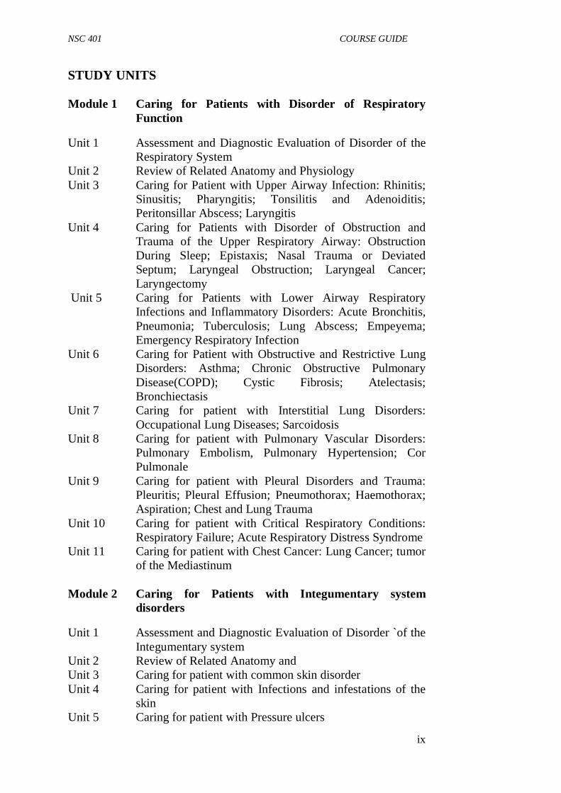
STUDY UNITS Module 1 Caring for Patients with Disorder of Respiratory
Function Unit 1 Assessment and Diagnostic Evaluation of Disorder of the
Respiratory System Unit 2 Review of Related Anatomy and Physiology Unit 3 Caring for Patient with Upper Airway Infection: Rhinitis;
Sinusitis; Pharyngitis; Tonsilitis and Adenoiditis; Peritonsillar Abscess; Laryngitis
Unit 4 Caring for Patients with Disorder of Obstruction and Trauma of the Upper Respiratory Airway: Obstruction During Sleep; Epistaxis; Nasal Trauma or Deviated Septum; Laryngeal Obstruction; Laryngeal Cancer; Laryngectomy
Unit 5 Caring for Patients with Lower Airway Respiratory Infections and Inflammatory Disorders: Acute Bronchitis, Pneumonia; Tuberculosis; Lung Abscess; Empeyema; Emergency Respiratory Infection
Unit 6 Caring for Patient with Obstructive and Restrictive Lung Disorders: Asthma; Chronic Obstructive Pulmonary Disease(COPD); Cystic Fibrosis; Atelectasis; Bronchiectasis
Unit 7 Caring for patient with Interstitial Lung Disorders: Occupational Lung Diseases; Sarcoidosis
Unit 8 Caring for patient with Pulmonary Vascular Disorders: Pulmonary Embolism, Pulmonary Hypertension; Cor Pulmonale
Unit 9 Caring for patient with Pleural Disorders and Trauma: Pleuritis; Pleural Effusion; Pneumothorax; Haemothorax; Aspiration; Chest and Lung Trauma
Unit 10 Caring for patient with Critical Respiratory Conditions: Respiratory Failure; Acute Respiratory Distress Syndrome
Unit 11 Caring for patient with Chest Cancer: Lung Cancer; tumor of the Mediastinum
Module 2 Caring for Patients with Integumentary system
disorders
Unit 1 Assessment and Diagnostic Evaluation of Disorder `of the Integumentary system
Unit 2 Review of Related Anatomy and Unit 3 Caring for patient with common skin disorder Unit 4 Caring for patient with Infections and infestations of the
skin Unit 5 Caring for patient with Pressure ulcers
NSC 401 COURSE GUIDE
x
Module 3 Caring for Patients with Musculoskeletal system disorders
Unit 1 Assessment and Diagnostic Evaluation of Disorder of the
Musculoskeletal system Unit 2 Review of Related Anatomy and Physiology Unit 3 Caring for Patients with musculoskeletal trauma: Soft
tissue trauma, fractures, Hip fracture Unit 4 Caring for Patients with Joint Trauma and injury:
Repetitive use injuries; Amputation; Unit 5 Caring for Patients with structural and bone disorders:
Scoliosis and Kyphosis; Osteoporosis; Osteomalacia and Pagiet’s disease; Osteomyelitis; common foot disorders; Bone Tumor
Unit 6 Caring for Patients with joint and connective tissue disorders: Osteoarthritis; Rheumatoid Arthritis ; systemic lupus erthematosus; Gout; Lyme disease; Ankylosing spondylitis; fibromyalgia; Low back pain; Muscular Dystrophy
Module 4 Caring for Patients with Urinary System Disorders Unit 1 Assessment and Diagnostic Evaluation of Disorder of the
Urinary System Unit 2 Review of Related Anatomy and Physiology Unit 3 Caring for Patients with fluid and electrolyte imbalances
in renal disorders Unit 4 Caring for Patients with Dysfunctional voiding patterns:
Congenital voiding dysfunction; Adult voiding dysfunction; urinary incontinence; urinary retention; neurogenic bladder
Unit 5 Caring for Patients with renal infectious and inflammatory disorders: urinary tract infection; glomerulonephritis; nephritic syndrome
Unit 6 Caring for Patients with Obstructive renal disorders: Urolithiasis; hydronephrosis; polycystic kidney disease
Unit 7 Caring for Patients with Genitourinary trauma: renal trauma; ureteral trauma; bladder trauma; urethral trauma
Unit 8 Caring for Patients with Urinary Tract Cancers Unit 9 Caring for Patients with Urinary Diversions Unit 10 Caring for Patients undergoing dialysis and kidney surgery Unit 11 Caring for Patients with renal failure and undergoing renal
transplant

CONTENTS PAGE Module 1 Caring for Patients with Disorder of
Respiratory Function……………………………… 1 Unit 1 Assessment and Diagnostic Evaluation of Disorder
of the Respiratory System………………………….. 1 Unit 2 Review of Related Anatomy and Physiology ……. Unit 3 Caring for Patient with Upper Airway Infection:
Rhinitis; Sinusitis; Pharyngitis; Tonsilitis and Adenoiditis; Peritonsillar Abscess; Laryngitis…….. 24
Unit 4 Caring for Patients with Disorder of Obstruction and Trauma of the Upper Respiratory Airway: Obstruction During Sleep; Epistaxis; Nasal Trauma or Deviated Septum; Laryngeal Obstruction; Laryngeal Cancer; Laryngectomy…………………… 33
Unit 5 Caring for Patients with Lower Airway Respiratory Infectious and Inflammatory Disorders: Acute Bronchitis, Pneumonia; Tuberculosis; Lung Abscess; Empeyema; Emergency Respiratory Infection………………………………………………. 45
Unit 6 Caring for Patient with Obstructive and Restrictive Lung Disorders: Asthma; Chronic Obstructive Pulmonary Disease(COPD); Cystic Fibrosis; Atelectasis; Bronchiectasis…………………………… 60
Unit 7 Caring for patient with Interstitial Lung Disorders: Occupational Lung Diseases; Sarcoidosis……………. 72
Unit 8 Caring for patient with Pulmonary Vascular Disorders: Pulmonary Embolism, Pulmonary Hypertension; CorPulmonale………………………………………… 76
Unit 9 Caring for patient with Pleural Disorders and Trauma: Pleuritis; Pleural Effusion; Pneumothorax; Haemothorax;
Aspiration; Chest and Lung Trauma…………………. 84 Unit 10 Caring for patient with Critical Respiratory Conditions: Respiratory Failure; Acute Respiratory Distress
Syndrome………………………………………………102 Unit 11 Caring for patient with Chest Cancer: Lung Cancer;
tumor of the Mediastinum……………………………..107
MAIN COURSE
Module 2 Caring for Patients with Integumentary system
Disorders………………………………………….. 113
Unit 1 Assessment and Diagnostic Evaluation of Disorder ` of the Integumentary system …………………… 113 Unit 2 Caring for patient with common skin disorder…….. 127 Unit 3 Caring for patient with Infections and infestations
of the skin………………………………………… 141 Unit 4 Caring for patient with Pressure ulcers…………… 176 Module 3 Caring for Patients with Musculoskeletal
system disorders…………………………………….183 Unit 1 Review of Related Anatomy and Physiology (Refer
to NSC ……. And NSC…..)………………………183 Unit 2 Assessment and Diagnostic Evaluation of Disorder of
the Musculoskeletal system ………………………… 188 Unit 3 Caring for Patients with musculoskeletal trauma:
Soft tissue trauma, fractures, Hip fracture…………… 194 Unit 4 Caring for Patients with Joint Trauma and
injury: Repetitive use injuries; Amputation; ………… 209 Unit 5 Caring for Patients with structural and bone
disorders: Scoliosis and Kyphosis; Osteoporosis; Osteomalacia and Pagiet’s disease; Osteomyelitis; common foot disorders; Bone Tumor……………… 216
Unit 6 Caring for Patients with joint and connective tissue disorders: Osteoarthritis; Rheumatoid Arthritis ; systemic lupuserthematosus; Gout; Lyme disease; Ankylosing spondylitis; fibromyalgia; Low back pain; Muscular Dystrophy……………….. 244
Module 4 Caring for Patients with Urinary System Disorders
…………………………………..... . 270 Unit 1 Assessment and Diagnostic Evaluation of Disorder of
the Urinary System ………………………………… 270 Unit 2 Review of Related Anatomy and Physiology (Refer
to NSC ……. And NSC…..)…………………… 280 Unit 3 Caring for Patients with fluid and electrolyte
imbalances in renal disorders……………………… 286 Unit 4 Caring for Patients with Dysfunctional voiding
patterns: Congenital voiding dysfunction; Adult voiding dysfunction; urinary incontinence; urinary retention; neurogenic bladder……………… 308

NSC 401 MODULE 1
1
MODULE 1 UNIT 1 ASSESSMENT AND DIAGNOSTIC EVALUATION
OF DISORDER OF THE RESPIRATORY SYSTEM CONTENTS 1.0 Introduction 2.0 Objectives 3.0 Main Content
3.1 Respiratory Physiology 3.2 Anatomy of the Respiratory System
4.0 Conclusion 5.0 Summary 6.0 Tutor-Marked Assignment 7.0 References/Further Reading 1.0 INTRODUCTION Have you ever nursed a patient with asthmatic attack? What is the first thing that comes to your mind about the disposition of the patient? Respiration is synonymous with life and respiration allows exchange of gases which are needed for metabolism. Nurses are often the first set of health professional in clinical practice that patients come in contact with when they have respiratory problems. Even when they are on admission in the hospital settings the nurse is always the first person to pick the changes in the respiration of the patients. In essence, the professional nurse must be highly knowledgeable and skillful in the assessment, early diagnosis prompt and effective management. In this module, you will learn more about disorder of the respiratory system that are common and are encountered by nurses in every setting from the community to the intensive care unit. For easier learning, the module is divided into 11 units. In this unit you will be reminded of your previous knowledge on respiratory anatomy and physiology to serve as a base for other units in module 3

NSC 401 MEDICAL-SURGICAL NURSING III
2
2.0 OBJECTIVES At the end of this module, you will be able to: • conduct quality assessment of patient • utilize the nursing process to provide care • describe the structures and functions of the upper and lower
respiratory tracts • describe ventilation, perfusion, diffusion, shunting, and the
relationship of pulmonary circulation to these processes. 3.0 MAIN CONTENT 3.1 Anatomy of the Respiratory System The respiratory system is divided into the upper airway and lower airway. Upper Airway The upper airway consists of the nose, sinuses, turbinates, pharynx, and larynx. Nose: Nasal bones and cartilage support the external nose. The noses are the external openings of the nose. The internal nose is divided into two cavities separated by the nasal septum. Each nasal cavity has three passages created by the projection of turbinates or conchae from the lateral walls. The vascular and ciliated mucous lining of the nasal cavities warms and humidifies inspired air. Mucus secreted from the nasal mucosa traps small particles (e.g., dust, pollen). Cilia (fine hairs) move the mucus to the back of the throat. This movement helps prevent irritation to and contamination of the lower airway. The nasal mucosa also contains olfactory sensory cells that are responsible for the sense of smell. The olfactory area lies at the roof of the nose. The cribriform plate forms part of the roof of the nose and the floor of the anterior cranial fossa. Trauma or surgery in this area carries the risk of injuring or causing infection in the brain. Paranasal Sinuses: The paranasal sinuses are extensions of the nasal cavity located in the surrounding facial bones. They lighten the weight of the skull and give resonance to the voice. There are four pairs of these bony cavities. The two frontal sinuses lie in the frontal bone that extends above the orbital cavities. The ethmoid bone, located between the eyes, contains a honeycomb of small spaces called the ethmoidalsinuses. The sphenoidal sinuses lie behind the nasal cavity. The maxillary sinuses are found on

NSC 401 MODULE 1
3
either side of the nose in the maxillary bones. The maxillary sinuses are the largests in uses and the most accessible to treatment. The lining of the sinuses is continuous with the mucous membrane lining of the nasal cavity. Mucus traps particles that cilia sweep toward the pharynx. Immunoglobulin A(IgA) antibodies in the mucus protect the lower respiratory tract from infection. Turbinate Bones (Conchae): The turbinates (or conchae) are bones that change the flow of inspired air to moisturize and warm it better. As air is inhaled, the turbinates deflect it toward the roof of the nose. They have a large, moist, and warm mucous-membrane surface that can trap almost all dust and microorganisms. They also contain sensitive nerves that detect odors or induce sneezing to remove irritating particles, such as dust or soot. Pharynx: The pharynx, or throat, carries air from the nose to the larynx, and food from the mouth to the esophagus. The pharynx is divided into three continuous areas: the nasopharynx (near the nose and above the soft palate), the oropharynx (near the mouth), and the laryngeal pharynx (near the larynx). The nasopharynx contains the adenoids and openings of the eustachian tubes. The eustachian tubes connect the pharynx to the middle ear and are the means by which upper respiratory infections spread to the middle ear. The oropharynx contains the tongue. The muscular nature of the pharynx allows for closure of the epiglottis during swallowing and relaxation of the epiglottis during respiration. Tonsils and adenoids, which do not contribute to respiration but instead protect against infection, are found in the pharynx. Palatine tonsils consist of two pairs of elliptically shaped bodies of lymphoid tissue. They are located on both sides of the upper oropharynx. Adenoids, or pharyngeal tonsils, also composed of lymphoid tissue, are found in the nasopharynx. Chronic throat infections often lead to removal of the tonsils and adenoids. In adults, adenoids may shrink and become nonfunctional. Larynx: The larynx, or voice box, is a cartilaginous framework between the pharynx and trachea. Its primary function is to produce sound. The larynx also protects the lower airway from foreign objects because it facilitates coughing. Important structures in the larynx include the epiglottis, a cartilaginous valve flap that covers the opening to the larynx during swallowing; the glottis, an opening between the vocal cords; and the vocal cords, folds of tissue in the larynx that vibrate and produce sound as air passes through. The pharynx, palate, tongue, teeth, and lips mold the sounds made by the vocal cords into speech.

NSC 401 MEDICAL-SURGICAL NURSING III
4
Lower Airway The lower respiratory airway consists of the trachea, bronchi, bronchioles, lungs, and alveoli. Accessory structures include the diaphragm, rib cage, sternum, spine, muscles, and blood vessels. Trachea: The trachea is a hollow tube composed of smooth muscle and supported by C-shaped cartilage. The cartilaginous rings are incomplete on the posterior surface. The trachea transports air from the laryngeal pharynx to the bronchi and lungs. Bronchi and Bronchioles: The trachea bifurcates (divides) at the carina (lower end of the trachea) to form the left and right bronchi. Stimulating the carina causes coughing and bronchospasm (spasm of the bronchial smooth muscle, leading to narrowing of the lumen). The right main stem bronchus is shorter, more vertical, and larger than the left main stem bronchus. Aspiration of foreign objects is more likely in the right main stem bronchus and right upper lung. Mucous membrane continues to line this portion of the respiratory tract. Cilia sweep mucusand particles toward the pharynx. The right and left main stem bronchi divide into three secondary right bronchi and two secondary left bronchi. Each secondary bronchus supplies air to the three right lobes and two left lobes of the lung. The entrance of the bronchi to the lungs is called the hilus. The bronchi branch, enter each lobe, and continue to branch to form smaller bronchi and finally terminal bronchioles (smaller subdivisions of bronchi). Lungs and Alveoli The lungs are paired elastic structures enclosed by the thoracicn cage. They contain the alveoli, small, clustered sacs that begin where the bronchioles end. Adult lungs contain approximately 300 million alveoli, which form most of the pulmonary mass. Each alveolus consists of a single layer of squamous epithelial cells. Capillaries surround these thin-walled alveoli and are the site of exchange of oxygen and CO2. The epithelium of the alveoli consists of the following types of cells: • Type I cells: line most alveolar surfaces • Type II cells: produce surfactant, a phospholipid that alters the
surface tension of alveoli, preventing their collapse during expiration and limiting their expansion during inspiration
• Type III cells: destroy foreign material, such as bacteria. The interstitium lies between the alveoli and contains the pulmonary capillaries and elastic connective tissue. Elastic and collagen fibers allow the lungs to have compliance, the ability to expand. Lung expansion creates

NSC 401 MODULE 1
5
a negative or sub-atmospheric pressure, which keeps the lungs inflated. If air gets into the space between the lungs and the thoracic wall, the lungs will collapse. Accessory Structures The diaphragm separates the thoracic and abdominal cavities. On inspiration, the respiratory muscles contract. The diaphragm also contracts and moves downward, enlarging the thoracic space and creating a partial vacuum. On expiration, the respiratory muscles relax, and the diaphragm returns to its original position. The mediastinum is a wall that divides the thoracic cavity into two halves. This wall has two layers of pleura, a saclike serous membrane. The visceral pleura covers the lung surface, whereas the parietal pleura covers the chest wall. Serous fluid within the pleural space separates and lubricates the visceral and parietal pleurae. The remaining thoracic structures are located between the two pleural layers.
A Diagram of the Respiratory System

NSC 401 MEDICAL-SURGICAL NURSING III
6
SELF-ASSESSMENT EXERCISE 1. With the aid of a well labeled diagram, discuss respiratory anatomy. 2. Briefly discuss mechanism of ventilation. 3.2 Respiratory Physiology The main function of the respiratory system is to exchange oxygen and CO2 between the atmospheric air and the blood and between the blood and the cells. This process is called respiration.
NSC 401 MODULE 1
7
Ventilation Ventilation is the actual movement of air in and out of the respiratory tract. Air must reach the alveoli for gas to be exchanged. This process requires a patent airway and intact and functioning respiratory muscles. Pressure gradients between atmospheric air and the alveoli enable ventilation. Air flows from an area of higher pressure to an area of lower pressure. Mechanics of Ventilation During inspiration, the diaphragm contracts and flattens which expands the thoracic cage and increases the thoracic cavity. The pressure in the thorax decreases to a level below atmospheric pressure. As a result, air moves into the lungs. When inspiration is complete, the diaphragm relaxes, and the lungs recoil to their original position. The size of the thoracic cavity decreases, increasing the pressure to levels greater than the atmospheric pressure. Air then flows out of the lungs into the atmosphere. Neurologic Control of Ventilation Several mechanisms control ventilation. The respiratory centers in the medulla oblongata and pons control rate and depth. Central chemoreceptors in the medulla respond to changes in CO2 levels and hydrogen ion concentrations (pH) in the cerebrospinal fluid. They convey a message to the lungs to change the depth and rate of ventilation. Peripheral chemoreceptors in the aortic arch and carotid arteries respond to changes in the pH and levels of oxygen and CO2 in the blood. Diffusion Diffusion is the exchange of oxygen and CO2 through the alveolar-capillary membrane. Concentration gradients determine the direction of diffusion. During inspiration, the concentration of oxygen is higher in the alveoli than in the capillaries. Therefore, oxygen diffuses from the alveoli to the capillaries and is carried to the arteries. The concentration of oxygen in the arteries is higher than that in the cells; thus, oxygen diffuses into the cells. As cellular CO2 gradients increase, CO2 diffuses from the cells into the capillaries and then into the venous circulatory system. As CO2 travels to the pulmonary circulation, its concentration is higher there than in the alveoli. Therefore, CO2 diffuses into the alveoli. Alveolar Respiration Alveolar respiration determines the amount of CO2 in the body. Increased CO2, which is present in body fluids primarily as carbonic acid, causes the pH to decrease below the normal 7.4. Decreased CO2 causes the pH to
NSC 401 MEDICAL-SURGICAL NURSING III
8
increase above 7.4. The pH affects the rate of alveolar respiration by a direct action of hydrogen ions on the respiratory center in the medulla oblongata. The kidneys contribute to maintaining normal pH by excreting excess hydrogen ions, which in turn keep serum the ratio of carbonic acid to bicarbonate at 1:20, fixing the pH at approximately 7.4. In a critically ill client, various homeostatic mechanisms compensate for alterations. In an attempt to maintain normal pH, two mechanisms may occur: • The lungs eliminate carbonic acid by blowing off more CO2. They
also conserve CO2 by slowing respiratory volume and reabsorbing bicarbonate (HCO3).
• The kidneys excrete more bicarbonate. A client’s condition remains compensated if the carbonic acid-to-bicarbonate ratio remains 1:20. Disturbances in pH that involve the lungs are considered respiratory. Disturbances in pH involving other mechanisms are termed metabolic. At times, respiratory and metabolic disturbances coexist. Transport of Gases Oxygen transport occurs in two ways: (1) A small amount is dissolved in water in the plasma (2) A greater portion combines with hemoglobin in red blood cells
(RBCs; oxyhemoglobin). Dissolved oxygen is the only form that can diffuse across cellular membranes. As this oxygen crosses cellular membranes, oxygen from the hemoglobin rapidly replaces it. Large amounts of oxygen are transported in the blood as oxyhemoglobin. CO2 diffuses from the tissue cells to the blood. Bicarbonate ions are then transported to the lungs for excretion. Most of the CO2 enters the RBCs, although some combines with hemoglobin to form carbaminohemoglobin. Most of the CO2 combines with water in the cells and exits as bicarbonateions (HCO3-), which the plasma transports to the kidneys. A small portion remains in the plasma and is called carbonic acid. The formation of carbonic acid yields hydrogen ions (Hþ). The amount of hydrogen ions determines the pH, which also determines the amount of CO2 for the lungs to excrete. Briefly, acid-base imbalances are compensated in the following ways: • Respiratory acidosis—kidneys retain more HCO3 to raise the pH • Respiratory alkalosis—kidneys excrete more HCO3 to lower pH
NSC 401 MODULE 1
9
• Metabolic acidosis—lungs ‘‘blow off’’ CO2 to raise pH • Metabolic alkalosis—lungs retain CO2 to lower pH. Pulmonary Perfusion
Perfusion refers to blood supply to the lungs, through which the lungs receive nutrients and oxygen. The two methods of perfusion are the bronchial and pulmonary circulation. Bronchial Circulation The bronchial arteries, which supply blood to the trachea and bronchi, arise in the thoracic aorta and intercostals arteries. The bronchial arteries also supply the lungs’ supporting tissues, nerves, and outer layers of the pulmonary arteries and veins. This circulation returns either to the left atrium through the pulmonary veins or to the superior vena cava through the bronchial and azygos veins. The bronchial circulation does not supply the bronchioles or alveoli unless pulmonary circulation is interrupted. Pulmonary Circulation The pulmonary artery transports venous blood from the right ventricle to the lungs. It divides into the right and left branches to supply the right and left lungs. The blood circulates through the pulmonary capillary bed, where diffusion of oxygen and CO2 occurs. The blood then returns to the left atrium through the pulmonary veins. Pulmonary circulation is referred to as a low-pressure system (Smeltzer et al., 2008). This means that gravity, alveolar pressure, and pulmonary artery pressure affect pulmonary perfusion. A person in an upright position has less perfusion to the upper lobes. If a person is in a side-lying position, perfusion is greater to the dependent side. In addition, increased alveolar pressure can cause pulmonary capillaries to narrow or collapse, affecting gas exchange. Decreased pulmonary artery pressure results in decreased perfusion to the lungs. Clients with lung and cardiovascular diseases may have decreased pulmonary perfusion. Ventilation/Perfusion Ratio A client’s cardiopulmonary status involves several factors; in particular, the client’s ventilation/perfusion ratio (V/Q ratio) indicates the effectiveness of airflow within the alveoli (ventilation) and the adequacy of gas exchange within the pulmonary capillaries (perfusion). 4.0 CONCLUSION Respiration is synonymous with life and respiration allows exchange of gases which are needed for metabolism. The main function of the

NSC 401 MEDICAL-SURGICAL NURSING III
10
respiratory system is to exchange oxygen and CO2 between the atmospheric air and the blood and between the blood and the cells. Professional nurse must be highly knowledgeable and skillful in the assessment, early diagnosis prompt and effective management of respiratory disorder. 5.0 SUMMARY
In this unit, you have learnt that: • The respiratory system is divided into the upper airway and lower
airway. • The main function of the respiratory system is to exchange oxygen
and CO2 between the atmospheric air and the blood and between the blood and the cells.
• Ventilation is the actual movement of air in and out of the respiratory tract.
• Alveolar respiration determines the amount of CO2 in the body. • Diffusion is the exchange of oxygen and CO2 through the alveolar-
capillary membrane. • Clients with lung and cardiovascular diseases may have decreased
pulmonary perfusion. 6.0 TUTOR-MARKED ASSIGNMENT Pinch your nostrils for 2 minutes and use your mouth to breathe. Did you notice any change in your respiration? Share your experiences in the discussion forum. 7.0 REFERENCES/FURTHER READING Bullock, B.A., &Henze, R.L. (2000). Focus on pathophysiology.
Philadelphia: Lippincott Williams & Wilkins. Burke, K.M; Mohn-Brown, E.L &Eby, L (2011). Medical-Surgical Nursing
Care. (3rded.). Boston: Pearson Education, Inc. Nettina, S.M (2010). Lippincott Manual of Nursing Practice (9thed). China:
Wolters Kluwer Health Lippincott Williams & Wilkins.

NSC 401 MODULE 1
11
UNIT 2 REVIEW OF RELATED ANATOMY AND PHYSIOLOGY
CONTENTS 1.0 Introduction 2.0 Objectives 3.0 Main Content
3.1 Assessment of patient with respiratory problem 3.2 Subjective Assessment 3.3 Objective Assessment 3.4 Diagnostic Tests
4.0 Conclusion 5.0 Summary 6.0 Tutor-Marked Assignment 1.0 INTRODUCTION Assessment of the respiratory system includes obtaining information about physical and functional issues related to breathing. It also means clarifying how these issues may affect the client’s quality of life. 2.0 OBJECTIVES At the end of this unit you will be able to: • discriminate between normal and abnormal breath sounds • discuss various diagnostic tests used in respiratory disorders. 3.0 MAIN CONTENT 3.1 Assessment of Patient with Respiratory Problem Assessment of the respiratory system includes obtaining information about physical and functional issues related to breathing. It also means clarifying how these issues may affect the client’s quality of life. Assessment can be subjective or objective 3.2 Subjective Assessment Often a client seeks medical attention because of respiratory problems related to one or more of the following: dyspnea (labored or difficult breathing), pain on inspiration, increased or more frequent cough, increased

NSC 401 MEDICAL-SURGICAL NURSING III
12
sputum production or change in the color/consistency of the mucus, wheezing, or hemoptysis (blood in the sputum). The nurse obtains information about the client’s general health history and his or her family history. He or she asks the client about the frequency of respiratory illnesses, allergies, smoking history, nature of any cough, sputum production, dyspnea and wheezing. Questioning the client about respiratory treatments or medications (prescription and over-the-counter) is essential. In addition, the nurse inquires about last pulmonary tests (chest radiograph, tuberculosis test). He or she includes questions about occupation, exercise tolerance, pain, and level of fatigue. Dyspnea Explore the patient’s symptoms through characterization and history taking to help anticipate needs and plan care. 1. Characteristics: Is the dyspnea acute or chronic? Has it come about
suddenly or gradually? Is more than one pillow required to sleep? Is the dyspnea progressive, recurrent, or paroxysmal? Walking how far leads to shortness of breath? How does it compare to the patient’s baseline level of dyspnea? Ask patient to rate dyspnea on a scale of 1 to 10 with 1 being no dyspnea and 10 being the worst imaginable. What relieves and what aggravates the dyspnea?
2. Associated factors: Is there a cough associated with the dyspnea and
is it productive? What activities precipitate the shortness of breath? Does it seem to be worse when upset? Is it influenced by the time of day, seasons, and/or certain environments? Does it occur at rest or with exertion? Any fever, chills, night sweats, ankle/leg swelling? Any change in body weight?
3. History: Is there a patient history or family history of chronic lung
disease, cardiac or neuromuscular disease, cancer, problems with blood clotting, or immune compromise? What is the smoking history?
4. Significance: Sudden dyspnea could indicate pulmonary embolus,
pneumothorax, myocardial infarction (MI), acute heart failure, or acute respiratory failure. In a postsurgical or postpartum patient, dyspnea may indicate pulmonary embolus or edema. Orthopnea can be indicative of heart disease or COPD. If dyspnea is associated with a wheeze, consider asthma, COPD, heart failure, or upper airway obstruction. When dyspnea occurs in combination with fatigue, pulmonary hypertension may exist. Metabolic disorders, psychiatric

NSC 401 MODULE 1
13
conditions, and neuromuscular disorders may also contribute to dyspnea.
Chest Pain 1. Characteristics: Is the pain sharp, dull, stabbing, or aching? Is it
intermittent or persistent? Is the pain localized or does it radiate? If it radiates, where? How intense is the pain? Are there factors that alleviate or aggravate the pain, such as position or activity?
2. Associated factors: What effect do inspiration and expiration have
on the pain? What other symptoms accompany the chest pain? Is there diaphoresis, shortness of breath, nausea?
3. History: Is there a smoking history or environmental exposure? Has
the pain ever been experienced before? What was the cause? Is there a preexisting pulmonary or cardiac diagnosis? Has there been recent trauma?
4. Significance: Chest pain related to pulmonary causes is usually felt
on the side where pathology arises, but it can be referred. Dull persistent pain may indicate carcinoma of the lung, whereas sharp stabbing pain usually arises from the pleura. Dyspnea with pleuritic chest pain indicates clinically significant pulmonary embolism. Cough Evidence BaseTarlo, S. (2006).
Cough: 1. Characteristics: Is the cough dry, hacking, loose, barky, wheezy, or
more like clearing the throat? Is it strong or weak? How frequent is it? Is it worse at night or at any time of day? Does the intensity change on days off from work? Is there seasonal variation? Is it aggravated by food intake or exertion; is it alleviated by any medication? How long has it been going on?
2. Associated factors: Is the cough productive? If so, what is the
consistency, amount, color, and odor of the sputum? How does sputum compare to the patient’s baseline? Is it associated with shortness of breath, pain, or nausea?
3. History: Is there a smoking history? Is the smoking current or in the
past? Has there been any environmental or occupational exposure to dust, fumes, or gases that could lead to cough? Are there past pulmonary diagnoses, asthma, rhinitis, allergy or exposure to allergens, such as pollen, house dust mites, animal dander, birds,
NSC 401 MEDICAL-SURGICAL NURSING III
14
mold or fungi, cockroach waste, irritants (smoke, odors, perfumes, cleaning products, exhaust, pollution, cold air)? Has there been prolonged exposure to dampness, chemical sanitizers, cobalt or other hard metals, beryllium, asbestos, dusts from coal, wood, or grains? Does the patient have a history of acid reflux or use an angiotens in converting enzyme inhibitor with a common adverse effect of cough? Has there been a concurrent voice change? Has the patient recently traveled outside the country? Can the patient identify any specific triggers?
4. Significance: A dry, irritating cough may indicate viral respiratory
tract infection. A cough at night should alert to potential left-sided heart failure, asthma, or postnasal drip worsening at night. A morning cough with sputum might be bronchitis. A cough that is less severe on days off from work may be related to occupational or environmental exposures. A patient with severe or changing cough should be evaluated for bronchogenic carcinoma. Consider bacterial pneumonia if sputum is rusty, and lung tumor if it is pink-tinged. A profuse pink frothy sputum could be indicative of pulmonary edema. A cough associated with food intake could indicate problems with aspiration. A dry cough may be associated with pulmonary fibrosis. History of recent travel may be associated with infection from a source not commonly identified in the U.S.
Hemoptysis 1. Characteristics: Is the blood from the lungs? It could be from GI
system (hematemesis) or upper airway (epistaxis). Is it bright red and frothy? How much? Is onset associated with certain circumstances or activities? Was the onset sudden, and is it intermittent or continuous?
2. Associated factors: Was there an initial sensation of tickling in the
throat? Was there a salty taste or burning or bubbling sensation in the chest? Has there been shortness of breath, chest pain, difficulty with exertion?
3. History: Was there any recent chest trauma or respiratory treatment
(chest percussion)? Does the patient have an upper respiratory infection, sinusitis, or recent epistaxis? Has the patient used cocaine or other illicit drugs?
4. Significance: Hemoptysis can be linked to pulmonary infection, lung
carcinoma, abnormalities of the heart or blood vessels, pulmonary artery or vein abnormalities, or pulmonary emboli and infarction.
NSC 401 MODULE 1
15
Small amounts of blood-tinged sputum may be from the upper respiratory tract, and regurgitation of blood comes from a GI bleed.
3.3 Objective Assessment Physical Examination: The physical examination begins with a general examination of overall health and condition. Clients with respiratory problems may show signs of shortness of breath when speaking, or they may have a certain posture or position to facilitate breathing. Other observations include skin color; level of consciousness; mental status; respiratory rate, depth, effort, and rhythm; use of accessory muscles; and shape of the chest and symmetry of chest movements. Extremities are assessed for finger clubbing, a condition in which the tips of the fingers or toes are enlarged because the soft tissue beneath the nail beds is increased. Although it is not always clear why this occurs, it may be related to levels of proteins that stimulate blood vessel growth or to genetic factors. Finger clubbing seems to occur with some lung diseases such as lung cancer, but not with others such as asthma; it can also occur with congenital heart, liver, and thyroid diseases. The nurse inspects the nose for signs of injury, inflammation, symmetry, and lesions. He or she examines the posterior pharynx and tonsils with a tongue blade and light and notes any evidence of swelling, inflammation, or exudate, as well as changes in color of the mucous membranes. The nurse also notes any difficulty with swallowing or hoarseness. The nurse inspects and gently palpates the trachea to assess for placement and deviation from the midline. He or she notes any lymph node enlargement. The nurse also examines the anterior, posterior, and lateral chest walls for lesions, symmetry, deformities, skin color, and evidence of muscle weakness or weight loss. Checking the contour of the chest walls is important. Normally the anteroposterior diameter of the chest wall is half the transverse diameter; however, some pulmonary conditions (e.g., emphysema) change the chest dimensions. An experienced examiner palpates the chest wall to detect tenderness, masses, swelling, or other abnormalities. Tactile or vocal fremitus (vibrations from the client’s voice transmitted to the examiner’s fingers) depends on the capacity to feel sound through the fingers and palm placed on the chest wall. The palpable vibrations occur when the client speaks. The examiner uses the palmar surfaces of the fingers and hands to palpate and asks the client to repeat ‘‘99’’ as the examiner moves his or her hands. If the client is healthy and thin, the fremitus will be highly palpable. Conditions that affect fremitus include a thick or muscular chest wall

NSC 401 MEDICAL-SURGICAL NURSING III
16
(decreased fremitus), lung diseases such as emphysema and pneumonia (increased fremitus), and fluid, air, or masses in the pleural space (decreased fremitus). The experienced examiner performs percussion of the chest wall to assess normal and abnormal sounds. With the client sitting, the examiner places his or her middle finger on the chest wall and taps that finger with the middle finger of the opposite hand. The nurse auscultates breath sounds from side to side, moving from the upper to the lower chest. He or she listens anteriorly, laterally, and posteriorly. Normal breath sounds include the following: • Vesicular sounds: Produced by air movement in bronchioles and
alveoli, these sounds are heard over the lung fields; they are quiet and low pitched, with long inspiration and short expiration.
• Bronchial sounds: Produced by air movement through the trachea, these sounds are heard over the trachea and are loud with long expiration.
• Bronchovesicular sounds: These normal breath sounds are heard between the trachea and upper lungs; pitch is medium with equal inspiration and expiration.
Adventitious or abnormal breath sounds are categorized as crackles or wheezes. Crackles (formerly called rales) are discrete sounds that result from the delayed openingof deflated airways. They resemble static or the sound made by rubbing hair strands together near one’s ear. Sometimes they clear with coughing. They may be present because of inflammation or congestion. Crackles that do not clear with coughing may indicate pulmonary edema or fluid in the alveoli. Wheezes may be sibilant (hissing or whistling) or sonorous (full and deep). Sibilant wheezes (formerly called wheezes) are continuous musical sounds that can be heard during inspiration and expiration. They result from air passing through narrowed or partially obstructed air passages and are heard in clients with increased secretions. Sonorous wheezes (formerly called rhonchi) are lower pitched and are heard in the trachea and bronchi. Friction rubs are heard as crackling or grating sounds on inspiration or expiration. They occur when the pleural surfaces are inflamed and do not change if the client coughs.

NSC 401 MODULE 1
17
http://en.wikipedia.org/wiki/File:Elderly_vietnamese_man_gets_examined.jpg SELF-ASSESSMENT EXERCISE 1. Discuss any five diagnostic tests that can be done for patients with
respiratory disorders. 2. For each of the diagnostic test taken, what are the nurses roles during
the procedures? 3.4 Diagnostic Tests Arterial Blood Gases: Oxygenation of body tissues depends on the amount of oxygenin arterial blood. Arterial blood gases (ABGs) determine the blood’s pH, oxygen-carrying capacity, and levels of oxygen, CO2, and bicarbonate ion. Blood gas samples are obtained through an arterial puncture at the radial, brachial, or femoral artery. A client also may have an indwelling arterial catheter from which arterial samples are obtained. ABGs frequently are ordered when a client is acutely ill or has a history of respiratory disorders. If the partial pressure of oxygen in arterial blood (PaO2) is decreased, body tissues do not receive sufficient oxygen. Clients with respiratory disorders can neither get oxygen into the blood nor get CO2 out of the blood. Some conditions that affect ABGs are as follows: • Hyperventilation during collection of ABGs, causing elevated PaO2. • Hypoventilation with neuromuscular disease, chronic obstructive
pulmonary disease (COPD), or insufficient oxygen in the atmosphere, causing decreased PaO2.
• Elevated PaCO2 in clients with COPD, inadequate ventilation with a mechanical ventilator, or decreased respiratory rates.
• Decreased PaCO2 in clients who are nervous or anxious or have a condition that causes hyperventilation or a rapid respiratory rate.

NSC 401 MEDICAL-SURGICAL NURSING III
18
Pulse oximetry is a noninvasive method that uses a light beam to measure the oxygen content of hemoglobin (SaO2). The monitoring device attaches to the client’s earlobe or fingertip and connects to the oximeter monitor. The monitor registers wavelengths of light passing through the earlobe or fingertip and uses them to calculate the arterial oxygen saturation. Normal values are 95% or higher. Pulmonary Function Studies: Pulmonary function studies measure the functional ability of the lungs. These studies are done to diagnose pulmonary conditions and to assess preoperative respiratory status. They also may be used to determine the effectiveness of bronchodilators or to screen employees who work in environments. Measurements of pulmonary function are obtained with a spirometer and include: • Tidal volume: volume of air inhaled and exhaled with a normal
breath. • Inspiratory reserve volume: maximum volume of air that normally
can be inspired. • Expiratory reserve volume: maximum volume of air that normally
can be exhaled by forced expiration. • Residual volume: volume of air left in the lungs after maximal
expiration. • Vital capacity: maximum amount of air that can be expired after
maximal inspiration • Forced vital capacity: amount of air exhaled forcefully and rapidly
after maximal inspiration • Inspiratory capacity: maximum amount of air that can be inhaled
after normal expiration. • Total lung capacity: total volume of air in the lungs when maximally
inflated. Pulmonary function results vary according to age, sex, weight, and height. The maximum lung capacities and volumes are best achieved when the client is sitting or standing. The test should not be performed within 2 hours after a meal. The nurse explains the procedure to the client and instructs him or her to wear loose-fitting clothing. A nose clip prevents air from escaping through the client’s nose when blowing into the spirometer. Bronchodilators may be used after the initial spirometry to see if there is any improvement or response with the inhaled medication. Although the test is simple, the client may be tired afterward. Sputum Studies: Sputum specimens are examined for pathogenic microorganisms and cancer cells. Culture and sensitivity tests are done to
NSC 401 MODULE 1
19
diagnose infections and prescribe antibiotics. Negative results on the examination of sputum smears do not always indicate the absence of disease, so collection of sputum for successive days may be necessary. Sputum is collected by having the client expectorate a specimen, by suctioning the client, or during a bronchoscopy. Radiography: Chest radiographs show the size, shape, and position of the lungs and other structures of the thorax. Physicians use chestradiography to screen for asymptomatic disease and to diagnosetumors, foreign bodies, and other abnormal conditions. Fluoroscopy enables the physician to view the thoracic cavity with all its contents in motion. It more precisely diagnoses the location of a tumor or lesion. Computed tomography scanning or magnetic resonance imaging may be used to produce axial views of the lungs to detect tumors and other lung disorders during early stages. Pulmonary Angiography: Pulmonary angiography is a radioisotope study that allows the physician to assess the arterial circulation of the lungs, particularly to detect pulmonary emboli. A catheter is introduced into an arm vein and threaded through the right atrium and ventricle into the pulmonary artery. Contrast medium is rapidly injected into the femoral artery, and radiographs are taken to see the distribution of the radiopaque material. During pulmonary angiography, the nurse obtains data about the client’s level of anxiety and knowledge of the procedure. The nurse provides explanations and reinforces the client’s understanding. The client will experience a feeling of pressure on catheter insertion. When the contrast medium is infused, the client will sense a warm, flushed feeling and an urge to cough. The nurse must determine if the client has any allergies, particularly to iodine, shellfish, or contrast dye. During the procedure, the nurse monitors for signs and symptoms of allergic reactions to the contrast medium, such as itching, hives, or difficulty breathing. Infusion of contrast dye is discontinued immediately if the client has an allergic reaction. After the procedure, the nurse inspects the puncture site for swelling, discoloration, bleeding, or hematoma. The nurse assesses distal circulation and sensation to ensure that circulation is unimpaired. If bleeding occurs, pressure must be applied to the site. The nurse must notify the physician about diminished or absent distal pulses, cool skin temperature in the affected limb, poor capillary refill, client complaints of numbness or tingling, and bleeding or hematoma. The client remains on bed rest for 2 to 6 hours after the procedure.
NSC 401 MEDICAL-SURGICAL NURSING III
20
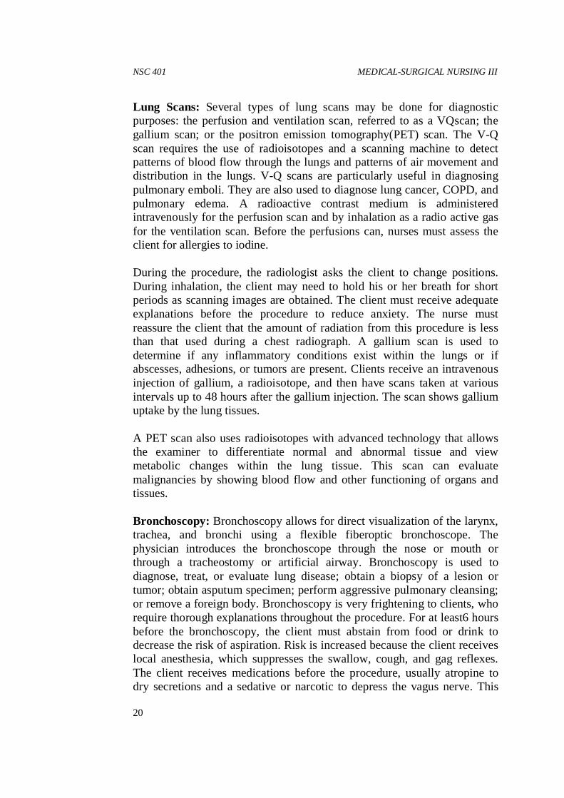
Lung Scans: Several types of lung scans may be done for diagnostic purposes: the perfusion and ventilation scan, referred to as a VQscan; the gallium scan; or the positron emission tomography(PET) scan. The V-Q scan requires the use of radioisotopes and a scanning machine to detect patterns of blood flow through the lungs and patterns of air movement and distribution in the lungs. V-Q scans are particularly useful in diagnosing pulmonary emboli. They are also used to diagnose lung cancer, COPD, and pulmonary edema. A radioactive contrast medium is administered intravenously for the perfusion scan and by inhalation as a radio active gas for the ventilation scan. Before the perfusions can, nurses must assess the client for allergies to iodine. During the procedure, the radiologist asks the client to change positions. During inhalation, the client may need to hold his or her breath for short periods as scanning images are obtained. The client must receive adequate explanations before the procedure to reduce anxiety. The nurse must reassure the client that the amount of radiation from this procedure is less than that used during a chest radiograph. A gallium scan is used to determine if any inflammatory conditions exist within the lungs or if abscesses, adhesions, or tumors are present. Clients receive an intravenous injection of gallium, a radioisotope, and then have scans taken at various intervals up to 48 hours after the gallium injection. The scan shows gallium uptake by the lung tissues. A PET scan also uses radioisotopes with advanced technology that allows the examiner to differentiate normal and abnormal tissue and view metabolic changes within the lung tissue. This scan can evaluate malignancies by showing blood flow and other functioning of organs and tissues. Bronchoscopy: Bronchoscopy allows for direct visualization of the larynx, trachea, and bronchi using a flexible fiberoptic bronchoscope. The physician introduces the bronchoscope through the nose or mouth or through a tracheostomy or artificial airway. Bronchoscopy is used to diagnose, treat, or evaluate lung disease; obtain a biopsy of a lesion or tumor; obtain asputum specimen; perform aggressive pulmonary cleansing; or remove a foreign body. Bronchoscopy is very frightening to clients, who require thorough explanations throughout the procedure. For at least6 hours before the bronchoscopy, the client must abstain from food or drink to decrease the risk of aspiration. Risk is increased because the client receives local anesthesia, which suppresses the swallow, cough, and gag reflexes. The client receives medications before the procedure, usually atropine to dry secretions and a sedative or narcotic to depress the vagus nerve. This
NSC 401 MODULE 1
21
consideration is important because if the vagus nerve is stimulated during the bronchoscopy, hypotension, bradycardia, or dysrhythmias may occur. Other potential complications include bronchospasm or laryngospasm secondary to edema, hypoxemia, bleeding, perforation, aspiration, cardiac dysrhythmias, and infection.
http://www.pennmedicine.org/encyclopedia/ency_images/encymulti/images/en/23232.jpg Laryngoscopy: Laryngoscopy provides direct visualization of the larynxusing a laryngoscope. It is done to diagnose lesions, evaluatelaryngeal function, and determine any inflammation. Physicians also may dilate laryngeal strictures and biopsy lesions. Mediastinoscopy: Mediastinoscopy provides visualization of the mediastinum and is done under local or general anesthesia. The physician makes an incision above the sternum and inserts a mediastinoscope. With this procedure, the physician can visualize lymph nodes and obtain biopsy samples. Possible complications include dysrhythmias, myocardial infarction, pneumothorax and bleeding. Thoracoscopy: Thoracoscopy allows for examination of the pleural cavity. Small incisions are made into the pleural cavity through anintercostal space. An endoscope is inserted to visualize aspecific area. The location selected is based on other clinical and diagnostic findings. If fluid is present, the examiner aspirates it and sends it for culture and cellular
NSC 401 MEDICAL-SURGICAL NURSING III
22
studies. Biopsies also may be done. A chest tube may be inserted following the procedure. Thoracoscopy is done to evaluate pleural effusions and pleural disease, and for staging of tumors. Thoracentesis: A small amount of fluid lies between the visceral and parietalpleurae. When excess fluid or air accumulates, the physician aspirates it from the pleural space by inserting a needle into the chest wall. This procedure, called thoracentesis, is performed with local anesthesia. Thoracentes is also may be used to obtain a sample of pleural fluid or a biopsy specimen from the pleural wall for diagnostic purposes, such as a culture and sensitivity or microscopic examination. Bloody fluid usually suggests trauma. Purulent fluid is diagnostic for infection. Serous fluid may be associated with cancer, inflammatory conditions, or heart failure. When thoracentes is is done for therapeutic reasons, 1 to 2 L of fluid may be withdrawn to relieve respiratory distress. Medication may be instilled directly into the pleural space to treat infection. Thoracentesis is done at the bedside or in a treatment or examining room. The client either sits at the side of the bed or examining table or is in a side-lying position on the unaffected side. If the client is sitting, a pillow is placed on a bedside table, and the client rests her or his arms and head on the pillow. The physician determines the site for aspiration by radiography and percussion. The site is cleaned and anesthetized with local anesthesia. When the procedure is complete, a small pressure dressing is applied. The client remains on bed rest and usually lies on the unaffected side for at least 1 hour to promote expansion of the lung on the affected side. A chest radiograph is done after the procedure to rule out a pneumothorax (also called collapsed lung). Complications that can follow a thoracentesis are pneumothorax, subcutaneous emphysema (air in subcutaneous tissue), infection, pulmonary edema, and cardiac distress. 4.0 CONCLUSION Assessment of the respiratory system includes obtaining information about physical and functional issues related to breathing. It also means clarifying how these issues may affect the client’s quality of life. 5.0 SUMMARY In this unit, you have learnt that:

NSC 401 MODULE 1
23
• Assessment of the respiratory system includes obtaining information about physical and functional issues related to breathing
• Vesicular sounds, bronchial sounds and broncho-vesicular sounds are normal sounds while crackles and wheezes are abnormal sounds.
• Some of the diagnostic tests required in respiratory disorder include the following: arterial blood gases, pulmonary function studies, sputum studies, radiography, pulmonary angiography, lung scans, bronchoscopy, etc.
6.0 TUTOR-MAKED ASSIGNMENT Working with your preceptor choose a patient with respiratory disorder in any health institution closer to use, do physical assessment and discuss your findings with other members of the discussion forum. 7.0 REFERENCES/FURTHER READING Bullock, B.A., &Henze, R.L. (2000). Focus on pathophysiology.
Philadelphia: Lippincott Williams & Wilkins. Burke, K.M; Mohn-Brown, E.L &Eby, L (2011). Medical-Surgical Nursing
Care. (3rded.). Boston: Pearson Education, Inc. Nettina, S.M (2010). Lippincott Manual of Nursing Practice (9thed). China:
Wolters Kluwer Health Lippincott Williams & Wilkins. Rueling, S., & Adams, C. (2003). Close to the vest: a novel way to keep
airways clear. Nursing 2003, 33(12), 56–57.

NSC 401 MEDICAL-SURGICAL NURSING III
24
UNIT 3 CARING FOR PATIENT WITH UPPER AIRWAY INFECTION: RHINITIS; SINUSITIS; PHARYNGITIS; TONSILITIS AND ADENOIDITIS; PERITONSILLAR ABSCESS; LARYNGITIS
CONTENTS 1.0 Introduction 2.0 Objectives 3.0 Main Content
3.1 Rhinitis 3.2 Sinusitis 3.3 Pharyngitis 3.4 Tonsillitis and Adenoiditis 3.5 Peritonsillar Abscess 3.6 Laryngitis
4.0 Conclusion 5.0 Summary 6.0 Tutor-Marked Assignment 1.0 INTRODUCTION The most common upper airway illnesses are infectious and inflammatory disorders. The average person experiences three to five upper respiratory infections (URIs) each year. For some individuals, URIs develop into bronchitis or pneumonia, which involves more serious symptoms and may require antibiotics or other treatments. 2.0 OBJECTIVES At the end of this unit you will be able to: • Compare and contrast upper airway infections with regard to
etiology, signs and symptoms, clinical manifestations, nursing management, and prevention.
• Apply the nursing process as a framework for developing a nursing care plan for patients with upper airway infection.
NSC 401 MODULE 1
25
3.0 MAIN CONTENT 3.1 Rhinitis Rhinitis is inflammation of the nasal mucous membranes. It also is referred to as the common cold, or coryza. Rhinitis may be acute, chronic, or allergic, depending on the cause. The most common cause is the rhinovirus, of which more than 100 strains exist. Colds are rapidly spread by inhalation of droplets and direct contact with contaminated articles (e.g., telephone receivers, doorknobs). Allergic rhinitis is a hypersensitive reaction to allergens, such as pollen, dust, animal dander, or food. Rhinitis is usually not a serious condition; however, it may lead to pneumonia and other more serious illnesses for debilitated, immuno-suppressed, or older clients. Symptoms associated with rhinitis include sneezing, nasal congestion, rhinorrhea (clear nasal discharge), sore throat, watery eyes, cough, low-grade fever, headache, aching muscles, and malaise. With the common cold, these symptoms continue for 5 to 14 days. A sustained elevated temperature suggests a bacterial infection or infection in the sinuses or ears. Symptoms of allergic rhinitis will persist as long as the client is exposed to the specific allergen. Treatment For most clients, treatment for rhinitis is minimal. Unless specific bacteria are identified as the cause of the infection, antibiotics are not used. Clients may be advised to use antipyretics, such as acetaminophen or non-steroidal analgesics, for fever. Decongestants such as pseudoephedrine may be recommended for severe nasal congestion. For clients experiencing a prolonged cough, anti tussives may be ordered. Saline gargles are useful for a sore throat, as is saline spray for nasal congestion and prevention of crusting. For allergic rhinitis, antihistamines are often used. An example of a first-generation antihistamine is diphenhydramine (Benadryl). Newer antihistamines include loratadine (Claritin), fexofenadine (Allegra), and cetirizine (Zyrtec). Combination decongestants and antihistamines may also be helpful. An example of this is brompheniramine/pseudoephedrine(Dimetapp). Medications that desensitize or suppress immune responses, such as cromolyn (Nasalcrom) or intranasal glucocorticosteroids, such as fluticasone (Flonase) may also be prescribed for allergic rhinitis. Mechanism of Action Side Effects Nursing Considerations

NSC 401 MEDICAL-SURGICAL NURSING III
26
3.2 Sinusitis Sinusitis is inflammation of the sinuses. The maxillary sinus is affected most often. Sinusitis can lead to serious complications, such as infection of the middle ear or brain. Pathophysiology and Etiology The principal causes are the spread of an infection from the nasal passages to the sinuses and the blockage of normal sinus drainage. Interference with sinus drainage predisposes a client to sinusitis because trapped secretions readily become infected. Impaired sinus drainage may result from allergies (which cause edema of the nasal mucous membranes), nasal polyps, or a deviated septum. Measures that help reduce the incidence or severity of sinusitis include eating a well-balanced diet, getting plenty of rest, engaging in moderate exercise, avoiding allergens, and seeking medical attention promptly if a cold persists longer than 10 days or nasal discharge is green or dark yellow and foul smelling. Sign and symptoms Signs and symptoms depend on which sinus is infected. They include headache, fever, pain over the affected sinus. Nasal congestion and discharge, pain and pressure around the eyes and malaise.
http://caramengobatisinusitis.bloginformasiteraktual.com/wpcontent/uploads/sites/538/2013/08/sinusitis.jpg Diagnosis A nasal smear or material obtained from irrigation of the sinus for culture and sensitivity testing identifies the infectious microorganism and appropriate antibiotic therapy. Transillumination and radiographs of the
NSC 401 MODULE 1
27
sinuses may show a change in the shape of or fluid in the sinus cavity. A thorough history, including an allergy history, usually confirms the diagnosis. Medical and Surgical Management Acute sinusitis frequently responds to conservative treatment designed to help overcome the infection. Saline irrigation of the maxillary sinus may be done to remove accumulated exudate and promote drainage. Such irrigation is accomplished by insertion of a catheter through the normal opening under the middle concha. Antibiotic therapy is necessary for severe infections. Vasoconstrictors, such as phenylephrine nose drops, may be recommended for short-term use to relieve nasal congestion and aid in sinus drainage. Surgery is often indicated for chronic sinusitis. Endoscopic sinus surgery helps provide an opening in the inferior meatus to promote drainage. More radical procedures, such as the Caldwell-Luc procedure and external sphenoethmoidectomy, are done to remove diseased tissue and provide an opening into the inferior meatus of the nose for adequate drainage. Nursing Management If the client is receiving medical treatment, the nurse informs him or her that use of mouthwashes and humidification, as well as increased fluid intake, may loosen secretions andincrease comfort. He or she instructs the client to take nasal decongestants and antihistamines as ordered. If the client has had sinus surgery, the nurse institutes standards for postoperative care. He or she observes the client for repeated swallowing, a finding that suggests possible hemorrhage. One risk of sinus surgery is damage to the optic nerve. Thus, the nurse assesses postoperative visual acuity by asking the client to identify the number of fingers displayed. The nurse monitors the client’s temperature at least every 4 hours. He or she assesses for pain over the involved sinuses, a finding that may indicate postoperative infection or impaired drainage. The nurse administers analgesics as indicated and applies ice compresses to involved sinuses to reduce pain and edema. The postsurgical client will have nasal packing and a dressing under the nares (‘‘moustache’’ dressing or ‘‘drip pad’’). Because nasal packing forces the client to breathe through the mouth, the nurse encourages oral hygiene and gives ice chips or small sips of fluids frequently. Such measures alleviate the dryness caused by mouth breathing. The nurse changes the drip pad as needed and reports excessive drainage. Postoperative client and family teaching includes telling the client not to blow the nose, lift heavy objects, or do the Valsalva maneuver for 10 to 14 days postoperatively. The

NSC 401 MEDICAL-SURGICAL NURSING III
28
nurse urges the client to remain in a warm environment and to avoid smoky or poorly ventilated areas. 3.3 Pharyngitis Pharyngitis, inflammation of the throat, is often associated with rhinitis and other URIs. Viruses and bacteria cause pharyngitis. The most serious bacteria are the group A streptococci, which cause a condition commonly referred to as strep throat. Strep throat can lead to dangerous cardiac complications (endocarditis and rheumatic fever) and harmful renal complications (glomerulonephritis). Pharyngitis is highly contagious and spreads via inhalation of or direct contamination with droplets. The incubation period for pharyngitis is 2 to 4 days.
http://www.google.com.ng/url?url=http://www.reddit.com/r/popping/comments/15rktr/my_friend_has_pharyngitis_xpost_rwtf/&rct=j&q=&esrc=s&sa=U&ei=GOq2VInOC6rd7QbDkYCQDg&ved=0CBcQ9QEwAQ&usg=AFQjCNFELfPISJX2IcAfUVdwW --0uYvV_Q Signs and symptoms The first symptom is a sore throat, sometimes severe, with accompanying dysphagia (difficulty swallowing), fever, chills, headache, and malaise. Some clients exhibit a white or exudate patch over the tonsillar area and swollen glands. Diagnosis A throat culture reveals the specific causative bacteria. Rapid identification methods, such as the Biostar or the Strep A optical immunoassay (OIA), are available to diagnose group Astreptococcal infections. These tests are done in clinics and physician offices. Standard 24-hour throat culture and sensitivity tests identify other organisms.

NSC 401 MODULE 1
29
Treatment Early antibiotic treatment is the best choice for pharyngitis to treat the infection and help prevent potential complications. Penicillin or its derivatives are generally the antibiotics of choice. Clients sensitive to penicillin receive erythromycin. The antibiotic regimen is 7 to 14 days. SELF-ASSESSMENT EXERCISE 1. Write short note on the following upper airway infections
a) Rhinitis, b) Sinusitis, c) Tonsillitis, d) Peritonsillar e) Abscess and Laryngitis.
2. Identify 3 nursing diagnoses of a patient with Tonsilitis and draw a nursing care plan to solve the problems of that patient.
3.4 Tonsillitis and Adenoiditis Tonsillitis is inflammation of the tonsils, and adenoiditis is inflammation of the adenoids. These conditions generally occur together—the common diagnosis is tonsillitis. Although both disorders are more common in children, they also may be seen in adults. Pathophysiology and Etiology The tonsils and adenoids are lymphatic tissues and common sites of infection. Primary infection may occur in the tonsils and adenoids, or the infection can be secondary to other URIs. Chronic tonsillar infection leads to enlargement and partial upper airway obstruction. Chronic adenoidal infection can result in acute or chronic infection in the middle ear (otitis media). If the causative organism is group A streptococcus, prompt treatment is needed to prevent potential cardiac and renal complications. Signs and symptoms Sore throat, difficulty or pain on swallowing, fever, and malaise are the most common symptoms. Enlarged adenoids may produce nasal obstruction, noisy breathing, snoring, and a nasal quality to the voice. Visual examination reveals enlarged and reddened tonsils. White patches may appear on the tonsils if group A streptococci are the cause. A throat

NSC 401 MEDICAL-SURGICAL NURSING III
30
culture and sensitivity test determines the causative microorganism and appropriate antibiotic therapy. Medical and Surgical Management Antibiotic therapy, analgesics such as acetaminophen, and saline gargles may be used to treat the infection and associated discomfort. Chronic tonsillitis and adenoiditis may require tonsillectomy, operative removal of the tonsils, and adenoidectomy, operative removal of the adenoids. The criteria for performing these procedures are repeated episodesof tonsillitis, hypertrophy of the tonsils, enlarged obstructive adenoids, repeated purulent otitis media, hearing loss related to serous otitis media associated with enlarged tonsils andadenoids, and other conditions (e.g., asthma, rheumatic fever) exacerbated by tonsillitis. Tonsillectomy and adenoidectomy are generally done as outpatient procedures. 3.5 Peritonsillar Abscess A peritonsillar abscess is an abscess that develops in the connective tissue between the capsule of the tonsil and the constrictor muscle of the pharynx. It may follow a severe streptococcal or staphylococcal tonsillar infection.
https://encryptedtbn0.gstatic.com/images?q=tbn:ANd9GcSEhmApS50mx0BHrhI_x_BlWgcTlsw92fc4UysZcJg47QLNcqbhxKPRNlhA Signs and symptoms Clients with a peritonsillar abscess experience difficulty and pain with swallowing, fever, malaise, ear pain, and difficulty talking. Diagnosis On visual examination, the affected side is red and swollen, as is the posterior pharynx. Drainage from the abscess is cultured to identify the microorganism. Sensitivity studies determine the appropriate antibiotic therapy. Treatment Immediate treatment of a peritonsillar abscess is recommended to prevent the spread of the causative microorganism to the bloodstream or adjacent structures. Penicillin or another antibiotic is given immediately after a culture is obtained and before results of the culture and sensitivity tests are

NSC 401 MODULE 1
31
known. Surgical incision and drainage of the abscess are done if the abscess partially blocks the oropharynx. A local anesthetic is sprayed or painted on the surface of the abscess, and the contents are evacuated. Repeated episodes may necessitate a tonsillectomy. Nursing management Nursing management of the client undergoing drainage of an abscess includes placing the client in a semi-Fowler’s position to prevent aspiration. An ice collar may be ordered to reduce swelling and pain. The nurse encourages the client to drink fluids. He or she observes the client for signs of respiratory obstruction (e.g., dyspnea, restlessness, cyanosis) or excessive bleeding. 3.6 Laryngitis Laryngitis is inflammation and swelling of the mucous membrane that lines the larynx. Edema of the vocal cords frequently accompanies laryngeal inflammation. Laryngitismay follow a URI and results from spread of the infection to the larynx. Other causes include excessive or improper use of the voice, allergies, and smoking. Signs and symptoms Hoarseness, inability to speak above a whisper, or aphonia (complete loss of voice) are the usual symptoms. Clients also complain of throat irritation and a dry, nonproductive cough. Dianosis The diagnosis is based on the symptoms. If hoarseness persists more than 2 weeks, the larynx is examined (laryngoscopy). Persistent hoarseness is a sign of laryngeal cancer and thus merits prompt investigation. Treatment It involves voice rest and treatment or removal of the cause. Antibiotic therapy may be used if a bacterial infection is the cause. If smoking is the cause, the nurse encourages smoking cessation and refers the client to a smoking-cessation program. 4.0 CONCLUSION Antibiotic therapy is necessary for severe infections. Vasoconstrictors, such as phenylephrine nose drops, may be recommended for short-term use to relieve nasal congestion and aid in sinus drainage. Surgery is often indicated for chronic sinusitis. Endoscopic sinus surgery helps provide an opening in the inferior meatus to promote drainage. More radical procedures, such as the Caldwell-Luc procedure and external

NSC 401 MEDICAL-SURGICAL NURSING III
32
sphenoethmoidectomy, are done to remove diseased tissue and provide an opening into the inferior meatus of the nose for adequate drainage. 5.0 SUMMARY In this unit, you have learnt that: • The most common upper airway illnesses are infectious and
inflammatory disorders and they include; Rhinitis, Sinusitis, Pharyngitis, Tonsillitis, Adenoiditis, Peritonsillar Abscess and Laryngitis.
• The upper airway infections can be managed with analgesics, antibiotics and surgical intervention may also be necessary.
6.0 TUTOR-MARKED ASSIGNMENT
Work with preceptors and visit a nearby health facility, identify at least two cases of upper airway infections. Discuss the medical and nursing management of those patients. Send your report to the discussion forum. 7.0 REFERENCES/FURTHER READING Bullock, B.A., &Henze, R.L. (2000). Focus on pathophysiology.
Philadelphia: Lippincott Williams & Wilkins. Burke, K.M; Mohn-Brown, E.L &Eby, L (2011). Medical-Surgical Nursing
Care. (3rded.). Boston: Pearson Education, Inc. Nettina, S.M (2010). Lippincott Manual of Nursing Practice (9thed). China:
Wolters Kluwer Health Lippincott Williams & Wilkins. Rueling, S., & Adams, C. (2003). Close to the vest: a novel way to keep
airways clear. Nursing 2003, 33(12), 56–57. Timby, B.K & Smith, N.E (2010). Introductory Medical-Surgical Nursing
(10th ed.).Philadelphia: Wolters Kluwer Health | Lippincott Williams & Wilkins.
Williams, L.S & Hopper, P.D (2003). Understanding Medical-Surgical
Nursing. philadelphia: F. A. Davis Company.
NSC 401 MODULE 1
33
UNIT 4 CARING FOR PATIENTS WITH DISORDER OF OBSTRUCTION AND TRAUMA OF THE UPPER RESPIRATORY AIRWAY: OBSTRUCTION DURING SLEEP; EPISTAXIS; NASAL TRAUMA OR DEVIATED SEPTUM; LARYNGEAL OBSTRUCTION; LARYNGEAL CANCER; LARYNGECTOMY
CONTENTS 1.0 Introduction 2.0 Objectives 3.0 Main Content
3.1 Obstruction during Sleep/Sleep Apnea Syndrome 3.2 Epistaxis 3.3 Nasal Trauma 3.4 Deviated Septum 3.5 Laryngeal Obstruction 3.6 Laryngeal Cancer
4.0 Conclusion 5.0 Summary 6.0 Tutor-Marked Assignment 7.0 References/Further Reading 1.0 INTRODUCTION According to the National Heart, Lung, and Blood Institute (NHLBI, 2008), more than 12 million Americans have obstructive sleep apnea, with half of those affected classified as overweight. Sleep apnea affects one out of 25 middle-aged men and one out of 50 middle aged women. Women are more likely to have sleep apnea after menopause. In general, as people age, they are at higher risk for sleep apnea, with one out of 10 people over 65 years of age diagnosed with sleep apnea. Other factors that may predispose people to sleep apnea are ethnicity (African Americans, Hispanics, and Pacific Islanders are more likely to develop sleep apnea), heredity, and having smaller airways, allergies, or other conditions that contribute to increased congestion (NHLBI, 2008). Cigarette smokers are at increased risk, as are clients with any condition that reduces pharyngeal muscle tone: neuromuscular disease, use of sedative or hypnotic medications, and frequent and heavy intake of alcohol.

NSC 401 MEDICAL-SURGICAL NURSING III
34
2.0 OBJECTIVES At the end of this unit you will be able to: • discuss the pathophysiology of obstructive sleep apnea and the
nursing management • describe the nursing management of a patient with epistaxis • identify the risk factors, incidence, and treatment for laryngeal
cancer • discuss the nursing management of a patient with laryngectomy.
3.0 MAIN CONTENT 3.1 Obstruction during Sleep/Sleep Apnea Syndrome Sleep apnea syndrome, characterized by frequent, brief episodes of respiratory standstill during sleep, is classified according to respiratory muscle effort: • Central: air movement is absent secondary to absence of ventilatory
efforts; the brain malfunctions in its normal signal to breathe. • Obstructive: air movement is absent secondary to pharyngeal
obstruction; chest and abdominal movements are present; this is the most common form of sleep apnea.
• Mixed: combination of central and obstructive sleep apnea in one apneic episode.
Pathophysiology and Etiology Obstructive sleep apnea results from a reduced diameter of the upper airway, which may develop when the upper airway collapses secondary to the normally reduced muscle tone during sleep. The repeated apneic spells have serious effects on the cardiopulmonary system. Clients with sleep apnea often have hypertension and are therefore at greater risk of cerebrovascular accident and myocardial infarction (MI), as well as heart arrhythmias and heart failure. Signs and symptoms

NSC 401 MODULE 1
35
During sleep, clients with obstructive sleep apnea snore loudly, with cessation of breathing for at least 10 seconds. These episodes may occur many times within one hour, from as few as five to thirty times, and up to a total of several hundred per night. Clients awaken suddenly as the partial pressure of oxygen (PaO2) level drops, usually with a loud snort. Other symptoms include daytime fatigue, morning headache, inability to concentrate when awake, sore throat, enuresis, and erectile dysfunction. Partners may report that the client behaves differently and is not the same in personality and that the snoring progressively worsens. Diagnosis Initial diagnosis is made according to the client’s reported symptoms. To determine the nature of the sleep apnea, clients undergo polysomnography, which consists of tests that monitor the client’s respiratory and cardiac status while he or she is asleep. Specifically, a polysomnogram records a client’s brain activity, eye movement, muscle movement, respiratory and heart rates; the amount of air that moves in and out of the lungs; and the oxygen concentration in the blood. Medical and Surgical Management Treatment for sleep apnea focuses on improving the quality of nighttime sleep and daytime wakefulness, as well as reducing risks for cardiovascular problems. Depending on the severity of sleep apnea, clients may change their lifestyle, including: • losing weight • quitting smoking • eliminating alcohol or other medications that depress respirations
and contribute to an inability to maintain an open airway • using special pillows to keep clients in a side-lying position when
sleeping • using allergy medications or saline nasal spray to reduce congestion
and dryness Another treatment is fitting the client for an oral appliance that assists in adjusting the lower jaw and tongue so that the airway remains open while the client is sleeping. A dentist or orthodontist fits the client for a custom-made oral appliance. Additional treatment includes the use of noninvasive positive pressure ventilation (NPPV), which is the application of positive pressure via full-face mask, nasal mask, or cannula with supplemental oxygen to enhance ventilation. There are two commonly used types:
NSC 401 MEDICAL-SURGICAL NURSING III
36
i. Continuous positive airway pressure (CPAP)—provides constant
airway pressure during inspiration and expiration
ii. Bilevel positive airway pressure (BIPAP)—provides two levels of
pressure: inspiratory and expiratory airway pressures.
With this treatment, the client receives airway pressure either through his or her own inspirations or by machine initiated inspirations. If the cause of sleep apnea is obstructive, surgical procedures are done to relieve the obstruction. The most common surgery is uvulopalatopharyngoplasty, a surgical procedure to remove tissues in the throat, including the uvula, palate, and pharynx, to relieve obstruction. Tracheostomy is a successful treatment. Clients may reject this option, however, because of the trauma, the seemingly barbaric nature of the procedure, and the alteration that it creates in appearance. Tracheostomy also may be technically difficult if the client is markedly obese. If a client chooses to have a tracheostomy, he or she may plug it during the day. Medication sometimes is prescribed for central sleep apnea, and clients take such drugs at bedtime. The goal is to increase the respiratory drive and improve upper airwaymuscle tone. An example of a medication for this purpose isprotriptyline (Vivactil, Triptil). Clients also may use lowflow oxygen at night to relieve hypoxemia. Nursing Management Clients with sleep apnea usually are anxious and require reassurance and adequate instruction about their condition. The nurse provides thorough explanations of the disease process, polysomnography, and treatments. He or she refers clients to self-help groups or to appropriate counseling for weight loss or alcohol and substance abuse issues. The nurse collaborates with respiratory therapists to instruct the client in the use of CPAP or other NPPV and furnishes the client with information about sleep apnea and its potential complications if not treated. 3.2 Epistaxis Pathophysiology and Etiology
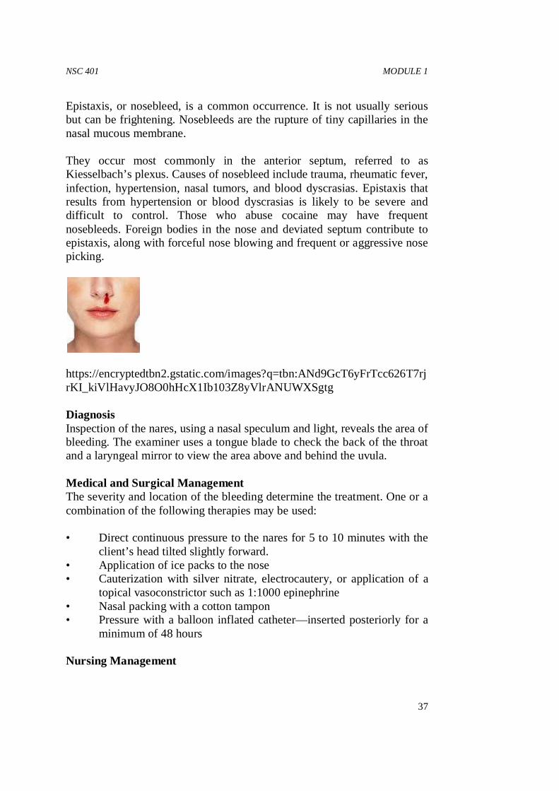
NSC 401 MODULE 1
37
Epistaxis, or nosebleed, is a common occurrence. It is not usually serious but can be frightening. Nosebleeds are the rupture of tiny capillaries in the nasal mucous membrane. They occur most commonly in the anterior septum, referred to as Kiesselbach’s plexus. Causes of nosebleed include trauma, rheumatic fever, infection, hypertension, nasal tumors, and blood dyscrasias. Epistaxis that results from hypertension or blood dyscrasias is likely to be severe and difficult to control. Those who abuse cocaine may have frequent nosebleeds. Foreign bodies in the nose and deviated septum contribute to epistaxis, along with forceful nose blowing and frequent or aggressive nose picking.
https://encryptedtbn2.gstatic.com/images?q=tbn:ANd9GcT6yFrTcc626T7rjrKI_kiVlHavyJO8O0hHcX1Ib103Z8yVlrANUWXSgtg Diagnosis Inspection of the nares, using a nasal speculum and light, reveals the area of bleeding. The examiner uses a tongue blade to check the back of the throat and a laryngeal mirror to view the area above and behind the uvula. Medical and Surgical Management The severity and location of the bleeding determine the treatment. One or a combination of the following therapies may be used: • Direct continuous pressure to the nares for 5 to 10 minutes with the
client’s head tilted slightly forward. • Application of ice packs to the nose • Cauterization with silver nitrate, electrocautery, or application of a
topical vasoconstrictor such as 1:1000 epinephrine • Nasal packing with a cotton tampon • Pressure with a balloon inflated catheter—inserted posteriorly for a
minimum of 48 hours Nursing Management

NSC 401 MEDICAL-SURGICAL NURSING III
38
The nurse monitors vital signs and assesses for signs of continued bleeding. He or she may initiate measures to control bleeding, such as applying pressure and ice packs. Othertreatments require a physician’s order. The client experiencing epistaxis is usually anxious and requires reassurance. If underlying conditions are the cause, the nurse refers the client for medical follow-up. He or she may also recommend humidification, use of a nasal lubricant to keep the mucous membranes moist, and avoidance of vigorous nose blowing and nose picking, or other nose trauma. 3.3 Nasal Trauma A nasal fracture usually results from direct trauma. It causes swelling and edema of the soft tissues, external and internal bleeding, nasal deformity, and nasal obstruction. In severe nasal fractures, cerebrospinal fluid, which is colorless and clear, may drain from the nares. Drainage of cerebrospinal fluid suggests a fracture in the cribriform plate.
https://encrypted-tbn3.gstatic.com/images?q=tbn:ANd9GcTYCFLciImOwXRZYz8dRn4ydajaprkNtBLrLyaQ4cnBA16c7AF3KKpwqvE Diagnosis The diagnosis of a nasal fracture may be delayed because of significant swelling and bleeding. As soon as the swelling decreases, the examiner inspects the nose internally to rule out a fracture of the nasal septum or septal hematoma. Both conditions require treatment to prevent destruction of the septal cartilage. If drainage of clear fluid is observed, a Dextrostix is used to determine the presence of glucose, which is diagnostic for cerebrospinal fluid. Radiography studies are done to ascertain any other facial fractures. Medical and Surgical Management
NSC 401 MODULE 1
39
If the fracture is a lateral displacement, pressure applied to the convex portion of the nose reduces the fracture. Cold compresses control the bleeding. If the fracture is more complex, surgery is done after the swelling subsides, usually after several days. The surgeon applies a splint postoperatively to maintain the alignment. Nursing Management Nursing management is similar to that for nasal obstruction. The nurse instructs the client to keep the head elevated and to apply ice four times a day for 20 minutes to reduce the swelling and pain. He or she gives analgesics as ordered to alleviate pain. Postoperatively, the nurse assesses the client for airway obstruction, respiratory difficulty (i.e., tachypnea, dyspnea), dysphagia, signs of infection, pupillary responses, level of consciousness, and periorbital edema. In addition, the nurse helps reduce the client’s anxiety by answering questions and offering reassurance that the bruising and swelling will subside and sense of smell will return. SELF ASSESSMENT EXERCISE 1. Discuss the pathophysiology of obstructive sleep apnea and the
nursing management. 2. Describe the nursing management of a patient with epistaxis. . 3. Discuss the nursing management of a patient with laryngectomy 3.4 Deviated Septum Pathophysiology and Etiology A deviated septum is an irregularity in the septum that results in nasal obstruction. The deviation may be a deflection from the midline in the form of lumps or sharp projections or a curvature in the shape of an ‘‘S.’’ Marked deviation can result in complete obstruction of one nostril and interference with sinus drainage. A deviated septum may be congenital, but often it results from trauma. Medical and Surgical Management A submucous surgical resection or septoplasty may be necessary to restore normal breathing and to permit adequate sinus drainage for the client with a deviated septum. This procedure involves an incision through the mucous membrane and removal of the portions of the septum that cause obstruction. After this procedure, both sides of the nasal cavity are packed with gauze, which remains in place for 24 to 48 hours. A moustache dressing or drip pad is applied to absorb any drainage. Rhinoplasty, reconstruction of the nose, may also be done at the same time. This procedure enhances the

NSC 401 MEDICAL-SURGICAL NURSING III
40
client’s appearance cosmetically and corrects any structural nasal deformities that interfere with air passage. The surgeon makes an incision inside the nostril and restructures the nasal bone and cartilage. As with septoplasty, the nasal cavity is packed with gauze, and the nose is taped. Application of a nasal splint maintains the shape and structure of the nose and reduces edema. The splint remains in place for at least 1 week. Nursing Management Surgery for correction of nasal obstruction is usually done on an outpatient basis. The nurse provides thorough explanations throughout the procedures to alleviate anxiety. It is particularly important to emphasize that nasal packing will be in place postoperatively, necessitating mouth breathing. The application of an ice pack will reduce pain and swelling 3.5 Laryngeal Obstruction Laryngeal obstruction is an extremely serious and often life-threatening condition. Some causes of upper airway obstruction include edema from an allergic reaction, severe head and neck injury, severe inflammation and edema of the throat, and aspiration of foreign bodies. Signs and Symptoms Client will exhibit stridor, a high-pitched, harsh sound during respiration, indicative of airway obstruction. The client also has dysphagia, hoarseness, cyanosis, and possible hemoptysis (expectoration of bloody sputum). Total obstruction prevents the passage of air from the upper to the lower respiratory airway; choking clients will clutch their throats—the universal distress sign for choking. Unless total obstruction is relieved immediately, death occurs from respiratory arrest. Partial obstruction results in difficulty breathing. Diagnostic Findings Laryngoscopy may reveal internal swelling or foreign body. Radiographs and oxygenation studies will be performed after a patent airway has been established. Medical and Surgical Management Maintenance of a patent airway is crucial. If the client has aspirated a foreign body, the Heimlich maneuver is performed to force the object out of the upper respiratory passagesAllergic reactions resulting in severe inflammation and edema may be treated with epinephrine or a corticosteroid and possibly intubation. Severe obstruction requires an emergency tracheostomy (surgical opening into the trachea).
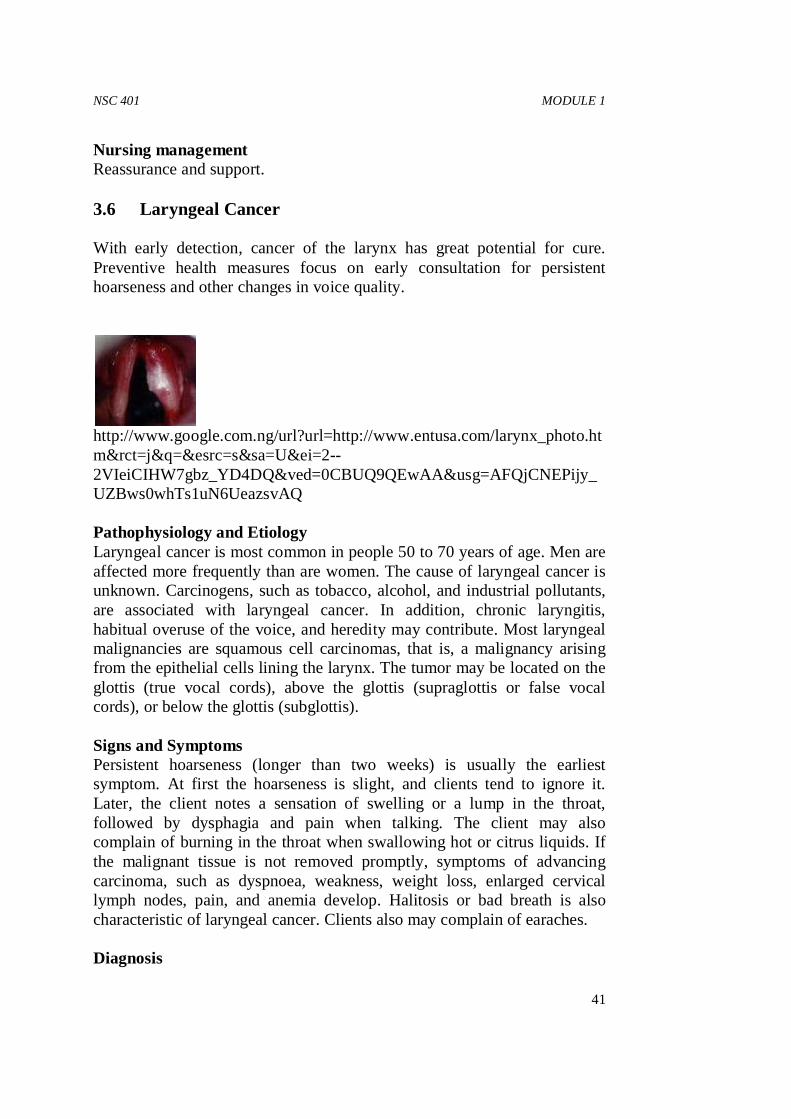
NSC 401 MODULE 1
41
Nursing management Reassurance and support. 3.6 Laryngeal Cancer With early detection, cancer of the larynx has great potential for cure. Preventive health measures focus on early consultation for persistent hoarseness and other changes in voice quality.
http://www.google.com.ng/url?url=http://www.entusa.com/larynx_photo.htm&rct=j&q=&esrc=s&sa=U&ei=2--2VIeiCIHW7gbz_YD4DQ&ved=0CBUQ9QEwAA&usg=AFQjCNEPijy_UZBws0whTs1uN6UeazsvAQ Pathophysiology and Etiology Laryngeal cancer is most common in people 50 to 70 years of age. Men are affected more frequently than are women. The cause of laryngeal cancer is unknown. Carcinogens, such as tobacco, alcohol, and industrial pollutants, are associated with laryngeal cancer. In addition, chronic laryngitis, habitual overuse of the voice, and heredity may contribute. Most laryngeal malignancies are squamous cell carcinomas, that is, a malignancy arising from the epithelial cells lining the larynx. The tumor may be located on the glottis (true vocal cords), above the glottis (supraglottis or false vocal cords), or below the glottis (subglottis). Signs and Symptoms Persistent hoarseness (longer than two weeks) is usually the earliest symptom. At first the hoarseness is slight, and clients tend to ignore it. Later, the client notes a sensation of swelling or a lump in the throat, followed by dysphagia and pain when talking. The client may also complain of burning in the throat when swallowing hot or citrus liquids. If the malignant tissue is not removed promptly, symptoms of advancing carcinoma, such as dyspnoea, weakness, weight loss, enlarged cervical lymph nodes, pain, and anemia develop. Halitosis or bad breath is also characteristic of laryngeal cancer. Clients also may complain of earaches. Diagnosis

NSC 401 MEDICAL-SURGICAL NURSING III
42
Visual examination of the larynx (laryngoscopy) and biopsy confirm the diagnosis and identify the type of malignancy. In addition, computed tomography (CT) scanning and chestradiography are used to detect metastasis and to determine tumor size. The physician also assesses the mobility of the vocal cords. Limited mobility indicates that the tumor growth is affecting the surrounding tissue, muscle, and airway. Medical and Surgical Management Treatment depends on factors such as the size of the lesion, the client’s age, and metastasis. Medical treatment may include chemotherapy, which appears to have only minimal effects, and radiation therapy, either alone or with surgery. Surgical treatment includes laser surgery for early lesions or a partial or total laryngectomy. In more advanced cases, total laryngectomy may be the treatment of choice. If the disease has extended beyond the larynx, a radical neck dissection (removal of the lymph nodes, muscles, and adjacent tissues) is performed. Laser surgery may also be used to relieve obstruction in more advanced cases. A client with a total laryngectomy has a permanent tracheal stoma (opening) because the trachea is no longer connected to the nasopharynx. The larynx is severed from the trachea and removed completely. The only respiratory organs in use are the trachea, bronchi, and lungs. Air enters and leaves through the tracheostomy. The client no longer feels air entering the nose. Because the anterior wall of the esophagus connects with the posterior wall of the larynx, it must be reconstructed. Tube feeding facilitates healing by preventing muscle activity and irritation of the esophagus. Loss of the ability to speak normally is a devastating consequence of laryngeal surgery. Clients with a malignancy of the larynx require emotional support before and after surgery and help in understanding and choosing an alternative method of speech. Some methods of alaryngeal speech used after a laryngectomy include the following: • Esophageal speech: requires regurgitation of swallowed air and
formation of words with lips; voice quality will be lower-pitched and gruff-sounding, but more natural
• Artificial (electric) larynx: a throat vibrator held against the neck that projects sound into the mouth; words are formed with the mouth.
• Tracheoesophageal puncture (TEP): a surgical opening in the posterior wall of the trachea, followed by the insertion of a prosthesis such as a Blom-Singer device.
NSC 401 MODULE 1
43
Air from the lungs is diverted through the opening in the posterior tracheal wall to the esophagus and out the mouth. The client covers the stoma with his or her finger and forces air through the esophagus; this causes the walls of the throat to vibrate as the client speaks. It sounds more natural than an artificial larynx. A speech pathologist works with the client to use an artificial speech device, learn esophageal speech or speak. LARYNGECTOMY INDICATION POSTOPERATIVE EXP It is a surgical removal of larynx. Indication : Laryngeal tumour or cancer Types Partial laryngectomy: The affected vocal cord is removed and other structures remain intact. It is for early stage laryngeal cancer and results into hoarseness of voice. Trachea is intact trachea and is no problem with swallowing. Total laryngectomy: Both vocal cords removed along withthe hyoid bone, epiglottis, cricoidscartilage, and two or three rings ofthe trachea; the tongue, pharyngealwalls, and trachea remain intact;usually a radical neck dissection isdone on the affected side.Itis done when the cancer extendsbeyond the vocal cords.A permanent tracheal stoma that preventsaspiration is left after the surgery. There is no voice but ability to swallow remains. Preoperative Care In addition to routine preoperative teaching, the patient undergoing laryngectomy surgery must be prepared for the loss of ability to breathe through the mouth and nose and the loss of ability to speak. Initial instruction in communication techniques should take place before surgery to prevent the patient from feeling panicky after surgery when he or she is unable to communicate any needs. A variety of techniques and devices are available. Consult the speech therapist before surgery to provide a picture board, magic slate, or paper and pencil. The patient is instructed to point to the picture that corresponds with the need or to write out his or her concern. A dietary consult is also important before surgery if the patient has been undernourished. Postoperative Care Assessment of physical and psychosocial status, comfort, nutritional status, and ability to swallow is important both before and after surgery. After surgery, assessment of airway patency and respiratory function is vital. Monitor lung sounds, oxygen saturation, and arterial blood gases. In addition, be sure to assess the patient’s understanding of the disease process and self-care needs after surgery. It is important to evaluate the patient’s
NSC 401 MEDICAL-SURGICAL NURSING III
44
support systems and ability to cope with the partial or total loss of voice after surgery.
4.0 CONCLUSION Clients with sleep apnea usually are anxious and require reassurance and adequate instruction about their condition. The nurse provides thorough explanations of the disease process, polysomnography, and treatments. He or she refers clients to self-help groups or to appropriate counseling for weight loss or alcohol and substance abuse issues.
5.0 SUMMARY
In this unit, you have learnt that: • Sleep apnea syndrome is characterized by frequent, brief episodes of
respiratory standstill during sleep. • Epistaxis or nosebleeds are the rupture of tiny capillaries in the nasal
mucous membrane. • A nasal fracture usually results from direct trauma. • A deviated septum is an irregularity in the septum that results in
nasal obstruction. • Laryngeal obstruction is an extremely serious and often life-
threatening condition. • Persistent hoarseness (longer than two weeks) is usually the earliest
symptom of Laryngeal cancer
6.0 TUTOR-MARKED ASSIGNMENT 1. Discuss the pathophysiology of obstructive sleep apnea and the
nursing management. 2. Describe the nursing management of a patient with epistaxis. . 3. Discuss the nursing management of a patient with laryngectomy 7.0 REFERENCES/FURTHER READING Bullock, B.A., &Henze, R.L. (2000). Focus on pathophysiology.
Philadelphia: Lippincott Williams & Wilkins. Burke, K.M; Mohn-Brown, E.L &Eby, L (2011). Medical-Surgical Nursing
Care. (3rded.). Boston: Pearson Education, Inc. Nettina, S.M (2010). Lippincott Manual of Nursing Practice (9thed). China:
Wolters Kluwer Health Lippincott Williams & Wilkins.

NSC 401 MODULE 1
45
UNIT 5 CARING FOR PATIENTS WITH LOWER AIRWAY RESPIRATORY INFECTIONS AND INFLAMMATORY DISORDERS: ACUTE BRONCHITIS, PNEUMONIA; TUBERCULOSIS; LUNG ABSCESS; EMPEYEMA; EMERGING RESPIRATORY INFECTION
CONTENTS 1.0 Introduction 2.0 Objectives 3.0 Main Content
3.1 Acute Bronchitis 3.2 Pneumonia 3.3 Tuberculosis 3.4 Lung Abscess 3.5 Empyema 3.6 Emerging Respiratory Infection 3.7 Severe Acute Respiratory Syndrome (SARS) 3.8 Inhalation Anthrax
4.0 Conclusion 5.0 Summary 6.0 Tutor-Marked Assignment 7.0 References/Further Reading 1.0 INTRODUCTION Disorders of the lower respiratory tract include problems of the lower portion of the trachea, bronchi, bronchioles, and alveoli. These disorders may be related to infection, noninfectious alterations in function, neoplasm (cancer), or trauma. Any pathological condition of the lower respiratory tract can seriously impair carbondioxide and oxygen exchange. 2.0 OBJECTIVES At the end of this unit you will be able to: • describe bronchiolectasis and its management • define the cause, treatment, collaborative interventions, and clinical
outcomes in tuberculosis(TB).
NSC 401 MEDICAL-SURGICAL NURSING III
46
3.0 MAIN CONTENT
3.1 Acute Bronchitis Bronchitis is an inflammation of the bronchial tree, which includes the right and left bronchi, secondary bronchi, and bronchioles. When the mucous membranes lining the bronchial tree become irritated and inflamed, excessive mucus is produced. The result is congested airways. Acute bronchitis is usually an isolated episode. It becomes chronic when it occurs more than 3 months out of the year for 2 consecutive years. Pathophysiology and Etiology Inflammation of the mucous membranes that line the major bronchi and their branches characterizes acute bronchitis. If the inflammatory process involves the trachea, it is referred to as tracheobronchitis. Typically, acute bronchitis begins as an upper respiratory infection (URI) the inflammatory process then extends to the tracheobronchial tree. The secretory cells of the mucosa produce increased mucopurulent sputum. Viral infections most commonly give rise to acute bronchitis. Clients with viral URIs are more vulnerable to secondary bacterial infections, which then may lead to acute bronchitis. Sputum cultures identify the causative bacterial organisms, the most common of which are Haemophilusinfluenzae, Streptococcus pneumoniae, and Mycoplasma pneumoniae. Fungal infections such as Aspergillus may be identified as the cause of acute bronchitis. Chemical irritation from noxious fumes, gases, and air contaminants also may induce acute bronchitis. A potential complication is bronchial asthma. Signs and symptoms Signs and symptoms initially include fever, chills, malaise, headache, and a dry, irritating, and nonproductive cough. Later, the cough produces mucopurulent sputum, which may be blood-streaked if the airway mucosa becomes irritated with severe tracheobronchitis and coughing. Clients experience paroxysmal attacks of coughing and may report wheezing. Laryngitis and sinusitis complicate the symptoms. Moist, inspiratory crackles may be heard on chest auscultation. A sputum sample is collected for culture and sensitivity testing to rule out bacterial infection. A chest film also may be done to detect additional pathology, such as pneumonia. Medical Management Acute bronchitis usually is self-limiting, lasting for several days. Suggested treatment is bed rest, antipyretics, expectorants, antitussives (drugs used to prevent coughing), and increased fluids. Humidifiers assist in keeping mucous membranes moist because dry air aggravates the cough. If

NSC 401 MODULE 1
47
secondary bacterial invasion occurs, the previously mild infection becomes more serious, and usually is accompanied by a persistent cough and thick, purulent sputum. Secondary infections usually subside as the bronchitis subsides, but they may persist for several weeks. When a secondary infection is evident, the physician orders a broad-spectrum antibiotic when sputum culture results are available. Nursing Management The nurse auscultates breath sounds and monitors vital signs every 4 hours, especially if the client has a fever. He or she encourages the client to cough and deep breathe every 2 hours while awake and to expectorate rather than swallow sputum. Humidification of surrounding air loosens bronchial secretions. The nurse changes the bedding and the client’s clothes if they become damp with perspiration and offers fluids frequently. The nurse, in an effort to prevent the spread of infection, teaches the client to wash the hands frequently, particularly when handling secretions and soiled tissues; cover the mouth when sneezing and coughing; discard soiled tissues in a plastic bag; and avoid sharing eating utensils and personal articles with others. 3.2 Pneumonia Pneumonia is an inflammatory process affecting the bronchioles and alveoli. Although it usually is associated with an acute infection, pneumonia also can result from radiation therapy, chemical ingestion or inhalation, or aspiration of foreign bodies or gastric contents. Pneumonia, when combined with influenza, ranks as the eighth leading cause of death in the United States (American Lung Association, 2007). Pathophysiology and Etiology Pneumonia is classified according to its etiology. Bacterial pneumonias are referred to as typical pneumonias. Atypical pneumonias are those caused by mycoplasmas, Legionella pneumophila (the causative agent of Legionnaire’s disease), chlamydiae, viruses, parasites, and fungi. Mycobacterium tuberculosis also may cause pneumonia. Viruses are the most common etiology, with influenza type A virus the usual causative organism. Bacterial pneumonias are less common but more serious. Causative bacterial organisms include Streptococcus pneumoniae, Pneumocystis jiroveci, Staphylococcus aureus, Klebsiellapneumoniae, Pseudomonas aeruginosa, and Haemophilusinfluenzae. Radiation pneumonia results from damage to the normal lung mucosa during radiation therapy for breast or lung cancer. Chemical pneumonia

NSC 401 MEDICAL-SURGICAL NURSING III
48
results from ingestion of kerosene or inhalation of volatile hydrocarbons (kerosene, gasoline, or other chemicals), which may occur in industrial settings. Aspiration pneumonia occurs when a person inhales a foreign body or gastric contents during vomiting or regurgitation. Hypoventilation of lung tissue over a prolonged period can occur when a client is bedridden and breathing with only part of the lungs. Bronchial secretions subsequently accumulate, which may lead to hypostatic pneumonia. Pneumonia is also categorized according to its presenting symptoms. Bronchopneumonia means that the infection is patchy, diffuse, and scattered throughout both lungs. Lobar pneumonia means that the inflammation is confined to one or more lobes of the lung. Another classification of pneumonia refers to where the client acquired the inflammatory process. There are four general categories. The first is community-acquired pneumonia (CAP), which means that the client contracted the illness in a community setting or within 48 hours of admission to a healthcare facility. Hospital-acquired pneumonia (HAP), or nosocomial pneumonia, occurs in a healthcare setting more than 48 hours after admission. Pneumonia in the immunocompromised host is a third category; this type includes Pneumocystis jiroveci pneumonia, fungal pneumonia, and pneumonia related to tuberculosis. The fourth category is aspiration pneumonia. Organisms that cause pneumonia reach the alveoli by inhalation of droplets, aspiration of organisms from the upper airway, or, less commonly, seeding from the bloodstream. When organisms reach the alveoli, the inflammatory reaction is intense, producing an exudate that impairs gas exchange. Capillaries surrounding the alveoli become engorged and cause the alveoli to collapse (atelectasis), further impairing gas exchange and interfering with ventilation. White blood cells (WBCs) move into the area to destroy the pathogens, filling the interstitial spaces. If untreated, consolidation occurs as the inflammation and exudate increase. Hypoxemia results from the inability of the lungs to oxygenate blood from the heart. Bronchitis, tracheitis (inflammation of the trachea), and spots of necrosis (death of tissue) in the lung may follow. In atypical pneumonias, the exudate infiltrates the interstitial spaces rather than the alveoli directly. The pneumonia is more scattered, as described for bronchopneumonia. As the inflammatory process continues, it increasingly interferes with gas exchange between the bloodstream and lungs. Increased carbon dioxide (CO2) in the blood stimulates the respiratory center, causing more rapid and shallow breathing. Without an interruption of any type of pneumonia, the client becomes increasingly ill. If the circulatory system cannot compensate for the burden of decreased gas exchange, the client is at risk for heart failure. Death from pneumonia is most common in older adults
NSC 401 MODULE 1
49
and those weakened by acute or chronic diseases or disorders (e.g., acquired immunodeficiency syndrome [AIDS], cancer and lung disease) or prolonged periods of inactivity. Complications of pneumonia include congestive heart failure (CHF), empyema (collection of pus in the pleural cavity), pleurisy (inflammation of the pleura), septicemia (infective microorganisms in the blood), atelectasis, hypotension, and shock. In addition, septicemia may lead to a secondary focus of infection, such as endocarditis (inflammation of the endocardium), pericarditis (inflammation of the pericardium), and purulent arthritis. Otitis media (infection of the middle ear), bronchitis, or sinusitis also may complicate recovery, especially from atypical pneumonia. Signs and Symptoms Symptoms vary for the different types of pneumonia. The onset of bacterial pneumonia is sudden. The client experiences fever, chills, a productive cough, and discomfort in the chest wall muscles from coughing. There also is general malaise. The sputum may be rust colored. Breathing causes pain; thus, the client tries to breathe as shallowly as possible. Viral pneumonia differs from bacterial pneumonia in that results of blood cultures are sterile, sputum may be more copious, chills are less common, and pulse and respiratory rates are characteristically slow. Diagnostic Findings Auscultation of the chest reveals wheezing, crackles, and decreased breath sounds. The nail beds, lips, and oral mucosa may be cyanotic. Sputum culture and sensitivity studies can help to identify the infectious microorganism and effective antibiotics for treatment in cases of bacterial pneumonia. A chest film shows areas of infiltrates and consolidation. A complete blood count discloses an elevated WBC count. Blood cultures also may be done to detect any microorganisms in the blood. A newer and more efficient method to diagnose pneumonia is called an electronic nose or ‘‘e-nose.’’ The maker, Cyrano Sciences, Inc., calls this device the Cyranose 320. Although not yet fully approved by the U. S. Food and Drug Administration, this hand-held device senses and evaluates exhaled breath for various types of pneumonia and sinusitis. Tests indicate a 70% to 90% accuracy rate, which is similar to standard testing. The e-nose also provides diagnosis in approximately 40 minutes, whereas standard testing results are usually not available for several hours (‘‘Sniffing out,’’ 2004). Medical Management Medical management involves: • prompt initiation of antibiotic therapy for bacterial pneumonia
NSC 401 MEDICAL-SURGICAL NURSING III
50
• hydration to thin secretions • supplemental oxygen to alleviate hypoxemia, • bed rest • chest physical therapy and postural drainage (techniques that involve
manual pounding or clapping to loosen secretions and positioning of the client to drain and remove secretions from specific areas of the lungs),
• bronchodilators, analgesics, antipyretics, and cough expectorants or suppressants, depending on the nature of the client’s cough.
If a client is hospitalized, treatment is more vigorous, depending on the potential or actual complications. Fluid and electrolyte replacement sometimes is necessary secondary to fever, dehydration, and inadequate nutrition. If the client experiences severe respiratory difficulty and thick, copious secretions, he or she may require intubation along with mechanical ventilation. Nursing Management The nurse auscultates lung sounds and monitors the client for signs of respiratory difficulty. He or she checks oxygenation status with pulse oximetry and monitors arterial blood gases (ABGs). Assessments of cough and sputum production also are necessary. The nurse places the client in the semi-Fowler’s position to aid breathing and increase the amount of air taken with each breath. Increased fluid intake is important to encourage because it helps to loosen secretions and replace fluids lost through fever and increased respiratory rate. The nurse monitors fluid intake and output, skin turgor, vital signs, and serum electrolytes. He or she administers antipyretics as indicated and ordered. Identifying clients at risk for pneumonia provides a means to practice preventive nursing care. In addition, nurses encourage at-risk and elderly clients to receive vaccination against pneumococcal and influenza infections. Because the nursing care of clients with infectious lung disorders is similar regardless of the etiology. 3.3 Tuberculosis Tuberculosis (TB) is an infectious disease caused by the Mycobacterium tuberculosisbacteria. Pathophysiology TB primarily affects the lungs, although other areas, such as the kidneys, liver, brain, and bone, may be affected as well. M. tuberculosis is an acid-
NSC 401 MODULE 1
51
fast bacillus (AFB), which means that when it is stained in the laboratory and then washed with an acid, the stain remains, or stays fast. M. tuberculosis can live in dark places in dried sputum for months, but a few hours in direct sunlight kills it. It is spread by inhalation of the tuberculosis bacilli from respiratory droplets (droplet nuclei) of an infected person. Once the bacilli enter the lungs, they multiply and begin to disseminate to the lymph nodes and then to other parts of the body. The patient is then infected but may or may not go on to develop clinical (active) disease. During this time, the body develops immunity, which keeps the infection under control. The immune system surrounds the infected lung area with neutrophils and alveolar macrophages. This process creates a lesion called a tubercle, which seals off the bacteria and prevents spread. The bacteria within the tubercle die or become dormant, and the patient is no longer infectious. If the patient’s immune system becomes compromised, however, some of the dormant bacteria can become active again, causing reinfection and active disease. Only 5 to 10 percent of infected individuals in the United States actually develop the disease, and even then, it may not occur for many years. Signs and Symptoms Active tuberculosis is characterized by a chronic productive cough, blood-tinged sputum, and drenching night sweats. A low-grade fever may be present. If effective treatment is not initiated, a downhill course occurs, with pulmonary fibrosis, hemoptysis, and progressive weight loss. Complications Spread of the tuberculosis bacilli throughout the body can result in pleurisy, pericarditis, peritonitis, meningitis, bone and joint infection, genitourinary or gastrointestinal infection, or infection of many other organs. Diagnostic Tests Routine screening for tuberculosis infection is usually done with a purified protein derivative (PPD) skin test. The PPD is injected intradermally; the test is considered positive if a raised area of induration occurs within 48 to 72 hours. If there is a red area around the induration, this is not measured. The size of induration that indicates a positive test varies based on the individual’s history. A red area without induration is not considered a positive result. A positive result indicates that a person has been exposed; it does not mean that active TB disease is present. Some health care institutions use a two-step process for baseline testing of employees and residents. If an individual has a negative test, he or she is retested in 1 to 3

NSC 401 MEDICAL-SURGICAL NURSING III
52
weeks. This is because someone who was exposed many years ago may not react to the first test. The first test acts as a “reminder” to the immune system to react. The second test will then be positive in the person with a past TB infection.BOX 28–2 The reduction in immune system function from aging can decrease the effectiveness of the tuberculosis antibodies in someone who previously had dormant disease. The tuberculosis bacilli can be reactivated, causing active disease. If the physician orders “sputum culture for AFB,” tuberculosis is suspected. Ask whether isolation precautions should be taken while waiting for culture results. Etiology Crowded or poorly ventilated living conditions place people at risk for becoming infected with tuberculosis. Although tuberculosis can infect any age group, the elderly are especially at risk. Elders may have contracted the disease many years before, but it reactivates as the aging process diminishes immune function. Patients with AIDS and chronic alcohol abuse have a very high risk because of their compromised immune function. In the United States, tuberculosis is also prevalent among the urban poor and minority groups. Before 1985 the incidence of TB was steadily decreasing. Now it is again on the rise, in part because of the prevalence of AIDS, the development of antibiotic-resistant strains of the TB bacillus, and ineffective treatment programs. TB kills 2 million people each year worldwide. Prevention Clean, well-ventilated living areas are essential to the health of all people. If a hospitalized patient is known or suspected to have tuberculosis, he or she is placed in respiratory isolation to prevent spread to staff or other patients. Special isolation rooms are ventilated to the outside. Staff should wear special high-efficiency filtration masks when in the patient’s room. A regular surgical mask is not effective against TB. Verify with the institution’s infection control department that the masks provided are effective for use with TB patients. If the patient must travel through the hallway for tests or other activities, the patient must wear a mask. Additional protective barriers, such as gowns, gloves, or goggles, are used when contact with sputum is likely. Nursing Care Perform thorough respiratory and psychosocial assessments of the patient with TB. The severity of the disease determines the impact on the patient’s lifestyle. It is also imperative to determine the patient’s knowledge of the

NSC 401 MODULE 1
53
disease and treatment and his or her compliance with drug treatment. Possible nursing diagnoses include impaired gas exchange, ineffective airway clearance, ineffective breathing pattern, anxiety, imbalanced nutrition, risk for infection of patient’s contacts, and possible noncompliance with drug therapy or ineffective therapeutic regimen management. Diagnoses should be chosen based on individual patient data. Anxiety may be reduced by educating the patient in self-care measures and by reassuring the patient that the disease can be controlled by careful compliance with treatment. The patient who is emaciated because of the disease will benefit from a dietitian consultation to provide specific recommendations or supplements. To prevent spread of infection to others, teach the patient to use a tissue to cover the mouth and nose when coughing or sneezing. Tissues should be flushed down the toilet or disposed of carefully in the trash. Teach all family members the importance of careful handwashing, how to manage drug therapy, and when to report side effects. Forewarn the patient that rifampin turns the urine and body fluids red. A visiting nurse is essential to evaluate the home environment and assess the patient’s ability to comply with therapy. If the patient is unable to comply with therapy, measures must be instituted to ensure that medications are taken to protect both the patient and the public. Directly observed therapy at a local health clinic or by a home health nurse may be necessary. The patient will be followed periodically by the physician for sputum cultures and drug monitoring. Once sputum cultures are negative, the patient is no longer contagious. Antibiotics Used in Treatment of Tuberculosis 28–3 Isoniazid Ethionamide Rifampin Kanamycin Streptomycin Para-aminosalicylic acid Ethambutol Cycloserine Pyrazinamide Medical Treatment Treatment consists of specific antibiotic therapy. First-line drugs have the fewest adverse effects. However, these drugs can be toxic to the liver and nervous system, as well as having other side effects. Second-line drugs are
NSC 401 MEDICAL-SURGICAL NURSING III
54
more toxic and are reserved for cases that do not respond to firstline drug therapy. Generally, two or three antibiotics are given simultaneously to allow lower doses of each individual drug and to reduce the incidence of serious side effects. Drugs must be taken for 6 to 8 months or longer. Because of the length of therapy and the incidence of side effects, you must anticipate that compliance may be a problem. 3.4 Lung Abscess A lung abscess is a localized necrotic lesion of the lung parenchyma containing purulent material that collapses and forms a cavity. It is generally caused by aspiration of anaerobic bacteria. By definition, the chest x-ray will demonstrate a cavity of at least 2 cm. Patients who have impaired cough reflexes and cannot close the glottis, or those with swallowing difficulties, are at risk for aspirating foreign material and developing a lung abscess. Other at risk patients include those with central nervous system disorders (seizure, stroke), drug addiction, alcoholism, esophageal disease, or compromised immune function, those without teeth, as well as patients receiving naso-gastric tube feedings and those with an altered state of consciousness from anesthesia. Pathophysiology Most lung abscesses are a complication of bacterial pneumonia or are caused by aspiration of oral anaerobes into the lung. Abscesses also may occur secondary to mechanical or functional obstruction of the bronchi by a tumor, foreign body, or bronchial stenosis, or from necrotizing pneumonias, TB, pulmonary embolism, or chest trauma. Most abscesses are found in areas of the lung that may be affected by aspiration. The site of the lung abscess is related to gravity and is determined by the patient’s position. For patients who are confined to bed, the posterior segment of an upper lobe and the superior segment of the lower lobe are the most common areas in which lung abscess occurs. However, atypical presentationsmay occur, depending on the position of the patient when the aspiration occurred. Initially, the cavity in the lung may or may not extend directly into a bronchus. Eventually the abscess becomes surrounded, or encapsulated, by a wall of fibrous tissue. The necrotic process may extend until it reaches the lumen of a bronchus or the pleural spaceand establishes communication with the respiratory tract, the pleural cavity, or both. If the bronchus is involved, the purulent contents are expectorated continuously in the form of sputum. If the pleura is involved, an empyema results. A communication or connection between the bronchus and pleura is known as a bronchopleural fistula. The organisms frequently associated with lung abscesses are S.

NSC 401 MODULE 1
55
aureus, Klebsiella, and other gram-negative species. Anaerobic organisms, however, may also be present. The organism varies depending on the underlying predisposing factors. Clinical Manifestations The clinical manifestations of a lung abscess may vary from a mild productive cough to acute illness. Most patients have a fever and a productive cough with moderate to copious amounts of foul smelling, often bloody, sputum. Leukocytosis may be present. Pleurisy or dull chest pain, dyspnea, weakness, anorexia, and weight loss are common. Fever and cough may develop insidiously and may have been present for several weeks before diagnosis. Assessment and Diagnostic Findings Physical examination of the chest may reveal dullness on percussion and decreased or absent breath-sounds with an intermittent pleural friction rub (grating or rubbing sound) on auscultation. Crackles may be present. Confirmation of the diagnosis is made by chest x-ray, sputum culture, and in some cases fiberoptic bronchoscopy. The chest x-ray reveals an infiltrate with an air–fluid level. A computed tomography (CT) scan of the chest may be required to provide more detailed pictures of different cross-sectional areas of the lung. Prevention The following measures will reduce the risk of lung abscess: • Appropriate antibiotic therapy before any dental procedures in
patients who must have teeth extracted while their gums and teeth are infected.
• Adequate dental and oral hygiene, because anaerobic bacteria play a role in the pathogenesis of lung abscess.
• Appropriate antimicrobial therapy for patients with pneumonia. Medical Management The findings of the history, physical examination, chest x-ray, and sputum culture indicate the type of organism and the treatment required. Adequate drainage of the lung abscess may be achieved through postural drainage and chest physiotherapy. The patient should be assessed for an adequate cough. A few patients need a percutaneous chest catheter placed for long-term drainage of the abscess. Therapeutic use of bronchoscopy to drain an abscess is uncommon. A diet high in protein and calories is necessary because chronic infection is associated with a catabolic state, necessitating increased intake of calories and protein to facilitate healing. Surgical
NSC 401 MEDICAL-SURGICAL NURSING III
56
intervention is rare, but pulmonary resection (lobectomy) is performed when there is massive hemoptysis (coughing up of blood) or little or no response to medical management. Nursing Management The nurse administers antibiotics and intravenous therapies as prescribed and monitors for adverse effects. Chest physiotherapy is initiated as prescribed to facilitate drainage of the abscess. The nurse teaches the patient to perform deep-breathing and coughing exercises to help expand the lungs. To ensure proper nutritional intake, the nurse encourages a diet high in protein and calories. The nurse also offers emotional support because the abscess may take a long time to resolve. SELF ASSESSMENT EXERCISE 1. Describe bronchiolectasis and its management. 2. Define the cause, treatment, collaborative interventions, and clinical
outcomes in tuberculosis (TB). 3. Identify 3 nursing diagnoses of a patient with Pulmonary
Tuberculosis and draw nursing care plan to solve them. 3.5 Empyema An empyema is an accumulation of thick, purulent fluid within the pleural space, often with fibrin development and a loculated (walled-off) area where infection is located. Causes Most empyemas occur as complications of bacterial pneumonia or lung abscess. Other causes include penetrating chest trauma, hematogenous infection of the pleural space, nonbacterial infections, or iatrogenic causes (after thoracic surgery or thoracentesis). Pathophysiology At first the pleural fluid is thin, with a low leukocyte count, but it frequently progresses to a fibropurulent stage and, finally, to a stage where it encloses the lung within a thick exudative membrane (loculated empyema). Clinical Manifestations With an empyema, the patient is acutely ill and has signs and symptoms similar to those of an acute respiratory infection or pneumonia (fever, night sweats, pleural pain, cough, dyspnea, anorexia, weight loss). If the patient
NSC 401 MODULE 1
57
is immunocompromised, the symptoms may be vaguer. If the patient has received antimicrobial therapy, the clinical manifestations may be less obvious Diagnostic Findings Chest auscultation demonstrates decreased or absent breath sounds over the affected area, and there is dullness on chest percussion as well as decreased fremitus. The diagnosis is established by a chest x-ray or chest CT scan. Usually a diagnostic thoracentesis is performed, often under ultrasound guidance. Medical Management The objectives of treatment are to drain the pleural cavity and to achieve full expansion of the lung. The fluid is drained and appropriate antibiotics, in large doses, are prescribed based on the causative organism. Sterilization of the empyema cavity requires 4 to 6 weeks of antibiotics. Drainage of the pleural fluid depends on the stage of the disease and is accomplished by one of the following methods: • Needle aspiration (thoracentesis) with a thin percutaneous catheter,
if the volume is small and the fluid not too purulent or thick • Tube thoracostomy (chest drainage using a large-diameter intercostal
tube attached to water-seal drainage) with fibrinolytic agents instilled through the chest tube in patients with loculated or complicated pleural effusions
• Open chest drainage via thoracotomy, including potential rib resection, to remove the thickened pleura, pus, and debris and to remove the underlying diseased pulmonary tissue
With long-standing inflammation, an exudate can form over the lung, trapping it and interfering with its normal expansion. This exudate must be removed surgically (decortication). The drainage tube is left in place until the pus-filled space is obliterated completely. The complete obliteration of the pleural space is monitored by serial chest x-rays, and the patient should be informed that treatment may be long term. Patients are frequently discharged from the hospital with a chest tube in place, with instructions to monitor fluid drainage at home. Nursing Management Resolution of empyema is a prolonged process. The nurse helps the patient cope with the condition and instructs the patient in lung-expanding breathing exercises to restore normal respiratory function. The nurse also provides care specific to the method of drainage of the pleural fluid (eg,

NSC 401 MEDICAL-SURGICAL NURSING III
58
needle aspiration, closed chest drainage, or rib resection and drainage). When a patient is discharged to home with a drainage tube or system in place, the nurse instructs the patient and family on care of the drainage system and drain site, measurement and observation of drainage, signs and symptoms of infection, and how and when to contact the health care provider. 3.6 Emerging Respiratory Infection
Recent years have seen any of newly identified respiratory infections as well as the use of infectious microbes as weapons. Severe Pneumonia may complicate a new strain of Influenza caused by the H1N1 virus (also called Swine flu). Although rare, this complication occurs primarily in children, young adults and pregnant women and can quickly lead to acute respiratory distress syndrome and respiratory failure. While manifestation of H1N1 influenza usually are mild, immediately report symptoms such as difficulty breathing, cyanosis or a drop in O2 saturation. 3.7 Severe Acute Respiratory Syndrome (SARS) It is a lower respiratory illness caused by a newly identified virus called SARS associated Coronavirus (SARS-CoV). This spreads primarily by close human contact. Most people with SARS develop severe pneumonia, typically beginning with flu-like symptoms including high-grade fever, headache and muscle aches, cough and shortness of breath. Intensive and supportive medical care is the primary treatment. 3.8 Inhalation Anthrax It is caused by Bacillus anthracis identified as a potential biologic weapon. Inhalation anthrax causes initial flu-like symptoms with malaise, dry cough and fever. Severe dyspnoea, stridor and cyanosis develop abruptly along with inflammation of lymph nodes in the mediastinum and thorax. Blood cultures and CXR are used to diagnose inhalation anthrax. Fortunately, the antibiotic Ciprofloxacin is effective for both prevention and treatment of inhalation anthrax. People with these emerging respiratory infections require intensive and supportive nursing care. The nursing diagnoses and intervention identified for pneumonia and respiratory failure may be appropriate. Contact and airborne precautions are implemented in addition to standard precautions for clients with SARS.
NSC 401 MODULE 1
59
4.0 CONCLUSION • Disorders of the lower respiratory tract include problems of the
lower portion of the trachea, bronchi, bronchioles, and alveoli. Examples are Bronchitis, Pneumonia, Tuberculosis, Lung abscess and Empyema.
5.0 SUMMARY
In this unit, you have learnt thatemerging respiratory infections include severe acute respiratory syndrome (SARS) and Inhalation Anthrax. You also learnt about medical and nursing management of disorders of the lower respiratory tract and emerging respiratory infections.
6.0 TUTOR-MARKED ASSIGNMENT Pay a visit to any health facility in collaboration with your preceptor. Identify the safety precautions guideline adopted in that institution for patients with confirmed pulmonary Tuberculosis. Report your observation in the discussion forum. 7.0 REFERENCES/FURTHER READING Bullock, B.A., & Henze, R.L. (2000). Focus on pathophysiology.
Philadelphia: Lippincott Williams & Wilkins. Burke, K.M; Mohn-Brown, E.L & Eby, L (2011). Medical-Surgical
Nursing Care. (3rded.). Boston: Pearson Education, Inc. Nettina, S.M (2010). Lippincott Manual of Nursing Practice (9thed). China:
Wolters Kluwer Health Lippincott Williams & Wilkins. Rueling, S., & Adams, C. (2003). Close to the vest: a novel way to keep
airways clear. Nursing 2003, 33(12), 56–57. Timby, B.K & Smith, N.E (2010). Introductory Medical-Surgical Nursing
(10th ed.).Philadelphia: Wolters Kluwer Health | Lippincott Williams & Wilkins.
Williams, L.S & Hopper, P.D (2003). Understanding Medical-Surgical
Nursing. Philadelphia: F. A. Davis Company.

NSC 401 MEDICAL-SURGICAL NURSING III
60
UNIT 6 CARING FOR PATIENT WITH OBSTRUCTIVE AND RESTRICTIVE LUNG DISORDERS: ASTHMA; CHRONIC OBSTRUCTIVE PULMONARY DISEASE(COPD); CYSTIC FIBROSIS; ATELECTASIS; BRONCHIECTASIS
CONTENTS 1.0 Introduction 2.0 Objectives 3.0 Main Content
3.1 Asthma 3.2 Chronic Obstructive Pulmonary Disease (COPD) 3.3 Cystic Fibrosis 3.4 Atelectasis 3.5 Bronchiectasis
4.0 Conclusion 5.0 Summary 6.0 Tutor-Marked Assignment 1.0 INTRODUCTION Asthma is characterized by inflammation of the mucosal lining of the bronchial tree and spasm of the bronchial smooth muscles (bronchospasm). This causes narrowed airways and air trapping. Symptoms are intermittent and reversible, with periods of normal airway function. About 50 percent of asthmatics develop the disorder in childhood, but contrary to popular belief, most children do not outgrow asthma. Instead, symptoms just diminish, often returning later in life. 2.0 OBJECTIVES At the end of this unit you will be able to:
• Describe the pathophysiology of asthma. • Discuss the medications used in asthma management. • Describe asthma self-management strategies. • Describe the pathophysiology of chronic obstructive pulmonary
disease (COPD). • Describe nursing management of patients with COPD • Describe the pathophysiology of cystic fibrosis.

NSC 401 MODULE 1
61
3.0 MAIN CONTENT 3.1 Asthma Asthma is a chronic inflammatory disease of the airways that causes airway hyperresponsiveness, mucosal edema, and mucus production. This inflammation ultimately leads to recurrent episodes of asthma symptoms: cough, chest tightness, wheezing, and dyspnea. Pathophysiology and Etiology The tendency to develop asthma is inherited. Some sources classify asthma as either allergic or idiosyncratic (unexpected). Allergic asthma is triggered by allergens such as pollen, foods, medications, animal dander, air pollution, molds, or dust mites. It is commonly seasonal. Individuals who developed asthma as children tend to have allergic asthma. Idiosyncratic asthma is generally diagnosed in adults and is related to environmental or other non-allergic factors, such as environmental irritants, smoking, and respiratory or sinus infection. Emotional upset and exercise can also trigger symptoms in some persons with asthma. Asthma frequently complicates chronic bronchitis or emphysema. A newer finding is asthma caused by gastroesophageal reflux disease (GERD). It is believed that stomach acid may reflux into the esophagus and then be aspirated, triggering asthma. Prevention Although asthma cannot be prevented, individual episodes can be. It is important that the patient identify triggers of asthma symptoms and avoid them whenever possible. Compliance with prophylactic and maintenance therapy is also important. Signs and Symptoms Asthma symptoms are intermittent and are often referred to as “attacks,” which may last from minutes to days. The patient complains of chest tightness, dyspnea, and difficulty moving air in and out of the lungs. Once initial symptoms are controlled, airways may remain hypersensitive and prone to asthma symptoms for many weeks. On examination, you will note an increased respiratory rate as the patient attempts to compensate for narrowed airways. Inspiratory and expiratory wheezing is heard because of turbulent airflow through swollen airways with thick secretions and may sometimes be audible even without a stethoscope. Air is trapped in the lungs, and expiration is prolonged. A cough is common and may produce thick, clear sputum. Use of accessory muscles to breathe is a sign that the

NSC 401 MEDICAL-SURGICAL NURSING III
62
attack is severe and warrants immediate attention. Be aware that an absence of audible wheezing may not signal improvement but rather may be an ominous sign that the patient is moving very little air. If wheezing is not heard, use of accessory muscles and peak expiratory flow rate values must be carefully evaluated. Once treatment begins to be effective and the patient is moving more air, wheezing may become audible. Complications Status asthmaticus occurs if bronchospasm is not controlled and symptoms are prolonged. As the patient increases the respiratory rate to compensate for narrowed airways, a lot of carbon dioxide is blown off and respiratory alkalosis occurs. If the attack is not resolved and the patient begins to tire, the patient will no longer be able to compensate and PaCO2 will rise, resulting in respiratory acidosis. This can lead to respiratory failure and death if untreated. Diagnostic Tests Diagnosis is based on the patient’s report of symptoms, physical examination, and pulmonary function studies. Peak expiratory flow rate is reduced. Arterial blood gases may initially show decreased PaCO2. Late in the course of an attack, PaO2 decreases and PaCO2 increases. Allergy skin testing and increased serum IgE and eosinophil levels indicate allergic involvement and may help determine treatment. Medical Management Symptomatic treatment is given at the time of the attack. Long-term care involves measures to treat as well as to prevent further attacks. An effort must be made to determine the cause. If the history and diagnostic tests indicate allergy as a causative factor, treatment includes avoidance of the allergen, desensitization, or antihistamine therapy. Oxygen usually is not necessary during an acute attack because most clients are actively hyperventilating. Oxygen may be necessary if cyanosis occurs. Pharmacologic management is often classified as rescue therapy and maintenance therapy. Rescue-therapy medications treat acute episodes of asthma, whereas maintenance therapy is a daily regimen designed to prevent and control symptoms. Many medications are taken through metred dose inhalers (MDIs). Humidification of inspired air is valuable because dehydration of the respiratory mucous membrane may lead to asthmatic attacks. Use of steam or cool vapor humidifiers also has proved effective. Liquefaction of the secretions promotes more effective clearing of the airways and a rapid
NSC 401 MODULE 1
63
return to normal. Air conditioners may filter offending allergens as well as control temperature and humidity.
Nursing Management During asthma attacks, clients are extremely anxious. The nurse reassures the client that someone will remain with him or her during the acute phase. The nurse administers oxygen if indicated and puts the client in a sitting position. Rest and adequate fluid intake are important. Increased fluid intake makes secretions less tenacious and replaces the fluids lost through perspiration. Thus, the nurse keeps fluids within easy reach and encourages the client to drink them. The nurse checks the intravenous (IV) site frequently for signs of extravasation. This monitoring is especially important during an acute attack because restlessness can result in catheter dislodgment. The nurse observes for adverse drug effects, especially when the client is receiving epinephrine or other adrenergic agents, which may cause palpitations, nervousness, trembling, pallor, and insomnia. Clients with asthma must demonstrate understanding of the following: • Asthma as a chronic inflammatory disease • Role of inflammation and broncho constriction • Action and purpose of medications • How to avoid triggers for asthma attacks • Use of metered-dose inhalers • Use of peak-flow monitoring • When and how to obtain medical assistance. The nurse assesses the client’s level of understanding of these topics and provides education as needed. The nurse determines whether the client has a peak flow meter and obtains one for the client if needed. The peak flow meter measures the peak expiratory flow rate (PEFR), which is the point of highest flow during forced expiration. The nurse instructs the client in using the peak flow meter to monitor the degree of asthma control. The client can use the peak flow meter to assess the effectiveness of medication or breathing status. The nurse tells the client to seek care if readings fall below baseline and teaches the correct use of inhalers. He or she also helps the client to identify 3.2 Chronic Obstructive Pulmonary Disease (COPD) COPD is a disease state characterized by airflow limitation that is not fully reversible. This newest definition of COPD, provided by the Global Initiative for Chronic Obstructive Lung Disease, provides a broad

NSC 401 MEDICAL-SURGICAL NURSING III
64
description that better explains this disorder and its signs and symptoms. While previous definitions have included emphysema and chronic bronchitis under the umbrella classification of COPD, this was often confusing because most patients with COPD present with overlapping signs and symptoms of these two distinct disease processes. COPD may include diseases that cause airflow obstruction (e.g., emphysema, chronic bronchitis) or a combination of these disorders. Other diseases such as cystic fibrosis, bronchiectasis, and asthma were previously classified as types of chronic obstructive lung disease. However, asthma is now considered a separate disorder and is classified as an abnormal airway condition characterized primarily by reversible inflammation. COPD can co-exist with asthma. Both of these diseases have the same major symptoms; however, symptoms are generally more variable in asthma than in COPD. It is the fifth leading cause of death in the United States for all ages and both genders; fifth for men and fourth for women. Approximately 16 million people in the United States have some form of COPD; it is responsible for over 13.4 million office visits per year and is the third most frequent justification for home care services (NCHS, 2000; National Heart, Lung and Blood Institute [NHLBI], 1998). People with COPD commonly become symptomatic during the middle adult years, and the incidence of COPD increases with age. Pathophysiology In COPD, the airflow limitation is both progressive and associated with an abnormal inflammatory response of the lungs to noxious particles or gases. The inflammatory response occurs throughout the airways, parenchyma, and pulmonary vasculature. Because of the chronic inflammation and the body’s attempts to repair it, narrowing occurs in the small peripheral airways. Over time, this injury-and-repair process causes scar tissue formation and narrowing of the airway lumen. Airflow obstruction may also be due to parenchymal destruction as seen with emphysema, a disease of the alveoli or gas exchange units. In addition to inflammation, processes relating to imbalances of proteinases and antiproteinases in the lung may be responsible for airflow limitation. When activated by chronic inflammation, proteinases and other substances may be released, damaging the parenchyma of the lung. The parenchymal changes may also be consequences of inflammation, environmental, or genetic factors (e.g., alpha1 antitrypsin deficiency). Early in the course of COPD, the inflammatory response causes pulmonary vasculature changes that are characterized by thickening of the vessel wall. These changes may

NSC 401 MODULE 1
65
occur as a result of exposure to cigarette smoke or use of tobacco products or as a result of the release of inflammatory mediators (NIH, 2001). 3.3 Cystic Fibrosis
Cystic fibrosis (CF) is an inherited multisystem disorder that affects infants, children, and young adults. It obstructs the lungs, leading to major lung infections, as well as obstructing the pancreas. In the past, children with CF did not survive much beyond adolescence. Although CF remains a serious childhood disease, new treatments and therapies are enabling clients with CF to live longer and are improving their lives in terms of quality and productivity. Pathophysiology and Etiology CF results from a defective autosomal recessive gene. A person with CF inherits a defective copy of the CF gene from both parents. A person who is a carrier has one normal copy of the gene and one defective copy. When two carriers give birth to a child, the child has a 25% chance of having CF, a 50% chance of being a carrier, and a 25% chance of not being a carrier. The genetic mutation causes dysfunction of the exocrine glands, involving the mucus-secreting and eccrine sweat glands. Resulting major abnormalities include the following: • Faulty transport of sodium and chloride in cells lining organs, such
as the lungs and pancreas, to their outer surfaces. • Production of abnormally thick, sticky mucus in many organs,
especially the lungs and pancreas. • Altered electrolyte balance in the sweat glands. The genetic defect causes inadequate synthesis of a protein (CF gene product) referred to as the CF trans-membrane conductance regulator (CFTR). CFTR molecules are located in the cells lining the ducts of the exocrine glands, particularly the lungs, pancreas, intestine, and sweat ducts. Clients with CF cannot synthesize adequate CFTR to regulate the combination of water and electrolytes with exocrine secretions and mucus. Subsequently, thick, viscous secretions and protein plugs eventually block the ducts of the exocrine glands. Eventually, ducts may become fibrotic and convert into cysts. Signs and Symptoms Clients usually exhibit signs and symptoms in infancy or early childhood. Some individuals, however, do not have signs of the disease until late childhood or adolescence. Clinical manifestations differ related to the
NSC 401 MEDICAL-SURGICAL NURSING III
66
degree of organ involvement and the progression of the disease. The three major reasons to suspect CF in children are respiratory symptoms, failure to thrive, and foul-smelling, bulky, greasy stools. In newborns, the first clinical sign may be a meconium ileus (impacted meconium in the intestines). Another sign may be salty-tasting skin. Respiratory symptoms become very common and include frequent respiratory infections, ranging from URIs with increased cough and purulent sputum to the production of thick, tenacious mucus. Finger clubbing is common. Hemoptysis also may occur as blood vessels are damaged in the lungs, secondary to frequent coughing and constant efforts to clear mucus. Children also experience malabsorption of fats and fat soluble vitamins, secondary to impaired pancreatic function. They have difficulty gaining weight. Risk for bowel obstruction, cholecystitis, and cirrhosis is increased. Diagnostic Findings The standard and most reliable diagnostic test for CF is the pilocarpineiontophoresis sweat test. Up to 20 years of age, levels higher than 60 mEq/L are diagnostic, and those between 50 and 60 mEq/L are highly suggestive for CF. Chest radiography demonstrates widespread consolidation, fibrotic changes, and over-aerated lungs. Some clients also have areas of collapse. Pulmonary function tests assist in determining current function as well as progression of the disease. Radiographic studies of the GI system show fibrous abnormalities. In 80% of those with CF, tests for pancreatic enzymes in duodenal contents fail to show evidence of trypsin (Bullock &Henze, 2000). Feces show steatorrhea (fat in stools). Medical and Surgical Management Treatment depends on the stage of the disease and the extent of organ involvement; it aims at relieving the symptoms. Respiratory treatment includes promoting the removal of the thick sputum through postural drainage, chest physical therapy with vigorous percussion and vibration, breathing exercises, hydration to help thin secretions, bronchodilator medications, nebulized mist treatments with saline or mucolytic medications, and prompt treatment of lung infections with antibiotics. Inhaled antibiotics, such as tobramycin, are being used successfully and have the benefit of decreasing systemic absorption. For some clients, ibuprofen, an anti-inflammatory, has been instrumental in slowing the rate at which lung function decreases; other clients are benefiting from azithromycin, an antibiotic that preserves and improves lung function (Cystic Fibrosis Foundation, 2007).
NSC 401 MODULE 1
67
When the digestive system is involved, clients take pancreatic enzyme replacements (such as Pancreas) with all meals and snacks to aid with the absorption of protein, fat, and fat-soluble vitamins. Clients also take multivitamins and fat-soluble vitamin supplements and follow a high-protein, high-calorie diet. A liberal sodium intake is recommended to replace sodium lost through sweat. Clients with end-stage lung disease sometimes receive a lung transplant. In some cases, clients may receive a liver transplant as well. If successful, the transplants greatly extend the client’s life. Other new treatments are in various stages of implementation and investigation. These include mucous-thinning drugs that reduce lung infections, NSAIDs, inhaled antibiotics, drugs to stimulate cells to secrete chloride and thin mucus, and gene therapy. The potential for clients with CF to live longer increases every year. Nursing Management Nursing care of clients with CF focuses on preventing complications and promoting as normal a lifestyle as possible. It is important that the client prevent respiratory infections by avoiding people with colds or flu like symptoms, particularly in the fall and winter months. Strict adherence to a vigorous pulmonary toilet (cleansing) is essential for the client with CF who has significant respiratory involvement. Components include chest physical therapy (including postural drainage, percussion, and vibration) two to four times daily, deep breathing and coughing exercises, nebulized treatments, and medications. New methods, such as high-frequency chest wall oscillation with an inflatable vest, may better clear secretions from the lungs. Attached to an air-pulse generator, the vest rapidly inflates and deflates to gently compress and release the chest wall, creating cough-like forces and increasing airflow in the lungs. In a 10- to 30-minute session, the airflow moves mucus toward larger airways, where the client can clear them by coughing, huffing, or suctioning (Rueling and Adams, 2003). Clients also need to recognize early signs and symptoms of infection, which include low-grade temperature, increased mucus production, increased cough, and change in color of secretions (white to yellow to greenish). Clients must begin antibiotics as soon as infection occurs to prevent the infection from getting worse. Preventing or minimizing infection prevents or slows lung damage. Some clients are on prophylactic antibiotic therapy to decrease the occurrence of infections. This form of treatment is not common because of the threat of developing antibiotic-resistant infections, which can be deadly
NSC 401 MEDICAL-SURGICAL NURSING III
68
for clients with CF. Clients may be taught to administer IV antibiotics at home. For the client with CF who has significant GI involvement, the nurse must review the client’s diet. Collaboration with dietitians can ensure that the client has a diet high in calories, with appropriate amounts of carbohydrates, fats, and proteins. It is essential for the client to take his or her pancreatic enzymes (Cotazym, Creon, or Pancrease), which aid in the digestion of carbohydrates, fats, and proteins. The nurse reminds the client to take the pancreatic enzymes before or during all meals and snacks. The nurse provides support for clients’ efforts in self-care. He or she refers the client as requested to other healthcare professionals, such as dietitians and respiratory and physical therapists, as needed. The nurse provides oral hygiene after treatment. 3.4 Atelectasis Atelectasis is the collapse of alveoli. It most commonly occurs in postsurgical patients who do not cough and deep breathe effectively, although it can be caused by anything that causes hypoventilation. Areas of the lungs that are not well aerated become plugged with mucus, which prevents inflation of alveoli. As a result, alveoli collapse. Compression of lung tissue from effusion or a tumor can also cause atelectasis. Pathophysiology Clients with COPD are at greater risk for developing atelectasis, the collapse of alveoli. Atelectasis may involve a small portion of the lung or an entire lobe. When alveoli collapse, they cannot perform their function of gas exchange. Atelectasis occurs secondary to aspiration of food or vomitus, a mucous plug, fluid or air in the thoracic cavity, compression on tissue by tumors, an enlarged heart, an aneurysm, or enlarged lymph nodes in the chest. Clients may experience atelectasis when on prolonged bed rest, when unable to breathe deeply or cough and raise secretions, or both. Signs and symptoms The amount of involved lung tissue determines the extent of the symptoms. Small areas of atelectasis may cause few symptoms. With larger areas, cyanosis, fever, pain, dyspnea, increased pulse and respiratory rates, and increased pulmonary secretions may be seen. Although crackling may be auscultated over the affected areas, usually breath sounds are absent. A chest radiograph reveals dense shadows, indicating collapsed lung tissue. Sometimes the radiograph results are inconclusive. ABG and pulse oximetry results may be abnormal.
NSC 401 MODULE 1
69
Medical Management Treatment includes improving ventilation, suctioning, and deep breathing and coughing to raise secretions. Bronchodilators and humidification assist in loosening and removing secretions. Oxygen is administered for dyspnea. Removal of the cause of atelectasis helps to correct the condition. Nursing Management Nursing care focuses on preventing atelectasis, especially when the client is at risk because of failure to aerate the lungs properly. Postoperative deep breathing and coughing can prevent atelectasis. If atelectasis occurs, the nurse encourages the client to take deep breaths and cough at frequent intervals and instructs the client in the use of an incentive spirometer. 3.5 Bronchiectasis Bronchiectasis is a dilation of the bronchial airways. The dilated areas form sacs that can remain localized or spread throughout the lungs. Secretions pool in these sacs and frequently become infected. Bronchiectasis is found in clients with COPD and is characterized by chronic infection and irreversible dilatation of the bronchi and bronchioles. Causes Bronchiectasis usually occurs secondary to another chronic respiratory disorder, such as cystic fibrosis, asthma, tuberculosis, bronchitis, or exposure to a toxin. Infection and inflammation of the airways weakens the bronchial walls and reduces ciliary function. Airway obstruction from excessive secretions, then predisposes the patient to development of Bronchiectasis. Pathophysiology Causes include bronchial obstruction by tumor or foreign body, congenital abnormalities, exposure to toxic gases, and chronic pulmonary infections. When clearance of the airway is impeded, an infection can develop in the walls of the bronchus or bronchioles. The structure of the wall tissue subsequently changes, resulting in formation of saccular dilatations, which collect purulent material. Airway clearance is further impaired, and the purulent material remains, causing more dilatation, structural damage and more infection. Signs and symptoms Clients with bronchiectasis experience a chronic cough with expectoration of copious amounts of purulent sputum and possible hemoptysis. The
NSC 401 MEDICAL-SURGICAL NURSING III
70
coughing worsens when the client changes position. The amount of sputum produced during one paroxysm varies with the stage of the disease, but it can be several ounces. When the sputum is collected, it settles into three distinct layers: the top layer is frothy and cloudy, the middle layer is clear saliva, and the bottom layer is heavy, thick, and purulent. Clients also experience fatigue, weight loss, anorexia, and dyspnea. Chest radiography and bronchoscopy demonstrate the increased size of the bronchioles, possible areas of atelectasis, and changes in the pulmonary tissue. Sputum culture and sensitivity tests identify the causative microorganism and effective antibiotics to control the infection. Pulmonary function studies also may be done. Medical Management Treatment of bronchiectasis includes drainage of purulent material from the bronchi; antibiotics, bronchodilators, and mucolytics to improve breathing and help raise secretions; humidification to loosen secretions; and surgical removal if bronchiectasis is confined to a small area. Nursing Management Nursing management focuses on instructing the client in postural drainage techniques, which help the client, mobilize and expectorate secretions. The positions for the client to assume depend on the site or lobe to be drained. The client remains in each position for 10 to 15 minutes. Chest percussion and vibration may be performed during this time. When complete, the client coughs and expectorates the secretions. This procedure may be repeated. The nurse provides oral hygiene after treatment. 4.0 CONCLUSION
Asthma is a chronic inflammatory disease of the airways that causes airway hyper-responsiveness, mucosal edema, and mucus production. COPD is a disease state characterized by airflow limitation that is not fully reversible and COPD can co-exist with asthma. 5.0 SUMMARY In this unit, you have learnt that:
i. Cystic fibrosis (CF) is an inherited multisystem disorder that affects infants, children, and young adults.
ii. Nursing care of clients with CF focuses on preventing complications and promoting as normal a lifestyle as possible.

NSC 401 MODULE 1
71
iii. Atelectasis is the collapse of alveoli. It most commonly occurs in postsurgical patients who do not cough and deep breathe effectively.
iv. Bronchiectasisis a dilation of the bronchial airways.
6.0 TUTOR-MARKED ASSIGNMENT 1. Describe the pathophysiology of asthma. 2. Discuss the medications used in asthma management. 3. Describe asthma self-management strategies. 4. Describe the pathophysiology of chronic obstructive pulmonary
disease (COPD). 5. Describe nursing management of patients with COPD. 6. Describe the pathophysiology of cystic fibrosis. 7.0 REFERENCES/FURTHER READING Bullock, B.A., &Henze, R.L. (2000). Focus on pathophysiology.
Philadelphia: Lippincott Williams & Wilkins. Burke, K.M; Mohn-Brown, E.L &Eby, L (2011). Medical-Surgical Nursing
Care. (3rded.). Boston: Pearson Education, Inc. Nettina, S.M (2010). Lippincott Manual of Nursing Practice (9thed). China:
Wolters Kluwer Health Lippincott Williams & Wilkins. Rueling, S., & Adams, C. (2003). Close to the vest: a novel way to keep
airways clear. Nursing 2003, 33(12), 56–57. Smeltzer, S. C., Bare, B. G., Hinkle, J. L. & Cheever, K. H. (2008).
Brunner &Suddarth’s Textbook of Medical–Surgical Nursing(11th ed.). Philadelphia: Lippincott Williams &Wilkins.
Timby, B.K & Smith, N.E (2010). Introductory Medical-Surgical Nursing
(10th ed.).Philadelphia: Wolters Kluwer Health | Lippincott Williams & Wilkins.
Williams, L.S & Hopper, P.D (2003). Understanding Medical-Surgical
Nursing. Philadelphia: F. A. Davis Company.
NSC 401 MEDICAL-SURGICAL NURSING III
72
UNIT 7 CARING FOR PATIENT WITH INTERSTITIAL LUNG DISORDERS: OCCUPATIONAL LUNG DISEASES; SARCOIDOSIS
CONTENTS 1.0 Introduction 2.0 Objectives 3.0 Main Content
3.1 Occupational Lung Diseases: Pneumoconioses 3.2 Sarcoidosis
4.0 Conclusion 5.0 Summary 6.0 Tutor-Marked Assignment 7.0 References/Further Reading 1.0 INTRODUCTION Laws require work areas to be safe in terms of dust control, ventilation, protective masks, hoods, industrial respirators, and other protection. Workers are encouraged to practice healthy behaviors, such as quitting smoking. Dyspnea and cough are the most common symptoms of occupational lung diseases. Those exposed to coal dust may expectorate black-streaked sputum. 2.0 OBJECTIVES At the end of this unit you will be able to: • describe preventive measures appropriate for controlling and
eliminating the problem of occupational lung disease • describe the pathophysiology, medical and nursing managements of
sarcoidosis. 3.0 MAIN CONTENT 3.1 Occupational Lung Diseases: Pneumoconioses Exposure to organic and inorganic dusts and noxious gases over a long period can cause chronic lung disorders. Pneumoconiosis refers to a fibrous inflammation or chronic induration of the lungs after prolonged exposure to dust or gases. It specifically refers to diseases caused by the inhalation of

NSC 401 MODULE 1
73
silica (silicosis), coal dust (black-lung disease, miners’ disease), or asbestos (asbestosis). The resulting effect is referred to as restrictive lung disease, which means that the lungs have decreased volume and inability to expand completely. Although these conditions are not malignant, they may increase the client’s risk for development of malignancies. The primary focus is prevention, with frequent examination of those who work in areas of highly concentrated dust or gases. Diagnosis The diagnosis is based on the history of exposure to dust or gases in the workplace. A chest radiograph may reveal fibrotic changes in the lungs. The results of pulmonary function studies usually are abnormal. Treatment Treatment typically is conservative because the disease is widespread rather than localized. Surgery seldom is of value. Infections, when they occur, are treated with antibiotics. Other treatment modalities include oxygen therapy if severe dyspnea is present, improved nutrition, and adequate rest. Many people with advanced disease are permanently disabled. Nursing management Nursing management of clients with occupational lung diseases is basically the same as for clients with emphysema. Many clients require a great deal of emotional support because these diseases may result in permanent disability at a relatively young age. 3.2 Sarcoidosis Sarcoidosis is a multisystem, granulomatous disease of unknown etiology. It may involve almost any organ or tissue but most commonly involves the lungs, lymph nodes, liver, spleen, central nervous system, skin, eyes, fingers, and parotid glands. The disease is not gender-specific, but some manifestations are more common in women. Pathophysiology Sarcoidosis is thought to be a hypersensitivity response to one or more agents (bacteria, fungi, virus, chemicals) in people with an inherited or acquired predisposition to the disorder. The hypersensitivity response results in granuloma formation due to the release of cytokines and other substances that promote replication of fibroblasts. In the lung, granuloma infiltration and fibrosis may occur, resulting in low lung compliance, impaired diffusing capacity and reduced lung volumes.

NSC 401 MEDICAL-SURGICAL NURSING III
74
Signs and symptoms
A hallmark of this disease is its insidious onset and lack of prominent clinical signs or symptoms. The clinical picture depends on the systems involved. With pulmonary involvement, signs and symptoms may include dyspnea, cough, hemoptysis, and congestion. Generalized symptoms include anorexia, fatigue, and weight loss. Other signs include uveitis, joint pain, fever, and granulomatous lesions of the skin, liver, spleen, kidney, and central nervous system. The granulomas may disappear or gradually convert to fibrous tissue. With multisystem involvement, the patient has fatigue, fever, anorexia, weight loss, and joint pain. Assessment and Diagnostic Findings Chest x-rays and CT scans are used to assess pulmonary adenopathy. The chest x-ray may show hilaradenopathy and disseminated miliary and nodular lesions in the lungs. A mediastinoscopy or transbronchialbiopsy (in which a tissue specimen is obtained through the bronchial wall) may be used to confirm the diagnosis. In rare cases, an open lung biopsy is performed. Pulmonary function test results are abnormal if there is restriction of lung function (reduction in total lung capacity). Arterial blood gas measurements may be normal or may show reduced oxygen levels (hypoxemia) and increased carbon dioxide levels (hypercapnia). Medical Management Many patients undergo remission without specific treatment. Corticosteroid therapy may benefit some patients because of its anti-inflammatory effect, which relieves symptoms and improves organ function. It is useful for patients with ocular and myocardial involvement, skin involvement, extensive pulmonary disease that compromises pulmonary function, hepatic involvement, and hypercalcemia. Other cytotoxic and immunosuppressive agents have been used, but without the benefit of controlled clinical trials. There is no single test that monitors the progression or recurrence of sarcoidosis. Multiple tests are used to monitor the involved systems. 4.0 CONCLUSION Occupational lung diseases or pneumoconiosis refers to a fibrous inflammation or chronic induration of the lungs after prolonged exposure to dust or gases.
NSC 401 MODULE 1
75
5.0 SUMMARY In this unit, you have learnt that: • Dyspnea and cough are the most common symptoms of occupational
lung diseases. • Sarcoidosis is a multisystem, granulomatous disease of unknown
etiology. • Pneumoconiosis includes silicosis and asbestosis.
6.0 TUTOR-MARKED ASSIGNMENT 1. Describe preventive measures appropriate for controlling and
eliminating the problem of occupational lung disease. 2. Describe the pathophysiology, medical and nursing managements of
Sarcoidosis. 7.0 REFERENCES/FURTHER READING Bullock, B.A., & Henze, R.L. (2000). Focus on pathophysiology.
Philadelphia: Lippincott Williams & Wilkins. Burke, K.M; Mohn-Brown, E.L & Eby, L (2011). Medical-Surgical
Nursing Care. (3rd ed.). Boston: Pearson Education, Inc. Nettina, S.M (2010). Lippincott Manual of Nursing Practice (9thed). China:
Wolters Kluwer Health Lippincott Williams & Wilkins. Rueling, S., & Adams, C. (2003). Close to the vest: a novel way to keep
airways clear. Nursing 2003, 33(12), 56–57. Smeltzer, S. C., Bare, B. G., Hinkle, J. L. & Cheever, K. H. (2008).
Brunner & Suddarth’s Textbook of Medical–Surgical Nursing(11th ed.). Philadelphia: Lippincott Williams &Wilkins.
Timby, B.K & Smith, N.E (2010). Introductory Medical-Surgical Nursing
(10th ed.).Philadelphia: Wolters Kluwer Health Lippincott Williams & Wilkins.
Williams, L.S & Hopper, P.D (2003). Understanding Medical-Surgical
Nursing. Philadelphia: F. A. Davis Company.

NSC 401 MEDICAL-SURGICAL NURSING III
76
UNIT 8 CARING FOR PATIENT WITH PULMONARY VASCULAR DISORDERS: PULMONARY EMBOLISM, PULMONARY HYPERTENSION; COR PULMONALE
CONTENTS 1.0 Introduction 2.0 Objectives 3.0 Main Content
3.1 Pulmonary Embolism 3.2 Pulmonary Arterial Hypertension 3.3 Pulmonary Heart Disease (CorPulmonale)
4.0 Conclusion 5.0 Summary 6.0 Tutor-Marked Assignment 7.0 References/Further Reading 1.0 INTRODUCTION Pulmonary embolism involves the obstruction of one of the pulmonary arteries or its branches. The blockage is the result of a thrombus that forms in the venous system or right side of the heart. 2.0 OBJECTIVES At the end of this unit you will be able to: • Describe risk factors for and measures appropriate for prevention
and management of pulmonary embolism. • Describe the pathophysiology, medical and nursing managed of
corpomonale.
3.0 MAIN CONTENT 3.1 Pulmonary Embolism Pulmonary embolism involves the obstruction of one of the pulmonary arteries or its branches. The blockage is the result of a thrombus that forms in the venous system or right side of the heart.
NSC 401 MODULE 1
77
Pathophysiology and Etiology An embolus is any foreign substance, such as a blood clot, air, or particle of fat that travels in the venous blood flow to the lungs. The clot moves to and occludes one of the pulmonary arteries, causing infarction (necrosis or death) of lung tissue distal to the clot. Scar tissue later replaces the infarcted area. Clots usually form in the deep veins of the lower extremities or pelvis and become the source for pulmonary emboli. Emboli also may arise from the endocardium of the right ventricle when that side of the heart is the site of a myocardial infarction (MI) or endocarditis. A fat embolus usually occurs after a fracture of a long bone, especially the femur. Other conditions that cause pulmonary emboli include recent surgery, prolonged bed rest, trauma, the postpartum state, and debilitating diseases. Three conditions, referred to as Virchow’s triad, predispose a person to clot formation: venostasis, disruption of the vessel lining, and hypercoagulability. (Bullock &Henze, 2000). Assessment Findings When a small area of the lung is involved, signs and symptoms usually are less severe and include pain, tachycardia, and dyspnea. The client also may have fever, cough, and blood-streaked sputum. Larger areas of involvement produce more signs and symptoms that are pronounced, such as severe dyspnea, severe pain, cyanosis, tachycardia, restlessness, and shock. Sudden death may follow a massive pulmonary infarction when a large embolism occludes a main section of the pulmonary artery. Serum enzymes typically are markedly elevated. A chest radiograph may show an area of atelectasis. An ECG rules out a cardiac disorder such as MI, which produces some of the same symptoms. In addition, a lung scan, CT scan, or pulmonary angiography may be performed to detect the involved lung tissue. Ultrasonography and impedance plethysmography are other imaging studies that help to confirm the presence of lower extremity deep vein thrombosis Medical and Surgical Management Treatment of a pulmonary embolism depends on the size of the area involved and the client’s symptoms. IV heparin may be administered to prevent extension of the thrombus and the development of additional thrombi in veins from which the embolus arose. IV injection of a thrombolytic drug (one that dissolves a thrombus) such as urokinase, streptokinase, or tissue plasminogen activator also may be used. Anticoagulants commonly are given after thrombolytic therapy. Other measures used to treat symptoms of pulmonary emboli include complete bed rest, oxygen, and analgesics.

NSC 401 MEDICAL-SURGICAL NURSING III
78
Pulmonary embolectomy, using cardiopulmonary bypass to support circulation while the embolus is removed, may be necessary if the embolus is lodged in a main pulmonary artery. The insertion of an umbrella filter device (Greenfield filter) in the vena cava prevents recurrent episodes of pulmonary embolus. The umbrella filter is inserted by an applicator catheter inserted into the right internal jugular vein and threaded downward to an area below the renal arteries. Another surgical treatment is the placement of Teflon clips on the inferior vena cava. These clips create narrow channels in the vena cava, allowing blood to pass through on its return to the right side of the heart but keeping back large clots. Nursing Management The best management of pulmonary emboli is through prevention of deep vein thrombosis (DVT). When determining the client’s potential for DVT, it is important to note the client’s ability to engage in activity such as leg exercises and ambulation. Clients on bed rest are encouraged to do active and/or passive leg exercises. Physicians may order clients to wear elastic compression stockings or use intermittent compression systems such as Venodyne. In addition, the nurse assesses the client for signs of localized calf tenderness, swelling, increased warmth, or prominence of superficial veins in one or both lower extremities, and history of DVT, all of which may indicate the presence of DVT. Pulmonary embolism usually occurs suddenly, and death can follow within 1 hour. Obviously, early recognition of this problem is essential. The nurse starts an IV infusion as soon as possible to establish a patent vein before shock becomes profound. He or she administers vasopressors such as dopamine or dobutamine as ordered to treat hypotension. The nurse provides oxygen for dyspnea and analgesics for pain and apprehension. Close monitoring of vital signs is necessary, as is observing the client at frequent intervals for changes. The nurse institutes continuous ECG monitoring because right ventricular failure is a common problem. Areas for the nurse to monitor include fluid intake and output, electrolyte determinations, and ABGs. The nurse assesses the client for cyanosis, cough with or without hemoptysis, diaphoresis, and respiratory difficulty. He or she monitors blood coagulation studies (e.g., partial thromboplastin time, prothrombin time) when anticoagulant or thrombolytic therapy is instituted. The nurse assesses the client for evidence of bleeding and relief of associated symptoms. Because clients with pulmonary emboli are discharged on oral anticoagulants, they require instruction related to checking for signs of occult bleeding, taking medication exactly as

NSC 401 MODULE 1
79
prescribed, reporting missed or extra doses, and keeping all appointments for follow-up blood tests and office visits. 3.2 Pulmonary Arterial Hypertension
Pulmonary arterial hypertension refers to continuous high pressure in the pulmonary arteries and results from heart disease, lung disease, or both. It does not become clinically apparent until the client is quite ill. Diagnosis is difficult without invasive testing. Clients with pulmonary arterial hypertension experience difficulty breathing and usually present as quite ill. Pathophysiology and Etiology Resistance to blood flow in the pulmonary circulation causes pulmonary arterial hypertension. The pressure in the pulmonary arteries increases, which in turn increases the workload of the right ventricle. Normal pulmonary arterial pressure is approximately 25/10 mm Hg. In pulmonary arterial hypertension, the pressure rises above 40/15 mm Hg and can be higher as the disease progresses. Primary pulmonary arterial hypertension, which exists without evidence of other disease, is a rare condition. Although the cause is not apparent, there appears to be a familial tendency. ‘‘It occurs most often in women 20 to 40 years of age and is usually fatal within 5 years of diagnosis’’. Secondary pulmonary arterial hypertension accompanies other heart and lung conditions, most commonly COPD. Complex mechanisms cause pulmonary arterial hypertension. In primary pulmonary arterial hypertension, the inner lining of the pulmonary arteries thickens and hypertrophies, followed by increased pressure in the pulmonary arteries and vascular bed. In secondary pulmonary arterial hypertension, alveolar destruction causes increased resistance and pressure in the pulmonary vascular bed. In both types of pulmonary arterial hypertension, the increased resistance and pressure in the pulmonary vascular bed results in pulmonary artery hypertension. Consequently, strain is placed on the right ventricle, resulting in enlargement and possible failure. Assessment Findings The most common symptoms of primary and secondary hypertension are dyspnea on exertion and weakness. In clients with secondary pulmonary arterial hypertension, additional symptoms are those of the underlying cardiac or respiratory disease: chest pain, fatigue, distended neck veins, orthopnea (difficulty breathing while lying flat), and peripheral edema.
NSC 401 MEDICAL-SURGICAL NURSING III
80
An electrocardiogram (ECG) may show right ventricular hypertrophy or failure. Results of ABG analysis are abnormal. Cardiac catheterization demonstrates elevated pulmonary arterial pressures. The results of pulmonary function studies show an increased residual volume but a decreased forced expiratory volume. Echocardiography may show various abnormalities, such as left ventricular dysfunction and tricuspid valve insufficiency. A ventilation-perfusion scan or pulmonary angiography may be done to determine any defects in the pulmonary vessels, such as a pulmonary embolism. Medical Management Treatment of primary pulmonary arterial hypertension includes the administration of vasodilators and anticoagulants. Primary pulmonary arterial hypertension has a poor prognosis; therefore, some affected clients are considered candidates for heart-lung transplantation. Treatment of secondary pulmonary arterial hypertension includes management of the underlying cardiac or respiratory disease. Oxygen therapy commonly is used to increase pulmonary arterial oxygenation. If right-sided heart failure is present, other treatments include medications such as digitalis to improve cardiac function, rest, and diuretics. Nursing Management Nursing management focuses on recognizing signs and symptoms of respiratory distress. The nurse can reduce the body’s need for oxygen by preventing fatigue, assisting with ADLs, and administering oxygen, when needed. SELF-ASSESSMENT EXERCISE 1. Describe risk factors for and measures appropriate for prevention
and management of pulmonary embolism. 2. Describe the pathophysiology, medical and nursing managed of
corpomonale. 3.3 Pulmonary Heart Disease (CorPulmonale) Corpulmonaleis a condition in which the right ventricle of the heart enlarges (with or without right-sided heart failure) as a result of diseases that affect the structure or function of the lung or its vasculature. Any disease affecting the lungs and accompanied by hypoxemia may result in corpulmonale. The most frequent cause is severe COPD, in which changes in the airway and retained secretions reduce alveolar ventilation. Other causes are conditions that restrict or compromise ventilator function,
NSC 401 MODULE 1
81
leading to hypoxemia or acidosis (deformities of the thoracic cage, massive obesity), or conditions that reduce the pulmonary vascular bed (primary idiopathic pulmonary arterial hypertension, pulmonary embolus). Certain disorders of the nervous system, respiratory muscles, chest wall, and pulmonary arterial tree also may be responsible for corpulmonale. Pathophysiology Pulmonary disease can produce physiologic changes that in time affect the heart and cause the right ventricle to enlarge and eventually fail. Any condition that deprives the lungs of oxygen can cause hypoxemia and hypercapnia, resulting in ventilatory insufficiency. Hypoxemia and hypercapnia cause pulmonary arterial vasoconstriction and possibly reduction of the pulmonary vascular bed, as in emphysema or pulmonary emboli. The result is increased resistance in the pulmonary circulatory system, with a subsequent rise in pulmonary blood pressure (pulmonary hypertension). A mean pulmonary arterial pressure of 45 mm Hg or more may occur in corpulmonale. Right ventricular hypertrophy may result, followed by right ventricular failure. In short, corpulmonale results from pulmonary hypertension, which causes the right side of the heart to enlarge because of the increased work required to pump blood against high resistance through the pulmonary vascular system. Clinical Manifestations Symptoms of corpulmonale are usually related to the underlying lung disease, such as COPD. With right ventricular failure, the patient may develop increasing edema of the feet and legs, distended neck veins, an enlarged palpable liver, pleural effusion, ascites, and a heart murmur. Headache, confusion, and somnolence may occur as a result of increased levels of carbon dioxide (hypercapnia). Patients often complain of increasing shortness of breath, wheezing, cough, and fatigue. Medical Management The objectives of treatment are to improve the patient’s ventilation and to treat both the underlying lung disease and the manifestations of heart disease. Supplemental oxygen is administered to improve gas exchange and to reduce pulmonary arterial pressure and pulmonary vascular resistance. Improved oxygen transport relieves the pulmonary hypertension that is causing the corpulmonale. Better survival rates and greater reduction in pulmonary vascular resistance have been reported with continuous, 24-hour oxygen therapy for patients with severe hypoxemia. Substantial improvement may require 4 to 6 weeks of oxygen therapy, usually in the home. Periodic assessment of pulse oximetry and arterial blood gases is

NSC 401 MEDICAL-SURGICAL NURSING III
82
necessary to determine the adequacy of alveolar ventilation and to monitor the effectiveness of oxygen therapy. Ventilation is further improved with chest physical therapy and bronchial hygiene maneuvers as indicated to remove accumulated secretions, and the administration of bronchodilators. Further measures depend on the patient’s condition. If the patient is in respiratory failure, endotracheal intubation and mechanical ventilation may be necessary. If the patient is in heart failure, hypoxemia and hypercapnia must be relieved to improve cardiac function and output. Bed rest, sodium restriction, and diuretic therapy also are instituted judiciously to reduce peripheral edema (to lower pulmonary arterial pressure through a decrease in total blood volume) and the circulatory load on the right side of the heart. Digitalis may be prescribed to relieve pulmonary hypertension if the patient also has left ventricular failure, a supraventricular dysrhythmia, or right ventricular failure that does not respond to other therapy. ECG monitoring may be indicated because of the high incidence of dysrhythmias in patients with corpulmonale. Any pulmonary infection must be treated promptly to avoid further impaired gas exchange and exacerbations of hypoxemia and pulmonary heart disease. The prognosis depends on whether the pulmonary hypertension is reversible. Nursing Management Nursing care of the patient with corpulmonale addresses the underlying disorder leading to corpulmonale as well as the problems related to pulmonary hyperventilation and right-sided cardiac failure. If intubation and mechanical ventilation are required to manage ARF, the nurse assists with the intubation procedure and maintains mechanical ventilation. The nurse assesses the patient’s respiratory and cardiac status and administers medications as prescribed. During the patient’s hospital stay, the nurse instructs the patient about the importance of close monitoring (fluid retention, weight gain, edema) and adherence to the therapeutic regimen, especially the 24-hour use of oxygen. Factors that affect the patient’s adherence to the treatment regimen are explored and addressed. 4.0 CONCLUSION Pulmonary embolism involves the obstruction of one of the pulmonary arteries or its branches. The blockage is the result of a thrombus that forms in the venous system or right side of the heart.
NSC 401 MODULE 1
83
5.0 SUMMARY In this unit, you have learnt that: • Pulmonary embolism involves the obstruction of one of the
pulmonary arteries or its branches. • Pulmonary arterial hypertension refers to continuous high pressure
in the pulmonary arteries and results from heart disease, lung disease, or both.
• Corpulmonale is a condition in which the right ventricle of the heart enlarges as a result of diseases that affect the structure or function of the lung or its vasculature.
6.0 TUTOR-MARKED ASSIGNMENT Pay a visit to any health facility of your and identify any patient with Pulmonary Embolism or Pulmonary Hypertension or CorPulmonale. List signs and symptoms present in that patient with the line of management. 7.0 REFERENCES/FURTHER READING Bullock, B.A., &Henze, R.L. (2000). Focus on pathophysiology.
Philadelphia: Lippincott Williams & Wilkins. Burke, K.M; Mohn-Brown, E.L &Eby, L (2011). Medical-Surgical Nursing
Care. (3rded.). Boston: Pearson Education, Inc. Nettina, S.M (2010). Lippincott Manual of Nursing Practice (9thed). China:
Wolters Kluwer Health Lippincott Williams & Wilkins. Rueling, S., & Adams, C. (2003). Close to the vest: a novel way to keep
airways clear. Nursing 2003, 33(12), 56–57. Smeltzer, S. C., Bare, B. G., Hinkle, J. L. & Cheever, K. H. (2008).
Brunner &Suddarth’s Textbook of Medical–Surgical Nursing(11th ed.). Philadelphia: Lippincott Williams &Wilkins.
Timby, B.K & Smith, N.E (2010). Introductory Medical-Surgical Nursing
(10th ed.).Philadelphia: Wolters Kluwer Health | Lippincott Williams & Wilkins.
Williams, L.S & Hopper, P.D (2003). Understanding Medical-Surgical
Nursing. Philadelphia: F. A. Davis Company.
NSC 401 MEDICAL-SURGICAL NURSING III
84
UNIT 9 CARING FOR PATIENT WITH PLEURAL
DISORDERS AND TRAUMA: PLEURITIS; PLEURAL EFFUSION; PNEUMOTHORAX; HAEMOTHORAX; ASPIRATION; CHEST AND LUNG TRAUMA
CONTENTS 1.0 Introduction 2.0 Objectives 3.0 Main Content
3.1 Pleurisy (Pleuritis) 3.2 Pleural Effusion 3.3 Pneumothorax 3.4 Hemothorax 3.5 Aspiration 3.6 Chest Trauma
3.6.1 Blunt Trauma 3.6.2 Penetrating Trauma: Gunshot and Stab Wounds
4.0 Conclusion 5.0 Summary 6.0 Tutor-Marked Assignment 7.0 References/Further Reading 1.0 INTRODUCTION Pleurisy or pleuritis refers to acute inflammation of the parietal and visceral pleurae. During the acute phase, the pleurae are inflamed, thick, and swollen, and an exudate forms from fibrin and lymph. Eventually the pleurae become rigid. During inspiration, the inflamed pleurae rub together, causing severe, sharp pain. 2.0 OBJECTIVES At the end of this unit, you should be able to: • describe the pathophysiology, nursing and medical management of
pleuritis • describe the complications of chest trauma and their clinical
manifestations and nursing management.
NSC 401 MODULE 1
85
• describe nursing measures to prevent aspiration.
3.0 MAIN CONTENT 3.1 Pleurisy (Pleuritis) Pleurisy or pleuritis refers to acute inflammation of the parietal and visceral pleurae. During the acute phase, the pleurae are inflamed, thick, and swollen, and an exudate forms from fibrin and lymph. Eventually the pleurae become rigid. During inspiration, the inflamed pleurae rub together, causing severe, sharp pain. Pathophysiology Recall that the visceral and parietal pleura are the membranes that surround the lungs. Between these membranes is a serious fluid that prevents friction as the pleurae slide over each other during respiration. If the membranes become inflamed for any reason, they do not slide as easily. Instead of sliding, one membrane may “catch” on the other, causing it to stretch as the patient attempts to inspire. This causes the characteristic sharp pain on inspiration. The irritation causes an increase in the formation of pleural fluid, which in turn reduces friction and decreases pain. Etiology Pleurisy is usually related to another underlying respiratory disorder, such as pneumonia, tuberculosis, tumor, or trauma. Signs and Symptoms Pleurisy causes a sharp pain in the chest on inspiration. Pain also occurs during coughing or sneezing. Breathing may be shallow and rapid, because deep breathing increases pain. The patient may also exhibit fever, chills, and an elevated white blood cell count if the cause is infectious. A pleural friction rub is heard on auscultation. Complications As pleural membranes become more inflamed, serous fluid production increases, which may result in pleural effusion. If pleuritic pain is not controlled, patients have difficulty breathing deeply and coughing, which may lead to atelectasis. If infection goes untreated, empyema can result. Diagnostic Tests Diagnosis is based on signs and symptoms, including auscultation of a pleural friction rub. A chest x-ray examination and complete blood cell

NSC 401 MEDICAL-SURGICAL NURSING III
86
count (CBC) may be done. Additional testing is done to determine the underlying cause. Medical Treatment Treatment is aimed at correcting the underlying cause. Narcotics are given to control pain and facilitate deep breathing and coughing. The physician may perform a nerve block, injecting anesthetic near the intercostal nerves to block pain transmission. Nursing Management The client has considerable pain with inspiration; sneezing and coughing make the pain worse. The nurse instructs the client to take analgesic medications as prescribed. Heat or cold applications may provide some topical comfort. The nurse teaches the client to splint the chest wall by turning onto the affected side. The client also can splint the chest wall with his or her hands or a pillow when coughing. Providing emotional support is essential because the client is very anxious and needs reassurance. 3.2 Pleural Effusion Pleural effusion is an abnormal collection of fluid between the visceral and parietal pleurae. Pleural effusion may be a complication of pneumonia, lung cancer, TB, pulmonary embolism, and CHF. The amount of accumulated fluid may be so large that the lung partially collapses on the affected side. As a consequence, pressure is placed on the heart and other organs of the mediastinum. Pathophysiology When excess fluid collects in the pleural space, it is called a pleural effusion. Fluid normally enters the pleural space from surrounding capillaries and is reabsorbed by the lymphatic system. When a pathological condition causes an increase in fluid production or inadequate reabsorption of fluid, excess fluid collects. A normal amount of pleural fluid around each lung is 1 to 15 mL. More than 25 mL of fluid is considered abnormal; as much as several liters of fluid can collect at one time. The effusion can be either transudative, forming a watery fluid from the capillaries, or exudative, with fluid containing white blood cells and protein from an inflammatory process. Etiology Like pleurisy, pleural effusion is generally caused by another lung disorder. It is a symptom rather than a disease. Transudative effusions may result

NSC 401 MODULE 1
87
from heart failure, liver disorders, or kidney disorders. Exudative effusions more commonly occur with lung cancer, infection, or inflammation. Signs and Symptoms Symptoms depend on the amount of fluid in the pleural space. The patient may or may not experience pleuritic pain. Increasing shortness of breath occurs because of the decreasing space for lung expansion. Cough and tachypnea may be present. A dull sound is heard when the affected area is percussed. Lung sounds are decreased or absent over the effusion, and a friction rub may be auscultated. Diagnostic Tests A chest x-ray examination is done to determine whether pleural effusion is present. If a thoracentesis is done, fluid samples are sent to the laboratory for culture and sensitivity and cytological examination. Further tests may be done to determine the cause of the effusion. Medical Treatment Bedrest is recommended to enhance spontaneous resolution of the effusion. If symptoms are severe, a therapeutic thoracentesis is done to remove the excess fluid from the pleural space and relieve the patient of dyspnea. The physician will use x-ray examinations and percussion to determine where to insert the needle to obtain the fluid. If the fluid accumulation is large or recurring, a chest tube might be placed to continuously drain the pleural space. Occasionally talc or another irritating agent will be instilled via the chest tube to cause the pleural membranes to adhere to each other, eliminating the pleural space and preventing future episodes of pleural effusion. Treatment of the underlying cause of the effusion is necessary to prevent recurrence. Nursing Management If thoracentesis is needed, the nurse prepares the client for this procedure. The client usually is frightened; thus, the nurse must provide support. If a client has a chest tube, the nurse monitors the function of the drainage system and the amount and nature of the drainage. 3.3 Pneumothorax The term pneumothorax literally means “air in the chest” and is used to describe conditions in which air has entered the pleural space outside the lungs. If the pneumothorax occurs without an associated injury, it is called a spontaneous pneumothorax. A secondary spontaneous pneumothorax may occur due to underlying lung disease. Traumatic pneumothorax may result from a penetrating chest injury.

NSC 401 MEDICAL-SURGICAL NURSING III
88
Pathophysiology and Etiology Recall that the pleural cavity has visceral and parietal pleurae. These membranes normally are separated only by a thin layer of pleural fluid. Each time a breath is taken in, the diaphragm descends, creating negative pressure in the thorax. This negative pressure pulls air into the lungs via the nose and mouth. If either the visceral pleura or the chest wall and parietal pleura is perforated, air enters the pleural space, negative pressure is lost, and the lung on the affected side collapses. Each time the patient takes a breath, the temporary increase in negative pressure draws air into the pleural space via the perforation. During expiration, air may or may not be able to escape through the perforation. Spontaneous pneumothorax: if no injury is present, the pneumothorax is considered spontaneous. This occurs mostly in tall, thin individuals and in smokers. Patients who have had one spontaneous pneumothorax are at greater risk for a recurrence. Patients with underlying lung disease (especially emphysema) may have blister-like defects in lung tissue, called bullae or blebs that can rupture, allowing air into the pleural space. Weakened lung tissue from lung cancer can also lead to pneumothorax. Traumatic pneumothorax: penetrating trauma to the chest wall and parietal pleura allows air to enter the pleural space. This can occur as a result of a knife or gunshot wound or from protruding broken ribs. Open pneumothorax: if air can enter and escape through the opening in the pleural space, it is considered an open pneumothorax. Closed pneumothorax: if air collects in the space and is unable to escape, a closed pneumothorax exists. Tension pneumothorax: if a pneumothorax is closed, Air, and therefore tension, builds up in the pleural space. As tension increases, pressure is placed on the heart and great vessels, pushing them away from the affected side of the chest. This is called a mediastinal shift. When the heart and vessels are compressed, venous return to the heart is impaired, resulting in reduced cardiac output and symptoms of shock. Tension pneumothorax is often related to the high pressures present with mechanical ventilation. It is a medical emergency. 3.4 Hemothorax

NSC 401 MODULE 1
89
The term hemothorax refers to the presence of blood in the pleural space. This can occur with or without accompanying pneumothorax (hemopneumothorax) is often the result of traumatic injury. Other causes include lung cancer, pulmonary embolism, and anticoagulant use. Signs and Symptoms Sudden dyspnea, chest pain, tachypnea, restlessness, and anxiety occur with pneumothorax. On examination, asymmetrical chest expansion on inspiration may be noted. Breath sounds may be absent or diminished on the affected side. In a “sucking” chest wound, air can be heard as it enters and leaves the wound. If tension pneumothorax develops, the patient becomes hypoxemic and hypotensive as well. The trachea may deviate to the unaffected side. Heart sounds may be muffled. Diagnostic Tests History, physical examination, and chest x-ray examination are used to diagnose pneumothorax. Chest x-ray examinations are repeated to monitor the resolution of the pneumothorax with treatment. Arterial blood gases and oxygen saturation are monitored throughout the course of treatment. Medical Treatment A small pneumothorax may absorb with no treatment other than rest, or the trapped air may be removed with a small bore needle inserted into the pleural space. Chest tubes connected to a water seal drainage system are used to remove larger amounts of air or blood from the pleural space. A Heimlich valve is another option, used for some patients who are treated at home. This is a small rubber tube that is attached to the chest tube instead of the water seal drainage system. The tube opens to allow air to escape, but then collapses during inspiration to prevent re-entry of air into the pleural space. Some injuries require surgical repair before the pneumothorax can be resolved. If the pneumothorax is recurrent, other treatments can be used to prevent additional episodes. Sterile talc or certain antibiotics (such as tetracycline) can be injected into the pleural space via thoracentesis, irritating the pleural membranes and making them stick together. This is called pleurodesis, or sclerosis, and prevents recurrent pneumothorax. Pleurodesis is painful; prepare the patient with an analgesic before the procedure. Nursing Care Nursing care of the patient with a pneumothorax involves close monitoring of the condition. A frequent and thorough assessment should be done,

NSC 401 MEDICAL-SURGICAL NURSING III
90
including level of consciousness, skin and mucous membrane color, vital signs, respiratory rate and depth, and presence of dyspnea, chest pain, restlessness, or anxiety. Regular auscultation of lung sounds provides information about reinflation of the affected lung. Any signs of increasing or tension pneumothorax are reported to the physician immediately. 3.5 Aspiration Aspiration of stomach contents into the lungs is a serious complication that may cause pneumonia and result in the following clinical picture: tachycardia, dyspnea, central cyanosis, hypertension, hypotension, and finally death. It can occur when the protective airway reflexes are decreased or absent from a variety of factors. Pathophysiology The primary factors responsible for death and complications after aspiration of gastric contents are the volume and character of the aspirated gastric contents. For example, a small, localized aspiration from regurgitation can cause pneumonia and acute respiratory distress; a massive aspiration is usually fatal. A full stomach contains solid particles of food. If these are aspirated, the problem then becomes one of mechanical blockage of the airways and secondary infection. During periods of fasting, the stomach contains acidic gastric juice, which, if aspirated, may be very destructive to the alveoli and capillaries. Fecal contamination (more likely seen in intestinal obstruction) increases the likelihood of death because the endotoxins produced by intestinal organisms may be absorbed systemically, or the thick proteinaceous material found in the intestinal contents may obstruct the airway, leading to atelectasis and secondary bacterial invasion. Aspiration pneumonitis may develop from aspiration of substances with a pH of less than 2.5 and a volume of gastric aspirate greater than 0.3 mL per kilogram of body weight (20 to 25 mL in adults). Aspiration of gastric contents causes a chemical burn of the tracheobronchial tree and pulmonary parenchyma. An inflammatory response occurs. This results in the destruction of alveolar–capillary endothelial cells, with a consequent outpouring of protein-rich fluids into the interstitial and intra-alveolar spaces. As a result, surfactant is lost, which in turn causes the airways to close and the alveoli to collapse. Finally, the impaired exchange of oxygen and carbon dioxide causes respiratory failure. Aspiration pneumonia develops following inhalation of colonized oropharyngeal material. The pathologic process involves an acute inflammatory response to bacteria and bacterial products. Most commonly, the bacteriologic findings include

NSC 401 MODULE 1
91
gram-positive cocci, gram-negative rods, and occasionally anaerobic bacteria. Risk Factors for Aspiration • Seizure activity • Decreased level of consciousness from trauma, drug or alcohol
intoxication, excessive sedation, or general anesthesia • Nausea and vomiting in the patient with a decreased level of
consciousness • Stroke • Swallowing disorders • Cardiac arrest • Silent aspiration: When a nonfunctioning nasogastric tube allows the
gastric contents to accumulate in the stomach, a condition known as silent aspiration may result. Silent aspiration often occurs unobserved and may be more common than suspected. If untreated, massive inhalation of gastric contents develops in a period of several hours.
Prevention Prevention is the primary goal when caring for patients at risk for aspiration. 1. Compensating for absent reflexes
Aspiration is likely to occur if the patient cannot adequately coordinate protective glottic, laryngeal, and cough reflexes. This hazard is increased if the patient has a distended abdomen, is in a supine position, has the upper extremities immobilized by intravenous infusions or hand restraints, receives local anesthetics to the oropharyngeal or laryngeal area for diagnostic procedures, has been sedated, or has had long-term intubation.
When vomiting, a person can normally protect the airway by sitting up or turning on the side and coordinating breathing, coughing, gag, and glottic reflexes. If these reflexes are active, an oral airway should not be inserted. If an airway is in place, it should be pulled out the moment the patient gags so as not to stimulate the pharyngeal gag reflex and promote vomiting and aspiration. Suctioning of oral secretions with a catheter should be performed with minimal pharyngeal stimulation.
2 Assessing feeding tube placement

NSC 401 MEDICAL-SURGICAL NURSING III
92
Even when the patient is intubated, aspiration may occur even with a nasogastric tube in place. This aspiration may result in nosocomial pneumonia. Assessment of tube placement is key to prevent aspiration. The best method for determining tube placement is via an x-ray. Other non-radiologic methods have been studied. Observation of the aspirate and testing of its pH are the most reliable. Gastric fluid may be grassy green, brown, clear, or colorless. An aspirate from the lungs may be off-white or tan mucus. Pleural fluid is watery and usually straw-colored. Gastric pH values are typically lower or more acidic that that of the intestinal or respiratory tract. Gastric pH is usually between 1 and 5, while intestinal or respiratory pH is 7 or higher. There are differences in assessing tube placement with continuous versus intermittent feedings. For intermittent feedings with small-bore tubes, observation of aspirated contents and pH evaluation should be performed.
3 Identifying delayed stomach emptying A full stomach may cause aspiration because of increased intragastric or extragastric pressure. The following clinical situations cause a delayed emptying time of the stomach and may contribute to aspiration: intestinal obstruction; increased gastric secretions in gastroesophageal reflex disease; increased gastric secretions during anxiety, stress, or pain; or abdominal distention because of ileus, ascites, peritonitis, use of opioids and sedatives, severe illness, or vaginal delivery. When a feeding tube is present, contents are aspirated, usually every 4 hours, to determine the amount of the last feeding left in the stomach (residual volume
Nursing Implications Aspiration is a common problem that can lead to severe pulmonary complications. Potential complications of aspiration include obstruction, inflammation (pneumonitis), and infection (aspiration pneumonia). Nursing assessment and knowledge of risk factors are key in evaluating patients at risk for potential aspiration problems and preventing this complication. SELF-ASSESSMENT EXERCISE 1. Describe the pathophysiology, nursing and medical management of
pleuritis 2. Describe the complications of chest trauma, its clinical
manifestations and nursing management. 3. Describe nursing measures to prevent aspiration.

NSC 401 MODULE 1
93
3.6 Chest Trauma Chest trauma is classified as either blunt or penetrating. 3.6.1 Blunt Trauma Blunt chest trauma results from sudden compression or positive pressure inflicted to the chest wall. Motor vehicle crashes (trauma due to steering wheel, seat belt), falls, and bicycle crashes (trauma due to handlebars) are the most common causes of blunt chest trauma. Although blunt chest trauma is more common, it is often difficult to identify the extent of the damage because the symptoms may be generalized and vague. In addition, patients may not seek immediate medical attention, which may complicate the problem. Pathophysiology Injuries to the chest are often life-threatening and result in one or more of the following pathologic mechanisms: • Hypoxemia from disruption of the airway; injury to the lung
parenchyma, rib cage, and respiratory musculature; massive hemorrhage; collapsed lung; and pneumothorax
• Hypovolemia from massive fluid loss from the great vessels, cardiac rupture, or hemothorax
• Cardiac failure from cardiac tamponade, cardiac contusion, or increased intrathoracic pressure
These mechanisms frequently result in impaired ventilation and perfusion leading to ARF, hypovolemic shock, and death. Assessment and Diagnostic Findings Time is critical in treating chest trauma. Therefore, it is essential to assess the patient immediately to determine the following: • When the injury occurred • Mechanism of injury • Level of responsiveness • Specific injuries • Estimated blood loss • Recent drug or alcohol use • Pre-hospital treatment

NSC 401 MEDICAL-SURGICAL NURSING III
94
The initial assessment of thoracic injuries includes assessment of the patient for airway obstruction, tension pneumothorax, open pneumothorax, massive hemothorax, flail chest, and cardiac tamponade. These injuries are life-threatening and need immediate treatment. Secondary assessment would include simple pneumothorax, hemothorax, pulmonary contusion, traumatic aortic rupture, tracheobronchial disruption, esophageal perforation, traumatic diaphragmatic injury, and penetrating wounds to the mediastinum Although listed as secondary, these injuries may be life-threatening as well depending upon the circumstances. Diagnosis The physical examination includes inspection of the airway, thorax, neck veins, and breathing difficulty. Specifics include assessing the rate and depth of breathing for abnormalities, such as stridor, cyanosis, nasal flaring, use of accessory muscles, drooling, and overt trauma to the face, mouth, or neck. The chest should be assessed for symmetric movement, symmetry of breath sounds, open chest wounds, entrance or exit wounds, impaled objects, tracheal shift, distended neck veins, subcutaneous emphysema, and paradoxical chest wall motion. In addition, the chest wall should be assessed for bruising, petechiae, lacerations, and burns. The vital signs and skin color are assessed for signs of shock. The thorax is palpated for tenderness and crepitus; the position of the trachea is also assessed. The initial diagnostic workup includes a chest x-ray, CT scan, complete blood count, clotting studies, type and cross-match, electrolytes, oxygen saturation, arterial blood gas analysis, and ECG. The patient is completely undressed to avoid missing additional injuries that can complicate care. Many patients with injuries involving the chest have associated head and abdominal injuries that require attention. Ongoing assessment is essential to monitor the patient’s response to treatment and to detect early signs of clinical deterioration. Medical Management The goals of treatment are to evaluate the patient’s condition and to initiate aggressive resuscitation. An airway is immediately established with oxygen support and, in some cases, intubation and ventilatory support. Re-establishing fluid volume and negative intrapleural pressure and draining intrapleural fluid and blood are essential. The potential for massive blood loss and exsanguination with blunt or penetrating chest injuries is high because of injury to the great blood vessels. Many patients die at the scene or are in shock by the time help arrives. Agitation and irrational and
NSC 401 MODULE 1
95
combative behavior are signs of decreased oxygen delivery to the cerebral cortex. Strategies to restore and maintain cardiopulmonary function include ensuring an adequate airway and ventilation, stabilizing and re-establishing chest wall integrity, occluding any opening into the chest (open pneumothorax), and draining or removing any air or fluid from the thorax to relieve pneumothorax, hemothorax, or cardiac tamponade. Hypovolemia and low cardiac output must be corrected. Many of these treatment efforts, along with the control of hemorrhage, are usually carried out simultaneously at the scene of the injury or in the emergency department. STERNAL AND RIB FRACTURES Sternal fractures are most common in motor vehicle crashes with a direct blow to the sternum via the steering wheel and are most common in women, patients over age 50, and those using shoulder restraints. Rib fractures are the most common type of chest trauma, occurring in more than 60% of patients admitted with blunt chest injury. Most rib fractures are benign and are treated conservatively. Fractures of the first three ribs are rare but can result in a high mortality rate because they are associated with laceration of the subclavian artery or vein. The fifth through ninth ribs are the most common sites of fractures. Fractures of the lower ribs are associated with injury to the spleen and liver, which may be lacerated by fragmented sections of the rib. Signs and symptoms The patient with sternal fractures has anterior chest pain, overlying tenderness, ecchymosis, crepitus, swelling, and the potential of a chest wall deformity. For the patient with rib fractures, clinical manifestations are similar: severe pain, point tenderness, and muscle spasm over the area of the fracture, which is aggravated by coughing, deep breathing, and movement. The area around the fracture may be bruised. To reduce the pain, the patient splints the chest by breathing in a shallow manner and avoids sighs, deep breaths, coughing, and movement. This reluctance to move or breathe deeply results in diminished ventilation, collapse of unaerated alveoli (atelectasis), pneumonitis, and hypoxemia. Respiratory insufficiency and failure can be the outcomes of such a cycle. Assessment and diagnostic findings The patient with a sternal fracture must be closely evaluated for underlying cardiac injuries. A crackling, grating sound in the thorax (subcutaneous crepitus) may be detected with auscultation. The diagnostic workup may include a chest x-ray, rib films of a specific area, ECG, continuous pulse oximetry, and arterial blood gas analysis.
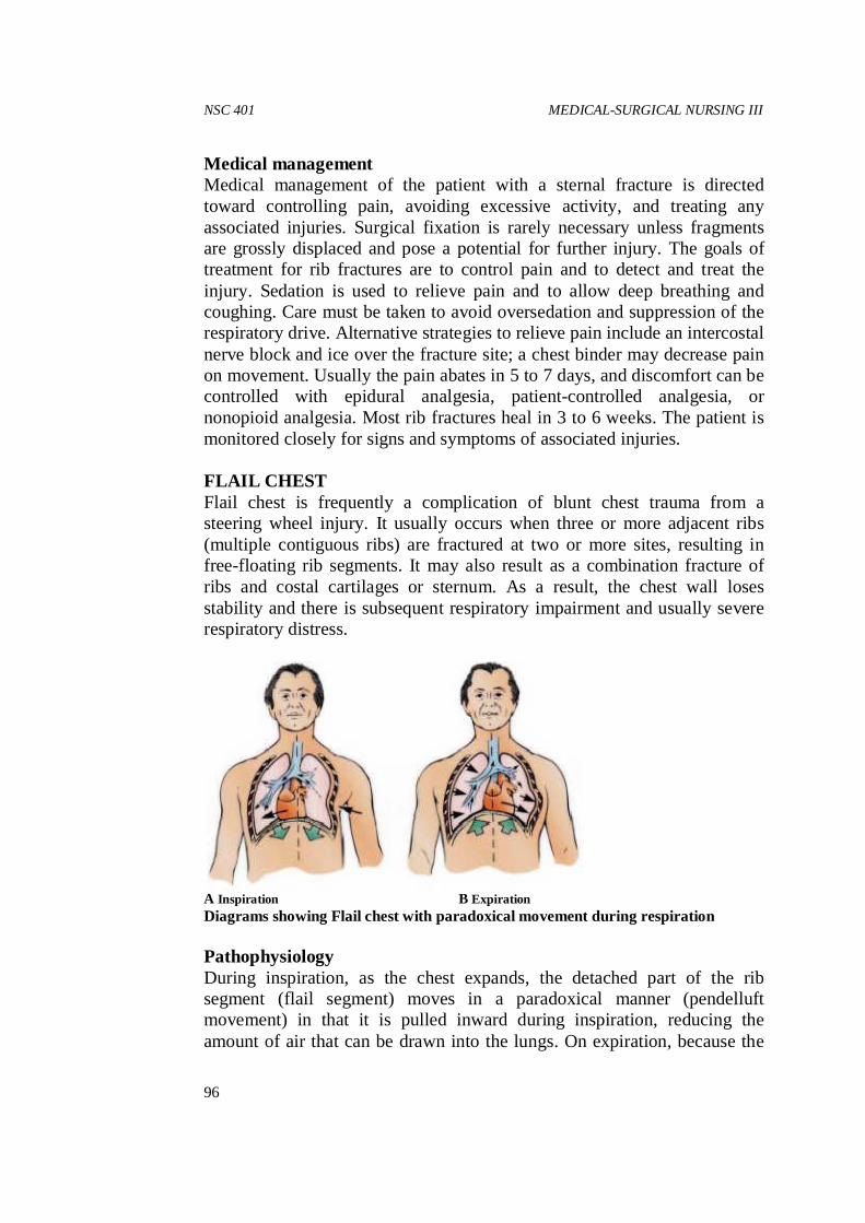
NSC 401 MEDICAL-SURGICAL NURSING III
96
Medical management Medical management of the patient with a sternal fracture is directed toward controlling pain, avoiding excessive activity, and treating any associated injuries. Surgical fixation is rarely necessary unless fragments are grossly displaced and pose a potential for further injury. The goals of treatment for rib fractures are to control pain and to detect and treat the injury. Sedation is used to relieve pain and to allow deep breathing and coughing. Care must be taken to avoid oversedation and suppression of the respiratory drive. Alternative strategies to relieve pain include an intercostal nerve block and ice over the fracture site; a chest binder may decrease pain on movement. Usually the pain abates in 5 to 7 days, and discomfort can be controlled with epidural analgesia, patient-controlled analgesia, or nonopioid analgesia. Most rib fractures heal in 3 to 6 weeks. The patient is monitored closely for signs and symptoms of associated injuries. FLAIL CHEST Flail chest is frequently a complication of blunt chest trauma from a steering wheel injury. It usually occurs when three or more adjacent ribs (multiple contiguous ribs) are fractured at two or more sites, resulting in free-floating rib segments. It may also result as a combination fracture of ribs and costal cartilages or sternum. As a result, the chest wall loses stability and there is subsequent respiratory impairment and usually severe respiratory distress.
A Inspiration B Expiration Diagrams showing Flail chest with paradoxical movement during respiration Pathophysiology During inspiration, as the chest expands, the detached part of the rib segment (flail segment) moves in a paradoxical manner (pendelluft movement) in that it is pulled inward during inspiration, reducing the amount of air that can be drawn into the lungs. On expiration, because the

NSC 401 MODULE 1
97
intrathoracic pressure exceeds atmospheric pressure, the flail segment bulges outward, impairing the patient’s ability to exhale. The mediastinum then shifts back to the affected side. This paradoxical action results in increased dead space, a reduction in alveolar ventilation, and decreased compliance. Retained airway secretions and atelectasis frequently accompany flail chest. The patient has hypoxemia, and if gas exchange is greatly compromised, respiratory acidosis develops as a result of CO2 retention. Hypotension, inadequate tissue perfusion, and metabolic acidosis often follow as the paradoxical motion of the mediastinum decreases cardiac output. Medical management As with rib fracture, treatment of flail chest is usually supportive. Management includes providing ventilatory support, clearing secretions from the lungs, and controlling pain. The specific management depends on the degree of respiratory dysfunction. If only a small segment of the chest is involved, the objectives are to clear the airway through positioning, coughing, deep breathing, and suctioning to aid in the expansion of the lung, and to relieve pain by intercostal nerve blocks, high thoracic epidural blocks, or cautious use of intravenous opioids. For mild to moderate flail chest injuries, the underlying pulmonary contusion is treated by monitoring fluid intake and appropriate fluid replacement, while at the same time relieving chest pain. Pulmonary physiotherapy focusing on lung volume expansion and secretion management techniques is performed. The patient is closely monitored for further respiratory compromise. When a severe flail chest injury is encountered, endotracheal intubation and mechanical ventilation are required to provide internal pneumatic stabilization of the flail chest and to correct abnormalities in gas exchange. This helps to treat the underlying pulmonary contusion. In rare circumstances, surgery may be required to more quickly stabilize the flail segment. This may be used in the patient who is difficult to ventilate or the high-risk patient with underlying lung disease who may be difficult to wean from mechanical ventilation. Regardless of the type of treatment, the patient is carefully monitored by serial chest x-rays, arterial blood gas analysis, pulse oximetry, and bedside pulmonary function monitoring. Pain management is key to successful treatment. Patient-controlled analgesia, intercostal nerve blocks, epidural analgesia, and intrapleural administration of opioids may be used to control thoracic pain. Pulmonary Contusion Pulmonary contusion is observed in about 20% of adult patients with multiple traumatic injuries and in a higher percentage of children due to increased compliance of the chest wall. It is defined as damage to the lung

NSC 401 MEDICAL-SURGICAL NURSING III
98
tissues resulting in hemorrhage and localized edema. It is associated with chest trauma when there is rapid compression and decompression to the chest wall (i.e. blunt trauma). It may not be evident initially on examination but will develop in the posttraumatic period. Pathophysiology The primary pathologic defect is an abnormal accumulation of fluid in the interstitial and intra-alveolar spaces. It is thought that injury to the lung parenchyma and its capillary network results in a leakage of serum protein and plasma. The leaking serum protein exerts an osmotic pressure that enhances loss of fluid from the capillaries. Blood, edema, and cellular debris (from cellular response to injury) enter the lung and accumulate in the bronchioles and alveolar surface, where they interfere with gas exchange. An increase in pulmonary vascular resistance and pulmonary artery pressure occurs. The patient has hypoxemia and carbon dioxide retention. Occasionally, a contused lung occurs on the other side of the point of body impact; this is called a contre-coup contusion. Clinical manifestations
Pulmonary contusion may be mild, moderate, or severe. The clinical manifestations vary from tachypnea, tachycardia, pleuritic chest pain, hypoxemia, and blood-tinged secretions to more severe tachypnea, tachycardia, crackles, frank bleeding, severe hypoxemia, and respiratory acidosis. Changes in sensorium, including increased agitation or combative irrational behavior, may be signs of hypoxemia. In addition, the patient with moderate pulmonary contusion has a large amount of mucus, serum, and frank blood in the tracheobronchial tree; the patient often has a constant cough but cannot clear the secretions. A patient with severe pulmonary contusion has the signs and symptoms of ARDS; these may include central cyanosis, agitation, combativeness, and productive cough with frothy, bloody secretions. Assessment and diagnostic findings The efficiency of gas exchange is determined by pulse oximetry and arterial blood gas measurements. Pulse oximetry is also used to measure oxygen saturation continuously. The chest x-ray may show pulmonary infiltration. The initial chest x-ray may show no changes; in fact, changes may not appear for 1 or 2 days after the injury. Medical management Treatment priorities include maintaining the airway, providing adequate oxygenation, and controlling pain. In mild pulmonary contusion, adequate
NSC 401 MODULE 1
99
hydration via intravenous fluids and oral intake is important to mobilize secretions. However, fluid intake must be closely monitored to avoid hypervolemia. Volume expansion techniques, postural drainage, physiotherapy including coughing, and endotracheal suctioning are used to remove the secretions. Pain is managed by intercostal nerve blocks or by opioids via patient-controlled analgesia or other methods. Usually, antimicrobial therapy is administered because the damaged lung is susceptible to infection. Supplemental oxygen is usually given by mask or cannula for 24 to 36 hours. The patient with moderate pulmonary contusion may require bronchoscopy to remove secretions; intubation and mechanical ventilation with PEEP may also be necessary to maintain the pressure and keep the lungs inflated. Diuretics may be given to reduce edema. A nasogastric tube is inserted to relieve gastrointestinal distention. The patient with severe contusion may develop respiratory failure and may require aggressive treatment with endotracheal intubation and ventilatory support, diuretics, and fluid restriction. Colloids and crystalloid solutions may be used to treat hypovolemia. Antimicrobial medications may be prescribed for the treatment of pulmonary infection. This is a common complication of pulmonary contusion (especially pneumonia in the contused segment), because the fluid and blood that extravasates into the alveolar and interstitial spaces serve as an excellent culture medium. 3.6.2 Penetrating Trauma: Gunshot and Stab Wounds Penetrating trauma occurs when a foreign object penetrates the chest wall. The most common causes of penetrating chest trauma include gunshot wounds and stabbings. Gunshot and stab wounds are the most common types of penetrating chest trauma. They are classified according to their velocity. Stab wounds are generally considered of low velocity because the weapon destroys a small area around the wound. Knives and switchblades cause most stab wounds. The appearance of the external wound may be very deceptive, because pneumothorax, hemothorax, lung contusion, and cardiac tamponade, along with severe and continuing hemorrhage, can occur from any small wound, even one caused by a small-diameter instrument such as an ice pick. Gunshot wounds to the chest may be classified as of low, medium, or high velocity. The factors that determine the velocity and resulting extent of damage include the distance from which the gun was fired, the caliber of the gun, and construction and size of the bullet. A gunshot wound can produce a variety of pathophysiologic changes. A bullet can cause damage

NSC 401 MEDICAL-SURGICAL NURSING III
100
at the site of penetration and along its pathway. It also may ricochet off bony structures and damage the chest organs and great vessels. If the diaphragm is involved in either a gunshot wound or a stab wound, injury to the chest cavity must be considered. Medical Management The objective of immediate management is to restore and maintain cardiopulmonary function. After an adequate airway is ensured and ventilation is established, the patient is examined for shock and intrathoracic and intra-abdominal injuries. The patient is undressed completely so that additional injuries will not be missed. There is a high risk for associated intra-abdominal injuries with stab wounds below the level of the fifth anterior intercostals space. Death can result from exsanguinating hemorrhage or intra abdominal sepsis. After the status of the peripheral pulses is assessed, a large-bore intravenous line is inserted. The diagnostic workup includes a chest x-ray, chemistry profile, arterial blood gas analysis, pulse oximetry, and ECG. Blood typing and cross-matching are done in case blood transfusion is required. An indwelling catheter is inserted to monitor urinary output. A nasogastric tube is inserted to prevent aspiration, minimize leakage of abdominal contents, and decompress the gastrointestinal tract. Shock is treated simultaneously with colloid solutions, crystalloids, or blood, as indicated by the patient’s condition. Chest x-rays are obtained, and other diagnostic procedures are carried out as dictated by the needs of the patient (eg, CT scans of chest or abdomen, flat plate x-ray of the abdomen, abdominal tap to check for bleeding). A chest tube is inserted into the pleural space in most patients with penetrating wounds of the chest to achieve rapid and continuing re-expansion of the lungs. The insertion of the chest tube frequently results in a complete evacuation of the blood and air. The chest tube also allows early recognition of continuing intrathoracic bleeding, which would make surgical exploration necessary. If the patient has a penetrating wound of the heart and great vessels, the esophagus, or the tracheobronchial tree, surgical intervention is required. 4.0 CONCLUSION Pleural effusion is an abnormal collection of fluid between the visceral and parietal pleurae and it may be a complication of pneumonia, lung cancer, TB, pulmonary embolism and CHF. 5.0 SUMMARY

NSC 401 MODULE 1
101
In this unit, you have learnt that:
i. Pleurisy or pleuritis refers to acute inflammation of the parietal and visceral pleurae.
ii. The term hemothorax refers to the presence of blood in the pleural space.
iii. Aspiration of stomach contents into the lungs is a serious complication that may cause pneumonia.
iv. Chest trauma is classified as either blunt or penetrating. v. Flail chest is frequently a complication of blunt chest trauma from a
steering wheel injury.
6.0 TUTOR-MARKED ASSIGNMENT Have you ever seen a patient with Flail chest before? What caused the injury and what signs and symptoms did that patient presented with? Discuss how that patient was managed. 7.0 REFERENCES/FURTHER READING Bullock, B.A., & Henze, R.L. (2000). Focus on pathophysiology.
Philadelphia: Lippincott Williams & Wilkins. Burke, K.M; Mohn-Brown, E.L & Eby, L (2011). Medical-Surgical
Nursing Care. (3rded.). Boston: Pearson Education, Inc. Nettina, S.M (2010). Lippincott Manual of Nursing Practice (9thed). China:
Wolters Kluwer Health Lippincott Williams & Wilkins. Rueling, S., & Adams, C. (2003). Close to the vest: a novel way to keep
airways clear. Nursing 2003, 33(12), 56–57. Smeltzer, S. C., Bare, B. G., Hinkle, J. L. & Cheever, K. H. (2008).
Brunner & Suddarth’s Textbook of Medical–Surgical Nursing(11th ed.). Philadelphia: Lippincott Williams &Wilkins.
Timby, B.K & Smith, N.E (2010). Introductory Medical-Surgical Nursing
(10th ed.).Philadelphia: Wolters Kluwer Health | Lippincott Williams & Wilkins.
Williams, L.S & Hopper, P.D (2003). Understanding Medical-Surgical
Nursing. Philadelphia: F. A. Davis Company.
NSC 401 MEDICAL-SURGICAL NURSING III
102
UNIT 10 CARING FOR PATIENT WITH CRITICAL RESPIRATORY CONDITIONS: RESPIRATORY FAILURE; ACUTE RESPIRATORY DISTRESS SYNDROME
CONTENTS 1.0 Introduction 2.0 Objectives 3.0 Main Content
3.1 Respiratory Failure 3.2 Acute Respiratory Distress Syndrome
4.0 Conclusion 5.0 Summary 6.0 Tutor-Marked Assignment 7.0 References/Further Reading 1.0 INTRODUCTION Respiratory failure is classified as acute or chronic. Acute respiratory failure occurs suddenly in a client who previously had normal lung function. In chronic respiratory failure, the loss of lung function is progressive, usually irreversible, and associated with chronic lung disease or other disease. 2.0 OBJECTIVES At the end of this unit you will be able to: • differentiate acute and chronic respiratory failure • discuss conditions that may lead to acute respiratory distress
syndrome.
3.0 MAIN CONTENT 3.1 Respiratory Failure Respiratory failure describes the inability to exchange sufficient amounts of oxygen and CO2 for the body’s needs. Even when the body is at rest, basic respiratory needs cannot be met. The ABG values that define respiratory failure include a PaO2 less than 50 mm Hg, a PaCO2 greater than 50 mm
NSC 401 MODULE 1
103
Hg, and a pH less than 7.25. Respiratory failure is classified as acute or chronic. Acute respiratory failure occurs suddenly in a client who previously had normal lung function. In chronic respiratory failure, the loss of lung function is progressive, usually irreversible, and associated with chronic lung disease or other disease. Pathophysiology and Etiology Acute respiratory failure is a life-threatening condition in which alveolar ventilation cannot maintain the body’s need for oxygen supply and CO2 removal. The result is a fall in arterial oxygen (hypoxemia) and a rise in arterial CO2 (hypercapnia), detected by ABG analysis. Ventilatory failure develops when the alveoli cannot adequately expand, when neurologic control of respirations is impaired, or when traumatic injury to the chest wall occurs. The most common diseases leading to chronic respiratory failure are COPD and neuromuscular disorders. The underlying disease accounts for the pathology that is seen when the respiratory system fails. Gas exchange dysfunction occurs over a long period. Symptoms of acute respiratory failure are not apparent in chronic respiratory failure because the client experiences chronic respiratory acidosis over a long period. Assessment Findings Apprehension, restlessness, fatigue, headache, dyspnea, wheezing, cyanosis, and use of the accessory muscles of respiration are seen in clients with impending respiratory failure. If the disorder remains untreated, or if treatment fails to relieve respiratory distress, confusion, tachypnea, cyanosis, cardiac dysrhythmias and tachycardia, hypotension, CHF, respiratory acidosis, and respiratory arrest occur. The client’s symptoms, history (e.g., surgery, known neurologic disorder), and ABG results form the basis for a diagnosis of respiratory failure. Additional tests include chest radiography and serum electrolyte determinations. Medical Management Treatment of respiratory failure focuses on maintaining a patent airway (in cases of upper respiratory airway obstruction) by inserting an artificial airway, such as an endotracheal or a tracheostomy tube. Additional treatments include administration of humidified oxygen by nasal cannula, Venturi mask, or rebreather masks. Respiratory failure is managed with mechanical ventilation using intermittent positive-pressure ventilation. When possible, theunderlying cause of respiratory failure is treated. Nursing Management

NSC 401 MEDICAL-SURGICAL NURSING III
104
Because symptoms often occur suddenly, recognition is important. The nurse must notify the physician immediately and obtain emergency resuscitative equipment. Assessment and monitoring of respirations and vital signs are necessary at frequent intervals. The nurse must pay particular attention to respiratory rate and depth, signs of cyanosis, other signs and symptoms of respiratory distress, and the client’s response to treatment. He or she monitors ABG results and pulse oximetry findings and implements strategies to prevent respiratory complications, such as turning and ROM exercises. The nurse provides explanations to the client and initiates measures to relieve anxiety. SELF ASSESSMENT EXERCISE 1. Differentiate acute and chronic respiratory failure 2. Discuss conditions that may lead to acute respiratory distress
syndrome. 3.2 Acute Respiratory Distress Syndrome Acute respiratory distress syndrome (ARDS), previously referred to as adult respiratory distress syndrome, is a clinical condition that occurs following other clinical conditions. The less severe form of this condition is referred to as acute lung injury (ALI). ARDS and ALI are not primary diseases. When it occurs, ARDS can lead to respiratory failure and death. It is referred to as noncardiogenic pulmonary edema (pulmonary edema not caused by a cardiac disorder; occurs without left-sided heart failure). Sudden and progressive pulmonary edema, increasing bilateral infiltrates seen on chest radiography, severe hypoxemia, and progressive loss of lung compliance characterize ARDS. Pathophysiology and Etiology Factors associated with the development of ARDS include aspiration related to near drowning or vomiting; drug ingestion overdose; hematologic disorders such as disseminated intravascular coagulation or massive transfusions. Other factors include direct damage to the lungs through prolonged smoke inhalation or other corrosive substances; localized lung infection; metabolic disorders such as pancreatitis or uremia; shock; trauma such as chest contusions, multiple fractures, or head injury; any major surgery; embolism; and septicemia. The mortality rate with ARDS is high, particularly if the underlying cause cannot be treated or is inadequately treated.

NSC 401 MODULE 1
105
The body responds to injury by reducing blood flow to the lungs, resulting in platelet clumping. The platelets release substances such as histamine, bradykinin, and serotonin, causing localized inflammation of the alveolar membranes. Increased permeability of the alveolar capillary membrane subsequently ensues. Fluid then enters the alveoli and causes pulmonary edema. The excess fluid in the alveoli and decreased blood flow through the capillaries surrounding them cause many of the alveoli to collapse (microatelectasis). Gas exchange decreases, resulting in respiratory and metabolic acidosis. ARDS also causes decreased surfactant production, which contributes to alveolar collapse. The lungs become stiff or noncompliant. Decreased functional residual capacity, severe hypoxia, and hypocapnia result. Assessment Findings Severe respiratory distress develops within 8 to 48 hours after the onset of illness or injury. In the early stages, few definite symptoms may be seen. As the condition progresses, the following signs appear: increased respiratory rate; shallow, labored respirations; cyanosis; use of accessory muscles; respiratory distress unrelieved with oxygen administration; anxiety; restlessness; and mental confusion, agitation, and drowsiness with cerebral anoxia. Diagnosis is made according to the following criteria: evidence of acute respiratory failure, bilateral infiltrates on chest radiography, and hypoxemia as evidenced by PaO2 less than 50 mm Hg with supplemental oxygen of 50% to 60%. Chest radiographs reveal increased infiltrates bilaterally. Medical Management The initial cause of ARDS must be diagnosed and treated. The client receives humidified oxygen. Insertion of an endotracheal or a tracheostomy tube ensures maintenance of a patent airway. Mechanical ventilation usually is necessary, using positive end-expiratory pressure (PEEP), which provides pressures to the airway that are higher than atmospheric pressures. Mechanical ventilators usually raise airway pressure during inspiration and let it fall to atmospheric or zero pressure during expiration (intermittent positive-pressure ventilation). When PEEP is used, positive airway pressure is maintained on inspiration, expiration, and at the end of expiration (continuous positive-pressure ventilation). The client’s pulmonary status, determined by ABG findings and pulse oximetry results, dictates the oxygen concentration and ventilator settings. Complications associated with the use of PEEP include pneumothorax and pneumo-mediastinum (air in the mediastinal space). Hypotension results in systemic hypovolemia. Although the client experiences pulmonary edema,
NSC 401 MEDICAL-SURGICAL NURSING III
106
the rest of the circulatory volume is decreased. Pulmonary artery pressure monitors the client’s fluid status and assists in determining the careful administration of IV fluids. Colloids such as albumin are used to help pull fluids in from the interstitium to the capillaries. Adequate nutritional support is essential. Usually, the first choice is enteral feedings, but total parenteral nutrition may be necessary. Nursing Management Nursing management focuses on promotion of oxygenation and ventilation and prevention of complications. Assessing and monitoring a client’s respiratory status are essential. Potential complications include deteriorating respiratory status, infection, renal failure, and cardiac complications. The client also is anxious and requires explanations and support. In addition, if the client is on a ventilator, verbal communication is impaired. The nurse provides alternative methods for the client to communicate. 4.0 CONCLUSION Acute respiratory distress syndrome (ARDS), previously referred to as adult respiratory distress syndrome, is a clinical condition that occurs following other clinical conditions. 5.0 SUMMARY In this unit, you have learnt that:
i. Respiratory failure describes the inability to exchange sufficient amounts of oxygen and CO2 for the body’s needs.
ii. Respiratory failure is classified as acute or chronic. iii. Treatment of respiratory failure focuses on maintaining.
6.0 TUTOR-MARKED ASSIGNMENT Visit any health facility that has a ventilator, take permission to see a patient being managed with it. Share your experience in the discussion forum. 7.0 REFERENCES/FURTHER READING Bullock, B.A., &Henze, R.L. (2000). Focus on pathophysiology.
Philadelphia: Lippincott Williams & Wilkins. Burke, K.M; Mohn-Brown, E.L &Eby, L (2011). Medical-Surgical Nursing
Care. (3rded.). Boston: Pearson Education, Inc. Nettina, S.M (2010). Lippincott Manual of Nursing Practice (9thed). China:
Wolters Kluwer Health Lippincott Williams & Wilkins.

NSC 401 MODULE 1
107
Rueling, S., & Adams, C. (2003). Close to the vest: a novel way to keep airways clear. Nursing 2003, 33(12), 56–57.
UNIT 11 CARING FOR PATIENT WITH CHEST CANCER:
LUNG CANCER; TUMOR OF THE MEDIASTINUM
CONTENTS 1.0 Introduction 2.0 Objectives 3.0 Main Content 4.0 Conclusion 5.0 Summary 6.0 Tutor-Marked Assignment 7.0 References/Further Reading 1.0 INTRODUCTION Lung cancer is a very common cancer, particularly among cigarette smokers and those regularly exposed to secondhand smoke. It remains the number one cause of cancer related deaths among men and women in the world. 2.0 OBJECTIVES At the end of this unit, you will be able to: • describe the pathophysiology and management of lung cancer • explain the difficulties associated with early diagnosis of lung cancer • list the clinical manifestations of tumors of the mediastinum • list surgical management of tumors of the mediastinum.
3.0 MAIN CONTENT 3.1 Lung Cancer Lung cancer is a very common cancer, particularly among cigarette smokers and those regularly exposed to secondhand smoke. It remains the number one cause of cancer related deaths among men and women in the United States. The incidence of lung cancer has markedly increased since the early 1980s, related to:
NSC 401 MEDICAL-SURGICAL NURSING III
108
• More accurate methods of diagnosis • The growing population of aging people • The continued popularity of cigarette smoking • Increased air pollution • Increased exposure to industrial pollutants. Lung cancer is more common in men than in women. The rate of women dying from lung cancer continues to increase, however, and indeed is greater than the rate of women dying from breast cancer. Most clients are older than 40 years of age when diagnosed with lung cancer. Pathophysiology and Etiology The exact mechanism for the development of lung cancer is unknown; however, the link between irritants and lung cancer is well established. Prolonged exposure to carcinogens more than likely will produce cancerous cells. Smokers who quit reduce their risk of lung cancer to that of nonsmokers within 10 to 15 years. Lung cancers are grouped in two overall categories: non-small cell carcinomas, which includes epidermoid or squamous cell carcinomas, large cell or undifferentiated type, and adenocarcinoma; and small cell carcinoma, also referred to as oat cell carcinoma. Many tumors begin in the bronchus and spread to the lung tissue, regional lymph nodes, and other sites, such as the brain and bone. Many tumors have more than one type of cancer cell. The transformation of an epithelial cell in the airway initiates the growth of a lung cancer lesion. As the tumor grows, it partially obstructs the lumen of an airway or completely obstructs it, resulting in airway collapse distal to the tumor. The tumor may hemorrhage, causing hemoptysis. Signs and Symptoms The cell type of the lung cancer, size and location of the tumor, and degree and location of metastasis determine the presenting signs and symptoms. A cough productive of mucopurulent or blood-streaked sputum is a cardinal sign of lung cancer. The cough may be slight at first and attributed to smoking or other causes. As the disease advances, the client may report fatigue, anorexia, and weight loss. Dyspnea and chest pain occur late in the disease. Hemoptysis is common. If pleural effusion occurs from tumor spread to the outside portion of the lungs, the client experiences dyspnea and chest pain. Other indications of tumor spread are symptoms related to pressure on nerves and blood vessels. Symptoms include head and neck edema, pericardial effusion, hoarseness, and vocal cord paralysis. Diagnostic Findings

NSC 401 MODULE 1
109
Early diagnosis of cancer of the lung is difficult because symptoms often do not appear until the disease is well established. The sputum is examined for malignant cells. Chest films may or may not show a tumor. A CT or PET scan or MRI is done if results from the chest radiograph are inconclusive, or to further delineate the tumor area. Bronchoscopy may be done to obtain bronchial washings and a tissue sample for biopsy. Fine-needle aspiration under fluoroscopy or CT guidance may be done to aspirate cells from a specific area that is not accessible by bronchoscopy. A lung scan also may locate the tumor. A bone scan detects metastasis to the bone. The results of a lymph node biopsy may be positive for malignant changes if the lung tumor has metastasized. Mediastinoscopy provides a direct view of the mediastinal area and possible visualization of tumors that extend into the mediastinal space. Medical and Surgical Management The client’s prognosis is poor unless the tumor is discovered in its early stages and treatment begins immediately. Because lung cancer produces few early symptoms, its mortality rate is high. Metastasis to the mediastinal and cervical lymph nodes, liver, brain, spinal cord, bone, and opposite lung is common. Treatment depends on several factors. One major consideration is the classification and staging of the tumor. After classification of the tumor, the stage of the disease is determined. Staging refers to the extent and location of the tumor and the absence or presence and extent of metastasis. Other factors that determine treatment are the client’s age and physical condition and other diseases or disorders, such as renal disease and CHF. Surgical removal of the tumor offers the only possibility of cure and usually is successful only in the early stages of the disease. The type of lung resection depends on the tumor’s size and location. Radiation therapy may help to slow the spread of the disease and provide symptomatic relief by reducing tumor size, thus easing the pressure exerted by the tumor on adjacent structures. In turn, pain, cough, dyspnea, and hemoptysis may be relieved. In a small percentage of cases, radiation may be curative, but for most, it is palliative. Complications associated with the use of radiation therapy include esophagitis, fibrosis of lung tissue, and pneumonitis. Chemotherapy may be used alone or with radiation therapy and surgery. The principal effects of chemotherapy are to slow tumor growth and reduce tumor size and accompanying pressure on adjacent structures. Chemotherapy also is used to treat metastatic lesions. Most chemotherapeutic regimens use a combination of drugs rather than a single agent and, although not curative, often make the client more comfortable. New treatments in various stages of development include the following:

NSC 401 MEDICAL-SURGICAL NURSING III
110
• New chemotherapy regimens • Monoclonal antibodies that target specific cancer proteins • Photodynamic therapy that is a combination treatment with
chemicals and light • Lung cancer vaccines to stimulate an effective immune response Nursing Management Management of clients with lung cancer is essentially the same as that for any client with a malignant disease. 3.2 Tumors of the Mediastinum Tumors of the mediastinum include neurogenic tumors, tumors of the thymus, lymphomas, germ cell, cysts, and mesenchymal tumors. These tumors may be malignant or benign. These tumors are usually described in relation to location: anterior, middle, or posterior masses or tumors. Clinical Manifestations Nearly all the symptoms of mediastinal tumors result from the pressure of the mass against important intrathoracic organs. Symptoms may include: - cough - wheezing - dyspnea - anterior chest or neck pain - bulging of the chest wall - heart palpitations - angina and other circulatory disturbances, central cyanosis, superior
vena caval syndrome (i.e., swelling of the face, neck, and upper extremities)
- marked distention of the veins of the neck and the chest wall (evidence of the obstruction of large veins of the mediastinum by extravascular compression or intravascular invasion)
- dysphasia - weight loss from pressure or invasion into the esophagus. Assessment and Diagnostic Findings Chest x-rays are the major method used initially to diagnose mediastinal tumors and cysts. CT scans are the gold standard for assessment of the mediastinum and surrounding structures. MRI may be used in some circumstances, as well as PET scans.

NSC 401 MODULE 1
111

NSC 401 MEDICAL-SURGICAL NURSING III
112
Medical Management If the tumor is malignant and has infiltrated surrounding tissue, radiation therapy and/or chemotherapy are the therapeutic modalities used when complete surgical removal is not feasible. Surgical management Many mediastinal tumors are benign and operable. The location of the tumor (anterior, middle, or posterior compartments) in the mediastinum dictates the type of incision. The common incision used is a median sternotomy; however, a thoracotomy may be used, depending on the location of the tumor. Additional approaches may include a bilateral anterior thoracotomy (clamshell incision) or video-assisted thoracoscopic. The care is the same as for any patient undergoing thoracic surgery. The major complications include hemorrhage, injury to the phrenic or recurrent laryngeal nerve, and infection. SELF-ASSESSMENT EXERCISE 1. Describe the pathophysiology and management of Lung cancer 2. Explain the difficulties associated with early diagnosis of lung
cancer 3. List the clinical manifestations of tumors of the mediastinum 4. List surgical management of tumors of the mediastinum.
4.0 CONCLUSION Lung cancer is more common in men than in women. The rate of women dying from lung cancer continues to increase, however, and indeed is greater than the rate of women dying from breast cancer. 5.0 SUMMARY In this unit, you have learnt that:
i. Lung cancer is a very common cancer, particularly among cigarette smokers and those regularly exposed to secondhand smoke.
ii. Early diagnosis of cancer of the lung is difficult because symptoms often do not appear until the disease is well established.
iii. Tumors of the mediastinum include neurogenic tumors, tumors of the thymus, lymphomas, germ cell, cysts, and mesenchymal tumors.
NSC 401 MODULE 1
113
iv. Mediastinal tumors may be malignant or benign. v. Chest x-rays are the major method used initially to diagnose
mediastinal tumors and cysts. 6.0 TUTOR-MARKED ASSIGNMENT Your 65-year-old neighbour tells you that he smoked for 25 years but quit 5 years ago. He has been experiencing a productive cough for 3 weeks. Think through your counsel to him and type your counsel out. Submit this to your facilitator. 7.0 REFERENCES/FURTHER READING Bullock, B.A., &Henze, R.L. (2000). Focus on pathophysiology.
Philadelphia: Lippincott Williams & Wilkins. Burke, K.M; Mohn-Brown, E.L &Eby, L (2011). Medical-Surgical Nursing
Care. (3rd ed.). Boston: Pearson Education, Inc. Nettina, S.M (2010). Lippincott Manual of Nursing Practice (9thed). China:
Wolters Kluwer Health Lippincott Williams & Wilkins. Rueling, S., & Adams, C. (2003). Close to the vest: a novel way to keep
airways clear. Nursing 2003, 33(12), 56–57. Smeltzer, S. C., Bare, B. G., Hinkle, J. L. & Cheever, K. H. (2008).
Brunner & Suddarth’s Textbook of Medical–Surgical Nursing(11th ed.). Philadelphia: Lippincott Williams &Wilkins.
Timby, B.K & Smith, N.E (2010). Introductory Medical-Surgical Nursing
(10th ed.).Philadelphia: Wolters Kluwer Health Lippincott Williams & Wilkins.
Williams, L.S & Hopper, P.D (2003). Understanding Medical-Surgical
Nursing. Philadelphia: F. A. Davis Company.

NSC 401 MODULE 2
113
MODULE 2 CARING FOR PATIENTS WITH INTEGUMENTARY SYSTEM DISORDERS – TEAM LEADER
Unit 1 Assessment and Diagnostic Evaluation of Disorder of the
Integumentary System Unit 2 Review of Related Anatomy Unit 3 Caring for Patient with Common Skin Disorder Unit 4 Caring for Patient with Infections and Infestations of the Skin Unit 5 Caring for Patient with Pressure Ulcers UNIT 1 ASSESSMENT AND DIAGNOSTIC EVALUATION
OF DISORDER OF THE INTEGUMENTARY SYSTEM
CONTENTS 1.0 Introduction 2.0 Objectives 3.0 Main Content
3.1 Assessment of the Integumentary System 3.1.1 Subjective Assessment of the Integumentary System 3.1.2 Objective Assessment of the Integumentary System 3.1.3 Assessment of Common Skin Variations 3.1.4 Assessment of Skin Lesions 3.1.5 Assessment of the Vascular Status of Skin 3.1.6 Assessment of the Hydration Status of Skin 3.1.7 Assessment of the Hair 3.1.8 Diagnostic Evaluation of the Integumentry System
4.0 Conclusion 5.0 Summary 6.0 Tutor-Marked Assignment 1.0 INTRODUCTION The integument system is composed of the skin and its appendages (hair and nails). The system covers almost the entire part of the body where it forms an amazing first-line defense system which can stop entry of any form of injurious objects into the body. It also functions in maintaining major physiological homeostasis. The system possesses a great deal of
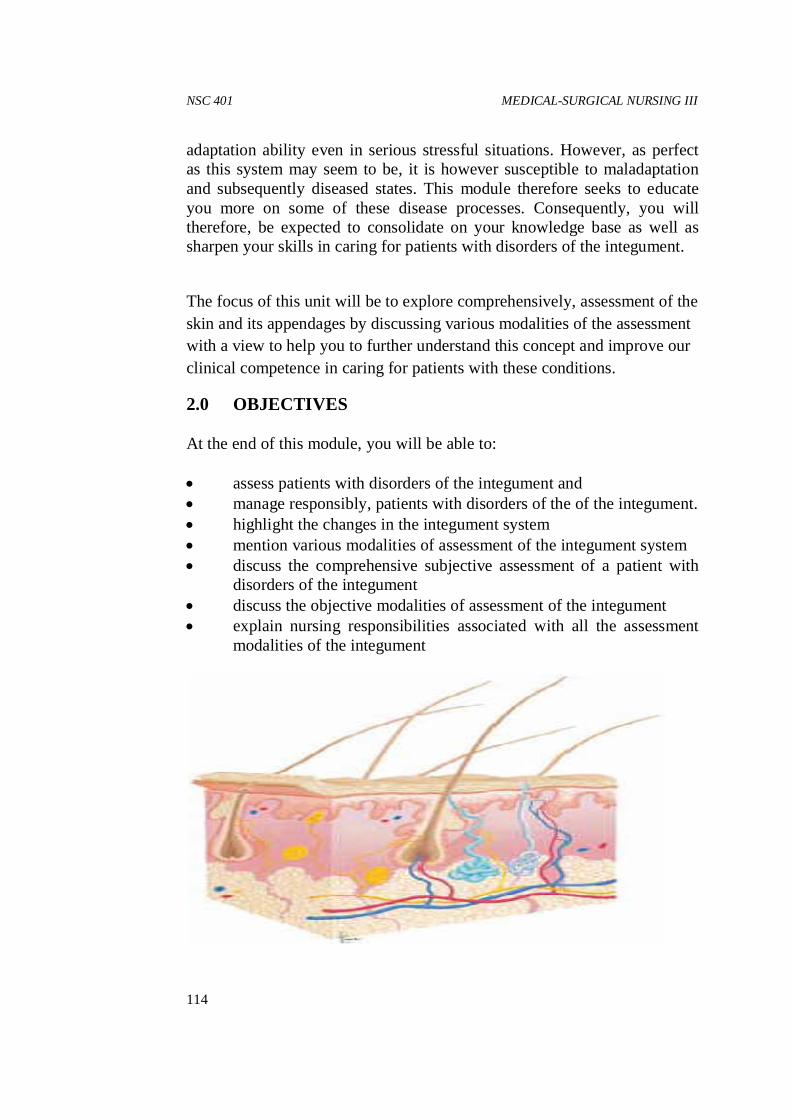
NSC 401 MEDICAL-SURGICAL NURSING III
114
adaptation ability even in serious stressful situations. However, as perfect as this system may seem to be, it is however susceptible to maladaptation and subsequently diseased states. This module therefore seeks to educate you more on some of these disease processes. Consequently, you will therefore, be expected to consolidate on your knowledge base as well as sharpen your skills in caring for patients with disorders of the integument.
The focus of this unit will be to explore comprehensively, assessment of the skin and its appendages by discussing various modalities of the assessment with a view to help you to further understand this concept and improve our clinical competence in caring for patients with these conditions.
2.0 OBJECTIVES At the end of this module, you will be able to: • assess patients with disorders of the integument and • manage responsibly, patients with disorders of the of the integument. • highlight the changes in the integument system • mention various modalities of assessment of the integument system • discuss the comprehensive subjective assessment of a patient with
disorders of the integument • discuss the objective modalities of assessment of the integument • explain nursing responsibilities associated with all the assessment
modalities of the integument

NSC 401 MODULE 2
115
3.0 MAIN CONTENT 3.1 Assessment of the Integumentary System
3.1.1 Subjective Assessment of the Integumentary System 3.1.2 Objective Assessment of the Integumentary System 3.1.3 Assessment of Common Skin Variations 3.1.4 Assessment of Skin Lesions 3.1.5 Assessment of the Vascular Status of Skin 3.1.6 Assessment of the Hydration Status of Skin 3.1.7 Assessment of the Nails 3.1.8 Assessment of the Hair 3.1.9 Diagnostic Evaluation of the Integumentry System
3.1.1 Subjective Assessment of the Integumentary System Caring for patients with dermatologic disorders will require the nurse to obtain important information and direct observations i.e. observing and noting deviations from normal. Health history interview include; • Family and personal history of;
o Skin allergies o Food allergies o Allergies to medications or chemicals o Previous skin problems and o Skin cancer
• Also the role of personal hygiene products such as cosmetics, soaps and shampoos in the incidence of recent skin problems
• Health history: o Onset, signs and symptoms of recent disorders o The location/ part affected o Duration of any disturbing symptoms like pain, itching or
rash 3.1.2 Objective Assessment of the Integumentary System This should involve the entire skin, mucous membranes and skin appendages such as scalp, hair, and nails. Alterations are common features of other systemic disease.

NSC 401 MEDICAL-SURGICAL NURSING III
116
Physical assessment includes inspection and palpation techniques. • Preparation and requirements include;
o A warm and well lited room o A penlight (to highlight lesions)
• Procedure: o Adequately drape the patient o Don gloves (if rash or lesions are present on the skin).
• Inspection; Observe the following characteristics; o Color; varies from person to person based on degree of
pigmentation. � Pink or reddish hue skin often follows vasodilation due
to fever, sunburn and inflammation � Pallor presents an absence of or decreased normal skin
color and vascularity; best observed in the conjunctivae or oral mucosa amongst dark skinned individuals
� Bluish hue color is due to cyanosis indicating cellular hypoxia; easily observed in the extremities, nail beds, lips and mucous membranes.
� Jaundice is a yellowish discoloration of the skin due to elevated serum bilirubin; more easily observable in the sclerae and mucous membranes
� Petechiae describes a pinpoint red spots on the skin due to blood leakage into the skin; usually appear as a grayish cast on dark skinned individuals o Temperature o Moisture or dryness o Texture (rough or smooth) o Lesions o Vascularity o The condition of the hair and nails.
• Palpation; check out for the following; o Skin turgor o Edema and o Elasticity
3.1.3 Assessment of Common Skin Variations Erythema

NSC 401 MODULE 2
117
• This denotes redness of the skin • It often is caused by the congestion of capillaries. • It is more observable at its location in light-skinned people • It complements increased warmth and edema and induration (due to
intracellular infiltration) in cases of inflammation. Rash This refers to skin lesions, which presents with; • Induration (local skin infiltrates); varies in sizes • Macule and papule are small indurations • Vesicles are skin lesions with clear fluid infiltrates • Pustle are small, suppurative skin lesions • Erythema Cyanosis • Cyanosis is the bluish discoloration of the skin and mucus
membranes • It is primarily due to hypoxia and/or hypoxemia • It reflects respiratory and/or circulatory compromise. • It may be a;
o Central cyanosis; affecting the torso and by far more severe, often due to respiratory and central circulatory failure
o Peripheral cyanosis; is evident on the fingertips and nail beds and often indicate peripheral circulatory failure; other associated signs include cold, clammy skin, thready pulse and rapid, shallow respirations.
Colour Changes • Hypopigmentation: decreased or loss of skin pigmentation due to
low melanin secretion; may follow fungal infection, eczema • Vitiligo , condition characterized by destruction of the melanin
secreting cells in a well-defined area of the skin, resulting in white patches.
• Hyperpigmentation, increased melanin secretion. Causes include post inflammatory conditions (e.g. a disease or injury); Sun burn injury; senility or in some cases, genetic predilection.
3.1.4 Assessment of Skin Lesions Skin lesions may vary in size, shape, and causes.
NSC 401 MEDICAL-SURGICAL NURSING III
118
Classification of skin lesions Primary lesions: These describe initial lesions that are not associated with any form of underlying disease conditions. They are skin lesions that originally arise from previously normal skin. Various forms of primary skin lesions include; • Macule:Flat, non-palpable skin lesion appearing only as localized
colored spot with a well-defined border: it usually is less than 1cm in diameter.
• A Patch: is a macule larger than 1cm in diameter. Color can vary from brown, white, tan, purple or red. Examples include Freckles, Flat moles, Petechia, Rubella, Vitiligo, Port Wine Stains or Ecchymosis
• Papule: an elevated, palpable, solid mass with a well-defined circumscribed border, often with diameter less than 0.5cm e.g. elevated nevi, warts, lichen planus
• Plaque:may be a coalesced papules with flat top andmore than 0.5cm in diameter e.g. Psoriasis, actinic keratosis
• Nodule: an elevated, palpable, solid mass involving more of the dermal structures, with a well-defined borders: it measures about 0.5-2cm e.g. Lipoma, squamous cell carcinoma, poorly absorbed injection, dermatofibroma
• Tumor: describes an elevated, palpable, solid mass rooted in the dermal structures: a tumor measures 1-2cm in diameter always without a sharp, well demarcated borders e.g. Larger lipoma, carcinoma
• Vesicle:Circumscribed, elevated, palpable mass containing serous fluid: usually less than 0.5cm in diameter e.g. lesions of Herpes simplex/zoster, chickenpox, poison ivy, blister of a second-degree burn
• Bulla: are vesicles more than 0.5cm wide e.g. are lesions of Pemphigus, contact dermatitis, large burn blisters, poison ivy, bullous impetigo
• Wheals: are elevated mass without permanent borders but often irregular and varying architectures (size and color). It is due to dermal transudation i.e. serous fluid infiltration of the dermis. This lesion does not contain free fluid in a cavity when compared with a vesicle. Examples include Urticaria (hives), insect bites
NSC 401 MODULE 2
119
• Pustule: is a pus filled vesicle or bulla e.g. Acne, impetigo, furuncles, carbuncles
• Cyst: is an encapsulated fluid-filled mass with varying consistency usually in the subcutaneous tissue or dermis e.g. Sebaceous cyst, epidermoid cysts.
Secondary lesions: These describe those lesions that are due to underlying diseases or those that surface due to changes in or worsening of a primary lesion usually due to external causes (such as scratching, trauma, infections) or changes caused by wound healing process. Examples include: • Erosion: presents with loss of superficial epidermis characterized by
a depressed, moist skin area. Usually the dermis is spared e.g. following ruptured vesicles, scratch marks
• Ulcer: is an area of skin loss affecting the whole epidemis and dermal structures with characteristic necrotic tissue loss; bleeding and possible scarring e.g. Stasis ulcer of venous insufficiency, pressure ulcer
• Fissure: is a linear crack in the skin which may extend to the dermis e.g. Chapped lips or hands, athlete’s foot
• Scales:Flakes secondary to desquamated, dead epithelium, which may adhere to skin surface. It presents varying color (silvery, white) and texture varies (thick, fine). Example include dandruff, psoriasis, dry skin, pityriasisrosea
• Crust:Dried residue of serum, blood, or pus on skin surface presenting as large, adherent crust (a scab). Example include residue vesicular rupture: impetigo, herpes, eczema
• Scar (Cicatrix): is a skin mark following healing of a wound/lesion, due to deposition of fibrilla connective tissue. Young scars appears red or purple while a mature scar is white or glistening: Example include healed wound or surgical incision
• Keloid: a hypertrophied (exaggerated) scar tissue formation due to excessive formation and deposition of fibrilla collagen during healing. It presents as elevated, irregular scar. It is a more common phenomenon among dark skinned people
• Atrophic leisions: it follows loss of or inadequate production of collagen and elastin tissue to replace tissue loss thus presenting as thin, dry, transparent epidermal appearance with characteristic loss of surface markings and visible underlying vessels. It may follow selinity ageing, or arterial insufficiency

NSC 401 MEDICAL-SURGICAL NURSING III
120
• Lichenification: presents secondary thickening of the skin with prominent skin markings often due to repeated rubbing, irritation, scratching as with contact dermatitis
Quick assessment of a skin lesion: Quick assessment should include; • A careful observation of the lesions characteristics and
documentation of the following; o Distribution of the lesion (e.g., bilateral, symmetric, linear,
circular); to elicit its pattern and shapes as characterized of certain diseases
o The extent of the distribution whether regional or global distribution
o The color of the lesions o The shape of lesions o Pattern of eruption (e.g., macular, papular, scaling, oozing,
discrete, confluent) o Presence of acute open wounds or lesions
• Palpation the lesions to determine; o Any redness, heat, pain, or swelling o Size: measure metric ruler to determine any further extension o Shape and border and o Consistencies and o Mobility i.e. fixed or fluctuant
• Document findings in the patient’s health record into details; Further assessment should include: • Wound bed: for necrotic and granulation tissue, epithelium, exudate,
color, and odor. • Wound edges and margins: for undermining (i.e., extension of the
wound under the surface skin), and evaluate for condition. • Wound size (in millimeters or centimeters) to determine diameter
and depth of the wound and surrounding erythema. • Surrounding skin: Assess for color, suppleness and moisture,
irritation, and scaling. 3.1.5 Assessment of the Vascular Status of Skin A description of vascular changes includes location, distribution, color, size, and the presence of pulsations.
NSC 401 MODULE 2
121
Common vascular changes include; • Petechia: small (1-2mm), round red or purple macule, secondary to
blood extravasation. It is associated with bleeding tendencies or emboli to skin
• Ecchymosis: Round or irregular macular lesion, larger than petechia. Its color varies and changes. It is secondary to blood extravasation and is associated with trauma, bleeding tendencies
• Cherry Angioma: Papular and round, red or purple and noted on trunk, extremities. It may blanch with pressure. It is a normal age-related skin alteration and usually not clinically significant
• Spider Angioma: Red, arteriole lesion with central body and radiating branches. It is notably evident on face, neck, arms, trunk and rare below the waist. The lesion may blanch with pressure. It is associated with liver disease, pregnancy, vitamin B deficiency
• Telangiectasia (Venous Star): Spider-like or linear shaped varies, bluish or red in color which does not blanch with pressure. It is commonly noted on legs, anterior chest and is secondary to superficial dilation of venous vessels and capillaries. Its formation follows increased venous pressure states (varicosities)
3.1.6 Assessment of the Hydration Status of Skin Skin moisture, temperature, and texture are assessed primarily by palpation. The elasticity (i.e., turgor) of the skin, which decreases in normal aging, may be a factor in assessing the hydration status of a patient. Assessment of the Nails Inspection includes observation of configuration, color, and consistency. Many alterations reflect local or systemic abnormalities • Transverse depressions known as Beau’s lines may reflect retarded
growth of the nail matrix due to severe illness or, more commonly, local trauma.
• Ridging, hypertrophy may also follow local trauma. • Paronychia, an inflammation of the skin around the nail, is usually
accompanied by tenderness and erythema. • Clubbing presents as extension of the normal angle between the nail
and its bed (usually 180 degrees or greater) and softening of the nail base (feels sponge-like on palpation). The angle between the normal nail and its base is 160 degrees and the nail base is usually firm on palpation.
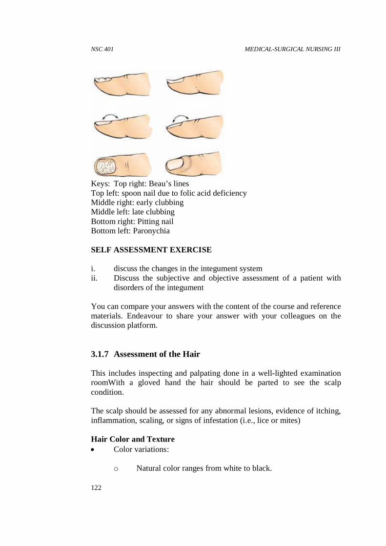
NSC 401 MEDICAL-SURGICAL NURSING III
122
Keys: Top right: Beau’s lines Top left: spoon nail due to folic acid deficiency Middle right: early clubbing Middle left: late clubbing Bottom right: Pitting nail Bottom left: Paronychia SELF ASSESSMENT EXERCISE i. discuss the changes in the integument system ii. Discuss the subjective and objective assessment of a patient with
disorders of the integument You can compare your answers with the content of the course and reference materials. Endeavour to share your answer with your colleagues on the discussion platform. 3.1.7 Assessment of the Hair This includes inspecting and palpating done in a well-lighted examination roomWith a gloved hand the hair should be parted to see the scalp condition. The scalp should be assessed for any abnormal lesions, evidence of itching, inflammation, scaling, or signs of infestation (i.e., lice or mites) Hair Color and Texture • Color variations:
o Natural color ranges from white to black.
NSC 401 MODULE 2
123
o Gray hair is associated with ageing; often first noticed as early as the third decade in some individuals. It is due to loss of melanocytes. However, grey hair also follows hereditary traits.
o Albinism (i.e., partial or complete absence of pigmentation) presents with white hair from birth.
Hair texture variations:
o Dry, brittle hair may result from overuse of hair dyes, hair dryers, and curling irons or from endocrine disorders, such as thyroid dysfunction.
o Oily hair is usually caused by increased secretion from the sebaceous glands close to the scalp.
Hair Distribution Physiologic variations: Body hair texture and distribution varies with the following factors; • Body location:
o Hair over most of the body is fine o Hair in the axillae and pubic areas is coarse. o Pubic hair develops at puberty as a secondary sexual
characteristic. Pubic hair in males is diamond-shaped extending up to the umbilicus while it is an inverted triangle in females. Variation of this pattern of distribution especially a characteristic of the opposite gender may signal an endocrine problem and that will require a further investigation. Male pattern hair distribution may be seen in some women after menopause due to estrogen suppression. • Racial differences: such as straight hair in Asians and curly, coarser
hair in people of African descent. • Gender: Men tend to have more body and facial hair than women. Pathophysiologic variations: • Hair Loss or alopecia; this can be focal or global
Scalp hair loss is the most common noticeable pattern and may range from localized, patchy areas to generalized baldness.
Some of the possible causes include:
NSC 401 MEDICAL-SURGICAL NURSING III
124
o Patchy hair loss has been thought to be due to habitual hair pulling or twisting from excessive;
o Traction on the hair (e.g., braiding too tightly); o Use of dyes, straighteners, and oils; o Chemotherapeutic agents (e.g., doxorubicin, cyclophosphamide); o Fungal infection; o Moles or lesions on the scalp Male pattern baldness: affects more than one half of the male population is believed to be related to;
o Heredity o Aging and o Androgen (male hormone) levels
• Hirsutism; Excessive hair on the face, chest, shoulders, and pubic area, in pre-menopausal women may indicate hormonal abnormalities particularly of the pituitary or adrenal origin.
3.1.8 Diagnostic Evaluation of the Integumentry System Diagnostic procedures that may identify skin conditions include: Skin Biopsy This procedure obtain tissue from nodules, plaques, blisters, and other lesions, for microscopic examination to exclude or establish diagnosis such as malignancyBiopsy may be by scalpel excision or a skin punch instrument. Skin Scrapings Tissue samples from suspected fungal lesions are scrapped with a scalpel blade moistened with oil to prevent tissue adherence to the blade. The scraped material can then be microscopically examined on a glass slide, covered with a slip Tzanck Smear Tzanck smear aim to examine cells from blistering skin leisions, such as herpes zoster, varicella, herpes simplex, and pemphigus: it involves collecting secretions from a lesion, smear it on a glass slide, stain the sample and examined under a microscope. Immunofluorescence Aimed at identifying the site of an immune reaction (inflammation)

NSC 401 MODULE 2
125
Immunofluorescence testing combines an antigen or antibody with a fluorochrome dye. Direct tests on skin detect auto-antibodies (i.e. antibodies directed against portions of the skin). Indirect immunofluorescence test detects specific antibodies in the serum. Patch Testing
Purpose is to identify allergens It involves applying the suspected allergens to normal skin under occlusive patches. Positive tests can be grouped thus; • Weak positive reaction; development of redness, induration
(swelling), or itching • Moderately positive reaction; fine blisters, papules and severe
itching • Strong positive reaction; blisters, pain and ulceration Wood’s Light Examination Wood’s lightis a special lamp that produces long-wave ultraviolet (UV) light rays in a characteristic dark purple fluorescence. Skin examination under this instrument can differentiate epidermal from dermal lesions and spot abnormal pigmented areas (hypo-pigmented or hyper-pigmented lesions) from normal skin. On examination, hyper-pigmented lesions will almost disappear, whereas hypo-pigmented lesions increase in paleness (whiteness). Clinical Photographs Photographs are taken to document; • The nature and extent of the skin condition and • Progress or improvement resulting from treatment. 4.0 CONCLUSION Caring for patients with dermatologic disorders will require the nurse to obtain important information and direct observations i.e. observing and noting deviations from normal.

NSC 401 MEDICAL-SURGICAL NURSING III
126
5.0 SUMMARY In this unit, you are expected to have learnt about:
i. The changes in the integument system. ii. Various modalities of assessment of the integument system. iii. Comprehensive subjective assessment of a patient with disorders of
the integument. iv. Objective modalities of assessment of the integument. v. Nursing responsibilities associated with all the assessment
modalities of the integument.
6.0 TUTOR-MARKED ASSIGNMENT Now that you have learnt this much, visit a hospital of choice within your vicinity and perform a comprehensive assessment of a named patient visiting the dermatologic clinic. Share your answers with your colleague in the discussion forum.

NSC 401 MODULE 2
127
UNIT 2 CARING FOR PATIENT WITH COMMON SKIN DISORDER
CONTENTS 1.0 Introduction 2.0 Objectives
3.0 Main Content 3.1 Pruritic Disorders
3.1.1 General Itching 3.1.2 Perineal and Perianal Itching 3.1.3 Secretory Disorders of the Skin
3.1.3.1 Hydradenitis Suppurativa 3.1.4 Seborrheic Dermatoses 3.1.5 Acne Vulgaris
4.0 Conclusion 5.0 Summary 6.0 Tutor-Marked Assignment 1.0 INTRODUCTION The focus of this unit is on exploring various disorders of the integument, with a view to improve your knowledge base and sharpen your clinical competence in caring for patients with these conditions. 2.0 OBJECTIVES At the end of this unit, you will be able to: • Discuss the types pruiritic disorders of skin • Discuss types of secretory disorders of the skin • Discuss the management of pruiritus • Highlight the management of acne vulgaris 3.0 MAIN CONTENT 3.1 Pruritic Disorders
3.1.1 General Itching 3.1.2 Perineal and Perianal Itching 3.1.3 Secretory Disorders of the Skin
Hydradenitis Suppurativa 3.1.4 Seborrheic Dermatoses 3.1.5 Acne Vulgaris

NSC 401 MEDICAL-SURGICAL NURSING III
128
3.1.1 General Itching Pruritus is one of the most common dermatologic complaints. Pruritus usually is more severe at night and is less frequently reported during waking hours, probably because the person is distracted by daily activities. Classifications: Primary (Essential) pruiritus: • It is caused by a primary skin disease • It may be associated with or without resultant rash or lesions. • Generally rapid in onset, and may be severe enough to interfere with
normal daily activities. Secondary Pruiritus: causes include; • Systemic disease such as diabetes mellitus; blood disorders; cancers;
renal, hepatic and thyroid diseases • Medications such as aspirin; antibiotics; hormones (i.e., estrogens,
testosterone, or oral contraceptives) and opioids (i.e., morphine or cocaine)
• Certain soaps and chemicals • Radiation therapy • Temperature extreme; Prickly heat (i.e., miliaria) and • Contact with woolen garments • Psychological factors, such as excessive stress in family or work
situations Pathophysiology Local irritation (scratch) stimulates the itch receptors on the skin and subcutaneous layers and the leads to release of histamine by neighbouring histaminegic cells. The usual response of the individual is scratching. Scratching will further produce more pruritus thus constituting a vicious itch–scratch cycle. Scratching may impair skin integrity by inducing excoriation, redness, raised areas (i.e., wheals), and subsequent infection with pigmentation changes may result. Gerontologic Considerations Pruritus occurs frequently among the elderly often because of; • Dry skin • Underlying systemic illness or even occult malignancy • Multiple medications

NSC 401 MODULE 2
129
All of these factors increase the incidence of pruritus. Medical Management • A thorough history to include recent medication, change of
cosmetics or soaps • Physical examination to identify the;
o Underlying environmental triggers such as warm, dry air or irritating bed linens
o Secondary causes of the pruritus, such as features of allergy o Identify signs of infection
Treatment should focus on relieving the condition and treating triggering conditions General measures include: • Avoid washing with soap and hot water • Use bath oils containing a surfactant • A warm bath with a mild soap • Application of a bland emollient to control xerosis (i.e., dry skin) • Cold compress, ice cube, or mentholated cool agents may also help
relieve pruritus by inducing vaso-constrict Pharmacologic Therapy • Topical corticosteroids as anti-inflammatory agent to decrease
itching • Oral antihistamines: sedative antihistamines such as
diphenhydramine (Benadryl) or hydroxyzine (Atarax) is best used at bed time to produce a restful and comfortable sleep. Non-sedating antihistamines such as fexofenadine (Allegra) should be used to relieve daytime pruritus.
• Tricyclic antidepressants; such as doxepin (Sinequan), may be used for neuropsychogenic pruritus.
• Non- resolving pruiritus will require further investigation for a systemic problem
Nursing Management The patient will benefit from the following; • Tepid (not hot) water bath • Shake off excess water and blot between intertriginous areas (i.e.,
body folds) with a towel. • Avoiding vigorous rubbing of skin with towel

NSC 401 MEDICAL-SURGICAL NURSING III
130
• Skin lubrication with an emollient immediately after bathing to trap moisture
• Avoiding situations that cause vasodilation such as exposure to an overly warm environment
• Stopping ingestion of alcohol or hot foods and liquids • Using humidifier if environmental air is dry • Limiting activities that increase perspiration • Wearing cotton clothing next to the skin rather than synthetic
materials to relieve night itching • Keeping the room cool and humidified • Avoiding vigorous scratching • Keeping the nails trimmed to prevent skin damage and infection. • Further testing in case of resistant cases • The nurse should explain each test and the expected outcome to the
patient. 3.1.2 Perineal and Perianal Itching This defines pruritus of the genital and anal regions Possible causes include: • Local irritants:
o Small particles of fecal material in the perianal crevices o Small fecal materials attached to anal hairs o Scabies and lice
• Peri-anal skin damage: caused by; o Scratching o Moisture
• Decreased skin resistance: due to; o Corticosteroid therapy or o Antibiotic therapy
• Local lesions: such as Hemorrhoids • Fungal or yeast infections • Pinworm infestation • Conditions such as;
o Diabetes mellitus o Anemia o Hyperthyroidism and o Pregnancy
• Occasionally, idiopathy Management

NSC 401 MODULE 2
131
The patient is instructed as follows; • Ensure proper hygiene measures • Discontinue home and over-the-counter remedies. • Rinse the perineal or anal area with lukewarm water and blotted dry
with cotton balls • Use pre-moistened tissues to clean up after defecation • Apply Cornstarch in perianal skin-fold to absorb perspiration • Avoid bathing in too hot water • Avoid using bubble baths, sodium bicarbonate and detergent soaps
(all aggravate dryness). • Keep the perineal or perianal skin area as dry as possible • Avoid wearing synthetic fabrics underwear • Avoid use of local anesthetics because of possible allergic effects. • Avoid vasodilating agents or stimulants (e.g., alcohol, caffeine) • Avoid mechanical irritants such as rough or woolen clothing. • Eat diet adequate in fiber to prevent minor trauma to the anal
mucosa due to constipation 3.1.3 Secretory Disorders of the Skin HydradenitisSuppurativa • This condition can occur in certain individuals. • The cause is unknown. • It seem to be triggered by pubertal changes • It has no genetic predilection
Pathophysiology There is abnormal blockage of the sweat glands This causes recurring inflammation with nodular formation and draining sinus tracts. Eventually, sweat gland fibrosis with formation of hypertrophic bands of scar tissue in the area of the sweat glands. Clinical Manifestations The condition occurs more frequently in the axilla It can also affect the inguinal folds, the mons pubis and the buttocks. The main feature is multiple suppurative lesions

NSC 401 MEDICAL-SURGICAL NURSING III
132
Management Management is difficult. • Hot compresses • Oral antibiotics • Isotretinoin (Accutane) or etretinate can be tried; with careful
monitoring for side effects • Incision and drainage of large suppurating areas • Packing wound with gauze to facilitate drainage • Surgical excision of the entire area to remove the scar tissue and any
infection may be necessary 3.1.4 SeborrheicDermatoses Seborrheic dermatitis is a chronic inflammatory disease of the skin It affects predominantly areas where; • Sebaceous glands are abundant such as the face, scalp, eyebrows,
eyelids, sides of the nose and upper lip, malar regions (i.e., cheeks), ears, axillae, under the breasts, groin, and gluteal crease of the buttocks
• The glands lie between skin folds • The bacteria count is high. It is associated with seborrhea i.e. excessive production of sebum The sebaceous gland secrets sebum Seborrheic dermatitis has a genetic predisposition Clinical Forms Two forms of seborrheicdermatoses can occur; • Oily form
o The skin appears moist or greasy. o Presents with;
� Patches of sallow, greasy skin with or without scaling and slight erythema (i.e., redness):
� Small pustules or papulopustules resembling acne on the trunk: o It affects predominantly the forehead, nasolabial fold, beard
area, scalp, and between adjacent skin surfaces in the regions of the axillae, groin, and breasts.
• Dry form: presents with flaky desquamation of the scalp with a profuse amount of fine, powdery scales, is commonly called dandruff

NSC 401 MODULE 2
133
Clinical Manifestations • Either may start in childhood and continue throughout life. • Mild forms are asymptomatic • Scaling often is accompanied by pruritus • Scratching can lead to secondary infections and excoriation. • It presents with periods of remissions and exacerbations • Factors such as hormones, nutritional status, infection, and
emotional stress influence its course. Medical Management • No known cure for seborrhea • Objective of therapy is to control the disorder and allow the skin to
repair itself. • Seborrheic dermatitis is a chronic problem with exacerbations and
remissions • The goal of management is to keep symptoms under control. • Topical corticosteroid cream: should be used with caution near the
eyelids as it can induce glaucoma and cataracts in some patients. • Anti-seborrheic shampoos: Those containing selenium sulfide
suspensions, zinc pyrithione, salicylic acid or sulfur compounds, and tar shampoo that contains sulfur or salicylic acid to treat dandruff.
Note that a secondary candidal (yeast) infection can occur in body creases or folds. The risk can be aggravated by diabetes mellitus Nursing Management A person with seborrheic dermatitis is advised to; • Avoid external irritants, excessive heat, and perspiration • Avoid rubbing and scratching • Avoid secondary infection by;
o Promoting skin aeration o Keeping the skin folds clean and dry
• Use medicated shampoos to treat dandruff • Adhere to the treatment program. Note that emotional management is essential due to its effect on body image; patient should be treated with sensitivity and an awareness of their need to express their feelings.

NSC 401 MEDICAL-SURGICAL NURSING III
134
SELF ASSESSMENT EXERCISE Now, you will be required to evaluate yourself with the following questions: Mr. O.Y. is a 19 year old, who presented in the dematologic clinic and has been diagnosed with acne vulgaris of the face. i. What category of skin disorders do acne vulgaris fall into ii. Enumerate the social and psychological concerns of Mr. O.Y. iii. Using the nursing care plan, identify and solve in order of priority,
the two nursing diagnoses of Mr. O.Y. Note that, you are to grade yourself based on the information provided within the content of the unit. If you have graded yourself poor, you can re-work. 3.1.5 Acne Vulgaris Acne vulgaris is a common follicular disorder • It affects the susceptible hair follicles, most commonly found on the
face, neck and upper trunk. It is characterized by; o Comedones (i.e., primary acne lesions) both closed and open o Papules o Pustules o Nodules and o Cysts
Epidemiology: • Acne is the most commonly encountered skin condition in
adolescents and young adults. • It affects both genders equally • Its onset is slightly earlier for girls because girls reach puberty
earlier than boys • Acne becomes more pronounced at puberty and during adolescence • Important factors in acne development include:
o Genetic factor o Hormonal factor and o Bacterial factor.
• In most cases, there is a family history of acne. Pathophysiology During childhood the sebaceous glands are small and virtually nonfunctioning.

NSC 401 MODULE 2
135
At puberty, these glands become enlarged and secrete sebum through the hair follicle out onto the skin surface under endocrine control, especially by the androgens. High androgenic stimulation during adolescence produces heightened sebaceous gland response thus resultant high levels of sebum cause accumulation and plugging of the pilosebaceous ducts. This accumulated material forms comedones. There are two forms of comedones: • Closed comedones i.e., “whiteheads”: obstructive lesions due to impacted lipids or oils and keratin plug in the dilated follicle. They appear as small, whitish papules with minute follicular openings. Rupture of especially closed comedones can occur and subsequent inflammation due to contamination of leaking follicular contents (e.g., sebum, keratin, bacteria) with certain skin bacteria, such as Propionibacterium acnes. • Open comedones; follicles are patent and the contents of the ducts are in open communication with the skin surface. Subsequent accumulation of lipid, bacterial, and epithelial debris results in formation of “blackheads”
Acne Vulgaris of the face Clinical Manifestations • The primary lesions of acne are comedones. • Contamination and inflammation of comedones presents as;
o Erythematous papules o Inflammatory pustules and o Inflammatory cysts
• Mild papules and cysts may drain and heal spontaneously. • Deeper papules and cysts may result in skin scarring.

NSC 401 MEDICAL-SURGICAL NURSING III
136
• Acne is usually graded as mild, moderate, or severe based on the number and type of lesions
• (e.g., comedones, papules, pustules, cysts). Assessment and Diagnostic Findings • The diagnosis is based on the history and physical examination • Biopsy of lesions is seldom necessary for a definitive diagnosis. Medical Management The goals of management are to; • Reduce bacterial colonies • Decrease sebaceous gland activity • Prevent the obstruction of follicles • Reduce inflammation • Combat secondary infection • Minimize scarring and • Eliminate factors that predispose the person to acne. The therapeutic regimen depends on the type of lesion (e.g., comedonal, papular, pustular, cystic). There is no predictable cure for the disease Combinations of therapies are available to effectively control its activity. The duration of treatment depends on the extent and severity of the acne. In severe cases, treatment may extend over years. Nutrition Therapy • No food restrictions have been recommended • Specific food/ product like chocolate, cola, fried foods, or milk
products have been reported to reduce acne formation. • Good nutrition boosts immune function against bacteria and
infection. Facial hygiene • Mild cases may require washing the face twice each day with a
cleansing soap to remove the excessive skin oil and the comedo in most cases.
• Oil-free cosmetics and creams should be chosen. • Psychological care: include positive reassurance, and being sensitive
to the patients feelings

NSC 401 MODULE 2
137
• Over-the-counter acne medications containing salicylic acid and benzoyl peroxide are very effective at removing the sebaceous follicular plugs.
Topical Pharmacologic Therapy • Benzoyl Peroxide;
o Depress sebum production and o Promote breakdown of comedo plugs. o Suppressing P. acnes activities. o Initial side effects include redness and scaling o Also available as benzoyl erythromycin, and benzoyl sulfur
combinations • Vitamin A acid (tretinoin): used to clear the keratin plugs from the
pilosebaceous ducts. o Vitamin A acid acts by speeding the cellular turnover and
forcing out the comedones thus preventing formation of new comedones.
o Symptoms may worsen during early weeks of therapy due to inflammation; erythema and desquamation. Improvement may take 8 to 12 weeks. Patient should avoid sun exposure while using this topical medication because it may cause an exaggerated sunburn.
• Topical Antibiotics; Topical antibiotic treatment for acne is common.
o Topical antibiotics acts by; � Suppressing the growth of P. acnes � Reducing superficial free fatty acid levels � Decreasing comedones, papules, and pustules formation and o They produce no systemic side effects. o Common topical preparations include tetracycline, clindamycin, and
erythromycin. Systemic therapies: • Systemic Antibiotics; o Oral antibiotics include tetracycline, doxycycline and minocycline o They can be administered in small doses over a long period for
months to years. o They are very effective in treating moderate and severe acne
especially when associated with inflammation and pustules, abscesses formation and scarring.
o Tetracycline is contraindicated in children younger than age 12 and in pregnant women. Administration during pregnancy can affect the

NSC 401 MEDICAL-SURGICAL NURSING III
138
development of teeth, causing enamel hypoplasia and permanent discoloration of teeth in infants.
o Side effects of tetracyclines include photosensitivity, nausea, diarrhea, cutaneous infection in either gender, and vaginitis in women and candidiasis, a fungal infection.
• Oral Retinoids o Synthetic vitamin A compounds (i.e., retinoids) o Dramatic in cases with nodular cystic acne unresponsive to
conventional therapy o Isotretinoin (Accutane); for active inflammatory popular pustular
acne with tendency to scar. o Mode of actions; � Reduces sebaceous gland size and � Inhibits sebum production. � Causes epidermal desquamation thereby unseating and expelling
existing comedones. o The most common side effects are � Cheilitis (i.e., inflammation of the lips) � Dry and chafed skin and mucous membranes � These are reversible with withdrawal of the medication. � Isotretinoin is teratogenic in humans causing central nervous system
and cardiovascular defects and structural abnormalities of the face in developing foetus.
� Contraceptive measures for women are mandatory during treatment and for about 4 to 8 weeks thereafter.
• Hormone Therapy o Estrogen therapy (including progesterone–estrogen preparations) o They suppress sebum production and reduce oily skin. o It is usually reserved for young women with exacerbations at certain
times in the menstrual cycle. o Estrogen in the form of estrogen-dominant oral contraceptive
compounds may be administered on a prescribed cyclic regimen. o Estrogen is not administered to male patients because of undesirable
side effects such as enlargement of the breasts and decrease in body hair.
Surgical Management Surgical treatment of acne consists of; • Comedo extraction • Injections of corticosteroids into the inflamed lesions and • Incision and drainage of large, fluctuant, nodular cystic lesions Modes of surgeries:
NSC 401 MODULE 2
139
• Cryosurgery (i.e., freezing with liquid nitrogen) may be used for nodular and cystic forms of acne.
• Deep abrasive therapy (i.e., dermabrasion) is reserved for cases with deep scars. It requires removal of the epidermis and some superficial dermis down to the level of the scars.
• Comedones extraction; may be done with a comedo extractor. o Removal of comedones leaves erythema, which may take several
weeks to subside. o Recurrence of comedones after extraction is common because of the
continuing activity of the pilosebaceous glands. Nursing Management Nursing care consists of; • Monitoring and managing potential complications of skin
treatments. • Patient education in proper skin care techniques and managing
potential problems related to the skin disorder or therapy. Instruct patients to; • Take prescribed medications • Wash their face and other affected areas with mild soap and water
twice each day to remove surface oils and prevent obstruction of the oil glands.
• Avoid scrubbing the face as acne is not caused by dirt and cannot be washed away.
• Use mild abrasive soaps and drying agents to eliminate the oily feeling
• Avoid excessive abrasion that often makes acne worse and increase possible bacterial contamination.
• Avoid irritating soap as the condition may warrant. • Avoid all forms of friction and trauma including propping the hands
against the face, rubbing the face, and wearing tight collars and helmets.
• Avoid manipulation of pimples or blackheads. • Avoid cosmetics, shaving creams and lotions unless otherwise
advised. • Eat a nutritious diet that will help the body maintain a strong
immune system.

NSC 401 MEDICAL-SURGICAL NURSING III
140
4.0 CONCLUSION Pruritus is one of the most common dermatologic complaints. Pruritus usually is more severe at night and is less frequently reported during waking hours, probably because the person is distracted by daily activities. 5.0 SUMMARY At this juncture, you are expected to have learnt about:
i. Various types of Pruiritic disorders of the skin ii. Various types of Secretory skin disorders iii. Management of pruiritus iv. Management of acne vulgaris
6.0 TUTOR-MARKED ASSIGNMENT
Now that you have learnt this much, visit a hospital of choice within your vicinity and manage a patient with pruiritus with particular reference to prevention

NSC 401 MODULE 2
141
UNIT 3 CARING FOR PATIENT WITH INFECTIONS AND INFESTATIONS OF THE SKIN
CONTENTS 1.0 Introduction 2.0 Objectives 3.0 Main Content
3.1 Bacterial Infections of the Skin 3.1.1 Pyodermas 3.1.2 Impetigo 3.1.3 Folliculitis 3.1.4 Furunclosis 3.1.5 Carbunclosis
3.2 Viral Skin Infections 3.2.1 Herpes Zoster 3.2.2 Herpes Simplex
3.3 Fungal (Mycotic) Infections of the Skin 3.3.1 Tineas
3.4 Parasitic Skin Infestation 3.4.1 Pediculosis: Lice Infestation 3.4.2 Scabies
3.5 Non-Infectious Inflammatory Dermatoses 3.5.1 Contact Dermatitis 3.5.2 Exfoliative Dermatitis 3.5.3 Psoriasis
3.6 Blistering Diseases of the Skin 3.6.1 Pemphigus 3.6.2 Bullous Pemphigoid 3.6.3 Dermatitis Herpetiformis 3.6.4 Herpes Gestationis 3.6.5 Toxic Epidermal Necrolysis and Stevens - Johnson
syndrome 4.0 Conclusion 5.0 Summary 6.0 Tutor-Marked Assignment 1.0 INTRODUCTION This unit will focus on exploring care of patients with Infections and infestations of the skin, with the intent of improving your knowledge base and requisite clinical skills.
NSC 401 MEDICAL-SURGICAL NURSING III
142
2.0 OBJECTIVES At the end of this unit, you will be able to: • discuss the types of bacterial skin infections • discuss the clinical manifestations of psoriasis • manage toxic epidermal necrolysis • highlight general preventive measures of scabies. 3.0 MAIN CONTENT 3.1 Bacterial Infections of the Skin 3.1.1 Pyodermas This means pus-forming bacterial infections of the skin It may be primary or secondary. • Primary skin infections: originate in previously normal skin
usually by a single organism. • Secondary skin infections: arise from a preexisting skin disorder or
from disruption of the skin integrity from injury or surgery. • Several microorganisms have been implicated most notably
Staphylococcus aureus and Group A streptococci. • The most common primary bacterial skin infections are impetigo and
folliculitis. 3.1.2 Impetigo Impetigo is a pyogenic superficial skin infection Causes:pyogenic Staphylococci, Streptococci and even multiple bacterial infections. Bullous impetigo:describes a more deep-seated skin infection caused by pyogenic S. aureus and characterized by formation of bullae (large, fluid-filled blisters) which may rupture, leaving raw, red areas. It commonly affects exposed areas of the body, face, hands, neck and extremities.
NSC 401 MODULE 2
143
Impetigo is contagious and may contiguously spread to other parts of the skin Mode of spread:includes direct contact such as; • Touching the patient • Using towels or combs soiled with exudates from the lesions Although impetigo is seen at all ages Predisposing factors; • Poor hygienic conditions • Pediculosiscapitis (head lice) • Scabies (itch mites) • Herpes simplex • Insect bites • Poison ivy or • Eczema • Chronic health problems • Malnutrition Some people have been identified as asymptomatic carriers of S. aureus, usually in the nasal passages. Clinical Manifestations • The lesions begin as small, red macules • Quickly become discrete, thin-walled vesicles • Soon rupture and become covered with a loosely adherent honey-
yellow crust • These crusts are easily removed to reveal smooth, red, moist
surfaces on which new crusts soon develop. • If the scalp is involved, the hair is matted, which distinguishes the
condition from ringworm. Medical Management Systemic antibiotic therapy is the usual treatment. • Advantages include;
o Reduces contagious bacterial spread o Treats deep infection o Prevents spread to the respiratory tract o Eradicates respiratory tract infection and o Prevents sequel such as acute glomerulonephritis (i.e., kidney
infection) which may follow Streptococcal skin diseases.
NSC 401 MEDICAL-SURGICAL NURSING III
144
• Non-bullous impetigo requires benzathine penicillin or oral penicillin.
• Bullous impetigo requires treatment with a penicillinase-resistant penicillin (e.g., cloxacillin, dicloxacillin) or erythromycin in penicillin-allergic patients
Topical antibacterial therapy (e.g., mupirocin) • Useful for focal infections • Generally not as effective as systemic therapy in eradicating
respiratory tract infection and thus increasing the risk for developing glomerulonephritis
Skin can be cleansed with an antiseptic solution, such as povidone-iodine (Betadine) to reduce bacterial content in the infected area, and prevent spread. Nursing Management The nurse instructs the patient and family members to; • Bathe at least once daily with bactericidal soap. • Practice cleanliness and good hygiene to help prevent the spread of
the lesions • Have a separate towel and washcloth per each person. • Avoid contact with infected member of the family until the lesions
heal. Note:Gloves should be worn when providing patient care 3.1.3 Folliculitis It is an infection of that arises within the hair follicles. It can be of bacterial or fungal origin Lesions may be superficial or deep. Leisions may present single or multiple papules or pustules close to the hair follicles The commonly affected areas include the beard area of men who shave; women’s legs; axillae; trunk and buttocks. Pseudofolliculitisbarbae (i.e., shaving bumps): • These are inflammatory leisions predominately on the faces of dark
skinned men • It mainly follows shaving. • Mechanism;
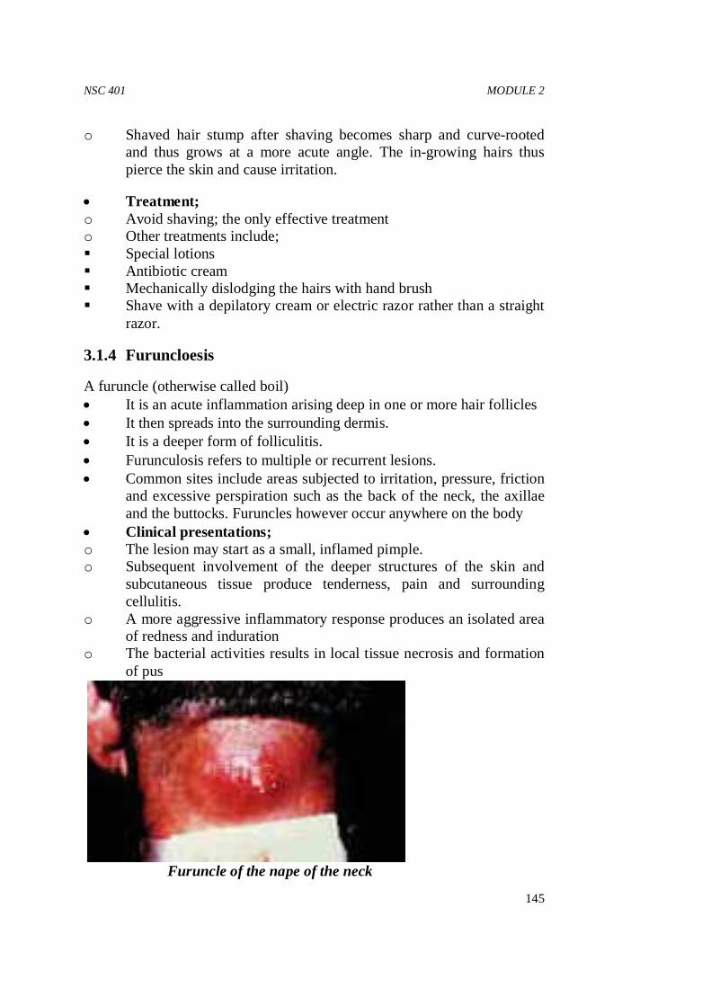
NSC 401 MODULE 2
145
o Shaved hair stump after shaving becomes sharp and curve-rooted and thus grows at a more acute angle. The in-growing hairs thus pierce the skin and cause irritation.
• Treatment; o Avoid shaving; the only effective treatment o Other treatments include; � Special lotions � Antibiotic cream � Mechanically dislodging the hairs with hand brush � Shave with a depilatory cream or electric razor rather than a straight
razor.
3.1.4 Furuncloesis
A furuncle (otherwise called boil) • It is an acute inflammation arising deep in one or more hair follicles • It then spreads into the surrounding dermis. • It is a deeper form of folliculitis. • Furunculosis refers to multiple or recurrent lesions. • Common sites include areas subjected to irritation, pressure, friction
and excessive perspiration such as the back of the neck, the axillae and the buttocks. Furuncles however occur anywhere on the body
• Clinical presentations; o The lesion may start as a small, inflamed pimple. o Subsequent involvement of the deeper structures of the skin and
subcutaneous tissue produce tenderness, pain and surrounding cellulitis.
o A more aggressive inflammatory response produces an isolated area of redness and induration
o The bacterial activities results in local tissue necrosis and formation of pus
Furuncle of the nape of the neck
NSC 401 MEDICAL-SURGICAL NURSING III
146
3.1.5 Carbunclosis A carbuncle is an abscess of the skin and subcutaneous tissue. It is a result of unresolved large, deep seated furuncle It is usually caused by infection with pyogenic Staphylococcus spp., or Gram-negative bacilli especially when the immune system is impaired Common sites include areas of thick and inelastic skin i.e. the back of the neck and the buttocks. Carbuncles presents with; o Systemic manifestations such as; � High fever � Pain � Leukocytosis � Bacteremia. Predisposing factors; o Underlying systemic diseases such as;
� Diabetes mellitus � Hematologic malignancies � Immunosuppressive therapy for other diseases
o Hot climates Medical Management • Do not to rupture or destroy the protective wall of induration that
localizes the infection. • The boil or pimple should never be squeezed. • Systemic antibiotic therapy;
o The choice of antibiotics should be based on sensitivity study. o Oral cloxacillin, dicloxacillin and flucloxacillin are first-line
medications. o Cephalosporins and erythromycin are also effective.
• Bed rest; for cases with boils on the perineum or in the anal region • Incision and drainage for localized, fluctuant leisions Nursing Management Supportive treatments such as; • Intravenous fluids • Fever management • Warm, moist compresses; increase vascularization and hasten
resolution of the furuncle or carbuncle.

NSC 401 MODULE 2
147
• Gently cleaning the surrounding skin with antibacterial soap and application of antibacterial ointment
• Soiled dressings should be handled with standard precautions. • Nursing personnel should wear disposable gloves when caring for
these patients. Teaching Patients Self-Care • The goals are to;
o Eliminate skin staphylococcal load o Increase patient’s resistance o Provide a hygienic environment.
• Actively draining lesions will require; o Covering the mattress and pillow with plastic material o Wiping and disinfectant the plastic materials daily o Laundering bed linens, towels and clothing after each use
• Encourage the patient to use an antibacterial soap and shampoo for an indefinite period, often for several months.
• Antibiotic therapy for a long time to avoid recurrent infection e.g., daily dose of oral clindamycin continuously for about 3 months)
3.2 Viral Skin Infections 3.2.1 Herpes Zoster • It is also called shingles • The disease is characterized by a painful vesicular eruption • It is caused by varicella-zoster virus, a member of a group of DNA
viruses. • The viruses also cause chickenpox. • Herpes zoster is thought to occur due to reactivation of latent
varicella virus in the sensory spinal nerve root following Chicken pox and especially reflects a lowered immunity.
• About 10% of adults get shingles during their lifetimes, usually after age 50 years.
• The incidence increases among patients with weakened immune systems and malignancies
• Herpes zoster in healthy adults is usually localized and benign however, the disease may be severe and acutely disabling in immunosuppressed patients.
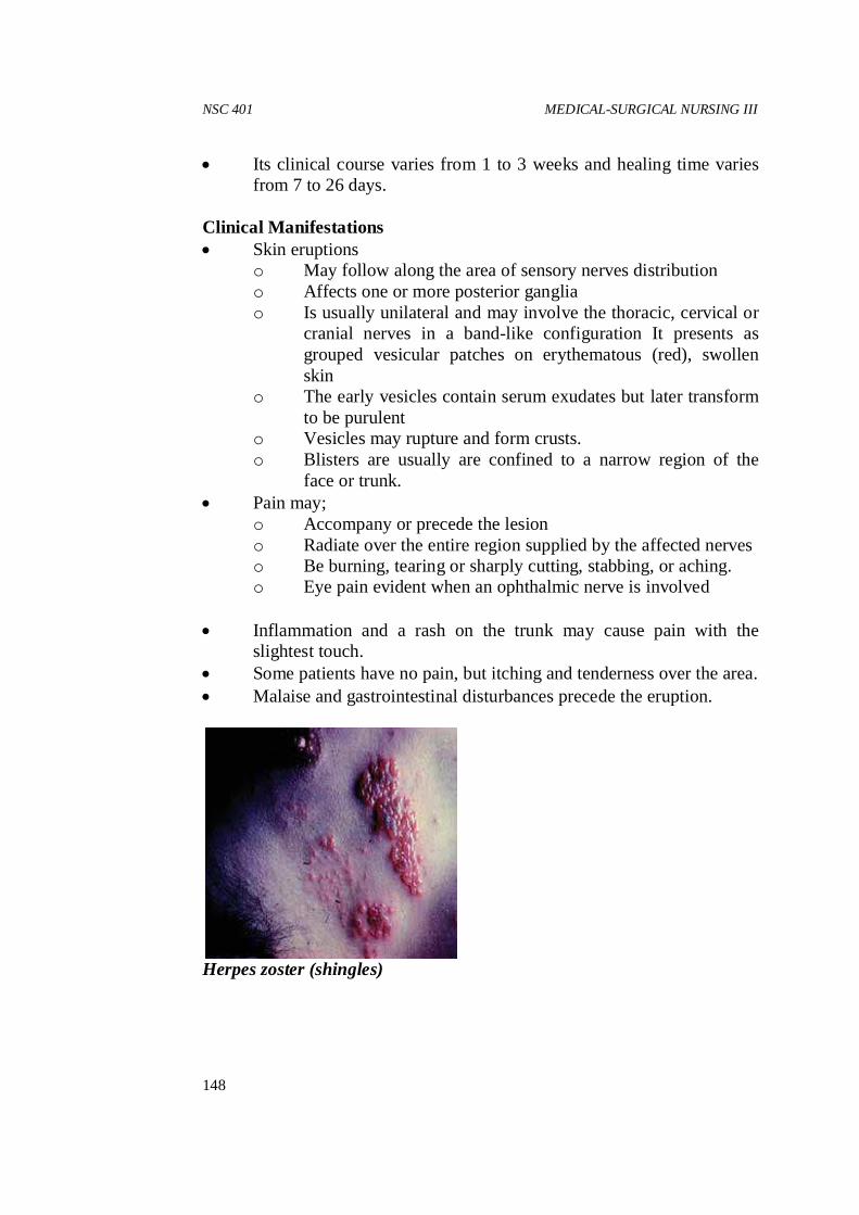
NSC 401 MEDICAL-SURGICAL NURSING III
148
• Its clinical course varies from 1 to 3 weeks and healing time varies from 7 to 26 days.
Clinical Manifestations • Skin eruptions
o May follow along the area of sensory nerves distribution o Affects one or more posterior ganglia o Is usually unilateral and may involve the thoracic, cervical or
cranial nerves in a band-like configuration It presents as grouped vesicular patches on erythematous (red), swollen skin
o The early vesicles contain serum exudates but later transform to be purulent
o Vesicles may rupture and form crusts. o Blisters are usually are confined to a narrow region of the
face or trunk. • Pain may;
o Accompany or precede the lesion o Radiate over the entire region supplied by the affected nerves o Be burning, tearing or sharply cutting, stabbing, or aching. o Eye pain evident when an ophthalmic nerve is involved
• Inflammation and a rash on the trunk may cause pain with the slightest touch.
• Some patients have no pain, but itching and tenderness over the area. • Malaise and gastrointestinal disturbances precede the eruption.
Herpes zoster (shingles)

NSC 401 MODULE 2
149
Medical Management • The goals of management are;
o Pain relief o Reduction or avoiding complications
Outline of management; usually conservative; • Pain control with analgesics • Systemic corticosteroids;
o Indicated for patients older than 50 years o Will reduce the incidence and duration of post-herpetic
neuralgia and promote healing
• Anti-inflammatory agent; Subcutaneous injection of Triamcinolone (Aristocort, Kenacort, Kenalog) under painful areas is effective.
• Antiviral agents: o Include acyclovir (Zovirax), valacyclovir (Valtrex) or
famciclovir (Famvir). o Oral preparations when administered within 24 hours of the
initial eruption will arrest the virus. o Intravenous acyclovir; if started early will effectively reduce
the pain and halt the progression of the disease.
• Ophthalmic herpes zoster is considered an emergency and requires immediate attention by an ophthalmologist
Nursing Management • Assess the patient’s discomfort and response to medication • Collaborate with the physician to make necessary adjustments to the
treatment regimen. • Teach patients to apply wet dressings or medication to the lesions • Advise patients to follow proper hand hygiene techniques to avoid
spreading the virus. • Diversionary activities and relaxation techniques to ensure restful
sleep and comfort • Advise patient to eat nourishing meals. Complications include: • Secondary infection • Scarring • Post-herpetic neuralgia: persistent pain of the affected nerve after
healing • Eye complications include keratitis, uveitis, ulceration and blindness
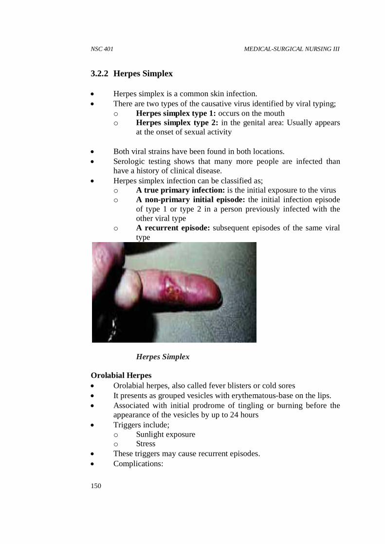
NSC 401 MEDICAL-SURGICAL NURSING III
150
3.2.2 Herpes Simplex • Herpes simplex is a common skin infection. • There are two types of the causative virus identified by viral typing;
o Herpes simplex type 1: occurs on the mouth o Herpes simplex type 2: in the genital area: Usually appears
at the onset of sexual activity • Both viral strains have been found in both locations. • Serologic testing shows that many more people are infected than
have a history of clinical disease. • Herpes simplex infection can be classified as;
o A true primary infection: is the initial exposure to the virus o A non-primary initial episode: the initial infection episode
of type 1 or type 2 in a person previously infected with the other viral type
o A recurrent episode: subsequent episodes of the same viral type
Herpes Simplex Orolabial Herpes • Orolabial herpes, also called fever blisters or cold sores • It presents as grouped vesicles with erythematous-base on the lips. • Associated with initial prodrome of tingling or burning before the
appearance of the vesicles by up to 24 hours • Triggers include;
o Sunlight exposure o Stress
• These triggers may cause recurrent episodes. • Complications:
NSC 401 MODULE 2
151
o Herpetic gingivostomatitis: Occur in less than 1% of people with primary orolabial herpes; More in children and young adults. It presents with; � High fever � Regional lymphadenopathy and � Generalized malaise.
• Erythema multiforme: an acute inflammation of the skin and mucous membranes with characteristic lesions that have the appearance of targets.
Genital Herpes • It manifests with a broad spectrum of clinical signs. • Minor infections may be asymptomatic; • Commonly caused by herpes Type II virus • Severe primary infections with type 1 virus presents thus;
o Vesicular leisions with an erythematous base o Grouped symmetrically distributed leisions primarily on the
vagina, rectum or penis o Nacent lesions propagation for the next 7 to 14 days o Associated regional lymphadenopathy o Fever and flulike symptoms o Erosions and ulcerations as the vesicles rupture.
• Typical recurrences presents with a prodromal burning, tingling or
itching sensations often about 24 hours before the vesicles appear. Assessment and Diagnostic Findings Diagnosis can be confirmed thus; • Skin eruption is strongly suggestive. • Viral cultures for older, crusted patches • Rapid assays for acute vesicular lesions Complications • Eczema herpeticum; a condition of contract herpes in eczematous
areas. • Herpes Whitlow an infection of the pulp of a fingertip with herpes
type 1 or 2. • Neonatal infection with herpes; occur during delivery by contact of
the infant with the mother’s active ulcerations. • Intrauterine neonatal infections. • Fetal anomalies include skin lesions, microcephaly, encephalitis, and
intracerebral calcifications.
NSC 401 MEDICAL-SURGICAL NURSING III
152
Medical Management Treatment of orolabial herpes infection • Avoid sun exposure • Use a sunscreen liberally on the lips and face. • Topical treatment with drying agents may accelerate healing. • Severe outbreaks may require intermittent treatment with 200 mg of
acyclovir five times each day for 5 days Treatment of genital herpes depends on; o Severity o Frequency o Psychological impact of recurrences o Infectious status of the sexual partner • Mild infection with rare relapse requires no treatment. • More severe infection will require early but intermittent treatment • Suppressive therapy with acyclovir, valacyclovir, or famciclovir is
indicated for cases with more than six recurrences per year. o Its benefit includes; o Suppress recurrence in 85% of cases o Reduces viral shedding by almost 95% i.e. make the person less
contagious. o Prevents recurrent erythema multiforme • Treatment of eczema herpeticum is with oral or intravenous
acyclovir. Management of genital herpes in pregnancy; • This is controversial. • Routine prenatal cultures may help but do not predict shedding
during delivery. • Scalp electrodes or any other instrument during delivery should be
avoided as they increase the risk for infection of the newborn. • Initial episode during pregnancy will require suppression therapy to
reduce third trimester relapse. • Cesarean section is the preferred mode of delivering women with
active lesions • Immunocompromised patients with severe infections should be
hospitalized and managed with intravenous acyclovir. 3.2.3 Fungal (Mycotic) Infections of the Skin Fungi are responsible for various common skin infections.
NSC 401 MODULE 2
153
Infections can sometimes involve only the skin and its appendages (i.e., hair and nails) It rarely causes any disability and responds readily to treatment. Secondary infection with bacteria, Candida may occur. 3.3.1 Tinea This is the most common fungal skin infection It isalso called ringworm because of its characteristic ring appearance under the skin. Tinea infections affect the head, body, groin, feet and nails. Diagnosis of tinea involves • Microscopic examination of tissue specimen for spores and hyphae
or • Isolating the organism in culture Clinical Forms of Tinea TINEA PEDIS: ATHLETE’S FOOT Tineapedis is also called athlete’s foot It is the most common cutaneous fungal infection. Clinical Manifestations • It may be an acute or chronic infection • It affects the soles of the feet or between the toes and sometimes, the
toenail. • Bacterial super-infection presents with Lymphangitis and cellulitis • Sometimes cultures will yield a mixed infection with fungi, bacteria
and yeast. Medical Management • The acute, vesicular phase of infection will require; • Soaking feet in Burow’s solution or potassium permanganate
solutions to remove crusts, scales and debris and to reduce the inflammation
• Topical antifungal agents (e.g., miconazole, clotrimazole): for several weeks because of the high rate of recurrence.
Nursing Management Instruct the patient to; • Keep the feet as dry as possible

NSC 401 MEDICAL-SURGICAL NURSING III
154
• Place small pieces of cotton in between the toes at night to absorb moisture
• Use cotton socks to effectively absorb perspiration • Wear perforated shoes that permit better aeration of the feet. • Avoid plastic or rubber soled footwear • Apply talcum powder or antifungal powder twice daily to keep the
feet dry. • Avoid wearing moist shoes • Alternate several pairs of shoes so that they can dry completely
before wearing again. TINEA CORPORIS: RINGWORM OF THE BODY Tineacorporis presents the typical ringed lesion on the face, neck, trunk and extremities. Mode of transmission is zoonotic i.e. transmitted to humans through direct or indirect contact with pets or other animal.
Tineacorporis of the check Medical Management Antifungal agents: • Topical medication for localized infections • Systemic treatment with Oral preparations is reserved only in
extensive cases o Side effects of oral agents include photosensitivity, skin rashes,
headache and nausea. o Newer agents include itraconazole, fluconazole and terbinafine.
They have limited side effects and thus are preferred for treatment of chronic fungal (dermatophyte) infections
o Older generations of drugs include griseofulvin. Nursing Management Instructed the patient to; • Use a clean towel and washcloth daily. • Keep all skin areas and skin folds dry always. • Wear clean cotton clothing next to the skin.
NSC 401 MODULE 2
155
TINEA CAPITIS: RINGWORM OF THE SCALP It is a contagious fungal infection It affects the hair shafts and is a common cause of hair loss in children. Clinical examination reveals; • One or several round, scaling patches • Small pustules or papules (at the edges of such patches) • Brittle hairs • Hair loss at or near the surface of the scalp although hair loss is only
temporary Medical Management Antifungal agents e.g. Griseofulvin • Topical agents are less effective as the infection occurs within the
hair shaft and below the surface of the scalp. o Their benefits however include; � Inactivation of the organisms � Minimization of contagion • Hair treatment with shampoo is necessary two or three times a week Nursing Management The patient and family should be instructed to; • Practice good hygiene. • Personalize utensils like comb and brush • Avoid exchanging hats and other headgear. • Subject themselves to routine examination for familial infections. • Household pets also should be examined. TINEA CRURIS: RINGWORM OF THE GROIN Tineacruris (i.e., jock itch) This is ringworm infection of the groin, which may extend to the inner thighs and buttock area. It occurs most frequently in; • Young joggers • Obese people and • Those who wear tight underclothing • People with diabetes mellitus
Heat, friction, and maceration (from sweating) worsen the infection
NSC 401 MEDICAL-SURGICAL NURSING III
156
Management • Topical medication indicated for mild infections o Preparations of clotrimazole, miconazole or terbinafine for at least 3
to 4 weeks will eradicate the infection. • Oral antifungal agents may be required for more severe infections. Nursing management The nurse instructs the patient to; • Avoid excessive heat and humidity as much as possible • Avoid wearing nylon underwear, tight-fitting clothing and wet
bathing suit. • Clean and dry the groin area thoroughly • Dusted the areas with a topical antifungal agent such as tolnaftate
(Tinactin) TINEA UNGUIUM: ONYCHOMYCOSIS Tineaunguium (i.e., ringworm of the nails) It is a chronic fungal infection Commonly affects the toenails and less commonly, the fingernails. Usual causative organisms include; • Trichophytonspecies (T. rubrum, T. mentagrophytes) or • Candida albicans It is usually associated with long-standing fungal infection of the feet. Clinical manifestations: • Thickened nails • Friable (i.e., easily crumbled) nails and • Lusterless of the nail • Debris accumulation under the free edge of the nail • Separation of the nail plate • Destruction of the entire nail Management • Antifungal agents; • Selection depends on the causative fungus. • Candidal infections will respond to fluconazole (Diflucan) or
itraconazole (Sporanox). • Oral antifungal agent for 6 weeks (for fingernails) and 12 weeks (for
toenails) are involved.

NSC 401 MODULE 2
157
• Griseofulvin is no longer considered effective because of its long treatment course and poor cure rate.
• Treating infections of the toenails is a bit difficult due to high rate of relapse with discontinuation of treatment.
3.4 Parasitic Skin Infestation 3.4.1 Pediculosis: Lice Infestation Lice infestation affects people of all ages. Lice are ectoparasites (i.e. they live on the outside of the host’s body). They feed on human blood approximately five times each day. While they feed, they also inject their digestive juices and excrement into the skin, which causes severe itching. Three varieties of lice infest humans: • Pediculushumanuscapitis (i.e., head louse) • Pediculushumanuscorporis (i.e., body louse) and • Phthirus pubis (i.e., pubic louse or crab louse) PEDICULOSIS CAPITIS This is infestation of the scalp by the head louse. The female louse lays her eggs (i.e., nits) close to the scalp. The nits become firmly attached to the hair shafts with a tenacious substance. The young lice hatch in about 10 days and reach maturity in 2 weeks. Clinical Manifestations Head lice are found most commonly along the back of the head and behind the ears. The eggs can be seen as silvery, glistening oval bodies that are adheres firmly to the hair. Infestation is more common in children and people with long hair. Mode of transmission: • Directly by physical contact • Indirectly by infested combs, brushes, wigs, hats, and bedding. Clinical manifestations: • Intense itching due to insect bites • Scratching often leads to secondary bacterial infection, such as
impetigo or furunculosis.
NSC 401 MEDICAL-SURGICAL NURSING III
158
Medical Management • Washing the hair with a shampoo according to the product directions • Rinse the hair thoroughly • Comb the hair with a fine-toothed comb dipped in vinegar to remove
any remaining nits or nit shells freed from the hair shafts. • All contaminated articles, clothing, towels and bedding should be
washed in hot water to prevent re-infestation. • Upholstered furniture, rugs, and floors should be vacuumed
frequently. • Combs and brushes are also disinfected with the shampoo. • All family members and close contacts are treated. • Complications such as severe pruritus, pyoderma, and dermatitis are
treated with anti-pruritics, systemic antibiotics, and topical corticosteroids.
Nursing Management The nurse informs the patient; • That head lice may infest anyone and are not a sign of un-
cleanliness. • Treatment must be started immediately. • To shampoo their hair regularly. • Not to share combs, brushes and hats. • Encourage each family member to have their hair inspected for head
lice daily for at least 2 weeks. • To use lindane properly as it may be toxic to the central nervous
system PEDICULOSIS CORPORIS AND PUBIS Pediculosiscorporis is an infestation of the body by the body louse. Predisposing factors; • Poor hygiene • Overcrowded living Pediculosis pubis is extremely common. The infestation is generally localized in the genital region It is transmitted chiefly by sexual contact. There may also be infestation of the hairs of the chest, armpit, beard, and eyelashes

NSC 401 MODULE 2
159
Clinical Manifestations • Bites cause characteristic minute hemorrhagic points. • Intense itching is the most common symptom, more particularly at
night • Subsequent widespread excoriation due to scratching • Reddish brown dust (i.e., excretions of the insects) may be found in
the patient’s underclothing. • Gray-blue macules may sometimes be seen on the skin due to; o Reaction of the insects’ saliva with bilirubin (converting it to
biliverdin) or o An excretion produced by the salivary glands of the louse. • The skin may become thick, dry and scaly with dark pigmented
areas in long-standing cases • Lice may be found crawling in the pubic area when examined with a
magnifying glass • Secondary lesions include parallel linear scratches and a slight
degree of eczema. Medical Management • Instruct the patient to bathe with soap and water • Topical therapies: o Lindane (Kell) or 5% permethrin (Elimite) o Over-the-counter strength of permethrin (1% Nix) o Petrolatum cream twice daily for 8 days followed by mechanical
removal of any remaining nits • Anti-pruritics • Systemic antibiotics • Topical corticosteroids Complications; • Severe pruritus • Pyoderma • Dermatitis • Rickettsial disease such as epidemic typhus, relapsing fever and
trench fever. The lice is a vector of these rickettsiae Nursing Management • Patients, all family members and sexual contacts must be; o Treated o Educated in personal hygiene o Enlightened on methods to prevent or control infestation.

NSC 401 MEDICAL-SURGICAL NURSING III
160
o Scheduled for a diagnostic workup for coexisting sexually transmitted disease.
• All clothing and bedding should be machine washed in hot water or dry-cleaned.
3.4.2 Scabies Scabies is an infestation of the skin by the itch mite Sarcoptesscabiei. The disease is common in substandard hygienic conditions It is also common among the sexually active individuals although infestations do not depend on sexual activity. The mites frequently involve the fingers and hand Contact with infected patients or contaminated clothing may produce infection. Life cycle: • The adult female burrows into the superficial layer of the skin • It remains there for the rest of her life. • With her jaws and the sharp edges of the joints of her forelegs, the
mite extends the burrow • She then lay two or three eggs daily for up to 2 months and dies
thereafter. • The larvae hatch from the eggs in 3 to 4 days and progress through
larval and nymphal states to form adult mites in about 10 days. Clinical Manifestations • Symptoms appear approximately 4 weeks from the time of contact • Severe itching is often the first symptom. It is caused by delayed
type of immunologic reaction to the mite or its fecal pellets. Itching increases at night
• Small, raised burrows evidenced by multiple, straight or wavy, brown or black, threadlike lesions on examination with a magnifying glass and a penlight held at an oblique angle to the skin
• It is commonly observed between the fingers and on the wrists; other sites include the extensor surfaces of the elbows, the knees, the edges of the feet, the points of the elbows, around the nipples, in the axillary folds, under pendulous breasts and in or near the groin or gluteal fold, penis, or scrotum.
• Red, pruritic eruptions usually appear between adjacent skin areas. • Secondary lesions are quite common and include vesicles, papules,
excoriations and crusts. Bacterial super-infection may result from constant excoriation of the burrows and papules.

NSC 401 MODULE 2
161
Assessment and Diagnostic Findings The diagnosis is confirmed by; • Recovering S. scabieibyproducts from the skin • Microscopic demonstration of the mite at any stage (e.g., egg, egg
casing, larva, nymph, adult) and fecal pellets on superficial epidermis sample.
Medical Management Instruct the patient to; • Take a warm, soapy bath or shower to remove the scaling debris • Dry the skin thoroughly and allow to cool. • Topical scabicide, such as lindane (Kwell), crotamiton (Eurax) or
5% permethrin (Elimite): requires thin application on the affected area for 12 to 24 hours, after which it should be washed thoroughly off. One application may be curative, but should be used up to 1 week.
Nursing Management Advise the patient to: • Wear clean clothing • Sleep between freshly laundered bed linens. • Wash all bedding and clothing in hot water and dried on the hot
dryer cycle or dry cleaned, as mites can survive up to 36 hours in linens.
• Apply topical corticosteroid on skin lesions after a successful treatment as scabicides may irritate the skin.
• Note for signs of hypersensitivity to scabicides which include itching for several weeks particularly in atopic (allergic) people. This is not a sign of treatment failure.
• Avoid frequent hot showers because they can dry the skin and produce itching.
• Use oral antihistamines such as diphenhydramine (Benadryl) or hydroxyzine (Atarax) to control itching.
• Encourage all family members and close contacts to be treated simultaneously to eliminate the mites.
• Avoid scabicides in infants and pregnant women unless otherwise approved.
• Avoid application of scabicide immediately after bathing; before the skin dries and cools as these conditions increase percutaneous absorption of the scabicide and the potential for central nervous system abnormalities such as seizures.
• Seek treatment for coexisting sexually transmitted disease and pediculosis as the case may be.

NSC 401 MEDICAL-SURGICAL NURSING III
162
3.5 Non-Infectious Inflammatory Dermatoses 3.5.1 Contact Dermatitis Contact dermatitis is an inflammatory reaction of the skin to physical, chemical, or biologic agents with resultant epidermal damage. Classifications; • Primary irritant type or the non-allergic type; results from irritating
substance such as soaps, detergents, scouring compounds and industrial chemicals
• Allergic contact dermatitis; from exposure of sensitized people to contact allergens
Predisposing factors; • Extremes of heat and cold • Frequent contact with irritants and • A preexisting skin disease Clinical Manifestations Initial contact with the irritant will result in; • Skin eruptions • Itching and burning sensations • Erythema • Skin edema with papule or vesicle formation (Less marked in sub-
acute presentations) • Vesicles may ooze or weep • Cuteneous crusting, drying, fissuring and peeling Repeated exposure to irritants will present with; • A more intense itching and continuous skin scratches • Lichenification and pigmentation may subsequently occur. • Secondary bacterial invasion also may follow
Contact dermatitis of the face
NSC 401 MODULE 2
163
Medical Management • The objectives of management are to; o Reduce contact with the irritant o Protect the skin from further damage • Note the pattern of distribution of the reaction to be able to
differentiate between allergic and irritant contact dermatitis. • Obtain a detailed history • Remove the offending irritant if known • Avoid local irritation • Withhold soap until healing occurs • Local dermatitis will require; o Small patches of bland, un-medicated lotion over small patches of
erythema) o Cool, wet dressings over small areas of vesicular dermatitis o Wet dressings to help clear the oozing eczematous lesions o Thin layer of corticosteroid cream or ointment • Larger areas of dermatitis will require; o Medicated baths at room temperature o Short course of systemic corticosteroids
3.5.2 Exfoliative Dermatitis Exfoliative dermatitis is characterized by progressive generalized erythematous inflammation and scaling of the skin It may be associated with chills, fever, prostration, severe toxicity, and an itchy scaling of the skin. Clinical features: • Profound loss of stratum corneum (i.e., outermost layer of the skin) • Subsequent capillary leakage • Hypoproteinemia and negative nitrogen balance. • Profound hypothermia due to widespread dilation of cutaneous
vessels Causes: • Secondary or reactive process to an underlying skin or systemic
disease • Accompany lymphoma group of diseases • Preexisting skin disorders like Psoriasis, Atopic dermatitis and
Contact dermatitis. • Severe reaction to medications, including penicillin and
phenylbutazone. • Idiopathy in approximately 25% of cases
NSC 401 MEDICAL-SURGICAL NURSING III
164
Clinical Manifestations Often an acute condition with both cutaneous and systemic manifestations • Cutaneous manifestations: o Generalized erythematous eruption o Associated fever, malaise and occasionally gastrointestinal
symptoms. o Characteristic exfoliation (i.e., scaling) after a week o Hair loss may accompany this disorder. o Relapses are common. • Systemic manifestations: effects include o High-output heart failure o Intestinal disturbances o Breast enlargement o Hyperuricemia (elevated levels of blood uric acid) and o Temperature lability. Medical Management • The objectives of management are; o Fluid and electrolyte balance and o Infection prophylaxis. • The treatment is individualized and supportive • It should be initiated as soon as the condition is diagnosed. • Hospitalization and bed rest is vital • Discontinue all medications • Maintain a comfortable room temperature to manage altered
thermoregulatory control • Maintain fluid and electrolyte balance • Replace protein. • Expand the plasma volume with IV fluids, blood or blood products
as may be indicated. Nursing Management • Continual nursing assessment • Inection surveillance • Adequate skin care. • Judicious antibiotic use based on results of culture and sensitivity. • Body heat conservation techniques • Monitor and report subtle changes in vital signs • Topical therapy is used to provide analgesia. • Provide soothing baths, compresses and lubrication with emollients
especially in cases with extensive dermatitis.

NSC 401 MODULE 2
165
• Oral or par-enteral corticosteroids to control inflammation and pruiritus
• Avoid all irritants particularly medications. 3.5.3 Psoriasis Psoriasis is a chronic non-infectious inflammatory skin disease characterized by increased epidermal cells proliferation (i.e. epidermopoiesis). It presents with as profuse scales or plaques of epidermal tissue. In psoriasis, there is rapid epidermal cell turnover so that a cell can migrate from the basal epidermis to the stratum corneum (skin surface) and be desquamated in 3 to 4 days as opposed to the normal 26 to 28 days. This hampers cell maturation and thus compromise normal protective skin layers integrity. Psoriasis is one of the most common skin diseases It affects approximately 2% of the population It appears more often in people who have a European ancestry. Etiology: • The primary cause is unknown • Suspicions are in the area of; o Specific hereditary defect especially of overproduction of keratin o Environmental stimuli/ triggers o Immune mediated response. Predisposing/ precipitating factors; • Periods of emotional stress • Anxiety • Trauma • Infections and • Seasonal changes and • Hormonal changes The onset may occur at any age but most commonly between 15 and 50 years. Psoriasis has a pattern of complete remissions to intermittent relapse.

NSC 401 MEDICAL-SURGICAL NURSING III
166
Clinical Manifestations • General cutaneous lesions include; o Red, raised patches with silvery scales o Scaly patches are due to accumulation of living and dead skin cells o Scraped scales exposes its dark red base producing multiple bleeding
points. o These patches are dry and pruritic. • Guttate psoriasis: a variant condition with a characteristic small skin
lesion (about 1 cm wide) scattered throughout the body. It is believed to follow a recent streptococcal throat infection.
• Leisions generally affect most body parts such as scalp, extensor surface of the elbows and knees, lower part of the back and the genitalia in a bilaterally symmetrical manner thus it pose a range of cosmetic and physical concerns
• Nail involvement: occur in approximately one fourth to half of cases and presents as nail pitting, discoloration, crumbling beneath the free edges and separation of the nail plate.
• Palms and soles involvement presents pustular lesions called Palmarpustular psoriasis.
Psoriasis: Note bright red scaly plaque with silvery scale.
NSC 401 MODULE 2
167
Assessment and Diagnostic Findings • Diagnosis can be generally confirmed with; o Presence of classic plaque-type lesions o Positive family history o Signs of nail and scalp involvement • Biopsy of the skin is of little diagnostic value. • No specific blood tests helpful in diagnosing the condition. Medical Management • The goals of management are; o Slow rapid epidermal cell turnover to promote resolution of the
psoriatic lesions and o Control the natural cycles of the disease • The therapeutic approach should be; o Thoroughly understood by the patient o Cosmetically acceptable to the patient and o Not too disruptive to the patient’s lifestyle. • There is no known cure • Treatment should include; o Addressing any precipitating or aggravating o Lifestyle modification o Gentle removal of scales: is the most important principle of
treatment; it involves; � Regular bathing � Adding oils (e.g., olive oil, mineral oil, AveenoOilated Oatmeal
Bath) or coal tar preparations (e.g., Balnetar) to the bath water to soften the scales
� Scrubbing the psoriatic plaques gently with a soft brush to remove the scales.
o Application of emollient creams containing alpha-hydroxyl acids (e.g., Lac-Hydrin, Penederm) or salicylic acid (to soften thick scales).
o Establish a regular skin care routine even when the psoriasis is not in an acute stage.
Pharmacologic Therapy Three types of therapy are standard: Topical Agents: Aimed tosuppress epidermopoiesis (i.e., development of epidermal cells) and cause sloughing of the rapidly growing epidermal cells without affecting other tissues • Topical salicylic acid

NSC 401 MEDICAL-SURGICAL NURSING III
168
• Topical vitamin D preparation, calcipotriene (Dovonex) and retinoid compound, tazarotene (Tazorac).
• Topical tar and anthralin baths and skin application are obsolete treatment and thus rarely used because they are malodorous, difficult to apply and no reliable results
• Topical corticosteroids; for their anti-inflammatory effect; high potency topical corticosteroids should not be used on the face and inter-triginous areas and use should be limited to a 4-week course of twice-daily applications with a 2-week break. Its effectiveness can be enhanced with occlusive dressings. Side effects of this treatment include skin atrophy and telangiectasias
• Other topical formulations include lotions, ointments, pastes, creams and shampoos.
Intralesional Agents: Agents like triamcinolone acetonide (Aristocort, Kenalog-10, Trymex) can be administered directly into highly evident psoriatic patches. This treatment is reserved for resistant cases. Care must be taken to ensure that normal skin is not injected with the medication. Systemic Agents: • Systemic corticosteroids; may cause rapid improvement but however
possibly trigger a severe flare-up on withdrawal limit their use. • Systemic cytotoxic preparations; such as methotrexate, hydroxyurea
(Hydrea) and cyclosporine A, have been used in extensive psoriasis that is resistant to other forms of therapy. They inhibit DNA synthesis in epidermal cells and thus reduce cell turnover time of the psoriatic epidermis. Toxic effects include irreversible liver damage, hematopoietic (bone marrow suppression), nephrotoxicity and teratogenicity
• Oral retinoids are synthetic derivatives of vitamin A and its metabolite (vitamin A acid). They modulate growth and differentiation of epithelial tissue. Etretinate is especially useful for severe pustular or erythrodermic psoriasis. It is however teratogenic hence cannot be used in women with childbearing potential.
Photochemotherapy Indicated for severe debilitating psoriasis • Ultraviolet-A light therapy o Usually combined with Psoralen (usually 8-methoxypsoralen); a
photosensitizing medication o The treatment is collectively called Psoralen-Ultraviolet-A (PUVA)
therapy

NSC 401 MODULE 2
169
o Mechanism of action: Although not completely understood o It is thought that when psoralen-treated skin is exposed to
ultraviolet-A light, the psoralen binds with DNA and decreases cellular proliferation.
o Hazards of PUVA include; � Long-term risks of skin cancer � Risks of cataracts formation and � Premature skin aging o Treatment course: � The patient is usually treated two or three times each week until the
psoriasis clears with a break period of 48 hours between treatments to avoid burns
� After the psoriasis clears, the patient begins a maintenance program.
• Ultraviolet-B (UVB) light therapy: o This is also an alternative to treat generalized plaques. o It is used alone or combined with topical coal tar. o Side effects are similar to those of PUVA therapy.
• Exposure to sunlight o Useful if access to a light treatment unit is not feasible. o It also presents risks such as; � Acute sunburn reaction � Exacerbation of photosensitive disorders such as lupus, rosacea and
polymorphic light eruption � Increased wrinkles and thickening � Increased risk for skin cancer.
• Excimer lasers: o It has been used to treat psoriasis using lasers function at 308 nm. o Studies show that medium-sized psoriatic plaques clear in four to six
treatments and remain clear for up to 9 months. o A laser can be more effective on the scalp. Laser can be aimed very
specifically on the plaque
Complications • Asymmetric rheumatism(without multiple joint arthritis); arthritic
development can occur before or after the skin lesions appear. The relation between arthritis and psoriasis is not understood.
• Erythrodermic psoriasis:an exfoliative psoriatic state involving the total body surface. The patient is more acutely ill with fever, chills, and an electrolyte imbalance. It occurs more commonly in chronic psoriasis aggravated by infections or certain medications, withdrawal of systemic corticosteroids

NSC 401 MEDICAL-SURGICAL NURSING III
170
3.6 Blistering Diseases of the Skin Common causes include: • Infectious agents like bacteria, fungi, or viruses eg, herpes simplex
and zoster infections • Allergic contact reactions e.g. contact dermatitis • Burns • Metabolic disorders and • Immunologically mediated reactions e.g. autoimmune reactions • Some of these conditions are life-threatening while others assume
chronicity Diagnosis is always made by; • Histologic examination of a biopsy specimen by a
dermatopathologist • Serum immunofluorescent studies to detect circulating antibodies 3.6.1 Pemphigus Pemphigus is a group of skin diseases characterized by bullae (i.e., blisters) of various sizes on apparently normal skin and mucous membranes. Pemphigus is an autoimmune disease involving immunoglobulin G. It is thought to be due to antibody against a specific cell-surface antigen in epidermal cells. A blister forms from the antigen–antibody reaction. The level of serum antibody is predictive of disease severity. This disorder usually occurs in men and women in middle and late adulthood. The condition may be associated with penicillins and captopril and with myasthenia gravis. Clinical Manifestations • Oral lesions are painful, irregularly shaped erosions that bleed easily
and heal slowly • Skin bullae enlarge, rupture and leave large, painful eroded areas
that are accompanied by crusting and oozing.

NSC 401 MODULE 2
171
• A characteristic offensive odor emanates from the bullae and the exuding serum.
• Blistering or sloughing of uninvolved skin with minimal pressure (i.e., Nikolsky’s sign).
• The eroded skin heals slowly • Bacterial super-infection is common. Complications • Pemphigus vulgaris i.e. wide spread disease process • Secondary bacterial infection. • Fluid and electrolyte imbalance • Hypoalbuminemia Management • The goals of therapy are; o Rapid disease control o Serum loss control o Secondary infection prophylaxis and o Re-epithelization promotion (i.e., renewal of epithelial tissue) • Treatment options: o Corticosteroids therapy o Immunosuppressive agents (e.g., azathioprine, cyclophosphamide,
gold) o Plasmapheresis (i.e., plasma exchange); temporarily decreases the
serum antibody level 3.6.2 Bullous Pemphigoid • Bullous pemphigoid is an acquired disease of flaccid blisters
appearing on normal or erythematous skin. • It appears more often on the flexor surfaces of the arms, legs, axilla,
and groin. • Oral lesions are usually transient and minimal. • Blister breakage leaves shallow erosions on the skin that heals fairly
quickly. • Pruritus can be intense, even before the appearance of the blisters. • Bullous pemphigoid is common in the elderly, with a peak incidence
at about 60 years of age. There is no gender or racial predilection • The disease can be found throughout the world.

NSC 401 MEDICAL-SURGICAL NURSING III
172
Management • Topical corticosteroids for localized eruptions and • Systemic corticosteroids e.g. prednisone, for widespread
involvement 3.6.3 Dermatitis Herpetiformis • Dermatitis herpetiformis is an intensely pruritic, chronic disease • It manifests with small, tense blisters that are distributed
symmetrically over the elbows, knees, buttocks and nape of the neck.
• It is most common between the ages of 20 and 40 years • It can appear at any age although. • Most patients have a subclinical defect in gluten metabolism. Management • Dapsone (combination of tetracycline and nicotinamide) • Gluten-free diet. • Dietary counseling because of the lifelong dietary restrictions • Emotional support associated with learning new habits and accepting
major life changes 3.6.4 Herpes Gestationis • Herpes gestationis is a disease that occurs during or shortly after
pregnancy. • It shares several clinical features with bullous pemphigoid • It has no relation to the herpes virus. • This disease is uncommon; occurs in approximately 1 case in every
50,000 pregnancies more commonly in the second or third trimester. • It begins with urticarial papules on the abdomen • Later spread to the trunk and extremities. • It usually resolves within a few weeks of delivery • There can be relapse in subsequent pregnancies, menses or with oral
contraceptive use Management • Systemic corticosteroids • No identifiable risk for fetal morbidity or mortality • Secondary infection prophylaxis as in other blistering diseases

NSC 401 MODULE 2
173
3.6.5 Toxic Epidermal Necrolysis and Stevens-Johnson Syndrome
They are potentially fatal skin disorders and the most severe form of erythema multiforme. Mortality rate approaches 30%. Both conditions are triggered by; • Medication e.g. antibiotics, anti-seizure agents, butazones, and
sulfonamides • Viral infection. Clinical Manifestations • Conjunctival burning or itching • Cutaneous tenderness • Fever • Cough • Sore throat • Headache • Extreme malaise and • Myalgias (i.e., aches and pains). • Cutaneous manifestations: • Rapid onset of erythema of the skin and mucous membranes • Large, flaccid bullae • Laryngeal, bronchial and esophageal ulcerations and damage (severe
form) • Excruciatingly tender skin • Loss of skin similar to a total-body, partial-thickness burn • Weeping skin (also referred to as scalded skin syndrome) The conditions occur in all ages and both genders. Risk factors: • Older people due to multiple medication tendencies • HIV and Acquired Immunodeficiency Syndrome (AIDS) treated
with sulfonamides • Other forms of immunosuppression Complications • Sepsis: unrecognized and untreated sepsis can be life-threatening.

NSC 401 MEDICAL-SURGICAL NURSING III
174
• Keratoconjunctivitis; result in conjunctival retraction, scarring and corneal lesions with subsequent vision impairment
Assessment and Diagnostic Findings • A history of ingestion of medications known to precipitate the
diseases • Histologic studies of frozen skin cells from a fresh lesion • Immunofluorescent studies; to detect atypical epidermal auto-
antibodies Medical Management • The goals of treatment include; o Fluid and electrolyte balance o Prevention of sepsis and o Prevention of ophthalmic complications • Supportive care is the mainstay of treatment. • All nonessential medications are discontinued immediately. • Manage the patient in a regional burn center • Aggressive treatment similar to that for severe burns is required as
skin loss may approach 100% of the total body surface area. • Surgical debridement or hydrotherapy in a Hubbard tank (i.e., large,
steel tub) may be performed to remove involved skin. • Obtain tissue samples from the nasopharynx, eyes, ears, blood,
urine, skin and un-ruptured blisters for culture to identify pathogenic organisms.
• Intravenous fluids are prescribed to maintain fluid and electrolyte balance
• Watch sites of indwelling intravenous catheter for infection • Fluid replacement through nasogastric tube and orally as soon as
possible. • Initial treatment with systemic corticosteroids (controversial though) • Intravenous administration of immunoglobulin (IVIG) • Topical antibacterial and anesthetic agents are used to prevent
wound sepsis and to assist with pain management. • Systemic antibiotic therapy is used with extreme caution. • Temporary biologic dressings (e.g., pigskin, amniotic membrane) or
plastic semi-permeable dressings (e.g., Vigilon) may be used to reduce pain, decrease evaporation, and prevent secondary infection until the epithelium regenerates.
• Meticulous oropharyngeal and eye care is essential when there is severe involvement of the mucous membranes and the eyes.

NSC 401 MODULE 2
175
4.0 CONCLUSION In this unit you have explored care of patients with infections and infestations of the skin, with the intent of improving your knowledge base and requisite clinical skills. 5.0 SUMMARY At this juncture, you are expected to have learnt about:
i. Various types of bacterial infections of the skin ii. Viral infections of the skin iii. Fungal infections of the skin iv. Parasitic infestations of the skin v. Blistering skin disorders
6.0 TUTOR-MARKED ASSIGNMENT Now that you have learnt this much, visit a hospital of choice within your vicinity and manage a patient with any of the infections or infestations of the skin using the nursing process approach. SELF-ASSESSMENT EXERCISE Now, you will be required to evaluate yourself with the following questions: Mrs. S.J. is a 35 year old, who presented in the clinic with erythematous skin eruptions with regional and ipsilateral distribution on the face leisions on the face associated with severe itching and pains at the site of the lesion. He was diagnosed to be having herpes zoster. i. What is herpes zoster? ii. Enumerate the clinical manifestations of herpes zoster. iii. Outline the management of Mrs. S.J. Note that, you are to grade yourself based on the information provided within the content of the unit. If you have graded yourself poor, you can re-work.
NSC 401 MEDICAL-SURGICAL NURSING III
176
UNIT 4 CARING FOR PATIENT WITH PRESSURE ULCERS
CONTENTS 1.0 Introduction 2.0 Objectives 3.0 Main Content
3.1 Definitions 3.2 Staging of Pressure Ulcer 3.3 Causes of Pressure Ulcer 3.4 Incidence and Prevalence 3.5 Predisposing Factors 3.6 Physical Examination 3.7 Nursing Management
3.7.1 Primary Nursing Diagnosis 3.7.2 Nursing Outcomes 3.7.3 Nursing Interventions 3.7.4 Documentation Guidelines
4.0 Conclusion 5.0 Summary 6.0 Tutor-Marked Assignment 1.0 INTRODUCTION This unit will focus on exploring care of patients with pressure ulcer, with a view to boost your knowledge base and requisite clinical skills. 2.0 OBJECTIVES At the end of this unit, you will be able to: • Identify pressure areas • Outline the stages of Pressure Ulcer • Enumerate the predisposing factors to development of pressure ulcer • Manage pressure ulcer using the nursing process

NSC 401 MODULE 2
177
3.0 MAIN CONTENT 3.1 Definitions A pressure ulcer is an irregularly shaped, depressed area that resulted from necrosis of the epidermis and/or dermis layers of the skin. Sometimes it can also involve deeper structures of muscles. It is primarily due to prolonged pressure with resultant inadequate circulation (ischemia) and ulceration (tissue breakdown) Pressure ulcers often occur in any area of the body mostly over bony prominences. These areas are called pressure area. Pressure areas include the occiput, thoracic and lumbar vertebrae, scapula, coccyx, sacrum, greater trochanter, ischial tuberosity, lateral knee, medial and lateral malleolus, metatarsals and calcaneus Pressure ulcers are most common in the lower part of the body (hip and buttock region) Pressure ulcers staging: 3.2 Staging of Pressure Ulcer National Ulcer Advisory Panel’s stages and description provide an order for progression. The stage descriptions of pressure Ulcer include; • Stage I: Non-blanchable erythema; involves changes in the
underlying vessels of the skin; bright red color that does not resolve after 30 min of pressure relief; can be painful and tender.
• Stage II: Partial thickness skin loss of epidermis and dermis; cracks or blisters on skin with erythema and/or indurations.
• Stage III: Full-thickness skin loss of epidermis and dermis; extends down to subcutaneous tissue; appears as a crater or covered by black eschar, wound base usually not painful; indistinct borders; may have sinus tracts or undermining present.
• Stage IV: Full-thickness skin loss with extensive destruction of tissue, muscle, bone, and/or supporting structures; appears as a deep

NSC 401 MEDICAL-SURGICAL NURSING III
178
crater or is covered by thick eschar; wound base not painful; may have sinus tracts and undermining present.
Stages of Pressure Ulcers 3.3 Causes of Pressure Ulcer • External pressure: more than normal capillary pressure of 25 mm
Hg; cause tissue hypo-perfusion • Ischemia occurs when the external pressure exceeds surpasses
arteriole pressure (i.e. 50mmHg); and tissue blood flow is completely blocked.
• Underlying tissues are compression: results from pressure on tissue from the underlying bony prominence
• This results in tissue necrosis • Ulceration is caused by shearing or friction as tissue layer slides over
another • Shearing results in stretching and angulating of blood vessels,
causing local injury and thrombosis 3.4 Incidence and Prevalence • No clear genetic contributions • Pressure ulcers can occur at any age and across both genders but are
more prevalent in the elderly population over 70 years of age. • Neurological impairment and immobility • Male young folks due to associated high incidence of traumatic
spinal cord injuries • No known racial and ethnic considerations.

NSC 401 MODULE 2
179
3.5 Predisposing Factors • Conditions of decreased circulation and sensation e.g. diabetes
mellitus, arterial insufficiency, peripheral vascular disease, • Decreased activity • Spinal cord injury • Immobility: Patients with casts, braces and splints • Malnutrition • Ageing due to reduced skin elasticity • Advanced medical conditions like peripheral edema due to chronic
renal disease, congestive cardiac diseases 3.6 Physical Examination • Assess the wound to determine the precise location, along with size
and depth. • Wound color range from pink, red, yellow or black to determine
stage of tissue devitalization. o A beefy red color signifies the presence of granulation tissue and
adequate healing. o Black tissue indicates necrotic and devitalized tissue with poor
potential for healing. • Observe for areas of sinus tracts and undermining, which indicate
deeper tissue involvement • Determine the amount of drainage and the type, color, odor,
consistency and quantity. • Assess skin area around the wound for redness, edema, indurations,
tenderness and breakdown: may be an evidence of infection. 3.7 Nursing Management 3.7.1 Primary Nursing Diagnosis • Impaired skin integrity related to pressure over bony prominences or
shearing forces 3.7.2 Nursing Outcomes • Tissue integrity: Skin and mucous membranes • Wound healing: Primary intention • Immobility consequences: Physiological
NSC 401 MEDICAL-SURGICAL NURSING III
180
• Knowledge: Treatment regime • Nutritional status • Tissue perfusion: Peripheral • Treatment behavior: Illness or injury 3.7.3 Nursing Interventions • Wound care and Infection control o Surgical intervention to excise necrotic tissue in late stages of ulcer
development o Skin grafts or musculocutaneous flaps for very deep wounds o Drains may be inserted to prevent fluid buildup in the wound and
infection o Mechanical debridement by an enzymatic agent (collagenase
[Santyl]) may be ordered. o Wound dressings with hydrocolloid, hydrogels, calcium alginates,
film dressings and topical agents and solutions. o When skin breakdown occurs, apply appropriate dressings using
clean technique o Manage wound with sterile technique. o Nutritional supplementation o Hyperbaric oxygen therapy for deep and difficult wounds o Electrotherapy to deliver low-intensity direct current to wounds in
attempts to assist the healing process o General guidelines for ulcer management � Stage I ulcers require no type of dressings. � Stage II pressure ulcers; moist or occlusive dressings to maintain a
moist, healing environment � Stage III ulcers require debridement, usually with an enzymatic
agent or wet-to-moist normal saline soak. � Stage IV ulcers are treated like stage III ulcers or by surgical
excision and grafting. • Skin surveillance • Positioning o Lift high-risk patients up in bed instead of pulling them to prevent
shearing and friction forces on the skin’s surfaces. o Do not elevate the patient’s head more than 20 degrees unless
contraindicated by other medical problems or treatment modalities. o Regular 2 hourly turning and proper positioning of high-risk patient • Pressure management o Pressure-relieving devices, such as silicone-filled pads and foam
mattresses

NSC 401 MODULE 2
181
o Dynamic devices include specialty beds (low-air-loss, air-fluidized, and air cushions)
o Airflow pressure mattresses • Pressure ulcer prevention o Identify patients at risk by using assessment tools such as the Braden
scale or the Norton scale o Keep the patient’s skin dry. o Clean incontinent patient of feces and urine soon to prevent skin
irritation. o Use absorbent under-pads and topical moisture barrier agents when
soiling of the skin cannot be controlled o Avoid the use of hot water and mild cleansing agent to minimize
dryness and irritation in high-risk patients. o Treat dry skin with moisturizers o Massage bony prominences carefully as vigorous massaging may
impede capillary blood flow and increase the risk of deep tissue injury.
o Keep linens dry and wrinkle-free. o Teach the caregiver preventive strategies o Determine if the patient’s situation is in jeopardy because of
inadequate care. o Note that the caregiver may have feelings of guilt because of failure
to prevent complications of immobility o Caregivers may need support rather than teaching, depending on the
situation. • Medication administration: Topical antibiotics • Circulatory precautions • Nutrition management 3.7.4 Documentation Guidelines • Physical findings for potential skin breakdown: Redness and dryness • Physical findings of direct wound assessment: Size, depth, type of
tissue present (granulation, necrotic), drainage; signs of infection • Type and frequency of dressing changes with sequencing of how the
wound was cleaned and the dressing applied • Response to treatments: Surgery, wound debridement, dressing,
medication application

NSC 401 MEDICAL-SURGICAL NURSING III
182
4.0 CONCLUSION A pressure ulcer is an irregularly shaped, depressed area that resulted from necrosis of the epidermis and/or dermis layers of the skin. Sometimes it can also involve deeper structures of muscles. 5.0 SUMMARY At this time, you should be acquainted with knowledge on pressure ulcers and how to manage a patient with pressure ulcers using the nursing process 6.0 TUTOR-MARKED ASSIGNMENT You may now proceed to your clinical area; identify and manage a patient with pressure ulcer using the nursing process model. SELF-ASSESSMENT EXERCISE i. What is pressure ulcers ii. Briefly discuss the various stages of pressure ulcer iii. Identify the predisposing factors to pressure ulcers iv. Using the nursing process, manage a patient with pressure ulcer When you are through, you are free to grade yourself with information from the course content and the reference given. You are then required to share your findings with your colleagues in the discussion platform.
NSC 401 MODULE 3
183
MODULE 3 CARING FOR PATIENTS WITH MUSCULOSKELETAL SYSTEM DISORDERS MR. OGUNLADE
Unit 1 Review of Related Anatomy and Physiology Unit 2 Assessment and Diagnostic Evaluation of Disorder of the
Musculoskeletal System Unit 3 Caring for Patients with Musculoskeletal Trauma: Soft Tissue
Trauma, Fractures, Hip Fracture Unit 4 Caring for Patients with Joint Trauma and Injury: Repetitive
Use Injuries; Amputation; Unit 5 Caring for Patients with Structural and Bone Disorders:
Scoliosis and Kyphosis; Osteoporosis; Osteomalacia and Pagiet’s Disease; Osteomyelitis; Common Foot Disorders; Bone Tumor
Unit 6 Caring for Patients with Joint and Connective Tissue Disorders: Osteoarthritis; Rheumatoid Arthritis ; Systemic Lupus Erthematosus; Gout; Lyme Disease; Ankylosing Spondylitis; Fibromyalgia; Low Back Pain; Muscular Dystrophy
UNIT 1 ANATOMY AND PHYSIOLOGY CONTENTS 1.0 Introduction 2.0 Objectives 3.0 Main Content
3.1 Bones 3.2 Muscles 3.3 Joints 3.4 Tendons 3.5 Ligaments 3.6 Cartilage 3.7 Bursae
4.0 Conclusion 5.0 Summary 6.0 Tutor-Marked Assignment
NSC 401 MEDICAL-SURGICAL NURSING III
184
1.0 INTRODUCTION The musculoskeletal system consists of bones, muscles, joints, tendons, ligaments, cartilage, and bursae. It supports the body and facilitates movement. Other functions include storage of calcium, phosphorus, magnesium, and fluoride; production of blood cells in the bone marrow; and protection and support to body organs, such as the lungs, heart, and brain. Injury to or disease in any part of the musculoskeletal system can cause pain, immobility, or disability and potentially affect quality of life. 2.0 OBJECTIVES At the end of this module, you will be able to: • describe major structures and functions of the musculoskeletal
system • discuss elements of the nursing assessment of the musculoskeletal
system • identify common diagnostic and laboratory tests used in the
evaluation of musculoskeletal disorders • discuss the nursing management of clients undergoing tests for • musculoskeletal disorders • list the components of the musculoskeletal system • describe anatomy and physiology of a long bone • describe a joint. 3.0 MAIN CONTENT 3.1 Bones The human body has 206 bones. The bones of the skeleton are classified as: • short bones, such as those in the fingers and toes • long bones, such as the femur and ulna • flat bones, such as the sternum • irregular bones, such as the vertebrae There are two types of bony tissue. The first is cancellous bone, ornspongy bone, which is light and contains many spaces. The second is cortical bone, or compact bone, which is dense and hard. Both types are found in varying amounts in all bones. Cancellous bone is found at the rounded, irregular ends, or epiphyses, of long bones. Cortical bony tissue covers bones and is

NSC 401 MODULE 3
185
found chiefly in the long shafts, or diaphyses, of bones in the arms and legs. The combination of the two types of bony tissue provides strength and support, yet keeps the skeleton light to promote endurance during activity. Bone is composed of cells, protein matrix, and mineral deposits. The three types of bone cells are osteoblasts, osteocytes, and osteoclasts. Cells that build bones are called osteoblasts. These cells secrete bone matrix (mostly collagen), in which inorganic minerals, such as calcium salts, are deposited. This process of ossification and calcification transforms the osteoblasts into mature bone cells, called osteocytes, which are involved in maintaining bone tissue. During times of rapid bone growth or bone injury, osteocytes function as osteoblasts to form new bone. Osteoclasts arethe cells involved in the destruction, resorption, and remodeling of bone. During growth, bones primarily lengthen. The diameter also increases when osteoclasts break down previously formed bone, however, making the central canal wider. When skeletal growth is complete, the osteoclasts, which are part of the mononuclear phagocyte system (blood cells involved in ingesting particulate matter or recycling old cells), continue with the remodeling of bones by balancing bone resorption with new bone cell replacement. Bone formation and resorption continue throughout life. The greatest activity occurs from birth through puberty. A layer of tissue called periosteum covers the bones (but not the joints). The inner layer of periosteum contains the osteoblasts necessary for bone formation. The periosteum is rich in blood and lymph vessels and supplies the bone with nourishment. Inside the bones are two types of bone marrow: red and yellow. Red bone marrow, found primarily in the sternum,ileum, vertebrae, and ribs, manufactures blood cells and hemoglobin. Long bones have yellow bone marrow, which consists primarily of fat cells and connective tissue. If the blood cell supply becomes compromised, the yellow marrow may take on the characteristics of red marrow and begin producing blood cells. 3.2 Muscles There are three kinds of muscles: skeletal, smooth, and cardiac. Skeletal muscles are voluntary muscles; impulses that travel from efferent nerves of the brain and spinal cord control their function. The skeletal muscles promote movement the biceps in the arms and the gastrocnemius in the calves. Skeletal muscle is composed of muscle cells or fibers that contain several myofibrils. Sliding filaments called sarcomeres make up myofibrils. They are the contractile units of skeletal muscle. Impulses from the central
NSC 401 MEDICAL-SURGICAL NURSING III
186
nervous system cause the release of acetylcholine at the motor end plate of the motor neuron that innervates the muscle. As a result, calcium ions are released, and the release stimulates actin and myosin in the sarcomeres to slide closer together, resulting in contraction of the muscle. When calcium is depleted, the actin and myosin fibers move apart, causing relaxation of the sarcomeres, and thus the muscle. Smooth and cardiac muscles are involuntary muscles; their activity is controlled by mechanisms in their tissue of origin and by neurotransmitters released from the autonomic nervous system. Smooth muscles are found mainly in the walls of certain organs or cavities of the body, such as the stomach, intestine, blood vessels, and ureters. Cardiac muscle is found only in the heart. 3.3 Joints A joint is the junction between two or more bones. Free moving joints, or diarthrodial joints, make up most skeletal joints. They allow certain movements. Terms related. The surfaces of diarthrodial joints are covered with hyaline cartilage, which reduces friction during joint movement. The space between is the joint cavity, which is enclosed by a fibrous capsule lined with synovial membrane. This membrane produces synovial fluid, which acts as a lubricant. 3.4 Tendons Tendons are cordlike structures that attach muscles to the periosteum of the bone. A muscle has two or more attachments. One is called the origin and is more fixed. The other is called the insertion and is more movable. When a muscle contracts, both attachments are pulled, and the insertion is drawn closer to the origin. An example can be found in the biceps of the arm, which has two origin tendons, attached to the scapula, and one insertion tendon, attached to the radius. When the biceps contract the lower arm (with the insertion tendon) move toward the upper arm (with the origin tendons). 3.5 Ligaments Ligaments consisting of fibrous tissue connect two adjacent, freely movable bones. They help protect the joints by stabilizing their surfaces and keeping them in proper alignment. In some instances, ligaments completely enclose a joint.

NSC 401 MODULE 3
187
3.6 Cartilage Cartilage is a firm, dense type of connective tissue that consists of cells embedded in a substance called the matrix. The matrix is firm and compact, thus enabling it to withstand pressure and torsion. The primary functions of cartilage are to reduce friction between articular surfaces, absorb shocks, and reduce stress on joint surfaces. Hyaline or articular cartilage covers the surface of movable joints, such as the elbow, and protects the surface of these joints. Other types of cartilage include costal cartilage, which connects the ribs and sternum; semilunar cartilage, which is one of the cartilages of the knee joint; fibrous cartilage, found between the vertebrae (intervertebral discs); and elastic cartilage, found in the larynx, epiglottis, and outer ear. 3.7 Bursae A bursa is a small sac filled with synovial fluid. Bursae reduce friction between areas, such as tendon and bone and tendon and ligament. Inflammation of these sacs is called bursitis. 4.0 CONCLUSION The musculoskeletal system consists of bones, muscles, joints, tendons, ligaments, cartilage, and bursae. It supports the body and facilitates movement.Injury to or disease in any part of the musculoskeletal system can cause pain, immobility, or disability and potentially affect quality of life. 5.0 SUMMARY In this unit, you have learnt that:
i. Musculoskeletal system consists of bones, muscles, joints, tendons, ligaments, cartilage and bursae.
ii. The bones of the skeleton are classified as short bones,long bones, flat bones and irregular bones.
6.0 TUTOR-MARKED ASSIGNMENT 1. List the components of the musculoskeletal system. 2. Describe anatomy and physiology of a long bone. 3. Describe a joint.
NSC 401 MEDICAL-SURGICAL NURSING III
188
UNIT 2 ASSESSMENT AND DIAGNOSTIC EVALUATION OF DISORDER OF THE MUSCULOSKELETAL SYSTEM
CONTENTS 1.0 Introduction 2.0 Objectives 3.0 Main Content
3.1 Assessment 3.2 Diagnostic Tests
4.0 Conclusion 5.0 Summary 6.0 Tutor-Marked Assignment 1.0 INTRODUCTION In this unit you will do a quick review of the musculoskeletal system that you have covered in Human Anatomy 102 in year one. 2.0 OBJECTIVES At the end of this unit you will be able to:
• discuss elements of the nursing assessment of the musculoskeletal system
• identify common diagnostic and laboratory tests used in the evaluation of musculoskeletal disorders.
3.0 MAIN CONTENT 3.1 Assessment History The focus of the initial history depends on whether the client has a chronic disorder or a recent injury. If the disorder is long-standing, the nurse obtains a thorough medical, drug, and allergy history. If the client is injured, the nurse finds out when and how the trauma occurred. He or she compiles a list of symptoms that includes information about the onset, duration, and location of discomfort or pain. Determining whether activity makes the symptoms better or worse is important. The nurse also identifies

NSC 401 MODULE 3
189
associated symptoms, such as muscle cramping or skin lesions, and asks the client if the problem interferes with activities of daily living. If the client has an open wound, the nurse ascertains when the client last received a tetanus immunization. The nurse must obtain a history of past disorders and medical or surgical treatments as soon as possible. Attention to chronic or concurrent disorders, such as diabetes mellitus, is essential. In addition, the nurse obtains a family history, especially when relatives have had similar symptoms, and an occupational history. Physical Examination For a general musculoskeletal assessment, the nurse observes the client’s ability to ambulate, sit, stand, and perform activities requiring fine motor skills, such as grasping objects. General inspection includes examining the client for symmetry, size, and contour of extremities and random movements. The nurse palpates the muscles and joints to identify swelling, degree of firmness, local warm areas, and any involuntary movements. To test the client’s muscle strength, the nurse applies force to the client’s extremity as the client pushes against that force. The nurse also must perform a neurovascular assessment which includes assessing range of motion for the joints, taking care not to force movement. The nurse notes any abnormal muscle movements such as spasms or tremors. In addition, the nurse: • Looks for abnormal size or alignment and symmetry, comparing one
side with the other. • Inspects and palpates for pain, tenderness, swelling, and redness. • Observes the degree of movement and range of motion, but never
persists beyond the point of pain. • Tests for muscle strength. • Inspects for muscle wasting. Depending on the symptoms and findings, additional assessments may include looking for changes in gait and body posture, favoring one side over the other, and ability to bend and twist the trunk, head, and extremities. As clients age, they experience many changes in the musculoskeletal system. After 35 years, people generally experience loss of bone mass and height and changes in the structure of the spine and joints. It is essential to assess clients for musculoskeletal changes because of their potential impact on activities of daily living. If the client has a traumatic injury, physical assessment begins with taking vital signs. Further assessment depends on the type and area of injury. As the nurse conducts the assessment, he or she maintains standard precautions. The nurse needs to cut the clothing from around an injured area if there is no other way to

NSC 401 MEDICAL-SURGICAL NURSING III
190
examine the client. Comparing structures and assessment findings on one side of the body with those on the opposite side assists the nurse in determining the degree of injury. Although the nurse must be thorough, it also is important to be gentle, recognizing that assessment techniques may increase the client’s pain. The examinationincludes the following: • Observing for swelling, external bleeding, or bruising • Palpating the peripheral pulses • Evaluating peripheral circulation; assessing peripheral pulse (rate
and character), skin coloration (pink, gray, pale, ashen), temperature, and capillary refill time
• Checking the sensation of the injured part • Looking for broken skin, open wounds, superficial or embedded
debris in or around the wound, protrusion of bone or other tissue from the wound
• Examining for injury beyond the original area; for example, auscultating the chest and abdomen if an abdominal or thoracic injury occurred or checking the pupils and mental status if a head injury occurred
• Looking for mal alignment of the injured limb • Assessing for pain, noting the type and location The physician needs
to examine the client before the nurse touches, cleans, or disturbs open wounds and before moving the injured extremity.
3.2 Diagnostic Tests Imaging Procedure Radiographic films, computed tomography (CT), and magnetic resonance imaging (MRI) help identify traumatic disorders such as fractures and dislocations, and other bone disorders, such as malignant bone lesions, joint deformities, calcification, degenerative changes, osteoporosis, and joint disease. An arthrogram i s a radiographic examination of a joint, usually the knee or shoulder. The physician first injects a local anesthetic and then inserts a needle into the joint space. Fluoroscopy may be used to verify correct placement of the needle. The synovial fluid in the joint is aspirated and sent to the laboratory for analysis. A contrast medium is then injected, and x-ray films are taken. After undergoing arthrography, the client is informed that he or she may hear crackling or clicking noises in the joint for up to 2 days. Noises beyond this time are abnormal; the client should report them.

NSC 401 MODULE 3
191
Arthroscopy Arthroscopy is the internal inspection of a joint using an instrument called an arthroscope. The most common use of arthroscopy is visualization of the knee joint, a common site of injury. After administering a local or general anesthetic, the physician inserts a large-bore needle into the joint and injects sterile normal saline solution to distend the joint. After inserting the arthroscope, the examiner inspects the joint for signs of injury or deterioration. Joint fluid may be removed and sent to the laboratory for examination. Depending on the findings, the physician sometimes can use the arthroscope to perform therapeutic procedures, such as removing bits of torn or floating cartilage. Afterward, the client’s entire leg is elevated without flexing the knee. A cold pack is placed over the bulky dressing covering the site where the arthroscope was inserted. Arthrocentesis Arthrocentesis is the aspiration of synovial fluid. The client receives local anesthesia just before this procedure. The physician inserts a large needle into the joint and removes the fluid. Synovial fluid may be aspirated to relieve discomfort caused by an excessive accumulation in the joint space or to inject a drug, such as a corticosteroid preparation. Theremoved synovial fluid may be sent to the laboratory for microscopic examination or for culture and sensitivity studies. Arthrocentesis also may be performed during an arthrogram or arthroscopy. Synovial Fluid Analysis Synovial fluid is aspirated and examined to diagnose disorders such as traumatic arthritis, septic arthritis (caused by a microorganism), gout, rheumatic fever, and systemic lupus erythematosus. Normally, synovial fluid is clear and nearly colorless. Laboratory examination of synovial fluid may include microscopic examination for blood cells, crystals, and formed debris that may be present in the joint space after an injury. If an infection is suspected, culture and sensitivity studies are ordered. A chemical analysis for substances such as protein and glucose also may be performed. Bone Densitometry Bone densitometry estimates bone density. Radiography of the wrist, hip, or spine helps to determine bone mineral density (BMD). Bone density scanning or dual-energy x-ray absorptiometry (DXA or DEXA) uses advanced radiographic technology to measure BMD. DEXA is most often done on the lower spine and hips. Portable DEXA devices use x-rays or ultrasound to measure the quantity and quality of wrist, finger, or heel bone and to provide an estimate of bone density.
NSC 401 MEDICAL-SURGICAL NURSING III
192
Bone Scan A bone scan uses the intravenous injection of a radionuclide to detect the uptake of the radioactive substance by the bone. A bone scan may be ordered to detect metastatic bone lesions, fractures, and certain types of inflammatory disorders. The radionuclide is taken up in areas of increased metabolism, which occur in bone cancer, metastatic bone disease, and osteomyelitis (bone infection). Electromyography Electromyography tests the electrical potential of the muscles and nerves leading to the muscles. It is done to evaluate muscle weakness or deterioration, pain, and disability and to differentiate muscle and nerve problems. The physician inserts needle electrodes into selected muscles and uses electrical current to stimulate the muscles. An oscilloscope records responses to the electrical stimuli. If the client experiences discomfort after the study, warm compresses to the area help relieve the discomfort. Biopsy A biopsy is done to identify the composition of bone, muscle, or synovium. The specimen may be removed with a needle or excised surgically while the client is under general anesthesia. Afterward, the nurse observes the site for signs of bleeding or swelling, assesses for pain, applies ice to the site, and administers analgesics as indicated. Blood Tests A complete blood count (which includes a red blood cell count, hemoglobin level, white blood cell count, and differential) may be ordered to detect infection, inflammation, or anemia. Examples of other diagnostic blood tests and findings of various musculoskeletal disorders include: • Elevated alkaline phosphatase level, which may indicate bone
tumors and healing fractures • Elevated acid phosphatase level, which may indicate Paget’s disease
(a disorder characterized by excessive bone destruction and disorganized repair) and metastatic cancer
• Decreased serum calcium level, which may indicate osteomalacia, osteoporosis, and bone tumors
• Increased serum phosphorus level, which may indicate bone tumors and healing fractures
• Elevated serum uric acid level, which may indicate gout (treated or untreated)
• Elevated antinuclear antibody level, which may indicate systemic lupus erythematosus, a connective tissue disorder

NSC 401 MODULE 3
193
Urine Tests When ordered, the nurse collects 24-hour urine samples for analysis to determine levels of uric acid and calcium excretion. In gout, the 24-hour excretion of uric acid is elevated. Elevated calcium levels are found in metastatic bone lesions and in clients with prolonged immobility. 4.0 CONCLUSION For a general musculoskeletal assessment, the nurse observes the client’s ability to ambulate, sit, stand, and perform activities requiring fine motor skills, such as grasping objects. General inspection includes examining the client for symmetry, size, and contour of extremities and random movements. 5.0 SUMMARY In this unit, you have learnt that:
i. The focus of the initial history depends on whether the client has a chronic disorder or a recent injury.
ii. In musculoskeletal assessment, the nurse observes the client’s ability to ambulate, sit, stand, and perform activities requiring fine motor skills, such as grasping objects.
iii. Diagnostic tests for musculoskeletal disorders include Imaging Procedure such as Radiographic films, computed tomography (CT), and magnetic resonance imaging (MRI), Bone Densitometry, Bone Scan, and Electromyography. Other tests are urine and blood tests.
6.0 TUTOR-MARKED ASSIGNMENT 1. Discuss elements of the nursing assessment of the musculoskeletal
system. 2. List common diagnostic and laboratory tests used in the evaluation
of musculoskeletal disorders.

NSC 401 MEDICAL-SURGICAL NURSING III
194
UNIT 3 CARING FOR PATIENTS WITH MUSCULOSKELETAL TRAUMA: SOFT TISSUE TRAUMA, FRACTURES, AND HIP FRACTURE
CONTENTS 1.0 Introduction 2.0 Objectives 3.0 Main Content 3.1 Strains, Contusions, and Sprains 3.2 Specific Injuries to Upper and Lower Extremities 3.3 Fractures 3.4 Fractured Hip
4.0 Conclusion 5.0 Summary 6.0 Tutor-Marked Assignment 1.0 INTRODUCTION Injuries to the musculoskeletal system affect more than just a muscle or bone. A fractured bone or other injury can potentially cause dysfunction to the surrounding muscle and injury to the blood vessels and nerves. 2.0 OBJECTIVES At the end of this unit you will be able to: • differentiate strains, contusions, and sprains • discuss potential complications associated with a fractured hip. 3.0 MAIN CONTENT 3.1 Strains, Contusions, and Sprains A strain is an injury to a muscle when it is stretched or pulled beyond its capacity. A contusion is a soft tissue injury resulting from a blow or blunt trauma. Sprains are injuries to the ligaments surrounding a joint. Pathophysiology and Etiology A strain results from excessive stress, overuse, or overstretching. Small blood vessels in the muscle rupture, and the muscle fibers sustain tiny tears.

NSC 401 MODULE 3
195
The client experiences inflammation, local tenderness, and muscle spasms. In contusions, injury is confined to the soft tissues and does not affect the musculoskeletal structure. Many small blood vessels rupture, causing bruises (ecchymosis) or a hematoma (collection of blood). Applying cold packs helps to alleviate local pain, swelling, and bruising. A contusion usually resolves within 2 weeks. Areas most subject to sprains are the wrist, elbow, knee, and ankle. A sprain of the cervical spine is commonly called a whiplash injury. Sprains result from sudden, unusual movement or stretching about a joint, which is common with falls or other accidental injuries. The force twists the joint in a direction it was not designed for or displaces it beyond its normal range of motion (ROM) by partially tearing or rupturing the attachment of ligaments. The damage usually is confined to the ligaments and adjacent soft tissue. In severe traumatic sprains, however, a chip of bone to which the ligament is attached may become detached. At this point, the injury becomes an avulsion fracture. A hematoma that may develop subsequently contributes to the pain because the mass exerts additional pressure on nerve endings in the area. Assessment Findings The injured area becomes painful immediately, and swelling usually follows. The person typically avoids full weight bearing or using the injured joint or limb. Later, ecchymoses may appear. In cases of extensive ligamental tearing, the joint may be unstable until it heals. In most cases, diagnosis is made by examination of the affected part and symptoms. Radiographic films may show a larger-than-usual joint space and rule out or confirm an accompanying fracture. Arthrography demonstrates asymmetry in the joint because of the damaged ligaments, or arthroscopy may disclose trauma in the joint capsule. Medical and Surgical Management Treatment consists of applying ice or a chemical cold pack to the area to reduce swelling and relieve pain for the first 24 to 48 hours. Elevation of the part and compression with an elastic bandage also may be recommended. The acronym RICES refers to rest, ice, compression, elevation, and stabilization— a method for remembering the treatment for strains, contusions, and sprains. After two days, when swelling no longer is likely to increase, applying heat reduces pain and relieves local edema by improving circulation. Full use of the injured joint is discouraged temporarily. Nonsteroidal anti-inflammatory drugs (NSAIDs) ease discomfort.

NSC 401 MEDICAL-SURGICAL NURSING III
196
Continued trauma during healing may result in a permanently unstable joint or the formation of fibrous adhesions that may limit full range of motion (ROM). Occasionally, a removable splint or light cast is applied for several weeks. A soft cervical collar limits motion if the client has a neck sprain. When sufficient healing has occurred, progressively active exercises are prescribed. DISLOCATION S Dislocations occur when the articular surfaces of a joint are no longer in contact. The shoulder, hip, and knee commonly are affected. A partial dislocation is referred to as a subluxation. Pathophysiology and Etiology In adults, trauma usually causes dislocations. Occasionally, diseases of the joint result in dislocations when the ligaments supporting a joint are torn, stretched, or relaxed. Separation of adjacent bones from their articulating joint interferes with normal use and produces a distorted appearance. The injury may disrupt local blood supply to structures such as the joint cartilage, causing degeneration, chronic pain, and restricted movement. Compartment syndrome (a condition in which a structure such as a tendon or nerve is constricted in a confined space) also may develop. The syndrome affects nerve innervation, leading to subsequent palsy (decreased sensation and movement). If compartment syndrome occurs in an upper extremity, it may lead to Volkmann’s contracture, a claw like deformity of the hand resulting from obstructed arterial blood flow to the forearm and hand. The client is unable to extend his or her fingers and complains of unrelenting pain, particularly if attempting to stretch the hand. There also are signs of compromised circulation to the hand. Another possible complication of dislocations during the healing process involves an insufficient deposit of collagen during the repair stage. The result is that the ligaments may have reduced tensile strength and future instability, leading to recurrent dislocations of the same joint. Assessment Findings The client often reports hearing a ‘‘popping’’ sound when the dislocation occurs. Another common complaint is that the joint suddenly ‘‘gave out,’’ implying that it became unstable or non-supportive. If the dislocation results from trauma, the client usually experiences considerable pain from the injury or the resultant muscle spasm. On inspection, the structural shape is altered. A depression may be noted about the joint’s circumference, indicating that the bones above and below are no longer aligned. If the
NSC 401 MODULE 3
197
dislocation affects an extremity, the arm or leg may be shorter than its unaffected counterpart as a result of the displacement of one of the articulating bones. ROM is limited. Evidence of soft-tissue injury includes swelling, coolness, numbness, tingling, and pale or dusky color of the distal tissue. Radiographic films show intact yet mal-positioned bones. Arthrography or arthroscopy may reveal damage to other structures in the joint capsule. Medical and Surgical Management The physician manipulates the joint or reduces the displaced parts until they return to normal position, then immobilizes the joint with an elastic bandage, cast, or splint for several weeks. Doing so allows the joint capsule and surrounding ligaments to heal. The client may receive a local or general anesthetic before the manipulation is performed. Some dislocations may require surgery, either to correct the dislocation or to repair damage caused by the injury. Nursing Management The nurse relieves the client’s discomfort by administering prescribed analgesics, elevating and immobilizing the affected limb, and applying cold packs to the injury. He or she performs neurovascular assessments every 30 minutes for several hours, and then at least every 2 to 4 hours for the next 1 or 2 days to detect complications such as compartment syndrome. 3.2 Specific Injuries to Upper and Lower Extremities Frequent sites of injury and pain in the extremities include the shoulder, elbow, wrist, knee, and ankle. Some of the injuries include acute injuries, such as those described above, and fractures (see later discussion). Other disorders occur more gradually as a result of repeated or overuse of a particular joint related to sports and exercise and work related injuries. These include tendonitis, stress fractures, and other related injuries. Tendonitis Tendonitis is the inflammation of a tendon caused by overuse. There are several types of tendonitis that commonly occur as a result of repeated sports and/or work activities. Epicondylitis, ganglions, and carpal tunnel syndrome are recurrent injuries that are frequently seen. Epicondylitis (tennis elbow) is a painful inflammation of the elbow. A ganglioncyst is a cystic mass that develops near tendon sheaths and joints of the wrist. Carpal tunnel syndrome is a term for a group of symptoms located in the carpal tunnel of the wrist, a narrow, inelastic canal through which the carpal tendons and median nerve pass.

NSC 401 MEDICAL-SURGICAL NURSING III
198
Pathophysiology and Etiology
The primary causes of these injuries are trauma and repeated stress. Injury also is responsible for epicondylitis, which occurs when the tendons of the medial or lateral radial and ulnar epicondyles sustain damage. The injury typically follows excessive pronation and supination of the forearm, such as that which occurs when playing tennis, pitching ball, or rowing. Ganglion cysts form through defects in the tendon sheath or joint capsule and occur most commonly in women younger than 50 years of age. Carpal tunnel syndrome results from repetitive wrist motion that traumatizes the tendon sheath or ligaments in the carpal canal. The trauma produces swelling that compresses the median nerve against the transverse carpal ligament. Those affected tend to be in occupations that perform repetitive hand movements, such as cashiers, typists, musicians, assemblers, and all who spend many hours using a computer keyboard and mouse. Assessment Findings Signs and Symptoms These injuries are marked by pain and inflammation, which can spread to surrounding tissues. In epicondylitis, clients report pain radiating down the dorsal surface of the forearm and a weak grasp. Clients with ganglion cysts experience pain and tenderness in the affected area. Clients with carpal tunnel syndrome describe pain or burning in one or both hands, which may radiate to the forearm and shoulder in severe cases. The pain tends to be more prominent at night and early in the morning. Shaking the hands may reduce the pain by promoting movement of edematous fluid from the carpal canal. Sensation may be lost or reduced in the thumb, index, middle, and a portion of the ring finger. The client may be unable to flex the index and middle fingers to make a fist. Flexion of the wrist usually causes immediate pain and numbness. Diagnostic Findings In general, x-ray studies are used to identify abnormalities and rule out fracture and other problems. In carpal tunnel syndrome, results of electromyography, which relies on a mild electrical current to stimulate the nerve, show a delay in motor response in muscles innervated by the median nerve. Other tests are Tinel’s sign, which is a test that elicits tingling, numbness, and pain for clients with carpal tunnel syndrome, and Phalen’s sign, which involves having the client flex the wrist for 30 seconds to determine if pain or numbness occurs (a positive sign for carpal tunnel syndrome). The examiner percusses the median nerve, located on the inner aspect of the wrist, to elicit this response.

NSC 401 MODULE 3
199
Medical and Surgical Management
Treatment of these disorders includes applications of cold (ice) and heat, exercise, steroidal anti-inflammatory medications, local injection of corticosteroids, analgesics, NSAIDs, and rest. Surgical intervention may be necessary to repair tears and ruptures. In many cases, clients with injuries of the shoulder or other portions of the upper extremity are referred for physical therapy. Treatment for epicondylitis may include splinting to rest and support the joint structures. Corticosteroids may be injected locally. Treatment of the ganglion cyst includes aspiration of the ganglion, corticosteroid injection, and surgical excision. Carpal tunnel treatment involves resting the hands when possible and splinting the hand and wrist. NSAIDs and periodic injections of a corticosteroid preparation may relieve the inflammation and discomfort. If conservative treatment fails, surgery to release the pressure of the ligament on the median nerve may be performed. Nursing Management The nurse provides information about medications. If the client is taking NSAIDs, the nurse stresses to take these medications with food. If corticosteroid injections are ordered, he or she explains what the client can expect and mentions that the injection itself may cause some discomfort. The nurse shows clients how to use and care for prescribed splints and perform related ROM exercises. Some clients find that hand exercises are less painful if performed with the hand under warm water. Additional management activities involve exploring ways to perform activities of daily living (ADLs) or alter job responsibilities to relieve stress and reduce injury to joints. Rotator Cuff Tear The rotator cuff is made up of a complexity of muscles and tendons that connect the proximal humerus, clavicle, and scapula, which in turn connect with the sternum and ribs. Rotator cuff injuries can occur as a result of a traumatic injury or from chronic overuse or irritation of the shoulder joint. Clients experience pain with movement and limited mobility of the shoulder and arm. They especially have difficulty with activities that involve stretching their arm above their head. Many clients find that the pain is worse at night and that they are unable to sleep on the affected side. The diagnosis is based on physical examination generally, there is tenderness on the acromioclavicular joint. Radiography, arthrography, and magnetic resonance imaging (MRI) can evaluate the extent of the rotator cuff tear and any soft tissue injury. Initial treatment begins with the use of NSAIDs. The physician will advise clients to modify their activities and to rest the joint. Physicians may also recommend corticosteroid injections into

NSC 401 MEDICAL-SURGICAL NURSING III
200
the shoulder joint, with progressive passive and active exercises and stretching. Surgical procedures include: • Arthroscopic debridement of devitalized tissue • Arthroscopic tendon repair • Open acromioplasty with tendon repair. Ligament and Meniscal Injuries Ligament and meniscal injuries to the knee occur as a result of a traumatic injury. Injuries can occur to the lateral or medial collateral knee ligaments (these ligaments provide stability to the sides of the knee) or to the anterior or posterior cruciate ligaments (ACL or PCL) (these ligaments provide stability to forward and backward movements). In addition, menisci can become injured, disrupting the stability of the leg when flexed or extended. Pathophysiology and Etiology Injury to the ligaments of the knee occurs at a time when the client is standing firmly and receives a blow or twists in a different direction while hyper-extending the knee. The client experiences pain, instability of the joint and ambulatory difficulty. When the ACL or PCL tears, the client may report a popping sound or tearing sensation. Meniscal injuries occur with twisting of the knee or repeated squatting. Clients report that their knee ‘‘gave way’’ and may experience a click in their knee as they ambulate. In some instances, the knee locks because the cartilage moves as it tears and prevents full flexion and extension (Smeltzer et al., 2008). Medical and Surgical Management Treatment depends on the extent of the injury. Initial treatment involves immobilizing the joint and limiting weight bearing. The physician may recommend NSAIDs, as well as the use of ice during the first 48 hours. Gradual introduction of activity assists the client to progress without causing further injury. Surgical procedures include repair of the ligaments and tendons involved. For torn menisci, the surgeon removes the damaged cartilage (meniscectomy). Following surgery the physician will immobilize, prescribe NSAIDs, and recommend the application of cold therapy. Physical rehabilitation includes exercises, gradual weight bearing, and the use of any ambulatory devices. Recovery is generally complete within 3 to 12 months, depending on the nature of the injury and the type of surgery. Ruptured Achilles Tendon Rupture of the Achilles tendon occurs because of trauma. As the client engages in an activity, the calf muscle contracts suddenly while the foot is grounded firmly in place. There is often a loud pop, and the client

NSC 401 MODULE 3
201
experiences severe pain and inability to plantar flex the affected foot. The client usually requires surgical repair for complete healing to occur. Following surgery, the client wears a cast or brace for 6 to 8 weeks. Physical therapy is necessary for the client to regain mobility, strength, and full ROM. The nurse teaches the client about activity restrictions, the use of ambulatory aids, and pain management. Clients who have surgery need to have preoperative and postoperative instructions. 3.3 Fractures A fracture is a break in the continuity of a bone. Fractures may affect tissues or organs near the bones as well. Pathophysiology and Etiology When force applied to a bone exceeds maximum resistance, the bone breaks. Sudden direct force from a blow or fall causes most fractures; however, some result from indirect force—for example, from a strong muscle contraction, such as during a seizure. A few fractures result from underlying weakness created by bone infections, bone tumors, or more bone resorption than production (as occurs in clients who are inactive or aging). For 10 to 40 minutes after a bone breaks, the muscles surrounding the bone are flaccid. Then they go into spasm, often increasing deformity and interfering with the vascular and lymphatic circulations. The tissue surrounding the fracture swells from hemorrhage and edema. Healing begins when blood in the area clots and a fibrin network forms between the broken bone ends. The fibrin network changes into granulation tissue. Osteoblasts, which proliferate in the clot, increase the secretion of an enzyme that restores the alkaline pH. As a result, calcium is deposited and true bone forms. The healing mass is called a callus. It holds the ends of the bone together but cannot endure strain. Bone repair is a local process. About 1 year of healing must pass before bone regains its former structural strength, becomes well consolidated and remodeled (re-formed), and possesses fat and marrow cells. Although fractures are common, they are associated with various complications, particularly when they are very complex. Possible complications include compartment syndrome, thromboembolism, fat embolism, delayed healing, nonunion, malunion, infection, and avascular necrosis (death of bone from an insufficient blood supply). In addition, any client who is inactive during convalescence is prone to pneumonia, thrombophlebitis, pressure sores, urinary tract infection, renal calculi, constipation, muscle atrophy, weight gain, and depression.

NSC 401 MEDICAL-SURGICAL NURSING III
202
A
B
Compact bone
Capillary buds
Fibrin meshwork
Hematoma
Medullary cavity
New blood vessels
Fibrocartilaginous callus
Dead bone
Fibroblasts
Collagen
Blood clot

NSC 401 MODULE 3
203
C
D Process of bone healing. (A) Immediately after a bone fractures, blood seeps into the area,
Proliferating osteogenic cells
Trabeculae of forming bone
Replacement of callus with mature bone
Osteoclasts
Reorganization of newly formed bone along lines of stress

NSC 401 MEDICAL-SURGICAL NURSING III
204
and a hematoma (blood clot) forms. (B) After 1 week, osteoblasts form as the clot retracts. After about 3 weeks, a procallus forms and stabilizes the fracture. (C) A callus with bone cells forms in 6 to 12 weeks. In 3 to 4 months, osteoblasts begin to remodel the fracture site. (D) If the fractured bone has been accurately aligned during healing, remodeling will be complete in about 12 months. (From Porth, C. M. [2007]. Essentials of pathophysiology: Concepts of altered health states. [2nd ed.]. Philadelphia: Lippincott Williams & Wilkins.) Assessment Findings Signs and Symptoms The signs and symptoms of a fracture vary, depending on the type and location. They include the following: • Pain—one of the most consistent symptoms of a fracture is pain,
which may be severe and attempts to move the part and pressure over the fracture increase pain.
• Loss of function—Skeletal muscular function depends on intact bone.
• Deformity—A break may cause an extremity to bend backward or to assume another unusual position.
• False motion—Unnatural motion occurs at the site of the fracture. • Crepitus—The grating sound of bone ends moving over one another
may be audible (this term also refers to a popping sound caused by air trapped in soft tissue).
• Edema—Swelling usually is greatest directly over the fracture. • Spasm—Muscles near fractures involuntarily contract. Spasm,
which accounts for some of the pain, may cause a limb to shorten when the fracture involves a long bone. If sharp bone fragments tear through sufficient surrounding soft tissue, there is bleeding and black and blue discoloration of the area. If a nerve is damaged, paralysis may result.
Diagnostic Findings One or more radiographic views of the area almost always demonstrate altered bone structure. Stress fractures may not be apparent radiographically for a few weeks. A bone scan usually can identify a nondisplaced or stress fracture before radiographic changes is evident. In some instances, a computed tomography (CT) scan or MRI may be necessary.
NSC 401 MODULE 3
205
Medical and Surgical Management The goal is to re-establish functional continuity of the bone. Treatment includes one or more methods: traction, closed or open reduction, internal or external fixation, or cast application. The treatment method depends on many factors, including the first aid given, the location and severity of the break, and the age and overall physical condition of the client. Nursing Management When caring for the client with a fracture, the nurse assesses for neurovascular and systemic complications. General nursing measures include administering analgesics, providing comfort measures, assisting with ADLs, preventing constipation, promoting physical mobility, preventing infection, maintaining skin integrity, and preparing client for self-care. Because the client may be discharged shortly after application of an immobilization device or a cast, the nurse reviews care with the client or family. In addition, he or she reinforces instructions regarding exercise and ambulatory activities. If a client is in traction, he or she requires simple and direct explanations about the traction and its purpose. The nurse points out activities that are allowed or contraindicated and identifies the approximate duration of the restrictions. When traction is discontinued, the nurse prepares the client for further treatment, such as casting, and for the appearance of the affected area—skin and muscles. He or she reassures the client that, with gradual exercise and use, muscles will regain strength and tone, and joints will be flexible. SELF ASSESSMENT EXERCISE 1. Differentiate strains, contusions, and sprains. 2. Discuss potential complications associated with a fractured hip. 3.4 Fractured Hip Usually a hip fracture affects the proximal end of the femur. This type of fracture commonly results from a fall and occurs more frequently in older adults with osteoporosis. Usually the falls are not very traumatic, but the client’s condition contributes to the resulting fracture. Fractures may occur in the femoral neck (intracapsular or inside the hip joint capsule), between the trochanters (intertrochanteric-extracapsular or outside the hip joint capsule), or below the trochanters (subtrochanteric-extracapsular).

NSC 401 MEDICAL-SURGICAL NURSING III
206
Regions of the proximal femur where hip fractures may occur Assessment Findings The client reports severe pain that increases with leg movement. The pain frequently radiates to the knee, and the client may have a sensation of pressure in the outer aspect of the hip. Discontinuity of the bone and muscle spasm cause shortening and external rotation of the leg. A large blood loss may accompany subtrochanteric and intertrochanteric fractures, leading to hypovolemic shock. There also may be extensive bruising and swelling in the hip, groin, and thigh. Femoral neck fractures are intracapsular, so bleeding is more likely to be contained within the joint capsule. Radiographic studies reveal the exact location of the fracture, which may be within or outside the joint capsule. Medical and Surgical Management Intracapsular hip fractures are prone to nonunion and avascular necrosis from the disruptedblood supply. Therefore, the fractured head and neck may be removed and replaced with a metal device such as an Austin-Moore or Thompson prosthesis. This procedure is referred to as hemiarthroplasty. The bone heals around the metallic device, which in the meantime holds the bone together. Thus,the bone is united immediately, and clients are mobilized much earlier than they are with traction. Plates, bands, screws, and pins may be removed after the bone has healed. More often, they are left in place permanently. The precautions with hemiarthroplasty are greater
Head
Neck
Intracapsular area
E
xtra
caps
ular
are
a
Trochanteric region
Subtrochanteric region

NSC 401 MODULE 3
207
because the surgeon must dislocate the hip to replace the femoral head. Clients may have a total hip arthroplasty. Nursing Management Most clients with a fractured hip are older adults and are prone to complications. After surgery, the nurse implements measures to prevent skin breakdown, wound infection, pneumonia, constipation, urinary retention, muscle atrophy, and contractures. The client usually has a wound drain in place for 1 to 2 days after surgery. The nurse monitors the drainage and administers antibiotics as prescribed. The nurse must show the client how to use the overhead trapeze safely for independent movement and activity. When the client is recumbent, the nurse places a trochanter roll beside the hip to maintain a neutral position so that the repaired hip stays in place. He or she places abductor pillows between the client’s legs when turning the client from side to side. If a hip prosthesis has been inserted, the nurse instructs the client to avoid adduction of the affected leg until it has healed. The client must use abductor pillows at all times. Soon after surgery, the nurse or physical therapist assists the client to transfer from the bed to a chair. The chair must have an elevated seat, either with its structure or with pillows, so that the client does not flex the hips beyond 900. The client usually requires much encouragement and assistance. Eventually the client progresses to ambulating with a walker. Before discharge, the nurse needs to explore ways to ensure safety in the client’s home to avoid future injuries and falls. 4.0 CONCLUSION Most clients with a fractured hip are older adults and are prone to complications. After surgery, the nurse implements measures to prevent skin breakdown, wound infection, pneumonia, constipation, urinary retention, muscle atrophy, and contractures. 5.0 SUMMARY In this unit, you have learnt that:
i. Injuries to the musculoskeletal system affect more than just a muscle or bone.
ii. A strain is an injury to a muscle when it is stretched or pulled beyond its capacity and a contusion is a soft tissue injury resulting from a blow or blunt trauma while sprains are injuries to the ligaments surrounding a joint.

NSC 401 MEDICAL-SURGICAL NURSING III
208
iii. The acronym RICES that refers to rest, ice, compression, elevation, and stabilization is a method for remembering the treatment for strains, contusions, and sprains.
iv. Dislocations occur when the articular surfaces of a joint are no longer in contact and the shoulder, hip, and knee commonly are affected.
v. A partial dislocation is referred to as a subluxation vi. A fracture is a break in the continuity of a bone.
vii. Fractures may affect tissues or organs near the bones as well. viii. A hip fracture affects the proximal end of the femur. 6.0 TUTOR-MARKED ASSIGNMENT You are at a playground when you notice a man, who has been playing basketball with his son, fall and grab his ankle. He says his ankle hurts very badly, and he does not think that he can walk. What action should you take?
NSC 401 MODULE 3
209
UNIT 4 CARING FOR PATIENTS WITH JOINT TRAUMA AND INJURY: REPETITIVE USE INJURIES; AMPUTATION
CONTENTS 1.0 Introduction 2.0 Objective 3.0 Main Content 3.1 Amputation 3.2 Minimizing Altered Sensory Perceptions 3.3 Promoting Wound Healing 3.4 Enhancing Body Image 4.0 Conclusion 5.0 Summary 6.0 Tutor-Marked Assignment 1.0 INTRODUCTION Amputationis the removal of a body part, usually an extremity. Amputation of a lower extremity is often made necessary by progressive peripheral vascular disease, congenital deformities, chronic osteomyelitis, or malignant tumor. 2.0 OBJECTIVE At the end of this unit, you will be able to: Discuss amputation, including reasons it may be performed and appropriate nursing management of the client. 3.0 MAIN CONTENT 3.1 Amputation Amputationis the removal of a body part, usually an extremity. Amputation of a lower extremity is often made necessary by progressive peripheral vascular disease (often a sequela of diabetes mellitus), fulminating gas gangrene, trauma (crushing injuries, burns, frostbite, and electrical burns), congenital deformities, chronic osteomyelitis, or malignant tumor. Of all these causes, peripheral vascular disease accounts for most amputations of lower extremities. Amputation is used to relieve symptoms, improve function, and save or improve the patient’s quality of life. If the health care
NSC 401 MEDICAL-SURGICAL NURSING III
210
team communicates a positive attitude, the patient adjusts to the amputation more readily and actively participates in the rehabilitative plan, learning how to modify activities and how to use assistive devices for ADLs and mobility. Levels of Amputation Amputation is performed at the most distal point that will heal successfully. The site of amputation is determined by two factors: circulation in the part, and functional usefulness (i.e., meets the requirements for the use of the prosthesis). The circulatory status of the extremity is evaluated through physical examination and specific studies. Muscle and skin perfusion is important for healing. Doppler flowmetry, segmental blood pressure determinations, and transcutaneous partial pressure of oxygen (PaO2) are valuable diagnostic aids. Angiographyis performed if revascularization is considered an option. The objective of surgery is to conserve as much extremity length as possible. Preservation of knee and elbow joints is desired. Almost any level of amputation can be fitted with a prosthesis. The amputation of toes and portions of the foot causes minor changes in gait and balance. A Syme amputation (modified ankle disarticulation amputation) is performed most frequently for extensive foot trauma and produces a painless, durable extremity end that can withstand full weight-bearing. Below-knee amputations are preferred to above-knee amputations because of the importance of the knee joint and the energy requirements for walking. Knee disarticulations are most successful with young, active patients who are able to develop precise control of the prosthesis. When above-knee amputations are performed, all possible length is preserved, muscles are stabilized and shaped, and hip contractures are prevented for maximum ambulatory potential. Most people who have a hip disarticulation amputation must rely on a wheel-chair for mobility. Upper extremity amputations are performed to preserve the maximum functional length. The prosthesis is fitted early for maximum function. A staged amputation may be used when gangrene and infection exist. Initially, a guillotine amputation is performed to remove the necrotic and infected tissue. The wound is débrided and allowed to drain. Sepsis is treated with systemic antibiotics. In a few days, after the infection has been controlled and the patient’s condition has stabilized, a definitive amputation with skin closure is performed. Complications Complications that may occur with amputation include hemorrhage, infection, skin breakdown, phantom limb pain, and joint contracture. Because major blood vessels have been severed, massivebleeding may

NSC 401 MODULE 3
211
occur. Infection is a risk with all surgical procedures.The risk for infection increases with contaminated wounds after traumatic amputation. Skin irritation caused by the prosthesismay result in skin breakdown. Phantom limb painis caused bythe severing of peripheral nerves. Joint contracture is caused bypositioning and a protective flexion withdrawal pattern associatedwith pain and muscle imbalance. Medical Management The objective of treatment is to achieve healing of the amputation wound, the result being a non-tender residual limb (stump) with healthy skin for prosthesis use. Healing is enhanced by gentle handling of the residual limb, control of residual limb edema through rigid or soft compression dressings, and use of aseptic technique in wound care to avoid infection. A closed rigid cast dressing is frequently used to provide uniform compression, to support soft tissues, to control pain, and to prevent joint contractures. Immediately after surgery, a sterilized residual limb sock is applied to the residual limb. Felt pads are placed over pressure-sensitive areas. The residual limb is wrapped with elastic plaster-of-Paris bandages while firm, even pressure is maintained. Care is taken not to constrict circulation. For the patient with a lower extremity amputation, the plaster cast may be equipped to attach a temporary prosthetic extension (pylon) and an artificial foot. This rigid dressing technique is used as a means of creating a socket for immediate postoperative prosthetic fitting. The length of the prosthesis is tailored to the individual patient. Early minimal weight bearing on the residual limb with a rigid cast dressing and a pylon attached produces little discomfort. The cast is changed in about 10 to 14 days. Elevated body temperature, severe pain, or a loose-fitting cast may necessitate earlier replacement. A removable rigid dressing may be placed over a soft dressing to control edema, to prevent joint flexion contracture, and to protect the residual limb from unintentional trauma during transfer activities. This rigid dressing is removed several days after surgery for wound inspection and is then replaced to control edema. The dressing facilitates residual limb shaping. A soft dressing with or without compression may be used if there is significant wound drainage and frequent inspection of the residual limb (stump) is desired. An immobilizing splint may be incorporated in the dressing. Stump (wound) hematomas are controlled with wound drainage devices to minimize infection. Rehabilitation Patients who require amputation because of severe trauma are usually, but not always, young and healthy, heal rapidly, and participate in a vigorous

NSC 401 MEDICAL-SURGICAL NURSING III
212
rehabilitation program. Because the amputation is the result of an injury, the patient needs psychological support in accepting the sudden change in body image and in dealing with the stresses of hospitalization, long-term rehabilitation, and modification of lifestyle. Patients who undergo amputation need support as they grieve the loss, and they need time to work through their feelings about their permanent loss and change in body image. Their reactions are unpredictable and can include anger, bitterness, and hostility. The multidisciplinary rehabilitation team (patient, nurse, physician, social worker, psychologist, prosthetist, vocational rehabilitation worker) helps the patient achieve the highest possible level of function and participation in life activities. Vocational counseling and job retraining may be necessary to help patients return to work. Psychological problems (eg, denial, withdrawal) may be influenced by the type of support the patient receives from the rehabilitation team and by how quickly ADLs and use of the prosthesis are learned. Knowing the full options and capabilities available with the various prosthetic devices can give the patient a sense of control over the disability. Nursing Process: The Patient Undergoing an Amputation Assessment Before surgery, the nurse must evaluate the neurovascular and functional status of the extremity through history and physical assessment. If the patient has experienced a traumatic amputation, the nurse assesses the function and condition of the residual limb. The nurse also assesses the circulatory status and function of the unaffected extremity. If infection or gangrene develops, the patient may have associated enlarged lymph nodes, fever, and purulent drainage. A culture is taken to determine the appropriate antibiotic therapy. The nurse evaluates the patient’s nutritional status and creates a plan for nutritional care, if indicated. For wound healing, a balanced diet with adequate protein and vitamins is essential. Any concurrent health problems (e.g., dehydration, anemia, cardiac insufficiency, chronic respiratory problems, and diabetes mellitus) need to be identified and treated so that the patient is in the best possible condition to withstand the trauma of surgery. The use of corticosteroids, anticoagulants, vasoconstrictors, or vasodilators may influence management and wound healing. The nurse assesses the patient’s psychological status. Determination of the patient’s emotional reaction to amputation is essential for nursing care. Grief response to a permanent alteration in body image is normal. An adequate support system and professional counseling can help the patient cope in the aftermath of amputation surgery.

NSC 401 MODULE 3
213
Nursing Diagnoses
Based on the assessment data, the patient’s major nursing diagnoses may include the following: • Acute pain related to amputation • Risk for disturbed sensory perception: phantom limb pain related to
amputation • Impaired skin integrity related to surgical amputation • Disturbed body image related to amputation of body part • Ineffective coping, related to failure to accept loss of body part • Risk for anticipatory and/or dysfunctional grieving related to loss of
body part • Self-care deficit: feeding, bathing/hygiene, dressing/grooming, or
toileting, related to loss of extremity • Impaired physical mobility related to loss of extremity Potential Complications Based on the assessment data, potential complications that may develop include the following: • Postoperative hemorrhage • Infection • Skin breakdown Planning and Goals The major goals of the patient may include relief of pain, absence of altered sensory perceptions, wound healing, acceptance of altered body image, resolution of the grieving process, independence in self-care, restoration of physical mobility, and absence of complications. Nursing Interventions Relieving pain Surgical pain can be effectively controlled with opioid analgesics, non-pharmaceutical interventions, or evacuation of the hematoma or accumulated fluid. Pain may be incisional or may be caused by inflammation, infection, pressure on a bony prominence, or hematoma. Muscle spasms may add to the patient’s discomfort. Changing the patient’s position or placing a light sandbag on the residual limb to counteract the muscle spasm may improve the patient’s level of comfort. Evaluation of the patient’s pain and responses to interventions is an important part of the nurse’s role in pain management. The pain may be an expression of grief and alteration of body image.
NSC 401 MEDICAL-SURGICAL NURSING III
214
3.2 Minimizing Altered Sensory Perceptions
Amputees may experience phantom limb pain soon after surgery or 2 to 3 months after amputation. It occurs more frequently may in above-knee amputations. The patient describes pain or unusual sensations, such as numbness, tingling, or muscle cramps, as well as a feeling that the extremity is present, crushed, cramped, or twisted in an abnormal position. When a patient describes phantom pains or sensations, the nurse acknowledges these feelings and helps the patient modify these perceptions. Phantom sensations diminish over time. The pathogenesis of the phantom limb phenomenon is unknown. Keeping the patient active helps decrease the occurrence of phantom limb pain. Early intensive rehabilitation and stump desensitization with kneading massage brings relief. Distraction techniques and activity are helpful. Transcutaneous electrical nerve stimulation (TENS), ultrasound, or local anesthetics may provide relief for some patients. In addition, beta-blockers may relieve dull, burning discomfort; anti seizure medications control stabbing and cramping pain; and tricyclic antidepressants are used to improve mood and coping ability. 3.3 Promoting Wound Healing The residual limb must be handled gently. Whenever the dressing is changed, aseptic technique is required to prevent wound infection and possible osteomyelitis. Residual limb shaping is important for prosthesis fitting. The nurse instructs the patient and family in wrapping the residual limb with elastic dressings. After the incision is healed, the nurse teaches the patient to care for the residual limb. 3.4 Enhancing Body Image Amputation is a reconstructive procedure that alters the patient’s body image. The nurse who has established a trusting relationship with the patient is better able to communicate acceptance of the patient who has experienced an amputation. The nurse encourages the patient to look at, feel, and then care for the residual limb. It is important to identify the patient’s strength and resources to facilitate rehabilitation. The nurse assists the patient to regain the previous level of independent functioning. The patient who is accepted as a whole person is more readily able to resume responsibility for self-care; self-concept improves, and body-image changes are accepted. Even with highly motivated patients, this process may take months.
NSC 401 MODULE 3
215
4.0 CONCLUSION Amputation is a reconstructive procedure that alters the patient’s body image. The nurse who has established a trusting relationship with the patient is better able to communicate acceptance of the patient who has experienced an amputation. 5.0 SUMMARY In this unit, you have learnt that:
i. Amputationis the removal of a body part, usually an extremity. ii. Amputation of a lower extremity is often made necessary by
progressive peripheral vascular diseases. iii. Almost any level of amputation can be fitted with a prosthesis. iv. Complications that may occur with amputation include hemorrhage,
infection, skin breakdown, phantom limb pain, and joint contracture v. The objective of treatment is to achieve healing of the amputation
wound, the result being a non tender residual limb (stump) with healthy skin for prosthesis use.
vi. Before surgery, the nurse must evaluate the neurovascular and functional status of the extremity through history and physical assessment.
6.0 TUTOR-MARKED ASSIGNMENT Discuss amputation, including reasons it may be performed and appropriate nursing management of the client.
NSC 401 MEDICAL-SURGICAL NURSING III
216
UNIT 5 CARING FOR PATIENTS WITH STRUCTURAL AND BONE DISORDERS: SCOLIOSIS; OSTEOPOROSIS; OSTEOMALACIA AND PAGIET’S DISEASE; OSTEOMYELITIS; COMMON FOOT DISORDERS; BONE TUMOR
CONTENTS 1.0 Introduction 2.0 Objectives 3.0 Main Content
3.1 Scoliosis 3.2 Osteoporosis 3.3 Osteomalacia 3.4 Paget’s Disease 3.5 Osteomyelitis 3.6 Common Foot Disorders
4.0 Conclusion 5.0 Summary 6.0 Tutor-Marked Assignment 1.0 INTRODUCTION In this unit, you will learn about scoliosis,osteoporosis, osteomalacia,and other structural and bone disorders. 2.0 OBJECTIVES At the end of this unit you will be able to: • discuss scoliosis • identify risk factors for development of osteoporosis • identify the causes of osteomyelitis • distinguish the pathophysiology of osteomalacia and paget’s disease • discuss characteristics of benign and malignant bone tumors.

NSC 401 MODULE 3
217
3.0 MAIN CONTENT 3.1 Scoliosis Scoliosis is a spinal deformity that is characterized by a lateral curve, spinal rotation causingrib asymmetry, and thoracic hypokyphosis (less than normal curvature in the thoracic spine) Epidemiology An estimated 2–3% of 10–16-year-olds are affected by curvatures of 10° or more (Kotwicki2000). Etiology Scoliosis can be congenital, or it may develop during infancy or childhood. Most often it isdetected early in adolescence. The cause of scoliosis is most often unknown, but it can beassociated with a number of neuromuscular disorders. Assessment with clinical manifestations Scoliosis is seldom seen before the age of 10, and parents will be referred to a health careprovider after a school screening; or they may notice uneven pant lengths or shirt sleeves. Pain is usually not an issue until the deformity has progressed. The physical assessment technique used in school screenings is known as the Adams Bending Forward Test (a test to assess for scoliosis). The patient, wearing only underwear, bends forward at the waist with both arms hanging toward the floor, palms together. Viewing the patient from behind, thehealth care worker observes the back for curvature by observing the symmetry of the shoulders, scapulae, flank shapes, hip heights, and pelvis. A scoliometer (an instrument for measuring curves, especially those in lateral curvature of the spine) can be used to measure trunk rotation during the Adams Test. Diagnostic Tests Standing spinal X-rays are performed, and curvature is measured. The amount of curvature noted determines actual diagnosis and the degree of deformity. It will also help to determine the type of treatments recommended. NursingDiagnoses Based on the information gathered, examples of nursing diagnoses for the patient withscoliosis may include the following:

NSC 401 MEDICAL-SURGICAL NURSING III
218
• Acute or chronic pain due to the scoliosis • Fear due to the diagnosis of scoliosis • Impaired physical mobility due to the scoliosis • Risk for impaired skin integrity related to compromised tissue
perfusion • Deficient knowledge related to self-care and risk prevention Planning and Implementation The goal of treatment, whether surgical or nonsurgical, is to correct the deformity and prevent complications. For mild curvature, exercise and bracing are the treatments of choice. Exercise is used in conjunction with bracing. Bracing allows the scoliosis progression to be stopped or slowed because of the limitation in truncal mobility. Usually utilized in growing children, braces are used until the child reaches maturity. Surgery is recommended for curves greater than 40° with continued development of the curve overtime (Yadla, Ratliff &Harrop, 2010). Fusion of the vertebrae is performed two vertebrae above the curve and two vertebrae below the curve. Bracing is continued after the surgery to provide additional support while recovery takes place. Patient and family teaching Patient and family teaching includes defining the disorder, discussing of treatment for the level of severity (may include bracing and exercise or surgery options), and understanding developmental support (both social and emotional) for the child. Evaluation of Outcomes Potential patient outcomes for each of the example nursing diagnoses for the patient with scoliosis are: i. Acute or chronic pain due to the scoliosis.
The patient should verbalize an adequate relief of pain along with the ability to realistically cope with the pain if it is not completely relieved.
ii. Fear due to the diagnosis of scoliosis. The patient should manifest positive coping behaviorsand verbalize a reduction in the amount of fear of having this disease.
iii. Impaired physical mobility due to the scoliosis. The patient should perform physical activity independently or with assistive devices as needed. In addition, the patient should be free of complications of immobility, as evidenced by intact skin, absence of thrombophlebitis, and normal bowel patterns.

NSC 401 MODULE 3
219
iv. Risk for impaired skin integrity related to compromised tissue perfusion. The condition of the skin should be improved as evidenced by decreased redness, swelling, and pain.
v. Deficient knowledge related to self-care and risk prevention. The patient should demonstrate motivation to learn, identify perceived learning needs, and verbalize an understanding of desired content. In addition, successful interventions provide the successful outcomes of halting the progress of the spinal curvature and supporting the growing body into an anatomically correct position. Other successful outcomes include adequate coping by the patient and family with the treatment of the disorder. Family and peer support groups can take a proactive role in the patient’s emotional and physical healing by providing the support and empowerment the patient needs. The involvement of these support groups can and will make the relatively long treatment and recovery period a positive experience, especially after surgery.
3.2 Osteoporosis Osteoporosis is a disease that threatens more than 28 million Americans (National Osteoporosis Foundation, 2000). Characteristics of osteoporosis include a reduction of bone density and a change in bone structure, both of which increase susceptibility to fracture. The normal homeostatic bone turnover is altered: the rate of bone resorption is greater than the rate of bone formation, resulting in a reduced total bone mass. Suboptimal bone mass development in children and teens contributes to the development of osteoporosis. With osteoporosis, the bones become progressively porous, brittle, and fragile; they fracture easily under stresses that would not break normal bone. Osteoporosis frequently results in compression fractures of the thoracic and lumbar spine, fractures of the neck and intertrochanteric region of the femur, and Colles’ fractures of the wrist. Osteoporosis is a costly disorder not only in terms of health care dollars but also in terms of human suffering, pain, disability, and death. The gradual collapse of a vertebra may be asymptomatic; it is observed as progressive kyphosis. With the development of kyphosis (“dowager’s hump”), there is an associated loss of height. Frequently, postmenopausal women lose height from vertebral collapse. The postural changes result in relaxation of the abdominal muscles and a protruding abdomen. The deformity may also produce pulmonary insufficiency. Many patients complain of fatigue. Prevention Primary osteoporosis occurs in women after menopause and later in life in men, but it is not merely a consequence of aging. Failure to develop
NSC 401 MEDICAL-SURGICAL NURSING III
220
optimal peak bone mass during childhood, adolescence, and young adulthood contributes to the development of osteoporosis without resultant bone loss. Early identification of at-risk teenagers and young adults, increased calcium intake, participation in regular weight-bearing exercise, and modification of lifestyle(e.g., reduced use of caffeine, cigarettes, and alcohol) are interventions that decrease the risk for development of osteoporosis, fractures, and associated disability later in life. Secondary osteoporosis is the result of medications or other conditions and diseases that affect bone metabolism. Specific disease states (e.g., celiac disease, hypogonadism) and medications (e.g., corticosteroids, antiseizure medications) that place patients at risk need to be identified and therapies instituted to reverse the development of osteoporosis. Pathophysiology Normal bone remodeling in the adult results in gradually increased one mass until the early 30s. Gender, race, genetics, aging, low body weight and body mass index, nutrition, lifestyle choices (e.g., smoking, caffeine and alcohol consumption), and physical activity influence peak bone mass and the development of osteoporosis. Although the consequences of osteoporosis (e.g., fractures) occur with aging, osteoporosis is not a disease of the elderly. Rather, its onset occurs earlier in life, when bone mass peaks and then begins to decline. Loss of bone mass is a universal phenomenon associated with aging. Age-related loss begins soon after the peak bone mass is achieved (i.e., in the fourth decade). Calcitonin, which inhibits bone resorption and promotes bone formation, is decreased. Estrogen, which inhibits bone breakdown, decreases with aging. On the other hand, parathyroid hormone (PTH) increases with aging, increasing bone turnover and resorption. The consequence of these changes is net loss of bone mass over time. The withdrawal of estrogens at menopause or with oophorectomy causes an accelerated bone resorption that continues during the postmenopausal years. Women develop osteoporosis more frequently and more extensively than men because of lower peak bone mass and the effect of estrogen loss during menopause. Secondary osteoporosis is associated with many disease states, nutritional deficiencies, and medications. Coexisting medical conditions (e.g., malabsorption syndromes, lactose intolerance, alcohol abuse, renal failure, liver failure, Cushing’s syndrome, hyperthyroidism, and hyperparathyroidism) contribute to bone loss and the development of osteoporosis. Medications (e.g., corticosteroids, antiseizure medications, heparin, tetracycline, aluminum containing antacids, and thyroid supplements) affect the body’s use and metabolism of calcium. The degree of osteoporosis is related to the duration of medication therapy. When the therapy is discontinued or the
NSC 401 MODULE 3
221
metabolic problem is corrected, the progression of osteoporosis is halted, but restoration of lost bone mass usually does not occur. Risk Factors Men have a greater peak bone mass and do not experience sudden estrogen reduction. As a result, osteoporosis occurs in men at a lower rate and at an older age (about one decade later). However, it has been determined that testosterone and estrogen are important in achieving and maintaining bone mass in men. Nutritional factors contribute to the development of osteoporosis. A balanced diet including adequate calories and nutrients needed to maintain bone, calcium, and vitamin D must be consumed. Vitamin D is necessary for calcium absorption and for normal bone mineralization. Dietary calcium and vitamin D must be adequate to maintain bone remodeling and body functions. The best source of calcium and vitamin D is fortified milk. A cup of milk or calcium-fortified orange juice contains about 300 mg of calcium. The recommended adequate intake (AI) level of calcium for the age range of puberty through young adulthood (9 to 19 years of age) is 1300 mg per day. The goal of this daily level of calcium is to maximize peak bone mass. The AI level for adults 19 to 50 years of age is 1000 mg per day, and the AI level for adults 51 years and older is 1200 mg per day. The actual estimated average daily intake is 300 to 500 mg. The recommended adult vitamin D intake is 400 to 600 IU per day. Inadequate intake of calcium or vitamin D over a period of years results in decreased bone mass and the development of osteoporosis. Bone formation is enhanced by the stress of weight and muscle activity. Resistance and impact exercises are most beneficial in developing and maintaining bone mass. Immobility contributes to the development of osteoporosis. When immobilized by casts, general inactivity, paralysis or other disability, the bone is resorbed faster than it is formed, and osteoporosis results. Assessment and Diagnostic Findings Osteoporosis may be identified on routine x-rays when there has been 25% to 40% demineralization. There is radiolucency to the bones. When the vertebrae collapse the thoracic vertebrae become wedge-shaped and the lumbar vertebrae become biconcave. Osteoporosis is diagnosed by dual-energy x-ray absorptiometry (DEXA), which provides information about BMD at the spine and hip. Quantitative ultrasound studies (QUS) of the heel also are used to diagnose osteoporosis and to predict the risk of hip and non-vertebral fracture. When standardization of sites and comparability of findings of BMD and QUS are determined, these measurements will
NSC 401 MEDICAL-SURGICAL NURSING III
222
become more clinically useful. These BMD studies are useful in identifying osteopenic and osteoporotic bone and in assessing response to therapy. Through early screening (using both assessment of risk factors and BMD scans), promotion of adequate dietary intake of calcium and vitamin D, encouragement of lifestyle changes, and early institution of preventive medications, bone loss and osteoporosis can be reduced, resulting in a reduced incidence of fracture. Laboratory studies (eg, serum calcium, serum phosphate, serum alkaline phosphatase, urine calcium excretion, urinary hydroxyproline excretion, hematocrit, erythrocyte sedimentation rate) and x-ray studies are used to exclude other possible medical diagnoses (eg, multiple myeloma, osteomalacia, hyperparathyroidism, malignancy) that contribute to bone loss. Medical Management An adequate, balanced diet rich in calcium and vitamin D throughout life, with an increased calcium intake during adolescence, young adulthood, and the middle years, protects against skeletal demineralization. Such a diet would include three glasses of skim or whole vitamin D–enriched milk or other foods high in calcium (eg, cheese and other dairy products, steamed broccoli, canned salmon with bones), daily. To ensure adequate calcium intake, a calcium supplement (eg, Caltrate, Citrocal) may be prescribed and taken with meals or with a beverage high in vitamin C to promote absorption. The recommended daily dose should be split and not taken as a single dose. Common side effects of calcium supplements are abdominal distention and constipation. Regular weight-bearing exercise promotes bone formation. From 20 to 30 minutes of aerobic exercise (eg, walking), 3 days or more a week, is recommended. Weight training stimulates an increase in BMD. In addition, exercise improves balance, reducing the incidence of falls and fractures. Pharmacologic therapy At natural or surgical menopause, hormone replacement therapy (HRT) with estrogen and progesterone has been the mainstay of therapy to retard bone loss and prevent occurrence of fractures. Estrogen replacement decreases bone resorption and increases bone mass, reducing the incidence of osteoporotic fractures. Selective estrogen receptor modulators (SERMs), such as raloxifene (Evista), reduce the risk for osteoporosis by preserving bone mineral density without estrogenic effects on the uterus. They are indicated for both prevention and treatment of osteoporosis. Other medications that may be prescribed to treat osteoporosis include bisphosphonates (e.g., alendronate [Fosamax]; risedronate [Actonel]) and calcitonin. Alendronate offers an alternative to HRT and produces

NSC 401 MODULE 3
223
increased bone mass (by inhibiting osteoclast function) and decreased bone loss. Bisphosphonates reduce spine and hip fractures associated with osteoporosis. Weekly dosage strength of alendronate is available and has been shown to be as effective as previously used daily dosing. Adequate calcium and vitaminD intake is needed for maximum effect, but these supplements should not be taken at the same time of day as bisphosphonates. Side effects of alendronate include gastrointestinal symptoms (e.g., dyspepsia, nausea, flatulence, diarrhea, constipation). Alendronate and risedronate are approved for the prevention and treatment of glucocorticoid-induced osteoporosis in men and women. Calcitonin (Miacalcin) primarily suppresses bone loss through direct action on osteoclasts and reduced bone turnover. It is effective in increasing BMD. Calcitonin is administered by nasal spray or by subcutaneous or intramuscular injection. Side effects include nasal irritation, flushing, gastrointestinal disturbances, and urinary frequency. Nursing Process: The Patient witha Spontaneous Vertebral Fracture Related to Osteoporosis Assessment Health promotion, identification of people at risk for osteoporosis, and recognition of problems associated with osteoporosis form the basis for nursing assessment. The health history includes questions concerning the occurrence of osteopenia and osteoporosis and focuses on family history, previous fractures, dietary consumption of calcium, exercise patterns, onset of menopause, and use of corticosteroids as well as alcohol, smoking, and caffeine intake. Any symptoms the patient is experiencing, such as back pain, constipation, or altered body image, are explored. Physical examination may disclose a fracture, kyphosis of the thoracic spine, or shortened stature. Problems in mobility and breathing may exist as a result of changes in posture and weakened muscles. Nursing Diagnoses Based on the assessment data, the major nursing diagnoses for the patient who experiences a spontaneous vertebral fracture related to osteoporosis may include the following: • Deficient knowledge about the osteoporotic process and treatment
regimen • Acute pain related to fracture and muscle spasm • Risk for constipation related to immobility or development of ileus
(intestinal obstruction) • Risk for injury: additional fractures related to osteoporosis
NSC 401 MEDICAL-SURGICAL NURSING III
224
Planning and Goals The major goals for the patient may include knowledge about osteoporosis and the treatment regimen, relief of pain, improved bowel elimination, and absence of additional fractures.
Nursing Interventions Promoting understanding of osteoporosis and the treatment regimen Patient teaching focuses on factors influencing the development of osteoporosis, interventions to arrest or slow the process, and measures to relieve symptoms. Adequate dietary or supplemental calcium and vitamin D, regular weight-bearing exercise, and modification of lifestyle, if necessary (eg, cessation of smoking, reduced use of caffeine and alcohol), help to maintain bone mass. Diet, exercise, and physical activity are the primary keys to developing high-density bones that are resistant to osteoporosis. It is emphasized that all people continue to need sufficient calcium, vitamin D, sunshine, and weight-bearing exercise to slow the progression of osteoporosis. Patient teaching related to medication therapy is important. Because gastrointestinal symptoms and abdominal distention are frequent side effects of calcium supplements, the nurse instructs the patient to take the calcium supplements with meals. Also, it is important to teach the patient to drink adequate fluids to reduce the risk of renal calculi. If HRT is prescribed, the nurse teaches the patient about the importance of compliance and periodic screening for breast and endometrial cancer. Alendronate requires compliance: it must be taken on an empty stomach with water, and then the patient must not consume foods or liquids or assume a reclining position for 30 to 60 minutes. Nasal calcitonin is administered daily, alternating the nares. An adequate daily intake of dietary calcium and vitamin D is needed along with these prescribed medications.
Relieving Pain Relief of back pain resulting from compression fracture may be accomplished by resting in bed in a supine or side-lying position several times a day. The mattress should be firm and non-sagging. Knee flexion increases comfort by relaxing back muscles. Intermittent local heat and back rubs promote muscle relaxation. The nurse instructs the patient to move the trunk as a unit and to avoid twisting. The nurse encourages good posture and teaches body mechanics. When the patient is assisted out of bed, a lumbosacral corset may be worn for temporary support and immobilization, although such a device is frequently uncomfortable and is poorly tolerated by many elderly patients. The patient gradually resumes activities as pain diminishes. Vertebroplasty may be considered for some patients.

NSC 401 MODULE 3
225
Improving Bowel Elimination Constipation is a problem related to immobility and medications. Early institution of a high-fiber diet, increased fluids, and the use of prescribed stool softeners help to prevent or minimize constipation. If the vertebral collapse involves the T10–L2 vertebrae, the patient may develop an ileus. The nurse therefore monitors the patient’s intake, bowel sounds, and bowel activity. Preventing Injury Physical activity is essential to strengthen muscles, improve balance, prevent disuse atrophy, and retard progressive bone demineralization. Isometric exercises can strengthen trunk muscles. The nurse encourages walking, good body mechanics, and good posture. Daily weight-bearing activity, preferably outdoors in the sunshine to enhance the body’s ability to produce vitamin D, is encouraged. Sudden bending, jarring, and strenuous lifting are avoided. Evaluation Expected patient outcomes may include: 1. Acquires knowledge about osteoporosis and the treatment regimen
a. States relationship of calcium and vitamin D intake and exercise to bone mass
b. Consumes adequate dietary calcium and vitamin D c. Increases level of exercise d. Takes prescribed hormonal or nonhormonal therapy e. Complies with prescribed screening and monitoring
procedures 2. Achieves pain relief
a. Experiences pain relief at rest b. Experiences minimal discomfort during ADLs c. Demonstrates diminished tenderness at fracture site
3. Demonstrates normal bowel elimination a. Has active bowel sounds b. Reports regular bowel movements
4. Experiences no new fractures a. Maintains good posture b. Uses good body mechanics c. Consumes a diet high in calcium and vitamin D d. Engages in weight-bearing exercises (walks daily) e. Rests by lying down several times a day f. Participates in outdoor activities g. Creates a safe home environment h. Accepts assistance and supervision as needed

NSC 401 MEDICAL-SURGICAL NURSING III
226
3.3 Osteomalacia Osteomalacia, a metabolic bone disease, is a softening of bones gererally caused by Vitamin D deficiency. Pathophysiology and Etiology The defect in osteomalacia results from insufficient calcium absorption caused by insufficient calcium intake or resistance to the action of vitamin D. Alternatively, it may occur from phosphate deficiency related to increased renal losses or decreased intestinal absorption. Large amounts of new bone fail to calcify. The bone mass is structurally weaker and bone deformities occur. Assessment Findings Clients with osteomalacia experience bone pain and weakness. They also complain of tenderness if the bones are palpated. Bone deformities, such as kyphosis and bowing of the legs, occur as the disease advances. Clients exhibit a waddling type of gait, putting them at risk for falls and fractures. Radiographic studies demonstrate demineralization of the bone. A bone scan detects increased and decreased areas of bone metabolism. Serum levels of calcium and phosphorus are low. Alkaline phosphatase levels typically are elevated. Medical and Surgical Treatment Treatment aims at correcting the underlying cause. This includes supplements of calcium, phosphorus, and vitamin D; adequate nutrition; exposure to sunlight; and progressive exercise and ambulation. Bone deformities may require braces or surgery for correction. Nursing Management The nurse is in a primary role of educating the client about the disease and its treatment and therefore includes teaching in the care plan. He or she teaches the client about methods and medications used to relieve pain and discomfort. The nurse allows the client to verbalize self-concept issues related to deformities and activity restrictions. 3.4 Paget’s Disease Paget’s disease (osteitisdeformans) is a chronic bone disorder characterized by abnormal bone remodeling. It affects adults older than 60 years of age. The most common areas of involvement are the long bones, spine, pelvis, and skull.

NSC 401 MODULE 3
227
Pathophysiology and Etiology In Paget’s disease, some skeletal bones are unaffected; other bones are marked by a disturbance in the ratio between bone formation and reabsorption. The excessive osteoclastic activity causes the bones to become soft and bowed initially. Later, the bones thicken when compensatory osteoblastic activity resumes. The process of bone turnover continues, resulting in a classic mosaic pattern of bone matrix development. The new bone has high mineral content but is not well formed. This causes the bones to be weak and prone to fracture. Although the cause of Paget’s disease is unknown, the process by which clients with this disorder deposit collagen, a protein in connective tissue, is thought to be defective. Thisis based on the fact that the affected bones are high in mineral content but poorly constructed. A family history of the disorder is not uncommon. Additional findings indicate a possible link between the disease and a previous viral infection. Complications include pathologic fractures, paralysis from spinal cord compression, cranial nerve damage, such as deafness from compression of the skull, and kidney stones. Occasionally, the lesions undergo malignant changes. Assessment Findings Some clients are asymptomatic, with only some mild skeletal deformity. Other clients have marked skeletal deformities, which may include enlargement of the skull, bowing of the long bones, and kyphosis. Bone pain and tenderness on pressure may be elicited. Paget’s disease may go undiscovered until an x-ray for another problem reveals the disorder. Radiographic examination discloses bones in various stages of resorption and remodeling with a mosaic appearance to the bone structure. Pathologic fractures appear and the bones are curved and enlarged. Bone scans usually are done. An elevated serum alkaline phosphatase level and increased urinary hydroxyproline (an amino acid found in collagen) excretion are common. Calcium levels usually are normal. Medical and Surgical Management Clients without symptoms usually do not need treatment. Those with symptoms may benefit from drug therapy. Analgesics such as aspirin or NSAIDs usually can control pain. Those with moderate to severe pain may benefit from treatment with calcitonin (Calcimar), a hormone that appears to block the resorption of bone by reducing the number of osteoclasts and decreasing the rate of bone turnover. Treatment with calcitonin usually results in a drop in the serum alkaline phosphatase level and urinary excretion of hydroxyproline, followed by regression of the lesions. Although not an analgesic, calcitonin reduces pain because it seems to promote the regression of lesions. The client still may require analgesics,
NSC 401 MEDICAL-SURGICAL NURSING III
228
however, until bone pain is relieved. Bisphosphonates, such as etidronate disodium (Didronel), given orally or IV, or alendronate sodium (Fosamax) may be given to reduce the activity of Paget’s disease and hopefully induce long-term remission of the disease. These drugs reduce normal and abnormal bone resorption and secondarily reduce bone formation that is coupled to boneresorption. Surgery may be performed to repair pathologic fractures, replace damaged joints, realign deformed bones, or relieve neurologic complications. Nursing Management The nurse implements prescribed drug therapy and monitors for side effects. If self-care is limited, the nurse assists the client with ADLs. Client safety is a priority because strength and balance may be compromised. As appropriate, the nurse also teaches the client how to use ambulatory aids (e.g., a walker or cane), self-administer prescribed drugs, and implement measures to reduce falls within the home. 3.5 Osteomyelitis Osteomyelitis is an infection of the bone. The bone becomes infected by one of three modes: 1. Extension of soft tissue infection (eg, infected pressure or vascular
ulcer, incisional infection) 2. Direct bone contamination from bone surgery, open fracture, or
traumatic injury (eg, gunshot wound) 3. Hematogenous (bloodborne) spread from other sites of infection (eg,
infected tonsils, boils, infected teeth, upper respiratory infections). Osteomyelitis resulting from hematogenous spread typically occurs in a bone area of trauma or lowered resistance, possibly from subclinical (nonapparent) trauma. Patients who are at high risk for osteomyelitis include those who are poorly nourished, elderly, or obese. Also at risk are patients with impaired immune systems, those with chronic illness (e.g., diabetes, rheumatoid arthritis), and those receiving longterm corticosteroid therapy. Postoperative surgical wound infections occur within 30 days after surgery. They are classified as incisional (superficial, located above the deep fascia layer) or deep (involving tissue beneath the deep fascia). If an implant has been used, deep postoperative infections may occur within a year. Deep sepsis after arthroplasty may be classified as follows:

NSC 401 MODULE 3
229
Stage 1, acute fulminating: occurring during the first 3 months after orthopedic surgery; frequently associated with hematoma, drainage, or superficial infection Stage 2, delayed onset: occurring between 4 and 24 months after surgery Stage 3, late onset: occurring 2 or more years after surgery, usually as a result of hematogenous spread. Bone infections are more difficult to eradicate than soft tissue infections because the infected bone becomes walled off. Natural body immune responses are blocked, and there is less penetration by antibiotics. Osteomyelitis may become chronic and may affect the patient’s quality of life. Pathophysiology Staphylococcus aureuscauses 70% to 80% of bone infections. Other pathogenic organisms frequently found in osteomyelitis include Proteus and Pseudomonas species and Escherichia coli. The incidence of penicillin-resistant, nosocomial, gram-negative, and anaerobic infections is increasing. The initial response to infection is inflammation, increased vascularity, and edema. After 2 or 3 days, thrombosis of the blood vessels occurs in the area, resulting in ischemia with bone necrosis. The infection extends into the medullary cavity and under the periosteum and may spread into adjacent soft tissues and joints. Unless the infective process is treated promptly, a bone abscess forms. The resulting abscess cavity contains dead bone tissue (the sequestrum), which does not easily liquefy and drain. Therefore, the cavity cannot collapse and heal, as occurs in soft tissue abscesses. New bone growth (the involucrum) forms and surrounds the sequestrum. Although healing appears to take place, a chronically infected sequestrum remains and produces recurring abscesses throughout the patient’s life. This is referred to as chronic osteomyelitis. Clinical Manifestations When the infection is bloodborne, the onset is usually sudden, occurring often with the clinical manifestations of septicemia (eg, chills, high fever, rapid pulse, general malaise). The systemic symptoms at first may overshadow the local signs. As the infection extends through the cortex of the bone, it involves the periosteum and the soft tissues. The infected area becomes painful, swollen, and extremely tender. The patient may describe a constant, pulsating pain that intensifies with movement as a result of the pressure of the collecting pus. When osteomyelitis occurs from spread of adjacent infection or from direct contamination, there are no symptoms of septicemia. The area is swollen, warm, painful, and tender to touch.

NSC 401 MEDICAL-SURGICAL NURSING III
230
The patient with chronic osteomyelitis presents with a continuously draining sinus or experiences recurrent periods of pain, inflammation, swelling, and drainage. The low-grade infection thrives in scar tissue, because it has a reduced blood supply. Assessment and Diagnostic Findings In acute osteomyelitis, early x-ray findings demonstrate soft tissue swelling. In about 2 weeks, areas of irregular decalcification, bone necrosis, periosteal elevation, and new bone formation are evident. Radioisotope bone scans, particularly the isotope labeled white blood cell (WBC) scan, and magnetic resonance imaging (MRI) help with early definitive diagnosis. Blood studies reveal elevated leukocyte levels and an elevated sedimentation rate. Wound and blood culture studies are performed to identify appropriate antibiotic therapy. With chronic osteomyelitis, large, irregular cavities, raised periosteum, sequestra, or dense bone formations are seen on x-ray. Bone scans may be performed to identify areas of infection. The sedimentation rate and the WBC count are usually normal. Anemia, associated with chronic infection, may be evident. The abscess is cultured to determine the infective organism and appropriate antibiotic therapy. Prevention Prevention of osteomyelitis is the goal. Elective orthopedic surgery should be postponed if the patient has a current infection (e.g., urinary tract infection, sore throat) or a recent history of infection. During orthopedic surgery, careful attention is paid to the surgical environment and to techniques to decrease direct bone contamination. Prophylactic antibiotics, administered to achieve adequate tissue levels at the time of surgery and for 24 hours after surgery, are helpful. Urinary catheters and drains are removed as soon as possible to decrease the incidence of hematogenous spread of infection. Treatment of focal infections diminishes hematogenous spread. Aseptic postoperative wound care reduces the incidence of superficial infections and osteomyelitis. Prompt management of soft tissue infections reduces extension of infection to the bone. When patients who have had joint replacement surgery undergo dental procedures or other invasive procedures (eg, cystoscopy), prophylactic antibiotics are frequently recommended. Medical Management The initial goal of therapy is to control and halt the infective process. Antibiotic therapy depends on the results of blood and wound cultures. Frequently, the infection is caused by more than one pathogen. General supportive measures (eg, hydration, diet high in vitamins and protein,

NSC 401 MODULE 3
231
correction of anemia) should be instituted. The area affected with osteomyelitis is immobilized to decrease discomfort and to prevent pathologic fracture of the weakened bone. Warm wet soaks for 20 minutes several times a day may be prescribed to increase circulation. Pharmacologic Therapy As soon as the culture specimens are obtained, IV antibiotic therapy begins, based on the assumption that infection results from a staphylococcal organism that is sensitive to a semi-synthetic penicillin or cephalosporin. The aim is to control the infection before the blood supply to the area diminishes as a result of thrombosis. Around-the-clock dosing is necessary to achieve a sustained high therapeutic blood level of the antibiotic. An antibiotic to which the causative organism is sensitive is prescribed after results of the culture and sensitivity studies are known. IV antibiotic therapy continues for 3 to 6 weeks. After the infection appears to be controlled, the antibiotic may be administered orally for up to 3 months. To enhance absorption of the orally administered medication, antibiotics should not be administered with food. Surgical Management If the patient does not respond to antibiotic therapy, the infected bone is surgically exposed, the purulent and necrotic material is removed, and the area is irrigated with sterile saline solution. Antibiotic-impregnated beads may be placed in the wound for direct application of antibiotics for 2 to 4 weeks. IV antibiotic therapy is continued. In chronic osteomyelitis, antibiotics are adjunctive therapy to surgical débridement. A sequestrectomy (removal of enough involucrum to enable the surgeon to remove the sequestrum) is performed. In many cases, sufficient bone is removed to convert a deep cavity into a shallow saucer (saucerization). All dead, infected and cartilage must be removed before permanent healing can occur. A closed suction irrigation system may be used to remove debris. Wound irrigation using sterile physiologic saline solution may be performed for 7 to 8 days. The wound is either closed tightly to obliterate the dead space or packed and closed later by granulation or possibly by grafting. The débrided cavity may be packed with cancellous bone graft to stimulate healing. With a large defect, the cavity may be filled with a vascularized bone transfer or muscle flap (in which a muscle is moved from an adjacent area with blood supply intact). These microsurgery techniques enhance the blood supply. The improved blood supply facilitates bone healing and eradication of the infection. These surgical procedures may be staged over time to ensure healing. Because surgical débridement weakens the bone, internal fixation or external supportive devices may be needed to stabilize or support the bone to prevent pathologic fracture.
NSC 401 MEDICAL-SURGICAL NURSING III
232
Nursing Process: The Patient with Osteomyelitis Assessment The patient reports an acute onset of signs and symptoms (eg, localized pain, swelling, erythema, fever) or recurrent drainage of an infected sinus with associated pain, swelling, and low-grade fever. The nurse assesses the patient for risk factors (eg, older age, diabetes, long-term corticosteroid therapy) and for a history of previous injury, infection, or orthopedic surgery. The patient avoids pressure on the area and guards movement. In acute hematogenous osteomyelitis, the patient exhibits generalized weakness due to the systemic reaction to the infection. Physical examination reveals an inflamed, markedly swollen, warm area that is tender. Purulent drainage may be noted. The patient has an elevated temperature. With chronic osteomyelitis, the temperature elevation may be minimal, occurring in the afternoon or evening. Nursing Diagnoses Based on the nursing assessment data, nursing diagnoses for the patient with osteomyelitis may include the following: • Acute pain related to inflammation and swelling • Impaired physical mobility related to pain, use of immobilization
devices, and weight-bearing limitations • Risk for extension of infection: bone abscess formation • Deficient knowledge related to the treatment regimen Planning and Goals The patient’s goals may include relief of pain, improved physical mobility within therapeutic limitations, control and eradication of infection, and knowledge of treatment regimen. Nursing Interventions Relieving pain The affected part may be immobilized with a splint to decrease pain and muscle spasm. The nurse monitors the neurovascular status of the affected extremity. The wounds are frequently very painful, and the extremity must be handled with great care and gentleness. Elevation reduces swelling and associated discomfort. Pain is controlled with prescribed analgesics and other pain reducing techniques. Improving Physical Mobility Treatment regimens restrict activity. The bone is weakened by the infective process and must be protected by immobilization devices and by avoidance of stress on the bone. The patient must understand the rationale for the

NSC 401 MODULE 3
233
activity restrictions. The joints above and below the affected part should be gently placed through their range of motion. The nurse encourages full participation in ADLs within the physical limitations to promote general well-being. Controlling the Infectious Process The nurse monitors the patient’s response to antibiotic therapy and observes the IV access site for evidence of phlebitis, infection, or infiltration. With long-term, intensive antibiotic therapy, the nurse monitors the patient for signs of superinfection (e.g. oral or vaginal candidiasis, loose or foul-smelling stools). If surgery was necessary, the nurse takes measures to ensure adequate circulation (wound suction to prevent fluid accumulation, elevation of the area to promote venous drainage, avoidance of pressure on grafted area), to maintain needed immobility, and to comply with weight-bearing restrictions. The nurse changes dressings using aseptic technique to promote healing and to prevent cross-contamination. The nurse continues to monitor the general health and nutrition of the patient. A diet high in protein and vitamin C ensures a positive nitrogen balance and promotes healing. The nurse encourages adequate hydration as well. Teaching Patients Self-Care The patient and family must learn and recognize the importance of strictly adhering to the therapeutic regimen of antibiotics and preventing falls or other injuries that could result in bone fracture. The patient needs to know how to maintain and manage the IV access and IV administration equipment in the home. Medication education includes medication name, dosage, frequency, administration rate, safe storage and handling, adverse reactions, and necessary laboratory monitoring. In addition, aseptic dressing and warm compress techniques are taught. The nurse carefully monitors the patient for the development of additional painful areas or sudden increases in body temperature. The nurse instructs the patient and family to observe and report elevated temperature, drainage, odor, increased inflammation, adverse reactions, and signs of superinfection. Continuing Care Management of osteomyelitis, including wound care and IV antibiotic therapy, is usually performed at home. The patient must be medically stable, physically able, and motivated to adhere strictly to the therapeutic regimen of antibiotic therapy. The home care environment needs to be conducive to promotion of health and to the requirements of the therapeutic regimen. If warranted, the nurse completes a home assessment to determine

NSC 401 MEDICAL-SURGICAL NURSING III
234
the patient’s and family’s abilities regarding continuation of the therapeutic regimen. If the patient’s support system is questionable or if the patient lives alone, a home care nurse may be needed to assist with intravenous administration of the antibiotics. The nurse monitors the patient for response to the treatment, signs and symptoms of superinfections, and adverse drug Evaluation Expected patient outcomes may include: 1. Experiences pain relief
a. Reports decreased pain b. Experiences no tenderness at site of previous infection c. Experiences no discomfort with movement
2. Increases physical mobility a. Participates in self-care activities b. Maintains full function of unimpaired extremities c. Demonstrates safe use of immobilizing and assistive devices d. Modifies environment to promote safety and to avoid falls
3. Shows absence of infection a. Takes antibiotic as prescribed b. Reports normal temperature c. Exhibits no swelling d. Reports absence of drainage e. Laboratory results indicate normal white blood cell count and
sedimentation rate f. Wound cultures are negative
4. Complies with therapeutic plan a. Takes medications as prescribed b. Protects weakened bones c. Demonstrates proper wound care d. Reports signs and symptoms of complications promptly e. Eats a diet that is high in protein and vitamin C f. Keeps follow-up health appointments g. Reports increased strength h. Reports no elevation of temperature or recurrence of pain,
swelling, or other symptoms at the site 3.6 Common Foot Disorders Many foot disorders are treated on an outpatient basis or encountered by nurses when caring for clients with other disorders. Foot disorders that

NSC 401 MODULE 3
235
commonly affect clients and for which surgery may be performed are bunions and hammer toes. Hallux Valgus Hallux valgus (commonly called a bunion) is a deformity in which the great toe deviates laterally. Associated with this is a marked prominence of the medial aspect of the first metatarsal–phalangeal joint. There is also osseous enlargement (exostosis) of the medial side of the first metatarsal head, over which a bursa may form (secondary to pressure and inflammation). Acute bursitis symptoms include a reddened area, edema, and tenderness. Factors contributing to bunion formation include heredity, ill-fitting shoes, and gradual lengthening and widening of the foot associated with aging. Osteoarthritis is frequently associated with hallux valgus. Treatment depends on the patient’s age, the degree of deformity, and the severity of symptoms. If a bunion deformity is uncomplicated, wearing a shoe that conforms to the shape of the foot or that is molded to the foot to prevent pressure on the protruding portions may be all the treatment that is needed. Corticosteroid injections control acute inflammation. Surgical removal of the bunion (exostosis) and osteotomies to realign the toe may be required to improve function and appearance. Complications related to bunionectomy include limited range of motion, paresthesias, tendon injury, and recurrence of deformity. Postoperatively, the patient may have intense throbbing pain at the operative site, requiring liberal doses of analgesic medication. The foot is elevated to the level of the heart to decrease edema and pain. The neurovascular status of the toes is assessed. The duration of immobility and initiation of ambulation depend on the procedure used. Toe flexion and extension exercises are initiated to facilitate walking. Shoes that fit the shape and size of the foot are recommended.

NSC 401 MEDICAL-SURGICAL NURSING III
236
A Hallux Valgus (Bunion) HAMMER TOE is a flexion deformity of the proximal interphalangeal (PIP) joint and may involve several toes
B Hammer Toe Mallet toeis a flexion deformity of the distal interphalangeal joint (DIP) and also can affect several toes. Although the affected joints for hammer toe and mallet toe differ, the symptoms and treatment are basically the same.

NSC 401 MODULE 3
237
Plantar Fasciitis Plantar fasciitis, an inflammation of the foot-supporting fascia, presents as an acute onset of heel pain experienced with the first steps in the morning. The pain is localized to the anterior medial aspect of the heel and diminishes with gentle stretching of the foot and Achilles tendon. Management includes stretching exercises, wearing shoes with support and cushioning to relieve pain, orthotic devices (eg, heel cups, arch supports), and NSAIDs. Unresolved plantar fasciitis may progress to fascial tears at the heel and eventual development of heel spurs. Corn A corn is an area of hyperkeratosis (overgrowth of a horny layer of epidermis) produced by internal pressure (the underlying bone is prominent because of congenital or acquired abnormality, commonly arthritis) or external pressure (ill-fitting shoes). The fifth toe is most frequently involved, but any toe may be involved. Corns are treated by soaking and scraping off the horny layer by a podiatrist, by application of a protective shield or pad, or by surgical modification of the underlying offending osseous structure. Soft corns are located between the toes and are kept soft by moisture. Treatment consists of drying the affected spaces and separating the affected toes with lamb’s wool or gauze. A wider shoe may be helpful. Usually, a podiatrist is needed to treat the underlying cause. Callus A callus is a discretely thickened area of the skin that has been exposed to persistent pressure or friction. Faulty foot mechanics usually precede the formation of a callus. Treatment consists of eliminating the underlying causes and having the callus treated by a podiatrist if it is painful. A keratolytic ointment may be applied and a thin plastic cup worn over the heel if the callus is on this area. Felt padding with adhesive backing is also used to prevent and relieve pressure. Orthotic devices can be made to remove the pressure from bony protuberances, or the protuberance may be excised. Ingrown Toenail An ingrown toenail (onychocryptosis) is a condition in which the free edge of a nail plate penetrates the surrounding skin, either laterally or anteriorly. A secondary infection or granulation tissue may develop. This painful condition is caused by improper self treatment, external pressure (tight shoes or stockings), internal pressure (deformed toes, growth under the nail), trauma, or infection. Trimming the nails properly (clipping them straight across and filing the corners consistent with the contour of the toe) can prevent this problem. Active treatment consists of washing the foot

NSC 401 MEDICAL-SURGICAL NURSING III
238
twice a day, followed by the application of a local antibiotic ointment, and relieving the pain by decreasing the pressure of the nail plate on the surrounding soft tissue. Warm, wet soaks help to drain an infection. A toenail may need to be excised by the podiatrist if there is severe infection. . PesCavus Pescavus (clawfoot) refers to a foot with an abnormally high arch and a fixed equinus deformity of the forefoot. The shortening of the foot and increased pressure produce calluses on the metatarsal area and on the dorsum (bottom) of the foot. Charcot-Marie-Tooth disease (a peripheral neuromuscular disease associated with a familial degenerative disorder), diabetes mellitus, and tertiary syphilis are common causes of pescavus. Exercises are prescribed to manipulate the forefoot into dorsiflexion and relax the toes. Bracing to protect the foot may be used. In severe cases, arthrodesis (fusion) is performed to reshape and stabilize the foot. Morton’s Neuroma Morton’s neuroma (plantar digital neuroma, neurofibroma) is a swelling of the third (lateral) branch of the median plantar nerve. The third digital nerve, which is located in the third intermetatarsal (web) space, is most commonly involved. Microscopically, digital artery changes cause an ischemia of the nerve. The result is a throbbing, burning pain in the foot that is usually relieved when the patient rests. Conservative treatment consists of inserting innersoles and metatarsal pads designed to spread the metatarsal heads and balance the foot posture. Local injections of hydrocortisone and a local anesthetic may provide relief. If these fail, surgical excision of the neuroma is necessary. Pain relief and loss of sensation are immediate and permanent. Flatfoot Flatfoot (pesplanus) is a common disorder in which the longitudinal arch of the foot is diminished. It may be caused by congenital abnormalities or associated with bone or ligament injury, muscle and posture imbalances, excessive weight, muscle fatigue, poorly fitting shoes, or arthritis. Symptoms include a burning sensation, fatigue, clumsy gait, edema, and pain. Exercises to strengthen the muscles and to improve posture and walking habits are helpful. A number of foot orthoses are available to give the foot additional support. Orthopedic surgeons and podiatrists treat severe flatfoot problems.

NSC 401 MODULE 3
239
Nursing Process: The Patient Undergoing Foot Surgery Assessment Surgery of the foot may be necessary because of various conditions, including neuromas and foot deformities (bunion, hammer toe, clawfoot). Generally, foot surgery is performed on an outpatient basis. Before surgery, the nurse assesses the patient’s ambulatory ability and balance and the neurovascular status of the foot. Additionally, the nurse considers the availability of assistance at home and the structural characteristics of the home in planning for care during the first few days after surgery. The nurse uses these data, in addition to knowledge of the usual medical management of the condition, to formulate appropriate nursing diagnoses. Nursing Diagnoses Based on the assessment data, the nursing diagnoses for the patient undergoing foot surgery may include the following: • Risk for ineffective peripheral tissue perfusion: related to swelling • Acute pain related to surgery, inflammation, and swelling • Impaired physical mobility related to the foot-immobilizing device • Risk for infection related to the surgical procedure/surgical incision Planning and Goals The goals for the patient may include adequate tissue perfusion, relief of pain, improved mobility, and absence of infection. Nursing Interventions 1. Promoting tissue perfusion
Neurovascular assessment of the exposed toes every 1 to 2 hours for the first 24 hours is essential to monitor the function of the nerves and the perfusion of the tissues. If the patient is discharged within several hours after the surgery, the nurse teaches the patient and family how to assess for swelling and neurovascular status (circulation, motion, sensation). Compromised neurovascular function can increase the patient’s pain.
2. Relieving pain Pain experienced by patients who undergo foot surgery is related to inflammation and edema. Formation of a hematoma may contribute to the discomfort. To control the swelling, the foot should be elevated on several pillows when the patient is sitting or lying. Intermittent ice packs applied to the surgical area during the first 24 to 48 hours may be prescribed to control swelling and provide some pain relief. As activity increases, the patient may find that dependent positioning of the foot is uncomfortable. Simply elevating the foot

NSC 401 MEDICAL-SURGICAL NURSING III
240
often relieves the discomfort. Oral analgesics may be used to control the pain. The nurse instructs the patient and family about appropriate use of these medications.
3. Improving mobility After surgery, the patient will have a bulky dressing on the foot, protected by a light cast or a special protective boot.
3.7 Bone Tumor
Bone tumors may be benign or malignant. Benign tumors of the bone are more common than malignant bone tumors. Malignant tumors are primary, originating in the bone, or secondary, originating from elsewhere in the body (e.g., breast, lung, prostate, or kidney) and traveling to the bone (metastasis). Secondary or metastatic bone tumors are more common than primary bone tumors.
Benign Bone Tumors Benign bone tumors have the potential to cause fractures of bones. However, they are not life threatening and usually cause few symptoms.
Pathophysiology and Etiology Benign tumors usually are the result of misplaced or overgrown clusters of normal bone or cartilage cells that cause the structure to enlarge and impair local function. They grow slowly and do not metastasize. Their growth can weaken the bone structure by compressing or displacing the normal tissue.
Types of bone tumors include (Smeltzer et al., 2008):
• Osteochondroma — occurs as a large projection of bone at the ends of long bones, developing during growth periods and then becoming a static bone mass.
• Enchondroma—a hyaline cartilage tumor that develops in the hand, ribs, femur, tibia, humerus, or pelvis
• Bone cysts: • Aneurysmal bone cysts—painful, palpable mass found in long or flat
bones and vertebrae • Unicameral bone cysts—may cause pathologic fractures in the
humerus or femur • Osteoid osteoma—painful tumor surrounded by reactive bone tissue • Osteoclastoma—giant cell tumors that may invade local tissue;
usually soft and hemorrhagic; may become malignant Assessment Findings Clients with benign bone tumors may experience pain or discomfort that worsens when bearing weight. The bone appears deformed and swelling may appear over the involved area. If the tumor is in a bone of the

NSC 401 MODULE 3
241
extremities, movement may be decreased and pathologic fractures may occur easily. Radiography, bone scans, and biopsy of the tumor determine the diagnosis. Medical and Surgical Management Medical management includes treating pain and preventing fractures. Surgery is performed if the tumor does not stop growing, bone deformity is present or the pain is interfering with ADLs and mobility. Curettage (scraping) or local excision is the usual procedure. Bone grafts may need to be done to promote bone growth and healing. Splints or casts are applied until the bone heals. Clients require close monitoring after surgery because benign bone tumors can recur. Nursing Management Providing adequate explanations to the client and alleviating anxiety are key nursing responsibilities. The nurse provides adequate explanations to the client, emphasizing the nature of the tumor, prognosis, and treatment. He or she allows time for questions and expressions of fear and anxiety. The nurse administers pain medications as indicated. He or she teaches the client methods to reduce pain and swelling and encourages the client to elevate the affected extremity. Malignant Bone Tumors Malignant bone tumors are abnormal osteoblasts or myeloblasts (marrow cells) that exhibit rapid and uncontrollable growth. Pathophysiology and Etiology Prior exposure to radiation and toxic chemicals has been associated with the genesis of some malignant bone tumors. A hereditary link in which a tumor suppressor gene may be absent or impaired also is suspected, because the same type of tumor may appear among siblings in the same family. Primary tumors include osteosarcoma, Ewing’s sarcoma, chondrosarcoma, and fibrosarcoma. Malignant bone tumors usually are located around the knee in the distal femur or proximal fibula; a few are found in the proximal humerus. As the tumor expands, it lifts the periosteum in much the same way as osteomyelitis. Metastasis occurs through the circulatory or lymphatic system. Metastasis to the lungs is common. Assessment Findings A pathologic fracture may be the event that leads the client to seek treatment. Clients with malignant tumors of the bone complain of persistent pain, swelling, and difficulty in moving the involved extremity. A limp or
NSC 401 MEDICAL-SURGICAL NURSING III
242
abnormal gait may be noted when the client walks. By the time the client experiences symptoms, however, the tumor usually has spread beyond its primary site. The bone appears abnormal on radiographic examination, MRI, or bone scan. Biopsy identifies abnormal cells. A malignancy of the skeletal system is associated with an elevated serum alkaline phosphatase level. Medical and Surgical Management Treatment of primary malignant bone tumors may involve surgical removal of the tumor by amputating the extremity or by wide local resection. However, limb-sparing surgery is much more common today, because chemotherapy before surgery and advanced surgical techniques make this possible. Chemotherapy and radiation therapy after surgery aims to destroy tumor cells that escape from the original tumor site. Clients with osteosarcoma will most likely have a prosthesis or transplant of bone from another part of the body. Nursing Management Clients with malignant bone tumors require extensive emotional support and information about the disease, treatment, and prognosis. The nurse implements preoperative and postoperative measures for clients who are having surgery. 4.0 CONCLUSION Treatment of primary malignant bone tumors may involve surgical removal of the tumor by amputating the extremity or by wide local resection. However, limb-sparing surgery is much more common today, because chemotherapy before surgery and advanced surgical techniques make this possible. 5.0 SUMMARY In this unit, you have learnt that:
i. Scoliosis is a spinal deformity that is characterized by a lateral curve, spinal rotation causingrib asymmetry.
ii. Characteristics of osteoporosis include a reduction of bone density and a change in bone structure, both of which increase susceptibility to fracture.
iii. Osteomalacia, a metabolic bone disease, is a softening of bones gererally caused by Vitamin D deficiency.
iv. Paget’s disease (osteitisdeformans) is a chronic bone disorder characterized by abnormal bone remodeling.
NSC 401 MODULE 3
243
v. Osteomyelitis is an infection of the bone vi. Surgery of the foot may be necessary because of various conditions,
including neuromas and foot deformities (bunion, hammer toe, clawfoot).
vii. Bone tumors may be benign or malignant. 6.0 TUTOR-MARKED ASSIGNMENT 1. Discuss scoliosis. 2. List risk factors for development of osteoporosis. 3. What is Osteomyelitis? 4. Distinguish between Osteomalacia and Paget’s disease. 5. Discuss characteristics of benign and malignant bone tumors.

NSC 401 MEDICAL-SURGICAL NURSING III
244
UNIT 6 CARING FOR PATIENTS WITH JOINT AND CONNECTIVE TISSUE DISORDERS: OSTEOARTHRITIS; RHEUMATOID ARTHRITIS ; SYSTEMIC LUPUS ERTHEMATOSUS; GOUT; LYME DISEASE; ANKYLOSING SPONDYLITIS; FIBROMYALGIA; LOW BACK PAIN; MUSCULAR DYSTROPHY
CONTENTS 1.0 Introduction 2.0 Objectives 3.0 Main Content
3.1 Assessment 3.2 Diagnostic Tests
4.0 Conclusion 5.0 Summary 6.0 Tutor-Marked Assignment 1.0 INTRODUCTION In this unit, you will learn about osteoarthritis, rheumatoid arthritis and other degenerative joint diseases. 2.0 OBJECTIVES At the end of this unit, you will be able to: • explain the difference between rheumatoid arthritis and degenerative
joint disease (osteoarthritis) and describe nursing management. • discuss the multisystem involvement associated with systemic
lupuserythematosus
• explain the inflammatory process associated with lyme disease • state the pathophysiology of gout, ankylosing spondylitis and
fibromyalgia.
NSC 401 MODULE 3
245
3.0 MAIN CONTENT 3.1 Degenerative Joint Disease (Osteoarthritis) OA, also known as degenerative joint disease or osteoarthrosis (even though inflammation may be present), is the most common and frequently disabling of the joint disorders. OA is both over-diagnosed and trivialized; it is frequently over-treated or undertreated. The functional impact of OA on quality of life, especially for elderly patients, is often ignored. OA has been classified as primary (idiopathic), with no prior event or disease related to the OA, and secondary, resulting from previous joint injury or inflammatory disease. The distinction between primary and secondary OA is not always clear. Increasing age directly relates to the degenerative process in the joint, as the ability of the articular cartilage to resist microfracture with repetitive low loads diminishes. OA often begins in the third decade of life and peaks between the fifth and sixth decades. By age 75 years, 85% of the population has either x-ray or clinical evidence of OA, but only 15% to 25% of these people experience significant symptoms (Ruddy et al., 2001). Pathophysiology OA may be thought of as the end result of many factors combining in a generalized predisposition to the disease. OA affects the articular cartilage, subchondral bone (the bony plate that supports the articular cartilage), and synovium. A combination of cartilage degradation, bone stiffening, and reactive inflammation of the synovium occurs. Understanding of OA has been greatly expanded beyond what previously was thought of as simply “wear and tear” related to aging. Congenital and developmental disorders of the hip are well known for predisposing a person to OA of the hip. These include congenital subluxation–dislocation of the hip, acetabular dysplasia, Legg-Calvé-Perthes disease, and slipped capital femoral epiphysis. Obesity is now a well-recognized risk factor for the development of OA). Being overweight or obese also increases the pain and discomfort associated with the disease. Clinical Manifestations The primary clinical manifestations of OA are pain, stiffness, and functional impairment. The pain is due to an inflamed synovium, stretching of the joint capsule or ligaments, irritation of nerve endings in the periosteum over osteophytes, trabecular microfracture, intraosseous hypertension, bursitis, tendinitis, and muscle spasm. Stiffness, which is most commonly experienced in the morning or after awakening, usually lasts less than 30 minutes and decreases with movement. Functional
NSC 401 MEDICAL-SURGICAL NURSING III
246
impairment is due to pain on movement and limited motion caused by structural changes in the joints. Although OA occurs most often in weight-bearing joints (hips, knees, cervical and lumbar spine), the proximal and distal finger joints are also often involved. Characteristic bony nodes may be present; on inspection and palpation, these are usually painless, unless inflammation is present. Assessment and Diagnostic Findings Diagnosis of OA is complicated because only 30% to 50% of patients with changes seen on X-rays report symptoms. Physical assessment of the musculoskeletal system reveals tender and enlarged joints. Inflammation, when present, is not the destructive type seen in the connective tissue diseases such as RA. OA is characterized by a progressive loss of the joint cartilage, which appears on x-ray as a narrowing of joint space. In addition, reactive changes occur at the joint margins and on the subchondral bone in the form of osteophytes (or spurs) as the cartilage attempts to regenerate. Neither the presence of osteophytes nor joint space narrowing alone is specific for OA; however, when combined, these are sensitive and specific findings. In early or mild OA, there is only a weak correlation between joint pain and synovitis. Blood tests are not useful in the diagnosis of OA. Medical Management Although no treatment halts the degenerative process, certain preventive measures can slow the progress if undertaken early enough. These include weight reduction, prevention of injuries, perinatal screening for congenital hip disease, and ergonomic modifications. Conservative treatment measures include the use of heat, weight reduction, joint rest and avoidance of joint overuse, orthotic devices to support inflamed joints (splints, braces), isometric and postural exercises, and aerobic exercise. Occupational and physical therapy can help the patient adopt self-management strategies. Pharmacologic Therapy Pharmacologic management of OA is directed toward symptom management and pain control. Medications are used in conjunction with nonpharmacologic strategies, which are the mainstay of OA management (Altman et al., 2000). In most patients with OA, the initial analgesic therapy is acetaminophen. Some patients respond to the nonselective NSAIDs, and patients who are at increased risk for gastrointestinal complications, especially gastrointestinal bleeding, have been managed effectively with COX-2. Selection of medication is based on the patient’s needs, the stage of disease, and the risk for side effects. Other medications that may be considered are the opioids and intra-articular corticosteroids. Topical analgesics such as capsaicin and methylsalicylate are also
NSC 401 MODULE 3
247
recommended. Newer therapeutic approaches include glucosamine and chondroitin, which are thought to improve tissue function and retard breakdown of cartilage. Viscosupplementation, the intra-articular injection of hyaluronic acid, is thought to improve cartilage function and retard degradation; it may also have some anti-inflammatory effects. Surgical Management In moderate to severe OA, when pain is severe or because of loss of function, surgical intervention may be used. Procedures most commonly used are osteotomy (to alter the force distribution in the joint) and arthroplasty. In arthroplasty, diseased joint components are replaced with artificial products. Other procedures include viscosupplementation (the reconstitution of synovial fluid viscosity). Hyaluronic acid (Hyalgan, Synvisc), a glycosaminogly can that acts as a lubricant and shock absorbing fluid in the joint, may be used in this procedure. Hyaluronic acid stimulates the production of synoviocytes, possibly providing better and more prolonged pain control. A series of three to five weekly intra-articular injections are given. Pain relief may last for 6 months. Tidal irrigation (lavage) of the knee involves the introduction and then removal of a large volume of saline into the jointthrough cannulas. In some cases it provides pain relief for up to 6 months. Nursing Management The nursing management of the patient with OA includes both pharmacologic and nonpharmacologic approaches. The non-pharmacologic interventions are used first and continued with the addition of pharmacologic agents. Pain management and optimizing functional ability are major goals of nursing intervention. Patients’ understanding of their disease process and symptom pattern is critical to a plan of care. Because patients with OA are older, they may have other health problems. Commonly they are overweight, and they may have a sedentary lifestyle. Weight loss and an increase in aerobic activity such as walking, with special attention to quadriceps strengthening, are important approaches to pain management. A referral for physical therapy or to an exerciseprogram for individuals with similar problems may be very helpful. Canes or other assistive devices for ambulation should be considered. Exercises such as walking should be begun in moderation and increased gradually. Patients should plan their daily exercise for a time when the pain is least severe or should plan to use an analgesic, if appropriate, before exercising. Adequate pain management is important for the success of an exercise program.

NSC 401 MEDICAL-SURGICAL NURSING III
248
3.2 Rheumatoid Arthritis
Rheumatoid arthritis (RA) is a systemic inflammatory disorder of connective tissue/joints characterized by chronicity, remissions, and exacerbations. The potential for disability with RA is great and related to the effects on joints, as well as the systemic problems. Pathophysiology and Etiology The nature of RA, a crippling disease, is not fully understood. Its cause is unknown, although it is believed to be an autoimmune disease. Genetic predisposition and other factors may be involved. RA strikes in the most productive years of adulthood, usually between 20 and 40 years of age. The disorder also can be found in young children and older adults. Young adult women appear to be affected more than men, but the incidence equalizes as adults age. Typically, RA affects small joints early and involves large joints later. The autoimmune reaction from RA occurs primarily in the synovial tissue. Approximately 70% to 80% of people with RA have a substance called rheumatoid factor (RF), an antibody that reacts with a fragment of immunoglobulin G (IgG). This self-produced (autologous) antibody forms immune complexes (IgG/RF). It is uncertain why. Theories include genetic predisposition or viral infections that alter the IgG so that it is seen as foreign. In many individuals there is also a strong genetic association of human leukocyte antigen (HLA) with RA (Porth, 2007). Basically, it seems that lymphocytes in the inflammatory infiltrate of the synovial tissue produce RF. Polymorphonuclear leukocytes, monocytes, and lymphocytes are attracted to the area and cause phagocytosis of the immune complexes. During this process, lysosomal enzymes are released, which cause destructive changes in the joint cartilage. The changes produce more inflammation, which perpetuates the entire process of RA: 1. The inflammatory process (synovitis) advances as the congestion
and edema develop in the synovial membrane and joint capsule. 2. Synovial tissue experiences reactive hyperplasia. 3. Vasodilation and increased blood flow cause warmth andredness. 4. Increased capillary permeability causes swelling. 5. Rheumatoid synovitis advances, leading to pannus formation
(destructive vascular granulation tissue, characteristicof RA). 6. Pannus destroys adjacent cartilage, joint capsule, and bone. 7. Pannus eventually forms between joint margins, reducing joint
mobility and leading to potential ankylosis (jointimmobility).
NSC 401 MODULE 3
249
3.3 Systemic Lupus Erythematosus The overall prevalence of SLE is estimated to be 100 per 100,000 persons. It occurs 10 times more frequently in women than in men and approximately three times more frequently in theAfrican-American population than in Caucasian’s (Ruddy, et al., 2001). Pathophysiology SLE is a result of disturbed immune regulation that causes an exaggerated production of autoantibodies. This immunoregulatory disturbance is brought about by some combination of genetic, hormonal (as evidenced by the usual onset during the childbearing years), and environmental factors (sunlight, thermal burns). Certain medications, such as hydralazine (Apresoline), procainamide (Pronestyl), isoniazid (INH), chlorpromazine (Thorazine), and some antiseizure medications, have been implicated in chemical or drug-induced SLE. In SLE, the increase in autoantibody production is thought to result from abnormal suppressor T-cell function, leading to immune complex deposition and tissue damage. Inflammation stimulates antigens, which in turn stimulate additional antibodies, and the cycle repeats. Clinical Manifestations The onset of SLE may be insidious or acute. For this reason, SLE may remain undiagnosed for many years. Clinical features of SLE involve multiple body systems. Systemic Manifestations SLE is an autoimmune systemic disease that can affect any body system. Involvement of the musculoskeletal system, with arthralgias and arthritis (synovitis), is a common presenting feature of SLE. Joint swelling, tenderness, and pain on movement are also common. Frequently, these are accompanied by morning stiffness. Several different types of skin manifestations may occur in patients with SLE, including subacute cutaneous lupus erythematosus, which involves papulosquamous or annular polycyclic lesions, and discoid lupus erythematosus, which is a chronic rash that has erythematous papules or plaques and scaling and can cause scarring and pigmentation changes. The most familiar skin manifestation (but occurring in fewer than half of patients with SLE) is an acute cutaneous lesion consisting of a butterfly-shaped rash across the bridge of the nose and cheeks. In some cases of discoid lupus erythematosus, only skin involvement may occur. In some SLE patients, the initial skin involvement may be the precursor to more systemic involvement. The lesions often worsen during exacerbations (flares) of the systemic disease and possibly

NSC 401 MEDICAL-SURGICAL NURSING III
250
are provoked by sunlight or artificial ultraviolet light. Oral ulcers, which may accompany skin lesions, may involve the buccal mucosa or the hard palate. The ulcers occur in crops andare often associated with exacerbations. Pericarditis is the most common cardiac manifestation. Women who have SLE are also at risk for early atherosclerosis. Serum creatinine levels and urinalysis are used in screening for renal involvement. Early detection allows for prompt treatment so that renal damage can be prevented. Renal involvement may lead to hypertension, which also requires careful monitoring and management. Central nervous system involvement is widespread, encompassing the entire range of neurologic disease. The varied and frequent neuropsychiatric presentations of SLE are now widely recognized. These are generally demonstrated by subtle changes in behavior patterns or cognitive ability. Assessment and Diagnostic Findings Diagnosis of SLE is based on a complete history, physical examination, and blood tests. In addition to the general assessment performed for any patient with a rheumatic disease, assessment for known or suspected SLE has special features. The skin is inspected for erythematous rashes. Cutaneous erythematous plaques with an adherent scale may be observed on the scalp, face, or neck. Areas of hyperpigmentation or depigmentation may benoted, depending on the phase and type of the disease. The patient should be questioned about skin changes (because these may be transitory) and specifically about sensitivity to sunlight or artificial ultraviolet light. The scalp should be inspected for alopecia and the mouth and throat for ulcerations reflecting gastrointestinal involvement. Cardiovascular assessment includes auscultation for pericardial friction rub, possibly associated with myocarditis and accompanying pleural effusions. The pleural effusions and infiltrations, which reflect respiratory insufficiency, are demonstrated by abnormal lung sounds. Papular, erythematous, and purpuric lesions developing on the fingertips, elbows, toes, and extensor surfaces of the forearms or lateral sides of the hand that may become necrotic suggest vascular involvement. Joint swelling, tenderness, warmth, pain on movement, stiffness, and edema may be detected on physical examination. The joint involvement is often symmetric and similar to that found in RA. Typically, assessment reveals classic symptoms, including fever, fatigue, and weight loss and possibly arthritis, pleurisy, and pericarditis. Interactions with the patient and family may provide further evidence of systemic involvement. The neurologic assessment is directed at identifying and describing any central nervous system changes. The patient and family members are asked about any behavioral changes, including manifestations of neuroses or psychosis.

NSC 401 MODULE 3
251
Signs of depression are noted, as are reports of seizures, chorea, or other central nervous system manifestations. No single laboratory test confirms SLE; rather, blood testing reveals moderate to severe anemia, thrombocytopenia, leukocytosis, or leukopenia and positive antinuclear antibodies. Other diagnostic immunologic tests support but do not confirm the diagnosis. Hematuria may be found on urinalysis. Medical Management Treatment of SLE includes management of acute and chronic disease. Although SLE can be life-threatening, advances in its treatment have led to improved survival and reduced morbidity. Acute disease requires interventions directed at controlling increased disease activity or exacerbations that may involve any organ system. Disease activity is a composite of clinical and laboratory features that reflect active inflammation secondary to SLE. Management of the more chronic condition involves periodic monitoring and recognition of meaningful clinical changes requiring adjustments in therapy. The goals of treatment include preventing progressive loss of organ function, reducing the likelihood of acute disease, minimizing disease-related disabilities, and preventing complications from therapy. Management of SLE involves regular monitoring to assess disease activity and therapeutic effectiveness. Pharmacologic Therapy Medication therapy for SLE is based on the concept that local tissue inflammation is mediated by exaggerated or heightened immune responses, which can vary widely in intensity and require different therapies at different times. The NSAIDs used for minor clinical manifestations are often used along with corticosteroids in an effort to minimize corticosteroid requirements. Corticosteroids are the single most important medication available for treatment. They are used topically for cutaneous manifestations, in low oral doses for minor disease activity, and in high doses for major disease activity. Intravenous administration of corticosteroids is an alternative to traditional high-dose oral use. Antimalarial medications are effective for managing cutaneous, musculoskeletal, and mild systemic features of SLE. Immunosuppressive agents (alkylating agents and purine analogs) are used because of their effect on immune function. These medications are generally reserved for patients who have serious forms of SLE and who have not responded to conservative therapies. Nursing Management The most common problems include fatigue, impaired skin integrity, body image disturbance, and lack of knowledge for self-management decisions.
NSC 401 MEDICAL-SURGICAL NURSING III
252
The disease or its treatment may produce dramatic changes in appearance and considerable distress for the patient. The changes and the unpredictable course of SLE necessitate expert assessment skills and nursing care and sensitivity to the psychological reactions of the patient. Patients may benefit from participation in support groups by receiving disease information, daily management tips, and social support. Because sun and ultraviolet light exposure can increase disease activity or cause an exacerbation, patients should be taught to avoid exposure or to protect themselves with sunscreen and clothing. Because of the increased risk for involvement of multiple organ systems, patients should understand the need for routine periodic screenings as well as health promotion activities. A dietary consultation may be indicated to ensure that the patient is knowledgeable about dietary recommendations, given the increased risk for cardiovascular disease, including hypertension and atherosclerosis. The nurse instructs the patient about the importance of continuing prescribed medications and addresses the changes and side effects that are likely with their use. The patient is reminded of the importance of monitoring because of the increased risk for systemic involvement, including renal and cardiovascular effects. 3.4 Gout Gout, a painful metabolic disorder involving an inflammatory reaction in the joints usually affects the feet (especially the great toe), hands, elbows, ankles, and knees. Pathophysiology and Etiology The disorder tends to be inherited and affects more men than women. Gout may occur secondary to other diseases marked by decreased renal excretion of uric acid. It also has beenidentified among clients who have received organ transplants and the anti-rejection drug cyclosporine. Gout is characterized by hyperuricemia (accumulation of uric acid in the blood), caused by alterations in uric acid production, excretion, or both. Hyperuricemia occurs from one or a combination of the following pathologies: • Primary hyperuricemia • Severe dieting or starvation • Excessive ingestion of purines (organ meats, shellfish, sardines) • Heredity • Secondary hyperuricemia • Abnormal purine metabolism

NSC 401 MODULE 3
253
• Increased rate of protein synthesis with overproduction or underexcretion of uric acid
• Increased cellular turnover, as in leukemia, multiple myeloma, and other cancers; some anemias; and psoriasis
• Altered renal tubular function related to use of diuretics and salicylates and excessive alcohol intake, leading to underexcretion of uric acid (Smeltzer et al., 2008)Urate (a salt of uric acid) crystallizes in body tissues and is deposited in soft and bony tissues, causing local inflammation and irritation. Collections of urate crystals, called tophi, are found in the cartilage of the outer ear (pinna), the great toe, hands, and other joints, ligaments, bursae, and tendons. As these deposits accumulate, they destroy the joint, producing a chronically swollen, deformed appearance. The uric acid also may precipitate in urine, causing renal stones.
Assessment Findings Signs and Symptoms A gout attack is characterized by a sudden onset of acute pain and tenderness in one joint. The skin turns red and the joint swells so that it is warm and hypersensitive to touch. Fever may be present. Tophi may be palpated around the fingers, great toes, or earlobes, particularly if the client has chronic and severe hyperuricemia. The attack may last for 1 or 2 weeks, but moderate swelling and tenderness may persist. A symptom-free period usually is followed by another attack, which may occur any time. Repeated episodes in the same joint may deform the joint. Diagnostic Findings Diagnosis usually is based on the obvious clinical signs and hyperuricemia. Synovial fluid aspirated from the joint during arthrocentesis contains urate crystals. The urate deposits alsomay be identifiable with a radiographic examination. Elevated uric acid levels in serum and urine (24-hour urine collection) correlate with gout, but these findings are common to other disorders as well. Medical and Surgical Management Although gout cannot be cured in the sense of removing the basic metabolic difficulty of constant or recurrent hyperuricemia, the attacks usually can be controlled. The aim of treatment is to decrease sodium urate in the extracellular fluid so that deposits do not form.

NSC 401 MEDICAL-SURGICAL NURSING III
254
Two main treatment approaches involve: (1) Usinguricosuric drugs that promote renal excretion of urates by
inhibiting the reabsorption of uric acid in the renal tubules. (2) Decreasing ingestion of purine. The regimen is individualized and
may be changed in response to the changes in the course of the disease.
Pain during a severe acute attack may require NSAIDs, such as ibuprofen and indomethacin. Acute attacks of gout also may be treated with colchicine or phenylbutazone. Colchicine is administered every 1 or 2 hours until the pain subsides or nausea, vomiting, intestinal cramping, and diarrhea develop. When one or more of these symptoms occurs, the drug should be stopped temporarily. Drugs used for longterm gout management include colchicine, allopurinol (Zyloprim), probenecid (Benemid), indomethacin (Indocin), and sulfinpyrazone (Anturane). To prevent future attacks, drug therapy continues after the acute attack subsides. Salicylates inactivate uricosurics, and clients with a history of gout should not use them. It is now known that the body can synthesize purines; thus, emphasis on strict diet restriction has decreased, with more focus on the use of uricosuric drugs. The prescribed diet includes adequate protein, with limitation of purine-rich foods to avoid contributing to the underlying problem. The diet prescription also is relatively high in complex carbohydrates and low in fats because carbohydrates increase urate excretion and fats retard it. Overweight clients are encouraged to lose weight. A high fluid intake helps increase excretion of uric acid. Surgery may be performed to remove the large tophi of advanced gout. Surgery also may be used to correct crippling deformities that may result from treatment delays or to fuse unstable joints and increase their function. Nursing Management The nurse places a bed cradle over the affected joint to protect it from the pressure of the bed linen. If colchicine is prescribed, he or she explains about the hourly administration until side effects occur or acute pain subsides. The nurse instructs the client to report gastrointestinal (GI) symptoms. He or she measures intake and output, especially when diarrhea accompanies colchicine therapy for acute gout. The nurse provides clear explanations of long-term drug and diet therapy before discharge. 3.5 Lyme Disease Lyme disease (Lyme borreliosis) gained wide recognition in the 1970s, when residents of Lyme, Connecticut, experienced an epidemic of
NSC 401 MODULE 3
255
progressive symptoms, beginning with a characteristic rash and eventually involving the cardiac, neurologic, and musculoskeletal systems. Pathophysiology and Etiology Typically, Lyme disease is prevalent during warmer months, when ticks are abundant, but it may occur at any time. It is most common in the northeast and mid-Atlantic states, and in other northern areas of the United States, where deer ticks (Ixodesdammini) are more prevalent. The ticks feed on white-tailed deer or white-footed mice, and then become carriers of the spirochetal bacterium Borreliaburgdorferi. When ticks bite humans, they transmit the bacteria, which results in a chronic inflammatory process and multisystem disease. Assessment Findings Signs and Symptoms If untreated, the disease moves through three stages. Early stage 1symptoms for about one third of clients include a red macule or papule at the site of the tick bite, a characteristic bull’s-eye rash (called erythema migrans) with round rings surrounding the center, headache, neck stiffness, and pain. Secondary pruritic lesions may accompany fever, chills, and malaise. The initial papule may not develop until 20 to 30 days after the bite. Some clients experience nausea, vomiting, and sore throat. Midstage symptoms occur as the organism proliferates throughout the body and cardiac and neurologic involvement becomes evident. Cardiac problems include dysrhythmias and heart block. Neurologic symptoms such as facial palsy, meningitis, and encephalitis are possible. Some clients have problems with weakness, pain, and paresthesia (abnormal sensations). Later symptoms (at least 4 weeks after the bite) include arthritis and other musculoskeletal problems. Joints, particularly knees, become warm, swollen, and painful. Joint erosion may result from the inflammatory process. Diagnostic Findings Diagnosis is based on the presenting signs and symptoms. The enzyme-linked immunosorbent assay (ELISA) test detects antibodies to Borreliaburgdorferi. This test can have false positive results, so other tests may be used. The Western blot test is done if the ELISA is positive. It detects antibodies to several proteins of Borreliaburgdorferi. The polymerase chain reaction (PCR) test detects bacterial DNA in fluid aspirated from an infected joint. This test is done on clients with chronic
NSC 401 MEDICAL-SURGICAL NURSING III
256
Lyme arthritis. It can also be done oncerebrospinal fluid for clients with nervous system symptoms. Medical and Surgical Management Treatment includes administering antibiotics and supportive measures. If the disease is treated early, the prognosis is favorable. Permanent multisystem problems may occur if treatment is delayed. Nursing Management Nursing management involves teaching the client and family about the disease and its treatment. It is extremely important to educate clients about avoiding Lyme disease the nurse teaches the client and family measures to avoid Lyme disease: Personal Protection • Wear light-colored clothing to increase tick visibility. • Wear long-sleeved shirts and long pants (tuck pants into socks or
boots). • Treat clothing with tick repellant. • Wear a hat; pull long hair back so that it does not brush against
shrubs or other vegetation. • Walk in the center of a path surrounded by grass, brush, or woods. • Do a tick check after being outside—ticks are particularly attracted
to hairy areas such as the scalp, groin, and armpits, as well as the back of knees and neck.
Environmental Protection • Remove leaf, grass, and brush litter. • Clear brush and tall grass from around house and other structures, as
well as gardens and flower beds. • Keep grass mowed. • Place a 3-foot wood chip barrier along lawn edges that border
woods. • Erect fences to keep deer away from houses and gardens. • Prune low-lying shrubs to let in more sunlight. • Keep woodpiles neat, dry, and off the ground. • Keep ground bare under bird feeders, place them away from house,
and suspend feeding when ticks are most active.

NSC 401 MODULE 3
257
3.6 Ankylosing Spondylitis Ankylosing spondylitis, or Marie-Strumpell disease, is a chronic connective tissue disorder of the spine and surrounding cartilaginous joints, such as the sacroiliac joints and soft tissues around the vertebrae. Characteristics include spondylosis and fusion of the vertebrae. Pathophysiology and Etiology Ankylosing spondylitis usually begins in early adulthood and is more common in men than in women. Its etiology is unknown, although some theorize that an altered immune response occurs when T-cell lymphocytes mistake human cells for similar-appearing bacterial antigens. There also is a strong familial tendency for some affected individuals. Once the inflammation begins, it continues, causing progressive immobility and fixation (ankylosis) of the joints in the hips, and ascends the vertebrae. Respiratory function may be compromisedif kyphosis (a hunchback-like spinal curve) develops. In a few cases, there may be extra-articular (nonjoint) manifestations, such as aortitis (inflammation of the aorta), iridocyclitis (inflammation of the iris and ciliary body of the eye), and pulmonary fibrosis. Assessment Findings Signs and Symptoms The most common symptoms are low back pain and stiffness. As the disease progresses, the spine and hips become more immobile, thus restricting movement. The lumbar curve of the spine may flatten. The neck can be permanently flexed and the client appears to be in a perpetual stooped position. Aortic regurgitation or atrioventricular node conduction disturbances may occur. Lung sounds may be reduced, especially in the apical areas. The client may experience fatigue, anorexia, and weight loss. Diagnostic Findings Evidence of inflammation is demonstrated by an elevated ESR. A culture of synovial fluid, however, is negative for causative microorganisms. Elevations of alkaline phosphatase and creatinine phosphokinase levels are common. An HLA test, used for determining inherited tissue markers for immune functions, demonstrates the presence of HLA-B2 in 90% of clients with this disorder. X-ray films or computed tomography (CT) scans show erosion, ossification, and fusion of the joints in the spine and hips. Medical and Surgical Management Treatment is supportive, the major goal being to maintain functional posture. NSAIDs such as naproxen or indomethacin are usually prescribed
NSC 401 MEDICAL-SURGICAL NURSING III
258
for relieving inflammation and pain. Drugs used to treat RA may also be prescribed, such as DMARDs and tumor necrosis factor (TNF) blockers. Sleeping on a firm mattress (preferably without a pillow) and following a prescribed exercise program may help delay or prevent spinal deformity, especially if begun in the early stages of the disease. A back brace also may be prescribed for some clients. Severe hip involvement may be treated with a total hip replacement. Nursing Management The nurse administers prescribed drugs and clarifies information about the disease. He or she encourages the client to perform ADLs as much as possible. The nurse teaches the client to perform mild exercises that reduce stiffness and pain. He or she provides emotional support, recognizing that the client must deal with pain, skeletal changes, and impaired mobility. 3.7 Fibromyalgia Fibromyalgia is a chronic syndrome of pain, fatigue, and sleep disturbances. The pain is widespread, affecting muscles, ligaments, and tendons. Pathophysiology and Etiology There is not any definitive pathophysiology identified with fibromyalgia. One theory of central pain syndromes states that the central nervous system becomes sensitized to a stimulus, increasing the client’s sensitivity to pain signals. It is believed that repeated nerve stimulation results in abnormal levels of neurotransmitters that signal pain. The pain receptors in the brain develop a memory of the pain and are more sensitive to the signals. This process of central nervous system sensitization theoretically lowers a client’s pain threshold. There are areas of pain on touch called tender points that can be identified on people with fibromyalgia that other people without this condition do not have. There is currently no explanation as to why this occurs. Women, in particular middle-aged women, are most vulnerable to fibromyalgia, but the syndrome does affect men, women, and children. Although it seems more prevalent and common today, fibromyalgia has been in existence for hundreds of years, but was never accurately diagnosed. Assessment Findings Signs and Symptoms Widespread and chronic pain is the most common finding. The American College of Rheumatology identifies 18 tender points on palpation that are particularly sensitive. If a client has 11 out of 18 tender points, that is

NSC 401 MODULE 3
259
considered diagnostic for fibromyalgia. However, many clients with fibromyalgia have lower pain thresholds everywhere, not just at the identified tender points (Fitz Gibbons, 2007). Other signs and symptoms include: • Fatigue and sleep disturbances • Irritable bowel syndrome • Chronic headaches and temporo mandibular joint (TMJ) dysfunction • Heightened sensitivity to lights, noise, and touch • Depression and/or anxiety • Cognitive or memory impairment, referred to as ‘‘fibrofog’’ Diagnostic Findings Diagnosis is often difficult and involves ruling out other diseases and conditions. The presence of widespread and chronic pain in all four quadrants of the body, but most especially the axial chest, neck, and back is particularly a hallmark for diagnosing fibromyalgia. Initial tests are done for blood counts, chemistry profile, thyroid levels, Lyme disease titer, and C-reactive protein, mostly to rule out other conditions. Identifying 11 of the 18 tender points identified by the American College of Rheumatology is useful, but not considered definitive by many physicians. Generally, clients are diagnosed based on all of their symptoms and not so much through specific tests. Medical Management Analgesics, including acetaminophen and NSAIDs, are prescribed to alleviate some of the painful symptoms of fibromyalgia. Tramadol (Ultram), a prescription pain reliever, may be taken with or without acetaminophen. Muscle relaxants such as cyclobenzaprine (Flexeril) may be prescribed short-term at bedtime to help with muscle aches. Pregabalin (Lyrica), an antiseizure medication also used to treat some types of pain, is the first drug approved by the Food and Drug Administration to treat fibromyalgia. It is used to reduce pain and fatigue and improve sleep quality for people with fibromyalgia. Tricyclic antidepressants (TCAs), especially amitriptyline, are used with some success for chronic pain. Dual serotonin norepinephrine reuptake inhibitors, such as venlafaxine (Effexor XR) and duloxetine (Cymbalta), are ordered to treat pain, sleep disorders, cognitive impairment, and mood changes. Other medications are prescribed for treatment of specific symptoms a client may experience with fibromyalgia, such as antiepileptics for burning pain, and corticosteroids as anti-inflammatory agents.
NSC 401 MEDICAL-SURGICAL NURSING III
260
Some clients benefit from acupuncture treatments, massage therapy, cognitive behavior therapy, biofeedback, aquatherapy and hypnotherapy. Nursing Management Nursing care focuses on providing support to clients. Often clients have endured disturbing symptoms for a long period of time and feel that they were not believed. Encouraging clients to live a healthy lifestyle is important. This includes a healthy diet, avoidance of caffeine and alcohol, regular exercise, decreased stress, and adequate sleep. A support group may prove helpful, so that clients can share their experiences. It is important to refer clients to reliable sources for fibromyalgia, such as the National Fibromyalgia Association, and to remind them not to engage in treatments that have not been verified. 3.8 Low Back Pain The number of medical visits resulting from low back pain is second only to the number of visits for upper respiratory illnesses. Most low back pain is caused by one of many musculoskeletal problems, including acute lumbosacral strain, unstable lumbosacral ligaments and weak muscles, osteoarthritis of the spine, spinal stenosis, intervertebral disk problems, and unequal leg length. Older patients may experience back pain associated with osteoporotic vertebral fractures or bone metastasis. Other causes include kidney disorders, pelvic problems, retroperitoneal tumors, abdominal aneurysms, and psychosomatic problems. In addition, obesity, stress, and occasionally depression may contribute to low back pain. Back pain due to musculoskeletal disorders usually is aggravated by activity, whereas pain due to other conditions is not. Patients with chronic low back pain may develop a dependence on alcohol or analgesics in an attempt to cope with and self-treat the pain. Pathophysiology The spinal column can be considered as an elastic rod constructed of rigid units (vertebrae) and flexible units (intervertebral disks) held together by complex facet joints, multiple ligaments, and paravertebral muscles. Its unique construction allows for flexibility while providing maximum protection for the spinal cord. The spinal curves absorb vertical shocks from running and jumping. The trunk muscles help to stabilize the spine. The abdominal and thoracic muscles are important in lifting activities. Disuse weakens these supporting structures. Obesity, postural problems, structural problems, and overstretching of the spinal supports may result in back pain. The intervertebral disks change in character as a person ages. A

NSC 401 MODULE 3
261
young person’s disks are mainly fibrocartilage with a gelatinous matrix. As a person ages the disks become dense, irregular fibrocartilage. Disk degeneration is a common cause of back pain. The lower lumbar disks, L4–L5 and L5–S1, are subject to the greatest mechanical stress and the greatest degenerative changes. Disk protrusion (herniated nucleus pulposus) or facet joint changes can cause pressure on nerve roots as they leave the spinal canal, which results in pain that radiates along the nerve. Clinical Manifestations The patient complains of either acute back pain or chronic back pain (lasting more than 3 months without improvement) and fatigue. The patient may report pain radiating down the leg, which is known as radiculopathy or sciatica and which suggests nerve root involvement. The patient’s gait, spinal mobility, reflexes, leg length, leg motor strength, and sensory perception may be altered. Physical examination may disclose paravertebral muscle spasm (greatly increased muscle tone of the back postural muscles) with a loss of the normal lumbar curve and possible spinal deformity. Assessment and Diagnostic Findings The initial evaluation of acute low back pain includes a focused history and physical examination, including general observation of the patient, back examination, and neurologic testing (reflexes, sensory impairment, straight-leg raising, muscle strength, and muscle atrophy). The findings suggest either nonspecific back symptoms or potentially serious problems, such as sciatica, spine fracture, cancer, infection, or rapidly progressing neurologic deficit. If the initial examination does not suggest a serious condition, no additional testing is performed during the first 4 weeks of symptoms. . Medical Management Most back pain is self-limited and resolves within 4 weeks with analgesics, rest, stress reduction, and relaxation. Based on initial assessment findings, the patient is reassured that the assessment indicates that the back pain is not due to a serious condition. Management focuses on relief of pain and discomfort, activity modification, and patient education. Nonprescription analgesics (acetaminophen, ibuprofen) are usually effective in achieving pain relief. At times, a patient may require the addition of muscle relaxants or opioids. Heat or cold therapy frequently provides temporary relief of symptoms. In the absence of symptoms of disease (radiculopathy of the roots of spinal nerves), manipulation may be helpful. Other physical modalities have no proven efficacy in treating acute low back pain. They include traction, massage, diathermy, ultrasound, cutaneous laser treatment, biofeedback, and transcutaneous electrical nerve stimulation. Most patients need to alter their activity patterns to avoid aggravating the pain. Twisting,
NSC 401 MEDICAL-SURGICAL NURSING III
262
bending, lifting, and reaching, all of which stress the back, are avoided. The patient is taught to change position frequently. Sitting should be limited to 20 to 50 minutes based on level of comfort. Bed rest is recommended for 1 to 2 days, with a maximum of 4 days only if pain is severe. A gradual return to activities and low-stress aerobic exercise is recommended. Conditioning exercises for the trunk muscles are begun after about 2 weeks. If there is no improvement within 1 month, additional assessments for physiologic abnormalities are performed. Management is based on findings. Nursing Process: The Patient with Acute Low Back Pain Assessment The nurse encourages the patient with low back pain to describe the discomfort (e.g., location, severity, duration, characteristics, radiation, associated weakness in the legs). Descriptions of how the pain occurred—with a specific action (e.g., opening a garage door) or with an activity in which weak muscles were overused (e.g., weekend gardening)—and how the patient has dealt with the pain often suggest areas for intervention and patient teaching. If back pain is a recurrent problem, information about previous successful pain control methods helps in planning current management. The nurse also asks how the back pain affects the patient’s lifestyle. Information about work and recreational activities helps to identify areas for back health education. Because stress and anxiety can evoke muscle spasms and pain, the nurse needs insight into environmental variables, work situations, and family relationships. In addition, the nurse assesses the effect of chronic pain on the emotional well-being of the patient. Referral to a psychiatric nurse clinician for assessment and management of stressors contributing to the low back pain and related depression may be appropriate. During the interview, the nurse observes the patient’s posture, position changes, and gait. Often, the patient’s movements are guarded, with the back kept as still as possible. The patient often selects a chair of standard seat height with arms for support. The patient may sit and stand in an unusual position, leaning away from the most painful side, and may ask for assistance when undressing for the physical examination. On physical examination, the nurse assesses the spinal curve, any leg length discrepancy, and pelvic crest and shoulder symmetry. The nurse palpates the paraspinal muscles and notes spasm and tenderness. When the patient is in a prone position, the paraspinal muscles relax, and any deformity caused by spasm subsides. The nurse asks the patient to bend forward and then laterally and notes any discomfort or limitations in movement. It is important to determine the effect of these limitations in movement on activities of daily living (ADLs). The nurse evaluates nerve involvement by

NSC 401 MODULE 3
263
assessing deep tendon reflexes, sensations (e.g., paresthesia), and muscle strength. Back and leg pain on straight-leg raising (with the patient supine, the patient’s leg is lifted upward with the knee extended) suggests nerve root involvement. Obesity can contribute to low back pain. If the patient is obese, the nurse completes a nutritional assessment. Nursing Diagnoses Based on the assessment data, the patient’s major nursing diagnoses may include the following: • Acute pain related to musculoskeletal problems • Impaired physical mobility related to pain, muscle spasms, and
decreased flexibility • Deficient knowledge related to back-conserving techniques of body
mechanics • Risk for situational low self-esteem related to impaired mobility,
chronic pain, and altered role performance • Imbalanced nutrition: more than body requirements related to
obesity Planning and Goals The major goals for the patient may include relief of pain, improved physical mobility, use of back-conserving techniques of body mechanics, improved self-esteem, and weight reduction Nursing Interventions 1. Relieving Pain
To relieve pain, the nurse encourages the patient to reduce stress on the back muscles and to change position frequently. Patients are taught to control and modify the perceived pain through behavioral therapies that reduce muscular and psychological tension. Diaphragmatic breathing and relaxation help reduce muscle tension contributing to low back pain. Diverting the patient’s attention from the pain to another activity (e.g. reading, conversation, watching television) may be helpful in some instances. Guided imagery, in which the relaxed patient learns to focus on a pleasant event, may be used along with other pain-relief strategies.
If medication is prescribed, the nurse assesses the patient’s response to each medication. As the acute pain subsides, medications are reduced as prescribed. Self-applied intermittent heat or cold may reduce the pain. The nurse evaluates and notes the patient’s response to various pain management modalities.
NSC 401 MEDICAL-SURGICAL NURSING III
264
2. Improving physical mobility Physical mobility is monitored through continuing assessments. The nurse assesses how the patient moves and stands. As the back pain subsides, self-care activities are resumed with minimal strain on the injured structures. Position changes should be made slowly and carried out with assistance as required. Twisting and jarring motions are avoided. The nurse encourages the patient to alternate lying, sitting, and walking activities frequently and advises the patient to avoid sitting, standing, or walking for long periods. The patient may find that sitting in a chair with arm rests to support some of the body weight and a soft support at the small of the back provides comfort. With severe pain, the patient limits activity for 1 to 2 days. Extended periods of inactivity are not effective and result in deconditioning.
The patient rests in bed on a firm, non-sagging mattress (a bed board may be used). Lumbar flexion is increased by elevating the head and thorax 30 degrees using pillows or a foam wedge and slightly flexing the knees supported on a pillow. Alternatively, the patient assumes a lateral position with knees and hips flexed (curled position) with a pillow between the knees and legs and a pillow supporting the head. A prone position is avoided because it accentuates lordosis. The nurse instructs the patient to get out of bed by rolling to one side and placing the legs down while pushing the torso up, keeping the back straight. As the patient achieves comfort, activities are gradually resumed, and an exercise program is initiated. Initially, low-stress aerobic exercises, such as short walks or swimming, are suggested. After 2 weeks, conditioning exercises for the abdominal and trunk muscles are started. The physical therapist designs an exercise program for the individual patient to reduce lordosis, increase flexibility, and reduce strain on the back. It may include hyperextension exercises to strengthen the paravertebral muscles, flexion exercises to increase back movement and strength, and isometric flexion exercises to strengthen trunk muscles. Each exercise period begins with relaxation. Exercise begins gradually and increases as the patient recovers. The nurse encourages the patient to adhere to the prescribed exercise program. Erratic exercising is ineffective. For most exercise programs, it is suggested that the person exercise twice a day, increasing the number of exercises gradually. Some patients may find it difficult to adhere to a program of prescribed exercises for a long period. These patients are encouraged to improve their posture, use good body mechanics on a regular basis, and engage in regular exercise activities (e.g. walking, swimming) to maintain a healthy back. Activities should not cause excessive lumbar strain, twisting, or discomfort; for example, activities such as horseback riding and weight-lifting are avoided.

NSC 401 MODULE 3
265
Activities to Promote a Healthy Back Standing • Avoid prolonged standing and walking. • When standing for any length of time, rest one foot on a small stool
or box to relieve lumbar lordosis. • Avoid forward flexion work positions. • Avoid high heels. Sitting • Avoid sitting for prolonged periods. • Sit in a straight-back chair with back well supported and arm rests to
support some of the body weight; use a footstool to position knees higher than hips if necessary.
• Eradicate the hollow of the back by sitting with the buttocks “tucked under.”
• Maintain back support; use a soft support at the small of the back. • Avoid knee and hip extension. When driving a car, have the seat
pushed forward as far as possible for comfort. • Guard against extension strains—reaching, pushing, and sitting with
legs straight out. • Alternate periods of sitting with walking. Lying • Rest at intervals; fatigue contributes to spasm of the back muscles. • Place a firm bed board under the mattress. • Avoid sleeping in a prone position. • When lying on the side, place a pillow under the head and one
between the legs, with the legs flexed at the hips and knees. • When supine, use a pillow under the knees to decrease lordosis. Lifting • When lifting, keep the back straight and hold the load as close to the
body as possible. • Lift with the large leg muscles, not the back muscles. • Use trunk muscles to stabilize the spine. • Squat while keeping the back straight when it is necessary to pick
something off the floor. • Avoid twisting the trunk of the body, lifting above waist level, and
reaching up for any length of time.

NSC 401 MEDICAL-SURGICAL NURSING III
266
Exercising • Daily exercise is important in the prevention of back problems. • Walking and gradually increasing the distance and pace of walking
is recommended. • Perform prescribed back exercises twice daily, increasing exercise
gradually. • Avoid jumping and jarring activities.
Positioning to promote lumbar flexion © B. Proud 3.9 Muscular Dystrophy The muscular dystrophies are a group of chronic muscle disorders characterized by progressive weakening and wasting of the skeletal or voluntary muscles. Most of these diseases are inherited. Duchenne muscular dystrophy is the most common and occurs in 1 of every 3,000 male births. The pathologic features include degeneration and loss of muscle fibers, variation in muscle fiber size, phagocytosis and regeneration, and replacement of muscle tissue by connective tissue. The common characteristics of these diseases include varying degrees of muscle wasting and weakness, abnormal elevation in blood muscle enzymes, and myopathic findings on EMG and muscle biopsy. The differences center on the pattern of inheritance, the muscles involved, the age of onset, and the rate of progression. The unique needs of these patients, who in the past did not live to adulthood must be addressed as they live longer as a result of better supportive care. Medical Management Treatment of the muscular dystrophies at this time focuses on supportive care and preventing complications in the absence of a cure or specific pharmacologic interventions. Supportive management aims to keep the patient active and functioning as normally as possible and to minimize functional deterioration. An individualized therapeutic exercise program is

NSC 401 MODULE 3
267
prescribed to prevent muscle tightness, contractures, and disuse atrophy. Night splints and stretching exercises are used to delay contractures of the joints, especially the ankles, knees, and hips. Braces may compensate for muscle weakness. Spinal deformity is a severe problem. Weakness of trunk muscles and spinal collapse occur almost routinely in patients with severe neuromuscular disease. In the battle against spinal deformity, the patient is fitted with an orthotic jacket to improve sitting stability and reduce trunk deformity. This measure also supports cardiovascular status. In time, spinal fusion is performed to maintain spinal stability. Other procedures may be carried out to correct deformities. Compromised pulmonary function may be due either to progression of the disease or to deformity of the thorax secondary to severe scoliosis. Inter-current illnesses, upper respiratory infections, and fractures from falls must be vigorously treated in a way that minimizes immobilization because joint contractures become worse when the patient’s activities are more restricted thanusual. Other difficulties may be manifested in relation to the underlying disease. Dental and speech problems may result from weakness of the facial muscles, which makes it difficult to attend to dental hygiene and to speak coherently. Gastrointestinal tract problems may include gastric dilation, rectal prolapse, and fecal impaction. Finally, cardiomyopathy appears to be a common complication in all forms of muscular dystrophy. Genetic counseling is advised for parents and siblings of the patient because of the genetic nature of this disease. The MuscularDystrophy Association works to combat neuromuscular disease through research, programs of patient services and clinical care, and professional and public education. Nursing Management The goals of the patient and the nurse are to maintain functionat optimal levels and to enhance the quality of life. Therefore, thepatient’s physical requirements, which are considerable, are addressedwithout losing sight of emotional and developmentalneeds. The patient and family areactively involved in decision-making, including end-of-lifedecisions. During hospitalization for treatment of complications, the knowledge and expertise of the patient and family members responsible for caregiving in the home are assessed. Because thepatient and family caregivers often have developed caregiving strategies that work effectively for them, these strategies need to be acknowledged and accepted, and provisions must be made to ensure that they are maintained during hospitalization. Families of chronically ill individuals often need assistance to shift the focus of care from pediatric to adult care. Nursing goals include assisting the person with a chronic condition to make the transition to adult values and expectations while providing ageappropriate ongoing care. The nurse may need to help

NSC 401 MEDICAL-SURGICAL NURSING III
268
build the confidence of an older adolescent or adult patient by encouraging him or her to pursue job training to become economically independent. Other nursing interventions might include guidance in accessing adult health care and finding appropriate programs in sex education. 4.0 CONCLUSION Treatment of the muscular dystrophies focuses on supportive care and preventing complications in the absence of a cure or specific pharmacologic interventions. Supportive management aims to keep the patient active and functioning as normally as possible and to minimize functional deterioration. 5.0 SUMMARY In this unit, you have learnt that:
i. Osteoarthritis also is known as degenerative joint disease or osteoarthrosis.
ii. Rheumatoid arthritis (RA) is a systemic inflammatory disorder of connective tissue/joints characterized by chronicity, remissions, and exacerbations.
iii. Systemic Lupus Erythematosus is a result of disturbed immune regulation that causes an exaggerated production of autoantibodies.
iv. Gout, a painful metabolic disorder involving an inflammatory reaction in the joints, usually affects the feet (especially the great toe), hands, elbows, ankles, and knees.
v. Lyme disease is prevalent during warmer months, when ticks are abundant, but it may occur at any time.
vi. Ankylosing spondylitis, or Marie-Str€umpell disease, is a chronic connective tissue disorder of the spine and surrounding cartilaginous joints, such as the sacroiliac joints and soft tissues around the vertebrae.
vii. Fibromyalgia is a chronic syndrome of pain, fatigue, and sleep disturbances. The pain is widespread, affecting muscles, ligaments and tendons.
viii. Back pain due to musculoskeletal disorders usually is aggravated by activity, whereas pain due to other conditions is not.
ix. The muscular dystrophies are a group of chronic muscle disorders characterized by progressive weakening and wasting of the skeletal or voluntary muscles.
NSC 401 MODULE 3
269
6.0 TUTOR-MARKED ASSIGNMENT
1. Explain the difference between rheumatoid arthritis and degenerative
joint disease (osteoarthritis) and describe nursing management. 2. Discuss the multisystem involvement associated with systemic lupuserythematosus. 3. Explain the inflammatory process associated with Lyme disease. 4. State the pathophysiology of gout, ankylosing spondylitis and
fibromyalgia. 7.0 REFERENCES/FURTHER READING Nettina, S.M (2010). Lippincott Manual of Nursing Practice (9thed).
China: Wolters Kluwer Health Lippincott Williams & Wilkins. Smeltzer, S. C., Bare, B. G., Hinkle, J. L. & Cheever, K. H. (2008).
Runner & Suddarth’s Textbook of Medical–Surgical Nursing (11th ed.). Philadelphia: Lippincott Williams &Wilkins
Timby, B.K & Smith, N.E (2010).Introductory Medical-Surgical Nursing
(10th ed.).Philadelphia: Wolters Kluwer Health | Lippincott Williams & Wilkins.

NSC 401 MEDICAL-SURGICAL NURSING III
270
MODULE 4 CARING FOR PATIENTS WITH URINARY SYSTEM DISORDERS
Unit 1 Assessment and Diagnostic Evaluation of Disorder of the Urinary System
Unit 2 Review of Related Anatomy and Physiology of the Urinary System
Unit 3 Caring for Patients with Fluid and Electrolyte Imbalances in Renal Disorders
Unit 4 Caring for Patients with Dysfunctional Voiding Patterns: Congenital Voiding Dysfunction; Adult Voiding dysfunction; Urinary Incontinence; Urinary Retention; Neurogenic Bladder
UNIT 1 ASSESSMENT AND DIAGNOSTIC EVALUATION
OF URINARY DISORDERS OF THE URINARY SYSTEM
CONTENTS 1.0 Introduction 2.0 Objectives 3.0 Main Content
3.1 Physical Examination 3.2 Abdominal Examination 3.3 Laboratory Testing 3.4 Urinary Tract Imaging
4.0 Conclusion 5.0 Summary 6.0 Tutor-Marked Assignment 1.0 INTRODUCTION The urinary system is a very important system. Urine production and elimination are one of the most important mechanisms of body homeostasis all body systems are directly or indirectly affected by kidney function e.g. composition of blood is determined more by kidney function than by diet main function of kidneys is to get rid of metabolic wastes typically referred to as “excretory system” excretory wastes = metabolic wastes. Chemicals and toxins produced by cells during metabolism. Some other functions include removal of metabolic wastes and toxins but we have several organs that serve an excretory function other than kidneys: skin, sweat glands rid body of water, minerals, some nitrogenous wastes (ammonia), lungs rid
NSC 401 MEDICAL-SURGICAL NURSING III
271
body of CO2 from energy metabolism of cells, liver; liver excretes bile pigments, salts, calcium, some toxins, elimination of excess nutrients and excess hormones, helps to regulate blood volume and pressure. Blood pressure is directly affected by the volume of fluids retained or removed from body: e.g. excessive salts promote water retention greater volume increases BP e.g. dehydration lower volume decreases BP, regulation of electrolytes and body pH any compromise to this system causes various deteriorating medical ailments. It is therefore important to assess the integrity of the urinary system constantly. 2.0 OBJECTIVES At the end of this unit, you should be able to: • discuss why assessment and diagnosis of the urinary system is
important • describe the physical examination of the urinary system • describe laboratory investigations and other relevant
examinations • discuss the nursing responsibilities of a patient undergoing any of
the investigations. 3.0 MAINCONTENT 3.1 Physical Examination A complete physical exam is important; however, certain aspects of the exam need to be emphasized. A focused physical examination should be performed to:
• assess the bladder for masses and fullness
• assess the external genitalia
• assess the pelvic floor, including anal sphincter tone, and thoroughly examine for support defects, prolapse, and other pelvic conditions in women
• assess the prostate in men
• demonstrate incontinence in patients with that symptom
• detect neurologic abnormalities that may contribute to dysfunction.

NSC 401 MODULE 4
272
3.2 Abdominal Examination An abdominal examination should be conducted. It includes examination of the flanks which begins with inspection for scars, masses, or hernias. Examination of the back should be performed to check for scars and scoliosis which may be an indication of potential spine abnormalities that may contribute to dysfunction of the urinary system.Suprapubic palpation is performed to determine if the patient has a distended bladder or pelvic mass. In women, a systematic examination of the vagina and pelvis is important. This is first done in lithotomy position and may be repeated with the patient standing. The external genitalia are first inspected followed by evaluation of the vaginal mucosa for signs of atrophic vaginitis, indicating estrogen deficiency, previous surgery, and vaginal discharge. The urethral meatus should be observed and the urethra palpated for any abnormalities. The anterior vaginal compartment is examined next. This can be aided by applying slight pressure wall with the posterior blade of a small vaginal speculum. The position of the urethra, bladder neck, and bladder can be observed at rest and with straining to evaluate support of these structures and determine the presence of urethral hypermobility and cystocele. Also with coughing and straining,the urethrashould beobserved forurine lossand whether that loss occurs with hypermobility. The central vaginal compartment is examined next. The uterus and cervix should be evaluated at rest and with straining to determine prolapse. Bimanual examination is done to evaluate the presence of uterine, adnexal, or other pelvic masses. If the patient has hysterectomy, the vagina should be assessed for enterocele. This is often best accomplished by first retracting the anterior vaginal wall and then the posterior wall. Finally the posterior vaginal compartment is examined by retracting the anterior vaginal wall with the speculum blade. A large rectocele is easily identifiable. 3.3 Laboratory Testing 1. Urine analysis: Urinalysis can screen for pyuria, bacteria hematuria,
and the presence of glucosuria or proteinuria. When abnormalities are found on urine analysis, further testing may be conducted suchas urine culture.A urinalysis (urine analysis) is a commonly performed diagnostic test for the renal system. Urinalysis is an invaluable tool in the diagnosis of kidney disease and other systemic diseases that may affect the kidneys. The results of the urinalysis give information regarding kidney function and various body functions. A routine

NSC 401 MEDICAL-SURGICAL NURSING III
273
urinalysis specimen may be collected at anytime of day; however, the first morning specimen is best. First morning specimens are usually concentrated and more likely to contain abnormal constituents if they are present. The specimen should be examined within 1 hour of urinating.
Urine that cannot be examined promptly should be refrigerated. Urine standing at room temperature longer than 2 hours has more bacteria present, changes in pH, and hemolysis of RBCs. Urine collected for cytology should not be a first morning specimen due to changes in epithelial cells in urine held overnight. Random specimens are done for cytology.
To collect a voided specimen for urinalysis, the nurse has the patient wash the perineum using soap and water or a special towelette from a clean-catch midstream urine collection kit. Women should be directed to wash from the front to the back of the perineum. The patient is instructed to begin to void into the toilet, and then move the collection container under the stream, and then finish voiding into the toilet. This is called a clean-catch midstream specimen. It is used to obtain the cleanest possible specimen. Female patients should be told to separate the labia with one hand and keep them separated while washing and collecting the specimen to decrease the risk of contamination of the specimen. If the female patient is menstruating, this should be specified on the laboratory form. A tampon may be used to prevent contamination of the specimen. The uncircumcised male patient should be directed to retract the foreskin with one hand and keep it retracted while cleansing and voiding. At least 10 mL of urine should be collected. If a urinalysis is ordered for a patient with a urinary catheter, the nurse obtains the urine specimen. This specimen is considered sterile because it is coming directly from the bladder into the urinary catheter tubing. To obtain the specimen, wear clean gloves and use an alcohol swab to clean the sample port on the catheter tubing. Insert a blunt needle of a syringe (usually 10 mL) into the port and withdraw urine from the tubing into the syringe. Then empty the urine from the syringe into a collection container and safely dispose of the syringe. Composite urine specimens are collected over a period of time that may range from 2 to 24 hours. These specimens are usually used to examine the urine for specific components such as glucose, electrolytes, protein, 17-ketosteroids,catecholamines, creatinine, and minerals. These specimens may need refrigeration or may have preservatives added to the collection container. The patient is instructed to void and

NSC 401 MODULE 4
274
discard this specimen. The time is noted and is the start time of the test. All subsequent voiding is saved in the container for the designated time period. At the end of the time frame, the patient is asked to void and this is added to the container as the last amount to be added. Reminding the patient to save all of the urine is critical for accurate results. Incomplete collections do not result in accurate results.
2. Urine culture : it is conducted in cases of suspected infection or
urine cytology, endoscopic, and radiographic studies when microscopic hematuria is present.
3. Blood tests: they are useful in selected cases of urinary disorders.
The most common tests are those that evaluate renal function, e.g., serum blood urea nitrogen and creatinine, quantitative test for protein, creatinine clearance etc. In some select cases, more specific blood and urine testing may be performed, but these are usually dependent on patient history and physical as well as the results of simple tests.
4. A voiding and intake diary or an intake and output record: a
record of intake and output provides a baseline to assess future treatment. A voiding and intake diary should include the time, type and amount of fluid intake, the time and amount of each void, and any associated symptoms such as incontinence, extreme urgency or pain. The diary should be done for a period of 24 hour, and several days are preferred. The patient should also note how representative a particular 24hour period was of his or her normal symptoms. It is useful to describe the nature and quantify the severity of symptoms such as frequency, nocturia and incontinence.
5. Measurement of post-void residual volume: the post void residual
volume (PVR) is defined as the volume of urine remaining in the bladder immediately following voiding. It provides information on the ability of the bladder to empty as well as its functional capacity (voided volume plus PVR). Normal lower urinary tract function is usually associated with a negligible PVR. Elevated PVR may be an indication of detrusor hypo-contractility or bladder outlet obstruction and may prompt further evaluation depending on the patient and the symptoms or condition being evaluated. PVR can be measured directly by in and out urethral catherizationordeterminednoninvasivelybyultrasonography.Portable
NSC 401 MEDICAL-SURGICAL NURSING III
275
bladder scanners based on ultrasound technology can be used to determine bladder volume.
6. Uroflowmetry : this is the determination of urinary flow rate over
time it is a simple way to measure bladder emptying. In and of itself, auroflow is rarely able to determine the cause of voiding dysfunction; however, in conjunction with a careful history and physical examination, it can provide valuable information. In addition, it is extremely useful in selecting patients for more complex urodynamic testing. Uroflow is measured by a device called auroflow meter. Modern uroflowmeters consist of electronic collection equipment with graphic expression of the flow rate as a function of time. Common parameters determined by uroflowmetry include: Voided volume: Actual volume of urine voided. Flow time: Time during which measurable flow occurs. Total voiding time: The total time of void taking into account periods of no flow in the patient with an intermittent pattern. Maximum flow rate (Qmax): The highest flow rate achieved during the voiding episode. Time to maximalflow: The elapsed time from the beginning of voiding to the point of maximal flow. It is generally about one third of the total voided time. Mean flow rate (Qave): Voided volume divided by flow time. Only interpretable if flow is continuous and uninterrupted. Urinary flow rate and Qmax varies as a function of patient age, sex, anxiety, and voided volume. Uroflow is more commonly utilized in men as opposed to women, probably because of the relatively high incidence of bladder outlet obstruction and decreased flow in elderly men.
7. Urodynamics: Urodynamics is the study of the transport, storage,
and evacuation of urine by the urinary tract. It is comprised of a number of tests thatindividuallyorcollectivelycanbeusedtogaininformationaboutlower urinary tract function and can provide a precise diagnosis of the etiology of urinary disorders.
Multichannel Urodynamics;Thevariouscomponentsoftheurodynamicevaluationinclude monitoring of bladder pressure during filling (cystometrogram), monitoring of bladder pressure and

NSC 401 MODULE 4
276
simultaneous urinary flow rate during voiding (voiding pressure flow study), and monitoring of pelvic floor and external sphincter activity (electromyography). Usually abdominal pressure is also monitored during filling and voiding so that subtracted detrusor pressure can be determined. In select cases, the urethral pressure can also be assessed during storage and voiding (urethral pressure profilometry ). It is when these components are combined together as “multichannel urodynamics” that a most sophisticated study of the lower urinary tract is obtained.
8. Cystometrogram: the cystometrogram (CMG) is a measure of the
bladder’s response to being filled. Normally the bladder should store increasing volumes of urine at low pressure and without involuntary contractions. CMG determines the pressure-volume relationship within the bladder and also provides a subjective measure of bladder sensation with the cooperation of the patient. Ideally, the CMG should mimic normal bladder filling and gives an accurate assessment of true bladder function. CMG is performed by filling the bladder at a constant rate (usually 10–100 mL/s) with fluid (normal saline or contrast media) or gas (such as carbon dioxide). Filling occurs via a catheter, which is inserted transurethrally or suprapublically. Usually there are two lumens on the catheter, one to measure pressure and one to fill the bladder. Most urodynamisists have abandoned gas cystometry because fluid is more physiologic and allows the determination more parameters.
9. Renal Function Tests:A number of blood and urine tests reflect kidney function. If then kidneys are not functioning adequately, these test results will be elevated. These tests are useful because they provide information about the severity of a patient’s kidney disease and also the patient’s response to any treatments or medications being used. In this way, clinical progress can be monitored. Renal function tests may still be within the normal range until the glomerular filtration rate is less than 50% of normal. The most accurate way to assess kidney function is to use several tests and analyze the results together.
10. Endoscopic Procedures: Endoscopic procedures examine the inside
of hollow organs with an endoscope. The endoscope is a device consisting of a tube and an optical system. The observations can be done through a natural body opening such as the urethra or a small incision through the skin. Cystoure throscopy has been used in the evaluation of patients with lower urinary tract symptoms and voiding

NSC 401 MEDICAL-SURGICAL NURSING III
277
dysfunction in order to assess the bladder, the urethra, and prostate and look for extra urethral causes of incontinence. Cystourethroscopy has a more definitive role in men who have undergone surgical treatment of the prostatic for benign or malignant disease when anatomic causes of postoperative voiding dysfunction are suspected (e.g., bladder neck contracture or anastomotic stricture). Extraurethral Incontinence Endoscopy can be an invaluable tool in the diagnosis and treatment of extraurethral incontinence due to vesicovaginal fistula and ectopic ureter. Cystourethroscopy can precisely localize a fistula site in the bladder and help plan surgical correction.
3.4 Urinary Tract Imaging
In certain cases of voiding dysfunction, imaging studies, including radiography, ultrasonography, magnetic resonance, and nuclear scanning, are an important part of the evaluation. Specifically, when detrimental effects on the upper urinary tract or anatomical abnormalities of the upper and lower urinary tract are suspected, such studies can be useful. We will limit our discussion to imaging of the upper and lower urinary tract; however, there are cases where a urologic work- up of voiding dysfunction may prompt radiographic investigation of the nervous system or spine (e.g., in cases of suspected neurogenic voiding dysfunction). Renal Ultrasound: Ultrasonography is a noninvasive study using sound waves passed into the body through a transducer to detect abnormalities. It is commonly used to examine the anatomy of the urinary tract. Ultrasound requires no contrast media and no preparation. There are also no contraindications to ultrasound. Other Investigations Include: 1. Voiding pressure flow study/cystometry: assesses the bladder’s
response to filling, however, by itself tells nothing about the bladder’s ability to empty. This can be determined by allowing a patient to void (voluntarily or involuntarily) during bladder pressure monitoring. When the simultaneous measurement of uroflow is added, i.e., voiding pressure-flow study, detrusor contractility as well as the resistance of the bladder outlet can be determined. In fact, detrus or pressure during voiding is actually determined by the amount of outlet resistance.
NSC 401 MODULE 4
278
2. Electromyography: The storage and emptying phases of them icturition cycle are affected by the perineal musculature including the striated external urethralsphincter.Sphincteractivitycanbemeasuredduringurodynamictesting either by surface electrodes (similar to those used for electro cardiogram) or by inserting needle electrodes directly into the sphincter muscle.
3. Videourodynamics: In certain cases, multichannel urodynamic testing is unable to provide a precise diagnosis. In such cases videouro dynamics may be necessary. Videouro dynamics refers to the simultaneous measurement and display of urodynamic parameters with radiographic visualization of the lower urinary tract. In these cases, the bladder is filled with radiographic contrast filling during urodynamics. Because all urodynamic parameters previously mentioned are visualized simultaneously with the radiographic appearance of the lower urinary tract, the clinician can better appreciate their interrelationships and recognize artifacts. Videourodynamics is the most precise way to evaluate lower urinary tract function and disturbances in micturition
Nursing Responsibilities during These Diagnostic Evaluations 1. Assess the patient for signs and symptoms of anxiety evidenced by
verbalization, tenseness, tachycardia, elevated blood pressure, facial pallor, and self-focused behaviors. Anxiety may be manifested by increases in vital signs, pallor, or self-focused behaviors. A high level of fear may interfere with learning and cooperation.
2. Introduce staff who will be caring for the patient. As the Familiarity with staff will decrease anxiety.
3. Orient patient to the environment, equipment, and routines this decreases anxiety.
4. Involve family members or significant others in orientation and teaching sessions to encourage their support of the patient.
5. Assess the patient’s understanding of the procedure to provide a baseline for teaching.
6. Explain all activities that will take place in the diagnostic area and afterward. This will reduce fear and promote cooperation during testing.
7. Provide information based on the patient’s current needs at the level the patient can understand.
8. Reinforce physician’s explanations and clarify misconceptions about the diagnostic test or procedure to help alleviate anxiety.
NSC 401 MEDICAL-SURGICAL NURSING III
279
9. Encourage family members or significant others to provide support to patient without obvious anxiousness as anxious family members will convey those feelings to the patient.
10. Identify allergies the patient may have to contrast agents, or drugs prior to diagnostic testing. This will prevent allergic responses and provide for patient safety.
11. Provide information about injections or invasive procedures that may be done to the patient to reduce anxiety.
12. Explain any unfamiliar machines or equipment to the patient to reduce anxiety.
13. Explain that the patient may have to drink increased fluids after the test and that the patient’s intake and output will be closely monitored. Increased fluids help rid the patient of the contrast media after the procedure.
14. Provide information about self-care following the procedure or diagnostic test. This will facilitate the patient in self-care at home.
15. Maintain a calm, supportive, and confident manner when interacting with the patient. This will reduce anxiety.
16. Respond to patient call signals as soon as possible to reduce anxiety. 17. Encourage the patient. 18. Document all findings.
NSC 401 MODULE 4
280
UNIT 2 REVEIEW OF RELATED ANATOMY AND PHYSIOLOGY OF THE URINARY SYSTEM
CONTENTS 1.0 Introduction 2.0 Objectives 3.0 Main Content
3.1 The Urinary System
3.2 The Physiology of Urine Formation 4.0 Conclusion 5.0 Summary 6.0 Tutor-Marked Assignment 1.0 INTRODUCTION In this unit, you will learn about urinary system which consists of the kidney, ureter, the urinary bladder and the urethra. The kidneys form urine, and the rest of the system eliminates urine. The purpose of urine formation is the removal of potentially toxic waste products from the blood. Urine production and elimination are one of the most important mechanisms of body homeostasis. 2.0 OBJECTIVES At the end of this unit, you should be able to: • discuss the function of the urinary system • describe the anatomy of the urinary system • describe the physiology of urinary formation.
3.0 MAIN CONTENT
3.1 The Urinary System
The urinary system consists of two kidneys, two ureters, the urinary bladder, and the urethra. The kidneys form urine, and the rest of the system eliminates urine. The purpose of urine formation is the removal of potentially toxic waste products from the blood. Urine production and elimination are one of the most important mechanisms of body homeostasis all body systems are directly or indirectly affected by kidney function e.g. composition of blood is determined more by kidney function than by diet

NSC 401 MEDICAL-SURGICAL NURSING III
281
main function of kidneys is to get rid of metabolic wastes !typically referred to as “excretory system” excretory wastes = metabolic wastes. Chemicals and toxins produced by cells during metabolism. General Functions of Urinary System: 1. removal of metabolic wastes & toxins but we have several organs
that serve an excretory function other than kidneys: skin, sweat glands rid body of water, minerals, some nitrogenous wastes (ammonia), lungs rid body of CO2 from energy metabolism of cells, liver; liver excretes bile pigments, salts, calcium, some toxins.
2. Elimination of excess nutrients and excess hormones helps to regulate blood volume and pressure. Blood pressure is directly affected by the volume of fluids retained or removed from body: e.g. excessive salts promote water retention greater volume increases BP e.g. dehydration lower volume decreases BP, regulation of electrolytes & body pH.
3. Regulates erythropoiesis; the kidneys produce hormone erythropoietin that regulates erythropoiesis. Hypoxic conditions secretes more erythropoietin excessive Oxygen inhibits hormone production. It also aids in calcium absorption; affects the absorption of Calcium from intestine by helping to activate Vitamin D circulating in blood. The general function of the kidneys is to clean and filter blood The ureters – are the tubes that take urine to bladder The bladder – it stores urine until eliminated The urethra – it removes urine from body.
Description 1. Kidneys: The two kidneys are located in the upper abdominal cavity
behind the peritoneum on each side of the vertebral column. The upper portions of both kidneys rest on the lower surface of the diaphragm and are enclosed and protected by the lower rib cage. The kidneys are cushioned by surrounding adipose tissue, which is in turn covered by a fibrous connective membrane called the renal fascia; both help hold the kidneys in place. On the medial side of each kidney is an indentation called the hilus (where the renal artery enters and the renal vein and ureter emerged). The renal artery is a branch of the abdominal aorta, and the renal vein returns blood to the

NSC 401 MODULE 4
282
inferior vena cava. The ureter carries urine from the kidney to the urinary bladder.
Internal Structure of the Kidney A frontal section of the kidney shows three distinct areas: The outermost area is the renal cortex, which contains the parts of the nephrons called renal corpuscles and convoluted tubules. The middle area is the renal medulla, which contains loops of Henle and collecting tubules. The renal medulla consists of wedge-shaped pieces called renal pyramids; the apex, or papilla, of each pyramid points medially. The third area is a cavity called the renal pelvis; it is formed by the expansion of the ureter within the kidney at the hilus. Funnel-shaped extensions of the renal pelvis, called calyces, enclose the papillae of the renal pyramids. Urine flows from the pyramids into the calyces, then to the renal pelvis, and finally into the ureter.There are extensions of the cortex into renal columns which divides the medulla into 6-10 renal pyramids, the papilla of each pyramid nestled in cup shaped calyces the calyces converge to form renal pelvis.
2. Nephron
The nephron is the structural and functional unit of the kidney. Urine is formed in the approximately 1 million nephrons in each kidney. The two major parts of a nephron are the renal corpuscle and the renal tubule.
A renal corpuscle consists of a glomerulus surrounded by a Bowman’s capsule. The glomerulus is a capillary network that arises from an afferent arteriole and empties into an efferent arteriole. The diameter of the efferent arteriole is smaller than that of the afferent arteriole, which helps maintain a fairly high blood pressure in the glomerulus. Bowman’s capsule is the expanded end of a renal tubule; it encloses the glomerulus. The inner layer of Bowman’s capsule has pores and is highly permeable; the outer layer has no pores and is not permeable. The space between the inner and outer layers contains renal filtrate, the fluid that is formed from the blood in the glomerulus and that will eventually become urine.
The renal tubule continues from Bowman’s capsule and consists of the proximal convoluted tubule, the loop of Henle, and the distal convoluted tubule. The distal convoluted tubules from several nephrons empty into a collecting tubule. Several collecting tubules then unite to form a papillary duct that empties urine into a calyx of
NSC 401 MEDICAL-SURGICAL NURSING III
283
the renal pelvis. All the parts of the renal tubule are surrounded by the peritubular capillaries, which arise from the efferent arteriole and receive the materials reabsorbed by the renal tubules.
3. Ureters: The rest of urinary system is “plumbing”. The renal pelvis
funnels urine to paired ureters tubular extensions of renal pelvis peristalsis moves urine along to bladder.
4. Bladder: It is small in size about the size of a walnut when empty; it
can hold up to 800 ml, voluntarily up to 2000 ml when obstructed.Its wall consists of 4 layers (same as GI tract). The mucosa -innermost layer, secretes mucous for protection from corrosive effects of urine; submucosa – fibrous connective tissue; muscular is -several smooth muscle layers; serosa -visceral peritoneum. It has involuntary internal and voluntary external urethral sphincters, as bladder expands to hold urine, activates stretch receptors in wall that monitor volume when volume exceeds 200 ml the receptor signals enter our conscious perception causing a desire to urinate.
5. Urethra : In the male it has dual function (rid body of urine and
release of seminal fluid during orgasm). In the female it has a single function (rids body of urine). It is shorter more prone to UTI’s. Blood Supply: Kidneys are highly vascularized every minute, 1200 ml/min of blood flows through kidneys which is 1/5th of cardiac output. The pathway of blood flow through the kidney is an essential part of the process of urine formation. Blood from the abdominal aorta enters the renal artery, which branches extensively within the kidney into smaller arteries. The smallest arteries give rise to afferent arterioles in the renal cortex. From the afferent arterioles, blood flows into the glomeruli (capillaries), to efferent arterioles, to peritubular capillaries, to veins in the kidney, to the renal vein, and finally to the inferior vena cava. In this pathway are two sets of capillaries; that is, two sites of exchanges between the blood and the surrounding tissues (in this case, the parts of the nephrons). The exchanges that take place in the capillaries of the kidneys form urine from blood plasma.
More blood perfuses the kidney per weight than any other organ (much more than e.g. brain, heart, liver, etc) within the kidney, blood flow is greatest in the cortex where glomeruli are located; flow decreases with depth in the medulla. The Renal Artery brings blood to kidney and it branches eventually into afferent arterioles. Afferent
NSC 401 MODULE 4
284
Arteriole bring blood to individual nephrons. Glomerulus dense capillary bed formed by afferent arteriole inside Bowman’s capsule.
Bowman’s Capsule + Glomerulus = Renal Corpuscle Efferent Arteriole blood leaves glomerulus via efferent arteriole. Peritubular Capillaries efferent arteriole divides into another capillary bed surrounds the rest of the nephric tubule (PCT-LH-DCT-CT). Renal Vein returns blood to vena cava.
3.2 The Physiology of Urine Formation Urine formation in nephrons occurs by: the processes of filtration, reabsorption and secretion 1. Filtration : it occurs in the renal corpuscle - Glomerulus, Bowmans
Capsule, water, salts, small molecules and wastes are filtered out of blood capillaries of glomerulus via the fenestrated capillaries which have higher filtration pressure than other capillaries of body afferent arteriole is larger than efferent arteriole increases pressure in glomerulus pressure ~55mmHg (vs 35mmHg in most capillaries). The kidneys can maintain a fairly constant filtration rate changes in arterial pressure from 80 to 180 mmHg produce little change in blood flow and filtration rate in glomerulus. If blood pressure is reduced below this urine formation slows down, filtrate is essentially the same composition as plasma without formed elements or proteins solutes (filtrate) enter Bowmans capsule
2. Tubular Reabsorption: urine is not the same composition as this
filtrate. Most of the filtrate is reabsorbed on the overall, 99% of glomerular filtrate gets reabsorbed and only 1% of original filtrate actually leaves the body as urine reabsorption is more selective. Needed nutrients are conserved wastes and toxins are eliminated blood levels of fluids, salts, acidity etc. are actively regulated.
The main metabolic wastes removed by kidneys are “nitrogen wastes” i.e. urea, uric acid and creatinine.
i. Urea: main nitrogen containing waste produced during
metabolism formed in liver as result of protein breakdown concentration in urine mainly determined by dietary intake.
ii. Uric acid: it is the end product of nucleic acid metabolism some is also secreted by PCT

NSC 401 MEDICAL-SURGICAL NURSING III
285
iii. Creatinine:it is the normal end product of muscle metabolism occurs all along nephric tubule.
Different substances are reabsorbed back into blood from different parts of tubule:
Proximal Convoluted Tubule: 80% of materials to be reabsorbed are reabsorbed in PCT cells lining PCT have microvilliall small proteins, glucose, amino acids are reabsorbed most water, most salts are reabsorbed.
Loop of Henle: additional Chloride and sodium ions are reabsorbed by active transportunder the control of aldosterone (mineral ocorticoids) secretion controlled by salt concentrations in tissue fluids also affects reabsorption of water (water follows salt).
Distal convoluted tubule and collecting tubule; additional water is reabsorbed under control of ADH (antidiuretic hormone). Without ADH the tubules are practically impermeable to water
3. Tubular secretion: in tubular secretion, substances are actively
secreted from the blood in the peritubular capillaries into the filtrate in the renal tubules. Waste products, such as ammonia and creatinine, excess water-soluble vitamins, and the metabolic products of medications may be secreted into the filtrate to be eliminated in urine. Hydrogen ions may be secreted by the tubule cells to help maintain the normal pH of the blood. Tubular reabsorption conserves useful materials, tubular secretion may add unwanted substances to the filtrate, and most waste products simply remain in the filtrate and are excreted in urine.

NSC 401 MODULE 4
286
UNIT 3 CARING FOR PATIENTS WITHFLUID AND ELECTROLYTE DISORDERS IN RENAL DISORDERS
CONTENTS 1.0 Introduction 2.0 Objectives 3.0 Main Content
3.1 Body Fluid and Electrolyte 4.0 Conclusion 5.0 Summary 6.0 Tutor-Marked Assignment 1.0 INTRODUCTION In this unit, you will learn about body fluid and electrolyte. In good health, a delicate balance of fluids, electrolytes, and acids and bases is maintained in the body. This balance, or physiologic homeostasis, depends on multiple physiologic processes that regulate fluid intake and output and the movement of water and the substances dissolved in it between the body compartments. 2.0 OBJECTIVES At the end of this unit, you should be able to: • Explain the importance of fluid and electrolyte to normal body
physiology • Describe the distribution and movement of body fluid and
electrolyte • Discuss the function of the kidney as the main regulator of body
fluid and electrolyte. • Describe the disturbances in fluid and acid base balance • Discuss the nursing management of clients with fluid and
electrolyte imbalance. 3.0 MAIN CONTENT 3.1 Body Fluid and Electrolyte
NSC 401 MEDICAL-SURGICAL NURSING III
287
In good health, a delicate balance of fluids, electrolytes, and acids and bases is maintained in the body. This balance, or physiologic homeostasis, depends on multiple physiologic processes that regulate fluid intake and output and the movement of water and the substances dissolved in it between the body compartments. Almost every illness has the potential to threaten this balance. Even in daily living, excessive temperatures or vigorous activity can disturb the balance if adequate water and salt intake is not maintained. Therapeutic measures, such as the use of diuretics or nasogastric suction, can also disturb the body’s homeostasis unless water and electrolytes are replaced. The proportion of the human body composed of fluid is surprisingly large. Approximately 60% of the average healthy adult’s weight is water, the primary body fluid. In good health this volume remains relatively constant and the person’s weight varies by less than 0.2 kg in 24 hours, regardless of the amount of fluid ingested. Water is vital to health and normal cellular function, serving as a medium for metabolic reactions within cells, a transporter for nutrients, waste products, and other substances. A lubricant, an insulator and shock absorber. Age, sex, and body fat affect total body water. Infants have the highest proportion of water, accounting for 70% to 80% of their body weight. The proportion of body water decreases with aging. In people older than 60 years of age, it represents only about 50% of the total body weight. Women also have a lower percentage of body water than men. Women and the elderly have reduced body water due to decreased muscle mass and a greater percentage of fat tissue. Fat tissue is essentially free of water, whereas lean tissue contains a significant amount of water. Water makes up a greater percentage of a lean person’s body weight than an obese person’s. Distribution of Body Fluids The body’s fluid is divided into two major compartments, intracellular and extracellular. Intracellular fluid (ICF) is found within the cells of the body. It constitutes approximately two-thirds of the total body fluid in adults. Extracellular fluid (ECF) is found outside the cells and accounts for about one-third of total body fluid. It is subdivided into compartments. The two main compartments of ECF are intravascular and interstitial. Intravascular fluid, or plasma, accounts for approximately 20% of the ECF and is found within the vascular system. Interstitial fluid, accounting for approximately 75% of the ECF, surrounds the cells. The other compartments of ECF are the lymph and transcellular fluids. Examples of transcellular fluid include cerebrospinal, pericardial, pancreatic, pleural, intraocular, biliary,
NSC 401 MODULE 4
288
peritoneal, and synovial fluids. Intracellular fluid is vital to normal cell functioning. It contains solutes such as oxygen, electrolytes, and glucose, and it provides a medium in which metabolic processes of the cell take place. Although extracellular fluid is in the smaller of the two compartments, it is the transport system that carries nutrients to and waste products from the cells. For example, plasma carries oxygen from the lungs and glucose from the gastrointestinal tract to the capillaries of the vascular system. From there, the oxygen and glucose move across the capillary membranes into the interstitial spaces and then across the cellular membranes into the cells. The opposite route is taken for waste products, such as carbon dioxide going from the cells to the lungs and metabolic acid wastes going eventually to the kidneys. Interstitial fluid transports wastes from the cells by way of the lymph system as well as directly into the blood plasma through capillaries. Composition of Body Fluids Extracellular and intracellular fluids contain oxygen from the lungs, dissolved nutrients from the gastrointestinal tract, excretory products of metabolism such as carbon dioxide, and charged particles called ions. Many salts dissociate in water, that is, break up into electrically charged ions. The salt sodium chloride breaks up into one ion of sodium and one ion of chloride. These charged particles are called ELECTROLYTES because they are capable of conducting electricity. The number of ions that carry a positive charge, called CATIONS , and ions that carry a negative charge, called ANIONS, should be equal. Examples of cations are sodium, potassium, calcium, and magnesium. Examples of anions include chloride, bicarbonate, phosphate, and sulfate. Electrolytes generally are measured in milliequivalents per liter of water (mEq/L) or milligrams per 100 milliliters (mg/100 mL). The composition of fluids varies from one body compartment to another. In extracellular fluid, the principal electrolytes are sodium, chloride, and bicarbonate. Other electrolytes such as potassium, calcium, and magnesium are also present but in much smaller quantities. Plasma and interstitial fluid, the two primary components of ECF, contain essentially the same electrolytes and solutes, with the exception of protein. Plasma is a protein-rich fluid, containing large amounts of albumin, but interstitial fluid contains little or no protein. The composition of intracellular fluid differs significantly from that of ECF. Potassium and magnesium are the primary cations present in ICF, with phosphate and sulfate the major anions. As in ECF, other electrolytes are present within the cell, but in much smaller concentrations. Maintaining a balance of fluid volumes and electrolyte compositions in the fluid compartments of the body is essential to health.

NSC 401 MEDICAL-SURGICAL NURSING III
289
Normal and unusual fluid and electrolyte losses must be replaced if homeostasis is to be maintained. Other body fluids such as gastric and intestinal secretions also contain electrolytes. This is of particular concern when these fluids are lost from the body (for example, in severe vomiting or diarrhea or when gastric suction removes the gastric secretions). Fluid and electrolyte imbalances can result from excessive losses through these routes . Movement of Body Fluids and Electrolytes The body fluid compartments are separated from one another by cell membranes and the capillary membrane. While these membranes are completely permeable to water, they are considered to be selectively permeable to solutes as substances move across them with varying degrees of ease. Small particles such as ions, oxygen, and carbon dioxide easily move across these membranes, but larger molecules like glucose and proteins have more difficulty moving between fluid compartments. The methods by which electrolytes and other solutes move are osmosis, diffusion, filtration, and active transport. In the body, water is the solvent; the solutes include electrolytes, oxygen and carbon dioxide, glucose, urea, amino acids, and proteins. Osmosis occurs when the concentration of solutes on one side of a selectively permeable membrane, such as the capillary membrane, is higher than on the other side. For example, a marathon runner loses a significant amount of water through perspiration, increasing the concentration of solutes in the plasma because of water loss. This higher solute concentration draws water from the interstitial space and cells into the vascular compartment to equalize the concentration of solutes in all fluid compartments. Osmosis is an important mechanism for maintaining homeostasis and fluid balance. Sodium is by far the greatest determinant of serum osmolality, with glucose and urea also contributing. Potassium, glucose, and urea are the primary contributors to the osmolality of intracellular fluid. The term tonicity may be used to refer to the osmolality of a solution. Solutions may be termed isotonic, hypertonic, or hypo- tonic. An isotonic solution has the same osmolality as body fluids. Normal saline, 0.9% sodium chloride, is an isotonic solution. Hypertonic solutions have a higher osmolality than body fluids; 3% sodium chloride is a hypertonic solution.
NSC 401 MODULE 4
290
Hypotonic solutions such as one-half normal saline (0.45% sodium chloride), by contrast, have a lower osmolality than body fluids. Regulating Body Fluids In a healthy person, the volumes and chemical composition of the fluid compartments stay within narrow safe limits. Normally fluid intake and fluid loss are balanced. Illness can upset this balance so that the body has too little or too much fluid. Fluid Intake During periods of moderate activity at moderate temperature, the average adult drinks about 1,500 mL per day but needs 2,500 mL per day, an additional 1,000 ml. This added volume is acquired from foods and from the oxidation of these foods during metabolic processes. Interestingly, the water content of food is relatively large, contributing about 750 mL per day. The water content of fresh vegetables is approximately 90%, of fresh fruits about 85%, and of lean meats around 60%. Water as a by-product of food metabolism accounts for most of the remaining fluid volume required. This quantity is approximately 200 mL per day for the average adult. The thirst mechanism is the primary regulator of fluid intake. The thirst center is located in the hypothalamus of the brain. A number of stimuli trigger this center, including the osmotic pressure of body fluids, vascular volume, and angiotensin (a hormone released in response to decreased blood flow to the kidneys). For example, a long-distance runner loses significant amounts of water through perspiration and rapid breathing during a race, increasing the concentration of solutes and the osmotic pressure of body fluids. This increased osmotic pressure stimulates the thirst center, causing the runner to experience the sensation of thirst and the desire to drink to replace lost fluids. Thirst is normally relieved immediately after drinking a small amount of fluid, even before it is absorbed from the gastrointestinal tract. However, this relief is only temporary, and the thirst returns in about 15 minutes. The thirst is again temporarily relieved after the ingested fluid distends the upper gastrointestinal tract. These mechanisms protect the individual from drinking too much, because it takes from 30 minutes to 1 hour for the fluid to be absorbed and distributed throughout the body. There are four routes of fluid output: 1. Urine 2. Insensible loss through the skin as perspiration and through the lungs
as water vapor in the expired air 3. Noticeable loss through the skin 4. Loss through the intestines in feces

NSC 401 MEDICAL-SURGICAL NURSING III
291
Urine Urine formed by the kidneys and excreted from the urinary bladder is the major avenue of fluid output. Normal urine output for an adult is 1,400 to 1,500 mLper 24 hours, or at least 0.5 mL per kilogram per hour. In healthy people, urine output may vary noticeably from day to day. Urine volume automatically increases as fluid intake increases. If fluid loss through perspiration is large, however, urine volume decreases to maintain fluid balance in the body. Insensible Losses Insensible fluid loss occurs through the skin and lungs. It is called insensible because it is usually not noticeable and cannot be measured. Insensible fluid loss through the skin occurs in two ways. Water is lost through diffusion and through perspiration (which is noticeable but not measurable). Water losses through diffusion are not noticeable but normally account for 300 to 400 mlper day. This loss can be significantly increased if the protective layer of the skin is lost as with burns or large abrasions. Perspiration varies depending on factors such as environmental temperature and metabolic activity. Fever and exercise increase metabolic activity and heat production, thereby increasing fluid losses through the skin. Another type of insensible loss is the water in exhaled air. In an adult, this is normally 300 to 400 mLper day. When respiratory rate accelerates, for example, due to exercise or an elevated body temperature, this loss can increase. Faeces The chyme that passes from the small intestine into the large intestine contains water and electrolytes. The volume of chyme entering the large intestine in an adult is normally about 1,500 mLper day. Of this amount, all but about 100 mL is reabsorbed in the proximal half of the large intestine. Certain fluid losses are required to maintain normal body function. These are known as obligatory losses. Approximately 500 mL of fluid must be excreted through the kidneys of an adult each day to eliminate metabolic waste products from the body. Water lost through respirations, through the skin, and in feces also are obligatory losses, necessary for temperature regulation and elimination of waste products. The total of all these losses is approximately 1,300 mLper day. Maintaining Homeostasis; The volume and composition of body fluids is regulated through several homeostatic mechanisms. A number of body systems contribute to this regulation, including the kidneys, the endocrine system, the cardiovascular system, the lungs, and the gastrointestinal system. Hormones such as antidiuretic hormone (ADH; also known as
NSC 401 MODULE 4
292
arginine vasopressin or AVP), the renin-angiotensin- aldosterone system, and atrial natriuretic factor are involved, as are mechanisms to monitor and maintain vascular volume.
The Kidneys as Primary Regulators of Body Fluid and Electrolyte The kidneys are the primary regulator of body fluids and electrolyte balance. They regulate the volume and osmolality of extracellular fluids by regulating water and electrolyte excretion. The kidneys adjust the reabsorption of water from plasma filtrate and ultimately the amount excreted as urine. Although 135 to 180 Lof plasma per day is normally filtered in an adult, only about 1.5 L of urine is excreted. Electrolyte balance is maintained by selective retention and excretion by the kidneys. The kidneys also play a significant role in acid base regulation, excreting hydrogen ion and retaining bicarbonate. Antidiuretic Hormone Antidiuretic hormone, which regulates water excretion from the kidney, is synthesized in the anterior portion of the hypothalamus and acts on the collecting ducts of the nephrons. When serum osmolality rises, ADH is produced, causing the collecting ducts to become more permeable to water. This increased permeability allows more water to be reabsorbed into the blood. As more water is reabsorbed, urine output falls and serum osmolality decreases because the water dilutes body fluids. Conversely, if serum osmolality decreases, ADH is sup- pressed, the collecting ducts become less permeable to water, and urine output increases. Excess water is excreted, and serum osmolality returns to normal. Other factors also affect the production and release of ADH, including blood volume, temperature, pain, stress, and some drugs such as opiates, barbiturates, and nicotine. Renin-Angiotensin-Aldosterone System Specialized receptors in the juxtaglomerular cells of the kidney nephrons respond to changes in renal perfusion. This initiates the renin- angiotensin-aldosterone system. If blood flow or pressure to the kidney decreases, renin is released. Renin causes the conversion of angiotensinogen to angiotensin I, which is then converted to angiotensin II by angiotensin-converting enzyme. Angiotensin II acts directly on the nephrons to promote sodium and water retention. In addition, it stimulates the release of aldosterone from the adrenal cortex. Aldosterone also promotes sodium retention in the distal nephron. The net effect of the renin-angiotensin- aldosterone system is to restore blood volume (and renal perfusion) through sodium and water retention.
NSC 401 MEDICAL-SURGICAL NURSING III
293
Atrial Natriuretic Factor Atrial natriuretic factor (ANF) is released from cells in the atrium of the heart in response to excess blood volume and stretching of the atrial walls. Acting on the nephrons, ANF promotes sodium wasting and acts as a potent diuretic, thus reducing vascular volume. ANF also inhibits thirst, reducing fluid intake . Electrolytes, charged ions capable of conducting electricity, are present in all body fluids and fluid compartments. Just as maintaining the fluid balance is vital to normal body function, so is maintaining electrolyte balance. Although the concentration of specific electrolytes differs between fluid compartments, a balance of cations (positively charged ions) and anions (negatively charged ions) always exists. Electrolytes are important for maintaining fluid balance, contributing to acid–base regulation, facilitating enzyme reactions, transmitting neuromuscular reactions. Most electrolytes enter the body through dietary intake and are excreted in the urine. Some electrolytes, such as sodium and chloride, are not stored by the body and must be consumed daily to maintain normal levels. Potassium and calcium, on the other hand, are stored in the cells and bone, respectively. When serum levels drop, ions can shift out of the storage “pool” into the blood to maintain adequate serum levels for normal functioning. The regulatory mechanisms and functions of the major electrolytes are summarized below. Sodium: Sodium is the most abundant cation in extracellular fluid and a major contributor to serum osmolality. Normal serum sodium levels are 135 to 145 mEq/L. Sodium functions largely in controlling and regulating water balance. When sodium is reabsorbed from the kidney tubules, chloride and water are reabsorbed with it, thus maintaining ECF volume. Sodium is found in many foods, such as bacon, ham, processed cheese, and table salt. Potassium: Potassium is the major cation in intracellular fluids, with only a small amount found in plasma and interstitial fluid. ICF levels of potassium are usually 125 to 140 mEq/L while normal serum potassium levels are 3.5 to 5.0 mEq/L. The ratio of intracellular to extracellular potassium must be maintained for neuromuscular response to stimuli. Potassium is a vital electrolyte for skeletal, cardiac, and smooth muscle activity. It is involved in maintaining acid–base balance as well, and it contributes to intracellular enzyme reactions. Potassium must be ingested daily because the body can’t conserve it. Many fruits and vegetables, meat, fish, and other foods contain potassium.
NSC 401 MODULE 4
294
Calcium: The vast majority, 99%, of calcium in the body is in the skeletal system, with a relatively small amount in extracellular fluid. Although this calcium outside the bones and teeth amounts to only about 1% of the total calcium in the body, it is vital in regulating muscle contraction and relaxation, neuromuscular function, and cardiac function. ECF calcium is regulated by a complex interaction of parathyroid hormone, calcitonin, and calcitriol, a metabolite of vitamin D. When calcium levels in the ECF fall, parathyroid hormone and calcitriol cause calcium to be released from bones into ECF and increase the absorption of calcium in the intestines, thus raising serum calcium levels. Conversely, calcitonin stimulates the deposition of calcium in bone, reducing the concentration of calcium ions in the blood. With aging, the intestines absorb calcium less effectively and more calcium is excreted via the kidneys. Calcium shifts out of the bone to replace these ECF losses, increasing the risk of osteoporosis and fractures of the wrists, vertebrae, and hips. Lack of weight-bearing exercise (which helps keep calcium in the bones) and a vitamin D deficiency because of inadequate expo- sure to sunlight contribute to this risk. Milk and milk products are the richest sources of calcium, with other foods such as dark green leafy vegetables and canned salmon containing smaller amounts. Many clients benefit from calcium supplements. Serum calcium levels are often reported in two ways, based uponthewayitiscirculatingintheplasma.Approximately50% of serum calcium circulates in a free, ionized, or unbound form. The other 50% circulates in the plasma bound to either plasma proteins or other non-protein ions. The normal total serum calcium levels, which range from 8.5 to 10.5 mg/dL, represent both bound and unbound calcium. The normal ionized serum calcium, which ranges from 4.0 to 5.0mg/dL, represents calcium circulating in the plasma in free, or unbound, form. Magnesium: Magnesium is primarily found in the skeleton and in intracellular fluid. It is the second most abundant intracellular cation with normal serum levels of 1.5 to 2.5 mEq/L. It is important for intracellular metabolism, being particularly involved in the production and use of ATP. Magnesium also is necessary for protein and DNA synthesis within the cells. Only about 1% of the body’s magnesium is in ECF; here it is involved in regulating neuromuscular and cardiac function. Maintaining and ensuring adequate magnesium levels is an important part of care of clients with cardiac disorders. Cereal grains, nuts, dried fruit, legumes, and green leafy vegetables are good sources of magnesium in the diet, as are dairy products, meat, and fish. Chloride : Chloride is the major anion of ECF, and normal serum levels are 95 to 108 mEq/L. Chloride functions with sodium to regulate serum

NSC 401 MEDICAL-SURGICAL NURSING III
295
osmolality and blood volume. The concentration of chloride in ECF is regulated secondarily to sodium; when sodium is reabsorbed in the kidney, chloride usually follows. Chloride is a major component of gastric juice as hydrochloric acid (HCl) and is involved in regulating acid–base balance. It also acts as a buffer in the exchange of oxygen and carbon dioxide in RBCs. Chloride is found in the same foods as sodium. Phosphate: Phosphate is the major anion of intracellular fluids. It also is found in ECF, bone, skeletal muscle, and nerve tissue. Normal serum levels of phospate in adults range from 2.5 to 4.5 mg/dL. Children have much higher phosphate levels than adults, with that of a newborn nearly twice that of an adult. Higher levels of growth hormone and a faster rate of skeletal growth probably account for this difference. Phosphate is involved in many chemical actions of the cell; it is essential for functioning of muscles, nerves, and red blood cells. It is also involved in the metabolism of protein, fat, and carbohydrate. Phosphate is absorbed from the intestine and is found in many foods such as meat, fish, poultry, milk products, and legumes. Bicarbonate: Bicarbonate is present in both intracellular and extracellular fluids. Its primary function is regulating acid–base balance as an essential component of the carbonic acid–bicarbonate buffering system. Extracellular bicarbonate levels are regulated by the kidneys: Bicarbonate is excreted when too much is present; if more is needed, the kidneys both regenerate and reabsorb bicarbonate ions. Unlike other electrolytes that must be consumed in the diet, adequate amounts of bicarbonate are produced through metabolic processes to meet the body’s needs. The Acid–Base Balance An important part of regulating the chemical balance or homeostasis of body fluids is regulating their acidity or alkalinity. An acid is a substance that releases hydrogen ions in solution. Strong acids such as hydrochloric acid release all or nearly all their hydrogen ions; weak acids like carbonic acid release some hydrogen ions. Bases or alkalis have a low hydrogen ion con- centration and can accept hydrogen ions in solution. The relative acidity or alkalinity of a solution is measured as pH. The pH reflects the hydrogen ion concentration of the solution: The higher the hydrogen ion concentration (and the more acidic the solution), the lower the pH. Water has a pH of 7 and is neutral; that is, it is neither acidic in nature nor is it alkaline. Solutions with a pH lower than 7 are acidic; those with a pH higher than 7 are alkaline. The pH scale is logarithmic: A solution with a pH of 5 is 10 times more acidic than one with a pH of 6. Regulation of Acid–Base Balance Body fluids are maintained within a narrow range that
NSC 401 MODULE 4
296
is slightly alkaline. The normal pH of arterial blood is between 7.35 and 7.45. Acids are continually produced during metabolism. Several body systems, including buffers, the respiratory system, and the renal system, are actively involved in maintaining the narrow pH range necessary for optimal function. Buffers help maintain acid–base balance by neutralizing excess acids or bases. The lungs and the kidneys help maintain a normal pH by either excreting or retaining acids and bases. Buffers prevent excessive changes in pH by removing or releasing hydrogen ions. If excess hydrogen ion is present in body fluids, buffers bind with the hydrogen ion, minimizing the change in pH. When body fluids become too alkaline, buffers can release hydrogen ion, again minimizing the change in pH. The action of a buffer is immediate, but limited in its capacity to maintain or restore normal acid–base balance. The major buffer system in extracellular fluids is the bicarbonate and carbonic acid system. When a strong acid such as hydrochloric acid is added, it combines with bi- carbonate and the pH drops only slightly. A strong base such as sodium hydroxide combines with carbonic acid, the weak acid of the buffer pair, and the pH remains within the narrow range of normal. The amounts of bicarbonate and carbonic acid in the body vary; however, as long as a ratio of 20 parts of bicarbonate to 1 part of carbonic acid is maintained, the pH remains within its normal range of 7.35 to 7.45. Adding a strong acid to ECF can change this ratio as bicarbonate is depleted in neutralizing the acid. When this happens, the pH drops, and the client has a condition called acidosis. The ratio can also be upset by adding a strong base to ECF, depleting carbonic acid as it combines with the base. In this case the pH rises and the client has alkalosis. In addition to the bicarbonate–carbonic acid buffer system, plasma proteins, hemoglobin, and phosphates also function as buffers in body fluids. The lungs help regulate acid–base balance by eliminating or retaining carbon dioxide, a potential acid. Combined with water, carbon dioxide forms carbonic acid. This chemical reaction is reversible; carbonic acid breaks down into carbon dioxide and water. Working together with the bicarbonate–carbonic acid buffer system, the lungs regulate acid–base balance and pH by altering the rate and depth of respirations. The response of the respiratory system to changes in pH is rapid, occurring within minutes. Carbon dioxide is a powerful stimulator of the respiratory center. When blood levels of carbonic acid and carbon dioxide rise, the respiratory center is stimulated and the rate and depth of respirations increase. Carbon dioxide is exhaled, and carbonic acid levels fall. By contrast, when bicarbonate levels are excessive, the rate and depth of respirations are reduced. This causes carbon dioxide to be retained, carbonic acid levels to

NSC 401 MEDICAL-SURGICAL NURSING III
297
rise, and the excess bicarbonate to be neutralized. Carbon dioxide levels in the blood are measured as the PCO2, or partial pressure of the dissolved gas in the blood. PCO2 refers to the pressure of carbon dioxide in venous blood. PaCO2 refers to the pressure of carbon dioxide in arterial blood. The normal PaCO2 is 35 to 45 mm Hg. Although buffers and the respiratory system can compensate for changes in pH, the kidneys are the ultimate long-term regulator of acid–base balance. They are slower to respond to changes, requiring hours to days to correct imbalances, but their response is more permanent and selective than that of the other systems. The kidneys maintain acid–base balance by selectively excreting or conserving bicarbonate and hydrogen ions. When excess hydrogen ion is present and the pH falls (acidosis), the kidneys reabsorb and regenerate bicarbonate and excrete hydrogen ion. In the case of alkalosis and a high pH, excess bicarbonate is excreted and hydrogen ion is retained. The normal serum bicarbonate level is 22 to 26 mEq/L. Factors Affecting Body Fluid, Electrolytes, And Acid–Base Balance The ability of the body to adjust fluids, electrolytes, and acid–base balance is influenced by age, gender and body size, environmental temperature, and lifestyle. Age Infants and growing children have much greater fluid turnover than adults because their higher metabolic rate increases fluid loss. Infants lose more fluid through the kidneys because immature kidneys are less able to conserve water than adult kidneys. In addition, infants’ respirations are more rapid and the body surface area is proportionately greater than that of adults, increasing insensible fluid losses. The more rapid turnover of fluid plus the losses produced by disease can create critical fluid imbalances in children much more rapidly than in adults. In elderly people, the normal aging process may affect fluid balance. The thirst response often is blunted. Antidiuretic hormone levels remain normal or may even be elevated, but the nephrons become less able to conserve water in response to ADH. Increased levels of a trial natriuretic factor seen in older adults may also contribute to this impaired ability to conserve water. These normal changes of aging increase the risk of dehydration. When combined with the increased likelihood of heart diseases, impaired renal function, and multiple drug regimens, the older adult’s risk for fluid and electrolyte imbalance is significant. Additionally, it is important to consider that the older adult has thinner, more fragile skin and veins, which can make an intravenous insertion more difficult. Gender and Body Size Total body water also is affected by gender and body size. Because fat cells contain little or no water, and lean tissue has a high water content, people with a higher percentage of body fat have less body fluid. Women have proportionately more body fat and less body water than men. Water
NSC 401 MODULE 4
298
accounts for approximately 60% of an adult man’s weight, but only 52% for an adult woman. In an obese individual this may be even less, with water responsible for only 30% to 40% of the person’s weight. Environmental Temperature People with an illness and those participating in strenuous activity are at risk for fluid and electrolyte imbalances when the environmental temperature is high. Fluid losses through sweating are increased in hot environments as the body attempts to dissipate heat. These losses are even greater in people who have not been acclimatized to the environment. Both salt and water are lost through sweating. When only water is replaced, salt depletion is a risk. The person who is salt depleted may experience fatigue, weakness, headache, and gastrointestinal symptoms such as anorexia and nausea. The risk of adverse effects is even greater if lost water is not replaced. Body temperature rises, and the person is at risk for heat exhaustion or heatstroke. Heatstroke may occur in older adults or ill people during prolonged periods of heat; it can also affect athletes and laborers when their heat production exceeds the body’s ability to dissipate heat. Consuming adequate amounts of cool liquids, particularly during strenuous activity, reduces the risk of adverse effects from heat. Balanced electrolyte solutions and carbohydrate-electrolyte solutions such as sports drinks are recommended because they replace both water and electrolytes lost through sweat. Lifestyle related factors such as diet, exercise, and stress affect fluid, electrolyte, and acid–base balance. The intake of fluids and electrolytes is affected by the diet. People with anorexia nervosa or bulimia are at risk for severe fluid and electrolyte imbalances because of inadequate intake or purging regimens (e.g., induced vomiting, use of diuretics and laxatives). Seriously malnourished people have decreased serum albumin levels, and may develop edema because the osmotic draw of fluid into the vascular compartment is reduced. When calorie intake is not adequate to meet the body’s needs, fat stores are broken down and fatty acids are released, increasing the risk of acidosis. Regular weight-bearing physical exercise such as walking, running, or bicycling has a beneficial effect on calcium balance. The rate of bone loss that occurs in postmenopausal women and older men is slowed with regular exercise, reducing the risk of osteoporosis. Stress can increase cellular metabolism, blood glucose concentration, and catecholamine levels. In addition, stress can in- crease production of ADH, which in turn decreases urine production. The overall response of the body to stress is to in- crease the blood volume. Other lifestyle factors can also affect fluid, electrolyte, and acid–base balance. Heavy alcohol consumption affects electrolyte balance, increasing the risk of low calcium, magnesium, and phosphate levels. The risk of acidosis

NSC 401 MEDICAL-SURGICAL NURSING III
299
associated with breakdown of fat tissue also is greater in the person who drinks large amounts of alcohol. Disturbances in Fluid Volume, Electrolyte, and Acid–Base Balances A number of factors such as illness, trauma, surgery, and medications can affect the body’s ability to maintain fluid, electrolyte, and acid–base balance. The kidneys play a major role in maintaining fluid, electrolyte, and acid–base balance, and renal disease is a significant cause of imbalance. Clients who are con- fused or unable to communicate their needs are at risk for inadequate fluid intake. Vomiting, diarrhea, or nasogastric suction can cause significant fluid losses. Tissue trauma, such as burns, causes fluid and electrolytes to be lost from damaged cells. Decreased blood flow to the kidneys due to impaired cardiac function stimulates the renin-angiotensin-aldosterone system, causing sodium and water retention. Medications such as diuretics or corticosteroids can result in abnormal losses of electrolytes and fluid loss or retention. Diseases such as diabetes mellitus or chronic obstructive lung disease may affect acid–base balance. Diabetic ketoacidosis, cancer, and head injury may also lead to electrolyte imbalances. Fluid imbalances are of two basic types: isotonic and osmolar. Isotonic imbalances occur when water and electrolytes are lost or gained in equal proportions, so that the osmolality of body fluids remains constant. Osmolar imbalances involve the loss or gain of only water, so that the osmolality of the serum is altered. Thus four categories of fluid imbalances may occur: (a) An isotonic loss of water and electrolytes, (b) An isotonic gain of water and electrolytes, (c) A hyperosmolar loss of only water (d) A hypo-osmolar gain of only water. These are referred to, respectively, as fluid volume deficit, fluid volume excess, dehydration (hyperosmolar imbalance), and overhydration (hypo-osmolar imbalance). Fluid Volume Deficit Isotonic fluid volume deficit (FVD) occurs when the body loses both water and electrolytes from the ECF in similar proportions. Thus, the decreased volume of fluid remains isotonic. In FVD, fluid is initially lost from the intravascular compartment, so it often is called hypovolemia.

NSC 401 MODULE 4
300
FVD generally occurs as a result of (a) abnormal losses through the skin, gastrointestinal tract, or kidney; (b) decreased intake of fluid (c)bleeding (d)movement of fluid into a third space.(In third space syndrome, fluid shifts from the vascular space into an area where it is not readily accessible as extracellular fluid. This fluid remains in the body but is essentially unavailable for use, causing an isotonic fluid volume deficit. Fluid may be sequestered in the bowel, in the interstitial space as edema, in inflamed tissue, or in potential spaces such as the peritoneal or pleural cavities. The client with third space syndrome has an isotonic fluid deficit but may not manifest apparent fluid loss or weight loss. Careful nursing assessment is vital to effectively identify and intervene for clients experiencing third-spacing. Because the fluid shifts back into the vascular compartment after time, assessment for manifestations of fluid volume excess or hypervolemia is also vital). Fluid Volume Excess Fluid volume excess (FVE) occurs when the body retains both water and sodium in similar proportions to normal EC. This is commonly referred to as hypervolemia (increased blood volume). FVE is always secondary to an increase in the total body sodium content, which leads to an increase in total body water. Because both water and sodium are retained, these sodium concentration remains essentially normal and the excess volume of fluid is isotonic. Specific causes of FVE include (a) excessive intake of sodium chloride; (b) administering sodium-containing infusions too rapidly, particularly to clients with impaired regulatory mechanisms; and (c) disease processes that alter regulatory mechanisms, such as heart failure, renal failure, cirrhosis of the liver and Cushing’s syndrome. Edema: In fluid volume excess, both intravascular and interstitial spaces have an increased water and sodium content. Excess interstitial fluid is known as edema. Edema typically is most apparent in areas where the tissue pressure is low, such as around the eyes, and in dependent tissues (known as dependent edema), where hydrostatic capillary pressure is high. Edema can be caused by several different mechanisms. The three main mechanisms are increased capillary hydrostatic pressure, decreased plasma oncotic pressure, and increased capillary permeability. It may be due to FVE that increases capillary hydrostatic pressures, pushing fluid into the interstitial tissues. This type of edema is often seen in dependent tissues such as the feet, ankles, and sacrum because of the effects of gravity. Low levels of plasma proteins from malnutrition or liver or kidney diseases can reduce the plasma oncotic pressure so that fluid is not drawn into the capillaries from interstitial tissues, causing edema. With tissue trauma and

NSC 401 MEDICAL-SURGICAL NURSING III
301
some disorders such as allergic reactions, capillaries become more permeable, allowing fluid to escape into interstitial tissues. Obstructed lymph flow impairs the movement of fluid from interstitial tissues back into the vascular compartment, resulting in edema. Pitting edema is edema that leaves a small depression or pit after finger pressure is applied to the swollen area. The pit is caused by movement of fluid to adjacent tissue, away from the point of pressure. Within 10 to 30 seconds the pit normally disappears. Dehydration: Or hyperosmolar imbalance, occurs when water is lost from the body leaving the client with excess sodium. Because water is lost while electrolytes, particularly sodium, are retained, the serum osmolality and serum sodium levels increase. Water is drawn into the vascular compartment from the interstitial space and cells, resulting in cellular dehydration. Older adults are at particular risk for dehydration because of decreased thirst sensation. This type of water deficit also can affect clients who are hyperventilating or have prolonged fever or are in diabetic ketoacidosis and those receiving enteral feedings with insufficient water intake. Overhydration: Also known as hypo-osmolar imbalance or water excess, occurs when water is gained in excess of electrolytes, resulting in low serum osmolality and low serum sodium levels. Water is drawn into the cells, causing them to swell. In the brain this can lead to cerebral edema and impaired neurologic function. Water intoxication often occurs when both fluid and electrolytes are lost, for example, through excessive sweating, but only water is replaced. It can also result from the syndrome of inappropriate antidiuretic hormone (SIADH), a disorder that can occur with some malignant tumors, AIDS, head injury, or administration of certain drugs such as barbiturates or anesthetics. Electrolyte Imbalances The most common and most significant electrolyte imbalances involve sodium, potassium, calcium, magnesium, chloride, and phosphate. Sodium as the most abundant cation in the extracellular fluid, not only moves into and out of the body but also moves in careful balance among the three fluid compartments. It is found in most body secretions, for example, saliva, gastric and intestinal secretions, bile, and pancreatic fluid. Therefore, continuous excretion of any of these fluids, such as via intestinal suction, can result in a sodium deficit. Because of its role in regulating water balance, sodium imbalances usually are accompanied by water imbalance. Hyponatremia is a sodium deficit, or serum sodium level of
NSC 401 MODULE 4
302
less than 135 mEq/L, and is, in acute care settings, a common electrolyte imbalance. Because of sodium’s role in determining the osmolality of ECF, hyponatremia typically results in a low serum osmolality. Water is drawn out of the vascular compartment into interstitial tissues and the cells, causing the clinical manifestations associated with this disorder. As sodium levels decrease, the brain and nervous system are affected by cellular edema. Severe hyponatremia, serum levels below 110 mEq/L, is a medical emergency and can lead to permanent neurological damage. Hypernatremia is excess sodium in ECF, or a serum sodium of greater than 145 mEq/L. Because the osmotic pressure of extracellular fluid is increased, fluid moves out of the cells into the ECF. As a result, the cells become dehydrated. Like hyponatremia, the primary manifestations of hypernatremia are neurological in nature. It is important to note that a person’s thirst mechanism protects against hypernatremia. For example, when an individual becomes thirsty, the body is stimulated to drink water which helps correct the hypernatremia. Clients at risk for hypernatremia are those who are unable to access water (e.g., unconscious, unable to request fluids such as infants or elders with dementia, or ill clients with an impaired thirst mechanism). Potassium: the amount of potassium in extracellular fluid is small, it is vital to normal neuromuscular and cardiac function. Normal renal function is important for maintenance of potassium balance as 80% of potassium is excreted by the kidneys. Potassium must be replaced daily to maintain its balance. Normally, potassium is replaced in food. Hypokalemia is a potassium deficit or a serum potassium level of less than 3.5 mEq/L. Gastrointestinal losses of potassium through vomiting and gastric suction are common causes of hypokalemia, as are the use of potassium-wasting diuretics, such as thiazide diuretics or loop diuretics (e.g., furosemide). Symptoms of hypokalemia are usually mild until the level drops below 3 mEq/L unless the decrease in potassium was rapid. When the decrease is gradual, the body compensates by shifting potassium from the intracellular environment into the serum. Hyperkalemia is a potassium excess or a serum potassium level greater than 5.0 mEq/L. Hyperkalemia is less common than hypokalemia and rarely occurs in clients with normal renal function. It is, however, more dangerous than hypokalemia and can lead to cardiac arrest. As with hypokalemia, symptoms are more severe and occur at lower levels when the increase in potassium is abrupt. Calcium: Regulating levels of calcium in the body is more complex than the other major electrolytes so calcium balance can be affected by many factors. Imbalances of this electrolyte are relatively common.

NSC 401 MEDICAL-SURGICAL NURSING III
303
Hypocalcemia is a calcium deficit, or a total serum calcium level of less than 8.5 mg/dL or an ionized calcium level of less than 4.0 mg/dL. Severe depletion of calcium can cause tetany with muscle spasms and paresthesias (numbness and tingling) around the mouth and hands and feet) and can lead to convulsions. Two signs indicate hypocalcemia: The Chvostek’s sign is contraction of the facial muscles that is produced by tapping the facial nerve in front of the ear. Trousseau’s sign is a carpal spasm that occurs by inflating a blood pressure cuff on the upper arm to 20 mm Hg greater than the systolic pressure for 2 to 5 minutes. Clients at greatest risk for hypocalcemia are those whose parathyroid glands have been removed. This is frequently associated with total thyroidectomy or bilateral neck surgery for cancer. Low serum magnesium levels (hypomagnesemia) and chronic alcoholism also increase the risk of hypocalcemia. Hypercalcemia, or total serum calcium levels greater than 10.5 mg/dL, or an ionized calcium level of greater than 5.0 mg/dL, most often occurs when calcium is mobilized from the bony skeleton. This may be due to malignancy or prolonged immobilization. Magnesium: Magnesium imbalances are relatively common in hospitalized clients, although they may be unrecognized. Hypomagnesemia is a magnesium deficiency, or a total serum magnesium level of less than 1.5 mEq/L. It occurs more frequently than hypermagnesemia. Chronic alcoholism is the most common cause of hypomagnesemia. Magnesium deficiency also may aggravate the manifestations of alcohol withdrawal, such as delirium tremens (DTs). Hypermagnesemia is present when the serum magnesium level rises above 2.5 mEq/L. It is due to in- creased intake or decreased excretion. It is often iatrogenic, that is, a result of overzealous magnesium therapy. Chloride Because of the relationship between sodium ions and chloride ions imbalances of chloride commonly occur in conjunction with sodium imbalances. Hypochloremia is a decreased serum chloride level, in adults a level below 95 mEq/L, and is usually related to excess losses of chloride ion through the GI tract, kidneys, or sweating. Hypochloremic clients are at risk for alkalosis and may experience muscle twitching, tremors, or tetany. Conditions that cause sodium retention also can lead to a high serum chloride level or hyperchloremia, in adults a level above 108 mEq/L. Excess replacement of sodium chloride or potassium chloride are additional risk factors for high serum chloride levels. The manifestations of hyperchloremia include acidosis, weak- ness, and lethargy, with a risk of dysrhythmias and coma.
NSC 401 MODULE 4
304
Phosphate The phosphate anion is found in both intracellular and extracellular fluid. Most of the phosphorus in the body exists as Phosphate is critical for cellular metabolism because it is a major component of adenosine triphosphate (ATP). Phosphate imbalances frequently are related to therapeutic interventions for other disorders. Glucose and insulin administration and total parenteral nutrition can cause phosphate to shift into the cells from extracellular fluid compartments, leading to hypophosphatemia, defined in adults as a total serum phosphate level less than 2.5 mg/dL. Alcohol withdrawal, acid–base imbalances, and the use of antacids that bind with phosphate in the GI tract are other possible causes of low serum phosphate levels. Manifestations of hypophosphatemia include paresthesias, muscle weakness and pain, mental changes, and possible seizures. Hyperphosphatemia, defined in adults as a total serum phosphate level greater than 4.5 mg/dL, occurs when phosphate shifts out of the cells into extracellular fluids (e.g., due to tissue trauma or chemotherapy for malignant tumors), in renal failure, or when excess phosphate is administered or ingested. Infants who are fed cow’s milk are at risk for hyperphosphatemia, as are people using phosphate-containing enemas or laxatives. Clients who have high serum phosphate levels may experience numbness and tingling around the mouth and in the fingertips, muscle spasms, and tetany. Acid–base imbalances generally are classified as respiratory or metabolic by the general or underlying cause of the disorder. Carbonic acid levels are normally regulated by the lungs through the retention or excretion of carbon dioxide, and problems of regulation lead to respiratory acidosis or alkalosis. Bicarbonate and hydrogen ion levels are regulated by the kidneys, and problems of regulation lead to metabolic acidosis or alkalosis. Healthy regulatory systems will attempt to correct acid–base imbalances, a process called compensation. Respiratory Acidosis; Hypoventilation and carbon dioxide retention cause carbonic acid levels to increase and the pH to fall below 7.35, a condition known as respiratory acidosis. Serious lung diseases such as asthma and COPD are common causes of respiratory acidosis. Central nervous system depression due to anesthesia or a narcotic overdose can sufficiently slow the respiratory rate so that carbon dioxide is retained. When respiratory acidosis occurs, the kidneys retain bicarbonate to restore the normal carbonic acid to bicarbonate ratio. The kidneys are relatively slow to respond to changes in acid–base balance, so this compensatory response may require hours to days to restore the normal pH.
NSC 401 MEDICAL-SURGICAL NURSING III
305
Respiratory Alkalosis: When a person hyperventilates, more carbon dioxide than normal is exhaled, carbonic acid levels fall, and the pH rises to greater than 7.45. This condition is termed respiratory alkalosis. Psychogenic or anxiety-related hyperventilation is a common cause of respiratory alkalosis. Other causes include fever and respiratory infections. In respiratory alkalosis, the kidneys will excrete bicarbonate to return the pH to within the normal range. Often, however, the cause of the hyperventilation is eliminated and the pH returns to normal before renal compensation occurs. Metabolic Acidosis: When bicarbonate levels are low in relation to the amount of carbonic acid in the body, the pH falls and metabolic acidosis develops. This may develop because of renal failure and the in- ability of the kidneys to excrete hydrogen ion and produce bicarbonate. It also may occur when too much acid is produced in the body, for example, in diabetic ketoacidosis or starvation when fat tissue is broken down for energy. Metabolic acidosis stimulates the respiratory center, and the rate and depth of respirations increase. Carbon dioxide is eliminated and carbonic acid levels fall, minimizing the change in pH. This respiratory compensation occurs within minutes of the pH imbalance. In metabolic alkalosis, the amount of bicarbonate in the body exceeds the normal 20-to-1 ratio. Ingestion of bicarbonate of soda as an antacid is one cause of metabolic alkalosis. Another cause is prolonged vomiting with loss of hydrochloric acid from the stomach. The respiratory center is depressed in metabolic alkalosis, and respirations slow and become shallower. Carbon dioxide is retained and carbonic acid levels increase, helping balance the excess bicarbonate. SIGNS AND SYMPTOMS OF FLUID AND ELCTROLYTE IMBALANCES 1. Muscular weakness 2. Constipation 3. Anorexia andvomiting 4. Polyuria, polydipsia and dehydration 5. Neuromuscular irritability 6. Tetany 7. Tachycardia, etc. NURSING MANAGEMENT OF PATIENTS WITH FLUID AND ELECTROLYTE DISORDERS IN RENAL DISEASE Assessment: Three simple clinical measurements can be initiated without a primary care provider’s order. They are daily weights, vital signs, and fluid intake and output.
NSC 401 MODULE 4
306
Daily Weights: Daily weight measurements provide a relatively accurate assessment of a client’s fluid status. Significant changes in weight over a short time (e.g., more than 5kg in a week or less) are indicative of acute fluid changes. Each kilogram of weight gained or lost is equivalent to 1 L of fluid gained or lost. Such fluid gains or losses indicate changes in total body fluid volume rather than in any specific compartment, such as the intravascular compartment. Rapid losses or gains of 5% to 8% of total body weight indicate moderate to severe fluid volume deficits or excesses. To obtain accurate weight measurements, the nurse should balance the scale before each use and weigh the client (a) At the same time each day (e.g., before breakfast and after the first
void) (b) Wearing the same or similar clothing (c) On the same scale. The type of scale (i.e., standing, bed, chair) should be documented. Regular assessment of weight is particularly important for clients in the community and extended care facilities who are at risk for fluid imbalance. For these clients, measuring intake and output may be impractical because of lifestyle or problems with incontinence. Regular weight measurement, either daily, every other day, or weekly, provides valuable information about the client’s fluid volume status. Vital Signs: Changes in the vital signs may indicate, or in some cases precede, fluid, electrolyte, and acid–base imbalances. For example, elevated body temperature may be a result of dehydration or a cause of increased body fluid losses. Tachycardia is an early sign of hypovolemia. Pulse volume will decrease in FVD and increase in FVE. Irregular pulse rates may occur with electrolyte imbalances. Changes in respiratory rate and depth may cause respiratory acid–base imbalances or act as a compensatory mechanism in metabolic acidosis or alkalosis. Blood pressure, a sensitive measure to detect blood volume changes, may fall significantly with FVD and hypovolemia or increase with FVE. Postural, or orthostatic, hypotension may also occur with FVD and hypovolemia. To assess for orthostatic hypotension, measure the client’s blood pressure and pulse in a supine position. Allow the client to remain in that position for 3 to 5 minutes, leaving the blood pressure cuff on the arm. Stand the client up and immediately reassess the blood pressure and pulse. A drop of 10 to 15 mm Hg in the systolic blood pressure with a corresponding drop in diastolic pressure and an increased pulse rate (by 10 or more beats per minute) is indicative of orthostatic or postural hypotension.

NSC 401 MEDICAL-SURGICAL NURSING III
307
Fluid Intake and Output The measurement and recording of all fluid intake and output (I & O) during a 24-hour period provides important data about the client’s fluid and electrolyte balance. Generally, intake and output are measured for hospitalized at-risk clients. The unit used to measure intake and output is the milliliter (mL) or cubic centimeter (cc); these are equivalent metric units of measurement. In household measures, 30 mL is roughly equivalent to 1 fluid ounce, 500 mL is about 1 pint, and 1,000 mL is about 1 quart. To measure fluid intake, nurses convert house- hold measures such as a glass, cup, or soup bowl to metric units. Most agencies provide conversion tables, since the sizes of dishes vary from agency to agency. Such a table is often provided on or with the bedside I & O record. It is important to inform clients, family members, and all caregivers that accurate measurements of the client’s fluid intake and output are required, explaining why and emphasizing the need to use a bedpan, urinal, commode, or in-toilet collection device (unless a urinary drainage system is in place). Instruct the client not to put toilet tissue into the container with urine. Clients who wish to be involved in recording fluid intake measurements need to be taught how to compute the values and what foods are considered fluids. Laboratory Investigations: various diagnostic measures can be used to measure the complete electrolyte profile to be able to ascertain the exact electrolyte deficit or excess. Diagnosis: diagnosis id made based on the assessment conducted and a plan of action can be made. Nursing Intervention 1. Involve all members of the health team in planning and
management. 2. A fluid challenge can be tried to assess the function of the kidney.
Fluid can be replaced orally or parentally. Various fluids that can be used as indicated.
3. A close monitoring of intake and output should be maintained. 4. Fluid and sodium restrictions may be indicated in some cases,
diuretics and dialysis may be indicated in some cases. 5. Certain electrolytes may be replaced e.g. parenteral potassium
replacement, intravenous infusion of glucose solution, bicarbonate replacement, magnesium replacement therapy etc.

NSC 401 MODULE 4
308
UNIT 4 CARING FOR PATIENTS WITH DYSFUNCTIONAL VOIDING PATTERNS; CONGENITALVOIDING DYSFUNCTION
CONTENTS 1.0 Introduction 2.0 Objectives 3.0 Main Content
3.1 Classification of Voiding Dysfunction 3.2 Management of Dysfunctions 3.3 Physical Examination 3.4 Laboratory Testing
4.0 Conclusion 5.0 Summary 6.0 Tutor-Marked Assignment 1.0 INTRODUCTION Voiding dysfunction is described as a condition where there is lack of coordination between the bladder muscle, and the urethra. With normal urination, the urethra relaxes and opens when the bladder muscle contracts allowing urine to pass out of the body freely. In those with voiding dysfunction, the urethra does not relax when the bladder muscle contract, making it difficult for urine to pass. Voiding dysfunction usually presents in one of two ways. The first is in the form of symptoms. Symptoms related to voiding dysfunction are broadly referred to as lower urinary tract symptoms (LUTS).LUTS have classically been divided into: Obstructive symptoms such as difficulty initiating a stream, decreased force of urinary stream, need to push and strain to void (stranguria), hesitancy or intermittent urine flow, and irritative symptoms such as urinary frequency, urgency, and nocturia. In addition, symptoms of incontinence and lower abdominalor pelvic pain may exist. The second way in which voiding dysfunction presents is in the form of urinary tract decompensation such as incomplete bladder emptying or urinary retention, renal insufficiency, and recurrent urinary tract infections. It is possible for patients who present with urinary tract decompensation to have little or no symptoms.

NSC 401 MEDICAL-SURGICAL NURSING III
309
In the case of symptoms, evaluation and treatment are often driven by the degree of bother to the patient. In many cases, patients with mild LUTS of a minimal bother will not even bring these to the attention of their physician. However, when urinary tract decompensation is diagnosed, a more aggressive diagnostic and treatment plan must be implemented. Therearealsopatientwhohavediseasesknowntoeffect the lower urinary tract and causes voiding dysfunction, yet do not have significant symptoms or obvious signs of decompensation. These include patients with a varietyof neurological conditions such as spinal cord injuries or multiple sclerosis, or non-neurological conditions such as prior pelvic irradiation or extensive pelvic surgery. In many cases careful evaluation of the urinary tract will uncover underlying voiding dysfunction. Thusthe diagnostic valuationofvoidingdysfunctionwill be influenced by the type and degree of bother of symptoms, the presence of urinary tract decompensation, and coexisting medical conditions that might affect the lower urinary tract or its treatment. 2.0 OBJECTIVES At the end of this unit, you should be able to: • define voiding dysfunction • list the classes of voiding dysfunction • discuss the management and nursing management of voiding
dysfunctions.
3.0 MAIN CONTENT
3.1 CLASSIFICATION OF VOIDING DYSFUNCTION Voiding dysfunction can be divided into three categories: 1. Failure to store urine. 2. Failure to empty urine. 3. Failure to store and empty. The symptom of urinary frequency or incontinence is usually associated with dysfunction of the storage phase of micturition, whereas decreased force of stream or elevated postvoid residual are associated with dysfunction of the emptying phase. Voiding dysfunction in simple anatomical terms can be classified thus:
NSC 401 MODULE 4
310
1. Bladder dysfunction (overactive, underactive). 2. Bladder outlet dysfunction (overactive, underactive). 3. Combined bladder and outlet dysfunction. These two concepts can be combined so that one can imagine that a patient could present with urinary incontinence (failure to store) secondary to bladder overactivity or bladder outlet underactivity. Similarly a patient with urinary retention (failure to empty) might have an underactive—or hypocontractile—bladder or an overactive—or obstructing—outlet. Failure to empty and failure to store as well as bladder and outlet dysfunction are not mutually exclusive conditions and can exist in multiple combinations. 3.2 Management of Dysfunctions The following concepts can be applied to all types of voiding dysfunction. Therefore when evaluating voiding dysfunction, from history and physical examination to simple and comprehensive testing, keeping these concepts in mind can greatly facilitate the process. HISTORY; The patient’s history is the first step in directing the clinician toward theappropriate evaluation andtreatment.Itshouldprovide a detailed account of the precise nature of the patient’s symptoms. The history is only as accurate as the patient’s ability to describe their symptoms, some skill is required by the physician to obtain this information. This is especially true for patients who have difficulty communicating or those who are anxious or embarrassed about their condition. The history begins with an assessment of a patient’s symptoms and their onset. Each symptom should be characterized as to its onset,frequency, duration, severity. It is important to note whether the onset of the symptom occurred after a specific event such as surgery, childbirth, menopause, or with the use of a new medication. Any prior treatments by other physicians for their symptoms and the resultant outcome should also be noted. Specific questions about childhood and adolescent voiding troubles or problems with toilet training should be asked. Patients will often present with one or more voiding symptoms that have been traditionally separated into irritative or obstructive in nature. Irritative voiding symptoms are common presenting complaints that mayheraldanumberofdifferenttypesofvoidingdysfunction.

NSC 401 MEDICAL-SURGICAL NURSING III
311
Urgency is defined as an intense desire to void secondary to an abrupt sensation of bladder discomfort or as a conditional response from the fear of urine leakage. Frequency is defined as more than seven diurnal voids and may reflect excessive fluid intake, diureticuse, or excessive caffeine consumption. Nocturia is nighttime frequency and may be secondary to detrusor over activity, reduced bladder capacity, or excessive fluid/ caffeine intake prior to bedtime. Daytime frequency without nocturia may be suggestive of timing of diuretic medications or a psychogenic component to the voiding dysfunction. Dysuria refers to the burning sensation that occurs during micturition and implies bladder, urethral, or prostatic inflammation. Obstructive voiding symptoms include decreased force of urinary stream, straining to void, hesitancy (the prolonged interval necessary to voluntarily initiate the urinary stream), and interruption of urinary stream. They may be present in men with bladder outlet obstruction secondary to benign prostatic enlargement or urethral stricture, or in women with pelvic organ prolapse. Obstructive and irritative symptoms with symptoms of storage (e.g. frequency, urgency, incontinence) and symptoms of voiding (hesitancy, decreased force of stream, incomplete emptying). Urinary incontinence is simply defined as the involuntary loss of urine; however, this can be further characterized according to the information relayed by the patient: 1. Urge incontinence: The symptom is incontinence is associated with a
sudden uncontrollable desire to void. This condition is usually due to involuntary detrusor contractions.
2. Stress incontinence: The symptom is incontinence that occurs during coughing, sneezing, physical exertion, changes in body position, or other action that causes an increase in abdominal pressure. This condition may be caused by sphincter abnormalities or bladder overactivity provoked by physical activity.
3. Unconscious incontinence: The symptom of incontinence is unconscious and occurs without patient awareness of urges or stress or increases in abdominal pressure. This condition may be caused by bladderoveractivity,sphincterabnormalities,overflow,orextraurethral causes such as a fistula or ectopic ureter.
NSC 401 MODULE 4
312
4. Continuous leakage: The symptom is a complaint of continuous loss of urine. This may be caused by sphincter abnormalities or extraurethral causes.
There are several aspects of a patient’s history that may be intimately related to voiding function.
i. Sexual and bowel dysfunction are often associated with voiding dysfunction. Therefore the review of symptoms should focus on these areas including defecation (constipation, diarrhea, fecal incontinence, changes in bowel movements), sexual function, dysparunia, and pelvic pain.
ii. Neurological problems are frequently associated with voiding dysfunction,athoroughneurologicalhistoryiscritical,includingknown neurologic disease as well as symptoms that could be related to occult neurological disease (back pain, radiculopathy, extremity numbness, tingling, or weakness, headaches, changes in eyesight, and so on).
iii. A focused history regarding LUTS and voiding dysfunction, a thorough urological history is important. This includes a history of hematuria, urinary tract infections, sexually transmitted diseases, urolilthiasis, and urological malignancy and their treatment.
iv. The past medical history should provide information about concurrent medical diseases, obstetric and gynecologic history, past surgical history, and medication use. Many medications have profound effects on the lower urinary tract or can effect fluid mobilization and urine production and thus contribute to LUTS. Examples of medications that may be associated with voiding dysfunction include alpha-adrenergicagonists, such as pseudoephedrine, diuretics, antidepressants, and anticholinergics.
v. A detailed history of known neurological diseases (e.g., stroke, Parkinson’s disease, spinal cord injury, multiple sclerosis, myelodysplasia, and so on) is important because these diseases have the potential to affect bladder and sphincteric function.
vi. Ahistoryofmedicaldiseases such as diabetes or congestive heart failure can cause LUTS by their effects on the lower urinary tract or fluid mobilization. For women with voiding dysfunction, obstetrical and gynecological history is extremely important. Pregnancy and childbirth, particularly vaginal delivery, are associated with voiding dysfunction, especially incontinence and pelvic prolapse. Thus, number of pregnancies, deliveries (including method, i.e., vaginal vs cesarean), and the onset of the symptoms in relation to these events is important.
NSC 401 MEDICAL-SURGICAL NURSING III
313
vii. A women’s hormone status (pre-, peri, or postmenopausal) and the onset of symptoms with changes in status should be noted.
viii. Prior surgery may have effects on lower urinary tract function. This includes surgery on the lower urinary tract (e.g., prostate surgery in men or incontinence surgery in women). Other pelvic surgery such as gynecological surgery or lower intestinal surgery also may affect the bladder directly or indirectly through damage to the nerve supply to the bladder or sphincter. History of pelvic radiation for treatment of pelvic malignancy (urological, gynecological, or rectal) is important as this can have a marked effect on lower urinary tract function and LUTS.
3.3 Physical Examination A complete physical exam is important; however, certain aspects of the exam need to be emphasized. A focused physical examination should be performed to: 1. Assess the bladder for masses and fullness 2. Assess the external genitalia 3. Assess the pelvic floor, including anal sphincter tone, and thoroughly
examine for support defects, prolapse, and other pelvic conditions in women
4. Assess the prostate in men 5. Demonstrate incontinence in patients with that symptom 6. Detect neurologic abnormalities that may contribute to voiding
dysfunction. The abdominal exam, which includes examination of the flanks, begins with inspection for scars, masses, or hernias. Examination of the back should be performed to check for scars and scolios is which may be an indication of potential spine abnormalities that may contribute to voiding dysfunction.
3.4 Laboratory Testing Urine analysis is part of the standard evaluation of the patient with LUTS and voiding dysfunction. Urinalysis can screen for pyuria, bacturia hematuria, and the presence of glucosuria or proteinuria. Voiding dysfunction and LUTS can be associated with infection, malignancy, or medical illness such as diabetes, which can be discovered as a result of an abnormal urine analysis. When abnormalities are found on urine analysis, further testing may be warranted such as urine culture in cases of suspected infection or urine cytology, endoscopic, and radiographic studies when
NSC 401 MODULE 4
314
microscopic hematuria is present. Blood tests are useful in select cases of voiding dysfunction. The most common tests are those that evaluate renal function, e.g., serum blood urea nitrogen and creatinine, in cases where renal insufficiency is known or suspected. In select cases, more specific blood and urine testing may be performed, but these are usually dependent on patient history and physical as well as the results of simple tests. Simple Tests for Evaluating Voiding Dysfunction When history and physical exam alone are insufficient to make a diagnosis or institute treatment, or when more objective informationis desired, the clinician may start with simple tests to evaluate lower urinary tract function. These are noninvasive or minimally invasive (placement of a urethral catheter) tests that can provide information that may influence treatment or further diagnostic evaluation. The most basic of these include:
o a voiding and intake diary
o measurement of postvoid residual volume
o uroflowmetry, and pad testing.
o Bedside urodynamics.
Nursing Management 1. Pelvic floor therapy; a variety of techniques can be used to correct
the nerves and muscles that may be responsible for the dysfunction. 2. Intermittent cathetherization of the bladder to avoid any form of
urinary retention 3. Muscle relaxants can be administered 4. A bladder pacemaker can be inserted beneath the skin to help the
nerves that control the bladder. 5. Other nursing care are accorded based on individual patients need.