Distinct Lipid Rafts in Subdomains from Human Placental Apical Syncytiotrophoblast Membranes
Upload
independentCategory
view
0download
0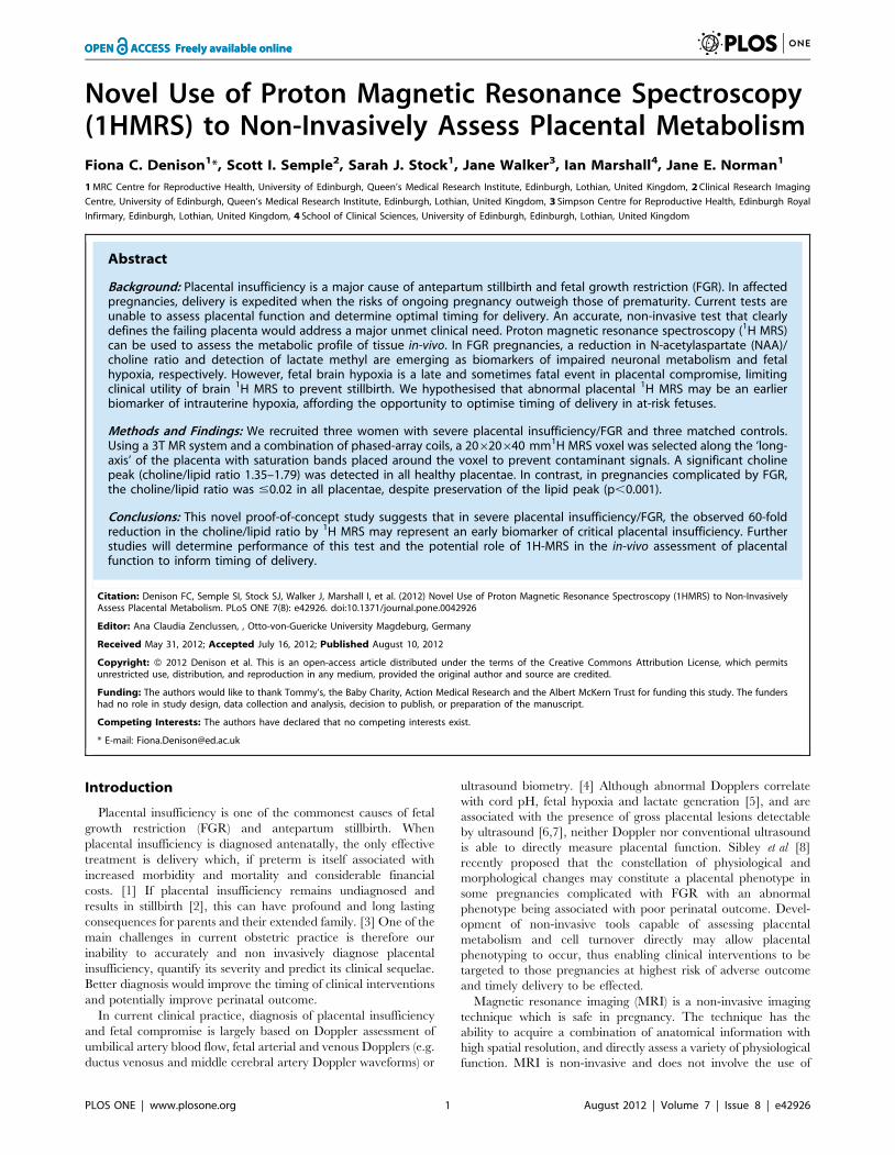
Novel Use of Proton Magnetic Resonance Spectroscopy(1HMRS) to Non-Invasively Assess Placental MetabolismFiona C. Denison1*, Scott I. Semple2, Sarah J. Stock1, Jane Walker3, Ian Marshall4, Jane E. Norman1
1 MRC Centre for Reproductive Health, University of Edinburgh, Queen’s Medical Research Institute, Edinburgh, Lothian, United Kingdom, 2 Clinical Research Imaging
Centre, University of Edinburgh, Queen’s Medical Research Institute, Edinburgh, Lothian, United Kingdom, 3 Simpson Centre for Reproductive Health, Edinburgh Royal
Infirmary, Edinburgh, Lothian, United Kingdom, 4 School of Clinical Sciences, University of Edinburgh, Edinburgh, Lothian, United Kingdom
Abstract
Background: Placental insufficiency is a major cause of antepartum stillbirth and fetal growth restriction (FGR). In affectedpregnancies, delivery is expedited when the risks of ongoing pregnancy outweigh those of prematurity. Current tests areunable to assess placental function and determine optimal timing for delivery. An accurate, non-invasive test that clearlydefines the failing placenta would address a major unmet clinical need. Proton magnetic resonance spectroscopy (1H MRS)can be used to assess the metabolic profile of tissue in-vivo. In FGR pregnancies, a reduction in N-acetylaspartate (NAA)/choline ratio and detection of lactate methyl are emerging as biomarkers of impaired neuronal metabolism and fetalhypoxia, respectively. However, fetal brain hypoxia is a late and sometimes fatal event in placental compromise, limitingclinical utility of brain 1H MRS to prevent stillbirth. We hypothesised that abnormal placental 1H MRS may be an earlierbiomarker of intrauterine hypoxia, affording the opportunity to optimise timing of delivery in at-risk fetuses.
Methods and Findings: We recruited three women with severe placental insufficiency/FGR and three matched controls.Using a 3T MR system and a combination of phased-array coils, a 20620640 mm1H MRS voxel was selected along the ‘long-axis’ of the placenta with saturation bands placed around the voxel to prevent contaminant signals. A significant cholinepeak (choline/lipid ratio 1.35–1.79) was detected in all healthy placentae. In contrast, in pregnancies complicated by FGR,the choline/lipid ratio was #0.02 in all placentae, despite preservation of the lipid peak (p,0.001).
Conclusions: This novel proof-of-concept study suggests that in severe placental insufficiency/FGR, the observed 60-foldreduction in the choline/lipid ratio by 1H MRS may represent an early biomarker of critical placental insufficiency. Furtherstudies will determine performance of this test and the potential role of 1H-MRS in the in-vivo assessment of placentalfunction to inform timing of delivery.
Citation: Denison FC, Semple SI, Stock SJ, Walker J, Marshall I, et al. (2012) Novel Use of Proton Magnetic Resonance Spectroscopy (1HMRS) to Non-InvasivelyAssess Placental Metabolism. PLoS ONE 7(8): e42926. doi:10.1371/journal.pone.0042926
Editor: Ana Claudia Zenclussen, , Otto-von-Guericke University Magdeburg, Germany
Received May 31, 2012; Accepted July 16, 2012; Published August 10, 2012
Copyright: � 2012 Denison et al. This is an open-access article distributed under the terms of the Creative Commons Attribution License, which permitsunrestricted use, distribution, and reproduction in any medium, provided the original author and source are credited.
Funding: The authors would like to thank Tommy’s, the Baby Charity, Action Medical Research and the Albert McKern Trust for funding this study. The fundershad no role in study design, data collection and analysis, decision to publish, or preparation of the manuscript.
Competing Interests: The authors have declared that no competing interests exist.
* E-mail: [email protected]
Introduction
Placental insufficiency is one of the commonest causes of fetal
growth restriction (FGR) and antepartum stillbirth. When
placental insufficiency is diagnosed antenatally, the only effective
treatment is delivery which, if preterm is itself associated with
increased morbidity and mortality and considerable financial
costs. [1] If placental insufficiency remains undiagnosed and
results in stillbirth [2], this can have profound and long lasting
consequences for parents and their extended family. [3] One of the
main challenges in current obstetric practice is therefore our
inability to accurately and non invasively diagnose placental
insufficiency, quantify its severity and predict its clinical sequelae.
Better diagnosis would improve the timing of clinical interventions
and potentially improve perinatal outcome.
In current clinical practice, diagnosis of placental insufficiency
and fetal compromise is largely based on Doppler assessment of
umbilical artery blood flow, fetal arterial and venous Dopplers (e.g.
ductus venosus and middle cerebral artery Doppler waveforms) or
ultrasound biometry. [4] Although abnormal Dopplers correlate
with cord pH, fetal hypoxia and lactate generation [5], and are
associated with the presence of gross placental lesions detectable
by ultrasound [6,7], neither Doppler nor conventional ultrasound
is able to directly measure placental function. Sibley et al [8]
recently proposed that the constellation of physiological and
morphological changes may constitute a placental phenotype in
some pregnancies complicated with FGR with an abnormal
phenotype being associated with poor perinatal outcome. Devel-
opment of non-invasive tools capable of assessing placental
metabolism and cell turnover directly may allow placental
phenotyping to occur, thus enabling clinical interventions to be
targeted to those pregnancies at highest risk of adverse outcome
and timely delivery to be effected.
Magnetic resonance imaging (MRI) is a non-invasive imaging
technique which is safe in pregnancy. The technique has the
ability to acquire a combination of anatomical information with
high spatial resolution, and directly assess a variety of physiological
function. MRI is non-invasive and does not involve the use of
PLOS ONE | www.plosone.org 1 August 2012 | Volume 7 | Issue 8 | e42926
ionizing radiation, making it an ideal candidate for the develop-
ment of biomarkers for fetal compromise.
Until recently the use of MRI for placental assessment has been
primarily used as a complementary technique to ultrasound, for
example assessing placental invasion in cases of suspected placenta
accreta. [9] However, with technological advances it has been
proposed that advanced placental MRI imaging could provide
biomarkers for disease onset and outcome. Recent studies
demonstrate that FGR is associated with a reduction in MRI
measures of proton diffusion within the placenta, and changes
in placental volume and thickness [10], thus supporting this
concept.
Proton magnetic resonance spectroscopy (1H MRS) is based on
similar principles to MRI. It uses a static magnetic field to
temporarily align the nuclear magnetization of protons within the
body. Radiofrequency pulses are then applied which give the
protons enough energy to alter this alignment and, as the protons
return to their original state, the resulting radiofrequency signal is
detected by the MR system. [11] Whilst MRI produces
information as an image, 1H MRS instead provides data on the
relative concentrations of specific metabolites contained within
selected regions of interest, with the local chemical and magnetic
environment in which each proton is situated within that region
determining the ‘chemical shift’ in resonant frequency that each
proton’s signal exhibits after excitation. This chemical shift data is
expressed as a frequency spectrum with contributions of specific
metabolites appearing as individual peaks at discrete frequencies.
The area under each frequency peak is proportional to the
number of protons, and hence the concentration, of the
metabolite. 1H MRS therefore has the potential to be a powerful,
non-invasive method of assessing the metabolic profile of tissue
in vivo.
In healthy pregnancies, a choline peak detected by 1H MRS is
associated with a normal high degree of cell-turnover in the
developing fetal brain whilst an increasing N-acetylaspartate
(NAA) peak with gestational age reflects the increase in the
number of neurons during brain development. In FGR pregnan-
cies, reduction in NAA/choline ratio is emerging as a biomarker of
impaired neuronal metabolism. [12] Although in vitro studies
suggest that altered placental metabolism and reduced cell
turnover may precede the onset of placental and intrauterine
hypoxia [13,14], we are not aware of any studies which have
attempted to assess the metabolic footprint of the placenta
in normal pregnancy and those complicated by FGR using 1H
MRS.
The aim of our study was therefore to establish the feasibility of
undertaking 1H MRS of the placenta in vivo, and to undertake the
first proof-of-concept study to assess whether the metabolic
footprint of the placenta differs in placenta from pregnancies
complicated by FGR compared to healthy controls.
Methods
Ethics StatementThe study was approved by Lothian Research Ethics Commit-
tee (10/S1103/36) and all participants gave written, informed
consent.
Study PopulationPatients were enrolled at the Simpson Centre for Reproductive
Health at the Royal Infirmary, Edinburgh, UK. Magnetic
resonance imaging (MRI) studies were performed at the Clinical
Research Imaging Centre in the Queen’s Medical Research
Institute, University of Edinburgh, Edinburgh, UK.
We studied 3 women with a singleton pregnancy complicated
by severe FGR and suspected fetal compromise and 3 gestation
matched controls. Gestation was calculated from the last
menstrual period and confirmed by routine ultrasonography at
11–13 weeks gestation. All participants had a structurally normal
fetal anomaly scan at 20 weeks gestation. Severe FGR was defined
as an abdominal circumference by ultrasound ,5th centile. [15]
Suspected fetal compromise was defined as absent or reversed end-
diastolic flow on umbilical artery Doppler. Exclusion criteria for
study participation included significant co-existing maternal
systemic disease including gestational diabetes, microvascular
disease, multiple pregnancy, or contraindication to MRI.
Magnetic Resonance StudiesAll magnetic resonance (MR) studies were performed using a
wide-bore dedicated clinical research 3 tesla MR Verio system
(Siemens Medical, Germany). Women were scanned in a left-
lateral tilt to avoid compression of vena-cava with blood pressure
constantly monitored using a Veris MR Vital Signs Monitor
(Medrad, UK). Total MR acquisition times were limited to 40–
45 minutes per participant. No fetal sedation was used. A
combination of body and spine matrix phased-array coils was
used to obtain all images and 1H MRS data. Prior to acquisition of1H MRS data, a series of 2D HASTE slices was acquired in three
orthogonal planes centred on the placenta. Multiple 6 mm slices
were acquired with no inter-slice gap in all three planes, in order
to localise the extent of the placenta. Each HASTE image took
approximately 1 second to acquire, so all images were acquired
with the mother free-breathing.
MR Spectroscopy StudiesFor 1H MRS acquisition, a 20620640 mm PRESS voxel was
selected along the ‘long-axis’ of the placenta (TE/TR 144/
1500 ms, 96 averages). The voxel was selected approximately
2 cm from the cord insertion in all cases and voxel selection was
confirmed to be limited to within the placenta using the range of
orthogonal HASTE slices (Figure 1). Six saturation bands were
placed around the voxel to further prevent any non-placenta
contaminant signals and a second-order semi-automatic shim was
applied over the selected voxel to counter any local inhomogene-
ities in the magnetic field. The voxel dimensions were selected to
maximise sampling of the placental unit, thereby increasing signal-
to-noise values of the resulting spectra. 1H MRS data was acquired
with the mother free-breathing. In our experience, the placental
unit does not significantly move outwith our selected voxel
dimensions during either maternal breathing, or fetal motion. The
resulting raw spectral data was exported to an external workstation
and MRS analysis to assess placental metabolism (Lipid/choline
ratio) was quantified using the Java-based MRS analysis tool
JMRUI (http://www.mrui.uab.es/mrui).
Data AnalysisThe 1H MRS quantification process was performed using the
nonlinear least-squares quantitation algorithm AMARES (Ad-
vanced Method for Accurate, Robust and Efficient Spectral fitting)
with peak fitting performed assuming a Lorentzian line shape.
Since only two peaks were clearly identified, each peak was
identified manually according to its frequency and the line widths
and areas under the curves semi-automatically estimated. Birth
percentiles were calculated using centile charts for birthweight for
gestational age for Scottish singleton births. [16] Data were
analysed by GraphPad Prism (Version 5.0).
Proton Spectroscopy (1H MRS) of Placenta In Vivo
PLOS ONE | www.plosone.org 2 August 2012 | Volume 7 | Issue 8 | e42926
Results
Demographics of Study Population
Maternal age ranged between 20 and 37 (mean, 2866.8 years)
and maternal body mass index (BMI) between 18.4 and 32.8
(mean, 25.764.8 kg/m2). The demographics of the study popu-
lation at the time of MRS scan and delivery are demonstrated in
Tables 1 and 2. Neonatal outcome and time between 1H MRS
and delivery are demonstrated in Table 3.
MRI ResultsIn utero 1H MRS of the placenta was obtained in all participants.
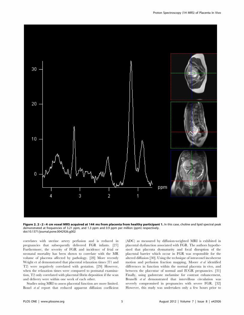
A choline and lipid peak were easily detectable, centred at
3.2 ppm and 1.2 ppm, respectively from placentae in all healthy
controls (Figure 2). In the healthy controls a significant choline
signal was obtained, resulting in a choline/lipid ratio of 1.35–1.79.
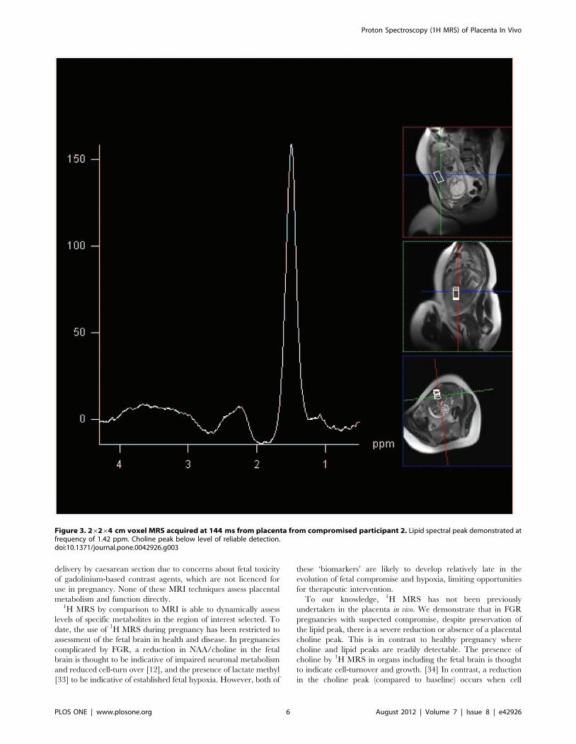
In contrast, despite preservation of the lipid peak, there was severe
attenuation or absence of detectable choline peak in placentae
from pregnancies complicated by severe FGR and suspected fetal
compromise (Figure 3). The choline/lipid ratio was reduced to
#0.02, a reduction of more than 60-fold in pregnancies
Figure 1. Voxel placement in the placenta with demonstration of saturation bands.doi:10.1371/journal.pone.0042926.g001
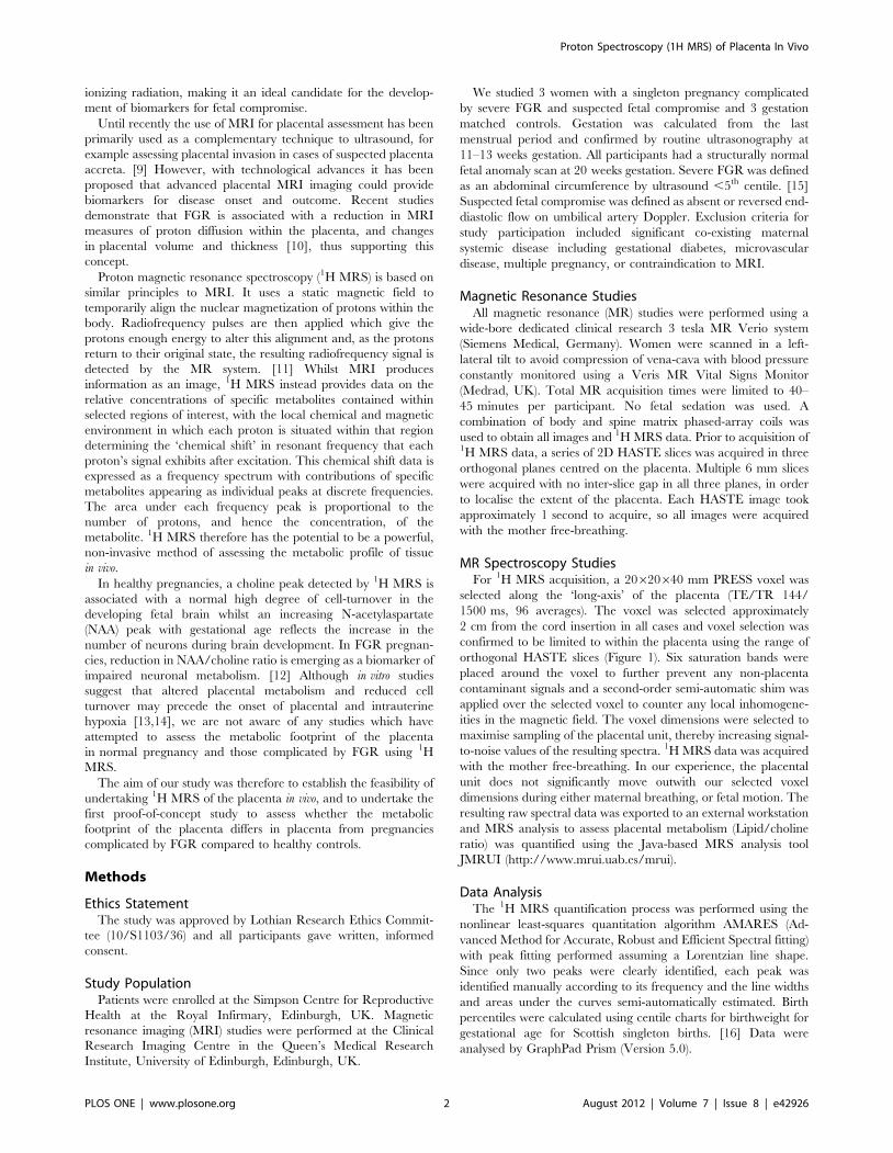
Table 1. Maternal demographics, gestational age and antenatal Dopplers at the time of 1H MRS.
Subject Age Parity BMIAbdominal circumferenceultrasound centile
Umbilicalartery
Liquorvolume
Gestational age at 1HMRS (wks+days)
Healthy 1 33 0+0 18.4 25th–50th Normal Normal 24+4
Healthy 2 31 1+0 23.1 50th–95th Normal Normal 30+3
Healthy 3 37 2+1 25.3 50th–95th Normal Normal 28+0
Compromised 1 30 0+0 23.1 ,5th AEDF Reduced 28+5
Compromised 2 20 0+1 26.4 ,5th AEDF Reduced 25+0
Compromised 3 23 0+1 32.8 ,5th AEDF Reduced 27+1
AEDF (absent end diastolic flow).doi:10.1371/journal.pone.0042926.t001
Proton Spectroscopy (1H MRS) of Placenta In Vivo
PLOS ONE | www.plosone.org 3 August 2012 | Volume 7 | Issue 8 | e42926
complicated by severe FGR compared to the gestation matched
healthy controls (Table 3) (p,0.001).
Discussion
To our knowledge, this is the first report of placental 1H MRS
in vivo in normal and FGR pregnancies. We demonstrate that in
healthy pregnancies, choline and lipid spectral peaks were clearly
detected in all placenta using 1H MRS. In contrast, in pregnancies
complicated by FGR, despite preservation of the lipid peak, the
choline peak was severely attenuated or absent from all placentae.
We speculate that a reduction in the choline/lipid ratio by 1H
MRS may provide a novel biomarker of critical placental failure,
indicative of reduction in cell turnover, which predates fetal
hypoxia and antepartum stillbirth in pregnancies with severe
FGR.
To date, knowledge about placental function in FGR pregnan-
cies has been largely extrapolated from in vitro and ex vivo studies.
Placental weight, total volume, villous volumes and surface area
are significantly reduced in FGR pregnancies. [17] At a
histological level, there is evidence of increased apoptosis, a
thickened basal lamina and reduction in cytotrophoblastic nuclei
and cell-turnover. [14,18] The marked impairment of nutrient
transport [13,19] and placental perfusion which occurs results in
global placental dysfunction and altered metabolism [20,21].
Cetin et al suggested that such alterations in placental metabolism
and function may precede the onset of placental and intrauterine
hypoxia in affected pregnancies. [13] If it were possible to detect
altered placental metabolism prior to the onset of critical placental
failure, this might afford an opportunity for more timely clinical
intervention (including delivery) thus preventing adverse perinatal
outcome.
A variety of non-invasive methods have attempted to assess
placental function in vivo to predict the functional capacity of the
pregnancy and/or pregnancy outcome. The ultrasound based
‘Grannum grading’ of placenta, which was originally developed as
a biomarker for fetal lung maturity, has been assessed as a
predictive tool for fetal growth restriction [22] and placental
function [23]. However, although there is a relationship between
Grannum III grade and FGR, the positive predictive and
sensitivity value of this ‘‘test’’ is low (62% and 66%, respectively)
[22] and Grannum grading at 31–34 weeks of gestation is unable
to reliably predict the functional capacity of the term placenta as
expressed by the surrogate measure, morphometric diffusive
conductance. [23] More recently, near infrared spectroscopy has
been explored as potential method of assessing tissue oxygenation
and placental function. [24–26] To date, results have been
conflicting in FGR pregnancies with tissue oxygenation indexes
exhibiting both increase and decrease in the presence of FGR
depending on its cause. [24–25] Furthermore, due to technical
limitations, the latter technique is only able to assess placental
oxygenation within a narrow range and in women with an anterior
placentae and a thin layer of subcutaneous fat. [24] Until further
method development occurs, these techniques are therefore
unlikely to have a clinical utility in quantifying placental
oxygenation and function in vivo.
Several groups have used MRI as a tool for assessing fetal and
placental structure and function in vivo. At the macroscopic level,
placental volume measured by MRI during the second trimester
Table 2. Characteristics of population at delivery.
SubjectGestational age at delivery,wks+days Mode delivery Sex Fetal weight (g) Percentile at birth
Healthy 1 40+0 SVD Male 3060 25th
Healthy 2 40+3 SVD Male 3760 50th
Healthy 3 39+5 SVD Male 3630 50th
Compromised 1 30+4 SVD Male 750 ,0.4th
Compromised 2 25+4 EmCS Male 670 9th
Compromised 3 28+6 EmCS Male 530 ,0.4th
SVD (spontaneous vertex delivery), EmCS (emergency caesarean section).doi:10.1371/journal.pone.0042926.t002
Table 3. Neonatal outcome and choline/lipid integral 1H MRS ratio.
Subject Outcome
Time between 1HMRS and delivery(days)
Choline/lipid integralratio
Healthy 1 Alive and well 108 1.35
Healthy 2 Alive and well 70 1.79
Healthy 3 Alive and well 82 1.36
Compromised 1 Stillbirth1 13 ,0.02
Compromised 2 Neonatal death at 42 days 4 ,0.02
Compromised 3 Discharged from NNU with BPD and ROP on supplemental oxygen at 42+4 weekscorrected gestation
12 0.02
1Compromised 1 pregnancy was expectantly managed until antenatal stillbirth occurred.NNU (neonatal unit), BPD (bronchopulmonary dysplasia), ROP (retinopathy of prematurity).doi:10.1371/journal.pone.0042926.t003
Proton Spectroscopy (1H MRS) of Placenta In Vivo
PLOS ONE | www.plosone.org 4 August 2012 | Volume 7 | Issue 8 | e42926
correlates with uterine artery perfusion and is reduced in
pregnancies that subsequently delivered FGR infants. [27]
Furthermore, the severity of FGR and incidence of fetal or
neonatal mortality has been shown to correlate with the MR
volume of placenta affected by pathology. [28] More recently
Wright et al demonstrated that placental relaxation times (T1 and
T2) were negatively correlated with gestation. [29] However,
when the relaxation times were compared to postnatal examina-
tion, T2 only correlated with placental fibrin deposition if the scan
and delivery were within one week of each other.
Studies using MRI to assess placental function are more limited.
Bonel et al report that reduced apparent diffusion coefficient
(ADC) as measured by diffusion-weighted MRI is exhibited in
placental dysfunction associated with FGR. The authors hypothe-
sised that placenta dysmaturity and focal disruption of the
placental barrier which occur in FGR was responsible for the
altered diffusion [30]. Using the technique of intravoxel incoherent
motion and perfusion fraction mapping, Moore et al identified
differences in function within the normal placenta in vivo, and
between the placentae of normal and IUGR pregnancies. [31]
Finally, using gadoterate melamine for contrast enhancement,
Brunelli et al demonstrated that intervillous circulation was
severely compromised in pregnancies with severe FGR. [32]
However, this study was undertaken only a few hours prior to
Figure 2. 26264 cm voxel MRS acquired at 144 ms from placenta from healthy participant 1. In this case, choline and lipid spectral peakdemonstrated at frequencies of 3.21 ppm, and 1.3 ppm and 0.9 ppm per million (ppm) respectively.doi:10.1371/journal.pone.0042926.g002
Proton Spectroscopy (1H MRS) of Placenta In Vivo
PLOS ONE | www.plosone.org 5 August 2012 | Volume 7 | Issue 8 | e42926
delivery by caesarean section due to concerns about fetal toxicity
of gadolinium-based contrast agents, which are not licenced for
use in pregnancy. None of these MRI techniques assess placental
metabolism and function directly.1H MRS by comparison to MRI is able to dynamically assess
levels of specific metabolites in the region of interest selected. To
date, the use of 1H MRS during pregnancy has been restricted to
assessment of the fetal brain in health and disease. In pregnancies
complicated by FGR, a reduction in NAA/choline in the fetal
brain is thought to be indicative of impaired neuronal metabolism
and reduced cell-turn over [12], and the presence of lactate methyl
[33] to be indicative of established fetal hypoxia. However, both of
these ‘biomarkers’ are likely to develop relatively late in the
evolution of fetal compromise and hypoxia, limiting opportunities
for therapeutic intervention.
To our knowledge, 1H MRS has not been previously
undertaken in the placenta in vivo. We demonstrate that in FGR
pregnancies with suspected compromise, despite preservation of
the lipid peak, there is a severe reduction or absence of a placental
choline peak. This is in contrast to healthy pregnancy where
choline and lipid peaks are readily detectable. The presence of
choline by 1H MRS in organs including the fetal brain is thought
to indicate cell-turnover and growth. [34] In contrast, a reduction
in the choline peak (compared to baseline) occurs when cell
Figure 3. 26264 cm voxel MRS acquired at 144 ms from placenta from compromised participant 2. Lipid spectral peak demonstrated atfrequency of 1.42 ppm. Choline peak below level of reliable detection.doi:10.1371/journal.pone.0042926.g003
Proton Spectroscopy (1H MRS) of Placenta In Vivo
PLOS ONE | www.plosone.org 6 August 2012 | Volume 7 | Issue 8 | e42926
turnover is significantly reduced and in the presence of apoptosis.
[35] Using ex vivo and in vitro models, Heazell et al [36] and others
[18] [14] have demonstrated a reduction in cell-turnover and
increase in apoptosis in placentae from pregnancies with FGR. We
therefore propose that the significant reduction in choline/lipid
ratio which we demonstrate in FGR placentae may be a novel
biomarker of reduced cell turnover before apoptosis resulting in
impaired placental function and critical organ failure.
Acquiring the 1H MRS data at 3T had the added benefit of the
increased signal to noise available at this higher clinical field
strength. This meant that less averages were required to obtain an
acceptable signal-to-noise ratio for our 1H MRS data. Modern
clinical 3T systems also allow rapid and accurate magnet
shimming to correct for static field inhomogeneities. This meant
that an entire MRI and 1H MRS placental examination could be
obtained with a 40-minutes total acquisition time.
Although our study is limited by small numbers, we were able to
detect a reproducible spectral output from placentae from all the
women that were scanned, regardless of placental site and size,
fetal motion and maternal habitus. However, for this proof-of-
concept study we specifically recruited women with severe FGR
who had evidence of fetal compromise and growth measurements
,5th centile. All babies with severe FGR had poor outcomes. To
assess the clinical utility of 1H MRS as a diagnostic tool for
placental failure, future studies should recruit women with less
severe FGR to assess whether there is a lipid/choline ratio below
which risk of adverse perinatal outcome increases.
In conclusion, our proof-of-concept study demonstrates that the
MRS spectra of placentae in pregnancies complicated by severe
FGR are significantly different from those from healthy pregnan-
cies. Future studies should explore whether the absence of a
choline peak represents a biomarker of critical placental failure
and the consequence of this for perinatal outcome.
Acknowledgments
We would also like to thank Isobel Crawford, Mary Simpson and Jennifer
Rowan for recruiting women to take part in this study. Finally, we would
like to thank the radiographers at the Clinical Research Imaging Centre for
facilitating and performing the MRI scans.
Author Contributions
Conceived and designed the experiments: FCD SIS JEN IM JW.
Performed the experiments: FCD SIS IM JW. Analyzed the data: FCD
SIS JEN IM. Contributed reagents/materials/analysis tools: FCD SIS JEN
IM JW. Wrote the paper: FCD SIS JEN IM JW SJS.
References
1. Mangham LJ, Petrou S, Doyle LW, Draper ES, Marlow N (2009) The cost of
preterm birth throughout childhood in England and Wales. Pediatrics 123:
e312–327.
2. Sankaran S, Kyle PM (2009) Aetiology and pathogenesis of IUGR. Best Pract
Res Clin Obstet Gynaecol 23: 765–777.
3. Schott J, Henley A After a late miscarriage, stillbirth or neonatal death. J Fam
Health Care 20: 116–118.
4. Chalubinski KM, Repa A, Stammler-Safar M, Ott J (2011) The impact of
doppler sonography on intrauterine management and neonatal outcome in
preterm fetuses with intrauterine growth retardation. Ultrasound Obstet
Gynecol.
5. Semple SI, Wallis F, Haggarty P, Abramovich D, Ross JA, et al. (2001) The
measurement of fetal liver T(*)(2) in utero before and after maternal oxygen
breathing: progress towards a non-invasive measurement of fetal oxygenation
and placental function. Magn Reson Imaging 19: 921–928.
6. Sebire NJ, Sepulveda W (2008) Correlation of placental pathology with prenatal
ultrasound findings. J Clin Pathol 61: 1276–1284.
7. Madazli R, Somunkiran A, Calay Z, Ilvan S, Aksu MF (2003) Histomorphology
of the placenta and the placental bed of growth restricted foetuses and
correlation with the Doppler velocimetries of the uterine and umbilical arteries.
Placenta 24: 510–516.
8. Sibley CP, Turner MA, Cetin I, Ayuk P, Boyd CA, et al. (2005) Placental
phenotypes of intrauterine growth. Pediatr Res 58: 827–832.
9. Teo TH, Law YM, Tay KH, Tan BS, Cheah FK (2009) Use of magnetic
resonance imaging in evaluation of placental invasion. Clin Radiol 64: 511–516.
10. Bonel HM, Stolz B, Diedrichsen L, Frei K, Saar B, et al. (2011) Diffusion-
weighted MR imaging of the placenta in fetuses with placental insufficiency.
Radiology 257: 810–819.
11. Story L, Damodaram MS, Allsop JM, McGuinness A, Wylezinska M, et al.
(2011) Proton magnetic resonance spectroscopy in the fetus. Eur J Obstet
Gynecol Reprod Biol 158: 3–8.
12. Story L, Damodaram MS, Allsop JM, McGuinness A, Patel A, et al. (2011) Brain
metabolism in fetal intrauterine growth restriction: a proton magnetic resonance
spectroscopy study. Am J Obstet Gynecol 205: 483 e481–488.
13. Cetin I, Alvino G (2009) Intrauterine growth restriction: implications for
placental metabolism and transport. A review. Placenta 30 Suppl A: S77–82.
14. Macara L, Kingdom JC, Kaufmann P, Kohnen G, Hair J, et al. (1996)
Structural analysis of placental terminal villi from growth-restricted pregnancies
with abnormal umbilical artery Doppler waveforms. Placenta 17: 37–48.
15. Loughna P, Chitty L, Evans T, Chudleigh T (2009) Fetal size and dating: charts
recommended for clinical obstetric practice. Ultrasound 17: 161–167.
16. Bonellie S, Chalmers J, Gray R, Greer I, Jarvis S, et al. (2008) Centile charts for
birthweight for gestational age for Scottish singleton births. BMC Pregnancy
Childbirth 8: 5.
17. Egbor M, Ansari T, Morris N, Green CJ, Sibbons PD (2006) Morphometric
placental villous and vascular abnormalities in early- and late-onset pre-
eclampsia with and without fetal growth restriction. BJOG 113: 580–589.
18. Smith SC, Baker PN, Symonds EM (1997) Increased placental apoptosis in
intrauterine growth restriction. Am J Obstet Gynecol 177: 1395–1401.
19. Horgan RP, Broadhurst DI, Dunn WB, Brown M, Heazell AE, et al. (2010)
Changes in the metabolic footprint of placental explant-conditioned medium
cultured in different oxygen tensions from placentas of small for gestational age
and normal pregnancies. Placenta 31: 893–901.
20. Dunn WB, Brown M, Worton SA, Crocker IP, Broadhurst D, et al. (2009)
Changes in the metabolic footprint of placental explant-conditioned culture
medium identifies metabolic disturbances related to hypoxia and pre-eclampsia.
Placenta 30: 974–980.
21. Heazell AE, Brown M, Dunn WB, Worton SA, Crocker IP, et al. (2008) Analysis
of the metabolic footprint and tissue metabolome of placental villous explants
cultured at different oxygen tensions reveals novel redox biomarkers. Placenta
29: 691–698.
22. Yin TT, Loughna P, Ong SS, Padfield J, Mayhew TM (2009) No correlation
between ultrasound placental grading at 31–34 weeks of gestation and a
surrogate estimate of organ function at term obtained by stereological analysis.
Placenta 30: 726–730.
23. Kazzi GM, Gross TL, Sokol RJ, Kazzi NJ (1983) Detection of intrauterine
growth retardation: a new use for sonographic placental grading. Am J Obstet
Gynecol 145: 733–737.
24. Hasegawa J, Nakamura M, Matsuoka R, Mimura T, Ichizuka K, et al. (2010)
Evaluation of placental function using near infrared spectroscopy during fetal
growth restriction. J Perinat Med 38: 29–32.
25. Kakogawa J, Sumimoto K, Kawamura T, Minoura S, Kanayama N (2010)
Noninvasive monitoring of placental oxygenation by near-infrared spectroscopy.
Am J Perinatol 27: 463–468.
26. Kakogawa J, Sumimoto K, Kawamura T, Minoura S, Kanayama N (2010)
Transabdominal measurement of placental oxygenation by near-infrared
spectroscopy. Am J Perinatol 27: 25–29.
27. Derwig IE, Akolekar R, Zelaya FO, Gowland PA, Barker GJ, et al. (2011)
Association of placental volume measured by MRI and birth weight percentile.
J Magn Reson Imaging 34: 1125–1130.
28. Damodaram M, Story L, Eixarch E, Patel A, McGuinness A, et al. (2010)
Placental MRI in intrauterine fetal growth restriction. Placenta 31: 491–498.
29. Wright C, Morris DM, Baker PN, Crocker IP, Gowland PA, et al. (2011)
Magnetic resonance imaging relaxation time measurements of the placenta at
1.5 T. Placenta 32: 1010–1015.
30. Bonel HM, Stolz B, Diedrichsen L, Frei K, Saar B, et al. (2010) Diffusion-
weighted MR imaging of the placenta in fetuses with placental insufficiency.
Radiology 257: 810–819.
31. Moore RJ, Strachan BK, Tyler DJ, Duncan KR, Baker PN, et al. (2000) In utero
perfusing fraction maps in normal and growth restricted pregnancy measured
using IVIM echo-planar MRI. Placenta 21: 726–732.
32. Brunelli R, Masselli G, Parasassi T, De Spirito M, Papi M, et al. (2010)
Intervillous circulation in intra-uterine growth restriction. Correlation to fetal
well being. Placenta 31: 1051–1056.
33. Cetin I, Barberis B, Brusati V, Brighina E, Mandia L, et al. (2011) Lactate
detection in the brain of growth-restricted fetuses with magnetic resonance
spectroscopy. Am J Obstet Gynecol 205: 350 e351–357.
Proton Spectroscopy (1H MRS) of Placenta In Vivo
PLOS ONE | www.plosone.org 7 August 2012 | Volume 7 | Issue 8 | e42926

34. Brighina E, Bresolin N, Pardi G, Rango M (2009) Human fetal brain chemistry
as detected by proton magnetic resonance spectroscopy. Pediatr Neurol 40: 327–342.
35. Lindskog M, Spenger C, Klason T, Jarvet J, Graslund A, et al. (2005) Proton
magnetic resonance spectroscopy in neuroblastoma: current status, prospectsand limitations. Cancer Lett 228: 247–255.
36. Heazell AE, Sharp AN, Baker PN, Crocker IP (2011) Intra-uterine growth
restriction is associated with increased apoptosis and altered expression of
proteins in the p53 pathway in villous trophoblast. Apoptosis 16: 135–144.
Proton Spectroscopy (1H MRS) of Placenta In Vivo
PLOS ONE | www.plosone.org 8 August 2012 | Volume 7 | Issue 8 | e42926
Copyright © 2022 FDOKUMEN