Novel Lineages in Cercozoa and Their Feeding Strategies
188
Novel Lineages in Cercozoa and Their Feeding Strategies Inaugural – Dissertation zur Erlangung des Doktorgrades der Mathematisch-Naturwissenschaftlichen Fakultät der Universität zu Köln vorgelegt von Kenneth Dumack aus Dormagen Stadtteil Hackenbroich
-
Upload
khangminh22 -
Category
Documents
-
view
0 -
download
0
Transcript of Novel Lineages in Cercozoa and Their Feeding Strategies
Novel Lineages in Cercozoa and Their Feeding Strategies
Inaugural – Dissertation
zur
Erlangung des Doktorgrades
der Mathematisch-Naturwissenschaftlichen Fakultät
der Universität zu Köln
vorgelegt von
Kenneth Dumack
aus Dormagen Stadtteil Hackenbroich

Berichterstatter:
Prof. Dr. Michael Bonkowski Prof. Dr. Hartmut Arndt
Vorsitz der Prüfung:
Prof. Dr. Tim Mansfeldt Beisitzer: Dr. Frank Nitsche Tag der mündlichen Prüfung: 24. 01. 2017
Table of Contents Summary ................................................................................................................................................................. 4
Zusammenfassung .................................................................................................................................................. 6
Introduction ............................................................................................................................................................ 8
Protists ................................................................................................................................................................ 8
Heterotrophic protists in soil biology and their feeding types ........................................................................... 9
Culturing heterotrophic protists and the difficulties ........................................................................................ 11
Cercozoa ........................................................................................................................................................... 14
What we know about fungivores and hypothesized evolutional adaptations ................................................. 15
Aims ...................................................................................................................................................................... 19
Chapters in three parts ......................................................................................................................................... 20
Results ................................................................................................................................................................... 21
Part 1 - Characterization of the unusual bacterivorous amoeba Kraken carinae. .................................. 21
Chapter 1: A Novel Lineage of ‘Naked Filose Amoebae’; Kraken carinae gen. nov. sp. nov. (Cercozoa) with a Remarkable Locomotion by Disassembly of its Cell Body. ........................................................................ 21
Chapter 2: Cercomonad or archaic Imbricatea? On the hunt for the true taxonomy of the scale-bearing Kraken (incertae sedis, Cercozoa, Rhizaria): Combining ultrastructure data and a two-gene (SSU + LSU) phylogeny. .................................................................................................................................................... 33
Part 2 - Eukaryvorous amoebae of the Thecofilosea, Cercozoa. ............................................................ 51
Chapter 3: Description of Lecythium terrestris sp. nov. (Chlamydophryidae, Cercozoa), a Soil Dwelling Protist Feeding on Fungi and Algae. ............................................................................................................. 51
Chapter 4: A bowl with marbles: Revision of the thecate amoeba genus Lecythium (Chlamydophryidae, Tectofilosida, Cercozoa, Rhizaria) including a description of four new species and an identification key. . 65
Chapter 5: Shedding light on the polyphyletic genus Plagiophrys: The transition of some of its species to Rhizaspis (Tectofilosida, Thecofilosea, Cercozoa) and the establishment of Sacciforma gen. nov. (Cryomonadida, Thecofilosea, Cercozoa). .................................................................................................... 86
Chapter 6: Polyphyly in the thecate amoeba genus Lecythium (Chlamydophryidae, Tectofilosida, Cercozoa), redescription of its type species L. hyalinum, description of L. jennyae sp. nov. and the establishment of Fisculla gen. nov. and Fiscullidae fam. nov. .................................................................... 116
Part 3 - Eukaryvorous protists, their capabilities and dispersal. ........................................................... 142
Chapter 7: What does it take to eat a fungus? A case study with the eukaryvorous amoeba Fisculla terrestris. .................................................................................................................................................... 142
Chapter 8: The soil food web revisited: Diverse and widespread mycophagous soil protists. .................. 158
Discussion ........................................................................................................................................................... 168
Unknown Diversity in Cercozoa ...................................................................................................................... 168
Culturing of ‘unculturable’ protists ................................................................................................................. 169
What is the ecological impact and dispersal of eukaryvorous protists? ........................................................ 171
Remaining questions and perspective for future investigations .................................................................... 172
References .......................................................................................................................................................... 175
Acknowledgements ............................................................................................................................................. 184
Subpublications in Three Parts and Record of Achievement .............................................................................. 185
Erklärung (gemäß § 4 Abs 1 Punkt 9 der Prüfungsordnung) ............................................................................... 187
Lebenslauf ........................................................................................................................................................... 188
Novel Lineages in Cercozoa and Their Feeding Strategies Kenneth Dumack
4
Summary The term protist describes an informal grouping of unicellular eukaryotic organisms that do not form
tissues. With a tremendous diversity in morphology and ecology, they represent the vast majority of
eukaryotic heterogeneity of which only a small fraction is yet known. Ubiquitously dispersed in
marine, freshwater and terrestrial habitats they occupy various ecological niches as e.g. primary
producers, osmotrophs, bacterivores, fungivores, algivores, omnivores, predators or parasites of e.g.
animals and plants. However, methodological drawbacks in culturing impeded research of protists.
Research focused on the easily culturable taxa, especially bacterivores and algae, leading to a skewed
image of protist diversity and many ‘unculturable’ protist taxa are still unknown to science.
Therefore we focused on protists with unusual feeding types (in particular bacterivorous sit-and-wait
predators and eukaryvorous predators) in the phylum Cercozoa. The Cercozoa CAVALIER-SMITH
1998, were discovered to be closely related based on molecular analyses although being highly
divergent in morphology and ecology. Molecular surveys revealed a high genetic diversity in the
Cercozoa of which only a small fraction can yet be linked to morphological data. Based on culture
material, cell morphology, feeding processes and life history stages of several cercozoan amoebae
have been studied by us. This was achieved by using mainly light microscopy and time-lapse
photography but also ultra structure data was obtained. Genetic markers, e.g. SSU rDNA and LSU
rDNA were subjected to phylogenetic analyses to draw conclusions on cercozoan evolution.
Based on six isolates from German and Spanish soils a novel lineage of bacterivorous amoebae was
described. Kraken carinae gen. nov. sp. nov. is an amoeba distinguished by a scale bearing cell body
(usually <10 µm in diameter) and a network of filopodia (up to 0.5 mm in diameter). K. carinae is one
of the few known sit-and-wait predators in the Cercozoa, preying on bacteria that get in contact with
its large filopodia network. Unlike other cercozoan amoebae that usually use the filopodia to drag
prey to their cell bodies for ingestion, K. carinae ingests bacteria directly at the point of contact and
transports them through the filopodia to the cell body for digestion. SSU rDNA phylogeny showed an
affinity to the order Cercomonadida in the class Sarcomonadea with only weak support, but a
concatenated approach, by combining SSU rDNA and LSU rDNA sequences, confirmed the results
with higher (though still moderate) support, in particular with the family Paracercomonadidae.
However, Kraken carinae still remains incertae sedis as ultrastructure revealed the presence of
scales, a morphological character predominantly known from the class Imbricatea, contradicting the
phylogenetic results.
By combining literature research with phylogenetic examination focusing on Lecythium (HERTWIG et
LESSER, 1874) and its family the Chlamydophryidae (DE SAEDELEER 1934) we were able to clarify the
confusing taxonomy of genera like Plagiophrys, Lecythium, Rhizaspis and others. All of these
amoebae bear a flexible organic theca, branching and anastomosing filopodia. However, they differ
in cell shape and show species (or strain) specific feeding preferences. SSU rDNA phylogenies
reflected the phenotypic differences between those genera but also revealed surprising results: The
genera Lecythium (Novel Clade 4) and Rhizaspis (Tectofilosida) were polyphyletic and had to be
separated, resulting in a secession of Fisculla gen. nov. (Tectofilosida) from Lecythium and Sacciforma
gen. nov. (Cryomonadida) from Rhizaspis.
Novel Lineages in Cercozoa and Their Feeding Strategies Kenneth Dumack
5
As these thecofilosean amoebae, similar to the predominantly known eukaryvores in the Cercozoa,
the Vampyrellida, have been shown to be eukaryvorous, we further focused on eukaryvorous protists
in terrestrial habitats, by investigating (a) their feeding preferences (b) their physiological
requirements to consume eukaryotic prey and (c) their dispersal in terrestrial and freshwater
systems. This was achieved by conducting thorough sampling, observing individuals in their unaltered
sample (if possible) and performing experiments on feeding preference, chemical sensing, and
enzyme production with several omnivorous or eukaryvorous Cercozoa. In laboratory experiments,
we could show that the eukaryvorous protist Fisculla terrestris is able to sense and select its
preferred prey and produce a battery of enzymes needed to digest cell wall compounds of
eukaryotes, such as chitin. F. terrestris preferred fungal prey (in particular Saccharomyces cerevisiae)
and only fed to a small extend on algae. To get more insight into the dispersal of eukaryvorous
protists in terrestrial systems, we screened metatranscriptomes of different terrestrial habitats for
the eukaryvorous Vampyrellida and Grossglockneriidae, showing high dispersal, since they were
present in all screened habitats, with up to 3% of total reads.
The phenotypic, phylogenetic and ecologic data on the investigated cercozoan amoebae resulted in a
comprehensive characterization of the Thecofilosea and the novel lineage Krakenidae. Based on
intensive literature research and a critical evaluation of it, first steps for a phylogeny-based taxonomy
of these cercozoan lineages were made. Finally, this thesis provides an evaluation of the hidden
diversity of eukaryvorous Cercozoa in terrestrial and freshwater habitats.
Novel Lineages in Cercozoa and Their Feeding Strategies Kenneth Dumack
6
Zusammenfassung
Der Begriff Protist beschreibt einzellige, eukaryotische Organismen die kein Gewebe ausbilden. Mit
einer enormen Mannigfaltigkeit in Morphologie und Ökologie repräsentieren sie die Mehrheit der
eukaryotischen Diversität, von der bisher nur ein kleiner Teil bekannt ist. Weltweit verbreitet, in
marinen, limnischen und terrestrischen Habitaten, besetzen sie unzählige ökologische Nischen, z. B.
als Primärproduzenten, Osmotrophen, Bakterivoren, Fungivoren, Algivoren, Omnivoren, Prädatoren
oder Parasiten z. B. von Tieren und Pflanzen. Probleme im Kultivieren von Protisten sorgten
allerdings für eine bevorzugte Erforschung von Bakterivoren und Algen, was zu einem verzerrten
Abbild der Protistendiversität führte. Viele „nicht kultivierbare“ Protisten Taxa sind der Forschung
noch immer unbekannt.
Deshalb befassten wir uns mit Protisten mit unüblichen Ernährungsweisen (insbesondere bakterivore
Lauerjäger und eukaryvore Räuber) in den Cercozoa. Die Cercozoa CAVALIER-SMITH 1998, wurden
aufgrund von molekularen Untersuchungen als phylogenetische Entität entdeckt obwohl sie starke
Unterschiede in Morphologie und Ökologie aufweisen. Molekulare Untersuchungen deckten eine
hohe Diversität in den Cercozoa auf, von der bisher nur ein geringer Bruchteil mit morphologischen
Daten verknüpft werden kann. Auf Basis von angelegten Kulturen wurden Zellmorphologie,
Ernährungsweise und Zellzyklen von verschiedenen cercozoen Amöben untersucht. Dafür wurden
hauptsächlich Lichtmikroskopie und Zeitraffer-Mikrofotographie und auch durch
Ultrastrukturaufnahmen Daten erhoben. Genetische Marker, d. h. SSU rDNA und LSU rDNA, wurden
sequenziert und für phylogenetische Untersuchungen genutzt, um Rückschlüsse über die Evolution
der Cercozoa zu ziehen.
Von einer unbeschriebenen bakterivoren Amöbe wurden sechs Stämme aus deutschen und
spanischen Böden isoliert. Kraken carinae gen. nov. sp. nov. ist eine Amöbe, unterteilt in einen
schuppentragenden Zellkörper (normalerweise <10 µm im Durchmesser) und ein Netzwerk aus
Filopodien (bis zu 0,5 mm im Durchmesser). K. carinae ist einer der seltenen Lauerjäger der Cercozoa,
sie bewegt sich nur selten und erbeutet Bakterien, die mit ihrem Netzwerk aus Filopodien in Kontakt
kommen. Anders als andere amöboide Vertreter der Cercozoa, die üblicherweise ihre Filopodien
nutzen, um ihre Beute zum Zellkörper zu ziehen und dann zu phagozytieren, ingestiert K. carinae
Bakterien direkt am Kontaktpunkt der Beute mit den Filopodien und transportiert diese dann
intrazellulär zum Zellkörper um sie dort zu verdauen. SSU rDNA Phylogenie zeigte eine nähere
Verwandtschaft mit den Cercomonadida auf, wenn auch nur mit mäßiger statistischer
Unterstützung. Ein weiterer Anlauf, diesmal mit verketteten SSU rDNA und LSU rDNA Sequenzen,
bestätigte die Verwandtschaft zu den Cercomonadida, insbesondere mit den Paracercomonadidae,
mit erhöhter (aber dennoch nur moderater) statistischer Unterstützung. Allerdings bleiben die
genaue Verwandtschaftsverhältnisse von Kraken carinae immernoch unklar, da die
Ultrastrukturdaten konträr zu den phylogenetischen Ergebnissen, auf eine nähere Verwandtschaft
mit den Imbricatea hinweisen, da Kraken carinae genau wie sie Schuppen auf dem Zellkörper trägt.
Durch die Kombination von intensiver Literaturrecherche mit phylogenetischen Untersuchungen von
Lecythium (HERTWIG et LESSER, 1874) und seiner Familie, der Chlamydophryidae (DE SAEDELEER
1934) konnten wir die unklare Taxonomie von Gattungen wie (Plagiophrys, Lecythium, Rhizaspis, …)
enträtseln. All diese Amöben haben gemeinsam, dass sie eine hyaline flexible Schale tragen und
verästelnde und anastomisierende Filopodien aufweisen, aber haben auch klare Unterschiede
zueinander. SSU rDNA Phylogenien haben die phänotypischen Unterschiede zwischen den Gattungen
Novel Lineages in Cercozoa and Their Feeding Strategies Kenneth Dumack
7
widergespiegelt, bargen aber auch Überraschungen: Die Gattungen Lecythium (Novel Clade 4) und
Rhizaspis (Tectofilosida) waren beide polyphyletisch und mussten daher geteilt werden. Daher
wurden Fisculla gen. nov. (Tectofilosida) von Lecythium und Sacciforma gen. nov. (Cryomonadida)
von Rhizaspis abgetrennt. Weiterhin zeigen diese Schalenamöben, ähnlich wie die meistbekannten
Eukaryvoren in den Cercozoa, die Vampyrellida, eukaryvores Verhalten. Deshalb führten wir weitere
Studien über die Ökologie von eukaryvoren Protisten in terrestrischen Habitaten durch. Vor allem
geben wir Einsicht auf (a) ihre Nahrungspräferenzen, (b) den physiologischen Voraussetzungen, um
eukaryotische Beute zu konsumieren und (c) ihrer Verbreitung in terrestrischen und limnischen
Systemen. Dazu wurden umfassende Probenahmen, gefolgt von Experimenten durchgeführt, welche
Fütterungsversuche und Experimente über die chemische Wahrnehmung und Enzymproduktion mit
diversen omnivoren oder eukaryvoren Cercozoa umfassten. Wir konnten zeigen, dass die eukaryvore
Amöbe Fisculla terrestris fähig ist, die Anwesenheit ihrer bevorzugten Beute (Saccharomyces
cerevisiae) wahrzunehmen und darauf zu selektieren. Außerdem produzieren sie eine Auswahl an
Enzymen zum Abbau der Zellwand der Beute, wie z. B. Chitin. Um mehr Einblick auf die Verbreitung
von terrestrischen Eukaryvoren zu erhalten, durchsuchten wir Metatranskriptom-Datenbanken von
verschiedenen terrestrischen Habitaten nach den eukaryvoren Vampyrellida und Grossglockneriidae,
die auf eine weite Verbreitung von Vampyrellida und Grossglockneriidae hinwiesen, da diese nicht
nur in allen überprüften Habitaten nachgewiesen werden konnten, sondern auch bis zu 3% der
absoluten Sequenzen ausmachten.
Die phänotypischen und phylogenetischen Daten der untersuchten Amöben der Cercozoa resultieren
in einer umfassenden Charakterisierung der Thecofilosea und der neuen Familie Krakenidae. Auf
Grundlage einer intensiven Literaturrecherche und einer kritischen Auswertung dieser im Kontext
der selbstständig durchgeführten Arbeiten wurden erste Schritte in Richtung einer
phylogeniebasierten Taxonomie gemacht. Diese Doktorarbeit umfasst weiterhin eine Diskussion über
die noch unerforschte Diversität der Cercozoa in terrestrischen und limnischen Habitaten.
Novel Lineages in Cercozoa and Their Feeding Strategies Kenneth Dumack
8
Introduction
Protists
The term protist defines an informal grouping of unicellular eukaryotes that do not form
tissues. Protists represent the vast majority of eukaryotic diversity of which only a small
fraction is yet known. Despite their estimated 60.000 - 300.000 species (Foissner 2008; Mora
et al. 2011) with a tremendous diversity in ecology and morphology, they were traditionally
assigned to the same single eukaryotic kingdom, Protista in the Eukaryota, next to plants,
animals and fungi (Haeckel 1866; Whittaker 1969). The Protista accommodated therefore
organisms of fundamentally different lifestyles, like autotrophic and mixotrophic algae,
heterotrophic bacterial grazers and parasites, but also organisms of diverse appearances,
like ciliates, flagellates, naked amoebae and testate amoebae. That is why protists were
shared as a field of research among zoologists, botanists and mycologists. The most animal-
like protists (i.e. heterotrophic protists feeding by means of phagocytosis) were called
protozoa (proto= first; zoa= animals) and studied by zoologists, whereas pigmented protists
were adopted by the botanists as protophytes (phytes= plants) and fungi-like protists (like
the fruiting body forming myxomycetes or the osmotrophic filamentous oomycetes) were
studied by mycologists. Researchers were well aware that even morphological highly similar
taxa (e.g. the dinoflagellates or euglenids) comprised heterotrophic protists and also
photosynthetic algae, but were unable to resolve this issue in a widely accepted taxonomy.
Since the 18th and 19th century where protists were most often studied by light microscopy,
novel methods were established: In the early 20th century electron microscopical techniques
were developed to acquire more detailed morphological data for protist taxonomy, in the
late 20th century molecular methods enabled the comparison of genetic markers for
phylogeny.
The molecular methods led finally to a widely accepted consensus in protist taxonomy (Adl
et al. 2005, Adl et al. 2012, Baldauf 2008). Protists turned out to be paraphyletic, instead of
being separated kingdoms, with multicellular eukaryotes nestled in between. Although
ciliates show monophyletic origin (Lynn and Sogin 1988; Sogin and Elwood 1986), flagellates,
naked and testate amoebae are dispersed all over the eukaryotic tree of life showing
paraphyly and polyphyly. For instance, testate amoebae evolved independently in at least
three different lineages, the Amoebozoa, Cercozoa and Stramenopiles (Kosakyan et al. 2016;
Novel Lineages in Cercozoa and Their Feeding Strategies Kenneth Dumack
9
Nikolaev et al. 2005; Wylezich et al. 2002). The search for the last eukaryotic common
ancestor is still ongoing and a highly discussed controversy. Currently it is only accepted that
the eukaryotic ancestor was probably a heterotrophic flagellate of unknown affiliation, but
concepts differ about the most basal protist taxon (Baldauf 2008; Stechmann & Cavalier-
Smith 2002). Cavalier-Smith (1981) established the Archezoa, but this grouping of lineages
he believed to be the most basal protists turned out to be an artifact of long branch
attraction (Philippe and Germot 2000). Based on rare genetic events like gene fusions and
fissions it is assumed that the eukaryotes are divided into two groups, the unikonts
(Opisthokonts and Amoebozoa) and bikonts (the remaining eukaryotic diversity). However,
still no consensus whether to root the eukaryotes in the unikonts or bikonts could be
achieved (Baldauf 2008). Nevertheless, it is widely accepted that during eukaryotic evolution
several lineages lost independently their flagella and evolved locomotion by amoeboid
movement and/ or gained (or secondarily lost again) autotrophy by endocytobiosis, leading
to the intermingled ecology and physiology, sometimes even between closely related protist
taxa (Nowack 2014; Rogers et al. 2007; Stechmann & Cavalier-Smith 2002).
Heterotrophic protists in soil biology and their feeding types
Protists, especially in soil ecology, were commonly considered to represent the major bacterial
grazers, thereby channeling the carbon flow to higher trophic levels (Bonkowski 2004, Crotty et al.
2011, de Ruiter et al. 1995, Hunt et al. 1987).
However, these assumptions were derived from simple model calculations, based on laboratory
experiments with few selected species. Although molecular methods gradually revealed the
enormous phenotypic and genetic diversity of heterotrophic protists, models on protist functional
roles have not changed in soil biology (Holtkamp et al. 2011, Banašek-Richter et al. 2009). For
instance Glücksman et al. (2010) and Weisse et al. (2001) showed that prey (bacteria) communities
have been altered by protist grazing in a species-specific manner, but still by far most information on
the impact of protist grazing have been obtained in studies either with whole microbial communities
in which measured effects can not be traced back to the causing species or in abstract assemblages
with just one or few selected organisms. Moreover, such studies were most often conducted only
with exclusively bacterivorous protists. The non-bacterivorous protists have been addressed in very
few, most often taxonomic studies (Bass et al. 2009; Berney et al. 2013; Foissner 1980; Petz and
Foissner 1985). Their ecology and functional importance is often not known.
Novel Lineages in Cercozoa and Their Feeding Strategies Kenneth Dumack
10
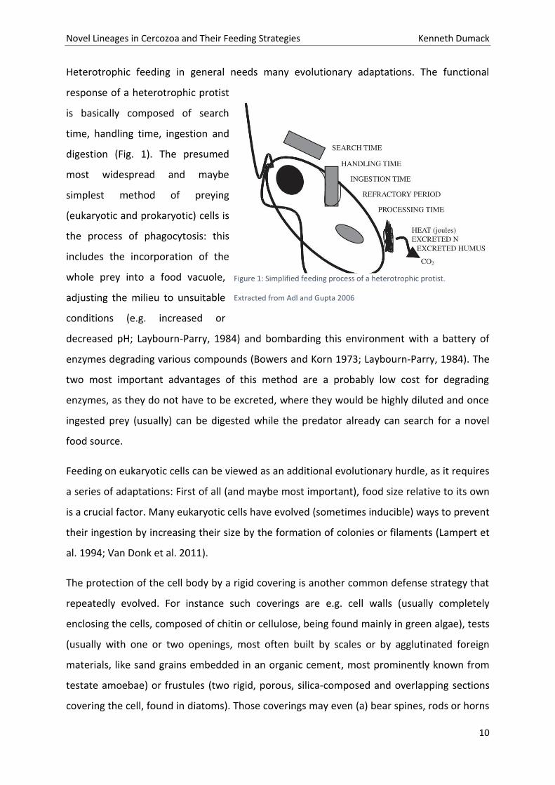
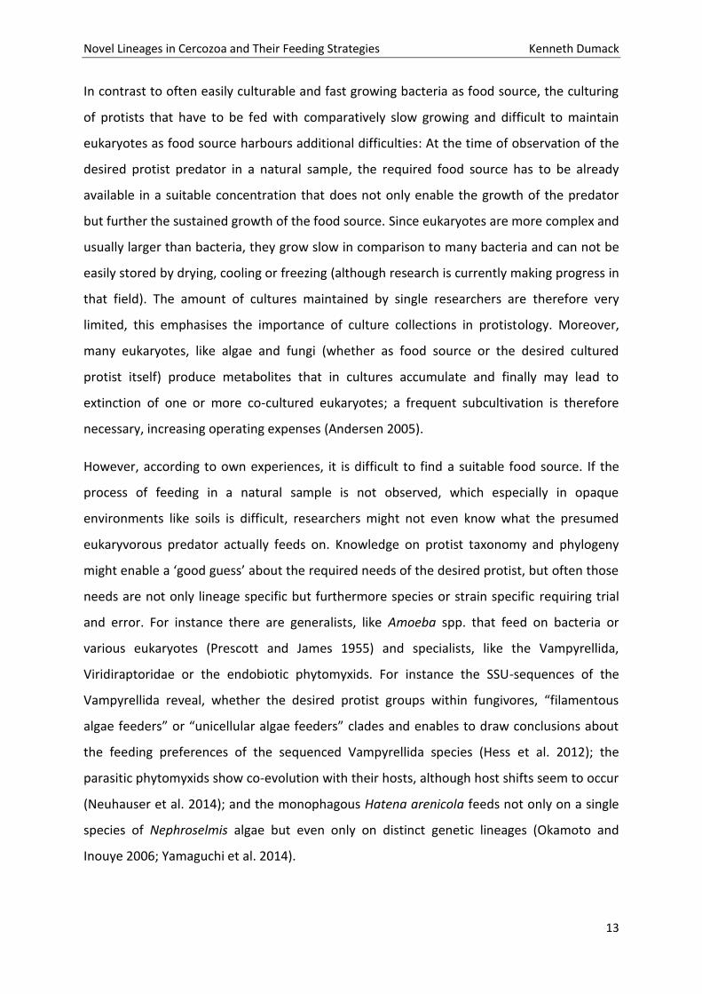
Heterotrophic feeding in general needs many evolutionary adaptations. The functional
response of a heterotrophic protist
is basically composed of search
time, handling time, ingestion and
digestion (Fig. 1). The presumed
most widespread and maybe
simplest method of preying
(eukaryotic and prokaryotic) cells is
the process of phagocytosis: this
includes the incorporation of the
whole prey into a food vacuole,
adjusting the milieu to unsuitable
conditions (e.g. increased or
decreased pH; Laybourn-Parry, 1984) and bombarding this environment with a battery of
enzymes degrading various compounds (Bowers and Korn 1973; Laybourn-Parry, 1984). The
two most important advantages of this method are a probably low cost for degrading
enzymes, as they do not have to be excreted, where they would be highly diluted and once
ingested prey (usually) can be digested while the predator already can search for a novel
food source.
Feeding on eukaryotic cells can be viewed as an additional evolutionary hurdle, as it requires
a series of adaptations: First of all (and maybe most important), food size relative to its own
is a crucial factor. Many eukaryotic cells have evolved (sometimes inducible) ways to prevent
their ingestion by increasing their size by the formation of colonies or filaments (Lampert et
al. 1994; Van Donk et al. 2011).
The protection of the cell body by a rigid covering is another common defense strategy that
repeatedly evolved. For instance such coverings are e.g. cell walls (usually completely
enclosing the cells, composed of chitin or cellulose, being found mainly in green algae), tests
(usually with one or two openings, most often built by scales or by agglutinated foreign
materials, like sand grains embedded in an organic cement, most prominently known from
testate amoebae) or frustules (two rigid, porous, silica-composed and overlapping sections
covering the cell, found in diatoms). Those coverings may even (a) bear spines, rods or horns
Figure 1: Simplified feeding process of a heterotrophic protist.
Extracted from Adl and Gupta 2006
Novel Lineages in Cercozoa and Their Feeding Strategies Kenneth Dumack
11
which may also prevent the ingestion by predators or (b) be enveloped by mucilage which
may prevent adhesion of predatory cells or even impede prey recognition (Van Donk et al.
2011).
Next to such morphological defense mechanisms, protists also evolved behavioral defense
strategies that might be inducible and aggressor-specific, like (a) the production of chemical
compounds, e.g. toxins; (b) flight, often aided by special organelles, like extrusomes or
trichocysts, that can be ‘fired’ to the possible predator; (d) metabolic movement that
enables already ingested prey to stretch and burst food vacuoles, like in Euglena mutabilis
when being consumed by Actinophrys sp. or a yet undescribed species in the Leptophryidae
(Hausmann 1978; Potin et al. 1999, Van Donk et al. 2011; Hess pers. comm.; own
observations unpublished).
Some specialized predators co-evolved strategies to overcome such defense mechanisms.
These adaptations are often highly specialized: Large cells, like filaments, even with a thick
cell wall might be an easy prey if the predator is adapted by morphology and behavior, e.g.
some ciliates (e.g. Grossglockneriidae; Foissner 1980, 1999) are equipped with a feeding
tubus for the lysis of fungal cell walls; or some cercozoans (e.g. Vampyrellida;
Viridiraptoridae; Hess et al. 2012, Hess & Melkonian 2013) are able to perforate algal or
fungal cell walls by extracellular enzymatic degradation. Moreover, trichocysts or
extrusomes might also be used by attacking cells (Hausmann 1978).
Research in protistology is astonishing and still full of surprises; we therefore focus in this
thesis on heterotrophic protists that show interesting and unusual feeding types.
Culturing heterotrophic protists and its difficulties
In early times of protistology, protists were predominantly studied by direct observations of
fresh sampled material. Marine and freshwater samples do not require any preparation thus
being intensively studied (Hertwig & Lesser 1874; Penard 1890). Soils however, due to being
opaque, can not be observed directly but need preparation like dilution. Diverse methods
have been developed, like the most probable number (MPN; Darbyshire et al. 1974) or liquid
aliquot method (LAM; Butler and Rogerson 1995), all of which have advantages and
disadvantages. Still today, there is no method that enables an objective unbiased view on
Novel Lineages in Cercozoa and Their Feeding Strategies Kenneth Dumack
12
soil microorganisms and soil protists are mainly studied by indirect observation of cultured
material extracted from soil samples. Despite the abstract nature of such observations,
culturing, i.e. the long term maintenance of single species or strains in an artificial and
controlled environment, enabled researchers to study complex life cycles, for instance the
amoebozoan genus Pelomyxa GREEFF 1874, comprises about 20 described species of which
the validity is still highly discussed (Frolov et al. 2004, 2005, 2006 and 2011). It is currently
not clear whether all described species are just different stages of the life cycle of Pelomyxa
palustris (the type species of the genus; Chapman-Andresen 1978, 1982), or how many true
Pelomyxa species exist (2004, 2005, 2006 and 2011). Another example for protists with
complex life cycles are the fruiting-body forming amoebae where in many cases the identity
of fruiting bodies and trophozoites can not be assigned to each other (Tice et al. 2016). Even
in well studied amoebae, like the Acanthamoebidae, novel life history stages (like fruiting-
bodies) can be discovered (Tice et al. 2016). Finally, cultures enable the conduction of
laboratory experiments, such as an alteration of environmental variables, i.e. biotic or
abiotic, enabling conclusions about species autecology, i.e. preferred food sources, necessity
of certain chemical compounds in the environment and in general conclusions might be
drawn about several biological aspects, such as their physiology and ecology.
For bacteria it is estimated that barely 1% of their diversity can be cultured (see for instance:
Ferrer et al. 2009; Lee et al. 2010). Although many (bacterivorous) protist lineages are easily
culturable by basically transferring single individuals into water with a carbon source, the
vast majority has highly specific requirements (Page 1976). Therefore it would not be
surprising to find a similar pattern for protists; and indeed culturing protists differs in the
degree of difficulty and is strongly depended of the requirements of the targeted species.
Besides abiotic conditions like medium composition and temperature, it seems obvious that
heterotrophic protists need a suitable food source in a sufficient concentration: e.g.
fungivorous protists need fungi; bacterivorous protists need bacteria and so on. Culturing
bacterivorous protists is often quite easy, often non-toxic strains of bacteria are added or
maybe even easier, co-transferred bacteria are fed with solved carbon, the bacteria then
grow fast, even under nutrient limited conditions and subsequently can be grazed by the
protists.
Novel Lineages in Cercozoa and Their Feeding Strategies Kenneth Dumack
13
In contrast to often easily culturable and fast growing bacteria as food source, the culturing
of protists that have to be fed with comparatively slow growing and difficult to maintain
eukaryotes as food source harbours additional difficulties: At the time of observation of the
desired protist predator in a natural sample, the required food source has to be already
available in a suitable concentration that does not only enable the growth of the predator
but further the sustained growth of the food source. Since eukaryotes are more complex and
usually larger than bacteria, they grow slow in comparison to many bacteria and can not be
easily stored by drying, cooling or freezing (although research is currently making progress in
that field). The amount of cultures maintained by single researchers are therefore very
limited, this emphasises the importance of culture collections in protistology. Moreover,
many eukaryotes, like algae and fungi (whether as food source or the desired cultured
protist itself) produce metabolites that in cultures accumulate and finally may lead to
extinction of one or more co-cultured eukaryotes; a frequent subcultivation is therefore
necessary, increasing operating expenses (Andersen 2005).
However, according to own experiences, it is difficult to find a suitable food source. If the
process of feeding in a natural sample is not observed, which especially in opaque
environments like soils is difficult, researchers might not even know what the presumed
eukaryvorous predator actually feeds on. Knowledge on protist taxonomy and phylogeny
might enable a ‘good guess’ about the required needs of the desired protist, but often those
needs are not only lineage specific but furthermore species or strain specific requiring trial
and error. For instance there are generalists, like Amoeba spp. that feed on bacteria or
various eukaryotes (Prescott and James 1955) and specialists, like the Vampyrellida,
Viridiraptoridae or the endobiotic phytomyxids. For instance the SSU-sequences of the
Vampyrellida reveal, whether the desired protist groups within fungivores, “filamentous
algae feeders” or “unicellular algae feeders” clades and enables to draw conclusions about
the feeding preferences of the sequenced Vampyrellida species (Hess et al. 2012); the
parasitic phytomyxids show co-evolution with their hosts, although host shifts seem to occur
(Neuhauser et al. 2014); and the monophagous Hatena arenicola feeds not only on a single
species of Nephroselmis algae but even only on distinct genetic lineages (Okamoto and
Inouye 2006; Yamaguchi et al. 2014).
Novel Lineages in Cercozoa and Their Feeding Strategies Kenneth Dumack
14
However, long-time maintained cultures enable research on a professional basis and are
therefore imperative for taxonomic and autecological research.
Cercozoa
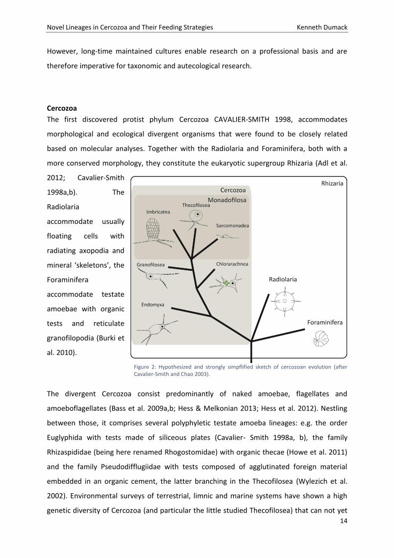
The first discovered protist phylum Cercozoa CAVALIER-SMITH 1998, accommodates
morphological and ecological divergent organisms that were found to be closely related
based on molecular analyses. Together with the Radiolaria and Foraminifera, both with a
more conserved morphology, they constitute the eukaryotic supergroup Rhizaria (Adl et al.
2012; Cavalier-Smith
1998a,b). The
Radiolaria
accommodate usually
floating cells with
radiating axopodia and
mineral ‘skeletons’, the
Foraminifera
accommodate testate
amoebae with organic
tests and reticulate
granofilopodia (Burki et
al. 2010).
The divergent Cercozoa consist predominantly of naked amoebae, flagellates and
amoeboflagellates (Bass et al. 2009a,b; Hess & Melkonian 2013; Hess et al. 2012). Nestling
between those, it comprises several polyphyletic testate amoeba lineages: e.g. the order
Euglyphida with tests made of siliceous plates (Cavalier- Smith 1998a, b), the family
Rhizaspididae (being here renamed Rhogostomidae) with organic thecae (Howe et al. 2011)
and the family Pseudodifflugiidae with tests composed of agglutinated foreign material
embedded in an organic cement, the latter branching in the Thecofilosea (Wylezich et al.
2002). Environmental surveys of terrestrial, limnic and marine systems have shown a high
genetic diversity of Cercozoa (and particular the little studied Thecofilosea) that can not yet
Figure 2: Hypothesized and strongly simpflified sketch of cercozoan evolution (after Cavalier-Smith and Chao 2003).
Novel Lineages in Cercozoa and Their Feeding Strategies Kenneth Dumack
15
be connected to morphological and autecological data (Bass and Cavalier-Smith 2004),
leading to our first hypothesis that there are still many lineages in Cercozoa to be
discovered.
The Thecofilosea were established by Cavalier-Smith and Chao (2003) to accommodate
thecate flagellates and amoebae. On a morphological basis it was assumed that the thecate
amoeba family Chlamydophryidae might branch as well in the Thecofilosea, but molecular
data was lacking (Howe et al. 2011).
The family Chlamydophryidae was established by de Saedeleer (1934) (then as a subfamily of
Gromiidae) to accommodate genera like Chlamydophrys CIENKOWSKI, 1876 and Lecythium
HERTWIG ET LESSER, 1874. Species of both genera are all filose amoebae with a hyaline test
or theca, but they divide in different ways (Meisterfeld 2002; for a discussion see Chapter 3).
The predominantly bacterivorous Chlamydophrys could easily be cultured and was therefore
well characterized long ago (Belar 1921). In contrast, no Lecythium (Hertwig and Lesser
1874) was successfully cultured, leading to sometimes poorly described species and
therefore a confusing taxonomy. We propose therefore the second and third hypotheses
that Lecythium, which has been reported to feed on algae, but being unculturable, can be
cultured with the right techniques and further groups on basis of molecular data in the
Thecofilosea.
What we know about fungivores and hypothesized evolutional adaptations
A series of studies has been conducted by Chakraborty et al. (1982, 1983, 1985) showing
that several omnivorous species of the Amoebozoa are able to feed on spores and yeasts
and further are even able to reduce the colonization of root by mycorrhizal fungi, suggesting
that fungi-feeding protists significantly affect the rhizosphere system.
Only few specialised fungivorous protists have been described (predominantly the
Grossglockneriidae and terrestrial lineages in the Vampryrellida (Cercozoa); Bass et al. 2009;
Foissner 1980). Morphology of those lineages is well characterized, but there are only few
available SSU sequences and nearly nothing is known about their dispersal and feeding
preferences.
Novel Lineages in Cercozoa and Their Feeding Strategies Kenneth Dumack
16
The terrestrial Vampyrellida have been repeatedly reported from agricultural fields all over
the world (predominantly of Germany, Australia and Japan but also reports of Iran and
Canada; Hess et al. 2012; Homma et al. 1979; Homma and Ishii 1984). Data about their true
nutrition in the field is scarce. In laboratories the amoebae could be cultured mainly on
spores of fungi, predominantly with Botryotinia fuckeliania, Gaeumunnomyces graminis and
Cochliobolus sativus and even on the oomycete Pythium debaryanum (Pakzad 2003; Pakzad
and Schlösser 1998; Chakraborty and Old 1982; Chakraborty et al. 1983).
Feeding on hyphae, as they are large filaments that can not be completely enveloped by a
food vacuole, seems more difficult to achieve. Next to the spore feeding Vampyrellida, some
have been reported to feed on fungal hyphae, probably by external degradation as found in
filamentous algae feeding Vampyrellida; Hess et al. 2012; Homma et al. 1979; Homma and
Ishii 1984). The Grossglockneriidae is a family of ciliates with several described genera
(Grossglockneria, Mykophagophrys and Pseudoplatyophyra) with specific oral structures
enabling them to feed on filament forming fungi (Foissner 1980; Lynn et al. 1999; Dunthorn
et al. 2008). They have been repeatedly studied from Austrian forest litters, out of twelve
tested fungi Grossglockneria only fed on three: Aspergillus sp., Mucor mucedo and a not
further determined Mucoraceae sp. (Foissner 1980; Petz et al. 1985). The latter two
representing typical “sugar fungi”, those fast growing fungi are usually the first to colonize
novel food sources and as a trade-off produce only few secondary metabolites (Frankland
1998; Torres et al. 2005).
Nevertheless, in comparison to the very few studies focussing on fungivores, there are more
studies, old and recent, targeting especially algivorous protist lineages. The question arises
why algivores are more intensely studied than fungivores.
There are two possible explanations: [1] fungivores are just rare, less abundant and less
diverse than e.g. algivores; [2] soil hampers work with protists and prevents direct
observation, thus fungivores are just not recognized. Resulting in the forth of our
hypotheses: Protist functions are understudied in terrestrial systems; especially the
functional roles of fungivores have been underestimated.
The few studied terrestrial relatives of algivorous protists most often show fungivorous
feeding habits. It is therefore likely, that the shift from being a freshwater algivorous protist
Novel Lineages in Cercozoa and Their Feeding Strategies Kenneth Dumack
17
to a terrestrial fungivorous protist (and maybe vice versa) is accompanied by few and/ or
simple adaptations.
The shift from freshwater to terrestrial (soil) habitats might include the following adaptions:
(1) Since soils are often referred to as harsh environments with a huge variability in
moisture and therefore salinity, protists should be able to cope with droughts. The
ability to form cysts (a life history stage that usually is defined as a partial
dehydration and the secretion of a more or less rigid cell envelope, usually composed
of cellulose, chitin, proteins or a combination of those) is necessary in terrestrial
protists as it enables the survival in dry periods or heatwaves that occur frequently
(see Adl and Gupta 2006).
The development of tests was seen as an additional adaptation to dry conditions,
since the test might reduce water loss and therefore represent an advantage to dry
environments. Accordingly a smaller test opening should be of advantage in drier
environments. Schönborn (1992) and Bobrov and Mazei (2004) have shown that the
size of the opening of the test varies and can be adapted to drought within few
generations. Series of physiological changes are likely, including an adaptation to
rapid changing osmotic pressure and very different chemical compositions of the
environment;
(2) As space is limited in soils many soil protists show a reduction in size compared to
their freshwater relatives. For instance, Parvularia is smaller than its closest
freshwater relative Nuclearia (Dirren et al. 2014; Torruella pers. comm.), terrestrial
Glissomonads are smaller than the freshwater inhabiting Viridiraptoridae (Hess and
Melkonian 2013). Behavioural adaptations, especially in locomotion, might be of
advantage when changing to a space limited environment like soils. For instance the
exclusively limnic Vampyrella predominantly float, whereas the closely related
Theratromyxa and Platyreta mainly creep (Bass et al. 2009, Hess et al. 2012), similar
evolutionary process seem to be found in Nuclearia and the closely related Parvularia
although there is not much data yet (Dirren et al. 2014; Dirren and Posch 2016;
Torruella pers. comm.), the described Viridiraptoridae show the ability to swim, a
behaviour not known from the terrestrial glissomonads but one (i.e. Proleptomonas
faecicola, see Hess & Melkonian 2013).
Novel Lineages in Cercozoa and Their Feeding Strategies Kenneth Dumack
18
(3) Since most soil systems harbour less algae than fungi (Damon et al. 2012, Cutler et al.
2013), algivorous protists might take advantage of novel food sources like fungi (i.e.
becoming fungivorous), this should lead to an alteration of the set of digestive
enzymes, since fungi mainly build their cell walls from chitin, in contrast to (green-)
algae that use mainly cellulose, it is likely that the composition of the enzyme cocktail
needed to degrade cell walls changes either in concentrations of compounds, by
evolving novel enzymes or by obtaining enzymes by lateral gene transfer from the
prey itself (Adams 2004; Popper et al. 2011; Xu et al. 2016).
Accordingly we hypothesize that life in terrestrial habitats requires special adaptations in
‘aquatic‘ protists, and we may detect adaptations of terrestrial protists, for example in the
predominantly algivorous Tectofilosida.
Novel Lineages in Cercozoa and Their Feeding Strategies Kenneth Dumack
19
Aims
The central aim of this thesis was to give insight into eukaryvorous protists, in particular
Cercozoa, in freshwater and terrestrial systems, in order to contribute to a deepened
understanding of cercozoan evolution. We cultured, investigated and characterized selected
‘unculturable’ protists of the phylum Cercozoa in terms of morphology, behaviour and
phylogeny.
The following hypotheses were proposed:
1. Many still unknown lineages exist in the Cercozoa.
2. The algivorous genus Lecythium groups in the Thecofilosea.
3. Significant culturing efforts are needed to link protist morphotypes and functions to
environmental sequences.
4. Protist functions are understudied in terrestrial systems; especially the functional roles of
fungivores have been underestimated.
5. Life in terrestrial habitats requires special adaptations in ‘aquatic‘ protists, and we may
detect adaptations in terrestrial protists that are lacking in closely related freshwater
inhabitants, for example in the predominantly algivorous Tectofilosida.
Novel Lineages in Cercozoa and Their Feeding Strategies Kenneth Dumack
20
Chapters in three parts
Part 1 - Characterization of the unusual bacterivorous amoeba Kraken carinae.
Chapter 1: A Novel Lineage of ‘Naked Filose Amoebae’; Kraken carinae gen. nov. sp. nov.
(Cercozoa) with a Remarkable Locomotion by Disassembly of its Cell Body.
Chapter 2: Cercomonad or archaic Imbricatea? On the hunt for the true taxonomy of the
scale-bearing Kraken (incertae sedis, Cercozoa, Rhizaria): Combining ultrastructure data and
a two-gene (SSU + LSU) phylogeny.
Part 2 - Eukaryvorous amoebae of the Thecofilosea, Cercozoa.
Chapter 3: Description of Lecythium terrestris sp. nov. (Chlamydophryidae, Cercozoa), a Soil
Dwelling Protist Feeding on Fungi and Algae.
Chapter 4: A bowl with marbles: Revision of the thecate amoeba genus Lecythium
(Chlamydophryidae, Tectofilosida, Cercozoa, Rhizaria) including a description of four new
species and an identification key.
Chapter 5: Shedding light on the polyphyletic genus Plagiophrys: The transition of some of
its species to Rhizaspis (Tectofilosida, Thecofilosea, Cercozoa) and the establishment of
Sacciforma gen. nov. (Cryomonadida, Thecofilosea, Cercozoa).
Chapter 6: Polyphyly in the thecate amoeba genus Lecythium (Chlamydophryidae,
Tectofilosida, Cercozoa), redescription of its type species L. hyalinum, description of L.
jennyae sp. nov. and the establishment of Fisculla gen. nov. and Fiscullidae fam. nov.
Part 3 - Eukaryvorous protists, their capabilities and dispersal.
Chapter 7: What does it take to eat a fungus? A case study with the eukaryvorous amoeba
Fisculla terrestris.
Chapter 8: The soil food web revisited: Diverse and widespread mycophagous soil protists.
Novel Lineages in Cercozoa and Their Feeding Strategies Kenneth Dumack
21
Results
Part 1 - Characterization of the unusual bacterivorous
amoeba Kraken carinae.
Chapter 1: A Novel Lineage of ‘Naked Filose Amoebae’; Kraken
carinae gen. nov. sp. nov. (Cercozoa) with a Remarkable Locomotion
by Disassembly of its Cell Body.
Novel Lineages in Cercozoa and Their Feeding Strategies Kenneth Dumack
22
Novel Lineages in Cercozoa and Their Feeding Strategies Kenneth Dumack
23
Novel Lineages in Cercozoa and Their Feeding Strategies Kenneth Dumack
24
Novel Lineages in Cercozoa and Their Feeding Strategies Kenneth Dumack
25
Novel Lineages in Cercozoa and Their Feeding Strategies Kenneth Dumack
26
Novel Lineages in Cercozoa and Their Feeding Strategies Kenneth Dumack
27
Novel Lineages in Cercozoa and Their Feeding Strategies Kenneth Dumack
28
Novel Lineages in Cercozoa and Their Feeding Strategies Kenneth Dumack
29
Novel Lineages in Cercozoa and Their Feeding Strategies Kenneth Dumack
30
Novel Lineages in Cercozoa and Their Feeding Strategies Kenneth Dumack
31
Novel Lineages in Cercozoa and Their Feeding Strategies Kenneth Dumack
32
Novel Lineages in Cercozoa and Their Feeding Strategies Kenneth Dumack
33
Chapter 2: Cercomonad or archaic Imbricatea? On
the hunt for the true taxonomy of the scale-
bearing Kraken (incertae sedis, Cercozoa, Rhizaria):
Combining ultrastructure data and a two-gene
(SSU + LSU) phylogeny.
Novel Lineages in Cercozoa and Their Feeding Strategies Kenneth Dumack
34
Authors Kenneth Dumacka, Alexander P. Mylnikovb & Michael Bonkowskia
Corresponding author Kenneth Dumack
Phone: +49-(0)221-470-6635 Fax: +49-(0)221-470-5038
a University of Cologne, Department of Terrestrial Ecology, Institute of Zoology, Zülpicher Str. 47b,
50674 Köln, Germany
b Institute for the Biology of Inland Waters, Russian Academy of Sciences, Borok, Yaroslavskaya Obl.
152742, Russia
[email protected], [email protected], [email protected]
Title
Cercomonad or archaic Imbricatea? On the hunt for the true taxonomy of the scale-bearing Kraken
(incertae sedis, Cercozoa, Rhizaria): Combining ultrastructure data and a two-gene (SSU + LSU)
phylogeny.
Keywords
Sarcomonadea, Imbricatea, scales, flat cristae, cercomonads, electron mircoscopy
Abstract
The genus Kraken represents a morphologically distinct lineage of filose amoebae within the
Cercozoa. Currently only a single species, Kraken carinae, has been described. SSU phylogeny
showed an affiliation with the Cercomonadida, branching with weak support at its base, close to
Paracercomonas, Metabolomonas, and Brevimastigomonas. Light microscopical analyses showed
several unique morphological and behavioral features of the genus Kraken, but ultrastructure data
was lacking. In this study, K. carinae has been studied by electron microscopy, this data conjoined
with a concatenated SSU and LSU phylogeny was used to give more insight into Kraken taxonomy.
The data confirmed the absence of flagella, but also showed novel characteristics, like the presence
of extrusomes, osmiophilic bodies, mitochondria with flat cristae and, surprisingly, the presence of
single-tier scales, which are carried by cell outgrowths, much of what is expected of the last common
ancestor of the class Imbricatea. The phylogenetic analyses however confirmed previous results,
indicating Kraken carinae as a sister group to Paracercomonas within the Sarcomonadea with an
increased but still moderate support of 0.98/63. Based on the unique features of the Kraken we
establish the Krakenidae fam. nov. that we, due to contradicting results in morphology and
phylogeny, assign incertae sedis, Cercozoa.
Novel Lineages in Cercozoa and Their Feeding Strategies Kenneth Dumack
35
Introduction
Recently, the genus Kraken was described as a new lineage of cercozoan filose amoebae inhabiting
soil and freshwater (probably sediment) ecosystems (Dumack et al. 2016a). Small subunit (SSU)
phylogeny indicated with low support an affiliation with Filosa (Cercozoa), in particular to
Paracercomonas, Metabolomonas, and Brevimastigomonas (Paracercomonadidae, Cercomonadida).
Cercozoa is a phylum of high morphological diversity, comprising naked amoebae (Hess et al. 2012),
testate amoebae (Dumack et al. 2016b,c) but also flagellates and amoeboflagellates (Bass et al.
2009a,b; Hess and Melkonian 2013). The Cercomonadida are a taxon of relatively well studied
amoeboflagellates with quite conserved morphology (Bass et al. 2009a). They are currently assigned
to the Sarcomonadea, CAVALIER-SMITH, 1993. The Sarcomonadea are, next to the Thecofilosea and
Imbricatea, one of the as yet known major classes of Filosa, Cercozoa. The latter two mainly
accommodate test- or scale-bearing protists, whereas the Sarcomonadea were established to unite
cercozoan free-living, heterotrophic and naked (amoebo-) flagellates (Cavalier-Smith 1993), although
they might be of polyphyletic origin (Cavalier-Smith and Karpov 2012). They comprise the orders
Pansomonadida, Glissomonadida and Cercomonadida (Cavalier-Smith and Karpov 2011). The latter
two are very common, diverse and abundant in terrestrial and freshwater systems and are commonly
referred to as the predominant protistan bacteria grazers (Glücksman et al. 2010).
Light microscopic observation of Kraken did not specifically support the affinity to cercomonads,
although its dimensions and general appearance was not inconsistent with the diversity of body
forms within the Cercomonadida (Dumack et al. 2016a). However, all as yet described species of the
Cercomonadida are biflagellate, naked cells and some may be able to form short filopodia. The
Kraken in contrast bears no flagella but has instead a huge network of filopodia that exceeds the size
of the cell body by a factor of 50. Kraken differs therefore markedly from the currently known
Cercomonadida. Since SSU phylogeny could not resolve its direct affiliation, ultrastructure as well as
more genetic data, enabling a more detailed comparison with other Cercomonadida and other
cercozoan amoebae or amoeboflagellates, are desirable.
Two-gene phylogenies have been shown to resolve low support in some questionable clades of single
gene phylogenies (Chantangsi et al. 2010; Wylezich et al. 2010). Although there is a considerable
amount of SSU data of cercozoans available, only few strains have been characterized by additional
genes, mostly large subunit sequence (LSU) data (Chantangsi et al. 2010; Wylezich et al. 2010).
Additionally various ultrastructural traits, obtained by SEM (e.g. overall cell shape, scales and tests)
or TEM (e.g. flagellar apparatus) display group-specific patterns and may reveal valuable
Novel Lineages in Cercozoa and Their Feeding Strategies Kenneth Dumack
36
morphological characteristics for comparison (Cavalier-Smith and Karpov 2012; Hess & Melkonian
2014).
We therefore decided to combine a concatenated SSU and LSU phylogeny with ultrastructure data to
get more insight into Kraken taxonomy. In particular, two questions arise: Does a concatenated
(SSU+LSU) phylogeny confirm the phylogenetic placement of Kraken carinae presented in Dumack et
al. (2016a) and does it further provide an enhanced support? Will ultrastructure data show
characteristics typical for cercomonads, its class Sarcomonadea or other cercozoans?
We were in particular interested to find a possible rudimentary flagellar apparatus, and to obtain
data of the cell (surface) architecture. Our data show the ultrastructure of a very unusual filosan
amoeba and may lead to a re-interpretation of cercozoan evolution.
Results
Ultrastructure of Kraken carinae
Cell shape and content
The light microscopical observations presented in Dumack et al. (2016a) correspond to the
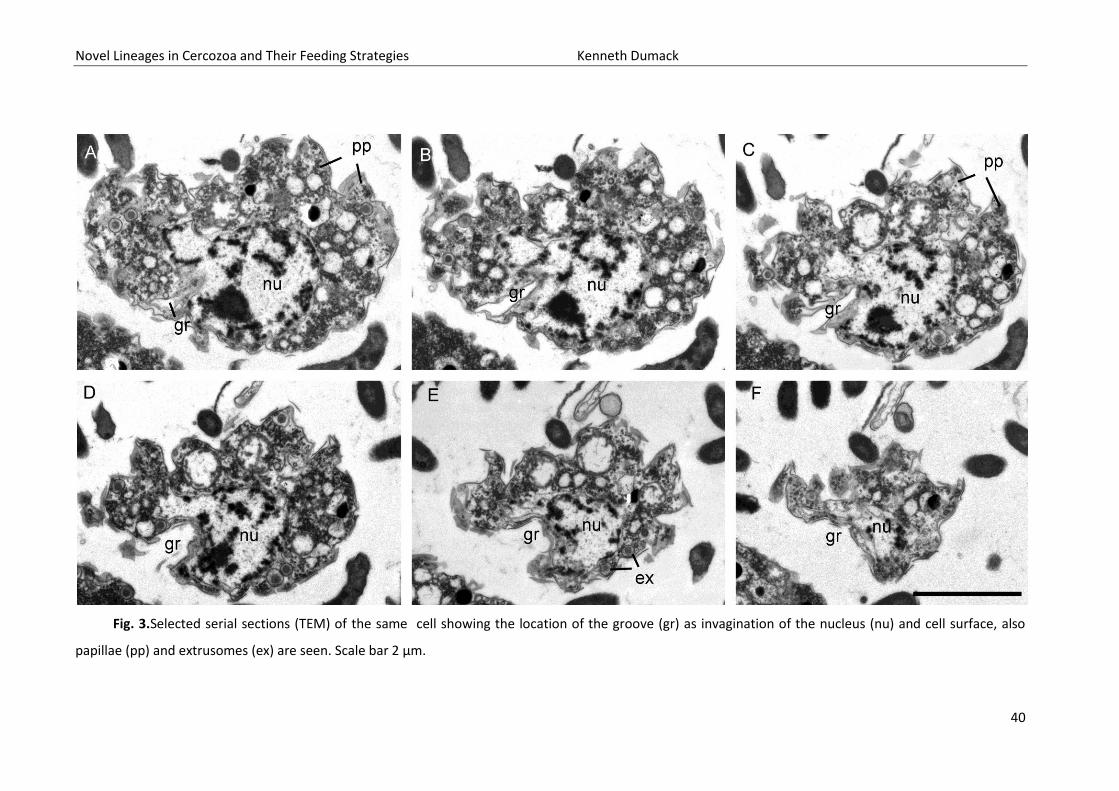
ultrastructural data. Briefly, the overall shape of the cell body is spherical with an invagination
(Dumack et al. 2016a called it ‘ring-like structure’) at the basal end from which branching and
anastomosing filopodia arise (Fig. 1A-E). Cysts and elongated cells (during cell body transport) have
not been studied in detail and are not presented here. The fragile cells lost filopodia during
centrifugation and therefore the cell body and filopodia are shown here separately.
The cells contain a single, eccentric nucleus (2.4 – 3.2 µm), usually located at the basal end of the cell
body close to the invagination, with a homogenously granular nucleolus in its center (Fig2A-C). The
invagination reaches usually into the cell body, deforming the nucleus spherical shape. The shape of
the invagination can be seen in the selected serial sections of a single cell (Fig. 3A-F). Golgi stacks are
found in association to the nucleus (Fig. 2I, 4A). The cells contain various amounts of spherical
mitochondria (0.5 - 0.6 µm) with an electron-translucent central matrix (Fig.2A). The plate-like
(flattened) cristae lie on the internal periphery of the mitochondria (Figs. 4B, 4C). Sections of tips of
the cristae are sometimes roundish (Fig. 4C, arrow). The contractile vacuole has irregular cell walls
during systole (Fig. 4D).
Novel Lineages in Cercozoa and Their Feeding Strategies Kenneth Dumack
37
In the periphery of the cell body extrusomes are located (Fig. 4). The extrusomes are reminiscent of
the kinetosomes of cercomonads and glissomonads (Mylnikov, 1988; Fig. 4G,I). They are enveloped
by a small vesicle and consist of a capsule with an internal cylinder. After discharging the cylinder is
partly exposed (Fig. 4J).
Additionally to the medium stained extrusomes, the cells contain a huge amount of small vesicles
which are either electron-translucent, resembling reserve granules (Fig. 4L) or highly stained,
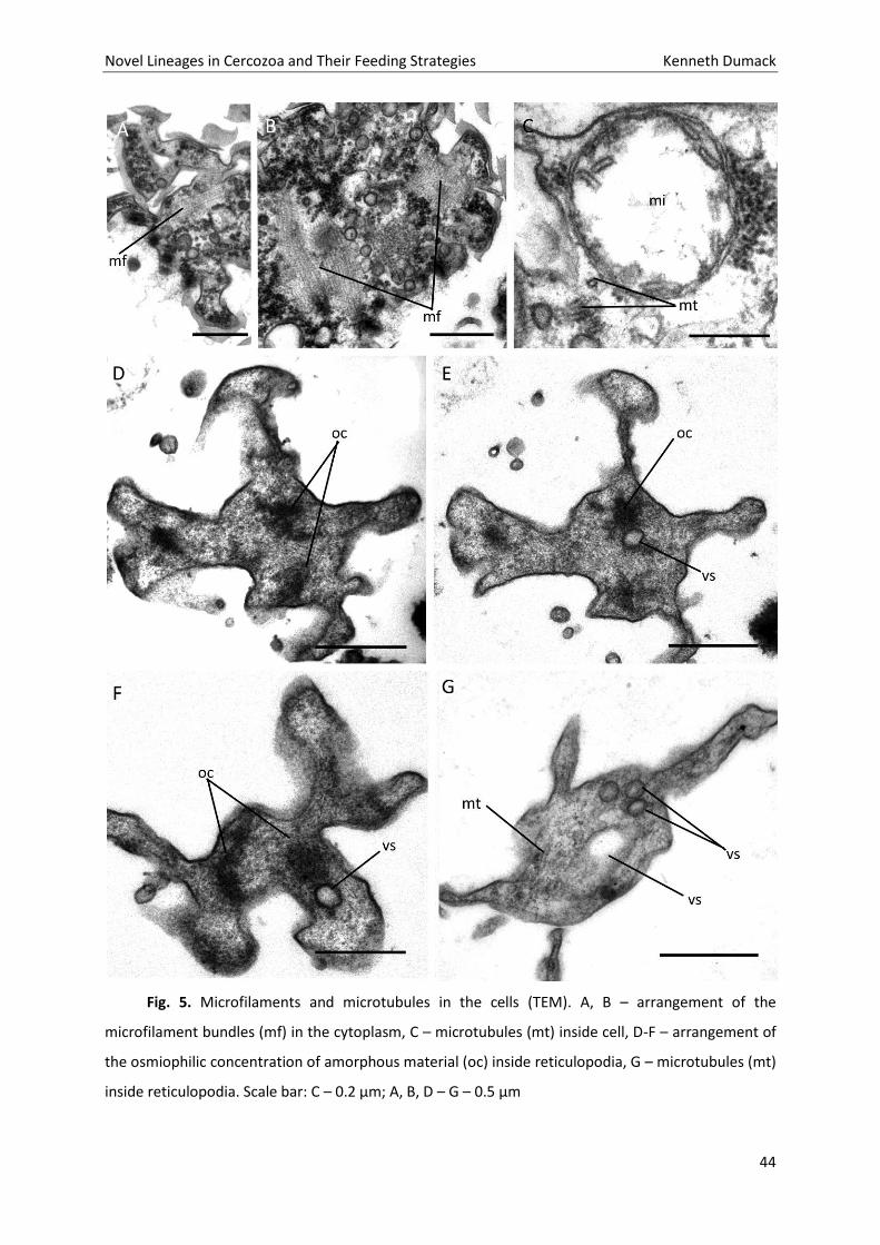
resembling osmiophilic bodies (Fig. 2A, 2E) and bundles of microfilaments (Figs 5A,B). Kraken cells do
not contain flagella, kinetosomes or a flagellar root system.
The filopodia are more electron-translucent than the cell body. Aggregations of osmiophilic
amorphous material, small electron-translucent vesicles and microtubules were seen inside the
filopodia (Figs 5D-G, selected serial sections). Additionally the filopodia contain small vacuoles with
single bacteria (Fig. 4E). According to light microscopical observations on the feeding process of the
Kraken, these bacteria were caught immediately before the fixation and were in the process of
transportation to the cell body. Within the cell body one (rarely two) food vacuoles are located in the
apical end (for the ejection of such a food vacuole during defecation, see Fig. 4F). No endocytobiotic
bacteria were observed.
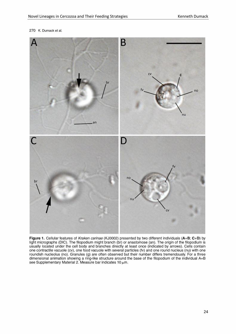
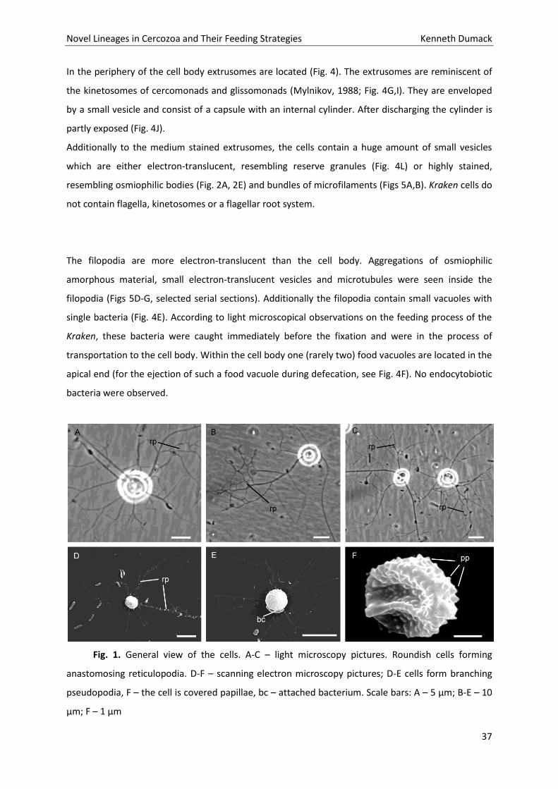
Fig. 1. General view of the cells. A-C – light microscopy pictures. Roundish cells forming
anastomosing reticulopodia. D-F – scanning electron microscopy pictures; D-E cells form branching
pseudopodia, F – the cell is covered papillae, bc – attached bacterium. Scale bars: A – 5 µm; B-E – 10
µm; F – 1 µm
Novel Lineages in Cercozoa and Their Feeding Strategies Kenneth Dumack
38
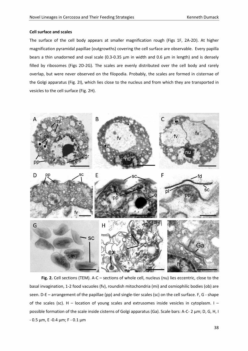
Cell surface and scales
The surface of the cell body appears at smaller magnification rough (Figs 1F, 2A-2D). At higher
magnification pyramidal papillae (outgrowths) covering the cell surface are observable. Every papilla
bears a thin unadorned and oval scale (0.3-0.35 µm in width and 0.6 µm in length) and is densely
filled by ribosomes (Figs 2D-2G). The scales are evenly distributed over the cell body and rarely
overlap, but were never observed on the filopodia. Probably, the scales are formed in cisternae of
the Golgi apparatus (Fig. 2I), which lies close to the nucleus and from which they are transported in
vesicles to the cell surface (Fig. 2H).
Fig. 2. Cell sections (TEM). A-C – sections of whole cell, nucleus (nu) lies eccentric, close to the
basal invagination, 1-2 food vacuoles (fv), roundish mitochondria (mi) and osmiophilic bodies (ob) are
seen. D-E – arrangement of the papillae (pp) and single-tier scales (sc) on the cell surface. F, G - shape
of the scales (sc). H – location of young scales and extrusomes inside vesicles in cytoplasm. I –
possible formation of the scale inside cisterns of Golgi apparatus (Ga). Scale bars: A-C- 2 µm; D, G, H, I
- 0.5 µm, E -0.4 µm; F - 0.1 µm
Novel Lineages in Cercozoa and Their Feeding Strategies Kenneth Dumack
39
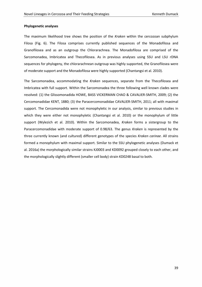
Phylogenetic analyses
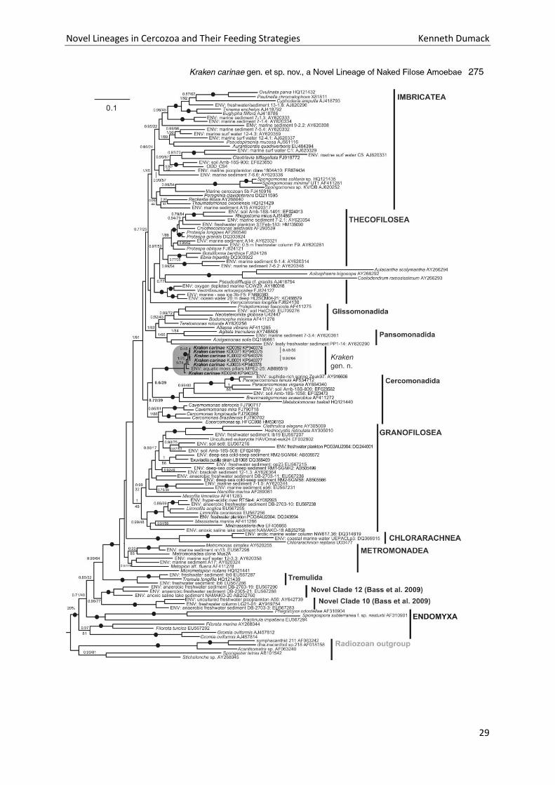
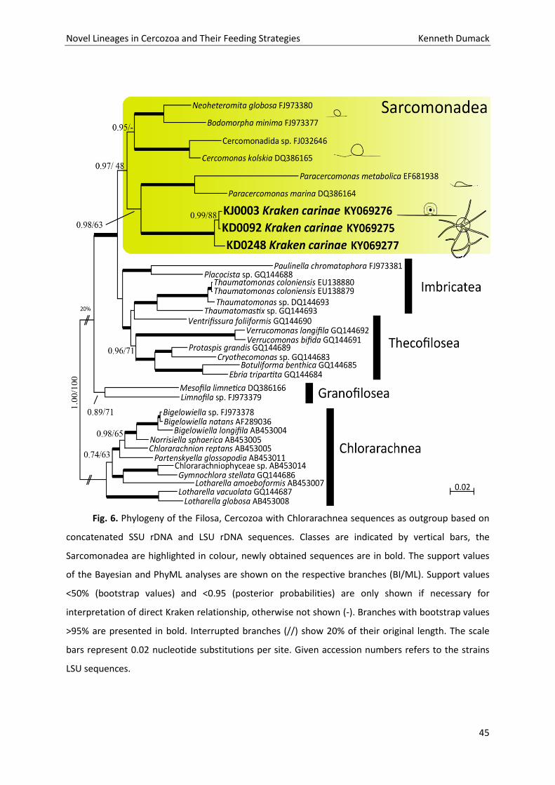
The maximum likelihood tree shows the position of the Kraken within the cercozoan subphylum
Filosa (Fig. 6). The Filosa comprises currently published sequences of the Monadofilosa and
Granofilosea and as an outgroup the Chlorarachnea. The Monadofilosa are comprised of the
Sarcomonadea, Imbricatea and Thecofilosea. As in previous analyses using SSU and LSU rDNA
sequences for phylogeny, the chlorarachnean outgroup was highly supported, the Granofilosea were
of moderate support and the Monadofilosa were highly supported (Chantangsi et al. 2010).
The Sarcomonadea, accommodating the Kraken sequences, separate from the Thecofilosea and
Imbricatea with full support. Within the Sarcomonadea the three following well known clades were
resolved: (1) the Glissomonadida HOWE, BASS VICKERMAN CHAO & CAVALIER-SMITH, 2009; (2) the
Cercomonadidae KENT, 1880; (3) the Paracercomonadidae CAVALIER-SMITH, 2011; all with maximal
support. The Cercomonadida were not monophyletic in our analysis, similar to previous studies in
which they were either not monophyletic (Chantangsi et al. 2010) or the monophylum of little
support (Wylezich et al. 2010). Within the Sarcomonadea, Kraken forms a sistergroup to the
Paracercomonadidae with moderate support of 0.98/63. The genus Kraken is represented by the
three currently known (and cultured) different genotypes of the species Kraken carinae. All strains
formed a monophylum with maximal support. Similar to the SSU phylogenetic analyses (Dumack et
al. 2016a) the morphologically similar strains KJ0003 and KD0092 grouped closely to each other, and
the morphologically slightly different (smaller cell body) strain KD0248 basal to both.
Novel Lineages in Cercozoa and Their Feeding Strategies Kenneth Dumack
40
Fig. 3.Selected serial sections (TEM) of the same cell showing the location of the groove (gr) as invagination of the nucleus (nu) and cell surface, also
papillae (pp) and extrusomes (ex) are seen. Scale bar 2 µm.
Novel Lineages in Cercozoa and Their Feeding Strategies Kenneth Dumack
41
Discussion
Ultrastructure data
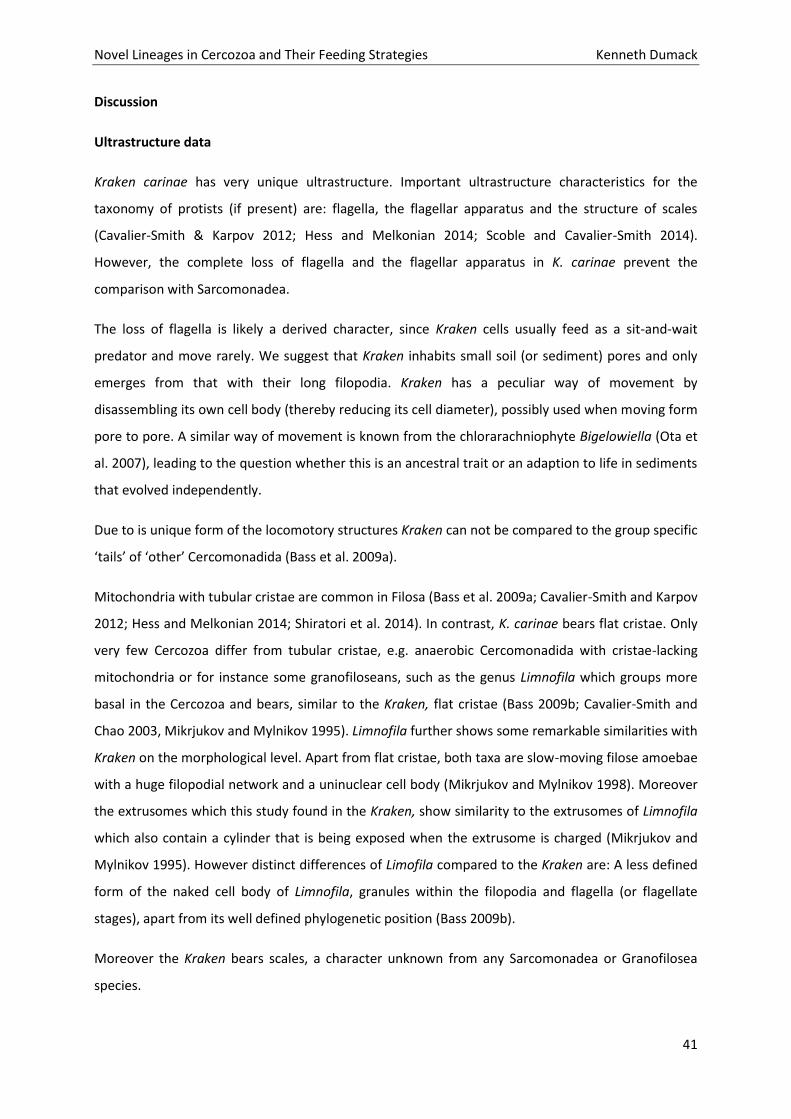
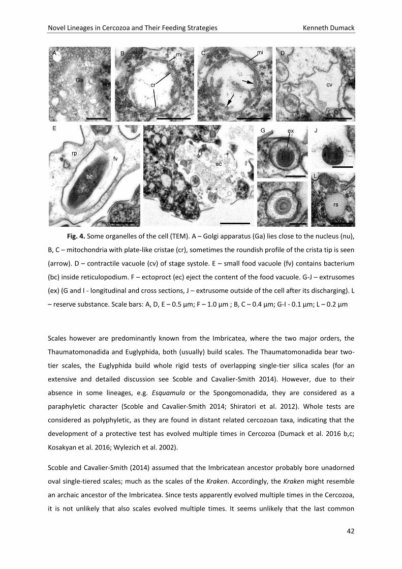
Kraken carinae has very unique ultrastructure. Important ultrastructure characteristics for the
taxonomy of protists (if present) are: flagella, the flagellar apparatus and the structure of scales
(Cavalier-Smith & Karpov 2012; Hess and Melkonian 2014; Scoble and Cavalier-Smith 2014).
However, the complete loss of flagella and the flagellar apparatus in K. carinae prevent the
comparison with Sarcomonadea.
The loss of flagella is likely a derived character, since Kraken cells usually feed as a sit-and-wait
predator and move rarely. We suggest that Kraken inhabits small soil (or sediment) pores and only
emerges from that with their long filopodia. Kraken has a peculiar way of movement by
disassembling its own cell body (thereby reducing its cell diameter), possibly used when moving form
pore to pore. A similar way of movement is known from the chlorarachniophyte Bigelowiella (Ota et
al. 2007), leading to the question whether this is an ancestral trait or an adaption to life in sediments
that evolved independently.
Due to is unique form of the locomotory structures Kraken can not be compared to the group specific
‘tails’ of ‘other’ Cercomonadida (Bass et al. 2009a).
Mitochondria with tubular cristae are common in Filosa (Bass et al. 2009a; Cavalier-Smith and Karpov
2012; Hess and Melkonian 2014; Shiratori et al. 2014). In contrast, K. carinae bears flat cristae. Only
very few Cercozoa differ from tubular cristae, e.g. anaerobic Cercomonadida with cristae-lacking
mitochondria or for instance some granofiloseans, such as the genus Limnofila which groups more
basal in the Cercozoa and bears, similar to the Kraken, flat cristae (Bass 2009b; Cavalier-Smith and
Chao 2003, Mikrjukov and Mylnikov 1995). Limnofila further shows some remarkable similarities with
Kraken on the morphological level. Apart from flat cristae, both taxa are slow-moving filose amoebae
with a huge filopodial network and a uninuclear cell body (Mikrjukov and Mylnikov 1998). Moreover
the extrusomes which this study found in the Kraken, show similarity to the extrusomes of Limnofila
which also contain a cylinder that is being exposed when the extrusome is charged (Mikrjukov and
Mylnikov 1995). However distinct differences of Limofila compared to the Kraken are: A less defined
form of the naked cell body of Limnofila, granules within the filopodia and flagella (or flagellate
stages), apart from its well defined phylogenetic position (Bass 2009b).
Moreover the Kraken bears scales, a character unknown from any Sarcomonadea or Granofilosea
species.
Novel Lineages in Cercozoa and Their Feeding Strategies Kenneth Dumack
42
Fig. 4. Some organelles of the cell (TEM). A – Golgi apparatus (Ga) lies close to the nucleus (nu),
B, C – mitochondria with plate-like cristae (cr), sometimes the roundish profile of the crista tip is seen
(arrow). D – contractile vacuole (cv) of stage systole. E – small food vacuole (fv) contains bacterium
(bc) inside reticulopodium. F – ectoproct (ec) eject the content of the food vacuole. G-J – extrusomes
(ex) (G and I - longitudinal and cross sections, J – extrusome outside of the cell after its discharging). L
– reserve substance. Scale bars: A, D, E – 0.5 µm; F – 1.0 µm ; B, C – 0.4 µm; G-I - 0.1 µm; L – 0.2 µm
Scales however are predominantly known from the Imbricatea, where the two major orders, the
Thaumatomonadida and Euglyphida, both (usually) build scales. The Thaumatomonadida bear two-
tier scales, the Euglyphida build whole rigid tests of overlapping single-tier silica scales (for an
extensive and detailed discussion see Scoble and Cavalier-Smith 2014). However, due to their
absence in some lineages, e.g. Esquamula or the Spongomonadida, they are considered as a
paraphyletic character (Scoble and Cavalier-Smith 2014; Shiratori et al. 2012). Whole tests are
considered as polyphyletic, as they are found in distant related cercozoan taxa, indicating that the
development of a protective test has evolved multiple times in Cercozoa (Dumack et al. 2016 b,c;
Kosakyan et al. 2016; Wylezich et al. 2002).
Scoble and Cavalier-Smith (2014) assumed that the Imbricatean ancestor probably bore unadorned
oval single-tiered scales; much as the scales of the Kraken. Accordingly, the Kraken might resemble
an archaic ancestor of the Imbricatea. Since tests apparently evolved multiple times in the Cercozoa,
it is not unlikely that also scales evolved multiple times. It seems unlikely that the last common
Novel Lineages in Cercozoa and Their Feeding Strategies Kenneth Dumack
43
ancestor of the Filosa already carried scales, the Thecofilosea may have developed from those their
organic tests and the Imbricatea further developed two-tier scales (Thaumatomonadida) or whole
rigid tests composed of scales (Euglyphida), but were lost in most Sarcomonadea, except in the
possible sarcomonadean Kraken.
Phylogenetic analyses
The phylogeny of the Sarcomonadea is still controversial (see Cavalier-Smith & Karpov 2012 for a
detailed discussion). It is still unclear whether Sarcomonadea are monophyletic, and especially the
phylogenetic position of the Paracercomonadidae (extremely long branch, changing position in trees)
contributes to this uncertainty. Since Kraken groups with SSU and concatenated SSU+LSU phylogeny
next to Paracercomonas, this long branch might (a) interfere with good support or (b) may even
indicate questionable results due to long branch attraction. Nevertheless the constructed two-gene
phylogeny confirms the SSU phylogeny published in Dumack et al. (2016a). Although still moderate,
the phylogenetic support was significantly enhanced (from 0.6/29 to 0.98/63) in this study. Since only
few cercozoan LSU sequences are currently available we were not able to achieve a highly supported
phylogeny. With an increase of (a) taxon sampling of Cercozoa and especially the
Paracercomonadidae (in particular Metabolomonas and Brevimastigomonas) or (b) gene sampling, in
particular whole transcriptomes the phylogenetic positions of the Kraken and Paracercomonadidae
within Cercozoa might be resolved.
The Kraken remains incertae sedis
The ultrastructure data give some indications that the Kraken resembles a direct descendant of the
scale-bearing last common ancestor of the Imbricatea. The two-gene phylogeny indicates, however
with moderate support, an affiliation with the Cercomonadida in the Sarcomonadea. Due to these
contradicting results we are not able to clearly assign the Kraken to either the class Sarcomonadea or
Imbricatea.
The characters of the Kraken have not been found in any other cercozoan family. We therefore
establish a new family Krakenidae fam. nov., but still hesitate to establish a novel order that should
be called Krakenida if it will be shown to be necessary in future studies. To resolve this issue,
ultrastructure data (of e.g. Discomonas), SSU sequencing data (of e.g. Zoelucasa; Nicholls 2012), or
large scale multi-gene transcriptome analyses are needed in future studies.
Novel Lineages in Cercozoa and Their Feeding Strategies Kenneth Dumack
44
Fig. 5. Microfilaments and microtubules in the cells (TEM). A, B – arrangement of the
microfilament bundles (mf) in the cytoplasm, C – microtubules (mt) inside cell, D-F – arrangement of
the osmiophilic concentration of amorphous material (oc) inside reticulopodia, G – microtubules (mt)
inside reticulopodia. Scale bar: C – 0.2 µm; A, B, D – G – 0.5 µm
Novel Lineages in Cercozoa and Their Feeding Strategies Kenneth Dumack
45
Fig. 6. Phylogeny of the Filosa, Cercozoa with Chlorarachnea sequences as outgroup based on
concatenated SSU rDNA and LSU rDNA sequences. Classes are indicated by vertical bars, the
Sarcomonadea are highlighted in colour, newly obtained sequences are in bold. The support values
of the Bayesian and PhyML analyses are shown on the respective branches (BI/ML). Support values
<50% (bootstrap values) and <0.95 (posterior probabilities) are only shown if necessary for
interpretation of direct Kraken relationship, otherwise not shown (-). Branches with bootstrap values
>95% are presented in bold. Interrupted branches (//) show 20% of their original length. The scale
bars represent 0.02 nucleotide substitutions per site. Given accession numbers refers to the strains
LSU sequences.
Novel Lineages in Cercozoa and Their Feeding Strategies Kenneth Dumack
46
Krakenidae Dumack, Mylnikov and Bonkowski fam. nov.
Diagnosis. Limnic or terrestrial; Cells in the trophic phase form a large web of surface attached
filopodia (irregular branched, elongate, anastomosing, network-forming). Filopodia thin and
tapering; originating from the basal end of the cell body; flagella absent. Cell body covered by non-
overlapping oval and unadorned single-tier scales; these are carried by small papillae (outgrowths);
usually not overlapping. Mitochondria with plate-like cristae in their inner periphery. Extrusomes
with unknown function contain a capsule and an internal cylinder present. Phagotrophic;
bacterivorous; bacteria are caught with filopodia at the point of contact and then transported in a
vacuole through the filopodia to the cell body for digestion. Cysts present, spherical.
Etymology: Name derived by the type genus.
Type genus: Kraken Dumack, Schuster, Bass et Bonkowski, 2016
Other genera included: none
Material and Methods
For detailed description of the source of samples, the isolation process and culture conditions of
Kraken carinae, see Dumack et al. (2016). Briefly, K. carinae was isolated from agricultural soils of
two European countries and cultured in low nutrient medium with co-cultivated bacteria. The cells
are extremely fragile and die when exposed to slightest mechanical stress, i.e. movement of water
body by lifting the Petri dish.
Sequencing
Phylogenetic analyses were done on one representative strain for each of the three known (and
cultured) Kraken SSU genotypes; i.e. KJ0003, KD0092 and KD0248 (DSMZ- Registration numbers:
244502341-KA; 244502023-KA and 244501215-KA, respectively). 15 µl aliquots of the monoclonal
cultures were transferred to a sterile 200 µl Eppendorf tube and 13 µl ddH2O were added. These
samples were frozen at −20°C to destroy the protist cells. The PCR was performed with a 50 µl
reaction mixture containing 5 µl of 0.1 µM forward and reverse primer solution each, 5 µl dNTPs (200
µM), 5 µl reaction buffer and 1 U DreamTaq DNA-polymerase (Applied Biosystems, Weiterstedt,
Germany). General eukaryotic primers 184F, 1126R, 1105F and 2018R were used (Van der Auwera et
al. 1994). The PCR products were purified by adding 0.15 µl of endonuclease I (20 U/µl, Fermentas
GmbH), 0.9 µl shrimp alkaline phosphatase (1 U/µl, Fermentas GmbH) and 1.95 µl water to 8 µl of
the PCR products. The mixtures were then heated 30 min at 37°C, and subsequently 20 min at 85°C.
Novel Lineages in Cercozoa and Their Feeding Strategies Kenneth Dumack
47
The sequencing reaction was done by using the Big Dye Terminator Cycle sequencing kit and an ABI
PRISM automatic sequencer.
The sequences were manually checked for sequencing errors, combined to sequence contigs and
deposited in the NCBI database (KY069275, KY069276 and KY069277).
Phylogenetic analyses
Publications were screened for SSU and LSU sequences that were obtained from the same strain of
as many cercozoan lineages as possible. These sequences were manually aligned in SeaView (V4.5.3,
Gouy et al. 2010). An alignment with 34 sequences and 4,091 unambiguously aligned sites of which
67.8% were without polymorphisms was used for phylogenetic analyses. The program jmodeltest
(V.2.1.5, Darriba et al. 2012) was used to determine the best fitting model: GTR+I+G, which was
selected among 88 models (settings: Substitution schemes 11; add base frequencies +I+G rate
variation nCat=4, ML optimized NNI as base tree). Phylogenetic trees were constructed using
maximum likelihood (ML) and Bayesian inference (BI). The support values of the PhyML and the
Bayesian analyses are given as (ML/BI).
Maximum likelihood phylogenetic analyses were run in PhyML V3.1 (Guindon and Gascuel 2003) with
the following settings: GTR model; a proportion of invariable sites and a gamma-shaped distribution
of the substitution rates across variable sites (GTR+I+G), with four rate categories; BIONJ distance-
based starting tree with all model parameters estimated from the data. The Bayesian analyses were
run using MrBayes v.3.2 (Altekar et al. 2004; Ronquist and Huelsenbeck 2003) with the following
settings: five million generations, trees sampled every 100 generations, convergence of the two runs
was estimated every 500 generations with a final average of the standard deviation of split
frequencies of <0.01 at the end of the run. Of the sampled trees, 25% were discarded as burn-in.
Electron microscopic analyses
Electron microscopy was performed on a culture of Kraken carinae (strain KJ0003; DSMZ-Registration
number 244502341-KA). Cells were maintained in Pratt medium (0.1 g/L KNO3, 0.01 g/L MgSO4.x 7
H2O, 0.01 g/L K2HPO4.x3 H2O, 0.001 g/L FeCl3
.x6 H2O, pH = 6.5-7.5) with the bacterium Pseudomonas
fluorescens, MIGULA 1895, as food source.
Light microscopic observations were made with an AxioScope A1 (Carl Zeiss, Germany) using phase
contrast, 70x water immersion objectives and an AVT HORN MC-1009/S video camera. Video clips
were digitized using a Behold TV 409 FM tuner.
Novel Lineages in Cercozoa and Their Feeding Strategies Kenneth Dumack
48
For scanning electron microscopy (SEM), cells from lag and exponential growth phase growing on the
surface of cover slips were fixed with 2% glutaraldehyde (final concentration) for 15 min at 20 °C. The
specimens were taken through a graded ethanol dehydration followed by a graded transfer to
acetone. After critical point drying, the dried cover slips were mounted on aluminum stubs, which
were coated with gold-palladium and then studied with a JSM-6510LV.
For transmission electron microscopy (TEM), cells were centrifuged to produce high density pellets.
The pellets were fixed in a cocktail of 0.6% glutaraldehyde and 2% OsO4 (final concentration) for 20-
60 min at 1 °C and dehydrated in an alcohol series and then in rising acetone concentrations (30, 50,
70, 96 %; 10-20 minutes in each step). Subsequently, cells were embedded in a mixture of araldite
and epon (Luft, 1961). Ultrathin sections were obtained with the LKB ultramicrotome. The TEM
observations were made by using a JEM-1011 (Jeol, Japan) electron microscope. The chemical
composition of the scales was not studied.
Acknowledgements
We like to thank Dr. Frank Nitsche for providing the LSU primers. This study was partly supported by
grant (BO 1907/2-2) of the DFG (Deutsche Forschungsgemeinschaft) to MB within the Research Unit
“Carbon flow in below- ground food webs assessed with isotope tracers” (FOR 918). This work
(A.P.Mylnikov, ultrastructure observation) was supported by the Russian Foundation for Basic
Research (15-04-05841, 14-04-00550). A.P. Mylnkov thanks Prof. S.A. Karpov for fruitful discussion on
ultrastructural data.
References
Bass D, Howe AT, Mylnikov AP, Vickerman K, Chao EE, Edwards Smallbone J, Snell J, Cabral C Jr,
Thomas Cavalier-Smith (2009a) Phylogeny and classification of Cercomonadida (Protozoa, Cercozoa):
Cercomonas, Eocercomonas, Paracercomonas, and Cavernomonas gen. nov. Protist 160:483–521
Bass D, Chao EE, Nikolaev S, Yabuki A, Ishida K, Berney C, Prasad U, Wylezich C, Cavalier-Smith T
(2009b) Phylogeny of novel naked filose and reticulose Cercozoa: Granofilosea cl. n. and
Proteomyxidea revised. Protist 160:75–109
Cavalier-Smith T, Chao EE (2003) Phylogeny and classification of phylum Cercozoa (Protozoa). Protist
154:341-358
Novel Lineages in Cercozoa and Their Feeding Strategies Kenneth Dumack
49
Cavalier-Smith T, Karpov SA (2012) Paracercomonas Kinetid Ultrastructure, Origins of the Body Plan
of Cercomonadida, and Cytoskeleton Evolution in Cercozoa. Protist 163:47–75
Chantangsi C, Hoppenrath M, Leander BS (2010) Molecular Phylogenetics and Evolution Evolutionary
relationships among marine cercozoans as inferred from combined SSU and LSU rDNA sequences and
polyubiquitin insertions. Molecular Phylogenetics and Evolution 57:518–527
Dumack K, Schuster J, Bass D, Bonkowski M (2016a) A Novel Lineage of “Naked Filose Amoebae”;
Kraken carinae gen. nov. sp. nov. (Cercozoa) with a Remarkable Locomotion by Disassembly of its Cell
Body. Protist 167: 268–278
Dumack K, Müller MEH, Bonkowski M (2016b) Description of Lecythium terrestris sp. nov.
(Chlamydophryidae, Cercozoa), a Soil Dwelling Protist Feeding on Fungi and Algae. Protist 167:93–
105
Dumack K, Baumann C, Bonkowski M (2016c) Revision of the thecate amoeba genus Lecythium
(Chlamydophryidae, Tectofilosida, Cercozoa, Rhizaria) including a description of four new species and
an identification key. Protist 167:440–59
Glücksman E, Bell T, Griffiths RI, Bass D (2010) Closely related protist strains have different
grazingimpacts on natural bacterial communities. Environmental Microbiology 12:3105–3113
Hess S, Sausen N, Melkonian M (2012) Shedding light on vampires: the phylogeny of vampyrellid
amoebae revisited. PLoS One 7(2):e31165
Hess S, Melkonian M (2013) The mystery of clade X: Orciraptor gen. nov. and Viridiraptor gen. nov.
are highly specialised, algivorous amoeboflagellates (Glissomonadida, Cercozoa). Protist 164:706–47
Hess S, Melkonian M (2014) Ultrastructure of the Algivorous Amoeboflagellate Viridiraptor invadens
(Glissomonadida, Cercozoa). Protist 165:605–35
Kosakyan A, Gomaa F, Lara E, Lahr DJG (2016) Current and future perspectives on the systematics,
taxonomy and nomenclature of testate amoebae. Eur J Protistol 1-13
Mikrjukov KA, Mylnikov AP (1995) Fine structure of an unusual rhizopod, Penardia cometa,
containing extru- somes and kinetosomes. Europ J Protistol 31: 90–96
Mikrjukov KA, Mylnikov AP (1998) A study of the fine structure and the life cycle of Gymnophrys
cometa Cienkowski, 1876 (Gymnophryea classis nov.) with taxo- nomical remarks to the amoebo-
flagellated genera Gymnophrys and Borkovia. Acta Protozool. 37: 179–189
Novel Lineages in Cercozoa and Their Feeding Strategies Kenneth Dumack
50
Mylnikov AP (1988) The structure of extrusomes in some flagellates. Tsitologia. Leningrad. 30:1402-
1408
Nicholls KH (2012) Zoelucasa sablensis n. gen. et n. sp. (Cercozoa, incertae sedis), a new
scale‐covered flagellate from marine sandy shores. Acta Protozool. 51:113–117.
Luft JH (1961) Improvements in epoxy resin embedding methods J. Bzophys. Biochem Cytol. 9:409.
Scoble JM, Cavalier-Smith T (2014) Scale evolution, sequence phylogeny, and taxonomy of
thaumatomonad Cercozoa: 11 new species and new genera Scutellomonas, Cowlomonas,
Thaumatospina and Ovaloplaca. Eur J Protistol 50:270–313.
Shiratori T, Yabuki A, Ishida K (2012) Esquamula lacrimiformis n. g., n. sp., a new member of
thaumatomonads that lacks siliceous scales. J Eukaryot Microbiol 59:527–536.
Van der Auwera G, Chapelle S, De Wachter R (1994) Structure of the large ribosomal subunit RNA of
Phytophthora megasperma, and phylogeny of the oomycetes. FEBS Letters 338:133-136.
Wylezich C, Meisterfeld R, Meisterfeld S, Schlegel M (2002) Phylogenetic analyses of small subunit
ribosomal RNA coding regions reveal a monophyletic lineage of euglyphid testate amoebae (order
Euglyphida). J Eukaryot Microbiol 49:108-118.
Wylezich C, Nies G, Mylnikov AP, Diethard Tautz, Hartmut Arndt (2010) An Evaluation of the Use of
the LSU rRNA D1-D5 Domain for DNA-based Taxonomy of Eukaryotic Protists. Protist 161:342–352.
Novel Lineages in Cercozoa and Their Feeding Strategies Kenneth Dumack
51
Part 2 - Eukaryvorous amoebae of the Thecofilosea,
Cercozoa.
Chapter 3: Description of Lecythium terrestris sp. nov.
(Chlamydophryidae, Cercozoa), a Soil Dwelling Protist
Feeding on Fungi and Algae.
Novel Lineages in Cercozoa and Their Feeding Strategies Kenneth Dumack
52
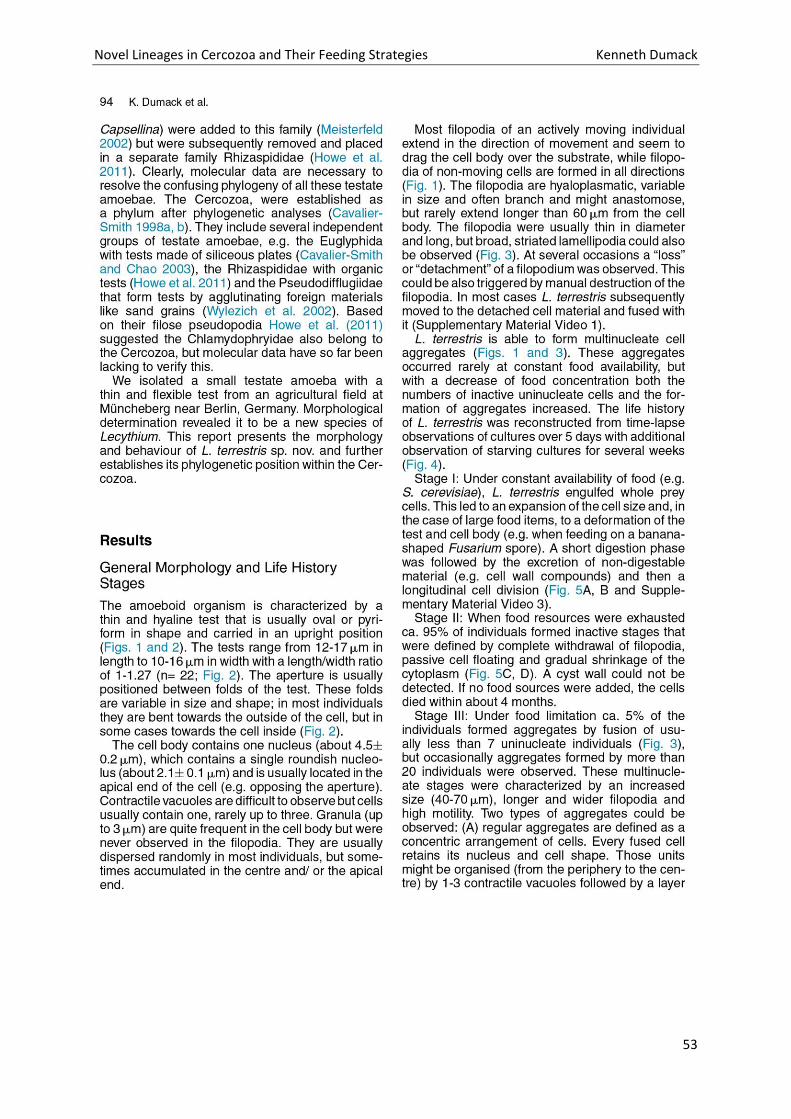
Novel Lineages in Cercozoa and Their Feeding Strategies Kenneth Dumack
53
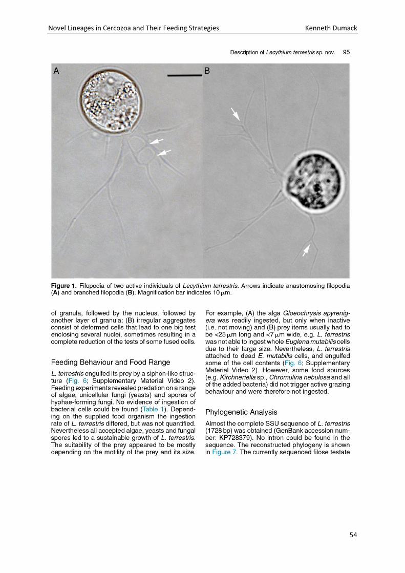
Novel Lineages in Cercozoa and Their Feeding Strategies Kenneth Dumack
54
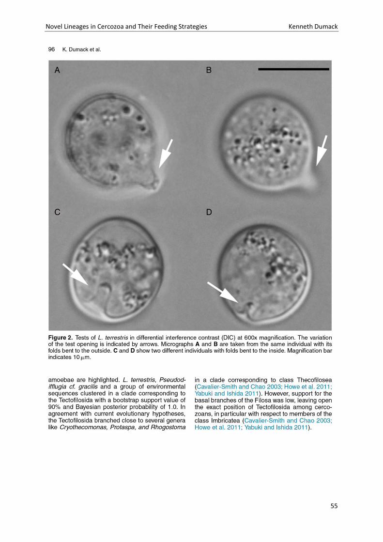
Novel Lineages in Cercozoa and Their Feeding Strategies Kenneth Dumack
55
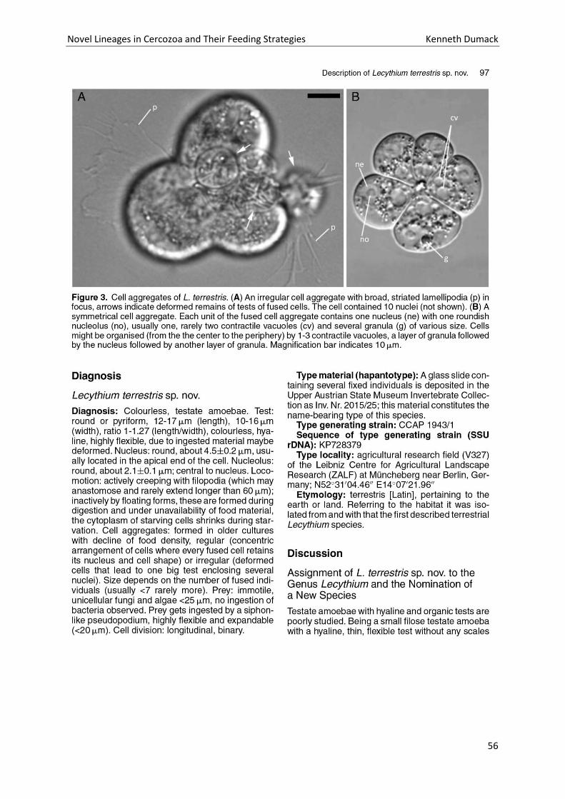
Novel Lineages in Cercozoa and Their Feeding Strategies Kenneth Dumack
56
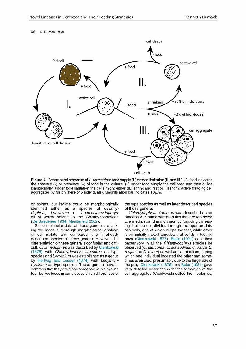
Novel Lineages in Cercozoa and Their Feeding Strategies Kenneth Dumack
57
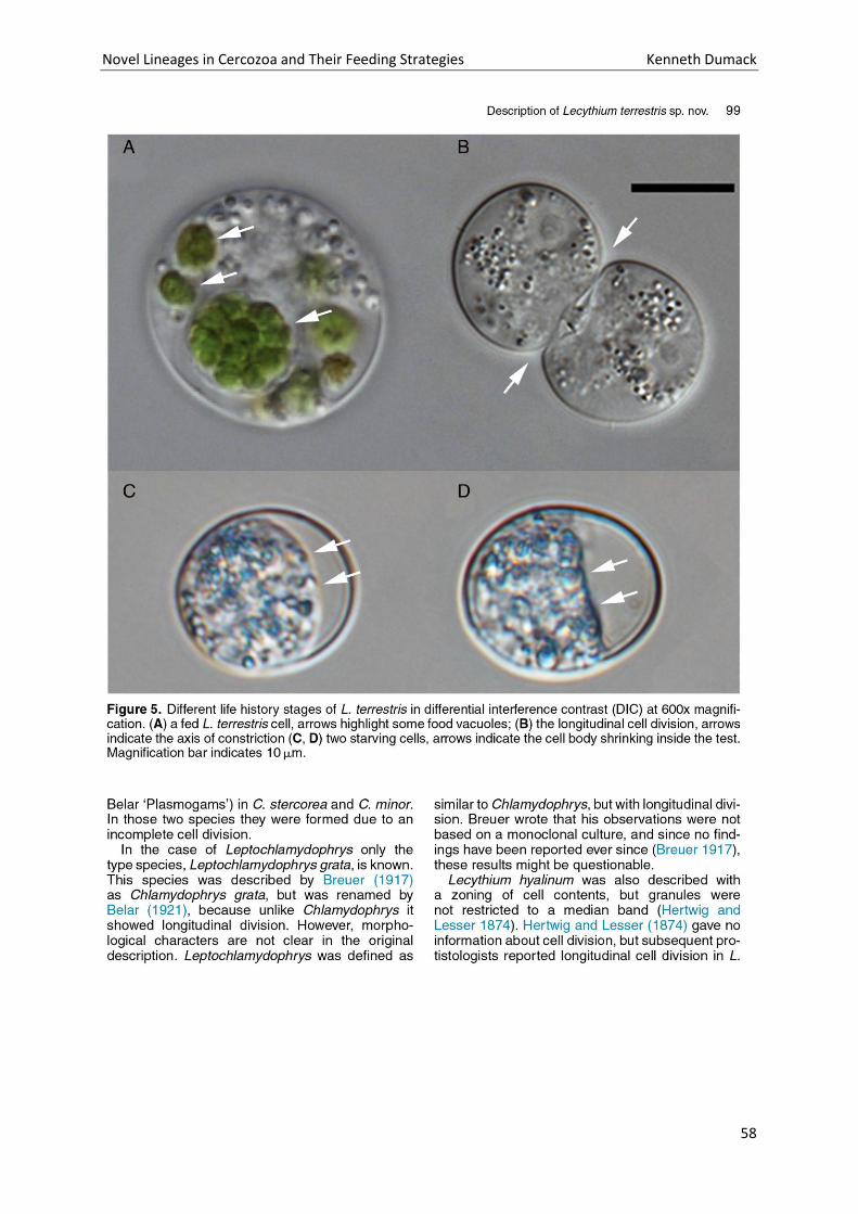
Novel Lineages in Cercozoa and Their Feeding Strategies Kenneth Dumack
58
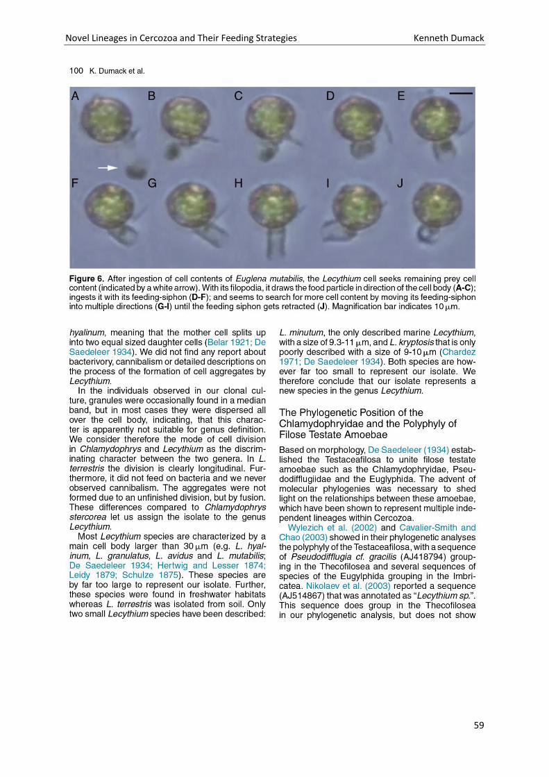
Novel Lineages in Cercozoa and Their Feeding Strategies Kenneth Dumack
59
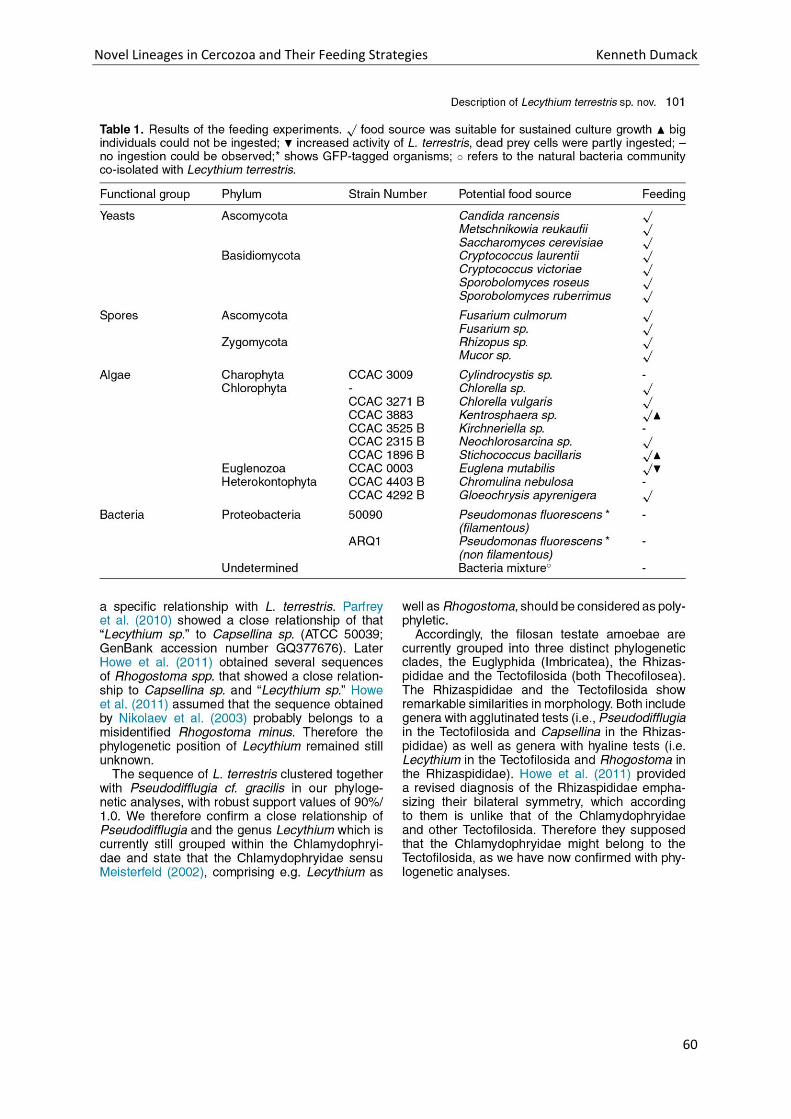
Novel Lineages in Cercozoa and Their Feeding Strategies Kenneth Dumack
60
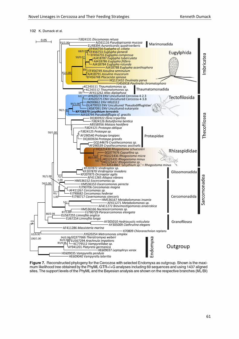
Novel Lineages in Cercozoa and Their Feeding Strategies Kenneth Dumack
61
Novel Lineages in Cercozoa and Their Feeding Strategies Kenneth Dumack
62
Novel Lineages in Cercozoa and Their Feeding Strategies Kenneth Dumack
63
Novel Lineages in Cercozoa and Their Feeding Strategies Kenneth Dumack
64
Novel Lineages in Cercozoa and Their Feeding Strategies Kenneth Dumack
65
Chapter 4: A bowl with marbles: Revision of the thecate
amoeba genus Lecythium (Chlamydophryidae,
Tectofilosida, Cercozoa, Rhizaria) including a
description of four new species and an identification
key.
Novel Lineages in Cercozoa and Their Feeding Strategies Kenneth Dumack
66
Novel Lineages in Cercozoa and Their Feeding Strategies Kenneth Dumack
67
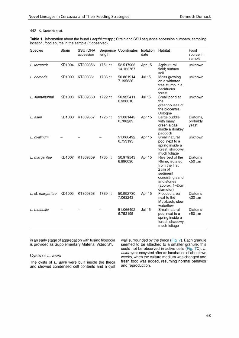
Novel Lineages in Cercozoa and Their Feeding Strategies Kenneth Dumack
68
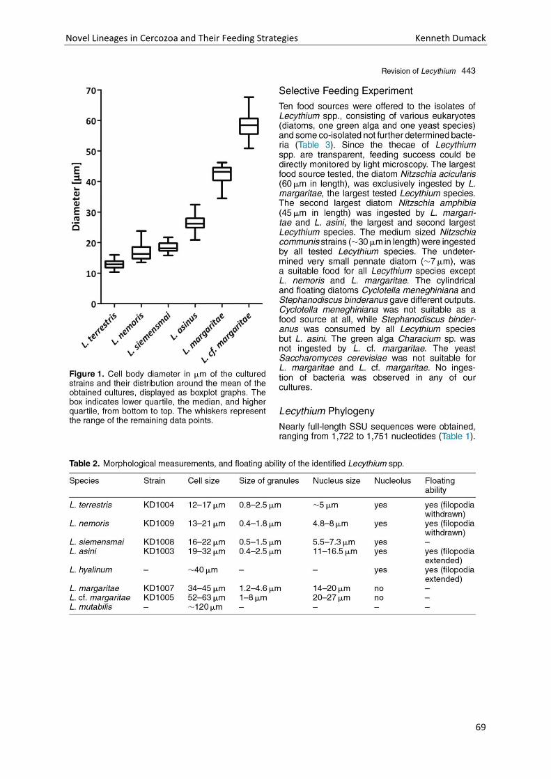
Novel Lineages in Cercozoa and Their Feeding Strategies Kenneth Dumack
69
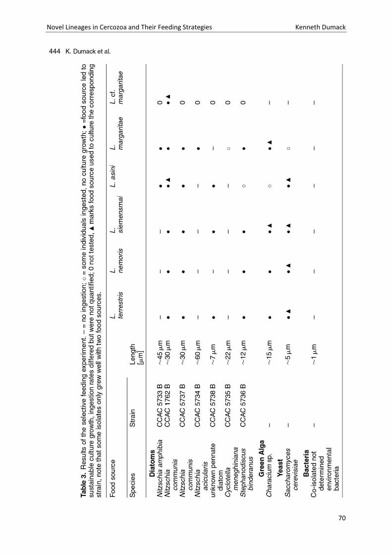
Novel Lineages in Cercozoa and Their Feeding Strategies Kenneth Dumack
70
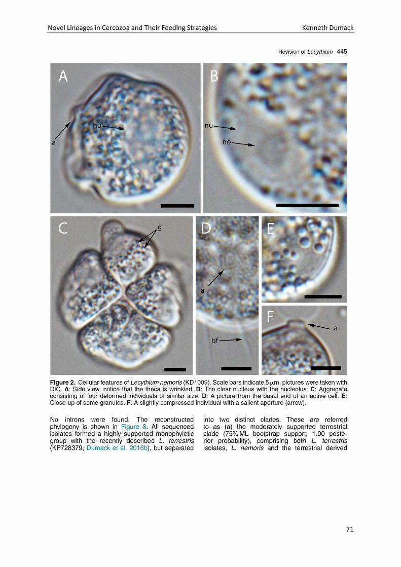
Novel Lineages in Cercozoa and Their Feeding Strategies Kenneth Dumack
71
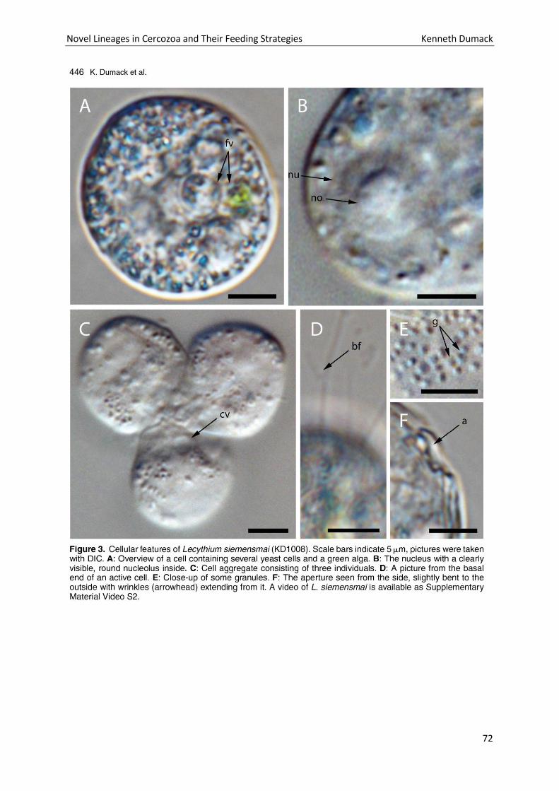
Novel Lineages in Cercozoa and Their Feeding Strategies Kenneth Dumack
72
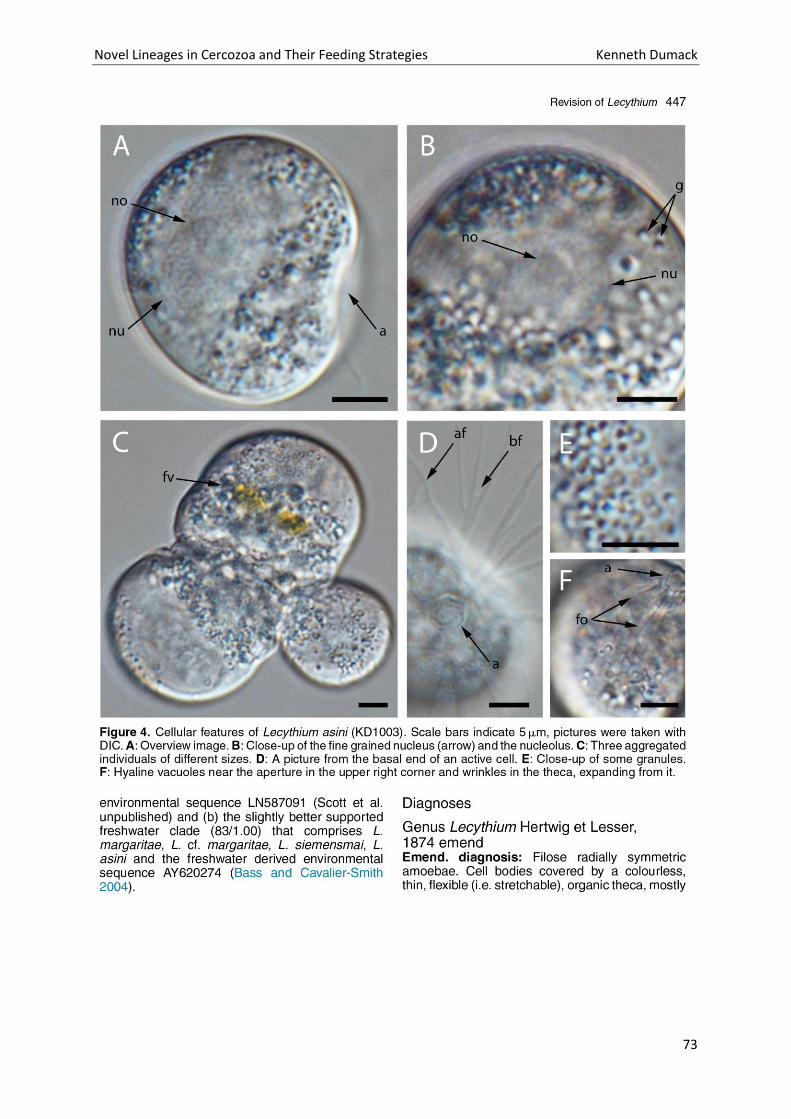
Novel Lineages in Cercozoa and Their Feeding Strategies Kenneth Dumack
73
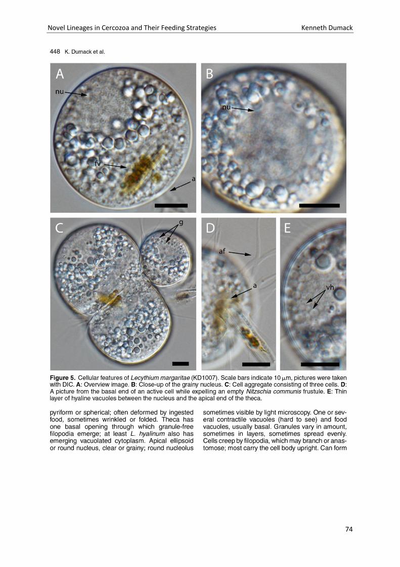
Novel Lineages in Cercozoa and Their Feeding Strategies Kenneth Dumack
74
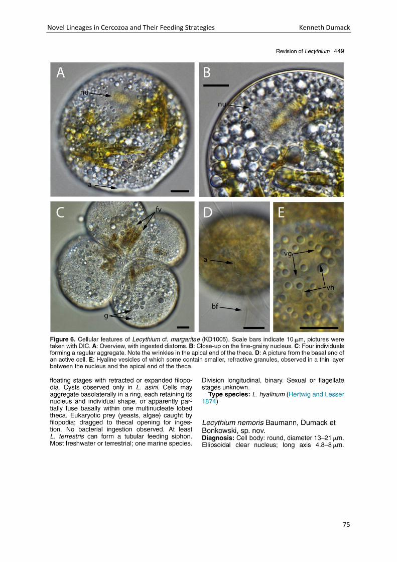
Novel Lineages in Cercozoa and Their Feeding Strategies Kenneth Dumack
75
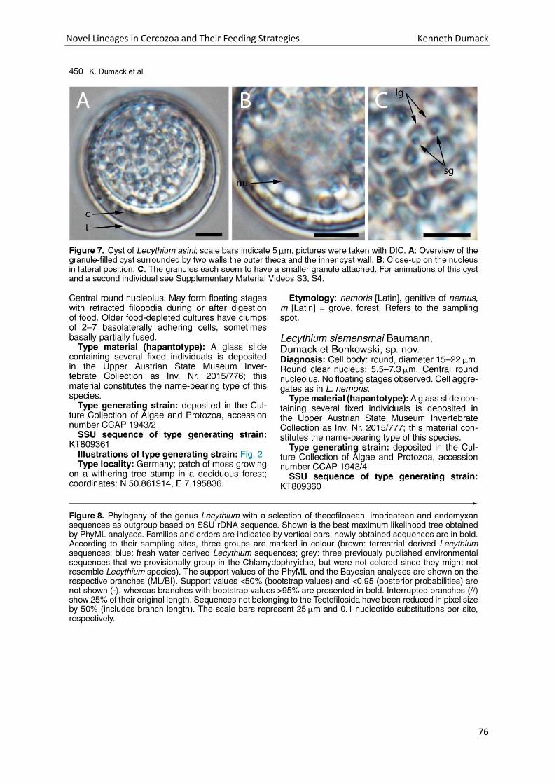
Novel Lineages in Cercozoa and Their Feeding Strategies Kenneth Dumack
76
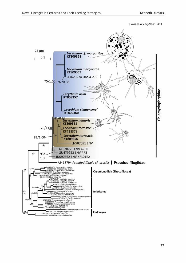
Novel Lineages in Cercozoa and Their Feeding Strategies Kenneth Dumack
77
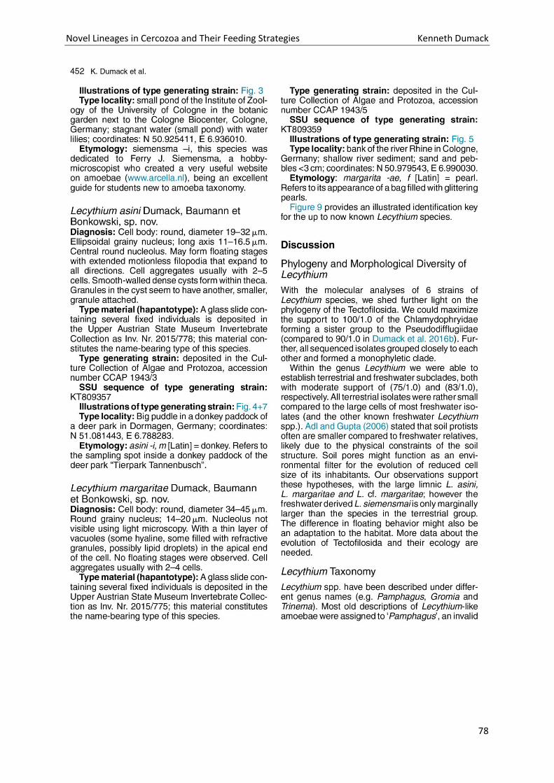
Novel Lineages in Cercozoa and Their Feeding Strategies Kenneth Dumack
78
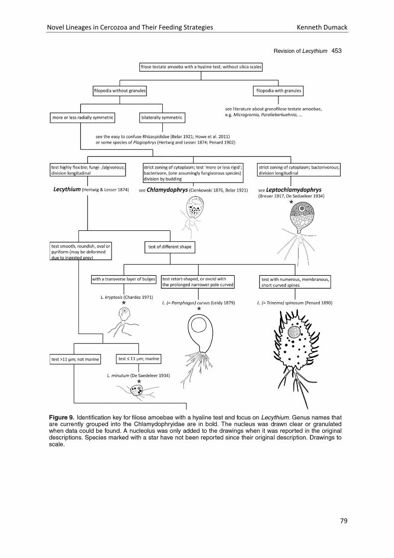
Novel Lineages in Cercozoa and Their Feeding Strategies Kenneth Dumack
79
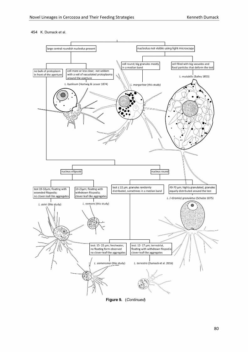
Novel Lineages in Cercozoa and Their Feeding Strategies Kenneth Dumack
80
Novel Lineages in Cercozoa and Their Feeding Strategies Kenneth Dumack
81
Novel Lineages in Cercozoa and Their Feeding Strategies Kenneth Dumack
82
Novel Lineages in Cercozoa and Their Feeding Strategies Kenneth Dumack
83
Novel Lineages in Cercozoa and Their Feeding Strategies Kenneth Dumack
84

Novel Lineages in Cercozoa and Their Feeding Strategies Kenneth Dumack
85
Novel Lineages in Cercozoa and Their Feeding Strategies Kenneth Dumack
86
Chapter 5: Shedding light on the polyphyletic genus
Plagiophrys: The transition of some of its species to
Rhizaspis (Tectofilosida, Thecofilosea, Cercozoa) and
the establishment of Sacciforma gen. nov.
(Cryomonadida, Thecofilosea, Cercozoa).
Novel Lineages in Cercozoa and Their Feeding Strategies Kenneth Dumack
87
Authors Kenneth Dumacka, Hüsna Öztopraka, Lioba Rügera and Michael Bonkowskia
Corresponding author Kenneth Dumack
Phone: +49-(0)221-470-6635 Fax: +49-(0)221-470-5038
a University of Cologne, Department of Terrestrial Ecology, Faculty of Zoology, Zülpicher Str. 47b,
50674 Köln, Germany
[email protected], [email protected], [email protected],
Title
Shedding light on the polyphyletic thecate amoeba genus Plagiophrys: Transition of some of its
species to Rhizaspis (Tectofilosida, Thecofilosea, Cercozoa) and the establishment of Sacciforma gen.
nov. and Rhogostomidae fam. nov. (Cryomonadida, Thecofilosea, Cercozoa).
Keywords
Rhogostoma, protist, algivorous, testate amoebae, amoeba, phylogeny
Abstract
For over a century testate amoebae have been a favoured group of interest for protistologists,
however there is still an endless amount of unanswered questions. The genus Plagiophrys, Claparède
and Lachmann 1859, is still one of the unresolved mysteries as it comprises species with high
morphological diversity of which no molecular data are available. To shed light on the phylogeny and
taxonomy of Plagiophrys we (a) cultured four isolates of three Plagiophrys morphospecies and
provided morphological observations (b) obtained three new SSU sequences and conducted
phylogenetic analyses of the Thecofilosea and (c) did intensive literature research, showing that
Plagiophrys is polyphyletic. We partially untangle this polyphyly by combining several of its species
with the genus Rhizaspis, Skuja 1948. Furthermore, we establish Sacciforma gen. nov. to
accommodate P. sacciformis as it groups within the formerly known Rhizaspididae, which do not
Novel Lineages in Cercozoa and Their Feeding Strategies Kenneth Dumack
88
comprise our isolates of Rhizaspis (and therefore were renamed as Rhogostomidae) as it groups with
maximum support as a sister-group to the Pseudodifflugiidae.
1. Introduction
Testate amoebae have been of high interest for protistologists and ecologists since their discovery.
They show the advantage of having clear morphological traits, making them ideal model organisms
for protist ecology and evolution. Accordingly the specific ways in which their tests are constructed
have been intensively studied and were used for testate amoeba identification and taxonomy. The
tests may be siliceous (e.g. Euglyphids; Cavalier-Smith 1998a,b; Wylezich et al. 2002), organic (e.g.
Thecofilosea; Dumack et al. 2016a,b; Howe et al. 2011) or composed of agglutinated material, e.g.
foreign particles, like sand grains or diatom frustules (e.g. Pseudodifflugiidae; Cavalier-Smith and
Chao 2003; Wylezich et al. 2002).
However, taxonomy solely based on morphology lead to contradicting taxonomical concepts (de
Saedeleer 1934; Cash, Wailes and Hopkinson 1915). Testate amoeba taxonomy was therefore for a
long time confusing and often changed. Phylogenetic approaches finally revealed their polyphyly
showing independent lineages in the Amoebozoa (e.g. Difflugia, Arcella, ..), Stramenopiles (e.g.
Amphitrema) and Cercozoa (e.g. Euglyphida, amoeboid Thecofilosea) and enabled the unification of a
widely accepted consensus (Cavalier-Smith 1998a,b; Kosakyan et al. 2016; Nikolaev et al. 2005).
The highly diverse phylum Cercozoa established by Cavalier-Smith (1998a,b) consists predominantly
of naked amoebae, flagellates and amoeboflagellates (Bass et al. 2009a,b; Dumack et al. 2016c; Hess
& Melkonian 2013; Hess et al. 2012). Nestling between those, it comprises several polyphyletic
testate amoeba lineages: e.g. the order Eugylphida (Cavalier-Smith 1998a,b), the family
Rhizaspididae in the order Cryomonadida (Howe et al. 2011) and the families Chlamydophryidae and
Pseudodifflugiidae in the order Tectofilosida (Cavalier-Smith & Chao 2003; Howe et al. 2011;
Wylezich et al. 2002).
Nevertheless, there are still genera lacking molecular data and therefore a phylogenetic placement.
Some of which include species of high morphological diversity which may even be polyphyletic
assemblages, like the genus Plagiophrys. It was first described by Claparède and Lachmann (1859) to
accommodate two species P. cylindrica and P. sphaerica and was assigned to the Actinophryidae.
They described non-‘armoured’ spherical amoebae with a ‘double-contoured’ flexible ‘skin’ and
granules in their numerous radiating filopodia.
Novel Lineages in Cercozoa and Their Feeding Strategies Kenneth Dumack
89
Later Hertwig and Lesser (1874) pointed out that Claparède and Lachmann distinguished between an
‘armour’ and ‘skin’ and came to the conclusion that what they have described as a ‘skin’ is in fact a
theca. Hertwig and Lesser therefore added two new species ‘P. scutiformis’ and ‘P. sacciformis’ with
a ‘double-contoured construct’ (=theca) to the genus, although they differ in major points to P.
cylindrica and P. sphaerica, as they are lateral compressed and have no granules in their filopodia.
Since then the genus Plagiophrys caused taxonomic confusion (de Saedeleer 1934; Cash, Wailes and
Hopkinson 1915). Cash, Wailes and Hopkinson (1915) pointed already out the presumed polyphyly of
the genus but did not create a new one.
Penard (1902) and de Groot (1979) added further species to this genus (P. parvipunctata, P. arcuatus
and P. scutiformis var. marginata) similar to Hertwig and Lessers’ description but not to Claparède
and Lachmanns’. To make things even more complicated, a phycologist, Skuja (1948) described
Rhizaspis, a genus highly similar to Plagiophrys sensu Hertwig and Lesser, which we herein discuss
and combine into one.
Clearly, molecular data are necessary to untangle the confusing taxonomy of Plagiophrys. We have
conducted intensive literature research, isolated four ‘Plagiophrys’ strains from freshwater habitats
of which we conducted light microscopy, SSU rDNA sequencing and phylogenetic analyses.
2. Results
2.1. Sampling and culturing
We were able to find and extract four different strains (i.e. KD1015, KD1016, KD1017 and KD1018) of
‘Plagiophrys spp.’ out of approximately 30 screened samples. Two of our isolates were morphological
similar (strain KD1015 and KD1018) the other two strains of unique morphology. Unfortunately two
of the isolates, KD1016 and KD1015, divided two to three times under our culture conditions, but
then ceased to grow, leading to the extinction of those cultures. The morphology of KD1015 was well
characterized, but the SSU sequence could not be obtained.
2.2. Microscopical observations
2.2.1. Rhizaspis rugosa sp. nov.
The cell bodies of strains KD1015 and KD1018 were oval and flattened with a length of 57.51 ± 2.65
µm, a width of 40.79 ± 6.33 µm and a length-width ratio of 1.44 ± 0.22 (Figs 1, 2 and Table 1; n=10).
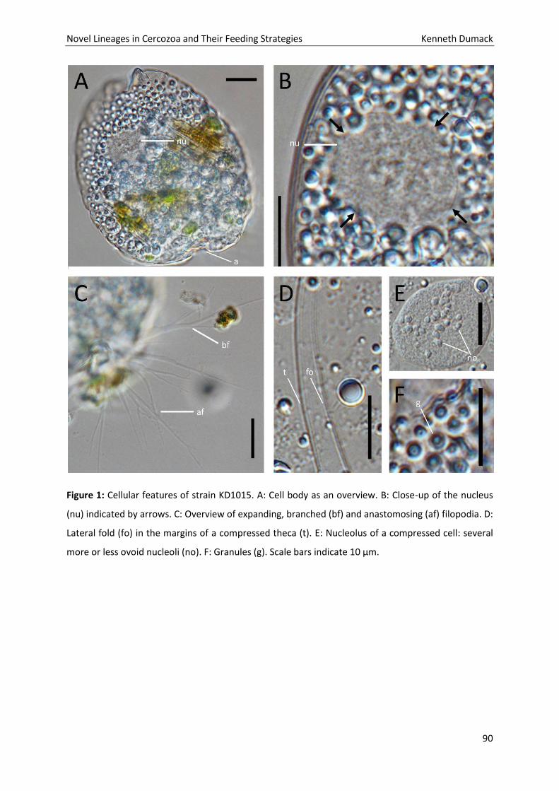
Novel Lineages in Cercozoa and Their Feeding Strategies Kenneth Dumack
90
Figure 1: Cellular features of strain KD1015. A: Cell body as an overview. B: Close-up of the nucleus
(nu) indicated by arrows. C: Overview of expanding, branched (bf) and anastomosing (af) filopodia. D:
Lateral fold (fo) in the margins of a compressed theca (t). E: Nucleolus of a compressed cell: several
more or less ovoid nucleoli (no). F: Granules (g). Scale bars indicate 10 µm.
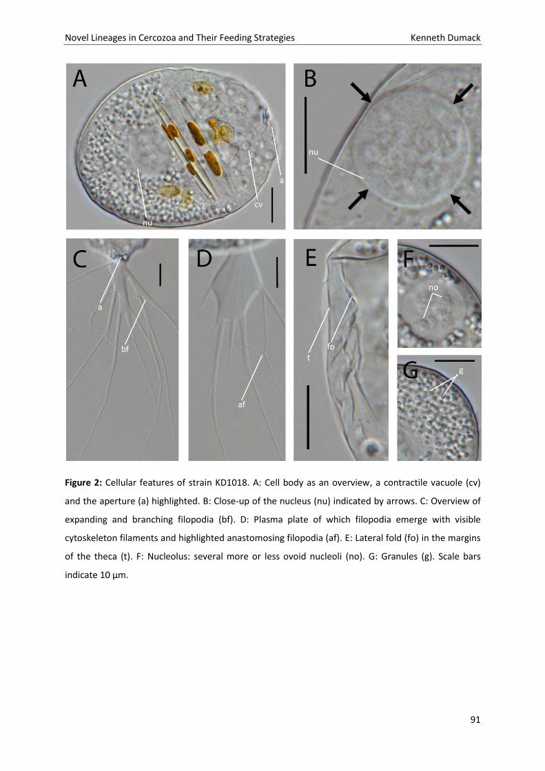
Novel Lineages in Cercozoa and Their Feeding Strategies Kenneth Dumack
91
Figure 2: Cellular features of strain KD1018. A: Cell body as an overview, a contractile vacuole (cv)
and the aperture (a) highlighted. B: Close-up of the nucleus (nu) indicated by arrows. C: Overview of
expanding and branching filopodia (bf). D: Plasma plate of which filopodia emerge with visible
cytoskeleton filaments and highlighted anastomosing filopodia (af). E: Lateral fold (fo) in the margins
of the theca (t). F: Nucleolus: several more or less ovoid nucleoli (no). G: Granules (g). Scale bars
indicate 10 µm.
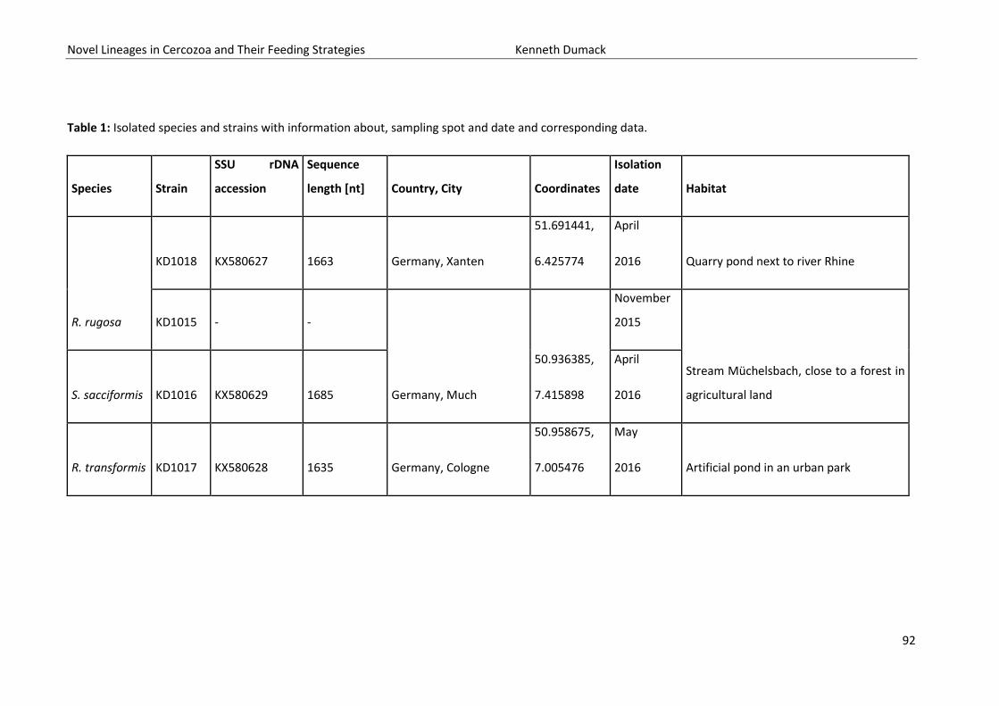
Novel Lineages in Cercozoa and Their Feeding Strategies Kenneth Dumack
92
Table 1: Isolated species and strains with information about, sampling spot and date and corresponding data.
Species Strain
SSU rDNA
accession
Sequence
length [nt] Country, City Coordinates
Isolation
date Habitat
R. rugosa
KD1018 KX580627 1663 Germany, Xanten
51.691441,
6.425774
April
2016 Quarry pond next to river Rhine
KD1015 - -
Germany, Much
50.936385,
7.415898
November
2015
Stream Müchelsbach, close to a forest in
agricultural land S. sacciformis KD1016 KX580629 1685
April
2016
R. transformis KD1017 KX580628 1635 Germany, Cologne
50.958675,
7.005476
May
2016 Artificial pond in an urban park
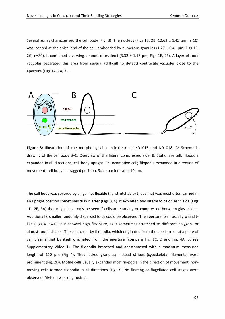
Novel Lineages in Cercozoa and Their Feeding Strategies Kenneth Dumack
93
Several zones characterized the cell body (Fig. 3): The nucleus (Figs 1B, 2B; 12.62 ± 1.45 µm; n=10)
was located at the apical end of the cell, embedded by numerous granules (1.27 ± 0.41 µm; Figs 1F,
2G; n=30). It contained a varying amount of nucleoli (3.32 ± 1.16 µm; Figs 1E, 2F). A layer of food
vacuoles separated this area from several (difficult to detect) contractile vacuoles close to the
aperture (Figs 1A, 2A, 3).
Figure 3: Illustration of the morphological identical strains KD1015 and KD1018. A: Schematic
drawing of the cell body B+C: Overview of the lateral compressed side. B: Stationary cell; filopodia
expanded in all directions; cell body upright. C: Locomotive cell; filopodia expanded in direction of
movement; cell body in dragged position. Scale bar indicates 10 µm.
The cell body was covered by a hyaline, flexible (i.e. stretchable) theca that was most often carried in
an upright position sometimes drawn after (Figs 3, 4). It exhibited two lateral folds on each side (Figs
1D, 2E, 3A) that might have only be seen if cells are starving or compressed between glass slides.
Additionally, smaller randomly dispersed folds could be observed. The aperture itself usually was slit-
like (Figs 4, 5A-C), but showed high flexibility, as it sometimes stretched to different polygon- or
almost round shapes. The cells crept by filopodia, which originated from the aperture or at a plate of
cell plasma that by itself originated from the aperture (compare Fig. 1C, D and Fig. 4A, B; see
Supplementary Video 1). The filopodia branched and anastomosed with a maximum measured
length of 110 µm (Fig 4). They lacked granules; instead stripes (cytoskeletal filaments) were
prominent (Fig. 2D). Motile cells usually expanded most filopodia in the direction of movement, non-
moving cells formed filopodia in all directions (Fig. 3). No floating or flagellated cell stages were
observed. Division was longitudinal.
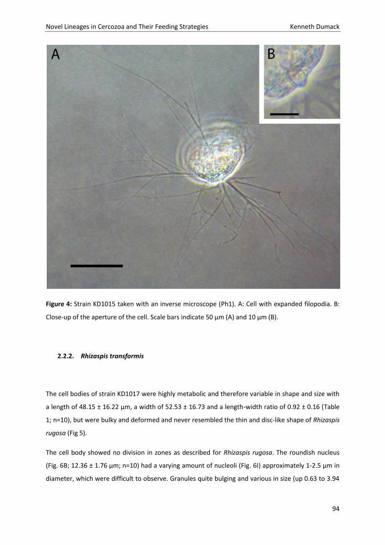
Novel Lineages in Cercozoa and Their Feeding Strategies Kenneth Dumack
94
Figure 4: Strain KD1015 taken with an inverse microscope (Ph1). A: Cell with expanded filopodia. B:
Close-up of the aperture of the cell. Scale bars indicate 50 µm (A) and 10 µm (B).
2.2.2. Rhizaspis transformis
The cell bodies of strain KD1017 were highly metabolic and therefore variable in shape and size with
a length of 48.15 ± 16.22 µm, a width of 52.53 ± 16.73 and a length-width ratio of 0.92 ± 0.16 (Table
1; n=10), but were bulky and deformed and never resembled the thin and disc-like shape of Rhizaspis
rugosa (Fig 5).
The cell body showed no division in zones as described for Rhizaspis rugosa. The roundish nucleus
(Fig. 6B; 12.36 ± 1.76 µm; n=10) had a varying amount of nucleoli (Fig. 6I) approximately 1-2.5 µm in
diameter, which were difficult to observe. Granules quite bulging and various in size (up 0.63 to 3.94
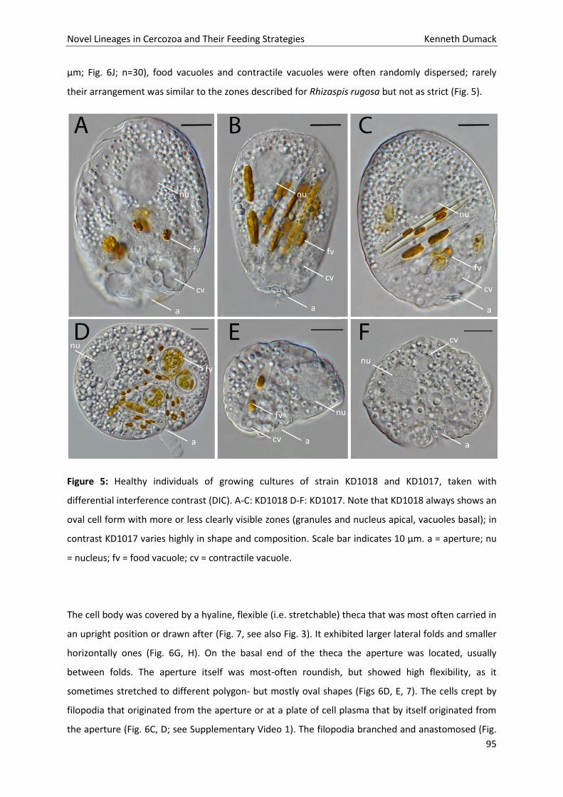
Novel Lineages in Cercozoa and Their Feeding Strategies Kenneth Dumack
95
µm; Fig. 6J; n=30), food vacuoles and contractile vacuoles were often randomly dispersed; rarely
their arrangement was similar to the zones described for Rhizaspis rugosa but not as strict (Fig. 5).
Figure 5: Healthy individuals of growing cultures of strain KD1018 and KD1017, taken with
differential interference contrast (DIC). A-C: KD1018 D-F: KD1017. Note that KD1018 always shows an
oval cell form with more or less clearly visible zones (granules and nucleus apical, vacuoles basal); in
contrast KD1017 varies highly in shape and composition. Scale bar indicates 10 µm. a = aperture; nu
= nucleus; fv = food vacuole; cv = contractile vacuole.
The cell body was covered by a hyaline, flexible (i.e. stretchable) theca that was most often carried in
an upright position or drawn after (Fig. 7, see also Fig. 3). It exhibited larger lateral folds and smaller
horizontally ones (Fig. 6G, H). On the basal end of the theca the aperture was located, usually
between folds. The aperture itself was most-often roundish, but showed high flexibility, as it
sometimes stretched to different polygon- but mostly oval shapes (Figs 6D, E, 7). The cells crept by
filopodia that originated from the aperture or at a plate of cell plasma that by itself originated from
the aperture (Fig. 6C, D; see Supplementary Video 1). The filopodia branched and anastomosed (Fig.
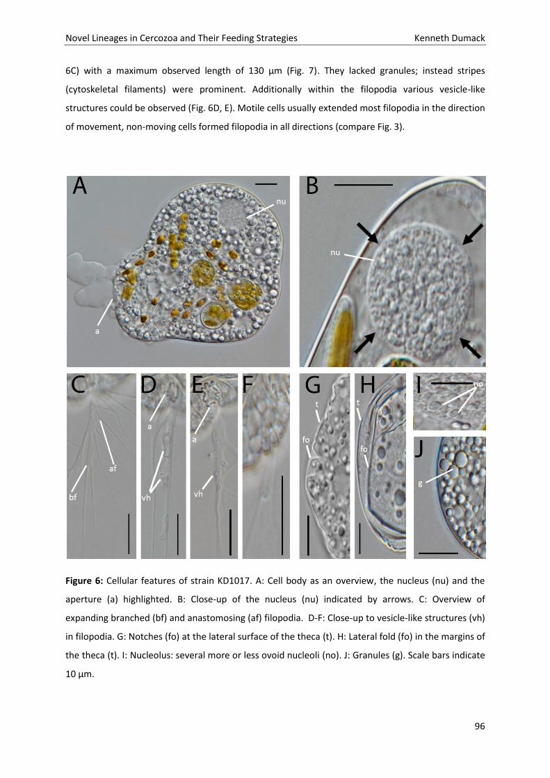
Novel Lineages in Cercozoa and Their Feeding Strategies Kenneth Dumack
96
6C) with a maximum observed length of 130 µm (Fig. 7). They lacked granules; instead stripes
(cytoskeletal filaments) were prominent. Additionally within the filopodia various vesicle-like
structures could be observed (Fig. 6D, E). Motile cells usually extended most filopodia in the direction
of movement, non-moving cells formed filopodia in all directions (compare Fig. 3).
Figure 6: Cellular features of strain KD1017. A: Cell body as an overview, the nucleus (nu) and the
aperture (a) highlighted. B: Close-up of the nucleus (nu) indicated by arrows. C: Overview of
expanding branched (bf) and anastomosing (af) filopodia. D-F: Close-up to vesicle-like structures (vh)
in filopodia. G: Notches (fo) at the lateral surface of the theca (t). H: Lateral fold (fo) in the margins of
the theca (t). I: Nucleolus: several more or less ovoid nucleoli (no). J: Granules (g). Scale bars indicate
10 µm.
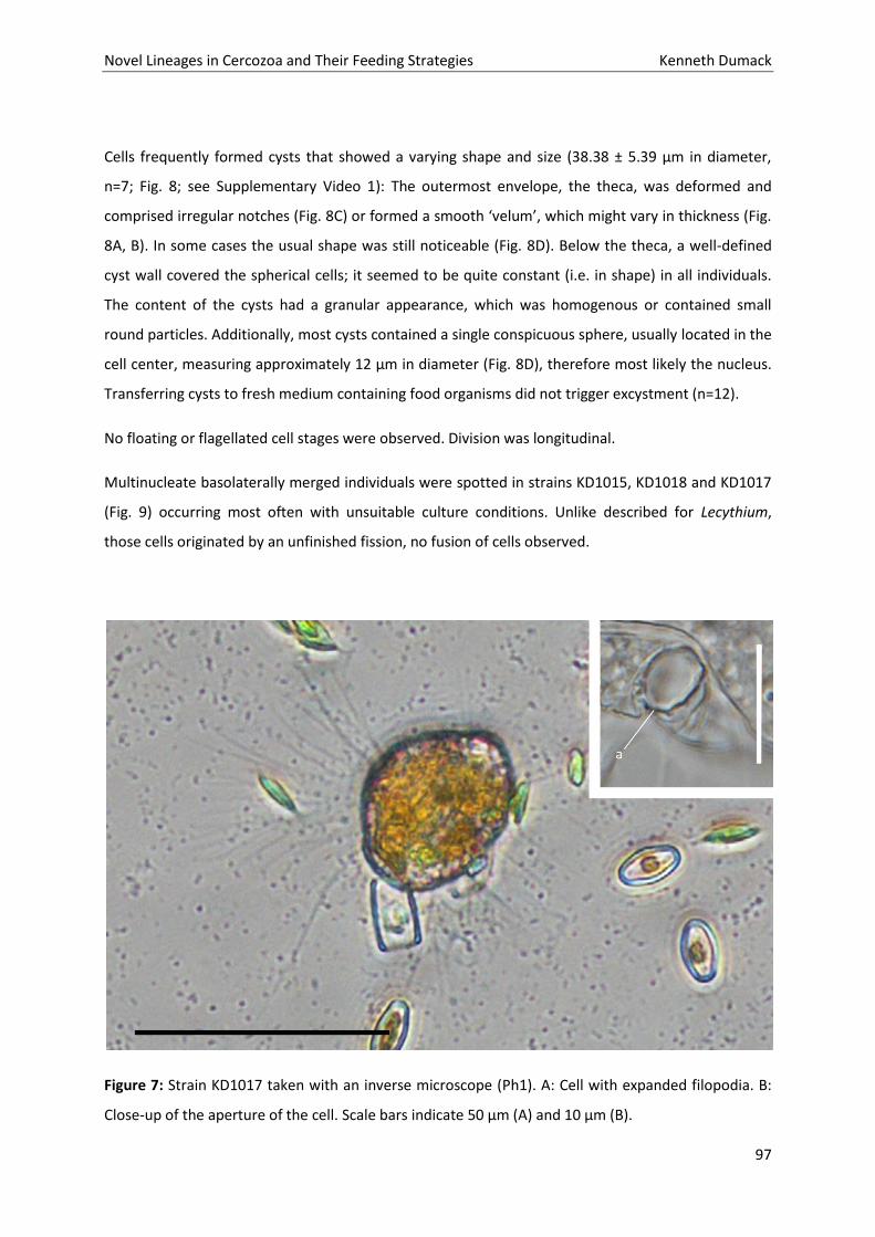
Novel Lineages in Cercozoa and Their Feeding Strategies Kenneth Dumack
97
Cells frequently formed cysts that showed a varying shape and size (38.38 ± 5.39 µm in diameter,
n=7; Fig. 8; see Supplementary Video 1): The outermost envelope, the theca, was deformed and
comprised irregular notches (Fig. 8C) or formed a smooth ‘velum’, which might vary in thickness (Fig.
8A, B). In some cases the usual shape was still noticeable (Fig. 8D). Below the theca, a well-defined
cyst wall covered the spherical cells; it seemed to be quite constant (i.e. in shape) in all individuals.
The content of the cysts had a granular appearance, which was homogenous or contained small
round particles. Additionally, most cysts contained a single conspicuous sphere, usually located in the
cell center, measuring approximately 12 µm in diameter (Fig. 8D), therefore most likely the nucleus.
Transferring cysts to fresh medium containing food organisms did not trigger excystment (n=12).
No floating or flagellated cell stages were observed. Division was longitudinal.
Multinucleate basolaterally merged individuals were spotted in strains KD1015, KD1018 and KD1017
(Fig. 9) occurring most often with unsuitable culture conditions. Unlike described for Lecythium,
those cells originated by an unfinished fission, no fusion of cells observed.
Figure 7: Strain KD1017 taken with an inverse microscope (Ph1). A: Cell with expanded filopodia. B:
Close-up of the aperture of the cell. Scale bars indicate 50 µm (A) and 10 µm (B).
Novel Lineages in Cercozoa and Their Feeding Strategies Kenneth Dumack
98
2.2.3. Sacciforma sacciformis gen. nov.
The cell bodies of strain KD1016 were oval and flattened with a length of approx. 35 µm, a width of
approx. 25 µm and a length-width ratio of approx. 1.5 (Table 1, Fig. 10). The nucleus was located at
the apical end of the cell. Cells contained numerous granules (2.87 ± 0.67 µm; n=10). Close the
aperture several food vacuoles and contractile vacuoles were located.
The cell body was covered by a hyaline, flexible (i.e. stretchable) theca that was carried in an upright
position. On the basal end of the theca the slit-like, non-flexible, aperture was located. The cells crept
by filopodia which originated of the aperture, cells never showed a large net of filopodia. The
filopodia lacked granules and branched. No floating or flagellated cell stages were observed. Division
was longitudinal.
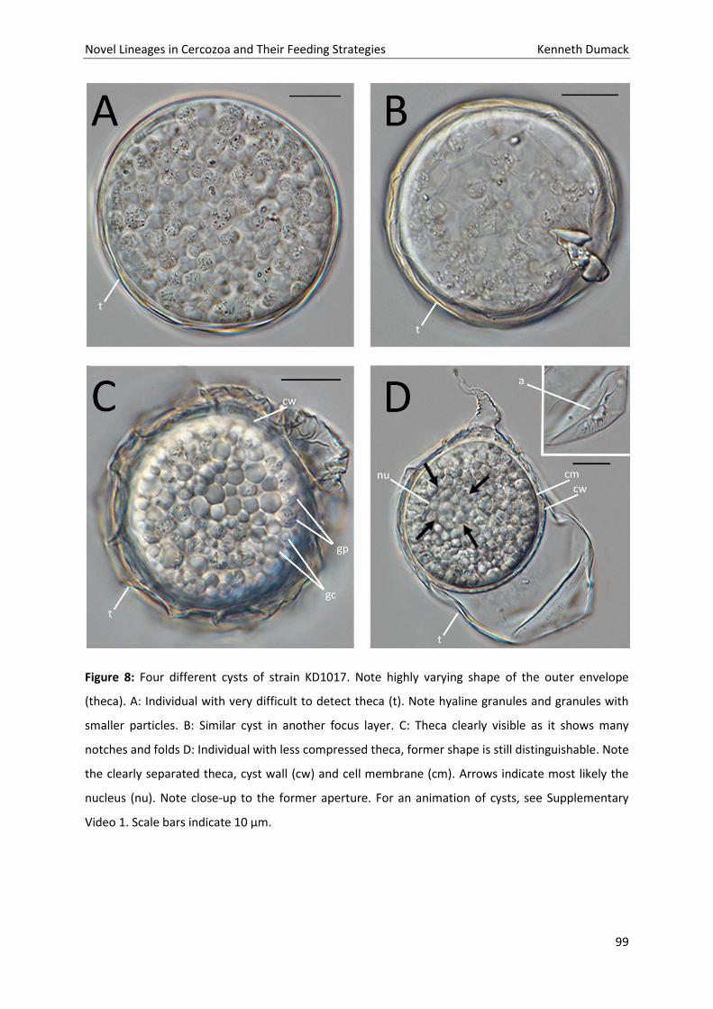
Novel Lineages in Cercozoa and Their Feeding Strategies Kenneth Dumack
99
Figure 8: Four different cysts of strain KD1017. Note highly varying shape of the outer envelope
(theca). A: Individual with very difficult to detect theca (t). Note hyaline granules and granules with
smaller particles. B: Similar cyst in another focus layer. C: Theca clearly visible as it shows many
notches and folds D: Individual with less compressed theca, former shape is still distinguishable. Note
the clearly separated theca, cyst wall (cw) and cell membrane (cm). Arrows indicate most likely the
nucleus (nu). Note close-up to the former aperture. For an animation of cysts, see Supplementary
Video 1. Scale bars indicate 10 µm.
Novel Lineages in Cercozoa and Their Feeding Strategies Kenneth Dumack
100
2.3. Phylogenetic analyses
Nearly full-length SSU sequences were obtained, ranging from 1,635 to 1,685 nucleotides (Table 2).
No introns were found. The maximum likelihood tree (Fig. 11) revealed the cercozoan subphylum
Filosa (Cavalier-Smith 1997) and selected sequences of the Endomyxa as out-group. The filosan class
Thecofilosea was composed of the Phaeodaria, Cryomonadida and Tectofilosida. Thecate amoeba
lineages sequenced in this study and close relatives within the Thecofilosea are highlighted. Similar to
previous analyses using SSU rDNA sequence comparisons, many basal branches within the Cercozoa
were not supported (Bass and Cavalier-Smith 2004; Bass et al. 2009a,b; Howe et al. 2011).
Sacciforma sacciformis (KD1016), Rhizaspis rugosa and Rhizaspis transformis (KD1017 and KD1018)
were not monophlyletic. Although Rhizaspis rugosa and Rhizaspis transformis (strain KD1017 and
KD1018) formed a maximally supported clade within the Tectofilosida, Sacciforma sacciformis
(KD1016) grouped in the Cryomonadida. The SSU sequences of Rhizaspis rugosa and Rhizaspis
transformis (KD1017 and KD1018) were to 100% identical and built together with Pseudodifflugia cf.
gracilis (AJ418794) a sistergroup to Lecythium with a bootstrap support value of 80 (ML) and
Bayesian posterior probability of 1.0 (BI).
Sacciforma sacciformis (KD1016) grouped next to Rhogostoma with (96/1.0). The clade has been
referred to as the Rhizaspididae by Howe et al. (2011) but was here renamed as Rhogostomidae (see
discussion).
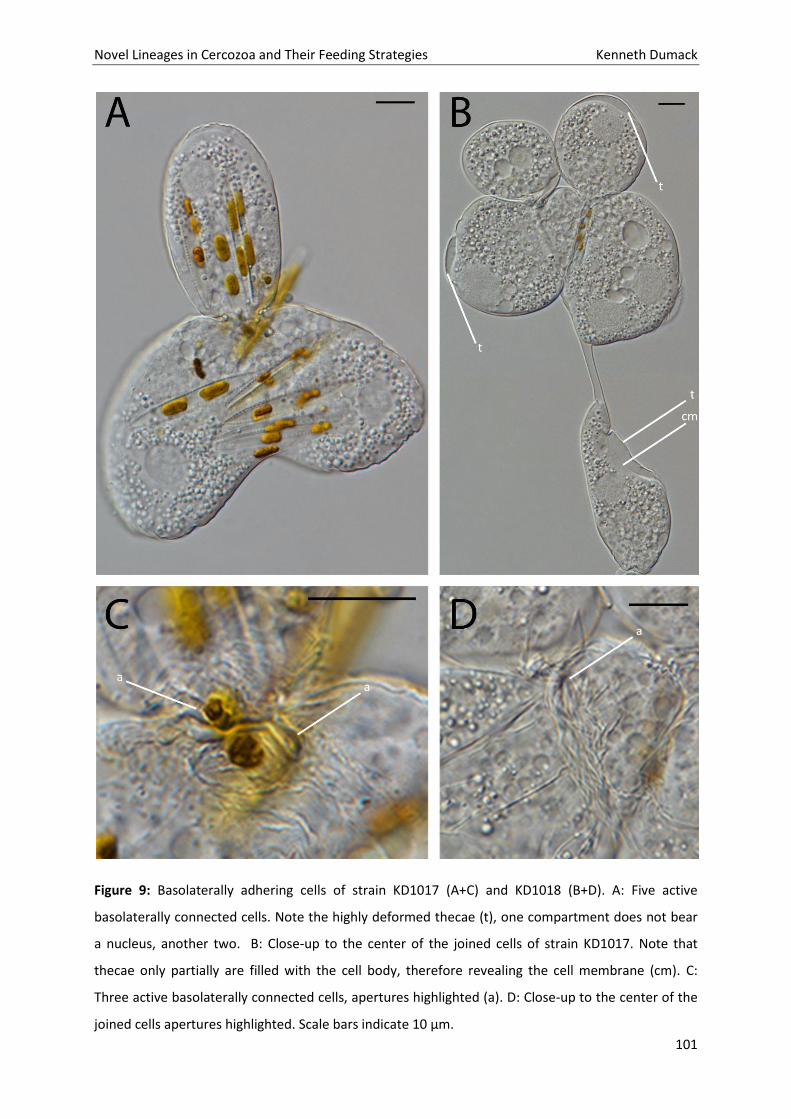
Novel Lineages in Cercozoa and Their Feeding Strategies Kenneth Dumack
101
Figure 9: Basolaterally adhering cells of strain KD1017 (A+C) and KD1018 (B+D). A: Five active
basolaterally connected cells. Note the highly deformed thecae (t), one compartment does not bear
a nucleus, another two. B: Close-up to the center of the joined cells of strain KD1017. Note that
thecae only partially are filled with the cell body, therefore revealing the cell membrane (cm). C:
Three active basolaterally connected cells, apertures highlighted (a). D: Close-up to the center of the
joined cells apertures highlighted. Scale bars indicate 10 µm.
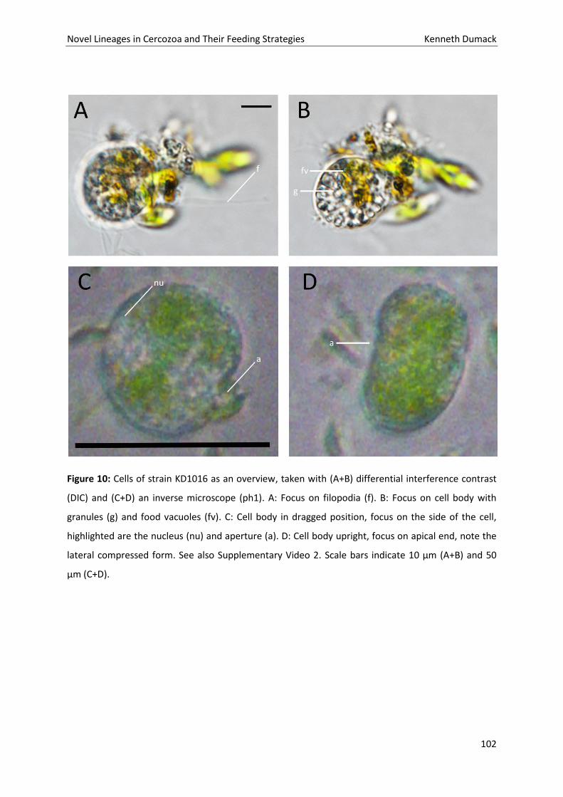
Novel Lineages in Cercozoa and Their Feeding Strategies Kenneth Dumack
102
Figure 10: Cells of strain KD1016 as an overview, taken with (A+B) differential interference contrast
(DIC) and (C+D) an inverse microscope (ph1). A: Focus on filopodia (f). B: Focus on cell body with
granules (g) and food vacuoles (fv). C: Cell body in dragged position, focus on the side of the cell,
highlighted are the nucleus (nu) and aperture (a). D: Cell body upright, focus on apical end, note the
lateral compressed form. See also Supplementary Video 2. Scale bars indicate 10 µm (A+B) and 50
µm (C+D).
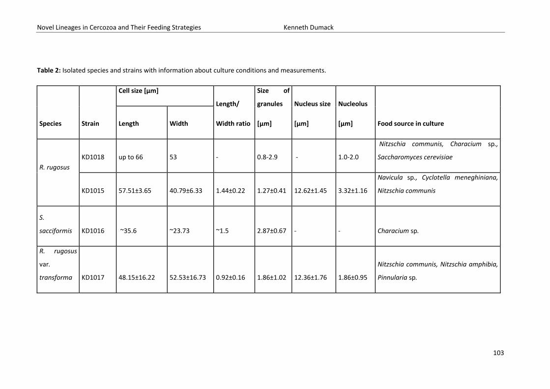
Novel Lineages in Cercozoa and Their Feeding Strategies Kenneth Dumack
103
Table 2: Isolated species and strains with information about culture conditions and measurements.
Species Strain
Cell size [µm]
Length/
Width ratio
Size of
granules
[µm]
Nucleus size
[µm]
Nucleolus
[µm] Food source in culture Length Width
R. rugosus
KD1018 up to 66 53 - 0.8-2.9 - 1.0-2.0
Nitzschia communis, Characium sp.,
Saccharomyces cerevisiae
KD1015 57.51±3.65 40.79±6.33 1.44±0.22 1.27±0.41 12.62±1.45 3.32±1.16
Navicula sp., Cyclotella meneghiniana,
Nitzschia communis
S.
sacciformis KD1016
~35.6 ~23.73 ~1.5 2.87±0.67 - - Characium sp.
R. rugosus
var.
transforma KD1017 48.15±16.22 52.53±16.73 0.92±0.16 1.86±1.02 12.36±1.76 1.86±0.95
Nitzschia communis, Nitzschia amphibia,
Pinnularia sp.
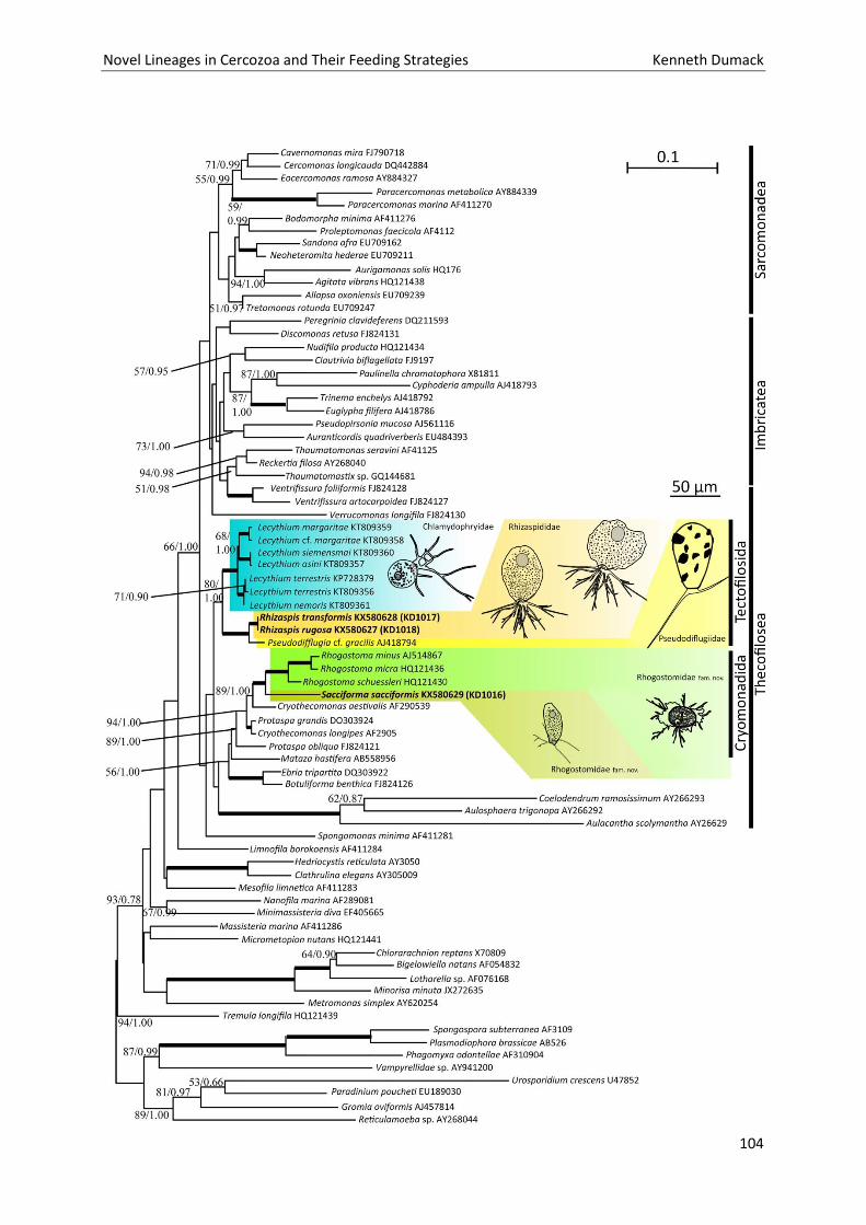
Novel Lineages in Cercozoa and Their Feeding Strategies Kenneth Dumack
104
Novel Lineages in Cercozoa and Their Feeding Strategies Kenneth Dumack
105
Figure 11: Reconstructed phylogenetic tree of the Cercozoa. Shown is the maximum likelihood tree
obtained by the PhyML GTR+I+G analyses including 76 sequences and 1587 unambiguously aligned
sites. On the respective branches are the support values of the PhyML and the Bayesian analysis
shown (ML/BI), if higher than 50. Bold lines indicate a support value above or equal 95/0.95.
Highlighted are the Tectofilosida (blue, orange and yellow) and Rhogostomidae in the Cryomonadida
(green-colours).
3. Discussion
3.1. Untangling Plagiophrys on morphological basis
Skuja (1948) described a species, highly similar to ‘Plagiophrys scutiformis’, only differing in the
nucleus structure. He described Rhizaspis granulata as an oval and flattened cell with longitudinal
folds and filopodia, which are used for locomotion by creeping over the surface and may be used for
floating stages. He even mentioned the zoning of vacuoles and granules. Since he was a phycologist
and was not familiar with filose amoebae, he saw resemblance in the petalomonads and suggested
that his isolates completely lost their flagella and evolved therefore filopodia de novo.
This is highly unlikely and since his description of R. granulata was in all major points (e.g. filopodia
without granules, aperture being slit-like, cell shape and nourishment) in common with Hertwig and
Lessers’, we conclude that Skuja unknowingly redescribed ‘Plagiophrys’ sensu Hertwig and Lesser
(1874).
We therefore adopt the name Rhizaspis for ‘Plagiophrys’ sensu Hertwig and Lesser (1874), therefore
all to us known Plagiophrys species except Plagiophrys sensu Claparède and Lachmann (i.e.
Plagiophrys cylindrica and Plagiophrys sphaerica) and transfer following species to Rhizaspis: P.
scutiformis, P. parvipunctata, P. arcuatus and P. scutiformis var. marginata.
Since pseudopodia structure (i.e. being lobose or filose and granule content) is a morphological
character of high specificity (Bass et al. 2009a, Dumack et al. 2016a, Nikoleav 2005), it is highly
unlikely, that these are monophyletic with Plagiophrys sensu Claparède and Lachmann.
The species described by Claparède and Lachmann (i.e. Plagiophrys cylindrica and Plagiophrys
sphaerica) show closer morphological affinity to Allogromia, Microgromia or other amoebae with
prominent granules in their filopodia and therefore might group in the Foraminifera or elsewhere in
Novel Lineages in Cercozoa and Their Feeding Strategies Kenneth Dumack
106
the Cercozoa (for instance in the Granofilosea), whereas the species described by Hertwig and Lesser
are presumed to group in the Thecofilosea.
3.2. Morphological analyses of our isolates
All our isolates show high similarities with Hertwig and Lessers’ ‘Plagiophrys’. Hertwig and Lesser
(1874) reported ‘Plagiophrys sacciformis’ as irregular cylindrical (35 µm in length and 25 µm in
width), just like our measurements of KD1016. We conclude that KD1016 resembled Hertwig and
Lessers’ Plagiophrys sacciformis.
Further they describe P. scutiformis as oval, 60 µm in length and 40 µm in width, with an often-
wrinkled surface. They did not describe contractile vacuoles, which are difficult to observe within
thecate amoebae as (a) thecate amoebae often are thicker than most naked amoebae and other cell
contents shade the hyaline vacuoles and (b) the vacuoles usually ‘beat’ very slowly compared to
most naked amoebae. However, Skuja (1948) described them. Both described the withdrawn slit-like
aperture and clear zones by pointing out that food vacuoles aggregate and granules, in the center of
the cell, embed the nucleus. These descriptions fit to our observations of strains KD1015 and
KD1018.
However, there is one significant difference between their and our descriptions. Opposing to our
observation Hertwig and Lesser (1874) described the nucleus to be homogeneous, even after
treatment with acetic acid, no nucleolus could be observed. Skuja (1948) however described his R.
granulata with one large central nucleolus. In contrast, we could define a varying amount of nucleoli.
Since nucleus structure is a morphological characteristic of high specificity and importance, we
conclude therefore, that we need to establish a new species name for our isolates KD1018 and
KD1015, Rhizaspis rugosa.
KD1017 resembled in many points Rhizaspis rugosa. But up to now, the highly different and varying
cell shape of strain KD1017 was never described to this extend for any other ‘Plagiophrys’ or
Rhizaspis species and never resembled the typical shield-like form of the previously mentioned
species.
Novel Lineages in Cercozoa and Their Feeding Strategies Kenneth Dumack
107
3.3. Untangling Plagiophrys on phylogenetic basis
Surprisingly strain KD1016, Rhizaspis rugosa and Rhizaspis transformis are not monophyletic within
our analyses. Although both group within the Thecofilosea, KD1016 groups close to Rhogostoma,
Rhizaspis rugosa and Rhizaspis transformis instead as a sistergroup to the Pseudodifflugiidae. We
therefore cannot adopt the name Rhizaspis for Hertwig and Lessers’ ‘Plagiophrys sacciformis’ and
therefore establish the new genus Sacciforma gen. nov., comprising exclusively Sacciforma
sacciformis, yet.
Howe et al. (2011) established the Rhizaspididae although no Rhizaspis has been sequenced back
then. They made this assumption based on morphological similarity between Rhogostoma,
Capsellina and Rhizaspis (i.e. the slit-like aperture, which is, as we could show, highly flexible in
Rhizaspis). Rhizaspis does not group close to Rhogostoma, although Sacciforma does. Those genera
can therefore not build a family which bears the name Rhizaspididae. We therefore have to switch
Rhogostoma and the presumed closely related Capsellina (see Howe et al. 2011) to a new family,
which includes Sacciforma and should be called Rhogostomidae.
‘Plagiophrys’ sensu Hertwig and Lesser (=Rhizaspis) was early believed to be closely-related to
Lecythium (de Saedeleer 1934). We could show that this is the case as R. rugosa and R. transformis
group within the Tectofilosida. However, they build no direct sistergroup to Lecythium but show a
closer relationship with the agglutinating testate amoeba Pseudodifflugia.
Although the morphology of the here described Rhizaspis species clearly differs as they could never
be confused with each other, surprisingly, their SSU sequences were identical. Several studies
already highlighted that the SSU gene is a limited marker for diversity of protists. More variable
genes probably will show genetic differences between Rhizaspis rugosa and Rhizaspis transformis.
Since the cultures are deposited in a culture collection and are therefore made available to the
public, genes such as ITS or LSU may be sequenced by us or others in future studies.
The other described ‘Plagiophrys’ species (P. scutiformis by Hertwig and Lesser 1874, P.
parvipunctata and P. arcuatus by Penard 1902 and Plagiophrys scutiformis var. marginata by de
Groot 1979) of which molecular data are still lacking, may also group with Tectofilosida, because of
their similarity to Rhizaspis rugosa and Rhizaspis transformis. However, this study confirmed that
assumptions about taxonomy solely based on few morphological characteristics may be incorrect
and are often confusing. Molecular data are therefore urgently required to shed more light on the
phylogeny of the highly interesting group of filose thecate amoebae.
Novel Lineages in Cercozoa and Their Feeding Strategies Kenneth Dumack
108
3.4. Culture conditions
Although we provide stable long-time maintainable cultures, we were not able to provide cultures of
two of our four strains (Rhizaspis rugosa (strain KD1015) and Sacciforma sacciformis), as they ceased
to grow after a couple divisions. In case of R. rugosa, we were not able to culture strain KD1015 with
a mixture of food sources including yeasts and green algae. We optimized culture conditions by
adding a variation of different diatoms and excluding yeasts and green algae, making KD1017 and
KD1018 well culturable (Table 1).
Nevertheless, strain KD1017 constantly formed cysts (in contrast to KD1018 and many other
Thecofilosea cultures) which we interpret as a stress response to our culture conditions. Sacciforma
sacciformis grouped in direct relation to Rhogostoma, an omnivorous amoeba, feeding on eukaryotic
cells like algae but not culturable without sufficient supply of bacteria as food source (own
observations, unpublished). We therefore conclude that Sacciforma needs bacteria as food source
and ceases to grow under our low bacteria conditions, similar to Rhogostoma.
However, as this study and many other recent studies show (Dumack et al. 2016 a,b; Hess &
Melkonian 2013; Hess et al. 2012), previously as ‘unculturable’ assigned protists, are culturable if
cultured under the right conditions.
4. Taxonomic acts
4.1. Rhizaspis (Skuja 1948)
Revised diagnosis: Thecate amoebae with ventral slit-like, but flexible, cleft that emits filopodia;
theca thin, flexible, in active cells adherent throughout to cell surface, with exosomes (R.
parvipunctata) or without (all other species), sometimes deformed by ingested food, often wrinkled
or folded, consisting of single smooth dense layer outside and scarcely thicker than the plasma
membrane; thus with bilateral symmetry (ventral flattened). Round nucleus; nucleoli may be visible
by light microscopy, often one, sometimes several. Cells may be divided in zones (basal to apical):
several contractile vacuoles, food vacuoles, nucleus embedded by a large number of in size varying
Novel Lineages in Cercozoa and Their Feeding Strategies Kenneth Dumack
109
granules. Inhabiting freshwaters. Eukaryvorous, mainly feeding on algae, especially diatoms. Division
longitudinal, binary. Sexual reproduction unknown.
Type species: R. granulata Skuja 1948
Other species: R. simplex by Skuja 1948, R. scutiformis comb. nov. by Hertwig and Lesser 1874, R.
parvipunctata comb. nov. and R. arcuatus comb. nov. by Penard 1902 and R. scutiformis var.
marginata comb. nov. by de Groot 1979
4.2. Rhizaspis rugosa sp. nov.
Diagnosis: The theca comprises lateral and basal folds. Round nucleus, several roundish nucleoli. Cell
divided into zones as described for the genus. No floating stages observed. Cysts not observed. In
cultures cells may occur basolaterally connected in a ring, built through unfinished fission, not fusion.
Easy to confuse with Rhizaspis scutiformis or Rhizaspis granulata which have different nucleoli.
Deposited preserved material: A glass slide containing several fixed individuals is deposited in the
Upper Austrian State Museum Invertebrate Collection (2016/119).
Type generating strain: KD1018; deposited in the Culture Collection of Algae and Protozoa under the
accession number CCAP 1267/1
SSU Sequence of type generating strain: KX580627
Illustrations of type generating strain: Figs 1, 3, 10, Supplementary Video 1; this material constitutes
the name-bearing type of this species.
Type locality: quarry pond next to river Rhine, in Xanten, Germany (51.691441, 6.425774)
Etymology: rugosa {adj} [Latin] = 1) wrinkled, wrinkly. Referring to the numerous folds in the theca.
Novel Lineages in Cercozoa and Their Feeding Strategies Kenneth Dumack
110
4.3. Rhizaspis transformis sp. nov.
Diagnosis: Similar to Rhizaspis rugosa, with following differences: Highly metabolic and therefore
varying in shape and size, usually bulky and deformed; never resembled the thin and disc-like shape
of Rhizaspis rugosa; no clear separation in zones; cysts vary in form: theca builds an envelope around
the spherical cyst, compressed with notches and protuberances; filopodia with numerous non-
contractile vacuoles.
Deposited preserved material: A glass slide containing several fixed individuals is deposited in the
Upper Austrian State Museum Invertebrate Collection (2016/120).
Type generating strain: KD1017; deposited in the Culture Collection of Algae and Protozoa under the
accession number CCAP 1267/2
SSU Sequence of type generating strain: KX580628
Illustrations of type generating strain: Figs 6, 7, 8, 9, 10; Supplementary Video 1; this material
constitutes the name-bearing type of this species.
Type locality: pond in urban park, in Cologne near Mülheim, Germany (50.958675, 7.005476)
Etymology: transforma –re {verb} [Latin] = 1) (to) transform, (to) reform. Referring to the highly
variable cell shape.
4.4. Rhogostomidae fam. nov.
Diagnosis: Thecate amoebae with ventral slit-like and not flexible cleft that emits filopodia; theca
thin, flexible, in active cells adherent throughout to cell surface, consisting of single smooth dense
layer outside and scarcely thicker than the plasma membrane; thus with bilateral symmetry. Theca
with exosomes (Capsellina) or without (Rhogostoma, Sacciforma). Omnivorous (mainly bacteria, also
yeasts, algae). Electron microscopy of Capsellina and Rhogostoma by Simitzis and Le Goff (1981).
Division longitudinal, binary. Sexual reproduction unknown.
Type genus: Rhogostoma Belar, 1921
other genera: Capsellina (Penard 1909; see Howe et al. 2011) Sacciforma (this study)
Novel Lineages in Cercozoa and Their Feeding Strategies Kenneth Dumack
111
4.5. Sacciforma gen. nov.
Diagnosis: Cell body in contrast to the apical-basal compressed Rhogostoma and Capsellina, to which
it is closely related, elongated (length-width ratio approx. 1.5) and ventral flattened. Freshwater.
Cysts, floating- or flagellate stages unknown.
Type species: Sacciforma sacciformis comb. nov., Hertwig and Lesser 1874
Etymology: saccus {noun} [Latin] = bag; formis {noun} [Latin] = shape; refers to the bag-like shape of
the cell body, derived from the species name of the type species Sacciforma sacciformis, described as
Plagiophrys sacciformis by Hertwig and Lesser, 1874.
5.Materials and methods
5.1.Sampling and isolation
We collected 100-200 ml of sediment or detritus rich water of freshwaters, like ponds and rivers
(Table 1). Most samples were stored at room temperature over night; few samples were stored at 10
°C incubation over two to three days before investigation.
The samples were then shaken to detach surface attached protists and 1 ml was given in each well of
a 24-well plate (Sarstedt, Germany). The plate was incubated for approx. 20 min to allow small
organisms to settle. Observation of amoebae was conducted with a light microscope (Nikon Eclipse
TS100; Ph1; 40x, 100x, 200x and 400x magnification). All samples were scanned for Plagiophrys-like
cells several times up to three weeks after collection. Promising cells were transferred to Waris-H
(McFadden & Melkonian 1986) and enriched with a mixture of putative food organisms: Nitzschia
communis (CCAC 5737 B), Nitzschia amphibia (CCAC 5733 B), Navicula sp. (CCAC 1772 B), Pinnularia
sp. (CCAC 0222 B), Cyclotella meneghiniana (CCAC 5735 B), Characium sp. and Saccharomyces
cerevisiae (Table 2). Cells were subcultured every week.
Novel Lineages in Cercozoa and Their Feeding Strategies Kenneth Dumack
112
5.2.Microscopical observations
Pictures and videos were taken with a Nikon Eclipse TE2000-E (Ph1, up to 400x magnification) Nikon
Eclipse 90i (DIC, up to 600x magnification), both with the mounted camera Nikon digital sight DS-U2
(program: NIS-Elements V4.13.04).
5.3. SSU sequencing
Single cells were starved for two to three days and transferred with approx. 1 µl of medium into 15
µm ddH2O into PCR-tubes. To this, 5 μl Green Taq Dream Buffer, 5 μl of 2 μM dNTP’s, 5 μl of 1 μM
forward and reverse primer each and 0.3 μl DreamTaq DNA-polymerase (Thermo Fisher Scientific,
Dreieich, Germany) were added with additional 14.7 μl ddH2O.
The SSU sequences were obtained in two successive steps. First the whole SSU was amplified with
the general eukaryotic primers, RibA and RibB (Cavalier-Smith and Chao 1995; Pawlowski 2000).
Using one µl of the first PCR as template, a semi-nested re-amplification was conducted with the
primers RibA+1300R targeting the 5’ part and 590F+RibB targeting the 3’ part of the gene (Quintela-
Alonso et al. 2011).
PCR conditions were as follows: initial denaturation at 95 ◦C for 5 min, 35 cycles (denaturation at 96
◦C for 32 s, annealing at 50 ◦C for 36 s, elongation at 72 °C for 2 min), terminal extension at 72 ◦C for
7 min, and hold at 4 ◦C.
For sequencing the Big dye Terminator Cycle sequencing Kit and an ABI PRISM automatic sequencer
were used.
5.4.Phylogenetic analyses
The partial sequences were manually checked for sequencing errors and combined into one
sequence contig. To create a dataset for phylogenetic analyses they were blasted (blastn 2.3.0)
against the NCBI GenBank database (last date of accession: 23.06.2016). Sequences with a similarity
of ≥ 96% were downloaded and manually aligned in SeaView (V4.5.3, Gouy et al. 2010). Additionally
Novel Lineages in Cercozoa and Their Feeding Strategies Kenneth Dumack
113
representative sequences of the major cercozoan subphyla were added, Endomyxa sequences were
used as outgroup. An alignment with 76 sequences and 1587 unambiguously aligned sites of which
46.82% were invariant was used for phylogenetic analyses. The program jmodeltest (V.2.1.5, Darriba
et al. 2012) was used to determine the best fitting model: GTR+I+G, which was selected among 88
models (settings: Substitution schemes 11; add Base frequencies +I+G rate variation nCat=4, ML
optimized NNI as base tree).
Phylogenetic trees were constructed using maximum likelihood (ML) and Bayesian inference (BI).
Maximum likelihood phylogenetic analyses were run using PhyML V3.1 (Guindon & Gascuel 2003)
with the following settings: GTR model; a proportion of invariable sites and a gamma-shaped
distribution of the substitution rates across variable sites (GTR+I+G), with four rate categories; BIONJ
distance-based starting tree with all model parameters estimated from the data. The Bayesian
analyses were run using MrBayes V3.2 (Altekar et al. 2004; Ronquist & Huelsenbeck 2003) with the
following settings: 5 million generations, trees were sampled every 100 generations, convergence of
the two runs was estimated every 500 generations. Of the sampled trees, 25% were discarded as
burn-in.
5.5.Preparation of cells for deposition
Saturated mercury-(II)-chloride solution and 96% ethanol were mixed (ratio 2:1) to obtain a fixing
solution. It was heated to 70 °C and 30 ml were added to 1.5 ml of a culture in growing phase. The
mixture was washed in 30 % ethanol once and further fixed in 1 ml osmium tetroxide for one minute.
Subsequently the cells were dehydrated in eight steps consisting of 20 min incubation in an ethanol
series of increasing concentration (from 30 to 96%). Cells were then preserved on a microscope slide
with a drop of glycerine under a cover slide. The slides were deposited in the Upper Austrian State
Museum; accession numbers pending.
6.Acknowledgments
The study was supported by the DFG research group FOR 918. We thank the Culture Collection of
Algae at the University of Cologne (CCAC) for their service and the provided algal strains.
Novel Lineages in Cercozoa and Their Feeding Strategies Kenneth Dumack
114
7.References
Altekar G, Dwarkadas S, Huelsenbeck JP, Ronquist F (2004) Parallel metropolis coupled Markov chain
Monte Carlo for Bayesian phylogenetic inference. Bioinform 20:407-415
Bass D, Cavalier-Smith T (2004) Phylum-specific environmental DNA analysis reveals remarkably high
global biodiversity of Cercozoa (Protozoa). Int J of Syst and Evol Microbiol 54:2393–2404
Bass D, Chao EE-Y, Nikolaev S, Yabuki A, Ishida K, Berney C, Pakzad U, Wylezich C, Cavalier-Smith T
(2009a) Phylogeny of novel naked filose and reticulose Cercozoa: Granofilosea cl. n. and
Proteomyxidea revised. Protist 160:75-109
Bass D, Howe AT, Mylnikov AP, Vickerman K, Chao EE-Y, Smallbone JE, Snell J, Cabral JrC, Cavalier-
Smith T (2009b) Phylogeny and classification of Cercomonadida (Protozoa, Cercozoa): Cercomonas,
Eocercomonas, Paracercomonas, and Cavernomonas gen. nov. Protist 160:483–521
Cash J, Wailes GH, Hopkinson J (1915) The British freshwater rhizopoda and heliozoa, volume 3. The
Rhizopoda. The Ray Society, London, U.K.
Cavalier-Smith T (1998a) Neomonada and the origin of animals and fungi. Coombs GH, Vickerman K,
Sleigh MA, Warren A (eds) Evolutionary Relationships among Protozoa. Chapman & Hall, London,
375–407
Cavalier-Smith T (1998b) A revised six-kingdom system of life. Biol Rev Camb Philos Soc 73:203–266
Cavalier-Smith T, Chao EE-Y (1995) The opalozoan Apusomonas is related to the common ancestor of
animals, fungi, and choanoflagellates. P Roy Soc Lond B Bio 261:1–6
Cavalier-Smith T, Chao EE-Y (2003) Phylogeny and classification of phylum Cercozoa (Protozoa).
Protist 154:341–358
Claparède RE, Lachmann KFJ (1859) Études sur les Infusoires et les Rhizopodes. Mém Inst Genev 5(6):
489 pp
Darriba D, Taboada GL, Doallo R, Posada D (2012) jModeltest 2: more models, new heuristics and
parallel computing. Nat Met 9:772
De Groot AA (1979) Über Einige Wenig Bekannten Rhizopoden. Aquatic Ecol 13.1:34-49
De Saedeleer H (1934) Beitrag zur Kenntnis der Rhizopoden: morphologische und systematische
Untersuchungen und ein Klassifikationsversuch. Mem Mus R Hist Nat Belg 60:1–112
Dumack K, Müller ME, Bonkowski M (2016a) Description of Lecythium terrestris sp. nov.
(Chlamydophryidae, Cercozoa), a soil dwelling protist feeding on fungi and algae. Protist 167:93-105
Dumack K, Baumann C, Bonkowski M. (2016b) A Bowl with Marbles: Revision of the Thecate Amoeba
Genus Lecythium (Chlamydophryidae, Cercozoa , Rhizaria) Including a Description of Four New
Species and an Identification Key. Protist 167:440–59.
Novel Lineages in Cercozoa and Their Feeding Strategies Kenneth Dumack
115
Dumack K, Schuster J, Bass D, Bonkowski M (2016c) A novel lineage of ‘naked filose amoebae’;
Kraken carinae gen. nov. sp. nov. (Cercozoa) with a remarkable locomotion by disassembly of its cell
body. Protist 167:268-278
Hess S, Melkonian M (2013) The mystery of clade X: Orciraptor gen. nov. and Viridiraptor gen. nov.
are highly specialised, algivorous amoeboflagellates (Glissomonadida, Cercozoa). Protist 164:706–47
Hess S, Sausen N, Melkonian M (2012) Shedding light on vampires: the phylogeny of vampyrellid
amoebae revisited. PloS One 7(2):e31165
Gouy M, Guindon S, Gascuel O (2010) SeaView version 4: a multiplatform graphical user interface for
sequence alignment and phylogenetic tree building. Mol Biol Evol 27:221-224
Guindon S, Gascuel O (2003) A simple, fast, and accurate algorithm to estimate large phylogenies by
maximum likelihood. Syst Biol 52:696-704
Hertwig R, Lesser E (1874) Über Rhizopoden und denselben nahestehende Organismen. Arch
Mikroskop Anat 10:35–243
Howe A, Bass D, Scoble J, Lewis R, Vickerman K, Arndt H, Cavalier-Smith T (2011) Novel cultured
protists identify deep- branching environmental DNA clades of Cercozoa: new genera Tremula,
Micrometopion, Minimassisteria, Nudifila, Peregrinia. Protist 162:332–372
Kosakyan A, Gomaa F, Lara E, Lahr DJG (2016) Current and future perspectives on the systematics,
taxonomy and nomenclature of testate amoebae. Eur J Protistol 1-13
McFadden GI, Melkonian M (1986) Use of Hepes buffer for microalgal culture media and fixation for
electron microscopy. Phycologia 25:551-557
Nikolaev SI, Mitchell EA, Petrov NB, Berney C, Fahrni J, Pawlowski J (2005) The testate lobose
amoebae (order Arcellinida Kent, 1880) finally find their home within Amoebozoa. Protist 156:191-
202
Penard E (1902) Faune Rhizopodique du Bassin du Léman. Henry Kündig, Genève 714 pp
Pawlowski J (2000) Introduction to the molecular systematics of Foraminifera. Micropaleontology
46:1–12
Quintela-Alonso P, Nitsche F, Arndt H (2011) Molecular characterization and revised systematics of
Microdiaphanosoma arcuatum (Ciliophora, Colpodea). J Eukaryot Microbiol 58:114–119
Ronquist F, Huelsenbeck JP (2003) MrBayes 3: Bayesian phylogenetic inference under mixed models.
Bioinform 19:1572-1574
Skuja H (1948) Taxonomie des Phytoplanktons einiger Seen in Uppland, Schweden. Symbolae
Botanicae Upsalienses 9(3):1–399
Wylezich C, Meisterfeld R, Meisterfeld S, Schlegel M (2002) Phylogenetic analyses of small subunit
ribosomal RNA coding regions reveal a monophyletic lineage of euglyphid testate amoebae (order
Euglyphida). J Eukaryot Microbiol 49:108-118
Novel Lineages in Cercozoa and Their Feeding Strategies Kenneth Dumack
116
Chapter 6: Polyphyly in the thecate amoeba genus
Lecythium (Chlamydophryidae, Tectofilosida,
Cercozoa), redescription of its type species L. hyalinum,
description of L. jennyae sp. nov. and the establishment
of Fisculla gen. nov. and Fiscullidae fam. nov.
Novel Lineages in Cercozoa and Their Feeding Strategies Kenneth Dumack
117
Authors Kenneth Dumacka, Paul Mausbacha, Mona Hegmanna and Michael Bonkowskia
Corresponding author Kenneth Dumack
Phone: +49-(0)221-470-6635 Fax: +49-(0)221-470-5038
a University of Cologne, Department of Terrestrial Ecology, Faculty of Zoology, Zülpicher Str. 47b,
50674 Köln, Germany
[email protected], [email protected], [email protected],
Title
Polyphyly in the thecate amoeba genus Lecythium (Chlamydophryidae, Tectofilosida, Cercozoa),
redescription of its type species L. hyalinum, description of L. jennyae sp. nov. and the establishment
of Fisculla gen. nov. and Fiscullidae fam. nov.
Keywords
Chlamydophyrs, protist, algivorous, testate amoebae, ectocytobiotic bacteria
Abstract
Testate amoebae have been studied and classified for more than 150 years. There are however some
groups of which little is known, such as the genus Lecythium in the family Chlamydophryidae.
Recently, five monophyletic species of thecate amoebae within Cercozoa were described, and
grouped into the genus Lecythium on morphological basis. Since sequences of the type species,
Lecythium hyalinum Hertwig and Lesser 1874, were lacking these species descriptions were based on
morphology. To clarify the taxonomy and phylogeny of Lecythium, we screened for L. hyalinum in
freshwater samples of Germany and the Netherlands. Four different isolates of L. hyalinum and one
novel species were cultured. We provide (a) light microscopy data, (b) five SSU sequences of the
genus Lecythium and (c) an updated phylogeny of Lecythium and the Thecofilosea. The data show
that the genus Lecythium is polyphyletic with the type species L. hyalinum grouping within the ‘Novel
Clade 4’, which was predominantly composed of environmental sequences. We therefore split this
genus into Lecythium and Fisculla gen. nov. and establish the Fiscullidae fam. nov.

Novel Lineages in Cercozoa and Their Feeding Strategies Kenneth Dumack
118
Introduction
Testate amoebae have been of high interest for protistologists and ecologists since their discovery.
Since they are one of the few protist groups with clear and easily identifiable morphological traits,
they represent ideal model organisms for protist ecology and evolution. The specific ways in which
their tests are constructed have been intensively studied and were used for testate amoeba
identification and taxonomy. Research solely based on morphology however led to complex
taxonomical concepts that were often contradicting and thus frequently changed (Cash et al. 1915;
de Saedeleer 1934). With phylogenetic approaches a widely accepted consensus has been achieved.
It has been shown that testate amoebae are polyphyletic and belong to different phyla, like
Amoebozoa and Cercozoa (Cavalier-Smith 1998a,b; Kosakyan et al. 2016; Nikolaev et al. 2005). The
phylum Cercozoa, established by Cavalier-Smith (1998a,b) is highly diverse in morphology and
ecology. The Cercozoa consist predominantly of naked amoebae, flagellates and amoeboflagellates
(Bass 2009; Dumack et al. 2016a; Hess and Melkonian 2013; Howe et al. 2011). Nestling between
those, it comprises several polyphyletic testate amoeba lineages: e.g. the order Euglyphida with tests
made out of siliceous plates (Cavalier-Smith 1998a,b; Wylezich et al. 2002) and the classes
Cryomonadida and Tectofilosida, both in the order Thecofilosea, comprising amoebae with organic
and agglutinated tests (Dumack et al. 2016b,c; Dumack et al. (in review); Howe et al. 2011; Wylezich
et al. 2002).
The Tectofilosida, established by Cavalier-Smith and Chao (2003), contain up to now the testate
amoeba families Rhizaspididae, Pseudodifflugiidae and Chlamydophryidae (Wylezich et al. 2002;
Dumack et al. 2016b,c; Dumack et al. 2016d).
Knowledge about the Chlamydophryidae is scarce. De Saedeleer (1934) established the family
Chlamydophryidae as a subfamily of the Gromiidae, to accommodate genera such as Lecythium and
Chlamydophrys, thus containing amoeba species with thin hyaline tests, but until recently no
molecular data was available. Dumack et al. (2016b,c) shed some light on the Chlamydophryidae by
culturing and describing several strains of amoebae with spherical organic tests and longitudinal
division. They showed that these isolates grouped as a sister clade to the Pseudodifflugiidae.
Nevertheless the type species described by Hertwig and Lesser (1874), could not yet be sequenced,
leaving the possibility open that the genus Lecythium might be polyphyletic. We therefore decided to
screen samples for L. hyalinum, isolate it and perform genetic and morphological analyses to clarify
the existing taxonomy.
We isolated four strains of Lecythium hyalinum from two central European countries, sequenced
their SSU sequence and conducted phylogenetic analyses. We show its phylogenetic placement in
Novel Lineages in Cercozoa and Their Feeding Strategies Kenneth Dumack
119
the Cercozoa and discuss the polyphyly of the genus Lecythium. Additionally, we isolated another
Lecythium-like amoeba. After morphological observations and intensive literature research, we are
convinced that this species is new to science, which we herein describe.
Results
Footnotes:
1 List of abbreviations: a = aperture; nu = nucleus; no = nucleolus; g = granules; bf = branched
filopodia; af = anastomosing filopodia; fv = food vacuole; cv = contractile vacuole; fo = folds; vg =
vacuole with granule; hv = hyaline vacuole; lg = large granule; sg = small granule; c = cyst wall; t =
theca
Sampling and culturing
We isolated four different strains of Lecythium-like amoebae from Germany and the Netherlands
(Table 1). Three cultures of L. hyalinum were not stable and therefore lost during the study. Strains of
Lecythium reacted highly sensitive on the composition of culture medium and food organisms:
Strains of L. hyalinum were only viable in WC-Medium (Guillard and Lorenzen 1972), whereas strain
KD1014 was only cultivable in Waris-H (McFadden and Melkonian 1986). L. hyalinum cultures fed
only with the green alga Characium sp. (or with a combination of Nitzschia communis and Characium
sp.) as food source suddenly collapsed after few weeks of culturing. Only the last isolated L. hyalinum
strain KD1013 which was cultured with a combination of Nitzschia communis (CCAC 5737B),
Characium sp., Nitzschia amphibia (CCAC 5733B) and Pinnularia sp. (CCAC 0222B) was culturable
over the whole time of our analyses (~ 5 months). Strain KD1014 was cultured with Nitzschia
amphibia (CCAC 5733B), Characium sp. and Pinnularia sp. (CCAC 0222B).
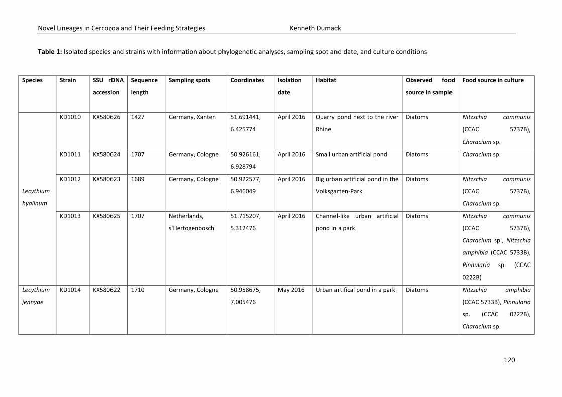
Novel Lineages in Cercozoa and Their Feeding Strategies Kenneth Dumack
120
Table 1: Isolated species and strains with information about phylogenetic analyses, sampling spot and date, and culture conditions
Species Strain SSU rDNA
accession
Sequence
length
Sampling spots Coordinates Isolation
date
Habitat Observed food
source in sample
Food source in culture
Lecythium
hyalinum
KD1010 KX580626
1427 Germany, Xanten 51.691441,
6.425774
April 2016 Quarry pond next to the river
Rhine
Diatoms Nitzschia communis
(CCAC 5737B),
Characium sp.
KD1011 KX580624 1707 Germany, Cologne
50.926161,
6.928794
April 2016 Small urban artificial pond Diatoms Characium sp.
KD1012 KX580623 1689 Germany, Cologne 50.922577,
6.946049
April 2016 Big urban artificial pond in the
Volksgarten-Park
Diatoms Nitzschia communis
(CCAC 5737B),
Characium sp.
KD1013 KX580625 1707 Netherlands,
s‘Hertogenbosch
51.715207,
5.312476
April 2016 Channel-like urban artificial
pond in a park
Diatoms Nitzschia communis
(CCAC 5737B),
Characium sp., Nitzschia
amphibia (CCAC 5733B),
Pinnularia sp. (CCAC
0222B)
Lecythium
jennyae
KD1014 KX580622 1710 Germany, Cologne 50.958675,
7.005476
May 2016 Urban artifical pond in a park Diatoms Nitzschia amphibia
(CCAC 5733B), Pinnularia
sp. (CCAC 0222B),
Characium sp.

Novel Lineages in Cercozoa and Their Feeding Strategies Kenneth Dumack
121
Morphological observations
Lecythium hyalinum
All isolates of L. hyalinum (strains KD1010, KD1011, KD1012 and KD1013) were of similar
morphology. The amoebae bear a thin and hyaline theca (18-50 µm in length and 24-61 µm in width;
n=37) of roundish or slightly curved shape and radial symmetry. Around the aperture of the cells,
small wrinkles and folds may be seen, occurring most often in starving cells (Figs 1E, 2E for individuals
without wrinkles or folds see: Figs 2F, 3D). The aperture might be retracted into an invagination of
the test (Fig. 1F). No obvious deformation of the test in consequence of ingested food was detected.
The nucleus (11.5-21 µm; n= 36; Figs 1B, 2B, 3B) is located at the apical end of the cell (opposite to
the aperture), most often ellipsoid, sometimes spherical, and a fine marbled structure. The nucleolus
(4-8 µm; n=36), rarely two nucleoli (Fig 4, Supplementary Video 2), are spherical and exhibit a more
rough structure, with indentations (Figs 1D, 2D, 3B). The test is carried in an upright position and four
different zones within the cell body are separated (Fig. 4). From the apical to the basal end: The first
zone (I) contains the nucleus embedded in hyaline cytoplasm and usually lacks other structures (e.g.
granules, vacuoles, etc.), the second zone (II) contains granules (0.92-2 µm, n=36). The third zone (III)
is dominated by food vacuoles and the fourth zone (IV), which lies in front of the aperture and
therefore outside of the theca, is defined by a highly vacuolated cytoplasm and emerging filopodia.
The vacuoles are contractile (Supplementary Videos 1), within the theca no contractile vacuoles were
observed. Thin, often branched and sometimes anastomosing filopodia arise from the vacuolated
plasma (IV) with a length of up to 100 µm (Figs 1C, 2C, 3C, 5). The filopodia may contain small
granules (Fig. 1G) which are difficult to observe, but seem to be connected to the cytoskeletal
filaments, as they move together in the same direction with similar velocity (Supplementary Video 1).
All isolates harbored in close connection to the cytoplasm at the base of the filopodia ‘dancing
particles’, possibly ectocytobiotic bacteria (Fig. 5, Supplementary Video 1). Those particles have been
present in all L. hyalinum isolates and individuals although in variable amounts.
When transferred to new culture medium with a high food density, most amoebae covered
themselves by an accumulation of algae, so that only the filopodia could be seen. Swimming stages
with slowly moving, non-branching and non-anastomosing filopodia were observed with a decline of
food density or when cultures were older than five days. In one strain (KD1013) clover leaf-shaped
swimming aggregates (Fig. 2G) as well as large aggregates of individuals forming a feeding
community were observed (Fig. 2H). Those aggregates may fuse partially in the areas of the
vacuolated cytoplasm in region IV. No cysts or flagellated cell stages were observed. Division
longitudinal.

Novel Lineages in Cercozoa and Their Feeding Strategies Kenneth Dumack
122
Figure 1: Cellular features of Lecythium hyalinum strain KD1010. Scale bars indicate 10 µm, pictures
were taken with DIC. A: Overview of the cell body. B: Close-up of the nucleus. C: Branching and
anastomosing filopodia. D: The nucleolus and its structure. E: Close up of the folds surrounding the
aperture and the vacuolated cytoplasm. F: The aperture seen from the side. G: Small granules within
the filopodia.
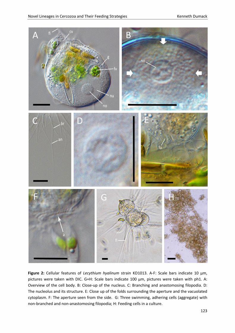
Novel Lineages in Cercozoa and Their Feeding Strategies Kenneth Dumack
123
Figure 2: Cellular features of Lecythium hyalinum strain KD1013. A-F: Scale bars indicate 10 µm,
pictures were taken with DIC. G+H: Scale bars indicate 100 µm, pictures were taken with ph1. A:
Overview of the cell body. B: Close-up of the nucleus. C: Branching and anastomosing filopodia. D:
The nucleolus and its structure. E: Close up of the folds surrounding the aperture and the vacuolated
cytoplasm. F: The aperture seen from the side. G: Three swimming, adhering cells (aggregate) with
non-branched and non-anastomosing filopodia; H: Feeding cells in a culture.
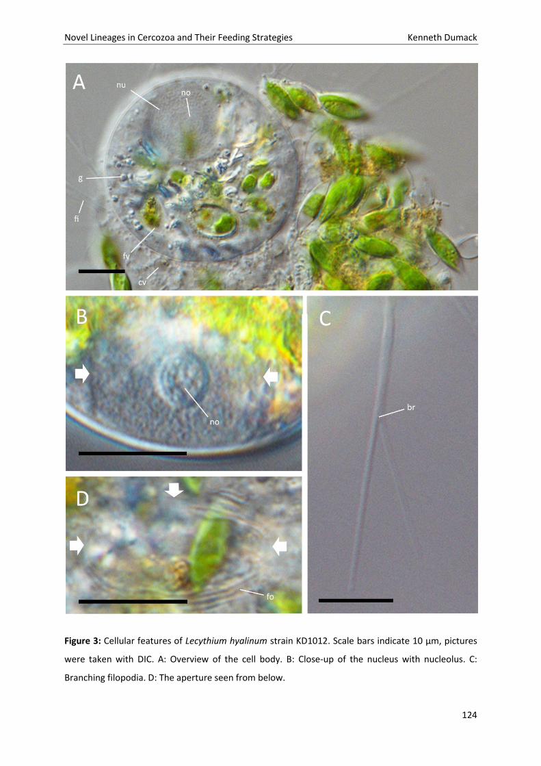
Novel Lineages in Cercozoa and Their Feeding Strategies Kenneth Dumack
124
Figure 3: Cellular features of Lecythium hyalinum strain KD1012. Scale bars indicate 10 µm, pictures
were taken with DIC. A: Overview of the cell body. B: Close-up of the nucleus with nucleolus. C:
Branching filopodia. D: The aperture seen from below.
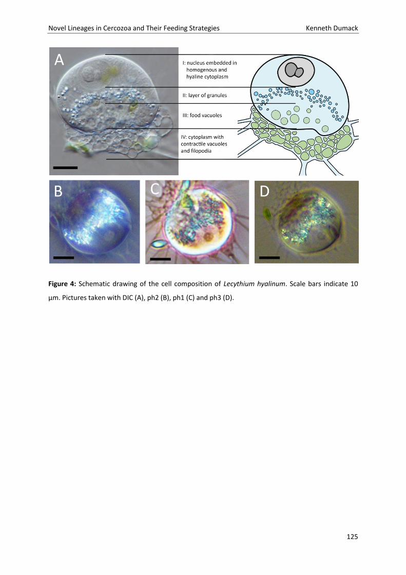
Novel Lineages in Cercozoa and Their Feeding Strategies Kenneth Dumack
125
Figure 4: Schematic drawing of the cell composition of Lecythium hyalinum. Scale bars indicate 10
µm. Pictures taken with DIC (A), ph2 (B), ph1 (C) and ph3 (D).
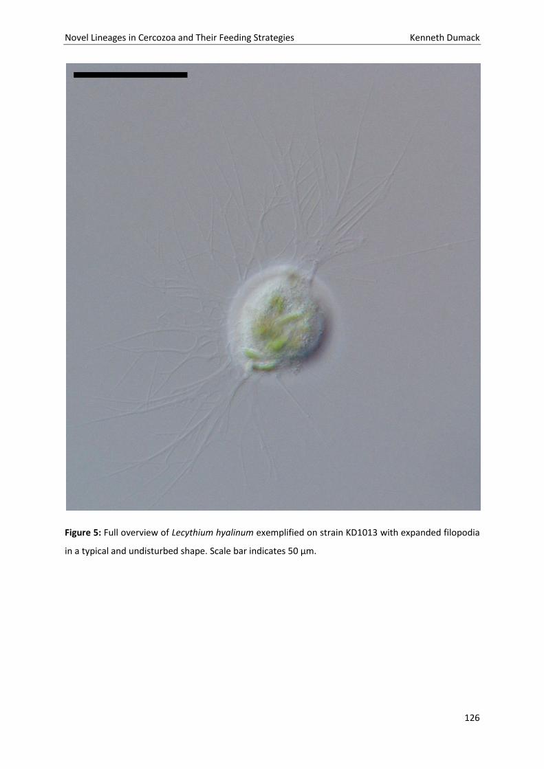
Novel Lineages in Cercozoa and Their Feeding Strategies Kenneth Dumack
126
Figure 5: Full overview of Lecythium hyalinum exemplified on strain KD1013 with expanded filopodia
in a typical and undisturbed shape. Scale bar indicates 50 µm.
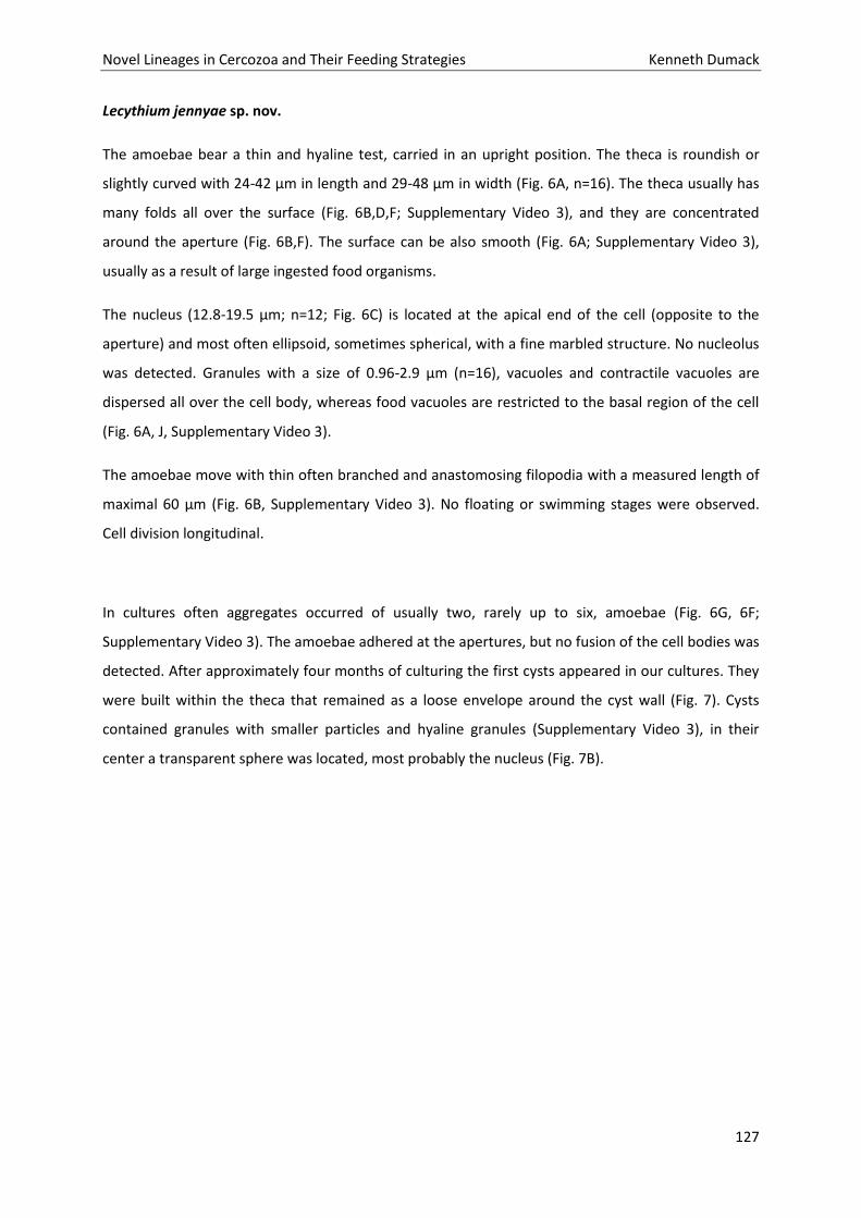
Novel Lineages in Cercozoa and Their Feeding Strategies Kenneth Dumack
127
Lecythium jennyae sp. nov.
The amoebae bear a thin and hyaline test, carried in an upright position. The theca is roundish or
slightly curved with 24-42 µm in length and 29-48 µm in width (Fig. 6A, n=16). The theca usually has
many folds all over the surface (Fig. 6B,D,F; Supplementary Video 3), and they are concentrated
around the aperture (Fig. 6B,F). The surface can be also smooth (Fig. 6A; Supplementary Video 3),
usually as a result of large ingested food organisms.
The nucleus (12.8-19.5 µm; n=12; Fig. 6C) is located at the apical end of the cell (opposite to the
aperture) and most often ellipsoid, sometimes spherical, with a fine marbled structure. No nucleolus
was detected. Granules with a size of 0.96-2.9 µm (n=16), vacuoles and contractile vacuoles are
dispersed all over the cell body, whereas food vacuoles are restricted to the basal region of the cell
(Fig. 6A, J, Supplementary Video 3).
The amoebae move with thin often branched and anastomosing filopodia with a measured length of
maximal 60 µm (Fig. 6B, Supplementary Video 3). No floating or swimming stages were observed.
Cell division longitudinal.
In cultures often aggregates occurred of usually two, rarely up to six, amoebae (Fig. 6G, 6F;
Supplementary Video 3). The amoebae adhered at the apertures, but no fusion of the cell bodies was
detected. After approximately four months of culturing the first cysts appeared in our cultures. They
were built within the theca that remained as a loose envelope around the cyst wall (Fig. 7). Cysts
contained granules with smaller particles and hyaline granules (Supplementary Video 3), in their
center a transparent sphere was located, most probably the nucleus (Fig. 7B).
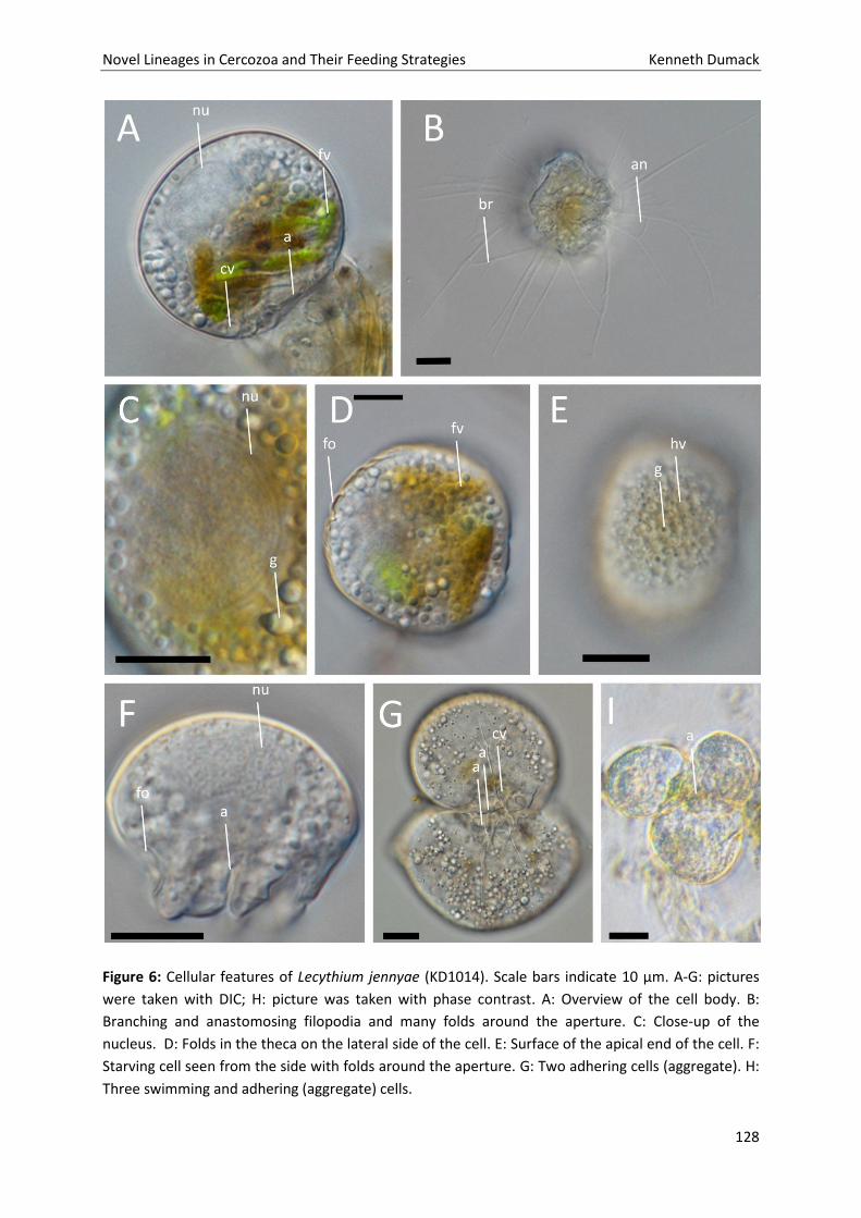
Novel Lineages in Cercozoa and Their Feeding Strategies Kenneth Dumack
128
Figure 6: Cellular features of Lecythium jennyae (KD1014). Scale bars indicate 10 µm. A-G: pictures
were taken with DIC; H: picture was taken with phase contrast. A: Overview of the cell body. B:
Branching and anastomosing filopodia and many folds around the aperture. C: Close-up of the
nucleus. D: Folds in the theca on the lateral side of the cell. E: Surface of the apical end of the cell. F:
Starving cell seen from the side with folds around the aperture. G: Two adhering cells (aggregate). H:
Three swimming and adhering (aggregate) cells.
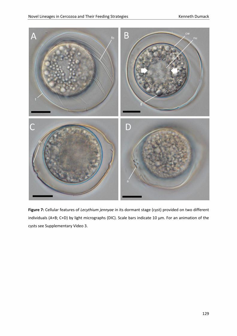
Novel Lineages in Cercozoa and Their Feeding Strategies Kenneth Dumack
129
Figure 7: Cellular features of Lecythium jennyae in its dormant stage (cyst) provided on two different
individuals (A+B; C+D) by light micrographs (DIC). Scale bars indicate 10 µm. For an animation of the
cysts see Supplementary Video 3.
Novel Lineages in Cercozoa and Their Feeding Strategies Kenneth Dumack
130
Sequencing of cultured amoebae and phylogeny
Near full length SSU gene sequences were obtained. The sequenced parts ranged from 1427 to 1710
nucleotides (Table 1). No introns were found. The sequences of all L. hyalinum strains were identical.
The best maximum likelihood tree (Fig. 8) reveald the cercozoan subphylum Filosa (consisting of the
Thecofilosea, Sarcomonadea and Imbricatea) and selected sequences of the Endomyxa as outgroup.
As in previous analyses using SSU rDNA sequence comparisons, some basal branches within the
Cercozoa were not supported (Bass et al. 2009; Howe et al. 2011). The Thecofilosea are composed of
the Phaeodaria, Cryomonadida, Tectofilosida and with moderate support of 51/0.97 the Novel Clade
4 (Bass and Cavalier-Smith 2004). The Novel Clade 4 was highly supported (100/1.00) and composed
of the four strains of L. hyalinum (KX580623, KX580624, KX580625, KX580626), Lecythium jennyae sp.
nov. (KX580622), the recently described Trachyrhizium urniformis (Shiratori and Ishida 2016) and two
marine environmental sequences.
Diagnoses
Genus Lecythium HERTWIG ET LESSER 1874 emend.
Emended diagnosis: Filose, radially symmetric amoebae. Cells bear a colourless, thin and organic test
(theca), carried in an upright position, with one basal opening from which branching and
anastomosing filopodia arise. Filopodia may show small granules. Nucleus at the apical end of the
cell. Cells with numerous contractile vacuoles and granules. Might form floating or swimming stages
(single cells and aggregates) with straight, non-branched and non-anastomosing filopodia.
Aggregates with fused filopodia or fused vacuolated cytoplasm occur. Cysts are built within the theca
that then remains as a loose envelope around the cyst wall. No flagellated cells observed. Species
feed on eukaryotic prey (i.e. algae); ingestion of bacteria not observed. Division: longitudinal, binary.
Sexual reproduction unknown.
Etymology: Lecythium [Greek] = flask, jug; referring to the vitreous test
Type species: L. hyalinum
Novel Lineages in Cercozoa and Their Feeding Strategies Kenneth Dumack
131
Lecythium hyalinum HERTWIG ET LESSER 1874 emend.
Emended diagnosis: Lecythium as above. Test: round or spherical in shape, 18-50 µm (length), 24-61
µm (width), ratio 0.7-1 µm (length/width). Nucleus: ellipsoid or spherical, about 11.5-21 µm in width.
Nucleolus:one, rarely two adhering, round, 4-8 µm in diameter; central to nucleus. Aperture:
roundish, vacuolated cytoplasm arises from the aperture. Cells show a clear zonation (apical to
basal): (I) zone of nucleus (no granules or vacuoles), (II) zone of granules (III) zone of food vacuoles
(IV) highly vacuolated cytoplasm in front of the aperture (contractile vacuoles) of which branching
and anastomosing filopodia arise. Locomotion: (i) actively creeping, filopodia rarely extend longer
than 70 µm; (ii) swimming with extended, non-branched and non-anastomosed and slowly moving
filopodia. Filopodia with small granules attached to cytoskeletal filaments occur. Prey: unicellular
algae, no ingestion of bacteria observed. Cell division: longitudinal, binary.
Etymology: hyalinum [Latin], due to the hyaline test.
Deposited culture: deposited in the Culture Collection of Algae and Protozoa under the accession
number CCAP 1943/6
Lecythium jennyae MAUSBACH DUMACK ET BONKOWSKI, sp. nov.
Diagnosis: Lecythium as above. Test: round or spherical in shape, 24-42 µm (length), 29-48 µm
(width), ratio 0.7-1 (length/width), often deformed due to folds. Nucleus: ellipsoid or spherical,
marbled structure, about 12.8-19.5 µm, located at the apical end of the cell; nucleolus not
detectable. Granules: about 0.96-2.9 µm, round, dispersed all over the cell. Vacuoles: contractile
vacuoles dispersed all over the cell, food vacuoles restricted to the basal end. Aperture: roundish,
filopodia arise directly from the aperture and not from a vacuolated cytoplasm, like in L. hyalinum.
Locomotion: actively creeping, filopodia rarely extend longer than 60 µm; Prey: unicellular algae, no
ingestion of bacteria observed. Cell division: longitudinal, binary.
Type material (hapantotype): A glass slide containing several fixed individuals is deposited in the
Upper Austrian State Museum Invertebrate Collection as Inv. Nr. (2016/121); this material
constitutes the name-bearing type of this species.
Culture of type generating strain: deposited in the Culture Collection of Algae and Protozoa under
the accession number CCAP 1943/7
Novel Lineages in Cercozoa and Their Feeding Strategies Kenneth Dumack
132
Sequence of type generating strain (SSU rDNA): KX580622
Type locality: urban pond in a park at Cologne-Mühlheim, Germany; 50.958675, 7,005476
Etymology: jenny- ae, this species is dedicated to Jennifer Schmitz.
Family Fiscullidae DUMACK MAUSBACH ET BONKOWSKI, fam. nov.
Limnic or terrestrial filose testate amoebae. Test: flexible, organic, round, pyriform or spherical, with
one basal aperture of which filopodia arise. No flagella or cilia. Surface grazer. Trophic stage:
incessant creeping with branched and anastomosed, tapering filopodia. Preying on eukaryotes with
filopodia. Food ingested through aperture, digested in the test. Nucleus round or ellipsoid. Granules
and vacuoles occur (contractile vacuoles hard to detect). May form cysts or floating stages. No sexual
reproduction observed. In contrast to Lecythium: no granules in filopodia, cells form aggregates by
fusion, often between the apical end and the nucleus an additional layer of granules which could not
be observed in L. hyalinum.
Etymology: Name derived from type genus.
Type genus: Fisculla
Genus Fisculla DUMACK MAUSBACH ET BONKOWSKI, gen. nov.
Filose radially symmetric amoebae. Theca often deformed by ingested food, sometimes wrinkled or
folded. Apical ellipsoid or round nucleus, clear or grainy; round nucleolus sometimes visible by light
microscopy. One or several contractile vacuoles (hard to see) and food vacuoles, usually basal.
Granules vary in amount, sometimes in layers, sometimes spread evenly. Cells creep by filopodia,
which may branch or anastomose but lack granules; cell body carried upright. May form floating
stages with retracted or expanded filopodia. Cysts formed within the theca, the theca forms a loose
envelope around the cyst wall. Cells may aggregate basolaterally in a ring, each retaining its nucleus
and individual shape, or apparently partially fuse basally within one multinucleate lobed theca.
Eukaryotic prey (yeasts, algae) caught by filopodia; dragged to thecal opening for ingestion. No
bacterial ingestion observed. At least F. terrestris can form a tubular feeding siphon. Freshwater or
terrestrial. Division longitudinal, binary. Sexual reproduction unknown.
Novel Lineages in Cercozoa and Their Feeding Strategies Kenneth Dumack
133
In contrast to Lecythium: no granules in filopodia, cells form aggregates by fusion, often between the
apical end and the nucleus an additional layer of granules which could not be observed in L. hyalinum
or L. jennyae.
Etymology: Fisculla [Latin] = Belittlement of the noun fiscus (= money bag); referring to the bag-like
shape of the test.
Type species: F. terrestris
Discussion
Morphological analysis
Lecythium hyalinum was first described by Hertwig and Lesser (1874) as a filose testate amoeba with
a non-flexible, hyaline and almost round test. Penard (1902), Belar (1921) and de Saedeleer (1934)
provided redescriptions of L. hyalinum which were in accordance with the original description of
Hertwig and Lesser (1874), but provided further details. They described the length of the test with a
range of 20-40 µm, which fits in our measurements (18-50 µm). Hertwig and Lesser (1874), Penard
(1902) and Belar (1921) did not describe any folds in the theca, but de Saedeleer (1934) did and
described them as restricted to the surroundings of the aperture, as they occurred often in our
isolates. Since not all our individuals carried folds, these folds might have also not been observed by
Hertwig and Lesser (1874).
We observed that the aperture showed a specific form, a neck-like structure reaching out of a notch,
which was described by Hertwig and Lesser (1874), Penard (1902) and de Saedeleer (1934). Further
and maybe most important, we could observe a protuberant, vacuolated cytoplasm in front of the
aperture, from which the filopodia arise. Up to now, this vacuolated cytoplasm was never described
for any other Lecythium species (Dumack et al. 2016b,c) and is therefore considered by us as the
most important discrimination factor of L. hyalinum to other species. In accordance to our
observations, Hertwig and Lesser (1874) also observed a well detectable nucleus with a visible
nucleolus in a homogenous area of cytoplasm at the apical end of the cell. In this area neither
granules nor vacuoles appear, which was clearly described by Penard (1902) and de Saedeleer
(1934).
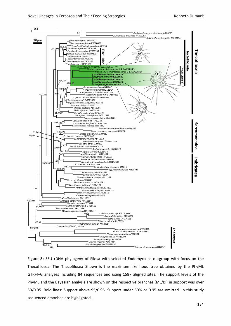
Novel Lineages in Cercozoa and Their Feeding Strategies Kenneth Dumack
134
Figure 8: SSU rDNA phylogeny of Filosa with selected Endomyxa as outgroup with focus on the
Thecofilosea. The Thecofilosea Shown is the maximum likelihood tree obtained by the PhyML
GTR+I+G analyses including 84 sequences and using 1587 aligned sites. The support levels of the
PhyML and the Bayesian analysis are shown on the respective branches (ML/BI) in support was over
50/0.95. Bold lines: Support above 95/0.95. Support under 50% or 0.95 are omitted. In this study
sequenced amoebae are highlighted.
Novel Lineages in Cercozoa and Their Feeding Strategies Kenneth Dumack
135
Unfortunately, Hertwig and Lesser (1874), did not give any descriptions of the nucleolus structure but
Belar (1921) described it either as homogenous or as vacuolated, as we could observe in our isolates.
In stable cultures, he observed large aggregates moving as a planar, outspread community, what we
call a feeding community. Similar to our redescription, Belar (1921) observed swimming stages either
as single cell or as an aggregated, roundish group of amoebae, when, as he states, ‘products of
metabolism accumulated in cultures’. These swimming groups aggregate basolaterally in a ring or
sphere. As observed in our isolates, filopodia of the swimming stages were described by Belar (1921)
as being straight, which rarely branch and never anastomose. We decided to use the word
‘swimming’ as the filopodia most often show slow but active movement in contrast to many floating
stages that seem immotile, being inactively carried by the current.
Accordingly, our morphological analyses confirm that our isolates (KD1010-KD1013) are strains of
Lecythium hyalinum, especially the highly vacuolated cytoplasm in front of the aperture, the
presence of the nucleus at the apical end with a vacuolated nucleolus, and the zonation of the cells
are considered as crucial morphological traits.
The isolate KD1014 shows differences in several characteristics to L. hyalinum. KD1014 never showed
a zonation of the cell body as in L. hyalinum. It contained neither an apical zone with the nucleus
embedded in homogenous cytoplasm, nor any zonation in three areas as described above. Granules
and vacuoles were dispersed all over the cell body. We never observed of the mentioned vacuolated
cytoplasm around the aperture. Also a nucleolus was never observed. The test often features
numerous folds to an extent as it was never seen in L. hyalinum. We therefore conclude that strain
KD1014 does not resemble L. hyalinum but must be a closely related species.
There are six other described Lecythium species of which molecular data is lacking and we therefore
have to compare their morphology with strain KD1014. The species are: L. granulatus (Schulze,
1875), L. mutabilis (Bailey, 1853), L. spinosum (Penard, 1890), L. curvus (Leidy, 1879), L. kryptosis
(Chardez, 1972) and L. minutum (de Saedeleer, 1934). The morphology of these species is quite
different to our isolate (see also identification key provided by Dumack et al. 2016c). L. minutum is
marine and by far too small to be the same species as our isolate. Due to the almost round test our
isolate cannot be identified as L. kryptosis, L. curvus, L. spinosum or L. mutabilis. Some similarities are
detectable between our isolate and L. granulatus (most prominently the occurrence of a ‘large
amount of granules’, for a discussion of this morphological character see Dumack et al. 2016c), but
the size difference and perhaps most importantly the absence of a clearly visible nucleolus in our
isolate are an obvious difference. Finally, we conclude our isolate is a novel species of Lecythium that
we call from now on L. jennyae.
Novel Lineages in Cercozoa and Their Feeding Strategies Kenneth Dumack
136
Lecythium phylogeny and taxonomy
Morphological analysis of the thecofilosean thecate amoebae is a difficult task. Dumack et al.
(2016b,c) grouped novel species into the genus Lecythium, which we show here was incorrect. As
discussed in Dumack et al. (2016c), Penard (1902) probably wrongly described L. margaritae as L.
granulatus, this shows that morphological misidentification of even distantly related species
occurred even to well trained protistologists. With molecular analyses this problem can be resolved.
Surprisingly, L. hyalinum and L. jennyae cluster within the Novel Clade 4 which groups as a novel
lineage with moderate support in the Thecofilosea (Bass and Cavalier-Smith, 2004), and not as
expected among the recently described ‘Lecythium’ species (Dumack et al. 2016b,c). The Novel Clade
4, was established by Howe et al. (2011) and until recently exclusively composed of environmental
sequences. Cavalier-Smith and Chao (2003) established the order Tectofilosida to accommodate next
to others the Chlamydophryidae. Since Lecythium currently is assigned to the Chlamydophryidae we
suggest to include the Novel Clade 4 into the order Tectofilosida although SSU phylogenetic support
in our analyses is only moderate.
Due to the fact that L. hyalinum is the type species of the genus, we have to provide new taxonomic
descriptions for the former genus, which we renamed Fisculla. In addition, we had to establish a new
family, the Fiscullidae. Accordingly, the other described Lecythium species (L. kryptosis, L. curvus, L.
spinosum, L. mutabilis, L. minutum, L. granulatus) of which molecular data is still lacking, might also
not group with the morphologically diverse genus Lecythium, but could instead group in the
Fiscullidae or Rhizaspididae. Until molecular data are provided, their taxonomy cannot be clarified.
Recently Shiratori and Ishida (2016) described Trachyrhizium urniformis, a small marine thecate
amoeba that groups within Novel Clade 4. They excluded the possibility, that their isolate resembled
L. minutum (the only known eukaryvorous marine Lecythium species) on morphological basis, since it
had granules in the filopodia and a test size of 12.0 µm on average (range: 7.5-17.6 µm). Small
granules could also be observed in the filopodia of our L. hyalinum isolates despite this character had
not been described before, possibly because these granules are very small, rare, and therefore hard
to detect. De Saedeleer (1934) described L. minutum from a single observation of two cells which
were 9.3-11 µm in size and as he wrote: ‘we may not have been able to get a clear image of the
whole filopodia network due to the fast movement of the cells’. Although Shiratori and Ishida (2016)
stated a size difference of T. urniformis to L. minutum, these values lie within their measured
variation and therefore fit in the size range of T. urniformis. We therefore suggest that T. urniformis
might resemble L. minutum or a close relative of it.
Novel Lineages in Cercozoa and Their Feeding Strategies Kenneth Dumack
137
The ‘dancing particles’ of L. hyalinum
The ‘dancing particles’ on the cell surface of the vacuolated cytoplasm of L. hyalinum might resemble
ectocytobiotic bacteria. Protists with ectocytobiotic ‘symbionts’ are already described for few
protists, predominantly studied in the genus Nuclearia (Dirren et al. 2014; Dirren and Posch 2016).
Further studies will be conducted investigating this possible eukaryotic-prokaryotic ‘symbiosis’.
Material and methods
Sampling and identification
Freshwater samples were collected from lakes, ponds, creeks and rivers in Germany and the
Netherlands. Plastic bottles were scratched over the bottom to detach detritus, sediment and the
surface grazing amoebae. Two ml of the sampled 100- 200 ml were given in each well of a 24-well
plate (Sarstedt, Germany). The samples were screened for L. hyalinum-like cells with an inverse
microscope (Nikon Eclipse TS100; Ph1; 40x, 100x, 200x and 400x magnification).
Isolation and cultivation
Cells were picked with a glass micro pipette and transferred into a new well, containing WC-Medium
or Waris-H. Additionally, diatoms (Nitzschia communis, CCAC 1762 B; Nitzschia amphibia, CCAC 5733
B; Pinnularia sp. CCAC 0222B) or green algae (Characium sp.) were provided as food source (Tab 1).
Cells were sub-cultured approximately every three to ten days.
Microscopial observations
Pictures and videos were taken with a Nikon digital sight DS-U2 camera (program: NIS-Elements
V4.13.04) with a Nikon Eclipse 90i upright microscope (up to 600x magnification, DIC) and a Nikon
TE2000-E inverse microscope (up to 400x magnification, phase contrast).
Sequencing of cultured amoebae
For sequencing, clonal individuals were starved for up to three days in fresh culture medium and with
approximately 1 µl of medium a single individual was transferred into PCR-tubes, containing 15 µl
ddH2O. The tubes were frozen at -20 °C for storage. Then 35 µl PCR mixture was added. The mixture
included 5 µl 0.1 µM forward and 5 µl 0.1 µM reverse primer, 5 µl 200 µM dNTPs, 5 µl Thermo
Scientific Dream Taq Green Buffer, 0.3 µl Dream Taq polymerase (Thermo Fisher Scientific, Dreieich,
Germany) and 14.7 µl water. The SSU sequences were obtained in two successive steps. First, the
whole SSU was amplified with the general eukaryotic primers, EukA and EukB (Medlin et al. 1988). In
the second step, semi-nested re-amplifications were performed by using primers specifically
Novel Lineages in Cercozoa and Their Feeding Strategies Kenneth Dumack
138
designed for cercozoans, with the same settings as above and the primer pairs EukA + S963R_Cerco
(Fiore-Donno, pers. comm.) targeting the 5’ part of the SSU and S616F_Cercomix (Fiore-Donno, pers.
comm) + EukB for the 3’ part of the gene. One µl of the first PCR product was used as template.
The PCR products were purified by adding 0.15 μl of Exonuclease, 0.9 μl FastAP and 1.95 μl water to
8 μl of the second PCR product. Then heated for 30 min at 37 °C, and subsequently for 20 min at 85
°C.
The Big dye Terminator Cycle sequencing Kit (Thermo Fisher Scientific, Dreieich, Germany) and an ABI
PRISM automatic sequencer were used for the sequencing.
Phylogenetic analyses
The partial sequences were manually checked for sequencing errors before they were assembled
into one sequence contig. To create a dataset for phylogenetic analyses sequences were blasted
(blastn 2.3.0) against the NCBI GenBank database (last date of accession: 14.06.2016).
Representative sequences belonging to the Filosa and to the Endomyxa (outgroup) were downloaded
and manually aligned in SeaView (V4.5.3, Gouy et al. 2010). 1587 sites were used for the alignment,
which were to 45.19 % without polymorphism. The best fitting model GTR+I+G was found by using
the program jmodeltest (V.2.1.5, Darriba et al. 2012), testing 88 different models (settings:
Substitution schemes 11; add Base frequencies +I+G rate variation nCat=4 resulting in 88 models, ML
optimized NNI as base tree). With this model phylogenetic trees were calculated in PhyML 3.1
(Guindon and Gascuel 2003) and MrBayes (settings: mcmc ngen = 10 M, sample freq = 100, print freq
= 100, diagn freq = 500; Altekar et al. 2004; Ronquist and Huelsenbeck 2003). The sequences were
submitted in the NCBI database under the accesstion numbers: KX580626, KX580624, KX580623,
KX580625 and KX580622.
Preparation of the hapantotype
1.5 ml of culture material was mixed 1:4 with Schaudinn’s fluid and was heated for 5 minutes at 60°C.
A serial dehydration in an ascending series of ethanol solutions with increasing concentration was
conducted. Finally, the preserved cells were mixed 1:2 with glycerin, transferred to a glass slide and a
cover slide was glued on top.
Novel Lineages in Cercozoa and Their Feeding Strategies Kenneth Dumack
139
Acknowledgements
The study was supported by the DFG research group FOR 918. We thank the Culture Collection of
Algae at the University of Cologne (CCAC) for their service and the provided algal strains.
References
Altekar G, Dwarkadas S, Huelsenbeck JP, Ronquist F (2004) Parallel metropolis coupled Markov chain
Monte Carlo for Bayesian phylogenetic inference. Bioinform 20:407-415
Bailey JW (1853) Observations on a newly discovered animalcule. Am J Sci 15:341-347
Bass D, Cavalier-Smith T (2004) Phylum-specific environmental DNA analysis reveals remarkably high
global biodiversity of Cercozoa (Protozoa). Int J Syst Evol Microbiol 54:2393-2404
Bass D, Chao EE, Nikolaev S, Yabuki A, Ishida KI, Berney C, Pakzad U, Wylezich C, Cavalier-Smith T
(2009) Phylogeny of novel naked filose and reticulose Cercozoa: Granofilosea cl. n. and
Proteomyxidea revised. Protist 160:75-109
Belar K (1921) Untersuchungen über Thecamöben der Clamydophrys-Gruppe. Arch Protistenkd
Cavalier-Smith T (1998a) Neomonada and the origin of animals and fungi. In Coombs GH, Vickerman
K, Sleigh MA, Warren A (eds) Evolutionary relationships among Protozoa. Chapman and Hall, London,
375–407
Cavalier‐Smith T (1998b) A revised six‐kingdom system of life. Biological Reviews 73:203-266
Cavalier-Smith T, Chao EE (2003) Phylogeny and classification of phylum Cercozoa (Protozoa). Protist
154: 341-358
Chardez D (1971) Etude sur les thecamoebiens des biotopes interstitiels, psammons littoraux et
zones marginales souterraines des eaux douces. Bull Rech Agron Gembloux 6:257–268
Darriba D, Taboada GL, Doallo R, Posada D (2012) jModeltest 2: more models, new heuristics and
parallel computing. Nature Methods 9:772
De Saedeleer H (1934) Beitrag zur Kenntnis der Rhizopoden: morphologische und systematische
Untersuchungen und ein Klassifikationsversuch. Mém Musée Roy d’Histoir Naturel de Belq 60:1–112
Dirren S, Salcher MM, Blom JF, Schweikert M, Posch T (2014) Ménage-à-trois: The Amoeba Nuclearia
sp. from Lake Zurich with its Ecto- and Endosymbiotic Bacteria. Protist 165:745–58

Novel Lineages in Cercozoa and Their Feeding Strategies Kenneth Dumack
140
Dirren S, Posch T (2016) Promiscuous and specific bacterial symbiont acquisition in the amoeboid
genus Nuclearia (Opisthokonta). FEMS Microbiology Ecology 92:1–44.
Dumack K, Schuster J, Bass D, Bonkowski M (2016a) A novel lineage of ‘naked filose amoebae’;
Kraken carinae gen. nov. sp. nov. (Cercozoa) with a remarkable locomotion by disassembly of its cell
body. Protist 167:268-278
Dumack K, Müller ME, Bonkowski M (2016b) Description of Lecythium terrestris sp. nov.
(Chlamydophryidae, Cercozoa), a soil dwelling protist feeding on fungi and algae. Protist 167:93-105
Dumack K, Baumann C, Bonkowski M (2016c) Revision of the thecate amoeba genus Lecythium
(Chlamydophryidae, Tectofilosida, Cercozoa, Rhizaria) including a description of four new species and
an identification key. Protist
Dumack K, Öztoprak H, Rüger L, Bonkowski M (in review) Shedding light on the polyphyletic thecate
amoeba genus Plagiophrys: Transition of some of its species to Rhizaspis (Tectofilosida, Cercozoa)
and the establishment of Sacciforma gen. nov. and Rhogostomidae fam. nov. (Cryomonadida,
Cercozoa). Protist
Gouy M, Guindon S, Gascuel O (2010) SeaView version 4: a multiplatform graphical user interface for
sequence alignment and phylogenetic tree building. Mol Biol Evol 27:221-224
Guillard RR, Lorenzen CJ (1972) Yellow‐green algae with chlorophyllide C1,2. Journal of Phycology
8:10-14
Guindon S, Gascuel O (2003) A simple, fast, and accurate algorithm to estimate large phylogenies by
maximum likelihood. Syst Biol 52:696-704
Hertwig R, Lesser E (1874) Über Rhizopoden und denselben nahestehende Organismen. Arch
mikroskop Anat 10:35–243
Hess S, Melkonian M (2013) The mystery of clade X: Orciraptor gen. nov. and Viridiraptor gen. nov.
are highly specialised, algivorous amoeboflagellates (Glissomonadida, Cercozoa). Protist 164:706-747
Howe AT, Bass D, Scoble JM, Lewis R, Vickerman K, Arndt H, Cavalier-Smith T (2011) Novel cultured
protists identify deep-branching environmental DNA clades of Cercozoa: new genera Tremula,
Micrometopion, Minimassisteria, Nudifila, Peregrinia. Protist 162:332-372
Kosakyan A, Gomaa F, Lara E, Lahr DJG (2016) Current and future perspectives on the systematics,
taxonomy and nomenclature of testate amoebae. Eur J Protistol 1-13
Novel Lineages in Cercozoa and Their Feeding Strategies Kenneth Dumack
141
Leidy J (1879) Fresh-Water Rhizopods of North America. U.S. Geol Survey of the Territories 544pp
McFadden GI, Melkonian M (1986) Use of Hepes buffer for microalgal culture media and fixation for
electron microscopy. Phycologia 25:551-557
Medlin L, Elwood HJ, Stickel S, Sogin ML (1988) The characterization of enzymatically amplified
eukaryotic 16S-like rRNA-coding regions. Gene 71:491-499
Nikolaev SI, Mitchell EA, Petrov NB, Berney C, Fahrni J, Pawlowski J (2005) The testate lobose
amoebae (order Arcellinida Kent, 1880) finally find their home within Amoebozoa. Protist 156:191-
202
Penard E (1890) Études sur les Rhizopodes d‘eau douce. Mém. Soc. Phys. Hist. nat. Genève 31:1-230
Penard E (1902) Faune Rhizopodique du bassin du Léman. Genf 714 pp
Schulze FE (1875) Rhizopodenstudien. Arch mikrosk Anat 11:94-139
Shiratori T, Ishida KI (2016) Trachyrhizium urniformis ng, n. sp. a novel marine filose thecate Amoeba
related to a Cercozoan environmental clade (Novel Clade 4). J Eukaryot Microbiol 0:1-10
Ronquist F, Huelsenbeck JP (2003) MrBayes 3: Bayesian phylogenetic inference under mixed models.
Bioinform 19:1572-1574
Wylezich C, Meisterfeld R, Meisterfeld S, Schlegel M (2002) Phylogenetic analyses of small subunit
ribosomal RNA coding regions reveal a monophyletic lineage of euglyphid testate amoebae (order
Euglyphida). J Eukaryot Microbiol 49:108-118
Novel Lineages in Cercozoa and Their Feeding Strategies Kenneth Dumack
142
Part 3 - Eukaryvorous protists, their capabilities and
dispersal.
Chapter 7: What does it take to eat a fungus? A case
study with the eukaryvorous amoeba Fisculla terrestris.
Novel Lineages in Cercozoa and Their Feeding Strategies Kenneth Dumack
143
What does it take to eat a fungus? A case study with the fungivorous amoeba Fisculla terrestris.
Kenneth Dumacka#, Julia Pundta, Sebastian Loeppmannb & Michael Bonkowskia
Phone: +49-(0)221-470-6635 Fax: +49-(0)221-470-5038
a Biozentrum Köln, Institut für Zoologie, Abt. Terrestrische Ökologie, Zülpicher Str. 47b, 50674 Köln,
Germany;
b Department of Soil Science of Temperate Ecosystems, University of Göttingen, Göttingen Germany
[email protected], [email protected], [email protected],
Running title: Fungivory in the Fiscullidae
# Corresponding author Kenneth Dumack, 27.09.2016
Keywords
Soil food web; mycophagy, protist; yeast; chemical sensing; Fusarium
Abstract
Protists are commonly suggested to regulate bacteria abundance in soils, whereas fungi are
considered to be consumed by arthropods and fungivorous nematodes. Nevertheless, some protist
taxa have been found to consume fungi and despise bacteria. Since these protists are difficult to
culture little is known about their (a) ecological impact, e.g. grazing selectivity, growth rates and
preferred prey organisms and (b) adaptation to fungal food sources, e.g. enzyme production and
chemical sensing abilities. We demonstrated that the eukaryvorous amoeba Fisculla terrestris senses
fungal presence and in fungal presence responds with an increased (5-fold) grazing activity. To test
for grazing selectivity, a food choice experiment including three different prey species: 1) a yeast
species (Saccharomyces cerevisiae), 2) a green alga (Chlorella vulgaris) and 3) a diatom (Navicula sp.)
was conducted. Although F. terrestris consumed all offered food sources, the predator selected for
the yeast and showed the highest growth rate (about 125 40 times increase of cell density over
three days) and shortest generation time (5.9 1.5 h) when feeding on the fungal food source. In
addition, F. terrestris produced enzymes specialized to degrade C- and N-sources, i.e. chitinases. We
conclude that F. terrestris is a eukaryvorous protist with strong preference for fungal cells and should
therefore be referred to as fungivorous.
Novel Lineages in Cercozoa and Their Feeding Strategies Kenneth Dumack
144
Importance
Fungivorous protists have been researched in very few often very basic studies. Especially in soil
ecology, protists are generally referred to as bacterivores. Nevertheless, as we show, protists occupy
various ecological niches such as the niche of fungivores. Some protist lineages are specialized and
harbour potential for enzyme analyses, experiments on chemical sensing and bio regulators for
potentially harmful fungi. To our knowledge this is the first study giving growth curves, generation
times, a direct relation of consumed individuals to predator growth and further first evidence for
chemical sensing in fungivorous protists. Thus enabling more detailed soil system modelling.
Introduction
Free-living protists are usually considered to feed on bacteria with low impact on fungal abundance
(1). Since protists have traditionally been cultivated on bacteria-rich media, those that did not grow
under these conditions are still poorly studied and probably many of non bacterivorous protists are
not even known to science yet. Therefore, only very few and often old studies about fungivorous
protists are available. Those were conducted by taxonomists and most lack ecological data, like their
grazing selectivity, the resulting growth rates and the mechanisms by which they locate and digest
their fungal prey (2-5).
Fisculla terrestris, a small amoeba that was recently isolated from agricultural soils in Eastern
Germany could not be cultivated on bacteria, but under lab conditions has been shown to feed not
only on yeasts (e.g. Cryptococcus laurentii), spores of plant pathogen fungi (e.g. Fusarium culmorum)
but also on unicellular algae (6, 7, Dumack, Mausbach, Hegmann and Bonkowski, submitted for
publication). According to observations and the assumption that fungal food sources are much more
abundant in terrestrial habitats than algae (8, 9) Dumack et al. (6) hypothesised that F. terrestris
feeds primarily on fungi. Selectivity for fungal nutrition should lead to a series of adaptations, e.g. the
ability to (a) sense fungal cells, (b) identify and select for them as a food source and (c) degrade or
penetrate chitin since this is the main cell-wall component of fungi.
Chemical sensing in protists has been reported already more than hundred years ago (10). Since then
numerous studies identified diverse attractants (e.g. volatile organic compounds (VOCs) and amino
acids) as well as chemosensory transduction in protists in a range of distantly related taxa (11-13).
These studies, however, exclusively focused on bacterivorous protists. Chitinase-coding genes have
been already found in the genome of protists (Amoebozoa: Acanthamoeba, Cercozoa:
Plasmodiophora; 14, 15) and chitinase activity has been confirmed by direct observation of lysed
chitin walls or lab experiments for several protist taxa (Amoebozoa: Hartmannella, Cercozoa:
Novel Lineages in Cercozoa and Their Feeding Strategies Kenneth Dumack
145
Leptophryidae spp.; 5, 16, 17). However, we are not aware of reports on chitinase production in
Fisculla, its order Tectofilosida or the whole class Thecofilosea.
Accordingly, we performed (a) chemical sensing experiments with a fungal food source, (b) food
choice experiments in which we offered one representative of the three dominant possible prey
groups, a yeast (Saccharomyces cerevisiae), a green alga (Chlorella vulgaris) and a diatom (Navicula
sp.) and (c) enzyme analyses of intact and homogenized F. terrestris cells.
Materials & Methods
Preparation of axenic cultures
Cultures of the algae C. vulgaris (strain CCAC 3272) and Navicula sp. (strain CCAC 1772) were
cultivated axenically in Waris-H+Si medium (18) at room temperature, with a light regime of 14 h
light to 10 h dark with a light-intensity of about 5 mol photons/m2/sec. The yeast S. cerevisiae was
cultivated axenically on potato agar plates after the manufacturer’s instructions (Fluka Analytical)
and incubated for 2-3 days at room temperature. S. cerevisiae cells were subsequently transferred to
sterile Waris-H+Si medium. With this, a monoxenic (bacteria-free) co-culture of F. terrestris (strain
CCAP 1943/1) was established by cell sorting as described by Hess & Melkonian (17). F. terrestris was
also cultivated in Waris-H+Si to control for medium effects.
Set up of the chemical sensing experiment
Erlenmeyer flasks (n=3) were prepared with (a) axenic S. cerevisiae in Waris-H and (b) sterile Waris-H
as a control. Both treatments were incubated for 5 days at room temperature and then 15 ml of the
suspension were subsampled and centrifuged for 1 h by 4,500 U/min (Hettich, Rotina 420R). One ml
of axenic culture of F. terrestris were transferred into wells of 24-well plates (n=6) and incubated for
2 hours then the base level of activity was determined by counting inactive cells and active grazing
cells, giving the activity in percentage. To check a response of F. terrestris in activity, out of each
Erlenmeyer flask 100 µl of the supernatant was added to the culture, incubated for 30 min and again
the grazing activity in percent was calculated.
Novel Lineages in Cercozoa and Their Feeding Strategies Kenneth Dumack
146
Set up of the food choice experiment
Cell numbers of each of the three food source cultures (Saccharomyces cerevisiae; Chlorella vulgaris
and Navicula sp.) were counted in a Neubauer Counting Chamber (Hecht-Assistant, Sondheim
Deutschland) Based on these values, 10*104 algae and yeast cells were pipetted together with 100
individuals of F. terrestris into separate wells of a 96-well-plate (n=6). Control treatments contained
each organism separately as well as the predator with each prey species individually. The final
volume was adjusted to 200 l Waris-H+Si. Changes in the numbers of cells were monitored by direct
counting with a Neubauer chamber once a day over the three days run time of the experiment.
Chitinase assay
A starving culture of F. terrestris (4 weeks of culturing, 14 days of starving) was used for the analyses.
To control for possible remaining yeast cells, 500 ml of the culture were filtered through a 5 µm pore
glass fibre filter. The protist cells were resuspended in Waris-H, examined for possible remaining
yeast cells by light microscopy and incubated for additional 4-5 days before analyses. The
determination of enzyme kinetics (chitinase) included three treatments, (1) non-treated medium (2)
starving, but intact cells of protists and (3) starving and homogenized cells of protists. The
homogenized cells were destroyed by pulsed sonication with 50 J s-1 for 120 s.
We used 4-methylumbelliferyl N-acetyl-β-D-glucosaminide to determine the enzyme activity of N-
acetyl-β-glucosaminidase (chitinase; EC 3.2.1.52) (18). Aliquots (50 µl) of liquid protist culture (3
analytical replicates) were withdrawn and dispensed into 96-well microplates (Brand pureGrade,
black). The 4-methylumbelliferyl N-acetyl-β-D-glucosaminide was dissolved in 300 µl dimethyl
sulfoxide (DMSO) and then diluted with 80 µl of 0.1 M, pH 6.1 MES to obtain 1 mM of working
solution (20, 21). A concentration series of 20, 40, 60, 80, 100, 200, 400 µmol was then prepared and
100 µl of the substrate solution was added to the wells.
Microplates were kept at 21°C, agitated and measured fluorometrically (excitation 360 nm; emission
450 nm) after 1 h, 2 h, and 3 h incubation with an automated fluorometric plate-reader (Wallac 1420,
Perkin Elmer, Turku, Finland).
Novel Lineages in Cercozoa and Their Feeding Strategies Kenneth Dumack
147
Statistical analyses
Food choice
Changes in protist and prey abundances were analyzed by multivariate analysis of variance
(MANOVA) according to Hurlbert (22). The data were tested for sphericity before the MANOVAs
were calculated and tested for differences in R (V.3.1.0).
Day 0 was excluded to correct for a possible adaptation phase of the predator, when the generation
times of F. terrestris were determined. With S. cerevisiae as food source, F. terrestris reached on the
third day food limitation, therefore day 3 was also excluded from the calculation, restricting the
values to the exponential growth phase of F. terrestris.
Chemical sensing
The data of the chemical sensing experiment were tested for normality (Shapiro Wilk test) and
variance homogeneity (Levene’s test). Differences between the two treatments and the control were
calculated in R with the settings described above since requirements for an ANOVA were given.
Chitinase assay
The substrate-dependent rate of reaction mediated by hydrolytic enzymes, followed Michaelis-
Menten kinetics (20, 21, 23). The parameters of the equation were fitted by minimizing the least-
square sum using GraphPad Version 6 software (Prism, USA). Parameter optimization was restricted
to the applied model equation as indicated by maximum values of statistic criteria (r2) (19, 20).
Results
Chemical sensing experiment
Starving cultures of F. terrestris contained an average of 5.2 3.5% actively grazing cells (Fig. 1). The
activity did not change significantly after incubation in fresh medium (control), but increased about
5-fold, to 26 9.6%, when supernatant of yeast cells was added (F[2,9]=18.38; p<0.001).
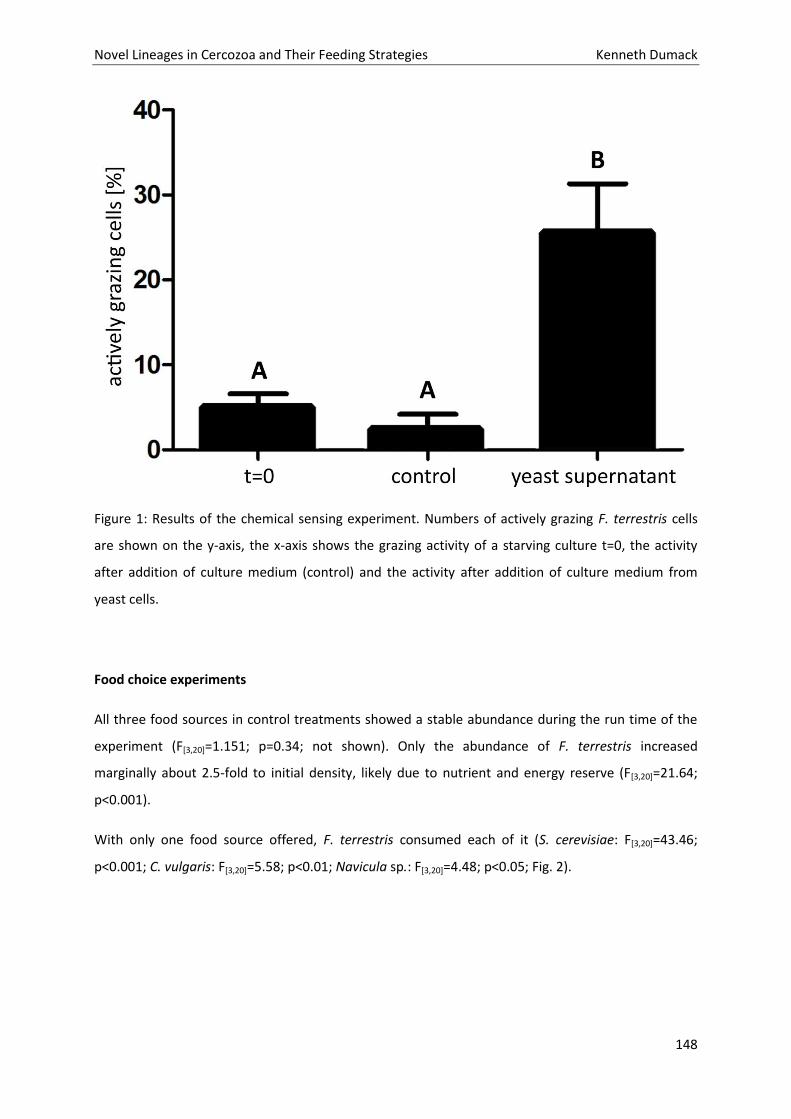
Novel Lineages in Cercozoa and Their Feeding Strategies Kenneth Dumack
148
Figure 1: Results of the chemical sensing experiment. Numbers of actively grazing F. terrestris cells
are shown on the y-axis, the x-axis shows the grazing activity of a starving culture t=0, the activity
after addition of culture medium (control) and the activity after addition of culture medium from
yeast cells.
Food choice experiments
All three food sources in control treatments showed a stable abundance during the run time of the
experiment (F[3,20]=1.151; p=0.34; not shown). Only the abundance of F. terrestris increased
marginally about 2.5-fold to initial density, likely due to nutrient and energy reserve (F[3,20]=21.64;
p<0.001).
With only one food source offered, F. terrestris consumed each of it (S. cerevisiae: F[3,20]=43.46;
p<0.001; C. vulgaris: F[3,20]=5.58; p<0.01; Navicula sp.: F[3,20]=4.48; p<0.05; Fig. 2).
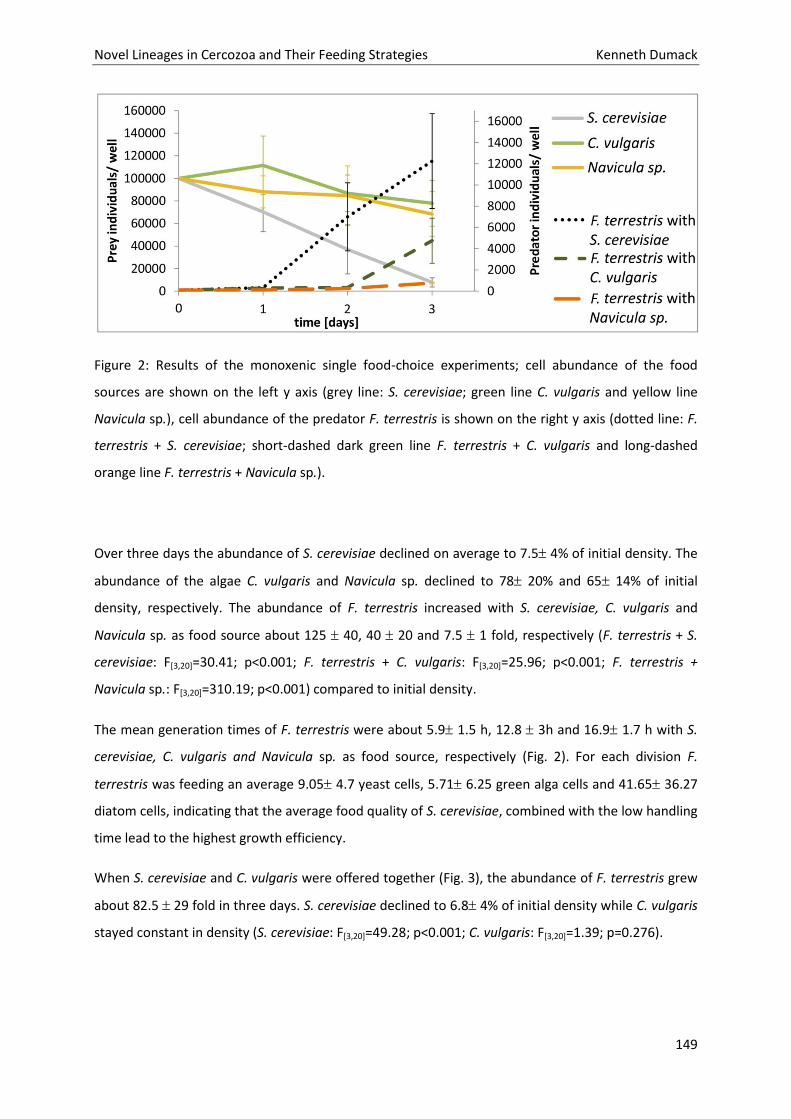
Novel Lineages in Cercozoa and Their Feeding Strategies Kenneth Dumack
149
Figure 2: Results of the monoxenic single food-choice experiments; cell abundance of the food
sources are shown on the left y axis (grey line: S. cerevisiae; green line C. vulgaris and yellow line
Navicula sp.), cell abundance of the predator F. terrestris is shown on the right y axis (dotted line: F.
terrestris + S. cerevisiae; short-dashed dark green line F. terrestris + C. vulgaris and long-dashed
orange line F. terrestris + Navicula sp.).
Over three days the abundance of S. cerevisiae declined on average to 7.5 4% of initial density. The
abundance of the algae C. vulgaris and Navicula sp. declined to 78 20% and 65 14% of initial
density, respectively. The abundance of F. terrestris increased with S. cerevisiae, C. vulgaris and
Navicula sp. as food source about 125 40, 40 20 and 7.5 1 fold, respectively (F. terrestris + S.
cerevisiae: F[3,20]=30.41; p<0.001; F. terrestris + C. vulgaris: F[3,20]=25.96; p<0.001; F. terrestris +
Navicula sp.: F[3,20]=310.19; p<0.001) compared to initial density.
The mean generation times of F. terrestris were about 5.9 1.5 h, 12.8 3h and 16.9 1.7 h with S.
cerevisiae, C. vulgaris and Navicula sp. as food source, respectively (Fig. 2). For each division F.
terrestris was feeding an average 9.05 4.7 yeast cells, 5.71 6.25 green alga cells and 41.65 36.27
diatom cells, indicating that the average food quality of S. cerevisiae, combined with the low handling
time lead to the highest growth efficiency.
When S. cerevisiae and C. vulgaris were offered together (Fig. 3), the abundance of F. terrestris grew
about 82.5 29 fold in three days. S. cerevisiae declined to 6.8 4% of initial density while C. vulgaris
stayed constant in density (S. cerevisiae: F[3,20]=49.28; p<0.001; C. vulgaris: F[3,20]=1.39; p=0.276).
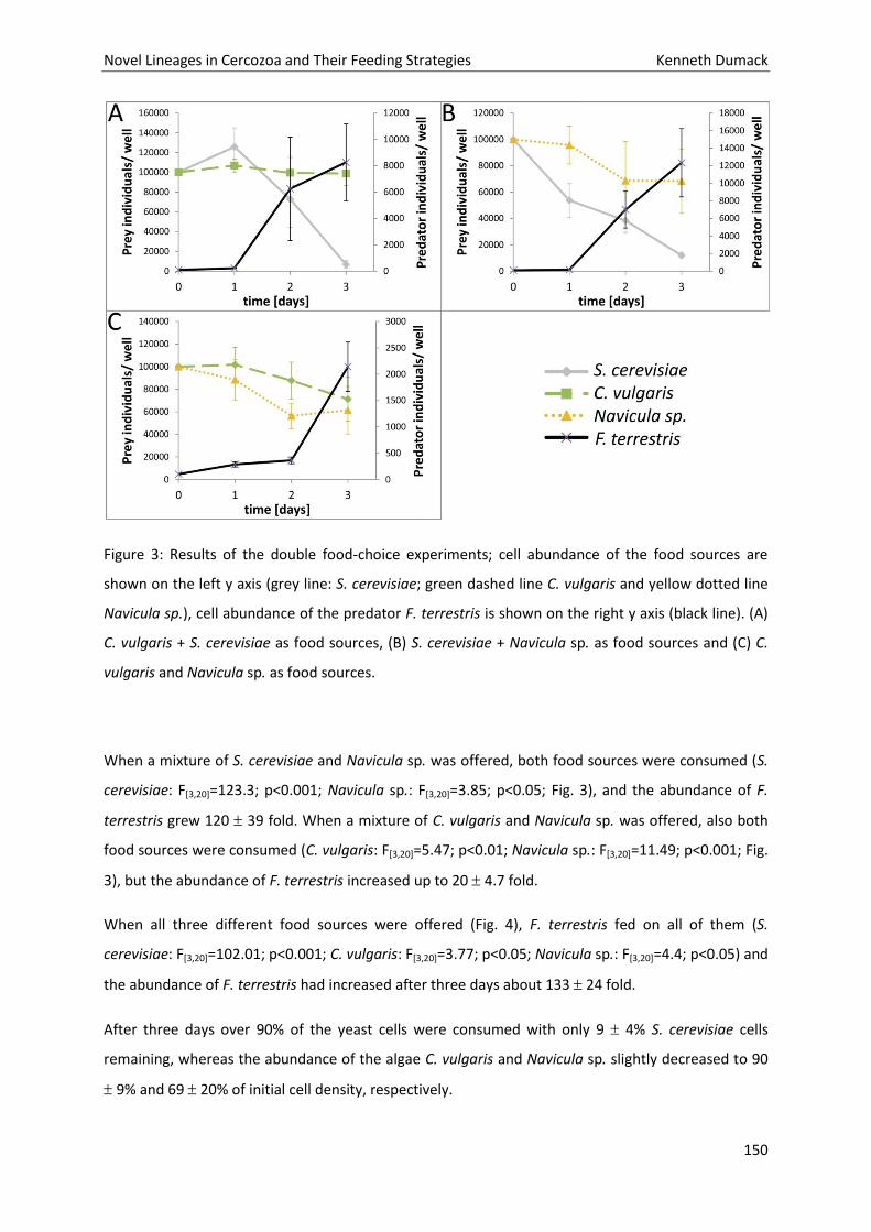
Novel Lineages in Cercozoa and Their Feeding Strategies Kenneth Dumack
150
Figure 3: Results of the double food-choice experiments; cell abundance of the food sources are
shown on the left y axis (grey line: S. cerevisiae; green dashed line C. vulgaris and yellow dotted line
Navicula sp.), cell abundance of the predator F. terrestris is shown on the right y axis (black line). (A)
C. vulgaris + S. cerevisiae as food sources, (B) S. cerevisiae + Navicula sp. as food sources and (C) C.
vulgaris and Navicula sp. as food sources.
When a mixture of S. cerevisiae and Navicula sp. was offered, both food sources were consumed (S.
cerevisiae: F[3,20]=123.3; p<0.001; Navicula sp.: F[3,20]=3.85; p<0.05; Fig. 3), and the abundance of F.
terrestris grew 120 39 fold. When a mixture of C. vulgaris and Navicula sp. was offered, also both
food sources were consumed (C. vulgaris: F[3,20]=5.47; p<0.01; Navicula sp.: F[3,20]=11.49; p<0.001; Fig.
3), but the abundance of F. terrestris increased up to 20 4.7 fold.
When all three different food sources were offered (Fig. 4), F. terrestris fed on all of them (S.
cerevisiae: F[3,20]=102.01; p<0.001; C. vulgaris: F[3,20]=3.77; p<0.05; Navicula sp.: F[3,20]=4.4; p<0.05) and
the abundance of F. terrestris had increased after three days about 133 24 fold.
After three days over 90% of the yeast cells were consumed with only 9 4% S. cerevisiae cells
remaining, whereas the abundance of the algae C. vulgaris and Navicula sp. slightly decreased to 90
9% and 69 20% of initial cell density, respectively.
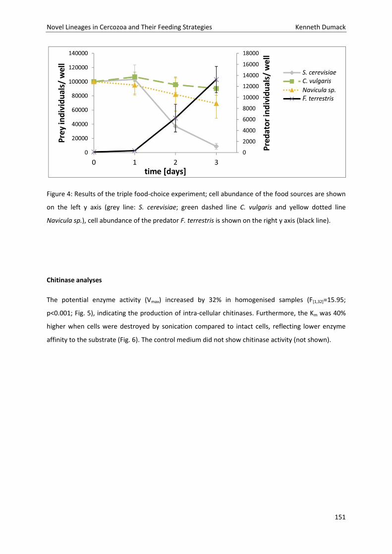
Novel Lineages in Cercozoa and Their Feeding Strategies Kenneth Dumack
151
Figure 4: Results of the triple food-choice experiment; cell abundance of the food sources are shown
on the left y axis (grey line: S. cerevisiae; green dashed line C. vulgaris and yellow dotted line
Navicula sp.), cell abundance of the predator F. terrestris is shown on the right y axis (black line).
Chitinase analyses
The potential enzyme activity (Vmax) increased by 32% in homogenised samples (F[1,32]=15.95;
p<0.001; Fig. 5), indicating the production of intra-cellular chitinases. Furthermore, the Km was 40%
higher when cells were destroyed by sonication compared to intact cells, reflecting lower enzyme
affinity to the substrate (Fig. 6). The control medium did not show chitinase activity (not shown).
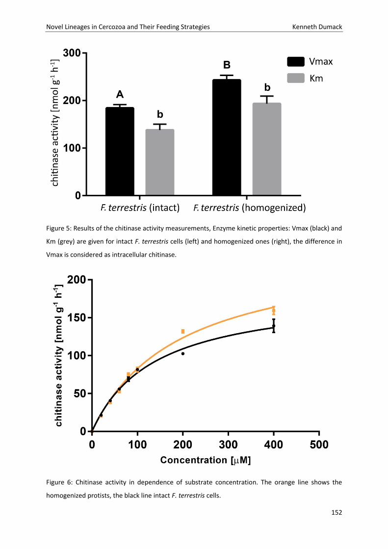
Novel Lineages in Cercozoa and Their Feeding Strategies Kenneth Dumack
152
Figure 5: Results of the chitinase activity measurements, Enzyme kinetic properties: Vmax (black) and
Km (grey) are given for intact F. terrestris cells (left) and homogenized ones (right), the difference in
Vmax is considered as intracellular chitinase.
Figure 6: Chitinase activity in dependence of substrate concentration. The orange line shows the
homogenized protists, the black line intact F. terrestris cells.
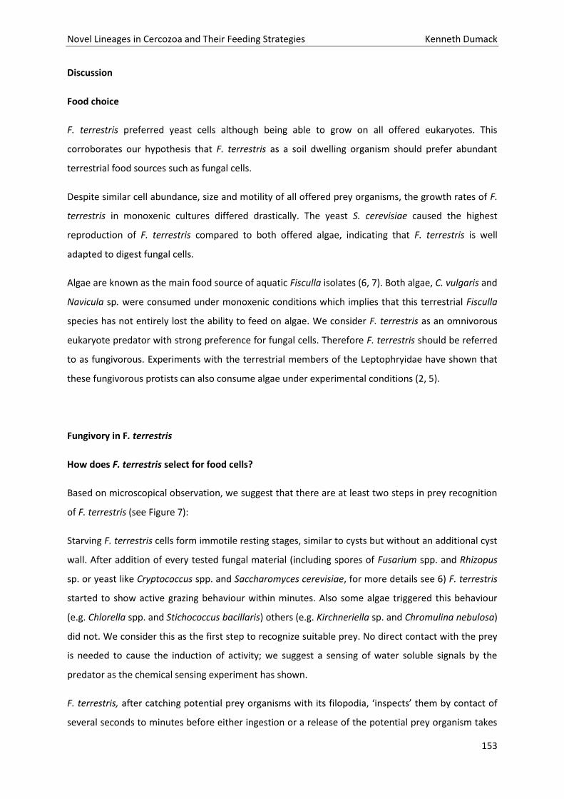
Novel Lineages in Cercozoa and Their Feeding Strategies Kenneth Dumack
153
Discussion
Food choice
F. terrestris preferred yeast cells although being able to grow on all offered eukaryotes. This
corroborates our hypothesis that F. terrestris as a soil dwelling organism should prefer abundant
terrestrial food sources such as fungal cells.
Despite similar cell abundance, size and motility of all offered prey organisms, the growth rates of F.
terrestris in monoxenic cultures differed drastically. The yeast S. cerevisiae caused the highest
reproduction of F. terrestris compared to both offered algae, indicating that F. terrestris is well
adapted to digest fungal cells.
Algae are known as the main food source of aquatic Fisculla isolates (6, 7). Both algae, C. vulgaris and
Navicula sp. were consumed under monoxenic conditions which implies that this terrestrial Fisculla
species has not entirely lost the ability to feed on algae. We consider F. terrestris as an omnivorous
eukaryote predator with strong preference for fungal cells. Therefore F. terrestris should be referred
to as fungivorous. Experiments with the terrestrial members of the Leptophryidae have shown that
these fungivorous protists can also consume algae under experimental conditions (2, 5).
Fungivory in F. terrestris
How does F. terrestris select for food cells?
Based on microscopical observation, we suggest that there are at least two steps in prey recognition
of F. terrestris (see Figure 7):
Starving F. terrestris cells form immotile resting stages, similar to cysts but without an additional cyst
wall. After addition of every tested fungal material (including spores of Fusarium spp. and Rhizopus
sp. or yeast like Cryptococcus spp. and Saccharomyces cerevisiae, for more details see 6) F. terrestris
started to show active grazing behaviour within minutes. Also some algae triggered this behaviour
(e.g. Chlorella spp. and Stichococcus bacillaris) others (e.g. Kirchneriella sp. and Chromulina nebulosa)
did not. We consider this as the first step to recognize suitable prey. No direct contact with the prey
is needed to cause the induction of activity; we suggest a sensing of water soluble signals by the
predator as the chemical sensing experiment has shown.
F. terrestris, after catching potential prey organisms with its filopodia, ‘inspects’ them by contact of
several seconds to minutes before either ingestion or a release of the potential prey organism takes
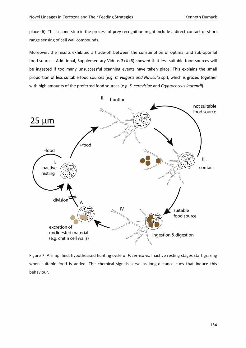
Novel Lineages in Cercozoa and Their Feeding Strategies Kenneth Dumack
154
place (6). This second step in the process of prey recognition might include a direct contact or short
range sensing of cell wall compounds.
Moreover, the results exhibited a trade-off between the consumption of optimal and sub-optimal
food sources. Additional, Supplementary Videos 3+4 (6) showed that less suitable food sources will
be ingested if too many unsuccessful scanning events have taken place. This explains the small
proportion of less suitable food sources (e.g. C. vulgaris and Navicula sp.), which is grazed together
with high amounts of the preferred food sources (e.g. S. cerevisiae and Cryptococcus laurentii).
Figure 7: A simplified, hypothesised hunting cycle of F. terrestris. Inactive resting stages start grazing
when suitable food is added. The chemical signals serve as long-distance cues that induce this
behaviour.
Novel Lineages in Cercozoa and Their Feeding Strategies Kenneth Dumack
155
How does F. terrestris digest its food?
Fungivorous protists like F. terrestris have to be able to mechanically penetrate (like the
Grossglockneriidae, 3, 4) or enzymatically degrade (like the Vampyrellida, 24) the protective fungal
cell wall (1). Since no hints for a mechanical penetration of fungal cells were observed, an enzymatic
degradation of the fungal cell wall was likely. To our knowledge, this is the first report of catalytic
properties of chitinases in Filosea. It is likely that many more Cercozoa are able to produce chitinases,
for example omnivorous cercomonads that occasionally may feed on yeast cells (1).
Conclusion
Our results confirmed that F. terrestris is a highly selective grazer and selects for fungal cells. F.
terrestris produced enzymes, such as chitinases, which are able to performed the degrade for
instance fungal cell walls to aquire C- and N- sources.
F. terrestris although being able to multiply to a much lower extend on algae and, therefore must be
considered as an omnivorous predator of eukaryotic cells, with strong preference for fungi, despite
other described close relatives are known to feed mainly on algae.
Therefore, F. terrestris as one of very few known fungivorous soil protists might be an ideal organism
for chemosensing as well as chemotaxis experiments in fungivorous soil protists.
With its easily maintained bacteria-free culture, the short generation time (~ 8 h) and the trait to stay
inactive until food is sensed it provides a convenient opportunity for bioassay experiments that might
simply be performed by addition of solved compounds in medium, a subsequent filming of the
culture and counting the percentage of activated individuals per area.
Acknowledgements
We thank the Culture Collection of Algae at the University of Cologne (CCAC) for providing the
Chlorella vulgaris strain CCAC 3272 and Navicula sp. strain CCAC 1772 and for their very professional
support.
Funding information
This study was supported by the German Research Foundation (DFG) within the Research Unit
“Carbon Flow in Belowground Food Webs assessed by Isotope Tracers”.
Novel Lineages in Cercozoa and Their Feeding Strategies Kenneth Dumack
156
References
1 Geisen S, Koller R, Hünninghaus M, Dumack K, Urich T, Bonkowski M. 2016. The soil food web
revisited: Diverse and widespread mycophagous soil protists. Soil Biol Biochem 94:10–8.
2 Bass D, Chao EEY, Nikolaev S, Yabuki A, Ishida KI, Berney C, Pakzad U, Wylezich C., Cavalier-Smith T .
2009. Phylogeny of novel naked Filose and Reticulose Cercozoa: Granofilosea cl. n. and
Proteomyxidea revised. Protist 160:75–109.
3 Foissner W. 1980. Colpodide Ciliaten (Protozoa: Ciliophora) aus alpinen Böden. Zool. Jb. Syst
107:391-432.
4 Foissner W. 1999. Description of two new, mycophagous soil ciliates (Ciliophora, Colpodea):
Fungiphrya strobli n. g., n. sp. and Grossglockneria ovata n. sp. J Eukaryot Microbiol 46:34-42.
5 Old KM, Darbyshire JF (1978) Soil fungi as food for giant amoebae. Soil Biol Biochem 10:93–100.
6 Dumack K, Müller ME, Bonkowski M. 2016a. Description of Lecythium terrestris sp. nov.
(Chlamydophryidae, Cercozoa), a soil dwelling protist feeding on fungi and algae. Protist 167:93-105.
7 Dumack K, Baumann C, Bonkowski M. 2016b. A bowl with marbles: Revision of the thecate amoeba
genus Lecythium (Chlamydophryidae, Tectofilosida, Cercozoa, Rhizaria) including a description of four
new species and an identification key. Protist 167:440-459.
8 Cutler NA, Oliver AE, Viles HA, Ahmad S, Whiteley AS. 2013. The characterisation of eukaryotic
microbial communities onsandstone buildings in Belfast, UK, using TRFLP and 454 pyrosequencing.
Int Biodeterior Biodegradation 82:124-133.
9 Damon C, Lehembre F, Oger-Desfeux C, Luis P, Ranger J, Fraissinet-Tachet L, Marmeisse R. 2012.
Metatranscriptomics reveals the diversity of genes expressed by eukaryotes in forest soils. PLoS ONE
7: e28967.
10 Jennings HS. 1906. Behavior of the Lower Organisms. Columbia University Press, New York.
11 Van Houten J, Preston RR. 1988. Chemokinesis. In Gbrtz H-D (ed) Paramecium. Springer-Verlag,
Berlin 282-300.
12 Hellung-Larsen P, Leick V, Tannerup N. 1986. Chemoattraction in Tetrahymena: on the role of
chemokinesis. Biological Bulletin 170:357-367.
13 Fenchel T, Blackburn N. 1999. Motile chemosensory behaviour of phagotrophic protists:
mechanisms for and efficiency in congregating at food patches. Protist 150:325-336.
Novel Lineages in Cercozoa and Their Feeding Strategies Kenneth Dumack
157
14 Schwelm A, Fogelqvist J, Knaust A, Jülke S, Lilja T. 2015. The Plasmodiophora brassicae genome
reveals insights in its life cycle and ancestry of chitin synthases. Sci. Rep Article:11153.
15 Anderson, IJ,Watkins RF, Samuelson J, Spencer DF, Majoros,WH, Gray MW, Loftus BJ. 2005. Gene
discovery in the Acanthamoeba castellanii genome. Protist 156:203-214.
16 Anderson TR, Patrick ZA (1980) Soil Vampyrellid amoebae that cause small perforations in conidia
of Cochliobolus sativus. Soil Biol Biochem 12:159-167.
17 Hess S, Melkonian M. 2013. The mystery of clade X: Orciraptor gen. nov. and Viridiraptor gen. nov.
are highly specialised, algivorous amoeboflagellates (Glissomonadida, Cercozoa). Protist 164:706–
747.
18 McFadden GI, Melkonian M (1986) Use of Hepes buffer for microalgal culture media and fixation
for electron microscopy. Phycologia 25:551–557.
19 Loeppmann S, Blagodatskaya E, Pausch J. 2016. Substrate quality affects kinetics and catalytic
efficiency of exo- enzymes in rhizosphere and detritusphere. Soil Biol Biochem 92:111–8.
20 Marx M, Wood M, Jarvis S. 2001. A fluorimetric assay for the study of enzyme diversity in soils.
Soil Biol Biochem 33:1633-1640.
21 Marx M, Kandeler E, Wood M, Wermbter N, Jarvis S. 2005. Exploring the enzymatic landscape:
distribution and kinetics of hydrolytic enzymes in soil particle-size fractions. Soil Biol Biochem 37:35-
48.
22 Hurlbert SH. 1993. Design and analysis of ecological experiments: edited by S.M. Scheiner and J.
Gurevitch Chapman , Hall.
23 Nannipieri P, Giagnoni L, Renella G, Puglisi E, Ceccanti B, Masciandaro G, Fornasier F, Moscatelli
MC, Marinari S. 2012. Soil enzymology: classical and molecular approaches. Biol Fert Soils 48:743-
762.
24 Hess S, Sausen N, Melkonian M (2012). Shedding light on vampires: the phylogeny of vampyrellid
amoebae revisited. PloS One 7(2), e31165.
Novel Lineages in Cercozoa and Their Feeding Strategies Kenneth Dumack
158
Chapter 8: The soil food web revisited: Diverse and
widespread mycophagous soil protists.
Novel Lineages in Cercozoa and Their Feeding Strategies Kenneth Dumack
159
Novel Lineages in Cercozoa and Their Feeding Strategies Kenneth Dumack
160
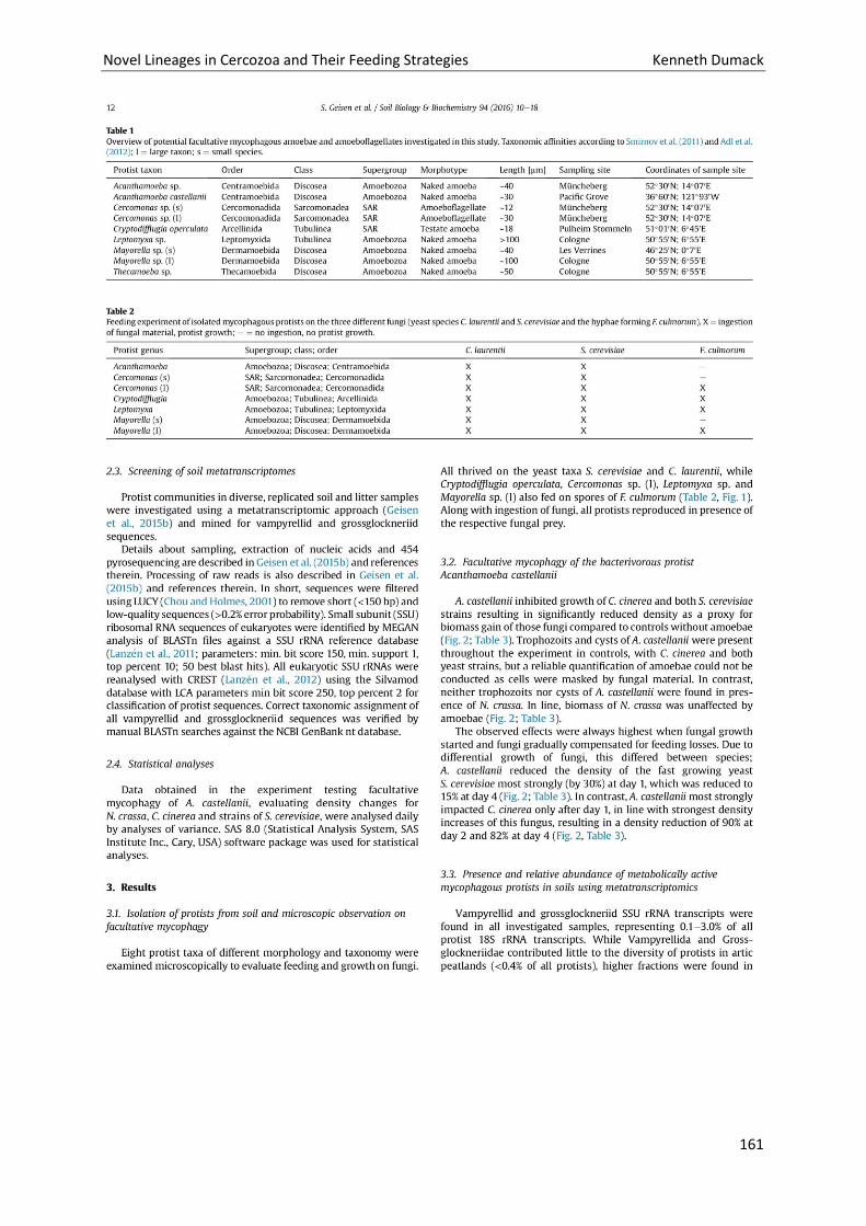
Novel Lineages in Cercozoa and Their Feeding Strategies Kenneth Dumack
161
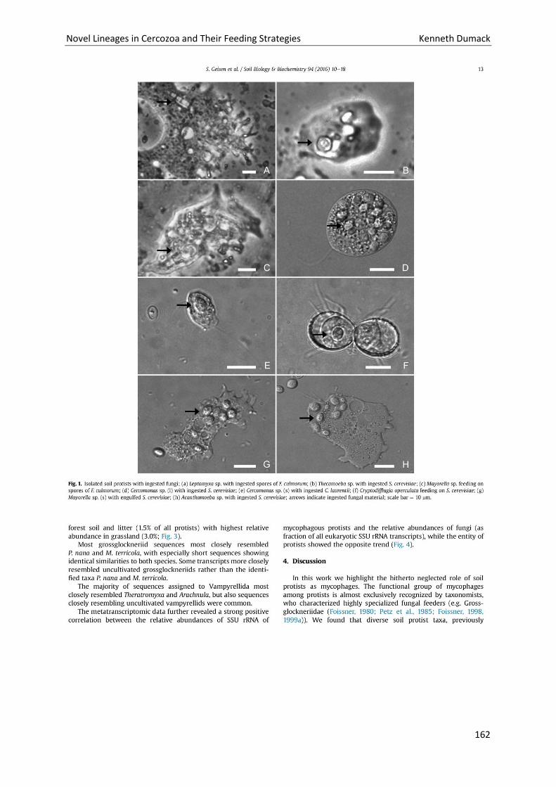
Novel Lineages in Cercozoa and Their Feeding Strategies Kenneth Dumack
162
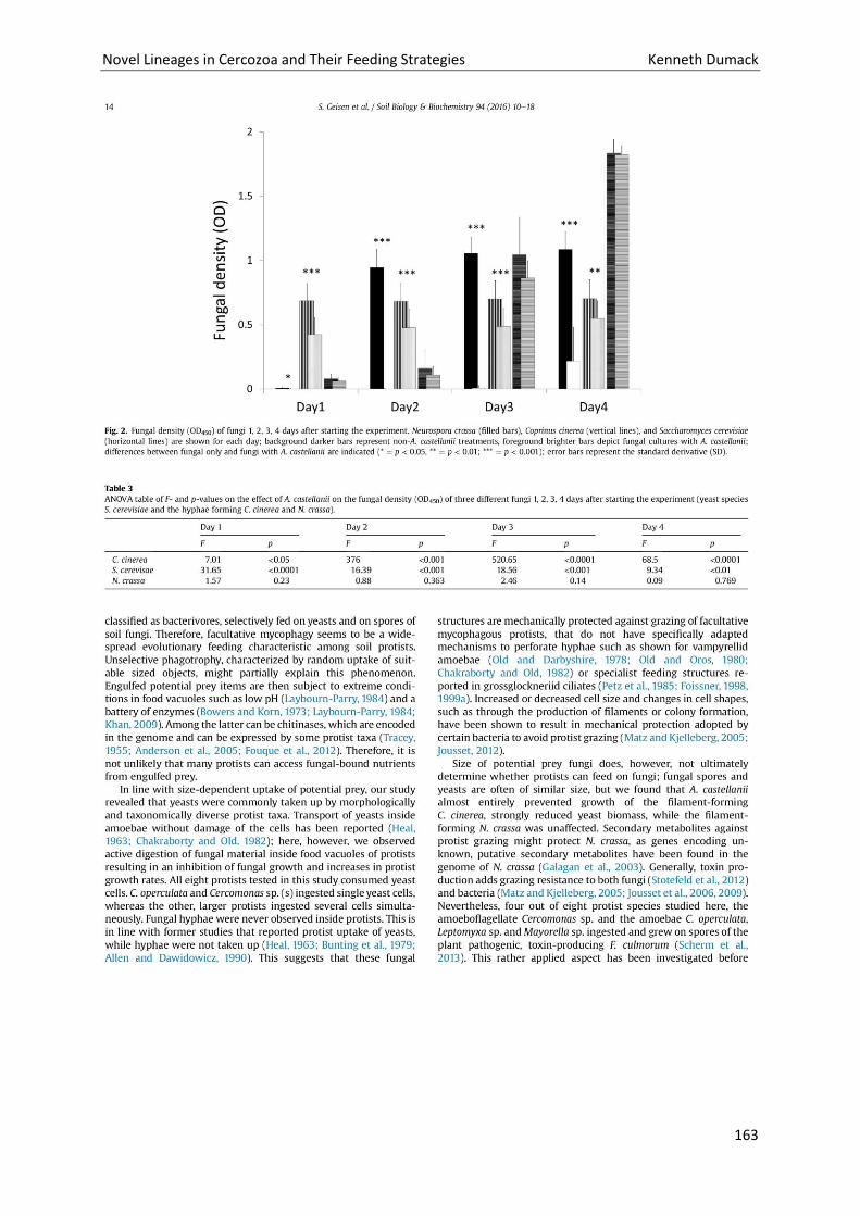
Novel Lineages in Cercozoa and Their Feeding Strategies Kenneth Dumack
163
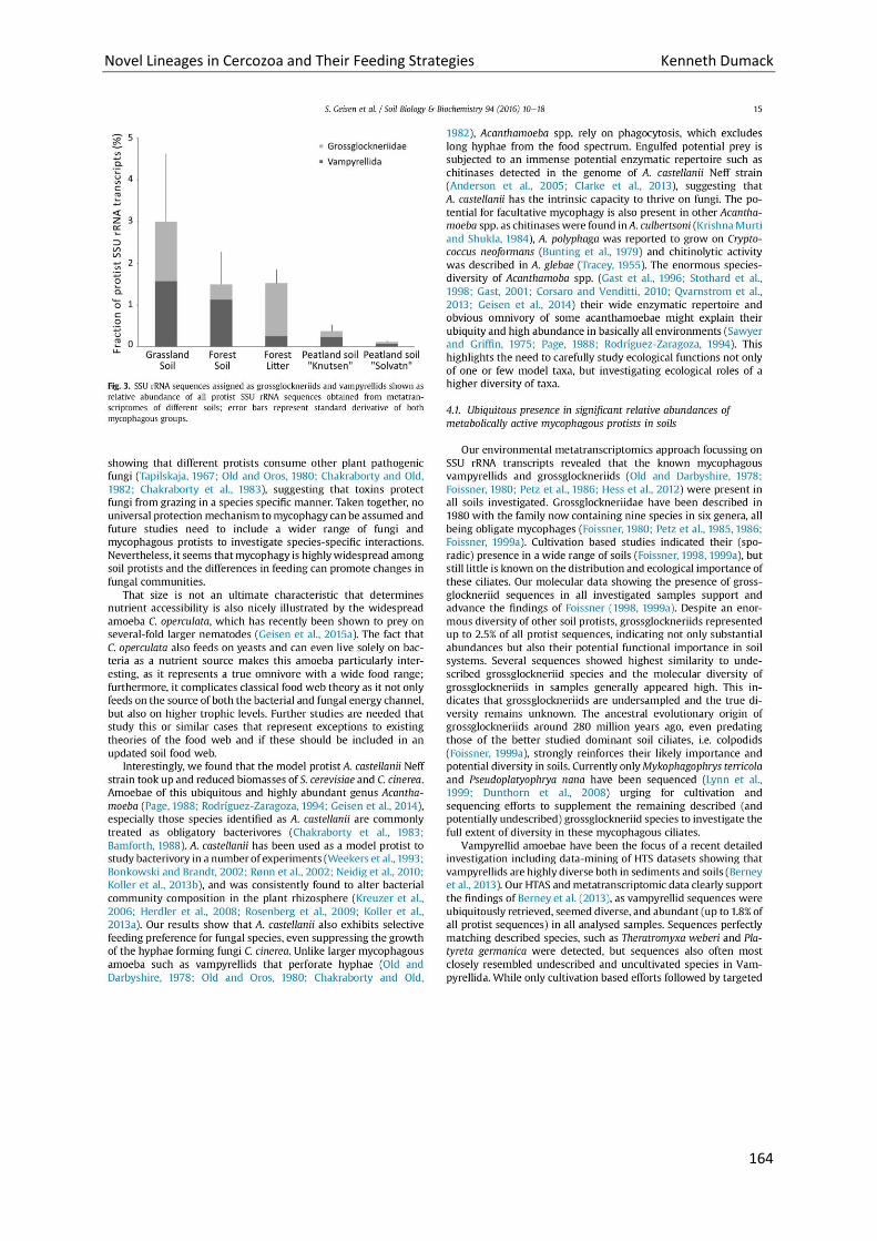
Novel Lineages in Cercozoa and Their Feeding Strategies Kenneth Dumack
164
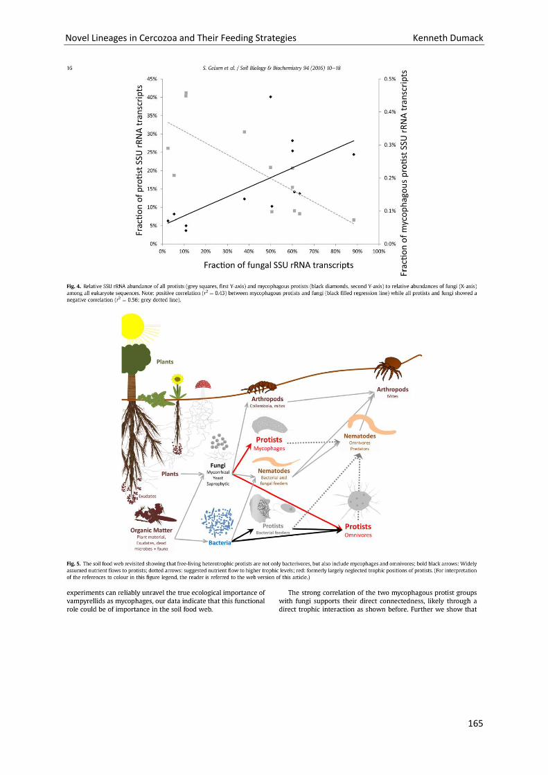
Novel Lineages in Cercozoa and Their Feeding Strategies Kenneth Dumack
165
Novel Lineages in Cercozoa and Their Feeding Strategies Kenneth Dumack
166
Novel Lineages in Cercozoa and Their Feeding Strategies Kenneth Dumack
167
Novel Lineages in Cercozoa and Their Feeding Strategies Kenneth Dumack
168
Discussion
Unknown Diversity in Cercozoa
Molecular studies hint a large uncharted diversity that needs to be characterized by
morphological means (Bass and Cavalier-Smith 2004). We were able to find several of such
unknown lineages of Cercozoa. The vast majority of Cercozoa of which morphology and
genetical data are available are bacterivores, most predominantly of the Sarcomonadea, in
particular the glissomonads and cercomonads (Bass et al. 2009b; Brabender et al. 2012;
Cavalier-Smith and Karpov 2011).
The previously undescribed scaled amoebae with a huge network of filopodia turned out to
be an independent lineage with cercozoan affiliation, Kraken carinae. They group (although
with only moderate support) within the class Sarcomonadea, CAVALIER-SMITH, 1993. If
Kraken turns out to be a true sarcomonadean, the discovery of a scale-bearing flagella-
lacking amoeboid with sarcomonadean affiliation contradicts the image of the
Sarcomonadea and might be a stepstone for a revision of cercozoan evolution. Moreover,
the discovery of Kraken underlines that morphologically, phylogenetically and ecologically
distinct lineages remain to be discovered even within groups such as Cercozoa and
sarcomonads in particular that have been relatively well studied by environmental
sequencing and culture-based surveys (Bass et al. 2009b; Brabender et al. 2012; Cavalier-
Smith and Karpov 2011; Harder et al. 2016; Howe et al. 2009).
We further were able to contribute to the known diversity of Cercozoa by the
characterization of many lineages feeding on eukaryotes. Such protists were common and by
far not an exception in Cercozoa. In probably 50% of the examined samples we found
unexplored diversity of Cercozoa feeding on eukaryotes. Thus it seems likely that there are
still many cercozoan lineages (bacterivorous and eukaryvorous) to discover, thereby
confirming our first hypothesis.
During the studies presented in this thesis it turned out that most of the isolated
eukaryvorous thecate amoeba genera (Lecythium, Rhizaspis, ...) obtained from freshwater
and terrestrial systems branch in the Thecofilosea, Cercozoa. These findings correspond with
previously published phylogenies including morphologically similar testate amoeba genera,
Novel Lineages in Cercozoa and Their Feeding Strategies Kenneth Dumack
169
e.g. Pseudodifflugia, Rhogostoma and Capsellina, all of which bear a theca, some with
accumulated exosomes (foreign materials, like sand grains or diatom frustules agglutinated
or attached to the theca), which all branch in the class Thecofilosea in the phylum Cercozoa
although with polyphyletic origin (Howe et al. 2011, Wylezich et al. 2002).
We therefore can confirm our second hypothesis and moreover, those eukaryvorous strains
of thecate amoebae now provide the opportunity to improve our understanding of thecate
amoeba diversity, test evolution, eukaryvorous feeding behaviour and the resulting
ecological impact.
Moreover, a stable taxonomic framework was obtained by combining intensive literature
research with morphological and molecular data of such poorly-known lineages of cercozoan
diversity. This enables future species descriptions and an easy identification of species by
others, e.g. ecologists. The connection of molecular data with morphological and
autecological data is still of fundamental necessity for high throughput sequencing studies
that can only then be interpreted on a professional basis. Giving information on the genetic
margins of morphospecies (and vice versa: morphological margins of operational taxonomic
units (OTUs)) is needed to draw conclusions about the investigated diversity, dispersal and
ecological function of protists.
Culturing of ‘unculturable’ protists
Petz et al. (1985) were able to culture an ‘unculturable’ lineage of fungivores with nutrient
agar that was inoculated with varying fungi and partially submerged with culture medium
and the desired organism (Grossglockneria acuta). This demonstrated with effort and the
development of novel techniques even ‘unculturable’ protists with unusual feeding habits
like fungivores are culturable.
Recently more and more ‘unculturable’ eukaryvorous protists have been cultured as
researchers invested more and more effort into protist surveys and the establishment of
cultures (Bass et al. 2009; Berney et al. 2013; Dirren et al. 2014; Hess et al. 2012; Hess &
Melkonian 2013). This includes a direct enrichment of freshwater samples with putative food
organisms (Dirren et al. 2014; Hess et al. 2012; Hess and Melkonian 2013) or the insertion of
“traps” with “bait” into soils (Pakzad and Schlösser 1998).
Novel Lineages in Cercozoa and Their Feeding Strategies Kenneth Dumack
170
We were able to culture numerous strains of several cercozoan lineages that previously have
been (or might have been) declared as unculturable, resulting in the discovery of novel
lineages and the molecular characterization of already described species, confirming our
third hypothesis.
(1) We could show that there is a novel lineage of protists that is extremely fragile and
slow growing: Weak disturbance (i.e. movement of the water body in the Petri dish)
leads to damage of the filopodia often followed by cell death. Since compared weak
mechanical disturbance of the Kraken is most often fatal, the establishment of a
single clonal Kraken culture was seldom achieved by only few attempts, but most
often had to be tried up to 20 times. Furthermore, to colonize the surface of a Petri
dish an actively growing culture required several months under our culture
conditions. Thus, the establishment of a culture of organisms that easily die off and
grow only very slowly needed a year of continuous effort and a tremendous amount
of time that not every researcher has, or is willing to invest. Thus, the key to a long-
time maintenance of culturing the Kraken is therefore immense patience. Moreover,
those characteristics hampered not only the process of culturing itself but also
culture based work, i.e. morphological analyses.
(2) The cultured Thecofilosea showed different characteristics that required a different
set of equipment and expertise. As they are eukaryvorous, they have to be
continuously co-cultured (or fed) with a suitable food source. The required culture
conditions could not be reliably predicted either by their phylogenetic position, the
type of habitat of which the sample originated from or by thorough investigation of
the amoebae in natural samples. Although many typical eukaryvorous protists, e.g.
the vast majority of Vampyrellida species show highly specialized monophagous
feeding preferences that are also reflected in their phylogeny (Hess et al. 2012, Hess
pers. comm.), we were not able to find such patterns in the Thecofilosea. For
instance, Fisculla margaritae and Fisculla cf. margaritae did not feed on fungi
whereas the closely related (and also limnic) Fisculla asini and Fisculla siemensmai
could not be cultured without. Moreover, some strains grew steadily over multiple
years of culturing on a single food source, others ceased to grow after a couple days
or even months unless being cultured on a variety of (up to four) food organisms.
Novel Lineages in Cercozoa and Their Feeding Strategies Kenneth Dumack
171
Moreover, we were able to establish the first known (and probably the first ever established)
bacteria-free culture of a thecofilosean amoeba.
Fisculla terrestris cultures contaminated with bacteria were very easy to handle, but
bacteria-free cultures of Fisculla terrestris were maintainable but unstable. Bacteria,
although not consumed, can play an important role in species coexistence in cultures.
Potential roles of bacteria in cultures are the degradation and/ or metabolisation of
secondary metabolites produced by eukaryotes, that may either just be waste products or
even be specifically produced repellents or toxins against eukaryotic predators; or (b) the
degradation of extracellular enzymes of fungi that might cause cell death when enriched to
high concentration (e.g. chitinases, own observations, unpublished).
What is the ecological impact and dispersal of eukaryvorous protists?
Bacterivory in protists has been extensively studied in terms of nutrient flows and trophic
interactions, eukaryvores have received much less attention. It has been shown that
eukaryvory plays a dominant role in freshwater systems in terms of carbon flux from primary
producers to higher trophic levels (Sherr and Sherr 1994; Sherr and Sherr 2002), but little is
known from eukaryvory in terrestrial systems.
Hess & Melkonian (2013) demonstrated with time-lapse photography what severe effects
protist grazing can have on algal cultures. Within few hours to days whole prey populations
(green algae of the Zygnematales) are eradicated. Our feeding experiments with the
fungivorous Fisculla terrestris showed similar patterns in fungi, where a starting population
of 100 predator individuals eradicated prey populations of 100.000 yeast cells in less than
three days, indicating a importance of fungivores on fungi populations and possibly a top-
down control of those.
It is important to note, that such a simplified two-species-system misses predators of higher
trophic levels. Protists are usually preyed on by other protists (often ciliates or large
omnivorous amoebae), or metazoa (e.g. cladocerans, rotifers, copepods, nematodes; see
Geisen 2016, Sherr and Sherr 2002). Accordingly, such experiments show what eukaryvores
Novel Lineages in Cercozoa and Their Feeding Strategies Kenneth Dumack
172
are capable of but lack insight in the real ecological impact of eukaryvores. Molecular
methods like co-occurence surveys might give insight into inter-specific interactions.
Direct observations of our freshwater samples and the thecofiloseans (and in particular the
freshwater Fisculla) within, revealed the freshwater inhabiting Fisculla to feed on algae,
although some fed in laboratory conditions also on fungi, they were not culturable without
sufficient algal concentration. Since direct observation of soil protists is not possible, a
laboratory approach was used to determine feeding preferences of the terrestrial Fisculla.
By laboratory experiments we were able to show that the terrestrial relatives of the genus
Fisculla feed on every of the presented fungal cells, are able to sense fungal presence,
produce chitinases and select for fungi, whereas we with diverse experiments could show
little (or no) algae ingestion and with different approaches we were not able to detect any
bacteria ingestion.
We therefore consider the terrestrial Fisculla as fungivorous, giving example of another
fungivorous lineage closely related to algivores, which supports our fourth and fifth
hypotheses. With sampling and a culture based approach, we could show the fungivorous
Fisculla in two distinct terrestrial habitats. We further showed that very common amoebae
or amoeboflagellates, like Cercomonas and Acanthamoeba, fed on fungal cells. Moreover a
metatranscriptome study focussing on fungivorous soil inhabiting protists contribute to our
understanding of less prominent but probably widespread trophic interactions between
micropredators and eukaryotic prey like algae and fungi, giving further support to our fourth
and fifth hypotheses.
Remaining questions and perspective for future investigations
Although the Kraken was only very recently described, it is already topic of ongoing
molecular studies (Sapp pers. comm. unpublished), emphasizing that the characterization of
novel lineages enables new ways to interpret ecological data, which may lead to a more
comprehensive understanding of nature.
Culture-based and molecular studies of the Tectofilosida show their diversity and functional
role of eukaryvores. Such feeding behaviour makes them a valuable target in food web
studies involving investigations of carbon and nutrient flow studies. It is therefore a
Novel Lineages in Cercozoa and Their Feeding Strategies Kenneth Dumack
173
necessity to uncover the large uncharted diversity of the Thecofilosea, especially the
Tectofilosida. Moreover, there are still old descriptions of species, like Lecythium mutabilis,
Lecythium spinosum, Lecythium curvus and more, of which molecular data is missing. All but
one of these species descriptions have been made by observing freshwater samples.
Molecular analyses already revealed a series of OTUs with Tectofilosida affiliation in
terrestrial and marine habitats (Bass and Cavalier-Smith 2004), clearly showing that there is
still much to be discovered when further targeting these habitats. These steps will be
essential to our understanding of the Tectofilosida in ecological context: (a) an increased
sampling of tectofilosidan amoebae of freshwater, marine and terrestrial habitats; (b) the
collection of morphological as well as autecological data; (c) the phylogenetic
characterization, predominantly targeting the SSU-coding gene and; (d) when those steps
have been conducted to a high extend, molecular studies can be performed to get insight
into ecological data on a broad scale, including diversity and dispersal surveys and detailed
food web (co-occurrence) investigations.
Moreover the Tectofilosida represent a valuable target to study a variety of physiological
adaptations of eukaryvorous protists: (a) The genus Fisculla comprises eukaryvores feeding
on algae and fungi, we therefore assume that Fisculla is able to produce a cocktail of
enzymes needed to digest β-polysaccharides, like in chitin, cellulose and other biopolymers.
The screening for novel enzymes often basically focuses on macroscopic organisms or
bacteria (Ferrer et al. 2009; Lee et al. 2010). Distant lineages of protists, like the Tectofilosida
in the Cercozoa may harbour enzymes highly valuable for applied purposes. A first insight
might be given by harvesting large amounts of bacteria-free Fisculla terrestris cells and
performing simple protein extractions; (b) such protein extractions might also give insight
into testate amoeba evolution as it is still unknown how Fisculla (and other thecofiloseans)
builds its test. (c) Fisculla terrestris showed to be further highly sensitive for chemical signals
released by its prey (kairomones). F. terrestris responds visibly in behaviour to those signals
and can be cultured in bacteria-free conditions, making it a valuable organism for the
establishment of a bioassay. Bioassays are necessary in chemical ecology to see the
reactivity of extracted and fractioned chemical compounds. A continuous filming of such a
culture is very easy and short-timed manageable.
Novel Lineages in Cercozoa and Their Feeding Strategies Kenneth Dumack
174
Nevertheless there are still plenty other cercozoan lineages or morphotypes of which barely
something is known. Just to name three very promising lineages of which a doctorial thesis
could lead to very interesting results: Terrestrial Viridiraptoridae are still not studied, since
the freshwater Viridiraptoridae feed on algae, their terrestrial relatives could also show
fungivorous behaviour. If so, further evidence for the fourth of our hypotheses could be
given. The Clade Y, closely related to the Viridiraptoridae, is still without any morphological
data. Since the Viridiraptoridae showed surprising unique morphology and ecology, it is easy
imaginable that the closely related Clade Y shows also parasitoid behaviour of unknown
extend. All environmental sequences of Clade Y have been obtained from soils, it is
therefore not unlikely that the organisms of Clade Y parasitize soil inhabiting organisms.
Also agglutinating testate amoebae of the Cercozoa are a yet uncovered field of research.
Although there are species known, these morphological descriptions show that
Pseudodifflugia, Diaphoropodon, Capsellina and more taxa are highly diverse. There are up
to now only one (maybe two, see Wylezich et al. 2002 and Howe et al. 2011) sequences
assigned to such genera. It is likely, that these genera of agglutinating testate amoebae will
be shown to be closely related to the thecate testate amoebae in the Cercozoa, therefore
probably also showing eukaryvorous or omnivorous feeding behaviour and interesting
ecology.
Novel Lineages in Cercozoa and Their Feeding Strategies Kenneth Dumack
175
References
Adams DJ (2004) Mini-Review Fungal cell wall chitinases and glucanases. Microbiology
150:2029–35.
Adl SM, Gupta VVSR (2006) Protists in soil ecology and forest nutrient cycling. Can. J. For.
Res. 36:1805–1817.
Adl SM, Simpson AGB, Farmer MA, Andersen RA, Anderson OR, Barta JR, Bowser SS,
Brugerolle G, Fensome RA, Fredericq S, James TY, Karpov S, Kugrens P, Krug J, Lane CE, Lewis
LA, Lodge J, Lynn DH, Mann DG, McCourt RM, Mendoza L, Moestrup O, Mozley-Standridge
SE, Nerad TA, Shearer CA, Smirnov AV, Spiegel FW, Taylor M (2005). The new higher level
classification of eukaryotes with emphasis on the taxonomy of protists. J. Eukaryot.
Microbiol. 52:399-451.
Adl SM, Simpson AGB, Lane CE, Lukeš J, Bass D, Bowser SS, Brown MW, Burki F,
Dunthorn M, Hampl V, Heiss A, Hoppenrath M, Lara E, le Gall L, Lynn DH, McManus H,
Mitchell EAD, Mozley-Stanridge SE, Parfrey LW, Pawlowski J, Rueckert S, Shadwick L, Schoch
CL, Smirnov A, and Spiegel FW (2012) The revised classification of eukaryotes. J. Eukaryot.
Microbiol. 59:429-514.
Anderson RA (2005) Algal culturing techniques. London, UK: Elsevier Science and Technology
Books, Academic Press.
Banašek-Richter C, Bersier LF, Cattin MF, Baltensperger R, Gabriel JP, Merz Y, Ulanowicz RE,
Tavares AF, Williams DD, Ruiter PC, Winemiller KO, Naisbit RE (2009) Complexity in
quantitative food webs Ecology, 90:1470–1477.
Baldauf SL (2008) An overview of the phylogeny and diversity of eukaryotes. J. Syst. Evol.
46:263–73.
Bass D, Cavalier-Smith T (2004) Phylum-specific environmental DNA analysis reveals
remarkably high global biodiversity of Cercozoa (Protozoa). Int J Syst Evol Microbiol
54:2393–2404.
Novel Lineages in Cercozoa and Their Feeding Strategies Kenneth Dumack
176
Bass D, Chao EE, Nikolaev S, Yabuki A, Ishida K, Berney C, Prasad U, Wylezich C, Cavalier-
Smith T (2009a) Phylogeny of novel naked filose and reticulose Cercozoa: Granofilosea cl. n.
and Proteomyxidea revised. Protist 160:75–109.
Bass D, Howe AT, Mylnikov AP, Vickerman K, Chao EE, Smallbone JE, Snell J, Cabral C Jr,
Cavalier-Smith T (2009b) Phylogeny and classification of Cercomonadida (Protozoa, Cer-
cozoa): Cercomonas, Eocercomonas, Paracercomonas, and Cavernomonas gen. nov. Protist
160:483–521.
Belar K (1921) Untersuchungen über Thecamöben der Chlamydophrys-Gruppe. Arch
Protistenkd. 43:287–354.
Berney C, Romac S, Mahé F, Santini S, Siano R, Bass D (2013) Vampires in the oceans:
predatory cercozoan amoebae in marine habitats. ISME J 7:2387–99.
Bobrov A, Mazei Y (2004) Morphological variability of testate amoebae (Rhizopoda:
Testacealobosea: Testaceafilosea) in natural populations. Acta Protozool. 43:133–46.
Bonkowski M (2004) Protozoa and plant growth: the microbial loop in soil revisited. New
Phytol 162:617–31.
Brabender M, Kiss AK, Domonell A, Nitsche F, Arndt H (2012) Phylogenetic and
morphological diversity of novel soil cercomonad species with a description of two new
genera (Nucleocercomonas and Metabolomonas). Protist 163:495–528
Burki F, Kudryavtsev A, Matz M V, Aglyamova G V, Bulman S, Fiers M, Keeling P Pawlowski J
(2010) Evolution of Rhizaria: new insights from phylogenomic analysis of uncultivated
protists. BMC Evol Biol 10:377.
Butler H, Rogerson A (1995) Temporal and spatial abundance of naked amoebae
(Gymnamoebae) in marine benthic sediments of the Clyde Sea area, Scotland. J. Eukaryot.
Microbiol. 42:724–730.
Cavalier-Smith T (1981) Eukaryote kingdoms: seven or nine? Biosystems 14:461–481.
Cavalier-Smith T (1998a) Neomonada and the Origin of Animals and Fungi. In Coombs GH,
Vickerman K, Sleigh MA, Warren A (eds) Evolutionary Relationships among Protozoa.
Chapman & Hall, London, pp 375–407.
Novel Lineages in Cercozoa and Their Feeding Strategies Kenneth Dumack
177
Cavalier-Smith T (1998b) A revised six-kingdom system of life. Biol Rev Cambr Philos Soc
73:203–266.
Cavalier-Smith T, Chao EEY (2003) Phylogeny and classification of phylum Cercozoa
(Protozoa). Protist 154:341–358.
Cavalier-Smith T, Karpov SA (2012) Paracercomonas Kinetid Ultrastructure, Origins of the
Body Plan of Cercomonadida, and Cytoskeleton Evolution in Cercozoa. Protist 163:47–75.
Chakraborty S, Old K (1982) Mycophagous soil amoeba: interactions with three plant
pathogenic fungi. Soil biol biochem 14:247-255.
Chakraborty S, Old KM, Warcup JH (1983) Amoebae from a take-all suppressive soil which
feed on Gaeumannomyces graminis tritici and other soil fungi. Soil biol biochem 15:17-24.
Chakraborty S, Theodorou C, Bowen GD (1985) The reduction of root colonization by
mycorrhizal fungi by mycophagous amoebae. Can. J. of Microbiol. 31:295-297.
Chapman-Andresen C (1978) The life cycle of Pelomyxa palustris. J. Protozool. 25, 1, 42A.
Chapman-Andresen C (1982) Identification of Pelomyxa binucleata as a stage in the life cycle
of P. palustris. J. Protozool. 29:499-500.
Crotty F, Blackshaw R, Murray P (2011) Tracking the flow of bacterially derived 13C and 15N
through soil faunal feeding channels. Rapid Commun. Mass Sp. 25:1503-1513.
Cutler NA, Oliver AE, Viles HA, Ahmad S, Whiteley AS (2013) The characterisation of
eukaryotic microbial communities onsandstone buildings in Belfast, UK, using TRFLP and 454
pyrosequencing. Int Biodeterior Biodegradation 82:124-133.
Darbyshire JF, Wheatley RE, Greaves MP, Inkson RHE (1974) Rapid micromethod for
estimating bacterial and protozoan populations in soil. Rev Ecol Biol Sol 11:465–475.
Damon C, Lehembre F, Oger-Desfeux C, Luis P, Ranger J, Fraissinet-Tachet L, Marmeisse R
(2012) Metatranscriptomics reveals the diversity of genes expressed by eukaryotes in forest
soils. PLoS ONE 7: e28967.
De Ruiter PC, Neutel AM, Moore JC (1995) Energetics, patterns of interaction strengths, and
stability in real ecosystems. Science 269:1257-1260.
Novel Lineages in Cercozoa and Their Feeding Strategies Kenneth Dumack
178
De Saedeleer H (1934) Beitrag zur Kenntnis der Rhizopoden: morphologische und
systematische Untersuchungen und ein Klassifikationsversuch. Mém Musée Roy d’Histoir
Naturel de Belq 60:1–112.
Dirren S, Salcher MM, Blom JF, Schweikert M, Posch T (2014) Ménage-à-trois: The Amoeba
Nuclearia sp. from Lake Zurich with its Ecto- and Endosymbiotic Bacteria. Protist 165:745–
58.
Dirren S, Posch T (2016) Promiscuous and specific bacterial symbiont acquisition in the
amoeboid genus Nuclearia (Opisthokonta). FEMS Microbiol Ecol 92:1–44.
Dunthorn M, Foissner W, Katz LA (2008) Molecular phylogenetic analysis of class Colpodea
(phylum Ciliophora) using broad taxon sampling. Mol Phylogenet. Evol 46:316-327.
Ferrer M, Beloqui A, Timmis KN, Golyshin PN (2009) Metagenomics for mining new genetic
resources of microbial communities. J Mol Microbiol Biotechnol 16:109-123.
Foissner W (1980) Colpodide Ciliaten (Protozoa: Ciliophora) aus alpinen Böden. Zool. Jb.
Syst. 107:391-432.
Foissner W (1999) Description of two new, mycophagous soil ciliates (Ciliophora, Colpodea):
Fungiphrya strobli n. g., n. sp. and Grossglockneria ovata n. sp. J. Eukaryot. Microbiol. 46:34-
42.
Foissner W (2008) Protist diversity and distribution: Some basic considerations. Biodivers.
Conserv. 17:235–42.
Frankland JC (1998) Fungal succession-unraveling the unpredictable. Mycological
Research 102:1-15.
Frolov AO (2004) The concept of pelobionts (class Peloflagellatea): a brief history and current
status. Zool. Zh. 83:643-654.
Frolov AO, Chystjakova LV, Goodkov AV (2005) Light- and electron-microscopic study of
Pelomyxa binucleata (Gruber, 1884) (Peloflagellatea, Pelobiontida). Protistology 4:57-72.
Frolov AO, Goodkov AV, Chystjakova LV, Skarlato SO (2006) Structure and development of
Pelomyxa gruberi sp. n. (Peloflagellatea, Pelobiontida). Protistology 4:227-244.
Novel Lineages in Cercozoa and Their Feeding Strategies Kenneth Dumack
179
Frolov AO, Chystyakovab LV, Malysheva NM (2011) A Light and Electron Microscopic
Study of Pelomyxa flava sp.n. (Archamoebae, Pelobiontida). Cell tissue boil. 5:81–89.
Geisen S (2016) The bacterial-fungal energy channel concept challenged by enormous
functional versatility of soil protists. 102:22–25.
Glücksman E, Bell T, Griffiths RI, Bass D (2010) Closely related protist strains have different
grazing impacts on natural bacterial communities. Environ. Microbiol. 12:3105-3113.
Haeckel E (1866) Generelle Morphologie der Organismen. Reimer, Berlin.
Harder CB, Rønn R, Brejnrod A, Bass D, Al-Soud WA, Ekelund F (2016) Local diversity of
heathland Cercozoa explored by in-depth sequencing. ISME J 1-10
Hausmann K (1978) Extrusive organelles in protists. Int. Rev. Cytol. 52:197-276.
Hertwig R, Lesser E (1874) Über Rhizopoden und denselben nahestehende Organismen.
Arch. mikroskop. Anat. 10:35–243.
Hess S, Sausen N, Melkonian M (2012) Shedding light on vampires: the phylogeny of
vampyrellid amoebae revisited. PloS ONE 7(2):e31165.
Hess S, Melkonian M (2013) The mystery of clade X: Orciraptor gen. nov. and Viridiraptor
gen. nov. are highly specialised, algivorous amoeboflagellates (Glissomonadida, Cercozoa).
Protist 164:706–747.
Holtkamp R, Wal A Van Der, Kardol P, Putten WH Van Der, Ruiter PC De, Dekker SC (2011)
Modelling C and N mineralisation in soil food webs during secondary succession on ex-arable
land. Soil Biol Biochem 43:251–60.
Homma Y, Sitton JW, Cook RY, Old KM (1979) Perforation and destruction of pigmented
hyphae of Gaeumannomyces graminis by vampyrellid amoebae from Pacific Northwest
wheat field soils. Ecol Epidemiol 69:1118–1122.
Homma Y, Ishii M (1984) Perforation of hyphae and sclerotia of Rhizoctonia solani Kühn by
mycophagous soil amoebae from vegetable field soils in Japan. Ann Phytopath Soc Japan
50:229–240.
Novel Lineages in Cercozoa and Their Feeding Strategies Kenneth Dumack
180
Howe AT, Bass D, Vickerman K, Chao EE, Cavalier-Smith T (2009) Phylogeny, taxonomy, and
astounding genetic diversity of Glissomonadida ord. nov., the dominant gliding zooflagel-
lates in soil (Protozoa: Cercozoa). Protist 160:159–189
Howe AT, Bass D, Scoble JM, Lewis R, Vickerman K, Arndt H, Cavalier-Smith T (2011) Novel
cultured protists identify deep-branching environmental DNA clades of cercozoa: New
genera Tremula, Micrometopion, Minimassisteria, Nudifila, Peregrinia. Protist 162:332–372.
Hunt HW, Coleman DC, Ingham ER, Ingham RE, Elliott ET, Moore JC, Rose SL, Reid CPP,
Morley CR (1987) The detrital food web in a shortgrass prairie. Biol. Fertil. Soils 3:57-68.
Mora C, Tittensor DP, Adl SM, Simpson AGB, Worm B (2011) How many species are there on
earth and in the ocean? PLoS Biol. 9:1–8.
Neuhauser S, Kirchmair M, Bulman S, Bass D (2014) Cross-kingdom host shifts of phytomyxid
parasites. BMC Evol Biol. 14:33.
Lampert W, Rothhaupt KO, Von Elert E (1994) Chemical induction of colony formation in a
green alga (Scenedesmus acutus) by grazers (Daphnia). Limnol Oceanogr. 39:1543–50.
Laybourn-Parry J (1984) A Functional Biology of Free-living Protozoa. Univ of California Press.
Lee HS, Kwon KK, Kang SG, Cha S, Kim S, Lee J (2010) Approaches for novel enzyme discovery
from marine environments. Curr Opin Biotechnol 21:353–7.
Lynn DH, Wright AD, Schlegel M, Foissner W (1999) Phylogenetic relationships of orders
within the class Colpodea (phylum Ciliophora) inferred from small subunit rRNA gene
sequences. J. Mol. Evol 48:605-614.
Lynn DH, Sogin ML (1988) Assessment of phylogenetic relationships among ciliated protists
using partial ribosomal RNA sequences derived from reverse transcripts. Biosystems 21:249–
54.
Meisterfeld R (2002) Testate Amoebae with Filopodia. In Lee JJ, Leedale GF, Bradbury P (eds)
An Illustrated Guide to the Protozoa. 2nd edition Society of Protozoologists, Lawrence,
Kansas, pp 1054–1084.
Novel Lineages in Cercozoa and Their Feeding Strategies Kenneth Dumack
181
Nikolaev SI, Mitchell EA, Petrov NB, Berney C, Fahrni J, Pawlowski J (2005) The testate lobose
amoebae (order Arcellinida Kent, 1880) finally find their home within Amoebozoa. Protist
156:191-202.
Nowack ECM (2014) Paulinella chromatophora – rethinking the transition from
endosymbiont to organelle. Acta Soc Bot Pol 83:387–97.
Okamoto N, Inouye I (2006) Hatena arenicola gen. et sp. nov., a Katablepharid Undergoing
Probable Plastid Acquisition. Protist 157:401–19.
Bowers B, Olszewski E (1972) PINOCYTOSIS IN ACANTHAMOEBA CASTELLANII The Journal of
Cell Biology 58(12):681–94.
Page FC (1976) An Illustrated Key to Freshwater and Soil Amoebae. Freshwater Biological
Association. Scientific Publication No. 34
Pakzad U, Schlösser E (1998) Isolation and maintenance of a vampire amoeba from a Hessian
field with wheat monoculture. J Plant Dis Protect 105: 95—99.
Penard E (1890) Études sur les Rhizopodes d‘eau douce. Mém Soc Phys Hist nat Genève
31:1–230.
Petz W, Foissner W, Adam H (1985) Culture, food selection and growth rate in the
mycophagous ciliate Grossglockneria acuta Foissner, 1980: first evidence of autochthonous
soil ciliates. Soil Biol. Biochem. 17:871-875.
Philippe H, Germot A. 2000. Phylogeny of eukaryotes based on ribosomal RNA: long-branch
attraction and models of sequence evolution. Molecular Biology and Evolution 17: 830–834.
Popper ZA, Michel G, Hervé C, Domozych DS, Willats WGT, Tuohy MG, Kloareg B, Stengel DB
(2011) Evolution and Diversity of Plant Cell Walls: From Algae to Flowering Plants. Annu. Rev.
Plant Biol. 62:567–90
Potin P, Bouarab K, Küpper F, Kloareg B (1999) Oligosaccharide recognition signals and
defense reactions in marine plant-microbe interactions. Curr Opin Microbiol 2:276-283.
Prescott D, James T (1955) Culturing of Amoeba proteus on Tetrahymena. Exp Cell Res 8:256.
Novel Lineages in Cercozoa and Their Feeding Strategies Kenneth Dumack
182
Rogers MB, Gilson PR, Su V, McFadden GI, Keeling PJ (2007) The complete chloroplast
genome of the chlorarachniophyte Bigelowiella natans: Evidence for independent origins of
chlorarachniophyte and euglenid secondary endosymbionts. Mol Biol Evol. 24:54–62.
Schönborn W (1992) Adaptive polymorphism in soil-inhabiting testate amebas (Rhizopoda) -
its importance for delimitation and evolution of asexual species. Arch. Protistenk. 142:139-
155.
Sherr EB, Sherr BF (1994) Bacterivory and Herbivory: Key Roles of Phagotrophic Protists in
Pelagic Food Webs. Microbial ecology. 28:223–35.
Sherr EB, Sherr BF (2002) Significance of predation by protists in aquatic microbial food
webs. A van Leeuw J Microb. 81:293–308.
Sogin ML, Elwood HJ, Gunderson JH (1986) Evolutionary diversity of eukaryotic small-subunit
rRNA genes. Proc Natl Acad Sci USA 83:1383–7.
Stechmann A, Cavalier-Smith T (2002) Rooting the Eukaryote Tree by Using a Derived Gene
Fusion. Science 297:89–91.
Tice AK, Shadwick LL, Fiore-Donno AM, Geisen S, Kang S, Spiegel FW, Wilkinson KA,
Bonkowski M, Dumack K, Lahr DJG, Voelcker E, Clauß S, Zhang J, Brown MW (in review)
Expansion of the Acanthamoebidae (Centramoebida, Amoebozoa). Biology Direct
Torres PA, Abril AB, Bucher EH (2005) Microbial succession in litter decomposition in the
semi-arid Chaco woodland. Soil Biol Biochem 37:49-54.
Van Donk E, Ianora A, Vos M (2011) Induced defences in marine and freshwater
phytoplankton: a review. Hydrobiologia 668:3-19.
Weisse T, Karstens N, Meyer VCL, Janke L, Lettner S, Teichgräber K (2001). Niche separation
in common prostome freshwater ciliates: the effect of food and temperature. Aquat.
Microb. Ecol. 26:167-179.
Wylezich C, Meisterfeld R, Meisterfeld S, Schlegel M (2002) Phylogenetic analyses of small
subunit ribosomal DNA coding regions reveal a monophyletic lineage of euglyphid testate
amoebae (Order Euglyphida). J Eukaryot Microbiol 49: 108–118.
Novel Lineages in Cercozoa and Their Feeding Strategies Kenneth Dumack
183
Whittaker RH (1969) New Concepts of Kingdoms of Organisms. Science 163:150-160.
Xu F, Jerlström-Hultqvist J, Kolisko M, Simpson AGB, Roger AJ, Svärd SG, Andersson JO (2016)
On the reversibility of parasitism: adaptation to a free-living lifestyle via gene acquisitions in
the diplomonad Trepomonas sp. PC1. BMC Biol 14:62
Yamaguchi H, Nakayama T, Hongoh Y, Kawachi M, Inouye I (2014) Molecular diversity of
endosymbiotic Nephroselmis (Nephroselmidophyceae) in Hatena arenicola
(Katablepharidophycota). J Plant Res. 127:241–7.
Novel Lineages in Cercozoa and Their Feeding Strategies Kenneth Dumack
184
Acknowledgements
First and foremost I would like to express my gratitude to my advisor Prof. Dr. Michael Bonkowski
for giving me the opportunity to do my thesis in his lab. Although funding was scarce Michael always
found a way to support me financially. Moreover, in my opinion most importantly, he guided my
thesis in a way that I appreciated very much. Instead of giving orders, he showed opportunities, gave
opinions and discussed those with me. The very precise Dr. Anna Maria Fiore-Donno introduced me
into bioinformatic analyses and enabled me therefore to continue my way in protistology with very
modern and timely methods. I thank Thi Anh Duong Nguyen and Fionn Clissmann for the very nice
atmosphere in our office and in general I thank the whole terrestrial ecology workgroup as I had very
pleasant time with my colleagues.
Additionally, I would like to thank Prof. Dr. Hartmut Arndt for being the second assessor of this work.
I am aware that his time is precious, thus reading and grading such a thesis is a tremendous amount
of work.
During the course of my thesis I contacted several external experts of which many became coauthors
of my publications. Foremost, I would like to thank Ferry Siemensma, this enthusiastic hobby
microscopist was especially valuable in the beginning of my thesis as he enjoyed to discuss my
questions. I would like to thank Sebastian Loeppmann, as he enabled me to carry out the enzyme
analyses of F. terrestris, he showed me the whole procedure and taught to such an extent that I am
now able to perform them for myself. I also thank Dr. Alexander Mynikov, Dr. Frank Nitsche and
Eckhard Völcker for helping with published and yet unpublished electron microscopy.
I enjoyed the work with several bachelor students of which I had the pleasure to work with.
And last of all, many persons that are not named here but were nevertheless important contributors.
Novel Lineages in Cercozoa and Their Feeding Strategies Kenneth Dumack
185
Subpublications in Three Parts and Record of Achievement
The Publications correspond to the chapters.
Part 1 - Characterization of the unusual bacterivorous amoeba Kraken carinae.
Chapter 11, 4: Dumack K, Schuster J, Bass D, Bonkowski M (2016) A novel lineage of ‘naked
filose amoebae’; Kraken carinae gen. nov. sp. nov. (Cercozoa) with a remarkable locomotion
by disassembly of its cell body. Protist 167:268-278
Chapter 21, 5: Dumack K, Mylnikov AP, Bonkowski M (in review) Cercomonad or archaic
Imbricatea? On the hunt for the true taxonomy of the scale-bearing Kraken (incertae sedis,
Cercozoa, Rhizaria): Combining ultrastructure data and a two-gene (SSU + LSU) phylogeny.
Protist
Part 2 - Eukaryvorous amoebae of the Thecofilosea, Cercozoa.
Chapter 31: Dumack K, Müller ME, Bonkowski M (2016) Description of Lecythium terrestris
sp. nov. (Chlamydophryidae, Cercozoa), a soil dwelling protist feeding on fungi and algae.
Protist 167:93-105
Chapter 41, 2: Dumack K, Baumann C, Bonkowski M (2016) Revision of the thecate amoeba
genus Lecythium (Chlamydophryidae, Tectofilosida, Cercozoa, Rhizaria) including a
description of four new species and an identification key. Protist 167:440-459
Chapter 51, 2: Dumack K, Öztoprak H, Rüger L, Bonkowski M (in review) Shedding light on
the polyphyletic thecate amoeba genus Plagiophrys: Transition of some of its species to
Rhizaspis (Tectofilosida, Cercozoa) and the establishment of Sacciforma gen. nov. and
Rhogostomidae fam. nov. (Cryomonadida, Cercozoa). Protist
Chapter 61, 2: Dumack K, Mausbach P, Hegmann M, Bonkowski M (in review) Polyphyly in
the thecate amoeba genus Lecythium (Chlamydophryidae, Tectofilosida, Cercozoa),
redescription of its type species L. hyalinum, description of L. jennyae sp. nov. and the
establishment of Fisculla gen. nov. and Fiscullidae fam. nov. Protist
Novel Lineages in Cercozoa and Their Feeding Strategies Kenneth Dumack
186
Part 3 - Eukaryvorous protists, their capabilities and dispersal.
Chapter 71, 6: Dumack K, Pundt J, Loeppmann S, Bonkowski M (in review) What does it take
to eat a fungus? A case study with the eukaryvorous amoeba Fisculla terrestris. Applied and
Environmental Microbiology
Chapter 83: Geisen S, Koller R, Hünninghaus M, Dumack K, Urich T, Bonkowski M (2016)
The soil food web revisited: Diverse and widespread mycophagous soil protists. Soil Biol
Biochem 94:10–18
1 The Study was designed and planned together with Prof. Dr. Michael Bonkowski and all
results described in this chapter – molecular, phylogenetic, taxonomic, and morphologic –
were carried out by the author, if not stated otherwise. The article was mainly written by the
author under guidance of Prof. Dr. Michael Bonkowski, if not stated otherwise.
2 The article was written or adjusted by the author, by usage of the developed thesis of the
supervised student, under guidance of Prof. Dr. Michael Bonkowski.
3 The author was involved in the writing of the article and in performing the laboratory
experiments.
4 Phylogenetic analyses were performed with help of the coauthor Prof. Dr. David Bass.
5 Electron microscopy was performed by the coauthor Dr. Alexander P. Mylnikov.
6 The enzymatic analyses were performed by the author and the coauthor Sebastian
Loeppmann.
Novel Lineages in Cercozoa and Their Feeding Strategies Kenneth Dumack
187
Erklärung (gemäß § 4 Abs 1 Punkt 9 der Prüfungsordnung)
Ich versichere, dass ich die von mir vorgelegte Dissertation selbständig angefertigt, die
benutzten Quellen und Hilfsmittel vollständig angegeben und die Stellen der Arbeit −
einschließlich Tabellen, Karten und Abbildungen −, die anderen Werken im Wortlaut oder
dem Sinn nach entnommen sind, in jedem Einzelfall als Entlehnung kenntlich gemacht habe;
dass diese Dissertation noch keiner anderen Fakultät oder Universität zur Prüfung
vorgelegen hat; dass sie − abgesehen von unten angegebenen Teilpublikationen − noch nicht
veröffentlicht worden ist, sowie, dass ich eine solche Veröffentlichung vor Abschluss des
Promotionsverfahrens nicht vornehmen werde. Die Bestimmungen der Promotionsordnung
sind mir bekannt. Die von mir vorgelegte Dissertation ist von Prof. Dr. Michael Bonkowski
betreut worden.
_____________________
Kenneth Dumack
Novel Lineages in Cercozoa and Their Feeding Strategies Kenneth Dumack
188
Lebenslauf Persönliche Daten Name: Kenneth Dumack Adresse: Gladbacher Straße 11, 50672 Köln Mail Adresse: [email protected] Staatsangehörigkeit: deutsch Geburtsdaten: 06.03.1990 Dormagen Stadtteil Hackenbroich Website kennethdumack.de
Studium
Seit 2014 Promotionsstudium, Institut für Zoologie, Terrestrische Ökologie, Universität zu Köln 2012 – 2014 Studium Master of Science in Biological Sciences, Universität zu Köln 2009 – 2012 Studium Bachelor of Science in Biologie, Universität zu Köln
Arbeitserfahrung Seit 2012 SHK, WHF und WHK Tätigkeit bei Prof. Dr. Michael Bonkowski, Universität zu Köln
Schulische Ausbildung 2006 – 2009 Besuch des Geschwister Scholl Gymnasiums Pulheim 2000 – 2006 Besuch der Marion Dönhoff Realschule, Pulheim
Tagungen und Beiträge
Moscow Forum «PROTIST–2016» Mündlicher Vortrag "The Chlamydophryidae revisited with special focus on the genus Lecythium" Poster Präsentation "The base of cercomonad radiation is still in for a surprise, Kraken gen. nov." Empfänger des Holz-Conner Preises. Unter den ehrenhaften Nennungen für die besten Poster und besten Vorträge.
35th annual meeting of the German Society for Protozoology Mündlicher Vortrag "A bowl with marbles; the fungal and algal-eating amoeba genus Lecythium (Chlamydophryidae, Tectofilosida, Cercozoa, Rhizaria) revisited; phylogeny and the description of four new species" Poster Präsentation "The base of cercomonad radiation is still in for a surprise, Kraken gen. nov." Empfänger der zweiten Preise für das beste Poster und den besten Vortrag.
Gastvortrag an der Universität Göttingen, eingeladen von Valentyna Krashevska Titel: "Lecythium, the neglected testate amoebae. Insight into phylogeny, morphology and ecology with focus on L. terrestris sp. nov."
34th annual meeting of the German Society for Protozoology Mündlicher Vortrag "Lecythium terrestris, its morphology, phylogeny and autecology." Empfänger des Preises für den besten Vortrag.
33rd annual meeting of the German Society for Protozoology