No causal association identified for human papillomavirus infections in lung cancer
29
1 No causal association identified for human papillomavirus infections in lung cancer 1 2 Devasena Anantharaman 1 , Tarik Gheit 2 , Tim Waterboer 3 , Gordana Halec 3 , Christine Carreira 4 , Behnoush 3 Abedi-Ardekani 1,5 , Sandrine McKay-Chopin 2 , David Zaridze 6 , Anush Mukeria 6 , Neonila Szeszenia- 4 Dabrowska 7 , Jolanta Lissowska 8 , Dana Mates 9 , Vladimir Janout 10 , Lenka Foretova 11 , Vladimir Bencko 12 , 5 Peter Rudnai 13 , Eleonora Fabianova 14 , Anne Tjønneland 15 , Ruth C Travis 16 , Heiner Boeing 17 , J Ramón 6 Quirós 18 , Mikael Johansson 19 , Vittorio Krogh 20 , H. Bas Bueno-de-Mesquita 21-23 , Anastasia Kotanidou 24,25 , 7 Françoise Clavel-Chapelon 26-28 , Elisabete Weiderpass 29-32 , Mattias Johansson 1 , Michael Pawlita 3 , Ghislaine 8 Scelo 1 , Massimo Tommasino 2 , Paul Brennan 1 9 10 Author affiliations: 11 1. Genetic Epidemiology Group, International Agency for Research on Cancer, Lyon, France 12 2. Infections and Cancer Biology Group, International Agency for Research on Cancer, Lyon, France 13 3. Infection and Cancer Program, German Cancer Research Center (DKFZ), Heidelberg, Germany 14 4. Molecular Pathology Group, International Agency for Research on Cancer, Lyon, France 15 5. Digestive Disease Research Center, Tehran University of Medical Sciences, Tehran, Iran 16 6. Department of Epidemiology and Prevention, Russian Cancer Research Center, Moscow, Russia. 17 7. Department of Environmental Epidemiology, Nofer Institute of Occupational Medicine, Lodz, Poland 18 8. M. Sklodowska-Curie Memorial Cancer Center and Institute of Oncology, Warsaw, Poland 19 9. Institute of Public Health, Bucharest, Romania 20 10. Department of Preventive Medicine, Palacky University, Olomouc, Czech Republic 21 11. Department of Cancer Epidemiology and Genetics, Masaryk Memorial Cancer Institute, Brno, Czech 22 Republic 23
-
Upload
independent -
Category
Documents
-
view
0 -
download
0
Transcript of No causal association identified for human papillomavirus infections in lung cancer
1
No causal association identified for human papillomavirus infections in lung cancer 1
2
Devasena Anantharaman1, Tarik Gheit2, Tim Waterboer3, Gordana Halec3, Christine Carreira4, Behnoush 3
Abedi-Ardekani1,5, Sandrine McKay-Chopin2, David Zaridze6, Anush Mukeria6, Neonila Szeszenia-4
Dabrowska7, Jolanta Lissowska8, Dana Mates9, Vladimir Janout10, Lenka Foretova11, Vladimir Bencko12, 5
Peter Rudnai13, Eleonora Fabianova14, Anne Tjønneland15, Ruth C Travis16, Heiner Boeing17, J Ramón 6
Quirós18, Mikael Johansson19, Vittorio Krogh20, H. Bas Bueno-de-Mesquita21-23, Anastasia Kotanidou24,25, 7
Françoise Clavel-Chapelon26-28, Elisabete Weiderpass29-32, Mattias Johansson1, Michael Pawlita3, Ghislaine 8
Scelo1, Massimo Tommasino2, Paul Brennan1 9
10
Author affiliations: 11
1. Genetic Epidemiology Group, International Agency for Research on Cancer, Lyon, France 12
2. Infections and Cancer Biology Group, International Agency for Research on Cancer, Lyon, France 13
3. Infection and Cancer Program, German Cancer Research Center (DKFZ), Heidelberg, Germany 14
4. Molecular Pathology Group, International Agency for Research on Cancer, Lyon, France 15
5. Digestive Disease Research Center, Tehran University of Medical Sciences, Tehran, Iran 16
6. Department of Epidemiology and Prevention, Russian Cancer Research Center, Moscow, Russia. 17
7. Department of Environmental Epidemiology, Nofer Institute of Occupational Medicine, Lodz, Poland 18
8. M. Sklodowska-Curie Memorial Cancer Center and Institute of Oncology, Warsaw, Poland 19
9. Institute of Public Health, Bucharest, Romania 20
10. Department of Preventive Medicine, Palacky University, Olomouc, Czech Republic 21
11. Department of Cancer Epidemiology and Genetics, Masaryk Memorial Cancer Institute, Brno, Czech 22
Republic 23
2
12. Institute of Hygiene and Epidemiology, 1st Faculty of Medicine, Charles University in Prague, Prague, 1
Czech Republic 2
13. National Institute of Environmental Health, Budapest, Hungary 3
14. Regional Authority of Public Health, Banská Bystrica, Slovakia 4
15. Danish Cancer Society Research Center, Copenhagen, Denmark 5
16. Cancer Epidemiology Unit, Nuffield Department of Clinical Medicine, University of Oxford, Oxford, 6
United Kingdom 7
17. Department of Epidemiology, German Institute of Human Nutrition Potsdam-Rehbruecke, Nuthetal, 8
Germany 9
18. Public Health Directorate, Asturias, Spain 10
19. Department of Radiation Sciences, Oncology, Umeå University, Umeå, Sweden 11
20. Epidemiology and Prevention Unit, Fondazione IRCCS Istituto Nazionale dei Tumori, Milano, Italy 12
21. National Institute for Public Health and the Environment (RIVM), Bilthoven, The Netherlands 13
22. Department of Gastroenterology and Hepatology, University Medical Centre, Utrecht, The 14
Netherlands 15
23. The School of Public health, Imperial College London, London, United Kingdom 16
24. First Department of Critical Care Medicine & Pulmonary Services, University of Athens Medical 17
School, Evangelismos Hospital, Athens, Greece 18
25. Bureau of Epidemiologic Research, Academy of Athens, Athens, Greece 19
26. INSERM, Centre for research in Epidemiology and Population Health (CESP), U1018, Nutrition, 20
Hormones and Women’s Health team, F-94805, Villejuif, France 21
27. University Paris Sud, UMRS 1018, F-94805, Villejuif, France 22
28. IGR, F-94805, Villejuif, France 23

3
29. Department of Community Medicine, Faculty of Health Sciences, The Artic University of Norway, 1
Tromsø, Norway 2
30. Department of Research, Cancer Registry of Norway, Oslo, Norway 3
31. Department of Medical Epidemiology and Biostatistics, Karolinska Institutet, Stockholm, Sweden 4
32. Samfundet Folkhälsan, Helsinki, Finland 5
6
Running Title: Role of human papillomavirus infections in lung cancer 7
8
Keywords: Lung cancer, cancer risk, Human Papillomavirus, HPV16, p16 9
10
Financial Support: 11
The EPIC study has been supported by the Europe Against Cancer Program of the European Commission 12
(SANCO); Deutsche Krebshilfe, Deutsches Krebsforschungszentrum (DKFZ), German Federal Ministry of 13
Education and Research; Danish Cancer Society; Health Research Fund (FIS) of the Spanish Ministry of 14
Health, Spanish Regional Governments of Andalucia, Asturias, Basque Country, Murcia and Navarra; 15
Catalan Institute of Oncology, Spain; the ISCIII of the Spanish Ministry of Health (RETICC DR06/0020); 16
Cancer Research UK; Medical Research Council, United Kingdom; Greek Ministry of Health; Stavros 17
Niarchos Foundation; Hellenic Health Foundation; Italian Association for Research on Cancer (AIRC); 18
Italian National Research Council, Fondazione-Istituto Banco Napoli, Italy; Compagnia di San Paolo; Dutch 19
Ministry of Public Health, Welfare and Sports; World Cancer Research Fund; Swedish Cancer Society; 20
Swedish Scientific Council; Regional Government of Västerbotten, Sweden; NordForsk (Centre of 21
excellence programme HELGA), Norway; French League against Cancer (LNCC), France; National Institute 22
for Health and Medical Research (INSERM), France; Mutuelle Générale de l’Education Nationale (MGEN), 23
France; 3M Co, France; Gustave Roussy Institute (IGR), France; and General Councils of France. The EPIC 24
4
study in Norway was also sponsored by the European Research Council, The Norwegian Research 1
Council, and The Norwegian Cancer Society. The Central Europe study was partly supported by the 2
European Regional Development Fund and the State Budget of the Czech Republic (RECAMO, 3
CZ.1.05/2.1.00/03.0101). 4
Corresponding author: 5 Paul Brennan 6 International Agency for Research on Cancer (IARC) 7 Genetic Epidemiology Group 8 150 cours Albert Thomas 9 69372 Lyon CEDEX 08, France 10 Tel: +33 (0)4 7273 8391 11 Fax: +33 (0)4 7273 8342 12 Email: [email protected] 13 14
Disclosure of Potential Conflicts of Interest: MP has received research support through cooperation 15
contracts of DKFZ with Roche and Qiagen in the field of development of HPV diagnostics. However, 16
Roche and Qiagen had no influence in this manuscript. The authors have declared that no competing 17
interests exist. 18
19
Manuscript word count: 3204 20 Number of Figures: 1 21 Number of Tables: 5 22
5
Abstract 1
Human papillomavirus (HPV) infections have been implicated in lung carcinogenesis, but causal 2
associations remain uncertain. We evaluated a potential causal role for HPV infections in lung cancer 3
through an analysis involving serology, tumor DNA, RNA and p16 protein expression. Association 4
between type-specific HPV antibodies and risk of lung cancer was examined among 3083 cases and 4328 5
controls in two case-control studies (retrospective) and one nested case-control study (prospective 6
design). Three hundred and thirty four available tumors were subjected to pathological evaluation and 7
subsequent HPV genotyping following stringent conditions to detect all high risk and two low risk HPV 8
types. All HPV DNA positive tumors were further tested for the expression of p16 protein and type-9
specific HPV mRNA. Based on the consistency of the results, although HPV11 and HPV31 E6 antibodies 10
were associated with lung cancer risk in the retrospective study, no association was observed in the 11
prospective design. Presence of type-specific antibodies correlated poorly with the presence of the 12
corresponding HPV DNA in the tumor. Although nearly 10% of the lung tumors were positive for any HPV 13
DNA (7% for HPV16 DNA), none expressed the viral oncogenes. No association was observed between 14
HPV antibodies or DNA and lung cancer survival. In conclusion, we found no supportive evidence for the 15
hypothesized causal association between HPV infections and lung cancer. 16
6
Introduction 1
Lung cancer accounts for over a million deaths worldwide each year (1). Although smoking is the most 2
important risk factor, lung cancer also occurs in never smokers with particularly high rates reported 3
among women in south Asia (2, 3). Among other known risk factors, a role for human papillomavirus 4
(HPV) infections has been postulated (4). 5
6
HPV infections cause almost all cervical cancers, nearly half of ano-genital cancers and a substantial 7
proportion of oropharyngeal cancers (5). Evidence for the association between HPV and lung cancer 8
comes from over 50 case-studies that have examined the presence of HPV-DNA in lung tumor tissues. A 9
recent meta-analysis estimated that nearly 7% (95% CI: 4.7-10.6) of lung cancer is HPV16-associated and 10
almost 6% (95% CI: 3.5-9.0) HPV18-associated (6). Interpreting these findings is difficult due to the 11
variation in HPV prevalence, both across and within geographic regions. A more recent study did not 12
observe any HPV DNA in nearly 400 lung tumors examined (7). A recent Taiwanese study, however, 13
associated HPV DNA presence with improved lung cancer survival. This study, based on nearly 200 cases 14
focused on lung adenocarcinoma and oversampled women (8). 15
16
That HPV can reach the lung is well established through the occurrence of respiratory papillomas (9). This 17
rare benign condition, caused by HPV6 and HPV11, occasionally transforms to frank malignancies. Known 18
risk factors for HPV transmission such as lifetime number of sexual partners and oral-genital sexual 19
contact are also associated with adult recurrent respiratory papillomatosis (10, 11). One of the 20
challenges in the hypothesis of HPV as a lung carcinogen lies in establishing the functional consequence 21
of the viral presence in the tumor. Though experimental studies have demonstrated that HPV 22
oncoproteins E6 and E7 can co-operate with tobacco smoke in transforming lung epithelial cells in vitro, 23
and can promote tumor angiogenesis both in vitro and in vivo (12, 13), limited direct evidence exists. The 24

7
few studies on the expression of HPV oncogenes in lung tumors have been inconclusive (14-16). These 1
limitations led the IARC Monograph expert group to conclude that there was limited evidence for the 2
carcinogenicity of HPV to the lung. The expert group also argued that the exclusively case-only design, 3
limited sample size (typically <100 cases), assay limitations and lack of comprehensive characterization 4
using multiple methods were important limitations in the interpretation of the association between HPV 5
infection and lung cancer (4). 6
7
In order to conclusively evaluate the causal role of HPV infection in lung cancer, we have (i) examined the 8
association between type-specific HPV antibodies and lung cancer risk in multiple series of lung cancer 9
cases and controls; (ii) tested lung tumors for the presence of HPV genomes of all high risk types and two 10
low risk types employing rigorous precautions against potential contamination; (iii) assessed the 11
expression of p16 protein and (iv) examined the transcriptional state of the HPV oncogene E6. 12
13
Materials and methods 14
Study population 15
This analysis included three large lung cancer studies; the Central Europe (CE) and the L2 study 16
(retrospective design), and the European Prospective Investigation into Cancer and Nutrition (EPIC) study 17
(prospective design). The CE study was conducted during 1998–2002 across six central-European 18
countries. Information on demographics, lifestyle factors and occupational exposures were recorded 19
upon personal interview using standardized questionnaires (17). The present analysis included 2035 20
controls and 1286 case subjects with an available plasma sample. The L2 study was conducted in 21
Moscow during 2007 to 2010, and similar to the L1 study, lifestyle and exposure data were recorded on 22
personal interview. The present analysis included 348 cases and 694 controls who donated a blood 23
sample. Briefly, both the retrospective studies recruited histologically or cytologically confirmed incident 24
8
lung cancer cases. Cases were hospitalized subjects (recruited mostly within three months of diagnosis), 1
identified through active search of diagnostic departments of the participating hospitals who reported to 2
have resided in the area of recruitment for at least a year prior to diagnosis. Corresponding hospital-3
based controls, none of whom reported a previous cancer history or any tobacco-related disease, were 4
frequency matched to cases based on age, gender and area of residence. Both retrospective studies 5
defined smokers as individuals who smoked at least 100 cigarettes in their lifetime. Details of the 6
ongoing EPIC study are published elsewhere (18). Briefly lifestyle, dietary and anthropometric data were 7
recorded from 519,978 subjects aged 25 to 70 years during 1992-2000 from 23 centers across 10 8
countries in Europe. Smokers were defined as those who responded to have ever smoked a cigarette or 9
cigar or pipe in their lifetime. Follow-up and identification of incident cancer was based on record linkage 10
and active follow-up through study participants and their next of kin. In this analysis, we excluded all 11
prevalent cancers (other than non-melanoma skin cancers), secondary lung cancer cases and subjects 12
lost to follow-up. HPV serology data were available among a similar group of cancer-free cohort 13
members as a part of a parallel head and neck cancer study (19). Even though controls in the nested 14
case-control study were matched to head and neck cancer cases on country, sex, date of blood collection 15
(1 month, relaxed to 5 months for sets without available controls), and date of birth (1 year, relaxed to 16
5years for sets without available participants), they were processed and analyzed at the same time as the 17
lung cases and under identical conditions. A total of 1449 lung cancer cases and 1599 controls were 18
available from the EPIC study. All three studies were approved by the IARC Ethics Committee and local 19
ethical review boards of the participating centers. 20
HPV serology 21
The three high-risk (HPV16, HPV18 and HPV31) and two low-risk (HPV6 and HPV11) types tested in this 22
study were selected based on pre-existing evidence of their association with benign conditions and frank 23
malignancies of the respiratory epithelium. Additionally, antibodies against cellular proteins, non-HPV 24
9
viral proteins and cutaneous HPV-types were tested as specificity controls. In total, plasma samples from 1
4328 controls and 3083 lung cancer cases were tested using the bead-based multiplex serology method 2
described elsewhere (20). Serology data were dichotomized based on study-specific cut-offs derived 3
using a set of known reference sera (21, 22). Thresholds derived for the prospective study, which were 4
higher in general, were applied to the retrospective studies to allow for comparisons. Serology testing 5
was performed blind to case-control status. 6
Tumor analyses 7
Tissue processing: Tumor tissues were available only for the retrospective studies. Of the 334 tumors 8
available, 290 tumors passed pathological evaluation performed in-blind to serology results. The reasons 9
for pathological exclusion included tissue size (too small to allow for complete testing, n=10); lack of 10
confirmation of lung origin of the tumor (n=6); insufficient tumor cell content (n=15), presence of non-11
tumor lung tissue such as reactive lymph nodes (n=7), fibroconnective tissue (n=2), necrotic tissue (n=2), 12
or inflammation (n=2). To avoid potential contamination, tumors were processed in small batches with 13
frequent change of gloves and cleaning of the worktable using DNA Away™ (Molecular BioProducts, 14
USA). Further, a negative control for tumor sectioning included processing 1 blank block every 10 tumors 15
blocks. 16
HPV genotyping: DNA was extracted from snap-frozen (n=139) tumor tissues using the Qiagen BioRobot 17
EZ1 with the EZ1 DNA tissue kit according to the manufacturer’s instructions (Qiagen, Hilden, Germany), 18
while crude DNA extraction method was used for formalin-fixed paraffin embedded (FFPE) tumor tissues 19
(n= 151). Briefly, FFPE tissues were incubated in proteinase K and buffer G2 (Qiagen, Hilden, Germany) at 20
56˚C until the tissue was completely lysed and crude cell extract containing nucleic acids was used for 21
HPV genotyping. To ensure DNA quality, beta actin gene primers were included as positive control; all 22
290 tumors were betaglobin positive. One extraction control was included every 10 tumors to monitor 23
potential cross-contamination. In addition, negative controls for the PCR assay (1 per 95 samples) and 24
10
luminex beads (1 per batch) were also included. HPV genotyping was performed under strict pre- and 1
post-PCR separation conditions using the Type-Specific E7 PCR-MPG Assay (TS-E7-MPG) that detects 19 2
high-risk (HPV16, 18, 26, 31, 33, 35, 39, 45, 51, 52, 53, 56, 58, 59, 66, 68a, 68b, 70, 73, 82) and 2 low-risk 3
types (HPV6, 11) (23). Results were dichotomized by applying cutoffs calculated as the median 4
background value plus five standard deviations (24). We adopted a stringent definition to call a HPV 5
DNA-positive tumor based on two rounds of HPV genotyping. In the first genotyping round, all cases that 6
passed the independent pathological evaluation were tested (n=290). Subsequently, in order to confirm 7
the initial findings and reduce potential false-positivity, all tumors that tested HPV DNA positive in the 8
first genotyping round were subjected to a second round of tissue preparation and genotyping. In 9
addition, 16 HPV-DNA negatives were included as additional controls. Only cases positive for the same 10
HPV-type in both rounds were considered true HPV DNA-positives. 11
Expression of p16 protein: The expression of p16 protein was qualitatively evaluated among all cases 12
subjected to the second genotyping round. These included tumors that tested positive for any HPV DNA 13
at least once and an additional 16 HPV DNA negatives. Immunohistochemical staining was performed 14
using the CINtec® Histology p16INK4a Kit (9511, mtm laboratories) following manufacturer’s instructions. 15
Expression was scored using a composite score based on the percentage of stained cells and the intensity 16
of the nucleic or cytoplasmic staining. A score of 4 or lesser was considered low to moderate expression 17
of p16 protein. 18
Expression of HPV oncogenes: Two assays for the detection of HPV-E6 mRNA were performed that 19
differed in the HPV types tested and their sensitivity. The two assays were performed in independent 20
laboratories and blind to HPV DNA status. Since HPV16 has been reported as the most important non-21
cervical oncogenic HPV type, we performed a dedicated assay to detect the most abundant splice variant 22
(E6*I) (25). This assay included all 19 HPV16-DNA positive tumors alongside 7 HPV16-DNA negatives. The 23
second assay utilized a recently developed method to detect HPV type-specific transcripts of the E6 gene 24
11
(26). This assay was customized to detect transcripts of all HPV types identified in this study (details 1
provided upon request). In the second assay, 34 tumors were tested in total that included 28 HPV DNA 2
positives (14 single HPV16 infections, 9 single infections of non-HPV16 type, 5 multiple infections 3
including HPV16) and 6 HPV DNA negatives, which also included all 26 tumors from the first assay. 4
Statistical analysis 5
The association between HPV antibodies and the risk of lung cancer was examined by calculating the 6
odds ratios (OR) and corresponding 95% confidence intervals (95% CI). Unconditional logistic regression 7
models were adjusted for age, sex, smoking status (never, former or current) and country. Further 8
stratified analyses were performed by sex and smoking status (ever, never) to examine any potential 9
effect modification. We used chi-squared tests to examine the heterogeneity in OR by study design. The 10
correlation between HPV DNA presence and seropositivity to the corresponding type-specific antibodies 11
and expression of p16 as a surrogate marker was examined by linear regression, coefficients of 12
correlation and p-values at 95% confidence interval are reported. Since HPV positivity has been 13
associated with improved survival in some non-genital cancers such as the oropharynx (19), we tested 14
the association between HPV markers and all-cause mortality among patients diagnosed with lung 15
cancer. Multivariate Cox regression models were employed using years since diagnosis as the time 16
variable. Hazard ratios (HR) were adjusted for age at lung cancer diagnosis, sex, country and smoking 17
status. Statistical significance was set at P<0.05, and all reported p-values are two sided. All statistical 18
analyses were performed using STATA® statistical software, version 11 (StataCorp, Texas, USA). 19
20
Results 21
The retrospective CE study contributed the largest number of participants (n=3321), followed by the EPIC 22
nested case-control study (n=3048). Lung cancer cases were more often men and compared to controls, 23
12
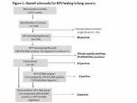
were more likely to complete lower levels of education, and ever smoke (Table 1). A schematic 1
representation of the tests performed in the retrospective studies is represented in figure 1. 2
HPV serology and lung cancer associations 3
Retrospective studies 4
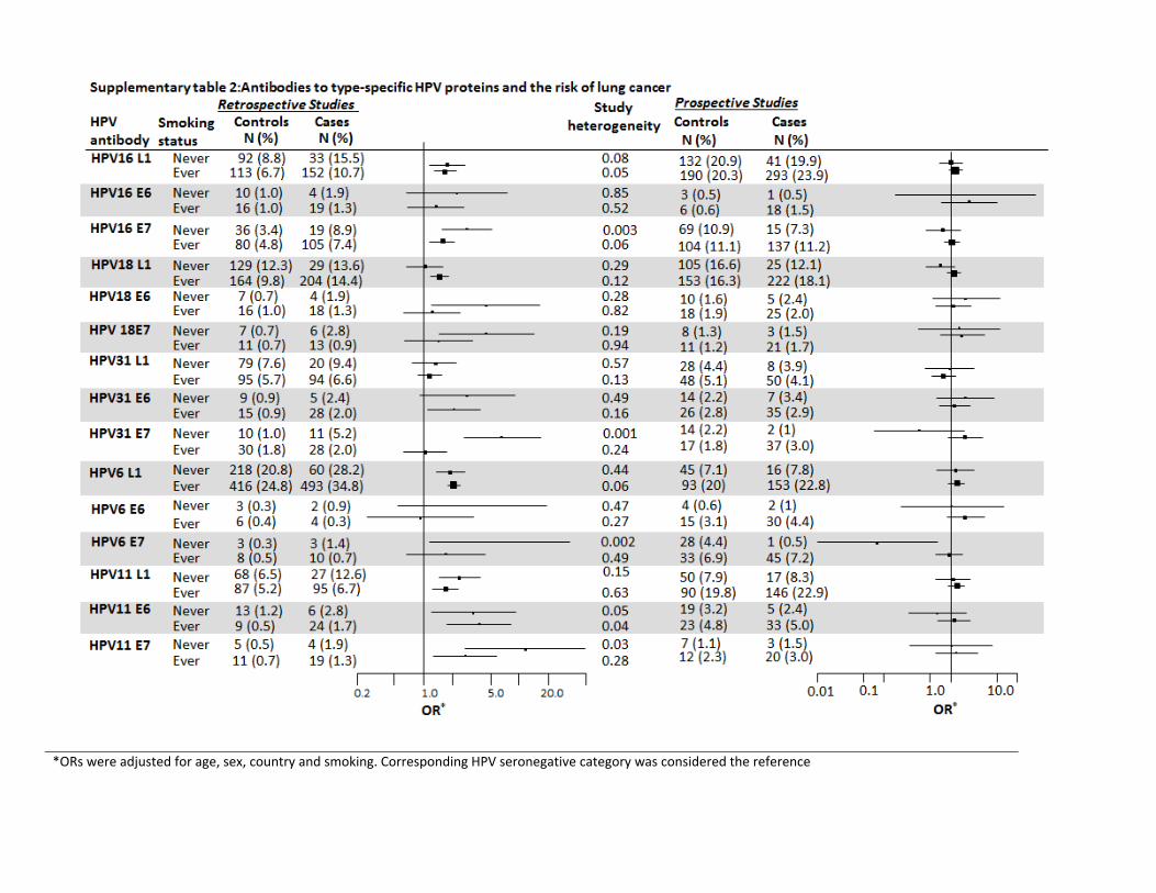
Combined results of the retrospective studies are presented (Table 2). Since early antibodies are 5
considered markers of HPV-related malignancy, they are expected to be rare. Consistent with this notion, 6
the prevalence of antibodies to early proteins (E6 & E7) was lower among controls compared to cases. 7
When were grouped based on their carcinogenic potential into any high risk, any low risk or any HPV 8
categories, seropositivity to most early and late antibodies was associated with increased risk of lung 9
cancer. Further, some type-specific antibodies were consistently associated with lung cancer, including 10
HPV16-L1, HPV16-E7, HPV18-L1, HPV18-E7, HPV31-E6, HPV6-L1, HPV11-L1, -E6 and -E7. We observed 11
directional consistency in the risk estimates of capsid and early protein antibodies by HPV-type (Table 2). 12
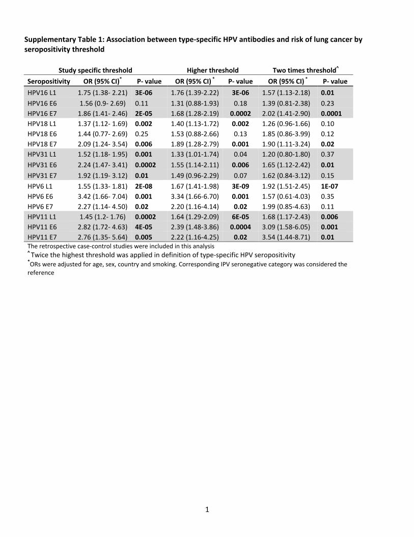
Irrespective of whether study-specific thresholds, higher cut-off of the two studies, or twice the highest 13
threshold was applied, the association between type-specific antibodies and the lung cancer risk 14
remained consistent (Supplementary table 1). We observed no effect modification by smoking status, 15
except for HPV31-E7 where a stronger association was observed among never smokers (OR: 6.59, 95% 16
CI: 2.59- 16.77) (Supplementary table 2). 17
Prospective study 18
None of the 15 candidate antibody markers tested was significantly associated with lung cancer risk in 19
the prospective design. Importantly, the lack of association between HPV16 E6 and HPV18 E6 antibodies 20
and lung cancer risk was also replicated (Table 2). 21
Tumor markers of HPV infection 22
Performance of quality indicators 23
13
Tumor testing was accompanied by inclusion of negative and positive controls at each step of the 1
multiple assays performed. These included negative controls for (i) sectioning where blank blocks were 2
processed 1 in every 10 tumor blocks, (ii) DNA extraction that included 1 tube per batch containing 3
buffer alone, (iii) PCR that consisted of PCR buffers and primers only, 1 per 96 samples, (iv) luminex that 4
included testing of 1 sample containing luminex beads alone. Together, these comprised of 42 controls in 5
the first genotyping round. Of these, 2 (of 12) DNA extraction controls were HPV DNA positive and 2 (of 6
22) sectioning controls were HPV DNA positive, indicating potential contamination in the respective 7
sample processing steps. In the second genotyping round that involved repetition of all the sample 8
preparation steps, none of the 34 controls incorporated were positive for any HPV DNA. In addition, all 9
tumor samples were positive for the betaglobin gene and ubiquitin expression as positive controls for 10
DNA and RNA quality respectively. 11
Based on the previously described definition of a HPV DNA-positive tumor, we identified 28 positives 12
(9.7%), 19 of which were positive for HPV16 DNA (6.6%) and 9 were positive for any non-HPV16 type 13
(3.1%). Non-HPV16 infections included single infections of HPV11 (n=6), HPV51 (n=1) and HPV58 (n=2). 14
Twenty three of 28 HPV DNA positive tumors were positive for a single HPV-type (n=23), all multiple 15
infections were double infections involving HPV16 (Table 3). In general, the presence of HPV-DNA 16
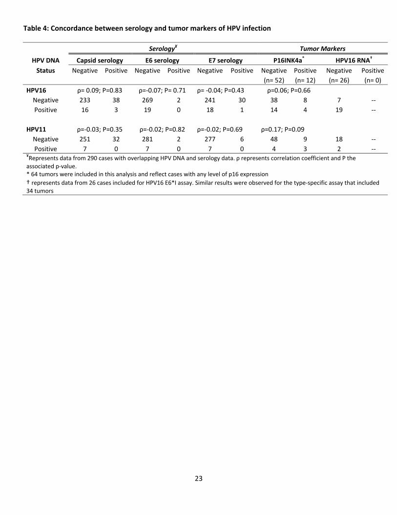
correlated poorly with the presence of corresponding type-specific antibodies (Table 4). Of the 19 HPV16 17
DNA-positive tumors, none were positive for HPV16 E6 antibodies, while 2 HPV16 DNA-negatives were 18
HPV16 E6 seropositive. Similarly, none of the 7 HPV11 DNA-positive tumors were HPV11 E6 seropositive, 19
while 2 HPV11 DNA-negative tumors were HPV11 E6 seropositive. Sixty four tumors were tested for p16 20
protein expression, 12 expressed low to medium levels (19%). Of these, four (1.4%) were also positive for 21
HPV16 DNA. Among the 52 tumors negative of p16 expression, 21 were HPV DNA-positive (14 HPV16 22
DNA-positive). Expression of p16 protein was also observed among 8 HPV16 DNA negative tumors 23
(2.8%), reflecting background p16 expression (Table 4). 24
14
In determining the causal association between HPV infection and lung cancer, we considered the 1
expression of HPV E6 mRNA as the gold standard. Based on the HPV16-specific assay, we found no 2
evidence for the expression of HPV16 E6 mRNA in the 26 tumors examined, 19 of which were HPV16 3
DNA-positive (Table 4). Further validation using the ultra-sensitive Luminex®-based assay capable of 4
detecting E6 mRNA of additional high and low risk HPV types confirmed the absence of not only HPV16 5
transcripts but also HPV31, HPV33, HPV56, HPV58 and HPV11 (data not shown). 6
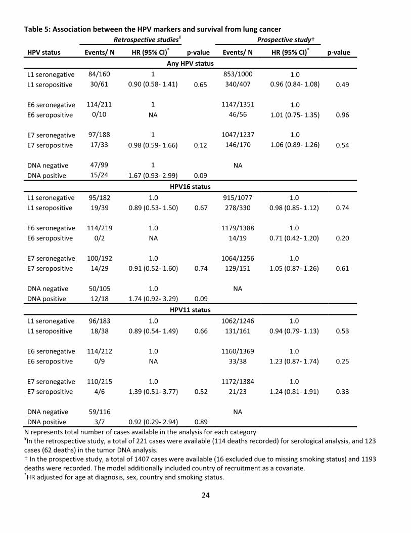
HPV infection and lung cancer survival 7
In the analysis of all-cause mortality, 221 cases with serology data were available in the L2 study and 114 8
deaths were recorded (87 from lung cancer). One hundred and twenty three cases were available in the 9
tumor analysis and 62 deaths were recorded. The longest follow-up time was 6.1 years and the median 10
follow-up time was 3.62 years. In the EPIC study, a total of 1423 cases were available and the maximum 11
follow-up time was 14.7 years during which 1203 deaths occurred. We observed no association between 12
the presence of any HPV antibodies or DNA and overall mortality. Individually, neither HPV16 or HPV11 13
antibodies, nor HPV16 or HPV11 DNA were associated with all-cause mortality following lung cancer 14
(Table 5). Further adjustment for stage at diagnosis did not change the results (data not shown). 15
Furthermore, none of the markers of HPV infection were associated with cause-specific mortality due to 16
lung cancer (data not shown). 17
18
Discussion 19
In this study, we found no evidence for a causal association between HPV infection and lung cancer. 20
Based on a stringent definition of HPV DNA-positivity we observed that 9.7% of lung cancers in Europe 21
were positive for any HPV type, 6.6% for HPV16 and 2.4% for HPV11. Though 1.4% (4 of 290) of the lung 22
tumors was positive for HPV16-DNA and p16 protein expression combined, no transcriptional activity of 23
the HPV16 E6 gene was observed, indicating inactive infections. 24

15
1
Serological methods that detect antibodies to HPV proteins provide an alternative method to test the 2
association between type-specific HPV infections and cancer. While capsid antibody positivity (L1) 3
indicates recent HPV infection, early antibodies (E6 and E7) are considered markers of an ongoing HPV-4
related malignancy (27, 28). Presence of HPV16 E6 antibodies in particular, are thought to be cancer 5
specific markers owing to their rarity among controls (<1% seropositivity in nearly 6500 controls based 6
on studies of head and neck cancer) (29-32). Conversely, HPV16 E6 appears to be a specific marker for 7
HPV16-positive oropharyngeal cancer (19). We applied HPV serology as a test in the retrospective and 8
prospective designs to address independent questions. In the retrospective studies, serological testing 9
was applied as an initial screen to examine the potential associations between type-specific HPV 10
antibodies and lung cancer and also determine whether HPV serology could predict the presence of the 11
corresponding HPV DNA in the tumor. The prospective analysis was conducted to examine the 12
temporality of the association and to determine if antibody markers may be detectable prior to clinically 13
apparent disease, as we recently observed for oropharyngeal cancer (19). In the interpretation of the 14
serological associations, we considered: (i) the consistency of antibody prevalence across the studies, (ii) 15
directional consistency within a study between capsid and early antibodies by HPV-type, (iii) potential 16
effect modification by smoking status, and (iv) sensitivity of the results to seropositivity thresholds. In 17
this study, the association between type-specific HPV antibodies and lung cancer appeared to differ by 18
study design. It remains possible that the apparent difference by study design could be attributed to 19
false findings or cancer-specific immune changes in the retrospective cases. However, it is important to 20
note that consistently no association was found for HPV16 E6 seropositivity, considered a cancer-specific 21
marker and also the HPV type with the strongest a priori hypothesis. It is important to mention that the 22
observations in the retrospective studies may be interpreted independently given the strengths of 23
comprehensive HPV evaluation. Here, although nearly 7% of lung tumors were consistently HPV16 DNA-24
16
positive, no expression of HPV16 E6 mRNA was observed, with two different methodologies applied. 1
Furthermore, based on the criteria previously outlined, though some HPV antibodies (HPV11 and HPV31 2
E6), appeared to be consistently associated with increased risk of lung cancer, no correlation with tumor 3
HPV DNA presence was observed. Further, no functional consequence of viral presence could be 4
established. 5
6
This study raises several questions regarding the published associations between HPV and lung cancer. 7
First, despite extensive efforts taken to avoid contamination and following a stringent definition for HPV 8
DNA positivity, we observed that 28 lung tumors consistently tested positive for the same type (of the 48 9
initial positives, nearly 58%). The high proportion of false positivity may explain the large variation in HPV 10
DNA prevalence reported within the same country, such as the observed range of 0-78% in Japan (33, 34) 11
and 12-55% in China (35, 36). Second, some of the regional difference in HPV prevalence has been 12
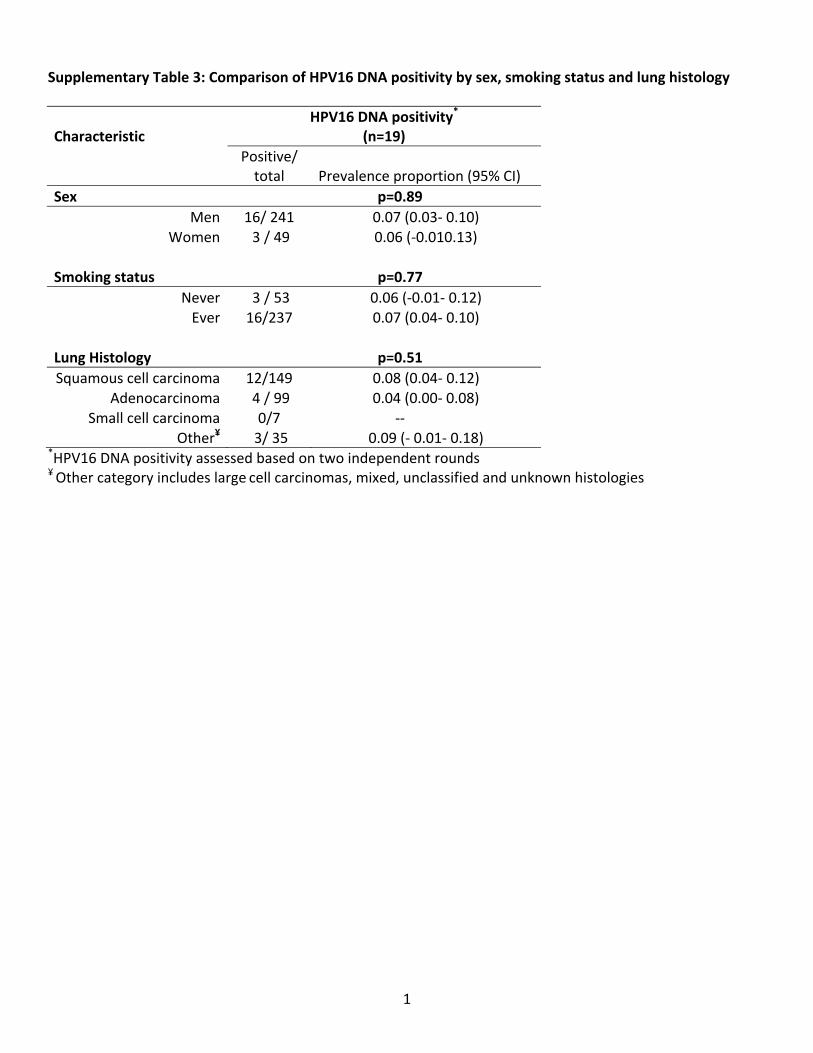
attributed to varying proportions of non-smoking women and lung histologies (37). In this study, we 13
observed that the proportion of HPV16 DNA-positivity was comparable between smoking men (7%) and 14
non-smoking women (7.5%) as well as across different lung cancer histologies (Supplementary table 3). 15
Third, this study demonstrates that although nearly 10% of lung tumors may be consistently HPV DNA-16
positive, none expressed viral oncoproteins, emphasizing that HPV DNA (and/or p16) is insufficient to 17
establish a causal role for HPV infection in lung carcinogenesis. Our conclusions are supported by the 18
only study reported to date that included a large series of lung tumors and took adequate precautions to 19
avoid potential contamination (7). Based on this evidence we suggest that future publications reporting 20
HPV DNA prevalence in lung cancer should: (i) include and describe precautions taken to avoid potential 21
contamination, and (ii) address the transcriptional state of the HPV genome. 22
23

17
In conclusion, using multiple markers, extensive precautions against potential contamination and state-1
of-the-art HPV detection methods, we demonstrate that several high and low risk HPV types are present 2
in the lung tissue and can be detected with varying level of evidence, but are unlikely to contribute 3
causally to lung carcinogenesis. These data do not support a causal role for HPV infections in lung cancer. 4
5

18
References 1 2 1. Ferlay J, Shin HR, Bray F, Forman D, Mathers C, Parkin DM. Estimates of worldwide burden of cancer in
2008: GLOBOCAN 2008. Int J Cancer. 2010;127:2893-917. 2. Parkin DM, Bray F, Ferlay J, Pisani P. Global cancer statistics, 2002. CA Cancer J Clin. 2005;55:74-108. 3. Sun S, Schiller JH, Gazdar AF. Lung cancer in never smokers--a different disease. Nat Rev Cancer.
2007;7:778-90. 4. Human papillomaviruses. IARC Monogr Eval Carcinog Risks Hum. 2007;90:1-636. 5. de Martel C, Ferlay J, Franceschi S, Vignat J, Bray F, Forman D, et al. Global burden of cancers attributable
to infections in 2008: a review and synthetic analysis. Lancet Oncol. 2012;13:607-15. 6. Srinivasan M, Taioli E, Ragin CC. Human papillomavirus type 16 and 18 in primary lung cancers--a meta-
analysis. Carcinogenesis. 2009;30:1722-8. 7. Koshiol J, Rotunno M, Gillison ML, Van Doorn LJ, Chaturvedi AK, Tarantini L, et al. Assessment of human
papillomavirus in lung tumor tissue. J Natl Cancer Inst. 2011;103:501-7. 8. Wang JL, Fang CL, Wang M, Yu MC, Bai KJ, Lu PC, et al. Human papillomavirus infections as a marker to
predict overall survival in lung adenocarcinoma. Int J Cancer. 2014;134:65-71. 9. Herrero R. Chapter 7: Human papillomavirus and cancer of the upper aerodigestive tract. Journal of the
National Cancer Institute Monographs. 2003:47-51. 10. Kashima HK, Shah F, Lyles A, Glackin R, Muhammad N, Turner L, et al. A comparison of risk factors in
juvenile-onset and adult-onset recurrent respiratory papillomatosis. Laryngoscope. 1992;102:9-13. 11. Shykhon M, Kuo M, Pearman K. Recurrent respiratory papillomatosis. Clin Otolaryngol Allied Sci.
2002;27:237-43. 12. Li G, He L, Zhang E, Shi J, Zhang Q, Le AD, et al. Overexpression of human papillomavirus (HPV) type 16
oncoproteins promotes angiogenesis via enhancing HIF-1alpha and VEGF expression in non-small cell lung cancer cells. Cancer Lett. 2011;311:160-70.
13. Willey JC, Broussoud A, Sleemi A, Bennett WP, Cerutti P, Harris CC. Immortalization of normal human bronchial epithelial cells by human papillomaviruses 16 or 18. Cancer Res. 1991;51:5370-7.
14. Aguayo F, Castillo A, Koriyama C, Higashi M, Itoh T, Capetillo M, et al. Human papillomavirus-16 is integrated in lung carcinomas: a study in Chile. Br J Cancer. 2007;97:85-91.
15. Cheng YW, Wu MF, Wang J, Yeh KT, Goan YG, Chiou HL, et al. Human papillomavirus 16/18 E6 oncoprotein is expressed in lung cancer and related with p53 inactivation. Cancer Res. 2007;67:10686-93.
16. Coissard CJ, Besson G, Polette MC, Monteau M, Birembaut PL, Clavel CE. Prevalence of human papillomaviruses in lung carcinomas: a study of 218 cases. Mod Pathol. 2005;18:1606-9.
17. Scelo G, Constantinescu V, Csiki I, Zaridze D, Szeszenia-Dabrowska N, Rudnai P, et al. Occupational exposure to vinyl chloride, acrylonitrile and styrene and lung cancer risk (europe). Cancer Causes Control. 2004;15:445-52.
18. Riboli E, Kaaks R. The EPIC Project: rationale and study design. European Prospective Investigation into Cancer and Nutrition. Int J Epidemiol. 1997;26 Suppl 1:S6-14.
19. Kreimer AR, Johansson M, Waterboer T, Kaaks R, Chang-Claude J, Drogen D, et al. Evaluation of human papillomavirus antibodies and risk of subsequent head and neck cancer. J Clin Oncol. 2013;31:2708-15.
20. Waterboer T, Sehr P, Michael KM, Franceschi S, Nieland JD, Joos TO, et al. Multiplex human papillomavirus serology based on in situ-purified glutathione s-transferase fusion proteins. Clin Chem. 2005;51:1845-53.
21. Clifford GM, Shin HR, Oh JK, Waterboer T, Ju YH, Vaccarella S, et al. Serologic response to oncogenic human papillomavirus types in male and female university students in Busan, South Korea. Cancer Epidemiol Biomarkers Prev. 2007;16:1874-9.
22. Michael KM, Waterboer T, Sehr P, Rother A, Reidel U, Boeing H, et al. Seroprevalence of 34 human papillomavirus types in the German general population. PLoS Pathog. 2008;4:e1000091.
19
23. Gheit T, Landi S, Gemignani F, Snijders PJ, Vaccarella S, Franceschi S, et al. Development of a sensitive and specific assay combining multiplex PCR and DNA microarray primer extension to detect high-risk mucosal human papillomavirus types. J Clin Microbiol. 2006;44:2025-31.
24. Schmitt M, Bravo IG, Snijders PJ, Gissmann L, Pawlita M, Waterboer T. Bead-based multiplex genotyping of human papillomaviruses. J Clin Microbiol. 2006;44:504-12.
25. Smeets SJ, Hesselink AT, Speel EJ, Haesevoets A, Snijders PJ, Pawlita M, et al. A novel algorithm for reliable detection of human papillomavirus in paraffin embedded head and neck cancer specimen. Int J Cancer. 2007;121:2465-72.
26. Halec G, Schmitt M, Dondog B, Sharkhuu E, Wentzensen N, Gheit T, et al. Biological activity of probable/possible high-risk human papillomavirus types in cervical cancer. Int J Cancer. 2013;132:63-71.
27. Clifford GM, Gallus S, Herrero R, Munoz N, Snijders PJ, Vaccarella S, et al. Worldwide distribution of human papillomavirus types in cytologically normal women in the International Agency for Research on Cancer HPV prevalence surveys: a pooled analysis. Lancet. 2005;366:991-8.
28. Herrero R, Castellsague X, Pawlita M, Lissowska J, Kee F, Balaram P, et al. Human papillomavirus and oral cancer: the International Agency for Research on Cancer multicenter study. J Natl Cancer Inst. 2003;95:1772-83.
29. Anantharaman D, Gheit T, Waterboer T, Abedi-Ardekani B, Carreira C, McKay-Chopin S, et al. Human papillomavirus infections and upper aero-digestive tract cancers: the ARCAGE study. J Natl Cancer Inst. 2013;105:536-45.
30. D'Souza G, Kreimer AR, Viscidi R, Pawlita M, Fakhry C, Koch WM, et al. Case-control study of human papillomavirus and oropharyngeal cancer. N Engl J Med. 2007;356:1944-56.
31. Gillison ML, D'Souza G, Westra W, Sugar E, Xiao W, Begum S, et al. Distinct risk factor profiles for human papillomavirus type 16-positive and human papillomavirus type 16-negative head and neck cancers. J Natl Cancer Inst. 2008;100:407-20.
32. Ribeiro KB, Levi JE, Pawlita M, Koifman S, Matos E, Eluf-Neto J, et al. Low human papillomavirus prevalence in head and neck cancer: results from two large case-control studies in high-incidence regions. Int J Epidemiol. 2011;40:489-502.
33. Szabo I, Sepp R, Nakamoto K, Maeda M, Sakamoto H, Uda H. Human papillomavirus not found in squamous and large cell lung carcinomas by polymerase chain reaction. Cancer. 1994;73:2740-4.
34. Tsuhako K, Nakazato I, Hirayasu T, Sunakawa H, Iwamasa T. Human papillomavirus DNA in adenosquamous carcinoma of the lung. J Clin Pathol. 1998;51:741-9.
35. Da J, Chen L, Hu Y. [Human papillomavirus infection and p53 gene mutation in primary lung cancer]. Zhonghua Zhong Liu Za Zhi. 1996;18:27-9.
36. Zhang X, Zhu Y, Li L. [Point mutation of p53 and detection of human papillomavirus DNA in bronchogenic carcinoma]. Zhonghua Nei Ke Za Zhi. 1995;34:673-5.
37. Cheng YW, Chiou HL, Sheu GT, Hsieh LL, Chen JT, Chen CY, et al. The association of human papillomavirus 16/18 infection with lung cancer among nonsmoking Taiwanese women. Cancer Res. 2001;61:2799-803.
20
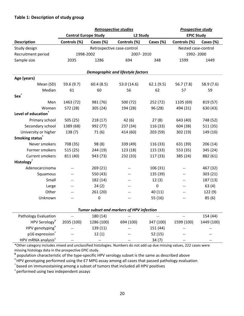
Table 1: Description of study group
Retrospective studies Prospective study Central Europe Study L2 Study EPIC Study
Description Controls (%) Cases (%) Controls (%) Cases (%) Controls (%) Cases (%)Study design Retrospective case-control Nested case-controlRecruitment period 1998-2002 2007- 2010 1992- 2000Sample size 2035 1286 694 348 1599 1449
Demographic and lifestyle factorsAge (years)
Mean (SD) 59.6 (9.7) 60.4 (8.5) 53.0 (14.6) 62.1 (9.5) 56.7 (7.8) 58.9 (7.6)Median 61 60 56 62 57 59
Sex* Men 1463 (72) 981 (76) 500 (72) 252 (72) 1105 (69) 819 (57)
Women 572 (28) 305 (24) 194 (28) 96 (28) 494 (31) 630 (43)Level of education*
Primary school 505 (25) 218 (17) 42 (6) 27 (8) 643 (40) 748 (52)Secondary school 1389 (68) 992 (77) 237 (34) 116 (33) 604 (38) 511 (35)
University or higher 138 (7) 71 (6) 414 (60) 203 (59) 302 (19) 149 (10)Smoking status*
Never smokers 708 (35) 98 (8) 339 (49) 116 (33) 631 (39) 206 (14)Former smokers 515 (25) 244 (19) 123 (18) 115 (33) 553 (35) 345 (24)Current smokers 811 (40) 943 (73) 232 (33) 117 (33) 385 (24) 882 (61)
Histology* Adenocarcinoma -- 269 (21) -- 106 (31) -- 467 (32)
Squamous -- 550 (43) -- 135 (39) -- 303 (21)Small -- 182 (14) -- 12 (3) -- 187 (13)Large -- 24 (2) -- 0 -- 63 (4)Other -- 261 (20) -- 40 (11) -- 122 (9)
Unknown -- 0 -- 55 (16) -- 85 (6)
Tumor subset and markers of HPV infectionPathology Evaluation -- 180 (14) -- -- -- 154 (44)
HPV Serology¥ 2035 (100) 1286 (100) 694 (100) 347 (100) 1599 (100) 1449 (100)HPV genotyping‡ -- 139 (11) -- 151 (44) -- --p16 expression† -- 12 (1) -- 52 (15) -- --
HPV mRNA analysis± -- -- -- 34 (7) -- --*Other category includes mixed and unclassified histologies. Numbers do not add up due missing values, 222 cases were missing histology data in the prospective EPIC study. ¥ population characteristic of the type-specific HPV serology subset is the same as described above ‡ HPV genotyping performed using the E7 MPG assay among all cases that passed pathology evaluation
† based on immunostaining among a subset of tumors that included all HPV positives ± performed using two independent assays

21
Table 2: Antibodies to type-specific HPV proteins and the risk of lung cancer
^ Central Europe and L2 studies were combined, thresholds from the prospective study were used to define HPV seropositivity. N represents the number of cases *ORs were adjusted for age, sex, smoking status (never, former and current) and country, corresponding HPV negative group was considered the reference category

22
Table 3: HPV DNA positives identified by genotyping
HPV Genotyping HPV DNA Status Round 1* Round 2† N (%)¥ N (%) Negative 242 (83) 37 Positive 48 (17) 28 (9.7) Single infections 34 (11.7) 23 (7.9) HPV16 24 (12) 14 (4.8) HPV31 1 (0.3) - HPV33 3 (1.0) - HPV51 - 1 (0.3) HPV58 2 (0.6) 2 (0.7) HPV11 4 (1.4) 6 (2.1) Multiple infections^ 14 (4.8) 5 (1.7) HPV16 11 (3.8) 5 (1.7) HPV31 5 (1.7) 2 (0.7) HPV33 1 (0.3) 1 (0.3) HPV51 1 (0.3) - HPV56 1 (0.3) 1 (0.3) HPV11 7 (2.4) 1 (0.3) *All 290 tumors that passed pathology were included in the first round of HPV genotyping †confirmation round included 48 HPV DNA positive tumors and 16 HPV DNA negative tumors were included in the confirmation stage ^ Multiple HPV infections do not add up to total since certain HPV types appear more than once ¥Percentage positivity is based on the entire series of 290 tumors tested
23
Table 4: Concordance between serology and tumor markers of HPV infection
Serology¥ Tumor Markers
HPV DNA Capsid serology E6 serology E7 serology P16INK4a* HPV16 RNA†
Status Negative Positive Negative Positive Negative Positive Negative Positive Negative Positive (n= 52) (n= 12) (n= 26) (n= 0)
HPV16 ρ= 0.09; P=0.83 ρ=-0.07; P= 0.71 ρ= -0.04; P=0.43 ρ=0.06; P=0.66 Negative 233 38 269 2 241 30 38 8 7 --Positive 16 3 19 0 18 1 14 4 19 --
HPV11 ρ=-0.03; P=0.35 ρ=-0.02; P=0.82 ρ=-0.02; P=0.69 ρ=0.17; P=0.09 Negative 251 32 281 2 277 6 48 9 18 --Positive 7 0 7 0 7 0 4 3 2 --
¥Represents data from 290 cases with overlapping HPV DNA and serology data. ρ represents correlation coefficient and P the associated p-value. * 64 tumors were included in this analysis and reflect cases with any level of p16 expression † represents data from 26 cases included for HPV16 E6*I assay. Similar results were observed for the type-specific assay that included 34 tumors
24
Table 5: Association between the HPV markers and survival from lung cancer Retrospective studies¥ Prospective study†
HPV status Events/ N HR (95% CI)* p-value Events/ N HR (95% CI)* p-value
Any HPV status
L1 seronegative 84/160 1 853/1000 1.0 L1 seropositive 30/61 0.90 (0.58- 1.41) 0.65 340/407 0.96 (0.84- 1.08) 0.49 E6 seronegative 114/211 1 1147/1351 1.0 E6 seropositive 0/10 NA 46/56 1.01 (0.75- 1.35) 0.96 E7 seronegative 97/188 1 1047/1237 1.0 E7 seropositive 17/33 0.98 (0.59- 1.66) 0.12 146/170 1.06 (0.89- 1.26) 0.54 DNA negative 47/99 1 NA DNA positive 15/24 1.67 (0.93- 2.99) 0.09
HPV16 status
L1 seronegative 95/182 1.0 915/1077 1.0 L1 seropositive 19/39 0.89 (0.53- 1.50) 0.67 278/330 0.98 (0.85- 1.12) 0.74
E6 seronegative 114/219 1.0 1179/1388 1.0 E6 seropositive 0/2 NA 14/19 0.71 (0.42- 1.20) 0.20
E7 seronegative 100/192 1.0 1064/1256 1.0 E7 seropositive 14/29 0.91 (0.52- 1.60) 0.74 129/151 1.05 (0.87- 1.26) 0.61
DNA negative 50/105 1.0 NADNA positive 12/18 1.74 (0.92- 3.29) 0.09
HPV11 status
L1 seronegative 96/183 1.0 1062/1246 1.0 L1 seropositive 18/38 0.89 (0.54- 1.49) 0.66 131/161 0.94 (0.79- 1.13) 0.53
E6 seronegative 114/212 1.0 1160/1369 1.0 E6 seropositive 0/9 NA 33/38 1.23 (0.87- 1.74) 0.25
E7 seronegative 110/215 1.0 1172/1384 1.0 E7 seropositive 4/6 1.39 (0.51- 3.77) 0.52 21/23 1.24 (0.81- 1.91) 0.33 DNA negative 59/116 NA DNA positive 3/7 0.92 (0.29- 2.94) 0.89
N represents total number of cases available in the analysis for each category ¥In the retrospective study, a total of 221 cases were available (114 deaths recorded) for serological analysis, and 123 cases (62 deaths) in the tumor DNA analysis. † In the prospective study, a total of 1407 cases were available (16 excluded due to missing smoking status) and 1193 deaths were recorded. The model additionally included country of recruitment as a covariate. *HR adjusted for age at diagnosis, sex, country and smoking status.

22
Figure legends Figure 1: Overall schematic for HPV testing in lung cancers Figure1 legends: †Same cases as for HPV genotyping round 2 were included for p16 testing. Positives indicate p16 expressing tumors. Of the 12 p16 positives, 2 were HPV16 DNA positive, 2 HPV11 positive, 1 HPV16 & 31 DNA positive and 1 HPV11 & HPV16 positive, and 6 HPV DNA negatives ¥Included all 19 HPV16 DNA positive tumors and randomly selected HPV 16 DNA negatives ‡ included all 28 HPV DNA positives and 6 HPV DNA negatives
1
Supplementary Table 1: Association between type-specific HPV antibodies and risk of lung cancer by seropositivity threshold
Study specific threshold Higher threshold Two times threshold^
Seropositivity OR (95% CI)* P- value OR (95% CI) * P- value OR (95% CI) * P- value
HPV16 L1 1.75 (1.38- 2.21) 3E-06 1.76 (1.39-2.22) 3E-06 1.57 (1.13-2.18) 0.01
HPV16 E6 1.56 (0.9- 2.69) 0.11 1.31 (0.88-1.93) 0.18 1.39 (0.81-2.38) 0.23HPV16 E7 1.86 (1.41- 2.46) 2E-05 1.68 (1.28-2.19) 0.0002 2.02 (1.41-2.90) 0.0001HPV18 L1 1.37 (1.12- 1.69) 0.002 1.40 (1.13-1.72) 0.002 1.26 (0.96-1.66) 0.10HPV18 E6 1.44 (0.77- 2.69) 0.25 1.53 (0.88-2.66) 0.13 1.85 (0.86-3.99) 0.12HPV18 E7 2.09 (1.24- 3.54) 0.006 1.89 (1.28-2.79) 0.001 1.90 (1.11-3.24) 0.02HPV31 L1 1.52 (1.18- 1.95) 0.001 1.33 (1.01-1.74) 0.04 1.20 (0.80-1.80) 0.37HPV31 E6 2.24 (1.47- 3.41) 0.0002 1.55 (1.14-2.11) 0.006 1.65 (1.12-2.42) 0.01
HPV31 E7 1.92 (1.19- 3.12) 0.01 1.49 (0.96-2.29) 0.07 1.62 (0.84-3.12) 0.15HPV6 L1 1.55 (1.33- 1.81) 2E-08 1.67 (1.41-1.98) 3E-09 1.92 (1.51-2.45) 1E-07HPV6 E6 3.42 (1.66- 7.04) 0.001 3.34 (1.66-6.70) 0.001 1.57 (0.61-4.03) 0.35HPV6 E7 2.27 (1.14- 4.50) 0.02 2.20 (1.16-4.14) 0.02 1.99 (0.85-4.63) 0.11HPV11 L1 1.45 (1.2- 1.76) 0.0002 1.64 (1.29-2.09) 6E-05 1.68 (1.17-2.43) 0.006HPV11 E6 2.82 (1.72- 4.63) 4E-05 2.39 (1.48-3.86) 0.0004 3.09 (1.58-6.05) 0.001HPV11 E7 2.76 (1.35- 5.64) 0.005 2.22 (1.16-4.25) 0.02 3.54 (1.44-8.71) 0.01The retrospective case-control studies were included in this analysis^ Twice the highest threshold was applied in definition of type-specific HPV seropositivity *ORs were adjusted for age, sex, country and smoking. Corresponding IPV seronegative category was considered the reference
*ORs were adjusted for age, sex, country and smoking. Corresponding HPV seronegative category was considered the reference
1
Supplementary Table 3: Comparison of HPV16 DNA positivity by sex, smoking status and lung histology
Characteristic HPV16 DNA positivity*
(n=19) Positive/
total Prevalence proportion (95% CI) Sex p=0.89
Men 16/ 241 0.07 (0.03- 0.10) Women 3 / 49 0.06 (-0.010.13)
Smoking status p=0.77 Never 3 / 53 0.06 (-0.01- 0.12)
Ever 16/237 0.07 (0.04- 0.10)
Lung Histology p=0.51 Squamous cell carcinoma 12/149 0.08 (0.04- 0.12)
Adenocarcinoma 4 / 99 0.04 (0.00- 0.08) Small cell carcinoma 0/7 --
Other¥ 3/ 35 0.09 (- 0.01- 0.18) *HPV16 DNA positivity assessed based on two independent rounds ¥ Other category includes large cell carcinomas, mixed, unclassified and unknown histologies