NIRx Newsletter Issue #1 – May 2017
15
~ Return to Top ~ NIRx Newsletter Issue #1 – May 2017 1 Dear friends, We are very pleased with the growth NIRx has seen since its inception in 2000. With our first official Newsletter, I am pleased to share with you some of the exciting updates to our technology platform! Through our hard work and dedicated team effort, we are consistently releasing new innovative hardware and software at an unmatched pace. I thank you all - and especially our end- users - for your time and commitment to furthering the fNIRS field. We look forward to working together to advance this technology through a diverse range of applications. Sincerely, Richard Barbour NIRx Interim CEO Message From The CEO 2 Upcoming NIRx Events 2 Event Highlight: NIRx BCI Webinar By popular demand, NIRx will be offering its first webinar on real-time fNIRS processing. 3 User Spotlight Prof. Niels Birbaumer’s team from The University of Tübingen receives global recognition for ALS research using fNIRS. 4 Recent Publications See some of the latest publications using NIRx systems. 5 Software Release: NIRStar 14-3 Learn about the release of NIRx’s system control software, NIRStar. 6 Software Release: nirsLAB 2017-04 Learn about the release of the nirsLAB fNIRS analysis software, which may be used with nearly any fNIRS system. 7 NIRx’s New Dedicated Support Website Learn about this great new resource and how our customers can register. 8 fNIRS Detectors: SiPD vs. APD Get an overview of fNIRS detectors, including the difference between SiPDs and APDs. 10 Laser and LED Sources from NIRx One of our most-asked questions is regarding the difference between Laser and LED sources, and why we offer both. Well, you can read about it here to find out. 14 Career Opportunities at NIRx: Scientific Consultants We are looking for Scientific Consultants to work in our office in U.S.A. and Germany! Contents
-
Upload
khangminh22 -
Category
Documents
-
view
1 -
download
0
Transcript of NIRx Newsletter Issue #1 – May 2017

~ Return to Top ~
NIRx Newsletter Issue #1 – May 2017
1
Dear friends,
We are very pleased with the growth NIRx has
seen since its inception in 2000. With our
first official Newsletter, I am pleased to
share with you some of the exciting updates
to our technology platform! Through our
hard work and dedicated team effort, we are
consistently releasing new innovative
hardware and software at an unmatched pace.
I thank you all - and especially our end-
users - for your time and commitment to
furthering the fNIRS field. We look forward
to working together to advance this
technology through a diverse range of
applications.
Sincerely,
Richard Barbour
NIRx Interim CEO
Message From The CEO
2 Upcoming NIRx Events
2 Event Highlight: NIRx BCI Webinar By popular demand, NIRx will be offering its first webinar on
real-time fNIRS processing.
3 User Spotlight Prof. Niels Birbaumer’s team from The University of Tübingen
receives global recognition for ALS research using fNIRS.
4 Recent Publications See some of the latest publications using NIRx systems.
5 Software Release: NIRStar 14-3 Learn about the release of NIRx’s system control software,
NIRStar.
6 Software Release: nirsLAB 2017-04 Learn about the release of the nirsLAB fNIRS analysis software,
which may be used with nearly any fNIRS system.
7 NIRx’s New Dedicated Support Website Learn about this great new resource and how our customers can
register.
8 fNIRS Detectors: SiPD vs. APD Get an overview of fNIRS detectors, including the difference
between SiPDs and APDs.
10 Laser and LED Sources from NIRx One of our most-asked questions is regarding the difference
between Laser and LED sources, and why we offer both. Well, you
can read about it here to find out.
14 Career Opportunities at NIRx: Scientific Consultants We are looking for Scientific Consultants to work in our office
in U.S.A. and Germany!
Contents

~ Return to Top ~
NIRx Newsletter Issue #1 – May 2017
2
NIRx BCI Webinar June 8th, 2017
Due to the increasing interest
of BCI researchers for the
fNIRS technology, NIRx has
decided to host a webinar on
the topic to be held on the
8th of June. We are extremely
pleased to announce Dr. Ujwal
Chaudhary and Dr. Bettina
Sorger as speakers. More
information on the event is
available here or on our
website.
Read more on the work of Dr.
Chaudhary, Lead Research
Scientist, and his Principal
Investigator, Prof. Niels
Birbaumer, in this month’s end-
user spotlight.
Event Highlight
NIRx BCI Webinar
June 8th
NIRx hosted event (right page for
details)
Organization for Human Brain Mapping
Conference
Vancouver, BC Canada
June 25-29,
Sponsor and Exhibitor
Lancaster International Conference on
Infant and Early Child Development
Lancaster, UK
August 23-25 2017
Sponsor and Exhibitor
Graz Brain-Computer Interface Conference
Graz, Austria
September 18th - September 22
Sponsor and Exhibitor
Mexican Symposium on NIRS Neuroimaging
(MEXNIRS)
Puebla, México
October 21-22 2017
Sponsor and Exhibitor
The Society for Neuroscience
Washington, DC USA
November 11 - November 15
Sponsor and Exhibitor
Real-Time Functional Imaging and
Neurofeedback
Nara, Japan
November 29- December
Sponsor and Exhibitor
Upcoming NIRx Events
Go to our Events Page for the latest on NIRx conferences,
workshops, webinars and other special events!

~ Return to Top ~
NIRx Newsletter Issue #1 – May 2017
3
Prof. Niels Birbaumer of The University of Tübingen Receives Recognition for fNIRS/EEG BCI Research with
ALS Patients
NIRx is extremely proud for being part of the
groundbreaking research of the team of Prof. Niels
Birbaumer, from the University of Tübingen (Germany).
The work published in the journal of PLUS Biology [1]
marks 25 years of their continuous work with completely
locked-in ALS patients. The group has been successful in
using fNIRS to open a communication channel with
patients who have no other means of interacting with the
external world. Patients were able to learn to modulate
their cortical activity in order to answer to yes/no
questions with an incredible 70% correct response rate.
NIRx has always firmly believed in the enormous
potential of fNIRS but it is extremely heartwarming and
encouraging to see that our technology truly helps
improving people’s lives. The work of the group of Prof.
Birbaumer has caught the attention of worldwide media
and their results have been presented on CNN, BBC, The
Guardian and Der Spiegel among others. Prof. Birbaumer,
Dr. Ujwal Chaudhary (lead researcher) and their team are
happy to answer questions related to their work on their
Reddit thread.
By: Lamija Pašalić
Support Director
[1] “Brain–Computer Interface–Based Communication in
the Completely Locked-In State.” Chaudhary et al. PLoS
Biol 15 (1), e1002593. 2017 Jan 31.
NIRx End-User Spotlight
Prof. Niels Birbaumer
Principal Investigator
Dr. Ujwal Chaudhary
Lead Research Scientist
From [1]: “Fig 2. Classification accuracy of Patient F.
Linear SVM CA across “training sessions—offline CA” (histogram in grey),
“feedback sessions—online CA” (green dot), and “open question session—online CA”
(plus sign in red), obtained using (A) relative change in O2Hb, (B) EEG, and (C)
EOG data. The classification accuracy reported here is daywise, as all the
“training sessions” in a day were used to calculate the average classification
accuracy of all the “training sessions” in a day. In the figure panels A, B, and
C, the x-axis is the number of days and the y-axis is the classification
accuracy. The solid black and dotted horizontal lines represent the chance-level
threshold calculated using the metric described in the BCI effectiveness metric
section for “training sessions” and “feedback sessions,” respectively. Since the
feedback during the feedback and open question sessions was provided using the
O2Hb, the online CA of the feedback and open question sessions is reported only
for the fNIRS data “

~ Return to Top ~
NIRx Newsletter Issue #1 – May 2017
4
Don’t see your recent publication listed? Have a publication coming out soon? Please update us on your work!
We love to hear from our customers (especially
when there is good news )! By keeping us
informed on your latest publications, posters,
presentations, and press/media (e.g., news
articles, blog posts, videos, etc.) we are able
to better serve both you and our community. We
would be happy to consider highlighting you and
your team on one of our upcoming newsletters, our
social media page, or our website as well.
Please send all research-related updates to
[email protected]. Thank you for contributing to
the NIRx community!
A. Galderisi et al., “Long-term
continuous monitoring of the preterm
brain with diffuse optical tomography
and electroencephalography: a
technical note on cap manufacturing,”
Neurophoton, vol. 3, no. 4, pp.
045009–045009, 2016.
J. Shin et al., “Open Access Dataset
for EEG+NIRS Single-Trial
Classification,” IEEE Transactions on
Neural Systems and Rehabilitation
Engineering, vol. PP, no. 99, pp. 1–1,
2016.
L. Pollonini, H. Bortfeld, and J. S.
Oghalai, “PHOEBE: a method for real
time mapping of optodes-scalp coupling
in functional near-infrared
spectroscopy,” Biomed. Opt. Express,
BOE, vol. 7, no. 12, pp. 5104–5119,
Dec. 2016.
M. Balconi and M. E. Vanutelli,
“Interbrains cooperation: Hyperscanning and
self-perception in joint actions,” Journal
of Clinical and Experimental
Neuropsychology, vol. 0, no. 0, pp. 1–14,
Nov. 2016.
J. Shin, K.-R. Müller, and H.-J. Hwang,
“Near-infrared spectroscopy (NIRS)-based
eyes-closed brain-computer interface (BCI)
using prefrontal cortex activation due to
mental arithmetic,” Scientific Reports,
vol. 6, p. 36203, Nov. 2016.
A. Vrana, M. L. Meier, S. Hotz-
Boendermaker, B. K. Humphreys, and F.
Scholkmann, “Cortical Sensorimotor
Processing of Painful Pressure in Patients
with Chronic Lower Back Pain—An Optical
Neuroimaging Study using fNIRS,” Front.
Hum. Neurosci., vol. 10, 2016.
L.-C. Chen, M. Stropahl, M. Schönwiesner,
and S. Debener, “Enhanced visual adaptation
in cochlear implant users revealed by
concurrent EEG-fNIRS,” Neuroimage, Sep.
2016.
L. Holper, E. Seifritz, and F. Scholkmann,
“Short-term pulse rate variability is
better characterized by functional near-
infrared spectroscopy than by
photoplethysmography,” J Biomed Opt, vol.
21, no. 9, p. 91308, Sep. 2016.
H. O. Keles, R. L. Barbour, and A.
Omurtag, “Hemodynamic correlates of
spontaneous neural activity measured by
human whole-head resting state EEG+fNIRS,”
Neuroimage, vol. 138, pp. 76–87, Sep.
2016.
A. Vrana, M. L. Meier, S. Hotz-
Boendermaker, B. K. Humphreys, and F.
Scholkmann, “Different mechanosensory
stimulations of the lower back elicit
specific changes in hemodynamics and
oxygenation in cortical sensorimotor
areas—A fNIRS study,” Brain Behav, p. n/a-
n/a, Sep. 2016.
I. Helmich, A. Berger, and H. Lausberg,
“Neural Control of Posture in Individuals
with Persisting Postconcussion Symptoms,”
Med Sci Sports Exerc, Jul. 2016.
Recent Publications with NIRx Systems

~ Return to Top ~
NIRx Newsletter Issue #1 – May 2017
5
Updated NIRStar (v. 14-3)
The newest NIRStar version (14-3) is planned to be
released very soon! NIRx has worked hard to incorporate
innovative and useful features.
The first, and by far most important, feature is the
capacity to control fNIRS systems using a yet-to-be-
released-commercially probe (optode) prototype for 8mm
short-distance measurements. (Stay tuned for the release
date of this new probe!) Second, we have implemented a
Lab Streaming Layer protocol for real-time communication
and synchronization with other systems (e.g. EEG, Eye-
Tracking, etc.). Third, further metrics to improve the
data quality and its assessment, e.g. automatic cross-
talk identification, are now available as well. Finally,
there are also some key bug fixes and further usability
enhancements to improve the interface and user
experience. A complete list of features, enhancements,
and fixes will be included in the accompanying user
manual. We recommend all NIRx users to update their
acquisition software ASAP and we are looking forward to
your feedback. In the NIRx Help Center, you may report
to us issues and desired features for next releases.
Please get in touch if you have questions related to
switching from an older version of NIRStar to NIRStar
14-3.
By: Guilherme Zimeo Morais
Scientific Consultant / R&D
Recording Software Update
Clockwise from top:
(1) NIRStar’s unique
signal quality
indicator (akin to EEG
impedance checking);
(2) The Lab Streaming
Layer (LSL) Extension
Module available in
NIRStar 14-3 which
will further enhance
and enable custom
real-time fNIRS
processing protocols
in multi-modal
applications;
(3) NIRStar’s real-
time block average
view, which allows on-
the-fly comparisons of
Oxy-Hb and Deoxy-HB
changes relative to
incoming event
markers.

~ Return to Top ~
NIRx Newsletter Issue #1 – May 2017
6
Updated nirsLAB (v. 2017-04)
In addition to NIRStar, an updated version of nirsLAB
will be released together with NIRStar. Fostered by the
feedback of our users, this new version (2017-04) will
integrate several new features that will ease the
interpretation and the reporting of results.
To this end, we strengthened group level analysis
capabilities, by expanding the available options for
the computation and visualization of block averages.
With the new version, the user will be able to average
the hemodynamic response over selected subjects within
groups. Versatile options to visualize the data under
different conditions are provided which facilitate the
comparison between groups and ease the interpretation
of the data.
Another important step that we’ve taken in the
direction of facilitating the reporting of the results
is the inclusion of the standard MNI 152 head model for
the visualization of measured data as well as
statistical results on scalp and brain maps.
Finally, following recent research, we improved the
statistical modeling of recorded time traces with a
General Linear Model by implementing a more advanced
noise model. This noise model accounts in a better way
for temporal correlations within the data and therefore
makes single subject analyses more robust against false
positives.
All users of nirsLAB should update to nirsLAB v. 2017-
04 as soon as it becomes available.
By: Dr. Eike Middell
Software Development Director
Analysis Software Update
Above:
The upcoming nirsLAB version will offer versatile options to
visualize and compare the average hemodynamic responses of
different subject groups. (Screenshot of the p-code version
of nirsLAB running under Mac OS X 10.9 Mavericks.)
~ Return to Top ~
NIRx Newsletter Issue #1 – May 2017
7
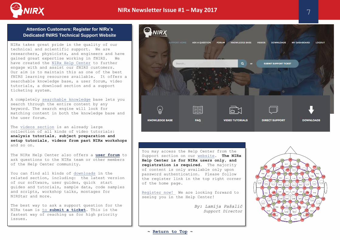
NIRx takes great pride in the quality of our
technical and scientific support. We are
researchers, physicists, and engineers and have
gained great expertise working in fNIRS. We
have created the NIRx Help Center to further
engage with and assist our fNIRS customers.
Our aim is to maintain this as one of the best
fNIRS learning resources available. It offers a
searchable knowledge base, a user forum, video
tutorials, a download section and a support
ticketing system.
A completely searchable knowledge base lets you
search through the entire content by any
keyword. The search engine will look for
matching content in both the knowledge base and
the user forum.
The videos section is an already large
collection of all kinds of video tutorials:
analysis tutorials, subject preparation and
setup tutorials, videos from past NIRx workshops
and so on.
The NIRx Help Center also offers a user forum to
ask questions to the NIRx team or other members
of the Help Center community.
You can find all kinds of downloads in the
related section, including: the latest version
of our software, user guides, quick start
guides and tutorials, sample data, code samples
and scripts, workshop talks, montages for
NIRStar and more.
The best way to ask a support question for the
NIRx team is to submit a ticket. This is the
fastest way of reaching us for high priority
issues.
Attention Customers: Register for NIRx’s
Dedicated fNIRS Technical Support Website
You may access the Help Center from the
Support section on our website. The NIRx
Help Center is for NIRx users only, and
registration is required. The majority
of content is only available only upon
password authentication. Please follow
the register link in the top right corner
of the home page.
Register now! We are looking forward to
seeing you in the Help Center!
By: Lamija Pašalić
Support Director

~ Return to Top ~
NIRx Newsletter Issue #1 – May 2017
8
fNIRS Detectors from NIRx - SiPD vs. APD
Overview
NIRx offers both ‘standard’ silicon photodiode (SiPD) and
‘high-sensitivity’ avalanche photodiode (APD) fNIRS
detectors. The NIRSport ‘Portable’ fNIRS System may only
use the APD detectors with an additional coupling unit- the
APDs in this case would not be for portable applications,
and would furthermore require a different probe set from
the standard probe sets generally. By contrast, the
NIRScout ‘Lab-Based’ fNIRS System may use either the SiPD
or APD detectors with identical probe sets.
fNIRS Detector Sensitivity
Our APD detectors are up to 8x as sensitive as our SiPD
detectors on average. This does give a much better signal
overall, though detector sensitivity is not the only factor
in determining signal level and quality.
Dynamic fNIRS Gain Settings
While considering detector sensitivity is critical, the
system’s dynamic gain is also important when evaluating
overall system performance. NIRx offers an industry-
leading 9 levels of automated/programmable detector gain
for both our SiPD and APD detectors. So, even though our
SiPDs have less sensitivity than our APDs, we are able to
make up for it against APD options offered by some of the
other fNIRS manufacturers out there by maximizing the gain
settings of the detectors.
Optimizing Unique fNIRS Detector Gain Settings
As part of every experiment, one must “calibrate” (AKA
“optimize”) the exact gains used for every detector. Most
systems only allow one gain setting per detector. By
contrast, NIRx systems allow for unique gain settings for
every source-detector pair. This is particularly important
when a detector is positioned between two sources that are
transmitting light through tissue with very different
scattering and absorption properties. For example, imagine
a detector placed on the hairline approximately around Fz
in EEG standard coordinates, and this detector is measuring
from two sources: source one is positioned 3cm anteriorly
towards the nasion (~AFz in EEG coordinates) on a hairless
part of the head with thinner scalp, skull and CSF, and
source 2 positioned posteriorly towards the inion (~FCz in
EEG coordinates) on a hairy part of the head with thicker
scalp, skull and CSF. There would be substantially
different amounts of scattering and absorption from each of
these sources as the photons pass through the tissue, and
thus a very different amount of light would actually reach
the detector in between the two sources. It would,
therefore, be ideal for the detector to have a higher gain
setting for source 2 (the source on the hair) vs. source 1
(the source on the forehead).
Top-left: A single-tipped fiber optic detector
probe (AKA “optode”).
Top-right: a dual-tipped fiber optic detector probe.

~ Return to Top ~
NIRx Newsletter Issue #1 – May 2017
9
Unique detector gain settings for every source-detector
pair are also important for multi-distance measurements and
maximizing the number of usable data channels. This dynamic
gain switching is the biggest advantageof the time-
multiplexed recording mode used in NIRx devices compared to
other ways to distinguish the signal from different sources.
Optimizing fNIRS Detector Gains and Signal
The process of setting gains differs substantially
between systems. Some fNIRS systems even require the
end-user to manually set each detector gain setting one-
by-one for each measurement, which, as you can probably
imagine, can take a very long time. NIRx’s recording
software uses a user-friendly fully-automated signal
optimization step which rapidly identifies the ideal
signal level, and associated detector gains for each
source-detector channel pairing.
Example of Unique Detector Gains Using NIRStar / NIRx Platform
1) Probe array (“Montage”): An 8-source/8-detector (16-probe) montage is shown
with 20x topographic data channels of interest positioned bilaterally over
the motor cortex.
2) Montage Close Up: Sources = S1, S2, S3, S4; Detectors = D1, D2, D3, D4.
3) Topolayout / Signal Quality Indicator: shows 2-D blocked abstraction of
montage, which allows easy-to-view color-based/changing signal quality and
levels. Each number pair corresponds to a respective source and detector
number, in that order, for that data channel of interest (i.e., “2-1” =
channel formed by source #1 and detector #2). Note: signal quality has
already been calibrated/optimized at this point and that the source-detector
pairs seen in the montage close up
4) Gain Settings Map: shows the detector gain level for each source-detector
pair. Note channels “2-1” and ”1-1” in particular for the moment. Detector
#1 is part of both channels but has a substantially different gain setting
for each channel. Channel 2-1 has a gain of 3, whereas channel 1-1 has a
gain of 5. This dynamic gain switching for a single detector with multiple
sources greatly improves ease of use and signal quality.
2) Montage Close Up 1) Probe Array
(“montage”)
4) Gain Settings Map 3) Topolayout /
Signal Quality Indicator

~ Return to Top ~
NIRx Newsletter Issue #1 – May 2017
10
Detector Probes and Caps
Achieving optimal signal quality involves more than just
the system electronics and control software. The design
and fit of the physical detector hardware, and its
connecting components, are arguably just as important.
Imagine a car with no or very poor tires- it won’t get
you very far, will it? Well, probes and caps are really
just like the tires for your fNIRS system. Without them,
you would not be able to collect any data, despite how
powerful the system’s engine may be. We have put
extensive research and user-feedback into our probe and
cap design. While we could tell you a lot more about it
here, this is really better seen than explained. You can
see here just how fast and easy our cap setup is. And
you can learn more about the probes and caps on our
website.
Which Detector to Choose: APD or SiPD?
It may come as no surprise, but the APD detectors are
quite a bit more expensive than the SiPDs. That said,
their value is unquestionable: a NIRx APD detector
system, along with our innovative probe systems, will
work on the vast majority of subjects, measuring from any
part of the head, with an incredibly easy/fast setup.
Our APDs also are best for end-users that wish to do
fMRI/fNIRS or fMRI/MEG studies.
The SiPD detectors will work excellent on child and
geriatric subjects on any part of the head, and will work
with ~60-70% of college-aged subjects (FYI: college
students are generally some of the most difficult
subjects due to their full-grown heads and thicker hair)
with thick black hair (thicker, darker hair is more
difficult than thinner, lighter hair) during measurements
on top of their head (e.g., the motor, somatosensory
cortex, etc.). In total it generally averages out to ~80%
of subjects in multi-cultural locations, such as big
cities in USA, Canada, UK, Germany, France, etc..
All that said, this still could be a major factor for you.
It is important to remember: with NIRx systems you can
start with SiPD detectors and switch over to APD
detectors. We offer one of the most versatile fNIRS
platforms out there in terms of upgrade options and
flexibility. Please do let us know what your decision
context is and we will be happy to work with you to
provide you the best-possible solution.
By: Thomas Johannsen
Technical Sales Manager
Hardware Support Consultant
Laser and LED Sources from NIRx
NIRx’s History with Laser & LED sources
As you may know, NIRx started out of the lab of Dr.
Randall Barbour at SUNY Downstate Medical Center in the
mid-1980s. It wasn’t until 2000 that NIRx formed and
sold its first NIRS system, the “DYNOT” (Dynamic Optical
Tomography); this laser-based system used straight fiber
optic probes which could be oriented in very high-density
grids for tomographic imaging. In fact, all of Dr.
Barbour and NIRx’s systems were laser based from the
1980s onward, until 2010, when the first “NIRScout”
system was released. As many of you know, this first-

~ Return to Top ~
NIRx Newsletter Issue #1 – May 2017
11
generation NIRScout used LED instead of Laser sources for
light illumination.
In 2015, NIRx introduced Lasers to the NIRScout family.
The new “NIRScoutX+” chassis enabled the breakthrough of
the first hybrid LED/Laser system. Users can now choose
either LED or Laser sources with this system, enabling
for the ideal source choice by application.
A Comparison of Laser vs. LED sources
Fiber Optic Performance
Lasers are a better option than LEDs for NIRS
measurements that require fiber optics, like those done
with MRI and MEG. The collimated-bandwidth Laser light
couples much more efficiently into a fiber cable than the
LED light. For MRI and MEG-concurrent NIRS studies, in
particular, very long probes are necessary as the NIRS
system sits in the control room, and its probes (usually
~8-10m long) go to and from the subject’s head in the
scanning room.
Fiber Optic Performance Summary:
• Lasers beat out LEDs in applications where fiber
optic probes are necessary (e.g.,
• NIRS/fMRI, NIRS/MEG, collocated NIRS-TMS, etc.)
Cable Length, Weight, and Flexibility
It is also important to note that cable length is a
factor for overall signal levels. Light signal, whether
from Lasers or LEDs, attenuates as it passes through
fiber optic cables. The level of attenuation does vary
based on the properties of the particular fiber, though
shorter fibers will always outperform longer fibers of
the same make/type, assuming all else equal.
NIRx does recommend our very high-powered active LEDs for
most use cases (we will cover why that is later).
The two main differences between our active and fiber
optic sources:
1) The location of the NIRS source
a. Active NIRS sources are contained within the probe
tip housing
b. Fiber optic NIRS sources are contained within the
NIRS system itself
2) The material of the probe cables
a. Active sources use electronic cables: very
lightweight, durable, and may be extended without
significant signal loss (i.e., you may have one
set of probes with multiple extensions if you like)
b. Fiber optic sources use glass cables: relatively
heavier; flexible, but less durable; may not be
extended (i.e., you need multiple probe sets if
different cable lengths are required)
The location of the active LED in the probe tip housing
minimizes signal attenuation, which greatly improves
overall system performance. The lighter weight cabling
with active LEDs is very helpful in child and mobile
applications, though all subjects appreciate less weight
on their heads during measurements.

~ Return to Top ~
NIRx Newsletter Issue #1 – May 2017
12
Cable Length, Weight, and Flexibility Summary:
• NIRx Active LEDs have far less cable-length-
related attenuation than fiber optic lasers
• NIRx Active LEDs are much lighter than fiber
optic lasers
• NIRx Active LEDs are more flexible than lasers
• Note: as mentioned above, Lasers beat out LEDs
when fiber optic probes are necessary (e.g.,
NIRS/fMRI, NIRS/MEG applications, etc.)
Multi-Wavelength Considerations
Most NIRS system use 2 wavelengths to distinguish between
oxy- and deoxy-hemoglobin. NIRx LED systems are only
offered with 2 wavelengths, though, as mentioned, one may
own a hybrid system with both LEDs and Lasers. By
contrast, Lasers may have many more than 2 wavelengths
(but typically also have just 2 wavelengths in most
commercial systems). NIRx systems currently offer 2, 4
and 8-wavelength source options.
Higher wavelength counts yield a potentially better
characterization of the Hb signal (though, how much
better is under debate) as each wavelength has a slightly
different depth of penetration, differential pathlength
factor (dpf), and associated molar extinction coefficient
for the respective chromophores of interest: generally,
oxy-Hb and deoxy-Hb. Note: some researchers are
interested in using NIRS to identify cytochrome c-oxidase
(see fig. 1 right), though this methodology is still
being refined.
That said, the information gained from the additional
wavelengths does not have a clear-cut advantage at the
moment for researchers looking at only oxy and deoxy-
hemoglobin. And when considering the great increase in
cost one must pay for a system with more wavelengths, it
may not be worth it for you to upgrade to more
wavelengths.
Multi-Wavelength Considerations Summary:
• NIRx LEDs are 2-wavelength sources
• NIRx Lasers may be 2 or 4-wavelength sources
(note: 8-wavlength custom option available)
• Additional wavelengths come at a great price
• The information gained from additional
wavelengths may not be worth the cost of the
upgrade if all you are interested in is
hemoglobin changes
Ideal Applications for NIRx LEDs and Lasers
NIRx LEDs may be used in nearly any application, even MRI
and MEG-concurrent measurements, though those particular
[2] Fig. 1. Absorption spectra for
deoxyhemoglobin (Hb), oxyhemoglobin
(HbO2), cytochrome c.
~ Return to Top ~
NIRx Newsletter Issue #1 – May 2017
13
applications are best left to Laser sources. LEDs are
generally better when considering child and mobile
applications, as they are lighter and more flexible than
Lasers. We recommend you use Laser sources if you have
used them before and your research requires it, or if you
want to do MRI or MEG-concurrent measurements.
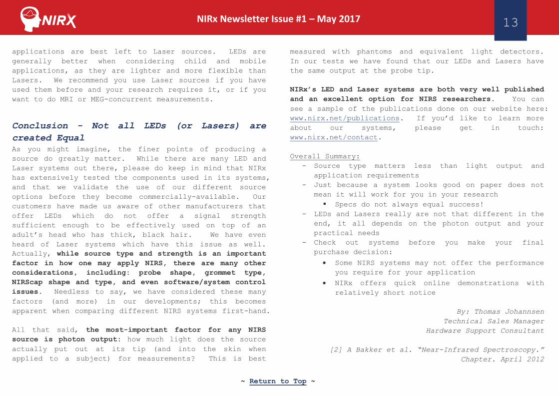
Conclusion - Not all LEDs (or Lasers) are
created Equal
As you might imagine, the finer points of producing a
source do greatly matter. While there are many LED and
Laser systems out there, please do keep in mind that NIRx
has extensively tested the components used in its systems,
and that we validate the use of our different source
options before they become commercially-available. Our
customers have made us aware of other manufacturers that
offer LEDs which do not offer a signal strength
sufficient enough to be effectively used on top of an
adult’s head who has thick, black hair. We have even
heard of Laser systems which have this issue as well.
Actually, while source type and strength is an important
factor in how one may apply NIRS, there are many other
considerations, including: probe shape, grommet type,
NIRScap shape and type, and even software/system control
issues. Needless to say, we have considered these many
factors (and more) in our developments; this becomes
apparent when comparing different NIRS systems first-hand.
All that said, the most-important factor for any NIRS
source is photon output: how much light does the source
actually put out at its tip (and into the skin when
applied to a subject) for measurements? This is best
measured with phantoms and equivalent light detectors.
In our tests we have found that our LEDs and Lasers have
the same output at the probe tip.
NIRx’s LED and Laser systems are both very well published
and an excellent option for NIRS researchers. You can
see a sample of the publications done on our website here:
www.nirx.net/publications. If you’d like to learn more
about our systems, please get in touch:
www.nirx.net/contact.
Overall Summary:
- Source type matters less than light output and
application requirements
- Just because a system looks good on paper does not
mean it will work for you in your research
Specs do not always equal success!
- LEDs and Lasers really are not that different in the
end, it all depends on the photon output and your
practical needs
- Check out systems before you make your final
purchase decision:
Some NIRS systems may not offer the performance
you require for your application
NIRx offers quick online demonstrations with
relatively short notice
By: Thomas Johannsen
Technical Sales Manager
Hardware Support Consultant
[2] A Bakker et al. “Near-Infrared Spectroscopy.”
Chapter. April 2012

~ Return to Top ~
NIRx Newsletter Issue #1 – May 2017
14
‘Like’ and Follow us on Social Media: Facebook LinkedIn Twitter
Career Opportunities at NIRx: Scientific Consultant
NIRx is hiring Master’s and Ph.D.-level Scientific Consultants to work in our
soon-to-be opened Minneapolis location.
Traits we are looking for:
- Master’s/Ph.D. in Neuroscience, Psychology, Biomedical Engineering or related field
- Experience working in neuroimaging; EEG, fMRI, and/or fNIRS would be preferred
- Outgoing personality that likes (or at least doesn’t mind) presenting to large groups
- Willingness to travel, up to 60% in Fall and Spring (our high seasons)
- Attention to detail, creative problem-solving skills, solutions-oriented mindset
- A love of science and openness to loving fNIRS!
Job duties:
- Scientific support and consultation with neuroscientist customer base
- Conducting training and installations
- Representing NIRx at relevant conferences, workshops, and events
We offer:
- Competitive compensation and benefits
- Intellectually-stimulating working environment and tasks
- Opportunity for growth within a rapidly-growing company and industry
- International travel
Interested? Email us at [email protected]
A dual-tipped,
lightweight high-
powered active LED
source probe.
A single-tipped, low-
profile fiber optic Laser
source probe.

~ Return to Top ~
15 NIRx Newsletter Issue #1 – March 1, 2017
NIRX is a world-leader in providing integrated solutions for fNIRS neuro-imaging. In 1988 we introduced
the concept of tomographic imaging (i.e., multi-distance measurements) in dense scattered media base on
diffusely scattered light. This approach has since been widely adapted and has served to launch the
modern day field of fNIRS tomography.
Through our offices in Berlin, Los Angeles, New York and São Paolo, our engineers and grant-funded
investigators are providing a growing number of research teams world-wide with comprehensive technology
solutions for the most demanding investigative applications.
General Inquiries: [email protected]
Customer Support: [email protected]
www.nirx.net
+49 (30) 46 307 340 (EU)
+1 323 648 6682 (US/Canada)
NIRx Headquarters
NIRx Medical Technologies, LLC
15 Cherry Lane
Glen Head, NY 11545 U.S.A.
NIRx Berlin
NIRx Medizintechnik GmbH
Gustav-Meyer-Allee 25
Building 12
13355 Berlin, Germany
NIRx Los Angeles
NIRx Medical Technologies, LLC
5670 Wilshire Blvd Suite 1800
Los Angeles, CA 90036 U.S.A.
© 2017 by NIRx Medical Technologies. All rights reserved.