New insights into the molecular basis of desmoplakinand desmin-related cardiomyopathies
12
4974 Research Article Introduction Desmosomes are multi-protein complexes that play a key role in promoting cell-cell adhesion and in tethering the intermediate filament (IF) system to the cell membrane. These structures are abundant in tissue exposed to mechanical stress, such as the epidermis and heart. They contribute to maintaining the integrity of embryonic tissues during development (reviewed in Getsios et al., 2004). The adhesive core of desmosomes comprises transmembrane glycoproteins of the cadherin family, the desmogleins and the desmocollins. Two members of the armadillo family of proteins, plakoglobin and plakophilins, as well as desmoplakin (DP) link the IF cytoskeleton to the adhesive core of desmosomes by means of linear and lateral interactions between these various molecules (Getsios et al., 2004). DP, the most abundant desmosomal plaque component, is a member of the plakin family, which also includes plectin, BP230, periplakin and epiplakin (reviewed in Jefferson et al., 2004). These proteins serve as cytolinkers and/or scaffolding proteins, connecting IF to other cytoskeletal networks and/or distinct sites at the plasma membrane. Plakins are predicted to form homodimers with a central coiled-coil domain flanked by globular end domains (Green et al., 1992a; Green et al., 1992b; Ruhrberg and Watt, 1997). The N-terminal region of DP encompasses a so-called plakin domain harboring -helical bundles (Green et al., 1990) and mediates its localization to desmosomes by binding to plakoglobin and plakophilins (Stappenbeck and Green, 1992; Stappenbeck et al., 1993; Kouklis et al., 1994; Cowin and Burke, 1996; Kowalczyk et al., 1997; Smith and Fuchs, 1998). The C-terminus of DP contains a plakin repeat domain (PRD) consisting of three homologous subdomains, denoted A, B and C, that are interrupted by intervening sequences of varying length (Fig. 1). These subdomains consist of 4.5 copies of a 38-amino acid sequence repeat motif (Green et al., 1990; Choi et al., 2002). The DP tail is able to associate with various IF proteins, such as epidermal and simple epithelial keratins and vimentin (Stappenbeck and Green, 1992; Stappenbeck et al., 1993; Kouklis et al., 1994; Kowalczyk et al., 1997; Meng et al., 1997; Fontao et al., 2003). Recent studies have identified sequences that allow DP to bind to distinct IFs. Specifically, the C subdomain within the C- terminal extremity of DP binds to K5/K14 and K8/K18, while its linker subdomain is able to associate with K8/K18 and vimentin (Fontao et al., 2003). Furthermore, DP-IF interactions Desmosomes are intercellular adhesive complexes that anchor the intermediate filament cytoskeleton to the cell membrane in epithelia and cardiac muscle cells. The desmosomal component desmoplakin plays a key role in tethering various intermediate filament networks through its C-terminal plakin repeat domain. To gain better insight into the cytoskeletal organization of cardiomyocytes, we investigated the association of desmoplakin with desmin by cell transfection, yeast two-hybrid, and/or in vitro binding assays. The results indicate that the association of desmoplakin with desmin depends on sequences within the linker region and C-terminal extremity of desmoplakin, where the B and C subdomains contribute to efficient binding; a potentially phosphorylatable serine residue in the C-terminal extremity of desmoplakin affects its association with desmin; the interaction of desmoplakin with non-filamentous desmin requires sequences contained within the desmin C-terminal rod portion and tail domain in yeast, whereas in in vitro binding studies the desmin tail is dispensable for association; and mutations in either the C-terminus of desmoplakin or the desmin tail linked to inherited cardiomyopathy seem to impair desmoplakin- desmin interaction. These studies increase our understanding of desmoplakin-intermediate filament interactions, which are important for maintenance of cytoarchitecture in cardiomyocytes, and give new insights into the molecular basis of desmoplakin- and desmin- related human diseases. Key words: Desmoplakin, Desmin, Vimentin, Intermediate filaments, Cardiomyocyte, Cardiomyopathy Summary New insights into the molecular basis of desmoplakin- and desmin-related cardiomyopathies Karine Lapouge 1, * ,‡ , Lionel Fontao 1, *, Marie-France Champliaud 1 , Fabienne Jaunin 1 , Miguel A. Frias 2 , Bertrand Favre 1,§ , Denise Paulin 3 , Kathleen J. Green 4 and Luca Borradori 1,¶ 1 Clinic of Dermatology, University Hospital, Geneva, Rue Micheli-du-Crest 14, 1211-Geneva 14, Switzerland 2 Division of Endocrinology, Diabetology and Nutrition, University Hospital, Geneva, Rue Micheli-du-Crest 14, 1211-Geneva 14, Switzerland 3 Biologie Moléculaire de la Différenciation, Université Paris-7, 2 Place Jussieu, 75005 Paris, France 4 Departments of Pathology and Dermatology, Feinberg School of Medicine, Northwestern University, 303 E. Chicago Avenue, Chicago, IL 60611, USA *These authors contributed equally to this work ‡ Present address: Département de Microbiologie Fondamentale, Bâtiment de Biologie, Université de Lausanne, CH-1015 Lausanne, Switzerland § Present address: Clinic of Dermatology, CHUV-University Hospital, 1011 Lausanne, Switzerland ¶ Author for correspondence (e-mail: [email protected]) Accepted 11 September 2006 Journal of Cell Science 119, 4974-4985 Published by The Company of Biologists 2006 doi:10.1242/jcs.03255 Journal of Cell Science
Transcript of New insights into the molecular basis of desmoplakinand desmin-related cardiomyopathies

4974 Research Article
IntroductionDesmosomes are multi-protein complexes that play a keyrole in promoting cell-cell adhesion and in tethering theintermediate filament (IF) system to the cell membrane. Thesestructures are abundant in tissue exposed to mechanical stress,such as the epidermis and heart. They contribute to maintainingthe integrity of embryonic tissues during development(reviewed in Getsios et al., 2004). The adhesive core ofdesmosomes comprises transmembrane glycoproteins of thecadherin family, the desmogleins and the desmocollins. Twomembers of the armadillo family of proteins, plakoglobin andplakophilins, as well as desmoplakin (DP) link the IFcytoskeleton to the adhesive core of desmosomes by means oflinear and lateral interactions between these various molecules(Getsios et al., 2004).
DP, the most abundant desmosomal plaque component, is amember of the plakin family, which also includes plectin,BP230, periplakin and epiplakin (reviewed in Jefferson et al.,2004). These proteins serve as cytolinkers and/or scaffoldingproteins, connecting IF to other cytoskeletal networks and/ordistinct sites at the plasma membrane. Plakins are predicted toform homodimers with a central coiled-coil domain flanked by
globular end domains (Green et al., 1992a; Green et al., 1992b;Ruhrberg and Watt, 1997). The N-terminal region of DPencompasses a so-called plakin domain harboring �-helicalbundles (Green et al., 1990) and mediates its localization todesmosomes by binding to plakoglobin and plakophilins(Stappenbeck and Green, 1992; Stappenbeck et al., 1993;Kouklis et al., 1994; Cowin and Burke, 1996; Kowalczyk et al.,1997; Smith and Fuchs, 1998). The C-terminus of DP containsa plakin repeat domain (PRD) consisting of three homologoussubdomains, denoted A, B and C, that are interrupted byintervening sequences of varying length (Fig. 1). Thesesubdomains consist of 4.5 copies of a 38-amino acid sequencerepeat motif (Green et al., 1990; Choi et al., 2002). The DP tailis able to associate with various IF proteins, such as epidermaland simple epithelial keratins and vimentin (Stappenbeck andGreen, 1992; Stappenbeck et al., 1993; Kouklis et al., 1994;Kowalczyk et al., 1997; Meng et al., 1997; Fontao et al., 2003).Recent studies have identified sequences that allow DP to bindto distinct IFs. Specifically, the C subdomain within the C-terminal extremity of DP binds to K5/K14 and K8/K18, whileits linker subdomain is able to associate with K8/K18 andvimentin (Fontao et al., 2003). Furthermore, DP-IF interactions
Desmosomes are intercellular adhesive complexes thatanchor the intermediate filament cytoskeleton to the cellmembrane in epithelia and cardiac muscle cells. Thedesmosomal component desmoplakin plays a key role intethering various intermediate filament networks throughits C-terminal plakin repeat domain. To gain better insightinto the cytoskeletal organization of cardiomyocytes, weinvestigated the association of desmoplakin with desmin bycell transfection, yeast two-hybrid, and/or in vitro bindingassays. The results indicate that the association ofdesmoplakin with desmin depends on sequences within thelinker region and C-terminal extremity of desmoplakin,where the B and C subdomains contribute to efficientbinding; a potentially phosphorylatable serine residue inthe C-terminal extremity of desmoplakin affects itsassociation with desmin; the interaction of desmoplakin
with non-filamentous desmin requires sequences containedwithin the desmin C-terminal rod portion and tail domainin yeast, whereas in in vitro binding studies the desmin tailis dispensable for association; and mutations in either theC-terminus of desmoplakin or the desmin tail linked toinherited cardiomyopathy seem to impair desmoplakin-desmin interaction. These studies increase ourunderstanding of desmoplakin-intermediate filamentinteractions, which are important for maintenance ofcytoarchitecture in cardiomyocytes, and give new insightsinto the molecular basis of desmoplakin- and desmin-related human diseases.
Key words: Desmoplakin, Desmin, Vimentin, Intermediate filaments,Cardiomyocyte, Cardiomyopathy
Summary
New insights into the molecular basis of desmoplakin-and desmin-related cardiomyopathiesKarine Lapouge1,*,‡, Lionel Fontao1,*, Marie-France Champliaud1, Fabienne Jaunin1, Miguel A. Frias2,Bertrand Favre1,§, Denise Paulin3, Kathleen J. Green4 and Luca Borradori1,¶
1Clinic of Dermatology, University Hospital, Geneva, Rue Micheli-du-Crest 14, 1211-Geneva 14, Switzerland2Division of Endocrinology, Diabetology and Nutrition, University Hospital, Geneva, Rue Micheli-du-Crest 14, 1211-Geneva 14, Switzerland3Biologie Moléculaire de la Différenciation, Université Paris-7, 2 Place Jussieu, 75005 Paris, France4Departments of Pathology and Dermatology, Feinberg School of Medicine, Northwestern University, 303 E. Chicago Avenue, Chicago, IL 60611,USA*These authors contributed equally to this work‡Present address: Département de Microbiologie Fondamentale, Bâtiment de Biologie, Université de Lausanne, CH-1015 Lausanne, Switzerland§Present address: Clinic of Dermatology, CHUV-University Hospital, 1011 Lausanne, Switzerland¶Author for correspondence (e-mail: [email protected])
Accepted 11 September 2006Journal of Cell Science 119, 4974-4985 Published by The Company of Biologists 2006doi:10.1242/jcs.03255
Jour
nal o
f Cel
l Sci
ence

4975Interaction between desmoplakin and desmin
are regulated by phosphorylation of Ser2849 within the DP C-terminus (Stappenbeck at al., 1994; Fontao et al., 2003; Godselet al., 2005). Crystallographic studies have shown that the Band C subdomains exhibit a globular structure with a conservedgroove, the features of which seem to be suitable for aninteraction with vimentin (Choi et al., 2002), but theimportance of which has yet to be empirically tested. Theimportance of understanding the mechanisms governing DP-IF associations is highlighted by recent studies indicating thatproper attachment of IFs to the desmosomal plaque via DP isrequired for maintaining cell-cell adhesive strength (Huen etal., 2002).
The importance of DP in vivo is supported by theobservation that DP-null mutant mice die at embryonic day6.5 owing to defects of the extraembryonic endoderm.Furthermore, chimeric morulae expressing DP inextraembryonic tissues do not survive beyond day E9.5 as aresult of defects in the developing epidermis, neuroepitheliumand heart with perturbation of desmosome assembly andsevering of the IF-cell membrane attachment (Gallicano et al.,1998; Gallicano et al., 2001). Finally, pathogenic mutations inthe DP gene cause cardiomyopathy and palmo-plantarkeratoderma (Armstrong et al., 1999; Norgett et al., 2000).
IF, which are present in all vertebrate cells and tissues, areclassified into five families (Herrmann and Aebi, 2004). All IFpolypeptides are built from a central rod domain, consisting of�-helical segments, flanked by globular non �-helical N-terminal head and C-terminal tail domains. The assembly of IFinto long 10-12 nm caliber polymers, is initiated by the centralrod of IF proteins. The process progresses by formation ofdimers and tetramers, which further associate following threeprincipal assembly modes. While the N-terminal head domainsseem to control IF assembly and stabilization, their tails arethought to regulate lateral packing and stabilization of higherorder filament structure (Herrmann and Aebi, 2004).
Desmin, an IF protein type III, is one of the earliestexpressed muscle-specific proteins. It is detected in early phaseof skeletal (Sejersen and Lendahl, 1993) and cardiac(Kachinsky et al., 1995) muscle differentiation together withvimentin and nestin, the expression of which decline duringdevelopment (Carlsson et al., 2000). Desmin-null mutant mice
show disorganization of myofibril architecture in mechanicallystressed striated muscles, including heart (Li et al., 1996;Milner et al., 1996; Wang et al., 2001). Furthermore, desmingene mutations cause certain forms of muscular dystrophies,with or without cardiomyopathies. Although certain mutationscompromise filament assembly (Park et al., 2000; Dalakas etal., 2000; Li and Dalakas, 2001; Bär et al., 2005) at variousstages, in other cases the implicated pathogenic mechanismsare unclear (Dalakas et al., 2000; Bär et al., 2004).
Ultrastructural studies have shown that desmin filamentsattach in a lateral fashion to the DP-containing region of thedesmosomal plaques of the heart, which suggests that specialdomains exist in desmin that are involved in binding(Kartenbeck et al., 1983). Recent immunoelectron microscopystudies have revealed that, at intercalated discs, desmosomesand fasciae adherentes form a highly integrated system, forwhich the term of area composita has been proposed (Frankeet al., 2006). At the molecular level, although DP was shownto bind to desmin in yeast two-hybrid assays (Meng et al.,1997), little information is available regarding the associationof DP and desmin and whether the latter is regulated as in thecase of vimentin (Fontao et al., 2003). Characterization of theseinteractions is key to deciphering the mechanisms by which thejunctional region of the intercalated discs mediates theattachment of IF bundles, generating a stress-resistantscaffolding able to integrate the mechanical forces generatedduring heart contraction. Recent discovery of mutations inhumans (reviewed by Sen-Chowdhry et al., 2005) andengineered gene disruption of desmosomal genes (Bierkamp etal., 1996; Ruiz et al., 1996; Grossmann et al., 2004) haveunequivocally confirmed the functional interdependence ofdesmosomal proteins and their importance for the integrity ofthe cytoarchitecture of cardiac muscle cells.
Therefore, the aim of our study was to investigate theinteraction between DP and desmin by defining functionallyimportant sequences implicated in the binding of thesemolecules with each other by comparative analysis using theyeast two-hybrid system, cell transfection studies andbiochemical assays. Furthermore, we sought to gain insightsinto the impact of pathogenic mutations in DP and desmin onthese interactions.
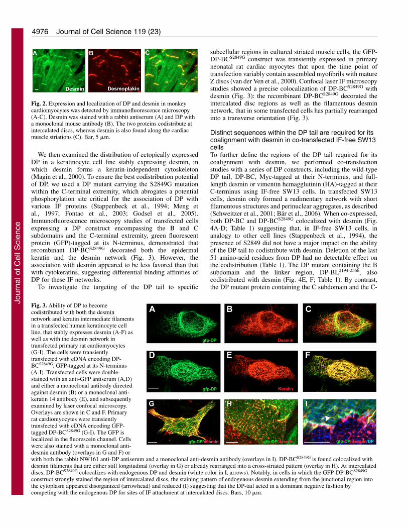
ResultsDP and desmin are found codistributed inthe intercalated disc regions of cardiacmyocytesPrevious studies have identified sequenceswithin DP that are important for its interactionwith vimentin and keratins (Stappenbeck andGreen, 1992; Stappenbeck et al., 1993; Meng etal., 1997; Choi et al., 2002; Fontao et al., 2003),but the interaction of DP with desmin has not yetbeen characterized. Hence, we first investigatedthe localization of DP and desmin inlongitudinal sections of monkey cardiac muscleby double immunofluorescence microscopy. Asexpected (Kartenbeck et al., 1983), these twoproteins were both found at the level ofintercalated discs. Desmin also showed thetypical cross-striated labeling in a distinctsarcomeric pattern (Fig. 2).
Fig. 1. Human wild type desmin, vimentin and the C-terminal half of DP showingthe predicted subdomains with the corresponding amino acid positions. The locationof a potential protein kinase A consensus site within the C-terminal extremity of DPis indicated in bold, while the position of Ser2849 is underlined.
Jour
nal o
f Cel
l Sci
ence
4976
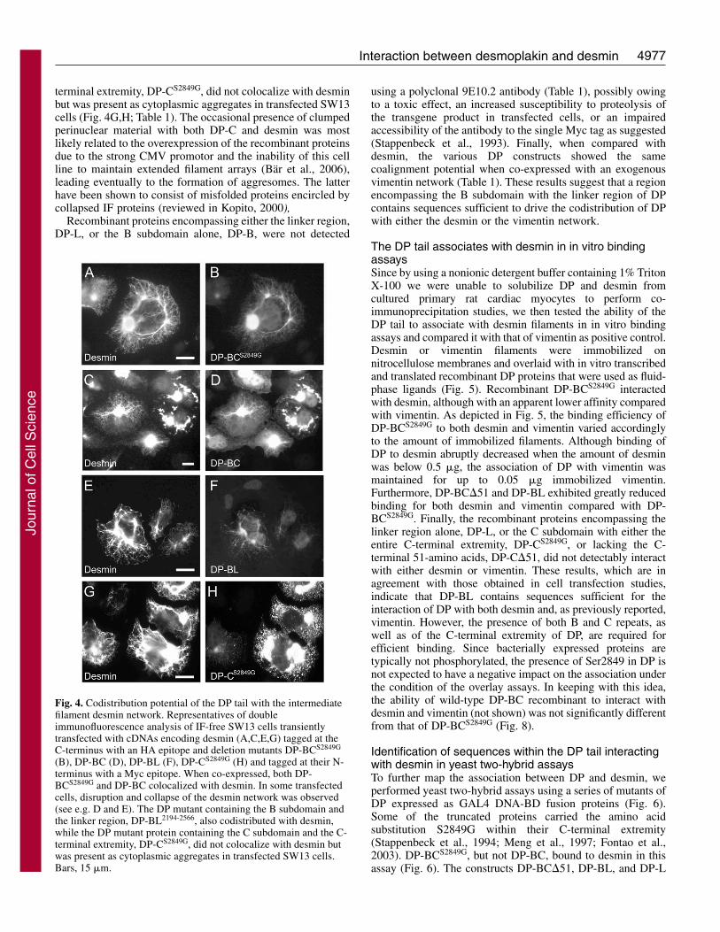
We then examined the distribution of ectopically expressedDP in a keratinocyte cell line stably expressing desmin, inwhich desmin forms a keratin-independent cytoskeleton(Magin et al., 2000). To ensure the best codistribution potentialof DP, we used a DP mutant carrying the S2849G mutationwithin the C-terminal extremity, which abrogates a potentialphosphorylation site critical for the association of DP withvarious IF proteins (Stappenbeck et al., 1994; Meng etal., 1997; Fontao et al., 2003; Godsel et al., 2005).Immunofluorescence microscopy studies of transfected cellsexpressing a DP construct encompassing the B and Csubdomains and the C-terminal extremity, green fluorescentprotein (GFP)-tagged at its N-terminus, demonstrated thatrecombinant DP-BCS2849G decorated both the epidermalkeratin and the desmin network (Fig. 3). However, theassociation with desmin appeared to be less favored than thatwith cytokeratins, suggesting differential binding affinities ofDP for these IF networks.
To investigate the targeting of the DP tail to specific
Journal of Cell Science 119 (23)
subcellular regions in cultured striated muscle cells, the GFP-DP-BCS2849G construct was transiently expressed in primaryneonatal rat cardiac myocytes that upon the time point oftransfection variably contain assembled myofibrils with matureZ discs (van der Ven et al., 2000). Confocal laser IF microscopystudies showed a precise colocalization of DP-BCS2849G withdesmin (Fig. 3): the recombinant DP-BCS2849G decorated theintercalated disc regions as well as the filamentous desminnetwork, that in some transfected cells has partially rearrangedinto a transverse orientation (Fig. 3).
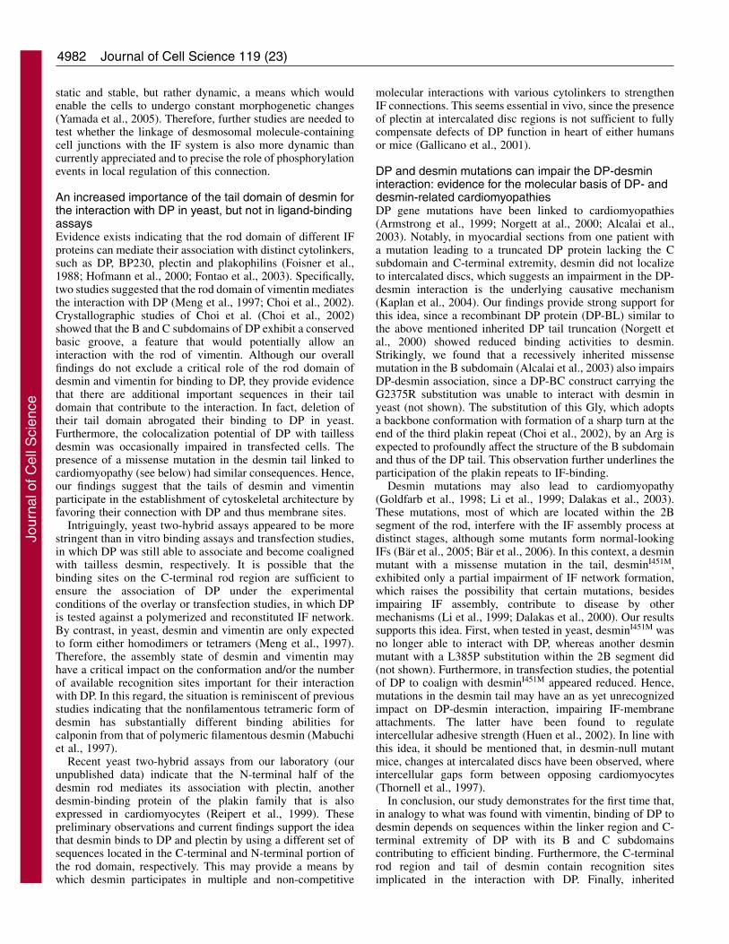
Distinct sequences within the DP tail are required for itscoalignment with desmin in co-transfected IF-free SW13cellsTo further define the regions of the DP tail required for itscoalignment with desmin, we performed co-transfectionstudies with a series of DP constructs, including the wild-typeDP tail, DP-BC, Myc-tagged at their N-terminus, and full-length desmin or vimentin hemagglutinin (HA)-tagged at theirC-terminus using IF-free SW13 cells. In transfected SW13cells, desmin only formed a rudimentary network with shortfilamentous structures and perinuclear aggregates, as described(Schweitzer et al., 2001; Bär et al., 2006). When co-expressed,both DP-BC and DP-BCS2849G colocalized with desmin (Fig.4A-D; Table 1) suggesting that, in IF-free SW13 cells, inanalogy to other cell lines (Stappenbeck et al., 1994), thepresence of S2849 did not have a major impact on the abilityof the DP tail to codistribute with desmin. Deletion of the last51 amino-acid residues from DP had no detectable effect onthe codistribution (Table 1). The DP mutant containing the Bsubdomain and the linker region, DP-BL2194-2566, alsocodistributed with desmin (Fig. 4E, F; Table 1). By contrast,the DP mutant protein containing the C subdomain and the C-
Fig. 2. Expression and localization of DP and desmin in monkeycardiomyocytes was detected by immunofluorescence microscopy(A-C). Desmin was stained with a rabbit antiserum (A) and DP witha monoclonal mouse antibody (B). The two proteins codistribute atintercalated discs, whereas desmin is also found along the cardiacmuscle striations (C). Bar, 5 �m.
Fig. 3. Ability of DP to becomecodistributed with both the desminnetwork and keratin intermediate filamentsin a transfected human keratinocyte cellline, that stably expresses desmin (A-F) aswell as with the desmin network intransfected primary rat cardiomyocytes(G-I). The cells were transientlytransfected with cDNA encoding DP-BCS2849G, GFP-tagged at its N-terminus(A-I). Transfected cells were double-stained with an anti-GFP antiserum (A,D)and either a monoclonal antibody directedagainst desmin (B) or a monoclonal anti-keratin 14 antibody (E), and subsequentlyexamined by laser confocal microscopy.Overlays are shown in C and F. Primaryrat cardiomyocytes were transientlytransfected with cDNA encoding GFP-tagged DP-BCS2849G (G-I). The GFP islocalized in the fluorescein channel. Cellswere also stained with a monoclonal anti-desmin antibody (overlays in G and F) orwith both the rabbit NW161 anti-DP antiserum and a monoclonal anti-desmin antibody (overlays in I). DP-BCS2849G is found colocalized withdesmin filaments that are either still longitudinal (overlay in G) or already rearranged into a cross-striated pattern (overlay in H). At intercalateddiscs, DP-BCS2849G colocalizes with endogenous DP and desmin (white color in I, arrows). Notably, in cells in which the GFP-DP-BCS2849G
construct strongly stained the region of intercalated discs, the staining pattern of endogenous desmin extending from the junctional region intothe cytoplasm appeared disorganized (arrowhead) and reduced (I) suggesting that the DP-tail acted in a dominant negative fashion bycompeting with the endogenous DP for sites of IF attachment at intercalated discs. Bars, 10 �m.
Jour
nal o
f Cel
l Sci
ence
4977Interaction between desmoplakin and desmin
terminal extremity, DP-CS2849G, did not colocalize with desminbut was present as cytoplasmic aggregates in transfected SW13cells (Fig. 4G,H; Table 1). The occasional presence of clumpedperinuclear material with both DP-C and desmin was mostlikely related to the overexpression of the recombinant proteinsdue to the strong CMV promotor and the inability of this cellline to maintain extended filament arrays (Bär et al., 2006),leading eventually to the formation of aggresomes. The latterhave been shown to consist of misfolded proteins encircled bycollapsed IF proteins (reviewed in Kopito, 2000),
Recombinant proteins encompassing either the linker region,DP-L, or the B subdomain alone, DP-B, were not detected
using a polyclonal 9E10.2 antibody (Table 1), possibly owingto a toxic effect, an increased susceptibility to proteolysis ofthe transgene product in transfected cells, or an impairedaccessibility of the antibody to the single Myc tag as suggested(Stappenbeck et al., 1993). Finally, when compared withdesmin, the various DP constructs showed the samecoalignment potential when co-expressed with an exogenousvimentin network (Table 1). These results suggest that a regionencompassing the B subdomain with the linker region of DPcontains sequences sufficient to drive the codistribution of DPwith either the desmin or the vimentin network.
The DP tail associates with desmin in in vitro bindingassaysSince by using a nonionic detergent buffer containing 1% TritonX-100 we were unable to solubilize DP and desmin fromcultured primary rat cardiac myocytes to perform co-immunoprecipitation studies, we then tested the ability of theDP tail to associate with desmin filaments in in vitro bindingassays and compared it with that of vimentin as positive control.Desmin or vimentin filaments were immobilized onnitrocellulose membranes and overlaid with in vitro transcribedand translated recombinant DP proteins that were used as fluid-phase ligands (Fig. 5). Recombinant DP-BCS2849G interactedwith desmin, although with an apparent lower affinity comparedwith vimentin. As depicted in Fig. 5, the binding efficiency ofDP-BCS2849G to both desmin and vimentin varied accordinglyto the amount of immobilized filaments. Although binding ofDP to desmin abruptly decreased when the amount of desminwas below 0.5 �g, the association of DP with vimentin wasmaintained for up to 0.05 �g immobilized vimentin.Furthermore, DP-BC�51 and DP-BL exhibited greatly reducedbinding for both desmin and vimentin compared with DP-BCS2849G. Finally, the recombinant proteins encompassing thelinker region alone, DP-L, or the C subdomain with either theentire C-terminal extremity, DP-CS2849G, or lacking the C-terminal 51-amino acids, DP-C�51, did not detectably interactwith either desmin or vimentin. These results, which are inagreement with those obtained in cell transfection studies,indicate that DP-BL contains sequences sufficient for theinteraction of DP with both desmin and, as previously reported,vimentin. However, the presence of both B and C repeats, aswell as of the C-terminal extremity of DP, are required forefficient binding. Since bacterially expressed proteins aretypically not phosphorylated, the presence of Ser2849 in DP isnot expected to have a negative impact on the association underthe condition of the overlay assays. In keeping with this idea,the ability of wild-type DP-BC recombinant to interact withdesmin and vimentin (not shown) was not significantly differentfrom that of DP-BCS2849G (Fig. 8).
Identification of sequences within the DP tail interactingwith desmin in yeast two-hybrid assaysTo further map the association between DP and desmin, weperformed yeast two-hybrid assays using a series of mutants ofDP expressed as GAL4 DNA-BD fusion proteins (Fig. 6).Some of the truncated proteins carried the amino acidsubstitution S2849G within their C-terminal extremity(Stappenbeck et al., 1994; Meng et al., 1997; Fontao et al.,2003). DP-BCS2849G, but not DP-BC, bound to desmin in thisassay (Fig. 6). The constructs DP-BC�51, DP-BL, and DP-L
Fig. 4. Codistribution potential of the DP tail with the intermediatefilament desmin network. Representatives of doubleimmunofluorescence analysis of IF-free SW13 cells transientlytransfected with cDNAs encoding desmin (A,C,E,G) tagged at theC-terminus with an HA epitope and deletion mutants DP-BCS2849G
(B), DP-BC (D), DP-BL (F), DP-CS2849G (H) and tagged at their N-terminus with a Myc epitope. When co-expressed, both DP-BCS2849G and DP-BC colocalized with desmin. In some transfectedcells, disruption and collapse of the desmin network was observed(see e.g. D and E). The DP mutant containing the B subdomain andthe linker region, DP-BL2194-2566, also codistributed with desmin,while the DP mutant protein containing the C subdomain and the C-terminal extremity, DP-CS2849G, did not colocalize with desmin butwas present as cytoplasmic aggregates in transfected SW13 cells.Bars, 15 �m.
Jour
nal o
f Cel
l Sci
ence

4978
(encompassing residues 2442-2630 of the linker region) didweakly bind to desmin only when tested as GAL4-AD fusionproteins against desmin fused to the GAL4-BD, as inferredfrom the activation of the reporter gene ADE2 only (Hudsonet al., 2004). By contrast, DP-CS2849G, DP-C�51 as well asDP-CtS2849G (consisting of the 79-residue-long C-terminalextremity of DP) never showed binding activity to desmin.
The ability of the various DP constructs to bind to vimentinseemed to be more robust when compared to that of desmin,since DP-BC�51, DP-BL and DP-L fused to GAL4 DNA-BDwere all able to associate with vimentin (Fig. 6) (Fontao et al.,2003).
Journal of Cell Science 119 (23)
Since the C-terminal extremity in DP-BC seems to influenceits binding to vimentin (Fontao et al., 2003), we next tested achimeric protein consisting of the B and C subdomains of
Table 1. Summary of data obtained in cell co-transfection experiments in IF-free SW13 cells, in biochemical and yeasttwo-hybrid studies
Tranfection studies Biochemical assays Yeast two-hybrid analysis
DP constructs Desmin Vimentin Desmin Vimentin Desmin Vimentin
DP-BC n.o. n.o. + + – –DP-BCS2849G + + + + + +DP-BC�51 + + ± ± ± +DP-BL + + ± ± ± +DP-B n.o. n.o. n.d. n.d. – –DP-L n.o. n.o. – – ± ±DP-CS2849G – – – – – –
Cell transfection, in vitro binding and yeast two-hybrid data are based on assays showed in Figs 4, 6 and 7, respectively. + and – indicate positive or negativephenotypes, while ± indicates that binding activity was reduced in either biochemical or yeast two-hybrid assays; n.o. indicates that tagged DP was not observedin transfection studies; n.d., not done.
Fig. 5. Dot-blot overlay assays of purified recombinant polymerized intermediate filament vimentin (B) and desmin (C) with radiolabeledfragments of the DP tail. 35S-radiolabeled c-myc-tagged recombinant forms of DP, DP-BCS2849G, DP-BC�51, DP-BL, DP-CS2849G, DP-C�51and DP-L were generated by coupled in vitro transcription/translation and analyzed by SDS-PAGE and autoradiography (A). The occasionaldetection of additional radiolabeled protein bands most likely reflect the presence of partially transcribed and translated products (A). Differentamounts of polymerized vimentin (B) and desmin (C) were immobilized on a nitrocellulose membrane by dot blotting and incubated withradiolabeled DP-BCS2849G, DP-BC�51, DP-BL, DP-CS2849G, DP-C�51, DP-L. Protein loading was verified by amino-black staining of thespotted proteins (P). The binding efficiency of DP-BCS2849G to both desmin and vimentin varied according to the amount of immobilizedfilaments. While binding of DP to desmin abruptly decreased when the amount of desmin was below 0.5 �g, the association of DP withvimentin was maintained for up to 0.05 �g immobilized vimentin. Furthermore, DP-BC�51 and DP-BL exhibited greatly reduced binding forboth desmin and vimentin compared with DP-BCS2849G. Finally, DP-L, DP-CS2849G, and DP-C�51 did not detectably interact with eitherdesmin or vimentin.
Fig. 6. Yeast two-hybrid analysis of interactions between varioussubdomains of the C-terminal tail of DP fused to GAL4-BD andvimentin or desmin proteins fused to GAL4-AD. + and –, indicategrowth or no growth respectively on selective media, tested asdescribed under Materials and Methods. # indicates no growth onmedium lacking adenine. * indicates that the interaction was testedbetween DP fused to GAL4-AD and desmin mutants fused toGAL4-BD.
Jour
nal o
f Cel
l Sci
ence

4979Interaction between desmoplakin and desmin
BP230 fused at its C-terminal extremity to the last C-terminal51-amino acid stretch of DP with the S2849G substitution,BP230 (BC)-DP CtS2849G. The C-terminal region of BP230,another plakin family member, contains a B and C subdomainand a linker region exhibiting high homology with those of DP,but does not associate with vimentin in yeast (Fontao et al.,2003). The results indicate that the last 51-amino acid stretchwithin the C-terminal extremity of DP contains sequences notonly affecting the association with vimentin (Fontao et al.,2003), but also to desmin, since the chimeric protein BP230(BC)-DP CtS2849G, but not BP230-BC, was able to bind desmin(Fig. 6).
These findings support and extend results from thetransfection and in vitro approaches, demonstrating that: (1)although the linker region and C-terminal extremity arecritically involved in binding, the presence of the B and Csubdomains is also required to ensure a robust interaction ofDP with desmin, and (2) in analogy to what is observed withother IF proteins (Stappenbeck et al., 1994; Meng et al., 1997;Fontao et al., 2003), the amino acid substitution S2849G withinthe C-terminal extremity of DP favors its binding to desmin.
Identification of sequences within desmin and vimentinbinding to DP by yeast two-hybrid assays: importance ofboth their C-terminal rod and tail regionsTo characterize sequences within desmin or vimentin importantfor their association with DP, we generated a series of desminand vimentin deletion constructs (Fig. 7). We first verified thatthe constructs containing a portion of the rod domain of desminor vimentin could make homodimers in yeast (not shown).Whereas deletion of their head domain had no effect on bindingto DP-BCS2849G, truncation of the tail domain from eitherdesmin or vimentin completely abolished the interaction.Furthermore, DP-BCS2849G was not able to associate with theheads or tails of either desmin or vimentin with or without theirentire rod domain (Fig. 7). To better map the involvedinteraction sites, we generated constructs encoding the C-terminal portion encompassing the linker sequence L2 and the
2B segment of these IF proteins (Fig. 7). When these constructswere tested against DP-BCS2849G, desmin287-470 andvimentin283-467 showed binding activity in yeast, whiledesmin287-418 and vimentin283-413 did not (Fig. 7). Thesefindings indicate that, in yeast assays, the interaction of DP-BCS2849G with either desmin or vimentin depends on their taildomain, since their deletion abrogates binding; and sequenceswithin the C-terminal region of the rod of both desmin andvimentin contribute to the interaction.
DP interacts with tailless desmin in in vitro bindingassaysTo verify the participation of the tail of desmin to its bindingto DP, we tested the ability of DP-BCS2849G to associate withtailless desmin in pull-down assays. Glutathione S-transferase(GST) fusion proteins encoding the B and C subdomains andthe C-terminal extremity of DP and BP230 as control weretested for their ability to associate with in vitro transcribed andtranslated recombinant desmin proteins used as fluid-phaseligands. In apparent contrast to the findings obtained in yeast,DP-BCS2849G, but not BP230-BC, formed a complex with bothwild-type and tailless desmin (Fig. 8). We also carried outcomparative overlay assays using wild-type and tailless desminfilaments, that were immobilized on nitrocellulose membranesand overlaid with in vitro transcribed and translatedrecombinant forms of DP-BC or DP-BCS2849G. There was nodetectable difference in binding activities (Fig. 8). Together,these observations indicate that under the conditions of our invitro binding assays the tail of desmin is dispensable for DPbinding.
Deletion of the desmin tail has a negative impact on thecolocalization potential of DP with the desmin network intransfected cellsTo further investigate in vivo the impact of the desmin tail onthe ability of DP to become coaligned with the desminnetwork, we performed transfection studies using cDNAs fortailless desmin and DP-BCS2849G. In SW13 cells expressing
Fig. 7. Yeast two-hybrid analysis ofinteractions between variousconstructions of desmin and vimentinproteins fused to GAL4-AD and theDP-BCS2849G fused to GAL4-BD. +and –, indicate growth or no growth,respectively, on selective media,tested as described under Materialsand Methods. # indicates no growthon medium lacking adenine.
Jour
nal o
f Cel
l Sci
ence

4980
tailless desmin, only short thin filamentous structures andcytoplasmic aggregates were observed (Fig. 9A,C) (Bär et al.,2006). In the majority of co-transfected cells, DP and taillessdesmin were found together in spot-like or large cytoplasmicaggregates (Fig. 9C,D). However, in a percentage oftransfected cells, in which transgene expression was not high(up to 10 out of 100 analyzed cells), as judged byimmunofluorescence microscopy, the recombinant DP-BCS2849G was diffusely distributed in the cytoplasm and did notcolocalize with desmin (Fig. 9A,B), suggesting that thecodistribution potential of DP with tailless desmin in thesecells was impaired. In this context, it is possible that theobserved perinuclear cytoplasmic aggregates containing bothDP-BCS2849G and tailless desmin reflects aggresome formationdue to protein overexpression rather than true codistribution.In fact, a profound redistribution of the IF network is observedduring the formation of aggresomes. The latter are finallyinterspersed and surrounded by IF (Kopito, 2000). Together,
Journal of Cell Science 119 (23)
these findings provide evidence indicating that deletion of thedesmin tail exerts a negative impact on the colocalizationpotential of DP with the desmin network.
Effects of disease-causing mutations on DP-desmininteractionA recessive mutation in the DP gene leading to a truncated DPprotein of 2541 residues lacking the C subdomain and C-terminal extremity has been shown to cause cardiac defects(Norgett et al., 2000). When a similar truncated proteinencompassing residues 2194 to 2566 of DP was tested (see DP-BL) in both overlay and yeast two-hybrid assays, the truncatedprotein exhibited a reduced ability to interact with desmin.
Since our findings suggested an implication of the desmintail in its binding to DP, we then investigated the effect of adominant desmin mutation associated with cardiomyopathy.The mutation consisted of a substitution of an Ile to a Met atposition 451 within the desmin tail, desminI451M (Li et al.,
Fig. 8. Interaction of DP with wild-type desmin and tailless desminin GST pull-down experiments. (A) 35S-radiolabeled recombinantforms of wild-type desmin and tailless desmin were generated bycoupled in vitro transcription/translation and analyzed by SDS-PAGEand autoradiography. Their Mr was 54K and 48K, respectively, aspredicted on the basis of their cDNA. Ladder: molecular massmarkers, Mr from top to bottom: 175K, 83K, 62K, 47.5K and 32.5K.(B) GST-fusion proteins of DP-BCS2849G and of BP230-BC wereimmobilized on glutathione agarose beads and incubated with either35S-labeled wild-type desmin or tailless desmin. After washing,samples were resolved by SDS-PAGE and 35S-labeled boundproteins were visualized by autoradiography (C). Note that wild-typeand tailless desmin showed a tendency to proteolytic degradationunder the conditions of the GST pull-down experiments.(B) Coomassie blue staining of the gel depicted in panel B to verifyequimolar loading of GST fusion proteins. (D) Different amounts ofpolymerized wild-type and tailless desmin were immobilized on anitrocellulose membrane by dot blotting and incubated withradiolabeled DP-BC. Protein loading was verified by amino-blackstaining of the spotted proteins. The binding efficiency of DP-BC toboth wild-type and tailless desmin was not significantly different andabruptly decreased when the amount of desmin was below 0.5 �g.Note that binding of radiolabeled DP-BCS2849G to wild-type desminwas comparable with that of DP-BC.
Fig. 9. The potential of the DP tail to become codistributed with thedesmin network is affected by sequences contained within the rodand tail domains of desmin. Intermediate-filament-free SW13 cellswere transiently co-transfected with the cDNAs encoding DP-BCS2849G, Myc-tagged at the N-terminus (B,D,F) and tailless desmin(Desmin�T) (A,C), or desmin I451M (E). Although in mosttransfected cells, DP and tailless desmin were found together in spot-like and large cytoplasmic aggregates (C,D), in ~5% of thetransfected cells the recombinant DP-BCS2849G was found diffuselydistributed in the cytoplasm without obvious colocalization withtailless desmin (A,B). In a few cells expressing both desminI451M andDP-BCS2849G, DP-BCS2849G did not become coaligned withdesminI451M, but retained a cytoplasmic distribution (E,F). Bar,20 �m.
Jour
nal o
f Cel
l Sci
ence

4981Interaction between desmoplakin and desmin
1999; Dalakas et al., 2003). In yeast two-hybrid assays, themutated molecule, which was able to self-dimerize (not shown)showed no binding activity with DP-BCS2849G (Fig. 7). We nextcarried out co-transfection studies using IF-free SW13 cells(Fig. 9). When single transfected cells expressing desminI451M
were examined, the majority of cells exhibited, in addition tosmall spot-like aggregates, thin and short filamentousstructures, which suggests that the mutated desminI451M
molecule retained some ability to assemble into higher orderedstructures (Fig. 9E) (Dalakas et al., 2003). In the majority ofco-transfected cells, DP-BCS2849G was found together withdesminI451M in cytoplasmic aggregates. However, in cells inwhich transgene expression was not high (5-10% of the cells),as judged by immunofluorescence microscopy, DP-BCS2849G
exhibited a diffuse cytoplasmic distribution with no obviouscolocalization with desminI451M similarly to that observed withtailless desmin (see above) (Fig. 9E,F). This observationprovides further support to the idea that the desmin tailcontributes to its interaction with DP. Together, mutations ineither DP or the desmin tail may impair their association.
DiscussionBinding of the desmin IF system to the intercalated disc regionsin cardiac myocytes is thought to critically participate in theestablishment of a stress-resistant scaffold that integrates themechanical forces generated during heart contraction. Here wehave dissected the interaction of DP, a junctional plaque proteinof intercalated discs (Franke et al., 2006), with desmin. Ourfindings show that DP is able to associate directly with desminas assessed by biochemical and yeast two-hybrid assays. Inextension to previous studies (Meng et al., 1997; Fontao et al.,2003), we show that sequences contained in the linker regionand the C-terminus of DP are not only critical for binding tovimentin and cytokeratins (Fontao et al., 2003), but also todesmin. However, the presence of both B and C repeats andflanking sequences is essential for efficient DP binding todesmin. Furthermore, the interaction of DP with non-filamentous desmin and vimentin in yeast assays is dependenton sequences contained within the C-terminal portion of therod and the tail domain, whereas in in vitro binding studies DPis also able to associate with polymerized tailless desmin. Thisobservation suggests that the assembly state of desmin affectsits binding to DP. Finally, we obtained evidence that disease-causing mutations in either the DP or desmin gene may havean as yet unrecognized impact on the ability of DP and desminto associate with each other.
Identification of sequences within the DP tail required forits association with desminHere we demonstrate that regions of DP important for vimentinbinding (Stappenbeck et al., 1993; Fontao et al., 2003) (thisstudy) also contain recognition sites contributing to theinteraction with desmin. First, in transfection studies, we foundthat the DP tail is not only able to coalign with the desminnetwork in non-myogenic cell types, but is also targeted to thesubcellular region of cardiac myocytes, where the endogenousdesmin network is found. Furthermore, combined yeast andligand-binding studies indicate that the linker region and theC-terminus of DP are critically implicated in the associationwith desmin, but that the B and C plakin repeat domains arerequired to ensure robust binding. This situation is thus
consistent with the results of in vitro binding assays showingthat a recombinant DP protein encompassing both the B and Csubdomains associates better with vimentin than the isolated Bor C subdomain (Choi et al., 2002). In this context, it shouldbe noted that although the regions within the DP tail implicatedin binding to vimentin and desmin were similar, the associationof DP with vimentin appeared to be invariably stronger thanthat with desmin. Overall the presented results provide furthersupport to the theory that the linker region between the B andC subdomain and C-terminal extremity of DP encompasssequences critical for binding to both desmin and vimentin,while the B and C subdomains participate to ensure a robustand efficient association (Fontao et al., 2003) (Fig. 10).
The presence of Ser at position 2849 within the C-terminal extremity of DP affects the association withdesminPhosphorylation of the tail of DP and plectin seems to criticallymodulate their interaction with various IFs (Fontao et al., 2003;Godsel et al., 2005). The 68-residue long COOH extremity ofDP contains smaller repeating units of G-S-R-X, the last ofwhich is modified to G-S-R-R-G-S and may serve as a targetsequence for protein kinases. Evidence has been providedindicating that the C-terminal extremity of DP is indeed subjectto phosphorylation in vivo in distinct cell lines (Stappenbecket al., 1994; Godsel et al., 2005) and in the PJ-49A yeast strainutilized here (Fontao et al., 2003). Our results in yeast revealthat the DP-BC mutant carrying the substitution S2849G, butnone of the recombinant proteins containing the wild-typeCOOH extremity of DP, showed binding activity to desmin andvimentin. Recent studies have demonstrated that a DPmolecule containing the S2849G substitution shows anenhanced association with IFs in living cells. Thisphosphorylation-deficient mutant was found at a five-timesgreater ratio in the Triton X-100 insoluble fraction whencompared with the wild-type construct (Godsel et al., 2005).Together, these findings further support the idea thatphosphorylation of the DP tail represents a general means bywhich the association of DP with various IF types isdifferentially modulated. In this context, evidence has beenrecently provided indicating that the interaction betweencadherin-catenin complexes with the actin cytoskeleton is not
Fig. 10. Binding sites for various IF proteins on the tail of DP. Thelinker region between the B and C plakin-repeats and the C-terminalextremity contain sequences critical for IF binding and ensurebinding specificity. The B and C plakin-repeat domains participate inthe association by providing additional binding sites and/or affectingthe proper folding of the linker region and C-terminal extremity.While the linker region and the B plakin repeat of DP contain theminimal sequences required for its association with desmin, the Cplakin repeat and C-terminal extremity contribute to efficientbinding.
Jour
nal o
f Cel
l Sci
ence
4982
static and stable, but rather dynamic, a means which wouldenable the cells to undergo constant morphogenetic changes(Yamada et al., 2005). Therefore, further studies are needed totest whether the linkage of desmosomal molecule-containingcell junctions with the IF system is also more dynamic thancurrently appreciated and to precise the role of phosphorylationevents in local regulation of this connection.
An increased importance of the tail domain of desmin forthe interaction with DP in yeast, but not in ligand-bindingassaysEvidence exists indicating that the rod domain of different IFproteins can mediate their association with distinct cytolinkers,such as DP, BP230, plectin and plakophilins (Foisner et al.,1988; Hofmann et al., 2000; Fontao et al., 2003). Specifically,two studies suggested that the rod domain of vimentin mediatesthe interaction with DP (Meng et al., 1997; Choi et al., 2002).Crystallographic studies of Choi et al. (Choi et al., 2002)showed that the B and C subdomains of DP exhibit a conservedbasic groove, a feature that would potentially allow aninteraction with the rod of vimentin. Although our overallfindings do not exclude a critical role of the rod domain ofdesmin and vimentin for binding to DP, they provide evidencethat there are additional important sequences in their taildomain that contribute to the interaction. In fact, deletion oftheir tail domain abrogated their binding to DP in yeast.Furthermore, the colocalization potential of DP with taillessdesmin was occasionally impaired in transfected cells. Thepresence of a missense mutation in the desmin tail linked tocardiomyopathy (see below) had similar consequences. Hence,our findings suggest that the tails of desmin and vimentinparticipate in the establishment of cytoskeletal architecture byfavoring their connection with DP and thus membrane sites.
Intriguingly, yeast two-hybrid assays appeared to be morestringent than in vitro binding assays and transfection studies,in which DP was still able to associate and become coalignedwith tailless desmin, respectively. It is possible that thebinding sites on the C-terminal rod region are sufficient toensure the association of DP under the experimentalconditions of the overlay or transfection studies, in which DPis tested against a polymerized and reconstituted IF network.By contrast, in yeast, desmin and vimentin are only expectedto form either homodimers or tetramers (Meng et al., 1997).Therefore, the assembly state of desmin and vimentin mayhave a critical impact on the conformation and/or the numberof available recognition sites important for their interactionwith DP. In this regard, the situation is reminiscent of previousstudies indicating that the nonfilamentous tetrameric form ofdesmin has substantially different binding abilities forcalponin from that of polymeric filamentous desmin (Mabuchiet al., 1997).
Recent yeast two-hybrid assays from our laboratory (ourunpublished data) indicate that the N-terminal half of thedesmin rod mediates its association with plectin, anotherdesmin-binding protein of the plakin family that is alsoexpressed in cardiomyocytes (Reipert et al., 1999). Thesepreliminary observations and current findings support the ideathat desmin binds to DP and plectin by using a different set ofsequences located in the C-terminal and N-terminal portion ofthe rod domain, respectively. This may provide a means bywhich desmin participates in multiple and non-competitive
Journal of Cell Science 119 (23)
molecular interactions with various cytolinkers to strengthenIF connections. This seems essential in vivo, since the presenceof plectin at intercalated disc regions is not sufficient to fullycompensate defects of DP function in heart of either humansor mice (Gallicano et al., 2001).
DP and desmin mutations can impair the DP-desmininteraction: evidence for the molecular basis of DP- anddesmin-related cardiomyopathiesDP gene mutations have been linked to cardiomyopathies(Armstrong et al., 1999; Norgett at al., 2000; Alcalai et al.,2003). Notably, in myocardial sections from one patient witha mutation leading to a truncated DP protein lacking the Csubdomain and C-terminal extremity, desmin did not localizeto intercalated discs, which suggests an impairment in the DP-desmin interaction is the underlying causative mechanism(Kaplan et al., 2004). Our findings provide strong support forthis idea, since a recombinant DP protein (DP-BL) similar tothe above mentioned inherited DP tail truncation (Norgett etal., 2000) showed reduced binding activities to desmin.Strikingly, we found that a recessively inherited missensemutation in the B subdomain (Alcalai et al., 2003) also impairsDP-desmin association, since a DP-BC construct carrying theG2375R substitution was unable to interact with desmin inyeast (not shown). The substitution of this Gly, which adoptsa backbone conformation with formation of a sharp turn at theend of the third plakin repeat (Choi et al., 2002), by an Arg isexpected to profoundly affect the structure of the B subdomainand thus of the DP tail. This observation further underlines theparticipation of the plakin repeats to IF-binding.
Desmin mutations may also lead to cardiomyopathy(Goldfarb et al., 1998; Li et al., 1999; Dalakas et al., 2003).These mutations, most of which are located within the 2Bsegment of the rod, interfere with the IF assembly process atdistinct stages, although some mutants form normal-lookingIFs (Bär et al., 2005; Bär et al., 2006). In this context, a desminmutant with a missense mutation in the tail, desminI451M,exhibited only a partial impairment of IF network formation,which raises the possibility that certain mutations, besidesimpairing IF assembly, contribute to disease by othermechanisms (Li et al., 1999; Dalakas et al., 2000). Our resultssupports this idea. First, when tested in yeast, desminI451M wasno longer able to interact with DP, whereas another desminmutant with a L385P substitution within the 2B segment did(not shown). Furthermore, in transfection studies, the potentialof DP to coalign with desminI451M appeared reduced. Hence,mutations in the desmin tail may have an as yet unrecognizedimpact on DP-desmin interaction, impairing IF-membraneattachments. The latter have been found to regulateintercellular adhesive strength (Huen et al., 2002). In line withthis idea, it should be mentioned that, in desmin-null mutantmice, changes at intercalated discs have been observed, whereintercellular gaps form between opposing cardiomyocytes(Thornell et al., 1997).
In conclusion, our study demonstrates for the first time that,in analogy to what was found with vimentin, binding of DP todesmin depends on sequences within the linker region and C-terminal extremity of DP with its B and C subdomainscontributing to efficient binding. Furthermore, the C-terminalrod region and tail of desmin contain recognition sitesimplicated in the interaction with DP. Finally, inherited
Jour
nal o
f Cel
l Sci
ence

4983Interaction between desmoplakin and desmin
mutations in these proteins may critically impair theirassociation. These studies further increase our understandingof DP-IF interactions important for maintenance ofcytoarchitecture in cardiac cells and give new insights into themolecular basis of desmosomal proteins and desmin-relatedhuman cardiomyopathies.
Materials and MethodscDNA constructsPlasmid inserts were generated by restriction digestion or PCR using theproofreading Pfu DNA polymerase (Promega, Madison, WI) and gene-specificsense and antisense primers containing restriction site tags. Primer design was basedon human DP, vimentin and desmin sequences (GenBank acc. no. m77830,bc030573 and nm-001927, respectively). Mutagenesis was carried out using theQuick Change site-directed mutagenesis kit (Stratagene, La Jolla, CA). The variousDP deletion mutants and IF proteins were cloned into either the yeast GAL4 DNA-BD vector pAS2-1 or GAL4-AD vector pACT2 (Clontech, Palo Alto, CA), into theeukaryotic expression vector pEGFP-C3 (Clontech), pcDNA3-myc vector (Fontaoet al., 2003), the prokaryotic expression vector pET15b (Novagen, Madison, WI)and pGEX-2T (Amersham Pharmacia Biotech, Piscataway, NJ). The chimericconstruct BP(BC)-DP(Ct) consisting of residues 2077 to 2649 of BP230 fused toresidues 2821 to 2871 of DP with or without the S2849G mutation has beenpreviously described (Fontao et al., 2003). Sequences were confirmed by nucleotidesequencing.
Cell culture, transfection and immunofluorescence microscopystudiesThe SW13 clone 2l human adrenal carcinoma cell line has been previouslydescribed (Sarria et al., 1990). Cells were cultured in DMEM and Ham’s F12 media,supplemented with 5% FBS, 100 U/ml glutamine, 100 U/ml penicillin and 100 U/mlstreptomycin. Cells were grown at 37°C in a humidified 5% CO2 atmosphere. Thehuman KEB-3D keratinocyte cell line stably expressing desmin was cultured asdescribed (Magin et al., 2000). Cells were grown to 40-60% confluence on glasscoverslips in 6-well tissue-culture plates. Transient transfections were performedwith 0.8 �g cDNA using 2 �l of Lipofectamine 2000 (Invitrogen) according to themanufacturer’s procedure. Neonatal cardiac cells were isolated from one to two-day-old Wistar rats ventricles by digestion with trypsin-EDTA and type 2collagenase as described (Springhorn and Clayhorn, 1989). Once the sequentialdigestions were terminated, the cells were pooled in Dulbecco’s modified Eagle’smedium (DMEM, Invitrogen, Groningen, Netherlands) supplemented with 10%FBS (GibcoBRL, Switzerland), penicillin (100 units/ml) and streptomycin (10�g/ml) and seeded in 150 cm2 flasks to allow selective adhesion of cardiacfibroblasts (Sadoshima and Izumo, 1993). Thereafter, cardiomyocytes weredecanted from the flasks. Electroporation was performed with cells usingNucleofectorTM (Amaxa, Cologne, Germany) according to the manufacturer’sprotocol. Briefly, 1 �g of pEGFP-C3 (Clontech) or GFP-DP-BC per 106
cardiomyocytes were resuspended in a mixture of 100 �l Nucleofector solution andelectroporated in the Nucleofector electroporator with the cardiomyocyte specificprogram (G09). Thereafter, cardiomyocytes were plated on fibronectin-gelatin-coated 12-mm glass slides. After 4 days of culture, transfected rat cardiomyocyteswere rinsed twice in PBS, fixed with 2% paraformaldehyde for 15 minutes, rinsedtwice in PBS and permeabilized with 0.1% Triton X-100 in PBS for 5 minutes atroom temperature. For the other immunofluorescence microscopy studies, cellgrown on glass coverslips were fixed with 1% paraformaldehyde in MTSB (0.1 MPipes, 1 mM EGTA, 4% PEG 4000, pH 6.9) for 10 minutes at 37°C andpermeabilized with 0.1% Triton X-100 in PBS for 5 minutes at room temperature.After rinsing in PBS and blocking with 1% BSA in PBS for 30 minutes at 37°C,the cells were incubated with primary antibodies for 30 minutes at 37°C and thenwashed twice with PBS. Cells were subsequently incubated with secondaryantibodies for 30 minutes at 37°C, washed twice, mounted in DAKO medium andviewed under a Zeiss inverted microscope Axiovert 200 (Zeiss, Oberkochen,Germany), under a Zeiss LSM410 confocal inverted laser scanning microscope(Zeiss) for the desmin-transfected keratinocyte cell line and under a LSM 510 Metaconfocal scanner mounted on an upright Axioskop 2FS microscope for thetransfected cardiomyocytes. Tissue sections of monkey heart were purchased (InovaDiagnostics Inc., San Diego, CA).
AntibodiesThe following immunoreagents were used: mouse monoclonal antibody (mAb)directed against the HA epitope tag (12CA5), mAb 9E10 against the Myc epitopetag (Boehringer Mannheim Corporation, CA), mAb GFP-2 against GFP (Santa CruzBiotechnology, Santa Cruz, CA), mAb D33 against desmin (Dako, Hamburg,Germany), mAb anti desmin (LabVision, CA), the rabbit NW161 anti-desmoplakinantiserum (Bornslaeger et al., 1996), mAb RCK107 directed against K14 (Monosan,Uden, The Netherlands), and anti-vimentin mAb clone V9 (Immunotech, Marseille,France); rabbit SC-805 anti-serum against hemagglutinin (HA) epitope tag, rabbit
anti-GFP antiserum and rabbit H76 anti-desmin antiserum (Santa CruzBiotechnology, Santa Cruz, CA), GP53 guinea pig anti-vimentin antiserum (ProgenBiotechnik GMBH, Heidelberg, Germany). Secondary antibodies were purchasedfrom Molecular Probes (Eugene, OR), Alexa Fluor 488-conjugated goat anti-rabbitIgG, Alexa Fluor 488 goat anti-guinea pig, Alexa Fluor 568 goat anti-rabbit, AlexaFluor 488 FITC goat anti-mouse, TRITC-conjugated donkey anti-mouse IgG andCy5-conjugated AffiniPure Donkey anti-Mouse IgG (Jackson ImmunoResearchLaboratories, PA).
Yeast two-hybrid assaysYeast two-hybrid assays were performed as previously described (Fontao et al.,2003; Gontier et al., 2005). The vectors used were the yeast GAL4-AD and GAL4-BD expression vectors pACT2 and pAS2.1, respectively (Clontech).
Metabolic labeling of c-myc and HA-tagged proteins35S-methionine-labeled recombinant forms of DP and desmin were generated bycoupled in vitro transcription/translation of pcDNA3-myc constructs using theQuick TNT coupled reticulocyte lysate system (Promega). Non-incorporated aminoacids were removed from the in vitro translation mixture (50 �l) using Ultrafree0.5-Biomax 5K (Millipore) filters. The translation mixture was diluted into 2 mlbinding buffer (20 mM HEPES, 10 mM PIPES, 0.2 mM CaCl2, 2 mM MgCl2, 50mM KCl, pH 7.2) supplemented with 0.1% (wt/vol) of BSA.
Expression and purification of recombinant proteinscDNA fragments encoding the B and C subdomains of DP and BP230 weresubcloned in frame with the C-terminus sequence of GST into pGEX-2T(Amersham Pharmacia Biotech). Escherichia coli BL21(DE3) (Novagen) wastransformed with these constructs or the vector pGEX-2T without insert. Expressionof GST or GST-fusion proteins were induced by adding 0.5 mM IPTG in the brothmedium for 3 hours. Bacteria were collected by centrifugation, resuspended inphosphate-buffered saline (PBS) supplemented with 1% Triton X-100 and 5 mMEDTA and lyzed by sonication. Purification of GST-fusion proteins was performedas previously described (Geerts et al., 1999). Protein concentration was determinedwith the protein assay reagent from Bio-Rad (Bio-Rad, Hercules) using bovineserum albumin (BSA) as a standard.
Overlay assaysIF proteins (human vimentin, a gift of H. Herrmann, Heidelberg, Germany, andhuman desmin, Progen) were first equilibrated by dialysis against a buffer consistingof 6 M urea, 10 mM Tris-HCl, 10 mM �-mercaptoethanol, pH 8.0 for 1 hour at4°C. Additional dialyses were performed with 3 M urea, 10 mM Tris-HCl, 10 mM�-mercaptoethanol, pH 8.0 for 4 hours at 4°C, then with 5 mM Tris-HCl, 10 mM�-mercaptoethanol, pH 8.0 for overnight at 4°C and finally with 10 mM Tris-HCl,2 mM �-mercaptoethanol, 5 mM EDTA, pH 8.0 for 3 hours at 4°C. The proteinswere polymerized by addition of 1/10 (vol/vol) of 0.2 M Tris/HCl, 1.6 M NaCl, pH7.0 for 1 hour at room temperature. The quality of the filaments was verified byelectron microscopy after negative staining with uranyl acetate. One to 0.01 �g ofthe polymerisation mixture was spotted onto a nitrocellulose membrane using a Dot-Blot apparatus (Schleider and Schuell, Keene, NH). Membranes were subsequentlywashed in binding buffer and incubated overnight at 4°C in blocking buffer [bindingbuffer supplemented with 2% (wt/vol) of heat-treated BSA]. Nitrocellulose stripswere then incubated overnight at 4°C with 35S-methionine-labeled proteins preparedas described above. After subsequent washes with binding buffer supplemented with0.1% BSA and with binding buffer, the nitrocellulose strips were air-dried andbound proteins were visualised by autoradiography.
GST pull-down assaysTo reduce non-specific binding to the GST moiety in subsequent steps, 35S-methionine-labeled recombinant forms of desmin, prepared as described above,were diluted in binding buffer supplemented with 1% heat inactivated BSA andpreabsorbed for 1 hour at room temperature to 100 �g of GST immobilized toglutathione beads. The preabsorbed mixture was then incubated for 1 hour at roomtemperature with 10 �g of GST-DP-BC or GST-BP230-BC immobilized onglutathione. After four washes in binding buffer, beads were resuspended in SDS-sample buffer, heated five minutes at 100°C and loaded on a 10% polyacrylamidegel. Bound proteins were visualized by autoradiography.
The authors are indebted to Prof. R. Evans, University of Colorado(USA) for kindly providing the IF-free SW13 cell line, to Prof. B.Lane, Dundee (UK) and Prof. T. Magin, Bonn (Germany) for thehuman EBS keratinocyte cell line stably expressing desmin, to Prof.H. Herrmann, Heidelberg for the recombinant vimentin and desminproteins and the various cDNAs, and Prof. F. C. Ramaekers, Maastrich(The Netherlands) for his generous gifts of antibodies. This work wassupported by grants from the Swiss National Foundation for Research(3100-067860 and 3100-109811 to L.B.), Téléthon Action Suisse (to
Jour
nal o
f Cel
l Sci
ence

4984 Journal of Cell Science 119 (23)
L.B.), Swiss Foundation for Research on Muscle Diseases (to L.B.),Association Française contre les Myopathies (Paris, to K.L.), and NIHAR43380 (to K.J.G.).
ReferencesAlcalai, R., Metzger, S., Rosenheck, S., Meiner, V. and Chajek-Shaul, T. (2003). A
recessive mutation in desmoplakin causes arrhythmogenic right ventricular dysplasia,skin disorder, and woolly hair. J. Am. Coll. Cardiol. 42, 319-327.
Armstrong, D. K., McKenna, K. E., Purkis, P. E., Green, K. J., Eady, R. A., Leigh,I. M. and Hughes, A. E. (1999). Haploinsufficiency of desmoplakin causes a striatesubtype of palmoplantar keratoderma. Hum. Mol. Genet. 8, 143-148.
Bär, H., Strelkov, S. V., Sjoberg, G., Aebi, U. and Herrmann, H. (2004). The biologyof desmin filaments: how do mutations affect their structure, assembly, andorganisation? J. Struct. Biol. 148, 137-152.
Bär, H., Mucke, N., Kostareva, A., Sjoberg, G., Aebi, U. and Herrmann, H. (2005).Severe muscle disease-causing desmin mutations interfere with in vitro filamentassembly at distinct stages. Proc. Natl. Acad. Sci. USA 102, 15099-15104.
Bär, H., Kostareva, A., Sjoberg, G., Sejersen, T., Katus, H. A. and Herrmann, H.(2006). Forced expression of desmin and desmin mutants in cultured cells: Impact ofmyopathic missense mutations in the central coiled-coil domain on network formation.Exp. Cell Res. 312, 1554-1565.
Bierkamp, C., Mclaughlin, K. J., Schwarz, H., Huber, O. and Kemler, R. (1996).Embryonic heart and skin defects in mice lacking plakoglobin. Dev. Biol. 180, 780-785.
Carlsson, L., Li, Z. L., Paulin, D., Price, M. G., Breckler, J., Robson, R. M., Wiche,G. and Thornell, L. E. (2000). Differences in the distribution of synemin, paranemin,and plectin in skeletal muscles of wild-type and desmin knock-out mice. Histochem.Cell Biol. 114, 39-47.
Choi, H. J., Park-Snyder, S., Pascoe, L. T., Green, K. J. and Weis, W. I. (2002).Structures of two intermediate filament-binding fragments of desmoplakin reveal aunique repeat motif structure. Nat. Struct. Biol. 9, 612-620.
Cowin, P. and Burke, B. (1996). Cytoskeleton-membrane interactions. Curr. Opin. CellBiol. 8, 56-65.
Dalakas, M. C., Park, K. Y., Semino-Mora, C., Lee, H. S., Sivakumar, K. andGoldfarb, L. G. (2000). Desmin myopathy, a skeletal myopathy with cardiomyopathycaused by mutations in the desmin gene. N. Engl. J. Med. 342, 770-780.
Dalakas, M. C., Dagvadorj, A., Goudeau, B., Park, K. Y., Takeda, K., Simon-Casteras, M., Vasconcelos, O., Sambuughin, N., Shatunov, A., Nagle, J. W. et al.(2003). Progressive skeletal myopathy, a phenotypic variant of desmin myopathyassociated with desmin mutations. Neuromuscul. Disord. 13, 252-258.
Foisner, R., Leichtfried, F. E., Herrmann, H., Small, J. V., Lawson, D. and Wiche, G.(1988). Cytoskeleton-associated plectin: in situ localization, in vitro reconstitution, andbinding to immobilized intermediate filament proteins. J. Cell Biol. 106, 723-733.
Fontao, L., Favre, B., Riou, S., Geerts, D., Jaunin, F., Saurat, J. H., Green, K. J.,Sonnenberg, A. and Borradori, L. (2003). Interaction of the bullous pemphigoidantigen 1 (BP230) and desmoplakin with intermediate filaments is mediated by distinctsequences within their COOH terminus. Mol. Biol. Cell 14, 1978-1992.
Franke, W. W., Borrmann, C. M., Grund, C. and Pieperhoff, S. (2006). The areacomposita of adhering junctions connecting heart muscle cells of vertebrates. I.Molecular definition in intercalated disks of cardiomyocytes by immunoelectronmicroscopy of desmosomal proteins. Eur. J. Cell Biol. 85, 69-82.
Gallicano, G. I., Kouklis, P., Bauer, C., Yin, M., Vasioukhin, V., Degenstein, L. andFuchs, E. (1998). Desmoplakin is required early in development for assembly ofdesmosomes and cytoskeletal linkage. J. Cell Biol. 143, 2009-2022.
Gallicano, G. I., Bauer, C. and Fuchs, E. (2001). Rescuing desmoplakin function inextra-embryonic ectoderm reveals the importance of this protein in embryonic heart,neuroepithelium, skin and vasculature. Development 128, 929-941.
Geerts, D., Fontao, L., Nievers, M. G., Schaapveld, R. Q., Purkis, P. E., Wheeler, G.N., Lane, E. B., Leigh, I. M. and Sonnenberg, A. (1999). Binding of integrinalpha6beta4 to plectin prevents plectin association with F-actin but does not interferewith intermediate filament binding. J. Cell Biol. 147, 417-434.
Getsios, S., Huen, A. C. and Green, K. J. (2004). Working out the strength and flexibilityof desmosomes. Nat. Rev. Mol. Cell Biol. 5, 271-281.
Godsel, L. M., Hsieh, S. N., Amargo, E. V., Bass, A. E., Pascoe-McGillicuddy, L. T.,Huen, A. C., Thorne, M. E., Gaudry, C. A., Park, J. K., Myung, K. et al. (2005).Desmoplakin assembly dynamics in four dimensions: multiple phases differentiallyregulated by intermediate filaments and actin. J. Cell Biol. 171, 1045-1059.
Goldfarb, L. G., Park, K. Y., Cervenakova, L., Gorokhova, S., Lee, H. S.,Vasconcelos, O., Nagle, J. W., Semino-Mora, C., Sivakumar, K. and Dalakas, M.C. (1998). Missense mutations in desmin associated with familial cardiac and skeletalmyopathy. Nat. Genet. 19, 402-403.
Gontier, Y., Taivainen, A., Fontao, L., Sonnenberg, A., van der Flier, A., Carpen, O.,Faulkner, G. and Borradori, L. (2005). The Z-disc proteins myotilin and FATZ-1interact with each other and are connected to the sarcolemma via muscle-specificfilamins. J. Cell Sci. 118, 3739-3749.
Green, K. J., Parry, D. A., Steinert, P. M., Virata, M. L., Wagner, R. M., Angst, B.D. and Nilles, L. A. (1990). Structure of the human desmoplakins. Implications forfunction in the desmosomal plaque. J. Biol. Chem. 265, 2603-2612.
Green, K. J., Virata, M. L., Elgart, G. W., Stanley, J. R. and Parry, D. A. (1992a).Comparative structural analysis of desmoplakin, bullous pemphigoid antigen andplectin: members of a new gene family involved in organization of intermediatefilaments. Int. J. Biol. Macromol. 14, 145-153.
Green, K. J., Stappenbeck, T. S., Parry, D. A. and Virata, M. L. (1992b). Structure ofdesmoplakin and its association with intermediate filaments. J. Dermatol. 19, 765-769.
Grossmann, K. S., Grund, C., Huelsken, J., Behrend, M., Erdmann, B., Franke, W.W. and Birchmeier, W. (2004). Requirement of plakophilin 2 for heart morphogenesisand cardiac junction formation. J. Cell Biol. 167, 149-160.
Herrmann, H. and Aebi, U. (2004). Intermediate filaments: molecular structure,assembly mechanism, and integration into functionally distinct scaffolds. Annu. Rev.Biochem. 73, 749-789.
Hofmann, I., Mertens, C., Brettel, M., Nimmrich, V., Schnolzer, M. and Herrmann,H. (2000). Interaction of plakophilins with desmoplakin and intermediate filamentproteins: an in vitro analysis. J. Cell Sci. 113, 2471-2483.
Hudson, T. Y., Fontao, L., Godsel, L. M., Choi, H. J., Huen, A. C., Borradori, L.,Weis, W. I. and Green, K. J. (2004). In vitro methods for investigating desmoplakin-intermediate filament interactions and their role in adhesive strength. Methods CellBiol. 78, 757-786.
Huen, A. C., Park, J. K., Godsel, L. M., Chen, X., Bannon, L. J., Amargo, E. V.,Hudson, T. Y., Mongiu, A. K., Leigh, I. M., Kelsell, D. P. et al. (2002). Intermediatefilament-membrane attachments function synergistically with actin-dependent contactsto regulate intercellular adhesive strength. J. Cell Biol. 159, 1005-1017.
Jefferson, J. J., Leung, C. L. and Liem, R. K. (2004). Plakins: goliaths that link celljunctions and the cytoskeleton. Nat. Rev. Mol. Cell Biol. 5, 542-553.
Kachinsky, A. M., Dominov, J. A. and Miller, J. B. (1995). Intermediate filaments incardiac myogenesis: nestin in the developing mouse heart. J. Histochem. Cytochem.43, 843-847.
Kaplan, S. R., Gard, J. J., Carvajal-Huerta, L., Ruiz-Cabezas, J. C., Thiene, G. andSaffitz, J. E. (2004). Structural and molecular pathology of the heart in Carvajalsyndrome. Cardiovasc. Pathol. 13, 26-32.
Kartenbeck, J., Franke, W. W., Moser, J. G. and Stoffels, U. (1983). Specificattachment of desmin filaments to desmosomal plaques in cardiac myocytes. EMBO J.2, 735-742.
Kopito, R. R. (2000). Aggresomes, inclusion bodies and protein aggregation. Trends CellBiol. 10, 524-530.
Kouklis, P. D., Hutton, E. and Fuchs, E. (1994). Making a connection: direct bindingbetween keratin intermediate filaments and desmosomal proteins. J. Cell Biol. 127,1049-1060.
Kowalczyk, A. P., Bornslaeger, E. A., Borgwardt, J. E., Palka, H. L., Dhaliwal, A. S.,Corcoran, C. M., Denning, M. F. and Green, K. J. (1997). The amino-terminaldomain of desmoplakin binds to plakoglobin and clusters desmosomal cadherin-plakoglobin complexes. J. Cell Biol. 139, 773-784.
Li, D., Tapscoft, T., Gonzalez, O., Burch, P. E., Quinones, M. A., Zoghbi, W. A., Hill,R., Bachinski, L. L., Mann, D. L. and Roberts, R. (1999). Desmin mutationresponsible for idiopathic dilated cardiomyopathy. Circulation 100, 461-464.
Li, M. and Dalakas, M. C. (2001). Abnormal desmin protein in myofibrillar myopathiescaused by desmin gene mutations. Ann. Neurol. 49, 532-536.
Li, Z., Colucci-Guyon, E., Pincon-Raymond, M., Mericskay, M., Pournin, S., Paulin,D. and Babinet, C. (1996). Cardiovascular lesions and skeletal myopathy in micelacking desmin. Dev. Biol. 175, 362-366.
Mabuchi, K., Li, B., Ip, W. and Tao, T. (1997). Association of calponin with desminintermediate filaments. J. Biol. Chem. 272, 22662-22666.
Magin, T. M., Kaiser, H. W., Leitgeb, S., Grund, C., Leigh, I. M., Morley, S. M. andLane, E. B. (2000). Supplementation of a mutant keratin by stable expression ofdesmin in cultured human EBS keratinocytes. J. Cell Sci. 113, 4231-4239.
Meng, J. J., Bornslaeger, E. A., Green, K. J., Steinert, P. M. and Ip, W. (1997). Two-hybrid analysis reveals fundamental differences in direct interactions betweendesmoplakin and cell type-specific intermediate filaments. J. Biol. Chem. 272, 21495-21503.
Milner, D. J., Weitzer, G., Tran, D., Bradley, A. and Capetanaki, Y. (1996). Disruptionof muscle architecture and myocardial degeneration in mice lacking desmin. J. CellBiol. 134, 1255-1270.
Norgett, E. E., Hatsell, S. J., Carvajal-Huerta, L., Cabezas, J. C., Common, J.,Purkis, P. E., Whittock, N., Leigh, I. M., Stevens, H. P. and Kelsell, D. P. (2000).Recessive mutation in desmoplakin disrupts desmoplakin-intermediate filamentinteractions and causes dilated cardiomyopathy, woolly hair and keratoderma. Hum.Mol. Genet. 9, 2761-2766.
Park, K. Y., Dalakas, M. C., Goebel, H. H., Ferrans, V. J., Semino-Mora, C., Litvak,S., Takeda, K. and Goldfarb, L. G. (2000). Desmin splice variants causing cardiacand skeletal myopathy. J. Med. Genet. 37, 851-857.
Reipert, S., Steinbock, F., Fischer, I., Bittner, R. E., Zeold, A. and Wiche, G. (1999).Association of mitochondria with plectin and desmin intermediate filaments in striatedmuscle. Exp. Cell Res. 252, 479-491.
Ruhrberg, C. and Watt, F. M. (1997). The plakin family: versatile organizers ofcytoskeletal architecture. Curr. Opin. Genet. Dev. 7, 392-397.
Ruiz, P., Brinkmann, V., Ledermann, B., Behrend, M., Grund, C., Thalhammer, C.,Vogel, F., Birchmeier, C., Gunthert, U., Franke, W. W. et al. (1996). Targetedmutation of plakoglobin in mice reveals essential functions of desmosomes in theembryonic heart. J. Cell Biol. 135, 215-225.
Sadoshima, J. and Izumo, S. (1993). Molecular characterization of angiotensin II-induced hypertrophy of cardiac myocytes and hyperplasia of cardiac fibroblasts.Critical role of the AT1 receptor subtype. Circ. Res. 73, 413-423.
Sarria, A. J., Nordeen, S. K. and Evans, R. M. (1990). Regulated expression of vimentincDNA in cells in the presence and absence of a preexisting vimentin filament network.J. Cell Biol. 1111, 553-565.
Schweitzer, S. C., Klymkowsky, M. W., Bellin, R. M., Robson, R. M., Capetanaki, Y.
Jour
nal o
f Cel
l Sci
ence

4985Interaction between desmoplakin and desmin
and Evans, R. M. (2001). Paranemin and the organization of desmin filamentnetworks. J. Cell Sci. 114, 1079-1089.
Sejersen, T. and Lendahl, U. (1993). Transient expression of the intermediate filamentnestin during skeletal muscle development. J. Cell Sci. 106, 1291-1300.
Sen-Chowdhry, S., Syrris, P. and McKenna, W. J. (2005). Desmoplakin disease inarrhythmogenic right ventricular cardiomyopathy: early genotype-phenotype studies.Eur. Heart J. 26, 1582-1584.
Smith, E. A. and Fuchs, E. (1998). Defining the interactions between intermediatefilaments and desmosomes. J. Cell Biol. 141, 1229-1241.
Springhorn, J. P. and Claycomb, W. C. (1989). Preproenkephalin mRNA expression indeveloping rat heart and in cultured ventricular cardiac muscle cells. Biochem. J. 258,73-78.
Stappenbeck, T. S. and Green, K. J. (1992). The desmoplakin carboxyl terminuscoaligns with and specifically disrupts intermediate filament networks when expressedin cultured cells. J. Cell Biol. 116, 1197-1209.
Stappenbeck, T. S., Bornslaeger, E. A., Corcoran, C. M., Luu, H. H., Virata, M. L.and Green, K. J. (1993). Functional analysis of desmoplakin domains: specification
of the interaction with keratin versus vimentin intermediate filament networks. J. CellBiol. 123, 691-705.
Stappenbeck, T. S., Lamb, J. A., Corcoran, C. M. and Green, K. J. (1994).Phosphorylation of the desmoplakin COOH terminus negatively regulates itsinteraction with keratin intermediate filament networks. J. Biol. Chem. 269, 29351-29354.
Thornell, L., Carlsson, L., Li, Z., Mericskay, M. and Paulin, D. (1997). Null mutationin the desmin gene gives rise to a cardiomyopathy. J. Mol. Cell Cardiol. 29, 2107-2124.
van der Ven, P. F., Wiesner, S., Salmikangas, P., Auerbach, D., Himmel, M., Kempa,S., Hayess, K., Pacholsky, D., Taivainen, A., Schroder, R. et al. (2000). Indicationsfor a novel muscular dystrophy pathway. gamma-filamin, the muscle-specific filaminisoform, interacts with myotilin. J. Cell Biol. 151, 235-248.
Wang, X., Osinska, H., Dorn, G. W., 2nd, Nieman, M., Lorenz, J. N., Gerdes, A. M.,Witt, S., Kimball, T., Gulick, J. and Robbins, J. (2001). Mouse model of desmin-related cardiomyopathy. Circulation 103, 2402-2407.
Yamada, S., Pokutta, S., Drees, F., Weis, W. I. and Nelson, W. J. (2005). Deconstructingthe cadherin-catenin-actin complex. Cell 123, 889-901.
Jour
nal o
f Cel
l Sci
ence