![Iminophosphorane-mediated Synthesis of Fused [1,3,4] Thiadiazoles: Preparation of Imidazo[2,1-b][1,3,4]thiadiazoles and [1,3,4]Thiadiazolo[2,3-c][1,2,4]triazine Derivatives](https://static.fdokumen.com/doc/165x107/6344d54f596bdb97a908a4fa/iminophosphorane-mediated-synthesis-of-fused-134-thiadiazoles-preparation-of.jpg)
New derivatives of 1,2,4-benzothiadiazines and their ... - CORE
334
University of Windsor University of Windsor Scholarship at UWindsor Scholarship at UWindsor Electronic Theses and Dissertations Theses, Dissertations, and Major Papers 9-27-2018 New derivatives of 1,2,4-benzothiadiazines and their coordination New derivatives of 1,2,4-benzothiadiazines and their coordination chemistry chemistry Konstantina Pringouri University of Windsor Follow this and additional works at: https://scholar.uwindsor.ca/etd Recommended Citation Recommended Citation Pringouri, Konstantina, "New derivatives of 1,2,4-benzothiadiazines and their coordination chemistry" (2018). Electronic Theses and Dissertations. 7560. https://scholar.uwindsor.ca/etd/7560 This online database contains the full-text of PhD dissertations and Masters’ theses of University of Windsor students from 1954 forward. These documents are made available for personal study and research purposes only, in accordance with the Canadian Copyright Act and the Creative Commons license—CC BY-NC-ND (Attribution, Non-Commercial, No Derivative Works). Under this license, works must always be attributed to the copyright holder (original author), cannot be used for any commercial purposes, and may not be altered. Any other use would require the permission of the copyright holder. Students may inquire about withdrawing their dissertation and/or thesis from this database. For additional inquiries, please contact the repository administrator via email ([email protected]) or by telephone at 519-253-3000ext. 3208.
-
Upload
khangminh22 -
Category
Documents
-
view
0 -
download
0
Transcript of New derivatives of 1,2,4-benzothiadiazines and their ... - CORE

University of Windsor University of Windsor
Scholarship at UWindsor Scholarship at UWindsor
Electronic Theses and Dissertations Theses, Dissertations, and Major Papers
9-27-2018
New derivatives of 1,2,4-benzothiadiazines and their coordination New derivatives of 1,2,4-benzothiadiazines and their coordination
chemistry chemistry
Konstantina Pringouri University of Windsor
Follow this and additional works at: https://scholar.uwindsor.ca/etd
Recommended Citation Recommended Citation Pringouri, Konstantina, "New derivatives of 1,2,4-benzothiadiazines and their coordination chemistry" (2018). Electronic Theses and Dissertations. 7560. https://scholar.uwindsor.ca/etd/7560
This online database contains the full-text of PhD dissertations and Masters’ theses of University of Windsor students from 1954 forward. These documents are made available for personal study and research purposes only, in accordance with the Canadian Copyright Act and the Creative Commons license—CC BY-NC-ND (Attribution, Non-Commercial, No Derivative Works). Under this license, works must always be attributed to the copyright holder (original author), cannot be used for any commercial purposes, and may not be altered. Any other use would require the permission of the copyright holder. Students may inquire about withdrawing their dissertation and/or thesis from this database. For additional inquiries, please contact the repository administrator via email ([email protected]) or by telephone at 519-253-3000ext. 3208.
NEW DERIVATIVES OF
1,2,4-BENZOTHIADIAZINES
AND THEIR COORDINATION CHEMISTRY
By
Konstantina Pringouri
A Dissertation
Submitted to the Faculty of Graduate Studies
through the Department of Chemistry and Biochemistry
in Partial Fulfillment of the Requirements for
the Degree of Doctor of Philosophy
at the University of Windsor
Windsor, Ontario, Canada
2018
© 2018 Konstantina Pringouri

ii
New derivatives of 1,2,4-benzothiadiazines and their coordination chemistry
by
Konstantina Pringouri
APPROVED BY:
_____________________________
J. Brusso, External Examiner
University of Ottawa
_____________________________
D. Northwood
Department of Mechanical, Automotive and Materials Engineering
_____________________________
C. Macdonald
Department of Chemistry and Biochemistry
_____________________________
S. Loeb
Department of Chemistry and Biochemistry
_____________________________
J. Rawson, Advisor
Department of Chemistry and Biochemistry
September 18th, 2018

Declaration of Co-Authorship and Previous Publication
iii
DECLARATION OF CO-AUTHORSHIP AND PREVIOUS PUBLICATION
I. Co-Authorship
I hereby declare that this thesis incorporates some material that is the result of joint
research, as follows: Kinetic studies on the preparation of benzothiadiazines presented in
Chapter 2 were undertaken by undergraduate student Mr. Nathan Doupnik under my
guidance. I gratefully acknowledge the generosity of Dr. M. Pilkington (Brock University)
for access to the single crystal X-ray diffractometer at the University of Brock for the
collection of selected sets of single crystal X-ray diffraction data presented in this thesis.
Dr. Rawson assisted with the processing and refinement of a number of particularly
problematic crystallographic data sets. I would like to thank Ms. Nadia Stephaniuk for
collecting the X-ray powder diffraction patterns presented in Chapter 4. Assistance with
the simulation of anisotropic EPR spectra throughout this thesis was provided by Dr.
Rawson. I acknowledge assistance with ligand development presented in Chapters 4 and
5 from two PDFs in the Rawson group, Dr. Elodie Heyer and Dr. M. Usman Anwar
respectively. Dr. Heyer, along with Dr. Yassine Beldjoudi, also provided assistance with
the electrochemical studies presented in Chapter 5. Magnetic data presented in this
thesis were collected by Dr. Emma Gavey (University of Brock) or Dr. Javier Campo
(University of Zaragoza) and analyzed by Dr. Rawson.
I am aware of the University of Windsor Senate Policy on Authorship and I certify that
I have properly acknowledged the contribution of other researchers to my thesis, and
have obtained written permission from each of the co-author(s) to include the above
material(s) in my thesis. I certify that, with the above qualification, this thesis, and the
research to which it refers, is the product of my own work.

Declaration of Co-Authorship and Previous Publication
iv
II. Previous Publication
At the time of submission of this thesis, some of the work originating from the studies
described in this thesis has already been published or has been submitted for publication
as follows:
1. 1. “Synthesis and characterization of green-to-yellow emissive Ir(III) complexes of
pyridylbenzothiadiazine ligand”
Amlan K. Pal, David B. Cordes, Konstantina Pringouri, Muhammad U. Anwar,
Alexandra M. Z. Slawin, Jeremy M. Rawson, and Eli Zysman-Colman,
Journal of Coordination Chemistry, 2016, 11-13, 1924-1937
The ligand synthesis was undertaken by myself in conjunction with Dr. Anwar, while
the iridium coordination chemistry was undertaken by A.K. Pal and D.B. Cordes within the
Zysman-Colman group at the University of St. Andrews. The crystallographic studies were
undertaken by Dr. Slawin and all authors contributed to preparation of the manuscript.
2. 2. Konstantina Pringouri, Muhammad U. Anwar, Bryce J. Leontowicz, and Jeremy M.
Rawson,
“Zinc complexes of 3-pyrimidinyl-benzo-1,2,4-thiadiazine”
Polyhedron, 2018, 150, 110-117
The initial ligand synthesis was undertaken by RA, Mr. Bryce Leontowicz, in
conjunction with Dr. Anwar. All other studies were undertaken by myself and all authors
contributed to manuscript preparation.
3. 3. Konstantina Pringouri, Muhammad U. Anwar, Liz Mansour, Nathan Doupnik, Yassine
Beldjoudi, Emma L. Gavey, Melanie Pilkington, and Jeremy M. Rawson,
“A novel bis-1,2,4-benzothiadiazine pincer ligand: Synthesis, characterization and
first row transition metal complexes”
Dalton Trans., Under revision
I acknowledge assistance in the synthesis of the ligand and metal complexes from Dr.
Anwar and Ms. Liz Mansour. Electrochemistry studies were undertaken from Dr.
Beldjoudi. Magnetic data were collected by Dr. Gavey (University of Brock) and analyzed

Declaration of Co-Authorship and Previous Publication
v
by Dr. Rawson. All other studies were undertaken by myself and all authors contributed
to manuscript preparation.
I certify that I have obtained a written permission from the copyright owners to
include the above published materials in my thesis. I certify that the above material
describes work completed during my registration as a graduate student at the University
of Windsor.
III. General
I certify that, to the best of my knowledge, my thesis does not infringe upon anyone’s
copyright nor violate any proprietary rights and that any ideas, techniques, quotations, or
any other material from the work of other people included in my thesis, published or
otherwise, are fully acknowledged in accordance with the standard referencing practices.
Furthermore, to the extent that I have included copyrighted material that surpasses the
bounds of fair dealing within the meaning of the Canada Copyright Act, I certify that I have
obtained a written permission from the copyright owner(s) to include such material(s) in
my thesis and have included copies of such copyright clearances to my appendix.
I declare that this is a true copy of my thesis, including any final revisions, as approved
by my thesis committee and the Graduate Studies office, and that this thesis has not been
submitted for a higher degree to any other University or Institution.
Abstract
vi
ABSTRACT
Chapter 1 provides an introduction to coordination chemistry and polydentate N-
donor ligands along with a synopsis of heterocycles with N and S/N donor atoms. An
overview of the chemistry and applications of 1,2,4-benzothiadiazines as well as their
previously reported coordination chemistry follows.
Chapter 2 describes the synthesis of the ligand 3-(2ʹ-pyridyl)benzothiadiazine
(pybtdaH). Computational studies on the ligand determining the most stable
conformation and tautomer, as well as the energy barrier of the pyridyl ring rotation. The
coordination chemistry of pybtdaH with more basic counterions (hfac– and OAc–) or Lewis
acidic metal (FeIII) results to oxidation of pybtdaH forming either pybtdaHox or pybtdaox–.
The ligand acts as terminal in the mononuclear complexes with formulas
FeCl3(pybtdaHox)(CH3OH), M(hfac)2(pybtdaHox) (M = Mn, Co, Ni) and Co(pybtdaox)3. The
pybtdaox– offers a N,Nʹ-pocket and an additional O-donor via the S=O group leading to
polynuclear metal complexes with formulas Fe4Cl4(OCH3)6(pybtdaox)2,
Cu2(OAc)2(pybtdaox)2(H2O)2, Zn2(OAc)2(pybtdaox)2, and the polymer [Cu(hfac)(pybtdaox)]n.
The addition of base (Et3N) in the reaction schemes favoured aggregation resulting to the
polynuclear complexes Ni3(hfac)(pybtdaox)5(H2O), Cu4(OH)4(pybtdaox)4 and
Cu14(OH)12(CO3)2(pybtdaox)12(H2O)2.
The synthesis of the novel redox active ligand 3-(2',6'-pyrimidine)-benzo-1,2,4-
thiadiazine (pmbtdaH) is reported in Chapter 3. The radical pmbtda• can be prepared by
in situ 1e– oxidation and its radical character confirmed by EPR spectroscopy and DFT
calculations. Reaction of pmbtdaH with MCl2·2H2O (M = Mn, Fe, Co, Ni) affords a series of
mononuclear complexes with formula MCl2(pmbtdaH)2 and the dinuclear complex
Zn2Cl4(pmbtdaH)2 in which the ligand coordinates in a simple N,Nʹ-chelate fashion. The
reactions of pmbtdaH with M(hfac)2 (M = Mn, Co, Ni, Cu, Zn) rapidly afforded the
mononuclear complexes M(hfac)2(pmbtdaH). The hfac– ligand appears sufficiently basic
to promote slow aerial oxidation of the pmbtdaH ligand and a series of complexes were
isolated on extended storage incorporating either pmbtdaHox or pmbtdaox–. These include
the mononuclear complexes M(hfac)2(pmbtdaHox) (M = Co, Ni) and the dimer
Abstract
vii
Mn2(hfac)2(tfa)2(pmbtdaHox)2. The deprotonated and oxidized form of the ligand bridges
three metal centres via two N,Nʹ-chelate coordination pockets and the S-O group
resulting in the tetranuclear complexes Cu4(hfac)4(tfa)2(pmbtdaox)2 and
Zn4(hfac)6(pmbtdaox)2. The complexes were characterized by X-ray diffraction, elemental
analysis, IR and UV-Vis spectroscopies, as well as 1H NMR spectroscopy for the
diamagnetic complex Zn(hfac)2(pmbtdaH).
Chapter 4 describes the S-functionalization of the pybtdaH ligand to afford the 1-
methyl-3-(2ʹ-pyridinyl)benzothiadiazine (pybtdaSMe). The reaction of the ligand with
MCl2·2H2O (M = Mn, Ni, Zn) gives two mononuclear complexes with general formula
MCl2(pybtdaSMe)2 and the 1:1 adduct ZnCl2(pybtdaSMe). The reaction of CuCl2 with
pybtdaSMe is sensitive to the solvent resulting in two polymorphs, Cu2Cl4(pybtdaSMe)2
and Cu2Cl4(pybtdaSMe)2·2CuCl2(pybtdaSMe). Preliminary results examining the reactivity
of this ligand with M(hfac)2 (M = Co, Ni) afforded the corresponding mononuclear
complexes M(hfac)2(pybtdaSMe) (M = Co, Ni).
In Chapter 5, the synthesis and characterization of the novel terpy-like ligand 3,3'-
pyridine-2,6-diylbis(benzothiadiazine) (bisbtdaH2) is reported. Electrochemical studies on
the free ligand bisbtdaH2 reveal a single well-defined 2e– oxidation process and EPR
studies of the in situ chemical oxidation on bisbtdaH2. The coordination chemistry of the
ligand with a range of divalent transition metal salts MX2 (M = Mn, Fe, Zn, X = CF3SO3; Fe,
X = BF4; Co, Ni, X = Cl) afforded mononuclear complexes of general formula
[M(bisbtdaH2)2][X]2, whereas reaction of Cu(NO3)2 with bisbtdaH2 afforded the 1:1
complex Cu(bisbtdaH2)(NO3)2. In all cases the bisbtdaH2 ligand binds in a tridentate
N,Nʹ,Nʺ-chelate fashion via both NBTDA and Npy atoms. The low-spin FeII complexes were
implemented for NMR and UV-Vis solution studies of ligand reactivity, as well as cyclic
voltammetry which reveals the oxidation process occurs via two single e– oxidation
waves. The metal complexes reveal a range of 6-coordinate geometries between
octahedral and trigonal prismatic with the greatest deviation from octahedral symmetry
apparent for ions with no crystal field stabilization energy.
Chapter 6 provides a brief overview of the results presented in this dissertation, the
insight provided within this research area and the potential for future exploitation.

viii
DEDICATION
To my parents, Γεωργία and Βασίλη, and my brother, Γιάννη
To my grandparents, Κωνσταντίνα and Παναγιώτη
ix
ACKNOWLEDGMENTS
The journey of the last four years has been one of the greatest experiences of my life.
As in any journey, the most exciting part is the people you meet along the way in addition
to the ones you always carry inside you.
First of all, I would like to thank Dr. Jeremy M. Rawson for the opportunity he gave me
to join his group, as the start of the whole journey. I am extremely grateful for the
guidance, encouragement and unwavering support during the last four years. Thank you
for your belief and trust throughout the journey, but especially towards the end of it.
Thank you for all your patience on my repetitive questions and always making everything
so simple and understandable. I am very blessed and grateful for having a “Rawsome”
supervisor.
I would like to thank my committee members, Dr. Stephen Loeb, Dr. Charles
Macdonald and Dr. Derek Northwood for their constructive feedback and guidance over
the course of my degree which helped me improve my existing knowledge. I would like
to thank Dr. Jaclyn Brusso for accepting the role of the external examiner. I thank you all
for taking the time to read this dissertation, but also to travel and be present in my
defence.
Ontario welcomed me with the best possible way with the Ontario Trillium
Scholarship to fund my studies, so “Thank you”!
The best welcome in the lab by Dr. M. Usman Anwar was a great start for me. Although
we shared a coordination “mixing” background, he made the introduction to organic
synthesis and the world of benzothiadiazines a very smooth and comfort procedure for
me. Thank you for being patient on my first steps, encouraging me with your calm and
positive attitude and reassuring me that the flask will not explode just like that!
Dr. Elodie Heyer gave me the boost I really needed, in my second year, both mentally
and physically. She took the organic synthesis to the next level, by turning it to a world of
columns and TLCs, which almost made me love organic chemistry (although I will betray
my crystals). Thank you for teaching me the way to look properly for the answers and
understanding whatever I observe. You were there to help me with every little thing I

x
could think or not! Thank you for communicating without having to talk (even though
sometimes it was extremely confusing for the boss!). There are so many memories
coming out of their box at this moment, and the least I can do is to thank you for every
single one.
The Rawson group has been essential for the completeness of this work. Starting from
the current group members Aisha Alsaleh, Dominique Leckie, Mitchell Nascimento, Nadia
Stephaniuk, and Lara Watanabe and going back to the former group members Zeinab
Ahmed, Natalia Mroz and Dr. Yassine Beldjoudi, I am thankful to all of you for all of your
encouragement and support throughout my studies. Thank you for helping with
measurements, offering great partnership when I had to share the lab, providing great
and valuable conference memories and supplies with lemons, soups and hugs through my
first rough winters in Canada.
I would like to thank Nathan Doupnik for all his help on various projects and the
supplies of starting material. Through him, I clearly realized that supervising is not
teaching but mostly learning and I am grateful for this. I would also like to thank Dr. John
Hayward for the mechanistic organic discussions, interpretation of NMR spectra and of
course my “Great Job” sticker for the reproducible Cu14!
Many thank you to: Ghazale Gholami, the first person I met in UWindsor and helped
me from my first day, Stephanie Kosnik, for the opportunity to act a student
representative during my last year, Giorgio Baggi, for his help with the titration drama I
had to experience, and Pablo Martinez-Bulit, Justin Binder, Stanislav Veinberg, Manar
Shoshani, Alex Stirk, and Thanasis Katsenis for helping with X-ray, as well as valuble
presentation and lab tips throughout the years.
A special thanks is owed to the members of the graduate office for their continued
support; Elizabeth Kickham for the endless work orders to have an efficient fume hood to
work and all the GA arrangements and travel reimbursements. Catherine Wilson for her
help introducing me in the administrative world of academia while I was the student
representative. Marlene Bezaire for being the greatest Ph.D. coordinator and keeping me
in the deadlines. It meant the world to me!!

xi
Thanks to Dr. Janeen Auld for introducing me to the “elemental” world and all her
help with accommodating the measurements to match my schedule. Dr. Matt Revington
for always being around to answer my questions on NMR and keeping the magnets
running properly. Joe Lichaa could not be forgotten (especially the one on the third
floor!); saving me from hardware and software crises and all his work on the X-ray
machine to have it running smoothly, even at very low temperatures to avoid fixing all
the disorder on my structures! I will try to remember all the life advices and stop being a
W2. A big thank you goes to Nedhal Al-Nidawy for the motherly advices and
encouragement all those years as well as her understanding with my load of work. Thank
you for your GA Excellence Award nominations; it was great to feel acknowledged.
Special thanks are to be given to Una Lee for her rides to school and Ronan San Juan for
the relaxing talks and chocolate supplies.
Besides chemistry, this Ph.D. helped realize that friends and family, are among the
greatest values in life. I was grateful to have many “families” around the world on this
journey and I would like to thank all of them.
My Windsor family: Spiro and Endelia, my Greek landlords for welcoming me in
“Spiroville” and their home these past four years, helping me on every aspect of my
everyday life, but especially for making sure I am staying alive. Pauline and Petro, my
Greek neighbours for finding a great aunt and uncle, always with nice treats and sweet
smelling lilac to brighten my day. Christopher and Susan, my yogi neighbours who helped
me get through the last four months, the writing season, to maintain a healthy mind in a
healthy body.
My Chicago family: my home away from home. My theio Jimmy and theia Georgia for
being there for me from the very first day, August 18th 2014, when we entered Canada
and always welcomed me when I needed a break. My cousin Angela, her husband Larry,
and their little princesses Isabel, Gabrielle and Danielle, for not letting me forget how life
is outside of school and truly believe in me. A special thank you goes to my goddaughter,
Isabel, encouraging me all this time: a vivid picture of her on one of the numerous
facetimes telling me: “You can do it Nouna!!”.

xii
My family: My mother, my dad and my brother. I would not have been the person I
am today without you. My uncle, Παναγιώτη, my aunt Μαρία and my cousin, Γιώργο.
Thank you for reassuring me that you will be there for my parents and brother while I am
far away. Always being the one leaving from home, I cannot imagine how hard was for
you to stand by my side. Thank you for not giving up on me, for your patience and constant
support, especially during my hardest moments of this journey.
Thank you all for being there for me in million ways and never getting tired of pushing
me forward. Words simply cannot express how grateful and blessed I feel.
As much as I really want to express my gratitude, I feel the same need to apologize to
my family and friends.
I am sorry for my absence from your lives all those years.
I am sorry I was not there to share your joy, not to stand by you at the hard times.
I am sorry for the all the weddings, christenings, birthdays, school plays and graduations
I missed.
I am sorry for not celebrating Christmas and Easter holidays with you.
Nothing can turn back time, but I hope that all of you can share my happiness at the
end of this chapter of my life.
Let the journey continue..
Konstantina Pringouri
August 15th, 2018
Table of Contents
xiii
TABLE OF CONTENTS
DECLARATION OF CO-AUTHORSHIP AND PREVIOUS PUBLICATION iii
ABSTRACT vi
DEDICATION viii
ACKNOWLEDGMENTS ix
LIST OF FIGURES xxii
LIST OF SCHEMES xxxi
LIST OF TABLES xxxiii
LIST OF COMPOUNDS xxxv
LIST OF ABBREVIATIONS xxxix
2
Introduction to Benzothiadiazines and Coordination Chemistry 2
Introduction 3
1.1.1 Overview of coordination chemistry 3
1.1.2 Polydentate N-donor ligands 4
1.1.3 Heterocycles containing N and S/N atoms 6
Overview of 1,2,4-benzothiadiazines 9
1.2.1 The family of 1,2,4-benzothiadiazines 9
1.2.2 Previous synthetic studies of 1,2,4-benzothiadiazines 11
1.2.3 Coordination chemistry of 1,2,4-benzothiadiazines 12
Coordination Chemistry 14
1.3.1 Theory of coordination chemistry 14
Classification of ligands 14
Table of Contents
xiv
Coordination number and geometries 16
Isomerism in coordination compounds 17
Crystal Field Theory (CFT) 18
Spin Crossover 21
1.3.2 Synthetic methodology of coordination chemistry 24
1.3.3 Coordination modes of acetate (OAc–) and hexafluoroacetylacetate
(hfac–) groups 26
Dissertation Objectives 29
References 32
39
Synthesis and characterization of pybtdaH and its coordination chemistry 39
Introduction 40
Results and Discussion 43
2.2.1 Synthesis, characterization and computational studies on the ligand
pybtdaH 43
Synthesis and characterization of the ligand pybtdaH 43
Mechanistic studies on the formation of the ligand pybtdaH 46
Computational studies on ligand pybtdaH 48
2.2.2 Reactivity of pybtdaH with FeCl3 50
Synthesis and crystal structure of [FeCl3(pybtdaHox)(CH3OH)]∙CH3OH
(2.3a) 50
Synthesis and crystal structure of Fe4Cl4(OCH3)6(pybtdaox)2 (2.3b) 52
2.2.3 Reactivity of pybtdaH with M(OAc)2 (M = Co, Cu and Zn) 54
Synthesis and crystal structure of Co(pybtdaox)3 (2.4) 54
Synthesis and crystal structure of [Cu2(OAc)2(pybtdaox)2(H2O)2]∙2H2O
(2.5) 56

Table of Contents
xv
Synthesis and crystal structure of Zn2(OAc)2(pybtdaox)2 (2.6) 59
2.2.4 Reactivity of pybtdaH with M(hfac)2 (M = Mn, Co, Ni, Cu, Zn) 62
Syntheses and crystal structures of M(hfac)2(pybtdaHox) (M = Mn
(2.7), Co (2.8), Zn (2.9)) 62
Synthesis and crystal structure of Ni(hfac)2(pybtdaH) (2.10) 65
Synthesis and crystal structure of Ni3(hfac)(pybtdaox)5(H2O) (2.11) 66
Synthesis and crystal structure of [Cu(hfac)(pybtdaox)]n (2.12) 68
Synthesis and crystal structure of [Cu4(OH)4(pybtdaox)4]∙H2O (2.13) 69
Synthesis and crystal structure of
[Cu14(OH)12(CO3)2(pybtdaox)12(H2O)2]∙14[H2O]∙[CH3OH] (2.14) 72
2.2.5 Spectroscopic studies of the metal complexes 2.3a, 2.4 – 2.10, 2.12 –
2.14 77
2.2.6 Reactivity trends in coordination chemistry of pybtdaH 78
Conclusions 81
Experimental 82
2.4.1 General considerations and physical measurements 82
2.4.2 Ligand Synthesis 82
2-(propylthio)aniline (2.1) 83
Nʹ-(2-propylthiophenyl)-picolinamidine (2.2) 84
3-(pyridin-2-yl)-4H-benzo-1,2,4-thiadiazine (pybtdaH) 85
2.4.3 Complex Syntheses with MCl3 (M = Fe) 86
[FeCl3(pybtdaHox)(CH3OH)]∙CH3OH (2.3a) 86
Fe4Cl4(OCH3)6(pybtdaox)2 (2.3b) 86
2.4.4 Complex Syntheses with M(OAc)2 (M = Co (2.4), Cu(2.5), Zn (2.6)) 87
Co(pmbtdaox)3 (2.4) 87
[Cu2(OAc)2(pybtdaox)2(H2O)2]∙2H2O (2.5) 87
Zn2(OAc)2(pybtdaox)2 (2.6) 88
Table of Contents
xvi
2.4.5 Complex Syntheses with M(hfac)2 (M = Mn, Co, Ni, Cu, Zn) 88
Mn(hfac)2(pybtdaHox) (2.7) 88
Co(hfac)2(pybtdaHox) (2.8) 89
Zn(hfac)2(pybtdaHox) (2.9) 89
Ni(hfac)2(pybtdaH) (2.10) 90
[Ni3(hfac)(pybtdaox)5(H2O) (2.11) 90
[Cu(hfac)(pybtdaox)]n (2.12) 90
[Cu4(OH)4(pybtdaox)4]∙H2O (2.13) 91
[Cu14(OH)12(CO3)2(pybtdaox)12(H2O)2]∙14[H2O]∙[CH3OH] (2.14) 91
2.4.6 Single-crystal X-ray crystallography 92
References 94
96
Synthesis and characterization of pmbtdaH and its coordination chemistry 96
Introduction 97
Results and Discussion 99
3.2.1 Synthesis and characterization of the ligand pmbtdaH 99
Synthesis of pmbtdaH 99
Electrochemical studies on pmbtdaH 101
In situ 1e– oxidation of pmbtdaH: DFT and EPR studies 102
3.2.2 Coordination chemistry of pmbtdaH with MCl2 (M = Mn, Fe, Co, Ni, Zn)
103
Syntheses and crystal structures of [MCl2(pmbtdaH)2]·2CH3OH (M =
Mn (3.2), Fe (3.3), Co (3.4), Ni (3.5)) 103
Synthesis and crystal structure of Zn2Cl4(pmbtdaH)2 (3.6) 107
3.2.3 Coordination chemistry of pmbtdaH with M(hfac)2 (M = Mn, Co, Ni,
Cu, Zn) 109
Table of Contents
xvii
Syntheses and crystal structures of M(hfac)2(pmbtdaH) (M = Mn
(3.7a), Co (3.8a), Ni (3.9a), Cu (3.10a) and Zn (3.11a)) 109
Synthesis and crystal structure of
[Mn2(hfac)2(tfa)2(pmbtdaHox)2].2CH2Cl2 (3.7b) 113
Syntheses and crystal structures of M(hfac)2(pmbtdaHox) (M= Co
(3.8a), Ni (3.9a)) 115
Synthesis and crystal structure of Cu4(hfac)4(tfa)2(pmbtdaox)2
(3.10b) 118
Synthesis and crystal structure of Zn4(hfac)6(pmbtdaox)2 (3.11b) 120
3.2.4 Spectroscopic studies of the metal complexes 122
3.2.5 Reactivity trends in the coordination chemistry of pmbtdaH 127
Conclusions 130
Experimental 132
3.4.1 General considerations and physical measurements 132
3.4.2 Ligand Synthesis 133
Nʹ-(2-propylthiophenyl)-picolinamidine (3.1) 133
3-(2',6'-pyrimidine)-benzo-1,2,4-thiadiazine (pmbtdaH) 134
3.4.3 Reactivity of pmbtdaH with MCl2 (M = Mn, Fe, Co, Ni, Zn) 135
[MnCl2(pmbtdaH)2]·2CH3OH (3.2) 135
[FeCl2(pmbtdaH)2]·2CH3OH (3.3) 136
[CoCl2(pmbtdaH)2]·2CH3OH (3.4) 136
[NiCl2(pmbtdaH)2]·2CH3OH (3.5) 137
Zn2Cl4(pmbtdaH)2 (3.6) 137
3.4.4 Reactivity of pmbtdaH with M(hfac)2 (M = Mn, Co, Ni, Cu, Zn) 138
Mn(hfac)2(pmbtdaH) (3.7a) 138
[Mn2(hfac)2(tfa)2(pmbtdaHox)2].2CH2Cl2 (3.7b) 139
Co(hfac)2(pmbtdaH) (3.8a) 139

Table of Contents
xviii
Co(hfac)2(pmbtdaHox) (3.8b) 140
Ni(hfac)2(pmbtdaH) (3.9a) 140
[Ni(hfac)2(pmbtdaHox)].2CH2Cl2 (3.9b) 140
Cu(hfac)2(pmbtdaH) (3.10a) 141
Cu4(hfac)4(tfa)2(pmbtdaox)2 (3.10b) 141
Zn(hfac)2(pmbtdaH) (3.11a) 142
Zn4(hfac)6(pmbtdaox)2 (3.11b) 142
3.4.5 Single-crystal X-ray crystallography 143
References 145
147
Synthesis and characterization of pybtdaSMe and its coordination chemistry 147
Introduction 148
Results and Discussion 150
4.2.1 Synthesis and characterization of the ligand pybtdaSMe 150
4.2.2 Reactivity of pybtdaSMe with MCl2 (M = Mn, Ni, Cu, Zn) 154
Syntheses and crystal structures of MCl2(pybtdaSMe)2 (M = Mn (4.1),
Ni (4.2)) 154
Synthesis and crystal structures of polymorphs Cu2Cl4(pybtdaSMe)2
(4.3a) and Cu2Cl4(pybtdaSMe)2·2(CuCl2pybtdaSMe) (4.3b) 157
Synthesis and crystal structure of ZnCl2(pybtdaSMe) (4.4) 161
4.2.3 Reaction of pybtdaSMe with M(hfac)2 (M = Co, Ni) 163
Syntheses and crystal structures of M(hfac)2(pybtdaSMe) (M = Co
(4.5), Ni (4.6)) 163
4.2.4 Spectroscopic studies of metal complexes 4.3a, 4.3b, 4.4 and 4.5 166
4.2.5 Reactivity trends in the coordination chemistry of pybtdaSMe 167
Conclusions 169
Table of Contents
xix
Experimental 170
4.4.1 General considerations and physical measurements 170
4.4.2 Ligand Synthesis 170
1-methyl-3-(pyridin-2-yl)-1λ4,2,4-benzothiadiazine (pybtdaSMe) 170
4.4.3 Complex Syntheses with MCl2 (M = Mn, Ni, Cu, Zn) 172
MnCl2(pybtdaSMe)2 (4.1) 172
NiCl2(pybtdaSMe)2 (4.2) 172
Cu2Cl4(pybtdaSMe)2 (4.3a) 173
Cu2Cl4(pybtdaSMe)2·2CuCl2(pybtdaSMe) (4.3b) 173
ZnCl2(pybtdaSMe) (4.4) 174
4.4.4 Complex Syntheses with M(hfac)2 (M = Co, Ni) 174
Co(hfac)2(pybtdaSMe) (4.5) 174
Ni(hfac)2(pybtdaSMe) (4.6) 175
4.4.5 Single-crystal X-ray crystallography 175
References 177
179
Synthesis and characterization of bisbtdaH2 and its coordination chemistry 179
Introduction 180
Results and Discussion 182
5.2.1 Synthesis and structural characterization of the ligand bisbtdaH2 182
5.2.2 Redox studies on bisbtdaH2 186
5.2.3 Syntheses and characterization of metal complexes of bisbtdaH2 189
Syntheses and description of [M(bisbtdaH2)2][X]2 (M = Mn, X = CF3SO3
(5.3); Fe, X = CF3SO3 (5.4); Fe, X = BF4 (5.5); Co, X = Cl (5.6); Ni, X = Cl
(5.7); Zn, X = CF3SO3 (5.8)) 189

Table of Contents
xx
Synthesis and characterization of complex [Cu(bisbtdaH2)(NO3)2]·DMF
(5.9) 196
5.2.4 Spectroscopic studies of the metal complexes 197
UV-Vis spectroscopy of complexes 5.3 − 5.8 197
Electronic structure of complexes [Fe(bisbtdaH2)2]2+ (5.4 and 5.5) 198
Electrochemical studies on complex [Fe(bisbtdaH2)2]2+ (5.4) 200
Magnetic properties of complex [Co(bisbtdaH2)2][Cl]2 (5.6) 201
5.2.5 Reactivity trends in coordination chemistry of bisbtdaH2 202
Conclusions 204
Experimental 205
5.4.1 General considerations and physical measurements 205
5.4.2 Ligand Synthesis 206
(2Z,6Z)-N'2,N'6-bis(2-(propylthio)phenyl)pyridine-2,6-
bis(carboximidamide) (5.1) 206
3,3'-pyridine-2,6-diylbis(4H-1,2,4-benzothiadiazine) (bisbtdaH2) 207
3-[6-(1,3-benzothiazol-2-yl)pyridin-2-yl]-4H-1,2,4-benzothiadiazine
(5.2) 208
5.4.3 Complex Syntheses with MX2 (M = Mn, Fe, Co, Ni, Cu, Zn, X = Cl, CF3SO3,
NO3) 209
[Mn(bisbtdaH2)2][CF3SO3]2 (5.3) 209
[Fe(bisbtdaH2)2][CF3SO3]2∙2CH3CN (5.4) 209
[Fe(bisbtdaH2)2][BF4]2∙CH3CN (5.5) 210
[Co(bisbtdaH2)2][Cl]2 (5.6) 210
[Ni(bisbtdaH2)2][Cl]2∙4CH3OH (5.7) 211
[Zn(bisbtdaH2)2][CF3SO3]2∙CH3OH (5.8) 211
[Cu(bisbtdaH2)(NO3)2]∙DMF (5.9) 212
5.4.4 Single-crystal X-ray crystallography 212
Table of Contents
xxi
References 215
220
Conclusions and Future Work 220
Conclusions 221
Future Work 229
References 234
APPENDIX 1: Crystal Data and Structure Refinement for Compounds of Chapter 2 236
APPENDIX 2: Crystal Data and Structure Refinement for Compounds of Chapter 3 252
APPENDIX 3: Crystal Data and Structure Refinement for Compounds of Chapter 4 270
APPENDIX 4: Crystal Data and Structure Refinement for Compounds of Chapter 5 279
VITA AUCTORIS 289

List of Figures
xxii
LIST OF FIGURES
Figure 1.1 Coordination modes of pybtdaH and pybtdaox– reported to date. ................. 14
Figure 1.2 The shapes of the five d-orbitals. .................................................................... 19
Figure 1.3 Splitting of the d-orbitals in an octahedral crystal field. ................................. 19
Figure 1.4 High-spin (weak field) and low-spin (strong field) configuration of Mn2+ (d5). 20
Figure 1.5 High- and low-spin configurations of Fe2+ (d6) ion. ......................................... 22
Figure 1.6 Spin transition curves of high-spin molar fraction vs temperature; (a) abrupt,
(b) gradual, (c) incomplete, (d) hysteresis. ....................................................................... 23
Figure 2.1 Coordination modes of neutral or anionic SII (left) and the SIV (right)
benzothiadiazine ligand. ................................................................................................... 42
Figure 2.2 Molecular structure of Nʹ-(2-propylthiophenyl)-picolinamidine (2.2). .......... 44
Figure 2.3 Molecular structure of pybtdaH. ..................................................................... 45
Figure 2.4 1H NMR screening of the pybtdaH formation upon addition of NCS in 2.2 at
35 oC highlighting the conversion of the pseudo-triplet into two doublets (aromatic
region) (500 MHz, CDCl3). ................................................................................................. 47
Figure 2.5 1H NMR screening of the pybtdaH formation upon addition of NCS in 2.2 at 35
oC focusing in the aliphatic region showing the formation of the by-product, 1-
chloropropane after 28 hours (500 MHz, CDCl3, (3.48 (Cl-CH2), 1.77 (Cl-CH2CH2), 0.99
(CH3), 2.69 (succinimide) ppm). ........................................................................................ 47
Figure 2.6 Rotation of pyridyl group of pybtdaH for the DFT studies. ............................. 49
Figure 2.7 DFT calculations (B3LYP/6-311G*+) of pybtdaH with the folding angle of
pyridyl group varied between 0° and 360°. ...................................................................... 50
Figure 2.8 Molecular structure of FeCl3(pybtdaHox)(CH3OH) (2.3a) (solvent molecule
omitted for clarity). ........................................................................................................... 51

List of Figures
xxiii
Figure 2.9 Crystal packing of 2.3a highlighting the centrosymmetric π-π interactions
(red) and the N-H∙∙∙O=S hydrogen bonding (green). ........................................................ 52
Figure 2.10 Molecular structure of Fe4Cl4(OCH3)6(pybtdaox)2 (2.3b) (H on CH3 groups
omitted for clarity). ........................................................................................................... 53
Figure 2.11 Molecular structure of Co(pybtdaox)3 (2.4) (the three ligands are
illustrated with different colors for a better perspective of the structure). ............ 55
Figure 2.12 Crystal packing of 2.4 highlighting the π-π interactions (red) and the C-H∙∙∙O
hydrogen bonding between the C-H groups and the O atom of the sulfoxide (green). .. 56
Figure 2.13 Molecular structure of Cu2(OAc)2(pybtdaox)2(H2O)2 (2.5) (solvent molecules
omitted for clarity). ........................................................................................................... 57
Figure 2.14 (top) Crystal packing of 2.5 highlighting the π-π interactions (red) and the O-
H∙∙∙N hydrogen bonding between the water molecule and the N atom of the ligand
(green); (bottom) One dimensional chain of dinuclear complexes along c axis. ............. 58
Figure 2.15 Crystal packing of 2.5 revealing the three dimensional nature of the
supramolecular arrangement of molecules generated through hydrogen bonds and π-π
interactions. ...................................................................................................................... 59
Figure 2.16 Molecular structure of one complete molecule in the asymmetric unit
Zn2(OAc)2(pybtdaox)2 (2.6). ................................................................................................ 60
Figure 2.17 (left) Crystal packing of 2.6 highlighting the intramolecular π-π interactions
(red) on Zn dimers, and the intermolecular interactions between Zn(1) (blue) and Zn(2)
(green) dimers along a axis; (right) top-view of the π-π interactions down the
crystallographic a axis. ...................................................................................................... 62
Figure 2.18 (left) Molecular structure of Co(hfac)2(pybtdaHox) (2.8); (right) Crystal
packing of 2.8 highlighting the π-π interactions (red) and the N-H∙∙∙O hydrogen bonding
between the amino group and the O atoms of the neighbouring sulfoxide (green). ...... 63

List of Figures
xxiv
Figure 2.19 (left) Molecular structure of Ni(hfac)2(pybtdaH) (2.10); (right) Crystal packing
of 2.10 highlighting the π-π interactions (red) and the N-H∙∙∙O hydrogen bonding
between the amino group and the O atoms of the neighbouring hfac– group (green). .. 66
Figure 2.20 Molecular structure of Ni3(hfac)(pybtdaox)5(H2O) (2.11). .............................. 67
Figure 2.21 (top) Asymmetric unit of polymer [Cu(hfac)(pybtdaox)]n (2.12); (bottom) One
dimensional polymer of complex 2.12 along b axis.......................................................... 69
Figure 2.22 Molecular structure of Cu4(OH)4(pybtdaox)4 (2.13) (solvent molecule omitted
for clarity). ......................................................................................................................... 70
Figure 2.23 The [Cu4(μ2-OH)2(μ3-OH)2]4+ core of complex 2.13 illustrating the ladder-type
conformation. ................................................................................................................... 71
Figure 2.24 Crystal packing of 2.13 highlighting the π-π interactions (red) between
neighbouring molecules and the centroids shown in yellow are not participating in π-π
interactions. ...................................................................................................................... 72
Figure 2.25 Molecular structure of Cu14(OH)12(CO3)2(pybtdaox)12(H2O)2 (2.14) (solvent
molecules omitted for clarity). ......................................................................................... 73
Figure 2.26 The [Cu14] core of complex 2.14 (green dashed lines indicate missing edges
of the “cubane”). .............................................................................................................. 76
Figure 2.27 Solid state EPR of complex 2.12 at room temperature (experimental-black,
simulation-red); gx = 2.030, gy = 2.137, gz = 2.220 (<g>= 2.129), ax = 20 G, ay = 34 G, az =
47 G. .................................................................................................................................. 77
Figure 2.28 Coordination modes of pybtdaH, pybtdaHox and pybtdaox– observed in the
metal complexes of Chapter 2. ......................................................................................... 81
Figure 3.1 Coordination modes of neutral or anionic SII (left) and the SIV (right)
benzothiadiazine ligand. ................................................................................................... 98
Figure 3.2 Molecular structure of 2-(propylthiophenyl)-picolinamidine (3.1). ................ 99
Figure 3.3 Molecular structure of pmbtdaH. ................................................................. 100

List of Figures
xxv
Figure 3.4 Crystal packing of pmbtdaH highlighting N-H∙∙∙N hydrogen bonding along c
axis. ................................................................................................................................. 101
Figure 3.5 Cyclic voltammogram of ligand pmbtdaH (C0 = 8.72 x 10⁻³ mol·L-1) in a solution
of nBu4N(PF6) in dichloromethane (0.1 M) (Pt working- and counter-electrodes and an
Ag+/AgCl/KCl reference electrode which was referenced to the ferrocene/ferrocenium
redox couple (E½(Fc+/Fc) = 502 mV vs SCE)) . ................................................................. 102
Figure 3.6 (left) Solution EPR spectrum and simulation of the radical pmbtda•; (right)
computed spin density distribution (UB3LYP/6-311G*) with red indicating regions of
positive spin density and blue reflecting regions of negative spin density. ................... 103
Figure 3.7 Molecular structure of CoCl2(pmbtdaH)2 (3.4, solvent molecules omitted for
clarity). ............................................................................................................................ 104
Figure 3.8 Molecular structure of 3.4 highlighting hydrogen bonding between the
heterocyclic N-H and the O atoms of the solvent CH3OH molecules, the O-H of the
CH3OH and the Cl of the complex (green); (bottom) π-π interactions between the benzo
rings of the neighbouring molecules (red). .................................................................... 106
Figure 3.9 Molecular structure of Zn2Cl4(pmbtdaH)2 (3.6). ............................................ 107
Figure 3.10 Crystal packing of 3.6 highlighting the π-π interactions (red) and the N-H∙∙∙Cl
hydrogen bonding between the heterocyclic N-H and the Cl atoms of the neighbouring
molecule (green). ............................................................................................................ 108
Figure 3.11 (left) Molecular structure of Zn(hfac)2(pmbtdaH) (3.11a); (right) Crystal
packing of 3.11a highlighting the π-π interactions (red) and the N-H∙∙∙O hydrogen
bonding between the amino group and the O atoms of the neighbouring hfac– group
(green). ............................................................................................................................ 110
Figure 3.12 Molecular structure of complex 3.10a, highlighting the two racemic forms
the complex can adopt (lighter colour carbon in front of the plane, darker colour
carbons behind the plane). ............................................................................................. 112

List of Figures
xxvi
Figure 3.13 Molecular structure of Mn2(hfac)2(tfa)2(pmbtdaHox)2 (3.7b, solvent
molecules omitted for clarity). ....................................................................................... 114
Figure 3.14 Crystal packing of 3.7b highlighting the π-π (red) interactions and the N-
H∙∙∙O hydrogen bonding between the amino group and the O atoms of the neighbouring
sulfoxide group (green) (solvent molecules omitted for clarity). ................................... 115
Figure 3.15 (left) Molecular structure of Ni(hfac)2(pmbtdaHox) (3.9b); (right) Crystal
packing of 3.9b highlighting the π-π interactions (red) and the N-H∙∙∙O hydrogen bonding
between the amino group and the O atoms of the neighbouring sulfoxide (green)
(solvent molecules have been omitted for clarity). ........................................................ 117
Figure 3.16 Molecular structure of Cu4(hfac)4(tfa)2(pmbtdaox)2 (3.10b). ...................... 119
Figure 3.17 Molecular structure of Zn4(hfac)6(pmbtdaox)2 (3.11b). ............................... 121
Figure 3.18 Solution UV-Vis spectra of complexes 3.2 − 3.5 with formula
MCl2(pmbtdaH)2, complex Zn2Cl4(pmbtdaH)2 (3.6) and the ligand pmbtdaH. Inset:
expansion of the visible region highlighting the similar max for complexes 3.2 − 3.6 and
ligand pmbtdaH (CH3OH or CH3CN, rt, CpmbtdaH ≈ 10-4 M; Ccomplexes ≈ 10-5 M). ................ 123
Figure 3.19 Solution UV-Vis spectra of M(hfac)2(pmbtdaH) (3.7a − 3.11a) and the ligand
pmbtdaH. Inset: expansion of the visible region highlighting the similar max for
complexes 3.7a − 3.11a and ligand pmbtdaH (Solvent: CH2Cl2, CH3OH or CH3CN, rt,
CpmbtdaH ≈ 10-4 M; Ccomplexes ≈ 10-5 M). .............................................................................. 124
Figure 3.20 Solution UV-Vis spectra of complexes 3.11a, 3.11b and the ligand pmbtdaH.
Inset: expansion of the visible region highlighting the similar max for complexes 3.11a,
3.11b and ligand pmbtdaH (CH3CN or CH3OH, rt, CpmbtdaH ≈ 10-4 M; Ccomplexes ≈ 10-5 M).125
Figure 3.21 1H NMR of complex 3.11a (red), pmbtdaH (blue) and Zn(hfac)2 (green) (500
MHz, CD3CN). .................................................................................................................. 126
Figure 3.22 Solid state EPR of complex 3.10a at room temperature (experimental-black,
simulation-red); gx = 2.27, gy = 2.25, gz = 2.07 (<g>= 2.197), ax = ay = 110 G, az = 0 G. ... 127

List of Figures
xxvii
Figure 3.23 Coordination modes of the ligand pmbtdaH observed in the metal
complexes of Chapter 3. ................................................................................................. 131
Figure 4.1 Coordination modes of ligand pybtdaSMe. ................................................... 149
Figure 4.2 Molecular structure of pybtdaSMe. .............................................................. 151
Figure 4.3 Comparison of the heterocycle bond lengths of pybtdaH and pybtdaSMe. . 152
Figure 4.4 Crystal packing of pybtdaSMe highlighting π-π interactions along bc plane.
......................................................................................................................................... 153
Figure 4.5 Crystal packing of pybtdaSMe illustrating the two dimensional network (left);
three dimensional nature along b axis (right). ............................................................... 154
Figure 4.6 Molecular structure of NiCl2(pybtdaSMe)2 (4.2). .......................................... 155
Figure 4.7 (top) Crystal packing of 4.2 highlighting the π-π interaction the aromatic rings
of neighbouring ligand molecules; (bottom) in the ab plane revealing the three
dimensional nature originated from the π-π interactions and hydrogen bonding. ....... 156
Figure 4.8 Molecular structure of Cu2Cl4(pybtdaSMe)2 (4.3a). ....................................... 158
Figure 4.9 Molecular structure of Cu2Cl4(pybtdaSMe)2·2CuCl2(pybtdaSMe) (4.3b). ...... 159
Figure 4.10 Structure overlay of complex 4.3b at T = 170 K (red) and 280 K (blue);
highlighting the bridging Cu-Cl bonds (green). ............................................................... 160
Figure 4.11 Powder X-ray diffraction patterns of the simulated data of the collection of
complex 4.3b at 170 K (blue) and 280 K (orange) and the experimental data for 4.3b
recorded at room temperature (green).......................................................................... 161
Figure 4.12 Molecular structure of ZnCl2(pybtdaSMe) (4.4). ......................................... 162
Figure 4.13 Crystal packing of 4.4 highlighting the π-π (red), the S∙∙∙N (cyan) and the
Cl∙∙∙S interactions (green). ............................................................................................... 163
Figure 4.14 (left) Molecular structure of Co(hfac)2(pybtdaSMe) (4.5); (right) Crystal
packing of 4.5 highlighting the π-π and the C-H∙∙∙centroid interactions highlighted in red.
......................................................................................................................................... 164

List of Figures
xxviii
Figure 4.15 Crystal packing of 4.5 revealing the three dimensional nature originated
from hydrogen bonding and π-π interactions highlighting the repeating unit (blue lines).
......................................................................................................................................... 165
Figure 4.16 Solution UV-Vis spectra of complexes 4.3a and 4.3b, and the ligand
pybtdaSMe (CH2Cl2, rt, C ≈ 10-5). .................................................................................... 166
Figure 4.17 Solution UV-Vis spectra of complexes 4.5 and 4.6, and the ligand pybtdaSMe
(CH2Cl2 or CH3CN, rt, C ≈ 10-5). ........................................................................................ 167
Figure 4.18 Potential bridging mode for ligand pmbtdaSMe. ........................................ 169
Figure 5.1 Molecular structure of bisbtdaH2 in one of the two crystallographically
independent molecules. ................................................................................................. 183
Figure 5.2 Crystal packing of bisbtdaH2 highlighting π-π stacking along b axis (top) and
the packing revealing the three dimensional nature of the hydrogen-bonded motif with
a view down b axis (bottom)........................................................................................... 184
Figure 5.3 Molecular structure of the side product 5.2. ............................................... 185
Figure 5.4 1H NMR of side product 5.2 (red) and bisbtdaH2 (blue) for comparison (500
MHz, CDCl3). .................................................................................................................... 186
Figure 5.5 Cyclic voltammograms of ligand bisbtdaH2 (C0 = 5.96 mmol·L-1) in a solution of
nBu4N(PF6) in dichloromethane (0.047 mol·L-1). a) Cyclic voltammograms of the ligand
bisbtdaH2 obtained at different scan rates (from 0.12V/s to 0.5V/s); b) Normalized cyclic
voltammograms of ligand; c) The intensity of the anodic current peaks as a function of
the square root of the scan rate. .................................................................................... 187
Figure 5.6 Solution EPR spectra of mono and di-radicals formed from oxidation of
bisbtdaH2; (left) Second derivative and simulation of the monoradical bisbtdaH•; (right)
First derivative of diradical, bisbtda•• (stars indicating the additional features, right). 188
Figure 5.7 Spin density distribution (UB3LYP/6-311G*+: (left) monoradical bisbtdaH• and
(right) the diradical triplet of bisbtda••. .......................................................................... 189
List of Figures
xxix
Figure 5.8 Molecular structure of 5.4 as representative of the series of complexes 5.3 −
5.8 (solvent molecules and counter-ions omitted for clarity). ....................................... 191
Figure 5.9 Side-view of complexes of Mn (5.3), Fe (5.4) and Zn (5.8) illustrating the
different conformations the ligand adopts in each compound, along with the angle
between the two planes of the ligand. ........................................................................... 193
Figure 5.10 Crystal packing of 5.4 highlighting the π-π (green) and C-H∙∙∙π (yellow)
interactions (left) and the N-H∙∙∙O hydrogen bonding between the amino group and the
triflate counter ions (red lines, right).............................................................................. 195
Figure 5.11 Crystal packing of 5.8 highlighting the π-π interactions between the benzo
groups in one of the two complete cations in the crystal structure. ............................. 196
Figure 5.12 Crystal structure of [Cu(bisbtdaH2)(NO3)2] (5.9) (Jahn-Teller axis is shown in
green, methanol solvent omitted for clarity). ................................................................ 196
Figure 5.13 Solution UV-Vis spectra of complexes 5.3 − 5.8 as well as ligand bisbtdaH2 to
emphasize the ligand-based nature of the spectroscopic properties (Solvent: CH3OH or
CH3CN, rt, CbisbtdaH2 ≈ 10-5 M; Ccomplexes ≈ 10-6 M). ............................................................ 198
Figure 5.14 1H NMR of complex 5.5 (red) and bisbtdaH2 (blue) (500 MHz, CD3CN). ..... 199
Figure 5.15 Titration profile at 570 nm. Changes in absorption spectra of FeII triflate (10-
4 M) with an increasing amount of bisbtdaH2 in CH3CN (inset). Red line corresponds to
the fit to formation of [Fe(bisbtdaH2)]2+ (K1 = 0.1 × 10⁶) whereas the green line is the fit
to formation of [Fe(bisbtdaH2)2]2+ (β = 4.6 × 10⁶). .......................................................... 200
Figure 5.16 Cyclic voltammogram of 5.4 (black) and bisbtdaH2 (red) in dichloromethane
(0.047 mol·L-1) with nBu4N(PF6) supporting electrolyte at a scan rate of 0.2 V/s. .......... 201
Figure 5.17 Temperature dependence of 1/χ for complex 5.6 with a fit to Curie-Weiss
behaviour (C = 2.465 emu·K·mol-1, = -0.48 K). ............................................................. 202
Figure 6.1 Coordination modes of the ligands pybtdaH, pmbtdaH, pybtdaSMe and
bisbtdaH2 under neutral conditions................................................................................ 227
List of Figures
xxx
Figure 6.2 Coordination modes of the ligands pybtdaH, pmbtdaH and pybtdaSMe under
basic conditions. ............................................................................................................. 228
Figure 6.3 Crystal structure of the S-oxide of the pyridyl derivative of benzothiadiazines
(pybtdaHox). ..................................................................................................................... 229
Figure 6.4 Crystal structure of the S,S-dioxide of the pyridyl derivative of
benzothiadiazines (pybtdaHdiox). .................................................................................... 231
Figure 6.5 The methylated derivative of pyrimidine analogue of 1,2,4-benzothiadiazine
(pmbtdaSMe) and its potential chelation modes. .......................................................... 231
Figure 6.6 Molecular structure of Fe(NCS)2(pmbtdaHox)2. ............................................. 232

List of Schemes
xxxi
LIST OF SCHEMES
Scheme 1.1 Polydentate N-donor ligands. ......................................................................... 5
Scheme 1.2 Nitrogen and sulphur/nitrogen-centered heterocyclic radicals. .................... 7
Scheme 1.3 Derivatives of phenyl and pyridyl derivatives of thiadiazines (TTA). .............. 8
Scheme 1.4 Benzothiadiazines containing SII, SIV, and SVI centres. ..................................... 9
Scheme 1.5 Molecular structure of hydrochlorothiazide. .................................................. 9
Scheme 1.6 Derivatives of SII systems with materials’ applications. ............................... 10
Scheme 1.7 Synthetic methodologies for the preparation of 1,2,4-benzothiadiazines.
*Route 1: (i) morpholine sulphide, (ii) disulphide, (iii) methane- and ethanesulfenyl
chloride. ............................................................................................................................ 11
Scheme 1.8 Catalytic cycle for the preparation of benzothiadiazines S-oxides. .............. 13
Scheme 1.9 Selected crystallographically reported coordination modes of the OAc– (left)
and hfac– (right) ligands, and the μ/η and Harris notations which describe these modes.
........................................................................................................................................... 27
Scheme 1.10 Derivatives of 1,2,4-benzothiadiazines presented in the dissertation. ...... 29
Scheme 1.11 Logarithimic dissociation constants of HCl, AcOH and Hhfac. ................... 30
Scheme 2.1 (top) Formal SII, SIV and SVI oxidation states of the benzothiadiazine
framework; (bottom) SII, SIV and SVI variants of the 2-pyridyl benzothiadiazine
heterocycle. ...................................................................................................................... 40
Scheme 2.2 Synthetic route to ligand pybtdaH. ............................................................... 43
Scheme 2.3 Mechanism of oxidative cyclization of pybtdaH proposed by Kaszynski.18 .. 46
Scheme 2.4 Proposed mechanism of the oxidative cyclization of pybtdaH. .................... 48

List of Schemes
xxxii
Scheme 3.1 (top) Formal SII, SIV and SVI oxidation states of the benzothiadiazine
framework; (bottom) SII and SIV variants of the 2-pyridyl and 2-pyrimidinyl
benzothiadiazine heterocycles. ........................................................................................ 97
Scheme 3.2 Synthetic route to ligand pmbtdaH. .............................................................. 99
Scheme 4.1 Molecular structures of pybtdaH, pybtdaHox and pybtdaSMe. ................. 148
Scheme 4.2 Synthesis of S-alkyl-3,5-bis-(2-pyridyl)-1,2,4,6-thiatriazines (Scheme
reproduced from publication4). ...................................................................................... 150
Scheme 4.3 Synthetic route to ligand pybtdaSMe. ........................................................ 151
Scheme 5.1 Molecular structures of terpy, bisbtdaH2, pybtdaH and pmbtdaH. ........... 180
Scheme 5.2 Synthetic route to ligand bisbtdaH2. .......................................................... 182
Scheme 6.1 Synthetic routes of coordination chemistry of py2TTAH. ........................... 224
Scheme 6.2 Molecular structure 2-pyrimidyl-dithiadiazolyl (pymDTDA). ...................... 224
Scheme 6.3 Molecular structure of S-alkylated 3,5-bis(2-pyridyl)-1,2,4,6-thiatriazines.
......................................................................................................................................... 226
Scheme 6.4 Molecular structure of tdapO2 and the potassium salt of its radical anion
demonstrating the chelating ability of the ligand. ......................................................... 230
List of Tables
xxxiii
LIST OF TABLES
Table 1.1 Coordination numbers and their common coordination geometries. ............. 16
Table 2.1 DFT calculations (B3LYP-D3/6-311G*+) of the tautomers of different
orientation of the pyridyl group of pybtdaH. ................................................................... 49
Table 2.2 Selected bond lengths and angles for complexes 2.7 – 2.9. ............................ 64
Table 2.3 Coordination geometry and geometry index values of Cu centres in complex
2.14. .................................................................................................................................. 74
Table 2.4 Selected bond lengths and angles of complex 2.14. ........................................ 75
Table 3.1 Selected bond lengths and angles for complexes 3.2 – 3.5. ........................... 105
Table 3.2 Selected bond lengths and angles for complexes 3.7a – 3.11a. .................... 111
Table 3.3 Selected bond lengths and angles for complexes 3.8b and 3.9b. ................. 118
Table 4.1 Selected bond lengths and angles for complexes 4.1 and 4.2. ....................... 155
Table 4.2 Unit cell parameters for complexes 4.3a and 4.3b in T = 170 K and 280 K. .. 160
Table 4.3 Selected bond lengths and angles for complexes 4.3a and 4.3b in T = 170 K
and 280 K. ....................................................................................................................... 160
Table 4.4 Selected bond lengths and angles for complexes 4.5 and 4.6. ...................... 164
Table 5.1 Density functional theory (DFT) calculations of the conformers of bisbtdaH2
using the dispersion-corrected B3LYP-D3 functional and 6-311G*+. ............................. 185
Table 5.2 Selected bond lengths and angles for complexes 5.3 − 5.9. ........................... 191
Table 5.3 Angles between planes for complexes 5.3 – 5.8 and the RMS deviation for
each complex from idealised octahedral, trigonal prismatic or pentagonal bipyramid
(with equatorial vacancy). For each complex the smallest deviation is underlined. ..... 192

List of Tables
xxxiv
Table 5.4 Density functional theory (DFT) single point energy calculations of the
conformation of the ligand found in the metal complexes using the dispersion-corrected
B3LYP-D3 functional and 6-311G**. ............................................................................... 194
List of Compounds
xxxv
LIST OF COMPOUNDS
CHAPTER 2
Ligands
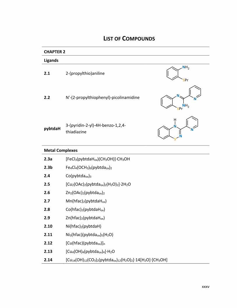
2.1 2-(propylthio)aniline
2.2 Nʹ-(2-propylthiophenyl)-picolinamidine
pybtdaH 3-(pyridin-2-yl)-4H-benzo-1,2,4-
thiadiazine
Metal Complexes
2.3a [FeCl3(pybtdaHox)(CH3OH)]∙CH3OH
2.3b Fe4Cl4(OCH3)6(pybtdaox)2
2.4 Co(pybtdaox)3
2.5 [Cu2(OAc)2(pybtdaox)2(H2O)2]∙2H2O
2.6 Zn2(OAc)2(pybtdaox)2
2.7 Mn(hfac)2(pybtdaHox)
2.8 Co(hfac)2(pybtdaHox)
2.9 Zn(hfac)2(pybtdaHox)
2.10 Ni(hfac)2(pybtdaH)
2.11 Ni3(hfac)(pybtdaox)5(H2O)
2.12 [Cu(hfac)(pybtdaox)]n
2.13 [Cu4(OH)4(pybtdaox)4]∙H2O
2.14 [Cu14(OH)12(CO3)2(pybtdaox)12(H2O)2]∙14[H2O]∙[CH3OH]

List of Compounds
xxxvi
CHAPTER 3
Ligands
3.1 Nʹ-(2-propylthiophenyl)-
picolinamidine
pmbtdaH (3-(2',6'-pyrimidine)-benzo-1,2,4-
thiadiazine)
Metal Complexes
3.2 [MnCl2(pmbtdaH)2]·2CH3OH
3.3 [FeCl2(pmbtdaH)2]·2CH3OH
3.4 [CoCl2(pmbtdaH)2]·2CH3OH
3.5 [NiCl2(pmbtdaH)2]·2CH3OH
3.6 Zn2Cl4(pmbtdaH)2
3.7a Mn(hfac)2(pmbtdaH) 3.7b [Mn2(hfac)2(tfa)2(pmbtdaHox)2]·2CH2Cl2
3.8a Co(hfac)2(pmbtdaH) 3.8b Co(hfac)2(pmbtdaHox)
3.9a Ni(hfac)2(pmbtdaH) 3.9b [Ni(hfac)2(pmbtdaHox)]·2CH2Cl2
3.10a Cu(hfac)2(pmbtdaH) 3.10b Cu4(hfac)4(tfa)2(pmbtdaox)2
3.11a Zn(hfac)2(pmbtdaH) 3.11b Zn4(hfac)6(pmbtdaox)2
List of Compounds
xxxvii
CHAPTER 4
Ligands
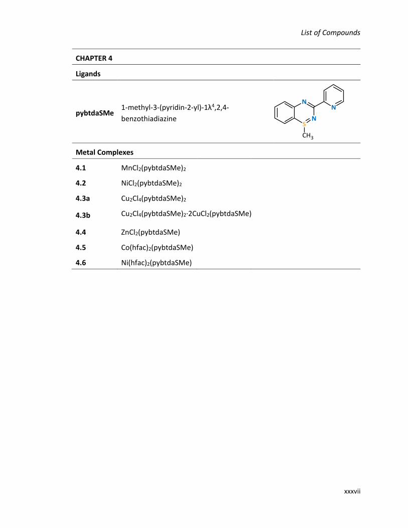
pybtdaSMe 1-methyl-3-(pyridin-2-yl)-1λ4,2,4-
benzothiadiazine
Metal Complexes
4.1 MnCl2(pybtdaSMe)2
4.2 NiCl2(pybtdaSMe)2
4.3a Cu2Cl4(pybtdaSMe)2
4.3b Cu2Cl4(pybtdaSMe)2·2CuCl2(pybtdaSMe)
4.4 ZnCl2(pybtdaSMe)
4.5 Co(hfac)2(pybtdaSMe)
4.6 Ni(hfac)2(pybtdaSMe)
List of Compounds
xxxviii
CHAPTER 5
Ligands
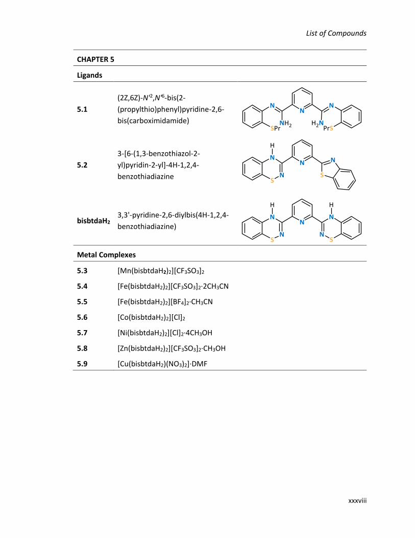
5.1
(2Z,6Z)-N'2,N'6-bis(2-
(propylthio)phenyl)pyridine-2,6-
bis(carboximidamide)
5.2
3-[6-(1,3-benzothiazol-2-
yl)pyridin-2-yl]-4H-1,2,4-
benzothiadiazine
bisbtdaH2 3,3'-pyridine-2,6-diylbis(4H-1,2,4-
benzothiadiazine)
Metal Complexes
5.3 [Mn(bisbtdaH2)2][CF3SO3]2
5.4 [Fe(bisbtdaH2)2][CF3SO3]2∙2CH3CN
5.5 [Fe(bisbtdaH2)2][BF4]2∙CH3CN
5.6 [Co(bisbtdaH2)2][Cl]2
5.7 [Ni(bisbtdaH2)2][Cl]2∙4CH3OH
5.8 [Zn(bisbtdaH2)2][CF3SO3]2∙CH3OH
5.9 [Cu(bisbtdaH2)(NO3)2]∙DMF

List of Abbreviations
xxxix
LIST OF ABBREVIATIONS
° degree
°C degree Celsius
6-311G*+ split-valence triple-zeta basis set with polarization and diffuse Gaussian functions
Å Angström
AcOH acetic acid
Ar aryl
aX hyperfine coupling constant to nucleus X
br broad (IR and NMR)
bpm 2,2ʹ-bipyrimidine
bpp 2,6-bis(pyrazol-3-yl)pyridine
bpy 2,2ʹ-bipyridine
BTA benzotriazinyl
btda benzothiadiazine
Bu butyl
ca circa (approximately)
cf confer (compare)
CF3SO3- trifluromethanesulfonic (triflate) anion
CFSE Crystal Field Stabilization Energy
CFT Crystal Field Theory
CSD Cambridge Structural Database
d doublet (NMR) or bond length (crystal structures)
dd doublet of doublets (NMR spectra)
DFT Density Functional Theory
DMF dimethylformamide
e– electron
e.g. exempli gratia (for example)
EPR Electron Paramagnetic Resonance
et al. et alia (and others)
List of Abbreviations
xl
etc et cetera (and so on)
Et2O diethyl ether
EtOH ethanol
eV electron Volt
g gram or g value (EPR)
h hour(s)
hfac hexafluoroacetylacetonate
HRMS high-resolution mass spectrometry
HS high-spin
Hz/MHz Hertz/Megahertz
i.e. id est (it is)
iPr isopropyl
IR infrared
IUPAC International Union of Pure and Applied Chemistry
J NMR coupling constant
K Kelvin
kJ kilojoule
LiHMDS lithium bis(trimethylsilyl)amide
LS low-spin
m multiplet (NMR) or medium (IR)
Me methyl
mg milligram
min minute
mL milliliter
mmol millimole
MOF metal organic frameworks
NCS N-chlorosuccinimide, isothiocyanate
NMR Nuclear Magnetic Resonance
OAc acetate
OTf trifluromethanesulfonic (triflate) anion
PB Prussian Blue
List of Abbreviations
xli
PE pairing energy
ph phenyl
phen 1,10-phenanthroline
pm pyrimidyl
PXRD Powder X-Ray Diffraction
py pyridyl
pyTTAH 3,5-dipyrid-2-yl-1,2,4,6-thiatriazinyl
q quartet (NMR)
R organic substituent
Rint reliability factors (crystallography)
R1, wR2 residual factors (crystallography)
RCOO– carboxylate
RO– alkoxide
RT room temperature
s singlet (NMR) or strong (IR)
SCO Spin Crossover
σm, σp Hammett parameters (meta and para substituted benzene rings)
T temperature
t triplet (NMR)
terpy terpyridine
THF tetrahydrofuran
TLC Thin Layer Chromatography
TTA thiatriazinyl
V volt
VD verdazyl
vdW van der Waal’s
w weak (IR)
X halogen
oct Octahedral Field Splitting
δ chemical shift (NMR) or atomic partial charge (DFT calculation)
G Gibb’s free energy

List of Abbreviations
xlii
H enthalpy
S entropy
θ angle
𝜐max (cm-1) IR absorption frequency

Ithaca gave you the marvelous journey.
Without her you would not have set out.
She has nothing left to give you now.
And if you find her poor, Ithaca won’t have fooled you.
Wise as you will have become, so full of experience,
you will have understood by then what these Ithacas mean.
-Ithaca, 1911
C.P. Cavafy
Η Ιθάκη σ’ έδωσε τ’ ωραίο ταξίδι.
Χωρίς αυτήν δε θα `βγαινες στον δρόμο.
Αλλά δεν έχει να σε δώσει πια.
Κι αν πτωχική την βρεις, η Ιθάκη δε σε γέλασε.
έτσι σοφός που έγινες, με τόση πείρα,
ήδη θα το κατάλαβες η Ιθάκες τι σημαίνουν.
-Ιθάκη, 1911
Κ.Π. Καβάφης

Chapter 1: Introduction to benzothiadiazines and coordination chemistry
2
Introduction to Benzothiadiazines and Coordination
Chemistry
Chapter 1: Introduction to benzothiadiazines and coordination chemistry
3
Introduction
1.1.1 Overview of coordination chemistry
Naturally occurring pigments have had applications as colorants since prehistoric
times. Archaeologists have evidence that humans used paint for aesthetic purposes such
as art and decorative uses. The range of colours available was very limited. Most of the
pigments originated from earth minerals, such as ochres and iron oxides, or derived from
pigments of biological origin. Some colours from unusual sources such as botanical
materials or insects were harvested and traded over long distances increasing the cost of
the pigment. For example, all shades of blue were associated with royalty because of their
rarity. Prussian Blue (PB) was one of the first synthetic pigments and has the formula
Fe4[Fe(CN)6]3·xH2O. It is believed to have been synthesized initially by the paint maker
Diesbach in Berlin around 1706 and used extensively. The oldest known use of PB was in
the ‘Entombment of Christ’ (1709) by van der Werff.1 The first published synthesis of PB
was reported by Stahl in 1731.2 Although first used as a pigment, PB has found many other
applications through the years. In the pharmaceutical industry, PB is used as an antidote
to treat thallium or radioactive cesium poisoning. It is also in the list of the most important
medications needed for a basic health system, the World Health Organization's List of
Essential Medicines.3 Pathologists use PB as a histopathology stain to detect the presence
of iron in biopsy specimens, such as in bone marrow samples. In analytical chemistry, PB
is used for the determination of total phenols or polyphenols. Last, Engineer's blue (PB in
an oily base) is used by toolmakers for spotting metal surfaces, such as surface
plates and bearings for hand scraping. PB is just one example of coordination compounds
and highlights the many varied applications they can exhibit.
In the 19th century, a number of theories were proposed for a better understanding
of their formation and properties, as more complexes were discovered. The most
successful and widely accepted of these theories was introduced by Alfred Werner (1866-
1919; Nobel Prize in Chemistry in 1913). His work was based on the physical and chemical
behaviour of a large number of coordination compounds which he studied by simple

Chapter 1: Introduction to benzothiadiazines and coordination chemistry
4
experimentation. Werner’s observations led him to propose that metal ions have two
different kinds of valence: (1) a primary valence that corresponds to the positive charge
of the metal ion, known as oxidation state and (2) a secondary valence that is the total
number of ligands bound to the metal ion, known as coordination number. A plethora of
coordination compounds can be formed given the numerous combinations of metal ions
and ligands. The applications of the metal complexes can be modified through ligand
design relying on the complementarity of ligand-based functional groups coupled with
the conformational preference(s) of metal ions. The field of coordination chemistry is
steadily increasing with applications in various fields from bioinorganic chemistry,4–6
medicine,7,8 catalysis9,10 and magnetism.11,12
1.1.2 Polydentate N-donor ligands
Coordination compounds are defined as ‘an assembly consisting of a central atom
(usually metallic) to which is attached a surrounding array of other groups of atoms
(ligands).13 Metal complexes can be mononuclear in which the metals are surrounded by
terminal ligands or polynuclear in which two or more metals are typically linked together
through organic ligands that can adopt bridging modes. Although there are many classes
of organic ligands, the focus of this dissertation will be polydentate N-donor ligands.
Polydentate N-donor ligands have exhibited predictable coordination reactivity and ease
of functionalization. The coordination chemistry of poly-pyridine ligands such as 2,2ʹ-
bipyridine (bpy),14,15 2,2ʹ-bipyrimidine (bpm),16–19 phenanthroline (phen),20 and the
terpyridine pincer (terpy)21–25 as well as larger macrocyclic ligands such as porphyrins26,27
has been extensively studied28–30 (Scheme 1.1). These ligands have found to act as ‘non-
innocent’ ligands which can potentially be involved in redox reactions.31–34

Chapter 1: Introduction to benzothiadiazines and coordination chemistry
5
Such polydentate N-donor ligands are found extensively in biological systems such as
haems and chlorophyll35,36 and materials applications include light-harvesting and light
emitting properties of the heavier transition metal complexes of 2,2ʹ-bipyridine and its
derivatives, such as [Ru(bpy)3]2+.37 These ligands have also found applications in molecular
magnetism since they offer a medium strength ligand field for first row transition metal
complexes.
Spin crossover (SCO) behaviour for these systems occurs when the crystal field
splitting () is comparable with the magnitude of the inter-electron repulsion term (PE)
making the two possible electronic states (high- and low-spin) for octahedral metal
complexes near equi-energetic.38–41 The ability to tune the crystal field by manipulating
the coordination sphere of the metal permits the value of to be tailored to compliment
the pairing energy, PE. The cobalt(II) complex, Co(terpy)2 complex ion was one of the first
compounds to be shown to exhibit thermal spin crossover.42–45 Some SCO transitions
occur slowly whereas other transitions are abrupt and others exhibit thermal hysteresis,
i.e. the temperature of the high-spin (HS) to low-spin (LS) transition does not occur at the
same temperature as the LS to HS transition (See section 1.3.1.e). There are many subtle
Scheme 1.1 Polydentate N-donor ligands.
bpy phen bpm
porphine terpy

Chapter 1: Introduction to benzothiadiazines and coordination chemistry
6
factors determining the characteristics of the HS/LS transition including (i) the nature of
the counter-ion (cationic complexes) and (ii) the existence of intermolecular interactions
between the SCO complexes, and (iii) the presence/absence of solvates in the lattice.46,47
Spin-transitions have been associated with the loss or gain of solvated molecules such as
in [Fe(bpp)2]X2 complex salts (bpp = 2,6-bis(pyrazol-3-yl)pyridine; X = anion).48,49 In these
cases, the imine N atoms coordinate to the Fe2+ cation, whereas the amino groups form
strong hydrogen bonds with solvent molecules and/or anions present in the crystal
structure.50 Therefore, the development of polydentate N-donor ligands with tunable
coordination chemistry is highly desirable for the construction of new functional
materials.
1.1.3 Heterocycles containing N and S/N atoms
New classes of magnetic materials include the formation of organic-inorganic
composite materials in which a coordinated bridging radical ligand achieves efficient
magnetic communication between paramagnetic transition metal ions. The ‘metal-radical
approach’ was first proposed by Gatteschi51 and a number of open-shell ligands such as
semi-quinones,52,53 nitroxides,54 thiazyl radicals,55–57 and verdazyls58–62 have been
extensively studied. The two most common strategies for the preparation of radical
complexes are: (i) the synthesis of the radical ligand followed by coordination to the
paramagnetic metal ion and (ii) coordination of the radical precursor to the desired
transition metal center and subsequent chemical/electrochemical oxidation or reduction
to afford the desired radical complex. The latter approach provides insight into the
preferred coordination modes of the radical precursor and its likely crystal field strength.
The thermodynamic stability of carbon-based radicals can be improved by the
replacement of one or more ring carbons by more electronegative atoms such as nitrogen
and sulphur. The incorporation of these electronegative elements lower the energies of
the orbitals adding electronic stabilization. In addition π-delocalization leads to the
distribution of the unpaired electron density over multiple atoms, reducing its chemical

Chapter 1: Introduction to benzothiadiazines and coordination chemistry
7
reactivity (with respect to free radical H atom abstraction, halogen atom abstraction or
reaction with O2).63,64 Below, we illustrate the cyclic 6-membered hydrazyl and thiazyl
radicals: verdazyls (VD), 1,2,4-benzotriazinyls (BTA), 1,2,4,6-thiatriazinyls (TTA) and
benzothiadiazines (BTDA) (Scheme 1.2).
Verdazyls (VD) were first reported in 1963, and their radicals are extensively
resonance delocalized and thermodynamically stable with exceptional air and moisture
stability.65 Pioneering work by Hicks and subsequently others on verdazyls has explored
them as ligands for coordination to first, second and third row transition metals, as well
as lanthanides.66,67 The 1,2,4-benzotriazinyl (BTA) radicals were first reported in 1968 and
are air, moisture, and thermally stable species which possess fully reversible redox
behavior.68 Although the majority of benzotriazinyls retain their monomeric character in
the solid state,69–71 there are two examples of strong association and spin-pairing
reported.72,73 Recently, the coordination chemistry of 1-phenyl-3-pyrid-2-yl-1,2,4-
benzotriazinyl to first-row metal ions has been explored.74,75 Thiazyls are electron-rich
radicals containing S and N atoms. In many cases thiazyl radicals are thermally robust but
the inherently polar nature of the S-N bond makes them susceptible to hydrolysis. The
first report of a thiatriazinyl (TTA) radical was described by Markovskii in 1983 using
electron paramagnetic resonance spectroscopy76 and in 1985 Oakley isolated and
structurally characterized the phenyl derivative (ph2TTAH, Scheme 1.3).77 The
coordination chemistry of 3,5-dipyrid-2-yl-1,2,4,6-thiatriazinyl (py2TTAH) has been
explored and shown to act as a pincer-type ligand forming mononuclear Fe complexes78,79
and a dysprosium dimer.80 The ligand has been oxidised in situ upon reaction to FeF3
Scheme 1.2 Nitrogen and sulphur/nitrogen-centered heterocyclic radicals.

Chapter 1: Introduction to benzothiadiazines and coordination chemistry
8
forming an one dimensional polymer with fluorine bridges but no coordination of the
S=O.79 In contrast, the ligand is bridging two dysprosium metal ions upon S-oxidation of
the ligand with the ligand bridging the metal centres.80 The Dy2 complex displays single-
molecule magnet behaviour where the origin has been attributed to the single-ion
anisotropy of the metal ion. The oxides of the phenyl (ph2TTAox)81 and pyridyl (py2TTAox)56
TTAs have been previously synthesised but no coordination chemistry has been reported
(Scheme 1.3).
The family of 1,2,4-benzothiadiazines (btda) will be discussed in more detail in the
following section.
Scheme 1.3 Derivatives of phenyl and pyridyl derivatives of thiadiazines (TTA).
ph2TTAH ph2TTAox
py2TTAH py2TTAox

Chapter 1: Introduction to benzothiadiazines and coordination chemistry
9
Overview of 1,2,4-benzothiadiazines
1.2.1 The family of 1,2,4-benzothiadiazines
The family of 1,2,4-benzothiadiazines in various oxidation states have attracted
attention for both materials and pharmaceuticals applications. The benzothiadiazine
heterocycle is known in the SII, SIV and SVI oxidation states (Scheme 1.4).
The benzothiadiazine ring system in the SVI oxidation state is particularly well-
established where it is an important constituent in a family of commercial thiazide
diuretics (Scheme 1.5) and was initially developed by Merck-Sharpe-Dohme82,83 for
treating hypertension. More recently it has proved successful in the treatment of
hyperglycemia and osteoporosis.3–6 Thiazides proved to decrease mineral bone loss by
directly stimulating bone mineral formation as well as promoting calcium retention in the
kidney.88 In addition, derivatives of 1,2,4-benzothiadiazine-dioxides have been used in
patents for various applications, such as inhibitors of hepatitis C virus replication,89
diabetes,90 arthritis91 and many others.
Scheme 1.5 Molecular structure of hydrochlorothiazide.
Scheme 1.4 Benzothiadiazines containing SII, SIV, and SVI centres.
thiadiazine (SII) thiadiazine S-oxide (SIV) thiadiazine S,S-dioxide
(SVI)

Chapter 1: Introduction to benzothiadiazines and coordination chemistry
10
The SII benzothiadiazines have found various applications in materials. The π-
conjugated SII system was first reported in the 1980’s to be redox active and undergoes
1e– chemical oxidation to form persistent (long-lived) radicals.92 Stable (isolable) radicals
have been isolated by Kaszynski when the benzo-fused ring is protected with chloro- or
fluoro-substituents, and long-chain alkyl ether-functionalised derivatives of phenyl-
benzothiadiazines have been found to exhibit liquid crystalline properties (Scheme 1.6,
A,B).93,94 Resonance-stabilized bis(thiadiazines) (R = H, Ph, thienyl) and related species
have also attracted attention as conducting materials (Scheme 1.6, C).95–97
Conversely, the SIV system had been barely investigated with the interconversion from
sulfide to sulfoxide to sulfone relying on careful control of the oxidizing agent and its
stoichiometric quantities.98–101 Recently, the metal-catalysed aerial oxidation of SII to SIV
under mild conditions was reported by Rawson.102 The SII system undergoes aerial
oxidation to the SIV system in the presence of a Lewis acidic metal of stoichiometric
(SII:Cu2+ = 2:1) and sub-stoichiometric (SII:Cu2+ = 20:1) quantities of CuII to form the
corresponding S-oxide. The Cu ions are taking part in a catalytic cycle in which initial N-
coordination of the btda ring activates the benzothiadiazine ring to N-deprotonation and
subsequent S-oxidation and the regeneration of Cu2+ results after liberation of the
deprotonated benzothiadiazine S-oxide. The first structurally characterized
benzothiadiazine S-oxides with non-coordinating phenyl or 2ʹ-thienyl rings in C(3)
Scheme 1.6 Derivatives of SII systems with materials’ applications.
A C B

Chapter 1: Introduction to benzothiadiazines and coordination chemistry
11
revealed that the S-oxide acts as a strong hydrogen-bond acceptor and unambiguously
determine the site of N-H protonation (at the 4-position of the heterocyclic ring).
1.2.2 Previous synthetic studies of 1,2,4-benzothiadiazines
There are a number of synthetic methodologies to access various derivatives of 1,2,4-
benzothiadiazines depending on the fused substituents and the group attached to C(3) as
shown in Scheme 1.7. Different synthetic routes are required depending on the
heteroatoms and ring substituents which affect the charge delocalisation and energy of
molecular orbitals. The most commonly used synthetic method is Route 1 in which the
benzothiadiazine is formed by the reaction of benzamidines with morpholine sulphide,
disulphide, or methane- and ethanesulfenyl chloride to give the ylide followed by a ring
closing reaction comprising NCS and heat.103–105 The relatively high temperatures
required in this synthetic route can lead to ring contraction and formation of
Scheme 1.7 Synthetic methodologies for the preparation of 1,2,4-benzothiadiazines. *Route 1: (i) morpholine sulphide, (ii) disulphide, (iii) methane- and ethanesulfenyl chloride.

Chapter 1: Introduction to benzothiadiazines and coordination chemistry
12
benzothiazole as side-products.103,106 Route 2 involves the ring closure of the benzamidine
with SOCl2 to give the S-oxide which is then reduced with thionyl chloride or Bu3P to give
the final product. Although this method gives yields over 70% there are side products
involved in the synthesis.107,108 For example, reduction with Bu3P can also result in ring
contraction to the benzothiazole and the use of SOCl2 results in chlorination of the benzo-
ring. The synthetic methodology of Route 3 provides an alternative strategy for the
formation of S-alkyl sulfilimines by the condensation of the amine and carbonitrile.
Oxidative cyclization of amidine by N-chlorosuccinimide gave the sulfilimine followed by
thermolysis to afford the benzothiadiazine.109,110 The only direct method to give the
desired benzothiadiazine is Route 4 and involves the condensation reaction of ortho-
aminophenols with hydroxamyl.111,112
1.2.3 Coordination chemistry of 1,2,4-benzothiadiazines
The first examples of coordination compounds with 1,2,4-benzothiadiazines were
recently reported by Rawson.113 The 3-(2’-pyridyl)-functionalized benzothiadiazine
(pybtdaH) has been shown to form a series of mononuclear complexes to first row
transition metal complexes of formula MClx(pybtdaH)y.113 The reaction of pybtdaH with
the transition metal chloride salts MCl2·xH2O (MII = Mn, Fe, Co, Cu, Zn) in a 2:1 mole ratio
afforded the MCl2(pybtdaH)2 complexes in which pybtdaH binds in a chelate fashion via
the heterocyclic N and the pyridyl N atoms. In the case of CuCl2 the 1:1 four-coordinate
complex MCl2(pybtdaH) was also isolated which adopts a polymeric structure with
pseudo-square planar molecules linked via long Cu⋯S contacts (dCu⋯S = 2.938(1) Å) in the
apical position. Reaction of pybtdaH with VCl3 in a 2:1 mole ratio afforded the vanadyl
complex [V(=O)Cl(pybtdaH)2][Cl]. In FeCl2(pybtdaH)2 the metal was found to adopt a high-
spin configuration, but reaction of pybtdaH with Fe(CF3SO3)2 in a 3:1 ratio afforded the
low-spin complex [Fe(pybtdaH)3][CF3SO3]2, consistent with the stronger field ligand
imposed by pybtdaH relative to Cl.
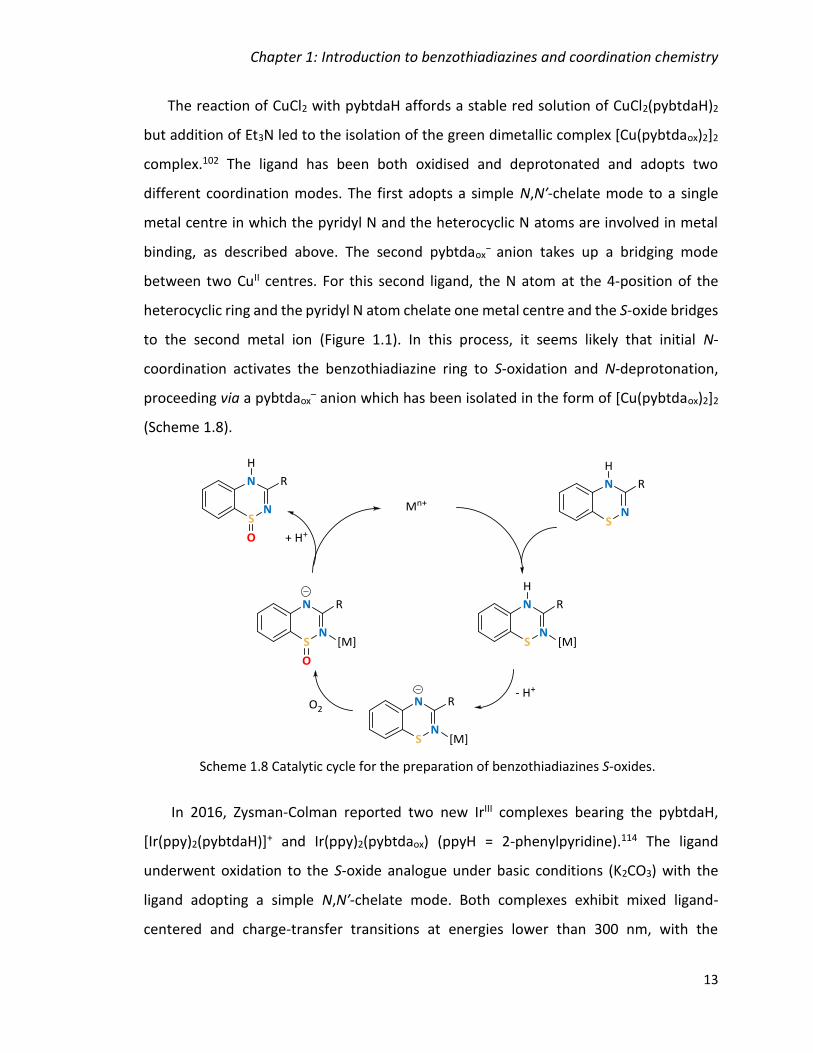
Chapter 1: Introduction to benzothiadiazines and coordination chemistry
13
The reaction of CuCl2 with pybtdaH affords a stable red solution of CuCl2(pybtdaH)2
but addition of Et3N led to the isolation of the green dimetallic complex [Cu(pybtdaox)2]2
complex.102 The ligand has been both oxidised and deprotonated and adopts two
different coordination modes. The first adopts a simple N,N′-chelate mode to a single
metal centre in which the pyridyl N and the heterocyclic N atoms are involved in metal
binding, as described above. The second pybtdaox− anion takes up a bridging mode
between two CuII centres. For this second ligand, the N atom at the 4-position of the
heterocyclic ring and the pyridyl N atom chelate one metal centre and the S-oxide bridges
to the second metal ion (Figure 1.1). In this process, it seems likely that initial N-
coordination activates the benzothiadiazine ring to S-oxidation and N-deprotonation,
proceeding via a pybtdaox– anion which has been isolated in the form of [Cu(pybtdaox)2]2
(Scheme 1.8).
In 2016, Zysman-Colman reported two new IrIII complexes bearing the pybtdaH,
[Ir(ppy)2(pybtdaH)]+ and Ir(ppy)2(pybtdaox) (ppyH = 2-phenylpyridine).114 The ligand
underwent oxidation to the S-oxide analogue under basic conditions (K2CO3) with the
ligand adopting a simple N,N′-chelate mode. Both complexes exhibit mixed ligand-
centered and charge-transfer transitions at energies lower than 300 nm, with the
Scheme 1.8 Catalytic cycle for the preparation of benzothiadiazines S-oxides.

Chapter 1: Introduction to benzothiadiazines and coordination chemistry
14
absorption in [Ir(ppy)2(pybtdaH)]+ trailing further into the visible region. Both complexes
were poorly emissive with the emission of cationic complex blue-shifted compared to the
neutral complex. The coordination modes of pybtdaH and pybtdaox– reported to date are
shown in Figure 1.1.
Coordination Chemistry
1.3.1 Theory of coordination chemistry
In a classical coordination complex, a central metal atom or ion is coordinated by one
or more molecules or ions (ligands), which act as Lewis bases forming coordination bonds
with the central atom or ion which acts as a Lewis acid. The atoms of the ligands that are
directly bonded to the central atom or ion are called donor atoms. A coordinate bond
(also called a dative covalent bond) is a covalent bond (a shared pair of electrons) in which
both electrons come from the same atom. In simple diagrams, a coordinate bond is
usually shown by an arrow pointing from the atom donating (ligand) the lone pair to the
atom accepting it (metal).
Classification of ligands
The ligands can be classified in terms of the charge and the number of coordination
sites it occupies. Ligands can be neutral (e.g. H2O, NH3, CH3OH) or anionic (e.g. HO–, Cl–,
CO32–). The ligands are also defined by their denticity (Latin, dentis, meaning ‘tooth’)
M
M
M
M
pybtdaH pybtdaox- pybtdaox
-
Figure 1.1 Coordination modes of pybtdaH and pybtdaox– reported to date.
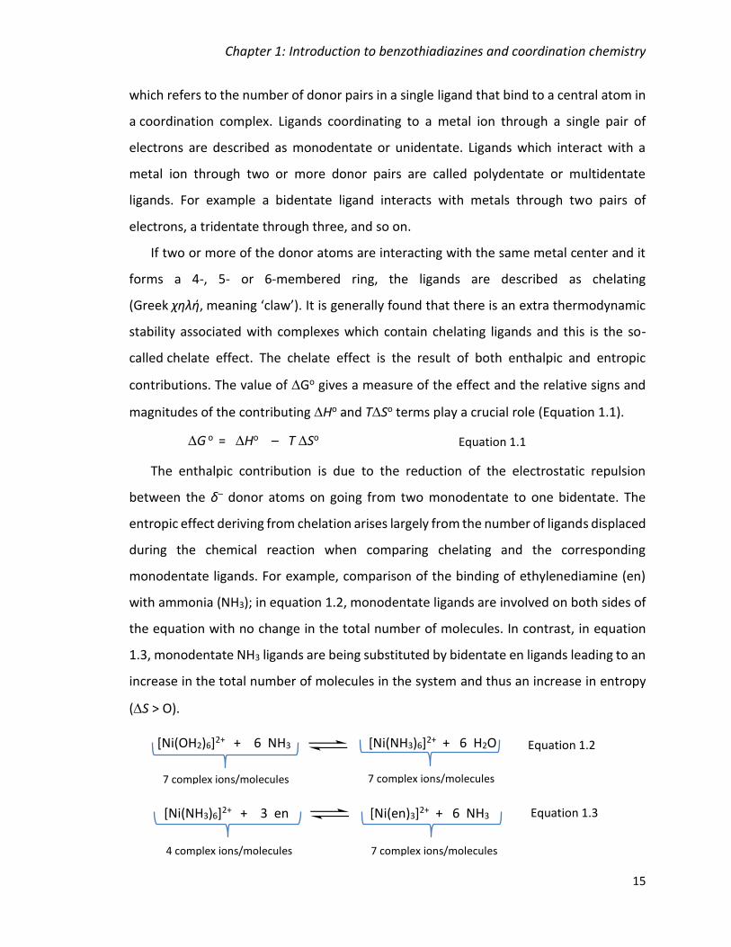
Chapter 1: Introduction to benzothiadiazines and coordination chemistry
15
which refers to the number of donor pairs in a single ligand that bind to a central atom in
a coordination complex. Ligands coordinating to a metal ion through a single pair of
electrons are described as monodentate or unidentate. Ligands which interact with a
metal ion through two or more donor pairs are called polydentate or multidentate
ligands. For example a bidentate ligand interacts with metals through two pairs of
electrons, a tridentate through three, and so on.
If two or more of the donor atoms are interacting with the same metal center and it
forms a 4-, 5- or 6-membered ring, the ligands are described as chelating
(Greek χηλή, meaning ‘claw’). It is generally found that there is an extra thermodynamic
stability associated with complexes which contain chelating ligands and this is the so-
called chelate effect. The chelate effect is the result of both enthalpic and entropic
contributions. The value of Go gives a measure of the effect and the relative signs and
magnitudes of the contributing Ho and TSo terms play a crucial role (Equation 1.1).
The enthalpic contribution is due to the reduction of the electrostatic repulsion
between the δ– donor atoms on going from two monodentate to one bidentate. The
entropic effect deriving from chelation arises largely from the number of ligands displaced
during the chemical reaction when comparing chelating and the corresponding
monodentate ligands. For example, comparison of the binding of ethylenediamine (en)
with ammonia (NH3); in equation 1.2, monodentate ligands are involved on both sides of
the equation with no change in the total number of molecules. In contrast, in equation
1.3, monodentate NH3 ligands are being substituted by bidentate en ligands leading to an
increase in the total number of molecules in the system and thus an increase in entropy
(S > O).
G o = Ho – T So Equation 1.1
[Ni(NH3)6]2+ + 3 en [Ni(en)3]2+ + 6 NH3
4 complex ions/molecules 7 complex ions/molecules
Equation 1.3
[Ni(OH2)6]2+ + 6 NH3 [Ni(NH3)6]2+ + 6 H2O
7 complex ions/molecules 7 complex ions/molecules
Equation 1.2
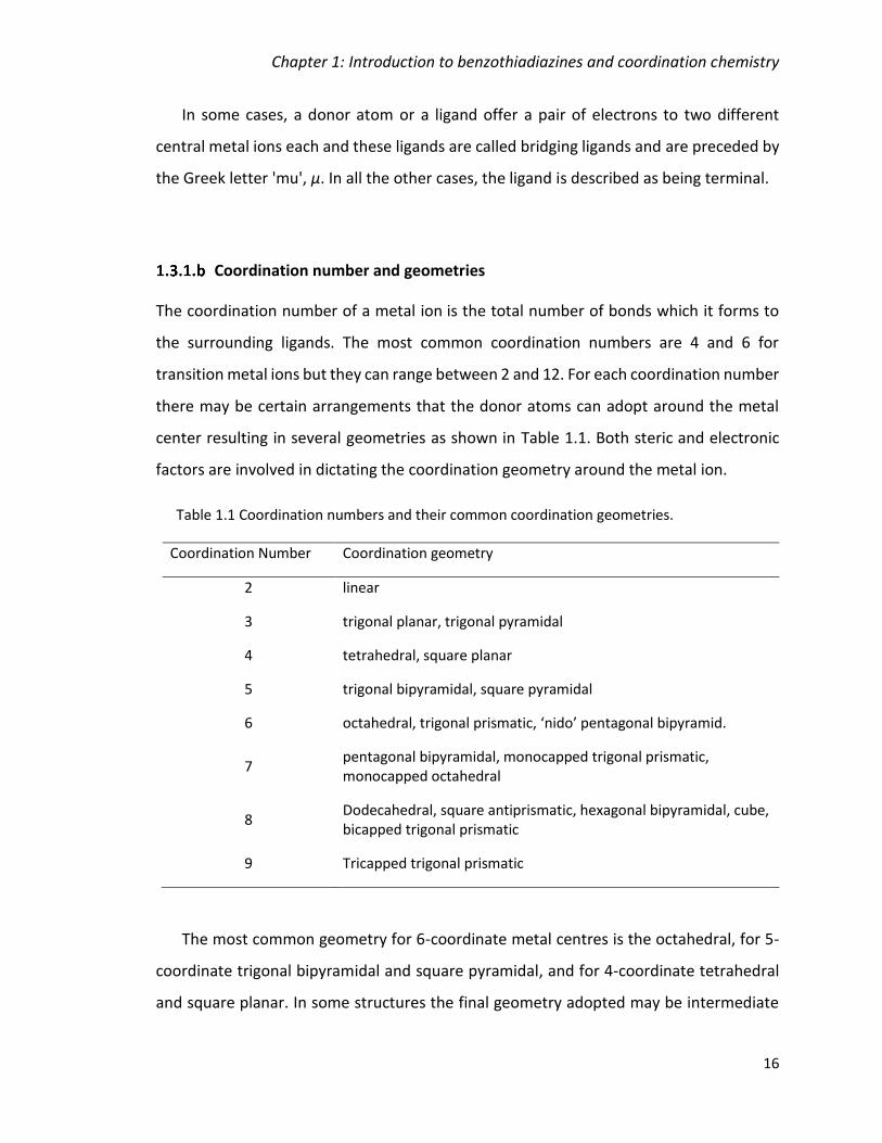
Chapter 1: Introduction to benzothiadiazines and coordination chemistry
16
In some cases, a donor atom or a ligand offer a pair of electrons to two different
central metal ions each and these ligands are called bridging ligands and are preceded by
the Greek letter 'mu', μ. In all the other cases, the ligand is described as being terminal.
Coordination number and geometries
The coordination number of a metal ion is the total number of bonds which it forms to
the surrounding ligands. The most common coordination numbers are 4 and 6 for
transition metal ions but they can range between 2 and 12. For each coordination number
there may be certain arrangements that the donor atoms can adopt around the metal
center resulting in several geometries as shown in Table 1.1. Both steric and electronic
factors are involved in dictating the coordination geometry around the metal ion.
Table 1.1 Coordination numbers and their common coordination geometries.
Coordination Number Coordination geometry
2 linear
3 trigonal planar, trigonal pyramidal
4 tetrahedral, square planar
5 trigonal bipyramidal, square pyramidal
6 octahedral, trigonal prismatic, ‘nido’ pentagonal bipyramid.
7 pentagonal bipyramidal, monocapped trigonal prismatic, monocapped octahedral
8 Dodecahedral, square antiprismatic, hexagonal bipyramidal, cube, bicapped trigonal prismatic
9 Tricapped trigonal prismatic
The most common geometry for 6-coordinate metal centres is the octahedral, for 5-
coordinate trigonal bipyramidal and square pyramidal, and for 4-coordinate tetrahedral
and square planar. In some structures the final geometry adopted may be intermediate

Chapter 1: Introduction to benzothiadiazines and coordination chemistry
17
between two or more of these idealized geometries and a series of parameters are
provided to establish the degree of distortion between different geometries for each
coordination number, e.g. τ4 would be used to measure the distortion between the four-
coordinate tetrahedral and square planar geometries.115 The τ parameter ranges between
0 and 1 with 0 and 1 corresponding to one of two idealized structures. The τ parameters
for 4, 5 and 6 coordinate geometries will be utilized in more detail later in this thesis. An
alternative approach to assess distortions from idealized geometries is to compute a set
of root mean square (RMS) deviations from the idealized geometries and the geometry
which has the smallest RMS deviation from ideality can be considered the best descriptor
of the coordination environment.116 The RMS deviation approach is discussed in Chapter
5.
Isomerism in coordination compounds
Isomers are compounds with the same formula but different properties that result
from different structures. There are two broad classes of isomers: structural isomers and
stereoisomers. Structural (or constitutional) isomers have the same molecular formula
but different molecular structures (different connectivities or different numbers of
chemical bonds). There are many types of structural isomers in transition metal
complexes. Some of the main types are the following:
(i) Ionization isomers: Interchange of anionic ligands between the first coordination
sphere with the outside, e.g. [Co(H2O)5F]+Cl- and [Co(H2O)5Cl]+F-.
(ii) Linkage isomers: one or more ligands can coordinate to the metal ion in more than
one way (ambidentate ligands), e.g. [CO(NH3)5(NO2)]2+ and [Co(NH3)5(ONO)]2+.
(iii) Coordination isomers: these exhibit the same ligands bound to the metal centre
but arranged in chemically distinct ways. Common examples are square planar complexes
where the ligands can be bound either cis (adjacent) or trans (opposite) to each other,
e.g. cis- and trans- isomers of platin, PtCl2(NH3)2.
Optical isomers have the same chemical formulae and atom connectivities but are
non-superimposable mirror images. Metal complexes in which the ligand bears three

Chapter 1: Introduction to benzothiadiazines and coordination chemistry
18
chelate ligands such as M(en)32+ can exhibit optical isomers and examples in this thesis
will be discussed in Chapter 3. Such complexes are typically described as being of R or S
handedness for tetrahedral complexes in an analogous fashion to organic chiral centres.
For octahedral complexes bearing three chelate ligands, the labels and are used
depending on whether their absolute configuration is left or right handed respectively.
The interconversion of and forms can occur through twist mechanisms including the
Bailar and the Ray-Dutt twist.117 These mechanisms include a transition through trigonal
prismatic geometry and the two mechanisms differ only in which pair of opposing
triangular faces is twisting. Some crystals of optical isomers may be of a single optical
isomer e.g. pure or pure and crystallise in non-centrosymmetric space groups.
However, optical isomers crystallising in centrosymmetric space groups will have a 50:50
mixture of both and forms. A racemic (50:50) mix of optical isomers may also be
generated by having two molecules in the asymmetric unit; one and one isomer.
Crystal Field Theory (CFT)
Crystal field theory is one of the three approaches to the bonding in complexes of the
d-block metals, along with ligand field (LF) theory and molecular orbital (MO) theory. The
most used approach in coordination chemistry is CFT which is a simple electrostatic model
which defines the ligands as negative point charges. Therefore, there is an electrostatic
attraction between the metal ion and the ligands, as well as repulsion between the
electrons in d-orbitals and the ligand point charges. In crystal field theory, the presence
of ligands leads to a splitting of the d-orbital degeneracy. The splitting depends on the
coordination geometry of the crystal field and the type of ligands which determine the
magnitude of the splitting. The shape of the d-orbitals is shown in Figure 1.2.
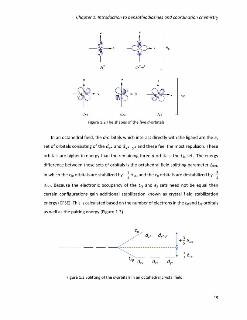
Chapter 1: Introduction to benzothiadiazines and coordination chemistry
19
In an octahedral field, the d-orbitals which interact directly with the ligand are the eg
set of orbitals consisting of the 𝑑𝑧2 and 𝑑𝑥2−𝑦2 and these feel the most repulsion. These
orbitals are higher in energy than the remaining three d-orbitals, the t2g set. The energy
difference between these sets of orbitals is the octahedral field splitting parameter oct,
in which the t2g orbitals are stabilized by − 2
5 oct and the eg orbitals are destabilized by +
3
5
oct. Because the electronic occupancy of the t2g and eg sets need not be equal then
certain configurations gain additional stabilization known as crystal field stabilization
energy (CFSE). This is calculated based on the number of electrons in the eg and t2g orbitals
as well as the pairing energy (Figure 1.3).
Figure 1.2 The shapes of the five d-orbitals.
dxy dxz dyz
dz2 dx2-y2
+
Δοct
-
Δοct
eg
t2g
Figure 1.3 Splitting of the d-orbitals in an octahedral crystal field.
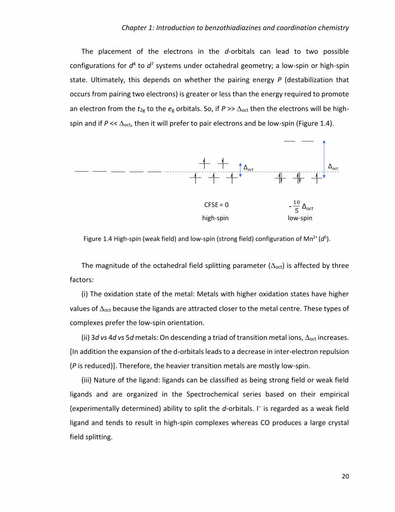
Chapter 1: Introduction to benzothiadiazines and coordination chemistry
20
The placement of the electrons in the d-orbitals can lead to two possible
configurations for d4 to d7 systems under octahedral geometry; a low-spin or high-spin
state. Ultimately, this depends on whether the pairing energy P (destabilization that
occurs from pairing two electrons) is greater or less than the energy required to promote
an electron from the t2g to the eg orbitals. So, if P >> oct then the electrons will be high-
spin and if P << oct, then it will prefer to pair electrons and be low-spin (Figure 1.4).
The magnitude of the octahedral field splitting parameter (oct) is affected by three
factors:
(i) The oxidation state of the metal: Metals with higher oxidation states have higher
values of oct because the ligands are attracted closer to the metal centre. These types of
complexes prefer the low-spin orientation.
(ii) 3d vs 4d vs 5d metals: On descending a triad of transition metal ions, oct increases.
[In addition the expansion of the d-orbitals leads to a decrease in inter-electron repulsion
(P is reduced)]. Therefore, the heavier transition metals are mostly low-spin.
(iii) Nature of the ligand: ligands can be classified as being strong field or weak field
ligands and are organized in the Spectrochemical series based on their empirical
(experimentally determined) ability to split the d-orbitals. I– is regarded as a weak field
ligand and tends to result in high-spin complexes whereas CO produces a large crystal
field splitting.
Figure 1.4 High-spin (weak field) and low-spin (strong field) configuration of Mn2+ (d5).
CFSE = 0
ΔοctΔοct
5Δοct
low-spin high-spin

Chapter 1: Introduction to benzothiadiazines and coordination chemistry
21
I– < Br– < S2– < SCN– < Cl– < NO3– < F– < OH– < H2O < NCS– < CH3CN < NH3 < en < bpy
< phen < NO2– < PPh3 < CN- < CO
Small
Weak-field ligand
Usually high-spin
Large
Strong-field ligands
Usually low-spin
It is also noteworthy that the size of the high-spin MII centres show an initial decrease
(M = Mn to Co) associated with increasing effective nuclear charge on traversing the first
transition metal series and subsequent increase (Cu and Zn) associated with the addition
of extra electrons into the eg orbitals since electrons in the eg set lead to greater electron-
electron repulsion with the ligand orbitals. [More complex arguments can be made
through MO theory where the eg set is found to be antibonding but ultimately generates
the same conclusion].
Spin Crossover
Spin crossover (SCO) or thermal spin transition is a change in the spin state of a metal
complex due to perturbation such as temperature, pressure or irradiation.40 If the
magnitude of the crystal field (oct) is energetically similar to the pairing energy (PE) then
the relative energies of the high- and low- spin complexes are similar. Under these
conditions then a perturbation such as temperature can result in a crossover event, where
the transition metal ion switches from being high-spin to being low-spin or vice versa.
The first row transition metals are the best candidates for SCO due to high inter-
electron repulsion (P) than second or third row transition metals which are predominantly
low-spin. Whilst many first row transition metals are high-spin, careful tailoring of the
crystal field by tuning the ligand coordination environment can lead to low-spin
configurations for these first row metals. Numerous methods exist to measure or detect
spin crossover including Mössbauer spectra, magnetic measurements, vibrational

Chapter 1: Introduction to benzothiadiazines and coordination chemistry
22
spectra, EPR and X-ray diffraction. Changes in both the magnetism and structure
accompanying a spin-transition will be described here.
One of the most characteristic changes associated with spin transitions is the change
in the magnetic response. For example, for FeII, the high-spin state has 4 unpaired
electrons (S = 2) whereas the low-spin state has no unpaired electrons (S = 0) (Figure 1.5).
As a consequence, the magnetic moment, µeff = g[S(S+1)]1/2 (or T = Ng22/3k S(S+1)) can
be used to detect a spin crossover event.
The abruptness of the transition (from high-spin to low-spin) is related to the
cooperativity of the process which relates to the ability to drive the spin change
throughout the whole lattice.39 In many cases, the spin-crossover phenomenon is
associated with an abrupt phase transition from 100 % low-spin (low temperature) to 100
% high-spin (high temperature), leading to a discontinuity in magnetic response which
results from the presence of strong cooperativity (Figure 1.6a). Such behaviour has
potential for exploitation in ‘spin-switching’ devices where the magnetic response is
turned ‘on/off’ with small changes in temperature or pressure. In other cases, the spin
transition can be more gradual (Figure 1.6b), occurring over a wider temperature range
(due to low cooperativity). When equilibrium exists between ‘high-spin’ (HS) and ‘low-
spin’ (LS) configurations, the conversion from HS to LS may be incomplete (Figure 1.6c).
Thermal hysteresis may occur for systems in which cooperativity is high but there is a
significant energy barrier to interconversion from LS to HS. Thus at some finite
ΔοctΔοct
Low-spin (S = 0)High-spin (S = 2)
eg
t2g
eg
t2g
Figure 1.5 High- and low-spin configurations of Fe2+ (d6) ion.

Chapter 1: Introduction to benzothiadiazines and coordination chemistry
23
temperature T the HS state may be more stable than the LS configuration but the phase
transition only occurs once the thermal energy kT > Ea. On cooling the HS state is
maintained until the LS state becomes more thermodynamically stable (Figure 1.6d), i.e.
the LS state may exist as a metastable state until there is sufficient thermal energy to
overcome the energy barrier for structural reorganization. This indicates bistability of the
system allowing the complex to transition from one electronic state to the other upon the
influence of some external perturbation. Such behaviour would allow such complexes to
be used in storage, memory and for lighting devices.39 The described scenarios are shown
in the spin transition curves (Figure 1.6, a-d) where T1/2 is the spin transition temperature
(at which the ratios of HS to LS are 1:1) and 𝛾𝐻𝑆 is the molar fraction of the high-spin
state.
Figure 1.6 Spin transition curves of high-spin molar fraction vs temperature; (a)
abrupt, (b) gradual, (c) incomplete, (d) hysteresis.
0
1
0.5
T
gHS
T1/2
0
1
0.5
T>
gHS
T1/2
>
0
1
0.5
T>
gHS
T↓ T↑
><
Heat
Cool
><
0
1
0.5
T
gHS
T1/2
a) b)
d)c)
Chapter 1: Introduction to benzothiadiazines and coordination chemistry
24
Another characteristic change associated with SCO transitions is the change in metal-
ligand bond distance observed in the crystal structure when the spin state goes from high-
to low-spin. The high-spin state has some of its electrons in the antibonding eg orbitals
however, demotion of these electrons to the low-spin state places all of them in the
nonbonding t2g orbitals (for example FeII, d6, Figure 1.5). Therefore, the metal-ligand bond
is strengthened (shortened) which can be observed in X-ray. The difference in metal-
ligand bond length for FeII HS vs LS is large, and amounts to approximately 0.2 Å.118 The
difference is smaller in FeIII and even in CoII because only one electron is being transferred
from eg to t2g orbitals.43
Octahedral complexes of d9 and high-spin d4 usually exhibit a Jahn-Teller distortion.
The Jahn-Teller theorem states that any non-linear system in a degenerate electronic
state will be unstable and will undergo distortion to form a system of lower symmetry
and lower energy by removing the degeneracy. As a result, the axial metal-ligand bonds
have different bond lengths than the equatorial bonds. The distortion can be either an
elongation or compression of the axial bonds and numerous Cu2+ complexes described in
this thesis show a preferred axial elongation.
1.3.2 Synthetic methodology of coordination chemistry
A key question currently central to the field of coordination chemistry is: ‘What are
the synthetic strategies for isolating polynuclear metal clusters?’. Two possible synthetic
strategies are reported in the literature.
The first approach is to use well-defined structural building units (SBUs) with well-
defined topologies coupled with ligands which offer a specific structure-directing metal-
ligand interaction. This approach underpins many of the recent developments in areas
such as the formation of metal organic frameworks (MOFs). Ligands which offer well-
defined chelating or bridging topologies such as polydentate N-donors are often
implemented in these approaches. This can result in the creation of aesthetically
Chapter 1: Introduction to benzothiadiazines and coordination chemistry
25
‘beautiful’ molecules through the synthesis of complex substitutes that have controlled
modes of coordination.119
Secondly, ‘serendipitous assembly’, in which a series of competing reactions are
present and a number of system variables (pH, temperature, concentration, ligand:metal
ratio, etc) by following some logical principles in order to influence the original product
rather than dictate it by using a dominant structure-directing group. A major disadvantage
of this approach is the limited synthetic control, while a significant advantage is the
possible isolation of a large number of clusters from a particular reaction system, and also
new synthetic methods of controlling the structure are discovered.120 The use of ligands
which offer a versatile or flexible set of metal binding sites are well-suited to
serendipitous approaches due to the potential for the metal centers to adopt several
coordination geometries. Small variations in the chemical identity of the ligands can
potentially lead to new products and often involve choice of differing carboxylate ligands
(RCOO−) or alkoxides (RO−) in which the carboxylate has an established versatility in the
bonding mode available (section 1.3.3) and the alkoxide can be terminal, or bridging (2
or 3). Tuning the R group can change both the steric and electronic demands of these
groups and hence change the outcome of their chemical reactivity patterns. Initial
observations should lead to new, more ‘designed’ experiments and logical synthetic
thoughts, exhausting all synthetic parameters at each time (ratio of reactants, pH,
solvent, counter-ions, and temperature). In this serendipitous self-assembly process, we
typically aim to predict certain likely outcomes of the reaction e.g. preferred coordination
modes for different ligands but cannot predict a priori how these interactions will
manifest themselves in the presence of different metals or ligands.
The general synthetic scheme for isolating polynuclear transition metal complexes of
the first row transition metals followed in our research is the following:
a : b = ratio of metal to ligand
a MXy + b LH ? base
solvent

Chapter 1: Introduction to benzothiadiazines and coordination chemistry
26
M = first row transition metal ions,
X = counter-ions of the metal salts which may act as a terminal ligand (monodentate,
chelate) or as a bridging (bidentate, tridentate, etc.) substituent or can be non-
coordinating ligands;
LH = organic molecules which have the ability to act as chelate substituents and after
deprotonation can adopt bridging modes.
The metal salts employed during our experiments are the following: MClx (x = 2, 3),
M(OAc)2 and M(hfac)2. When using MClx the reaction mixture is slightly acidic since the
Cl− anion is pH neutral (conjugate base of a strong acid) and the Mx+ cation is Lewis acidic.
Conversely the use of M(OAc)2 and M(hfac)2 offer more basic conditions due to the
presence of OAc− and hfac− anions which are the conjugate bases of weak acids (HOAc
and Hhfac respectively). The pH can be further manipulated by addition of base (Et3N) to
create an even more basic environment and enhance ligand deprotonation.
The ratio of the metal to organic ligand is altered to explore the effect of the reaction
outcome. Several solvents and crystallization techniques are employed to adjust to the
solubility of the product and isolate single crystals for X-ray crystallography.
The simultaneous presence of a terminal ligand to terminating potential
polymerization and a bridging ligand (preferably the organic ligand) that can act as a
bridging or simultaneously bridging and chelating substituent is expected to favour the
formation of polynuclear complexes.
1.3.3 Coordination modes of acetate (OAc–) and hexafluoroacetylacetate (hfac–)
groups
The employment of acetate (OAc–) and hexafluoroacetylacetate (hfac–) groups in our
reaction scheme is due to two reasons: (i) the weakly basic environment they offer as
anions in solution and (ii) the variety of ways in which they can complex metal ions.
Simple carboxylate ions can act as monodentate, O-donor ligands, as 1,3-chelate
ligands and/or as bridging substituents using 2-oxygen atoms. Conversely the hfac−

Chapter 1: Introduction to benzothiadiazines and coordination chemistry
27
groups mostly adopt a chelate mode but some examples of bridging hfac groups have
been reported. The unsolvated form of first row transition metals, M(hfac)3 (M = Mn, Co,
Ni) exhibits the chelating-bridging mode,121 as well as complexes incorporating Ag, Cd, Hg
and In metal ions.122,123
The crystallographically established ways of complexing AcO– and hfac– with various
metal ions are shown in Scheme 1.9 and clearly highlight the diversity of potential
coordination modes possible.
There are two ways to describe the coordination mode of a ligand to a metal centre.
The μ/η and the Harris notation. Denticity is the coordination of a ligand to a metal center
through multiple atoms and is described with the Greek letter η ('eta'). In the case of a
bridging ligand the letter μ ('mu') is used to indicate the total number of metals bridged.
The Harris notation was proposed in 2000 where the binding mode is referred to as
[X.Y1Y2Y3…Yn], where X is the overall number of metals bound by the whole ligand, and
each value of Y refers to the number of metal atoms attached to the different donor
η1:η1
1.11
η1:η0
1.10
η1:η1
1.10
μ2-η1:η1
2.11
μ3-η2:η1
3.21
μ3-η2:η2
3.22
μ2-η2:η1
2.21
Scheme 1.9 Selected crystallographically reported coordination modes of the OAc– (left) and
hfac– (right) ligands, and the μ/η and Harris notations which describe these
modes.
μ2-η2:η1
2.21

Chapter 1: Introduction to benzothiadiazines and coordination chemistry
28
atoms.124 The ordering of Y is listed by the Cahn-Ingold-Prelog priority rules (i.e. S before
O before N). The proposed notation was to distinguish the coordination of different type
of atoms of a ligand such as a ligand coordinating through N and O atoms.

Chapter 1: Introduction to benzothiadiazines and coordination chemistry
29
Dissertation Objectives
The coordination chemistry of 1,2,4-benzothiadiazines known to date is limited to a
series of mononuclear first row metal complexes of 3-(2’-pyridyl)-functionalized
benzothiadiazine (pybtdaH)113, a dimer with formula [Cu(pybtdaox)2]2102, and two iridium
complexes, [Ir(ppy)2(pybtdaH)]+ and Ir(ppy)2(pybtdaox)114, as discussed previously. The SII
system undergoes oxidation to the SIV system in the presence of a base such as Et3N or
K2CO3.102,114 The S-oxide on the dimer offers an additional donor atom leading to a
bridging mode of the ligand in addition to the N,N′-chelate pocket. This ability of the
ligand gives rise to a new perspective on the coordination chemistry of 1,2,4-
benzothiadiazines since it translates the strictly chelating nature of the pybtdaH ligand to
a more versatile bridging ligand with N,N’ and O donor pockets favouring a more
serendipitous approach to coordination chemistry. In the following chapters, we will
present the synthesis and characterization of a series of 1,2,4-benzothiadiazines in which
their structures are tailored to enhance their coordination abilities (Scheme 1.10).
pybtdaH pmbtdaH pybtdaSMe
bisbtdaH2
Scheme 1.10 Derivatives of 1,2,4-benzothiadiazines presented in the dissertation.

Chapter 1: Introduction to benzothiadiazines and coordination chemistry
30
In Chapter 2, a new synthetic route of the synthesis of pybtdaH will be discussed as
well as kinetics studies on the formation of the final product. Computational studies of
the ligand to probe the energetics of the heterocycle upon rotation of the pyridyl ring will
be presented. The pybtdaH ligand undergoes oxidation to form the S-oxide and
deprotonation to form the pybtdaox– anion under basic conditions, as discussed above.
Previous work investigated only MCl2 where the chloride has no effect on pH since
chloride is the conjugate base of a strong acid (pKa (HCl) = -6.1 125) and has minimal base
character. Conversely, the OAc– and hfac– anions are the conjugate bases of weak acids
(pKa (AcOH) = 4.76 126, pKa (Hhfac) = 4.71 127) and generation of the anions in solution
could promote the oxidation of the pybtdaH ligand instead of a base, such as Et3N or
K2CO3 used previously (Scheme 1.11).102,114 In addition, the acetate groups (OAc–) offer a
variety of coordination modes and can potentially bridge up to four metal centers
favouring the formation of polynuclear complexes (Scheme 1.9).
The employment of first row transition metal salts containing the basic counterions
OAc– and hfac– with the ligand pybtdaH will be presented. The effect of additional base
(Et3N) in the reaction scheme has been explored and will be discussed.
In Chapter 3, the synthesis and characterization of the novel redox active ligand 3-
(2',6'-pyrimidine)-benzo-1,2,4-thiadiazine (pmbtdaH) is reported (Scheme 1.10). The
pmbtdaH ligand employs a pyrimidyl ring on position 3 of the heterocycle increasing the
donor atoms offered for coordination. The neutral pmbtdaH ligand acts as a simple N,N′-
chelate to transition metal ions whereas the pmbtdaox– anion is more versatile offering
HCl H
+ + Cl– pKa = -6.1 pH neutral
HOAc H
+ + OAc–
Hhfac H+ + hfac–
pKa = 4.76
pKa = 4.71
mild base
mild base
Scheme 1.11 Logarithimic dissociation constants of HCl, AcOH and Hhfac.

Chapter 1: Introduction to benzothiadiazines and coordination chemistry
31
two N,N′ binding sites for bridging two metal centres as well as a pendant O-donor which
offers an additional binding site favouring formation of polynuclear metal complexes. The
coordination chemistry of the ligand with various first row transition metals salts is
presented as well as the effect on the reactivity pathways of pmbtdaH with metal ions
with weakly basic anions (hfac–) favouring the deprotonation/oxidation process.
The synthesis and characterization of the 1-methyl-3-(pyridinyl)-1,2,4-
benzothiadiazine (pybtdaSMe, Scheme 1.10) is presented in Chapter 4. Although in
pybtdaHox the oxygen acts as an additional donor atom, the oxidation of the ligand in
reaction schemes involve basic conditions is inevitable in open-air conditions. Alkylation
at sulphur could prevent such behaviour and the effect of the electronics of the
heterocycle to the reactivity of the ligand can be explored. The ligand pybtdaSMe offers
one coordination pocket to a metal centre through N,N′-chelation by coordination of
either the heterocyclic N atoms at either 2 or 4 positions. In addition, the ligand is not
protonated leaving both nitrogen atoms of the heterocycle equally available as donor
atoms. Its coordination chemistry to selected first row transition metal complexes with
MCl2 and M(hfac)2 is presented.
In Chapter 5, the novel S/N based terpyridine type ligand bisbtdaH2 has been
synthesized and its coordination chemistry with first row transition metal complexes has
been investigated (Scheme 1.10). The ligand is designed to offer a tridentate mer-binding
coordination geometry and its coordination chemistry with selected first row transition
metal ions is described.

Chapter 1: Introduction to benzothiadiazines and coordination chemistry
32
References
1 J. Bartoll, in 9th International Conference on NDT of Art, Jerusalem Israel, 2008.
2 G. E. Stahl, Exp. Obs. Animadversiones CCC Numer. Chymicae Phys., 1731, 281.
3 World Health Organization, WHO Model List Essential Medicines, 2013.
4 W. Ruttinger and G. C. Dismukes, Chem. Rev., 1997, 97, 1.
5 V. K. Yachandra, K. Sauer and M. P. Klein, Chem. Rev., 1996, 96, 2927.
6 A. K. Poulsen, A. Rompel and C. J. McKenzie, Angew. Chemie - Int. Ed., 2005, 44, 6916.
7 S. Dasari and P. B. Tchounwou, Eur. J. Pharmacol., 2015, 5, 364.
8 J. Paken, C. D. Govender, M. Pillay and V. Sewram, J. Toxicol., 2016.
9 G. W. Parshall and S. D. Ittel, Adv. Chem., 1992, 42.
10 D. Morales-Morales, Rev. la Soc. Química México, 2004, 48, 338.
11 O. Kahn, Molecular Magnetism, Wiley-VCH, New York, 1993.
12 D. Gatteschi, L. Bogani, A. Cornia, M. Mannini, L. Sorace and R. Sessoli, Solid State Sci., 2008, 10, 1701.
13 IUPAC. Compendium of Chemical Terminology, 2nd ed. Compiled by A. D. McNaught and A. Wilkinson., Blackwell Scientific Publications, Oxford, 1997.
14 C. Kaes, A. Katz and M. W. Hosseini, Chem. Rev., 2000, 100, 3553.
15 B. H. Ye, M. L. Tong and X. M. Chen, Coord. Chem. Rev., 2005, 249, 545.
16 P. Alborés and E. Rentschler, Dalton Trans., 2013, 42, 9621.
17 P. O. Adelani, N. D. Cook and P. C. Burns, Cryst. Growth Des., 2014, 14, 5692.
18 I. F. Díaz-Ortega, J. M. Herrera, D. Aravena, E. Ruiz, T. Gupta, G. Rajaraman, H. Nojiri and E. Colacio, Inorg. Chem., 2018, 57, 6362.
19 P. Alborés and E. Rentschler, Inorg. Chem., 2010, 49, 8953.
20 P. G. Sammes and G. Yahioglu, Chem. Soc. Rev., 1994, 327.
21 K. Takada, R. Sakamoto, S. T. Yi, S. Katagiri, T. Kambe and H. Nishihara, J. Am. Chem. Soc., 2015, 137, 4681.
22 H. Hofmeier and U. S. Schubert, Chem. Soc. Rev., 2004, 33, 373.
Chapter 1: Introduction to benzothiadiazines and coordination chemistry
33
23 U. S. Schubert, H. Hofmeier and G. R. Newkome, Modern Terpyridine Chemistry, Wiley-VCH, 2006.
24 E. C. Constable, Chem. Soc. Rev., 2007, 36, 246.
25 A. M. W. Cargill Thompson, Coord. Chem. Rev., 1997, 160, 1.
26 C. G. Claessens, U. Hahn and T. Torres, Chem. Rec., 2008, 8, 75.
27 K. Kadish, R. Guilard and K. M. Smith, The Porphyrin Handbook, Academic Press, 2003.
28 J. A. G. Williams, Chem. Soc. Rev., 2009, 38, 1783.
29 M. A. Halcrow, Coord. Chem. Rev., 2005, 249, 2880.
30 C. Bazzicalupi, A. Bencini, A. Bianchi, A. Danesi, E. Faggi, C. Giorgi, S. Santarelli and B. Valtancoli, Coord. Chem. Rev., 2008, 252, 1052.
31 G. Accorsi, A. Listorti, K. Yoosaf and N. Armaroli, Chem. Soc. Rev., 2009, 38, 1690.
32 P. J. Chirik and K. Wieghardt, Science, 2010, 327, 794.
33 A. B. Sorokin, Chem. Rev., 2013, 113, 8152.
34 T. Zell, P. Milko, K. L. Fillman, Y. Diskin-Posner, T. Bendikov, M. A. Iron, G. Leitus, Y. Ben-David, M. L. Neidiq and D. Milstein, Chem. – Eur. J., 2014, 4403.
35 M. Paoli, J. Marles-Wright and A. Smith, DNA Cell Biol., 2002, 21, 271.
36 J. K. Hoober, L. L. Eggink and M. Chen, Photosynth. Res., 2007, 94, 387.
37 E. A. Medlycott and G. S. Hanan, Chem. Soc. Rev., 2005, 34, 133.
38 M. del C. Gimenez Lopez, M. Clemente Leon and C. Gimenez-Saiz, Dalton Trans., 2018, 47, 10453.
39 P. Gutlich and H. A. Goodwin, Spin Crossover in Transition Metal Compounds I–III, Springer, Top. Curr. Chem., 2004.
40 M. A. Halcrow, Chem. Soc. Rev., 2011, 40, 4119.
41 A. G. Craig, O. Roubeau and G. Aromí, Coord. Chem. Rev., 2014, 269, 13.
42 C. A. Kilner and M. A. Halcrow, Dalton Trans., 2010, 39, 9008.
43 I. Krivokapic, M. Zerara, M. L. Daku, A. Vargas, C. Enachescu, C. Ambrus, P. Tregenna-Piggott, N. Amstutz, E. Krausz and A. Hauser, Coord. Chem. Rev., 2007, 251, 364.

Chapter 1: Introduction to benzothiadiazines and coordination chemistry
34
44 S. Hayami, Y. Komatsu, T. Shimizu, H. Kamihata and Y. H. Lee, Coord. Chem. Rev., 2011, 255, 1981.
45 S. Hayami, M. R. Karim and Y. H. Lee, Eur. J. Inorg. Chem., 2013, 683.
46 G. Dupouy, M. Marchivie, S. Triki, J. Sala-Pala, J. Y. Salaün, C. J. Gómez-García and P. Guionneau, Inorg. Chem., 2008, 47, 8921.
47 J. A. Thomas, Dalton Trans., 2011, 40, 12005.
48 G. A. Craig, J. S. Costa, O. Roubeau, S. J. Teat and G. Aromí, Chem. - A Eur. J., 2012, 18, 11703.
49 E. Coronado, M. Giménez López, C. Gimenez-Saiz, J. M. Martinez-Agudo and F. M. Romero, Polyhedron, 2003, 22, 2375.
50 K. H. Sugiyarto and H. A. Goodwin, Aust. J. Chem., 1988, 41, 1645.
51 A. Caneschi, D. Gatteschi, R. Sessoli and P. Rey, Acc. Chem. Res., 1989, 22, 392.
52 R. M. Buchanan, B. J. Fitzgerald and C. G. Pierpont, Inorg. Chem., 1979, 18, 3439.
53 H. H. Downs, R. M. Buchanan and C. G. Pierpont, Inorg. Chem., 1979, 18, 1736.
54 A. Caneschi, D. Gatteschi, R. Sessoli and P. Rey, Inorg. Chem., 1988, 27, 1756–1761.
55 D. J. Sullivan, R. Clérac, M. Jennings, A. J. Lough and K. E. Preuss, Chem. Commun., 2012, 48, 10963.
56 K. E. Preuss, Coord. Chem. Rev., 2015, 289–290, 49.
57 J. Wu, D. J. MacDonald, R. Clérac, I. R. Jeon, M. Jennings, A. J. Lough, J. Britten, C. Robertson, P. A. Dube and K. E. Preuss, Inorg. Chem., 2012, 51, 3827.
58 D. J. R. Brook, S. Fornell, J. E. Stevens, B. Noll, T. H. Koch and W. Eisfeld, Inorg. Chem., 2000, 39, 562.
59 D. J. R. Brook, Comments Inorg. Chem., 2015, 35, 1.
60 V. Polo, A. Alberola, J. Andres, J. Anthony and M. Pilkington, Phys. Chem. Chem. Phys., 2008, 10, 857.
61 M. Chahma, K. Macnamara, A. Van der Est, A. Alberola, V. Polo and M. Pilkington, New J. Chem., 2007, 31, 1973.
62 R. G. Hicks, M. T. Lemaire, L. K. Thompson and T. M. Barclay, J. Am. Chem. Soc., 2000, 122, 8077.
63 J. M. Rawson, A. Alberola and A. Whalley, J. Mater. Chem., 2006, 16, 2560.
Chapter 1: Introduction to benzothiadiazines and coordination chemistry
35
64 D. C. Nonhebel, J. M. Tedder and J. C. Walton, Radicals, Cambridge University Press, 1979.
65 R. Kuhn and H. Trischmann, Angew. Chem. Int. Ed., 1963, 2, 155.
66 G. N. Lipunova, T. G. Fedorchenko and O. N. Chupakhin, Russ. Chem. Rev., 2013, 82, 701.
67 C. Train, L. Norel and M. Baumgarten, Coord. Chem. Rev., 2009, 2342.
68 H. M. Blatter and H. Lukaszewski, Tetrahedron Lett., 1968, 9, 2701.
69 Y. Takahashi, Y. Miura and N. Yoshioka, New J. Chem., 2015, 39, 4783.
70 Y. Miura and N. Yoshioka, Chem. Phys. Lett., 2015, 626, 11.
71 Y. Takahashi, Y. Miura and N. Yoshioka, Chem. Lett., 2014, 43, 1236.
72 C. P. Constantinides, A. A. Berezin, G. A. Zissimou, M. Manoli, G. M. Leitus, M. Bendikov, M. R. Probert, J. M. Rawson and P. A. Koutentis, J. Am. Chem. Soc., 2014, 136, 11906.
73 C. P. Constantinides, P. A. Koutentis and G. Loizou, Org. Biomol. Chem., 2011, 9, 3122.
74 I. S. Morgan, A. Peuronen, M. M. Hänninen, R. W. Reed, R. Clérac and H. M. Tuononen, Inorg. Chem., 2014, 53, 33.
75 I. S. Morgan, A. Mansikkamäki, G. A. Zissimou, P. A. Koutentis, M. Rouzières, R. Clérac and H. M. Tuononen, Chem. - A Eur. J., 2015, 21, 15843.
76 O. M. Markovskii, L. N.; Kornuta, P. P.; Kachkovskaya, L. S.; Polumbrik, Sulfur Lett., 1983, 143.
77 P. J. Hayes, R. T. Oakley, A. W. Cordes and W. T. Pennington, J. Am. Chem. Soc., 1985, 107, 1346.
78 K. L. M. Harriman, A. A. Leitch, S. A. Stoian, F. Habib, J. L. Kneebone, S. I. Gorelsky, I. Korobkov, S. Desgreniers, M. L. Neidig, S. Hill, M. Murugesu and J. L. Brusso, Dalton Trans., 2015, 44, 10516.
79 K. L. M. Harriman, I. A. Kühne, A. A. Leitch, I. Korobkov, R. Clérac, M. Murugesu and J. L. Brusso, Inorg. Chem., 2016, 55, 5375.
80 N. Yutronkie, I. Kuehne, I. Korobkov, J. L. Brusso and M. Murugesu, Chem. Commun., 2015, 677.
81 A. W. Cordes and R. T. Oakley, Acta Cryst., 1987, 2, 1645.

Chapter 1: Introduction to benzothiadiazines and coordination chemistry
36
82 P. G. Welling, Biopharm. Drug Dispos., 1986, 7, 501.
83 A. Fretheim, BMC Fam. Pr., 2003, 4, 19.
84 R. M. Taylor and M. M. Winbury, Nature, 1960, 187, 603.
85 J. M. Sprague and F. C. Novello, J. Am. Chem. Soc., 1957, 2028.
86 S. S. Fajans, J. C. Floyd Jr., R. F. Knopf, J. Rull, E. M. Guntsche and J. W. Conn, J. Clin. Invest., 1966, 481.
87 A. Fretheim, BMC Fam. Pr., 2003, 4, 19.
88 M. M. Dvorak, C. De Joussineau, D. H. Carter, T. Pisitkun, M. A. Knepper, G. Gamba, P. J. Kemp and D. Riccardi, J. Am. Soc. Nephrol., 2007, 18, 2509.
89 L. Beigelman, G. Wang, B. Buckman and A. D. Stoycheva, Preparation of benzothiadiazine dioxides as inhibitors of hepatitis C virus replication, InterMune, Inc., USA, 2009.
90 M. S. Congreve, B. G. Christopher, John Andrew Tehan, S. S. Klair and S. J. Aves, Preparation of 1,2,4-benzothiadiazin-3(4H)-one 1, 1-dioxides as orexin receptor antagonists, Heptares Therapeutics Limited, UK, 2014.
91 J. Busch-Petersen, W. Fu, J. K. Kerns, M. R. Palovich, K. L. Widdowson and U. Assignee: SmithKline Beecham Corporation, IL-8 receptor antagonists of substiuted 3-phenylamino-4H-1,2,4-benzothiadiazin-5-ol 1,1-dioxides, SmithKline Beecham Corporation, USA 2005, 2005.
92 L. N. Markovskii, V. S. Talanov, O. M. Polumbrik and Y. G. Shermolovich, J. Org. Chem., 1981, 17, 2338.
93 J. Zienkiewicz, P. Kaszynski and V. G. Young Jr, J. Org. Chem., 2004, 69, 7525.
94 J. Zienkiewicz, A. Fryszkowska, K. Zienkiewicz, F. Guo, P. Kaszynski, A. Januszko and D. Jones, J. Org. Chem., 2007, 72, 3510.
95 L. Beer, R. C. Haddon, M. E. Itkis, A. A. Leitch, R. T. Oakley, R. W. Reed, J. F. Richardson and D. G. VanderVeer, Chem. Commun., 2005, 1218.
96 S. M. Winter, A. R. Balo, R. J. Roberts, K. Lekin, A. Assoud, P. A. Dubeand and R. T. Oakley, Chem. Commun., 2013, 49, 1603.
97 A. A. Leitch, R. T. Oakley, R. W. Reed and L. K. Thompson, Inorg. Chem., 2007, 46, 6261.
98 D. D. Ross and D. Lednicer, Zh. Org. Khim., 1984, 20, 196.
99 G. Kresze, C. Seyfried and A. Trede, Tetrahedron Lett., 1965, 6, 3933.
Chapter 1: Introduction to benzothiadiazines and coordination chemistry
37
100 N. Finch, S. J. Ricca, L. H. Werner and R. Rodebaugh, J. Org. Chem., 1980, 3416.
101 K. Kaczorowska, Z. Kolarska, K. Mitka and P. Kowalski, Tetrahedron, 2005, 61, 8315–8327.
102 E. R. Clark, J. J. Hayward, B. J. Leontowicz, M. U. Anwar, M. Pilkington and J. M. Rawson, Dalton Trans., 2014, 44, 2071.
103 T. L. Gilchrist, C. W. Rees and D. Vaughan, J. Chem. Soc., Perkin Trans, 1983, 55.
104 T. L. Gilchrist, C. W. Rees and D. Vaughan, J. Chem. Soc., Chem. Commun., 1978, 1049.
105 T. L. Gilchrist, C. W. Rees and D. Vaughan, J. Chem. Soc., Perkin Trans, 1983, 49.
106 V. W. Bohnisch, T. L. Gilchrist and C. W. Rees, J. Chem. Soc., Perkin Trans, 1979, 2851.
107 N. Finch, S. Ricca, L. H. Werner and R. Rodebaughs, J. Org. Chem., 1980, 45, 3416.
108 G. Kresze, C. Seyfried and A. Trede, Liebigs Ann. Chem., 1968, 715, 223.
109 P. Kaszynski and V. G. Young, J. Org. Chem., 2004, 69, 2551.
110 E. R. Clark, J. J. Hayward, B. J. Leontowicz, D. J. Eislera and J. M. Rawson, CrystEngComm, 2014, 16, 1755.
111 A. A. Leitch, I. Korobkov, A. Assoud and J. L. Brusso, Chem. Commun., 2014, 50, 4934.
112 A. O. Abdelhamid, A. M. Negm and T. M. S. Abdeen, Arch. Pharm. (Weinheim Ger.), 1988, 321, 913.
113 E. R. Clark, M. U. Anwar, B. J. Leontowicz, Y. Beldjoudi, J. J. Hayward, W. T. K. Chan, E. L. Gavey, M. Pilkington, E. Zysman-Colman and J. M. Rawson, Dalton Trans., 2014, 43, 12996.
114 A. K. Pal, D. B. Cordes, K. Pringouri, M. U. Anwar, A. M. Z. Slawin, J. M. Rawson and E. Zysman-Colman, J. Coord. Chem., 2016, 69, 1924.
115 L. Yang, D. R. Powell and R. P. Houser, Dalton Trans., 2007, 955.
116 C. Andreini, G. Cavallaro and S. Lorenzini, Bioinformatics, 2012, 28, 1658.
117 H. S. Rzepa and M. E. Cass, Inorg. Chem., 2007, 46, 8024.
118 H. Zheng, K. M. Langner, G. P. Shields, J. Hou, M. Kowiel, F. H. Allen, G. Murshudov and W. Minor, Acta Crystallogr. Sect. D Struct. Biol., 2017, 73, 316.

Chapter 1: Introduction to benzothiadiazines and coordination chemistry
38
119 M. Fujita, K. Umemoto, M. Yoshizawa, N. Fujita, T. Kusukawa and K. Biradha, Chem. Commun., 2001, 509.
120 R. E. P. Winpenny, J. Chem. Soc. Dalton Trans., 2002, 6, 1.
121 H. Zhang, B. Li, J. Sun, R. Clérac and E. V. Dikarev, Inorg. Chem., 2008, 47, 10046.
122 H. Zhang, B. Li and E. V. Dikarev, J. Clust. Sci., 2008, 19, 311.
123 J.-L. Jin, Y.-L. Shen, Y.-P. Xie and X. Lu, CrystEngComm, 2018, 20, 2036.
124 R. A. Coxall, S. G. Harris, D. K. Henderson, S. Parsons, P. A. Tasker and R. E. P. Winpenny, J. Chem. Soc. Dalton Trans., 2000, 2349.
125 A. Trummal, L. Lipping, I. Kaljurand, I. A. Koppel and I. Leito, J. Phys. Chem. A, 2016, 120, 3663.
126 N. J. Tro, Chemistry: A molecular approach, Pearson, New Jersey, 2nd Ed., 2011.
127 M. Ellinger, H. Duschner and K. Starke, J. Inorg. Nucl. Chem., 1978, 40, 1063.

Synthesis and characterization of pybtdaH and its
coordination chemistry

Chapter 2: Synthesis and characterization of pybtdaH and its coordination chemistry
40
Introduction
The 3-(2ʹ-pyridyl)-functionalized benzothiadiazine (pybtdaH) has been shown to form
a series of chelate complexes to first row transition metal complexes of formula
MClx(pybtdaH)y.1 More recently we have shown that the SII system undergoes aerial
oxidation to the SIV system (Scheme 2.1) in the presence of a Lewis acidic metal and a
base, such as Et3N or K2CO3.2 In this process, it seems likely that initial N-coordination
activates the benzothiadiazine ring to S-oxidation and N-deprotonation, proceeding via a
pybtdaox– anion which has been isolated in the form of [Cu(pybtdaox)2]2
2 and
Ir(ppy)2(pybtdaox).3 In the former case the ligand adopts a bridging mode in which the
ligand is N,Nʹ-chelating to one metal and binds to a second metal via the S-oxide O atom.
In the iridium complex the ligand adopts a terminal N,Nʹ-chelate mode. S-oxidation of
some other thiazyl heterocycles at metal centres has also been described recently.4,5
Scheme 2.1 (top) Formal SII, SIV and SVI oxidation states of the benzothiadiazine framework;
(bottom) SII, SIV and SVI variants of the 2-pyridyl benzothiadiazine heterocycle.

Chapter 2: Synthesis and characterization of pybtdaH and its coordination chemistry
41
These initial studies indicated that S-oxidation of pybtdaH and other
benzothiadiazines only occurred in the presence of a metal and Et3N.2 We speculated that
the use of metal complexes bearing more basic anions might favour such ligand oxidation
processes without the addition of a formal base. Previous studies investigated the
reactivity of MCl2 in which the chloride anion is pH neutral since Cl– is the conjugate base
of a strong acid (pKa (HCl) = -6.1 6). In contrast, anions such as OAc– and hfac– are the
conjugate bases of weak acids (pKa (AcOH) = 4.76 7, pKa (Hhfac) = 4.71 8) and generation
of OAc– and hfac– in solution could promote the in situ oxidation of the pybtdaH ligand.
In addition, the acetate groups (OAc–) offer a variety of coordination modes (Section
1.3.3) and can potentially bridge up to four metal centers,9,10 while the resultant pybtdaox–
ligand can bind at least two metal ions. The combination of these ligands should favour
formation of polynuclear complexes. While hfac– has been reported to adopt O,Oʹ-
bridging modes,11–13 it normally adopts a chelate mode and is likely to lead to monomeric
complexes, M(hfac)2(pybtdaHox).
The synthesis of the ligand 3-pyridyl-benzothiadiazine (pybtdaH, Scheme 2.1) has
been described previously14 but several improvements to the ligand synthesis are
described in this Chapter as well as an examination of its coordination chemistry to
selected first row transition metal complexes, M(OAc)2 and M(hfac)2. The potential
versatility of coordination modes of the benzothiadiazine ligand to metal centres is shown
in Figure 2.1. The pybtdaH ligand offers one coordination pocket to a metal centre
through N,Nʹ-chelation in its neutral and anionic (pybtda–) or radical form (pybtda•)
(Figure 2.1, left). Upon S-oxidation, the ligand can offer a more versatile hard O-donor
centre in addition to the favourable, softer, N,Nʹ-chelate, leading to enhanced
aggregation of up to two metal ions for the pybtdaox– ligand (Figure 2.1, right) or even up
to three metal centers if the oxygen of the S-oxide adopts a bridging mode.
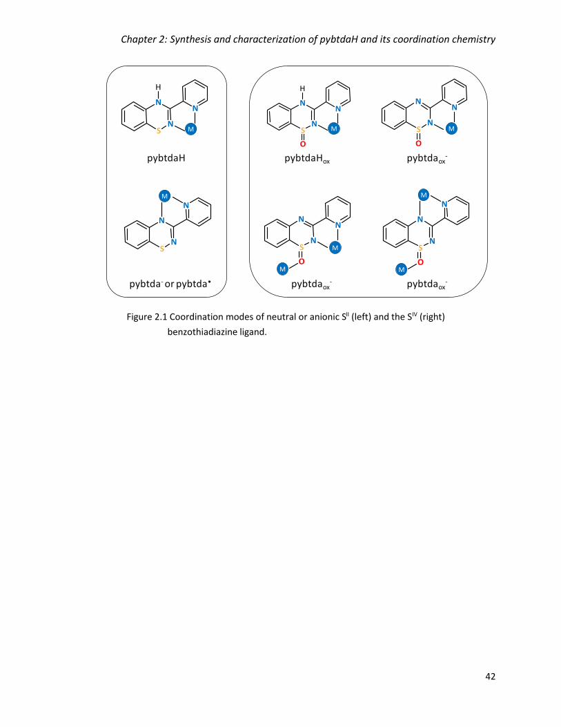
Chapter 2: Synthesis and characterization of pybtdaH and its coordination chemistry
42
Figure 2.1 Coordination modes of neutral or anionic SII (left) and the SIV (right)
benzothiadiazine ligand.
M
M
M
M
M
pybtdaH
pybtda- or pybtda•
pybtdaHox pybtdaox-
pybtdaox- pybtdaox
-
M
M
M
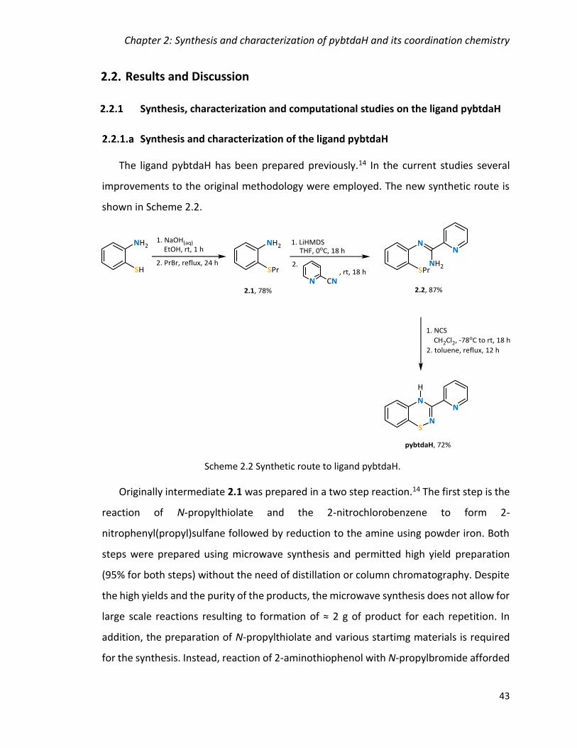
Chapter 2: Synthesis and characterization of pybtdaH and its coordination chemistry
43
Results and Discussion
2.2.1 Synthesis, characterization and computational studies on the ligand pybtdaH
Synthesis and characterization of the ligand pybtdaH
The ligand pybtdaH has been prepared previously.14 In the current studies several
improvements to the original methodology were employed. The new synthetic route is
shown in Scheme 2.2.
Originally intermediate 2.1 was prepared in a two step reaction.14 The first step is the
reaction of N-propylthiolate and the 2-nitrochlorobenzene to form 2-
nitrophenyl(propyl)sulfane followed by reduction to the amine using powder iron. Both
steps were prepared using microwave synthesis and permitted high yield preparation
(95% for both steps) without the need of distillation or column chromatography. Despite
the high yields and the purity of the products, the microwave synthesis does not allow for
large scale reactions resulting to formation of ≈ 2 g of product for each repetition. In
addition, the preparation of N-propylthiolate and various startimg materials is required
for the synthesis. Instead, reaction of 2-aminothiophenol with N-propylbromide afforded
Scheme 2.2 Synthetic route to ligand pybtdaH.

Chapter 2: Synthesis and characterization of pybtdaH and its coordination chemistry
44
2-(propylthio)aniline (2.1) over one step in 78% yield. Although the yield is lower than the
reported one, the reaction can be scaled up to 10 g and there is no need for preparation
of other reagents. The purification of the product by Kugelrohr distillation does not
require the use of solvents making it overall a greener synthesis.
Deprotonation of 2-(propylthio)phenylamine (2.1) with LiHMDS, under anaerobic, dry
conditions followed by treatment with one equivalent of 2-cyanopyridine and work-up
resulted in a brown oil. The oil was washed with hexanes to yield the pure intermediate
(2.2) in 87% yield as a yellow crystalline powder. The preparation of the amidine follows
the reported method with the exception of the purification of the product (washes with
hexanes) resulting to the crystalline material and thus more moderate yields. Single-
crystal X-ray diffraction revealed the product shown in Figure 2.2 crystallizing in the
monoclinic space group C2/c with one molecule in the asymmetric unit. The 1H NMR
spectrum of 2.2 exhibits the expected proton environments in the aromatic and the
aliphatic region (propyl group).
The subsequent oxidative cyclization and pericyclic elimination of 2.2 in the presence
of N-chlorosuccinimide (NCS) afforded the ligand pybtdaH, which was isolated in 72%
yield. The ligand pybtdaH could not be purified by recrystallization using a heated mixture
Figure 2.2 Molecular structure of Nʹ-(2-propylthiophenyl)-picolinamidine (2.2).
N3
N1N2
S1
C8C9
C10
C11
C7C6 C5
C4
C2
C1
C14
C13
C15
C12
C3

Chapter 2: Synthesis and characterization of pybtdaH and its coordination chemistry
45
of hexanes and dichloromethane (80:20) as reported,14 therefore column
chromatography was used with dichloromethane as eluent. Single crystals of pybtdaH
were grown by slow evaporation from dichloromethane. The 1H NMR spectrum of ligand
pybtdaH exhibits the expected proton environments in the aromatic region. The N-H
vibration was observed at 3308 cm-1 and the imine C=N stretching vibration was observed
at 1627 cm-1, as expected.15 The molecular structure has previously been confirmed by
single crystal X-ray studies (Figure 2.3).14
The compound crystallizes in the monoclinic space group P1̅ with two molecules in
the asymmetric unit. The C(6)-N(2) distance is 1.286(2) Å, consistent with predominant
double bond character (cf imine C=N at 1.279 Å)16. The bond length between C(6)-N(3) at
1.375(2) Å is more consistent with a conjugated C-N single bond (cf Ph-NH2 at 1.355 Å).16
The S-N bond length (1.708(1) Å) is comparable with other S-N single bonds (1.62 – 1.74
Å for isomers of S8-x(NH)x)17 and 1.69 – 1.72 Å for other benzothiadiazine derivatives).14
Another feature in the benzothiadiazine ring is the folding (θ) of the molecule about
the trans-annular vector S(1)···N(3). The values for the majority of previously reported
benzothiadiazines fall in the range 21° < θ < 42° 14 whereas θ is 27.27° in pybtdaH. The
folding of the ring is consistent with the formally 8π anti-aromatic nature of the
thiadiazine framework.
Figure 2.3 Molecular structure of pybtdaH.
N3
N1
N2
S1
C8
C9
C10
C11
C7
C6
C5 C3
C4
C2
C1
C12
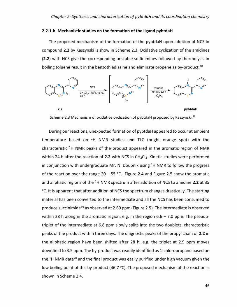
Chapter 2: Synthesis and characterization of pybtdaH and its coordination chemistry
46
Mechanistic studies on the formation of the ligand pybtdaH
The proposed mechanism of the formation of the pybtdaH upon addition of NCS in
compound 2.2 by Kaszynski is show in Scheme 2.3. Oxidative cyclization of the amidines
(2.2) with NCS give the corresponding unstable sulfinimines followed by thermolysis in
boiling toluene result in the benzothiadiazine and eliminate propene as by-product.18
During our reactions, unexpected formation of pybtdaH appeared to occur at ambient
temperature based on 1H NMR studies and TLC (bright orange spot) with the
characteristic 1H NMR peaks of the product appeared in the aromatic region of NMR
within 24 h after the reaction of 2.2 with NCS in CH2Cl2. Kinetic studies were performed
in conjunction with undergraduate Mr. N. Doupnik using 1H NMR to follow the progress
of the reaction over the range 20 – 55 oC. Figure 2.4 and Figure 2.5 show the aromatic
and aliphatic regions of the 1H NMR spectrum after addition of NCS to amidine 2.2 at 35
oC. It is apparent that after addition of NCS the spectrum changes drastically. The starting
material has been converted to the intermediate and all the NCS has been consumed to
produce succinimide19 as observed at 2.69 ppm (Figure 2.5). The intermediate is observed
within 28 h along in the aromatic region, e.g. in the region 6.6 – 7.0 ppm. The pseudo-
triplet of the intermediate at 6.8 ppm slowly splits into the two doublets, characteristic
peaks of the product within three days. The diagnostic peaks of the propyl chain of 2.2 in
the aliphatic region have been shifted after 28 h, e.g. the triplet at 2.9 ppm moves
downfield to 3.5 ppm. The by-product was readily identified as 1-chloropropane based on
the 1H NMR data20 and the final product was easily purified under high vacuum given the
low boiling point of this by-product (46.7 oC). The proposed mechanism of the reaction is
shown in Scheme 2.4.
Scheme 2.3 Mechanism of oxidative cyclization of pybtdaH proposed by Kaszynski.18

Chapter 2: Synthesis and characterization of pybtdaH and its coordination chemistry
47
Figure 2.4 1H NMR screening of the pybtdaH formation upon addition of NCS in 2.2 at
35 oC highlighting the conversion of the pseudo-triplet into two doublets (aromatic
region) (500 MHz, CDCl3).
pybtdaH
75 h
70 h
48 h
28 h
2.2
Figure 2.5 1H NMR screening of the pybtdaH formation upon addition of NCS in 2.2 at 35 oC
focusing in the aliphatic region showing the formation of the by-product,
1-chloropropane after 28 hours (500 MHz, CDCl3, (3.48 (Cl-CH2), 1.77 (Cl-CH2CH2),
0.99 (CH3), 2.69 (succinimide) ppm).
pybtdaH
75 h
70 h
48 h
28 h
2.2

Chapter 2: Synthesis and characterization of pybtdaH and its coordination chemistry
48
Computational studies on ligand pybtdaH
Two possible tautomers exist for each orientation of the pyridyl ring of the ligand,
depending on whether the N(2) or N(3) atom is protonated, as shown in Table 2.1.
Structural studies located the H atom position at N(3) and this assignment was further
supported by shorter imine-like C(6)-N(3) and longer C(6)-N(2) bonds as discussed above.
The preference for the crystallographically-determined tautomer (structure 1) was
confirmed by DFT calculations (B3LYP-D3/6-311G*+) on structures 1 - 4. The studies
revealed that structure 1 was more stable by 60 kJ/mol, when compared to the least
stable structure 4 (Table 2.1). Notably reorientation of the ligand conformation from 1 to
2 in order to adopt an N,Nʹ-chelate conformation also has a significant energetic cost (44
kJ/mol).
Scheme 2.4 Proposed mechanism of the oxidative cyclization of pybtdaH.

Chapter 2: Synthesis and characterization of pybtdaH and its coordination chemistry
49
Table 2.1 DFT calculations (B3LYP-D3/6-311G*+) of the tautomers of different orientation
of the pyridyl group of pybtdaH.
Structure
1 2 3 4
Relative energy
(kJ/mol) 0 44 29 60
Geometry optimized B3LYP/6-311G*+ DFT studies on pybtdaH were undertaken to
probe the energetics of the ring system, varying the rotation angle of the pyridyl ring
[torsion N(3)C(6)C(5)N(1)] between 0° and 360° whilst permitting the rest of the molecule
to optimize (Figure 2.6).
These calculations reveal an energy maximum at θ = 180°, as expected due to the
repulsion of N(2) of the heterocycle and N(1) of the pyridyl group (Figure 2.7). Two lower
energy points are observed at 135° and 225°, which are ±45° from the energy maximum
of 180° orientation. This can be attributed to less repulsion of the N atoms in that
conformation as the orbitals are not in the same plane.
Figure 2.6 Rotation of pyridyl group of pybtdaH for the DFT studies.
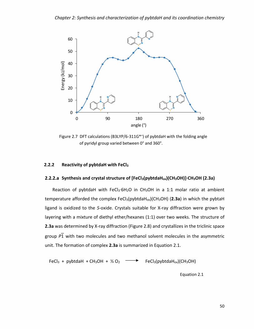
Chapter 2: Synthesis and characterization of pybtdaH and its coordination chemistry
50
2.2.2 Reactivity of pybtdaH with FeCl3
Synthesis and crystal structure of [FeCl3(pybtdaHox)(CH3OH)]∙CH3OH (2.3a)
Reaction of pybtdaH with FeCl3·6H2O in CH3OH in a 1:1 molar ratio at ambient
temperature afforded the complex FeCl3(pybtdaHox)(CH3OH) (2.3a) in which the pybtaH
ligand is oxidized to the S-oxide. Crystals suitable for X-ray diffraction were grown by
layering with a mixture of diethyl ether/hexanes (1:1) over two weeks. The structure of
2.3a was determined by X-ray diffraction (Figure 2.8) and crystallizes in the triclinic space
group P1 with two molecules and two methanol solvent molecules in the asymmetric
unit. The formation of complex 2.3a is summarized in Equation 2.1.
FeCl3 + pybtdaH + CH3OH + ½ O2 FeCl3(pybtdaHox)(CH3OH)
Equation 2.1
Figure 2.7 DFT calculations (B3LYP/6-311G*+) of pybtdaH with the folding angle
of pyridyl group varied between 0° and 360°.
0
10
20
30
40
50
60
0 90 180 270 360
Ener
gy (k
J/m
ol)
angle (°)

Chapter 2: Synthesis and characterization of pybtdaH and its coordination chemistry
51
The metal centre is in oxidation state 3+ and adopts a pseudo-octahedral geometry
with the three chloride ligands counter-balancing the charge. The Fe-Cl distances are in
the range 1.9949(15) – 2.5679(19) Å, as expected for FeIII. The ligand adopts a simple
N,Nʹ-chelate mode to a single metal centre in which the pyridyl N(1) and the heterocyclic
N(2) atoms are involved in metal binding. The Fe-N bond distances are in the range
2.157(4) – 2.494(5) Å and the chelate ligand pybtdaHox exhibits internal Npy-Fe-NBTDA
angles in the range 62.77(15) – 72.2(2)° and a neutral methanol molecule completes the
coordination sphere.
Two molecules form a centrosymmetric dimer via a pair of N-H···O hydrogen bonding
interaction (dN-H···O = 2.328 Å, θN-H···O = 151.79°) between the N-H group of the
benzothiadiazine ring and the oxygen of the sulfoxide (Figure 2.9, green lines). This
packing additionally places the benzo and pyridyl rings of neighbouring molecules in close
proximity with centroid···centroid distances of 4.148 Å with the closest C···C distance at
3.700 Å (cf interlayer separation in graphite at 3.354 Å21) (Figure 2.9, red lines). In
addition, the molecules are linked via O-H···Cl hydrogen bonding from the solvent CH3OH
oxygen atoms and the chlorides on the metal complex (dO-H···Cl = 2.870 Å,
θO-H···Cl = 130.91°).
Figure 2.8 Molecular structure of FeCl3(pybtdaHox)(CH3OH) (2.3a) (solvent molecule
omitted for clarity).
N2
N1
Cl3
Cl2 Cl1
Fe1 O2
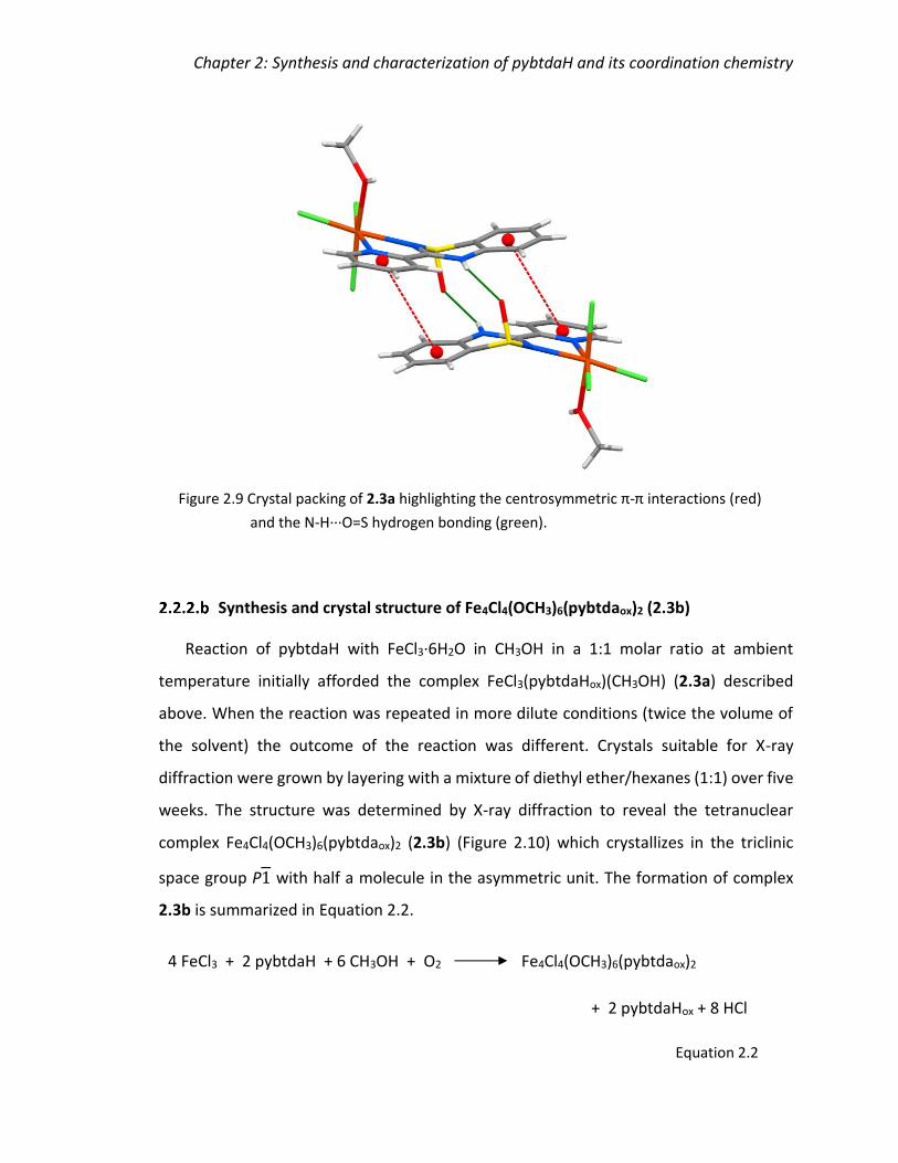
Chapter 2: Synthesis and characterization of pybtdaH and its coordination chemistry
52
Synthesis and crystal structure of Fe4Cl4(OCH3)6(pybtdaox)2 (2.3b)
Reaction of pybtdaH with FeCl3·6H2O in CH3OH in a 1:1 molar ratio at ambient
temperature initially afforded the complex FeCl3(pybtdaHox)(CH3OH) (2.3a) described
above. When the reaction was repeated in more dilute conditions (twice the volume of
the solvent) the outcome of the reaction was different. Crystals suitable for X-ray
diffraction were grown by layering with a mixture of diethyl ether/hexanes (1:1) over five
weeks. The structure was determined by X-ray diffraction to reveal the tetranuclear
complex Fe4Cl4(OCH3)6(pybtdaox)2 (2.3b) (Figure 2.10) which crystallizes in the triclinic
space group P1 with half a molecule in the asymmetric unit. The formation of complex
2.3b is summarized in Equation 2.2.
4 FeCl3 + 2 pybtdaH + 6 CH3OH + O2 Fe4Cl4(OCH3)6(pybtdaox)2
+ 2 pybtdaHox + 8 HCl
Equation 2.2
Figure 2.9 Crystal packing of 2.3a highlighting the centrosymmetric π-π interactions (red)
and the N-H∙∙∙O=S hydrogen bonding (green).

Chapter 2: Synthesis and characterization of pybtdaH and its coordination chemistry
53
Formation of the tetranuclear complex could result from slow condensation of the initially
formed mononuclear complex 2.3a (Equation 2.3). This would be favoured under the
more dilute conditions employed which require more time for crystallization:
Complex 2.3b consists of four FeIII centres, four terminal Cl ligands, two pybtdaox− and
6 CH3O− groups, comprising four μ2-ΟCH3 and two μ3-ΟCH3 bridges. The FeIII ions adopt
pseudo-octahedral geometry; Fe(1) has a ClO5 donor set comprising one Cl ligand, one
oxygen (S=O), two oxygen atoms from μ2-ΟCH3 and two oxygen atoms from μ3-ΟCH3
whereas Fe(2) has an N2ClO3 donor set with one Cl ligand, two nitrogen atoms from
pybtdaox–, two oxygen atoms from μ2-ΟCH3 and one oxygen atom from μ3-ΟCH3.
4 FeCl3(pybtdaHox)(CH3OH) + 2 CH3OH Fe4Cl4(OCH3)6(pybtdaox)2
+ 2 pybtdaHox + 8 HCl
Equation 2.3
Figure 2.10 Molecular structure of Fe4Cl4(OCH3)6(pybtdaox)2 (2.3b) (H on CH3
groups omitted for clarity).
Fe1
Fe1´
Fe2
Fe2´
Cl1
N3
Cl2
N1
O1
O2
O3 O4
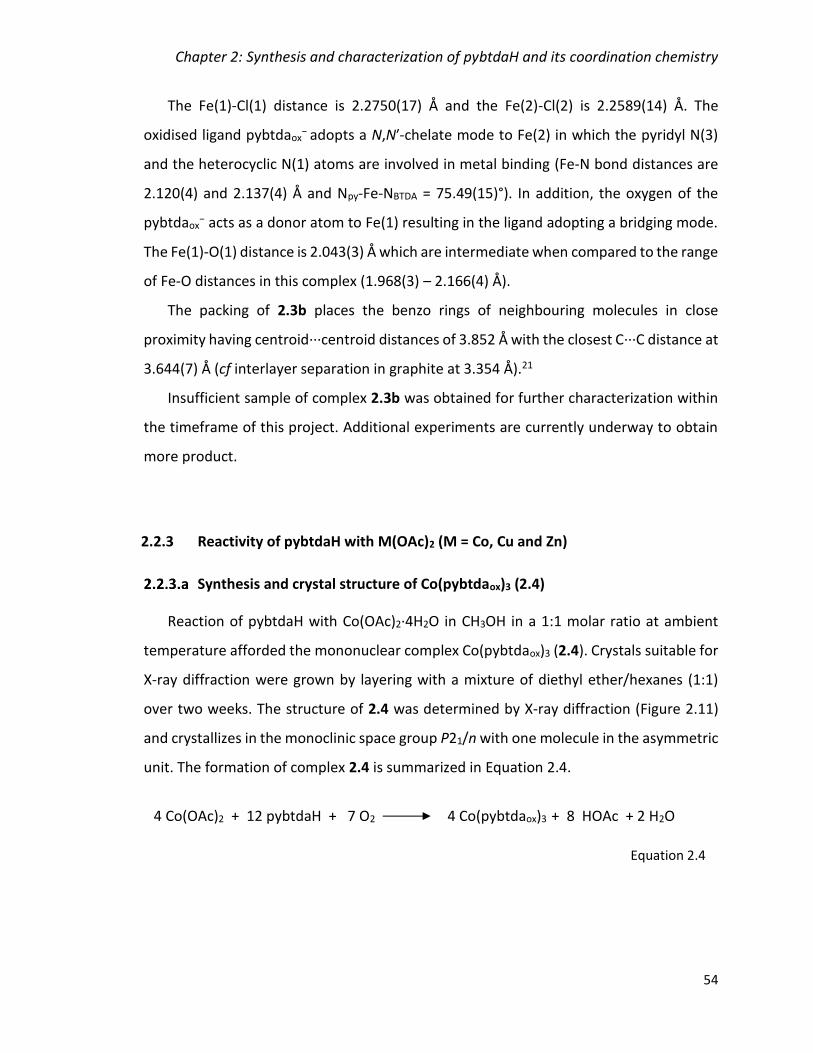
Chapter 2: Synthesis and characterization of pybtdaH and its coordination chemistry
54
The Fe(1)-Cl(1) distance is 2.2750(17) Å and the Fe(2)-Cl(2) is 2.2589(14) Å. The
oxidised ligand pybtdaox− adopts a N,Nʹ-chelate mode to Fe(2) in which the pyridyl N(3)
and the heterocyclic N(1) atoms are involved in metal binding (Fe-N bond distances are
2.120(4) and 2.137(4) Å and Npy-Fe-NBTDA = 75.49(15)°). In addition, the oxygen of the
pybtdaox− acts as a donor atom to Fe(1) resulting in the ligand adopting a bridging mode.
The Fe(1)-O(1) distance is 2.043(3) Å which are intermediate when compared to the range
of Fe-O distances in this complex (1.968(3) – 2.166(4) Å).
The packing of 2.3b places the benzo rings of neighbouring molecules in close
proximity having centroid···centroid distances of 3.852 Å with the closest C···C distance at
3.644(7) Å (cf interlayer separation in graphite at 3.354 Å).21
Insufficient sample of complex 2.3b was obtained for further characterization within
the timeframe of this project. Additional experiments are currently underway to obtain
more product.
2.2.3 Reactivity of pybtdaH with M(OAc)2 (M = Co, Cu and Zn)
Synthesis and crystal structure of Co(pybtdaox)3 (2.4)
Reaction of pybtdaH with Co(OAc)2·4H2O in CH3OH in a 1:1 molar ratio at ambient
temperature afforded the mononuclear complex Co(pybtdaox)3 (2.4). Crystals suitable for
X-ray diffraction were grown by layering with a mixture of diethyl ether/hexanes (1:1)
over two weeks. The structure of 2.4 was determined by X-ray diffraction (Figure 2.11)
and crystallizes in the monoclinic space group P21/n with one molecule in the asymmetric
unit. The formation of complex 2.4 is summarized in Equation 2.4.
4 Co(OAc)2 + 12 pybtdaH + 7 O2 4 Co(pybtdaox)3 + 8 HOAc + 2 H2O
Equation 2.4
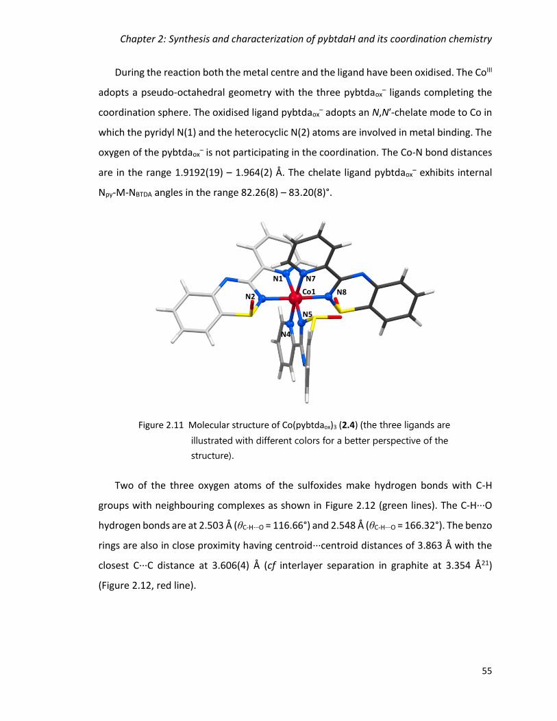
Chapter 2: Synthesis and characterization of pybtdaH and its coordination chemistry
55
During the reaction both the metal centre and the ligand have been oxidised. The CoIII
adopts a pseudo-octahedral geometry with the three pybtdaox– ligands completing the
coordination sphere. The oxidised ligand pybtdaox– adopts an N,Nʹ-chelate mode to Co in
which the pyridyl N(1) and the heterocyclic N(2) atoms are involved in metal binding. The
oxygen of the pybtdaox– is not participating in the coordination. The Co-N bond distances
are in the range 1.9192(19) – 1.964(2) Å. The chelate ligand pybtdaox– exhibits internal
Npy-M-NBTDA angles in the range 82.26(8) – 83.20(8)°.
Two of the three oxygen atoms of the sulfoxides make hydrogen bonds with C-H
groups with neighbouring complexes as shown in Figure 2.12 (green lines). The C-H···O
hydrogen bonds are at 2.503 Å (θC-H···O = 116.66°) and 2.548 Å (θC-H···O = 166.32°). The benzo
rings are also in close proximity having centroid···centroid distances of 3.863 Å with the
closest C···C distance at 3.606(4) Å (cf interlayer separation in graphite at 3.354 Å21)
(Figure 2.12, red line).
Figure 2.11 Molecular structure of Co(pybtdaox)3 (2.4) (the three ligands are
illustrated with different colors for a better perspective of the
structure).
N2
N1
N4
N8
N5
N7
Co1

Chapter 2: Synthesis and characterization of pybtdaH and its coordination chemistry
56
Synthesis and crystal structure of [Cu2(OAc)2(pybtdaox)2(H2O)2]∙2H2O (2.5)
Reaction of pybtdaH with [Cu2(OAc)4]·2H2O in CH3OH in a 1:1 molar ratio at ambient
temperature afforded the dinuclear complex Cu2(OAc)2(pybtdaox)2(H2O)2 (2.5). Crystals
suitable for X-ray diffraction were grown by layering with a mixture of diethyl
ether/hexanes (1:1) over two days. The structure crystallizes in the monoclinic space
group C2/c with half a molecule and a water solvent molecule in the asymmetric unit. The
formation of complex 2.5 is summarized in Equation 2.5.
The crystal structure of complex 2.5 is shown in Figure 2.13. The structure is similar
to the starting material [Cu2(OAc)4]·2H2O in which two of the bridging acetate groups of
the Cu2(OAc)4 paddlewheel have been substituted by two chelate pybtdaox– ligands. The
[Cu2(OAc)4]·2H2O + 2 pybtdaH + O2 [Cu2(OAc)2(pybtdaox)2(H2O)2] + 2 AcOH
Equation 2.5
Figure 2.12 Crystal packing of 2.4 highlighting the π-π interactions (red) and the
C-H∙∙∙O hydrogen bonding between the C-H groups and the O atom of the
sulfoxide (green).

Chapter 2: Synthesis and characterization of pybtdaH and its coordination chemistry
57
two CuII centres within 2.5 are related via a 2-fold rotation with each copper ion adopting
a 5-coordinate geometry with a N2O3 donor set. The Addison τ5 value22 (0.17) is consistent
with an essentially square pyramidal geometry (trigonal bipyramidal (τ = 1) and square
pyramidal (τ = 0)). Approximating the geometry to square pyramidal, the O(4) defines the
axial ligand, with the remaining angles in the equatorial plane in the range 81.41(10) –
176.20(10)°. The heterocyclic ligand provides an N,Nʹ-chelate pocket analogous to
complex 2.4 through N(1) and N(2) atoms with Cu-N distances 1.972(3) and 1.982(3) Å,
and Npy-M-NBTDA angle 81.41(10)°. The acetate groups are bridging the two equivalent Cu
ions with Cu-O distances in the range of 1.955(2) – 2.205(3) Å. The coordination sphere is
completed by one water molecule.
The structure of 2.5 places the benzo rings in close proximity with the pyridine rings
of the neighbouring molecule having centroid···centroid distances of 3.818 Å with the
closest C···C distance at 3.454(5) Å (cf interlayer separation in graphite at 3.354 Å21)
(Figure 2.14, top). In addition, the molecules are linked via O-H···N hydrogen bonding
between the water molecules and the nitrogen atom of the heterocycle (dO-H···N = 1.975
Å, θO-H···N = 166.64°, Figure 2.14, top). Π - π interactions between adjacent dimers have
N2
N1
O4
O3´ Cu1
O2
Cu1´
Figure 2.13 Molecular structure of Cu2(OAc)2(pybtdaox)2(H2O)2 (2.5) (solvent
molecules omitted for clarity).
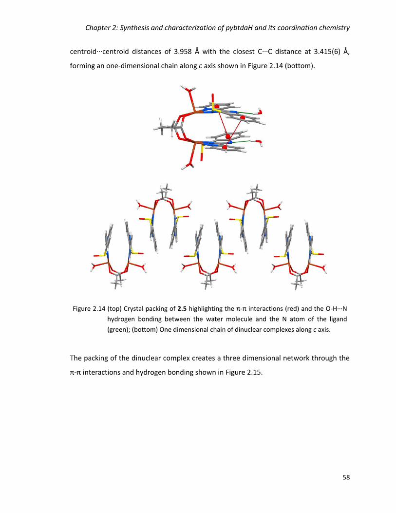
Chapter 2: Synthesis and characterization of pybtdaH and its coordination chemistry
58
centroid···centroid distances of 3.958 Å with the closest C···C distance at 3.415(6) Å,
forming an one-dimensional chain along c axis shown in Figure 2.14 (bottom).
The packing of the dinuclear complex creates a three dimensional network through the
π-π interactions and hydrogen bonding shown in Figure 2.15.
Figure 2.14 (top) Crystal packing of 2.5 highlighting the π-π interactions (red) and the O-H∙∙∙N
hydrogen bonding between the water molecule and the N atom of the ligand
(green); (bottom) One dimensional chain of dinuclear complexes along c axis.

Chapter 2: Synthesis and characterization of pybtdaH and its coordination chemistry
59
Synthesis and crystal structure of Zn2(OAc)2(pybtdaox)2 (2.6)
Reaction of pybtdaH with [Zn(OAc)2]·2H2O in CH3OH in a 1:1 molar ratio at ambient
temperature afforded the dinuclear complex Zn2(OAc)2(pybtdaox)2 (2.6). Crystals suitable
for X-ray diffraction were grown by layering with a mixture of diethyl ether/hexanes (1:1)
over two days. The structure crystallizes in the triclinic space group P1̅ with two half
molecules in the asymmetric unit. The formation of complex 2.6 is summarized in
Equation 2.6.
Figure 2.15 Crystal packing of 2.5 revealing the three dimensional nature of the
supramolecular arrangement of molecules generated through hydrogen
bonds and π-π interactions.
2[Zn(OAc)2]·2H2O + 2pybtdaH + O2 Zn2(OAc)2(pybtdaox)2 + 2AcOH
Equation 2.6

Chapter 2: Synthesis and characterization of pybtdaH and its coordination chemistry
60
While the formula of 2.6 (Zn2(OAc)2(pybtdaox)2) is comparable with the copper
complex 2.5 ((Cu2(OAc)2(pybtdaox)2(H2O)2), their coordination geometries are very
different. The crystal structure of complex 2.6 is shown in Figure 2.16. The two ZnII centres
within 2.6 adopt a 5-coordinate geometry with a N2O3 donor set. The Addison τ5 values22
(0.22 for Zn(1) and 0.25 for Zn(2)) are consistent with a distorted square pyramidal
geometry (τ = 1 for trigonal bipyramidal and τ = 0 square pyramidal). Approximating the
geometry to square pyramidal, the O(1) (for Zn(1)) and O(4) (for Zn(2)) define the axial
ligands, with the remaining angles in the equatorial plane in the range 57.38(6) –
144.38(6)° for Zn(1) and 61.35(6) – 152.07(6)° for Zn(2). As with complex 2.5, the
heterocyclic ligand provides an N,Nʹ-chelate pocket through N(1) and N(2) atoms with Cu-
N distances in the range 2.035(3) – 2.054(3) Å and Npy-M-NBTDA angles of 80.410(12)° and
81.34(12)°. However in 2.6 the N,Nʹ-chelate comprises the pyridyl-N and the nitrogen at
the 4-position of the pybtdaox− heterocyclic ring rather than the N atom at the 2-position,
N2
N1
O1
O3O2
Zn1
Zn1´
Figure 2.16 Molecular structure of one complete molecule in the asymmetric
unit Zn2(OAc)2(pybtdaox)2 (2.6).

Chapter 2: Synthesis and characterization of pybtdaH and its coordination chemistry
61
common to benzothiadiazines, pybtdaH. Notably, coordination through this N atom has
also been seen in another benzothiadiazine S-oxide complex,2 suggesting a possible
reversal of N-donor ability upon S-oxidation and deprotonation. The coordination sphere
of each Zn centre is completed by a chelating rather than bridging acetate group with Zn-
O distances in the range of 1.982(3) – 2.507(3) Å.
The structure of 2.6 places the benzothiadiazine ligands within the dimers in close
proximity resulting in centroid···centroid distances of 3.636 Å (closest C···C distance at
3.468 Å) and 3.643 Å (closest C···C distance at 3.476 Å) for Zn(1) and Zn(2), respectively
(cf interlayer separation in graphite at 3.354 Å21) (Figure 2.17, left, red lines). One-
dimensional chains are also formed along a axis with π-π interactions between adjacent
dimers having centroid···centroid distances of 3.798 Å for Zn(2) dimers (Figure 2.17, left,
green lines). The centroid···centroid distances for Zn(1) are at 4.467 Å (Figure 2.17, left,
blue lines), much longer than anticipated for π-π interactions and a better visualization of
the parallel displacement of the ligand is shown in Figure 2.17 (right).
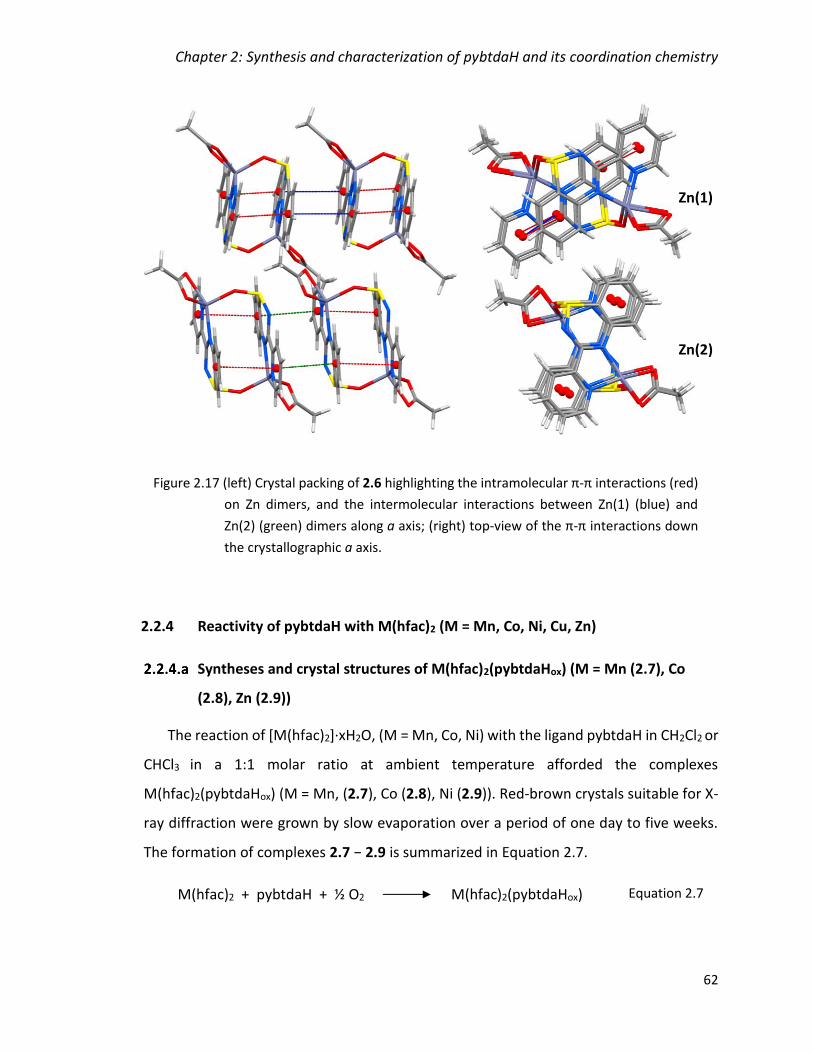
Chapter 2: Synthesis and characterization of pybtdaH and its coordination chemistry
62
2.2.4 Reactivity of pybtdaH with M(hfac)2 (M = Mn, Co, Ni, Cu, Zn)
Syntheses and crystal structures of M(hfac)2(pybtdaHox) (M = Mn (2.7), Co
(2.8), Zn (2.9))
The reaction of [M(hfac)2]·xH2O, (M = Mn, Co, Ni) with the ligand pybtdaH in CH2Cl2 or
CHCl3 in a 1:1 molar ratio at ambient temperature afforded the complexes
M(hfac)2(pybtdaHox) (M = Mn, (2.7), Co (2.8), Ni (2.9)). Red-brown crystals suitable for X-
ray diffraction were grown by slow evaporation over a period of one day to five weeks.
The formation of complexes 2.7 − 2.9 is summarized in Equation 2.7.
Figure 2.17 (left) Crystal packing of 2.6 highlighting the intramolecular π-π interactions (red)
on Zn dimers, and the intermolecular interactions between Zn(1) (blue) and
Zn(2) (green) dimers along a axis; (right) top-view of the π-π interactions down
the crystallographic a axis.
Zn(1)
Zn(2)
M(hfac)2 + pybtdaH + ½ O2 M(hfac)2(pybtdaHox) Equation 2.7
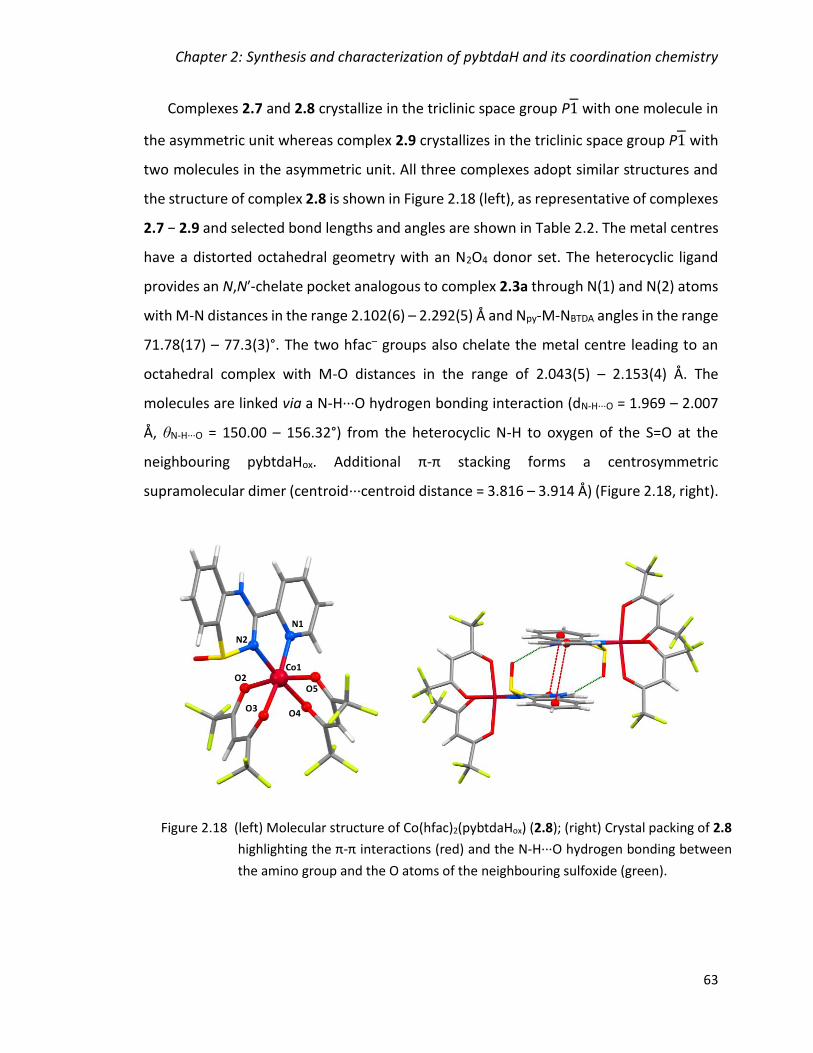
Chapter 2: Synthesis and characterization of pybtdaH and its coordination chemistry
63
Complexes 2.7 and 2.8 crystallize in the triclinic space group P1 with one molecule in
the asymmetric unit whereas complex 2.9 crystallizes in the triclinic space group P1 with
two molecules in the asymmetric unit. All three complexes adopt similar structures and
the structure of complex 2.8 is shown in Figure 2.18 (left), as representative of complexes
2.7 − 2.9 and selected bond lengths and angles are shown in Table 2.2. The metal centres
have a distorted octahedral geometry with an N2O4 donor set. The heterocyclic ligand
provides an N,Nʹ-chelate pocket analogous to complex 2.3a through N(1) and N(2) atoms
with M-N distances in the range 2.102(6) – 2.292(5) Å and Npy-M-NBTDA angles in the range
71.78(17) – 77.3(3)°. The two hfac– groups also chelate the metal centre leading to an
octahedral complex with M-O distances in the range of 2.043(5) – 2.153(4) Å. The
molecules are linked via a N-H···O hydrogen bonding interaction (dN-H···O = 1.969 – 2.007
Å, θN-H···O = 150.00 – 156.32°) from the heterocyclic N-H to oxygen of the S=O at the
neighbouring pybtdaHox. Additional π-π stacking forms a centrosymmetric
supramolecular dimer (centroid···centroid distance = 3.816 – 3.914 Å) (Figure 2.18, right).
Figure 2.18 (left) Molecular structure of Co(hfac)2(pybtdaHox) (2.8); (right) Crystal packing of 2.8
highlighting the π-π interactions (red) and the N-H∙∙∙O hydrogen bonding between
the amino group and the O atoms of the neighbouring sulfoxide (green).
O4
N2
Co1
N1
O3
O2O5
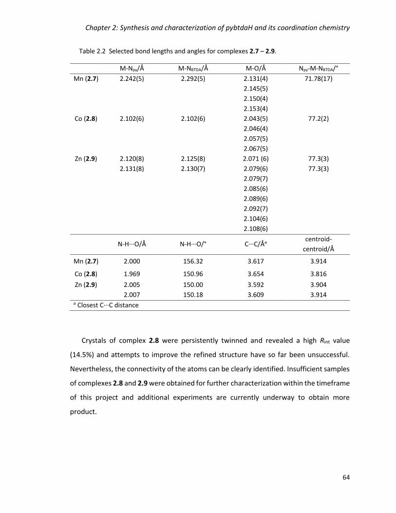
Chapter 2: Synthesis and characterization of pybtdaH and its coordination chemistry
64
Table 2.2 Selected bond lengths and angles for complexes 2.7 – 2.9.
M-Npy/Å M-NBTDA/Å M-O/Å Npy-M-NBTDA/°
Mn (2.7) 2.242(5) 2.292(5) 2.131(4)
2.145(5)
2.150(4)
2.153(4)
71.78(17)
Co (2.8) 2.102(6) 2.102(6) 2.043(5)
2.046(4)
2.057(5)
2.067(5)
77.2(2)
Zn (2.9) 2.120(8)
2.131(8)
2.125(8)
2.130(7)
2.071 (6)
2.079(6)
2.079(7)
2.085(6)
2.089(6)
2.092(7)
2.104(6)
2.108(6)
77.3(3)
77.3(3)
N-H∙∙∙O/Å N-H∙∙∙O/° C∙∙∙C/Åa centroid-
centroid/Å
Mn (2.7) 2.000 156.32 3.617 3.914
Co (2.8) 1.969 150.96 3.654 3.816
Zn (2.9) 2.005
2.007
150.00
150.18
3.592
3.609
3.904
3.914 a Closest C∙∙∙C distance
Crystals of complex 2.8 were persistently twinned and revealed a high Rint value
(14.5%) and attempts to improve the refined structure have so far been unsuccessful.
Nevertheless, the connectivity of the atoms can be clearly identified. Insufficient samples
of complexes 2.8 and 2.9 were obtained for further characterization within the timeframe
of this project and additional experiments are currently underway to obtain more
product.

Chapter 2: Synthesis and characterization of pybtdaH and its coordination chemistry
65
Synthesis and crystal structure of Ni(hfac)2(pybtdaH) (2.10)
The metal salt [Ni(hfac)2]·xH2O was reacted with the ligand pybtdaH in CH2Cl2 in a 1:1
molar ratio at ambient temperature and the solution was layered with hexanes. Unlike
the Mn, Fe, Co complexes described in section 2.2.4.a, red single crystals of the complex
with formula Ni(hfac)2(pybtdaH) (2.10), suitable for X-ray diffraction were isolated within
24 h. Complex 2.10 crystallizes in the triclinic space group P1 with one molecule in the
asymmetric unit. The formation of complex 2.10 is summarized in Equation 2.8.
The NiII ion has a distorted octahedral geometry with an N2O4 donor set. The
heterocyclic ligand provides an N,Nʹ-chelate pocket analogous to complex 2.3a through
N(1) and N(2) atoms (Figure 2.19) with M-N distances 2.044(2) − 2.049(2) Å and
Npy-M-NBTDA angle at 78.39(9)°. The two hfac– groups also chelate the metal centre leading
to an octahedral complex with M-O distances in the range of 2.016(2) − 2.074(2) Å. The
molecules on complex 2.10 are linked via an N-H···O hydrogen bonding interaction
(dN-H···O = 2.382 Å, θN-H···O = 145.33°) from the heterocyclic N-H to one of the hfac– O atoms.
Additional π-π stacking forms a centrosymmetric supramolecular dimer
(centroid···centroid distance = 3.870 Å) (cf interlayer separation in graphite at 3.354 Å21)
(Figure 2.19, right).
Ni(hfac)2 + pybtdaH Ni(hfac)2(pybtdaH) Equation 2.8

Chapter 2: Synthesis and characterization of pybtdaH and its coordination chemistry
66
Synthesis and crystal structure of Ni3(hfac)(pybtdaox)5(H2O) (2.11)
The metal salt [Ni(hfac)2]·xH2O was reacted with the ligand pybtdaH in CH2Cl2 in a 1:1
molar ratio in the presence of 4 equivalents of Et3N at ambient temperature and the
solution was layered with hexanes. Red single crystals of Ni(hfac)2(pybtdaH) (2.10) formed
within two days (see above) but green crystals formed after a more prolonged period.
Single X-ray diffraction revealed these to be the trimetallic complex
Ni3(hfac)(pybtdaox)5(H2O) (2.11). Complex 2.11 crystallizes in the monoclinic space group
P21/c with one molecule in the asymmetric unit. The formation of complex 2.11 is
summarized in Equation 2.9.
The structure of complex 2.11 is shown in Figure 2.20. The NiII ions all have distorted
octahedral geometries but each metal center has a different donor set. The Ni(1) ion has
an N2O4 donor set; two nitrogen atoms from pybtdaox−, two oxygen atoms from an hfac−
Figure 2.19 (left) Molecular structure of Ni(hfac)2(pybtdaH) (2.10); (right) Crystal packing of
2.10 highlighting the π-π interactions (red) and the N-H∙∙∙O hydrogen bonding
between the amino group and the O atoms of the neighbouring hfac– group
(green).
O4
O3O2
O1
N2
N1
Ni1
3 Ni(hfac)2 + 5 pybtdaH + H2O +
O2 Ni3(hfac)(pybtdaox)5(H2O) + 5 hfacH
Equation 2.9

Chapter 2: Synthesis and characterization of pybtdaH and its coordination chemistry
67
ligand, one oxygen atom from the S=O and one terminal water molecule. The Ni(2) ion
has also N2O4 donor set; two nitrogen atoms from pybtdaox– and four oxygen atoms from
S=O. The coordination sphere of Ni(3) is completed by six nitrogen atoms from three
pybtdaox– ligands.
The heterocyclic ligand takes up a bridging mode between two metal centres,
analogous to complex 2.3b. The N,Nʹ-chelate pocket, through the pyridyl nitrogen atom
and the heterocyclic nitrogen atom next to S, chelates one metal ion and the S-oxide
bridges to the second metal centre. The M-N distances are in the range of 1.989(15) −
2.118(4) Å and Npy-M-NBTDA angles are in the range of 78.02(15) − 81.3(9)°. The M-O
distances of the S=O groups are in the range of 2.051(3) – 2.093(3) Å. The hfac– group
chelates to Ni(1) with M-O distances at 2.033(3) Å and 2.042(3) Å.
Complex 2.11 crystallized alongside the mononuclear complex Ni(hfac)2(pybtdaH)
(2.10), thus making the characterization of complex 2.11 not possible. Attempts to
reproduce complex 2.11 and isolate it selectively were unsuccessful.
Figure 2.20 Molecular structure of Ni3(hfac)(pybtdaox)5(H2O) (2.11).
O8N4Ni1
N7 O3
O2
O1
Ni3
Ni2
O6
O5
O7 O4
N8
N5
N1
N2N14
N10N11
N13
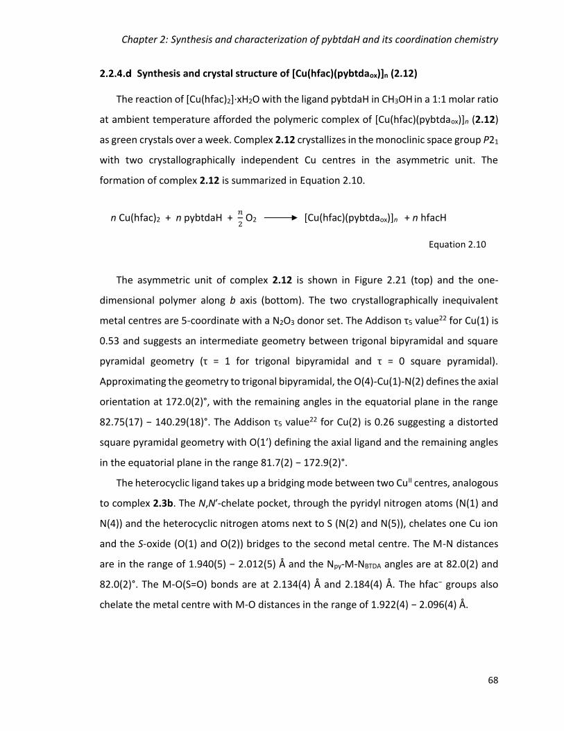
Chapter 2: Synthesis and characterization of pybtdaH and its coordination chemistry
68
Synthesis and crystal structure of [Cu(hfac)(pybtdaox)]n (2.12)
The reaction of [Cu(hfac)2]·xH2O with the ligand pybtdaH in CH3OH in a 1:1 molar ratio
at ambient temperature afforded the polymeric complex of [Cu(hfac)(pybtdaox)]n (2.12)
as green crystals over a week. Complex 2.12 crystallizes in the monoclinic space group P21
with two crystallographically independent Cu centres in the asymmetric unit. The
formation of complex 2.12 is summarized in Equation 2.10.
The asymmetric unit of complex 2.12 is shown in Figure 2.21 (top) and the one-
dimensional polymer along b axis (bottom). The two crystallographically inequivalent
metal centres are 5-coordinate with a N2O3 donor set. The Addison τ5 value22 for Cu(1) is
0.53 and suggests an intermediate geometry between trigonal bipyramidal and square
pyramidal geometry (τ = 1 for trigonal bipyramidal and τ = 0 square pyramidal).
Approximating the geometry to trigonal bipyramidal, the O(4)-Cu(1)-N(2) defines the axial
orientation at 172.0(2)°, with the remaining angles in the equatorial plane in the range
82.75(17) − 140.29(18)°. The Addison τ5 value22 for Cu(2) is 0.26 suggesting a distorted
square pyramidal geometry with O(1ʹ) defining the axial ligand and the remaining angles
in the equatorial plane in the range 81.7(2) − 172.9(2)°.
The heterocyclic ligand takes up a bridging mode between two CuII centres, analogous
to complex 2.3b. The N,Nʹ-chelate pocket, through the pyridyl nitrogen atoms (N(1) and
N(4)) and the heterocyclic nitrogen atoms next to S (N(2) and N(5)), chelates one Cu ion
and the S-oxide (O(1) and O(2)) bridges to the second metal centre. The M-N distances
are in the range of 1.940(5) − 2.012(5) Å and the Npy-M-NBTDA angles are at 82.0(2) and
82.0(2)°. The M-O(S=O) bonds are at 2.134(4) Å and 2.184(4) Å. The hfac− groups also
chelate the metal centre with M-O distances in the range of 1.922(4) − 2.096(4) Å.
n Cu(hfac)2 + n pybtdaH + 𝑛
2 O2 [Cu(hfac)(pybtdaox)]n + n hfacH
Equation 2.10
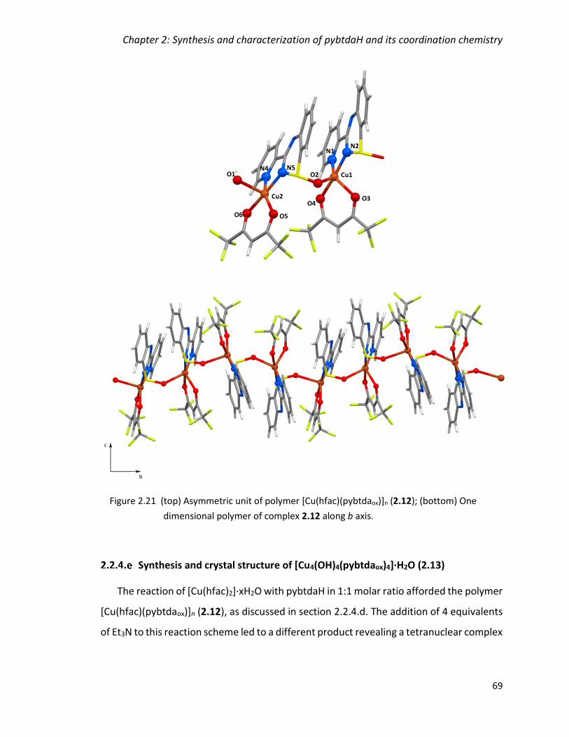
Chapter 2: Synthesis and characterization of pybtdaH and its coordination chemistry
69
Synthesis and crystal structure of [Cu4(OH)4(pybtdaox)4]∙H2O (2.13)
The reaction of [Cu(hfac)2]·xH2O with pybtdaH in 1:1 molar ratio afforded the polymer
[Cu(hfac)(pybtdaox)]n (2.12), as discussed in section 2.2.4.d. The addition of 4 equivalents
of Et3N to this reaction scheme led to a different product revealing a tetranuclear complex
N4
N2
Cu1O2
Cu2
O1´
N1
N5
O3O4
O5O6
Figure 2.21 (top) Asymmetric unit of polymer [Cu(hfac)(pybtdaox)]n (2.12); (bottom) One
dimensional polymer of complex 2.12 along b axis.
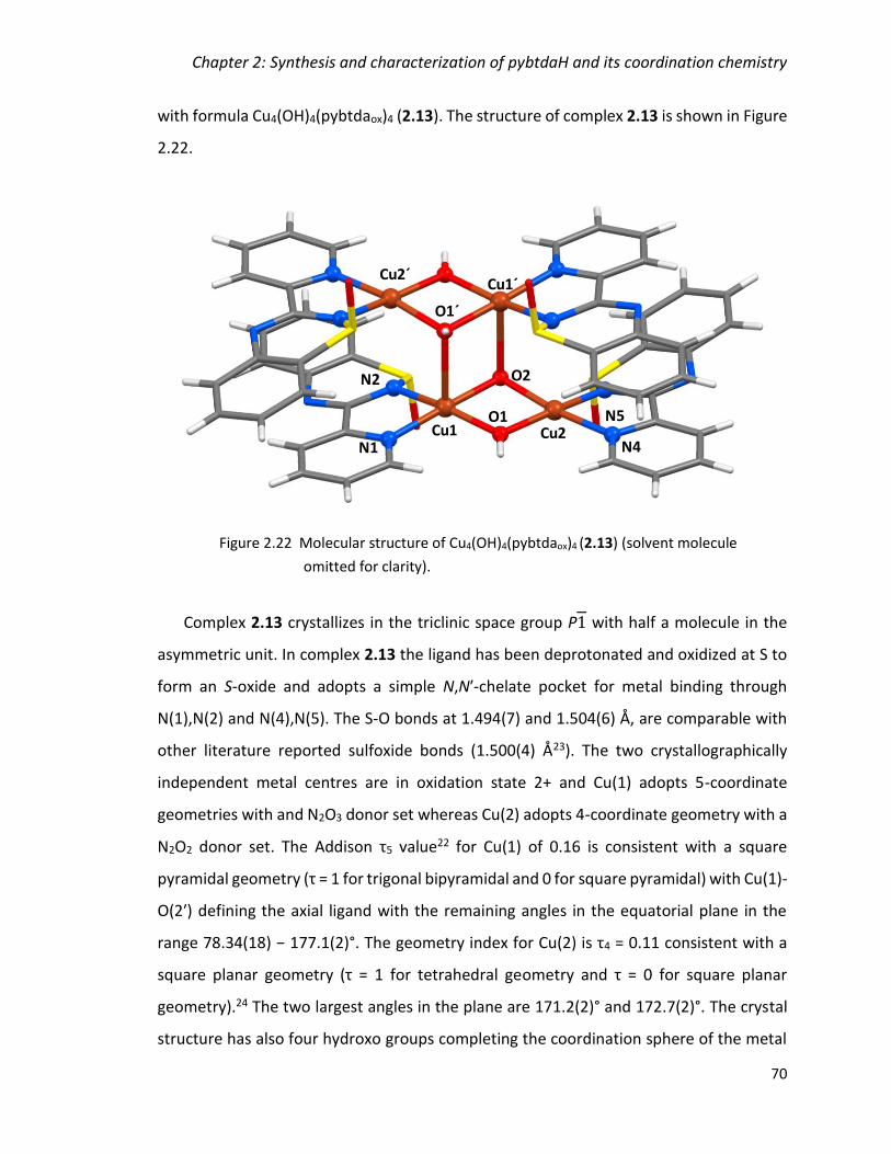
Chapter 2: Synthesis and characterization of pybtdaH and its coordination chemistry
70
with formula Cu4(OH)4(pybtdaox)4 (2.13). The structure of complex 2.13 is shown in Figure
2.22.
Complex 2.13 crystallizes in the triclinic space group P1 with half a molecule in the
asymmetric unit. In complex 2.13 the ligand has been deprotonated and oxidized at S to
form an S-oxide and adopts a simple N,Nʹ-chelate pocket for metal binding through
N(1),N(2) and N(4),N(5). The S-O bonds at 1.494(7) and 1.504(6) Å, are comparable with
other literature reported sulfoxide bonds (1.500(4) Å23). The two crystallographically
independent metal centres are in oxidation state 2+ and Cu(1) adopts 5-coordinate
geometries with and N2O3 donor set whereas Cu(2) adopts 4-coordinate geometry with a
N2O2 donor set. The Addison τ5 value22 for Cu(1) of 0.16 is consistent with a square
pyramidal geometry (τ = 1 for trigonal bipyramidal and 0 for square pyramidal) with Cu(1)-
O(2ʹ) defining the axial ligand with the remaining angles in the equatorial plane in the
range 78.34(18) − 177.1(2)°. The geometry index for Cu(2) is τ4 = 0.11 consistent with a
square planar geometry (τ = 1 for tetrahedral geometry and τ = 0 for square planar
geometry).24 The two largest angles in the plane are 171.2(2)° and 172.7(2)°. The crystal
structure has also four hydroxo groups completing the coordination sphere of the metal
Figure 2.22 Molecular structure of Cu4(OH)4(pybtdaox)4 (2.13) (solvent molecule
omitted for clarity).
N2
N1
N5
N4
O1Cu1
O2
Cu1´
Cu2
Cu2´
O1´

Chapter 2: Synthesis and characterization of pybtdaH and its coordination chemistry
71
centres. Two hydroxo groups are bridging two Cu ions whereas two are bridging three
metals with Cu-O bonds in the range 1.905(5) − 2.396(5) Å. The [Cu4(μ2-OH)2(μ3-OH)2]4+
core of the complex can be described as ladder type as shown in Figure 2.23.
The ligand provides a simple N,Nʹ-chelate pocket through the pyridyl and heterocyclic
nitrogen atoms. The chelate ligand pybtdaox− exhibits Npy-Cu-NBTDA angles of 82.3(2)° and
82.4(3)° and Cu-N bond lengths are in the range 1.951(6) − 1.982(6) Å. The formation of
complex 2.13 is summarized in Equation 2.11.
The structure of 2.13 exhibits intramolecular π-π interaction with centroid···centroid
distances 3.591 Å and 3.932 Å (cf interlayer separation in graphite at 3.354 Å21). The
structure exhibits intermolecular π-π stacking on six of the eight aromatic rings of the
ligands with centroid···centroid distances 3.570 Å and 4.113 Å. The π-π interactions are
shown in Figure 2.24 as red lines between the red centroids. One of the aromatic rings
does not participate in π-π stacking but instead has a short interaction with a Cu ion with
4Cu(hfac)2 + 4pybtdaH + 4H2O + 2O2 Cu4(OH)4(pybtdaox)4 + 8 hfacH
Equation 2.11
O1Cu1
O2
Cu1´
Cu2
Cu2´ O1´
O2´
Figure 2.23 The [Cu4(μ2-OH)2(μ3-OH)2]4+ core of complex 2.13 illustrating the ladder-type
conformation.
Chapter 2: Synthesis and characterization of pybtdaH and its coordination chemistry
72
centroid-Cu distance of 3.466 Å. The rings not exhibiting π-π interactions are shown with
a yellow centroid in Figure 2.24.
Synthesis and crystal structure of
[Cu14(OH)12(CO3)2(pybtdaox)12(H2O)2]∙14[H2O]∙[CH3OH] (2.14)
Attempts to reproduce complex 2.13 using the correct stoichiometric ratio (Equation
2.11) led to the reaction of pybtdaH with Cu(hfac)2·xH2O and H2O in CH3OH in a 1:1:1
molar ratio under basic conditions. The solution was left for slow evaporation and green
needles suitable for X-ray diffraction formed after three days. The crystal structure
revealed the complex to unexpectedly be Cu14(OH)12(CO3)2(pybtdaox)12(H2O)2 (2.14)
shown in Figure 2.25. Complex 2.14 crystallizes in the monoclinic space group P21/n with
one [CuII14] molecule in the asymmetric unit, one methanol and fourteen water
molecules.
Figure 2.24 Crystal packing of 2.13 highlighting the π-π interactions (red) between
neighbouring molecules and the centroids shown in yellow are not
participating in π-π interactions.
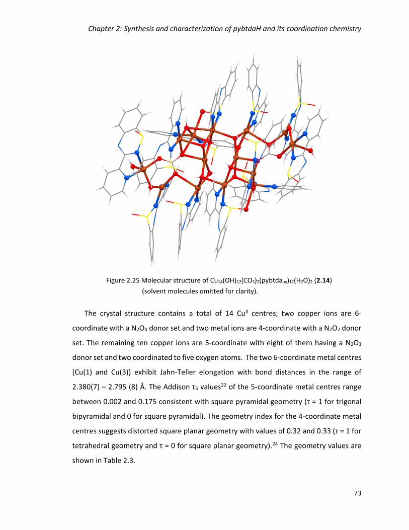
Chapter 2: Synthesis and characterization of pybtdaH and its coordination chemistry
73
The crystal structure contains a total of 14 CuII centres; two copper ions are 6-
coordinate with a N2O4 donor set and two metal ions are 4-coordinate with a N2O2 donor
set. The remaining ten copper ions are 5-coordinate with eight of them having a N2O3
donor set and two coordinated to five oxygen atoms. The two 6-coordinate metal centres
(Cu(1) and Cu(3)) exhibit Jahn-Teller elongation with bond distances in the range of
2.380(7) – 2.795 (8) Å. The Addison τ5 values22 of the 5-coordinate metal centres range
between 0.002 and 0.175 consistent with square pyramidal geometry (τ = 1 for trigonal
bipyramidal and 0 for square pyramidal). The geometry index for the 4-coordinate metal
centres suggests distorted square planar geometry with values of 0.32 and 0.33 (τ = 1 for
tetrahedral geometry and τ = 0 for square planar geometry).24 The geometry values are
shown in Table 2.3.
Figure 2.25 Molecular structure of Cu14(OH)12(CO3)2(pybtdaox)12(H2O)2 (2.14)
(solvent molecules omitted for clarity).

Chapter 2: Synthesis and characterization of pybtdaH and its coordination chemistry
74
Table 2.3 Coordination geometry and geometry index values of Cu centres in complex 2.14.
Cu
2 × 4-coordinate 10 × 5-coordinate 2 × 6-coordinate
Geometry index 0.32 (Cu13) 0.33 (Cu11)
0.002 (Cu14) 0.015 (Cu7) 0.017 (Cu4) 0.017 (Cu9)
0.017 (Cu10) 0.061 (Cu2) 0.085 (Cu5) 0.097 (Cu6) 0.103 (Cu8)
0.175 (Cu12)
(Cu1) (Cu3)
The formula of complex 2.14 contains twelve pybtdaox− ligands located in the
periphery of the structure. The heterocyclic ligand adopts two coordination modes; ten
of the ligands in the structure offer a simple N,Nʹ-chelate pocket through the pyridyl and
heterocyclic nitrogen atoms whereas two of them adopt a bridging mode between two
CuII centres, analogous to complex 2.3b. The Cu-O (O=S) distances are 2.471(9) and
2.473(9) Å, the M-N distances are in the range of 1.938(12) − 2.010(10) Å and the
Npy-M-NBTDA angles in the range of 81.1(4) − 82.8(4)° (Table 2.4).
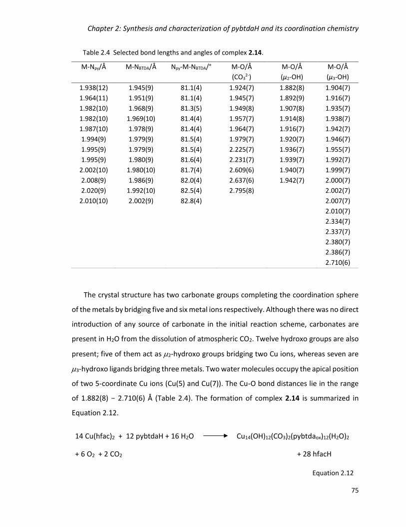
Chapter 2: Synthesis and characterization of pybtdaH and its coordination chemistry
75
Table 2.4 Selected bond lengths and angles of complex 2.14.
The crystal structure has two carbonate groups completing the coordination sphere
of the metals by bridging five and six metal ions respectively. Although there was no direct
introduction of any source of carbonate in the initial reaction scheme, carbonates are
present in H2O from the dissolution of atmospheric CO2. Twelve hydroxo groups are also
present; five of them act as 2-hydroxo groups bridging two Cu ions, whereas seven are
3-hydroxo ligands bridging three metals. Two water molecules occupy the apical position
of two 5-coordinate Cu ions (Cu(5) and Cu(7)). The Cu-O bond distances lie in the range
of 1.882(8) − 2.710(6) Å (Table 2.4). The formation of complex 2.14 is summarized in
Equation 2.12.
M-Npy/Å M-NBTDA/Å Npy-M-NBTDA/° M-Ο/Å
(CO32-)
M-Ο/Å
(μ2-OH)
M-Ο/Å
(μ3-OH)
1.938(12)
1.964(11)
1.982(10)
1.982(10)
1.987(10)
1.994(9)
1.995(9)
1.995(9)
2.002(10)
2.008(9)
2.020(9)
2.010(10)
1.945(9)
1.951(9)
1.968(9)
1.969(10)
1.978(9)
1.979(9)
1.979(9)
1.980(9)
1.980(10)
1.986(9)
1.992(10)
2.002(9)
81.1(4)
81.1(4)
81.3(5)
81.4(4)
81.4(4)
81.5(4)
81.5(4)
81.6(4)
81.7(4)
82.0(4)
82.5(4)
82.8(4)
1.924(7)
1.945(7)
1.949(8)
1.957(7)
1.964(7)
1.979(7)
2.225(7)
2.231(7)
2.609(6)
2.637(6)
2.795(8)
1.882(8)
1.892(9)
1.907(8)
1.914(8)
1.916(7)
1.920(7)
1.936(7)
1.939(7)
1.940(7)
1.942(7)
1.904(7)
1.916(7)
1.935(7)
1.938(7)
1.942(7)
1.946(7)
1.955(7)
1.992(7)
1.999(7)
2.000(7)
2.002(7)
2.007(7)
2.010(7)
2.334(7)
2.337(7)
2.380(7)
2.386(7)
2.710(6)
14 Cu(hfac)2 + 12 pybtdaH + 16 H2O Cu14(OH)12(CO3)2(pybtdaox)12(H2O)2
+ 6 O2 + 2 CO2 + 28 hfacH
Equation 2.12

Chapter 2: Synthesis and characterization of pybtdaH and its coordination chemistry
76
The [Cu14] core is shown in Figure 2.26. It consists of two [Cu7O8] sub-cores linked by
two μ2-ΟΗ (O(3), O(6)) and two μ3-ΟΗ (O(8), O(12)) groups. Each sub-core can be
described as a zig-zag chain of [Cu3O2] ending to a [Cu4O4] cubane connected by the two
μ3-ΟΗ (O(8), O(12)). The zig-zag chains have the three metal centres (Cu(7), C(13), Cu(6)
and Cu(5), Cu(11), Cu(10)) linked by one μ2-ΟΗ (O(7), O(4), respectively) and one μ3-Ο
atom of the CO32– groups (O(302), O(203), respectively). The first [Cu4O4] cubane (Cu(1),
Cu(2), Cu(3) and Cu(4)) is bridged by three μ3-ΟΗ groups (O(5), O(10), O(13)) and μ3-Ο
atom from the CO32– group (O(204)). The second “cubane” comprised by Cu(8), Cu(9),
Cu(12) and Cu(14) is missing two edges, shown in Figure 2.26 as dashed green lines. The
four metal centers are linked by one μ2-ΟΗ group (O(2), two μ3-ΟΗ groups (O(9), O(14))
and one μ3-Ο atom from the CO32– group (O(303)). The missing edges correspond to
elongated Jahn-Teller axes with the Cu(2)-O(2) distance at 2.801(6) Å and the Cu(12)-
O(303) at 2.863(8) Å. The three metal centres Cu(6), Cu(9) and Cu(14) are forming a rod-
like chain linked by one μ2-ΟΗ group (O(3) and three μ3-ΟΗ groups (O(9), O(12), O(14) in
the centre of the [Cu14] core. The Cu(3), Cu(4) and Cu(10) metal ions are forming a similar
rod-like chain connected by one μ2-ΟΗ group and three μ3-ΟΗ groups (O(8), O(10), O(13)).
Cu1
Cu2
Cu3
Cu4
Cu5
Cu11
Cu10
Cu6
Cu7
Cu13
Cu8Cu12
Cu14
Cu9
O7
O302
O12 O3
O10
O5
O303
O2
O9O14
O8
O6O203
O204
O4
O13
Figure 2.26 The [Cu14] core of complex 2.14 (green dashed lines indicate missing
edges of the “cubane”).
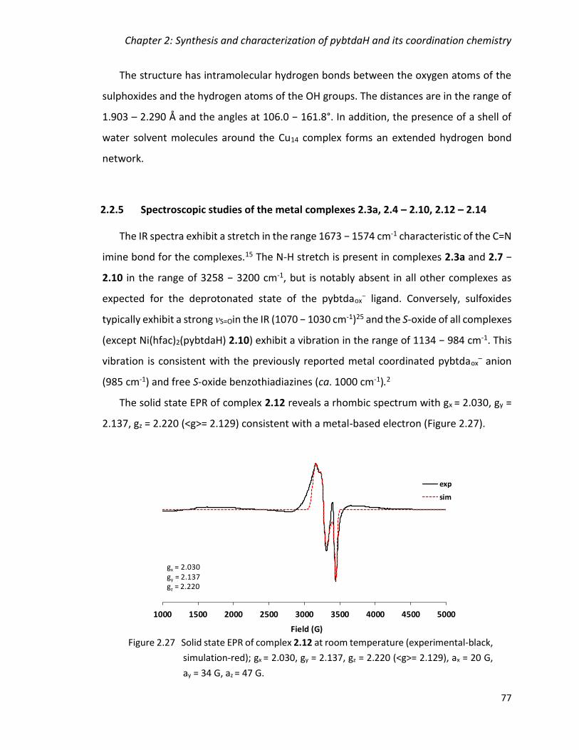
Chapter 2: Synthesis and characterization of pybtdaH and its coordination chemistry
77
The structure has intramolecular hydrogen bonds between the oxygen atoms of the
sulphoxides and the hydrogen atoms of the OH groups. The distances are in the range of
1.903 – 2.290 Å and the angles at 106.0 − 161.8°. In addition, the presence of a shell of
water solvent molecules around the Cu14 complex forms an extended hydrogen bond
network.
2.2.5 Spectroscopic studies of the metal complexes 2.3a, 2.4 – 2.10, 2.12 – 2.14
The IR spectra exhibit a stretch in the range 1673 − 1574 cm-1 characteristic of the C=N
imine bond for the complexes.15 The N-H stretch is present in complexes 2.3a and 2.7 −
2.10 in the range of 3258 − 3200 cm-1, but is notably absent in all other complexes as
expected for the deprotonated state of the pybtdaox− ligand. Conversely, sulfoxides
typically exhibit a strong νS=Oin the IR (1070 − 1030 cm-1)25 and the S-oxide of all complexes
(except Ni(hfac)2(pybtdaH) 2.10) exhibit a vibration in the range of 1134 − 984 cm-1. This
vibration is consistent with the previously reported metal coordinated pybtdaox– anion
(985 cm-1) and free S-oxide benzothiadiazines (ca. 1000 cm-1).2
The solid state EPR of complex 2.12 reveals a rhombic spectrum with gx = 2.030, gy =
2.137, gz = 2.220 (<g>= 2.129) consistent with a metal-based electron (Figure 2.27).
Figure 2.27 Solid state EPR of complex 2.12 at room temperature (experimental-black,
simulation-red); gx = 2.030, gy = 2.137, gz = 2.220 (<g>= 2.129), ax = 20 G,
ay = 34 G, az = 47 G.
1000 1500 2000 2500 3000 3500 4000 4500 5000
Field (G)
exp
sim
gx = 2.030gy = 2.137gz = 2.220

Chapter 2: Synthesis and characterization of pybtdaH and its coordination chemistry
78
2.2.6 Reactivity trends in coordination chemistry of pybtdaH
The coordination chemistry of the ligand 3-(2ʹpyridyl)benzothiadiazine (pybtdaH) has
been previously explored with MCl2 forming a range of N,Nʹ-chelate complexes with first
row transition metals including FeCl2(pybtdaH) and CuCl2(pybtdaH)2.1 Previous studies
indicated that S-oxidation of pybtdaH or related benzothiadiazines only occurred in the
presence of a metal and Et3N. For example, reaction of CuCl2 with pybtdaH affords a
stable red solution of CuCl2(pybtdaH)2 but which led to the isolation of the green
dimetallic complex [Cu(pybtdaox)2]2 complex upon addition of Et3N.2 In this chapter, we
investigated the effect of Lewis acidic metal ions, more basic counter ions (hfac− and OAc−)
and the introduction of base in the reaction scheme in the oxidation of the ligand pybtdaH
to pybtdaox−.
The reaction of pybtdaH and FeCl3·6H2O afforded the mononuclear complex
FeCl3(pybtdaHox)(CH3OH) (2.3a) and the tetranuclear complex Fe4Cl4(OCH3)6(pybtdaox)2
(2.3b) depending on the concentration of the solution. In these complexes, the ligand
pybtdaH has been oxidized to either pybtdaHox or pybtdaox
– even without the presence of
base (such as Et3N) in the reaction mixture. Previous results with FeCl2 and the ligand
pybtdaH resulted in the mononuclear complex FeCl2(pybtdaH)1 leading to the conclusion
that the Lewis acidic nature of FeIII is promoting the oxidation of the ligand in this system.
The next reaction scheme investigated was the ligand pybtdaH with metal salts
containing OAc– as counterions. The mononuclear complex Co(pybtdaox)3 (2.4) was
isolated which does not contain acetate groups. The reaction of pybtdaH and M(OAc)2
(M = Cu, Zn) afforded the dinuclear complexes Cu2(OAc)2(pybtdaox)2(H2O)2 (2.5) and
Zn2(OAc)2(pybtdaox)2 (2.6) with the acetate groups adopting bridging and chelate
coordination modes, respectively. In all three complexes the ligand has been oxidised and
deprotonated only by the presence of a more basic counter ion (pKa (AcOH) = 4.76)7 and
without introduction of base such as Et3N. In complexes 2.4 and 2.5, the ligand pybtdaox–
is chelating one metal centre whereas in complex 2.6 the ligand is bridging two Zn ions.
The isolation of the mononuclear complexes 2.7 − 2.9 with general formula
M(hfac)2(pybtdaHox)2 (M = Mn (2.7), Co (2.8), Zn (2.9)) on addition of pybtdaH to M(hfac)2
Chapter 2: Synthesis and characterization of pybtdaH and its coordination chemistry
79
in which the ligand has been oxidised was therefore unsurprising, given the similar pKa
values of OAc– and hfac–. The ligand pybtdaHox acts as a simple N,Nʹ-chelate to transition
metal ions in a similar fashion to the pybtdaH ligand.1 In contrast, Ni appears more
resilient to promoting ligand oxidation with the reaction of pybtdaH and Ni(hfac)2·xH2O
affording the mononuclear complex with formula Ni(hfac)2(pybtdaH) (2.10), in which the
ligand has not been oxidised or deprotonated and the basic nature of the hfac group has
no effect on the outcome of the reaction, suggesting that this is not merely due to the
acidity of the metal ion (Ni2+ is intermediate between Mn and Co which both undergo
ligand oxidation under the same conditions). Addition of base (Et3N) in the reaction
scheme resulted to the trimetallic complex Ni3(hfac)(pybtdaox)5(H2O) (2.11) where the
ligand has been oxidised to form the deprotonated pybtdaox–.
The outcome of the reaction between the ligand pybtdaH and Cu(hfac)2 proved
dependent on: (i) ratio of Cu:pybtdaH, (ii) basic conditions and (iii) the presence of H2O.
The reaction of pybtdaH and Cu(hfac)2 in 1:1 molar ratio afforded the polymer
[Cu(hfac)(pybtdaox)]n (2.12). The product isolated from the reaction in 1:2 molar ratio was
the previously reported dimer [Cu(pybtdaox)2]2 which was isolated by the reaction of CuCl2
with pybtdaH in the presence of base.2 The addition of base in the 1:1 molar ratio resulted
in formation of the tetranuclear complex Cu4(OH)4(pybtdaox)4 (2.13). The stoichiometric
reaction for formation of tetranuclear complex 2.13 including 1:1 molar ratio of
Cu:pybtdaH in the presence of Et3N and H2O unexpectedly afforded the polymetallic
complex Cu14(OH)12(CO3)2(pybtdaox)12(H2O)2 (2.14), permitting us to conclude that
increasing the basic environment resulted in complexes free of hfac– groups and also of
higher nuclearity.
In initial studies with MCl2 displacement of chloride has no effect on pH since chloride
is the conjugate base of a strong acid (pKa (HCl) = -6.1)6 and effectively has minimal base
character. The reaction with more acidic metal ion such as FeCl3 promoted the oxidation
of the ligand in this system. Conversely the OAc– and hfac– anions are the conjugate bases
of weak acids (pKa (AcOH) = 4.76 7, pKa (Hhfac) = 4.71 8) and generation of OAc– and hfac–
in solution appeared sufficient to promote the oxidation of the coordinated pybtdaH
Chapter 2: Synthesis and characterization of pybtdaH and its coordination chemistry
80
ligand and reflected in the decolourization of the initial red solution associated with SII
pybtdaH. The resultant pybtdaox− ligand offers a more versatile hard O-donor centre in
addition to the favourable, softer, N,Nʹ-chelate, leading to enhanced aggregation and
polynuclear complexes.
A comparison of the ligand coordination geometry with respect to the free ligand
indicates, as expected, that the major changes in geometry of the coordinated ligand are
associated with the N-bound heteroatom of the thiadiazine ring. The free ligand
pybtdaHox has been isolated and a better comparison can be made with the complexes.
When comparing pybtdaH with pybtdaHox, a marked shortening of the S-N bond by
approximately 0.04 Å (from ca. 1.71 to ca. 1.67 Å) is observed.
The N-coordination to the metal ions leads to somewhat variable results. The N-S
bond lengths of the pybtdaHox are in the range 1.684 – 1.700 Å indicating a slight
lengthening of the bond upon coordination (cf pybtdaHox ligand at 1.6656(18) Å). The
N-S bonds in the species in which the ligand is in its pybtdaox− form is affected by the
coordination of the oxygen of the S=O. The distances are in the range 1.635 – 1.680 Å for
complexes with non-coordinating O, whereas for the complexes with coordinating oxygen
the bonds in the range of 1.635 – 1.657 Å indicating a shortening of the bond. The C–N
bond lengths of the metal complexes containing pybtdaHox are in the range 1.289 – 1.322
Å showing no significant deviation from the free ligand (cf pybtdaHox ligand at 1.306(3)
Å). In the complexes where the ligand adopts the anionic form, pybtdaox−, the electronics
of the heterocycle are altered, resulting to a lengthening of the bonds indicative of single
bond character ranging from 1.332 – 1.378 Å. The S–O bonds in the terminal pybtdaHox
are in the range of 1.488 – 1.499 Å and are comparable with the free ligand (cf 1.4969(16)
Å) as well as other literature reported sulfoxide bonds (average 1.50 Å).2,26

Chapter 2: Synthesis and characterization of pybtdaH and its coordination chemistry
81
Conclusions
The synthesis of the ligand 3-(2ʹpyridyl)benzothiadiazine (pybtdaH) is reported using
a new synthetic route. Computational studies have been performed on the ligand
determining the most stable conformation and tautomer as well as the energy minimum
of the pyridyl ring rotation. A new mechanism has been proposed for the formation of
the benzothiadiazine based on NMR studies.
The coordination chemistry of the ligand 3-(2ʹpyridyl)benzothiadiazine (pybtdaH) with
FeCl3, M(OAc)2 (M = Co, Cu, Zn) and M(hfac)2 (M = Mn, Co, Ni, Cu, Zn) is reported. A series
of mononuclear complexes have been isolated as well as polymetallic with the
nuclearities of 2 (2.5, 2.6), 3 (2.11), 4 (2.3b, 2.13) and 14 (2.14). The reactivity pathway is
dependent on the basic environment of the reaction mixture; by the use of (i) basic
ligands (OAc–, hfac–) and/or (ii) basic conditions (Et3N) promoting ligand oxidation and
deprotonation to form pybtdaHox or the pybtdaox– anion. The neutral pybtdaH and
pybtdaHox ligands act as a simple N,Nʹ-chelate to transition metal ions, whereas the
pybtdaox– anion is more versatile offering one N,Nʹ binding site as well as a pendant O-
donor which offers an additional binding site favouring formation of polynuclear metal
complexes (Figure 2.28).
M
M
M
M
pybtdaH pybtdaHox
pybtdaox- pybtdaox
- pybtdaox-
M
M
M
Figure 2.28 Coordination modes of pybtdaH, pybtdaHox and pybtdaox– observed in the
metal complexes of Chapter 2.
Chapter 2: Synthesis and characterization of pybtdaH and its coordination chemistry
82
Experimental
2.4.1 General considerations and physical measurements
Solvents, starting materials and metal salts were obtained from commercial suppliers
and used without further purification. Manipulation of air-sensitive materials was carried
out under an atmosphere of dry nitrogen using standard Schlenk techniques and a dry-
nitrogen glove box (MBraun Labmaster). Melting points were determined using a
Stanford Research Systems MPA120 EZ-Melt automated melting point apparatus.
Elemental compositions were determined on a Perkin Elmer 2400 Series II CHNS/O
Analyzer. IR spectra were obtained using a Bruker Alpha FT-IR spectrometer equipped
with a Platinum single reflection diamond ATR module. NMR spectra were recorded on a
Bruker 500 MHz spectrometer with a Broadband AX Probe using CDCl3 (1H δ = 7.26 ppm,
s; 13C δ = 77.16 ppm) or CD2Cl2 (1H δ = 5.32 ppm, t; 13C δ = 53.84 ppm) as an internal
reference point relative to Me4Si (δ = 0 ppm). Mass spectra were recorded on a Waters
XEVO G2-XS specifically using the atmospheric solids analysis probe in positive resolution
mode. EPR spectrum of complex 2.12 was measured on a Bruker EMXplus X-band EPR
spectrometer at room temperature. Simulation of the anisotropic EPR spectrum of
complex 2.12 was made using the PIP4Win software.27 Density functional theory (DFT)
calculations of pybtdaH were undertaken using the B3LYP and dispersion-corrected
B3LYP-D3 functional and 6-311G*+ basis set28–30 functional within Jaguar31.
2.4.2 Ligand Synthesis
Ligand pybtdaH was prepared according to a modification of the literature procedure for
pybtdaH14:

Chapter 2: Synthesis and characterization of pybtdaH and its coordination chemistry
83
2-(propylthio)aniline (2.1)
Sodium hydroxide (1.6 g, 40 mmol) was dissolved in H2O (3 mL) and added dropwise to a
solution of 2-aminothiophenol (5.0 g, 40 mmol) in ethanol (50 mL). The reaction mixture
was heated at reflux for 1 h. A solution of 1-bromopropane (4.92 g, 40 mmol) in EtOH (25
mL) was added to the reaction mixture and it was stirred at reflux for 18 h. The solvent
was removed in vacuo and the residue was dissolved in CH2Cl2 (20 mL). The organic phase
was washed with H2O (3 × 10 mL) and brine (1 × 10 mL), dried over Na2SO4 and the solvent
was removed in vacuo to yield a dark green viscous oil. The product was purified by
Kugelrohr distillation (140 ◦C, 0.5 – 2.0 Torr) to yield a honey-coloured oil. Yield: 5.22 g,
78%.
Elemental Analysis calc. for C9H13NS: C, 64.62; H, 7.83; N, 8.37, Found: C, 64.68; H, 7.73;
N. 8.41%.
IR (oil, cm-1) �̃�max = 3458 (w), 3351 (w), 3065 (w), 3017 (w), 2960 (m), 2929 (w), 2870 (w),
1603 (m), 1477 (m), 1447 (m), 1301 (m), 1237 (w), 1157 (w), 1025 (w), 744 (s).
1H NMR (500 MHz, ppm, CD2Cl2) δH = 7.35 (1H, dd, J = 7.7, 1.5 Hz, C6H), 7.09 (1H, td, J =
7.6, 1.5 Hz, C4H), 6.72 (1H, dd, J = 8.0, 1.2 Hz, C3H), 6.67 (1H, td, J = 7.5, 1.2 Hz, C5H), 4.37
(2H, bs, NH2), 2.72 (2H, t, J = 7.3 Hz, S C7H2), 1.57 (2H, sextet, J = 7.3 Hz, SCH2C8H2), 0.98
(3H, t, J = 7.3 Hz, C9H3).
13C NMR (125 MHz, ppm, CD2Cl2) δC = 148.76 (C2), 135.88 (C6), 129.70 (C4), 118.51 (C5),
118.46 (C1), 114.99 (C3), 37.16 (C7), 23.36 (C8), 13.46 (C9).
HRMS (ASAP) m/z [M+H]+ calc. for C9H14NS: 168.0847, found 168.0840.

Chapter 2: Synthesis and characterization of pybtdaH and its coordination chemistry
84
N′-(2-propylthiophenyl)-picolinamidine (2.2)
Compound 2.1 (5 g, 30 mmol) was dissolved in dry THF (10 mL) and added dropwise to a
stirred solution of lithium bis(trimethylsilyl)amide (6.02 g, 36 mmol) in THF (25 mL) at 0
°C under a nitrogen atmosphere. The dark reaction mixture was allowed to warm to room
temperature and stirred for 18 h. 2-Cyanopyridine (3.12 g, 30 mmol) was dissolved in THF
(10 mL) then added dropwise to the reaction mixture and stirred for a further 18 h. The
volume of solvent was reduced to approximately 10 mL and the reaction mixture treated
with 50 mL of NaHCO3(aq.) on ice, then extracted with CH2Cl2 (50 mL). The organic phase
was washed with NaHCO3(aq.) (1 x 50 mL) and brine (1 x 20 mL), dried over Na2SO4 and the
solvent was removed in vacuo. The resulting brown oil was washed with hexanes (3 x 10
mL) to yield the product as a white crystalline powder. Yield 7.08 g, 87%.
Melting point 58.3 °C.
Elemental Analysis calc. for C15H17N3S: C, 66.39; H, 6.31; N, 15.48. Found: C, 66.08; H,
6.46; N, 15.09%.
IR (solid, cm-1) �̃�max = 3490 (w), 3371 (w), 3054 (w), 2960 (m), 2929 (w), 2870 (w), 1637 (s),
1560 (s), 1469 (m), 1434 (m), 1370 (m), 1265 (m), 1056 (m), 1039 (m), 828 (m), 800 (m),
743 (m), 623 (w).
1H NMR (500 MHz, ppm, CDCl3) δH = 8.58 (1H, dq, J = 4.8, 0.8 Hz, C1H), 8.52 (1H, d, J = 7.9
Hz, C4H), 7.83 (1H, td, J = 11.6, 1.8 Hz, C3H), 7.40 (1H, ddd, J = 7.5, 4.8 , 1.2 Hz, C2H), 7.31
(1H, dd, J = 7.9, 1.2 Hz, C8H), 7.16 (1H, td, J = 11.3, 1.3 Hz, C10H), 7.05 (1H, td, J = 11.4, 1.4
Hz, C9H), 6.98 (1H, d, J = 7.1 Hz, C11H), 2.87 (2H, t, J = 7.4 Hz, SC13H2), 1.69 (2H, sextet, J =
7.4 Hz, SCH2C14H2), 1.02 (3H, t, J = 7.3 Hz, C15H3).

Chapter 2: Synthesis and characterization of pybtdaH and its coordination chemistry
85
13C NMR (125 MHz, ppm, CDCl3) δC = 152.24 (C6), 151.31 (C5), 147.99 (C1), 147.73 (C7),
136.97 (C3), 129.94 (C12), 127.67 (C8), 126.16 (C10), 125.34 (C2), 123.74 (C9), 122.02 (C4),
121.02 (C11), 33.88 (C13), 22.43 (C14), 13.82 (C15).
HRMS (ASAP+) m/z [M+H]+ calc. for C15H18N3S: 272.1221, found 272.1225.
3-(pyridin-2-yl)-4H-benzo-1,2,4-thiadiazine (pybtdaH)
A solution of N-chlorosuccinimide (2.88 g, 22 mmol) in CH2Cl2 (40 mL) was added dropwise
to a solution of 2.2 (5.0 g, 18 mmol) in CH2Cl2 (40 mL) at -78 °C and the reaction mixture
was allowed to warm to room temperature and stirred for 18 h. The reaction mixture was
then washed with water (2 × 40 mL), brine (1 × 40 mL) and then dried over Na2SO4. The
solvent was removed in vacuo and the oily residue re-dissolved in toluene (40 mL) and
brought to reflux for 12 h. The solvent was again removed in vacuo to yield a brown solid.
The product was purified by column chromatography in CH2Cl2/hexane 1:1 ratio to yield
orange powder. Yield 2.94 g, 72%.
Melting point 117.5 °C.
Elemental Analysis calc. for C12H9N3S: C, 63.41; H, 3.99; N, 18.49. Found: C, 63.59; H, 4.09;
N, 18.22%.
IR (solid, cm-1) �̃�max = 3308 (m, NH), 3054 (w), 1627 (m), 1586 (m), 1564 (m), 1488(m),
1461 (s), 1438 (m), 1417 (m), 1294 (m), 1101 (m), 1040 (w), 994 (m), 792 (m), 739 (s), 619
(m), 468 (w), 455 (w).
1H NMR (500 MHz, ppm, CDCl3) δH = 8.59 (1H, b, NH), 8.52 (1H, d, J = 4.8 Hz, C1H), 8.09
(1H, d, J = 8.0 Hz, C4H), 7.76 (1H, td, J = 7.7, 1.7 Hz, C3H), 7.37 (1H, ddd, J = 7.5, 4.8, 1.1 Hz,
C2H), 6.94 (1H, td, J = 7.6, 1.4 Hz, C9H), 6.88 (1H, td, J = 7.6, 1.3 Hz, C10H), 6.69 (1H, d, J =
7.5 Hz, C11H), 6.50 (1H, dd, J = 7.7, 1.0 Hz, C8H).
Chapter 2: Synthesis and characterization of pybtdaH and its coordination chemistry
86
13C NMR (125 MHz, ppm, CDCl3) δC = 154.34 (C6), 148.04 (C5), 147.50 (C1), 137.04 (C3),
136.79 (C7), 127.75 (C9), 125.39 (C2), 125.24 (C10), 122.83 (C11), 121.33 (C4), 120.14 (C12),
114.43 (C8).
HRMS (ASAP+) m/z [M+H]+ calc. for C12H10N3S: 228.0595, found 228.0597.
2.4.3 Complex Syntheses with MCl3 (M = Fe)
[FeCl3(pybtdaHox)(CH3OH)]∙CH3OH (2.3a)
Solid ligand pybtdaH (0.023 g, 0.100 mmol) was added to a solution of FeCl3·6H2O (0.027
g, 0.100 mmol) in CH3OH (5 mL). The mixture was stirred for 30 min and a deep red
solution obtained. The solution was filtered and dark red crystals were grown by layering
with a mixture of diethyl ether/hexanes (1:1) within two weeks. Yield 0.012 g, 26%.
Elemental Analysis calc. for [C14H13N3FeSCl3O2]: C, 37.41; H, 2.91; N, 9.35. Found: C, 35.39;
H, 2.18; N, 10.21%.
IR (solid, cm-1) �̃�max = 3230 (br), 1617 (m), 1543 (w), 1515 (m), 1471 (m), 1434 (m), 1362
(w), 1308 (w), 1256 (w), 1155 (w), 1059 (m), 1018 (m), 945 (m), 874 (w), 804 (m), 772 (s),
728 (m), 697 (m), 674 (m), 643 (m), 575 (m), 552 (m), 520 (w), 498 (m), 478 (m), 460 (m),
438 (m).
Fe4Cl4(OCH3)6(pybtdaox)2 (2.3b)
Solid ligand pybtdaH (0.023 g, 0.100 mmol) was added to a solution of FeCl3·6H2O (0.027
g, 0.100 mmol) in CH3OH (10 mL). The mixture was stirred for 30 min and a deep red
solution obtained. The solution was filtered and dark red crystals were grown by layering
with a mixture of diethyl ether/hexanes (1:1) within five weeks. Yield 0.003 g, 3%.
Chapter 2: Synthesis and characterization of pybtdaH and its coordination chemistry
87
2.4.4 Complex Syntheses with M(OAc)2 (M = Co (2.4), Cu(2.5), Zn (2.6))
Co(pmbtdaox)3 (2.4)
Solid ligand pybtdaH (0.023 g, 0.100 mmol) was added to a solution of Co(OAc)2∙4H2O
(0.025 g, 0.100 mmol) in CH3OH (10 mL). The mixture was stirred for 2.5 h and a deep red
solution obtained. The solution was filtered and dark red crystals were grown by layering
with a mixture of diethyl ether/hexanes (1:1) over six weeks. Yield 0.003 g, 4%.
Elemental Analysis calc. for [C36H24N9CoS3O3]∙5H2O: C, 49.37; H, 3.91; N, 14.39. Found: C,
49.33; H, 3.30; N, 14.24%.
IR (solid, cm-1) �̃�max = 1606 (w), 1594 (w), 1560 (m), 1524 (s), 1476 (m), 1458 (m), 1439
(m), 1330 (s), 1279 (m), 1259 (m), 1137 (m), 1119 (m), 1075 (m), 1033 (m), 935 (m), 864
(w), 822 (m), 790 (m), 759 (s), 737 (m), 698 (m), 670 (m), 561 (m), 547 (m), 508 (w), 475
(w), 458 (w), 438 (w), 417 (w).
[Cu2(OAc)2(pybtdaox)2(H2O)2]∙2H2O (2.5)
Solid ligand pybtdaH (0.023 g, 0.100 mmol) was added to a solution of Cu(OAc)2∙2H2O
(0.020 g, 0.100 mmol) in CH3OH (10 mL). The mixture was stirred for 1 h and a green
solution obtained. The solution was filtered and green crystals were grown by layering
with a mixture of diethyl ether/hexanes (1:1) over two days. Yield 0.006 g, 7%.
Elemental Analysis calc. for [C28H26N6CuS2O8]∙2H2O: C, 41.94; H, 3.77; N, 10.48. Found: C,
41.61; H, 3.63; N, 10.28%.
IR (solid, cm-1) �̃�max = 1580 (m), 1561 (m), 1522 (m), 1474 (m), 1427 (s), 1331 (m), 1275
(m), 1134 (m), 1075 (m), 1034 (s), 1025 (s), 935 (w), 866 (w), 830 (m), 775 (s), 750 (m),
677 (m), 653 (m), 620 (m), 565 (m), 532 (m), 508 (m), 482 (m), 460 (m), 445 (w), 422 (w).

Chapter 2: Synthesis and characterization of pybtdaH and its coordination chemistry
88
Zn2(OAc)2(pybtdaox)2 (2.6)
Solid ligand pybtdaH (0.023 g, 0.100 mmol) was added to a solution of Zn(OAc)2∙2H2O
(0.022 g, 0.100 mmol) in CH3OH (10 mL). The mixture was stirred for 1 h and an orange
solution obtained. The solution was filtered and yellow crystals were grown by layering
with a mixture of diethyl ether/hexanes (1:1) over two days. Yield 0.008 g, 10%.
Elemental Analysis calc. for [C28H28N6Zn2S2O6]∙H2O: C, 44.76; H, 3.22; N, 11.18. Found: C,
45.17; H, 2.99; N, 11.15%.
IR (solid, cm-1) �̃�max = 1575 (m), 1563 (m), 1501 (m), 1462 (m), 1445 (m), 1418 (m), 1385
(m), 1335 (m), 1297 (m), 1262(m), 1240 (m), 1148 (w), 1049 (s), 1026 (s), 964 (m), 858
(m), 792 (m), 763 (m), 732 (m), 681 (s), 651 (m), 619 (m), 599 (m), 558 (m), 528 (m), 497
(w), 477 (m), 439 (m), 415 (m).
2.4.5 Complex Syntheses with M(hfac)2 (M = Mn, Co, Ni, Cu, Zn)
Mn(hfac)2(pybtdaHox) (2.7)
Solid ligand pybtdaH (0.023 g, 0.100 mmol) was added to a solution of Mn(hfac)2∙3H2O
(0.026 g, 0.100 mmol) in CHCl3 (15 mL). The mixture was stirred for 45 min in room
temperature and for 15 min the solution was placed in a water bath of 35 °C. The light
orange solution was filtered and yellow crystals were grown by slow evaporation the next
day. Yield 0.017 g, 48%.
Elemental Analysis calc. for [C22H11N3MnSO4F12]: C, 37.10; H, 1.56; N, 5.90. Found: C,
37.23; H, 1.48; N, 6.07%.
Chapter 2: Synthesis and characterization of pybtdaH and its coordination chemistry
89
IR (solid, cm-1) �̃�max = 3010 (w), 1644 (m), 1616 (w), 1556 (w), 1517 (m), 1495 (m), 1467
(m), 1439 (w), 1253 (m), 1191 (m), 1136 (m), 1096 (s), 1082 (m), 1041 (m), 1016 (m), 943
(w), 797 (m), 751 (m), 683 (m), 663 (m), 638 (m), 581 (m), 552 (m), 527 (w), 515 (m), 474
(m), 457 (w), 441 (w).
Co(hfac)2(pybtdaHox) (2.8)
Solid ligand pybtdaH (0.023 g, 0.100 mmol) was added to a solution Co(hfac)2 (0.047 g,
0.100 mmol) in CHCl3 (15 mL). The mixture was stirred for 45 min and a deep red solution
obtained. The solution was filtered and light orange crystals were grown by slow
evaporation over five weeks. Yield 0.001 g, 1%.
IR (solid, cm-1) �̃�max = 3259 (br), 1622 (m), 1565 (m), 1526 (m), 1477 (s), 1462 (m), 1444
(m), 1303 (m), 1279 (m), 1156 (m), 1137 (m), 1076 (m), 943 (w), 826 (w), 759 (m), 698 (w),
665 (m), 613 (m), 560 (m), 558 (m), 545 (m), 535 (m), 508 (m), 498 (m), 469 (m), 458 (m),
422 (m), 412 (m).
Zn(hfac)2(pybtdaHox) (2.9)
Solid ligand pybtdaH (0.023 g, 0.100 mmol) was added to a solution Zn(hfac)2 (0.052 g,
0.100 mmol) in CH2Cl2 (15 mL). The mixture was stirred for 30 min and a light orange
solution obtained. The solution was filtered and colorless crystals were grown by slow
evaporation over five weeks. Yield 0.003 g, 4%.
IR (solid, cm-1) �̃�max = 3200 (br), 1660 (m), 1616 (w), 1503 (m), 1474 (m), 1460 (m), 1385
(m), 1298 (w), 1257 (m), 1193 (m), 1138 (s), 1102 (m), 1052 (m), 1028 (m), 959 (m), 791
(m), 759 (m), 731 (m), 683 (m), 665 (m), 650 (m), 642 (m), 597 (m), 585 (m), 568 (m), 554
(m), 528 (m), 480 (m), 463 (m), 442 (m), 415 (m).

Chapter 2: Synthesis and characterization of pybtdaH and its coordination chemistry
90
Ni(hfac)2(pybtdaH) (2.10)
Solid ligand pybtdaH (0.023 g, 0.100 mmol) was added to a solution of Ni(hfac)2∙xH2O
(0.047 g, 0.100 mmol) in CH2Cl2 (15 mL). The mixture was stirred for 30 min and an orange
solution obtained. The solution was filtered and red crystals were grown by layering with
hexanes over two days. Yield 0.042 g, 60%.
Elemental Analysis calc. for [C22H11N3NiSO4F12]: C, 37.74; H, 1.58; N, 6.00. Found:
C, 37.74; H, 1.51; N, 6.04%.
IR (solid, cm-1) �̃�max = 3368 (br), 1652 (m), 1638 (m), 1583 (m), 1554 (m), 1526 (m), 1466
(m), 1438 (m), 1348 (w), 1252 (m), 1191 (m), 1134 (s), 1099 (m), 1024 (m), 950 (w), 793
(m), 742 (m), 670 (m), 651 (m), 585 (m), 527 (w), 436 (w).
[Ni3(hfac)(pybtdaox)5(H2O) (2.11)
Solid ligand pybtdaH (0.046 g, 0.100 mmol) was added to a solution of Ni(hfac)2∙xH2O
(0.047 g, 0.100 mmol) in CH2Cl2 (15 mL). Upon addition of Et3N (0.05 mL, 0.400 mmol) the
mixture was stirred for 1.5 h. The orange solution was filtered and was layering with
hexanes. Red crystals formed over two days but after a prolong period of time green
crystals appeared.
[Cu(hfac)(pybtdaox)]n (2.12)
Solid ligand pybtdaH (0.023 g, 0.100 mmol) was added to a solution of Cu(hfac)2∙xH2O
(0.049 g, 0.100 mmol) in CH3OH (10 mL). The mixture was stirred for 30 min and a deep
red solution obtained. The solution was filtered and green crystals were grown by slow
evaporation over one week. Yield 0.005 g, 4%.

Chapter 2: Synthesis and characterization of pybtdaH and its coordination chemistry
91
Elemental Analysis calc. for [C24H18N6Cu2S2O6F12]: C, 39.13; H, 1.93; N, 8.05. Found: C,
39.09; H, 1.66; N, 7.77%.
IR (solid, cm-1) �̃�max = 1652 (m), 1609 (w), 1562 (m), 1552 (m), 1502 (m), 1475 (m), 1455
(m), 1383 (m), 1350 (m), 1298 (w), 1255 (m), 1191 (m), 1135 (s), 1102 (m), 1051 (m), 1032
(m), 974 (m), 940 (m), 854 (w), 811 (m), 789 (m), 756 (m), 731 (m), 670 (m), 654 (m), 584
(m), 558 (m), 529 (m), 483 (m), 441 (w), 414 (m).
[Cu4(OH)4(pybtdaox)4]∙H2O (2.13)
Solid ligand pybtdaH (0.023 g, 0.100 mmol) was added to a solution of Cu(hfac)2∙xH2O
(0.049 g, 0.100 mmol) in CH3OH (10 mL). Upon addition of Et3N (0.05 mL, 0.400 mmol)
the mixture was stirred for 30 min in a water bath of 35 °C. The dark orange solution was
filtered and dark brown crystals were grown by slow evaporation over six weeks. Yield
0.002 g, 1%.
IR (solid, cm-1) �̃�max = 3277 (br), 1673 (w), 1562 (m), 1523 (s), 1457 (m), 1333 (m), 1298
(w), 1277 (m), 1263 (m), 1135 (m), 1020 (s), 932 (m), 830 (m), 794 (m), 770 (m), 740 (m),
678 (m), 651 (m), 560 (m), 536 (m), 508 (m), 484 (m), 458 (m), 416 (m).
[Cu14(OH)12(CO3)2(pybtdaox)12(H2O)2]∙14[H2O]∙[CH3OH] (2.14)
Solid ligand pybtdaH (0.023 g, 0.100 mmol) was added to a solution of Cu(hfac)2∙xH2O
(0.049 g, 0.100 mmol) in CH3OH (10 mL) and 2 μL H2O. Et3N (0.05 mL, 0.400 mmol) was
added within 30 min. The mixture was stirred for another 50 min in a water bath of 42 °C.
The dark olive solution was filtered and green crystals were grown by slow evaporation
over three days. Yield 0.001 g, < 1%.
Chapter 2: Synthesis and characterization of pybtdaH and its coordination chemistry
92
Elemental Analysis calc. for [C147H115N36Cu14S12O47]·5H2O: C, 39.22; H, 3.80; N, 11.20.
Found: C, 38.89; H, 2.55; N, 10.88%.
IR (solid, cm-1) �̃�max = 3441 (br), 1605 (w), 1560 (m), 1453 (m), 1384 (m), 1330 (m), 1273
(m), 1134 (w), 1017 (s), 985 (s), 827 (m), 791 (m), 748 (m), 678 (m), 641 (m), 597 (m), 560
(m), 530 (m), 479 (w), 442 (w).
2.4.6 Single-crystal X-ray crystallography
Crystals of the ligand and the complexes 2.2 − 2.13 were mounted on a cryoloop with
paratone oil and examined on a Bruker APEX diffractometer equipped with CCD area
detector and Oxford Cryostream cooler using graphite-monochromated Mo-Kα radiation
(λ = 0.71073 Å). Data were collected using the APEX-II software32, integrated using SAINT33
and corrected for absorption using a multi-scan approach (SADABS)34. Single crystals of
complex 2.14 were mounted on a cryoloop with paratone oil and measured on a Bruker
APEX-II diffractometer equipped with an Oxford Cryoflex low temperature device and an
Incoatec Mo micro-source. Data were collected at 150(2) K using graphite-
monochromated Mo-Kα radiation (λ = 0.71073 Å) using the APEX-II software.
Final cell constants were determined from full least squares refinement of all
observed reflections. The structures were solved using Olex235, with the ShelXT structure
solution program36 using intrinsic phasing and refined within the ShelXL37 refinement
package using least-squares minimization on F2. All hydrogen atoms were added at
calculated positions and refined isotropically with a riding model.
PLATON/SQUEEZE38 was used to treat the contents of the voids because the electron
density was too diffuse to model on complexes 2.3b, 2.4, 2.6, 2.7, 2.11 and 2.13.
Complex 2.5 exhibited disorder of the water molecule which was modelled over two
sites in a 75:25 ratio with a common Uiso. Complex 2.7 exhibited rotational disorder on all
four CF3 groups in the asymmetric unit, which were modelled over two sites in a 1:1 ratio
and refined anisotropically. TWINROTMAT within PLATON revealed a possible non-
merohedral twin and a modest improvement in R1 value. The data were re-examined
Chapter 2: Synthesis and characterization of pybtdaH and its coordination chemistry
93
using cell_now which confirmed the twin law and the data were re-integrated as a two
component non-merohedral twin and an absorption correction applied using TWINABS.
Complexes 2.8 and 2.10 exhibited rotational disorder on one of the four CF3 groups in the
asymmetric unit, which was modelled over two sites in a 1:1 ratio with a common Uiso.
Complex 2.9 exhibited rotational disorder on two of the eight CF3 groups in the
asymmetric unit, which were modelled over two sites in a 1:1 ratio with a common Uiso.
Complex 2.11 revealed substantial disorder of the one pyridyl ring which was modelled
as two over two sites in a 70:30 ratio and also exhibited rotational disorder of both CF3
groups in the asymmetric unit which were modelled over two sites in a 1:1 ratio with a
common Uiso. A summary of key crystallographic data for the ligands and the complexes
is presented in Appendix 1.
Chapter 2: Synthesis and characterization of pybtdaH and its coordination chemistry
94
References
1 E. R. Clark, M. U. Anwar, B. J. Leontowicz, Y. Beldjoudi, J. J. Hayward, W. T. K. Chan, E. L. Gavey, M. Pilkington, E. Zysman-Colman and J. M. Rawson, Dalton Trans., 2014, 43, 12996.
2 E. R. Clark, J. J. Hayward, B. J. Leontowicz, M. U. Anwar, M. Pilkington and J. M. Rawson, Dalton Trans., 2014, 44, 2071.
3 A. K. Pal, D. B. Cordes, K. Pringouri, M. U. Anwar, A. M. Z. Slawin, J. M. Rawson and E. Zysman-Colman, J. Coord. Chem., 2016, 69, 1924.
4 N. Yutronkie, I. Kuehne, I. Korobkov, J. L. Brusso and M. Murugesu, Chem. Commun., 2015, 677.
5 K. L. M. Harriman, I. A. Kühne, A. A. Leitch, I. Korobkov, R. Clérac, M. Murugesu and J. L. Brusso, Inorg. Chem., 2016, 55, 5375.
6 A. Trummal, L. Lipping, I. Kaljurand, I. A. Koppel and I. Leito, J. Phys. Chem. A, 2016, 120, 3663.
7 N. J. Tro, Chemistry: A molecular approach, Pearson, New Jersey, 2nd Ed., 2011.
8 M. Ellinger, H. Duschner and K. Starke, J. Inorg. Nucl. Chem., 1978, 40, 1063.
9 Z. Chen, L. Quan, L. Liu, Y. Wang, H. Zou and F. Liang, Eur. J. Inorg. Chem., 2015, 2015, 1463.
10 E. E. Moushi, C. Lampropoulos, W. Wernsdorfer, V. Nastopoulos, G. Christou and A. J. Tasiopoulos, J. Am. Chem. Soc., 2010, 132, 16146.
11 J.-L. Jin, Y.-L. Shen, Y.-P. Xie and X. Lu, CrystEngComm, 2018, 20, 2036.
12 H. Zhang, B. Li and E. V. Dikarev, J. Clust. Sci., 2008, 19, 311.
13 H. Zhang, B. Li, J. Sun, R. Clérac and E. V. Dikarev, Inorg. Chem., 2008, 47, 10046.
14 E. R. Clark, J. J. Hayward, B. J. Leontowicz, D. J. Eislera and J. M. Rawson, CrystEngComm, 2014, 16, 1755.
15 J. Coates, Encycl. Anal. Chem., 2006, 1.
16 J. G. Stark and H. G. Wallace, Chemistry Data Book, J. Murray. Publ., London, S. I. Edit., 1980.
17 N. N. Earnshaw and A. Greenwood, Chemistry of the Elements, Pergamon Press, Oxford, 1st edn., 1986.
18 P. Kaszynski and V. G. Young, J. Org. Chem., 2004, 69, 2551.
Chapter 2: Synthesis and characterization of pybtdaH and its coordination chemistry
95
19 AIST Integr. Spectr. Database Syst. Org. Compd. (Data were obtained from Natl. Inst. Adv. Ind. Sci. Technol. (Japan)), SciFinder search.
20 ACD-A Sigma-Aldrich (Spectral data were obtained from Adv. Chem. Dev. Inc.), SciFinder Search.
21 N. N. Earnshaw and A. Greenwood, Chemistry of the Elements, Pergamon Press, Oxford, 1st edn., 1986.
22 A. W. Addison and T. N. Rao, J. Chem. Soc., Dalton Trans., 1984, 1349.
23 M. Calligaris, Coord. Chem. Rev., 2004, 248, 351.
24 L. Yang, D. R. Powell and R. P. Houser, Dalton Trans., 2007, 955.
25 R. M. Silverstein, G. C. Bassler and T. C. Morrill, Spectroscopic Identification of Organic Compounds, J. Wiley, 4th edn., 1981.
26 Y. Li, T. Thiemann, T. Sawada, S. Mataka and M. Tashiro, J. Org. Chem., 1997, 62, 7926.
27 PIP4WIN v.1.2, 2011, J.M. Rawson, University of Windsor, Windsor, ON.
28 A. D. Becke, J. Chem. Phys., 1993, 5648.
29 C. Lee, W. Yang and R. G. Parr, Phys. Rev. B Condens. Matter, 1988, 785.
30 A. S. Miehlich, H. Stoll and H. Preuss, Chem. Phys. Lett., 1989, 200.
31 A. D. Bochevarov, E. Harder, T. F. Hughes, J. R. Greenwood, D. A. Braden, D. M. Philipp, D. Rinaldo, M. D. Halls, J. Zhang and R. A. Friesner, Int. J. Quantum Chem., 2013, 113, 2110.
32 APEX-II, Bruker AXS, Inc., Madison, Wisconsin, USA.
33 SAINT, Bruker AXS Inc., Madison, Wisconsin, USA.
34 SADABS, Bruker AXS Inc., Madison, Wisconsin, USA.
35 O. V. Dolomanov, L. J. Bourhis, R. . Gildea, J. A. K. Howard and H. Puschmann, J. Appl. Cryst., 2009, 42, 339.
36 G. M. Sheldrick, Acta Cryst., 2015, A71, 3.
37 G. M. Sheldrick, Acta Cryst., 2015, C71, 3.
38 A. L. Spek, Acta Crystallogr, 2015, C71, 9.
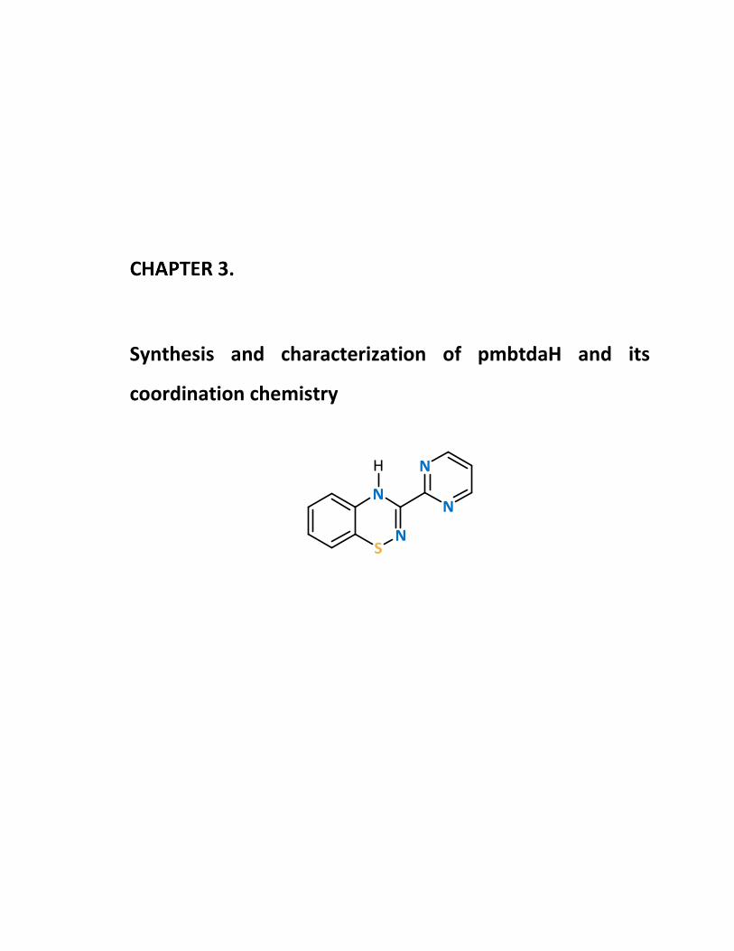
Synthesis and characterization of pmbtdaH and its
coordination chemistry
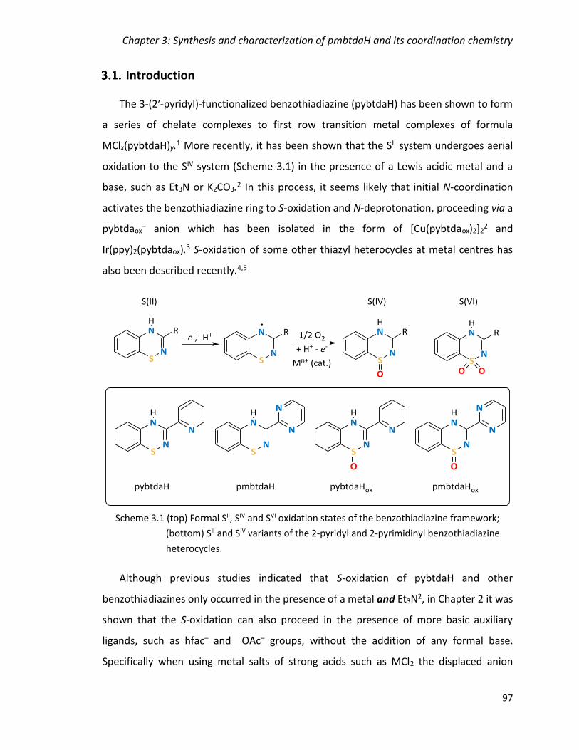
Chapter 3: Synthesis and characterization of pmbtdaH and its coordination chemistry
97
Introduction
The 3-(2ʹ-pyridyl)-functionalized benzothiadiazine (pybtdaH) has been shown to form
a series of chelate complexes to first row transition metal complexes of formula
MClx(pybtdaH)y.1 More recently, it has been shown that the SII system undergoes aerial
oxidation to the SIV system (Scheme 3.1) in the presence of a Lewis acidic metal and a
base, such as Et3N or K2CO3.2 In this process, it seems likely that initial N-coordination
activates the benzothiadiazine ring to S-oxidation and N-deprotonation, proceeding via a
pybtdaox– anion which has been isolated in the form of [Cu(pybtdaox)2]2
2 and
Ir(ppy)2(pybtdaox).3 S-oxidation of some other thiazyl heterocycles at metal centres has
also been described recently.4,5
Although previous studies indicated that S-oxidation of pybtdaH and other
benzothiadiazines only occurred in the presence of a metal and Et3N2, in Chapter 2 it was
shown that the S-oxidation can also proceed in the presence of more basic auxiliary
ligands, such as hfac– and OAc– groups, without the addition of any formal base.
Specifically when using metal salts of strong acids such as MCl2 the displaced anion
Scheme 3.1 (top) Formal SII, SIV and SVI oxidation states of the benzothiadiazine framework;
(bottom) SII and SIV variants of the 2-pyridyl and 2-pyrimidinyl benzothiadiazine
heterocycles.

Chapter 3: Synthesis and characterization of pmbtdaH and its coordination chemistry
98
(chloride) has no effect on pH since chloride is the conjugate base of a strong acid (pKa
(HCl) = -6.1 6) and has minimal base character. Conversely the hfac– anion is the conjugate
base of a weak acid (pKa (Hhfac) = 4.71 7) and generation of hfac– in solution appears
sufficient to promote the oxidation of the coordinated pybtdaH ligand. Nevertheless the
introduction of additional base (Et3N) in the reaction scheme appears to assist the S-
oxidation and the deprotonation of the pybtdaH ligand, resulting in higher nuclearity
clusters.
In this chapter, we will present the synthesis and characterization of the 3-pyrimidinyl-
benzothiadiazine (pmbtdaH, Scheme 3.1) as well as its coordination chemistry to selected
first row transition metal centres. The choice of this ligand is based on its versatility of
coordination modes to metal centres as shown in Figure 3.1. The pmbtdaH ligand offers
one coordination pocket to a metal centre through N,Nʹ-chelation but in its anionic
(pmbtda–) or radical form (pmbtda•) the ligand offers a second chelate pocket with the
ability to bridge two metal centres (Figure 3.1, left). The S-oxidation of this ligand to SIV
offers a more versatile hard O-donor centre in addition to the favourable, softer, N,Nʹ-
chelate, leading to the potential for enhanced aggregation with the pmbtdaox– ligand
capable of binding three metal ions (Figure 3.1, right).
pmbtdaH
M
M
M
pmbtda- or pmbtda•
M
M
M
M
M
M
M
pmbtdaHox pmbtdaox-
pmbtdaHox pmbtdaox-
M
Figure 3.1 Coordination modes of neutral or anionic SII (left) and the SIV (right)
benzothiadiazine ligand.
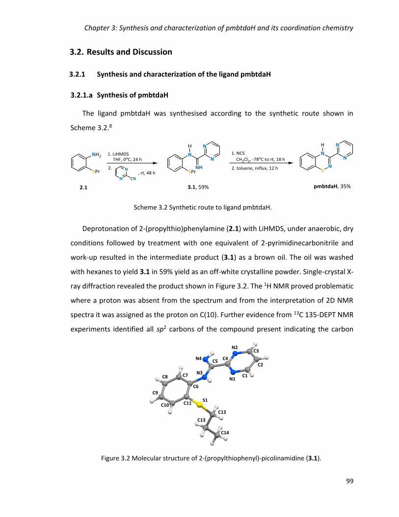
Chapter 3: Synthesis and characterization of pmbtdaH and its coordination chemistry
99
Results and Discussion
3.2.1 Synthesis and characterization of the ligand pmbtdaH
Synthesis of pmbtdaH
The ligand pmbtdaH was synthesised according to the synthetic route shown in
Scheme 3.2.8
Deprotonation of 2-(propylthio)phenylamine (2.1) with LiHMDS, under anaerobic, dry
conditions followed by treatment with one equivalent of 2-pyrimidinecarbonitrile and
work-up resulted in the intermediate product (3.1) as a brown oil. The oil was washed
with hexanes to yield 3.1 in 59% yield as an off-white crystalline powder. Single-crystal X-
ray diffraction revealed the product shown in Figure 3.2. The 1H NMR proved problematic
where a proton was absent from the spectrum and from the interpretation of 2D NMR
spectra it was assigned as the proton on C(10). Further evidence from 13C 135-DEPT NMR
experiments identified all sp2 carbons of the compound present indicating the carbon
Figure 3.2 Molecular structure of 2-(propylthiophenyl)-picolinamidine (3.1).
N3
N4
N1
N2
S1
C8
C9
C10 C11
C7
C6
C5
C3
C4C2
C1
C14
C13
C12
Scheme 3.2 Synthetic route to ligand pmbtdaH.
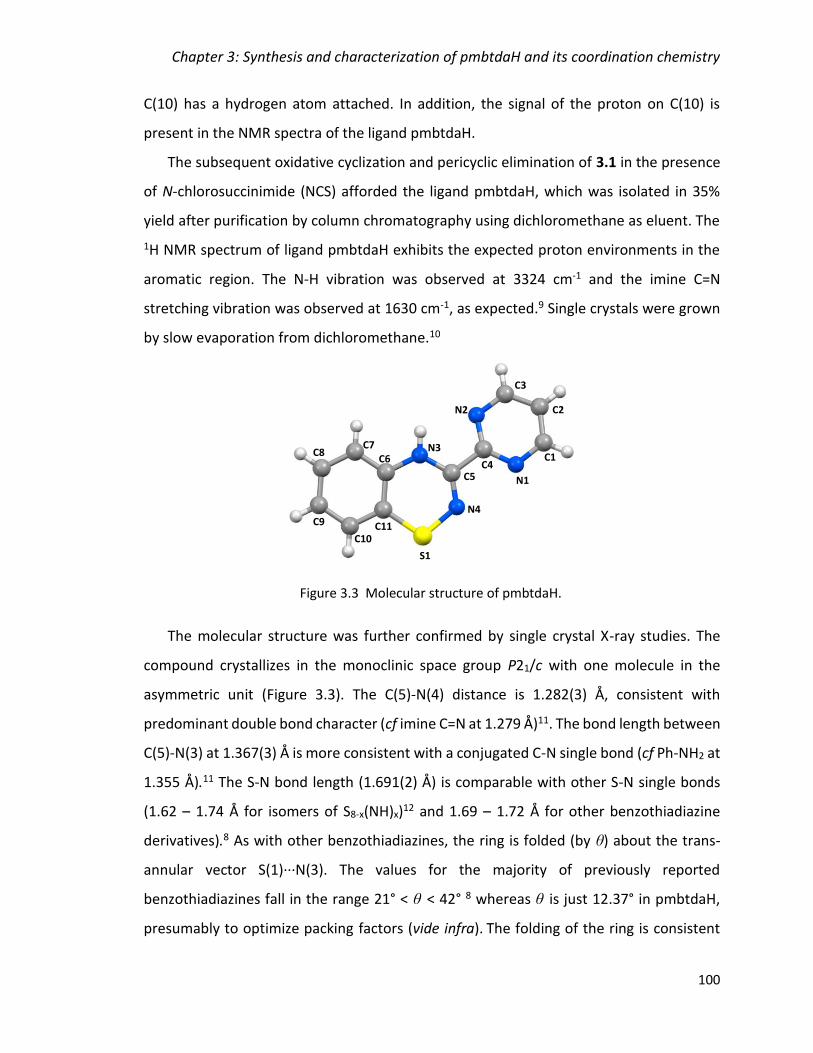
Chapter 3: Synthesis and characterization of pmbtdaH and its coordination chemistry
100
C(10) has a hydrogen atom attached. In addition, the signal of the proton on C(10) is
present in the NMR spectra of the ligand pmbtdaH.
The subsequent oxidative cyclization and pericyclic elimination of 3.1 in the presence
of N-chlorosuccinimide (NCS) afforded the ligand pmbtdaH, which was isolated in 35%
yield after purification by column chromatography using dichloromethane as eluent. The
1H NMR spectrum of ligand pmbtdaH exhibits the expected proton environments in the
aromatic region. The N-H vibration was observed at 3324 cm-1 and the imine C=N
stretching vibration was observed at 1630 cm-1, as expected.9 Single crystals were grown
by slow evaporation from dichloromethane.10
The molecular structure was further confirmed by single crystal X-ray studies. The
compound crystallizes in the monoclinic space group P21/c with one molecule in the
asymmetric unit (Figure 3.3). The C(5)-N(4) distance is 1.282(3) Å, consistent with
predominant double bond character (cf imine C=N at 1.279 Å)11. The bond length between
C(5)-N(3) at 1.367(3) Å is more consistent with a conjugated C-N single bond (cf Ph-NH2 at
1.355 Å).11 The S-N bond length (1.691(2) Å) is comparable with other S-N single bonds
(1.62 – 1.74 Å for isomers of S8-x(NH)x)12 and 1.69 – 1.72 Å for other benzothiadiazine
derivatives).8 As with other benzothiadiazines, the ring is folded (by θ) about the trans-
annular vector S(1)∙∙∙N(3). The values for the majority of previously reported
benzothiadiazines fall in the range 21° < θ < 42° 8 whereas θ is just 12.37° in pmbtdaH,
presumably to optimize packing factors (vide infra). The folding of the ring is consistent
Figure 3.3 Molecular structure of pmbtdaH.
N3
N4
N1
N2
S1
C8
C9
C10C11
C7C6
C5
C3
C4
C2
C1

Chapter 3: Synthesis and characterization of pmbtdaH and its coordination chemistry
101
with the formally 8π anti-aromatic nature of the thiadiazine framework. Two possible
tautomers exist for the ligand, depending on whether N(3) or N(4) atom (Figure 3.3) is
protonated. The shorter imine-like C(5)-N(4) and longer C(5)-N(3) bonds coupled with an
N-H···N hydrogen bonding pattern along the crystallographic c axis (dN-H∙∙∙N = 2.826 Å, θN-
H∙∙∙N = 145.10°, Figure 3.4) clearly point to protonation at N(3). This was additionally
supported by DFT calculations (B3LYP-D3/6-311G*+) which revealed the tautomer with
the H atom bonded at N(3) was more stable by 30 kJ/mol.
Notably pmbtdaH was contaminated with slightly variable quantities (mostly < 10%)
of Cl-pmbtdaH in which the benzo ring has been chlorinated para to the NH group. This
was based on X-ray determination of the metal complexes, presented in Section 3.2.2 and
3.2.3, and it was the case only in some batches of the synthesis where the ligand was used
without further purification. Chlorination appears to be driven by the presence of a slight
molar excess of NCS during synthesis.
Electrochemical studies on pmbtdaH
The redox state of pmbtdaH was monitored by cyclic voltammetry. The
electrochemical studies of the ligand pmbtdaH (C0 = 8.72 x 10⁻³ mol·L-1) were performed
using a deoxygenated solution of nBu4N(PF6) (0.1 M) in dichloromethane with Pt working-
Figure 3.4 Crystal packing of pmbtdaH highlighting N-H∙∙∙N hydrogen bonding along c axis.
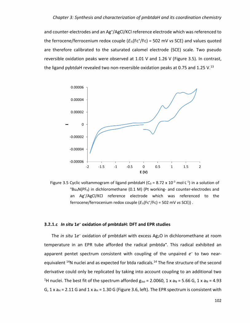
Chapter 3: Synthesis and characterization of pmbtdaH and its coordination chemistry
102
and counter-electrodes and an Ag+/AgCl/KCl reference electrode which was referenced to
the ferrocene/ferrocenium redox couple (E½(Fc+/Fc) = 502 mV vs SCE) and values quoted
are therefore calibrated to the saturated calomel electrode (SCE) scale. Two pseudo
reversible oxidation peaks were observed at 1.01 V and 1.26 V (Figure 3.5). In contrast,
the ligand pybtdaH revealed two non-reversible oxidation peaks at 0.75 and 1.25 V.13
In situ 1e– oxidation of pmbtdaH: DFT and EPR studies
The in situ 1e– oxidation of pmbtdaH with excess Ag2O in dichloromethane at room
temperature in an EPR tube afforded the radical pmbtda•. This radical exhibited an
apparent pentet spectrum consistent with coupling of the unpaired e– to two near-
equivalent 14N nuclei and as expected for btda radicals.14 The fine structure of the second
derivative could only be replicated by taking into account coupling to an additional two
1H nuclei. The best fit of the spectrum afforded giso = 2.0060, 1 x aN = 5.66 G, 1 x aN = 4.93
G, 1 x aH = 2.11 G and 1 x aH = 1.30 G (Figure 3.6, left). The EPR spectrum is consistent with
Figure 3.5 Cyclic voltammogram of ligand pmbtdaH (C0 = 8.72 x 10⁻³ mol·L-1) in a solution of nBu4N(PF6) in dichloromethane (0.1 M) (Pt working- and counter-electrodes and
an Ag+/AgCl/KCl reference electrode which was referenced to the
ferrocene/ferrocenium redox couple (E½(Fc+/Fc) = 502 mV vs SCE)) .
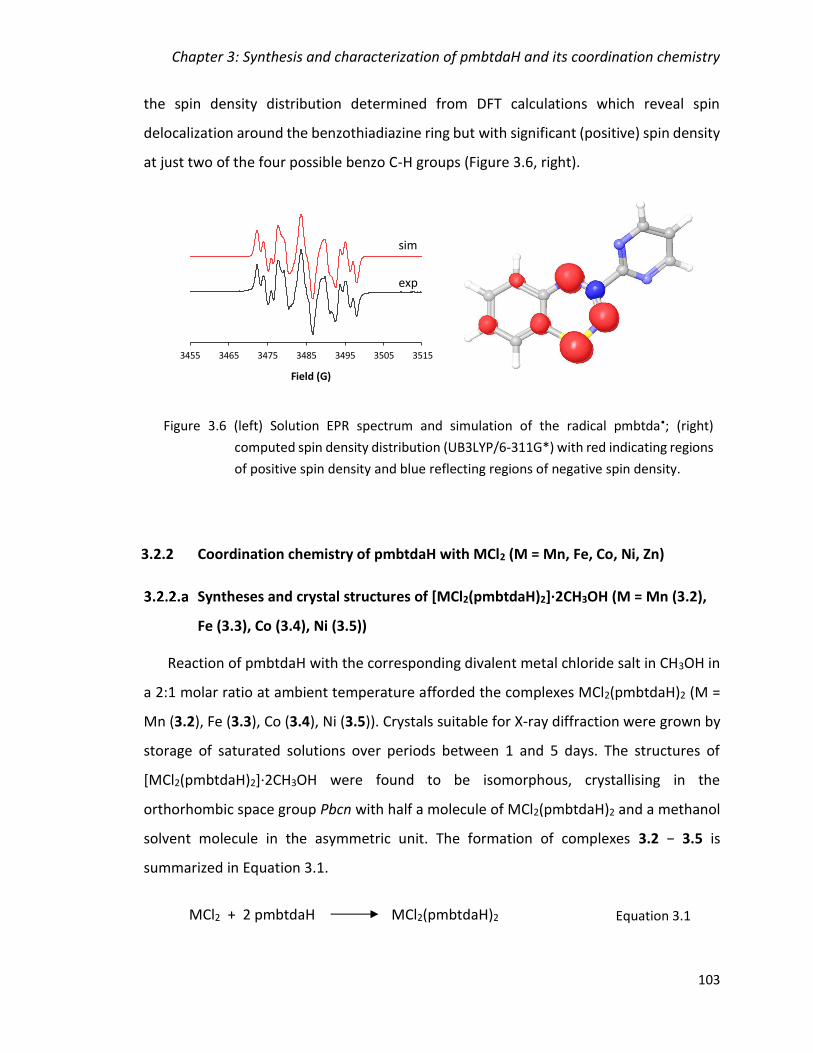
Chapter 3: Synthesis and characterization of pmbtdaH and its coordination chemistry
103
the spin density distribution determined from DFT calculations which reveal spin
delocalization around the benzothiadiazine ring but with significant (positive) spin density
at just two of the four possible benzo C-H groups (Figure 3.6, right).
3.2.2 Coordination chemistry of pmbtdaH with MCl2 (M = Mn, Fe, Co, Ni, Zn)
Syntheses and crystal structures of [MCl2(pmbtdaH)2]·2CH3OH (M = Mn (3.2),
Fe (3.3), Co (3.4), Ni (3.5))
Reaction of pmbtdaH with the corresponding divalent metal chloride salt in CH3OH in
a 2:1 molar ratio at ambient temperature afforded the complexes MCl2(pmbtdaH)2 (M =
Mn (3.2), Fe (3.3), Co (3.4), Ni (3.5)). Crystals suitable for X-ray diffraction were grown by
storage of saturated solutions over periods between 1 and 5 days. The structures of
[MCl2(pmbtdaH)2]·2CH3OH were found to be isomorphous, crystallising in the
orthorhombic space group Pbcn with half a molecule of MCl2(pmbtdaH)2 and a methanol
solvent molecule in the asymmetric unit. The formation of complexes 3.2 − 3.5 is
summarized in Equation 3.1.
3455 3465 3475 3485 3495 3505 3515
Field (G)
sim
exp
Figure 3.6 (left) Solution EPR spectrum and simulation of the radical pmbtda•; (right)
computed spin density distribution (UB3LYP/6-311G*) with red indicating regions
of positive spin density and blue reflecting regions of negative spin density.
MCl2 + 2 pmbtdaH MCl2(pmbtdaH)2 Equation 3.1
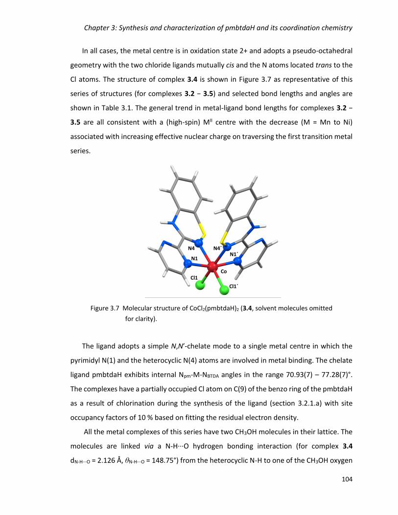
Chapter 3: Synthesis and characterization of pmbtdaH and its coordination chemistry
104
In all cases, the metal centre is in oxidation state 2+ and adopts a pseudo-octahedral
geometry with the two chloride ligands mutually cis and the N atoms located trans to the
Cl atoms. The structure of complex 3.4 is shown in Figure 3.7 as representative of this
series of structures (for complexes 3.2 − 3.5) and selected bond lengths and angles are
shown in Table 3.1. The general trend in metal-ligand bond lengths for complexes 3.2 −
3.5 are all consistent with a (high-spin) MII centre with the decrease (M = Mn to Ni)
associated with increasing effective nuclear charge on traversing the first transition metal
series.
The ligand adopts a simple N,Nʹ-chelate mode to a single metal centre in which the
pyrimidyl N(1) and the heterocyclic N(4) atoms are involved in metal binding. The chelate
ligand pmbtdaH exhibits internal Npm-M-NBTDA angles in the range 70.93(7) – 77.28(7)°.
The complexes have a partially occupied Cl atom on C(9) of the benzo ring of the pmbtdaH
as a result of chlorination during the synthesis of the ligand (section 3.2.1.a) with site
occupancy factors of 10 % based on fitting the residual electron density.
All the metal complexes of this series have two CH3OH molecules in their lattice. The
molecules are linked via a N-H∙∙∙O hydrogen bonding interaction (for complex 3.4
dN-H∙∙∙O = 2.126 Å, θN-H···O = 148.75°) from the heterocyclic N-H to one of the CH3OH oxygen
Figure 3.7 Molecular structure of CoCl2(pmbtdaH)2 (3.4, solvent molecules omitted
for clarity).
Co
N4N1
Cl1 Cl
1’
N4’ N1´
Co
N4
N1
Cl1
Cl1´
N4´

Chapter 3: Synthesis and characterization of pmbtdaH and its coordination chemistry
105
atoms. In addition, the molecules are linked via O-H∙∙∙Cl hydrogen bonding from the
CH3OH oxygen atoms and the chlorides on the metal complex (for complex 3.4
dO-H∙∙∙Cl = 2.471 Å, θO-H∙∙∙Cl = 150.22°, Figure 3.8, top). The packing of 3.4 places the benzo
rings of neighbouring molecules in close proximity having centroid∙∙∙centroid distances of
3.751 Å with the closest C∙∙∙C distance at 3.659 Å (cf interlayer separation in graphite at
3.354 Å12) (Figure 3.8, bottom).
Table 3.1 Selected bond lengths and angles for complexes 3.2 – 3.5.
M-Npm/Å M-NBTDA/Å M-Cl/Å NpmMNBTDA/°a NpmMCl/° NBTDAMCl/° ClMCl/°
Mn (3.2) 2.2507(17) 2.3242(19) 2.4438(6) 70.93(7)
88.44(7)
92.42(5)
104.17(5)
87.54(5)
161.17(5)
105.53(3)
Fe (3.3) 2.1640(19) 2.237(2) 2.4002(7) 73.50(8)
89.54(8)
92.69(6)
101.62(6)
88.25(6)
164.37(6)
101.61(6)
Co (3.4) 2.132(2) 2.173(2) 2.3911(7) 75.29(8)
91.79(8)
92.54(6)
98.46(5)
87.45(6)
165.80(6)
101.80(4)
Ni (3.5) 2.0753(17) 2.1187(18) 2.3906(6) 77.28(7)
91.96(7)
92.50(5)
97.21(5)
87.41(5)
168.78(5)
98.60(3)
N-H∙∙∙O/Å N-H∙∙∙O/° O-H∙∙∙Cl/Å O-H∙∙∙Cl/° C∙∙∙C/Åb centroid-
centroid/Å
Mn (3.2) 2.148 150.08 2.512 154.07 3.605 3.615
Fe (3.3) 2.127 150.29 2.500 149.45 3.625 3.635
Co (3.4) 2.126 148.75 2.471 150.22 3.659 3.751
Ni (3.5) 2.126 147.74 2.417 155.15 3.669 3.679
a top: Angle within the chelate ring; bottom: Angles between N atoms in different rings b Closest C∙∙∙C distance

Chapter 3: Synthesis and characterization of pmbtdaH and its coordination chemistry
106
Complex 3.2 can also be isolated by the reaction in dichloromethane, where the
methanol solvate molecules are replaced by two molecules of CH2Cl2 and the structure is
crystallizing in the monoclinic space group P21/c with one molecule and CH2Cl2 in the
asymmetric unit). Complex 3.3, in which Fe is in oxidation 2+, was produced from reaction
of FeCl2 with pmbtdaH but also formed unexpectedly from reaction of FeCl3 with
pmbtdaH. Notably the recovered yield was higher (23% as opposed to 15%) and the
quality of the crystals proved better for single crystal X-ray diffraction. Generation of 3.3
from reaction of pmbtdaH with FeCl3 can be considered to arise from stoichiometric 1e–
reduction of FeCl3 by pmbtdaH which is known to oxidize to pmbtda•. The overall
stoichiometric equation is shown in Equation 3.2.
Figure 3.8 Molecular structure of 3.4 highlighting hydrogen bonding between the
heterocyclic N-H and the O atoms of the solvent CH3OH molecules, the O-H of
the CH3OH and the Cl of the complex (green); (bottom) π-π interactions
between the benzo rings of the neighbouring molecules (red).

Chapter 3: Synthesis and characterization of pmbtdaH and its coordination chemistry
107
Complex 3.4 (M = Co) can also be isolated by the 1:1 molar ratio of ligand to metal but
with a lower yield, suggesting that the binding constant (2) for the second equivalent of
pmbtdaH is greater than 1 for Co which is in contrast to Zn (below).
Synthesis and crystal structure of Zn2Cl4(pmbtdaH)2 (3.6)
Reaction of the ligand pmbtdaH with ZnCl2 in CH2Cl2 in a 1:1 molar ratio at ambient
temperature afforded a dark precipitate. Crystals of the red dinuclear complex
Zn2Cl4(pmbtdaH)2 (3.6) were formed upon slow evaporation of a CH3OH solution over one
week. The structure of 3.6 was determined by X-ray diffraction (Figure 3.9) and crystallizes
in the triclinic space group P1 with half a molecule in the asymmetric unit and the
complete molecule located about the crystallographic inversion centre. The formation of
complex 3.6 is summarized in Equation 3.3.
Figure 3.9 Molecular structure of Zn2Cl4(pmbtdaH)2 (3.6).
ZnN4´
N1´
Cl1´
Cl1
N4
N1
Zn´Cl2
Cl2´
2 ZnCl2 + 2 pmbtdaH Zn2Cl4(pmbtdaH)2 Equation 3.3
FeCl3 + 3 pmbtdaH FeCl2(pmbtdaH)2 + pmbtda• + HCl Equation 3.2
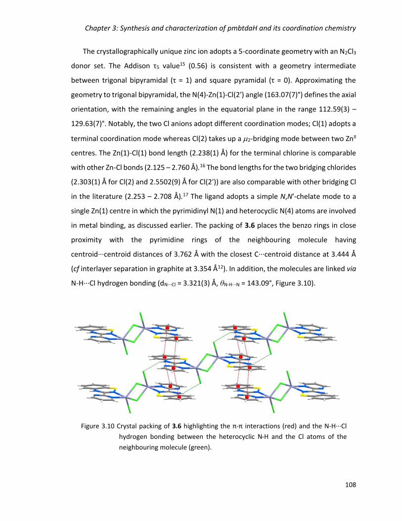
Chapter 3: Synthesis and characterization of pmbtdaH and its coordination chemistry
108
The crystallographically unique zinc ion adopts a 5-coordinate geometry with an N2Cl3
donor set. The Addison τ5 value15 (0.56) is consistent with a geometry intermediate
between trigonal bipyramidal (τ = 1) and square pyramidal (τ = 0). Approximating the
geometry to trigonal bipyramidal, the N(4)-Zn(1)-Cl(2ʹ) angle (163.07(7)°) defines the axial
orientation, with the remaining angles in the equatorial plane in the range 112.59(3) –
129.63(7)°. Notably, the two Cl anions adopt different coordination modes; Cl(1) adopts a
terminal coordination mode whereas Cl(2) takes up a 2-bridging mode between two ZnII
centres. The Zn(1)-Cl(1) bond length (2.238(1) Å) for the terminal chlorine is comparable
with other Zn-Cl bonds (2.125 – 2.760 Å).16 The bond lengths for the two bridging chlorides
(2.303(1) Å for Cl(2) and 2.5502(9) Å for Cl(2ʹ)) are also comparable with other bridging Cl
in the literature (2.253 – 2.708 Å).17 The ligand adopts a simple N,Nʹ-chelate mode to a
single Zn(1) centre in which the pyrimidinyl N(1) and heterocyclic N(4) atoms are involved
in metal binding, as discussed earlier. The packing of 3.6 places the benzo rings in close
proximity with the pyrimidine rings of the neighbouring molecule having
centroid∙∙∙centroid distances of 3.762 Å with the closest C∙∙∙centroid distance at 3.444 Å
(cf interlayer separation in graphite at 3.354 Å12). In addition, the molecules are linked via
N-H∙∙∙Cl hydrogen bonding (dN∙∙∙Cl = 3.321(3) Å, θN-H∙∙∙N = 143.09°, Figure 3.10).
Figure 3.10 Crystal packing of 3.6 highlighting the π-π interactions (red) and the N-H∙∙∙Cl
hydrogen bonding between the heterocyclic N-H and the Cl atoms of the
neighbouring molecule (green).
Chapter 3: Synthesis and characterization of pmbtdaH and its coordination chemistry
109
3.2.3 Coordination chemistry of pmbtdaH with M(hfac)2 (M = Mn, Co, Ni, Cu, Zn)
Syntheses and crystal structures of M(hfac)2(pmbtdaH) (M = Mn (3.7a), Co
(3.8a), Ni (3.9a), Cu (3.10a) and Zn (3.11a))
The metal salts [M(hfac)2]·xH2O, where M = Mn, Co, Ni, Cu, and Zn, were reacted with
the ligand pmbtdaH in CH2Cl2 in a 1:1 molar ratio at ambient temperature and the solution
was layered with hexanes. Red single crystals of the anticipated complexes
M(hfac)2(pmbtdaH) (M = Mn (3.7a), Co (3.8a), Ni (3.9a), Cu (3.10a) and Zn (3.11a)) which
were suitable for X-ray diffraction formed over periods between two days and 3 weeks,
reflecting the robust nature of these complexes with respect to ligand oxidation. Complex
3.7a crystallizes in the triclinic space group P1 with two molecules in the asymmetric unit,
complexes 3.8a and 3.9a crystallize in the monoclinic P21/c with one and two molecules
in the asymmetric unit, respectively, whereas complexes 3.10a and 3.11a crystallize in the
monoclinic space group C2/c with one molecule in the asymmetric unit. The formation of
complexes 3.7a − 3.11a is summarized in Equation 3.4.
The structure of complex 3.11a is shown in Figure 3.11 (left) as representative of the
series of structures 3.7a − 3.11a. All the metal centres are divalent and have a distorted
octahedral geometry with a N2O4 donor set. The heterocyclic ligand provides an N,Nʹ-
chelate pocket analogous to complex 3.4 through N(1) and N(4) atoms (Figure 3.7). The
two hfac– groups also chelate the metal centre leading to an octahedral coordination
geometry. Selected bond lengths and angles are shown in Table 3.2. The general trend in
metal-ligand bond lengths for complexes 3.7a – 3.11a are consistent with a (high-spin) MII
centre with the initial decrease (M = Mn to Ni) associated with increasing effective nuclear
charge on traversing the first transition metal series and subsequent increase (Cu and Zn)
associated with the addition of extra electrons into the eg* orbitals. In the case of CuII
(3.10a), the complex exhibits a Jahn Teller elongation along the O(1)-Cu-O(4) axis with
bond lengths at 2.3000(17) Å and 2.3209(17) Å.
M(hfac)2 + pmbtdaH M(hfac)2(pmbtdaH) Equation 3.4

Chapter 3: Synthesis and characterization of pmbtdaH and its coordination chemistry
110
Within the structure of 3.11a, molecules are linked via a N-H∙∙∙O hydrogen bonding
interaction (dN-H∙∙∙O = 2.397 Å, θN-H∙∙∙O = 151.82°) from the heterocyclic N-H to one of the
hfac– O atoms. Additional π-π stacking forms a centrosymmetric supramolecular dimer
(centroid∙∙∙centroid distance = 3.874 Å) (cf interlayer separation in graphite at 3.354 Å12)
(Figure 3.11, right).
Figure 3.11 (left) Molecular structure of Zn(hfac)2(pmbtdaH) (3.11a); (right) Crystal packing
of 3.11a highlighting the π-π interactions (red) and the N-H∙∙∙O hydrogen
bonding between the amino group and the O atoms of the neighbouring hfac–
group (green).
O4
N4
Zn1
N1
O3
O2
O1

Chapter 3: Synthesis and characterization of pmbtdaH and its coordination chemistry
111
Table 3.2 Selected bond lengths and angles for complexes 3.7a – 3.11a.
M-Npm/Å M-NBTDA/Å M-O/Å Npm-M-NBTDA/°
Mn (3.7a) 2.269(11)(Mn1)
2.285(10)(Mn2)
2.287(11)(Mn1)
2.329(12)(Mn2)
2.146(11)(Mn1)
2.165(10)(Mn1)
2.183(10)(Mn1)
2.211(10)(Mn1)
2.161(8)(Mn2)
2.176(11)(Mn2)
2.178(9)(Mn2)
2.223(10)(Mn2)
72.9(4)(Mn1)
71.8(4)(Mn2)
Co (3.8a) 2.104(4) 2.106(4) 2.051(4)
2.054(3)
2.067(4)
2.077(3)
77.50(14)
Ni (3.9a) 2.102(5)(Ni1)
2.064(5)(Ni2)
2.113(5)(Ni1)
2.137(5)(Ni2)
2.015(4)(Ni1)
2.022(4)(Ni1)
2.029(4)(Ni1)
2.049(4)(Ni1)
2.001(4)(Ni2)
2.015(4)(Ni2)
2.017(4)(Ni2)
2.068(5)(Ni2)
76.91(18)(Ni1)
78.5(2)(Ni2)
Cu (3.10a) 2.004(2) 2.0183(19) 1.9447(18)
1.9696(17)
2.3000(17)
2.3209(17)
80.55(8)
Zn (3.11a) 2.123(4) 2.126(4) 2.02(3)
2.041(19)
2.092(18)
2.158(18)
77.42(15)
N-H∙∙∙O/Å N-H∙∙∙O/° C∙∙∙C/Åa centroid-
centroid/Å
Mn (3.7a) 2.252(Mn1)
2.270(Mn2)
153.92(Mn1)
150.17(Mn2)
3.840(Mn1)
3.510(Mn2)
4.093(Mn1)
3.652(Mn2)
Co (3.8a) 2.292 150.64 3.638 3.797
Ni (3.9a) 2.202(Ni1)
2.352(Ni2)
157.70(Ni1)
151.98(Ni2)
3.599(Ni1)
3.695(Ni2)
3.765(Ni1)
3.879(Ni2)
Cu (3.10a) 2.099 153.14 3.507 3.651
Zn (3.11a) 2.397 151.82 3.709 3.874
a Closest C∙∙∙C distance

Chapter 3: Synthesis and characterization of pmbtdaH and its coordination chemistry
112
Such octahedral “triple-chelate” complexes can adopt or forms. In all these
complexes both and forms exist in the crystal structure as a racemate. However this
is manifested in different ways. In complex 3.11a there is one molecule in the asymmetric
unit with the two hfac– ligands exhibiting significant structural disorder such that it
comprises a racemic mix of both optical isomers. For complex 3.9a there is no structural
disorder but both the and forms are present in the asymmetric unit. The and
forms of complex 3.10a are shown in Figure 3.12, and originate from X-ray collection data
on two different crystals of the complex.
Complex 3.7a (M = Mn) has a high Rint value (10.27%) and all attempts to improve the
quality of the crystals, including different ratios, different solvents and crystallization
methods, were unsuccessful. Nevertheless, the R1 and wR2 were satisfactory (6.97 and
21.37%, respectively) and the connectivity clearly identified.
Figure 3.12 Molecular structure of complex 3.10a, highlighting the two racemic forms the
complex can adopt (lighter colour carbon in front of the plane, darker colour
carbons behind the plane).
Chapter 3: Synthesis and characterization of pmbtdaH and its coordination chemistry
113
Synthesis and crystal structure of [Mn2(hfac)2(tfa)2(pmbtdaHox)2].2CH2Cl2
(3.7b)
The reaction of [Mn(hfac)2]·3H2O with the ligand pmbtdaH in CH2Cl2 in a 1:1 molar
ratio at ambient temperature afforded Mn(hfac)2(pmbtdaH) (3.7a) as red crystals (section
3.2.3.a). However, upon prolonged undisturbed storage, a number of lighter coloured
crystals formed. The crystals were suitable for single X-ray crystallography which revealed
the dinuclear complex Mn2(hfac)2(tfa)2(pmbtdaHox)2 (3.7b). The tfa− anion presumably
arises from degradation of the hfac− anion and decomposition of hfac− to form tfa− has
been reported previously.18,19 This is believed to occur through a retro-Claisen
condensation reaction of -diketones (to generate a ketone and a carboxylate) when
strong bases are present (Equation 3.5).
The decomposition of -diketonate ligands in metal complexes with alkoxy ligands has
also been observed previously19 and was attributed to the presence of the trace amount
of H2O and the alkoxy ligand in the reaction medium. The pmbtdaH ligand itself could
function as the base to promote the decomposition of the hfac– group. The formation of
complex 3.7b is summarized in Equation 3.6.
Complex 3.7b is the minor product of the reaction of Mn(hfac)2 with pmbtdaH.
Attempts to reproduce complex 3.7b were made by the addition of base and/or H2O to
the reaction mixture but failed to produce 3.7b without 3.7a or in higher yields for full
characterization.
2Mn(hfac)2 + 2pmbtdaH + 2H2O + O2 Mn2(hfac)2(tfa)2(pmbtdaHox)2
+ 2CH3COCF3
Equation 3.6
Equation 3.5
hfac− tfa−

Chapter 3: Synthesis and characterization of pmbtdaH and its coordination chemistry
114
The structure of complex 3.7b is shown in Figure 3.13. Complex 3.7b crystallizes in the
triclinic space group P1 with half a molecule in the asymmetric unit and one molecule of
CH2Cl2. The two metal centres are in the +2 oxidation state and adopt a distorted
octahedral geometry with an N2O4 donor set. The neutral ligand has been oxidized and
provides an N,Nʹ-chelate pocket analogous to complex 3.4 through N(1) and N(4) atoms
(Figure 3.7) but in this case the oxygen of the sulfoxide does not act as an additional S-
oxide donor. The chelate ligand pmbtdaHox exhibits a Npm-M-NBTDA bite angle of 72.57(19)°
and the bond lengths of Mn-N are 2.257(5) Å for the pyrimidine N and 2.282(6) Å for the
benzothiadiazine N. The two hfac– groups chelate the metal centre providing two oxygen
atoms to the metal center. The crystal structure also contains two trifluoroacetate (tfa)
groups which each provide a 2-O bridge between the two metal centres, completing their
coordination spheres. All Mn-O bond distances are in the range 2.143(5) – 2.229(4) Å.
The crystal packing of complex 3.7b is shown in Figure 3.14. The dimers are linked via
pairs of centrosymmetric N-H∙∙∙O=S hydrogen bonding interactions (dN-H∙∙∙O = 3.086 Å,
θN-H···O = 150.39°) between the heterocyclic N-H group and the sulfoxide oxygen of a
neighbouring dimer, forming a one dimensional chain structure parallel to the
O4
O4´
O3´O2´
Mn1´
N4´
N1´
Mn1
N1N4
O3
O2
Figure 3.13 Molecular structure of Mn2(hfac)2(tfa)2(pmbtdaHox)2 (3.7b, solvent molecules
omitted for clarity).
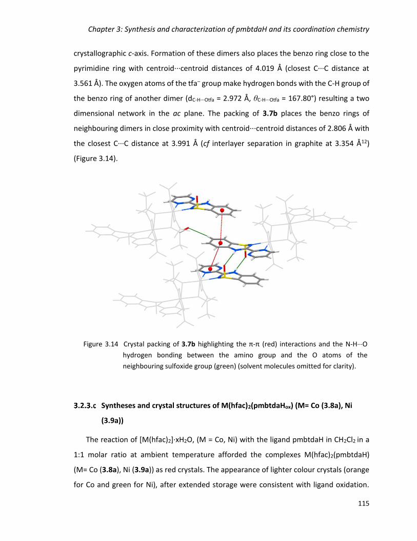
Chapter 3: Synthesis and characterization of pmbtdaH and its coordination chemistry
115
crystallographic c-axis. Formation of these dimers also places the benzo ring close to the
pyrimidine ring with centroid∙∙∙centroid distances of 4.019 Å (closest C∙∙∙C distance at
3.561 Å). The oxygen atoms of the tfa– group make hydrogen bonds with the C-H group of
the benzo ring of another dimer (dC-H∙∙∙Otfa = 2.972 Å, θC-H∙∙∙Otfa = 167.80°) resulting a two
dimensional network in the ac plane. The packing of 3.7b places the benzo rings of
neighbouring dimers in close proximity with centroid∙∙∙centroid distances of 2.806 Å with
the closest C∙∙∙C distance at 3.991 Å (cf interlayer separation in graphite at 3.354 Å12)
(Figure 3.14).
Syntheses and crystal structures of M(hfac)2(pmbtdaHox) (M= Co (3.8a), Ni
(3.9a))
The reaction of [M(hfac)2]·xH2O, (M = Co, Ni) with the ligand pmbtdaH in CH2Cl2 in a
1:1 molar ratio at ambient temperature afforded the complexes M(hfac)2(pmbtdaH)
(M= Co (3.8a), Ni (3.9a)) as red crystals. The appearance of lighter colour crystals (orange
for Co and green for Ni), after extended storage were consistent with ligand oxidation.
Figure 3.14 Crystal packing of 3.7b highlighting the π-π (red) interactions and the N-H∙∙∙O
hydrogen bonding between the amino group and the O atoms of the
neighbouring sulfoxide group (green) (solvent molecules omitted for clarity).

Chapter 3: Synthesis and characterization of pmbtdaH and its coordination chemistry
116
The crystals formed were suitable for X-ray diffraction and structural studies revealed
mononuclear complexes of formula M(hfac)2(pmbtdaHox) (M= Co (3.8b), Ni (3.9b·CH2Cl2)).
The crystal structures of 3.8b and 3.9b are similar to complexes 3.8a and 3.9a,
respectively, but in this case the ligand has been oxidised to the sulfoxide of the pmbtdaH
to form the pmbtdaHox. The formation of complexes 3.8b and 3.9b is summarized in
Equation 3.7.
Complexes 3.8b and 3.9b are the minor products of the reactions which primarily
afford the complexes M(hfac)2(pmbtdaH) (3.8a and 3.9a) as the major products. Attempts
to increase the yield of 3.8b and 3.9b through the introduction of base proved
unsuccessful and the complexes could not be isolated in higher yields for full
characterization beyond structure determination.
Complex 3.8b crystallizes in the monoclinic P21/n with one molecule in the asymmetric
unit whereas complex 3.9b crystallizes in the triclinic space group P1 with one molecule
and two dichloromethane molecules in the asymmetric unit. Crystals of 3.9b had a high
propensity for twinning and afforded high Rint value (13.43%). Nevertheless, the
connectivity of the atoms can be clearly identified. The structure of complex 3.9b is shown
in Figure 3.15 (left), as representative of those two complexes (3.8b and 3.9b) and
selected bond lengths and angles are shown in Table 3.3.
The metal centres have a distorted octahedral geometry with a N2O4 donor set. The
heterocyclic ligand provides an N,Nʹ-chelate pocket analogous to complex 3.4 through
N(1) and N(4) atoms with M-N distances in the range 2.074(11) – 2.122(5) Å and
Npm-M-NBTDA angles in the range 77.09(18) – 78.8(4)°. The two hfac– groups also chelate
the metal centre leading to an octahedral coordination geometry with M-O distances in
the range of 1.993(9) – 2.082(10) Å. A comparison with the M(hfac)2(pmbtdaH) complexes
(3.8a and 3.9a) where the bond lengths of M-O are in the range of 2.015(4) – 2.077(3)
indicates no significant effect on the distances in the metal sphere upon oxidation of the
M(hfac)2 + pmbtdaH + ½ O2 M(hfac)2(pmbtdaHox) Equation 3.7
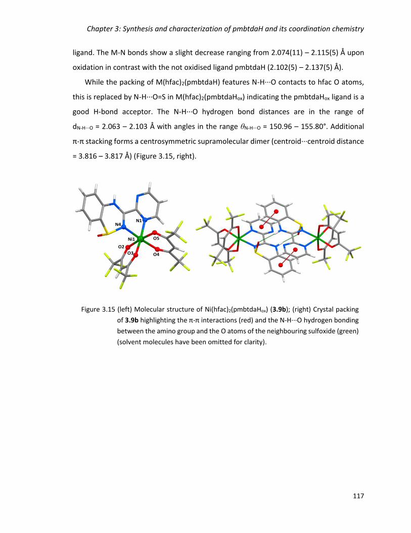
Chapter 3: Synthesis and characterization of pmbtdaH and its coordination chemistry
117
ligand. The M-N bonds show a slight decrease ranging from 2.074(11) – 2.115(5) Å upon
oxidation in contrast with the not oxidised ligand pmbtdaH (2.102(5) – 2.137(5) Å).
While the packing of M(hfac)2(pmbtdaH) features N-H∙∙∙O contacts to hfac O atoms,
this is replaced by N-H∙∙∙O=S in M(hfac)2(pmbtdaHox) indicating the pmbtdaHox ligand is a
good H-bond acceptor. The N-H∙∙∙O hydrogen bond distances are in the range of
dN-H∙∙∙O = 2.063 – 2.103 Å with angles in the range θN-H···O = 150.96 – 155.80°. Additional
π-π stacking forms a centrosymmetric supramolecular dimer (centroid∙∙∙centroid distance
= 3.816 – 3.817 Å) (Figure 3.15, right).
O4
N4
Ni1
N1
O3
O2
O5
Figure 3.15 (left) Molecular structure of Ni(hfac)2(pmbtdaHox) (3.9b); (right) Crystal packing
of 3.9b highlighting the π-π interactions (red) and the N-H∙∙∙O hydrogen bonding
between the amino group and the O atoms of the neighbouring sulfoxide (green)
(solvent molecules have been omitted for clarity).
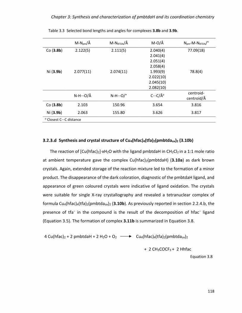
Chapter 3: Synthesis and characterization of pmbtdaH and its coordination chemistry
118
Table 3.3 Selected bond lengths and angles for complexes 3.8b and 3.9b.
M-Npm/Å M-NBTDA/Å M-O/Å Npm-M-NBTDA/°
Co (3.8b) 2.122(5) 2.111(5) 2.040(4) 2.041(4) 2.051(4) 2.058(4)
77.09(18)
Ni (3.9b) 2.077(11) 2.074(11) 1.993(9) 2.022(10) 2.045(10) 2.082(10)
78.8(4)
N-H∙∙∙O/Å N-H∙∙∙O/° C∙∙∙C/Åa centroid-
centroid/Å
Co (3.8b) 2.103 150.96 3.654 3.816
Ni (3.9b) 2.063 155.80 3.626 3.817
a Closest C∙∙∙C distance
Synthesis and crystal structure of Cu4(hfac)4(tfa)2(pmbtdaox)2 (3.10b)
The reaction of [Cu(hfac)2]·xH2O with the ligand pmbtdaH in CH2Cl2 in a 1:1 mole ratio
at ambient temperature gave the complex Cu(hfac)2(pmbtdaH) (3.10a) as dark brown
crystals. Again, extended storage of the reaction mixture led to the formation of a minor
product. The disappearance of the dark coloration, diagnostic of the pmbtdaH ligand, and
appearance of green coloured crystals were indicative of ligand oxidation. The crystals
were suitable for single X-ray crystallography and revealed a tetranuclear complex of
formula Cu4(hfac)4(tfa)2(pmbtdaox)2 (3.10b). As previously reported in section 2.2.4.b, the
presence of tfa− in the compound is the result of the decomposition of hfac− ligand
(Equation 3.5). The formation of complex 3.11b is summarized in Equation 3.8.
4 Cu(hfac)2 + 2 pmbtdaH + 2 H2O + O2 Cu4(hfac)4(tfa)2(pmbtdaox)2
+ 2 CH3COCF3 + 2 Hhfac
Equation 3.8
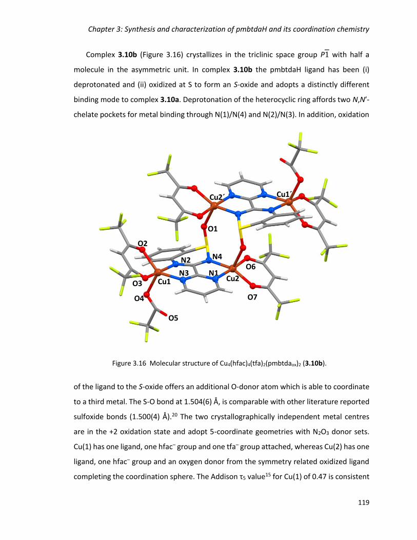
Chapter 3: Synthesis and characterization of pmbtdaH and its coordination chemistry
119
Complex 3.10b (Figure 3.16) crystallizes in the triclinic space group P1 with half a
molecule in the asymmetric unit. In complex 3.10b the pmbtdaH ligand has been (i)
deprotonated and (ii) oxidized at S to form an S-oxide and adopts a distinctly different
binding mode to complex 3.10a. Deprotonation of the heterocyclic ring affords two N,Nʹ-
chelate pockets for metal binding through N(1)/N(4) and N(2)/N(3). In addition, oxidation
of the ligand to the S-oxide offers an additional O-donor atom which is able to coordinate
to a third metal. The S-O bond at 1.504(6) Å, is comparable with other literature reported
sulfoxide bonds (1.500(4) Å).20 The two crystallographically independent metal centres
are in the +2 oxidation state and adopt 5-coordinate geometries with N2O3 donor sets.
Cu(1) has one ligand, one hfac– group and one tfa– group attached, whereas Cu(2) has one
ligand, one hfac– group and an oxygen donor from the symmetry related oxidized ligand
completing the coordination sphere. The Addison τ5 value15 for Cu(1) of 0.47 is consistent
Cu1
N4
N1
O5
O2
O3
O4
Cu2N3
N2
O1
O6
O7
Cu2´ Cu1´
Figure 3.16 Molecular structure of Cu4(hfac)4(tfa)2(pmbtdaox)2 (3.10b).

Chapter 3: Synthesis and characterization of pmbtdaH and its coordination chemistry
120
with an intermediate geometry of trigonal bipyramidal and square pyramidal (τ = 1 for
trigonal bipyramidal and 0 for square pyramidal) with N(3)-Cu(1)-O(3) = 167.0(2)° defining
the axial orientation with the remaining angles in the equatorial plane in the range
109.2(2) – 139.1(3)°. For Cu(2) the Addison value is τ5 = 0.27 consistent with a trigonal
pyramidal geometry with the angles in the equatorial plane in the range 82.4(2) –
173.9(2)°. The pmbtdaox– ligand bridges between a Cu(hfac) and a Cu(hfac)(tfa) unit to
form Cu2(hfac)2(tfa)(pmbtdaox). Two such Cu2(hfac)2(tfa)(pmbtdaox) units dimerise
through the S-oxide oxygen atoms to form a centrosymmetric tetranuclear
Cu4(hfac)4(tfa)2(pmbtdaox)2 cluster. The chelate ligand pmbtdaox− exhibits Npm-Cu-NBTDA
angles of 81.4(2)° and 82.4(2)°. The Cu-N bond lengths are in the range 1.990(6) – 2.071(6)
Å and the Cu-O are 1.929(5) – 2.217(5) Å. The four hfac− groups chelate to the metal center
and the two trifluoroacetate (tfa−) groups are monodentate, providing two more atoms
and completing the coordination sphere of the metal ions.
Synthesis and crystal structure of Zn4(hfac)6(pmbtdaox)2 (3.11b)
Reaction of pmbtdaH with Zn(hfac)2·2H2O in CH2Cl2 in a 1:1 molar ratio, in an
analogous fashion to 3.11a, followed by leaving the solution undisturbed in a sealed vial
for two weeks at ambient temperature afforded crystals of the colourless tetranuclear
complex Zn4(hfac)6(pmbtdaox)2 (3.11b) (Figure 3.17). The structure of 3.11b crystallizes in
the monoclinic space group P21/n with half a molecule in the asymmetric unit. In complex
3.11b the ligand has been also deprotonated and oxidized at S to form an S-oxide and as
in complex 3.10b adopts a distinctly different binding mode to complex 3.11a. The
formation of complex 3.11b is summarized in Equation 3.9.
4 Zn(hfac)2 + 2 pmbtdaH + O2 Zn4(hfac)6(pmbtdaox)2 + 2 hfacH
Equation 3.9

Chapter 3: Synthesis and characterization of pmbtdaH and its coordination chemistry
121
Deprotonation of the heterocyclic ring affords two N,Nʹ-chelate pockets for metal
binding through N(1)/N(4) and N(2)/N(3). In addition, oxidation of the ligand to the S-
oxide offers an additional O-donor atom which is able to coordinate to a third metal. The
S-O bond at 1.520(5) Å, is comparable with other literature reported sulfoxide bonds
(1.500(4) Å20). The two crystallographically independent ZnII centres adopt different
coordination geometries. Zn(1) has a 6-coordinate geometry with a N2O4 donor set, having
one ligand and two hfac– groups attached. On the other hand, Zn(2) is 5-coordinate with
one chelate ligand, one chelate hfac– group and an oxygen donor from the symmetry
related oxidized ligand completing the coordination sphere. The Addison τ5 value15 (0.67)
is consistent with a geometry close to trigonal bipyramidal (τ = 1 for trigonal bipyramidal
and 0 for square pyramidal) with N(3)-Zn(2)-O(6) = 160.0(2)° defining the axial orientation
with the remaining angles in the equatorial plane in the range 112.6(2) – 123.7(2)°.
Figure 3.17 Molecular structure of Zn4(hfac)6(pmbtdaox)2 (3.11b).
Zn1
N4N1
O5
O2
O3
O4 Zn2
N3
N2
O1
O6
O7
Zn2´
Zn1´
Chapter 3: Synthesis and characterization of pmbtdaH and its coordination chemistry
122
The pmbtdaox– ligand bridges between a Zn(hfac)2 and a Zn(hfac) unit to form
Zn2(hfac)3(pmbtdaox). Two such Zn2(hfac)3(pmbtdaox) units dimerise through the S-oxide
oxygen atom to form a centrosymmetric tetranuclear Zn4(hfac)6(pmbtdaox)2 cluster. The
core structure of 3.11b is analogous to 3.10b, comprising a M2(pmbtdaox)2(hfac)2 unit. In
3.10b the remaining N,Nʹ-chelate pocket binds a Cu(hfac)(tfa) unit but in 3.11b it binds a
Zn(hfac)2 unit. The absence of heterocyclic N-H groups in the pmbtdaox– ligand precludes
a hydrogen bonded network and the molecular packing of 3.11b appears driven by
dispersion forces. Intermolecular π-π interactions form a chain parallel to the
crystallographic a axis with centroid∙∙∙centroid distances at 3.654 Å.
3.2.4 Spectroscopic studies of the metal complexes
The UV-Vis spectra of the complexes MCl2(pmbtdaH)2 (3.2 − 3.6) are dominated by a
series of intense π-π* ligand-centered absorption bands between 230 and 300 nm (Figure
3.18). In particular, the visible region exhibits lower intensity absorption bands at 430 –
455 nm which are comparable to the free ligand, pmbtdaH (441 nm) and which give rise
to the diagnostic red colour of the btda ligand1 and its complexes (Figure 3.18, inset).
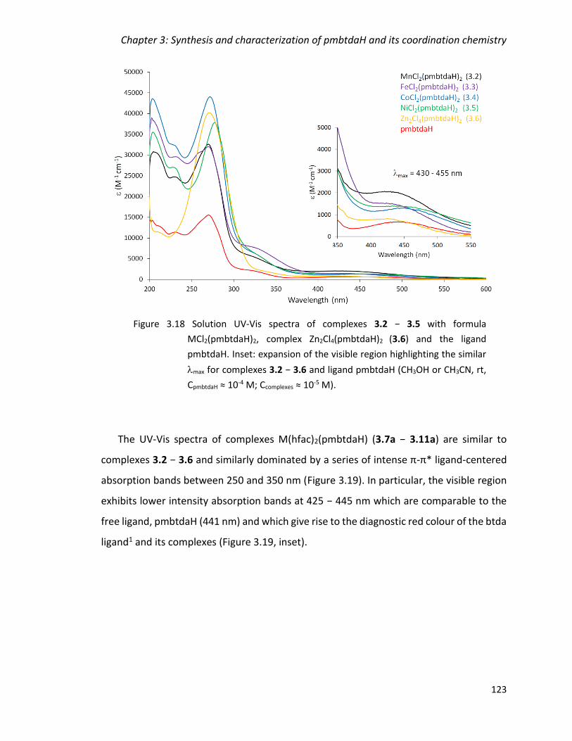
Chapter 3: Synthesis and characterization of pmbtdaH and its coordination chemistry
123
The UV-Vis spectra of complexes M(hfac)2(pmbtdaH) (3.7a − 3.11a) are similar to
complexes 3.2 − 3.6 and similarly dominated by a series of intense π-π* ligand-centered
absorption bands between 250 and 350 nm (Figure 3.19). In particular, the visible region
exhibits lower intensity absorption bands at 425 − 445 nm which are comparable to the
free ligand, pmbtdaH (441 nm) and which give rise to the diagnostic red colour of the btda
ligand1 and its complexes (Figure 3.19, inset).
Figure 3.18 Solution UV-Vis spectra of complexes 3.2 − 3.5 with formula
MCl2(pmbtdaH)2, complex Zn2Cl4(pmbtdaH)2 (3.6) and the ligand
pmbtdaH. Inset: expansion of the visible region highlighting the similar
max for complexes 3.2 − 3.6 and ligand pmbtdaH (CH3OH or CH3CN, rt,
CpmbtdaH ≈ 10-4 M; Ccomplexes ≈ 10-5 M).
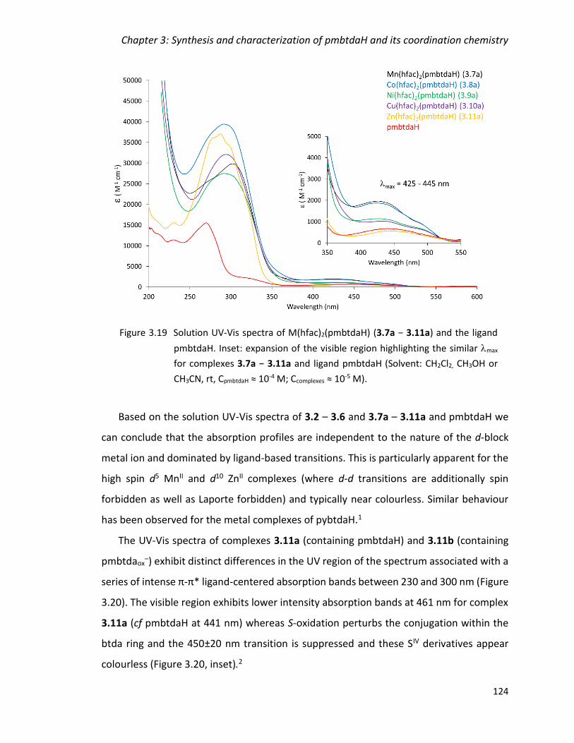
Chapter 3: Synthesis and characterization of pmbtdaH and its coordination chemistry
124
Based on the solution UV-Vis spectra of 3.2 – 3.6 and 3.7a – 3.11a and pmbtdaH we
can conclude that the absorption profiles are independent to the nature of the d-block
metal ion and dominated by ligand-based transitions. This is particularly apparent for the
high spin d5 MnII and d10 ZnII complexes (where d-d transitions are additionally spin
forbidden as well as Laporte forbidden) and typically near colourless. Similar behaviour
has been observed for the metal complexes of pybtdaH.1
The UV-Vis spectra of complexes 3.11a (containing pmbtdaH) and 3.11b (containing
pmbtdaox–) exhibit distinct differences in the UV region of the spectrum associated with a
series of intense π-π* ligand-centered absorption bands between 230 and 300 nm (Figure
3.20). The visible region exhibits lower intensity absorption bands at 461 nm for complex
3.11a (cf pmbtdaH at 441 nm) whereas S-oxidation perturbs the conjugation within the
btda ring and the 450±20 nm transition is suppressed and these SIV derivatives appear
colourless (Figure 3.20, inset).2
Figure 3.19 Solution UV-Vis spectra of M(hfac)2(pmbtdaH) (3.7a − 3.11a) and the ligand
pmbtdaH. Inset: expansion of the visible region highlighting the similar max
for complexes 3.7a − 3.11a and ligand pmbtdaH (Solvent: CH2Cl2, CH3OH or
CH3CN, rt, CpmbtdaH ≈ 10-4 M; Ccomplexes ≈ 10-5 M).

Chapter 3: Synthesis and characterization of pmbtdaH and its coordination chemistry
125
The IR spectra, incorporating the ligand pmbtdaH, exhibit a stretch at ca. 1640 cm-1
characteristic of the C=N imine bond for the complexes whereas in complex 3.11b the
stretch is at 1647 cm-1.9 The N-H stretch is present in all complexes where the ligand is not
oxidised, in the range of 3221 – 3452 cm-1, but is notably absent in complex 3.11b as
expected for the deprotonated state of the pmbtdaox− ligand. Conversely, sulfoxides
typically exhibit a strong νS=O in the IR (1070 – 1030 cm-1)21 and the S-oxide complex 3.11b
exhibits a vibration at 1000 cm-1, a feature notably absent in all other complexes. This
vibration is consistent with the previously reported metal coordinated pybtdaox– anion
(985 cm-1) and free S-oxide benzothiadiazines (ca. 1000 cm-1).2
Figure 3.20 Solution UV-Vis spectra of complexes 3.11a, 3.11b and the ligand pmbtdaH.
Inset: expansion of the visible region highlighting the similar max for
complexes 3.11a, 3.11b and ligand pmbtdaH (CH3CN or CH3OH, rt, CpmbtdaH ≈
10-4 M; Ccomplexes ≈ 10-5 M).

Chapter 3: Synthesis and characterization of pmbtdaH and its coordination chemistry
126
The diamagnetic nature of ZnII ions permitted 1H NMR studies to be undertaken for
complex 3.11a. Coordination of the pmbtdaH ligand to Zn(hfac)2 leads to a diagnostic
downfield shift of all 1H peaks by less than 1 ppm (Figure 3.21). The appearance of a new
signal at 6.04 ppm corresponds to the protons of the two hfac– groups of the complex.
This C-H proton may be broadened due to equilibriation in solution between and
forms of 3.11a via a Bailar twist or Ray-Dutt mechanism.22 Notably the structure of 3.11a
has O atoms either cis or trans to N leading to formally chemically inequivalent CF3 groups,
yet the observation of a singlet in the 19F NMR is consistent with a dynamic process in
solution.
The solid state EPR of complex 3.10a reveals a pseudo-axial spectrum with gx = 2.27,
gy = 2.25, gz = 2.07 (<g>= 2.197) consistent with a metal-based electron (Figure 3.22).
Figure 3.21 1H NMR of complex 3.11a (red), pmbtdaH (blue) and Zn(hfac)2 (green)
(500 MHz, CD3CN).
hfac CH
pmbtdaH
3.11a
Zn(hfac)2

Chapter 3: Synthesis and characterization of pmbtdaH and its coordination chemistry
127
3.2.5 Reactivity trends in the coordination chemistry of pmbtdaH
The ligand 3-(2ʹ-pyridyl)benzothiadiazine (pybtdaH) can form a range of N,Nʹ-chelate
complexes with first row transition metals including CuCl2(pybtdaH) and CuCl2(pybtdaH)2.1
Previous studies indicated that S-oxidation of pybtdaH or related benzothiadiazines only
occurred in the presence of a metal and Et3N. For example, reaction of CuCl2 with pybtdaH
affords a stable red solution of CuCl2(pybtda)2 but which led to the isolation of the green
dimetallic complex [Cu(pybtdaox)2]2 complex upon addition of Et3N.2 In Chapter 2, we
showed that weakly basic counter ions (hfac− and OAc−) could facilitate the oxidation of
the ligand pybtdaH even without the introduction of base and afforded a series of
polynuclear complexes containing the pybtdaox− anion.
The isolation of the mononuclear complexes 3.2 − 3.5 with general formula
MCl2(pmbtdaH)2 and the dimeric complex Zn2Cl4(pmbtdaH)2 (3.6) on addition of pmbtdaH
to MCl2 was therefore unsurprising with the pmbtdaH ligand acting as a simple N,Nʹ-
chelate to transition metal ions in a similar fashion to the pybtdaH ligand.1
Figure 3.22 Solid state EPR of complex 3.10a at room temperature (experimental-black,
simulation-red); gx = 2.27, gy = 2.25, gz = 2.07 (<g>= 2.197), ax = ay = 110 G,
az = 0 G.
500 1000 1500 2000 2500 3000 3500 4000 4500 5000 5500 6000
Field (G)
exp
sim
gx = 2.27gy = 2.25gz = 2.07

Chapter 3: Synthesis and characterization of pmbtdaH and its coordination chemistry
128
Replacement of Cl− by the weakly basic OAc− and hfac− anions revealed some
differences in reactivity patterns between pybtdaH and pmbtdaH with pmbtdaH showing
a stronger resilience to ligand oxidation. Thus reaction of pmbtdaH and M(hfac)2·xH2O
afforded the mononuclear complexes M(hfac)2(pmbtdaH) (3.7a − 3.11a) and the isolation
of oxidized ligand complexes was only apparent after extended periods of storage. This
led to a series of mononuclear complexes M(hfac)2(pmbtdaHox) (M = Co (3.8b), Ni (3.9b))
and dinuclear Mn2(hfac)2(tfa)2(pmbtdaHox)2 (3.7b) in which the pmbtdaHox ligand is
neutral and acts as an N,Nʹ-chelate. In the tetranuclear complexes
Cu4(hfac)4(tfa)2(pmbtdaox)2 (3.10b) and Zn4(hfac)6(pmbtdaox)2 (3.11b) the ligand is
deprotonated and offers two N,Nʹ-chelate pockets as well as a bridging S=O unit. In most
cases where the ligand is oxidised (pmbtdaHox and pmbtdaox−), the metal complexes are
only formed as the minor product with oxidation only occurring in the presence of (i)
extended crystallization time coupled with (ii) the use of a more basic auxiliary ligand. To
date, attempts to promote deprotonation (and oxidation) of the ligand by introducing a
base in the reaction mixture with Cl− or hfac− were not fruitful.
A comparison of the ligand coordination geometry with respect to the free ligand
indicates, as expected, that the major changes in geometry of the coordinated ligand are
associated with the N-bound heteroatom of the thiadiazine ring. The N-coordination leads
to somewhat lengthening of both C-N and N-S bond lengths with C-N bond distances in
the range 1.290 – 1.314 Å and N-S bond distances in the range 1.692 – 1.720 Å (cf free
ligand at 1.282(3) and 1.691(2) Å, respectively). Upon oxidation of the ligand, the N-S bond
lengths show no significant change (1.685 – 1.699 Å) whereas the C-N bonds (N ortho to
S) show an increase of about 0.03 – 0.05 Å (1.308 – 1.327 Å) losing its double bond
character. In contrast, the C-N bonds (N para to S) show a decrease of about 0.03 – 0.05
Å (1.316 – 1.343 Å) in comparison to the corresponding bond length in free ligand
(1.367(3) Å). In the complexes where the ligand adopts the anionic form, pmbtdaox−,
(3.10b and 3.11b) the electronics of the heterocycle are altered, resulting to a lengthening
of the C-N bonds (N ortho to S) of about 0.06 Å (1.341 – 1.343 Å) in comparison to the
corresponding bond length in free ligand (1.282(3) Å). The C-N bonds (N para to S) show
Chapter 3: Synthesis and characterization of pmbtdaH and its coordination chemistry
129
a decrease of 0.06 Å (1.312 – 1.316 Å) in comparison to the pmbtdaH (1.367(3) Å)
indicative of double bond character. The S–O bonds are in the range of 1.483 – 1.504 Å
and are comparable with the S-O bonds in Chapter 2 (1.488 – 1.499 Å) as well as other
literature reported sulfoxide bonds (average 1.50 Å).2,23 The only exception is on one of
the two complexes with pmbtdaox− (3.11b) in which the S-O is longer at 1.520 Å but also
comparable with other coordinated oxygen atoms (Chapter 2, 1.506 – 1.548 Å).
Chapter 3: Synthesis and characterization of pmbtdaH and its coordination chemistry
130
Conclusions
The synthesis of the novel redox active ligand 3-(2',6'-pyrimidine)-benzo-1,2,4-
thiadiazine (pmbtdaH) has been described. The radical pmbtda• can be prepared by in situ
1e– oxidation and its radical character is confirmed by EPR spectroscopy, along with DFT
calculations of the spin density distribution.
Reaction of pmbtdaH with MCl2 (M = Mn, Fe, Co, Ni) in a 2:1 mole ratio afforded the
mononuclear complexes of general formula MCl2(pmbtdaH)2 (M = Mn (3.2), Fe (3.3), Co
(3.4), Ni (3.5)) in which the ligand binds in a N,Nʹ-chelate fashion via heterocyclic N(4) and
the pyrimidinyl N(1) atom while the Cl– ligands are located mutually cis. Reaction of
pmbtdaH with ZnCl2·2H2O in a 1:1 mole ratio afforded the dinuclear complex
Zn2Cl4(pmbtdaH)2 (3.6) in which the two trigonal bipyramidal metal centres are linked via
2-bridging Cl– ligands while the pmbtdaH ligand continues to bind in an N,Nʹ-chelate
fashion. Reaction of pmbtdaH with M(hfac)2 (M = Mn, Co, Ni, Cu, Zn) in 1:1 mole ratio
afforded the mononuclear complexes with general formula M(hfac)2(pmbtdaH)
(M = Mn (3.7a), Co (3.8a), Ni (3.9a), Cu (3.10a), Zn (3.11a)). Here the ligand also binds in
an N,Nʹ-chelate fashion via heterocyclic N(4) and the pyrimidinyl N(1) atom. Only under
extended storage does oxidation of the pmbtdaH ligand occur and appears to also require
the presence of a weakly basic anion. The mononuclear complexes M(hfac)2(pmbtdaHox)
(M = Co (3.8b), Ni (3.9b)) provide the simplest examples of ligand oxidation. In other cases
deprotonation of the ligand affords two N,Nʹ-chelate pockets affording a bis-chelate
bridge, exemplified by Zn4(hfac)6(pmbtdaox)2 (3.11b). Under these reaction conditions
hydrolysis of the hfac− ligand to generate tfa− further complicates matters, exemplified by
the dimer Mn2(hfac)2(tfa)2(pmbtdaHox)2 (3.7b) and the tetranuclear complex
Cu4(hfac)4(tfa)2(pmbtdaox)2 (3.10b). The different coordination modes isolated from
reactivity studies with pmbtdaH are highlighted in Figure 3.23. Experimentally UV-Vis and
IR studies prove useful diagnostic tools for discriminating between pmbtdaH (max ~ 430
nm, N-H) and pmbtdaox− ligands (absence of max at 430 nm and IR S=O ).

Chapter 3: Synthesis and characterization of pmbtdaH and its coordination chemistry
131
pmbtdaH
M
M
M
M
pmbtdaHox pmbtdaox-
M
Figure 3.23 Coordination modes of the ligand pmbtdaH observed in the metal
complexes of Chapter 3.

Chapter 3: Synthesis and characterization of pmbtdaH and its coordination chemistry
132
Experimental
3.4.1 General considerations and physical measurements
Solvents, starting materials and metal salts were obtained from commercial suppliers
and used without further purification. Manipulation of air-sensitive materials was carried
out under an atmosphere of dry nitrogen using standard Schlenk techniques and a dry-
nitrogen glove box (MBraun Labmaster). Melting points were determined using a Stanford
Research Systems MPA120 EZ-Melt automated melting point apparatus. Elemental
compositions were determined on a Perkin Elmer 2400 Series II CHNS/O Analyzer. UV-Vis
spectra were measured on an Agilent 8453 Spectrophotometer using ca. 1 × 10⁻⁵ M
solutions in methanol, acetonitrile or dichloromethane in the range 200 – 600 nm. IR
spectra were obtained using a Bruker Alpha FT-IR spectrometer equipped with a Platinum
single reflection diamond ATR module. NMR spectra were recorded on a Bruker 500 MHz
spectrometer with a Broadband AX Probe using CDCl3 (1H δ = 7.26 ppm, s; 13C δ = 77.16
ppm) or CD3CN (1H δ = 1.94 ppm, septet) as an internal reference point relative to Me4Si
(δ = 0 ppm). Mass spectra were recorded on a Waters XEVO G2-XS specifically using the
atmospheric solids analysis probe in positive resolution mode. EPR spectra of pmbtda•
and complex 3.10 were measured on a Bruker EMXplus X-band EPR spectrometer at room
temperature. Simulation of the EPR spectrum of pmbtda• was made using WinSim
software24 and of the anisotropic EPR spectrum of complex 3.10 was made using the PIP4Win
software. Density functional theory (DFT) calculations of pmbtdaH were undertaken using
the dispersion-corrected B3LYP-D3 functional and 6-311G*+ basis set25–27 functional
within Jaguar28 whereas spin density calculations on pmbtda• implemented the UB3LYP
functional and 6-311G* basis set.
Cyclic voltammetry was performed at 25 °C, under argon, in anhydrous CH2Cl2 using
nBu4NPF6 (0.1 M) as supporting electrolyte. A Pine Research Compact voltammetry low
volume cell equipped with a ceramic screen printed Pt electrode and a low profile
Ag+/AgCl/KCl reference electrode was used to record data at 200 mV/s scan rates.
Potentials were recalibrated vs. the Ferrocene/Ferrocenium couple (+502 mV vs.

Chapter 3: Synthesis and characterization of pmbtdaH and its coordination chemistry
133
Ag+/AgCl/KCl reference electrode in the above mentioned conditions). Concentrations of
the electro-active species were around 10-3 M.
3.4.2 Ligand Synthesis
Ligand pmbtdaH was prepared according to a modification of the literature procedure for
pybtdaH8:
N′-(2-propylthiophenyl)-picolinamidine (3.1)
2-Propylsulphanylphenylamine (2.1) (5.00 g, 30 mmol) was dissolved in THF (10 mL) and
added dropwise to a stirred solution of lithium bis(trimethylsilyl)amide (6.02 g, 36 mmol)
in THF (25 mL) at 0 °C under a nitrogen atmosphere. The dark reaction mixture was
allowed to warm to room temperature and stirred for 24 h. A solution of 2-
pyrimidinecarbonitrile (3.15 g, 30 mmol) in THF (10 mL) was added dropwise to the
reaction mixture and stirred for a further 48 h. The solvent was removed in vacuo and the
reaction mixture was extracted with CH2Cl2 and treated with 100 mL of NaHCO3(aq.). The
organic phase was washed with NaHCO3(aq.) (1 × 50 mL) and brine (1 × 20 mL), dried over
Na2SO4 and the solvent removed in vacuo. The resulting brown oil was washed with
hexanes (3 × 10 mL) to yield the product as a white crystalline powder (4.82 g, 59%).
Melting point 80 °C.
Elemental Analysis calc. for C14H16N4S: C, 61.74; H, 5.92; N, 20.57. Found: C, 61.73; H,
5.87; N, 20.46%.
IR (solid, cm-1) �̃�max = 3300 (m), 3278 (m), 3077 (w), 3062 (w), 2956 (w), 2932 (w), 2903
(w), 2870 (w), 1630 (m), 1563 (s), 1541 (s), 1455 (m), 1439 (m), 1405 (s), 1300 (m), 1239
(s), 864 (m), 811 (s), 757 (s), 710 (m), 651 (s), 636 (s), 588 (m).

Chapter 3: Synthesis and characterization of pmbtdaH and its coordination chemistry
134
1H NMR (500 MHz, ppm, CDCl3) δH = 8.89 (2H, d, J = 4.9 Hz, C1H, C3H), 8.27 (1H, bs, NH),
7.47 (1H, d, J = 7.5 Hz, C7H), 7.40 (1H, t, J = 4.8 Hz, C2H), 7.31 (1H, t, J = 7.6 Hz, C8H), 6.98
(1H, td, J = 11.5, 1.2 Hz, C9H), 2.80 (2H, t, J = 7.3 Hz, SC12H2), 1.61 (2H, sextet, J = 7.3 Hz,
SCH2C13H2), 0.97 (3H, t, J = 7.3 Hz, C14H3).
13C NMR (125 MHz, ppm, CDCl3) δC = 157.55 (C5), 157.37 (C1,3), 154.31 (C4), 144.15 (C6),
133.13 (C7), 128.56 (C8), 124.86 (C11), 122.78 (C9), 121.62 (C2), 120.14 (C10), 36.76 (C12),
22.76 (C13), 13.70 (C14).
HRMS (ASAP+) m/z [M+H]+ calc. for C14H17N4S: 273.1174, found 273.1164.
3-(2',6'-pyrimidine)-benzo-1,2,4-thiadiazine
(pmbtdaH)
A solution of N-chlorosuccinimide (2.94 g, 22 mmol) in CH2Cl2 (40 mL)
was added dropwise to a solution of 3.1 (5.00 g, 20 mmol) in CH2Cl2 (40
mL) at -78 °C, and the reaction mixture was allowed to warm to room
temperature and stirred for 18 h. The reaction mixture was washed with water (2 × 20
mL), brine (1 × 20 mL) and then dried over Na2SO4. The solvent was removed in vacuo and
the oily residue re-dissolved in toluene (30 mL) and brought to reflux for 12 h. The solvent
was again removed in vacuo to yield a brown solid. The product was purified by column
chromatography using a mixture of CH2Cl2/EtOH (4%) as eluent. The slow evaporation of
the solvent resulted in red shiny crystals of ligand. Yield 1.60 g, 35%.
Melting point 114 °C.
Elemental Analysis calc. for C11H8N4S: C, 57.88; H, 3.53; N, 24.54. Found: C, 57.78; H, 3.34;
N, 24.45%.
UV-Vis (CH3OH; λmax, nm; ε, M-1·cm-1): 270 (15550), 320 (2200), 430 (662), 441 (683).

Chapter 3: Synthesis and characterization of pmbtdaH and its coordination chemistry
135
IR (solid, cm-1) �̃�max = 3324 (m), 3045 (w), 2960 (w), 1630 (m), 1556 (s), 1513 (m), 1465 (s),
1437 (s), 1403 (s), 1319 (m), 1260 (m), 1123 (m), 1094 (m), 1015 (m), 931 (w), 859 (w),
813 (w), 744 (s), 709 (m), 631 (m).
1H NMR (500 MHz, ppm, CDCl3) δH = 8.81 (2H, d, J = 4.9 Hz, C1H, C3H), 8.22 (1H, bs, NH),
7.30 (1H, t, J = 4.9 Hz, C2H), 6.89 (1H, td, J = 11.4, 1.5 Hz, C9H), 6.84 (1H, td, J = 11.2, 1.3
Hz, C8H), 6.60 (1H, d, J = 7.3 Hz, C7H), 6.40 (1H, dd, J = 7.6, 1.2 Hz, C10H).
13C NMR (125 MHz, ppm, CDCl3) δC = 157.25 (C1,3), 155.72 (C4), 152.17 (C5), 136.43 (C6),
127.86 (C9), 125.68 (C8), 122.64 (C7), 121.78 (C2), 119.97 (C11), 114.31 (C10).
HRMS (ASAP+) m/z [M+H]+ calc. for C11H9N4S: 229.0548, found 229.0553.
3.4.3 Reactivity of pmbtdaH with MCl2 (M = Mn, Fe, Co, Ni, Zn)
[MnCl2(pmbtdaH)2]·2CH3OH (3.2)
Ligand pmbtdaH (0.046 g, 0.200 mmol) was added to a solution of MnCl2 (0.013 g, 0.100
mmol) in CH3OH (10 mL). The mixture was stirred for 30 min and a deep red solution
obtained. The solution was filtered and dark red crystals were grown by layering with a
mixture of diethyl ether/hexanes (1:1) the next day. Yield 0.026 g, 45%.
Elemental Analysis calc. for [C22H16N8MnS2Cl2]∙0.5H2O: C, 45.16; H, 3.03; N, 18.72. Found:
C, 45.54; H, 2.89; N, 18.68%.
UV-Vis (CH3OH; λmax, nm; ε, M-1 cm-1): 205 (31010), 229 (25021), 270 (32888), 328 (5386),
421 (2340), 430 (2339).
IR (solid, cm-1) �̃�max = 3452 (br), 1623 (w), 1579 (m), 1558 (m), 1516 (m), 1471 (s), 1440
(m), 1413 (s), 1261 (m), 1014 (s), 942 (m), 810 (m), 781 (m), 746 (s), 705 (m), 644 (s), 583
(w), 477 (w), 453 (w), 434 (w).
Chapter 3: Synthesis and characterization of pmbtdaH and its coordination chemistry
136
[FeCl2(pmbtdaH)2]·2CH3OH (3.3)
Ligand pmbtdaH (0.023 g, 0.100 mmol) was added to a solution of FeCl3∙6H2O (0.027 g,
0.100 mmol) in CH3OH (10 mL). The mixture was stirred for 30 min and a deep red solution
obtained. The solution was filtered and dark brown crystals were grown by layering with
a mixture of diethyl ether/hexanes (1:1) over five days. Yield 0.009 g, 15%.
Elemental Analysis calc. for [C22H16N8FeS2Cl2]·2H2O: C, 42.67; H, 3.25; N, 18.09. Found:
C, 42.26; H, 2.51; N, 17.67%.
UV-Vis (CH3OH; λmax, nm; ε, M-1 cm-1): 202 (38500), 231 (29177), 270 (31609), 340 (5831),
487 (608).
IR (solid, cm-1) �̃�max = 3411 (br), 1649 (w), 1609 (m), 1598 (m), 1555 (m), 1534 (m), 1474
(m), 1444 (m), 1413 (m), 1285 (w), 1176 (m), 1113 (w), 1084 (m), 1072 (m), 1012 (m), 945
(w), 827 (m), 762 (s), 709 (w), 669 (m), 646 (m), 584 (w), 552 (m), 516 (m), 455 (m), 430
(m), 424 (w), 410 (w).
[CoCl2(pmbtdaH)2]·2CH3OH (3.4)
Ligand pmbtdaH (0.046 g, 0.200 mmol) was added to a solution of anhydrous CoCl2 (0.013
g, 0.100 mmol) in CH3OH (10 mL). The mixture was stirred for 30 min and a deep red
solution obtained. The solution was filtered and dark brown crystals were grown by
layering with a mixture of diethyl ether/hexanes (1:1) the next day. Yield 0.023 g, 38%.
Elemental Analysis calc. for [C22H16N8CoS2Cl2]·H2O: C, 43.72; H, 3.00; N, 18.54. Found: C,
44.07; H, 2.55; N, 18.49%.
UV-Vis (CH3OH; λmax, nm; ε, M-1 cm-1): 203 (44387), 230 (33090), 272 (44839), 330 (6514),
490 (1792).

Chapter 3: Synthesis and characterization of pmbtdaH and its coordination chemistry
137
IR (solid, cm-1) �̃�max = 3375 (br), 1633 (m), 1575 (m), 1557 (m), 1506 (m), 1477 (m), 1439
(m), 1404 (m), 1667 (w), 1197 (w), 1099 (w), 1016 (s), 940 (w), 812 (m), 781 (w), 742 (s),
705 (m), 644 (m), 525 (m), 488 (w), 454 (w).
[NiCl2(pmbtdaH)2]·2CH3OH (3.5)
Ligand pmbtdaH (0.023 g, 0.100 mmol) was added to a solution of NiCl2∙6H2O (0.012 g,
0.050 mmol) in CH3OH (10 mL). The mixture was stirred for 20 min and a deep red solution
obtained. The solution was filtered and dark brown crystals were grown by layering with
a mixture of diethyl ether/hexanes (1:1) over two days. Yield 0.013 g, 22%.
Elemental Analysis calc. for [C22H16N8NiS2Cl2]·H2O: C, 43.74; H, 3.00; N, 18.55. Found:
C, 43.72; H, 2.53; N, 18.35%.
UV-Vis (CH3OH; λmax, nm; ε, M-1 cm-1): 204 (35038), 227 (26579), 277 (37339), 330 (5250),
487 (832).
IR (solid, cm-1) �̃�max = 3376 (br), 1625 (m), 1580 (m), 1559 (m), 1520 (m), 1472 (s), 1444
(m), 1439 (m), 1408 (s), 1287 (w), 1261 (m), 1242 (w), 1204 (m), 1165 (w), 1127 (w), 1104
(w), 1072 (w), 1025 (s), 949 (m), 941 (m), 809(m), 794 (m), 647 (s), 559 (br), 527 (m), 477
(m), 455 (m), 435 (w), 422 (w).
Zn2Cl4(pmbtdaH)2 (3.6)
Ligand pmbtdaH (0.023 g, 0.100 mmol) was added to a solution of ZnCl2 (0.013 g, 0.100
mmol) in CH2Cl2 (15 mL). The mixture was stirred for 1 h and a bright orange solution
formed over a dark precipitate. The reaction mixture was filtered and re-dissolved in
CH3OH (6 mL). A small number of dark red crystals were grown by slow evaporation over
a period of one week. Yield 0.003 g, 3%.

Chapter 3: Synthesis and characterization of pmbtdaH and its coordination chemistry
138
Elemental Analysis calc. for [C22H16N8Zn2S2Cl4]∙3CH2Cl2: C, 30.52; H, 2.25; N, 11.39. Found:
C, 30.86; H, 2.49; N, 11.33%.
UV-Vis (CH3CN; λmax, nm; ε, M-1·cm-1): 272 (39778), 357 (662), 432 (324).
IR (solid, cm-1) �̃�max = 3221 (br), 1634 (m), 1556 (m), 1517 (m), 1477 (s), 1440 (s), 1412 (s),
1262 (w), 1201 (w), 1158 (w), 1016 (w), 946 (w), 876 (w), 817 (w), 792 (w), 768 (s), 710
(w), 670 (w), 630 (s), 585 (m), 431 (w).
3.4.4 Reactivity of pmbtdaH with M(hfac)2 (M = Mn, Co, Ni, Cu, Zn)
Mn(hfac)2(pmbtdaH) (3.7a)
Ligand pmbtdaH (0.023 g, 0.100 mmol) was added to a solution of Mn(hfac)2∙3H2O (0.052
g, 0.100 mmol) in CH2Cl2 (15 mL). The mixture was stirred for 30 min and a deep red
solution obtained. The solution was filtered and dark red crystals were grown by layering
with hexanes over three weeks. Yield 0.014 g, 21%.
Elemental Analysis calc. for [C21H10N4MnSO4F12]: C, 36.17; H, 1.45; N, 8.03. Found: C,
36.34; H, 1.41; N, 8.08%.
UV-Vis (CH2Cl2; λmax, nm; ε, M-1 cm-1): 274 (28963), 310 (35084), 440 (3953).
IR (solid, cm-1) �̃�max = 3306 (br), 1644 (m), 1581 (w), 1556 (m), 1484 (m), 1446 (m), 1416
(m), 1252 (m), 1192 (m), 1133 (br), 1094 (m), 1015 (w), 794 (m), 753 (m), 708 (w), 664 (w),
646 (m), 617 (w), 582 (m), 527 (w), 472 (w).

Chapter 3: Synthesis and characterization of pmbtdaH and its coordination chemistry
139
[Mn2(hfac)2(tfa)2(pmbtdaHox)2].2CH2Cl2 (3.7b)
In the same reaction described in section 3.4.4.a, when the sealed vial was left for a longer
period of time, a small number of yellow crystals formed in addition to the dark red
crystals of complex 3.7a which proved suitable for X-ray diffraction. No further analytical
data were collected due to the very low yield of this product.
Co(hfac)2(pmbtdaH) (3.8a)
Ligand pmbtdaH (0.023 g, 0.100 mmol) was added to a solution Co(hfac)2 (0.047 g, 0.100
mmol) in CH2Cl2 (15 mL). The mixture was stirred for 15 min and a deep red solution
obtained. The solution was filtered and dark brown crystals were grown by layering with
hexanes over three weeks. Yield 0.033 g, 46%.
Elemental Analysis calc. for [C21H10N4CoSO4F12]: C, 35.97; H, 1.44; N, 7.99. Found: C, 35.73;
H, 1.88; N, 7.86%.
UV-Vis (CH2Cl2; λmax, nm; ε, M-1 cm-1): 280 (40892), 305 (43233), 425 (3865).
IR (solid, cm-1) �̃�max = 3307 (br), 1634 (m), 1583 (w), 1558 (m), 1524 (m), 1481 (m), 1446
(m), 1418 (m), 1251 (m), 1194 (m), 1136 (br), 1098 (m), 1019 (w), 949 (w), 797 (m), 748
(m), 666 (m), 648 (m), 581 (m), 527 (m), 432 (w), 424 (w).
Chapter 3: Synthesis and characterization of pmbtdaH and its coordination chemistry
140
Co(hfac)2(pmbtdaHox) (3.8b)
In the same reaction as in section 3.4.4.c, when the sealed vial was left for a longer period
of time, orange plates formed in addition to the dark brown crystals of complex 3.8a
suitable for X-ray diffraction. No further analytical data were collected due to very low
yield of this product.
Ni(hfac)2(pmbtdaH) (3.9a)
Ligand pmbtdaH (0.023 g, 0.100 mmol) was added to a solution of Ni(hfac)2∙xH2O (0.047
g, 0.100 mmol) in CH2Cl2 (15 mL). The mixture was stirred for 20 min and a deep red
solution obtained. The solution was filtered and dark brown crystals were grown by
layering with hexanes over three weeks. Yield 0.026 g, 36%.
Elemental Analysis calc. for [C21H10N4NiSO4F12]·0.25hexanes: C, 37.40; H, 1.88; N, 7.75.
Found: C, 37.06; H, 1.50; N, 8.42%.
UV-Vis (CH2Cl2; λmax, nm; ε, M-1 cm-1): 280 (29875), 305 (29511), 435 (2558).
IR (solid, cm-1) �̃�max = 3295 (br), 1640 (m), 1583 (w), 1555 (m), 1477 (m), 1418 (m), 1348
(w), 1253 (m), 1193 (m), 1135 (br), 1096 (m), 1022 (m), 950 (w), 810 (m), 793 (m), 760 (m),
744 (m), 710 (w), 671 (m), 651 (m), 585 (m), 528 (m), 484 (w).
[Ni(hfac)2(pmbtdaHox)].2CH2Cl2 (3.9b)
In the same reaction as in section 3.4.4.e, when the sealed vial was left for a longer period
of time, green blocks formed in addition to the dark brown crystals of complex 3.9a
suitable for X-ray diffraction. No further analytical data were collected due to very low
yield of this product.

Chapter 3: Synthesis and characterization of pmbtdaH and its coordination chemistry
141
Cu(hfac)2(pmbtdaH) (3.10a)
Ligand pmbtdaH (0.023 g, 0.100 mmol) was added to a solution of Cu(hfac)2∙xH2O (0.049
g, 0.100 mmol) in CH2Cl2 (10 mL). The mixture was stirred for 30 min and a deep red
solution obtained. The solution was filtered and dark brown crystals were grown by
layering with hexanes over two days. Yield 0.009 g, 13%.
Elemental Analysis calc. for [C21H10N4CuSO4F12]: C, 35.73; H, 1.43; N, 7.94. Found:
C, 35.50; H, 1.12; N, 8.25%.
UV-Vis (CH2Cl2; λmax, nm; ε, M-1 cm-1): 290 (35355), 305 (33779), 440 (2960).
IR (solid, cm-1) �̃�max = 3242 (br), 1654 (m), 1585 (m), 1548 (m), 1527 (m), 1482 (m), 1454
(m), 1420 (m), 1340 (w), 1254 (m), 1190 (m), 1134 (br), 1087 (m), 1025 (m), 946 (m), 864
(w), 814 (m), 790 (m), 754 (m), 741 (m), 714 (m), 666 (m), 635 (m), 585 (m), 576 (m), 525
(m), 429 (w).
Cu4(hfac)4(tfa)2(pmbtdaox)2 (3.10b)
In the same reaction as in section 3.4.4.g, when the sealed vial was left for a longer period
of time, green blocks formed in addition to the dark brown crystals of complex 3.10a
which proved suitable for X-ray diffraction. No further analytical data were collected due
to very low yield of this product.

Chapter 3: Synthesis and characterization of pmbtdaH and its coordination chemistry
142
Zn(hfac)2(pmbtdaH) (3.11a)
Ligand pmbtdaH (0.023 g, 0.100 mmol) was added to a solution of Zn(hfac)2∙2H2O (0.052
g, 0.100 mmol) in CH2Cl2 (15 mL). The mixture was stirred for 30 min and a deep red
solution obtained. The solution was filtered and dark red crystals were grown over two
days by layering with hexanes. Yield 0.055 g, 78%.
Elemental Analysis calc. for [C21H10N4ZnSO4F12]: C, 35.64; H, 1.42; N, 7.92. Found: C, 35.36;
H, 1.34; N, 7.80%.
UV-Vis (CH3CN; λmax, nm; ε, M-1·cm-1): 234 (16844), 278 (38773), 287 (39012), 461 (1985).
IR (solid, cm-1) �̃�max = 3296 (br), 1640 (m), 1582 (w), 1557 (w), 1527 (w), 1481 (m), 1458
(w), 1447 (w), 1417 (m), 1250 (m), 1190 (m), 1134 (s), 1096 (s), 1020 (w), 949 (w), 795 (m),
752 (m), 711 (w), 665 (m), 648 (m), 618 (w), 581 (m), 526 (w).
1H NMR (500 MHz, ppm, CD3CN) δH = 9.06 (2H, bs, C1H, C3H), 8.94 (1H, bs, NH), 7.85 (1H,
t, J = 5.2 Hz, C2H), 7.06 (1H, td, J = 7.7, 1.4 Hz, C9H), 7.02 (1H, td, J = 7.5, 1.3 Hz, C8H), 6.87
(1H, d, J = 7.6 Hz, C7H), 6.72 (1H, d, J = 7.9 Hz, C10H), 6.04 (2H, bs, hfac-CH).
19F NMR (470 MHz, ppm, CD3CN) δF = -73.11 (bs).
Zn4(hfac)6(pmbtdaox)2 (3.11b)
Ligand pmbtdaH (0.023 g, 0.100 mmol) was added to a solution of Zn(hfac)2∙2H2O (0.052
g, 0.100 mmol) in CH2Cl2 (15 mL). The mixture was stirred for 30 min and a deep red
solution obtained. The solution was filtered and left undisturbed in a sealed vial. White
crystals were grown over two weeks. Yield 0.006 g, 3%.
Elemental Analysis calc. for [C52H20N8Zn4S2O14F36]∙4H2O: C, 30.28; H, 1.37; N, 5.43. Found:
C, 30.07; H, 1.25; N, 5.37%.
UV-Vis (CH3CN; λmax, nm; ε, M-1·cm-1): 264 (7595), 300 (11163), 356 (16998).
Chapter 3: Synthesis and characterization of pmbtdaH and its coordination chemistry
143
IR (solid, cm-1) �̃�max = 1647 (m), 1570 (w), 1524 (w), 1494 (m), 1460 (m), 1375 (w), 1253
(m), 1194 (m), 1134 (s), 1000 (s), 969 (w), 837 (m), 824 (m), 804 (m), 770 (m), 743 (w), 665
(m), 582 (m), 559 (w), 527 (w), 485 (w).
3.4.5 Single-crystal X-ray crystallography
Crystals of the ligand and the complexes 3.1 − 3.10a and 3.11 were mounted on a
cryoloop with paratone oil and examined on a Bruker APEX diffractometer equipped with
CCD area detector and Oxford Cryostream cooler using graphite-monochromated Mo-Kα
radiation (λ = 0.71073 Å). Data were collected using the APEX-II software29, integrated
using SAINT30 and corrected for absorption using a multi-scan approach (SADABS)31. Single
crystals of complex 3.10b were mounted on a cryoloop with paratone oil and measured
on a Bruker Kappa system APEX-II diffractometer equipped with an Oxford Cryoflex low
temperature device. Data were collected at 150(2) K using graphite-monochromated Mo-
Kα radiation (λ = 0.71073 Å) using the APEX-II software.
Final cell constants were determined from full least squares refinement of all observed
reflections. The structures of pmbtdaH and complexes 3.10b and 3.11a were solved by
direct methods (SHELXS within SHELXTL32) to reveal most non-H atoms. Remaining atom
positions were located in subsequent difference maps and the structure refined with full
least squares refinement on F2 within the SHELXTL suite.32 Structures 3.1 − 3.10a and
3.11b were solved using Olex233, with the ShelXT structure solution program34 using
intrinsic phasing and refined within the ShelXL35 refinement package using least-squares
minimization on F2. All hydrogen atoms were added at calculated positions and refined
isotropically with a riding model. In addition a residual electron density peak close to C(9)
was identified as a partial occupancy Cl atom (10% occupancy) for complexes 3.2 − 3.5,
3.7b and 3.8b. For complex 3.7a, the occupancy of the Cl atoms was 40% and 60% and
exhibited rotational disorder on two of the eight CF3 groups in the asymmetric unit, which
were modelled over two sites in a 1:1 ratio with a common Uiso. PLATON/SQUEEZE36 was
used to treat the contents of the voids because the electron density was too diffuse to
Chapter 3: Synthesis and characterization of pmbtdaH and its coordination chemistry
144
model on complex 3.7a. Complex 3.8a exhibited rotational disorder of three of the four
CF3 groups in the asymmetric unit whereas complex 3.8b exhibited rotational disorder of
two of the four CF3 groups in the asymmetric unit which were modelled over two sites in
a 1:1 ratio with a common Uiso. Complex 3.9a exhibited rotational disorder of three CF3
groups in the asymmetric unit, which were modelled over two sites in a 1:1 ratio, and
three CF3 groups modelled over three sites with a common Uiso. A twin law was applied
on complex 3.9b and also exhibited rotational disorder of all four CF3 groups in the
asymmetric unit which were modelled over two sites in a 1:1 ratio with a common Uiso.
Complex 3.10b exhibited rotational disorder on the free O of the tfa− group in the
asymmetric unit which was modelled over two sites in a 60:40 ratio with a common Uiso.
Complex 3.11a revealed substantial disorder of the two hfac− ligands which were
modelled as two orientations of the hfac− anion in a 50:50 ratio and exacerbated by
additional rotational disorder of many of the CF3 groups. Complex 3.11b exhibited
rotational disorder of four of the six CF3 groups in the asymmetric unit which were
modelled over two sites in a 1:1 ratio with a common Uiso. In addition a residual electron
density peak close to C(9) was identified as a partially occupancy Cl atom (5% occupancy)
and confirmed by a low abundance peak in the m/z data with appropriate isotopomer
ratio. A summary of key crystallographic data for the ligands and complexes is presented
in Appendix 2.
Chapter 3: Synthesis and characterization of pmbtdaH and its coordination chemistry
145
References
1 E. R. Clark, M. U. Anwar, B. J. Leontowicz, Y. Beldjoudi, J. J. Hayward, W. T. K. Chan, E. L. Gavey, M. Pilkington, E. Zysman-Colman and J. M. Rawson, Dalton Trans., 2014, 43, 12996.
2 E. R. Clark, J. J. Hayward, B. J. Leontowicz, M. U. Anwar, M. Pilkington and J. M. Rawson, Dalton Trans., 2014, 44, 2071.
3 A. K. Pal, D. B. Cordes, K. Pringouri, M. U. Anwar, A. M. Z. Slawin, J. M. Rawson and E. Zysman-Colman, J. Coord. Chem., 2016, 69, 1924.
4 N. Yutronkie, I. Kuehne, I. Korobkov, J. L. Brusso and M. Murugesu, Chem. Commun., 2015, 677.
5 K. L. M. Harriman, I. A. Kühne, A. A. Leitch, I. Korobkov, R. Clérac, M. Murugesu and J. L. Brusso, Inorg. Chem., 2016, 55, 5375.
6 A. Trummal, L. Lipping, I. Kaljurand, I. A. Koppel and I. Leito, J. Phys. Chem. A, 2016, 120, 3663.
7 M. Ellinger, H. Duschner and K. Starke, J. Inorg. Nucl. Chem., 1978, 40, 1063–1067.
8 E. R. Clark, J. J. Hayward, B. J. Leontowicz, D. J. Eislera and J. M. Rawson, CrystEngComm, 2014, 16, 1755.
9 J. Coates, Encycl. Anal. Chem., 2006, 1.
10 K. Pringouri, M. U. Anwar, B. J. Leontowicz and J. M. Rawson, Polyhedron, 2018, 150, 110.
11 J. G. Stark and H. G. Wallace, Chemistry Data Book, J. Murray. Publ., London, S. I. Edit., 1980.
12 N. N. Earnshaw and A. Greenwood, Chemistry of the Elements, Pergamon Press, Oxford, 1st edn., 1986.
13 E. R. Clark, Ph.D. Thesis, 2008, Synthesis and chemistry of benzo-1,2,4thiadiazines.
14 J. Zienkiewicz, A. Fryszkowska, K. Zienkiewicz, F. Guo, P. Kaszynski, A. Januszko and D. Jones, J. Org. Chem., 2007, 72, 3510.
15 A. W. Addison and T. N. Rao, J. Chem. Soc., Dalton Trans., 1984, 1349.
16 CSD search Zn-Cl, mean Bond length all results.
17 CSD search Zn-Cl-Zn, mean Bond length all results.

Chapter 3: Synthesis and characterization of pmbtdaH and its coordination chemistry
146
18 L. O. Occf, S. Wang, Z. Pang, K. D. L. Smith, C. Deslippe and M. J. Wagner, Inorg. Chem., 1995, 4, 908.
19 C. J. Milios, E. Kefalloniti, C. P. Raptopoulou, A. Terzis, A. Escuer, R. Vicente and S. P. Perlepes, Polyhedron, 2004, 23, 83.
20 M. Calligaris, Coord. Chem. Rev., 2004, 248, 351.
21 R. M. Silverstein, G. C. Bassler and T. C. Morrill, Spectroscopic Identification of Organic Compounds, J. Wiley, 4th edn., 1981.
22 H. S. Rzepa and M. E. Cass, Inorg. Chem., 2007, 46, 8024.
23 Y. Li, T. Thiemann, T. Sawada, S. Mataka and M. Tashiro, J. Org. Chem., 1997, 62, 7926.
24 D.R. Duling, J. Magn. Reson., 1994, B104, 105.
25 A. D. Becke, J. Chem. Phys., 1993, 5648.
26 C. Lee, W. Yang and R. G. Parr, Phys. Rev. B Condens. Matter, 1988, 785.
27 A. S. Miehlich, H. Stoll and H. Preuss, Chem. Phys. Lett., 1989, 200.
28 A. D. Bochevarov, E. Harder, T. F. Hughes, J. R. Greenwood, D. A. Braden, D. M. Philipp, D. Rinaldo, M. D. Halls, J. Zhang and R. A. Friesner, Int. J. Quantum Chem., 2013, 113, 2110.
29 APEX-II, Bruker AXS, Inc., Madison, Wisconsin, USA.
30 SAINT, Bruker AXS Inc., Madison, Wisconsin, USA.
31 SADABS, Bruker AXS Inc., Madison, Wisconsin, USA.
32 SHELXTL, Bruker AXS Inc., Madison, Wisconsin, USA, 2015.
33 O. V. Dolomanov, L. J. Bourhis, R. . Gildea, J. A. K. Howard and H. Puschmann, J. Appl. Cryst., 2009, 42, 339.
34 G. M. Sheldrick, Acta Cryst., 2015, A71, 3.
35 G. M. Sheldrick, Acta Cryst., 2015, C71, 3.
36 A. L. Spek, Acta Crystallogr, 2015, C71, 9.
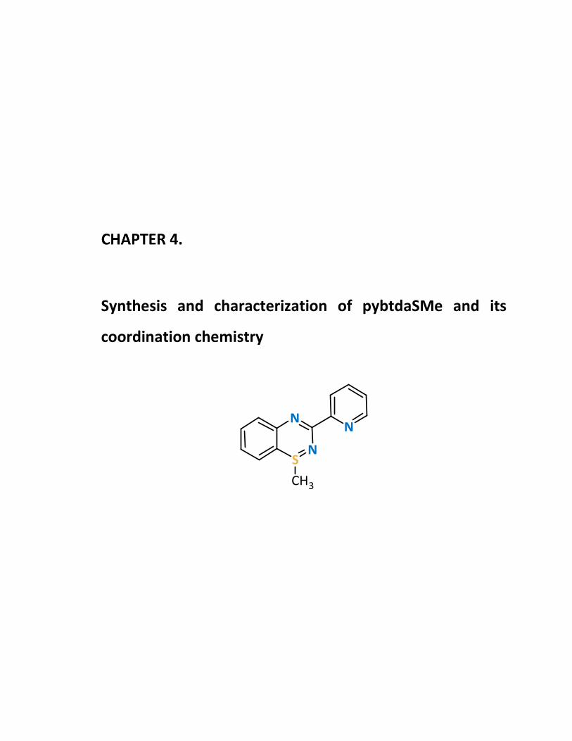
Synthesis and characterization of pybtdaSMe and its
coordination chemistry

Chapter 4: Synthesis and characterization of pybtdaSMe and its coordination chemistry
148
Introduction
The benzothiadiazine ligand pybtdaH has been reported to form a series of
mononuclear complexes to first row transition metal complexes of formula
MClx(pybtdaH)y.1 The SII system undergoes aerial oxidation to the SIV system (Scheme 4.1)
in the presence of a Lewis acidic metal and a base.2 The pybtdaox– anion is formed which
has been isolated in the form of [Cu(pybtdaox)2]22 and Ir(ppy)2(pybtdaox).3 In Chapter 2, S-
oxidation was shown to also proceed in the presence of more Lewis acidic metals
(complexes 2.3a and 2.3b) or in the presence of more basic counterions, such as the hfac−
and OAc− group. The introduction of additional base (Et3N) assisted the S-oxidation and
the deprotonation of the pybtdaH ligand, resulting in higher nuclearity clusters
(complexes 2.11 − 2.14). The S-oxidation of the benzothiadiazine ligands offers a more
versatile, hard, O-donor centre in addition to the favourable, softer, N,Nʹ-chelate,
favouring aggregation and leading to polynuclear complexes such as those described in
Chapters 2 and 3.
In the benzothiadiazine system, alkylation of the S or N is expected to alter the
electronics of the heterocycle with respect to SII pybtdaH ligand and the coordination
behaviour of the S-methylated ligand pybtdaSMe (Scheme 4.1) is expected to differ
significantly from pybtdaH and pybtdaHox. For example pybtdaH can undergo both 1e–
oxidation (loss of H•) and loss of H+ and pybtdaHox can also lose H+. For pybtdaSMe, the S
atom is formally in the S(IV) oxidation state and there is no N-H group, potentially leading
to more controlled reaction chemistry. In addition the methyl group can potentially hinder
Scheme 4.1 Molecular structures of pybtdaH, pybtdaHox and pybtdaSMe.
pybtdaH pybtdaHox pybtdaSMe
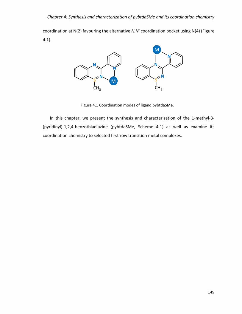
Chapter 4: Synthesis and characterization of pybtdaSMe and its coordination chemistry
149
coordination at N(2) favouring the alternative N,Nʹ coordination pocket using N(4) (Figure
4.1).
In this chapter, we present the synthesis and characterization of the 1-methyl-3-
(pyridinyl)-1,2,4-benzothiadiazine (pybtdaSMe, Scheme 4.1) as well as examine its
coordination chemistry to selected first row transition metal complexes.
Figure 4.1 Coordination modes of ligand pybtdaSMe.
M
M
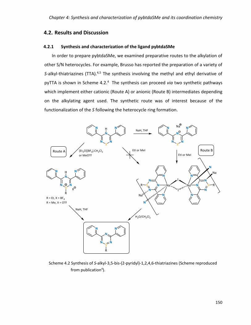
Chapter 4: Synthesis and characterization of pybtdaSMe and its coordination chemistry
150
Results and Discussion
4.2.1 Synthesis and characterization of the ligand pybtdaSMe
In order to prepare pybtdaSMe, we examined preparative routes to the alkylation of
other S/N heterocycles. For example, Brusso has reported the preparation of a variety of
S-alkyl-thiatriazines (TTA).4,5 The synthesis involving the methyl and ethyl derivative of
pyTTA is shown in Scheme 4.2.4 The synthesis can proceed via two synthetic pathways
which implement either cationic (Route A) or anionic (Route B) intermediates depending
on the alkylating agent used. The synthetic route was of interest because of the
functionalization of the S following the heterocycle ring formation.
Scheme 4.2 Synthesis of S-alkyl-3,5-bis-(2-pyridyl)-1,2,4,6-thiatriazines (Scheme reproduced
from publication4).

Chapter 4: Synthesis and characterization of pybtdaSMe and its coordination chemistry
151
A similar synthetic way to Route B was used for the preparation of pybtdaSMe in the
presence of a base and a alkylating agent shown in Scheme 4.3.
The synthesis of the ligand pybtdaH is described in Chapter 2. Deprotonation of
pybtdaH with LiHMDS, under anaerobic, dry conditions followed by treatment with one
equivalent of iodomethane and work-up resulted in pybtdaSMe as a brown oil.
Subsequent recrystallization from CH2Cl2 yielded the product as yellow crystals in 89%
yield. The product showed some decomposition to red pybtdaH (detected by TLC) and
therefore was stored in the freezer. Similar decomposition was reported previously for
the phenyl derivative of the methylated benzothiadiazine.6 The 1H NMR spectrum of
pybtdaSMe exhibits the expected proton environments in the aromatic region and a
Scheme 4.3 Synthetic route to ligand pybtdaSMe.
Figure 4.2 Molecular structure of pybtdaSMe.
N3
N2
N1
C4
S1
C9
C10
C11 C12
C8C7 C6
C3
C5
C2
C1
C13

Chapter 4: Synthesis and characterization of pybtdaSMe and its coordination chemistry
152
singlet in the aliphatic region corresponding to the methyl group. The imine C=N
stretching vibration was observed at 1585 cm-1, as expected.7
The molecular structure was confirmed by single crystal X-ray diffraction studies and
is shown in Figure 4.2. The compound crystallizes in the monoclinic space group P21/c with
one molecule in the asymmetric unit. The C(6)-N(3) distance is 1.312(4) Å, consistent with
predominant double bond character (cf imine C=N at 1.279 Å)8. The bond length between
C(6)-N(2) at 1.372(4) Å is more consistent with a conjugated C-N single bond (cf Ph-NH2 at
1.355 Å)8. The S-N bond length (1.629(3) Å) is comparable with other S-N single bonds
(1.62 – 1.74 Å for isomers of S8-x(NH)x)9 and 1.69 – 1.72 Å for other benzothiadiazine
derivatives)10. The bond lengths assess the best resonance structure as shown in Figure
4.3.
The preference for the crystallographically-determined isomer (orientation of the
pyridyl ring) was confirmed by DFT calculations (B3LYP/6-311G*+) on the two possible
isomers which revealed the experimentally determined structure was only 6 kJ/mol more
stable. Another notable feature in the benzothiadiazine ring is the folding (θ) of the
molecule about the trans-annular vector S(1)···N(3). The values for the majority of
previously reported benzothiadiazines fall in the range 21° < θ < 42° 10 consistent with
formally 8π anti-aromatic structure. For pybtdaSMe, θ = 23.79°. Here alkylation at S breaks
the π-delocalization such that there is conjugation around just the C3N2 part of the ring.
Figure 4.3 Comparison of the heterocycle bond lengths of pybtdaH and pybtdaSMe.
1.286
1.7081.770
1.399
1.400 1.375
1.372
1.6291.771
1.393
1.392 1.312
pybtdaH pybtdaSMe

Chapter 4: Synthesis and characterization of pybtdaSMe and its coordination chemistry
153
The crystal packing of pybtdaSMe is manifested by π-π interactions extending the
network along the bc plane. The centroid···centroid distances are 3.573 Å with the closest
C···C distance at 3.423 Å (cf interlayer separation in graphite at 3.354 Å9) (Figure 4.4).
The ligand creates a two dimensional network through the π-π interactions and
extended hydrogen bonding as shown in Figure 4.5 (left). The network is extended in the
third dimension but only by another 2D plane creating a tube-like framework along b axis
with the S-CH3 in the periphery (Figure 4.5, right).
Figure 4.4 Crystal packing of pybtdaSMe highlighting π-π interactions along bc plane.
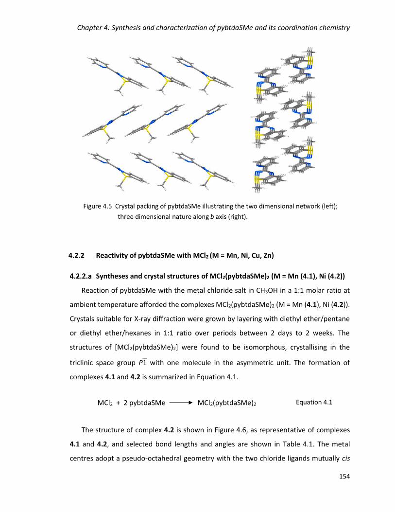
Chapter 4: Synthesis and characterization of pybtdaSMe and its coordination chemistry
154
4.2.2 Reactivity of pybtdaSMe with MCl2 (M = Mn, Ni, Cu, Zn)
Syntheses and crystal structures of MCl2(pybtdaSMe)2 (M = Mn (4.1), Ni (4.2))
Reaction of pybtdaSMe with the metal chloride salt in CH3OH in a 1:1 molar ratio at
ambient temperature afforded the complexes MCl2(pybtdaSMe)2 (M = Mn (4.1), Ni (4.2)).
Crystals suitable for X-ray diffraction were grown by layering with diethyl ether/pentane
or diethyl ether/hexanes in 1:1 ratio over periods between 2 days to 2 weeks. The
structures of [MCl2(pybtdaSMe)2] were found to be isomorphous, crystallising in the
triclinic space group P1 with one molecule in the asymmetric unit. The formation of
complexes 4.1 and 4.2 is summarized in Equation 4.1.
The structure of complex 4.2 is shown in Figure 4.6, as representative of complexes
4.1 and 4.2, and selected bond lengths and angles are shown in Table 4.1. The metal
centres adopt a pseudo-octahedral geometry with the two chloride ligands mutually cis
MCl2 + 2 pybtdaSMe MCl2(pybtdaSMe)2 Equation 4.1
Figure 4.5 Crystal packing of pybtdaSMe illustrating the two dimensional network (left);
three dimensional nature along b axis (right).
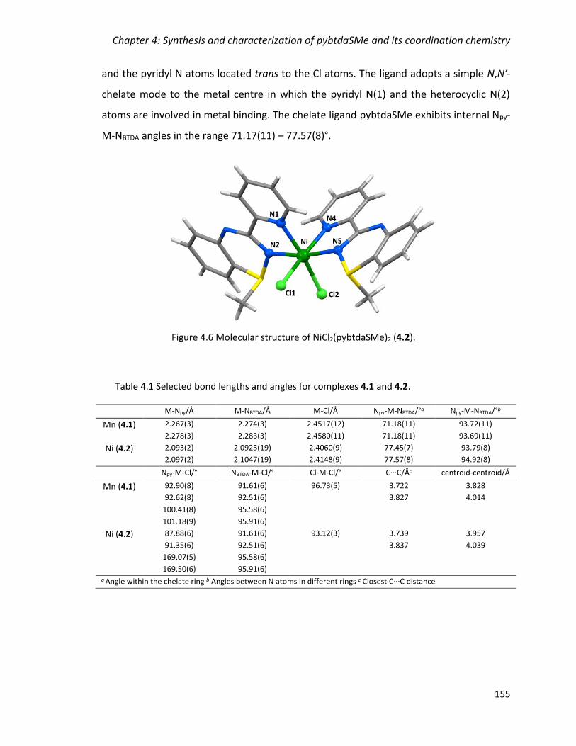
Chapter 4: Synthesis and characterization of pybtdaSMe and its coordination chemistry
155
and the pyridyl N atoms located trans to the Cl atoms. The ligand adopts a simple N,N′-
chelate mode to the metal centre in which the pyridyl N(1) and the heterocyclic N(2)
atoms are involved in metal binding. The chelate ligand pybtdaSMe exhibits internal Npy-
M-NBTDA angles in the range 71.17(11) – 77.57(8)°.
Table 4.1 Selected bond lengths and angles for complexes 4.1 and 4.2.
M-Npy/Å M-NBTDA/Å M-Cl/Å Npy-M-NBTDA/°a Npy-M-NBTDA/°b
Mn (4.1) 2.267(3)
2.278(3)
2.274(3)
2.283(3)
2.4517(12)
2.4580(11)
71.18(11)
71.18(11)
93.72(11)
93.69(11)
Ni (4.2) 2.093(2)
2.097(2)
2.0925(19)
2.1047(19)
2.4060(9)
2.4148(9)
77.45(7)
77.57(8)
93.79(8)
94.92(8)
Npy-M-Cl/° NBTDA-M-Cl/° Cl-M-Cl/° C···C/Åc centroid-centroid/Å
Mn (4.1) 92.90(8)
92.62(8)
100.41(8)
101.18(9)
91.61(6)
92.51(6)
95.58(6)
95.91(6)
96.73(5) 3.722
3.827
3.828
4.014
Ni (4.2) 87.88(6)
91.35(6)
169.07(5)
169.50(6)
91.61(6)
92.51(6)
95.58(6)
95.91(6)
93.12(3) 3.739
3.837
3.957
4.039
a Angle within the chelate ring b Angles between N atoms in different rings c Closest C···C distance
Figure 4.6 Molecular structure of NiCl2(pybtdaSMe)2 (4.2).
N4
N5Ni
Cl1 Cl2
N2
N1
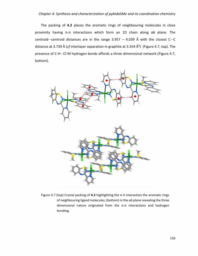
Chapter 4: Synthesis and characterization of pybtdaSMe and its coordination chemistry
156
The packing of 4.2 places the aromatic rings of neighbouring molecules in close
proximity having π-π interactions which form an 1D chain along ab plane. The
centroid···centroid distances are in the range 3.957 – 4.039 Å with the closest C···C
distance at 3.739 Å (cf interlayer separation in graphite at 3.354 Å9) (Figure 4.7, top). The
presence of C-H···Cl-M hydrogen bonds affords a three dimensional network (Figure 4.7,
bottom).
Figure 4.7 (top) Crystal packing of 4.2 highlighting the π-π interaction the aromatic rings
of neighbouring ligand molecules; (bottom) in the ab plane revealing the three
dimensional nature originated from the π-π interactions and hydrogen
bonding.
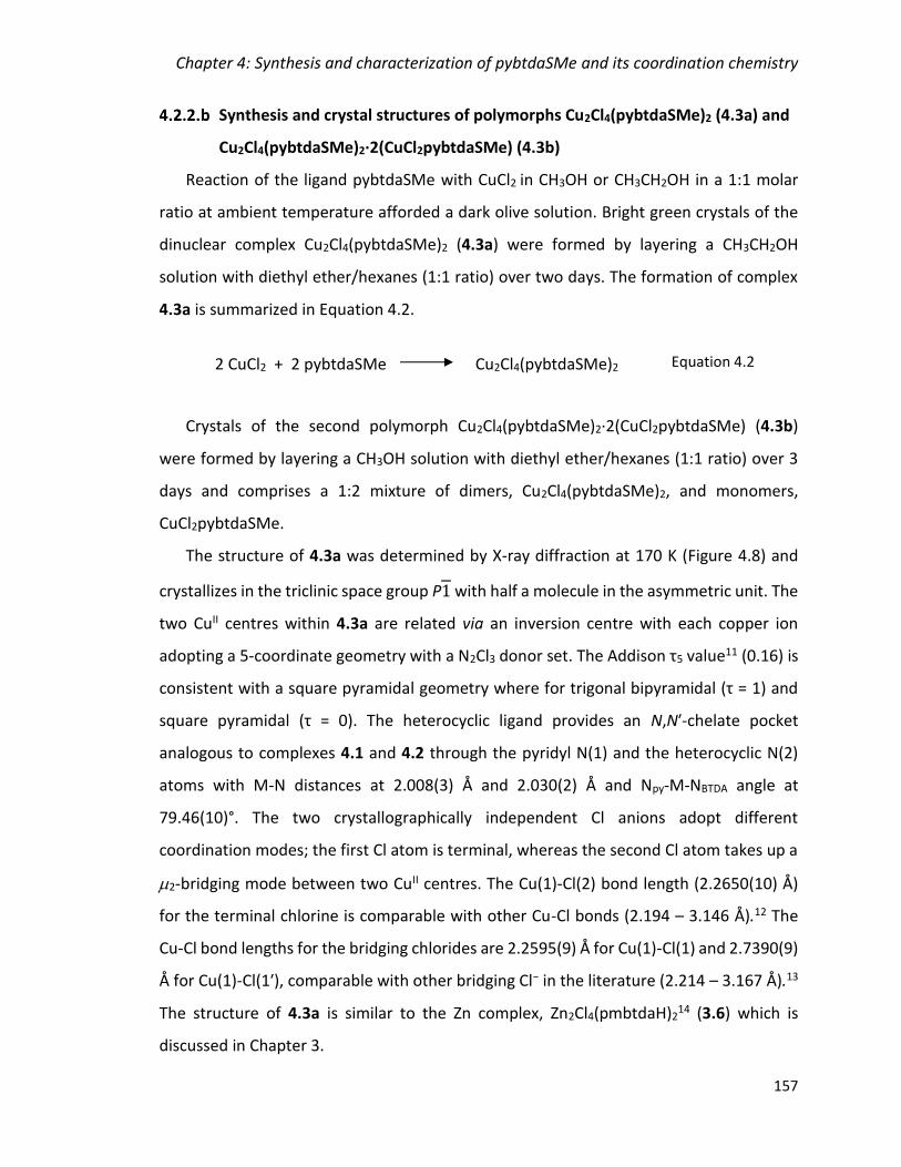
Chapter 4: Synthesis and characterization of pybtdaSMe and its coordination chemistry
157
Synthesis and crystal structures of polymorphs Cu2Cl4(pybtdaSMe)2 (4.3a) and
Cu2Cl4(pybtdaSMe)2·2(CuCl2pybtdaSMe) (4.3b)
Reaction of the ligand pybtdaSMe with CuCl2 in CH3OH or CH3CH2OH in a 1:1 molar
ratio at ambient temperature afforded a dark olive solution. Bright green crystals of the
dinuclear complex Cu2Cl4(pybtdaSMe)2 (4.3a) were formed by layering a CH3CH2OH
solution with diethyl ether/hexanes (1:1 ratio) over two days. The formation of complex
4.3a is summarized in Equation 4.2.
Crystals of the second polymorph Cu2Cl4(pybtdaSMe)2·2(CuCl2pybtdaSMe) (4.3b)
were formed by layering a CH3OH solution with diethyl ether/hexanes (1:1 ratio) over 3
days and comprises a 1:2 mixture of dimers, Cu2Cl4(pybtdaSMe)2, and monomers,
CuCl2pybtdaSMe.
The structure of 4.3a was determined by X-ray diffraction at 170 K (Figure 4.8) and
crystallizes in the triclinic space group P1 with half a molecule in the asymmetric unit. The
two CuII centres within 4.3a are related via an inversion centre with each copper ion
adopting a 5-coordinate geometry with a N2Cl3 donor set. The Addison τ5 value11 (0.16) is
consistent with a square pyramidal geometry where for trigonal bipyramidal (τ = 1) and
square pyramidal (τ = 0). The heterocyclic ligand provides an N,Nʹ-chelate pocket
analogous to complexes 4.1 and 4.2 through the pyridyl N(1) and the heterocyclic N(2)
atoms with M-N distances at 2.008(3) Å and 2.030(2) Å and Npy-M-NBTDA angle at
79.46(10)°. The two crystallographically independent Cl anions adopt different
coordination modes; the first Cl atom is terminal, whereas the second Cl atom takes up a
2-bridging mode between two CuII centres. The Cu(1)-Cl(2) bond length (2.2650(10) Å)
for the terminal chlorine is comparable with other Cu-Cl bonds (2.194 – 3.146 Å).12 The
Cu-Cl bond lengths for the bridging chlorides are 2.2595(9) Å for Cu(1)-Cl(1) and 2.7390(9)
Å for Cu(1)-Cl(1ʹ), comparable with other bridging Cl− in the literature (2.214 – 3.167 Å).13
The structure of 4.3a is similar to the Zn complex, Zn2Cl4(pmbtdaH)214 (3.6) which is
discussed in Chapter 3.
2 CuCl2 + 2 pybtdaSMe Cu2Cl4(pybtdaSMe)2 Equation 4.2
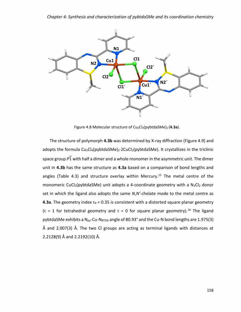
Chapter 4: Synthesis and characterization of pybtdaSMe and its coordination chemistry
158
The structure of polymorph 4.3b was determined by X-ray diffraction (Figure 4.9) and
adopts the formula Cu2Cl4(pybtdaSMe)2·2CuCl2(pybtdaSMe). It crystallizes in the triclinic
space group P1 with half a dimer and a whole monomer in the asymmetric unit. The dimer
unit in 4.3b has the same structure as 4.3a based on a comparison of bond lengths and
angles (Table 4.3) and structure overlay within Mercury.15 The metal centre of the
monomeric CuCl2(pybtdaSMe) unit adopts a 4-coordinate geometry with a N2Cl2 donor
set in which the ligand also adopts the same N,N′-chelate mode to the metal centre as
4.3a. The geometry index τ4 = 0.35 is consistent with a distorted square planar geometry
(τ = 1 for tetrahedral geometry and τ = 0 for square planar geometry).16 The ligand
pybtdaSMe exhibits a Npy-Cu-NBTDA angle of 80.93° and the Cu-N bond lengths are 1.975(3)
Å and 2.007(3) Å. The two Cl groups are acting as terminal ligands with distances at
2.2128(9) Å and 2.2192(10) Å.
Figure 4.8 Molecular structure of Cu2Cl4(pybtdaSMe)2 (4.3a).
Cu1 Cl1
Cl2
N2
N1
Cu1´Cl1´
Cl2´
N1´
N2´

Chapter 4: Synthesis and characterization of pybtdaSMe and its coordination chemistry
159
An additional crystallographic study was undertaken on 4.3b at 280 K. The unit cell
parameters were very close to those measured at 170 K (Table 4.2). An initial examination
of the high temperature structure revealed breakdown of the dimer unit. Careful
examination of the structure at 280 K showed Cu···Cl interactions at 2.961(2) Å, slightly
larger than the sum of the van der Waals radii whereas the 170 K structure revealed a
shorter Cu-Cl distance at 2.897(1) Å (Table 4.3) formally sufficiently close to be considered
as a long bond corresponding to the molecular Jahn-Teller axis. An overlay of the
structures collected at 170 and 280 K using Mercury15 confirmed the similarity of these
two structures (Figure 4.10).
Cu1
Cl1
Cl2
N2
N1
Cu1´Cl1´
Cl2´
N1´
N2´
Cl3
Cl4N4
N5
Cu2
Figure 4.9 Molecular structure of Cu2Cl4(pybtdaSMe)2·2CuCl2(pybtdaSMe) (4.3b).
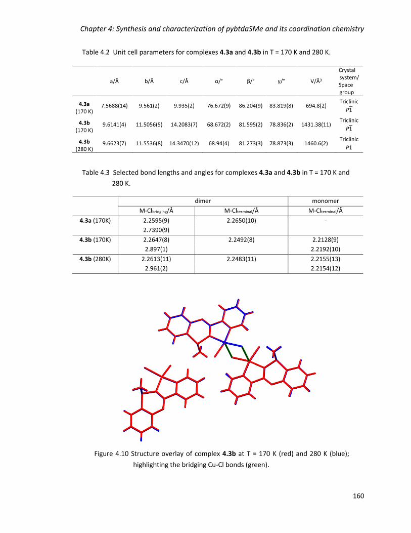
Chapter 4: Synthesis and characterization of pybtdaSMe and its coordination chemistry
160
Table 4.2 Unit cell parameters for complexes 4.3a and 4.3b in T = 170 K and 280 K.
Table 4.3 Selected bond lengths and angles for complexes 4.3a and 4.3b in T = 170 K and
280 K.
dimer monomer
M-Clbridging/Å M-Clterminal/Å M-Clterminal/Å
4.3a (170K) 2.2595(9) 2.2650(10) -
2.7390(9)
4.3b (170K) 2.2647(8) 2.2492(8) 2.2128(9)
2.897(1) 2.2192(10)
4.3b (280K) 2.2613(11) 2.2483(11) 2.2155(13)
2.961(2) 2.2154(12)
a/Å b/Å c/Å α/° β/° γ/° V/Å3
Crystal system/ Space group
4.3a (170 K)
7.5688(14) 9.561(2) 9.935(2) 76.672(9) 86.204(9) 83.819(8) 694.8(2) Triclinic
P1
4.3b (170 K)
9.6141(4) 11.5056(5) 14.2083(7) 68.672(2) 81.595(2) 78.836(2) 1431.38(11) Triclinic
P1
4.3b (280 K)
9.6623(7) 11.5536(8) 14.3470(12) 68.94(4) 81.273(3) 78.873(3) 1460.6(2) Triclinic
P1
Figure 4.10 Structure overlay of complex 4.3b at T = 170 K (red) and 280 K (blue);
highlighting the bridging Cu-Cl bonds (green).
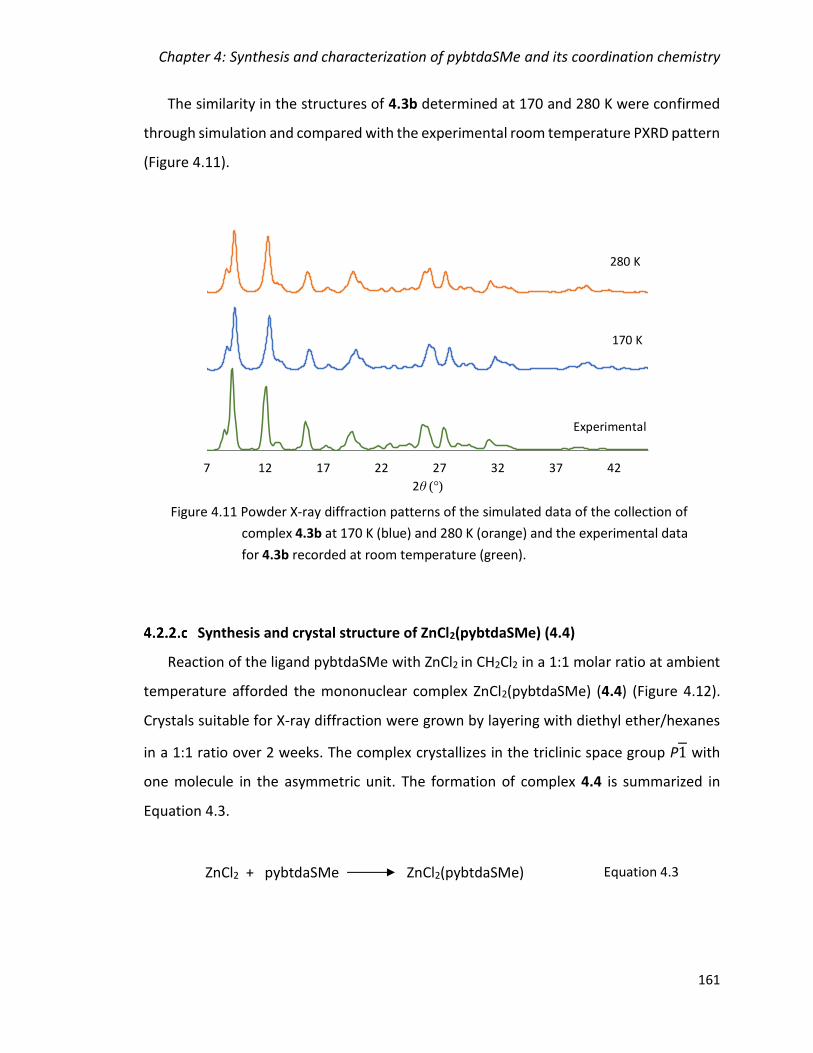
Chapter 4: Synthesis and characterization of pybtdaSMe and its coordination chemistry
161
The similarity in the structures of 4.3b determined at 170 and 280 K were confirmed
through simulation and compared with the experimental room temperature PXRD pattern
(Figure 4.11).
Synthesis and crystal structure of ZnCl2(pybtdaSMe) (4.4)
Reaction of the ligand pybtdaSMe with ZnCl2 in CH2Cl2 in a 1:1 molar ratio at ambient
temperature afforded the mononuclear complex ZnCl2(pybtdaSMe) (4.4) (Figure 4.12).
Crystals suitable for X-ray diffraction were grown by layering with diethyl ether/hexanes
in a 1:1 ratio over 2 weeks. The complex crystallizes in the triclinic space group P1 with
one molecule in the asymmetric unit. The formation of complex 4.4 is summarized in
Equation 4.3.
ZnCl2 + pybtdaSMe ZnCl2(pybtdaSMe) Equation 4.3
7 12 17 22 27 32 37 42
2θ ( )
280 K
170 K
Exp
Experimental
170 K
280 K
Figure 4.11 Powder X-ray diffraction patterns of the simulated data of the collection of
complex 4.3b at 170 K (blue) and 280 K (orange) and the experimental data
for 4.3b recorded at room temperature (green).
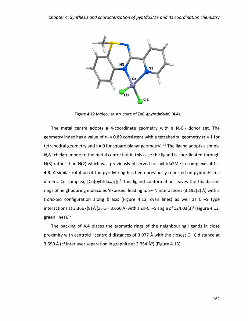
Chapter 4: Synthesis and characterization of pybtdaSMe and its coordination chemistry
162
The metal centre adopts a 4-coordinate geometry with a N2Cl2 donor set. The
geometry index has a value of τ4 = 0.89 consistent with a tetrahedral geometry (τ = 1 for
tetrahedral geometry and τ = 0 for square planar geometry).16 The ligand adopts a simple
N,N′-chelate mode to the metal centre but in this case the ligand is coordinated through
N(3) rather than N(2) which was previously observed for pybtdaSMe in complexes 4.1 −
4.3. A similar rotation of the pyridyl ring has been previously reported on pybtdaH in a
dimeric Cu complex, [Cu(pybtdaox)2]2.2 This ligand conformation leaves the thiadiazine
rings of neighbouring molecules ‘exposed’ leading to S···N interactions (3.192(2) Å) with a
trans-oid configuration along b axis (Figure 4.13, cyan lines) as well as Cl···S type
interactions at 3.3667(8) Å (ΣvdW = 3.650 Å) with a Zn-Cl···S angle of 124.03(3)° (Figure 4.13,
green lines).17
The packing of 4.4 places the aromatic rings of the neighbouring ligands in close
proximity with centroid···centroid distances of 3.977 Å with the closest C···C distance at
3.690 Å (cf interlayer separation in graphite at 3.354 Å9) (Figure 4.13).
Figure 4.12 Molecular structure of ZnCl2(pybtdaSMe) (4.4).
N3
Zn
Cl1Cl2
N1
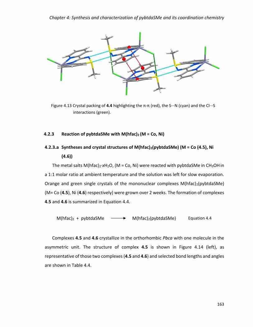
Chapter 4: Synthesis and characterization of pybtdaSMe and its coordination chemistry
163
4.2.3 Reaction of pybtdaSMe with M(hfac)2 (M = Co, Ni)
Syntheses and crystal structures of M(hfac)2(pybtdaSMe) (M = Co (4.5), Ni
(4.6))
The metal salts M(hfac)2·xH2O, (M = Co, Ni) were reacted with pybtdaSMe in CH3OH in
a 1:1 molar ratio at ambient temperature and the solution was left for slow evaporation.
Orange and green single crystals of the mononuclear complexes M(hfac)2(pybtdaSMe)
(M= Co (4.5), Ni (4.6) respectively) were grown over 2 weeks. The formation of complexes
4.5 and 4.6 is summarized in Equation 4.4.
Complexes 4.5 and 4.6 crystallize in the orthorhombic Pbca with one molecule in the
asymmetric unit. The structure of complex 4.5 is shown in Figure 4.14 (left), as
representative of those two complexes (4.5 and 4.6) and selected bond lengths and angles
are shown in Table 4.4.
M(hfac)2 + pybtdaSMe M(hfac)2(pybtdaSMe) Equation 4.4
Figure 4.13 Crystal packing of 4.4 highlighting the π-π (red), the S∙∙∙N (cyan) and the Cl∙∙∙S
interactions (green).

Chapter 4: Synthesis and characterization of pybtdaSMe and its coordination chemistry
164
Table 4.4 Selected bond lengths and angles for complexes 4.5 and 4.6.
M-Npy/Å M-NBTDA/Å M-O/Å NpyMNBTDA/°
Co (4.5) 2.094(3) 2.101(3) 2.078(3) 2.074(3) 2.076(3) 2.093(3)
78.30(13)
Ni (4.6) 2.043(4) 2.071(5) 2.034(4) 2.053(4) 2.055(4) 2.069(4)
79.96(17)
C-H···centroid/Å C-H···centroid/°
centroid-centroid/Å
Co (4.5) 3.687 118.36 4.038
Ni (4.6) 3.689 117.66 4.048
The metal centres have a distorted octahedral geometry with a N2O4 donor set. The
heterocyclic ligand provides an N,Nʹ-chelate pocket analogous to complex 4.4 through
N(1) and N(3) atoms with M-N distances in the range 2.043(4) – 2.101(3) Å and
Npy-M-NBTDA angles at 78.30(17) – 79.69(17)°. The two hfac groups also chelate the metal
Figure 4.14 (left) Molecular structure of Co(hfac)2(pybtdaSMe) (4.5); (right) Crystal packing of
4.5 highlighting the π-π and the C-H∙∙∙centroid interactions highlighted in red.
O1
O2
Co1
O4
O3
N3
N1
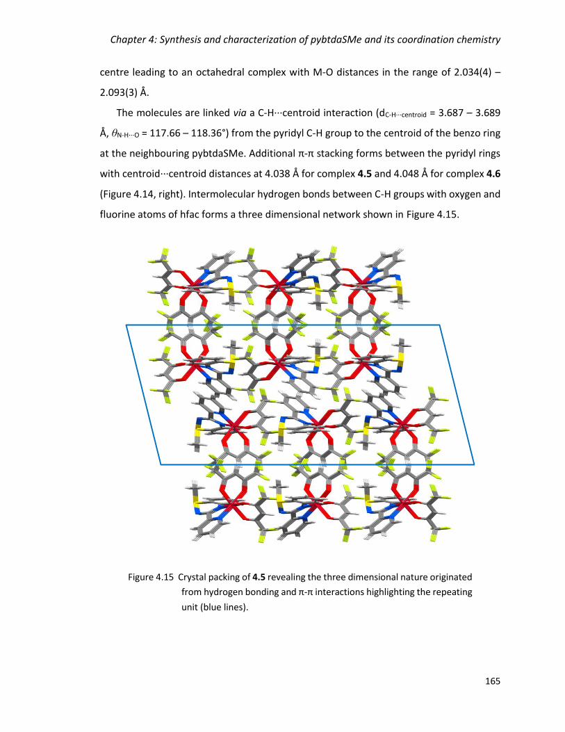
Chapter 4: Synthesis and characterization of pybtdaSMe and its coordination chemistry
165
centre leading to an octahedral complex with M-O distances in the range of 2.034(4) –
2.093(3) Å.
The molecules are linked via a C-H···centroid interaction (dC-H···centroid = 3.687 – 3.689
Å, θN-H···O = 117.66 – 118.36°) from the pyridyl C-H group to the centroid of the benzo ring
at the neighbouring pybtdaSMe. Additional π-π stacking forms between the pyridyl rings
with centroid···centroid distances at 4.038 Å for complex 4.5 and 4.048 Å for complex 4.6
(Figure 4.14, right). Intermolecular hydrogen bonds between C-H groups with oxygen and
fluorine atoms of hfac forms a three dimensional network shown in Figure 4.15.
Figure 4.15 Crystal packing of 4.5 revealing the three dimensional nature originated
from hydrogen bonding and π-π interactions highlighting the repeating
unit (blue lines).

Chapter 4: Synthesis and characterization of pybtdaSMe and its coordination chemistry
166
4.2.4 Spectroscopic studies of metal complexes 4.3a, 4.3b, 4.4 and 4.5
The solution UV-Vis spectra of the polymorphic complexes 4.3a and 4.3b are
dominated by a series of intense π-π* ligand-centered absorption bands between 230 and
400 nm (Figure 4.16). The spectra of the two complexes are remarkably similar suggesting
that the two polymorphs exhibit the same solution properties. Notably the visible region
exhibits low intensity absorption bands around 420 nm which is absent to the free ligand,
pybtdaSMe and which give rise to the diagnostic green colour of the Cu complexes and is
likely attributable to a spin-forbidden ‘d-d’ transition ( < 103 M-1 cm-3).
The UV-Vis spectra of complexes 4.5 and 4.6 are dominated by a series of π-π* ligand-
centered absorption bands between 310 nm (Figure 4.17). In particular, near the visible
region exhibits lower intensity absorption bands are observed around 370 nm. S-
methylation perturbs the electronics of the btda ligand and appear almost colourless in a
similar manner the S-oxidation does and the SIV derivatives of the btda family appear
colourless.2,14
Figure 4.16 Solution UV-Vis spectra of complexes 4.3a and 4.3b, and the ligand pybtdaSMe
(CH2Cl2, rt, C ≈ 10-5).

Chapter 4: Synthesis and characterization of pybtdaSMe and its coordination chemistry
167
The IR spectrum of the ligand pybtdaSMe exhibits a stretch at 1585 cm-1 characteristic
of the C=N imine bond.7 In the complexes, the stretch appears in the region of 1598 –
1642 cm-1 with an increase of 10 – 50 cm-1 upon complexation. The spectra of the ligand
and the complexes exhibit stretches at ca. 2980, 2890 and 642 all related to the S-CH3
group. The N-H stretch is absent for this ligand and in all complexes, as opposed to the SII
ligands, pybtdaH and pmbtdaH (Chapter 2 and 3).
4.2.5 Reactivity trends in the coordination chemistry of pybtdaSMe
The ligand 3-(2ʹ-pyridyl)benzothiadiazine (pybtdaH) can form a range of N,Nʹ-chelate
complexes with first row transition metals with general formula MCl2(pybtdaH)2, including
the 1:1 adduct CuCl2(pybtdaH).1 In a similar fashion, the ligand pmbtdaH which could
offer two binding pockets proved to also form a range of N,Nʹ-chelate complexes with first
row transition metals with general formula MCl2(pmbtdaH)2 as discussed in Chapter 3.
Initial studies showed that S-oxidation of pybtdaH or related benzothiadiazines only
Figure 4.17 Solution UV-Vis spectra of complexes 4.5 and 4.6, and the ligand
pybtdaSMe (CH2Cl2 or CH3CN, rt, C ≈ 10-5).

Chapter 4: Synthesis and characterization of pybtdaSMe and its coordination chemistry
168
occurred in the presence of a metal and Et3N.2 In Chapters 2 and 3, we presented the
effect of the counter ions (hfac– and OAc–) in the oxidation of the ligands pybtdaH and
pmbtdaH even without the introduction of base in the reaction scheme where a series of
polynuclear complexes have been isolated with pybtdaox– and pmbtdaox
–.14
Reaction of pybtdaSMe with MCl2 (M = Mn, Ni) afforded the mononuclear octahedral
complexes MCl2(pybtdaSMe)2 (M = Mn (4.1), Ni (4.2)) while reaction of pybtdaSMe with
ZnCl2·2H2O afforded the mononuclear tetrahedral complex ZnCl2(pybtdaSMe) (4.4). In the
case of the reaction of pybtdaSMe with CuCl2 two polymorphs are found with 4.3b
containing both mononuclear CuCl2(pybtdaSMe) and dinuclear Cu2Cl4(pybtdaSMe)2 units.
While pybtdaH exhibits a tendency to oxidize in the presence of M(hfac)2 and M(OAc)2
salts, alkylation suppresses ligand oxidation affording the anticipated mononuclear
complexes of formula M(hfac)2(pybtdaSMe) (M = Co (4.5), Ni (4.6)). The pybtdaSMe ligand
adopts two coordination modes; through the heterocyclic N(3) and the pyridyl N(1) atom
or through the heterocyclic N(2) and the pyridyl N(1) atom. As discussed in section 4.2.1
both conformations are very close energetically so the outcome depends mostly on
intermolecular interactions and steric effects.
While the ligand pybtdaH is prone to oxidation resulting to the S-oxide of the ligand
under basic conditions by introducing either Et3N or more basic anions, such as hfac–, the
S-methylation is prohibiting such behaviour even in the presence of hfac. Although the
possibility of further oxidation of sulphur cannot be excluded,18,19 the detection would be
more difficult given the loss of the red intense red colour of pybtdaH and pmbtdaH.
Nevertheless, there was no indication of S-oxidation of pybtdaSMe in the experiments
within the timeframe of this project.
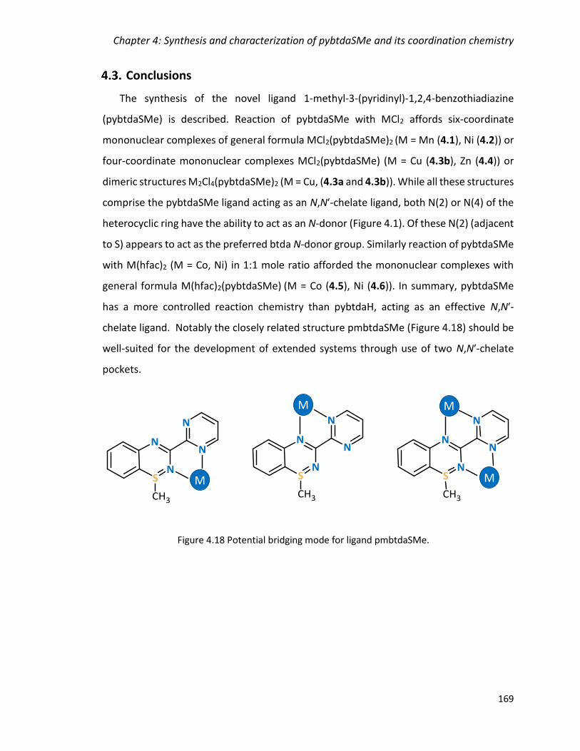
Chapter 4: Synthesis and characterization of pybtdaSMe and its coordination chemistry
169
Conclusions
The synthesis of the novel ligand 1-methyl-3-(pyridinyl)-1,2,4-benzothiadiazine
(pybtdaSMe) is described. Reaction of pybtdaSMe with MCl2 affords six-coordinate
mononuclear complexes of general formula MCl2(pybtdaSMe)2 (M = Mn (4.1), Ni (4.2)) or
four-coordinate mononuclear complexes MCl2(pybtdaSMe) (M = Cu (4.3b), Zn (4.4)) or
dimeric structures M2Cl4(pybtdaSMe)2 (M = Cu, (4.3a and 4.3b)). While all these structures
comprise the pybtdaSMe ligand acting as an N,Nʹ-chelate ligand, both N(2) or N(4) of the
heterocyclic ring have the ability to act as an N-donor (Figure 4.1). Of these N(2) (adjacent
to S) appears to act as the preferred btda N-donor group. Similarly reaction of pybtdaSMe
with M(hfac)2 (M = Co, Ni) in 1:1 mole ratio afforded the mononuclear complexes with
general formula M(hfac)2(pybtdaSMe) (M = Co (4.5), Ni (4.6)). In summary, pybtdaSMe
has a more controlled reaction chemistry than pybtdaH, acting as an effective N,Nʹ-
chelate ligand. Notably the closely related structure pmbtdaSMe (Figure 4.18) should be
well-suited for the development of extended systems through use of two N,Nʹ-chelate
pockets.
Figure 4.18 Potential bridging mode for ligand pmbtdaSMe.
M
MM
M
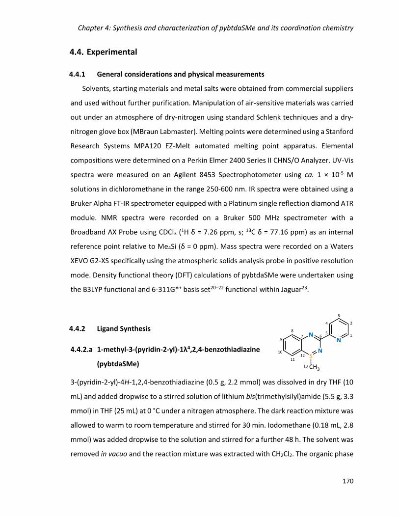
Chapter 4: Synthesis and characterization of pybtdaSMe and its coordination chemistry
170
Experimental
4.4.1 General considerations and physical measurements
Solvents, starting materials and metal salts were obtained from commercial suppliers
and used without further purification. Manipulation of air-sensitive materials was carried
out under an atmosphere of dry-nitrogen using standard Schlenk techniques and a dry-
nitrogen glove box (MBraun Labmaster). Melting points were determined using a Stanford
Research Systems MPA120 EZ-Melt automated melting point apparatus. Elemental
compositions were determined on a Perkin Elmer 2400 Series II CHNS/O Analyzer. UV-Vis
spectra were measured on an Agilent 8453 Spectrophotometer using ca. 1 × 10⁻⁵ M
solutions in dichloromethane in the range 250-600 nm. IR spectra were obtained using a
Bruker Alpha FT-IR spectrometer equipped with a Platinum single reflection diamond ATR
module. NMR spectra were recorded on a Bruker 500 MHz spectrometer with a
Broadband AX Probe using CDCl3 (1H δ = 7.26 ppm, s; 13C δ = 77.16 ppm) as an internal
reference point relative to Me4Si (δ = 0 ppm). Mass spectra were recorded on a Waters
XEVO G2-XS specifically using the atmospheric solids analysis probe in positive resolution
mode. Density functional theory (DFT) calculations of pybtdaSMe were undertaken using
the B3LYP functional and 6-311G*+ basis set20–22 functional within Jaguar23.
4.4.2 Ligand Synthesis
1-methyl-3-(pyridin-2-yl)-1λ4,2,4-benzothiadiazine
(pybtdaSMe)
3-(pyridin-2-yl)-4H-1,2,4-benzothiadiazine (0.5 g, 2.2 mmol) was dissolved in dry THF (10
mL) and added dropwise to a stirred solution of lithium bis(trimethylsilyl)amide (5.5 g, 3.3
mmol) in THF (25 mL) at 0 °C under a nitrogen atmosphere. The dark reaction mixture was
allowed to warm to room temperature and stirred for 30 min. Iodomethane (0.18 mL, 2.8
mmol) was added dropwise to the solution and stirred for a further 48 h. The solvent was
removed in vacuo and the reaction mixture was extracted with CH2Cl2. The organic phase

Chapter 4: Synthesis and characterization of pybtdaSMe and its coordination chemistry
171
was washed with H2O (2 × 20 mL) and brine (1 × 20 mL), dried over Na2SO4 and the solvent
was removed in vacuo to yield a dark brown viscous oil. The product was isolated as yellow
crystals by slow evaporation of CH2Cl2. Yield 0.48 g, 89.6%.
Melting point 63 °C.
Elemental Analysis calc. for [C13H11N3S]·3H2O: C, 52.87; H, 5.80; N, 14.23. Found: C, 52.94
H, 5.45; N, 14.19%.
UV-Vis (CH2Cl2; λmax, nm; ε, M-1·cm-1): 274 (21560), 359 (4797).
IR (solid, cm-1) �̃�max = 3354 (br), 2970 (w), 2903 (w), 2872 (w), 1585 (w), 1560 (w), 1484
(m), 1448 (s), 1422 (m), 1330 (m), 1271 (m), 1247 (m), 1130 (m), 1112 (w), 1085 (w), 1045
(m), 993 (w), 964 (w), 917 (w), 851 (w), 819 (w), 762 (m), 686 (m), 662 (w), 621 (m), 576
(w), 547 (w), 473 (w), 452 (w).
1H NMR (500 MHz, ppm, CDCl3) δH = 8.61 (1H, dq, J = 4.75, 0.83 Hz, C1H), 8.25 (1H, dt, J =
7.95, 0.93 Hz, C4H), 7.63 (1H, td, J = 11.5, 1.75 Hz, C3H), 7.37-7.43 (2H, m, C9H, C10H), 7.19
(1H, ddd, J = 7.45, 4.75, 1.15 Hz, C2H), 7.12-7.14 (2H, m, C8H, C11H), 2.37 (3H, s, C13H3).
13C NMR (125 MHz, ppm, CDCl3) δC = 163.41 (C6), 156.18 (C5), 149.52 (C1), 144.66 (C7),
136.53 (C3), 133.30 (C9 or C10), 127.22 (C9 or C10), 126.15 (C8 or C11), 124.76 (C2), 124.63
(C8 or C11), 123.37 (C4), 107.09 (C12), 35.05 (C13).
HRMS (ASAP) m/z [M+H]+ calc. for C13H11N3S: 242.0752, found 242.0750.

Chapter 4: Synthesis and characterization of pybtdaSMe and its coordination chemistry
172
4.4.3 Complex Syntheses with MCl2 (M = Mn, Ni, Cu, Zn)
MnCl2(pybtdaSMe)2 (4.1)
MnCl2 (0.006 g, 0.050 mmol) was added to a solution of pybtdaSMe (0.012 g, 0.050 mmol)
in CH3OH (5 mL). The mixture was stirred for 1 h and a yellow solution was obtained. The
solution was filtered and yellow crystals were grown by layering with a mixture of diethyl
ether/hexanes (1:1) in two days. Yield 0.007 g, 23%.
Elemental Analysis calc. for [C26H22N6MnS2Cl2]∙0.1C5H12: C, 51.70; H, 3.80; N, 13.65.
Found: C, 51.60; H, 3.77; N, 12.87%.
IR (solid, cm-1) �̃�max = 2967 (w), 1598 (w), 1562 (w), 1509 (s), 1456 (m), 1439 (m), 1414 (m),
1320 (m), 1298 (m), 1268 (m), 1252 (w), 1151 (m), 1130 (m), 1112 (w), 1067 (m), 1044
(m), 1015 (m), 989 (m), 921 (m), 827 (m), 796 (m), 773 (s), 745 (m), 680 (m), 637 (m), 548
(w), 498 (m), 474 (m), 464 (m), 452 (m), 412 (m).
NiCl2(pybtdaSMe)2 (4.2)
NiCl2·6H2O (0.027 g, 0.050 mmol) was added to a solution of pybtdaSMe (0.012 g, 0.050
mmol) in CH3OH (5 mL). The mixture was stirred for 30 min and a golden solution
obtained. The solution was filtered and green crystals were grown by layering with a
mixture of diethyl ether/hexanes (1:1) over two weeks. Yield 0.006 g, 19%.
Elemental Analysis calc. for [C26H22N6NiS2Cl2]∙CH3OH: C, 50.34; H, 4.07; N, 13.04. Found:
C, 50.20; H, 4.14; N, 12.66%.
IR (solid, cm-1) �̃�max = 3043 (w), 2981 (w), 2885 (w) 1600 (w), 1587 (w), 1561 (w), 1511 (m),
1456 (m), 1412 (m), 1327 (m), 1295 (m), 1269 (m), 1255 (m), 1150 (w), 1132 (m), 1117
(m), 1067 (w), 1045 (m), 1020 (m), 988 (m), 954 (m), 924 (m), 860 (w), 834 (m), 797 (w),
773 (s), 747 (m), 676 (m), 642 (m), 550 (w), 510 (m), 452 (m), 417 (w).
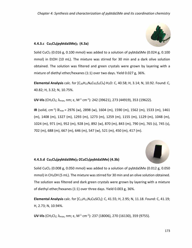
Chapter 4: Synthesis and characterization of pybtdaSMe and its coordination chemistry
173
Cu2Cl4(pybtdaSMe)2 (4.3a)
Solid CuCl2 (0.016 g, 0.100 mmol) was added to a solution of pybtdaSMe (0.024 g, 0.100
mmol) in EtOH (10 mL). The mixture was stirred for 30 min and a dark olive solution
obtained. The solution was filtered and green crystals were grown by layering with a
mixture of diethyl ether/hexanes (1:1) over two days. Yield 0.027 g, 36%.
Elemental Analysis calc. for [C26H22N6Cu2S2Cl4]∙H2O: C, 40.58; H, 3.14; N, 10.92. Found: C,
40.82; H, 3.32; N, 10.75%.
UV-Vis (CH2Cl2; λmax, nm; ε, M-1 cm-1): 242 (39621), 273 (44919), 353 (19622).
IR (solid, cm-1) �̃�max = 2976 (w), 2898 (w), 1604 (m), 1590 (m), 1562 (m), 1533 (m), 1461
(m), 1408 (m), 1327 (m), 1293 (m), 1273 (m), 1259 (m), 1155 (m), 1129 (m), 1048 (m),
1024 (m), 971 (m), 952 (m), 928 (m), 892 (w), 870 (m), 843 (m), 790 (m), 765 (s), 745 (s),
702 (m), 688 (m), 667 (m), 646 (m), 547 (w), 521 (m), 450 (m), 417 (m).
Cu2Cl4(pybtdaSMe)2·2CuCl2(pybtdaSMe) (4.3b)
Solid CuCl2 (0.008 g, 0.050 mmol) was added to a solution of pybtdaSMe (0.012 g, 0.050
mmol) in CH3OH (5 mL). The mixture was stirred for 30 min and an olive solution obtained.
The solution was filtered and dark green crystals were grown by layering with a mixture
of diethyl ether/hexanes (1:1) over three days. Yield 0.003 g, 36%.
Elemental Analysis calc. for [C13H11N3CuSCl2]: C, 41.55; H, 2.95; N, 11.18. Found: C, 41.19;
H, 2.73; N, 10.94%.
UV-Vis (CH2Cl2; λmax, nm; ε, M-1 cm-1): 237 (18006), 270 (16130), 359 (9755).
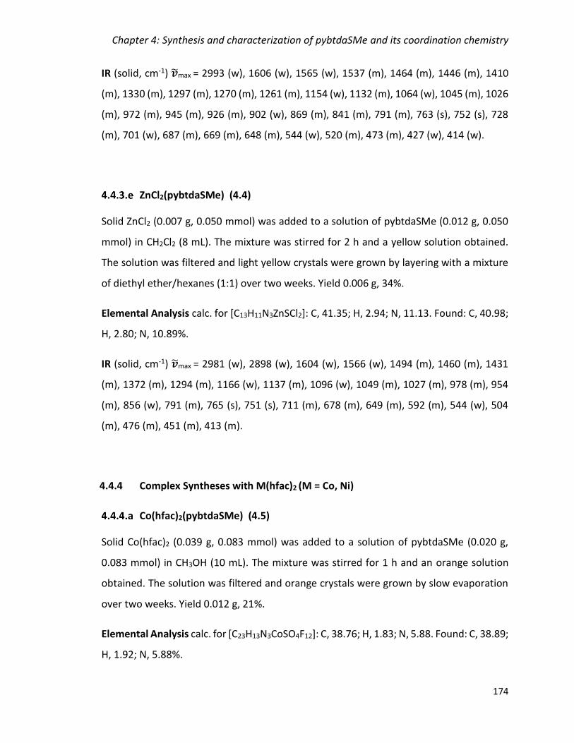
Chapter 4: Synthesis and characterization of pybtdaSMe and its coordination chemistry
174
IR (solid, cm-1) �̃�max = 2993 (w), 1606 (w), 1565 (w), 1537 (m), 1464 (m), 1446 (m), 1410
(m), 1330 (m), 1297 (m), 1270 (m), 1261 (m), 1154 (w), 1132 (m), 1064 (w), 1045 (m), 1026
(m), 972 (m), 945 (m), 926 (m), 902 (w), 869 (m), 841 (m), 791 (m), 763 (s), 752 (s), 728
(m), 701 (w), 687 (m), 669 (m), 648 (m), 544 (w), 520 (m), 473 (m), 427 (w), 414 (w).
ZnCl2(pybtdaSMe) (4.4)
Solid ZnCl2 (0.007 g, 0.050 mmol) was added to a solution of pybtdaSMe (0.012 g, 0.050
mmol) in CH2Cl2 (8 mL). The mixture was stirred for 2 h and a yellow solution obtained.
The solution was filtered and light yellow crystals were grown by layering with a mixture
of diethyl ether/hexanes (1:1) over two weeks. Yield 0.006 g, 34%.
Elemental Analysis calc. for [C13H11N3ZnSCl2]: C, 41.35; H, 2.94; N, 11.13. Found: C, 40.98;
H, 2.80; N, 10.89%.
IR (solid, cm-1) �̃�max = 2981 (w), 2898 (w), 1604 (w), 1566 (w), 1494 (m), 1460 (m), 1431
(m), 1372 (m), 1294 (m), 1166 (w), 1137 (m), 1096 (w), 1049 (m), 1027 (m), 978 (m), 954
(m), 856 (w), 791 (m), 765 (s), 751 (s), 711 (m), 678 (m), 649 (m), 592 (m), 544 (w), 504
(m), 476 (m), 451 (m), 413 (m).
4.4.4 Complex Syntheses with M(hfac)2 (M = Co, Ni)
Co(hfac)2(pybtdaSMe) (4.5)
Solid Co(hfac)2 (0.039 g, 0.083 mmol) was added to a solution of pybtdaSMe (0.020 g,
0.083 mmol) in CH3OH (10 mL). The mixture was stirred for 1 h and an orange solution
obtained. The solution was filtered and orange crystals were grown by slow evaporation
over two weeks. Yield 0.012 g, 21%.
Elemental Analysis calc. for [C23H13N3CoSO4F12]: C, 38.76; H, 1.83; N, 5.88. Found: C, 38.89;
H, 1.92; N, 5.88%.

Chapter 4: Synthesis and characterization of pybtdaSMe and its coordination chemistry
175
UV-Vis (CH3CN; λmax, nm; ε, M-1 cm-1): 240 (12102), 304 (16304), 366 (4128).
IR (solid, cm-1) �̃�max = 1639 (m), 1553 (w), 1524 (w), 1489 (m), 1456 (m), 1426 (w), 1375
(w), 1252 (m), 1205 (m), 1190 (m), 1135 (s), 1092 (m), 1050 (w), 1025 (w), 975 (w), 948
(w), 812 (w), 794 (m), 771 (w), 763 (w), 753 (w), 683 (w), 667 (m), 583 (m), 481 (w), 452
(w).
Ni(hfac)2(pybtdaSMe) (4.6)
Solid Ni(hfac)2∙xH2O (0.039 g, 0.083 mmol) was added to a solution of pybtdaSMe (0.020
g, 0.083 mmol) in CH3OH (10 mL). The mixture was stirred for 1 h and a yellow solution
obtained. The solution was filtered and green crystals were grown by slow evaporation
over two weeks. Yield 0.008 g, 13%.
Elemental Analysis calc. for [C23H13N3NiSO4F12]: C, 38.69; H, 1.83; N, 5.88. Found: C, 38.29;
H, 1.92; N, 5.88%.
UV-Vis (CH3CN; λmax, nm; ε, M-1 cm-1): 295 (9336), 310 (10548), 374 (2602).
IR (solid, cm-1) �̃�max = 1642 (m), 1553 (w), 1524 (w), 1483 (m), 1421 (w), 1376 (m), 1252
(m), 1206 (m), 1190 (m), 1133 (s), 1093 (m), 1050 (m), 1028 (w), 949 (w), 935 (w), 813 (w),
792 (m), 771 (m), 762 (m), 753 (m), 682 (w), 669 (m), 584 (m), 528 (w), 481 (w), 453 (w).
4.4.5 Single-crystal X-ray crystallography
Crystals of the ligand and complexes 4.2 – 4.6 were mounted on a cryoloop with
paratone oil and examined on a Bruker APEX diffractometer equipped with CCD area
detector and Oxford Cryostream cooler using graphite-monochromated Mo-Kα radiation
(λ = 0.71073 Å). Single crystals of complex 4.1 were mounted on a cryoloop with paratone
oil and measured on a Bruker Kappa system APEX-II diffractometer equipped with an
Oxford Cryoflex low temperature device and an Incoatec Mo micro-source. Data were
collected at 150(2) K using graphite-monochromated Mo-Kα radiation (λ = 0.71073 Å).

Chapter 4: Synthesis and characterization of pybtdaSMe and its coordination chemistry
176
Data were collected using the APEX-II software24, integrated using SAINT25 and
corrected for absorption using a multi-scan approach (SADABS)26. Final cell constants
were determined from full least squares refinement of all observed reflections. The
structures of pybtdaSMe and complexes 4.1 – 4.6 were solved using Olex227, with the
ShelXT structure solution program28 using intrinsic phasing and refined within the
ShelXL29 refinement package using least-squares minimization on F2. Complex 4.1 was
solved by direct methods (SHELXS within SHELXTL30) to reveal most non-H atoms. All
hydrogen atoms were added at calculated positions and refined isotropically with a riding
model.
Ligand pybtdaSMe exhibited some residual electron density and was treated with
SQUEEZE.31 Complex 4.1 had half a hexane solvent molecule in the asymmetric unit
located about an inversion centre. There was some evidence for disorder in this solvent
molecule but remaining residual electron density did not form a well-connected set. The
half molecule of C6H14 in the asymmetric unit was clearly defined and the poorly defined
residual electron density in this region (likely a minor component of disorder) was left
rather than treated with SQUEEZE. Complex 4.5 exhibited rotational disorder of two of
the four CF3 groups in the asymmetric unit which were modelled over two sites in a 1:1
ratio with a common Uiso. Complex 4.6 exhibited rotational disorder of three of the four
CF3 groups in the asymmetric unit which were modelled over two sites in a 1:1 ratio with
a common Uiso. A summary of key crystallographic data for the ligands and complexes is
presented in Appendix 3.

Chapter 4: Synthesis and characterization of pybtdaSMe and its coordination chemistry
177
References
1 E. R. Clark, M. U. Anwar, B. J. Leontowicz, Y. Beldjoudi, J. J. Hayward, W. T. K. Chan, E. L. Gavey, M. Pilkington, E. Zysman-Colman and J. M. Rawson, Dalton Trans., 2014, 43, 12996.
2 E. R. Clark, J. J. Hayward, B. J. Leontowicz, M. U. Anwar, M. Pilkington and J. M. Rawson, Dalton Trans., 2014, 44, 2071.
3 A. K. Pal, D. B. Cordes, K. Pringouri, M. U. Anwar, A. M. Z. Slawin, J. M. Rawson and E. Zysman-Colman, J. Coord. Chem., 2016, 69, 1924.
4 E. Kleisath, N. J. Yutronkie, I. Korobkov, B. M. Gabidullin and J. L. Brusso, New J. Chem., 2016, 40, 4472.
5 N. J. Yutronkie, P. Tami, S. Singh, E. Kleisath, B. M. Gabidullin, R. Davis and J. L. Brusso, New J. Chem., 2017, 41, 2268.
6 T. L. Gilchrist, C. W. Rees and D. Vaughan, J. Chem. Soc., Perkin Trans, 1983, 55.
7 J. Coates, Encycl. Anal. Chem., 2006, 1.
8 J. G. Stark and H. G. Wallace, Chemistry Data Book, J. Murray. Publ., London, S. I. Edit., 1980.
9 N. N. Earnshaw and A. Greenwood, Chemistry of the Elements, Pergamon Press, Oxford, 1st edn., 1986.
10 E. R. Clark, J. J. Hayward, B. J. Leontowicz, D. J. Eislera and J. M. Rawson, CrystEngComm, 2014, 16, 1755.
11 A. W. Addison and T. N. Rao, J. Chem. Soc., Dalton Trans., 1984, 1349.
12 CSD search Cu-Cl, mean Bond length all results.
13 CSD search Cu-Cl-Cu, mean Bond length all results.
14 K. Pringouri, M. U. Anwar, B. J. Leontowicz and J. M. Rawson, Polyhedron, 2018, 150, 110.
15 C. F. Macrae, P. R. Edgington, P. McCabe, E. Pidcock, G. P. Shields, R. Taylor, M. Towler and J. Van De Streek, J. Appl. Crystallogr., 2006, 39, 453.
16 L. Yang, D. R. Powell and R. P. Houser, Dalton Trans., 2007, 955.
17 G. Cavallo, P. Metrangolo, R. Milani, T. Pilati, A. Priimagi, G. Resnati and G. Terraneo, Chem. Rev., 2016, 116, 2478.
Chapter 4: Synthesis and characterization of pybtdaSMe and its coordination chemistry
178
18 N. P. Kolesnik, A. B. Rozhenko, V. Kinzhybalo, T. Lis and Y. G. Shermolovich, J. Fluor. Chem., 2014, 160, 16.
19 J. Huang, Y. Huang, T. Wang, Q. Huang, Z. Wang and Z. Chen, Org. Lett., 2017, 19, 1128.
20 A. D. Becke, J. Chem. Phys., 1993, 5648.
21 C. Lee, W. Yang and R. G. Parr, Phys. Rev. B Condens. Matter, 1988, 785.
22 A. S. Miehlich, H. Stoll and H. Preuss, Chem. Phys. Lett., 1989, 200.
23 A. D. Bochevarov, E. Harder, T. F. Hughes, J. R. Greenwood, D. A. Braden, D. M. Philipp, D. Rinaldo, M. D. Halls, J. Zhang and R. A. Friesner, Int. J. Quantum Chem., 2013, 113, 2110.
24 APEX-II, Bruker AXS, Inc., Madison, Wisconsin, USA.
25 SAINT, Bruker AXS Inc., Madison, Wisconsin, USA.
26 SADABS, Bruker AXS Inc., Madison, Wisconsin, USA.
27 O. V. Dolomanov, L. J. Bourhis, R. . Gildea, J. A. K. Howard and H. Puschmann, J. Appl. Cryst., 2009, 42, 339.
28 G. M. Sheldrick, Acta Cryst., 2015, A71, 3.
29 G. M. Sheldrick, Acta Cryst., 2015, C71, 3.
30 SHELXTL, Bruker AXS Inc., Madison, Wisconsin, USA, 2015.
31 A. L. Spek, Acta Crystallogr, 2015, C71, 9.
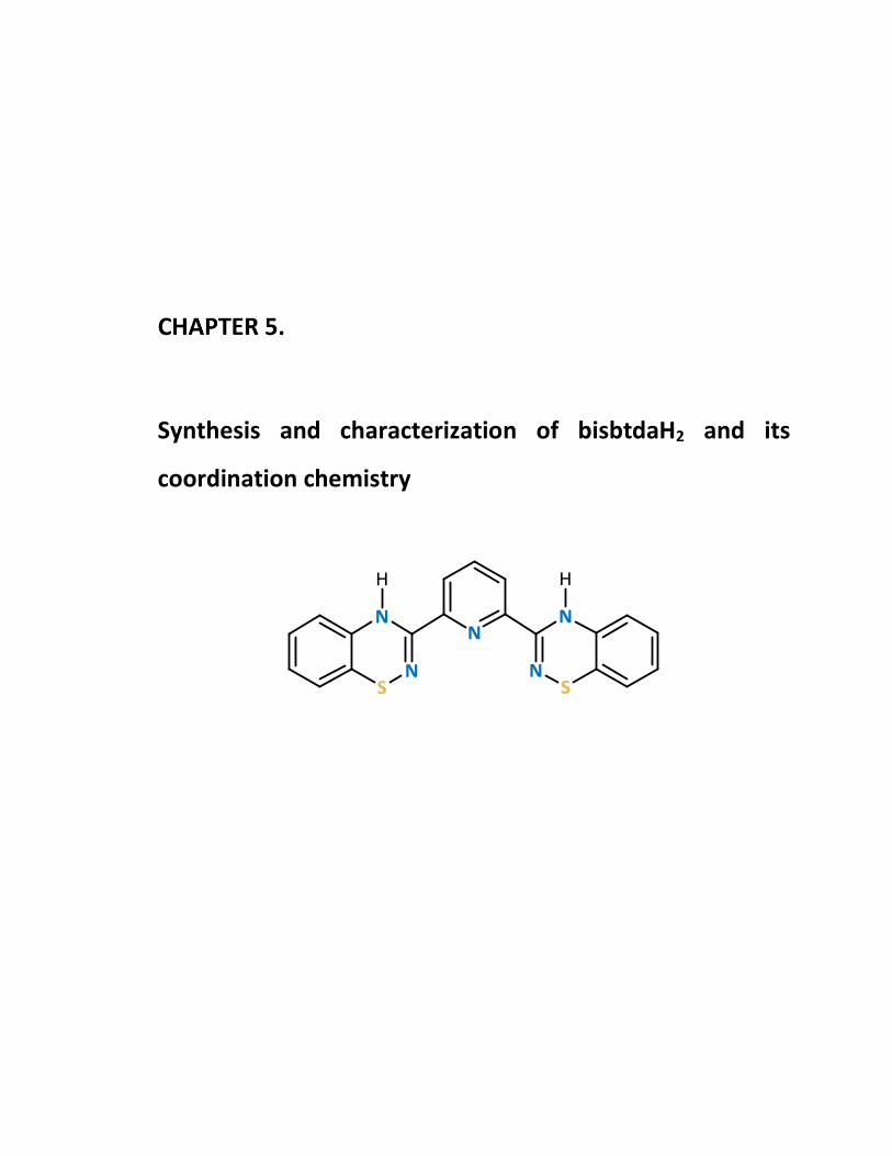
Synthesis and characterization of bisbtdaH2 and its
coordination chemistry

Chapter 5: Synthesis and characterization of bisbtdaH2 and its coordination chemistry
180
Introduction
Ligand design is a powerful tool to assemble desired supramolecular architectures
through coordination chemistry. Such ligand-based approaches to the self-organization of
coordination assemblies rely on the complementarity of ligand-based functional groups
coupled with the conformational preference(s) of metal ions. In recent decades this has
led to the emergence of elegant supramolecular structures such as molecular knots1 and
catenanes2 as well as coordination polymers,3,4 metal-organic frameworks5,6 and recent
extensions to reticular chemistry.6–8 A key design element often involves placing the donor
atoms within the ligand in such a way that five- or six-membered chelate rings are formed
upon coordination.9–11 Poly-pyridine ligands such as 2,2ʹ-bipyridine and its terpyridine
pincer analogue have a central position in coordination chemistry.12–15 Among poly-
pyridine ligands the terpyridine (terpy) pincer ligand (Scheme 5.1)16,17 can bind to both
low and high oxidation state metal ions almost always in a tridentate fashion. It has been
extensively studied specifically for octahedral complexes with various transition metal
ions such as FeII/III,18–21 CoII,22,23 RuII/III,24 ZnII.25 Square planar complexes of PtII,26–29 PdII,30,31
and AuIII 32–35 with terpy are also well known. Its rich coordination chemistry and high
binding affinity towards metal ions in conjunction with redox and photo-physical
Scheme 5.1 Molecular structures of terpy, bisbtdaH2, pybtdaH and pmbtdaH.

Chapter 5: Synthesis and characterization of bisbtdaH2 and its coordination chemistry
181
properties have given rise to a variety of applications in material science, such as the
development of solar cells,36,37 light emitting electrochemical cells,38–40 luminescent
systems,27,41 non-linear optical devices,42 etc. In organic synthesis, terpy complexes have
also been used as efficient catalysts for organic transformation such as C-C bond
formation,43 etherification,44 oxidation of alcohols or ethers,45,46 cyclopropanation,47 cross
coupling reactions,44 hydrosilation,48 controlled radical polymerization,49 photocatalytic
splitting of water37 etc. They have also found applications in molecular magnetism since
they offer a medium strength ligand field for first row transition metal complexes and
exhibit spin crossover behaviour for systems in which the crystal field is comparable
with the magnitude of the inter-electron repulsion term (PE) making the two possible
electronic states near quasi-energetic. The CoII(terpy)2 complex ion was one of the first
compounds to be shown to exhibit thermal spin crossover.22,23,50,51 Terpyridine complexes
have also found a wide range of potential application in pharmaceutical drug research
e.g., PtII complexes of terpy were first reported to bind double-stranded DNA by
intercalation.52 Other biological activities such as antitumor, radiotherapy, antiprotozoal
agents, and protein probes have been thoroughly documented.53–57 Such a wide range of
applications (vide supra) of terpy type complexes calls for high structural variability of this
tridentate donor in terms of solubility, flexibility, redox chemistry etc. Recently, bis-azinyl
analogues of terpyridine were reported with their 3d transition metal complexes
exhibiting reversible redox properties, spin crossover, and superexchange coupling.58
In this chapter, we report the synthesis and characterization of a novel bis-btda pincer
ligand bisbtdaH2 (Scheme 5.1) designed to offer a tridentate N,Nʹ,Nʺ mer-binding
coordination geometry and describe its coordination chemistry with selected first row
transition metal ions.

Chapter 5: Synthesis and characterization of bisbtdaH2 and its coordination chemistry
182
Results and Discussion
5.2.1 Synthesis and structural characterization of the ligand bisbtdaH2
The ligand bisbtdaH2 was synthesised according to the synthetic route shown in
Scheme 5.2. The first step is the reaction of 2-aminothiophenol with N-propylbromide to
form 2-(propylthio)aniline, as discussed in Chapter 2 (preparation of ligand 2.1).
Deprotonation of 2-(propylthio)aniline with LiHMDS, under anaerobic, dry conditions
followed by treatment with half an equivalent of 2,6-dicyanopyridine59 resulted in the
intermediate carboximidamide in 80% yield. Subsequent cyclization and pericyclic
elimination in the presence of N-chlorosuccinimide (NCS) afforded the ligand bisbtdaH2 in
43% yield after purification by column chromatography using dichloromethane as eluent.
Single crystals were grown by slow evaporation from dichloromethane.
The 1H NMR spectrum of ligand bisbtdaH2 exhibits the expected proton environments
in the aromatic region, consistent with a C2 symmetric ligand geometry. The imine C=N
stretching vibration was observed at 1615 cm-1. The molecular structure was further
confirmed by single crystal X-ray studies. The compound crystalizes in the triclinic space
group P 1 with two molecules in the asymmetric unit (Figure 5.1).
Scheme 5.2 Synthetic route to ligand bisbtdaH2.

Chapter 5: Synthesis and characterization of bisbtdaH2 and its coordination chemistry
183
The C-N (C(7)-N(1) and C(13)-N(4)) distances are 1.285(3) Å and 1.281(3) Å respectively
which are consistent with predominant double bond character (cf imine C=N at 1.279 Å).60
The bond lengths between C(7)-N(2) and C(13)-N(5) are 1.358(3) Å and 1.362(3) Å
respectively are more consistent with a conjugated C-N single bond (cf Ph-NH2 at 1.355
Å).60 The S-N bond lengths (1.705(2) and 1.696(2) Å) are comparable with other S-N single
bonds (1.62 – 1.74 Å for isomers of S8-x(NH)x)61 and 1.69 – 1.72 Å for other
benzothiadiazine derivatives62). Another notable feature of the benzothiadiazine ring is
the folding (θ) of molecule about the trans-annular vector S(1)∙∙∙N(2) and S(2)∙∙∙N(5) where
θ values are 24.15 and 26.41 for the two rings, respectively, comparable with previous
reported values (21° – 42°).62 This folding of the ring is consistent with the formally 8π
anti-aromatic nature of the thiadiazine framework.
In addition π-π stacking was manifested in the form of a one-dimensional chain motif
along the crystallographic b axis dimer with centroid∙∙∙centroid distances at 3.765 Å and
4.392 Å in which the shortest C-C distance is at 3.364 Å (Figure 5.2, top). The presence of
hydrogen bonds between the molecules affords a three-dimensional hydrogen-bonded
grid (Figure 5.2, bottom). The hydrogen bonding between the C-H and the N atom on the
N2
N3
N1
N5
N4
S2S1
C1
C2
C3 C4
C5
C7
C6
C8
C9
C10
C11
C12C13
C14
C15
C16C17
C18
C19
Figure 5.1 Molecular structure of bisbtdaH2 in one of the two crystallographically
independent molecules.

Chapter 5: Synthesis and characterization of bisbtdaH2 and its coordination chemistry
184
thiadiazine heterocycle extends the connectivity along a axis with distances at 2.633 Å
(θC-H∙∙∙N = 140.84°) and 2.671 Å (θC-H∙∙∙N = 168.25°) and along c axis with distances at 2.641
Å (θC-H∙∙∙N = 144.39°).
The preference for the crystallographically-determined conformer was also confirmed
by DFT calculations (B3LYP-D3/6-311G*+) on the three possible conformers derived by
rotating each heterocycle by 180o; the crystallographically determined structure was
computed to be more stable by 27 kJ/mol over the isomer where one btda ring is rotated
by 180° and by 81 kJ/mol when both btda rings are rotated by 180° (Table 5.1).
4.392 Å
4.392 Å
3.765 Å3.765 Å
Figure 5.2 Crystal packing of bisbtdaH2 highlighting π-π stacking along b axis (top) and the
packing revealing the three dimensional nature of the hydrogen-bonded motif
with a view down b axis (bottom).

Chapter 5: Synthesis and characterization of bisbtdaH2 and its coordination chemistry
185
Table 5.1 Density functional theory (DFT) calculations of the conformers of bisbtdaH2 using
the dispersion-corrected B3LYP-D3 functional and 6-311G*+.
Energy/kJ/mol 0 27 81
Torsion angle/° -7.3/3.65 2.95/-138.97 -128.84/136.59
Notably initial attempts to recrystallize bisbtdaH2 from hexanes resulted in the
formation of 5.2 which was characterized by X-ray crystallography (Figure 5.3). Such
thermally-induced ring contractions of benzothiadiazines has previously been reported by
Rees.63 Subsequent 1H NMR studies revealed bisbtdaH2 was thermally unstable upon
heating in hexanes and reflected in a lower symmetry structure with a peak for each
chemically distinct different environment (Figure 5.4). The side product (5.2) also appears
in the fragmentation pattern (calc. 361.0582, found 361.0578) of the ligand bisbtdaH2.
N2N3
N1N4
S2
S1
C1
C2
C3 C4
C5
C7
C6
C8
C9C10
C11
C12C13
C14C15
C16
C17C18
C19
Figure 5.3 Molecular structure of the side product 5.2.

Chapter 5: Synthesis and characterization of bisbtdaH2 and its coordination chemistry
186
5.2.2 Redox studies on bisbtdaH2
The ligand bisbtdaH2 possesses two thiadiazine moieties which can be oxidized to
generate the corresponding diradical ligand. However, the oxidation mechanism can be
achieved through two sequential 1e– transfer processes or via a single concomitant 2e–
transfer. Electrochemical studies on bisbtdaH2 (C0 = 5.96 x 10⁻³ mol·L-1) were performed
using a glassy carbon electrode (S = 1.57 mm2) in a deoxygenated solution of nBu4N(PF6)
in dichloromethane (0.047 mol∙L-1). Initial cyclic voltammograms revealed a single
reversible redox wave consistent with a 2e– process with E1/2 = +0.90 V vs Ag/AgCl (Figure
5.5a). The potential did not change as a function of the scan rate (Figure 5.5b) and the
anodic current peaks were found to be linearly proportional to the square root of the scan
rate (Figure 5.5c), consistent with a reversible process and the Nernstian system, in which
electron transfer is faster than the diffusion rate.
Figure 5.4 1H NMR of side product 5.2 (red) and bisbtdaH2 (blue) for
comparison (500 MHz, CDCl3).
bisbtdaH2
5.2
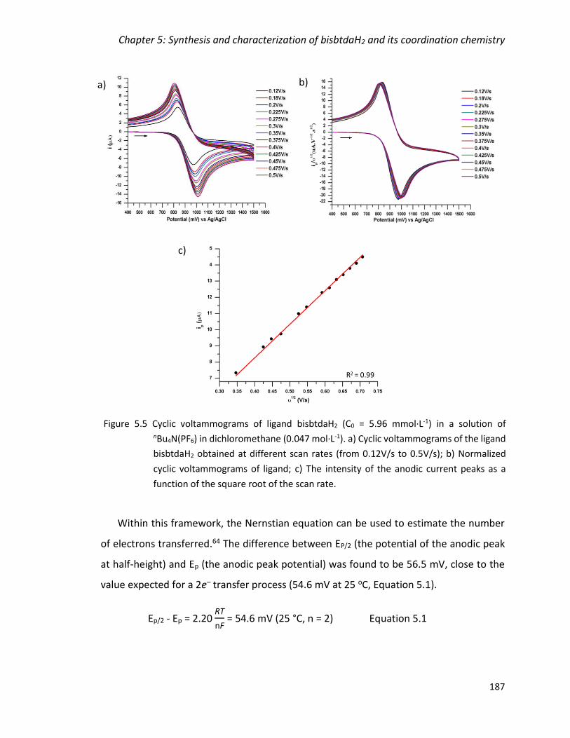
Chapter 5: Synthesis and characterization of bisbtdaH2 and its coordination chemistry
187
Within this framework, the Nernstian equation can be used to estimate the number
of electrons transferred.64 The difference between EP/2 (the potential of the anodic peak
at half-height) and Ep (the anodic peak potential) was found to be 56.5 mV, close to the
value expected for a 2e– transfer process (54.6 mV at 25 oC, Equation 5.1).
Ep/2 - Ep = 2.20 RT
nF = 54.6 mV (25 °C, n = 2) Equation 5.1
R2 = 0.99
a) b)
c)
Figure 5.5 Cyclic voltammograms of ligand bisbtdaH2 (C0 = 5.96 mmol·L-1) in a solution of nBu4N(PF6) in dichloromethane (0.047 mol·L-1). a) Cyclic voltammograms of the ligand
bisbtdaH2 obtained at different scan rates (from 0.12V/s to 0.5V/s); b) Normalized
cyclic voltammograms of ligand; c) The intensity of the anodic current peaks as a
function of the square root of the scan rate.

Chapter 5: Synthesis and characterization of bisbtdaH2 and its coordination chemistry
188
where R is the gas constant, T is the temperature, F is the faraday constant and n is the
number of electron transferred.
Although the electrochemical studies in solution revealed a single 2e– transfer,
heterogeneous oxidation of bisbtdaH2 with Ag2O in dichloromethane appeared kinetically
slow and we believe the evolution of the EPR spectra over time (vide infra) is consistent
with initial formation of the mono-radical followed by generation of the diradical. The in
situ room temperature EPR spectra of the ligand bisbtdaH2, dissolved in dichloromethane
and oxidized with an excess of Ag2O, led firstly to the formation of pentet spectrum typical
for btda radicals65 consistent with two near-equivalent 14N nuclei, expected for bisbtdaH•.
The fine structure of the second derivative could only be replicated by taking into account
coupling to two additional 1H nuclei associated with the benzo-fused ring. The best fit of
the spectrum afforded giso = 2.0058 and the hyperfine coupling constant of 14N atoms, aN
= 5.55 and 4.85 G, and 1H atoms, aH = 2.00 and 1.27 G (Figure 5.6, left).
Figure 5.6 Solution EPR spectra of mono and di-radicals formed from oxidation of bisbtdaH2;
(left) Second derivative and simulation of the monoradical bisbtdaH•; (right) First
derivative of diradical, bisbtda•• (stars indicating the additional features, right).
3480 3500 3520 3540Field (G)
3480 3500 3520 3540Field (G)
sim
exp
*
*

Chapter 5: Synthesis and characterization of bisbtdaH2 and its coordination chemistry
189
These parameters are similar to other btda radicals65 and in accord with the computed
spin density distribution (Figure 5.7, left) which delocalizes spin density to two positions
on the benzo ring. After a few minutes, the shape of the spectrum changed and additional
features emerged between the initial 5-line spectrum (Figure 5.6, right). The emergence
of a second species which we tentatively assign to a diradical species in which the
exchange coupling (J) is similar in energy to the hyperfine coupling constant suggesting
the presence of weak intramolecular magnetic exchange between the two radicals.66 The
EPR spectra are consistent with the spin density distribution determined from DFT
calculations which reveal that the nodal nature of the C(7) and C(13) positions of the btda
rings leads to limited ‘spin leakage’ onto the pyridyl linker and only weak exchange
coupling between these two radicals (Figure 5.7).
5.2.3 Syntheses and characterization of metal complexes of bisbtdaH2
Syntheses and description of [M(bisbtdaH2)2][X]2 (M = Mn, X = CF3SO3 (5.3); Fe,
X = CF3SO3 (5.4); Fe, X = BF4 (5.5); Co, X = Cl (5.6); Ni, X = Cl (5.7); Zn, X = CF3SO3
(5.8))
Reactions of ligand bisbtdaH2 with a range of MII salts in either CH3OH or CH3CN in a
ratio of 2:1 at ambient temperatures afforded complexes with general formula
Figure 5.7 Spin density distribution (UB3LYP/6-311G*+: (left) monoradical bisbtdaH• and
(right) the diradical triplet of bisbtda••.

Chapter 5: Synthesis and characterization of bisbtdaH2 and its coordination chemistry
190
[M(bisbtdaH2)2][X]2 (M = Mn, X = CF3SO3 (5.3); Fe, X =CF3SO3 (5.4); Fe, X = BF4 (5.5); Co,
X = Cl (5.6); Ni, X = Cl (5.7); Zn, X = CF3SO3 (5.8)). The crystals of complexes 5.3 − 5.8 proved
slightly hygroscopic, absorbing small quantities of water upon standing in the open
atmosphere but otherwise appeared air stable. All complexes were characterized by a
variety of analytical techniques including X-ray crystallography, IR and elemental analysis.
Complex 5.3 crystallizes in the orthorhombic space group Pccn with half a cation and
one OTf− anion in the asymmetric unit. Complex 5.4 crystallizes in the monoclinic space
group Pc with one cation, two OTf− anions and two CH3CN solvent molecules in the
asymmetric unit. Complexes 5.5 crystallizes in the triclinic space group P1 with one cation,
two BF4− anions and two CH3CN solvent molecules in the asymmetric unit. Complex 5.6
crystallizes in the monoclinic space group P21/n with one cation and two Cl− in the
asymmetric unit. Complex 5.7 crystallizes in the triclinic space group P21/n with one
cation, two Cl− anions and four CH3OH solvent molecules in the asymmetric unit. Complex
5.8 crystallizes in the monoclinic space group C2/c with two halves of two cations, two
OTf− anions and one CH3OH solvent molecules in the asymmetric unit. The formation of
complexes 5.3 − 5.8 is summarized in Equation 5.2.
Complexes 5.3 − 5.8 are mononuclear compounds of general formula
[M(bisbtdaH2)2][X]2 in which two bisbtdaH2 ligands bind in a tridentate N,Nʹ,Nʺ-chelate
fashion via two NBTDA and Npy donor atoms, adopting a mer coordination geometry. Each
metal adopts a six-coordinate MN6 geometry. The structure of complex 5.4, is shown in
Figure 5.8 as representative of this series of structures (for complexes 5.3, 5.5 – 5.8 see
Appendix 4) and selected bond lengths and angles are shown in Table 5.2. The FeII has
octahedral geometry with a N6 donor set, with Fe-N distances in the range of 1.864(5) −
1.975(5) Å consistent with a low spin state.67 The ligand bisbtdaH2 exhibits internal
NBTDA-M-Npy angles in the range 79.9(2) − 80.1(2)°.
MX2 + 2 bisbtdaH2 [M(bisbtdaH2)2][X]2 X = Cl, CF3SO3, BF4
Equation 5.2

Chapter 5: Synthesis and characterization of bisbtdaH2 and its coordination chemistry
191
Table 5.2 Selected bond lengths and angles for complexes 5.3 − 5.9.
M-Npy/Å M-NBTDA/Å NpyMNBTDA/°a NpyMNBTDA/°b NBTDAMNBTDA/° a NBTDAMNBTDA/° b NpyMNpy/°
Mn (5.3) 2.276(2) 2.201(2) 71.00(8) 103.55(8) 142.96(8) 86.05(8) 171.23(12) 2.297(2) 72.22(8) 113.50(8) 104.77(12)
106.62(12)
Fe (5.4) 1.864(5) 1.969(5) 79.9(2) 98.7(2) 159.9(2) 91.2(2) 178.6(2) 1.888(5) 1.969(5) 79.9(2) 99.3(2) 159.9(2) 91.6(2)
1.973(5) 80.0(2) 101.4(2) 91.7(2)
1.975(5) 80.1(2) 100.7(2) 92.4(2)
Fe (5.5) 1.881(4) 1.962(5) 79.63(19) 98.20(17) 160.0(2) 89.48(19) 178.47(17)
1.967(5) 79.80(18) 99.95(19) 160.09(19) 90.21(19) 1.970(4) 80.29(17) 100.04(19) 93.04(19)
1.976(4) 80.36(19) 101.71(17) 94.15(19)
Co (5.6) 2.046(3) 2.128(3) 74.93(10) 93.94(10) 147.63(11) 90.38(11) 163.82(11) 2.059(3) 2.139(3) 75.04(10) 94.45(10) 149.37(11) 92.28(11)
2.144(3) 75.51(11) 116.43(11) 93.94(11)
2.157(3) 75.61(10) 117.39(11) 99.90(11)
Ni (5.7) 1.985(2) 2.080(2) 77.16(10) 93.16(10) 154.57(10) 91.85(10) 177.98(10) 1.987(2) 2.081(2) 77.29(10) 93.32(9) 154.82(9) 92.66(9)
2.083(2) 77.41(9) 100.85(9) 93.16(10)
2.094(2) 77.53(9) 104.57(10) 93.32(9)
Zn (5.8) 2.054(3) 2.165(3) 75.35(11) 73.63(11) 149.00(10) 78.85(11) 174.64(17) 2.055(3) 2.266(3) 103.87(11) 73.98(11) 149.12(11) 80.69(11) 176.37(17) 2.209(3) 75.39(11) 106.94(11) 105.11(16)
2.312(3) 103.17(11) 107.81(11) 109.38(15)
109.92(16)
110.12(16)
Cu (5.9)c 1.929(2) 2.041(2) 79.33(8) - 157.77(8) - - 2.051(2) 79.36(8)
a Angle within the chelate ring b Angles between N atoms in different rings c Cu-O/Å = 1.9298(16)-2.651(2), O-Cu-O/° = 53.95(7)-153.68(6), O-Cu-N/° = 86.83(7)-173.05(7)
Fe N1N4
N3
N6
N8
N9
Figure 5.8 Molecular structure of 5.4 as representative of the series of complexes 5.3 − 5.8
(solvent molecules and counter-ions omitted for clarity).

Chapter 5: Synthesis and characterization of bisbtdaH2 and its coordination chemistry
192
A comparison of the cis NBTDA-M-NBTDA bond angles at the metal centre reveal
significant variation in conformation, ranging from 142.96(8) – 160.0(2)°. This variation is
highlighted when comparing the angle formed between the two N3 donor sets of the two
coordinated bisbtdaH2 ligands (Table 5.3, L1-L2 plane/°).
Table 5.3 Angles between planes for complexes 5.3 – 5.8 and the RMS deviation for each
complex from idealised octahedral, trigonal prismatic or pentagonal bipyramid
(with equatorial vacancy). For each complex the smallest deviation is underlined.
Mn (5.3) Fe (5.4) Fe (5.5) Co (5.6) Ni (5.7) Zn (5.8)
L1 - L2 plane/° 80.59 89.91 88.05 84.92 89.81 73.70 (Zn2) 75.83 (Zn1)
Octahedral 0.833 0.429 0.430 0.735 0.541 0.727 (Zn2)
0.751 (Zn1)
Trigonal prismatic 0.888 1.074 1.069 0.757 1.046 0.936 (Zn1)
0.955 (Zn2)
Pentagonal bipyramid
with a vacancy (eq.)
0.828 1.575 0.742 0.690 0.752 0.841(Zn1)
0.844(Zn2)
For an idealized octahedral geometry this angle should be 90°. This is observed for FeII
and NiII but deviates significantly for MnII and ZnII and is highlighted in Figure 5.9. The
effect of this distortion on the geometry of the metal centre was quantified using
FindGeo.68 In the cases of Fe and Ni, the RMS deviation clearly indicates an octahedral
geometry but in all other cases, the calculated RMS deviation is comparable for
octahedral, trigonal prismatic and pentagonal bipyramid geometries, as shown in Table
5.3.

Chapter 5: Synthesis and characterization of bisbtdaH2 and its coordination chemistry
193
Notably, the distortion parameter is maximized for MnII and ZnII, in which the high spin
MnII (d5) and the ZnII (d10) have no crystal field stabilization energy (CFSE) and so there is
no electronic destabilization in distorting away from octahedral in these two cases.
Conversely the distortion is minimal for low spin FeII (-2.4o) and NiII (-1.2o). These CFSE
effects clearly provide some stabilizing influence for the octahedral coordination
geometry over other 6-coordinate geometries, despite the contributions of other packing
forces (cation-anion and/or cation-cation interactions) which may contribute to the
overall lattice enthalpy. In addition, DFT calculations were performed on the
conformation of the ligand found in the Mn (5.3), Fe (5.4) and Zn (5.8) complexes. The
energy is lowest for the ligand conformation in Mn and Zn complexes, whereas the energy
increased significantly for the conformer observed in the Fe complex (Table 5.4). These
89.91°
Fe
80.59°
Mn
75.83°
Zn
Figure 5.9 Side-view of complexes of Mn (5.3), Fe (5.4) and Zn (5.8) illustrating the different
conformations the ligand adopts in each compound, along with the angle between
the two planes of the ligand.
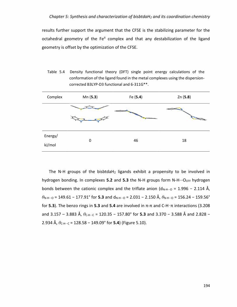
Chapter 5: Synthesis and characterization of bisbtdaH2 and its coordination chemistry
194
results further support the argument that the CFSE is the stabilizing parameter for the
octahedral geometry of the FeII complex and that any destabilization of the ligand
geometry is offset by the optimization of the CFSE.
Table 5.4 Density functional theory (DFT) single point energy calculations of the
conformation of the ligand found in the metal complexes using the dispersion-
corrected B3LYP-D3 functional and 6-311G**.
Complex Mn (5.3) Fe (5.4) Zn (5.8)
Energy/
kJ/mol 0 46 18
The N-H groups of the bisbtdaH2 ligands exhibit a propensity to be involved in
hydrogen bonding. In complexes 5.2 and 5.3 the N-H groups form N-H∙∙∙OOTf hydrogen
bonds between the cationic complex and the triflate anion (dN-H∙∙∙O = 1.996 − 2.114 Å,
θN-H∙∙∙O = 149.61 − 177.91° for 5.3 and dN-H∙∙∙O = 2.031 − 2.150 Å, θN-H∙∙∙O = 156.24 − 159.56°
for 5.3). The benzo rings in 5.3 and 5.4 are involved in π-π and C-H…π interactions (3.208
and 3.157 – 3.883 Å, θC-H∙∙∙C = 120.35 − 157.80° for 5.3 and 3.370 − 3.588 Å and 2.828 −
2.934 Å, θC-H∙∙∙C = 128.58 − 149.09° for 5.4) (Figure 5.10).

Chapter 5: Synthesis and characterization of bisbtdaH2 and its coordination chemistry
195
In complex 5.5, the N-H groups form N-H∙∙∙Cl hydrogen bonds between the cationic
complex and the Cl anions (dN-H∙∙∙Cl = 2.268 – 2.598 Å, θN-H∙∙∙Cl = 159.99 – 176.60°). In
complex 5.6 the N-H groups form two N-H∙∙∙Cl hydrogen bonds between the cationic
complex and the Cl anions (dN-H∙∙∙Cl = 2.172 and 2.464 Å, θN-H∙∙∙Cl = 166.97 and 171.17°,
respectively) and two hydrogen bonds with the CH3OH solvent molecules (dN-H∙∙∙O = 1.896
and 1.898 Å, θN-H∙∙∙O = 170.23 and 173.19°, respectively).
The structure of zinc complex 5.8 exhibits the greatest distortion from octahedral
geometry, placing two benzo rings in close proximity to exhibit an intramolecular π-π
interaction with centroid…centroid distances of 3.784 Å (Figure 5.11). In this complex, the
cationic complex of Zn(1) forms four N-H∙∙∙OOTf hydrogen bonds between the cationic
complex and the triflate anions (dN-H∙∙∙O = 2.002 and 2.114 Å, θN-H∙∙∙O = 168.18 and 148.20°
respectively); whereas Zn(2) forms two N-H∙∙∙OOTf hydrogen bonds between the cationic
complex and the triflate anions (dN-H∙∙∙O = 2.495 Å, θN-H∙∙∙O = 128.74°) and two N-H∙∙∙OCH3OH
between the cationic complex and the methanol solvent molecules (dN-H∙∙∙O = 2.113 Å,
θN-H∙∙∙O = 138.19°).
Figure 5.10 Crystal packing of 5.4 highlighting the π-π (green) and C-H∙∙∙π (yellow) interactions
(left) and the N-H∙∙∙O hydrogen bonding between the amino group and the triflate
counter ions (red lines, right).
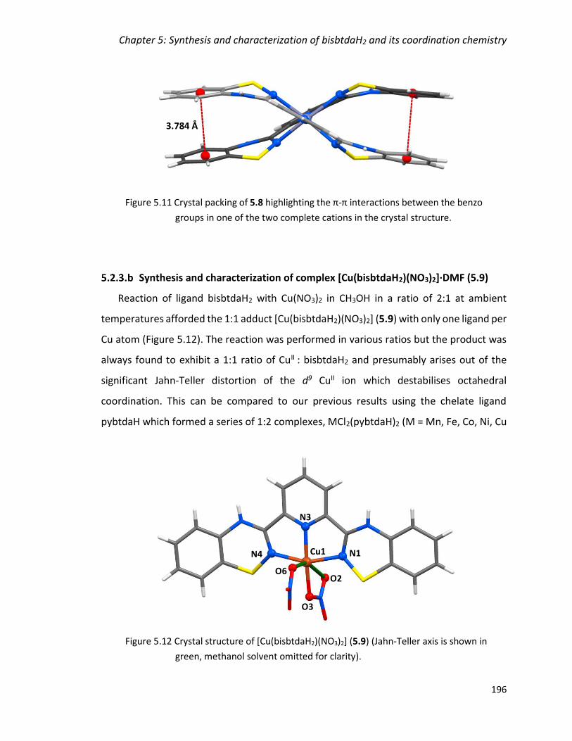
Chapter 5: Synthesis and characterization of bisbtdaH2 and its coordination chemistry
196
Synthesis and characterization of complex [Cu(bisbtdaH2)(NO3)2]·DMF (5.9)
Reaction of ligand bisbtdaH2 with Cu(NO3)2 in CH3OH in a ratio of 2:1 at ambient
temperatures afforded the 1:1 adduct [Cu(bisbtdaH2)(NO3)2] (5.9) with only one ligand per
Cu atom (Figure 5.12). The reaction was performed in various ratios but the product was
always found to exhibit a 1:1 ratio of CuII : bisbtdaH2 and presumably arises out of the
significant Jahn-Teller distortion of the d9 CuII ion which destabilises octahedral
coordination. This can be compared to our previous results using the chelate ligand
pybtdaH which formed a series of 1:2 complexes, MCl2(pybtdaH)2 (M = Mn, Fe, Co, Ni, Cu
Cu1
O2
O3
O6
N4
N3
N1
Figure 5.12 Crystal structure of [Cu(bisbtdaH2)(NO3)2] (5.9) (Jahn-Teller axis is shown in
green, methanol solvent omitted for clarity).
3.784 Å
Figure 5.11 Crystal packing of 5.8 highlighting the π-π interactions between the benzo
groups in one of the two complete cations in the crystal structure.
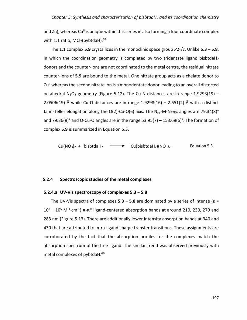
Chapter 5: Synthesis and characterization of bisbtdaH2 and its coordination chemistry
197
and Zn), whereas CuII is unique within this series in also forming a four coordinate complex
with 1:1 ratio, MCl2(pybtdaH).69
The 1:1 complex 5.9 crystallizes in the monoclinic space group P21/c. Unlike 5.3 – 5.8,
in which the coordination geometry is completed by two tridentate ligand bisbtdaH2
donors and the counter-ions are not coordinated to the metal centre, the residual nitrate
counter-ions of 5.9 are bound to the metal. One nitrate group acts as a chelate donor to
CuII whereas the second nitrate ion is a monodentate donor leading to an overall distorted
octahedral N3O3 geometry (Figure 5.12). The Cu-N distances are in range 1.9293(19) –
2.0506(19) Å while Cu-O distances are in range 1.9298(16) – 2.651(2) Å with a distinct
Jahn-Teller elongation along the O(2)-Cu-O(6) axis. The Npy-M-NBTDA angles are 79.34(8)°
and 79.36(8)° and O-Cu-O angles are in the range 53.95(7) – 153.68(6)°. The formation of
complex 5.9 is summarized in Equation 5.3.
5.2.4 Spectroscopic studies of the metal complexes
UV-Vis spectroscopy of complexes 5.3 − 5.8
The UV-Vis spectra of complexes 5.3 − 5.8 are dominated by a series of intense (ε =
103 − 105 M-1·cm-1) π-π* ligand-centered absorption bands at around 210, 230, 270 and
283 nm (Figure 5.13). There are additionally lower intensity absorption bands at 340 and
430 that are attributed to intra-ligand charge transfer transitions. These assignments are
corroborated by the fact that the absorption profiles for the complexes match the
absorption spectrum of the free ligand. The similar trend was observed previously with
metal complexes of pybtdaH.69
Cu(NO3)2 + bisbtdaH2 Cu(bisbtdaH2)(NO3)2 Equation 5.3

Chapter 5: Synthesis and characterization of bisbtdaH2 and its coordination chemistry
198
Electronic structure of complexes [Fe(bisbtdaH2)2]2+ (5.4 and 5.5)
The FeII complexes 5.4 and 5.5 were found to be low-spin diamagnetic d6 based on
structural parameters and 1H NMR spectra which exhibit no paramagnetic shift or
broadening. The 1H NMR spectrum of 5.5 shows that the chemical shift range is in the
‘normal’ range expected consistent with low spin FeII. The peaks associated with the free
ligand are fully replaced by the 1H NMR signals for the coordinated ligand of complex 5.5
(Figure 5.14). Additional room temperature X-ray studies were performed on complex 5.5
but revealed no significant increase in bond length expected for a LS to HS phase
transition.67 These data are all consistent with a LS FeII centre.
Figure 5.13 Solution UV-Vis spectra of complexes 5.3 − 5.8 as well as ligand bisbtdaH2 to
emphasize the ligand-based nature of the spectroscopic properties (Solvent:
CH3OH or CH3CN, rt, CbisbtdaH2 ≈ 10-5 M; Ccomplexes ≈ 10-6 M).

Chapter 5: Synthesis and characterization of bisbtdaH2 and its coordination chemistry
199
Attempts to model the binding affinity of bisbtdaH2 to FeII by UV-Vis titration in MeCN
proved ambiguous. While a marked absorption maximum was observed at 570 nm and
increased on addition of bisbtdaH2, a fit of the data to formation of both 1:1 and 2:1
complexes were similar (Figure 5.15).70,71 Assuming formation of [Fe(bisbtdaH2)2]2+, the
formation constant (β) for the process:
is = 4.6 x 10⁶. For [Fe(terpy)]2+ K1 was found to be 107.1 and = 1020.9 for formation of
[Fe(terpy)2]2+.72,60 These clearly suggest that bisbtdaH2 is a significantly weaker
coordinating ligand than terpy. This is perhaps unsurprising given the large computed
reorientation energy for the ligand to adopt the tridentate N,N´,Nʺ coordination geometry
(80 kJ·mol-1, vide supra).
Figure 5.14 1H NMR of complex 5.5 (red) and bisbtdaH2 (blue) (500 MHz, CD3CN).
Complex 5.5
bisbtdaH2
Fe2+ + 2 bisbtdaH2 [Fe(bisbtdaH2)2]2+ Equation 5.4
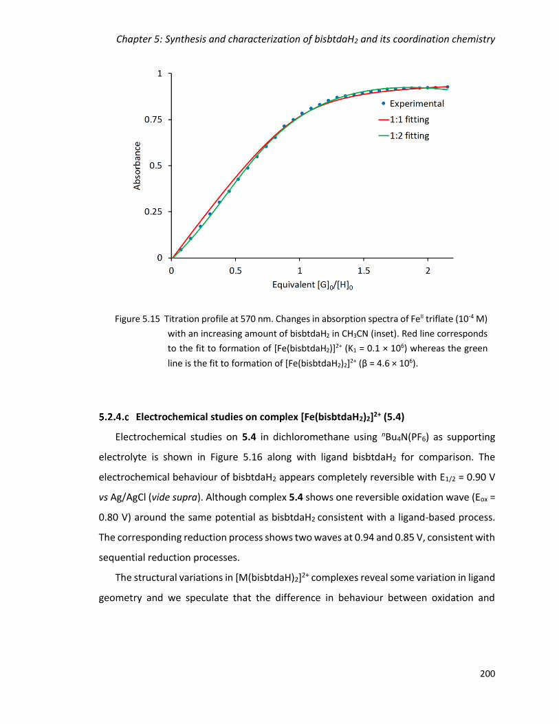
Chapter 5: Synthesis and characterization of bisbtdaH2 and its coordination chemistry
200
Electrochemical studies on complex [Fe(bisbtdaH2)2]2+ (5.4)
Electrochemical studies on 5.4 in dichloromethane using nBu4N(PF6) as supporting
electrolyte is shown in Figure 5.16 along with ligand bisbtdaH2 for comparison. The
electrochemical behaviour of bisbtdaH2 appears completely reversible with E1/2 = 0.90 V
vs Ag/AgCl (vide supra). Although complex 5.4 shows one reversible oxidation wave (Eox =
0.80 V) around the same potential as bisbtdaH2 consistent with a ligand-based process.
The corresponding reduction process shows two waves at 0.94 and 0.85 V, consistent with
sequential reduction processes.
The structural variations in [M(bisbtdaH)2]2+ complexes reveal some variation in ligand
geometry and we speculate that the difference in behaviour between oxidation and
Figure 5.15 Titration profile at 570 nm. Changes in absorption spectra of FeII triflate (10-4 M)
with an increasing amount of bisbtdaH2 in CH3CN (inset). Red line corresponds
to the fit to formation of [Fe(bisbtdaH2)]2+ (K1 = 0.1 × 10⁶) whereas the green
line is the fit to formation of [Fe(bisbtdaH2)2]2+ (β = 4.6 × 10⁶).
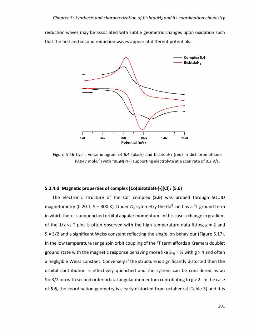
Chapter 5: Synthesis and characterization of bisbtdaH2 and its coordination chemistry
201
reduction waves may be associated with subtle geometric changes upon oxidation such
that the first and second reduction waves appear at different potentials.
Magnetic properties of complex [Co(bisbtdaH2)2][Cl]2 (5.6)
The electronic structure of the CoII complex (5.6) was probed through SQUID
magnetometry (0.20 T, 5 − 300 K). Under Oh symmetry the CoII ion has a 4T ground term
in which there is unquenched orbital angular momentum. In this case a change in gradient
of the 1/χ vs T plot is often observed with the high temperature data fitting g ≈ 2 and
S = 3/2 and a significant Weiss constant reflecting the single ion behaviour (Figure 5.17).
In the low temperature range spin orbit coupling of the 4T term affords a Kramers doublet
ground state with the magnetic response behaving more like Seff = ½ with g > 4 and often
a negligible Weiss constant. Conversely if the structure is significantly distorted then the
orbital contribution is effectively quenched and the system can be considered as an
S = 3/2 ion with second order orbital angular momentum contributing to g > 2. In the case
of 5.6, the coordination geometry is clearly distorted from octahedral (Table 3) and it is
Complex 5.4BisbtdaH2
Figure 5.16 Cyclic voltammogram of 5.4 (black) and bisbtdaH2 (red) in dichloromethane
(0.047 mol·L-1) with nBu4N(PF6) supporting electrolyte at a scan rate of 0.2 V/s.
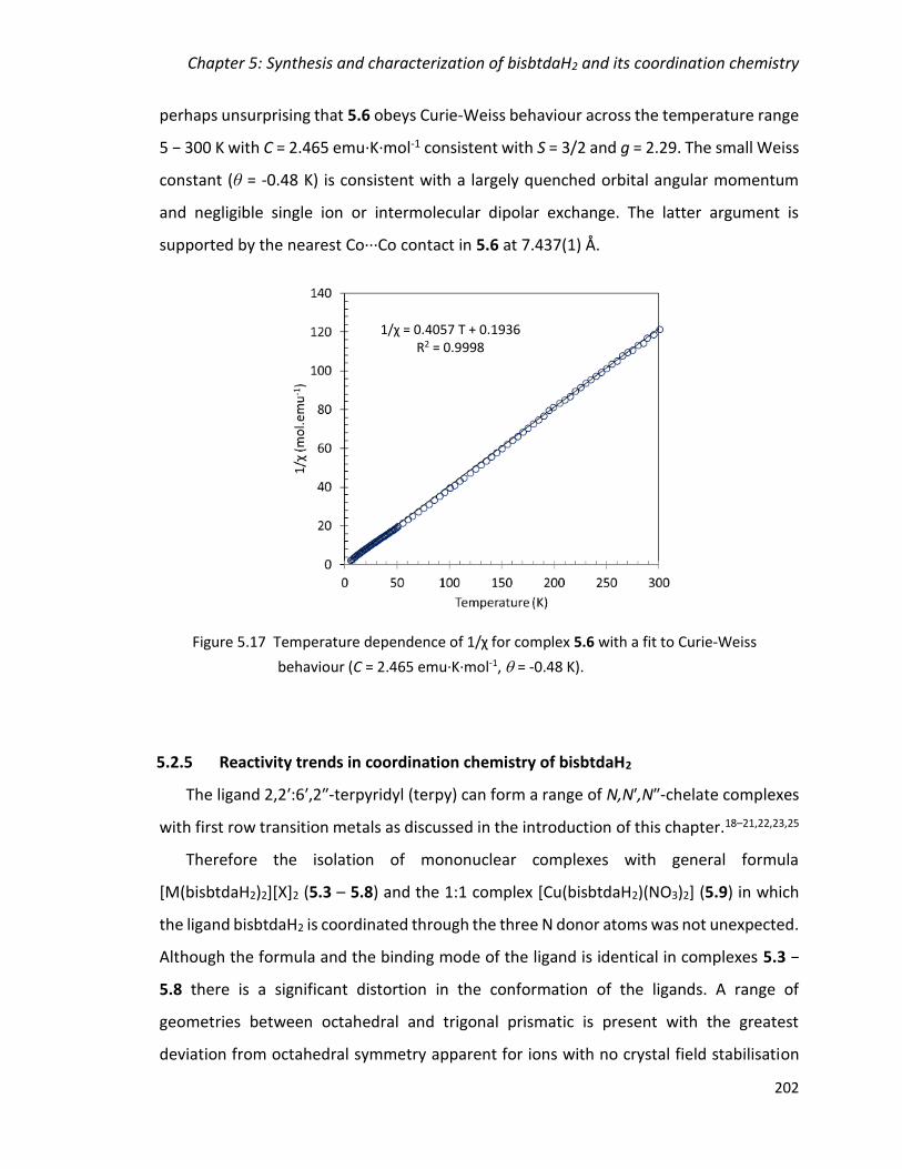
Chapter 5: Synthesis and characterization of bisbtdaH2 and its coordination chemistry
202
perhaps unsurprising that 5.6 obeys Curie-Weiss behaviour across the temperature range
5 − 300 K with C = 2.465 emu·K·mol-1 consistent with S = 3/2 and g = 2.29. The small Weiss
constant (θ = -0.48 K) is consistent with a largely quenched orbital angular momentum
and negligible single ion or intermolecular dipolar exchange. The latter argument is
supported by the nearest Co∙∙∙Co contact in 5.6 at 7.437(1) Å.
5.2.5 Reactivity trends in coordination chemistry of bisbtdaH2
The ligand 2,2ʹ:6ʹ,2ʺ-terpyridyl (terpy) can form a range of N,Nʹ,Nʺ-chelate complexes
with first row transition metals as discussed in the introduction of this chapter.18–21,22,23,25
Therefore the isolation of mononuclear complexes with general formula
[M(bisbtdaH2)2][X]2 (5.3 – 5.8) and the 1:1 complex [Cu(bisbtdaH2)(NO3)2] (5.9) in which
the ligand bisbtdaH2 is coordinated through the three N donor atoms was not unexpected.
Although the formula and the binding mode of the ligand is identical in complexes 5.3 −
5.8 there is a significant distortion in the conformation of the ligands. A range of
geometries between octahedral and trigonal prismatic is present with the greatest
deviation from octahedral symmetry apparent for ions with no crystal field stabilisation
Figure 5.17 Temperature dependence of 1/χ for complex 5.6 with a fit to Curie-Weiss
behaviour (C = 2.465 emu·K·mol-1, = -0.48 K).
1/χ = 0.4057 T + 0.1936 R2 = 0.9998

Chapter 5: Synthesis and characterization of bisbtdaH2 and its coordination chemistry
203
energy, i.e. d10 ZnII and high-spin d5 MnII ions. The counter-ion has no effect in the cationic
complex as confirmed from a comparison of the bond lengths of Fe complexes 5.4 and 5.5
(Table 5.2).
A CSD search73 revealed that only 2% of the reported first row metal complexes of
terpy analogues fall in the 70° – 80° range of which the majority is either non-symmetrical
ligands74 or the ligand is coordinating two metal centres resulting to a helix
conformation.75 In all cases, the distortion was due to the sterics of the terpy analogue
and not a result of the electronic contribution.
A similar deviation in first fow transition metal complexes with the ligand 4ʹ-
methylthio-2,2ʹ:6ʹ,2ʺ-terpyridyl has been reported by Turnbull.76 In this work, the Mn
complex has an angle of 78.6° between the two planes of the ligand, whereas the Fe and
Ni complexes are close to 90°. The Co and Cu complexes were not characterized by X-ray
crystallography for comparison, but they isolated a 1:1 adduct of Zn, in the metal center
which is 5-coordinate.
Chapter 5: Synthesis and characterization of bisbtdaH2 and its coordination chemistry
204
Conclusions
The novel S/N based terpyridine type ligand bisbtdaH2 has been synthesized and its
coordination chemistry with first row transition metal complexes has been investigated.
Electrochemical studies on the free ligand bisbtdaH2 reveal a single well-defined 2e–
oxidation process with E1/2 = +0.90 V. EPR studies of the in situ chemical oxidation on
bisbtdaH2 reveal the presence of both mono and diradical species. The anticipated six-
coordinate metal complexes of general formula [M(bisbtdaH2)2][X]2 (5.3 − 5.8) were
isolated for M = Mn, Fe, Co, Ni and Zn whereas Cu generates an unexpected 1:1 adduct,
[Cu(bisbtdaH2)(NO3)2] (5.9). For the CoII complex (5.6) SQUID measurements reflect the
structure is high-spin across the temperature range whereas for FeII complexes (5.4 and
5.5) solution NMR data and crystallographic studies point to a low-spin configuration up
to ambient temperature. Cyclic voltammetry of complex 5.4 reveals the oxidation process
occurs via two single e– oxidation waves. In all cases the bisbtdaH2 ligand binds in a
tridentate N,Nʹ,Nʺ-chelate fashion via benzothiadiazinyl NBTDA and pyridyl Npy atoms. The
geometries of the [M(bisbtdaH2)2]2+ ions appear sensitive to the crystal field stabilisation
energy; in the absence of a significant crystal field the geometry distorts significantly from
octahedral and only provides systems with small distortion parameters when the CFSE can
be maximized. This tendency to distort significantly from octahedral may contribute to
the relatively low binding constant for bisbtdaH2 towards M2+ ions, exemplified by FeII.
Chapter 5: Synthesis and characterization of bisbtdaH2 and its coordination chemistry
205
Experimental
5.4.1 General considerations and physical measurements
Solvents and other anhydrous or hydrated metal salts were obtained from commercial
suppliers and used without further purification. 2,6-dicyanopyridine,59 2-(propylthio)-
benzenamine,77 Mn(CF3SO3)2,78 and Fe(CF3SO3)279 were prepared according to reported
methods. Zn(CF3SO3)2 was prepared by a similar method reported in the literature.78
Manipulation of air-sensitive materials was carried out under an atmosphere of dry
nitrogen using standard Schlenk techniques and a dry-nitrogen glove box (MBraun
Labmaster).
Melting points were determined using a Stanford Research Systems MPA120 EZ-Melt
automated melting point apparatus. Elemental compositions were determined on a
Perkin Elmer 2400 Series II CHNS/O Analyzer. UV-Vis spectra were measured on an Agilent
8453 Spectrophotometer using ca. 1 × 10⁻⁶ M solutions in methanol or acetonitrile in the
ranger 200 – 600 nm. IR spectra were obtained using a Bruker Alpha FT-IR spectrometer
equipped with a Platinum single reflection diamond ATR module. NMR spectra were
recorded on a Bruker 500 MHz spectrometer with a Broadband AX Probe using CDCl3 (1H
δ = 7.26 ppm, s; 13C δ = 77.16 ppm), CD2Cl2 (1H δ = 5.32 ppm, t; 13C δ = 53.84 ppm) or
CD3CN (1H δ = 1.94 ppm, septet) as an internal reference point relative to Me4Si (δ = 0
ppm). Mass spectra were recorded on a Waters XEVO G2-XS specifically using the
atmospheric solids analysis probe in positive resolution mode (ASAP+). EPR spectra of
monoradical bisbtdaH• and diradical bisbtda•• as well as complex 5.9 were measured on
a Bruker EMXplus X-band EPR spectrometer at room temperature. Simulations of the EPR
spectra were made using WinSim software80 for the monoradical bisbtdaH• and the
PIP4Win software for the anisotropic spectrum of complex 5.9.81 Variable temperature
magnetic data (2 – 300 K) on complex 5.6 were obtained using a Quantum Design MPMS
SQUID magnetometer using a magnetic field strength of 0.1 T. Background corrections for
the sample holder assembly and sample diamagnetism (Pascal’s constants) were applied.
Density functional theory (DFT) calculations on bisbtdaH2 were undertaken using the
dispersion-corrected B3LYP-D3 functional and 6-311G** basis set82–84 functional within
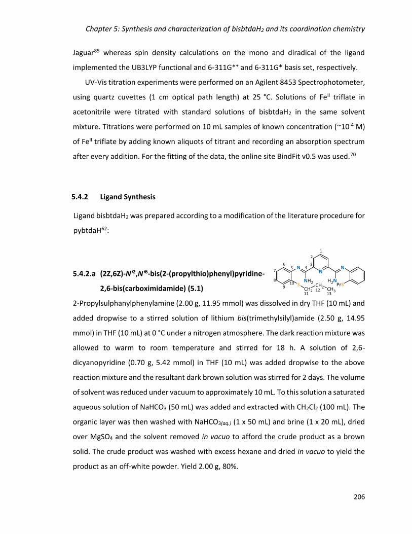
Chapter 5: Synthesis and characterization of bisbtdaH2 and its coordination chemistry
206
Jaguar85 whereas spin density calculations on the mono and diradical of the ligand
implemented the UB3LYP functional and 6-311G*+ and 6-311G* basis set, respectively.
UV-Vis titration experiments were performed on an Agilent 8453 Spectrophotometer,
using quartz cuvettes (1 cm optical path length) at 25 °C. Solutions of FeII triflate in
acetonitrile were titrated with standard solutions of bisbtdaH2 in the same solvent
mixture. Titrations were performed on 10 mL samples of known concentration (~10-4 M)
of FeII triflate by adding known aliquots of titrant and recording an absorption spectrum
after every addition. For the fitting of the data, the online site BindFit v0.5 was used.70
5.4.2 Ligand Synthesis
Ligand bisbtdaH2 was prepared according to a modification of the literature procedure for
pybtdaH62:
(2Z,6Z)-N'2,N'6-bis(2-(propylthio)phenyl)pyridine-
2,6-bis(carboximidamide) (5.1)
2-Propylsulphanylphenylamine (2.00 g, 11.95 mmol) was dissolved in dry THF (10 mL) and
added dropwise to a stirred solution of lithium bis(trimethylsilyl)amide (2.50 g, 14.95
mmol) in THF (10 mL) at 0 °C under a nitrogen atmosphere. The dark reaction mixture was
allowed to warm to room temperature and stirred for 18 h. A solution of 2,6-
dicyanopyridine (0.70 g, 5.42 mmol) in THF (10 mL) was added dropwise to the above
reaction mixture and the resultant dark brown solution was stirred for 2 days. The volume
of solvent was reduced under vacuum to approximately 10 mL. To this solution a saturated
aqueous solution of NaHCO3 (50 mL) was added and extracted with CH2Cl2 (100 mL). The
organic layer was then washed with NaHCO3(aq.) (1 x 50 mL) and brine (1 x 20 mL), dried
over MgSO4 and the solvent removed in vacuo to afford the crude product as a brown
solid. The crude product was washed with excess hexane and dried in vacuo to yield the
product as an off-white powder. Yield 2.00 g, 80%.
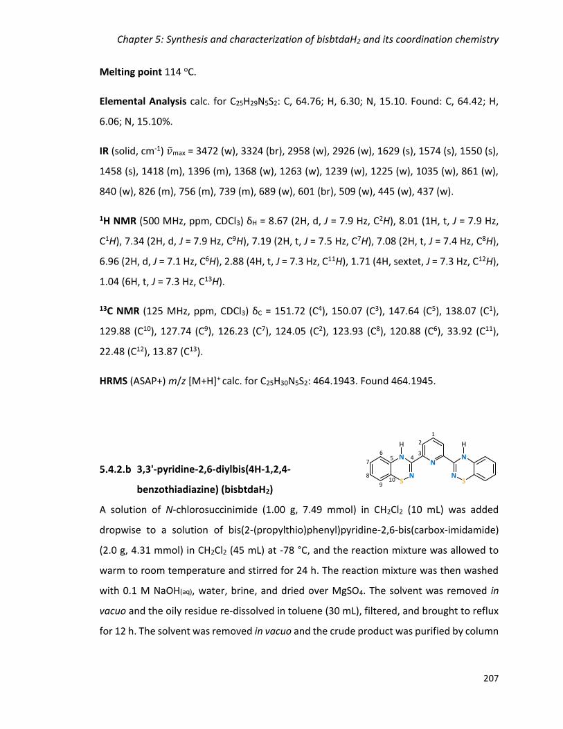
Chapter 5: Synthesis and characterization of bisbtdaH2 and its coordination chemistry
207
Melting point 114 oC.
Elemental Analysis calc. for C25H29N5S2: C, 64.76; H, 6.30; N, 15.10. Found: C, 64.42; H,
6.06; N, 15.10%.
IR (solid, cm-1) �̃�max = 3472 (w), 3324 (br), 2958 (w), 2926 (w), 1629 (s), 1574 (s), 1550 (s),
1458 (s), 1418 (m), 1396 (m), 1368 (w), 1263 (w), 1239 (w), 1225 (w), 1035 (w), 861 (w),
840 (w), 826 (m), 756 (m), 739 (m), 689 (w), 601 (br), 509 (w), 445 (w), 437 (w).
1H NMR (500 MHz, ppm, CDCl3) δH = 8.67 (2H, d, J = 7.9 Hz, C2H), 8.01 (1H, t, J = 7.9 Hz,
C1H), 7.34 (2H, d, J = 7.9 Hz, C9H), 7.19 (2H, t, J = 7.5 Hz, C7H), 7.08 (2H, t, J = 7.4 Hz, C8H),
6.96 (2H, d, J = 7.1 Hz, C6H), 2.88 (4H, t, J = 7.3 Hz, C11H), 1.71 (4H, sextet, J = 7.3 Hz, C12H),
1.04 (6H, t, J = 7.3 Hz, C13H).
13C NMR (125 MHz, ppm, CDCl3) δC = 151.72 (C4), 150.07 (C3), 147.64 (C5), 138.07 (C1),
129.88 (C10), 127.74 (C9), 126.23 (C7), 124.05 (C2), 123.93 (C8), 120.88 (C6), 33.92 (C11),
22.48 (C12), 13.87 (C13).
HRMS (ASAP+) m/z [M+H]+ calc. for C25H30N5S2: 464.1943. Found 464.1945.
3,3'-pyridine-2,6-diylbis(4H-1,2,4-
benzothiadiazine) (bisbtdaH2)
A solution of N-chlorosuccinimide (1.00 g, 7.49 mmol) in CH2Cl2 (10 mL) was added
dropwise to a solution of bis(2-(propylthio)phenyl)pyridine-2,6-bis(carbox-imidamide)
(2.0 g, 4.31 mmol) in CH2Cl2 (45 mL) at -78 °C, and the reaction mixture was allowed to
warm to room temperature and stirred for 24 h. The reaction mixture was then washed
with 0.1 M NaOH(aq), water, brine, and dried over MgSO4. The solvent was removed in
vacuo and the oily residue re-dissolved in toluene (30 mL), filtered, and brought to reflux
for 12 h. The solvent was removed in vacuo and the crude product was purified by column

Chapter 5: Synthesis and characterization of bisbtdaH2 and its coordination chemistry
208
chromatography using CH2Cl2 as eluent. The slow evaporation of CH2Cl2 resulted in red
shiny crystals of ligand bisbtdaH2. Yield 0.70 g, 43%.
Melting Point 208 oC (dec.)
Elemental Analysis calc. for C19H13N5S2: C, 60.78; H, 3.49; N, 18.65. Found: C, 60.90; H,
3.34; N, 18.24%.
UV-Vis (CH3OH; λmax, nm; ε, M-1 cm-1): 203 (24761), 229 (33006), 270 (40828), 282 (33101),
335 (6332), 439 (2723).
IR (solid, cm-1) �̃�max = 3382 (w), 3357 (m), 1615 (m), 1564 (m), 1494 (m), 1425 (m), 1289
(m), 1240 (m), 1124 (m), 1068 (m), 995 (m), 920 (m), 822 (m), 747 (s), 643 (m), 610 (br),
466 (m), 429 (m).
1H NMR (500 MHz, ppm, CDCl3) δH = 8.18 (2H, d, J = 7.9 Hz, C2H), 7.95 (2H, s, NH), 7.86
(1H, t, J = 7.9 Hz, C1H), 6.99 (2H, t, J = 7.6 Hz, C7H), 6.93 (2H, t, J = 7.5 Hz, C8H), 6.71 (2H, d,
J = 7.5 Hz, C9H), 6.49 (2H, dd, J = 7.6, 0.9 Hz, C6H).
13C NMR (125 MHz, ppm, CDCl3) δC = 153.26 (C4), 146.83 (C3), 138.20 (C1), 136.26 (C10),
127.96 (C7), 125.82 (C8), 123.47 (C2), 123.09 (C9), 120.26 (C5), 114.48 (C6).
HRMS (ASAP+) m/z [M+H]+ calc. for C19H14N5S2: 376.0691. Found 376.0685.
3-[6-(1,3-benzothiazol-2-yl)pyridin-2-yl]-4H-
1,2,4-benzothiadiazine (5.2)
Formation upon heating in hexanes as orange crystals.
1H NMR (500 MHz, ppm, CDCl3) δH = 8.48 (1H, NH), 8.46 (1H, dd, J = 7.8, 1.0 Hz), 8.19 (1H,
dd, J = 7.8, 1.0 Hz), 7.99 (1H, d, J = 8.0 Hz), 7.96 (1H, t, J = 7.8 Hz), 7.57-7.54 (1H, m), 7.49-
7.46 (1H, m), 7.00 (1H, td, J = 11.4, 1.4 Hz), 6.92 (1H, td, J = 11.3, 1.2 Hz), 6.73 (1H, d, J =
7.5 Hz), 6.59 (1H, dd, J = 7.7, 1.1 Hz).
Chapter 5: Synthesis and characterization of bisbtdaH2 and its coordination chemistry
209
5.4.3 Complex Syntheses with MX2 (M = Mn, Fe, Co, Ni, Cu, Zn, X = Cl, CF3SO3, NO3)
[Mn(bisbtdaH2)2][CF3SO3]2 (5.3)
Solid ligand bisbtdaH2 (0.020 g, 0.053 mmol) was added to a solution of
Mn(CF3SO3)2∙CH3CN (0.010 g, 0.025 mmol) in CH3CN (10 mL). The mixture was stirred until
a clear solution obtained. The solution was filtered and dark red crystals were grown by
slow diffusion of diethyl ether into the mother liquor over several days. Yield 0.024 g, 79%.
Elemental Analysis calc. for [C40H26N10MnS6O6F6]∙H2O: C, 42.82; H, 2.52; N, 12.48. Found:
C, 42.80; H, 2.51; N, 12.54%.
UV-Vis (CH3OH; λmax, nm; ε, M-1 cm-1): 209 (107565), 230 (62412), 264 (80600), 286
(55908), 337 (10918), 440 (4670).
IR (solid, cm-1) �̃�max = 3249 (br), 1602 (w), 1583 (m), 1557 (m), 1526 (m), 1463 (s), 1434 (s),
1224 (br), 1150 (br), 1028 (s), 924 (m), 864 (w), 801(m), 758 (br), 666 (w), 631 (br), 569
(m), 513 (m), 468 (m), 470 (m).
[Fe(bisbtdaH2)2][CF3SO3]2∙2CH3CN (5.4)
Solid ligand bisbtdaH2 (0.050 g, 0.133 mmol) was added to a solution of
Fe(CF3SO3)2∙2CH3CN (0.029 g, 0.067 mmol) in CH3CN (10 mL). The mixture was stirred until
a clear purple solution obtained. The solution was filtered and dark purple crystals were
grown by slow diffusion of diethyl ether into the mother liquor over 3 days. Yield 0.020 g,
26%.
Elemental Analysis calc. for [C40H26N10FeS6O6F6]∙3H2O: C, 41.45; H, 2.78; N, 12.09. Found:
C, 41.52; H, 2.51; N, 11.81%.
UV-Vis (CH3CN; λmax, nm; ε, M-1 cm-1): 211 (122317), 231 (82067), 268 (94929), 289
(84308), 303 (67607), 334 (28875), 579 (8902).
Chapter 5: Synthesis and characterization of bisbtdaH2 and its coordination chemistry
210
IR (solid, cm-1) �̃�max = 3259 (br), 1606 (m), 1531 (s), 1471 (s), 1434 (s), 1398 (s), 1287 (br),
1219 (br), 1154 (br), 1024 (s), 931 (w), 873 (w), 805 (w), 742 (s), 689 (w), 678 (w), 631 (s),
571 (w), 613 (m), 513 (m), 437 (w).
[Fe(bisbtdaH2)2][BF4]2∙CH3CN (5.5)
Solid ligand bisbtdaH2 (0.020 g, 0.053 mmol) was added to a solution of Fe(BF4)2∙6H2O
(0.010 g, 0.030 mmol) in CH3CN (10 mL). The mixture was stirred for a few minutes to
afford a dark purple solution which was filtered and left undisturbed. Dark purple crystals
appeared in few days. Yield 0.010 g, 36%.
Elemental analysis calc. for [C38H26N10FeS4B2F8]∙3H2O: C, 44.12; H, 3.12; N, 13.54. Found:
C, 44.44; H, 2.91; N, 13.15%.
UV-Vis (CH3CN; λmax, nm; ε, M-1 cm-1): 214 (129161), 260 (62371), 290 (59994), 329
(37681), 580 (8563).
IR (solid, cm-1) �̃�max = 3300 (w), 1607 (s), 1521 (m), 1474 (m), 1433 (s), 1027 (w), 743 (s),
514 (w), 441 (s).
1H NMR (500 MHz, ppm, CD3CN) δH = 9.80 (2H, bs, NH), 9.07 (2H, d, J = 7.2 Hz, C2H), 8.87
(1H, t, J = 8.2 Hz, C1H), 7.08 (2H, t, J = 7.6 Hz, C7H), 6.93 (2H, t, J = 7.4 Hz, C8H), 6.87 (2H, d,
J = 8.1 Hz, C9H), 6.38 (2H, d, J = 7.5 Hz, C6H).
[Co(bisbtdaH2)2][Cl]2 (5.6)
Solid ligand bisbtdaH2 (0.020 g, 0.053 mmol) was added to a solution of anhydrous CoCl2
(0.004 g, 0.031 mmol) in degassed CH3OH (10 mL). The mixture was stirred for 15 min and
the dark brown solution was filtered and left undisturbed. A few dark red crystals
appeared over one week. Yield 0.002 g, 8%.
Chapter 5: Synthesis and characterization of bisbtdaH2 and its coordination chemistry
211
Elemental Analysis calc. for [C38H26N10CoS4Cl2]∙CH3OH: C, 51.32; H, 3.31; N, 15.34. Found:
C, 51.72; H, 3.00; N, 15.67%.
UV-Vis (CH3OH; λmax, nm; ε, M-1 cm-1): 208 (283249), 269 (253317), 285 (207329), 338
(38933), 450 (9434).
IR (solid, cm-1) �̃�max = 2922 (w), 1605 (m), 1528 (m), 1466 (m), 1237 (w), 1023 (w), 922 (w),
870 (w), 809 (s), 778 (w), 636 (w), 527 (s), 470 (s), 434 (s).
[Ni(bisbtdaH2)2][Cl]2∙4CH3OH (5.7)
Solid ligand bisbtdaH2 (0.010 g, 0.027 mmol) was added to a solution of NiCl2∙6H2O (0.010
g, 0.042 mmol) in CH3OH (10 mL). The mixture was stirred for a few minutes and the dark
red solution obtained was filtered. Dark red crystals were grown by slow diffusion with
diethyl ether over one week. Yield 0.004 g, 21%.
Elemental Analysis calc. for [C38H26N10NiS4Cl2]∙CH3OH: C, 51.33; H, 3.31; N, 15.35. Found:
C, 50.88; H, 3.97; N, 15.77%.
UV-Vis (CH3CN; λmax, nm; ε, M-1 cm-1): 204 (93664), 227 (57908), 271 (81012), 286 (74320),
370 (7268), 428 (3241).
IR (solid, cm-1) �̃�max = 2982 (br), 1608 (m), 1586 (m), 1531 (s), 1464 (s), 1432 (m), 1289 (m),
1283 (m), 1158 (w), 1108 (w), 1021 (m), 974 (w), 925 (w), 871 (w), 817 (s), 741 (s), 674
(w), 624 (br), 520 (w), 472 (w), 431 (m).
[Zn(bisbtdaH2)2][CF3SO3]2∙CH3OH (5.8)
Solid ligand bisbtdaH2 (0.010 g, 0.027 mmol) was added to a solution of
Zn(CF3SO3)2∙2CH3CN (0.010 g, 0.022 mmol) in CH3OH (5 mL). The mixture was stirred for a
few minutes and the dark red solution obtained was filtered and left undisturbed. Dark
red crystals appeared over one week. Yield 0.008 g, 30%.
Chapter 5: Synthesis and characterization of bisbtdaH2 and its coordination chemistry
212
Elemental Analysis calc. for [C40H26N10ZnS6O6F6]∙H2O: C, 42.42; H, 2.49; N, 12.37. Found:
C, 41.96; H, 2.52; N, 12.07%.
UV-Vis (CH3CN; λmax, nm; ε, M-1 cm-1): 201 (360701), 264 (212953), 285 (149485), 323
(30927).
IR (solid, cm-1) �̃�max = 3468 (w), 1560 (w), 1531 (w), 1465(m), 1234 (w), 1171 (m), 1028 (s),
738 (s), 628 (m), 516 (m), 421 (s).
[Cu(bisbtdaH2)(NO3)2]∙DMF (5.9)
Solid ligand bisbtdaH2 (0.010 g, 0.027 mmol) was added to a solution of Cu(NO3)2∙3H2O
(0.010 g, 0.041 mmol) in a mixture of CH3OH (15 mL) and CH2Cl2 (5 mL). The mixture was
stirred for an hour and the resultant dark precipitate was filtered, dried in air and re-
dissolved in DMF. Slow diffusion of diethyl ether into the saturated DMF solution gave
black crystals over 24 hours. Yield 0.009 g, 53%.
Elemental Analysis calc. for [C19H13N7CuS2O6]∙(C3H7NO): C, 41.54; H, 3.17; N, 17.61.
Found: C, 41.50; H, 2.90; N, 17.29%.
IR (solid, cm-1) �̃�max = 3342 (w), 3080 (w), 1652 (w), 1608 (m), 1586 (m), 1539 (s), 1468 (s),
1439 (m), 1394 (br), 1315 (br), 1281(s), 1263 (m), 1237 (m), 1211 (w), 1165 (w), 1043 (m),
1020 (m), 978 (w), 929 (m), 863 (s), 760 (s), 675 (w), 622 (br), 525 (w), 493(w).
5.4.4 Single-crystal X-ray crystallography
Crystals of the ligand and the complexes were mounted on a cryoloop with paratone
oil and examined on a Bruker D8 Venture diffractometer equipped with Photon 100 CCD
area detector and Oxford Cryostream cooler using graphite-monochromated Mo-Kα
radiation (λ = 0.71073 Å). Data were collected using the APEX-II software86, integrated
using SAINT87 and corrected for absorption using a multi-scan approach (SADABS).88 Final
cell constants were determined from full least squares refinement of all observed
Chapter 5: Synthesis and characterization of bisbtdaH2 and its coordination chemistry
213
reflections. The structures 5.3, 5.4, 5.6 and 5.8 were solved by direct methods (SHELXS)89
while structures bisbtdaH2, 5.7 and 5.9 were solved using intrinsic phasing (SHELXT)90. All
structures were refined with full least squares refinement on F2 using either SHELXTL91 or
Olex2 software92. All hydrogen atoms were added at calculated positions and refined
isotropically with a riding model. Complex 5.3 exhibited rotational disorder of both the
CF3 and SO3 groups of the triflate counterion which was modelled as two sites in a 1:1
ratio with common Uiso. Complex 5.4 was solved using direct methods to reveal all non-H
atoms in cation and two triflate counterions. Two acetonitrile molecules were located,
one of which was part occupancy. The sof of the partial occupancy was refined against the
full-occupancy CH3CN using a common Uiso for chemically equivalent atoms. Attempts to
refine the CH3CN anisotropically led to distorted ellipsoids indicating potential disorder in
CH3CN positions. The solvent was refined isotropically given the poor data: parameter
ratio. Residual diffuse electron density appeared associated with a disordered solvent
molecule which was modelled as an O atom (H2O) over 5 sites until the residual electron
density was less than 1e–/A3. In the latter stages of refinement, a 2-component inversion
twin was included to accommodate a slight twinning based on the Flack parameter (<
0.05). Complex 5.5 was solved by intrinsic phasing to reveal cationic complex and one BF4–
anion. A second BF4– anion was located in subsequent difference maps along with several
part occupancy CH3CN solvent molecules and a water. Both BF4– anions showed some
disorder. One BF4– via a rotation about two F atoms which are H-bonded to N-H groups
and the second one via translational disorder reflected in Uij for all five atoms elongated
in the same direction. The former was modelled over two sites using common Uiso to
identify sof's and then sof and Uij were fixed and refined anisotropically. The second BF4–
was modelled isotropically over three sites with common Uiso and in a 0.25:0.25:0.50 ratio.
Two CH3CN molecules were identified and refined with 1,2- and 1,3-DFIX commands to
maintain a linear geometry. One CH3CN was disordered over two sites via a pivoting
motion about the terminal N atom. Two part occupancy O atoms were located in the
vicinity of a ligand N-H group - subsequent anisotropic refinement led to further splitting
and in latter cycles of least squares the O was refined isotropically over 4 positions with

Chapter 5: Synthesis and characterization of bisbtdaH2 and its coordination chemistry
214
common Uiso. Residual remaining electron density was treated with SQUEEZE. Although
the final R values remained relatively high, the connectivity of the cationic complex and
the two BF4- anions were clearly defined.
Complex 5.7 was solved using intrinsic phasing within APEX-II to reveal all non-H
atoms. H atoms were added at calculated positions except N-H and H-bonded O-H groups
of CH3OH solvate which were located in the difference map and refined with a common
Uiso and DFIX restraints. One of the four CH3OH solvent molecules was not H-bonded and
poorly defined within the structure. This was refined isotropically over three sites using
common Uiso for C and O atoms to afford sof's of 0.27, 0.37 and 0.37 respectively and led
to residual electron density less than 1e- in the vicinity of this solvent molecule. Complex
5.8 was solved using direct methods and has disorder in one triflate anion modelled over
2 sites in a 50:50 ratio with appropriate geometric constraints for CF3 and SO3 units.
Complex 5.9 solved with intrinsic phasing within SHELXT to reveal all non-H atoms in main
residue and (hydrogen bonded) DMF solvate molecule. All non-H atoms were refined
anisotropically. Hydrogen atoms added at calculated positions except N-H protons which
were located in difference map and refined freely with common Uiso. A summary of key
crystallographic data for the ligand bisbtdaH2, ligand 5.2 and complexes 5.3-5.9 is
presented in Appendix 4.

Chapter 5: Synthesis and characterization of bisbtdaH2 and its coordination chemistry
215
References
1 D. A. Roberts, B. S. Pilgrim and J. R. Nitschke, Chem. Soc. Rev., 2017, 47, 626.
2 E. M. G. Jamieson, F. Modicom and S. M. Goldup, Chem. Soc. Rev., 2018, 5266.
3 R. D. Mukhopadhyay, G. Das and A. Ajayaghosh, Nat. Commun., 2018, 9, 1987.
4 A. Y. Robin and K. M. Fromm, Coord. Chem. Rev., 2006, 250, 2127.
5 C. Avci, I. Imaz, A. Carné-Sánchez, J. A. Pariente, N. Tasios, J. Pérez-Carvajal, M. I. Alonso, A. Blanco, M. Dijkstra, C. López and D. Maspoch, Nat. Chem., 2018, 10, 78.
6 O. M. Yaghi and Q. Li, MRS Bull., 2009, 34, 682.
7 Y. Liu, M. O ’Keeffe, M. M. J. Treacy and O. M. Yaghi, Chem. Soc. Rev., 2018, 4642.
8 C. S. Diercks, Y. Liu, K. E. Cordova and O. M. Yaghi, Nat. Mater., 2018, 17, 3017.
9 C. E. Housecroft, Dalton Trans., 2014, 43, 6594.
10 C. J. Elsevier, J. Reedijk, H. Walton and M. D. Ward, Dalton Trans., 2003, 1869.
11 L. N. Dawe, K. V. Shuvaev and L. K. Thompson, Chem. Soc. Rev., 2009, 38, 2334.
12 J. A. G. Williams, Chem. Soc. Rev., 2009, 38, 1783.
13 H. Hofmeier and U. S. Schubert, Chem. Soc. Rev., 2004, 33, 373.
14 M. A. Halcrow, Coord. Chem. Rev., 2005, 249, 2880.
15 C. Bazzicalupi, A. Bencini, A. Bianchi, A. Danesi, E. Faggi, C. Giorgi, S. Santarelli and B. Valtancoli, Coord. Chem. Rev., 2008, 252, 1052.
16 E. C. Constable, Chem. Soc. Rev., 2007, 36, 246.
17 A. M. W. Cargill Thompson, Coord. Chem. Rev., 1997, 160, 1.
18 K. Naka, H. Sato, T. Fujita, N. Iyi and A. Yamagishi, J. Phys. Chem. B, 2003, 107, 8469.
19 N. Anitha and M. Palaniandavar, Dalton Trans., 2011, 40, 1888.
20 T. Dhanalakshmi, M. Bhuvaneshwari and M. Palaniandavar, J. Inorg. Biochem., 2006, 100, 1527.
21 S. A. Cotton, V. Franckevicius and J. Fawcett, Polyhedron, 2002, 21, 2055.
22 S. Hayami, Y. Komatsu, T. Shimizu, H. Kamihata and Y. H. Lee, Coord. Chem. Rev., 2011, 255, 1981.

Chapter 5: Synthesis and characterization of bisbtdaH2 and its coordination chemistry
216
23 C. A. Kilner and M. A. Halcrow, Dalton Trans., 2010, 39, 9008.
24 J. Delgado, Y. Zhang, B. Xu and I. R. Epstein, J. Phys. Chem. A, 2011, 115, 2208.
25 G. Albano, V. Balzani, E. C. Constanble, M. Maestri and D. R. Smith, Inorganica Chim. Acta, 1998, 225.
26 R. McGuire, M. C. McGuire and D. R. McMillin, Coord. Chem. Rev., 2010, 254, 2574.
27 D. R. McMillin and J. J. Moore, Coord. Chem. Rev., 2002, 229, 113.
28 S. D. Cummings, Coord. Chem. Rev., 2009, 253, 449.
29 S. D. Cummings, Coord. Chem. Rev., 2009, 253, 1495.
30 S. Kern, P. Illner, S. Begel and R. Van Eldik, Eur. J. Inorg. Chem., 2010, 4658.
31 Ž. D. Bugarčić, B. Petrović and E. Zangrando, Inorganica Chim. Acta, 2004, 357, 2650.
32 V. Gomez, M. C. Hardwick and C. Hahn, J. Chem. Crystallogr., 2012, 42, 824.
33 A. Djeković, B. Petrović, Ž. D. Bugarčić, R. Puchta and R. van Eldik, Dalton Trans., 2012, 41, 3633.
34 Q. A. de Paula, J. B. Mangrum and N. P. Farrell, J. Inorg. Biochem., 2009, 103, 1347.
35 L. Messori, G. Marcon, A. Innocenti, E. Gallori, M. Franchi and P. Orioli, Bioinorg. Chem. Appl., 2005, 239.
36 B. Bozic-Weber, E. C. Constable and C. E. Housecroft, Coord. Chem. Rev., 2013, 257, 3089.
37 K. J. Young, L. A. Martini, R. L. Milot, R. C. Snoeberger, V. S. Batista, C. A. Schmuttenmaer, R. H. Crabtree and G. W. Brudvig, Coord. Chem. Rev., 2012, 256, 2503.
38 B. Bozic-Weber, E. C. Constable, C. E. Housecroft, P. Kopecky, M. Neuburger and J. A. Zampese, Dalton Trans., 2011, 40, 12584.
39 H. Nishihara, K. Kanaizuka, Y. Nishimori and Y. Yamanoi, Coord. Chem. Rev., 2007, 251, 2674.
40 R. Sakamoto, S. Katagiri, H. Maeda and H. Nishihara, Coord. Chem. Rev., 2013, 257, 1493.
41 I. M. Dixon, E. Lebon, P. Sutra and A. Igau, Chem. Soc. Rev., 2009, 38, 1621.
42 M. Heller and U. S. Schubert, Macromol. Rapid Commun., 2001, 22, 1358.
Chapter 5: Synthesis and characterization of bisbtdaH2 and its coordination chemistry
217
43 P. Liu, C.-Y. Zhou, S. Xiang and C.-M. Che, Chem. Commun., 2010, 46, 2739.
44 D. Gnanamgari, C. H. Leung, N. D. Schley, S. T. Hilton and R. H. Crabtree, Org. Biomol. Chem., 2008, 6, 4442.
45 X. Chen, Q. Liu, H. B. Sun, X. Q. Yu and L. Pu, Tetrahedron Lett., 2010, 51, 2345.
46 M. K. Tse, S. Bhor, M. Klawonn, G. Anilkumar, H. Jiao, C. Döbler, A. Spannenberg, W. Mägerlein, H. Hugl and M. Beller, Chem. - A Eur. J., 2006, 12, 1855.
47 C. T. Yeung, W. S. Lee, C. S. Tsang, S. M. Yiu, W. T. Wong, W. Y. Wong and H. L. Kwong, Polyhedron, 2010, 29, 1497.
48 T. Seckin, I. Özdemir, S. Köytepe and N. Gürbüz, J. Inorg. Organomet. Polym. Mater., 2009, 19, 143.
49 R. Hoogenboom and U. S. Schubert, Chem. Soc. Rev., 2006, 35, 622.
50 I. Krivokapic, M. Zerara, M. L. Daku, A. Vargas, C. Enachescu, C. Ambrus, P. Tregenna-Piggott, N. Amstutz, E. Krausz and A. Hauser, Coord. Chem. Rev., 2007, 251, 364.
51 S. Hayami, M. R. Karim and Y. H. Lee, Eur. J. Inorg. Chem., 2013, 683.
52 A. Mccoubrey, H. C. Latham, P. R. Cook, A. Rodger and G. Lowe, FEBS Lett., 1996, 380, 73.
53 A. Anthonysamy, S. Balasubramanian, V. Shanmugaiah and N. Mathivanan, Dalton Trans., 2008, 300, 2136.
54 A. Hussain, K. Somyajit, B. Banik, S. Banerjee, G. Nagaraju and A. R. Chakravarty, Dalton Trans., 2013, 42, 182.
55 W. Zhou, X. Wang, M. Hu and Z. Guo, J. Inorg. Biochem., 2013, 121, 1140.
56 I. Eryazici, C. N. Moorefield and G. R. Newkome, Chem. Rev., 2008, 1834.
57 G. Lowe, A. S. Droz, T. Vilaivan, G. W. Weaver, L. Tweedale, J. M. Pratt, P. Rock, V. Yardley and S. L. Croft, J. Med. Chem., 1999, 42, 999.
58 Y. Zhang, K. L. M. Harriman, G. Brunet, A. Pialat, B. Gabidullin and M. Murugesu, Eur. J. Inorg. Chem., 2018, 1212.
59 A. M. Tondreau, J. M. Darmon, B. M. Wile, S. K. Floyd, E. Lobkovsky and P. J. Chirik, Organometallics, 2009, 28, 3928.
60 J. G. Stark and H. G. Wallace, Chemistry Data Book, J. Murray. Publ., London, S. I. Edit., 1980.
Chapter 5: Synthesis and characterization of bisbtdaH2 and its coordination chemistry
218
61 N. N. Earnshaw and A. Greenwood, Chemistry of the Elements, Pergamon Press, Oxford, 1st edn., 1986.
62 E. R. Clark, J. J. Hayward, B. J. Leontowicz, D. J. Eislera and J. M. Rawson, CrystEngComm, 2014, 16, 1755.
63 V. W. Bohnisch, T. L. Gilchrist and C. W. Rees, J. Chem. Soc., Perkin Trans, 1979, 2851.
64 A. Bard and L. R. Faulkner, Electrochemical methods: Fundamentals and application, Edit. 2nd., 2001.
65 J. Zienkiewicz, A. Fryszkowska, K. Zienkiewicz, F. Guo, P. Kaszynski, A. Januszko and D. Jones, J. Org. Chem., 2007, 72, 3510.
66 Y. Kanzaki, D. Shiomi, K. Sato and T. Takui, J. Phys. Chem. B, 2012, 116, 1053.
67 H. Zheng, K. M. Langner, G. P. Shields, J. Hou, M. Kowiel, F. H. Allen, G. Murshudov and W. Minor, Acta Crystallogr. Sect. D Struct. Biol., 2017, 73, 316.
68 C. Andreini, G. Cavallaro and S. Lorenzini, Bioinformatics, 2012, 28, 1658.
69 E. R. Clark, M. U. Anwar, B. J. Leontowicz, Y. Beldjoudi, J. J. Hayward, W. T. K. Chan, E. L. Gavey, M. Pilkington, E. Zysman-Colman and J. M. Rawson, Dalton Trans., 2014, 43, 12996.
70 P. Thordarson, Chem. Soc. Rev., 2011, 40, 1305.
71 BindFit, http://supramolecular.org.
72 R. H. Holyer, C. D. Hubbard, S. F. A. Kettle and R. G. Wilkins, Inorg. Chem., 1966, 5, 622.
73 CSD search [M(terpy)2], mean angle between planes 3 nitrogen atoms each ligand.
74 D. B. Dell’ Amico, F. Calderazzo, M. Curiardi, L. Labella and F. Marchetti, Inorg. Chem., 2004, 43, 5459.
75 E. C. Constable, G. Baum, E. Bill, R. Dyson, R. van Eldik, D. Fenske, S. Kaderli, D. Morris, A. Neubrand, M. Neuburger, D. R. Smith, K. Wieghardt, M. Zehnder and A. D. Zuberbühler, Chem. - A Eur. J., 1999, 5, 498.
76 J. R. Jeitler, M. M. Turnbull and J. L. Wikaira, Inorganica Chim. Acta, 2003, 351, 331.
77 M. Uchida, K. Otsubo, J. Matsubara, T. Ohtani, S. Morita and K. Yamasaki, Chem. Pharm. Bull. (Tokyo)., 1995, 43, 693.
78 P. S. Bryan and J. C. Dabrowiak, Inorg. Chem., 1975, 14, 296.
79 K. S. Hagen, Inorg. Chem., 2000, 39, 5867.

Chapter 5: Synthesis and characterization of bisbtdaH2 and its coordination chemistry
219
80 D.R. Duling, J. Magn. Reson., 1994, B104, 105.
81 PIP4WIN v.1.2, 2011, J.M. Rawson, University of Windsor, Windsor, ON.
82 A. D. Becke, J. Chem. Phys., 1993, 5648.
83 C. Lee, W. Yang and R. G. Parr, Phys. Rev. B Condens. Matter, 1988, 785.
84 A. S. Miehlich, H. Stoll and H. Preuss, Chem. Phys. Lett., 1989, 200.
85 A. D. Bochevarov, E. Harder, T. F. Hughes, J. R. Greenwood, D. A. Braden, D. M. Philipp, D. Rinaldo, M. D. Halls, J. Zhang and R. A. Friesner, Int. J. Quantum Chem., 2013, 113, 2110.
86 APEX-II, Bruker AXS, Inc., Madison, Wisconsin, USA.
87 SAINT, Bruker AXS Inc., Madison, Wisconsin, USA.
88 SADABS, Bruker AXS Inc., Madison, Wisconsin, USA.
89 G. G. M. Sheldrick, SHELXS-97, A Program for Automatic Solution of Crystal Structures, University of Göttingen, 1997.
90 G. M. Sheldrick, Acta Cryst., 2015, A71, 3.
91 SHELXTL, Bruker AXS Inc., Madison, Wisconsin, USA, 2015.
92 O. V. Dolomanov, L. J. Bourhis, R. Gildea, J. A. K. Howard and H. Puschmann, J. Appl. Cryst., 2009, 42, 339.

APPENDIX 1: Crystal Data and Structure Refinement for Compounds of Chapter 2
Conclusions and Future Work

Chapter 6: Conclusions and Future Work
221
Conclusions
Initial studies of the coordination chemistry of the pybtdaH ligand has been previously
explored with MCl2 forming a range of N,Nʹ-chelate complexes with first row transition
metals with general formula MCl2(pybtdaH)2.1 Previous studies indicated that S-oxidation
of pybtdaH or related benzothiadiazines only occurred in the presence of a metal and
Et3N.2 The isolation of the pybtdaox− in the dimetallic complex [Cu(pybtdaox)2]2 showed
the greater versatility of the oxidized ligand to adopt a bridging mode through the oxygen
atom of the sulfoxide group.2
Further investigation of the coordination chemistry of pybtdaH with more acidic metal
ions (FeIII) and the employment of more basic co-ligands (hfac− and OAc−) was described
in Chapter 2. These studies revealed that the pybtdaH ligand is prone to oxidation to form
the pybtdaox− upon reaction with first row metal ions (except for NiII, complex 2.10) in the
presence of Lewis acidic metals and weak basic anions (complexes 2.3 – 2.9 and 2.12).
The bridging ability of the acetate groups, as discussed in the introduction of this
dissertation, also affect the nuclearity of the final product, by bridging two metal centres,
resulting in the dimetallic Cu complex, 2.5. Increasing the basic conditions of the reaction
scheme, by introducing Et3N, afforded the polymetallic complexes 2.11, 2.13 and 2.14
with nuclearities of 3, 4 and 14, respectively. The SII pybtdaH and the SIV pybtdaHox ligands
adopt a simple N,Nʹ-chelate coordination modes through the pyridyl and the heterocyclic
nitrogen atoms. The deprotonated SIV pybtdaox− adopts a bridging/chelate mode
coordinating through the N,Nʹ-pocket and the oxygen of the sulfoxide leading to
aggregation and the isolation of polymetallic complexes.
The pyrimidine analogue of 1,2,4-benzothiadiazine, pmbtdaH was synthesised and its
reactivity to first row transition metals is discussed in Chapter 3. The coordination of
pmbtdaH is promising upon deprotonation as it provides two N,Nʹ-chelate pockets and
upon deprotonation/oxidation has the potential to implement both N,Nʹ-chelate pockets
and an additional S-oxide donor. A series of mononuclear complexes of general formula
MCl2(pmbtdaH)2 (complexes 3.2 – 3.5) were formed from the reaction of pmbtdaH with
MCl2 (M = Mn, Fe, Co, Ni) and the dimer Zn2Cl4(pmbtdaH)2 (3.6) was isolated from the
Chapter 6: Conclusions and Future Work
222
reaction with ZnCl2. The ligand acts as a simple N,Nʹ-chelate as anticipated given the
neutral conditions of the reaction mixture (pKa (HCl) = -6.1).3 While both OAc– and hfac–
counterions appeared to promote oxidation of pybtdaH (Chapter 2), the introduction of
these more basic co-ligands did not prove sufficient to promote rapid
oxidation/deprotonation of the corresponding pmbtdaH ligand. Instead, the
mononuclear complexes M(hfac)2(pmbtdaH)2 (M = Mn, Co, Ni, Cu, Zn, complexes 3.7a –
3.11a) were isolated as the major product. However, upon prolonged storage, oxidation
of pmbtdaH was observed to occur and a manganese dimer (3.7b) and two mononuclear
complexes (M = Co (3.8b), Ni(3.9b)) were formed where the ligand has its oxidised
protonated form, pmbtdaHox. The neutral oxidised pmbtdaHox ligand acts as a simple
N,Nʹ-chelate to transition metal ions. In the cases of Cu(hfac)2 and Zn(hfac)2, two
tetranuclear complexes with formulas Cu4(hfac)4(tfa)2(pmbtdaox)2 (3.10b) and
Zn4(hfac)6(pmbtdaox)2 (3.11b) were isolated with the ligand being deprotonated and
oxidised. In both cases the pmbtdaox− ligand bridges three metal ions, through both N,Nʹ-
chelate pockets and the sulfoxide oxygen atom, favouring formation of polynuclear metal
complexes. All complexes containing the oxidised ligand, except 3.11b, were the minor
products of the reactions making reproducibility and full characterization more
problematic.
In Chapters 2 and 3, we presented the coordination chemistry of the ligands pybtdaH
and pmbtdaH. Both ligands formed the SIV systems upon metal-assisted aerial oxidation.
Although the oxidation/deprotonation of the pybtdaH and pmbtdaH ligands showed the
potential to bridge two and three metal centres, respectively, the propensity was quite
different. The ligand pmbtdaH showed a much slower oxidation to pmbtdaHox than the
corresponding pybtdaH to pybtdaHox, reflected in the isolation of pmbtdaox− complexes
as minor products. Since the sterics of pybtdaH and pmbtdaH are similar, it would appear
that the electronics of the substituent at C(3) (py or pm) would appear to alter the
reactivity of the thiadiazine ring. Hammett parameters are a convenient approach to
assess the electronic factors of a diverse range of substituents.4 These indicate that the
more electron-withdrawing pyridyl ring in the meta position of the heterocycle (σm = 0.33)

Chapter 6: Conclusions and Future Work
223
activates the heterocycle and the Sδ+ character making it more susceptible to oxidation
than the pyrimidyl ring (σm = 0.23). This is in qualitative agreement with the experimental
observations. It is also noteworthy that the addition of the external base, such as Et3N,
resulted to complexes mostly (2.11) or completely (2.13 and 2.14) free of the auxiliary
ligands (hfac–), despite the strongly chelate nature of the hfac− ligand. The complexes
isolated in the presence of Et3N have the highest nuclearities (3, 4 and 14) observed for
pybtdaH, in which the ligand is in its oxidised and deprotonated form. The metal centres
of the complexes are linked mainly by pybtdaox– and/or OH– groups.
A common characteristic of many complexes incorporating neutral ligands, either
oxidized or not, is the hydrogen bond network they form through the N-H group. The
pmbtdaH ligand forms a one dimensional chain through N-H···N interactions between the
N-H group and one nitrogen atom of the pyrimidine ring. In the mononuclear complexes
of pmbtdaH with formula MCl2(pmbtdaH) the N-H group is hydrogen bonded to methanol
solvate molecules via N-H···O hydrogen bonds or, in the absence of lattice methanol, N-
H···Cl hydrogen bonds are formed between neighbouring molecules.1 For complexes of
formula M(hfac)2(pmbtdaH), a centrosymmetric supramolecular dimer is generated via
N-H···O contacts to the hfac− group in a neighbouring molecule. Upon oxidation the
pybtdaHox and pmbtdaHox ligands offer an S=O group which can also act as an H-bond
acceptor of the form N-H···O=S. In their deprotonated forms pybtdaox– and pmbtdaox
−
offer both N atoms and O atoms which can act as hydrogen bond acceptors. The
combination of H-bond donors and acceptors could be used to construct supramolecular
framework structures.
Recently, Brusso has reported the metal coordination of pyTTAH chemistry to iron
salts in which the ligand binds through the N,Nʹ,Nʺ-pocket. The metal complexes were
affected from the nature of the halide employed; the use of FeCl3 and FeBr3 resulted in
mononuclear 5-coordinate iron complexes (FeX2(py2TTA), X = Cl, Br, Scheme 6.1).5 In
addition, the reaction of FeCl2 with the radical pyTTA• gave the same product as the use
of FeCl3.6 In contrast, the fluoro- derivative resulted in oxidation of the ligand and the
formation of an one dimensional coordination polymer held together through a bridging

Chapter 6: Conclusions and Future Work
224
fluoride ion whereas the oxygen of the S=O is not coordinating to a metal center
([FeF2(py2TTAox)]n, Scheme 6.1).5 The reaction of FeCl3 with pybtdaH lead to the oxidation
of the ligand (complexes 2.3a and 2.3b) whereas the use of either FeCl2 or FeCl3 with
pmbtdaH lead to the mononuclear complex with FeII (3.3) and a neutral SII ligand. The
ligand pmbtdaH shows more robust behaviour towards oxidation than the reported
pyTTA complexes. The oxidation of pyTTAH to the S-oxide has been also observed upon
reaction with lanthanides resulting in the dimer Ln2(py2TTAox)2(NO3)4(H2O)2 bridged by
the O atom (Scheme 6.1).7
Similarly to pmbtdaH, the 2-pyrimidyl-dithiadiazolyl (pymDTDA) ligand was designed
to act as a paramagnetic analogue of 2,2ʹ-bipyrimidine with the ability to bridge two metal
centres via the two chelate pockets (Scheme 6.2). A series of dinuclear complexes were
isolated from which the CoII and NiII showed ferromagnetic coupling.8,9
Scheme 6.1 Synthetic routes of coordination chemistry of py2TTAH.
Scheme 6.2 Molecular structure 2-pyrimidyl-dithiadiazolyl (pymDTDA).

Chapter 6: Conclusions and Future Work
225
Although the pendant O-donor atom upon oxidation favours the aggregation leading
to polynuclear complexes, it was interesting to investigate a SIV system and the effect on
the coordination without offering additional donor atoms. In this context, the
S-methylated derivative of pybtdaH, pybtdaSMe, was selected as there was no potential
for deprotonation and the oxidation state of the S is already formally SIV and so
pybtdaSMe offers more controlled reactivity pathways. Its preparation and coordination
to first row transition metals was presented in Chapter 4. These studies, as anticipated,
revealed more predictable reactivity with pydtdaSMe acting as an N,Nʹ-chelate ligand in
all cases. Thus the reaction of pybtdaSMe with MCl2 (M = Mn, Ni) afforded the
mononuclear complexes with general formula MCl2(pybtdaSMe)2 (M = Mn (4.1), Ni (4.2))
whereas the reaction with ZnCl2 gave the mononuclear 4-coordinate Zn complex,
ZnCl2(pybtdaSMe) (4.4). In the case of pybtdaSMe with CuCl2, the reaction resulted in two
polymorphs depending on the reaction solvent; either the dinuclear complex
Cu2Cl4(pybtdaSMe)2 (4.3a) or the co-crystal polymorph
Cu2Cl4(pybtdaSMe)2·2CuCl2(pybtdaSMe) (4.3b). The reaction of pybtdaSMe with M(hfac)2
((M = Co, Ni) afforded the mononuclear complexes with general formula
M(hfac)2(pybtdaSMe) (M = Co (4.5), Ni (4.6)). In all these cases, the pybtdaSMe ligand
binds in an N,Nʹ-chelate fashion through one of the two heterocyclic N atoms and the
pyridyl nitrogen atom. The modest steric bulk of the S-methyl group does not seem to
greatly influence the coordination ability of the ortho-nitrogen. Preliminary results using
more basic counterions showed no signs of ligand oxidation to date. It is noteworthy that
a series of alkyl-functionalized pyTTAH derivatives (Scheme 6.3) have been isolated10,11
but the coordination chemistry of these alkylated derivatives has yet to be reported.
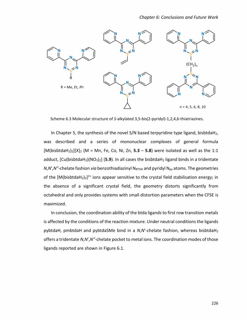
Chapter 6: Conclusions and Future Work
226
In Chapter 5, the synthesis of the novel S/N based terpyridine type ligand, bisbtdaH2,
was described and a series of mononuclear complexes of general formula
[M(bisbtdaH2)2][X]2 (M = Mn, Fe, Co, Ni, Zn, 5.3 – 5.8) were isolated as well as the 1:1
adduct, [Cu(bisbtdaH2)(NO3)2] (5.9). In all cases the bisbtdaH2 ligand binds in a tridentate
N,Nʹ,Nʺ-chelate fashion via benzothiadiazinyl NBTDA and pyridyl Npy atoms. The geometries
of the [M(bisbtdaH2)2]2+ ions appear sensitive to the crystal field stabilisation energy; in
the absence of a significant crystal field, the geometry distorts significantly from
octahedral and only provides systems with small distortion parameters when the CFSE is
maximized.
In conclusion, the coordination ability of the btda ligands to first row transition metals
is affected by the conditions of the reaction mixture. Under neutral conditions the ligands
pybtdaH, pmbtdaH and pybtdaSMe bind in a N,Nʹ-chelate fashion, whereas bisbtdaH2
offers a tridentate N,Nʹ,Nʺ-chelate pocket to metal ions. The coordination modes of those
ligands reported are shown in Figure 6.1.
Scheme 6.3 Molecular structure of S-alkylated 3,5-bis(2-pyridyl)-1,2,4,6-thiatriazines.

Chapter 6: Conclusions and Future Work
227
Under basic conditions, by introducing weak auxiliary co-ligands (OAc– and hfac–), the
oxidation of the ligand to SIV is promorted. The ligand pybtdaHox binds to one metal ion
through the N,Nʹ-chelate whereas pybtdaox– has observed to adopt two coordination
modes; as terminal ligand through the N,Nʹ-chelate and as bridging through the O of the
S=O. The ligand pmbtdaHox acts as a N,Nʹ-chelate ligand whereas the deprotonated form
pmbtdaox– is bridging three metal centres; offering two N,Nʹ binding sites for bridging two
metal centres as well as a pendant O-donor. The S-functionalization on pybtdaSMe did
not lead to further oxidation of sulphur and the ligand binds in a simple N,Nʹ-chelate
fashion. The coordination modes of the ligands under basic conditions are shown in Figure
6.2.
M
pybtdaSMe
bisbtdaH2
M
M
pybtdaH pmbtdaH
M
Figure 6.1 Coordination modes of the ligands pybtdaH, pmbtdaH, pybtdaSMe and
bisbtdaH2 under neutral conditions.
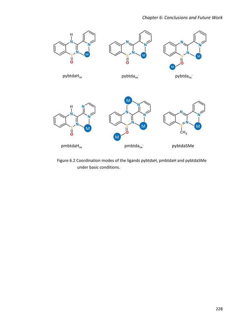
Chapter 6: Conclusions and Future Work
228
M
pybtdaSMe
M
pybtdaHox pybtdaox- pybtdaox
-
M
M
M
M
M
M
pmbtdaHox pmbtdaox-
M
Figure 6.2 Coordination modes of the ligands pybtdaH, pmbtdaH and pybtdaSMe
under basic conditions.

Chapter 6: Conclusions and Future Work
229
Future Work
A large number of metal complexes synthesized in this dissertation incorporate the
SIV system of the pyridyl and pyrimidine derivatives of the 1,2,4-benzothiadiazines. The
metal-assisted oxidation was found to be relatively slow for the pmbtdaH ligand resulting
in lower yields and, in some cases, irreproducible results. The isolation of the S-oxide
ligand prior to coordination would therefore offer a possible solution. The conversion of
the SII system to SIV is conventionally achieved by the use of stoichiometric quantities of
strong oxidants such as peracids, high oxidation state metals (such as MnO2 and CrO3) or
oxidizing non-metal species (such as IO4–
or PhIO).12 Rawson structurally characterized the
first S-oxides of the thiadiazine family through a metal-assisted oxidation, in which the
substituent on C(3) was the non-coordinating phenyl and 2-thienyl rings.13 Preliminary
results, using the same synthetic way afforded the pyridyl derivative of the
benzothiadiazine S-oxide (pybtdaHox, Figure 6.3). More experiments are underway to
optimize the synthetic route with final goal to employ the ligand in coordination chemistry
with first row transition metals. However, treatment with organic mild oxidants such as
Me3NO might also prove to be a direct process to generate pybtdaHox directly. With this
parent ligand in hand it will permit comparative studies with pybtdaH and potentially a
more directed route to some of the less easily accessed compounds isolated during the
course of this dissertation. In particular, structural studies on pmbtdaox− complexes have
so far been scarce due to the apparently much longer incubation time required for aerial
Figure 6.3 Crystal structure of the S-oxide of the pyridyl derivative of
benzothiadiazines (pybtdaHox).

Chapter 6: Conclusions and Future Work
230
oxidation. This ligand has the potential to develop a large family of polynuclear complexes
whose magnetic properties might be of interest.
Given the propensity of the S-oxide ligand to also become involved in bonding, the
S,S- dioxide derivative could potentially offer two binding sites via the SO2 unit in addition
to the N,Nʹ-chelate pocket. A dioxide derivative of 1,10-phenanthroline,
[1,2,5]thiadiadiazolo[3,4-f][1,10]phenanthroline 1,1-dioxide (tdapO2), was introduced by
Awaga, in which the dioxide acts as a chelate pocket in addition to the N,Nʹ-chelation via
phenanthroline bridging two potassium ions (Scheme 6.4).14
Oxidation of SIV to the dioxide SVI system is typically achieved under quite harsh
conditions (35% H2O2 at 100 °C in glacial acetic acid) in moderate yields.15 A series of
1,2,4-benzothiadiazine dioxides have previously been synthesized by condensation of
sulfonamide with heterocyclic methyl carbimidates16 but no crystallographic
characterization was reported. We were able to isolate the dioxide derivative of the
pyridyl benzothiadiazine by reaction of the SII pybtdaH treated with NaOCl (Figure 6.4).
The coordination chemistry of the dioxide would be interesting and it could potentially
lead to polynuclear complexes and/or polymers.
tdapO2 [K][tdapO2]
Scheme 6.4 Molecular structure of tdapO2 and the potassium salt of its radical anion
demonstrating the chelating ability of the ligand.
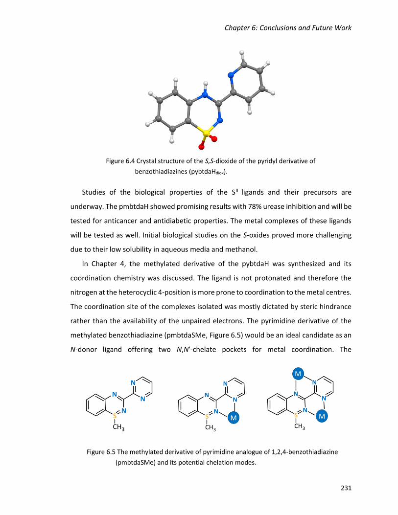
Chapter 6: Conclusions and Future Work
231
Studies of the biological properties of the SII ligands and their precursors are
underway. The pmbtdaH showed promising results with 78% urease inhibition and will be
tested for anticancer and antidiabetic properties. The metal complexes of these ligands
will be tested as well. Initial biological studies on the S-oxides proved more challenging
due to their low solubility in aqueous media and methanol.
In Chapter 4, the methylated derivative of the pybtdaH was synthesized and its
coordination chemistry was discussed. The ligand is not protonated and therefore the
nitrogen at the heterocyclic 4-position is more prone to coordination to the metal centres.
The coordination site of the complexes isolated was mostly dictated by steric hindrance
rather than the availability of the unpaired electrons. The pyrimidine derivative of the
methylated benzothiadiazine (pmbtdaSMe, Figure 6.5) would be an ideal candidate as an
N-donor ligand offering two N,Nʹ-chelate pockets for metal coordination. The
Figure 6.4 Crystal structure of the S,S-dioxide of the pyridyl derivative of
benzothiadiazines (pybtdaHdiox).
Figure 6.5 The methylated derivative of pyrimidine analogue of 1,2,4-benzothiadiazine
(pmbtdaSMe) and its potential chelation modes.
M
M
M

Chapter 6: Conclusions and Future Work
232
pmbtdaSMe ligand would offer similar binding sites as 2,2-bipyrimidine, the employment
of has led to the isolation of polynuclear complexes and metal-organic polymers.17–20
A future avenue of direction in the coordination chemistry explored here is the ability
to further tune the crystal field at the metal ion. For both CoII (d7) and FeII (d6) complexes
there is the potential to adopt either high-spin or low-spin electronic configurations
depending on the magnitude of the crystal field () in relation to the pairing energy PE.
Clearly the N-donor system offered by these btda ligands is of the right order of
magnitude to drive spin crossover (SCO) behaviour since Fe(pybtdaH)2Cl2 is high-spin but
[Fe(bisbtdaH2)2][X]2 is low-spin. Replacement of the chloride ligand by a moderate field
ligand may lead to such SCO phenomenon. Preliminary studies by Mr. Nathan Doupnik in
this group has shown that metathesis of high-spin Fe(pmbtdaH)2Cl2 with NCS– generates
Fe(NCS)2(pmbtdaHox)2 (Figure 6.6) which still appears high-spin across the temperature
range 150 – 300 K. Further studies with yet stronger field ligands should permit some
tunability to a SCO regime. Notably cooperativity in SCO phenomena have been linked to
the existence of intermolecular interactions between the SCO complexes and either
counterions or lattice solvent.21,22 Ligands such as pmbtdaH which show a strong
propensity for hydrogen bonding are therefore well-suited to potentially exhibit
cooperative SCO behaviour.
Fe
N3
N1
N5
N5´
N3´
N1´
Figure 6.6 Molecular structure of Fe(NCS)2(pmbtdaHox)2.

Chapter 6: Conclusions and Future Work
233
Overall there is considerable potential for the study of btda derivatives and their
complexes for their biological and materials properties and the fundamental studies of
ligand reactivity described here will help guide future studies in this area.

Chapter 6: Conclusions and Future Work
234
References
1 E. R. Clark, M. U. Anwar, B. J. Leontowicz, Y. Beldjoudi, J. J. Hayward, W. T. K. Chan,
E. L. Gavey, M. Pilkington, E. Zysman-Colman and J. M. Rawson, Dalton Trans.,
2014, 43, 12996.
2 E. R. Clark, J. J. Hayward, B. J. Leontowicz, M. U. Anwar, M. Pilkington and J. M.
Rawson, Dalton Trans., 2014, 44, 2071.
3 A. Trummal, L. Lipping, I. Kaljurand, I. A. Koppel and I. Leito, J. Phys. Chem. A, 2016,
120, 3663.
4 C. Hansch, A. Leo and R. W. Taft, Chem. Rev., 1991, 91, 165.
5 K. L. M. Harriman, I. A. Kühne, A. A. Leitch, I. Korobkov, R. Clérac, M. Murugesu and
J. L. Brusso, Inorg. Chem., 2016, 55, 5375.
6 K. L. M. Harriman, A. A. Leitch, S. A. Stoian, F. Habib, J. L. Kneebone, S. I. Gorelsky,
I. Korobkov, S. Desgreniers, M. L. Neidig, S. Hill, M. Murugesu and J. L. Brusso,
Dalton Trans., 2015, 44, 10516.
7 N. Yutronkie, I. Kuehne, I. Korobkov, J. L. Brusso and M. Murugesu, Chem.
Commun., 2015, 677.
8 M. Jennings, K. E. Preuss and J. Wu, Chem. Commun., 2006, 341.
9 J. Wu, D. J. MacDonald, R. Clérac, I. R. Jeon, M. Jennings, A. J. Lough, J. Britten, C.
Robertson, P. A. Dube and K. E. Preuss, Inorg. Chem., 2012, 51, 3827.
10 E. Kleisath, N. J. Yutronkie, I. Korobkov, B. M. Gabidullin and J. L. Brusso, New J.
Chem., 2016, 40, 4472.
11 N. J. Yutronkie, P. Tami, S. Singh, E. Kleisath, B. M. Gabidullin, R. Davis and J. L.
Brusso, New J. Chem., 2017, 41, 2268.
12 K. Kaczorowska, Z. Kolarska, K. Mitka and P. Kowalski, Tetrahedron, 2005, 61, 8315.
13 E. R. Clark, J. J. Hayward, B. J. Leontowicz, M. U. Anwar, M. Pilkington and J. M.
Rawson, Dalton Trans., 2014, 44, 2071.
14 Y. Shuku, R. Suizu and K. Awaga, Inorg. Chem., 2011, 50, 11859.
15 G. Kresze, C. Seyfried and A. Trede, Liebigs Ann. Chem., 1968, 715, 223.

Chapter 6: Conclusions and Future Work
235
16 K. Gobis, H. Foks, J. Sławiński, E. Augustynowicz-Kopeć and A. Napiórkowska,
Monatshefte für Chemie - Chem. Mon., 2013, 144, 1197.
17 P. Alborés and E. Rentschler, Inorg. Chem., 2010, 49, 8953.
18 P. O. Adelani, N. D. Cook and P. C. Burns, Cryst. Growth Des., 2014, 14, 5692.
19 I. F. Díaz-Ortega, J. M. Herrera, D. Aravena, E. Ruiz, T. Gupta, G. Rajaraman, H. Nojiri
and E. Colacio, Inorg. Chem., 2018, 57, 6362.
20 P. Alborés and E. Rentschler, Dalton Trans., 2013, 42, 9621.
21 G. Dupouy, M. Marchivie, S. Triki, J. Sala-Pala, J. Y. Salaün, C. J. Gómez-García and
P. Guionneau, Inorg. Chem., 2008, 47, 8921.
22 J. A. Thomas, Dalton Trans., 2011, 40, 12005.

APPENDIX 1: Crystal Data and Structure Refinement for Compounds of Chapter 2
236
APPENDIX 1: Crystal Data and Structure Refinement for
Compounds of Chapter 2

APPENDIX 1: Crystal Data and Structure Refinement for Compounds of Chapter 2
237
2.2
Nʹ-
(2-p
rop
ylth
iop
hen
yl)-
pic
olin
amid
ine

APPENDIX 1: Crystal Data and Structure Refinement for Compounds of Chapter 2
238
pyb
tdaH

APPENDIX 1: Crystal Data and Structure Refinement for Compounds of Chapter 2
239
2.3
a F
eCl 3
(pyb
tdaH
ox)
(CH
3O
H)]
∙CH
3O
H

APPENDIX 1: Crystal Data and Structure Refinement for Compounds of Chapter 2
240
2.3
b F
e 4C
l 4(O
CH
3) 6
(pyb
tda o
x)2
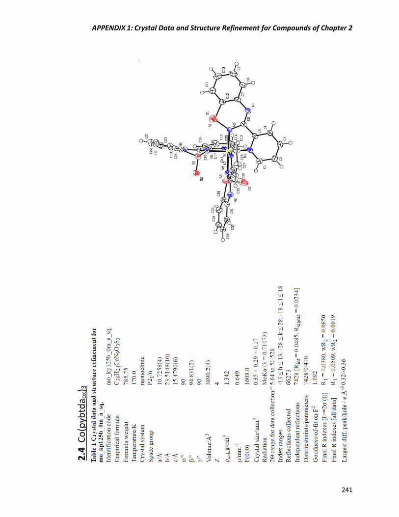
APPENDIX 1: Crystal Data and Structure Refinement for Compounds of Chapter 2
241
2.4
Co
(pyb
tda o
x)3

APPENDIX 1: Crystal Data and Structure Refinement for Compounds of Chapter 2
242
2.5
[C
u2(O
Ac)
2(p
ybtd
a ox)
2(H
2O
) 2]∙
2H
2O

APPENDIX 1: Crystal Data and Structure Refinement for Compounds of Chapter 2
243
2.6
Zn
2(O
Ac)
2(p
ybtd
a ox)
2

APPENDIX 1: Crystal Data and Structure Refinement for Compounds of Chapter 2
244
2.7
Mn
(hfa
c)2(p
ybtd
aHo
x)
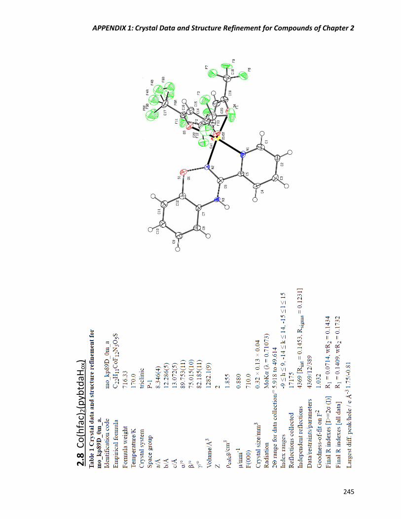
APPENDIX 1: Crystal Data and Structure Refinement for Compounds of Chapter 2
245
2.8
Co
(hfa
c)2(p
ybtd
aHo
x)

APPENDIX 1: Crystal Data and Structure Refinement for Compounds of Chapter 2
246
2.9
Zn
(hfa
c)2(p
ybtd
aHo
x)

APPENDIX 1: Crystal Data and Structure Refinement for Compounds of Chapter 2
247
2.1
0 N
i(h
fac)
2(p
ybtd
aH)
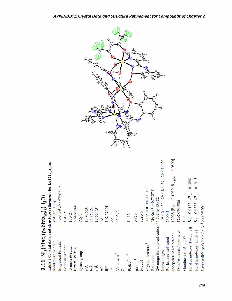
APPENDIX 1: Crystal Data and Structure Refinement for Compounds of Chapter 2
248
2.1
1 N
i 3(h
fac)
(pyb
tda o
x)5(H
2O
)
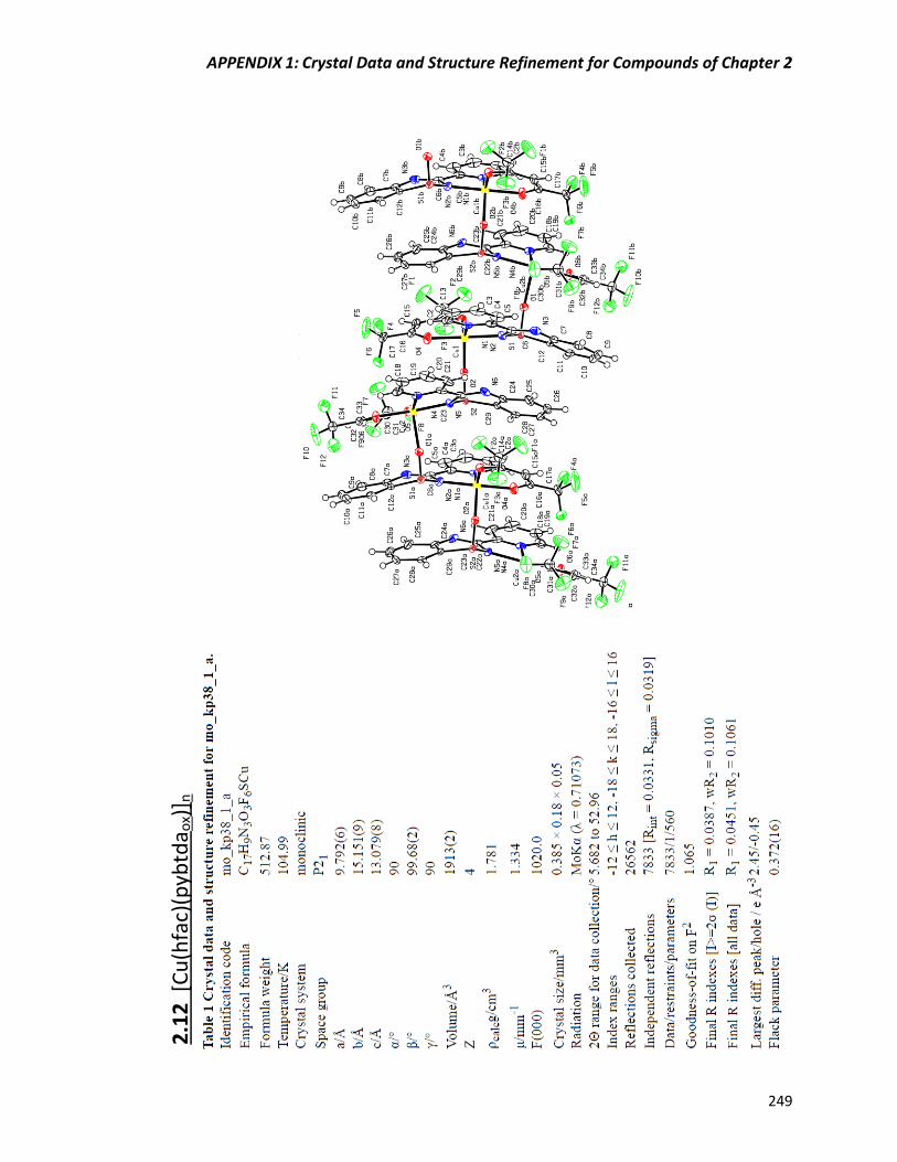
APPENDIX 1: Crystal Data and Structure Refinement for Compounds of Chapter 2
249
2
.12
[C
u(h
fac)
(pyb
tda o
x)] n

APPENDIX 1: Crystal Data and Structure Refinement for Compounds of Chapter 2
250
2.1
3 [
Cu
4(O
H) 4
(pyb
tda o
x)4]∙
H2O

APPENDIX 1: Crystal Data and Structure Refinement for Compounds of Chapter 2
251
2.1
4 [
Cu
14(O
H) 1
2(C
O3) 2
(pyb
tda o
x)1
2(H
2O) 2
]∙1
4[H
2O
]∙[C
H3O
H]

APPENDIX 2: Crystal Data and Structure Refinement for Compounds of Chapter 3
252
APPENDIX 2: Crystal Data and Structure Refinement for
Compounds of Chapter 3
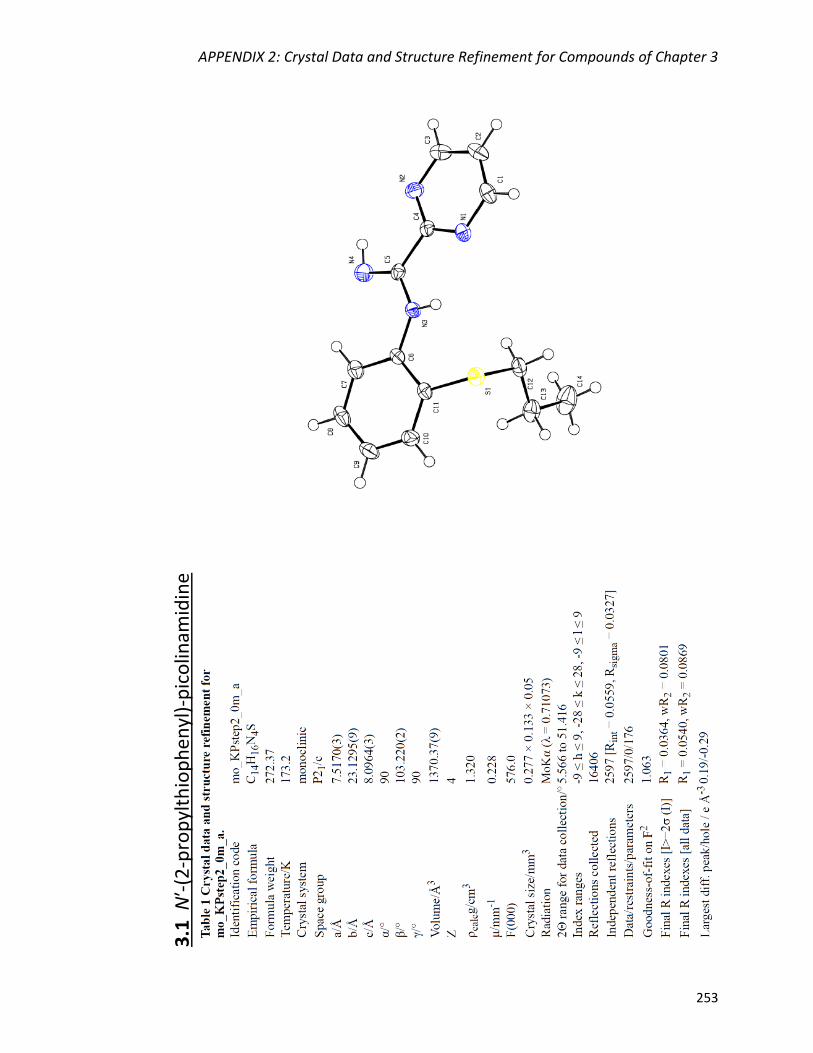
APPENDIX 2: Crystal Data and Structure Refinement for Compounds of Chapter 3
253
3.1
Nʹ-
(2-p
rop
ylth
iop
hen
yl)-
pic
olin
amid
ine
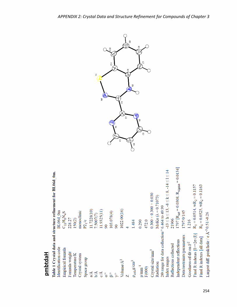
APPENDIX 2: Crystal Data and Structure Refinement for Compounds of Chapter 3
254
pm
btd
aH
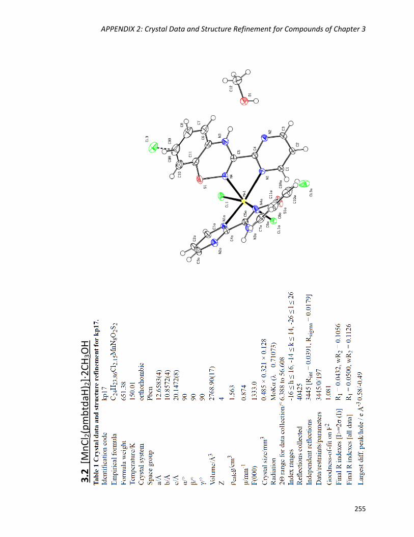
APPENDIX 2: Crystal Data and Structure Refinement for Compounds of Chapter 3
255
3.2
[M
nC
l 2(p
mb
tdaH
) 2]·
2C
H3O
H
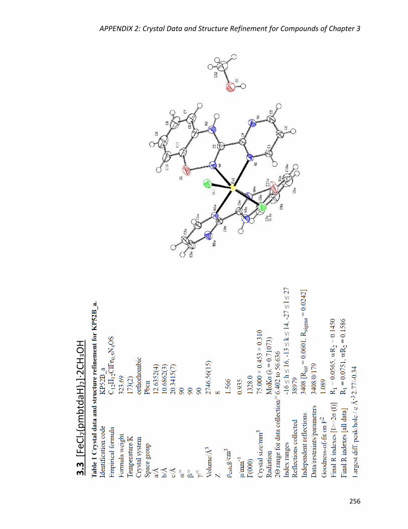
APPENDIX 2: Crystal Data and Structure Refinement for Compounds of Chapter 3
256
3.3
[Fe
Cl 2
(pm
btd
aH) 2
]·2
CH
3O
H

APPENDIX 2: Crystal Data and Structure Refinement for Compounds of Chapter 3
257
3.4
[C
oC
l 2(p
mb
tdaH
) 2]·
2C
H3O
H
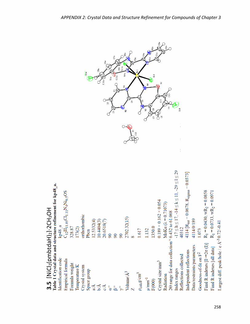
APPENDIX 2: Crystal Data and Structure Refinement for Compounds of Chapter 3
258
3
.5 [
NiC
l 2(p
mb
tdaH
) 2]·
2C
H3O
H

APPENDIX 2: Crystal Data and Structure Refinement for Compounds of Chapter 3
259
3
.6 Z
n2C
l 4(p
mb
tdaH
) 2
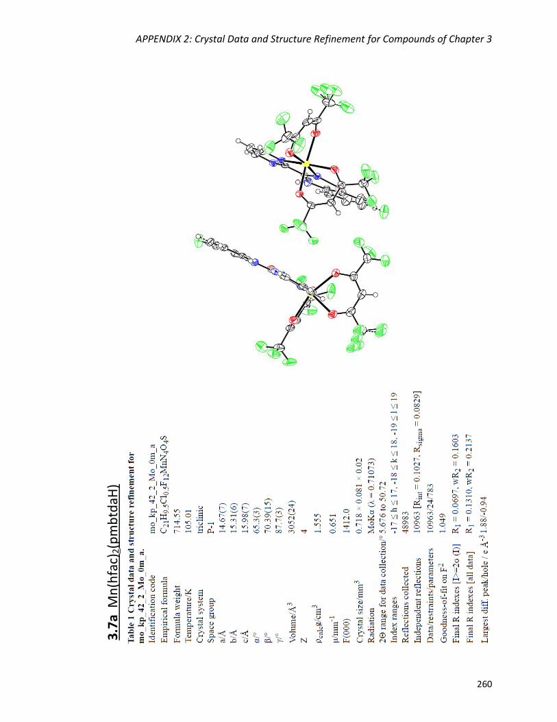
APPENDIX 2: Crystal Data and Structure Refinement for Compounds of Chapter 3
260
3.7
a M
n(h
fac)
2(p
mb
tdaH
)
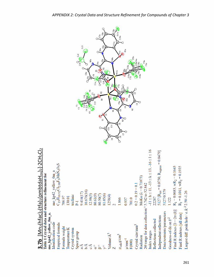
APPENDIX 2: Crystal Data and Structure Refinement for Compounds of Chapter 3
261
3.7
b [
Mn
2(h
fac)
2(t
fa) 2
(pm
btd
aHo
x)2]·
2C
H2C
l 2
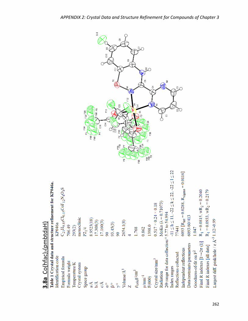
APPENDIX 2: Crystal Data and Structure Refinement for Compounds of Chapter 3
262
3.8
a C
o(h
fac)
2(p
mb
tdaH
)
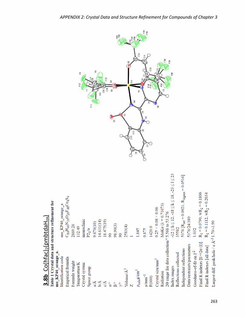
APPENDIX 2: Crystal Data and Structure Refinement for Compounds of Chapter 3
263
3.8
b C
o(h
fac)
2(p
mb
tdaH
ox)

APPENDIX 2: Crystal Data and Structure Refinement for Compounds of Chapter 3
264
3.9
a N
i(h
fac)
2(p
mb
tdaH
)
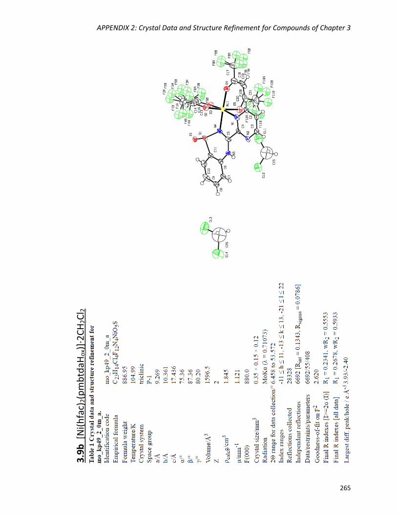
APPENDIX 2: Crystal Data and Structure Refinement for Compounds of Chapter 3
265
3.9
b [
Ni(
hfa
c)2(p
mb
tdaH
ox)
]·2
CH
2C
l 2

APPENDIX 2: Crystal Data and Structure Refinement for Compounds of Chapter 3
266
3.1
0a
Cu
(hfa
c)2(p
mb
tdaH
)

APPENDIX 2: Crystal Data and Structure Refinement for Compounds of Chapter 3
267
3.1
0b
Cu
4(h
fac)
4(t
fa) 2
(pm
btd
a ox)
2

APPENDIX 2: Crystal Data and Structure Refinement for Compounds of Chapter 3
268
3.1
1a
Zn(h
fac)
2(p
mb
tdaH
)

APPENDIX 2: Crystal Data and Structure Refinement for Compounds of Chapter 3
269
3.1
1b
Zn
4(h
fac)
6(p
mb
tda o
x)2

APPENDIX 3: Crystal Data and Structure Refinement for Compounds of Chapter 4
APPENDIX 3: Crystal Data and Structure Refinement for
Compounds of Chapter 4

APPENDIX 3: Crystal Data and Structure Refinement for Compounds of Chapter 4
271
pyb
tdaS
Me
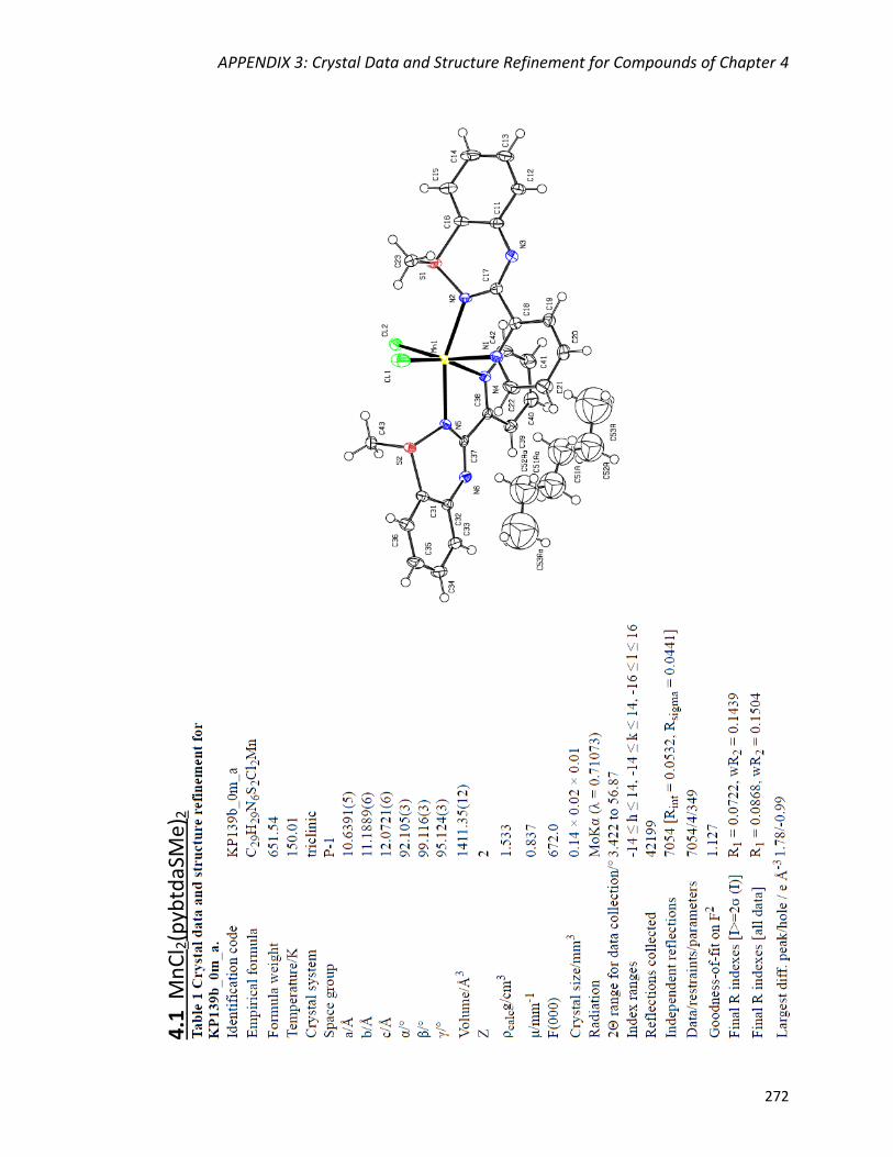
APPENDIX 3: Crystal Data and Structure Refinement for Compounds of Chapter 4
272
4
.1 M
nC
l 2(p
ybtd
aSM
e) 2
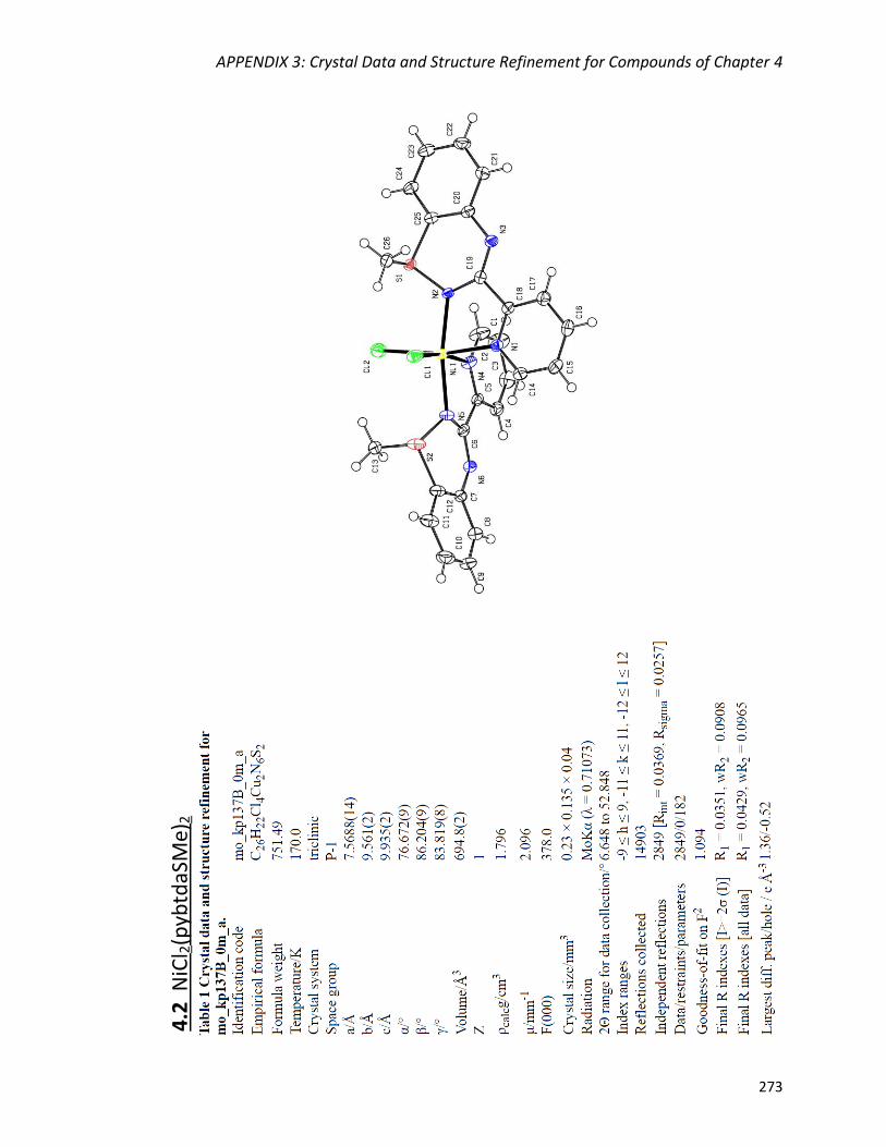
APPENDIX 3: Crystal Data and Structure Refinement for Compounds of Chapter 4
273
4.2
NiC
l 2(p
ybtd
aSM
e)2
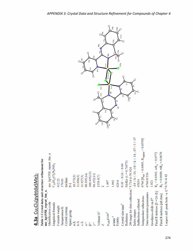
APPENDIX 3: Crystal Data and Structure Refinement for Compounds of Chapter 4
274
4
.3a
Cu
2C
l 4(p
ybtd
aSM
e)2
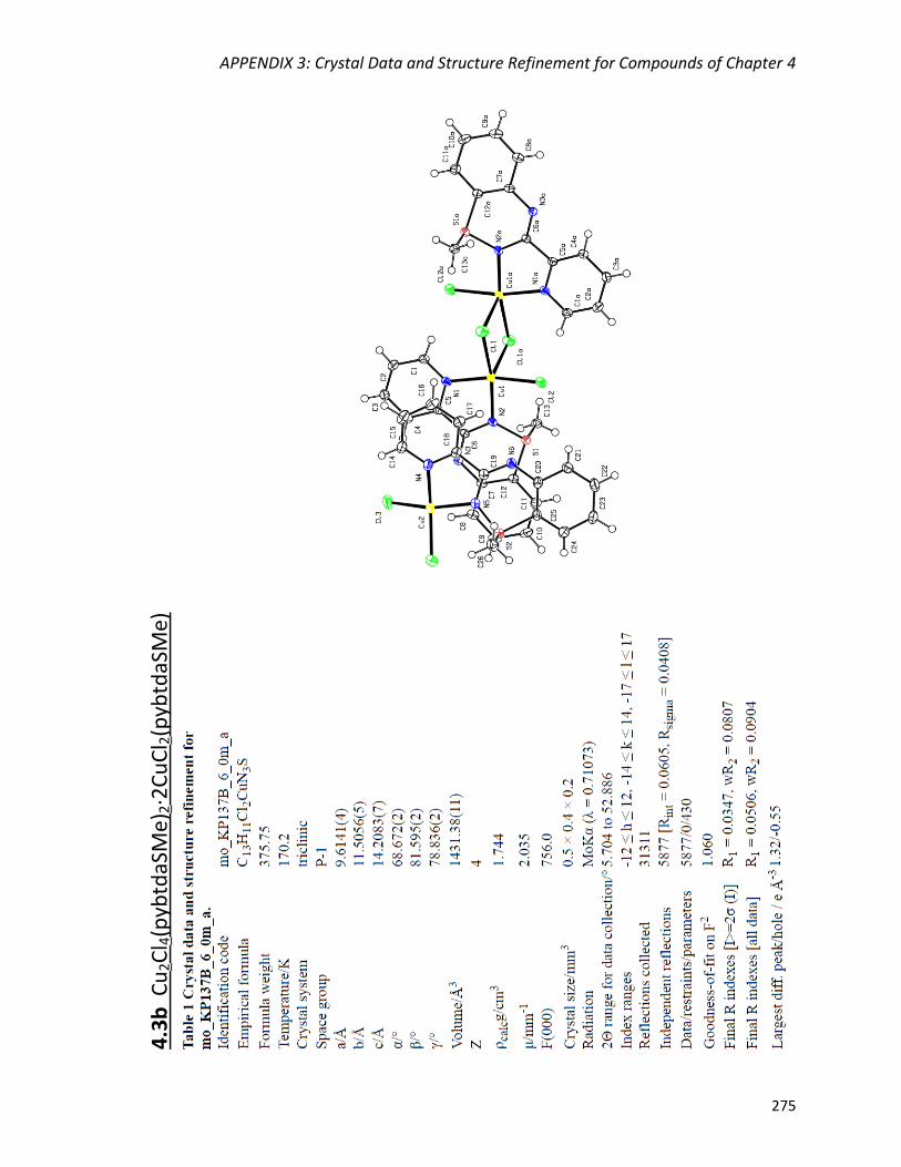
APPENDIX 3: Crystal Data and Structure Refinement for Compounds of Chapter 4
275
4.3
b C
u2C
l 4(p
ybtd
aSM
e)2·2
Cu
Cl 2
(pyb
tdaS
Me)

APPENDIX 3: Crystal Data and Structure Refinement for Compounds of Chapter 4
276
4.4
Zn
Cl 2
(pyb
tdaS
Me)
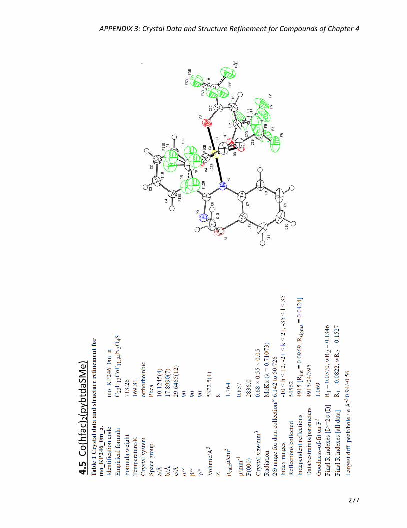
APPENDIX 3: Crystal Data and Structure Refinement for Compounds of Chapter 4
277
4.5
Co
(hfa
c)2(p
ybtd
aSM
e)
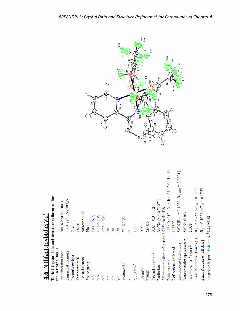
APPENDIX 3: Crystal Data and Structure Refinement for Compounds of Chapter 4
278
4.6
Ni(
hfa
c)2(p
ybtd
aSM
e)

APPENDIX 4: Crystal Data and Structure Refinement for Compounds of Chapter 5
279
APPENDIX 4: Crystal Data and Structure Refinement for
Compounds of Chapter 5
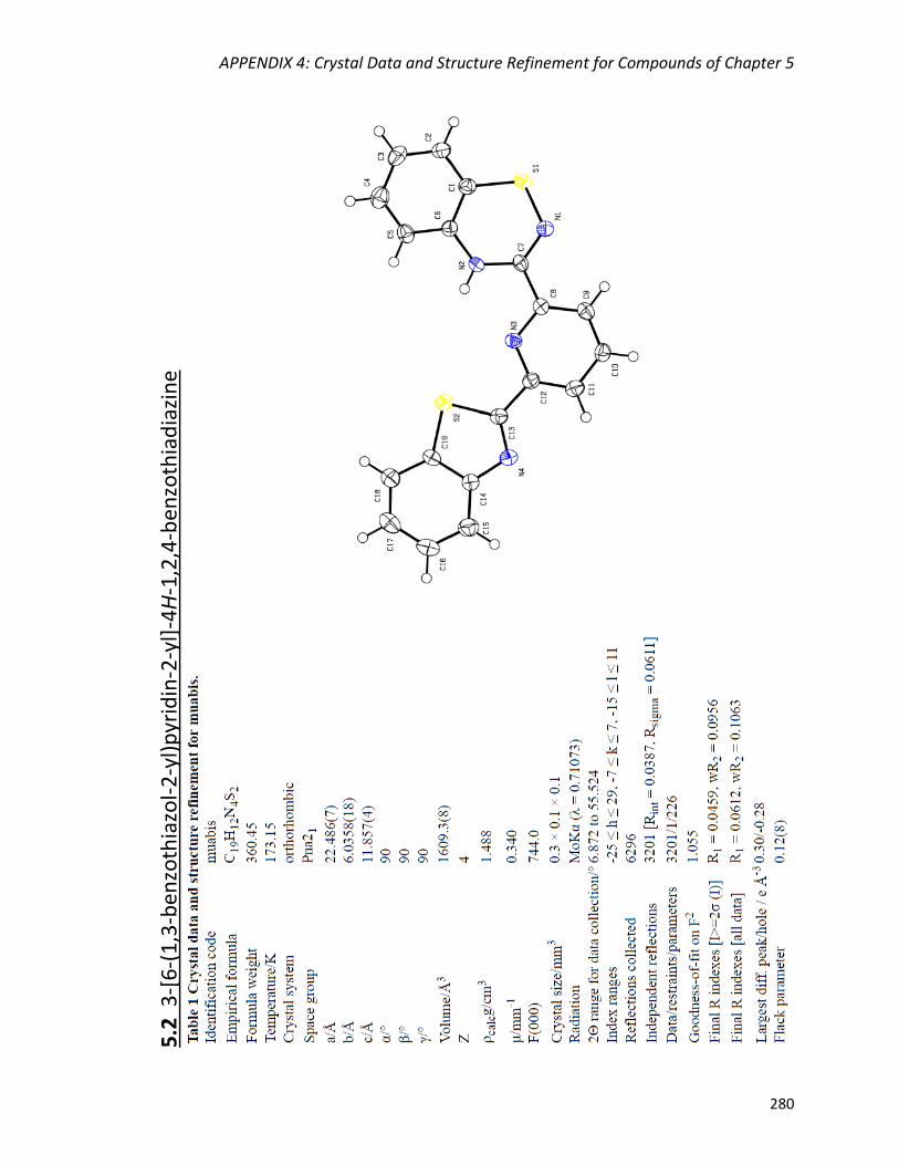
APPENDIX 4: Crystal Data and Structure Refinement for Compounds of Chapter 5
280
5.2
3-[
6-(
1,3
-ben
zoth
iazo
l-2
-yl)
pyr
idin
-2-y
l]-4
H-1
,2,4
-ben
zoth
iad
iazi
ne
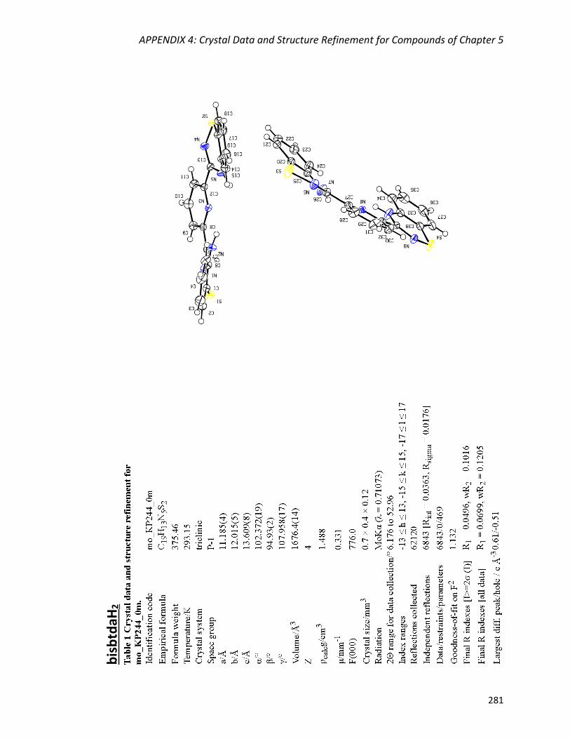
APPENDIX 4: Crystal Data and Structure Refinement for Compounds of Chapter 5
281
b
isb
tdaH
2
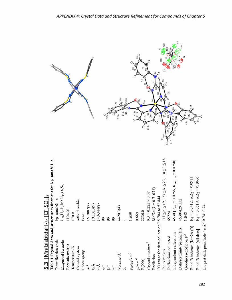
APPENDIX 4: Crystal Data and Structure Refinement for Compounds of Chapter 5
282
5.3
[M
n(b
isb
tdaH
2) 2
][C
F 3SO
3]2
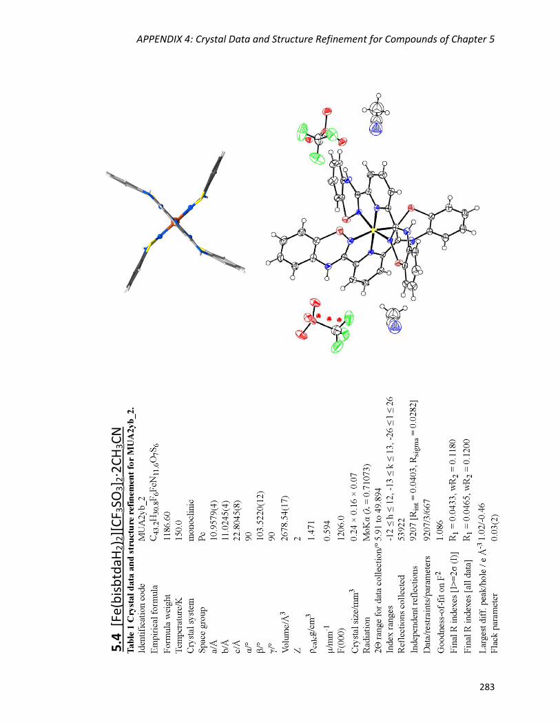
APPENDIX 4: Crystal Data and Structure Refinement for Compounds of Chapter 5
283
5.4
[Fe
(bis
btd
aH2) 2
][C
F 3SO
3] 2
·2C
H3C
N

APPENDIX 4: Crystal Data and Structure Refinement for Compounds of Chapter 5
284
5.5
[Fe
(bis
btd
aH2) 2
][B
F 4] 2
·CH
3C
N

APPENDIX 4: Crystal Data and Structure Refinement for Compounds of Chapter 5
285
5.6
[C
o(b
isb
tdaH
2) 2
][C
l]2

APPENDIX 4: Crystal Data and Structure Refinement for Compounds of Chapter 5
286
5
.7 [
Ni(
bis
btd
aH2) 2
][C
l]2·2
CH
3O
H

APPENDIX 4: Crystal Data and Structure Refinement for Compounds of Chapter 5
287
5.8
[Zn
(bis
btd
aH2) 2
][C
F 3SO
3] 2
·2C
H3O
H
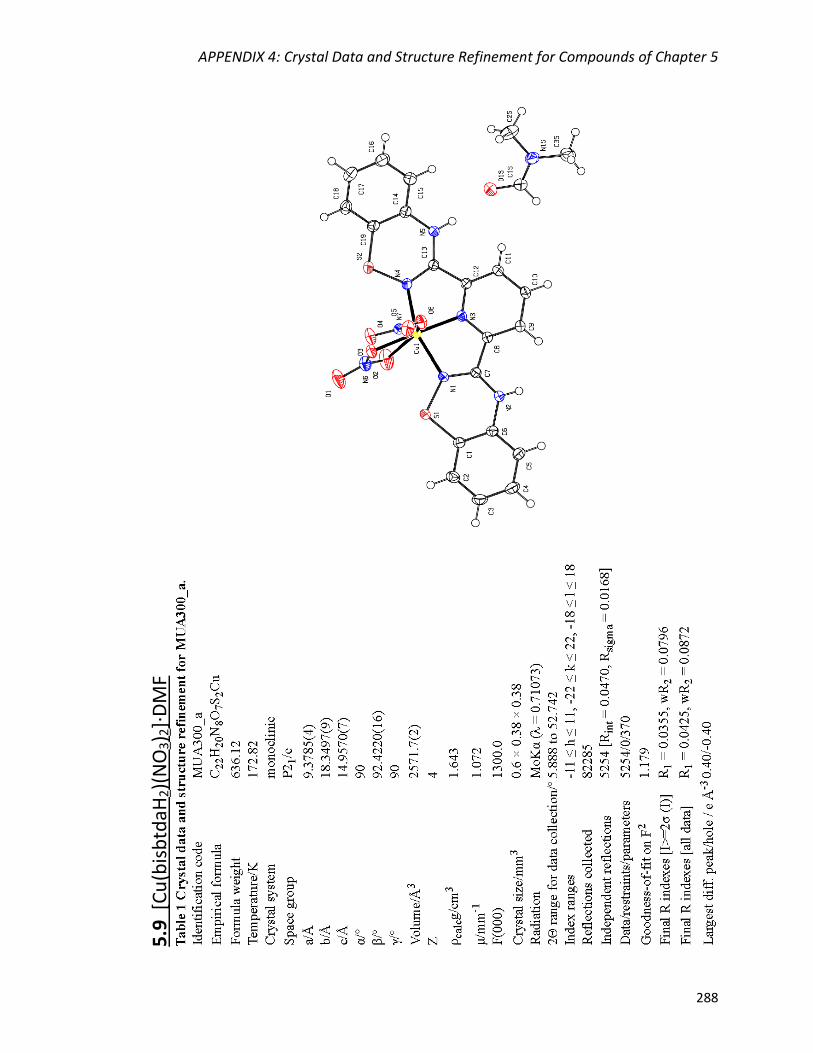
APPENDIX 4: Crystal Data and Structure Refinement for Compounds of Chapter 5
288
5.9
[C
u(b
isb
tdaH
2)(
NO
3) 2
]∙D
MF

Vita Auctoris
289
VITA AUCTORIS
NAME: Konstantina Pringouri
PLACE OF BIRTH: Amaroussio, Attikis, GREECE
EDUCATION:
Ph.D. Chemistry
Department of Chemistry and Biochemistry
University of Windsor
Windsor, ON, CANADA 2018
M.Sc. Chemistry
Department of Chemistry
University of Florida
Gainesville, FL, U.S.A. 2008
B.Sc. Chemistry
Department of Chemistry
University of Patras
Patras, GREECE 2006
AWARDS
Ontario Trillium Scholarship (2014, $40,000 annually)
Travel Award for ACA Summer Course in Chemical Crystallography (2017, $300)
PUBLICATIONS
4. 1. Amlan K. Pal, David B. Cordes, Konstantina Pringouri, Muhammad U. Anwar,
Alexandra M. Z. Slawin, Jeremy M. Rawson, and Eli Zysman-Colman,
“Synthesis and characterization of green-to-yellow emissive Ir(III) complexes of
pyridylbenzothiadiazine ligand”
Journal of Coordination Chemistry, 2016, 11-13, 1924-1937
5.
6. 2. Konstantina Pringouri, Muhammad U. Anwar, Bryce J. Leontowicz, and Jeremy M.
Rawson,
“Zinc complexes of 3-pyrimidinyl-benzo-1,2,4-thiadiazine”
Polyhedron, 2018, 150, 110-117

Vita Auctoris
290
3. Konstantina Pringouri, Muhammad U. Anwar, Liz Mansour, Nathan Doupnik, Yassine
Beldjoudi, Emma L. Gavey, Melanie Pilkington, and Jeremy M. Rawson,
“A novel bis-1,2,4-benzothiadiazine pincer ligand: Synthesis, characterization and
its first row transition metal complexes”
Dalton Trans., Under revision
PRESENTATIONS
1. Konstantina Pringouri, Muhammad U. Anwar, and Jeremy M. Rawson,
“Exploring New Derivatives of 1,2,4-Benzothiadiazines and their Coordination Chemistry”
98th CSC, June 13-17, 2015, Ottawa, Ontario, Canada, Poster Presentation
2. Konstantina Pringouri, Muhammad U. Anwar, and Jeremy M. Rawson,
“Exploring New Derivatives of 1,2,4-Benzothiadiazines and their Coordination Chemistry”
3rd CEMWOQ, May 14-15, 2016, University of Windsor, Windsor, Ontario, Canada, Poster Presentation
3. Konstantina Pringouri, Muhammad U. Anwar, and Jeremy M. Rawson,
“Exploring New Derivatives of 1,2,4-Benzothiadiazines and their Coordination Chemistry”
99th CSC, June 5-9, 2016, Halifax, Nova Scotia, Canada, Poster Presentation
4. Konstantina Pringouri, Muhammad U. Anwar, and Jeremy M. Rawson,
“New Derivatives of 1,2,4-Benzothiadiazines and their Coordination Chemistry”
49th IDW, November 11-13, 2016, Hamilton, Ontario, Canada, Oral Presentation
5. Konstantina Pringouri, Muhammad U. Anwar, and Jeremy M. Rawson,
“New Derivatives of 1,2,4-Benzothiadiazines and their Coordination Chemistry”
4th CEMWOQ, May 26 - 28, 2017, Wilfrid Laurier University, Waterloo, Ontario, Canada, Poster Presentation
6. Konstantina Pringouri, Muhammad U. Anwar, and Jeremy M. Rawson,
“New Derivatives of 1,2,4-Benzothiadiazines and their Coordination Chemistry”
100th CSC, May 28 - June 1, 2017, Toronto, Ontario, Canada, Poster Presentation

Vita Auctoris
291
7. Konstantina Pringouri, Muhammad U. Anwar, and Jeremy M. Rawson,
“A Novel Bis-1,2,4-benzothiadiazine Pincer Ligand: Synthesis, Characterization and Coordination Chemistry”
50th IDW, November 3-5, 2017, Ryerson University, Toronto, Ontario, Canada, Oral Presentation


![Electroactive Benzothiazole Hydrazones and Their [Mo6O19]2− Derivatives: Promising Building Blocks for Conducting Molecular Materials](https://static.fdokumen.com/doc/165x107/634592e6df19c083b1082088/electroactive-benzothiazole-hydrazones-and-their-mo6o192-derivatives-promising.jpg)