Neutron and X-ray scattering experiments on fully deuterated liquid N-methylacetamide CD 3CONDCD 3...
8
Neutron and X-ray scattering experiments on fully deuterated liquid N-methylacetamide CD 3 CONDCD 3 at various temperatures and under pressure Sahbi Trabelsi a , Férid Hammami a , Salah Nasr a, * , Marie-Claire Bellissent-Funel b a Laboratoire Physico-Chimie des Matériaux, Département de Physique, Faculté des Sciences de Monastir, 5019 Monastir, Tunisia b Laboratoire Léon Brillouin (CEA-CNRS) CEA-Saclay, 91191 Gif-sur-Yvette Cedex, France article info Article history: Received 14 February 2008 Received in revised form 3 April 2008 Accepted 7 April 2008 Available online 14 April 2008 Keywords: N-Methylacetamide Hydrogen bond X-ray scattering Neutron scattering abstract A structural investigation of liquid N-methylacetamide (NMA) was performed in the 308–383 K temper- ature range by both neutron and X-ray diffraction and using only neutron diffraction at 308 K and pres- sures of up to 4 kbar. The analysis of experimental data yields the total structure factor, the molecular form factor, and the distinct pair correlation function. The spectra obtained are consistent with the pres- ence of hydrogen-bonded open chain in the liquid state. Surprisingly, our measurements clearly show, in contrast to similar liquids, that the NO and DO lengths increase in liquid NMA with increasing tem- perature, while no significant variation is observed with increasing pressure. Ó 2008 Elsevier B.V. All rights reserved. 1. Introduction The microscopic properties of liquids such as water, alcohols, and amides are essential for a deep understanding of many chemical and biological processes [1]. The crystal structure of NMA determined by conventional X-ray diffraction [2], consists of a linear chain in which two hydrogen bonds (2.825 Å A 0 in length, which is similar to that ob- served in proteins) between CO and NH groups connect each mole- cule to the adjacent ones. Compared to the molecular structure of the gas phase determined by electron diffraction Kitano et al. [3] and Kimura et al. [4], have been suggested that molecules are planar with a fewer differences in the bond lengths. A lengthening of the C– N and a shortening of the C@O intramolecular bonds in the gas phase are observed and could probably account for the effect of intermo- lecular hydrogen bonds. Similar trends are also observed in the case of acetamide [5] and N-methylformamide [6]. Because of the presence of a peptide group, N-methylacetamide (NMA) has been recognized as a useful model compound and has been extensively studied by a variety of experimental and theoret- ical methods [7–12,3]. Numerous experimental NMA studies including Raman and Infrared spectroscopy [13,14] have been done. In a more recent study [15], IR spectra and a quantum cluster equilibrium (QCE) theory are used to describe the equilibrium li- quid properties of NMA. However, none of the earlier studies has experimentally discussed the problem of local order in liquid NMA. So, we have performed a first study [16] where the local or- der of liquid NMA is compared to the short crystal structure [2]. In more recent papers [17,18], X-ray and neutron scattering data were investigated in parallel with DFT previsions to provide a more detailed analysis of local order in liquid NMA. Among two series of dimers and trimers including cis and trans monomers, it has been found that the experimental data are better reproduced by the fol- lowing models: a ring cis trimer and a chainlike trans trimer where each molecule can establish two H-bonds. Hydrogen bonds are known to be highly directional and conse- quently sensitive to both temperature and pressure variation indi- cating that the local order in H-bonded liquids changes in function of these two variables. In the special case of water, it has been shown that the hydrogen bond network can be modified by vary- ing either the temperature or the pressure [19]; a similar effect has been observed in associated liquids such as formamide [20], acetamide [5], formic acid [21], and N-methylformamide [6]. In or- der to get further insight into the local order of liquid N-methylac- etamide, neutron and X-ray scattering are investigated, particularly at non-ambient conditions, to study both the temper- ature and the pressure effects on the hydrogen bond network in this organic compound. To our knowledge, this is the first time that experimental information about this problem is discussed. 2. Experiment details Two samples of liquid N-methylacetamide (NMA) were used in this study. The first one, used in neutron scattering experiments, was a fully deuterated sample (NMA-d 7 ) which was supplied by C/D/N isotopes (Chemical purity 99% +). The second one, used in 0022-2860/$ - see front matter Ó 2008 Elsevier B.V. All rights reserved. doi:10.1016/j.molstruc.2008.04.014 * Corresponding author. E-mail address: [email protected] (S. Nasr). Journal of Molecular Structure 891 (2008) 388–395 Contents lists available at ScienceDirect Journal of Molecular Structure journal homepage: www.elsevier.com/locate/molstruc
Transcript of Neutron and X-ray scattering experiments on fully deuterated liquid N-methylacetamide CD 3CONDCD 3...

Journal of Molecular Structure 891 (2008) 388–395
Contents lists available at ScienceDirect
Journal of Molecular Structure
journal homepage: www.elsevier .com/locate /molstruc
Neutron and X-ray scattering experiments on fully deuterated liquidN-methylacetamide CD3CONDCD3 at various temperatures and under pressure
Sahbi Trabelsi a, Férid Hammami a, Salah Nasr a,*, Marie-Claire Bellissent-Funel b
a Laboratoire Physico-Chimie des Matériaux, Département de Physique, Faculté des Sciences de Monastir, 5019 Monastir, Tunisiab Laboratoire Léon Brillouin (CEA-CNRS) CEA-Saclay, 91191 Gif-sur-Yvette Cedex, France
a r t i c l e i n f o
Article history:Received 14 February 2008Received in revised form 3 April 2008Accepted 7 April 2008Available online 14 April 2008
Keywords:N-MethylacetamideHydrogen bondX-ray scatteringNeutron scattering
0022-2860/$ - see front matter � 2008 Elsevier B.V. Adoi:10.1016/j.molstruc.2008.04.014
* Corresponding author.E-mail address: [email protected] (S. Nasr).
a b s t r a c t
A structural investigation of liquid N-methylacetamide (NMA) was performed in the 308–383 K temper-ature range by both neutron and X-ray diffraction and using only neutron diffraction at 308 K and pres-sures of up to 4 kbar. The analysis of experimental data yields the total structure factor, the molecularform factor, and the distinct pair correlation function. The spectra obtained are consistent with the pres-ence of hydrogen-bonded open chain in the liquid state. Surprisingly, our measurements clearly show, incontrast to similar liquids, that the N� � �O and D� � �O lengths increase in liquid NMA with increasing tem-perature, while no significant variation is observed with increasing pressure.
� 2008 Elsevier B.V. All rights reserved.
1. Introduction
The microscopic properties of liquids such as water, alcohols, andamides are essential for a deep understanding of many chemical andbiological processes [1]. The crystal structure of NMA determined byconventional X-ray diffraction [2], consists of a linear chain in whichtwo hydrogen bonds (2.825 ÅA
0
in length, which is similar to that ob-served in proteins) between CO and NH groups connect each mole-cule to the adjacent ones. Compared to the molecular structure ofthe gas phase determined by electron diffraction Kitano et al. [3]and Kimura et al. [4], have been suggested that molecules are planarwith a fewer differences in the bond lengths. A lengthening of the C–N and a shortening of the C@O intramolecular bonds in the gas phaseare observed and could probably account for the effect of intermo-lecular hydrogen bonds. Similar trends are also observed in the caseof acetamide [5] and N-methylformamide [6].
Because of the presence of a peptide group, N-methylacetamide(NMA) has been recognized as a useful model compound and hasbeen extensively studied by a variety of experimental and theoret-ical methods [7–12,3]. Numerous experimental NMA studiesincluding Raman and Infrared spectroscopy [13,14] have beendone. In a more recent study [15], IR spectra and a quantum clusterequilibrium (QCE) theory are used to describe the equilibrium li-quid properties of NMA. However, none of the earlier studies hasexperimentally discussed the problem of local order in liquidNMA. So, we have performed a first study [16] where the local or-
ll rights reserved.
der of liquid NMA is compared to the short crystal structure [2]. Inmore recent papers [17,18], X-ray and neutron scattering datawere investigated in parallel with DFT previsions to provide a moredetailed analysis of local order in liquid NMA. Among two series ofdimers and trimers including cis and trans monomers, it has beenfound that the experimental data are better reproduced by the fol-lowing models: a ring cis trimer and a chainlike trans trimer whereeach molecule can establish two H-bonds.
Hydrogen bonds are known to be highly directional and conse-quently sensitive to both temperature and pressure variation indi-cating that the local order in H-bonded liquids changes in functionof these two variables. In the special case of water, it has beenshown that the hydrogen bond network can be modified by vary-ing either the temperature or the pressure [19]; a similar effecthas been observed in associated liquids such as formamide [20],acetamide [5], formic acid [21], and N-methylformamide [6]. In or-der to get further insight into the local order of liquid N-methylac-etamide, neutron and X-ray scattering are investigated,particularly at non-ambient conditions, to study both the temper-ature and the pressure effects on the hydrogen bond network inthis organic compound. To our knowledge, this is the first time thatexperimental information about this problem is discussed.
2. Experiment details
Two samples of liquid N-methylacetamide (NMA) were used inthis study. The first one, used in neutron scattering experiments,was a fully deuterated sample (NMA-d7) which was supplied byC/D/N isotopes (Chemical purity 99% +). The second one, used in

S. Trabelsi et al. / Journal of Molecular Structure 891 (2008) 388–395 389
X-ray scattering experiments, was a fully hydrogenated sample. Ilwas purchased from Aldrich Chemical (State purity 99% +). In allthe experiments, NMA was used without further purification.
The neutron scattering experiments were performed at thereactor Orphée of the Laboratoire Léon Brillouin, at Saclay (France)using the 7C2 spectrometer [22]. This spectrometer was equippedwith a BF3 position sensitive detector with 640 cells; the angularstep between two adjacent cells is equal to 0.2�, which leads to amaximum diffraction angle of 128.7 degrees. We selected an inci-dent wavelength of k = 0.711 Å by means of a Cu (111) monochro-mator. The sample was contained inside a null matrix Ti67Zr33
high-pressure cell for which the intensity was principally incoher-ent. The cell had an internal diameter /int = 5.70 mm and a wallthickness e = 5.22 mm. It was placed in a helium cryostat and con-nected to the pressure generator via a capillary; the pressurizingmedium was water and it was isolated from the sample by a Teflonpiston (see Fig. 1). The sample holders were filled and sealed, underhelium gas, in a glove box in order to avoid contamination byatmospheric water. The temperature was measured, with a sensi-bility of 0.1 �C, by two thermocouples, one at each end of the cell.In such a way we verified, during each experiment, that the tem-perature gradient along the cell was less than 0.2 �C. The usualmeasurements of the vanadium rod ðIexpt
v Þ, the cadmium rodðIexpt
cd Þ and the empty cryostat ðIexptvacuumÞ were also performed.
To the pressure generator
Capillary
0. 3 mm I. D.
Ti-Zr Cell
Cu-Be Part
TEFLON Piston
Irra
diat
ed p
art
50 m
m
Fig. 1. Scheme of the Ti–Zr high-pressure cell.
The X-ray scattering measurements were carried out with a(h � 2h) diffractometer operating in the transmission mode withMoKa radiation (k = 0.7093 Å) monochromated by a bent asymmet-ric quartz. The scattered intensity was measured in steps of 0.25�(2h) in the 3�–110� range. The sample was contained in a flat sam-ple holder (4 mm thick) equipped with Mylar windows. Severalscans were accumulated until about 80,000 counts were obtainedat each angle. All the experiments were carried out under atmo-spheric pressure and at various temperatures.
3. Theoretical formalism
3.1. Neutron and X-ray scattering
The basic theory of studying X-ray and neutron scattering frommolecular liquids is well known [23,24] and only the essentials willbe reported here. The liquid structure factor SM(Q) can be writtenin terms of a molecular form factor and a difference function whichcontains all the intermolecular contributions, i.e.
SMðQÞ ¼ F1ðQÞ þ DMðQÞ ð1Þ
where Q is the modulus of the scattering wave vector.The molecular form factor F1(Q) is given by
F1ðQÞ ¼Xm
a;b¼1
aaabJ0ðQrabÞ exp �hDr2abi
Q2
2
" #, Xm
a¼1aa
� �2ð2Þ
where J0(x) = sinx/x is the zero order spherical Bessel func-tion,hDr2
abi1=2 ¼ lab is the root mean-square vibrational amplitude
for the a–b atom pair, and aja is the scattering factor of atom a inmolecule j (for neutrons, aa = hbai is the coherent scattering lengthof the a nucleus after averaging over all isotopes and nuclear spins;for X-rays, aa = fa is the atomic scattering factor). The F1(Q) functionis equivalent to the diffraction pattern that would be observed forindividual molecules in the low-density regime and is solely depen-dent on the molecular conformation and the scattering factor aa. Athigh Q-values, the structure factor is dominated by the oscillatorynature of the F1(Q) contribution so that the molecular conformationcan be established from the shape of the diffraction pattern in thisregion.
The distinct structure factor DM(Q) which contains the informa-tion on the intermolecular structure of the liquid usually decays tozero very rapidly at large Q-values. However, for hydrogen-bondedstructures, this function continues to oscillate in the high Q-range;therefore, it can be split into two parts [25]:
DMðQÞ ¼ DHBM ðQÞ þ DNHB
M ðQÞ ð3Þ
where DNHBM contains the intermolecular correlations other than the
H-bonded interactions, and
DHBM ðQÞ ¼ 2n aNaO
sinðQrNOÞQrNO
exp �l2NO
Q 2
2
" #(
þaDaOsinðQrHOÞ
QrHOexp �l2
HOQ 2
2
" #), Xm
a¼1aa
� �2ð4Þ
represents the H-bonds contribution of a given NMA moleculelinked to n other ones. The pair correlation function of only inter-molecular terms, gL(r), may be expressed by
gLðrÞ ¼ 1þ 12p2qr
Z 1
0QDMðQÞ sinðQrÞdQ ð5Þ
this function gives the probability of finding another atom lying inanother molecule at a distance r from a given atom. By analogy toEq. (3), gL(r) may be written as:
gLðrÞ ¼ gHBL ðrÞ þ gNHB
L ðrÞ ð6Þ
390 S. Trabelsi et al. / Journal of Molecular Structure 891 (2008) 388–395
where gHBL ðrÞ and gNHB
L ðrÞ are, respectively, the Fourier transforma-tions of DHB
M ðQÞ and DNHBM ðQÞ. We should use the weighted function,
given by
dLðrÞ ¼ 4pqr½gLðrÞ � 1� ð7Þ
3.2. Difference technique
Hydrogen-bonded liquids are dependent on the thermodynamicparameters such as temperature and pressure [19]. The investiga-tion of these effects is essentially based on the first-order differ-ence method developed by Dore [26]. We report here only theessential of this method in the case of temperature study. A similarformalism can be easily developed for a pressure effect [20].
The first-order difference function DSM(Q,DT) which representsthe modification of the structure with temperature may be writtenas
DSMðQ ;DTÞ ¼ SMðQ ; TÞ � SMðQ ; T0Þ ð8Þ
where SM(Q,T0) is the structure factor which is considered as knownat a reference temperature T0. By admitting that the molecular con-formation is conserved in the DT range, the DSM(Q,DT) function canbe simplified as
DSMðQ ;DTÞ ¼ DDMðQ ;DTÞ ð9Þ
In the real space representation, the structural changes while vary-ing temperature can be given when the DdL(r,DT) function is evalu-ated, since, for strongly hydrogen-bonded liquids, we have
DdLðr;DTÞ � 4prqDgLðrÞ ð10Þ
According to Eq. (4) and the DgL(r) function, it is easy to deduce thecontribution of the hydrogen-bond interactions at the temperatureT:
gHBL ðr; TÞ ¼ DgLðr;DTÞ þ gHB
L ðr; T0Þ ð11Þ
where gHBL ðr; T0Þ represents the H-bond contribution, considered as
known, at a reference temperature T0.The advantage of this formalism is that the changes in structure
can be evaluated without the need for a precise knowledge of thevarious experimental and analytic correction factors. In fact, thesefactors will be identical for both measurements and the final re-sults will be much less sensitive to the data treatment.
4. Data treatment
4.1. X-ray scattering
Many papers have been devoted to methods of carrying out thetreatment which must be applied to the experimental intensity[27]. We restrict ourselves to the main corrections correspondingto the case of an incident monochromatic beam. So, the measuredintensity, Imeas, is corrected according to the relation [21]
IcorrðHÞ ¼ ðImeasðHÞBðHÞÞPðHÞ�1AðHÞ�1GðHÞ�1 ð12Þ
where B(H) is the background correction which is given by the scat-tering of the empty cell, reduced by the transmittance of the liquid,P(H) is the polarization factor which is calculated from the expres-sion [28]
PðHÞ ¼ 1þ cos2 2H0 cos2 2H1þ cos2 2H0
ð13Þ
valid in the case where a crystal monochromatic with Bragg angleH0 is introduced into the beam. A(H) And G(H) are, respectively,the absorption correction and the effective irradiated volume; thesecorrections are also discussed in Ref. [23]. The correction of multiplescattering can be neglected by choosing a sample of relatively small
thickness. The corrected intensity must be transformed from arbi-trary to electron units so that
IðHÞ ¼ aIcorrðHÞ � IincohðHÞ ð14Þ
where the normalisation factor a is determined by the Krogh-Moemethod [29] and may be refined as mentioned below. Iincoh(H) isthe incoherent scattering [30].
The total experimental structure factor is given by the relation
SMðQÞ ¼IðQÞPm
a¼1faðQÞ� �2 ð15Þ
where fa(Q) (a = H, C, N, and O) is the tabulated atomic scatteringfactor [31].
4.2. Neutron scattering
Many terms contribute to the measured intensity (background,single scattering from the cell, single scattering from the sample,and multiple scattering either in the cell alone or both). The differ-ential scattering cross section of the sample a is given by [32].
drdX
� �a
¼ drdX
� �V
qV
IcorrV
ISaPa
Aaqa
ð16Þ
where (dr/dX)V is the differential incoherent scattering cross sectionof vanadium; Icorr
V is obtained from ðIexptv Þ after corrections of multiple
scattering, absorption and inelastic scattering according to the stan-dard procedure [32]; IS
a is the experimental intensity of the sample,corrected from the background, cell and multiple scattering contri-butions; qa is the attenuation factor for the neutrons scattered fromthe sample and attenuated by the cell and the sample itself; qV andqa are the number densities of vanadium and sample, respectively,and Pa(Q) is the factor which takes into account the inelasticity ef-fects. The inelasticity correction has been performed using a polyno-mial procedure described elsewhere [32]; the distinct structurefactor [see Eq. (3)] may be expressed by the relation
DMðQÞ ¼1K IcorrðQÞ � A� BQ2 � CQ 4 þ 7b2
D þ 3b2C þ b2
N þ b2OPm
a¼1ba
� �2 � F1ðQ effÞ
ð17Þ
where Icorr is the intensity corrected for the container scattering,sample attenuation and multiple scattering, K is a normalisationfactor and Qeff is the actual momentum transfer modulus which isgiven by Bellissent-Funel et al. [24].
Qeff
Q
� �2
¼ 1þ C2EðHÞ � 2CEðHÞ cosð2HÞ
2ð1� cosð2HÞÞ ð18Þ
with
CEðHÞ ¼mn
Mreff þmn
� �cosð2HÞ þ Mr
eff
mn� sin2ð2HÞ
� �1=2( )
ð19Þ
where Mreff is a recoil effective mass close to the mass of the mole-
cule for liquids or gases and mn is the neutron mass.The first step in the data reduction is to calculate the contribu-
tion of the intramolecular correlations. Since the DM(Q) functiongenerally decays to zero much faster than F1(Q), SM(Q) � F1(Q) atlarge values of Q. Therefore, the intramolecular contribution canbe deduced by a least-square fitting of Eq. (2) to the large Q-portionof the experimental structure functionSM(Q). In order to take intoaccount the hydrogen-bond contribution, one can adjust theD� � �O, N� � �O distances, their associated r.m.s. vibrational ampli-tudes as well as the number n of H-bonds per molecule. This couldbe achieved by adjusting the analytical expression of DHB
M ðQÞ to theexperimental intermolecular structure factor DM(Q) in the high Q-range. However, for hydrogen-bonded liquids, the intermolecular

S. Trabelsi et al. / Journal of Molecular Structure 891 (2008) 388–395 391
contribution can be observed at high Q-values [4], so it is difficultto define the low limit of Q from which DNHB
M ðQÞ [see Eq. (3)] tendsto zero. Because no intermolecular contribution, except H-bondinteractions, is expected in the gL(r) function below 1.8 Å, it is con-venient to reconsider the previous adjustment by minimizing, inthe real space, the oscillations of DNHB
M ðQÞ Fourier transform inthe low r-range. In this way, the fitted parameters are more signif-icant. This procedure allows us to submit to refinement the nor-malisation factor K, the D.W. (Debye-Waller) parameters (lab),and the A, B, C coefficients as well as the Meff factor.
Table 1Some of the intramolecular (rab) distances of liquid N-methylacetamide
5. Results and discussion
5.1. Room temperature and atmospheric pressure results
In this section, we give a review of the essential results of ourprevious investigations related to the liquid N-methylacetamide
a
b
c
Q (Å-1 )
r (Å)
SM(Q)
F1(Q)
0.0
0.3
0.6
0.9
0 2 4 6 8 10
0.0
0.3
0.6
0.9
1.2
0.05
0.10
0.15
0 4 8 12 16
)(rg L
1)( −rg HBL
)(rg L
1)( −rg HBL
N···O
N···O
D···O
H···O
Fig. 2. The total structure function SM(Q) (solid line) of liquid NMA as deduced at308 K from neutron scattering, compared to the computed molecular form factorF1(Q) (dashed line) using the parameters listed in Table 1 (a). The X-ray and neutronintermolecular pair correlation functions gL(r) (solid line) are compared to the co-ntributions, ðgHB
L � 1Þ (dashed line) of H-bond interactions (b and c, respectively)inside a chainlike trans trimer.
(NMA) structure at ambient conditions [16,17]. These results areused here as a reference to study the temperature and the pressureeffects on the hydrogen-bond network in liquid NMA.
Fig. 2a shows the total structure function SM(Q) (solid line) asdeduced from neutron scattering measurements in liquid N-methylacetamide (NMA-d7) at ambient temperature and atmo-spheric pressure [17]. The molecular structure function F1(Q)(dashed line) is calculated from Eq. (2) after the fitting proceduredescribed above. One can see a good agreement between theexperimental structure function and the computed molecular formfactor at high Q-range. The intramolecular parameters deducedfrom neutron and X-ray data are listed in Table 1. It is worth notic-
C1–C2 C2@O N–C3 N–C2 C3–D1 C3–D2 C3–D3
rab (Å) 1.521 1.221 1.364 1.454 1.089 1.089 1.093lab (Å) 0.320a 0.240a 0.001a 0.090a 0.050 0.040 0.040
C1� � �D1 D5–D6 D5-D7 N–D4 N� � �D1 N� � �D2 D4� � �D7
rab (Å) 2.048 1.768 1.777 1.005 2.084 2.095 2.326lab (Å) 0.200 0.250 0.250 0.019 0.190 0.180 0.250
Some of the intramolecular (rab) distances of liquid N-methylacetamide. The Debye-Waller parameters (lab) are obtained by least squares fitting using neutron and X-ray scattering data at 308 K and atmospheric pressure.
a Values deduced from X-ray scattering.
0 4 8 12 160.00
0.05
0.10
0.15
S M(Q
)
Q( Å -1 )
N
N
N
C
C
C
C
C
C
C
C
C
O
O
O
D
D
Fig. 3. The total structure factor SM(Q) (solid line) of liquid NMA as deduced at308 K from neutron scattering, compared to the computed one (dashed line) using achain of three trans conformers, similar to that found in the crystal.
D⋅⋅⋅O
392 S. Trabelsi et al. / Journal of Molecular Structure 891 (2008) 388–395
ing that the intramolecular distances are kept fixed and equal tothe crystal ones, only the D.W. parameters are submitted to refine-ment [2]. The X–X (X = N, C, and O) parameters are deduced fromX-ray scattering data whereas the X–H (X = N, C, and O) and H–Hones are based on neutron scattering measurements. The residualdiscrepancy between the experimental structure functionSM(Q)and the molecular form factor F1(Q) disappears if one takes into ac-count the contribution of the hydrogen bonds that is by fittingðF1ðQÞ þ DHB
M ðQÞÞ to the high Q-part of the experimental data andreducing the spurious oscillations of gL(r) function at low r-range[17,18]. In this calculation, the N� � �O, D� � �O distances and the cor-responding DW parameters are fitted in order to have a good con-cordance between experimental and calculated curves in the caseof both neutron and X-ray scattering. The number n of H-bondsper monomer is fixed equal to that existing in the crystalline state,i.e. n = 2 [2].
In Fig. 2b and c, the Fourier inversion of, respectively, the neutronand X-ray intermolecular structure factors DM(Q), i.e. gL(r) (solid line)are compared to the contribution, ðgHB
L ðrÞ � 1Þ (dotted line) of H-bond interactions in liquid NMA. The pronounced peak in gL(r) at1.8 Å (see Fig. 2c) is assigned to the D� � �O interaction, whereas theweak hump at about 3 Å is attributed to the N� � �O interaction, whichis more manifest in the X-ray results (see Fig. 2b). It is interesting tounderline here the complementarity between structural informa-tion that can be extracted from X-ray and neutron scattering. Fig. 3shows that the computed structure factor as deduced from a crystalchain largely reproduces the neutron experimental one.
5.2. Temperature effect at ambient pressure
The experimental structure factors of liquid N-methylacetamideas deduced from neutron scattering at 308, 333, 358, and 383 K areplotted in Fig. 4. One can see a great similarity between the SM(Q)
0 4 8 12 16
S M(Q
)
T = 383 K
T = 358 K
T = 333 K
T = 308 K
Q ( Å -1)
Fig. 4. The neutron structure factors SM(Q) of liquid NMA at 308, 333, 358, and383 K and under atmospheric pressure.
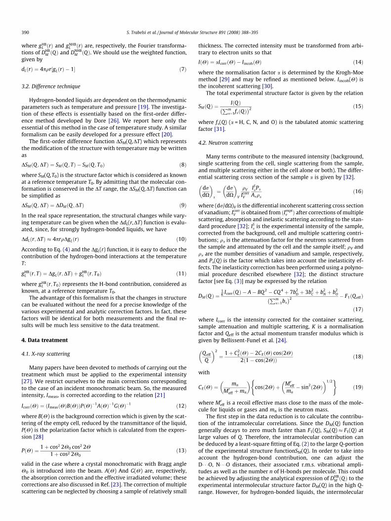
curves particularly at high Q-values, which indicates that there isno variation in the intramolecular structure when varying temper-ature. Using the first-order difference technique described above,we have deduced the H-bond contribution at different tempera-tures. In Fig. 5, we compare the contribution of H-bond interactionðgHB
L ðr; TÞ � 1Þ at 333, 358, and 383 K to the ðgHBL ðr; TÞ � 1Þ (lower
curve) determined at 308 K, 1 bar and which is considered as a ref-erence. One can see a great similarity between the curves: the twocharacteristic peaks of hydrogen-bond interactions (D� � �O andN� � �O) are at the same position, but the heights decrease slowlywith increasing temperature, which is due to a normal increasein the DW values with temperature.
X-ray scattering measurements, performed on liquid NMA inthe same temperature range, give more significant results. The X-ray weighted functions dL(r) of NMA as deduced at 308, 333, 358,and 383 K are plotted in Fig. 6. The signature of the H-bond inter-action is attested here by the N� � �O contribution at about 3 Å,which is more important than H� � �O interaction. The pronouncedpeak at about 4 Å and the shoulders at higher r-values are due tothe non-hydrogen-bonded interactions between molecules. Thecurves also show that the N� � �O peak moves toward higher r-val-ues with increasing temperature. In Fig. 7, the N� � �O distance is dis-played as a function of temperature. One can see that the rN� � �Ovalue decays from 3.05 Å at 383 K to 2.85 Å at ambient tempera-ture (308 K). A Similar effect is assigned by Ludwig et al. [33] inthe case of liquid N-methylformamide (NMF, C2D5NO). In fact, a
0 2 4 6
T = 383K
T = 358K
T = 333K
T = 308K
r (Å)
gHB
L(r
, T )
-1
N⋅⋅⋅O
Fig. 5. The neutron contribution ðgHBL ðr; TÞ � 1Þ of hydrogen-bond interactions in
liquid NMA at 333, 358, and 383 K and under atmospheric pressure as deducedfrom the DgL(r,DT) functions [see Eq. (11)] relative to a reference temperature equalto 308 K (lower curve). The D� � �O and N� � �O hydrogen bond interactions are clearlyhighlighted.

0 2 4 6 8 10
T = 383K
T = 358K
T = 333K
dL(
r )
T = 308K
r ( Å )
N⋅⋅⋅O
Fig. 6. The dL(r) pair correlation function (see text) derived from X-ray scatteringmeasurements at 308, 333, 358, and 383 K. The first peak at about 3 Å representsthe signature of N� � �O hydrogen bond in liquid NMA.
300 330 360 390
2.85
2.90
2.95
3.00
3.05
T (K)
r N...
O(Å
)
Fig. 7. The hydrogen-bond length rN� � �O variation versus temperature as deducedfrom X-ray scattering measurements.
S. Trabelsi et al. / Journal of Molecular Structure 891 (2008) 388–395 393
theoretical investigation in most abundant clusters in liquid NMFshows that the calculated hydrogen-bond distances, as a functionof temperature, increase from 2.80 Å at 250 K up to 2.85 Å at455 K. However, the change of about 0.05 Å with temperature is,as the author signals, still within the range of current experimentaluncertainty. Otherwise, our recently detailed experimental investi-gations on liquid NMF [6], using X-ray and neutron scattering showthat the rN� � �O value decays from 3.01 Å at ambient temperature to2.85 Å at 398 K. A similar phenomenon was observed in the case ofliquid formamide [20], formic acid [21] acetamide [5], and water[19] and the more probable explanation is that, at high tempera-ture, the H-bonds are bent but as the temperature decreases, theN, H, and O atoms line up. Then, it seems that the H-bond networkbehaviour in liquid NMA has an opposite trend to that observed insimilar liquids since an increase in temperature induces an in-crease in H-bond length. It would be so interesting to perform the-oretical investigations such as molecular dynamic simulation atnon-ambient conditions to compare theoretical results to experi-mental ones.
5.3. Pressure effect
The structure factors SM(Q) of liquid NMA derived from neutronscattering measurements at 308 K and P = 1 bar, 1 kbar, 2 kbar,3 kbar, and 4 kbar are plotted in Fig. 8. We can notice the high levelof likeness between curves. At high Q, the curves are well superim-posed which demonstrates that there is no variation in the intra-molecular structure when varying the pressure from 1 bar to
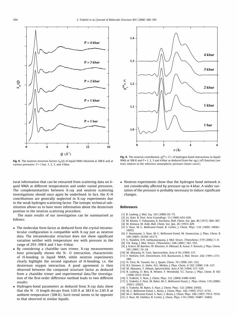
4 kbar. The slight differences at the level of the main peak inSM(Q) are probably due to the density variation that affects onlythe distances between adjacent chains. In Fig. 9, we have repre-sented the hydrogen-bond contribution ðgHB
L ðr; PÞÞ as deduced byanalogy to Eq. (11) from the derivative pressure functionDgL(r,P � Pref) which is given at constant temperature (T = 308 K)by the relation:
DgLðr; P � Pref Þ ¼ DgLðr;DPÞ
¼ 1þ 12p2qr
Z 1
0qDDMðQ ;DPÞ sinðQrÞdQ ð20Þ
with P being, respectively, equal to 1, 2, 3 and 4 kbar and Pref = 1 bar.The contribution of H-bond interactions at 308 K and atmosphericpressure, i.e. ðgHB
L ðr; T0ÞÞ, determined above is also given for compar-ison (lower curve). The effect of pressure is clearly subtle in thesense that it does not substantially change the hydrogen-bond net-work in the pressure range 0–4 kbar. This situation may be com-pared to that observed in liquid N-methylformamide [6] andformamide [20]. In fact, the H-bonds along chain direction becomestronger in the crystalline state of formamide when pressure up to50 kbar is applied. However, unfortunately until now there has beenno similar study on crystal N-methylacetamide.
6. Conclusion
In this study, we have presented a structural investigation of li-quid NMA by means of X-ray and neutron scattering at ambientand non-ambient conditions. Some of the results presented toour knowledge for the first time, may be regarded as the first struc-
0 4 8 12 16
S M(Q
)
P = 4 kbar
P = 3 kbar
P = 2 kbar
P = 1 kbar
P = 1 bar
Q (Å-1)
Fig. 8. The neutron structure factors SM(Q) of liquid NMA obtained at 308 K and atvarious pressures: P = 1 bar, 1, 2, 3, and 4 kbar.
0 2 4 6
1.0
1.1
1.2
1.3
1.4
4 kbar
3 kbar
2 kbar
1 kbar
1 bar
r (Å)
gHB
L(r
, P)
Fig. 9. The neutron contribution ðgHBL ðr; PÞÞ of hydrogen-bond interactions in liquid
NMA at 308 K and P = 1, 2, 3 and 4 kbar as deduced from the DgL(r,DP) function (seetext) relative to the reference atmospheric pressure (lower curve).
394 S. Trabelsi et al. / Journal of Molecular Structure 891 (2008) 388–395
tural information that can be extracted from scattering data on li-quid NMA at different temperatures and under varied pressures.The complementarities between X-ray and neutron scatteringinvestigations should once again be underlined. In fact, the X–Hcontributions are generally neglected in X-ray experiments dueto the weak hydrogen scattering factor. The isotopic technical sub-stitution allows us to have more information about the deuteriumposition in the neutron scattering procedure.
The main results of our investigation can be summarised asfollows:
� The molecular form factor as deduced form the crystal intramo-lecular configuration is compatible with X-ray just as neutrondata. The intramolecular structure does not show significantvariation neither with temperature nor with pressure in therange of 293–398 K and 1 bar–4 kbar.
� By considering a chainlike tans trimer, X-ray measurementshave principally shown the N� � �O interaction, characteristicof H-bonding in liquid NMA, while neutron experimentsclearly highlight the second signature of H-bonding, i.e. thedeuterium oxygen interaction. A good agreement is thenobserved between the computed structure factor as deducedfrom a chainlike trimer and experimental data.The investiga-tion of the first-order difference method leads to two differentresults:
� Hydrogen-bond parameters as deduced from X-ray data showthat the N� � �O length decays from 3.05 Å at 383 K to 2.85 Å atambient temperature (308 K). Such trend seems to be oppositeto that observed in similar liquids.
� Neutron experiments show that the hydrogen bond network isnot considerably affected by pressure up to 4 kbar. A wider var-iation of the pressure is probably necessary to induce significantchanges.
References
[1] R. Ludwig, J. Mol. Liq. 163 (2000) 65–75.[2] J.L. Katz, B. Post, Acta Crystallogr. 13 (1960) 624–628.[3] M. Kitano, T. Fukuyama, K. Kuchitsu, Bull. Chem. Soc. Jpn. 46 (1973) 384–387.[4] M. Kimura, M. Aoki, Bull. Chem. Soc. Jpn. 26 (1953) 429.[5] S. Nasr, M.-C. Bellissent-Funel, R. Cortès, J. Chem. Phys. 110 (1999) 10945–
10952.[6] F. Hammami, S. Nasr, M.-C. Bellissent-Funel, M. Oumezzine, J. Phys. Chem. B
109 (2005) 16169–16175.[7] G. Nandini, D.N. Sathyanarayana, J. Mol. Struct. (Theochem.) 579 (2002) 1–9.[8] Y.K. Kang, J. Mol. Struct. (Theochem.) 546 (2001) 183–193.[9] J. Eckert, M. Barthes, W. Klooster, A. Albinati, R. Aznar, T. Koetzle, J. Phys. Chem.
105 (2001) 19–24.[10] M. Akiyama, H. Torii, Spectrochim. Acta A 56 (1999) 137.[11] F. Nielsen, D.H. Christensen, O.H. Rasmussen, J. Mol. Struct. 242 (1991) 273–
280.[12] H. Torii, M. Tasumi, Int. J. Quant. Chem. 70 (1998) 241–252.[13] R.S. Stenner, G. Sieler, N.G. Mirkin, J. Phys. Chem. A 102 (1998) 118–127.[14] M. Akiyama, T. Othani, Spectrochim. Acta A 50 (1994) 317–329.[15] R. Ludwig, O. Reis, R. Winter, F. Weinhold, T.C. Farrar, J. Phys. Chem. B 102
(1998) 9312–9318.[16] S. Trabelsi, S. Nasr, J. Chem. Phys. 121 (2004) 6380–6385.[17] S. Trabelsi, S. Nasr, M. Bahri, M.-C. Bellissent-Funel, J . Phys. Chem. 110 (2006)
25021–25025.[18] S. Trabelsi, M. Bahri, S. Nasr, J. Chem. Phys. 122 (2004) 24502.[19] M.-C. Bellissent-Funel, L. Bosio, J. Chem. Phys. 102 (1995) 3727–3735.[20] M.-C. Bellissent-Funel, S. Nasr, L. Bosio, J. Chem. Phys. 106 (1997) 7913–7919.[21] S. Nasr, M. Ghédira, R. Cortès, J. Chem. Phys. 110 (1999) 10487–10492.

S. Trabelsi et al. / Journal of Molecular Structure 891 (2008) 388–395 395
[22] J.P. Ambroise, R. Bellissent, in: P. Convert, J.B. Forsyth (Eds.), Position SensitiveDetection of Thermal Neutrons, Academic, New York, 1983.
[23] H. Bertagnolli, P. Chieux, M.D. Zeidler, Mol. Phys. 32 (1976) 759–773.[24] M.-C. Bellissent-Funel, J. Teixeira, L. Bosio, J. Chem. Phys. 87 (1987) 2231–2235.[25] S. Nasr, L. Bosio, J. Chem. Phys. 108 (1998) 2297–2301.[26] J.C. Dore, Water Sciences Reviews, Cambridge University Press, New York,
1985.[27] F. Hajdu, G. Palinkas, J. Appl. Crystallogr. 5 (1972) 395–401.
[28] H. Bertagnolli, M.D. Zeidler, Mol. Phys. 35 (1978) 177–192.[29] J. Krogh-Moe, Acta Crystallogr. 9 (1956) 951–953.[30] V.H. Smith, A.J. Thakkar, D.C. Chapman, Acta Crystallogr. A: Cryst. Phys., Theor.
Gen. Crystallogr. 31 (1975) 391–392.[31] International Tables for Crystallography, Kynoch Press, Birmingham, 1974.[32] M.C. Bellissent-Funel, U. Buontempo, C. Petrillo, F.P. Ricci, Phys. Rev. A 40
(1989) 7346–7354.[33] R. Ludwig, F. Weinhold, C. Farrar, J. Chem. Phys. 107 (1997) 499–507.