Neurophysiologic assessment of brain maturation after an 8-week trial of skin-to-skin contact on...
16
Neurophysiologic Assessment of Brain Maturation after an Eight-Week Trial of Skin-to-Skin Contact on Preterm Infants Mark S. Scher, MD *,1 , Susan Ludington-Hoe, PhD 3 , Farhad Kaffashi, PhD 2 , Mark W. Johnson, PhD 1 , Diane Holditch-Davis, PhD 4 , and Kenneth A. Loparo, PhD 2 1 Department of Pediatrics, Rainbow Babies and Children's Hospital, Case Western Reserve University, Cleveland, OH, USA 2 EECS Department, Case School of Engineering, Case Western Reserve University, Cleveland, OH, USA 3 School of Nursing, Case Western Reserve University, Cleveland, OH, USA 4 School of Nursing, Duke University, Durham, NC, USA Abstract Objective: Skin-to-skin contact (SSC) promotes physiological stability and interaction between parents and infants. Analyses of EEG-sleep studies can compare functional brain maturation between SSC and non-SSC cohorts. Methods: Sixteen EEG-sleep studies were performed on eight preterm infants who received eight weeks of SSC, and compared with two non-SSC cohorts at term (N=126), a preterm group corrected to term age and a full term group. Seven linear and two complexity measures were compared (Mann-Whitney U test comparisons p<.05). Results: Fewer REMs, more quiet sleep, increased respiratory regularity, longer cycles, and less spectral beta were noted for SSC preterm infants compared with both control cohorts. Fewer REMs, greater arousals and more quiet sleep were noted for SSC infants compared with the non- SSC preterms at term. Three right hemispheric regions had greater complexity in the SSC group. Discriminant analysis showed that the SSC cohort was closer to the non-SSC full-term cohort. Conclusion: Skin to skin contact accelerates brain maturation in healthy preterm infants compared with two groups without SSC. Significance: Combined use of linear and complexity analysis strategies offer complementary information regarding altered neuronal functions after developmental care interventions. Such analyses may be helpful to assess other neuroprotection strategies. Keywords neonate; developmental care; skin-to-skin contact; kangaroo care; EEG-sleep; brain plasticity © 2009 International Federation of Clinical Neurophysiology. Published by Elsevier Ireland Ltd. All rights reserved. * Corresponding Author Mark S. Scher, MD Rainbow Babies and Children's Hospital University Hospitals Case Medical Center 11100 Euclid Ave., RBC 6090 Cleveland, OH 44106 Phone: (216) 844-3691 Fax: (216) 844-8444 [email protected]. Publisher's Disclaimer: This is a PDF file of an unedited manuscript that has been accepted for publication. As a service to our customers we are providing this early version of the manuscript. The manuscript will undergo copyediting, typesetting, and review of the resulting proof before it is published in its final citable form. Please note that during the production process errors may be discovered which could affect the content, and all legal disclaimers that apply to the journal pertain. NIH Public Access Author Manuscript Clin Neurophysiol. Author manuscript; available in PMC 2010 October 1. Published in final edited form as: Clin Neurophysiol. 2009 October ; 120(10): 1812–1818. doi:10.1016/j.clinph.2009.08.004. NIH-PA Author Manuscript NIH-PA Author Manuscript NIH-PA Author Manuscript
Transcript of Neurophysiologic assessment of brain maturation after an 8-week trial of skin-to-skin contact on...
Neurophysiologic Assessment of Brain Maturation after anEight-Week Trial of Skin-to-Skin Contact on Preterm Infants
Mark S. Scher, MD*,1, Susan Ludington-Hoe, PhD3, Farhad Kaffashi, PhD2, Mark W.Johnson, PhD1, Diane Holditch-Davis, PhD4, and Kenneth A. Loparo, PhD21Department of Pediatrics, Rainbow Babies and Children's Hospital, Case Western ReserveUniversity, Cleveland, OH, USA2EECS Department, Case School of Engineering, Case Western Reserve University, Cleveland,OH, USA3School of Nursing, Case Western Reserve University, Cleveland, OH, USA4School of Nursing, Duke University, Durham, NC, USA
AbstractObjective: Skin-to-skin contact (SSC) promotes physiological stability and interaction betweenparents and infants. Analyses of EEG-sleep studies can compare functional brain maturationbetween SSC and non-SSC cohorts.
Methods: Sixteen EEG-sleep studies were performed on eight preterm infants who receivedeight weeks of SSC, and compared with two non-SSC cohorts at term (N=126), a preterm groupcorrected to term age and a full term group. Seven linear and two complexity measures werecompared (Mann-Whitney U test comparisons p<.05).
Results: Fewer REMs, more quiet sleep, increased respiratory regularity, longer cycles, and lessspectral beta were noted for SSC preterm infants compared with both control cohorts. FewerREMs, greater arousals and more quiet sleep were noted for SSC infants compared with the non-SSC preterms at term. Three right hemispheric regions had greater complexity in the SSC group.Discriminant analysis showed that the SSC cohort was closer to the non-SSC full-term cohort.
Conclusion: Skin to skin contact accelerates brain maturation in healthy preterm infantscompared with two groups without SSC.
Significance: Combined use of linear and complexity analysis strategies offer complementaryinformation regarding altered neuronal functions after developmental care interventions. Suchanalyses may be helpful to assess other neuroprotection strategies.
Keywordsneonate; developmental care; skin-to-skin contact; kangaroo care; EEG-sleep; brain plasticity
© 2009 International Federation of Clinical Neurophysiology. Published by Elsevier Ireland Ltd. All rights reserved.*Corresponding Author Mark S. Scher, MD Rainbow Babies and Children's Hospital University Hospitals Case Medical Center 11100Euclid Ave., RBC 6090 Cleveland, OH 44106 Phone: (216) 844-3691 Fax: (216) 844-8444 [email protected]'s Disclaimer: This is a PDF file of an unedited manuscript that has been accepted for publication. As a service to ourcustomers we are providing this early version of the manuscript. The manuscript will undergo copyediting, typesetting, and review ofthe resulting proof before it is published in its final citable form. Please note that during the production process errors may bediscovered which could affect the content, and all legal disclaimers that apply to the journal pertain.
NIH Public AccessAuthor ManuscriptClin Neurophysiol. Author manuscript; available in PMC 2010 October 1.
Published in final edited form as:Clin Neurophysiol. 2009 October ; 120(10): 1812–1818. doi:10.1016/j.clinph.2009.08.004.
NIH
-PA Author Manuscript
NIH
-PA Author Manuscript
NIH
-PA Author Manuscript
INTRODUCTIONNeonatal electroencephalographic-polysomnographic studies have been performed for overhalf a century (Scher 2006). From the earliest days of the development of the neonatalintensive care unit, EEG-sleep studies have been proposed to assess brain organization andmaturation, determine the severity and persistence of a neonatal encephalopathy, detectneonatal seizures, and identify associations with serial clinical examinations andneuroimaging studies. Serial EEG-sleep assessments can include both visual and computeranalyses to assess brain organization and maturation (Scher 2004).
Since the establishment of the modern neonatal intensive care unit, there has been a growingincidence of premature infants. A recently published report estimates that 12.3 percent of alllive births in the United States are children less than 37 weeks gestation (Behrman andButler 2006). Altered brain structure and function due to conditions of prematurity havebeen presented. Such changes may influence long-term outcomes (Isaacs et al. 2003) (Scheret al. 2003) (Skranes et al. 2007) (Srinivasan et al. 2007) (Thompson et al. 2007).
Emphasis is now focused on optimizing environmental factors in the neonatal intensive careunit as a form of neuroprotection, particularly light, sound, tactile stimulation and sleepduring the long convalescence, in an attempt to shorten hospitalization and improve short-term outcome (Blackburn 1998) (Aucott 2002) (Gary and Philbin 2004). During thisextended convalescent time period, environmental alterations include adjustments of light,sound, tactile stimulation and sleep length and quality. The most immature neonates willspend as long as three to four months in the neonatal intensive care unit and are subjected toenvironmental effects for a longer period of time. Developmentally sensitive carepaths fornurses and physicians have been developed to improve ongoing care for neonates asassessed by sleep, growth and age at discharge (Bertelle et al 2005) (Bertelle et al. 2007).
One developmental care path is skin-to-skin contact (SSC) or kangaroo care (Ludington-Hoeet al. 1994) (Tessier et al. 2003). Past studies demonstrated that this specific developmentalcare program promotes physiologic stability and parental-infant interactions to facilitatehealth and improve short and long-term outcomes (Feldman et al. 2002) (Feldman andEidelman 2003) (Bergman et al 2004). Developmental care practices for the newborn aresupported by experimental evidence that maternal care epigentically programs stressresponses in offspring with later effects on adult behavior (Szyf et al 2007).
Electroencephalographic/polysomnographic (EEG-Sleep) studies are one method by whichone can judge the effectiveness of a neuroprotective protocol such as SSC on neonatal brainorganization and maturation. Behavioral and neurophysiological parameters must berigorously defined to accurately assess neonatal sleep state and transitions between activeand quiet sleep segments within the sleep cycle. These parameters can then form the basisfor comparing neonates with and without therapeutic interventions. For the comparison ofSSC and non-SSC cohorts, we have chosen a neurophysiologic approach using serial EEG-polygraphic data files that were submitted for computational analyses.
The purpose of this study was to extend our initial observations (Ludington-Hoe et al 2006)that a single SSC session at 32 weeks postmenstrual age (PMA) significantly altered EEG-sleep organization in infants assessed. This present study provides evidence that SSC altersneurophysiologic maturation when EEG-sleep studies for a SSC cohort were compared withtwo non-SSC cohorts using linear and complexity analysis techniques at term ages.
Scher et al. Page 2
Clin Neurophysiol. Author manuscript; available in PMC 2010 October 1.
NIH
-PA Author Manuscript
NIH
-PA Author Manuscript
NIH
-PA Author Manuscript
METHODSDesign
An institutional review board approved a pretest-test, randomized control trial of SSC.Seventy-five preterm infants were evaluated once between October 2002 and June 2004.Longitudinal data for eight infants were collected at both 32 weeks and 40 weeks PMA.Infants in this pilot study were assigned SSC while maintaining the pretest-test randomizedassessment for later sleep scoring and analyses as previously described (Ludington-Hoe et al2006).
SubjectsSubjects were recruited before PMA of 32 weeks, following an examination by aneonatologist who determined that the infant had no encephalopathy, intraventricularhemorrhage of more than grade II severity, white matter lucencies on cranial ultrasoundscans, seizures, meningitis, or congenital brain malformations. Infants also exhibited 5-minute Apgar scores >6, were born at a gestational age ≥28 weeks, with a testing weight of>1000 gms at the time of the study. Each infant was fed every 2 or 3 hours by bolus gavageor oral feedings and experienced no painful procedures or sedative medication within 12hours of the testing protocol. Mothers had no history of prenatal substance use.
Two control cohorts were also recorded at term age. One cohort included healthy preterminfants studied when they reached a corrected term. The second control cohort consisted ofhealthy full-term infants who were studied 1 to 3 days after delivery. Both control cohortswere recruited for earlier studies at the University of Pittsburgh (Scher et al 2003). EEG-sleep analyses of all infants were recorded on a relational database together withdemographic and clinical information. Both the raw EEG-sleep recordings and the visualand digital calculations of physiologic measures were entered into the database.
Research SettingInfants in the SSC cohort were tested in one of the seven nursery rooms of the NICU or inthe step-down unit at Rainbow Babies and Children's Hospital. Each room accommodatesone to six infants. The step-down unit consists of private or semiprivate rooms that containan incubator or crib and sleeping accommodations for the mother. Some rooms have largewindows. Studies for the two control cohorts were similarly recorded in either a NICUsetting or an EEG laboratory location at Magee-Women's Hospital of the University ofPittsburgh.
Recording ConditionsRecordings were conducted during two consecutive inter-feeding periods, beginning atapproximately 9:00 am. Each child received one and one-half hours of SSC, four days aweek, for eight weeks. Recording conditions, equipment, and procedures were identical todescriptions provided in earlier publications (Ludington-Hoe et al. 2006, Scher et al 2003).All infants received a diaper change after a feeding, followed by a multiple hour inter-feeding EEG-sleep study. Studies were terminated when the next feeding was required.Infants receiving SSC were studied with an EEG-sleep study for at least one complete sleepcycle before and during skin-to-skin care.
Visually Scored EEG-sleep MeasuresMeasurement—Rudimentary quiet (non-REM) sleep (QS), active (REM) sleep (AS), andindeterminate sleep (IS) were identified through visual scoring of EEG continuity,discontinuity, and arousals previously defined (Scher et al. 2003). QS, AS and IS measures
Scher et al. Page 3
Clin Neurophysiol. Author manuscript; available in PMC 2010 October 1.
NIH
-PA Author Manuscript
NIH
-PA Author Manuscript
NIH
-PA Author Manuscript
for term infants were similarly identified based on conventional pattern descriptions (Scher2006). Descriptions for QS, AS, IS, arousals and cycling architecture were identical to anearlier publication (Ludington-Hoe et al. 2006).
Active sleep segments for preterm infants consist of continuous EEG tracings withsimultaneously recorded polygraphic parameters documenting rapid eye movements(REMs), body movements and irregular cardiorespiratory rhythms. Conversely, quiet sleepsegments for the preterm infants consist of discontinuous periods of EEG consisting ofbursts of EEG activity alternating with episodes of continuous EEG activity. Polygraphicparameters are simultaneously recorded to complete the sleep state identification and arecomprised of minimal body movements, no REMs and regular cardiorespiratory rhythms.
Active sleep segments for infants at term consisted of two types of continuous EEGbackground rhythms consist of either moderate or low amplitude activities. Simultaneouslyrecorded polygraphic measures recorded abundant body movements, REMs and irregularcardiorespiratory rhythms. Quiet sleep segments for infants at term consist of two types ofEEG background rhythms, either a high voltage slow or a discontinuous type called tracéalternant. Tracé alternant consist of alternating EEG epochs of EEG bursts and quiescentintervals. Polygraphic measured during quiet sleep consist of minimal body movements, noREMs and regular cardiorespiratory rhythms. More completed descriptions of sleeparchitecture are presented elsewhere (Scher 2006).
Outcome MeasuresTwenty-one outcome variables were analyzed, as in the previous study (Ludington-Hoe etal. 2006). Seven measures comprised the physiologic dysmaturity index which collectivelyhas been used to distinguish preterm from full-term sleep organization at term ages (Scher2004). These measures were previously selected based on statistical assessments to winnowdown from thirty-four measures that best represent differences in physiologic neonatal sleepparameters between the cohorts. Some measures were derived from visual scoring, andothers were calculated from computerized analyses. Each measure was summarized for boththe test and pretest periods. All outcome measures were analyzed as test-pretest changes.Most measures were summarized across study periods (the whole test period, compared withthe whole pretest period), but several measures were summarized across comparable test andpretest segments of rudimentary QS or rudimentary AS. Changes in REMs counts weremeasured across the study period and within AS. Rudimentary AS among preterm infants of<36 weeks PMA is defined by periods of continuous EEG-sleep background activity (nodiscontinuity), and is usually associated with eye movements. For term infants, AS and QSwere defined by electrographic-polygraphic segments, described above that conform todefinitions for children >37 weeks PMA.
REMs are rapid lateral movements of both eyes that are characterized by classic signaturewaveforms on a polysomnographic recording. For term or older infants, children, and adults,REMs can be scored easily from polysomnographic records; among young preterm infants,however, the electrical signal produced by the immature retinas is very weak. Therefore, inthis study, we relied on a combination of direct visual and video observations of eyemovements to score REMs from the polysomnographic record. The REMs count measureswere recorded for the test-pretest changes in the SSC cohort as the mean percentage of 10-second intervals that contained >1 polysomnographic or visually observed rapid eyemovement.
Arousals were measured across the study period for each subject within QS and AS epochs.An EEG or cortical arousal was identified and defined as a desynchronization of the EEGactivity (i.e. an abrupt diminution in amplitude), which was usually associated with body
Scher et al. Page 4
Clin Neurophysiol. Author manuscript; available in PMC 2010 October 1.
NIH
-PA Author Manuscript
NIH
-PA Author Manuscript
NIH
-PA Author Manuscript
movements, muscle activity, alterations in the respiratory pattern, and/or eye opening. Thearousal numbers and durations were defined for the SSC cohort as the test-pretest changes inthe percentage of durations and numbers of arousals within each time period.
The mean duration of the sleep cycle segments QS, IS and AS were measured. RudimentaryQS, AS and IS for the preterm and QS, AS and IS for the term infants (as defined above)were derived from visual scoring of EEG discontinuity and arousals. The duration of thesleep cycles was measured from quiet sleep to quiet sleep segments of at least three minutesduration. Changes in percentages of QS, AS and IS were measured. States were scored on acontinuous basis as the duration of particular sleep state. The percentage of each state wasthe total percentage of the study period (test or pretest) for the SSC cohort that was occupiedby that state.
Changes in the respiratory ratio and respiratory rate were measured. The respiratory ratio isa computer-calculated measure of the regularity of respiration. It is a measure of the spreadof energy in the frequency domain. A sinusoidal signal has all of its energy focused at asingle frequency, resulting in a respiratory ratio of 0. The energy of a chaotic signal isspread very widely across the frequency spectrum, with a respiratory ratio approaching 1. Ingeneral, the regular respirations of QS have a low respiratory ratio, the irregular respirationsof AS have higher values, and the chaotic respirations of IS have the highest values. Therespiratory rate was taken from a measure of the center frequency in the respiratory ratiocalculation. These two outcome measures were the test-pretest changes calculated from theminute-by-minute averages for each subject.
Changes in the EEG spectral beta/alpha ratio and EEG left/right hemisphere correlationwere assessed. These 2 measures were derived from computer calculations of the frequencycontent of the EEG signals in both the alpha and beta ranges. The EEG was analyzed as anaverage across all EEG channels in minute to minute epochs and sampled at a rate of 1 kHz.EEG frequencies are conventionally separated into alpha (8 to 13 Hz) and beta (>13.0 to 22Hz). The EEG beta/alpha ratio is a unitless measure of the energy in the beta band versus theenergy in the alpha band, which shows fairly robust changes between QS and AS; it is amodification of measures previously described (Scher et al. 2003) (Scher et al. 1997). Themeasure was calculated for a number of electrode pairs for each minute, expressed inlogarithmic units. The median value across the electrode pairs was used because it limits theeffects of artifacts if they are present in a limited number of channels.
The EEG left/right hemisphere correlation was calculated as the cross-covariance betweenthe spectral content in left central-left temporal (C3-T3) and right central-right temporal(C4-T4) homologous electrode pairs. The EEG signals in these two channels were analyzedin one minute epochs at a sampling rate of 1 kHz. This measure changes with age anddevelopment. This EEG measure compared test-pretest changes for the minute-by-minutevalues averaged over the study period.
Changes in mean and standard deviation (SD) of heart rate and blood oxygen saturationwere measured. The oximeter averaging time was set to 2 seconds. The means and SDs ofthe heart rate and blood oxygen saturation values measured with the Masimo pulse oximeter(Radical model, Irvine California) were calculated for each 1-minute interval. Each measurecompared the test-pretest changes for the minute-by-minute values averaged over the studyperiod.
It is well understood that physiological signals, in both health and disease, contain bothlinear and non-linear sources of variability, in either the frequency or time domain. Lineartime domain methods (e.g. mean, variance and coefficient of variation) along with linearfrequency domain methods (e.g. frequency band powers) are commonly used to analyze
Scher et al. Page 5
Clin Neurophysiol. Author manuscript; available in PMC 2010 October 1.
NIH
-PA Author Manuscript
NIH
-PA Author Manuscript
NIH
-PA Author Manuscript
EEG signals to distinguish state (e.g. awake and sleep) as well as to distinguish health fromdisease. To complement our linear analyses of the polysomnographic data, we chose toinvestigate the application of two complexity measures approximate entropy and sampleentropy (Pincus 1991; Richman and Moorman 2000). These measures were used to compareEEG complexity of the SSC cohort to the non-SSC cohort on a channel-by-channel basis.Approximate entropy and sample entropy quantify the complexity of signal in terms of thepredictability of patterns in the time series data being analyzed. Entropies are measures oforder or organization of the signal either in the frequency or time domain. The essentialhypothesis proposes that a signal is less complex if given the existence of a pattern of lengthm in the data. There is increased prevalence of patterns of length m+1. Hence, approximateand sample entropy quantify complexity in terms of the predictability of patterns in the timeseries data, with the assumption that an increase in complexity (decrease in predictability).Our hypothesis was that higher complexity would be expressed in the cohort who receivedSSC compared with two non-SSC cohorts, with the further speculation that highercomplexity suggested more advanced brain maturation. Further, discriminant analysis usingthe Mahalanobis Distance metric was used to make individual, rather than group,assessments between the neonates in the SSC cohort and the two non-SSC cohorts.
EEG/Sleep Record AnalysesA single neonatal neurophysiologist (M.S.S), who was blinded with respect to pretest-testperiods, visually analyzed all records. Digital annotations were made on each record,marking the beginning and end of each interburst interval (measure of discontinuity), thebeginning and end of each arousal, and each REM (identified as an out-of-phase signal onthe two electrooculographic channels). Each record was reviewed multiple times by thesame reader, to determine whether notations had consistent entries (e.g., beginning and endof interbursts, arousals and REM occurrences). The raw annotations made by the technicianand neurophysiologist were transferred into a database, where they were checked again forconsistency and then used in analyses of the sleep architecture.
Statistical AnalysesDifferences in the outcome variables between the SSC cohort and both non-SSC controlcohorts were tested using Mann-Whitney non-parametric U test comparisons (p< .05).
RESULTSDemographic Features
Table 1 presents the eight subjects in the SSC group compared to the 126 non-SSC controlsubjects with respect to gender, gestational age, birthweight, and postmenstrual age at thestudy time.
Sleep Organization VariablesLinear Measures—The means and standard deviations for the seven linear measures arelisted in Table 2a. Five linear measures distinguish the SSC pilot group from the two non-SSC cohorts and consist of fewer REMs (p <.0001), longer sleep cycle lengths (p = .01),higher percentage of quiet sleep (p = .0001), less spectral beta power (p = .025), andincreased spectral respiratory irregularity (p = .02).
Three linear measures distinguished the SSC pilot group from the non-SSC preterm at termgroup and included fewer REMs (p <.0001), higher percentage of quiet sleep (p = .0003),and greater arousals during quiet sleep (p = .0003).
Scher et al. Page 6
Clin Neurophysiol. Author manuscript; available in PMC 2010 October 1.
NIH
-PA Author Manuscript
NIH
-PA Author Manuscript
NIH
-PA Author Manuscript
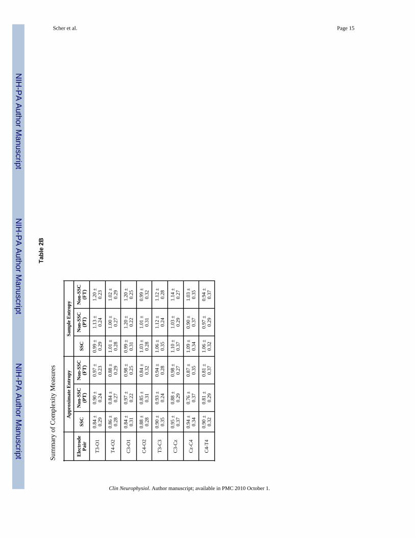
The mean and standard deviation values for the non-linear measures are listed in Table 2b.Five brain regions in the SSC cohort had greater complexity when compared with the non-SSC preterm cohort at the corrected term age. These included specific brain regions in theright hemisphere, as well as, in left and right parasagittal regions corresponding to theelectrodes C4-T4, T4-O2, C4-CZ, C3-CZ, T3-C3.
There were also three brain regions that showed greater EEG complexity for the SSC cohortgroup when compared with both the preterm and full-term groups from the non-SSC cohort.The regions involved were exclusively in the right hemisphere represented by the channelsC4-CZ, C4-T4, T4-O2.
Less complexity was noted in the channels corresponding with the posterior quadrant of theleft hemisphere in the SSC cohort, consisting of T3-O1, C3-CZ, and T3-T3 channels,compared with both non-SSC full term cohorts.
Values for discriminant analyses are listed for each SSC subject in Table 3. Discriminantanalysis using the means of the Mahalanobis metric was then used to statistically evaluatethe relationship between the complexity measures for each SSC subject compared with bothnon-SSC cohorts. It was determined that the SSC cohort subject by subject is statisticallymore similar to the full-term non-SSC cohort than to the non-SSC preterm group.
DISCUSSIONWe report more accelerated neurophysiological development for neonates who receivedskin-to-skin contact (SSC) over an eight-week period using both linear and complexityanalyses of EEG-sleep behaviors, compared with two non-SSC cohorts. These behaviors arephysiologic surrogates for multiple interconnecting neuronal pathways throughout theneuroaxis within brainstem, diencephalon and cortex which subserve state regulation(Steriade 2006) (Datta and MacLean 2007) (McCarley 2007). Five specific neuronalpathways include the ponto-medullary to basal-frontal pathway subserving respiratoryactivity, the pedunculo-pontine/geniculocalacarine pathways identified with REM behavior,the ascending reticular activating pathway subserving arousals, the corticothalamicpathways subserving quiet sleep (NREM) expression and the cortico-cortical pathwaysrepresenting spectral beta power and complexity. Our study results suggest that theimmature brain responds differently to the developmental intervention of skin-to-skincontact when compared with groups not receiving this form of developmental care. Thepresent findings reinforce our previous report of more organized sleep behavior after asingle SSC session at 32 weeks PMA (Ludington-Hoe et al. 2006) and support previouslypublished findings that a different form of developmental intervention, NeonatalIndividualized Developmental Care and Assessment Program (NIDCAP), which alsodocumented accelerated brain maturation (Als et al. 2004), based on EEG and neuroimagingmeasures. The beneficial results of neonatal developmental practices are also supported bythe experimental evidence that maternal care epigenetically programs stress responses inoffspring with effects on adult behavior. (Szyf et al 2007).
We previously reported both advanced and delayed expressions of brain function in ahealthy preterm cohort at term compared to a full-term cohort, based on a seven-itemphysiologic dysmaturity index comprised of seven EEG-sleep behaviors (Scher 1997a)(Scher 1997b). This dysmaturity index was derived after winnowing down from thirty-fourto seven physiologic measures of EEG-sleep that best differentiated between two cohorts.We also reported that increased complexity was positively associated with brain maturation(Scher 2005) (Janjarasjitt 2008). Combined linear and complexity measures can betterreflect the brain's physiologic adaptive response to the conditions of prematurity. This
Scher et al. Page 7
Clin Neurophysiol. Author manuscript; available in PMC 2010 October 1.
NIH
-PA Author Manuscript
NIH
-PA Author Manuscript
NIH
-PA Author Manuscript
biological adaptive process was previously defined as ontogenetic adaptation (Oppenheim1981), and is now integrated into the contemporary concept of developmental neuralplasticity (Cicchetti and Blender 2006). The dysmaturity index, as defined by our researchgroup, represents a physiologic marker of the brain's adaptation to the environmentalinfluences of extrauterine life for a healthy preterm cohort who did not suffer postnatalillnesses (Scher1997a) (Scher1997b). Our findings of altered sleep organization andmaturation after the developmental intervention of SSC, further extends this concept ofadaptation of a preterm cohort to the therapeutic intervention of one type of developmentalcare. Given the long convalescent hospitalization of most preterm infants, attention to themaintenance of state homeostasis using various forms of developmental intervention hasbeen emphasized (Bertelle et al. 2007) as a strategy of neuroprotection. SSC appears toaccelerate EEG-sleep state organization and maturation as a non-pharmacologicneuroprotective intervention when compared with two non-SSC cohorts. The prolongedbenefits of these non-pharmacologic neuroprotective techniques will need to be moresystematically studied in different categories of at-risk infants after discharge to the homesetting. Recent functional MRI studies of mother's responses to their infant's facial cuesemphasize the positive plasticity changes that are documented in the caregiver as a result ofan enriched interaction with their infant (Strathern et al 2008). Recommendedneuroprotective strategies that are beneficial in group studies, within the Neonatal IntensiveCare Unit and at home, need to be tailored for the individual child relative to his/her specificstrengths and vulnerabilities.
Our finding of more accelerated (i.e. more complex) right hemispheric maturation for theSSC cohort compared to both non-SSC controlled cohorts is supported by past studies whichdemonstrated right hemispheric dominance of the immature brain (Chiron et al. 1997,Schore 2001). We speculate that more advanced cortico-cortical connections exist in earlylife preferentially in the right rather than the left hemisphere, because of the greaterresponsiveness of the right hemisphere to sensory stimulation. This right hemisphericresponse to sensory input has been previously demonstrated based on the neonate's responseto painful stimuli (Fernandez et al. 2003). Despite a general paucity in the immature brain ofcortical lateralization for many functions, the right hemisphere in the neonate demonstratesthat more dominant cortical functions are already expressed for spatial sensoryrepresentation and arousal in response to positive or noxious stimuli.
To our knowledge, our study is the first to report the impact of SSC on neonatalneurophysiologic maturation over multiple-weeks, using both linear and complexityanalyses. Comparisons among SSC studies remain difficult because different methods toaccess sleep organization were used, different methods of developmental intervention wereapplied, and environmental variables such as light, sound, and tactile stimulation were notuniformly reported. For example, studies reported no differences in sleep organization withchanges in sleep input (Becker et al 1993, Ariagno el al 1997, Hellstrom-Westas etal 2001,Brandon et al 2001, Westrup et al 2002, Mirmiran et al 2003).
Our combined use of linear and complexity analysis methods emphasizes the potentialadvantage of combining multiple analytic strategies to assess in brain maturation involvingmultiple interconnected neuronal pathways after neuroprotective interventions. Whilefrequency analyses can best characterize the strength of a regional source by power spectra,complexity analysis methods can interpret physiological behaviors as a measure of the stateof a dynamical system. Our research group previously demonstrated how anothercomplexity measure, correlation dimension (CD), can characterize functional brainmaturation in preterm populations (Scher et al. 2005) (Janjarasjitt et al. 2008). CD increasesin neonates with maturation during both active and quiet sleep (Scher et al. 2005) (Pereda etal. 2006) (Janjarasjitt et al. 2008). Non-linear methods also have been used to assess
Scher et al. Page 8
Clin Neurophysiol. Author manuscript; available in PMC 2010 October 1.
NIH
-PA Author Manuscript
NIH
-PA Author Manuscript
NIH
-PA Author Manuscript
interdependences between EEG signals within different brain regions in both neonates(Pereda et al. 2001). De la Cruz et al (2007) recently reported that both linear and non-linearinterdependences coexist in the term infant while exclusively non-linear relationships existin the preterm infant.
We recognize that our study cohort was small. Repeated assessments with a larger samplesize are required, including equal numbers of subjects of each gender and with similarranges of birthweights. Moreover, our study did not examine sleep measures when theinfants were not receiving SSC. Therefore, the degree to which sleep changes carryover tonon-SSC periods is unknown. However, our results are provocative because of the highlysignificant results in the same direction, supporting the conclusion that acceleratedneurophysiologic maturation may occur in the SSC cohort. This is also supported by ourdiscriminant analyses. Secondly, no contemporaneous controls were obtained. Historicalcontrols certainly introduce bias, although the recording conditions for both SSC and non-SSC cohorts were the same in both study protocols based on feeding, recording parametersand environmental aspects in similar level III neonatal intensive care units. Also, this is onlya preliminary study in preparation for more expanded testing and analyses withcontemporaneous control cohorts. Thirdly, the neurophysiologist scoring the pilot studieswas not blinded to the knowledge that SSC had been the pilot group at term age. Fourthly,EEG-sleep segments were scored without knowledge of pretest or test conditions. Lastly,neonatal cohorts with varying severities of illness who receive SSC were not tested sincealtered or injured brain circuitries may respond differently to developmental careintervention.
CONCLUSIONThe developmental care practice of SSC alters neonatal EEG-sleep organization withaccelerated maturation. SSC practices in the neonatal intensive care unit may have animportant impact when administered over an extended period on brain maturation involvingmultiple interconnected neuronal pathways as assessed by computer analyses of EEG-sleepmeasures.
AcknowledgmentsFinancial Support: This study was supported in part by NR04926, NR08587, NS26793 from the National Institutesof Health.
REFERENCESAls H, Duffy FH, McAnulty GB, Rivkin MJ, Vajapeyam S, Mulkern RV, Warfield SK, Huppi PS,
Butler SC, Conneman N, Fischer C, Eichenwald EC. Early Experience Alters Brain Function andStructure. Pediatr 2004;113:846–857.
Ariagno RL, Thoman EB, Boeddiker MA, Kugener B, Constantinou JL, Mirmiran M, Baldwin RB.Developmental care does not alter sleep and development of premature infants. Pediatr1997;100:E9.
Aucott S, Donohue PK, Atkins E, Allen MC. Neurodevelopmental Care in the NICU. Ment RetardDev Disabil Res Rev 2002;8:298–308. [PubMed: 12454906]
Becker PT, Grunwald PC, Moorman J, Stuhr S. Effects of developmental care on behavioralorganization in very-low-birth-weight infants. Nurs Res 1993;42:214–220. [PubMed: 8337159]
Behrman, R.; Butler, A., editors. Institute of Medicine of the National Academies. National AcademicsPress; Washington DC: 2006. Preterm birth: causes, consequences and prevention. Committee onunderstanding premature birth and assuring healthy outcomes.
Scher et al. Page 9
Clin Neurophysiol. Author manuscript; available in PMC 2010 October 1.
NIH
-PA Author Manuscript
NIH
-PA Author Manuscript
NIH
-PA Author Manuscript
Bergman NJ, Linley LL, Fawcus SR. Randomized controlled trial of skin to skin contact from birthversus conventional incubator for physiological stabilization in 1200 to 2199 gram newborns. ActaPediatr 2004;93:779–85.
Bertelle V, Mabin D, Adrien J, Sizun J. Sleep of preterm neonates under developmental care or regularenvironmental conditions. Early Hum Dev 2005;81:595–600. [PubMed: 16009284]
Bertelle V, Sevestre A, Laou-Hap K, Nagahapitiye MC, Sizuth J. Sleep in the Neonatal Intensive CareUnit. J Perinat Neonat Nurs 2007;21:140–148.
Blackburn S. Environmental Impact of the NICU on Developmental Outcomes. J Pediatr Nurs1998;13(5)
Brandon DH, Holditch-Davis D, Belyea M. Preterm infants born at less than 31 weeks gestation haveimproved growth in cycle light compared with continuous near darkness. J. Pediatr 2002;140(2):192–199. [PubMed: 11865270]
Cicchetti D, Blender JA. A multiple-levels of analysis perspective on resilience: implications for thedeveloping brain, neural plasticity, and preventive interventions. Ann NY Acad Sci2006;1094:248–58. [PubMed: 17347356]
Chiron C, Jambaque I, Nabbout R, Lounes R, Syrota A, Dulac O. The right brain hemisphere isdominant in human infants. Brain 1997;120:1057–1065. [PubMed: 9217688]
Datta R, MacLean R. Neurobiological mechanisms for the regulation of mammalian sleep-wakebehavior: Reinterpretation of historical evidence and inclusion of contemporary cellular andmolecular evidence. Neurosci Biobehav Rev 2007;31:775–824. [PubMed: 17445891]
De La Cruz D, Manas S, Pereda E, Garrido JM, Lopez S, Devera L, González J. Maturational changesin the interdependencies between cortical brain areas of neonates during sleep. Cereb Cortex2007;17:583–590. [PubMed: 16627860]
Feldman R, Eidelman AI. Skin-to-skin contact (Kangaroo Care) accelerates autonomic andneurobehavioral maturation in preterm infants. Dev Med Child Neurol 2003;45:274–281.[PubMed: 12647930]
Feldman R, Weller A, Sirota L, Eidelman AI. Skin-to-Skin contact (Kangaroo care) promotes self-regulation in premature infants: sleep-wake cyclicity, arousal modulation, and sustainedexploration. Dev Psychol 2002;38:194–207. [PubMed: 11881756]
Fernandez M, Blass EM, Hernandez-Reif M, Fieid T, Diego M, Sanders C. Sucrose Attenuates aNegative Electroencephalographic Response to an Aversive Stimulus for Newborns. Dev BehavPediatr 2003;24:261–266.
Gray L, Philbin MK. Effects of the neonatal intensive care unit on auditory attention and distraction.Clin Perinatol 2004;31:243–260. [PubMed: 15289031]
Hellström-Westas L, Inghammar M, Isaksson K, Roger I, Stjernqvist K. Short-term effects ofincubator covers on quiet sleep in stable premature infants. Acta Paediatr 2001;90:1004–1008.[PubMed: 11683187]
Holditch-Davis DD, Barham LM, O'Hale A, Tucker B. Effect of standard rest periods on convalescentpreterm infants. J Obstet Gynecole Neonatal Nurs 1995;24:424–432.
Isaacs EB, Edmonds CJ, Chong WK, Lucas A, Gadian DG. Cortical anomalies associated withvisuospatial processing deficits. Ann Neurol 2003;53:768–73. [PubMed: 12783423]
Ludington-Hoe SM, Johnson MW, Morgan K, Lewis T, Gutman J, Wilson PD, Scher MS.Neurophysiologic assessment of neonatal sleep organization: preliminary results of a randomized,controlled trial of skin contact with preterm infants. Pediatr 2006;117:e909–923.
Ludington-Hoe SM, Thompson C, Swinth JY, Hadeed AJ, Anderson GC. Kangaroo Case: Researchresults and practice implications and guidelines. Neonatal Netw 1994;13:19–27. [PubMed:8114658]
McCarley RW. Neurobiology of REM and NREM sleep. Sleep Med 2007;8:302–330. [PubMed:17468046]
Mimiran M, Baldwin RB, Ariagno RL. Circadian and sleep development in preterm infants occursindependently from the influences of environmental lighting. Pediatr Res 2003;53:933–938.[PubMed: 12621096]
Oppenheim, RW. Ontogenetic adaptations and retrogressive processes in the development of thenervous system and behavior: A neuroembryological perspective. In: Connolly, KJ.; Prechtl,
Scher et al. Page 10
Clin Neurophysiol. Author manuscript; available in PMC 2010 October 1.
NIH
-PA Author Manuscript
NIH
-PA Author Manuscript
NIH
-PA Author Manuscript
HFR., editors. Maturation and Development Biological and Perspectives. Lippincott; Philadelphia:1981. p. 73-109.
Pereda E, De La Cruz DM, Manas S, Garrido JM, Lopez S, González J. Topography of EEGcomplexity in human neonates: effect of the postmenstrual age and the sleep state. NeuroscienceLetters 2006;2:152–157. [PubMed: 16278043]
Pereda E, Manas S, Vera LD,JM, Lopez S, González J. Non-linear asymmetric interdependencies inthe electroencephalogram of healthy term neonates during sleep. Neuroscience Letters2003;337:101–105. [PubMed: 12527398]
Pereda E, Rial R, Gamundi A, González J. Assessment of changing interdependencies between humanelectroencephalograms using nonlinear methods. Physica D 2001;148:147–158.
Pincus SM. Approximate entropy as a measure of system complexity. Proc Natl Acad Sci 1991;88(6):2297–2301. [PubMed: 11607165]
Richman JS, Moorman JR. Physiological time-series analysis using approximate entropy and sampleentropy. Am J Physiol Heart Circ Physiol 2002;278:2039–2049.
Janjarasjitt S, Scher MS, Loparo KA. Nonlinear dynamical analysis of the neonatal EEG time series:The relationship between neurodevelopment and complexity. Clinical Neurophysiology2008;119:822–836. [PubMed: 18203659]
Scher MS, Waisanen H, Loparo KA, Johnson MW. Prediction of neonatal state and maturationalchange using dimensional analysis. J. Clin. Neurophysiol 2005;3:159–165. [PubMed: 15933487]
Scher MS, Jones BL, Steppe DA, Cork DL, Seltmen HJ, Banks DL. Functional brain maturation inneonates as measured by EEG-sleep analyses. Clin Neurophysiol 2003;144:875–82. [PubMed:12738433]
Scher MS, Steppe DA, Sclabassi RJ, Banks, DL. Regional differences in spectral EEG measuresbetween healthy term and preterm infants. Pediatr Neurol 1997;17:218–223. [PubMed: 9390697]
Scher MS. Automated EEG-sleep analyses and neonatal neurointensive care. J Sleep Med 2004;5:533–540.
Scher, MS. Electroencephalography of the newborn: normal and abnormal features. In: Niedermeyer,E.; Lopes, da; Silva, F., editors. Electroencephalography: basic principles, clinical applications,and related fields. Lippincott Williams & Wilkins; Philadelphia: 2006. p. 937-989.Chapter 49
Scher MS. Neurophysiological assessment of brain function and maturation: I. A measure of brainadaptation in high risk infants. Pediatr Neurol 1997b;16:191–8. [PubMed: 9165508]
Scher MS. Neurophysiological assessment of brain function and maturation. II. A measure of braindysmaturity in healthy preterm neonates. Pediatr Neurol 1997a;16:287–95. [PubMed: 9258960]
Schore AN. The effects of early relational trauma on right brain development, affect regulation, andinfant mental health. Infant Men Health J 2001;22:201–269.
Skranes J, Vangberg TR, Kulseng, Indredavik MS, Evensen KA, Martinussen M, Dale AM,Haraldseth O, Brubakk AM. Clinical findings and white matter abnormalities seen on diffusiontensor imaging in adolescents with very low birth weight. Brain 2007;130:645–66.
Srinivasan L, Dutta R, Counsell SJ, Allsup JM, Boardman JP, Rutherford MA, Edwards AD.Quantification of deep gray matter in preterm infants at term-equivalent age using manualvolumetry of 3-tesla magnetic resonance images. Pediatr 2007;119:759–65.
Steriade M. Grouping of Brain Rhythms in Corticothalamic Systems. Neuroscience 2006;137:1087–1106. [PubMed: 16343791]
Szyf M, Weaver I, Meaney M. Maternal Care, the epigenome and phenotypic differences in behavior.Reproductive Toxicology 2007;24:9–19. [PubMed: 17561370]
Strathearn L, Li J, Fonagy P, Montague PR. What's in a Smile? Maternal Brain Responses to InfantFacial Cues. Pediatrics 2008;122:40–51. [PubMed: 18595985]
Tessier R, Cristo MB, Velez S, Giron M, Nadeau L, deCalume ZF, Ruiz-Paláez JG, Charpak N.Kangaroo Mother Care: A method for protecting high-risk low-birth-weight and premature infantsagainst developmental delay. Infant Behavior & Development 2003;26:384–397.
Thompson DK, Warfield SK, Carlin JB, Pavlovic M, Wang HX, Bear M, Kean MJ, Doyle LW, EganGF, Inder TE. Perinatal risk factors altering regional brain structure in the preterm infant. Brain2007;130:667–77. [PubMed: 17008333]
Scher et al. Page 11
Clin Neurophysiol. Author manuscript; available in PMC 2010 October 1.
NIH
-PA Author Manuscript
NIH
-PA Author Manuscript
NIH
-PA Author Manuscript
Westrup B, Hellstrom-Westas L, Stjernqvist K, Lagercrantz H. No indications of increased quiet sleepin infants receiving care based on the newborn individualized developmental care and assessmentprogram (NIDCAP). Acta Paediatr 2002;91:318–322. [PubMed: 12022306]
Scher et al. Page 12
Clin Neurophysiol. Author manuscript; available in PMC 2010 October 1.
NIH
-PA Author Manuscript
NIH
-PA Author Manuscript
NIH
-PA Author Manuscript
NIH
-PA Author Manuscript
NIH
-PA Author Manuscript
NIH
-PA Author Manuscript
Scher et al. Page 13
Table 1
Subject Summary
SSC Non-SSC (PT) Non-SSC (FT)
Number of subjects 8 78 48
Sex
Female 2 37 27
Male 6 41 21
Race
Caucasian 5 44 33
African American 2 29 13
Other 1 5 2
Birth Data
Gestational Age (wks) 30.6 ± 0.7 29.5 ± 1.9 40.2 ± 1.4
Birth Weight (kg) 1.52 ± 0.20 1.19 ± 0.21 3.59 ± 0.49
Study Data
Day of Life 72.3 ± 2.7 79.5 ± 19.9 5.3 ± 10.0
Study Postmenstrual age (wks) 40.8 ± 0.6 40.8 ± 2.0 41.0 ± 1.5
Study Weight (kg) 3.49 ± 0.47 3.28 ± 0.81 3.58 ± 0.55
Clin Neurophysiol. Author manuscript; available in PMC 2010 October 1.
NIH
-PA Author Manuscript
NIH
-PA Author Manuscript
NIH
-PA Author Manuscript
Scher et al. Page 14
Table 2A
Summary of EEG-Sleep Comparisons
SSC Non-SSC (PT) Non-SSC (FT)
Left/Right Correlation Index 0.13 ± 0.10 0.16 ± 0.08 0.11 ± 0.03
REM Index 0.96 ± 0.61 4.49 ± 2.47* 6.66 ± 4.12*
Quiet Sleep % 46.3 ± 9.0 36.2 ± 8.0* 34.2 ± 7.4*
Respiratory Ratio Index 0.73 ± 0.05 0.75 ± 0.05 0.77 ± 0.04*
Sleep Cycle Length (minutes) 71.0 ± 13.2 70.0 ± 18.7 58.9 ± 14.3*
Spectral Index −6.84 ± 0.40 −6.94 ± 0.62 −6.51 ± 0.44*
Arousal Index 2.96 ± 0.99 1.41 ± 1.14* 3.02 ± 1.79
*significant difference from SSC
Clin Neurophysiol. Author manuscript; available in PMC 2010 October 1.
NIH
-PA Author Manuscript
NIH
-PA Author Manuscript
NIH
-PA Author Manuscript
Scher et al. Page 15
Tabl
e 2B
Sum
mar
y of
Com
plex
ity M
easu
res
App
roxi
mat
e E
ntro
pySa
mpl
e E
ntro
py
Ele
ctro
dePa
irSS
CN
on-S
SC(P
T)
Non
-SSC
(FT
)SS
CN
on-S
SC(P
T)
Non
-SSC
(FT
)
T3-O
10.
84 ±
0.29
0.90
±0.
240.
97 ±
0.23
0.99
±0.
291.
13 ±
0.24
1.20
±0.
23
T4-O
20.
86 ±
0.28
0.84
±0.
270.
88 ±
0.29
1.01
±0.
281.
00 ±
0.27
1.02
±0.
29
C3-
O1
0.84
±0.
310.
97 ±
0.22
0.98
±0.
250.
99 ±
0.31
1.20
±0.
221.
20 ±
0.25
C4-
O2
0.88
±0.
280.
85 ±
0.31
0.84
±0.
321.
03 ±
0.28
1.01
±0.
310.
99 ±
0.32
T3-C
30.
90 ±
0.35
0.93
±0.
240.
94 ±
0.28
1.06
±0.
351.
12 ±
0.24
1.12
±0.
28
C3-
Cz
0.95
±0.
370.
88 ±
0.29
0.98
±0.
271.
10 ±
0.37
1.03
±0.
291.
14 ±
0.27
Cz-
C4
0.94
±0.
340.
76 ±
0.37
0.87
±0.
351.
09 ±
0.34
0.90
±0.
371.
03 ±
0.35
C4-
T40.
90 ±
0.32
0.81
±0.
290.
81 ±
0.37
1.06
±0.
320.
97 ±
0.29
0.94
±0.
37
Clin Neurophysiol. Author manuscript; available in PMC 2010 October 1.

NIH
-PA Author Manuscript
NIH
-PA Author Manuscript
NIH
-PA Author Manuscript
Scher et al. Page 16
Table 3
Discriminant analysis using the Mahalanobis metric.Mean Mahalanobis Distance Between Neonates at 40-41 Week and the Pittsburgh Controls using ComplexityMeasures.
Approximate Entropy Sample Entropy
SSC Non-SSC Non-SSC
Neonate Number Full term Preterm Full term Preterm
1 4.33 5.47 3.07 4.20
2 2.90 3.27 2.12 2.57
3 2.22 3.45 1.71 2.75
4 2.73 4.50 2.13 3.65
5 2.85 4.71 2.27 3.94
6 2.64 3.53 2.10 2.90
7 2.49 4.64 1.95 3.72
8 2.96 4.67 2.23 3.81
Mean Distance 2.90 4.32 2.20 3.48
Standard Dev. 0.59 0.74 0.37 0.58
Clin Neurophysiol. Author manuscript; available in PMC 2010 October 1.