Neuronal injury from cardiac arrest: aging years in minutes
14
Neuronal injury from cardiac arrest: aging years in minutes Brandon H. Cherry & Nathalie Sumien & Robert T. Mallet Received: 27 March 2014 /Accepted: 26 June 2014 # American Aging Association 2014 Abstract Cardiac arrest is a leading cause of death and permanent disability. Most victims succumb to the oxida- tive and inflammatory damage sustained during cardiac arrest/resuscitation, but even survivors typically battle long-term neurocognitive impairment. Although exten- sive research has delineated the complex mechanisms that culminate in neuronal damage and death, no effective treatments have been developed to interrupt these mech- anisms. Of importance, many of these injury cascades are also active in the aging brain, where neurons and other cells are under persistent oxidative and inflammatory stress which eventually damages or kills the cells. In light of these similarities, it is reasonable to propose that the brain essentially ages the equivalent of several years with- in the few minutes taken to resuscitate a patient from cardiac arrest. Accordingly, cardiac arrest-resuscitation models may afford an opportunity to study the deleterious mechanisms underlying the aging process, on an acceler- ated time course. The aging and resuscitation fields both stand to gain pivotal insights from one another regarding the mechanisms of injury sustained during resuscitation from cardiac arrest and during aging. This synergism between the two fields could be harnessed to foster devel- opment of treatments to not only save lives but also to enhance the quality of life for the elderly. Keywords Caspases . Glutathione . Inflammation . Ischemia . Neurodegeneration . Oxidative Stress Abbreviations AD Alzheimer’ s disease Bak Bcl-2 homologous antagonist killer Bax Bcl-2-associated X protein BBB Blood–brain barrier Bcl-2 B-cell lymphoma 2 family of proteins BH 4 Tetrahydrobiopterin Casp-3 Caspase-3 Casp-9 Caspase-9 CoQ Coenzyme-Q CPR Cardiopulmonary resuscitation Cyt C Cytochrome C eNOS Endothelial isoform of nitric oxide synthase GSH Reduced form of glutathione GSH/GSSG Concentration ratio of reduced to oxidized glutathione iNOS Inducible isoform of nitric oxide synthase mPTP Mitochondrial permeability transition pore AGE (2014) 36:9680 DOI 10.1007/s11357-014-9680-x B. H. Cherry (*) : R. T. Mallet Department of Integrative Physiology and Anatomy, University of North Texas Health Science Center, 3500 Camp Bowie Blvd, Fort Worth, TX 76107-2699, USA e-mail: [email protected] N. Sumien Department of Pharmacology and Neuroscience, University of North Texas Health Science Center, Fort Worth, TX, USA B. H. Cherry : N. Sumien : R. T. Mallet Institute for Aging and Alzheimer’ s Disease Research, University of North Texas Health Science Center, Fort Worth, TX, USA B. H. Cherry : R. T. Mallet Cardiovascular Research Institute, University of North Texas Health Science Center, Fort Worth, TX, USA
-
Upload
independent -
Category
Documents
-
view
0 -
download
0
Transcript of Neuronal injury from cardiac arrest: aging years in minutes

Neuronal injury from cardiac arrest: aging years in minutes
Brandon H. Cherry & Nathalie Sumien &
Robert T. Mallet
Received: 27 March 2014 /Accepted: 26 June 2014# American Aging Association 2014
Abstract Cardiac arrest is a leading cause of death andpermanent disability. Most victims succumb to the oxida-tive and inflammatory damage sustained during cardiacarrest/resuscitation, but even survivors typically battlelong-term neurocognitive impairment. Although exten-sive research has delineated the complex mechanisms thatculminate in neuronal damage and death, no effectivetreatments have been developed to interrupt these mech-anisms. Of importance, many of these injury cascades arealso active in the aging brain, where neurons and othercells are under persistent oxidative and inflammatorystress which eventually damages or kills the cells. In lightof these similarities, it is reasonable to propose that thebrain essentially ages the equivalent of several years with-in the few minutes taken to resuscitate a patient fromcardiac arrest. Accordingly, cardiac arrest-resuscitation
models may afford an opportunity to study the deleteriousmechanisms underlying the aging process, on an acceler-ated time course. The aging and resuscitation fields bothstand to gain pivotal insights from one another regardingthe mechanisms of injury sustained during resuscitationfrom cardiac arrest and during aging. This synergismbetween the two fields could be harnessed to foster devel-opment of treatments to not only save lives but also toenhance the quality of life for the elderly.
Keywords Caspases . Glutathione . Inflammation .
Ischemia . Neurodegeneration . Oxidative Stress
AbbreviationsAD Alzheimer’s diseaseBak Bcl-2 homologous antagonist killerBax Bcl-2-associated X proteinBBB Blood–brain barrierBcl-2 B-cell lymphoma 2 family of proteinsBH4 TetrahydrobiopterinCasp-3 Caspase-3Casp-9 Caspase-9CoQ Coenzyme-QCPR Cardiopulmonary resuscitationCyt C Cytochrome CeNOS Endothelial isoform of nitric oxide
synthaseGSH Reduced form of glutathioneGSH/GSSG Concentration ratio of reduced to
oxidized glutathioneiNOS Inducible isoform of nitric oxide
synthasemPTP Mitochondrial permeability
transition pore
AGE (2014) 36:9680DOI 10.1007/s11357-014-9680-x
B. H. Cherry (*) :R. T. MalletDepartment of Integrative Physiology and Anatomy,University of North Texas Health Science Center,3500 Camp Bowie Blvd, Fort Worth, TX 76107-2699, USAe-mail: [email protected]
N. SumienDepartment of Pharmacology and Neuroscience, Universityof North Texas Health Science Center,Fort Worth, TX, USA
B. H. Cherry :N. Sumien : R. T. MalletInstitute for Aging and Alzheimer’s Disease Research,University of North Texas Health Science Center,Fort Worth, TX, USA
B. H. Cherry :R. T. MalletCardiovascular Research Institute, University of North TexasHealth Science Center,Fort Worth, TX, USA

mtDNA Mitochondrial DNANFκB Nuclear factor-kappa BNO Nitric oxideO2
− SuperoxideONOO− PeroxynitriteOxS Oxidative stressROS/RNS Reactive oxygen and nitrogen
speciesSMAC Second mitochondria-derived
activator of caspasesTNF-α Tumor necrosis factor alpha
Introduction
Cardiac arrest remains a leading cause of death andpersistent disability in the USA. In its 2014 update onheart disease and stroke statistics, the American HeartAssociation estimated that approximately 380,000 outof 424,000 (~90%) Americans who experience out-of-hospital cardiac arrest annually do not survive (Go et al.2014). Only 23% (97,520) of all cardiac arrest victimspresent to emergency medical services personnel with ashockable cardiac rhythm, and most who are initiallyresuscitated later succumb to extensive ischemia-reperfusion injury to the brain and other vital organs(Dezfulian et al. 2009; Heron 2012; Nolan et al. 2012;Young 2009; Go et al. 2014). Moreover, approximatelyhalf of the c. 10 % of cardiac arrest victims who dosurvive to hospital discharge experience persistentneurocognitive impairment manifested as memory andsensorimotor deficits that profoundly impact their qual-ity of life (Adrie et al. 2004; Moulaert et al. 2009;Wachelder et al. 2009; Young 2009; Go et al. 2014).
While many studies have examined the complexmechanisms of brain damage following cardiac arrest-initiated ischemia-reperfusion injury, the precise cas-cade of events culminating in neurocognitive impair-ment remains to be completely delineated. It is knownthat ATP depletion, intracellular Ca2+ overload (Banoand Nicotera 2007; Li et al. 2007), reactive oxygen andnitrogen species (Calapai et al. 2000), inflammation,and glutamate-induced excitotoxicity (Conroy et al.1999; Backstrom et al. 2003) initiated by cardiac arrestand resuscitation collectively inflict lethal damage toneurons, oligodendrocytes, microglia, and the cerebro-vascular endothelium and disrupt the blood–brain barri-er (BBB). Despite mounting knowledge of the
mechanisms of brain injury, currently there are no clin-ically proven pharmacological treatments to protect thebrain during cardiac arrest and cardiopulmonary resus-citation (CPR) (Dezfulian et al. 2009).
Similar to long-term recovery from cardiac arrest andCPR, the principal mechanisms of neurocognitive im-pairment in the aging brain have yet to be assembledinto a coherent cascade of events that would allow fordevelopment of efficacious preventative therapies.However, it is becoming increasingly evident that manyage-related changes leading to neuronal damage anddeath parallel those observed during and following car-diac arrest. Specifically, accumulation of reactive oxy-gen and nitrogen species as well as proinflammatorycytokines and markers of inflammation have all beenobserved in brain aging studies (Hagen 2003; Kregeland Zhang 2007; Cortese et al. 2011). Moreover, as thebrain ages, calcium mismanagement and mitochondrialdysfunction also contribute to the death and dysfunctionof neurons and other cells within the most vulnerableregions such as the hippocampus (Landfield 1988;Foster and Norris 1997; Toescu et al. 2004). The pro-gression of neuronal impairment and cell death observedduring aging is, however, a much slower process thanthe injury cascade that follows cardiac arrest, CPR, andpost-arrest recovery. The purpose of this review is tohighlight parallel mechanisms of brain damage andneuronal death that ensue following cardiac arrest andin the aging brain. Despite their different time courses,mechanistic information gained from studying the twoconditions could be harnessed to synergistically ad-vance both fields and to develop treatments targetingspecific components in these neurodegenerative path-ways to provide more robust protection of patients fromneurocognitive impairment and/or death.
Oxidative stress
Oxidative injury during cardiac arrestand cardiopulmonary resuscitation
A major culprit in the ischemia-reperfusion injurysustained following cardiac arrest and CPR is the oxida-tive stress imposed on the brain (Idris et al. 2005; Wanget al. 2007). Intense formation and accumulation of reac-tive oxygen (Opie 1991; Cerchiari et al. 1987; Becker2004; Idris et al. 2005) and nitrogen species, i.e., ROS/RNS (Lipton 1999; Love 1999; White et al. 2000; Dohi
9680, Page 2 of 14 AGE (2014) 36:9680
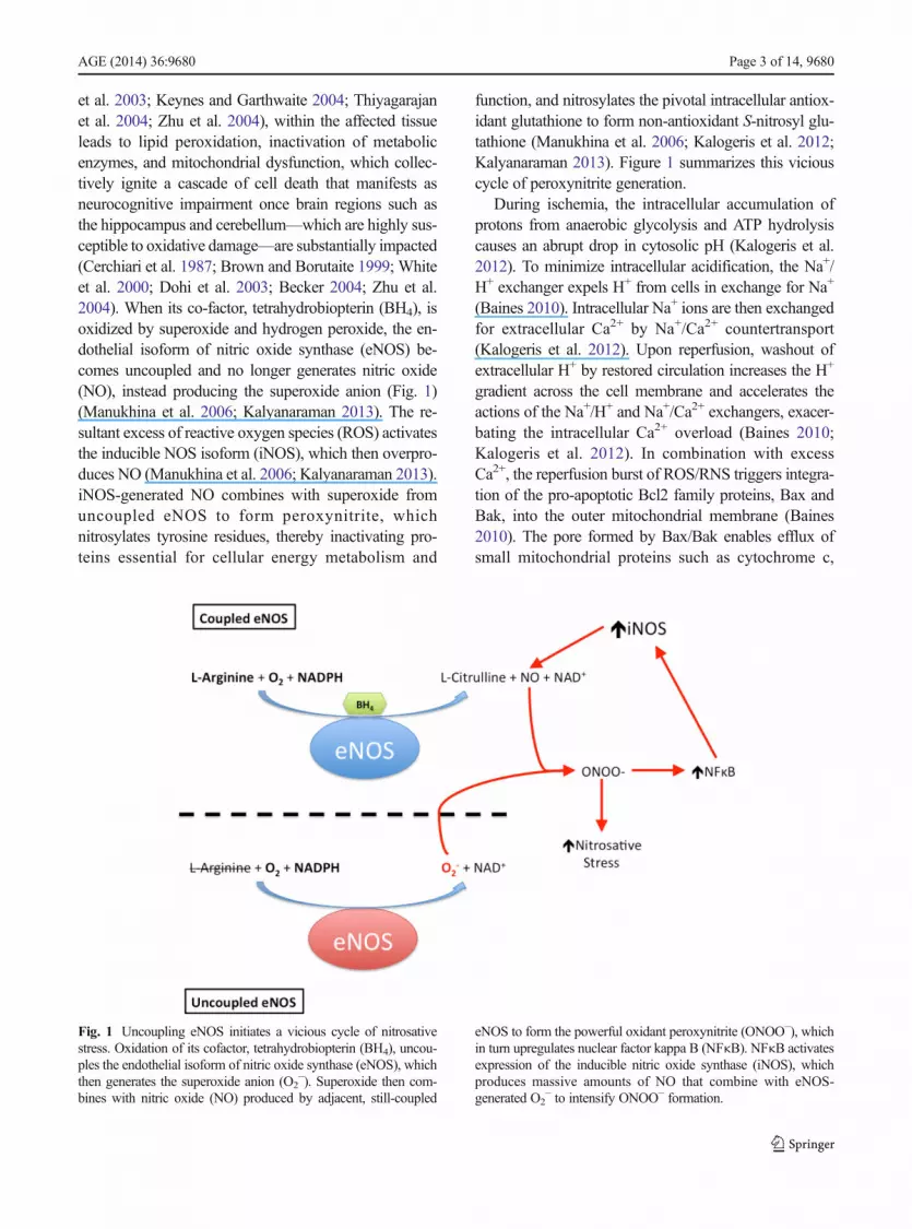
et al. 2003; Keynes and Garthwaite 2004; Thiyagarajanet al. 2004; Zhu et al. 2004), within the affected tissueleads to lipid peroxidation, inactivation of metabolicenzymes, and mitochondrial dysfunction, which collec-tively ignite a cascade of cell death that manifests asneurocognitive impairment once brain regions such asthe hippocampus and cerebellum—which are highly sus-ceptible to oxidative damage—are substantially impacted(Cerchiari et al. 1987; Brown and Borutaite 1999; Whiteet al. 2000; Dohi et al. 2003; Becker 2004; Zhu et al.2004). When its co-factor, tetrahydrobiopterin (BH4), isoxidized by superoxide and hydrogen peroxide, the en-dothelial isoform of nitric oxide synthase (eNOS) be-comes uncoupled and no longer generates nitric oxide(NO), instead producing the superoxide anion (Fig. 1)(Manukhina et al. 2006; Kalyanaraman 2013). The re-sultant excess of reactive oxygen species (ROS) activatesthe inducible NOS isoform (iNOS), which then overpro-duces NO (Manukhina et al. 2006; Kalyanaraman 2013).iNOS-generated NO combines with superoxide fromuncoupled eNOS to form peroxynitrite, whichnitrosylates tyrosine residues, thereby inactivating pro-teins essential for cellular energy metabolism and
function, and nitrosylates the pivotal intracellular antiox-idant glutathione to form non-antioxidant S-nitrosyl glu-tathione (Manukhina et al. 2006; Kalogeris et al. 2012;Kalyanaraman 2013). Figure 1 summarizes this viciouscycle of peroxynitrite generation.
During ischemia, the intracellular accumulation ofprotons from anaerobic glycolysis and ATP hydrolysiscauses an abrupt drop in cytosolic pH (Kalogeris et al.2012). To minimize intracellular acidification, the Na+/H+ exchanger expels H+ from cells in exchange for Na+
(Baines 2010). Intracellular Na+ ions are then exchangedfor extracellular Ca2+ by Na+/Ca2+ countertransport(Kalogeris et al. 2012). Upon reperfusion, washout ofextracellular H+ by restored circulation increases the H+
gradient across the cell membrane and accelerates theactions of the Na+/H+ and Na+/Ca2+ exchangers, exacer-bating the intracellular Ca2+ overload (Baines 2010;Kalogeris et al. 2012). In combination with excessCa2+, the reperfusion burst of ROS/RNS triggers integra-tion of the pro-apoptotic Bcl2 family proteins, Bax andBak, into the outer mitochondrial membrane (Baines2010). The pore formed by Bax/Bak enables efflux ofsmall mitochondrial proteins such as cytochrome c,
Fig. 1 Uncoupling eNOS initiates a vicious cycle of nitrosativestress. Oxidation of its cofactor, tetrahydrobiopterin (BH4), uncou-ples the endothelial isoform of nitric oxide synthase (eNOS), whichthen generates the superoxide anion (O2
−). Superoxide then com-bines with nitric oxide (NO) produced by adjacent, still-coupled
eNOS to form the powerful oxidant peroxynitrite (ONOO−), whichin turn upregulates nuclear factor kappa B (NFκB). NFκB activatesexpression of the inducible nitric oxide synthase (iNOS), whichproduces massive amounts of NO that combine with eNOS-generated O2
− to intensify ONOO− formation.
AGE (2014) 36:9680 Page 3 of 14, 9680

second mitochondria-derived activator of caspases(SMAC), and endonuclease-G (Baines 2010). In thecytosol, SMAC and cytochrome c combine with apopto-tic protease activating factor 1 (APAF1), forming anapoptosome which activates caspase-9 and caspase-3(Baines 2010). In concert with caspase-mediated pro-apoptotic signaling, Bax/Bak permits endonuclease-Gefflux frommitochondria; this enzyme enters the nucleusand fragments genomic DNA, a pivotal event in apopto-tic cell death (Baines 2010). The post-ischemic Ca2+
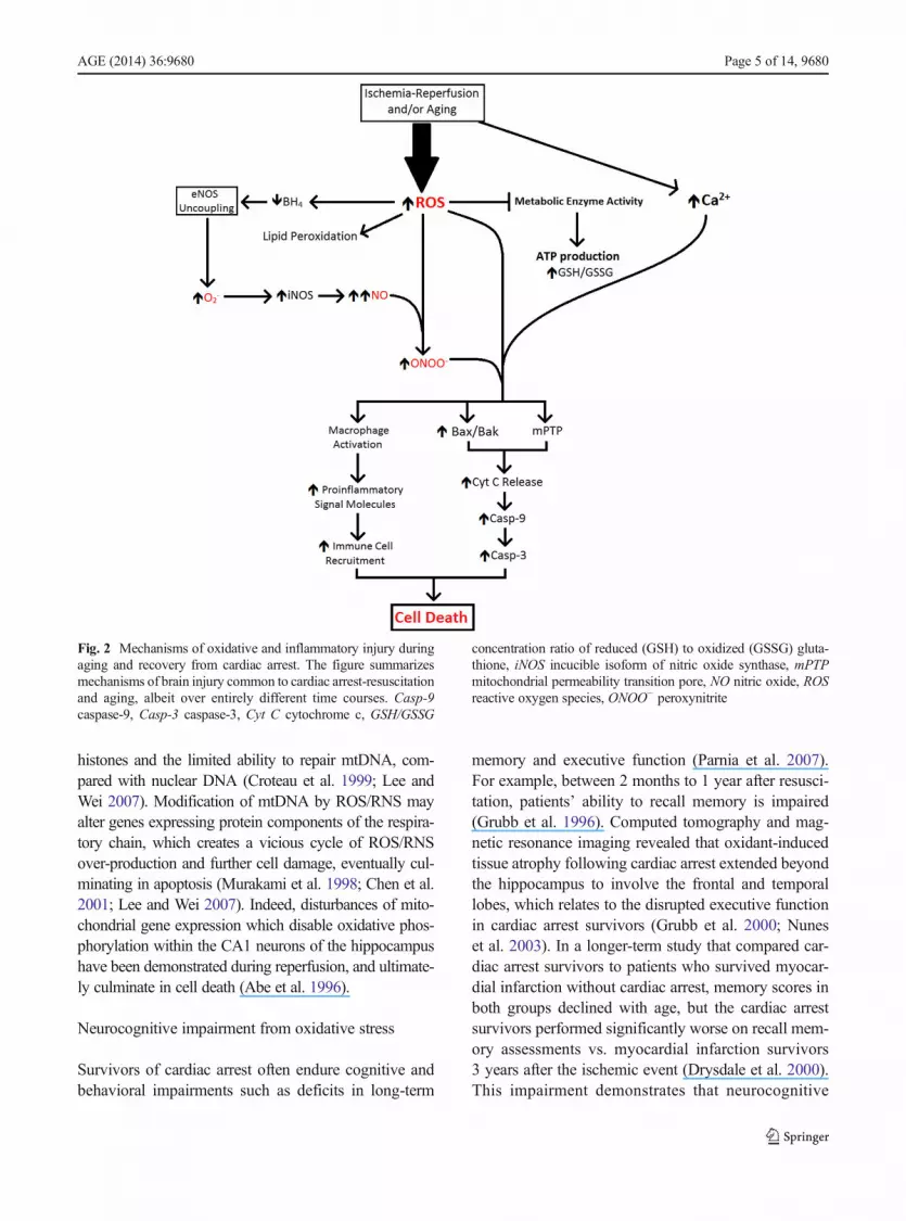
overload and ROS/RNS burst in the mitochondrial ma-trix also open a large, non-selective channel in the innermitochondrial membrane, the mitochondrial permeabili-ty transition pore (mPTP). Opening of mPTP collapsesthe proton electrochemical gradient required for oxida-tive phosphorylation, further draining cellular ATP re-serves already depleted by ischemia (Baines 2010;Halestrap 2010; Kalogeris et al. 2012). Figure 2 summa-rizes the cascade by which this intense oxidative insultultimately opens both the Bax/Bak and mPTP pores,thereby activating caspase-9 and caspase-3, DNA frag-mentation and mitochondrial rupture culminating in ap-optosis of neurons and astroglia (Kirkland and Franklin2003; Baines 2010; Halestrap 2010; Franklin 2011;Martin et al. 2011).
Oxidative injury during aging
The “oxidative stress theory” of aging identifies the accu-mulation of oxidative damage caused by ROS/RNS overthe course of the aging process as pivotal to the progres-sive decline of biological function and shorter lifespan(Kregel and Zhang 2007). According to this paradigm,ROS as well as RNS accumulate due to an imbalancebetween their production and detoxification by endoge-nous redox systems, e.g., the glutathione peroxidase/reductase and thioredoxin/peroxiredoxin systems (Hagen2003; Kregel and Zhang 2007). This oxidative imbalancepotentiates deleterious protein oxidation, lipid peroxida-tion, and apoptotic cell death (Blumberg 2004; Stadtman2004; Matsuzawa and Ichijo 2005).
One line of evidence supporting the “oxidative stresstheory” of aging stems from measurements of the com-mon biomarkers of antioxidative capacity—that is, thedegree to which the body is able to neutralize existingand de novo oxidative stress via its endogenous antioxi-dant defense mechanisms. Among the most widely ac-cepted measures of in vivo redox state are the ratios ofreduced to oxidized glutathione (GSH/GSSG) and
nicotinamide adenine dinucleotide phosphate (NADPH/NAPD+) (Kregel and Zhang 2007). Of these redox sys-tems, GSH/GSSG can be taken as a global measure of thecollective poise of endogenous antioxidant defenses, asthe glutathione reductase/peroxidase system is linked, viaredox cycles, to the other cellular antioxidant systems(Schafer and Buettner 2001). A progressive decline inGSH/GSSG with advancing age has been identified(Droge 2002), suggesting either a decreased ability of cellsto neutralize oxidative stress, increased formation of ROS/RNS, or perhaps impaired GSH generation as the result ofdecreased glutathione reductase activity. In support of thelatter possibility, several studies have reported oxidativeinactivation of metabolic enzymes and membrane lipidperoxidation in aging brain (Beckman and Ames 1998;Bokov et al. 2004; Poon et al. 2004; Rodrigues Siqueiraet al. 2005; Kregel and Zhang 2007). A major source ofoxidant accumulation with age is mitochondrial dysfunc-tion leading to overproduction and release of ROS/RNSinto the cytosol (Sohal et al. 1995; Giulivi 1998). TheROS/RNS then uncouple eNOS, causing the enzyme tooverproduce superoxide (O2
−), which activates iNOS anddrives the production of cytotoxic peroxynitrite in a man-ner similar to that of cardiac arrest induced brain ischemia-reperfusion (Manukhina et al. 2006; Ungvari et al. 2010;Kalyanaraman 2013). Collectively, cytosolic ROS/RNSaccumulation activates death cascades mediated bycaspase-9 and caspase-3 (Kirkland and Franklin 2003;Franklin 2011; Martin et al. 2011). Finally, it is importantto note that chronic oxidative stress with advancing agehas been implicated in the pathogenesis of age-relatedneurodegenerative disorders (Volicer and Crino 1990;Dexter et al. 1994).
Oxidative damage to mitochondrial DNA
According to the mitochondrial theory of aging, the accu-mulation of mutations in mitochondrial DNA (mtDNA)over the lifetime leads to bioenergetic impairment andcontributes substantially to aging (Linnane et al. 1989;Lee and Wei 2007). Because of its close proximity to therespiratory chain, the mitochondrial genome is particular-ly susceptible to oxidative damage from excessive ROS/RNS produced during ischemia-reperfusion (Richter1995; Chen et al. 2001) and accumulated during aging(Mecocci et al. 1993; Ozawa 1995; Barja and Herrero2000). Moreover, mtDNA is much more vulnerable tooxidative modification than nuclear DNA (Ames et al.1993; Richter 1995), due to its lack of protection by
9680, Page 4 of 14 AGE (2014) 36:9680
histones and the limited ability to repair mtDNA, com-pared with nuclear DNA (Croteau et al. 1999; Lee andWei 2007). Modification of mtDNA by ROS/RNS mayalter genes expressing protein components of the respira-tory chain, which creates a vicious cycle of ROS/RNSover-production and further cell damage, eventually cul-minating in apoptosis (Murakami et al. 1998; Chen et al.2001; Lee and Wei 2007). Indeed, disturbances of mito-chondrial gene expression which disable oxidative phos-phorylation within the CA1 neurons of the hippocampushave been demonstrated during reperfusion, and ultimate-ly culminate in cell death (Abe et al. 1996).
Neurocognitive impairment from oxidative stress
Survivors of cardiac arrest often endure cognitive andbehavioral impairments such as deficits in long-term
memory and executive function (Parnia et al. 2007).For example, between 2 months to 1 year after resusci-tation, patients’ ability to recall memory is impaired(Grubb et al. 1996). Computed tomography and mag-netic resonance imaging revealed that oxidant-inducedtissue atrophy following cardiac arrest extended beyondthe hippocampus to involve the frontal and temporallobes, which relates to the disrupted executive functionin cardiac arrest survivors (Grubb et al. 2000; Nuneset al. 2003). In a longer-term study that compared car-diac arrest survivors to patients who survived myocar-dial infarction without cardiac arrest, memory scores inboth groups declined with age, but the cardiac arrestsurvivors performed significantly worse on recall mem-ory assessments vs. myocardial infarction survivors3 years after the ischemic event (Drysdale et al. 2000).This impairment demonstrates that neurocognitive
Fig. 2 Mechanisms of oxidative and inflammatory injury duringaging and recovery from cardiac arrest. The figure summarizesmechanisms of brain injury common to cardiac arrest-resuscitationand aging, albeit over entirely different time courses. Casp-9caspase-9, Casp-3 caspase-3, Cyt C cytochrome c, GSH/GSSG
concentration ratio of reduced (GSH) to oxidized (GSSG) gluta-thione, iNOS incucible isoform of nitric oxide synthase, mPTPmitochondrial permeability transition pore, NO nitric oxide, ROSreactive oxygen species, ONOO− peroxynitrite
AGE (2014) 36:9680 Page 5 of 14, 9680

function may be sufficiently compromised to severelyimpact quality of life (O’Reilly et al. 2003). Similarly,memory and executive function become impaired dur-ing aging. As oxidative stress accumulates with advanc-ing age, the brain deterioration eventually causesneurocognitive impairments resembling to those thatfollow cardiac arrest-resuscitation (Mahncke et al.2006; Kim and Oh 2013).
Oxidative stress has been implicated in the pathogen-esis of neurodegeneration and neurocognitive impair-ment after cardiac arrest (Liu et al. 1998; Parnia et al.2007; Fiskum et al. 2008) and during aging (Dexteret al. 1994; Volicer and Crino 1990). To test the hypoth-esis that overproduction of ROS/RNS producesneurocognitive impairment, Vereczki et al. comparedresuscitation of dogs with 100 % oxygen vs. room air(ca. 21 % oxygen) and found that hyperoxic resuscita-tion increased hippocampal tyrosine nitration—a mark-er of oxidative cell injury—and intensified post-ischemic impairment of hippocampus-dependent func-tions (Matsuzawa and Ichijo 2005; Vereczki et al. 2006;Kregel and Zhang 2007).
Summary
A common mechanism underlying brain deteriorationboth following cardiac arrest-resuscitation and duringaging is the accumulation of ROS/RNS, which act tomodify mtDNA, disrupt cellular function, initiate apo-ptosis, and ultimately impair neurocognitive function.An important difference between aging vs. post-cardiacarrest is the time course of ROS/RNS accumulation—that is, alterations in ROS/RNS concentrations occurwithin minutes following resuscitation from cardiac ar-rest, but develop over many years during aging. In bothcases, the accumulated ROS/RNS attack mitochondrialand nuclear DNA, inactivate enzymes catalyzing energymetabolism, compromise ATP production, and impairthe glutathione peroxidase/reductase and thioredoxin/peroxiredoxin antioxidant systems. Additionally, oxida-tive stress in both settings provokes opening of the Bax/Bak and mitochondrial permeability transition pores,which respectively release cytochrome c into the cytosoland dissipate the electrochemical gradient required foroxidative phosphorylation. Cytochrome-c release initi-ates activation of caspase-9 and caspase-3 and eventu-ally apoptotic cell death within the affected brain tissue.Cell death caused by oxidative stress in both post-resuscitation and aging results in recall memory loss
and deficits in executive function. Thus, aging andcardiac arrest-resuscitation produce remarkably similarcascades of oxidative stress, cell death, andneurocognitive impairment, albeit over vastly differenttime courses.
Immune response
Immune response to cardiac arrest and resuscitation
The sterile inflammatory response—i.e., that in the ab-sence of microorganisms—to ischemia and reperfusionduring cardiac arrest and resuscitation is initiated in aneffort to repair damaged tissue. In a manner similar tothe response directed against invading pathogens, ische-mia increases neutrophil recruitment and production ofcytokines, chemokines, and other pro-inflammatorystimuli with in the brain (Kalogeris et al. 2012;Kvietys and Granger 2012). Activated neutrophils infil-trate the ischemic brain parenchyma and initiate damageby releasing ROS/RNS, hydrolytic enzymes, and pore-forming molecules onto targeted cells (Kalogeris et al.2012). The neutrophil-generated ROS/RNS promoteleukocyte adhesion to post-capillary venules and theirinfiltration of the tissue, intensifying post-ischemic in-jury (Kalogeris et al. 2012; Kvietys and Granger 2012).In the brain capillary endothelium, xanthine oxidase andother ROS-forming enzymes perturb nitric oxide pro-duction and induce endothelial expression of leukocyte-specific adhesion molecules to promote adhesion ofinnate immune cells (Kalogeris et al. 2012; Kvietysand Granger 2012). Moreover, during this time, otherperivascular cells in the brain including macrophagesand mast cells are activated and begin to release inflam-matory mediators such as TNF-α, platelet-activatingfactor, leukotriene B4, and other cytokines to promoteleukocyte adhesion to the post-capillary endothelium(Kalogeris et al. 2012). Collectively, these maladaptiveresponses to ischemia and reperfusion of the brain andcapillary endothelium exacerbate the oxidative injuryinflicted by cardiac arrest and provoke endothelium-dependent microcirculatory dysfunction, which disruptsdelivery of nutrients and clearance of waste productsafter resuscitation (Jerome et al. 1995; Kalogeris et al.2012; Kvietys and Granger 2012).
Release of TNF-α triggers an extrinsic apoptoticpathway in post-ischemic tissue that activates caspase-8, which then cleaves and activates caspase-3, leading to
9680, Page 6 of 14 AGE (2014) 36:9680

cleavage of cellular proteins and death of affected cells(Kroemer et al. 2007; Broughton et al. 2009). An intrin-sic pathway is activated by oxidant-induced Bax andBak integration into the outer mitochondrial membrane,allowing the aforementioned cytochrome-c release andactivation of the caspase-9 and caspase-3 cell deathcascade (Kroemer et al. 2007; Broughton et al. 2009).Moreover, necrosis induced by ischemia-reperfusionalso activates the complement system (Hill and Ward1971; Rossen et al. 1994; Frangogiannis et al. 2002;Ioannou et al. 2011). The classical, alternative, andmannose-binding lectin complement pathways have allbeen implicated in ischemia-reperfusion injury(Kalogeris et al. 2012). The activated complement sys-tem recruits neutrophils and macrophages to the site ofinjury and also causes direct cell lysis by formation of aplasma membrane attack complex (Kalogeris et al.2012). Thus, cardiac arrest leads to brain damagethrough a multifaceted mechanism of inflammationand cytotoxic pore formation.
Immune response to aging
Increased basal inflammation is considered an underly-ing mechanism of aging (Sierra et al. 2014). There is anage-related increase in proinflammatory cytokines in theaging brain, including IL-1β, IL-6, and TNFα (Krabbeet al. 2004; Diniz et al. 2010). Additionally, the chronicoxyradical burden that accompanies aging causes stressto the endoplasmic reticulum of microglia, which in turnprovokes NF-κB activity to exacerbate the inflammato-ry response to aging (Hasnain et al. 2012). In accor-dance with the “oxidative stress theory” of aging, it isconceivable that the chronic accumulation of oxidantswould potentiate an immune response similar to thatensuing after the acute, rapid accumulation of ROS/RNS following cardiac arrest-induced ischemia-reperfusion.
As endogenous antioxidant defenses are graduallydepleted in the aging brain, sustained activation ofperivascular macrophages by ROS/RNSwould provokethese cells to infiltrate the brain parenchyma and causedamage and cell death. Specifically, by releasing ROS,proteolytic enzymes, and inflammatory cytokines (e.g.,IL-1β, IL-6, and TNFα), these activated macrophagesinitiate mechanisms that incorporate Bax and Bak intothe outer mitochondrial membrane (Kroemer et al.2007; Broughton et al. 2009). The resulting cytochromec release activates the intrinsic, caspase-mediated
apoptotic pathway which eventually destroys the affect-ed neurons, astrocytes, and microglia (Kroemer et al.2007; Broughton et al. 2009). When TNF-α released bythe macrophages triggers the extrinsic apoptotic path-way, caspase-8 and caspase-3 are activated, leading tofurther cell death (Kroemer et al. 2007; Broughton et al.2009). The microcirculatory dysfunction summarizedabove exacerbates these intrinsic and extrinsic apoptoticpathways (Jerome et al. 1995; Kalogeris et al. 2012;Kvietys and Granger 2012). During aging, chronic ap-optosis of this nature would lead to degeneration ofbrain tissue in the regions that are particularly suscepti-ble to oxidative stress, culminating in neurocognitiveimpairment. Additionally, recent studies have revealedthat triggering of the innate immune system provokes anexaggerated local immune response within the hippo-campus of aged rats (Barrientos et al. 2006; Corteseet al. 2011). In this experimental model, Escherichiacoli were injected into the peritoneum of aged andyoung rats in order to activate the innate immune sys-tem. In response to signals triggered by this immuneactivation, aged rats showed a more intense inflamma-tion within the brain than the young rats, exemplified bypersistently increased hippocampal production of thepro-inflammatory cytokine interleukin-1β (Barrientoset al. 2006). This exaggerated inflammatory responsedid not affect short-term memory, but did produce sub-stantial deficits in hippocampus-dependent long-termmemory (Barrientos et al. 2006).
Summary
Cardiac arrest-resuscitation and aging trigger an im-mune response that provokes brain degeneration, par-ticularly in the regions most susceptible to oxidativestress and oxidant-induced inflammation. In bothcases, and by similar mechanisms, this response isexaggerated by excessive production of ROS/RNSand leads to activation of perivascular inflammatorymacrophages. Once activated, these cells release pro-inflammatory cytokines to recruit neutrophils to thesite of injury and provoke leukocyte adhesion to thepost-capillary endothelium. The neutrophils releaseproteolytic enzymes and ROS/RNS to cause apoptosisof the injured cells, and the accumulated leukocytesthen clear the cellular remnants, leading to degenera-tion of the regions affected by ischemia-reperfusionand aging. One susceptible region—the hippocam-pus—is pivotal in neurocognitive functions such as
AGE (2014) 36:9680 Page 7 of 14, 9680

learning and memory. Ischemia-reperfusion and agingimpose oxidative stress, initiating an inflammatoryresponse leading to tissue degeneration andneurocognitive impairment (Fig. 2).
Antioxidant therapies
Numerous preclinical and clinical studies have exam-ined the potential neuroprotective effects of variousantioxidants during both cardiac arrest (Table 1) andaging (Table 2). However, many agents that exertedrobust neuroprotection in preclinical studies failed toprotect when used in clinical trials. Thus, clinicallyeffective pharmacological treatments to mitigate oxida-tive stress and brain injury from cardiac arrest-resuscitation and/or aging remain elusive.
Antioxidant therapy following cardiacarrest-resuscitation
In rats subjected to ventricular fibrillation and CPR,treatment with ascorbic acid (vitamin C) following car-diac arrest reduced lipid peroxidation and mitochondrialoxidative stress (Tsai et al. 2011), but failed to preserveleft ventricular distensibility during CPR and negativelyimpacted resuscitability (Motl et al. 2012). The study of
Motl et al. (2012) suggests that scavenging ROS maydisrupt protective oxidant-mediated signaling.
Erythropoietin minimizes ischemia-reperfusion inju-ry of brain by stabilizing mitochondrial function andpreventing formation of ROS (Nguyen et al. 2014).Erythropoietin induces key components of the brain’santioxidant defenses, such as glutathione S-transferase,NAD(P)H:quinone oxidoreductase-1, and hemeoxygenase-1 (Zhang et al. 2010). Erythropoietin, how-ever, does not readily traverse the blood brain barrier, somassive doses are required for neuroprotection, greatlyincreasing the risk for thrombosis and stroke(McPherson and Juul 2008).
Therapeutic hypothermia is the only intervention thathas proven to be clinically effective in minimizing braininjury from cardiac arrest-induced ischemia-reperfusion.By slowing cellular metabolism, hypothermia dampensproduction of ROS and fortifies endogenous antioxidantdefenses during rewarming (Dohi et al. 2013). In a swinemodel of cardiac arrest-resuscitation, therapeutic hypo-thermia maintained blood pressure and cerebral oxygen-ation after ROSC and prevented organ damage by sup-pressing oxidative stress (Ostadal et al. 2013). This anti-oxidant action of hypothermia during cardiac arrest ispartly attributed to protection of respiratory enzymes andupregulation of an antioxidant enzyme, manganese su-peroxide dismutase (Gong et al. 2012).
Table 1 Preclinical and clinical studies of interventions to protect the brain from cardiac arrest-resuscitation
Reference Trial type Species Treatment Factor(s) tested Findings
Undén et al. (2013) Preclinical Rat Erythropoietin Ischemia-reperfusioninjury of brain
Post-ischemic treatment with erythropoietin isnot neuroprotective in a cardiac arrest model.
Ostadal et al. (2013) Preclinical Swine Hypothermia Oxidative stress Therapeutic hypothermia aided in maintenanceof blood pressure and cerebral oxygenation,and prevented oxidant-induced organ damageafter cardiac arrest
Dohi et al. (2013) Clinical Human Hypothermia Oxidative stress Hypothermia downregulated ROS productionand fortified endogenous antioxidant systemsduring resuscitation
Motl et al. (2012) Preclinical Rat Vitamin C Resuscitability Vitamin C failed to preserve ventriculardistensibility and impaired resuscitability
Gong et al. (2012) Preclinical Swine Hypothermia Oxidative stress Hypothermia decreased production of ROS,preserved function of mitochondrial respiratoryenzymes and upregulated the antioxidantMnSOD and Nrf2
Tsai et al. (2011) Preclinical Rat Vitamin C Oxidative stress Vitamin C (100 mg/kg body wt) decreasedlipid peroxidation and respiratory dysfunctionfollowing cardiac arrest
9680, Page 8 of 14 AGE (2014) 36:9680

Antioxidant therapy in aging
Ascorbic acid content of brain is lower in dementedelderly individuals, and an analysis of 894 patient recordsrevealed that dementia patients taking pharmacologicaldosages of vitamin C were less likely to develop signif-icant cognitive decline (von Arnim et al. 2012).
However, supplementation with vitamin C has failed toprevent cognitive decline with aging (Arzi et al. 2004;Galasko et al. 2012). Similarly, α-tocopherol (vitamin E)had no impact on steady-state oxidative damage (Sumienet al. 2003) and short-term supplementation with vitaminE did not reverse preexisting age-related cognitive im-pairments in mice (Sumien et al. 2004), but did slow the
Table 2 Preclinical and clinical studies of interventions to slow the neurodegenerative effects of aging
Reference Stage Subject Treatment Factor(s) tested Findings
Dysken et al. (2014) Clinical Human Vitamin E Cognitive functionin AD
Among patients with mild to moderateAD, those who received vitamin E(2,000 IU/day) showed slowerdecline in cognitive function
Shetty et al. (2013) Preclinical Mouse CoQ Cognitive function Protein oxidation was decreased andspatial learning impairment was notas severe in aged mice supplementedwith high-dose CoQ
von Arnim et al. (2012) Clinical Human N/A Serum antioxidantconcentrations
Vitamin C and β-carotene concentrationswere lower in demented vs control subjects
Galasko et al. (2012) Clinical Human Vitamin C +vitamin E +α-lipoic acidcoenzyme-Q
CSF biomarkers ofAD and OxS
Vitamin C (500 mg) + vitamin E (800 IU)+ α-lipoic acid (900 mg) administereddaily for 16 weeks did not influencebiomarkers of AD, but did reduceoxidative stress. However, this treatmentmay accelerate cognitive decline
Lloret et al. (2009) Clinical Human Vitamin E Cognitive function Vitamin E lowers oxidative stress andmaintains preserves function in someAD patients, but in patients for whomvitamin E did not prevent oxidative stress,supplementation caused detrimentaleffects to cognition
Pérez et al. (2009) Preclinical Mouse N/A Overexpression ofantioxidant enzymes
Overexpression of copper zince superoxidedismutase, catalase, and/or manganesesuperoxide dismutase was insufficient toextend lifespan
Mcdonald et al. (2005) Preclinical Mouse Coenzyme-Q+ vitamin E
Cognitive function Aged mice given daily supplements of CoQ(123 mg/kg body wt) with (+)-α-tocopherol(200 mg/kg body wt) showed enhancedlearning
Maxwell et al. (2005) Prospective analysis Human Vitamin C andvitamin E
Risk of cognitivedecline
Population-based prospective 5-year studyshows that patients who take antioxidantvitamins were less likely to developsignificant cognitive decline
Arzi et al. (2004) Preclinical Mouse Vitamin C andvitamin E
Cognitive function Separately, vitamin C and E had no effect oncognitive function in aged mice. Combined,improved cognitive function in aged but notyoung mice. Synergistic effect of combinedadministration proposed to be regenerationof α-tocopherol by vitamin C
Sumien et al. (2004) Preclinical Mouse Vitamin E Cognitive function Short-term supplementation of vitamin E(1.65 g/kg body wt) did not reverse preexistingage-related impairments in cognitive function
Sumien et al. (2003) Preclinical Mouse Vitamin E Oxidative damage Supplementation with vitamin E had little or noimpact on the steady-state degree of cellularoxidative damage
AD Alzheimer’s disease, OxS oxidative stress
AGE (2014) 36:9680 Page 9 of 14, 9680
decline in cognitive function in Alzheimer’s disease pa-tients (Dysken et al. 2014). On the other hand, co-administration of vitamin E with other antioxidant vita-mins (vitamin C, coenzyme Q, α-lipoic acid) improvedcognitive function in aged mice (Arzi et al. 2004;Mcdonald et al. 2005) and elderly human subjects(Galasko et al. 2012). Finally, high-dose coenzyme Q10
was shown to preserve spatial learning and decreaseprotein oxidation in brain mitochondria of aged micewhen given for a short duration (Shetty et al. 2013).
Summary
Although antioxidant therapies have not proven un-equivocally effective against oxidant-induced damageand neurological impairment following cardiac arrest-resuscitation and during aging, treatments are still beingdeveloped. Therapeutic hypothermia greatly reducespost-resuscitation oxidative stress, and coenzyme Q ad-ministration has shown promise in preservation of cog-nitive function during aging, but it has been recentlysuggested by Ghosh et al. that the reactive oxygenspecies generated during both cardiac arrest-resuscitation and aging may be formed downstream ofmore impactful therapeutic targets. Accordingly, induc-tion of antioxidant defenses upstream of RONS produc-tion, e.g., by activating NAD(P)H production or induc-tion of antioxidant gene expression may afford morerobust protection of cognitive function than simple an-tioxidant treatments (Ghosh et al. 2014a, b).
Conclusions and commentary
In accordance with the “oxidative stress theory” ofaging, it is apparent that many components of the path-ogenesis of damage and death in the aging brain arecommon to the mechanisms of brain injury followingcardiac arrest-resuscitation, particularly those mecha-nisms mediated by ROS/RNS. The principal differencebetween these two forms of neurodegeneration andneurocognitive impairment is their respective timecourses. That is, the years of ROS/RNS accumulationand the resulting mitochondrial and cellular dysfunctionthat provoke inflammatory responses and cell death inthe aged brain occur within a matter of minutes follow-ing cardiac arrest and resuscitation. Accordingly, a col-laborative effort to resolve the mechanisms of injury inthese neurodegenerative scenarios could potentially
enhance our understanding of both the pathobiology ofaging and of brain resuscitation following cardiac arrest.Indeed, cardiac arrest-resuscitation may provide an ac-celerated model of the brain aging process, affordingmore efficient development of treatments to target thecommon elements of these injury cascades to ultimatelypromote patient survival and quality of life.
Acknowledgments This work was supported by research grantR01 NS076975 from the U.S. National Institute of NeurologicalDisorders and Stroke and by research grant P01 AG22550 fromthe National Institute on Aging. BHC was supported by a pre-doctoral fellowship from the National Institute of Aging, Trainingin the Neurobiology of Aging, grant T31 AG020494. This workwas conducted in partial fulfillment of the requirements for thePh.D. degree for BHC.
References
Abe K, Kawagoe J, Itoyama Y, Kogure K (1996) Isolation of anischemia-induced gene and early disturbance of mitochon-drial DNA expression after transient forebrain ischemia. AdvNeurol 71:485–503
Adrie C, Laurent I, Monchi M, Cariou A, Dhainaou JF, SpauldingC (2004) Postresuscitation disease after cardiac arrest: asepsis-like syndrome? Curr Opin Crit Care 10:208–212
Ames BN, Shigenaga MK, Hagen TM (1993) Oxidants, antioxi-dants, and the degenerative diseases of aging. Proc Natl AcadSci U S A. 90:7915–7922
Arzi A, Hemmati AA, Razian AA (2004) Effect of Vitamins C andE on cognitive function in mouse. Pharm Res 49:249–252.doi:10.1016/j.phrs.2003.10.004
Backstrom T, Goiny M, Lockowandt U, Liska J, Franco-CerecedaA (2003) Cardiac outflow of amino acids and purines duringmyocardial ischemia and reperfusion. J Appl Physiol 94:1122–1128. doi:10.1152/japplphysiol.00138.2002
Baines CP (2010) The cardiac mitochondrion: nexus of stress.Annu Rev Physiol 72:61–80. doi:10.1146/annurev-physiol-021909-135929
Bano D, Nicotera P (2007) Ca2+ signals and neuronal death inbrain ischemia. Stroke 38:674–676. doi:10.1161/01.STR.0000256294.46009.29
Barrientos RM, Higgins EA, Biedenkapp JC, Sprunger DB,Wright-Hardesty KJ, Watkins LR, Rudy JW, Maier SF(2006) Peripheral infection and aging interact to impairhippocampal memory consolidation. Neurobiol Aging27:723–732
Barja G, Herrero A (2000) Oxidative damage to mitochondrialDNA is inversely related to maximum life span in the heartand brain of mammals. FASEB J 14:312–318
Becker LB (2004) New concepts in reactive oxygen species andcardiovascular reperfusion physiology. Cardiovasc Res 61:461–470. doi:10.1016/j.cardiores.2003.10.025
Beckman KB, Ames BN (1998) The free radical theory of agingmatures. Physiol Rev 78:547–581
Blumberg J (2004) Use of biomarkers of oxidative stress in re-search studies. J Nutr 134:3188S–3189S
9680, Page 10 of 14 AGE (2014) 36:9680
Bokov A, Chaudhuri A, Richardson A (2004) The role of oxida-tive damage and stress in aging. Mech Ageing Dev 125:811–826. doi:10.1016/j.mad.2004.07.009
Broughton BR, Reutens DC, Sobey CG (2009) Apoptotic mech-anisms after cerebral ischemia. Stroke 40:e331–e339. doi:10.1161/STROKEAHA.108.531632
Brown GC, Borutaite V (1999) Nitric oxide, cytochrome c andmitochondria. Biochem Soc Symp 66:17–25
Calapai G, MarcianoMC, Corica F, Allegra A, Parisi A, Frisina N,Caputi AP, Buemi M (2000) Erythropoietin protects againstbrain ischemic injury by inhibition of nitric oxide formation.Eur J Pharm 401:349–356
Cerchiari EL, Hoel TM, Safar P, Sclabassi RJ (1987) Protectiveeffects of combined superoxide dismutase and deferoxamineon recovery of cerebral blood flow and function after cardiacarrest in dogs. Stroke 18:869–878
Chen H, Hu CJ, He YY, Yang DI, Xu J, Hsu CY (2001) Reductionand restoration of mitochondrial dna content after focal cere-bral ischemia/reperfusion. Stroke 32:2382–2387
Conroy BP, Black D, Lin CY, Jenkins LW, Crumrine RC, DeWittDS, Johnston WE (1999) Lamotrigine attenuates corticalglutamate release during global cerebral ischemia in pigs oncardiopulmonary bypass. Anesthesiology 90:844–854
Cortese GP, Barrientos RM, Maier SF, Patterson SL (2011) Agingand a peripheral immune challenge interact to reduce maturebrain-derived neurotrophic factor and activation of TrkB,PLCgamma1, and ERK in hippocampal synaptoneurosomes.J Neurosci 31:4274–4279. doi:10.1523/JNEUROSCI.5818-10.2011
Croteau DL, Stierum RH, Bohr VA (1999) Mitochondrial DNArepair pathways. Mutat Res 434:137–148
Dexter DT, Holley AE, Flitter WD, Slater TF, Wells FR, DanielSE, Lees AJ, Jenner P, Marsden CD (1994) Increased levelsof lipid hydroperoxides in the parkinsonian substantia nigra:an HPLC and ESR study. Mov Disord 9:92–97. doi:10.1002/mds.870090115
Dezfulian C, Shiva S, Alekseyenko A, Pendyal A, Beiser DG,Munasinghe JP, Anderson SA, Chesley CF, Vanden HoekTL, Gladwin MT (2009) Nitrite therapy after cardiac arrestreduces reactive oxygen species generation, improves cardiacand neurological function, and enhances survival via revers-ible inhibition of mitochondrial complex I. Circulation 120:897–905. doi:10.1161/CIRCULATIONAHA.109.853267
Diniz BS, Teixeira AL, Talib L, Gattaz WF, Forlenza OV (2010)Interleukin-1β serum levels is increased in antidepressant-free elderly depressed patients. Am J Geriatr Psychiatr 18:172–176. doi:10.1097/JGP.0b013e3181c2947f
Dohi K, Ohtaki H, Inn R, Ikeda Y, Shioda HS, Aruga T (2003)Peroxynitrite and caspase-3 expression after ischemia/reperfusion in mouse cardiac arrest model. Acta NeurochirSuppl 86:87–91
Dohi K,Miyamoto K, FukudaK, Nakamura S, HayashiM, OhtakiH, Shioda S, Aruga T (2013) Status of systemic oxidativestress during therapeutic hypothermia in patients with post-cardiac arrest syndrome. Oxidative Med Cell Longev 2013:562429. doi:10.1155/2013/562429
Droge W (2002) Free radicals in the physiological control of cellfunction. Physiol Rev 82:47–95. doi:10.1152/physrev.00018.2001
Drysdale EE, Grubb NR, Fox KA, O’Carroll RE (2000)Chronicity of memory impairment in long-term out-of-
hospital cardiac arrest survivors. Resuscitation 47:27–32.doi:10.1016/S0300-9572(00)00194-5
DyskenMW, SanoM,Asthana S, SanoM,Asthana S, Vertrees JE,Pallaki M, Llorente M, Love S, Schellenberg GD, McCartenJR, Malphurs J, Preto S, Chen P, Loreck DJ, Trapp G, BakshiRS, Mintzer JE, Heidebrink JL, Vidal-Cardona A, ArroyoLM, Cruz AR, Zachariah S, Kowall NW, Chopra MP, CraftS, Thielke S, Turvey CL,Woodman C, Monnell KA, GordonK, Tomaska J, Segal Y, Peduzzi PN, Guarino PD (2014)Effect of vitamin E and memantine on functional decline inAlzheimer disease: the TEAM-AD VAcooperative random-ized trial. JAMA 311:33–44. doi:10.1001/jama.2013.282834
Fiskum G, Danilov CA, Mehrabian Z, Bambrick LL, Kristian T,McKenna MC, Hopkins I, Richards EM, Rosenthal RE(2008) Postischemic oxidative stress promotes mitochondrialmetabolic failure in neurons and astrocytes. Postischemicoxidative stress promotes mitochondrial metabolic failure inneurons and astrocytes. Ann N Y Acad Sci 1147:129–138.doi:10.1196/annals.1427.026
Foster TC, Norris CM (1997) Age-associated changes in Ca(2+)-dependent processes: relation to hippocampal synaptic plas-ticity. Hippocampus 7:602–612. doi:10.1002/(SICI)1098-1063(1997)7:6<602::AID-HIPO3>3.0.CO;2-G
Frangogiannis NG, Smith CW, Entman ML (2002) The inflam-matory response in myocardial infarction. Cardiovasc Res53:31–47
Franklin JL (2011) Redox regulation of the intrinsic pathway inneuronal apoptosis. Antioxid Redox Signal 14:1437–1448.doi:10.1089/ars.2010.3596
Galasko DR, Peskind E, Clark CM, Quinn JF, Ringman JM, JichaGA, CotmanC, Cottrell B,Montine TJ, Thomas RG, Aisen P(2012) Antioxidants for alzheimer disease: a randomizedclinical trial with cerebrospinal fluid biomarkers. ArchNeurol 60:836–841. doi:10.1001/archneurol.2012.85
Ghosh D, LeVault KR, Brewer GJ (2014a) Dual-energy precurserand nuclear erythroid-related factor 2 activator treatmentadditively improve redox glutathione levels and neuron sur-vival in aging and Alzheimer mouse neurons upstream ofreactive oxygen species. Neurobiol Aging 35:179–190. doi:10.1016/j.neurobiolaging.2013.06.023
Ghosh D, LeVault KR, Brewer GJ (2014b) Relative importance ofredox buffers GSH and NAD(P)H in age-related neurode-generation and Alzheimer disease-like mouse neurons.Aging Cell 1–10. doi: 10.1111/acel.12216
Giulivi C (1998) Functional implications of nitric oxide producedby mitochondria in mitochondrial metabolism. Biochem J332:673–679
Go AS, Mozaffarian D, Roger VL, Benjamin EJ, Berry JD, BlahaMJ, Dai S, Ford ES, Fox CS, Franco S, Fullerton HJ,Gillespie C, Hailpern SM, Heit JA, Howard VJ, HuffmanMD, Judd SE, Kissela BM, Kittner SJ, Lackland DT,Lichtman JH, Lisabeth LD, Mackey RH, Magid DJ,Marcus GM, Marelli A, Matchar DB, McGuire DK,Mohler ER, Moy CS 3rd, Mussolino ME, Neumar RW,Nichol G, Pandey DK, Paynter NP, Reeves MJ, Sorlie PD,Stein J, Towfighi A, Turan TN, Virani SS, Wong ND, WooD, Turner MB, American Heart Association Statistics C,Stroke Statistics S (2014) Heart disease and stroke statis-tics–2014 update: a report from the american heart associa-tion. Circulation 129:e28–e292. doi:10.1161/01.cir.0000441139.02102.80
AGE (2014) 36:9680 Page 11 of 14, 9680

Gong P, Li CS, Hua R, Zhao H, Tang ZR,Mei X, ZhangMY, Cui J(2012) Mild hypothermia attenuates mitochondrial oxidativestress by protecting respiratory enzymes and upregulatingMnSOD in a pig model of cardiac arrest. PLoS One 7:e35313. doi:10.1371/journal.pone.0035313
Grubb NR, O’Carroll R, Cobbe SM, Sirel J, Fox KA (1996)Chronic memory impairment after cardiac arrest outside hos-pital. BrMed J 313:143–146. doi:10.1136/bmj.313.7050.143
Grubb NR, Fox KA, Smith K et al (2000) Memory impairment inout-of-hospital cardiac arrest survivors is associated withglobal reduction in brain volume, not focal hippocampalinjury. Stroke 31:1509–1514. doi:10.1161/01.STR.31.7.1509
Hagen TM (2003) Oxidative stress, redox imbalance, and theaging process. Antioxid Redox Signal 5:503–506. doi:10.1089/152308603770310149
Halestrap AP (2010)A pore way to die: the role of mitochondria inreperfusion injury and cardioprotection. Biochem Soc Trans38:841–860. doi:10.1042/BST0380841
Hasnain SZ, Lourie R, Das I, Chen CH, McGuckin MA (2012)The interplay between endoplasmic reticulum stress andinflammation. Immunol Cell Biol 90:260–270. doi:10.1038/icb.2011.112
Heron M (2012) Deaths: leading causes for 2008. Natl Vital StatRep 60:1–94
Hill JH, Ward PA (1971) The phlogistic role of C3 leukotacticfragments in myocardial infarcts of rats. J ExpMed 133:885–900
Idris AH, Roberts LJ 2nd, Caruso L, Showstark M, Layon AJ,Becker LB, Vanden Hoek T, Gabrielli A (2005) Oxidantinjury occurs rapidly after cardiac arrest, cardiopulmonaryresuscitation, and reperfusion. Crit Care Med 33:2043–2048
I o a n n o u A , D a l l e L u c c a J , Ts o k o s GC ( 2 0 11 )Immunopathogenesis of ischemia/reperfusion-associated tis-sue damage. Clin Immunol 141:3–14. doi:10.1016/j.clim.2011.07.001
Jerome SN, Akimitsu T, Gute DC, Korthuis RJ (1995) Ischemicpreconditioning attenuates capillary no-reflow induced byprolonged ischemia and reperfusion. Am J Physiol 268:H2063–H2067
Kalogeris T, Baines CP, KrenzM, Korthuis RJ (2012) Cell biologyof ischemia/reperfusion injury. Int Rev Cell Mol Biol 298:229–317. doi:10.1016/B978-0-12-394309-5.00006-7
Kalyanaraman B (2013) Teaching the basics of redox biology tomedical and graduate students: oxidants, antioxidants anddisease mechanisms. Redox Biol 1:244–257. doi:10.1016/j.redox.2013.01.014
Keynes RG, Garthwaite J (2004) Nitric oxide and its role inischaemic brain injury. Curr Mol Med 4:179–191
Kim BJ, Oh S (2013) Age-related changes in cognition and speechperception. Korean J Audiol 17:54–58. doi:10.7874/kja.2013.17.2.54
Kirkland RA, Franklin JL (2003) Bax, reactive oxygen, and cyto-chrome c release in neuronal apoptosis. Antioxid RedoxSignal 5:589–596. doi:10.1089/152308603770310257
Krabbe KS, Pedersen M, Bruunsgaard H (2004) Inflammatorymediators in the elderly. Exp Gerontol 39:687–699
Kregel KC, Zhang HJ (2007) An integrated view of oxidative stressin aging: basic mechanisms, functional effects, and patholog-ical considerations. Am J Physiol Regul Integr Comp Physiol292:R18–R36. doi:10.1152/ajpregu.00327.2006
Kroemer G, Galluzzi L, Brenner C (2007) Mitochondrial mem-brane permeabilization in cell death. Physiol Rev 87:99–163.doi:10.1152/physrev.00013.2006
Kvietys PR, Granger DN (2012) Role of reactive oxygen andnitrogen species in the vascular responses to inflammation.Free Radic Biol Med 52:556–592. doi:10.1016/j.freeradbiomed.2011.11.002
Landfield PW (1988) Hippocampal neurobiological mechanismsof age-related memory dysfunction. Neurobiol Aging 9:571–579
Lee HC, Wei YH (2007) Oxidative stress, mitochondrial DNAmutation, and apoptosis in aging. Exp Biol Med (Maywood)232:592–606
Li XM, Yang JM, Hu DH, Hou FQ, ZhaoM, Zhu XH,Wang Y, LiJG, Hu P, Chen L, Qin LN, Gao TM (2007) Contribution ofdownregulation of L-type calcium currents to delayed neuro-nal death in rat hippocampus after global cerebral ischemiaand reperfusion. J Neurosci 27:5249–5259. doi:10.1523/JNEUROSCI.0802-07.2007
Linnane AW, Marzuki S, Ozawa T, Tanaka M (1989)Mitochondrial DNA mutations as an important contributorto ageing and degenerative diseases. Lancet 1(8639):642–645
Lipton P (1999) Ischemic cell death in brain neurons. Physiol Rev79:1431–1568
Liu T, Rosenthal RE, Haywood Y, Miljkovic-Lolic M,Vanderhoek JY, Fiskum G (1998) Normoxic ventilation aftercardiac arrest reduces oxidation of brain lipids and improvesneurological outcome. Stroke 29:1679–1686. doi:10.1161/01.STR.29.8.1679
Lloret A, Badía MC, Mora NJ, Pallardó FV, Alonso MD, Viña J(2009) Vitamin E paradox in Alzheimer’s desease: it does notprevent loss of cognittion and may even be detrimental. JAlzheimers Dis 17:143–149. doi:10.3233/JAD-2009-1033
Love S (1999) Oxidative stress in brain ischemia. Brain Pathol 9:119–131
Mahncke HW, Bronstone A, Merzenich MM (2006) Brain plas-ticity and functional losses in the aged: scientific bases for anovel intervention. Prog Brain Res 157:81–109
Manukhina EB, Downey HF, Mallet RT (2006) Role of nitricoxide in cardiovascular adaptation to intermittent hypoxia.Exp Biol Med (Maywood) 231:343–365
Martin LJ, Adams NA, Pan Y, Price A, Wong M (2011) Themitochondrial permeability transition pore regulates nitricoxide-mediated apoptosis of neurons induced by target dep-rivation. J Neurosci 31:359–370. doi:10.1523/JNEUROSCI.2225-10.2011
Matsuzawa A, Ichijo H (2005) Stress-responsive protein kinases inredox-regulated apoptosis signaling. Antioxid Redox Signal7:472–481. doi:10.1089/ars.2005.7.472
Maxwell CJ, Hicks MS, Hogan DB, Basran J, Ebly EM (2005)Supplemental use of antioxidant vitamins and subsequentrisk of cognitive decline and dementia. Dement GeriatrCogn Disord 20:45–51. doi:10.1159/000085074
Mcdonald SR, Sohal RS, Forster MJ (2005) Concurrent adminis-tration of coenzyme Q10 and α-tocopherol improves learningin agedmice. Free Radic BiolMed 38:729–736. doi:10.1016/j.freeradbiomed.2004.11.014
McPherson RJ, Juul SE (2008) Recent trends in erythropoietin-mediated neuroprotection. Int J Dev Neurosci 26:103–111.doi:10.1016/j.ijdevneu.2007.08.012
9680, Page 12 of 14 AGE (2014) 36:9680

Mecocci P, MacGarvey U, Kaufman AE, Koontz D, Shoffner JM,Wallace DC, Beal MF (1993) Oxidative damage to mito-chondrial DNA shows marked age-dependent increases inhuman brain. Ann Neurol 34:609–616. doi:10.1002/ana.410340416
Motl J, Radhakrishnan J, Ayoub IM, Grmec S, Gazmuri RJ(2012) Vitamin C Compromises Cardiac Resuscitabilityin a Rat Model of Ventricular Fibrillation. Am J Ther.doi:10.1097/MJT.0b013e31824e2b9f. http://www.ncbi.nlm.nih.gov/pubmed/?term=Motl+J%2C+Radhakrishnan+J%2C+Ayoub+IM%2C+Grmec+S%2C+Gazmuri+RJ+(2012)+Vitamin+C+Compromises+Cardiac+Resuscitability%C2%A0%C2%A0in+a+Rat+Model+of+Ventricular+Fibrillation.+Am+J+Ther+(in+press)
Moulaert VRMP, Verbunt JA, van Heugten CM, Wade DT (2009)Cognitive impairments in survivors of out-of-hospital cardiacarrest: a systematic review. Resuscitation 80:297–305. doi:10.1016/j.resuscitation.2008.10.034
Murakami K, Kondo T, Kawase M, Li Y, Sato S, Chen SF, ChanPH (1998) Mitochondrial susceptibility to oxidative stressexacerbates cerebral infarction that follows permanent focalcerebral ischemia in mutant mice with manganese superoxidedismutase deficiency. J Neurosci 18:205–213
Nguyen AQ, Cherry BH, Scott GF, Ryou M, Mallet RT (2014)Erythropoietin: powerful protection of ischemic and post-i schemic bra in . Exp Bio l Med. doi : 10 .1177/1535370214523703
Nolan JP, Lyon RM, Sasson C, Rossetti AO, Lansky AJ, Fox KA,Meier P (2012) Advances in the hospital management ofpatients following an out of hospital cardiac arrest. Heart98:1201–1206. doi:10.1136/heartjnl-2011-301293
Nunes B, Pais J, Garcia R, Magalhães Z, Granja C, Silva MC(2003) Cardiac arrest: long-term cognitive and imaging anal-ysis. Resuscitation 57:287–297. doi:10.1016/s0300-9572(03)00033-9
Opie LH (1991) Reperfusion injury—fad, fashion, or fact?Cardiovasc Drugs Ther 5(Suppl 2):223–224
O’Reilly SM, Grubb NR, O’Carroll RE (2003) In-hospital cardiacarrest leads to chronic memory impairment. Resuscitation 58:73–79
Ostadal P, Mlcek M, Kruger A, Horakova S, Skabradova M, HolyF, Svoboda T, Belohlavek J, Hrachovina V, Taborsky L,Dudkova V, Psotova H, Kittnar O, Neuzil P (2013) Mildtherapeutic hypothermia is superior to controlled normother-mia for the maintenance of blood pressure and cerebraloxygenation, prevention of organ damage and suppressionof oxidative stress after cardiac arrest in a porcine model. JTransl Med 11:124. doi:10.1186/1479-5876-11-124
Ozawa T (1995) Mitochondrial DNA mutations associated withaging and degenerative diseases. Exp Gerontol 30:269–290
Parnia S, Spearpoint K, Fenwick PB (2007) Near death experi-ences, cognitive function and psychological outcomes ofsurviving cardiac arrest. Resuscitation 74:215–221. doi:10.1016/j.resuscitation.2007.01.020
Pérez VI, Remmen HV, Bokov A, Epstein CJ, Vijg J, RichardsonA (2009) The overexpression of major antioxidant enzymesdoes not extend the lifespand of mice. Aging Cell 8:73–75.doi:10.1111/j.1474-9726.2008.00449.x
Poon HF, Calabrese V, Scapagnini G, Butterfield DA (2004) Freeradicals and brain aging. Clin Geriatr Med 20:329–359. doi:10.1016/j.cger.2004.02.005
Richter C (1995) Oxidative damage to mitochondrial DNA and itsrelationship to ageing. Int J Biochem Cell Biol 27:647–653
Rodrigues Siqueira I, Fochesatto C, da Silva Torres IL, Dalmaz C,Alexandre Netto C (2005) Aging affects oxidative state inhippocampus, hypothalamus and adrenal glands of Wistarrats. Life Sci 78:271–278. doi:10.1016/j.lfs.2005.04.044
Rossen RD, Michael LH, Hawkins HK, Youker K, Dreyer WJ,Baughn RE, Entman ML (1994) Cardiolipin-protein com-plexes and initiation of complement activation after coronaryartery occlusion. Circ Res 75:546–555
Schafer FQ, Buettner GR (2001) Redox environment of the cell asviewed through the redox state of the glutathione disulfide/glutathione couple. Free Radic Biol Med 30:1191–1212
Shetty RA, Forster MJ (2013) Coenzyme Q10 supplementationreverses age-related impairments in spatial learning andlowers protein oxidation. AGE 35:1821–1834. doi:10.1007/s11357-012-9484-9
Sierra A, Beccari S, Diaz-Aparicio I, Encinas JM, Comeau S,Trembley M (2014) Surveillance, phagocytosis, and inflam-mation: how never-resting microglia influence adult hippo-campal neurogenesis. Neural Plast 2014:610343. doi:10.1155/2014/610343
Sohal RS, Agarwal S, Sohal BH (1995) Oxidative stress and agingin the Mongolian gerbil (Meriones unguiculatus). MechAgeing Dev 81:15–25
Stadtman ER (2004) Role of oxidant species in aging. Curr MedChem 11:1105–1112
Sumien N, Forster MJ, Sohal RS (2003) Supplementation withvitamin E fails to attenuate oxidative damage in aged mice.Exp Gerontol 38:699–704. doi:10.1016/S0531-5565(03)00068-8
Sumien N, Heinrich KR, Sohal RS, Forster MJ (2004) Short-termvitamin E intake fails to improve cognitive or psychomotorperformance of aged mice. Free Radic Biol Med 36:1424–1433. doi:10.1016/j.freeradbiomed.2004.02.081
Th i y aga r a j a n M, Kau l CL , Sha rma SS (2004 )Neuroprotective efficacy and therapeutic time windowof peroxynitrite decomposition catalysts in focal cere-bral ischemia in rats. Br J Pharmacol 142:899–911.doi:10.1038/sj.bjp.0705811
Toescu EC, Verkhratsky A, Landfield PW (2004) Ca2+ regulationand gene expression in normal brain aging. Trends Neurosci27:614–620. doi:10.1016/j.tins.2004.07.010
Tsai MS, Huang CH, Tsai CY, Chen HW, Lee HC, Cheng HJ, HsuCY, Wang TD, Chang WT, Chen WJ (2011) Ascorbic acidmitigates the myocardial injury after cardiac arrest and elec-trical shock. Intensive Care Med 37:2033–2040. doi:10.1007/s00134-011-2362-6
Undén J, Sjölund C, Länsberg JK, Wieloch T, Ruscher K, RomnerB (2013) Post-ischemic continuous infusion of erythropoeitinenhances recovery of lost memory function after global ce-rebral ischemia in the rat. BMC Neurosci 12:14–27. doi:10.1186/1471-2202-14-27
Ungvari Z, Kaley G, de Cabo R, Sonntag WE, Csiszar A (2010)Mechanisms of vascular aging: new perspectives. J GerontolA 65:1028–1041. doi:10.1093/gerona/glq113
Vereczki V, Martin E, Rosenthal RE, Hof PR, Hoffman GE,Fiskum G (2006) Normoxic resuscitation after cardiac arrestprotects against hippocampal oxidative stress, metabolic dys-function, and neuronal death. J Cereb Blood Flow Metab 26:821–835. doi:10.1038/sj.jcbfm.9600234
AGE (2014) 36:9680 Page 13 of 14, 9680
Volicer L, Crino PB (1990) Involvement of free radicals in de-mentia of the Alzheimer type: a hypothesis. Neurobiol Aging11:567–571
von Arnim CA, Herbolsheimer F, Nikolaus T, Peter R, BiesalskiHK, Ludolph AC, Riepe M, Nagel G (2012) Dietary antiox-idants and dementia in a population-based case–control studyamong older people in South Germany. J Alzheimers Dis 31:717–724. doi:10.3233/JAD-2012-120634
Wachelder EM, Moulaert VRMP, van Heugten C, Verbunt JA,Bekkers SCAM, Wade DT (2009) Life after survival: long-term daily functioning and quality of life after an out-of-hospital cardiac arrest. Resuscitation 80:517–522. doi:10.1016/j.resuscitation.2009.01.020
Wang X, Perez E, Liu R, Yan LJ, Mallet RT, Yang SH (2007)Pyruvate protects mitochondria from oxidative stress in hu-man neuroblastoma SK-N-SH cells. Brain Res 1132:1–9.doi:10.1016/j.brainres.2006.11.032
White BC, Sullivan JM, DeGracia DJ, O'Neil BJ, Neumar RW,Grossman LI, Rafols JA, Krause GS (2000) Brain ischemiaand reperfusion: molecular mechanisms of neuronal injury. JNeurol Sci 179:1–33
Young GB (2009) Clinical practice. Neurologic prognosis aftercardiac arrest. N Engl J Med 361:605–611. doi:10.1056/NEJMcp0903466
Zhang J, Zhu Y, Zhou D, Want Z, Chen G (2010) Recombinanthuman erythropoietin (rhEPO) alleviates early brain injuryfollowing sub-arachnoid hemorrhage in rats: possible in-volvement of Nrf2-ARE pathway. Cytokine 52:252–257.doi:10.1016/j.cyto.2010.08.011
Zhu C, Wang X, Qiu L, Peeters-Scholte C, Hagberg H, BlomgrenK (2004) Nitrosylation precedes caspase-3 activation andtranslocation of apoptosis-inducing factor in neonatal ratcerebral hypoxia-ischaemia. J Neurochem 90:462–471. doi:10.1111/j.1471-4159.2004.02500.x
9680, Page 14 of 14 AGE (2014) 36:9680