Neurogenetics of food anticipation
12
REVIEW ARTICLE Neurogenetics of food anticipation Etienne Challet, 1 Jorge Mendoza, 1 Hugues Dardente 2 and Paul Pe ´vet 1 1 Centre National de la Recherche Scientifique, UPR3212 associe ´a ` l’Universite ´ de Strasbourg, Institut de Neurosciences Cellulaires et Inte ´ gratives, De ´ partement de Neurobiologie des Rythmes, 5 rue Blaise Pascal, 67084 Strasbourg, France and 2 Aberdeen University, School of Biological Sciences, Tillydrone Avenue, Aberdeen, Scotland, UK Keywords: circadian rhythms, clock gene mutant, food-anticipatory activity, restricted feeding Abstract Circadian clocks enable the organisms to anticipate predictable cycling events in the environment. The mechanisms of the main circadian clock, localized in the suprachiasmatic nuclei of the hypothalamus, involve intracellular autoregulatory transcriptional loops of specific genes, called clock genes. In the suprachiasmatic clock, circadian oscillations of clock genes are primarily reset by light, thus allowing the organisms to be in phase with the light–dark cycle. Another circadian timing system is dedicated to preparing the organisms for the ongoing meal or food availability: the so-called food-entrainable system, characterized by food- anticipatory processes depending on a circadian clock whose location in the brain is not yet identified with certainty. Here we review the current knowledge on food anticipation in mice lacking clock genes or feeding-related genes. The food-entrainable clockwork in the brain is currently thought to be made of transcriptional loops partly divergent from those described in the light- entrainable suprachiasmatic nuclei. Possible confounding effects associated with behavioral screening of meal anticipation in mutant mice are also discussed. Introduction The master circadian clock in the mammalian brain is located in the suprachiasmatic nuclei (SCN) of the hypothalamus. The SCN clock controls the daily sleep–wake (rest–activity) cycle and adjusts the timing of a myriad of oscillators throughout the body, in both the brain and peripheral organs (Ralph et al., 1990; Herzog & Schwartz, 2002; Schibler et al., 2003). Light is the most potent synchronizer of the SCN clock (for review, see Meijer et al., 2007). In contrast, most other circadian oscillators in the brain and peripheral tissues appear quite sensitive to nutritional cues because their phase is readily synchro- nized by imposed meal times out of the regular phase of feeding, that is, during the usual resting period (Damiola et al., 2000; Wakamatsu et al., 2001; Angeles-Castellanos et al., 2007; Waddington Lamont et al., 2007; Feillet et al., 2008a). This demonstrates that feeding cues can override synchronizing signals from the SCN and suggests that feeding-related cues act as the dominant synchronizer for extra-SCN circadian clocks. When food availability is limited to a restricted temporal window every day at the same time (temporal restricted feeding), animals display a bout of locomotor activity prior to food presentation (for review, see Mistlberger, 1994; Stephan, 2001). Moreover, multiple physiological parameters such as body temperature and corticosterone release also rise before meal time, in phase with anticipatory behavior (Honma et al., 1984; Nelson & Halberg, 1986). Such a food- anticipatory activity is manifest not only in food-restricted adult animals (Mistlberger, 1994; Fig. 1) but also in pups nursed by the mother only for a short time every day (Caba & Gonza ´lez-Mariscal, 2009). In rodents rendered arrhythmic by SCN lesions, daily restricted feeding provides temporal cues to the rest of the circadian system, thus restoring behavioral rhythmicity via circadian food-anticipatory activity (Stephan et al., 1979; Mendoza, 2007) and hormonal rhythmicity as well (Feillet et al., 2008b). Entrainment of food- anticipatory activity typically occurs when the periodicity of food access is between 23 and 29 h, well into the circadian range. Also, when the time of daily food access is abruptly changed (‘jet-lag’ of feeding schedule), food-anticipatory activity shows transient cycles misaligned to both old and new feeding times (Stephan, 2001). Such transients are typical of endogenous clocks and physical pendula and reflect the inertia within a system in transition between one phase and another. Furthermore, the fact that food-anticipatory activity can reappear at the expected time of restricted food access while the animal is fasted strengthens the notion that this behavior depends upon an endogenous time-keeping system. Taken together, these obser- vations indicate that another clock, distinct from the SCN, drives food-anticipatory components and is synchronized by meal time (Mistlberger, 1994; Stephan, 2001). Even though its location remains to be determined, the so-called food-entrainable clock probably resides within the brain (Davidson et al., 2003). The prevalent hypothesis is that the food-entrainable circadian system is indeed a network that involves several cerebral regions (Davidson, 2006). Nonetheless, nutritional signals from the periphery are known to convey critical information on feeding state and metabolic status to the brain (King, 2005). These nutritional messages from peripheral organs would constitute synchronizing inputs to this specific timing system. Both forward (from phenotype to genotype) and reverse (from genotype to phenotype) genetics have been successful in identifying Correspondence: Dr E. Challet, as above. E-mail: [email protected] Received 30 April 2009, revised 3 July 2009, accepted 6 July 2009 European Journal of Neuroscience, Vol. 30, pp. 1676–1687, 2009 doi:10.1111/j.1460-9568.2009.06962.x ª The Authors (2009). Journal Compilation ª Federation of European Neuroscience Societies and Blackwell Publishing Ltd European Journal of Neuroscience
-
Upload
independent -
Category
Documents
-
view
1 -
download
0
Transcript of Neurogenetics of food anticipation

REVIEW ARTICLENeurogenetics of food anticipation
Etienne Challet,1 Jorge Mendoza,1 Hugues Dardente2 and Paul Pevet11Centre National de la Recherche Scientifique, UPR3212 associe a l’Universite de Strasbourg, Institut de Neurosciences Cellulaireset Integratives, Departement de Neurobiologie des Rythmes, 5 rue Blaise Pascal, 67084 Strasbourg, France and2Aberdeen University, School of Biological Sciences, Tillydrone Avenue, Aberdeen, Scotland, UK
Keywords: circadian rhythms, clock gene mutant, food-anticipatory activity, restricted feeding
Abstract
Circadian clocks enable the organisms to anticipate predictable cycling events in the environment. The mechanisms of the maincircadian clock, localized in the suprachiasmatic nuclei of the hypothalamus, involve intracellular autoregulatory transcriptionalloops of specific genes, called clock genes. In the suprachiasmatic clock, circadian oscillations of clock genes are primarily resetby light, thus allowing the organisms to be in phase with the light–dark cycle. Another circadian timing system is dedicated topreparing the organisms for the ongoing meal or food availability: the so-called food-entrainable system, characterized by food-anticipatory processes depending on a circadian clock whose location in the brain is not yet identified with certainty. Here wereview the current knowledge on food anticipation in mice lacking clock genes or feeding-related genes. The food-entrainableclockwork in the brain is currently thought to be made of transcriptional loops partly divergent from those described in the light-entrainable suprachiasmatic nuclei. Possible confounding effects associated with behavioral screening of meal anticipation inmutant mice are also discussed.
Introduction
The master circadian clock in the mammalian brain is located in thesuprachiasmatic nuclei (SCN) of the hypothalamus. The SCN clockcontrols the daily sleep–wake (rest–activity) cycle and adjusts thetiming of a myriad of oscillators throughout the body, in both the brainand peripheral organs (Ralph et al., 1990; Herzog & Schwartz, 2002;Schibler et al., 2003). Light is the most potent synchronizer of theSCN clock (for review, see Meijer et al., 2007). In contrast, most othercircadian oscillators in the brain and peripheral tissues appear quitesensitive to nutritional cues because their phase is readily synchro-nized by imposed meal times out of the regular phase of feeding, thatis, during the usual resting period (Damiola et al., 2000; Wakamatsuet al., 2001; Angeles-Castellanos et al., 2007; Waddington Lamontet al., 2007; Feillet et al., 2008a). This demonstrates that feeding cuescan override synchronizing signals from the SCN and suggests thatfeeding-related cues act as the dominant synchronizer for extra-SCNcircadian clocks.When food availability is limited to a restricted temporal window
every day at the same time (temporal restricted feeding), animalsdisplay a bout of locomotor activity prior to food presentation (forreview, see Mistlberger, 1994; Stephan, 2001). Moreover, multiplephysiological parameters such as body temperature and corticosteronerelease also rise before meal time, in phase with anticipatory behavior(Honma et al., 1984; Nelson & Halberg, 1986). Such a food-anticipatory activity is manifest not only in food-restricted adultanimals (Mistlberger, 1994; Fig. 1) but also in pups nursed by the
mother only for a short time every day (Caba & Gonzalez-Mariscal,2009). In rodents rendered arrhythmic by SCN lesions, daily restrictedfeeding provides temporal cues to the rest of the circadian system, thusrestoring behavioral rhythmicity via circadian food-anticipatoryactivity (Stephan et al., 1979; Mendoza, 2007) and hormonalrhythmicity as well (Feillet et al., 2008b). Entrainment of food-anticipatory activity typically occurs when the periodicity of foodaccess is between 23 and 29 h, well into the circadian range. Also,when the time of daily food access is abruptly changed (‘jet-lag’ offeeding schedule), food-anticipatory activity shows transient cyclesmisaligned to both old and new feeding times (Stephan, 2001). Suchtransients are typical of endogenous clocks and physical pendula andreflect the inertia within a system in transition between one phase andanother. Furthermore, the fact that food-anticipatory activity canreappear at the expected time of restricted food access while theanimal is fasted strengthens the notion that this behavior depends uponan endogenous time-keeping system. Taken together, these obser-vations indicate that another clock, distinct from the SCN, drivesfood-anticipatory components and is synchronized by meal time(Mistlberger, 1994; Stephan, 2001). Even though its location remainsto be determined, the so-called food-entrainable clock probably resideswithin the brain (Davidson et al., 2003). The prevalent hypothesis isthat the food-entrainable circadian system is indeed a network thatinvolves several cerebral regions (Davidson, 2006). Nonetheless,nutritional signals from the periphery are known to convey criticalinformation on feeding state and metabolic status to the brain (King,2005). These nutritional messages from peripheral organs wouldconstitute synchronizing inputs to this specific timing system.Both forward (from phenotype to genotype) and reverse (from
genotype to phenotype) genetics have been successful in identifying
Correspondence: Dr E. Challet, as above.E-mail: [email protected]
Received 30 April 2009, revised 3 July 2009, accepted 6 July 2009
European Journal of Neuroscience, Vol. 30, pp. 1676–1687, 2009 doi:10.1111/j.1460-9568.2009.06962.x
ª The Authors (2009). Journal Compilation ª Federation of European Neuroscience Societies and Blackwell Publishing Ltd
European Journal of Neuroscience

key genes in the molecular clockwork of the SCN and local clocks inperipheral organs (Takahashi et al., 1994, 2008). Some functionaldifferences between clockworks have now been demonstrated, forinstance between the SCN and the liver (DeBruyne et al., 2007a),raising the possibility that the various circadian clocks throughout thebody might tick with a blend of both ubiquitous and tissue-specificmolecular gears. Therefore, it was initially hypothesized that themolecular machinery involved in meal anticipation would rely at leastin part on clock genes employed by the ‘classical’ circadian system(Pitts et al., 2003). The aim of this review is to evaluate, in light ofresults obtained using circadian mutant and knock-out (KO) mice, theusefulness of the neurogenetic approach in understanding the molec-ular mechanisms of food anticipation.
Circadian clockwork
Several genes, so-called (circadian) clock genes, have been shown toplay a role in circadian oscillations. Of note, identification of the firstcircadian gene in the mouse, i.e. Clock (circadian locomotor outputcycles kaput; King et al., 1997), resulted from a forward geneticapproach in which mice were screened for altered circadian behaviorafter large-scale random chemical mutagenesis (Vitaterna et al.,1994). Most of the other mammalian circadian genes, with thenotable exception of BMAL1 (brain and muscle ARNT-like
protein 1), have been cloned by sequence homology with genes inthe fruit fly.The current model of circadian oscillations in the light-entrainable
SCN clock is based on autoregulatory transcriptional loops and alsoimplements a handful of post-translational modifications (Ko &Takahashi, 2006; Dardente & Cermakian, 2007; Maywood et al.,2007; Fig. 2). In the core of these oscillatory mechanisms are twotranscription factors, CLOCK and BMAL1. Both proteins belong tothe bHLH ⁄ PAS family (where bHLH is basic helix–loop–helix andPAS is PER-ARNT-SIM, PER being period and SIM single-minded).CLOCK ⁄ BMAL1 heterodimers activate the transcription of otherclock genes, including three Period (Per1-3) and two Cryptochrome(Cry1-2) genes via E-box sequences in their promoter. The PER andCRYproteins then form complexes that are translocated in the nucleuswhere they inhibit their own CLOCK ⁄ BMAL1-induced transactiva-tion. Inactivation of PER ⁄ CRY repressor complexes targeted forubiquitination and proteasome degradation by F-box proteins [FBXL3and FBXL21 for the CRYs and b-TRCP (beta-transducin repeat-containing proteins) 1-2 for the PERs] is considered a critical step forallowing a new cycle of autoregulation to ensue (Shirogane et al.,2005; Godinho et al., 2007; Siepka et al., 2007; Dardente et al., 2008;Maier et al., 2009). Phosphorylation of the PER proteins in thecytoplasm by Casein kinases Id and Ie bears several functionalconsequences that together contribute to their delayed nuclear trans-location, hence delayed feedback effects on their own transcription
Fig. 1. Behavioral and physiological responses in B6CBA mice challenged with temporal restricted feeding under a light–dark cycle. (A) Daily wheel-runningactivity in a mouse initially fed ad libitum for 10 days and subsequently exposed to a restricted feeding (food available from midday to dark onset) for 10 days. About of food-anticipatory activity (FAA) was expressed prior to the time of food access. (B) Daily body temperature in a mouse initially fed ad libitum for 10 daysand subsequently exposed to a restricted feeding (food available from midday to dark onset) for 10 days. A peak of thermogenesis (so-called food-anticipatorythermogenesis, or FAT) was expressed prior to the time of food access. Thermogenesis was also increased during mealtime, corresponding to the so-called diet-induced thermogenesis (DIT). Successive 24-h periods are double-plotted (48 h horizontal time scale). Nighttime, when mice were under light–dark conditions, isindicated by a black bar on the abscissa.
Neurogenetics of food anticipation 1677
ª The Authors (2009). Journal Compilation ª Federation of European Neuroscience Societies and Blackwell Publishing LtdEuropean Journal of Neuroscience, 30, 1676–1687

(Lowrey et al., 2000; Lee et al., 2004). Adding to the robustness ofthe system, CLOCK ⁄ BMAL1 heterodimers drive transcription of theorphan nuclear receptors of the REV-ERB (Reverse viral erythroblas-tis oncogene product) and ROR (Retinoic acid receptor-related orphanreceptor) families. In turn, REV-ERBa ⁄ b and RORa ⁄ b ⁄ c inhibit andactivate, respectively, the rhythmic transcription of Bmal1 via twoROR response elements present in the proximal promoter (Preitneret al., 2002; Sato et al., 2004; Guillaumond et al., 2005). The bHLH-Orange transcription factors DEC1 (bHLHB2, SHARP2 or STRA13)and DEC2 (bHLHB3 or SHARP1), whose rhythmic transcription isalso imparted by CLOCK ⁄ BMAL1, are usually viewed as circadian
repressors although they may also, in a cell type- and promoter-dependent context, behave as transcriptional activators (Honma et al.,2002; Rossner et al., 2008; Dardente et al., 2009). Several clock-controlled genes are considered to be outputs of the circadian clockresponsible for the local distribution of circadian signals. In the light-entrainable SCN clock, vasopressin and D-site albumin bindingprotein (DBP) are clock-controlled factors regulated by the interlockedfeedback loops described above (Lopez-Molina et al., 1997; Jin et al.,1999).As a nocturnal rodent species, mice exposed to a light–dark cycle
show a pattern of feeding and drinking, general and wheel-runningactivities that is mostly restricted to the night while being mostlyinactive during daytime. When released in constant darkness, micecontinue to be active during the projected nighttime and rest during theprojected daytime. This qualifies this rhythm as being truly circadian,that is, self-sustained as opposed to daily rhythmicity triggered bydirect responses to daily events. Under constant darkness the activityonset, however, occurs each day a bit earlier (approximately 0.5 h)than the day before, defining an endogenous (free-running) period thatis shorter (approximately 23.5 h) than 24 h. In rats and also in humansthe endogenous (free-running) period is different from 24 h, usuallyslightly longer than 24 h.The implication of a candidate gene in the clockwork is classically
evaluated by assessment of the free-running rhythm of locomotoractivity in mice mutant or KO for this gene. Any dramatic effect onparameters such as the period or the relative strength of the rhythm,ranging from wild-type to complete arrhythmicity, is deemed sugges-tive of a role for this gene within the clockwork. Further character-ization is nevertheless required as disruption of rhythmicity mightresult from distinct effects at different levels as exemplified forVPAC2-KO mice, which show arrhythmicity as a consequence of lackof coupling by the vasoactive intestinal peptide between neurons ofthe SCN (for review, see Vosko et al., 2007).Assuming that the food-entrainable clock uses the same clock genes
as those found in the light-entrainable SCN clock, mice mutant or KOfor clock genes were challenged with temporal restricted feeding. Theexpectation for this phenotyping of food entrainment was that food-anticipatory processes would be affected by functionally impairedclock genes.
Mice lacking specific clock genes
Clock
Mice carrying the ClockD19 gene mutation (so-called Clock-mutantmice) were the first line of clock mutants to be phenotyped for mealanticipation. Homozygous Clock-mutant mice retain food-anticipatoryactivity when challenged with temporal restricted feeding, whether itis performed in a light–dark cycle or in constant darkness (Pitts et al.,2003). Another lab has confirmed this observation (Horikawa et al.,2005). The persistence of food-anticipatory activity in Clock-mutantmice is generally interpreted as evidence that CLOCK is dispensablefor food-anticipatory processes (Pitts et al., 2003; Horikawa et al.,2005). It should be noted, however, that the circadian phenotype inconstant darkness largely differs between ClockD19-mutant mice andClock null-mutant () ⁄ )) mice, the former and latter being arrhythmicand rhythmic, respectively (King et al., 1997; DeBruyne et al., 2006).This probably reflects the fact that the CLOCK protein is still presentin the ClockD19-mutant mice, although it lacks a 51-amino-acidstretch (corresponding to exon 19, hence the name of the mutant) andtherefore behaves as a dominant-negative, while no CLOCK protein ispresent in the null-mutant (King et al., 1997; DeBruyne et al., 2006).
Fig. 2. (A) Current model of light-entrainable oscillations in the suprachias-matic clock. (B) Hypothetical model of the core feedback loops in the food-entrainable clock. PER2 is thought to participate in the molecular mechanismsregulating food anticipation. Other molecular candidates are NPAS2 andCRY1 ⁄ 2. AVP, vasopressin; b-TRCP, beta-transducin repeat-containing pro-tein; BMAL1-2, brain and muscle arylhydrocarbon receptor nuclear translo-cator-like proteins 1-2; CLOCK, circadian locomotor output cycles kaput; CRY,cryptochrome; DBP, D-site albumin-binding protein; FBXL, F-box protein;HIF1a, hypoxia-inducible factor 1a; NPAS2, neuronal PAS domain protein 2;PER1-2, Period proteins 1-2; REV-ERBa, reverse viral erythroblastis oncogeneproduct a; RORa, retinoic acid receptor-related orphan receptor a.
1678 E. Challet et al.
ª The Authors (2009). Journal Compilation ª Federation of European Neuroscience Societies and Blackwell Publishing LtdEuropean Journal of Neuroscience, 30, 1676–1687

Furthermore, the onset of food-anticipatory activity occurs earlier inClock mutants than in wild-type mice (i.e. its duration is longer inmutants).
As previously discussed (Stephan, 2003), the relatively modestphenotype of food anticipation in Clock-mutant mice does not implythat this gene plays no role in the food-entrainable clock. It may wellmean that this internal deletion does not impact equally on light- andfood-entrained mechanisms. Alternatively, it may reflect compensa-tory mechanisms or redundant function by closely related proteins.Besides CLOCK, there are several members of the bHLH ⁄ PAS familyable to bind to BMAL1 to provide a positive transcriptional drive.NPAS2 (Neuronal PAS domain protein 2), a paralog of CLOCK thatcan dimerize with BMAL1 (Hogenesch et al., 2000; Reick et al.,2001), is indeed also expressed in the SCN, albeit at very low levels(i.e. not detectable by in situ hybridization; Dudley et al., 2003;DeBruyne et al., 2006). In homozygous Clock null-mutants, these lowlevels of NPAS2 appear sufficient to maintain a (mostly) normal light-entrainable clock in the SCN. Interestingly, the suprachiasmatic clockis no longer functional in Clock) ⁄ );Npas2) ⁄ ) double-KO mice(DeBruyne et al., 2007b). Thus, before drawing a definitive conclu-sion on the role of CLOCK in food entrainment, it seems necessary toinvestigate food anticipation in Clock null-mutant (Clock) ⁄ )) mice. Ifthis null mutant turns out to be indistinguishable from wild-typelittermates, it will be of interest to investigate further a possible rolefor other transcription factors of the bHLH ⁄ PAS family in the food-entrainable clockwork (see below).
Bmal1
It is rare that a single clock gene KO leads to major disruptions in thecircadian rhythm of rest and activity. This robustness and resilience togenetic perturbations probably reflects redundancy in the molecularmechanism of the light-entrainable clock within the SCN. Anexception to that rule might be the clock gene Bmal1, also knownas MOP3 (Member of PAS superfamily No 3): Bmal1 null-mutant() ⁄ )) mice are arrhythmic as soon as they are transferred to constantdarkness (Bunger et al., 2000; McDearmon et al., 2006).
When challenged with temporal restricted feeding, Bmal1) ⁄ ) micewere first described as lacking food-anticipatory increases in locomo-tor activity and body temperature, those mice being actually torpidbefore the expected time of food access, that is, when food-restrictedwild-type mice would display food anticipation (Fuller et al., 2008).Several shortcomings in that study have been detailed elsewhere(Mistlberger et al., 2009a). Three other datasets from independentlaboratories show that food-anticipatory activity is maintained inBmal1) ⁄ ) mice when the final duration of limited temporal foodaccess is reached through a gradual narrowing of the time windowover several days (Mistlberger et al., 2008; Pendergast et al., 2009;Storch & Weitz, 2009). It is noteworthy that, under constant darkness,the behavioral activity expressed by Bmal1) ⁄ ) mice before foodaccess is much longer (up to approximately 12 h in Storch & Weitz,2009 and at least 6 h in Mistlberger et al., 2008; Pendergast et al.,2009) than the regular food-anticipatory activity (approximately2–3 h) of wild-type individuals. Also in Bmal1) ⁄ ) mice, daily onsetsof food-anticipatory activity appear quite unstable. Therefore, expres-sion of food-anticipatory activity is clearly affected in these KO mice,mimicking to some extent the phenotype of Clock mutants (Pitts et al.,2003). The prolonged activity of Bmal1) ⁄ ) mice before meal time isunlikely to be related to an unmasking effect of suprachiasmaticdysfunction on the expression of food-anticipatory activity becausebilateral complete lesions of the mouse SCN clock either leave
unchanged or reduce the duration of food-anticipatory activity(Marchant & Mistlberger, 1997). The main bout of activity precedingtime of feeding may actually reflect residual rhythmic activity of theSCN clock that would be retained due to temporal restricted feedingand synchronized to feeding time. Similar patterns of food synchro-nization (i.e. with the main SCN-controlled pattern of activitypreceding meal time) have already been described for wild-type miceand rats in constant darkness (Marchant & Mistlberger, 1997; Caldelaset al., 2005). Therefore, SCN lesions are necessary to assess whetherthe unusually prolonged activity prior to feeding time in Bmal1) ⁄ )
mice is real food-anticipatory activity independent of any SCNcontrol.
Npas2
NPAS2 (also called MOP4) forms transcriptionally active heterodi-mers with either BMAL1 or BMAL2 (Hogenesch et al., 1998, 2000;Dardente et al., 2007). The lack of NPAS2 expression prevents dailyvariations in other clock genes in the forebrain, indicating that NPAS2can act as a clock component therein (Reick et al., 2001) in addition toits role in the SCN (DeBruyne et al., 2007b). In response to temporalrestricted feeding, mice KO for Npas2 display delayed food-anticipa-tory activity, eat less and lose weight compared to their wild-typelittermates (Dudley et al., 2003). These alterations have been inter-preted as indicating that NPAS2 plays a role in adaptability to limitedtemporal food availability. Due to the possible redundancy betweenCLOCK and NPAS2, it would also be informative to challengeClock) ⁄ );Npas2) ⁄ ) double-KO mice with restricted feeding.
Cry1 and Cry2
Mutant mice lacking either Cry1 or Cry2 show altered circadianrhythmicity (i.e. significant changes in the endogenous period). Thedouble Cry1) ⁄ );Cry2) ⁄ ) KO mice become arrhythmic when trans-ferred to constant darkness due to a severely blunted SCN clockwork(Van der Horst et al., 1999; Vitaterna et al., 1999). These findingstogether with an analysis of clock gene oscillations reveal that Cry1and Cry2 are central components of the light-entrainable clock.Double Cry1) ⁄ );Cry2) ⁄ ) KO mice challenged with restricted feedingexpress delayed and unstable food-anticipatory activity under a light–dark cycle or constant darkness. Interestingly, clearer reduction infood-anticipatory activity is apparent in these double-mutants whenthe SCN clock is lesioned (Iijima et al., 2005). Nevertheless, thesedata show that Cry1 and Cry2 may be involved to some extent in thefood-entrainable clock.
Per1 and Per2
Because they are major molecular components of the SCN circadianclock, are readily induced by light therein and are also induced bymultiple chemical and physical cues in other tissues and cell lines, it isthought that Per1 and Per2 provide a molecular conduit throughwhich clockwork resetting can be achieved (Albrecht et al., 1997,2001; Shigeyoshi et al., 1997; Balsalobre et al., 2000; Hamada et al.,2004). For these reasons, Per1 and Per2 genes could be involved inthe adaptation to limited food availability. Accordingly, mice KO forPer1 (Per1Brdm1, considered a null-mutant allele; described in Zhenget al., 2001) have been studied during restricted feeding. Young adulthomozygous Per1Brdm1 mice show food-anticipatory activity at levelsclose to those of wild-type littermates (Feillet et al., 2006). Theseresults were initially interpreted as meaning that food-entrainable
Neurogenetics of food anticipation 1679
ª The Authors (2009). Journal Compilation ª Federation of European Neuroscience Societies and Blackwell Publishing LtdEuropean Journal of Neuroscience, 30, 1676–1687
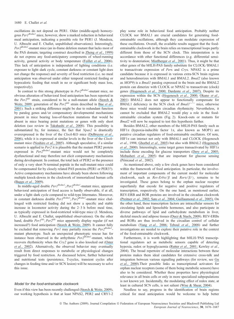
oscillations do not depend on PER1. Older (middle-aged) homozy-gous Per1Brdm1 mice, however, show a marked reduction in behavioralmeal anticipation, indicating a possible role for PER1 (J. Mendoza,U. Albrecht and E. Challet, unpublished observations). Interestingly,Per2Brdm1-mutant mice (an in-frame deletion mutant that lacks most ofthe PAS domain; targeting construct described in Zheng et al., 1999)do not express any food-anticipatory components of wheel-runningactivity, general activity or body temperature (Feillet et al., 2006).This lack of anticipation is independent of lighting conditions (i.e.exposure to light–dark cycle, constant darkness or constant light doesnot change the response) and severity of food restriction (i.e. no mealanticipation was observed under either temporal restricted feeding orhypocaloric feeding that result in no or significant body mass loss,respectively).In contrast to this strong phenotype in Per2Brdm1-mutant mice, no
obvious alteration of behavioral food anticipation has been reported inthe Per2ldc strain, considered to be a null-mutant allele (Storch &Weitz, 2009; generation of the Per2ldc strain described in Bae et al.,2001). Such a striking difference might be due to redundant functionby paralog genes and ⁄ or compensatory developmental mechanismspresent in mice bearing loss-of-function mutations that would beabsent in mice bearing point mutations or genes with only shortdeletion (see review in Takahashi et al., 2008). This possibility issubstantiated by, for instance, the fact that Npas2 is drasticallyoverexpressed in the liver of the Clock-KO mice (DeBruyne et al.,2006), while it is expressed at similar levels in the liver of the Clock-mutant mice (Noshiro et al., 2005). Although speculative, if a similarscenario is applied to Per2 it is plausible that the mutant PER2 proteinexpressed in Per2Brdm1-mutant mice would not be completelydysfunctional and may therefore not elicit compensatory mechanismsduring development. In contrast, the total lack of PER2 or the presenceof only a very short N-terminal peptide in the null-mutant mice couldbe compensated for by closely related PAS proteins (PER1 or PER3?).Active compensatory mechanisms have already been shown followingmultiple knock-downs in the clockwork of immortalized human cells(Baggs et al., 2009).In middle-aged double Per1Brdm1;Per2Brdm1-mutant mice, apparent
behavioral anticipation of food access is hardly observable, if at all,under a light–dark cycle compared to wild-type littermates. Moreover,in constant darkness double Per1Brdm1;Per2Brdm1-mutant mice chal-lenged with restricted feeding did not show a specific and stableincrease in locomotor activity during the 2–3 h before meal time,as typically expressed in food-restricted wild-type mice (J. Mendoza,U. Albrecht and E. Challet, unpublished observations). On the otherhand, double Per1ldc;Per2ldc mice apparently display regular (if notincreased?) food anticipation (Storch & Weitz, 2009). It cannot fullybe excluded that removing Per1 may partially rescue the Per2Brdm1-mutant phenotype. Such an unexpected phenotypic rescue has forinstance been observed in the arrhythmic Per2Brdm1-mutant, whichrecovers rhythmicity when the Cry2 gene is also knocked out (Osteret al., 2002). Alternatively, the observed behavior may eventuallyresult from direct responses to metabolic or physiological changestriggered by food restriction. As discussed below, further behavioraland nutritional tests (persistence, T-cycles, transient cycles afterchanges in feeding time and ⁄ or SCN lesion) will be necessary to solvethis issue.
Model for the food-entrainable clockwork
Even if this view has been recently challenged (Storch & Weitz, 2009),our working hypothesis is that at least NPAS2, PER2 and CRY1-2
play some role in behavioral food anticipation. Probably neitherCLOCK nor BMAL1 are crucial candidates for generating food-entrainable oscillations, although they can modulate expression ofthese oscillations. Overall, the available results suggest that the food-entrainable clockwork in the brain relies on transcriptional loops partlydifferent from those of the SCN clock. This interpretation is inaccordance with other functional differences (e.g. differential sensi-tivity to deuteriation; Mistlberger et al., 2001). Thus, it might be thatother genes of the bHLH-PAS family substitute for CLOCK ⁄ BMAL1to transactivate expression of Pers and Crys. NPAS2 is a primecandidate because it is expressed in various extra-SCN brain regionsand heterodimerizes with BMAL1 and BMAL2. Bmal2 (also knownas MOP9) is a Bmal1 paralog expressed in the brain and the BMAL2protein can dimerize with CLOCK or NPAS2 to transactivate (clock)genes (Hogenesch et al., 2000; Dardente et al., 2007). Despite itsexpression within the SCN (Hogenesch et al., 2000; Okano et al.,2001) BMAL2 does not appear to functionally compensate forBMAL1 deficiency in the SCN clock of Bmal1) ⁄ ) mice, otherwisethese mice would maintain circadian rhythmicity. Nevertheless, itcannot be excluded that BMAL2 would play a role in the food-entrainable circadian system (Fig. 2). Knock-outs or mutants forBmal2 will now be required to test this hypothesis further.Besides BMAL2, other members of the bHLH-PAS family such as
HIF1a (hypoxia-inducible factor 1a, also known as MOP1) areputative circadian regulators of food-entrainable oscillators. Of note,HIF1a can interact not only with BMAL1 and CLOCK (Hogeneschet al., 1998; Ghorbel et al., 2003) but also with BMAL2 (Hogeneschet al., 2000). Interestingly, some target genes transactivated by HIF1ainclude those encoding for glucose transporters (Dery et al., 2005;Mobasheri et al., 2005) that are important for glucose sensing(Penicaud et al., 2002).As mentioned above, only a few clock genes have been considered
within the framework of food anticipation. In particular, the involve-ment of important components of the current model for molecularclockwork, such as Rev-Erba ⁄ b and Rora ⁄ b ⁄ c, remains to beinvestigated. These genes belong to the orphan nuclear receptorsuperfamily that encode for negative and positive regulators oftranscription, respectively. On the one hand, as mentioned earlier,REV-ERB and ROR proteins are involved in the circadian clockwork(Preitner et al., 2002; Sato et al., 2004; Guillaumond et al., 2005). Onthe other hand, these transcription factors are intracellular sensors forcirculating lipids and liposoluble hormones, and also participate indiverse pathways of lipid and carbohydrate metabolism in liver,skeletal muscle and adipose tissues (Duez & Staels, 2008). REV-ERBsand RORs are thus involved in the circadian control of cellularmetabolism (Yang et al., 2006; Teboul et al., 2008) and furtherinvestigations are needed to explore their putative role in the contextof the food-entrainable clockwork.Furthermore, it is worth highlighting that bHLH ⁄ PAS transcrip-
tional regulators act as metabolic sensors capable of detectinghypoxia, redox or hypoglycaemia (Rutter et al., 2001; Kewley et al.,2004). The broad repertoire of molecular interactions between theseproteins makes them ideal candidates for extensive cross-talk andintegration between various signalling pathways (for review, see Guet al., 2000).Their possible links as transcriptional activators fororphan nuclear receptors (some of them being metabolic sensors) havealso to be considered. Whether these properties have physiologicalrelevance in all brain cells or only in more specialized subpopulationsis not known yet. Incidentally, the modulating effect of redox state, atleast in cultured SCN cells, is not salient (Wise & Shear, 2004).Needless to say, progress in the identification of brain regions
critical for meal anticipation would be welcome to help better
1680 E. Challet et al.
ª The Authors (2009). Journal Compilation ª Federation of European Neuroscience Societies and Blackwell Publishing LtdEuropean Journal of Neuroscience, 30, 1676–1687

understand the underlying clockwork (e.g. self-sustained properties,temporal patterns of clock gene expression, synchronizing cues etc).Moreover, not all brain clocks outside the suprachiasmatic nuclei sharethe same molecular oscillations (for review, see Guilding & Piggins,2007; Mendoza & Challet, 2009). Therefore, it is also conceivable thatthe different oscillatory structures underlying the food-entrainablenetwork use slightly different clockworks, a hypothesis that, if correct,would add another level of complexity.
Mice lacking specific feeding-related genes
Food intake and energy metabolism are regulated within the brain by anetwork of discrete nuclei in the mediobasal hypothalamus andbrainstem. Briefly, the mediobasal hypothalamus, including thearcuate and ventromedial hypothalamic nuclei, receives and integratesblood-borne nutritional signals from peripheral organs. Among thesesignals are metabolic cues (plasma glucose, free fatty acids),orexigenic cues mediated by circulating ghrelin synthesized in thestomach, and anorexigenic cues mediated by circulating leptin fromfat tissues, insulin from pancreas or intestinal gluco-incretin hormones(Penicaud et al., 2002; King, 2005).
Genetically obese Zucker ( fa ⁄ fa) rats have a missense mutation inthe leptin receptor gene (Cusin et al., 1996). Because obese Zuckerrats display greater food-anticipatory activity than do wild-typelittermates (Mistlberger & Marchant, 1999), leptin receptor signallingis dispensable (albeit negative regulator?) for food-anticipatoryprocesses.
Ghrelin receptor-KO mice express reduced food-anticipatory activ-ity, suggesting that ghrelin modulates food-anticipatory activity(LeSauter et al., 2009).
In the arcuate nuclei, several neuropeptides have opposite actions onfood intake: neuropeptide Y and Agouti-related peptide (AgRP) havepotent orexigenic effects while a-melanocyte stimulating hormone(a-MSH) and cocain- and amphetamine-related transcript (CART) areanorexigenic. Depending on their role, release of these peptides will beactivated or inhibited by peripheral signals of satiety or hunger,respectively. Release from nerve terminals of neuropeptide Y, AgRP,a-MSH and CART in targets of the arcuate nuclei, such as ventromedialhypothalamic nuclei (also sensitive to circulating leptin and glucose asthe arcuate nuclei are), paraventricular hypothalamic nuclei and lateralhypothalamic areas, will then modulate food intake and energy balance(Dietrich & Horvath, 2009). Of note, paraventricular hypothalamicnuclei produce two anorexigenic peptides, thyrotropin-releasinghormone and corticotrophin-releasing hormone, while the lateral andperifornical hypothalamic areas produce three orexigenic peptides,orexins A and B (also named hypocretins 1 and 2) and melanin-concentrating hormone (MCH).
Surprisingly, only a few rodent strains mutant for these feeding-related neuropeptides have been phenotyped for meal anticipation. Toour knowledge, anticipation responses to timed meals have not beeninvestigated yet in mice mutant for neuropeptide Y or its receptors.Chemical lesion of the arcuate nucleus leads to enhanced expressionof food-anticipatory behavior (Mistlberger & Antle, 1999). Such alesion destroyed neurons expressing neuropeptide Y, but probably alsoaffected expression of AgRP, a-MSH and CART in the arcuate region.Further investigations are thus needed to assess possible involvementof neuropeptide Y and other arcuate neuropeptides in the regulation offood anticipation.
Genetic ablation of orexin-producing neurons in mice leads to areduction in food-anticipatory activity and wakefulness during mealanticipation (Akiyama et al., 2004; Mieda et al., 2004), suggesting
that orexin neurons contribute to the regulation of food-anticipatoryactivity. This implication is probably specific to anticipatory activityand arousal because orexin-KO mice show reduced food-anticipatorymotor activity, while anticipatory body temperature is similar to that inwild-type mice (Kaur et al., 2008). Specific damage to orexin neuronscan be induced by intrahypothalamic injections of an immunotoxindirected against orexin neurons. To some extent, this treatment willlead to destruction of orexin neurons, as it would produce aconditional ablation of these neurons in adult rodents. In rats,however, the destruction of orexin-producing neurons does not impairfood-anticipatory components, either of food cup activity or ofdrinking behavior (Mistlberger et al., 2003), in keeping with the lackof effect after chemical lesion of the lateral hypothalamus in rats(Mistlberger & Rusak, 1988). Besides species differences, furtherwork would thus be beneficial to clarify the role of the orexin systemin meal anticipation.The putative involvement of the MCH system in food anticipation
has been investigated by studying mice deficient for the MCH1receptor. Mchr1) ⁄ ) mice display food-anticipatory activity indistin-guishable from that in wild-type mice, suggesting that MCH does notplay an essential role in that circadian behavior (Zhou et al., 2005).The MSH system mediates its actions on ingestive behavior and
energy metabolism via two melanocortin receptors, MCR3 andMCR4. Interestingly, food-anticipatory activity and wakefulness arereduced in Mcr3) ⁄ ) mice (Sutton et al., 2008).
Possible confounding effects
When phenotyping mutant mice for meal anticipation, one shouldclearly define what is meant by food-anticipatory activity. In wild-typemice, food-anticipatory activity typically begins 2–3 h before foodaccess and drops when the meal starts. It takes only 3–4 days ofrestricted feeding to see the first signs of food-anticipatory activity andthis becomes fairly stable in the next few days. As discussed later,there are several possibilities, especially in mutant mice, that can leadto misinterpretations of apparent ‘food-anticipatory’ behaviors. Evenminor procedural differences can influence the expression of food-anticipatory activity in mice (e.g. housing conditions; de Groot &Rusak, 2004). Furthermore, one should always bear in mind that, dueto putative pleiotropic effects of genes, their mutation may producealtered phenotypes not directly linked to their expected role (i.e.circadian rhythmicity in the case of clock genes), but whose alterationscan interfere with expression of food-anticipatory variables.
Measured parameters
Behavioral anticipation of food access can be recorded by variousmeans. A commonly used parameter is wheel-running activity but thishas the disadvantage of being rewarding in itself (de Visser et al.,2007). Other possibilities include general cage activity assessed by anintraperitoneal transmitter and infrared motion sensor, both devicesbeing considered equivalent for phenotyping food anticipation(Mistlberger et al., 2009b). Motor activity directed at a food bin hasalso been used (Mistlberger & Rusak, 1988; Davidson et al., 2000)but, in some instances, this behavior can be dissociated from food-anticipatory activity (e.g. Mistlberger & Rusak, 1988).Food-anticipatory processes are not limited to behavioral anticipa-
tion of meal time, even if this is the most studied parameter. Otheranticipatory physiological (e.g. core temperature), hormonal (e.g.corticosterone) or metabolic (e.g. free fatty acids) parameters can beconsidered clock hands of the food-entrainable circadian system.
Neurogenetics of food anticipation 1681
ª The Authors (2009). Journal Compilation ª Federation of European Neuroscience Societies and Blackwell Publishing LtdEuropean Journal of Neuroscience, 30, 1676–1687

However, because these processes are regulated by different down-stream pathways, altered expression in one single output parameterdoes not necessarily imply impaired function in the clockwork itself.Differential effects on outputs, as exemplified above by the unaffectedfood-anticipatory body temperature concomitant with a bluntedanticipatory activity in orexin-KO mice (Kaur et al., 2008), clearlysuggest that the affected pathways are efferent to the food-entrainableclockwork and, in that case, specific to behavioral activity and arousal.Thus, it is advisable to investigate at the same time several food-anticipatory components for a given genotype.
Circadian entrainment vs. Associated learning or hourglass
Food-anticipatory activity in rats and mice has long been conceptu-alized as the output of a self-sustained food-entrainable clock that doesnot rely on associated learning or hourglass measurement (Mistlberger,1994; Stephan, 2001). These features have been demonstrated in wild-type animals. Our point here is that these possibilities have to be testedin clock-mutant mice that may use compensatory clues to maintainapparent ‘food-anticipatory’ behaviors. For instance, wild-type miceexposed to food access at midday will express food-anticipatoryactivity during the 2–3 h prior to feeding time. If mutant mice start torun as soon as lights are on (e.g. at 06.00 h) until feeding time, thissustained activity lasting 6 h might have nothing to do with food-anticipatory activity, even if expressed before food access. In that case,the mice may use the time of light onset as an external time-giving cueso that they ‘remember’ that they will be fed 6 h after that signal. Tocheck this eventuality, it would be important to test food anticipationin mice of the same genotype under constant darkness where theywould then lose the time cue associated with lights on.Food-anticipatory activity is well-known not to be simply triggered
after a certain time lag (e.g. when depletion of energy stores reaches agiven threshold), indicating that the food-entrainable system is probablynot an hourglass (interval timer). Again, this is true in wild-type animalsbut merits checking in the studied mutant mice that may become more(or less) sensitive to peripheral metabolic cues. This test can be done bymodifying the circadian periodicity of food access (T-cycles).Even when the SCN clock is thought to be altered in clock-mutant
or KO mice, there might be interferences with expression of food-anticipatory processes. The SCN in mice can be synchronized torestricted feeding schedules under light–dark cycle or constantdarkness (Abe et al., 1989; Marchant & Mistlberger, 1997; Castilloet al., 2004; Mendoza et al., 2005). When a nocturnal activity (i.e.controlled by the SCN clock) is still expressed it may become difficultto distinguish between food-entrainable activity per se and a drift inthe nocturnal activity towards the time of feeding. In mutant micearrhythmic in constant darkness, daily rhythmicity induced byrestricted feeding may trigger some behavioral outputs from a subsetof newly coupled cells in the light-entrainable clock. In that case, theapparent food-anticipatory activity is expected to be distinct in termsof duration, intensity and stability from that expressed in wild-typelittermates. Thus, to circumvent a potential bias due to indirect effectson the SCN, it seems important to assess food-anticipatory processesin mutant mice with bilateral SCN lesions, as done in doubleCry1) ⁄ );Cry2) ⁄ ) mice (Iijima et al., 2005).Also, clock mutations may alter the sensitivity to synchronizing
cues. In the case of Per1Brdm1- and Per2Brdm1-mutant mice, the alteredresponses to light have been used to support the notion of a role forthese genes in photic resetting of the SCN (Albrecht et al., 2001).Altered synchronization has also been observed in Clock-mutant micein response to photic cues, interpreted as a result of reduced amplitude
of the SCN oscillations (Vitaterna et al., 2006). In the same vein, theClock mutation also modifies the synchronizing effects of nonphotic,including metabolic, cues on the SCN clock (Challet et al., 2000). It isthus possible that resetting properties of the food-entrainable clock arechanged by mutations of clock genes as well.
Homeostatic vs. Circadian regulation
When food-anticipatory processes are not observed under temporalrestricted feeding (with a daily time window of food access longenough to prevent body mass loss), it could be interesting toinvestigate what occurs under more severe conditions (i.e. conditionsleading to a body mass loss) such as hypocaloric feeding, which hasquite potent synchronizing effects (Challet et al., 1997; Mendozaet al., 2007). Under both conditions of food shortage, Per2Brdm1-mutant mice do not anticipate daily meals (Feillet et al., 2006),indicating that the lack of anticipation in this strain is not due tosecondary effects, such as deep torpor or hypothermia that wouldpreclude arousal at the right time. When no or reduced anticipation isfound in mutant mice during a restricted feeding paradigm that clearlyappears harmful for them (Dudley et al., 2003; Fuller et al., 2008), itwould be appropriate to repeat the experiment in less metabolicallychallenging conditions (e.g. with a longer daily access to food). Thishas been done with a progressive reduction in the duration of foodaccess for Bmal1) ⁄ ) mice which then display recurrent behavioralactivity before feeding time (Mistlberger et al., 2008; Pendergastet al., 2009; Storch & Weitz, 2009).Conversely, under conditions of positive energy balance, diet-
induced obesity is known to alter anticipation of food access (Personset al., 1993). Therefore, in mutant or transgenic mice thought to haveincreased adiposity (e.g. orexin ⁄ ataxin-3 and Mcr3r) ⁄ ); see Haraet al., 2001; and Sutton et al., 2008, respectively), the circadianresponses to meal anticipation evoked above might have beenmodified indirectly due to altered lipid metabolism.Because nutritional messages from peripheral organs would
constitute synchronizing inputs to the cerebral food-entrainable timingsystem, it is relevant to investigate possible alterations of plasmametabolites and hormonal cues in mutant mice. For instance, mice thatdo not express Bmal1 specifically in the liver show metabolicabnormalities, even under conditions of ad libitum food (Lamia et al.,2008).
Fasting test
One of the strong arguments for a food-entrainable clock is thereappearance of a bout of food-anticipatory activity in food-deprivedanimals at the previous time of restricted food access. Historically, thisdemonstration has been done in rats. Because rats and mice largelydiffer in terms of fat stores and metabolic rate, what is tolerable for ratsmay not be appropriate for mice. Indeed, rats can cope with fooddeprivation over several days before showing a fasting-induced rise indaytime locomotor activity (Challet et al., 1995, 1996). By contrast,mice can display fasting-induced hyperactivity as soon as after only asingle day of food deprivation (Challet et al., 1999; Pendergast et al.,2009), which may totally override any putative reappearance of food-anticipatory activity. Furthermore, even in mice that have not beenpreviously challenged with restricted feeding, the fasting-induced risein activity is not clearly distinguishable from the expected reappear-ance of food-anticipatory activity (Fig. 3). In our opinion, it isadvisable not to systematically use the fasting test (if possible, for nolonger than 1 day) in mice and to avoid it in skinny mutant mice.
1682 E. Challet et al.
ª The Authors (2009). Journal Compilation ª Federation of European Neuroscience Societies and Blackwell Publishing LtdEuropean Journal of Neuroscience, 30, 1676–1687

Lighting conditions
Light conditions can impact on the levels of locomotor activity. Inparticular, light exposure reduces or suppresses motor activity innocturnal rodents (Redlin, 2001). Therefore, in mutant mice exposedunder a light–dark cycle, low food-anticipatory activity during thedaytime may actually reflect an increased masking effect of light. Inthis case, a complementary investigation in constant darkness could beinformative, although the ‘nocturnal’ activity controlled by the SCN,either free-running or synchronized to feeding time, may becomesuperimposed on the food-anticipatory activity, thus precludingreliable quantitative estimation.
Furthermore, exposure to constant darkness in mice may activatelipolysis and favor torpor (Zhang et al., 2006), which could in turn
decrease the ability to anticipate meal time, independently of circadianmechanisms.Exposure to constant light has been shown to maintain circadian
rhythmicity in several clock-mutant mice that would otherwise bearrhythmic under constant darkness (Spoelstra et al., 2002; Abrahamet al., 2006). Moreover, in wild-type rats rendered arrhythmic by aprolonged exposure to constant illumination, daily restricted feeding isable to restore clock gene oscillation in the suprachiasmatic clock(Lamont et al., 2005). Thus, constant light could be a suitablecondition under which to obtain more insights into the food-entrainable system.
Cognitive deficits
A number of mutations in clock genes have been correlated withimpaired learning abilities (e.g. Npas2) ⁄ ) and Cry1) ⁄ );Cry2) ⁄ )
mice; Garcia et al., 2000; Van der Zee et al., 2008; respectively) oraltered behavioral responses (e.g. Clock-mutant mice; Easton et al.,2003).Any cognitive or emotional defect has to be taken into account as a
potential confounding effect when interpreting food-anticipatory data.Hypoactivity of a clock-mutant strain during food anticipation mightbe due to depressive-like behavior instead of involvement in food-entrainable mechanisms.
Genetic background
Last but not least, the genetic background in mice deeply influencesvarious parameters of circadian rhythmicity. For instance, a widestrain-dependent diversity has been shown for the free-running rhythmof locomotor activity controlled by the SCN clock (Mayeda &Hofstetter, 1999; Tankersley et al., 2002; Siepka & Takahashi, 2005)and its responsiveness to light (Schwartz & Zimmerman, 1990) ortimed feeding as well (Abe et al., 1989). Indeed, there are numerousexamples of how genetic background impacts on circadian behavior.For instance, depending on the genetic background in which the Clockmutation is established, mice can become either fattier (Turek et al.,2005) or leaner than wild-type littermates (Oishi et al., 2006). Asdiscussed previously, the Per2ldc-mutant mice are arrhythmic (Baeet al., 2001). However, when established in a different geneticbackground, the mice are perfectly rhythmic (Xu et al., 2007). Thereare also discrepancies between the circadian phenotype, which rangesfrom virtually unaffected to complete arrhythmicity, of the threedifferent Per1-KO strains that have been generated (Bae et al., 2001;Cermakian et al., 2001; Zheng et al., 2001). A difference in thegenetic background appears a plausible explanation although differ-ences in the targeting constructs themselves might also be invoked.Indeed, apart from possible complications arising from the geneticbackground issue, the phenotype of circadian mutant mice has to bescrutinized, also taking into account how the strain has been generated(random mutagenesis, gene targeting KO) and what this implies interms of mRNA and protein expression (truncated protein probablyretaining interaction with some partners while losing the ability tointeract with others, no protein at all, only a short peptide present…).Although further experiments will be needed to conclude so,
differences in the targeting construct could well explain in part whywe found that food-anticipatory behavior is missing in Per2Brdm1
while it is retained in Per2ldc. A putative effect of genetic backgroundcan be ruled out in this case as Storch & Weitz (2009) were careful toestablish the Per2ldc mutation in the same mixed C57BL ⁄ 6·129Svbackground. Surprisingly enough, wild-type mice of this mixed
Fig. 3. Behavioral responses in C57BL ⁄ 6·129Sv mice challenged withtemporal restricted feeding under a light–dark cycle and ⁄ or food deprivationin constant darkness. (A) Daily wheel-running activity in a mouse initially fedad libitum for 2 weeks and then exposed to a restricted feeding (food available4 h after lights on until lights off) for 3 weeks under a light–dark cycle. A boutof food-anticipatory activity (FAA) was expressed prior to the time of foodaccess. Thereafter, the mouse was transferred to constant darkness and fed adlibitum for 4 weeks until being challenged with a 2-day fasting period. A boutof locomotor activity was expressed in late subjective afternoon (correspondingmore or less to FAA under conditions of restricted feeding). (B) Daily wheel-running activity in a mouse fed ad libitum for 5 weeks under a light–dark cycleand transferred to constant darkness for 4 weeks until being challenged with a2-day fasting period. A fasting-induced rise in activity is visible during theusual inactive period. This fasting-induced rise in diurnal activity is not clearlydistinguishable from the so-called reappearance of food-anticipatory activityshown by the mouse in panel A. Successive 24-h periods are double-plotted(48 h horizontal time scale). Nighttime, when mice were under light–darkconditions, is indicated by a black bar on the abscissa.
Neurogenetics of food anticipation 1683
ª The Authors (2009). Journal Compilation ª Federation of European Neuroscience Societies and Blackwell Publishing LtdEuropean Journal of Neuroscience, 30, 1676–1687

genetic background display robust and consistent food-anticipatoryactivity in our hands (Feillet et al., 2006) while food anticipation forthis mouse strain is considered weak or undetectable in another study(Storch & Weitz, 2009).As a conclusion, it seems safe to say that great caution is required in
the interpretation of the circadian phenotypes observed for foodanticipation as they might be modulated, modified or disturbed byseveral methodological and technical variables, some of which are notreadily controlled.
Future directions of research
First of all, it will be necessary to pursue phenotyping mealanticipation in mutant mice for other known clock genes (e.g. Rev-erbs, Rors, Bmal2, Dec1-2) or closely related genes (e.g. Hif1a).Moreover, anticipation of feeding time could also be studied in micebearing brain-specific KO of clock genes. This strategy would avoidany secondary effect due to altered peripheral defects. Alternatively, itwould be interesting to restore altered food-anticipatory activity inclock-mutant mice by viral gene transfer. This elegant approach hasalready been used in that context to try to restore food anticipation inBmal1) ⁄ ) mice (Fuller et al., 2008; for a critique of the presenteddata, see Mistlberger et al., 2009a). Other promising methods includein vivo knock-down of clock gene expression by RNA interference.Besides molecular mechanisms underlying oscillations and identi-
fication of key brain structures, comprehension of the food-entrainableclock will not be possible without clarifying how it is reset by feedingcues. Concerning the light-entrainable clock, both Per1 and Per2genes play an important role in photic resetting (Albrecht et al., 1997,2001; Shigeyoshi et al., 1997; Akiyama et al., 1999; Yan et al., 1999;Yan & Silver, 2002). In contrast, genes mediating synchronization ofthe food-entrainable clock are not identified yet. To provide insightson these resetting mechanisms, we propose to expose clock-mutantmice to abrupt changes in the time of daily food access (‘jet-lag’ offeeding schedule) for determining the rate of re-entrainment. Ourhypothesis is that impaired re-entrainment of transient food-anticipa-tory activity to a new feeding schedule may reflect an involvement ofthe mutated gene in food resetting.As we have discussed in this review, it seems that the food-
entrainable clockwork in the brain relies on transcriptional loops partlydifferent from those described in the light-entrainable SCN clock. Toget a more detailed picture of the molecular underpinnings of the food-entrainable clock, it is now required that the behavioral screening beextended to additional clock-mutant strains. Additionally, the devel-opment of refined murine models with brain-specific conditional KOalong with targeted knock-down approaches aiming at various cerebralregions should help to localize the still elusive neural substrate(s) ofthe food-entrainable clock.
Acknowledgements
We thank Julien Delezie and Dr Mireille Masson-Pevet for constructivecomments. Our studies were supported by grants from Centre National de laRecherche Scientifique (E.C. and P.P.) and Agence Nationale pour la Recherche‘Jeunes Chercheurs ⁄ Jeunes Chercheuses’ (E.C. and J.M.).
Abbreviations
AgRP, Agouti-related peptide; ARNT, arylhydrocarbon receptor nucleartranslocator; bHLH, basic helix–loop–helix; BMAL1 or -2, brain and muscleARNT-like protein 1 or 2; CART, cocain- and amphetamine-related transcript;CLOCK, circadian locomotor output cycles kaput; CRY, cryptochrome; DBP,
D-site albumin-binding protein; FBXL, F-box protein; HIF, hypoxia-induciblefactor; KO, knock-out; MCH, melanin-concentrating hormone; MCR, mela-nocortin receptor; MOP, member of PAS superfamily; NPAS, neuronal PASdomain protein; PAS, PER-ARNT-SIM; PER, Period; REV-ERB, reverse viralerythroblastis oncogene product; ROR, retinoic acid receptor-related orphanreceptor; SCN, suprachiasmatic nuclei; SIM, single-minded; a-MSH,a-melanocyte stimulating hormone; b-TRCP, beta-transducin repeat-containingprotein.
References
Abe, H., Kida, M., Tsuji, K. & Mano, T. (1989) Feeding cycles entraincircadian rhythms of locomotor activity in CS mice but not in C57BL ⁄ 6Jmice. Physiol. Behav., 45, 397–401.
Abraham, D., Dallmann, R., Steinlechner, S., Albrecht, U., Eichele, G. & Oster,H. (2006) Restoration of circadian rhythmicity in circadain clock-deficientmice in constant light. J. Biol. Rhythms, 21, 169–176.
Akiyama, M., Kouzu, Y., Takahashi, S., Wakamatsu, H., Moriya, T., Maetani,M., Watanabe, S., Tei, H., Sakaki, Y. & Shibata, S. (1999) Inhibition of light-or glutamate-induced mPer1 expression represses the phase shifts into themouse circadian locomotor and suprachiasmatic firing rhythms. J. Neurosci.,19, 1115–1121.
Akiyama, M., Yuasa, T., Hayasaka, N., Horikawa, K., Sakurai, T. & Shibata, S.(2004) Reduced food anticipatory activity in genetically orexin (hypocretin)neuron-ablated mice. Eur. J. Neurosci., 20, 3054–3062.
Albrecht, U., Sun, Z.S., Eichele, G. & Lee, C.C. (1997) A differential responseof two putative mammalian circadian regulators, mper1 and mper2, to light.Cell, 91, 1055–1064.
Albrecht, U., Zheng, B., Larkin, D., Sun, Z.S. & Lee, C.C. (2001) mPer1 andmper2 are essential for normal resetting of the circadian clock. J. Biol.Rhythms, 16, 100–104.
Angeles-Castellanos, M., Mendoza, J. & Escobar, C. (2007) Restricted feedingschedules phase shift daily rhythms of c-Fos and protein Per1 immunore-activity in corticolimbic regions in rats. Neuroscience, 144, 344–355.
Bae, K., Jin, X., Maywood, E.S., Hastings, M.H., Reppert, S.M. & Weaver,D.R. (2001) Differential functions of mPer1, mPer2, and mPer3 in the SCNcircadian clock. Neuron, 30, 525–536.
Baggs, J.E., Price, T.S., DiTacchio, L., Panda, S., Fitzgerald, G.A. &Hogenesch, J.B. (2009) Network features of the mammalian circadianclock. PLoS Biol., 7, e1000052.
Balsalobre, A., Marcacci, L. & Schibler, U. (2000) Multiple signaling pathwayselicit circadian gene expression in cultured Rat-1 fibroblasts. Curr. Biol., 10,1291–1294.
Bunger, M.K., Wilsbacher, L.D., Moran, S.M., Clendenin, C., Radcliffe, L.A.,Hogenesch, J.B., Simon, M.C., Takahashi, J.S. & Bradfield, C.A. (2000)Mop3 is an essential component of the master circadian pacemaker inmammals. Cell, 103, 1009–1017.
Caba, M. & Gonzalez-Mariscal, G. (2009), The rabbit pup, a natural model ofnursing-anticipatory activity. Eur. J. Neurosci., 30, 1697–1706.
Caldelas, I., Feillet, C.A., Dardente, H., Eclancher, F., Malan, A., Gourmelen,S., Pevet, P. & Challet, E. (2005) Timed hypocaloric feeding and melatoninsynchronize the suprachiasmatic clockwork in rats, but with opposite timingof behavioral output. Eur. J. Neurosci., 22, 921–929.
Castillo, M.R., Hochstetler, K.J., Tavernier, R.J. Jr, Greene, D.M. & Bult-Ito,A. (2004) Entrainment of the master circadian clock by scheduled feeding.Am. J. Physiol. Regul. Integr. Comp. Physiol., 287, R551–R555.
Cermakian, N., Monaco, L., Pando, M.P., Dierich, A. & Sassone-Corsi, P.(2001) Altered behavoral rhythms and clock gene expression in mice with atargeted mutation in the Period1 gene. EMBO J., 20, 3967–3974.
Challet, E., le Maho, Y., Robin, J.P., Malan, A. & Cherel, Y. (1995)Involvement of corticosterone in the fasting-induced rise in proteinutilization and locomotor activity. Pharmacol. Biochem. Behav., 50, 405–412.
Challet, E., LeMaho, Y., Pevet, P., Nobelis, P. & Malan, A. (1996)Ventromedial hypothalamic lesions prevent the fasting-induced changes inday-night pattern of locomotor activity. Behav. Brain Res., 77, 155–163.
Challet, E., Pevet, P., Vivien-Roels, B. & Malan, A. (1997) Phase-advanceddaily rhythms of melatonin, body temperature, locomotor activity in food-restricted rats fed during daytime. J. Biol. Rhythms, 12, 65–79.
Challet, E., Losee-Olson, S. & Turek, F.W. (1999) Reduced glucose availabilityattenuates circadian responses to light in mice. Am. J. Physiol. Regul. Integr.Comp. Physiol., 276, R1063–R1070.
Challet, E., Takahashi, J.S. & Turek, F.W. (2000) Nonphotic phase-shifting inclock mutant mice. Brain Res., 859, 398–403.
1684 E. Challet et al.
ª The Authors (2009). Journal Compilation ª Federation of European Neuroscience Societies and Blackwell Publishing LtdEuropean Journal of Neuroscience, 30, 1676–1687

Cusin, I., Rohner-Jeanrenaud, F., Stricker-Krongrad, A. & Jeanrenaud, B.(1996) The weight-reducing effect of an intracerebroventricular bolusinjection of leptin in genetically obese fa ⁄ fa rats. Reduced sensitivitycompared with lean animals. Diabetes, 45, 1446–1450.
Damiola, F., Le Minh, N., Preitner, N., Kornmann, B., Fleury-Olela, F. &Schibler, U. (2000) Restricted feeding uncouples circadian oscillators inperipheral tissues from the central pacemaker in the suprachiasmatic nucleus.Genes Dev., 14, 2950–2961.
Dardente, H. & Cermakian, N. (2007) Molecular circadian rhythms in centraland peripheral clocks in mammals. Chronobiol. Int., 24, 195–213.
Dardente, H., Fortier, E.E., Martineau, V. & Cermakian, N. (2007)Cryptochromes impair phosphorylation of transcriptional activators inthe clock: a general mechanism for circadian repression. Biochem. J., 402,525–536.
Dardente, H., Mendoza, J., Fustin, J.M., Challet, E. & Hazlerigg, D.G. (2008)Implication of the F-Box Protein FBXL21 in circadian pacemaker functionin mammals. PLoS ONE, 3, e3530.
Dardente, H., Fustin, J.M. & Hazlerigg, D.G. (2009) Transcriptional feedbackloops in the ovine circadian clock. Comp. Biochem. Physiol., Part A Mol.Integr. Physiol., 153, 391–398.
Davidson, A.J. (2006) Search for the food-entrainable circadian oscillator: acomplex proposition. Am. J. Physiol. Regul. Integr. Comp. Physiol., 290,R1524–R1526.
Davidson, A.J., Cappendijk, S.L. & Stephan, F.K. (2000) Feeding-entrainedcircadian rhythms are attenuated by lesions of the parabrachial region in rats.Am. J. Physiol. Regul. Integr. Comp. Physiol., 278, R1296–R1304.
Davidson, A.J., Poole, A.S., Yamazaki, S. & Menaker, M. (2003) Is the food-entrainable circadian oscillator in the digestive system? Genes Brain Behav.,2, 32–39.
DeBruyne, J.P., Noton, E., Lambert, C.M., Maywood, E.S., Weaver, D.R. &Reppert, S.M. (2006) A clock shock: mouse CLOCK is not required forcircadian oscillator function. Neuron, 50, 465–477.
DeBruyne, J.P., Weaver, D.R. & Reppert, S.M. (2007a) Peripheral circadianoscillators require CLOCK. Curr. Biol., 17, R538–R539.
DeBruyne, J.P., Weaver, D.R. & Reppert, S.M. (2007b) CLOCK and NPAS2have overlapping roles in the suprachiasmatic circadian clock. Nat.Neurosci., 10, 543–545.
Dery, M.A., Michaud, M.D. & Richard, D.E. (2005) Hypoxia-inducible factor1: regulation by hypoxic and non-hypoxic activators. Int. J. Biochem. CellBiol., 37, 535–540.
Dietrich, M.O. & Horvath, T.L. (2009) Feeding signals and brain circuitry. Eur.J. Neurosci., 30, 1688–1696.
Dudley, C.A., Erbel-Sieler, C., Estill, S.J., Reick, M., Franken, P., Pitts, S. &McKnight, S.L. (2003) Altered patterns of sleep and behavioral adaptabilityin NPAS2-deficient mice. Science, 301, 379–383.
Duez, H. & Staels, B. (2008) Rev-erb alpha gives a time cue to metabolism.FEBS Lett., 582, 19–25.
Easton, A., Arbuzova, J. & Turek, F.W. (2003) The circadian clock mutationincreases exploratory activity and escape-seeking behavior. Genes BrainBehav., 2, 11–19.
Feillet, C.A., Ripperger, J.A., Magnone, M.C., Dulloo, A., Albrecht, U. &Challet, E. (2006) Lack of food anticipation in Per2 mutant mice. Curr. Biol.,16, 2016–2022.
Feillet, C.A., Mendoza, J., Albrecht, U., Pevet, P. & Challet, E. (2008a)Forebrain oscillators ticking with different clock hands.Mol. Cell. Neurosci.,37, 209–221.
Feillet, C.A., Mendoza, J., Pevet, P. & Challet, E. (2008b) Restricted feedingrestores rhythmicity in the pineal gland of arrhythmic suprachiasmatic-lesioned rats. Eur. J. Neurosci., 28, 2451–2458.
Fuller, P.M., Lu, J. & Saper, C.B. (2008) Differential rescue of light- and food-entrainable circadian rhythms. Science, 320, 1074–1077.
Garcia, J.A., Zhang, D., Estill, S.J., Michnoff, C., Rutter, J., Reick, M., Scott,K., Diaz-Arrastia, R. & McKnight, S.L. (2000) Impaired cued and contextualmemory in NPAS2-deficient mice. Science, 288, 2226–2230.
Ghorbel, M.T., Coulson, J.M. & Murphy, D. (2003) Cross-talk betweenhypoxic and circadian pathways: cooperative roles for hypoxia-induciblefactor 1alpha and CLOCK in transcriptional activation of the vasopressingene. Mol. Cell. Neurosci., 22, 396–404.
Godinho, S.I., Maywood, E.S., Shaw, L., Tucci, V., Barnard, A.R., Busino, L.,Pagano, M., Kendall, R., Quwailid, M.M., Romero, M.R., O’neill, J.,Chesham, J.E., Brooker, D., Lalanne, Z., Hastings, M.H. & Nolan, P.M.(2007) The after-hours mutant reveals a role for Fbxl3 in determiningmammalian circadian period. Science, 316, 897–900.
de Groot, M.H. & Rusak, B. (2004) Housing conditions influence the expressionof food-anticipatory activity in mice. Physiol. Behav., 83, 447–457.
Gu, Y.Z., Hogenesch, J.B. & Bradfield, C.A. (2000) The PAS superfamily:sensors of environmental and developmental signals. Annu. Rev. Pharmacol.Toxicol., 40, 519–561.
Guilding, C. & Piggins, H.D. (2007) Challenging the omnipotence of thesuprachiasmatic timekeeper: are circadian oscillators present throughout themammalian brain? Eur. J. Neurosci., 25, 3195–3216.
Guillaumond, F., Dardente, H., Giguere, V. & Cermakian, N. (2005)Differential control of Bmal1 circadian transcription by REV-ERB andROR nuclear receptors. J. Biol. Rhythms, 20, 391–403.
Hamada, T., Antle, M.C. & Silver, R. (2004) The role of Period1 in non-photicresetting of the hamster circadian pacemaker in the suprachiasmatic nucleus.Neurosci. Lett., 362, 87–90.
Hara, J., Beuckmann, C.T., Nambu, T., Willie, J.T., Chemelli, R.M., Sinton,C.M., Sugiyama, F., Yagami, K., Goto, K., Yanagisawa, M. & Sakurai, T.(2001) Genetic ablation of orexin neurons in mice results in narcolepsy,hypophagia, and obesity. Neuron, 30, 345–354.
Herzog, E.D. & Schwartz, W.J. (2002) A neural clockwork for encodingcircadian time. J. Appl. Physiol., 92, 401–408.
Hogenesch, J.B., Gu, Y.Z., Jain, S. & Bradfield, C.A. (1998) The basic-helix-loop-helix-PAS orphan MOP3 forms transcriptionally active complexes withcircadian and hypoxia factors. Proc. Natl Acad. Sci. USA, 95, 5474–5479.
Hogenesch, J.B., Gu, Y.Z., Moran, S.M., Shimomura, K., Radcliffe, L.A.,Takahashi, J.S. & Bradfield, C.A. (2000) The basic helix-loop-helix-PASprotein MOP9 is a brain-specific heterodimeric partner of circadian andhypoxia factors. J. Neurosci., 20, RC83.
Honma, K.I., Honma, S. & Hiroshige, T. (1984) Feeding-associated cortico-sterone peak in rats under various feeding cycles. Am. J. Physiol. Regul.Integr. Comp. Physiol., 246, R721–R726.
Honma, S., Kawamoto, T., Takagi, Y., Fujimoto, K., Sato, F., Noshiro, M.,Kato, Y. & Honma, K. (2002) Dec1 and Dec2 are regulators of themammalian molecular clock. Nature, 419, 841–844.
Horikawa, K., Minami, Y., Iijima, M., Akiyama, M. & Shibata, S. (2005) Rapiddamping of food-entrained circadian rhythm of clock gene expression inclock-defective peripheral tissues under fasting conditions. Neuroscience,134, 335–343.
Iijima, M., Yamaguchi, S., van der Horst, G.T., Bonnefont, X., Okamura, H. &Shibata, S. (2005) Altered food-anticipatory activity rhythm in Crypto-chrome-deficient mice. Neurosci. Res., 52, 166–173.
Jin, X., Shearman, L.P., Weaver, D.R., Zylka, M.J., de Vries, G.J. & Reppert,S.M. (1999) A molecular mechanism regulating rhythmic output from thesuprachiasmatic circadian clock. Cell, 96, 57–68.
Kaur, S., Thankachan, S., Begum, S., Blanco-Centurion, C., Sakurai, T.,Yanagisawa, M. & Shiromani, P.J. (2008) Entrainment of temperature andactivity rhythms to restricted feeding in orexin knock out mice. Brain Res.,1205, 47–54.
Kewley, R.J., Whitelaw, M.L. & Chapman-Smith, A. (2004) The mammalianbasic helix-loop-helix ⁄ PAS family of transcriptional regulators. Int. J.Biochem. Cell Biol., 36, 189–204.
King, P.J. (2005) The hypothalamus and obesity. Curr. Drug Targets, 6, 225–240.
King, D.P., Zhao, Y., Sangoram, A.M., Wilsbacher, L.D., Tanaka, M., Antoch,M.P., Steeves, T.D., Vitaterna, M.H., Kornhauser, J.M., Lowrey, P.L., Turek,F.W. & Takahashi, J.S. (1997) Positional cloning of the mouse circadianclock gene. Cell, 89, 641–653.
Ko, C.H. & Takahashi, J.S. (2006) Molecular components of the mammaliancircadian clock. Hum. Mol. Genet., 15, R271–R277.
Lamia, K.A., Storch, K.F. & Weitz, C.J. (2008) Physiological significance of aperipheral tissue circadian clock. Proc. Natl Acad. Sci. USA, 105, 15172–15177.
Lamont, E.W., Diaz, L.R., Barry-Shaw, J., Stewart, J. & Amir, S. (2005) Dailyrestricted feeding rescues a rhythm of period2 expression in the arrhythmicsuprachiasmatic nucleus. Neuroscience, 132, 245–248.
Lee, C., Weaver, D.R. & Reppert, S.M. (2004) Direct association betweenmouse PERIOD and CKIepsilon is critical for a functioning circadian clock.Mol. Cell. Biol., 24, 584–594.
LeSauter, J., Hoque, N., Weintraub, M., Pfaff, D.W. & Silver, R. (2009)Stomach ghrelin secreting cells as food-entrainable circadian clocks. Proc.Natl Acad. Sci. USA, 106, 13582–13587.
Lopez-Molina, L., Conquet, F., Dubois-Dauphin, M. & Schibler, U. (1997) TheDBP gene is expressed according to a circadian rhythm in the suprachias-matic nucleus and influences circadian behavior. EMBO J., 16, 6762–6771.
Lowrey, P.L., Shimomura, K., Antoch, M.P., Yamazaki, S., Zemenides, P.D.,Ralph, M.R., Menaker, M. & Takahashi, J.S. (2000) Positional synteniccloning and functional characterization of the mammalian circadian mutationtau. Science, 288, 483–492.
Neurogenetics of food anticipation 1685
ª The Authors (2009). Journal Compilation ª Federation of European Neuroscience Societies and Blackwell Publishing LtdEuropean Journal of Neuroscience, 30, 1676–1687

Maier, B., Wendt, S., Vanselow, J.T., Wallach, T., Reischl, S., Oehmke, S.,Schlosser, A. & Kramer, A. (2009) A large-scale functional RNAi screenreveals a role for CK2 in the mammalian circadian clock. Genes Dev., 23,708–718.
Marchant, E.G. & Mistlberger, R.E. (1997) Anticipation and entrainment tofeeding time in intact and SCN- ablated C57BL ⁄ 6j mice. Brain Res., 765,273–282.
Mayeda, A.R. & Hofstetter, J.R. (1999) A QTL for the genetic variance in free-running period and level of locomotor activity between inbred strains ofmice. Behav. Genet., 29, 171–176.
Maywood, E.S., O’Neill, J.S., Reddy, A.B., Chesham, J.E., Prosser, H.M.,Kyriacou, C.P., Godinho, S.I., Nolan, P.M. & Hastings, M.H. (2007) Geneticand molecular analysis of the central and peripheral circadian clockwork ofmice. Cold Spring Harb. Symp. Quant. Biol., 72, 85–94.
McDearmon, E.L., Patel, K.N., Ko, C.H., Walisser, J.A., Schook, A.C., Chong,J.L., Wilsbacher, L.D., Song, E.J., Hong, H.K., Bradfield, C.A. & Takahashi,J.S. (2006) Dissecting the functions of the mammalian clock protein BMAL1by tissue-specific rescue in mice. Science, 314, 1304–1308.
Meijer, J.H., Michel, S. & Vansteensel, M.J. (2007) Processing of daily andseasonal light information in the mammalian circadian clock. Gen. Comp.Endocrinol., 152, 159–164.
Mendoza, J. (2007) Circadian clocks: setting time by food. J. Neuroendocri-nol., 19, 127–137.
Mendoza, J. & Challet, E. (2009) Brain clocks: from the suprachiasmatic nucleito a cerebral network. Neuroscientist, 15, 477–488.
Mendoza, J., Graff, C., Dardente, H., Pevet, P. & Challet, E. (2005) Feedingcues alter clock gene oscillations and photic responses in the suprach-iasmatic nuclei of mice exposed to a light–dark cycle. J. Neurosci., 25,1514–1522.
Mendoza, J., Pevet, P. & Challet, E. (2007) Circadian and photic regulation ofclock and clock-controlled proteins in the suprachiasmatic nuclei of calorie-restricted mice. Eur. J. Neurosci., 25, 3691–3701.
Mieda, M., Williams, S.C., Sinton, C.M., Richardson, J.A., Sakurai, T. &Yanagisawa, M. (2004) Orexin neurons function in an efferent pathway of afood-entrainable circadian oscillator in eliciting food-anticipatory activityand wakefulness. J. Neurosci., 24, 10493–10501.
Mistlberger, R.E. (1994) Circadian food-anticipatory activity: formal modelsand physiological mechanisms. Neurosci. Biobehav. Rev., 18, 171–195.
Mistlberger, R.E. & Antle, M.C. (1999) Neonatal monosodium glutamate alterscircadian organization of feeding, food anticipatory activity and photicmasking in the rat. Brain Res., 842, 73–83.
Mistlberger, R.E. & Marchant, E.G. (1999) Enhanced food-anticipatorycircadian rhythms in the genetically obese Zucker rat. Physiol. Behav., 66,329–335.
Mistlberger, R.E. & Rusak, B. (1988) Food-anticipatory circadian rhythms inrats with paraventricular and lateral hypothalamic ablations. J. Biol.Rhythms, 3, 277–291.
Mistlberger, R.E., Marchant, E.G. & Kippin, T.E. (2001) Food-entrainedcircadian rhythms in rats are insensitive to deuterium oxide. Brain Res., 919,283–291.
Mistlberger, R.E., Antle, M.C., Kilduff, T.S. & Jones, M. (2003) Food- andlight-entrained circadian rhythms in rats with hypocretin-2-saporin ablationsof the lateral hypothalamus. Brain Res., 980, 161–168.
Mistlberger, R.E., Yamazaki, S., Pendergast, J.S., Landry, G.J., Takumi, T. &Nakamura, W. (2008) Comment on ‘‘Differential rescue of light- and food-entrainable circadian rhythms’’. Science, 322, 675a.
Mistlberger, R.E., Buijs, R.M., Challet, E., Escobar, C., Kalsbeek, A., Landry,G.J., Pevet, P. & Shibata, S. (2009a) Standards of evidence in chronobiology:critical review of a report that restoration of Bmal1 expression in thedorsomedial hypothalamus is sufficient to restore circadian food anticipatoryrhythms in Bmal1) ⁄ ) mice. J. Circadian Rhythms, 7, 3.
Mistlberger, R.E., Kent, B.A. & Landry, G.J. (2009b) Phenotyping foodentrainment: motion sensors and telemetry are equivalent. J. Biol. Rhythms,24, 95–98.
Mobasheri, A., Richardson, S., Mobasheri, R., Shakibaei, M. & Hoyland, J.A.(2005) Hypoxia inducible factor-1 and facilitative glucose transportersGLUT1 and GLUT3: putative molecular components of the oxygen andglucose sensing apparatus in articular chondrocytes. Histol. Histopathol., 20,1327–1338.
Nelson, W. & Halberg, F. (1986) Schedule-shifts, circadian rhythms andlifespan of freely-feeding and meal-fed mice. Physiol. Behav., 38, 781–788.
Noshiro, M., Furukawa, M., Honma, S., Kawamoto, T., Hamada, T., Honma,K. & Kato, Y. (2005) Tissue-specific disruption of rhythmic expression ofDec1 and Dec2 in clock mutant mice. J. Biol. Rhythms, 20, 404–418.
Oishi, K., Atsumi, G., Sugiyama, S., Kodomari, I., Kasamatsu, M., Machida,K. & Ishida, N. (2006) Disrupted fat absorption attenuates obesity inducedby a high-fat diet in Clock mutant mice. FEBS Lett., 580, 127–130.
Okano, T., Sasaki, M. & Fukada, Y. (2001) Cloning of mouse BMAL2 and itsdaily expression profile in the suprachiasmatic nucleus: a remarkableacceleration of Bmal2 sequence divergence after Bmal gene duplication.Neurosci. Lett., 300, 111–114.
Oster, H., Yasui, A., van der Horst, G. & Albrecht, U. (2002) Disruption ofmCry2 restores circadian rhythmicity in mPer2 mutant mice. Genes Dev., 16,2633–2638.
Pendergast, J.S., Nakamura, W., Friday, R.C., Hatanaka, F., Takumi, T. &Yamazaki, S. (2009) Robust food anticipatory activity in BMAL1-deficientmice. PLoS ONE, 4, e4860.
Penicaud, L., Leloup, C., Lorsignol, A., Alquier, T. & Guillod, E. (2002) Brainglucose sensing mechanism and glucose homeostasis. Curr. Opin. Clin. Nutr.Metab. Care, 5, 539–543.
Persons, J.E., Stephan, F.K. & Bays, M.E. (1993) Diet-induced obesityattenuates anticipation of food access in rats. Physiol. Behav., 54, 55–64.
Pitts, S., Perone, E. & Silver, R. (2003) Food-entrained circadian rhythms aresustained in arrhythmic Clk ⁄ Clk mutant mice. Am. J. Physiol. Regul. Integr.Comp. Physiol., 285, R57–R67.
Preitner, N., Damiola, F., Lopez-Molina, L., Zakany, J., Duboule, D., Albrecht,U. & Schibler, U. (2002) The orphan nuclear receptor REV-ERBalphacontrols circadian transcription within the positive limb of the mammaliancircadian oscillator. Cell, 110, 251–260.
Ralph, M.R., Foster, R.G., Davis, F.C. & Menaker, M. (1990) Transplantedsuprachiasmatic nucleus determines circadian period. Science, 247, 975–978.
Redlin, U. (2001) Neural basis and biological function of masking by light inmammals: suppression of melatonin and locomotor activity. Chronobiol. Int.,18, 737–758.
Reick, M., Garcia, J.A., Dudley, C. & McKnight, S.L. (2001) NPAS2: ananalog of clock operative in the mammalian forebrain. Science, 293, 506–509.
Rossner, M.J., Oster, H., Wichert, S.P., Reinecke, L., Wehr, M.C., Reinecke, J.,Eichele, G., Taneja, R. & Nave, K.A. (2008) Disturbed clockwork resettingin Sharp-1 and Sharp-2 single and double mutant mice. PLoS ONE, 3, e2762.
Rutter, J., Reick, M., Wu, L.C. & McKnight, S.L. (2001) Regulation of clockand NPAS2 DNA binding by the redox state of NAD cofactors. Science, 293,510–514.
Sato, T.K., Panda, S., Miraglia, L.J., Reyes, T.M., Rudic, R.D., McNamara, P.,Naik, K.A., FitzGerald, G.A., Kay, S.A. & Hogenesch, J.B. (2004) Afunctional genomics strategy reveals Rora as a component of the mammaliancircadian clock. Neuron, 43, 527–537.
Schibler, U., Ripperger, J. & Brown, S.A. (2003) Peripheral circadianoscillators in mammals: time and food. J. Biol. Rhythms, 18, 250–260.
Schwartz, W.J. & Zimmerman, P. (1990) Circadian timekeeping in BALB ⁄ cand C57BL ⁄ 6 inbred mouse strains. J. Neurosci., 10, 3685–3694.
Shigeyoshi, Y., Taguchi, K., Yamamoto, S., Takekida, S., Yan, L., Tei, H.,Moriya, T., Shibata, S., Loros, J.J., Dunlap, J.C. & Okamura, H. (1997)Light-induced resetting of a mammalian circadian clock is associated withrapid induction of the mPer1 transcript. Cell, 91, 1043–1053.
Shirogane, T., Jin, J., Ang, X.L. & Harper, J.W. (2005) SCFbeta-TRCPcontrols clock-dependent transcription via casein kinase 1-dependentdegradation of the mammalian period-1 (Per1) protein. J. Biol. Chem.,280, 26863–26872.
Siepka, S.M. & Takahashi, J.S. (2005) Forward genetic screens to identifycircadian rhythm mutants in mice. Methods Enzymol., 393, 219–229.
Siepka, S.M., Yoo, S.H., Park, J., Song, W., Kumar, V., Hu, Y., Lee, C. &Takahashi, J.S. (2007) Circadian mutant Overtime reveals F-box proteinFBXL3 regulation of cryptochrome and period gene expression. Cell, 129,1011–1023.
Spoelstra, K., Oklejewicz, M. & Daan, S. (2002) Restoration of self-sustainedcircadian rhythmicity by the mutant clock allele in mice in constantillumination. J. Biol. Rhythms, 17, 520–525.
Stephan, F.K. (2001) Food-entrainable oscillators in mammals. In Takahashi,J.S., Turek, F.W. & Moore, R.Y. (Eds), Circadian clocks, Volume 12,Handbook of Behavioral Neurobiology. Kluwer Academic ⁄ Plenum Publish-ers, New York, pp. 223–246.
Stephan, F.K. (2003) Broken circadian clocks: a clock gene mutation andentrainment by feeding. Am. J. Physiol. Regul. Integr. Comp. Physiol., 285,R32–R33.
Stephan, F.K., Swann, J.M. & Sisk, C.L. (1979) Entrainment of circadianrhytms by feeding schedules in rats with suprachiasmatic lesions. Behav.Neural Biol., 25, 545–554.
1686 E. Challet et al.
ª The Authors (2009). Journal Compilation ª Federation of European Neuroscience Societies and Blackwell Publishing LtdEuropean Journal of Neuroscience, 30, 1676–1687

Storch, K.F. & Weitz, C.J. (2009) Daily rhythms of food-anticipatorybehavioral activity do not require the known circadian clock. Proc. NatlAcad. Sci. USA, 106, 6808–6813.
Sutton, G.M., Perez-Tilve, D., Nogueiras, R., Fang, J., Kim, J.K., Cone, R.D.,Gimble, J.M., Tschop, M.H. & Butler, A.A. (2008) The melanocortin-3receptor is required for entrainment to meal intake. J. Neurosci., 28, 12946–12955.
Takahashi, J.S., Pinto, L.H. & Vitaterna, M.H. (1994) Forward and reversegenetic approaches to behavior in the mouse. Science, 264, 1724–1733.
Takahashi, J.S., Shimomura, K. & Kumar, V. (2008) Searching for genesunderlying behavior: lessons from circadian rhythms. Science, 322, 909–912.
Tankersley, C.G., Irizarry, R., Flanders, S. & Rabold, R. (2002) Circadianrhythm variation in activity, body temperature, and heart rate betweenC3H ⁄ HeJ and C57BL ⁄ 6J inbred strains. J. Appl. Physiol., 92, 870–877.
Teboul, M., Guillaumond, F., Grechez-Cassiau, A. & Delaunay, F. (2008) Thenuclear hormone receptor family round the clock. Mol. Endocrinol., 22,2573–2582.
Turek, F.W., Joshu, C., Kohsaka, A., Lin, E., Ivanova, G., McDearmon, E.,Laposky, A., Losee-Olson, S., Easton, A., Jensen, D.R., Eckel, R.H.,Takahashi, J.S. & Bass, J. (2005) Obesity and metabolic syndrome incircadian clock mutant mice. Science, 308, 1043–1045.
Van der Horst, G.T., Muijtjens,M., Kobayashi, K., Takano, R., Kanno, S., Takao,M., de Wit, J., Verkerk, A., Eker, A.P., Van Leenen, D., Buijs, R.M., Bootsma,D., Hoeijmakers, J.H. & Yasui, A. (1999) Mammalian Cry1 and Cry2 areessential for maintenance of circadian rhythms. Nature, 398, 627–630.
Van der Zee, E., Havekes, R., Barf, R.P., Hut, R.A., Nijholt, I.M., Jacobs, E.H.& Gerkema, M.P. (2008) Circadian time-place learning in mice depends onCry genes. Curr. Biol., 18, 844–848.
de Visser, L., van den, B.o.R., Stoker, A.K., Kas, M.J. & Spruijt, B.M. (2007)Effects of genetic background and environmental novelty on wheel runningas a rewarding behaviour in mice. Behav. Brain Res., 177, 290–297.
Vitaterna, M.H., King, D.P., Chang, A.M., Kornhauser, J.M., Lowrey, P.L.,McDonald, J.D., Dove, W.F., Pinto, L.H., Turek, F.W. & Takahashi, J.S.(1994) Mutagenesis and mapping of a mouse gene, Clock, essential forcircadian behavior. Science, 264, 719–725.
Vitaterna, M.H., Selby, C.P., Todo, T., Niwa, H., Thompson, C., Fruechte,E.M., Hitomi, K., Thresher, R.J., Ishikawa, T., Miyazaki, J., Takahashi, J.S.& Sancar, A. (1999) Differential regulation of mammalian period genes andcircadian rhythmicity by cryptochromes 1 and 2. Proc. Natl Acad. Sci. USA,96, 12114–12119.
Vitaterna, M.H., Ko, C.H., Chang, A.M., Buhr, E.D., Fruechte, E.M., Schook,A., Antoch, M.P., Turek, F.W. & Takahashi, J.S. (2006) The mouse Clock
mutation reduces circadian pacemaker amplitude and enhances efficacy ofresetting stimuli and phase-response curve amplitude. Proc. Natl Acad. Sci.USA, 103, 9327–9332.
Vosko, A.M., Schroeder, A., Loh, D.H. & Colwell, C.S. (2007) Vasoactiveintestinal peptide and the mammalian circadian system. Gen. Comp.Endocrinol., 152, 165–175.
Waddington Lamont, E., Harbour, V.L., Barry-Shaw, J., Renteria Diaz, L.,Robinson, B., Stewart, J. & Amir, S. (2007) Restricted access to food, butnot sucrose, saccharine, or salt, synchronizes the expression of Period2protein in the limbic forebrain. Neuroscience, 144, 402–411.
Wakamatsu, H., Yoshinobu, Y., Aida, R., Moriya, T., Akiyama, M. & Shibata,S. (2001) Restricted-feeding-induced anticipatory activity rhythm is associ-ated with a phase-shift of the expression of mPer1 and mPer2 mRNA in thecerebral cortex and hippocampus but not in the suprachiasmatic nucleus ofmice. Eur. J. Neurosci., 13, 1190–1196.
Wise, D.D. & Shear, J.B. (2004) Circadian tracking of nicotinamide cofactorlevels in an immortalized suprachiasmatic nucleus cell line. Neuroscience,128, 263–268.
Xu, Y., Toh, K.L., Jones, C.R., Shin, J.Y., Fu, Y.H. & Ptacek, L.J. (2007)Modeling of a human circadian mutation yields insights into clock regulationby PER2. Cell, 128, 59–70.
Yan, L. & Silver, R. (2002) Differential induction and localization of mPer1 andmPer2 during advancing and delaying phase shifts. Eur. J. Neurosci., 16,1531–1540.
Yan, L., Takekida, S., Shigeyoshi, Y. & Okamura, H. (1999) Per1 and Per2gene expression in the rat suprachiasmatic nucleus: circadian profile and thecompartment-specific response to light. Neuroscience, 94, 141–150.
Yang, X., Downes, M., Yu, R.T., Bookout, A.L., He, W., Straume, M.,Mangelsdorf, D.J. & Evans, R.M. (2006) Nuclear receptor expression linksthe circadian clock to metabolism. Cell, 126, 801–810.
Zhang, J., Kaasik, K., Blackburn, M.R. & Lee, C.C. (2006) Constant darknessis a circadian metabolic signal in mammals. Nature, 439, 340–343.
Zheng, B., Larkin, D.W., Albrecht, U., Sun, Z.S., Sage, M., Eichele, G., Lee,C.C. & Bradley, A. (1999) The mPer2 gene encodes a functional componentof the mammalian circadian clock. Nature, 400, 169–173.
Zheng, B., Albrecht, U., Kaasik, K., Sage, M., Lu, W., Vaishnav, S., Li, Q.,Sun, Z.S., Eichele, G., Bradley, A. & Lee, C.C. (2001) Nonredundant rolesof the mPer1 and mPer2 genes in the mammalian circadian clock. Cell, 105,683–694.
Zhou, D., Shen, Z., Strack, A.M., Marsh, D.J. & Shearman, L.P. (2005)Enhanced running wheel activity of both Mch1r- and Pmch-deficient mice.Regul. Pept., 124, 53–63.
Neurogenetics of food anticipation 1687
ª The Authors (2009). Journal Compilation ª Federation of European Neuroscience Societies and Blackwell Publishing LtdEuropean Journal of Neuroscience, 30, 1676–1687