Detection of the CMT1A/HNPP recombination hotspot in unrelated patients of European descent
Upload
uni-marburgCategory
view
3download
0
r Human Brain Mapping 30:3309–3324 (2009) r
Neural Integration of Iconic and UnrelatedCoverbal Gestures: A Functional MRI Study
Antonia Green,1,2* Benjamin Straube,1,2 Susanne Weis,3 Andreas Jansen,2,4
Klaus Willmes,5 Kerstin Konrad,6 and Tilo Kircher4
1Department of Psychiatry and Psychotherapy—Section Experimental Psychopathology,RWTH Aachen University, Aachen, Germany
2Interdisciplinary Centre for Clinical Research ‘‘Biomat.,’’ RWTH Aachen University,Aachen, Germany
3Department of Neurology—Section Clinical Neuropsychology, RWTH Aachen University,Aachen, Germany
4Department of Psychiatry und Psychotherapy, Philipps University Marburg, Marburg, Germany5Department of Neurology—Section Neuropsychology, RWTH Aachen University, Aachen, Germany6Department of Child and Adolescent Psychiatry and Psychotherapy—Section Child Neuropsychology,
RWTH Aachen University, Aachen, Germany
r r
Abstract: Gestures are an important part of interpersonal communication, for example by illustrating phys-ical properties of speech contents (e.g., ‘‘the ball is round’’). The meaning of these so-called iconic gesturesis strongly intertwined with speech. We investigated the neural correlates of the semantic integration forverbal and gestural information. Participants watched short videos of five speech and gesture conditionsperformed by an actor, including variation of language (familiar German vs. unfamiliar Russian), variationof gesture (iconic vs. unrelated), as well as isolated familiar language, while brain activation was measuredusing functional magnetic resonance imaging. For familiar speech with either of both gesture types con-trasted to Russian speech-gesture pairs, activation increases were observed at the left temporo-occipitaljunction. Apart from this shared location, speech with iconic gestures exclusively engaged left occipitalareas, whereas speech with unrelated gestures activated bilateral parietal and posterior temporal regions.Our results demonstrate that the processing of speech with speech-related versus speech-unrelated ges-tures occurs in two distinct but partly overlapping networks. The distinct processing streams (visual versuslinguistic/spatial) are interpreted in terms of ‘‘auxiliary systems’’ allowing the integration of speech andgesture in the left temporo-occipital region. Hum Brain Mapp 30:3309–3324, 2009. VC 2009 Wiley-Liss, Inc.
Keywords: fMRI; perception; language; semantic processing; multimodal integration
r r
Additional Supporting Information may be found in the onlineversion of this article.
Contract grant sponsor: Interdisciplinary Centre for ClinicalResearch ‘‘BIOMAT’’ within the Faculty of Medicine at the RWTHAachen University; Contract grant number: VV N68-e; Contractgrant sponsor: Deutsche Forschungsgemeinschaft (DFG); Contractgrant number: IRTG 1328.
*Correspondence to: Antonia Green, Department of Psychiatryund Psychotherapy, Philipps-University Marburg, Rudolf-
Bultmann-Straße 8, D-35039 Marburg, Germany.E-mail: [email protected]
Received for publication 14 July 2008; Revised 31 December 2008;Accepted 20 January 2009
DOI: 10.1002/hbm.20753Published online 6 April 2009 in Wiley InterScience (www.interscience.wiley.com).
VC 2009 Wiley-Liss, Inc.
INTRODUCTION
Everyone uses hands and arms to transmit informationusing gestures. Most prevalent are iconic gestures, sharinga formal relationship with the co-occurring speech content.Iconic gestures illustrate forms, shapes, events or actionsthat are the topic of the simultaneously occurring speech.The phrase ‘‘The fisherman caught a huge fish,’’ for exam-ple, is often accompanied by a gesture indicating the sizeof the fish, which helps the listener to understand howincredibly big the prey was [McNeill, 1992].
How precisely this enhanced understanding is achievedis still unclear. While speech reveals information only suc-cessively, gesture can convey multifaceted information atone time [Kita and Ozyurek, 2003; McNeill, 1992]. Thus, tocombine speech and gestures into one representation ges-tures have to be integrated into the successively unfoldinginterpretation of speech semantics. Since the meaning oficonic gestures is not fixed but has to be constructedonline by the listener according to the sentence content,interaction of speech and gesture processing is required ona semantic-conceptual level, as previous studies usingevent-related potentials have demonstrated [Holle andGunter, 2007; Ozyurek et al., 2007; Wu and Coulson, 2007].This interaction is distinct from basic sensory level proc-essing as it has been studied in previous multisensorystudies [for a review see Calvert, 2001]. Although informa-tion transmitted by gestures is different from speech, bothmodalities are tightly intertwined [McNeill, 1992]: Speechand gestures usually transmit at least a similar meaning,the most meaningful part of the gesture (stroke) tempo-rally aligned with the respective speech segment and bothaiming at communicating a message. Hence, a processingmechanism integrating both sources of information seemsrather likely.
Findings about the neural correlates of isolated process-ing of speech or gestures converge on the assumption thatunderstanding language as well as gestures relies onpartly overlapping brain networks [for a review see Wil-lems and Hagoort, 2007]. Speech-gesture interactions sofar have only been targeted by a few recent fMRI studiesthat used different kinds of paradigms: either mismatchmanipulations [gestures that were incongruent with theutterance; Willems et al., 2007], a disambiguation para-digm [gestures clarifying the meaning of ambiguouswords; Holle et al., 2008] or a memory paradigm [effectsof speech-gesture relatedness on subsequent memory per-formance; Straube et al., in press]. Across these studies,brain activations were reported in the left inferior frontalgyrus (IFG), inferior parietal cortex, posterior temporalregions (superior temporal sulcus, middle temporal gyrus)and in the precentral gyrus. Activity in the left IFG wasrepeatedly found for speech-gesture pairs that were moredifficult to integrate (mismatching speech-gesture pairs orabstract sentence contents), temporal activations have beenrelated to semantic integration, and inferior parietalinvolvement has been related to action processing, some-
times interpreted in terms of the putative human mirrorneuron system (MNS) [Rizzolatti and Craighero, 2004].This system is meant to determine the goals of actions byan observation-execution matching process [Craigheroet al., 2007] showing stronger activations when the initialgoal hypothesis (internal motor simulation) of an observedaction is not matched by the visual input and initiatingnew simulation cycles. To summarize, due to the differentparadigms it cannot be clearly stated which regions in thebrain are implicated in natural speech-gesture integration.
In the present study, we investigated the neural net-works engaged in the integration of speech and iconic ges-tures. By ‘‘integration’’ we refer to implicitly initiatedcognitive processes of combining semantic audiovisual in-formation into one representation. We assume that thisleads to increased processing demands which should bereflected in activation increases in brain regions involvedin the processing of gestures with familiar speech asopposed to gestures with unfamiliar speech. Thisincreased activation is hypothesized to reflect the creationof a semantic connection of both modalities. Subjects werepresented with short video clips of an actor speaking Ger-man (comprehensible) or Russian sentences (incomprehen-sible to our subjects who had no knowledge of Russian)that were either presented in isolation, were accompaniedby iconic gestures, or by gestures that were unrelated tothe sentence. The Russian language and speech-unrelatedgesture conditions were used as control conditions inorder to be able to present both modalities, but preventingsemantic processing in the respective irrelevant modality.
We were interested in two questions: first, we wanted toidentify brain areas activated by the natural semantic inte-gration of speech and iconic gestures. Second, we exam-ined whether the same cerebral network is engaged in theprocessing of speech with speech-unrelated gestures.
We hypothesized that integration of congruent iconicspeech-gesture information would be reflected in leftposterior temporal activations [Holle et al., 2008; Straubeet al., in press]. For unrelated gestures we expected thatbecause of less congruency between speech and gesturesmainly parietal regions would be involved, most likelyreflecting the mapping of irrelevant complex movementsonto speech [Willems et al., 2007].
MATERIALS AND METHODS
Participants
Sixteen healthy male subjects were included in the study(mean age 28.8 � 8.3 years, range 23–55 years). All partici-pants were right handed [Oldfield, 1971], native Germanspeakers and had no knowledge of Russian. The subjectshad normal or corrected-to-normal vision, none reportedhearing deficits. Exclusion criteria were a history of rele-vant medical or psychiatric illness of the participanthimself or in his first-degree relatives. All subjects gave
r Green et al. r
r 3310 r
written informed consent before participation in the study.The study was approved by the local ethics committee.
Stimulus Material
The stimulus production is described in more detail inStraube et al. [in press] for gestures in the context ofabstract speech contents (metaphoric coverbal gestures),here only a shorter description is given. In the currentstudy though, different stimuli with concrete speech con-tents were used, but these were produced accordingly.
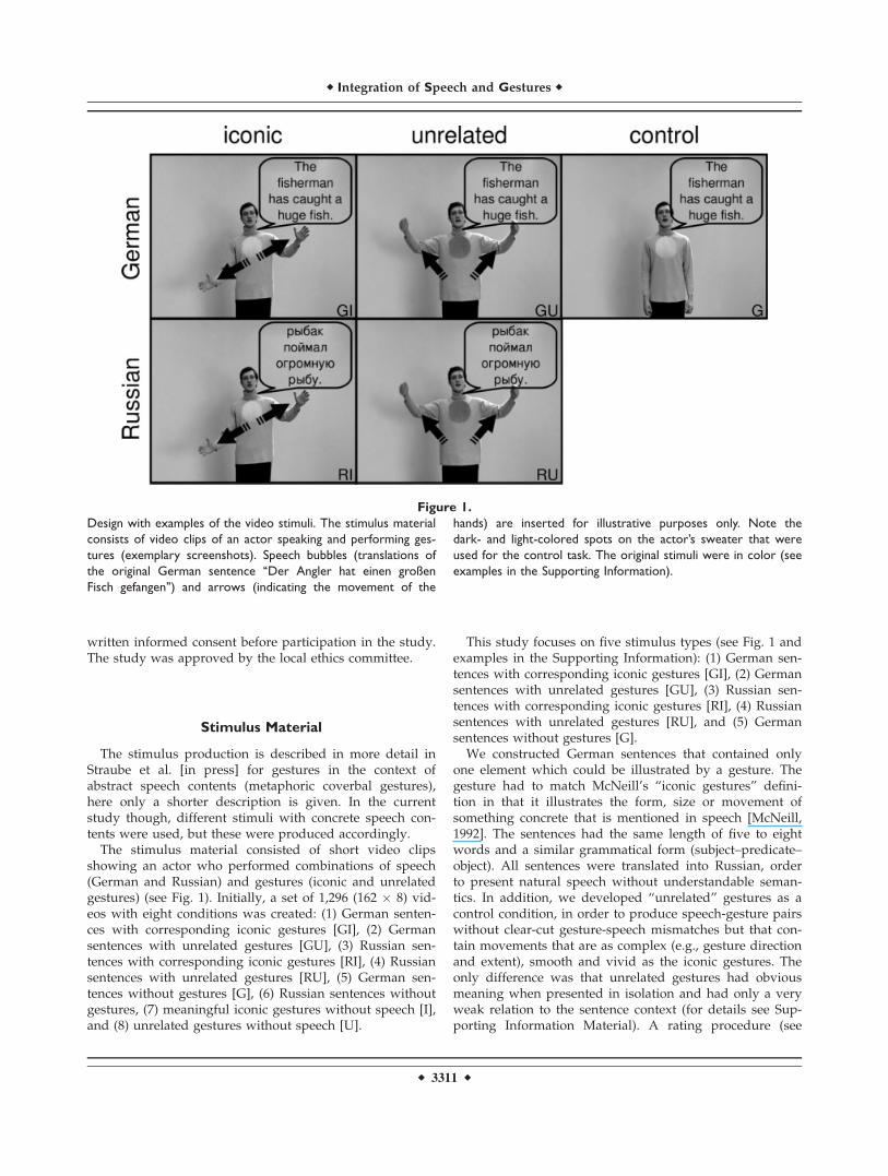
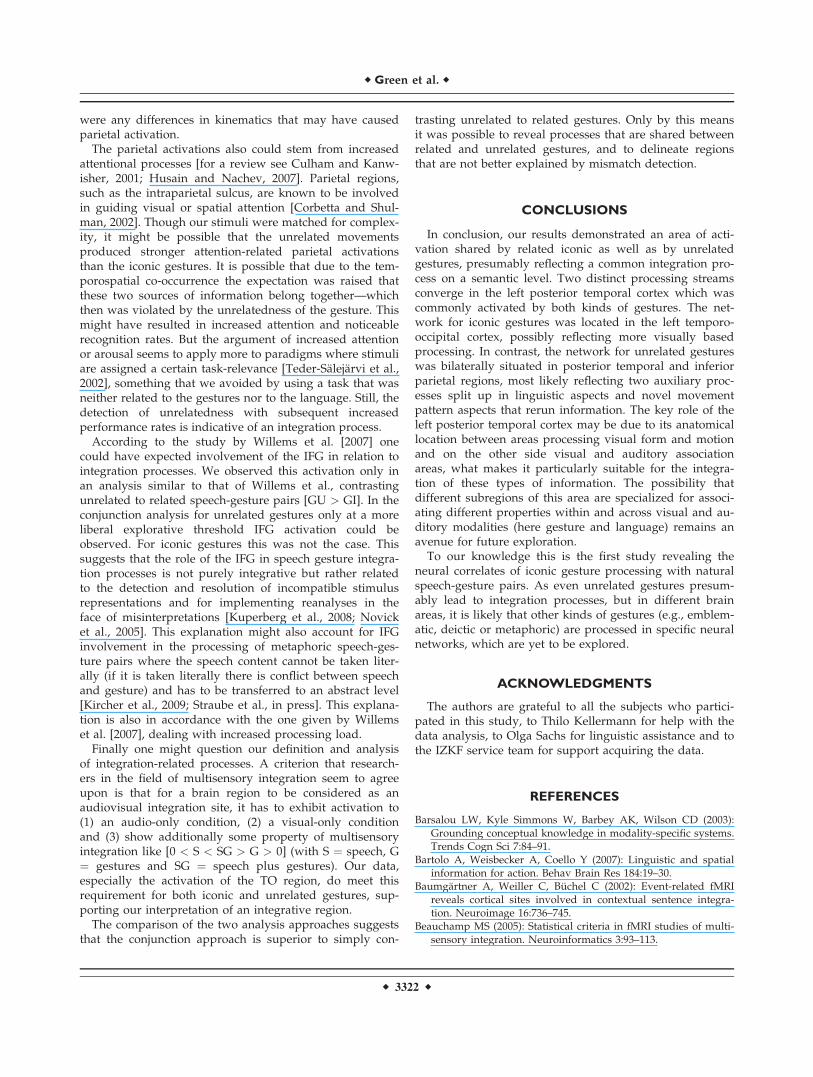
The stimulus material consisted of short video clipsshowing an actor who performed combinations of speech(German and Russian) and gestures (iconic and unrelatedgestures) (see Fig. 1). Initially, a set of 1,296 (162 � 8) vid-eos with eight conditions was created: (1) German senten-ces with corresponding iconic gestures [GI], (2) Germansentences with unrelated gestures [GU], (3) Russian sen-tences with corresponding iconic gestures [RI], (4) Russiansentences with unrelated gestures [RU], (5) German sen-tences without gestures [G], (6) Russian sentences withoutgestures, (7) meaningful iconic gestures without speech [I],and (8) unrelated gestures without speech [U].
This study focuses on five stimulus types (see Fig. 1 andexamples in the Supporting Information): (1) German sen-tences with corresponding iconic gestures [GI], (2) Germansentences with unrelated gestures [GU], (3) Russian sen-tences with corresponding iconic gestures [RI], (4) Russiansentences with unrelated gestures [RU], and (5) Germansentences without gestures [G].
We constructed German sentences that contained onlyone element which could be illustrated by a gesture. Thegesture had to match McNeill’s ‘‘iconic gestures’’ defini-tion in that it illustrates the form, size or movement ofsomething concrete that is mentioned in speech [McNeill,1992]. The sentences had the same length of five to eightwords and a similar grammatical form (subject–predicate–object). All sentences were translated into Russian, orderto present natural speech without understandable seman-tics. In addition, we developed ‘‘unrelated’’ gestures as acontrol condition, in order to produce speech-gesture pairswithout clear-cut gesture-speech mismatches but that con-tain movements that are as complex (e.g., gesture directionand extent), smooth and vivid as the iconic gestures. Theonly difference was that unrelated gestures had obviousmeaning when presented in isolation and had only a veryweak relation to the sentence context (for details see Sup-porting Information Material). A rating procedure (see
Figure 1.
Design with examples of the video stimuli. The stimulus material
consists of video clips of an actor speaking and performing ges-
tures (exemplary screenshots). Speech bubbles (translations of
the original German sentence ‘‘Der Angler hat einen großen
Fisch gefangen’’) and arrows (indicating the movement of the
hands) are inserted for illustrative purposes only. Note the
dark- and light-colored spots on the actor’s sweater that were
used for the control task. The original stimuli were in color (see
examples in the Supporting Information).
r Integration of Speech and Gestures r
r 3311 r

below and in the Supporting Information) has proven thatin fact our unrelated gestures did not contain any clear-cutsemantic information and that they differed significantlyin semantic strength from the iconic gestures.
A male bilingual actor was instructed to produce eachutterance together with an iconic or unrelated gesture in away that he felt as the originator of the gestures. Thus, thesynchrony of speech and gesture was determined by theactor and was left unchanged during the editing process.It is important to note that the unrelated gestures wereonly roughly choreographed, but not previously scripted.They rather were developed in collaboration with theactor, often derived on the basis of already used iconicgestures and trained until they looked and felt likeintrinsically produced spontaneous gestures. By doing sowe ensured that the gestures evolved at the correctmoment and were as smooth and dynamic as the iconicgestures. Only when a specific GI sentence was recordedtogether with an iconic gesture in a natural way the re-spective control conditions were produced successively.This was done to keep all item characteristics constant(e.g., sentence duration or movement complexity), regard-less of the manipulated factors of language (German,Russian, no speech) and gesture (iconic, unrelated, no ges-ture). Before and after the utterance the actor stood withhands hanging comfortably. Each clip had a duration of5,000 ms, including at least 500 ms before and after thescene, where the actor neither spoke nor moved. This wasdone to account for variable utterance lengths to get stand-ardized clips.
The recorded sentences presented here had an averagespeech duration of 2,330 ms (SD ¼ 390 ms) and an averagegesture duration of 2,700 ms (SD ¼ 450 ms) and did notdiffer between conditions (see Table S-II, Supporting Infor-mation Material).
Based upon ratings of understandability, imageabilityand naturalness (see Supporting Information Material, Ta-ble S-I) as well as upon parameters such as movementcharacteristics, pantomimic content, transitivity or handed-ness, we chose a set of 1,024 video clips (128 German sen-tences with iconic gestures and their counterparts in theother seven conditions) from the initial set of 1,296 clips asstimuli for the fMRI experiment. Stimuli were divided intofour sets in order to present each participant with 256 clipsduring the scanning procedure (32 items per condition),counterbalanced across subjects. Across subjects, each itemwas presented in all eight conditions but a single partici-pant only saw complementary derivatives of one item, i.e.the same sentence or gesture information was only seenonce per participant. This was done to prevent speech orgesture repetition effects. Again, all parameters listedabove were used for an equal assignment of video clips tothe four experimental sets, to avoid set-related between-subject differences.
Previous research has shown that coverbal gestures arenot only semantically but also temporally aligned with thecorresponding utterance. We assured this natural syn-
chrony by making the actor initiate the gestures and addi-tionally checked for it when postprocessing the videoclips. Each sentence contained only one element that couldbe illustrated, which was intuitively done by the actor.During postprocessing we checked in the GI conditionwith which word the gesture stroke (peak movement)coincided and then chose the end of this word as the timepoint of highest semantic correspondence between speechand the gesture stroke, assuming that the stroke of the ges-ture precedes or ends at, but does not follow the phono-logical peak syllable of speech [McNeill, 1992]. Forexample, for the sentence ‘‘The house has a vaulted roof’’the temporal alignment was marked at the end of theword (‘‘vaulted’’) coinciding with and corresponding tothe iconic gesture (form representation) in the GI condi-tion. The time of alignment was then transferred to theRussian equivalent (condition RI). This was possiblebecause of the standardized timing, form and structure ofeach of the different conditions corresponding to an item.Accordingly, the time of alignment was determined for theGU and G conditions (end of ‘‘vaulted’’) and transferredto the Russian counterparts. The concordance between GIand GU conditions was checked and revealed no differen-ces, the times of alignment occurred on average 1.17 s (GI,SD ¼ 0.51 second)/1.19 seconds (GU, SD ¼ 0.50 second)after the actual gesture onset and 1.53 s (GI, SD ¼ 0.53 sec-ond)/1.54 seconds (GU, SD ¼ 0.58 second) before theactual gesture offset. The time of alignment representingthe stroke-speech synchrony occurred on average 2,150 ms(SD ¼ 535 ms) after the video start (1,650 ms after speechonset) (see Table S-II, Supporting Information Material)and was used for the modulation of events in the event-elated fMRI analysis.
Experimental Design and Procedure
An experimental session comprised 256 trials (32 foreach condition) and consisted of four 11-minute blocks.Each block contained 64 trials with a matched number ofitems from each condition. The stimuli were presented inan event-related design in pseudo-randomized order andcounterbalanced across subjects. Each clip was followedby a fixation cross on grey background with a variableduration of 3,750–6,750 ms (average: 5,000 ms).
During scanning participants were instructed to watchthe videos and to indicate via left hand key presses at thebeginning of each video whether the spot displayed on theactor’s sweater was light or dark colored. This task waschosen to focus participants’ attention on the middle ofthe screen and enabled us to investigate implicit speechand gesture processing without possible instruction-relatedattention biases. Performance rates and reaction timeswere recorded. Before scanning, each participant receivedat least 10 practice trials outside the scanner, which weredifferent from those used in the main experiment. Before
r Green et al. r
r 3312 r
the experiment started, the volume of the videos was indi-vidually adjusted so that the clips were clearly audible.
Fifteen minutes after scanning an unannounced recogni-tion paradigm was used to control for participants’ atten-tion and to examine the influence of gestures on memoryperformance. All videos of the German-iconic and Ger-man-unrelated conditions (32 each) and half of the isolatedspeech condition (16) were presented in random order, to-gether with an equal number of new items for each of thethree conditions (altogether 160 videos). Participants hadto indicate via key press whether they had seen that clipbefore or not. Memory data of three participants are miss-ing for technical reasons.
MRI Data Acquisition
The video clips were presented via MR-compatiblevideo goggles (stereoscopic display with up to 1,024 � 768pixel resolution) and nonmagnetic headphones. Further-more, participants wore ear plugs, which act as an addi-tional low-pass filter.
All MRI data were acquired on a Philips Achieva 3Tscanner. Functional images were acquired using a T2*-weighted echo planar image sequence (TR ¼ 2 seconds,TE ¼ 30 ms, flip angle 90�, slice thickness 3.5 mm with a0.3-mm interslice gap, 64 � 64 matrix, FoV 240 mm, in-plane resolution 3.5 � 3.5 mm, 31 axial slices orientatedparallel to the AC-PC line covering the whole brain). Fourruns of 330 volumes were acquired during the experiment.The onset of each trial was synchronized to a scannerpulse. Additionally, an anatomical scan was acquired foreach participant using a high resolution T1-weighted 3D-sequence consisting of 180 sagittal slices (TR ¼ 9,863 ms,TE ¼ 4.59 ms, FoV ¼ 256 mm, slice thickness 1 mm, inter-slice gap ¼ 1 mm).
MRI Data Analysis
SPM2 (www.fil.ion.ucl.ac.uk) standard routines and tem-plates were used for analysis of fMRI data. After discard-ing the first five volumes to minimize T1-saturationeffects, all images were spatially and temporally realigned,normalized (resulting voxel size 4 � 4 � 4 mm3),smoothed (8 mm isotropic Gaussian filter) and high-passfiltered (cut-off period 128 seconds).
Statistical analysis was performed in a two-level, mixed-effects procedure. At the first level, single-subject BOLDresponses were modeled by a design matrix comprisingthe onsets of each event (i.e., time of alignment, see stimu-lus construction) of all eight experimental conditions. Thehemodynamic response was modeled by the canonicalhemodynamic response function (HRF) and its temporalderivative. The volume of interest was restricted to greymatter voxels by use of an inclusive mask created fromthe segmentation of the standard brain template. Parame-ter estimate (b-) images for the HRF were calculated for
each condition and each subject. Direct contrasts betweenevents (GI > RI, GI > G, GI > I, GU > RU, GU > G, GU> U, GU > GI) were computed per participant. At the sec-ond level, a random-effects group analysis was performedby entering corresponding contrast images of the first levelfor each participant into one-sample t-tests to compute sta-tistical parametric maps for the above contrasts. All differ-ence contrasts were inclusively masked by their minuendsto ensure that only differences with respect to the activa-tions of the first condition are evaluated. Further analysesand the contrasts of interest are described below in moredetail.
We chose to employ Monte-Carlo simulation of thebrain volume to establish an appropriate voxel contiguitythreshold [Slotnick and Schacter, 2004]. This correction hasthe advantage of higher sensitivity to smaller effect sizes,while still correcting for multiple comparisons across thewhole brain volume. The procedure is based on the factthat the probability of observing clusters of activity due tovoxel-wise Type I error (i.e., noise) decreases systemati-cally as cluster size increases. Therefore, the cluster extentthreshold can be determined to ensure an acceptable levelof corrected cluster-wise Type I error. To implement suchan approach, we ran a Monte-Carlo simulation to modelthe brain volume (http://www2.bc.edu/�slotnics/scripts.htm; with 1,000 iterations), using the same parame-ters as in our study [i.e., acquisition matrix, number of sli-ces, voxel size, resampled voxel size, FWHM of 6.5 mm—this value was estimated using the t-statistic map associ-ated with the contrast of interest [GI > RI \ GI > G \ GU> RU \ GU > G]; similar procedures have been used pre-viously to estimate fMRI spatial correlation, e.g., see Kata-noda et al., 2002; Ross and Slotnick, 2008; Zarahn et al.,1997]. An individual voxel threshold was then applied toachieve the assumed voxel-wise Type I error rate (P <0.05). The probability of observing a given cluster extentwas computed across iterations under P < 0.05 (correctedfor multiple comparisons). In the present study, this trans-lated to a minimum cluster extent threshold of 23 contigu-ous resampled voxels. In order to basically demonstratethe expected regions we present all contrasts at the samethreshold. The reported voxel coordinates of activationpeaks are located in MNI space. For the anatomical local-ization the functional data were referenced to probabilisticcytoarchitectonic maps [Eickhoff et al., 2005].
Contrasts of Interest
A common way of testing for semantic processing is themanipulation of semantic fit, here between a sentence anda gesture [e.g., Willems et al., 2007]. Incorrect or mismatch-ing information is thought to increase semantic integrationload, revealing areas that are involved in integration proc-esses if contrasted against matching pairs [e.g., Friedericiet al., 2003; Kuperberg et al., 2000; Kutas and Hillyard,1980]. To compare the results of the present study with
r Integration of Speech and Gestures r
r 3313 r
previous results, we first calculated the difference contrastbetween unrelated and iconic gestures in combinationwith German speech [GU > GI].
We assumed that the temporospatial co-occurrence ofspeech and gestures results in some sort of integrationprocesses not only for iconic but also for unrelated ges-tures. Unrelated or ‘‘mismatching’’ gestures, however, mayresult in unnatural processing leading not only to strongeractivations in involved brain areas but also to activationsin regions that are not engaged in natural speech-gestureprocessing. In addition, if both related and unrelated ges-tures are integrated with speech in a similar way, brainareas common to both processes will not be detected bysuch mismatch contrasts. Finally, it cannot be ruled outthat activations revealed by such an analysis have resultedfrom or at least have been influenced by error detection ormismatch processing. Instead, we were interested in theneural correlates of naturally and implicitly occurringspeech-gesture integration processes which we revealed bya stepwise analysis that gradually isolated the process wewere interested in. In the context of this article we defineintegration as follows: semantic integration can occur onlyin combination with understandable language (here Ger-man, G), perhaps not only for iconic (I) but also for unre-lated gestures (U). With unfamiliar language (like Russianfor our subjects, R) it is impossible to create a commonrepresentation although both modalities are presented.Thus, integration processes in the familiar language condi-tions should be reflected in additional activations as com-pared with the unfamiliar language conditions. Therefore,we subtracted the Russian gesture conditions from the re-spective German gesture conditions (contrast 1: [GI > RI],[GU > RU]), revealing not only the neural correlates ofintegration processes but also those of more fundamentalprocessing of speech semantics. In order to further sub-tract the activations related to semantic processing (inwhich we were not interested), we incorporated the iso-lated familiar language condition (contrast 2: [GI > G],[GU > G]) in the analyses. In a next step contrasts 1 and 2were entered into conjunction analyses that should onlyreveal activations related to integration processes. Thisprocedure resulted in an iconic conjunction analysis [GI >RI \ GI > G] and an unrelated conjunction analysis [GU> RU \ GU > G], both testing for independent significanteffects compared at the same threshold [using the mini-mum statistic compared with the conjunction null, seeNichols et al., 2005]. Finally, we wanted to know whetherthere are similarities or differences between the processingof iconic and unrelated speech-gesture pairs which mighthelp in clarifying the specific functions of activatedregions. Specifically, this question targeted the previouslyisolated processes that are most likely related to integra-tion sparing motion or auditory processing. To address thequestion of overlap between areas involved in integrationprocesses for iconic as well as for unrelated gestures weentered both conjunctions into a first-order conjunctionanalysis [GI > RI \ GI > G \ GU > RU \ GU > G]. Logi-
cally, this procedure equals a second-order conjunctionanalysis of the two previous conjunction analyses [(GI >RI \ GI > G) \ (GU > RU \ GU > G)]. Exclusive masking(mask threshold P < 0.05 uncorrected) was used to iden-tify voxels where effects were not shared between the twoconjunctions, showing the distinctness of iconic versusunrelated speech-gesture interaction sites.
To enable interpretation of regions detected by the over-lap analysis, a more classical analysis from the domain ofmultisensory integration was added. It is designed toshow enhanced responses to the bimodal audiovisual stim-uli [GI, GU] relative to either auditory [G] or visual [I, U]stimuli alone ([0 < G < GI > I > 0] and [0 < G < GU > U> 0]); [see Beauchamp, 2005; Hein et al., 2007]. Small-vol-ume correction of these results was computed on a 10-mmsphere around the coordinates localized by the overlapconjunction analysis (small-volume correction at P < 0.05).
RESULTS
Behavioral Results
The average reaction time for the control task (‘‘indicatethe color of the spot on the actor’s sweater’’) did not differacross colors and conditions (color: F1,15 ¼ 0.506, P ¼0.488; condition: F4,60 ¼ 0.604, P ¼ 0.604; interaction: F4,60¼ 1.256, P ¼ 0.301; within-subjects two-factorial ANOVA;mean ¼ 1.23 seconds, SD ¼ 0.94). Participants showed anaverage accuracy rate of 99% which did not differ acrossconditions (F4,60 ¼ 0.273, P ¼ 0.841, within-subjectsANOVA). Thus, the attention control task indicates thatparticipants did pay attention to the video clips.
In contrast to the performance in the control task, thesubsequent recognition performance of our participantswas significantly influenced by the item condition (F2,24 ¼12.336, P < 0.005). This was caused by better performancefor the bimodal familiar speech plus gesture conditions(GI: 50.9% correct; GU: 55.5% correct) as compared withthe isolated familiar speech condition (G: 34.3% correct;both P < 0.05). As a tendency GU items were betterrecalled than GI items, but this difference was not signifi-cant (P < 0.129). Taken together these results suggest thatboth bimodal conditions led to better encoding, presum-ably through offering an opportunity for integration.
fMRI Results
The fMRI results are presented as described in theMethods section, first showing the results of the directcontrast between unrelated and iconic gestures. Second,the conjunction analyses that were designed to reveal acti-vations related to natural semantic integration processesare gradually developed and finally checked for overlapsand differences of iconic and unrelated speech-gestureprocessing.
r Green et al. r
r 3314 r

Unrelated Versus Iconic Gestures in the Context
of Familiar Language
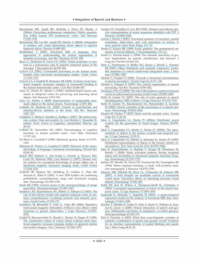
The direct comparison of unrelated versus iconicspeech-gesture pairs [GU > GI] resulted in large activationclusters in the IFG (BA 44, 45) and supplementary motorareas (SMA, BA 6) bilaterally, in the left inferior parietallobule, inferior temporal cortex and hippocampus as wellas in the right supramarginal gyrus, postcentral gyrus andmiddle frontal gyrus (Table I and Fig. 2).
Neural Correlates of Speech-Gesture Integration
Processes for Iconic and Unrelated Gestures
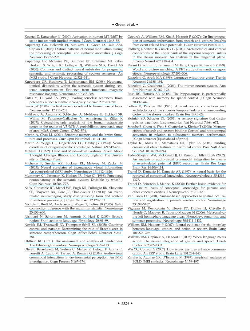
We start building the conjunction analyses by revealingthe neural correlates of integration processes confoundedby more basic semantic processing for iconic and unre-lated gestures, respectively (contrast 1). For iconic gestures[GI > RI] we found the largest cluster of activation alongthe left middle temporal gyrus (MTG) spreading into theIFG. Other areas of activation included the right MTG, leftinferior parietal lobule, left superior frontal and medialgyrus and left hippocampus. For unrelated gestures [GU> RU] we observed similar activations; however, withadditional activity in left middle frontal and right inferiorparietal regions (Table II and Fig. 3A).
In the next step, a contrast that controls for activationsrelated to general language processing is computed forboth kinds of gestures (contrast 2). For iconic gestures [GI> G] the largest clusters of activation were found in leftand right occipito-temporal regions. Other areas of activa-tion included the right superior temporal gyrus and lefthippocampus. For unrelated gestures [GU > G] similaractivation patterns were observed with bilateral parieto-temporal regions being additionally involved (Table IIIand Fig. 3B).
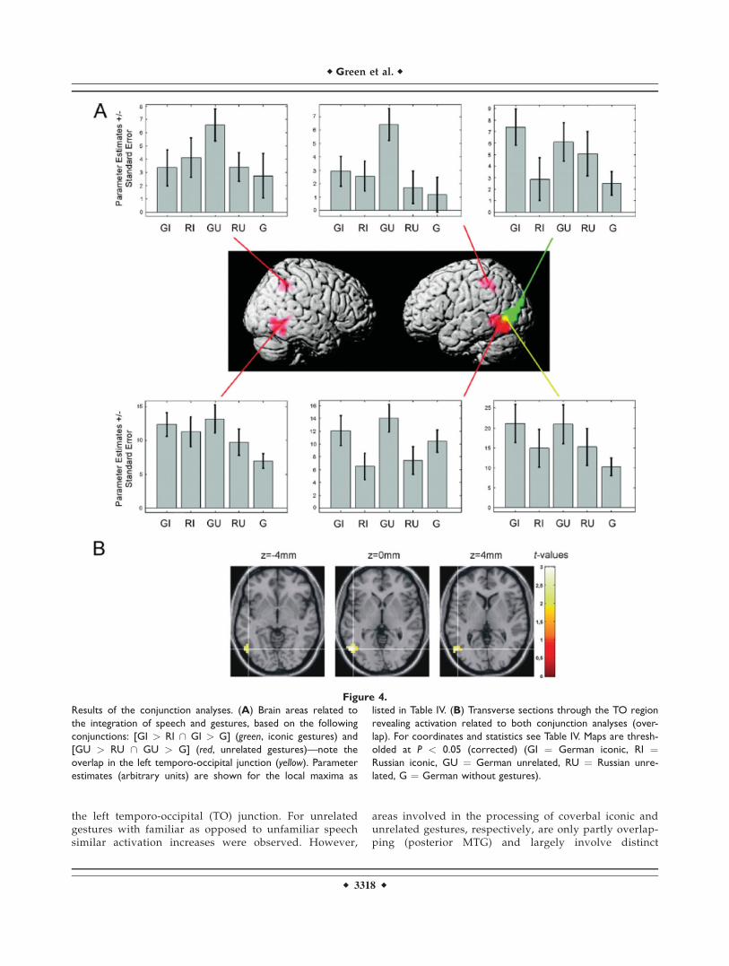
To proceed, the conjunctions of contrast 1 and 2 werecomputed for iconic [GI > RI \ GI > G] and unrelatedgestures [GU > RU \ GU > G], respectively. These con-junctions were designed to reveal the neural correlates ofsemantic integration uninfluenced by pure speech process-ing. For both conjunctions the left MTG was significantlyactivated in combination with the familiar language. Foriconic gestures this cluster extended more occipitally, forthe unrelated gestures, more temporally. For the unrelatedgesture condition additional activations were found in theleft MTG, inferior occipital gyrus, inferior parietal lobuleand postcentral gyrus (BA 2) and in the right inferior tem-poral gyrus and inferior parietal lobule (Table IV and Fig.4A, with iconic gestures in green, unrelated gestures inred).
The overlap of the areas involved in integration proc-esses for iconic and unrelated gestures was statisticallyconfirmed by calculating a conjunction of all previous con-trasts [GI > RI \ GI > G \ GU > RU \ GU > G] andrevealed a cluster in the left posterior MTG at the TO junc-tion (see Table IV and Fig. 4A, with overlap in yellow).
TABLE I. Brain activations for unrelated versus iconic gestures in combination with familiar language
Region Cluster extension x y z Extent t-Value
German unrelated > German iconic [GU > GI]L Inferior parietal lobule Precuneus, SPL, IPC, hIP, post-
and precentral gyrus�44 �36 40 217 4.37
L Inferior frontal gyrus Precentral gyrus, MFG �60 16 24 195 3.84R Supramarginal gyrus IPC, hIP, angular gyrus, STG 52 �36 36 142 3.64R Inferior frontal gyrus 48 32 12 136 3.68R/L Supplementary motor area SFG, SMG 8 20 56 64 2.88R Paracentral lobule Postcentral gyrus, SPL, precuneus 8 �36 56 55 3.32L Middle temporal gyrus ITG, IOG �60 �60 �4 45 3.67R Middle frontal gyrus 44 12 48 29 2.59L Hippocampus Putamen �20 �12 �16 23 2.82
Stereotaxic coordinates in MNI space and t-values of the foci of maximum activation (P < 0.05 corrected). hIP, human intraparietalarea; IOG, inferior occipital gyrus; IPC, inferior parietal cortex; ITG, inferior temporal gyrus; MFG, middle frontal gyrus; SFG, superiorfrontal gyrus; SMG, superior medial gyrus; SPL, superior parietal lobule; STG, superior temporal gyrus.
Figure 2.
Unrelated versus related gestures. Brain areas stronger activated
for familiar language with unrelated gestures compared with fa-
miliar language with iconic gestures [GU > GI]. For coordinates
and statistics see Table I. Map is thresholded at P < 0.05 (cor-
rected) (GI ¼ German iconic, GU ¼ German unrelated).
r Integration of Speech and Gestures r
r 3315 r
Finally, both conjunctions were masked reciprocallywith each other to reveal differences of iconic and unre-lated speech-gesture processing ([GI > RI \ GI > G] exclu-sively masked by [GU > RU \ GU > G]; [GU > RU \ GU> G] exclusively masked by [GI > RI \ GI > G]). Apartfrom the overlap calculated above all other activationsobserved for the iconic as well as for the unrelatedconjunction analyses were shown to be unaffected by therespective other gesture condition (see Table IV).
According to probabilistic cytoarchitectonic maps thereis only a small possibility (below 30%, see Table IV andFig. 4B) that the activations revealed by the conjunction
analyses are located in area hOC5 (V5/MTþ) [Eickhoffet al., 2005; Malikovic et al., 2007]. Nevertheless we cannotexclude that parts of the activation are situated within thatregion.
To further elucidate the function of the overlap regionwe conducted a multimodal integration analysis forrelated [0 < G < GI > I > 0] and unrelated gestures [0 <G < GU > U > 0], respectively, revealing areas that arestrongly activated by speech-gesture stimuli than by iso-lated speech or gestures. According to these analyses bothkinds of gestures elicited multimodal integration in the TOregion (iconic speech gesture pairs: t ¼ 2.12; unrelated
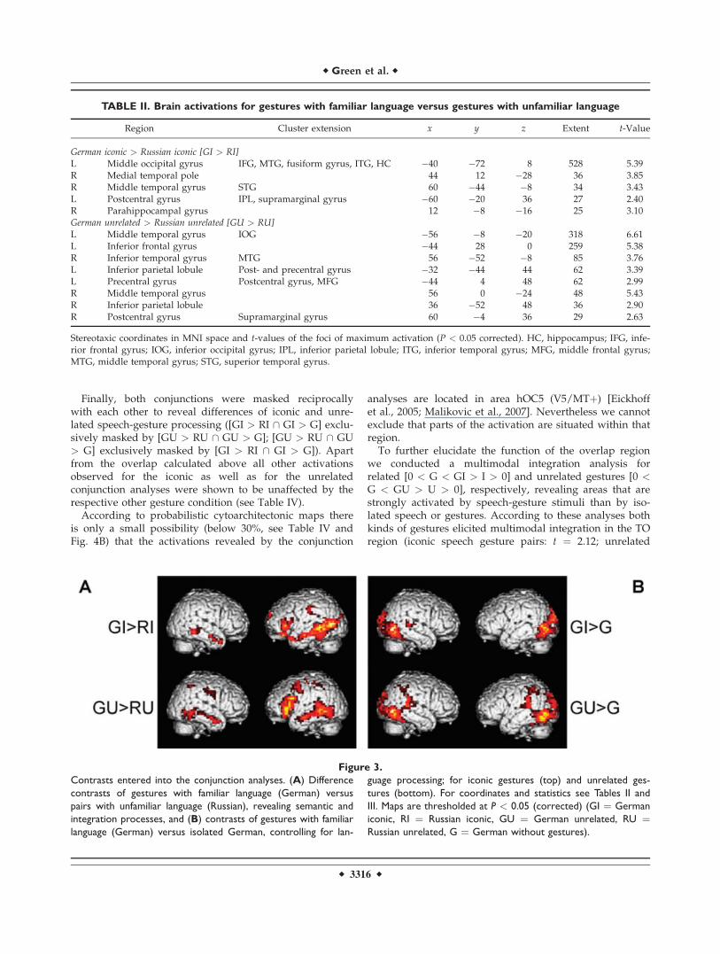
TABLE II. Brain activations for gestures with familiar language versus gestures with unfamiliar language
Region Cluster extension x y z Extent t-Value
German iconic > Russian iconic [GI > RI]L Middle occipital gyrus IFG, MTG, fusiform gyrus, ITG, HC �40 �72 8 528 5.39R Medial temporal pole 44 12 �28 36 3.85R Middle temporal gyrus STG 60 �44 �8 34 3.43L Postcentral gyrus IPL, supramarginal gyrus �60 �20 36 27 2.40R Parahippocampal gyrus 12 �8 �16 25 3.10German unrelated > Russian unrelated [GU > RU]
L Middle temporal gyrus IOG �56 �8 �20 318 6.61L Inferior frontal gyrus �44 28 0 259 5.38R Inferior temporal gyrus MTG 56 �52 �8 85 3.76L Inferior parietal lobule Post- and precentral gyrus �32 �44 44 62 3.39L Precentral gyrus Postcentral gyrus, MFG �44 4 48 62 2.99R Middle temporal gyrus 56 0 �24 48 5.43R Inferior parietal lobule 36 �52 48 36 2.90R Postcentral gyrus Supramarginal gyrus 60 �4 36 29 2.63
Stereotaxic coordinates in MNI space and t-values of the foci of maximum activation (P < 0.05 corrected). HC, hippocampus; IFG, infe-rior frontal gyrus; IOG, inferior occipital gyrus; IPL, inferior parietal lobule; ITG, inferior temporal gyrus; MFG, middle frontal gyrus;MTG, middle temporal gyrus; STG, superior temporal gyrus.
Figure 3.
Contrasts entered into the conjunction analyses. (A) Difference
contrasts of gestures with familiar language (German) versus
pairs with unfamiliar language (Russian), revealing semantic and
integration processes, and (B) contrasts of gestures with familiar
language (German) versus isolated German, controlling for lan-
guage processing; for iconic gestures (top) and unrelated ges-
tures (bottom). For coordinates and statistics see Tables II and
III. Maps are thresholded at P < 0.05 (corrected) (GI ¼ German
iconic, RI ¼ Russian iconic, GU ¼ German unrelated, RU ¼Russian unrelated, G ¼ German without gestures).
r Green et al. r
r 3316 r

speech gesture pairs: t ¼ 2.53, both P < 0.05 corrected aftersmall volume correction with a 10-mm sphere at�52�68 0).
DISCUSSION
The goal of the present study was to reveal the neuralcorrelates of semantic interaction processes of iconicspeech-gesture pairs and to examine whether there aresimilar activations when gestures are unrelated to speech.Subjects saw video clips of an actor speaking sentences ina familiar (German) or unfamiliar (Russian) language thatwere either presented in isolation or were accompanied byiconic gestures or by gestures that were unrelated to thesentence.
As previous research on speech and gesture processingusing ERPs [Ozyurek et al., 2007] and fMRI [Willemset al., 2007] has focused on incongruent speech-gesture in-formation, we first contrasted directly the processing ofunrelated gestures with iconic gestures. In line with a pre-vious study, we found inferior frontal as well as parietalactivations [Willems et al., 2007]. Willems and colleaguesinterpreted this pattern as reflecting stronger integrationload, but we cannot rule out that these activations are aresult of the processing of unnatural stimuli and ratherrelate to error detection processes.
The main focus of the present study, however, wasthe analysis of neural networks engaged in the naturalintegration of speech and iconic gestures. We showedthat semantically related speech and iconic gestureswith familiar as opposed to unfamiliar speech activates
TABLE III. Brain activations for gestures with familiar language versus isolated familiar language
Region Cluster extension x y z Extent t-Value
German iconic > German [GI > G]L Inferior occipital gyrus SOG, MOG, cerebellum, fusiform gyrus, IOG �48 �76 �4 299 6.24R Inferior temporal gyrus MOG, SOG, IOG, fusiform gyrus, cuneus 52 �72 �4 274 7.06R Superior temporal gyrus 68 �24 8 33 3.52L Hippocampus �20 �28 �8 25 3.54German unrelated > German [GU > G]L Inferior occipital gyrus Supramarginal gyrus, MOG, SOG, postcentral gyrus, IPL,
hIP, calcarine gyrus, fusiform gyrus, STG, MTG�48 �76 �4 527 6.71
R Inferior temporal gyrus IOG, MOG, ITG, STG, MTG, SOG, cuneus, lingual gyrus 44 �68 �4 292 6.31R Angular gyrus SPL, postcentral gyrus, IPL, hIP 32 �56 44 82 3.30
Stereotaxic coordinates in MNI space and t-values of the foci of maximum activation (P < 0.05 corrected). hIP, human intraparietalarea; IOG, inferior occipital gyrus; IPL, inferior parietal lobule; ITG, inferior temporal gyrus; MOG, middle occipital gyrus; MTG, middletemporal gyrus; SOG, superior occipital gyrus; SPL, superior parietal lobule; STG, superior temporal gyrus.
TABLE IV. Conjunction analyses—generally involved, overlapping, and distinct regions
Region Cluster extension x y z Extent t-Value
Conjunction analysis for iconic gestures [GI > RI \ GI > G]
L Middle occipital gyrus Probability for V5/MTþ: 30% �48 �68 4 72 3.98Conjunction analysis for unrelated gestures [GU > RU \ GU > G]
L Middle temporal gyrus IOG �56 �68 0 99 3.22R Inferior temporal gyrus 56 �64 �4 47 3.14L Inferior parietal lobule Postcentral gyrus �32 �44 44 39 3.05R Inferior parietal lobule 32 �52 44 25 2.54Overlap: Conjunction of iconic and unrelated conjunction analyses [GI > RI \ GI > G] \ [GU > RU \ GU > G]
L Middle temporal gyrus Probability for V5/MTþ: 20% �52 �68 0 24 3.01Distinct: Iconic conjunction exclusively masked by unrelated conjunction [GI > RI \ GI > G] excl. masked by [GU > RU \ GU > G]L Middle occipital gyrus Probability for V5/MTþ: 20% �40 �76 8 48 3.94Distinct: Unrelated conjunction exclusively masked by iconic conjunction [GU > RU \ GU > G] excl. masked by [GI > RI \ GI > G]L Middle temporal gyrus IOG (probability for V5/MTþ: 20%) �56 �56 0 71 2.85R Inferior temporal gyrus Probability for V5/MTþ: 20% 56 �64 �4 45 3.14L Inferior parietal lobule Postcentral gyrus �32 �44 44 39 3.05R Inferior parietal lobule 32 �52 44 25 2.54
Stereotaxic coordinates in MNI space and t-values of the foci of maximum activation (P < 0.05 corrected). IOG, inferior occipital gyrus.
r Integration of Speech and Gestures r
r 3317 r
the left temporo-occipital (TO) junction. For unrelatedgestures with familiar as opposed to unfamiliar speechsimilar activation increases were observed. However,
areas involved in the processing of coverbal iconic andunrelated gestures, respectively, are only partly overlap-ping (posterior MTG) and largely involve distinct
Figure 4.
Results of the conjunction analyses. (A) Brain areas related to
the integration of speech and gestures, based on the following
conjunctions: [GI > RI \ GI > G] (green, iconic gestures) and
[GU > RU \ GU > G] (red, unrelated gestures)—note the
overlap in the left temporo-occipital junction (yellow). Parameter
estimates (arbitrary units) are shown for the local maxima as
listed in Table IV. (B) Transverse sections through the TO region
revealing activation related to both conjunction analyses (over-
lap). For coordinates and statistics see Table IV. Maps are thresh-
olded at P < 0.05 (corrected) (GI ¼ German iconic, RI ¼Russian iconic, GU ¼ German unrelated, RU ¼ Russian unre-
lated, G ¼ German without gestures).
r Green et al. r
r 3318 r

regions, specifically activated for either iconic (middleoccipital gyrus) or unrelated gestures (bilateral temporaland parietal regions). Based on our findings we proposethat both related and unrelated gestures induce an inte-gration process that is reflected in two different process-ing streams converging in the left posterior temporalgyrus.
Processing of Iconic Gestures With
Familiar Speech
Speech and gesture have to be integrated online into asemantic representation. To avoid problems associatedwith the comparison of unrelated and related gestures weinvestigated the processing of natural speech-gesture pairsand manipulated the understandability of the speech com-ponent. Our first hypothesis targeted the neural correlatesof semantic integration processes, independent of mis-match manipulations.
The neural correlate of the integration of iconic gestureswith familiar German speech was located in the left tem-poro-occipital (TO) junction, extending from the MTG intothe superior occipital gyrus. Several studies have demon-strated the involvement of the left posterior MTG in lan-guage processing [for reviews see Demonet et al., 2005;Vigneau et al., 2006], in contextual sentence integration[Baumgartner et al., 2002], in action representations andconcepts [e.g., Kable et al., 2005; Martin and Chao, 2001]and in multisensory integration for the identification oftool or animal sounds [Beauchamp et al., 2004b; Lewis,2006]. It has been argued that multimodal responses in theposterior MTG may reflect the formation of associationsbetween auditory and visual features that represent thesame object [Beauchamp et al., 2004b].
Similarly, activations in the left TO junction have alsobeen demonstrated for a variety of tasks, including mean-ing-based paradigms as well as diverse paradigms relatedto visual aspects such as motion. Hickok and Poeppel[2000] argue in their review that language tasks accessingthe mental lexicon (i.e., accessing meaning-based represen-tations) rely on auditory-to-meaning interface systems inthe vicinity of the left TO junction and call this region anauditory-conceptual interface, which may be part of amore widely distributed and possibly modality-specificconceptual network [Barsalou et al., 2003; Hickok andPoeppel, 2007; Tranel et al., 2008]. More generally, the leftTO junction is a multimodal association area involved insemantic processing, including the mapping of visualinput onto linguistic representations [Ebisch et al., 2007].Furthermore, patients with lesions in this region showideational apraxia, an impaired knowledge of action con-cepts and inadequate object use [De Renzi and Lucchelli,1988]. Tranel et al. [1997] found in a large sample ofpatients with brain lesions that defective conceptualknowledge was related to lesions in the left TO junction.As 50% of the items used in this study were related to
objects and another 17% were reenacted actions withoutobjects [which is similar to proportions observed in spon-taneous speech, cf. Hadar and Krauss, 1999] it is likelythat the activation of the TO junction resulted from thesemotion-related images.
Apart from involvement in semantic-conceptual proc-esses, the TO junction is engaged in the processing of vis-ual input. It was involved in the processing of motioneven if static stimuli only implied motion [Kourtzi andKanwisher, 2000] or were somehow related to visual topo-graphical or spatial information [e.g., Mummery et al.,1998]. Thus, it seems likely that a region that is sensitiveto semantic concepts as well as to visuospatial aspectsintegrates information from speech and gestures into onerepresentation.
At first glance the activation of temporo-occipitalregions for the integration of familiar speech and iconicgestures seems to be inconsistent with previous fMRIstudies on the integration of speech and iconic gestures,mentioning instead the left IFG and superior temporalsulcus [STS) as the key regions for speech-gesture inte-gration. These discrepancies, however, may be explainedby differences in experimental paradigms. Willems et al.[2007] found activation of the left STS specifically for thelanguage mismatch (sentences with inappropriate verband a gesture fitting the expected verb) but not for thegesture mismatch (correct sentences with an incongruentgesture) or the double mismatch condition (sentenceswith inappropriate verb and a gesture fitting the inap-propriate verb). This might reflect a violation of expect-ancies derived from speech semantics, as has beenshown by Ni et al. [2000] and Kuperberg et al. [2003].The semantic mismatch may be much more dominant inthe language mismatch condition (absence of useful lan-guage information) than in the gesture mismatch condi-tion. The apparent mismatch in speech may lead to astronger focus on gesture semantics. These might beused to ‘‘correct’’ the faulty utterance and therefore theyneed to be integrated even more into the preceding sen-tence context. In the case of gesture mismatches thisinteraction between context and retrieval of lexical-semantic information may be less important because thespeech semantics likely dominate the processing of thewhole sequence. The activation of the STS in the studyby Holle et al. [2008] might rather reflect the interactionbetween the meaning of a gesture and an ambiguoussentence. We, in contrast, used only unambiguous, natu-ral sentences without mismatches for this analysis.Importantly, a closer look at the coordinates of the STS-clusters reported in the study by Holle et al. [2008]reveals that these activations are located in the temporo-occipital cortex, showing their strongest activation fordominant meanings in the middle occipital gyrus and forsubordinate meanings in the occipitotemporal junction.Thus, our results are largely in congruence with thestudy of Holle et al. Additionally, studies on metaphoricgestures performed by our group also revealed
r Integration of Speech and Gestures r
r 3319 r
integration-related activations in the left posterior MTG[Kircher et al., 2009; Straube et al., in press].
The focus on the pSTS in integration processes mainlystems from nonhuman primate studies demonstrating con-verging afferents from different senses in the primatehomolog of human pSTS [e.g., Seltzer and Pandya, 1978].Further support comes from several studies associatingthis region with the crossmodal binding of audiovisualinput [e.g., Calvert et al., 2000]. But there also are studiesthat did not report enhanced activation for bimodal versusunimodal stimuli [e.g., Taylor et al., 2006], and the super-and subadditive effects of the Calvert study have not beenreplicated so far [for a review including the discussion ofmethodological problems see Hocking and Price, 2008]. Insum, the role of pSTS in higher-level audiovisual integra-tion processes may have been overrated in the past. Inaddition, single cell recordings in the macaque brain haveshown that regions integrating audiovisual stimuli werelocated in the macaque STS-STP-TPO-region [Bruce et al.,1981; Padberg et al., 2003]. In the human brain the equiva-lent regions are predicted to extend inferiorly from poste-rior STS into MTG [Van Essen, 2004] so that a distinctionbetween functional STS and MTG or MOG activations hasto be accepted only with reservation. This may be the casewhy some studies do not separate these regions andinstead use less precise anatomical terms like ‘‘pSTS/MTG’’ [e.g., Beauchamp et al., 2004b].
Nevertheless, the left posterior TO junction seems to becrucially involved in the processing of speech and iconicgestures.
Processing of Unrelated Gestures With
Familiar Speech
Our second hypothesis stated that for unrelated gesturesmainly parietal activations would be observed, resultingfrom less congruency between speech and gestures.
Before discussing the fMRI results for unrelated ges-tures, the behavioral recognition data merit attention.Unrelated gesture recognition, as well as iconic gesturerecognition, were equally enhanced compared withspeech-only stimuli. This indicates that for bimodal stimulideeper processing had occurred, most likely through offer-ing binding opportunity. This is in line with other studiesthat demonstrated even better memory performances forincongruous than for congruous pictures, probably due toincreased processing [Michelon et al., 2003].
On the neural level we observed bilateral activations inposterior temporal regions and in the inferior parietallobule (IPL). In contrast to iconic gestures, the temporalactivations for unrelated gestures were located more ante-riorly and inferiorly and closely matched the activationsobserved for the [GU > GI] analysis. Besides its generalinvolvement in semantic language processing [cf. Book-heimer, 2002; Vigneau et al., 2006], the MTG has beenfound activated for such aspects of object knowledge as
associations with sensorimotor correlates of their use [e.g.,Chao and Martin, 2000; Martin et al., 1996]. Comparedwith iconic gestures, this more anterior MTG activation isinterpreted as a stronger reliance on linguistic aspects, asmeaning mainly could be extracted from speech, becauseour unrelated gestures did not contain clear semanticinformation.
Some authors suggest a strong link between action andlanguage systems that could be fulfilled by the postulatedmirror neuron system, including the IPL [Nishitani et al.,2005; Rizzolatti and Arbib, 1998]. Concerning the interac-tion of language and gestures, our results are consistentwith the existing studies. Willems et al. [2007] found sig-nificant activations in the left intraparietal sulcus for thegesture mismatch condition compared with correct pairsof speech and gesture. This condition is similar to theunrelated gestures used in our study. Holle et al. [2008]did not use mismatches in their study but instead manipu-lated the ambiguity of their sentences and also found infe-rior bilateral parietal activations for gestures supportingthe meaning of a sentence as opposed to grooming ges-tures. All of these IPL activations could be interpreted as aprocess of observation-execution mapping involving moresimulation costs. In the study by Holle et al. [2008] theiconic gestures corresponding to the dominant and subor-dinate meanings were more complex than grooming andtheir meaning was still somewhat unclear [see Hadar andPinchas-Zamir, 2004] and thus open to different interpreta-tions. Thus, an initially attributed action goal may emergeas not appropriate and more simulation cycles would beneeded for gestures than for grooming. For mismatchingor unrelated gestures this explanation holds even moreand is further supported by numerous studies showingparietal activations for the observation of meaninglesshand movements [for a review see Grezes and Decety,2001]. Interestingly, apart from being activated by pureaction observation IPL seems to be modulated by semanticinformation from speech. This might be accomplished byhigher order cortical areas that modulate motor represen-tations in a top-down process. But this explanation raisesthe question why we did not observe parietal activationsfor the processing of iconic gestures. In none of the differ-ence contrasts used for the iconic conjunction analysis pa-rietal activations were found. As our sentences wereunambiguous and the gestures paralleled the speech con-tent, it may have been that the clear language contextinfluenced and constrained the simulation process. Buteven though there are some authors postulating a stronglink between action and language systems [Nishitani et al.,2005; Rizzolatti and Arbib, 1998] and some studies show-ing influences of language domains on action processing[e.g., Bartolo et al., 2007; Gentilucci et al., 2006], this is arather tentative explanation. In sum, the activation levelsin the IPL for the processing of unrelated gestures mostlikely relate to an action processing component, possiblyrerunning gesture information in order to create integra-tion opportunities.
r Green et al. r
r 3320 r
Common and Separate Systems for Iconic and
Unrelated Gestures
For the semantic processing of effective and faulty stim-uli usually the same networks are found. But the process-ing of faulty stimuli often is characterized by additionalactivation patterns, and this is what we have found: com-mon to both related and unrelated speech-gesture pairs isactivation in the left posterior temporal cortex. Besides thissmall overlap all activations retrieved by the conjunctionanalyses for both iconic and unrelated gestures were spe-cific to the respective gesture category. The left middleoccipital gyrus was exclusively activated for iconic ges-tures, whereas bilateral parietal as well as inferior andmiddle temporal areas were specifically observed for un-related gestures. We interpret these distinct activationpatterns as different routes of processing reflecting integra-tion effort.
The temporal activations during the processing ofspeech with unrelated gestures are interpreted as a strongreliance on the linguistic network, as the meaning of thesequence can only be grasped from speech. This is in linewith studies revealing activations in these regions inresponse to words activating object-related knowledge[e.g., Chao et al., 1999; Perani et al., 1999]. The integrationprocess for iconic gestures as reflected in occipital activa-tions, in contrast, seems to be based on the familiar visualfeatures of speech contents and their representations ingestures (e.g., ‘‘spiral stair’’), rendering activation of lin-guistic semantic areas in the temporal lobe redundant.This view is mainly supported by studies on visual im-agery, showing involvement of the left middle occipitaland inferior temporal gyrus in visual imagery of seenscenes [Johnson et al., 2007], walking [Jahn et al., 2004],objects like chairs in contrast to faces or houses [Ishaiet al., 1999, 2000a,b] and in motion imagery of graphicstimuli [Goebel et al., 1998]. In addition, a meta-analysis ofimage generation in relation to lesions has noted a trendtoward posterior damage [Farah, 1995]. Thus, iconic ges-tures possibly activate internal visual representations auto-matically. The observed parietal activations during theprocessing of unrelated speech-gesture pairs presumablyindicate the gesture-oriented part of the integration pro-cess, activating the dorsal visual pathway in order to pro-cess the novel movement patterns by encoding theirspatiotemporal relationships. This is substantiated by find-ings related to action observation (see above) and by itsdeconstructing function during the encoding of abstractmotion sequences [Grafton et al., 1998; Jenkins et al., 1994].
There are several possible interpretations of the resultsof the unrelated conjunction analysis. First of all, integra-tion of unrelated information may seem unnecessary.However, there are several studies that have shownaudiovisual integration effects for unrelated or incongru-ent information [Hein et al., 2007; Hocking and Price,2008; Olivetti Belardinelli et al., 2004; Taylor et al., 2006].Second, our behavioral results suggest that effective inte-
gration has occurred because recognition of unrelatedspeech gesture pairs was similar to that of related pairs.Hence, it is rather unlikely that the observed activation isonly the correlate of the attempt to integrate. At leastparts of the revealed areas should be indicative of effec-tive integration. Third, we cannot rule out that theregions revealed by the unrelated conjunction analysisare somewhat influenced by mismatch processing. But asboth kinds of gestures commonly activated parts of theleft posterior MTG, it can be assumed that even forspeech with unrelated gestures the brain tries to create acommon representation. We suggest that, independent ofrelatedness, it is the left posterior MTG where effectivemotion-language mapping takes place. The temporal, pa-rietal and occipital regions found specifically activatedfor unrelated and iconic gestures, respectively, may serveas auxiliary regions processing single aspects of the pre-sented stimulus in order to enable integration of theseaspects in the posterior MTG. We suggest that due to thetemporospatial co-occurrence of speech and gestures thebrain assigns relatedness to these two sources of informa-tion. Cognitive processes try to map speech and gesturesonto each other, be they related (naturally occurring) orunrelated (unnatural experimental setting). In the case ofunrelated gestures, the mapping process is more difficultand requires more effort, leading to additional activationsin temporal and parietal regions that probably rerun in-formation from both modalities in order to enable inte-gration. The core integration process in the left posteriorMTG, finally, could be achieved by means of a patchy or-ganization as it has been found in the STS [Beauchampet al., 2004a]. With high-resolution fMRI small patcheswere found that responded primarily to unimodal visualor auditory input, presumably translated that informationin a common code for both modalities, which then wassubsequently integrated in multisensory patches thatwere located between the unimodal ones.
Because of the limited temporal resolution we cannotmake precise inferences about the temporal sequence ofcortical activity and whether these activations relate to in-termediate processing stages (functions) or to later stagesof accessing the end-product ‘‘representation,’’ i.e., themeaning of each sentence. Thus, the additional temporaland parietal activations found for the processing of unre-lated gestures may either represent auxiliary processeslooking for information that can be integrated or may rep-resent additional elements of the representation of theseunrelated speech gesture pairs.
Concerning the parietal activations observed for unre-lated gestures alternative interpretations are possible. Onemight ask whether the unrelated gestures differed in kine-matics from the iconic gestures. But we constructed ourstimuli carefully and statistical analyses of movementextent, movement time and relations between speech andgestures did not reveal any significant differences betweenthese two kinds of gestures (see Supporting InformationMaterial). This suggests that it is rather unlikely that there
r Integration of Speech and Gestures r
r 3321 r
were any differences in kinematics that may have causedparietal activation.
The parietal activations also could stem from increasedattentional processes [for a review see Culham and Kanw-isher, 2001; Husain and Nachev, 2007]. Parietal regions,such as the intraparietal sulcus, are known to be involvedin guiding visual or spatial attention [Corbetta and Shul-man, 2002]. Though our stimuli were matched for complex-ity, it might be possible that the unrelated movementsproduced stronger attention-related parietal activationsthan the iconic gestures. It is possible that due to the tem-porospatial co-occurrence the expectation was raised thatthese two sources of information belong together—whichthen was violated by the unrelatedness of the gesture. Thismight have resulted in increased attention and noticeablerecognition rates. But the argument of increased attentionor arousal seems to apply more to paradigms where stimuliare assigned a certain task-relevance [Teder-Salejarvi et al.,2002], something that we avoided by using a task that wasneither related to the gestures nor to the language. Still, thedetection of unrelatedness with subsequent increasedperformance rates is indicative of an integration process.
According to the study by Willems et al. [2007] onecould have expected involvement of the IFG in relation tointegration processes. We observed this activation only inan analysis similar to that of Willems et al., contrastingunrelated to related speech-gesture pairs [GU > GI]. In theconjunction analysis for unrelated gestures only at a moreliberal explorative threshold IFG activation could beobserved. For iconic gestures this was not the case. Thissuggests that the role of the IFG in speech gesture integra-tion processes is not purely integrative but rather relatedto the detection and resolution of incompatible stimulusrepresentations and for implementing reanalyses in theface of misinterpretations [Kuperberg et al., 2008; Novicket al., 2005]. This explanation might also account for IFGinvolvement in the processing of metaphoric speech-ges-ture pairs where the speech content cannot be taken liter-ally (if it is taken literally there is conflict between speechand gesture) and has to be transferred to an abstract level[Kircher et al., 2009; Straube et al., in press]. This explana-tion is also in accordance with the one given by Willemset al. [2007], dealing with increased processing load.
Finally one might question our definition and analysisof integration-related processes. A criterion that research-ers in the field of multisensory integration seem to agreeupon is that for a brain region to be considered as anaudiovisual integration site, it has to exhibit activation to(1) an audio-only condition, (2) a visual-only conditionand (3) show additionally some property of multisensoryintegration like [0 < S < SG > G > 0] (with S ¼ speech, G¼ gestures and SG ¼ speech plus gestures). Our data,especially the activation of the TO region, do meet thisrequirement for both iconic and unrelated gestures, sup-porting our interpretation of an integrative region.
The comparison of the two analysis approaches suggeststhat the conjunction approach is superior to simply con-
trasting unrelated to related gestures. Only by this meansit was possible to reveal processes that are shared betweenrelated and unrelated gestures, and to delineate regionsthat are not better explained by mismatch detection.
CONCLUSIONS
In conclusion, our results demonstrated an area of acti-vation shared by related iconic as well as by unrelatedgestures, presumably reflecting a common integration pro-cess on a semantic level. Two distinct processing streamsconverge in the left posterior temporal cortex which wascommonly activated by both kinds of gestures. The net-work for iconic gestures was located in the left temporo-occipital cortex, possibly reflecting more visually basedprocessing. In contrast, the network for unrelated gestureswas bilaterally situated in posterior temporal and inferiorparietal regions, most likely reflecting two auxiliary proc-esses split up in linguistic aspects and novel movementpattern aspects that rerun information. The key role of theleft posterior temporal cortex may be due to its anatomicallocation between areas processing visual form and motionand on the other side visual and auditory associationareas, what makes it particularly suitable for the integra-tion of these types of information. The possibility thatdifferent subregions of this area are specialized for associ-ating different properties within and across visual and au-ditory modalities (here gesture and language) remains anavenue for future exploration.
To our knowledge this is the first study revealing theneural correlates of iconic gesture processing with naturalspeech-gesture pairs. As even unrelated gestures presum-ably lead to integration processes, but in different brainareas, it is likely that other kinds of gestures (e.g., emblem-atic, deictic or metaphoric) are processed in specific neuralnetworks, which are yet to be explored.
ACKNOWLEDGMENTS
The authors are grateful to all the subjects who partici-pated in this study, to Thilo Kellermann for help with thedata analysis, to Olga Sachs for linguistic assistance and tothe IZKF service team for support acquiring the data.
REFERENCES
Barsalou LW, Kyle Simmons W, Barbey AK, Wilson CD (2003):Grounding conceptual knowledge in modality-specific systems.Trends Cogn Sci 7:84–91.
Bartolo A, Weisbecker A, Coello Y (2007): Linguistic and spatialinformation for action. Behav Brain Res 184:19–30.
Baumgartner A, Weiller C, Buchel C (2002): Event-related fMRIreveals cortical sites involved in contextual sentence integra-tion. Neuroimage 16:736–745.
Beauchamp MS (2005): Statistical criteria in fMRI studies of multi-sensory integration. Neuroinformatics 3:93–113.
r Green et al. r
r 3322 r
Beauchamp MS, Argall BD, Bodurka J, Duyn JH, Martin A(2004a): Unraveling multisensory integration: Patchy organiza-tion within human STS multisensory cortex. Nat Neurosci7:1190–1192.
Beauchamp MS, Lee KE, Argall BD, Martin A (2004b): Integrationof auditory and visual information about objects in superiortemporal sulcus. Neuron 41:809–823.
Bookheimer S (2002): Functional MRI of language: Newapproaches to understanding the cortical organization ofsemantic processing. Ann Rev Neurosci 25:151–188.
Bruce C, Desimone R, Gross CG (1981): Visual properties of neu-rons in a polysensory area in superior temporal sulcus of themacaque. J Neurophysiol 46:369–384.
Calvert GA (2001): Crossmodal processing in the human brain:Insights from functional neuroimaging studies. Cereb Cortex11:1110–1123.
Calvert GA, Campbell R, Brammer MJ (2000): Evidence from func-tional magnetic resonance imaging of crossmodal binding inthe human heteromodal cortex. Curr Biol 10:649–657.
Chao LL, Haxby JV, Martin A (1999): Attribute-based neural sub-strates in temporal cortex for perceiving and knowing aboutobjects. Nat Neurosci 2:913–919.
Chao LL, Martin A (2000): Representation of manipulable man-made objects in the dorsal stream. Neuroimage 12:478–484.
Corbetta M, Shulman GL (2002): Control of goal-directed andstimulus-driven attention in the brain. Nat Rev Neurosci3:201–215.
Craighero L, Metta G, Sandini G, Fadiga L (2007): The mirror-neu-rons system: Data and models. In: von Hofsten C, Rosander K,editors. From Action to Cognition. Amsterdam: Elsevier. pp39–59.
Culham JC, Kanwisher NG (2001): Neuroimaging of cognitivefunctions in human parietal cortex. Curr Opin Neurobiol11:157–163.
De Renzi E, Lucchelli F (1988): Ideational apraxia. Brain 111:1173–1185.
Demonet JF, Thierry G, Cardebat D (2005): Renewal of the neuro-physiology of language: Functional neuroimaging. Physiol Rev85:49–95.
Ebisch SJH, Babiloni C, Del Gratta C, Ferretti A, Perrucci MG,Caulo M, Sitskoorn MM, Luca Romani G (2007): Human neu-ral systems for conceptual knowledge of proper object use: Afunctional magnetic resonance imaging study. Cereb Cortex17:2744–2751.
Eickhoff SB, Stephan KE, Mohlberg H, Grefkes C, Fink GR,Amunts K, Zilles K (2005): A new SPM toolbox for combiningprobabilistic cytoarchitectonic maps and functional imagingdata. Neuroimage 25:1325–1335.
Farah MJ (1995): Current issues in the neuropsychology of imagegeneration. Neuropsychologia 33:1455–1471.
Friederici AD, Ruschemeyer SA, Hahne A, Fiebach CJ (2003): Therole of left inferior frontal and superior temporal cortex in sen-tence comprehension: Localizing syntactic and semantic proc-esses. Cereb Cortex 13:170–177.
Gentilucci M, Bernardis P, Crisi G, Volta RD (2006): Repetitivetranscranial magnetic stimulation of Broca’s area affects verbalresponses to gesture observation. J Cogn Neurosci 18:1059–1074.
Goebel R, Khorram-Sefat D, Muckli L, Hacker H, Singer W (1998):The constructive nature of vision: Direct evidence from func-tional magnetic resonance imaging studies of apparent motionand motion imagery. Eur J Neurosci 10:1563–1573.
Grafton ST, Hazeltine E, Ivry RB (1998): Abstract and effector-spe-cific representations of motor sequences identified with PET. JNeurosci 18:9420–9428.
Grezes J, Decety J (2001): Functional anatomy of execution, mentalsimulation, observation, and verb generation of actions: Ameta analysis. Hum Brain Mapp 12:1–19.
Hadar U, Krauss RK (1999): Iconic gestures: The grammatical cat-egories of lexical affiliates. J Neurolinguistics 12:1–12.
Hadar U, Pinchas-Zamir L (2004): The semantic specificity of ges-ture: Implications for gesture classification and function. JLang Soc Psychol 23:204–214.
Hein G, Doehrmann O, Muller NG, Kaiser J, Muckli L, NaumerMJ (2007): Object familiarity and semantic congruency modu-late responses in cortical audiovisual integration areas. J Neu-rosci 27:7881–7887.
Hickok G, Poeppel D (2000): Towards a functional neuroanatomyof speech perception. Trends Cogn Sci 4:131–138.
Hickok G, Poeppel D (2007): The cortical organization of speechprocessing. Nat Rev Neurosci 8:393–402.
Hocking J, Price CJ (2008): The role of the posterior superior temporalsulcus in audiovisual processing. Cereb Cortex 18:2439–2449.
Holle H, Gunter TC (2007): The role of iconic gestures in speechdisambiguation: ERP evidence. J Cogn Neurosci 19:1175–1192.
Holle H, Gunter TC, Ruschemeyer SA, Hennenlotter A, IacoboniM (2008): Neural correlates of the processing of co-speech ges-tures. Neuroimage 39:2010–2024.
Husain M, Nachev P (2007): Space and the parietal cortex. TrendsCogn Sci 11:30–36.
Ishai A, Ungerleider LG, Haxby JV (2000a): Distributed neuralsystems for the generation of visual images. Neuron 28:979–990.
Ishai A, Ungerleider LG, Martin A, Haxby JV (2000b): The repre-sentation of objects in the human occipital and temporal cor-tex. J Cogn Neurosci 12:35–51.
Ishai A, Ungerleider LG, Martin A, Schouten JL, Haxby JV (1999):Distributed representation of objects in the human ventral vis-ual pathway. Proc Natl Acad Sci USA 96:9379–9384.
Jahn K, Deutschlander A, Stephan T, Strupp M, Wiesmann M,Brandt T (2004): Brain activation patterns during imaginedstance and locomotion in functional magnetic resonance imag-ing. Neuroimage 22:1722–1731.
Jenkins IH, Brooks DJ, Nixon PD, Frackowiak RS, Passingham RE(1994): Motor sequence learning: A study with positron emis-sion tomography. J Neurosci 14:3775–3790.
Johnson MR, Mitchell KJ, Raye CL, D’Esposito M, Johnson MK(2007): A brief thought can modulate activity in extrastriatevisual areas: Top-down effects of refreshing just-seen visualstimuli. Neuroimage 37:290–299.
Kable JW, Kan IP, Wilson A, Thompson-Schill SL, Chatterjee A(2005): Conceptual representations of action in the lateral tem-poral cortex. J Cogn Neurosci 17:1855–1870.
Katanoda K, Matsuda Y, Sugishita M (2002): A spatio-temporalregression model for the analysis of functional MRI data. Neu-roimage 17:1415–1428.
Kircher T, Straube B, Leube D, Weis S, Sachs O, Willmes K, Kon-rad K, Green A (2009): Neural interaction of speech and ges-ture: Differential activations of metaphoric co-verbal gestures.Neuropsychologia 47:169–179.
Kita S, Ozyurek A (2003): What does cross-linguistic variation insemantic coordination of speech and gesture reveal? Evidencefor an interface representation of spatial thinking and speak-ing. J Mem Lang 48:16–32.
r Integration of Speech and Gestures r
r 3323 r
Kourtzi Z, Kanwisher N (2000): Activation in human MT/MST bystatic images with implied motion. J Cogn Neurosci 12:48–55.
Kuperberg GR, Holcomb PJ, Sitnikova T, Greve D, Dale AM,Caplan D (2003): Distinct patterns of neural modulation duringthe processing of conceptual and syntactic anomalies. J CognNeurosci 15:272–293.
Kuperberg GR, McGuire PK, Bullmore ET, Brammer MJ, Rabe-Hesketh S, Wright IC, Lythgoe DJ, Williams SCR, David AS(2000): Common and distinct neural substrates for pragmatic,semantic, and syntactic processing of spoken sentences: AnfMRI study. J Cogn Neurosci 12:321–341.
Kuperberg GR, Sitnikova T, Lakshmanan BM (2008): Neuroana-tomical distinctions within the semantic system during sen-tence comprehension: Evidence from functional magneticresonance imaging. Neuroimage 40:367–388.
Kutas M, Hillyard SA (1980): Reading senseless sentences: Brainpotentials reflect semantic incongruity. Science 207:203–205.
Lewis JW (2006): Cortical networks related to human use of tools.Neuroscientist 12:211–231.
Malikovic A, Amunts K, Schleicher A, Mohlberg H, Eickhoff SB,Wilms M, Palomero-Gallagher N, Armstrong E, Zilles K(2007): Cytoarchitectonic analysis of the human extrastriatecortex in the region of V5/MT: A probabilistic, stereotaxic mapof area hOc5. Cereb Cortex 17:562–574.
Martin A, Chao LL (2001): Semantic memory and the brain: Struc-ture and processes. Curr Opin Neurobiol 11:194–201.
Martin A, Wiggs CL, Ungerleider LG, Haxby JV (1996): Neuralcorrelates of category-specific knowledge. Nature 379:649–652.
McNeill D (1992): Hand and Mind—What Gestures Reveal AboutThought. Chicago, Illinois, and London, England: The Univer-sity of Chicago Press.
Michelon P, Snyder AZ, Buckner RL, McAvoy M, Zacks JM(2003): Neural correlates of incongruous visual information:An event-related fMRI study. Neuroimage 19:1612–1626.
Mummery CJ, Patterson K, Hodges JR, Price CJ (1998): Functionalneuroanatomy of the semantic system: Divisible by what? JCogn Neurosci 10:766–777.
Ni W, Constable RT, Mencl WE, Pugh KR, Fulbright RK, ShaywitzSE, Shaywitz BA, Gore JC, Shankweiler D (2000): An event-related neuroimaging study distinguishing form and contentin sentence processing. J Cogn Neurosci 12:120–133.
Nichols T, Brett M, Andersson J, Wager T, Poline JB (2005): Validconjunction inference with the minimum statistic. Neuroimage25:653–660.
Nishitani N, Schurmann M, Amunts K, Hari R (2005): Broca’sregion: From action to language. Physiology 20:60–69.
Novick JM, Trueswell JC, Thompson-Schill SL (2005): Cognitivecontrol and parsing: Reexamining the role of Broca’s area insentence comprehension. Cogn Affect Behav Neurosci 5:263–281.
Oldfield RC (1971): The assessment and analysis of handedness:The Edinburgh inventory. Neuropsychologia 9:97–113.
Olivetti Belardinelli M, Sestieri C, Matteo R, Delogu F, Gratta C,Ferretti A, Caulo M, Tartaro A, Romani G (2004): Audio-visualcrossmodal interactions in environmental perception: An fMRIinvestigation. Cogn Process 5:167–174.
Ozyurek A, Willems RM, Kita S, Hagoort P (2007): On-line integra-tion of semantic information from speech and gesture: Insightsfrom event-related brain potentials. J CognNeurosci 19:605–616.
Padberg J, Seltzer B, Cusick CG (2003): Architectonics and corticalconnections of the upper bank of the superior temporal sulcusin the rhesus monkey: An analysis in the tangential plane.J Comp Neurol 467:418–434.
Perani D, Schnur T, Tettamanti M, Italy, Cappa SF, Fazio F (1999):Word and picture matching: A PET study of semantic categoryeffects. Neuropsychologia 37:293–306.
Rizzolatti G, Arbib MA (1998): Language within our grasp. TrendsNeurosci 21:188–194.
Rizzolatti G, Craighero L (2004): The mirror neuron system. AnnRev Neurosci 27:169–192.
Ross RS, Slotnick SD (2008): The hippocampus is preferentiallyassociated with memory for spatial context. J Cogn Neurosci20:432–446.
Seltzer B, Pandya DN (1978): Afferent cortical connections andarchitectonics of the superior temporal sulcus and surroundingcortex in the rhesus monkey. Brain Res 149:1–24.
Slotnick SD, Schacter DL (2004): A sensory signature that distin-guishes true from false memories. Nat Neurosci 7:664–672.
Straube B, Green A, Weis S, Chatterjee A, Kircher T (2008): Memoryeffects of speech and gesture binding: Cortical and hippocampalactivation in relation to subsequent memory performance.J Cogn Neurosci [Epub ahead of print].
Taylor KI, Moss HE, Stamatakis EA, Tyler LK (2006): Bindingcrossmodal object features in perirhinal cortex. Proc Natl AcadSci USA 103:8239–8244.
Teder-Salejarvi WA, McDonald JJ, Di Russo F, Hillyard SA (2002):An analysis of audio-visual crossmodal integration by meansof event-related potential (ERP) recordings. Brain Res CognBrain Res 14:106–114.
Tranel D, Damasio H, Damasio AR (1997): A neural basis for theretrieval of conceptual knowledge. Neuropsychologia 35:1319–1327.
Tranel D, Feinstein J, Manzel K (2008): Further lesion evidence forthe neural basis of conceptual knowledge for persons andother concrete entities. J Neuropsychol 2:301–320.
Van Essen DC (2004): Surface-based approaches to spatial localiza-tion and registration in primate cerebral cortex. Neuroimage23:S97–S107.
Vigneau M, Beaucousin V, Herve PY, Duffau H, Crivello F,Houde O, Mazoyer B, Tzourio-Mazoyer N (2006): Meta-analyz-ing left hemisphere language areas: Phonology, semantics, andsentence processing. Neuroimage 30:1414–1432.
Willems RM, Hagoort P (2007): Neural evidence for the interplaybetween language, gesture, and action: A review. Brain Lang101:278–289.
Willems RM, Ozyurek A, Hagoort P (2007): When language meetsaction: The neural integration of gesture and speech. CerebCortex 17:2322–2333.
Wu YC, Coulson S (2007): How iconic gestures enhance communi-cation: An ERP study. Brain Lang 101:234–245.
Zarahn E, Aguirre GK, D’Esposito M (1997): Empirical analyses ofBOLD fMRI statistics. Neuroimage 5:179–197.
r Green et al. r
r 3324 r
Copyright © 2022 FDOKUMEN