Neonatal chlamydial pneumonia induces altered respiratory structure and function lasting into adult...
10
Neonatal chlamydial pneumonia induces altered respiratory structure and function lasting into adult life Madhulika Jupelli 1 , Ashlesh K Murthy 1 , Bharat KR Chaganty 1 , M Neal Guentzel 1 , Dale M Selby 2 , Margarita M Vasquez 3 , Shamimunisa B Mustafa 3 , Barbara M Henson 3 , Steven R Seidner 3 , Guangming Zhong 4 and Bernard P Arulanandam 1 Respiratory dysfunction in adults has been correlated with neonatal Chlamydia trachomatis pneumonia in several studies, but a causal association has not been clearly demonstrated. In this study, we examined radial alveolar counts (RACs) by microscopy, and airway and parenchymal lung function using a small animal ventilator in juvenile (5 weeks age) and adult (8 weeks age) BALB/c mice challenged as neonates with Chlamydia muridarum (C. mur) on day 1 or day 7 after birth, representing saccular (human pre-term neonates) and alveolar (human term neonates) stages of lung development, respectively. Pups challenged with C. mur on either day 1 or 7 after birth demonstrated significantly enhanced airway hyperreactivity and lung compliance, both as juveniles (5 weeks age) and adults (8 weeks age), compared with mock- challenged mice. Moreover, mice challenged neonatally with Chlamydia displayed significantly reduced RACs, suggesting emphysematous changes. Antimicrobial treatment during the neonatal infection induced early bacterial clearance and partially ameliorated the Chlamydia-induced lung dysfunction as adults. These results suggest that neonatal chlamydial pneumonia, especially in pre-term neonates, is a cause of respiratory dysfunction continuing into adulthood, and that antimicrobial administration may be partially effective in preventing the adverse respiratory sequelae in adulthood. The results of our studies also emphasize the importance of prenatal screening and treatment of pregnant women for C. trachomatis in order to prevent the infection of neonates. Laboratory Investigation (2011) 91, 1530–1539; doi:10.1038/labinvest.2011.103; published online 18 July 2011 KEYWORDS: airway hyperreactivity; antimicrobial treatment; infection during saccular and alveolar lung development; lung parenchymal changes; neonatal pulmonary chlamydial infection; respiratory sequelae in juvenile and adult life Perinatal transmission of Chlamydia trachomatis has been reported to cause neonatal pneumonia within 4–6 weeks after birth. 1–3 The infection is characterized by interstitial and peribronchiolar inflammation 4 in the first few weeks of life leading to significant morbidity manifested as low grade fever and staccato cough unless intervened by antibiotic treatment. 5 It is given that prenatal screening and treat- ment of pregnant women for C. trachomatis in developed countries has been successful in largely reducing the incidence and morbidity due to neonatal pulmonary chlamydial infection (CDC, 2010 STD Guidelines; cdc.gov/ std/treatment/2010/chlamydial-infections.html). However, if infection of pregnant women is not detected and treated, several anomalies in lung function of children, including reduced total lung capacity and forced expiratory volume, have been correlated with the past clinical history of neonatal C. trachomatis pneumonia. 6 Likewise, human respiratory infections with Chlamydia pneumoniae later in life (infant, childhood and adult) have been associated with induction, severity and exacerbations of wheezing, and asthma. 7–9 In a recent study of Chlamydia muridarum (C. mur) infection of mice as neonates, infants or adults, followed 6 weeks later with sensitization and then challenge with ovalbumin, the early life (neonatal and infant), but not the adult infection with Chlamydia, led to enhanced allergic airways disease with features of asthma. Some of the problems associated with a better understanding of the role of the developmental stage of life and past, acute Received 20 October 2010; revised 23 May 2011; accepted 23 May 2011 1 Department of Biology, South Texas Center for Emerging Infectious Disease, University of Texas at San Antonio, San Antonio, TX, USA; 2 Department of Pathology, Wilford Hall Medical Center, San Antonio, TX, USA; 3 Department of Pediatrics, University of Texas Health Science Center, San Antonio, TX, USA and 4 Department of Microbiology and Immunology, University of Texas Health Science Center, San Antonio, TX, USA Correspondence: Professor BP Arulanandam, Department of Biology, South Texas Center for Emerging Infectious Diseases, University of Texas at San Antonio, One UTSA Circle, San Antonio, TX 78249, USA. E-mail: [email protected] Laboratory Investigation (2011) 91, 1530–1539 & 2011 USCAP, Inc All rights reserved 0023-6837/11 $32.00 1530 Laboratory Investigation | Volume 91 October 2011 | www.laboratoryinvestigation.org
-
Upload
independent -
Category
Documents
-
view
1 -
download
0
Transcript of Neonatal chlamydial pneumonia induces altered respiratory structure and function lasting into adult...

Neonatal chlamydial pneumonia induces alteredrespiratory structure and function lasting into adult lifeMadhulika Jupelli1, Ashlesh K Murthy1, Bharat KR Chaganty1, M Neal Guentzel1, Dale M Selby2,Margarita M Vasquez3, Shamimunisa B Mustafa3, Barbara M Henson3, Steven R Seidner3, Guangming Zhong4
and Bernard P Arulanandam1
Respiratory dysfunction in adults has been correlated with neonatal Chlamydia trachomatis pneumonia in several studies,but a causal association has not been clearly demonstrated. In this study, we examined radial alveolar counts (RACs) bymicroscopy, and airway and parenchymal lung function using a small animal ventilator in juvenile (5 weeks age) and adult(8 weeks age) BALB/c mice challenged as neonates with Chlamydia muridarum (C. mur) on day 1 or day 7 after birth,representing saccular (human pre-term neonates) and alveolar (human term neonates) stages of lung development,respectively. Pups challenged with C. mur on either day 1 or 7 after birth demonstrated significantly enhanced airwayhyperreactivity and lung compliance, both as juveniles (5 weeks age) and adults (8 weeks age), compared with mock-challenged mice. Moreover, mice challenged neonatally with Chlamydia displayed significantly reduced RACs, suggestingemphysematous changes. Antimicrobial treatment during the neonatal infection induced early bacterial clearance andpartially ameliorated the Chlamydia-induced lung dysfunction as adults. These results suggest that neonatal chlamydialpneumonia, especially in pre-term neonates, is a cause of respiratory dysfunction continuing into adulthood, and thatantimicrobial administration may be partially effective in preventing the adverse respiratory sequelae in adulthood.The results of our studies also emphasize the importance of prenatal screening and treatment of pregnant women forC. trachomatis in order to prevent the infection of neonates.Laboratory Investigation (2011) 91, 1530–1539; doi:10.1038/labinvest.2011.103; published online 18 July 2011
KEYWORDS: airway hyperreactivity; antimicrobial treatment; infection during saccular and alveolar lung development; lungparenchymal changes; neonatal pulmonary chlamydial infection; respiratory sequelae in juvenile and adult life
Perinatal transmission of Chlamydia trachomatis has beenreported to cause neonatal pneumonia within 4–6 weeks afterbirth.1–3 The infection is characterized by interstitial andperibronchiolar inflammation4 in the first few weeks of lifeleading to significant morbidity manifested as low gradefever and staccato cough unless intervened by antibiotictreatment.5 It is given that prenatal screening and treat-ment of pregnant women for C. trachomatis in developedcountries has been successful in largely reducing theincidence and morbidity due to neonatal pulmonarychlamydial infection (CDC, 2010 STD Guidelines; cdc.gov/std/treatment/2010/chlamydial-infections.html). However, ifinfection of pregnant women is not detected and treated,several anomalies in lung function of children, including
reduced total lung capacity and forced expiratory volume,have been correlated with the past clinical history of neonatalC. trachomatis pneumonia.6
Likewise, human respiratory infections with Chlamydiapneumoniae later in life (infant, childhood and adult) havebeen associated with induction, severity and exacerbations ofwheezing, and asthma.7–9 In a recent study of Chlamydiamuridarum (C. mur) infection of mice as neonates, infants oradults, followed 6 weeks later with sensitization and thenchallenge with ovalbumin, the early life (neonatal andinfant), but not the adult infection with Chlamydia, led toenhanced allergic airways disease with features of asthma.Some of the problems associated with a better understandingof the role of the developmental stage of life and past, acute
Received 20 October 2010; revised 23 May 2011; accepted 23 May 2011
1Department of Biology, South Texas Center for Emerging Infectious Disease, University of Texas at San Antonio, San Antonio, TX, USA; 2Department of Pathology,Wilford Hall Medical Center, San Antonio, TX, USA; 3Department of Pediatrics, University of Texas Health Science Center, San Antonio, TX, USA and 4Department ofMicrobiology and Immunology, University of Texas Health Science Center, San Antonio, TX, USACorrespondence: Professor BP Arulanandam, Department of Biology, South Texas Center for Emerging Infectious Diseases, University of Texas at San Antonio, OneUTSA Circle, San Antonio, TX 78249, USA.E-mail: [email protected]
Laboratory Investigation (2011) 91, 1530–1539
& 2011 USCAP, Inc All rights reserved 0023-6837/11 $32.00
1530 Laboratory Investigation | Volume 91 October 2011 | www.laboratoryinvestigation.org

or chronic infection with C. pneumoniae or C. trachomatisin human respiratory dysfunction relates to lack of well-standardized, validated, commercial diagnostic tests, andhow to best interpret them10,11 as well as the need for greaterdata and detail on respective clinical manifestations of theinfections.9,11 For example, the incidence of respiratoryinfection due to C. pneumoniae in adults was 0–17% and inpediatric patients 3–44% in one review10 of studies differingby location and diagnostic method. The rates of C. tracho-matis conjunctivitis varied from 8 to 44% (average 15%) andC. trachomatis pneumonia from 0 to 17% (average 7%) instudies of infants exposed at birth in another review.12
Although the incidence of C. trachomatis pneumonia will below in locales with vigorous prenatal screening and treatmentprograms, in developing countries without prenatal screen-ing, the incidence of C. trachomatis pneumonia in exposedneonates may be high, as in studies in Kenya2 and in India.1
In a recent United States study by Webley et al,13 Chlamydia-specific DNA was detected in 124 (68%) of 182 pediatricpatients with chronic respiratory diseases with 42% positivefor C. trachomatis, 43% positive for C. pneumoniae and 18%positive for both. Although the greatest numbers of infectedpatients were in the age range of 0–2, a significant number,including ones with cultivable organisms in BALF, werefound in older age pediatric patients. The deficient under-standing of the role of Chlamydia in respiratory dysfunctionunderscores a need for detailed evaluation of a cause–effectrelationship and mechanisms involved in the inductionof such chronic sequelae.
To this end, we recently established a mouse model tostudy the acute and chronic effects of neonatal infection withChlamydia muridarum and demonstrated that 1-day-oldpups challenged intranasally (i.n.) with the bacterium displayinterstitial- and bronchopneumonia, until 2 weeks afterchlamydial challenge.14 In addition, we demonstratedthat the IL-12/IFN-g axis has an important role in theinflammatory cellular recruitment into the lungs which, inturn, appeared to augment containment and clearance of theneonatal pulmonary chlamydial infection.15 Others have useda similar model and recently reported that the pulmonarychlamydial infection in neonatal mice led to chronic sequelaein adult life. This was apparent as an altered T-helper (Th) 1type of immune response to an otherwise predominantlyTh2 type response to the prototypic allergen ovalbumin,16
associated with exacerbated airway hyperreactivity (AHR)and allergic airway disease (AAD).16 It also was noted that theneonatal mouse infection per se might induce alveolar andbronchiolar dysfunction in adulthood.17
In this study, we characterized in detail the pathologicalsequleae occurring weeks after chlamydial clearance from thelungs, in respiratory structure and function induced byneonatal C. mur infection in BALB/c mice. C. mur challengeat 1 or 7 days following birth (D1 and D7) was used to modelthe infection occurring in two different stages of lungdevelopment: saccular and alveolar, respectively. The chronic
sequelae were then evaluated at 5 and 8 weeks after birthto represent the effects in adolescent and adult life, respec-tively. C. mur-challenged mice demonstrated greater AHR,emphysematous changes and decreased radial alveolar counts(RACs). The sequelae following challenge in D1 pups weremore pronounced than those seen following challenge inD7 pups. Importantly, we found that administration of anantimicrobial, although efficacious in reducing bacterialburden and acute inflammation, only partially amelioratedthe chronic sequelae in adult life following infection on animmature lung compared with infection on a matured lung.
MATERIALS AND METHODSBacteriaC. mur was grown in HeLa cell monolayers and purified asdescribed previously.14 Briefly, the chlamydial elementarybodies (EBs) were harvested by lysing the infected HeLacells using a sonicator (Fisher, Pittsburgh, PA), followed bypurification of the EBs on renografin gradients. Aliquotsof bacteria were stored at �80 1C in sucrose–phosphate–glutamine buffer.
MiceAdult (6–8 weeks old) BALB/c mice were purchased fromJackson Laboratory (Bar Harbor, ME). Mice were housedand bred at the University of Texas at San Antonio AnimalCare Facility and provided food and water ad libitum. Oneand seven day old pups were infected and evaluations ofrespiratory structure and function were conducted at the ageof 5 and 8 weeks. Animal care and experimental procedureswere performed in compliance with the Institutional AnimalCare and Use Committee guidelines.
Intranasal Chlamydial ChallengeGroups of 1- and 7-day-old BALB/c mice were challenged i.n.with approximately 100 inclusion-forming units (IFUs) ofC. mur in 5 ml of sterile PBS. Mock-challenged mice receivedsterile PBS. The pups were monitored daily for body weightand weaned at 3 weeks of age. Tests of lung function wereperformed at 5 weeks (juvenile stage) or 8 weeks (adult stage)using the FlexiVent, a small animal ventilator (SCIREQ,Montreal, Province of Quebec, Canada).
Western Blot AnalysisGroups of 1-day-old BALB/c mice were challenged withC. mur and rested for 7 days or 5 weeks before surfactantexpression was analyzed by Western blot. Briefly, lungs werecollected (7 days or 5 weeks post challenge), homogenizedand protein concentrations were determined by the Bradfordassay using bovine serum albumin as a standard. Equalamounts of proteins (30 mg) were resolved by SDS-PAGE on10% gels and transferred onto nitrocellulose membranes.Nonspecific binding sites were blocked by incubating themembranes with 5% low-fat dried milk in Tris-bufferedsaline–Tween 20 (TBST). The membranes were then probed
Neonatal chlamydial pneumonia induced respiratory sequelae
M Jupelli et al
www.laboratoryinvestigation.org | Laboratory Investigation | Volume 91 October 2011 1531
for surfactant proteins overnight at 4 1C in 1% milk usingantibodies specific for SP-A, SP-B, SP-C and SP-D (SantaCruz Biotechnology Inc., CA). Membranes were then washedwith TBST and further incubated with secondary antibody;goat anti-rabbit conjugated with horseradish peroxidase.The blots were developed and visualized using ECL reagents(Pierce, IL). The images were then analyzed using a phos-phor-image analyzer and the protein concentrationsexpressed were determined by normalizing to b-actin.
Antimicrobial TherapyGroups of 1- and 7-day-old BALB/c mice were challengedwith C. mur and rested for 2 days before antibiotic treat-ment. Erythromycin (Sigma Aldrich, MI) was chosen fromone of the drugs of choice based on CDC, 2010 STD Treat-ment Guidelines (cdc.gov/std/treatment/2010/chlamydial-infections.html) and the available literature,5 and wasadministered at 50 mg/Kg/day body weight into the perito-neal cavity every 12 h for 3 consecutive days. Lungs werecollected before and after completion of antibiotic adminis-tration, homogenized and plated onto HeLa cell monolayersto measure bacterial burden, as described previously, bystaining for chlamydial inclusions at 28 h after inoculation ofthe monolayers.14,18 Groups of pups were rested for 8 weeksand lung function was evaluated using the FlexiVent.
Radial Alveolar CountsTo quantify the number of alveoli, RACs were obtained asdescribed by Emery and Mithal19 and validated by Cooneyand Thurlbeck.20 Briefly, the mice were killed at the indicatedtimes, a small midline incision was made on the ventralsurface of the neck and the trachea was exposed by bluntdissection. The trachea was cannulated with a 27-guageneedle, and the lungs were fixed with 10% neutral formalin ata constant pressure of 25 cm of H20. Following overnightincubation in neutral formalin, the lungs were embeddedand serial mid-sagittal sections were made as described. Thesections were then stained with hematoxylin and eosin(H&E), and RACs performed as described.19,20 Basically, aperpendicular line was drawn from the respiratory bronch-iole to either the pleura or the nearest connective tissueseptum. Using low power images a minimum of fifteen lineswere drawn for each lung (three sections per mouse) and thenumber of alveoli that intersected was counted for each lineto obtain RACs.
Pulmonary Function TestsFlexiVent, a small animal ventilator, was used to assess re-spiratory mechanics in anesthetized, mechanically ventilatedanimals. Mice were ventilated with a tidal volume of 10 ml/kgand a frequency of 2.5 Hz following isoflurane anesthesia andi.p. injection of the muscle relaxant pancuronium bromide.The lung function analyses were carried out in two steps:first, pressure–volume (P–V) loops were obtained by inflat-ing the lungs at increasing pressures up to 33 cm of H2O
followed by stepwise deflation at corresponding intervals.Changes in lung resistance and dynamic compliance weremeasured in response to increasing doses of methacholine(MeCh, Sigma; 0, 3, 6, 12, 25 and 50 mg/ml in isotonic saline).Resistance and compliance data were collected using a singlecompartment model and the data plotted after normaliz-ing to percent baseline resistance/compliance (% difference¼ 100� (value�baseline)/baseline) with resistances obtainedat 0 mg/ml of MeCh for the baseline resistance.
StatisticsData are expressed as mean±s.e.m. of two to three inde-pendent experiments. SigmaStat (Systat Software Inc.,San Jose, CA) was used to perform all tests of significance.Student’s t-test was used for comparisons between two groupsand one-way ANOVA with Bonferroni post hoc test for com-parisons between more than two groups. Differences wereconsidered statistically significant if P-values were p0.05.
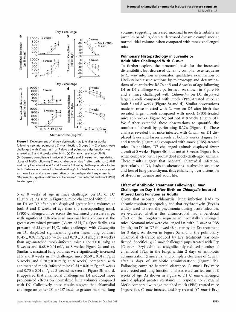
RESULTSPulmonary Function in Juvenile or Adult MiceChallenged With C. mur at 1 or 7 Days After BirthTo determine the effect of chlamydial infection in inducingdysfunctional respiratory sequelae, mice were challenged i.n.with C. mur either 1 day (D1) or 7 days (D7) after birth andthen studied at the age of 5 or 8 weeks. First, airwayresistance in response to escalating doses of MeCh, acholinergic bronchoconstrictor, was measured. As shown inFigure 1a (left panel), mice infected with C. mur on D1 afterbirth displayed increased lung resistance in response to es-calating doses of inhaled MeCh compared with age-matchedmock (PBS)-treated mice at both 5 weeks (C. mur; 559±44,PBS; 407±82, at 25 mg/ml MeCh) and 8 weeks (C. mur;782±96, PBS; 370±118, at 25 mg/ml MeCh) after challenge.Similarly, D7 infected mice also displayed increased resistancein response to MeCh challenge at 5 weeks (C. mur; 238±47,PBS; 160±14, at 25 mg/ml MeCh) with greater resistance by8 weeks (C. mur; 789±96, PBS; 602±74) after birth (Figure1c, left panel). No significant difference was observed inbaseline airway resistance at 5 or 8 weeks of age betweenC. mur (mean±s.e.m.; 0.69±0.04 H2O s/ml) or mock (PBS)infected mice (0.72±0.09). As expected, the dynamic com-pliance in response to 25 mg/ml MeCh was affected inverselyto the dynamic resistance, and was significantly reduced at 8weeks of age in D1 and D7 challenged mice (Figure 1b and d,right panel). These results suggest that chlamydial infectioneither during the saccular or alveolar stage of lung develop-ment, in the absence of additional triggers of AAD, is suffi-cient to induce development of AHR as juveniles or adults.
Quasi-Static P–V Curves in Juveniles or Adult MiceChallenged With C. murAs pulmonary chlamydial infection leads to both peri-bronchiolar and interstitial inflammation,14 quasi-static P–Vloops were assessed to evaluate incremental lung volumes at
Neonatal chlamydial pneumonia induced respiratory sequelae
M Jupelli et al
1532 Laboratory Investigation | Volume 91 October 2011 | www.laboratoryinvestigation.org
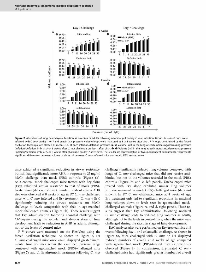
5 or 8 weeks of age in mice challenged on D1 or D7(Figure 2). As seen in Figure 2, mice challenged with C. muron D1 or D7 after birth displayed greater lung volumes atboth 5 and 8 weeks of age than the corresponding mock(PBS)-challenged mice across the examined pressure range,with significant differences in maximal lung volumes at thegreatest examined pressure (33 cm of H2O). Specifically, at apressure of 33 cm of H2O, mice challenged with Chlamydiaon D1 displayed significantly greater mean lung volumes(0.45±0.02 ml/g at 5 weeks and 0.79±0.01 ml/g at 8 weeks)than age-matched mock-infected mice (0.36±0.01 ml/g at5 weeks and 0.68±0.01 ml/g at 8 weeks; Figure 2a and c).Similarly, maximal lung volumes were significantly increasedat 5 and 8 weeks in D7 challenged mice (0.59±0.01 ml/g at5 weeks and 0.78±0.01 ml/g at 8 weeks) compared withage-matched mock-infected mice (0.54±0.01 ml/g at 5 weeksand 0.73±0.01 ml/g at 8 weeks) as seen in Figure 2b and d.It appeared that chlamydial challenge on D1 induced morepronounced effects on normalized lung volumes comparedwith D7. Collectively, these results suggest that chlamydialchallenge on either D1 or D7 leads to greater maximal lung
volume, suggesting increased maximal tissue distensibility asjuveniles or adults, despite decreased dynamic compliance atnormal tidal volumes when compared with mock-challengedmice.
Pulmonary Histopathology in Juvenile orAdult Mice Challenged With C. murTo further explore the structural basis for the increaseddistensibility, but decreased dynamic compliance as sequelaeto C. mur infection as neonates, qualitative examination ofH&E-stained tissue sections by microscopy and determina-tions of quantitative RACs at 5 and 8 weeks of age followingD1 or D7 challenge were performed. As shown in Figure 3band e, mice challenged with Chlamydia on D1 displayedlarger alveoli compared with mock (PBS)-treated mice atboth 5 and 8 weeks (Figure 3a and d). Similar observationsmade in mice infected with C. mur on D7 after birth alsorevealed larger alveoli compared with mock (PBS)-treatedmice at 5 weeks (Figure 3c) but not at 8 weeks (Figure 3f).We further extended these observations to quantify thenumber of alveoli by performing RACs (Figure 4). Theseanalyses revealed that mice infected with C. mur on D1 dis-played fewer and larger alveoli at both 5 weeks (Figure 4a)and 8 weeks (Figure 4c) compared with mock (PBS)-treatedmice. In addition, D7 challenged animals displayed feweralveoli at 5 weeks (Figure 4b), but not at 8 weeks (Figure 4d),when compared with age-matched mock-challenged animals.These results suggest that neonatal chlamydial infection,particularly at D1, leads to alterations in alveolar structureand loss of lung parenchyma, thus enhancing over distensionof alveoli in juvenile and adult life.
Effect of Antibiotic Treatment Following C. murChallenge on Day 1 After Birth on Chlamydia-InducedAltered Lung Function as AdultsGiven that neonatal chlamydial lung infection leads tochronic respiratory sequelae, and that erythromycin (Ery) iswidely used to treat the pneumonia during acute infection,we evaluated whether this antimicrobial had a beneficialeffect on the long-term sequelae in neonatally challengedmice. Neonatal mice were challenged i.n. with C. mur or PBS(mock) on D1 or D7 followed 48 h later by i.p. Ery treatmentfor 3 days. As shown in Figure 5a and b, the pulmonarychlamydial clearance induced by Ery treatment was con-firmed. Specifically, C. mur challenged pups treated with Ery(C. murþ Ery) exhibited a significantly reduced number ofchlamydial IFUs in the lungs within 2 days of antibioticadministration (Figure 5a) and complete clearance of C. murafter 3 days of antibiotic administration (Figure 5b).Following complete bacterial clearance, C. murþ Ery micewere rested and lung function analyses were carried out at 8weeks of age. As shown in Figure 6, D1 C. mur-challengedmice displayed greater resistance in response to 25 mg/mlMeCh compared with age-matched mock (PBS)-treated mice(Figure 6a). C. mur-infected and Ery-treated (C. murþ Ery)
Figure 1 Development of airway dysfunction as juveniles or adults
following neonatal pulmonary C. mur infection. Groups (n¼ 6) of pups were
challenged with C. mur at 1 or 7 days and pulmonary dysfunction was
assayed at 5 and 8 weeks after birth. (a) Dynamic resistance (AHR).
(b) Dynamic compliance in mice at 5 weeks and 8 weeks with escalating
doses of MeCh following C. mur challenge on day 1 after birth. (c, d) AHR
and compliance in mice at 5 and 8 weeks following challenge on day 7 after
birth. Data are normalized to baseline (0 mg/ml of MeCh) and are expressed
as mean±s.e. and are representative of two independent experiments.
*Represents significant differences between C. mur infected and mock (PBS)
treated groups.
Neonatal chlamydial pneumonia induced respiratory sequelae
M Jupelli et al
www.laboratoryinvestigation.org | Laboratory Investigation | Volume 91 October 2011 1533
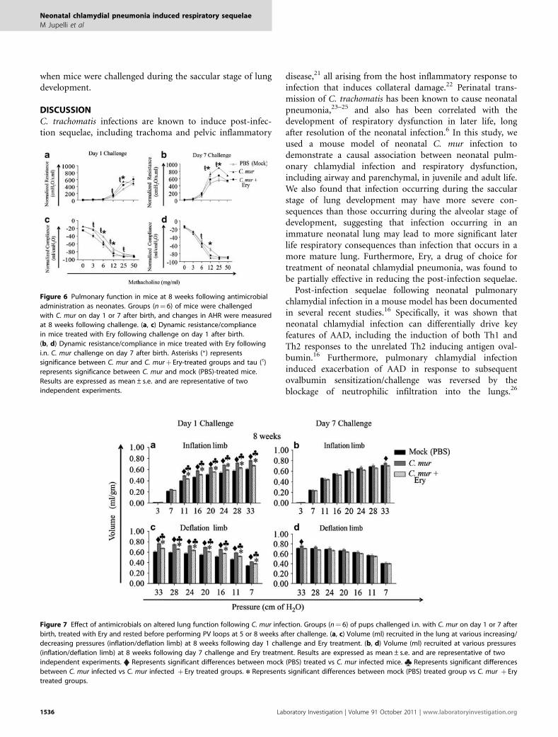
mice exhibited a significant reduction in airway resistance,but still had significantly more AHR in response to 25 mg/mlMeCh challenge than mock (PBS) controls (Figure 6a).As a control, mock-challenged mice treated with Ery alone(Ery) exhibited similar resistance to that of mock (PBS)-treated mice (data not shown). Similar trends of greater AHRalso were observed at 8 weeks of age in D7 C. mur-challengedmice, with C. mur infected and Ery treatment (C. murþ Ery)significantly reducing the airway resistance on MeChchallenge to levels comparable with that in age-matchedmock-challenged animals (Figure 6b). These results suggestthat Ery administration following neonatal challenge withChlamydia during the saccular and alveolar stage of lungdevelopment leads to reduction in AHR as adults, althoughnot to the levels of control mice.
P–V curves were measured on the FlexiVent using theforced oscillation technique. As shown in Figure 7, D1C. mur-challenged mice once again displayed greater incre-mental lung volumes across the examined pressure rangecompared with age-matched mock (PBS)-challenged mice(Figure 7a and c). Erythromycin treatment following C. mur
challenge significantly reduced lung volumes compared withlungs of C. mur-challenged mice that did not receive anti-biotics, but not to the volumes recorded in the mock (PBS)controls (Figure 7a and c, left panel). Unchallenged micetreated with Ery alone exhibited similar lung volumesto those measured in mock (PBS)-challenged mice (data notshown). In D7 C. mur-challenged mice at 8 weeks of age,Ery treatment only led to significant reductions in maximallung volumes down to levels seen in age-matched mock-challenged animals (Figure 7a and d, right panel). These re-sults suggest that Ery administration following neonatalC. mur challenge leads to reduced lung volumes as adults,although not to the levels in control mice, when the mice werechallenged during the saccular stage of lung development.
RAC analyses also were performed on Ery-treated mice at 8weeks following day 1 or 7 chlamydial challenge. As shown inFigure 8a, mice challenged with C. mur on D1 displayedreduced numbers of alveoli at 8 weeks of age comparedwith age-matched mock (PBS)-treated mice as previouslyobserved (Figure 4). In comparison, Ery-treated C. mur-challenged mice had significantly greater numbers of alveoli
Figure 2 Alterations of lung parenchymal function as juveniles or adults following neonatal pulmonary C. mur infection. Groups (n¼ 6) of pups were
infected with C. mur on day 1 or 7 and quasi-static pressure–volume loops were measured at 5 or 8 weeks after birth. P–V loops determined by the forced
oscillation technique are plotted as mean±s.e. at each inflation/deflation pressure. (a, c) Volume (ml) in the lung at each increasing/decreasing pressure
(inflation/deflation limb) at 5 or 8 weeks after C. mur challenge on day 1 after birth. (b, d) Volume (ml) in the lung at each increasing/decreasing pressure
(inflation/deflation limb) at 5 or 8 weeks after challenge on day 7 after birth. The results are representative of two independent experiments. *Represents
significant differences between volume of air in ml between C. mur infected mice and mock (PBS) treated mice.
Neonatal chlamydial pneumonia induced respiratory sequelae
M Jupelli et al
1534 Laboratory Investigation | Volume 91 October 2011 | www.laboratoryinvestigation.org

compared with untreated C. mur-challenged mice. Similaranalysis performed on D7 challenged mice (Figure 8b)revealed that Ery treatment had a minimal effect on the
numbers of alveoli in comparison with untreated micechallenged with C. mur These results suggest that earlyintervention with Ery leads to restoration of lung structure
Figure 3 Pulmonary histopathology at 5 or 8 weeks following neonatal C. mur infection. Groups (n¼ 6) of pups were infected with C. mur on day 1 or 7
and qualitative histological evaluation was performed at 5 or 8 weeks after birth demonstrates qualitative images on lung sections at � 100 magnification.
The slides b and e demonstrate representative images at 5 and 8 weeks after day 1 challenge, respectively, and c and f demonstrate lung sections at 5 and
8 weeks following day 7 challenge. Representative lung sections from age-matched mock (PBS)-treated mice are shown in a and d. The inserts show high
resolution images at the corresponding time points. Results are representative of two independent experiments.
Figure 4 Radial alveolar counts at 5 or 8 weeks following neonatal
pulmonary C. mur challenge. Groups (n¼ 6–7) of 1 or 7 day old pups were
challenged with C. mur and rested for 5 or 8 weeks followed by
determination of the RACs. (a, c) The RAC at 5 and 8 weeks following day 1
C. mur challenge. (b, d) The RAC at 5 and 8 weeks following challenge on
day 7 after birth. Results are expressed as mean±s.e. and are
representative of two independent experiments. *Represents significant
differences in RAC between mock (PBS) treated and C. mur infected mice.
Figure 5 Effect of antimicrobials on bacterial clearance in the lung
following neonatal pulmonary chlamydial challenge. Groups (n¼ 4) of pups
were challenged i.n with C. mur on day 1 after birth and the reduction in
bacterial burden was confirmed by plating the lung homogenates on HeLa
cell monolayers after 2 (a) and 3 (b) days of antimicrobial administration.
Results are expressed as mean±s.e. are representative of two independent
experiments. *Represents significant differences in bacterial burden
between C. mur infected vs C. mur infected þ Ery treated mice.
Neonatal chlamydial pneumonia induced respiratory sequelae
M Jupelli et al
www.laboratoryinvestigation.org | Laboratory Investigation | Volume 91 October 2011 1535
when mice were challenged during the saccular stage of lungdevelopment.
DISCUSSIONC. trachomatis infections are known to induce post-infec-tion sequelae, including trachoma and pelvic inflammatory
disease,21 all arising from the host inflammatory response toinfection that induces collateral damage.22 Perinatal trans-mission of C. trachomatis has been known to cause neonatalpneumonia,23–25 and also has been correlated with thedevelopment of respiratory dysfunction in later life, longafter resolution of the neonatal infection.6 In this study, weused a mouse model of neonatal C. mur infection todemonstrate a causal association between neonatal pulm-onary chlamydial infection and respiratory dysfunction,including airway and parenchymal, in juvenile and adult life.We also found that infection occurring during the saccularstage of lung development may have more severe con-sequences than those occurring during the alveolar stage ofdevelopment, suggesting that infection occurring in animmature neonatal lung may lead to more significant laterlife respiratory consequences than infection that occurs in amore mature lung. Furthermore, Ery, a drug of choice fortreatment of neonatal chlamydial pneumonia, was found tobe partially effective in reducing the post-infection sequelae.
Post-infection sequelae following neonatal pulmonarychlamydial infection in a mouse model has been documentedin several recent studies.16 Specifically, it was shown thatneonatal chlamydial infection can differentially drive keyfeatures of AAD, including the induction of both Th1 andTh2 responses to the unrelated Th2 inducing antigen oval-bumin.16 Furthermore, pulmonary chlamydial infectioninduced exacerbation of AAD in response to subsequentovalbumin sensitization/challenge was reversed by theblockage of neutrophilic infiltration into the lungs.26
Figure 7 Effect of antimicrobials on altered lung function following C. mur infection. Groups (n¼ 6) of pups challenged i.n. with C. mur on day 1 or 7 after
birth, treated with Ery and rested before performing PV loops at 5 or 8 weeks after challenge. (a, c) Volume (ml) recruited in the lung at various increasing/
decreasing pressures (inflation/deflation limb) at 8 weeks following day 1 challenge and Ery treatment. (b, d) Volume (ml) recruited at various pressures
(inflation/deflation limb) at 8 weeks following day 7 challenge and Ery treatment. Results are expressed as mean±s.e. and are representative of two
independent experiments. Represents significant differences between mock (PBS) treated vs C. mur infected mice. Represents significant differences
between C. mur infected vs C. mur infected þ Ery treated groups. n Represents significant differences between mock (PBS) treated group vs C. mur þ Ery
treated groups.
Figure 6 Pulmonary function in mice at 8 weeks following antimicrobial
administration as neonates. Groups (n¼ 6) of mice were challenged
with C. mur on day 1 or 7 after birth, and changes in AHR were measured
at 8 weeks following challenge. (a, c) Dynamic resistance/compliance
in mice treated with Ery following challenge on day 1 after birth.
(b, d) Dynamic resistance/compliance in mice treated with Ery following
i.n. C. mur challenge on day 7 after birth. Asterisks (*) represents
significance between C. mur and C. murþ Ery-treated groups and tau (t)
represents significance between C. mur and mock (PBS)-treated mice.
Results are expressed as mean±s.e. and are representative of two
independent experiments.
Neonatal chlamydial pneumonia induced respiratory sequelae
M Jupelli et al
1536 Laboratory Investigation | Volume 91 October 2011 | www.laboratoryinvestigation.org

Although the neutrophilic infiltration was associated withTh1/Th17 responses, the blockade of IFN-g or IL-17 did notsignificantly alter the ovalbumin-induced AAD, suggestingthat factors other than these cytokines may have a crucial rolein the process.26
The mechanistic basis of chlamydial neonatal pulmonaryinfection-induced changes to subsequent events in adult lifesuch as allergen exposure may be unraveled by primarilyunderstanding the effects of infection per se on lung structureand function. Although the majority of the characterizationin earlier studies focused on ovalbumin-induced AAD, the
authors made anecdotal observations that the neonatalinfection per se, in the absence of additional ovalbuminsensitization/challenge, led to induction of significantly en-hanced AHR and alveolar diameter.17 We now confirm andextend these observations by detailed experiments demon-strating that neonatal pulmonary chlamydial infection leadsto chronic sequelae in juvenile and adult life long afterclearance of the bacterium, as evidenced by enhanced AHR,reduced RACs and increased lung compliance, collectivelysuggesting emphysematous changes. These effects were evi-dent both at the age of 5 weeks (juvenile) and 8 weeks (adult)in mice challenged neonatally with Chlamydia in the pul-monary compartment, suggesting that the changes inducedby the neonatal infection are long lasting, and may be per-manent. Interestingly, we also observed significant reductionin expression of surfactant protein C during the acute phaseof the infection (day 7) and at 5 weeks following C. murchallenge on day 1 after birth (Figure 9). In parallel analyses,differences in expression of various enzymes such as esterases,alkaline phosphatases and lipases (Supplementary Table 1)were observed at 5 weeks following chlamydial clearance inmice infected on day 1 after birth compared with mock-challenged animals, using API-ZYM assays (SupplementaryInformation). Thus, in addition to the chronic modulationof inflammatory cytokines such as IFN-g and IL-17 asshown in the C. mur-ovalbumin AAD model,26 the neonatalpulmonary chlamydial infection also may affect the levelsof surfactant proteins that are involved in maintenance oflung structure and function,27–31 and various biochemicalenzymes that could differentially induce inflammatoryresponses in the lung (Figure 9).31,32
Chlamydial infection in mice at 1 day after birth inducedmore severe respiratory sequelae at 8 weeks of age comparedwith infection at 7 days after birth. The neonatal mouse lungat 1 day after birth is in the saccular stage of development,whereas the 7-day-old mouse lung is in the alveolar stageof development.33 This suggests that respiratory sequelaefollowing chlamydial infection in an immature lung may bemore severe than the infection that occurs in a matured lung.Our results are in accordance with Horvat et al,17 whoshowed that early life (neonatal; day 1) respiratory C. murinfections result in greater altered lung structure, functionand enhanced AHR compared with infection that occurs inlater life. These results suggest that the environment of theneonatal lung is a major determinant of disease patterns inlater life. In this context, Culley et al34 showed that the age atwhich primary viral infection occurs determines the patternof T-cell-mediated disease during re-infection in adulthoodin a mouse model of RSV infection.
The macrolide antimicrobial Ery is an approved treatmentfor symptomatic neonatal C. trachomatis pneumonia (CDC,2010 STD Treatment Guidelines; cdc.gov/std/treatment/2010/chlamydial-infections.html). Macrolides have beenshown to reduce neutrophil chemotaxis, oxidative burst andproinflammatory cytokine generation in various models.35
Figure 8 Effect of antimicrobials on lung structure as adults following
neonatal C. mur infection. Groups (n¼ 6) of pups challenged i.n. with C. mur
on day 1 or 7 after birth and treated with Ery were rested before performing
RAC at 8 weeks after challenge. (a) RAC counts at 8 weeks following day 1
C. mur challenge and Ery treatment. (b) RACs at 8 weeks following day 7
C. mur challenge and Ery treatment. Results are expressed as mean±s.e.
*P¼ 0.05 is considered significant.
Figure 9 Pulmonary surfactant protein expression following neonatal
C. mur infection. Groups (n¼ 4) of pups were challenged i.n. with C. mur on
day 1 after birth and lungs were collected on day 7 (a) or 5 weeks (b) after
challenge to determine alterations in surfactant expression by western
blotting analysis. Results are expressed as mean±s.e. and are
representative of two independent experiments. *Represents significant
differences in SP-C expression between C. mur infected vs mock (PBS)
treated mice at the corresponding time intervals.
Neonatal chlamydial pneumonia induced respiratory sequelae
M Jupelli et al
www.laboratoryinvestigation.org | Laboratory Investigation | Volume 91 October 2011 1537

Macrolides also reduce mucus hypersecretion in patientswith diffuse panbronchiolitis, chronic bronchitis and sinusdisease.35 Evidence suggests that macrolides may protect theepithelium from attack by bioactive phospholipids generatedas part of the inflammatory process.36,37 In our study, Erytreatment of Chlamydia-infected neonatal mice promoted theclearance of infection from the lungs within 3 days. This earlyelimination of the infection also was partially effectivein reducing the respiratory sequelae in adult life as shown byRACs, which demonstrated that Ery administration restoredChlamydia induced reduced alveoli seen in adults. Thesefindings suggest that the usage of antibiotics to treat neonatalchlamydial pneumonia may have important benefits beyondthe alleviation of the acute pneumonia. However, the com-plete set of adverse effects of neonatal chlamydial infectionmay not be alleviated by administration of antibiotics,although it cannot be excluded that usage of a differentantibiotic and/or regimen may be more efficacious in pre-venting the chronic sequelae. In addition, asymptomaticchlamydial pulmonary infections have been described25 andas a consequence, infected neonates may not receive treat-ment and grow into adulthood with respiratory dysfunction.As with other animal models of human disease, results fromour model of C. mur pulmonary infection of neonatal miceand subsequent respiratory dysfunction in later life may notbe directly extrapolated to humans. For example, C. tracho-matis has been shown to use different mechanisms to evadethe host IFN-g response in humans, when compared withC. mur in mice,38 and these organisms have certain differ-ences, such as the shorter life cycle of C. mur (approximately24 h vs 48–72 h for C. trachomatis) as well as differences inpathogenesis due to differences in development and lifespan of their respective hosts, despite several strikingsimilarities between the murine and human organisms.39
Although our results from the C. mur infection in neonatalmice may not be directly extrapolated to those of C.trachomatis infections in humans (owing to the different lifecycles and pathogenesis), it provides a useful animal model tostudy the serious nature of the potential sequelae of earlychlamydial respiratory infection and lends justification tofurther detailed mechanistic investigations. It also highlightsthe importance of prenatal screening and treatment ofpregnant women for C. trachomatis in order to prevent thepossible long-lasting effects of neoanatal pulmonary chla-mydial infections.
Supplementary Information accompanies the paper on the Laboratory
Investigation website (http://www.laboratoryinvestigation.org)
ACKNOWLEDGEMENT
This work is supported by the National Institutes of Health Grant 1 RO1
AI074860.
DISCLOSURE/CONFLICT OF INTEREST
The authors declare no conflict of interest.
1. Chawla R, Bhalla P, Sachdev H. A pilot study of Chlamydia trachomatispneumonia in infants. Indian J Med Microbiol 2004;22:185–187.
2. Were F, Govedi A, Revathi G, et al. Chlamydia as a cause of lateneonatal pneumonia at Kenyatta National Hospital, Nairobi. East AfrMed J 2002;79:476–479.
3. Chen C, Wu K, Tang R, et al. Characteristics of Chlamydia trachomatisinfection in hospitalized infants with lower respiratory tract infection.J Microbiol Immunol Infect 2007;40:255–259.
4. Beem M, Saxon E. Respiratory-tract colonization and a distinctivepneumonia syndrome in infants infected with Chlamydia trachomatis.N Engl J Med 1977;296:306–310.
5. Beem M, Saxon E, Tipple M. Treatment of chlamydial pneumonia ofinfancy. Pediatrics 1979;63:198–203.
6. Weiss S, Newcomb R, Beem M. Pulmonary assessment of childrenafter chlamydial pneumonia of infancy. J Pediatr 1986;108(5 part 1):659–664.
7. Cunningham C, Johnston S, Julious S, et al. Chronic Chlamydiapneumoniae infection and asthma exacerbations in children. Eur RespirJ 1998;11:345–349.
8. Esposito S, Blasi F, Arosio C, et al. Importance of acute Mycoplasmapneumoniae and Chlamydia pneumoniae infections in children withwheezing. Eur Respir J 2000;16:1142–1146.
9. Blasi F. Atypical pathogens and respiratory tract infections. Eur Respir J2004;24:171–181.
10. Kumar S, Hammerschlag M. Acute respiratory infection dueto Chlamydia pneumoniae: current status of diagnostic methods.Clin Infect Dis 2007;44:567–576.
11. Darville T. Chlamydia trachomatis infections in neonates and youngchildren. Semin Pediatr Infect Dis 2005;16:235–244.
12. Roseman M, Mahon B, Downs S, et al. Oral erythromycin prophylaxis vswatchful waiting in caring for newborns exposed to Chlamydiatrachomatis. Arch Pediatr Adolesc Med 2003;157:565–571.
13. Webley W, Tilahun Y, Lay K, et al. Occurance of Chlamydia trachomatisand Chlamydia pneumoniaein pediatric respiratory infections. Respir J2009;33:360–367.
14. Jupelli M, Guentzel M, Meier P, et al. Endogenous IFN-gammaproduction is induced and required for protective immunity againstpulmonary chlamydial infection in neonatal mice. J Immunol2008;180:4148–4155.
15. Jupelli M, Selby D, Guentzel M, et al. The contribution of interleukin-12/interferon-gamma axis in protection against neonatal pulmonaryChlamydia muridarum challenge. J Interferon Cytokine Res 2010;30:407–415.
16. Horvat J, Beagley K, Wade M, et al. Neonatal chlamydial infectioninduces mixed T-cell responses that drive allergic airway disease.Am J Respir Crit Care Med 2007;176:556–564.
17. Horvat J, Starkey M, Kim R, et al. Early-life chlamydial lung infectionenhances allergic airways disease through age-dependent differencesin immunopathology. J Allergy Clin Immunol 2010;125:617–625,625.e611–625.e616.
18. Abdelrahman Y, Belland R. The chlamydial developmental cycle.FEMS Microbiol Rev 2005;29:949–959.
19. Cooney T, Thurlbeck W. The radial alveolar count method of Emeryand Mithal: a reappraisal 1–postnatal lung growth. Thorax 1982;37:572–579.
20. Cooney T, Thurlbeck W. The radial alveolar count method of Emeryand Mithal: a reappraisal 2–intrauterine and early postnatal lunggrowth. Thorax 1982;37:580–583.
21. Darville T, Hiltke T. Pathogenesis of genital tract disease due toChlamydia trachomatis. J Infect Dis 2010;201(Suppl 2):S114–S125.
22. Brunham R, Rey-Ladino J. Immunology of Chlamydia infection: implica-tions for a Chlamydia trachomatis vaccine. Nat Rev Immunol 2005;5:149–161.
23. Gencay M, Koskiniemi M, Fellman V, et al. Chlamydia trachomatisinfection in mothers with preterm delivery and in their newborninfants. APMIS 2001;109:636–640.
24. Darville T. Chlamydia trachomatis genital infection in adolescents andyoung adults. Adv Exp Med Biol 2006;582:85–100.
25. Jain S. Perinatally acquired Chlamydia trachomatis associatedmorbidity in young infants. J Matern Fetal Med 1999;8:130–133.
26. Horvat J, Starkey M, Kim R, et al. Chlamydial respiratory infectionduring allergen sensitization drives neutrophilic allergic airwaysdisease. J Immunol 2010;184:4159–4169.
Neonatal chlamydial pneumonia induced respiratory sequelae
M Jupelli et al
1538 Laboratory Investigation | Volume 91 October 2011 | www.laboratoryinvestigation.org
27. Bland R. Neonatal chronic lung disease in the post-surfactant era.Biol Neonate 2005;88:181–191.
28. Ferrer-Roca O, Gotzens V, Ruano-Gil D, et al. Alveolar develop-ment of the lung of the rat. Part I. Arch Ital Anat Embriol 1985;90:91–99.
29. Ferrer-Roca O, Gotzens V, Ruano-Gil D, et al. Alveolar developmentof the lung of the rat. Part II. Morphogenesis of surfactant. Arch ItalAnat Embriol 1985;90:101–110.
30. Hohlfeld J, Fabel H, Hamm H. The role of pulmonary surfac-tant in obstructive airways disease. Eur Respir J 1997;10:482–491.
31. Harrison H, English M, Lee C, et al. Chlamydia trachomatis infantpneumonitis: comparison with matched controls and other infantpneumonitis. N Engl J Med 1978;298:702–708.
32. Lian X, Yan C, Yang L, et al. Lysosomal acid lipase deficiency causesrespiratory inflammation and destruction in the lung. Am J PhysiolLung Cell Mol Physiol 2004;286:L801–L807.
33. Perl A, Whitsett J. Molecular mechanisms controlling lung morpho-genesis. Clin Genet 1999;56:14–27.
34. Culley F, Pollott J, Openshaw P. Age at first viral infection determinesthe pattern of T cell-mediated disease during reinfection in adulthood.J Exp Med 2002;196:1381–1386.
35. Sharma S, Jaffe A, Dixon G. Immunomodulatory effects of macrolideantibiotics in respiratory disease: therapeutic implications for asthmaand cystic fibrosis. Paediatr Drugs 2007;9:107–118.
36. Tamaoki J, Kadota J, Takizawa H. Clinical implications of the immuno-modulatory effects of macrolides. Am J Med 2004;117(Suppl 9A):5S–11S.
37. Takizawa H, Desaki M, Ohtoshi T, et al. Erythromycin suppressesinterleukin 6 expression by human bronchial epithelial cells: apotential mechanism of its anti-inflammatory action. BiochemBiophys Res Commun 1995;210:781–786.
38. Nelson D, Virok D, Wood H, et al. Chlamydial IFN-gamma immuneevasion is linked to host infection tropism. Proc Natl Acad Sci USA2005;102:10658–10663.
39. McClarty G, Caldwell H, Nelson D. Chlamydial interferon gamma immuneevasion influences infection tropism. Curr Opin Microbiol 2007;10:47–51.
Neonatal chlamydial pneumonia induced respiratory sequelae
M Jupelli et al
www.laboratoryinvestigation.org | Laboratory Investigation | Volume 91 October 2011 1539